A recurrent 15q13.3 microdeletion syndrome associated with mental retardation and seizures
7
A recurrent 15q13.3 microdeletion syndrome associated with mental retardation and seizures Andrew J Sharp 1,15 , Heather C Mefford 1 , Kelly Li 2 , Carl Baker 1 , Cindy Skinner 3 , Roger E Stevenson 3 , Richard J Schroer 3 , Francesca Novara 4 , Manuela De Gregori 4 , Roberto Ciccone 4 , Adam Broomer 2 , Iris Casuga 2 , Yu Wang 2 , Chunlin Xiao 2 , Catalin Barbacioru 2 , Giorgio Gimelli 5 , Bernardo Dalla Bernardina 6 , Claudia Torniero 6 , Roberto Giorda 7 , Regina Regan 8 , Victoria Murday 9 , Sahar Mansour 10 , Marco Fichera 11 , Lucia Castiglia 11 , Pinella Failla 11 , Mario Ventura 12 , Zhaoshi Jiang 1 , Gregory M Cooper 1 , Samantha J L Knight 8 , Corrado Romano 11 , Orsetta Zuffardi 4,13 , Caifu Chen 2 , Charles E Schwartz 3 & Evan E Eichler 1,14 We report a recurrent microdeletion syndrome causing mental retardation, epilepsy and variable facial and digital dysmorphisms. We describe nine affected individuals, including six probands: two with de novo deletions, two who inherited the deletion from an affected parent and two with unknown inheritance. The proximal breakpoint of the largest deletion is contiguous with breakpoint 3 (BP3) of the Prader-Willi and Angelman syndrome region, extending 3.95 Mb distally to BP5. A smaller 1.5-Mb deletion has a proximal breakpoint within the larger deletion (BP4) and shares the same distal BP5. This recurrent 1.5-Mb deletion contains six genes, including a candidate gene for epilepsy (CHRNA7) that is probably responsible for the observed seizure phenotype. The BP4–BP5 region undergoes frequent inversion, suggesting a possible link between this inversion polymorphism and recurrent deletion. The frequency of these microdeletions in mental retardation cases is B0.3% (6/2,082 tested), a prevalence comparable to that of Williams, Angelman and Prader-Willi syndromes. Genomic disorders result from DNA rearrangements mediated by nonallelic homologous recombination (NAHR) between large, highly homologous flanking segmental duplications 1 . The clinical features of many of the known genomic disorders include mental retardation and developmental delay, and several recent studies of individuals with mental retardation have led to the identification of new recurrent genomic disorders 2 . By whole-genome array CGH screening of 757 individuals with mental retardation and/or congenital anomalies, we identified two unrelated individuals with mild to moderate mental retardation, dysmorphic features and abnormal electroencephalogram (EEG) findings who both have identical de novo 1.5-Mb deletions of 15q13.3. These two deletions share the same distal breakpoint (BP5) with a previously reported 3 3.95-Mb deletion of 15q13 (Fig. 1 and Fig. 2a) and a proximal breakpoint (BP4) within this larger deletion (Fig. 1 and Fig. 2b–d). The shared 1.5-Mb region contains six known genes. Our array CGH screening also detected a single individual (543/06) with a proximal deletion breakpoint corresponding to break- point region 3 (BP3) of the Prader-Willi and Angelman syndrome region and a distal breakpoint at BP4 (Fig. 1). However, the deletion was also detected in the individual’s unaffected father. Therefore, we interpret this BP3–BP4 deletion as probably representing a benign copy number variant, although we cannot exclude that it may instead be a pathogenic deletion with incomplete penetrance. In order to rapidly screen a large collection of affected individuals for deletions in the shared 1.5-Mb interval between BP4 and BP5, we developed two TaqMan quantitative PCR (qPCR) assays targeted to this region and screened 1,040 individuals with mental retardation of unknown etiology. This cohort, obtained from the Greenwood Genetic Center, consists of an approximately equal number of indi- viduals of European and African American descent. qPCR analyses identified four individuals as potentially harboring a deletion of the interval BP4–BP5. We subsequently validated deletions by BAC array CGH (data not shown) and a custom oligonucleotide array (Fig. 1). A review of pedigrees showed evidence of multiple affected individuals for each case (Fig. 2 and Supplementary Fig. 1 online), and review Received 2 October 2007; accepted 7 January 2008; published online 17 February 2008; doi:10.1038/ng.93 1 Department of Genome Sciences, University of Washington School of Medicine, 1705 NE Pacific St., Seattle, Washington 98195, USA. 2 Assays/Arrays R&D, Applied Biosystems, Foster City, California 94404, USA. 3 JC Self Research Institute, Greenwood Genetic Center, Greenwood, South Carolina 29646, USA. 4 Biologia Generale e Genetica Medica, Universita ` di Pavia, Pavia 27100, Italy. 5 Istituto G. Gaslini, Genova 16147, Italy. 6 Servizio Neuropsichiatria Infantile, Policlinico GB Rossi, Universita ` di Verona,Verona 37134, Italy. 7 IRCCS E. Medea, Bosisio Parini 23842, Italy. 8 Oxford National Institute for Health Research (NIHR) Biomedical Research Centre, The Wellcome Trust Centre for Human Genetics, Churchill Hospital, Oxford OX3 7BN, UK. 9 Department of Medical Genetics, Duncan Guthrie Institute, Glasgow G3 8SJ, UK. 10 SW Regional Genetics Service, St George’s Hospital, London SW17 0RE, UK. 11 IRCCS Associazione Oasi Maria Santissima, Troina 94018, Italy. 12 Dipartimento di Genetica e Microbiologia, Universita’ degli Studi di Bari, Bari 27100, Italy. 13 Fondazione IRCCS Policlinico San Matteo, Pavia 27100, Italy. 14 Howard Hughes Medical Institute, 1705 NE Pacific St., Seattle, Washington 98195, USA. 15 Present address: Department of Genetic Medicine and Development, University of Geneva Medical School, 1 rue Michel-Servet, 1211 Geneva, Switzerland. Correspondence should be addressed to E.E.E. ([email protected]). 322 VOLUME 40 [ NUMBER 3 [ MARCH 2008 NATURE GENETICS LETTERS © 2008 Nature Publishing Group http://www.nature.com/naturegenetics
Transcript of A recurrent 15q13.3 microdeletion syndrome associated with mental retardation and seizures
A recurrent 15q13.3 microdeletion syndrome associatedwith mental retardation and seizuresAndrew J Sharp1,15, Heather C Mefford1, Kelly Li2, Carl Baker1, Cindy Skinner3, Roger E Stevenson3,Richard J Schroer3, Francesca Novara4, Manuela De Gregori4, Roberto Ciccone4, Adam Broomer2,Iris Casuga2, Yu Wang2, Chunlin Xiao2, Catalin Barbacioru2, Giorgio Gimelli5, Bernardo Dalla Bernardina6,Claudia Torniero6, Roberto Giorda7, Regina Regan8, Victoria Murday9, Sahar Mansour10, Marco Fichera11,Lucia Castiglia11, Pinella Failla11, Mario Ventura12, Zhaoshi Jiang1, Gregory M Cooper1, Samantha J L Knight8,Corrado Romano11, Orsetta Zuffardi4,13, Caifu Chen2, Charles E Schwartz3 & Evan E Eichler1,14
We report a recurrent microdeletion syndrome causingmental retardation, epilepsy and variable facial and digitaldysmorphisms. We describe nine affected individuals, includingsix probands: two with de novo deletions, two who inheritedthe deletion from an affected parent and two with unknowninheritance. The proximal breakpoint of the largest deletion iscontiguous with breakpoint 3 (BP3) of the Prader-Willi andAngelman syndrome region, extending 3.95 Mb distally to BP5.A smaller 1.5-Mb deletion has a proximal breakpoint withinthe larger deletion (BP4) and shares the same distal BP5. Thisrecurrent 1.5-Mb deletion contains six genes, including acandidate gene for epilepsy (CHRNA7) that is probablyresponsible for the observed seizure phenotype. The BP4–BP5region undergoes frequent inversion, suggesting a possible linkbetween this inversion polymorphism and recurrent deletion.The frequency of these microdeletions in mental retardationcases is B0.3% (6/2,082 tested), a prevalence comparable tothat of Williams, Angelman and Prader-Willi syndromes.
Genomic disorders result from DNA rearrangements mediated bynonallelic homologous recombination (NAHR) between large, highlyhomologous flanking segmental duplications1. The clinical features ofmany of the known genomic disorders include mental retardation anddevelopmental delay, and several recent studies of individuals withmental retardation have led to the identification of new recurrentgenomic disorders2. By whole-genome array CGH screening of 757individuals with mental retardation and/or congenital anomalies, we
identified two unrelated individuals with mild to moderate mentalretardation, dysmorphic features and abnormal electroencephalogram(EEG) findings who both have identical de novo 1.5-Mb deletions of15q13.3. These two deletions share the same distal breakpoint (BP5)with a previously reported3 3.95-Mb deletion of 15q13 (Fig. 1 andFig. 2a) and a proximal breakpoint (BP4) within this larger deletion(Fig. 1 and Fig. 2b–d). The shared 1.5-Mb region contains six knowngenes. Our array CGH screening also detected a single individual(543/06) with a proximal deletion breakpoint corresponding to break-point region 3 (BP3) of the Prader-Willi and Angelman syndromeregion and a distal breakpoint at BP4 (Fig. 1). However, the deletionwas also detected in the individual’s unaffected father. Therefore, weinterpret this BP3–BP4 deletion as probably representing a benigncopy number variant, although we cannot exclude that it may insteadbe a pathogenic deletion with incomplete penetrance.
In order to rapidly screen a large collection of affected individualsfor deletions in the shared 1.5-Mb interval between BP4 and BP5, wedeveloped two TaqMan quantitative PCR (qPCR) assays targeted tothis region and screened 1,040 individuals with mental retardation ofunknown etiology. This cohort, obtained from the GreenwoodGenetic Center, consists of an approximately equal number of indi-viduals of European and African American descent. qPCR analysesidentified four individuals as potentially harboring a deletion of theinterval BP4–BP5. We subsequently validated deletions by BAC arrayCGH (data not shown) and a custom oligonucleotide array (Fig. 1). Areview of pedigrees showed evidence of multiple affected individualsfor each case (Fig. 2 and Supplementary Fig. 1 online), and review
Received 2 October 2007; accepted 7 January 2008; published online 17 February 2008; doi:10.1038/ng.93
1Department of Genome Sciences, University of Washington School of Medicine, 1705 NE Pacific St., Seattle, Washington 98195, USA. 2Assays/Arrays R&D, AppliedBiosystems, Foster City, California 94404, USA. 3JC Self Research Institute, Greenwood Genetic Center, Greenwood, South Carolina 29646, USA. 4Biologia Generale eGenetica Medica, Universita di Pavia, Pavia 27100, Italy. 5Istituto G. Gaslini, Genova 16147, Italy. 6Servizio Neuropsichiatria Infantile, Policlinico GB Rossi,Universita di Verona,Verona 37134, Italy. 7IRCCS E. Medea, Bosisio Parini 23842, Italy. 8Oxford National Institute for Health Research (NIHR) Biomedical ResearchCentre, The Wellcome Trust Centre for Human Genetics, Churchill Hospital, Oxford OX3 7BN, UK. 9Department of Medical Genetics, Duncan Guthrie Institute, GlasgowG3 8SJ, UK. 10SW Regional Genetics Service, St George’s Hospital, London SW17 0RE, UK. 11IRCCS Associazione Oasi Maria Santissima, Troina 94018, Italy.12Dipartimento di Genetica e Microbiologia, Universita’ degli Studi di Bari, Bari 27100, Italy. 13Fondazione IRCCS Policlinico San Matteo, Pavia 27100, Italy.14Howard Hughes Medical Institute, 1705 NE Pacific St., Seattle, Washington 98195, USA. 15Present address: Department of Genetic Medicine and Development,University of Geneva Medical School, 1 rue Michel-Servet, 1211 Geneva, Switzerland. Correspondence should be addressed to E.E.E. ([email protected]).
3 22 VOLUME 40 [ NUMBER 3 [ MARCH 2008 NATURE GENETICS
LET TERS©
2008
Nat
ure
Pub
lishi
ng G
roup
ht
tp://
ww
w.n
atur
e.co
m/n
atur
egen
etic
s

of the sample collection showed that although the series was thoughtto have only unrelated individuals, two of the individuals we identifiedwith deletions are mother (CMS7833) and son (CMS5803).
Review of the phenotypes observed in the nine individuals identi-fied with deletions of 15q13 showed a number of phenotypic features(summarized in Table 1 and Fig. 2, with full phenotypic details in theSupplementary Note online). The most consistent features among theindividuals in our series were mild to moderate mental retardation(9/9 individuals) and epilepsy and/or abnormal EEG findings, whichwe noted in seven of nine individuals. All affected individuals also hadmild facial dysmorphism, although these features were variable.Features shared among three or more individuals included hyper-telorism, upslanting palpebral fissures, prominent philtrum with fulleverted lips, short and/or curved fifth finger and short fourthmetacarpals. Related to the latter, it is noteworthy that we notedskeletal and/or joint defects of the hand in seven of nine individuals(Table 1). Therefore, testing for the 15q13.3 deletion should beconsidered in individuals with unexplained mental retardation,seizures and mild dysmorphic features.
Each breakpoint region corresponds to a complex set of segmentalduplications, termed duplication blocks. BP4 is an 818-kb duplicationblock with three large regions of homology to BP5 (95 kb, 140 kb and
218 kb with 99.6% identity, according to the University of CaliforniaSanta Cruz Genome Browser (see URLs section of Methods, below)),each of which lies in an inverted orientation relative to the corre-sponding region in BP5 (Fig. 3, Supplementary Table 1 online andSupplementary Fig. 2 online). Consistent with NAHR as the mechan-ism underlying rearrangements of 15q13.3, our data localized thebreakpoints of all five BP4–BP5 deletions and a BP4–BP5 duplicationto these large paralogous sequences (Figs. 1,4 and SupplementaryFig. 3 online). The BP3 region, which is the common distal breakpointin Prader-Willi and Angelman syndrome deletions, corresponds toan 843-kb duplication block (hg17/Build 35, chr 15: 26053472–26896735). Duplicated sequence within BP3 shows relatively smallstretches of homology to BP4 (17 kb with 93% identity) and BP5(30 kb with 98% identity and 11 kb with 93% identity). BP3 alsocontains two assembly gaps, and our data from IMR338 and indivi-dual 543/06 suggest that the proximal breakpoint in these deletionsoccurs within the distal gap. Given the likelihood that NAHR under-lies rearrangements of 15q13, we suggest that this region containsas-yet-uncharacterized segmental duplications, paralogous to those atBP4 and BP5, that catalyze recurrent rearrangements of 15q13. Thus,the increased prevalence of rearrangements observed between BP4–BP5 relative to those involving BP3–BP4–BP5 is consistent with thesize and homology of duplicons in 15q13. We find it interesting thatmost of the breakpoint regions associated with genomic disorders onchromosome 15 correspond to duplication blocks that harbor copiesof the GOLGA gene family4,5. Within the limits of oligonucleotidearray CGH resolution, breakpoint regions seem to overlap with thesespecific duplicons (Supplementary Fig. 4 online).
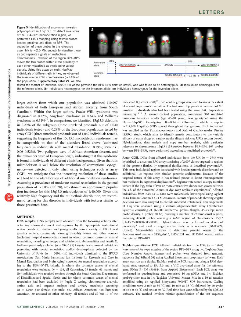
Observations in several genomic disorders suggest that microdele-tions arise preferentially from chromosomes carrying an inversion ofthat same region3,6–9. Given the opposing orientation of the dupliconsat BP4 relative to BP5 in the reference assembly (SupplementaryFig. 2), we hypothesized that 15q13.3 might also be a site of inversionpolymorphism that could create a configuration predisposed torecurrent rearrangement. To investigate this possibility, we usedFISH to assay the BP4–BP5 region. Testing of eight HapMap indivi-duals of varied ethnicities showed the presence of a common inversionof this interval, which was present on 7 of the 16 chromosomesassayed (frequency of inversion relative to reference assembly: 0.44;95% confidence interval (c.i.): 0.19–0.68) (Fig. 5 and SupplementaryTable 2 online). In the two instances where we were able to study theparent-of-origin of BP4–BP5 deletions (the mother of individual69/06 and the father of individual 02961), we found both to beheterozygous for the inversion. Although the high frequency of thisinversion in the normal population and the lack of sufficient numbersof parental samples preclude any definitive conclusions, our observa-tions are consistent with a model in which inversion polymorphism ofthe BP4–BP5 region results in these flanking duplicons being placed in
Segmentalduplications
IMR338M
Critical regionBP4BP3 BP5
69/06
543/06
CMS7833
CMS5803
CMS5826
CMS7906
GapsOCA2
HERC2
GOLGA8GGOLGA8G
APBA2NDNL2
TJP1 MTMR15TRPM1
MTMR10KLF13OTUD7A
CHRNA7ARHGAP11A
SCG5C15orf45
GREM1
CHRFAM7A
15q13.115q13.2
15q13.3
27000000 28000000 29000000 30000000 31000000
ARHGAP11BLOC440248
02961
Figure 1 High-resolution oligonucleotide array mapping of 15q12–q13.3
rearrangements (chr15:25700000–31400000). Although there
seems to be variation in the exact location of breakpoints, all map to large
blocks of segmental duplication at BP3, BP4 and BP5 (indicated by dashed
lines). For each individual, deviations of probe log2 ratios from 0 are
depicted by gray and black lines. Those exceeding a threshold of 1.5 s.d.
from the mean probe ratio are colored green and red to represent relative
gains and losses, respectively. Segmental duplications of increasing
similarity (90–98%, 98–99% and 499%) are represented by gray,
yellow and orange bars, respectively. A number of other 15q
rearrangements with breakpoints mapping to BP3, BP4 and BP5
are shown in Supplementary Figure 5.
NATURE GENETICS VOLUME 40 [ NUMBER 3 [ MARCH 2008 32 3
LET TERS©
2008
Nat
ure
Pub
lishi
ng G
roup
ht
tp://
ww
w.n
atur
e.co
m/n
atur
egen
etic
s

direct orientation, creating a configuration predisposed to microdele-tion or duplication by NAHR. These data strengthen the growing linkbetween the occurrence of large inversion polymorphisms and geno-mic disorders (reviewed in ref. 10).
To assess the frequency of copy number changes between BP3, BP4and BP5 in the general population, we screened 960 control indivi-duals at this locus (n ¼ 960 unrelated European Americans genotypedwith a high-density SNP Genotyping BeadChip11 for variable efficacyof statin drug response on cardiovascular disease; R. Krauss, Children’sHospital Oakland Research Institute, and D. Nickerson, University ofWashington, personal communication). We identified two individualswith duplications of 15q13: one individual with a duplication withinthe interval BP3–BP4 and a second with a duplication between BP4and BP5 (Fig. 3). Within the limits of resolution of these data, theBP4–BP5 duplication seems to be reciprocal to the deletions shown inFigure 1, providing further evidence that NAHR probably underliesrearrangements of 15q13. However, the BP3–BP4 duplication issmaller than the deletion identified in individual 543/06, with break-points located within unique sequence (that is, not within thesegmental duplications). We did not detect any large deletions of15q13 in this control set. While some smaller regions of copy numberpolymorphism are present within 15q13 (mostly corresponding tothe segmental duplication blocks at BP3, BP4 and BP5), deletionsor duplications comparable to those that we describe have not beenfound in previous studies of an additional 2,002 control indivi-duals12–19. Together, these results suggest that deletions of BP4–BP5are pathogenic (6/2,082 probands with mental retardation versus0/2,962 controls; P ¼ 0.005, Fisher’s exact test). In contrast, our
limited data suggest that duplication events of BP3–BP4 and BP4–BP5are probably either (i) benign copy number variants or (ii) frequentlyassociated with milder phenotypic abnormalities, although additionalstudies are necessary to confirm this interpretation. Parental originstudies in the two individuals with de novo deletions showed paternalorigin of the deletion in individual 02961 and maternal origin inindividual 69/06. In addition, no imprinted genes have been identifiedthus far within the BP3–BP5 region (according to the GenomicImprinting Website, February 2008; see URLs section of Methodsbelow), suggesting that imprinting is unlikely to significantly affect thephenotype of these individuals (Supplementary Fig. 5 online).
Notably, seven of nine deletion carriers presented with seizures orabnormal EEG findings (Table 1). One of the genes within the criticalregion is CHRNA7 (cholinergic receptor, neuronal nicotinic, alphapolypeptide 7), encoding a synaptic ion channel protein thatmediates neuronal signal transmission. Linkage studies have suggestedCHRNA7 as a susceptibility factor for both juvenile myoclonicepilepsy20 and benign epilepsy of childhood with centrotemporalspikes21. Furthermore, it has been reported that mice with a knockoutof Chrna7 show a hypersynchronous neocortical EEG phenotype22. Asa result, CHRNA7 represents an excellent candidate gene, haploinsuf-ficiency for which may underlie the epilepsy and seizure phenotypeseen in the individuals we describe. As copy number variation of aregion including CHRNA7 has also been observed in the generalpopulation13–16, the possible involvement of this gene as a genetic riskfactor for epilepsy warrants further investigation.
Although, to our knowledge, we present the first description ofrecurrent BP4–BP5 microdeletions, there are single reports of
IMR338M3.9 Mb del
No del No del No del No del
58261.5 Mb del
14509No del
029611.5 Mb del
69/061.5 Mb del
IMR3383.9 Mb del
IMR338CbNo del
IMR338Cc3.9 Mb del
Developmentaldelay
Seizures
IMR338 family 02961 69/06 CMS5826 family
4
a b c d
Figure 2 Pedigrees and photographs of individuals with 15q13 deletions. Half-filled symbols represent developmental delay and seizure phenotypes, as
indicated in the figure. The presence or absence of 15q13 deletions is shown below each symbol in all individuals tested (the absence of text indicates that
an individual was unavailable for testing). Photographs of affected family members are below each pedigree. We obtained consent to publish photographs
from each individual included in this figure. (a) Family of proband IMR338. All affected individuals have 3.9-Mb deletions. Note the full everted lips and
deep-set eyes evident in affected individuals. IMR338Cb is unaffected and does not have the deletion. (b) Individual 02961 (with a de novo deletion). Note
the hypertelorism, synophrys, prominent philtrum, everted upper lip and hypotonic facies. (c) Individual 69/06 (with a de novo deletion). Note the prominent
philtrum, everted upper lip, hypertelorism and hypotonic facies. (d) Family of proband CMS5826. Note the upslanting palpebral fissures and prominent
philtrum in the affected individual.
3 24 VOLUME 40 [ NUMBER 3 [ MARCH 2008 NATURE GENETICS
LET TERS©
2008
Nat
ure
Pub
lishi
ng G
roup
ht
tp://
ww
w.n
atur
e.co
m/n
atur
egen
etic
s

apparently similar deletions at BP3–BP4–BP5 (according to the SangerDatabase of Chromosomal Imbalance and Phenotype in HumansUsing Ensembl Resources (February 2008, see URLs section inMethods)). Several previous studies have also described a variety of
different structural rearrangements involving 15q13, suggesting thatthis is a highly unstable genomic region. BAC array CGH studies ofisodicentric(15q) chromosomes, marker(15) chromosomes, atypicaldeletions associated with Angelman syndrome and deletions of 15q14
Table 1 Phenotypic features of nine individuals with deletions of 15q13
Individual ID;
inheritance Cognitive Growth Facial features Hands Neurologic Other
69/06 de novo;
maternal origin
Moderate delays,
Brunet-Lezine
global score 0.37
BW 3rd centile
OFC (birth) 25th
centile
At 2 years:
Wt 75th centile
Ht 50th centile
OFC 10th centile
Mild hypertelorism,
prominent philtrum,
everted upper lip, small
L eye, strabismus,
astigmatism
Moderate to severe
hypotonia,
stereotypic hand
movements,
abnormal EEG
EEG: unusual delta-theta
activity with sporadic slow
waves
Normal brain MRI scan
Right-side heart defect
02961 de novo;
paternal origin
Mild MR, speech
delay
Wt 25th centile
Ht 3–10th centile
OFC 10–25th
centile
Brachycephaly,
hypertelorism,
astigmatism, synophris,
wide nasal bridge,
anteverted nares, short
thick philtrum, full
everted upper lip,
low-set ears
Tapering fingers,
5th-finger
clinodactyly
ADHD, hypotonia,
myoclonic epilepsy
EEG: 3 c/s Sharps-Waves
complexes, high voltage
spikes in frontal region
Normal brain MRI scan
Early pubarche,
hypertrichosis
IMR338; maternal
inheritance
Developmental
delay, speech
difficulties
Ht 97th centile
OFC 497th centile
Round flat face,
upslanting PF,
epicanthic folds, optic
pit vs. coloboma,
everted upper lip,
nasal speech
5th-finger
clinodactyly, lax
thumb joint, single
palmar crease
Absence seizures,
one tonic clonic
seizure
EEG: abnormal spike and
wave activity, focus in
left frontal area
MRI: patchy change in
white matter adjacent to
left ventricle
Hyperphagia, obesity,
type II DM
IMR338M;
unknown
inheritance
Developmental
delay, mild learning
difficulties
Squint Lax thumb joint Grand mal seizures Normal head CT scan
IMR338Cc;
maternal
inheritance
Developmental
delay, moderate to
severe learning
difficulties
OFC 90th centile Deep-set eyes, squint,
everted upper lip
Lax thumb joint,
long fingers
Intractable epilepsy
CMS7906;
unknown
inheritance
Mild to moderate
MR
IQ 52
Ht 30th centile
OFC 35th centile
Full round face,
upslanting PF, widely
spaced teeth
Short 4th MCPs,
limited elbow
extension
Single seizure, 12
years old
CMS5826;
unknown
inheritance
Mild to moderate
MR
IQ 62 at 4 years
IQ 44 at 15 years
Speech delay
BW 50th centile
OFC 30th centile
Long face, upslanting
PF, prominent philtrum,
depressed nasal bridge,
anteverted nares, thick
ear helices
Short 5th finger Mild autism Normal brain MRI
Aggressive behavior, rage
CMS5803;
maternal
inheritance
Mild MR
IQ 56
Ht 5–10th centile
OFC o3rd centile
Short philtrum, full lips,
everted lower lip
Short 4th MCPs,
stiff fingers
Hypotonia
CMS7833;
unknown
inheritance
Moderate MR
IQ 34–46
Ht 5th centile
OFC 3rd centile
Round face, depressed
nasal bridge, smooth
philtrum, everted
lower lip
Short 4th MCPs,
stiff fingers
Seizures under good
control with
medication
BW, birth weight; Wt, weight; Ht, height; OFC, occipitofrontal circumference; PF, palpebral fissures; MCP, metacarpals; ADHD, attention deficit hyperactivity disorder; DM, diabetesmellitus; MR, mental retardation. Boldface indicates features shared by three or more individuals.
NATURE GENETICS VOLUME 40 [ NUMBER 3 [ MARCH 2008 32 5
LET TERS©
2008
Nat
ure
Pub
lishi
ng G
roup
ht
tp://
ww
w.n
atur
e.co
m/n
atur
egen
etic
s
have suggested that the breakpoints of these rearrangements oftenmap to the segmental duplication blocks at BP3, BP4 and BP5 (refs.23–26). Therefore, we investigated a number of other rearrangementsof chromosome 15 using high-resolution oligonucleotide array CGH.Consistent with previous studies25, data obtained in two unrelated invdup(15) carriers were similar. Results showedthat both inv dup(15) chromosomes arecomplex, being composed of two copies ofthe region 15cen-BP4 and a single copy ofregion BP4–BP5. We subsequently confirmedthis by FISH (data not shown). Results in anindividual with an Angelman syndrome classII deletion and in a second individual with amar(15) showed that, in both cases, the distalbreakpoints mapped to the large segmentalduplication block at BP3 (SupplementaryFig. 6 online).
Our screen of 2,082 individuals with idio-pathic mental retardation identified six unre-lated individuals with 15q13.3 deletions,suggesting that this microdeletion accountsfor B0.3% of mental retardation of
unknown etiology. The 1,040 individuals screened by qPCR are asubset of a larger population of individuals who receive servicesthrough the South Carolina Department of Disabilities and SpecialNeeds27. We compared the frequency of the 15q13.3 deletion to that ofother known microdeletions resulting in mental retardation in the
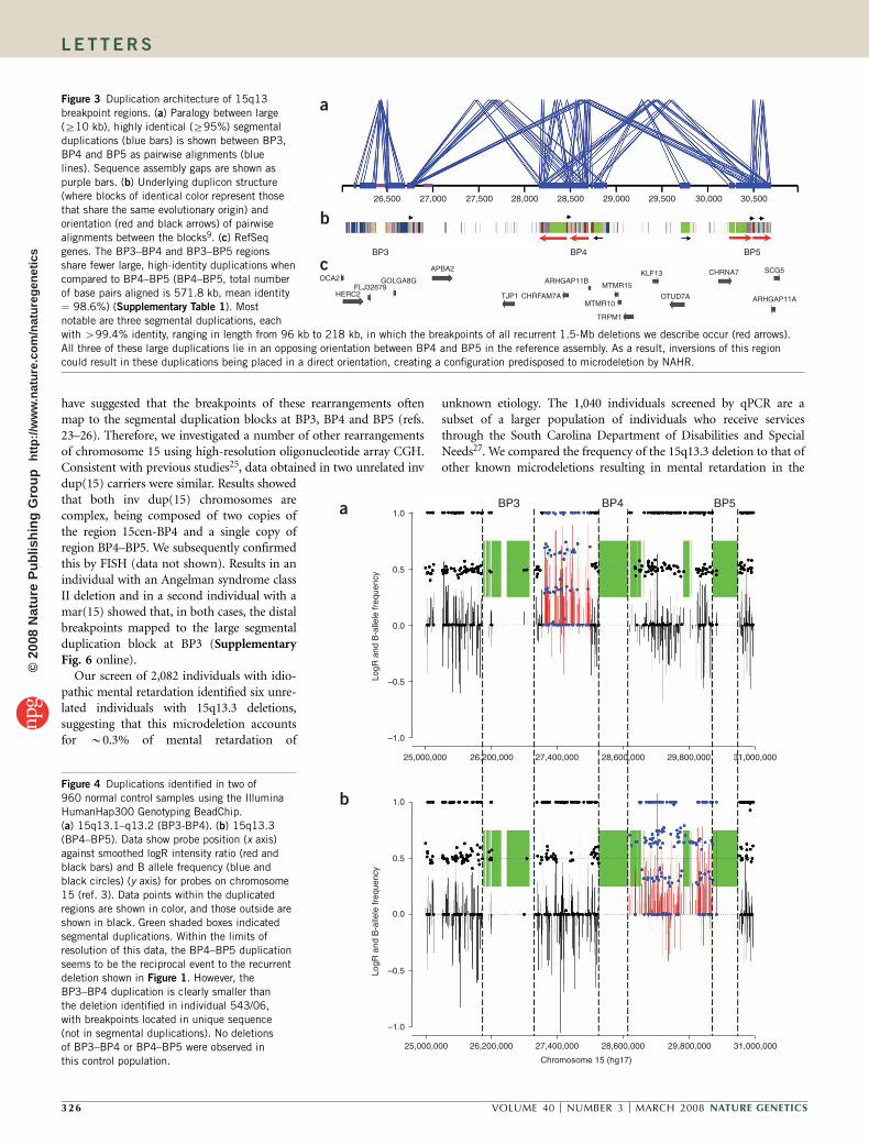
Figure 3 Duplication architecture of 15q13
breakpoint regions. (a) Paralogy between large
(Z10 kb), highly identical (Z95%) segmental
duplications (blue bars) is shown between BP3,
BP4 and BP5 as pairwise alignments (blue
lines). Sequence assembly gaps are shown as
purple bars. (b) Underlying duplicon structure
(where blocks of identical color represent those
that share the same evolutionary origin) and
orientation (red and black arrows) of pairwise
alignments between the blocks9. (c) RefSeq
genes. The BP3–BP4 and BP3–BP5 regions
share fewer large, high-identity duplications when
compared to BP4–BP5 (BP4–BP5, total number
of base pairs aligned is 571.8 kb, mean identity¼ 98.6%) (Supplementary Table 1). Most
notable are three segmental duplications, each
with 499.4% identity, ranging in length from 96 kb to 218 kb, in which the breakpoints of all recurrent 1.5-Mb deletions we describe occur (red arrows).
All three of these large duplications lie in an opposing orientation between BP4 and BP5 in the reference assembly. As a result, inversions of this region
could result in these duplications being placed in a direct orientation, creating a configuration predisposed to microdeletion by NAHR.
BP5BP4BP31.0
0.5
0.0
–0.5
–1.0
LogR
and
B-a
llele
freq
uenc
yLo
gR a
nd B
-alle
le fr
eque
ncy
25,000,000 26,200,000 27,400,000 28,600,000 29,800,000 31,000,000
31,000,00029,800,00028,600,00027,400,00026,200,00025,000,000
Chromosome 15 (hg17)
–1.0
–0.5
0.0
0.5
1.0
a
b
OCA2
BP3 BP4 BP5
HERC2FLJ32679
GOLGA8G
APBA2
TJP1 CHRFAM7A
ARHGAP11BMTMR15
MTMR10
TRPM1
KLF13
OTUD7A
CHRNA7 SCG5
30,50030,00029,50029,00028,50028,00027,50027,00026,500
ARHGAP11A
a
b
c
Figure 4 Duplications identified in two of
960 normal control samples using the Illumina
HumanHap300 Genotyping BeadChip.
(a) 15q13.1–q13.2 (BP3-BP4). (b) 15q13.3
(BP4–BP5). Data show probe position (x axis)
against smoothed logR intensity ratio (red and
black bars) and B allele frequency (blue and
black circles) (y axis) for probes on chromosome
15 (ref. 3). Data points within the duplicated
regions are shown in color, and those outside are
shown in black. Green shaded boxes indicated
segmental duplications. Within the limits of
resolution of this data, the BP4–BP5 duplication
seems to be the reciprocal event to the recurrentdeletion shown in Figure 1. However, the
BP3–BP4 duplication is clearly smaller than
the deletion identified in individual 543/06,
with breakpoints located in unique sequence
(not in segmental duplications). No deletions
of BP3–BP4 or BP4–BP5 were observed in
this control population.
3 26 VOLUME 40 [ NUMBER 3 [ MARCH 2008 NATURE GENETICS
LET TERS©
2008
Nat
ure
Pub
lishi
ng G
roup
ht
tp://
ww
w.n
atur
e.co
m/n
atur
egen
etic
s
larger cohort from which our population was obtained (10,997individuals of both European and African ancestry from SouthCarolina). Within the larger cohort, Prader-Willi syndrome wasdiagnosed in 0.22%, Angelman syndrome in 0.34% and Williamssyndrome in 0.31%27. In comparison, we identified 15q13.3 deletionsin 0.29% of the subgroup (three unrelated probands out of 1,040individuals tested) and 0.29% of the European populations tested byarray CGH (three unrelated probands out of 1,042 individuals tested),suggesting the frequency of this 15q13.3 microdeletion syndrome maybe comparable to that of the disorders listed above (estimatedfrequency in individuals with mental retardation: 0.29%; 95% c.i.:0.06–0.52%). Two pedigrees were of African American descent, andthe remainder were of European origin, indicating that this syndromeis found in individuals of different ethnic backgrounds. Given that thismicrodeletion is well below the resolution of conventional cytoge-netics—we detected it only when using techniques such as arrayCGH—we anticipate that the increasing resolution of these studieswill lead to the identification of additional microdeletion syndromes.Assuming a prevalence of moderate mental retardation in the generalpopulation of B0.8% (ref. 28), we estimate an approximate popula-tion incidence for this 15q13.3 microdeletion of 1/40,000. Given thisrelatively high frequency and the multiethnic distribution, we recom-mend testing for this disorder in individuals with features similar tothose presented here.
METHODSDNA samples. DNA samples were obtained from the following cohorts after
obtaining informed consent and approval by the appropriate institutional
review boards: (i) children and young adults from a variety of UK clinical
genetics centers, community learning disability teams and other sources
(including hospital neuropediatricians) in whom common causes of mental
retardation, including karyotype and subtelomeric abnormalities and Fragile X,
had been previously excluded (n¼ 394)3; (ii) karyotypically normal individuals
presenting with mental retardation and/or dysmorphism collected by the
University of Pavia (n ¼ 510); (iii) individuals admitted to the IRCCS
Associazione Oasi Maria Santissima (an Institute for Research and Care in
Mental Retardation and Brain Aging) screened for mental retardation accord-
ing to the DSM-IV-TR criteria, in whom the common causes of mental
retardation were excluded (n ¼ 138, all Caucasian, 75 female, 63 male); and
(iv) individuals who received services through the South Carolina Department
of Disabilities and Special Needs and for whom common causes of mental
retardation had been excluded by fragile X testing, chromosome analysis,
amino acid and organic analyses and urinary metabolic screening
(n ¼ 1,040, 540 female, 500 male, 562 African American, 448 European
American, 30 unstated or other ethnicity; all females and all but 10 of the
males had IQ scores o70)29. Two control groups were used to assess the extent
of normal copy number variation. The first control population consisted of 316
unrelated individuals who had been tested using the same BAC duplication
microarray13,15. A second control population, comprising 960 unrelated
European American adults (age 40–70 years), was genotyped using the
HumanHap300 Genotyping BeadChips (Illumina), which comprise
B317,000 HapMap SNPs spread throughout the genome. Each individual
was enrolled in the Pharmacogenomics and Risk of Cardiovascular Disease
(PARC) study, which aims to identify genetic contributors to the variable
efficacy of statin drugs on cardiovascular disease risk (see URLs section below).
Hybridizations, data analysis and copy number analysis, with particular
reference to chromosome 15q13 (155 probes between BP3–BP4, 167 probes
between BP4–BP5), were performed according to published protocols11.
Array CGH. DNA from affected individuals from the UK (n ¼ 394) were
hybridized to a custom BAC array consisting of 2,007 clones targeted to regions
of the genome flanked by segmental duplications, as described previously15.
This array includes all regions associated with known genomic disorders and an
additional 105 regions with similar genomic architecture. Because of the
targeted nature of this array, it has reduced power to detect rearrangements
not mediated by segmental duplications15. Regions were scored as copy number
variant if the log2 ratio of two or more consecutive clones each exceeded twice
the s.d. of the autosomal clones in dye-swap replicate experiments7. Affected
individuals from Italy (n ¼ 648) were molecularly karyotyped using Agilent
244A Human Genome CGH Microarrays. Parents of probands showing cryptic
deletions were also analyzed to exclude inherited imbalances. Rearrangements
of 15q were analyzed using a custom oligonucleotide array (NimbleGen
Systems) consisting of 166,000 isothermal probes (length, 45–75 bp; mean
probe density, 1 probe/130 bp) covering a number of chromosomal regions,
including 42,698 probes covering a 6-Mb region of chromosome 15q13
(chr15:25500000–31500000). Hybridizations were performed as described
previously3 and used a single normal male as a reference (GM15724,
Coriell). Microsatellite analysis to determine parental origin of the
deletions used markers STS6_chr15, D15S1031 and D15S165 located within
the interval BP4–BP5.
TaqMan quantitative PCR. Affected individuals from the USA (n ¼ 1,040)
were assayed for copy number of the region BP4–BP5 using two TaqMan Gene
Copy Number Assays. Primers and probes were designed from genomic
sequence (hg18/Build 36) using Applied Biosystems proprietary software. Each
assay was run as a duplex TaqMan real-time PCR reaction, using a FAM dye–
based assay targeted to 15q13.3 and a VIC dye–based assay for the reference
gene, RNase P (PN 4316844 from Applied Biosystems). Each PCR assay was
performed in quadruplicate and comprised 10 ng gDNA and 1� TaqMan
probe/primer mix in 1� TaqMan Universal Master Mix in a 10-ml reaction
amplified using an Applied Biosystems 7900HT SDS instrument. Cycling
conditions were 2 min at 50 1C and 10 min at 95 1C, followed by 40 cycles
of 15 s at 92 1C and 60 s at 60 1C. Real-time data were collected by the SDS 2.3
software. The method involves relative quantification of the test sequence
Figure 5 Identification of a common inversion
polymorphism in 15q13.3. To detect inversions
of the BP4–BP5 microdeletion region, we
performed FISH mapping using fosmid probes
located proximal and distal to BP5. The
separation of these probes in the reference
assembly is B2.5 Mb, enough to visualize these
as two separate signals on metaphase
chromosomes. Inversion of the region BP4–BP5
moves the two probes within close proximity to
each other, visualized as overlapping yellow
signals. Using this assay on eight HapMap
individuals of different ethnicities, we observed
the inversion on 7/16 chromosomes (B44% of
the population; Supplementary Table 2). We alsotested the mother of individual 69/06 (in whose germline the BP4–BP5 deletion arose), who was found to be heterozygous. (a) Individuals homozygous for
the reference allele. (b) Individuals heterozygous for the inversion allele. (c) Individuals homozygous for the inversion allele.
WIBR2-2422k14WIBR2-3205j20 ~0.9 Mb
WIBR2-2422k14~2.5 Mb
BP5BP4WIBR2-3205j20
Referenceallele
Inversionallele
a b c
NATURE GENETICS VOLUME 40 [ NUMBER 3 [ MARCH 2008 32 7
LET TERS©
2008
Nat
ure
Pub
lishi
ng G
roup
ht
tp://
ww
w.n
atur
e.co
m/n
atur
egen
etic
s

versus a reference gene known to have two copies per diploid genome. Relative
quantity is determined by the DDCt ((FAM Ct – VIC Ct)sample – (FAM
Ct – VIC Ct)calibrator) method, where a reference sample or calibrator known to
have two copies of the test sequence is used as the basis for comparative results.
The gene copy number is two times the relative quantity30. The two regions
assayed were (i) chr15:29000001–29000077 and (ii) chr15:29239994–29240092.
FISH inversion assay. Metaphase spreads were obtained from lymphoblast cell
lines from eight HapMap individuals (Coriell Cell Repository) and from
cultured peripheral blood lymphocytes from the mother of individual 69/06.
FISH was performed using fosmid WIBR2-3205j20 (chr15:29009842–
29052657) directly labeled by nick translation with Cy3-dUTP (Perkin-Elmer)
and WIBR2-2422k14 (chr15:31531310–31570294) labeled with fluorescein-
dUTP (Enzo). Each hybridization used 300 ng of labeled probe, 5 mg COT1
DNA (Roche) and 3 mg sonicated salmon sperm DNA at 37 1C in 10 ml
2� SSC/50% formamide/10% dextran sulfate, followed by three post-
hybridization washes at 60 1C in 0.1� SSC. Nuclei were stained with 4,6-
diamidino-2-phenylindole (DAPI) and digital images obtained using a Leica
DMRXA2 epifluorescence microscope equipped with a cooled CCD camera
(Princeton Instruments).
Accession number. Microarray data have been deposited in the Gene Expres-
sion Omnibus database under accession number GSE10189.
URLs. University of California Santa Cruz genome browser, http://genome.
ucsc.edu/; Genomic Imprinting Website, http://www.geneimprint.com/site/
genes-by-species; Sanger DECIPER, http://decipher.sanger.ac.uk/; PARC study,
http://www.pharmgkb.org/network/members/parc.jsp#team.
Note: Supplementary information is available on the Nature Genetics website.
ACKNOWLEDGMENTSWe are grateful to R. Krauss and the PARC project for the use and analysis ofIllumina SNP genotyping data, funded by US National Institutes of Health (NIH)grant U01 HL069757. This work was supported in part by grants from the NIH(HD043569, E.E.E.), the South Carolina Department of Disabilities and SpecialNeeds (C.S., R.E.S., R.J.S. and C.E.S.), Oxford Genetics Knowledge Park and theOxford National Institute for Health Research (NIHR) Biomedical ResearchCentre (R.R. and S.J.L.K.), Fondazione Mariani, CARIPLO and PRIN 2005(O.Z.), and the Italian Ministry of Health (C.R., P.F., L.C. and M.F.). EEE isan Investigator of the Howard Hughes Medical Institute.
AUTHOR CONTRIBUTIONSA.J.S., H.C.M. and E.E.E. contributed to the writing of this paper. The study wascoordinated by A.J.S., H.C.M., S.J.L.K., C.R., O.Z., C.C., C.E.S. and E.E.E.;experimental work was done by A.J.S., H.C.M., K.L., C. Baker, F.N., M.D.G., R.C.,A.B., G.G., R.R., M.F., L.C., P.F. and M.V.; clinical work was done by R.E.S.,R.J.S., B.D.B., C.T., R.G., V.M., S.M., C.S. and C.R.; computational analysis wasperformed by Y.W., C.X., C. Barbacioru, Z.J., I.C. and G.M.C.
COMPETING INTERESTS STATEMENTThe authors declare competing financial interests: details accompany the full-textHTML version of the paper at http://www.nature.com/naturegenetics/.
Published online at http://www.nature.com/naturegenetics
Reprints and permissions information is available online at http://npg.nature.com/
reprintsandpermissions
1. Stankiewicz, P. & Lupski, J.R. Genome architecture, rearrangements and genomicdisorders. Trends Genet. 18, 74–82 (2002).
2. de Vries, B.B. et al. Diagnostic genome profiling in mental retardation. Am. J. Hum.Genet. 77, 606–616 (2005).
3. Sharp, A.J. et al. Discovery of previously unidentified genomic disorders from theduplication architecture of the human genome. Nat. Genet. 38, 1038–1042(2006).
4. Jiang, Z. et al. Ancestral reconstruction of segmental duplications reveals punctuatedcores of human genome evolution. Nat. Genet. 39, 1361–1368 (2007).
5. Zody, M.C. et al. Analysis of the DNA sequence and duplication history of humanchromosome 15. Nature 440, 671–675 (2006).
6. Gimelli, G. et al. Genomic inversions of human chromosome 15q11-q13 in mothers ofAngelman syndrome patients with class II (BP2/3) deletions. Hum. Mol. Genet. 12,849–858 (2003).
7. Kurotaki, N., Stankiewicz, P., Wakui, K., Niikawa, N. & Lupski, J.R. Sotos syndromecommon deletion is mediated by directly oriented subunits within inverted Sos-REPlow-copy repeats. Hum. Mol. Genet. 14, 535–542 (2005).
8. Lupski, J.R. Genome structural variation and sporadic disease traits. Nat. Genet. 38,974–976 (2006).
9. Osborne, L.R. et al. A 1.5 million-base pair inversion polymorphism in families withWilliams-Beuren syndrome. Nat. Genet. 29, 321–325 (2001).
10. Sharp, A.J., Cheng, Z. & Eichler, E.E. Structural variation of the human genome. Annu.Rev. Genomics Hum. Genet. 7, 407–442 (2006).
11. Peiffer, D.A. et al. High-resolution genomic profiling of chromosomal aberrations usingInfinium whole-genome genotyping. Genome Res. 16, 1136–1148 (2006).
12. Iafrate, A.J. et al. Detection of large-scale variation in the human genome. Nat. Genet.36, 949–951 (2004).
13. Locke, D.P. et al. Linkage disequilibrium and heritability of copy-number polymorph-isms within duplicated regions of the human genome. Am. J. Hum. Genet. 79,275–290 (2006).
14. Sebat, J. et al. Large-scale copy number polymorphism in the human genome. Science305, 525–528 (2004).
15. Sharp, A.J. et al. Segmental duplications and copy-number variation in the humangenome. Am. J. Hum. Genet. 77, 78–88 (2005).
16. Simon-Sanchez, J. et al. Genome-wide SNP assay reveals structural genomic variation,extended homozygosity and cell-line induced alterations in normal individuals. Hum.Mol. Genet. 16, 1–14 (2007).
17. Wong, K.K. et al. A comprehensive analysis of common copy-number variations in thehuman genome. Am. J. Hum. Genet. 80, 91–104 (2007).
18. de Smith, A.J. et al. Array CGH analysis of copy number variation identifies 1284 newgenes variant in healthy white males: implications for association studies of complexdiseases. Hum. Mol. Genet. 16, 2783–2794 (2007).
19. Zogopoulos, G. et al. Germ-line DNA copy number variation frequencies in a largeNorth American population. Hum. Genet. 122, 345–353 (2007).
20. Elmslie, F.V. et al. Genetic mapping of a major susceptibility locus for juvenilemyoclonic epilepsy on chromosome 15q. Hum. Mol. Genet. 6, 1329–1334(1997).
21. Neubauer, B.A. et al. Centrotemporal spikes in families with rolandic epilepsy: linkageto chromosome 15q14. Neurology 51, 1608–1612 (1998).
22. Orr-Urtreger, A. et al. Mice deficient in the alpha7 neuronal nicotinic acetylcholinereceptor lack alpha-bungarotoxin binding sites and hippocampal fast nicotinic cur-rents. J. Neurosci. 17, 9165–9171 (1997).
23. Sahoo, T. et al. Identification of novel deletions of 15q11q13 in Angelman syndromeby array-CGH: molecular characterization and genotype-phenotype correlations. Eur. J.Hum. Genet. 15, 943–949 (2007).
24. Sahoo, T. et al. Array-based comparative genomic hybridization analysis ofrecurrent chromosome 15q rearrangements. Am. J. Med. Genet. A. 139, 106–113(2005).
25. Wang, N.J., Liu, D., Parokonny, A.S. & Schanen, N.C. High-resolution molecularcharacterization of 15q11-q13 rearrangements by array comparative genomic hybri-dization (array CGH) with detection of gene dosage. Am. J. Hum. Genet. 75, 267–281(2004).
26. Erdogan, F. et al. Characterization of a 5.3 Mb deletion in 15q14 by comparativegenomic hybridization using a whole genome ‘‘tiling path’’ BAC array in a girl with heartdefect, cleft palate, and developmental delay. Am. J. Med. Genet. A. 143, 172–178(2007).
27. Stevenson, R.E., Procopio-Allen, A.M., Schroer, R.J. & Collins, J.S. Genetic syndromesamong individuals with mental retardation. Am. J. Med. Genet. A. 123, 29–32(2003).
28. Larson, S.A. et al. Prevalence of mental retardation and developmental disabilities:estimates from the 1994/1995 National Health Interview Survey Disability Supple-ments. Am. J. Ment. Retard. 106, 231–252 (2001).
29. Stevenson, R.E. & Schroer, R.J. Mental retardation in South Carolina: Characteristicsof the study population. in Proc. Greenwood Genetic Center (Greenwood GeneticCenter, Greenwood, South Carolina, 1996).
30. Livak, K.J. & Schmittgen, T.D. Analysis of relative gene expression data using real-timequantitative PCR and the DDCt method. Methods 25, 402–408 (2001).
3 28 VOLUME 40 [ NUMBER 3 [ MARCH 2008 NATURE GENETICS
LET TERS©
2008
Nat
ure
Pub
lishi
ng G
roup
ht
tp://
ww
w.n
atur
e.co
m/n
atur
egen
etic
s