A recurrent 16p12.1 microdeletion supports a two-hit model for severe developmental delay
8
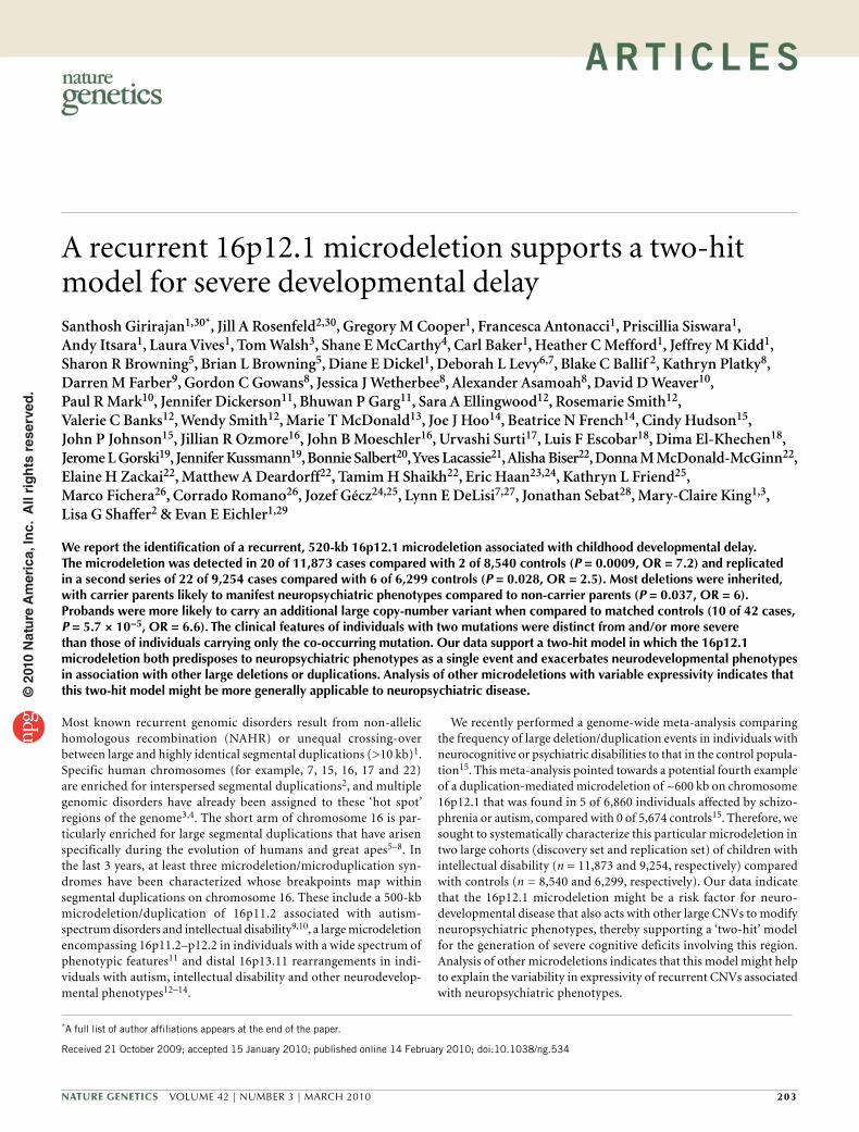
NATURE GENETICS VOLUME 42 | NUMBER 3 | MARCH 2010 203 ARTICLES Most known recurrent genomic disorders result from non-allelic homologous recombination (NAHR) or unequal crossing-over between large and highly identical segmental duplications (>10 kb) 1 . Specific human chromosomes (for example, 7, 15, 16, 17 and 22) are enriched for interspersed segmental duplications 2 , and multiple genomic disorders have already been assigned to these ‘hot spot’ regions of the genome 3,4 . The short arm of chromosome 16 is par- ticularly enriched for large segmental duplications that have arisen specifically during the evolution of humans and great apes 5–8 . In the last 3 years, at least three microdeletion/microduplication syn- dromes have been characterized whose breakpoints map within segmental duplications on chromosome 16. These include a 500-kb microdeletion/duplication of 16p11.2 associated with autism- spectrum disorders and intellectual disability 9,10 , a large microdeletion encompassing 16p11.2–p12.2 in individuals with a wide spectrum of phenotypic features 11 and distal 16p13.11 rearrangements in indi- viduals with autism, intellectual disability and other neurodevelop- mental phenotypes 12–14 . We recently performed a genome-wide meta-analysis comparing the frequency of large deletion/duplication events in individuals with neurocognitive or psychiatric disabilities to that in the control popula- tion 15 . This meta-analysis pointed towards a potential fourth example of a duplication-mediated microdeletion of ~600 kb on chromosome 16p12.1 that was found in 5 of 6,860 individuals affected by schizo- phrenia or autism, compared with 0 of 5,674 controls 15 . Therefore, we sought to systematically characterize this particular microdeletion in two large cohorts (discovery set and replication set) of children with intellectual disability (n = 11,873 and 9,254, respectively) compared with controls (n = 8,540 and 6,299, respectively). Our data indicate that the 16p12.1 microdeletion might be a risk factor for neuro- developmental disease that also acts with other large CNVs to modify neuropsychiatric phenotypes, thereby supporting a ‘two-hit’ model for the generation of severe cognitive deficits involving this region. Analysis of other microdeletions indicates that this model might help to explain the variability in expressivity of recurrent CNVs associated with neuropsychiatric phenotypes. A recurrent 16p12.1 microdeletion supports a two-hit model for severe developmental delay Santhosh Girirajan 1,30* , Jill A Rosenfeld 2,30 , Gregory M Cooper 1 , Francesca Antonacci 1 , Priscillia Siswara 1 , Andy Itsara 1 , Laura Vives 1 , Tom Walsh 3 , Shane E McCarthy 4 , Carl Baker 1 , Heather C Mefford 1 , Jeffrey M Kidd 1 , Sharon R Browning 5 , Brian L Browning 5 , Diane E Dickel 1 , Deborah L Levy 6,7 , Blake C Ballif 2 , Kathryn Platky 8 , Darren M Farber 9 , Gordon C Gowans 8 , Jessica J Wetherbee 8 , Alexander Asamoah 8 , David D Weaver 10 , Paul R Mark 10 , Jennifer Dickerson 11 , Bhuwan P Garg 11 , Sara A Ellingwood 12 , Rosemarie Smith 12 , Valerie C Banks 12 , Wendy Smith 12 , Marie T McDonald 13 , Joe J Hoo 14 , Beatrice N French 14 , Cindy Hudson 15 , John P Johnson 15 , Jillian R Ozmore 16 , John B Moeschler 16 , Urvashi Surti 17 , Luis F Escobar 18 , Dima El-Khechen 18 , Jerome L Gorski 19 , Jennifer Kussmann 19 , Bonnie Salbert 20 , Yves Lacassie 21 , Alisha Biser 22 , Donna M McDonald-McGinn 22 , Elaine H Zackai 22 , Matthew A Deardorff 22 , Tamim H Shaikh 22 , Eric Haan 23,24 , Kathryn L Friend 25 , Marco Fichera 26 , Corrado Romano 26 , Jozef Gécz 24,25 , Lynn E DeLisi 7,27 , Jonathan Sebat 28 , Mary-Claire King 1,3 , Lisa G Shaffer 2 & Evan E Eichler 1,29 We report the identification of a recurrent, 520-kb 16p12.1 microdeletion associated with childhood developmental delay. The microdeletion was detected in 20 of 11,873 cases compared with 2 of 8,540 controls (P = 0.0009, OR = 7.2) and replicated in a second series of 22 of 9,254 cases compared with 6 of 6,299 controls (P = 0.028, OR = 2.5). Most deletions were inherited, with carrier parents likely to manifest neuropsychiatric phenotypes compared to non-carrier parents (P = 0.037, OR = 6). Probands were more likely to carry an additional large copy-number variant when compared to matched controls (10 of 42 cases, P = 5.7 × 10 −5 , OR = 6.6). The clinical features of individuals with two mutations were distinct from and/or more severe than those of individuals carrying only the co-occurring mutation. Our data support a two-hit model in which the 16p12.1 microdeletion both predisposes to neuropsychiatric phenotypes as a single event and exacerbates neurodevelopmental phenotypes in association with other large deletions or duplications. Analysis of other microdeletions with variable expressivity indicates that this two-hit model might be more generally applicable to neuropsychiatric disease. * A full list of author affiliations appears at the end of the paper. Received 21 October 2009; accepted 15 January 2010; published online 14 February 2010; doi:10.1038/ng.534 © 2010 Nature America, Inc. All rights reserved.
-
Upload
independent -
Category
Documents
-
view
3 -
download
0
Transcript of A recurrent 16p12.1 microdeletion supports a two-hit model for severe developmental delay
Nature GeNetics VOLUME 42 | NUMBER 3 | MARCH 2010 203
A rt i c l e s
Most known recurrent genomic disorders result from non-allelic homologous recombination (NAHR) or unequal crossing-over between large and highly identical segmental duplications (>10 kb)1. Specific human chromosomes (for example, 7, 15, 16, 17 and 22) are enriched for interspersed segmental duplications2, and multiple genomic disorders have already been assigned to these ‘hot spot’ regions of the genome3,4. The short arm of chromosome 16 is par-ticularly enriched for large segmental duplications that have arisen specifically during the evolution of humans and great apes5–8. In the last 3 years, at least three microdeletion/microduplication syn-dromes have been characterized whose breakpoints map within segmental duplications on chromosome 16. These include a 500-kb microdeletion/duplication of 16p11.2 associated with autism- spectrum disorders and intellectual disability9,10, a large microdeletion encompassing 16p11.2–p12.2 in individuals with a wide spectrum of phenotypic features11 and distal 16p13.11 rearrangements in indi-viduals with autism, intellectual disability and other neurodevelop-mental phenotypes12–14.
We recently performed a genome-wide meta-analysis comparing the frequency of large deletion/duplication events in individuals with neurocognitive or psychiatric disabilities to that in the control popula-tion15. This meta-analysis pointed towards a potential fourth example of a duplication-mediated microdeletion of ~600 kb on chromosome 16p12.1 that was found in 5 of 6,860 individuals affected by schizo-phrenia or autism, compared with 0 of 5,674 controls15. Therefore, we sought to systematically characterize this particular microdeletion in two large cohorts (discovery set and replication set) of children with intellectual disability (n = 11,873 and 9,254, respectively) compared with controls (n = 8,540 and 6,299, respectively). Our data indicate that the 16p12.1 microdeletion might be a risk factor for neuro-developmental disease that also acts with other large CNVs to modify neuropsychiatric phenotypes, thereby supporting a ‘two-hit’ model for the generation of severe cognitive deficits involving this region. Analysis of other microdeletions indicates that this model might help to explain the variability in expressivity of recurrent CNVs associated with neuropsychiatric phenotypes.
A recurrent 16p12.1 microdeletion supports a two-hit model for severe developmental delaySanthosh Girirajan1,30*, Jill A Rosenfeld2,30, Gregory M Cooper1, Francesca Antonacci1, Priscillia Siswara1, Andy Itsara1, Laura Vives1, Tom Walsh3, Shane E McCarthy4, Carl Baker1, Heather C Mefford1, Jeffrey M Kidd1, Sharon R Browning5, Brian L Browning5, Diane E Dickel1, Deborah L Levy6,7, Blake C Ballif 2, Kathryn Platky8, Darren M Farber9, Gordon C Gowans8, Jessica J Wetherbee8, Alexander Asamoah8, David D Weaver10, Paul R Mark10, Jennifer Dickerson11, Bhuwan P Garg11, Sara A Ellingwood12, Rosemarie Smith12, Valerie C Banks12, Wendy Smith12, Marie T McDonald13, Joe J Hoo14, Beatrice N French14, Cindy Hudson15, John P Johnson15, Jillian R Ozmore16, John B Moeschler16, Urvashi Surti17, Luis F Escobar18, Dima El-Khechen18, Jerome L Gorski19, Jennifer Kussmann19, Bonnie Salbert20, Yves Lacassie21, Alisha Biser22, Donna M McDonald-McGinn22, Elaine H Zackai22, Matthew A Deardorff22, Tamim H Shaikh22, Eric Haan23,24, Kathryn L Friend25, Marco Fichera26, Corrado Romano26, Jozef Gécz24,25, Lynn E DeLisi7,27, Jonathan Sebat28, Mary-Claire King1,3, Lisa G Shaffer2 & Evan E Eichler1,29
We report the identification of a recurrent, 520-kb 16p12.1 microdeletion associated with childhood developmental delay. The microdeletion was detected in 20 of 11,873 cases compared with 2 of 8,540 controls (P = 0.0009, OR = 7.2) and replicated in a second series of 22 of 9,254 cases compared with 6 of 6,299 controls (P = 0.028, OR = 2.5). Most deletions were inherited, with carrier parents likely to manifest neuropsychiatric phenotypes compared to non-carrier parents (P = 0.037, OR = 6). Probands were more likely to carry an additional large copy-number variant when compared to matched controls (10 of 42 cases, P = 5.7 × 10⁻⁵, OR = 6.6). The clinical features of individuals with two mutations were distinct from and/or more severe than those of individuals carrying only the co-occurring mutation. Our data support a two-hit model in which the 16p12.1 microdeletion both predisposes to neuropsychiatric phenotypes as a single event and exacerbates neurodevelopmental phenotypes in association with other large deletions or duplications. Analysis of other microdeletions with variable expressivity indicates that this two-hit model might be more generally applicable to neuropsychiatric disease.
*A full list of author affiliations appears at the end of the paper.
Received 21 October 2009; accepted 15 January 2010; published online 14 February 2010; doi:10.1038/ng.534
© 2
010
Nat
ure
Am
eric
a, In
c. A
ll ri
gh
ts r
eser
ved
.

204 VOLUME 42 | NUMBER 3 | MARCH 2010 Nature GeNetics
A rt i c l e s
RESULTSPathogenic association of 16p12.1 microdeletionFrom our initial discovery cohort, we identified 20 affected individuals (cases) with a 520.8-kb 16p12.1 microdeletion (UCSC Human Genome Version Hg18, NCBI Build 36, Chr. 16: 21854025–22374785) among 11,873 individuals with indications of intellectual disability/developmental delay (ID/DD) and congenital malformation (Fig. 1; see Online Methods). By contrast, CNV studies on 8,540 controls identified only two individuals, both from the GAIN cohort, with the 16p12.1 microdeletion (Table 1). Thus, 16p12.1 microdeletions are significantly enriched in the panel of children with develop-mental delay studied here (Fisher’s exact test, P = 0.0009, OR = 7.2). To replicate this asso-ciation, we evaluated CNV data from an inde-pendent set of 9,254 individuals with ID/DD and 6,299 controls (see Online Methods). The microdeletion was identified in 22 of 9,254 cases and 6 of 6,299 controls, confirming a sig-nificant enrichment of 16p12.1 microdeletion in the affected individuals (Fisher’s exact test, P = 0.028, OR = 2.5; Table 1 and Supplementary Fig. 1). For the combined set (21,127 cases
and 14,839 controls), the pathogenic associa-tion of the 16p12.1 microdeletion was highly significant (Fisher’s exact test, P = 1.18 × 10⁻⁴, OR = 3.7; Table 1).
We also examined 3,061 individuals who had been diagnosed with schizophrenia and identified three 16p12.1 microdeletions, all in individuals with sporadic schizophrenia (Table 1). However, we did not observe significant enrichment for the 16p12.1 event specifically in the schizophrenia cases compared to 14,839 total controls (Fisher’s exact test, P = 0.29, OR = 1.8), although this might indicate a lack of statistical power in the schizophrenia panel rather than a true lack of
disease association (only 3,061 individuals, in contrast to the 21,127 individuals with developmental delay). It is interesting, in this regard, that in one family where both schizophrenia and mental retardation phenotypes were segregating, the 16p12.1 deletion was observed only among individuals diagnosed with both psychosis and severe intel-lectual disability (see Supplementary Note, family LD1205).
Using high-density and targeted array-based comparative genomic hybridization (CGH) experiments (see Online Methods), we mapped the 16p12.1 microdeletion breakpoints in 37 individuals to two large blocks of segmental duplications (Fig. 1 and Supplementary Fig. 1).
OTOA UQCRC2OTOA
VWA3A
EEF2K
POLR3E
CDR2C15Orf65 LOC23117
LOC100132247
21.9 Mb 22.1 Mb21.6 Mb 22.3 Mb 22.5 Mb
Segmentalduplications
SG01
SG02
SG04
SG05
SG06
SG07
SG08
SG09
SG10
SG11
SG12
SG13
SGA3
SGA5
SGA6
SGA7
LD1205-03
GAIN_43163
GAIN_18125
GAIN_26140
Developmentaldelay
Schizophrenia
Controls
520 kbp
Segmentalduplications
Chr. 16 (p12.2-p12.1) 16p13.3 p13.2 p12.3 16p12.1 16p11.2 16q12.1 q22.1 22.3 24.113q11.2 12.2 16q21 q23.1 Figure 1 High-resolution array-based CGH characterization of 16p12.1 microdeletion. Shown is validation of 16p12.1 microdeletions in a representative set of cases using high-resolution tiling-path custom array-based CGH. Probes with log2 ratios above or below a threshold of 1.5 s.d. from the normalized mean log2 ratio are colored green (duplication) or red (deletion), respectively. Dotted lines represent breakpoint regions. Subjects SG01–13 and SGA3–7 have indications of developmental delay or cognitive disability, sample 43163 is from the GAIN schizophrenia study and subject LD1205-03 has schizophrenia and intellectual disability (from family LD1205). Note that samples 26140 and 18125 were analyzed as part of the GAIN control cohort for schizophrenia. It is noteworthy that one control (subject 26140) was retrospectively diagnosed with a major depressive disorder. Six RefSeq genes map within the 16p12.1 microdeletion. Four subjects (SG04, SG07, SG11 (affected with hypoplastic left heart syndrome) and LD1205-03 (diagnosed with schizophrenia)) were sequenced for CDR2, EEF2K and UQCRC2; no recessive mutations were identified.
table 1 Frequency of 16p12.1 microdeletion in cases and controlsCases Controls Significance
del 16p12.1 Total del 16p12.1 Total P value OR
ID/DD cohort
Discovery seta 20 11,873 2 8,540 0.0009 7.2
Replication setb 22 9,254 6 6,299 0.028 2.5
Combinedc 42 21,127 8 14,839 0.000118 3.7
Schizophrenia cohort 3 3,061 8 14,839 0.29 1.8aThe 16p12.1 microdeletion was originally identified from a meta-analysis that identified 5 cases from 6,860 individuals with autism, schizophrenia and developmental delay compared to 0 observations in a control group of 5,674 (P = 0.049). None of these cases were used in the discovery set, although the original control group was expanded in the discovery set. bFor the replication set, all controls and cases were independent. cCombined set composed of discovery set and replication set.
© 2
010
Nat
ure
Am
eric
a, In
c. A
ll ri
gh
ts r
eser
ved
.
Nature GeNetics VOLUME 42 | NUMBER 3 | MARCH 2010 205
A rt i c l e s
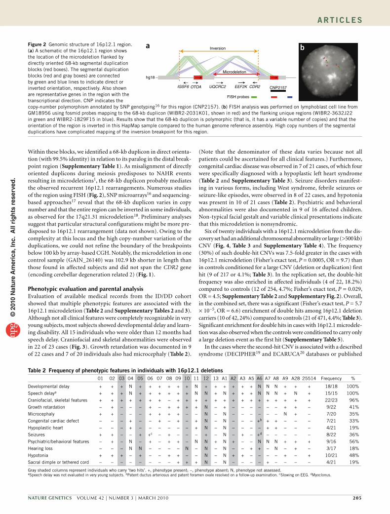
Within these blocks, we identified a 68-kb duplicon in direct orienta-tion (with 99.5% identity) in relation to its paralog in the distal break-point region (Supplementary Table 1). As misalignment of directly oriented duplicons during meiosis predisposes to NAHR events resulting in microdeletions1, the 68-kb duplicon probably mediates the observed recurrent 16p12.1 rearrangements. Numerous studies of the region using FISH (Fig. 2), SNP microarrays16 and sequencing- based approaches17 reveal that the 68-kb duplicon varies in copy number and that the entire region can be inverted in some individuals, as observed for the 17q21.31 microdeletion18. Preliminary analyses suggest that particular structural configurations might be more pre-disposed to 16p12.1 rearrangement (data not shown). Owing to the complexity at this locus and the high copy-number variation of the duplications, we could not refine the boundary of the breakpoints below 100 kb by array-based CGH. Notably, the microdeletion in one control sample (GAIN_26140) was 102.9 kb shorter in length than those found in affected subjects and did not span the CDR2 gene (encoding cerebellar degeneration related 2) (Fig. 1).
Phenotypic evaluation and parental analysisEvaluation of available medical records from the ID/DD cohort showed that multiple phenotypic features are associated with the 16p12.1 microdeletion (Table 2 and Supplementary Tables 2 and 3). Although not all clinical features were completely recognizable in very young subjects, most subjects showed developmental delay and learn-ing disability. All 15 individuals who were older than 12 months had speech delay. Craniofacial and skeletal abnormalities were observed in 22 of 23 cases (Fig. 3). Growth retardation was documented in 9 of 22 cases and 7 of 20 individuals also had microcephaly (Table 2).
(Note that the denominator of these data varies because not all patients could be ascertained for all clinical features.) Furthermore, congenital cardiac disease was observed in 7 of 21 cases, of which four were specifically diagnosed with a hypoplastic left heart syndrome (Table 2 and Supplementary Table 3). Seizure disorders manifest-ing in various forms, including West syndrome, febrile seizures or seizure-like episodes, were observed in 8 of 22 cases, and hypotonia was present in 10 of 21 cases (Table 2). Psychiatric and behavioral abnormalities were also documented in 9 of 16 affected children. Non-typical facial gestalt and variable clinical presentations indicate that this microdeletion is nonsyndromic.
Six of twenty individuals with a 16p12.1 microdeletion from the dis-covery set had an additional chromosomal abnormality or large (>500 kb) CNV (Fig. 4, Table 3 and Supplementary Table 4). The frequency (30%) of such double-hit CNVs was 7.5-fold greater in the cases with 16p12.1 microdeletion (Fisher’s exact test, P = 0.0005, OR = 9.7) than in controls conditioned for a large CNV (deletion or duplication) first hit (9 of 217 or 4.1%; Table 3). In the replication set, the double-hit frequency was also enriched in affected individuals (4 of 22, 18.2%) compared to controls (12 of 254, 4.7%; Fisher’s exact test, P = 0.029, OR = 4.5; Supplementary Table 2 and Supplementary Fig. 2). Overall, in the combined set, there was a significant (Fisher’s exact test, P = 5.7 × 10⁻⁵, OR = 6.6) enrichment of double hits among 16p12.1 deletion carriers (10 of 42, 24%) compared to controls (21 of 471, 4.4%; Table 3). Significant enrichment for double hits in cases with 16p12.1 microdele-tion was also observed when the controls were conditioned to carry only a large deletion event as the first hit (Supplementary Table 5).
In the cases where the second-hit CNV is associated with a described syndrome (DECIPHER19 and ECARUCA20 databases or published
hg18
a bInversion
Microdeletion
IGSF6 OTOA UQCRC2 EEF2K CDR2 CNP2157
FISH probes
Figure 2 Genomic structure of 16p12.1 region. (a) A schematic of the 16p12.1 region shows the location of the microdeletion flanked by directly oriented 68-kb segmental duplication blocks (red boxes). The segmental duplication blocks (red and gray boxes) are connected by green and blue lines to indicate direct or inverted orientation, respectively. Also shown are representative genes in the region with the transcriptional direction. CNP indicates the copy-number polymorphism annotated by SNP genotyping16 for this region (CNP2157). (b) FISH analysis was performed on lymphoblast cell line from GM18956 using fosmid probes mapping to the 68-kb duplicon (WIBR2-2031K01, shown in red) and the flanking unique regions (WIBR2-3632J22 in green and WIBR2-1829F15 in blue). Results show that the 68-kb duplicon is polymorphic (that is, it has a variable number of copies) and that the orientation of the region is inverted in this HapMap sample compared to the human genome reference assembly. High copy numbers of the segmental duplications have complicated mapping of the inversion breakpoint for this region.
table 2 Frequency of phenotypic features in individuals with 16p12.1 deletions01 02 03 04 05 06 07 08 09 10 11 12 13 A1 A2 A3 A5 A6 A7 A8 A9 A28 25514 Frequency %
Developmental delay + + + N + + + + + + N + + + + + + N N N + + + 18/18 100%
Speech delaya + + + N + + + + + + N N + N + + + N N N + N + 15/15 100%
Craniofacial, skeletal features + + + + + + + − + + + + + + + + + + + + + + + 22/23 96%
Growth retardation − + − − − + − + + + + N − + − − − − − − + + − 9/22 41%
Microcephaly + + − − − + + + + − − N − N − − − − − − N + − 7/20 35%
Congenital cardiac defect − − − + − − + − + − + N − N − − − +b + + − − − 7/21 33%
Hypoplastic heart − − − + − − − − − − + N − N − − − − + + − − − 4/21 19%
Seizures + + − − + +c − + − − − + − N − + − +d − − − − − 8/22 36%
Psychiatric/behavioral features − + − N − + − + + − N N + N + − − N N N + + + 9/16 56%
Hearing loss − − − N N − − − − N − N − N − − + + − N − + − 3/17 18%
Hypotonia + + + − + − − + + − − N − N + + − − − − + − + 10/21 48%
Sacral dimple or tethered cord − − − − − − − − + + + N − N − − − − + − − − − 4/21 19%
Gray shaded columns represent individuals who carry ‘two hits’. +, phenotype present; −, phenotype absent; N, phenotype not assessed.aSpeech delay was not evaluated in very young subjects. bPatent ductus arteriosus and patent foramen ovale resolved on a follow-up examination. cSlowing on EEG. dMyoclonus.
© 2
010
Nat
ure
Am
eric
a, In
c. A
ll ri
gh
ts r
eser
ved
.

206 VOLUME 42 | NUMBER 3 | MARCH 2010 Nature GeNetics
A rt i c l e s
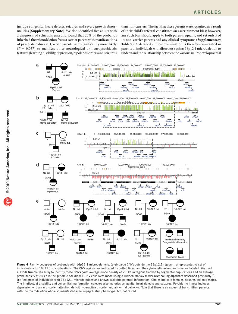
literature), two-hit 16p12.1 carriers manifested more severe or distinct phenotypes than the typical features of the syndrome (Supplementary Note). For example, patient SG03 carries a 16p12.1 microdeletion in addition to a 22q13 terminal deletion. The single del22q13ter event has been reported in Phelan-McDermid syndrome21 and is associated with autism-spectrum disorders22, developmental delay, hypotonia, normal to accelerated growth and dysmorphic features including macrocephaly, dolichocephaly and micrognathia. However, SG03 manifests with learning disability, microcephaly, exotropia and periventricular abnormalities, with no autistic features yet apparent (age 2 years 3 months). SG10 carries a 35-Mb deletion on chromo-some 5q15q23.2 along with the 16p12.1 microdeletion. Previously, Lindgren and colleagues reported on two individuals carrying a del5q15q23.2 with craniofacial features and benign adenomatous polyps in the intestine23. SG10 has developmental delay, significant growth retardation, overt craniofacial features (ptosis, hypertelorism, bifid uvula, exotropia, ectopic pupils, wide nasal bridge and smooth philtrum), hypoactivity and seizures—a more severe phenotype than that reported from a single second hit. Patient SGA2 carries a 1.3-Mb duplication on chromosome 2p13p12 in addition to the 16p12.1 microdeletion and has overt clinical features, including impaired intellectual function, behavior problems, growth anomalies, cranio-facial features and café-au-lait spots. Although dup2p13p12 has been reported to be associated with primary cutaneous lymphoma24, there have been no reports of developmental defect or craniofacial anoma-lies. Subject SGA6 carries an 848-kb duplication on chromosome 14q32.1 along with the 16p12.1 microdeletion. Additionally, he has had a mutation identified in the BRAF gene (F468S) that is consistent with a diagnosis of cardiofaciocutaneous syndrome (CFCS). The sub-ject presents with a diverse range of severe clinical features including craniofacial anomalies, complete agenesis of corpus callosum, renal and cardiac defects and Hirschsprung disease (Supplementary Table 3). These features are more severe than has been described for CFCS25 or a de novo dup14q32.1 case reported in association with schizophrenia26.
To test whether the subjects inherited the 16p12.1 microdeletion from their parents, we obtained DNA from 23 sets of parents. The 16p12.1 microdeletion was inherited in 22 of 23 cases (17 maternal, 5 paternal), with one case confirmed as being de novo (Fig. 4 and Supplementary Figs. 3 and 4). Of the seven double-hit cases where inheritance could be assessed, 6 of 7 large CNV second hits were inherited and one large CNV arose de novo. In most cases (5 of 7), either the large CNV second hit was inherited from the parent who did not carry a 16p12.1 microdeletion or it arose de novo. In one case (SG12), a heterozygous dup22q11.21 was transmitted from both parents to the homozygote proband (Fig. 4). SGA30 inherited both events from the mother (Supplementary Table 6).
Parental phenotypic information was obtained during the initial visit before CNV testing from 11 of 13 inherited cases. In two cases, clinical inquiries were made only after the child was found to carry the microdeletion (Supplementary Table 7). Carrier parents with the 16p12.1 microdeletion were more likely (P = 0.037, OR = 6, Fisher’s exact test) to present with learning disability, depression or bipo-lar disorders, or seizures than the non-carrier parents (Fig. 4 and Supplementary Table 8). We found only three cases (SG11, SGA9 and 25514) in whom the microdeletion was inherited from an apparently normal parent (Supplementary Table 9). Compared to the carrier parents, the index cases manifested with more severe, clinically recog-nizable manifestations, including severe speech and motor disability, recognizable facial dysmorphology and systemic organ abnormalities (Table 2, Figs. 3 and 4 and Supplementary Note). Finally, we assessed the SNP genotypes for chromosome 16p in 17 probands and 4 avail-able parents to investigate whether there is one specific sequence haplotype that predisposes to this microdeletion. We found that the microdeletion occurred on different haplotype backgrounds, con-sistent with a recurrent nature for this rearrangement as opposed to inheritance from a common ancestor (Supplementary Note).
DISCUSSIONPreviously, we undertook a large meta-analysis study of CNVs in individuals diagnosed with intellectual disability, autism and schizo-phrenia, identifying the 16p12.1 region as a potential pathogenic locus (five cases and no controls)15. However, this study lacked the power to definitively identify a disease association or to delineate the phenotypic consequences of the microdeletion. Here we used the CNV data from one of the largest collections of individuals with intellectual disability and developmental delay to reassess this region. We identified 42 index cases with developmental delay, craniofacial dysmorphology and congenital heart defects and three sporadic and one familial case of overt schizophrenia carrying a similar-sized micro-deletion of 16p12.1; we found only eight control individuals carrying a 16p12.1 deletion. Although all cases appear to share the segmental duplication-mediated 520-kb minimal deletion region, it is noteworthy that one deletion-carrying control was retrospectively diagnosed with a major depressive disorder and also had an atypical, smaller deletion (417 kb). Removal of this control sample from our analysis strengthens the association between 16p12.1 microdeletion and disease (Fisher’s exact test, P = 4.4 × 10⁻⁵, OR = 4.2). Comparison to other genomic disorders in our cohort indicates that the incidence of 16p12.1 micro-deletion is ~1/15,000 live births, similar to that of Smith-Magenis syndrome (Table 4). However, the subtle clinical features and variable phenotypes associated with the single 16p12.1 event probably lead to underascertainment or misdiagnoses.
Most of our pediatric cases had indications of developmental delay and learning disability and congenital abnormalities. However, the variable phenotypes associated with the 16p12.1 microdeletion
a c
SG07, 1 y 10 m
b
eSGA3, 2 y 6 m
SG10, 2 y
d
SG04, 1 y 3 m
SGA5, 2 y
f
25514, 5 y
Figure 3 Representative photographs of individuals with 16p12.1 microdeletion. (a–f) Facial features of patient SG07 at 22 months (a), patient SGA3 at 2.5 years (b), patient SGA5 at 2 years (c), patient SG04 at 15 months (d), patient SG10 at 2 years (e) and patient 25514 at 5 years (f). Specific consents were obtained to publish these photographs.
© 2
010
Nat
ure
Am
eric
a, In
c. A
ll ri
gh
ts r
eser
ved
.
Nature GeNetics VOLUME 42 | NUMBER 3 | MARCH 2010 207
A rt i c l e s
include congenital heart defects, seizures and severe growth abnor-malities (Supplementary Note). We also identified five adults with a diagnosis of schizophrenia and found that 23% of the probands inherited the microdeletion from a carrier parent with manifestations of psychiatric disease. Carrier parents were significantly more likely (P = 0.037) to manifest other neurological or neuropsychiatric features (learning disability, depression, bipolar disorders and seizures)
than non-carriers. The fact that these parents were recruited as a result of their child’s referral constitutes an ascertainment bias; however, any such bias should apply to both parents equally, and yet only 5 of 14 non-carrier parents had any clinical symptoms (Supplementary Table 9). A detailed clinical examination is therefore warranted in parents of individuals with disorders such as 16p12.1 microdeletion to understand the relationship between the various neurodevelopmental
16p12.1 del22q11 dup
22q11 dup
16p12.1 delHomo dup 22q11
No delHomo dup22q11
SG12
2.52 Mb
848 kb
16p12.1 del
16p12.1 del5q deletion
No del
SG10
35 Mb
16p12.1 delNo dup
16p12.1 del15q dup
NT
SG05
5.6 Mb
16p12.1 del
16p12.1 del
No del
SG11
No del
16p12.1 del
16p12.1 del
SGA516p12.1 del
16p12.1 del
No delSGA3
NT
16p12.1 del22q13ter del
16p12.1 del
SG03
16p12.1 del
16p12.1 del No del
No del
SG07
No del
16p12.1 del
No del
SG04
No del
16p12.1 del
16p12.1 del
SG08
16p12.1 del
16p12.1 del
NT
SG09
No del
16p12.1 del
16p12.1 del
SG06
Psychiatric illness
Intellectual disabilityCongenital malformation
a
b
c
d
e
NT No del14q32 dup
SGA6
16p12.1 del14q32 dup
Chr. 15:Segmental dups
21,000,000 22,000,000 23,000,000 24,000,000 25,000,000 26,000,000 27,000,000
15q11.2 15q12 15q13.1
MKRN3
MAGEL2 SNRPN
SNURF
UBE3A
ATP10A
GABRB3
GABRA5
GABRG3
OCA2
HERC2
APBA2
KIAA0574
NDNL2
0 -1 -
0 -
Chr. 22:
Segmental dups
17,000,000 17,500,000 18,000,000 18,500,000 19,000,000 19,500,000 20,000,000 20,500,000
22q11.21 q11.22
USP18
PRODH
SLC25A1
CLTCL1
HIRA
UFD1L
SEPT5
TBX1 TXNRD2
COMT
ZDHHC8
RTN4R
RIMBP3
SCARF2
KLHL22
MED15
PI4KA
SERPIND1
SNAP29 HIC2
PPIL2
YPEL1
MAPK1
PPM1F
TOP3B
0 -
1 -
0 -
Chr. 14: 95,000,000 95,500,000 96,000,000 96,500,000 97,000,000 97,500,000
14q32.13 14q32.2
DICER1
CLMN
TCL6
TCL1B
TCL1A BDKRB2
BDKRB1 ATG2B
AK7
PAPOLA
VRK1
0 -
1 -
0 -
Chr. 5:Segmental dups
100,000,000 110,000,000 120,000,000 130,000,000
5q14.3 5q15 5q21.1 21.2 5q21.3 22.1 q22.3 5q23.1 5q23.2 5q23.3 5q31.1 5q31.2
NR2F1 CAST
CHD1 ST8SIA4
PAM
GIN1
EFNA5
FER
PJA2
APC
MCC
KCNN2 DTWD2
HSD17B4
FTMT
SRFBP1
ZNF608
ALDH7A1
FBN2
LYRM7
TCF7
0 -
1 -
0 -
Figure 4 Family pedigrees of probands with 16p12.1 microdeletions. (a–d) Large CNVs outside the 16p12.1 region in a representative set of individuals with 16p12.1 microdeletions. The CNV regions are indicated by dotted lines, and the cytogenetic extent and size are labeled. We used a 135K NimbleGen array to identify these CNVs (with average probe density of 2.5 kb in regions flanked by segmental duplications and an average probe density of 35 kb in the genomic backbone). CNV calls were made using a Hidden Markov Model CNV-calling algorithm described previously15. (e) Pedigrees of individuals with 16p12.1 microdeletions and known available parental information. Circles indicate females; squares indicate males. The intellectual disability and congenital malformation category also includes congenital heart defects and seizures. Psychiatric illness includes depression or bipolar disorder, attention deficit hyperactive disorder and abnormal behavior. Note that there is an excess of transmitting parents with the microdeletion who also manifested a neuropsychiatric phenotype. NT, not tested.
© 2
010
Nat
ure
Am
eric
a, In
c. A
ll ri
gh
ts r
eser
ved
.

208 VOLUME 42 | NUMBER 3 | MARCH 2010 Nature GeNetics
A rt i c l e s
and cognitive phenotypes. In probands carrying the microdeletion, we observed a six-fold excess of double CNV hits (10 of 42, 24%) as com-pared to normal controls when conditioned for the presence of at least one large CNV (21 of 471, 4%) and a more than 60-fold excess as com-pared to the general population. Compared to the classical phenotypes associated with the known pathogenic locus, each of these children showed additional or more severe phenotypes (Supplementary Note). Collectively, these data indicate that phenotypes associated with the 16p12.1 microdeletion show variable expressivity that depends on the genetic background.
We conclude that the deletion of 16p12.1 is a significant, inde-pendent risk factor for intellectual disability and developmental delay that also acts with other factors to modify neurological phe-notypes. We propose a ‘two-hit’ model, wherein a secondary insult is necessary during development to result in a more severe clinical manifestation of pediatric disease. The second hit could potentially be another CNV, a disruptive single-base-pair mutation in a pheno-typically related gene or an environmental event that influences the phenotype. It has been noted that genetic factors that are originally identified to associate with a specific neurological disease are often subsequently identified in individuals with other conditions (for example, del15q13.3 is associated with epilepsy, schizophrenia and intellectual disability)27–29.
The prevalence of schizophrenia in individuals with a learning dis-ability is reported to be three times that of the general population, and several studies indicate that the comorbidity of schizophrenia in learning-disabled individuals is mainly due to a greater tendency for schizophrenic individuals to develop cognitive delay30–32. The observation, in family LD1205, that cognitive impairment is seen only
among siblings with the 16p12.1 deletion is also consistent with the hypothesis that the 16p12.1 deletion exacerbates the phenotypic con-sequences of other heritable neurological disease risk factors within this multiplex family (Supplementary Note). Notably, about 70% of individuals with autism also present with a learning disability33. Our ‘two-hit’ model might also help to explain the significant comorbidity that exists among cognitive impairment, schizophrenia and autism34 and the underlying phenotypic variability reported for several recur-rent microdeletions29,35.
To test this, we investigated the occurrence of two hits more broadly among other genomic disorders. We first reanalyzed the recently reported 1q21.1 microdeletion for the presence of a second large CNV36. We found a 40-fold enrichment for two hits (4 of 25, 16%) among cases with 1q21.1 microdeletion when compared to controls. The phenotypes of these four cases were variable, ranging from severe neurological deficit and craniofacial abnormalities to severe schizo-phrenia without cognitive impairment (Supplementary Note). On the basis of this finding, we expanded our analysis to consider both syndromic and non-syndromic genomic disorders for the frequency of double hits. Our analysis for two hits in nine genomic disorders shows that 16p12.1 microdeletion ranks as the top recurrent CNV that is enriched for the occurrence of two hits (Table 4). Notably, the propor-tion of cases with a second hit is generally much higher (9–24% of the cases) for recurrent microdeletions or microduplications where vari-able expressivity has been reported, including del15q13.3, del16p11.2, dup22q11.2 and del16p12.1. We find an inverse correlation between the proportion of cases that are de novo and the prevalence of the second hit. For example, only one de novo case (~4%) of the 16p12.1 microdeletion has been reported, and this microdeletion shows the greatest fraction of double hits. By contrast, we observed either few or no double hits among canonical syndromes such as Williams and Smith-Magenis syndromes (Table 4), for which almost all cases arise de novo. We note, however, that we did not find a significant enrich-ment for two hits among cases with 22q11.2 deletion—similar to that observed by Bassett and colleagues37. In general, these findings provide additional support for the two-hit hypothesis, with elevated double-hit rates among pathogenic CNVs with clearly variable penetrance and expressivity. We propose that variable phenotypes of microdeletions such as those at 16p12.1 and 1q21.1 are subject to substantial modifica-tion (or that these copy-number variants are themselves modifiers).
METhODSMethods and any associated references are available in the online version of the paper at http://www.nature.com/naturegenetics/.
Accession code. dbGaP (genotype data): GAIN study of schizophrenia (phs000021.v2.p1).
Note: Supplementary information is available on the Nature Genetics website.
ACKNOWLEDGMENTSWe thank the subjects and their families for participating in this study. We thank M. Whyte for providing cardiological evaluation (for case SG07). We thank A. Singleton, L. Ferrucci and R. Krauss for sharing the control CNV data generated with support from the Intramural Research Program of the National Institute on Aging, National Heart, Lung, and Blood Institute (PARC project), and the National Institutes of Health, Department of Health and Human Services. For the schizophrenia study, genotyping of samples was also provided through the genetic association information network (GAIN). Samples and associated phenotype data
table 3 enrichment for ‘second-hit’ cNVs among 16p12.1 microdeletion carriers
Two large CNVs Total Percent Significance OR
Discovery cases 6 20 30% P = 0.0005 9.7
Discovery controlsa 9 217 4.1%
All discovery controls 9 2,493 0.4%
Replication cases 4 22 18.2%
Replication controlsa 12 254 4.7% P = 0.029 4.5
All replication controls 12 2,792 0.42%
Combined cases 10 42 23.8%
Combined controlsa 21 471 4.4% P = 0.000057 6.6
All controls 12 5,285 0.39%aConditioned on the presence of one large CNV (>500 kb) and finding of a second hit.
table 4 Percentage of second hits and de novo rates of microdeletions
Disorder CasesTotal cases
Incidence (%) De novo
De novo (%)
Second hits
Second hit (%) P-value
Smith-Magenis syndrome 25 20,647 0.12 3/3 100% 0 0% –
17q21.31 deletion 29 20,647 0.14 7/7 100% 0 0% –
Williams syndrome 60 20,647 0.29 5/5 100% 3 5% 0.52
DiGeorge syndrome 113 20,647 0.54 13/17 76.4% 9 8% 0.10
15q13.3 deletion 66 20,647 0.32 6/25 24% 6 9.1% 0.1
16p11.2 deletion 91 20,647 0.44 26/35 74.3% 9 9.9% 0.038
1q21.1 deletion 98 25,866 0.37 15/45 33% 11 11.2% 0.012
22q11.2 duplication 61 20,647 0.30 2/21 9.5% 9 14.8% 0.003
16p12.1 deletion 42 21,127 0.20 1/23 4.3% 10 23.8% 5.7 × 10⁻⁵
Controlsa 471 5,285 – – – 21 4.4% –
Controls (unconditioned)b – 5,285 – – – 21 0.39% –aFor comparison, controls were conditioned to have at least one large CNV (>500 kb) and then the number of second hits in these cases were counted. bControls were not conditioned for first hit. This gives an estimate of two hits in the general population compared to affected individuals.
© 2
010
Nat
ure
Am
eric
a, In
c. A
ll ri
gh
ts r
eser
ved
.

Nature GeNetics VOLUME 42 | NUMBER 3 | MARCH 2010 209
A rt i c l e s
for the genome-wide association of schizophrenia study were provided by the Molecular Genetics of Schizophrenia Collaboration (principal investigator: P.V. Gejman, Evanston Northwestern Healthcare (ENH) and Northwestern University, Evanston, Illinois, USA). We also thank J. Smith for SNP genotyping; T. Louie for bioinformatics support; C. Campbell, L. Perez-Jurado, G. Kirov, M.K. Rudd and C. Lese-Martin for useful discussions; and T. Brown, P. Sudmant and J. Kitzman for critical review of the manuscript. This work was supported by NIH grant HD065285 (E.E.E.), a grant from the Simons Foundation (SFARI 137578 to E.E.E.) and a NARSAD Award (M.-C.K.). G.M.C. is supported by a Merck, Jane Coffin Childs Memorial Fund post-doctoral fellowship. E.E.E. is an investigator of the Howard Hughes Medical Institute.
AUTHOR CONTRIBUTIONSThis study was designed by S.G., L.G.S. and E.E.E. J.A.R., B.C.B. and L.G.S. supervised array-CGH experiments at Signature Genomics. J.A.R. coordinated clinical data collection. T.W., S.E.M., D.E.D., D.L.L., J.S., L.E.D. and M.-C.K. contributed to schizophrenia data collection and analysis. S.G., L.V. and C.B. performed high-density array-CGH experiments. G.M.C. and A.I. analyzed control CNV data. S.G., F.A. and J.M.K. performed genome structure analysis. F.A. performed FISH experiments. S.G. and P.S. sequenced and analyzed candidate genes. S.R.B. and B.L.B. performed haplotype analysis. K.P., D.M.F., G.C.G., J.J.W., A.A., D.D.W., P.R.M., J.D., B.P.G., S.A.E., R.S., V.C.B., W.S., M.T.M., J.J.H., B.N.F., C.H., J.P.J., J.R.O., J.B.M., U.S., L.F.E., D.E.-K., J.L.G., J.K., B.S., Y.L., A.B., D.M.M.-M., E.H.Z., M.A.D., T.H.S., E.H., K.L.F., M.F., C.R. and J.G. provided clinical information. H.C.M. provided 1q21.1 data. S.G., G.M.C., M.-C.K. and E.E.E. contributed to data interpretation. S.G. and E.E.E. wrote the manuscript.
COMPETING FINANCIAL INTERESTSThe authors declare competing financial interests: details accompany the full-text HTML version of the paper at http://www.nature.com/naturegenetics/.
Published online at http://www.nature.com/naturegenetics/. Reprints and permissions information is available online at http://npg.nature.com/reprintsandpermissions/.
1. Lupski, J.R. Genomic disorders: structural features of the genome can lead to DNA rearrangements and human disease traits. Trends Genet. 14, 417–422 (1998).
2. Marques-Bonet, T., Girirajan, S. & Eichler, E.E. The origins and impact of primate segmental duplications. Trends Genet. 25, 443–454 (2009).
3. Mefford, H.C. & Eichler, E.E. Duplication hotspots, rare genomic disorders, and common disease. Curr. Opin. Genet. Dev. 19, 196–204 (2009).
4. Zhang, F., Gu, W., Hurles, M.E. & Lupski, J.R. Copy number variation in human health, disease, and evolution. Annu. Rev. Genomics Hum. Genet. 10, 451–481 (2009).
5. Eichler, E.E. et al. Divergent origins and concerted expansion of two segmental duplications on chromosome 16. J. Hered. 92, 462–468 (2001).
6. Johnson, M.E. et al. Recurrent duplication-driven transposition of DNA during hominoid evolution. Proc. Natl. Acad. Sci. USA 103, 17626–17631 (2006).
7. Johnson, M.E. et al. Positive selection of a gene family during the emergence of humans and African apes. Nature 413, 514–519 (2001).
8. Loftus, B.J. et al. Genome duplications and other features in 12 Mb of DNA sequence from human chromosome 16p and 16q. Genomics 60, 295–308 (1999).
9. Kumar, R.A. et al. Recurrent 16p11.2 microdeletions in autism. Hum. Mol. Genet. 17, 628–638 (2008).
10. Weiss, L.A. et al. Association between microdeletion and microduplication at 16p11.2 and autism. N. Engl. J. Med. 358, 667–675 (2008).
11. Ballif, B.C. et al. Discovery of a previously unrecognized microdeletion syndrome of 16p11.2-p12.2. Nat. Genet. 39, 1071–1073 (2007).
12. Hannes, F.D. et al. Recurrent reciprocal deletions and duplications of 16p13.11: the deletion is a risk factor for MR/MCA while the duplication may be a rare benign variant. J. Med. Genet. 46, 223–232 (2009).
13. Mefford, H.C. et al. A method for rapid, targeted CNV genotyping identifies rare variants associated with neurocognitive disease. Genome Res. 19, 1579–1585 (2009).
14. Ullmann, R. et al. Array CGH identifies reciprocal 16p13.1 duplications and deletions that predispose to autism and/or mental retardation. Hum. Mutat. 28, 674–682 (2007).
15. Itsara, A. et al. Population analysis of large copy number variants and hotspots of human genetic disease. Am. J. Hum. Genet. 84, 148–161 (2009).
16. McCarroll, S.A. et al. Integrated detection and population-genetic analysis of SNPs and copy number variation. Nat. Genet. 40, 1166–1174 (2008).
17. Kidd, J.M. et al. Mapping and sequencing of structural variation from eight human genomes. Nature 453, 56–64 (2008).
18. Zody, M.C. et al. Evolutionary toggling of the MAPT 17q21.31 inversion region. Nat. Genet. 40, 1076–1083 (2008).
19. Firth, H.V. et al. DECIPHER: Database of Chromosomal Imbalance and Phenotype in Humans Using Ensembl Resources. Am. J. Hum. Genet. 84, 524–533 (2009).
20. Feenstra, I. et al. European Cytogeneticists Association Register of Unbalanced Chromosome Aberrations (ECARUCA); an online database for rare chromosome abnormalities. Eur. J. Med. Genet. 49, 279–291 (2006).
21. Phelan, M.C. et al. 22q13 deletion syndrome. Am. J. Med. Genet. 101, 91–99 (2001).
22. Durand, C.M. et al. Mutations in the gene encoding the synaptic scaffolding protein SHANK3 are associated with autism spectrum disorders. Nat. Genet. 39, 25–27 (2007).
23. Lindgren, V., Bryke, C.R., Ozcelik, T., Yang-Feng, T.L. & Francke, U. Phenotypic, cytogenetic, and molecular studies of three patients with constitutional deletions of chromosome 5 in the region of the gene for familial adenomatous polyposis. Am. J. Hum. Genet. 50, 988–997 (1992).
24. Mao, X. et al. Genetic alterations in primary cutaneous CD30+ anaplastic large cell lymphoma. Genes Chromosom. Cancer 37, 176–185 (2003).
25. Roberts, A. et al. The cardiofaciocutaneous syndrome. J. Med. Genet. 43, 833–842 (2006).
26. Xu, B. et al. Strong association of de novo copy number mutations with sporadic schizophrenia. Nat. Genet. 40, 880–885 (2008).
27. Helbig, I. et al. 15q13.3 microdeletions increase risk of idiopathic generalized epilepsy. Nat. Genet. 41, 160–162 (2009).
28. Sharp, A.J. et al. A recurrent 15q13.3 microdeletion syndrome associated with mental retardation and seizures. Nat. Genet. 40, 322–328 (2008).
29. Stefansson, H. et al. Large recurrent microdeletions associated with schizophrenia. Nature 455, 232–236 (2008).
30. Turner, T.H. Schizophrenia and mental handicap: an historical review, with implications for further research. Psychol. Med. 19, 301–314 (1989).
31. Moorhead, T.W. et al. Voxel-based morphometry of comorbid schizophrenia and learning disability: analyses in normalized and native spaces using parametric and nonparametric statistical methods. Neuroimage 22, 188–202 (2004).
32. Sanderson, T.L., Best, J.J., Doody, G.A., Owens, D.G. & Johnstone, E.C. Neuroanatomy of comorbid schizophrenia and learning disability: a controlled study. Lancet 354, 1867–1871 (1999).
33. Fombonne, E. Epidemiological trends in rates of autism. Mol. Psychiatry 7 Suppl 2, S4–S6 (2002).
34. Woodberry, K.A., Giuliano, A.J. & Seidman, L.J. Premorbid IQ in schizophrenia: a meta-analytic review. Am. J. Psychiatry 165, 579–587 (2008).
35. International Schizophrenia Consortium. Rare chromosomal deletions and duplications increase risk of schizophrenia. Nature 455, 237–241 (2008).
36. Mefford, H.C. et al. Recurrent rearrangements of chromosome 1q21.1 and variable pediatric phenotypes. N. Engl. J. Med. 359, 1685–1699 (2008).
37. Bassett, A.S., Marshall, C.R., Lionel, A.C., Chow, E.W. & Scherer, S.W. Copy number variations and risk for schizophrenia in 22q11.2 deletion syndrome. Hum. Mol. Genet. 17, 4045–4053 (2008).
1Department of Genome Sciences, University of Washington School of Medicine, Seattle, Washington, USA. 2Signature Genomic Laboratories, Spokane, Washington, USA. 3Department of Medicine, University of Washington School of Medicine, Seattle, Washington, USA. 4Cold Spring Harbor Laboratory, Cold Spring Harbor, New York, USA. 5Department of Statistics, Faculty of Science, The University of Auckland, Auckland, New Zealand. 6Psychology Research Laboratory, McLean Hospital, Belmont, Massachusetts, USA. 7Department of Psychiatry, Harvard Medical School, Boston, Massachusetts, USA. 8Weisskopf Child Evaluation Center, Department of Pediatrics, University of Louisville, Louisville, Kentucky, USA. 9Division of Child Neurology, Department of Neurology, University of Louisville, School of Medicine, Louisville, Kentucky, USA. 10Department of Medical and Molecular Genetics, School of Medicine, and 11Department of Neurology, Division of Pediatric Neurology, School of Medicine, Indiana University, Indianapolis, Indiana, USA. 12Division of Genetics, Maine Medical Partners Pediatric Specialty Care, Maine Medical Center, Portland, Maine, USA. 13Division of Medical Genetics, Duke University Medical Center, Durham, North Carolina, USA. 14Department of Pediatrics, University of Toledo Medical College and Northwest Ohio Regional Genetics Center, Toledo, Ohio, USA. 15Medical Genetics, Shodair Children’s Hospital, Helena, Montana, USA. 16Division of Clinical Genetics, Dartmouth-Hitchcock Medical Center, Lebanon, New Hampshire, USA. 17Magee-Womens Hospital of University of Pittsburgh Medical Center, Pittsburgh, Pennsylvania, USA. 18Medical Genetics and Neurodevelopmental Center, St. Vincent Children’s Hospital, Indianapolis, Indiana, USA. 19Division of Medical Genetics, University of Missouri, Columbia, Missouri, USA. 20Geisinger Medical Center, Danville, Pennsylvania, USA. 21Division of Genetics, Department of Pediatrics, Louisiana State University Health Sciences Center and Children’s Hospital, New Orleans, Louisiana, USA. 22Department of Pediatrics and Genetics, University of Pennsylvania, and the Children’s Hospital of Philadelphia, Philadelphia, Pennsylvania, USA. 23South Australian Clinical Genetics Service, South Australian Pathology at Women’s and Children’s Hospital, Adelaide, Australia. 24Department of Paediatrics, The University of Adelaide, Adelaide, Australia. 25Genetics and Molecular Pathology, and South Australian Pathology at Women’s and Children’s Hospital, Adelaide, Australia. 26Oasi Institute for Research and Care in Mental Retardation and Brain Aging, Troina, Italy. 27VA Boston Healthcare System, Brockton, Massachusetts, USA. 28Departments of Psychiatry and Cellular and Molecular Medicine, University of California, San Diego, La Jolla, California, USA. 29Howard Hughes Medical Institute, University of Washington, Seattle, Washington, USA. 30These authors contributed equally to this work. Correspondence should be addressed to E.E.E. ([email protected]).
© 2
010
Nat
ure
Am
eric
a, In
c. A
ll ri
gh
ts r
eser
ved
.
Nature GeNetics doi:10.1038/ng.534
ONLINE METhODSSubjects. We obtained CNV data for 16p12.1 microdeletion analysis on 14,454 subjects comprising two phenotypically distinct cohorts from four independ-ent sources: (i) DNA samples (n = 11,393) with indications primarily of intel-lectual disability/developmental delay and congenital malformation (ID/DD cohort) submitted to Signature Genomic Laboratories during the period of 2007–2008 for CNV analysis (Supplementary Note); (ii) CNV data from indi-viduals diagnosed with schizophrenia (n = 416) analyzed on the NimbleGen HD2 array-based CGH platform at Cold Spring Harbor Laboratories; (iii) 2,645 DNA samples from individuals with schizophrenia analyzed on the Affymetrix 6.0 platform by the Genetic Association Information Network (GAIN) project for the study of schizophrenia (phs000021.v2.p1); and (iv) 480 individuals from Italy and Australia diagnosed with neurodevelopmental anomalies for CNV analysis using a custom targeted ‘hotspot’ NimbleGen array (Supplementary Note). For the replication study, we analyzed CNV data from DNA from 9,254 individuals with ID/DD submitted to Signature Genomic Laboratories in 2009. In addition, we included 96 individuals affected with neurocognitive features and schizophrenia from 26 multiplex families. These families were interviewed, diagnosed and sampled as described38,39. Informed consent was also obtained from a subset of individuals with 16p12.1 microdeletions to perform sequencing analysis of candidate genes within the 16p12.1 microdeletion region. Phenotypic information about probands’ par-ents was obtained on the basis of family history information gathered during the initial clinic visit before genetic testing (Supplementary Note).
Copy-number variation data on controls (n = 8,540) consisted of six sets: (i) 671 individuals of European descent with no family history or first-degree relative with amyotrophic lateral sclerosis, ataxia, autism, brain aneurysm, dystonia, Parkinson’s disease or schizophrenia; (ii) 936 middle-aged (40– 70 years) individuals of European descent living in the United States tested for statin response and cholesterol levels; (iii) 886 individuals from the Human Genome Diversity Panel (HGDP)15; (iv) control individuals (n = 3,181) used for a large study of schizophrenia35; (v) 446 schizophrenia control samples; and (vi) 2,420 GAIN controls used for a genome-wide association study of schizophrenia. The GAIN cohort was collected to represent unrelated cases or controls. Their health and ethnicity were evaluated through a web-based questionnaire and ascertained by source of sample, geographic representa-tiveness, comparability of cases and controls, and comprehensiveness of trait and phenotypic definitions40. For the replication study, we used published CNV data on 2,792 individuals (2,792 of 2,998 passed QC) from Wellcome Trust Case Control Consortium (WTCCC) controls41,42 and CNV data from 2,026 individuals excluded for neurological disorders43. The University of Washington Committee on Research Involving Human Subjects and the Institutional Review Board approved this study.
Despite the platform heterogeneity in CNV detection, we and others have shown that these platforms have comparable sensitivity and specificity for large CNV events (>500 kb)15. We compared the frequency of probands with two large CNVs to that observed in the 2,493 controls15 (Supplementary Note). We used 2,493 control samples from set i (n = 671), set ii (n = 936) and set iii (n = 886), described above, from the discovery set of controls for this purpose. Less than 1% (9/2,493) of our controls had two or more events in excess of 500 kb. As the selection of 16p12.1 deletion probands requires that there is at least one hit already present, we considered only those control individuals who harbored at least one large (>500 kb) CNV as a comparison set. Among the 2,493 controls, 217 individuals have at least one event >500 kb, and of these 217 individuals, only 9 have double hits, for a general popula-tion frequency of 4.1% (Table 3). We assessed the frequency of two hits in the replication set of 22 cases with the 16p12.1 microdeletions and compared this to the replication set controls (n = 2,792) where individual sample identifiers
were available for controls42. In the replication cohort, we identified seven cases with a second CNV. Four (18%) of these cases carried a second hit greater than 500 kb in length. We also analyzed the WTCCC data for the second hit42. No >500-kb second hit was identified in the three WTCCC controls with the 16p12.1 microdeletion.
Copy-number variation detection. Microarray-based comparative genomic hybridization was performed with a whole-genome bacterial artificial chromosome (BAC) microarray chip (SignatureChipWG), an oligo-based (SignatureChipOS) chip (Agilent Technologies) or NimbleGen ‘hotspot’ array and was validated by FISH (Supplementary Note)44. To refine the breakpoints of the 16p12.1 deletions identified by whole-genome BAC/oligo arrays, we used a custom, high-density oligonucleotide array (NimbleGen) (Supplementary Note). All high-density microarray hybridization experiments were performed as described45 using a single, unaffected male (GM15724, Coriell) as refer-ence. For the schizophrenia cohort, DNA samples were evaluated for large CNVs (>100 kb) with whole-genome NimbleGen HD2 arrays, Affymetrix 6.0, or Representational Oligonucleotide Microarray Analysis (ROMA)46. The replication set controls were analyzed using Illumina Human Hap550 chip43, Illumina Quad61 or using Affymetrix GeneChip 500K42.
16p12 genome structure analysis. Metaphase spreads were obtained from a HapMap lymphoblast cell line (Coriell Cell Repository). FISH experiments were performed using fosmid clones directly labeled by nick-translation with Cy3-dUTP (Perkin-Elmer), Cy5-dUTP (Perkin-Elmer) and fluorescein-dUTP (Enzo). Briefly, 300 ng of labeled probe were used for the FISH experiments; hybridization was performed at 37 °C in 2 × SSC, 50% (v/v) formamide, 10% (w/v) dextran sulfate and 3 µg sonicated salmon sperm DNA in a volume of 10 µl. Post-hybridization washing was at 60 °C in 0.1 × SSC (three times, high stringency). Nuclei were simultaneously DAPI stained. Digital images were obtained using a Leica DMRXA2 epifluorescence microscope equipped with a cooled CCD camera (Princeton Instruments). DAPI, Cy3, Cy5 and fluorescein fluorescence signals, detected with specific filters, were recorded separately as grayscale images. Pseudo-coloring and merging of images were performed using Adobe Photoshop software. A minimum of 50 interphase cells were scored for each experiment.
38. DeLisi, L.E. et al. Clinical characteristics of schizophrenia in multiply affected Spanish origin families from Costa Rica. Psychiatr. Genet. 11, 145–152 (2001).
39. DeLisi, L.E. et al. Genome-wide scan for linkage to schizophrenia in a Spanish-origin cohort from Costa Rica. Am. J. Med. Genet. 114, 497–508 (2002).
40. Manolio, T.A. et al. New models of collaboration in genome-wide association studies: the Genetic Association Information Network. Nat. Genet 39, 1045–1051 (2007).
41. Wellcome Trust Case Control Consortium. Genome-wide association study of 14,000 cases of seven common diseases and 3,000 shared controls. Nature 447, 661–678 (2007).
42. Kirov, G. et al. Support for the involvement of large copy number variants in the pathogenesis of schizophrenia. Hum. Mol. Genet. 18, 1497–1503 (2009).
43. Shaikh, T.H. et al. High-resolution mapping and analysis of copy number variations in the human genome: a data resource for clinical and research applications. Genome Res. 19, 1682–1690 (2009).
44. Bejjani, B.A. et al. Use of targeted array-based CGH for the clinical diagnosis of chromosomal imbalance: is less more? Am. J. Med. Genet. A. 134, 259–267 (2005).
45. Selzer, R.R. et al. Analysis of chromosome breakpoints in neuroblastoma at sub-kilobase resolution using fine-tiling oligonucleotide array CGH. Genes Chromosom. Cancer 44, 305–319 (2005).
46. Sebat, J. et al. Strong association of de novo copy number mutations with autism. Science 316, 445–449 (2007).
© 2
010
Nat
ure
Am
eric
a, In
c. A
ll ri
gh
ts r
eser
ved
.