Complex craniosynostosis is associated with the 2p15p16.1 microdeletion syndrome
10
RESEARCH ARTICLE Complex Craniosynostosis Is Associated With the 2p15p16.1 Microdeletion Syndrome Joyce M.G. Florisson, 1 Irene M.J. Mathijssen, 1 Belinda Dumee, 2 Jeannette A.M. Hoogeboom, 3 Pino J. Poddighe, 3 Ben A. Oostra, 3 Jean Pierre Frijns, 4 Linda Koster, 2 Annelies de Klein, 3 Bert Eussen, 3 Bert B.A. de Vries, 5 Sigrid Swagemakers, 2,6 Peter J. van der Spek, 2 and Annemieke J.M.H. Verkerk 2 * 1 Department of Plastic and Reconstructive Surgery, Erasmus University Medical Centre, Rotterdam, The Netherlands 2 Department of Bioinformatics, Erasmus University Medical Centre, Rotterdam, The Netherlands 3 Department of Clinical Genetics, Erasmus University Medical Centre, Rotterdam, The Netherlands 4 Centre for Human Genetics, Leuven, Belgium 5 Department of Human Genetics, Radboud University Nijmegen Medical Centre, Nijmegen, The Netherlands 6 Department of Genetics and Cancer Genomics Centre, Erasmus University Medical Centre, Rotterdam, The Netherlands Manuscript Received: 23 January 2012; Manuscript Accepted: 28 July 2012 In a screening project of patients with (complex) craniosynos- tosis using genomic arrays, we identified two patients with craniosynostosis and microcephaly with a deletion in the 2p15p16.1 chromosomal region. This region has been associated with a new microdeletion syndrome, for which patients have various features in common, including microcephaly and intel- lectual disability. Deletions were identified using Affymetrix 250K SNP array and further characterized by fluorescence in situ hybridization (FISH) analysis and qPCR. The deletions in our two patients overlapped within the 2p15p16.1 microdeletion syndrome area and were 6.8 and 6.9 Mb in size, respectively. FISH and qPCR confirmed the presence of only one copy in this region. Finemapping of the breakpoints indicated precise borders in our patients and were further finemapped in two other previously reported patients. Clinical features of patients with deletions in the 2p15p16.1 region vary. Including data from our patients, now eight out of nine reported patients have microcephaly, one of the major features, and all had intellectual disability. The current reported two patients add different forms of craniosynostosis to the clinical spectrum of this recently recognized microdeletion syndrome. Ó 2013 Wiley Periodicals, Inc. Key words: chromosome 2p15p16.1; craniosynostosis; micro- cephaly; microdeletion; 250K SNP array INTRODUCTION Genomic array studies have recently revealed a new microdeletion syndrome on chromosome 2p15p16.1 [Rajcan-Separovic et al., 2007; Chabchoub et al., 2008; de Leeuw et al., 2008; Liang et al., 2009; Felix et al., 2010; Prontera et al., 2011]. In total, seven patients have been described with overlapping deletions, varying from 570 kb to 5.7 Mb, using various comparative genomic hybridization (CGH) array analyses [bacterial artificial chromosome (BAC), oligo, or SNP-based techniques]. Of these seven patients, the one with the small 570 kb deletion seems to have a significantly milder phenotype compared to the other six with larger deletions varying from 3.2 to 5.7 Mb. In the latter six cases, common clinical features are moderate to severe developmental delay, mild to moderate intellectual disability, microcephaly and characteristic and recognizable facial dysmor- phisms. In addition, five were known to have optic nerve hypo- plasia, three had a hydronephrosis, two had cortical dysplasia, and hand/foot abnormalities were reported in four patients. Liang et al. [2009] proposed two critical regions: the distal 570 kb for Additional supporting information may be found in the online version of this article. Conflict of interest: none to report. *Correspondence to: Annemieke J.M.H. Verkerk, PhD, Department of Internal Medicine, Erasmus Medical Centre Rotterdam, P.O. Box 2040, Rotterdam 3000 CA, The Netherlands. E-mail: [email protected] Article first published online in Wiley Online Library (wileyonlinelibrary.com): 00 Month 2012 DOI 10.1002/ajmg.a.35632 How to Cite this Article: Florisson JMG, Mathijssen IMJ, Dumee B, Hoogeboom JAM, Poddighe PJ, Oostra BA, Frijns JP, Koster L, de Klein A, Eussen B, de Vries BBA, Swagemakers S, van der Spek PJ, Verkerk AJMH. 2013. Complex craniosynostosis is associated with the 2p15p16.1 microdeletion syndrome. Am J Med Genet Part A 9999:1–10. Ó 2013 Wiley Periodicals, Inc. 1
-
Upload
independent -
Category
Documents
-
view
2 -
download
0
Transcript of Complex craniosynostosis is associated with the 2p15p16.1 microdeletion syndrome

RESEARCH ARTICLE
Complex Craniosynostosis Is Associated With the2p15p16.1 Microdeletion SyndromeJoyce M.G. Florisson,1 Irene M.J. Mathijssen,1 Belinda Dumee,2 Jeannette A.M. Hoogeboom,3
Pino J. Poddighe,3 Ben A. Oostra,3 Jean Pierre Frijns,4 Linda Koster,2 Annelies de Klein,3 Bert Eussen,3
Bert B.A. de Vries,5 Sigrid Swagemakers,2,6 Peter J. van der Spek,2 and Annemieke J.M.H. Verkerk2*1Department of Plastic and Reconstructive Surgery, Erasmus University Medical Centre, Rotterdam, The Netherlands2Department of Bioinformatics, Erasmus University Medical Centre, Rotterdam, The Netherlands3Department of Clinical Genetics, Erasmus University Medical Centre, Rotterdam, The Netherlands4Centre for Human Genetics, Leuven, Belgium5Department of Human Genetics, Radboud University Nijmegen Medical Centre, Nijmegen, The Netherlands6Department of Genetics and Cancer Genomics Centre, Erasmus University Medical Centre, Rotterdam, The Netherlands
Manuscript Received: 23 January 2012; Manuscript Accepted: 28 July 2012
In a screening project of patients with (complex) craniosynos-
tosis using genomic arrays, we identified two patients with
craniosynostosis and microcephaly with a deletion in the
2p15p16.1 chromosomal region. This region has been associated
with a new microdeletion syndrome, for which patients have
various features in common, including microcephaly and intel-
lectual disability. Deletions were identified using Affymetrix
250K SNP array and further characterized by fluorescence in
situ hybridization (FISH) analysis and qPCR. The deletions in
our two patients overlapped within the 2p15p16.1 microdeletion
syndrome area and were 6.8 and 6.9 Mb in size, respectively. FISH
and qPCR confirmed the presence of only one copy in this region.
Finemapping of the breakpoints indicated precise borders in our
patients and were further finemapped in two other previously
reported patients. Clinical features of patients with deletions in
the 2p15p16.1 region vary. Including data from our patients, now
eight out of nine reported patients have microcephaly, one of the
major features, and all had intellectual disability. The current
reported two patients add different forms of craniosynostosis to
the clinical spectrum of this recently recognized microdeletion
syndrome. � 2013 Wiley Periodicals, Inc.
Key words: chromosome 2p15p16.1; craniosynostosis; micro-
cephaly; microdeletion; 250K SNP array
INTRODUCTION
Genomic array studies have recently revealed a new microdeletion
syndrome on chromosome 2p15p16.1 [Rajcan-Separovic et al.,
2007; Chabchoub et al., 2008; de Leeuw et al., 2008; Liang et al.,
2009; Felix et al., 2010; Prontera et al., 2011]. In total, seven patients
have been described with overlapping deletions, varying from
570 kb to 5.7 Mb, using various comparative genomic hybridization
(CGH) array analyses [bacterial artificial chromosome (BAC),
oligo, or SNP-based techniques].
Of these seven patients, the one with the small 570 kb deletion
seems to have a significantly milder phenotype compared to the
other six with larger deletions varying from 3.2 to 5.7 Mb. In the
latter six cases, common clinical features are moderate to severe
developmental delay, mild to moderate intellectual disability,
microcephaly and characteristic and recognizable facial dysmor-
phisms. In addition, five were known to have optic nerve hypo-
plasia, three had a hydronephrosis, two had cortical dysplasia,
and hand/foot abnormalities were reported in four patients. Liang
et al. [2009] proposed two critical regions: the distal 570 kb for
Additional supporting information may be found in the online version of
this article.
Conflict of interest: none to report.
*Correspondence to:
Annemieke J.M.H. Verkerk, PhD, Department of Internal Medicine,
Erasmus Medical Centre Rotterdam, P.O. Box 2040, Rotterdam 3000
CA, The Netherlands. E-mail: [email protected]
Article first published online in Wiley Online Library
(wileyonlinelibrary.com): 00 Month 2012
DOI 10.1002/ajmg.a.35632
How to Cite this Article:Florisson JMG, Mathijssen IMJ, Dumee B,
Hoogeboom JAM, Poddighe PJ, Oostra BA,
Frijns JP, Koster L, de Klein A, Eussen B, de
Vries BBA, Swagemakers S, van der Spek PJ,
Verkerk AJMH. 2013. Complex
craniosynostosis is associated with the
2p15p16.1 microdeletion syndrome.
Am J Med Genet Part A 9999:1–10.
� 2013 Wiley Periodicals, Inc. 1

developmental delay and some of the facial dysmorphisms and the
proximal 2.1 Mb for autistic behavior, short stature, microcephaly,
additional facial dysmorphisms, optic nerve hypoplasia, and hydro-
nephrosis. The patient reported by Prontera et al. [2011], with a
deletion of 3.5 Mb, had many clinical features overlapping with the
other described patients, but that deletion encompasses only few
genes.
Here, we report on two additional patients with overlapping de
novo deletions in the same chromosomal area, detected by 250K
SNP array analysis. These patients do not only show the common
features of the 2p15p16.1 microdeletion syndrome, but addition-
ally show a complex form of craniosynostosis. We have fine mapped
the deletion breakpoints of our patients and the two previously
reported patients described by de Leeuw et al. [2008] and Chab-
choub et al. [2008] using qPCR. Additionally, we have further
defined the published borders of the deletion breakpoints by
mapping data in UCSC build 37.
MATERIALS AND METHODS
Clinical AssessmentAll patients seen at the Dutch craniofacial center for syndromic
forms of craniosynostosis undergo genetic screening for micro-
scopic chromosomal abnormalities and/or FGFR1, FGFR2, FGFR3
mutations, or TWIST1 mutations or deletions. In the patients
described in this article, who were suspected for a syndromic
form of craniosynostosis, no such mutation was found. We per-
formed SNP array analysis for these patients.
SNP Array AnalysisWhole genome analysis was performed by microarray analysis
using Affymetrix 250K NspI SNP arrays. The assays were carried
out according to the manufacturer’s instructions (Affymetrix
GeneChip Mapping assay: www.affymetrix.com). Genotype data
analysis was performed in Nexus 5 (Biodiscovery, Hawthorne, CA)
using the Rank Segmentation algorithm.
FISH AnalysisFISH analysis was performed on chromosomes of Patients 1 and 2
and their parents.
The BAC clones used were selected from the University of
California Santa Cruz UCSC browser and purchased from BAC-
PAC Resources.
DNA was digested (MboI) and labeled with Bio-16-dUTP or dig-
11-dUTP by a Random Prime labeling system (Invitrogen, Carsl-
bad, CA). The FISH experiments were performed according to
standard protocols with minor modifications. FISH slides were
analyzed with a Axioplan 2 Imaging microscope (Zeiss, Sliedrecht,
The Netherlands). Images were captured using the fluorescent
software Isis (MetaSystems, Altlussheim, Germany).
Names and location (build 37) of the BAC clones used were:
RP11-373L24; 2p16.1; 61,073,681–61,282,740 bp.
RP11-772D22; 2p15; 62,167,828–62,350,291 bp.
RP11-355B11; 2p15; 61,660,124–61,819,815 bp.
CTB-8L3; 2p25.3; sub-telomeric chromosome 2 probe used as a
control.
Quantitative PCR AnalysisRelative copy numbers were determined by using real-time PCR
analysis. Twenty-five nanogram of patient or control DNA was used
in a 25 ml reaction containing 1� iTaq SYBR Green Supermix with
ROX (Bio-Rad, Veenendaal, The Netherlands) and 200 nM of
each primer. To verify results, three replicates were performed
for each sample. Real-time PCR was performed on a 7300 Real Time
PCR system (Applied Biosystems, Carlsbad, CA), cycle conditions:
10 min, 95�C initial denaturation, followed by 40 cycles, 15 sec
denaturation; annealing/extension and data collection 1 min,
60�C. Data were analyzed using the software package 7300 System
SDS Software RQ Study Application v1.2.3 (Applied Biosystems).
Q-PCR Primer sequences used and their location on chromosome 2
are summarized in Supplementary eTable SI (See Supporting
Information online).
RESULTS
Clinical Description of the PatientsWe describe two patients who were referred to the Dutch Cranio-
facial Centre in Rotterdam.
Patient 1 is a boy who was born after 42 weeks of gestation
with a weight of 3,240 g. At 3 months of age he had a small skull and
a deviating skull circumference of �3 SD, based on curve ‘‘head
circumference for age [growth analyzer, The Netherlands 1997]. He
showed a protrusion of the central forehead and a triangular shaped
head when viewed from above (Fig. 1A) and had a palpable metopic
ridge, hypotelorism, and supraorbital retrusion, all characteristics
in concordance with a combined trigonocephaly and microcephaly.
The face showed strabismus, mild epicanthal folds, mild ptosis,
short palpebral fissures, smooth and long philtrum, everted lower
lip, and retrognathia. The hands showed no abnormalities; both feet
show a clinodactyly of the 4th ray (Fig. 1A). A 3D CT scan image at
the age of 3 months showed a synostosis of the metopic and the
sagittal sutures (Fig. 1B). The coronal sutures and the lambdoid
sutures were still open. Additionally the CT scan showed that the
trigonocephaly caused the hypotelorism. Furthermore impressions
of the inner table of the skull, mainly situated at the occipital and the
parietal regions, were seen. The brain parenchyma and the ven-
tricles showed a normal aspect. A MRI scan performed at the age of 2
years and 10 months (1.5 Tesla GE system, General Electric) showed
a simplified gyral pattern in the supra tentorial region, a hypoplastic
corpus callosum, and a rather small aspect of the cerebellum and the
pons (Fig. 1C).
Patient 1 was operated for his craniosynostosis at the age of
7 months in our clinic. A parietal and frontal correction was
performed without any complications. At the age of 3 years, his
motor and mental developments were delayed. At the age of
22 months he started walking with support, which normally starts
around the age of 13–15 months and there was no speech at all. His
cognitive development was more severely retarded then his motor
development. At the age of 33 months he had a mental development
matching a 7.5-month-old child, and a motor development match-
ing a 15-month-old child [mental and motor scale of Bayley Scales
2 AMERICAN JOURNAL OF MEDICAL GENETICS PART A

FIG. 1. Clinical data of Patients 1 and 2. A: Patient 1 at the age of six months. Note the trigonocephalic form of the skull and the small size of the head,
the strabismus and epicanthal folds on both eyes and clinodactyly of the fourth ray on the feet. B: Three-dimensional CT scan of patient 1 at the age of
3 months showing a synostosis of the metopic and sagittal suture, hypotelorism and impressions at the occipital and parietal bones. C: MRI of Patient
1 at the age of 2 years and 10 months shows a simplified gyral pattern in the supra tentorial region and a hypoplastic corpus callosum. The cerebellum
and pons are rather small. D: Patient 2 at the age of 6 months. Note the abnormal skull shape and small size of the head, a very broad nasal root and a
flat philtrum, blepharophimosis, long eyelashes, and an expired helix fold of the ears. E: Hands of Patient 2 at the age of 12 years. Tapering of the
fingers is clearly seen (in the dorsal view) as is the campodactyly of the third and fourth ray on the hands.

of Infant Development—Second Edition (BSID-II)]. Both parents
had a normal phenotype.
Patient 2 was operated abroad and after that seen at our clinic at
the age of 6 months. She was born after 36 weeks of gestational age
with a weight of 2,150 g. Directly after birth different deformities
were noticed (Fig. 1D). The head showed an abnormal skull shape,
matching the sagittal suture and left coronal suture synostosis. The
skull circumference at birth was 29 cm (<P3). The nose had a very
broad nasal root and the philtrum was flat. Blepharophimosis and
long downslanting eyelashes were noticed. The ears showed an
hypoplastic helical rim, and the palate was flat. The hands showed
tapering fingers and a camptodactyly of all the digits but mainly the
third and fourth digit. The 5th finger was small on both hands
(Fig. 1E). She had a very short neck and one caf�e au lait spot on the
left buttock. There was severe motor and mental developmental
delay. Motor development at the age of 13 years was restricted to
walking for several hours; climbing stairs was possible with support.
Verbally the patient was restricted to signs and the use of
single words. Psychological testing was performed at the age of
10 years and matched functioning at an age level of 17 months. Both
parents and the brother and sister of the patient had a normal
phenotype.
This study was approved by the medical ethical committee of the
Erasmus MC, MEC-2005-273.
SNP Array Analysis and Validation With FISHAnalysisDNA of Patients 1 and 2 was analyzed on Affymetrix 250K
Nsp1 SNP array. Both patients showed a deletion of respectively
6.9 and 6.8 Mb in chromosome 2, showing an overlap in the
p15p16 area (Fig. 2A,B). The deletions were confirmed by FISH
analysis with chromosome 2p15p16.1 and 2p25 BAC probes
(Fig. 3A,B) in both patients. The deletions were absent in their
parents (data not shown), indicating a de novo deletion in both
children.
Defining Deletion Borders and RegionqPCR. Breakpoints of the deletions of Patients 1 and 2 were
further mapped by qPCR. In addition, we have fine mapped the
breakpoints for the chromosome 2 deletion patients described by
Chabchoub et al. [2008] and de Leeuw et al. [2008] by qPCR, as the
breakpoints in these patients were based on 1 Mb resolution BAC
array data, with the BACs not covering the complete chromosomal
2p15p16.1 area. Primers including base pair positions for qPCR are
indicated in Supplementary eTable SI (See Supporting Information
online). Histogram displaying results of the qPCR are shown in
supplementary eFigure S1 (See Supporting Information online).
FIG. 2. Identification of the 2p15p16 deletions by analysis of Affymetrix 250 K NSPI SNP data in Nexus 5 (Biodiscovery) in (A) Patient 1 (6.9 Mb) and
(B) Patient 2 (6.8 Mb). Blue dots represent the log2 ratio’s of SNP probe intensities of experiment and control samples. Arrows indicate the presence
of copy number loss at 2p15p16.
4 AMERICAN JOURNAL OF MEDICAL GENETICS PART A
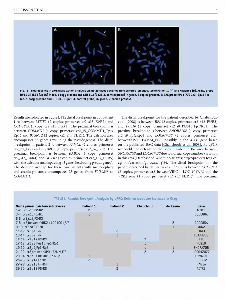
Results are indicated in Table I. The distal breakpoint in our patient
1 is between MTIF2 (2 copies; primerset cr2_s13_F2/R2) and
CCDC88A (1 copy; cr2_s15_F1/R1). The proximal breakpoint is
between COMMD1 (1 copy; primerset cr2_s5_COMMD1_Fp1/
Rp1) and B3GNT2 (2 copies; cr2_s14_F1/R1). The deletion area
encompasses 35 genes (excluding the pseudogenes). The distal
breakpoint in patient 2 is between FANCL (2 copies; primerset
cr2_p1_F/R) and FLJ30838 (1 copy, primerset cr2_p2_F/R). The
proximal breakpoint is between RAB1A (1 copy; primerset
cr2_s13_F4/R4) and ACTR2 (2 copies; primerset cr2_s13_F5/R5)
with the deletion encompassing 43 genes (excluding pseudogenes).
The deletion overlap for these two patients with microcephaly
and craniosynostosis encompasses 23 genes, from FLJ30838 to
COMMD1.
The distal breakpoint for the patient described by Chabchoub
et al. [2008] is between REL (2 copies, primerset cr2_s12_F3/R3)
and PUS10 (1 copy, primerset cr2_s8_PUS10_Fp1/Rp1). The
proximal breakpoint is between SNORA70B (1 copy, primerset
cr2_s9_Fp3/Rp3) and LOC647077 (2 copies, primerset cr2_
betweenXPOþ FAMM_F/R), possibly in the XPO1 gene based
on the published BAC data [Chabchoub et al., 2008]. By qPCR
we could not determine the copy number in the area between
SNORA70B and LOC647077 due to normal copy number variation
in this area (Database of Genomic Variants; http://projects.tcag.ca/
cgi-bin/variation/gbrowse/hg19). The distal breakpoint for the
patient described by de Leeuw et al. [2008] is between CCDC85A
(2 copies, primerset cr2_betweenVRK2þ LOC1001F/R) and the
VRK2 gene (1 copy, primerset cr2_s12_F1/R1)2. The proximal
FIG. 3. Fluorescence in situ hybridization analysis on metaphases obtained from cultured lymphocytes of Patient 1 (A) and Patient 2 (B). A: BAC probe
RP11-373L24 (2p16) in red, 1 copy present and CTB-8L3 (2p25.3, control probe) in green, 2 copies present. B: BAC probe RP11-772D22 (2p15) in
red, 1 copy present and CTB-8L3 (2p25.3, control probe) in green, 2 copies present.
TABLE I. Results Breakpoint Analysis by qPRC: Deletion Areas Are Indicated in Gray
Name primer pair forward/reverse Patient 1 Patient 2 Chabchoub de Leeuw Gene1-2: cr2_s13_F2/R2 2 MTIF23-4: cr2_s15_F1/R1 1 CCDC88A5-6: cr2_s13_F3/R37-8: cr2_betweenVRK2þLOC1001_F/R 2 CCDC85A9-10: cr2_s12_F1/R1 1 VRK211-12: cr2_p1_F/R 2 FANCL13-14: cr2_p2_F/R 1 FLJ3083815-16: cr2_s12_F3/R3 2 REL17-18: cr2_s8_Pus10_Fp1/Rp1 1 PUS1019-20: cr2_s9_Fp3/Rp3 1 1 SNORA70B21-22: cr2_betweenXPOþFAMM_F/R 2 2 LOC64707723-24: cr2_s5_COMMD1_Fp1/Rp1 1 COMMD125-26: cr2_s14_F1/R1 2 B3GNT227-28: cr2_s13_F4/R4 1 RAB1A29-30: cr2_s13_F5/R5 2 ACTR2
FLORISSON ET AL. 5

breakpoint is the same as for the patient described by Chabchoub
et al. [2008].
Deletions of all patients, encompassing genes in the area, are
indicated in Figure 4.
EnhancersIn the region from 55,463,756 to 65,498,387, 42 Human Vista
enhancer elements are located [Visel et al., 2007]. These are
highly conserved elements with possible gene distant-acting
enhancer activity. These 42 are all between FANCL and PAPOLG,
the region that is deleted in seven and partially deleted in one of
the patients (Supplementary eFigure S2—See Supporting Infor-
mation online). Fourteen of these elements have expression
as assessed in transgenic mice, in amongst others brain
(hs394,hs399,hs779,hs975,hs1076,hs1119,hs1535), facial mesen-
chym (hs836) eye (hs393), and ear (hs1071). This indicates that
these enhancers possibly play a role in the observed phenotype and
have a function in these tissues in regulating other genes.
DISCUSSION
We describe two patients with overlapping deletions in the
2p15p16.1 region, an area that is recently recognized as a new
microdeletion syndrome [Rajcan-Separovic et al., 2007; Chab-
choub et al., 2008; de Leeuw et al., 2008; Liang et al., 2009; Felix
et al., 2010; Prontera et al., 2011]. These patients have the largest
deletions in this area described so far, but the common overlapping
deleted region is similar to the deletions described in the patients by
Liang et al. [2009] and Felix et al. [2010]. Notably, the current
patients add craniosynostosis as a new clinical characteristic to this
novel microdeletion syndrome (Table II), though both patients
have different forms. Patient 1 has a synostosis of the metopic
and the sagittal suture in addition to a trigonocephalic head,
whereas Patient 2 has a synostosis of the left coronal and sagittal
suture. Patient 1 additionally showed brain abnormalities in the
form of a simplified gyral pattern in the supra tentorial region, a
hypoplastic corpus callosum and a small cerebellum and pons.
Because Patient 2 was operated in another center abroad, no
original MRI or CT data were available. We propose that the
craniosynostosis in these patients is primary, and not secondary
to the microcephaly, as this last would generally result in pansy-
nostosis rather than sagittal, coronal or metopic synostosis. Except
for the craniosynostosis, clinically both patients do resemble the
2p15p16.1 microdeletion syndrome. However, for some clinical
features, for example, optic nerve hypoplasia and hydronephrosis,
the children may still be too young to express these abnormalties
(Table II).
Rajcan-Separovic et al. [2007] reported on brachycephaly as a
dysmorphic feature in their patients, although it is not clear what
the underlying cause of this feature in these patients is. In addition
to our Patient 1, the two patients from Rajcan-Separovic et al. also
showed brain abnormalities; perisylvian migration disorder
(Patient 1 [Rajcan-Separovic et al., 2007]) and generally thickened
cortex with hyperintense subcortical tissue suggesting dysmyeli-
nation or cortical dysplasia; enlarged 4th ventricle, mild hypoplasia
of the inferior cerebellar vermis, and small anterior pituitary and
pons (Patient 2 [Rajcan-Separovic et al., 2007]). These partly
overlap with the brain features in our patient.
We have determined the deletion borders in our two patients and
the patients described by Chabchoub et al. [2008] and de Leeuw
et al. [2008] by qPCR. The data of these authors were based on BAC
data, and therefore, the borders were not determined precisely.
According to de Leeuw et al. [2008], XPO1 is outside their deletion
area, according to Chabchoub et al. [2008], XPO1 is included in
their deletion area. However, by qPCR we have not been able to
resolve whether XPO1 is deleted in the two patients described by
these authors. This is likely due to copy number variation in that
area (Database of Genomic Variants; http://projects.tcag.ca/cgi-
bin/variation/gbrowse/hg19).
The patient described by Chabchoub et al. [2008], with the
smallest deletion, is also intellectually disabled, similar to the other
patients with a deletion in this area. However, he is the only one
without microcephaly. Additionally, several other clinical features
observed in the majority of 2p15p161 microdeletion patients, for
example, some facial dysmorphisms, optic nerve hypoplasia, and
hydronephrosis, could not be observed in this patient (Table II).
This milder phenotype is in line with the small size of its deletion
which is only 570 kb versus the 3.2–6.9 Mb in all the other so far
reported cases. The patient reported by Prontera et al. [2011], with a
deletion of 3.5 Mb, has many clinical features overlapping with the
other described patients, including microcephaly and intellectual
disability, but no craniosynostosis or structural brain abnormalities
and that deletion encompasses only one uncharacterized and two
known genes. Additionally, this patient has two other chromosomal
rearrangements that could possibly influence the clinical pheno-
type. All nine patients have moderate to severe mental disability,
though not all deletions overlap, indicating that genes in different
regions can have influence on mental disability. For the micro-
cephaly, the only gene that seems left, is the uncharacterized gene
LOC100506891. Considering the presence of craniosynostosis in
our two patients, we propose that the candidate region for this
feature lies within the region between FANCL and B3GNT2. Addi-
tionally, features may depend on the different sizes of deletions
found in the different patients and may show variable expression
depending on genetic background [Girirajan et al., 2010]. Also the
enhancer elements present in the area may play a role.
Some genes in the deletion region have a known function in
brain. BCL11A (CTIP1) [OMIM:606557], a gene that plays a role in
globin gene regulation [Sankaran et al., 2008] is also highly
expressed in brain [Kuo and Hsueh, 2007] and thought to be
involved in axon outgrowth and branching [Kuo et al., 2010].
BCL11A is deleted in seven of the nine patients. As information on
brain development on MRI is available in six of the patients, with
brain abnormalities in three of them, it is at this point not possible to
conclude whether absence of this gene plays a major role in the
formation of brain abnormalities seen in these patients. XPO1 (or
CRM1) [OMIM:602559] is evolutionary conserved and expressed
in the developing brain and proposed to be involved in
motor neuron development and survival [Kolle et al., 2000]. Addi-
tionally it is implicated to have a role during mitosis [Hutten and
Kehlenbach, 2007].
Notably, REL [OMIM:164910] binds to CREBBP [OMIM:600140]
and EP300 [OMIM:602700], two genes involved in Rubinstein–Taybi
6 AMERICAN JOURNAL OF MEDICAL GENETICS PART A

FIG. 4. Microdeletion area chromosome 2p15p16. Deletions (in red) are indicated in our Patients 1 and 2 and in all patients described in literature until
now. Genes in the region with base pair positions (build 37, hg19) are indicated on the left. The light-gray area around Xpo1 could not be resolved by q-
PCR, due to normal copy number variation in this region. The minimal distal deletion borders in the two subjects from Rajcan-Separovic et al. are
indicated as described in Liu et al. [2011]. Unresolved areas are indicated in light-gray. Basepair positions from the deletions described in the
literature were all converted to build 37, hg 19. The deletion from Prontera et al. seems small in the figure, because in their 3.5 Mb deletion area, only
three genes are located (distances are not drawn to scale).
FLORISSON ET AL. 7

TAB
LEII
.O
verv
iew
Clin
ical
Feat
ures
ofPa
tien
tsW
ith
Chro
mos
ome
2p1
5-p
16
.1D
elet
ion
s
Pres
ent
(Pat
ien
t1
)Pr
esen
t(P
atie
nt
2)
Chab
chou
bet
al.
deLe
euw
etal
.R
ajca
n-S
epar
ovic
etal
.(s
ubje
ct1
)R
ajca
n-S
epar
ovic
etal
.(s
ubje
ct2
)Li
ang
etal
.F� e
lixet
al.
Pro
nte
raet
al.
Tota
l6
.96
.80
.57
3.9
4.5
5.7
3.2
3.3
53
.5G
ener
alAg
e(y
ears
)at
tim
eof
asse
ssm
ent
3m
onth
s/4
year
s6
mon
ths/
13
yea
rs1
63
28
64
,54
9
Gen
der
MF
MM
FM
FF
FIU
GR
NA
NA
NM
�þ
�þ
þN
M3
/9Sh
ort
stat
ure
��
�þ
�þ
þN
MN
M3
/9D
evel
opm
enta
lde
lay
þþ
NM
þþ
þN
Mþ
þ7
/9In
telle
ctua
ldi
sabi
lity
Mod
erat
eSe
vere
Mild
Mod
erat
e/Se
vere
Mod
erat
eM
oder
ate
Mod
erat
eM
oder
ate
Mod
erat
e9
/9
Auti
stic
feat
ures
/con
firm
edau
tism
spec
trum
diso
rder
NA
NA
�N
Mþ
þN
M�
NM
2/9
Atte
nti
onde
fici
thy
pera
ctiv
ity
diso
rder
NA
NA
NM
NM
þþ
þN
Mþ
4/9
Del
ayed
lan
guag
esk
ills
��
Mild
þSe
vere
Seve
reþ
Seve
reþ
9/9
Feed
ing
prob
lem
sN
Mþ
þþ
–þ
þM
icro
ceph
aly
þþ
�þ
þþ
þþ
þ8
/9Cr
anio
syn
osto
sis
þþ
��
��
��
�2
/9St
ruct
ural
brai
nab
nor
mal
itie
sþ
Un
know
n�
Un
know
nþ
þ�
��
3/7
Faci
alfe
atur
esB
item
pora
ln
arro
win
gþ
þN
Mþ
þþ
�N
Mþ
6/9
Rec
edin
gsh
ort
fore
head
��
�þ
þþ
�þ
�5
/9St
rabi
smus
þ�
�þ
þ�
��
�3
/9Pt
osis
þþ
�þ
þþ
þþ
þ8
/9Te
leca
nth
usþ
þþ
þþ
þþ
þþ
9/9
Wid
ened
inn
erca
nth
aldi
stan
ceþ
þ�
þþ
þþ
NM
þ7
/9Sh
ort
palp
ebra
lfi
ssur
es�
��
þþ
þþ
þþ
6/9
Dow
nsl
anti
ng
palp
ebra
lfi
ssur
es�
��
þþ
þ�
NM
�3
/9Ep
ican
thal
fold
sþ
þþ
þ�
þþ
NM
þ7
/8B
road
/hig
hn
asal
root
þþ
þþ
þþ
�þ
þ9
/9Pr
omin
ent
nas
alti
p�
�þ
�þ
þ�
NM
þ4
/9Lo
ng,
stra
ight
eyel
ashe
s�
þN
Mþ
þþ
�þ
�5
/9Lo
ng,
thin
eyeb
row
s�
þN
M�
�þ
�N
M�
2/9
Larg
eea
rsþ
�þ
�þ
þ�
þþ
6/9
Smoo
than
d/or
lon
gph
iltru
mþ
þ�
þþ
þþ
þþ
8/9
Smoo
thup
per
verm
illio
nbo
rder
��
þþ
þþ
�N
Mþ
5/9
Ever
ted
low
erlip
��
þþ
þþ
�N
Mþ
5/9
Hig
hn
arro
wpa
late
��
þþ
þþ
�þ
þ6
/9R
etro
gnat
hia
��
NM
þ�
þþ
Mic
rogn
ath
ia�
3/9
Oth
erph
ysic
alfe
atur
esW
iden
edin
tern
ippl
edi
stan
ce�
�N
Mþ
þþ
�þ
þ5
/9Ex
tra
nip
ple
��
NM
��
þ�
NM
�1
/9Ca
mpt
odac
tyly
digi
t(s)
þþ
��
þþ
�þ
�5
/9
8 AMERICAN JOURNAL OF MEDICAL GENETICS PART A

syndrome [OMIM:180849 and 61384], where intellectual disability
and microcephaly are part of the phenotypic characteristics, and
structural brain abnormalities have been reported in a minority of
cases [Roelfsema and Peters, 2007].
Array CGH and SNP array screening has led to the character-
ization of many heterozygous (novel) microdeletion and micro-
duplication syndromes [Slavotinek, 2008]. Intellectual disability
is often a feature of these syndromes, as is microcephaly [Slavotinek,
2008; Walczak-Sztulpa et al., 2008; Reddy et al., 2009; Girirajan
et al., 2010]. Additionally, microcephaly is found as an isolated
feature, but can also be associated with many syndromes [Abuelo,
2007]. Also, disruption of one gene can lead to a combination of
phenotypic features including microcephaly and intellectual dis-
ability [Mukhpadhyay et al., 2010; Williams et al., 2010; Campbell
et al., 2011]. For most microdeletion syndromes it is difficult to
pinpoint one clinical feature to the absence of a specific gene in a
deletion encompassing many genes [Kumar, 2008; Klaassens et al.,
2009; Masurel-Paulet et al., 2010; Stankiewicz and Lupski, 2010;
Vissers et al., 2010], and this is supported by comparing the
deletions with the clinical phenotype in this study. Mouse
models carrying targeted deletions of genes within these regions
may help to elucidate the function of the individual genes [Abrams
and Jiao, 2009]. Deletions in the 2p15p16.1 area show variable
clinical expression and may lead to microcephaly, intellectual
disability and additionally to craniosynostosis, depending on the
size and extension of the deletion combined with genetic
background.
ACKNOWLEDGMENTS
The authors extend their sincere appreciation to Patients 1 and 2
and their parents for their support of this study. We thank Tom de
Vries Lentsch for graphical support.
REFERENCES
Abrams JM, Jiao Y. 2009. Keeping it simple: What mouse models of Wolf-Hirschhorn syndrome can tell us about large chromosomal deletions.Disease Models Mech 2:315–316.
Abuelo D. 2007. Microcephaly syndromes. Semin Pediatr Neurol 14:118–127.
Campbell IM, Kolodziejska KE, Quach MM, Wolf VL, Cheung SW, LalaniSR, Ramocki MB, Stankiewicz P. 2011. TGFBR2 deletion in a 20-month-old female with developmental delay and microcephaly. Am J Med GenetPart A 9999A:1–6.
Chabchoub E, Vermeesch JR, de Ravel T, de Cock P, Fryns JP. 2008.The facial dysmorphy in the newly recognised microdeletion2p15-p16.1 refined to a 570 kb region in 2p15. J Med Genet 45:189–192.
de Leeuw N, Pfundt R, Koolen DA, Neefs I, Scheltinga I, Mieloo H,Sistermans EA, Nillesen W, Smeets DF, de Vries BB, KnoersNV. 2008. A newly recognised microdeletion syndrome involving2p15p16.1: Narrowing down the critical region by adding another patientdetected by genome wide tiling path array comparative genomic hybrid-isation analysis. J Med Genet 45:122–124.
Felix TM, Petrin AL, Sanseverino MT, Murray JC. 2010. Further character-ization of microdeletion syndrome involving 2p15-p16. Am J Med GenetPart A 152A:2604–2608.
Met
atar
sus
abdu
ctus
��
��
þþ
þþ
�4
/9Sp
asti
city
legs
��
��
þþ
þN
Mþ
4/9
Oth
er Opt
icn
erve
hypo
plas
ia�
�N
Mþ
þþ
þ�
�4
/9D
istu
rbed
visi
on�
�N
Mþ
(myo
pia)
�þ
(hyp
erop
ia)
NA
þm
ildh
yp
erm
etro
pia
NM
3/9
Hea
rin
glo
ss�
þN
M�
�þ
��
�2
/9Fr
eque
nt
uppe
rre
spir
ator
yin
fect
ion
s�
�N
Mþ
þ�
��
�2
/9
Lary
ngo
mal
acia
��
NM
��
þ�
NM
�1
/9H
ydro
nep
hros
is�
��
þþ
þ�
��
3/9
Hyp
ogon
adis
m�
�N
M�
�þ
��
�1
/9
NM
,n
otm
enti
oned
;N
A,n
otas
sess
ed.
FLORISSON ET AL. 9

Girirajan S, Rosenfeld JA, Cooper GM, Antonacci F, Siswara P, Itsara A,Vives L, Walsh T, McCarthy SE, Baker C, Mefford HC, Kidd JM,Browning SR, Browning BL, Dickel DE, Levy DL, Ballif BC, Platky K,Farber DM, Gowans GC, Wetherbee JJ, Asamoah A, Weaver DD, MarkPR, Dickerson J, Garg BP, Ellingwood SA, Smith R, Banks VC, Smith W,McDonald MT, Hoo JJ, French BN, Hudson C, Johnson JP, Ozmore JR,Moeschler JB, Surti U, Escobar LF, El-Khechen D, Gorski JL, Kussmann J,Salbert B, Lacassie Y, Biser A, McDonald-McGinn DM, Zackai EH,Deardorff MA, Shaikh TH, Haan E, Friend KL, Fichera M, RomanoC, G�ecz J, DeLisi LE, Sebat J, King MC, Shaffer LG, Eichler EE. 2010. Arecurrent 16p12.1 microdeletion supports a two-hit model for severedevelopmental delay. Nat Genet 42:203–210.
Hutten S, Kehlenbach RH. 2007. CRM1-mediated nuclear export: The poreand beyond. Trends Cell Biol 17:193–201.
Klaassens M, de Klein A, Tibboel D. 2009. The etiology of congenitaldiaphragmatic hernia: Still largely unknown? Eur J Med Genet 52:281–286.
Kolle G, Georgas K, Holmes GP, Little MH, Yamada T. 2000. CRIM1, anovel gene encoding a cysteine-rich repeat protein, is developmentallyregulated and implicated in vertebrate CNS development and organo-genesis. Mech Dev 90:181–193.
Kumar D. 2008. Disorders of the genome architecture: A review. GenomicMed 2:69–76.
Kuo TY, Hsueh YP. 2007. Expression of zinc finger transcription factorBcl11A/Evi9/CTIP1 in rat brain. J Neurosci Res 85:1628–1636.
Kuo TY, Hong CJ, Chien HL, Hsueh YP. 2010. X-linked mental retardationgene CASK interacts with Bcl11A/CTIP1 and regulates axon branchingand outgrowth. J Neurosci Res 88:2364–2373.
Liang JS, Shimojima K, Ohno K, Sugiura C, Une Y, Ohno K, Yamamoto T.2009. A newly recognised microdeletion syndrome of 2p15-16.1 man-ifesting moderate developmental delay, autistic behaviour, short stature,microcephaly, and dysmorphic features: A new patient with 3.2 Mbdeletion. J Med Genet 46:645–647.
Liu X, Malenfant P, Reesor C, Lee A, Hudson ML, Harvard C, Qiao Y,Persico AM, Cohen IL, Chudley AE, Forster-Gibson C, Rajcan-SeparovicE, Lewis MES, Holden JJA. 2011. 2p15-16.1 microdeletion syndrome:Molecular characterization and association of the OTX1 and XPO1 geneswith autism spectrum disorders. Eur J Hum Genet 19:1264–1270.
Masurel-Paulet A, Andrieux J, Callier P, Cuisset JM, Le Caignec C, HolderM, Thauvin-Robinet C, Doray B, Flori E, Alex-Cordier MP, Beri M, BouteO, Delobel B, Dieux A, Vallee L, Jaillard S, Odent S, Isidor B, Beneteau C,Vigneron J, Bilan F, Gilbert-Dussardier B, Dubourg C, Labalme A, BidonC, Gautier A, Pernes P, Pinoit JM, Huet F, Mugneret F, Aral B, JonveauxP, Sanlaville D, Faivre L. 2010. Delineation of 15q13,3 microdeletions.Clin Genet 78:149–161.
Mukhpadhyay A, Kramer JM, Merkx G, Lugtenberg D, Smeets DF,Ooortveld MAW, Blokland EAW, Agrawal J, Schenck A, van BokhovenH, Huys E, Schoenmakers EF, Geurts van Kessel A, van Nouhuys CE,Cremers FPM. 2010. CDK19 is disrupted in a female patient with bilateralcongenital retinal folds, microcephaly and mild mental retardation. HumGenet 128:128–291.
Prontera P, Bernardini L, Stangoni G, Capalbo A, Rogaia D, Romani R,Ardisia C, Dallapiccola B, Donti E. 2011. Deletion 2p15-16.1 syndrome:Case report and review. Am J Med Genet Part A 155A:2473–2478.
Rajcan-Separovic E, Harvard C, Liu X, McGillivray B, Hall JG, Qiao Y,Hurlburt J, Hildebrand J, Mickelson EC, Holden JJ, Lewis ME. 2007.Clinical and molecular cytogenetic characterisation of a newlyrecognised microdeletion syndrome involving 2p15-16.1. J Med Genet44:269–276.
Reddy S, Dolzhanskaya N, Krogh J, Velinov M. 2009. A novel 1.4 Mb denovo microdeletion of chromosome 1q21.3 in a child with microcephaly,dysmorphic features and mental retardation. Eur J Med Genet 52:443–445.
Roelfsema JH, Peters DJ. 2007. Rubinstein-Taybi syndrome: Clinical andmolecular overview. Expert Rev Mol Med 9:1–16.
Sankaran VG, Menne TF, Xu J, Akie TE, Lettre G, Van Handel B, MikkolaHKA, Hirschhorn JN, Cantor AB, Orkin SH. 2008. Human fetal hemo-globin expression is regulated by the developmental stage-specificrepressor BCL11A. Science 322:1839–1842.
Slavotinek AM. 2008. Novel microdeletion syndromes detected by chro-mosome microarrays. Hum Genet 124:1–17.
Stankiewicz P, Lupski JR. 2010. Structural variation in the human genomeand its role in disease. Ann Rev Med 61:437–455.
Visel A, Minovitsky S, Dubchak I, Pennacchio LA. 2007. VISTA EnhancerBrowser—A database of tissue-specific human enhancers. Nucleic AcidsRes 35:D88–D92.
Vissers LELM, de Vries BBA, Veltman JA. 2010. Genomic microarrays inmental retardation: From copy number variation to gene, from researchto diagnosis. J Med Genet 47:289–297.
Walczak-Sztulpa J, Wisniewska M, Latos-Bielenska A, Linne M, Kelbova C,Belitz B, Pfeiffer L, Kalscheuer V, Erdogan F, Kuss AW, Ropers HH,Ullmann R, Tzschach A. 2008. Chromosome deletions in 13q33-34:Report of four patients and review of the literature. Am J Med Genet PartA 146A:337–342.
Williams SR, Mullegama SV, Rosenfeld JA, Dagli AI, Hatchwell E, AllenWP, Williams CA, Elsea SH. 2010. Haploinsufficiency of MBD5associated with a syndrome involving microcephaly, intellectualdisabilities, severe speech imparment and seizures. Eur J Med Genet18:436–441.
10 AMERICAN JOURNAL OF MEDICAL GENETICS PART A