Kindler syndrome
17
Kindler syndrome Authors: Vikram K Mahajan, M.D., Nand Lal Sharma 1 , M.D.and Ramesh Chander Sharma, M.D. Creation date: March 2005 Scientific editor: Jemima Mellerio, MD 1 Department of Dermatology, Venereology & Leprosy, Indira Gandhi Medical College, Shimla, India. [email protected] Abstract Keywords Disease name/synonyms Definition/diagnostic criteria Differential Diagnosis Etiology Clinical description Diagnostic methods Management including treatment Kindler syndrome and epidermolysis bullosa Kindler syndrome and Weary’s hereditary acrokeratotic poikiloderma References Abstract Kindler syndrome (KS) is a rare autosomal recessive genophotodermatosis. It combines features of acral blistering and photosensitivity from infancy, which tend to improve through childhood, with progressive poikiloderma and cutaneous atrophy developing later. Phenotypic heterogeneity and variable expression of the condition is known and additional features have been described. It shares some clinicopathologic similarities with dystrophic epidermolysis bullosa and Weary’s hereditary acrokeratotic poikiloderma. However, characteristic immunohistochemical, ultrastructural and molecular features have been demonstrated which distinguish KS as a separate entity. Specifically, this condition has recently been shown to result from loss of function mutations in KIND1, the gene encoding kindlin-1, a novel protein involved in attachment of the actin cytoskeleton to the extracellular matrix via focal contacts. Keywords Genodermatosis, poikiloderma, skin blistering, periodontitis Disease name/synonyms Kindler syndrome Poikiloderma of Kindler Definition/diagnostic criteria KS is a rare genophotodermatosis described first in 1954 by Theresa Kindler (Kindler, 1954). Its salient features include trauma- induced blistering and photosensitivity usually starting in early infancy. With advancing age, improvement in blistering and photosensitivity usually occurs; a progressive and more persistent poikiloderma and cutaneous atrophy arise. Ultrastructurally, variable planes of cleavage are seen at the dermal-epidermal junction along with reduplication of the basement membrane. The locus of the disease has been mapped to 20p12.3 (Jobard et al., 2003) and, subsequently, the causative gene identified as KIND1, which encodes kindlin-1, a protein involved in attaching the actin cytoskeleton to the extracellular matrix at focal contacts (Jobard et al., 2003; Siegel et al., 2003; Ashton et al., 2004). Differential Diagnosis A number of other conditions can cause blistering, cutaneous atrophy and / or poikiloderma. Notably, dystrophic Mahajan VK, Sharma NL and Sharma RC, Kindler syndrome. Orphanet Encyclopedia. Mars 2005. http://www.orpha.net/data/patho/GB/uk-kindler.pdf 1
Transcript of Kindler syndrome

Kindler syndrome Authors: Vikram K Mahajan, M.D., Nand Lal Sharma1, M.D.and Ramesh Chander Sharma, M.D. Creation date: March 2005 Scientific editor: Jemima Mellerio, MD 1Department of Dermatology, Venereology & Leprosy, Indira Gandhi Medical College, Shimla, India. [email protected]
Abstract Keywords Disease name/synonyms Definition/diagnostic criteria Differential Diagnosis Etiology Clinical description Diagnostic methods Management including treatment Kindler syndrome and epidermolysis bullosa Kindler syndrome and Weary’s hereditary acrokeratotic poikiloderma References Abstract Kindler syndrome (KS) is a rare autosomal recessive genophotodermatosis. It combines features of acral blistering and photosensitivity from infancy, which tend to improve through childhood, with progressive poikiloderma and cutaneous atrophy developing later. Phenotypic heterogeneity and variable expression of the condition is known and additional features have been described. It shares some clinicopathologic similarities with dystrophic epidermolysis bullosa and Weary’s hereditary acrokeratotic poikiloderma. However, characteristic immunohistochemical, ultrastructural and molecular features have been demonstrated which distinguish KS as a separate entity. Specifically, this condition has recently been shown to result from loss of function mutations in KIND1, the gene encoding kindlin-1, a novel protein involved in attachment of the actin cytoskeleton to the extracellular matrix via focal contacts. Keywords Genodermatosis, poikiloderma, skin blistering, periodontitis
Disease name/synonyms Kindler syndrome Poikiloderma of Kindler Definition/diagnostic criteria KS is a rare genophotodermatosis described first in 1954 by Theresa Kindler (Kindler, 1954). Its salient features include trauma-induced blistering and photosensitivity usually starting in early infancy. With advancing age, improvement in blistering and photosensitivity usually occurs; a progressive and more persistent poikiloderma and cutaneous atrophy arise. Ultrastructurally, variable planes of cleavage
are seen at the dermal-epidermal junction along with reduplication of the basement membrane. The locus of the disease has been mapped to 20p12.3 (Jobard et al., 2003) and, subsequently, the causative gene identified as KIND1, which encodes kindlin-1, a protein involved in attaching the actin cytoskeleton to the extracellular matrix at focal contacts (Jobard et al., 2003; Siegel et al., 2003; Ashton et al., 2004). Differential Diagnosis A number of other conditions can cause blistering, cutaneous atrophy and / or poikiloderma. Notably, dystrophic
Mahajan VK, Sharma NL and Sharma RC, Kindler syndrome. Orphanet Encyclopedia. Mars 2005. http://www.orpha.net/data/patho/GB/uk-kindler.pdf 1

epidermolysis bullosa (DEB) and Weary's hereditary acrokeratotic poikiloderma (HAP) may cause confusion with KS (see below). A set of clinical criteria (Table 1) may assist in making a bedside diagnosis until new molecular diagnostic techniques are widely available. It may also be difficult to differentiate and individually define different congenital poikiloderma syndromes as these rare conditions may have overlapping clinical features (Table 2). Again, delineation of the molecular pathology underlying KS will help confirm this as a distinct entity. Etiology KS has been recently mapped to a locus on chromosome 20p12.3 and pathogenic mutations have been detected in a new gene KIND1, encoding a 677 amino acid protein, kindlin-1 (Jobard et al., 2003; Siegel et al., 2003; Ashton et al., 2004). Kindlin-1 is expressed mainly in basal keratinocytes and plays a role in the attachment of the actin cytoskeleton via focal contacts to the extracellular matrix (Ashton et al., 2004). Loss of function mutations in KIND1 appear to be responsible for the mucocutaneous fragility observed in KS, therefore. However, the precise mechanisms whereby mutations in this gene lead to photosensitivity and poikiloderma are not yet clear. Clinical description KS usually becomes clinically evident by the age of 2-3 years when photosensitivity, acral blistering and poikiloderma are established (Hovnanian et al., 1989). Hair, teeth and nails have no or only mild abnormalities. Endocrine or hematologic abnormalities are not seen, and physical and intellectual development remains unimpaired throughout life (Thappa et al., 2000). Skin fragility and blistering, mostly over the dorsa of hands and feet, are the most consistent clinical features in KS. Bullae may be present at birth or appear during first few days of life. Blistering follows cutaneous trauma (Fig. 1) or occasionally exposure to sunlight, and may get secondarily infected. Blistering continues to occur in all age groups albeit in a diminished frequency after 10 – 12 years of age (Penagos et al., 2004). Photosensitivity of variable severity is observed by 1 month to 2 years of life. Erythema associated with a burning sensation followed by blister formation is usual after sun exposure. With advancing age and coinciding with diminished blister formation, the photosensitivity, too, tends to
wane in intensity (Penagos et al., 2004). Subsequently a more persistent reticular poikilodermatous pigmentation and cutaneous atrophy ensue. Poikiloderma, first noticed between 2-3 years of age, becomes persistent for life (Penagos et al., 2004). Although photosensitivity diminishes, the poikiloderma becomes more pronounced and generalized eventually involving both sun exposed and non-sun exposed areas (Fig. 2 & 3). Evidently, photodamage is not the sole cause for poikiloderma. A characteristic diffuse cutaneous atrophy that involves abdomen, thighs, knees and elbows occurs in all patients. Dry, atrophic, cigarette-paper-thin skin atrophy is more marked over dorsa of hands (Fig. 4) and feet. Webbing of the digits is also a feature as is dystrophy of finger- and toenails. Mild to moderately severe diffuse, sometimes punctate, hyperkeratosis of palms (Fig. 5) and soles with a characteristic waxy feel is usual. Associated fissuring, wrinkling and scaling may occur. Acral sclerosis, pseudoainhum and loss of dermatoglyphics may accompany sometimes (Krunic et al., 1997; Binder et al., 2002). Mucosal involvement is frequent in KS. Leukokeratosis of oral mucous membranes (Fig. 6), seen as adherent white patches, is the most frequent finding but may occasionally involve the anal mucosa as well (Fig.7) (Sharma et al., 2003). Restricted mouth opening has been attributed to repeated erosions and scarring of oral commissures (Fig. 6). In addition, atrophy of buccal mucosa and gums, erosions of lips and hard palate and geographic tongue have also been observed (Wiebe et al., 2003). Severe phimosis to the extent that prepucial skin may appear adherant to the glans is another frequent feature (Fig.8) (Thappa et al., 2000). Stenosis of the anal canal, esophagus and urethral meatus may be encountered occasionally (Hovnanian et al., 1989). Dental abnormalities (Wiebe et al., 2003) include severe periodontal bone loss and periodontitis, swollen, fragile bleeding gums and early exfoliation of deciduous as well as permanent dentition (Fig. 9). Periodontal disease in most cases begins in early adolescence coinciding with eruption of permanent teeth. Patients presenting before the age of 10 years, therefore, may exhibit little or no periodontal disease. The pathomechanism of these defects is not well understood. The inherited defect of
Mahajan VK, Sharma NL and Sharma RC, Kindler syndrome. Orphanet Encyclopedia. Mars 2005. http://www.orpha.net/data/patho/GB/uk-kindler.pdf 2

cutaneous fragility is perhaps also expressed in gums and mucous membranes. Spontaneous bleeding from gums suggests it to be due to microblistering and breakdown of periodontal tissues due to minor trauma of normal occlusal function. Occasional stenosis of esophagus/anus also points towards mucosal fragility and subsequent fibrosis (Forman et al., 1989). Many uncommon and less consistent features of this syndrome have also been described (Person et al., 1979; Forman et al., 1989; Hovnanian et al., 1989; Binder et al., 2002; Sharma et al., 2003 and Penagos et al., 2004). Ophthalmic involvement may occur in the form of pigment on lens surface, corneal opacities, thickened corneal nerves, keratoconjunctivitis, bacterial blephritis and ectropion. Isolated abnormalities such as laryngeal webs, imperforate anus, synechia of labia, anhidrosis / hypohidrosis and abnormalities of skeletal maturation manifesting as turricephaly, malformation of ribs, mandible and metacarpals have also been observed. Development of actinic keratoses and squamous cell carcinoma involving the lower lip and leg in KS (Alper et al., 1978; Karthikaeyan et al., 2003) suggests some predilection for malignant change. Recurrent erosion and regeneration of mucosal surfaces has also been postulated to be responsible for transitional cell carcinoma of bladder in an isolated case (Alper et al., 1978). Diagnostic methods The histopathology of KS is not diagnostic and varies with the type of lesion biopsied. Moreover, due to the sequential appearance of blistering, photosensitivity and poikiloderma, the age at which the patient has been assessed is also important. On light microscopy, blister formation may be evident within or just beneath basal keratinocytes (Hovnanian et al., 1989). Features of poikiloderma with epidermal atrophy, hyperkeratosis, hyper- or hypopigmentation and telangiectatic vessels may be present. Additionally, there may be disrupted collagen and elastic fibres in the papillary dermis and increased numbers of melanophages (Hovnanian et al., 1989; Patrizi et al., 1996). With electron microscopy, variable levels of cleavage through basal keratinocytes, the lamina lucida and sub-lamina densa have been observed (Hovnanian et al., 1989; Shimizu et al., 1997; Şenturk et al., 1999). Also, there is a characteristic interruption
and reduplication of the lamina densa which may reflect continuous remodeling of the basement membrane zone (Shimizu et al., 1997; Yasukawa et al., 2002). Immunohistochemically, a broad reticular labeling pattern of types IV and VII collagen streaking deep into the connective tissue beneath the basement membrane is seen, reflecting the basement membrane reduplication seen ultrastructurally (Shimizu et al., 1997; Wiebe et al., 1999). With the identification of kindlin-1 pathology in KS, it is now possible to use immunohistochemistry to assist diagnosis (Siegel et al., 2003). Using a polyclonal antibody against kindlin-1, the skin from KS patients shows very reduced or absent staining compared to bright staining in basal keratinocytes and along the dermal-epidermal junction in normal controls. Mutation analysis of KIND1 in affected families can further confirm diagnosis and also provides the potential for first trimester DNA-based prenatal testing in at risk pregnancies. Management including treatment The treatment of KS is mainly symptomatic. Avoidance of trauma, photoprotection and use of emollients helps prevent blistering. Antibiotics may be needed for bacterial infections and physiotherapy is recommended to prevent contractures. Gingival problems improve with conservative periodontal therapy and by maintaining good oral hygiene (Wiebe et al., 1996). Psychological counseling is important in view of cosmetic disability. Kindler syndrome and epidermolysis bullosa As the characteristic poikilodermatous cutaneous changes appear in later life, the bullous component of KS in infancy may often be misdiagnosed as epidermolysis bullosa. Indeed, Kindler’s original case was considered to have incidental co-occurrence of DEB and congenital poikiloderma. Clinical absence of photosensitivity and poikiloderma in DEB, helps differentiate it from KS, but these features may not be apparent for a number of months or years. In contrast, bullae in DEB heal with extensive scarring, milia formation, nail loss, flexural contractures and often marked digital webbing. Although blistering at the dermal-epidermal junction may occur in both KS and different forms of EB, multiple planes of cleavage in an individual patient are more consistent with KS. Also, the extensive
Mahajan VK, Sharma NL and Sharma RC, Kindler syndrome. Orphanet Encyclopedia. Mars 2005. http://www.orpha.net/data/patho/GB/uk-kindler.pdf 3

reduplication of the lamina densa seen ultrastructurally is not a feature of EB. Finally, the molecular mechanisms underlying KS and DEB are now understood and separate (Yasukawa et al., 2002): whilst DEB is caused by mutations in the type VII collagen gene, COL7A1, KS is caused by mutations in the kindlin-1 gene, KIND1 (Jobard et al., 2003; Siegel et al., 2003). Kindler syndrome and Weary’s hereditary acrokeratotic poikiloderma In 1971, an autosomal dominant condition named hereditary acrokerototic poikiloderma (HAP) with clinical features similar to those of KS was described (Weary et al., 1971). Subsequent reports also revealed significant overlapping clinical findings in KS and Weary’s HAP. Some workers even adopted the term “Weary-Kindler syndrome” to describe similar cases (Krunic et al.,1997; Kapasi et al., 1993). Weary’s HAP is the main differential diagnosis when poikiloderma appears in KS. However, the hallmark photosensitivity, cutaneous atrophy and mucosal lesions of KS are not seen in Weary’s HAP and poikiloderma appears before the first year of life in most cases of KS. Palmoplantar keratoderma when present in Weary’s HAP is usually of punctuate variety. Keratotic papules over knees, elbows, dorsa of hands and feet and eczematous dermatitis are strikingly exclusive to Weary’s HAP. Histologically, also, cleft formation is epidermal in Weary’s HAP. References Alper JC, Baden HP, Goldsmith LA. Kindler syndrome. Arch Dermatol 1978 ; 114 : 457 Ashton GH, McLean WH, South AP, et al. Recurrent mutations in Kindlin-1, a novel keratinocyte focal contact protein in the autosomal recessive skin fragility and photosensitivity disorder, Kindler syndrome. J Invest Dermatol 2004 ; 122 : 78 – 83. Binder B, Metze D, Smolle J. Congenital bullous poikiloderma (Kindler syndrome) Hautarzt 2002 ; 53 : 546 – 549. Forman AB, Prendiville JS, Esterly NB, et al. Kindler syndrome : report of two cases and review of the literature. Ped Dermatol 1989 ; 6 : 91 – 101. Hovnanian A, Blanchet-Bardon C, de Prost Y. Poikiloderma of Theresa Kindler : report of a case with ultrastructural study and review of the literature. Ped Dermatol 1989 ; 6: 82 – 90
Jobard F, Bouadjar B, Caux F, et al. Identification of mutations in a new gene encoding a FERM family protein with a pleckstrin homology domain in Kindler syndrome. Hum Mol Genet 2003 ; 12 : 925 – 935. Kapasi AY, Khopkar U, Raj S, Wadhwa SI. Weary-Kindler syndrome with multiple seborrhoeic keratosis. Int J Dermatol 1993 ; 32 : 444 – 445. Karthikeyan K, Thappa DM, Jeevankumar B. Kindler syndrome with squamous cell carcinoma of the leg. Indian J Dermatol 2003 ; 48 : 231 – 233. Kindler T. Congenital poikiloderma with traumatic bulla formation and progressive cutaneous atrophy. Br J Dermatol 1954 ; 66 : 104 – 111. Krunic ALJ, Ljiljana M, Novak A, et al. Hereditary bullous acrokeratotic poikiloderma of Weary-Kindler associated with pseudoanihum and sclerotic bands. Int J Dermatol 1997; 36: 529 – 533 Patrizi A, Pauluzzi P, Neri I, et al. Kindler syndrome : report of a case with ultrastructrual study and review of the literature. Ped Dermatol 1996 ; 13 : 397 – 402. Penagos H, Jaen M, Sancho MT, et al. Kindler syndrome in native Americans from Panama. Arch Dermatol 2004 ; 140 : 939 – 944. Person JR, Perry HO. Congenital poikiloderma with traumatic bulla formation, anhidrosis and keratoderma. Acta Dermatol Venereol 1979 ; 59 : 347 –351. Şentürk N, Usubütün A, Şahin S, et al. Kindler syndrome : absence of definite ultrastructural features. J Am Acad Dermatol 1999 ; 40 : 335 – 337. Sharma RC, Mahajan V, Sharma NL, Sharma AK. Kindler syndrome. Int J Dermatol 2003 ; 42 : 727 – 732. Shimizu H, Sato M, Ban M, et al. Immunohistochemical , ultrastructrual and molecular features of kindler syndrome distinguish it from dystrophic epidermolysis bullosa. Arch Dermatol 1997; 133 : 1111 – 1117. Siegel DH, Ashton GH, Penagos HG, et al. Loss of kindlin-1, a human homolog of the Caenorhabditis elegans actin-extracellular-matrix linker protein UNC-112, causes Kindler syndrome. Am J Hum Genet 2003; 73: 174-87. Thappa DM, Jeevankumar B, Karthikeyan K, Sethuraman G. Kindler syndrome : a case report. Indian J Ped Dermatol 2000; 3 : 19 – 22.
Mahajan VK, Sharma NL and Sharma RC, Kindler syndrome. Orphanet Encyclopedia. Mars 2005. http://www.orpha.net/data/patho/GB/uk-kindler.pdf 4

Weary PE, Manley WF Jr, Graham GF. Hereditary acrokeratotic poikiloderma. Arch Dermatol 1971 ; 103 : 409 – 422. Wiebe CB, Larjava HS. Abnormal deposition of type-VII collagen in Kindler syndrome. Arch Dermatol Res 1999 ; 291 : 6 – 13. Wiebe CB, Penagos H, Luong N, et al. Clinical and micro biological study of periodontitis associated with Kindler syndrome. J Periodontol 2003 ; 74 : 25 – 31.
Wiebe CB, Silver JG, Larjava HS. Early-onset periodontitis associated with Weary-Kindler syndrome : a case report. J Periodontol 1996 ; 67 : 1004 – 1010. Yasukawa K, Sato-Matsumura KC, McMillan J, et al,. Exclusion of COL 7A1 mutation in Kindler syndrome. J Am Acad Dermatol 2002 ; 46 : 447 – 450.
Mahajan VK, Sharma NL and Sharma RC, Kindler syndrome. Orphanet Encyclopedia. Mars 2005. http://www.orpha.net/data/patho/GB/uk-kindler.pdf 5

Table 1 : Proposed Clinical criteria for bedside diagnosis of Poikiloderma of Kindler
Essential Criteria
1) Progressively diminishing photosensitivity since early infancy.
2) Traumatic acral bullae since birth or developing during first few days of life.
3) Characteristic cigarette-paper-thin cutaneous atrophy, more pronounced over
acral areas.
4) Diffuse, generalized poikiloderma involving sun exposed and non-sun
exposed skin with onset after 1st year of birth.
Minor Criteria
1) Early onset of severe gingivitis/periodontitis.
2) Mucosal leukokeratosis.
3) Normal physical and mental development.
4) Palmoplantar hyperkeratosis with a waxy feel.
5) Severe phimosis.
6) Webbing of fingers and toes.
7) Absence of definite histopathologic features.
Presence of all essential and four of minor criteria is diagnostic
Mahajan VK, Sharma NL and Sharma RC, Kindler syndrome. Orphanet Encyclopedia. Mars 2005. http://www.orpha.net/data/patho/GB/uk-kindler.pdf 6
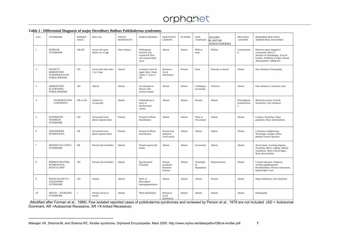
Table 2 : Differential Diagnosis of major Hereditary Bullous Poikiloderma syndromes
S.No.
SYNDROME
INHERITANCE
BULLAE
PHOTO SENSITIVITY
POIKILODERMA
KERATOTIC LESIONS
ECZEMA
NAIL CHANGES
PALMO-PLANTAR KERATODERMA
MUCOSAL LESIONS
REMARKS/ISOLATED ADDITIONAL FEATURES
1
KINDLER SYNDROME
AR/AD
Acral with onset before 1yr of age
Since infancy
Widespread, Involves Sun exposed & Non-sun exposed body areas.
Absent
Absent
Mild or none
Diffuse
Leukokeratosis
Defective gene mapped to cromosome 20p12.3. Stenosis of Oesophagus, Anus & Urethra, Webbing of digits, Dental Abnormalities Anhidrosis
2
WEARY’S HEREDITORY ACROKERATOTIC POIKILODERMA
AD
Acral with onset after 1 yr of age
Absent
Limited to neck & upper chest. Onset within 1st year of life
Present in Acral distribution
Present
None
Punctate or absent
Absent
Poor dentition, Enteropathy
3
HEREDITORY SCLEROSING POIKILODERMA
AD
Absent
Absent
Accentuated in flexors with sclerotic bands
Absent
Absent
Clubbing is occasional
Sclerosis
Absent
Poor dentition, Calcinosis cutis
4
DYSKERATOSIS CONGENITA
XR or AD
Absent (or occasional)
Absent
Poikiloderma is more of dyschromatic variety
Absent
Absent
Present
Absent
Premalignant leukokeratosis
Blood dyscrasias, Growth retardation, Poor dentition
5
ROTHMUND – THOMSON SYNDROME
AD
Occasional (over photo exposed skin)
Present
Present (in Photo distribution)
Absent
Absent
None or Occasional
Absent
Absent
Cataract, Dwarfism, Hypo gonadism, Bone abnormalities
6
XERODERMA PIGMENTOSA
AR
Occasional (over photo exposed skin)
Present
Present (in Photo distribution)
Present (not limited to Acral areas)
Absent
Absent
Absent
Absent
Cutaneous malignancies, Neurologic changes, Photo phobia/Corneal opacities
7
MENDES DA COSTA SYNDROME
XR
Present (Acral bullae)
Absent
Present (spares the trunk)
Absent
Absent
Occasional
Absent
Absent
Acrocynosis, Scarring alopecia, Dwarfism, Micro cephaly, Mental retardation, Short conical digits, Bone abnormalities
8
DERMATOPATHIA PIGMENTOSA – RETICULARIS
AD
Present (Acral bullae)
Absent
Dyschromatic (Trunkal)
Present (scattered Keratotic lesions)
Absent
Dystrophy or Hypoplasia
Hyperkeratosis
Absent
Corneal opacities, Alopecia, Axillary papillomatosis Pseudoainhum, Flexon contractures, Hypertrophic scars
9
FRANCESCHETTI – JADASSOHN SYNDROME
AD
Absent
Absent
More of Reticulated hyperpigmentation
Absent
Absent
Absent
Present
Absent
Hypo/Anhidrosis, Poor dentition
10
DEGOS – TOURAINE SYNDROME
?
Present (Acral or facial)
Absent
Photo distributed
Present in Acral distribution
Absent
Absent
Absent
Absent
Enteropathy
(Modified after Forman et al., 1989). Few isolated reported cases of poikiloderma syndromes and reviewed by Person et al., 1979 are not included (AD = Autosomal Dominant, AR =Autosomal Recessive, XR =X-linked Recessive)
Mahajan VK, Sharma NL and Sharma RC, Kindler syndrome. Orphanet Encyclopedia. Mars 2005. http://www.orpha.net/data/patho/GB/uk-kindler.pdf 7

Fig. 1. Multiple infected crusted lesions of acral bullae over lower limbs. Note: Characteristic atrophic skin over dorsa of feet and dystrophic toe nails.
Mahajan VK, Sharma NL and Sharma RC, Kindler syndrome. Orphanet Encyclopedia. Mars 2005. http://www.orpha.net/data/patho/GB/uk-kindler.pdf 8

Fig. 2. Poikiloderma of left side of neck extending over face
Mahajan VK, Sharma NL and Sharma RC, Kindler syndrome. Orphanet Encyclopedia. Mars 2005. http://www.orpha.net/data/patho/GB/uk-kindler.pdf 9
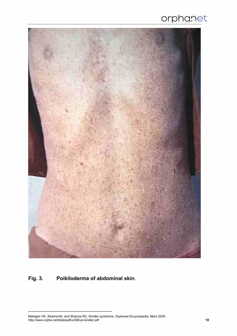
Fig. 3. Poikiloderma of abdominal skin.
Mahajan VK, Sharma NL and Sharma RC, Kindler syndrome. Orphanet Encyclopedia. Mars 2005. http://www.orpha.net/data/patho/GB/uk-kindler.pdf 10

Fig. 4. Characteristic atrophic skin over dorsa of hands.
Mahajan VK, Sharma NL and Sharma RC, Kindler syndrome. Orphanet Encyclopedia. Mars 2005. http://www.orpha.net/data/patho/GB/uk-kindler.pdf 11

Fig. 5. Mild palmar hyperkeratosis. Note: Unusual shape of fingers.
Mahajan VK, Sharma NL and Sharma RC, Kindler syndrome. Orphanet Encyclopedia. Mars 2005. http://www.orpha.net/data/patho/GB/uk-kindler.pdf 12

Fig. 6. Leukokeratosis of oral mucosa near angle of mouth. Note: Erosions and scarring at oral commissures
Mahajan VK, Sharma NL and Sharma RC, Kindler syndrome. Orphanet Encyclopedia. Mars 2005. http://www.orpha.net/data/patho/GB/uk-kindler.pdf 13

Fig. 7 Leukokeratosis of anal mucosa.
Mahajan VK, Sharma NL and Sharma RC, Kindler syndrome. Orphanet Encyclopedia. Mars 2005. http://www.orpha.net/data/patho/GB/uk-kindler.pdf 14

Fig. 8. Severe phimosis.
Mahajan VK, Sharma NL and Sharma RC, Kindler syndrome. Orphanet Encyclopedia. Mars 2005. http://www.orpha.net/data/patho/GB/uk-kindler.pdf 15

Fig. 9. Severe periodontitis and loss of teeth
Mahajan VK, Sharma NL and Sharma RC, Kindler syndrome. Orphanet Encyclopedia. Mars 2005. http://www.orpha.net/data/patho/GB/uk-kindler.pdf 16

17
Fig.10.Turricephaly. Premature obliteration of transverse sutures causes upward growth via sagital sutures resulting in a dome shaped skull. Note: Loss of mandibular angle.
Mahajan VK, Sharma NL and Sharma RC, Kindler syndrome. Orphanet Encyclopedia. Mars 2005. http://www.orpha.net/data/patho/GB/uk-kindler.pdf 17