Tourette’s Syndrome and Tic Disorders
264
-
Upload
independent -
Category
Documents
-
view
1 -
download
0
Transcript of Tourette’s Syndrome and Tic Disorders


Neurological and Psychiatric Disorders

C U R R E N T C L I N I C A L N E U R O L O G Y
Daniel Tarsy, MD, SERIES EDITORS
Neurological and Psychiatric Disorders: From Bench to Bedside, edited by Frank I.Tarazi and John A. Schetz, 2005
Status Epilepticus: A Clinical Perspective, edited by Frank W. Drislane, 2005
Thrombolytic Therapy for Acute Stroke, Second Edition, edited by Patrick D. Lyden,2005
Movement Disorders Emergencies: Diagnosis and Treatment, edited by Steven J.Frucht and Stanley Fahn, 2005
Inflammatory Disorders of the Nervous System: Pathogenesis, Immunology,and Clinical Management, edited by Alireza Minagar and J. Steven Alexander, 2005
Multiple Sclerosis: Etiology, Diagnosis, and New Treatment Strategies, edited byMichael J. Olek, 2005
Parkinson’s Disease and Nonmotor Dysfunction, edited by Ronald F. Pfeiffer and IvanBodis-Wollner, 2005
Seizures in Critical Care: A Guide to Diagnosis and Therapeutics, edited byPanayiotis N. Varelas, 2005
Vascular Dementia: Cerebrovascular Mechanisms and Clinical Management, edited byRobert H. Paul, Ronald Cohen, Brian R. Ott, Stephen Salloway, 2005
Atypical Parkinsonian Disorders, edited by Irene Litvan, 2005
Handbook of Neurocritical Care, edited by Anish Bhardwaj, Marek A. Mirski,and John A. Ulatowski, 2004
Handbook of Stroke Prevention in Clinical Practice, edited by Karen L. Furieand Peter J. Kelly, 2004
Clinical Handbook of Insomnia, edited by Hrayr P. Attarian, 2004
Critical Care Neurology and Neurosurgery, edited by Jose I. Suarez, 2004
Alzheimer’s Disease: A Physician’s Guide to Practical Management, edited by Ralph W.Richter and Brigitte Zoeller Richter, 2004
Field of Vision: A Manual and Atlas of Perimetry, edited by Jason J. S. Bartonand Michael Benatar, 2003
Surgical Treatment of Parkinson’s Disease and Other Movement Disorders, edited byDaniel Tarsy, Jerrold L. Vitek, and Andres M. Lozano, 2003
Myasthenia Gravis and Related Disorders, edited by Henry J. Kaminski, 2003
Seizures: Medical Causes and Management, edited by Norman Delanty, 2002
Clinical Evaluation and Management of Spasticity, edited by David A. Gelberand Douglas R. Jeffery, 2002
Early Diagnosis of Alzheimer's Disease, edited by Leonard F. M. Scinto and Kirk R.Daffner, 2000
Sexual and Reproductive Neurorehabilitation, edited by Mindy Aisen, 1997

Neurologicaland Psychiatric Disorders
From Bench to Bedside
Edited by
Frank I. Tarazi, PhD, MScDepartment of Psychiatry and Neuroscience Program,
Harvard Medical School, Boston, MA;Laboratory of Psychiatric Neuroscience,
McLean Hospital, Belmont MA
John A. Schetz, PhDDepartment of Pharmacology and Neuroscience,Univesity of North Texas Health Science Center,
Fort Worth, TX
Foreword by
John L. Waddington, PhD, DScDepartment of Clinical Pharmacology,Royal College of Surgeons in Ireland,
Dublin

© 2005 Humana Press Inc.999 Riverview Drive, Suite 208Totowa, New Jersey 07512
humanapress.com
All rights reserved. No part of this book may be reproduced, stored in a retrieval system, or transmitted inany form or by any means, electronic, mechanical, photocopying, microfilming, recording, or otherwisewithout written permission from the Publisher.
All papers, comments, opinions, conclusions, or recommendations are those of the author(s), and do notnecessarily reflect the views of the publisher.
Due diligence has been taken by the publishers, editors, and authors of this book to assure the accuracy ofthe information published and to describe generally accepted practices. The contributors herein havecarefully checked to ensure that the drug selections and dosages set forth in this text are accurate and inaccord with the standards accepted at the time of publication. Notwithstanding, as new research, changesin government regulations, and knowledge from clinical experience relating to drug therapy and drugreactions constantly occurs, the reader is advised to check the product information provided by the manu-facturer of each drug for any change in dosages or for additional warnings and contraindications. This isof utmost importance when the recommended drug herein is a new or infrequently used drug. It is theresponsibility of the treating physician to determine dosages and treatment strategies for individual pa-tients. Further it is the responsibility of the health care provider to ascertain the Food and DrugAdministration status of each drug or device used in their clinical practice. The publisher, editors, andauthors are not responsible for errors or omissions or for any consequences from the application of theinformation presented in this book and make no warranty, express or implied, with respect to the contentsin this publication.
This publication is printed on acid-free paper.ANSI Z39.48-1984 (American Standards Institute) Permanence of Paper for Printed Library Materials.
Cover illustration: From Fig. 1 in Chapter 2, “Pharmacotherapeutic Principles of Neurologicaland Psychiatric Disorders,” by John A. Schetz.
Cover design by Patricia F. Cleary
Production Editor: Robin B. Weisberg
For additional copies, pricing for bulk purchases, and/or information about other Humana titles, contactHumana at the above address or at any of the following numbers: Tel.: 973-256-1699; Fax: 973-256-8314; E-mail: [email protected], or visit our Website: http://humanapress.com
Photocopy Authorization Policy:
Authorization to photocopy items for internal or personal use, or the internal or personal use of specificclients, is granted by Humana Press Inc., provided that the base fee of US $25.00 per copy is paid directly tothe Copyright Clearance Center at 222 Rosewood Drive, Danvers, MA 01923. For those organizations thathave been granted a photocopy license from the CCC, a separate system of payment has been arranged andis acceptable to Humana Press Inc. The fee code for users of the Transactional Reporting Service is: [1-58829-369-6/05 $25.00].
Printed in the United States of America. 10 9 8 7 6 5 4 3 2 1
eISBN: 1-59259-856-0
Library of Congress Cataloging-in-Publication Data
Neurological and psychiatric disorders : from bench to bedside / edited byFrank I. Tarazi, John A. Schetz. p. ; cm. -- (Current clinical neurology) Includes bibliographical references and index. ISBN 1-58829-369-6 (hardcover : alk. paper) 1. Neuropsychiatry. I. Tarazi, Frank I. II. Schetz, John A. III. Series. [DNLM: 1. Mental Disorders. 2. Nervous System Diseases. WL 140 N49192005]RC341.N397 2005616.8--dc22
2004015772

Dedication
To the countless colleagues and organizations that have helped us realizethis book project and to our families for their everlasting support
and encouragement.

Series Editor’s Introduction
vi
The disciplines of neurology and psychiatry have had a long and complicated rela-tionship for many years. Although both specialties deal with the brain,scientific and clinical approaches to this complex organ have differed widelyduring the past century. However, beginning in the 1960s both fields saw anenormous growth in understanding the role of neurotransmitters in normal andabnormal brain function and, as a result, an important convergence of scientific inquirytook place. During this time, dopamine was recognized to be a neurotransmitter ratherthan a precursor of norepinephrine, dopamine replacement therapy was discovered tobe an effective treatment for Parkinson’s disease, and dopamine receptor-blocking drugswere found to be effective for the treatment of schizophrenia and other psychotic disor-ders. The era of biogenic amines was launched and dopamine, norepinephrine, and se-rotonin came under intensive study with the strong expectation that further insights intothe pathophysiology and treatment of other neurological and psychiatric disorderswould be forthcoming.
Fast forwarding to the present, Neurological and Psychiatric Disorders: From Benchto Bedside brings us up to date on the role of the biogenic amines in the pathophysiol-ogy and treatment of a core group of neurological and psychiatric disorders. However,the early notion that manipulation of a single neurotransmitter could produce profoundand lasting therapeutic effects for any of these conditions was clearly simplistic. Aspointed out by Drs. Tarazi and Schetz, many pharmacological treatments provide relieffor only one or several symptoms in a particular disorder. This includes evenParkinson’s disease where there is much more to the story than simple dopamine defi-ciency. Other neurotransmitters including but not limited to -aminobutyric acid,glutamic acid, and acetylcholine as well as a variety of neuropeptides and other smallmolecules also play a key role in these diseases. More also needs to be understoodabout the complex neuronal circuits and pathways between basal ganglia, limbic sys-tem, and cerebral cortex, which appear to underlie normal and abnormal motor, emo-tional, and cognitive functions.
Neurological and Psychiatric Disorders: From Bench to Bedside provides muchmore than an overview of the neurobiology of a group of neurological and psychiatricdisorders. The chapters are uniformly very well organized to provide a broad, compre-hensive and systematic review of the epidemiology, pathophysiology, molecularbiology, genetics, neuropathology, brain imaging, clinical features, and pharmacologi-cal and nonpharmacological treatments of several mental illnesses, that will serve as avaluable core of information for physicians, scientists, advanced students, and healthcare professionals in this area. A unique bonus in this volume is the very sophisticatedpsychiatric approaches to neurological disorders and neurological approaches to psy-chiatric disorders, which are provided, in the individual chapters. Finally and most im-portantly, this book should also serve to promote future interdisciplinary research whichwill further the understanding of these disorders.
Daniel Tarsy, MD
Parkinson’s Disease and Movement Disorders CenterBeth Israel Deaconess Medical CenterHarvard Medical School, Boston, MA

Foreword
vii
In the 1970s I had the privilege of meeting the long-serving British Prime Min-ister Harold Wilson at a degree-conferring ceremony, in his capacity as chancellorof the University of Bradford, United Kingdom. Our brief conversation gave nohint of what was to come: within a short period he had unexpectedly resigned asprime minister, engendering enormous intrigue and controversy. Only many yearslater, on his death, was it confirmed that he had developed Alzheimer’s disease andthat his resignation from office appeared to reflect some insight and concern thathis mental abilities were not what they were. This is in striking contrast to a genera-tion later when, in the 1990s, former US President Ronald Reagan wrote his poi-gnant letter to the American people informing them that he was leaving public lifeto face the challenges of Alzheimer’s disease.
Subsequently, a series of high-profile individuals in the United States have re-vealed and spoken publicly of their conditions, including Charlton Heston(Alzheimer’s disease) and Michael J. Fox (Parkinson’s disease). In the United King-dom, the leading English soccer team, Manchester United, signed a new goalkeeper,Tim Howard, who speaks openly of having Tourette’s syndrome. This new publicopenness in relation to neurological and psychiatric disorders, which had been facedpreviously in secrecy and even shame, has served both to help de-stigmatize suchdisorders and to indicate the need for increasing research thereon. Perhaps no indi-vidual in the public eye helped to focus attention on neuroscience as a route toimproved treatment modalities than did Christopher Reeve. Prior to his recent un-timely death, Reeve’s passionate advocacy of neuroscience research as an impera-tive for his own salvation and that of others affected similarly exemplified theindividual adversities that give real, highly personalized meaning to “from bench tobedside.”
Here, Drs. Frank Tarazi and John Schetz have assembled a team of expert au-thors to give an authoritative, contemporary overview of the field. In sport, teamsusually have to play to a manager’s tactical plan. The editors have proceeded simi-larly with their team of authors to good effect, focusing on the extent to which the“holy grail” of a bench-to-bedside research trajectory has been realized. Introduc-tory chapters describe, at an accessible level, the principles underpinning researchin this area: first, the neural principles of neurological and psychiatric disorders,from laboratory methods to clinical neuroimaging techniques and the insights thatthey can provide; second, the pharmacotherapeutic principles that underpin treat-ment, from receptorology to applied issues that govern the most effective use ofmedications. Thereafter, chapters describe our current understanding of major dis-orders in neurology (Alzheimer’s disease, Huntington’s disease, Parkinson’s dis-ease) and psychiatry (schizophrenia, autism spectrum disorders, Tourette’s
viii Foreword
syndrome and tic disorders, obsessive-compulsive disorder, unipolar depression,bipolar disorder, attention deficit hyperactivity disorder). It should be noted thatthese chapters indicate how contemporary neuroscience reveals the arbitrariness ofthis somewhat artificial separation into “neurological” and “psychiatric” disorders;these may more reflect historical artifact and professional territory than indicatefundamental distinctions in terms of scientific understanding and human suffering.
To achieve their bench-to-bedside perspective, the editors have prevailed on theirauthors to adopt a uniform structure for each chapter. This leads the reader from therelevant brain pathways, molecular mechanisms and targets, through genetics andanimal models, to clinical epidemiology, phenomenology, and treatment issues.Unusually, at the end of each chapter is a useful glossary of medical terminologythat constitutes an additional aid to clarity and accessibility. Through these innova-tions, the editors and authors have marshalled concisely the most up-to-date infor-mation pertaining to major neurological and psychiatric disorders that constitutesome of our most serious public health problems. Although not yet fully realized,this volume documents that the bench-to-bedside research trajectory is no longer a“holy grail” but a realistic goal toward which considerable progress has been made.
John L. Waddington, PhD, DSc,Department of Clinical Pharmacology,
Royal College of Surgeons in Ireland,Dublin

Preface
ix
The neurological and psychiatric disorders discussed in Neurological and Psy-chiatric Disorders: From Bench to Bedside are commonly treated with pharmaco-therapies that target brain dopaminergic, serotonergic, or noradrenergic systems,either individually or in combination. Additional treatment strategies, includingpsychiatric, psychosocial, psychosurgical, and electrical/magnetic therapies are alsoavailable either as monotherapies or to augment drug treatments. Pharmacothera-pies for neurological and psychiatric disorders are thought to be primarily pallia-tive, and often they are prescribed to provide relief for only one of a range ofsymptoms encountered for each disorder. For example, antipsychotic drugs are pre-scribed for the treatment of agitation in Alzheimer’s disease, aggression and self-injury in autism, mania in bipolar disorder, motor dysfunction in Huntington’sdisease, motor tics in Tourette’s syndrome, and psychosis in schizophrenia. Anti-depressant drugs are employed in the treatment of depression in unipolar and bipo-lar depression and obsessive behaviors in obsessive–compulsive disorder. Such arange of treatments for different symptom modalities for different disordersowes to either the direct or indirect dysfunction of dopaminergic, serotonergic, andnoradrenergic system(s). In addition, there is considerable overlap in selectedportions of the extended brain pathways related to the pathophysiology andtreatment of certain of these disorders. Neurological and Psychiatric Disorders:From Bench to Bedside provides the reader with a full appreciation for all suchfactors as they relate to preclinical research and to clinical diagnosis and treatmentof the neurological and psychiatric disorders.
Neurological and Psychiatric Disorders: From Bench to Bedside aims to pro-vide comprehensive and systematic coverage of the basic and clinical aspects of thecore neurological and psychiatric disorders that are commonly taught to doctorate-and postdoctorate-level students. Until now, many texts were needed to cover thepreclinical and clinical aspects of different disorders in sufficient breadth and depthto allow for MDs, PhDs, and PharmDs to master their qualifying (board) exams,and, at the same time, effectively and safely practice their future health care profes-sions. Having a single text will not only facilitate cross-discipline teaching andlearning by providing a uniform instructional platform, but will also satisfy studentconcerns about the costs and variable coverage generated by the need for multipletexts. The editors and contributing authors undertook a collaborativeeffort to produce such a textbook, which is based on the syllabus and content of anadvanced graduate-level course concerning the neurological basis of diseases anddisorders and their treatment that Dr. Schetz developed and continues to teach. Theprimary editorial responsibilities and the recruitment of authoritative authors forthe chapters on the individual disorders were divided evenly between the editors:

x Preface
Dr. Tarazi recruited authors for and edited the chapters on attention deficit hyper-activity disorder, bipolar disorder, Huntington’s disease, Parkinson’s disease, andschizophrenia, and Dr. Schetz recruited authors for and edited the chapters onAlzheimer’s disease, autism, obsessive–compulsive disorder, Tourette’s syndrome,and unipolar depression.
Neurological and Psychiatric Disorders: From Bench to Bedside opens withtwo introductory chapters that reintroduce the reader to the neural and pharmaco-therapeutic principles of neurological and psychiatric disorders. These chapters alsodiscuss theoretical and methodological parameters relevant to the appropriateinterpretation of pharmacodynamic, pharmacokinetic, neuroanatomical, and imag-ing data presented throughout. Subsequent chapters provide a standard minimumcontent concerning each disorder along with a uniformity that is intended topromote not only the comparing and contrasting of the disorders discussed through-out, but also the cross-discipline training many health care industry experts believeis lacking. The standard minimum uniform content for each disorder can be dividedinto the following nine categories:
1. Incidence/prevalence2. Etiology and general description3. Molecular targets and mechanisms of action4. Brain structures and pathways, and neurotransmitter systems5. Animal models6. Signs and symptoms in humans (diagnostics, markers, comorbidities, subclassifica-
tions)7. Genetics8. Treatments (pharmacological and other types of therapies)9. Related medical terminology
Thus, the objective is to promote a systematic, comprehensive, and advancedunderstanding of each disorder from molecules to human behaviors. Our intendedaudience includes graduate- and postgraduate-level physicians and scientists, pre-clinical and clinical research scientists, and pharmacists with an intermediate toadvanced understanding of neuroscience and pharmacology.
We extend our deepest gratitude to the chapter authors for their outstandingscholarly contributions. A special thanks goes to our advisors and mentors whohave over the years generated in us an intense and perpetual desire to challengeourselves and to instill in the next generation of young scientists, physicians andpharmacists the excitement and satisfaction that we experience from working in thehealth care profession and answering the call to public service. In particular, wewish to recognize the kind and astute influences that Dr. Ian Creese and Dr. Ross J.Baldessarini have had on Dr. Tarazi and that Dr. William G. Luttge, Dr. David R.Sibley, and Dr. Harel Weinstein have had on Dr. Schetz.
Frank I. Tarazi, PhD, MScJohn A. Schetz, PhD

xi
Contents
Series Editor’s Introduction .............................................................................. vii
Foreword ............................................................................................................... ix
Preface .................................................................................................................... xi
Contributors ......................................................................................................... xv
Part I. Introduction1 Neural Principles of Neurological and Psychiatric
Disorders ...................................................................................................3Frank I. Tarazi and Marc J. Kaufman
2 Pharmacotherapeutic Principles of Neurologicaland Psychiatric Disorders ................................................................... 29
John A. Schetz
Part II. Neurological Disorders3 Alzheimer’s Disease .................................................................................. 51
Mark P. Mattson
4 Huntington’s Disease ............................................................................... 63Susan E. Browne
5 Parkinson’s Disease .................................................................................. 87Thomas Wichmann
Part III. Psychiatric Disorders6 Schizophrenia ........................................................................................... 111
Stephan Heckers and Sabina Berretta
7 Autism Spectrum Disorders .................................................................. 131Evdokia Anagnostou and Eric Hollander
8 Tourette’s Syndrome and Tic Disorders: From Moleculesand Cells to Symptoms and Management ........................................... 151
James E. Swain, Robert A. King, and James F. Leckman
9 Obsessive-Compulsive Disorder .......................................................... 171David S. Husted, Nathan A. Shapira, and Wayne K. Goodman
10 Unipolar Depression ............................................................................... 189Julie A. Blendy and Irwin Lucki

xii Contents
11 Bipolar Disorder ...................................................................................... 205Leonardo Tondo, Matthew J. Albert, Alexia E. Koukopoulos,
Christopher Baethge, and Ross J. Baldessarini
12 Attention Deficit Hyperactivity Disorder ........................................... 229Kehong Zhang, Eugen Davids, and Ross J. Baldessarini
Index .................................................................................................................... 251
Color Plates
Color plates 1–5 appear in an insert preceding p. 51.
PLATE 1 Fig. 1 from Chapter 2; for complete caption see p. 30.
PLATE 2 Fig. 1 from Chapter 3; for complete caption see p. 52.
PLATE 3 Fig. 1 from Chapter 7; for complete caption see p. 133.
PLATE 4 Fig. 1 from Chapter 8; for complete caption see p. 156.
PLATE 5 Fig. 1 from Chapter 9; for complete caption see p. 173.

MATTHEW J. ALBERT, BA • Department of Psychiatry, University of Romeat San Andrea Hospital and Centro Lucio Bini, Rome, Italy
EVDOKIA ANAGNOSTOU, MD • Seaver and New York Autism Centerof Excellence, Department of Psychiatry, Mount Sinai Schoolof Medicine, New York, NY
CHRISTOPHER BAETHGE, MD • Department of Psychiatry, McLean Hospital,Harvard Medical School, Boston, MA; Department of Psychiatry,Free University of Berlin, Berlin, Germany
ROSS J. BALDESSARINI, MD • Mailman Research Center, Departmentof Psychiatry, McLean Hospital, Harvard Medical School, Boston, MA
SABINA BERRETTA, MD • Laboratory of Translational Neuroscience, McLeanHospital, Department of Psychiatry, Harvard Medical School, Boston,MA
JULIE A. BLENDY, PhD • Department of Pharmacology, University of PennsylvaniaSchool of Medicine, Philadelphia, PA
SUSAN E. BROWNE, PhD • Department of Neurology and Neuroscience, WeillMedical College of Cornell University, New York, NY
EUGEN DAVIDS, MD • Departments of Psychiatry and Psychotherapy,University of Essen, Essen, Germany
WAYNE K. GOODMAN, MD • Department of Psychiatry, University of FloridaCollege of Medicine, Gainesville, FL
STEPHAN HECKERS, MD • Schizophrenia and Bipolar Disorder Programand Functional Neuroanatomy Laboratory, McLean Hospital,Department of Psychiatry, Harvard Medical School, Boston, MA
ERIC HOLLANDER, MD • Seaver and New York Autism Center of Excellence,Department of Psychiatry, Mount Sinai School of Medicine,New York, NY
DAVID S. HUSTED, MD • Department of Psychiatry, University of FloridaCollege of Medicine, Gainesville, FL
MARC J. KAUFMAN, PhD • Brain Imaging Center, McLean Hospital,Department of Psychiatry and Neuroscience Program, HarvardMedical School, Boston, MA
ROBERT A. KING, MD • Child Study Center, Yale University Schoolof Medicine, New Haven, CT
ALEXIA E. KOUKOPOULOS, MD • Department of Psychiatry, Universityof Rome at San Andrea Hospital and Centro Lucio Bini, Rome, Italy
JAMES F. LECKMAN, MD • Child Study Center, Yale University Schoolof Medicine, New Haven, CT
Contributors
xiii
IRWIN LUCKI, PhD • Department of Psychiatry, University of PennsylvaniaSchool of Medicine, Philadelphia, PA
MARK P. MATTSON, PhD • Laboratory of Neurosciences, GerontologyResearch Center, National Institute on Aging; Departmentof Neurology, Johns Hopkins University School of Medicine,Baltimore, MD
JOHN A. SCHETZ, PhD • Department of Pharmacology and Neuroscience,University of North Texas Health Science Center, Fort Worth, TX
NATHAN A. SHAPIRA, MD, PhD • Department of Psychiatry, Universityof Florida College of Medicine, Gainesville, FL
JAMES E. SWAIN, MD, PhD • Child Study Center, Yale University Schoolof Medicine, New Haven, CT
DANIEL TARSY, MD • Parkinson’s Disease and Movement Disorders Center,Beth Israel Deaconess Medical Center, Harvard Medical School,Boston, MA
FRANK I. TARAZI, PhD, MSC • Laboratory of Psychiatric Neuroscience,McLean Hospital, Department of Psychiatry, Harvard Medical School,Boston, MA
LEONARDO TONDO, MD • Department of Psychology, University of Cagliari,and Centro Lucio Bini, Stanley Medical Research Center, Cagliari,Sardinia
JOHN L. WADDINGTON, PhD DSC • Department of Clinical Pharmacology,Royal College of Surgeons in Ireland, Dublin, Ireland
THOMAS WICHMANN, MD • Department of Neurology, Emory UniversitySchool of Medicine, Atlanta, GA
KEHONG ZHANG, MD, PhD • McLean Hospital, Department of Psychiatry,Harvard Medical School, Boston, MA
xiv Contributors

Neural Principles 3
3
From: Current Clinical Neurology: Neurological and Psychiatric Disorders:From Bench to Bedside
Edited by: F. I. Tarazi and J. A. Schetz © Humana Press Inc., Totowa, NJ
1Neural Principles of Neurological
and Psychiatric Disorders
Frank I. Tarazi and Marc J. Kaufman
SummaryRecent progress in understanding the neurobiology and pharmacology of brain neu-
rotransmitters and their neural circuits and pathways has been remarkable and serves as thefoundation for in-depth preclinical research on neurological and psychiatric disorders. Mostof the neurotransmitter receptors have been cloned and their anatomical localization anddistribution have been clarified using advanced neurochemical methods. Novel discoverieshave greatly stimulated clinical studies in the neuropathophysiology and neurogenetics ofbrain disorders. Sophisticated developments in neuroimaging techniques have yielded revo-lutionary insights into the complexity of maldevelopment or dysfunction of neuronalelements in neurological and psychiatric disorders, and have assisted in the rational designof novel neuropsychotropic agents for improved treatment of these disorders.
Key Words: Brain pathways; dopamine; emission tomography; magnetic resonance;neuroimaging; norepinephrine; serotonin.
1. INTRODUCTIONNerve cells in the brain (neurons) communicate with each other via chemical
substances (neurotransmitters) that are released from synaptic terminals. Oncereleased, these neurotransmitters target adjacent neurons and alter cellular activi-ties and functions of influenced neurons. Neurons and neurotransmitters can beorganized into specialized circuits and signaling pathways. Neural circuits can beas simple as one synapse and one neurotransmitter or very complex andinvolve different synapses, multiple neurotransmitters, and extend across differ-ent brain regions. Preclinical and clinical evidences strongly suggest that abnormaldevelopment of brain neurons and malfunctions of brain circuits significantlycontribute to the development of different neurological and psychiatric disorders.Subsequently, drugs developed for treatment of these disorders tend to restoreneurotransmission and normalize neuron–neuron communications. Advances inhistochemical and autoradiographic techniques have improved the detailed charac-

4 Tarazi and Kaufman
terization of the anatomical localization of different neurotransmitter receptors, andthe quantification of their levels in laboratory animals and in postmortem humantissue. Additionally, sophisticated imaging techniques have helped to identify spe-cific brain regions associated with particular modes of behaviors and permitted thedirect visualization of neurotransmitter receptors in healthy subjects and diseasedpatients. This chapter introduces some of the key brain neurotransmitters and brainpathways as well as neurochemical and neuroimaging techniques that greatly enrichedour knowledge of the neural principles of neurological and psychiatric disorders.
2. BRAIN NEUROTRANSMITTERSThree major brain neurotransmitter systems—dopamine (DA), norepinephrine
(NE), and serotonin—are intimately involved in the pathophysiology, neuropathol-ogy, and pharmacotherapy of neurological and psychiatric disorders discussed infollowing chapters.
2.1. DopamineFor the longest time, DA was considered an intermediate molecule in the bio-
synthesis of other catecholamines including epinephrine and NE. In mid-1950s,Arvid Carlsson and his colleagues proposed a prominent biological role for DA asan independent neurotransmitter. These seminal proposals were the impetus forexplosive advances in DA research. Several clinically relevant contributions hademerged from the study of DA systems, including the role of these systems in thepathophysiology and treatment of attention deficit hyperactivity disorder (ADHD),Huntington’s chorea, Parkinson’s disease, schizophrenia, Tourette’s syndrome, andother tic disorders (1).
2.1.1. DA PathwaysDA neurons of the mammalian central nervous system (CNS) are organized into
four major subsystems: (a) the nigrostriatal system: neurons project from the sub-stantia nigra pars compacta, also called A9 neurons, to the caudate-putamen (CPu).This system accounts for 70% of the total brain content of DA, and its degenerationmakes a major contribution to the pathophysiology of Parkinson’s disease; (b) themesolimbic system: originates in the ventral tegmental area, also called A10 neu-rons, and projects to different components of the limbic system including thenucleus accumbens, lateral septal nuclei, amygdala, hippocampus, and theentorhinal cortex; (c) the mesocortical system: arises from cell bodies in ventraltegmental area and project to cerebral cortex, particularly to mesioprefrontal areas;and (d) the tuberinfundibular neuroendocrinological system: neurons arise in thearcuate and other nuclei of the hypothalamus and end in the median eminence ofthe inferior hypothalamus, in which DA exerts regulatory effects in the anteriorpituitary, and serves as a prolactin inhibitory factor (2).
2.1.2. DA Synthesis and MetabolismDA is synthesized from L-tyrosine, which is converted to L-3,4-dihydroxy-
phenylalanine (L-DOPA) in a reaction catalyzed by the enzyme tyrosine hydroxy-
Neural Principles 5
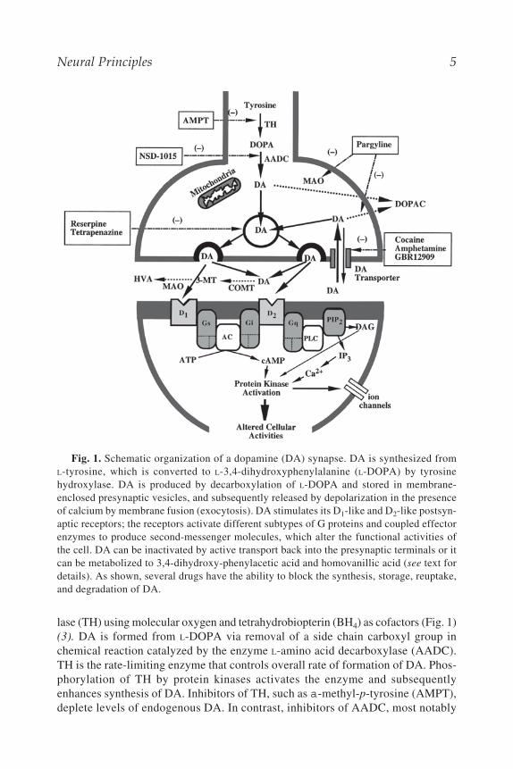
lase (TH) using molecular oxygen and tetrahydrobiopterin (BH4) as cofactors (Fig. 1)(3). DA is formed from L-DOPA via removal of a side chain carboxyl group inchemical reaction catalyzed by the enzyme L-amino acid decarboxylase (AADC).TH is the rate-limiting enzyme that controls overall rate of formation of DA. Phos-phorylation of TH by protein kinases activates the enzyme and subsequentlyenhances synthesis of DA. Inhibitors of TH, such as a-methyl-p-tyrosine (AMPT),deplete levels of endogenous DA. In contrast, inhibitors of AADC, most notably
Fig. 1. Schematic organization of a dopamine (DA) synapse. DA is synthesized fromL-tyrosine, which is converted to L-3,4-dihydroxyphenylalanine (L-DOPA) by tyrosinehydroxylase. DA is produced by decarboxylation of L-DOPA and stored in membrane-enclosed presynaptic vesicles, and subsequently released by depolarization in the presenceof calcium by membrane fusion (exocytosis). DA stimulates its D1-like and D2-like postsyn-aptic receptors; the receptors activate different subtypes of G proteins and coupled effectorenzymes to produce second-messenger molecules, which alter the functional activities ofthe cell. DA can be inactivated by active transport back into the presynaptic terminals or itcan be metabolized to 3,4-dihydroxy-phenylacetic acid and homovanillic acid (see text fordetails). As shown, several drugs have the ability to block the synthesis, storage, reuptake,and degradation of DA.

6 Tarazi and Kaufman
a-methyl-dopa-hydrazine (carbidopa) and 3-hydroxybenzyl-hydrazine (NSD 1015)causes an accumulation of L-DOPA in the brain (Fig. 1) (3).
After synthesis, DA is stored in membrane-enclosed presynaptic vesicles, thenreleased through the mechanism of vesicular exocytosis that is triggered by a largeinflux of calcium (Ca2+) into the nerve terminals; however, nonvesicular Ca2+ inde-pendent-DA release can occur in some cases. Once DA is released into synapticcleft, it becomes subjected to multiple routes of inactivation. It is inactivated byactive transport back into the presynaptic terminals via cell membrane DA trans-porters (Fig. 1). Additionally, excessive synaptic DA undergoes a series of chemi-cal degradation reactions that involves two enzymes: catechol-O-methyltransferase(COMT) and monoamine oxidase (MAO). About 90% of DA metabolism startswith oxidative deamination by MAO. The product of this reaction, 3,4–dihydroxy-phenylacetaldehyde (DHPA) is rapidly oxidized to 3,4–dihydroxy-phenylaceticacid (DOPAC) by aldehyde dehydrogenase. A major portion of DOPAC is O-methy-lated to yield homovanillic acid (HVA). DOPAC and HVA are the most significantmetabolites of DA, and they are transported outside the brain into the blood streamand eventually excreted. The remaining 10% of DA can be metabolized by COMTinto 3-methoxytyramine (3-MT), which is then converted to HVA by MAO andaldehyde dehydrogenase (Fig. 1) (3).
The DA synapse is selected as a representative of other neurotransmitter syn-apses, and a schematic organization of this synapse is shown in Fig. 1.
2.1.3. DA Receptor Subtypes
DA receptors belong to a superfamily of large proteins characterized by havingseven relatively hydrophobic transmembrane-spanning (TMS) segments, as well asa functionally critical third intracytoplasmic loop located between the fifth and sixthtransmembrane segments. This loop couples to G proteins that interact with severalmembrane or cytoplasmic effector molecules that lead to the physiological regu-lation of neuronal function.
DA receptors were originally differentiated into two major types—D1 and D2.D1 and D2 receptors were characterized initially as mediating stimulation and inhi-bition of cyclic adenosine monophosphate (cAMP) production, respectively. Tech-nical advances in molecular biology have greatly facilitated the discovery of threenovel, closely related but less abundant DA receptor subtypes. Based on their closestructural homologies and their opposite effects on cAMP productions, the five DAreceptors are classified into D1-like (D1, D5) and D2-like (D2, D3, D4) receptorfamilies (1,2).
2.1.3.1. D1-LIKE RECEPTORS
D1 receptors are the most abundant DA receptor subtype in mammalian fore-brain. The human D1 gene has been localized to chromosome 5. The messengerriboprobes (mRNA) for D1 receptor has been localized in striatal neurons of thebasal ganglia, olfactory tubercule, and nucleus accumbens, followed by cerebralcortex and hippocampus. Additionally to stimulation of cAMP production, D1 recep-

Neural Principles 7
tors show ability to stimulate phospholipase C and activate phosphoinositide sec-ond messenger cascade in in vitro expression system.
D5 receptors are less studied than their D1 siblings. The protein has an overall50% homology with the D1 receptor and 80% if only the transmembrane segmentsare considered. The human D5 gene has been localized to chromosome 4.The mRNA for D5 receptor has been localized in hippocampus and thalamus.D5 receptors appear to interact with G proteins and can stimulate adenylyl cyclasein transfected cells, with relatively high affinity for DA and D1-selective agonists.
2.1.3.2. D2-LIKE RECEPTORS
D2 receptors are the second most abundant DA receptor subtype, although theywere the first to be cloned. The human D2 gene has been localized to chromosome11. Human and rat D2 receptor splice variants are generated by alternative splicing,and vary by the presence or absence of 29-amino acids in the third cytoplasmicloop. These variants are referred to as long vs short isoforms (D2L vs D2S). Bothisoforms have similar pharmacological profiles and both inhibit adenylyl cyclaseactivity. The mRNA for D2 receptor has been localized in basal ganglia, nucleusaccumbens, and olfactory tubercle, followed by substantia nigra and ventral teg-mental area. D2 receptors are linked to several signal transduction mechanisms.Additionally to inhibition of cAMP production, D2 receptor stimulation inhibitsphosphoinositide cycle, activates potassium (K+) channels, and potentiates arachi-donic acid release.
D3 receptors are the first novel DA receptors to be cloned. The human D3 genehas been localized to chromosome 3, and, like the genes for D2 receptors, D3 mRNAalso occurs in longer and shorter spliced forms. The mRNA for D3 receptor hasbeen localized in limbic regions including the islands of Calleja, the olfactorytubercle, and the shell of NAc. It is also found sparingly in some midbrain DAneurons and in cerebellum. The functional status of D3 receptors remains uncertainbecause they have not been linked consistently with effector mechanisms in trans-fected cell preparations.
D4 receptors are the third members of D2-like receptor family. The human D4gene has been localized to chromosome 11. In the brain, mRNA for the D4 receptoris expressed in the hippocampus and frontal cortex. Substantial levels of D4 recep-tor proteins, but only low levels of their mRNA, are reported to occur in the stria-tum, suggesting that a portion of D4 receptors reside on terminals of corticostriatalprojections innervating striatum. The human D4 receptor can be transcribed intodifferent polymorphic variants that differ in the number of repeats of a small,48-amino acid sequence within the functionally critical third cytoplasmic loop.There can be 2 to 11 repeats, but 2, 4, or 7 are the most common (D4.2, D4.4, D4.7).Stimulation of D4 receptors can inhibit adenylyl cyclase activity and activate releaseof arachidonic acid in transfected cells.
2.2. NorepinephrineNE was originally proposed to serve as a neurotransmitter in the peripheral sym-
pathetic nerves. Subsequently, NE was identified as a central neurotransmitter in
8 Tarazi and Kaufman
the mammalian brain with specialized cortical executive functions. Disturbances inNE neurotransmission have been clinically associated with depression, as wellADHD, schizophrenia, and other disorders of attention and cognition (3).
2.2.1. NE PathwaysThe nuclei of origin for the central NE system are in the pons and medulla (4).
These nuclei consist of three main groups:
1. locus coerulus (LC) complex: This is the most prominent NE nucleus. Axons of LCproject to several telencephalic and diencephalic regions, including several areas ofcerebral cortex, hippocampus, amygdala, septum, thalamus, and hypothalamus;
2. lateral tegmental system: Axons of this system project caudally to the spinal cord andterminate in the intermediolateral cell columns of the thoracic and upper lumbar cord.The rostral projections of this system make up the ventral NE bundle, which termi-nates in the hypothalamus and other diencephalic structures; and
3. dorsal medullary group: Fibers of this system project to nucleus of tractus solitarius inthe medulla, dorsal vagal complex, and other primary motor and visceral nuclei of thecranial nerves. Sympathetic ganglia and chromaffin cells of adrenal medulla representmajor populations of peripheral NE-containing neurons (4).
2.2.2. NE Synthesis and MetabolismNE is synthesized from DA in a reaction catalyzed by dopamine b-hydroxylase
(DbH) using copper, oxygen and ascorbic acid as cofactors. Because NE and DAshare a similar anabolic pathway, TH is also the rate-limiting enzyme that controlsoverall rate of formation of NE. Synthesis of NE can be blocked by TH inhibitors orby DbH inhibitors, such as FLA63 (3).
NE is stored in membrane-enclosed presynaptic vesicles, and it is released intothe synaptic cleft by Ca2+-mediated exocytosis. It is inactivated by active reuptakeinto presynaptic terminals via NE transporters or by degradation. Degradation ofNE starts with either deamination or O-methylation. MAO can catalyze the break-down of NE into 3,4-dihydroxyphenylglycoaldeshyde (DHPGA), which is furthermetabolized into two intermediates 3,4-dihydroxyphenylglycol (DHPG) or 3,4-dihydroxymandelic acid (DHMA). DHPG and DHMA are further O-methylatedinto 3-methoxy-4-hydroxyphenylglycol (MHPG) and vanillymandelic acid , respec-tively. NE can be metabolized by COMT to give normetanephine, which is laterdeaminated to 3-methoxy-4-hydroxyphenylglycolaldehyde (MHPGA). MHPGA isfurther broken down by aldehyde dehydrogenase to a major metabolite MHPG (3).
2.2.3. NE Receptor SubtypesNE receptor proteins are also characterized by having seven relatively hydro-
phobic TMS segments, as well as a functionally critical third intracytoplasmic loopthat couples to G proteins. NE receptors are subdivided into a-adrenergic (a1 anda2) and b-adrenergic (b1, b2, and b3) subfamilies. Although b-adrenergic receptorsubtypes are expressed at variable levels in different areas of the brain, they appar-ently do not play a prominent role in the treatment of several neurological andpsychiatric disorders, and consequently, they are not emphasized here (5,6).

Neural Principles 9
2.2.3.1. a1 RECEPTORS
These receptors are divided into two subtypes: a1A and a1B. a1A receptors arehighly expressed in hippocampus, pons-medulla and spinal cord, whereas a1B
receptors are mainly detected in cerebral cortex, thalamus, hypothalamus, andcerebellum. Both receptors are equally distributed in the striatum. Both a1A anda1B receptor activation results in generation of second messengers inositol 1,4,5-trisphosphate and diacylglycerol and activation of Ca2+ channels.
2.2.3.2. a2 RECEPTORS
a2 receptor family were originally divided into two subtypes (a2A and a2B).Subsequently, two additional members (a2C and a2D) were included in the family.Autoradiographic and in situ studies showed that a2A receptors are expressed insubstantial levels in amygdalo–hippocampal area, the central and basolateral nucleiof the amygdala, and dorsolateral nucleus of the thalamus. The a2A receptor proteinand mRNA are highly expressed in locus coerulus, indicating that this receptorsubtype serves as a somatodendritic autoreceptor in NE neurons. Other a2 recep-tors are found in dentate gyrus, substantia nigra pars reticulata and hippocampal CA1
region. a2 receptors decrease the activity of adenylyl cyclase, whereas a2 auto-receptors inhibit the release of NE by increasing K+ conductance which hyper-polarizes NE neurons (5).
2.3. SerotoninSerotonin or 5-hydroxytryptamine (5-HT) was initially isolated from chromaf-
fin cells of the intestinal mucosa. Only 1 to 2% of total body 5-HT is found in themammalian CNS. Biochemical abnormalities in 5-HT synthesis and degradationhave been linked to various forms of mental illness including bipolar disorder,depression, obsessive–compulsive disorders, and schizophrenia (3).
2.3.1. 5-HT PathwaysThere are nine major groups of 5-HT containing neurons known as B1–B9. These
groups are mainly localized in the raphe nuclei and reticular region of the lowerbrain stem. The 5-HT neurons of the brain stem are divided into caudal and rostralsystems. The caudal system consists of B1–B4 groups and projects from medianand paramedian groups of the medulla and caudal pons to the spinal cord. Therostral 5-HT system comprises the B5–B9 cell groups, and they are associated withthe raphe nuclei of the rostral pons and mesencephalon, as well as the caudal linearnucleus, the nucleus pontis oralis, and the supralemniscal region (7).
The rostral 5-HT system gives rise to two distinct ascending projections termedthe ventral and dorsal pathways. The ventral ascending pathway originates fromgroup B6–B8 cells and innervates basal ganglia, limbic system, and cortex. A majorpart of the ventral pathway enters the medial forebrain bundle and innervates themedial habenula, thalamus, and hypothalamus. Other fibers innervate the dorsaland ventral striatum, amygdala, hippocampus, septal nucleus, olfactory cortex, andall regions of cerebral cortex. The dorsal ascending pathway originates from groupcells B7 and B8 and innervates mesencephalic gray as well as inferior and superior
10 Tarazi and Kaufman
colliculi (7). Two additional 5-HT systems have been identified in the brain.One system projects to cerebellum and terminates in the cerebellar cortex and deepcerebellar nuclei, and the other system innervates locus coeruleus, dorsal tegmentalnucleus, nucleus solitarius, reticular formation, and several cranial nerve nuclei.
2.3.2. 5-HT Synthesis and MetabolismThe first step in synthesis of 5-HT is the hydroxylation of the amino acid tryp-
tophan to yield 5-hydroxytryptophan (5-HTP) in a reaction catalyzed by tryptophanhydroxylase (3). 5-HTP is then converted by AADC to 5-HT. Tryptophan hydroxy-lase, the rate-limiting enzyme that controls the synthesis of 5-HT, requires molecu-lar oxygen and BH4 cofactor. It can be activated by phosphorylation, proteolysisand Ca2+ phospholipids. Once 5-HT is synthesized, it is stored in vesicles andreleased into the synaptic cleft by exocytosis. Released 5-HT can be inactivated byactive reuptake into the presynaptic terminals via 5-HT transporters. Excessivesynaptic 5-HT undergoes deamination by monoamine oxidase to yield 5-hydroxy-indolaldehyde, which is rapidly oxidized by the enzyme aldehyde dehydrogenaseto form 5-hydroxyindolacetic acid , the major metabolite of 5-HT. 5-hydroxy-indolacetic acid is then transported by an acid transport system out of the brain andinto cerebrospinal fluid (3).
2.3.3. 5-HT Receptors5-HT receptor family is more heterogenous than DA and NE receptor families as
it includes a large number of members (8). 5-HT receptors are currently classifiedas: 5-HT1 subfamily (includes subtypes A, B, D, E, and F), 5-HT2 subfamily(includes types A, B, and C), and separate 5-HT3, 5-HT4, 5-HT5 (and its subtypes5-HT5A and 5-HT5B), 5-HT6, and 5-HT7 receptors. There are additional “orphan”5-HT that are not well characterized (8). All in all, there are probably 18 5-HTreceptor subtypes. With the exception of 5-HT3 receptor, which is a ligand gatedchannel, all of the other 5-HT receptor subtypes have the characteristic seven TMSsegments, are coupled to G-proteins and initiate multiple signal transduction mecha-nisms. 5-HT1A, and 5-HT2A remain the most pharmacologically and functionallycharacterized 5-HT receptor subtypes (8).
2.3.3.1. 5-HT1A RECEPTORS
Rat and human 5-HT1A receptors show 99% sequence homology in their sevenTMS domains. They are expressed in high densities in hippocampus, lateral sep-tum, amygdala, frontal and entorhinal cortices. 5-HT1A receptors are also found inmedian and dorsal raphe nuclei indicating a somatodendritic autoreceptor role forthese receptors. 5-HT1A receptor stimulation inhibits cAMP production, decreasesCa2+ conductance and increases K+ conductance. The development of 5-HT1A
selective agonists (8-OH-DPAT, ipsapirone) and antagonists (SDZ 216-525, WAY-100135) helped to characterize the behavioral and physiological functions of thesereceptors. Administration of 5-HT1A agonists induces hyperphagia, hypothermiaand other behaviors predictive of anxiolytic-like activity. 5-HT1A agonists can alsoproduce resting tremor, muscular rigidity, lateral head weaving, and excessive sali-

Neural Principles 11
vation. Recent evidence suggests that 5-HT1A agonism contributes to improvedtreatment of schizophrenia and other psychotic disorders (9).
2.3.3.2. 5-HT2A RECEPTORS
5-HT2A receptors are highly expressed in different areas of cerebral cortex, hip-pocampus, olfactory nuclei, and parts of the basal ganglia. In the frontal cortex, 5-HT2A receptors are the dominant 5-HT receptor subtype. These receptors mediateneuroexcitation in cortical pyramidal neurons, raphe cell bodies, and NAc neurons.Additionally, 5-HT2A receptor activation stimulates phospholipase C leading to thehydrolysis of phosphatidylinositol and elevation of intracellular Ca2+. The agonist(a-methyl-serotonin) and the antagonists (ketanserin, ritanserin and LY-53857)are among the most selective agents in terms of their 5-HT2A receptor affinities. 5-HT2A receptors exhibit unique regulatory mechanisms to stimulation or block-age because they are downregulated after repeated treatment with either 5-HT2A
agonists or antagonists.
3. NEUROTRANSMITTER LOCALIZATIONAND VISUALIZATION
Several neurochemical techniques have been discovered and successfully imple-mented in examining the localization and quantification of different neurotransmit-ter receptors and their gene expression in CNS. The following section describessome of the most common of these techniques.
3.1. In Vitro Receptor AutoradiographyAutoradiography is the labeling of different receptors and cellular components
with specific radioactive ligands. This method uses frozen tissue sections that aresliced in a cryostat and mounted on gelatin-coated slides. Following incubationwith the appropriate radioligand in specific incubating buffers, sections are washedto remove unbound radioligand, air-dried, and then are placed against radioisotopesensitive films or dipped in liquid photographic emulsion. After appropriate expo-sure time, films or emulsions are developed and the generated autoradiograms areprocessed for image analysis. Optical densities of brain regions of interest are com-monly determined using computerized image analysis systems. The amount ofradioactivity can be calculated by using standard precalibrated amounts of radio-activity that are placed adjacent to tissue sections and exposed identically. A stan-dard curve generated from optical densities of standards is used to quantify opticaldensities produced by radiolabeled tissue (10,11).
Autoradiographic techniques provide an advantage over homogenate bindingbecause different radioligands can be used to evaluate and compare multiplereceptors in adjacent brain sections generated from the same experimental animal.This technique has been useful in defining the receptor and transporter targets ofdifferent psychotropic drugs and in studying the effects of surgical and chemicalbrain lesions on the expression of selected neurotransmitter receptors andtransporters (10,11).

12 Tarazi and Kaufman
3.2. In Vivo Receptor AutoradiographyIn this method, the radioligand of choice is injected systemically into a live ani-
mal. The radioligand enters the vascular circulation, crosses blood–brain barrier,and binds to the receptor of interest (12). The animal is then sacrificed, the brainis sliced, and radiolabeled brain sections are processed for autoradiography andimage analysis, as described above. Several neurotransmitter receptors includingdopamine receptors have been successfully labeled using in vivo technique. Addi-tionally, this method provides a means of measuring transmitter levels or receptoroccupancy under controlled experimental conditions. Despite its technical simplic-ity, the analysis of the results generated from this method is more complex thanthat of in vitro autoradiography. Several factors such as blood levels of radioligand,its metabolism, and biodistribution must be factored in the analysis (12).
3.3. ImmunocytochemistryImmunocytochemistry (ICC) provides information on the location and density
of protein targets of interest using a specific antigen–antibody reaction labeled witha tag suitable for light or electron microscopy (13). Fluorescein and alkaline phos-phatase are commonly used for light microscopy detection, whereas ferritin andcolloidal gold are used for electron microscopy detection. There are two types ofantibodies. The first, “polyclonal antibodies” can be produced by injection of anti-genic substance of choice repeatedly into a host (most often rabbit). However, theseantibodies often lack the high degree of selectivity and they can be heterogenousfrom batch to batch. The production of the second type, “monoclonal antibodies,”overcame most of these shortcomings. In this method, cells from the spleen of themouse that have been sensitized to a specific antigen are fused with tumor cells toproduce hybrid cells or “hybridoma.” The antibody-producing cells are isolated,cloned, and then screened for their antibody characteristics. Only cells with viableand selective immunoreactivity are maintained and grown in culture medium (14).The generated antibodies can be directly labeled with a tag and then applied totissue section. They can be also applied unlabeled in the form of antiserum to thetissue to localize the antigen, then the reaction in visualized by a labeled secondantibody that has been prepared to the immunoglobulin of the first antibody(e.g., fluorescence-labeled goat-antirabbit immunoglobulin G) (13).
Additionally to its ability to map the neuronal localization of neurotransmitterand neuropeptide receptor proteins and related synthesizing enzymes, ICC can ana-lyze the effects of psychotropic drugs on neurotransmitter and neuropeptide sys-tems. ICC can be also employed to localize chemicals in subsets of neurons orwithin subcellular structures in a single neuron.
3.4. In Situ HybridizationIn situ hybridization (ISH) is a histochemical technique that detects the location
and levels of mRNA molecules encoding proteins essential to neuron functionssuch as enzymes, ion channels, receptors, and peptides (15). ISH can be also used

Neural Principles 13
to quantify psychotropic drug-induced alterations in regional mRNAs expression.It provides an advantage over other methods of RNA detection, such as Northernblots, because the morphology and integrity of nucleic acids within nerve cellsremain intact (15).
The ISH method commonly uses fixed tissue slices. First, the sliced fixed tissueis mounted on gelatin-coated slides. The tissue is then dehydrated, delipidated, andrehydrated before being covered with hybridization buffer, which contains a probefor the targeted nucleic acid. The probe can be one of three types: complementaryDNA (cDNA), RNA, or oligonucleotide probes (16). These probes are producedeither by cloning procedures (cDNA and RNA) or manufactured by automated DNAsynthesizers (oligonucleotides). They can be labeled with different isotopes(32P, 35S, 125I) or a nonradioactive label. The labeled probe is hybridized (typicallyovernight), and the tissue is then washed, dehydrated, and dried before beingexposed, together with precalibrated standards to X-ray films. Hybridization signalis either examined autoradiographically with a computerized image analyzer asdetailed above, or evaluated microscopically after the sections are dipped in photo-graphic emulsion, exposed, developed, and stained (15,16).
4. BRAIN CIRCUITS AND PATHWAYS
Knowledge of complex maps of neuronal circuits and pathways provides pro-found insights into how the brain and nervous system function. There is significantconvergence and divergence of circuits and pathways between different brainregions that ensure efficient flow of neuronal information and its transformationinto motor commands, emotional behaviors, and cognitive functions. In this sec-tion, we examine the anatomical organization of three functional circuits thatexemplify the sophisticated architecture of the brain.
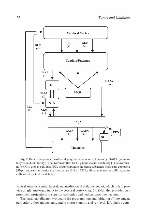
4.1. Basal Ganglia-Thalamocortical CircuitryThe basal ganglia consist of five interconnected subcortical nuclei that span the
telencephalon, diencephalon, and mid-brain. These nuclei include the caudate puta-men (CPu), globus pallidus (GP), subthalamic nucleus (STN), substantia nigra parscompacta, and substantia nigra pars reticulata (SNpr) (17,18). The medium spinyneurons are the principle neurons in the CPu and contain the inhibitory neu-rotransmitter g-aminobutyric acid (GABA). The medium spiny neurons receivethe bulk of the incoming excitatory glutamatergic input from the cerebral cortexand a heavy dopaminergic input from substantia nigra pars compacta. These neu-rons, which constitute the only striatal output, send their projections via two majorpathways. The direct or striatonigral pathway, in which the striatal neurons projectdirectly to the SNpr, and the indirect or striatopallidal pathway, in which striatalneurons project to GP, then to STN and terminate in SNpr (Fig. 2). The two striatalpathways and projections from the GP to STN are GABAergic in nature. The STN,which receives an excitatory glutamateric input from the cortex, provides an exci-tatory output to both GP and SNpr. The SNpr sends GABAergic projections to the
14 Tarazi and Kaufman
ventral anterior, ventral lateral, and mediodorsal thalamic nuclei, which in turn pro-vide an glutamatergic input to the cerebral cortex (Fig. 2). SNpr also provides lessprominent projections to superior colliculus and pedunclopontine nucleus.
The basal ganglia are involved in the programming and initiation of movement,particularly slow movements, and in motor memory and retrieval. DA plays a com-
Fig. 2. Detailed organization of basal ganglia-thalamocortical circuitry. GABA, g-amino-butyric acid, inhibitory [–] neurotransmitter; GLU, glutamic acid, excitatory [+] neurotrans-mitter; GP, globus pallidus; PPN, pedunclopontine nucleus; substantia nigra pars compacta[SNpc] and substantia nigra pars reticulata [SNpr]; STN, subthalamic nucleus; SC: superiorcolliculus (see text for details).
Neural Principles 15
plex role within the basal ganglia as it appears to have a net excitatory effect on thedirect pathway and an inhibitory effect on the indirect pathway. Therefore, DA caninitiate programmed movement by stimulating the direct pathway, which activatesthalamocortical projections and subsequently motor cortex, or can inhibit pro-grammed movement by suppressing the activity of the indirect pathway and inhib-iting activity of thalamocortical projections and their cortical inputs. The relativeand balanced responsiveness of striatonigral and striatopallidal neurons to corticalglutamatergic input, nigral dopaminergic input, and the orchestrated interactionbetween different neurotransmitter receptors in the basal ganglia-thalamocorticalcircuitry determine the functional and behavioral outcome of the basal ganglia.Disorders of the basal ganglia may produce uncontrollable and involuntarymovements as in Huntington’s disease or restricted and rigid movements as inParkinson’s disease (19). Additionally, abnormalities in the basal ganglia nucleiand/or their projecting targets have been linked to schizophrenia and motorextrapyramidal side effects associated with classical antipsychotic drug treatment (1).
4.2. The Limbic System
The limbic system is the primary circuit of the brain that mediates feelings andemotionally significant stimuli (20). The original circuitry, proposed by JamesPapez and also known as the Papez circuit, is comprised of cingulate gyrus, theparahippocampal gyrus, and the hippocampal formation, which includes the hip-pocampus, dentate gyrus, and the subiculum. Papez proposed that the hippocampalformation processes information from the cingulate gyrus and conveys it to themammillary bodies of the hypothalamus via the fornix (the main fiber bundle thatcommunicates the outflow of hippocampus). In turn, the mammillary bodies of thehypothalamus provides information to cingulate gyrus via anterior thalamic nuclei(Fig. 3) (21). In the expanded limbic circuit proposed by Paul MacLean, severaladditional brain structures are included such as parts of the hypothalamus, the sep-tal area, nucleus accumbens, orbitofrontal cortex, and the amygdala (Fig. 3).The amygdala is composed of many nuclei that are reciprocally connected to thehypothalamus, hippocampal formation, neocortex and thalamus. More recent cluesindicated that the amygdala is the part of the limbic system being specificallyinvolved in mediating emotional feelings, whereas the hippocampus, the mammil-lary bodies, and anterior thalamic nuclei are more involved in cognitive forms ofmemory storage (21,22). Maldevelopment of neuronal connectivity between dif-ferent components of limbic brain structures, and subsequent dysfunction in infor-mation processing within the limbic system and among closely associated structuresand regions may contribute to the pathophysiology of several neurological andpsychiatric disorders, as detailed in the following chapters.
4.3. The Cortico-Striato-Pallido-Mescencephalic Circuitry
Cortico-striato-pallido-mescencephalic circuitry also mediates cognitive andaffective behaviors. This circuit is implicated in the pathophysiology of schizo-

16 Tarazi and Kaufman
phrenia and Parkinson’s disease, and it is modulated by several classes of drugsused for treatment of different neurological and psychiatric disorders (23).An important brain region of this circuitry is the medial prefrontal cortex (mPFC),which can be subdivided into distinct subregions including the medial precentral,anterior cingulate, prelimbic and infralimbic cortices. These subregions receiveafferents from the mediodorsal thalamic nuclei and basolateral amygdala. All ofthese areas receive dopaminergic afferents from the DA neurons in the ventral teg-mental area (VTA). The mPFC sends projections to nucleus accumbens in additionto caudate putamen. These projections are topographically organized within thedifferent subregions and each of these projections has distinct targets in differentparts of striatum.
Fig. 3. Detailed organization of limbic system. Thick lines show original circuit.Fine lines show expanded circuit. The hippocampal formation includes hippocampus, sub-iculum and entorhinal cortex. The hippocampal formation projects via fornix to hypotha-lamic regions and via reciprocal connections to association cortex. Hypothalamic-corticalprojections are indicated. (Diagram modified from ref. 21.)
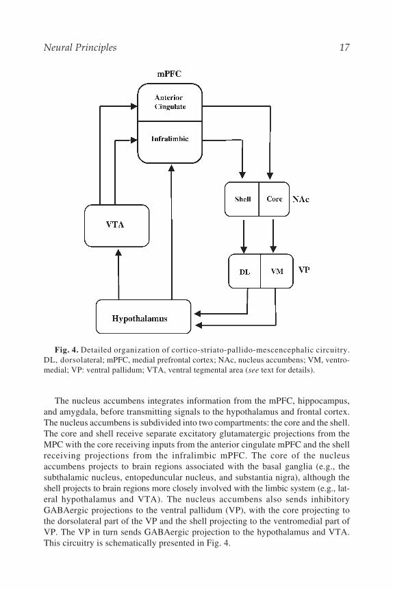
Neural Principles 17
The nucleus accumbens integrates information from the mPFC, hippocampus,and amygdala, before transmitting signals to the hypothalamus and frontal cortex.The nucleus accumbens is subdivided into two compartments: the core and the shell.The core and shell receive separate excitatory glutamatergic projections from theMPC with the core receiving inputs from the anterior cingulate mPFC and the shellreceiving projections from the infralimbic mPFC. The core of the nucleusaccumbens projects to brain regions associated with the basal ganglia (e.g., thesubthalamic nucleus, entopeduncular nucleus, and substantia nigra), although theshell projects to brain regions more closely involved with the limbic system (e.g., lat-eral hypothalamus and VTA). The nucleus accumbens also sends inhibitoryGABAergic projections to the ventral pallidum (VP), with the core projecting tothe dorsolateral part of the VP and the shell projecting to the ventromedial part ofVP. The VP in turn sends GABAergic projection to the hypothalamus and VTA.This circuitry is schematically presented in Fig. 4.
Fig. 4. Detailed organization of cortico-striato-pallido-mescencephalic circuitry.DL, dorsolateral; mPFC, medial prefrontal cortex; NAc, nucleus accumbens; VM, ventro-medial; VP: ventral pallidum; VTA, ventral tegmental area (see text for details).

18 Tarazi and Kaufman
5. NEUROIMAGING METHODS
The past decade has seen a tremendous increase in utilization of neuroimagingtechnology for research into neurological and psychiatric disorders. The most com-monly used methods are emission tomographic techniques including positron emis-sion tomography (PET) and single photon emission computed tomography(SPECT), and magnetic resonance techniques including imaging (MRI), functionalimaging (fMRI), and magnetic resonance spectroscopy (MRS).
Each of these techniques is relatively noninvasive and has a high safety marginfor most patients/research subjects. Each technique also has important advantagesand limitations in terms of sensitivity, spatial or temporal resolution, safety, andcost. This section briefly describes PET, SPECT, MRI, fMRI, and MRS methodsand applications in neurological and psychiatric diagnosis and research. Althoughdetailed technical descriptions are beyond the scope of this section, references areprovided that will allow interested readers to learn more about the technical foun-dations for these methods.
5.1. Emission TomographyPET and SPECT imaging are routinely used to study brain and other organ sys-
tem blood flow, metabolism (glucose uptake or oxygen utilization), drug pharma-codynamics, and pharmacokinetics, neurotransmitter synthesis and release, receptordistribution and density (DA, 5-HT, NE, their transporters, and other receptorsystems), enzyme function, and BBB transport in healthy and diseased patients.PET and SPECT imaging utilize chemical probes containing radioactive atoms(radionuclides) to allow detection of these biological phenomena (Table 1). Someprobes are identical to endogenous molecules of biological interest that have had aradioactive atom (radioisotope) substituted in for a stable (nonradioactive) atom(e.g., the PET probe [15O]water). Others, such as (18F)fluorodeoxyglucose([18F]FDG), a glucose analog used to detect glucose uptake into brain and othertissues, are biologically active molecules with an added radioactive atom that doesnot alter biological activity . Some probes are synthetic compounds that have beendeveloped to study specific phenomena; for example, (99mTc)hexamethylpro-pyleneamine oxime ([99mTc]HMPAO; a SPECT cerebral blood flow tracer). Radio-nuclide probes most often are administered intravenously, although other routescan be used. Once administered, these probes accumulate at sites in the body con-taining high concentrations of receptors or other types of association sites at whichthey have high affinity, or in regions in which blood flow or metabolism areincreased. Their chemical nature determines probe tissue distribution patterns.
The process of radioactive decay generates the signals detected from theseprobes. Radioactive atoms are unstable and have a nuclear excess of protons thatdecay to form more stable nuclei. The decay process generates photons that can bedetected by PET and SPECT scanners. The decay rate of a radioactive atom isreferred to as its half-life, or the time it takes half the substance to decay to the morestable, nonradioactive material. Radionuclide half-lives differ substantially (Table 1)
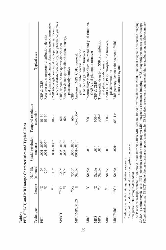
Neural Principles 19
19
Tab
le 1
PE
T, S
PE
CT
, an
d M
R I
soto
pe
Ch
arac
teri
stic
s an
d T
ypic
al U
ses
Hal
f-li
feS
pati
al r
esol
utio
nT
empo
ral r
esol
utio
nT
echn
ique
Isot
ope
(min
utes
)(m
eter
s)(s
econ
ds)
Typ
ical
use
s
PE
T15
O
2a
.00
1–.0
05b
10–3
0C
BF
& C
MR
11C
2
0a .
001–
.005
b10
–30
Rec
epto
r an
d tr
ansp
orte
r di
stri
buti
on, d
ensi
ty,
and
phar
mac
odyn
amic
s, d
opam
ine
neur
otra
nsm
issi
on18
F 1
10a
.00
1–.0
05b
10–3
0C
MR
(de
oxyg
luco
se u
ptak
e), d
opam
ine
synt
hesi
s,re
cept
or d
istr
ibut
ion,
den
sity
, and
pha
rmac
odyn
amic
sS
PE
CT
99m
Tc
360
a .
005–
.010
b60
+C
BF
, dop
amin
e tr
ansp
orte
r di
stri
buti
on12
3 I 7
80a
.00
5–.0
10b
60+
Rec
epto
r &
tran
spor
ter
dist
ribu
tion
, den
sity
,ph
arm
acod
ynam
ics
133 X
e75
60a
.00
5–.0
10b
60+
CB
FM
RI/
fMR
I/M
RS
1 HS
tabl
e.0
001–
.010
c.0
5–30
0+c
Ana
tom
y, f
MR
I, C
BF
, neu
rona
l,gl
ial a
nd m
itoc
hond
rial
fun
ctio
n,G
AB
A a
nd g
luta
mat
e tu
rnov
erM
RS
13C
Sta
ble
.01c
300+
cIn
term
edia
ry m
etab
olis
m, n
euro
nal a
nd g
lial
fun
ctio
n,G
AB
A a
nd g
luta
mat
e tu
rnov
erM
RS
17O
Sta
ble
.01c
300+
cC
BF
& C
MR
MR
S19
FS
tabl
e.0
1c30
0+c
The
rape
utic
dru
g (e
.g.,
SS
RI)
dis
trib
utio
nan
d ph
arm
acod
ynam
ics
MR
S31
PS
tabl
e.0
1c30
0+c
CM
R (
AT
P, P
Cr)
, pho
spho
lipi
d tu
rnov
er,
pH d
eter
min
atio
nM
RI/
fMR
I15
7 Gd
Sta
ble
.00
1c.0
5–1+
cB
BB
pat
ency
, les
ion
enha
ncem
ent,
fMR
I,sm
art c
ontr
ast a
gent
s
a rad
ioac
tive
, wit
h io
nizi
ng r
adia
tion
exp
osur
e.b d
oes
not i
nclu
de a
nato
mic
al im
age
core
gist
rati
on e
rror
s.c m
agne
tic
fiel
d st
reng
th-d
epen
dent
.A
TP
, ade
nosi
ne tr
ipho
spha
te; B
BB
, blo
od–b
rain
bar
rier
; CB
F/C
MR
, cer
ebra
l blo
od fl
ow/m
etab
olis
m; f
MR
I, fu
ncti
onal
mag
neti
c re
sona
nce
imag
ing;
GA
BA
, g-
amin
obut
yric
ac i
d; P
ET
, po
sitr
on e
mis
sion
tom
ogra
phy;
MR
I, m
agne
tic
reso
nanc
e im
agin
g; M
RS
, m
agne
tic
reso
nanc
e sp
e ctr
osc o
py;
PCr,
pho
spho
crea
tine;
SPE
CT
, sin
gle-
phot
on e
mis
sion
com
pute
d to
mog
raph
y; S
SRI,
sel
ectiv
e se
roto
nin
reup
take
inhi
bito
r (e.
g., f
luox
etin
e an
d fl
uvox
amin
e).
20 Tarazi and Kaufman
and range from about 2 minutes (15O) to several days (133Xenon). PET radionuclidesgenerally have shorter half-lives than SPECT probes and must be generated (by acyclotron) in proximity to the PET scanner, so that radiochemistry preparatory stepsoccur rapidly and radionuclides can be administered within the first few half livesafter probe generation (e.g., <6 minutes for 15O). Thus, PET probes can bemore difficult to work with than SPECT probes. However, their shorter half-livesare considered advantageous in studies requiring multiple assessments over shorttime frames because some can be administered several times during the course of asingle study. SPECT radionuclides with longer half-lives tend to offer much moreflexibility in terms of preparatory steps and can even be prepared remotely from theadministration site. However, their slower decay rates mean that subjects mayexperience higher radiation doses.
Although PET and SPECT radionuclide decays both result in g-ray (photon)emissions, hence the term “emission” tomography, their decay and detection pro-cesses differ. PET radionuclide decay involves ejection of a positively charged elec-tron (positron) from the nucleus. After traveling a short distance, the positroncollides with an electron creating an “annihilation event” that produces two high-energy photons. These photons travel away from the collision site in the opposite(antiparallel) direction and are detected by two photon-sensitive crystals placed atopposite sides of PET camera circular sensor arrays. PET camera detectors are elec-tronically linked and only register photons activating opposing crystals within ashort time window (on the order of nanoseconds) as reflecting true radionuclidedecay events. This detection scheme is termed “coincidence detection” and itensures high sensitivity by reducing the likelihood that noncoincident photons,which can be generated as a result of random noise, photon scatter, or other events,contribute to PET images. Photon paths identified as originating from coincidentphotons are registered and their summation is used to form multidimensional imagesof radionuclide localization. The fact that multiple (as opposed to single) imageplanes are acquired and can be used for image construction makes this a tomo-graphic, as opposed to a planar, imaging technique. Images can be filtered or other-wise corrected for confounding processes, such as attenuation or scatter, prior tofinal image generation.
In SPECT, radionuclide decay occurs when the extra nuclear proton attracts anorbital electron by a process termed electron capture, to form a metastable nucleus.This generates energy (80–160 keV) that is emitted in the form of a single g-ray(photon). SPECT cameras use the collimation (definition: to make straight or paral-lel) process for image formation. Collimators contain many parallel narrow, chan-nels that open to crystal photon sensors. Photons traveling parallel paths passthrough the collimator and are registered and treated similarly to those in PET stud-ies, with similar filtering and reconstruction methods applied to generate SPECTimages. However, because collimators eliminate signals from photons travelingnonparallel paths, some photons are unregistered and their signal is lost. Anothersource of signal loss in SPECT occurs when photons originally maintaining trajec-tories parallel to collimator holes are diverted by interactions with soft tissue
Neural Principles 21
components. These “scattered” photons are excluded from image formation. The inverseproblem also occurs when photons originally traveling in nonparallel paths arediverted into parallel paths to provide incorrect image localization information.Photon energy levels are substantially lower for SPECT (80–160 keV) than forPET radionuclides (511 keV), and SPECT photons can undergo more attenuationas they pass through tissues (via absorption or scattering) than PET radionuclides,further lowering SPECT image generation efficiency vs PET. Thus, SPECT is aless efficient image generation technique and tends to have lower spatial resolutionand than PET. However, SPECT radionuclides have longer half-lives than PETradionuclides and thus can be handled more easily and synthesized remotely fromthe study site. Furthermore, SPECT typically costs less than half of PET. Theseadvantages in part account for SPECT’s widespread use.
Although both PET and SPECT are highly sensitive for detecting low concen-trations of radionuclide-labeled probes (subnanomolar concentrations), this sensi-tivity requires sampling times on the order of multiple seconds to minutes, so thatsufficient quantities of radioactive decays can occur and photon counts be accumu-lated for image formation. Although this temporal resolution is more than adequateto study many physiological processes, it presents some limitations in studies ofprocesses that occur at rates on the order of one to several seconds. Additionally,neither PET nor SPECT produce high-resolution anatomical images of tissues understudy. Their spatial resolution, on the order of millimeters, is adequate for manypurposes, though some applications require image coregistration with high-reso-lution anatomical images (e.g., computed axial tomography [CT], or MRI) to pre-cisely determine radionuclide localization. The image coregistration processitself has limitations and thus the spatial resolution of anatomically coregisteredPET/SPECT images can approach but not equal that afforded by CT or MRI (24–26).
5.2. Magnetic Resonance
MRI, formerly referred to by the acronym NMR (nuclear magnetic resonance),is actually a family of techniques whose signals are generated as a consequence ofseveral fundamental phenomena that when linked, permit structural imaging,fMRI, and chemical imaging (also known as MRS). The nuclear part of NMRrefers to the fact that the technique is limited to molecules containing atoms withodd numbers of protons and/or neutrons, which possess net nuclear magneticmoments. MR-detectible atoms include hydrogen (1H–protons and 2H–deuterium),helium (3He), lithium (7Li), carbon (13C), oxygen (17O), fluorine (19F), sodium(23Na), phosphorus (31P), and potassium (39K). Thus, most atoms present in mol-ecules of biological interest can be visualized with MR techniques. Moreover,all of these atoms are stable, nonradioactive isotopes, so, as noted above, MR stud-ies are conducted without exposure to ionizing radiation. Thus, MRI techniques areinherently safer than those utilizing ionizing radiation (e.g., PET, SPECT, andCT imaging). This difference is especially important in repeated measures studiesrequiring multiple image acquisition sessions.

22 Tarazi and Kaufman
The magnetic part of NMR refers to the fact that when MR-visible atoms areplaced in strong magnetic fields, their molecular spins become quantized into higherand lower energy states, oriented antiparallel and parallel to the main magneticfield (Bo), respectively. These spins behave like small gyroscopes and rotate aroundthe Bo axis. As Bo increases, the energy difference between higher and lower spinstates (determined by the Boltzmann constant) increases such that at sufficientlylarge Bo, a few excess spins drop down into the lower energy state and becomeunpaired. These unpaired spins serve as tracers for the rest of the paired (MR-invis-ible) spins in the substance of interest. For hydrogen atoms (1H, protons) studied at1.5 Tesla (1.5 T, equivalent to about 30,000 times the earth’s magnetic field atAsian, European, and North American latitudes), roughly 10 out of every 1 millionprotons have unpaired spins in the lower energy state and are observable in MRstudies. This is the reason that MR techniques are relatively insensitive (can detectmicromolar or higher concentrations) when compared to PET or SPECT imaging.
In MR studies, once tracer spins have been generated by placing an object into alarge magnetic field, a radiofrequency (RF) pulse at the resonance (larmor) fre-quency (determined by the nucleus under study and the field strength of the mag-net, e.g., 63.87 MHz for protons at 1.5 T) is broadcast into the object. This is theresonance part of NMR. Tracer protons spinning at the resonance frequency absorbRF pulse energy and are boosted to the higher energy spin orientation (antiparallelto Bo). After the RF pulse is turned off, tracer spins begin relaxing back to the lowerenergy state, shedding their excess energy by resonating RF signals at the larmorfrequency. Thus, MR can in some ways, like PET and SPECT, be considered anemission technique. Each MR-visible atom spins at a different resonance frequencyat a given magnetic field strength, making it possible to selectively detect sub-stances containing any MR-visible atom by choosing the resonance frequency atwhich to broadcast and absorb RF energy.
During relaxation, tracer spins rotate around the main magnetic field axis andtheir emitted energy is detected with highly sensitive antennas (e.g., volume, sur-face, or phased array coils) tuned to the larmor frequency. These antennas detecttwo different types of relaxation behaviors, longitudinal relaxation, also known asspin-lattice or T1 relaxation, and transverse relaxation, also known as spin–spin orT2 relaxation. Longitudinal relaxation is the process by which higher energy statetracer spins release their absorbed energy to the environment as they return to thelower energy state. Transverse relaxation is the process by which higher energystate tracer spins exchange energy with other local spins either within the samemolecule or with other molecules in the object, as the entire spin ensemble comesto equilibrium. The parameters T1 and T2 reflect the times it takes half of the excitedspins to return to equilibrium either with the main magnetic field (Bo) or with otherspins in the object of interest, respectively, and T2 generally is much shorter thanT1. MRI measures water relaxation behavior (T1 and T2) that is modulated incomplex ways by its interactions with proteins and lipids in brain or other tissues.By timing image acquisitions to more heavily represent T1 or T2 behavior or theircombination, one can acquire “T1-weighted,” “T2-weighted,” or “proton density”

Neural Principles 23
images, respectively. These image types offer different forms of tissue contrast.For example, in T1-weighted images (used to study normal anatomy), gray matterlooks gray and white matter looks white, whereas in T2-weighted images (used toassess brain pathology), abnormal tissue is highlighted as bright or white. By manipu-lating image acquisition timing and thus T1 and T2 contrast, it is possible to studybrain structure (and function) with many image contrast variations.
5.3. fMRI Techniques
5.3.1. Blood Oxygen Level-Dependent (BOLD) fMRI TechniquePerhaps the most well-known fMRI technique used in studies of neurological
and psychiatric disorders is called BOLD fMRI. This technique is, as its acronymsuggests, sensitive to changes in brain oxygenation that occur in response to focalactivations associated with sensations, movements, thoughts, or their combinations.When brain is activated, local metabolism and blood flow increase to provide extraoxygen and glucose. However, by design, more oxygen is delivered than requiredto support increased metabolic demand, and this leads to oxygen oversupply and anet reduction in content of the endogenous MR contrast agent, deoxyhemoglobin(deOHb). Because the iron atom at the active site of deOHb (unbound to oxygen) isparamagnetic and distorts local magnetic fields (changing water T1 and T2), a reduceddeOHb content improves local magnetic field homogeneity and increases imageintensity. BOLD images obtained during activation are subtracted from baselineimages, revealing areas of deOHb washout, which appear bright. BOLD imagingcan be conducted at high speeds (~1 second) permitting dynamic studies of brainfunction. This speed, combined with the fact that BOLD data can be overlaid onsequentially acquired high resolution anatomical (MRI) images, makes fMRI opti-mal for localization studies of brain function.
However, although BOLD is extremely fast, versatile, and popular, the BOLDsignal response is a complex and convoluted measurement of dynamic blood flow,volume, and oxygenation changes (27), and it is difficult to determine absolutechanges in any of these parameters with BOLD fMRI. Additionally, at conven-tional magnetic field strengths (~1.5 T), BOLD acquisition parameters stronglyinfluence whether fMRI signals reflect intravascular events in large vessels (some-what remote from activated neurons, typical of gradient echo acquisitions) or insmall vessels (closer to the site of neuronal activation, typical of spin echo acqui-sitions). However, as magnet field strength increases, BOLD measurements tend toreflect extravascular events occurring within tissues. This fact, along with improvedtemporal and spatial resolution that can be obtained with stronger magnets, hasdriven an instrumentation trend toward the development of high field MRI scan-ners (>3.0 T). An excellent discussion of advantages and disadvantages of highermagnetic field strength scanning is presented by Kim and Garwood (28).
In addition to BOLD contrast, other contrast mechanisms are used in fMRI stud-ies. Some studies involve intravenous administration of contrast agents containinga chelated form of the lanthanide metal gadolinium, which is strongly paramag-

24 Tarazi and Kaufman
netic and potently increases water T1 and T2 relaxation and alters image intensity.These agents are used clinically to determine blood brain barrier patency, but alsocan be infused dynamically to determine cerebral blood volume (CBV) and itsresponses to drugs or other challenges. Newer contrast agents containing novel para-magnetic atoms or particles (e.g., superparamagnetic iron oxide particles) also areunder development. Additionally, “smart contrast agents” containing molecularstructures that respond to specific biological processes are being developed (29).
5.3.2. Arterial Spin Labeling (ASL) fMRI TechniqueActive manipulation of the magnetization and relaxation properties of water
flowing through blood vessels is the basis for the ASL fMRI technique, which gen-erates images of blood flowing through vessels (30). With this technique, the mag-netization of blood water is altered by scan pulses and the resultant image issubtracted from a normal image taken from the same plane, revealing a blood flowimage. This method is used to study brain blood flow abnormalities in many disor-ders and can be used dynamically to quantitatively assess blood flow responses tofunctional or chemical challenges.
5.3.3. Diffusion Tensor Imaging (DTI) TechniqueAnother scan type that is being used increasingly to identify and characterize
white matter integrity and connectivity is DTI, which is a complex variant of diffu-sion weighted imaging. DTI applies multiple gradients over time to alter watermagnetization in six or more directions, to identify three-dimensional water diffu-sion patterns. Water tends to diffuse more readily outside of or along neuronal orglial membranes (as opposed to within or across those membranes). As diffusioncoherence, or anisotropy, increases, so does the DTI signal. The DTI signal is high-est in white matter areas containing myelinated axons that form tightly packed waterdiffusion channels, through which water tends to diffuse unidirectionally. The tech-nique is very sensitive to white matter disruption or damage, which reduces diffu-sion anisotropy and DTI signal.
5.4. Chemical Spectroscopy Techniques
5.4.1. Proton SpectroscopyA chemical form of imaging, known as proton (1H) spectroscopy (1H MRS), is
now widely used to study neurological and psychiatric disorders. Proton MRS stud-ies (as well as MRS studies of other MR-visible nuclei including phosphorus, car-bon, and so on) (Table 1) produce spectra containing several types of informationincluding chemical environment (chemical shift and peak splitting) and concen-tration (peak amplitude) measures. MRS studies can acquire spectra from singlebrain cubes (voxels) and also can simultaneously acquire data from planar arrays ofvoxels using a technique called chemical shift imaging, which like imaging, appliescombinations of gradients to localize spectral data from multiple voxels in two orthree dimensions. The chemical shift (horizontal part of the spectrum at which a

Neural Principles 25
chemical peak occurs) of any particular functional group (e.g., methyl-, methylene,or hydroxyl-) is determined by the magnetic field experienced by the hydrogennucleus when it is in a large magnetic field. Electron-donating and -withdrawinggroups produce more negatively and positively charged nuclei, respectively, anddifferentially alter chemical shift. Additionally, the hydrogen atoms attached toadjacent carbon atoms can magnetically couple with each other to induce peak split-ting patterns in proton spectra.
In proton spectra, metabolites easily visualized include N-acetylaspartate(a marker for neuronal viability and perhaps mitochondrial function), choline-con-taining compounds (membrane phospholipids and other constituents), and a peakcombining creatine and phosphocreatine (the latter of which is the high energyphosphorus storage form). Proton MRS also can be used to detect the main inhibi-tory and excitatory neurotransmitters GABA and glutamate. However, glutamate isboth a neurotransmitter and an energy intermediate, and the neurotransmitter com-ponent is best assessed indirectly by measuring glutamine (the catabolite ofglutamate neurotransmission, which is shuttled from glial to neuronal cells forresynthesis into neurotransmitter glutamate). MRS studies are the least sensitive ofthe techniques discussed in this chapter; sample volumes must be large (>1 cm3)and sampling times are long (multiple minutes). Despite its limitations, the abilityto acquire chemical information noninvasively makes MRS quite attractive for usein psychiatric and other types of brain research.
5.4.2. Other Forms of Chemical Spectroscopy
Other MRS forms also provide meaningful information on brain chemistryand metabolism, and can be used to quantify brain levels of therapeutic drugs(Table 1). For example, phosphorus spectroscopy (31P MRS) detects both high-energy phosphate intermediates phosphocreatine and adenosine triphosphate(ATP), and can also be used to determine tissue pH and magnesium concentra-tion. Fluorine spectroscopy (19F MRS) can be used to detect the antidepressantfluoxetine in brain or other tissues, as well as other therapeutic compounds con-taining fluorine atoms. Lithium (7Li) MRS has been used to determine brainlithium concentrations during therapeutic administration. Carbon spectroscopy(13C MRS) can be used to detect signals from molecules involved in tissuemetabolism including glucose, the neurotransmitters GABA and glutamate, aswell as intermediary metabolism. Unfortunately, 31P and 13C MRS signals areonly about 6 and 2% as strong, as the proton MRS signal, respectively. Thus, 31P and13C MRS are even more limited in terms of spatial and temporal resolution thanproton MRS. This has limited their use in psychiatric research to date. However,as noted previously, the MR sensitivity problem can be overcome in part byincreasing magnet field strength. For further review of the advantages and disad-vantages of higher strength magnets on MRS applications and for additionaldetails on MR imaging methods and applications see Kim and Garwood (28) andRenshaw and colleagues (31).
26 Tarazi and Kaufman
6. CONCLUSIONPrinciples of neural sciences represent a merger of molecular biology, neuro-
chemistry, neuroanatomy, neurophysiology and embryology. Proper study of neu-rological and psychiatric disorders begins with the study of the brain. Understandingneuron–neuron communications and synaptic neurotransmission is necessary forconsidering how disruption of neural functions leads to brain disorders. Clinicaltreatment of these disorders has been greatly strengthened by novel insights intocellular neurobiology of neurons. Advanced neuroimaging techniques helped toidentify brain regions and neural pathways that mediate normal emotional, cogni-tive, motivated, and motor behaviors, which are typically disturbed, at least in part,in patients diagnosed with neurological or psychiatric disorders.
ACKNOWLEDGMENTThis work was supported in part by NARSAD, NIH grants HM-68359, HD-043649
(FIT), and DA-017324 (MJK).
REFERENCES1. Baldessarini RJ, Tarazi FI. Drugs and the treatment of psychiatric disorders. In: Hardman JG,
Limbird LE, eds. Goodman and Gilman’s The pharmacologic basis of therapeutics. 10th ed.New York: McGraw-Hill, 2001:485–520.
2. Baldessarini RJ, Tarazi FI. Brain dopamine receptors: A primer on their current status, basic andclinical. Harvard Rev Psychiatry 1996;3:301–325.
3. Cooper JR, Bloom EF, Roth RH. The biochemical basis of neuropharmacology. 8th ed.New York: Oxford University Press, 2003.
4. Lindvall O, Bjorklund A. Dopamine and norepinephrine containing neuron systems: their anatomyin the rat brain. In: Emson PC, ed. Chemical Neuroanatomy. New York: Raven Press, 1983:229–255.
5. Bylund DB, Eikenberg DC, Hieble JP, et al. International Union of Pharmacology nomenclatureof adrenoceptors. Pharmacol Rev 1994;46:121–136.
6. Hieble JP, Ruffolo RR Jr. Functions mediated by b-adrenoceptor activation. In: Ruffolo RR Jr,ed. Progress in basic and clinical pharmacology, vol. 7, b-Adrenoceptors: molecular biology,biochemistry and pharmacology. Basel: Karger, 1991:1–25.
7. Tork I. Anatomy of the serotonergic system. Ann NY Acad Sci 1990;600:9–35.8. Barnes NM, Sharp T. A review of central 5-HT receptors and their function. Neuropharmacology
1999;38:1083–1152.9. Millan MJ. Improving the treatment of schizophrenia: focus on serotonin 5-HT1A recep-
tors. J Pharmacol Exp Ther 2000;295:853–86110. Tarazi FI, Zhang K, Baldessarini RJ. Long-term effects of olanzapine, risperidone, and quetiapine
on dopamine receptor types in regions of rat brain: implications for antipsychotic drug treatment.J Pharmacol Exp Ther 2001;297:711–717.
11. Tarazi FI, Campbell A, Yeghiayan SK, Baldessarini RJ. Localization of dopamine receptor sub-types in caudate-putamen and nucleus accumbens septi of rat brain: comparison of D1-, D2-, andD4-like receptors. Neuroscience 1998;83:169–176.
12. Young AB, Frey KA, Agranoff BW. Receptor assays: in vitro and in vivo. In: Phelps M, MazziottaJ, Schelbert H, eds. Positron Emission Tomography and Autoradiography: Principles and Appli-cations for the Brain and Heart. New York: Raven Press, 1986:73–111.
13. Sternberger LA. Immunocytochemistry. New York: Wiley, 1986.14. Mudgett-Hunter M. Monoclonal antibodies. In: Fozzard H, Haber E, Jennings R, Katz H, Morgan
H, eds. The Heart and Cardiovascular System. New York: Raven Press, 1986:189–201.

Neural Principles 27
15. Eberwine JH, Valentino KL, Barchas JD. In Situ Hybridization in Neurobiology. New York:Oxford University Press, 1994.
16. Chesselet M-F. In Situ Hybridization Histochemistry. Neurobiology. Boca Raton: CRC Press,1990.
17. Graybiel AM. Neurotransmitters and neuromodulators in the basal ganglia. Trends Neurosci1990;13:244–254.
18. Gerfen CR. The neostriatal mosaic: multiple levels of compartmental organization. TrendsNeurosci 1992;15:133–139.
19. Alexander GE, Crutcher MD. Functional architecture of basal ganglia circuits: neural substratesof parallel processing. Trends Neursci 1990;13:266–271.
20. LeDoux JE. The Emotional Brain. New York: Simon and Schuster, 1996.21. Iversen S, Kupfermann I, Kandel ER. Emotional states and feelings. In: Kandel ER, Schwartz
JH, Jessell TM, eds. Principles of Neural Science. 4th edition. New York: McGraw-Hill,2000:982–997.
22. Aggleton JP. The contribution of the amygdala to normal and abnormal emotional states. TrendsNeurosci 1993;16:328–333.
23. Deutch AY. Prefrontal cortical dopamine systems and the elaboration of functional corticostriatalcircuits: implications for schizophrenia and Parkinson’s disease. J Neural Transm 1993;91:197–221.
24. Malison RT. Positron emission tomography and single-photon emission computed tomography.Methods and applications in substance abuse research, In: Kaufman MJ, ed. Brain Imaging inSubstance Abuse: Research, Clinical, and Forensic Applications. Totowa, NJ: Humana Press,2001:29–46.
25. Volkow ND, Fowler JS, Wang GJ. Positron Emission Tomography and Single Photon Emission Com-puted Tomography in Substance Abuse Research. Seminars Nuclear Med 2003;XXXIII:114–128.
26. Fowler JS, Volkow ND, Wang G-J, Ding Y-S. 2-Deoxy-2-[18F]Fluoro-D-Glucose and alternativeradiotracers for positron emission tomography imaging using the human brain as a model.Seminars Nuclear Med 2004;XXXIV:112–121.
27. Logothetis NK, Wandell BA. Interpreting the BOLD signal. Ann Rev Physiol 2004;66:735–769.28. Kim D-S, Garwood M. High-field magnetic resonance techniques for brain research. Curr Opin
Neurobiol 2003;13:612–619.29. Meade TJ, Taylor AK, Bull SR. New magnetic resonance contrast agents as biochemical report-
ers. Curr Opin Neurobiol 2003;13:597–602.30. Wong EC, Buxton RB, Frank LR. Implementation of quantitative perfusion imaging techniques
for functional brain mapping using pulsed arterial spin labeling. NMR Biomed 1997;10:237–249.31. Renshaw PF, Frederick B, Maas LC III. Fundamentals of magnetic resonance. In: Kaufman MJ,
ed. Brain Imaging in Substance Abuse: Research, Clinical, and Forensic Applications. Totowa,NJ: Humana Press, 2001:47–76.

Pharmacotherapeutic Principles 29
29
From: Current Clinical Neurology: Neurological and Psychiatric Disorders:From Bench to Bedside
Edited by: F. I. Tarazi and J. A. Schetz © Humana Press Inc., Totowa, NJ
2Pharmacotherapeutic Principles
of Neurological and Psychiatric Disorders
John A. Schetz
SummaryThe pharmacotherapeutic management of neurological and psychiatric disorders relies
primarily on the modulation of central nervous system (CNS) neurotransmission with drugsthat intervene at chemical synapses. The receptors, transporters, and enzymes for the dopam-inergic, serotonergic, and noradrenergic systems are the most common neuropsychiatricdrug targets, because these neurotransmitter systems play a central role in the regulation ofa range of cognitive and motor behaviors. The key to understanding or anticipating theclinical profile (dose–effect) of a particular drug is to have an appreciation for both itspharmacodynamic and pharmacokinetic properties.
Key Words: Psychiatric; neuroscience; pharmacology; pharmacodynamics; pharmacokinet-ics; synapse; G protein-coupled receptors; dose–effect; theory; disorder; neurotransmission.
1. INTRODUCTIONThe appropriate, effective, and safe utilization of drugs in the treatment of dis-
ease requires a basic understanding of the dose–effect relationships of medications.Relating dose to effect requires a combined appreciation of pharmacodynamicconcentration–effect relationships, or what drugs do to the body, and of pharmaco-kinetic dose–concentration relationships, or what the body does to or with drugs.For this reason, considerable attention is devoted to both the pharmacodynamic andpharmacokinetic aspects of drug therapies. The aim of this chapter is to provide atheoretical rationale that is necessary for appropriately interpreting the results ofbasic and clinical neuropharmacology studies and for understanding many of thedrug treatment strategies commonly encountered in clinical neurology andpsychiatry.
Approximately one-fourth of all drugs prescribed worldwide exert their thera-peutic actions on CNS targets. Of the top five selling drugs in this category, threeare antidepressants and two are atypical antipsychotics (1). The relative success ofpharmacological intervention is highlighted further when one considers that these

30 Schetz
drugs are treating an estimated 7–15% of the population who suffer from one ormore of the neurological or psychiatric disorders discussed in this book. Currently,the biogenic amine neurotransmitter systems, and in particular dopaminergic,serotonergic, and noradrenergic receptors, transporters, and metabolic enzymes,cover the vast majority of neuropsychiatric drug targets. The reason for this is thatthe biogenic amine systems are key modulators of neuronal excitability, and themolecular components of these systems are located at chemical synapses, whichare sites that are accessible to intervention by drugs.
2. THE CHEMICAL SYNAPSEAS THE MAIN SITE OF DRUG INTERVENTION
Therapeutic approaches to modulating neuronal excitability at chemical synapsescan be categorized as presynaptic and postsynaptic. Presynaptic strategies involvealtering the levels of neurotransmitter in the synaptic cleft. This can be achieved bychanging the amount of endogenous neurotransmitter released or available forrelease into the synaptic cleft, or by altering the amount of neurotransmitter takenback up (reuptake) into the presynaptic terminal. The dopaminergic synapse can beused as a specific example to illustrate these points (Fig. 1). For example, monoam-ine oxidase B inhibitors, such as selegiline, block dopamine (DA) degradation,which makes more DA available for release. Inhibitors of DA synthesis, such asa-methylparatyrosine, reduce the amount of DA available for release. Drugs likereserpine and tetrabenazine decrease vesicular-mediated release by blockingvesicular monoamine transporters, which prevents the storage of neurotransmitterinto synaptic vesicles. Inhibitors of plasmalemmal DA transporters, such asbuproprion or cocaine, block the reuptake of DA from the synapse, and thereby,keep levels of DA in the synaptic cleft high. Certain drugs, like the psychostimulantamphetamine, cause nonvesicular DA release by running the DA transporter inreverse. In some cases, receptors are located on presynaptic terminals that bind theneurotransmitter being released, e.g., a DA receptor located on a presynapticdopaminergic terminal. When these so-called autoreceptors are stimulated theyattenuate, and when blocked they facilitate, subsequent rounds of neurotransmitterrelease.
In contrast to presynaptic strategies, which alter the levels of endogenous neu-rotransmitter in the synaptic cleft, postsynaptic strategies modulate neurotransmis-sion with chemical agents that act directly on postsynaptic receptors. For example,
Fig. 1. (opposite page) Schematic of a chemical synapse. The dopaminergic synapse isshown to illustrate the common points of drug intervention, which include presynapticeffects on dopamine synthesis, storage in synaptic vesicles, release and reuptake, andpostsynaptic effects on postsynaptic dopamine receptors. Degradation is depicted as occur-ring at both presynaptic and postsynaptic sites, which reflects the different locations of thedifferent metabolic enzymes (e.g., monoamine oxidases and catechol-O-methyltransferase[2,3]). See color insert preceding p. 51. (Copyright John A. Schetz, 2003.)

Pharmacotherapeutic Principles 31
31
Fig
. 1.
32 Schetz
agonists, such as DA or the antiparkinsonian drug pergolide, directly activate DAreceptors, whereas neuroleptic drugs like thioridazine and haloperidol block DAreceptor activation.
3. CLASSIFICATION OF DRUGS ON THE BASISOF THE RESPONSES THEY PRODUCE ON THEIR RECEPTORS
When a drug reversibly binds to the orthosteric (primary) site on its receptor oneof four outcomes are to be expected: the receptor becomes activated, the receptorbecomes partly activated, the receptor becomes inactivated, or the receptor is unableto be activated. Consequently, drugs are generally classified based on their actions.A drug is an agonist if it fully activates receptors, a partial agonist if it partly acti-vates receptors, an inverse agonist if it inactivates receptors (and prevents themfrom being activated), or an antagonist if it only prevents receptors from beingactivated. For instance, the endogenous neurotransmitter DA is an (full) agonist ofDA receptors, and the antiparkinsonian drug bromocriptine is a partial agonist atD2 receptors. Antipsychotics like haloperidol and clozapine may be inverse ago-nists of D2 DA receptors (4,5), whereas L-741,626 is an (neutral) antagonist.
Receptor activation is a thermodynamic process, whereby agonist bindinginduces a conformational change in the receptor and converts it from the inacti-vated state (agonist low-affinity binding state) to the activated state (agonist high-affinity binding state). Typically, neutral antagonist binding is indifferent to theconformational (affinity) state of its receptor, because it must only occupy theorthosteric site rather than occupy and then induce a change in it. However, aninverse agonist is a special type of antagonist in that when it binds to a receptor inthe activated state it converts it to the inactivated state. For this reason, inverseagonists reduce the basal levels of constitutive receptor activity, which correspondsto the (typically small) proportion of receptors that are in the activated state in theabsence of agonist. Such distinctions in the molecular mechanisms of action ofantipsychotic drugs that act on D2-like dopamine and 5-hydroxytryptamine (5-HT)2-like serotonin receptors may be critical to understanding their unique therapeuticprofiles (4,6).
Although most drugs bind directly to the orthosteric site of the receptor, otherdrugs bind at another (secondary) receptor site, called an allosteric site. Ligandsthat bind to the allosteric site are known as allosteric modulators, because theyindirectly modulate the binding of primary ligands to the orthosteric site byremotely altering the orthosteric-binding site. The modulation is said to be positiveif the modulator facilitates a primary ligand’s interaction with the primary site ornegative if the modulator attenuates its interaction with the primary site. The extentto which the allosteric site and the orthosteric site are coupled, or their cooperativity,can be weak or strong. Noncompetitive interactions, which result in a completeocclusion of the orthosteric site leading only to a decrease in the maximum densityof sites with no change in affinity, are also allosteric in nature but they are a specialcase of neutral cooperativity. Within the DA receptor family, for example, a diverse
Pharmacotherapeutic Principles 33
range of allosteric mechanisms and corresponding allosteric sites exist for modu-lating the effects of endogenous and therapeutic agents (7). For example, theendogenous tripeptide proline–leucine–glycine (PLG) is a positively cooperativeallosteric modulator of agonist binding to D2 DA receptors. Sodium ions are nega-tively cooperative allosteric modulators of agonist binding to D2 DA receptors, andzinc ions are neutrally cooperative allosteric modulators of antagonist binding toD4 DA receptors.
4. PHARMACODYNAMICS OF PHARMACOTHERAPIES
Chemical agents that have therapeutic actions are referred to as drugs. The termpharmacodynamics describes what drugs do to the body. Most drugs exert theiractions on the body by interacting with specific sites called receptors. Consequently,pharmacodynamics deals with the interactions of drugs with their receptor sites.The most critical drug–receptor properties concern the strength of their attraction(binding affinity) and functional effects (potency) expressed in units of drug con-centration, and the quantity of receptor in the target tissue (receptor density) or themaximal extent of a receptor’s functional effect (efficacy). The density of receptorsites is typically expressed as moles receptor per amount of tissue, whereas themaximal functional effect, which relies in part on receptor density, is usuallyexpressed as receptor activity per unit amount of tissue. The functional activity of areceptor can be measured by a variety of endpoints, ranging from changes in bio-chemical markers to behaviors.
Two coupled events occur when a drug interacts with its receptor. First the drugbinds to its receptor, and second it mediates some functional effect that is trans-duced by the receptor. Although drug binding and receptor activation are coupled,they are mechanistically distinct molecular processes under the control of uniquereceptor microdomains and they can be influenced by different factors. Conse-quently, there may not be a direct one to one correspondence linking one process tothe other.
4.1. Determination of Drug Affinity and Maximal Receptor Density:Ligand–Receptor Binding Interactions
The reversible (noncovalent) binding of a ligand with its receptor is a dynamicprocess, which is usually studied in one of two ways. The first way is to measure thekinetics of binding—the rate of approach to or departure from the equilibrium condi-tion. The second way is to measure the free energy forces of binding under the equi-librium condition. It is helpful to review the general principles of receptor bindingtheory, in order to know what sorts of experiments to perform to extract kinetic andequilibrium properties of ligand–receptor binding interactions, and additionally, toknow how to interpret the meaning of such properties in the context of drug therapies.
The theoretical construct that allows one to extract the properties that describeboth kinetic and equilibrium types of ligand binding processes and the relationshipbetween them is referred to as the mass action law. This law assumes that a ligand

34 Schetz
reversibly binds to a single homogenous population of receptor sites. Because thelaw is restricted to reversible reactions, which are those that can attain an equilib-rium condition, the ligand–receptor interaction can be modeled as an equilibriumreaction. As with all equilibrium reactions, when equilibrium is achieved the rate ofthe forward and reverse reactions are equal; at equilibrium, the rate of ligand–receptor association equals the rate of ligand–receptor dissociation as shownin Eq. 1.
Rate of association (forward reaction) = Rate of dissociation (reverse reaction) (1)
The rates at equilibrium can be expressed mathematically in terms of reactantsand products as shown in Eqs. 2 and 3.
Rate of association = [LIGAND][RECEPTOR]kon (2)
Rate of dissociation = [LIGAND·RECEPTOR]koff (3)
in which[LIGAND] is the ligand concentration expressed in units of Molarity (i.e., moles/liter)[RECEPTOR] is the total receptor concentration expressed in units of Molarity[LIGAND·RECEPTOR] is the ligand–receptor complex expressed in units of
Molaritykon is the association rate constant for the binding of a ligand with its receptor
expressed in units of (s-1 M-1)koff is the dissociation rate constant for the separation of ligand from its receptor
expressed in units of s-1
A mathematical model of receptor occupancy can thus be formulated from thetheoretical expectation at equilibrium by substitution of the equalities in Eqs. 2 and3 for those in Eq. 1 to yield Eq. 4.
[LIGAND][RECEPTOR] kon = [LIGAND·RECEPTOR] koff (4)
Equation 4 can be rearranged such that all the concentration variables occur onone side of the equation and all rate constants occur on the other side as shown inEq. 5. The ratio of the reactants (ligand and receptor) to products (ligand–receptorcomplex) thus equals the ratio of the rate of complex dissociation over the rate ofreactant association.
([LIGAND][RECEPTOR])/[LIGAND·RECEPTOR] = koff/kon = KD (5)
These ratios are also equal to the equilibrium dissociation constant (KD), whichrepresents the concentration of ligand required to occupy half of the total number ofreceptors. The units of KD are Molarity. A series of substitutions and algebraicmanipulations to Eq. 5 (8) puts it in the general form of a rectangular hyperbola(Eq. 6) to yield Eq. 7.
y = ax/(b+x) (6)
[LIGAND·RECEPTOR] = ([RECEPTOR][LIGAND])/(KD + [LIGAND]) (7)

Pharmacotherapeutic Principles 35
Separation of the dependent and independent variables allows for the graphingof the data and the extraction of the receptor-binding properties for a ligand that arethe constants in the square hyperbola equation (e.g., a = [RECEPTOR] and b = KD).In the laboratory, the amount of ligand that is specifically bound to its receptor([LIGAND·RECEPTOR]) is measured as a function of various ligand concentra-tions ([LIGAND]), and then the [RECEPTOR] and KD are solved for graphically(by applying a square hyperbolic math function). A common practice is to intro-duce a radioactive atom into the ligand (so that it can be detected), incubate variousconcentrations of this radiolabeled ligand in a solution containing a fixed amountof its receptor until equilibrium is reached, and then rapidly separate (so as not todisrupt the equilibrium condition) the radioligand bound to the receptor ([LIGAND·RECEPTOR]) from the unbound radioligand in solution ([LIGAND]). The radio-activity of the receptor-bound and (unbound) free radioligand is then quantified ina radioactivity counter. The resulting data obtained for such a saturation isothermtype of binding experiment is depicted in Fig. 2.
As can be seen from Eq. 5, the equilibrium dissociation constant can also becalculated by measuring the kinetic rates of ligand association and dissociation fromits receptor as the binding reaction proceeds toward or away from the equilibriumcondition. This can be accomplished by measuring the amount of radioligand boundto its receptor as a function of time. Kinetic determinations of KD require two sepa-rate experiments (association and dissociation rates) for each KD determination andprovide no information on receptor density. Therefore, they are usually not themethod of choice for determining equilibrium dissociation constant values.
Fig. 2. Example of saturation isotherm data for [3H]mesurguline equilibrium binding tocloned human serotonin 5-HT2C receptor expressed in COS-7 cells. A saturation isothermexperiment is conducted by keeping all conditions fixed while varying the concentration ofradioligand. The KD = 0.24 nM and Bmax = 0.5 pmoles/mg protein. (Copyright John A.Schetz, 2003.)

36 Schetz
Although saturation isotherms have the advantage that they are direct measuresof affinity (KD) and receptor density (Bmax), relatively few radiolabeled ligands areavailable, and consequently the binding affinity for most ligands must be deter-mined indirectly. The inhibition constant (Ki) is an indirect measure of a ligand’saffinity for its receptor that is numerically equivalent to the equilibrium dissocia-tion constant (Ki = KD). In contrast to saturation isotherm experiments, in which theonly ligand present is the radioligand, an inhibition type equilibrium bindingexperiment examines the ability of a nonisotopic ligand to compete with theradioligand for binding to the receptor site. The inhibition binding experiment isperformed with a fixed concentration of radioligand and receptor vs increasing con-centrations of competing ligand. An inhibition affinity constant for the nonisotopicligand is derived from its IC50, which is the concentration of nonisotopic (cold)ligand required to displace half of the total amount of radioligand bound to thereceptor (Fig. 3). The semilog dose–response curves for competition experimentstake on a sigmoidal appearance. The relative IC50 value extracted from the sigmoi-dal dose–response curve is then converted, by applying the Cheng-Prusoff transfor-mation (9), to an absolute affinity value (Ki) that is independent of radioligandaffinity and concentration. The competitive form of the Cheng-Prusoff equation(Eq. 8) is a measure of receptor occupancy at equilibrium that obeys the law of
Fig. 3. Example of competition binding data for raclopride displacement of [3H]methyl-spiperone from cloned rat dopamine D2 and D4 receptors expressed in COS-7 cells.The IC50 is the concentration of competing ligand, which is needed to displace half of theradioligand occupying the receptors. Note that IC50 values are relative measures that aredependent on the concentration of radioligand employed in the experiment. In order to con-vert IC50 values to a concentration-independent equilibrium binding constant (Ki) a correc-tion factor called the Cheng-Prusoff equation must be applied (9). (Copyright John A.Schetz, 2003.)

Pharmacotherapeutic Principles 37
mass action, i.e., it assumes that the nonisotopic ligand binds the same receptor inthe same manner as the radioligand—a perfectly competitive inhibition at a singlehomogenous population of receptor sites.
Ki = IC50/(1 + ([RADIOLIGAND]/KD)) (8)
The KD value in Eq. 8 corresponds to the affinity of the radioligand and theKi value corresponds to the affinity of the competing ligand.
If the interaction is truly competitive then the linear part of the sigmoidal inhibi-tion curve will have a negative slope equal to unity (pseudo Hill slope = 1). Moreshallow slopes can indicate more than one type of receptor, more than one affinitystate for a single receptor or a negatively cooperative allosteric interaction.Steeper slopes indicate a positively cooperative allosteric interaction. For example,agonists can bind different conformational states of the receptor (e.g., high- andlow-affinity states) with different affinities, and in these cases, the apparent slopewill be shallow. When the slope is different from unity the assumptions of the lawof mass action are violated and a true Ki value cannot be determined. In practicemany ligand–receptor interactions are not perfectly competitive, which is some-times indicated by reporting a relative inhibition constant (K0.5). If the differencebetween high- and low-affinity states are large enough (e.g., approx 100-fold) thebinding curve will be clearly biphasic, and in these cases, the binding interactioncan be described with a two-state model. A simple competitive binding model isusually not appropriate for determining the equilibrium dissociation constants forallosteric modulators, because they are by definition not acting at the same site onthe receptor as the primary ligand-binding site. Instead Schild-type null pharmaco-logical methods (10) or complex kinetic methods (11) must be used, in order toassign an equilibrium dissociation constant that accurately reflects the bindinginteraction of the allosteric modulator with its allosteric receptor site.
4.2. Determination of Ligand Potency and Efficacy:Ligand–Receptor Functional Interactions
Although the description of the binding of a ligand to a receptor provides infor-mation about the affinity of the ligand for its receptor, it lacks information aboutwhat sort of response the ligand induces in the receptor once it is bound. In order toaccommodate a response component, additional terms, which describe factors thataffect the functional response can be incorporated into the framework of the recep-tor occupancy model outlined above. For example, the Ariens equation (12)expresses receptor activity as a fraction, Afraction, of the maximal activity, Amax, andequates this activity ratio to the fraction of ligand–receptor complex ([LIGAND·RECEPTOR]) and the total amount of receptors ([RECEPTOR]) as shown in Eq. 9.
Afraction/Amax = (a[LIGAND·RECEPTOR])/[RECEPTOR] (9)
The term a is a proportionality factor that is an expression of the efficiency ofthe coupling of the binding of the ligand with its receptor to its subsequent activationof a receptor response. This efficiency of coupling term is an acknowledgment thatsome agonists, known as partial agonists, promote less than the optimal coupling

38 Schetz
that is required to produce a full response. Consequently, even at maximal receptoroccupancy the maximal response for a partial agonist will be less than for a fullagonist. In other words, the amount of receptor occupancy is not directly propor-tional to the relative amount of response if the ligand is not a full agonist. Thequantity a thus represents the intrinsic activity of a ligand, which is generallydefined as equal to one for the endogenous agonist, 0 for an antagonist, and inbetween 0 and 1 for a partial agonist. Because the endogenous agonist is assumedto be a full agonist, xenobiotic agonists that produce a greater maximal responsethan the endogenous agonist can have an efficacy greater than unity.
Inverse agonists are a special case of negative efficacy. The negative value isowing to the fact that low levels of receptor can, under normal circumstances,assume the activated state in the absence of agonist. This basal agonist-independentactivated state is known as constitutive activity. The concept of negative efficacyis a result of defining the basal state (agonist-independent activity) as the 0 orbaseline value for agonist-stimulated activity. Because inverse agonists bind to theactivated (high-affinity) state of the unoccupied receptor and convert it to the inac-tivated (low-affinity) state, they inhibit basal activity and are said to possess nega-tive efficacy. In contrast to an inverse agonist, an antagonist has no effect on basalactivity and because it also is incapable of stimulating the receptor to produce afunctional response it is said to have no efficacy.
From Eq. 9 it can be seen that two important factors controlling the measuredfunctional activity of receptors in response to ligand binding are receptor density([RECEPTOR]) and stimulus–response coupling efficiency (intrinsic activity, a).Like Eq. 7, which describes a saturation binding reaction, Eq. 9 describing the func-tional response also can be expressed in the form of a rectangular hyperbola (Eq. 6)to yield Eq. 10.
Afraction = (a Amax[LIGAND])/([LIGAND] + (1/ KD)) (10)
When plotted on a semilogrithmic scale the rectangular hyperbolic function takeson the form of a sigmoidal curve. Consequently, a plot of the fraction of functionalresponse (Afraction) vs the logarithmic concentration of drug (log[LIGAND]) can befitted with Boltzman’s equation describing sigmoidal functions (Fig. 4). The maxi-mal function response or efficacy for a given ligand is the point in which the func-tional response reaches a plateau at higher concentrations of ligand (Fig. 4),although the concentration of ligand that produces half of the maximal response(Afraction/Amax = 0.5 = EC50) is defined as the potency. Both potency and efficacyare relative measures whose values rely in part on receptor density. Examples ofthe receptor mechanisms underlying the expected functional responses producedby ligands with different functional activities are depicted in Fig. 4.
The functional response term EC50 and the competitive ligand binding propertyIC50 bear some relation to one another, and although it is tempting to try to draw ananalogy between them, there are some important distinctions. Both the EC50 andthe IC50 are terms that correspond to concentrations of ligand that produce a halfmaximal measurement (i.e., activity or inhibition of binding). However, the IC50 is

Pharmacotherapeutic Principles 39
a measure of the ability of a competing ligand to inhibit the binding of a radioligandto its receptor that is both independent of receptor density and directly proportionalto receptor occupancy. The EC50 is a measure of functional effect that is dependenton receptor density and not necessarily directly proportional to receptor occupancy.The reason that the functional response is not always directly proportional to recep-tor occupancy by ligand is that the strength of coupling between binding andresponse must be considered. This is not the case for a ligand–receptor-bindinginteraction because there is no additional coupling component to consider. Thisdifference between binding interactions and functional responses is the molecularexplanation regarding why, depending on the ligand’s intrinsic activity and the con-ditions under which it is tested, a ligand’s affinity value for its receptor may bedifferent from its potency value.
5. PHARMACOKINETICS OF PHARMACOTHERAPIESThe clinical evaluation of a drug in vivo concerns dose–effect relationships, but
the pharmacodynamic measures of the concentration–effect of drugs, describedabove, provide only part of the information. Relating dose to effect requires one toconsider the dose–concentration relationships of a drug and then associate this withits concentration–effect relationships. A knowledge of pharmacokinetics, which iswhat the body does to or with a drug once it is administered, is key to understandingthe relationship between drug dose and attaining a concentration of drug at thedesired target site for an appropriate period of time to produce the intended thera-peutic effect.
Because the drug targets for neuropsychiatric disorders are embedded in brainstructures that are not readily accessible, drugs cannot be easily applied directly to
Fig. 4. Examples of responses for ligands with agonist, partial agonist, inverse agonistand (neutral) antagonist functional properties. The EC50 is the concentration of ligand thatproduces a half maximal effect, while the efficacy corresponds to the relative level of maxi-mal effect, which can be denoted as intrinsic activity (a). (Copyright John A. Schetz, 2003.)

40 Schetz
the target tissues. Rather neuropsychiatric drugs must be introduced into the bodyat some distal site and then travel to their target sites in the brain. Of great impor-tance to dosing is what happens to a drug once it is administered and en route to itstarget site. Although some drugs are applied intravenously in clinical trails,once their effectiveness is established most drugs are formulated for oral dosing.The oral administration of drugs is the preferred route of administration for clinicalapplications, because it eliminates safety concerns associated with the use ofneedles and it facilitates outpatient treatment. Following oral administration and onits way to its target site, a drug will encounter various biological barriers, metabolictissues, and nontarget tissue deposition sites. The collective effect of these factorslargely determines the amount of intact drug that is free to interact with the intendedreceptor target within a given time frame after dosing. Some critical pharmacoki-netic parameters to consider for a drug are the time and concentration of its maxi-mal blood levels, its apparent volume of distribution, its rate of clearance and itshalf-life. These parameters depend on the processes of drug absorption, distribu-tion, metabolism, and excretion.
5.1. Absorption of Orally Administered Drugsand the Time and Amount of Maximal Drug Levels in Blood
For orally administered drugs, absorption begins with the transport of a drugfrom the gut to portal blood, continues as the drug passes through the liver, andends when the drug reaches systemic circulation. If the drug is metabolized by theliver or its passage across the gastrointestinal barrier is incomplete, then the drughas reduced bioavailability. Bioavailability is defined as the fraction of intact drugthat reaches the systemic circulation relative to the administered dose. With theexception of replacement strategies, such as L-DOPA treatment for Parkinson’sdisease, most drugs cannot utilize existing active transport mechanisms utilized byendogenous agents, and consequently, their transport properties are largely deter-mined by passive diffusion across biological barriers. The rate and extent of oraldrug absorption depends strongly on the physiochemical characteristics of the drug,the formulation state of the drug, and the gastric composition. Because the gut–blood barrier is comprised of cells with lipid membranes and aqueous interiors, thepassive transport properties of a drug correlates well with partition coefficient mea-sures of its preference for octanol (a lipophilic environment) over water (a polarenvironment). Consequently, octanol–water partition coefficients expressed aslogarithmic values are frequently used to estimate a drug’s absorption. Although itis true that lipophilic drugs are readily transported across lipid barriers, extremelipophilicity is detrimental to transport. The reason for this parabolic nature of drugtransport is that drugs with low lipophilicity will have a low probability of enteringthe lipid barrier, and those that are very lipophilic have a high probability of enter-ing the lipid barrier but a low probability of leaving it. If a drug has an ionizablegroup, then the pH of the gastric contents can alter lipophilicity by changing theoverall charge character of the drug resulting in altered absorption from the gut.

Pharmacotherapeutic Principles 41
The drug formulation is another factor that can affect absorption of a drug.For example, oral formulations for the antiparkinsonian drug Sinemet® (L-DOPAplus carbidopa) can range from a rapidly absorbed liquid to a slowly absorbed cap-sule and even more slowly absorbed controlled release tablet. For many therapeuticapplications it is desirable to gradually increase and then maintain steady bloodlevels, as rapid rises in blood levels of drugs can desensitize receptor responses orproduce significant adverse side effects (e.g., nausea in the case of DA receptoragonists), and large changes in blood levels can result in fluctuating therapeuticresponses. The blood adsorption characteristics of a drug that are usually of mostinterest are its maximal blood concentration and the time at which this maximum isachieved.
5.2. Distribution of Absorbed Drugs and Apparent Volume of DistributionDistribution is a process involving the exchange of drug in systemic blood with
tissues that it comes in contact with as it travels throughout the body. Circulatingdrugs can either remain soluble in the aqueous blood phase or they can be carriedby blood components. Usually the carrier components in blood are proteins, but inrare instances, such as for the mood stabilizer sodium valproate, lipids can be thecarrier. In many cases, the drug is not very tightly bound to blood components andit will prefer to associate with a tissue with which it comes in contact. Once thedrug has transferred from blood to a tissue it is said to have been distributed ordeposited. In certain cases, a drug may bind so tightly to carrier proteins that itcannot readily dissociate and interact with other tissues, and the blood proteins thenact as a nontarget tissue deposition sites. Such tightly protein-bound drugs are usu-ally therapeutically inactive in vivo.
Distribution can be a complex process requiring passage across more than onebarrier that separates biological compartments. For example, neuropsychiatric drugsmust cross the bloodbrain barrier (central nervous system compartment) before theycan cross the cellular membrane barriers (cellular compartment) surrounding theirtarget tissues in the brain. Some drugs can redistribute themselves to the peripheryonce deposited in the brain, but this effect is rarely significant for neuropsychiatricdrugs. More relevant to the pharmacokinetics of neuropsychiatric drugs is the dis-tribution of antipsychotic and antidepressant drugs into lipophilic stores such asfat. The reason for this is that antipsychotic and antidepressant drugs tend to bevery lipophilic owing to having a number of aromatic rings. Such antipsychotic orantidepressant drugs can remain intact when stored in fatty tissues, and they can beslowly released over time, which can account for their sometimes long washoutperiod. On the other hand, the antimania drug lithium is a very water-solubleelemental ion that distributes in a manner similar to bulk water. Lithium is alsounique among neuropsychiatric agents in that it is not protein bound, and it is pri-marily transported into cells via passage through voltage-dependent sodium chan-nels. Once inside cells, lithium is only slowly released, because it does not substitutefor sodium for active transport through the sodium-potassium pump.

42 Schetz
A useful parameter for describing drug distribution is the volume of distribution(Vd), which is an apparent measure of the accessible space in the body that is avail-able to contain a drug. It can be defined as the ratio of the amount of drug in thebody to the concentration present in the aqueous portion of blood (blood water) asshown in Eq. 11.
Vd = amount of drug/concentration of drug in blood water (11)
Vd is only an apparent value, because it often does not relate to the real volumeof the body. Instead, Vd is an operational definition that relates to a volume thatwould be required to homogeneously contain drug at the concentration found inblood. Large volumes of distribution indicate that the amount of drug measured inthe blood is low as a result of distribution of the remaining drug into various tissues.Drugs that are not highly bound to blood constituents and that readily distributeinto body tissues will have larger volumes of distribution. Note that the Vd valuesapply to intravenously administered drugs, unless an orally administered drug iscompletely or almost completely bioavailable, otherwise Vd values for an orallyadministered drug must be estimated by multiplying Vd by drug bioavailability.
5.3. Termination of Drug ResponsesA drug response is terminated by excreting the drug from the body or by meta-
bolically inactivating it. The excretion of a drug from the body depends on its clear-ance. Systemic clearance is a process by which the portion of drug that it is notmetabolized and not protein-bound is removed from systemic circulation by hepaticexcretion into the bile and/or by renal excretion into the urine. Renal excretion iscommon for small or polar drugs. For the majority of neuropsychiatric drugs at thedoses utilized in clinical settings, the clearance is assumed to be a first-order pro-cess and is constant.
Another parameter that describes the elimination of drug is the elimination rateconstant (Ke). The constant Ke is the fraction of drug excreted at any instant in time,and it is a function of clearance and volume of distribution as shown in Eq. 12.
Ke = systemic clearance/Vd (12)
The elimination half-life (t1/2) is the time needed to eliminate half of the drugfrom the body. The Ke, or its related clearance and Vd values, can be used to esti-mate the elimination half-life (t1/2) of a drug as shown in Eq. 13.
Elimination half-life = t1/2 = ((ln(2))(Vd))/systemic clearance = (ln(2))/Ke (13)
The value ln(2) in Eq. 13 is the proportionality constant for the first-order elimi-nation of half of the drug. The elimination half-life value can be utilized to estimatedrug-dosing regimens, the time needed to achieve steady-state levels, and the timeneeded to wash out the drug following the last dose. In general, the time needed toattain steady-state drug levels, or to approximate the drug wash out period is esti-mated to be greater than five elimination half-lives.

Pharmacotherapeutic Principles 43
The dependence of the elimination half-life on Vd is as a result of the fact thatonly drugs that are in systemic circulation and in contact with organs of elimination(e.g., liver and kidney) can be cleared, although drugs distributed into other tissuescannot. The clearance of many drugs relies on the rate of blood flow to the organsof elimination. In these cases, the functional status of the heart, as a result of age,disease, or drugs that alter cardiac function, can significantly affect clearance,because blood flow rate is altered. The functional status of the major organs ofelimination, owing to disease or age, for example, can also affect clearance rates,and consequently, the elimination half-life of drugs that are cleared by these organs.For example, impaired renal function, which is common in the elderly populations,can essentially double the elimination half-life of lithium as it is primarily clearedby the kidney (13).
Although clearance is often the predominant factor in the termination ofresponses for drugs with low-molecular weights or significant polarity, most drugsused to treat neuropsychiatric disorders tend to be lipophilic and to have relativelylarge molecular volumes. Thus, the majority of such drugs must undergo bio-transformation to more polar metabolites before they can be effectively excreted.The production of more polar metabolites can occur by enzymatic reactions thateither induce or unmasked polar functional groups (phase I reactions), or that con-jugate endogenous polar groups like sugars and polar amino acids (phase II reac-tions), or both. For example, desimipramine metabolism involves hydroxylationfollowed by glucuronidation.
Although many drug metabolites are biologically inactive, some retain activityor have modified activity. A variety of drugs used to treat neuropsychiatric disor-ders have active metabolites. For example, desimipramine is an active metaboliteof the tricyclic antidepressant imipramine, and norfluoxetine is an active metabo-lite of the selective serotonin reuptake inhibitor fluoxetine; in both cases themetabolites have the same targets as the parent drug. In other cases, the activityprofile of the metabolites is significantly different from the parent drug. For example,buproprion selectively blocks the DA transporter over the norepinephrine (NE)transporter, although one of its hydroxylated metabolites gains significant affinityfor the NE transporter (14,15). In another example, the antipsychotic drug loxapineis metabolized to the antidepressant amoxapine, which converts it from a D2 DAreceptor-blocking drug to a drug with significantly more norepinephrine transportblocking activity. Consequently, the metabolism of drugs can either terminate theiractions, by forming inactive metabolites, or when active metabolites are formed,metabolism can be an underlying reason for their unique pharmacological effects.
6. RECEPTOR RESPONSIVENESS AND TIME OF ONSETOF THE THERAPEUTIC ACTIONS OF DRUGS
The relationship between drug concentration and functional effect described inthe sections above is for a single challenge of drug at a naïve receptor. Followingprolonged or repeated occupancy, most receptors undergo changes in responsive-

44 Schetz
ness or density that protects them from excessive stimulation or blockade. Suchadaptive responses to the repeated application of drugs can have significant conse-quences with respect to their actions. For example, attenuated responsiveness maylimit the effective therapeutic use of a drug, it may result in tolerance to side effects,or it may be the underlying cause for their effectiveness.
Persistent activation as a result of persistent receptor occupancy by agonists leadsto a reduction in receptor responsiveness. In the case of G protein-coupled recep-tors (GPCR), such as DA receptors, NE receptors, and most serotonin receptors,attenuated responsiveness is characterized by three types of temporally and mecha-nistically distinct adaptive processes (16). Persistent receptor stimulation by acutelyadministered agonists results in GPCR desensitization followed by internalization.Receptor desensitization is the result of an uncoupling of the GPCRs from their Gproteins. This uncoupling involves a phosphorylation-dependent (e.g., by kinases)blocking by cytoplasmic accessory proteins (e.g., arrestins) of intracellular por-tions of the GPCR that interact with G proteins (e.g., the intracellular loops andcytoplasmic tail). Desensitized receptors then undergo internalization wherebyGPCRs are redistributed from plasma membranes to intracellular membranes viaendocytosis. In some case, the internalized receptors are resensitized by dephos-phorylation in clathrin-coated vesicles and recycled back to the plasma membrane.Under conditions of chronic stimulation, internalized GPCRs are not resensitized;rather, they are downregulated, which leads to a reduction in receptor density owingto proteolytic degradation. In some cases, chronic stimulation is additionally asso-ciated with a reduction in the amount of newly synthesized receptor. Although mostGPCRs display attenuated responsiveness following persistent activation, the rateand extent of this effect can vary considerably depending on the receptor subtypeand drug pharmacokinetics. In contrast to persistent activation, persistentblockade of GPCRs can lead to receptor supersensitivity or receptor upregulation.Neurotransmitter transporters and metabolic enzymes can also display changes inresponsiveness as a result of persistent occupancy, but the details of the molecularmechanisms are distinct from those described for GPCRs (17,18).
The general expectation is that the onset of drug action will be a function of howlong it takes for a drug to reach its target tissue and then act on its receptor, whichin most cases is rapid. For instance, intravenous bolus injection of the appropriatedose of phenobarbital into the tail vein of a rat produces sedation in less than 1 minute.However, the rate of onset of the therapeutic actions of drugs used to treat neurop-sychiatric disorders can vary considerably. The anti-attention deficit hyperactivitydisorder effect of psychostimulants, like D-amphetamine and methylphenidate, pro-duce dramatic changes in behavior that closely parallels the expected dose–effectrelationship. In contrast, the onset of action of chronically administered antipsy-chotic or antidepressant drugs can be much longer, requiring weeks for a full thera-peutic effect to be achieved. In these cases, the large disparity between the expectedand actual time course of the therapeutic effect implies that clinical efficacy is notas a result of acute effects on the target receptor; rather, it is because of chroniccompensatory changes in the target receptor (e.g., up- or downregulation of recep-

Pharmacotherapeutic Principles 45
tor density) or some other receptor system whose function is linked to the targetreceptors. For instance, the therapeutic effect of chronic antidepressant treatmentmay be as a result of desensitization of presynaptic autoreceptors, such assomatodendritic serotonin 5-HT1A receptors (19) or terminal serotonin 5-HT1Breceptors (20), and/or downregulation of serotonin transporters (21). The end resultof each of these effects is an increase in the level of synaptic serotonin. A chronicelevation in synaptic serotonin could be signaling changes in the levels of nucleartranscription factors, which then regulate the expression of genes related to neu-rotransmission, and this might also account for the delay between the onset of drugtreatments and their therapeutic effect.
7. THE MEANING OF DRUG SELECTIVITY
When the term “selectivity” is used to describe a drug it can take on a variety ofcontextual meanings. Selective effects of drugs can be as a result of differences inpotency, efficacy, or pharmacokinetic accessibility. However, drug selectivity usu-ally refers to the binding affinity for one receptor (or a subfamily of receptors) overothers. The most important factors to consider are the relative frame of referenceand the magnitude of the drug selectivity. Although it may be possible to accuratelymeasure a fivefold difference in the affinity for one drug over another in isolatedtissue fractions or when dealing with cloned receptor systems, for whole tissue invitro or in vivo work, in which a large number of potential receptor sites are avail-able, at least a 200-fold difference in affinity is usually required to elicit a trulyselective response. A selectivity window of this size allows for dosing that willresult in a maximal occupancy of the intended receptor target with little or no occu-pancy at nontarget receptors. An important caveat with respect to drug selectivity isthat the selectivity of any drug may be difficult to rigorously define, because it isnot feasible to screen all known related receptor sites and a drug may bind to recep-tor sites that have yet to be discovered or pharmacologically characterized.
The term “frame of reference” refers to the number of competing targets for aparticular drug. For instance, a compound like NGD 94-1 has an affinity that isover 500-fold higher for the D4 subtype of DA receptor than for any of the otherDA receptor subtypes (D1, D2, D3, and D5). It also has over a 500-fold higher affin-ity for the D4 receptor than for other GPCRs (e.g., serotonin, sigma, and adrenergicreceptors) for which it has been evaluated (22). Thus, within the frame of referenceof receptor sites that were tested, it can be said that NGD 94-1 is a DA D4 receptor-selective drug. However, drugs this selective for a particular receptor subtype arenot available for many key receptor systems. The antipsychotic haloperidol is amore prototypical example as it binds with high affinity to cloned D2, D3, and D4
receptors (Ki = 1.2, 4.1, and 1.6 nM, respectively). Because haloperidol has lessthan a four-fold lower affinity for the D3 subtype, its in vivo selectivity over the D2
and D4 subtypes is negligible. However, if the comparison is expanded to includethe entire DA family of receptors, then it can be said that haloperidol is D2-likeselective, as it binds with higher affinity to all members of the D2-like subfamily

46 Schetz
(i.e., D2, D3, D4) than to the D1-subfamily (D1 and D5, Ki = 63 and 124 nM, respec-tively). If our frame of reference is among serotonin 5-HT1A, 5-HT2A, and5-HT2C receptors (Ki = 2425, 54, and 4475 nM, respectively), then haloperidolcan be said to be 5-HT2A selective. If we extend our frame of reference to includeboth these serotonin receptor subtypes and the entire family of DA receptors, thenhaloperidol’s selectivity can be said to be mixed and would thus more accurately bedefined as being 5-HT2A/D2-like receptor selective. For these reasons, quantitativein vitro tissue or in vivo studies often must be interpreted with caution, especially ifone neglects to selectively block, with other selective drugs, known sites that arenot of interest. For instance, low concentrations of the high-affinity serotoninreceptor selective antagonist mianserin and the high-affinity D1-like selectiveantagonist SCH23390 could be added to block 5-HT2A and D1-like sites in braintissue when using [3H]haloperidol as a radioligand to detect D2-like sites. By anal-ogy, in vivo chemical lesioning with the neurotoxin 6-hydroxydopamine (6-OH) isusually performed in the present of a norepinephrine transporter inhibitor, likeimipramine, to permit uptake (via catecholamine transporters) into dopaminergic,but not noradrenergic neurons.
8. TARGETED PHARMACOTHERAPEUTIC MANAGEMENTOF SELECTED SYMPTOM MODALITIES
Drugs that target dopaminergic and serotonergic, and to a lesser extentnoradrenergic systems, are the ones most often encountered in the pharmaco-therapeutic management of the neurological and psychiatric disorders discussedthroughout the following chapters. This may seem odd given the vast array ofunique clinical symptoms observed for the different neuropsychiatric disorders,but sense can be made of this by realizing that the pharmacotherapies for neurop-sychiatric disorders are largely palliative, and usually, they are designed to pro-vide relief for only one of a range of symptom modalities encountered for eachdisorder. For example, antipsychotic drugs are prescribed for the treatment ofdisorders as divergent as autism, Tourette’s syndrome, and schizophrenia, buttheir application is designed to alleviate different symptoms associated with each:aggression and self-injurious behavior for autism, repetitive motor behaviors forTourette’s, and psychosis for schizophrenia. Utilization of similar treatments fordifferent neuropsychiatric symptom modalities is thus possible because of thekey roles that the various dopaminergic pathways play in the modulation of arange of cognitive and motor functions.
ACKNOWLEDGMENTS
The author thanks Drs. Michael Oglesby, Robert Luedtke, and Anna Ratka forinsightful discussions and helpful comments. This work was supported in part bygrant R01 MH063162-01 awarded to J.A.S.

Pharmacotherapeutic Principles 47
REFERENCES1. Jones BJ, Blackburn TP. The medical benefit of 5-HT research. Pharmacol Biochem Behav.
2002;71:555–568.2. Xu Y, Ito A, Arai R. Immunohistochemical localization of monoamine oxidase type B in the taste
bud of the rat. Neurotoxicology. 2004;25:149–154.3. Mannisto PT, Kaakkola S. Catechol-O-methyltransferase (COMT): biochemistry, molecular
biology, pharmacology, and clinical efficacy of the new selective COMT inhibitors. PharmacolRev. 1999;51:593–628.
4. Hall DA, Strange PG. Evidence that antipsychotic drugs are inverse agonists at D2 dopaminereceptors. Br J Pharmacol. 1997;121:731–736.
5. Wilson J, Lin H, Fu D, Javitch JA, Strange PG. Mechanisms of inverse agonism of antipsychoticdrugs at the D(2) dopamine receptor: use of a mutant D(2) dopamine receptor that adopts the acti-vated conformation. J Neurochem. 2001;77:493–504.
6. Weiner DM, Burstein ES, Nash N, et al. 5-hydroxytryptamine2A receptor inverse agonists asantipsychotics. J Pharmacol Exp Ther 2001;299:268–276.
7. Schetz JA. Allosteric modulation of dopamine receptors. Mini-review. Med Chem. 2004; in press.8. Limbird LL. Identification of receptors using direct radioligand binding techniques. In: Cell Sur-
face Receptors: A short course on Theory and Methods. Martinus Nijhoff Publishing, 1986:51–96.9. Cheng Y-C, Prusoff WH. Relationship between the inhibition constant (Ki) and the concentration
of inhibitor which causes 50 percent inhibition (IC50) of an enzymatic reaction. BiochemPharmacol. 1973;22:3099–3108.
10. Ehlert FJ. Estimation of the affinities of allosteric ligands using radioligand binding and pharma-cological null methods. Mole Pharmacol. 1988;33:187–194.
11. Christopoulos A, Kenakin T. G protein-coupled receptor allosterism and complexing. PharmacolRev. 2002;54:323–374.
12. Ariens EJ. Affinity and intrinsic activity in the theory of competitive inhibition. Arch IntPharmacodyn Ther. 1954;99:32–49.
13. Ritschel WA. Pharmacokinetics in the aged. In: Pagliaro LA and Pagliaro AM, eds. Pharmaco-logic aspects of aging. Mosby, 1983.
14. Ascher JA, Cole JO, Colin, J-N, et al. Bupropion: a review of its mechanism of antidepressantactivity. J Clin Psychiatry. 1995;56:395–401
15. Bondarev ML, Bondareva TS, Young R, Glennon RA. Behavioral and biochemical investiga-tions of bupropion metabolites. Eur J Pharmacol. 2003;474:85–93.
16. Tsao P, Cao T, von Zastrow M. Role of endocytosis in mediating downregulation of G-protein-coupled receptors. Trends Pharmacol Sci. 2001;22:91–96.
17. Torres GE, Gainetdinov RR, Caron MG. Plasma membrane monoamine transporters: structure,regulation and function. Nat Rev Neurosci. 2003;4:13–25.
18. Kumer SC, Vrana KE. Intricate regulation of tyrosine hydroxylase activity and gene expression.J. Neurochem. 1996;67:443–462.
19. Hensler JG. Regulation of 5-HT1A receptor function in brain following agonist or antidepressantadministration. Life Sci. 2003;72:1665–1682.
20. Blier P. Pharmacology of rapid-onset antidepressant treatment strategies. J Clin Psychiatry.2001;62 Suppl 15:12–17.
21. Benmansour S, Owens WA, Cecchi M, Morilak DA, Frazer A. Serotonin clearance in vivo isaltered to a greater extent by antidepressant-induced downregulation of the serotonin transporterthan by acute blockade of this transporter. J Neurosci. 2002;22:6766–6772.
22. Tallman JF, Primus RJ, Brodbeck R, et al. NGD 94-1: identification of a novel, high-affinityantagonist at the human dopamine D4 receptor. J Pharmacol Exp Ther. 1997;282:1011–1019.
Alzheimer’s Disease 51
51
From: Current Clinical Neurology: Neurological and Psychiatric Disorders:From Bench to Bedside
Edited by: F. I. Tarazi and J. A. Schetz © Humana Press Inc., Totowa, NJ
3Alzheimer’s Disease
Mark P. Mattson
SummaryCognitive impairment and psychiatric disturbances in Alzheimer’s disease (AD) result
from the dysfunction and degeneration of synapses, and consequent death of neurons, in thelimbic system and associated regions of the cerebral cortex. A major molecular alteration inAD is increased amyloidogenic processing of the amyloid precursor protein (APP) resultingin increased production and accumulation of amyloid b-peptide (Ab) in the brain. Ab maypromote synaptic dysfunction and can render neurons vulnerable to excitotoxicity andapoptosis by a mechanism involving oxidative stress and disruption of cellular calciumhomeostasis. Some cases of inherited AD are caused by mutations in presenilins (PS)1 andPS2, which perturb cellular calcium homeostasis. Abnormalities in astrocytes, oligodendro-cytes, and microglia have also been documented in studies of experimental models of AD,suggesting contributions of these alterations to neuronal dysfunction and cell death.
Key Words: Amyloid; apolipoprotein E; apoptosis; astrocytes; microglia; oligodendro-cytes; presenilin.
1. BRAIN PATHWAYSAlzheimer’s disease (AD) is a devastating, and always fatal, neurodegenerative
disorder characterized by progressive impairment of cognitive function and emo-tional disturbances (1). AD is most commonly associated with cognitive impair-ment that results from degeneration of neurons in brain regions involved in learningand memory including the hippocampus, entorhinal cortex, and basal forebrain.Within each of the latter brain regions, subpopulations of neurons are selectivelyvulnerable including CA1 pyramidal neurons in the hippocampus, layer 2 neuronsin the entorhinal cortex, and cholinergic neurons in the basal forebrain. However,degeneration of neurons in limbic structures including the amygdala and frontalcortex, and in serotonergic and noradrenergic pathways also occur in AD (2,3).Degeneration of the latter pathways may play a major role in the depression, anxi-ety, and anger of AD patients (4,5). Brain-imaging analyses of AD patients haverevealed significant atrophy of brain structures that control affective behaviorsincluding the amygdala, hippocampus, and septal area (6).

52 Mattson
2. MOLECULAR MECHANISMS AND TARGETS
Two major histopathological alterations in AD (Fig. 1) are the accumulation ofinsoluble aggregates of the amyloid b-protein (Ab) that form plaques, and thedegeneration and death of neurons containing “neurofibrillary tangles,” which arefibrillar aggregates of the microtuble-associated protein t (7) (Fig. 1). Most casesof AD are sporadic with no clear genetic basis. However, some cases are caused bymutations in the amyloid precursor protein (APP), or presenilins (PS1 and PS2) (8).The genetic mutations, and age-related increases in oxidative stress and metabolicimpairment alter the proteolytic processing of APP such that increased amounts ofa long 42-amino acid form of Abare produced. Ab may promote neuronal dysfunc-tion and death in AD by inducing oxidative stress and disrupting cellular calciumhomeostasis (8). Because of their high-energy demands and their subjection toheavy loads of ion fluxes and oxyradical production, synapses are particularly sen-sitive to the adverse effects of Ab (9,10). Ab can activate apoptotic biochemicalcascades in synapses that may be pivotal for the dysfunction and death of neuronsthat occurs in AD (11,12). Several risk factors for AD have been identified, includ-ing apolipoprotein E (ApoE) genotype (13), a high calorie intake (14), elevated
Fig. 1. Histopathological abnormalities in Alzheimer’s disease (AD). A tissue sectionfrom the hippocampus of an AD patient shows the two cardinal features of AD, amyloidplaques with associated dystrophic neurites (arrowhead) and neurofibrillary tangles (arrow).See color insert preceding p. 51.

Alzheimer’s Disease 53
homocysteine levels and folate deficiency (15), and reduced levels of intellectualand physical activities in midlife (16).
Mutations in PS1 and PS2 are believed to cause AD, in part, by increasing pro-duction of the neurotoxic forms of Ab (17). However, studies of the pathogenicmechanism of PS1 mutations have revealed a role for perturbed cellular calciumhomeostasis. When AD-linked mutant forms of PS1 are overexpressed in culturedneural cells or transgenic mice, the cells exhibit increased elevations of cytoplas-mic calcium levels when stimulated with agonists, such as carbachol and glutamate,that induce calcium release from the endoplasmic reticulum (ER) (18–20). The PS1mutations increase the pool of ER calcium and may thereby render cells vulnerableto death induced by a variety of stimuli including trophic factor withdrawal andexposure to oxidative, ischemic, and metabolic insults (19,21,22).
Inflammation-like processes occur in association with amyloid plaques anddegenerating neurons in the brains of AD patients (23). These inflammatory pro-cesses may be a response to neuronal degeneration, but may also be key events inthe propagation of the disease process. Ab may trigger activation of microglia andcytokine cascades involving tumor necrosis factor (TNF)-a and interleukin (IL)-1b(24). Microglia express PS1 and PS1 mutant knock-in mice exhibit abnormal cal-cium homeostasis and an enhanced inflammatory cytokine response to immunechallenge with bacterial lipopolysaccharide (LPS) (25). The latter study showedthat levels of TNF-a, IL-1a, IL-1b, IL-1 receptor antagonist, and IL-6 were inducedby LPS to significantly greater amounts in the hippocampus and cerebral cortex ofPS1 mutant mice as compared to wild-type mice. LPS also induced a greaterincrease in levels of inducible nitric oxide synthase and activation of mitogen-acti-vated protein kinase in cultured microglia from PS1 mutant mice compared tomicroglia from control mice (25). Oligodendrocytes, the myelinating cells in thecentral nervous system, may also be adversely affected in AD. As evidence, thevulnerability of oligodendrocytes to damage and death induced by demyelinatingagents and glutamate is increased by AD-linked PS1 mutations (22). Experimentsin which cultured astrocytes were exposed to Ab and related insults have suggestedroles for perturbed calcium homeostasis (26) and impaired glutamate transport (27)in the pathogenesis of AD. Collectively, the available data suggest that abnormali-ties in glial cell function caused by Ab, oxidative stress, and perturbed calciumregulation, contribute to neuronal dysfunction and death in AD.
It has recently become clear that adult mammals, including humans, containpopulations of neural stem cells in their brains that are capable of proliferating andof differentiating into neurons—some of these newly generated neurons may inte-grate into circuits and become functional (28). Such adult neurogenesis may evenplay an important role in learning and memory processes (29). Although it is notknown if neurogenesis is adversely affected in AD patients, recent studies of mousemodels of AD suggest such a possibility. Hippocampal neurogenesis is impaired inAPP mutant mice with Ab deposits (30). The latter studies further showed thatsubtoxic amounts of Ab can impair neurogenesis in human neurosphere cultures.In other studies, it was shown that infusion of Ab into the lateral ventricles of mice

54 Mattson
impairs neurogenesis in the subventribular zone (31). There may be ways in whichneurogenesis can be enhanced so as to prevent the onset or slow the progression ofAD. Animal studies have identified three manipulations that can stimulate hippoc-ampal neurogenesis—dietary restriction (32), environmental enrichment (33), andphysical exercise (34). These different environmental factors appear to act througha common pathway involving upregulation of brain-derived neurotrophic factorsignaling (14,35). Thus, it may be possible to reduce the risk of AD, and perhaps tothe slow the progression of the disease, by environmental manipulations.
3. GENETICS
In rare inherited cases of AD, the alteration in APP processing results frommutations in APP itself or in PS1 or PS2 (36). In contrast to sporadic forms of ADin which the age of disease onset is 70 years or older, the age of onset in familialAD cases caused by APP or PS mutations is typically in the age range of 35 to50 years. The histopathological and clinical features of familial AD are indistin-guishable from those of sporadic AD, although disease progression is typicallymuch more rapid in familial AD cases. The APP mutations result in amino acidsubstitutions near or within the Ab sequence, and each mutation increases theamount of Ab, especially the 42 amino acid form of Ab. Numerous missense muta-tions in PS1, and a few PS2 mutations are responsible for early onset autosomaldominant forms of AD. PS1 is an essential component of the g-secretase activitythat cleaves APP at the N-terminus of Ab, and PS mutations may therefore increaseproduction of Ab. However, PS mutations may also perturb cellular calciumhomeostasis in ways that render neurons vulnerable to synaptic dysfunction andcell death (8,37).
In addition to disease-causing mutations in APP and PS, genetic factors mayinfluence the risk of AD. One such genetic risk factor is the ApoE gene, which canencode one of three isoforms of ApoE (E2, E3, and E4). Individuals who inherit theE4 isoform of ApoE are at increased risk for AD, whereas those with one or both ofthe other two isoforms (E2 and E3) are at reduced risk (38). Several hypotheseshave been forwarded to explain how E4 increases the risk of AD. Three such possi-bilities include differential effects of the various ApoE isoforms on APP process-ing (38), oxyradical metabolism (39,40), and cellular calcium homeostasis (41,42).Further studies will be required to better understand how ApoEs modulate neuronalplasticity and survival, and so influence risk of AD.
4. ANIMAL MODELS
Prior to the discovery of genes responsible for inherited forms of AD, animalmodels were based on the selective damage to neurons in brain regions in whichneuronal degeneration occurs in humans with AD. For example, administration ofthe excitotoxin kainic acid to rats results in selective destruction of hippocampalpyramidal neurons and impairment of spatial learning and memory (43). Injectionof ibotenic acid or N-methyl-D-aspartate into the basal forebrain damages cholin-

Alzheimer’s Disease 55
ergic neurons and impairs learning and memory in rats (44). Once the amino acidsequence of Ab was determined, synthetic Ab was shown to be neurotoxic in cellculture and in vivo (45,46). When administered into the lateral ventricles or basalforebrain of rats, Ab can impair learning and memory (47). Excitotoxins can inducesome changes in t and the cytoskeleton of hippocampal neurons similar to thoseseen in the neurofibrillary tangles (48), suggesting a role for excitotoxicity inthe pathogenesis of AD. Although informative, the lack of Ab deposition in thetoxin-based models, clearly made these animal models inadequate replicates of thehuman disease.
Once mutations in APP and PS were identified as being responsible for earlyonset autosomal dominant AD, transgenic mouse models were developed in whichprogressive plaque-like deposits of Ab occur. Transgenic mice overexpressing theAPP Swedish mutation exhibited Ab deposits in the hippocampus and cerebral cor-tex and impaired performance in spatial learning tasks (49,50). Interestingly, learn-ing and memory impairment occurs very early in these mice, prior to the appearanceof visible deposits of Ab, suggesting that soluble oligomeric forms of Ab may beneurotoxic. Mice expressing AD mutations in both APP and PS1 exhibit more rapidAb deposition and more severe deficits in spatial learning, as well as an increase inopen field activity and motor dysfunction (51). Transgenic and knock-in miceexpressing disease-causing PS1 mutations exhibit no overt abnormalities, but neu-rons in their brains are more vulnerable to oxidative, excitotoxic, and apoptoticinsults (19,37). PS1 mutant transgenic mice exhibit enhanced hippocampal synap-tic potentiation, which can be normalized by treatment with a g-aminobutyric acidreceptor agonist (52,53).
Although t mutations have not been linked to familial AD, they have been iden-tified in families with the inherited disorder frontotemporal dementia and Parkin-sonism linked to chromosome 17 (FTDP-17), a disorder characterized by extensivedegeneration of neurons that accumulate neurofibrillary tangles indistinguishablefrom those in AD. t deficient mice exhibit hyperactivity in a novel environmentand an impairment in contextual fear conditioning (54), suggesting a role for t inregulating behavior. Most recently, a triple-mutant mouse model of AD (3 · TgADmice) was produced, which express APP, PS1, and t mutations (53). In these mice,Ab first accumulates intracellularly in CA1 hippocampal pyramidal neurons andneurons in layer IV of the neocortex. Subsequently, plaque-like Ab deposits accu-mulate in the hippocampus and cortex and activated microglia are associated withthese Ab deposits. The 3 · TgAD mice also develop progressive neurofibrillarytangle-like t pathology in the hippocampus and cerebral cortex. Severe impairmentof hippocampal synaptic plasticity occurs relatively early in the 3 · TgAD mice,during a time period when intracellular Ab begins to accumulate (53). The abnor-malities of CA1 and cortical layer IV neurons in the 3 · TgAD mice is of interestfrom the perspective of the role of neuroendocrine stress in the pathogenesis of AD,because these two populations of neurons contain very high amounts of glucocorti-coid receptors. Studies of other animal models of AD suggest a role for abnormalneuroendocrine stress responses in disease pathogenesis. For example, prolonged

56 Mattson
uncontrollable stress can promote neuronal degeneration and may contribute toneuronal degeneration in AD (48). Additionally, recent studies have documentedabnormal stress responses in APP mutant mice characterized by an enhanced glu-cocorticoid response and hypoglycemia following immobilization stress (55).
Many of the features of AD in humans are reproduced in the 3 · TgAD mice(53) including Ab deposition, neurofibrillary pathology, synaptic dysfunction, andmemory impairment. However, overt death of neurons has not yet been clearlyestablished in this or any other transgenic model of AD. The reasons for the lack ofneuronal death are unclear and it will be important to elucidate the cellular andmolecular basis of this relative resistance of neurons to death in the mouse models.
5. DISEASE PREVALENCE
Obtaining accurate estimates of the prevalence rates for AD among populationsthroughout the world is difficult because there is no definitive premortem diagnos-tic test and symptoms similar to those in AD occur in other disorders. The availableepidemiological data suggest that there are no major differences in the prevalenceof AD among countries or ethnic groups. However, women have a greater risk ofAD than do men. The prevalance of AD in the United States has been estimated inseveral studies (56) with the resulting numbers of AD cases ranging from 2 to5 million in the year 2000. Taking into account the increasing number of individu-als that live to more advanced ages in which their likelihood of developing ADincreases, it is estimated that there will be 8 to 13 million Americans living withAD in the year 2050. The estimated health care cost for patients with AD is$38,000 per patient per year. The dire projections for large increases in the numberof individuals with AD in the coming decades are, of course, based on the assump-tion that no effective means of preventing the disease are identified. In this regard,there is room for optimism based on recent research findings in several areas ofinvestigation. One example comes from studies showing that dietary restriction iseffective in protecting neurons from degeneration in mouse models relevant to AD (57)and that individuals with high-calorie intakes may be at increased risk of AD (58).
6. SIGNS AND SYMPTOMS
Impairment of short-term memory is considered the behavioral hallmark of AD(1). The memory deficits arise insidiously and progressively worsen over periodsof years. Most patients die within 5 to 10 years of diagnosis, but some may live forseveral decades. In addition to memory impairment, anxiety, aggression, depres-sion, perturbed sleep–wake cycles, and other behavioral abnormalities are promi-nent symptoms of patients with AD (59,60). Severe behavioral problems are oftenthe reason that AD patients are placed in a nursing home. AD patients are typicallyprescribed a variety of drugs to alleviate their psychiatric symptoms includingantipsychotics, sedatives, and antidepressants (61). Patients with AD often sufferfrom depression and aggression and agitation and these disturbances may occurvery early in the course of the disease (62).

Alzheimer’s Disease 57
Damage to serotonergic pathways is a likely explanation for the anxiety andaggressive behaviors of patients with AD. Genetic factors related to serotonin sig-naling may influence psychiatric symptoms in patients with AD as is suggested bya study showing that patients with AD homozygous for the long variant of a biallelicpolymorphism of the serotonin transporter promoter region have an increased riskof aggressive behavior (63). Links between psychiatric disturbances and polymor-phisms in dopamine receptor genes have also been reported (64).
7. PREVENTION AND TREATMENTAdvances are being made in identifying ways to prevent AD and to treat symp-
tomatic patients. On the prevention front there are several promising approaches.Data from epidemiological and animal studies suggest that individuals with high-calorie intakes and elevated homocysteine levels are at increased risk of AD (65,66).Dietary restriction or dietary supplementation with folic acid (which lowershomocysteine levels) can protect neurons against neuronal dysfunction and degen-eration. By keeping homocysteine levels low, folic acid may prevent DNA damageand protect neurons against age-related oxidative and metabolic stress. In rodents,dietary restriction can enhance learning and memory during aging (67) and canprotect synapses against insults relevant to AD pathogenesis (68). Moreover,neurogenesis in the hippocampus is stimulated by dietary restriction in mice (32,35),suggesting another way in which dietary restriction might protect against AD.Oxidative stress is thought to play an important role in various steps of theneurodegenerative process in AD, with production of superoxide anion radical,hydroxyl radical, peroxynitrite, and lipid peroxidation being increased in cell cul-ture and animal models of AD, as well as in vulnerable brain regions of AD patients(69,70). Dietary supplementation with antioxidants such as vitamin E, lipoic acid,and polyphenols might be expected to reduce the risk of AD, although this remainsto be established. Because there is evidence that iron plays a role in the neuro-degenerative process in AD, studies should be conducted to determine if dietaryiron is a risk factor for AD.
Another promising approach for the prevention of AD is the use of a vaccine.When APP mutant mice with extensive amyloid deposits in their brains wereimmunized with aggregated forms of human Ab1-42 much of the amyloid depositswere cleared from their brains (71,72). Passive immunization with Ab antibodiesalso resulted in a reduction in the amyloid burden in mouse models of AD (73).Some data suggest that behavioral deficits of APP mutant mice can be improved byAb immunization (74). However, the initial results of a clinical trial of an Ab vac-cine in patients with AD suggest that there are potential side effects of Ab immuni-zation including an encephalitis-like condition (75). It has been suggested that,depending on the specific Ab antibodies that the patient’s immune system pro-duces, the antibodies may either promote Ab clearance and reduce neurotoxicity,or they may enhance the toxicity of Ab (76).
There are currently no treatments that can slow the progression of the neuro-degenerative process in AD. However, some patients show improved memory when

58 Mattson
treated with acetylcholinesterase inhibitors (77), and psychiatric/behavioral abnor-malities can be relieved by drugs used to treat anxiety disorders and depression(78). Acetylcholinesterase inhibitors are particularly beneficial in the early stagesof the disease, presumably during a time period when cholinergic synapses are stillpresent, but dysfunctional. There have been clinical trials of several promisingapproaches for slowing the neurodegenerative process including antioxidants suchas vitamin E (79), anti-inflammatory agents (80), and estrogens (81). However, noevidence of a clear slowing of the course of the disease was obtained in these stud-ies. The vaccine—or passive immunization—approaches hold promise and theresults of further clinical studies are eagerly awaited. Another approach that initialresults suggest may prove effective in treating patients with AD involves adminis-tration of a copper-chelating drug called clioquinol, which was effective in remov-ing amyloid from the brains of APP mutant mice, and is currently being evaluatedin clinical trials in AD patients (82). Finally, glutamate receptor-modulating drugshave proven beneficial in the treatment of AD patients (83).
One continuing impediment to developing effective treatments for AD is theinability to identify patients in presymptomatic stages of the disease. Additionally,it would be of great value to identify healthy individuals with increased risk of AD.Early diagnosis will likely result in treatments that delay the onset of the disease.
GLOSSARY OF MEDICAL TERMINOLOGYAlzheimer’s disease (AD): A neurodegenerative disorder in which neurons in brain regions
involved in learning and memory processes become dysfunction and die, and charac-terized by amyloid deposits and neurofibrillary tangles.
Amyloid b-peptide (Ab): A 40- to 42-amino acid peptide generated by proteolytic process-ing of the amyloid precursor; Ab is the major component of amyloid deposits in thebrains of AD patients.
Amyloid precursor protein (APP): A transmembrane protein that is the source of Ab;APP may normally function in the regulation of synaptic plasticity and cell survival.
Dementia: An impairment of cognitive function that is a major symptom of AD, but mayalso occur in other disorders including cerebrovascular disease.
Dietary restriction (DR): A reduction in the size and/or frequency of meals with maintenanceof nutritional requirements; DR can increase life span and may reduce the risk of AD.
Hippocampus: A brain region that plays a central role in learning and memory processes,and a site of pathology in AD.
Homocysteine: An amino acid derivative of methionine that can promote damage to cells;individuals with elevated homocysteine levels are at increased risk of AD.
Membrane lipid peroxidation: An autocatalytic process in which free radicals attackdouble bonds in unsaturated membrane fatty acids resulting in the production of neuro-toxic peroxidation products such as 4-hydroxynonenal.
Neurofibrillary tangles: Abnormal intracellular accumulations of fibrillar deposits of themicrotubule-associated protein t in neurons.
Neurogenesis: The process in which neurons are produced from neural stem cells;neurogenesis occurs in some regions of the adult brain and may be impaired in AD.
Oxidative stress: A circumstance in which levels of reactive oxygen species (e.g., super-oxide anion radical, hydrogen peroxide, hydroxyl radical, and peroxynitrite) are suchthat oxidative damage to proteins, lipids, and DNA occurs to such an extent that itdisrupts cellular functions.

Alzheimer’s Disease 59
Presenilins: Integral membrane proteins localized primarily in the endoplasmic reticulum;mutations in presenilins are responsible for many cases of inherited forms of earlyonset AD.
Synaptic plasticity: Changes in synaptic structure and function that occur in response toenvironmental demands including learning and memory
REFERENCES
1. DeKosky ST, Orgogozo JM. Alzheimer disease: diagnosis, costs, and dimensions of treatment.Alzheimer Dis Assoc Disord 2001;15:S3–7.
2. Engelborghs S, De Deyn PP. The neurochemistry of Alzheimer’s disease. Acta Neurol Belg1997;97:67–84
3. Buhot MC, Martin S, Segu L. Role of serotonin in memory impairment. Ann Med 2000;32:210–221.
4. Braak H, Braak E, Yilmazer D, de Vos RA, Jansen EN, Bohl J. Pattern of brain destruction inParkinson’s and Alzheimer’s diseases. J. Neural. Transm. 1996;103:455–490.
5. Kromer Vogt LJ, Hyman BT, Van Hoesen GW, Damasio AR. Pathological alterations in theamygdala in Alzheimer’s disease. Neuroscience 1990;37:377–385.
6. Callen DJ, Black SE, Gao F, Caldwell CB, Szalai JP. Beyond the hippocampus: MRI volumetryconfirms widespread limbic atrophy in AD. Neurology 2001;57:1669–1674.
7. Yankner BA. Mechanisms of neuronal degeneration in Alzheimer’s disease. Neuron 1996;16:921–932.
8. Mattson MP. Cellular actions of beta-amyloid precursor protein and its soluble and fibrillogenicderivatives. Physiol Rev 1997;77:1081–1132.
9. Mark RJ, Hensley K, Butterfield DA, Mattson MP. Amyloid beta-peptide impairs ion-motiveATPase activities: evidence for a role in loss of neuronal calcium homeostasis and cell death.J Neurosci 1995;15:6239–6249.
10. Keller JN, Pang Z, Geddes JW, et al. Impairment of glucose and glutamate transport and induc-tion of mitochondrial oxidative stress and dysfunction in synaptosomes by amyloid beta-peptide:role of the lipid peroxidation product 4-hydroxynonenal. J Neurochem 1997;69:273–284.
11. Mattson MP, Partin J, Begley JG. Amyloid beta-peptide induces apoptosis-related events in syn-apses and dendrites. Brain Res 1998;807:167–176.
12. Mattson MP. Apoptotic and anti-apoptotic synaptic signaling mechanisms. Brain Pathol 2000;10:300–312.
13. Smith JD. Apolipoproteins and aging: emerging mechanisms. Ageing Res Rev 2002;1:345–365.14. Mattson MP, Chan SL, Duan W. Modification of brain aging and neurodegenerative disorders by
genes, diet, and behavior. Physiol Rev 2002;82:637–672.15. Mattson MP, Shea TB. Folate and homocysteine metabolism in neural plasticity and neuro-
degenerative disorders. Trends Neurosci 2003;26:137–146.16. Friedland RP, Fritsch T, Smyth KA, et al. Patients with Alzheimer’s disease have reduced activi-
ties in midlife compared with healthy control-group members. Proc Natl Acad Sci USA.2001;98:3440–3445.
17. Esler WP, Wolfe MS. A portrait of Alzheimer secretases—new features and familiar faces.Science 2001;293:1449–1454.
18. Guo Q, Sopher BL, Furukawa K, et al. Alzheimer’s presenilin mutation sensitizes neural cells toapoptosis induced by trophic factor withdrawal and amyloid beta-peptide: involvement of cal-cium and oxyradicals. J Neurosci 1997;17:4212–4222.
19. Guo Q, Fu W, Sopher BL, et al. Increased vulnerability of hippocampal neurons to excitotoxicnecrosis in PS-1 mutant knock-in mice. Nat Med. 1999;5:101–106.
20. Chan SL, Culmsee C, Haughey N, Klapper W, Mattson MP. PS-1 mutations sensitize neurons toDNA damage-induced death by a mechanism involving perturbed calcium homeostasis and acti-vation of calpains and caspase-12. Neurobiol Dis 2002;11:2–19.

60 Mattson
21. Mattson MP, Zhu H, Yu J, Kindy MS. PS-1 mutation increases neuronal vulnerability to focalischemia in vivo and to hypoxia and glucose deprivation in cell culture: involvement of perturbedcalcium homeostasis. J Neurosci 2000;20:1358–1364.
22. Pak K, Chan SL, Mattson MP. PS-1 mutation sensitizes oligodendrocytes to glutamate and amy-loid toxicities, and exacerbates white matter damage and memory impairment in mice.Neuromolecular Med 2003;3:53–64.
23. McGeer PL, McGeer EG. Anti-inflammatory drugs in the fight against Alzheimer’s disease.Ann NY Acad Sci 1996;777:213–220.
24. Benveniste EN, Nguyen VT, O’Keefe GM. Immunological aspects of microglia: relevance toAlzheimer’s disease. Neurochem Int 2001;39:381–391.
25. Lee J, Chan SL, Mattson MP. Adverse effect of a PS-1 mutation in microglia results in enhancednitric oxide and inflammatory cytokine responses to immune challenge in the brain Neuro-molecular Med. 2002;2:29–45.
26. Haughey NJ, Mattson MP. Alzheimer’s amyloid beta-peptide enhances ATP/gap junction-medi-ated calcium-wave propagation in astrocytes. Neuromolecular Med. 2003;3:173–180.
27. Blanc EM, Keller JN, Fernandez S, Mattson MP. 4-hydroxynonenal, a lipid peroxidation product,impairs glutamate transport in cortical astrocytes. Glia. 1998;22:149–160.
28. Van Praag H, Schinder AF, Christie BR, Toni N, Palmer TD, Gage FH. Functional neurogenesisin the adult hippocampus. Nature 2002;415:1030–1034.
29. Feng R, Rampon C, Tang YP, et al. Deficient neurogenesis in forebrain-specific PS-1 knockoutmice is associated with reduced clearance of hippocampal memory traces. Neuron. 2001;32:911–926.
30. Haughey NJ, Nath A, Chan SL, Borchard AC, Rao MS, Mattson MP. Disruption of neurogenesisby amyloid beta-peptide, and perturbed neural progenitor cell homeostasis, in models ofAlzheimer’s disease. J. Neurochem. 2002;83:1509–1524.
31. Haughey NJ, Liu D, Nath A, Borchard AC, Mattson MP. Disruption of neurogenesis in thesubventricular zone of adult mice, and in human cortical neuronal precursor cells in culture, byamyloid beta-peptide: implications for the pathogenesis of Alzheimer’s disease. NeuromolecularMed. 2002;1:125–135.
32. Lee J, Seroogy KB, Mattson MP. Dietary restriction enhances neurotrophin expression andneurogenesis in the hippocampus of adult mice. J. Neurochem. 2002;80:539–547.
33. Kempermann G, Gast D, Gage FH. Neuroplasticity in old age: sustained fivefold inductionof hippocampal neurogenesis by long-term environmental enrichment. Ann. Neurol. 2002;52:135–143.
34. Cotman CW, Berchtold NC. Exercise: a behavioral intervention to enhance brain health and plas-ticity. Trends Neurosci. 2002;25:295–301.
35. Lee J, Duan W, Mattson MP. Evidence that brain-derived neurotrophic factor is required forbasal neurogenesis and mediates, in part, the enhancement of neurogenesis by dietary restrictionin the hippocampus of adult mice. J. Neurochem. 2002;82:1367–1375.
36. Hardy J. Amyloid, the presenilins and Alzheimer’s disease. Trends Neurosci 1997;20:154–159.37. Mattson MP, Chan SL, Camandola S. Presenilin mutations and calcium signaling defects in the
nervous and immune systems. Bioessays 2001;23:733–744.38. Cedazo-Minguez A, Cowburn RF. Apolipoprotein E: a major piece in the Alzheimer’s disease
puzzle. J. Cell. Mol. Med. 2001;5:254–266.39. Miyata M, Smith JD. Apolipoprotein E allele-specific antioxidant activity and effects on cytotox-
icity by oxidative insults and beta-amyloid peptides. Nat. Genet. 1996;14:55–61.40. Pedersen WA, Chan SL, Mattson MP. A mechanism for the neuroprotective effect of apolipo-
protein E: isoform-specific modification by the lipid peroxidation product 4-hydroxynonenal.J. Neurochem. 2000;74:1426–1433.
41. Hartmann H, Eckert A, Muller WE. Apolipoprotein E and cholesterol affect neuronal calciumsignalling: the possible relationship to beta-amyloid neurotoxicity. Biochem. Biophys. Res.Commun. 1994;200:1185–1192.
Alzheimer’s Disease 61
42. Tolar M, Keller JN, Chan SL, Mattson MP, Marques MA, Crutcher KA. Truncated apolipoproteinE (ApoE) causes increased intracellular calcium and may mediate ApoE neurotoxicity. J. Neuro-sci. 1999;19:7100–7110.
43. Bruce-Keller AJ, Umberger G, McFall R, Mattson MP. Food restriction reduces brain damageand improves behavioral outcome following excitotoxic and metabolic insults. Ann. Neurol.1999;45:8–15.
44. Boegman RJ, Cockhill J, Jhamandas K, Beninger RJ. Excitotoxic lesions of rat basal forebrain:differential effects on choline acetyltransferase in the cortex and amygdala. Neuroscience1992;51:129–135.
45. Yankner BA, Duffy LK, Kirschner DA. Neurotrophic and neurotoxic effects of amyloid betaprotein: reversal by tachykinin neuropeptides. Science. 1990;250:279–282.
46. Geula C, Wu CK, Saroff D, Lorenzo A, Yuan M, Yankner BA. Aging renders the brain vulner-able to amyloid beta-protein neurotoxicity. Nat. Med. 1998;4:827–831.
47. Tran MH, Yamada K, Nabeshima T. Amyloid beta-peptide induces cholinergic dysfunction andcognitive deficits: a minireview. Peptides 2002;23:1271–1283.
48. Stein-Behrens B, Mattson MP, Chang I, Yeh M, Sapolsky RM. Stress exacerbates neuron lossand cytoskeletal pathology in the hippocampus. J. Neurosci. 1994;14:5373–5380.
49. Hsiao K, Chapman P, Nilsen S, et al. Correlative memory deficits, Abeta elevation, and amyloidplaques in transgenic mice. Science 1996;274:99–102.
50. Chapman PF, White GL, Jones MW, et al. Impaired synaptic plasticity and learning in agedamyloid precursor protein transgenic mice. Nat. Neurosci. 1999;2:271–276.
51. Arendash GW, King DL, Gordon MN, et al. Progressive, age-related behavioral impairments intransgenic mice carrying both mutant amyloid precursor protein and PS-1 transgenes. Brain Res2001;891:42–53.
52. Zaman SH, Parent A, Laskey A, et al. Enhanced synaptic potentiation in transgenic mice express-ing presenilin 1 familial Alzheimer’s disease mutation is normalized with a benzodiazepine.Neurobiol. Dis. 2000;7:54–63.
53. Oddo S, Caccamo A, Shepherd JD, et al. Triple-transgenic model of Alzheimer’s disease withplaques and tangles: intracellular Abeta and synaptic dysfunction. Neuron. 2003;39:409–421.
54. Ikegami S, Harada A, Hirokawa N. Muscle weakness, hyperactivity, and impairment in fear con-ditioning in tau-deficient mice. Neurosci. Lett. 2000;279:129–132.
55. Pedersen WA, Culmsee C, Ziegler D, Herman JP, Mattson MP. Aberrant stress response associ-ated with severe hypoglycemia in a transgenic mouse model of Alzheimer’s disease. J. Mol.Neurosci. 1999;13:159–165.
56. Sloane PD, Zimmerman S, Suchindran C, et al. The public health impact of Alzheimer’s disease,2000-2050. Annu. Rev. Public Health 2002;23:213–231.
57. Zhu H, Guo Q, Mattson MP. Dietary restriction protects hippocampal neurons against the death-promoting action of a PS-1 mutation. Brain Res 1999;842:224–229.
58. Luchsinger JA, Tang MX, Shea S, Mayeux R. Caloric intake and the risk of Alzheimer disease.Arch. Neurol. 2002;59:1258–1263.
59. Aarsland D, Cummings JL, Yenner G, Miller B. Relationship of aggressive behavior to otherneuropsychiatric symptoms in patients with Alzheimer’s disease. Am. J. Psychiatry 1996;153:243–247.
60. Devanand DP, Jacobs DM, Tang MX, et al. The course of psychopathologic features in mild tomoderate Alzheimer disease. Arch. Gen. Psychiatry 1997;54:257–263.
61. Stoppe G, Brandt CA, Staedt JH. Behavioural problems associated with dementia: the role ofnewer antipsychotics. Drugs Aging 1999;14:41–54.
62. Lyketsos CG, Steinberg M, Tschanz JT, Norton MC, Steffens DC, Breitner JC. Mental andbehavioral disturbances in dementia: findings from the Cache County Study on Memory in Aging.Am. J. Psychiatry 2000;157:708–714.
63. Sukonick DL, Pollock BG, Sweet RA, et al. The 5-HTTPR*S/*L polymorphism and aggressivebehavior in Alzheimer disease. Arch. Neurol. 2001;58:1425–1428.
62 Mattson
64. Holmes C, Smith H, Ganderton R, et al. Psychosis and aggression in Alzheimer’s disease: theeffect of dopamine receptor gene variation. J. Neurol. Neurosurg. Psychiatry 2001;71:777–779.
65. Mattson MP. Gene–diet interactions in brain aging and neurodegenerative disorders. Ann InternMed. 2003;139:441–444.
66. Mattson MP. Will caloric restriction and folate protect against AD and PD? Neurology.2003;60:690–695.
67. Ingram DK, Weindruch R, Spangler EL, Freeman JR, Walford RL. Dietary restriction benefitslearning and motor performance of aged mice. J. Gerontol. 1987;42:78–81.
68. Guo Z, Ersoz A, Butterfield DA, Mattson MP. Beneficial effects of dietary restriction on cerebralcortical synaptic terminals: preservation of glucose transport and mitochondrial function afterexposure to amyloid beta-peptide and oxidative and metabolic insults. J. Neurochem. 2000;75:314–320.
69. Mattson MP. Modification of ion homeostasis by lipid peroxidation: roles in neuronal degenera-tion and adaptive plasticity. Trends Neurosci. 1998;21:53–57.
70. Butterfield DA, Drake J, Pocernich C, Castegna A. Evidence of oxidative damage in Alzheimer’sdisease brain: central role for amyloid beta-peptide. Trends Mol. Med. 2001;7:548–554.
71. Selkoe DJ, Schenk D. Alzheimer’s disease: molecular understanding predicts amyloid-basedtherapeutics. Annu. Rev. Pharmacol. Toxicol. 2003;43:545–584.
72. et al. A beta peptide immunization reduces behavioural impairment and plaques in a model ofAlzheimer’s disease. Nature. 2000; 408:979-982.
73. Mohajeri MH, Saini K, Schultz JG, Wollmer MA, Hock C, Nitsch RM. Passive immunizationagainst beta-amyloid peptide protects central nervous system (CNS) neurons from increasedvulnerability associated with an Alzheimer’s disease-causing mutation. J. Biol. Chem. 2002;277:33012–33017.
74. Morgan D, Diamond DM, Gottschall PE, et al. A beta peptide vaccination prevents memory lossin an animal model of Alzheimer’s disease. Nature 2000;408:982–985.
75. Hock C, Konietzko U, Streffer JR, et al. Antibodies against beta-amyloid slow cognitive declinein Alzheimer’s disease. Neuron 2003;38:547–554.
76. Mattson MP, Chan SL. Good and bad amyloid antibodies. Science, in press.77. Lanctot KL, Herrmann N, LouLou MM. Correlates of response to acetylcholinesterase inhibitor
therapy in Alzheimer’s disease. J. Psychiatry Neurosci. 2003;28:13–26.78. Tariot PN, Loy R, Ryan JM, Porsteinsson A, Ismail S. Mood stabilizers in Alzheimer’s disease:
symptomatic and neuroprotective rationales. Adv. Drug Deliv. Rev. 2002;54:1567–1577.79. Rutten BP, Steinbusch HW, Korr H, Schmitz C. Antioxidants and Alzheimer’s disease: from
bench to bedside (and back again). Curr. Opin. Clin. Nutr. Metab. Care 2002;5:645–651.80. Aisen PS. The potential of anti-inflammatory drugs for the treatment of Alzheimer’s disease.
Lancet Neurol. 2002;1:279–284.81. Cholerton B, Gleason CE, Baker LD, Asthana S. Estrogen and Alzheimer’s disease: the story so
far. Drugs Aging 2002;19:405–427.82. Bush AI. The metallobiology of Alzheimer’s disease. Trends Neurosci. 2003;26:207–214.83. Ferris SH. Evaluation of nemantine for the treatment of Alzheimer’s disease. Expert Opin
Pharmacother 2003;4:2305–2313.

Huntington’s Disease 63
63
From: Current Clinical Neurology: Neurological and Psychiatric Disorders:From Bench to Bedside
Edited by: F. I. Tarazi and J. A. Schetz © Humana Press Inc., Totowa, NJ
4Huntington’s Disease
Susan E. Browne
SummaryHuntington’s disease (HD) is an autosomal dominantly inherited, fatal neurodegenerative
disorder, named for George Huntington, the author of the first definitive report of the condi-tion in 1872. It is characterized by the progressive development of involuntary choreiformmovements, although neuropsychiatric symptoms are sometimes the earliest and often themost devastating features of HD. These include detrimental emotional disturbances, behav-ioral and personality changes, and cognitive impairment. Gross pathological changes arerestricted to the brain. Degeneration of specific basal ganglia neurons is a hallmark of HD,but dysfunction in multiple central nervous system pathways contributes to the motor andneuropsychiatric phenotype. HD is caused by an abnormal expansion of a trinucleotiderepeat in the huntingtin gene. It is a relatively rare disease with highest prevalence rates of5 to 10 per 100,000 found in Europe and the United States, whereas incidence is extremelylow in Japan and Africa. The typical duration of disease before premature death is 15 to20 years. Age of onset is associated with the size of the trinucleotide expansion and isgenerally in adulthood, although approx 10% of cases have juvenile onset. There are cur-rently no effective treatments for the disease.
Key Words: Huntington’s disease; trinucleotide; polyglutamine; neurodegeneration;striatum; movement disorder.
1. BRAIN PATHWAYS AFFECTEDIN HUNTINGTON’S DISEASE
The motor and behavioral disturbances in Huntington’s disease (HD) result fromalterations in specific neurotransmitter systems and degeneration of selective neu-ronal subpopulations in the brain. The principal neuropathological feature of HD isprogressive caudal to rostral degeneration of the neostriatum (caudate and puta-men) (1). A postmortem grading system classifies patients according to the extentof neuropathological severity at death, grades ranging from 0 to 4 with increasingseverity and extent of striatal involvement. Grade 0 brains show 30 to 40% neu-ronal loss in the head of the caudate, with no visible gliosis. In grade 4 brains, thestriatum is severely atrophic with neuronal depletion exceeding 95%, gliosis is

64 Browne
extensive, and 50% of cases show cell loss in the nucleus accumbens. Most patientswith HD reach grade 3 or 4 by the time of death, in which neuronal degeneration isevident in several nonstriatal regions including the globus pallidus (GP) and cere-bral cortex, and to a lesser extent in thalamus, subthalamic nucleus, substantia nigra,white matter, and cerebellum. Cerebellar atrophy is particularly prevalent in juve-nile-onset HD.
Striatal medium spiny projection neurons are most vulnerable to degeneration inHD. Positron emission tomography (PET) has demonstrated that the first clinicalsymptoms of the disease correlate with loss of 30 to 40% of striatal dopamine (DA)D1 and D2 receptors, which are localized on the medium spiny neurons (2). Theseconstitute 80% of all striatal neurons and are the principal input and output neuronsof the striatum. They utilize the inhibitory neurotransmitter g-aminobutyric acid(GABA), and subsets additionally contain either enkephalin (ENK), substance-P(SP), dynorphin, or calbindin. Aspiny interneurons containing nicotinamide adeninedinucleotide phosphate diaphorase (NADPH-d), neuropeptide-Y (NPY), somatosta-tin (SS), and nitric oxide synthase (NOS) are relatively spared in HD, but are even-tually affected by the disease process. Striatal spiny projection neurons containingSP or ENK degenerate earliest in the disease, ENK-positive neurons projecting tothe external segment of the globus pallidus (GPe) degenerating prior to SP-contain-ing neurons that project to the internal segment (GPi). Neuronal degeneration even-tually affects other brain regions and dysfunction within the cerebral cortex andlimbic subcortical circuitry is thought to underlie the mood and personality changesprevalent in many HD patients (3).
1.1. Neuropathologic Basis of Motor Dysfunction
The uncontrolled, hyperkinetic choreic movements typical of HD result fromthe disruption of basal ganglia–thalamocortical pathways that regulate movements(4). Signals to stimulate initiation and execution of movements originate in thecerebral cortex in response to sensory afferent input. Excitatory glutamatergicefferents from the cortex innervate the neostriatum, which sends projections to otherbasal ganglia nuclei where signals are processed. Basal ganglia output nuclei (GPi,GPe, the substantia nigra pars reticulata [SNr], and ventral pallidum) then transmit“sorted” impulses to appropriate thalamic nuclei. From the thalamus, excitatoryprojections are sent back to topographically organized motor output areas of thefrontal cortex to initiate execution of the appropriate motor response. The type ofmovement initiated thus depends on the combination of neostriatal neuron path-ways activated, their downstream routing to the thalamus, and the resultant corticalactivation (as discussed in Chapter 1). An imbalance in the relative contributions ofthe two regulatory basal ganglia pathways (“direct” mediated by GABA and SP,“indirect” mediated by GABA and ENK) triggers the motor dysfunction in HD anddictates its phenotype. In HD, there is preferential loss of the GABA/ENK neuronscomprising the indirect pathway. Disinhibition of the thalamus and increased exci-tation of cortical motor output regions result (Fig. 1), manifest in HD patients by

Huntington’s Disease 65
the development of involuntary, unregulated choreic movements. The late onset ofrigidity and akinesia in some HD patients is thought to be owing to disruption of thedirect motor pathway following additional loss of striatal GABA/SP neurons.The predominance of bradykinesia in juvenile-onset HD results from simultaneousloss of SP and ENK neurons.
1.2. Neuropathological Basis of Psychiatric SymptomsA number of psychiatric disorders are prevalent in HD, often occurring before
motor symptoms. Although neuropathological pathways underlying these traits spe-cifically in the context of HD are not well documented, these syndromes generallyinvolve anatomical and neurotransmitter pathways that are disrupted by HD pathol-ogy. Common psychiatric syndromes including depression, apathy, mania, and
Fig. 1. Modulation of the “indirect” basal ganglia pathway controlling movement inHuntington’s disease (HD). A representative diagram showing the major neurotransmittersystems involved in the indirect basal ganglia output pathways. Under normal circumstances(left), activation of the indirect pathway increases the inhibitory input to thalamic nuclei,resulting in reduced excitatory output to the cortex. Choreic movements in HD are thoughtto result from decreased activity of the indirect pathway as a result of loss of GABA/Enkneurons projecting from the striatum to GPe, reducing the tonic inhibition of the thalamo-cortical projection (4). HD, Huntington’s disease; CTX, cerebral cortex; GPe, Globuspallidus, external segment; GPi, Globus pallidus, internal segment; Thal, Thalamus;SNR, substantia nigra pars reticulata; STN, Subthalamic nucleus; Stri, Striatum. “X” rep-resents pathways inhibited by presynaptic inhibitory input, or in the case of GABA/ENKStri-GPe projection in HD, by neuronal loss.

66 Browne
cognition changes in HD generally involve dysfunction in the cerebral cortex andthe subcortical limbic system.
Loss of cerebral cortex neurons is well documented in HD brain. At disease end-stage more than half the neurons in layer VI of frontal cortex that project to thala-mus, claustrum, and other cortical regions are lost, and in excess of 70% ofcortico-striatal projection neurons in layer V (1). PET studies of cerebral glucosemetabolism in presymptomatic and at-risk patients indicate cortical dysfunctionprior to disease manifestation in many patients (3). In particular, reduced cerebralmetabolism in the prefrontal cortex of symptomatic and presymptomatic patients,and at-risk individuals, correlates with depression and increased suicide risk. Apa-thy is also associated with frontal lobe dysfunction. Bipolar disorder and mania areassociated with alterations in neurotransmitter systems affected by HD pathology,including glutamatergic transmission in the cerebral cortex and striatum, andGABAergic transmission in cortex and hippocampus. Dysfunction and damage toneurons in the cerebral cortex and striatum, and subsequent dysfunction within syn-aptic projections from these regions to limbic structures including cingulate cortexand hippocampus are therefore likely to contribute to several of the emotional dis-turbances identified in HD.
2. GENETIC BACKGROUND
2.1. The HD Gene Mutation
HD is caused by a mutation in a gene on chromosome 4p16.3 termed “huntingtin”(HD; formally IT15, “interesting transcript 15”) (5). The mutation is an expansionof an unstable CAG repeat region in exon 1 of HD that is manifest as an expandedpolyglutamine (Qn) stretch in the 348 kDa protein product “huntingtin” (Htt).The polyQ stretch is associated with a proline (P)-rich domain. Wild-type HD inunaffected individuals contains 11 to 34 CAGs. Repeat lengths of 35 to 39 CAGs inone allele are variably penetrant, conferring the possibility of developing HD latein life, while the disease shows complete penetrance when triplet repeats exceed 39.
HD, generally a monozygotic trait, is considered a true dominant disorder withfull penetrance because carriers with greater than 39 CAG repeats will develop thedisease during a normal life span (6). Homozygosity does not alter either ageof onset, duration, or severity. Another disease trait is anticipation, whereby age ofonset in an individual precedes that in their affected parent. Trinucleotide repeats of27 or more are polymorphic and undergo alterations during meiosis, fluctuating by–1 to 5 repeats per transmission with a bias toward expansion. Instability in sper-matogenesis typically yields the largest expansions, meaning that anticipation ismore pronounced following paternal inheritance. This characteristic contributes tothe appearance of “sporadic” HD cases estimated to constitute about 8% of HDpatients. Nonpenetrant but unstable repeats (27–35 CAGs) may expand to penetrantlength during transmission, or expand owing to novel mutations. Other factors con-tributing to apparent sporadic onset include lack of knowledge of a family history

Huntington’s Disease 67
of HD, for example because of adoption, no parental contact, or presymptomaticdeath or misdiagnosis of the affected parent.
Several features of the HD phenotype are influenced by CAG repeat length.Age of onset is inversely correlated with CAG expansion size, and especially longrepeats (>55) confer juvenile onset. The most common repeat lengths of 42 to50 generally correspond to symptom onset in the fourth and fifth decades, althoughthe large variances in onset ages mean that repeat sizes in this range are often notaccurate predictors for the age of symptom appearance. Disease severity, extent ofneuropathological degeneration, and degree of DNA damage are all positively cor-related with triplet repeat length. In contrast, rates of functional decline do not cor-respond with mutation size. It therefore seems likely that aspects of the diseaseprocess are influenced by extrinsic factors such as stochastic, environmental, orgenetic modifiers. Genomic studies in HD families have revealed a number ofgenetic loci that may influence disease course including genes encoding ubiquitinationenzymes (such as UCHL1, which encodes ubiquitin carboxyl-terminal hydrolaseL1), the glutamate GluR6 kainate receptor subunit, and apolipoprotein E (6,7).
Polymerase chain reaction (PCR)-based assays to quantify CAG repeat lengthsare now available for confirmatory diagnostic testing, predictive testing in at-riskindividuals, and prenatal testing in known mutation carriers (for review see ref. 8).Linkage-based exclusion testing is offered to pregnant at-risk women who choosenot to know their own gene status (termination is only an option if gene linkageanalysis implies the fetus shows the same 50% genetic risk as the mother). Pre-implantation testing of in vitro fertilized embryos is also available. Genetic testingis an emotive subject and testing procedures are closely governed by internationalguidelines. Genetic counseling is a prerequisite for participants. Current testingapproaches are not 100% reliable, but identify CAG repeat number to within one ortwo triplets. Potential problems include interpretation of disease risk when CAGrepeat lengths in the mutable or at-risk lengths are detected, which provide no accu-rate determination of the probability of disease onset or the risk of transmission toa child.
2.2 Htt ProteinHuntingtin (htt) protein was unknown until identification of the HD gene muta-
tion in 1993, and the endogenous functions of both wild-type and mutant huntingtinhave still to be definitively elucidated (6).
2.2.1. Wild-Type Huntingtin (Htt)A role in developmental apoptosis is suggested by observations that (a) mice
entirely lacking the murine HD homologue gene Hdh die in utero, but heterozygousknock-out mice show little or no pathology, and (b) abnormally low levels ofhuntingtin expression are associated with developmental abnormalities. Huntingtinprotein is ubiquitously expressed throughout the body. At the subcellular level it ispresent in multiple compartments with a largely cytoplasmic distribution in perikarya,axons, dendrites and some nerve terminals. The protein contains a nuclear export
68 Browne
signal domain and at any given time approx 5% of the protein is found in thenucleus, suggesting that huntingtin shuttles between the nucleus and the cytoplasm.Reports of colocalization with synaptic vesicles and microtubules infer an involve-ment in trafficking, and possibly RNA biogenesis or signaling (see Section 3) (9,10).
Htt is a large protein that takes on multiple conformations within cells, putativelyas a result of multiple HEAT domains throughout the protein (approx 40 amino acidsequence repeats, named for the first four disease-related proteins this motif wasidentified in: htt, elongation factor 3, regulatory A subunit of protein phosphatase2A, and TOR1). HEAT repeats form hydrophobic a helices that can generate elon-gated superhelices. Htt binding with other proteins is facilitated by several proteincomponents, including the HEAT superhelices, htt amino terminal, and thepolyglutamine/proline tract.
2.2.2. Mutant HttMutant htt’s toxicity is a result of the gain of a novel function, rather than loss of
wild-type huntingtin function, because (a) mice homozygous for the mutant genestill develop normally, and (b) expression of a single allele of mutant huntingtin issufficient to rescue Hdh-null mice from death in utero (6). Mutant htt is alsoexpressed ubiquitously throughout the body and its distribution shows no apparentselectivity for cerebral regions targeted by the disease process, suggesting thatanother property of neurons within these regions confers vulnerability to htt toxicity.
Htt contains multiple cleavage sites and proteolytic processing of htt appears to bea normal physiological event (11,12). Studies suggest that polyQ-induced toxicity isexacerbated in protein fragments, and is inversely correlated with fragment length.Therefore cleavage of mutant htt to generate N-terminal htt fragments encompassingthe expanded polyQ region may contribute to htt toxicity. Htt fragments are mostabundant in cortical projection neurons, suggesting that accumulation of mutant httfragments contributes to cortico-striatal dysfunction in HD pathogenesis.
N-terminal fragments of mutant htt eventually form ubiquitinated sodiumdodecyl sulphate-resistant protein aggregates in neuronal nuclei (neuronal intra-nuclear inclusions [NII]) and in dystrophic neurites (cytoplasmic inclusions).Aggregates have been identified in HD brain and in the brains of transgenic miceexpressing mutant htt, but are not restricted to central nervous system (CNS) cells.Aggregate deposition patterns differ between CNS and somatic cells. In skeletalmuscle cells, for example, aggregates are found solely within nuclei, whereas in thebrain the bulk of aggregates are neuritic. The mechanism of inclusion formation isnot yet known, but CAG repeat length is critical for aggregate formation and mutantpolyglutamine stretches lend themselves to the formation of b-pleated sheets heldtogether by hydrogen bonds between amide groups.
3. CELLULAR AND MOLECULAR TARGETS OF THE DISEASEHow the HD gene defect leads to progressive, selective neurodegeneration
remains a tantalizing question. In vivo imaging and analysis of postmortem humantissue, in vitro studies, and animal models expressing mutant htt, have identified a

Huntington’s Disease 69
number of cellular components and pathways that involve wild-type htt interac-tions, or are altered during mutant htt-mediated toxicity, discussed below and sum-marized in Table 1. The htt-mediated event triggering toxicity remains elusive.
3.1. Abnormal Protein–Protein InteractionsOne hypothesis of neurotoxicity in HD is that mutant htt influences cellular func-
tions via interactions with proteins that are either novel, or modify the effects ofwild-type htt binding (reviewed in ref. 9). Htt contains several binding sites forprotein interactions:
1. The polyglutamine/proline (Q/P)-rich domain binds with proteins with Src homologyregion 3 (SH3) or tryptophan (WW) residues. These include protein kinase C andcasein kinase substrate 1 (PACSIN1) and SH3GL3/endophilin3 involved in endocyto-sis, the transcription factor p53, and the synaptic scaffold protein PSD-95. Otherinteractors include the multifunctional enzyme glyceraldehyde 3-phosphate dehydro-genase (GAPDH), the calcium sensor protein calmodulin, and pro-apoptotic caspase-3.
2. HEAT domains bind with several proteins, including the transcription factor NFkB,and contribute to binding with htt-associated protein (HAP) 1, and htt-interacting pro-teins (HIP) 1 and 14 (HYP-H) involved in intraneuronal trafficking and endocytosis.
3. The amino terminal region (including the polyQ region) binds to molecules involvedin endocytosis (e.g., a-adaptin C/HYP-J) and several transcription regulators includ-ing cAMP response element binding protein (CREB) binding protein (CBP), specificityprotein 1 (Sp1), TATA-binding protein-associated factor (TAFII-130), and Sin3a.Other interactors include components of signal transduction pathways includingCdc42-interacting protein 4 (CIP4), Akt/PKB, and Ras GTPase-activating protein(RasGAP). The functional consequences of some of these interactions are reviewed next.
Findings of increased transglutaminase (TGase) activity in HD caudate and intransgenic mouse models imply that the gene mutation increases the extent of htt–protein interactions. TGases catalyze the covalent linkage of Q residues with lysine(K) and polyamines in other proteins. The concentration of glutamyl-lysinecrosslinks is also elevated in HD brain and cerebrospinal fluid, and crosslinksare found in htt-positive aggregates. TGase-mediated htt–protein interactionsappear to contribute to htt-mediated toxicity, because knocking out the TGase-2gene delayed neuronal damage in HD transgenic mice (13).
3.2. Endocytosis PathwaysEndocytosis is the process whereby the cell membrane invaginates to form
membrane-bound vesicles containing substances previously bound at the cell sur-face. A number of proteins that interact with htt modulate steps in the endocytosisprocess, and thus mutant htt may deleteriously affect this pathway (reviewed inref. 9). An early stage in vesicle generation is the formation of pits in the cell mem-brane that are lined with the structural protein clathrin, via clathrin’s interactionwith adaptor protein-2 (AP2), which binds to membrane phospholipids. A numberof htt-interacting proteins may modify the formation of clathrin-coated pits andvesicles. HIP1 can interact with clathrin, and both HIP1 and htt directly interactwith the AP2 a-adaptin subunit. HIP1 and HIP12 influence vesicle budding and

70 Browne
70
Tab
le 1
Syn
opsi
s of
Wil
d-T
ype
and
Mu
tan
t Htt
s’ I
nte
ract
ion
s W
ith
Cel
lula
r an
d M
olec
ula
r T
arge
ts
Pro
cess
alt
ered
Htt
targ
etE
ffec
t
Pro
tein
–pro
tein
inte
ract
ions
Mul
tipl
e, d
escr
ibed
bel
ow,
Pot
enti
al to
mod
ify
or a
brog
ate
acti
vity
of
boun
d pr
otei
n.pl
us c
alm
odul
in, G
AP
DH
Man
y co
vale
nt in
tera
ctio
ns a
re c
atal
yzed
by
tran
sglu
tam
inas
es,
form
ing
glut
amyl
-lys
ine
cros
slin
ks.
End
ocyt
osis
HIP
-1, H
IP-1
2, H
IP-1
4, A
P-2a
adap
tin,
Htt
mod
ulat
es c
lath
rin-
med
iate
d ve
sicl
e fo
rmat
ion
and
endo
cyto
sis.
PA
CS
IN1,
SH
3GL
3M
utan
t htt
may
dis
rupt
ves
icle
for
mat
ion
and
tran
spor
t.In
trac
ellu
lar
traf
fick
ing
HA
P-1
Pol
yQ in
crea
ses
axon
al b
lock
ages
Gen
e tr
ansc
ript
ion
CB
P, p
53 S
p1, T
AF
II-1
30, N
-CoR
,M
utan
t htt
aff
ects
tran
scri
ptio
n fa
ctor
s, tr
ansc
ript
iona
l act
ivat
ors
Sin
3Aan
d co
-act
ivat
ors,
and
tran
scri
ptio
n re
pres
sor
acti
viti
es.
Pos
tsyn
apti
c si
gnal
ing
PS
D-9
5, C
IP4,
FIP
-2H
tt in
flue
nces
org
aniz
atio
n of
the
post
-syn
apti
c de
nsit
y,in
clud
ing
glut
amat
e re
cept
or s
ubty
pes,
and
den
drit
e m
orph
olog
y.
Apo
ptos
isH
IP1,
Cas
pase
-3H
tt b
indi
ng to
HIP
1 pr
even
ts H
IP-i
nduc
ed c
aspa
se-d
epen
dent
cell
dea
th c
asca
des.
Pol
yQ e
xpan
sion
red
uces
htt
’s b
indi
ng to
HIP
1.
Ene
rgy
met
abol
ism
Mit
ocho
ndri
al e
nzym
esM
itoc
hond
rial
dam
age,
red
uced
ene
rgy
prod
ucti
on,
(TC
A c
ycle
and
res
pira
tory
cha
in)
incr
ease
d ri
sk o
f ex
cito
toxi
city
.
Glu
tam
ater
gic
sign
alin
gN
MD
A r
ecep
tor-
bear
ing
neur
ons
Bra
in r
egio
ns r
ecei
ving
glu
tam
ater
gic
inne
rvat
ion
at r
isk
of e
xcit
otox
ic d
amag
e.
Oxi
dati
ve d
amag
eM
utan
t htt
-med
iate
d ca
scad
esO
xida
tive
dam
age
to p
rote
ins,
DN
A, a
nd p
hosp
holi
pids
.in
duce
fre
e ra
dica
l gen
erat
ion
Htt
agg
rega
tion
Mut
ant-
leng
th p
olyQ
exp
ansi
ons
lead
Dep
osit
ion
of u
biqu
itin
ated
htt
-pos
itiv
e pr
otei
n ag
greg
ates
,to
htt
sel
f-ag
greg
atio
n.th
at m
ay s
eque
ster
or
bind
oth
er p
rote
ins,
and
may
dis
rupt
prot
eoso
mal
pro
cess
ing.

Huntington’s Disease 71
actin-mediated transport. PACSIN1 has roles in actin polymerization and phos-phoinositide turnover, and is thought to contribute to vesicle detachment. Anotherhtt interactor, SH3GL3 (endophilin 3), appears to affect membrane curvature,invagination and vesicle scission. Htt also binds to HIP14 (HYP-H), which hassequence similarity with a yeast endocytotic protein, Ark1p. Because HIP14 local-izes to the Golgi apparatus and cytoplasmic vesicles in cell lines, it has been pro-posed to act as a perinuclear endocytosis regulator. Thus, wild-type htt may serveregulatory roles in clathrin-mediated endocytosis, and mutations in htt could del-eteriously modify clathrin binding to membranes, vesicle budding, and actin-medi-ated vesicle transport.
3.3. Intraneuronal TraffickingMutant htt-induced defects in axonal trafficking were first proposed on the basis
that distribution of htt aggregates within neuropil seemed to correlate withneurodegeneration more closely than nuclear deposition, in HD patients and“knock-in” mouse models. Additionally, htt is enriched in membranous cell frac-tions and shows affinity for cytoskeletal and vesicular components. More compel-ling evidence for a role of wild-type htt in trafficking is provided by a recent studydemonstrating that reduced expression of htt in Drosophila induces abnormalaccumulations of cellular organelles in larval neurons, termed axonal blockages,and concomitant disruption of axonal transport. Another study demonstrated thatmutant polyQ expansions in truncated htt and androgen receptor (AR) proteinsimpair intergraded and retrograde fast axonal transport in squid giant axons.Increased polyQ repeat length exacerbates this defect (reviewed in ref. 10).
3.4. Transcriptional RegulationHuntingtin protein can bind to and functionally alter the activity of several tran-
scription factors, co-activators, and repressors (14).
1. Transcription factors: Mutant htt exon 1 interacts with both p53 and CBP and repressestranscription of the p53-regulated promoters p21WAF/CIP1 and MDR1. CBP colocalizeswith htt aggregates in HD mouse models, cell culture preparations, and in human HDpostmortem brain tissue. PolyQ toxicity in vitro can be blocked by overexpressionof CBP.
2. Transcriptional activators: Htt binds to the transcriptional activator Sp1 that recruitsthe transcription factor TAFIID to DNA. It also binds the co-activator TAFII-130.Because TAFII-130 interacts directly with Sp1, it has been proposed that htt may forma support to facilitate transcription activation.
3. Transcriptional repression: Htt also interacts with repressor machinery, including thecomplex containing N-CoR and Sin3A that represses transcription induced by someligand-activated nuclear receptors (e.g., retinoic acid receptors). N-CoR links DNA-binding proteins to histone deacetylases, and interacts directly with transcription fac-tors through TFIIB. Several genes regulated by NcoR-Sin3A-mediated transcriptionare altered in HD (9,14). Furthermore, mSin3 also colocalizes with htt and CBP inprotein aggregates.
The mechanism underlying mutant htt’s effects on gene transcription is currentlyunclear. It was initially suggested that reduced transcriptional activity of some fac-

72 Browne
tors (e.g., CBP and TAFII-130) might result from sequestration into htt-containingaggregates. Sequestration now seems unlikely to be the primary step in HD patho-genesis because transcriptional changes precede aggregate formation in severalexperimental models. An alternative hypothesis is that a deficiency in cAMPunderlies decreases in CRE-mediated gene transcription. Adenosine triphosphate(ATP)-dependent cAMP generation is reduced in HD CSF, cerebral cortex, andlymphoblastoid cell lines, whereas the adenylate cyclase stimulator forskolinattenuates mutant htt-induced toxicity in cell lines. Reduced cortical and striatallevels of cAMP also precede alterations in PKA/CREB signaling, nuclear aggre-gate formation, and neuronal death in a mouse model of HD (15).
Changes in cAMP-mediated transcription will have downstream effects on manycell components. There is substantial evidence of defects in brain-derived neu-rotrophic factor (BDNF) function, a cell-survival protein regulated by CREB, inHD. BDNF protein and mRNA levels are markedly reduced in the frontoparietalcortex of symptomatic patients. Reduced BDNF gene transcription and protein lev-els are also evident in cerebral cortex and striatum of multiple mouse modelsexpressing mutant htt (15). In contrast, mice expressing human wild-type huntingtinshow increased levels of BDNF. It has therefore been hypothesized that the selec-tive vulnerability of striatal neurons may result from loss of neurotrophic supportby BDNF, because the striatal pool of BDNF arises from cortical projectionneurons.
3.5. Postsynaptic SignalingHtt binds to postsynaptic density-95 (PSD-95), a member of the membrane-
associated guanylate kinase (MAGUK) protein family that is involved in organiza-tion of the postsynaptic density. PSD-95 binds to kainite and N-methyl-D-aspartate(NMDA) glutamate receptor subtypes, as well as to cytoplasmic signaling mol-ecules including SynGAP (synaptic GTPase-activating protein). SynGAP alsomodulates excitatory synapses via down-regulation of GTPase Ras. PSD-95 furtherinfluences dendritic spine morphology via effects on Rac1 signalling involved inactin cytoskeleton remodeling. Htt binding to CIP4 and FIP-2 may also affect den-dritic spine morphology (9).
3.6. Apoptotic CascadesOne putative function of wild-type htt is regulation of apoptosis during develop-
ment, and it has been suggested that striatal htt protects neurons from pro-apoptoticpathways, possibly through inhibition of the cytochrome C (cyt C)-dependentprocaspase-9 pathway. The htt interactor HIP1 induces caspase-3-dependent celldeath, but co-expression of htt abrogates this toxicity in cell lines. Mutant expan-sion of the htt polyQ tract reduces the ability of htt to bind HIP1, suggesting apossible pathway for increased apoptotic damage in HD. Free HIP1 can associatewith HIP1 protein interactor (Hippi) to induce procaspase-8 cleavage and apoptosisin vitro (16). It has also been proposed that htt acts as a survival factor in the
Huntington’s Disease 73
phosphoinositide 3-kinase (PI3K)-Akt pathway, as it is a substrate for Akt. Thispathway stimulates the expression of survival genes including Bcl-xL and BDNF,and represses death genes including BAX and Bcl-2.
3.7. Mitochondria and Energy Metabolism PathwaysEnergy metabolism is impaired in affected brain regions of HD patients (17).
Reduced ATP generation can be detrimental to cells in a number of ways, includingdirect reduction of functional activity within cells, increasing free radical genera-tion, and increasing neuronal vulnerability to excitotoxic damage. Thus energeticdefects are postulated to be important contributors to HD pathogenesis.
The most prominent metabolic alteration in HD patients is profound weight loss,even when caloric intake is maintained. In the CNS, impairments in energymetabolism have been found in brain regions targeted by the disease. Glucoseuptake is reduced in the caudate, putamen, and cerebral cortex prior to the bulk oftissue loss in symptomatic patients, and presymptomatically in approx 50% of gene-positive mutation carriers. Cerebral levels of lactate are abnormally elevated inaffected basal ganglia and cerebral cortex of HD patients. This abnormality can beameliorated by treatment with the metabolic co-factor Coenzyme Q10 (CoQ10) (18).Biochemical studies in HD postmortem tissue have revealed selective dysfunctionof components of the mitochondrial tricarboxylic acid (TCA) cycle and electrontransport chain (ETC) in affected brain regions, in particular succinate dehydroge-nase in complex II and the TCA cycle, complex IV, and aconitase. Further indirectevidence that energetic defects contribute to neurodegeneration in HD is providedby findings that creatine and CoQ10 are protective in animal models of HD, puta-tively via enhancing the efficiency of energy production and delivery in neurons.
Involvement of mitochondrial damage in HD is supported by observations ofdegenerating mitochondria in mutant mouse models of HD, in some cases con-comitant with symptom onset. Htt staining in degenerated mitochondria insymptomatic mice suggests that mutant htt may interact directly with neuronalmitochondria (19). Another HD mouse line (N171-82Q) shows evidence ofapoptotic cell death in the striatum and cerebral cortex associated with mitochon-drial cytochrome C release and caspase-9 activation in the striatum and cerebralcortex. In vitro studies show that mutant htt can alter mitochondrial function byinfluencing mitochondrial calcium handling (17). Pathogenic polyglutamine con-structs have also been shown to decrease mitochondrial membrane potential and toincrease mitochondrial vulnerability to Ca2+-induced depolarization.
3.8. Excitotoxic ProcessesMutant htt expression induces alterations in several components of the glutamate
neurotransmitter system in affected brain regions that may render cells vulnerableto glutamate-mediated excitotoxic damage (17). The considerable glutamatergicinnervation of the neostriatum from the cerebral cortex is postulated to exacerbatethe risk of excitotoxic damage to striatal neurons.
74 Browne
Studies of glutamate receptor subtypes in postmortem tissue from late-stage andpresymptomatic HD patients show that NMDA receptors are selectively depletedin HD striatum. Findings suggest that NMDA receptor-bearing neurons are prefer-entially vulnerable to degeneration, and that excitotoxic stress may occur early inthe disease course. In animal models, striatal lesions induced by excitotoxins(glutamate receptor agonists) in rodents and primates closely resemble those seenin HD brain. The NMDA receptor agonist quinolinic acid produces neuronal-spe-cific lesions typical of HD, with relative sparing of NADPH-diaphorase andparvalbumin-positive neurons. NMDA receptor-mediated lesions in primates areassociated with an apomorphine-inducible movement disorder resembling the chor-eic movements in HD. Some genetic models of HD (discussed further in Section 4)also show age-dependent declines in glutamate receptor densities in striatum andcerebral cortex, and increased vulnerability to excitotoxic damage.
Excitotoxic damage may also occur in circumstances when extracellularglutamate levels are normal but energy metabolism is impaired, by so-called “sec-ondary excitotoxicity” (20). Reduced ATP production disrupts energy-dependentprocesses within neurons, including the maintenance of pumps regulating ionic andvoltage gradients across cell membranes. Impaired Na+/K+-ATPase activities at thecell membrane may cause prolonged or inappropriate opening of voltage-depen-dent ion channels leading to partial depolarization. If depolarization is severeenough, normally inert extracellular levels of glutamate can trigger NMDA recep-tor activation resulting in Ca2+ influx, nitric oxide synthase (NOS) activation andfree radical production. Secondary excitotoxicity is responsive to blockade byNMDA receptor antagonists and glutamate release inhibitors.
3.9. Htt and Oxidative DamageMutant htt expression is linked with oxidative damage to multiple cellular com-
ponents in HD, including proteins, DNA and phospholipids (21). Studies in HDpostmortem brain show increased levels of DNA strand breaks, DNA oxidativedamage products such as 8-hydroxydeoxyguanosine (OH8dG), 3-nitrotyrosine(a marker for peroxynitrite-mediated protein nitration), malondialdehyde (markerfor oxidative damage to lipids), lipofuscin accumulation (a marker of lipidperoxidation), and heme oxygenase (formed during oxidative stress) in HD stria-tum and cortex. Findings that oxidative damage to DNA and lipids is increasedafter symptom onset in mouse models of HD suggest that it is a downstream conse-quence of htt-induced damage. Glutamate agonists, Ca2+ influx, and nitric oxidecan stimulate free radical generation, and therefore oxidative damage in HD mayresult from excitotoxic damage or energy impairments.
3.10. Htt Aggregates in PathogenesisIntracellular htt-containing protein aggregates are a hallmark of HD. In some
experimental models, including Drosophila photoreceptors, mutant htt’s toxicity isassociated with the presence of neuronal aggregates (NII). Thus, it has been pro-posed that htt-positive aggregates could be detrimental to cells by sequestering other

Huntington’s Disease 75
critical cell proteins and macromolecules, by preventing htt from performing itsnormal function, or by disrupting vital processes such as proteosome pathways.However, this hypothesis is countered by observations that aggregates are foundthroughout the body of HD patients and deposition patterns do not mirror cell deathin postmortem tissue. Additionally, several studies in cell lines have shown thathuman mutant htt can induce cell-selective neurodegeneration independent of thepresence of NII, whereas suppression of NII formation can increase htt-mediatedtoxicity, suggesting that aggregate deposition may be part of a protective mecha-nism within cells (17). Transgenic and knock-in mouse models (discussed in Sec-tion 4) further support the argument that NII are not a prerequisite for toxicity inHD models, because there is no direct link between the timing and distribution ofNII deposition and cell damage or dysfunction in the majority of models. Aggre-gates may, therefore, be the result of an intrinsic mechanism to remove toxic httfrom the cell milieu, or alternatively be an inert side effect of the propensity for httto self-aggregate.
4. ANIMAL MODELS
The existing approaches for modeling Huntington’s disease in animals utilizeknowledge of the genetic basis of the disease and neurochemical facets of its etiology(largely the involvement of the glutamate neurotransmitter system and energeticdefects). Three categories predominate: models recapitulating specific neurotrans-mitter pathway lesions using either (a) excitotoxic or (b) mitochondrial toxins inrodents and nonhuman primates, and (c) models expressing the HD gene defect.
4.1. ExcitotoxinsExcitotoxic striatal lesions in rodents and primates closely resemble those seen
in HD brain (17). Intrastriatal injection of the NMDA agonist quinolinic acidinduces striatal neurodegeneration of medium spiny projection neurons, but sparesNADPH-diaphorase and parvalbumin-positive neurons. In primates, NMDA recep-tor-mediated striatal lesions result in an apomorphine inducible movement disorderresembling that seen in HD patients. This phenomenon appears to be NMDA recep-tor-specific, because degeneration induced by AMPA and kainate receptor agonistsdoes not target the same neuronal populations.
Expression of the HD gene mutation also confers some susceptibility toexcitotoxic damage, but reports to date suggest that the context in which the mutantgene is expressed influences vulnerability. For example, striatal NMDA receptor-mediated quinolinic acid lesions are exacerbated in a full-length htt mouse model(YAC-72Q mice), possibly associated with expression of the NMDA receptorNR2B subunit in this model. In contrast, another transgenic mouse model express-ing an N-terminal fragment of mutant huntingtin (N171-82Q) showed no preferen-tial susceptibility to quinolinic acid lesions, while expression of a shorter genefragment with longer CAG repeats (in R6/2 and R6/1 mice) conferred resistance toquinolinic acid, malonate, NMDA and 3-NP toxicity. Hypotheses to explain the
76 Browne
differences between models include reduced synaptic activity in resistant lines, ordifferential vulnerabilities of background mouse strains. In contrast, R6/2 striataland cortical neurons show increased susceptibility to NMDA ex vivo. The fact thatthe glutamate release inhibitor riluzole prolongs survival in R6/2 mice reinforcessuggestions of an excitotoxic component of pathogenesis in this model.
4.2. Mitochondrial Toxins:3-Nitropropionic Acid (3-NP) and Malonate
Mitochondrial defects are implicated in HD by findings of reduced glucosemetabolism and mitochondrial succinate dehydrogenase (SDH) activity in the cau-date and putamen of symptomatic patients. SDH, located on the matrix surface ofthe inner mitochondrial membrane, catalyzes the oxidation of succinate to fumaratein both the TCA cycle and complex II of the electron transport chain (ETC).Impaired SDH activity therefore decreases substrate delivery from the TCA cycleto complex I of the ETC and impairs electron transfer, ultimately reducing ATPgeneration. Some of the clinical and pathological characteristics of HD are repli-cated by inhibiting striatal SDH activity with 3-nitropropionic acid (3-NP) andmalonate (17).
4.2.1. 3-Nitropropionic AcidSystemic administration of 3-NP causes selective degeneration of striatal
medium spiny neurons and motor abnormalities resembling HD. 3-NP’s utility as aHD mimetic was discovered by chance, when a group of children in China devel-oped motor disturbances and dystonia associated with discrete basal ganglia lesionsafter eating sugar cane contaminated with the fungus Arthrinium. The toxic moietywas found to be the 3-nitopropanol metabolite 3-NP (22). 3-NP toxicity in humansinduces gait abnormalities, multiple cognitive symptoms including acute encepha-lopathies and coma, and may be fatal. Systemic administration of 3-NP to both ratsand primates produces age-dependent striatal lesions that are strikingly similar tothose seen in HD. For example, striatal 3-NP lesions in nonhuman primates showsparing of NADPH-d neurons and proliferative changes in the dendrites of spinyneurons. Animals also develop both spontaneous and apomorphine-inducible move-ment disorders. The extent of basal ganglia lesions induced in animals are minutelydependent on the dosing regimen used, with chronic low dose paradigms inducingcerebral pathology most closely mimicking HD.
4.2.2. MalonateMalonate is another selective inhibitor of SDH activity that induces motor
impairments following intrastriatal administration in rodents. Because malonate doesnot cross the blood brain barrier systemic administration is ineffective, but it remainsa useful tool for modeling the effects of complex II inhibition in vitro and in vivo.
4.2.3. Selective Toxicity of Mitochondrial ToxinsThe striatum is discretely targeted by 3-NP toxicity despite uniform distribution
of 3-NP throughout the brain and uniform depression of SDH activity following

Huntington’s Disease 77
systemic administration. It has been suggested that this susceptibility of striatalneurons to metabolic stress might contribute to their selective vulnerability in HD,supported by findings that improving energy generation within neurons (by creat-ine or CoQ10 administration) protects against 3-NP toxicity. 3-NP administrationalso causes activation of NMDA receptors, and lesions can be prevented by priorremoval of glutamatergic excitatory cortico-striatal inputs by decortication, byglutamate release inhibitors, and by glutamate receptor antagonists, suggesting that3-NP toxicity is mediated by excitotoxic mechanisms secondary to metabolicdefects (23). NMDA receptor antagonists also abrogate malonate toxicity. 3-NPand malonate lesions induce oxidative damage, resulting in increased hydroxyl radi-cal generation, DNA, and protein damage. Free radical scavengers and nitric oxidesynthase inhibitors attenuate lesions (21).
4.3. Genetic Models of HDOne of the major drawbacks of relying on human tissue to determine disease
mechanism in progressive neurodegenerative disorders is the inability to adequatelymap early events in the disease process. The development of animal modelsexpressing the HD gene mutation has been invaluable in circumventing many ofthese issues. A number of model organisms have been utilized, including C. elegansand Drosophila, which are particularly useful for rapid, high-throughput screeningapplications, and HD-mutant rodent models, which allow multisystem evaluationof potential disease pathways.
Mutant mouse lines are the most thoroughly characterized HD genetic modelscurrently available (Table 2) (17,24). Several different mutants have been gener-ated that vary in the technique used to incorporate the gene mutation into the mousegenome. As a result, phenotypes vary between mouse lines depending on the natureof the mutation incorporated (full-length human HD, a fragment of human HD, orprecise “knock-in” of a polyglutamine repeat into the murine HD homologue Hdh),CAG repeat length, copy number of the mutant gene, promoter-dependent cellularspecificity of expression, expression levels, and background strains of the mice.The utilities of these different types of mouse models can be divided into threebroad categories.
4.3.1. Mice Expressing N-Terminal Exon-1 Fragmentsof the Human Huntingtin Gene Containing CAG Expansions(Plus Both Alleles of Murine Wild-Type Huntingtin, Hdh)
Typical characteristics of “fragment” mouse models are short life spans, andrelatively rapid onset of phenotype that includes reductions in body weight andbrain weight, htt aggregate formation, and neuronal atrophy. R6/2 mice expressingabout 150 CAGs were the first mutant HD mice developed, and are therefore thebest characterized to date (25). Mice have extremely short life spans (approx 13–17 weeks), and develop NII (approx 3–4 weeks); gait abnormalities, rotarodimpairments, glucose intolerance, body weight loss, brain weight loss, diabetes,striatal atrophy and ventricular enlargement (6–10 weeks); and cerebral neurotrans-

78 Browne
78
Tab
le 2
An
imal
Mod
els
of H
un
tin
gton
’s D
isea
se
App
roac
hM
odel
HD
-lik
e ph
enot
ypes
Aty
pica
l phe
noty
pes
Exc
itot
oxic
lesi
ons
Intr
astr
iata
l inj
ecti
ons
Str
iata
l neu
rode
gene
rati
on.
Rap
idly
pro
gres
sing
, acu
te p
heno
type
s.of
NM
DA
rec
epto
r ag
onis
tsM
ediu
m s
piny
neu
rons
targ
eted
.T
oxin
s ar
e no
t reg
ion-
spec
ific
,(q
uino
lini
c ac
id, k
ynur
enic
aci
d)A
pom
orph
ine-
indu
cibl
e m
ovem
ent
and
intr
astr
iata
l pla
cem
ent i
s re
quir
ed.
in n
on-h
uman
pri
mat
es, r
ats,
mic
e d
isor
der
in p
rim
ates
.M
itoc
hond
rial
tox
ins
SD
H /
Com
plex
II
inhi
biti
onS
tria
tal n
euro
dege
nera
tion
.R
apid
ly p
rogr
essi
ng, a
cute
phe
noty
pes
by s
yste
mic
adm
inis
trat
ion
Med
ium
spi
ny n
euro
ns ta
rget
ed.
(but
can
be
tite
red
for
mor
e ch
roni
cof
3-N
P; i
ntra
stri
atal
inje
ctio
nO
ften
fat
al. 3
-NP
’s e
ffec
tsef
fect
s).
of m
alon
ate.
Pri
mat
es, r
ats,
mic
e a
re s
tria
tal-
spec
ific
.In
duce
mot
or d
istu
rban
ces
and
dyst
onia
.D
isru
pt e
nerg
y m
etab
olis
m a
nd i
nduc
e ox
idat
ive
dam
age.
Gen
e co
nstr
ucts
Mou
se li
nes
Fra
gmen
t, hu
man
htt
.R
6/2
(144
Q) ,
R6/
1 (1
15Q
) ,W
eigh
t los
s, m
ovem
ent d
isor
der,
Lit
tle
or n
o ov
ert s
tria
tal c
ell d
eath
.N
171-
82Q
(82Q
) , H
D10
0 (1
00Q
)ht
t agg
rega
tes,
atr
ophy
of
cort
exD
iabe
tes
or g
luco
se in
tole
ranc
e.an
d st
riat
um, n
euro
tran
smit
ter
Dis
ease
ons
et v
ery
rapi
d.re
cept
or c
hang
es, t
rans
crip
tion
alP
erip
hera
l tox
icit
y.dy
sreg
ulat
ion,
red
uced
life
spa
n.F
ull-
leng
th, h
uman
htt
.Y
AC
(72Q
& 1
28Q
) HD
89 (8
9Q)
Wei
ght l
oss,
mov
emen
t dis
orde
r,N
orm
al li
fe s
pan.
htt a
ggre
gate
s, tr
ansc
ript
iona
ldy
sre g
ula t
ion,
atr
ophy
of
c ort
e xa n
d st
ria t
um, s
ome
c ell
de a
th.
Sym
ptom
s de
velo
p sl
owly
.In
duc i
ble ,
hum
an h
tt.
Te t
-off
(94Q
)M
ovem
ent d
isor
der,
htt
agg
rega
tes,
Nor
ma l
life
spa
n.a t
roph
y of
cor
tex
a nd
stri
a tum
,S
ympt
om-o
nse t
ra p
id a
fte r
ge n
e tu
rn-o
n.ne
urot
rans
mit
ter
rec e
ptor
cha
nge s
.(M
any
sym
ptom
s re
vers
ible
on g
ene
turn
-off
).C
AG
inse
rtio
n,H
dhQ
111
(111
Q) ,
Hdh
-150
Q (1
50Q
) ,S
e le c
tive
ce l
l de a
th a
nd a
trop
hyN
orm
a l li
fe s
pan.
mur
ine
htt.
Hdh
4/6
Kno
ck-i
n (8
0Q) ,
of c
orte
x a n
d st
ria t
um o
bse r
ved
No
ove r
t mov
emen
t dis
orde
r.H
D/H
dh C
him
e ra
(80Q
)in
som
e m
ode l
s, h
tt a
ggre
gate
s.S
ubtl
e be
havi
ora l
cha
nge s
.S
ympt
oms
deve
lop
slow
ly.
Fro
m r
ef. 1
7.

Huntington’s Disease 79
mitter receptor alterations including depletions of metabotropic glutamate and DAreceptors (approx 12 weeks). N171-82Q mice express a longer fragment of humanhtt exon 1 with a shorter polyQ repeat (82Q). As a result, phenotype onset is laterand progression slower than R6/2s, but mice develop a behavioral phenotype moreclosely reminiscent of HD. Weight loss, gait abnormalities, impaired motor perfor-mance, systemic glucose intolerance, and NII deposition are evident by 3 to 4months of age, before premature death at 4 to 6 months (26). There is little evidenceof overt neuronal loss in R6/2 mice, but degeneration of organelles including mito-chondria has been reported in symptomatic mice (19). End-stage N171-82Q miceshow evidence of apoptotic cell death in striatum and cortex.
Another genetic HD modeling strategy uses an inducible tetracycline-respon-sive gene system to turn on expression of an HD gene fragment postnatally (27).Gene expression in this context induced a progressive phenotype of intranuclearand cytoplasmic inclusion formation, striatal atrophy and astrocytosis, hind-limbclasping and rotarod motor impairments, and receptor changes in the striatum. Someof these changes partially reverse or resolve following transgene turn-off, includ-ing aggregate load, receptor fluctuations, and motor performance. These observa-tions suggest that appropriate HD treatments may still be beneficial when appliedafter symptom onset.
4.3.2. Mice Expressing the Full-Length Human HD Gene (and Murine Hdh)“Full-length” mouse models show behavioral phenotypes and selective neuronal
degeneration more closely resembling HD than fragment models, but onset andprogression is slower. YAC128 mice express full length HD using the human pro-moter, with expression levels approx 75% of endogenous (28). Mice show earlyhyperkinetic movements, followed by impaired motor function, reduced brainweight, cortical and striatal atrophy, neuronal atrophy and striatal cell loss (affect-ing primarily medium spiny neurons), and late deposition of htt aggregates. Thesefull-length mutant mice live normal life spans.
4.3.3. Mice With Pathogenic CAG Repeats Inserted Into Murine Hdh“Knock-in” mice generated by inserting mutant CAG expansions into the murine
HD homologue Hdh represent the most accurate models for mutant htt expression.In general, these models develop pathologic phenotypes most closely resemblingHD, but behavioral phenotypes are less severe and slower progressing than humanfull-length and fragment HD models. Mice have normal life spans. Several knock-in models have been generated of which the HdhQ92 and HdhQ111 mice are the bestcharacterized (29). Mice show nuclear localization of htt selectively in striatalmedium spiny neurons, eventual htt aggregate formation, and striatal-specific cellloss around 24 months of age in HdhQ111 mice. Increased CAG repeat length isassociated with acceleration of NII formation and cell death. Other knock-in mod-els show overt behavioral abnormalities including hypoactivity, hindlimb clasping,and gait abnormalities (24).
In summary, although all mutant HD models exhibit some features of the humandisorder, invariably involving htt aggregate formation, few develop the cardinal

80 Browne
signs of striatal neuronal degeneration. Neurodegeneration seems to depend ulti-mately on the context in which the HD mutation is expressed. Some generalitiescan be drawn from a survey of existing mouse lines. First, mice require longer CAGrepeats than humans to elicit pathogenic events, putatively as a result of their rela-tively short life spans. Second, onset is accelerated when gene fragments areexpressed, and both age of onset and survival decrease with shorter fragments.In contrast, expression of longer fragments or full-length huntingtin is associatedwith neuronal death patterns more closely resembling human HD pathology. Ageof phenotype onset, aggregate formation, and cell death are all slower in full-lengthmodels (including knock-in mice), compared with fragment models. Within eachof these subsets, longer CAG repeats accelerate all phenotypes.
5. CLINICAL PHENOTYPESA diagnosis of HD is based largely on the development of the hallmark choreic
movement disorder, and is aided by a positive family history. Genetic testing todetect the abnormal CAG repeat may now be used to confirm suspected HD.Approximately one-third of HD cases initially present with emotional disturbancesand neuropsychiatric conditions, however, which can lead to misdiagnosis. In theabsence of a family history, a correct diagnosis in these cases may be difficult untilthe movement disorder presents.
The clinical phenotype of HD includes a primary movement disorder, emotionaldisturbances, and cognitive decline. The heterogeneous clinical presentations of HDand treatment paradigms are extensively reviewed by Rosenblatt and colleagues (30).
5.1. HD Movement DisorderThe earliest motor abnormalities in HD typically affect the eye. Initiation of eye
movement is delayed, the speed of saccades declines, patients show impaired abil-ity to suppress blinks when saccades are initiated, and pursuit movements becomejerky. Choreic body movements gradually develop, and are the most noticeablefeature of HD. The earliest changes are subtle and are often suppressible at first.These include clumsiness, variable stride length, a propensity to fidget, restless-ness, and the appearance of fragmentary or exaggerated facial expressions or ges-tures. As the movement disorder becomes more profound and disabling patientsdevelop a choreic “dancing” or “drunken” gait. Patients are unable to suppressinvoluntary movements involving many muscle groups, principally affecting thelimbs and trunk generating irregular jerking and writhing movements, and oftenface, mouth, and tongue. As a result, HD patients show an increasing frequency offalls and muscle movements controlling respiration may become irregular. Motorimpersistence develops, typified by incomplete chewing (increasing the risk ofchoking and aspiration), inconsistent driving speed, and a tendency to drop objects.Dystonia and bradykinesia become prominent later in the disease. Voluntary move-ments and postural reflexes slow, and patients lose facial expression. As a result,dysphagia, dysarthria, aspiration, reduced dexterity and coordination, and balanceproblems are common occurrences in HD.

Huntington’s Disease 81
5.2. Emotional DisturbancesMood and behavioral disorders are often the most disruptive features of HD not
only for patients, but also their families and other relationships. Emotional changesprecede the appearance of overt motor symptoms in approximately one-third ofpatients, and an increased risk of suicide has been identified in presymptomaticHD patients. Although the appearance of emotional disturbances is quite variablebetween individuals, they may include depression (with concomitant social with-drawal and suicide risk), increased impulsivity, obsessive-compulsive behavior,increased anxiety and irritability, or psychosis. Overt changes in personality arealso common, including the development of uncharacteristically rude behavior andimpatience, violent outbursts, changes in sexual behavior, and errors in socialjudgment.
5.3. Cognitive DeclineAs the disease progresses, symptoms of cognitive decline become prevalent.
Patients become easily distractible and the speed of their intellectual maneuversdecreases. Visuospatial abilities become impaired, often manifest as a decreasedawareness of their environment and the relationship of their body to objects, and atendency to get lost. Executive function is impaired, affecting attention, decision-making, planning, and organization abilities, and initiation and sequencing ofactions. Decline in short-term memory and learning skills are also prevalent.Cognitive changes in HD are considered “subcortical” in origin, because they gen-erally lack cortical-associated symptoms such as aphasia, agnosia, and amnesiatypical of Alzheimer’s type dementia.
5.4. Juvenile- and Late-Onset HDConsistent with the pathological differences between adult and juvenile-onset
forms, juvenile HD shows phenotypic variations. Chorea is much less prominent,and bradykinesia is overt and occurs early (the so-called Westphal variant). Juve-nile patients also typically show tremor, rigidity, and dystonia. Myoclonic jerksand seizures may occur. Behavioral problems are often manifest as attention defi-cit, poor impulse control, inappropriate behavior, and failure at school. Late-onsetHD may present purely as chorea.
6. CURRENT AND PROTOTYPICAL TREATMENTS
6.1. Current TreatmentsThere are no therapeutic approaches to prevent the neuronal degeneration in HD
available at present (agents in clinical trial are discussed later). Approaches cur-rently used treat the symptoms rather than the disease. The multiple neurologicaland psychological components of HD mean that medications used to treat thesesymptoms often have side effects that compound other features of the disease.Wherever possible, therefore, nonpharmacological techniques are applied first, and

82 Browne
if pharmacological agents are necessary the lowest effective doses are used. HD isassociated with reduced energy metabolism, weight loss, and associated loss ofstrength. Therefore, nutrition is important and patients should be maintained onhigh-calorie diets, with supplements if necessary. Swallowing is often impairedand easy-to-swallow food should be chosen. An eating environment designed toreduce distractions and improve food access (e.g., using adapted utensils) is alsobeneficial. Pharmacological and nonpharmacological approaches are discussed indetail in “A Physician’s Guide to the Management of Huntington’s Disease” (30).
6.1.1. Movement DisorderAlthough the appearance of involuntary choreic movements is typically the most
overt feature of HD, it is often the least troublesome symptom for patients andtherefore often needs no treatment. If chorea is severe and distressful, standardantipsychotics such as the neuroleptics haloperidol or fluphenazine may be used tosuppress movements. Possible side effects include sedation, parkinsonism, and dys-tonia, of which the latter two can be addressed by using less potent antipsychotics(e.g., clozapine or quetiapine [31]). Benzodiazepines including clonazepam anddiazepam, or DA-depleting agents (e.g., reserpine and tetrabenazine) may havesome beneficial effects. The impaired initiation and control of voluntary move-ments have greater impacts on functional ability in HD, but no effective pharmaco-logical agents are available. Physical, occupational, speech, and swallowingtherapies can delay decline of these functions. Bradykinesia, dystonia, and spastic-ity may develop late in the disease. Antiparkinsonism drugs such as levodopa/carbidopa and amantadine can be used to treat bradykinesia, but are associated withdelirium and tolerance. Benzodiazepines may relieve stiffness, although increasedbradykineisa is a potential side effect. Myoclonus may respond to clonazepam ordivalproex sodium, although it is often not debilitating enough to warrant treat-ment. Divalproex sodium is also used to treat myoclonic epilepsy or seizures thatoccur in approx 30% of juvenile HD patients.
6.1.2. Emotional ChangesMany of the symptoms of mood disturbances and personality changes can be
improved by conventional pharmacological approaches. Antidepressants are com-monly used, and do not generally affect motor deficits. Selective serotonin reuptakeinhibitors (SSRIs; including fluoxetine, sertraline, paroxetine, and fluvoxamine)are usually the class chosen because they have less severe side effects and lesschance of overdose. Tricyclic antidepressants are avoided if possible, especially inimpulsive or suicidal patients, as a result of their adverse cardiovascular effects andrelatively high risk of toxicity in overdose, although they may be used for acutetreatment if depression is unresponsive to SSRIs (e.g., Nortryptaline). Psychotherapycan be beneficial, and in refractive or severe cases electroconvulsive therapy hasbeen used (30). Concomitant presentation of hallucinations, delusions, or severeagitation can be treated with antipsychotic medications. Standard antipsychoticsshould be administered at low doses to avoid the detrimental side effects of parkin-

Huntington’s Disease 83
sonism, rigidity, or sedation. If chorea is not an overt problem in psychotic patients,newer agents that are less sedative and display low or no parkinsonian side effects(e.g., quetiapine or aripiprazole) may be beneficial (31).
A small cohort of patients with HD develop mania, and some may developbipolar disorder (alternating between depression, mania, and normal mood). Moodstabilizers such as lithium are useful in these cases. Impulsivity can also betreated with mood stabilizers (e.g., carbamazepine, gabapentin, divalproex sodium,and valproic acid). SSRIs or the tricyclic antidepressant clomipramine are typicallyused to treat obsessive-compulsive behavior. Commonly used anti-anxiety agentsinclude SSRIs and benzodiazepines. Mood stabilizers, SSRIs, and antipsychoticagents are often useful to treat irritability. One prevalent behavioral affect of HD isa change in sexual behavior. If inappropriate sexual activity is a problem, agentsthat reduce libido as a side effect such as propranolol or fluoxetine may be useful.
6.1.3. Cognitive ChangesNo pharmacological interventions are currently available. Nonpharmacological
approaches include compensating for poor organizational skills by instigating dailyroutines, prompting patients to initiate activity, and behavioral approaches to dealwith impulsivity and temper outbursts.
6.2. Prototypical TreatmentsMutant htt interactions and animal models of the disease have revealed several
targets for pharmacological interventions aimed at preventing or arresting the pro-gression of neurodegeneration in HD. A number of agents induced moderate ben-eficial effects in animal models, including agents that improve energy metabolism,modulate glutamate receptor activation, inhibit free radical-mediated oxidativedamage, modulate DA and cannabinoid neurotransmission, and prevent caspaseactivation. The most promising agents identified to date are being assessed in clini-cal trials in the HD population.
CoQ10 and creatine improve aspects of HD phenotype putatively via enhancingcerebral energy metabolism (17). Oral administration of CoQ10 amelioratedelevated lactate levels seen in the cortex of patients with HD, an effect that wasreversible on withdrawal of the agent (18). CoQ10 attenuated neurotoxicity inducedby the mitochondrial toxins MPTP and malonate in animal models, and increasedsurvival and delayed symptom onset in genetic mouse models of HD. These find-ings led to CoQ10 being tested in a 30-month clinical trial in early-stage HDpatients, both alone and in combination with the weak NMDA-receptor antagonistremacemide (32). Although this multiarm trial did not detect a significant amelio-rative effect of CoQ10, it did demonstrate a trend toward a protective effect withtreatment slowing the decline in “total functional capacity” of patients with HD by13%. Although findings were not immediately conclusive, results are encouragingand further studies of different doses are planned.
An alternative strategy to enhance cerebral energy metabolism is to increasebrain energy stores of the high-energy compound phosphocreatine (PCr) via sys-

84 Browne
temic creatine administration. Oral creatine treatment successfully attenuated neu-rotoxicity induced by 3-NP in rats, ameliorating increases in striatal lactate levelsand decreases in energy metabolites including ATP induced by the toxin (17). Fur-thermore, oral creatine administration delayed disease onset in HD mouse models,and protected against purkinje cell loss in a transgenic mouse model of anotherpolyglutamine repeat disease, SCA1. As a result of these promising effects,creatine’s efficacy in HD is currently being assessed in clinical trials.
Riluzole is a glutamate release inhibitor that has been shown to abrogate damagein models of excitotoxic damage, and of HD and other neurodegenerative disor-ders. The effects of riluzole on components of the Unified HD Rating Scale(UHDRS) were recently assessed in an 8-week two-dose study in 63 patients withHD (33). The higher dose tested (200 mg/kg per day) significantly reduced theprogression of chorea over this period, but had no effects on motor, cognitive,behavioral, or functional components of the UHDRS.
The modified tetracycline antibiotic minocycline is currently in initial clinicaltrials as an HD therapeutic. This agent has been shown to inhibit htt aggregation,caspase-3 activation, and calpain cleavage. It has been reported to be protective insome animal models of focal and global cerebral ischemia, mitochondrial toxicity,and neurodegenerative disorders including HD and amyotrophic lateral sclerosis.However, beneficial effects are not universal, and a number of recent studies reportno effects or even detrimental effects of this agent, suggesting its reported benefi-cial effects may be associated with a small dosing window. The potential utility ofthis agent in HD therapy is controversial.
A number of other therapeutic approaches are being actively explored for HD,including stem-cell transplantation, viral vector delivery of neurotrophic hormones,gene-therapy approaches, and genetic technology designed to silence mutant HDgene expression. As a result, HD is currently at the forefront of therapeutic designin neurodegenerative disorders, and the current high volume of HD-related researchimplies that ameliorative treatments may be available in the near future.
GLOSSARY OF MEDICAL TERMINOLOGY
Anticipation: Age of disease onset decreases in successive generations, owing to mutationinstability.
Atrophy: A wasting or decrease in size of a body organ, tissue, or part, owing to disease,injury, or lack of use.
Apoptosis: Programmed cell death (or “cell suicide”) induced by intracellular signals inresponse to a variety of stimuli. Apoptosis may occur in development, or as a result ofdisease, or age. Cell death occurs in a controlled fashion, requires energy, and gener-ally occurs without inflammatory responses.
Autosomal dominant: A dominant trait (expressed even if represented in only one of twoalleles) in a nongender-determining chromosome.
Chorea: Irregular, sustained jerking or writhing movements involving multiple muscle groups.Dysarthria: Difficulty with the physical production of speech.Dysphagia: Difficulty swallowing.

Huntington’s Disease 85
Dystonia: A neurologic movement disorder characterized by sustained muscle contractions,usually producing twisting and repetitive movements or abnormal postures or positions.
Monozygotic: Trait resulting from expression of a gene carried on only one of a pair ofchromosome alleles.
Mutation: A random change in a gene or chromosome resulting in a new trait or character-istic that can be inherited.
Myoclonus: Sudden brief jerking movements involving discrete groups of muscles.Oxidative damage: Detrimental events in cells induced by altering the function or
bioavailability of molecules by oxidization, for example by interaction with free radicals.Polyglutamine (polyQ): Repetitive sequence of glutamine amino acid moieties.Transcription: The first step in converting genetic instructions encoded in DNA. The genetic
code in DNA is transferred to messenger RNA molecules that carry the code to pro-tein-generating machinery.
REFERENCES
1. Vonsattel JPG, DiFiglia M. Huntington Disease. J Neuropathol Exp Neurol 1998;57:369–384.2. Backman L, Farde L. Dopamine and cognitive functioning: brain imaging findings in Huntington’s
disease and normal aging. Scand J Psychol 2001;42:287–296.3. Mayberg HS, Starkstein SE, Peyser CE, Brandt J, Dannals RF, Folstein SE. Paralimbic frontal
lobe hypometabolism in depression associated with Huntington’s disease. Neurology 1992;42:1791–1797.
4. Albin RL, Young AB, Penney JB. The functional anatomy of basal ganglia disorders. TrendsNeurosci 1989;12:366–375.
5. The Huntington’s Disease Collaborative Research Group. A novel gene containing a trinucle-otide repeat that is expanded and unstable on Huntington’s disease chromosome. Cell 1993;72:971–983.
6. MacDonald ME, Gines S, Gusella JF, Wheeler VC. Huntington’s disease. Neuromolecular Med2003;42:7–20.
7. Kehoe P, Krawczak M, Harper PS, Owen MJ, Jones AL. Age of onset in Huntington disease:sex specific influence of apolipoprotein E genotype and normal CAG repeat length. J Med Genet1999;36:108–111.
8. Margolis RL, Ross CA. Diagnosis of Huntington disease. Clin Chem 2003;49:1726–1732.9. Harjes P, Wanker EE. The hunt for huntingtin function: interaction partners tell many different
stories. Trends Biochem Sci 2003;28:425–433.10. Feany MB, La Spada AR. Polyglutamines stop traffic: axonal transport as a common target in
neurodegenerative diseases. Neuron 2003;40:1,2.11. DiFiglia M. Huntingtin fragments that aggregate go their separate ways. Mol Cell 2002;10:
224,225.12. Wellington CL, Ellerby LM, Gutekunst CA, et al. Caspase cleavage of mutant huntingtin pre-
cedes neurodegeneration in Huntington’s disease. J Neurosci 2003;22:7862–7872.13. Mastroberardino PG, Iannicola C, Nardacci R, et al. “Tissue” transglutaminase ablation reduces
neuronal death and prolongs survival in a mouse model of Huntington’s disease. Cell Death Dif-fer 2002;9:873–880.
14. Sugars KL, Rubinsztein DC. Transcriptional abnormalities in Huntington disease. Trends Genet2003;19:233–238.
15. Gines S, Seong IS, Fossale E, et al. Specific progressive cAMP reduction implicates energy defi-cit in presymptomatic Huntington’s disease knock-in mice. Hum Mol Genet 2003;12:497–508.
16. Gervais FG, Singaraja R, Xanthoudakis S, et al. Recruitment and activation of caspase-8 by theHuntingtin-interacting protein Hip-1 and a novel partner Hippi. Nat Cell Biol 2002;4:95–105.
17. Browne SE, Beal MF. The energetics of HD. Neurochem Res 2004;29:531–546.
86 Browne
18. Koroshetz WJ, Jenkins BG, Rosen BR, Beal MF. Energy metabolism defects in Huntington’sdisease and possible therapy with coenzyme Q10. Ann. Neurol 1997;41:160–165.
19. Yu Z-X, Li S-H, Evans J, Pillarisetti A, Li H, Li X-J. Mutant huntingtin causes context-dependentneurodegeneration in mice with Huntington’s Disease. J Neurosci 2003; 23:2193–2202.
20. Albin RL, Greenamyre JT. Alternative excitotoxic hypotheses. Neurology 1992; 42:733–738.21. Browne SE, Ferrante RJ, Beal MF. Oxidative stress in Huntington’s disease. Brain Pathol
1999;9:147–163.22. Ludolph AC, He F, Spencer PS, Hammerstad J, Sabri M. 3-nitropropionic acid: exogenous ani-
mal neurotoxin and possible human striatal toxin. Can J Neurol Sci 1990;18:492-498.23. Beal MF. Huntington’s disease, energy, and excitotoxicity. Neurobiol Aging 1994;15:275,276.24. Hickey MA, Chesselet MF. The use of transgenic and knock-in mice to study Huntington’s dis-
ease. Cytogenet Genome Res. 2003;100:276–286.25. Mangiarini L, Sathasivam K, Seller M, et al. Exon 1 of the HD gene with an expanded CAG
repeat is sufficient to cause a progressive neurological phenotype in transgenic mice. Cell1996;87:493–506.
26. Schilling G, Becher MW, Sharp AH, et al. Intranuclear inclusions and neuritic aggregates intransgenic mice expressing a mutant N-terminal fragment of huntingtin. Hum Mol Genet1999;8:397–407.
27. Yamamoto A, Lucas JJ, Hen R. Reversal of neuropathology and motor dysfunction in a condi-tional model of Huntington’s disease. Cell 2000;101:57–66.
28. Slow EJ, van Raamsdonk J, Rogers D, et al. Selective striatal neuronal loss in a YAC128 mousemodel of Huntington disease. Hum Mol Genet 2003;12:1555–1567.
29. Wheeler VC, Gutekunst CA, Vrbanac V, et al. Early phenotypes that presage late-onsetneurodegenerative disease allow testing of modifiers in Hdh CAG knock-in mice. Hum Mol Genet2002;15;11:633–640.
30. Rosenblatt A, Ranen NG, Nance MA, Paulsen JS. A Physician’s Guide to the Management ofHuntington’s Disease. Huntington’s Disease Society of America, 1999.
31. Tarsy D, Baldessarini RJ, Tarazi FI. Effects of newer antipsychotics on extrapyramidal function.CNS Drugs 2002;16:23–45.
32. Huntington Study Group. A randomized, placebo-controlled trial of coenzyme Q10 andremacemide in Huntington’s disease. Neurology 2001;57:375,376.
33. Huntington Study Group. Dosage effects of riluzole in Huntington’s disease: a multicenter pla-cebo-controlled study. Neurology 2003;61:1551–1556.

Parkinson’s Disease 87
87
From: Current Clinical Neurology: Neurological and Psychiatric Disorders:From Bench to Bedside
Edited by: F. I. Tarazi and J. A. Schetz © Humana Press Inc., Totowa, NJ
5Parkinson’s Disease
Thomas Wichmann
SummaryParkinson’s disease (PD) is the second most common neurodegenerative disorder,
affecting millions of patients worldwide. Individuals with parkinsonism develop a slowlyprogressive and disabling disorder with motor, cognitive, and behavioral symptoms. It iswell established that neurodegeneration in this disease affects mainly dopaminergic neu-rons in the basal ganglia, and recent progress in neuroscience research has begun to estab-lish the pathophysiological links that lead from loss of dopaminergic transmission to thesymptoms of parkinsonism. The newly gained knowledge in this area has already resulted inthe development of a number of new therapeutic options for patients with PD. The processof neurodegeneration itself is also under very active investigation, primarily using geneticand molecular research. Results from these studies promise to help in the future develop-ment of neuroprotective treatments. This chapter outlines the scientific basis for establishedtreatments as well as therapeutic options that are currently under development.
Key Words: MPTP; primate; pathophysiology; pallidum; subthalamic nucleus; substan-tia nigra.
1. CLINICAL ASPECTS
1.1. Incidence and PrevalenceThe incidence of Parkinson’s disease (PD) is reported to be between 5 and 24
per 100,000 persons worldwide; in the United States, this figure is 20.5 per 100,000.Estimates of the prevalence of the disease are similarly wide ranging, between57 and 371 per 100,000 worldwide, and about 300 per 100,000 for the United Statesand Canada. At any given time, about 40% of cases are considered undiagnosed.With the increasing age of the population, there will be a large (up to fourfold)increase in the prevalence of PD over the next 20 years. The age of onset differssubstantially between patients. Although the average age of onset is 62 years, 4 to10% of cases occur before the age of 40. The younger cases are commonly suffer-ing from familial forms of PD (see Subheading 2.2.). Males appear to be affectedslightly more frequently than females.

88 Wichmann
1.2. Signs and SymptomsIn his seminal paper, written in 1817 (1), James Parkinson described many of the
salient behavioral abnormalities of the disease that later went on to carry his name.He recognized that the disease manifests itself in most patients with prominentabnormalities of movement, including a tremor at rest, typically occurring at 5 Hz,rigidity (velocity-independent muscle stiffness), bradykinesia (slowness of move-ment), and akinesia (a failure of movement initiation). Bradykinesia and akinesiaare often early and insidious signs, most evident in problems such as slowed andillegible handwriting, reduced volume of speech, dysarthria, freezing episodes,reduced arm swing, or facial masking, and varying degrees of apraxia or dystonia.Many of these motor signs predominate unilaterally early in the disease, with agradual progression to involve the opposite side in later stages. Other symptomsand signs develop in more advanced phases of the disease, such as autonomic insuf-ficiency, ranging from mild constipation to autonomic failure with severe orthos-tatic hypotension, impotence, or postural abnormalities, such as stooping orretropulsion (often described by patients as the experience of being “pulled back-ward”). Prominent psychiatric and cognitive complications also develop. The cog-nitive disability often represents a continuum of problems, starting with a milddisruption of sleep, followed by the development of a propensity to have night-mares, rapid eye movement sleep (REM) behavioral problems, drug-induceddaytime hallucinations, and cognitive decline. Dementia has been described in upto 60 to 70% of patients.
1.3. Differential DiagnosisPD has to be separated from a variety of other conditions that can mimic the
parkinsonian phenotype. Some of the more important conditions are listed inTable 1. Typical “red flags” that betray the presence of a parkinsonian syndromedifferent from idiopathic PD include bilaterally symmetric onset of the disorder,an abrupt onset of motor abnormalities, the absence of tremor, or the presence ofunusual physical signs that are not part of typical PD, such as oculomotor abnor-malities, prominent apraxia, cerebellar signs, upper motor neuron signs, and theearly development of dementia, hallucinations, or autonomic failure. In any patientwith parkinsonism, a particularly important differential diagnostic consideration isthe possibility that symmetrically appearing parkinsonian signs may be induced bymedications such as commonly used antipsychotic drugs, for instance, haloperidol,antiemetics drugs, such as metoclopramide, or antihypertensive drugs, such asreserpine. All of these medications may produce parkinsonism by interference withdopaminergic transmission (see Section 3). In addition, other neurodegenerativeconditions may mimic PD. These disorders are often accompanied by physical signsthat are not usually part of parkinsonism. The most common among these disordersis progressive supranuclear palsy, a condition in which patients develop a promi-nent supranuclear gaze palsy, stiff posture, a typical “staring” facial expression andearly dementia and autonomic signs. Another neurodegenerative disorder to con-

Parkinson’s Disease 89
sider is cortico-basal ganglionic degeneration. In this condition, patients typicallypresent with an akinetic rigid syndrome, associated with cortical signs, such aslimb apraxia, or corticospinal tract signs, dystonia, and myoclonus. Striatonigraldegeneration and the Shy-Drager syndrome are also in the differential diagnosis.Both are accompanied by early and severe autonomic insufficiency. Finally, the
Table 1Differential Diagnosis of Parkinson’s Disease
1. Neurodegenerative diseasesa. Idiopathic Parkinson’s disease
i. Sporadic formsii. Genetic forms (PARK1–PARK11)
b. Progressive supranuclear palsyc. Cortico-basal ganglionic degenerationd. Multiple systems atrophy
i. Striatonigral degenerationii. Shy-Drager syndrome
e. Hallervorden-Spatz syndromef. Neuroacanthocytosisg. Segawa syndrome (Dopa-responsive dystonia)h. Spinocerebellar ataxias (Machado-Joseph disease [SCA 3])i. Olivopontocerebellar atrophyj. Dementias
i. Diffuse Lewy body diseaseii. Alzheimer’s disease
2. Drug-induced parkinsonisma. Antipsychotics: haloperidol, chlorpromazine, perphenazine, olanzapine, othersb. Antiemetics: metoclopramide, perphenazine, prochloperazinec. Dopamine depleting agents: methyldopa, reserpine, tetrabenazine
3. Metabolic causes of parkinsonisma. Often reversibleb. Hypo- or hyperthyroidismc. Hypo- or hyperparathyroidismd. Liver failuree. Central pontine myelinolysis
4. Infectious causes of parkinsonisma. Postencephaliticb. Prion diseasesc. Brain abscessd. HIV infection
5. Toxica. Mitochondrial toxins: MPTP, rotenone (?)b. Carbon monoxidec. Manganesed. Cyanide
6. Vascular parkinsonism

90 Wichmann
olivopontocerebellar atrophies are worth considering when cerebellar signs areprominent. More recently, these conditions, as well as others that are part of thedifferential diagnosis of Parkinson’s disease, such as Machado-Joseph disease,and dentato-rubro-cerebellar atrophy have been grouped with the inheritedspinocerebellar ataxias (SCAs) rather than with PD. In all of these conditions,some aspects of parkinsonism, particularly akinesia and rigidity, are present tovarying degrees, although other aspects, such as tremor at rest, are often missing.All of these parkinsonian conditions differ pathologically from true idiopathic PD(see Subheading 2.1.).
Although the characteristics mentioned here appear to clearly distinguish PDfrom its look-alikes, in clinical practice, this distinction is often difficult, particu-larly in more advanced stages of the diseases. In a study by Hughes et al., it wasfound that one-fourth of cases of PD, diagnosed by experienced movement disorderspecialists, may, in fact, represent one of the Parkinson-plus syndromes, as judgedby autopsy examination (2). Commonly available laboratory tests rarely help inmaking distinctions between these conditions, but magnetic resonance imagingscans of the brain may show lesions suggestive of vascular parkinsonism orolivopontocerebellar atrophy. 18F-DOPA-poistron emission tomography scansmay demonstrate the striatal dopamine (DA) deficit typical of PD (see Section 3),and genetic testing may help with identifying the SCAs or one of the genetic formsof parkinsonism.
2. PATHOLOGIC SUBSTRATE,GENETIC ASPECTS, AND MOLECULAR TARGETS
2.1. Pathology
Although recent studies have suggested that the earliest pathological changes inPD may occur in structures outside of the basal ganglia, such as the lower brainstemnuclei or the olfactory bulb (3), it is well documented that the most salient patho-logical abnormality in later stages of the disease is the relatively selective degen-eration of dopaminergic neurons in the substantia nigra pars compacta (SNc), whichprojects to the striatum, and, to a lesser extent, to other basal ganglia nuclei such asthe external and internal pallidal segments (GPe, GPi, respectively), the subtha-lamic nucleus (STN), and the substantia nigra pars reticulata (SNr). DA loss inthese nuclei is easily compensated for in early stages of the disease; identifiablesymptoms occur only when more than 70% of striatal DA is lost. Degeneratingdopaminergic cells leave characteristic eosinophilic inclusions in their wake, calledLewy neurites and Lewy bodies. Recent studies have also shown that low-levelinflammatory responses may accompany the loss of dopaminergic cells, and maycontribute to cell death (4).
In early phases of PD, DA loss affects particularly the sensorimotor portion ofthe striatum, the posterior putamen, resulting in the appearance of motor distur-bances. In later stages, more widespread DA loss, affects other regions of the basal

Parkinson’s Disease 91
ganglia (such as the caudate nucleus and the pallidum) and extra-basal ganglia areas,such as cortex, hypothalamus, and thalamus. Further spread of neuronal degenera-tion to nondopaminergic systems, such as the locus coeruleus and the raphe nuclei,may cause the development of the aforementioned nonmotor signs and symptoms ofthe disease, such as cognitive disabilities, sleep disorders, and mood disturbances (5).
2.2. EtiologyThe etiology of the disease is varied. Most cases of parkinsonism are multifacto-
rial in origin, caused by genetic as well as environmental/toxic factors. Purelygenetic forms of the disease probably account for less than 10% of cases, but evenin sporadic parkinsonism, the risk of family members of an affected patient todevelop PD is significantly increased. Studying familial forms of parkinsonism hassignificantly enhanced our understanding of the molecular mechanisms that under-lie the neurodegenerative process in PD. Such familial forms of parkinsonism havebeen known at least since the early 1900s. In the last decade, a large number ofspecific genetic mechanisms have been described to underlie specific forms of PD.Several of these genetic defects appear to interfere with DA metabolism or release.An example of this is Segawa’s disease, also known as DA-responsive dystonia.This disease is caused by a defect in the gene coding for GTP-cyclohydrolase (6),an enzyme needed in the production of DA. Patients with this disease develop acharacteristic combination of dystonia and parkinsonism, which are both strikinglyresponsive to dopaminergic medications.
A larger group of genetic defects resulting in parkinsonism is characterized bydegeneration of dopaminergic neurons. The best example for this is parkinsonismowing to a-synuclein accumulation in dopaminergic cells. In fact, one of the firstidentified genetic defects leading to parkinsonism is the result of a mutation of thegene encoding for a-synuclein (7). a-Synuclein abnormalities may also occur in“sporadic” PD. Thus, a-synuclein has been identified as one of the major compo-nents of the hallmark eosinophilic inclusion bodies that are found in the SNc ofpatients with the disease (Lewy bodies; see Subheading 2.1.).
This constellation of findings has resulted in the classification of PD as ansynucleinopathy, together with less frequent diseases that also share synucleinabnormalities, including some of the multiple system atrophies, cortical Lewy bodydisease, olivopontocerebellar atrophy and the Hallervorden-Spatz syndrome. Theseclinically distinct entities differ pathologically in the location of synuclein depos-its. Interestingly, some diseases, which clinically mimic PD (such as progressivesupranuclear palsy) appear to be associated with aggregation of t-protein ratherthan a-synuclein, and are now classified as tauopathies, a group of diseases thatalso include Alzheimer’s disease.
The hydrophilic protein a-synuclein is a normal constituent of many neurons inthe adult nervous system, including dopaminergic and other vulnerable cells (3).It is preferentially found in synaptic vesicles and membranes along axons and ter-minals. The pathological process leading to aggregation of a-synuclein in PDappears to result from a conformational change of the a-synuclein molecule from
92 Wichmann
its usual unfolded, soluble form into a b-pleated sheath, which readily aggregateswith itself and with other proteins (8), eventually forming large deposits that appearto be toxic to the cell.
Even under normal conditions, dopaminergic cells seem to be exposed to a highlevel of oxidative stress as a result of the presence of dopaminergic metabolism andother factors. This may render these neurons particularly vulnerable to nonspecificgenetic or environmental insults that, by themselves, would not be sufficient toinduce cell death in other cells. Interestingly, many of the factors known to beinvolved in neuronal damage in PD, including many of the known genetic defects,mitochondrial diseases, or toxins appear to interfere with the cell’s ability to elimi-nate damaged or mutated proteins through the ubiquitin proteasome system (9).Failure of this system may result in the accumulation of toxic proteins, such asmutated a-synuclein, in the cell, eventually leading to cell death (9). Although thisis still under considerable debate, the DA cell degeneration in PD is, at least in part,due to apoptosis (programmed cell death).
3. BRAIN PATHWAYS INVOLVED IN PARKINSONISM
A substantial body of research has addressed the question of how loss of dopam-inergic cells in the SNc may affect the function of the basal ganglia, and relatedstructures to eventually result in the behavioral signs of parkinsonism. The currentanatomic and functional model of basal ganglia circuitry will first be introduced,followed by a discussion of the abnormalities seen in parkinsonism.
3.1. AnatomyThe neuroanatomy of the basal ganglia has been reviewed extensively in previ-
ously published comprehensive reviews (10–12). In brief, the basal ganglia are agroup of functionally related subcortical nuclei that include the neostriatum (com-prised of the caudate nucleus and the putamen), the ventral striatum, GPe, STN,GPi, SNr, and SNc. They are anatomically connected to large portions of the cere-bral cortex, thalamus, and brain stem. These nuclei appear to be organized in segre-gated modular re-entrant circuits, a structural setup that is thought to significantlyenhance the efficiency and speed of cortical processing.
Most research focused on the dysfunction of basal ganglia circuits in parkin-sonism had concentrated primarily on the “motor circuit,” which will be describedin some detail. In primates, this circuit is centered around the somatosensory, motor,and premotor cortices that send projections to the “motor portion” of the striatum,the postcommissural putamen, and to the STN. A second major group of inputs tostriatum and STN arises from the intralaminar thalamic nuclei (i.e., the centromedianand parafascicular nucleus [CM/Pf]).
Topographically segregated cortical information is conveyed from the striatumto the output nuclei of the basal ganglia (GPi and SNr). The connections betweenthe striatum and the basal ganglia output nuclei are thought to be organized into“direct” and “indirect” pathways (10). The direct pathway arises from striatal neu-

Parkinson’s Disease 93
rons that project monosynaptically to neurons in GPi and SNr, whereas the indirectpathway arises from a different set of neurons that projects to Gpe (11). Somestriatofugal neurons may also collateralize more extensively, reaching GPe, Gpi,and SNr (13). GPe conveys the information it receives to GPi/SNr either directly,or via the STN. The population of striatal neurons that gives rise to the direct path-way can be further characterized by the presence of the neuropeptides substance Pand dynorphin, by the preferential expression of DA D1 receptors, and by the factthat these neurons (as well as some striatal interneurons) appear to be targets ofinputs from the intralaminar thalamic nuclei. The population of striatal neurons thatgives rise to the indirect pathway expresses preferentially enkephalin and DA D2
receptors (11). The functionally important segregation of D1 and D2 receptorsbetween the direct and indirect pathways has been most clearly demonstrated inDA-depleted animals, although several studies in normal animals have supportedthe existence of a degree of overlap between D1- and D2-positive cell populations.However, the D1–D2 dichotomy may still serve to explain the apparent dual actionsof DA, released from the nigrostriatal pathway arising in the substantia nigra parscompacta, on striatal output. DA appears to modulate the activity of the basal gan-glia output neurons in GPi and SNr by facilitation of transmission over the directpathway and inhibition of transmission over the indirect pathway (11). The neteffect of striatal DA release appears to be a reduction of basal ganglia output.On the other hand, a reduction of DA release as is seen in PD will result in a netincrease in basal ganglia output.
Movement-related basal ganglia output arises particularly from the caudo-ven-tral “motor” portion of GPi. This region of GPi projects almost exclusively to theposterior part of the ventrolateral nucleus (VLo in macaques), which in turn sendsprojections toward the supplementary motor area, the primary motor cortexand premotor cortical areas. Another source of movement-related output fromthe basal ganglia are projections from the dorsolateral sensorimotor territory of theSNr. These projections terminate in the ventral anterior (VA) nucleus and in themediodorsal (MD) nucleus of thalamus. These nuclei, in turn, innervate premotor(and non-motor) regions of the frontal lobe. Both GPi and SNr also send projec-tions to noncholinergic neurons in the pedunculopontine nucleus (PPN) and to theintralaminar thalamic nuclei. Additional projections from the SNr reach the supe-rior colliculus. This connection may play a critical role in the control of saccadesand orienting behaviors.
3.2. Normal Function of the Basal GangliaVoluntary movements appear to be initiated at the cortical level of the motor
circuit. Cortical activation of those striatal neurons that give rise to the direct path-way is thought to lead to a reduction of inhibitory basal ganglia output, subsequentdisinhibition of related thalamocortical neurons, and facilitation of movement.In contrast, activation of those striatal neurons that give rise to the indirect pathwaywill lead to increased basal ganglia output and, presumably, to suppression of move-ment. Because the majority of neurons in GPi increase their firing rate with move-

94 Wichmann
ment, it has been speculated that the main role of the basal ganglia is to inhibit andto stabilize the activity of the thalamo-cortical network. The basal ganglia may alsoplay a role in the control of specific kinematic parameters, such as amplitude,velocity, or direction of movement or may focus movements, allowing intendedmovements to proceed while suppressing competing motor acts (12). Besides theseelemental functions in motor control, the basal ganglia may also be involved inother motor functions, such as the generation self-initiated (internally generated)movements, procedural learning, and movement sequencing (14).
3.3. Animal ModelsThe study of pathophysiological changes in the basal ganglia that result from
loss of dopaminergic transmission has been greatly facilitated by a variety of ani-mal models. Earlier models of the disease such as the reserpine-treated rodent, ortreatment of animals with DA-receptor blockers are no longer in common usebecause of the nonspecificity and transitory nature of the symptoms they produce.
More recent models of PD utilize specific dopaminergic toxins that mayinduce pathological changes in the SNc that are strikingly similar to those seen inthe human disorder. Thus, the 6-hydroxydopamine (6-OHDA)-treated rat hasbecome the standard rodent model for the disease. Injections of this toxin into theSNc, or into the median forebrain bundle result in oxidative damage to dopaminer-gic neurons in the SNc, and large reductions of the striatal DA content. Anotherpotentially useful rodent model can be produced by treatment of the animals withRotenone (15). In primates, by far the most faithful model of parkinsonism,both in terms of behavioral and pathological changes, is produced by treatmentwith 1-methyl-4-phenyl-1,2,3,6-tetrahydropyridine (MPTP) (16). Like rotenone,MPTP is an inhibitor of the mitochondrial complex I.
The most recent attempts to model parkinsonism in animals have been based onoverexpression of a-synuclein, either globally through the use of transgenic meth-ods, or targeted to the SNc through the use of viral vectors (17). Although thesemodels replicate some of the neurodegenerative aspects of the disease, they do notreproduce the movement abnormalities typically observed in parkinsonism.
3.4. PathophysiologyCurrent views concerning the pathophysiology of parkinsonism are in large part
based on studies in the MPTP- and 6-OHDA models of the disorder. These studiesfocused initially on global activity changes in the basal ganglia. Thus, metabolicand microelectrode recordings of neuronal activity showed that MPTP-inducedparkinsonism in primates is associated with reduced tonic neuronal discharge inGPe, and increased discharge in STN, GPi and SNr, as compared to normal con-trols (Fig. 1). These results, and the known anatomical connections between thedifferent structures, have been used to develop the model shown in Fig. 2. Accord-ing to this pathophysiological scheme, which remains the most widely used modelfor circuit dysfunction in parkinsonism, DA loss in the striatum results in a cascadeof activity changes throughout the basal ganglia nuclei. On the one hand, DA loss

Parkinson’s Disease 95
may result in reduced activity in the direct pathway, resulting in disinhibition of thebasal ganglia output nuclei. More importantly, however, it seems that DA loss inthe striatum results in greater inhibition of GPe, and, consequently, in disinhibitionof STN and GPi. The circuit dysfunction within the indirect pathway has been fur-ther corroborated by microdialysis studies (18).
The net effect of the change, in the direct and indirect pathways is an increase inbasal ganglia output to the thalamus, which may result in greater inhibition of
Fig. 1. Raster displays of spontaneous neuronal activity recorded in different basal gan-glia structures within the basal ganglia circuitry in normal and parkinsonian primates. Shownare 10 consecutive 1000-ms segments of data from GPe, STN, and GPi. The neuronal activ-ity is reduced in GPe, and increased in STN and GPi. In addition to the rate changes, thereare also obvious changes in the firing patterns of neurons in all four structures, with a markedprominence of burstiness and oscillatory discharge patterns in the parkinsonian state.

96 Wichmann
thalamo-cortical neurons. Conceivably, this may reduce the responsiveness of cor-tical mechanisms involved in motor control. Increased tonic inhibition of thalamo-cortical neurons by increased basal ganglia output in parkinsonism may also renderprecentral motor areas less responsive to other inputs normally involved in initiat-ing movements.
Brainstem areas such as the pedunculopontine (PPN) region may also beinvolved in the development of akinesia. It has been shown that lesions of thisnucleus in normal monkeys can lead to hemiakinesia, possibly by reducing stimu-lation of SNc neurons by input from the PPN, or by a direct influence on descend-ing pathways (19). It remains unclear, however, whether the motor abnormalitiesseen after PPN inactivation are, in fact, related to parkinsonism, or representchanges in the behavioral state or other disturbances that have no direct relation toParkinson’s disease. It is noteworthy that these animals do not manifest rigidity ortremor, which appear to be critically dependent on thalamic circuitry.
Fig. 2. Basal ganglia–thalamo-cortical circuitry. The basal ganglia circuitry (grey box)involves striatum, external and internal pallidal segment (GPe, GPi, respectively), substan-tia nigra pars reticulata and pars compacta (SNr, SNc, respectively), subthalamic nucleus(STN). Cortical input reaches the basal ganglia via the corticostriatal and the cortico-subthalamic connections. Basal ganglia output is directed toward the centromedian (CM)and ventral anterior/ventral lateral nucleus of thalamus (VA/VL), as well as the pedunculo-pontine nucleus (PPN). Excitatory connections are shown in grey, inhibitory pathways inblack. In the parkinsonian state, loss of dopamine in the striatum results in activity changesthroughout the basal ganglia (represented by the thickness of the connecting arrows), whichlead to increased basal ganglia output to the thalamus.

Parkinson’s Disease 97
Because parkinsonism is a network or circuit disease, surgical or pharmacologi-cal interventions at a variety of targets within the network could be successful.This can indeed be appreciated when considering the results of lesion studies inparkinsonian primates. One of the most important and dramatic experiments in thisregard was the demonstration that lesions of the STN in MPTP-treated primatesreverse all of the cardinal signs of parkinsonism, presumably by reducing GPiactivity (20). Similarly, GPi and SNr inactivation have been shown to be effectiveagainst at least some parkinsonian signs in MPTP-treated primates (21,22).
Several important findings in animals and patients with basal ganglia lesions arenot compatible with the rate-based model outlined above. For instance, lesions ofthe “basal ganglia-receiving” areas of the thalamus (VA/VL) do not lead to parkin-sonism, as the model would suggest. Such lesions are, in fact, beneficial in thetreatment of tremor and rigidity. Similarly, lesions of GPi in the setting of parkin-sonism lead to improvement in all aspects of Parkinson’s disease without any obvi-ous detrimental effects. Furthermore, they are effective against both parkinsonismand drug-induced dyskinesias (23). Dyskinesias are thought to arise from patho-logic reduction in basal ganglia outflow, and should, thus, not respond positively tofurther reduction of pallidal outflow. Additionally, some of the more recent ana-tomical and biochemical studies have provided results, which are not in full agree-ment with the aforementioned model (13). For instance, comparisons betweenMPTP-intoxicated monkeys or PD patients and their respective controls failed toreveal any significant decrease in the density of GPe output neurons expressing themessenger RNA (mRNA) coding for the g-aminobutyric acid (GABA)-synthesiz-ing enzyme glutamic acid decarboxylase (GAD) in the parkinsonian state. Assum-ing that GAD activity reflects the production and subsequent release of GABA, thelack of change in GAD activity in GPe suggests that activity changes in GPe maynot be significant. Furthermore, the expression of mRNA coding for cytochromeoxidase subunit I (COI mRNA) was significantly increased in parkinsonian mon-keys in neurons of STN, GPi and SNr, confirming that their activity is increasedafter nigrostriatal denervation, but there was no significant change in COI mRNAin GPe. Taken together, these findings suggest that the simple rate-based model ofparkinsonian pathophysiology may not be sufficient to explain the circuit abnor-malities in PD, and that other factors may be important as well.
Among these, alterations in discharge patterns and synchronization betweenneighboring neurons have been extensively documented in parkinsonian monkeysand patients. For instance, neuronal responses to passive limb manipulations inSTN, GPi, and thalamus occur more often, are more pronounced and neurons havewidened receptive fields after treatment with MPTP (24). There is also a markedchange in the degree of synchronization of discharge between neurons in the basalganglia. Cross-correlation studies have revealed that a substantial proportion ofneighboring neurons in globus pallidus and STN discharge in unison in MPTP-treated primates (25), in contrast to the virtual absence of synchronized dischargeof such neurons in normal monkeys. Finally, the proportion of cells in STN, GPi,
98 Wichmann
and SNr with discharge in oscillatory or nonoscillatory bursts is greatly increasedin the parkinsonian state (25).
Although some of these abnormal discharge patterns may reflect abnormal prop-rioceptive input in parkinsonian individuals, it now seems that tremor and otherparkinsonian abnormalities are indeed caused by synchronized oscillations in thebasal ganglia. In agreement with this, lesions of the STN or GPi significantly reducetremor in MPTP-treated monkeys and in patients with parkinsonism (26). Suchabnormal oscillatory activity may arise from changes in local pacemaker networks,such as a feedback circuit involving GPe and STN (27), or through loss ofextrastriatal DA (28). Additionally, intrinsic membrane properties of basal ganglianeurons are conducive to the development of oscillatory discharge (29). Increasedinhibitory basal ganglia output may also contribute to the generation of oscillatorydischarge in the thalamus, which may then be transmitted to cortex. It has proven tobe extremely difficult to identify a single source of oscillatory discharge in thebasal ganglia–thalamo-cortical circuitry. Oscillations throughout the entire basalganglia–thalamo-cortical network may be tightly related to each other, so that noone oscillator can be identified as their sole source (29).
Although tremor is perhaps the most obvious example of a parkinsonian signthat may develop as a direct consequence of abnormally patterned basal gangliaoutput, certain aspects of akinesia or bradykinesia may also be related to alteredneuronal activity. Thus, the role of oscillatory discharge in the basal ganglia and itsrelationship to parkinsonian motor signs was recently re-evaluated, primarily basedon results in patients in whom deep brain stimulation leads, implanted into the STNwere used to record local field potentials from that nucleus (30). These and otherstudies have lead to the concept that oscillatory interactions between basal gangliaand cortex may be a normal phenomenon, and, that high-frequency (>60 Hz) oscil-lations, may, in fact be prokinetic. In parkinsonism, however, low frequencyoscillations (<30 Hz) dominate the cortico-basal ganglionic interaction. Theseoscillations are seen as antikinetic.
Nonoscillatory, phasic activity in the basal ganglia may also be deleterious tonormal movement, because it may erroneously signal excessive movement orvelocity to precentral motor areas, leading to a slowing or premature arrest of ongo-ing movements and to greater reliance on external clues during movement. Alterna-tively, phasic alteration of discharge in the basal ganglia may simply introducenoise into thalamic output to the cortex that is detrimental to cortical operations.
4. TREATMENT
4.1. Pharmacotherapy
4.1.1. Symptomatic TherapyThe currently available treatments for PD are in large part symptomatic, and do
not alter the course of the disease. The oldest type of therapy that is still in usetoday, are the anticholinergic medications, such as trihexiphenidyl or benztropine.These drugs remain useful, particularly in cases of tremor-predominant PD.

Parkinson’s Disease 99
However, their use is often problematic because of unpleasant side effects, such asmemory disturbances, sedation, dry mouth, or urinary retention, particularly in olderpatients.
After its introduction in the early 1960s, DA-replacement therapy quicklybecame the mainstay of treatment of the disorder. The available treatments aim toreplace the lost striatal DA, but may, of course, also work in other location, such asGPi, SNr, and STN, or areas outside of the basal ganglia, such as thalamus andcortex. The first agent to be introduced was the DA precursor levodopa, an aminoacid, which is metabolized into DA both peripherally and centrally. It quicklybecame apparent that the peripheral conversion into DA results in gastrointestinaland autonomic side effects, which significantly limit the usefulness of this agent.This problem was partially remedied by combining levodopa with blockers of dopa-decarboxylase (carbidopa or benserazide). These combination treatments have hadan enormous impact on the outlook of patients with PD, greatly ameliorating manyof their clinical problems, and virtually normalizing their life expectancy. Morerecently, blockers of another enzyme, which is involved in peripheral metabolismof levodopa, catechol-O-methyl-transferase (COMT), was added to the therapeuticarsenal. The two available COMT inhibitors—tolcapone and entacapone—are par-ticularly useful in patients who suffer significant fluctuations in their clinical statusthroughout the day. Although problems with liver toxicity forced almost completewithdrawal of tolcapone from the market, entacapone is now offered as a separatepreparation, or as part of a combination drug, which also contains levodopa andcarbidopa. The key to the effectiveness of both, carbidopa and entacapone, is thatthey do not cross the blood–brain barrier, so that their effects are strictly peripheral.
Levodopa therapy has a number of troubling side effects. The most prevalentshort-term problems include autonomic disturbances, such as orthostatic hypoten-sion and nausea. However, more troublesome are long-term complications, includ-ing drug-induced dyskinesias, hallucinations, and effect fluctuations (such aswearing-off phenomena). Although hallucinations and fluctuations are likely to berelated to the acute presence of the drug, and are thus treatable by reducing the doseof levodopa, this drug appears to predispose the patient to develop involuntarymovements. Additionally, it remains a possibility that levodopa may favor the pro-duction of free radicals in dopaminergic cells, and may hasten the degeneration ofthese neurons. Although this hypothesis is supported by some in vitro studies, it hasnot been convincingly demonstrated to be applicable to the human disorder.However, because of these potential problems associated with the long-term use oflevodopa, many physicians recommend to delay the use of this agent as long aspossible.
The most important alternatives to levodopa therapy are direct acting DA-recep-tor agonists, such as ropinirole, pramipexole or pergolide. Although there are sig-nificant differences in their DA-receptor subtype specificity, the clinical differencesbetween these medications are minor. A number of studies have shown that use ofthese agents may help to delay the need for use of levodopa/carbidopa. This hasprompted the recommendation that DA-receptor agonists should be used as first-

100 Wichmann
line agents in patients with PD. It appears that these agents may also have a mildneuroprotective effect (31), which has further bolstered the case for their early use,often as monotherapy. However, in all stages of the disease, the maximal benefitthat can be accomplished with these agents does not reach that of levodopa, so thatmost patients have to use levodopa when the direct acting agonists are no longersufficiently active. Because all of the direct acting DA agonists have a longer half-life than levodopa/carbidopa, they are also often useful as add-on agents in laterstages of the disease, when the response to levodopa/carbidopa may become erratic.
The side effects of the direct-acting DA-receptor agonists are similar to those oflevodopa, including nausea, cognitive side effects, and dyskinesias (although theseagents do not appear to “kindle” involuntary movements). Although also describedfor levodopa, recent studies have shown that daytime sedation is a particularly sig-nificant side effect of these compounds that frequently limits their use. Addition-ally, pergolide is an ergot derivative, and, like other ergot compounds, may resultin ergotism, and may also induce cardiac fibrosis. This agent should therefore onlybe used with caution in patients with pre-existing heart conditions. Ropinirole andpramipexole are nonergot compounds, and, therefore, do not have these side effects.
4.1.2. Neuroprotective TherapyBased on recent insights into the pathogenesis and pathophysiology of PD,
a large number of compounds have been suggested for neuroprotection in thisdisease. Drugs that reduce a-synuclein aggregation or inhibit apoptosis (pro-pargylamine, caspase inhibitors), antioxidants (glutathione enhancers, tocopherol,flavonoids), anti-inflammatory drugs (nonsteroidal anti-inflammatory agents,inhibitors of cyclooxygenase-2), as well as glutamate receptor antagonists, given inan effort to reduce excitotoxicity, may all serve to provide some level of neuro-protection. Additionally, growth factors and agents to protect mitochondrial func-tion have also been proposed as neuroprotective agents. Conceivably, cocktails ofsuch drugs may be used to provide effects beyond those achievable with single-drug approaches.
Fortunately, some of these neuroprotective agents are already available for clini-cal use. One of them, the monoamine oxidase (MAO)-B inhibitor selegiline, wasintroduced based on the premise that PD may be caused by an (unknown) environ-mental toxin akin to MPTP, which requires an MAO-B dependent toxification step(32). The initial studies with this medication were complicated by the fact thatselegiline also has modest symptomatic effects that were not completely accountedfor in these experiments. This drug remains widely used to prevent the progressionof PD, and it is now thought that mechanisms independent of its MAO-B-block-ing actions may contribute to its neuroprotective actions. Additionally, DA-recep-tor agonists such as ropinirole or pramipexole, and the glutamate receptorantagonists amantadine and riluzole, as well as Coenzyme Q-10 (CoQ10), an agentthat may help to protect mitochondrial function, are available to parkinsonianpatients. Other agents, such as glia-derived nerve growth factor are under activeinvestigation as new neuroprotectants.

Parkinson’s Disease 101
4.2. Treatment AlgorithmIt is useful to consider the use of the available pharmacotherapies in the context
of an algorithm that takes into account the patient’s most prominent problem (Table 2).Thus, many patients with minimal or no disabling symptoms may be best servedwith a recommendation to not use any medications, or to use only the neuro-protective agents, such as selegiline or CoQ10. Once symptoms become moredisabling, other medications can be added. In cases of tremor-predominant parkin-sonism, it may be sufficient, particularly in young patients, to use one of the anti-cholinergic agents, otherwise, dopaminergic medications should be added in astepwise fashion, starting with a relatively mild agent such as amantadine, thenusing DA-receptor agonists, and finally levodopa in combination with eithercarbidopa alone, or carbidopa and entacapone. Complications such as dyskinesiasor cognitive impairments can often be treated with a reduction of the dose of theused medication. However, if the patient truly needs the dopaminergic medicationto control otherwise disabling symptoms, it may be necessary to add other medica-tions to treat the side effects. For instance, dyskinesias appear to respond toamantadine in many patients, likely as a result of its anti-glutamatergic effects, anddrug-induced hallucinations can be treated with the newer atypical antipsychoticagents such as clozapine, quetiapine, ziprasidone, aripiprazole, or risperidone.All of these agents have a low rate of extrapyramidal side effects, and can thereforebe used in parkinsonian patients. Finally, effect fluctuations (wearing-off phenom-ena) can be treated by shortening the interdose intervals, by using slow-releaseforms of levodopa/carbidopa, or by adding the COMT inhibitor entacapone or adirect agonist to the regimen.
Nonmotor signs and symptoms of PD usually require other types of treatment.Thus, depression in PD often responds readily to serotonin reuptake inhibitors suchas fluoxetine and sertraline, or tricyclic antidepressants such as imipramine andamitriptyline. Symptomatic orthostasis may require the use of mineralocorticoids(fludrocortison) or the a-receptor mimetic midodrine. Finally, nonpharmacologicalinterventions such as speech therapy or physical therapy are also often useful.Additionally, many patients and their families require extensive help through reha-bilitation services or social interventions to adjust to the emerging physical or cog-nitive deficits.
4.3. Surgical TreatmentMany patients eventually reach a clinical state in which the commonly used
antiparkinsonian pharmacotherapeutic agents are no longer effective. For thesepatients, functional neurosurgical approaches can be very beneficial. Neurosurgi-cal procedures were, in fact, widely used in the 1950s and 1960s, but were essen-tially abandoned with the dawn of the levodopa era. After highly promising resultsfrom animal experimentation, which demonstrated reversal of parkinsonian motorsigns through lesioning of the STN in MPTP-treated monkeys (20), there has beenrenewed interest in surgical treatments of parkinsonism. This was first employed in

102 Wichmann
102
Tab
le 2
Ava
ilab
le T
reat
men
t Op
tion
s fo
r P
atie
nts
Wit
h P
ark
inso
n’s
Dis
ease
Cli
nica
l pro
blem
Tre
atm
ent o
ptio
n
No
func
tion
al im
pair
men
t1.
Del
ay s
ympt
omat
ic th
erap
y2.
Sta
rt s
eleg
ilin
e an
d/or
Coe
nzym
e Q
-10
Mil
d sy
mpt
oms
1.A
kine
sia/
brad
ykin
esia
—st
art a
man
tadi
ne2.
Tre
mor
—st
art a
ntic
holi
nerg
ic d
rugs
(tr
ihex
iphe
nidy
l, be
nztr
opin
e)3.
Dep
ress
ion—
star
t ser
oton
in r
eupt
ake
inhi
bito
r or
tric
ycli
c an
tide
pres
sant
4.A
nxie
ty—
star
t anx
ioly
tic
(ben
zodi
azep
ine)
Dis
abli
ng m
otor
sym
ptom
s1.
Sta
rt d
opam
iner
gic
ther
apy
wit
h do
pam
ine
rece
ptor
ago
nist
s2.
Lat
er a
dd le
vodo
pa/c
arbi
dopa
in g
radu
ally
incr
easi
ng d
oses
Dis
ease
com
plic
atio
ns/m
edic
atio
n si
de e
ffec
ts1.
Wea
ring
off
a.A
dd d
opam
ine
agon
ist (
if n
ot ta
king
)b.
Add
/cha
nge
levo
dopa
/car
bido
pa to
long
-act
ing
form
c.A
dd C
OM
T in
hibi
tor
2.D
yski
nesi
asa.
Red
uce
dopa
min
ergi
c m
edic
atio
nsb.
Con
side
r ad
ding
am
anta
dine
(if
not
taki
ng)
3.H
allu
cina
tion
sa.
Gra
ded
redu
ctio
n of
sym
ptom
atic
med
icat
ions
(an
tich
olin
ergi
cs ?
sel
egil
ine,
aman
tadi
ne ?
CO
MT
inhi
bito
r ?
dopa
min
e a g
onis
ts ?
slo
w-r
e le a
se le
vodo
pa/c
a rbi
dopa
?‘r
e gul
a r’
levo
dopa
/ca r
bido
pa)
b.R
e duc
e ps
ycho
a cti
ve m
edic
a tio
ns (
seda
tive
s, o
pioi
ds, a
ntid
e pre
ssa n
ts)
c .S
tart
ant
ipsy
c hot
ic a
gent
(qu
e tia
pine
, clo
z api
ne, r
ispe
rido
ne, a
ripi
pra z
ole ,
or
z ipr
a sid
one )
4.E
xce s
sive
se d
a tio
na .
Re d
uce
seda
tive
s/a n
xiol
ytic
sb.
Re d
uce
dopa
min
e rgi
c m
edic
a tio
ns (
dopa
min
e a g
onis
ts, C
OM
T in
hibi
tor,
levo
dopa
/ca r
bido
pa)
5.A
uton
omic
fa i
lure
a .A
dd f
ludr
ocor
tiso
neb.
Add
mid
odri
neT
rea t
men
t-re
sist
e nt m
otor
sym
ptom
sF
unc t
iona
l ne u
rosu
rgic
a l tr
e atm
ents
1.T
rem
or-p
redo
min
ant d
ise a
se—
cons
ide r
tha l
amot
omy
or th
a lam
ic s
tim
ula t
or (
DB
S)
2.P
rom
inen
t dys
kine
sia s
or
dyst
onia
—pa
llid
a l D
BS
or
lesi
ons
3.P
rom
inen
t aki
nesi
a /br
a dyk
ine s
ia—
ST
N D
BS
or
lesi
ons,
pa l
lida
l DB
S o
r le
sion
s.

Parkinson’s Disease 103
the form of GPi lesions (pallidotomy) (33) and, more recently, with STN lesions(34). Additionally, high-frequency deep brain stimulation (DBS) of both the STNand GPi have been shown to reverse parkinsonian signs, probably by multiplemodes of action, for instance, by inhibition of STN neurons through depolarizationblock, by activation of inhibitory afferents, or by true activation of STN efferents tothe pallidum. Follow-up studies in patients who have been treated with these proce-dures have demonstrated that the disease continues to progress in patients withlesions or DBS, but that these patients get strong symptomatic benefits, which maylast in excess of 5 years.
The available surgical procedures should also be tailored to the needs of theindividual patient. Thus, thalamotomy or thalamic DBS implantation procedures(targeting the thalamic ventral intermediate nucleus [Vim]) are most effectiveagainst parkinsonian tremor (and, perhaps, rigidity), whereas interventions at thelevel of the STN appear to be effective against akinesia, bradykinesia, and tremor.GPi lesions or stimulation are particularly useful in the treatment of disablingdyskinesias and dystonia.
Through the use of modern imaging techniques and microelectrode mappingprior to lesioning or DBS lead placement, these procedures have a lesser incidenceof surgical and other complications than they had in the 1950s and 1960s. Similarto other neurosurgical procedures, they may induce hemorrhages, infections,or more specific complications such as cognitive problems (likely in part due tocollateral damage from probe passage through frontal areas), visual field defectsowing to optic tract damage during pallidal procedures, or stroke-like symptoms asa result of vascular or mechanical damage (35).
Most researchers believe that lesions and DBS procedures are either equallyeffective, or that DBS procedures may be (marginally) superior to pallidotomy inthe treatment of PD. The advantages of lesions are that they are generally relativelystraightforward and inexpensive to carry out, and that patients with lesions do notneed any particular follow-up. However, these procedures are irreversible.Additionally, bilateral pallidal lesions have an increased risk of side effects, suchas speech and swallowing dysfunction. DBS procedures, on the other hand, arereversible, and can be done bilaterally without significantly increased risk of sideeffects. However, DBS procedures carry the risk of infection and hematomaassociated with any permanent introduction of a foreign body into brain matter,stimulators require battery replacements, and patients with these devices need rela-tively intensive follow-up because frequent adjustments of stimulation parametersare needed.
One of the persistent paradoxes in basal ganglia research remains that theseinvasive procedures seem to have very little effect on normal motor performance.Thus, no clear motor deficits and only subtle cognitive side effects have been dem-onstrated. Mood disturbances, such as depression or manic episodes have beendescribed, but are also relatively uncommon. It appears that the brain can tolerateor compensate for a lack of basal ganglia function much more readily that for theabnormal basal ganglia output that occurs in PD.

104 Wichmann
4.4. Treatments Under DevelopmentA variety of new treatment options are currently under development, and may
become available in the near future. Among these are new dopaminergic medica-tions such as transdermal application of short-acting dopamine agonists, or attemptsto use DA-D1-receptor specific agents, such as dihydrexidine and others. Non-dopaminergic treatments are also under development. For instance, adenosine A2Areceptors antagonists have antiparkinsonian effects in animal experimentation andhumans (36). There is also substantial interest in drugs that target glutamatergicsystem. Initial attempts have focused on antagonists that block ionotropic glutamatereceptors. Preclincial and clinical studies with these medications (such as MK-801or remacemide) have yielded mixed results, mostly because of the development ofsubstantial side effects because such glutamate receptors are ubiquitously distrib-uted throughout the central nervous system (37). More recently, metabotropicglutamate receptors has emerged as worthwhile targets for therapeutic interven-tions, and several laboratories and pharmaceutical companies are actively investi-gating the possibility of using these drugs in parkinsonism. These compounds havethe advantage of being relatively selectively distributed to certain basal ganglialoci, so that side effects may be less likely to occur than with the ionotropicglutamate receptor ligands. Although these agents are developed to provide symp-tomatic relief, a large number of neuroprotective treatments are under very activedevelopment, as was mentioned earlier.
Finally, other surgical procedures are also being tested. For instance, there hasbeen a long history of using dopaminergic transplants in patients with PD. Initialtransplantation studies have focused on the use of dopaminergic adrenal or fetalmesencephalic donor tissues. Owing to the mixed results of studies investigatingthe use of such grafts in parkinsonian patients, these procedures remain experimen-tal. A particular problem that has become apparent is the appearance of trans-plantation-induced dyskinesias that may be the result of unregulated DA releasefrom the transplanted tissue (38). Because of ethical and efficacy concerns, it islikely that graft procedures will increasingly rely on stem cells or encapsulatedgenetically modified cells that may offer the opportunity to regulate the graft’s DAproduction.
Another promising surgical approach is based on primate and rodent experi-ments that have shown that treatment of SNc cells with glia-derived nerve growthfactor may protect such neurons from degeneration and may induce sprouting ofdopaminergic terminals (39).
GLOSSARY OF MEDICAL TERMINOLOGYAkinesia: Inability to initiate or sustain movements.Apraxia: Inability to perform a previously learned motor activity in the absence of motor,
sensory or comprehension deficits. Apraxia is often described as an impairment ofselection or sequencing of voluntary movement.
Ataxia: Uncoordinated or inaccurate movement that is not the result of paresis, alteration intone, loss of postural sense or involuntary movements.

Parkinson’s Disease 105
Basal ganglia: A group of subcortical brain structures strongly implicated in the develop-ment of movement disorders such as Parkinson’s disease.
Bradykinesia: Abnormal slowness of movement, sluggishness.Cerebellar signs: Physical signs of cerebellar disease. Ataxia is a typical cerebellar sign.Chorea: Discrete involuntary arrhythmic movements.Dementia: Loss of cognitive abilities; usually affects more than one area of cognition
(e.g., memory and executive functions and visuospatial abilities).Dystonia: Slow, sustained involuntary movements or postures, often associated with promi-
nent co-contraction of antagonist muscle groups and overflow of activation to inap-propriate muscle groups; severely disruptive to the execution of intended movements.
Dyskinesia: Descriptive summary term which encompasses all abnormal, nonoscillatory,nonepileptic involuntary movements (including chorea, athetosis, dystonia, ballismus,myoclonus).
Freezing: Sudden and unexpected occurrence of akinesia.Hypophonia: Voice of low volume.Incidence: Number of new cases of a disease diagnosed each year.Lewy body: Pathologic abnormality found on microscopic examination of degenerating
dopaminergic neurons in patients who died of Parkinson’s disease.Movement disorder: Term used either to describe a physical sign of involuntary or abnor-
mal movement in the absence of weakness, or to describe the syndrome that causessuch motor abnormalities; encompasses hypokinetic disorders (e.g., Parkinson’s dis-ease) and hyperkinetic disorders (associated with dyskinesias or nonparkinsoniantremor).
Myoclonus: Jerking, nonoscillatory movements.Orthostasis: Precipitous fall in blood pressure on arising; a sign of autonomic failure.Paratonia: Increased muscle tone, which has a voluntary component to it. Paratonia is a
classical sign of frontal lobe dysfunction, and is seen in advanced Parkinson’s disease.Parkinson’s disease: Pathological entity in which loss of dopaminergic neurons in the sub-
stantia nigra and the occurrence Lewy bodies are associated with the development ofphysical motor (such as akinesia, bradykinesia, rigidity and postural abnormalities)and nonmotor signs (such as orthostasis and dementia).
Parkinsonism: The behavioral syndrome associated with Parkinson’s disease and otherconditions.
Parkinson-plus syndrome: A condition in which patients show signs of parkinsonism inaddition to physical signs that are not part of Parkinson’s disease.
PET: A nuclear medical examination technique that traces the distribution of radioactivematerials in the brain.
Prevalence: Total number of people in a population with a certain disease at a given time.Rapid eye-movement sleep behavior disorder: A failure of movement suppression during
rapid eye-movement (REM) sleep, associated with complex behavior althoughdreaming.
Retropulsion: The sensation of being pulled backward. Retropulsion is a typical form ofpostural instability seen in Parkinson’s disease.
Rigidity: Increased muscle tone, which, on examination by passive movements of theaffected limb is independent of the velocity of the imposed movement. Rigidity is aclassic sign of Parkinson’s disease.
Spasticity: Increased muscle tone, which, on examination by passive movements of theaffected limb, is more pronounced with imposed movements of high velocity. Spastic-ity is seen in many diseases in which the cortical control of spinal motor neurons isdisrupted.
106 Wichmann
Supranuclear gaze palsy: The inability to carry out voluntary eye movements in patientsin whom the same eye movements can be elicited as a response to activation ofbrainstem reflexes.
Tremor: Oscillatory involuntary movement.Micrographia: Small handwriting.Upper motor neuron signs: Physical signs that indicate the presence of damage to the
cortical or subcortical control of brain stem and spinal motor neurons. Classical uppermotor neuron signs are spasticity and increased deep tendon reflexes.
Wearing-off: The premature loss of the effect of a given dose of a medication.
REFERENCES1. Parkinson J. An Essay on the Shaking Palsy, Sherwood, Neely and Jones, 1817.2. Hughes AJ, Daniel SE, Kilford L, Lees AJ. Accuracy of clinical diagnosis of idiopathic
Parkinson’s disease: a clinico-pathological study of 100 cases. J Neurol Neurosurg Psychiatry1992;55:181–184.
3. Braak H, Del Tredici K, Rub U, de Vos RA, Jansen Steur EN Braak E. Staging of brain pathologyrelated to sporadic Parkinson’s disease. Neurobiology of Aging 2003;24:197–211.
4. Beal MF. Mitochondria, oxidative damage, and inflammation in Parkinson’s disease. Ann N YAcad Sci 2003;991:120–131.
5. Cummings JL, Masterman DL. Depression in patients with Parkinson’s disease. Int J GeriatrPsychiatry 1999;14:711–718.
6. Ichinose H, Ohye T, Takayashi E, et al. Hereditary progressive dystonia with marked diurnal fluc-tuation caused by mutations in the GTP cyclohydrolase I gene. Nature Genetics 1994;8:236–242.
7. Polymeropoulos MH, Lavedan C, Leroy E, et al. Mutation in the alpha-synuclein gene identifiedin families with Parkinson’s disease. Science 1997;276:2045–2047.
8. Trojanowski JQ, Lee VM. “Fatal attractions” of proteins. A comprehensive hypothetical mecha-nism underlying Alzheimer’s disease and other neurodegenerative disorders. Ann N Y Acad Sci2000;924:62–67.
9. McNaught KS, Olanow CW, Halliwell B, Isacson O, Jenner P. Failure of the ubiquitin-proteasomesystem in Parkinson’s disease. Nature Reviews Neuroscience 2001;2:589–594.
10. Albin RL, Young AB, Penney JB. The functional anatomy of basal ganglia disorders. TrendsNeurosci 1989;12:366–375.
11. Gerfen CR, Wilson CJ. The Basal Ganglia. In: A Björklund, T HökfeldL Swanson (Eds.), Hand-book of Chemical Neuroanatomy, Integrated Systems of the CNS, Part III, Vol., Amsterdam:Elsevier, 1996:369.
12. Wichmann T, DeLong MR. Functional neuroanatomy of the basal ganglia in Parkinson’s disease.Adv Neurol 2003;91:9–18.
13. Parent A, Cicchetti F. The current model of basal ganglia organization under scrutiny. MovementDisorders 1998;13:199–202.
14. Schultz W. The phasic reward signal of primate dopamine neurons. Advances in Pharmacology1998;42:686–690.
15. Betarbet R, Sherer TB, MacKenzie G, Garcia-Osuna M, Panov AV, Greenamyre JT. Chronicsystemic pesticide exposure reproduces features of Parkinson’s disease. Nature Neuroscience2000;3:1301–1306.
16. Burns RS, Chiueh CC, Markey SP, Ebert MH, Jacobowitz DM, Kopin IJ. A primate model ofparkinsonism: selective destruction of dopaminergic neurons in the pars compacta of the substan-tia nigra by N-methyl-4-phenyl-1,2,3,6-tetrahydropyridine. Proc Natl Acad Sci USA 1983;80:4546–4550.
17. Kirik D, Annett LE, Burger C, Muzyczka N, Mandel RJ, Bjorklund A. Nigrostriatal alpha-synucleinopathy induced by viral vector-mediated overexpression of human alpha-synuclein:a new primate model of Parkinson’s disease. Proceedings of the National Academy of Sciencesof the United States of America 2003;100:2884–2889.

Parkinson’s Disease 107
18. Robertson RG, Graham WC, Sambrook MA, Crossman AR. Further investigations into the patho-physiology of MPTP-induced parkinsonism in the primate: an intracerebral microdialysis studyof gamma-aminobutyric acid in the lateral segment of the globus pallidus. Brain Research1991;563:278–280.
19. Kojima J, Yamaji Y, Matsumura M, et al. Excitotoxic lesions of the pedunculopontine tegmen-tal nucleus produce contralateral hemiparkinsonism in the monkey. Neurosci Lett 1997;226:111–114.
20. Bergman H, Wichmann T, DeLong MR. Reversal of experimental parkinsonism by lesions of thesubthalamic nucleus. Science 1990;249:1436–1438.
21. Wichmann T, Kliem MA, DeLong MR. Antiparkinsonian and behavioral effects of inactivationof the substantia nigra pars reticulata in hemiparkinsonian primates. Exp Neurol 2001;167:410–424.
22. Lieberman DM, Corthesy ME, Cummins A, Oldfield EH. Reversal of experimental parkin-sonism by using selective chemical ablation of the medial globus pallidus. J Neurosurg 1999;90:928–934.
23. Obeso JA, Rodriguez-Oroz MC, Rodriguez M, DeLong MR, Olanow CW. Pathophysiology oflevodopa-induced dyskinesias in Parkinson’s disease: problems with the current model. Annalsof Neurology 2000;47:S22-32; discussion S32–S24.
24. Filion M, Tremblay L, Bedard PJ. Abnormal influences of passive limb movement on the activityof globus pallidus neurons in parkinsonian monkeys. Brain Res 1988;444:165–176.
25. Bergman H, Wichmann T, Karmon B, DeLong MR. The primate subthalamic nucleus. II. Neu-ronal activity in the MPTP model of parkinsonism. J Neurophysiol 1994;72:507–520.
26. Bergman H, Wichmann T, DeLong MR. Amelioration of Parkinsonian Symptoms by Inactiva-tion of the Subthalamic Nucleus (STN) in MPTP Treated Green Monkeys. Movement Disorders1990;5 (Suppl 1):79.
27. Plenz D, Kitai S. A basal ganglia pacemaker formed by the subthalamic nucleus and externalglobus pallidus. Nature 1999;400:677–682.
28. Bergman H, Raz A, Feingold A, et al. Physiology of MPTP tremor. Movement Disorders 1998;13,Suppl. 3:29–34.
29. Bevan MD, Magill PJ, Terman D, Bolam JP, Wilson CJ. Move to the rhythm: oscillations in thesubthalamic nucleus-external globus pallidus network. Trends Neurosci 2002;25:525–531.
30. Brown P. Oscillatory nature of human basal ganglia activity: relationship to the pathophysiologyof Parkinson’s disease. Movement Disorders 2003;18:357–363.
31. Schapira AH. Neuroprotection and dopamine agonists. Neurology 2002;58:S9–S18.32. Anonymous. DATATOP: a multicenter controlled clinical trial in early Parkinson’s disease.
Parkinson Study Group. Arch Neurol 1989;46:1052–1060.33. Baron MS, Vitek JL, Bakay RAE, et al. Treatment of advanced Parkinson’s disease by GPi
pallidotomy: 1 year pilot-study results. AnnNeurol 1996;40:355–366.34. Gill SS, Heywood P. Bilateral subthalamic nucleotomy can be accomplished safely. Movement
Disorders 1998;13:201.35. de Bie RM, de Haan RJ, Schuurman PR, Esselink RA, Bosch DA, Speelman JD. Morbidity and
mortality following pallidotomy in Parkinson’s disease: a systematic review. Neurology 2002;58:1008–1012.
36. Ikeda K, Kurokawa M, Aoyama S, Kuwana Y. Neuroprotection by adenosine A2A receptor block-ade in experimental models of Parkinson’s disease. J Neurochem 2002;80:262–270.
37. Greenamyre JT, Eller RV, Zhang Z, Ovadia A, Kurlan R, Gash DM. Antiparkinsonian effects ofremacemide hydrochloride, a glutamate antagonist, in rodent and primate models of Parkinson’sdisease [comment]. Annals of Neurology 1994;35:655–661.
38. Freed CR, Greene PE, Breeze RE, et al. Transplantation of embryonic dopamine neurons forsevere Parkinson’s disease. [comment]. N Engl J Med 2001;344:710–719.
39. Gash DM, Zhang Z, Ovadia A, et al. Functional recovery in parkinsonian monkeys treated withGDNF. Nature 1996;380:252–255.

Schizophrenia 111
111
From: Current Clinical Neurology: Neurological and Psychiatric Disorders:From Bench to Bedside
Edited by: F. I. Tarazi and J. A. Schetz © Humana Press Inc., Totowa, NJ
6Schizophrenia
Stephan Heckers and Sabina Berretta
SummarySchizophrenia is one of the major diagnostic and treatment challenges in medicine.
Although approx 1% of the population suffers from schizophrenia, available treatmentsremain symptomatic and rarely allow for a full functional recovery. This results in a largeburden for persons afflicted with schizophrenia, their families, and the society at large.
We have made progress in reliably diagnosing schizophrenia and in exploring the neuralbasis of schizophrenia. Neural circuits involving dopaminergic, glutamatergic, andg-aminobutyric acid (GABA)ergic neurons in prefrontal cortical regions, in the temporallobe, and in the thalamus are involved in mediating the psychotic features and the cognitivedeficits of schizophrenia. Studies of this emerging neural circuitry include the mapping ofbrain structure and function in patients, the quantification of cellular and molecular changesin postmortem specimens, and the validation of changes in animal models of schizophrenia.Recently, several risk genes for schizophrenia have been identified, providing specific tar-gets for the development of diagnostic tests and therapeutic interventions.
In this chapter, we review basic concepts of the clinical presentation, the neural andgenetic basis, and the pharmacological treatment of schizophrenia.
Key Words: Schizophrenia; psychopathology; epidemiology; genetics; neurochemis-try; neuroanatomy; pathology; animal models; treatment; outcome.
1. INTRODUCTION
Schizophrenia is a severe mental illness. Although many questions remain—about the diagnostic boundaries, disease mechanisms, optimal treatment, and dis-ease progression and outcome—there is no debate about the seriousness ofschizophrenia. Schizophrenia affects about 1% of the population, typically in thesecond and third decade of life. A person suffering from schizophrenia often doesnot accept the diagnosis, which can lead to poor compliance with pharmacologicaland nonpharmacological treatment and unstable social relationships. The limitedresources of the existing mental health care system only add to the daunting chal-lenges a person with schizophrenia has to overcome.

112 Heckers and Berretta
In this chapter, we review the diagnostic concept of schizophrenia and its his-tory, the signs and symptoms on which the diagnosis is based, the current status ofneuroscientific and genetic investigations of schizophrenia, the currently availablepharmacological and nonpharmacological treatment modalities, and the predictorsof disease progression and outcome
2. DIAGNOSIS OF SCHIZOPHRENIA
The term schizophrenia was coined by the Swiss psychiatrist Eugen Bleuler in1911. It was his intention to change the diagnostic term for a group of severely illpsychiatric patients, who at that time were diagnosed with dementia praecox, aconcept introduced by the German psychiatrist Emil Kraepelin in 1886. It is impor-tant to understand the concept of dementia praecox first, before we can appreciateBleuler’s contribution, as well as the still ongoing debate about the boundaries ofthe diagnostic construct schizophrenia.
Kraepelin introduced new diagnostic entities for patients who presented withsimilar (but not always overlapping) signs and symptoms, and—this was the cru-cial criterion—showed the same course and outcome. The term dementia praecoxcaptures this notion very well: it describes a group of patients who suffer a declineof function in the realms of cognition and affect regulation (i.e., dementia) at anearly age (i.e., praecox). Three years after his conceptualization of dementia prae-cox, Kraepelin introduced the diagnosis manic-depressive illness (now referred toas bipolar disorder), to separate from dementia praecox a group of patients whoalso present with abnormalities of cognition and affect regulation, often at anearly age, but who do not progress to dementia and severe impairments of socialfunctioning.
It was exactly this dichotomy into dementia praecox and manic-depressive ill-ness that Bleuler wanted to overcome: he felt strongly that the patients who werediagnosed with dementia praecox did not always progress to an end stage ofdementia, but could recover some, if not all, of their skills lost during the acuteepisodes of their illness. Therefore, he abandoned the term dementia praecox, andproposed schizophrenia, which translates best into “splitting of the mind.”This term is often misunderstood as a split identity (as in the concept of the mul-tiple personalities). Instead, Bleuler felt that at the core of the illness was an inabil-ity to synchronize cognition (which leads to rational decisions) and affect (whichallows for more immediate assessments and responses). This leads to abnormalassociations between thoughts, to affective indifference, to an ambivalence aboutmost life decisions, and to an uncoupling from reality to the degree that the innerlife assumes pathological predominance (i.e., autism). These four cardinal symp-toms—association, affectivity, ambivalence, and autism (often referred to asBleuler’s Four As)—can be accompanied by accessory symptoms, that emanate outof the cardinal symptoms. Accessory symptoms include the well-known symptomsof hallucinations and delusions, as well as other abnormalities of cognition andmotor behavior.

Schizophrenia 113
The debate about the exact definition of the diagnostic construct schizophreniaand about the boundaries between psychosis (i.e., the impairment of reality testingas seen in delusions and hallucinations) and affective illness still continues. Thereare some who strongly endorse the Kraepelinian dichotomy into schizophrenia andbipolar disorder, whereas others favor the unitary concept of psychosis, whichencompasses all patients with schizophrenia and affective psychosis (1). Thesedebates are not just of interest for the academic psychiatrist—they impact on ourability to predict disease outcome, select the best treatment, and ultimately uncoverthe mechanisms and causes leading to schizophrenia.
The diagnostic system proposed in the fourth edition of the Diagnostic and Sta-tistical Manual of Mental Disorders (DSM-IV) is currently considered to be thegold standard for the diagnosis of schizophrenia (Table 1). To be given the diagno-sis schizophrenia, the person has to present with at least two of the followingsymptoms: delusions, hallucinations, disorganized speech, grossly disorganizedbehavior, and negative symptoms (such as flat affect, social withdrawal, or anhe-donia) for at least 6 months, during which the symptoms have to be prominent orcontinuous for at least 1 month. These changes must lead to a decrease of socialfunction (e.g., poor school or job performance, strained relationships). Once thesecriteria of severity and duration are met, it must be confirmed that these changesare not a result of another medical illness or the use of drugs, and that the persondoes not suffer from major abnormalities of affect regulation, in which case thediagnoses bipolar disorder or schizoaffective disorder (an intermediate betweenschizophrenia and bipolar disorder) have to be considered.
These widely used diagnostic criteria have greatly improved the reliability ofthe diagnosis, i.e., there is generally good agreement between clinicians in arrivingat the diagnosis. However, there still remains a great deal of uncertainty, even forvery experienced clinicians, when evaluating patients who straddle betweennonaffective psychosis and affective psychosis. More importantly, it is not clear atall whether the reliable concept of schizophrenia guarantees a common cause and
Table 1The DSM-IV Diagnosis of Schizophrenia
CriterionA >2 criteria for >1 month
delusionshallucinationsdisorganized speechgrossly disorganized/catatonic behaviornegative symptoms
B Social dysfunctionC Duration: 6 monthsD Schizoaffective/mood disorder exclusionE Substance/general medical condition exclusionF Relationship to a pervasive developmental disorder
114 Heckers and Berretta
disease mechanism. For example, it is possible that distinct causes and pathologieswill be uncovered for subgroups of schizophrenia patients. Evidence for such het-erogeneity has been difficult to obtain, but is accumulating for dichotomous mod-els (e.g., patients with and without prominent negative symptoms, i.e., deficit andnondeficit schizophrenia) (2) and for even finer parcellation into more than twosubgroups.
3. THE SIGNS AND SYMPTOMS OF SCHIZOPHRENIA
The diagnosis of schizophrenia rests entirely on the observation of signs by theevaluator and the report of symptoms by the patient. There are, as of yet, no otherobjective markers (such as blood test, radiological exam, or physiological stresstest) to assist with the diagnosis. Furthermore, the documentation of signs and symp-toms is not as straightforward as in other fields of medicine, in which the physicalexam and the interview often provide the data necessary to make a diagnosis.
A major challenge in the diagnostic process of schizophrenia is poor coopera-tion, because the person often rejects being labeled with a psychiatric illness.However, a skilled clinician is able to arrive at a tentative psychiatric diagnosisafter a thorough interview of the patient. The diagnostic process can be supportedby information gleaned from an inspection of available records and, most impor-tantly, further interviews with relatives and friends. The cornerstone of the diagno-sis of schizophrenia, however, is the structured patient interview, often referred toas the Mental Status Exam (MSE). The material provided by the MSE comes inthree different flavors: observation of behavior (signs), report of experiences by thepatient (symptoms), and inferences of abnormal mental states based on observationand report.
The initial contact can provide evidence that the person suffers from a mentalillness. The attire might be inappropriate (such as three winter coats in summertime) and the grooming might be poor. Eye contact is often avoided or, at times,prohibited by wearing sunglasses. The facial expression can be flat and with littleor no range throughout the interview. The attitude toward the interviewer might beguarded or suspicious and at times frankly hostile. The person’s body might showvery little activity and movement (so-called psychomotor retardation) and no modu-lation throughout the interview. This might range from a frozen facial expression,to bradykinesia or abulia, and ultimately catatonia. In contrast, some patients, espe-cially those who feel constrained by the interview setting, might become agitatedand possibly assaultive. All of these observations are not diagnostic for schizophre-nia, but in aggregate with the symptoms and inferences are very helpful in makingthe diagnosis. They inform primarily about the item “grossly disorganized/cata-tonic behavior” of criterion A (see Table 1) for the diagnosis of schizophrenia.
Documenting how the person responds verbally is a crucial part of the MSE.The normal person responds logically, coherently, and with direction toward a goal.The schizophrenia patient might show a lack of coherence and no clear path whenproviding a narrative or when answering questions. This can vary from circumstan-
Schizophrenia 115
tiality or tangentiality, over flight of ideas, to loose associations and completederailment of the thought process. Additionally, other abnormalities such asperseveration, clang association, and thought blocking can be found. These abnor-malities of thought process inform about the item “disorganized speech” of crite-rion A (see Table 1).
An important piece for making the diagnosis of schizophrenia is the patient’sreport of unusual sensory experiences in the form of illusions (misperceived objects)or hallucinations (a percept-like experience in the absence of an appropriate stimu-lus, but with the full force and impact of an actual perception, which is not underthe voluntary control of the experiencer) (3).
Such experiences are often in the auditory modality and typically in the form ofdistinct voices. If the voices provide a running commentary to the person’s actionsor if two voices are conversing, then this experience is considered to be of highdiagnostic significance, and no additional criterion A item (see Table 1) is neededto make the diagnosis schizophrenia. But hallucinations are often less prominent, attimes barely identifiable, and over time can become integrated into the everydaylife of the patient with schizophrenia. A concerning form of auditory hallucinationare command hallucinations, in which a voice orders the person to engage in aparticular behavior. This can range from mundane acts, such as brushing teeth, toserious self-injury and possibly the death of others or oneself. Hallucinations occurin other sensory domains as well, especially the visual and the somatosensory realm.
The reported mood and the description of the person’s life provide importantinformation as well. Most schizophrenia patients withdraw from their friends andfamily members and become isolated. This almost invariably leads to a decrease ofsocial functioning, with strains in personal relationships and poor school/work per-formance. Such data provide information about the item “negative symptoms” ofcriterion A.
The interviewer has to weigh the validity of the person’s responses to questionsand the free verbal production. In some cases, it is obvious that the thought contentis bizarre and clearly not congruent with normal reality testing. For example, whena woman reports that she avoids her own apartment because she is convinced thather neighbors have planted metal devices into her own, her daughter’s, and herdog’s bowel, in order to control their movements, it is intuitively clear that thisbelief (and possibly her sensory experience, which would constitute a viscerosen-sory hallucination) is not grounded in reality. However, many other reports remainmore ambiguous. What do we make of the fear that all actions are monitored by asecret agency—it is possible but is it true? How do we evaluate overvalued andrigidly held religious beliefs? To diagnose a delusion, the interviewer has to evalu-ate how the opinions and beliefs fit into a commonly shared sense of reality andthen infer that the person is in an abnormal mental state.
The quality of delusions can vary considerably: they can be rational and highlysystematized or they can be bizarre and internally inconsistent (4). Delusions maylead to strong affective responses and at times violent acts or they can be wellintegrated into the person’s life without affecting the person’s feelings and deci-
116 Heckers and Berretta
sions. Delusions may also affect the ability of the patient to accept the diagnosis ofa mental illness. Such lack of insight can create a major challenge, because thepatient may refuse treatment or avoid contact with treaters all together.
To summarize, the schizophrenia patient might present with a lack of function(i.e., negative symptoms such as decreased psychomotor activity, anhedonia, socialwithdrawal) or abnormal experiences (i.e., positive symptoms such as delusions,hallucinations, and thought disorder). Now that we have a basic understanding ofthe signs and symptoms of schizophrenia, how can we explain them? How does thebrain give rise to positive and negative symptoms?
4. THE NEURAL BASIS OF SCHIZOPHRENIA
Kraepelin’s dementia praecox concept was closely linked to the hypothesis thatdistinct brain areas and brain functions are affected, giving rise to the panoply ofclinical features. At the end of his career, Kraepelin was convinced that the pathol-ogy of dementia praecox had been defined (i.e., specific abnormalities in the upperthree layers of the cerebral cortex).
We know now that multiple, interconnected brain regions, including corticalareas such as the anterior cingulate gyrus and medial orbitofrontal cortex, andpaleocortical and subcortical regions, e.g. hippocampus, thalamus, amygdala, andnucleus accumbens, are involved in the pathophysiology of schizophrenia (seeTable 2). These regions are reciprocally connected with each other, with higherorder association areas of the cerebral cortex such as the dorsolateral prefrontalcortex (governing cognition, judgment, and insight), and with the hypothalamus(modulating vegetative functions such heart rate, temperature control, feedingbehavior, and sexual drive). This allows cortico-limbic brain circuits to play animportant role in assigning emotional and motivational significance to sensoryinputs. These interactions are crucial for the creation of representations and in theevaluation of the intentionality of others. Such functions are perturbed in schizo-phrenia. Compelling parallels can be drawn between disturbances observed follow-ing lesions of specific cortico-limbic regions (such as the amygdala or the anteriorcingulate gyrus) and the clinical symptomatology of schizophrenia, particularly flator inappropriate affect, social withdrawal and delusional thinking, loss of attentive-ness, and “akinetic mutism.” Furthermore, stimulation of the human amygdalaevokes a variety of highly complex emotions and hallucinations and patients withtemporal lobe epilepsy can develop psychotic symptoms similar to schizophrenia(5). This supports the notion that pathology of limbic regions such as the amygdalaand hippocampus can give rise to some of the clinical features of schizophrenia.
Support for a circuitry-based pathology of schizophrenia involving multiple,interconnected, cortico-limbic brain regions comes also from the wide array of neu-rotransmitters and neuromodulators, as well as synaptic proteins, that appear to beaffected in this disease. The original “dopamine (DA) hypothesis” of schizophre-nia originated from the observation that drugs of abuse increasing DA release(e.g., cocaine, amphetamine) can trigger psychotic symptoms and the finding that

Schizophrenia 117
117
Tab
le 2
Th
e N
eura
l Bas
is o
f S
chiz
oph
ren
ia
Bra
in r
egio
nC
ellu
lar
path
olog
yF
unct
iona
l abn
orm
alit
yR
esul
ting
def
icit
s
Pre
fron
tal c
orte
xIn
tern
euro
n de
fici
tA
bnor
mal
act
ivat
ion
Impa
ired
insi
ght,
poor
att
enti
on,
duri
ng w
orki
ng m
emor
y ta
sks
poor
wor
king
mem
ory,
del
usio
nsS
uper
ior
tem
pora
l gyr
us
?
Abn
orm
al a
ctiv
atio
nA
udit
ory
hall
ucin
atio
ns, t
houg
ht d
isor
der
duri
ng a
udit
ory
hall
ucin
atio
nD
ecre
ased
act
ivat
ion
duri
ng p
assi
ve a
udit
ory
dete
ctio
n(m
ism
atch
)T
hala
mus
Cel
l los
s (?
)Im
pair
ed a
ctiv
atio
n du
ring
mem
ory
task
sP
oor
epis
odic
mem
ory
Hip
poca
mpu
sIn
tern
euro
n de
fici
tIm
pair
ed a
ctiv
atio
n du
ring
mem
ory
task
sP
oor
decl
arac
tive
mem
ory,
delu
sion
s an
d ha
lluc
inat
ions
Am
ygda
la
?
?A
bnor
mal
pro
cess
ing
of e
mot
iona
l sti
mul
i,ne
gati
ve s
ympt
oms
Cer
ebel
lum
?Im
pair
ed a
ctiv
atio
n du
ring
mem
ory
task
sA
bnor
mal
tim
ing
Str
iatu
m
?
Incr
ease
d do
pam
ine
rele
ase
Psy
chom
otor
abn
orm
alit
ies

118 Heckers and Berretta
antipsychotic drugs block DA D2-like receptors in a direct correlation with theirantipsychotic efficacy. Although it has been surprisingly difficult to uncover thegenetic and molecular basis for DA dysfunction in schizophrenia, there is littledoubt that DA plays an important role in schizophrenia.
More recent hypotheses on DA and schizophrenia propose that altered dopamin-ergic transmission, and in particular the phasic increase of DA release at the time ofpsychosis, may result from a disruption of its regulation by other neurotransmitters,such as glutamate (6). A dysfunction of glutamatergic neurotransmission, possiblywith a selective decrease of N-methyl-D-asparate (NMDA) glutamate receptor func-tion, is also gaining increasing support (7). Glutamate is the most prevalent excita-tory neurotransmitter, involved in many brain functions. A drug-induced psychosisprovided some of the initial evidence for a role of glutamate in schizophrenia: theanesthetic ketamine and the illicit drug phencyclidine (also known as PCP or angeldust) block the NMDA glutamate receptor and can induce a mental state thatresembles schizophrenia. As for the DA hypothesis, uncovering the exact details ofglutamatergic dysfunction in schizophrenia has been challenging, although recentpostmortem and genetic findings (see Section 6) have supported the notion thatglutamatergic neurotransmission is perturbed in schizophrenia.
A role for serotonin in the pathogenesis of schizophrenia had been originallyproposed on the basis of the similarity between LSD, which is psychotomimetic,and serotonin (8) and was lately supported by the finding that LSD activates5-hydroxytryptamine (5-HT)2A receptors. Furthermore, the atypical antipsychoticsclozapine, olanzapine, risperidone, and quetiapine exhibit antagonistic activities atmultiple serotonin receptors and have been found to downregulate cortical 5-HT2A
following chronic treatment in animal studies (9). Although the actual occurrenceof disruptions of the serotoninergic system in schizophrenia is still a matter ofdebate, recent findings seem to support its role in this disease (10). Interestingly, anumber of serotoninergic receptor families have been shown to modulate the effectof other neurotransmitters, such as glutamate, GABA, acetylcholine, and DA (11).
Abnormalities of GABAergic transmission in schizophrenia appear to be amongthe most consistent findings in postmortem studies and strongly suggest involve-ment of specific interneuronal subpopulations in multiple limbic regions (12).Altered numbers of GABAergic interneurons, as well as changes in the expressionof several markers for GABAergic transmission, have been reported in most of thebrain regions currently believed to be involved in schizophrenia. GABA is aninhibitory neurotransmitter broadly expressed throughout the central nervous sys-tem. Intrinsic GABAergic circuits in cortical and subcortical regions tightly regulatethe activity of glutamatergic projection neurons. Thus, alterations of glutamatergicand GABAergic transmission are inextricably linked to each other. It has beenhypothesized that the overinclusive (unable to filter out extraneous information),disorganized thought processes typical of schizophrenia may reflect, at least in part,a disruption of GABAergic transmission resulting in failure of appropriate gatingmechanisms. On a highly speculative level, it is intriguing to note that specificsubpopulations of interneurons have been shown to generate and support oscilla-

Schizophrenia 119
tory patterns of brain activity, which play an important role in information process-ing and storage.
Within the context of circuit-based hypotheses of schizophrenia, it may be ofinterest to understand how specific anatomic models of schizophrenia could linkdistinct signs and symptoms to specific brain areas or neural circuits. For example,how does the brain give rise to hallucinations? It had long been proposed that thesame brain areas that are engaged in normal perception also give rise to an abnor-mal hallucinatory experience. Recent neuroimaging studies have now convincinglyshown that this is indeed the case (13).
Let us review this for the auditory domain. Information arrives from the innerear via the brainstem and the thalamus to tonotopically organized regions in theprimary auditory cortex, located in a region of the superior temporal gyrus calledHeschl’s gyrus. The highly segregated auditory information is then processed inthe surrounding auditory association cortex and integrated with all other sensorymodalities (i.e., vision, somatosensory) in the large areas of the prefrontal and pari-etal cortices. When a person experiences auditory hallucinations in form of voices,the region of the primary auditory cortex is significantly activated, indicating thatthe experience of hearing the voices is similar to the perception of “real” voicesat the level of the brain (13). Similar findings have also been reported for the visualsystem and the experience of hallucinating colors and shaped objects. Many ques-tions still remain unanswered: how is the primary auditory cortex information inte-grated into the larger mental representation, including the ability to rememberpreviously stored information and the ability to identify the person behind the voiceexperience? How is it possible, for most schizophrenia patients, to gain insight intothe nonveridical nature of the hallucinatory experience?
In summary, we are steadily gaining insight into the specific neuropathology ofschizophrenia, affecting distinct cortico-limbic regions such as the anterior cingu-late gyrus, the dorsolateral prefrontal cortex, the hippocampus and limbic thala-mus. It is now obvious that schizophrenia is the result of a disruption of informationprocessing within multiple, interconnected circuits linking these regions. Such dis-ruption involves complex interactions among numerous neurotransmitters (GABA,glutamate) and neuromodulators (DA, several neuropeptides such as cholecystoki-nin, neuropeptide Y, neurotensin). To explore the cellular basis of schizophreniafurther, animal models have proven to be a fruitful area of investigation.
5. ANIMAL MODELS
As for other diseases, modeling schizophrenia in experimental animals serves anecessary, indispensable role. The main purpose and advantage is to allow experi-mental testing of specific hypotheses regarding schizophrenia. However, the designof heuristic models for schizophrenia, with adequate face, construct, and predictivevalidity has represented an enormous challenge and has met, at least until recently,with limited success. Construct validity has been restricted by our incompleteunderstanding of the pathophysiological mechanisms of schizophrenia and by its

120 Heckers and Berretta
heterogeneous etiology involving complex interaction among genetic, epigenetic,and environmental factors. Convincing face validity is hampered by the fact thatschizophrenia is a disorder of the highest human functions (e.g., hallucinations,delusions, thought disorder, lack of speech content), so that reproducing the fullclinical spectrum of this disease is not a meaningful goal. Predictive validity, par-ticularly considered in relationship to neuroleptic treatment, has been somewhatmore successful.
Despite these difficulties, increasingly sophisticated animal models of schizo-phrenia have contributed useful and much needed information to the understandingof this disease. These models have naturally developed in parallel with our under-standing of this disease. For instance, initial hypotheses postulating excessivedopaminergic transmission in schizophrenia prompted a generation of animal mod-els designed to mimic such condition. We know now that such models, by them-selves, hold little construct validity. More recently, however, dopamine-basedmodels have been used in combination with other experimental manipulations, suchas NMDA receptor blockade, to test the role of DA in the context of more complex,circuit-based, abnormalities. In turn, postmortem findings indicating abnormalitiesin glutamatergic transmission in several brain regions support the basic assumptionof NMDA receptor antagonist-based models but point to subtle, neurochemicallyand anatomically selective, changes rather than the generalized disruption inducedby systemic administration of NMDA receptor antagonists.
These recent phases of the evolution of animal models of schizophrenia indicatethat the emerging strategy has shifted from models designed to reproduce the dis-ease as a whole to what has been defined as “component” or “partial” modeling(12,14). The basic idea is to focus on specific aspects of the disease with the ulti-mate goal of recomposing the complex, multifaceted puzzle that schizophrenia stillrepresents. A specific example of this strategy is to consider the multiple, intercon-nected brain circuits involved in this disease and investigate how a distinct abnor-mality within a specific brain region, as detected in schizophrenia, can affectdownstream target regions. Recently, this approach has been used to dissect out,from a complex network of cortico-limbic circuitry, one potential source of abnor-mal afferent activity to the hippocampus. In these experiments, a broad set ofchanges detected in the hippocampus of schizophrenic patients, involving specificsubpopulations of hippocampal interneurons, was reproduced in experimental ani-mals in which a local disruption of GABAergic transmission within the amygdalawas induced pharmacologically (15).
Another interesting example of “component” modeling is the study of specificbehavioral responses known to be impaired in schizophrenia that can be tested inexperimental animals. These include behavioral paradigms such as prepulse inhibi-tion of startle, auditory sensory gating, latent inhibition and social isolation (16).These behavioral paradigms are used as models for psychophysiologic constructsof schizophrenia and are particularly useful in combination with other experimen-tal manipulations as a way to test the face validity of specific models.
Schizophrenia 121
Finally, recent animal models have been developed to investigate the etiology ofschizophrenia with respect to genetic abnormalities, prenatal and postnatal events,epigenetic factors, and disrupted neurogenesis and stress. Some of these modelssuccessfully reproduce neurochemical and behavioral changes similar to thosefound in schizophrenia and, in some cases, have a degree of predictive validity,i.e., changes can be reversed by neuroleptics. Other animal models, involving neo-natal brain lesions, are aimed at disrupting the development of specific brain regionsinvolved in schizophrenia in order to test their effects on cortical and subcorticalregions connected to them. Interestingly, neonatal lesions of the hippocampusinduce schizophrenia-related behaviors and molecular changes that emerge onlyafter puberty, thus mimicking the typical course of this disease (17).
6. CAUSES OF SCHIZOPHRENIA
Schizophrenia is caused by genetic as well as nongenetic factors. Genetic fac-tors include mutations in the DNA, abnormalities of DNA transcription into RNA,and abnormal translation of mRNA into protein. Nongenetic factors vary frominsults during early brain development (e.g., in utero infection/hypoxia and birthtrauma), postnatal developmental phases (e.g., viral infections and malnutrition),as well as social stressors during adolescence and adulthood (e.g., leaving home,migration to another country, urban environment). How these factors work togetherto create devastating changes of behavior in the second or third decade of liferemains a puzzle. Here, we briefly review the genetics of schizophrenia.
The search for the genetic causes of schizophrenia dates back 100 years. It becameclear early on, that the risk to develop schizophrenia is increased if another familymember is already affected with schizophrenia (Table 3). The highest rates arereported for monozygotic twins, who have a 50% concordance rate for the develop-ment of schizophrenia. The family studies of schizophrenia have been confirmed
Table 3Lifetime Expectancy (Morbid Risk) Estimates of Schizophrenia
Lifetime expectancy(%)
If no relative has schizophrenia 1If the following relative has schizophrenia:
One parent 10Both parents 46Sibling 10Child 6
Proband-wise concordanceFor twin if co-twin has schizophrenia: (%)
Dizygotic twin 14Monozygotic twin 46

122 Heckers and Berretta
122
Tab
le 4
Nov
el G
enes
for
Sch
izop
hre
nia
Gen
eA
bbre
viat
ion
Loc
usP
rote
in f
unct
ion
Neu
regu
lin
NR
G1
8p12
-p21
Mod
ulat
es g
lial
cel
ls a
nd g
luta
mat
e N
MD
A r
ecep
tors
Dys
bind
inD
TN
BP
16p
22S
ynap
tic
func
tion
(?)
G72
G72
13q3
4In
tera
cts
wit
h D
AA
OD
-am
ino
acid
oxi
dase
DA
AO
12q2
4M
odul
ates
D-s
erin
eR
GS
4R
GS
41q
21-2
2R
egul
ates
G p
rote
in s
igna
lC
atec
hol-
O-m
ethy
l tra
nsfe
rase
CO
MT
22q1
1C
atab
olic
enz
yme
of d
opam
ine
Pro
line
deh
ydro
gena
seP
RO
DH
22q1
1D
egra
des
prol
ine,
aff
ects
glu
tam
ate

Schizophrenia 123
and extended by adoption studies, which helped to decrease the confounding effectof the shared environment on the genetic factors in schizophrenia. Taken together,there is now convincing evidence that a combination of risk genes contributes tothe development of schizophrenia. However, it is also obvious that schizophrenia isnot completely determined by genes (as in an autosomal dominant disease with fullpenetrance, such as Huntington’s disease).
The exact genetic mechanisms of schizophrenia have remained elusive. The intro-duction of molecular biology techniques and the access to large patient populationshas resulted in many links between the diagnosis of schizophrenia and a polymor-phism in the human genome, thereby allowing researchers to home in on specificstretches of one of the chromosomes. Unfortunately, linkage to chromosomalregions has been hard to confirm in independent replication studies.
More recently, several genes have been implicated in the disease mechanism ofschizophrenia (Table 4). The identification of individual genes and their products(i.e., proteins) is a significant step forward in identifying the genetic determinantsof schizophrenia (18). Several genes code for proteins known to affect path-ways relevant for schizophrenia. For example, the newly found protein neuregulin 1(NRG1), a protein coded for by the gene G72 on chromosome 13q34, and theenzyme D-amino acid oxidase (DAA) are all in the position to affect glutamatergicneurotransmission.
On the other hand, a mutation in the gene coding for catechol-O-methyl trans-ferase (COMT), the catabolic enzyme of DA, is associated with impaired brainfunction in schizophrenia. The COMT gene contains a missense mutation, whichtranslates into a substitution of Met for Val at codon 108y158 (Val108/158 Met).The enzyme containing Met is unstable and has only one-fourth of the activity ofthe enzyme containing Val. This means that the Val/Val phenotype is associatedwith increased DA catabolism, resulting in impaired brain function in regions thatdepend on proper COMT function. A recent study found that the COMT Val allele,which was more common in schizophrenic offspring, predicted a less efficientphysiological response in prefrontal cortex (19).
Taken together, there is now compelling evidence for specific risk genes forschizophrenia, which will lead to a better understanding of the disease mechanismsand the discovery of targets for drug development.
7. TREATMENT OF SCHIZOPHRENIA
So far, the pharmacological treatment of schizophrenia has been guided by ser-endipity. The first generation of antipsychotic drugs was introduced in the 1950s,after French researchers discovered their calming and sedating properties (whichexplains why they are also referred to as neuroleptics). Only after the clinical effi-cacy of these drugs was established, did it become clear that they all modulate theDA neurotransmitter system by blocking preferentially the D2 DA-receptor sub-type. Following this discovery, the search for antipsychotic drugs focused on spe-

124 Heckers and Berretta
cific neurotransmitter systems: first the dopaminergic system, then the serotonergicsystem, and most recently the glutamatergic system.
Two classes of antipsychotic drugs are currently available on the US marketfor the treatment of psychosis in general and schizophrenia specifically (Table 5).The first generation antipsychotic drugs range from low-potency drugs such as chlo-rpromazine to high-potency drugs such as haloperidol and fluphenazine. All medi-ate their antipsychotic effects primarily via blockade of DA receptors, which isdirectly related to their propensity to cause adverse effects in the motor system.These motor side effects are referred to as extra-pyramidal side effects (EPS),because striatal neurons rather than the motor cortex pyramidal neurons are prima-rily affected. EPS range from those that present within hours or days (dyskinesiasand akathisia), to months (parkinsonian symptoms), and possibly years (tardivedyskinesia). One potentially lethal adverse event is the neuroleptic malignant syn-drome (NMS), which includes fever, extrapyramidal symptoms (typically withmuscle rigidity), and autonomic dysfunction. NMS is rare, can occur with any typeof first-generation antipsychotic, and requires immediate medical attention. Thepatient should be taken off the antipsychotic drug that caused NMS, but can be triedon another antipsychotic drug.
Although first-generation antipsychotic drugs are effective in many patients,a substantial number of patients do not respond adequately or suffer debilitatingside effects. This explains the continued search for better treatment of schizophre-nia, which resulted in the introduction of the second-generation antipsychotics.The first and most significant advance in the treatment of schizophrenia wasclozapine, which is superior to first-generation antipsychotics in the treatment ofpsychosis and causes little, if any, EPS. However, clozapine is associated with apotentially lethal adverse event, agranulocytosis, which requires regular blood test-
Table 5Antipsychotic Medication
FDA CPZ Daily doseCompound TM approval Manufacturer (mg/d) (mg)
Clozapine Clozaril® 1989 Novartis 50 300–900Risperidone Risperdal® 1994 Janssen 2 2–6Olanzapine Zyprexa® 1996 Lilly 5 10–20Quetiapine Seroquel® 1998 AstraZeneca 75 300–750Ziprasidone Geodon® 2000 Pfizer 60 120–160Aripiprazole Abilify® 2002 Otsuka/ 7.5 10–30
Bristol-Myers SquibbChlorpromazine Thorazine® Generic 100 100–1000Thioridazine Mellaril® Generic 100 30–800Perphenazine Trilafon® Generic 10 2–60Fluphenazine Prolixin® Generic 2 5–40Haloperidol Haldol® Generic 2 3–50

Schizophrenia 125
ing. Furthermore, clozapine can cause NMS, excessive sedation, and a dose-depen-dent risk of epileptic seizures.
The introduction of clozapine was followed by five second-generation anti-psychotics (olanzapine, risperidone, quetiapine, ziprasidone, and aripiprazole)within the last 10 years. These compounds differ in their receptor-binding profile,their half-life time, their side-effect profile, and potentially their ability to improvecognition, but all are effective in the treatment of psychosis. The receptor profile ofthe second-generation antipsychotics differs from the traditional compounds, bytargeting receptors other than the DA D2 receptor, including serotonin receptors(primarily the 5-HT2A receptor subtype), histaminergic receptors, and muscarinicreceptors. They all share with clozapine its higher affinity for 5-HT2A over D2
receptors, which may contribute to their benign extrapyramidal profile. Some ofthese drugs (i.e., clozapine, quetiapine, and ziprasidone) also have direct 5-HT1A
agonist activity, although others (i.e., olanzapine and risperidone) can interact indi-rectly with 5-HT1A receptors to improve symptoms of schizophrenia (9).
The newest addition to the armamentarium of antipsychotic drugs is aripiprazole,a partial agonist at the D2 and 5-HT1A receptors, and an antagonist at 5-HT2A recep-tors. The mechanism of D2 partial agonist (i.e., blockade of D2 receptors in thesetting of a hyperdopaminergic state, and stimulation of the D2 receptor whenintrasynaptic DA levels are low) tends to stabilize dopaminergic neurotransmissionand holds the promise that the drug might alleviate symptoms of psychosis, improvecognitive deficits, minimize sedation, and avoid extrapyramidal side effects andthe observed increase in metabolic activities and body weight. The question, whichof the available antipsychotic drugs should be chosen as first-line treatment is cur-rently a matter of intense debate (20). Most experts recommend one of the newersecond-generation antipsychotic drugs and reserve clozapine and a combination ofsecond- and first-generation antipsychotic drugs for those patients who have failedadequate trials (i.e., 6 weeks) of two or three of the newer second-generation antip-sychotic drugs.
In addition to the two classes of antipsychotic drugs, many patients with schizo-phrenia also benefit from other classes of psychotropic medication. First, severaldrugs have been shown to augment the efficacy of antipsychotic drugs (21). Thesedrugs include mood stabilizers such as valproic acid and lithium, tranquilizers suchas benzodiazepines, and novel (still experimental) approaches, such as glycine,D-cyclo-serine and AMPAkines, which facilitate glutamatergic function. Second,patients with schizophrenia often suffer from other psychiatric conditions (such aspanic attacks, obsessive-compulsive disorder, posttraumatic stress disorder, andsubstance abuse), which require the administration of additional psychotropicmedication.
The pharmacological management of schizophrenia needs to be implementedinto a comprehensive treatment plan. Poor communication between the patient andthe mental health professional, the frequent occurrence of adverse effects, and thecognitive deficits lead many patients to discontinue their medication. Educationabout the illness and supportive programs that involve the patient’s family are con-

126 Heckers and Berretta
firmed methods to improve the treatment of psychosis. Specific forms of psycho-therapy, such as cognitive-behavioral interventions, have been shown to be effec-tive in the treatment of positive symptoms, such as delusions and hallucinations.Finally, the reintegration of the person with schizophrenia into the society is greatlyenhanced by social skills training and supportive community services, whichinclude clubhouses, residential programs, and mobile outreach teams of mentalhealth workers.
8. OUTCOME OF SCHIZOPHRENIA
Kraepelin was initially convinced that poor outcome is an essential part of theclinical presentation of dementia praecox/schizophrenia. At the end of his career,however, he became more cautious and questioned whether all cases alwaysprogress with severe cognitive decline. Extensive research has now shown thatschizophrenia does not necessarily lead to an end stage of severe deficits. In fact,about 25% of patients will recover with little if any deficits, 25% will remain withsevere deficits, and the remaining 50% cover the spectrum between these two poles.These outcome statistics remind us that we have to consider every schizophrenicpatient to be eligible for recovery. However, several predictors of outcome allow usto estimate these chances. For example, female patients and those with higher lev-els of education and stronger cognitive skills have a better outcome. But these sta-tistical inferences, derived from large patient samples, do not necessarily allow usto predict the course and outcome of the illness in an individual patient.
Schizophrenia is also associated with a significantly increased risk of medicalmorbidity. This is explained, in part, by the insufficient access to medical care, butthere is compelling evidence for a link between schizophrenia and specific medicalillnesses. For example, the risk for the development of cardiovascular illness isgreatly increased in schizophrenia, which leads to increased morbidity and mortal-ity. It is currently a matter of intense debate, how much this increased risk of medi-cal morbidity in schizophrenia is caused or aggravated by the administration ofantipsychotic drugs.
The worst outcome of schizophrenia is suicide. Approximately 6% of all schizo-phrenic patients die by their own hands, an eightfold increased risk compared tothe general population (22). Suicide in schizophrenia, however, can be prevented.A comprehensive treatment plan, including antipsychotic medication (especiallyclozapine), has proven to reduce the risk of suicide.
9. CONCLUSION
The clinical presentation of schizophrenia can be complex, but most patients areaccurately diagnosed using standardized criteria. The signs and symptoms ofschizophrenia often appear in the second or third decade of life and may persist forshort periods (e.g., 6 months) or can result in a lifelong struggle. Although thedisease mechanisms and ultimate causes of schizophrenia remain unknown,we have seen great progress in unraveling the neural and genetic basis of schizo-
Schizophrenia 127
phrenia. Pharmacological and nonpharmacological treatments of schizophrenia canprovide significant relief for most patients and extensive periods of remission forsome. The outcome of schizophrenia varies between complete recovery of functionto persistent cognitive impairments and psychiatric disability. Future research willuncover the neural basis of the clinical heterogeneity, the variable response to treat-ment, and the range in outcome.
GLOSSARY OF MEDICAL TERMINOLOGYAbulia: Abnormal reduction in speech, movement, thought, and emotional reaction.Accumbens, nucleus: Forebrain region corresponding to the ventral portion of the corpus
striatum.Akathisia: A condition of motor restlessness, an urge to move about constantly and an
inability to sit still; a common extrapyramidal side effect of neuroleptic drugs.Akinetic mutism: Subacute or chronic state of altered consciousness, in which the patient
appears alert intermittently, but is not responsive, although descending motor path-ways appear intact; as a result of lesions of various cerebral structures.
Amygdala: Subcortical brain region located within the temporal lobe. It is one of thecomponents of the limbic system that is most specifically involved with emotionalexperience.
Anhedonia: Absence of pleasure from the performance of acts that would ordinarily bepleasurable.
Antipsychotic: A drug that is effective in the treatment of psychosis.Autosomal gene: A gene located on any chromosome other than the sex chromosomes
(X or Y).Bradykinesia: An abnormal slowness of movement, sluggishness of physical and mental
responses.Brainstem: The lowest part of the brain, which merges with the spinal cord. It consists of
the medulla oblongata, midbrain, and pons.Clang association: Specific type of formal thought disorder, i.e., use of two words with a
similar sound.Catatonia: A syndrome of psychomotor disturbances, characterized by mutism (inability to
talk), stupor (inability to move), muscular rigidity, and excitement.Delusion: A fixed, false belief, seen most often in psychosis.Dopamine: A catecholamine neurotransmitter.Dopamine D2-like receptor: One of the two families of dopamine receptors (i.e., D1 and
D2). It includes dopamine receptors D2, D3, and D4.Dyskinesia: Impaired (fragmentary or incomplete) voluntary movement.Hallucination: A percept like experience in the absence of an appropriate stimulus.Hippocampus: Allocortical structure situated beneath the cortical mantle. Together with
the amygdala constitutes the core of the limbic system.Interneuron: Short axon neurons mostly forming local circuits within a specific brain
region.Limbic (system): a heterogeneous array of cortical and subcortical brain structures, includ-
ing the anterior cingulate gyrus, hippocampus, amygdala, and portions of the thala-mus, that are involved in the processing of emotion.
NMDA glutamate receptor: One of the four ion channel-coupled glutamate receptorsubtypes, named after one of the most selective agonists N-methyl-D-aspartate. Thesereceptors are implicated in memory and learning, neuronal cell death, ischaemia, andepilepsy.

128 Heckers and Berretta
Paleocortex: The phylogenetically oldest part of the cortical mantle of the cerebralhemisphere.
Perseveration: The constant repetition of a word or phrase.Polymorphism (of genes): A difference in DNA sequence among individuals, groups, or
populations.Psychotomimetic: A drug or substance that produces psychological and behavioral changes
resembling those of psychosis; e.g., LSD.Psychotropic medication: A drug or substance capable of modifying mental activity.Serotonin: A monoamine neurotransmitter, also called 5-hydroxytryptamine (5-HT).Serotonin 5HT2A receptor: A subtype of the 5HT2 receptor group.Subcortical: Brain region situated beneath the cortical mantle.Superior temporal gyrus: A longitudinal gyrus on the lateral surface of the temporal lobe
between the lateral (sylvian) fissure and the superior temporal sulcus.Synaptic proteins: Proteins in the synapse, involved in the regulation of neurotransmitter
release.Temporal lobe epilepsy: Seizures with elaborate and multiple sensory, motor, and/or psy-
chic components. A common feature is the clouding of consciousness and amnesia forthe event.
Thalamus: Large, ovoid masses, consisting chiefly of grey substance, situated one on eachside of and forming part of the lateral wall of the third ventricle. It is divided into twomajor parts: dorsal and ventral, each of which contains many nuclei.
REFERENCES
1. Kendler KS, Karkowski LM, Walsh D. The structure of psychosis: latent class analysis ofprobands from the Roscommon Family Study [see comments]. Arch Gen Psychiatry 1998;55:492–499.
2. Kirkpatrick B, Buchanan RW, Ross DE, Carpenter WI. A separate disease within the syndromeof schizophrenia. Archives of General Psychiatry 2001;58:165–171.
3. Slade PD, Bentall RP. Sensory Deception. A scientific analysis of hallucination. 1 ed. Baltimore& London: The Johns Hopkins University Press, 1988.
4. Sims A. Symptoms in the mind. An introduction to descriptive psychopathology. London:W.B. Saunders, 1999.
5. Gloor P. The temporal lobe and limbic system. New York, NY: Oxford University Press, 1997.6. Grace AA. Phasic versus tonic dopamine release and the modulation of dopamine system
responsivity: a hypothesis for the etiology of schizophrenia. Neuroscience 1991;41:1–24.7. Konradi C, Heckers S. Molecular aspects of glutamate dysregulation: implications for schizo-
phrenia and its treatment. Pharmacology & Therapeutics 2003;97:153–179.8. Wooley D, Shaw E. A biochemical and pharmacological suggestion about certain mental disor-
ders. Proc. Natl. Acad. Sci. 1954;40:228–231.9. Tarazi FI, Zhang K, Baldessarini RJ. Long-term effects of olanzapine, risperidone, and quetiapine
on serotonin 1A, 2A and 2C receptors in rat forebrain regions. Psychopharmacology 2002;161:263–270.
10. Lopez-Figueroa AL, Norton CS, Lopez-Figueroa MO, et al. Serotonin 5-HT1A, 5-HT1B, and5-HT2A receptor mRNA expression in subjects with major depression, bipolar disorder, andschizophrenia. Biol Psychiatry 2004;55:225–233.
11. Roth BL, Hanizavareh SM, Blum AE. Serotonin receptors represent highly favorable moleculartargets for cognitive enhancement in schizophrenia and other disorders. Psychopharmacology(Berl) 2003;174:17.
12. Benes FM, Berretta S. Gabaergic interneurons: Implications for understanding schizophrenia andbipolar disorder. Neuropsychopharmacology 2001;25:1–27.
Schizophrenia 129
13. Weiss AP, Heckers S. Neuroimaging of hallucinations: a review of the literature. PsychiatryResearch: Neuroimaging 1999;92:61–74.
14. Kilts CD. The changing roles and targets for animal models of schizophrenia. Biol Psychiatry2001;50:845–855.
15. Berretta S, Lange N, Bhattacharyya S, Sebro R, Garces J, Benes FM. Long Term Effects ofAmygdala GABA Receptor Blockade on Specific Subpopulations of Hippocampal Interneurons.Hippocampus 2004;14:876–894.
16. Swerdlow NR, Braff DL, Geyer MA. Animal models of deficient sensorimotor gating: what weknow, what we think we know, and what we hope to know soon. Behav Pharmacol 2000;11:185–204.
17. Lipska BK, Weinberger DR. To model a psychiatric disorder in animals: schizophrenia as a real-ity test. Neuropsychopharmacology 2000;23:223–239.
18. O’Donovan MC, Williams NM, Owen MJ. Recent advances in the genetics of schizophrenia.Human Molecular Genetics 2003;12:R125–R133.
19. Egan MF, Goldberg TE, Kolachana BS, et al. Effect of COMT Val108/158 Met genotype onfrontal lobe function and risk for schizophrenia. Proceedings of the National Academy of Sci-ences of the United States of America. 2001;98:6917–6922.
20. Davis JM, Chen N, Glick ID. A meta-analysis of the efficacy of second-generation antipsychotics.Archives of General Psychiatry 2003;60:553–564.
21. Goff DC, Freudenreich O, Evins AE. Augmentation strategies in the treatment of schizophrenia.CNS Spectrums 2001;6:904–911.
22. Harris EC, Barraclough B. Suicide as an outcome for mental disorders: a meta-analysis. BritishJournal of Psychiatry 1997;170:205–228.

Autism 131
131
From: Current Clinical Neurology: Neurological and Psychiatric Disorders:From Bench to Bedside
Edited by: F. I. Tarazi and J. A. Schetz © Humana Press Inc., Totowa, NJ
7Autism Spectrum Disorders
Evdokia Anagnostou and Eric Hollander
SummaryAutism is a developmental disorder, characterized by repetitive behaviors and by defi-
cits in social skills and communicative abilities. Autism spectrum disorders affect 60 out of10,000 individuals, resulting in significant costs to families and society. Neuropathologicaland imaging studies point to abnormalities in the limbic system and the cerebellum, andaccelerated brain grown in the first 2 years of life. Abnormalities in serotonin function havebeen identified in affected patients and their families. Genetic studies document high con-cordance in monozygotic twins and genome screens have identified multiple loci as possi-bly associated with autism, with the strongest evidence pointing to regions 2q and 7q.Although there is a relative lack of well-controlled treatment studies, selective serotoninreuptake inhibitors and atypical antipsychotics are useful in the treatment of repetitivebehaviors and aggression. Nonpharamacological interventions include intensive early child-hood educational programs, and interventions targeted at problem behaviors and social skillsimprovement.
Key Words: PDD; autism; Asperger’s; neurobiology; genetics; phenomenology;treatment.
1. AUTISM SPECTRUM DISORDERS
Autism is a devastating developmental disorder classified under the pervasivedevelopmental disorders. It affects social interaction, communication, and repeti-tive/compulsive behaviors. Autism, Asperger’s syndrome, and pervasive develop-mental disorder not otherwise specified (PDD-NOS), collectively known as autismspectrum disorders (ASD), cause lifelong functional impairment and severelyimpact both affected individuals and their families. This chapter emphasizes theneurobiology, phenotypes, and current treatments for autism.
2. BRAIN PATHWAYS
Abnormalities in almost every neural system in the brain have been causallylinked to autism. Studies of the small number of autistic brains currently available
132 Anagnostou and Hollander
have revealed abnormalities mostly in forebrain limbic structures, cerebellar cir-cuits, and the growth patterns of the brain. New imaging techniques have started tocorroborate data from pathology studies.
2.1. Limbic SystemIn the limbic system, the most consistent histological abnormality is the pres-
ence of small, densely packed neurons in the amygdala, hippocampus, and mam-millary bodies (1,2), and large neurons in the diagonal band of Broca in the septumin young children. These brain structures are part of the circuit of Papez, which isimportant in memory and emotions. Cortical malformations are also occasionallyobserved, which indicates abnormalities in neuronal migration and points to injuryduring fetal development.
Volumetric studies of the limbic structures have produced some conflicting datathat probably reflects heterogeneity within the autistic population. In contrast,functional imaging studies are somewhat more consistent. For instance, transientfrontal hypoperfusion at 2 to 4 years of age has been demonstrated by regionalcerebral blood flow (rCBF) studies. Marked bitemporal hypoperfusion has beenreported by two independent groups (3,4) using single photon emission computedtomography (SPECT) or positron emission tomography (PET). Neuronal hypo-function also has been suggested by magnetic resonance spectroscopy (MRS).Functional magnetic resonance imaging (fMRI) studies have shown decreasedactivation of the left amygdala during “theory of mind” tasks. Overall, functionalimaging techniques offer preliminary data to support amygdala dysfunction in autism.
2.2. The CerebellumIn the cerebellum, the most consistent abnormality is that of a decreased number
of Purkinje cells in the cerebellar cortex (1,2,5). This change is associated withsmall neurons in the inferior olive and deep cerebellar nuclei, as well as with adecreased neuronal density in deep nuclei. Given that the Purkinje and inferior olivecells develop a tight synaptic relationship by 28 weeks gestation, the above find-ings imply that the Purkinje cell loss takes place prenatally. Furthermore, the post-natal changes in number and size of the deep nuclei and inferior olive neuronssuggest that these pathological processes continue into the postnatal period. Incon-sistent findings include hypoplasia of vermal lobules VI and VII, malformations ofthe inferior olive, and prominent arcuate nuclei (2,6).
Structural imaging studies of the cerebellum and posterior fossa have failed toreveal consistent abnormalities. Reduced brainstem, midbrain, and pons size, andcerebellar atrophy, particularly of vermal lobules VI and VII, have all been reported.Functional imaging studies have recently demonstrated increased cerebellar motoractivation and decreased cerebellar attention activation in autistic patients. The cor-relation of clinical findings and observed cerebellar abnormalities remains prob-lematic, because the reported anomalies are not observed across studies and may bepresent in other non-autistic neurological disorders.

Autism 133
2.3. Brain GrowthAlterations in brain size is the most consistently replicated finding. Autistic chil-
dren tend to have small heads at birth followed by an excessive increase in headsize between 1 and 2 months and 6 and 24 months of age (7). Megalencephalyduring childhood has been suggested by MRI and pathology studies. In a recentvolumetric study, the white matter was shown to be disproportionately large com-pared to controls. Increased brain size may be associated with altered functionalconnections between specialized neural systems as a result of disorganization. Fur-ther support for this hypothesis comes from analysis of the corpus callosum, themajor fiber pathway interconnecting the two cerebral hemispheres, which is foundto be smaller in autistic patients than in controls. Less neural integration is actuallyconsistent with the theory of “lack of central coherence” as an explanation forautistic symptoms. Abnormalities in other cortical and subcortical areas have beenreported but not replicated.
2.4. Other Clues From Functional ImagingIn terms of functional imaging, auditory stimulation studies have shown
decreased perfusion of the left superior temporal gyrus indicating that autism isassociated with an abnormal pattern of activation of the left temporal cortex. Giventhat the left temporal cortex is implicated in the organization of language, the lan-guage impairment observed in autistic patients may be linked to this abnormality.In studies in which patients were asked to perform tasks looking at faces, the faceperception area in the fusiform gyrus was not activated (8) (Fig. 1). This is in keep-
Fig. 1. Composite activation maps superimposed on averaged anatomic images.The region of interest (fusiform gyrus) is outlined in green. Note that the fusiform gyrus isactivated during face discrimination in the control group (NC1) (A) but not in the autisticgroup (B). See color insert preceding p. 51. (From ref. 8. Copyright 2000, American Medi-cal Association. All rights reserved.)

134 Anagnostou and Hollander
ing with clinical observations of autistic patients having difficulty recognizing anddiscriminating amongst facial expressions. Decreased activation in the dorsolateralprefrontal cortex and posterior cingulate cortex during spatial memory tasks hasbeen observed and may explain the impairment in executive function seen in autis-tic patients.
In summary, neuropathological and imaging studies point to anatomicabnormalities, and dysfunction of the amygdala and limbic forebrain structures.These findings seem to be consistent with the social deficits observed in autism.Abnormalities noted in the cerebellum are still somewhat difficult to interpretclinically. Acceleration of brain growth in early childhood followed by a dra-matic deceleration is the most consistent finding in this population, possiblysuggesting poor cortical organization and supporting the theory of “lack of cen-tral coherence.”
3. MOLECULAR TARGETS
Abnormalities in multiple systems have been postulated, including sero-toninergic, noradrenergic, dopaminergic, opioid, and oxytocin/vasopressinsystems. The evidence so far does not support primary involvement of the norad-renergic and opioid systems, although the evidence for the involvement of thedopaminergic system is rather conflicting. In contrast, there is significant evi-dence implicating serotoninergic and oxytocin/vasopressin systems in the patho-physiology of autism.
3.1. SerotoninThere are multiple studies implicating serotonin in the pathophysiology of
autism, and serotonin seems to be critical in neurodevelopment. The first sero-toninergic neurons in the brain are evident by 5 weeks of gestation and by 15 weekstypical serotonin bodies are seen in the cells of raphae nuclei. All serotonin recep-tors and transporters are present by 4 months of gestation. The fact that serotoninarrives in target regions earlier that other monoamines may mean that it regulatesthe terminal maturation of other monoamine systems, such as dopamine, and there-fore, it may be key for further development.
Evidence for low serotonin in the synapses in the central nervous system hasbeen reported. A PET study using a radiolabeled serotonin precursor founddecreased serotonin synthesis in the cortex, thalamus, and dentate nucleus. The lossof serotonin terminals can lead to altered developmental processes in the targetareas described above. Additionally, increased 5-hydroxytryptamine (5-HT)1dinhibitory autoreceptor sensitivity in adult autistic patients has been demonstrated,which is consistent with decreased 5-HT synthesis. Furthermore, the severity ofthese 5-HT1d abnormalities has been correlated with the severity of repetitivebehaviors in autism (9).
Pharmacological manipulation of serotonin systems in the autistic populationhas shown that acute depletion of the 5-HT precursor tryptophan can exacerbate

Autism 135
anxiety, self-injurious behaviors, and stereotypies in autistic subjects. Decreasedcentral serotonin responsiveness was demonstrated by a blunted prolactin responseto the 5-HT releasing agent fenfluramine. Recent data suggest that serotoninreuptake inhibitors may be useful in controlling global severity and repetitivebehaviors.
Platelet hyperserotonemia has been also documented in multiple studies. In somestudies, there is a correlation between elevated serotonin levels in the platelets andthe severity of cognitive impairment, stereotypies, and behavioral disturbances,but these findings have not always been replicated. Additionally, it seems thatserotonin abnormalities in autistic patients are shared by other asymptomatic, non-autistic family members. This seems to imply that elevated blood serotonin levelsmay be associated with a genetic liability to certain subtypes of autism.
3.2. Oxytocin/Vasopressin SystemThere has been an abundance of evidence linking the vasopressin/oxytocin
system to social attachment, mate preference, and repetitive movements inanimal studies (10,11). Children with autism seem to manifest lower plasmaoxytocin levels than their normal peers, and they fail to show the expectedincreases in plasma oxytocin levels with age. Deficits in oxytocin-processingpeptides in autism have also been reported. Finally, a significant reduction inrepetitive behaviors was demonstrated after oxytocin infusion in a group ofpatients with autism (12), which suggests a possible role for intervention withoxytocin agonists.
4. ANIMAL MODELS
Although no animal model exists that adequately explains all the features ofautism, several studies have explored structural, genetic, toxic, or infectious etiolo-gies of autism in mice, rats, and pigs (Table 1). The most promising ones involvelesions of the limbic system resulting in deficits in social interaction and repetitivebehaviors in monkeys and rats. Additionally, eliminating the disheveled gene bygenetic manipulation has produced a mouse with limited social interaction. Thisgene functions in the Wnt pathway, which is a pathway important for cell-to-cellinteraction during embryogenesis. A different gene (WNT2) in this pathway hasbeen found to be mutated in some autistic families. In a cohort of 160 families withno autism, the mutated gene was not found. Knock-out mice for the oxytocin geneshow an inability to develop social memory, and a Fragile X Mental Retardation 1(FMR1) knock-out mouse demonstrates increased perseveration. Furthermore,maternal infection during pregnancy has been cited as the “principal non-geneticcause” of autism (13). Neonatal or fetal inoculation with the borna or the influenzavirus results in impaired play and exploratory behavior and deficits in social inter-action in rat and mouse models. Several attempts at creating autism models byteratogen exposure, i.e., thalidomide or valproate, look promising but behavioralstudies have not been done yet.

136 Anagnostou and Hollander
136
Tab
le 1
Sel
ecte
d A
uti
sm A
nim
al M
odel
s
Aut
hor
Spe
cies
Mod
elB
ehav
iora
l phe
noty
pe
Bec
hava
lier
et a
l., 1
996
Mon
key
Mid
dle
tem
pora
l lob
e le
sion
sS
ocia
l def
icit
s, s
tere
otyp
ies
Wol
tern
ink
et a
l., 2
001
Rat
Cen
tral
and
late
ral a
myg
dala
e le
sion
sS
ocia
l def
icit
sB
obee
et a
l., 2
000
Rat
Med
ial c
ereb
ella
r le
sion
s, d
ay 1
0›
spo
ntan
eous
act
ivit
y, fl
anx
iety
in n
ovel
sit
uati
ons
Lij
am e
t al.,
199
7M
ouse
Dv1
1 or
dis
heve
led
gene
kno
ckou
tS
ocia
l def
icit
, abn
orm
al s
enso
rim
otor
gat
ing
Fer
guso
n et
al.,
200
0M
ouse
Oxy
toci
n ge
ne k
nock
out
No
soci
al m
emor
yE
ngel
man
n et
al.,
199
4B
ratt
lebo
ro r
atS
pont
aneo
us m
utat
ion
Soc
ial &
cog
niti
ve d
efic
its
lead
ing
to V
asop
ress
in k
nock
out
Dia
bete
s in
sipi
dus
Ple
tnik
ov e
t al.,
199
9L
ewis
rat
Neo
nata
l bor
na in
fect
ion,
Impa
ired
pla
y be
havi
or &
em
otio
nal r
eact
ivit
yH
orni
ng e
t al.,
199
9le
adin
g to
lim
bic
& c
ereb
ella
r m
alfo
rmat
ions
Shi
et a
l., 2
003
Mou
seF
etal
infl
uenz
a in
fect
ion
Def
icit
s in
soc
ial i
nter
acti
on,
decr
ease
d ex
plor
ator
y be
havi
orC
omer
y et
al.,
199
7M
ouse
FM
R1
knoc
kout
,In
crea
sed
pers
ever
atio
nre
sult
ing
in a
bnor
mal
den
drit
esof
laye
r V
neu
rons

Autism 137
5. GENETICSAlthough Kanner and Asperger felt that autism is a disorder of neuropathologi-
cal origin, the psychiatric community of the 1950s questioned the heritability of thesyndrome. In the late 1960s, a case series of families who had more than one autis-tic child was published. The incidence of autism in these families was reported tobe two to three times greater than that of the general population and it was arguedthat this would support a genetic etiology.
5.1. Twin and Family StudiesIn the late 1970s, the first twin study indicated a higher concordance for autism
among monozygotic (MZ) vs dizygotic (DZ) twins. Subsequent twin studies haverevealed a MZ concordance rate of 70% vs a rate of 0% in the DZ twins. Whenmilder cognitive or social deficits were included, the MZ concordance rate was82% vs a rate of 10% in the DZ pairs. Several studies of the non-autistic familymembers followed. Social reticence, pragmatic language deficits, and difficultyadjusting to changes in environment are present with increased frequency in theparents of affected children compared to controls, and are known as the broaderautistic phenotype.
5.2. Genome ScreensThere have been seven genome screens for autism susceptibility loci in autistic
siblings. Possible loci associated with autism have been mapped to chromosomes1, 2, 3, 5, 6, 7, 13, 15, 16, 17, 19, and X with chromosomes 2 and 7 being the mostconsistent.
On the long arm of chromosome 2, a region approx 35 megabases long andcontaining approx 100 genes shows a strong association with autism. The associa-tion becomes even stronger if phrase speech delay (PSD) is included in the pheno-type (14). Similarly, region 7q shows a strong association with autism that becomeseven stronger if PSD is used as an endophenotype (15,16). Interestingly, chromo-somal region 7q includes the AUTS1 gene, which is responsible for severe familialexpressive aphasia (17). AUTS1 has been found in siblings and unrelated individu-als with either autism or language disorder. In one screen, chromosomal region 6qwas also shown to strongly associate with autism. A mutation of the glutamatereceptor gene in 6q21 has been identified in 8% of autistic subjects vs 4% of con-trols and appears to be transmitted from mothers to autistic sons.
5.3. Other Candidate Gene Regions
5.3.1. 15q11-q13An increased frequency of cytogenetic abnormalities of the region 15q11-13 has
been reported in the autistic population (18). These include a variety of anomaliessuch as maternally derived interstitial duplications, submicroscopic deletions and asupernumerary chromosome 15. Chromosomal region 15q11-13 is of particularinterest for two main reasons. First, it harbors genes for several subunits of the

138 Anagnostou and Hollander
GABAA receptor and some association has been found between one marker andautism. Second, abnormalities in the same region also result in Angelman syndrome,which is a childhood neurological disorder with autistic features. Angelman syn-drome usually results from a lack of maternal gene expression, but also from amutation in the UBE3A gene. However, mutations of the UBE3A gene have notbeen found in autistic individuals without Angelman syndrome.
5.3.2. Serotonin Transporter and ReelinGiven the considerable evidence for abnormalities in the serotonin system in
autism, attempts have been made to identify an abnormality in the serotonin trans-porter genes. Several polymorphic variants of the 5-HTTLRP (a polymorphism ofthe serotonin transporter gene) have been reported to show preferred transmissionin families of autistic probands. Despite this, the evidence remains conflicting withdifferent polymorphisms associating with autism in different studies.
Reelin is an extracellular matrix protein that is responsible for correct lamellarformation of the fetal brain. Reelin is also important for cell signaling and synapticplasticity in the adult brain. Abnormalities of brain reelin mRNA have been reportedin multiple psychiatric disorders, including autism, and consequently, reelin poly-morphisms remain an area of active research.
5.4. ImmunogeneticsThere is some evidence for an association between autoimmunity and autism.
Families of autistic children have a higher number of individuals with autoimmunediseases than families of nonaffected children. Some autistic children or their moth-ers have major histocompatibility complex (MHC) haplotypes that predispose toautoimmunity. In twin studies, autistic children are more likely to have auto-anti-bodies to myelin basic protein, neurofilament protein, and vascular endotheliumthan controls. The question of a functional deficit of the MHC molecules in autismhas been raised as a result. However, none of the genome screens has had anysignals near the MHC locus on 6p so far. Our group has demonstrated increasedantibodies to heat shock protein 90 (19) and increased expression of D8/17,an immune marker on B-cells (20), in autistic patients.
5.5. Other Chromosomal and Genetic DisordersGenetic conditions associated with autism include tuberous sclerosis, neuro-
fibromatosis, Down syndrome, and Fragile X syndrome. The reported prevalencerates of autism in these disorders varies widely because of small sample sizes, butthey collectively represent less than 6 to 7% of autistic individuals and this figureincludes other nongenetic medical disorders, such us congenital rubella.
In summary, autism has a large genetic contribution, although this does notexclude environmental factors in the pathogenesis of the disorder. Twin, cytoge-netic, and genome screens studies have been invaluable and identification of patho-genetic genes is expected in the near future. Among patients with autism, only asmall percentage also has another identifiable genetic disorder.

Autism 139
6. EPIDEMIOLOGY
The first epidemiological studies of autism took place in England in the mid-1960s. Since then, 32 large surveys of autism have been published in English (21).The studies vary significantly in sample size, approach to identifying subjects,screening methodology, and the diagnostic instruments employed. Furthermore,the diagnostic criteria have changed over time from Kanner’s criteria in the 1960sto DSM-IV and ICD-10 in the 1990s. Accordingly, the prevalence estimates varygreatly. When prevalence data is limited to studies after 1987, the prevalence isestimated to be 10 of 10,000. In 12 recent studies, patients meeting criteria forPDD-NOS were also included. The estimated prevalence of PDD-NOS is 15 per10,000. Additional efforts have been made to calculate the prevalence of Asperger’ssyndrome, which is estimated to be 2.5 per 10,000. However, the number of patientsdiagnosed as such remains small in the current studies making the prevalence ratecalculation difficult. By combining the above data, the collective prevalence ofASDs is calculated to be 27.5 per 10,000. Despite this, recent epidemiological sur-veys have reported rates on the order of 60 per 10,000.
This apparent increase in prevalence rates has stimulated a debate over whetherthe incidence of autism is increasing. It is worth noting that there is very little inci-dence data in the literature and most approaches to answer this question have usedprevalence data. Incidence refers to the number of new cases diagnosed per year,whereas prevalence refers to the number of cases both old and new at a given time.The increasing numbers of children referred to a specialist have been used as evi-dence of increasing incidence of PDD. However, there were no controls for con-founders such as changes in diagnosis, availability of services, public awarenessand decreasing age at diagnosis. Comparing cross-sectional studies is also plaguedby differences in design, diagnostic criteria, and identification methods of autisticsubjects. Repeat surveys over time in the same geographic region could control forsome of these variables, but still improved detection over time and a dynamic popu-lation may skew the results. Successive birth cohort studies have failed to show achange in prevalence so far. Incidence studies would be more useful in assessingthe perceived increased rate of autism in the general population, although control-ling for changes in diagnostic criteria and case detection methods would still benecessary. Two current incidence studies currently showed an upward trend, but noattempt was made to investigate the above cofounders. Everything considered, thereis currently no adequate evidence to support the hypothesis of an increase in theincidence of autism.
Regarding other epidemiological features of autism, the mean male to femalesex ratio is calculated to be 4.3 to 1. There is no correlation between socioeconomicstatus or intellectual level and autism in the studies conducted after 1980. Rates ofmedical conditions associated with autism vary greatly among studies. Epilepsy,Down syndrome, Fragile X syndrome, sensory deficits, congenital rubella, andcerebral palsy have all been evaluated in this population. Only 6% of autism caseshave been associated with another medical condition that is potentially linked to

140 Anagnostou and Hollander
the etiology of autism. Of the above disorders, the most frequent is epilepsy with aco-prevalence rate of up to 42%. Approximately 30% of patients with autistic spec-trum disorders have normal intelligence, 30% have mild deficits, and 40% are pro-foundly retarded. The rates of mental retardation are considered to be lower inAsperger’s syndrome and PDD-NOS.
In conclusion, epidemiological surveys to date estimate PDD prevalence to beup to 60 per 10,000 with PDD-NOS being the most common form. It predominatelyaffects boys, and is frequently associated with mental retardation. A small percent-age of affected patients have another medical disorder whose cause may be possi-bly linked to autism. The available data has not yet provided firm evidence that theincidence of autism is rising.
7. PHENOMENOLOGY
The DSM-IV lists autism, Asperger syndrome, PDD-NOS, childhood disinte-grative disorder (CDD), and Rett syndrome under the broader category of perva-sive developmental disorders (PDD). The first three are commonly referred to asASDs, whereas Rett syndrome and CDD should be considered distinct entities.The clinical characteristics of ASDs are social interaction and communicationdeficits, and repetitive stereotyped movements. By definition, onset is before3 years of age.
7.1. Social ImpairmentThe most characteristic deficit in autism is the impairment in social interaction,
which often presents itself as the inability to form relationships and to reciprocate.The social deficits in autism are found in basic social communicative behaviors,i.e., lack of eye contact and facial expression, in contexts in which coordination ofthese behaviors results in a “social-cognitive event,” such as pointing, and in recip-rocal relationships. Three forms of social phenotypes have been reported in theliterature: aloof, passive, and active but odd. Retrospective studies and a systematicreview of videotapes recorded at birthdays have shown that eye contact abnormali-ties, joint attention and imitative behaviors can be identified as early as 12 monthsof age. In a large prospective study, 83% of children with abnormalities in pointing,gaze monitoring, and pretend play that presented at 18 months had a diagnosis ofautism at 3.5 years of age. However, the sensitivity of this finding drops whenchildren with severe mental handicaps are included.
7.2. Communicative DeficitsImpairments in communication include both verbal and nonverbal language.
There is an overlap between social skills and communication. Nonverbal commu-nicative behaviors, such as facial expressions and eye contact are classified underthe social domain. However, use of gestures, such as pointing, nodding, and show-ing are considered communicative behaviors. Deficits in nonverbal communicationhave been repeatedly reported in the autistic population. About half of autistic chil-

Autism 141
dren are considered to be nonverbal, but this number seems to be decreasing withearly intervention programs. For verbal autistic persons, there is a multitude ofdeficits reported, including deficits in pragmatics, variable expressive, and recep-tive difficulties, semantic impairments, and occasionally phonology abnormalities.Autistic patients may initiate conversation about preoccupations or routine detailsand some children are quite talkative, but their speech output is more like a mono-logue than directed communication. In other words, their language lacks a socialquality. Additionally, echolalia, reversing pronouns, neologisms, abnormalities inthe rhythm, and intonation have all been observed. In autistic patients, verbal IQ isusually lower than performance IQ. However, patients with Asperger’s syndromemay have verbal IQ in the same range or higher than performance IQ. Verbal apraxiaor dyspraxia is often noted and it is usually accompanied by some degree of recep-tive language deficit.
7.3. Repetitive BehaviorsRestrictive and/or repetitive behaviors are the third category required by DSM-
IV and ICD-10 to make the diagnosis of ASD. Repetitive motor behaviors such asarm flapping, rocking, bouncing, and spinning are quite common. However, theyare probably not specific to autism, as such behaviors are commonly observed innormally developing infants and young children and they are also seen in otherdevelopmental and psychiatric disorders. Importantly, the rates of these repetitivebehaviors seem to correlate with mental age in non-autistic individuals, but not inautistic patients. Higher level repetitive behaviors, such as restricted interests, aremore characteristic of autistic behavior. Rigid routines and rituals are seen inautism, but are also present in obsessive-compulsive disorder (OCD), Tourette’ssyndrome, and learning disabilities; however, they are more prevalent and elabo-rate in the autistic population and changes to routines cause much more distress.There are several theories explaining repetitive behaviors in autism: repetitivebehaviors as a homeostatic mechanism, as an operant behavior, as a result ofimpaired mentalizing ability, as a consequence of weak central coherence, or as asymptom of impaired executive function. Although all of these theories have sig-nificant shortcomings, different theories may explain different types of behavior.For example, the homeostatic mechanism may explain how high levels of arousallead to increased repetitive movements, although the central coherence hypothesismay be more useful in explaining the circumscribed content of some of thesebehaviors.
PDD-NOS represents a category of patients with autistic features too mild tomeet the criteria for autism. According to DSM-IV and ICD-10, Asperger’s isdefined by significant social deficits and repetitive behaviors and interests, in theabsence of language or cognitive delay. Some have postulated that Asperger’s syn-drome is somewhat qualitatively different from autistic disorder, although othersfeel that it is similar to high-functioning autism. This controversy remains an areaof active research.

142 Anagnostou and Hollander
8. TREATMENT
Although several classes of medications have been used to target specific symp-toms in autism (22), there are no medications approved for the treatment of autism.With few exceptions, most drug treatment studies have been open label studies orbased on clinical experience. The evidence currently points to the use of serotoninreuptake inhibitors (SRIs), atypical antipsychotics, and possibly anticonvulsants asmost beneficial in this population.
8.1. SRIsAs discussed previously, there is significant evidence for the involvement of the
serotonin system in the development of symptoms in autism. Hence, the use ofSRIs is quite promising. One of the original SRIs to be studied in this populationwas clomipramine. Clomipramine has been shown to be superior to placebo in rat-ings of certain autistic symptoms, namely compulsive ritualistic behavior and an-ger (23,24). However, severe side effects have been reported in some patients, suchas QT prolongation, urinary retention, sedation or insomnia, and grand mal sei-zures. Thus, the development of selective serotonin reuptake inhibitors (SSRIs) hasbeen of great interest given that the side-effect profile is more favorable. Forexample, fluoxetine and fluvoxamine have been shown to improve ritualisticbehavior, social interaction, temper outbursts, OCD symptoms, and global severityin uncontrolled trials (25,26). Side effects reported for both medications includehyperactivity and agitation or sedation, insomnia, elation, and nausea. Open labelstudies have shown sertraline to be useful in improving anxiety, aggression, self-injury, repetitive behaviors, and global severity, and to be well tolerated. Citalopramhas been used successfully in OCD, anxiety disorder, and school phobia. In a retro-spective review, it was associated with significant improvement in PDD, anxiety,and mood General Cognitive Index scores. Mixed serotonin and norepinephrinereuptake inhibitors, such as venlafaxine, have also been shown to improve irritabil-ity and repetitive behaviors in small studies (27).
8.2. Atypical AntipsychoticsAtypical antipsychotics block both serotonin and dopamine receptors. Two
double-blind placebo trials, one in children and one in adults, and multiple open-label studies have shown risperidone to improve irritability, anxiety, aggression,and self-injurious behaviors, repetitive behaviors, and global severity. Commonside effects include sedation and weight gain in children. The incidence of dyskine-sia and dystonia could not be assessed in theses studies given their relative shortcourse (28,29). Other atypical antipsychotics currently under investigation includeolanzepine and quetiapine.
8.3. AnticonvulsantsThere are several case reports of valproic acid being effective in the treatment of
autistic individuals who also had a seizure disorder. A group of autistic children

Autism 143
and adults showed improvement in repetitive behaviors, social relatedness, aggres-sion, and affective liability in an open-label study, with 4 out of 10 respondershaving abnormal electroencephalograms (EEGs) (30). An open-label study and acase report with patients who had comorbid symptoms of mania showed valproateto be effective and safe in this population. A double-blind, placebo-controlled trialof lamotrigine showed no effect in the PDD population.
8.4. Other Drug TreatmentsAlthough typical antipsychotics are effective in improving stereotypies and
attention, they are associated with significant side effects including dyskinesias,acute dystonic reactions, and sedation, and therefore are not considered to be firstline of treatment. There is evidence that lithium may be useful as an adjunct to treataggression, but it puts patients at risk for iatrogenic diabetes insipidus (31).Psychostimulants such as methylphenidate have been shown to reduce hyperactiv-ity and irritability, but autistic children seem to be more prone to its side effects.Clonidine, an a2-adrenergic receptor agonist, improves global severity, irritability,and stereotypies, but produces side effects like sedation and hypotension. Buspirone,an anxiolytic medication that is a partial agonist of the 5-HT1A receptor mayimprove anxiety, but may also worsen aggression. There is anecdotal evidence ofefficacy for donepezil, a cholinesterase inhibitor, famotidine, an antihistamine, andamantadine, an antiparkinsonian agent. Controlled trials are needed to evaluate openlabel preliminary results.
8.5. Natural Products and Dietary RestrictionsDespite the public attention that secretin (32) and dimethylglycine (DMG) have
received, carefully controlled studies have failed to demonstrate any benefit in thispopulation. Controlled studies with multivitamin supplementation such as pyridox-ine or pyridoxine–magnesium combination are negative or have not yet been com-pleted. Dietary interventions have also been controversial in autism. The mostcommonly used one is a low casein and/or gluten diet. The rational is supposedlyrelated to removal of proteins from the diet that may produce toxic brain metabo-lites. Moderate improvements have been reported, but large well-controlled trialsare needed. There is also some preliminary evidence that the ketogenic diet may beused in this population as an additional or alternative treatment.
In summary, there is evidence that supports the use of SSRIs and atypicalantipsychotics in autistic children for the treatment of repetitive movements andaggression. There is also preliminary data that suggests that valproic acid may bebeneficial in this population. Other medications discussed need to be further inves-tigated before general recommendations can be made. Current pharmacologicaltreatment approaches involve either recruiting stratified populations who score highfor a particular symptom to determine efficacy for that particular symptom (i.e., aggres-sion or repetitive behaviors) or early intervention to determine the impact on thedevelopmental of symptoms (22).
144 Anagnostou and Hollander
8.6. Nonpharmacological Interventions8.6.1. Comprehensive Early Intervention Programs
A striking feature of autism is variability in the final outcome. Some young chil-dren with autism make impressive gains, speak almost normally, and function innormal classrooms although others remain aloof, never speak, or never integrate inpeer groups. Autism researchers have felt that there is a window of opportunity tointervene in early childhood, in order to possibly modify the ultimate outcome.Encouraging data first arose in the 1980s, when it was reported that about half thepatients enrolled in an intensive early intervention program were in regular class-rooms in elementary school and maintained this effect through adolescence. How-ever, most researchers cannot agree on what type of intervention is the mostappropriate and what are the underlying mechanisms that are responsible for suc-cess. Currently, there are several early intervention programs used. Behavioral pro-grams, i.e., Applied Behavioral Analysis (ABA), the Walden program, and Lifeskills and Education for students with Autism and other Pervasive developmentaldisorders (LEAP) all focus on the acquisition of discrete skills. Newer behavioralmethods are better at using more naturalistic interventions as the children improve.Developmental programs such as Greenspan and Wieder’s Developmental inter-vention and the Denver model recognize the interplay among cognitive, communi-cative, social, and emotional development. These interventions are child-centered.Treatment and Education for Autistic and related Communication handicappedChildren (TEACCH) is a program that combines behavioral and developmentalapproaches and emphasizes individual functioning in a group setting. All programsreport back with encouraging results. However, none of these studies have comparedprograms against each other, the sample sizes are small, most did not randomize theirsubjects and most used different assessment methods pre- and post-interventionmaking interpretation of the results difficult (33). Large multicenter studies areneeded to address which interventions are effective in which patients, and what arethe critical ingredients of a program that make it a successful intervention.
8.6.2. Problem Behavior InterventionsThe most commonly identified problem behaviors in the research literature are
aggression, tantrums, self-injury, and stereotypy (34). Stimulus or instruction-basedinterventions (teaching alternative prosocial skills), as well as systems change(ecological interventions, e.g., staff ratios) and extinction (reducing maladaptivebehaviors by removing rewarding stimuli) are used more often in recent studies,although punishment and reinforcement are more commonly found in the olderstudies. Although there is a significant decrease in the problem behaviors reportedin almost all studies (at least 80% reduction of target behavior in 50% of subjects),interventions developed using functional assessment information seem to be themost effective. While the above data is encouraging, gaps remain and more studiesare needed to address how to prevent problem behaviors, whether the acquiredskills can be generalized to other settings, and whether the typical interventionagents (teachers, parents) can implement the intervention with similar efficacy.

Autism 145
8.6.3. Sensory and Motor InterventionsThe rationale for sensorimotor interventions in the autistic population is based
on the fact that these children are known to have sensory and motor deficits.A comprehensive review of the literature has been published by Baranek (35). Theavailable studies are plagued with problems, such as methodological issues, failingto establish a link between the assumed mechanism and the functional outcome,and interventions that are based on older neurological theories that have been dis-proved (e.g., sensorimotor handling). Most categories of intervention have shownmixed results in poorly controlled trials, including sensory integration and stimula-tion approaches, auditory integration training, and prism lenses. Because there isno evidence that these programs can substitute for the available educational inter-ventions, they should be used only as supplementary interventions.
8.6.4. Interventions Facilitating Social InteractionDifficulties in social interactions and relationships are one of the core features
of autism. Several interventions target specifically social skills, especially in lightof limited success in this domain with pharmacological intervention (36). There areseveral types of social interventions. Ecological interventions, which are manipula-tions of the physical and social environment, produce weak to moderate improve-ments in young adults, and variable results in children. They are mostly viewed asnecessary, but not sufficient, for producing significant changes in social develop-ment. Collateral skill interventions are those in which autistic children improvesocially as a result of training in another seemingly unrelated skill. This probablyworks by bringing autistic children in contact with typically developing peers, thusactivating natural processes of social development. These interventions seem to beeffective when incorporated in more comprehensive programs. Non-autistic peer-mediated intervention procedures offer social skills training to other non-autisticchildren that are designed to change social interaction for children with autism.A number of studies have demonstrated a robust treatment effect, but these studiesare limited by the fact that the results need to be extended to “untrained” peers.
In summary, there is evidence to suggest that early nonpharmacological inter-vention is effective in the autistic population. However, well-controlled studies withadequate sample sizes are needed, as well as studies that compare the individualmethods. Behavioral interventions are successful for problem behaviors, and alsoseem to improve social skills. The evidence for the effectiveness of sensorimotorinterventions is weak and such interventions should be used only as a supplemen-tary tool.
9. FUTURE DIRECTIONS
At this time, our understanding of the neurobiology of autism remains limited.Substantial progress has been made in our ability to accurately diagnose ASD. How-ever, work remains to be done in further delineating the boundaries among autisticdisorder, PDD-NOS, and Asperger’s syndrome. Better methods for quantifying

146 Anagnostou and Hollander
aspects of ASD will be necessary for advancing neurobiological and genetics stud-ies. The explosion of knowledge in genetics has brought much enthusiasm to thefield. A further delineation of candidate chromosomal regions and the identifica-tion of genes responsible for the symptoms of autism is imminent. The recent avail-ability of postmortem tissue from autistic patients has enabled neurochemical andcytological investigations. The analysis of presynaptic and postsynaptic serotoner-gic measures across cortical and subcortical regions will be of great value. Imagingmodalities promise to better elucidate the neural basis of autism. For example, fMRIwill be helpful in identifying the dynamic processes involved in the generation ofsymptom domains in autism. The use of large and homogenous samples is neces-sary in future functional imaging studies. A challenge will be to adapt in vivoneuroimaging techniques to infants and toddlers, because the disorder evolves to itsfull form in the first few years of life. Significant progress has also been made inthe psychopharmacology of autism, and preliminary studies have demonstrated theefficacy of SSRIs and atypical antipsychotics for controlling some symptoms. It isunlikely that one drug will significantly impact all signs of autism, and large pla-cebo-controlled trials in populations stratified for severity in a specific targetdomain are underway. Therapeutic agents affecting other systems, such asglutamatergic, oxytocin, or immune systems, may offer other avenues for treat-ment. Finally, a careful study of behavioral and educational techniques will allowavailable treatments to be compared, and will enable the development of specifictreatments for individual patients.
GLOSSARY OF MEDICAL TERMINOLOGY
Childhood disintegrative disorder (CDD): Pervasive developmental disorder in whichapparently normal development in the first 2 years is followed by developmentalregression and behavioral change, often accompanied by loss of coordination and lossof bowel and bladder control.
Congenital Rubella: Syndrome caused by maternal rubella infection during pregnancy.It is associated with multisystem abnormalities and autism.
Down syndrome: The most common cause of mental retardation, it is caused by duplica-tion of chromosome 22. It gives rise to characteristic dysmorphology, as well as, mul-tisystem abnormalities.
Echolalia: A disorder of speech in which the subject repeats the words or phrases of others;usually present in patients with PDD and organic mental disorders
Endophenotype: A subgroup of subjects from the population being studied that share aunique characteristic in addition to the phenotype being studied.
Fragile X: Aa syndrome caused by mutations of the FMR1 gene, leading to an unstableCCG repeat and an unstable X chromosome. It is the second most common cause ofmental retardation affecting boys more that girls. It is associated with characteristicdysmorphic features.
Homeostatic mechanism as related to repetitive behaviors: Theory based on the hypoth-esis that the reticular activating system in autistic patients is overactive and that repeti-tive behaviors serve as displacement activities to block further sensory input relatingto the arousing situation.
Hypoplasia: Decreased growth

Autism 147
Incidence: Tthe number of new cases of a disease occurring in a defined period over thenumber of people at risk.
Magnetic resonance spectroscopy (MRS): Functional brain-imaging technique that canprovide information on concentrations of endogenous substances that contain natu-rally occurring paramagnetic nuclei.
Megalencephaly: Large brain sizeNeologism: New word invented by the subject or word to which new idiosyncratic meaning
has been given, or distorted wordPrevalence: Frequency of cases both old and new in the population at a given time.Rett Syndrome: Neurodevelopmental disorder, caused by mutations of the MECP2 gene
on X-chromosome. It is classified under the pervasive developmental disorders. It ischaracterized by progressively decreasing brain and body organ growth, autistic fea-tures, seizures, dementia, and early death.
Perseveration: Continuation of a response after it is no longer appropriate; often associatedwith frontal lobe dysfunction.
Polymorphism: Occurrence of two or more gene structures in the same populationPragmatics: The effective use of language appropriate to a given contextSemantics: The meaning of wordsSpeech apraxia: The inability to produce speech in the absence of any primary motor or
sensory deficitsTheory of central coherence: The ability to interpret stimuli in a global way dependent on
the contextTheory of mind: The ability to recognize that others have minds apart from their own mind,
and understand the mental state of others
REFERENCES1. Kemper TL, Bauman ML. Neuropathology of infantile autism. J Neuropathol Exp Neurol
1998;57:645–652.2. Bailey A, Luthert P, Dean A, et al. A clinicopathological study of autism. Brain 1998;121:889–905.3. Zilbovicius M, Boddaert N, Belin P et al. Temporal lobe dysfunction in childhood autism: a PET
study. Am J Psychiatry 2000;157:1988–93.4. Ohnishi T, Matsuda H, Hashimoto T et al. Abnormal regional blood flow in childhood autism.
Brain 123:1838–1844.5. Ritvo ER, Freeman BJ, Scheibel AB et al. Lower Purkinje cell counts in the cerebella of four
autistic subjects: initial findings of the UCLA-NSAC autopsy research report. Am J Psychiatry1984;143(7):862–866.
6. Courchesne E, Yeung-Courchesne R, Press G et al. Hypoplasia of cerebellar vermal lobules VIand VII in autism. N Engl J Med 1998;318:1349–1354.
7. Courchesne E, Carper R, Akshoomoff N. Evidence of brain overgrowth in the first year of life inautism. JAMA 2003;290(3):393,394.
8. Schultz RT, Gauthier I, Klin A et al. Abnormal ventral temporal cortical activity during face discrimi-nation among individuals with autism and Asperger syndrome. Arch Gen Psych 2000;57:331–340.
9. Hollander E, Novotny S, Allen A, Aranowitz B, Cartwrite C, DeCaria C. The relationship betweenrepetitive behaviors and growth hormone response to sumatriptan challenge in adult autistic dis-order. Neuropsychopharmacology 2000;22:163–167.
10. Popik P, Vetulani S, Van Ree JM. Low dose of oxytocin facilitates social recognition in rats.Psychopharm 1992;106(1):71–74.
11. Popik P, Van Ree JM. Long term facilitation of social recognition in rats by vasopressin relatedpeptide: a structure-activity study. Life Sci 1992;50(8):567–72.
12. Hollander E, Novortny S, Hanratty M et al. Oxytocin infusion reduces repetitive behaviors inadults with autistic and Asperger’s disorders. Neuropsychopharmacology 2003;28:193–98.

148 Anagnostou and Hollander
13. Ciaranello AL, Ciaranello RD. The neurobiology of infantile autism. Annu Rev Neurosci1995;18:101–128.
14. Buxbaum JD, Silverman JM, Smith CJ et al. Evidence for susceptibility gene for autism on chro-mosome 2 and for genetic heterogeneity. Am J Hum Genetics 2001;68:1514–1520.
15. International Molecular Genetics Study of Autism Consortium. A full genome screen for autismwith evidence for linkage to a region on chromosome 7q. Human Mol Genetics 1998;7:571–578.
16. Barrett S, Beck JC, Bernier R et al. An autosomal genomic screen for autism. Collaborative Link-age study of Autism. Am J Med Genet 1999;88:609–615.
17. Koch A, Wolpert CM, Menold MM et al. Genetic studies of autistic disorder and chromosome 7.Genomics 1999;61:227–236.
18. Lamb JA, Moore J, Bailey A, Monaco AP. Autism: recent molecular genetic advances. Hum MolGenet 200;9:861–868.
19. Evers M, Cunningham-Rundles C, Hollander E. Heat shock protein 90 antibodies in autism.Mol Psych 2002;7(Suppl2):S26–S28.
20. Hollander E, DelGiudice-Asch G, Simon L et al. B lymphocyte antigen D8/17 and repetitivebehaviors in autism. J Am Psych 1999;156:317–20.
21. Fombonne E. Epidemiology of the pervasive developmental disorders. TEN 2003;5(1):29–36.22. Hollander E, Phillips AT, Yeh CC. Targeted treatments for symptom domains in child and ado-
lescent autism. Lancet 2003;362:732–734.23. Gordon CT, State RC, Nelson JE et al. A double blind comparison of clomipramine, desipramine
and placebo in the treatment of autistic disorder. Arch Gen Psychiatry 1993;50:441–447.24. Brasic JR, Barnett JY, Kaplan D et al. Clomipramine ameliorates adventitious movements and
compulsions in pre-pubertal boys with autistic disorder and mental retardation. Neurology1994;44:1309–1312.
25. DeLong GR, Teague LA, McSwain KM. Effects of Fluoxetine treatment in young children withidiopathic autism. Dev Med Child Neurol 1998;40:551–562.
26. McDougle CJ, Naylor ST, Cohen DJ, et al. A double-blind, placebo controlled study offluvoxamine in adults with autistic disorder. Arch Gen Psychiatry 1996;53:1001–1008.
27. Hollander E, Kaplan A, Catwrite C, Reichman D. Venlafaxine in children, adolescents and youngadults with autism spectrum disorders: an open retrospective clinical report. J Child Neurology2001;14:132–135.
28. McDougle CJ, Holmes JP, Carlson DC et al. A double blind placebo controlled study ofrisperidone in adults with autistic disorder and other pervasive developmental disorders. ArchGen Psych 1998;55:663–641.
29. Research Units on Pediatric psychopharmacology autism network. Risperidone in children withautism and serious behavioral problems. N Engl J Med 2002;347(5):314–321.
30. Hollander E, Dolgoff-Kaspar R, Cartwrite C et al. An open trial of divalproex sodium in autismspectrum disorders. J Clin Psychiatry 2001;62:530–534.
31. Malone RP, Delaney MA, Luebbert JF, Cater J, Cambell M. A double-blind placebo-controlledstudy of lithium in hospitalized aggressive children and adolescents with conduct disorders. ArchGen Psychiatry 2000;57:649–54.
32. Corbett B, Khan K, Czapansky-Beilman D et al. a double blind placebo controlled crossover studyinvestigating the effect of porcine secretin in children with autism. Clin Pediatr 2001;40:327–331.
33. Kasari C. Assessing Change in early intervention programs for children with autism. J autismDev Dis 2002;32:447–461
34. Horner RH, Carr EG, Strain PS, Todd AW, Reed HK. Problem behavior interventions for youngchildren with Autism: A research synthesis. J autism Dev Dis 2002;32:423–446.
35. Baranek G. Efficacy of sensory and motor interventions for children with autism. J Autism DevDis 2002;32:397–422.
36. McConnell S. Interventions to facilitate Social Interaction for young children with autism: Reviewof available research and recommendations for educational intervention and future research. JAutism Dev Dis 2002;32:351–372.

Autism 149
FURTHER READINGAndres C. Molecular Genetics and animal models in autistic disorder. Brain Res Bull 2002;57:109–119.Deuel RK. Autism: A cognitive Developmental Riddle. Pediatr Neurol 2002;26:349–357.Cody H, Pelphrey K, Piven J. Structural and functional magnetic resonance imaging of autism. Int J
Devl Neuroscience 2002;20:421–438.Schultz R, Klin A. Genetics of childhood disorders: XLIII. Autism, Part 2: Neural foundations. J Am
Acad Child Adolesc Psychiatry 2002;41:1259–1262.Klin A, Jones W, Schultz R, Volkmar F, Cohen D. Defining and quantifying the social phenotype in
autism. Am J Psych 2002;159:895–908.

Tourette’s Syndrome and Tic Disorders 151
151
From: Current Clinical Neurology: Neurological and Psychiatric Disorders:From Bench to Bedside
Edited by: F. I. Tarazi and J. A. Schetz © Humana Press Inc., Totowa, NJ
8Tourette’s Syndrome and Tic Disorders
From Molecules and Cells to Symptoms and Management
James E. Swain, Robert A. King, and James F. Leckman
SummaryTourette’s syndrome (TS) is a neuropsychiatric disorder characterized by motor and
phonic tics such as eye-blinks and grunts, respectively. It usually manifests in the first decadeof life, and may be a considerable source of morbidity as it waxes and wanes. Advances inneuroscience, psychopharmacology, and psychotherapy are improving our understandingand the treatment of TS. Multimodal interventions that include focused treatment ofcomorbid conditions as well as educational and long term supportive relationships are usu-ally the best approach.
Key Words: Tourette; tics; dopamine; basal ganglia; motor control; habits.
1. BRAIN PATHWAYS
Despite the lack of evidence for a discrete explanatory pathogenetic pathway orlesion for Tourette’s syndrome (TS), clinical and research data point to neurochemi-cal and functional abnormalities in the neural substrates of habit formation (1),especially motor control loops involving the basal ganglia circuits, motor cortex,limbic nuclei, and their interconnecting pathways. Habits may be defined asassembled routines that link sensory cues with motor action. The ability to learnand subsequently perform complex behaviors through coordination of such rou-tines underlies the phenomenon of improvement with practice. Thus, habits are anenormously adaptive part of a common evolutionary heritage that we share withother vertebrates as we engage in goal-directed behavior. The mechanisms of habitacquisition, regulation, and coordination—whether part of an adaptive response oran inappropriate movement such as a tic—are poorly understood, but likely involvethe basal ganglia and functionally related areas of the cortex and thalamus (2,3).
The basal ganglia comprise several richly interconnected subcortical nuclei: thestriatum (caudate and putamen), globus pallidus, subthalamic nucleus (STN), andsubstantia nigra (SN). The striatum serves as the primary input structure, while thepars interna of the globus pallidus and the pars reticulata of the SN serve as the

152 Swain, King, and Leckman
primary output structures of the basal ganglia. Intermediate structures within thebasal ganglia include the STN, pars externa of the globus pallidus, and the parscompacta of the SN. Motor, sensorimotor, association, and limbic neural circuitscourse through the basal ganglia forming multiple, partially overlapping, but largely“parallel” circuits that direct information from the cerebral cortex to the subcortexand then back again to specific regions of the cortex for further processing.Although various anatomically and functionally related cortical regions provideinput into a particular circuit, each circuit in turn refines and refocuses its projec-tions, sculpting the information and feeding back into discrete subsets of the corti-cal regions that initially contributed to that circuit’s input. Within the basal gangliaand thalamus, these subcircuits appear to be anatomically segregated from othersthat pass through the same macroscopic structure, hence the conceptualization ofthese overlapping pathways as being “parallel.” Despite controversy, the currentconsensus holds that cortical-basal ganglia circuitry has at least three reciprocalloops: those originating from, and projecting back to sensorimotor, association,and limbic cortices.
Cortical neurons projecting to the striatum outnumber striatal neurons by roughlya factor of ten. These cortical projection neurons to the striatum segregate into twostructurally similar, but neurochemically distinct, compartments: striosomal andmatrisomal. Each of these compartments contains large numbers of medium spinyg-aminobutyric acid (GABA)ergic neurons that project to the basal ganglia outputsystem (pars interna of the globus pallidus and the pars reticulata of the SN) eitherdirectly (the so-called “direct” pathway) or indirectly via the globus pallidus, parsexterna (the so-called “indirect” pathway).
One model for the neural origins of tics is that proposed by Mink (3), whichinvokes the intricate internal organization of basal ganglia circuitry, but postulatesthat the organizing principle for involuntary motor behavior corresponds mostclosely to dysregulated firing of so-called medium spiny projection (MSP) neuronsof the striatum. These matrisomes receive motor and somatosensory cortical inputsand project to basal ganglia output nuclei. According to Mink, a mechanism invok-ing repeated inappropriate activation of discrete matrisomes is the most parsimoni-ous explanation for the pattern of stereotyped motor output seen with tics (3).Experimental evidence in support of this comes from Alexander and DeLong (4),who showed that microstimulation of discrete striatal foci in conscious monkeysevoked tic-like stereotyped movements of individual body parts. These foci werepresumed to be striatal output neurons, because the effect of microstimulation wasabolished following microinjection of ibotenic acid into striatum, which producesfiber-sparing lesions.
Another model of tics (stereotypies) proposed by Graybiel and colleagues, pos-tulates that they are the consequence of an imbalance of striosomal vs matrisomalactivity. This model is supported by evidence that the major cortical inputs to thestriosomes arise from stress-sensitive limbic regions, which could account for theincrease in tics seen during periods of emotional stress or excitement, and the reportthat gene expression imbalances between the two compartments is related to tic

Tourette’s Syndrome and Tic Disorders 153
severity (2). However, recent data raise questions about aspects of this hypothesis.In particular, data from Graybiel’s group have shown that the selective destructionof certain intercompartmental cells along with a marked shift in the striosome-matrix balance does little to alter the number of stereotypies seen in response topsychostimulants and dopamine (DA) agonists in rats (5).
Because lesions to the STN, globus pallidus, SN, and prefrontal cortex can pro-duce unwanted motor output, they must also be candidates for TS pathobiology.For example, lesions of the STN produce involuntary movements of contralaterallimbs (hemiballism and hemichorea). However, the movement patterns thus pro-duced typically do not have the same stereotyped character as seen in TS and thismay favor a mechanism that leads to the discrete over activity of a specific set ofstriatal matrisomes.
Magnetic resonance (MR) imaging volumetric studies of basal ganglia do showalterations in caudate, putamen, and globus pallidus volume in subjects with TS.In the largest and most recent MR study of basal ganglia structure involving 154subjects with TS and 130 healthy controls (6), regional asymmetries across groupsdid not differ; but there were significant decreases in caudate volumes in patientswith TS compared with control subjects in both children and adults. Lenticularnucleus volumes were also smaller in adults and children with TS who were diag-nosed as having comorbid obsessive-compulsive disorder (OCD). However, therewas no correlation between symptom severity and volume, suggesting that otherbrain regions are more critical for the modulation of disease severity, or that theyact as target structures for therapy.
Thus far, there has only been one high-resolution study using functional MRimaging of blood oxygenation of brain tissue in a relatively small group of adultswith TS (7). In this study, subjects were asked to alternately suppress their tics, orrelease them voluntarily over 40-second blocks. Tic suppression was associatedwith activation of regions of prefrontal cortex and caudate nucleus, which werecorrelated with deactivation of the putamen and the globus pallidus. Furthermore,it was found that these state-related brain activation changes were highly inter-correlated with one another as well as being correlated with tic symptom severityduring the week prior to the scan as rated by an expert clinician. These findingssuggest that prefrontal cortex linked to portions of the caudate are crucially involvedin tic suppression.
Metabolism and blood-flow studies have also generally implicated disturbancesin cortico-striato-thalamo-cortical circuits in the pathophysiology of TS. The mostconsistent findings in these positron emission tomography (PET) and single photonemission computed tomography (SPECT) studies have involved decreases in basalganglia activity. Similarly, most blood-flow studies have reported reduced perfu-sion either of the globus pallidus and putamen, or of the basal ganglia as a wholesuggesting that metabolism and blood flow in the basal ganglia are probably reducedin adults with TS relative to healthy control subjects. However, it is also possiblethat this finding may be as a result of the efforts of the patients to suppress their ticsin the scanner (9). A number of these reports also describe regions of cortical acti-

154 Swain, King, and Leckman
vation or deactivation, but there has been little consistency across studies. Investi-gators have also examined the functional coupling of various brain regions andfound that changes in the coupling of the putamen and ventral striatum with a num-ber of other brain regions differentiated TS patients from controls. For example, themotor and lateral orbitofrontal circuits in both patients with TS and controls appearto be functionally connected (9), however, the nature of the connections differedbetween the two groups. In controls, activity in these circuits was negatively corre-lated (i.e., increased activity in one is associated with relative inactivity the other),although among patients with TS activity in the motor and lateral orbitofrontal cir-cuits was positively coupled. These results lend further credence to the hypothesisthat altered limbic-motor interactions are a hallmark of this disease.
Other investigators have sought to correlate state-dependent regional activationswith tic occurrence. Increased activity as measured by PET has been highly corre-lated with tic behavior in a set of neocortical, paralimbic, and subcortical regions,including supplementary motor, premotor, anterior cingulate, dorsolateral-rostralprefrontal, and primary motor cortices, Broca’s area, insula, claustrum, putamen,and caudate (9). Not surprisingly, in the one patient with prominent coprolalia, thevocal tics were associated with increased activity in prerolandic and postrolandiclanguage regions, insula, caudate, thalamus, and cerebellum.
In vivo neurophysiological research in TS has led to two potentially importantstudies of individuals with TS. The first concerns the use of a startle paradigm tomeasure inhibitory deficits by monitoring the reduction in startle reflex magnitudewhen the startling stimulus is preceded 30 to 500 milliseconds by a weak stimulus,or prepulse. This prepulse inhibition (PPI) of the startle response is an operationalmeasure of sensorimotor gating, which refers to the brain’s ability to “protect”incoming information for a very brief time period. TS patients have well-estab-lished deficits in the gating or inhibiting of intrusive sensory, motor, and cognitiveinformation, such as electrical and mechanical stimuli (10). Because PPI abnor-malities have been observed across a variety of neuropsychiatric populationsincluding schizophrenia, OCD, Huntington’s disease, nocturnal enuresis, attentiondeficit disorder, Asperger’s syndrome, and TS, some final common pathways mightmediate abnormal movements associated with all of these diseases. Hypotheses toaccount for abnormal PPI include hypofunctioning GABAergic output of thepallidum and excess dopaminergic input from limbic cortex. These deficits ininhibitory gating are consistent with the idea that TS involves some diminishedability to appropriately inhibit or gate intrusive emotional, cognitive, and sensorydrives that are needed to execute motor programs and that are released as tics in TS.Second, motor system excitability has been investigated in vivo by means of singleand paired pulse transcranial magnetic stimulation (TMS). Following a single TMSpulse to the motor cortex and the corresponding muscle contraction, the electromyo-graphy is silent for a very brief period of time reflecting the general degree ofinhibitory control within the sensorimotor loop. However, a paired pulse TMS mea-sures intracortical excitability and reflects the degree of inhibitory and facilitatorycontrol within the motor cortex following a subthreshold prepulse. Studies to date

Tourette’s Syndrome and Tic Disorders 155
in groups of patients with TS have indicated that the cortical silent period is short-ened in TS and that intracortical excitability is frequently seen in children withattention deficit hyperactivity disorder (ADHD) comorbid with a tic disorder (11).
Although the cortico-striato-thalami-cortical circuits have been consistentlyimplicated in TS, the precise mechanisms in TS remain elusive. Why do tics appearwhen they do? Why do they wax and wane? Why do they reach a worst-ever pointin early adolescence, for a majority, and become even more severe in adulthood foran unlucky few? These developmental issues are likely crucial for a full under-standing of this disorder (or set of disorders). Prospective longitudinal studies areneeded to explore the developmental and compensatory processes at work, andthe possible effects of treatments. It will also be important to determine if anyof the volumetric findings are predictive of later clinical outcomes. Ideally, futurestudies will combine imaging techniques with real-time neurophysiologicaltechniques.
2. MOLECULAR TARGETS/NEUROCHEMICAL PATHWAYSSuperimposed on the brain pathways involved in TS, is a complex neurochemi-
cal organization such that neurotransmitter-specific compartments exist allowingfor multiple integrative and regulatory controls of cerebral cortical input. The vari-ous neurotransmitters and neuromodulators in the basal ganglia, particularly in thestriatum, appear to facilitate learning and habit formation on the basis of sensorimo-tor, memory-related, conditional, or reward-based cues. Understanding the neuro-chemistry of these integrated influences on habit formation will likely provide moreelegant and effective treatments for TS.
As with habits and stereotypies, ascending dopaminergic pathways likely play akey role in the consolidation and performance of tics. Evidence for abnormal DAneurotransmission comes from the seminal clinical observations that althoughblockade of DA receptors using neuroleptic drugs suppresses tics in a majority ofpatients, the use of DA-releasing drugs may precipitate or exacerbate tics (12).Indeed, it appears that some adult patients with TS release more DA intra-synaptically in response to amphetamine compared to normal controls (13). How-ever, simple theories involving the absolute amount of DA, which do not addressintercompartmental balance, are unlikely to provide a complete explanation for allcases of TS, as dramatically evidenced by the co-occurrence of Parkinson’s diseaseand TS. Studies of monozygotic twins indicate that developmental shifts in thebalance of tonic-phasic dopaminergic tone occur as a result of epigenetic differ-ences and the density of DA D2 receptors may influence or be influenced by theseverity of TS (14). Dopaminergic tone could potentially be altered by wide spreadactivation of dopaminergic projections from the SN following limbic activationthrough inhibition of GABAergic interneurons (or pars reticulata cells) that termi-nate on dopaminergic cells (15). Dopaminergic inputs on cholinergic striatal inter-neurons also play a key role in the crafting of habits. All these factors suggest thatascending dopaminergic pathways may be important neural circuits in tics, habits,and stereotypies.

156 Swain, King, and Leckman
Radioligand binding studies of TS have primarily focused on characterizing thedopaminergic system in TS adults. This focus is based on the established efficacyof DA antagonists in the treatment of TS, the ability of DA agonists to provoke ticsymptoms in some TS patients, and the unequivocally important role DA plays inmodulating cortico-striato-thalamo-cortical circuits. With the exception of studiesshowing exaggerated synaptic DA release in response to amphetamine (13) and anincreased number of transporter sites (16), most results from studies of the dopam-inergic system have been variable and inconsistent. The differences in DA levelsbetween control and TS groups may represent medication-induced regulation ofterminal marker expression, or may result from intrinsic differences in striataldopaminergic synaptic function as suggested by the work of Albin and colleagues(17) shown in Fig. 1. This figure shows greater dopaminergic terminal innervationin the ventral striatum of TS patients in the lower three images compared to theupper three images of control subjects.
The literature on TS postmortem molecular neuropathology is limited to no morethan seven presumed TS cases, and it is limited by a lack of specific indicators ofthe neurotransmitter systems involved. Although these studies generally implicatecortico-striato-thalamo-cortical circuits and the data are far from definitive, twofindings deserve comment. First, four cases were reported using markers for DAtransport to suggest an increased dopaminergic innervation of the striatum (18).
Fig. 1. This figure depicts a false-color signal proportional to dopaminergic terminalinnervation in axial views through the striatum using positron emission tomography.See color insert preceding p. 51. (Reprinted with permission from ref. 17.)

Tourette’s Syndrome and Tic Disorders 157
This finding has been replicated in vivo in two small series of patients using SPECT(16). If there is indeed a dopaminergic hyperinnervation of the striatum, this couldpotentially contribute to a general increased vulnerability for subsets of matrisomesto become selectively activated in response to specific stimuli. However, dopamin-ergic hyperinnervation of the striatum may not be a universal finding in TS as onesubsequent report could not replicate this finding despite using the same SPECTligand and similar procedures (19). Second, a series of additional neurochemicalfindings in the same four cases (18) were reported (20). The most striking findingwas a dramatic reduction in the levels of glutamate in the globus pallidus and theSN pars reticulata that was interpreted as reflecting a loss of input from the STN.Because the STN input to these basal ganglia output structures is excitatory andbecause increased activity in the GABAergic output neurons leads to inhibition ofthe thalamocortical projections, a loss of glutamatergic input from the STN wouldlead to a general tendency for the thalamocortical projections to become disinhibitedand thus facilitate tic-like behaviors.
3. ANIMAL MODELS OF TSTrue animal models that represent the multiplicity of TS etiologies and symp-
toms have been elusive. However, one possible source of animal models is thehypothesis that postinfectious autoimmune mechanisms contribute to the patho-genesis of some cases of TS as well as OCD. The reemergence of this line of rea-soning is remarkable because speculation concerning a postinfectious etiology fortic disorder symptoms dates from the late 1800s. It is well established that group Ab hemolytic streptococci (GABHS) can trigger immune-mediated disease ingenetically predisposed individuals. Rheumatic fever (RF) is a delayed sequela ofGABHS, occurring about 3 weeks following an inadequately treated upper respira-tory tract infection. Inflammatory lesions involving the joints, heart, and/or centralnervous system (CNS) characterize RF. The CNS manifestations are referred to asSydenham’s chorea (SC). In addition to chorea, some patients with SC displaymotor and phonic tics as well as OCD and ADHD symptoms, suggesting the possi-bility that at least in some instances these disorders may share a common etiology.Pediatric autoimmune neuropsychiatric disorder associated with streptococcal(PANDAS) infection has been proposed to represent a distinct clinical entity andincludes cases of TS and OCD (21). In PANDAS it is postulated that althoughGABHS is the initial autoimmunity-inciting event, viruses, other bacteria, or non-infectious immunological responses are capable of triggering subsequent symptomexacerbations. Recently, it has been demonstrated that antibodies produced by a14-year-old female with RF specifically recognized mammalian neuron markersincluding lysoganglioside and N-acetyl-b-D-glucosamine (a carbohydrate epitopeof GABHS) (22). More importantly, these antibodies also targeted the surface ofhuman neuronal cells and triggered the calcium/calmodulin-dependent proteinkinase II cascade. Convalescent sera and sera from individuals with other GABHSdisorders in the absence of chorea did not activate this kinase. If confirmed, thiscould provide evidence of how specific matrisomal MSP neurons might be acti-

158 Swain, King, and Leckman
vated in SC and may lead to a better understanding of other, possibly antibody-mediated neuropsychiatric disorders, such as TS. The recent report, that humansera from patients with high levels of auto-antibodies significantly increased oralstereotypies in rats (23), has raised hopes that an autoimmune animal model of ticmay be developed. A number of prospective longitudinal studies are underway toexamine the relationships between newly acquired GABHS infections and otherimmune activators. If confirmed, the link between common childhood infectionsand certain neuropsychiatric disorders is tantalizing and may lead to the develop-ment of novel treatments based on intercellular immune signaling, or intracellularsecond messenger cascade modulation.
Another approach to modeling TS has been to generate transgenic mice, whichexpress proteins (e.g., a cholera toxin subunit) that alter dopaminergic andglutamatergic orbitofrontal, sensorimotor, limbic, and efferent striatal circuits (24).In such transgenic mice, frequent bursting, complex twitches are observed, whichare reminiscent of tics in humans. Features of these behaviors, which lend credenceto this animal model of TS include juvenile onset, ability to temporarily suppress,preponderance in males, and responsiveness to the human anti-tic medicationclonidine. Thus far, the model suggests that excess glutamate from cortical-limbicoutput neurons may trigger tics.
It is likely that the next 10 years will see the refinement and integration of thesekinds of animal models with human studies to account of the neural substrate of ticsin all their varieties. Until the mechanisms of abnormal movements are betterunderstood, mathematical modeling of stereotypical movements will allow mean-ingful extrapolation of animal model findings to human tic disorders. Thus far,animal models and research suggest that, although activation of specific, anatomi-cally distinct subcircuits in the direct pathway that involve matrisomal MSP neu-rons may be key to the expression of simple tics, the execution of complexmovements and vocalizations in humans may well require the coordinated partici-pation of different striatal compartments.
4. ETIOLOGY/PHYSIOLOGY
Etiological theories for TS have ranged from “hereditary degeneration” to “irri-tation of the motor neural systems by toxic substances” to “a constitutional inferi-ority of the subcortical structures …[that] renders the individual defenseless againstoverwhelming emotional and dynamic forces.” Predictably, each of these etiologi-cal explanations prompted new treatments and new ways of relating to families.Current theories focus on the interaction of genetic vulnerabilities with early devel-opmental insults and hormonal exposures, psychosocial stress and the possibleinvolvement of postinfectious autoimmune processes (1).
4.1. PsychologyNeuropsychological studies of TS have focused on a broad array of functions.
Although motor and phonic tics constitute the core diagnostic criteria for TS, per-

Tourette’s Syndrome and Tic Disorders 159
ceptual, cognitive, and motor regulation difficulties are also common. These psy-chological symptoms are potentially informative about the pathobiology of the dis-order. Moreover, these associated difficulties can be much more problematic forschool and social adjustment than the primary motor symptoms.
Review of the literature suggests that the most consistently observed deficits inTS occur with tasks requiring the accurate copying of geometric designs, i.e., “visual-motor integration” or “visual-graphic” ability (25). Even after statistically control-ling for visual-perceptual skill, intelligence, and fine motor control, children withTS continued to perform worse than controls on the visual-motor tasks, suggestingthat the integration of visual inputs and organized motor output is a specific area ofweakness in individuals with TS. More recent reports have made a case for deficits inexecutive functions predominantly in the areas of response inhibition and action moni-toring suggesting impairment of the frontal-striatal-thalamic-frontal circuits (26).
4.2. Environmental FactorsIn addition to psychosocial stress, many other stressors have been implicated in
the pathogenesis of TS, including gestational and perinatal insults, sex hormones,and postinfectious autoimmune mechanisms (1). For example, perinatal hypoxic/ischemic events appear to increase the risk of developing TS (27) by altering DAsignaling. Male sex is also a risk factor and transmission within families is fre-quently male-to-male. The increased prevalence of TS in males has led to thehypothesis that the presence of androgenic steroids during critical periods in fetaldevelopment may play a role in the later development of the illness.
A stress-related model of pathogenesis is frequently invoked to help explain theemergence or variable expression of tic disorders, as a result of the strong clinicalassociation between TS symptom severity and stressful life events. Such problemsmay find a common final pathway in the hypothalamic–pituitary–adrenal-gonadalaxis, and their associated stress-related neurotransmitters and hormones, and theirtargets. In support of this, data suggest that TS patients may have a heightenedstress reactivity of the hypothalamic–pituitary–adrenal and noradrenergic sympa-thetic systems as compared to healthy control subjects (28).
5. GENETICS
Genetic vulnerability factors have been convincingly implicated in the transmis-sion of TS and related disorders. The pattern of hereditary transmission in affectedfamily members suggests major gene effects, and the results of segregation analy-ses are in accord with models of autosomal transmission set against a polygenicbackground (1). Historically, efforts to identify susceptibility genes within highlyaffected families with traditional linkage strategies have met with limited success.However, investigators studying a large French-Canadian family have reportedevidence for linkage at 11q23c. Nonparametric approaches with families in whichtwo or more siblings are affected with TS have also been undertaken. This sib-pairapproach is suitable for diseases with an unclear mode of inheritance, and has been

160 Swain, King, and Leckman
used successfully in studies of other complex disorders, such as type 1 diabetesmellitus and essential hypertension. In one sib-pair study of TS, two areas weresuggestive of linkage, one on chromosome 4q, and another on chromosome 8p.
Identity-by-descent approaches have been applied to South African and CostaRican populations. These techniques assume that a few founder individuals con-tributed the vulnerability genes that are now distributed within a much larger popu-lation. The South African study implicated regions near the centromere ofchromosome 2, and on 6p, 8q, 11q, 14q, 20q, and 21q. The marker in a French-Canadian family, that was associated with the highest logarithmic odds score, wasthe same marker (11q23c) for which significant linkage disequilibrium with TSwas detected in the South African population.
Several candidate genes have been assessed in people with TS, including vari-ous DA receptors (DRD1, DRD2, DRD4, and DRD5), the DA transporter, variousnoradrenergic genes (ADRA2A, ADRA2C, and DBH), a few serotonergic genes(5-HTT), and a monoaminoxidase (MAO-A) gene. Genetic variation at any one ofthese loci is unlikely to be a major source of vulnerability to the disorder, but inconcert, these alleles could have an important cumulative effect, and contribute tothe phenotypic variability.
Additionally, a number of cytogenetic abnormalities have been reported in TSfamilies at chromosomal locations 3p21.3, 7q35-36, 8q21.4, 9pter, and 18q22.3.Among the more recent findings is the report of the disruption of the contactin-associated protein 2 gene on chromosome 7. This gene encodes for a membraneprotein located specifically at the nodes of Ranvier of axons. The authors hypoth-esized that disruption or decreased expression of this gene leads to an altered distri-bution of potassium channels, thereby influencing conduction of action potentialsalong myelinated neurons. More research is needed to confirm this finding.
Many vulnerability genes probably have a role in TS. Clarity about the natureand normal expression of even a few of the susceptibility genes in TS is likely toprovide major insight concerning the pathogenesis of this disorder. Future progresscould also depend on the identification of characteristic, biologically established,endophenotypes that are closely associated with specific vulnerability genes.Endophenotypes are measurable aspects of human psychiatric disorders or animalmodels that can be used as quantitative traits in linkage analyses. Promisingendophenotypes include neurocognitive, neurophysiological, and neuroanatomicalmeasures and patterns of treatment response.
6. PREVALENCE
Prevalence rates of TS vary according to a wide range of measures includingage, sex, ethnicity, and method of assessment. Controversy between psychiatristshas raged over symptoms and their timing. Although it was thought to be a veryrare disorder, current best estimates of the prevalence of TS hover around 1% ofchildren between the ages of 6 and 17 years old (29). A considerably higher per-centage of children are thought to experience less severe variants of TS. Studies
Tourette’s Syndrome and Tic Disorders 161
indicate point prevalences of tics for school-age children of 3 to 18% for boys and0 to 11% for girls (30). A recent longitudinal study of symptom exacerbation inchildren with TS suggests a high degree of intrasubject variability (31).
TS is associated with a variety of symptoms and comorbidities (32). It is gener-ally agreed that about 50% of patients with TS also have substantial OCD symp-toms, which likely involve the downstream effects of some common inheritedinfluences. ADHD also occurs frequently among children with TS, with prevalencesas high as 60 to 70%. Recent work on TS comorbidity with ADHD suggests thatthis dual diagnosis is associated with aggressive, delinquent, and disruptive behav-ior, which impacts on social, adaptive, and family functioning (32). Comorbiddepression, anxiety, and adjustment problems are just beginning to be systemati-cally studied with respect to tic severity and duration, and may reflect the cumulativepsychosocial burden of having tics, or else some shared biological predispositionsuch as abnormal cortical excitability (11). Autism is another comorbidity associ-ated with unexpectedly high rates of TS (33), suggesting shared mechanismsbetween these disorders.
7. BEHAVIORAL PHENOTYPES IN HUMANS(SIGNS AND SYMPTOMS)
TS is a developmental neuropsychiatric disorder of childhood onset and consid-ered a subclass of tic disorders in general. According to the Diagnostic and Statisti-cal Manual, Fourth Edition (DSM-IV), it is characterized by brief, stereotypical butnonrhythmic “jerky” movements and vocalizations called tics. Tics, including eye-blinking, grimacing, jaw, neck, shoulder or limb movements, sniffing grunting,chirping, throat–clearing, etc., may occur over a period of months to years, or havea chronic course extending over the entire life span. Tics are infamous for follow-ing a waxing and waning pattern of severity (1), making decisions regarding treat-ment, and the assessment of treatment interventions at times very difficult toevaluate outside of randomized, controlled clinical trials. Tics can additionally beconfused with normal coordinated movements or vocalizations, or mistaken forakathisia, tardive dyskinesia, or other hyperkinetic movement disorders. Onceestablished, however, any given tic tends to persist for a time. Tics are often exac-erbated by stress and fatigue. In contrast to other movement disorders, like choreaand dystonia, tics can occur during sleep but are usually much attenuated. Someseverely affected patients have atypical, soft signs on a neurological examination.These may suggest disturbances in sensory-motor integration, but their relevance isnot really clear.
Motor tics often occur years before vocal tics may be noticed, and usually beginbetween the ages of 3 and 8. They are usually accentuated by repetitive bouts ofhigher frequency and intensity. In uncomplicated cases, motor and phonic ticseverity peaks early in the second decade of life with many patients showing amarked reduction in tic severity by the age of 19 or 20 (1), although recent datasuggest that, despite their being attenuated, tics persist in most adults. The most
162 Swain, King, and Leckman
severe cases of TS persist into adulthood. These severe cases are associatedwith significant interference in social and academic development and withcomorbidities, such as ADHD and OCD. It remains unclear how TS and comorbidconditions may be related, but it is possible that they share common risk factors orneural substrates. These coexisting conditions can seriously detract from overallquality of life (32), often causing more distress and functional impairment than thetics per se. Extreme forms of TS may even involve violent episodes of self-injuri-ous motor tics involving hitting or biting, and socially stigmatizing coprolalicutterances and gestures.
Patterns of bouts of tics occurring over time scales of days to years have beendescribed (1), but have yet to be modeled in an attempt to predict likely outcome ortreatment response. The bouts are characterized by brief periods of stable inter-ticintervals of short duration, typically 0.5 to 1.0 seconds and interbout intervals maylast from minutes to hours over the course of a day. The frequency distribution ofinter-tic interval appears to obey an inverse power law of temporal scaling, andsuggest the presence of self-similar, fractal, or mathematically chaotic processesthat may yield to future modeling and clinically valuable tools. Understanding theupstream processes that govern the timing of tic expression may ultimately clarifyboth neural events occurring in millisecond time scales as well as the natural his-tory of tic disorders that occurs over the first two decades of life.
The simple description of tics as intermittent trains of involuntary motor dis-charge is quite incomplete. Indeed, tics are under partial voluntary control, as evi-denced by patients’ partial capacity to suppress them for brief periods of time, whichis subject to many factors including stress, anxiety, and emotion. As such, tics maybe thought of as part of a spectrum of partially voluntary behaviors including hairpulling, nail-biting, and other compulsions, substance abuse, pathological gambling,and our daily personal and interpersonal habits. Interestingly, activities that requirefocused attention and fine motor control, such as reading aloud, playing a musicalinstrument, engaging in certain sports, and (in one well-documented case) perform-ing surgery are associated with transient improvements in tics.
Another feature of tics, especially evident in adolescents and adults, is that theyare often associated with antecedent sensory phenomena described as faint pre-monitory urges, like the urge to scratch an itchy patch of skin, that are experiencedas nearly irresistible and in rare cases painful. In some individuals with TS theseurges are a major source of mental distress. There may also be sensory cues thatprompt tics such as a cough or a particular word, suggesting the gating of motorprograms by association with sensory pathways. A momentary sense of physicalrelief or a generalized abatement of inner tension often follows performance of a ticsuggesting the addiction-like involvement of reward neurocircuitry.
8. TREATMENTSAppropriate management of tic disorders must begin with assessment, which
should involve the complete child, in the context of a rich personal and social life,and not just as someone with an abnormal motor system (1). In the process of a

Tourette’s Syndrome and Tic Disorders 163
comprehensive evaluation, the full range of difficulties and competencies shouldbe charted. During the process of evaluation, the clinician, family, and child col-laborate in the reconstruction of the child’s history, tic symptomatology (onset,progression, waxing and waning, and factors that have worsened or ameliorated ticstatus), presence of comorbid conditions, treatment history, and current function-ing. A critical question is the degree to which tics are interfering with the child’semotional, social, familial, and school functioning. To determine this, it is useful tomonitor symptoms over a few months in order to assess their severity and fluctua-tion, impact on the family, as well as the adaptive capacity. This monitoring canoften be accomplished with the family keeping records or using standard forms.
A neurological examination of a child with TS can also be of considerable value,to rule out movement disorders, although electroencephalography and structuralmagnetic resonance imaging are generally normal and are not yet of proven clinicaluse (except where there are other neurological suspicions). Similarly, laboratorystudies may establish a child’s general health profile and assist in differential diag-nosis of other movement disorders, but there are no laboratory tests for the positivediagnosis of TS or other tic disorders.
Diagnostic criteria currently in use include the International Classification ofDisease and Related Health Problems, 10th Revision (ICD-10) and DSM-IV TextRevision. Although clear differences exist comparing these classification schemes,they are broadly congruent with each other. However, although the criterion offunctional impairment is consistent, it is not a useful criterion from the point ofview of genetic or epidemiological studies. In order to minimize error in case ascer-tainment and to produce an instrument measuring the likelihood of having TS, aninternational team of experts has recently published a TS Diagnostic ConfidenceIndex (34). This supports the psychometric soundness of the Yale Global TicSeverity Scale (1) for clinicians.
Additionally, a thorough perinatal, personal, and family history along withscreening for possible comorbidities including ADHD, OCD, and learning difficul-ties is essential. A complete review of stressors is also in order as stress is a keydeterminant of natural history (28). Equally important is reviewing the strengthsand abilities, along with an exploration of how these may be fully engaged. Psycho-logical testing is also useful to assess and manage learning difficulties.
The decision to begin treatment is based on symptom severity and clinical pre-sentation of each case. Many cases of TS, which are more troubling to family thanthe affected individual, may be managed successfully without resorting to medica-tions. In patients presenting with comorbid ADHD, OCD, depression, or bipolardisorder, it is sometimes better to treat the comorbid condition first as treatment ofsuch disorders will likely diminish tic severity. Similarly, it is important to addresssignificant family, school, or social stressors as these may also exacerbate tics.Medications must be used with care as the natural, idiosyncratic, and sometimesdramatic fluctuations in tic severity in TS may be mistakenly interpreted as medi-cation efficacy, which is purely coincidental. Likewise, fluctuations may tempo-rarily mask a potentially useful treatment. For example, if medication is introduced

164 Swain, King, and Leckman
during tic exacerbation, the subsequent natural coincidental improvement the ticmay convince clinician and family that a medication was effective. Later, naturalworsening of the symptoms may lead to reactive and sometimes dangerousincreases in medication far beyond any effective anti-tic doses, with associateddisabling sedation, side effects, and frustration. Often the most difficult, but wisecourse of treatment consists of education, reassurance, and periods of supportive,active watchful waiting, during which stressors may be addressed.
8.1. Pharmacological Treatment of Tics
No ideal anti-tic medications are currently available. Medications that have beenshown to be effective in at least one positive double-blind, controlled clinical trialinclude a-adrenergic agonists, and atypical and typical neuroleptics. An exhaustivedrug-by-drug summary of proven effects, side effects, dosing, effect size, cost, andfull references may be found elsewhere (12). The choice of treatment should includeconsideration of the side-effect profile and financial cost, and involve a full discus-sion involving the family.
Pharmacological treatment is usually started with low doses of a-adrenergicdrugs such as clonidine (12), which primarily activate presynaptic autoreceptors inthe locus ceruleus to reduce norepinephrine release and turnover. This may also beresponsible for sedation and reduced motor activity. It is recommended that lowdivided doses starting at 0.05 mg per day be given and then gradually increased totarget doses of 0.2 to 0.3 mg per day. Primary side- effect considerations for adren-ergic medications are mild initial sedation, blood pressure decrease, constipation,and waking during sound sleep. Transdermal patches for continuous controlledrelease of clonidine are now available. Another available a-2-agonist is guanfacine.It activates postsynaptic prefrontal a-adrenergic cortical receptors, which is thoughtto improve impulsivity, attention, and working memory. The target dose for thelonger-acting guanfacine is 1.5 to 3 mg per day. Guanfacine and clonidine havealso been shown to be effective in treating comorbid ADHD symptoms, althoughguanfacine appears to be the superior agent.
Neuroleptics have a long history of efficacy against tics. They are thought to actprimarily by decreasing dopaminergic input from SN and ventral tegmentum to thebasal ganglia. Of the typical neuroleptics, haloperidol, pimozide, and fluphenazineare among the most widely used and best-studied treatments of tics. Pimozide andfluphenazine appear to be less sedating than haloperidol. All of the neurolepticscarry a risk of significant potential side effects for acute dystonic reactions, tardivedyskinesia, oculogyric crises, torticollis, drug-induced Parkinsonism, akathisia,social phobia, weight gain, sedation, dry mouth, blurred vision, galactorrhea,gynecomastia, constipation, urinary retention, echocardiogram (EKG) changesincluding tachycardia, and loss of drive, energy, and personality. Using low dosesand changing the dose gradually minimizes side effects. Medication dose reductionor discontinuation must be done very gradually, as abrupt decrements can producewithdrawal or rebound tic exacerbations.

Tourette’s Syndrome and Tic Disorders 165
Because of parents’ and clinicians’ concerns about the remote possibility of tar-dive dyskinesia, atypical neuroleptics are often considered before typicals. Thesenewer, combination antidopaminergic and antiserotonergic medications carry alower risk of tardive dyskinesia than the older typical neuroleptics, but their use inchildren and efficacy against tics is not as well established as for the typicalneuroleptics. Double-blind, controlled trials for risperidone, olanzipine, andziprasidone all indicate effectiveness against tics. However, like the typical antip-sychotic pimozide, the atypical antipsychotics risperidone and olanzipine carry risksof dramatic weight gain, sedation, and sleep disturbance. Ziprasidone treatmentrequires cardiac monitoring with EKG for possible QTc interval changes. Therecan also be significant drug–drug interactions, especially macrolide antibiotics,which can alter the metabolism of these neuroleptics leading to serious or even fatalresults. Other dopaminergic agents including tiapride and sulpiride are widely usedin Europe, but are not currently available in the United States
Benzodiazepines such as clonazepam are sedative, anxiolytic drugs that act onGABA receptors to decrease excitability of nervous tissue, and they may have someantidopaminergic properties. Clonazepam is widely used for tics even though itsefficacy is supported by only a few reports. Its side effects include sedation,depression, disinhibited behavior, and addiction. Tetrabenazine is a DA-depletingdrug that is only approved for investigational use in the United States. Selectiveserotonin reuptake inhibitors (SSRIs) are not indicated for the treatment ofcomorbid depression, as they may either initiate or worsen tics in some individuals.
8.2. Diet and LifestyleAcute and chronic stress can exacerbate tics, so efforts to reduce the stress in the
lives of patients with TS constitute a reasonable treatment approach. A long-termrelationship with the same clinical team, who can help the patient and family dealwith the changing manifestations of the disorder throughout the years, is highlyrecommended. Regular contact via telephone or e-mail may also be helpful. Par-ticipation in regular school and extracurricular activities is encouraged. No specificdiet is known to be of particular benefit, although a balanced, healthy diet mightcontribute to overall well-being and stress reduction. Caffeine (e.g., cola drinks)should be minimized as it may exacerbate tics in some children. The impact ofphysical exercise on tic symptoms has not been systematically studied, although aregular program of exercise can be beneficial by reducing stress, increasing thechild’s sense of mastery, and well-being.
8.3. Educational InterventionsWith the support of advocacy groups such as the Tourette Syndrome Associa-
tion, TS awareness for families, educators, and peers can promote school and peerunderstanding, tolerance, and support, which can have a positive influence on theoverall course of the illness. Active collaboration with the school by the clinician isessential, to ensure appropriate classroom management, curricular planning,strength building, and matter-of-fact teacher and community education about TS.

166 Swain, King, and Leckman
8.4. PsychotherapyAlthough not yet well-established, a number of promising reports support the
behavioral therapy of habit reversal training for isolated, troublesome tics, and to alesser extent awareness training, especially for TS adults. A recent report (35) sug-gests that habit reversal psychotherapy in adults with TS, which consists of aware-ness training, relaxation training, competing response training, and contingencymanagement, is associated with significant improvement in tic severity that per-sists to a 10-month follow-up.
Cognitive-behavioral treatment continues to be a mainstay of comorbid OCD,especially when there is significant anxiety or phobic avoidance. Although not rig-orously supported, other formal dynamic interpersonal or supportive psychothera-peutic interventions may facilitate normal developmental tasks of friendshipdevelopment, school mastery, coherent personality formation and day-to-day self-esteem and family functioning.
8.5. Other Emerging/Experimental TherapiesPlasmaphoresis and intravenous immunoglobulin administration are experimen-
tal treatments under study that have proven useful in a controlled study of childrenwith TS and/or OCD who met rigorous criteria for the A b hemolytic streptococcusinfection-associated subtype, but not in children whose TS or OCD were unrelatedto streptococcus infection. Because these are often arduous procedures, the indica-tion for their use is unclear. They should only be undertaken with experts in thecontext of a formal research study, because the standard treatments discussed inprevious sections have proven to be effective. With certain unambiguous and sud-den tic onset associated with streptococcal infection, antibiotic treatment has beenoccasionally remarkably effective, but antibiotic treatment is only warranted whenthere is clear evidence of streptococcal infection.
TMS is a new, experimental technology in which a brief, powerful magneticfield generated by a small coil positioned over the skull, induces an electrical cur-rent in the brain (36). Such noninvasive brain stimulation may effect long-termchanges in cortical excitability, which may be abnormal in TS (11). This experi-mental therapy, whose stimulation parameters are still under study, will require agreat deal of basic work before it may become a tic treatment.
Invasive procedures worthy of consideration for severe tics involving discretemuscle groups include botulinum toxin injections (37). Botulinum toxin blocks ace-tylcholine release at the neuromuscular junction, and produces a reversible andtemporary reduction in muscle activity, which may last weeks to months fordystonic tics. Main side effects include soreness, transient weakness, ptosis ifinjected for eye-blinking, and mild transient dysphagia or dysphoria if injected intothe larynx.
The results of neurosurgical procedures reinforce the functional importance ofthalamic regions that are part of cortical–subcortical loops (38). A single case studyfound that high-frequency stimulation of the median and rostral intralaminar tha-
Tourette’s Syndrome and Tic Disorders 167
lamic nuclei produced an important reduction of tics. This effect could either be asa result of the influence of these midline thalamic nuclei on the striatum, or onbroadly distributed cortical systems and their cortico-striatal projections or both.As in other movement disorders, a deeper understanding of the circuitry involvedin TS may lead to specific circuit-based therapies using deep brain stimulation totreat refractory cases. However, because TS often spontaneously resolves by ado-lescence, it should only be considered in the most severe and treatment refractorycases which persist into adulthood.
9. CONCLUSION
Our evolving understanding of tic disorders such as TS has been shaped by anemerging awareness of the role of basal ganglia in habit formation. Continuingneuroscience advances promise to further elucidate the functioning of these struc-tures and the relevant neural circuits down to a molecular level, and point to thebetter management of TS and associated comorbidities. Research frameworks forthe study of tics include neurophysiological, pharmacological, autoimmune, andgenetic vulnerability in humans as well as in animal models. Work in these para-digms will also undoubtedly be relevant to other neuropsychiatric disorders of child-hood onset and add to our knowledge of normal development and movement.
GLOSSARY OF MEDICAL TERMINOLOGYAkathisia: A feeling of inner restlessness, and urge to move.Coprolalia: Rude or obscene vocalizations.Galactorrhea: The expression of milk from the breast.Gynecomastia: The development of breast tissue.Matrisomes and striosomes: Physiologically distinct grouping of cells in the striatum.Oculogyric crisis: An acute rolling back of the eyes and loss of balance.Plasmaphoresis: Removal of antibodies from the blood.Ptosis: Drooping of an eyelid.Rheumatic fever: A systemic streptococcal infection resulting in long-term autoimmune
activity.Sydenham’s chorea: Movement disorder from autoimmune brain changes after rheumatic
fever.Torticollis: A twisting of the neck because of muscle contraction.
ACKNOWLEDGMENTS
This work was supported in part by NIH grants MH493515, MH61940, andRR00125 and the encouragement of the Tourette Syndrome Association.
REFERENCES1. Leckman JF. Tourette’s syndrome. Lancet 2002;360:1577–1586.2. Graybiel AM, Canales JJ. The neurobiology of repetitive behaviors: Clues to the neurobiology of
Tourette syndrome. Adv Neurol 2001;85:123–131.3. Mink JW. Basal ganglia dysfunction in Tourette’s syndrome: A new hypothesis. Pediatr Neurol
2001;25:190–198.

168 Swain, King, and Leckman
4. Alexander GE, DeLong MR. Microstimulation of the primate neostriatum. I. Physiological prop-erties of striatal microexcitable zones. J Neurophysiol 1998;53:1401–1416.
5. Saka E, Iadarola M, Fitzgerald DJ, Graybiel AM. Local circuit neurons in the striatum regulateneural and behavioral responses to dopaminergic stimulation. Proc Natl Acad Sci USA2002;99:9004–9009.
6. Peterson BS, Thomas P, Kane MJ, et al. Basal ganglia volumes in patients with Gilles de laTourette syndrome. Arch Gen Psychiatry 2003;60:415–424.
7. Peterson BS, Skudlarski P, Anderson AW, et al. A functional magnetic resonance imaging studyof tic suppression in Tourette syndrome. Arch Gen Psychiatry 1998;55:326–333.
8. Jeffries KJ, Schooler C, Schoenbach C, Herscovitch P, Chase TN, Braun AR. The functionalneuroanatomy of Tourette’s syndrome: An FDG pet study III: Functional coupling of regionalcerebral metabolic rates. Neuropsychopharmacology 2002;27:92–104.
9. Stern E, Silbersweig DA, Chee KY, et al. A functional neuroanatomy of tics in Tourette syn-drome. Arch Gen Psychiatry 2000;57:741–748.
10. Swerdlow NR, Karban B, Ploum Y, Sharp R, Geyer MA, Eastvold A. Tactile prepuff inhibitionof startle in children with Tourette’s syndrome: In search of an “fMRI-friendly” startle paradigm.Biol Psychiatry 2001;50:578–585.
11. Moll GH, Wischer S, Heinrich H, Tergau F, Paulus, Rothenberger A. Deficient motor control inchildren with tic disorder: Evidence from transcranial magnetic stimulation. Neurosci Lett1999;272:37–40.
12. Swain JE, Leckman JF. Tourette’s Syndrome in Children. Current Treatment Options in Neurol-ogy 2003;5:299–308.
13. Singer HS, Szymanski S, Giuliano J, et al. Elevated intrasynaptic dopamine release in Tourette’ssyndrome measured by pet. Am J Psychiatry 2002;159:1329–1336.
14. Wolf SS, Jones DW, Knable MB, et al. Tourette syndrome: Prediction of phenotypic variation inmonozygotic twins by caudate nucleus D2 receptor binding. Science 1996;273:1225–1227.
15. Haber SN, Fudge JL, McFarland NR. Striatonigrostriatal pathways in primates form an ascend-ing spiral from the shell to the dorsolateral striatum. J Neurosci 2000;20:2369–2382.
16. Muller-Vahl KR, Berding G, Brucke T, et al. Dopamine transporter binding in Gilles de laTourette syndrome. J Neurol 2000;247:514–520.
17. Albin RL, Koeppe RA, Bohnen NI, et al. Increased ventral striatal monoaminergic innervation inTourette syndrome. Neurol 2003;61:310–315.
18. Singer HS. Neurochemical analysis of postmortem cortical and striatal brain tissue in patientswith Tourette syndrome. Adv Neurol 1992;58:135–144.
19. Stamenkovic M, Schindler SD, Asenbaum S, et al. No change in striatal dopamine re-uptake sitedensity in psychotropic drug naive and in currently treated Tourette’s disorder patients:A [(123)I]-beta-CIT SPECT-study. Eur Neuropsychopharmacol 2001;11:69–74.
20. Anderson GM, Pollak ES, Chatterjee D, Leckman JF, Riddle MA, Cohen DJ. Postmortem analysisof subcortical monoamines and amino acids in Tourette syndrome. Adv Neurol 1992;58:123–133.
21. Swedo SE, Leonard HL, Garvey M, et al. Pediatric autoimmune neuropsychiatric disorders asso-ciated with streptococcal infections: Clinical description of the first 50 cases. Am J Psychiatry1998;155:264–271.
22. Kirvan CA, Swedo SE, Heuser JS, Cunningham MW. Mimicry and autoantibody-mediated neu-ronal cell signaling in Sydenham chorea. Nat Med 2003;9:914–920.
23. Taylor JR, Morshed SA, Parveen S, et al. An animal model of Tourette’s syndrome. Am J Psy-chiatry 2002;159:657–660.
24. Nordstrom EJ, Burton FH. A transgenic model of comorbid Tourette’s syndrome and obsessive-compulsive disorder circuitry. Mol Psychiatry 2002;7:617–625.
25. Schultz RT, Carter AS, Gladstone M, et al. Visual-motor integration functioning in children withTourette syndrome. Neuropsychology 1998;12:134–145.
26. Muller SV, Johannes S, Wieringa B, et al. Disturbed monitoring and response inhibition in patientswith Gilles de la Tourette syndrome and co-morbid obsessive compulsive disorder. Behav Neurol2003;14:29–37.

Tourette’s Syndrome and Tic Disorders 169
27. Burd L, Severud R, Klug MG, Kerbeshian J. Prenatal and perinatal risk factors for Tourette dis-order. J Perinat Med 1999;27:295–302.
28. Findley DB, Leckman JF, Katsovich L, et al. Development of the Yale children’s global stressindex (YCGSI) and its application in children and adolescents with Tourette’s syndrome andobsessive-compulsive disorder. J Am Acad Child Adolesc Psychiatry 2003;42:450–457.
29. Robertson MM. Diagnosing Tourette syndrome. Is it a common disorder? J Psychosom Res2003;55:3–6.
30. Snider LA, Seligman LD, Ketchen BR, Levitt SJ, Bates LR, Garvey MA, Swedo SE: Tics andproblem behaviors in schoolchildren: prevalence, characterization, and associations. Pediatrics2002;110:331–336.
31. Lin HY, Yeh CB, Peterson BS, et al. Assessment of symptom exacerbations in a longitudinalstudy of children with Tourette’s syndrome or obsessive-compulsive disorder. J Am Acad ChildAdol Psychiatry 2002;41:1070–1077.
32. Sukhodolsky DG, Scahill L, Zhang H, et al. Disruptive behaviors in children with Tourette’ssyndrome: association with ADHD comorbidity, tic severity, and functional impairment. J AmAcad Child Adol Psychiatry 2003;42:98–105.
33. Rapin I. Autism spectrum disorders: relevance to Tourette syndrome. Adv Neurol 2001;85:89–101.
34. Robertson MM, Banerjee S, Kurlan R, et al. The Tourette syndrome diagnostic confidence index:Development and clinical associations. Neurol 1999;53:2108–2112.
35. Wilhelm S, Deckersbach T, Coffey BJ, Bohne A, Peterson AL, Baer L. Habit reversal versussupportive psychotherapy for Tourette’s disorder: a randomized controlled trial. Am J Psychiatry2003;160:1175–1177.
36. George MS, Sallee FR, Nahas Z, Oliver NC, Wasserman EM. Transcranial magnetic stimula-tion (TMS) as a research tool in Tourette syndrome and related disorders. Adv Neurol 2001;85:225–235.
37. Marras C, Andrews D, Sime E, Lang AE. Botulinum toxin for simple motor tics: a randomized,double blind, controlled clinical trial. Neurol 2001;56:605–610.
38. Vandewalle V, van der Linden C, Groenewegen HJ, Caemaert J. Stereotactic treatment of Gillesde la Tourette syndrome by high frequency stimulation of thalamus. Lancet 1999;353:724.
Obsessive-Compulsive Disorder 171
171
From: Current Clinical Neurology: Neurological and Psychiatric Disorders:From Bench to Bedside
Edited by: F. I. Tarazi and J. A. Schetz © Humana Press Inc., Totowa, NJ
9Obsessive-Compulsive Disorder
David S. Husted, Nathan A. Shapira,and Wayne K. Goodman
SummaryObsessive-compulsive disorder (OCD) is a psychiatric illness that can be quite debilitat-
ing and historically has proven difficult to treat. Research conducted over the past decadehas provided much insight into the underlying neuropathology, allowing significantadvances in the treatment of these patients. Whereas psychotherapy was previously the onlymode of treatment for OCD, pharmacological agents and psychosurgical procedures areproving to be effective in treating this disorder. The use of noninvasive or minimally inva-sive procedures to treat OCD also are currently being investigated. This chapter reviews theanimal models and clinical features of OCD, as well as the neurochemicals and brain regionsinvolved in this disorder, and the role of autoimmunity and familial inheritance. Treatmentoptions including psychotherapy, medications, and invasive and noninvasive proceduresare also discussed in detail.
Key Words: Obsessive-compulsive disorder; resistant obsessive-compulsive disorder;refractory obsessive-compulsive disorder; etiology of obsessive-compulsive disorder; treat-ment of obsessive-compulsive disorder; deep brain stimulation; transcranial magnetic stimu-lation; psychosurgery; psychotherapy; PANDAS.
1. BEHAVIORAL PHENOTYPES, PREVALENCE,AND ETHNIC AND SEX DIFFERENCES
Obsessive-compulsive disorder (OCD) is a disorder characterized by recurrentintrusive thoughts, images, or impulses (obsessions) that are typically ego-dystonicand anxiety producing. The obsessions commonly provoke the individual to engagein ritualistic behavior (compulsions) in an attempt to relieve the consequent anxi-ety. Compulsions that are commonly noted in OCD include repetitive handwashing(in response to contamination concerns), arranging items in a particular order(in response to concern about symmetry), hoarding (in response to concerns aboutsaving objects), or checking/praying/asking for reassurance (in response to concernover harm, sexual behavior, or religious conflict). Compulsions are often governedby internal rules that the affected individual feels must be applied rigidly, and arenot connected in a realistic way with the obsession they are designed to neutralize.

172 Husted, Shapira, and Goodman
They often are excessive and unreasonable, leading to marked impairment of theindividual’s social or occupational routine as a result of the time required to com-plete the compulsions.
The symptoms of OCD are similar across the spectrum of cultures andethnicities, and have varied little in its description in the literature over the pastcentury (1). OCD is the fourth most prevalent psychiatric disorder in the UnitedStates, with a lifetime prevalence of 2.5% (2). OCD has been estimated to affectalmost 3% of the world’s population (2). Prevalence does not appear to differ acrossdifferent ethnic backgrounds and populations. The male to female ratio of OCD isapproximately equal, which is in contrast to other anxiety disorders that are morecommon in females. OCD demonstrates a bimodal age of onset, with peaks prior topuberty and in the fourth decade of life (2). A statistically significant associationwith pregnancy, miscarriage, or parturition, as well as with streptococcal infec-tions, has been reported (3,4). OCD-like symptoms can present in acute episodes(e.g., sequelae of acute streptococcal infections in children [pediatric autoimmuneneuropsychiatric disorders associated with streptococcus; PANDAS]), but it is morecommonly thought of as a chronic illness. Epidemiological studies clearly demon-strate OCD has a high comorbidity with other anxiety and mood disorders, and it isassociated with increased impulsivity and suicide attempts (2). OCD-like symp-toms are also noted to be inherent to other psychiatric disorders, includingTourette’s syndrome, autism, body dysmorphic disorder, and hypochondriasis (5).OCD is associated with extraordinary direct and indirect costs. This is the result ofthe chronic nature of the illness and the delay in diagnosis and treatment, owing tothe strong desire by many patients to hide their pathology out of embarrassment.Other contributing factors are ignorance by the clinician with resultant under-diagnosis and inappropriate treatment, and the tendency of about one-third of thepatients to be resistant or refractory to treatment (2).
Hoarding is a subtype of OCD that is difficult to treat. Hoarding is defined as theinability to discard worthless or worn-out things, or “the acquisition of and failureto discard possessions which appear to be useless or of limited value” (6). The mostcommonly accepted criteria for hoarding behavior to be considered pathological isthe cluttering of living spaces to the extent that they are no longer usable, and asignificant impairment in function because of hoarding. Prevalence appears to rangefrom one-quarter to one-third of patients with OCD (6). These patients are notori-ously refractory to treatment and characteristically carry a poor prognosis.
It is not uncommon to witness patients in dermatology clinics with markedlychapped skin from excessive washing rituals, children in pediatric clinics with arecent onset of excessive checking and recurrent ear infections resulting from agroup A streptococcal infection, or pregnant or postpartum females in obstetricclinics with a sudden concern about symmetry (2–4). Therefore, it is important forthe clinician to evaluate for OCD in settings other than just the psychiatric clinic.In evaluating patients with OCD, it is also important to assess for prevalentcomorbid tics, mood disorders, anxiety disorders, and suicidal behavior (5).The severity of symptoms is best measured by using the Yale–Brown Obsessive-

Obsessive-Compulsive Disorder 173
Compulsive Scale (Y-BOCS), given the reliability and validity of this scale in anumber of randomized controlled trials of OCD (7). The scale is relatively easy toadminister, and has been adapted for use in children and adolescents (8).
2. BRAIN PATHWAYS
Symptomatology of OCD is evident in a variety of disorders of the striatum,including Tourette’s syndrome, Sydenham’s chorea, Huntington’s disease, andParkinson’s disease (5). Therefore, it has been hypothesized that OCD involvespathology of the striatum and, indeed, recent brain imaging provides persuasivedata to support this hypothesis. Imaging studies have demonstrated significantlydecreased volume and increased grey matter density of the cortico-striatal-thalamic-cortical circuits, and increased baseline activity in the orbitofrontal cortex, cingu-late gyrus, and striatum in patients with OCD (9,10). These regions also aremarkedly more active when patient’s obsessions are exacerbated. Cortico-striatalsystems are known to be involved in implicit learning, which may explain the per-formance deficits in implicit learning tasks appreciated in neurological patients withstriatal pathology (e.g., Huntington’s disease) (11). Imaging studies using positronemission tomography (PET) have demonstrated that patients with OCD are unable
Fig. 1. Positron emission tomography demonstrates bilateral activation of the hippocam-pal/parahippocampal region of the brain in patients with obsessive-compulsive disorder(OCD) performing implicit learning tasks (left panel), vs bilateral activation of the inferiorstriatum seen in normal control patients while performing the same tasks (right panel).These findings indicate cortico-striatal dysfunction in OCD patients. See color insertpreceding p. 51. (Reprinted from ref. 11 with permission.)

174 Husted, Shapira, and Goodman
to access the cortico-striatal system when confronted with implicit learning tasks(Fig. 1), indicating cortico-striatal dysfunction (11). PET studies in patients withOCD symptoms also have consistently shown increased [18F]2-fluoro-2-deoxy-D-glucose uptake in the prefrontal cortex, leading to the hypothesis that this region,among others, is hyperactive in OCD (10). Further studies have also indicated arole for the temporal cortex and the amygdala (12). Functional imaging studiesperformed in patients following pharmacotherapy and behavioral therapy have dem-onstrated normalization of brain activity in the cortico-striatal-thalamic-corticalcircuitry (10).
3. NEUROCHEMISTRY AND RECEPTOR SUBTYPESSerotonin (5-hydroxytryptophan [5-HT]) appears to be an important factor in
the pathogenesis of OCD. It is postulated that OCD patients have, among otherabnormalities, excessive baseline activity of the excitatory glutamatergic neuronsof the orbitofrontal cortex. Serotonin is a known inhibitor of these neurons, whichwould lead one to postulate that increased release of 5-HT by the serotonergic neu-rons of the orbitofrontal cortex would lead to a diminution of symptoms clinically.Administration of meta-chlorophenylpiperazine (mCPP), a nonselective ligand thatacts as an agonist of postsynaptic 5-HT2C receptors, has been noted to exacerbatethe symptoms of OCD presumably through feedback inhibition of serotonin release.However, administration of mCPP to patients with OCD after treatment with aserotonin reuptake inhibitor (SRI) does not produce a worsening of baseline symp-toms (13). Studies indicate that the important 5-HT receptor subtypes in OCDappear to be the 5-HT2C and 5-HT1D receptors (13,14). Studies demonstrate thatdesensitization of the 5-HT1D receptor requires a high dose and long duration ofadministration of SRIs, which correlates with what is clinically observed to dimin-ish the symptoms of OCD (14).
Dopamine is another neurotransmitter of the cortico-striatal-thalamic-corticalsystem that appears to be important in the pathology of OCD. Administration ofdopamine (DA) agonists in preclinical studies has been noted to exacerbate OCDsymptoms and tics when they are present (15). This is consistent with the use ofantipsychotics, which are DA-receptor blockers, for treating the tics associated withTourette’s syndrome.
4. GENETICS OF OCDThere is substantial evidence for a significant genetic component to OCD. Stud-
ies have shown a 70 to 80% concordance rate amongst monozygotic twins com-pared with 22 to 47% concordance in dizygotic twins (16). Additionally, recentfamilial studies demonstrate a significant increase in the relative risk of developingOCD in relatives of affected probands vs relatives of controls (16). Complex segre-gation analyses have led some investigators to propose that a major gene underliesOCD, and that a Mendelian-dominant model with significant sex effects andresidual familial effects best explains the inheritance of OCD (16). Further studieshave demonstrated that genetic polymorphisms have a role in the pathogenesis of
Obsessive-Compulsive Disorder 175
OCD, with data supportive of 5-HT1D and 5-HT2A polymorphisms being a factor inmediating OCD (17–20). Some data also suggest a sexually dimorphic associationwith low activity in catechol-O-methyltransferase (COMT) alleles and monoamineoxidase-A (MAO-A) alleles, but subsequent reports have been inconsistent (20).Dopaminergic polymorphisms also appear to be a factor in the expression of OCDsymptomatology, as dopaminergic alleles have been demonstrated to be distributeddifferently in patients with OCD with and without tics (17,20). Other studies havedemonstrated a genetic relation between OCD and Tourette’s (17,20). Despite sig-nificant advancements, genetic research concerning OCD is still in the early stages,as many of the initial findings have been difficult to replicate.
5. AUTOIMMUNE BASIS OF AN OCD SUBTYPEThe investigation of a potential autoimmune basis of an OCD subtype has been
the focus of much recent research. More than a decade ago, an association betweenOCD and Sydenham’s chorea was confirmed, leading many to hypothesize the pres-ence of an autoimmune disruption of cortico-striatal-thalamic-cortical circuits in atleast a subpopulation of patients with OCD (21). In the past decade, a body ofliterature has been published describing the potential existence of a subtype of OCDthat has been labeled PANDAS. PANDAS appears to be a syndrome in which chil-dren develop acute OCD-like symptoms following a clinical or subclinical strepto-coccal infection (4,21). Systematic studies have demonstrated the onset andsubsequent exacerbations of OCD and tics in this subset of children to be caused bygroup A b-hemolytic streptococcal (GABHS) infections (21). Five clinical charac-teristics seem to define this OCD subgroup: the presence of OCD-like symptomsand/or a tic disorder, prepubertal symptom onset, sudden onset or abrupt exacerba-tions, neurologic abnormalities most prominent during exacerbations (choreiformmovements, motoric hyperactivity), and streptococcal-triggered exacerbations.It has been estimated that up to 30% of OCD patients could be considered part ofthe PANDAS subtype based on a retrospective analysis of age of onset and clinicalcharacteristics (22). Investigators have demonstrated a significant improvement inOCD symptoms following antibiotic treatment of acute exacerbations in patientswith PANDAS (21). Long-term follow-up further demonstrated continued improve-ment in symptoms, with antibiotic prophylaxis preventing recurrent streptococcalinfections. Further research is needed, however, before prophylactic antibiotic treat-ment in patients with PANDAS can be considered the standard of care.
It appears the autoimmune reaction in this OCD subtype is mediated by autoan-tibody production, as studies demonstrate increased expression of D8/17, a B-lym-phocyte alloantigen, and the presence of autoantibodies in affected patients. Theseantistreptococcal–antineuronal antibodies appear to target the basal ganglia andother central nervous system (CNS) regions, leading to OCD symptomatology, dys-tonia, chorea, choreoathetosis, and encephalopathy (21). Given the pathophysiol-ogy proposed for PANDAS, immunomodulatory therapies that serve to interruptthe action of auto-antibodies on the CNS would seem to be a reasonable course oftreatment. Indeed, administration of intravenous immunoglobulin (IVIG) orplasmapharesis was demonstrated in a clinical trial to lead to long-term (>1 year)
176 Husted, Shapira, and Goodman
resolution of symptoms, with plasma exchange being better tolerated and providinggreater relief in symptoms (23). The use of plasma exchange or IVIG acutely for anexacerbation of neuropsychiatric symptoms in PANDAS patients requires furtherresearch, however, before being considered the standard of care.
6. ANIMAL MODELS OF OCDThe use of animal models can be a very effective tool for understanding the
etiology of diseases afflicting humans and for developing new pharmacotherapeu-tic treatments. Investigators have suggested that symptoms of OCD are expressedin nature via animal stereotypies, which are motor behaviors in animals that arerepetitive and nonfunctional in purpose. One intriguing animal stereotypy noted innature is that of the acral lick dermatitis (ALD) observed in canines (17,24). Thisveterinary condition is characterized by repetitive licking of the paws, which leadsto dermatitis, hair loss, and discomfort. This behavior is reminiscent of the chappedskin noted in the OCD patient with germ phobia and repeated handwashing. Fur-thermore, the morbidity that results from the repetitive licking does not lead to anylessening of the behavior, which is similar to OCD patients who are bothered bytheir compulsions, yet unable to stop them. Other OCD-like behaviors that havebeen reported in canines are circling, tail-chasing, fly-biting, pacing, hair-biting,and spinning (24). These behaviors are more commonly noted in certain caninefamilies (e.g., Great Danes, German pointers, German shepherds, Dalmatians), withan increased prevalence amongst first-degree relatives, which lends support to thetheory of it having a genetic basis (24). Additionally, the pharmacotherapeuticresponse profile in canines is similar to that of humans, as fluoxetine (a selectiveserotonin reuptake inhibitor [SSRI]) and clomipramine (a tricyclic antidepressant)have been found to be beneficial in lessening these OCD-like behaviors (17,24).Similarly, fluoxetine has been demonstrated to be superior to placebo in reducingstereotypical behavior in primates (17).
Transgenic mice also serve as a good animal model of OCD. Researchers haveengineered transgenic mice that express cholera toxin intracellularly within a corti-cal-limbic subset of DA D1-receptor expressing neurons (25). This neuro-potentiating protein triggers glutamatergic excitation of orbitofrontal, sensorimotor,limbic, and efferent striatal circuits, leading to marked OCD-like behaviors in thismice strain. These mice are observed to engage perseveringly in all normal mousebehaviors, and to compulsively leap and groom. Furthermore, this transgenic strainexhibits a significant increase in comorbid motor tics, mimicking the associationbetween Tourette’s syndrome and OCD in humans (26). Compulsive behavior alsohas been noted in 5-HT2C knockout mice, and rats treated chronically with the DAagonist quinpirole (27,28).
7. TREATMENT-RESISTANTAND TREATMENT-REFRACTORY OCD
Few patients with OCD ever experience complete resolution of symptoms. In fact,effectiveness of treatment is often gauged by the clinician as a decrease in symp-

Obsessive-Compulsive Disorder 177
toms to a level that the patient finds tolerable and at which the patient is able tofunction. In clinical trials, a 25 to 35% reduction of mean Y-BOCS scores isconsidered an adequate response to a given treatment (29). Even this modest reduc-tion in Y-BOCS scores is not achieved in 40 to 60% of patients treated withmonotherapy, however (29). The terms “treatment-resistant” and “treatment-refrac-tory” (also known as “intractable”) are often used interchangeably by the clinician.The synonymous use of these terms is likely a relic of the past, in which the medi-cation options for treating OCD were limited, and resistance to first and second-line treatment exhausted the available options. With the availability of moderntreatment options, it is generally accepted that the failure of at least two adequatetherapeutic trials of SRIs constitutes treatment-resistant OCD (29). The term treat-ment-refractory denotes a greater degree of resistance, but it is still debatable atwhich point in treatment this term becomes applicable. Based on a review of theliterature and our own clinical experience, it seems reasonable to consider patientswho fail a number of therapeutic trials of SRIs, standard augmentation strategies,and behavioral therapy to be treatment-refractory. Furthermore, patients who dem-onstrate some response to treatment yet still experience significant impairment fromtheir residual OCD symptoms should also be considered to be treatment-refractory.This is the definition of treatment-refractory that is generally used for the selectionof patients for novel treatments, such as psychosurgery. For the purpose of thischapter, therefore, treatment-refractory generally applies to patients who have failedto respond to at least three therapeutic trials of SRIs (with clomipramine being oneof the SRI trials), the use of at least two atypical antipsychotics as augmentingagents, and treatment with behavioral therapy while on a therapeutic dose of anSRI. These patients have “failed” treatment by demonstrating less than a 25% reduc-tion of Y-BOCS scores or by still experiencing significant impairment from theirillness. Treatment-resistance is noted in 20 to 40% of OCD patients, although 10%of patients are noted to be refractory (29).
8. TREATMENTS
8.1. PharmacotherapiesTreatment for OCD typically requires a lifetime of medication, as OCD symp-
toms rarely abate over time if left untreated. Furthermore, reinstatement of a medi-cation following a relapse can be associated with a poorer response than was seenwith initial use (29). Consequently, although the clinician may taper a patient’sdose in order to maximize benefit while minimizing side effects, rarely will medi-cation be entirely discontinued.
8.1.1. Serotonin Reuptake Inhibitors as a First-Line TreatmentSRIs, including clomipramine and several SSRIs (sertraline, fluoxetine,
paroxetine, citalopram, and fluvoxamine), have been demonstrated in a number ofrigorous randomized, double-blind placebo-controlled studies to be effective intreating OCD in both adults and children (29–31). SRIs have, therefore, become the

178 Husted, Shapira, and Goodman
first-line agents in the pharmacological treatment of OCD. The best results oftenrequire application of SRIs for a long duration (10–12 weeks) and at a high dose(often the maximum recommended dose) (29,30). Although these extreme dosingregimes only alleviate symptoms, rather than producing a complete remission, apartial diminution of symptoms can be associated with a significant improvementin quality of life and overall function (29,30). In general, if the patient fails todemonstrate a significant response (at least 25% reduction Y-BOCS) to an adequatetrial of a particular SRI, the clinician should switch treatment to a different SRI(29). With a partial response, the clinician is best served to leave the initial maxi-mally titrated agent in place and to add an additional agent, such as an atypicalantipsychotic, to augment the effect (29). Although clomipramine may be moder-ately more efficacious than the other SRIs, no SSRI has been clearly shown to bemore efficacious than another (31). However, all of the SSRIs demonstrate bettersafety and tolerability profiles than clomipramine, leading clinicians to more fre-quently utilize SSRIs rather than clomipramine.
8.1.2. Experimental Pharmacological AgentsAgents such as monoamine oxidase inhibitors, lithium, buspirone, trazodone,
triiodothyronine, pindolol, anticonvulsants, and benzodiazepines have been foundto be ineffective in the treatment of OCD either as monotherapies or as adjuvants(29,30). Both fenfluramine and tryptophan have been found to be effective as aug-menting agents in open-label studies (29,30), but serious questions concerning theirsafety have made both medications unavailable in the United States. The role ofantidepressants like venlafaxine and mirtazapine in treating OCD is not yet clear(32–35). Inositol, a component of the second messenger signaling molecule inosi-tol 1,4,5-trisphosphate, has potential as a monotherapy (36), but it has been demon-strated to be ineffective when used as an augmenting agent (37).
Clomipramine is metabolized in the liver to the less potent derivative desmethyl-clomipramine. Bypassing this first pass hepatic metabolism by administeringclomipramine intravenously, therefore, floods the CNS with higher pulse concen-trations of the more potent parent compound. Indeed, treatment with intravenousclomipramine has been demonstrated to be rapidly effective in a certain subset ofOCD patients who are treatment-resistant to SRIs (38,39). Intravenous clomi-pramine has been demonstrated to be effective in adults with OCD resistant to orintolerant of oral clomipramine and SSRIs in a double-blind, placebo-controlledtrial (38). In a double-blind, pulse-loaded study involving intravenous clomi-pramine, the investigators noted that subjects administered intravenous clomi-pramine demonstrated a more rapid response than those given oral clomipramine(39). However, there was no significant difference in improvement of Y-BOCSscore between the two study groups at the endpoint of the study (8 weeks).Further double-blind trials evaluating the efficacy of intravenous clomipramine arecurrently underway.
The role of the endogenous opioid system in the pathophysiology of OCD hasbeen postulated by a number of researchers, given some evidence in the literature

Obsessive-Compulsive Disorder 179
that opioid antagonists exacerbate OCD symptoms (40). It was demonstrated in anopen-label study that the opioid agonist tramadol (mean dose 254 mg) significantlyimproved symptoms when administered for a 6-week period as monotherapy intreatment-resistant OCD patients (40). Additionally, researchers have performed adouble-blind, placebo-controlled trial that demonstrated the benefit of oral mor-phine (15–45 mg/day) in treatment-resistant OCD (41). It has been postulated thatopiates decrease OCD symptoms via inhibition of glutamate release in the cerebralcortex, disinhibition of serotonergic neurons in the dorsal raphe, and increased DAtransmission in the striatum (41).
8.1.3. Atypical Antipsychotics as Augmenting AgentsAtypical antipsychotics are indicated in the treatment of OCD and comorbid tic
disorders. Numerous studies document the strong relationship between tic disor-ders and OCD, with evidence of a greater than 35% prevalence of tic disorders inOCD patients (15,17,29,42). Given that antipsychotics are a standard treatment forthe tics associated with Tourette’s syndrome, investigators postulated that the con-current use of neuroleptics and SRIs in the treatment of OCD patients with ticswould be an effective regimen (42). Indeed, this group demonstrated in a double-blind, placebo-controlled trial that haloperidol and fluvoxamine, when used incombination, led to significant improvement in Y-BOCS scores vs the use offluvoxamine alone in these patients (42). Whereas the initial belief was that neuro-leptic augmentation preferentially benefited OCD patients with comorbid tics, fur-ther research has demonstrated that OCD patients without evidence of tics alsomanifested significant improvement with haloperidol augmentation (17,29,30).This finding has led to a large body of research concerning the use of antipsychoticsin treating OCD.
The literature to date demonstrates dopamine antagonists to be the most effec-tive agent for augmentation, with the atypical antipsychotic agents being bettertolerated than the traditional neuroleptics as a result of fewer side effects (29,30).It is theorized that in addition to DA blockade, the synergistic action of blockade of5-HT2A receptors by atypical antipsychotics with the simultaneous inhibition of5-HT uptake by SRIs leads to overall greater therapeutic efficacy (43). Risperidoneaugmentation at an average dose of 2.2 mg per day has been demonstrated in adouble-blind, placebo-controlled trial to lead to a significant improvement inY-BOCS scores vs placebo augmentation (44). Similar findings have been reportedsubsequently in further double-blind placebo-controlled trials using risperidoneaugmentation (45,46). Olanzapine augmentation (5–20 mg/day, mean 11.2 mg/day)of SRI treatment of refractory OCD has been demonstrated to lead to significantclinical improvement in a double-blind, placebo-controlled study (47). In anotherdouble-blind, placebo-controlled trial, however, no significant difference inimprovement in Y-BOCS scores was appreciated using olanzapine (mean dose6 mg/day) as an augmenting agent (48). Results from a single-blind, placebo-con-trolled trial have demonstrated that quetiapine augmentation of SRI therapy leadsto a significant improvement in the Y-BOCS score in OCD patients (49). Double-

180 Husted, Shapira, and Goodman
blind studies involving quetiapine as an augmenting agent are underway, as arestudies involving the use of other atypical antipsychotics, such as ziprasidone andaripiprazole.
8.2. Nonpharmacological Treatments
8.2.1. Psychotherapy for Treatment of OCDIn his writings, Sigmund Freud devoted a fair amount of attention to OCD, pos-
tulating that OCD existed on a spectrum ranging from obsessive-compulsive per-sonality disorder to psychosis. Psychoanalytic treatment was suggested by Freud,and was the accepted treatment for OCD for half a century (1). At present, it appearsthere is little data to support such an approach. Behavioral therapy is the currentfocus of psychotherapy in the treatment of OCD. Behavioral therapy for treatingOCD consists largely of repeated exposure to feared stimuli in order to desensitizepatients and, thereby, prevent a subsequent response. A number of controlled stud-ies demonstrate significant improvement in OCD with concurrent behavioraltherapy and SSRI treatment, and meta-analyses indicate no differences between thetwo approaches when they are used separately (50,51). OCD patients treated withbehavioral therapy maintain an improved condition following discontinuation oftreatment, although up to 80% of patients treated pharmacologically relapse ontreatment discontinuation (50). However, only one well-designed study to date hasdemonstrated an improvement in Y-BOCS scores using behavioral therapy inpatients who are partially responsive to SRI monotherapy (52). Despite the dearthof evidence in the literature, it is generally agreed that behavioral therapy should beincorporated into the treatment plan, if it is available and can be tolerated andafforded by the patient, when SRI monotherapy is inadequate (17,50,51).
8.2.2. Neurosurgery for Treatment-Refractory OCDAblative neurosurgery (psychosurgery) has been practiced for decades for a
variety of debilitating psychiatric conditions, including schizophrenia, bipolar dis-order, and OCD. As the pharmacological treatment of these conditions hasimproved with respect to their efficacy and side-effect profiles, the role of psycho-surgery has diminished. However, in the case of OCD, a condition that can be mark-edly impairing and commonly unresponsive to a variety of treatment regimens,psychosurgery has remained a viable option. Patients with refractory OCD typi-cally have insight into their condition and are desperate for relief from their symp-toms, which makes the risks of surgery acceptable. A variety of stereotacticneurosurgical procedures have been described in the literature for the treatment ofsevere, treatment-refractory OCD. In general, these procedures involve radio-frequency ablation of a portion of the cortico-striatal circuit, which consists of theorbitofrontal cortex, the caudate nucleus, the pallidum, the thalamus, and the ante-rior cingulate cortex (53). The main regions of pathogenesis of OCD are believedto be the orbitofrontal cortex, cingulated gyrus, basal forebrain, and the caudatenucleus (9,10,12,17). Differences between the various procedures mainly involvethe location of the lesion and the postoperative side-effect profile. Anterior

Obsessive-Compulsive Disorder 181
cingulotomy specifically targets the anterior cingulate cortex and the fibers of thecingulum, although anterior capsulotomy and subcaudate tractotomy interrupts thefrontothalamic fibers (basal forebrain) (53). Limbic leukotomy combines anteriorcingulotomy and lesioning of the frontothalamic projections and is often performedin a staged manner. To date, no trial has been performed to compare the efficacy oflimbic leukotomy to that of anterior cingulotomy alone.
The mechanisms by which psychosurgery improves OCD symptomatologyremain poorly understood. The literature suggests persistent anatomic and meta-bolic changes of the anterior and posterior cingulate gyrus after lesioning the ante-rior cingulate, which may be the mechanism by which OCD symptoms improvefollowing surgery (54,55). There appears to be a delay in the onset of symptomimprovement, suggesting the benefit of surgery is related not only to interruption ofneural pathways, but also the reorganization of neural pathways following surgery(53). This correlates well with the clinical finding of continuous improvement inOCD symptoms for more than 1-year postoperatively (53,56–58). The differentpsychosurgical procedures for OCD are roughly comparable in efficacy, and appearto be relatively safe with low risk for long-term adverse effects. Given that theanterior cingulated cortex is in close proximity to the fronto-striato-pallido-thalamo-frontal circuit, there is the potential for postoperative frontal lobe abnor-mality and executive dysfunction. The risk of incontinence postoperatively is notsurprising as the anterior cingulated gyrus is also involved in visceromotor controlmechanisms. Reports of transient memory deficits and cognitive function impair-ments have been noted in the literature, although the incidence is rare and thechanges noted are quite subtle (53).
8.2.2.1. ANTERIOR CINGULOTOMY
The efficacy of stereotactic bilateral anterior cingulotomy has been demonstratedin a prospective investigation in fourteen patients with severe, treatment-refractoryOCD (56). Significant improvement in OCD symptoms following anteriorcingulotomy was appreciated in the study, with a mean decrease in Y-BOCS scoresof 12.6 at 12 months follow-up. No significant surgery-related impairments lastingover 3 months were observed. Similar efficacy with anterior cingulotomy has beendemonstrated using magnetic resonance imaging (MRI) validation of lesion loca-tions and modern diagnostic criteria for OCD (57).
8.2.2.2. LIMBIC LEUKOTOMY
A retrospective chart review of 21 patients with either major depressive disorder(MDD) or OCD who underwent limbic leukotomy using MRI-guided stereotaxishas been encouraging (58). Twelve of the patients had previously under-gone bilateral anterior cingulotomy and, therefore, required only lesioning of thefrontothalamic fibers (in essence, a “staged” limbic leukotomy), whereas theremaining six patients underwent concurrent anterior cingulotomy and lesioning ofthe frontothalamic fibers. Postoperative Y-BOCS values demonstrated a 36%“response” rate when compared to preoperative Y-BOCS scores. Post hoc analysisdemonstrated a significant benefit to those who underwent a staged vs single

182 Husted, Shapira, and Goodman
procedure limbic leucotomy. Persistent complex partial seizures, short-termmemory problems, and incontinence were reported in less than 5% of the patients.Some authors have reported even greater efficacy, with 62% of study subjects withsevere OCD rated as responders following limbic leucotomy although, arguably,the investigators used liberal criteria for improvement (59). Another retrospectivereview of the outcome of limbic leukotomy in 12 patients with severe OCD (meanY-BOCS score 34) demonstrated similar findings (60). Postoperatively, the meanY-BOCS score was noted to be 3, and these results were appreciated to persist forthe mean 45 months of postoperative follow-up. Except for one case of mild tran-sient urinary incontinence, no significant morbidity was observed.
8.2.2.3. VAGUS NERVE STIMULATION
Vagus nerve stimulation (VNS) is predicated on the belief that the tenth cranialnerve has reciprocal influences on the limbic system and higher cortical activity asa result of it being predominantly an afferent nerve, its extensive arborization withinthe brain, and its widespread distribution in the body. The vagus cell bodies conveyinformation centrally to the nucleus tractus solitarius which then projects to theremainder of the brain via three pathways: an autonomic feedback loop, direct pro-jections to the medullary reticular formation, and ascending projections to the fore-brain. The ascending projections have connections to the locus coeruleus,parabrachial nucleus, thalamus, hypothalamus, amygdala, and stria terminalis,which are all regions thought to be involved in the modulation of mood and anxietydisorders (61).
VNS involves stimulation of the left vagus nerve in the cervical region using abipolar pulse generator. This generator is implanted in the left chest wall and deliv-ers electrical signals to the electrode that is wrapped around the vagus nerve in theneck. Surgical adverse effects include pain, coughing, left vocal cord paralysis,hoarseness, nausea, and rare infection. Of note, no alteration of normal pulmonaryor cardiac function has been reported postoperatively (62).
Initial reports of mood changes in patients receiving VNS for the treatment oftreatment-resistant epilepsy prompted speculation that VNS would have psychiat-ric uses (62). Later research demonstrated central increases in noradrenergic andserotonergic neurotransmission following VNS (62). An open-label pilot studyinvolving seven adults with treatment-refractory OCD was performed to explorethe efficacy of VNS (63). These patients were treated with 10 weeks of VNS withconcurrent psychotropic medication. Overall, a 17% mean reduction from baselinein the Y-BOCS score was noted at 6 months, although one patient withdrew fromthe study at 3 months as a result of lack of response. Further investigation of VNSas a treatment of OCD appears warranted, especially given its minimally invasivenature and low side-effect profile.
8.2.2.4. DEEP BRAIN STIMULATION
Deep brain stimulation (DBS), which is currently an accepted treatment formovement disorders such as Parkinson’s disease, is a potentially promising alter-

Obsessive-Compulsive Disorder 183
native to traditional psychosurgery for the treatment of severe, treatment-refractoryOCD. Unlike traditional surgical approaches for OCD that involve irreversiblelesioning of the involved circuitry, DBS offers the benefit of a potentially revers-ible procedure that can be utilized in a graded fashion by adjusting stimulus inten-sity. Consequently, treatment can be modified to fit the individual patient’s needsand side-effect profile. DBS involves delivering a current via an implantedquadripolar electrode that is connected to a battery-powered impulse generator thatis implanted beneath the skin in the subclavicular region. Abnormalities of neu-ronal activity can be suppressed by either lesioning or chronic electrical stimula-tion. Although the mechanism of action of DBS is not fully clarified, it ishypothesized that neuronal inactivation occurs secondary to direct disruption ofneuronal firing or increased g-aminobutyric acid (GABA) production (62). The elec-trical current may inhibit cells and stimulate fibers depending on the stimulationparameters chosen. This therapy electrophysiologically alters abnormal patterns ofneuronal firing in diseases such as Parkinson’s disease and it is likely that it has asimilar effect on behavioral disorders such as OCD. Using positron computedtomographic studies in patients following treatment with DBS for 3 months, a sig-nificant reduction in frontal metabolism is observed (64). The DBS device mayinduce an “informational lesion” and disrupt one of the many nonmotor circuits ofbasal ganglia. These induced changes in the nonmotor circuits may be the mecha-nism by which frontal lobe metabolism is affected. Much research is needed toclarify the mechanisms by which DBS is effective in treating OCD, and to furtherelucidate the optimal target location in the CNS.
Researchers have reported marked improvement in function and symptoms inpatients with severe, debilitating OCD treated with long-term electrical anteriorcapsular stimulation (64). In a follow-up of four of the six patients originally treatedwith DBS, three demonstrated a significant postoperative decrease in Y-BOCSscores (>35% decrease) during stimulator-on conditions. This improvement wassustained for greater than the 21-month postoperative follow-up period. The fourthpatient, who was noted to be nonresponsive was discovered to have an implant withmalfunctioning batteries.
DBS is attractive from a research perspective as it allows for randomized, blindedresearch as a result of the reversible nature of the electrical stimulation. Intracere-bral hemorrhage is the most serious adverse outcome from surgery (61,63). Confu-sion, speech disturbance, parasthesia, oculomotor abnormalities, and musclecontractions are the most common complications of stimulation, although they areoften transient and not generally disturbing to the patient (64).
8.2.3. Transcranial Magnetic Stimulation
Transcranial magnetic stimulation (TMS) is the least invasive form of physicaltreatment of OCD. This procedure involves the induction of a magnetic field overthe scalp by passing an electric current through a coil. The resultant electrical fieldcauses depolarization of the surface cortex, which leads to either net stimulation or
184 Husted, Shapira, and Goodman
disruption of the neurons in the brain region targeted by the stimulator. Repeatedapplication of TMS to a given region is termed repetitive TMS (rTMS), and therepetitive use of frequencies greater than one Hertz is termed fast-frequency rTMS(FF-rTMS). Most studies of TMS in the treatment of OCD use FF-rTMS.Although no significant effect on obsessions was noted following a single sessionof right prefrontal rTMS on 12 patients with OCD, a significant and sustained(>8 hours) decrease in compulsive behavior was noted (65). Later studies dem-onstrated no difference in outcome between real and sham rTMS, although theresearchers used a technique that differs from that which is used in most otherrTMS studies (66,67). Adverse effects, such as a severe headache or a temporaryshift in auditory threshold, are mainly related to the intensity and frequency ofstimuli applied, and seizures are rare (65–67). The recently reported ineffective-ness of rTMS in treating OCD may be related to the magnet field strength beinginadequate to reach the deeper brain regions implicated in OCD. Given theencouraging results thus far with the use of TMS in treating depression and therelative safety of this procedure, further evaluation of the efficacy of TMS intreating OCD appears warranted.
9. CONCLUSION
OCD is a disorder characterized by recurrent intrusive ego-dystonic thoughts,images, or impulses (obsessions) that tend to provoke compulsive behavior in anattempt to relieve the anxiety. OCD affects approx 3% of the population of theUnited States, and has a similar prevalence worldwide. The male to female ratio ofOCD is approximately equal, and onset of this disease is bimodal, with peaks priorto puberty and in the fourth decade of life. The association of other disorders of thestriatum with OCD, as well as the results of recent brain imaging studies, suggestthat OCD is related to striatal pathology. Abnormalities of the orbitofrontal cortexand cingulate gyrus are also thought to be involved in OCD. The results of recentresearch strongly suggest a familial component to OCD. Genetic polymorphisms ofcertain serotonin receptor subtypes may play a role in the pathogenesis of OCD.Animal models and clinical research also suggest a role for serotonergic, glutamatergic,and possibly dopaminergic systems in OCD. As a result, serotonin reuptake inhibi-tors and antipsychotics have become the mainstay of pharmacologic treatment.Behavioral psychotherapy also has been shown to be effective in treating OCD.A variety of stereotactic neurosurgical procedures have been described in the lit-erature for the treatment of severe, treatment-refractory OCD. The different psy-chosurgical procedures for OCD are roughly comparable in efficacy, and appear tobe relatively safe with low risk for long-term adverse effects. DBS, which is cur-rently an accepted treatment for movement disorders such as Parkinson’s disease,is a potentially promising alternative to traditional psychosurgery for the treatmentof severe, treatment-refractory OCD. VNS and TMS, likewise, are less invasiveprocedures that may also have a role in the treatment of OCD.

Obsessive-Compulsive Disorder 185
GLOSSARY OF MEDICAL TERMINOLOGYEgo-dystonic: Aspects of a person, such as behavior or thoughts that are viewed by the self
as repugnant, incompatible, or unacceptable with one’s personality.Obsessive-compulsive disorder (OCD): An anxiety disorder characterized by intrusive,
recurrent thoughts (obsessions) that patients find senseless and often disturbing plusritualistic behaviors (compulsions), usually in response to the obsessive thoughts, thatinterfere with psychosocial function and well-being; most patients experience bothsymptoms.
Parturition: The act or process of giving birth.Plasmapharesis: The separation of the cellular components of whole blood from plasma in
order to remove toxins from the blood. The plasma, which contains the undesired tox-ins, is disposed of while the cellular components are re-infused into the patient com-bined with normal saline or a plasma substitute.
Syndenham’s chorea: A nonprogressive neurological movement disorder characterized byspontaneous movements, lack of coordination of voluntary movements, and muscularweakness. This disorder may occur following a streptococcal infection such as rheu-matic fever, meningitis, or scarlet fever.
REFERENCES1. Stein DJ, Stone MH. Essential papers on obsessive-compulsive disorders. New York, New York:
New York University Press, 1997.2. Bebbington PE. Epidemiology of obsessive-compulsive disorder. Br J Psychiatry 1998;35
(Suppl):2–6.3. Williams KE, Koran LM. Obsessive-compulsive disorder in pregnancy, the puerperium, and the
premenstruum. J Clin Psychiatry 1997;58:330–334.4. Swedo SE, Leonard HL, Garvey M, et al. Pediatric autoimmune neuropsychiatric disorders asso-
ciated with streptococcal infections: clinical description of the first 50 cases. Am J Psychiatry1998;155:264–271.
5. Hollander E. Obsessive-compulsive related disorders. Washington, D.C.: American PsychiatricPress; 1993.
6. Frost RO, Steketee GS. Hoarding: clinical aspects and treatment strategies. In: Jenicke MA, BaerL, Minichiello WE, eds. Obsessive-compulsive disorders: practical management, 3rd Ed. St Louis,Missouri: Mosby, 1998, Chapter 23.
7. Goodman WK, Price LH, Rasmussen SA, et al. The Yale-Brown obsessive compulsive scale.I: development, use, and reliability. Arch Gen Psychiatry 1989;46:1006–1011.
8. Scahill L, Riddle MA, McSwiggin-Hardin M, et al. Children’s Yale-Brown obsessive compul-sive scale: reliability and validity. J Am Acad Child Adolesc Psychiatry 1997;36:844–852.
9. Rauch SL, Baxter LR Jr. Neuroimaging in obsessive-compulsive disorder and related disorders.In: Jenicke MA, Baer L, Minichiello WE, eds. Obsessive-compulsive disorders: practical man-agement, 3rd Ed. St Louis, Missouri: Mosby;1998:289–317.
10. Hugo F, van Heerden BB, Zungu-Dirwayi N, Stein DJ. Functional brain imaging in obsessive-compulsive disorder secondary to neurological lesions. Depress Anxiety 1999;10:129–136.
11. Rauch SL, Savage CR, Alpert NM, et al. Probing striatal function in obsessive-compulsive disor-der: a PET study of implicit sequence learning. J Neuropsych Clin Neurosci 1997;9:568–573.
12. Szeszko PR, Robinson D, Alvir JMJ, et al. Orbital frontal and amygdala volume reductions inobsessive-compulsive disorder. Arch Gen Psychiatry 1999;56:913–919.
13. Baumgarten HG, Grozdanovic Z. Role of serotonin in obsessive-compulsive disorder. Br J Psy-chiatry 1998;35 (Suppl):13–20.
14. Mundo E, Richter MA, Sam F, Macciardi F, Kennedy JL. Is the 5-HT(1Dbeta) receptor geneimplicated in the pathogenesis of obsessive-compulsive disorder? Am J Psychiatry 2000;157:1160–61.

186 Husted, Shapira, and Goodman
15. Goodman WK, McDougle CJ, Lawrence LP. Beyond the serotonin hypothesis: a role for dopam-ine in some forms of obsessive-compulsive disorder. J Clin Psychiatry 1990;51 (suppl):36–43.
16. Nestadt G, Lan T, Samuels J, et al. Complex segregation analysis provides compelling evidencefor a major gene underlying obsessive-compulsive disorder and for heterogeneity by sex. Am JHum Genet 2000;67:1611–1616.
17. Stein DJ. Obsessive-compulsive disorder (seminar). Lancet 2002;360:397–405.18. Enoch MA, Kaye WH, Rotondo A, Greenberg BD, Murphy DL, Goldman D. 5-HT2A promoter
polymorphism –1438G/A, anorexia nervosa, and obsessive-compulsive disorder. Lancet1998;351:1785–1786.
19. Enoch MA, Greenberg BD, Murphy DL, Goldman D. Sexually dimorphic relationship of a 5-HT2A promoter polymorphism with obsessive-compulsive disorder. Biol Psychiatry 2001;49:385–388.
20. Hemmings SMJ, Kinnear CJ, Niehaus DJH, et al. Investigating the role of dopamine and serotoner-gic candidate genes in obsessive-compulsive disorder. Eur Neuropyschopharmacol 2003;13:93–98.
21. Leonard HL, Swedo SE: Pediatric autoimmune neuropsychiatric disorders associated with strep-tococcal infection (PANDAS). Int J Neuropsychopharmacol 2001;4:191–198.
22. Singer HS, Loiselle C. PANDAS: a commentary. J Psychosom Res 2003;55:31–39.23. Perlmutter SJ, Leitman SF, Garvey MA, et al. Therapeutic plasma exchange and intravenous
immunoglobulin for obsessive-compulsive disorder and tic disorder in childhood. Lancet1999;354:1153–1158.
24. Overall KL. Natural animal models of human psychiatric conditions: assessment of mechanismand validity. Prog Neuropsychopharmacol Biol Psychiatry 2000;24:727–776.
25. Campbell KM, de Lecea L, Severynse DM, et al. OCD-like behaviors caused by a neuro-potentiating transgene targeted to cortical and limbic D1+ neurons. J Neuroscience 1999;19:5044–5053.
26. Nordstrom EJ, Burton FH. A transgenic model of comorbid tourette’s syndrome and obsessive-compulsive disorder circuitry. Mol Psychiatry 2002;7:617–625.
27. Chou-Green JM, Holscher TD, Dallman MF, Akana SF. Compulsive behavior in the 5-HT2Creceptor knockout mouse. Physiol Behav 2003;78:641–649.
28. Szechtman H, Sulis W, Eilam D. Quinpirole induces compulsive checking behavior in rats: apotential animal model of obsessive-compulsive disorder (OCD). Behav Neurosci 1998;112:1475–1485.
29. Goodman WK, Ward HE, Kablinger AS, Murphy TK. Biological approaches to treatment-resis-tant obsessive-compulsive disorder. In: Goodman WK, Rudorfer MV, and Maser JD (eds).Obsessive-compulsive disorder: contemporary issues in management. London, UK: LawrenceErlbaum Associates; 2000:333–369.
30. Dougherty DD, Rauch SL, Jenike MA. Pharmacological treatments for obsessive compulsivedisorder. In: Nathan PE and Gorman JM (eds). A guide to treatments that work, 2nd Ed. London,UK: Oxford University Press; 2002:387–410.
31. Geller DA, Biederman J, Stewart SE, et al. Which SSRI? A meta-analysis of pharmacotherapy inpediatric obsessive-compulsive disorder. Am J Psychiatry 2003;160:1919–1928.
32. Albert U, Aguglia E, Maina G, Bogetto F. Venlafaxine versus clomipramine in the treatment ofobsessive-compulsive disorder: a preliminary single-blind, 12-week, controlled study. J Clin Psy-chiatry 2002;63:1004–1009.
33. Denys D, Van Der Wee N, Van Megen HJ, Westenberg HG. A double-blind comparison ofvenlafaxine and paroxetine in obsessive-compulsive disorder. J Clin Psychopharmacol 2003;23:568–575.
34. Koran LM, Quirk T, Lorberbaum JP, Elliott M. Mirtazapine treatment of obsessive-compulsivedisorder (letter). J Clin Psychopharmacol 2001;21:537–539.
35. Koran LM, Chuong HW. Mirtazapine treatment for adult OCD (poster). Presented at the 156th
Annual Meeting of the American Psychiatric Association, May 17-22, 2003, San Francisco, CA.36. Fux M, Levine J, Aviv A, Belmaker RH. Inositol treatment of obsessive-compulsive disorder.
Am J Psychiatry 1996;153:1219–1221.

Obsessive-Compulsive Disorder 187
37. Fux M, Benjamin J, Belmaker RH. Inositol versus placebo augmentation of serotonin reuptakeinhibitors in the treatment of obsessive-compulsive disorder: a double-blind cross-over study.Int J Neuropsychopharmacol 1999;2:193–195.
38. Fallon BA, Liebowitz MR, Campeas R, et al. Intravenous clomipramine for obsessive-compul-sive disorder refractory to oral clomipramine: a placebo-controlled study. Arch Gen Psychiatry1998;55:918–924.
39. Koran LM, Sallee FR, Pallanti S. Rapid benefit of intravenous pulse loading of clomipramine inobsessive-compulsive disorder. Am J Psychiatry 1997;154:396–401.
40. Shapira NA, Keck PE, Goldsmith TD, McConville BJ, Eis M, McElroy SL. Open-label pilotstudy of tramadol hydrochloride in treatment-refractory obsessive-compulsive disorder. Depres-sion and Anxiety 1997;6:170–173.
41. Koran LM, Bullock KD, Franz BE, Elliot MA. Double-blind oral morphine in treatment-resistantOCD. Presented at the 156th Annual Meeting of the American Psychiatric Association, May 17-22,2003, San Francisco, CA.
42. McDougle CJ, Goodman WK, Leckman JF, Lee NC, Heninger GR, Price LH. Haloperidol addi-tion in fluvoxamine-refractory obsessive-compulsive disorder: a double-blind, placebo-controlledstudy in patients with and without tics. Arch Gen Psychiatry 1994;51:302–308.
43. Marek GJ, Carpenter LL, McDougle CJ, Price LH. Synergistic action of 5-HT2A antagonists andselective serotonin reuptake inhibitors in neuropsychiatric disorders. Neuropsychopharmacology2003;28:402–412
44. McDougle CJ, Epperson CN, Pelton GH, Wasylink S, Price LH. A double-blind, placebo-con-trolled study of risperidone addition in serotonin reuptake inhibitor-refractory obsessive-compul-sive disorder. Arch Gen Psychiatry 2000;57:794–802.
45. Hollander E, Ross NB, Sood E, Pallanti S. Risperidone augmentation in treatment-resistantobsessive-compulsive disorder: a double-blind, placebo controlled study. Int J Neuropsycho-pharmacol 2003;6:397–401.
46. Arias F, Soto JA, Garcia MJ, Rodriguez-Calvin JL, Morales J, Salgado M. Efficacy and toleranceof risperidone addition in serotonin reuptake inhibitors (SRI) treatment for refractory obsessive-compulsive disorder. Eur Neuropsychopharmacol 2002;12:S341.
47. Bystritsky A, Ackerman DL, Rosen RM, et al. Augmentation of SSRI response in refractoryOCD using adjunct olanzapine: a placebo-controlled trial. APA Annual Meeting, New ResearchAbstracts; 2001; New Orleans, LA:171,172.
48. Shapira NA, Ward HE, Mandoki M, et al. Placebo-controlled trial of fluoxetine versus fluoxetineplus olanzapine in obsessive-compulsive disorder. Paper presented at: New Clinical Drug Evalu-ation Unit (NCDEU) Annual Meeting; May 29, 2003; Boca Raton, Florida.
49. Atmaca M, Kuloglu M, Tezcan E, Gecici O. Quetiapine augmentation in patients with treatmentresistant obsessive-compulsive disorder: a single-blind, placebo-controlled study. Int Clin Psycho-pharmacol 2002;17:115–119.
50. Foa EB, Kozak MJ. Psychological treatment for obsessive-compulsive disorder. In: MavissakalianMR, Prien RF (eds). Long-term treatments of Anxiety Disorders. Washington, DC: AmericanPsychiatric Press;1996:285–309.
51. Cox BJ, Swinson RP, Morrison B, Lee PS. Clomipramine, fluoxetine, and behavior therapy in thetreatment of obsessive-compulsive disorder: a meta-analysis. J Behav Ther Exp Psychiatry1993;24:149–153.
52. Albert U, Maina G, Forner F, Bogetto F. Cognitive-behavioral therapy in obsessive-compulsivedisorder patients partially unresponsive to SRIs (abstract). J Eur Neuropsychopharm 2003;13:S357–S358.
53. Cosgrove GR, Rauch SL. Psychosurgery. Neurosurg Clin N Am 1995;6:167–176.54. Rauch SL, Markris N, Cosgrove GR, et al. A magnetic resonance imaging study of regional
cortical volumes following stereotactic anterior cingulotomy. CNS Spectrums 2001;6:214–216,221,222.

188 Husted, Shapira, and Goodman
55. Sachdev P, Trollor J, Walker A, et al. Bilateral orbitomedial leuocotomy for obsessive-compul-sive: a single-case study using positron emission tomography. Aust N Z J Psychiatry 2001;35:684–690.
56. Kim CH, Chang JW, Koo MS, et al. Anterior cingulotomy for refractory obsessive-compulsivedisorder. Acta Psychiatr Scand 2003;107:283–290.
57. Dougherty DD, Baer L, Cosgrove GR, et al. Update on cingulotomy for intractable obsessive-compulsive disorder: prospective long-term follow-up of 44 patients. Am J Psychiatry 2002;159:269–275.
58. Montoya A, Weiss AP, Price BH, et al. Magnetic resonance imaging-guided stereotactic limbicleucotomy for treatment of intractable psychiatric disease. Neurosurgery 2002;50:1043–1052.
59. Hay P, Sachdev P, Cumming S, Smith JS, Lee T, Kitchener P, Matheson J. Treatment of obses-sive-compulsive disorder by psychosurgery. Acta Psychiatr Scand 1993;87:197–207.
60. Kim MC, Lee TK, Choi CR. Review of long-term results of stereotactic psychosurgery. NeurolMed Chir (Tokyo) 2002;42:365–371.
61. Van Bockstaele EJ, Peoples J, Valentino RJ. Anatomic basis for differential regulation of therostrolateral peri-locus coeruleus region by limbic afferents. Biol Psychiatry 1999;46:1352–1363.
62. Malhi GS, Sachdev P. Novel physical treatments for the management of neuropsychiatric disor-ders. J Psychosom Res 2002;53:709–719.
63. Ward H, Ninan PT, Pollack M, et al. Treatment-refractory obsessive-compulsive disorder: poten-tial benefit of VNS therapy. Poster presented at 23rd Annual Conference of the Anxiety DisordersAssociation of America, Toronto, Canada; March 27–30, 2003.
64. Nuttin BJ, Gabriels LA, Cosyns PR, et al. Long-term electrical capsular stimulation in patientswith obsessive-compulsive disorder. Neurosurgery 2003;52:1263–1274.
65. Greenberg BD, George MS, Martin JD, et al. Effect of prefrontal repetitive transcranial magneticstimulation (rTMS) in obsessive-compulsive disorder: a preliminary study. Am J Psychiatry1997;154:867–869.
66. Alonso P, Pujol J, Cardoner N, et al. Right prefrontal repetitive transcranial magnetic stimulationin obsessive-compulsive disorder: A double-blind, placebo-controlled study. Am J Psychiatry2001;158:1143–1145.
67. Martin JLR, Barbanoj MJ, Clos S. Transcranial magnetic stimulation (TMS) for the treatment ofobsessive-compulsive disorder (OCD). Cochrane Database Syst Rev 2003;3:CD003387.

Unipolar Depression 189
189
From: Current Clinical Neurology: Neurological and Psychiatric Disorders:From Bench to Bedside
Edited by: F. I. Tarazi and J. A. Schetz © Humana Press Inc., Totowa, NJ
10Unipolar Depression
Julie A. Blendy and Irwin Lucki
SummaryUnipolar depression is characterized by mood dysregulation that is manifested in recur-
rent affective episodes over the lifetime of an individual. This chapter covers the epidemiol-ogy, pathology, genetics, and brain circuitry associated with unipolar depression. In addition,animal models of depression that have been useful in characterizing neurobiological causesfor depression and development of new treatments are reviewed. Effective treatments existfor antidepressant treatment. The efficacy, side effects, toxicity, and mechanisms of actionof various classes of these drugs are discussed. Finally, it has been shown that long-term useof antidepressants is effective in preventing recurring episodes of depression; cellular andmolecular mediators that may underlie this effect are covered.
Key Words: Unipolar depression; noradrenaline; serotonin; antidepressant; animal models.
1. EPIDEMIOLOGY OF MAJOR DEPRESSION
Unipolar depression, also known as major depression, is the leading cause ofdisability in the world when measured by the number of years lived with a dis-abling condition. It is second only to heart disease in disease burden, which is cal-culated by healthy life-years lost to disability, including premature death (1). Majordepression has an overall life prevalence rate of 21% in women and 13% in men inthe United States according to recent epidemiological studies (2). The prevalenceof depression has reportedly increased 6% over the past 15 years, and it currentlyaffects nearly 20 million American adults. Additionally, the average age of onsetfor depression has sharply decreased from 40–50 years to 25–35 years of age and itaffects individuals from all socioeconomic and ethnic backgrounds. This apparentincrease in prevalence and decrease in the age of onset is probably a result ofimproved recognition and diagnosis of depression and an increase in individualsseeking treatment. However, these statistics likely underestimate the incidence ofdepression as the disease often presents with other comorbid psychiatric disorderssuch as panic disorder, obsessive-compulsive disorder (OCD) and posttraumaticstress disorder (PTSD).

190 Blendy and Lucki
2. DEPRESSION AND BEHAVIOR
In his book, the Noonday Demon, Andrew Solomon writes of his struggles withdepression in a way that clearly characterizes the symptoms of this disease:“ . . . the slippage was steady. I worked less and less well. I had begun to feel thatno one could love me and that I would never be in a relationship again. I had nosexual feeling at all. I began eating irregularly because I seldom felt hungry.”
As evident from this description, signs of depression include depressed mood,diminished interest or pleasure in activities, feelings of worthlessness, and a loss ofenergy and ability to concentrate. Additionally, patterns of sleep, appetite, andactivity can be affected in depression, often in opposite directions. For example,the individual can exhibit insomnia or hypersomnia, hypophagia or hyperphagia,psychomotor agitation, or reduction (3). In addition to feelings of worthlessness, orinappropriate guilt, recurrent thoughts of death or suicide are common depressivesymptoms. A diagnosis of unipolar depression is made if an individual has five ormore of these symptoms occurring in a 2-week period. Typically, these symptomsoccur throughout a depressed person’s lifetime in discrete recurring episodes (Table 1).
Milder forms of depression that do not meet the full criteria for major depressionare often associated with significant anxiety symptoms, such as exaggerated worryand tension. Dysthymia produces symptoms that are less intense than in majordepression and can last for years, but the low mood and associated symptoms mustbe present on most days for at least 2 years to qualify for this diagnosis. This dys-thymic form of depression is present in about 6% of the general population (3).
High rates of depression in certain neurological diseases such as Parkinson’sand Huntington’s diseases provide some evidence linking the dysfunction of motorand mood behaviors. Additionally, depression can occur as a biological reaction tocertain physical illnesses (e.g., cardiovascular disease, diabetes, strokes affecting
Table 1DSM-IV Criteria for Diagnosis of Major Depression
1. Depressed mood2. Loss of interest or pleasure in almost all activities3. Significant weight loss or gain or an increase or decrease in appetite nearly every day4. Insomnia or hypersomnia5. Objective psychomotor agitation or retardation6. Fatigue or loss of energy7. Feelings of worthlessness or excessive or inappropriate guilt8. Diminished ability to think or concentrate, or indecisiveness (either by subjective account
or observation of others)9. Recurrent thoughts of death (not just fear of dying), or suicidal ideation, or a suicide
attempt, or a specific plan for committing suicide.
At least 2 weeks of five or more of the above features, which are present most of the day, ornearly every day and must include depressed mood or loss of interest or pleasure in almost allactivities.
Unipolar Depression 191
the left frontal cerebrum, hypothyroidism, pancreatic cancer) or to chemical sub-stances (e.g., alcohol, methamphetamine, b-blocking antihypertensive medica-tions). The relationship between these comorbid factors and depression is unknown,but it could result from the pathology of the primary disease, or from a response tothe specific psychological stress associated with the disease.
3. DEPRESSION AND STRESS
Psychiatric morbidity that is associated with depression can often be accompa-nied or even precipitated by stress. There is a close clinical and biochemical resem-blance between depressive symptoms and the response to stressful experiences,which suggests that depression involves activation of the primary mediators of thestress response. In fact, stressors, such as those associated with a serious loss, diffi-cult relationship, financial problem, or change in life patterns, are associated withgreater initial severity of depressive symptoms both in adults and adolescents (4).Recent studies have generated substantial evidence that alterations of the stresshormone system also plays a major, causal role in the development of depression (5).
The body’s complex hormonal response to stress is regulated by the hypotha-lamic–pituitary–adrenalcortical (HPA) axis. Corticotropin-releasing factor (CRF)and arginine vasopressin (AVP) are synthesized and released from the para-ventricular nucleus of the hypothalamus. This results in expression and release ofadrenocorticotropic hormone (ACTH) from the anterior pituitary, which then stimu-lates glucocorticoid secretion from the cortex of the adrenal gland. Glucocorticoidsexert a negative feedback on the synthesis and release of ACTH and CRF. Clinicaldata indicate that a subset of patients with depression secrete excessive glucocorti-coids from the adrenal gland or exhibit hyperactivity of the HPA axis (6). Further-more, HPA axis disturbance is normalized following successful antidepressanttherapy in clinically depressed patients (7).
4. PATHOLOGY
In addition to hormonal alterations, clinically depressed patients exhibit distincthistopathological changes in selective brain regions, including such limbic structuresas the hippocampus and the prefrontal cortex (PFC) (8). Specifically, imaging studiesindicate that PFC, ventral striatal, and hippocampal volumes are decreased inpatients with depression (9). Similarly, distinct reductions in hippocampal volumeare observed in patients with HPA hyperactivity (6). Preclinical studies have focusedon examining stress-induced changes in the hippocampus, as this region is particu-larly susceptible (6). In animal models, these stress-induced structural impairmentscan be reversed by chronic antidepressant treatment (10). Furthermore, recent studiessuggest that reduced hippocampal volume in depressed patients may be related to theduration of untreated episodes, in that treated patients shown significantly lessdamage than untreated controls (9,11). Although the precise pathobiology of stressand depression remain unclear, the role of antidepressants in mediating cellular andstructural repair may be an important aspect of antidepressant efficacy.

192 Blendy and Lucki
5. BRAIN PATHWAYS
All antidepressant drugs currently approved for the treatment of unipolardepression affect two neurotransmitter systems: the noradrenergic and theserotinergic systems. The majority of noradrenergic cell bodies in the brain arefound in the locus ceruleus, which is located on the floor of the fourth ventricle inthe rostral pons. The neurons emanating from the locus ceruleus project to virtuallyevery area of the brain and spinal cord. Alterations in neuronal firing in the locusceruleus can alter norepinephrine release to postsynaptic sites throughout the brain.The other noradrenergic neurons in the brain are scattered through out the brainstemin loose collections of cells referred to as the lateral tegmental regions. These cellsalso project to the spinal cord and brain but are not as widely distributed as locusceruleus neuronal projections.
In contrast to the relatively small numbers of noradrenergic neurons in the brain,the number of serotonergic neurons is far greater. These neurons are located inseveral discrete nuclei along the midline of the brain stem, which are collectivelyreferred to as the raphe nuclei. The dorsal raphe is located in the midbrain and ispart of the ventral periaqueductal gray matter, and projections from this nucleusinnervate the cerebral cortex, thalamus, striatum, the substantia nigra, and the ven-tral tegmental area. The median raphe is located ventral to the dorsal raphe alongthe midbrain and innervates the hippocampus, septum, and other limbic structures.The diffuse and overlapping projections of serotinergic neurons has led to the beliefthat they modulate virtually all neurons in the brain.
Neuroimaging studies reveal abnormal regional cerebral blood flow (CBF)and glucose metabolism in limbic and PFC structures in mood disorders, althoughsome disagreement exists regarding the specific locations and the direction of thesechanges (12). The most consistent findings are that CBF and metabolism are
Table 2Therapeutic Targets and Generic and Trade Namesof Some Commonly Prescribed Antidepressants
Generic Category Trade
Buproprion NRI/DRI WellbutrinCitalopram SSRI CelexaFluoxetine SSRI ProzacFluvoxamine SSRI LuvoxSertraline SSRI ZoloftParoxetine SSRI PaxilVenlafaxine SSRI/NRI EffexorTranylcypromine MAOI ParnatePhenelzine MAOI Nardil
NRI, norepinephrine reuptake inhibitor; DRI, dopamine reuptakeinhibitor; SSRI, selective serotonin reuptake inhibitor; MAOI, mono-amine oxidase inhibitor.

Unipolar Depression 193
increased in the amygdala, orbital cortex, and medial thalamus, and decreased inthe PFC and the anterior cingulate cortex of depressed patients relative to healthycontrol subjects. The overall pattern of these metabolic changes suggests that thestructures mediating emotional and stress responses are pathologically activated indepression. During antidepressant drug treatment, some of these neurophysiologi-cal abnormalities are reversed in those patients that respond well to treatment.
6. NEUROTRANSMITTER SYSTEMS
The monoamine hypothesis of depression postulated that a functional deficiencyof serotonin (5-HT) or norpeinephrine (NE) in the brain is key to the pathology andor behavioral manifestations associated with depression. Reduced levels of the5-HT metabolite 5-hydroxyindole acetic acid (5-HIAA) in cerebrospinal fluid havebeen reported in patients with mood disorders and suicidal behavior (13). In con-trast, no consistent relationship between levels of the dopamine metabolitehomovanillic acid (HVA) or the NE metabolite 3-methoxy-4-hydroxy-phenylglycol(MHPG) and affective disorders has been reported over studies. However, otherstudies reveal a complex pathology of the noradrenergic system in major depres-sion, especially in the NE-containing cells of the locus ceruleus, and there is someevidence for dysfunctional dopaminergic and glutamatergic systems as well.Although the ultimate neurochemical mechanism underlying the therapeutic effi-cacy of antidepressant drugs is still under investigation and is likely to involvemultiple brain regions and neurotransmitter systems, antidepressant drugs areknown to produce acute increases in 5-HT and NE levels or signaling through avariety of mechanisms. This widespread release of NE and 5-HT allows for theactivation of disparate biological systems regulating stress pathology, which arelikely to account for the range of behavioral symptoms observed in depression,such as changes in affect, cognition, anxiety, food intake, sleep, and norendocrinesecretion.
7. GENETICS OF DEPRESSION
As a result of the high degree of symptom variation and treatment responseamong patients with depression, it is likely that depression results from a variety ofcomplex and interacting causes. As in the case of many psychiatric diseases, geneticfactors may play a role in the vulnerability to depression. Unipolar depression car-ries a familial risk, as relatives of individuals diagnosed with depression have asignificantly greater chance of developing depression (11–18%) than relatives ofnondepressed controls (0.7–7%) (14–16). Several twin studies have established highconcordance rates for unipolar depression. These studies indicate that if onemonozygotic twin is affected, the other has a 60 to 80% lifetime chance of develop-ing the disorder. In contrast, if one dizygotic twin has major depression, the otherhas only an 8% chance of being affected (17–19). Despite evidence for the herita-bility of depression, few genes have been convincingly identified in genetic asso-ciation studies. For example, a polymorphism in the human serotonin transporter

194 Blendy and Lucki
gene has been linked to unipolar depression (20). In other studies, linkage analysisidentified a region on chromosome 2 that exhibited significant evidence of linkageto mood disorders among women, but not men (21). Cyclic AMP response elementbinding protein 1 (CREB1) lies within this region and sequence variation in itspromoter and intron eight have been detected that cosegregate with unipolardepression and mood disorders in women (22). As depression affects women twiceas often as men, these data would seem to implicate CREB as a gender-specificsusceptibility gene for unipolar depression. Part of the difficulty in identifying spe-cific genes for unipolar depression is likely the result of the complex nature of thisdisease, which results from multiple and overlapping causes.
8. ANIMAL MODELS OF DEPRESSION
Animal models are essential for identifying potential neurobiological causes fordepression and for developing improved treatments. A number of animal models ofdepression have been proposed, but the most widespread ones are those that screenfor new antidepressant treatments. Their utility rests on their ability to predict whichtreatments will be efficacious or have low side-effect liability in humans. Anotherapproach is to reproduce in laboratory animals particular symptoms of depression.Such models, which have face validity, are used to study the biological mecha-nisms underlying specific symptoms. The ideal model would reproduce etiologicalfactors that cause depression resulting in a full range of symptoms. Such a model issaid to have construct validity. Unfortunately, no such model for depression hasbeen identified, because the core etiological factors that cause the illness areunknown and are likely to involve a mixture of environmental and genetic influ-ences. The most commonly used predictive animal models of depression arediscussed here.
8.1. Forced Swim and Tail Suspension Tests
The forced swim test, also known as the Porsolt test, is the most widely usedanimal model in depression research. It is not a true model of depression; rather it isused more as a screen for antidepressant treatments. It is based on a behavioral traitthat has been shown to be sensitive to changes in affective state. The test involvesplacing a rat or mouse in a tank filled with water, and measuring the amount of timethe animal is immobile when it stops struggling. Acute treatment or short-term treat-ment with most antidepressants increases the latency to immobility and decreasesthe amount of immobility time, and in most cases this occurs at drug doses that donot increase locomotor activity on their own. The interpretation is that antidepres-sants reinstate active coping mechanisms and decrease the passive immobilityevoked by stress. Although false-positives in the test include drugs that are stimu-lants (and hence decrease immobility) rather than antidepressants, such false-posi-tives can be detected with an additional locomotor activity test. A modified versionof the test in rats measures other behaviors in the tank that enables more specificidentification of antidepressants. For example, NE reuptake blockers increase

Unipolar Depression 195
climbing behavior, whereas selective serotonin reuptake blockers increaseswimming.
A variant of the forced swim test used with mice is the tail suspension test. Here,mice are suspended by their tails and time until they become immobile is measuredand acute administration of most antidepressants decreases immobility. A majoradvantage of the tail suspension test or forced swim test is that they are relativelyeasy to perform and amenable to high throughput. Most antidepressants decreaseimmobility after acute or short-term treatment making these tests good screens fornovel antidepressant compounds. However, a problem for the validity of these testsis that antidepressant drugs must be administered for several weeks to achieve amaximal clinical effect in humans.
8.2. Chronic Stress-Induced Depression Models
Several tests based on exposure of animals to uncontrollable stress, have beenutilized in rats. In the learned helplessness paradigm, animals that are first exposedto inescapable shock subsequently fail to escape from a situation in which escape ispossible. The attraction of the learned helplessness model is that it is based on aplausible theory of depression that links exposure to uncontrollable stress to thecognitive, affective, and motoric deficits observed in major depression. Remark-ably, animals that have developed learned helplessness show several changes thatare reminiscent of depression, such as alterations in rapid eye movement sleep,reduced body weight, diminished sexual behavior, and altered secretion of stresshormones. Repeated treatment with antidepressants reduces the latency to escapeand decreases the number of animals that display the learned helplessness behav-ior. However, because the exposure to stressors is extreme, it is unclear whether thelearned helplessness model might be more appropriate for PTSD.
In the chronic mild stress paradigm, rodents are exposed to a variety of rela-tively mild stresses (e.g., isolation housing, disruption of light–dark cycles, brieffood or water deprivation, tilting of home cages) intermittently for relatively pro-longed periods of time (e.g., several weeks). The advantage of these tests is theirface and construct validity, because they involve chronic exposure of rodents tomore naturalistic stressors. Chronic mildly stressed animals also exhibit anothercore symptom of depression, anhedonia, as inferred from a reduction in sucrosedrinking. This model is not as widely employed, however, as it is extremely diffi-cult to replicate these abnormalities, and the palliative effects of antidepressantsare difficult to demonstrate.
8.3. Selective Breeding
Selective breeding has been used to generate animals with larger differences indepression-like phenotypes. This approach is based on the realization that mostrodent models of depression involve the use of normal, healthy individuals, whereasdepression in most humans requires a genetic vulnerability that interacts in anexaggerated manner with environmental provocation. Rats and mice have been
196 Blendy and Lucki
selectively breed for high or low levels of swimming activity in the forced swimtest. Breeds with inherently low swimming activity show increased activity afterantidepressant administration. Furthermore, continued breeding paradigms have ledto the identification of quantitative trait loci or gene regions that are associated withdifferential performance. Such selectively breed animals may be valuable researchtools for identifying homologous genetic components that underlie depression inhumans.
9. ANTIDEPRESSANT TREATMENTS
9.1. PharmacotherapiesAntidepressant drugs are the most common treatment for clinical depression,
and they relieve symptoms in approx 60% of those who use them as prescribed(23). Until the 1980s, tricyclic antidepressants (TCAs), which are named for theirchemical structure, were the primary drug treatment for major depression. TCAs,including clomipramine and imipramine, act to increase NE and/or serotonin levelsby blocking their reuptake through synaptic transporters. However, most of theearly TCAs, like desipramine, function primarily as selective inhibitors of NEreuptake. Although effective in alleviating depressive episodes, treatment withTCAs is associated with significant toxicity and side effects owing to the highaffinity these compounds have for adrenergic a1 receptors, histamine H1 receptors,and muscarinic receptors.
The second class of antidepressants developed has no structural similarities tothe TCAs, but they also act at monoamine reuptake sites and are highly selectiveblockers of the serotonin transporter. These selective serotonin reuptake inhibitors(SSRIs) include escitalopram, citalopram, fluoxetine, paroxetine, and sertraline.These drugs act to increase 5-HT levels but they have low affinity for neurotrans-mitter G protein-coupled receptors. Additionally, metabolites of some of the anti-depressants bind directly to serotonin receptors, which may contribute to theirtherapeutic effectiveness. For instance, trazodone has an active metabolite (meta-chlorophenylpeperazine [mCPP]), which is a serotonin receptor ligand. On the otherhand, some metabolites can be problematic, particularly when switching patientsfrom one antidepressant drug class to another. For example, norfluoxetine is anactive metabolite of fluoxetine, which has an exceptionally long half-life of 4 to16 days. Hence, the presence of this metabolite must be carefully monitored to avoida “serotonin syndrome” when switching from fluoxetine to another antidepressantmedication. Serotonin syndrome results from an overactivation of central 5-HTreceptors and may manifest with features such as abdominal pain, diarrhea, sweat-ing, fever, tachycardia increased blood pressure, and an altered mental state (24).
Monoamine oxidase inhibitors (MAOIs) are a third class of effective antidepres-sants. These drugs have a different mechanism of action from both TCAs and SSRIsin that they bind irreversibly to the active site of MAO, which is an enzyme respon-sible for the degradation of both NE and serotonin. Although most MAOIs are notused because of toxicity, three are still prescribed: phenelzine, tranylcypromine,

Unipolar Depression 197
and isocarboxazid. These drugs are often effective in those depressed patients thatdo not respond to the other classic pharmacotherapies mentioned above. Addition-ally, MAOIs are especially effective in treating atypical depression, which isdepression with a greater expression of anxiety or agitation. One disadvantage ofMAOIs is the need for dietary restrictions, in order to prevent a hypertensive crisis.However, recently developed reversible MAOIs, such as moclobemide, can beespecially advantageous for patients because of a more favorable side-effect profileand the rapid elimination of effects after the drug is discontinued (25).
More recent classifications are based on acute pharmacological effects of anti-depressant drugs and their affinities for serotonin and NE transporters (26–28) (Fig. 1).New classes include selective norepinephrine reuptake inhibitors (NRIs), such asreboxetine and desipramine, and dual-action serotonin–norepinephrine reuptakeinhibitors (SNRIs), such as venlafaxine and duloxetine. In a recent review of sev-eral clinical trials, the rates of response with SSRIs and NRIs were not significantlydifferent, that is, 61.4 and 59.5%, respectively (23). Although equally effectiveduring the treatment phase, combining a SSRI with a NRI produces a greater pro-tection from relapse (29).
Although preclinical studies confirm that SSRIs increase extracellular levels of5-HT, the maximal effects caused by the acute systemic administration of SSRIsare restricted to only a two- to fourfold increase (30). This minimal level of increaseis the result of a restraining effect exerted by the activation of 5-HT autoreceptorsthat inhibit neurotransmitter synthesis and release. Pharmacological blockade orgenetic disruption of inhibitory somatodendritic 5-HT1A and terminal 5-HT1B
autoreceptors, for example, augments the magnitude of 5-HT increase produced byacute administration of fluoxetine or paroxetine (31).
Although all classes of antidepressants demonstrate varying degrees of clinicalefficacy, the majority of patients do not respond to therapy for 2 to 4 weeks after theinitiation of treatment. Chronic administration of SSRIs cause diminished respon-siveness of 5-HT autoreceptors: somatodendritic 5-HT1A receptors or 5-HT1B
autoreceptors located at nerve terminals (32). Additionally, chronic administrationof SSRIs causes a downregulation of serotonin transporters and a prolongation of5-HT clearance in the CA3 region of the hippocampus (33). These changes follow-
Fig. 1. Range of selectivity of some antidepressant drugs for inhibition of 5-HT and NEreuptake. Drugs such as desipramine and maprotiline have a greater selectivity for NEreuptake compared to drugs such as paroxetine and citalopram, which have a greater selec-tivity for 5-HT reuptake (26,28).
198 Blendy and Lucki
ing chronic administration would help to further enhance 5-HT neurotransmission,which is important, because a persistently elevated synaptic level of 5-HT isrequired for the maintenance of antidepressant effects (34).
9.2. Natural Product TreatmentsHypericum perforatum, commonly called St. Johns Wort, is used extensively in
Europe to treat mild to moderate depression. Despite its widespread use, recentrandomized, double-blind, placebo-controlled clinical studies indicate no signifi-cant antidepressant effect (35).
9.3. Electroconvulsive TherapyElectroconvulsive therapy (ECT) has been used as a treatment for mental disor-
ders for the last half of the century. Although ECT can be highly effective forpatients with severe depression, concerns regarding its safety and efficacy areprevalent. Briefly, the procedure involves pretreatment with a short-duration anes-thetic, followed by medications to relax the muscles. Electrodes are placed on thepatient’s scalp and a current is passed through them, producing a seizure lasting30 to 45 seconds. Generally, ECT treatments are applied two to three times a week.Following complete recovery or no further improvement, maintenance ECT is givenperiodically or antidepressant medication is administered, in order to preventrelapse. The most common side effects of ECT include headache and transientmemory disturbances, which are dependent to some extent on details of the treat-ment (electrode placement, current, frequency) and patient status (age, history ofbrain injury, or brain disease). In a recent review of the literature, ECT was foundto be significantly more effective than pharmacotherapy, as a short-term treatmentfor depression (36).
9.4. Transcranial Magnetic StimulationTranscranial magnetic stimulation (TMS) is a noninvasive treatment that directly
stimulates cortical neurons through electromagnetic induction. The procedureinvolves the discharge of a large current (up to 5000 amps) through a copper-wirecoil. When the coil is held up to the head of a subject, the magnetic field producedpenetrates the scalp and skull and induces a small current in the brain parallel to theplane of the coil. Once sufficient current has penetrated the brain (several mA/cm2),depolarization of neuronal membranes occurs and action potentials can be gener-ated (37). TMS is becoming increasingly popular for a number of applications as aresult of its ease of use and relatively few side effects. Although TMS is certainlyless established than pharmacotherapy or ECT, some evidence for its effectivenessas an antidepressant has accumulated. Both low- and high-frequency TMS resultsin antidepressant effects greater than those of placebo.
9.5. ExerciseStrong correlations in animal and human studies have emerged suggesting an
important role for exercise in mediating the therapeutic benefits of antidepressant
Unipolar Depression 199
treatment. In animal models, long-term physical activity has been shown toincrease the expression of brain-derived neurotrophic factor (BDNF) and induceneurogenesis. The effects of exercise combined with antidepressant drugs are addi-tive (38). The therapeutic benefit of exercise also has been demonstrated in patientswith major depression, with exercise alone eliciting similar changes in the HamiltonRating Scale for Depression as sertraline therapy, or a combination of exercise andsertraline (39). In a recent pilot study, patients who exercised at least 30 minutes aday for most days significantly decreased symptoms of depression in people alreadytaking SSRIs or venlafaxine (40). Although neurogenesis cannot be identified as acausal factor in these clinical studies, the evidence from animal studies suggeststhat the effects of exercise and antidepressant drugs can converge at the cellularlevel to potentially improve depression treatment.
10. MOLECULAR MEDIATORSOF ANTIDEPRESSANT ACTIVITY
The need for chronic treatment with antidepressant drugs has led to researchefforts aimed at understanding the long-term molecular mechanisms that underliedepression and antidepressant treatments. The regulation of key signaling path-ways downstream of the serotonin and norepinephrine receptors involved in genetranscription, as well as changes in cell proliferation and survival, have become aprimary focus in depression research. Evidence associating depression, stress, andantidepressant action suggest that depression may result from an impairment ofneuronal adaptations and/or disruption of neuronal plasticity. Antidepressant treat-ments are hypothesized to reverse dysfunctional neuronal adaptations that can occurin response to stress, which then contribute to depression. These neuronal adapta-tions, including neuronal activity, gene expression, and cell survival are under thecontrol of intracellular signaling cascades. Long-term adaptations in these intracel-lular signal transduction cascades may underlie the efficacy of chronic antidepres-sant treatment (Fig. 2). Among the second messenger cascades that mediate theeffects of antidepressant treatments is the cAMP pathway, which can be regulatedby both serotonin and norepinephrine. Regulation of G proteins by antidepressantshas been shown both at the level of enhanced coupling of Gsa to adenylyl cyclaseand as increased adenylyl cyclase activity. cAMP elevation results in the activationof cAMP-dependent protein kinase (PKA) and PKA activity is reported to increasefollowing chronic antidepressant administration (41). Among the many substratesof PKA in the brain, is the family of cAMP-sensitive transcription factors, consist-ing of CREB (cAMP response element binding protein), and CREM (cAMPresponse element modulator protein). These proteins are related by their ability tobind a cAMP response element (CRE), which are present in the regulatory regionsof most cAMP-responsive genes. CREB has been shown to be regulated by chronic,but not acute antidepressant treatment. Chronic administration of the selectiveserotonin reuptake inhibitor fluoxetine increases phosphorylated CREB levels inthe mouse in several brain regions, including the amygdala, cortex, dentate gyrus,

200 Blendy and Lucki
and hypothalamus, although desipramine increases CREB phosphorylation only inthe dentate gyrus (42).
CREB is hypothesized to regulate the expression of BDNF, which is a peptidethat is involved in neuronal plasticity and differentiation and that more recently hasbeen implicated as a mediator of antidepressant action (43). BDNF infusion intothe midbrain of rats induces antidepressant-like behaviors in both the learned help-lessness paradigm and the forced swimming test (44). Furthermore, postmortemstudies indicate increased levels of BDNF expression in the dentate gyrus, hilusand supragranular regions in depressed patients treated with antidepressant medi-cations as compared with untreated subjects (45).
Clinical and preclinical studies have focused on the interactions between stressand depression and their effects on the hippocampus (46). Hippocampal volume isreduced in patients with stress-related psychiatric illnesses, such as clinical depres-sion and PTSD. In animal models, physiological and psychosocial stress alsoinduces hippocampal atrophy and cell loss.
The hippocampus is a unique brain region that supports the production of newneurons throughout life (47). The birth of new neurons, or neurogenesis, is adynamic process regulated by environmental, endocrine, and pharmacologicalstimuli. For example, stress suppresses hippocampal granule cell formation in mam-mals, and both acute and chronic stress paradigms decrease cell proliferation (48).Together, these data suggest that the stress-induced hippocampal atrophy and con-
Fig. 2. Overview of the molecular mechanisms associated with antidepressant treatments.Current therapies lead to increases in synaptic 5-HT or NE, which activate postsynapticreceptors that then alter levels of intracellular cAMP or Ca++. Antidepressant drugs havebeen shown to increase levels of CREB, BDNF, promote neurogenesis, and alter behaviorsin animal models that predict antidepressant efficacy in humans.

Unipolar Depression 201
comitant reduction of hippocampal neurogenesis may be associated with the loss ofhippocampal volume observed in depressed patients.
Recent studies have explored the potential for antidepressants to attenuate oreven reverse the effects of stress on hippocampal cell proliferation. BDNF pro-motes neuronal differentiation and survival throughout development and into adult-hood. Stress can downregulate BDNF expression in the hippocampus and this effectcan be reversed by chronic antidepressant administration. Because the regulation ofBDNF is hypothesized to mediate alterations in hippocampal neurogenesis (49)and chronic antidepressant treatment increases neurogenesis in the hippocampus(50), antidepressants are hypothesized to oppose the dystrophic effects of stressthrough a BDNF-dependent mechanism.
GLOSSARY OF MEDICAL TERMINOLOGY
Anhedonia: The lack of enjoyment or interest in activities that were once pleasurable.Construct validity: A criterion for evaluating animal models that examines whether animal
models measure factors that could contribute to the etiology of a disease in humans.Dysthymic depression: A neuropsychiatric illness similar to major depression, often with
an onset beginning at an early age. The illness is often less severe than unipolardepression involving symptoms that do not disable, but are pervasive and keep indi-viduals from functioning well or feeling good.
Face validity: A criterion for evaluating animal models that assesses whether responsesmeasured in animals are similar to symptoms exhibited by patients.
Serotonin syndrome: An overactivation of central serotonin receptors resulting in abdomi-nal pain, diarrhea, sweating, fever, tachycardia, hypertenison, and altered mood state.Risk of serotonin syndrome is increased when SSRIs are administered temporally witha second serotonin-enhancing agent (MAOI).
Unipolar depression: A neuropsychiatric illness manifested by a combination of symp-toms that interfere with the ability to work, study, sleep, eat, and enjoy once pleasur-able activities.
REFERENCES
1. Murray CJ, Lopez AD. Alternative projections of mortality and disability by cause 1990–2020:Global Burden of Disease Study. Lancet 1997;349:1498–1504.
2. Kessler RC, McGonagle KA, Zhao S, et al. Lifetime and 12-month prevalence of DSM-III-Rpsychiatric disorders in the United States. Results from the National Comorbidity Survey. ArchGen Psychiatry 1994;51:8–19.
3. DSM-IV. Diagnostic and Statistical Manual of Mental Disorders. Fourth ed. Washington, DC:American Psychiatric Association; 2000.
4. Olsson GI, Nordstrom ML, Arinell H, von Knorring AL. Adolescent depression: social networkand family climate—a case-control study. J Child Psychol Psychiatry 1999;40:227–237.
5. Kessler RC. The effects of stressful life events on depression. Annu Rev Psychol 1997;48:191–214.
6. Sapolsky RM. Glucocorticoids and hippocampal atrophy in neuropsychiatric disorders. Arch GenPsychiatry 2000;57:925–935.
7. Holsboer-Trachsler E, Hemmeter U, Hatzinger M, Seifritz E, Gerhard U, Hobi V. Sleep depriva-tion and bright light as potential augmenters of antidepressant drug treatment—neurobiologicaland psychometric assessment of course. J Psychiatr Res 1994;28:381–399.

202 Blendy and Lucki
8. Manji HK, Duman RS. Impairments of neuroplasticity and cellular resilience in severe mooddisorders: implications for the development of novel therapeutics. Psychopharmacol Bull2001;35:5–49.
9. Sheline YI, Wang PW, Gado MH, Csernansky JG, Vannier MW. Hippocampal atrophy in recur-rent major depression. Proc Natl Acad Sci U S A 1996;93:3908–3913.
10. Czeh B, Michaelis T, Watanabe T, et al. Stress-induced changes in cerebral metabolites, hippoc-ampal volume, and cell proliferation are prevented by antidepressant treatment with tianeptine.Proc Natl Acad Sci U S A 2001;98:12796–12801.
11. Sheline YI, Gado MH, Kraemer HC. Untreated depression and hippocampal volume loss. Am JPsychiatry 2003;160:1516–1518.
12. Davidson RJ. Anxiety and affective style: role of prefrontal cortex and amygdala. Biol Psychiatry2002;51:68–80.
13. Placidi GP, Oquendo MA, Malone KM, Huang YY, Ellis SP, Mann JJ. Aggressivity, suicideattempts, and depression: relationship to cerebrospinal fluid monoamine metabolite levels. BiolPsychiatry 2001;50:783–791.
14. Gershon ES, Hamovit J, Guroff JJ, et al. A family study of schizoaffective, bipolar I, bipolar II,unipolar, and normal control probands. Arch Gen Psychiatry 1982;39:1157–1167.
15. Winokur G, Tsuang MT, Crowe RR. The Iowa 500: affective disorder in relatives of manic anddepressed patients. Am J Psychiatry 1982;139:209–212.
16. Weissman MM, Gershon ES, Kidd KK, et al. Psychiatric disorders in the relatives of probandswith affective disorders. The Yale University—National Institute of Mental Health CollaborativeStudy. Arch Gen Psychiatry 1984;41:13–21.
17. McGuffin P, Katz R, Watkins S, Rutherford J. A hospital-based twin register of the heritability ofDSM-IV unipolar depression. Arch Gen Psychiatry 1996;53:129–136.
18. Allen MG, Cohen S, Pollin W, Greenspan SI. Affective illness in veteran twins: a diagnosticreview. Am J Psychiatry 1974;131:1234–1239.
19. Bertelsen A, Harvald B, Hauge M. A Danish twin study of manic-depressive disorders. Br JPsychiatry 1977;130:330–351.
20. Ogilvie AD, Battersby S, Bubb VJ, et al. Polymorphism in serotonin transporter gene associatedwith susceptibility to major depression. Lancet 1996;347:731–733.
21. Zubenko GS, Hughes HB, 3rd, Maher BS, Stiffler JS, Zubenko WN, Marazita ML. Genetic link-age of region containing the CREB1 gene to depressive disorders in women from families withrecurrent, early-onset, major depression. Am J Med Genet 2002;114:980–987.
22. Zubenko GS, Hughes HB, 3rd, Stiffler JS, et al. Sequence variations in CREB1 cosegregate withdepressive disorders in women. Mol Psychiatry 2003;8:611–618.
23. Nelson JC. A review of the efficacy of serotonergic and noradrenergic reuptake inhibitors fortreatment of major depression. Biol Psychiatry 1999;46:1301–1308.
24. Ener RA, Meglathery SB, Van Decker WA, Gallagher RM. Serotonin syndrome and other sero-tonergic disorders. Pain Med 2003;4:63–74.
25. Bonnet U. Moclobemide: evolution, pharmacodynamic, and pharmacokinetic properties. CNSDrug Rev 2002;8:283–308.
26. Owens MJ, Morgan WN, Plott SJ, Nemeroff CB. Neurotransmitter receptor and transporterbinding profile of antidepressants and their metabolites. J Pharmacol Exp Ther 1997;283:1305–1322.
27. Sanchez C, Hyttel J. Comparison of the effects of antidepressants and their metabolites onreuptake of biogenic amines and on receptor binding. Cell Mol Neurobiol 1999;19:467–489.
28. Frazer A. Antidepressants. J Clin Psychiatry 1997;58 Suppl 6:9–25.29. Nelson JC. Managing treatment-resistant major depression. J Clin Psychiatry 2003;64(Suppl 1):5–12.30. Fuller R. Uptake inhibitor increase extracellular serotonin concentration measured by brain
microdialysis. Life Science 1994;55:163–167.31. Hjorth S, Bengtsson HJ, Kullberg A, Carlzon D, Peilot H, Auerbach SB. Serotonin autoreceptor
function and antidepressant drug action. J Psychopharmacol 2000;14:177–185.

Unipolar Depression 203
32. Blier P, de Montigny C. Current advances and trends in the treatment of depression. TrendsPharmacol Sci 1994;15:220–226.
33. Benmansour S, Owens WA, Cecchi M, Morilak DA, Frazer A. Serotonin clearance in vivo isaltered to a greater extent by antidepressant-induced downregulation of the serotonin transporterthan by acute blockade of this transporter. J Neurosci 2002;22:6766–6772.
34. Heninger GR, Delgado PL, Charney DS. The revised monoamine theory of depression: a modu-latory role for monoamines, based on new findings from monoamine depletion experiments inhumans. Pharmacopsychiatry 1996;29:2–11.
35. Shelton RC, Keller MB, Gelenberg A, et al. Effectiveness of St John’s Wort in major depression:a randomized controlled trial. Jama 2001;285:1978–1986.
36. Group tUER. Efficacy and safety of electroconvulsive therapy in depressive disorders: a system-atic review and meta-analysis. Lancet 2003;361:799–808.
37. Maeda F, Pascual-Leone A. Transcranial magnetic stimulation: studying motor neurophysiologyof psychiatric disorders. Psychopharmacology (Berl) 2003;168:359–376.
38. Russo-Neustadt AA, Beard RC, Huang YM, Cotman CW. Physical activity and antidepressanttreatment potentiate the expression of specific brain-derived neurotrophic factor transcripts in therat hippocampus. Neuroscience 2000;101:305–312.
39. Babyak M, Blumenthal JA, Herman S, et al. Exercise treatment for major depression: mainte-nance of therapeutic benefit at 10 months. Psychosom Med 2000;62:633–638.
40. Trivedi MH, Greer TL, Grannemann BD, et al. Exercise as an augmenting treatment for majordepressive disorder: a pilot study. In: Society for Neuroscience; 2003; Washington, D.C.; 2003.
41. Perez J, Tardito D, Mori S, Racagni G, Smeraldi E, Zanardi R. Abnormalities of cAMP signalingin affective disorders: implication for pathophysiology and treatment. Bipolar Disord 2000;2:27–36.
42. Thome J, Sakai N, Shin K, et al. cAMP response element-mediated gene transcription isupregulated by chronic antidepressant treatment. J Neurosci 2000;20:4030–4036.
43. Duman RS, Heninger GR, Nestler EJ. A molecular and cellular theory of depression. Arch GenPsychiatry 1997;54:597–606.
44. Siuciak JA, Lewis DR, Wiegand SJ, Lindsay RM. Antidepressant-like effect of brain-derivedneurotrophic factor (BDNF). Pharmacol Biochem Behav 1997;56:131–137.
45. Chen AC, Shirayama Y, Shin K, Neve RL, Duman RS. Expression of the cAMP response elementbinding protein (CREB) in hippocampus produces an antidepressant effect. Biol Psychiatry2001;49:753–762.
46. McEwen BS. Protective and damaging effects of stress mediators: central role of the brain. Prog BrainRes 2000;122:25–34.
47. Eriksson PS, Perfilieva E, Bjork-Eriksson T, et al. Neurogenesis in the adult human hippocam-pus. Nat Med 1998;4:1313–1317.
48. Gould E, McEwen BS, Tanapat P, Galea LA, Fuchs E. Neurogenesis in the dentate gyrus of theadult tree shrew is regulated by psychosocial stress and NMDA receptor activation. J Neurosci1997;17:2492–2498.
49. Duman RS, Malberg J, Nakagawa S, D’Sa C. Neuronal plasticity and survival in mood disorders.Biol Psychiatry 2000;48:732–739.
50. Malberg JE, Eisch AJ, Nestler EJ, Duman RS. Chronic antidepressant treatment increasesneurogenesis in adult rat hippocampus. J Neurosci 2000;20:9104–10.

Bipolar Disorder 205
205
From: Current Clinical Neurology: Neurological and Psychiatric Disorders:From Bench to Bedside
Edited by: F. I. Tarazi and J. A. Schetz © Humana Press Inc., Totowa, NJ
11Bipolar Disorder
Leonardo Tondo, Matthew J. Albert,Alexia E. Koukopoulos, Christopher Baethge,
and Ross J. Baldessarini
SummaryThe 19th-century concept of manic-depressive illness was separated into the distinct
syndromes of bipolar disorder (BPD) and recurrent major depression in the mid-20th cen-tury. BPD is an episodic illness characterized by acute depressive, manic, or mixed states ofvariable frequency, duration and severity, and often with psychotic features, with intervalsof full or partial recovery. Modern diagnostic systems recognize three forms: bipolar I(mania and usually also depression), bipolar II (recurrent depression and hypomania), andcyclothymia (moderate, continuous mood swings). BPD is associated with variable dys-function and excess mortality, with a 20-fold increased risk of suicide. Lifetime prevalenceof type I BPD is approx 1.2%, but including type II, cyclothymia, juvenile variants, andproposed “bipolar spectrum” disorders yields total rates at least 5%. Heritability of risk forBPD is supported by family, twin, and some adoption studies, but specific genetic factorsare not firmly identified. Biological investigations in BPD have documented disturbed bio-rhythms; knowledge of actions of mood-altering medicines has encouraged speculationabout biochemical factors, and brain-imaging technologies are being applied intensively toseek structural or functional differences unique to patients with BPD. However, a BPD-specific pathophysiology has not been defined. Clinical care of patients with BPD is com-plex and challenging. Risks are associated with under- or overtreating with mood-alteringagents. Treatment of bipolar depression remains particularly unsatisfactory. Overuse ofantidepressants can induce mixed states, mania, or rapid-cycling, and the adverse sedative,neurological, and metabolic effects of some antipsychotic agents and anticonvulsants canbe intolerable. Modern therapeutic management is based on long-term efforts to stabilizemood, optimize functioning and quality of life, and reduce risks of premature mortality withcombinations of mood-stabilizing medicines and supportive psychosocial care.
Key Words: Anticonvulsants; antidepressants; antipsychotics; bipolar disorder; depres-sion; disability; lithium; mania; manic-depressive illness; mortality; psychoeducation; psy-chosis; suicide.

206 Tondo et al.
1. INTRODUCTION
Bipolar disorder (BPD) or as it was called earlier, manic-depressive illness, ischaracterized by an episodic course of major changes in mood, thinking, andbehavior of variable severity. Phases of the disorder range from depression, throughdysphoric-agitated, mixed states of simultaneous depression and mania, to hypo-mania or mania, often with psychotic features (1). Three forms of the disorder arenow recognized (2): bipolar I disorder with mania, and usually severe depression,bipolar II disorder with severe recurrent depression and hypomania, and cyclo-thymia with relatively moderate and more continuous mood swings. Introductionof modern psychopharmacological treatments since the 1950s has markedlyimproved short-term clinical care of acute mania and bipolar depression, andsupportive and psychosocial treatments and long-term prophylactic treatment havesubstantially limited long-term morbidity in BPD (1,2).
Even with sustained treatment, intervals between acute episodes of BPD maystill be characterized by at least mild and fluctuating symptoms and varying levelsof dysfunction; prolonged and full recovery (euthymia) may not occur in some cases(1,3,4). The prominent and difficult-to-treat depressive component of the illnessappears to limit or slow treatment response, and contributes importantly to disabil-ity and premature mortality. Early deaths occur not only with extraordinarily highrates of suicide, but also from excess mortality associated with cardiovascular, pul-monary, endocrine, and other medical disorders, many of which are probably wors-ened by the stress associated with BPD illness (1,5,6). Anxiety and dysphoric moodmay worsen the course and prognosis and are also likely to contribute to high ratesof comorbid abuse of alcohol and other substances by patients with BPD, withadditional contributions to mortality owing to intoxication, accidents, and medicalcomplications (1).
2. HISTORICAL BACKGROUND
Ancient physicians (Hippocrates 5th century BC) were familiar with states ofdepression (melancholia) and agitated exaltation (mania) and some also recognizedtheir occurrence in the same persons at different times (Aretæus of Cappadocia,second century AD) (7). However, the modern concept of BPD did not evolve untilthe 19th and 20th centuries. Emil Kraepelin (1856–1926) distinguished “manic-depressive insanity” as a group of episodic mood disorders with a relatively benignprognosis from more chronic and primarily psychotic disorders, which he labeleddementia præcox, later termed schizophrenia. Clear separation of bipolar mooddisorders from unipolar major depression did not occur until the mid-20th century.Broad international acceptance of lithium and chlorpromazine as innovative treat-ments for mania, effective medical treatments for major depression, and later use oflithium to prevent recurrences of mania and bipolar depression in the 1960s–1970sfurther helped to establish BPD as a relatively treatment-responsive form of severemental illness (3).

Bipolar Disorder 207
Four issues were addressed to arrive at the modern conceptualization of BPD,and still remain under discussion: (a) distinguishing it from other forms of psy-chotic illness; (b) including or excluding predominantly depressive disorders;(c) clarifying the significance of mixed states, rapid-cycling, and residual symp-toms in BPD; and (d) determining the significance of BPD in children and the eld-erly. Current scientific controversy centers around widening the BPD concept to aspectrum of bipolarity, to include a growing proportion of cases of depression withmild or brief forms of hypomania, recognizing pediatric and geriatric forms of BPD,shifting diagnosis toward BPD from some personality disorders, and generallyincreasing recognition of bipolarity in a growing proportion of the generalpopulation.
3. EPIDEMIOLOGY
3.1. Incidence, Prevalence, and Onset
Owing to the preceding definitional uncertainties and to ascertainment prob-lems, the epidemiology in BPD is in flux, and reported rates remain tentative. Cur-rently estimated international incidence of type I BPD is about 0.02% per year,with 1.2% lifetime prevalence (1,8). These rates are at least twice higher with typeII BPD cases included, and may be 5% or more of the general population ifcyclothymia and other proposed “bipolar spectrum” disorders are added (8,9). Thesehigher rates than former estimates may reflect a real trend toward rising incidenceof major affective illness in Western countries over the past century or simply reflectwider recognition of mood disorders. Unlike major depression, which is much moreprevalent among women than men, the lifetime risk of BPD is probably only mod-erately greater in women who do have higher rates of type II BPD, somewhat moredepression than mania, and more rapid-cycling (10). Given that many BPD patients,especially type I, have psychotic features during acute episodes of mania, and some-times in depression, BPD, and psychotic forms of major depression probably repre-sent the most prevalent forms of idiopathic psychotic illness (8). Salient findings inthe epidemiology of BPD are summarized in Table 1.
The reported median age at onset of symptoms of BPD is about 20 years (1,11).Many adults with BPD have had psychiatric symptoms before age 20, although it isunclear whether BPD or precursor states can be diagnosed reliably in juveniles, andparticularly in young children (11). Behavioral and mood changes in childhood areoften considered to represent attentional, conduct, anxiety, or depressive disorders,and the diagnosis of BPD is often delayed until a more familiar, adult-like, symp-tomatic picture evolves, or until mania is induced by treatment with an antidepres-sant or stimulant for another presumed disorder. Prevalence of mania beforeadulthood has been reported to be as high as 1% in the United States, whereas in theUnited Kingdom childhood mania is very rarely diagnosed (1,9,11,12), indicatingthat the diagnosis of BPD in juveniles remains to be clarified.
208 Tondo et al.
Table 1Epidemiology of Bipolar Disorder
Annual incidenceBipolar I 0.02%
Lifetime prevalenceBipolar I 1.2%Bipolar II ~2%Cyclothymia ~2%Juvenile £1%Bipolar spectrum ~5%
Female/male risk ratioBipolar I ~1.3Bipolar II ‡1.5
Rapid cyclingWomen 15–25%Men 10–15%
Median onset age (years) 20 (with skewing toward 30–50)Recurrence risk
Recurrences after first mania ~90%More than 10 lifetime recurrences 10–15%Recurrence rate, untreated ~1 per yearRecurrence rate, treated ~1/1–2 years (briefer, milder)
Risk or concordance ratesFirst-degree relatives 10–20%Dizygotic twin 20%Monozygotic twin 40–70%
Episode duration (untreated vs treated)Mania 4–12 vs 2–4 monthsMixed similar to maniaDepressive 6–9 vs 2–4 months
Morbidity (% of time with modern treatment)Mania/hypomania 5–15%Depression/dysthymia 25–35%Major dysfunction 30–40%Psychiatric comorbidity 10–30%
(panic, obsessive-compulsive disorder)Substance use comorbidity 30–60%
SuicideAmong causes of death 6–18%Annual suicide rate 0.3%Annual attempt rate ~1.5%Attempt/suicide ratio ~5 (15–20 in general population)Lifetime attempt risk 20–40%
208

Bipolar Disorder 209
3.2. Mortality
Mortality rates are greatly elevated with BPD. They are at least three times higherthan in other patients with general medical disorders, and BPD patients experienceincreased mortality from accidents associated with intoxication, dangerous driving,and other risk-taking behaviors, as well as medical complications of highly preva-lent, comorbid substance use disorders (5,6). However, by far, the greatest increaseof mortal risk results from suicide, rates of which are higher than in other psychiat-ric disorders, including major depression, particularly when milder cases of depres-sion that do not involve psychiatric hospitalization are considered (Table 1).
Estimated rates of completed suicide in patients with BPD average about 0.3%per year, untreated, or more than 20 times above those in the general population(approx 0.015% per year). Suicide accounts for 6 to 18% of causes of death amongBPD patients, and this disorder is associated with more than half of all suicides(1,5). Suicide attempts occur in 20 to 40% of patients with BPD (1,5). Moreover,their potential lethality of intent or means appears to be much greater than in thegeneral population because the ratio of attempts/suicides is only about 5 to 1 amongpatients with BPD, compared to estimates of 15 or 20 to 1 in the general population (5).
The depressive phase of a BPD carries the highest suicide risk and accounts forabout 75% of all attempts and fatalities in the disorder; most of the remaining riskis accounted for by mixed states of dysphoric agitation (5). Other specific risk fac-tors include previous attempts, frequent and severe previous bipolar depressiveepisodes, early onset of illness and older current age, a family history of suicidalbehavior, and co-occurring substance abuse (5). Women as well as men with BPDare at high risk for suicide, whereas the risk is much higher for men in the generalpopulation (1). Suicidal risk arises early in the illness, often before BPD is diag-nosed, and typically long before adequate long-term treatment is established.
4. BIOLOGY
4.1. Genetics
Genetic factors in the transmission of BPD are supported by family, twin, andsome adoption studies (9,13). Risks of BPD itself, or of depression, psychosis orsuicide in first-degree relatives of index cases of BPD have ranged from 10% to ashigh as 60%, but typically are 10 to 20%. Rates in dizygotic (DZ) twins (like othersiblings) also have been in the range of 10 to 20%, compared to 40 to 70% inmonozygotic (MZ; identical) twins, to provide MZ/DZ concordance ratios of 3 or 4to 1 that further support a genetic transmission hypothesis (13). Rare adoption stud-ies in BPD have found more affective illness and suicides, but not necessarily moreBPD, in biological relatives than in adopting families (13). In addition to contribut-ing to risk of BPD, genetic factors are also strongly implicated in suicidal behavior,possibly independent of mood disorders per se (14).
Attempts to define molecular genetic linkages to BPD or to associate specificcandidate genes (such as those for enzymes, transporter proteins, or receptors

210 Tondo et al.
essential to the cerebral metabolism of monoamine neurotransmitters) with the dis-order have not yet led to consistently replicated findings. Neither a specific modeof inheritance nor a discrete genetic marker for BPD has yet to be firmly identified(15). Even within the same pedigrees, linkage may occur on different chromosomes(16,17). Such findings are consistent with the emerging view of that BPD, particu-larly when broadly diagnosed, is a complex condition involving multifactorialinherited risk factors with interactions among genetic factors and their modifiedexpression by environmental influences.
4.2. NeurochemistryBiochemical or metabolic abnormalities associated with BPD, specifically, and
likely to have a pathophysiological or etiological role remain to be elucidated. Basedon current neuroscientific understanding of characteristic emotional and autonomicmanifestations of BPD, brain regions and mechanisms of obvious interest for apathophysiology of the disorder include limbic, striatal, and frontal cerebral corti-cal neuronal circuits and their neurotransmitter and receptor-effector mechanisms.Indeed, the interacting cerebral catecholaminergic, serotonergic, and cholinergicsystems of limbic forebrain have been studied for four decades in search of candi-date abnormalities. These systems are strongly implicated in functions that are char-acteristically disturbed in BPD patients (motility and sleep–wake rhythms, sexualactivity, appetite, emotions, and systemic responses of stress-hormone systems,including corticosteroids). Some of these biological manifestations of BPD(endophenotypes), such as altered circadian rhythms, may be more tractable targetsfor genetic studies than the range of clinical conditions currently diagnosed as BPD(18). Interest in the biology of these systems also has been given strong indirectencouragement by partial knowledge of the action mechanisms of antimanic, anti-depressant, and mood-stabilizing drugs that are clinically effective in managingsymptoms of BPD, as a route to pathophysiology (3,19). Pharmacocentric specula-tions arising in this process include proposals that mania may involve excessivedopaminergic functioning because most antipsychotic-antimanic agents blockdopamine (DA) receptors and lithium can inhibit release of DA from nerve termi-nals, and that bipolar depression may reflect a functional deficiency of norepineph-rine or serotonin because most effective antidepressants enhance these functions (3).
Indeed, changes in the metabolism of monoamines based on assays of bodyfluids, are found, and are often qualitatively opposite, in bipolar depression andmania, whereas stress-hormone responses occur in both states (19,20). Many ofthese findings are likely to represent coincidental molecular manifestations of pro-found excitement and overactivity in mania or of being psychomotorically, andprobably metabolically, inhibited in bipolar depressive states, or to represent non-specific stress responses (19). Moreover, many molecular measurements pertain-ing to neurotransmitters or neurohormones are nonspecific by diagnosis. In short,none of these pharmacocentric hypotheses has led to a compelling pathophysiologi-cal theory that is specific to BPD. This circumstance is hardly surprising becausemost psychotropic drugs are not specific to particular diagnoses, and moreover,
Bipolar Disorder 211
because actions of drugs are typically remote from pathophysiological or etiologi-cal factors in most disorders (3,19).
A recent trend in this kind of research is to focus on the molecular and cellularphysiology of neurotransmitter and neurohormone systems for additional leads,if not to pathophysiology, at least to innovative targets for development of noveltreatments for BPD patients (21,22). This strategy has proved quite successful indefining apparently shared mechanisms of action of dissimilar antimanic or mood-stabilizing agents, particularly lithium and valproic acid, with a greatly expandedunderstanding of the actions of these agents. Such insights include evidence thatlithium, and often valproate as well, can alter the functioning of a growing number ofcritical and ubiquitous intercellular signaling pathways. These include: (a) changes inthe GTP-associated (G) proteins that regulate receptor functions; (b) decreasing theactivity of protein kinase C (PKC) and reducing levels of second messengers diacylglycerol (DAG) and inositol 1,4,5-trisphosphate (IP3) in pathways that mediate thefunctions of receptors for neurotransmitters and neurohormones; (c) decreasing theabundance of myrisoylated, alanine-rich C-kinase substrate (MARCKS), the majorprotein substrate for PKC; (d) activating ras-mitogen-activated protein (MAP)kinase signaling processes; (e) increasing the abundance of the cyclic-AMP (3',5'-cyclic-adenosine-monophosphate) response element binding protein (CREB) andcell-protective factors such as the antiapoptotic oncoprotein bcl-2 and brain-derivedneurotrophic factor (BDNF); (f) inhibiting glycogen synthase kinase-3 (CSK-3);and (g) promoting the accumulation of b-catenin, which has anti-apoptotic, cell-sparing, effects.
In addition to stimulating searches for novel mood-stabilizing agents (21,22),such findings have renewed exploration of biological differences of BPDpatients and their responses to mood-stabilizing treatments. Interesting findingsinclude: (a) suggestive evidence for altered distribution of calcium and othercations in cells, (b) possibly increased expression of Gas proteins, (c) subtlechanges in PKC, and (d) emerging evidence for possible neuroprotective effects oflong-term treatment with lithium and perhaps also valproate.
Finally, some of the findings arising from the preceding molecular neuro-pharmacodynamics of mood-stabilizing agents might lead to plausible laboratorymodels that may predict novel treatments. To date, however, efforts to model thecyclic nature of BPD in the behavioral responses of animals subjected to variousdrug treatments or brain lesioning or stimulation have not proved useful in provid-ing correlations or predictions of clinically effective treatments.
4.3. Biological Rhythms
The episodic or cyclic nature of BPD and the tendency to shift from manic-excited to depressive-withdrawn states of mood and behavior have encouragedinterest in physiological rhythms. Many cases of BPD, and especially type II, showseasonal mood shifts, sometimes with striking regularity. Typically such seasonalaffective disorders involve fall-winter depression and spring-summer mania, but in

212 Tondo et al.
some patients, the opposite pattern is found. The tendency toward major shifts inmood, energy, and behavior approximately every 6 months is considered ahemicircannual rhythm, and may be a fundamental feature of the psychobiology ofBPD (23).
Additionally, shorter biological rhythms may be altered in BPD patients. Dailyor circadian rhythms of motor activity, temperature, and cortisol secretion tend toshow a peak (acrophase) earlier in the day (phase-advanced) in patients with BPD(24). Alterations in shorter (ultradian) rhythms include earlier onset of rapid eye-movement (REM) sleep associated with dreaming in both depression and mania(24). Some of these changes are illness-related and tend to normalize during periodsof relative euthymia (so-called “state” rather than “trait” indicators), and their speci-ficity to BPD or indication of a primary central pathophysiology is far from clear.
4.4. Brain ImagingModern computed brain-imaging technologies, including structural, functional,
and chemical magnetic resonance imaging (MRI), positron emission tomography(PET), and single-photon emission computed tomography (SPECT) are beingapplied intensively to compare patients with BPD and other human subjects. Struc-tural changes include increases in the volume of the fluid-filled cerebral ventricles,minor changes in estimated volume or relative abundance of gray and white matterin specific brain regions, and functional characteristics include changes in localcerebral blood flow or glucose utilization rates. Some changes appear to correlatewith specific neuropsychological defects in patients with BPD; others are diagnos-tically nonspecific, inconsistent, or poorly replicated (25). Neurochemical studiesbased on magnetic resonance spectroscopy (MRS) suggest altered cerebral levelsof an important neuronal metabolite, N-acetyl-aspartate, and of membrane phos-pholipids in specific forebrain regions (25).
5. CLINICAL FEATURES
5.1. Acute IllnessClinical features of BPD are summarized in Table 2.
5.1.1. ManiaEpisodes of mania are the hallmark of bipolar I disorder (1,2). They are charac-
terized by profound changes in emotions, cognition, and motor activity. Typically,mood is elevated or irritable, but highly characteristic of severe mania are rapidshifts in mood (lability) from extreme excitement and grandiosity, through irrita-bility or anger, to profound dejection or tearfulness, that may suggest a mixedmanic-depressive state. Activity can be markedly increased and, untreated, maylead to exhaustion. The need for sleep is decreased, and sex drive increased. Speechis rapid and pressured and the content of thinking usually is marked by excessiveand unrealistic optimism, grandiose expansiveness, and may turn to excited butdysphoric and paranoid preoccupations. Cognitive disturbances include rapid, over-productive thinking, with rapid shifts in topics that can be hard to follow (“flight-

Bipolar Disorder 213
of-ideas”) or appear only semi-logical (with “clang associations”), and a tendencyto easy distractibility. The patient is poorly able to modulate speech, behavior, orinteractions with others, with a low tolerance to frustration, and irritability or anger,particularly if facing opposition or restricted freedom.
Severe acute mania can include prominent, and diagnostically nonspecific, psy-chotic features that can include grandiose, religious, persecutory-paranoid, orsexual delusions, and even hallucinations. Behavior can be extremely disinhibited,intrusive, impulsive, or reckless, and sometimes dangerously aggressive. Risk-tak-ing is common, and may include impulsive and unrealistic plans or business initia-tives, reckless driving, excessive spending, imprudent sexual activity, and substanceabuse. Judgment and self-appraisal are severely impaired, so that manic patientsoften require protective interventions.
Table 2Typical Symptoms of Manic and Depressive Episodes
Mania Depression
Mood Elevated, intolerant, Depressed, anhedonic,easily irritable “dead” or exquisite suffering,
anxiety
Drive Hyperactive Decreased, lack of interest
ThinkingForm Pressured speech, Slowed, often quiet,
overtalkativeness, impaired attention and memoryassociative thinking,clang associationsFlight-of-ideas
Content Ideas of power and grandiosity Ideas of guilt, illness,(sometimes delusional) financial distress, nihilism
(sometimes delusional)
Perception Hallucinations when severe Hallucinations uncommon
Sleep Decreased need (not tired) Early or slow awakening,hypersomnia
Sexuality Increased interest or activity Decreased libido, lack of arousal
Appetite Lack of interest Variable: anorexia with weight lossor overeatingwith carbohydrate craving
Vigor Inexhaustible to latter collapse Somatic complaints(aches, gastrointestinal, fatigue)
Psychomotor activity Acceleration Retardation
Behavior Disinhibition, risk-taking, Withdrawal, decreasedspending social contacts isolation
Suicidality Rare unless mixed-dysphoric Suicidal thoughts common,(reckless accidents) high risk of attempts and deaths

214 Tondo et al.
Hypomania can arise in both type I and II BPD (1) and usually presents withfeatures that are similar, but less intense than in mania and with lesser degrees ofsocial and occupational impairment. Hypomania typically is not recognized asabnormal by the patient during or after an episode, and discussion with a relative orfriend may be required to verify the occurrence of such episodes. Even milderupswings of mood and energy occur in cyclothymia, which involves more or lesscontinuous cycling between mild depression and mild hyperthymia or euphoria(1,2). Sustained high energy, with elevated mood or excessive emotionality can beconsidered a “hyperthymic” temperament or personality type, sometimes with strik-ing social and occupational success and accomplishment.
5.1.2. Bipolar DepressionThe depressive phase in BPD shares clinical features with major depressive dis-
order, and indeed, recurring severe forms of depression (melancholia) were origi-nally included by Kraepelin in his broad concept of manic-depressive insanity (1,7).Bipolar depression presents with depressed mood, sometimes characterized as emo-tional emptiness or “deadness” rather than sadness. Nevertheless, suffering can beextreme, with despair and suicidal ideation or behavior. Some patients may be quiteirritable or agitated, and may raise the diagnostic question of a mixed state.
Vegetative signs and symptoms include characteristic psychomotor retardation,with a profound lack of energy, interest or pleasure, lassitude, and decreased sexualdrive. Appetite can be decreased or increased (sometimes with carbohydrate-crav-ing) with consequent weight variations. Sleep disturbances are routine, and caninclude insomnia or hypersomnia. Lethargy and anergy can be either sustained ormore profound early in the day. Thinking is slow, attention and memory impaired,reaction times prolonged, problem solving strategies limited, and feelings of guilt,worthlessness, self-blame, despair, hopelessness may be present. Self-esteem is lowwith unrealistic, negative, and pessimistic self-assessments.
Somatic complaints such as headaches, muscular aches, gastrointestinal, respi-ratory, cardiac, and other vegetative symptoms, may be present and even moreprominent than psychic symptoms, and may consume the patient’s attention or seempsychotic in their exaggeration or irrational interpretation. Psychotic features mayinclude extreme pessimism to the level of nihilism, with gross exaggerations ofsocial isolation, financial failure, self-blame, and guilt. Persecutory preoccupationsand delusions may stem from the idea of receiving a just punishment. Perceptualdisturbances and hallucinations occur infrequently, with content similar to thedelusional thoughts. Bipolar depression can severely impair social, interpersonal,and occupational functioning, as well as judgment, and can be lethal.
In addition to full episodes of major depression, depressed mood in BPD caninclude a relatively high proportion of time in subsyndromal or dysthymic states,and milder depressive phases are characteristic of cyclothymia.
5.1.3. Mixed StatesDepressive and manic symptoms occurring at the same time or in rapid alterna-
tion or transition between manic and bipolar depressive states are considered a
Bipolar Disorder 215
“mixed” manic-depressive episode (2). Classic descriptions of such complex moodstates were provided by German authors in the 1890s and may have encouragedKraepelin’s shift toward a more broadly inclusive manic-depressive concept. Com-mon forms of mixed states are dysphoric mania and agitated depression (26–28).Both states include prominent depression or dysphoric feelings along with agita-tion, irritability, and anger, with variable inclusion of crowded or rapid thoughts.Bipolar mixed states probably are often misdiagnosed and considered a form ofdepression, with the risk of potentially dangerous worsening if standard antidepres-sant treatment is pursued.
5.1.4. Juvenile and Geriatric Bipolar DisorderPediatric forms of BPD, particularly in preadolescent children, can be difficult
to diagnose because they often lack features considered typical of more familiaradult forms of the condition (11). Pediatric BPD usually is less clearly episodic andmore rapidly or continuously cycling, with prominent dysphoric irritability andoutbursts of angry, aggressive, and potentially destructive behavior that differ fromordinary tantrums in their intensity and duration. Psychotic features may be associ-ated, and anxiety and insomnia are common features. BPD-like conditions in chil-dren were described a century ago, but until recently had been largely ignored orascribed to other psychiatric disorders of childhood, including attentional andbehavioral disorders as well as depressive or anxiety syndromes (1,29,30).
A small proportion of cases of BPD begin in later life. Such syndromes may beassociated with early phases of degenerative brain diseases, tend to follow anunstable or chronic course, respond relatively poorly to treatment, and often end inearly death, probably by worsening the course of commonly comorbid generalmedical disorders of older ages (29,31,32).
5.2. DiagnosisThe diagnosis of adult BPD is made when one or more major depressive epi-
sodes are preceded or followed by one or more manic or hypomanic episodes (1,2).Differential diagnoses include other mood disorders and other psychotic illnesses,particularly acute forms of psychosis. Formerly, psychotic forms of BPD were com-monly misdiagnosed as schizophrenia. It is important to recognize that BPD is asyndrome and not necessarily a disease. As such, it can emerge in response to treat-ment with certain mood-elevating agents including psychostimulants or corticos-teroids, as well as infectious, inflammatory, metabolic, traumatic, and degenerativebrain diseases. In the pre-antibiotic era, mania as a manifestation of tertiary syphi-lis of the brain (“general paresis of the insane”) was very common. The extent towhich specific individual proclivities or vulnerabilities are also required in suchapparently “secondary” forms of mania or BPD remains unclear (32).
Type II BPD is easily misdiagnosed as a nonbipolar form of depression. Diagno-sis can be improved by seeking evidence from a family member or friend of hypo-mania not recognized as such by the patient. Several depressive episodes may occurbefore hypomania emerges, but late diagnoses of bipolarity are rare after three or

216 Tondo et al.
more depressive episodes (1). Personality disorders with prominent emotionalinstability and hyper-reactivity to minor stressors can be confused with BPD, whichinstead requires discrete and sustained episodes of mania or depression.
5.3. ComorbidityPatients with BPD often show features of, or may meet diagnostic criteria for,
other psychiatric syndromes, especially anxiety disorders such as panic with orwithout agoraphobia, and obsessive-compulsive disorders (1,2,31). Many childrenwith possible BPD may also meet criteria for a diagnosis of attention deficit hyper-activity disorder (ADHD), although BPD is uncommon among children with ADHD(29) (see also Chapter 6). Co-occurrence with substance use disorders is found in40 to 60% of patients with BPD who have been ill for several years. There arecultural and regional differences in choices of specific substances, but alcohol andstimulants are often encountered (1,31). Substance-use comorbidity is also a majorcontributor to risk of suicide in patients with BPD (5). Among somatic disorders,patients with BPD have an excess risk of migraine syndrome and may have anincreased risk of insulin-independent diabetes mellitus (33,34). Complex clinicalpresentations of comorbidly ill patients with BPD can complicate diagnosis andchallenge the planning of comprehensive and well-tolerated treatments.
6. ILLNESS COURSE
6.1. Episode FrequencyBPD may not be easily recognized very early in its development, with an aver-
age delay of several years to appropriate diagnosis and treatment. There is anapproximately equal probability of manic or depressive first episodes, with somesex bias toward early and more frequent depressive episodes, and of the mainlydepressive type II syndrome, in women (1). In some patients with BPD, the diagno-sis arises with an initial hypomanic or manic episode soon after starting treatmentwith an antidepressant for depression or anxiety symptoms, or a stimulant for anattention disorder (3). Women with BPD are at high risk for depressive, manic, orpsychotic episodes in the early postpartum period, when their first episode of ill-ness may occur (35).
Single episodes of mania, or recurrences of mania-only are very unusual.By definition, bipolar depression requires at least two lifetime episodes of illness(at least one depression and a mania or hypomania). Recurrent episodes are to beexpected and, in most patients, an episode of mania is either preceded or followedby an episode of depression within several months. Shifts between mood states canbe gradual or rapid (the “switch process”). Type I BPD, and especially type II andjuvenile cases, can follow a rapid-cycling course (no less than four episodes within12 months) that continues for varying periods.
Untreated episodes of mania average about 6 to 9 months in duration, anddepressive episodes are somewhat longer. Treated episodes are shorter, although fullsymptomatic recovery may still require 2 to 3 months for mania and 3 to 4 months

Bipolar Disorder 217
for depression (4). Curiously, average recurrence rates of illness episodes inuntreated BPD are not much higher than in large samples of patients maintained onmood-stabilizing treatments, and major benefits of long-term mood-stabilizingtreatment are in limiting the severity and duration of episodes (1,36).
Kraepelin initially reported a tendency for progressive shortening of periods ofrelative wellness between recurrences of BPD episodes. This tendency towardcycle-acceleration or a worsening course has been compared to the “kindling” phe-nomenon in animal models of experimental epilepsy, and has been supported bythe efficacy of anticonvulsant treatments (24). However, the finding of a progres-sive course in BPD has not been replicated consistently, probably occurs in aminority of cases, and can arise from a sampling artifact unless patients are matchedfor episode counts or analyzed for within-subject trends (37). In general, the courseof BPD is highly variable and largely unpredictable within and between patients.
Overall, patients with bipolar I disorder spend much more time in depressiveand subsyndromal dysthymic states than in mania, and in bipolar II syndrome,depression is the predominant, clinically relevant polarity (1,2). Major and minordepression accounts for about one-third of days during follow-up of patients withbipolar I disorder, both early and later in the illness course, despite ongoing, clini-cally determined treatment, and patients with bipolar II disorder spend an evenhigher proportion of time in depressive-dysthymic states (36,38). In contrast, maniaor hypomania typically accounts for less than 10% of days during long-term treat-ment of patients with bipolar I disorder.
Even with modern treatment, highly unfavorable long-term outcomes, withmore-or-less chronic illness or highly unstable mood and substantial disability,occur in a minority of patients with BPD, particularly of type I (1,4). A very smallproportion of patients with BPD, at late stages of the illness, may present sustainedpsychotic features, cognitive impairment, and severe disability. Without knowl-edge of their earlier clinical history, a cross-sectional assessment might suggest adiagnosis of a schizoaffective disorder in such cases. As noted above, unfavorableoutcomes and premature mortality also are common among geriatric-onset cases ofBPD (29,32).
7. CLINICAL MANAGEMENT
7.1. General ConsiderationsClinical care of patients with BPD is a complex and challenging undertaking,
with risks associated with under- and overtreating. Overuse of antidepressants caninduce mixed states, mania, or rapid-cycling, and the adverse sedative, neurologi-cal, and metabolic effects of some antipsychotic agents can be intolerable. Pharma-cological management has moved beyond relatively short-term interventions intoacute episodes with lithium or neuroleptic drugs for mania or antidepressants fordepression, usually during hospitalization. Modern treatment is based on long-term,prophylactic efforts at stabilizing mood and optimizing functioning and quality oflife, as well as improving longevity in this typically complex and lifelong illness.

218 Tondo et al.
Treatment programs increasingly include psychosocial interventions and rehabili-tation efforts as well as medication, and many patients with BPD can be managedon an ambulatory basis most of the time. Optimal use of the broadening array ofpharmacological treatment options requires detailed knowledge of the illness, treat-ment-response history of individual patients, and a long-term therapeutic collabo-ration among patient, family, and one or more experienced clinicians working inminimally restrictive settings.
Manic, psychotic, and suicidally despondent patients with BPD may require pro-tection from excessively stimulating environments, impulsive and potentially self-damaging actions or unwise decisions, and risks of being taken advantage ofsexually or financially. Required interventions can also include voluntary or court-ordered hospitalization, or guardianship for very uncooperative and dangerouslydisturbed patients. Impaired judgment and lack of insight into having a major men-tal illness is a very common accompaniment of BPD that can limit cooperation andacceptance of treatment. This pattern is especially common among young patientsearly in their experience with the illness. Moreover, judgment and cooperation tendto decline with re-emerging symptoms, often leading to treatment discontinuationwith rapid worsening of illness, further impairment of self-care, and risk of acci-dents or suicide.
Patients and their families can benefit from education about the illness and itstreatment, its potential personal impact, and effects on work, finances, and the fam-ily (39). Specific genetic counseling regarding risks to offspring can be offered,and advice provided about the major risks of an unplanned pregnancy or of con-tinuing or discontinuing treatment during pregnancy (35). Close clinical monitor-ing is required to optimize medical treatment and its potential adverse effects underchanging conditions over time, as well as to detect early signs of impending moodchanges. Such comprehensive programs of care are important to achieve optimaloutcomes in the clinical management of this complex illness.
7.2. Medical Treatment
7.2.1. Short-Term TreatmentTreatment of acute episodes of mania in BPD typically relies on rapidly acting
antimanic and sedating agents that can be administered vigorously and rapidly (3).Accordingly, treatment usually involves use of either an antimanic-antipsychoticagent or an antimanic-anticonvulsant, typically supplemented briefly with sedatingdoses of a potent benzodiazepine such as lorazepam or clonazepam. Virtually allantipsychotic drugs are highly effectively antimanic, although only chlorpromazine,olanzapine, quetiapine and risperidone currently are approved by the US Food andDrug Administration (FDA) specifically for mania, although others may follow(Table 3). Among anticonvulsants, use of loading doses of valproate (10–20 mg/kgbody weight in the first 24 hours) is a powerful, rapidly effective, and well-toler-ated antimanic treatment (40). Lithium is effective and FDA-approved for mania,but is slower acting and can be toxic if dosed aggressively.

Bipolar Disorder 219
The optimal treatment of acute bipolar depression remains much less well stud-ied than in other forms of major affective illness. Trials of modern antidepressantshave systematically excluded bipolar, psychotic, suicidal, or substance use-comorbid subjects, leaving major gaps in research-based treatment options. Tradi-tionally, antidepressants have been used freely, even though their safely andefficacy for the short- or long-term treatment of bipolar depression remains largelyunproved and increasingly in doubt (3,41). Modern practice is shifting toward reli-ance on mood-stabilizing agents even for acute bipolar depression, and particularlywhen new episodes emerge despite ongoing treatment. If a mood-stabilizing treat-ment is unable to prevent new depression, treatment options include addition of asecond mood-stabilizer, or temporary and cautious addition of an antidepressant inmoderate doses. Plausible choices include lithium if not already in place and
Table 3Treatments for Bipolar Disorder
Agents (generic and trade names) Clinical applications
FDA-approved applicationsAntipsychotics
Chlorpromzaine (Thorazine® and others) AntimanicOlanzapine (Zyprexa®) Antimanic, may be mood-stabilizingRisperidone (Risperdal®) Antimanic
AnticonvulsantsDivalproex (Depakote®) Antimanic, probably mood-stabilizingLamotrigine (Lamictal®) Mood-stabilizing (depression > mania)
Mood-stabilizerLithium carbonate or citrate Antimanic, mood-stabilizing
AntidepressantsTricyclics Variable efficacy, high switch risk,
antipanicMonoamine oxidase inhibitors Relatively effective, switch risk, antipanicSerotonin reuptake inhibitors Low doses tolerated, variable efficacy,
anxiolytic, switch riskBupropion (Wellbutrin® and others) Low doses tolerated, variable efficacy,
switch riskBenzodiazepines Sedative, anxiolytic
Off-label indicationsOther antipsychotics AntimanicAnticonvulsants:
Carbamazepine (Tegretol® and others) Antimanic, mood-stabilizingGabapentin (Neurontin®) Weakly antimanic, anxiolytic-sedativeOxcarbazepine (Trileptal®) Inadequately tested; relatively well toleratedTopiramate (Topamax®) Antimanic?; weight controlZonisamide (Zonegran®) Inadequately tested; weight control
Benzodiazepines (potent) Antimanic, sedative, anxiolytic

220 Tondo et al.
lamotrigine, recently FDA-approved for long-term treatment in BPD, with researchsupport for efficacy in bipolar depression and little risk of inducing mania. Electro-convulsive treatment (ECT) is also a powerful option for manic and severe mixedstates, and especially for psychotic and acutely suicidal forms of bipolar depres-sion, with limited risk of inducing mania (1).
7.2.2. Long-Term TreatmentThe greatest therapeutic challenge in the care of BPD patients is to minimize the
frequency, severity, and duration of recurrences of mania and depression over manyyears. Treatments with evidence of ability to limit long-term morbidity in BPDinclude lithium, several anticonvulsants, and some modern antipsychotic drugs (3).As noted above, the symptoms in many patients with BPD are not fully controlledwith any individual long-term treatment option. Accordingly, modern practice hastended toward use of more than one mood-stabilizing agent at once, sometimeswith intermittent supplementation with an antipsychotic, anxiolytic-sedative, orantidepressant agent, as required by changing symptoms. These complex practices,although plausible, remain almost entirely empirical, and systematic research tosupport them is needed.
The decision to undertake indefinitely prolonged prophylactic treatment withmedicines that carry substantial burdens of adverse effects, subjective discomfort,inconvenience, and rising acquisition costs is never a simple one. Current practiceusually includes continuation of treatment for at least several months or a yearfollowing an acute episode of mania. It is usual to recommend indefinitely pro-longed prophylactic treatment after two episodes within 5 years, especially if one isa suicidal depression, but sometimes even following a single, severe manic epi-sode. Many patients are reluctant to continue such treatment for more than a fewmonths at a time. When a patient has been relatively stable for many months, andcontinues to suffer from adverse effects of medication, acceptance of prolongedtreatment can be particularly difficult.
Recent research supports the important generalization that discontinuing anylong-term treatment with a psychotropic medicine is not equivalent to not treating.Indeed, abrupt discontinuation of mood-stabilizing agents such as lithium leads tosharp increases in early recurrences of both mania and depression, with markedlyincreased risk of suicide (42). This response is not merely a manifestation ofre-emergence of untreated illness, but almost certainly an iatrogenically produced,pharmacodynamic, stress effect. Support for this conclusion is that gradual dis-continuation of treatment over weeks or even longer not only delays recurrences,but actually reduces risk (42).
As discussed above, mechanisms of action of mood-stabilizing agents are prob-ably associated with the interference with cellular signaling processes that includemolecular pathways that mediate the actions of neurotransmitter reception (second-messengers) and intracellular regulatory processes (including those mediated byprotein kinases and phosphorylated proteins that regulate the functioning, genetic
Bipolar Disorder 221
expression, and survival of neurons (22). Several of these mechanisms are commonto the actions of lithium and valproic acid (21).
7.2.2.1. LITHIUM SALTS
By the mid-1970s, abundant evidence had accumulated to support the conceptthat sustained treatment of manic-depressive patients (with BPD or recurrent severedepression) with lithium following recovery from an index episode of acute illnesscould reduce long-term morbidity (3,43). This highly innovative approach to thecare of such patients was modeled after earlier use of long-term treatment withneuroleptic agents to minimize long-term morbidity in schizophrenia and otherchronic psychotic illnesses.
Preventive, long-term treatment with lithium is effective in reducing recurrencesof both mania and depression, and in lessening the intensity of manic and depres-sive recurrences in about two-thirds of patients with BPD over many years withoutevidence of loss of benefits over time (3). For safety, serum concentrations oflithium (and the status of other electrolytes, as well as renal and thyroid function-ing) are assayed regularly. Lithium (and most mood-stabilizing agents) appear tobe somewhat more effective against recurrences of mania than depression, but long-term benefits in both phases are substantial, and reductions of time in bipolar IIdepression is about as great as in bipolar I mania (36). Moreover, lithium is the onlypsychiatric treatment with clear evidence of major reductions in risk of suicides(by about 80%), as well as attempts (44).
Clinically useful predictors of long-term responses to mood-stabilizing treat-ment are not well established, but patients who present first in mania and laterbecome depressed may benefit more from lithium than those with the oppositecourse-pattern (45). It is also likely that delay of treatment, even for years, and agreater number of pretreatment illness-episodes or cycles do not limit benefits oflong-term treatment with mood-stabilizers (46,47).
7.2.2.2. ANTICONVULSANTS
Several drugs developed primarily to treat and prevent epileptic seizures alsohave proved antimanic efficacy (3) (Table 3). Initially, carbamazepine was com-monly used; it appears to have secure antimanic, and considerably less secure long-term mood-stabilizing actions, and little evidence of reducing risk of suicide (3,48)(Table 3). Its congener, oxcarbazepine, was introduced recently. It is somewhatsimpler to use than carbamazepine, particularly in having less risk of inducing thehepatic metabolism of itself or other drugs. Neither of these agents is FDA-approvedfor the treatment of BPD.
The valproic acid salt, sodium divalproex, has compelling evidence of a rapidantimanic effect, and it is FDA-approved for that indication (4,40,48). Evidence forits long-term protective effects against recurrences of mania and especially of bipo-lar depression, as well as of reducing risk of suicide, is less secure. Nevertheless,it became the most widely employed mood-stabilizing agent the United States inrecent years, in part owing to the relative simplicity of its clinical use and accept-ability by many patients.

222 Tondo et al.
Lamotrigine was recently FDA-approved as the only mood-stabilizing agent thatis not indicated for acute mania. It appears to have beneficial effects against bipolardepression with little risk of inducing mania or rapid-cycling. However, its lack ofconvincing evidence of acute antimanic effects, and the need to increase doses veryslowly to avoid potentially severe dermatological complications, particularly incombination with valproic acid salts, preclude its use in acute episodes, and evenlimit its use as a primary long-term mood-stabilizing treatment, and may requirecombination with other agents (3,48).
Potential antimanic or mood-stabilizing effectiveness of other anticonvulsantsis unproved. However, topiramate and zonisamide are being studied currently. Bothare of interest because they are rare among antimanic or mood-stabilizing agents inhaving little risk of weight gain (3,48).
7.2.2.3. ANTIPSYCHOTICS
All effective antipsychotic agents appear to have useful antimanic effects.Among early agents, only chlorpromazine is FDA-approved for mania in the UnitedStates. Some of the older neuroleptics, including haloperidol, although powerfullyantimanic, are suspected of worsening bipolar depression, and they produceuncomfortable and potentially serious adverse extrapyramidal neurological effects.These properties limit their utility in comparison to a series of newer antipsychoticagents (3) (Table 3). Currently olanzapine, quetiapine, and risperidone are FDA-approved for the treatment of mania, and olanzapine has a growing body of researchto support long-term mood-stabilizing effects that may include protection againstbipolar depression (49). Long-term adverse effects of olanzapine, and perhapsquetiapine, include weight gain, and possibly increased risk of insulin-independentdiabetes mellitus and hyperlipidemia. Those of risperidone include extrapyramidalneurological effects at even moderate doses, as well as major increases in circulat-ing concentrations of prolactin, with uncertain long-term medical implications.These adverse risks may limit indefinitely prolonged use of these drugs, but theyalso appear to have clinically useful benefits when used for short periods as adjunctsto lithium or anticonvulsants. Risperidone is available in a long-acting injectableform that may be useful for treatment-nonadherent patients.
Clozapine appears to be quite effective in the treatment of type I BPD as well asschizoaffective disorder patients. It also has some evidence of reduced risk of sui-cide attempts in schizophrenia patients. However, its safe use requires regular moni-toring of white blood cell counts to limit risk of leukopenia or agranulocytosis, andit has a dose-dependent risk of inducing epileptic seizures, as well as metaboliccomplications associated with weight gain. These shortcomings have limited itsuse to the empirical, off-label treatment of otherwise poorly treatment-responsiveBPD cases (49).
7.2.2.4. ANTIDEPRESSANTS
As stated above, modern and older antidepressant agents are very poorly studiedin either the short-or long-term treatment of bipolar depression and dysthymia,

Bipolar Disorder 223
despite their status as the major, and potentially lethal, morbidity of bipolar I and IIpatients (2) (Table 3). All types of antidepressants appear to be less effective inacute bipolar than nonbipolar depression, tend to lose effectiveness over time, andmay dose-dependently increase risk of switching into mixed states or a rapid-cycling course, particularly when used without a mood-stabilizing agent. More-over, their long-term effectiveness and safety compared to a mood-stabilizingregimen alone, is not secure (3,41).
7.3. Nonpharmacological Treatments
7.3.1. Biological Rhythms and Hygienic MeasuresThe cyclic nature of BPD and its frequent presentation as a seasonal disturbance
of mood and behavior have suggested interventions aimed at adjusting or correct-ing disturbances in biological rhythms, particularly daily (circadian) rhythms.Timed exposure to intense daylight-mimicking artificial light is a proposed option,but remains an unproved treatment for severe BPD. Additionally, comprehensiveclinical management of patients with BPD includes attention to sleep hygiene andmaintaining regular daily rhythms of activity, meals, and rest, as well as avoidanceof alcohol and stimulants including caffeine.
7.3.2. Psychosocial InterventionsPsychosocial components of comprehensive care of patients with BPD and their
families are much less well developed and studied than for other major psychiatricdisorders (39,50). Nevertheless, there is growing interest in this approach, andemerging evidence of combined benefits that exceed those of treatment with mood-stabilizing agents alone. Methods that have received particular clinical and researchattention include interpersonal psychotherapy, aimed at improving coping strate-gies for social and interpersonal relationships; cognitive–behavioral therapy, whichattempts to modify ineffectual, self-detrimental, or exaggerated concepts byencouraging more flexible schemas, and rehearsal of new cognitive and behavioralresponses; interpersonal and social rhythm therapy, which helps to re-organizeeveryday life, and improve sleep-activity rhythms and social relationships; family-focused therapy aims at reducing distress levels within the patients’ families. Edu-cational psychotherapies that include family members, and other group therapiesappear to improve interpersonal communication, recognition of early symptomswith earlier interventions, and improved knowledge of the illness and its treatments.Moreover, a controlled trial of group psychoeducation was recently found todecrease recurrence rates of bipolar I and II disorder (50).
All of these supplemental interventions appear to improve treatment-adherence,add protection against illness recurrences, support coping skills, and avoid demor-alization. These methods require further research, but they appear to be cost-effec-tive as well as clinically useful for adults and children with BPD, and to enhancethe effectiveness of mood-stabilizing medical treatment.

224 Tondo et al.
8. CONCLUSIONS
Bipolar disorder, one of the most recently defined major mental illnesses is char-acterized by discrete recurrences of depressive, manic, or mixed states that vary intheir frequency, duration, and severity. BPD is associated with high rates of sub-stance abuse, variable disability, and premature mortality, with very high suiciderates. Subtypes differ in the severity of their manic phases. Lifetime prevalencevaries from about 1% for bipolar I disorder (with mania), to perhaps 5% if bipolarII (with hypomania), cyclothymia, and juvenile forms are included. BPD is highlyheritable, but specific genetic factors and a coherent pathophysiology remainuncertain. Treatment of patients with BPD has advanced greatly in recent years, buttreatment of bipolar depression and mixed dysphoric-agitated states is less wellstudied than the treatment of mania. Lithium and several newer medicines, supple-mented with supportive psychosocial measures, provide long-term prophylacticbenefits by reducing long-term morbidity in BPD, limiting disability and substanceabuse, and prolonging life.
GLOSSARY OF MEDICAL TERMINOLOGY
Antidepressants: Drugs effective in treating depressive and some anxiety disorders;they risk inducing mania in vulnerable bipolar disorder patients
Bipolar disorder: A prevalent major psychiatric illness marked by mania or hypomania,almost always recurrent and alternating with major depressive or mixed states.
Circadian rhythms: Daily fluctuations of motor activity, temperature, cortisol secretionand other biological activities over an approximate 24-hour time course.
Dysphoria: A type of depressed mood, usually with a degree of anguish, tension, orirritability.
Electroconvulsive treatment (ECT): Controlled, electrically induced epileptic convulsiveseizures as a highly effective treatment for severe and difficult to treat depression ormania.
Euthymia: More-or-less stable mood, especially intervals between major recurrent episodesof bipolar disorder or major depression.
Kindling: Phenomenon in animal models of experimental epilepsy whereby initially focalabnormal irritability of brain tissue spontaneously becomes more widespread, and sei-zure activity more generalized.
Limbic system: A neuroanatomical-neurophysiological concept of a large component ofthe forebrain involved in the management of emotional aspects of behavioralresponses.
Manic-depressive illness: A group of recurring major psychiatric disorders marked bymania or severe depression, alone or in various combinations.
Mood stabilizers: Drugs (including lithium salts, certain anticonvulsants, and some mod-ern antipsychotic agents) that reduce the frequency, severity, or duration of recur-rences of mania and depression in bipolar disorder with low risk of switching moodstates.
Rapid-cycling: A course specifier in DSM-IV for bipolar disorders, requiring at least fourdistinct episodes of mania, hypomania, or major depression within 12 months ofassessment

Bipolar Disorder 225
Seasonal affective disorders (SADs): Usually involve fall-winter depression and spring-summer hypomania, or the opposite pattern, and are common among patients withbipolar disorders.
Ultradian rhythms: Changes in biological functioning shorter than 24 hours, includingphases of sleep, attention, and many others.
ACKNOWLEDGMENTSThis work was supported, in part, by awards from the National Alliance for
Research on Schizophrenia and Depression (NARSAD) and the Stanley MedicalResearch Institute (LT), the Max Kade Foundation (CB), the Bruce J. AndersonFoundation, and the McLean Private Donors Neuropsychopharmacology ResearchFund (RJB).
REFERENCES1. Goodwin FK, Jamison, KR. Manic-Depressive Illness. New York: Oxford University Press, 1990,
and in press.2. American Psychiatric Association (APA). Diagnostic and Statistical Manual of Mental Disor-
ders, fourth edition, text revision (DSM-IV-TR). Washington, DC: American Psychiatric Press,2000.
3. Baldessarini RJ, Tarazi FI. Drugs and the treatment of psychiatric disorders: Antipsychotic andantimanic agents. Chapts 19 and 20. In: Hardman JG, Limbird LE, Gilman AG, eds. Goodmanand Gilman’s The Pharmacological Basis of Therapeutics, 10th ed., New York: McGraw-HillPress, 2001, pp. 447–520.
4. Tohen M, Zarate CA Jr, Hennen J, et al. The McLean-Harvard First-Episode Mania Study: Pre-diction of recovery and first recurrence. Am J Psychiatry 2003;160:2099–2107.
5. Tondo L, Isacsson G, Baldessarini RJ. Suicidal behavior in bipolar disorder: Risk and prevention.CNS Drugs 2003;17:491–511.
6. Sharma R, Markar HR. Mortality in affective disorder. J Affect Disord 1994;31:91–96.7. Angst J, Marneros A. Bipolarity from ancient to modern times: conception, birth and rebirth. J
Affect Disord 2001;67:3–19.8. Tsuang MT, Tohen M, eds. Textbook in Psychiatric Epidemiology, 2nd edition. New York: John
Wiley & Sons, 2002.9. Angst J, Gamma A, Benazzi F, Ajdacic V, Eich D, Rössler W. Toward a re-definition of sub-
threshold bipolarity: Epidemiology and proposed criteria for bipolar-II, minor bipolar disordersand hypomania. J Affect Disord 2003;73:133–146.
10. Arnold LM. Gender differences in bipolar disorder. Psychiatr Clin No Am 2003;26:595–620.11. Lish JD, Dime-Meenan S, Whybrow PC, Price RA, Hirschfield RM. National Depressive and
Manic-Depressive Association (NDMDA) survey of bipolar members. J Aff Disord 1994;31:281–294.
12. Harrington R, Myatt T. Is preadolescent mania the same condition as adult mania? A Britishperspective. Biol Psychiatry 2003;53:961–999.
13. Craddock N, Jones I. Genetics of bipolar disorder. J Med Genet 1999;36:585–594.14. Baldessarini RJ, Hennen J. Genetics of suicide: An overview. Harvard Rev Psychiatry 2004;
in press.15. Berrettini W. Review of bipolar molecular linkage association studies. Curr Psychiatry Rep
2002;4:124–129.16. Ewald H, Flint T, Kruse TA, Mors O. A genome-wide scan shows significant linkage between
bipolar disorder and chromosome 12q24.3 and suggestive linkage to chromosomes 1p22-21, 4p16,6q14-22, 10q26 and 16p13.3. Mol Psychiatry 2002;7:734–744.
17. Dick DM, Foroud T, Flury L, et al. Genome scan meta-analysis of schizophrenia and bipolardisorder, part III: Bipolar disorder. Am J Hum Genet. 2003;73:49–62.

226 Tondo et al.
18. Lenox RH, Gould TD, Manji HK. Endophenotypes in bipolar disorder. Am J Med Genet(Neuropsychiatr Genet) 2002;114:391–406.
19. Baldessarini RJ. American biological psychiatry and psychopharmacology 1944–1994, Chapter16. In: Menninger RW, Nemiah JC, eds. American Psychiatry After World War II (1944–1994),Washington, DC: American Psychiatric Press, 2000:371–412.
20. Arana GW, Baldessarini RJ, Ornsteen M. The dexamethasone suppression test for diagnosis andprognosis in psychiatry. Arch Gen Psychiatry 1985;42:1193–1204.
21. Manji HK, Zarate CA. Molecular and cellular mechanisms underlying mood stabilization inbipolar disorder: Implication for the development of improved therapeutics. Mol Psychiatry2002;7(Suppl):S1–S7.
22. Manji HK, Quiroz JA, Payne JL, et al. The underlying neurobiology of bipolar disorder. WorldPsychiatry 2003;2:137–146.
23. Faedda GL, Tondo L, Teicher MH, Baldessarini RJ, Gelbard HA, Floris GF. Seasonal mooddisorders: Patterns of seasonal recurrence in major affective disorder. Arch Gen Psychiatry1993;50:17–23.
24. Post RM, Ballenger JC, eds. Neurobiology of Mood Disorders. Baltimore, MD: Williams &Wilkins, 1984.
25. Soares JC. Contributions from brain imaging to the elucidation of pathophysiology of bipolardisorder. Int J Neuropsychopharmacol 2003;6:171–180.
26. Salvatore P, Baldessarini RJ, Centorrino F, et al. Weygandt’s On the Mixed States of Manic-Depressive Insanity: Translation and commentary on its significance in the evolution of the con-cept of bipolar disorder. Harvard Rev Psychiatry. 2002;10:255–275.
27. Koukopoulos A and Koukopoulos A. Agitated depression as a mixed state and the problem ofmelancholia. Psychiatr Clin No Am 1999;22:547–564.
28. Cassidy F, Carroll BJ. Frequencies of signs and symptoms in mixed and pure episodes of mania:implications for the study of manic episodes. Progr Neuropsychopharmacol Biol Psychiatry2001;25:659–665.
29. Shulman KI, Tohen M, Kutcher S, eds. Mood Disorders Across the Life Span. New York, NY:John Wiley & Sons, 1996.
30. Biederman J, Mick E, Faraone SV, Spencer T, Wilens TE, Wozniak J. Current concepts in thevalidity, diagnosis and treatment of pediatric bipolar disorder. Int J Neuropsychopharmacol2003;6:293–300.
31. McElroy SL, Altshuler LL, Suppes T, et al. Axis I psychiatric comorbidity and its relationship tohistorical illness variables in 288 patients with BPD. Am J Psychiatry 2001;158:420–426.
32. Krauthammer C, Klerman GL. Secondary mania: Manic syndromes associated with antecedentphysical illness or drugs. Arch Gen Psychiatry 1978;35:1333–1339.
33. Cassidy F, Ahearn E, Carroll BJ. Elevated frequency of diabetes mellitus in hospitalized manic-depressive patients. Am J Psychiatry 1999;156:1417–1420.
34. Low NC, Du Fort GG, Cervantes P. Prevalence, clinical correlates, and treatment of migraine inbipolar disorder. Headache 2003;43:940–949.
35. Viguera AC, Cohen LS, Baldessarini RJ, Nonacs R. Managing bipolar disorder in pregnancy:Weighing the risks and benefits. Can J Psychiatry 2002;47:426–436.
36. Tondo L, Baldessarini RJ, Floris G. Long-term clinical effectiveness of lithium maintenance treat-ment in types I and II bipolar manic-depressive disorders. Br J Psychiatry 2001;178 (Suppl40):S184–S190.
37. Oepen G, Baldessarini RJ, Salvatore P. On the periodicity of manic-depressive insanity, by EliotSlater (1938): Translated excerpts and commentary. J Affect Disord. 2004;78:1–9.
38. Judd LL, Akiskal AS, Schettler PJ, et al. The long-term natural history of the weekly symptom-atic status of bipolar I disorder. Arch Gen Psychiatry 2003;59:530–537.
39. Huxley NA, Parikh SV, Baldessarini RJ. Effectiveness of psychosocial treatments in bipolar dis-order: State of the evidence. Harvard Rev Psychiatry 2000;8:126–140.
40. Keck PE Jr, McElroy SL, Bennett JA. Pharmacologic loading in the treatment of acute mania.Bipolar Disord. 2000;2:42–46.

Bipolar Disorder 227
41 Ghaemi SN, Ko JY, Rosenquist KJ, Balassano CF, Kontos NJ, Baldessarini RJ. Effects of antide-pressant treatment in bipolar vs. unipolar depression. Am J Psychiatry 2004;161:163–165.
42. Baldessarini RJ, Tondo L, Viguera AC. Effects of discontinuing lithium maintenance treatment.Bipolar Disord 1999;1:17–24.
43. Baldessarini RJ, Tondo L, Hennen J, Viguera AC. Is lithium still worth using? An update ofselected recent research. Harvard Rev Psychiatry 2002;10:59–75.
44. Tondo L, Hennen J, Baldessarini RJ. Reduced suicide risk with long-term lithium treatment inmajor affective Illness: A meta-analysis. Acta Psychiatr Scand 2001;104:163–172.
45. Kukopulos A, Reginaldi D, Laddomada P, Floris G, Serra G, Tondo L. Course of the manic-depressive cycle and changes caused by treatment. Pharmakopsychiatr Neuropsychopharmakol1980;13:156–167.
46. Baethge C, Baldessarini RJ, Bratti IM, Tondo L. Effect of treatment-latency on outcome in bipo-lar disorder. Can J Psychiatry 2003;48:449–457.
47. Bratti IM, Baldessarini RJ, Baethge C, Tondo L. Pretreatment episode count and response tolithium treatment in manic-depressive illness. Harvard Rev Psychiatry 2003;11:245–256.
48. Bowden CL. Acute and maintenance treatment with mood stabilizers. Int J Neuropsycho-pharmacol 2003;6:269–275.
49. Ertugrul A, Meltzer HY. Antipsychotic drugs in bipolar disorder. Int J Neuropsychopharmacol2003;6:277–284.
50. Colom F, Vieta E, Martinez-Aran A, et al. A randomized trial on the efficacy of grouppsychoeducation in the prophylaxis of recurrences in bipolar patients whose disease is in remis-sion. Arch Gen Psychiatry 2003;60:402–407.

Attention Deficit Hyperactivity Disorder 229
229
From: Current Clinical Neurology: Neurological and Psychiatric Disorders:From Bench to Bedside
Edited by: F. I. Tarazi and J. A. Schetz © Humana Press Inc., Totowa, NJ
12Attention Deficit Hyperactivity Disorder
Kehong Zhang, Eugen Davids, and Ross J. Baldessarini
SummaryAttention deficit hyperactivity disorder (ADHD) is a highly prevalent, heterogeneous
syndrome of cognitive and behavioral features including inattention, impulsivity, deficientbehavioral inhibition, and hyperactivity. Prevalence is about 4%, with a moderate excess ofa mainly inattentive subtype, and fewer mixed or mainly hyperactive/impulsive cases.Comorbid conditions include conduct, learning, and mood disorders. ADHD can be foundat any age, but the diagnosis is more common in boys than girls or adults, and in the UnitedStates than in Europe. Risk is 35% to 40% among first-degree relatives, and more than twicegreater in identical twins; heritability is high. Polymorphisms of the dopamine neuronaltransporter gene (DAT1) and D4 receptor gene (DRD4) have been associated with ADHD.Diagnosis remains essentially clinical and there are no specific diagnostic tests. Prominentdeficits occur in executive functions, including response inhibition, sustained attention, andworking memory, all suggesting dysfunction of prefrontal cerebral cortex that is tentativelysupported by some brain-imaging studies. A precise neuropathophysiology remains unde-fined. Laboratory animal models of ADHD include genetic (spontaneously hypertensiverat, DAT-knock-out mouse, coloboma mouse) and neurotoxin models (neonatal 6-hydroxy-dopamine-lesioned rodents). Most models emphasize hyperactivity rather than specific cog-nitive dysfunctions, and vary in their neuropharmacological congruence with the clinicaltherapeutics of ADHD. Empirically employed treatments with research support for cogni-tive as well as behavioral benefits include most stimulants, and antidepressants with norad-renergic actions; these treatments are best combined with comprehensive behavioral andeducational interventions.
Key Words: ADHD; antidepressants; attention; attention deficit hyperactivity disorder;cognition; dopamine; hyperactivity; norepinephrine; prefrontal cerebral cortex; receptors;serotonin; transporters.
1. INTRODUCTION AND HISTORY
Attention deficit hyperactivity disorder (ADHD) is a highly prevalent, hetero-geneous syndrome consisting of both cognitive and behavioral features, notably,inattention, impulsive behavior, and hyperactivity (1,2). It has been recognized in
230 Zhang, Davids, and Baldessarini
some form for at least a century, particularly in children and adolescents. The firstwell-documented clinical description was in a series of 20 pediatric patientsreported by physician George Still in London in 1902. His cases were characterizedby excessive motor activity, inattention, and poor self-control. Still also found alarge excess of boys, as well as a frequent family history of depression, alcoholism,or antisocial behavior.
In the early decades of the 20th century, there was a great interest in behavioraldisorders of children who had experienced encephalitis or other coarse brain dis-eases. Clinical phenomena associated with residual states of neurological diseaseswere later extended to similar behavioral abnormalities without obvious neurologi-cal disease, leading to the concept of “minimum brain dysfunction.” By the late1950s, there was more specific interest in deficits in attention or in hyperactivity asnoteworthy clinical elements in some children with learning and conduct disordersbut no evidence of neurological illness.
Interest in these conditions was advanced in the late 1930s through systematicclinical observations by physician Charles Bradley in children and adolescents withbehavioral disorders in Providence, Rhode Island. Based on contemporary findingsthat amphetamine produced beneficial effects on cognition and behavior in adults,he carried out a planned and partly blinded, 1-week, intervention with D,L-amphet-amine sulfate (Benzedrine®, at 10–30 mg per day). In all but 2 of the 30 patients inhis sample, amphetamine reduced overactivity, and improved attention as well asschool performance, with striking benefits in about half of the cases.
2. CLINICAL FEATURES AND DIAGNOSIS
2.1. Clinical FeaturesClinical features of inattention, impulsivity, and overactivity, particularly in chil-
dren, have been recognized in the Diagnostic and Statistical Manual (DSM) of theAmerican Psychiatric Association since its second edition of 1968, initially as“hyperkinetic disorder of childhood” or as the informal clinical concept, “mini-mum brain dysfunction.” The concept of hyperkinetic disorder has been sustainedup to the most recent (1990) edition of the International Classification of Diseases(ICD-10) of the World Health Organization (WHO).
The third edition of the DSM (DSM-III) of 1980 included two subtypes ofillness with either prominent inattention or hyperactivity. Excessive impulsivitywas considered to be common in both forms, but greater emphasis was placed oncognitive deficits. This shift in emphasis was reflected in the new diagnostic term“attention deficit disorder” (ADD), based in part on influential studies by Douglas(3). ADD subtypes were removed in the 1987 revision of DSM-III, and the diagno-sis was modified to the collective term “attention deficit hyperactivity disorder”(ADHD). Subtypes reappeared in the fourth DSM edition (DSM-IV) of 1990 andcontinued in its latest revision in 2000. Subtyping was reconsidered based on addi-tional observations of uneven distribution of specific clinical features in largesamples of patients with ADHD (4). The current DSM diagnostic term “attention

Attention Deficit Hyperactivity Disorder 231
deficit hyperactivity disorder” recognizes the subtypes proposed in DSM-III, and athird subtype with combined features of inattention and hyperactivity/impulsivity.
Continuing changes in American diagnostic terminology reflect the complexityand heterogeneity of ADHD. Clinical features vary greatly among individuals andthis heterogeneity has been used to challenge the validity of ADHD as a discretedisorder. The heterogeneity of ADHD also contributes to dissimilar US and inter-national diagnostic criteria, which in turn, complicate epidemiological and clinicalcomparisons among case samples ascertained by different criteria. Broadly defined,ADHD is largely an American phenomenon; WHO diagnostic criteria are consider-ably narrower. Moreover, in the United States, ADHD is widely considered a neuro-psychiatric disorder with prominent cerebral dysfunction, whereas Europeanexperts tend to view it as a developmental behavioral abnormality.
2.2. Diagnosis and Assessment
The current American DSM diagnostic criteria for ADHD and its subtypes aresummarized, and compared to the international criteria of ICD-10 in Table 1.Whether the three DSM subtypes represent variable forms of a single disorder, orseparate clinical entities is unknown. By definition, greater impairment of selectiveattention and sluggish information processing are characteristic of the predomi-nantly inattentive type. The hyperactive/impulsive and combined types are charac-terized by prominent hyperactivity, excessive impulsive behavior, as well asdistractibility.
As of 2004, diagnosis of ADHD remains essentially descriptive, and moreheavily dependent on secondary reports from parents and teachers than on directclinical observation. Indeed, evaluation of behavior by direct observation in mostclinical settings is of limited value owing to unrepresentativeness of behaviorat home or school. Applications of specific psychological testing methods can beof some help. Beneficial effects of stimulant treatment do not confirm a diagnosisof ADHD, because similar effects can occur in normal boys or men.
Efforts to define specific features in ADHD by analyses of symptom clustershave met only limited success. Psychological studies have identified many deficitsin ADHD, as described below. However, it is important to point out that none ofthese abnormalities is specific to, or diagnostic of ADHD. Psychological testingmethods used to support the diagnosis of ADHD have included the Delay AversionTask, Continuous Performance Task, Go/No-Go, and Stop-Signal tests. In thesetasks, highly variable and slower reaction times are characteristic of subjects diag-nosed with ADHD. This finding contrasts with the common belief that ADHD sub-jects respond more quickly than normal. Additionally, rates of errors-of-omission(failure to respond when given a signal to do so) are increased among ADHD sub-jects, suggesting either lack of motivation or poor attention. Errors-of-commission(responding when given a signal not to do so) also are frequent, suggesting lack ofinhibitory control. There is some indication that ADHD subjects are more easilydistracted from tasks. Use of electronic devices to detect body motion has indicated

232 Zhang, Davids, and Baldessarini
Table 1Standard American Diagnostic Criteriafor Attention Deficit Hyperactivity Disorder
A. Fulfills six or more of either of the following conditions that have persisted for at least6 months:1. Inattention
a. Lacks close attention to details or makes careless mistakes at school, work, orother activities.
b. Shows difficulty in attention to tasks at play or work.c. Does not listen when addressed directly.d. Does not follow through on instructions or fails to complete tasks (not due to
oppositional behavior or lack of comprehension).e. Has difficulty in organizing tasks or activities.f. Avoids, dislikes, or is reluctant to engage in tasks requiring sustained mental
effort.g. Loses materials required for tasks or activities.h. Easily distracted by extraneous stimuli.i. Forgetful in daily activities.
2. Hyperactivity–Impulsivitya. Fidgets with hands or feet and does not sit still.b. Leaves seat during a task.c. Overactive and may feel restless.d. Has difficulty in engaging quietly in leisure activities.e. On the go as if driven.f. Talks excessively.g. Blurts out answers before question is completed.h. Has difficulty in waiting a turn.i. Interrupts others or intrudes socially.
B. Onset before age 7 years.C. Symptoms and some impairment in at least two settings (e.g., home and school or
work).D. Clear evidence of clinically significant dysfunction (social, academic, occupational).E. Absence of a pervasive developmental or psychotic disorder, and not better accounted
for by diagnosis of a mood, anxiety, or personality disorder.
The tabulated criteria are based on the most recent revision of the American Psychiatric Asso-ciation Diagnostic and Statistical Manual of Mental Disorders (fourth edition, DSM-IV, text-revised) of 2000. DSM-IV considers three subtypes: Inattentive (I), Hyperactive-Impulsive (HI), orCombined (C; with both attentional and motor features), based on predominant clinical features forthe preceding 6 months, with no upper age-limit. The World Health Organization’s most recentedition of the Manual of the International Statistical Classification of Diseases, Injuries, and Causesof Death (tenth revision, ICD-10) of 1990 includes the narrower diagnosis of Hyperkinetic Dis-order, which is very similar to the combined type of ADHD in DSM-IV, except for requiring onsetbefore age 6 years and IQ greater than 50, with no subtypes, and direct observation of the patient’sbehavior by the diagnostician.
232
Attention Deficit Hyperactivity Disorder 233
that children with ADHD are indeed more active than normal controls, particularlyduring tasks requiring sustained attention.
3. EPIDEMIOLOGY OF ADHD
Estimates of the prevalence of ADHD in general population, as well as in age-and gender-specific groups vary widely with the diagnostic and case-finding meth-ods employed. Surveys of relatively large samples of school-age children haveindicated lifetime prevalence in excess of 10%—an extraordinarily large propor-tion of the population. However, DSM-IV estimates of the prevalence of ADHDare lower, at about 4%, probably owing partly to the requirement of meeting fulldiagnostic criteria in two dissimilar settings. For comparison, the rate of prescrip-tion for methylphenidate—the most commonly prescribed medical treatment forADHD in the United States in the 1990s—ranged from 1.5% to 10% of school-aged children, involving several million children in the United States alone (5).
Prevalence of specific DSM-IV subtypes ranks: inattentive type ‡ combined type‡ hyperactive-impulsive type, with a total of nearly 10% of youngsters aged 7 to18 years (6). Diagnosis of ADHD is made at least 2.5 times more often among boysor men than girls or women, but such gender differences probably are confoundedby several factors (7). First, girls or women with ADHD often display minimalfeatures of hyperactivity and disruptive behaviors, and so tend to be overlooked.Second, symptoms of the hyperactive/impulsive type of ADHD may appear at anearlier age, and tend to diminish with maturation. Third, reported estimates in chil-dren may not reflect prevalence of ADHD in late adolescence and adulthood, espe-cially in adult women, in whom syndrome definitions and diagnostic criteria areeven less well established than in children or men.
The clinical similarity, or co-occurrence, of ADHD to other behavioral, cogni-tive, and emotional disorders of childhood presents a particular problem for diag-nosis, as well as for effective and safe clinical management. For example, manysymptoms of mild or moderate mental retardation or pervasive developmental dis-orders (such as Asperger’s syndrome and autism) resemble those of ADHD. Otherconfusing conditions include oppositional-defiant disorder (ODD) of childhood,diagnosed in nearly half of children considered to have ADHD, and other conductdisorders reported to occur in about 14% of children diagnosed with ADHD (8).Comorbidity of ADHD with anxiety disorders also is common, at about 25%, andrates of specific learning disabilities vary greatly, but average about 23%.
The diagnostic overlap or comorbidity of ADHD with mood disorders is anespecially challenging phenomenon. Mood disorders, including severe depressionand bipolar (manic-depressive) disorder, have long been recognized among chil-dren (9), even though their clinical presentations may differ from classic syndromedescriptions based on observations of adult patients. Both clinical depression(nearly one-third) and bipolar disorder (approx 10%) have been reported in samplesof children diagnosed with ADHD, and first-degree relatives of patients with ADHDhave increased rates of mood disorders (10). Bipolar disorder in children with

234 Zhang, Davids, and Baldessarini
comorbid or misdiagnosed ADHD can be particularly problematic because use ofstimulants and antidepressants can induce potentially dangerous agitated-aggres-sive manic or mixed manic-depressive reactions, with the potential for violentbehavior (11).
4. HERITABILITY AND GENETICS
ADHD has a strong familial, and presumably genetic component, with an esti-mated heritability of 0.60 to 0.85 (12). First-degree biological relatives of probandsdiagnosed with ADHD, have 5 to 10 times greater risk for ADHD (35–40%) thanadoptive relatives or the general population. These rates do not differ in familiesidentified with male or female probands. Family risk studies also provide opportu-nities to test for separate heritability of specific components or subtypes of ADHD,as well as to separate heritability of ADHD from commonly comorbid conditions,but the results have not been consistent so far. In twin studies, concordance aver-ages about 83% for identical or monozygotic (MZ) twins, and 38% for fraternal ordizygotic (DZ) twins, as among other first-degree relatives. These rates yield amodest, 2.2-fold, difference in relative risk in MZ/DZ twins.
Molecular genetic studies have provided interesting, but generally scattered andpoorly replicated leads in families with identified ADHD patients (13). Some genesthat encode proteins required for central monoaminergic neurotransmission havebeen implicated. These include polymorphisms of genes for the DA transporter(DAT1) and DA D4 receptor (DRD4) (14). The DA transporter is a cell membraneprotein uniquely expressed in dopaminergic neurons; it provides the most impor-tant physiological mechanism for inactivating DA released into the synaptic cleftby transporting it back to the nerve terminals. Some support for abnormality of thistransporter in ADHD patients is also provided by brain-imaging studies based onuse of DAT-selective radioligands. The D4 receptor membrane protein is a memberof the D2-like receptor family that inhibits the formation of cyclic AMP, an impor-tant second messenger that mediates intracellular molecular actions of DA and othertransmitters and hormones.
The DAT1 allele associated with ADHD is more effective than other alleles intransporting or inactivating DA. The D4 allele associated with ADHD is less effi-cient in transducing synaptic DA signals into an intracellular adenylyl cyclaseresponse. These associations, consistent with the beneficial actions of DA-facilitat-ing stimulant drugs, suggest that dopaminergic functioning in the brain may bedecreased in ADHD patients. However, caution must be exercised in interpretingthese findings because statistical association or linkage of a genetic polymorphismto a disorder does not necessarily imply a cause–effect relationship. Moreover, themagnitude of genetic linkage of ADHD to both the DA transporter and the D4
receptor has been small (14).Other candidate genes related to cerebral neurotransmitters include those for the
DA D2, D3, and D5 receptors, the monoamine (catecholamines or serotonin)metabolizing enzymes monoamine oxidase (MAO) and catechol-O-methyl-

Attention Deficit Hyperactivity Disorder 235
transferase (COMT), the a1C and a2C adrenoceptors for norepinephrine, as well asthe a4 nicotinic acetylcholine receptor, and the synaptosomal-associated protein ofmolecular weight 25 kDa (SNAP-25).
In general, efforts in identifying susceptibility genes in ADHD have yieldedlargely inconsistent results, with the possible exception of the DA D4 receptor.Recent trends in genetics research on ADHD include use of intermediate pheno-types (“endophenotypes”) instead of the complex ADHD syndrome itself as targetsfor genetic association (15), and a genome-wide approach supported by powerfulnew technical innovations developed largely to support the human genome project(16). An intermediate phenotype is a heritable, quantifiable trait of the disorder(such as response in a specific neurocognitive test) that may underlie or contributeto its pathophysiology and clinical features. Use of such intermediate targets shouldlimit heterogeneity of samples and perhaps increase the statistical power of geneticassociations. Genome-wide scanning compares large numbers of DNA markers inclose relatives who share or do not share a disorder, and assumes that susceptibilitygenes reside within regions of the genome that display high degree of similaritybetween affected family members.
In the first genome-wide search for ADHD susceptibility genes, Ogdie and col-leagues (16) recruited 270 ADHD sibling pairs, and identified chromosomes 5, 6,16, and 17 as prime sites for candidate genes. Some of the same regions have alsobeen implicated in genetic contributions to autism and dyslexia, suggesting thatADHD may share some common pathophysiological features with these clinicallydissimilar neurobehavioral disorders.
5. NEUROIMAGING
Recent advances in clinical neuroimaging techniques have provided means ofunprecedented power for probing the structure and functioning of the living humanbrain. These techniques have helped to identify several candidate brain structuresas playing a critical role in ADHD. Most of the available evidence implicates struc-tural and functional changes in the frontal cerebral lobe, but changes in the basalganglia, corpus collosum, and cerebellum also have been reported. This complex,highly technical, and rapidly evolving topic has been reviewed elsewhere (2).
In normal human subjects, the right frontal lobe is slightly larger than the left.In subjects diagnosed with ADHD, this asymmetry is reduced owing to a decreaseof right frontal volume (17). Moreover, the degree of reduction of right frontalvolume correlated with decreased response inhibition, a characteristic feature ofADHD (18). Consistent with these structural abnormalities, regionally selectivedecreases in cerebral blood flow and glucose utilization have been reported inADHD subjects compared to normal controls during the performance of a continu-ous performance task (CPT; a test for sustained attention). The regions moststrongly implicated are the premotor and prefrontal cerebral cortex (PFC).Functional magnetic resonance imaging (fMRI) studies of the brain during the per-formance of other cognitive tasks that require frontal lobe functioning also demon-

236 Zhang, Davids, and Baldessarini
strated subnormal activation of PFC and anterior cingulate cortex in ADHDpatients (19).
It is important to emphasize that not all imaging studies have found structural orfunctional changes in the PFC (2). Additionally, uncertainties surround the inter-pretation of functional brain-imaging data obtained during cognitive testing. In par-ticular, it is often very difficult to ascertain whether abnormalities revealed byimaging methods are primary or causal, or if they merely reflect deficits in cogni-tive functioning in ADHD. Despite limitations of reported studies based on modernneuroimaging techniques, there is a growing consensus that the frontal lobes repre-sent a brain region of especially great interest for understanding the neurobiologi-cal basis of ADHD. This conclusion is consistent with neuropsychological modelsof ADHD discussed in the next section, because the cognitive functions describedin these models require integrity of the frontal lobes, and especially the PFC andanterior cingulate cortex.
6. EXPERIMENTAL MODELS OF ADHD
6.1. Animal ModelsMore than a dozen animal models representing specific components of clinical
ADHD have been studied in efforts to advance knowledge of the pathophysiol-ogy and therapeutics of the clinical disorder (20). Animal models of ADHD gener-ally fall into two categories: genetic variants or neurotoxin exposures in earlyneurodevelopment. Such models allow greater experimental control of suspectedcontributing factors, and avoid confounding factors such as comorbidity and drugexposure than are possible in clinical studies. Particularly extensively studied mod-els include three genetic models, the spontaneously hypertensive rat, the DA trans-porter knock-out mouse, the coloboma mutant mouse with defective SNAP-25protein, and a neurotoxin model involving juvenile rats with neonatal lesions ofcentral dopaminergic neurons.
The earliest animal model proposed to represent some features of ADHD is thespontaneously hypertensive rat (SHR). SHR was selected for hypertension fromthe Wistar-Kyoto rat (WKY). High motor activity co-segregated unexpectedly withhypertension. However, substrains that selectively express either hyperactivity orhypertension have subsequently been developed (21). SHR bears several similari-ties to patients diagnosed with ADHD, including motor hyperactivity and learningdeficits. SHR is also more sensitive to immediate but smaller rewards than to larger,delayed rewards. Sagvolden and colleagues argue that preference for immediaterewards is a key feature of ADHD, and that SHR particularly faithfully representsADHD in domains of behavior under control of reinforcers. The stimulant drugmethylphenidate, increases motor activity in SHR, albeit less than in the wild-typeWKY rat, suggesting a hypo-dopaminergic state. However, the fact that stimulantsincrease rather than decrease motor activity in SHR is inconsistent with the hyper-activity-decreasing effects of these drugs in ADHD patients.

Attention Deficit Hyperactivity Disorder 237
A recent ADHD model is a genetically engineered knock-out mouse (DAT-KOor DAT–/– [22]) that lacks functional DA transporters in neuronal membranes. Motorhyperactivity in this model parallels dramatically reduced clearance of DA fromthe synaptic cleft. This behavioral hyperactivity can be attenuated by various stimu-lants, but this effect may not be directly relevant to their effectiveness in ADHDbecause the drug effects in the mutant mouse appear to be mediated through activa-tion of 5-HT, and not DA neurotransmission. Because selective serotonin reuptakeinhibitors (SSRIs) are essentially ineffective in the treatment of ADHD, the useful-ness of the DAT-KO mice as an ADHD model remains uncertain. The DAT-KOmouse also displays learning deficits, the pharmacology of which requires fur-ther study.
The Coloboma mutant mouse was produced by neutron irradiation (23). Thesemice display robust and highly variable motor hyperactivity and developmentaldelays. Their hyperactivity can be inhibited by moderate doses of amphetamine,but methylphenidate paradoxically increases activity to limit the validity of thispreparation as a model of ADHD. This mutant mouse carries a mutation in the genethat encodes protein SNAP-25, which contributes to the fusion of synaptic vesicleswith presynaptic membranes. The mutation impairs release of monoamine neu-rotransmitters by exocytosis. Genetic linkage of polymorphisms for the SNAP-25protein has been suggested in clinical ADHD, as mentioned previously, but thespecific mutation associated with the coloboma mouse mutation is not known to beinvolved.
Juvenile rats and mice with selective dopaminergic lesions induced by the neu-rotoxin 6-hydroxydopamine shortly after birth is another extensively studied labo-ratory model of ADHD (24). These rodents do not develop bradykinesia and otherfeatures of the parkinsonism syndrome as is found with adult lesioning. Instead,they display paradoxical motor hyperactivity during youth that inexplicablydisappears in adulthood despite persistent loss of DA innervation to forebrain.Their hyperactive behavior is inhibited by both stimulants and tricyclic anti-depressants, as in clinical ADHD, but also by SSRIs, thus limiting its value as amodel of ADHD. Encouraged by the findings of genetic linkage of DA D4 recep-tor polymorphisms with ADHD, we found that the magnitude and timing ofmotor hyperactivity in this model correlated well with abnormal expression of D4
receptor in forebrain, and several selective D4 receptor antagonists inhibited thehyperactivity (25).
Although there are many laboratory models for ADHD, none is entirely satisfac-tory. Their limitations as models of clinical ADHD include use of experimentalmanipulations that are not relevant to the clinical disorder, variable concordancebetween the neuropharmacology of the models and of the disorder, and heavy reli-ance on hyperactivity or lack of habituation to a novel environment as a primarymeasure that is only one component of ADHD, and probably one that is less essen-tial than its cognitive manifestations.

238 Zhang, Davids, and Baldessarini
6.2. Neuropsychological ModelsMany attempts have been made to devise theoretical constructs for ADHD that
account for clinical symptoms as well as experimental findings. One influentialmodel emphasizes the importance and primacy of deficient inhibition of behavioralresponses (26). Other proposals emphasize the importance of working memory andattention (27,28).
6.2.1. Response InhibitionDeficient behavioral response inhibition is a measure of impulsivity (29).
Impulsivity is a characteristic feature of ADHD, and deficient response inhibitionmay underlie some of its symptoms. Barkley (26) suggested that impaired responseinhibition leads to dysfunctions of other executive cognitive functions in ADHD,including attention, motivation, working memory, and behavioral synthesis,although probably not in the predominantly inattentive subtype.
Empirical evidence supporting this theory mainly comes from human studiesusing tests sensitive to behavioral inhibition, particularly the “Stop-Signal” task(30). In this procedure, subjects engaged in a primary task are required to respondto a stimulus (go signal) as fast as possible. In a small proportion of randomlyselected trials, a “stop” signal is presented at a specified delay after the “go signal,”and subjects are required to refrain from responding. The basic assumption in thistesting paradigm is that outcome is determined by the race between a “go” processand a “stop” process. A shorter stop-signal reaction time (SSRT) is a primary indexof response inhibition, indicating more efficient inhibitory control. Studies usingthis paradigm have consistently found that ADHD patients have longer and morevariable SSRT, and that stimulant drugs decrease SSRT (31).
Despite a critical role of response inhibition in ADHD, there was little informa-tion about the basic neurobiology of this executive cognitive function until therecent development of a behavioral testing method applicable to laboratory animals(32). In this paradigm, rats are engaged in a primary task, and must stop the “go”response and perform an alternative response when a “stop” signal is presented at acertain delay after the “go” signal. In rats with long baseline SSRT, d-amphetaminesignificantly decreased the SSRT. In rats with short baseline SSRT, however, stimu-lants had no effect. This finding provides the first direct evidence in experimentalanimals that stimulants can improve response inhibition.
6.2.2. AttentionAttention is a broad cognitive construct with multiple dimensions. Depending
on the context, it may refer to: (a) initial selection of a stimulus to which to allocateneurocognitive processing resources, (b) sustaining these resources to a relevantstimulus (sustained attention), (c) inhibition of irrelevant stimuli (selective atten-tion), and (d) shifting of the same resources when other stimuli become relevant.Problems with attention in ADHD subjects typically include short attention spanand distractibility, which are suggestive of impaired sustained attention and defi-cient selective attention, respectively.

Attention Deficit Hyperactivity Disorder 239
The most widely used test for sustained attention is the continuous performancetask (CPT). In its original version, subjects are presented a series of visual stimuli(typically, letters A–Z), with a rarely occurring stimulus (e.g., the letter X) desig-nated as a target stimulus. Subjects are required to respond by pressing a leverwhen a target stimulus occurs, and to withhold response otherwise. Response accu-racy (the proportion of correct responses relative to the number of target presenta-tions) is used as the primary index of sustained attention.
Many studies using the CPT have demonstrated decreased response accuracyand slower responding in subjects with ADHD compared to normal controls (33).Slow responses are inconsistent with Barkley’s model in which deficient responseinhibition should result in decreased, not increased, response latency. This incon-sistency suggests that attention deficit and deficient response inhibition are sepa-rate features of ADHD.
In laboratory animals, sustained attention has been studied extensively using a5-choice serial reaction time task (5-CSRTT), developed by Robbins and colleagues(34). This behavioral paradigm has basic elements of the CPT, and measures abilityto sustain attention over a large number of trials. Choice accuracy is used as aprimary index for sustained attention. Several neurotransmitter systems are impli-cated in sustained attention as reflected in responses in the 5-CSRTT, includingdopamine, norepinephrine, g-aminobutyric acid (GABA), and others. Alternatively,sustained attention can be examined with a method developed by Sarter and col-leagues that indicates paramount importance of acetylcholine to this function (35).Whereas the 5-CSRTT has a component of spatial attention (rats must monitorevents in several different locations), Sarter’s method emphasizes the importanceof nonsignal trials that are analogous to components of the human CPT.
The mesocortical DA pathway projecting from the ventral tegmental area ofmidbrain to the PFC is important in maintaining choice accuracy in the 5-CSRTTparadigm. Rats with low baseline choice accuracy show improved sustained atten-tion with direct infusion of the D1 partial agonist SKF-38393 into PFC, whereas theselective D1 antagonist SCH-23390 can reduce choice accuracy in rats with rela-tively high baseline performance. When given either systemically or directly intothe nucleus accumbens (a primary mesolimbic dopaminergic projection site), stimu-lants usually increase overall responses and premature responding, with no changein choice accuracy at moderate doses. However, in rats with spontaneous poor per-formance, low doses of methylphenidate may enhance choice accuracy and decreasepremature responding, suggesting that rats with naturally occurring poor perfor-mance in this task may serve as a model for ADHD.
Lesions of norepinephrine neurons that project from the brainstem to forebrainusually do not affect performance in the 5-CSRTT task. However, poor choiceaccuracy becomes evident when interfering stimuli are presented prior to salientstimuli under such conditions, suggesting that the central noradrenergic system mayprotect selective attention from distractions. Studies using selective noradrenergicdrugs indicate further, that stimulation of postsynaptic a1 adrenoceptors can improvechoice accuracy, whereas stimulation of a2 adrenoceptors impairs test performance,

240 Zhang, Davids, and Baldessarini
presumably by reducing overall noradrenergic activity through an autoreceptormechanism. Effects of tricyclic antidepressants in this model have yet to be studied.
6.2.3. Working MemoryWorking memory encodes and maintains newly acquired, task relevant informa-
tion that is held “on-line” for a short time, and used to guide future response selec-tion, and therefore is essential for the temporal organization of goal-directedbehavior. Based on extensive testing of ADHD patients with relevant cognitivetasks, working memory appears to be a major area of deficit (2). ADHD subjectsperform poorly in tasks that require intact working memory (e.g., CPT, Stop-Sig-nal, Go/No-Go, and digit span), but not in others with minimal demand for workingmemory (e.g., visuomotor, finger-tapping, pegboard, and others). One possibility isthat “inability to maintain working memory representations may lead to anincreased rate at which input is delivered to working memory so as to compensatefor rapid fading of representations in working memory” (2). Such considerationssupport the hypothesis that a deficit in working memory may underlie the rapidshifts of attention and the disorganized/hyperactive behavior in ADHD patients.
In laboratory animals, working memory is commonly assessed with delayedresponse/alternation tasks. Studies using such paradigms support a critical role ofDA and norepinephrine innervation to the PFC in the regulation of working memory(36). Removal of dopaminergic input to the PFC impairs working memory. Thesedeficits can be reversed by treatment with a direct dopaminergic agonist, and opti-mal responses are supported at an intermediate level of stimulation of D1 DA recep-tors in the PFC.
The DA D4 receptor also may play a role in the regulation of working memorybecause deficits in working memory induced by the benzodiazepine inverse agonistFG-7142 in monkeys could be prevented by pretreatment with a selective D4
antagonist (37). Furthermore, we recently found in rats with poor baseline workingmemory, that this cognitive function was enhanced with low doses of the selectiveD4 antagonist L-745,870, whereas high doses reversed this beneficial effect. In ratswith good baseline performance, the D4 antagonist had no effect at low doses, butdisrupted working memory at high doses. Again, interest in D4 receptor-mediatedmechanisms in ADHD is supported by evidence of genetic linkage of the disorderto polymorphism of the D4 receptor gene, as already discussed (14).
In addition to DA, norepinephrine also plays an important role in workingmemory. Selective stimulation of the a2A adrenoceptor ameliorates workingmemory deficits induced by depletion of catecholamines in the PFC of laboratoryanimals. Beneficial effects of a2 receptor agonists such as clonidine are especiallyprominent when a working memory task is affected by competing stimuli. Thisobservation is consistent with the findings in the 5-CSRTT testing paradigm thatnoradrenergic lesions decrease choice accuracy when interfering stimuli are present.Such effects of clonidine parallel the benefits of this direct a2 receptor agonist inclinical ADHD, and may reflect actions on postsynaptic adrenoceptors in PFC ratherthan on autoreceptors that limit the production and release of norepinephrine.

Attention Deficit Hyperactivity Disorder 241
6.2.4. Summary of Neuropsychological ModelsIn summary, neuropsychological studies demonstrate that executive functions
(response inhibition, sustained attention, working memory, and others) are defi-cient or defective in persons diagnosed with ADHD. Experimental models for thesecognitive domains provide a heuristic theoretical framework for future studies ofADHD, including further development of reliable diagnostic tools for clinicalADHD, potential endophenotypes for genetic analysis, and methods for predictinginnovative treatments. The broad spectrum of ADHD symptoms is not adequatelyexplained by any single model of response inhibition, attention, or working memoryalone. Instead, such cognitive models should be viewed as complimentary to eachother, as well as partly overlapping in their underlying mechanisms. For example,interference control, or resistance to distraction can be viewed as a component ofresponse inhibition, as a manifestation of selective attention, or as an integral partof working memory.
Despite limitations of these models, they point to common neuroanatomical andneurophysiological substrates. Electrophysiological and lesioning studies have pro-vided ample evidence indicating that certain cognitive processes found to be defi-cient in ADHD patients (e.g., response inhibition, attention, and working memory)are critically dependent on the integrity of neuronal circuitry in the frontal cerebralcortex, and particularly prefrontal cortex. This conclusion is consistent with theclinical observation that some features of ADHD are reproduced in human subjectswith lesions or injuries of the frontal cortex.
The catecholamines DA and norepinephrine are implicated in response inhibi-tion, attention, and working memory. This involvement is consistent with the clini-cal efficacy of stimulants and noradrenergic antidepressants, and adds strongsupport to the validity of these cognitive measures as targets of experimental mod-eling as well as clinical investigation. Moreover, they may serve as important inter-mediate phenotypes of ADHD and therefore useful experimental targets in futurestudies of the genetics and neuropharmacology of the clinical disorder.
7. TREATMENT
Patients diagnosed with ADHD, especially children, require comprehensiveinterventions that include use of appropriate medication. Various elements of spe-cial education, behavior therapy, psychotherapy, and family therapy are also essen-tial parts of the intervention, but are beyond the scope of the present discussion.The clinical management of ADHD is complicated by comorbidity with other dis-orders. A matter of particular concern is that the syndrome of bipolar disorder isoften undiagnosed or misdiagnosed among patients considered to have ADHD, andespecially children. In such patients, use of stimulants or antidepressants can pro-duce adverse behavioral responses including manic or mixed manic-depressivestates, psychosis, aggression, self-injury, or suicide (11). Treatments with evidenceof effectiveness in clinical ADHD are summarized in Table 2.

242 Zhang, Davids, and Baldessarini
242
Tab
le 2
Ph
arm
acol
ogic
al T
reat
men
ts f
or A
tten
tion
Def
icit
Hyp
erac
tivi
ty D
isor
der
FD
A s
tatu
sF
DA
-app
rove
dT
ypic
al d
oses
Dos
e R
ange
Age
nts
Bra
nd N
ames
for
AD
HD
ages
(ye
ars)
(mg/
day)
(mg/
day)
Mix
ed D
AT
/NE
T in
hibi
tors
(sti
mul
ants
)A
mph
etam
ines
aA
dder
all®
App
rove
d6–
175–
202.
5–40
(co
ntro
lled
: II
)d-
Am
phet
amin
eD
exed
rine
®, e
tc.
App
rove
d‡
35–
202.
5–40
d,l-
Am
phet
amin
ebB
enze
drin
e®, e
tc.
Not
mar
kete
d––
–––
–––
–B
upro
pion
Wel
lbut
rin®
, etc
.O
ff-l
abel
‡18
100–
300
75–4
50G
W-3
2065
9––
–E
xper
imen
tal
–––
d-M
ethy
lphe
nida
tec
Foc
alin
®A
ppro
ved
6–17
5–10
2.5–
20 (
cont
roll
ed:
II)
d,l-
Met
hylp
heni
date
dC
once
rta®
,A
ppro
ved
‡6
20–3
010
–60
Rit
alin
®, e
tc.
(co
ntro
lled
: II
)P
emol
ine
mag
nesi
ume
Cyl
ert®
Lim
ited
app
rova
l‡
637
.5–7
518
.75–
112.
5 (
cont
roll
ed:
IV)
R-D
inor
sibu
tram
inef
–––
Exp
erim
enta
l––
–(1
0)(5
–15)
Mix
ed N
ET
/SE
RT
inhi
bito
rs(a
ntid
epre
ssan
ts)
Ato
mox
etin
eS
trat
tera
®A
ppro
ved
(AD
HD
,un
rest
rict
ed60
–80
20–1
00 (
not
depr
essi
on)
Des
ipra
min
eN
orpr
amin
®, e
tc.
Off
-lab
el>
1250
–200
25–3
00(c
a. 2
.5–5
mg/
kg)
Imip
ram
ine
Tof
rani
l®, e
tc.
Off
-lab
el>
1210
0–20
025
–300
(ca.
2.5
–5 m
g/kg
)N
ortr
ipty
line
Ave
ntyl
®, e
tc.
Off
-lab
el>
1250
–150
25–2
00(c
a. 2
mg/
kg)
Ven
lafa
xine
gE
ffex
or®
Off
-lab
el‡
1810
0–22
550
–375
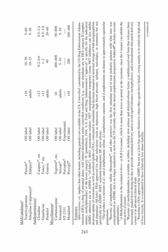
Attention Deficit Hyperactivity Disorder 243
243
MA
O in
hibi
tors
h
Tra
nylc
ypro
min
eP
arna
te®
Off
-lab
el‡
1610
–30
5–60
Sel
egil
ine
(l-d
epre
nyl)
hE
ldep
ryl®
Off
-lab
elad
ults
10–1
55–
30A
ntih
yper
tens
ives
i
Clo
nidi
neC
atap
res®
, etc
.O
ff-l
abel
>12
0.2–
0.6
0.2–
2.4
Gua
nfac
ine
Ten
ex, ®
etc
.O
ff-l
abel
>12
1–2
0.5–
3.0
Pin
dolo
lG
ener
icO
ff-l
abel
adul
ts40
20–6
0M
isce
llan
eous
j
Car
bam
azep
ine
Teg
reto
l®, e
tc.
Off
-lab
el£
640
0–60
050
–800
Don
epez
ilA
rice
pt®
Off
-lab
elad
ults
5–10
5–10
GT
-233
1P
erce
ptin
®E
xper
imen
tal
–––
––––
–––
Mod
afin
ilP
rovi
gil®
Off
-lab
el‡
1620
010
0–40
0
Foo
tnot
es:
Abb
revi
atio
ns: e
tc. i
mpl
ies
that
oth
er b
rand
s, in
clud
ing
gene
rics
are
ava
ilab
le in
the
US
; Con
trol
led:
con
side
red
by th
e U
S D
rug
Enf
orce
men
t Adm
in-
istr
atio
n (D
EA
) a
drug
of
pote
ntia
l ab
use,
wit
h li
mit
ed d
istr
ibut
ion
unde
r co
ntro
lled
con
diti
ons
(e.g
., so
me
stim
ulan
ts a
re i
n D
EA
cat
egor
y II
[am
phet
-am
ines
, m
ethy
lphe
nida
te]
or t
he l
esse
r ca
tego
ry I
V [
pem
olin
e]);
FD
A:
U.S
. F
ood
and
Dru
g A
dmin
istr
atio
n (“
appr
oval
” is
spe
cifi
c fo
r th
e in
dica
tion
AD
HD
, ot
herw
ise
use
in A
DH
D i
s “o
ff-l
abel
”);
MA
O:
mon
oam
ine
oxid
ase;
NE
T:
nore
pine
phri
ne t
rans
port
er (
som
e N
ET
inh
ibit
ors
can
also
inh
ibit
neur
onal
upt
ake
of s
erot
onin
, as
wel
l as
cort
ical
upt
ake
of D
A),
con
tras
ted
to s
tim
ulan
ts th
at d
ecre
ase
tran
spor
t or n
euro
nal s
tora
ge o
f bot
h no
repi
neph
rine
and
dopa
min
e; O
ff-l
abel
: F
DA
-app
rove
d fo
r ot
her
indi
cati
ons
(suc
h as
dep
ress
ion,
hig
h bl
ood
pres
sure
, ep
ilep
sy,
narc
olep
sy,
or d
emen
tia)
but
not
spec
ific
ally
for
AD
HD
; SE
RT
: se
roto
nin
tran
spor
ter;
SR
: slo
w-r
elea
se.
a Thi
s pr
oduc
t is
a m
ixtu
re o
f d,
l- a
nd d
-am
phet
amin
e su
lfat
e, d
,l-am
phet
amin
e as
part
ate,
and
d-a
mph
etam
ine
sacc
hara
te in
app
roxi
mat
ely
equi
mol
arpr
opor
tion
s.b R
acem
ic d
,l-am
phet
amin
e su
lfat
e (B
enze
drin
e®,
and
othe
r ge
neri
cs)
was
the
fir
st s
tim
ulan
t us
ed t
o tr
eat
pedi
atri
c pa
tien
ts w
ith
wha
t la
ter
was
conc
eptu
aliz
ed a
s A
DH
D, b
ut is
no
long
er m
arke
ted
as s
uch
(in
the
US
cur
rent
ly o
nly
in th
e m
ixed
am
phet
amin
e pr
epar
atio
n A
dder
all®
incl
udes
rac
emic
amph
etam
ine)
.c d
-Met
hylp
heni
date
is th
e is
olat
ed d
-thr
eo-
or R
,R-[
+]-
isom
er, w
hich
is m
ore
than
twic
e as
pot
ent a
s th
e ra
cem
ate,
sin
ce th
e l-
isom
er c
an in
hibi
t the
mor
e ac
tive
d-i
som
er.
d Rac
emic
d,l-
met
hylp
heni
date
is
avai
labl
e as
tab
lets
, inc
ludi
ng s
low
-rel
ease
and
del
ayed
-rel
ease
for
ms,
a m
ulti
laye
red
oral
for
m t
hat
rele
ases
thr
eein
crea
sing
ly g
rade
d do
ses
duri
ng th
e da
y to
pro
vide
sus
tain
ed d
eliv
ery
and
to a
void
rapi
d-to
lera
nce
(tac
hyph
ylax
is),
and
a tr
ansd
erm
al s
kin
patc
h de
live
rysy
stem
is in
adv
ance
d cl
inic
al tr
ials
.e P
emol
ine
is F
DA
-app
rove
d fo
r A
DH
D, b
ut n
ot a
s a
firs
t-li
ne a
gent
(li
mit
ed to
whe
n ot
her
agen
ts h
ave
fail
ed),
ow
ing
mai
nly
to it
s re
lati
vely
hig
h ri
skof
live
r to
xici
ty. I
t is
cons
ider
ed to
hav
e re
lati
vely
low
abu
se-l
iabi
lity
.
(con
tinu
ed)

244 Zhang, Davids, and Baldessarini
244
Tab
le 2
(con
tinu
ed)
Foo
tnot
es:
f An
expe
rim
enta
l com
poun
d th
at is
the
acti
ve, i
sola
ted
R-i
som
er o
f the
N,N
-di-
desm
ethy
l met
abol
ite
of d
,l-si
butr
amin
e [M
erid
ia®
] a d
iet-
cont
rol a
gent
wit
h m
ild
stim
ulan
t-li
ke p
rope
rtie
s an
d ab
ilit
y to
inhi
bit t
rans
port
ers
for
dopa
min
e, n
orep
inep
hrin
e, a
nd s
erot
onin
, but
itse
lf is
not
use
d to
trea
t AD
HD
).g V
enla
faxi
ne m
ay b
e ac
tive
in A
DH
D, p
erha
ps o
win
g to
its
NE
T-a
ntag
onis
m a
t hig
h do
ses,
whe
reas
oth
er m
ore
sele
ctiv
e S
ER
T (
sele
ctiv
e se
roto
nin
tran
spor
t inh
ibit
ors)
app
ear
to h
ave
litt
le th
erap
euti
c ef
fect
in A
DH
D.
h Tra
nylc
ypro
min
e al
so h
as a
mph
etam
ine-
like
str
uctu
re a
nd s
tim
ulan
t-li
ke a
ctio
ns; s
eleg
ilin
e is
rel
ativ
ely
sele
ctiv
e fo
r M
AO
type
B, a
t lea
st a
t dai
lydo
ses
less
than
20
mg,
but
at h
ighe
r do
ses
it a
lso
inhi
bits
MA
O-A
and
may
pro
duce
hyp
erte
nsiv
e cr
ises
in th
e pr
esen
ce o
f ty
ram
i ne
or o
ther
sym
path
omi-
met
ic a
min
es; i
t may
be
met
abol
ized
in p
art t
o m
etha
mph
etam
ine
or o
ther
am
phet
amin
e-li
ke a
gent
s (a
s m
ay a
lso
occu
r w
ith
bupr
opi o
n). T
he e
xper
i-m
enta
l, se
lect
ive,
irre
vers
ible
MA
O-A
inhi
bito
r cl
orgy
line
als
o ha
s be
en f
ound
eff
ecti
ve in
AD
HD
but
is n
ot a
vail
able
for
cli
nica
l use
.i C
loni
dine
and
gua
nfac
ine
are
cent
rall
y ac
tive
a2-
adre
noce
ptor
ago
nist
s w
ith
(aut
orec
epto
r-m
edia
ted)
ant
ihyp
erte
nsiv
e ac
tion
and
pro
babl
e po
stsy
n-ap
tic
acti
ons
in c
ereb
ral
cort
ex (
Cat
apre
s® i
s av
aila
ble
as a
tra
nsde
rmal
ski
n pa
tch
as w
ell
as t
able
ts);
pin
dolo
l is
a c
entr
ally
act
ive b
-adr
enoc
epto
ran
tago
nist
that
may
als
o bl
ock
pres
ynap
tic
sero
toni
n (5
-HT
) au
tore
cept
ors
to e
nhan
ce r
elea
se o
f se
roto
nin.
j Car
bam
azep
ine
is a
n an
tico
nvul
sant
wit
h m
ood-
stab
iliz
ing
effe
cts;
don
epez
il is
a c
entr
ally
act
ive,
rev
ersi
ble
anti
chol
ines
tera
se a
gent
use
d pr
imar
ily
for
earl
y de
men
tia;
GT
-233
1 is
a c
entr
ally
act
ive
hist
amin
e H
3 re
cept
or a
ntag
onis
t; m
odaf
inil
is a
n no
nsti
mul
ant a
gent
of
unce
rtai
n ne
urop
harm
acol
ogy
that
sel
ecti
vely
sup
pres
ses
slee
p in
nar
cole
psy.
Not
e th
at n
one
of th
ese
agen
ts is
spe
cifi
call
y F
DA
-app
rove
d fo
r lo
ng-t
erm
use
(m
ore
than
abo
ut 6
wee
ks),
thou
gh s
tand
ard
care
req
uire
s in
defi
nite
lypr
olon
ged
use
in A
DH
D; t
enta
tivel
y pr
opos
ed d
oses
of
non-
FDA
-app
rove
d ag
ents
are
bas
ed o
n lit
erat
ure
repo
rts
and
may
cha
nge
with
fur
ther
exp
erie
nce.
Attention Deficit Hyperactivity Disorder 245
7.1. StimulantsStimulants have been used to treat both behavioral and cognitive features of
ADHD since the 1930s, when Bradley introduced d,l-amphetamine for the treat-ment of a range of behavioral disorders in children and adolescents. Indeed,amphetamines and other stimulants have become the cornerstone of medical treat-ment for ADHD. This group of drugs includes several amphetamines, racemic ord-methylphenidate, pemoline, the antidepressant bupropion, and an emerging groupof experimental agents that share some neuropharmacological features of typicalstimulants, notably their ability to release or prevent the inactivation of catechola-mines by neuronal reuptake.
The pharmacology of stimulants, which facilitate release of both norepinephrineand DA, overlaps that of many antidepressants that act by inhibition of norepineph-rine transport and also have proved to be effective in the treatment of ADHD.Agents that act selectively to block neuronal transport of DA, or use of stimulantsin combination with selective antagonists of DA receptors, have not been testedexperimentally in ADHD patients, leaving considerable uncertainty about the rela-tive importance of norepinephrine and DA in mediating clinically useful effects ofstimulants in ADHD.
The only drugs that are Food and Drug Administration (FDA)-approved specifi-cally for the treatment of ADHD are amphetamines, methylphenidate, pemoline(under limited circumstances), and the new antidepressant-like agent atomoxetine(Table 2). All other treatment options represent empirical, “off-label” applicationsof agents approved for other indications, such as depression or hypertension. More-over, use of many agents found to be effective in the treatment of ADHD in chil-dren is considered “off-label” as a result of specific FDA-approval for treatment ofadults only, notably including the tricyclic antidepressants. Additionally, FDA-approved recommendations for the duration of treatment are typically as brief as4 to 6 weeks, even although ADHD typically requires indefinitely prolongedtreatment.
By far, the most commonly employed treatment for ADHD in the United Stateshas been racemic d,l-threo-methylphenidate (Ritalin®), a diasteriomeric compoundwith four possible conformations (d- and l-, threo or erythro). Methylphenidate isavailable in a wide range of preparations, including ordinary and slow-release tab-lets, as well as a multicompartment oral preparation (Concerta®) that releases thedrug in rising amounts over at least 12 hours, with minimized risk of rapidtolerance or tachyphylaxis that can limit the action of ordinary long-acting prepara-tions (38). A transdermal skin patch delivery system also is available. Finally, thepure d-threo-isomer of methylphenidate (Focalin®) was recently FDA-approvedfor treatment of ADHD.
Pemoline is recommended only when other stimulants fail to alleviate symp-toms or are poorly tolerated, owing to an increased risk of liver damage. Bupropionappears to be somewhat less consistently effective than other stimulants, and isassociated with a risk of epileptic seizures at higher doses. Other stimulant-

246 Zhang, Davids, and Baldessarini
like agents remain experimental, including an active metabolite of the anorexicagent sibutramine, and the new agent GW-329659, that inhibits both DA and nore-pinephrine transporters.
Despite their status as a treatment of choice for ADHD at all ages, stimulantscontinue to draw criticism for their alleged overuse and potential for abuse or illicitdiversion. Some stimulants (amphetamines and pemoline, but not methylphenidate,bupropion, or sibutramine) are categorized as controlled substances by US DrugEnforcement Administration regulations. A recent study of a sample of nearly6000 children and adolescents suggested that overuse of stimulants may not be aswidespread as once thought (39). In that study, the prevalence of “definite” ADHDacross all ages was 7.4%, and 86.5% of youths so diagnosed were treated with astimulant at some time. However, among those without ADHD, only 0.2% wasever given a stimulant, with intermediate rates for those with less certain diag-nostic status.
7.2. AntidepressantsAntidepressants that selectively block norepinephrine reuptake, or both norepi-
nephrine and serotonin transport, are effective in the treatment of ADHD, whereasSSRI antidepressants are not (1,40). Beneficial effects of adrenergic antidepres-sants have been demonstrated with both older tricyclic antidepressants (e.g., imi-pramine, desipramine, and nortriptyline), which are relatively toxic in overdosesand the newer, safer agent atomoxetine (41). These agents may also have beneficialeffects in suppressing tics in children with a primary tic disorder such as Gilles dela Tourette’s syndrome, or those induced by stimulants. Monoamine oxidaseinhibitors (MAOIs; e.g., tranylcypromine and selegiline), which potentiate cat-echolamines and serotonin, also have been reported to be of benefit in ADHD.
The potential toxicity of the tricyclic antidepressants and MAOIs has limitedclinical enthusiasm for their use except when stimulants prove to be ineffective orpoorly tolerated. Newer agents that block the norepinephrine transporter, such asatomoxetine and duloxetine, have a much improved safety profile, and are emerg-ing as plausible alternatives to the older antidepressants and even the stimulants.
7.3. Miscellaneous AgentsThe a2 adrenoceptor agonists clonidine and guanfacine have some evidence of
clinical utility in ADHD although their primary indication is hypertension.Mechanisms by which these drugs alleviate ADHD symptoms remain unknown.An autoreceptor mechanism seems unlikely because activation of the presynaptica2 autoreceptor reduces noradrenergic neurotransmission, and so, a direct postsyn-aptic action is more likely. There is some evidence that centrally active b-adrener-gic blockers including pindolol may also be helpful in ADHD, through unexplainedmechanisms.
Finally, several agents developed mainly for other purposes may be useful inthe treatment of ADHD. These include the anticonvulsant carbamazepine, thecholinesterase inhibitor donepezil, the antinarcolepsy agent of uncertain mecha-

Attention Deficit Hyperactivity Disorder 247
nism of action, modafinil, as well as the experimental histamine H3 receptorantagonist GT-2331 (Perceptin®). However, experimental trials of these agentsremain limited and mainly anecdotal.
8. CONCLUSIONS
ADHD is a highly prevalent and heterogeneous syndrome consisting of cogni-tive and behavioral deficits, notably, inattention, impulsivity, deficient behavioralinhibition, and hyperactivity that can be comorbid with conduct, learning, and mooddisorders. ADHD can be found at any age, but is more often diagnosed in boys thangirls or adults, and in the United States more than Europe. Compared to the generalpopulation, risk of ADHD is 5 to 10 times greater among first-degree relatives ofindex cases, and more than twice greater in identical than fraternal twins. Heritabil-ity of ADHD is high and there are suggestive associations with specific geneticpolymorphisms of neuronal proteins essential to cerebral dopaminergic neu-rotransmission. Diagnosis remains essentially clinical, based on multiple sourcesof information. There are no specific psychological or other diagnostic tests, butobjective measures of body motility during sustained attention tasks are promising.
Prominent deficits in executive functions suggest dysfunction of the prefrontalcerebral cortex. This theory is tentatively supported by some brain-imaging stud-ies, although a specific pathophysiology is unknown. Laboratory animal models ofADHD include genetic and neurotoxin models, and paradoxically, include states ofeither dopaminergic excess or deficiency. These models vary in their similarity toclinical ADHD, and emphasize behavioral hyperactivity rather than specific cogni-tive dysfunctions. Newer approaches to laboratory modeling include use of rela-tively specific cognitive testing methods that are applicable to animal and humansubjects. These models of specific cognitive deficits (including response inhibi-tion, sustained attention, working memory, and others) may also representendophenotypes appropriate for genetic and pharmacological study instead of themore complex and heterogeneous clinical syndrome of ADHD.
Effective treatment of ADHD includes most stimulants (with dopaminergic andnoradrenergic actions), and antidepressants with noradrenergic, but not selectiveserotonergic, actions, along with comprehensive behavioral and educational inter-ventions. Interest in developing innovative treatments for ADHD has increasedgreatly in recent years following decades of heavy reliance on the use of stimulants.
GLOSSARY OF MEDICAL TERMINOLOGYAllele: Any one of a series of two or more different genes that occupy the same locus on a
chromosome.Attention: The ability to attend to a specific stimulus. Notice the contrast to alertness in
which a person can response to any stimulus.Comorbidity: A concomitant but separable disease.Polymorphism: Occurrence of multiple discrete alleles.Prevalence: The number of cases of a disease relative to a given population at a specific
time or period of time.

248 Zhang, Davids, and Baldessarini
Proband: The patient carrying a specific disease.Relative risk: The ratio of the risk of a disease among those exposed to a specific factor,
to the risk among those not exposed.Stimulant: An agent that increases motor activity, decreases fatigue, or promotes a sense of
well-being.
ACKNOWLEDGMENTS
This work was supported, in part, by the Fred Corneel Research Fellowship (KZ),and by the Bruce J. Anderson Foundation and the McLean Private DonorsNeuropsychopharmacology Research Fund (RJB).
REFERENCES1. Barkley RA. Attention-Deficit Hyperactivity Disorder: A Handbook for Diagnosis and Treat-
ment. New York, NY: Guilford Press, 1998.2. Solanto MV, Arnsten AFT, Castellanos FX, ed. Stimulant Drugs and ADHD. New York, NY:
Oxford University Press, 2001.3. Douglas VI. Stop, look, and listen: The problems of sustained attention and impulse control in
hyperactive and normal children. Can J Behav Sci 1972;41:259–283.4. Lahey BB, Applegate B, McBurnett K, et al. DSM-IV field trials for attention deficit hyperactiv-
ity disorder in children and adolescents. Am J Psychiatry 1994;151:1673–1685.5. Zito JM, Safer DJ, DosReis S, et al. Psychotropic practice patterns for youth: 10-year perspec-
tive. Arch Pediatr Adolesc Med 2003;157:17–25.6. McBurnett K, Pfiffner LJ, Willcutt E, et al. Experimental cross-validation of DSM-IV types of
attention deficit/hyperactivity disorder. J Am Acad Child Adolesc Psychiatry 1999;38:17–24.7. Biederman J, Faraone SV, Mick E, et al. Clinical correlates of ADHD in females: Findings from
a large group of girls ascertained from pediatric and psychiatric referral sources. J Am AcadChild Adolesc Psychiatry 1999;38:966–975.
8. Ishii T, Takahashi O, Kawamura Y, Ohta T. Comorbidity in attention deficit-hyperactivity disor-der. Psychiatry Clin Neurosci 2003;57:457–463.
9. Faedda GL, Baldessarini RJ, Suppes T, Tondo L, Becker I, Lipschitz DS. Pediatric-onset bipo-lar disorder: A neglected clinical and public health problem. Harvard Rev Psychiatry 1995;3:171–195.
10. Wozniak J, Biederman J, Kiely K, et al. Mania-like symptoms suggestive of childhood onsetbipolar disorder in clinically referred children. J Am Acad Child Adolesc Psychiatry 1995;34:1577–1583.
11. Faedda GL, Baldessarini RJ, Glovinsky IP, Austin NB. Mania in pediatric bipolar illness associ-ated with antidepressant and stimulant treatment. J Affect Disord 2004; in press.
12. Smidt J, Heiser P, Dempfle A, et al. [Formal genetic findings in attention-deficit/hyperactivity-disorder]. Fortschr Neurol Psychiatr 2003;71:366–377.
13. Galili-Weisstub E, Segman RH. Attention deficit and hyperactivity disorder: Review of geneticassociation studies. Israeli J Psychiatry Relat Sci 2003;40:57–66.
14. Faraone SV, Doyle AE, Mick E, Biederman J. Meta-analysis of the association between the7-repeat allele of the dopamine D4 receptor gene and attention deficit hyperactivity disorder.Am J Psychiatry 2001;158:1052–1057.
15. Castellanos FX, Tannock R. Neuroscience of attention-deficit/hyperactivity disorder: The searchfor endophenotype. Nature Rev Neurosci 2002;3:617–628.
16. Ogdie MN, Macphie IL, Minassian SL, et al. A genome-wide scan for attention-deficit/hyper-activity disorder in an extended sample: Suggestive linkage on 17p11. Am J Human Genet2003;72:1268–1279.

Attention Deficit Hyperactivity Disorder 249
17. Castellanos FX, Giedd JN, Marsh WL, et al. Quantitative brain magnetic resonance imaging inattention-deficit hyperactivity disorder. Arch Gen Psychiatry 1996;53:607–616.
18. Casey BJ, Castellanos FX, Giedd JN, et al. Implications of right frontostriatal circuitry in responseinhibition and attention-deficit/hyperactivity disorder. J Am Acad Child Adolesc Psychiatry1997;36:374–383.
19. Schweitzer JB, Faber TL, Grafton ST, Tune LE, Hoffman JM, Kilts CD. Alterations in the func-tional anatomy of working memory in adult attention deficit hyperactivity disorder. Am J Psy-chiatry. 2000;157:278–280.
20. Davids E, Zhang K, Tarazi FI, Baldessarini RJ. Animal models of attention-deficit hyperactivitydisorder. Brain Res Rev 2003;42:1–21.
21. Sagvolden T. Behavioral validation of the spontaneously hypertensive rat (SHR) as an animal modelof attention-deficit/hyperactivity disorder (ADHD). Neurosci Biobehav Rev 2000;24:31–39.
22. Giros B, Jaber M, Jones SR, Wightman RM, Caron MG. Hyperlocomotion and indifference tococaine and amphetamine in mice lacking the dopamine transporter. Nature 1996;379:606–612.
23. Hess EJ, Jinnah HA, Kozak CA, Wilson MC. Spontaneous locomotor hyperactivity in a mousemutant with a deletion including the SNAP gene on chromosome 2. J Neurosci 1992;12:2865–2874.
24. Shaywitz BA, Klopper JH, Yager RD, Gordon JW. Paradoxical response to amphetamine indeveloping rats treated with 6-hydroxydopamine. Nature 1976;261:153–155.
25. Zhang K, Davids E, Tarazi FI, Baldessarini RJ. Effects of dopamine D4 receptor-selectiveantagonists on motor hyperactivity in rats with neonatal 6-hydroxydopamine lesions. Psycho-pharmacology 2002;161:100–106.
26. Barkley RA. ADHD and the Nature of Self-Control. New York, NY: Guilford Press, 1997.27. Douglas VI. Cognitive control processes in attention deficit/hyperactivity disorder. In: Quay HC,
Hogan AE, ed. Handbook of Disruptive Behavior Disorders. New York, NY: Kluwer-Plenum,1991:105–138.
28. Denckla MB. Biological correlates of learning and attention: What is relevant to learning disabil-ity and attention-deficit hyperactivity disorder? J Devel Behav Pediatrics 1996;17:114–119.
29. Evenden JL. Varieties of impulsivity. Psychopharmacology 1999;146:348–161.30. Logan GD. On the ability to inhibit thought and action: A user’s guide to the stop signal para-
digm. In: Dagenbach D, Carr TH, ed. Inhibitory Processes in Attention, Memory, and Language.San Diego, CA: Academic Press 1994:189–239.
31. Aron AR, Dowson JH, Sahakian BJ, Robbins TW. Methylphenidate improves response inhibi-tion in adults with attention-deficit/hyperactivity disorder. Biol Psychiatry 2003;54:1465–1468.
32. Feola TW, de Wit H, Richards JB. Effects of d-amphetamine and alcohol on a measure of behav-ioral inhibition in rats. Behav Neurosci 2000;114:838–848.
33. Riccio CA, Reynolds CR. Continuous performance tests are sensitive to ADHD in adults but lackspecificity: Review and critique for differential diagnosis. Ann NY Acad Sci 2001;931:113–139.
34. Robbins TW. The 5-choice serial reaction time task: Behavioral pharmacology and functionalneurochemistry. Psychopharmacology 2002;163:362–380.
35. Sarter M, Givens B, Bruno JP. The cognitive neuroscience of sustained attention: where top-down meets bottom-up. Brain Res Rev 2001;35:146–60.
36. Goldman-Rakic PS. Working memory dysfunction in schizophrenia. In: Salloway SP, MalloyPF, Duffy JD, ed. The Frontal Lobes and Neuropsychiatric Illness. Washington DC: AmericanPsychiatric Press, 2001:71–82.
37. Arnsten AFT, Murphy B, Merchant K. The selective dopamine D4 receptor antagonist, PNU-101387G, prevents stress-induced cognitive deficits in monkeys. Neuropsychopharmacology2000;4:405–410.
38. Swanson J, Gupta S, Lam A, Shoulson I, Lerner M, Modi N, Lindemulder E, Wigal S. Devel-opment of a new once-a-day formulation of methylphenidate for the treatment of attention-defi-cit/hyperactivity disorder: Proof-of-concept and proof-of-product studies. Arch Gen Psychiatry2003;60:204–211.

250 Zhang, Davids, and Baldessarini
39. Barbaresi WJ, Katusic SK, Colligan RC, et al. How common is attention-deficit/hyperactivitydisorder? Arch Pediatr Adolesc Med 2002:156:217–224.
40. Baldessarini RJ. Drugs and the treatment of psychiatric disorders: Antidepressant and antianxietyagents. In: Hardman JG, Limbird LE, Gilman AG, ed. Goodman and Gilman’s The Pharmaco-logical Basis of Therapeutics, tenth edition. New York, NY: McGraw-Hill Press, 2001:447–483,and Chapter 17, 2005, in press.
41. Kratochvil CJ, Vaughan BS, Harrington MJ, Burke WJ. Atomoxetine: Selective noradrenalinereuptake inhibitor for the treatment of attention-deficit/hyperactivity disorder. Expert OpinPharmacother 2003;4:1165–1174.

Index 251
Index
251
A
AADC, 5ABA, 144Absorbed drugs
distribution, 41–42Acetylcholinesterase inhibitors
Alzheimer’s disease, 58Acral lick dermatitis (ALD), 176ADHD. See Attention deficit hyper-
active disorder (ADHD)Akinetic mutism
schizophrenia, 116ALD, 176α-methyl-p-tyrosine (AMPT), 5α-1 receptors, 9α-2 receptors, 9α-synuclein, 91Alzheimer’s disease, 51–58
animal models, 54–56brain pathways, 51genetics, 54glossary, 58–59histopathological abnormalities, 52fmolecular mechanisms and targets,
52–54prevalence, 56prevention treatment, 57–58signs and symptoms, 56–57
Amino acid decarboxylase (AADC), 5Amino acid oxidase, 122tAmino terminal region, 69Amoxapine, 43AMPAkines
schizophrenia, 125Amphetamines
ADHD, 242tAmyloid β-protein, 52Amyloid precursor protein, 52A9 neurons, 4A10 neurons, 4Anterior cingulotomy
obsessive-compulsive behavior, 181
Anticonvulsantsautism spectrum disorders, 142–143bipolar disorder, 221–222
Antidepressants, 192t, 196–197ADHD, 246bipolar disorder, 222–223
Anti-inflammatory agentsAlzheimer’s disease, 58Parkinson’s disease, 100
AntioxidantsAlzheimer’s disease, 57
Antipsychoticsautism spectrum disorders, 142bipolar disorder, 222Huntington’s disease, 82schizophrenia, 124t, 125
ApoE gene, 54Applied Behavioral Analysis (ABA), 144Aripiprazole
Huntington’s disease, 83schizophrenia, 124t, 125
Arterial spin labeling (ASL) fMRI, 24Asperger’s syndrome, 131, 140Atomoxetine
ADHD, 242tAttention
ADHD, 238–239Attention deficit hyperactive disorder
(ADHD), 157, 229–248animal models, 236–237clinical features, 230–231diagnosis, 231–233, 232tepidemiology, 233–234experimental models, 236–241genetics, 234–235glossary, 246–248history, 229–230neuroimaging, 235–236neuropsychological models, 238–
239pharmacological treatment, 242tTourette’s syndrome, 155treatment, 241–247

252 Index
Autism spectrum disorders, 131–147animal models, 135, 136tbrain growth, 133brain pathways, 131–134cerebellum, 132epidemiology, 139–140future directions, 145–146genetic conditions associated with,
138genetics, 137–138glossary, 146–147limbic system, 132molecular targets, 134–135phenomenology, 140–141treatment, 142–145
Autoradiographyneurotransmitters, 11–12
B
Basal ganglia, 151normal function, 93–94
Basal ganglia-thalamocortical circuitry,13–15, 96f
organization, 14fBDNF, 72Behavior
depression, 190–191Benserazide
Parkinson’s disease, 99Benzodiazepines
Huntington’s disease, 82schizophrenia, 125
Biological rhythmsbipolar disorder, 211–212, 223
Bipolar depression, 214Bipolar disorder (BPD), 112, 205–225,
213tbiology, 209–210clinical features, 212–216comorbidity, 216diagnosis, 215–216epidemiology, 207–208, 208tepisode frequency, 216–217genetics, 209–210glossary, 224–225historical background, 206–207illness course, 216–217
incidence, 207–208mortality, 209onset, 207–208prevalence, 207–208treatment, 217–223, 219t
long-term, 220–221short-term, 218–219
Blood oxygen level-dependent (BOLD)fMRI, 23–24
Botulinum toxinTourette’s syndrome, 166
BPD. SeeBipolar disorder (BPD)Brain circuits, 13–17Brain-derived neurotrophic factor
(BDNF), 72Brain imaging
bipolar disorder, 212Brain neurotransmitters, 3, 4–11Buproprion, 192t
ADHD, 242t
C
CaffeineTourette’s syndrome, 165
Candidate gene regionsautism spectrum disorders, 137
CarbamazepineADHD, 243t, 246bipolar disorder, 221Huntington’s disease, 83
CarbidopaParkinson’s disease, 99
Carbon spectroscopy (13C MRS), 25Casein
autism spectrum disorders, 143Caspase inhibitors
Parkinson’s disease, 100Catatonia
schizophrenia, 114Catechol-O-methyltransferase (COMT),
6inhibitors
Parkinson’s disease, 99schizophrenia, 123
Caudate putamen (CPu), 4, 13CDD, 140Chemical spectroscopy, 24–25

Index 253
Chemical synapsedrug intervention, 30–32schematic, 30f–31f
Childhood disintegrative disorder(CDD), 140
Chlorpromazinebipolar disorder, 218, 222schizophrenia, 124, 124t
5-choice serial reaction time task(5-CSRTT)
ADHD, 239Chronic stress-induced depression
models, 195Cingulate gyrus, 15Cingulotomy
obsessive-compulsive behavior, 181Circadian rhythms
bipolar disorder, 211–212, 223Citalopram, 192t
autism spectrum disorders, 142obsessive-compulsive behavior,
177–178unipolar depression, 196
Clomipramineautism spectrum disorders, 142Huntington’s disease, 83obsessive-compulsive behavior,
177–178, 178unipolar depression, 196
Clonazepambipolar disorder, 218Huntington’s disease, 82Tourette’s syndrome, 165
ClonidineADHD, 243t, 246autism spectrum disorders, 143Tourette’s syndrome, 164
Clozapinebipolar disorder, 222classification, 32schizophrenia, 124, 124t, 125
Coenzyme Q10, 73Huntington’s disease, 83Parkinson’s disease, 100
CognitionHuntington’s disease, 81
treatment, 83
Cognitive-behavioral therapybipolar disorder, 223
Communicative deficitsautism spectrum disorders, 140–141
Component modeling, 120Continuous performance task (CPT)
ADHD, 239Cortico-striato-pallido-mesencephalic
circuitry, 15–16organization, 17f
CPTADHD, 239
CreatineHuntington’s disease, 83
Cycloserineschizophrenia, 125
Cyclothymia, 214
D
DA. See Dopamine (DA)D-amino acid oxidase, 122tD-cyclo-serine
schizophrenia, 125Deep brain stimulation (DBS)
obsessive-compulsive behavior,182–183
Parkinson’s disease, 103Delusions
schizophrenia, 115, 116Dementia praecox, 112, 116, 206–207Dentato rubro cerebellar atrophy,
90Depression. See also specific type
behavior, 190–191DSM-IV, 190tepidemiology, 189pathology, 191–192stress, 191
Depression modelschronic stress-induced, 195
Desipramine, 197fADHD, 242tunipolar depression, 197
DHPA, 6Diazepam
Huntington’s disease, 82
254 Index
Dietautism spectrum disorders, 143Tourette’s syndrome, 165
Diffusion tensor imaging (DTI), 24Dihydroxyphenylalanine, 4–5Dihyroxyphenylacetaldehyde (DHPA),
6Dimethylglycine (DMG)
autism spectrum disorders, 143Dinorsibutramine
ADHD, 242tDivalproex sodium
Huntington’s disease, 83bipolar disorder, 221
D1-like receptors, 6–7D2-like receptors, 7DMG
autism spectrum disorders, 143Donepezil
ADHD, 243t, 246Dopamine (DA), 4–7
blocking degradation, 30classification, 32hypothesis
schizophrenia, 118metabolism, 4–6pathways, 4–5receptor subtypes, 6synapse, 5synthesis, 4–6Tourette’s syndrome, 155
Dopamine (DA) antagonistsobsessive-compulsive behavior, 179
Dopamine (DA) receptorsresponsiveness, 44
Dopamine (DA) replacement therapyParkinson’s disease, 99
Dopaminergic terminal innervation,156f
Dorsal medullary group, 8Dorsal pathway, 9–10Down syndrome, 138Drug(s)
absorbeddistribution, 41–42
affinity determination, 33–37classification, 32–33
interventionchemical synapse, 30–32
maximal drug levels, 40–41nonsteroidal anti-inflammatory
Parkinson’s disease, 100orally administered
absorption, 40–41response termination, 42–43selectivity, 45–46therapeutic action, 43–45
DTI, 24Duloxetine
unipolar depression, 197Dysbindin, 122t
E
Early interventionautism spectrum disorders, 144
ECTunipolar depression, 198
Educational interventionsTourette’s syndrome, 165
Electroconvulsive therapy (ECT)unipolar depression, 198
Emission tomography, 18–21Emotional disturbances
Huntington’s disease, 81treatment, 82–83
Energy metabolism pathwaysHuntington’s disease, 73
EntacaponeParkinson’s disease, 99
Environmental factorsTourette’s syndrome, 159
EPSschizophrenia, 124
Escitalopramunipolar depression, 196
EstrogenAlzheimer’s disease, 58
Ethnicityobsessive-compulsive behavior,
171–173Excitotoxic processes
Huntington’s disease, 73–74Excitotoxins
Huntington’s disease, 75–76

Index 255
Exerciseunipolar depression, 198–199
Extrapyramidal side effects (EPS)schizophrenia, 124
F
Family-focused therapybipolar disorder, 223
Family studiesautism spectrum disorders, 137
Fenfluramineobsessive-compulsive behavior,
178Flavonoids
Parkinson’s disease, 100Fluorine spectroscopy (19F MRS), 25Fluoxetine, 43, 192t
autism spectrum disorders, 142Huntington’s disease, 82, 83obsessive-compulsive behavior,
177–178unipolar depression, 196
FluphenazineHuntington’s disease, 82schizophrenia, 124, 124tTourette’s syndrome, 164
Fluvoxamine, 192tautism spectrum disorders, 142Huntington’s disease, 82obsessive-compulsive behavior,
177–178fMRI. See Functional magnetic reso-
nance imaging (fMRI)Folic acid
Alzheimer’s disease, 57Forced swim test
unipolar depression, 194–195Fragile X syndrome, 138Fragment mouse models
Huntington’s disease, 77–79Full-length mouse models
Huntington’s disease, 79Functional magnetic resonance imag-
ing (fMRI), 18, 23–24autism spectrum disorders, 132
Fusiform gyrus, 133f
GGABA, 13, 97
Tourette’s syndrome, 152Gabapentin
Huntington’s disease, 83GABHS, 157GAD, 97Gadolinium, 23–24γ-aminobutyric acid (GABA), 13, 97γγ
Tourette’s syndrome, 152Ganglia output nuclei (GPi), 64, 95Genetics
ADHD, 234–235Alzheimer’s disease, 54autism spectrum disorders, 137–138bipolar disorder, 209–210Huntington’s disease, 66–68, 77obsessive-compulsive behavior,
174–175Parkinson’s disease, 90–92schizophrenia, 121–123Tourette’s syndrome, 159–160unipolar depression, 193–194
Genome screensautism spectrum disorders, 137
Geriatric bipolar disorder, 215Globus pallidus (GP), 13, 151, 153Glutamic acid decarboxylase (GAD), 97Glutathione enhancers
Parkinson’s disease, 100Gluten
autism spectrum disorders, 143Glycine
schizophrenia, 125GP, 13, 151, 153GPCR
responsiveness, 44GPi, 64, 95G protein-coupled receptors (GPCR)
responsiveness, 44Group A β-hemolytic streptococci
(GABHS), 157Growth factors
Parkinson’s disease, 100Guanfacine
ADHD, 243t, 246Tourette’s syndrome, 164

256 Index
H
Hallervorden Spatz syndrome, 91Hallucinations
schizophrenia, 115Haloperidol
classification, 32Huntington’s disease, 82schizophrenia, 124, 124tTourette’s syndrome, 164
HEAT domain, 69Heschl’s gyrus, 119Hippocampal formation, 15Hoarding, 172Homocysteine
Alzheimer’s disease, 57Homovanillic acid (HVA), 65-HT. See also Serotonin
1A receptors, 10–112A receptors, 11
Huntingtin, 66–67aggregates in pathogenesis, 74–
75apoptotic cascades, 72–73intraneuronal trafficking, 71oxidative damage, 74postsynaptic signaling, 72protein, 67transcriptional regulation, 71–72wild-type, 67–68
cellular and molecular targets,70t
Huntington’s disease, 63–84abnormal protein–protein interac-
tions, 69animal models, 75–80, 78tbasal ganglia pathway, 65fbrain pathways, 63–66cellular and molecular targets, 68–
75clinical phenotypes, 80–81endocytosis pathways, 69–71fragment mouse models, 77–79full-length mouse models, 79genetic background, 66–68genetic models, 77glossary, 84–85
knock-in mice, 79–80neuropathological basis of psychiatric
symptoms, 65–66neuropathological basis of motor
dysfunction, 64–65treatment, 81–84
prototypical, 83–84HVA, 65-hydroxytryptamine (5-HT). See also
Serotonin1A receptors, 10–112A receptors, 11
Hypericum perforatum (St. John’sWort)
unipolar depression, 198Hypomania, 214Hypothalamus, 4
I
Illusionsschizophrenia, 115
ImipramineADHD, 242tunipolar depression, 196
Immunocytochemistryneurotransmitters, 12
Immunogeneticsautism spectrum disorders, 138
ImpulsivityHuntington’s disease, 83
Inositolobsessive-compulsive behavior,
178In situ hybridization (ISH)
neurotransmitters, 12–13Interpersonal and social rhythm
therapybipolar disorder, 223
Interpersonal psychotherapybipolar disorder, 223
Intravenous immunoglobulinTourette’s syndrome, 166
ISHneurotransmitters, 12–13
Isolationschizophrenia, 115
Index 257
J
Juvenile bipolar disorder, 215Juvenile Huntington’s disease, 81
K
Knock-in miceHuntington’s disease, 79–80
L
L-amino acid decarboxylase (AADC),5
Lamotriginebipolar disorder, 222
Late-onset Huntington’s disease, 81Lateral tegmental system, 8LC complex, 8L-3,4-dihydroxyphenylalanine (L-
DOPA), 4–5, 5fLEAP, 144Levodopa
Parkinson’s disease, 99Libido
Huntington’s disease, 83Life Skills and Education for Students
with Autism (LEAP), 144Lifestyle
Tourette’s syndrome, 165Ligand receptor functional interactions,
37–39Ligands
EC50, 39fefficacy, 37–39potency, 37–39
Limbic leukotomyobsessive-compulsive behavior,
181–182Limbic system, 15
organization, 16fLipoic acid
Alzheimer’s disease, 57Lithium
autism spectrum disorders, 143bipolar disorder, 218, 221Huntington’s disease, 83schizophrenia, 125
Lithium spectroscopy (Li MRS), 25Locus coerulus (LC) complex, 8Lorazepam
bipolar disorder, 218Loxapine, 43
M
Machado Joseph disease, 90Magnetic resonance imaging (MRI), 18,
21–23isotope characteristics, 19tTourette’s syndrome, 153
Magnetic resonance spectroscopy(MRS), 18–21. See also specifictype e.g., lithium spectroscopy
autism spectrum disorders, 132Major depression
DSM-IV, 190tepidemiology, 189
MalonateHuntington’s disease, 76
Maniabipolar disorder, 212–213Huntington’s disease, 83
Manic depressive illness, 112. See alsoBipolar disorder (BPD)
MAOIADHD, 246unipolar depression, 196
Maprotiline, 197fMaximal receptor density
ligand receptor binding interactionsdetermination, 33–37
Medial prefrontal cortex (mPFC), 16Medical terminology
ADHD, 246–248Alzheimer's disease, 58–59autism spectrum disorders, 146–147bipolar disorder, 224–225Huntington’s disease, 84–85obsessive-compulsive behavior, 185Parkinson’s disease, 104–106schizophrenia, 127–128Tourette’s syndrome, 167unipolar depression, 201
Medications. See Drug(s)
258 Index
Medium spin projection (MSP) neurons,152
Mental Status Exam (MSE)schizophrenia, 114
Mesocortical system, 4Mesolimbic system, 4Mesurguline equilibrium binding
saturation isotherm data, 35f3-Methoxytyramine (3-MT), 6Methylphenidate (Ritalin)
ADHD, 242t, 245autism spectrum disorders, 143
Methyl-4-phenyl-1,2,3,6-tetrahydropyridine (MPTP), 97
Parkinson’s disease, 94Methylspiperone
raclopride displacement, 36fMinocycline
Huntington’s disease, 84Misperceived objects
schizophrenia, 115Mitochondria
Huntington’s disease, 73Mitochondrial toxins
Huntington’s disease, 76selective toxicity
Huntington’s disease, 76–77Mixed states
bipolar disorder, 214–215Modafinil
ADHD, 243tMonoamine oxidase inhibitors (MAOI)
ADHD, 246unipolar depression, 196
Mood stabilizersschizophrenia, 125
Motor interventionsautism spectrum disorders, 145
Motor ticsTourette’s syndrome, 161
Movement disordersHuntington’s disease, 80
treatment, 82mPFC, 16MPTP, 97
Parkinson’s disease, 94MRI. See Magnetic resonance imaging
(MRI)
MRS. See Magnetic resonance spectros-copy (MRS)
MSEschizophrenia, 114
MSP neurons, 152Mutant huntingtin, 68
N
Natural productsautism spectrum disorders, 143unipolar depression, 198
Nerve cells, 3Neural circuits, 3Neuregulin, 122tNeurochemistry
bipolar disorder, 210–211Neurofibromatosis, 138Neuroimaging methods, 18–26Neuroleptic malignant syndrome (NMS)
schizophrenia, 124Neuroleptics
Huntington’s disease, 82Neurological disorders
neural principles, 3–26pharmacotherapeutic principles, 29–
46Neurons, 3Neurosurgery
obsessive-compulsive behavior,180–181
Neurotransmitters, 3, 4–11immunocytochemistry, 12ISH, 12–13localization and visualization, 11–13in vitro receptor autoradiography,
11in vivo receptor autoradiography, 12
NGD 94-1, 45Nigrostriatal system, 43 nitropropionic acid (3 NP)
Huntington’s disease, 76N-methyl-NN D-asparate (NMDA)
schizophrenia, 118NMR. See Magnetic resonance imaging
(MRI)NMS
schizophrenia, 124

Index 259
Nonsteroidal anti-inflammatory drugs(NSAID)
Parkinson’s disease, 100Norepinephrine, 7–9
metabolism, 8pathways, 8receptor subtypes, 8synthesis, 8
Norepinephrine receptorsresponsiveness, 44
Norepinephrine reuptake inhibitors(NRI)
unipolar depression, 197Nortryptaline
ADHD, 242tHuntington’s disease, 82
Novel genesschizophrenia, 122t
3 NPHuntington’s disease, 76
NRIunipolar depression, 197
Nuclear magnetic resonance (NMR).SeeMagnetic resonance imaging(MRI)
O
Obsessive-compulsive disease (OCD),141, 157, 171–185
animal models, 176atypical antipsychotics, 179–180autoimmune basis, 175–176behavioral phenotypes, 171–173brain pathways, 173–174ethnicity, 171–173experimental pharmacological
agents, 178–179genetics, 174–175glossary, 185neurochemistry, 174nonpharmacological treatment, 180–
184pharmacotherapy, 177–180prevalence, 171–173sex differences, 171–173treatment, 177–184treatment resistant, 176–177
OCD. See Obsessive-compulsive dis-ease (OCD)
Olanzapineautism spectrum disorders, 142bipolar disorder, 218, 222obsessive–compulsive behavior, 179schizophrenia, 124t, 125
Olivopontocerebellar atrophy, 91Orally administered drugs
absorption, 40–41Oxcarbazepine
bipolar disorder, 221Oxidative stress
Alzheimer’s disease, 57Oxytocin vasopressin system
autism spectrum disorders, 135
P
PANDAS, 157, 175Papez circuit, 15Parahippocampal gyrus, 15Parkinsonian primates, 95fParkinson’s disease, 87–106
anatomy, 92–93animal models, 94brain pathways, 92–98clinical aspects, 87–88differential diagnosis, 88–90, 89tetiology, 91–92genetics, 90–92glossary, 104–106incidence, 87molecular targets, 90–92neuroprotective therapy, 100pathological substrate, 90–92pathology, 90–91pathophysiology, 94–98pharmacotherapy, 98–100prevalence, 87signs and symptoms, 88surgery, 101–103treatment, 98–104, 102t
algorithm, 101future, 104symptoms, 98–100
Paroxetine, 192tHuntington’s disease, 82

260 Index
obsessive-compulsive behavior,177–178
unipolar depression, 196Passive immunization
Alzheimer’s disease, 57, 58PCr
Huntington’s disease, 83PDD-NOS, 131, 140, 141Pediatric autoimmune neuropsychiat-
ric disorder associated withstreptococcal (PANDAS), 157, 175
Pedunculopontine (PPN) region, 96Pemoline magnesium
ADHD, 242t, 245Pergolide
Parkinson’s disease, 99Perphenazine
schizophrenia, 124tPervasive developmental disorder not
otherwise specified (PDD-NOS),131, 140, 141
PET. See Positron emission tomogra-phy (PET)
Pharmacotherapypharmacodynamics, 33–39pharmacokinetics, 39–43
Phenelzine, 192tunipolar depression, 196
Phosphocreatine (PCr)Huntington’s disease, 83
Phosphoinositide 3 kinase (PI3K) Aktpathway, 73
Phosphorus spectroscopy (31P MRS), 25Pimozide
Tourette’s syndrome, 164Pindolol
ADHD, 243tPlasmaphoresis
Tourette’s syndrome, 166PLG, 33Polyglutamine proline domain, 69Polyphenols
Alzheimer’s disease, 57Porsolt test
unipolar depression, 194–195Positron emission tomography (PET),
18–21
autism spectrum disorders, 132Huntington’s disease, 64isotope characteristics, 19tOCD, 173fTourette’s syndrome, 153, 154
PPITourette’s syndrome, 154
PPN region, 96Pramipexole
Parkinson’s disease, 99Prefrontal cortex, 153Prepulse inhibition (PPI)
Tourette’s syndrome, 154Presenilin, 52–53Primates
Parkinson’s disease, 94Problem behavior intervention
autism spectrum disorders, 144Proline dehydrogenase, 122tProline leucine glycine (PLG), 33Propargylamine
Parkinson’s disease, 100Propranolol
Huntington’s disease, 83Proton spectroscopy, 24–25Psychiatric disorders
neural principles, 3–26pharmacotherapeutic principles, 29–
46Psychosocial intervention
bipolar disorder, 223Psychostimulants
autism spectrum disorders, 143Psychotherapy
Huntington’s disease, 82obsessive-compulsive behavior,
180Tourette’s syndrome, 166
Q
15q11q13autism spectrum disorders, 137
Quetiapineautism spectrum disorders, 142bipolar disorder, 218, 222Huntington’s disease, 83schizophrenia, 124t, 125

Index 261
R
Radionuclides, 18–21rCBF
autism spectrum disorders, 132Reboxetine
unipolar depression, 197Receptors. See also specific types
activation, 32allosteric sites, 32orthosteric sites, 32responsiveness, 43–45
Reelinautism spectrum disorders, 138
Regional cerebral blood flow (rCBF)autism spectrum disorders, 132
Repetitive behaviorsautism spectrum disorders, 141
ReserpineHuntington’s disease, 82
Response inhibitionADHD, 238
Rett syndrome, 140Rheumatic fever, 157Riluzole
Huntington’s disease, 84Risperidone
autism spectrum disorders, 142bipolar disorder, 218, 222obsessive-compulsive behavior, 179schizophrenia, 124t, 125
Ritalin. See Methylphenidate (Ritalin)Ropinirole
Parkinson’s disease, 99
S
SCA, 90Schizophrenia, 111–128, 206–207
animal models, 119–121diagnosis, 112–114DSM-IV, 113, 113tetiology, 120–123glossary, 127–128lifetime expectancy, 121tneural basis, 116–119, 117tnovel genes, 122toutcome, 126
signs and symptoms, 114–116treatment, 123–126
Secretinautism spectrum disorders, 143
Selective norepinephrine reuptakeinhibitors
unipolar depression, 197Selegiline, 30
ADHD, 243tParkinson’s disease, 100
Sensory interventionsautism spectrum disorders, 145
Serotonin, 9–11autism spectrum disorders, 134–
135metabolism, 10pathways, 9–10receptors, 10
responsiveness, 44synthesis, 10transporter
autism spectrum disorders, 138Serotonin reuptake inhibitors (SSRI)
autism spectrum disorders, 142Huntington’s disease, 82, 83obsessive-compulsive behavior,
177–178unipolar depression, 196
Sertraline, 192tHuntington’s disease, 82obsessive-compulsive behavior,
177–178unipolar depression, 196
Sex differencesobsessive-compulsive behavior,
171–173Sexual behavior
Huntington’s disease, 83Shy-Drager syndrome, 89Single-photon emission computed
tomography (SPECT), 18–21autism spectrum disorders, 132isotope characteristics, 19tTourette’s syndrome, 153
SN. See Substantia nigra (SN)Social impairment
autism spectrum disorders, 140

262 Index
Social interactionautism spectrum disorders, 145
Social rhythm therapybipolar disorder, 223
SPECT, 18–21autism spectrum disorders, 132isotope characteristics, 19tTourette’s syndrome, 153
Spinocerebellar ataxias (SCA), 90SSRI. See Serotonin reuptake inhibitors
(SSRI)St. John’s Wort
unipolar depression, 198Startle response
Tourette’s syndrome, 154Stereotypies, 152Stimulants
ADHD, 245–246STN, 13, 95, 151, 153Stress
depression, 191Stress-induced depression models, 195Striatonigral degeneration, 89Striatum, 151Substantia nigra (SN), 151, 153
pars compacta, 4, 13pars reticulata, 13, 64
Subthalamic nucleus (STN), 13, 95, 151,153
Suicideschizophrenia, 126
Synaptic terminals, 3Synuclein, 91
T
Tail suspension testunipolar depression, 195
Tardive dyskinesia, 165TCA. See Tricyclic antidepressants
(TCA)TEACCH, 144Tetrabenazine
Huntington’s disease, 82Tourette’s syndrome, 165
TH, 4–5Thioridazine
schizophrenia, 124t
Tic disorders, 151–167. See also specifictype
model, 152–153TMS
obsessive-compulsive behavior,183–184
Tourette’s syndrome, 154unipolar depression, 198
TocopherolParkinson’s disease, 100
TolcaponeParkinson’s disease, 99
Topiramatebipolar disorder, 222
Tourette’s syndrome, 141, 151–167animal models, 157–158behavioral phenotypes, 161–162brain pathways, 151–155DSM-IV, 161environmental factors, 159etiology, 158–159genetics, 159–160glossary, 167molecular targets, 155neurochemical pathways, 155pharmacological treatment, 164–165physiology, 158–159prevalence, 160–161psychology, 158–159treatment, 162–167
Tramadolobsessive-compulsive behavior, 179
Tranquilizersschizophrenia, 125
Transcranial magnetic stimulation (TMS)obsessive-compulsive behavior,
183–184Tourette’s syndrome, 154unipolar depression, 198
Tranylcypromine, 192tADHD, 243tunipolar depression, 196
Treatment and Education for Autisticand Related CommunicationHandicapped Children(TEACCH), 144
Tricyclic antidepressants (TCA)

Index 263
ADHD, 246Huntington’s disease, 83unipolar depression, 196
Tryptophanobsessive-compulsive behavior, 178
Tuberinfundibular neuroendocrino-logical system, 4
Tuberous sclerosis, 138Twin studies
autism spectrum disorders, 137Tyrosine hydroxylase (TH), 4–5
U
Unified HD Rating Scale (UHDRS)Huntington’s disease, 84
Unipolar depression, 189–201animal models, 194–195brain pathways, 192–193genetics, 193–194glossary, 201molecular mediators, 199–201, 200fneurotransmitter systems, 193pharmacotherapy, 196–197treatment, 196–199
V
VaccineAlzheimer’s disease, 57, 58
Vagus nerve stimulation (VNS)obsessive-compulsive behavior, 182
Valproatebipolar disorder, 218
Valproic acidautism spectrum disorders, 142–143,
143bipolar disorder, 221Huntington’s disease, 83schizophrenia, 125
Venlafaxine, 192tADHD, 242tunipolar depression, 197
Ventral pathway, 9–10Ventral tegmental area (VTA), 4, 16Vitamin E
Alzheimer’s disease, 57, 58Voices
schizophrenia, 115
W
Wild-type huntingtin, 67–68cellular and molecular targets, 70t
Working memoryADHD, 240
Z
Ziprasidoneschizophrenia, 124t, 125Tourette’s syndrome, 165
Zonisamidebipolar disorder, 222