Anatomy, Physiology, and Pathophysiology of Erectile Dysfunction
Upload
khangminh22Category
view
1download
0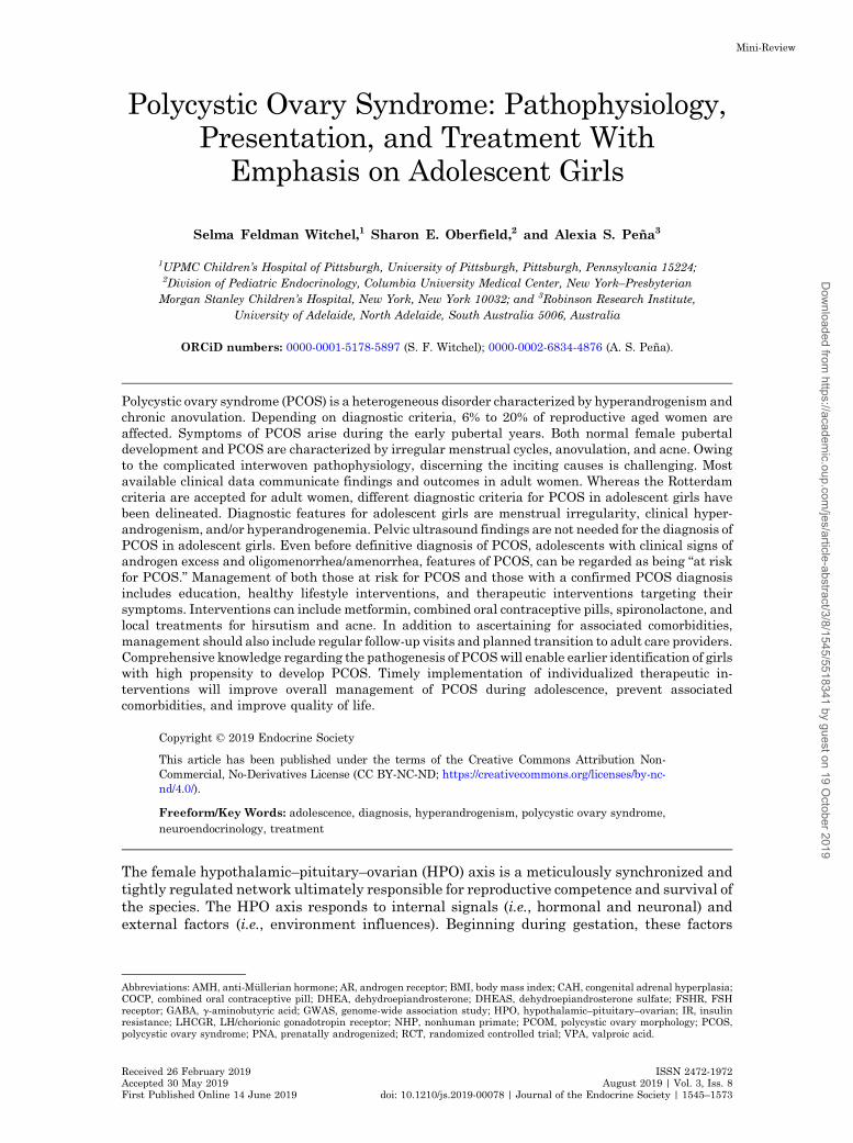
Mini-Review
Polycystic Ovary Syndrome: Pathophysiology,Presentation, and Treatment With
Emphasis on Adolescent Girls
Selma Feldman Witchel,1 Sharon E. Oberfield,2 and Alexia S. Peña3
1UPMC Children’s Hospital of Pittsburgh, University of Pittsburgh, Pittsburgh, Pennsylvania 15224;2Division of Pediatric Endocrinology, Columbia University Medical Center, New York–PresbyterianMorgan Stanley Children’s Hospital, New York, New York 10032; and 3Robinson Research Institute,
University of Adelaide, North Adelaide, South Australia 5006, Australia
ORCiD numbers: 0000-0001-5178-5897 (S. F. Witchel); 0000-0002-6834-4876 (A. S. Peña).
Polycystic ovary syndrome (PCOS) is a heterogeneous disorder characterized by hyperandrogenism andchronic anovulation. Depending on diagnostic criteria, 6% to 20% of reproductive aged women areaffected. Symptoms of PCOS arise during the early pubertal years. Both normal female pubertaldevelopment and PCOS are characterized by irregular menstrual cycles, anovulation, and acne. Owingto the complicated interwoven pathophysiology, discerning the inciting causes is challenging. Mostavailable clinical data communicate findings and outcomes in adult women. Whereas the Rotterdamcriteria are accepted for adult women, different diagnostic criteria for PCOS in adolescent girls havebeen delineated. Diagnostic features for adolescent girls are menstrual irregularity, clinical hyper-androgenism, and/or hyperandrogenemia. Pelvic ultrasound findings are not needed for the diagnosis ofPCOS in adolescent girls. Even before definitive diagnosis of PCOS, adolescents with clinical signs ofandrogen excess and oligomenorrhea/amenorrhea, features of PCOS, can be regarded as being “at riskfor PCOS.” Management of both those at risk for PCOS and those with a confirmed PCOS diagnosisincludes education, healthy lifestyle interventions, and therapeutic interventions targeting theirsymptoms. Interventions can include metformin, combined oral contraceptive pills, spironolactone, andlocal treatments for hirsutism and acne. In addition to ascertaining for associated comorbidities,management should also include regular follow-up visits and planned transition to adult care providers.Comprehensive knowledge regarding the pathogenesis of PCOSwill enable earlier identification of girlswith high propensity to develop PCOS. Timely implementation of individualized therapeutic in-terventions will improve overall management of PCOS during adolescence, prevent associatedcomorbidities, and improve quality of life.
Copyright © 2019 Endocrine Society
This article has been published under the terms of the Creative Commons Attribution Non-Commercial, No-Derivatives License (CC BY-NC-ND; https://creativecommons.org/licenses/by-nc-nd/4.0/).
Freeform/Key Words: adolescence, diagnosis, hyperandrogenism, polycystic ovary syndrome,neuroendocrinology, treatment
The female hypothalamic–pituitary–ovarian (HPO) axis is a meticulously synchronized andtightly regulated network ultimately responsible for reproductive competence and survival ofthe species. The HPO axis responds to internal signals (i.e., hormonal and neuronal) andexternal factors (i.e., environment influences). Beginning during gestation, these factors
Abbreviations: AMH, anti-Mullerian hormone; AR, androgen receptor; BMI, body mass index; CAH, congenital adrenal hyperplasia;COCP, combined oral contraceptive pill; DHEA, dehydroepiandrosterone; DHEAS, dehydroepiandrosterone sulfate; FSHR, FSHreceptor; GABA, g-aminobutyric acid; GWAS, genome-wide association study; HPO, hypothalamic–pituitary–ovarian; IR, insulinresistance; LHCGR, LH/chorionic gonadotropin receptor; NHP, nonhuman primate; PCOM, polycystic ovary morphology; PCOS,polycystic ovary syndrome; PNA, prenatally androgenized; RCT, randomized controlled trial; VPA, valproic acid.
Received 26 February 2019Accepted 30 May 2019First Published Online 14 June 2019
ISSN 2472-1972August 2019 | Vol. 3, Iss. 8
doi: 10.1210/js.2019-00078 | Journal of the Endocrine Society | 1545–1573
Dow
nloaded from https://academ
ic.oup.com/jes/article-abstract/3/8/1545/5518341 by guest on 19 O
ctober 2019

impact future generations through epigenetic factors affecting the brain and the developinggerm cells [1].
Polycystic ovary syndrome (PCOS), a disorder primarily characterized by signs andsymptoms of androgen excess and ovulatory dysfunction, disrupts HPO axis function.Depending on diagnostic criteria, this disorder affects ;6% to 20% of reproductive agedwomen [2, 3]. Typical clinical features include hirsutism, irregular menses, chronic anov-ulation, and infertility. The persistent hyperandrogenism is associated with impairedhypothalamic–pituitary feedback, LH hypersecretion, premature granulosa cell luteiniza-tion, aberrant oocyte maturation, and premature arrest of activated primary follicles [4].
1. Pathophysiology
By the time the diagnosis is established, PCOS presents as a phenotype reflecting a self-perpetuating vicious cycle involving neuroendocrine, metabolic, and ovarian dysfunction.Over the years, numerous hypotheses have been proposed regarding the proximate physi-ologic origins for PCOS. PCOS reflects the interactions among multiple proteins and genesinfluenced by epigenetic and environmental factors (Fig. 1) [5]. Specific sections of this articledeconstruct the factors contributing to the development of PCOS in humans and preclinicalmodels. Clinical and biochemical hyperandrogenism are major features of PCOS.
PCOS develops during the early pubertal years [6]. However, most relevant informationhas been accrued through clinical studies involving adult women in which referral biasfocuses on investigation of the more severe phenotypes [7]. Preclinical models involvinganimal and in vitro studies supplement clinical investigation and benefit from other ap-proaches to study this complex disorder. Recent clinical, experimental, and genetic dataemphasize neuroendocrine involvement in the pathophysiology of PCOS.
Figure 1. Factors contributing to PCOS phenotype. PCOS encompasses a woman’s life cycle.Factors potentially impacting the pathophysiology of PCOS are shown in circles. Not allfactors affect each individual. PCOS epitomizes a biologic network of interactingneuroendocrine, hormonal, metabolic, genetic, and environmental influences.
1546 | Journal of the Endocrine Society | doi: 10.1210/js.2019-00078
Dow
nloaded from https://academ
ic.oup.com/jes/article-abstract/3/8/1545/5518341 by guest on 19 O
ctober 2019
A. Ovary, Adrenal, and Androgen Excess
PCOS is characterized by excessive ovarian and/or adrenal androgen secretion. Intrinsicovarian factors such as altered steroidogenesis and factors external to the ovary such ashyperinsulinemia contribute to the excessive ovarian androgen production. Characteristicfeatures include more growing follicles in women with PCOS compared with normal controlswith premature growth arrest of antral follicles at 5 to 8mm. The classic ovarian phenotype ofenlarged ovaries with string-of-pearl morphology and theca interstitial hyperplasia reflectsandrogen exposure; this morphology has also been observed in women with congenital ad-renal hyperplasia (CAH) and female-to-male transgender individuals [8]. Distorted in-teractions among the endocrine, paracrine, and autocrine factors responsible for follicularmaturation may contribute to ovarian dysregulation in PCOS.
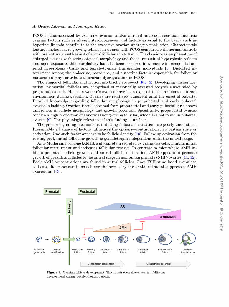
The stages of follicular maturation are briefly reviewed (Fig. 2). Developing during ges-tation, primordial follicles are comprised of meiotically arrested oocytes surrounded bypregranulosa cells. Hence, a woman’s ovaries have been exposed to the ambient maternalenvironment during gestation. Ovaries are relatively quiescent until the onset of puberty.Detailed knowledge regarding follicular morphology in prepubertal and early pubertalovaries is lacking. Ovarian tissue obtained from prepubertal and early pubertal girls showsdifferences in follicle morphology and growth potential. Specifically, prepubertal ovariescontain a high proportion of abnormal nongrowing follicles, which are not found in pubertalovaries [9]. The physiologic relevance of this finding is unclear.
The precise signaling mechanisms initiating follicular activation are poorly understood.Presumably a balance of factors influences the options—continuation in a resting state oractivation. One such factor appears to be follicle density [10]. Following activation from theresting pool, initial follicular growth is gonadotropin-independent until the antral stage.
Anti-Mullerian hormone (AMH), a glycoprotein secreted by granulosa cells, inhibits initialfollicular recruitment and indicates follicular reserve. In contrast to mice where AMH in-hibits preantral follicle growth and antral follicle maturation, AMH appears to promotegrowth of preantral follicles to the antral stage in nonhuman primate (NHP) ovaries [11, 12].Peak AMH concentrations are found in antral follicles. Once FSH-stimulated granulosacell estradiol concentrations achieve the necessary threshold, estradiol suppresses AMHexpression [13].
Figure 2. Ovarian follicle development. This illustration shows ovarian folliculardevelopment during developmental periods.
doi: 10.1210/js.2019-00078 | Journal of the Endocrine Society | 1547
Dow
nloaded from https://academ
ic.oup.com/jes/article-abstract/3/8/1545/5518341 by guest on 19 O
ctober 2019

Despite prior assumptions that androgens negatively impact follicles, androgens syn-thesized in preantral follicle theca cells promote growth of preantral and antral follicles andinduce granulosa cell FSH receptor (FSHR) expression in early antral follicles [14]. An-drogens promote aromatase expression and, ultimately, LH/chorionic gonadotropin receptor(LHCGR) expression in granulosa cells. As a follicle matures, androgens appear to inhibitproliferation and promote apoptosis. This biphasic androgen action was initially demon-strated in an NHP, the marmoset; androgens augmented FSH action in small antral folliclesbut had an inhibitory effect in larger follicles [15].
Androgen actions are mediated by androgen receptors (ARs), which are expressed in thecacells, granulosa cells, oocytes, and stromal cells [16]. Both canonical androgen signalingwhere AR functions as ligand-dependent transcription factor and nongenomic signalingoccur. Peak AR gene expression occurs in small antral follicles (;6 mm in diameter) anddecreases in antral and preovulatory follicles [17].
Typically, one follicle is “selected” as the dominant follicle [18]. With increasing estrogensecretion, pituitary FSH secretion declines due to negative feedback. The dominant folliclecompensates for this loss of FSH stimulation through increased LHCGR expression and in-creased responsiveness to LH stimulation. Subordinate follicles undergo atresia, presumablydue to relative FSH deficiency and androgen excess. Upon achieving a sufficient estradiolconcentration, neuroendocrine mechanisms trigger the LH surge to induce ovulation.
Under normal circumstances, the ovarian stroma provides a structural framework un-dergoing dynamic changes to support follicular growth. However, the ovarian stroma fromwomen with PCOS tends to be more rigid. The developing oocyte and its surroundingscaffolding rely on endocrine, paracrine, and autocrine signaling mechanisms to maintaincell-to-cell communication and assure synchronized developmental progression. Aberrantdevelopment during these earliest stages of follicular growth likely contributes to the ovarianaspects of PCOS [19]. Another feature of PCOS ovaries is accelerated transition from pri-mordial to growing follicles with increased numbers of 2- to 3-mm and 3- to 4-mm follicles [20,21]. AMH concentrations correlate with the number of these small antral follicles [22]. Thegrowing follicle is exposed to an atypical environment with increased LH, insulin, androgen,and AMH concentrations accompanied by insufficient FSH concentrations [19]. Additionaldifferences in PCOS ovaries include factors impacting vascular function and immuneresponsiveness [23].
Additionally, intrinsic alterations in ovarian steroidogenesis likely contribute to excessiveovarian androgen production. Available data document constitutively increased androgenproduction and CYP17A1 expression in cultured theca cells isolated from PCOS ovaries[24–26]. Steroidogenesis in the ovary involves both theca and granulosa cells. The theca cellsproduce ovarian androgens, which are converted to estrogens in the granulosa cell due to theactions of FSH-stimulated aromatase.
One interesting locus identified through genetic studies is DENND1A (see “H. Genetics”below). Overexpression of the alternative spliced variant, DENND1A.V2, of this generecapitulated a PCOS phenotype in cultured theca cells obtained from normal women,indicating a role for this variant in the excessive theca cell androgen production [27].Overexpression of DENND1A.V2 in an adrenal cell line led to increased expression of themRNAs for CYP17A1 and CYP11A1. However, the mechanisms responsible for increasedexpression of this alternatively spliced variant of DENND1A remain to be elucidated [28].
The adrenal zona reticularis is responsible for biosynthesis of the C-19 adrenal androgens,including dehydroepiandrosterone (DHEA), DHEA sulfate (DHEAS), androstenedione, andtestosterone. At least three distinct adrenal pathways contribute to androgen synthesis: (i)canonical/classical, (ii) “alternative backdoor,” and (iii) 11-oxo-androgens (Fig. 3). In thecanonical/classical pathway, progesterone is successively transformed by the enzyme 17a-hydroxylase/17,20-lyase (P450c17) to DHEA, which is subsequently converted by 3b-hydroxysteroid dehydrogenase type 2 to androstenedione. The alternative backdoor pathwaybypasses the usual steroid hormone intermediates, DHEA, androstenedione, and testos-terone, to produce dihydrotestosterone [29]. This pathway likely contributes to virilization of
1548 | Journal of the Endocrine Society | doi: 10.1210/js.2019-00078
Dow
nloaded from https://academ
ic.oup.com/jes/article-abstract/3/8/1545/5518341 by guest on 19 O
ctober 2019
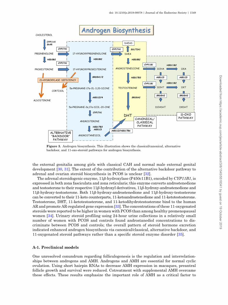
the external genitalia among girls with classical CAH and normal male external genitaldevelopment [30, 31]. The extent of the contribution of the alternative backdoor pathway toadrenal and ovarian steroid biosynthesis in PCOS is unclear [32].
The adrenal steroidogenic enzyme, 11b-hydroxylase (P450c11B1), encoded byCYP11B1, isexpressed in both zona fasciculata and zona reticularis; this enzyme converts androstenedioneand testosterone to their respective 11b-hydroxyl derivatives, 11b-hydroxy-androstenedione and11b-hydroxy-testosterone. Both 11b-hydroxy-androstenedione and 11b-hydroxy-testosteronecan be converted to their 11-keto counterparts, 11-ketoandrostenedione and 11-ketotestosterone.Testosterone, DHT, 11-ketotestosterone, and 11-ketodihydrotestosterone bind to the humanARand promote AR-regulated gene expression [33]. The concentrations of these 11-oxygenatedsteroids were reported to be higher inwomenwith PCOS than among healthy premenopausalwomen [34]. Urinary steroid profiling using 24-hour urine collections in a relatively smallnumber of women with PCOS and controls found androstanediol concentrations to dis-criminate between PCOS and controls; the overall pattern of steroid hormone excretionindicated enhanced androgen biosynthesis via canonical/classical, alternative backdoor, and11-oxygenated steroid pathways rather than a specific steroid enzyme disorder [35].
A-1. Preclinical models
One unresolved conundrum regarding folliculogenesis is the regulation and interrelation-ships between androgens and AMH. Androgens and AMH are essential for normal cyclicovulation. Using short hairpin RNAs to decrease AMH expression in macaques, preantralfollicle growth and survival were reduced. Cotreatment with supplemental AMH overcamethese effects. These results emphasize the important role of AMH as a critical factor to
Figure 3. Androgen biosynthesis. This illustration shows the classical/canonical, alternativebackdoor, and 11-oxo-steroid pathways for androgen biosynthesis.
doi: 10.1210/js.2019-00078 | Journal of the Endocrine Society | 1549
Dow
nloaded from https://academ
ic.oup.com/jes/article-abstract/3/8/1545/5518341 by guest on 19 O
ctober 2019

promote preantral follicle survival and growth in primates [36]. AMH treatment of culturedantral stage rhesus macaque follicles decreased estradiol production compared with un-treated follicles despite similar follicle size [12]. Hence, AMH appears to have a dual role:whereas AMHpromotes preantral follicle survival, it negatively impacts later stages of antralfollicle maturation [12].
In a series of in vivo and in vitro experiments, a subset of hypothalamic GnRH neurons,both mouse and human, were shown to express AMR receptors; AMH treatment increasedGnRH-dependent LH pulsatility and secretion [37]. Using a mouse model, AMH treatment ofpregnant mice was associated with diminished placental metabolism of testosterone to es-tradiol, decreased aromatase expression, masculinization of exposed female offspring, estruscycle disturbances, increased LH pulse frequency, brain masculinization, and infertilitycompared with unexposed mice; postnatal GnRH antagonist treatment reversed this PCOS-like phenotype [38]. These data suggest that excessive prenatal AMH exposure could promotethe aberrant neuroendocrine function typical of PCOS and that AMH can modulate GnRHneuron function [37, 38].
Knockout mice have been used to explore consequences of gene deletions. Using a post-natal androgen PCOS model, global AR knockout mice were protected from DHT-inducedPCOS-like features. Curiously, neuron-specific AR knockout mice were protected from DHT-induced ovarian dysfunction and several metabolic traits, reinforcing a role for extraovariantissues in the pathophysiology of PCOS [39]. Hence, intricate interrelationships exist be-tween androgens, AMH, follicle growth, metabolism, and neuroendocrine factors in PCOS.
Recently, a group of naturally hyperandrogenic female rhesus monkeys have been de-scribed. The high testosterone animals had increased LH, AMH, and androstenedioneconcentrations. Additionally, five of the six high-testosterone monkeys had no live offspring[40]. Future study of these animals will provide insight into the pathogenesis of PCOS.
B. Neuroendocrine Factors
Increased LH pulse frequency, LH pulse amplitude, and increased LH/FSH ratios are de-scribed in women with PCOS. The initial features of PCOS emerge during the early pubertalyears, concomitant with reactivation of the hypothalamic GnRH pulse generator, increasedgonadotropin secretion, and subsequent increased ovarian estrogen production. Loci iden-tified in the genome-wide association studies (GWASs) studies include LHCGR, FSHR, andFSH-b polypeptide (FSHB) genes, emphasizing neuroendocrine contributions to PCOSpathophysiology (see H. Genetics below).
Hypothalamic neurons in the arcuate nucleus secrete kisspeptin, neurokinin B, anddynorphin. These neurons, labeled as the KNDy neurons, are the leading contenders for thehypothalamic GnRH pulse generator because of the colocalization of these three peptides andtheir roles in episodic GnRH secretion [41]. Rather than initiating puberty, the GnRH pulsegenerator and GnRH neurons represent downstream nodes modulated by other hormonesand neurosecretory factors [42]. In other words, activation of excitatory inputs and in-activation of inhibitory inputs moderated by multiple influences regulate the output of theGnRH pulse generator to govern the timing of puberty [43–45]. This process culminates inincreased GnRH and gonadotropin secretion.
The hypothalamic GnRH neurons secrete GnRH in discrete pulses that travel through themedian eminence to the pituitary gonadotrophs, resulting in pulsatile LH and FSH secretion[46]. LH and FSH pulse frequencies are modulated by GnRH pulse frequency. IncreasedGnRH pulse frequency increases LH pulse frequency and decreases FSH pulse frequency[47]. The GnRH neurons integrate diverse influences, decode metabolic signals, and serve asthe output “managers” of the HPO axis [48, 49].
Increased LH pulse amplitude and pulse frequency observed in PCOS are likely drivenby increased pulsatile GnRH secretion. Manipulation of the hypothalamic kisspeptin–neurokinin B–GnRH pathway with an NK3 receptor antagonist, AZD4901, reduced serumLH pulse frequency and, subsequently, serum LH and testosterone concentrations. These
1550 | Journal of the Endocrine Society | doi: 10.1210/js.2019-00078
Dow
nloaded from https://academ
ic.oup.com/jes/article-abstract/3/8/1545/5518341 by guest on 19 O
ctober 2019

data suggest the possibility of targeting neuroendocrine pathophysiology to treat HPO axisdysfunction in PCOS [50].
GnRH neurons express estrogen receptor-b, but they do not express AR, progesteronereceptor, or estrogen receptor-a. Hence, steroid-mediated negative feedback is indirect and ismediated through the hypothalamic neuronal network upstream of the GnRH neuron. Thisnegative feedback mechanism is impaired in some women with PCOS who appear to requirehigher progesterone and estradiol concentrations. This effect can be abrogated with androgenantagonist treatment [51].
One conundrum is that LH hypersecretion is less obvious in women with obesity withPCOS. Although GnRH and LH pulses generally exhibit a 1:1 ratio, preclinical data existsuggesting that a faster GnRH pulse frequency may be associated with decreased LH se-cretion [52]. Potential explanations for this mismatch between GnRH and LH pulses includethe longer half-life of LH obscuring pulse detection, exhaustion of the pituitary pool of readilyreleasable LH, or lower amplitude GnRH pulses [53]. Measurement of circulating kisspeptinand LH concentrations showed temporal kisspeptin–LH pulse coupling in eumenorrheicwomen with PCOS; however, a greater frequency of kisspeptin pulses was associated with aloss of temporal coupling in women with oligomenorrhea with PCOS [54]. This studyidentified dissociated coupling of kisspeptin and LH pulses in women with oligomenorrheawith PCOS.
Tanycytes are specialized nonciliated cells lining the floor of the third ventricle. Thesepolarized cells contribute to regulation of reproduction and metabolism in the median em-inence. Specifically, tanycytes affect GnRH secretion, generate active forms of thyroidhormone, and influence exchange of signaling factors such as leptin between the blood andhypothalamic extracellular fluid [55]. Dynamic structural remodeling of tanycytes modulatesGnRH neuron access to the pituitary portal system. Leptin and ghrelin enter the hypo-thalamus through the tanycytes [56]. Astrocytes, located at the interface between bloodvessels and neurons, can function as metabolic sensors. This physical location enables themtomodulate glucose fluxes between the periphery and the central nervous system [57]. Hence,dynamic tanycyte–neuron interactions and astrocytes orchestrate the ongoing communi-cation between the neuroendocrine axis and the periphery [58]. Whereas the precise role oftanycytes in PCOS is indeterminate, these cells likely allow leptin, ghrelin, and AMH accessto GnRH neurons.
B-1. Preclinical models
Numerous studies have described the development of neuroendocrine features reminiscent ofPCOS following prenatal androgen exposure in rodents, sheep, and rhesusmacaques [59–61].Prenatal androgen exposure during early gestation (late first to second trimester) increasedLH and androgen secretion in female rhesus monkeys [59]. Prenatally androgenized (PNA)female mice showed increased g-aminobutyric acid (GABA)ergic transmission to GnRHneurons by 3 weeks of age, suggesting that prenatal androgen treatment affected neuronaldevelopment [62]. Questions regarding prenatal imprinting of the neuroendocrine compo-nents of the HPO axis persist.
C. Valproate and HPO Axis Function
Valproic acid (VPA), a branched short-chain fatty acid derived from valeric acid, is used totreat epilepsy, bipolar disorders, and prevent migraine headaches. VPA increases GABAlevels by interfering with GABA degradation pathways [63]. GnRH neurons express bothGABAA and GABAB receptors, implicating GABA signaling in the regulation of GnRH se-cretion. Signaling through the GABAA receptor can elicit an excitatory effect on GnRHneurons [64].
Women treated with VPA can develop PCOS-like symptoms. Lean women with PCOS hadsignificantly higher CSF GABA concentrations compared with eumenorrheic lean control
doi: 10.1210/js.2019-00078 | Journal of the Endocrine Society | 1551
Dow
nloaded from https://academ
ic.oup.com/jes/article-abstract/3/8/1545/5518341 by guest on 19 O
ctober 2019
women; the women with PCOS also demonstrated increased LH pulse amplitude and LHpulse frequency on frequent blood sampling [65]. These clinical observations suggest thatGABA signaling could influence the neuroendocrine changes associated with PCOS such asLH pulse frequency.
C-1. Preclinical models
PNA mice models have enabled investigation regarding the consequences of prenatal an-drogen exposure. In an elegant series of experiments, Silva et al. [66] demonstrated increasedGABA synaptic input in prepubertal PNA mice. Their observations suggest that prenatalandrogenization is associated with prenatal enhanced GABAergic structural wiring inputonto GnRH neurons, that these changes are reversible with long term antiandrogentreatment, and that these structural changes precede postpubertal development of PCOSfeatures [66].
D. Insulin Resistance, Hyperinsulinemia, and the b-Cell
The phenotype of female patients with insulin receptor gene mutations includes insulinresistance (IR), compensatory hyperinsulinemia, and hyperandrogenism [67]. Although IRand hyperinsulinemia are commonly detected in women with PCOS, insulin receptor genemutations are extremely rare among women with PCOS.
Womenwith PCOS have intrinsic IR independent of the extent of obesity andmagnitude ofandrogen concentrations [68]. Even lean women with PCOS manifest IR; increasing bodymass index (BMI) exacerbates IR [69]. Normal-weight adolescent girls with PCOS haveperipheral IR, increased liver fat, and muscle mitochondrial dysfunction compared withnormal-weight girls [70].
Insulin is the hormone primarily responsible for glucose homeostasis and lipogenesis. Inaddition to its effects on carbohydrate, fat, and protein metabolism, insulin functions as amitogenic hormone. Insulin actions are mediated by insulin receptors, which are found innumerous tissues of the HPO axis. In steroidogenic tissues such as the ovary and the adrenalcortex, insulin potentiates the cognate trophic hormones to promote steroidogenesis. Thecompensatory hyperinsulinemia associated with IR provokes excessive ovarian/adrenalandrogen secretion and decreases hepatic SHBG synthesis with the net result of increas-ing circulating testosterone concentrations. This leads to the paradox of insulin signaling inPCOS; liver, skeletal muscle, and adipose tissue exhibit IR, whereas steroid-producingtissues and the pituitary retain insulin sensitivity [71, 72]. This paradox is illustratedby differences in insulin actions in granulosa–lutein cells obtained from women withanovulation with PCOS; insulin-stimulated glucose uptake is impaired whereas insulin-stimulated progesterone production is preserved [73].
The central role of compensatory hyperinsulinemia has been established by improvedclinical features with insulin-sensitizing medications and weight loss. The transient IR andhyperinsulinemia typical of early puberty may kindle the factors associated with develop-ment of PCOS [74, 75].
The prevalence of the metabolic syndrome defined as obesity, hypertension, dyslipidemia,and hyperglycemia is approximately threefold higher in women with PCOS [76]. Althougha consensus definition of metabolic syndrome in adolescents is lacking, published pediat-ric criteria are based on adult criteria and include a combination of elevated triglycerideconcentration, elevated low high-density lipoprotein cholesterol concentration, fasting bloodglucose $110 mg/dL, increased waist circumference, and hypertension for age [77]. A meta-analysis suggested that although IR is likely a common factor linking the metabolic andreproductive features of PCOS, the metabolic and reproductive features develop throughindependent mechanisms [78]. One relatively consistent finding is that obesity exacerbatesthe symptoms of PCOS, especially regarding the risk for development of T2D and themetabolic syndrome [76].
1552 | Journal of the Endocrine Society | doi: 10.1210/js.2019-00078
Dow
nloaded from https://academ
ic.oup.com/jes/article-abstract/3/8/1545/5518341 by guest on 19 O
ctober 2019

Primary hyperinsulinemia can precede the development of peripheral tissue IR. It isbeyond the scope of this review to discuss arguments supporting the opposing viewpoints,that is, primary IR vs primary hyperinsulinemia [79]. Importantly, numerous genetic andepigenetic factors, nonheritable prenatal and extrauterine environmental influences, andvarying adaptations to nutrient excess likely contribute to the development of IRand hyperinsulinemia.
D-1. Preclinical models
Preclinical data show b-cell dysfunction associated with hyperinsulinemia in monkeys andsheep prenatally exposed to androgens [80, 81].
E. Obesity, the Adipocyte, and Nutrient Excess
Overweight and obesity are common among adolescent girls and adult women with PCOS. Inresponse to nutrient excess, adipocytes can enlarge (hypertrophy) or form new adipocytes(hyperplasia). According to the adipose tissue expandability hypothesis, adipocyte hyper-trophy establishes a microenvironment characterized by hypoxia, proinflammatory cytokinesecretion, free fatty acid “spillover,” macrophage invasion, and IR [82]. IR decreases sup-pression of adipocyte lipolysis, resulting in increased serum free fatty acids and triglycerides,ultimately leading to increased hepatic de novo lipogenesis and hyperlipidemia [83]. Anotherconsequence is increased fat storage in skeletal muscle, liver, and pancreas because theadipose tissue capacity to store lipid is exceeded. In the liver, ectopic fat storage is labeledhepatic steatosis, which can develop into nonalcoholic fatty liver disease [84].
White adipose tissue has several distinct locations, that is, visceral and subcutaneous.Partitioning of fat among different storage sites influences metabolic consequences: in-creased abdominal fat is associated with greater risk for dysglycemia and cardiovasculardisease. Investigation of normal-weight women with PCOS showed increased total ab-dominal fat mass due to preferential deposition of intra-abdominal fat with an increasedpopulation of small subcutaneous abdominal adipocytes [85]. In a pilot study involvingnormal-weight women with PCOS, subcutaneous adipose IR correlated with serum androgenconcentrations and the percentage of small subcutaneous abdominal adipocytes. These datasupport the hypothesis that expansibility of the subcutaneous abdominal adipose depot islimited and unable to expand sufficiently tomeet themetabolic needs formost normal-weightwomen with PCOS [86]. Emerging pilot data in adolescent girls with PCOS showed thatreduction of visceral fat improved menstrual irregularity [87].
In a small cross-sectional study, girls related to women with PCOS showed higher 17-hydroxyprogesterone concentrations, decreased insulin sensitivity, and decreased insulin-induced suppression of nonesterified fatty acid concentrations compared with healthy controlgirls. These findings suggest onset of adipocyte dysfunction, IR, and possible lipotoxicityamong girls aged ;9 to 15 years [88]. In another small study using frequently sampled IVglucose tolerance tests, the authors reported early b-cell dysfunction in first-degree femalerelatives with overweight/obesity of women with PCOS compared with control girls withoverweight/obesity [89]. Small sample sizes limit the conclusions that can be drawn fromthese studies. Nevertheless, the studies hint that b-cell function and insulin sensitivitymay differ beginning in childhood and early adolescent years among girls “destined” todevelop PCOS.
Mismatches between prenatal and postnatal weights have led to the advance of the de-velopmental origins of disease hypothesis [90]. The longitudinal prospective population-based study (Northern Finland Birth Cohort Study) found that women with PCOS had lowerbirth weights, experienced adiposity rebounds at younger ages, and had higher subsequentBMI values [91]. These findings are consistent with the concept that a mismatch betweenprenatal weight and postnatal weight gain is associated with increased risk for PCOS, ectopicfat storage, and hepatic steatosis [92–94].
doi: 10.1210/js.2019-00078 | Journal of the Endocrine Society | 1553
Dow
nloaded from https://academ
ic.oup.com/jes/article-abstract/3/8/1545/5518341 by guest on 19 O
ctober 2019

Adipose tissue expresses enzymes that activate and inactivate androgen precursors. Theenzyme aldo-ketoreductase type 1C, encoded by the AKR1C3 gene, is expressed in adiposetissue and converts the preandrogen androstenedione to testosterone. Additionally, theenzyme 5a-reductase type 1, encoded by the SRD5A1 gene, converts testosterone to DHT andis expressed in adipose tissue. A deep in vivometabolic phenotyping study showed increasedAKR1C3 and decreasedSRD5A1mRNA expression in subcutaneous fat of womenwith PCOS[95]. Activation appears to regulate adipocyte proliferation and differentiation, insulinsensitivity, adipokine signaling, and lipid metabolism [96]. Using a human preadipocytecell line, both testosterone and DHT increased de novo lipogenesis in the absence of insulin[95], whereas pharmacologic inhibition of AKR1C3 activity prevented androgen-mediatedadverse effects on adipocyte lipogenesis. Using this model system, insulin increased AKR1C3expression. Based on these data, O’Reilly et al. [95] proposed the existence of a viciouscycle linking adipocyte androgen biosynthesis and adipocyte lipid accumulation to IRand hyperinsulinemia.
Another situation demonstrating androgen effects on lipid metabolism was described ingirls with obesity with andwithout PCOS. Girls with obesity with PCOS compared with thosewithout PCOS demonstrated decreased lipid mobilization, diminished fat oxidation, andimpaired ability to switch from lipid to carbohydrate oxidation during insulin stimulation(metabolic inflexibility) [97].
E-1. Preclinical models
In a treatment paradigm comparing a high-fat diet with/or without testosterone treatment inrhesus monkeys, the combination of a high-fat diet and testosterone treatment accelerateddevelopment of white adipose tissue dysfunction [98].
F. Developmental Hypothesis/Fetal Origins
The developmental theory of PCOS proposes that exposure of the female fetus to elevatedandrogen concentrations contributes to the development of PCOS. Potential mechanisms in-clude effects on steroidogenesis, insulin signaling, pancreatic b-cell function, hypothalamic–pituitary organization, neuroendocrine secretory patterns, and epigenetic modifications [99].
Fetal, neonatal, prepubertal, and/or pubertal ovaries may be genetically predisposed toincreased androgen secretion [100, 101]. Women with classical CAH often develop a sec-ondary PCOS phenotype; it is unclear whether this reflects prenatal imprinting of the hy-pothalamus and GnRH pulse generator or androgen effects on the ovary [102]. Available datasupport the hypothesis that prenatal androgen exposure programs the neuroendocrine,metabolic, and reproductive manifestations of PCOS [103]. Women with PCOS typically havehigher androgen concentrations than do women without PCOS. One report involving 23mothers self-reporting PCOS and 277 women reporting no PCOS indicated increased ano-genital differences, a marker of prenatal androgen exposure, in daughters of women withPCOS [104]. How the fetus is exposed to androgen excess when placental aromatase andmaternal SHBG limit fetal exposure to maternal androgens remains an enigma.
F-1. Preclinical models
Preclinical models involving androgen exposure in rodents, sheep, and NHPs recapitulatefeatures of PCOS. Impaired adipocyte differentiation has been demonstrated in NHPmodels[105]. Among prenatally androgenized NHPs, when the capacity of subcutaneous adipocytesto store fat is exceeded, excess free fatty acids may be deposited in ectopic locations such aliver andmuscle; consequences of ectopic fat depositionmay include impaired tissue hypoxia,inflammation, and IR [106]. Curiously, transient pancreatic dysfunction manifested byhypoglycemia, an increased number of b-cells, small islets, and relative hyperinsulinemiahave been observed in this NHP model of early gestational androgen exposure [80]. Early
1554 | Journal of the Endocrine Society | doi: 10.1210/js.2019-00078
Dow
nloaded from https://academ
ic.oup.com/jes/article-abstract/3/8/1545/5518341 by guest on 19 O
ctober 2019
pubertal NHP treated with testosterone and a “Western style diet”with increased fat contentshowed increased larger visceral adipocytes, greater IR, and ectopic fat storage [106].
G. Microbiome
Bacteria, archaea, fungi, and viruses comprise the microbial community or microbiome ofthe gastrointestinal tract. These organisms play roles in fermentation of dietary fiber, bileacid metabolism, host defense, and modulation of metabolism. It has been suggested that thegut microbiome influences development of nonalcoholic fatty liver disease and is associatedwith insulin sensitivity [107, 108]. Sex and sex steroids modulate the composition of thegut microbiome. Women are reported to show greater a-diversity. a-Diversity representsthe number of species, and b-diversity indicates similarity between samples. Decreaseda-diversity has been described in women with PCOS [109, 110]. Numerous questions remainto be answered regarding the functional relationships, if any, between sex steroids, metabolicdysregulation, and the gut microbiome [111]. To the best of our knowledge, no data foradolescents are available.
H. Genetics
Twin studies suggest that the hereditability is ;70% [112]. The few identified genetic lociexplain only a modest proportion of estimated hereditability. GWASs involving women ofHan Chinese and European origins have identified at least 16 susceptibility loci for PCOS[113–116]. Several genetic variants are similar in both Han Chinese and European pop-ulations, implying that PCOS is an ancient disease [117]. Several novel loci have recentlybeen identified [118]. A meta-analysis showed that identified loci are linked to genesplausibly associated with the metabolic and reproductive characteristics of PCOS [118].Linkage disequilibrium score regression analysis demonstrated genetic correlations withmetabolic traits, that is, fasting insulin, lipid levels, and PCOS. With the exception of theGATA4/NEIL2 locus, the genetic architecture did not differ whether National Institutes ofHealth or Rotterdam criteria were used to diagnose PCOS [118]. Genes involved in HPO axisfunction, that is, LHCGR, FSHR, and FSHB, were identified in these GWASs implicatinggonadotropins in the pathophysiology of PCOS [115]. Using family-based quantitative traitmeta-analysis, rare DENND1A variants were associated with metabolic and reproductivetraits in PCOS families; these data are consistent with the hypothesis that complex disorderssuch as PCOS are associated with genetic variations in noncoding regions [119]. Epigeneticmodifications such as changes in methylation and miRNAs offer another level of regulationaffecting the PCOS phenotype. Epigenetic variants have been reported for adipose tissue andmuscle [120, 121].
2. Diagnosis of PCOS
The classic features of PCOS include clinical or biochemical hyperandrogenism, oligome-norrhea or amenorrhea associated with chronic anovulation, and polycystic ovary syndromemorphology [122]. The current consensus is that use of the Rotterdam criteria is appropriatefor adult women. For diagnosis of PCOS, women must fulfill two of the three characteristics:oligo-ovulation or anovulation, clinical and/or biochemical hyperandrogenism, or polycysticovary morphology on ultrasound with exclusion of other disorders. The 2012 National In-stitutes of Health–sponsored Evidence-Based Methodology PCOS Workshop categorizedPCOS into four phenotypes as follows: phenotype A, hyperandrogenism, ovulatory dys-function, and polycystic ovary morphology; phenotype B, hyperandrogenism and ovulatorydysfunction; phenotype C, hyperandrogenism and polycystic ovary morphology; and phe-notype D, ovulatory dysfunction and polycystic ovary morphology [123, 124].
However, delineating appropriate diagnostic criteria for PCOS among adolescent girlshas been problematic because irregular menses, cystic acne, mild hyperandrogenism, and
doi: 10.1210/js.2019-00078 | Journal of the Endocrine Society | 1555
Dow
nloaded from https://academ
ic.oup.com/jes/article-abstract/3/8/1545/5518341 by guest on 19 O
ctober 2019

multifollicular ovarian morphology occur during normal pubertal maturation. These simi-larities between normal pubertal development and the clinical features associated withPCOS confound the diagnosis in adolescent girls (Table 1) [125–127]. Similar to the evaluationof adult women, other disorders associated with irregular menses and/or hyperandrogenismneed to be excluded. These disorders include CAH, typically nonclassic 21-hydroxylase de-ficiency, androgen-secreting tumors, thyroid dysfunction, hyperprolactinemia, Cushing syn-drome, exogenous use of steroid hormones/androgens, or severe IR syndrome [128, 129].
A. Menses
With reactivation of the GnRH pulse generator, increased gonadotropin secretion stimulatesovarian estrogen secretion and follicular development. Estrogen promotes uterine growthand endometrial proliferation; endometrial estrogen exposure eventually culminates invaginal withdrawal bleeding and menarche. A longitudinal study found that the median ageat menarche for American girls was 12.25 years, with lower menarcheal ages in black andHispanic girls compared with white and Asian girls [130]. By age 15 years, 98% of girls willhave experienced menarche [131].
Contemporary understanding is that it takes 3 to 4 years postmenarche for adult men-strual cyclicity to mature. By the third year after menarche, 10 or more menses occur an-nually in 90% of adolescent girls [132]. Approximately 41% of girls have achieved ovulatorycycles by the fourth gynecologic year [133]. Importantly, ovulation may occur despite ir-regular menses [134].
Currently, evidence-based data regarding the first gynecologic year are limited and arelargely derived from studies published prior to 2000. A 2018 systemic review of menstrualpatterns during the first gynecologic year concluded that menstrual and ovulatory patternsare diverse during this time period. In 22 studies involving .2000 adolescents, frequentmenstrual bleeding (,21 days) occurred in 23% and prolonged menstrual bleeding (.30to 45 days) occurred in at least 33% [135]. A pilot study entailing serial hormone concen-trations and ultrasound studies in ovulatory postmenarcheal girls revealed lower steroid(estrogen and progesterone) concentrations, slower dominant follicle growth rate, andlonger follicular phases compared with adult women; these data suggest that coordinateddevelopment of all components of the HPO axis may take up to 5 years postmenarche[136, 137].
Oligomenorrheic adolescents tend to have persistent oligomenorrhea [138, 139]. Sec-ondary amenorrhea for.90 days is uncommon and warrants additional consideration. Girlspresenting with primary amenorrhea at ages 15 to 16 years merit further evaluation.
B. Hyperandrogenism
Hirsutism, defined as excessive terminal hair growth in male pattern distribution in women,is the primary clinical sign of hyperandrogenism. The modified semisubjective Ferriman–Gallwey scoring system is one widely used approach [140, 141]. The extent of the clinicalfeatures of hyperandrogenism represents the interactions between circulating androgen
Table 1. Definition of Irregular Menses in Adolescent Girls
• Normal during the first year postmenarche• From 1 to 3 y postmenarche, ,21 d or .45 d• From 3 y postmenarche to perimenopause, ,21 d or .35 d or fewer than eight cycles per year• From 1 y postmenarche, .90 d for any one cycle• Primary amenorrhea by age 15 y or .3 y after thelarche
[Adapted from: Teede HJ, Misso ML, Costello MF, Dokras A, Laven J, Moran L, Piltonen T, Norman RJ; In-ternational PCOS Network. Recommendations from the international evidence-based guideline for the assessmentand management of polycystic ovary syndrome. Fertil Steril 2018;110(3):364–379.].
1556 | Journal of the Endocrine Society | doi: 10.1210/js.2019-00078
Dow
nloaded from https://academ
ic.oup.com/jes/article-abstract/3/8/1545/5518341 by guest on 19 O
ctober 2019

concentrations, local androgen concentrations, and sensitivity of the pilosebaceous unit/hairfollicle to androgens. The severity of hirsutism does not correlate with circulating androgenconcentrations. Ethnic and genetic variations influence the development of hirsutism [141].Depending on ethnicity, a modified Ferriman–Gallwey score $4 to 6 indicates hirsutism[125]. Other cutaneous signs of androgen excess include severe cystic acne and malepattern baldness.
Biochemical hyperandrogenism is confirmed by documentation of elevated serum an-drogen concentrations. One caveat is the importance of measuring androgens using high-quality assays such as liquid chromatography–tandem mass spectrometry or extraction/chromatography immunoassays [142]. Calculated free testosterone, free androgen index,calculated bioavailable testosterone, androstenedione, and DHEAS may provide helpfulinformation. Testosterone determinations are confounded by several problems, includinginadequate assay sensitivity to accurately measure low concentrations, limited evidence-based normal ranges, assay interference due to other steroid molecules or SHBG, andtechnical aspects of the assay methodology. In view of these constraints, the CanadianLaboratory Initiative in Pediatric Reference Intervals (CALIPER) project has developedsensitive and accurate liquid chromatography–tandem mass spectrometry methodology tosimultaneously measure eight steroids [143]. Measuring 11-oxo-androgens shows promiseas a method to assess for hyperandrogenism [144, 145].
C. Polycystic Ovary Morphology
Polycystic ovary morphology (PCOM) is defined as enlarged ovaries with increased stromaand more small peripheral cysts. The Androgen Excess–PCOS Society Task Force recom-mended that PCOM is defined as$20 follicles per ovary using a transvaginal probe and high-resolution technology (transducer frequency$8MHz) [146]. However, assessment of ovarianmorphology is difficult in the adolescent girl because the increased gonadotropin stimulationleads to increased ovarian volume and follicular growth, giving rise to the appearance ofmultifollicular ovaries in adolescent girls. Additionally, use of transvaginal probes areproblematic in adolescent girls. PCOM is an inconsistent finding in adolescent girls and is notassociated with anovulation or metabolic abnormalities [147]. Hence, ovarian ultrasoundsare unnecessary in adolescent girls.
D. Evaluation and Diagnosis
The approach to the evaluation of a girl with signs and symptoms suggestive of PCOS beginswith a thorough history, including detailed family history and complete physical exami-nation. The individualized laboratory evaluation typically includes thyroid function studiesas well as the determination of prolactin, total testosterone, androstenedione, SHBG,DHEAS, and 17-hydroxyprogesterone concentrations. Direct free testosterone assays shouldbe avoided due to inadequate sensitivity, accuracy, and reproducibility of available assays.Fasting glucose, HbA1c, and lipid concentrations should be determined. Ideally, the bloodsample should be obtained prior to 8:30 AM. If CAH is a diagnostic possibility, an ACTHstimulation test can be obtained. The cut point of a basal 17-hydroxyprogesterone.200 ng/dLhas been suggested as the threshold for performing ACTH stimulation tests [148]. Never-theless, when the clinical picture is highly suggestive of a steroidogenic enzyme deficiency, anACTH stimulation test might be warranted. Adrenal and pelvic imaging may be considereddepending on the clinical information, physical examination, and initial laboratory data.
AMH concentrations are often elevated in women with PCOS. AMH concentrations reflectovarian reserve and are correlated with the number of growing follicles [149]. Although it ispremature to use AMH concentrations to diagnose PCOS, AMH concentrations have beenfound to be elevated in nonobese girls with PCOS [150, 151]. AMH concentrations were foundto be higher in girls with obesity with PCOS compared to girls with obesity without PCOS ofcomparable age and pubertal status [152].
doi: 10.1210/js.2019-00078 | Journal of the Endocrine Society | 1557
Dow
nloaded from https://academ
ic.oup.com/jes/article-abstract/3/8/1545/5518341 by guest on 19 O
ctober 2019

Insulin resistance, hyperinsulinemia, and obesity are commonly identified in women withPCOS. However, with the exception of a single publication, none of the current definitions,recommendations, or guidelines includes IR and/or hyperinsulinemia as a diagnosticfeature [153].
Hence, the diagnosis of PCOS can be considered for the adolescent girl with persistence ofoligoamenorrhea for 3 to 4 years postmenarche with clinical and/or biochemical hyper-androgenism after exclusion of other disorders associated with irregular menses or hyper-androgenism. When oligomenorrhea has not persisted for .2 years, these girls can beconsidered to be “at risk” for PCOS and require longitudinal evaluation to assess for ongoingfeatures of PCOS. Deferred diagnosis attempts to avoid overdiagnosis with its potential forpremature labeling, anxiety, and unnecessary interventions. Nevertheless, diagnostic la-beling needs to be balanced with the patient’s desire for a diagnosis and specific therapeuticinterventions [125–127, 154, 155].
3. Treatment of Adolescent PCOS
Adolescents presenting with PCOS features, before the diagnosis is confirmed, often requiremanagement of their symptoms [125–127]. The management of adolescents with a cleardiagnosis of PCOS should include education about the condition and lifestyle interventions.The interventions can be individualized to target the foremost complaints and symptoms.Interventions include metformin, combined oral contraceptive pills (COCPs), spironolactone,and local treatments for hirsutism and acne. Management should also include managementof comorbidities, regular follow-up, and a plan for transition to adult care providers.
A. Education and Counseling
Education and counseling about the condition is very important. The explanation and dis-cussion of PCOS should be culturally sensitive as well as appropriate, comprehensive, andtailored to the individual [125]. This discussion should use an empathetic approach, promoteself-care, and highlight peer support groups, which are available in multiple countries(www.pcoschallenge.org/, www.verity-pcos.org.uk/, and www.facebook.com/PCOSAustralia/).Counseling about fertility concerns is important, as adolescents with PCOS are more con-cerned than theirs peers about future fertility after diagnosis [156].
B. Lifestyle Interventions
Healthy lifestyle interventions must be incorporated in the management plan of all ado-lescents with PCOS [125–127] because a large proportion of these adolescents are overweight/obese or are at risk for gaining excessive weight [157]. Lifestyle interventions comprisemultiple components, including healthy diets, physical activity, decreased sedentary be-haviors, and behavioral strategies [158]. The interventions should also include the family, asparents’ involvement and their readiness to change affect adolescent outcomes [159, 160].Engagement and adherence to lifestyle interventions can be improved by management ofpsychological factors such as anxiety, body image concerns, and disordered eating, which arecommon in adolescents [125, 161]. Two systematic reviews of lifestyle interventions in womenwith PCOS show improvements in weight, hyperandrogenism, and IR [162, 163]. Lifestyleinterventions in adolescents with PCOS have shown additional improvements in quality oflife [160, 164, 165].
Limited data are available regarding the specific type of diet to achieve weight loss inPCOS [125]. Five randomized controlled trials (RCTs) have evaluated diets in the man-agement of adolescents with overweight/obesity with PCOS, with only three that evaluateddiet as a single intervention [160, 164, 166–168]. A low-carbohydrate diet (20 to 40 g/d) and ahypocaloric diet (,40 g of fat per day) during 12 weeks improved weight and menstrualirregularities with no difference between the diets. Similarly, both low–glycemic load and
1558 | Journal of the Endocrine Society | doi: 10.1210/js.2019-00078
Dow
nloaded from https://academ
ic.oup.com/jes/article-abstract/3/8/1545/5518341 by guest on 19 O
ctober 2019

low-fat diets during 6 months improved weight with no difference between diets [168]. A low-energy diet compared with a healthy diet for 6 months was associated with weight loss, moreregular menses, and decreased hirsutism [167]. Nutrition education in addition to exercisetraining and behavioral therapy for 12months resulted inweight loss, aswell as improvement ofmenstrual irregularities and androgen levels in adolescents with obesity and PCOS [160].
Physical activity of longer duration, frequency, and intensity results in bettermaintenanceof health. Importantly, moderate to vigorous physical activity for at least 60 minutes per dayis associated with better physical and psychosocial health in children and adolescents [169].Sixty minutes of moderate to vigorous physical activity at least 3 times a week should beencouraged for the prevention of weight gain and maintenance of health in PCOS [125, 170].Exercise interventions can also improve cardiometabolic risk factors in women with PCOS[171]. Alternative exercise activities such as yoga for 12 weeks can also improve PCOSsymptoms during adolescence [172]. Limiting sedentary behaviors such as watching tele-vision and the use of tablets, computers, and/or mobile phones to 2 h/d is advised for ado-lescents and relates to better health [173].
Data regarding behavioral interventions in adolescents with PCOS are limited [125].However, family therapy in addition to other lifestyle interventions show beneficial effects onadolescent PCOS symptoms, and a small open trial shows that cognitive behavior therapyimproved depressive symptoms [160, 174].
Prevention of weight gain and effective weight management is important in adolescentPCOS, as obesity exacerbates metabolic and psychological comorbidities of PCOS [175, 176].Additionally, weight loss strategies up to 7% of body weight have resulted in improvingmenstrual irregularity and testosterone levels [164, 167]. There are limited data for the use ofweight loss medications in adolescents.
C. Metformin
Metformin is the single most studied insulin sensitizer in PCOS. It is commonly used inadolescents 15 to 19 years of age despite being “off label” for this indication [177]. Addi-tionally, according to the recent international evidence-based guidelines for assessment andmanagement of PCOS, “The use of metformin in addition to lifestyle could be considered inadolescents with a clear diagnosis of PCOS or with symptoms of PCOS before the diagnosis ismade” [125].
A meta-analysis of metformin use with and without lifestyle changes in PCOS (including twoRCTs in adolescents [164, 178]) showed beneficial effects on BMI andmenstrual cycles [164, 178,179]. There have been multiple observational studies and six RCTs evaluating the effect ofmetformin on a total of 275 adolescentswithPCOS. These studies have demonstrated short-termbeneficial effects mostly in adolescents with overweight/obesity [164, 180–183]. Metformin dosesused ranged from 1000 to 2000 mg daily with the major side effect being mild gastrointestinaldistress. Limitations are that the frequency of side effects and adherence tomedications have notbeen fully reported. Side effects can be reduced by starting metformin at a lower dose with slowincrements and theuse of extended release preparations. RCTsweremostly of 6-month duration;only one study lasted 24 months, and no longer-term studies have been reported.
Metformin at a dose of 1700 to 2000 mg/d is associated with greater improvement of BMI,and COCPs are associated with improvement in menstrual irregularity and acne accordingto a meta-analysis of metformin vs oral contraceptives in adolescents with PCOS and in-cluding four RCTs (170 adolescents) [164, 180–184]. Both metformin and oral contraceptiveshad similar beneficial effects on hirsutism, triglycerides, and high-density lipoprotein cho-lesterol, but the estimates of effect were derived from low-quality evidence involving smallstudies [184]. Meta-analyses including larger number of RCTs in women with PCOS showedlimited or no benefit of insulin sensitizers on hirsutism [185, 186].
Metformin also can be used in addition to COCPs, especially in adolescents with PCOS andBMI $25 kg/m2, as well as high–metabolic risk groups such as certain ethnicities and in-dividuals at increased risk of type 2 diabetes [125].
doi: 10.1210/js.2019-00078 | Journal of the Endocrine Society | 1559
Dow
nloaded from https://academ
ic.oup.com/jes/article-abstract/3/8/1545/5518341 by guest on 19 O
ctober 2019
D. COCPs
COCPs (estrogen and progestin preparations) should be considered for management ofmenstrual irregularity and/or clinical hyperandrogenism in adolescents with a clear diag-nosis of PCOS and in adolescents at risk of PCOS before the diagnosis is confirmed accordingto the recent international evidence-based guidelines [125]. There are limited evidence-baseddata regarding specific types or doses of progestins, estrogens, or combinations of COCPs formanagement of PCOS in adolescents and women, but the lowest effective estrogen dose (20 to30 mg of ethinylestradiol) should be considered [125]. Contraindications such as thrombo-embolism risk should be assessed when prescribing COCPs by obtaining thorough medicalhistories of the patient and her family. In most instances, 35 mg of ethinylestradiol pluscyproterone acetate preparations should not be considered first line in PCOS [125, 187].Duration of treatment has not been evaluated beyond 24 months in adolescents with PCOS.However, COCPs have been used for contraception in longer periods of time.
COCPs improve menstrual irregularity in adolescents with PCOS [164, 180–184]. COCPsshould be also offered when contraception is required and/or medical treatment of hirsutismor acne is needed [184]. When no contraception is required, menstrual irregularity alone canalso be managed with cyclical medroxyprogesterone acetate (10 mg per day for 10 days) [188,189]. This can be offered when there is a desire to have fewer menstrual cycles and/or apreference for not taking daily medications or being on COCPs due to cultural reasons.
E. Management of Hirsutism
Acknowledgment of the significance of the hirsutism, irrespective of the severity, for a particularadolescent is important when offering treatment options as well as understanding expectationsof the treatment [125]. Long-term commitment is required for any topical and/or medical in-terventions. More severe hirsutism may require a combination of strategies. Current availabletherapies have been mostly evaluated in women and include physical hair removal methods,topical medications, light-based therapies, COCPs, and antiandrogens [190–192].
Physical hair removal methods include waxing, shaving, chemical epilation, plucking,bleaching, and electrolysis. All but electrolysis are temporary hair removal methods, easilyavailable and commonly used by adolescents even before they are evaluated for PCOS. Therehave been no RCTs evaluating these methods. Electrolysis is a permanent hair removalmethod, as it causes destruction of hair bulb, but it requires an experienced technician andcan cause scaring and pigmentation changes [193].
Topical medications such as 13.9% eflornithine cream, an irreversible inhibitor of orni-thine decarboxylase, affects hair follicle growth and differentiation and can improve mildfacial hirsutism in women with mild skin irritation [194, 195].
Professional light-based therapies include lasers (alexandrite, diode, and neodymium-doped yttrium aluminum) and intense pulsed light. These light therapies provide wave-lengths of 600 to 1100 nm that are absorbed by the melanin in the hair and destroy the hair.This approach provides a prolonged solution for hirsutism after multiple treatments. Thelight can also be absorbed by epidermal melanin, which is greater in darker skinnedindividuals, increasing the risk of blisters, dyspigmentation, and scarring [196]. Theneodymium-doped yttrium laser has longer wavelengths, which is less absorbed by epidermalmelanin of darker skinned individuals, decreasing side effects. Light-based therapies shouldbe the first line of treatment of localized hirsutism [125, 126]. Laser treatment was associatedwith a 50% reduction of hair at 6 months after treatment with mild side effects such as pain,skin redness, and perifollicular edema [197]. Uncommon side effects include burns, blisters,hyperpigmentation/hypopigmentation, and scarring that can be reduced by topical anes-thetic creams prior to treatment and by cooling mechanisms after treatment. Sun exposureshould be avoided before and after treatment. Improvement of hirsutism with laser has beenassociated with improvement in quality of life, anxiety, and depression in young women withPCOS [198, 199].
1560 | Journal of the Endocrine Society | doi: 10.1210/js.2019-00078
Dow
nloaded from https://academ
ic.oup.com/jes/article-abstract/3/8/1545/5518341 by guest on 19 O
ctober 2019
Light-based home-use devices are also available and approved by the US Food and DrugAdministration. These devices provide less optical energy and should be carefully used toavoid injuries to skin and eyes [200, 201]. Fewer RCTs have evaluated the efficacy of thesedevices [202].
Hormonal therapies should be considered in moderate or severe forms of hirsutism andinclude COCPs and antiandrogens [192]. COCPs alone improve hirsutism in adolescents withPCOS [184]. Estrogens in the COCPs decrease free androgens by increasing hepatic pro-duction of SHBG and decrease ovarian and adrenal androgen production by suppressing LHlevels [203]. Progestins in the COCPs also have some antiandrogenic properties by blockingthe AR and inhibiting 5a-reductase activity. A small RCT involving adolescents with PCOSshowed no difference in hirsutism improvement when two COCPs were compared during12 months (30 mg of ethinyl estradiol and 0.15 mg of desogestrel vs 35 mg of ethinyl estradioland 2 mg of cyproterone acetate) [204]. However, cyproterone acetate is not available in theUnited States.
Spironolactone, cyproterone acetate (which can be part of COCPs), and flutamide areantiandrogens that have been evaluated and used to treat hirsutism in women [191]. Spi-ronolactone is an aldosterone antagonist that blocks the AR. It should be used after 6 monthsof COCPs; monitoring for side effects such as volume depletion and electrolyte disturbancesshould be explained and performed [125, 205]. The starting dose for spironolactone is;25 mg/d.Subsequently, doses can range from 100 to 200mg/d divided in two doses. Flutamide at a dose of250 to 500mg/d divided in two doses during 12months has shown beneficial effects on hirsutismin women, but there are no RCTs evaluating the effect of flutamide alone or in combination withCOCPs in adolescents. Low doses of flutamide (125 mg/d) in combination with metformin havebeen used in adolescents with ovarian hyperandrogenism [206]. Flutamide has been associatedwith severe side effects such as liver toxicity [191, 207]. Finasteride is a topical medication thatinhibits 5a-reductase that should be avoided in adolescents, as data are very limited even amongadult women [190].
Antiandrogens alone could be considered to treat hirsutism or alopecia when COCPs arecontraindicated or poorly tolerated. However, antiandrogens must be used with effectivecontraception in sexually active adolescents to avoid fetal undervirilization [125, 126]. Thecombination of COCPs and antiandrogens is superior for management of hirsutism [186].
F. Management of Acne
Treatment will be guided by severity of acne with the following goals of treatment: reductionof sebum production, prevention of formation of microcomedones, suppression of Propioni-bacterium acnes, and reduction of inflammation to prevent scaring [208]. Mild acne can bemanaged initially with over-the-counter topical treatments such as benzoyl peroxide 0.1%/2.5% (Epiduo gel) or topical retinoids or the combination of the two agents as well as ap-propriate skin care. Moderate and severe forms of acne require the addition of systemicantibiotics (macrolides) for 3 or 4 months but discontinuation after new inflammatory lesionshave stopped appearing [208, 209]. COCPs can also be added for management of moderate tosevere acne in adolescents [184]. Timely referral to a dermatologist should be consideredwhen the response is poor or in severe cases, as acne has a major negative impact on ad-olescent psychosocial well-being.
G. Screening of Other Comorbidities
Additional comorbidities can occur in adolescents with PCOS that might be independent ofoverweight status. These comorbidities include impaired glucose tolerance and type 2 di-abetes [125, 126, 210, 211]. Additional comorbidities include decreased quality of life, de-pression, anxiety, eating disorders and disordered eating, and altered body image [192,212–214]. Identification of IR, hyperinsulinemia, and obesity galvanizes efforts to investigateand initiate treatment of associated comorbidities such as impaired glucose tolerance, type 2
doi: 10.1210/js.2019-00078 | Journal of the Endocrine Society | 1561
Dow
nloaded from https://academ
ic.oup.com/jes/article-abstract/3/8/1545/5518341 by guest on 19 O
ctober 2019

diabetes mellitus, dyslipidemia, and sleep apnea. Adequate screening for comorbiditiesshould be guided by symptoms, clinical examination, and specific personal and family risksfactors. This should be followed by appropriate management to avoid further complications[125, 215, 216].
As per management of any adolescent, the HEEADDSS screening tool should be used (thatis, H, home environment; E, education and employment; E, exercise and healthy eating; A,activity and peers; D, drugs, smoking, and alcohol; D, depression and suicide ideation; S,sexuality and sexual health; S, sleep) [217]. Prompt referral to social work, psychology, andcounseling in the presence of psychosocial comorbidities is necessary, as these comorbiditieswill affect adherence to any interventions. Self-management strategies such as mindfulnessand yoga in PCOS are emerging and requiremore research [172, 218]. Contraception should bediscussed in sexually active adolescents with PCOS who are not taking COCPs for PCOS.
H. Transition to Adult Care Providers
Preparation for transition to adult care will require reinforcement of education about PCOS,its comorbidities, lifestyle interventions,medical treatment, and the need of long-term follow-up [125, 126, 219]. Women with PCOS are best managed by multidisciplinary health careteams comprised of endocrinologists, general physicians, gynecologists, family doctors, orgeneral practitioners. Therapeutic options should be discussed with the adolescent oremerging adult. The selection of an appropriate specialist for adult care should be based onadolescent preferences andmajor complaints, local availability of health care professionals orspecialized clinics, health care insurance, and the possible need of fertility management inthe near future.
4. Summary
PCOS is a complex disorder involving multiple organ systems with onset during the earlypubertal years (Fig. 1). The list of factors involved in the pathophysiology continues to ex-pand, with accruing evidence indicating that hyperandrogenism is a pivotal factor affectingmultiple tissues [220, 221]. GWASs have identified genes common to both Han Chinese andwhite populations that are involved in neuroendocrine, metabolic, and reproductive path-ways [118]. Data obtained from animal models have consistently implicated testosterone asan important factor in the pathogenesis of PCOS. The important contributions of ectopic fatstorage and adipocyte androgen biosynthesis are emerging. Promising clinical and preclinicaldata point toward neuroendocrine involvement with supporting roles for GABA signaling andneuronal ARs.
At this time, an individualized treatment plan can be developed for the adolescent girl withfeatures of PCOS. Attention to the history, physical examination, and laboratory datais important to identify adolescent girls at risk to develop PCOS.Whereas deferring diagnosticlabeling may be appropriate, treatment of clinical features and comorbidities is vital to thehealth and self-esteem of these patients. One future goal includes prevention through timelyidentification of at-risk prepubertal and early pubertal girls through lifestyle interventions.
Acknowledgments
Financial Support: This workwas supported in part by the AustralianNational Health andMedicalResearch Council Centre for Research Excellence scheme in the origins, outcomes, and optimalmanagement of PCOS (Grant APP1078444 to A.S.P.).
Additional Information
Correspondence: Selma Feldman Witchel, MD, UPMC Children’s Hospital of Pittsburgh, Uni-versity of Pittsburgh, 4401 Penn Avenue, Pittsburgh, Pennsylvania 15224. E-mail: [email protected].
1562 | Journal of the Endocrine Society | doi: 10.1210/js.2019-00078
Dow
nloaded from https://academ
ic.oup.com/jes/article-abstract/3/8/1545/5518341 by guest on 19 O
ctober 2019
Disclosure Summary: The authors have nothing to disclose.Data Availability: Data sharing is not applicable to this article as no data sets were generated or
analyzed during the current study.
References and Notes1. Hochberg Z, Feil R, Constancia M, Fraga M, Junien C, Carel JC, Boileau P, Le Bouc Y, Deal CL,
LillycropK, ScharfmannR, Sheppard A, SkinnerM, SzyfM,WaterlandRA,WaxmanDJ,WhitelawE,Ong K, Albertsson-Wikland K. Child health, developmental plasticity, and epigenetic programming.Endocr Rev. 2011;32(2):159–224.
2. Escobar-Morreale HF. Polycystic ovary syndrome: definition, aetiology, diagnosis and treatment.NatRev Endocrinol. 2018;14(5):270–284.
3. Azziz R, Carmina E, Chen Z, Dunaif A, Laven JS, Legro RS, Lizneva D, Natterson-Horowtiz B, TeedeHJ, Yildiz BO. Polycystic ovary syndrome. Nat Rev Dis Primers. 2016;2(1):16057.
4. Palomba S, Daolio J, La Sala GB. Oocyte competence in women with polycystic ovary syndrome.Trends Endocrinol Metab. 2017;28(3):186–198.
5. Mohamed-Hussein ZA, Harun S. Construction of a polycystic ovarian syndrome (PCOS) pathwaybased on the interactions of PCOS-related proteins retrieved from bibliomic data. Theor Biol MedModel. 2009;6(1):18.
6. Apter D, Butzow T, Laughlin GA, Yen SS. Accelerated 24-hour luteinizing hormone pulsatile activityin adolescent girlswith ovarian hyperandrogenism: relevance to the developmental phase of polycysticovarian syndrome. J Clin Endocrinol Metab. 1994;79(1):119–125.
7. Lizneva D, Kirubakaran R, Mykhalchenko K, Suturina L, Chernukha G, Diamond MP, Azziz R.Phenotypes and body mass in women with polycystic ovary syndrome identified in referral versusunselected populations: systematic review and meta-analysis. Fertil Steril. 2016;106(6):1510–1520.e2.
8. Pache TD, Fauser BC. Polycystic ovaries in female-to-male transsexuals. Clin Endocrinol (Oxf). 1993;39(6):702–703.
9. Anderson RA, McLaughlin M, Wallace WH, Albertini DF, Telfer EE. The immature human ovaryshows loss of abnormal follicles and increasing follicle developmental competence through childhoodand adolescence. Hum Reprod. 2014;29(1):97–106.
10. Gaytan F,Morales C, Leon S, Garcia-GalianoD, Roa J, Tena-SempereM. Crowding and follicular fate:spatial determinants of follicular reserve and activation of follicular growth in the mammalian ovary.PLoS One. 2015;10(12):e0144099.
11. Carlsson IB, Scott JE, Visser JA, RitvosO, ThemmenAP,HovattaO. Anti-Mullerian hormone inhibitsinitiation of growth of human primordial ovarian follicles in vitro. Hum Reprod. 2006;21(9):2223–2227.
12. Xu J, Bishop CV, Lawson MS, Park BS, Xu F. Anti-Mullerian hormone promotes pre-antral folliclegrowth, but inhibits antral follicle maturation and dominant follicle selection in primates. HumReprod. 2016;31(7):1522–1530.
13. Dumont A, Robin G, Dewailly D. Anti-Mullerian hormone in the pathophysiology and diagnosis ofpolycystic ovarian syndrome. Curr Opin Endocrinol Diabetes Obes. 2018;25(6):377–384.
14. Weil SJ, Vendola K, Zhou J, Adesanya OO, Wang J, Okafor J, Bondy CA. Androgen receptor geneexpression in the primate ovary: cellular localization, regulation, and functional correlations. J ClinEndocrinol Metab. 1998;83(7):2479–2485.
15. Harlow CR, Shaw HJ, Hillier SG, Hodges JK. Factors influencing follicle-stimulating hormone-responsive steroidogenesis inmarmoset granulosa cells: effects of androgens and the stage of follicularmaturity. Endocrinology. 1988;122(6):2780–2787.
16. Sen A, Hammes SR. Granulosa cell-specific androgen receptors are critical regulators of ovariandevelopment and function. Mol Endocrinol. 2010;24(7):1393–1403.
17. Jeppesen JV, Kristensen SG, NielsenME, Humaidan P, Dal CantoM, Fadini R, Schmidt KT, Ernst E,Yding Andersen C. LH-receptor gene expression in human granulosa and cumulus cells from antraland preovulatory follicles. J Clin Endocrinol Metab. 2012;97(8):E1524–E1531.
18. Kristensen SG, Mamsen LS, Jeppesen JV, Bøtkjær JA, Pors SE, Borgbo T, Ernst E, Macklon KT,Andersen CY. Hallmarks of human small antral follicle development: implications for regulationof ovarian steroidogenesis and selection of the dominant follicle. Front Endocrinol (Lausanne).2018;8:376.
19. Franks S, Hardy K. Androgen action in the ovary. Front Endocrinol (Lausanne). 2018;9:452.
doi: 10.1210/js.2019-00078 | Journal of the Endocrine Society | 1563
Dow
nloaded from https://academ
ic.oup.com/jes/article-abstract/3/8/1545/5518341 by guest on 19 O
ctober 2019

20. Webber LJ, Stubbs S, Stark J, Trew GH, Margara R, Hardy K, Franks S. Formation and earlydevelopment of follicles in the polycystic ovary. Lancet. 2003;362(9389):1017–1021.
21. Maciel GA, Baracat EC, Benda JA, Markham SM, Hensinger K, Chang RJ, Erickson GF. Stockpilingof transitional and classic primary follicles in ovaries of womenwith polycystic ovary syndrome. J ClinEndocrinol Metab. 2004;89(11):5321–5327.
22. Homer MV, Toloubeydokhti T, Lawson MA, Garzo G, Duleba AJ, Chang RJ. Individual 17-hydroxy-progesterone responses to hCG are not correlated with follicle size in polycystic ovary syndrome.J Endocr Soc. 2019;3(4):687–698.
23. Richards JS, Ren YA, Candelaria N, Adams JE, Rajkovic A. Ovarian follicular theca cell recruitment,differentiation, and impact on fertility: 2017 update. Endocr Rev. 2018;39(1):1–20.
24. Gilling-Smith C, Willis DS, Beard RW, Franks S. Hypersecretion of androstenedione by isolatedthecal cells from polycystic ovaries. J Clin Endocrinol Metab. 1994;79(4):1158–1165.
25. Gilling-Smith C, Story H, Rogers V, Franks S. Evidence for a primary abnormality of thecal cellsteroidogenesis in the polycystic ovary syndrome. Clin Endocrinol (Oxf). 1997;47(1):93–99.
26. Nelson VL, Legro RS, Strauss JF III, McAllister JM. Augmented androgen production is a stablesteroidogenic phenotype of propagated theca cells from polycystic ovaries.Mol Endocrinol. 1999;13(6):946–957.
27. McAllister JM, Modi B, Miller BA, Biegler J, Bruggeman R, Legro RS, Strauss JF III. Overexpressionof a DENND1A isoform produces a polycystic ovary syndrome theca phenotype. Proc Natl Acad SciUSA. 2014;111(15):E1519–E1527.
28. TeeMK, SpeekM, LegezaB,Modi B, TevesME,McAllister JM, Strauss JF III, MillerWL. Alternativesplicing of DENND1A, a PCOS candidate gene, generates variant 2. Mol Cell Endocrinol. 2016;434:25–35.
29. Auchus RJ. The backdoor pathway to dihydrotestosterone. Trends Endocrinol Metab. 2004;15(9):432–438.
30. Kamrath C, Hochberg Z, Hartmann MF, Remer T, Wudy SA. Increased activation of the alternative“backdoor” pathway in patients with 21-hydroxylase deficiency: evidence from urinary steroid hor-mone analysis. J Clin Endocrinol Metab. 2012;97(3):E367–E375.
31. Miller WL, Auchus RJ. The “backdoor pathway” of androgen synthesis in human male sexual de-velopment. PLoS Biol. 2019;17(4):e3000198.
32. Marti N, Galvan JA, Pandey AV, Trippel M, Tapia C, Muller M, Perren A, Fluck CE. Genes andproteins of the alternative steroid backdoor pathway for dihydrotestosterone synthesis are expressedin the human ovary and seem enhanced in the polycystic ovary syndrome.Mol Cell Endocrinol. 2017;441:116–123.
33. Pretorius E, Africander DJ, Vlok M, Perkins MS, Quanson J, Storbeck KH. 11-Ketotestosterone and11-ketodihydrotestosterone in castration resistant prostate cancer: potent androgens which can nolonger be ignored. PLoS One. 2016;11(7):e0159867.
34. O’ReillyMW,KempegowdaP, JenkinsonC, TaylorAE,QuansonJL, StorbeckKH,ArltW. 11-OxygenatedC19 steroids are thepredominant androgens in polycystic ovary syndrome.JClinEndocrinolMetab. 2017;102(3):840–848.
35. Dhayat NA, Marti N, Kollmann Z, Troendle A, Bally L, Escher G, Grossl M, Ackermann D, Ponte B,PruijmM, Muller M, Vogt B, Birkhauser MH, Bochud M, Fluck CE; members of the SKIPOGH StudyGroup. Urinary steroid profiling in women hints at a diagnostic signature of the polycystic ovarysyndrome: a pilot study considering neglected steroid metabolites. PLoS One. 2018;13(10):e0203903.
36. Xu J, Xu F, Lawson MS, Tkachenko OY, Ting AY, Kahl CA, Park BS, Stouffer RR, Bishop CV. Anti-Mullerian hormone is a survival factor and promotes the growth of rhesus macaque preantral folliclesduring matrix-free culture. Biol Reprod. 2018;98(2):197–207.
37. Cimino I, Casoni F, Liu X,Messina A, Parkash J, Jamin SP, Catteau-Jonard S, Collier F, BaronciniM,Dewailly D, Pigny P, Prescott M, Campbell R, Herbison AE, Prevot V, Giacobini P. Novel role for anti-Mullerian hormone in the regulation of GnRH neuron excitability and hormone secretion. NatCommun. 2016;7(1):10055.
38. Tata B, Mimouni NE, Barbotin AL, Malone SA, Loyens A, Pigny P, Dewailly D, Catteau-Jonard S,Sundstrom-Poromaa I, Piltonen TT, Dal Bello F, Medana C, Prevot V, Clasadonte J, Giacobini P.Elevated prenatal anti-Mullerian hormone reprograms the fetus and induces polycystic ovary syn-drome in adulthood. Nat Med. 2018;24(6):834–846.
39. Caldwell AS, Edwards MC, Desai R, Jimenez M, Gilchrist RB, Handelsman DJ, Walters KA. Neu-roendocrine androgen action is a key extraovarian mediator in the development of polycystic ovarysyndrome. Proc Natl Acad Sci USA. 2017;114(16):E3334–E3343.
1564 | Journal of the Endocrine Society | doi: 10.1210/js.2019-00078
Dow
nloaded from https://academ
ic.oup.com/jes/article-abstract/3/8/1545/5518341 by guest on 19 O
ctober 2019

40. Abbott DH,RayomeBH,Dumesic DA, LewisKC,EdwardsAK,WallenK,WilsonME,Appt SE, LevineJE. Clustering of PCOS-like traits in naturally hyperandrogenic female rhesus monkeys. HumReprod. 2017;32(4):923–936.
41. LehmanMN, Coolen LM, GoodmanRL.Minireview: kisspeptin/neurokinin B/dynorphin (KNDy) cellsof the arcuate nucleus: a central node in the control of gonadotropin-releasing hormone secretion.Endocrinology. 2010;151(8):3479–3489.
42. Moore AM, Coolen LM, Porter DT, Goodman RL, Lehman MN. KNDy cells revisited. Endocrinology.2018;159(9):3219–3234.
43. Plant TM. Neuroendocrine control of the onset of puberty. Front Neuroendocrinol. 2015;38:73–88.44. Messina A, Langlet F, Chachlaki K, Roa J, Rasika S, Jouy N, Gallet S, Gaytan F, Parkash J, Tena-
Sempere M, Giacobini P, Prevot V. A microRNA switch regulates the rise in hypothalamic GnRHproduction before puberty [published correction appears in Nat Neurosci. 2016;19(8):1115]. NatNeurosci. 2016;19(6):835–844.
45. Lomniczi A, Ojeda SR. The emerging role of epigenetics in the regulation of female puberty. EndocrDev. 2016;29:1–16.
46. Casoni F, Malone SA, Belle M, Luzzati F, Collier F, Allet C, Hrabovszky E, Rasika S, Prevot V,Chedotal A, Giacobini P. Development of the neurons controlling fertility in humans: new insightsfrom 3D imaging and transparent fetal brains. Development. 2016;143(21):3969–3981.
47. Wildt L, Hausler A, Marshall G, Hutchison JS, Plant TM, Belchetz PE, Knobil E. Frequency andamplitude of gonadotropin-releasing hormone stimulation and gonadotropin secretion in the rhesusmonkey. Endocrinology. 1981;109(2):376–385.
48. Herbison AE. Control of puberty onset and fertility by gonadotropin-releasing hormone neurons.NatRev Endocrinol. 2016;12(8):452–466.
49. Hill JW, Elias CF. Neuroanatomical framework of the metabolic control of reproduction. Physiol Rev.2018;98(4):2349–2380.
50. George JT, Kakkar R, Marshall J, Scott ML, Finkelman RD, Ho TW, Veldhuis J, Skorupskaite K,Anderson RA, McIntosh S, Webber L. Neurokinin B receptor antagonism in women with polycysticovary syndrome: a randomized, placebo-controlled trial. J Clin Endocrinol Metab. 2016;101(11):4313–4321.
51. Blank SK, McCartney CR, Marshall JC. The origins and sequelae of abnormal neuroendocrinefunction in polycystic ovary syndrome. Hum Reprod Update. 2006;12(4):351–361.
52. Wakabayashi Y, Nakada T, Murata K, Ohkura S, Mogi K, Navarro VM, Clifton DK, Mori Y,Tsukamura H, Maeda K, Steiner RA, Okamura H. Neurokinin B and dynorphin A in kisspeptinneurons of the arcuate nucleus participate in generation of periodic oscillation of neural activitydriving pulsatile gonadotropin-releasing hormone secretion in the goat. J Neurosci. 2010;30(8):3124–3132.
53. Moenter SM. Leap of faith: does serum luteinizing hormone always accurately reflect central re-productive neuroendocrine activity? Neuroendocrinology. 2015;102(4):256–266.
54. Katulski K, Podfigurna A, Czyzyk A, Meczekalski B, Genazzani AD. Kisspeptin and LH pulsatiletemporal coupling in PCOS patients. Endocrine. 2018;61(1):149–157.
55. Garcıa-Caceres C, Balland E, Prevot V, Luquet S,Woods SC, KochM, Horvath TL, Yi CX, Chowen JA,Verkhratsky A, Araque A, Bechmann I, Tschop MH. Role of astrocytes, microglia, and tanycytes inbrain control of systemic metabolism. Nat Neurosci. 2019;22(1):7–14.
56. BallandE, DamJ, Langlet F, CaronE, SteculorumS,MessinaA, Rasika S, Falluel-Morel A, AnouarY,Dehouck B, Trinquet E, Jockers R, Bouret SG, Prevot V. Hypothalamic tanycytes are an ERK-gatedconduit for leptin into the brain. Cell Metab. 2014;19(2):293–301.
57. Garcıa-Caceres C, Quarta C, Varela L, Gao Y, Gruber T, Legutko B, Jastroch M, Johansson P,Ninkovic J, Yi CX, Le Thuc O, Szigeti-Buck K, Cai W, Meyer CW, Pfluger PT, Fernandez AM, LuquetS, Woods SC, Torres-Aleman I, Kahn CR, Gotz M, Horvath TL, Tschop MH. Astrocytic insulin sig-naling couples brain glucose uptake with nutrient availability. Cell. 2016;166(4):867–880.
58. Prevot V, Dehouck B, Sharif A, Ciofi P, Giacobini P, Clasadonte J. The versatile tanycyte: a hypo-thalamic integrator of reproduction and energy metabolism. Endocr Rev. 2018;39(3):333–368.
59. Birch RA, Padmanabhan V, Foster DL, Unsworth WP, Robinson JE. Prenatal programming of re-productive neuroendocrine function: fetal androgen exposure produces progressive disruption ofreproductive cycles in sheep. Endocrinology. 2003;144(4):1426–1434.
60. Sullivan SD, Moenter SM. Prenatal androgens alter GABAergic drive to gonadotropin-releasinghormone neurons: implications for a common fertility disorder.ProcNatl Acad Sci USA. 2004;101(18):7129–7134.
doi: 10.1210/js.2019-00078 | Journal of the Endocrine Society | 1565
Dow
nloaded from https://academ
ic.oup.com/jes/article-abstract/3/8/1545/5518341 by guest on 19 O
ctober 2019
61. Abbott DH, Tarantal AF, Dumesic DA. Fetal, infant, adolescent and adult phenotypes of polycys-tic ovary syndrome in prenatally androgenized female rhesus monkeys. Am J Primatol. 2009;71(9):776–784.
62. Berg T, Silveira MA, Moenter SM. Prepubertal development of GABAergic transmission togonadotropin-releasing hormone (GnRH) neurons and postsynaptic response are altered by prenatalandrogenization. J Neurosci. 2018;38(9):2283–2293.
63. Johannessen CU, Johannessen SI. Valproate: past, present, and future. CNS Drug Rev. 2003;9(2):199–216.
64. DeFazio RA, Heger S, Ojeda SR, Moenter SM. Activation of A-type g-aminobutyric acid receptorsexcites gonadotropin-releasing hormone neurons. Mol Endocrinol. 2002;16(12):2872–2891.
65. Kawwass JF, Sanders KM, Loucks TL, Rohan LC, Berga SL. Increased cerebrospinal fluid levels ofGABA, testosterone and estradiol inwomenwith polycystic ovary syndrome.HumReprod. 2017;32(7):1450–1456.
66. Silva MS, Prescott M, Campbell RE. Ontogeny and reversal of brain circuit abnormalities in apreclinical model of PCOS. JCI Insight. 2018;3(7):99405.
67. Moller DE, Flier JS. Detection of an alteration in the insulin-receptor gene in a patient with insulinresistance, acanthosis nigricans, and the polycystic ovary syndrome (type A insulin resistance).N Engl J Med. 1988;319(23):1526–1529.
68. Dunaif A, Segal KR, Futterweit W, Dobrjansky A. Profound peripheral insulin resistance, in-dependent of obesity, in polycystic ovary syndrome. Diabetes. 1989;38(9):1165–1174.
69. Stepto NK, Cassar S, Joham AE, Hutchison SK, Harrison CL, Goldstein RF, Teede HJ. Women withpolycystic ovary syndrome have intrinsic insulin resistance on euglycaemic–hyperinsulaemic clamp.Hum Reprod. 2013;28(3):777–784.
70. Cree-GreenM, RahatH, Newcomer BR, BergmanBC, BrownMS, Coe GV,Newnes L, Garcia-Reyes Y,Bacon S, Thurston JE, Pyle L, Scherzinger A, Nadeau KJ. Insulin resistance, hyperinsulinemia, andmitochondria dysfunction in nonobese girls with polycystic ovarian syndrome. J Endocr Soc. 2017;1(7):931–944.
71. Geffner ME, Golde DW. Selective insulin action on skin, ovary, and heart in insulin-resistant states.Diabetes Care. 1988;11(6):500–505.
72. Wu S, Divall S, Wondisford F, Wolfe A. Reproductive tissues maintain insulin sensitivity in diet-induced obesity. Diabetes. 2012;61(1):114–123.
73. Rice S, Christoforidis N, Gadd C, Nikolaou D, Seyani L, Donaldson A, Margara R, Hardy K, Franks S.Impaired insulin-dependent glucose metabolism in granulosa-lutein cells from anovulatory womenwith polycystic ovaries. Hum Reprod. 2005;20(2):373–381.
74. Caprio S. Insulin: the other anabolic hormone of puberty. Acta Paediatr Suppl. 1999;88(433):84–87.75. Hannon TS, Janosky J, Arslanian SA. Longitudinal study of physiologic insulin resistance and
metabolic changes of puberty. Pediatr Res. 2006;60(6):759–763.76. MoranLJ,MissoML,Wild RA, NormanRJ. Impaired glucose tolerance, type 2 diabetes andmetabolic
syndrome in polycystic ovary syndrome: a systematic review andmeta-analysis.HumReprodUpdate.2010;16(4):347–363.
77. de Ferranti SD, Gauvreau K, Ludwig DS, Neufeld EJ, Newburger JW, Rifai N. Prevalence of themetabolic syndrome in American adolescents: findings from the Third National Health and NutritionExamination Survey. Circulation. 2004;110(16):2494–2497.
78. Lim SS, Kakoly NS, Tan JWJ, Fitzgerald G, Bahri Khomami M, Joham AE, Cooray SD, Misso ML,Norman RJ, Harrison CL, Ranasinha S, Teede HJ, Moran LJ. Metabolic syndrome in polycystic ovarysyndrome: a systematic review, meta-analysis and meta-regression. Obes Rev. 2019;20(2):339–352.
79. CzechMP. Insulin action and resistance in obesity and type 2 diabetes.NatMed. 2017;23(7):804–814.80. Nicol LE, O’Brien TD, Dumesic DA, Grogan T, Tarantal AF, Abbott DH. Abnormal infant islet
morphology precedes insulin resistance in PCOS-like monkeys. PLoS One. 2014;9(9):e106527.81. Ramaswamy S, Grace C, Mattei AA, Siemienowicz K, Brownlee W, MacCallum J, McNeilly AS,
Duncan WC, Rae MT. Developmental programming of polycystic ovary syndrome (PCOS): prenatalandrogens establish pancreatic isleta/b cell ratio and subsequent insulin secretion.Sci Rep. 2016;6(1):27408.
82. Virtue S, Vidal-Puig A. Adipose tissue expandability, lipotoxicity and the metabolic syndrome—anallostatic perspective. Biochim Biophys Acta. 2010;1801(3):338–349.
83. Samuel VT, Shulman GI. The pathogenesis of insulin resistance: integrating signaling pathways andsubstrate flux. J Clin Invest. 2016;126(1):12–22.
84. Grundy SM. Overnutrition, ectopic lipid and the metabolic syndrome. J Investig Med. 2016;64(6):1082–1086.
1566 | Journal of the Endocrine Society | doi: 10.1210/js.2019-00078
Dow
nloaded from https://academ
ic.oup.com/jes/article-abstract/3/8/1545/5518341 by guest on 19 O
ctober 2019

85. Dumesic DA, Akopians AL, Madrigal VK, Ramirez E, Margolis DJ, Sarma MK, Thomas AM, GroganTR, Haykal R, Schooler TA, Okeya BL, Abbott DH, Chazenbalk GD. Hyperandrogenism accompaniesincreased intra-abdominal fat storage in normal weight polycystic ovary syndrome women. J ClinEndocrinol Metab. 2016;101(11):4178–4188.
86. Dumesic DA, Phan JD, Leung KL, Grogan TR, Ding X, Li X, Hoyos LR, Abbott DH, Chazenbalk GD.Adipose insulin resistance in normal-weight women with polycystic ovary syndrome. J Clin Endo-crinol Metab. 2019;104(6):2171–2183.
87. Ibañez L, Del Rıo L, Dıaz M, Sebastiani G, Pozo OJ, Lopez-Bermejo A, de Zegher F. Normalizingovulation rate by preferential reduction of hepato-visceral fat in adolescent girls with polycystic ovarysyndrome. J Adolesc Health. 2017;61(4):446–453.
88. Trottier A, Battista MC, Geller DH, Moreau B, Carpentier AC, Simoneau-Roy J, Baillargeon JP.Adipose tissue insulin resistance in peripubertal girls with first-degree family history of polycysticovary syndrome. Fertil Steril. 2012;98(6):1627–1634.
89. Torchen LC, Fogel NR, Brickman WJ, Paparodis R, Dunaif A. Persistent apparent pancreatic b-celldefects in premenarchal PCOS relatives. J Clin Endocrinol Metab. 2014;99(10):3855–3862.
90. Osmond C, Barker DJ. Fetal, infant, and childhood growth are predictors of coronary heart disease,diabetes, and hypertension in adult men and women. Environ Health Perspect. 2000;108(Suppl 3):545–553.
91. KoivuahoE, Laru J,OjaniemiM,PuukkaK,KettunenJ,TapanainenJS, Franks S, JarvelinMR,Morin-Papunen L, Sebert S, Piltonen TT. Age at adiposity rebound in childhood is associated with PCOSdiagnosis and obesity in adulthood-longitudinal analysis of BMI data from birth to age 46 in cases ofPCOS [published online ahead of print 4 February 2019]. Int J Obes (Lond). 2019;43(7):1370–1379.
92. Ibañez L, Ong KK, Lopez-Bermejo A, Dunger DB, de Zegher F. Hyperinsulinaemic androgen excess inadolescent girls [published correction appears in Nat Rev Endocrinol. 2014;10:578]. Nat RevEndocrinol. 2014;10(8):499–508.
93. de Zegher F, Lopez-Bermejo A, Ibañez L. Central obesity, faster maturation, and “PCOS” in girls.Trends Endocrinol Metab. 2018;29(12):815–818.
94. de Zegher F, Reinehr T, Malpique R, Darendeliler F, Lopez-Bermejo A, Ibañez L. Reduced prenatalweight gain and/or augmented postnatal weight gain precedes polycystic ovary syndrome in ado-lescent girls. Obesity (Silver Spring). 2017;25(9):1486–1489.
95. O’Reilly MW, Kempegowda P, Walsh M, Taylor AE, Manolopoulos KN, Allwood JW, Semple RK,Hebenstreit D, Dunn WB, Tomlinson JW, Arlt W. AKR1C3-mediated adipose androgen generationdrives lipotoxicity in women with polycystic ovary syndrome. J Clin Endocrinol Metab. 2017;102(9):3327–3339.
96. O’Reilly MW, House PJ, Tomlinson JW. Understanding androgen action in adipose tissue. J SteroidBiochem Mol Biol. 2014;143:277–284.
97. Kim JY, Tfayli H, Michaliszyn SF, Arslanian S. Impaired lipolysis, diminished fat oxidation, andmetabolic inflexibility in obese girls with polycystic ovary syndrome. J Clin Endocrinol Metab. 2018;103(2):546–554.
98. Varlamov O, Bishop CV, Handu M, Takahashi D, Srinivasan S, White A, Roberts CT, Jr. Combinedandrogen excess andWestern-style diet accelerates adipose tissue dysfunction in young adult, femalenonhuman primates. Hum Reprod. 2017;32(9):1892–1902.
99. Filippou P, Homburg R. Is foetal hyperexposure to androgens a cause of PCOS?HumReprod Update.2017;23(4):421–432.
100. Abbott DH, Dumesic DA, Franks S. Developmental origin of polycystic ovary syndrome—a hy-pothesis. J Endocrinol. 2002;174(1):1–5.
101. Abbott DH, Barnett DK, Bruns CM, Dumesic DA. Androgen excess fetal programming of femalereproduction: a developmental aetiology for polycystic ovary syndrome? Hum Reprod Update. 2005;11(4):357–374.
102. Witchel SF. Congenital adrenal hyperplasia. J Pediatr Adolesc Gynecol. 2017;30(5):520–534.103. Abbott DH, Bacha F. Ontogeny of polycystic ovary syndrome and insulin resistance in utero and early
childhood. Fertil Steril. 2013;100(1):2–11.104. Barrett ES, Hoeger KM, Sathyanarayana S, Abbott DH, Redmon JB, Nguyen RHN, Swan SH.
Anogenital distance in newborn daughters of women with polycystic ovary syndrome indicates fetaltestosterone exposure. J Dev Orig Health Dis. 2018;9(3):307–314.
105. Keller E, Chazenbalk GD, Aguilera P, Madrigal V, Grogan T, Elashoff D, Dumesic DA, Abbott DH.Impaired preadipocyte differentiation into adipocytes in subcutaneous abdominal adipose of PCOS-like female rhesus monkeys. Endocrinology. 2014;155(7):2696–2703.
doi: 10.1210/js.2019-00078 | Journal of the Endocrine Society | 1567
Dow
nloaded from https://academ
ic.oup.com/jes/article-abstract/3/8/1545/5518341 by guest on 19 O
ctober 2019

106. True C, Abbott DH, Roberts CT Jr, Varlamov O. Sex differences in androgen regulation of metabolismin nonhuman primates. Adv Exp Med Biol. 2017;1043:559–574.
107. Sharpton SR, Ajmera V, Loomba R. Emerging role of the gut microbiome in nonalcoholic fatty liverdisease: from composition to function. Clin Gastroenterol Hepatol. 2019;17(2):296–306.
108. Pedersen HK, Gudmundsdottir V, Nielsen HB, Hyotylainen T, Nielsen T, Jensen BA, Forslund K,Hildebrand F, Prifti E, Falony G, Le Chatelier E, Levenez F, Dore J, Mattila I, Plichta DR, Poho P,Hellgren LI, ArumugamM, Sunagawa S, Vieira-Silva S, Jørgensen T, Holm JB, Trost K, KristiansenK, Brix S, Raes J, Wang J, Hansen T, Bork P, Brunak S, Oresic M, Ehrlich SD, Pedersen O; MetaHITConsortium. Human gut microbes impact host serum metabolome and insulin sensitivity. Nature.2016;535(7612):376–381.
109. InsenserM,MurriM,Del CampoR,Martınez-GarcıaMA, Fernandez-DuranE, Escobar-MorrealeHF.Gut microbiota and the polycystic ovary syndrome: influence of sex, sex hormones, and obesity. J ClinEndocrinol Metab. 2018;103(7):2552–2562.
110. Torres PJ, Siakowska M, Banaszewska B, Pawelczyk L, Duleba AJ, Kelley ST, Thackray VG. Gutmicrobial diversity in women with polycystic ovary syndrome correlates with hyperandrogenism.J Clin Endocrinol Metab. 2018;103(4):1502–1511.
111. Thackray VG. Sex, microbes, and polycystic ovary syndrome. Trends Endocrinol Metab. 2019;30(1):54–65.
112. Vink JM, Sadrzadeh S, Lambalk CB, Boomsma DI. Heritability of polycystic ovary syndrome in aDutch twin-family study. J Clin Endocrinol Metab. 2006;91(6):2100–2104.
113. Chen ZJ, Zhao H, He L, Shi Y, Qin Y, Shi Y, Li Z, You L, Zhao J, Liu J, Liang X, Zhao X, Zhao J, Sun Y,Zhang B, JiangH, ZhaoD, Bian Y, Gao X, Geng L, Li Y, ZhuD, Sun X, Xu JE, Hao C, RenCE, Zhang Y,Chen S, Zhang W, Yang A, Yan J, Li Y, Ma J, Zhao Y. Genome-wide association study identifiessusceptibility loci for polycystic ovary syndrome on chromosome 2p16.3, 2p21 and 9q33.3. Nat Genet.2011;43(1):55–59.
114. Shi Y, Zhao H, Shi Y, Cao Y, Yang D, Li Z, Zhang B, Liang X, Li T, Chen J, Shen J, Zhao J, You L, GaoX, Zhu D, Zhao X, Yan Y, Qin Y, Li W, Yan J, Wang Q, Zhao J, Geng L, Ma J, Zhao Y, He G, Zhang A,Zou S, Yang A, Liu J, Li W, Li B, Wan C, Qin Y, Shi J, Yang J, Jiang H, Xu JE, Qi X, Sun Y, Zhang Y,Hao C, Ju X, Zhao D, Ren CE, Li X, Zhang W, Zhang Y, Zhang J, Wu D, Zhang C, He L, Chen ZJ.Genome-wide association study identifies eight new risk loci for polycystic ovary syndrome.NatGenet.2012;44(9):1020–1025.
115. Hayes MG, Urbanek M, Ehrmann DA, Armstrong LL, Lee JY, Sisk R, Karaderi T, Barber TM,McCarthy MI, Franks S, Lindgren CM, Welt CK, Diamanti-Kandarakis E, Panidis D, Goodarzi MO,Azziz R, Zhang Y, James RG, Olivier M, Kissebah AH, Stener-Victorin E, Legro RS, Dunaif A;Reproductive Medicine Network. Genome-wide association of polycystic ovary syndrome implicatesalterations in gonadotropin secretion in European ancestry populations [published correction appearsin Nat Commun. 2016;7:10762]. Nat Commun. 2015;6(1):7502.
116. Day FR, Hinds DA, Tung JY, Stolk L, Styrkarsdottir U, Saxena R, Bjonnes A, Broer L, Dunger DB,Halldorsson BV, Lawlor DA, Laval G, Mathieson I, McCardle WL, Louwers Y, Meun C, Ring S, ScottRA, Sulem P, Uitterlinden AG, Wareham NJ, Thorsteinsdottir U, Welt C, Stefansson K, Laven JS,Ong KK, Perry JR. Causal mechanisms and balancing selection inferred from genetic associationswith polycystic ovary syndrome. Nat Commun. 2015;6(1):8464.
117. Fessler DM,Natterson-Horowitz B, Azziz R. Evolutionary determinants of polycystic ovary syndrome:part 2. Fertil Steril. 2016;106(1):42–47.
118. Day F, Karaderi T, Jones MR, Meun C, He C, Drong A, Kraft P, Lin N, Huang H, Broer L, Magi R,Saxena R, Laisk T, Urbanek M, Hayes MG, Thorleifsson G, Fernandez-Tajes J, Mahajan A, MullinBH, Stuckey BGA, Spector TD,Wilson SG, Goodarzi MO, Davis L, Obermayer-Pietsch B, UitterlindenAG, Anttila V, Neale BM, JarvelinMR, Fauser B, Kowalska I, Visser JA, AndersenM, Ong K, Stener-Victorin E, Ehrmann D, Legro RS, Salumets A, McCarthy MI, Morin-Papunen L, Thorsteinsdottir U,Stefansson K, Styrkarsdottir U, Perry JRB, Dunaif A, Laven J, Franks S, Lindgren CM, Welt CK;23andMe Research Team. Large-scale genome-wide meta-analysis of polycystic ovary syndromesuggests shared genetic architecture for different diagnosis criteria. PLoS Genet. 2018;14(12):e1007813.
119. Dapas M, Sisk R, Legro RS, Urbanek M, Dunaif A, Hayes MG. Family-based quantitative trait meta-analysis implicates rare noncoding variants in DENND1A in polycystic ovary syndrome [publishedonline ahead of print 30 April 2019]. J Clin Endocrinol Metab. doi: 10.1210/jc.2018-02496.
120. Kokosar M, Benrick A, Perfilyev A, Fornes R, Nilsson E, Maliqueo M, Behre CJ, Sazonova A, OhlssonC, Ling C, Stener-Victorin E. Epigenetic and transcriptional alterations in human adipose tissue of
1568 | Journal of the Endocrine Society | doi: 10.1210/js.2019-00078
Dow
nloaded from https://academ
ic.oup.com/jes/article-abstract/3/8/1545/5518341 by guest on 19 O
ctober 2019

polycystic ovary syndrome [published correction appears inSci Rep. 2016;6:25321].Sci Rep. 2016;6(1):22883.
121. Nilsson E, Benrick A, Kokosar M, Krook A, Lindgren E, Kallman T, Martis MM, Højlund K, Ling C,Stener-Victorin E. Transcriptional and epigenetic changes influencing skeletal muscle metabolism inwomen with polycystic ovary syndrome. J Clin Endocrinol Metab. 2018;103(12):4465–4477.
122. Dumesic DA, Oberfield SE, Stener-Victorin E, Marshall JC, Laven JS, Legro RS. Scientific statementon the diagnostic criteria, epidemiology, pathophysiology, and molecular genetics of polycystic ovarysyndrome. Endocr Rev. 2015;36(5):487–525.
123. Johnson T, Kaplan L, Ouyang P, Rizza P. National Institutes of Health Evidence-Based MethodologyWorkshop on Polycystic Ovary Syndrome. NIH EbMW Reports. Bethesda, MD: National Institutes ofHealth, 2012;1:1–14. Available at: https://prevention.nih.gov/sites/default/files/2018-06/FinalReport.pdf.Accessed 17 February 2019.
124. Lizneva D, Suturina L, Walker W, Brakta S, Gavrilova-Jordan L, Azziz R. Criteria, prevalence, andphenotypes of polycystic ovary syndrome. Fertil Steril. 2016;106(1):6–15.
125. TeedeHJ,MissoML, CostelloMF,Dokras A, Laven J,MoranL, PiltonenT, NormanRJ; InternationalPCOS Network. Recommendations from the international evidence-based guideline for the assess-ment and management of polycystic ovary syndrome. Clin Endocrinol (Oxf). 2018;89(3):251–268.
126. Ibañez L, Oberfield SE, Witchel S, Auchus RJ, Chang RJ, Codner E, Dabadghao P, Darendeliler F,ElbarbaryNS, Gambineri A, Garcia RudazC,Hoeger KM, Lopez-Bermejo A, OngK, PeñaAS, ReinehrT, Santoro N, Tena-Sempere M, Tao R, Yildiz BO, Alkhayyat H, Deeb A, Joel D, Horikawa R, deZegher F, Lee PA. An international consortium update: pathophysiology, diagnosis, and treatment ofpolycystic ovarian syndrome in adolescence. Horm Res Paediatr. 2017;88(6):371–395.
127. Witchel SF, Oberfield S, Rosenfield RL, Codner E, Bonny A, Ibañez L, Pena A, Horikawa R, Gomez-Lobo V, Joel D, Tfayli H, Arslanian S, Dabadghao P, Garcia Rudaz C, Lee PA. The diagnosis ofpolycystic ovary syndrome during adolescence. Horm Res Paediatr. 2015;83(6):376–389.
128. Trapp CM, Oberfield SE. Recommendations for treatment of nonclassic congenital adrenal hyper-plasia (NCCAH): an update. Steroids. 2012;77(4):342–346.
129. Pall M, Azziz R, Beires J, Pignatelli D. The phenotype of hirsute women: a comparison of polycysticovary syndrome and 21-hydroxylase-deficient nonclassic adrenal hyperplasia. Fertil Steril. 2010;94(2):684–689.
130. Biro FM, Pajak A, Wolff MS, Pinney SM, Windham GC, Galvez MP, Greenspan LC, Kushi LH,Teitelbaum SL. Age of menarche in a longitudinal US cohort. J Pediatr Adolesc Gynecol. 2018;31(4):339–345.
131. Chumlea WC, Schubert CM, Roche AF, Kulin HE, Lee PA, Himes JH, Sun SS. Age at menarche andracial comparisons in US girls. Pediatrics. 2003;111(1):110–113.
132. Legro RS, Lin HM, Demers LM, Lloyd T. Rapid maturation of the reproductive axis during peri-menarche independent of body composition. J Clin Endocrinol Metab. 2000;85(3):1021–1025.
133. Vihko R, Apter D. Endocrine characteristics of adolescentmenstrual cycles: impact of earlymenarche.J Steroid Biochem. 1984;20(1):231–236.
134. Peña AS, Doherty DA, Atkinson HC, Hickey M, Norman RJ, Hart R. The majority of irregularmenstrual cycles in adolescence are ovulatory: results of a prospective study. Arch Dis Child. 2018;103(3):235–239.
135. Gunn HM, Tsai MC, McRae A, Steinbeck KS. Menstrual patterns in the first gynecological year: asystematic review. J Pediatr Adolesc Gynecol. 2018;31(6):557–565.e6.
136. Sun BZ, Kangarloo T, Adams JM, Sluss PM, Welt CK, Chandler DW, Zava DT, McGrath JA, UmbachDM, Hall JE, Shaw ND. Healthy post-menarchal adolescent girls demonstrate multi-level re-productive axis immaturity. J Clin Endocrinol Metab. 2019;104(2):613–623.
137. Treloar AE, Boynton RE, Behn BG, Brown BW. Variation of the human menstrual cycle throughreproductive life. Int J Fertil. 1967;12(1 Pt 2):77–126.
138. vanHooffMH,Voorhorst FJ, KapteinMB,HirasingRA, Koppenaal C, Schoemaker J. Predictive valueof menstrual cycle pattern, bodymass index, hormone levels and polycystic ovaries at age 15 years foroligo-amenorrhoea at age 18 years. Hum Reprod. 2004;19(2):383–392.
139. Southam AL, Richart RM. The prognosis for adolescents with menstrual abnormalities. Am J ObstetGynecol. 1966;94(5):637–645.
140. FerrimanD, Gallwey JD. Clinical assessment of body hair growth inwomen. JClin EndocrinolMetab.1961;21(11):1440–1447.
141. Yildiz BO, Bolour S, Woods K, Moore A, Azziz R. Visually scoring hirsutism. Hum Reprod Update.2010;16(1):51–64.
doi: 10.1210/js.2019-00078 | Journal of the Endocrine Society | 1569
Dow
nloaded from https://academ
ic.oup.com/jes/article-abstract/3/8/1545/5518341 by guest on 19 O
ctober 2019

142. Rosner W, Vesper H; Endocrine Society; American Association for Clinical Chemistry; AmericanAssociation of Clinical Endocrinologists; Androgen Excess/PCOS Society; American Society for Boneand Mineral Research; American Society for Reproductive Medicine; American Urological Associa-tion; Association of Public Health Laboratories; Endocrine Society;Laboratory Corporation ofAmerica; North American Menopause Society; Pediatric Endocrine Society. Toward excellence intestosterone testing: a consensus statement. J Clin Endocrinol Metab. 2010;95(10):4542–4548.
143. Kyriakopoulou L, YazdanpanahM, ColantonioDA, ChanMK,Daly CH, Adeli K. A sensitive and rapidmass spectrometric method for the simultaneous measurement of eight steroid hormones andCALIPER pediatric reference intervals. Clin Biochem. 2013;46(7-8):642–651.
144. Turcu AF, Auchus RJ. Clinical significance of 11-oxygenated androgens. Curr Opin Endocrinol Di-abetes Obes. 2017;24(3):252–259.
145. Pretorius E, Arlt W, Storbeck KH. A new dawn for androgens: novel lessons from 11-oxygenated C19steroids. Mol Cell Endocrinol. 2017;441:76–85.
146. DewaillyD, LujanME,CarminaE, CedarsMI, Laven J,NormanRJ, Escobar-MorrealeHF.Definitionand significance of polycystic ovarian morphology: a task force report from the Androgen Excess andPolycystic Ovary Syndrome Society. Hum Reprod Update. 2014;20(3):334–352.
147. Codner E, Villarroel C, Eyzaguirre FC, Lopez P, Merino PM, Perez-Bravo F, Iñiguez G, Cassorla F.Polycystic ovarian morphology in postmenarchal adolescents. Fertil Steril. 2011;95(2):702–706.e1–2.
148. Armengaud JB, Charkaluk ML, Trivin C, Tardy V, Breart G, Brauner R, Chalumeau M. Precociouspubarche: distinguishing late-onset congenital adrenal hyperplasia from premature adrenarche.J Clin Endocrinol Metab. 2009;94(8):2835–2840.
149. Pigny P, Merlen E, Robert Y, Cortet-Rudelli C, Decanter C, Jonard S, Dewailly D. Elevated serumlevel of anti-Mullerian hormone in patients with polycystic ovary syndrome: relationship to theovarian follicle excess and to the follicular arrest. J Clin Endocrinol Metab. 2003;88(12):5957–5962.
150. Tokmak A, Timur H, Aksoy RT, Çınar M, Yılmaz N. Is anti-Mullerian hormone a good diagnosticmarker for adolescent and young adult patients with Polycystic ovary syndrome? Turk J ObstetGynecol. 2015;12(4):199–204.
151. Sopher AB, Grigoriev G, Laura D, Cameo T, Lerner JP, Chang RJ, McMahon DJ, Oberfield SE. Anti-Mullerian hormonemay be a useful adjunct in the diagnosis of polycystic ovary syndrome in nonobeseadolescents. J Pediatr Endocrinol Metab. 2014;27(11–12):1175–1179.
152. Kim JY, Tfayli H, Michaliszyn SF, Lee S, Nasr A, Arslanian S. Anti-Mullerian hormone in obeseadolescent girls with polycystic ovary syndrome. J Adolesc Health. 2017;60(3):333–339.
153. Sultan C, Paris F. Clinical expression of polycystic ovary syndrome in adolescent girls. Fertil Steril.2006;86(Suppl 1):S6.
154. Gibson-HelmM, TeedeH, Dunaif A, Dokras A. Delayed diagnosis and a lack of information associatedwith dissatisfaction in women with polycystic ovary syndrome. J Clin Endocrinol Metab. 2017;102(2):604–612.
155. Cree-Green M. Worldwide dissatisfaction with the diagnostic process and initial treatment of PCOS.J Clin Endocrinol Metab. 2017;102(2):375–378.
156. Trent ME, Rich M, Austin SB, Gordon CM. Fertility concerns and sexual behavior in adolescent girlswith polycystic ovary syndrome: implications for quality of life. J Pediatr Adolesc Gynecol. 2003;16(1):33–37.
157. Lim SS, Davies MJ, Norman RJ, Moran LJ. Overweight, obesity and central obesity in women withpolycystic ovary syndrome: a systematic review and meta-analysis. Hum Reprod Update. 2012;18(6):618–637.
158. Ells LJ, Rees K, Brown T, Mead E, Al-Khudairy L, Azevedo L, McGeechan GJ, Baur L, Loveman E,Clements H, Rayco-Solon P, Farpour-Lambert N, Demaio A. Interventions for treating children andadolescents with overweight and obesity: an overview of Cochrane reviews. Int J Obes. 2018;42(11):1823–1833.
159. JakubowskiKP, Black JJ, ElNokali NE, BelendiukKA,HannonTS, Arslanian SA, RofeyDL. Parents’readiness to change affects BMI reduction outcomes in adolescents with polycystic ovary syndrome.j obes. 2012;2012:298067.
160. Lass N, Kleber M, Winkel K, Wunsch R, Reinehr T. Effect of lifestyle intervention on features ofpolycystic ovarian syndrome, metabolic syndrome, and intima-media thickness in obese adolescentgirls. J Clin Endocrinol Metab. 2011;96(11):3533–3540.
161. KaczmarekC,HallerDM,YaronM.Health-related quality of life in adolescents and young adultswithpolycystic ovary syndrome: a systematic review. J Pediatr Adolesc Gynecol. 2016;29(6):551–557.
162. Moran LJ, Hutchison SK, Norman RJ, Teede HJ. Lifestyle changes in women with polycystic ovarysyndrome. Cochrane Database Syst Rev. 2011; (7):CD007506.
1570 | Journal of the Endocrine Society | doi: 10.1210/js.2019-00078
Dow
nloaded from https://academ
ic.oup.com/jes/article-abstract/3/8/1545/5518341 by guest on 19 O
ctober 2019
163. Domecq JP, Prutsky G, Mullan RJ, Hazem A, Sundaresh V, Elamin MB, Phung OJ, Wang A, HoegerK, Pasquali R, Erwin P, Bodde A, Montori VM, Murad MH. Lifestyle modification programs inpolycystic ovary syndrome: systematic review and meta-analysis. J Clin Endocrinol Metab. 2013;98(12):4655–4663.
164. Hoeger K, Davidson K, Kochman L, Cherry T, Kopin L, Guzick DS. The impact of metformin, oralcontraceptives, and lifestyle modification on polycystic ovary syndrome in obese adolescent women intwo randomized, placebo-controlled clinical trials. J Clin Endocrinol Metab. 2008;93(11):4299–4306.
165. Harris-Glocker M, Davidson K, Kochman L, Guzick D, Hoeger K. Improvement in quality-of-lifequestionnaire measures in obese adolescent females with polycystic ovary syndrome treated withlifestyle changes and oral contraceptives, with or without metformin. Fertil Steril. 2010;93(3):1016–1019.
166. Ornstein RM, Copperman NM, Jacobson MS. Effect of weight loss on menstrual function in ado-lescents with polycystic ovary syndrome. J Pediatr Adolesc Gynecol. 2011;24(3):161–165.
167. Marzouk TM, Sayed AhmedWA. Effect of dietary weight loss on menstrual regularity in obese youngadult women with polycystic ovary syndrome. J Pediatr Adolesc Gynecol. 2015;28(6):457–461.
168. Wong JM, Gallagher M, Gooding H, Feldman HA, Gordon CM, Ludwig DS, Ebbeling CB. A ran-domized pilot study of dietary treatments for polycystic ovary syndrome in adolescents. Pediatr Obes.2016;11(3):210–220.
169. Poitras VJ, Gray CE, Borghese MM, Carson V, Chaput JP, Janssen I, Katzmarzyk PT, Pate RR,ConnorGorber S, KhoME, SampsonM, TremblayMS. Systematic review of the relationships betweenobjectively measured physical activity and health indicators in school-aged children and youth. ApplPhysiol Nutr Metab. 2016;41(6 Suppl 3):S197–S239.
170. Harrison CL, Lombard CB, Moran LJ, Teede HJ. Exercise therapy in polycystic ovary syndrome: asystematic review. Hum Reprod Update. 2011;17(2):171–183.
171. Benham JL, Yamamoto JM, Friedenreich CM, Rabi DM, Sigal RJ. Role of exercise training inpolycystic ovary syndrome: a systematic review and meta-analysis. Clin Obes. 2018;8(4):275–284.
172. Nidhi R, Padmalatha V, Nagarathna R, Amritanshu R. Effect of holistic yoga program on anxietysymptoms in adolescent girlswith polycystic ovarian syndrome: a randomized control trial. Int J Yoga.2012;5(2):112–117.
173. Carson V, Hunter S, Kuzik N, Gray CE, Poitras VJ, Chaput JP, Saunders TJ, Katzmarzyk PT, OkelyAD, Connor Gorber S, Kho ME, Sampson M, Lee H, Tremblay MS. Systematic review of sedentarybehaviour and health indicators in school-aged children and youth: an update. Appl Physiol NutrMetab. 2016;41(6 Suppl 3):S240–S265.
174. Rofey DL, Szigethy EM, Noll RB, Dahl RE, Lobst E, Arslanian SA. Cognitive–behavioral therapy forphysical and emotional disturbances in adolescents with polycystic ovary syndrome: a pilot study.J Pediatr Psychol. 2009;34(2):156–163.
175. Li L, Feng Q, Ye M, He Y, Yao A, Shi K. Metabolic effect of obesity on polycystic ovary syndrome inadolescents: a meta-analysis. J Obstet Gynaecol. 2017;37(8):1036–1047.
176. Trent M, Austin SB, Rich M, Gordon CM. Overweight status of adolescent girls with polycystic ovarysyndrome: body mass index as mediator of quality of life. Ambul Pediatr. 2005;5(2):107–111.
177. Wang T, McNeill AM, Chen Y, Senderak M, Shankar RR. Metformin prescription patterns among USadolescents aged 10–19 years: 2009–2013. J Clin Pharm Ther. 2016;41(2):229–236.
178. Ladson G, Dodson WC, Sweet SD, Archibong AE, Kunselman AR, Demers LM, Lee PA, Williams NI,Coney P, Legro RS. Effects of metformin in adolescents with polycystic ovary syndrome under-taking lifestyle therapy: a pilot randomized double-blind study. Fertil Steril. 2011 Jun 30;95(8):2595–2598.e1–6.
179. Naderpoor N, Shorakae S, de Courten B, Misso ML, Moran LJ, Teede HJ. Metformin and lifestylemodification in polycystic ovary syndrome: systematic review and meta-analysis. Hum ReprodUpdate. 2015;21(5):560–574.
180. Allen HF, Mazzoni C, Heptulla RA, Murray MA, Miller N, Koenigs L, Reiter EO. Randomizedcontrolled trial evaluating response to metformin versus standard therapy in the treatment of ad-olescents with polycystic ovary syndrome. J Pediatr Endocrinol Metab. 2005;18(8):761–768.
181. Bridger T, MacDonald S, Baltzer F, Rodd C. Randomized placebo-controlled trial of metformin foradolescents with polycystic ovary syndrome. Arch Pediatr Adolesc Med. 2006;160(3):241–246.
182. Al-Zubeidi H, Klein KO. Randomized clinical trial evaluating metformin versus oral contraceptivepills in the treatment of adolescents with polycystic ovarian syndrome. J Pediatr Endocrinol Metab.2015;28(7-8):853–858.
doi: 10.1210/js.2019-00078 | Journal of the Endocrine Society | 1571
Dow
nloaded from https://academ
ic.oup.com/jes/article-abstract/3/8/1545/5518341 by guest on 19 O
ctober 2019

183. El Maghraby HA, Nafee T, Guiziry D, Elnashar A. Randomized controlled trial of the effects ofmetformin versus combined oral contraceptives in adolescent PCOSwomen through a 24month followup period. Middle East Fertil Soc J. 2015;20(3):131–137.
184. Al Khalifah RA, Florez ID, Dennis B, Thabane L, Bassilious E. Metformin or oral contraceptives foradolescents with polycystic ovarian syndrome: a meta-analysis. Pediatrics. 2016;137(5):e20154089.
185. Cosma M, Swiglo BA, Flynn DN, Kurtz DM, Labella ML, Mullan RJ, Elamin MB, Erwin PJ, MontoriVM. Clinical review: insulin sensitizers for the treatment of hirsutism: a systematic review andmetaanalyses of randomized controlled trials. J Clin Endocrinol Metab. 2008;93(4):1135–1142.
186. Barrionuevo P, Nabhan M, Altayar O, Wang Z, Erwin PJ, Asi N, Martin KA, Murad MH. Treatmentoptions for hirsutism: a systematic review and networkmeta-analysis. JClin EndocrinolMetab. 2018;103(4):1258–1264.
187. Bird ST, Hartzema AG, Brophy JM, Etminan M, Delaney JA. Risk of venous thromboembolism inwomen with polycystic ovary syndrome: a population-based matched cohort analysis. CMAJ. 2013;185(2):E115–E120.
188. Ozdemir S, Gorkemli H, Gezginç K, Ozdemir M, Kiyici A. Clinical and metabolic effects ofmedroxyprogesterone acetate and ethinyl estradiol plus drospirenone in womenwith polycystic ovarysyndrome. Int J Gynaecol Obstet. 2008;103(1):44–49.
189. Chung JP, Yiu AK, Chung TK, Chan SS. A randomized crossover study of medroxyprogesteroneacetate and Diane-35 in adolescent girls with polycystic ovarian syndrome. J Pediatr Adolesc Gynecol.2014;27(3):166–171.
190. Pasquali R, Gambineri A. Therapy in endocrine disease: treatment of hirsutism in the polycystic ovarysyndrome. Eur J Endocrinol. 2013;170(2):R75–R90.
191. SomaniN, TurvyD.Hirsutism: an evidence-based treatment update.AmJClinDermatol. 2014;15(3):247–266.
192. van Zuuren EJ, Fedorowicz Z. Interventions for hirsutism excluding laser and photoepilation therapyalone: abridged Cochrane systematic review including GRADE assessments. Br J Dermatol. 2016;175(1):45–61.
193. Wagner RF Jr, Tomich JM, Grande DJ. Electrolysis and thermolysis for permanent hair removal.J Am Acad Dermatol. 1985;12(3):441–449.
194. Jackson J, Caro JJ, Caro G, Garfield F, Huber F, ZhouW, Lin CS, Shander D, Schrode K; EflornithineHCl Study Group. The effect of eflornithine 13.9% cream on the bother and discomfort due to hir-sutism. Int J Dermatol. 2007;46(9):976–981.
195. Wolf JE Jr, Shander D, Huber F, Jackson J, Lin CS, Mathes BM, Schrode K; Eflornithine HCl StudyGroup. Randomized, double-blind clinical evaluation of the efficacy and safety of topical eflornithineHCl 13.9% cream in the treatment of women with facial hair. Int J Dermatol. 2007;46(1):94–98.
196. Gan SD, Graber EM. Laser hair removal: a review. Dermatol Surg. 2013;39(6):823–838.197. Haedersdal M, Gøtzsche PC. Laser and photoepilation for unwanted hair growth. Cochrane Database
Syst Rev. 2006; (4):CD004684.198. Maziar A, Farsi N, Mandegarfard M, Babakoohi S, Gorouhi F, Dowlati Y, Firooz A. Unwanted facial
hair removal with laser treatment improves quality of life of patients. J Cosmet Laser Ther. 2010;12(1):7–9.
199. Clayton WJ, Lipton M, Elford J, Rustin M, Sherr L. A randomized controlled trial of laser treatmentamong hirsute women with polycystic ovary syndrome. Br J Dermatol. 2005;152(5):986–992.
200. Town G, Ash C, Dierickx C, Fritz K, Bjerring P, Haedersdal M. Guidelines on the safety of light-basedhome-use hair removal devices from the European Society for Laser Dermatology. J Eur AcadDermatol Venereol. 2012;26(7):799–811.
201. Town G, Botchkareva NV, Uzunbajakava NE, Nuijs T, van Vlimmeren M, Ash C, Dierickx C. Light-based home-use devices for hair removal: why do they work and how effective they are? [publishedonline ahead of print 25 January 2019]. Lasers Surg Med. 2019;51(6):481-490.
202. Thaysen-Petersen D, Bjerring P, Dierickx C, Nash JF, Town G, Haedersdal M. A systematic review oflight-based home-use devices for hair removal and considerations on human safety. J Eur AcadDermatol Venereol. 2012;26(5):545–553.
203. Creatsas G, Hassan E, Deligeoroglou E, Tolis G, Aravantinos D. Treatment of polycystic ovariandisease during adolescence with ethinylestradiol/cyproterone acetate versus a D-Tr-6-LHRH analog.Int J Gynaecol Obstet. 1993;42(2):147–153.
204. Mastorakos G, Koliopoulos C, Creatsas G. Androgen and lipid profiles in adolescents with polycysticovary syndrome who were treated with two forms of combined oral contraceptives. Fertil Steril. 2002;77(5):919–927.
1572 | Journal of the Endocrine Society | doi: 10.1210/js.2019-00078
Dow
nloaded from https://academ
ic.oup.com/jes/article-abstract/3/8/1545/5518341 by guest on 19 O
ctober 2019

205. Martin KA, Chang RJ, Ehrmann DA, Ibanez L, Lobo RA, Rosenfield RL, Shapiro J, Montori VM,Swiglo BA. Evaluation and treatment of hirsutism in premenopausal women: an Endocrine Societyclinical practice guideline. J Clin Endocrinol Metab. 2008;93(4):1105–1120.
206. Ibañez L, Ong K, Ferrer A, Amin R, Dunger D, de Zegher F. Low-dose flutamide-metformin therapyreverses insulin resistance and reduces fat mass in nonobese adolescents with ovarian hyper-androgenism. J Clin Endocrinol Metab. 2003;88(6):2600–2606.
207. Domecq JP, Prutsky G, Mullan RJ, Sundaresh V, Wang AT, Erwin PJ, Welt C, Ehrmann D, MontoriVM, Murad MH. Adverse effects of the common treatments for polycystic ovary syndrome: a sys-tematic review and meta-analysis. J Clin Endocrinol Metab. 2013;98(12):4646–4654.
208. Eichenfield LF, Krakowski AC, Piggott C, Del Rosso J, Baldwin H, Friedlander SF, Levy M, Lucky A,Mancini AJ, Orlow SJ, Yan AC, Vaux KK, Webster G, Zaenglein AL, Thiboutot DM; American Acneand Rosacea Society. Evidence-based recommendations for the diagnosis and treatment of pediatricacne. Pediatrics. 2013;131(Suppl 3):S163–S186.
209. Mwanthi M, Zaenglein AL. Update in the management of acne in adolescence. Curr Opin Pediatr.2018;30(4):492–498.
210. Palmert MR, Gordon CM, Kartashov AI, Legro RS, Emans SJ, Dunaif A. Screening for abnormalglucose tolerance in adolescents with polycystic ovary syndrome. JClin EndocrinolMetab. 2002;87(3):1017–1023.
211. Kakoly NS, KhomamiMB, JohamAE, Cooray SD,MissoML, Norman RJ, Harrison CL, Ranasinha S,Teede HJ, Moran LJ. Ethnicity, obesity and the prevalence of impaired glucose tolerance and type 2diabetes in PCOS: a systematic review and meta-regression. Hum Reprod Update. 2018;24(4):455–467.
212. Trent ME, Rich M, Austin SB, Gordon CM. Quality of life in adolescent girls with polycystic ovarysyndrome. Arch Pediatr Adolesc Med. 2002;156(6):556–560.
213. Jones GL, Hall JM, Lashen HL, Balen AH, Ledger WL. Health-related quality of life among ado-lescents with polycystic ovary syndrome. J Obstet Gynecol Neonatal Nurs. 2011;40(5):577–588.
214. Guidi J, Gambineri A, Zanotti L, Fanelli F, Fava GA, Pasquali R. Psychological aspects of hyper-androgenic states in late adolescent and young women. Clin Endocrinol (Oxf). 2015;83(6):872–878.
215. Styne DM, Arslanian SA, Connor EL, Farooqi IS, Murad MH, Silverstein JH, Yanovski JA. Pediatricobesity—assessment, treatment, and prevention: an Endocrine Society clinical practice guideline.J Clin Endocrinol Metab. 2017;102(3):709–757.
216. Legro RS, Arslanian SA, Ehrmann DA, Hoeger KM, Murad MH, Pasquali R, Welt CK; EndocrineSociety. Diagnosis and treatment of polycystic ovary syndrome: an Endocrine Society clinical practiceguideline. J Clin Endocrinol Metab. 2013;98(12):4565–4592.
217. Goldenring J, Cohen E. Getting into adolescents HEADS. Contemp Pediatr. 1988;5(7):75–90.218. Raja-Khan N, Agito K, Shah J, Stetter CM, Gustafson TS, Socolow H, Kunselman AR, Reibel DK,
Legro RS. Mindfulness-based stress reduction in women with overweight or obesity: a randomizedclinical trial. Obesity (Silver Spring). 2017;25(8):1349–1359.
219. Dokras A, Witchel SF. Are young adult women with polycystic ovary syndrome slipping through thehealthcare cracks? J Clin Endocrinol Metab. 2014;99(5):1583–1585.
220. Abbott DH. Neuronal androgen receptor: molecular gateway to polycystic ovary syndrome? Proc NatlAcad Sci USA. 2017;114(16):4045–4047.
221. Puttabyatappa M, Padmanabhan V. Ovarian and extra-ovarian mediators in the development ofpolycystic ovary syndrome. J Mol Endocrinol. 2018;61(4):R161–R184.
doi: 10.1210/js.2019-00078 | Journal of the Endocrine Society | 1573
Dow
nloaded from https://academ
ic.oup.com/jes/article-abstract/3/8/1545/5518341 by guest on 19 O
ctober 2019
Copyright © 2022 FDOKUMEN