Effects of Exercise on Skeletal Muscle Pathophysiology in ...
Upload
khangminh22Category
view
1download
0
nutrients
Review
Role of Citrate in Pathophysiology and MedicalManagement of Bone Diseases
Donatella Granchi 1,* , Nicola Baldini 1,2, Fabio Massimo Ulivieri 3 and Renata Caudarella 4
1 Laboratory for Orthopedic Pathophysiology and Regenerative Medicine, IRCCS Istituto Ortopedico Rizzoli,via di Barbiano 1/10, 40136 Bologna, Italy; [email protected]
2 Department of Biomedical and Neuromotor Sciences, Via Pupilli 1, University of Bologna,40136 Bologna, Italy
3 Nuclear Medicine, Bone Metabolic Unit, IRCCS Ca’ Granda Ospedale Maggiore Policlinico, Via F.Sforza 35,20122 Milano, Italy; [email protected]
4 Maria Cecilia Hospital, GVM Care and Research, Via Corriera 1, 48033 Cotignola (RA), Italy;[email protected]
* Correspondence: [email protected]; Tel.: +39-051-636-6896
Received: 23 September 2019; Accepted: 22 October 2019; Published: 25 October 2019�����������������
Abstract: Citrate is an intermediate in the “Tricarboxylic Acid Cycle” and is used by all aerobicorganisms to produce usable chemical energy. It is a derivative of citric acid, a weak organic acidwhich can be introduced with diet since it naturally exists in a variety of fruits and vegetables, and canbe consumed as a dietary supplement. The close association between this compound and bone waspointed out for the first time by Dickens in 1941, who showed that approximately 90% of the citratebulk of the human body resides in mineralised tissues. Since then, the number of published articleshas increased exponentially, and considerable progress in understanding how citrate is involved inbone metabolism has been made. This review summarises current knowledge regarding the role ofcitrate in the pathophysiology and medical management of bone disorders.
Keywords: bone metabolism; bone mineral density; bone remodelling; citrate supplement; osteopenia;osteoporosis; kidney diseases
1. Introduction
Citrate is an intermediate in the tricarboxylic acid cycle (TCA cycle, Krebs cycle), a centralmetabolic pathway for all aerobic organisms, including animals, plants, and bacteria [1,2]. In humans,citrate is produced in the mitochondria after the condensation of acetyl coenzyme A (acetylCoA) andoxaloacetate, which are catalysed by citrate synthase; it then enters the TCA cycle, thus becoming theprimary adenosine 5′-triphosphate (ATP) provider by which living cells harvest the energy they needto accomplish essential and specific functions [1].
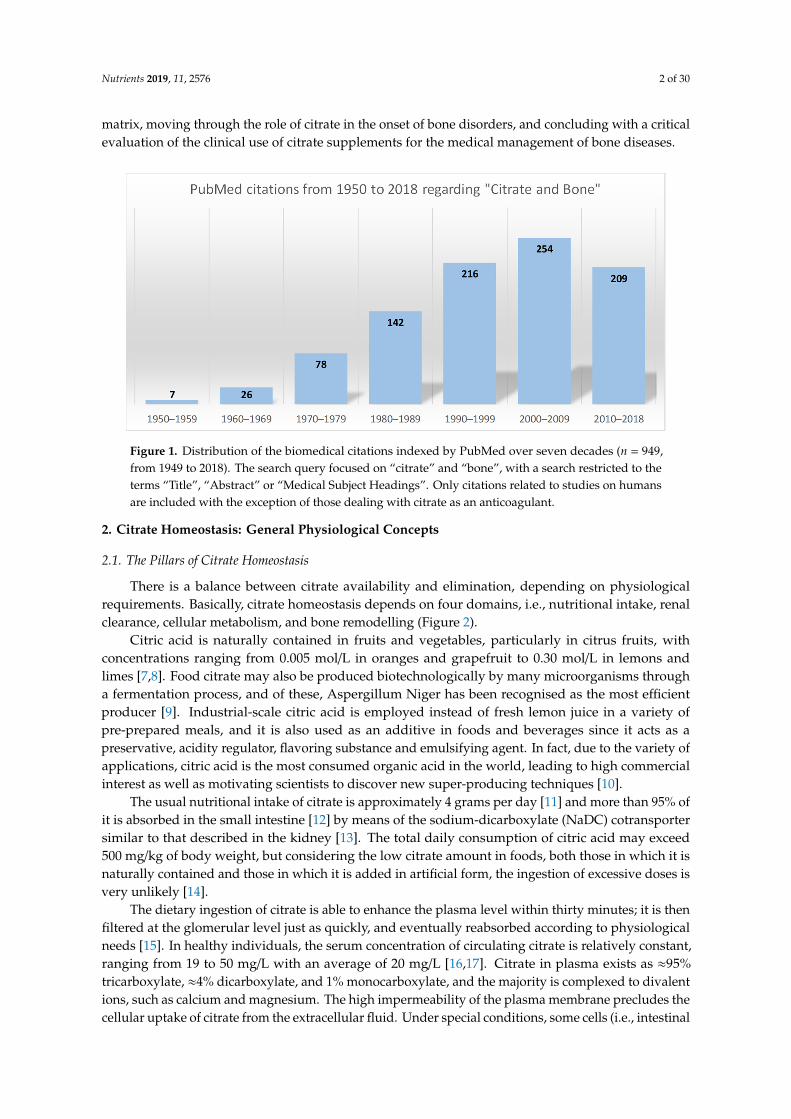
The intracellular citrate level reflects the energy status of the cell and acts as a regulator. Whenthe cellular ATP is abundant and the energy demand of the cells is low, the excess citrate can beexported to the cytosol by means of a mitochondrial citrate carrier [3]. It can be used for the lipidbiosynthesis of highly proliferating cells [4] or for supporting the tissue-related functions of specialisedcells, including the mineralisation of the extracellular matrix by osteoblasts, the bone-forming cells [5].In this regard, the close association between citrate and bone was pointed out for the first time byDickens in 1941 [6]. Since then, the number of published articles dealing with this topic has increasedexponentially (Figure 1), and considerable progress in understanding how citrate is involved in bonemetabolism has been made. This review summarises the current knowledge regarding the relationshipbetween citrate and bone pathophysiology, starting from the link with bone cells and the mineralised
Nutrients 2019, 11, 2576; doi:10.3390/nu11112576 www.mdpi.com/journal/nutrients
Nutrients 2019, 11, 2576 2 of 30
matrix, moving through the role of citrate in the onset of bone disorders, and concluding with a criticalevaluation of the clinical use of citrate supplements for the medical management of bone diseases.
Nutrients 2019, 11, x FOR PEER REVIEW 2 of 35
forming cells [5]. In this regard, the close association between citrate and bone was pointed out for the first time by Dickens in 1941 [6]. Since then, the number of published articles dealing with this topic has increased exponentially (Figure 1), and considerable progress in understanding how citrate is involved in bone metabolism has been made. This review summarises the current knowledge regarding the relationship between citrate and bone pathophysiology, starting from the link with bone cells and the mineralised matrix, moving through the role of citrate in the onset of bone disorders, and concluding with a critical evaluation of the clinical use of citrate supplements for the medical management of bone diseases.
Figure 1. Distribution of the biomedical citations indexed by PubMed over seven decades (n = 949, from 1949 to 2018). The search query focused on "citrate" and "bone", with a search restricted to the terms "Title", "Abstract" or "Medical Subject Headings". Only citations related to studies on humans are included with the exception of those dealing with citrate as an anticoagulant.
2. Citrate Homeostasis: General Physiological Concepts
2.1. The Pillars of Citrate Homeostasis
There is a balance between citrate availability and elimination, depending on physiological requirements. Basically, citrate homeostasis depends on four domains, i.e., nutritional intake, renal clearance, cellular metabolism, and bone remodelling (Figure 2).
Citric acid is naturally contained in fruits and vegetables, particularly in citrus fruits, with concentrations ranging from 0.005 mol/L in oranges and grapefruit to 0.30 mol/L in lemons and limes [7,8]. Food citrate may also be produced biotechnologically by many microorganisms through a fermentation process, and of these, Aspergillum Niger has been recognised as the most efficient producer [9]. Industrial-scale citric acid is employed instead of fresh lemon juice in a variety of pre-prepared meals, and it is also used as an additive in foods and beverages since it acts as a preservative, acidity regulator, flavoring substance and emulsifying agent. In fact, due to the variety of applications, citric acid is the most consumed organic acid in the world, leading to high commercial interest as well as motivating scientists to discover new super-producing techniques [10].
The usual nutritional intake of citrate is approximately 4 grams per day [11] and more than 95% of it is absorbed in the small intestine [12] by means of the sodium-dicarboxylate (NaDC) cotransporter similar to that described in the kidney [13]. The total daily consumption of citric acid may exceed 500 mg/kg of body weight, but considering the low citrate amount in foods, both those in which it is naturally contained and those in which it is added in artificial form, the ingestion of excessive doses is very unlikely [14].
Figure 1. Distribution of the biomedical citations indexed by PubMed over seven decades (n = 949,from 1949 to 2018). The search query focused on “citrate” and “bone”, with a search restricted to theterms “Title”, “Abstract” or “Medical Subject Headings”. Only citations related to studies on humansare included with the exception of those dealing with citrate as an anticoagulant.
2. Citrate Homeostasis: General Physiological Concepts
2.1. The Pillars of Citrate Homeostasis
There is a balance between citrate availability and elimination, depending on physiologicalrequirements. Basically, citrate homeostasis depends on four domains, i.e., nutritional intake, renalclearance, cellular metabolism, and bone remodelling (Figure 2).
Citric acid is naturally contained in fruits and vegetables, particularly in citrus fruits, withconcentrations ranging from 0.005 mol/L in oranges and grapefruit to 0.30 mol/L in lemons andlimes [7,8]. Food citrate may also be produced biotechnologically by many microorganisms througha fermentation process, and of these, Aspergillum Niger has been recognised as the most efficientproducer [9]. Industrial-scale citric acid is employed instead of fresh lemon juice in a variety ofpre-prepared meals, and it is also used as an additive in foods and beverages since it acts as apreservative, acidity regulator, flavoring substance and emulsifying agent. In fact, due to the variety ofapplications, citric acid is the most consumed organic acid in the world, leading to high commercialinterest as well as motivating scientists to discover new super-producing techniques [10].
The usual nutritional intake of citrate is approximately 4 grams per day [11] and more than 95% ofit is absorbed in the small intestine [12] by means of the sodium-dicarboxylate (NaDC) cotransportersimilar to that described in the kidney [13]. The total daily consumption of citric acid may exceed500 mg/kg of body weight, but considering the low citrate amount in foods, both those in which it isnaturally contained and those in which it is added in artificial form, the ingestion of excessive doses isvery unlikely [14].
The dietary ingestion of citrate is able to enhance the plasma level within thirty minutes; it is thenfiltered at the glomerular level just as quickly, and eventually reabsorbed according to physiologicalneeds [15]. In healthy individuals, the serum concentration of circulating citrate is relatively constant,ranging from 19 to 50 mg/L with an average of 20 mg/L [16,17]. Citrate in plasma exists as ≈95%tricarboxylate, ≈4% dicarboxylate, and 1% monocarboxylate, and the majority is complexed to divalentions, such as calcium and magnesium. The high impermeability of the plasma membrane precludes thecellular uptake of citrate from the extracellular fluid. Under special conditions, some cells (i.e., intestinal

Nutrients 2019, 11, 2576 3 of 30
enterocytes and kidney tubular cells) express a plasma membrane citrate transporter belonging to the“solute carrier” 13 family (Slc13, NaDC), which is essential for uptake from the gastrointestinal tractand tubular fluid.Nutrients 2019, 11, x FOR PEER REVIEW 3 of 34
Figure 2. The four domains of citrate homeostasis. The plasma level of citrate mainly depends on four sources, i.e., nutritional intake, renal clearance, cellular metabolism, and bone remodelling. Food citrate is rapidly introduced into the circulation, filtered at the glomerular level, and eventually reabsorbed according to physiological needs. The citrate uptake from the extracellular milieu may occur only when specific transporter proteins are expressed, i.e., sodium-dicarboxylate (NaDC)1 belonging to the “solute carrier” 13 (Slc13) family. The citrate produced by mitochondria only marginally contributes to citrate homeostasis, since it is almost all used by cells as an energy source, or for the synthesis of lipids and other specific functions, i.e., citration of the extracellular matrix by the osteoblasts. In fact, the bulk stored in bone is the main endogenous source of citrate which becomes available following the resorption of the mineralised matrix by the osteoclasts. The mitochondrial citrate-transport protein (CTP) is essential for the release of citrate from the mitochondria to cytosol.
The dietary ingestion of citrate is able to enhance the plasma level within thirty minutes; it is then filtered at the glomerular level just as quickly, and eventually reabsorbed according to physiological needs [15]. In healthy individuals, the serum concentration of circulating citrate is relatively constant, ranging from 19 to 50 mg/L with an average of 20 mg/L [16,17]. Citrate in plasma exists as ≈95% tricarboxylate, ≈4% dicarboxylate, and 1% monocarboxylate, and the majority is complexed to divalent ions, such as calcium and magnesium. The high impermeability of the plasma membrane precludes the cellular uptake of citrate from the extracellular fluid. Under special conditions, some cells (i.e., intestinal enterocytes and kidney tubular cells) express a plasma membrane citrate transporter belonging to the “solute carrier” 13 family (Slc13, NaDC), which is essential for uptake from the gastrointestinal tract and tubular fluid.
However, the net balance between gastrointestinal absorption and the urinary excretion of citrate suggests that nutritional intake cannot be solely responsible for the maintenance of plasma homeostasis [15,17]. The cellular metabolism also has a scarce impact on citrate availability since almost all the mitochondrial production is consumed by cells as an energy source or for supporting specific cell functions [4,5]. Nowadays, it is well known that the main endogenous bulk of citrate is
Figure 2. The four domains of citrate homeostasis. The plasma level of citrate mainly depends on foursources, i.e., nutritional intake, renal clearance, cellular metabolism, and bone remodelling. Food citrateis rapidly introduced into the circulation, filtered at the glomerular level, and eventually reabsorbedaccording to physiological needs. The citrate uptake from the extracellular milieu may occur onlywhen specific transporter proteins are expressed, i.e., sodium-dicarboxylate (NaDC)1 belonging to the“solute carrier” 13 (Slc13) family. The citrate produced by mitochondria only marginally contributes tocitrate homeostasis, since it is almost all used by cells as an energy source, or for the synthesis of lipidsand other specific functions, i.e., citration of the extracellular matrix by the osteoblasts. In fact, thebulk stored in bone is the main endogenous source of citrate which becomes available following theresorption of the mineralised matrix by the osteoclasts. The mitochondrial citrate-transport protein(CTP) is essential for the release of citrate from the mitochondria to cytosol.
However, the net balance between gastrointestinal absorption and the urinary excretion ofcitrate suggests that nutritional intake cannot be solely responsible for the maintenance of plasmahomeostasis [15,17]. The cellular metabolism also has a scarce impact on citrate availability since almostall the mitochondrial production is consumed by cells as an energy source or for supporting specificcell functions [4,5]. Nowadays, it is well known that the main endogenous bulk of citrate is stored inbone and is mobilised following the resorption of the mineralised matrix by the osteoclasts [17].
2.2. Citraturia as A Marker of Citrate Homeostasis and Bone Health Status
As circulating citrate is freely filtered in the glomerulus, 24-h excretion is considered to be avalid marker for highlighting alterations of citrate homeostasis [11]. The reference values for urinarycitrate levels range from 320 to 1260 mg/24 h, with an average in males of 550 mg/24 h and in femalesof 680 mg/24 h [18,19]. The higher excretion of citrate in females is in relation to the estrogenic

Nutrients 2019, 11, 2576 4 of 30
rate [20] and explains the lower incidence of nephrolithiasis in premenopausal women, consideringthat citrate-calcium binding is one of the main mechanisms for inhibiting stone formation [21]. Basedon the reference values for lithogenic risk, the threshold for the diagnosis of hypocitraturia is usuallyfixed as less than 320 mg per day. However, hypocitraturia may be severe (citrate excretion of less than100 mg per day) or mild-moderate (from 100 to 320 mg), but low excretion (less than 640 mg per day)could also be a significant sign and should be monitored [18]. In general, elevated citrate excretionmay be considered a non-pathological condition which reflects the restoration of the acid-base balanceand occurs, for instance, after chronic alkali intake [22]. Low citrate excretion is a relatively commonfinding, and even though the majority of patients have idiopathic hypocitraturia, there are severalmedical and physiological conditions associated with this abnormality. All the conditions listed inTable 1 may be potentially associated with skeletal disorders or, more broadly, with bone metabolismalterations, even when there are no obvious symptoms.
Table 1. Causes of low citrate excretion.
Cause Annotation
Acid-base status [16,23]
• Acidosis increases citrate utilization by the mitochondria in thetricarboxylic acid cycle (TCA cycle), thus decreasing intra- andextracellular availability. As a consequence, citrate reabsorption isenhanced and urine excretion is reduced. On the contrary, alkalosisincreases citrate elimination.
Hypokalemia [16,23] • Low potassium levels cause intracellular acidosis (see above).
Diet [24,25]
• Low intake of high-citrate content food (fruit/vegetables).• A diet rich in animal proteins contains sulfate and phosphate
moieties which are not metabolised and are excreted as acids whichdecrease urinary pH and citrate excretion.
• High sodium intake, ketosis-promoting diet, and starvation.
Distal renal tubular acidosis(dRTA) [26]
• Complete form (hyperchloremic metabolic acidosis, hypokalemia,elevated urine pH).
• Incomplete form (normal serum electrolytes, inability to acidifyurine following an ammonium chloride load).
Chronic diarrheal syndrome[16,23]
• The fluid loss and intestinal alkali wasting alter the acid-base status,with low urinary pH and citrate retention.
Medications [16,23,27,28]
• Thiazide diuretics induce hypokalemia with resultantintracellular acidosis.
• Acetazolamide (carbonic anhydrase inhibitor) produces changes inurine composition which are similar to those found in dRTA.
• Angiotensin-converting enzyme inhibitors cause a reduction inurinary citrate by increasing the adenosine triphosphate (ATP)citrate lyase activity.
• Topiramate (carbonic anhydrase inhibitor) exerts a dose-dependenteffect on the renal excretion of citrate.
Strenuous physical exercise [23] • It causes lactic acidosis, producing hypocitraturia.
Hyperuricosuria [23]
• With normouricemia, generally caused by dietary excess of purines(animal proteins).
• With hyperuricemia (gouty diathesis), the urinary pH is typicallylow, with increased citrate reabsorption.
Nutrients 2019, 11, 2576 5 of 30
Table 1. Cont.
Cause Annotation
Active urinary tract infection [23]• Bacteria which degrade citrate lower the urinary
citrate concentration.
Chronic kidney disease (CKD) [29]• The decrease in the glomerular filtration rate causes a stepwise
reduction in the amount of filtered citrate. Overt hypocitraturia isusually observed in advanced stages of CKD.
Primary hyperaldosteronism [30]• Hypocitraturia (and hypercalciuria) occurs via Na-dependent
volume expansion and chronic hypokalemia.
Menopause [31–34]
• Estrogen deficiency induces metabolic alteration related to thelowering of estrogen-induced signaling onto the mitochondriawhich promotes glycolysis and glycolytic-coupled TCA cyclefunction. Hormone replacement therapy restores the citrate levelwhich is decreased in postmenopausal women.
Genetic defects [16]• All inheritable diseases, gene defects, and polymorphisms
associated with the above mentioned conditions (additional detailsin Table 2).
Modulation of citrate excretion in the kidney is influenced by multiple factors, but pH regulation,particularly in the proximal tubule, has the strongest impact, and even a small decrease in tubularpH significantly increases tubular reabsorption [16]. Therefore, in response to the elevated acidload occurring in metabolic acidosis, there is a notable increase in citrate recovery with subsequenthypocitraturia; urinary citrate excretion may be used as a laboratory parameter for monitoring thediet- and metabolism-dependent systemic acid-base status, even in subjects without overt metabolicacidosis [29,35,36].
The detrimental effect of acid-base imbalance on bone metabolism has been proven withouta doubt [36,37], thus suggesting that citraturia could be a noninvasive and indirect view of bonehealth status. Actually, the relationship among urinary citrate excretion, bone quality parameters,and circulating levels of bone turnover markers has been demonstrated [34,36], even if its clinicalusefulness is still controversial [37].
3. Citrate and Bone Tissue
In 1941 Dickens stated that approximately 90% of the total citrate found in the body of“osteovertebrates” resides in mineralised tissues, but the most valuable insight was that, due toits high binding affinity to calcium stored in the hard tissue, citrate could play a pivotal role inregulating metabolic functions and in maintaining the structural integrity of bone [6]. Moreover,Dickens postulated that the presence of citrate in bone is crucial for preventing calcium precipitation,either when bone tissue is resorbed in response to lowered serum calcium or when the biomineralisationprocess starts. Over time, data in the literature regarding the relationship between citrate and bonephysiology have been increasing exponentially (Figure 1), but the role of citrate in driving the structuraland functional properties of healthy bone in humans has only been partially elucidated. Early studieswere mainly aimed at searching for the origin and the role of calciotropic hormones (calcitonin,parathyroid hormone (PTH), and vitamin D) in the regulation of its metabolism. However, these issueshave remained largely unresolved and/or highly speculative, due to the absence of necessary researchmethodology and technology. Nowadays, there is adequate knowledge regarding the role of bone cellsin producing citrate, how citrate enters the crystalline structure of bone, and how it controls the sizeand morphology of apatite nanocrystals.

Nutrients 2019, 11, 2576 6 of 30
3.1. Citrate and Mineral Structure
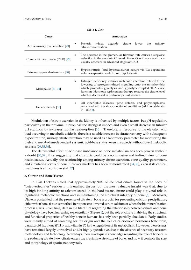
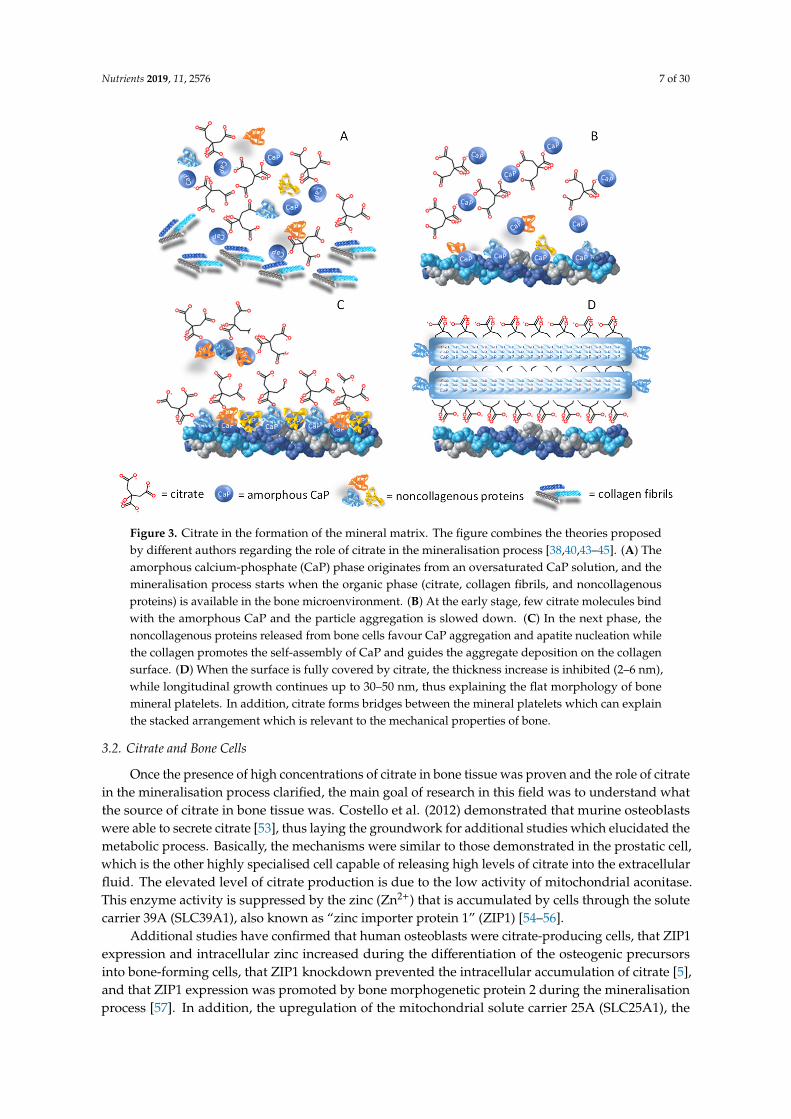
Citrate is abundant in bone, representing 1–5 weight percent (wt%) of the organic componentswith a density of approximately 1 molecule per 2 nm2, which implies that more than 15% of theapatite surface area available in bone is covered by citrate molecules [38]. Before the introduction ofmodern analytical techniques, the testing methods to evaluate bone tissue composition were entirelylimited to classic wet-chemical analyses. At that time, the evidence of a higher amount of the citratemetabolic activity in bone as compared to other tissues, such as kidney or liver, was a significantbreakthrough. These findings allowed hypothesising that bone tissue was endowed with the specialcapacity of producing and storing a high concentration of citric acid, and suggested the role of citrate inregulating the mineralisation process [39]. On the one hand, citrate may influence the amount of mineraldeposition by complexing calcium-phosphate (CaP) and favouring its precipitation; on the other hand,if citrate exceeds the amount incorporated in the bone matrix, it could even reverse the mineralisationphase, thus functioning as a solubilising agent which recalls calcium ions. Hu et al. (2010) have proventhat citrate is not a dissolved solubilising agent but is firmly bound to apatite as an integral part of thenanocrystal structure. This could be explained by the fact that the elevated amount of citrate in theorganic fraction (5.5 wt%) provides more COO- groups than all the noncollagenous proteins in bone,and therefore, the chances of the binding between citrate and calcium of apatite are proportionallyincreased [40]. Simultaneously, Xie and Nancollas (2010) proposed a three-phase model to explainhow citrate may influence the size, longitudinal growth and thickness of apatite nanocrystals [38].The biomineralisation process initiates with the formation of the amorphous CaP phase starting froman oversaturated CaP solution [41] (Figure 3A). At the early stage, few citrate molecules can bindwith the surface of small-size amorphous CaP clusters, but even so, they are sufficient to slow downparticle aggregation (Figure 3B). In the next phase, the noncollagenous proteins released from bonecells promote the amorphous CaP cluster clumping, and apatite nucleation starts within these largeramorphous aggregates. Moreover, the presence of collagen fibrils promotes the self-assembly of thesmall amorphous CaP clusters and guides their direction on the collagen surface [42] (Figure 3C). At thefinal stage, the surface of mature apatite nanocrystals is fully covered by citrate, so that the increasein thickness is inhibited whilst the growth may continue in a longitudinal direction, thus explainingthe plate-like morphology which apatite crystals exhibit in bone [38]. The final apatite structure has aunique geometry since it does not exceed 30–50 nm in length and maintains 2–6 nm thickness [43].
By using a combination of solid-state Nuclear Magnetic Resonance spectroscopy, powder X-raydiffraction, and the principles of electronic structure calculations, Davies et al. (2014) postulated thatcitrate anions could be incorporated in a hydrated layer of the CaP structure. This binding configurationfavours the growth of the mineral crystals in a plate-like morphology and explains their propensityto form stacks [44,45] (Figure 3D). More recently, Delgado-Lopez et al. (2017) focused on the earlymineralisation phase, taking into account the interaction between citrate and collagen [46]. By meansof an in-depth characterisation based on X-ray scattering and imaging techniques, they found thatcollagen and citrate work synergically to favour the platy morphology, since both contributed tomaintaining the transient amorphous phase. The amorphous CaP clusters are figured as spherulites orglobules which gradually occupy the gap zones of the aligned collagen fibrils [43].
The results of the above-mentioned studies confirmed that citrate is an integral part of theapatite-collagen nanocomposite, and the degree of incorporation into bone mineral, as well as thespatial orientation in the mineral structure, play a key role in maintaining the biomechanic propertiesof bone, i.e., stability, strength, and resistance to fracture. In light of its structural role, citrate has beenused to explain the changes in the bone microarchitecture typical of some diseases [47]. In addition,a series of citrate-based materials for orthopaedic applications have been developed to favour theosteoinductive and osteoconductive properties of scaffolds for bone tissue engineering [48,49], as wellas to promote bone healing in other surgical procedures [50–52].
Nutrients 2019, 11, 2576 7 of 30Nutrients 2019, 11, x FOR PEER REVIEW 7 of 34
Figure 3. Citrate in the formation of the mineral matrix. The figure combines the theories proposed by different authors regarding the role of citrate in the mineralisation process [38,40,43–45]. (A) The amorphous calcium-phosphate (CaP) phase originates from an oversaturated CaP solution, and the mineralisation process starts when the organic phase (citrate, collagen fibrils, and noncollagenous proteins) is available in the bone microenvironment. (B) At the early stage, few citrate molecules bind with the amorphous CaP and the particle aggregation is slowed down. (C) In the next phase, the noncollagenous proteins released from bone cells favour CaP aggregation and apatite nucleation while the collagen promotes the self-assembly of CaP and guides the aggregate deposition on the collagen surface. (D) When the surface is fully covered by citrate, the thickness increase is inhibited (2–6 nm), while longitudinal growth continues up to 30–50 nm, thus explaining the flat morphology of bone mineral platelets. In addition, citrate forms bridges between the mineral platelets which can explain the stacked arrangement which is relevant to the mechanical properties of bone.
3.2. Citrate and Bone Cells
Once the presence of high concentrations of citrate in bone tissue was proven and the role of citrate in the mineralisation process clarified, the main goal of research in this field was to understand what the source of citrate in bone tissue was. Costello et al. (2012) demonstrated that murine osteoblasts were able to secrete citrate [53], thus laying the groundwork for additional studies which elucidated the metabolic process. Basically, the mechanisms were similar to those demonstrated in the prostatic cell, which is the other highly specialised cell capable of releasing high levels of citrate into the extracellular fluid. The elevated level of citrate production is due to the low activity of mitochondrial aconitase. This enzyme activity is suppressed by the zinc (Zn2+) that is accumulated by cells through the solute carrier 39A (SLC39A1), also known as “zinc importer protein 1” (ZIP1) [54–56].
Additional studies have confirmed that human osteoblasts were citrate-producing cells, that ZIP1 expression and intracellular zinc increased during the differentiation of the osteogenic precursors into bone-forming cells, that ZIP1 knockdown prevented the intracellular accumulation of citrate [5], and that ZIP1 expression was promoted by bone morphogenetic protein 2 during the mineralisation process [57]. In addition, the upregulation of the mitochondrial solute carrier 25A (SLC25A1), the citrate transport protein (CTP), was essential for the release of citrate from the
Figure 3. Citrate in the formation of the mineral matrix. The figure combines the theories proposedby different authors regarding the role of citrate in the mineralisation process [38,40,43–45]. (A) Theamorphous calcium-phosphate (CaP) phase originates from an oversaturated CaP solution, and themineralisation process starts when the organic phase (citrate, collagen fibrils, and noncollagenousproteins) is available in the bone microenvironment. (B) At the early stage, few citrate molecules bindwith the amorphous CaP and the particle aggregation is slowed down. (C) In the next phase, thenoncollagenous proteins released from bone cells favour CaP aggregation and apatite nucleation whilethe collagen promotes the self-assembly of CaP and guides the aggregate deposition on the collagensurface. (D) When the surface is fully covered by citrate, the thickness increase is inhibited (2–6 nm),while longitudinal growth continues up to 30–50 nm, thus explaining the flat morphology of bonemineral platelets. In addition, citrate forms bridges between the mineral platelets which can explainthe stacked arrangement which is relevant to the mechanical properties of bone.
3.2. Citrate and Bone Cells
Once the presence of high concentrations of citrate in bone tissue was proven and the role of citratein the mineralisation process clarified, the main goal of research in this field was to understand whatthe source of citrate in bone tissue was. Costello et al. (2012) demonstrated that murine osteoblastswere able to secrete citrate [53], thus laying the groundwork for additional studies which elucidated themetabolic process. Basically, the mechanisms were similar to those demonstrated in the prostatic cell,which is the other highly specialised cell capable of releasing high levels of citrate into the extracellularfluid. The elevated level of citrate production is due to the low activity of mitochondrial aconitase.This enzyme activity is suppressed by the zinc (Zn2+) that is accumulated by cells through the solutecarrier 39A (SLC39A1), also known as “zinc importer protein 1” (ZIP1) [54–56].
Additional studies have confirmed that human osteoblasts were citrate-producing cells, that ZIP1expression and intracellular zinc increased during the differentiation of the osteogenic precursorsinto bone-forming cells, that ZIP1 knockdown prevented the intracellular accumulation of citrate [5],and that ZIP1 expression was promoted by bone morphogenetic protein 2 during the mineralisationprocess [57]. In addition, the upregulation of the mitochondrial solute carrier 25A (SLC25A1), the

Nutrients 2019, 11, 2576 8 of 30
citrate transport protein (CTP), was essential for the release of citrate from the mitochondria [56].In order to define the gene expression patterns underlying the differentiation of mesenchymal stem cells(MSCs) into mature osteoblasts, a large-scale transcriptome analysis starting from the bone-marrowMSCs maintained in osteogenic medium up to deposition of mineral nodules was carried out [58].While the role of ZIP1 was already described [5,56,59], the microarray analysis showed that solutecarrier 13A (SLC39A13), or ZIP13, was upregulated throughout osteogenic differentiation and was alsodetectable during the mineralisation process [60].
As shown in Figure 4A–E, changes in the citrate metabolism occur during the whole processof differentiation of the MSCs into mature osteoblasts. After the osteogenic commitment of restingMSCs, a highly proliferative phase is expected, and the exportation of citrate into cytosol provides theacetylCoA for the synthesis of the new lipids required for the assembly of the new plasma membranesduring cell duplication. Mitochondrial citrate seems to be the preferential source of acetylCoA,since the alternative sources pose some limitations. In fact, extracellular citrate could be used forlipogenesis; however, cellular uptake is subordinated to the expression of the specific transporter, i.e.,“sodium-dependent citrate transporter” (NaCT; SLC13A5) [61]. AcetyilCoA could also be obtainedfrom plasma acetate passing through the cell membrane by means of the monocarboxylate transporter(MCT/SLC16A). In order to use this source, the upregulation of acetylCoA synthetase is required, butit is not highly expressed in mammalian cells [56]. Moreover, aconitase inhibition and the lack ofmitochondrial citrate affect the energy supply via the Krebs cycle, so that the bioenergetic demand forproliferating/differentiating cells has to be satisfied by alternative sources. For instance, cytosol malatemay enter the mitochondrion by CTP in exchange for citrate and may be converted into oxalacetic acid,which in turn may originate new citrate [56].
On the basis of recent studies, citrate released during the bone resorption phase could be considereda matrix-derived signal which contributes to the overall process of bone remodelling within the “basicmulticellular unit” [62]. In this regard, Ma et al. have demonstrated that extracellular citrate played apivotal role regarding the osteogenic differentiation of MSCs [63]. This “metabonegenic” regulationstarted with citrate uptake through the sodium-citrate transporter SLC13A5 followed by the activationof energy-producing pathways leading to an elevated cell energy status, which in turn fueled the highmetabolic demands of MSCs differentiating into osteoblasts. In vitro experiments have shown that thetiming and dosage of the citrate supply were critical factors. In fact, the effects were dose-dependent andmore evident at the early stages of osteogenic differentiation, with higher proliferation and increasedexpression of bone-related genes, i.e., alkaline phosphatase and alpha 1 chain of type I collagen [63].Identical effects have also been observed in a microenvironment hostile to bone cells, for instanceusing culture conditions which simulated low-grade acidosis [64]. In fact, citrate supplementationopposed the detrimental effects resulting from extracellular acidosis, which inhibited the synthesis ofcollagen and non-collagen proteins, the activity of alkaline phosphatase, and the formation of mineralnodules [65].
Taken together, studies regarding the role of citrate in the nanocrystal structure and those showingthat osteoblasts were the specialised citrate-producing cells in bone have led to a new concept of boneformation related to “citration”. Briefly, Costello et al. (2012) stated that . . . .”when considering themineralisation role of osteoblasts in bone formation, it now becomes evident that ’citration’ must beincluded in the process. Mineralisation without ‘citration‘ will not result in the formation of normalbone, i.e., bone that exhibits its important properties, such as stability, strength, and resistance tofracture” [53].

Nutrients 2019, 11, 2576 9 of 30Nutrients 2019, 11, x FOR PEER REVIEW 9 of 34
Figure 4. Citrate metabolism, osteoblast differentiation, and mineralisation process. The figure combines the concept of “osteoblast citration” with the main steps of the differentiation of mesenchymal stem cells (MSCs) into bone-forming cells (osteoblasts) [5,53,56]. (A) Resting MSCs are quiescent, nonproliferating cells which exhibit the typical mitochondrial metabolism with the oxidation of citrate via the Krebs cycle. (B) In the presence of proper stimuli, the undifferentiated MSCs are committed to osteogenic differentiation and, at the early phase, high proliferation is required. To accomplish this goal, the following events are necessary: 1) the upregulation of ZIP1 which promotes the zinc intake, 2) the accumulation of mitochondrial citrate due to the zinc-dependent inhibition of the mitochondrial aconitase, 3) the exportation of citrate into cytosol by means of the “citrate transport protein” (CTP/SLC25A1), 4) the use of cytosol citrate for the lipogenesis process which is essential for cell duplication. (C, D) The citrate exportation from cytosol to extracellular fluid starts during cell differentiation, and it is simultaneous for the synthesis and the release of amorphous CaP, collagen, and noncollagenous proteins. (E) The “osteoblast citration” is completed when the mineralised matrix is assembled. The role of citrate in growing the apatite nanocrystals and driving the mineralisation process is explained in Figure 3.
4. Citrate Pathophysiology and Bone Diseases
The role of citrate in mineralised tissues poses several questions regarding the consequences of a low bioavailability at the systemic level. For the most part, published data linking citrate alteration with bone metabolism refer to renal diseases, acid-base imbalance or also physiological conditions such as menopause, but there are also inheritable genetic defects which affect the TCA cycle in mitochondria or the citrate transport. In the following paragraphs, the medical conditions in which the association between citrate and bone health status has been implied are discussed.
4.1. Bone Health Status and Alterations of Citrate Homeostasis in Kidney Diseases.
With the progressive aging of the population, epidemiological studies have shown a higher rate of elderly-related illnesses, including the impairment of bone quality leading to osteoporosis and decreased renal function with chronic kidney disease (CKD), which in turn may influence bone health status [66–68]. The decrease in renal function may be mild, moderate or severe on the basis of estimated-glomerular filtration rate (GFR) equations and is associated with the simultaneous impairment of mineral homeostasis, including serum and tissue concentrations of phosphorus and calcium, circulating levels of calciotropic hormones (PTH, 25-hydroxyvitamin D, 1,25-dihydroxyvitamin D), fibroblast growth factor-23, and growth hormone. The modifications of
Figure 4. Citrate metabolism, osteoblast differentiation, and mineralisation process. The figurecombines the concept of “osteoblast citration” with the main steps of the differentiation of mesenchymalstem cells (MSCs) into bone-forming cells (osteoblasts) [5,53,56]. (A) Resting MSCs are quiescent,nonproliferating cells which exhibit the typical mitochondrial metabolism with the oxidation of citratevia the Krebs cycle. (B) In the presence of proper stimuli, the undifferentiated MSCs are committedto osteogenic differentiation and, at the early phase, high proliferation is required. To accomplishthis goal, the following events are necessary: (1) the upregulation of ZIP1 which promotes the zincintake, (2) the accumulation of mitochondrial citrate due to the zinc-dependent inhibition of themitochondrial aconitase, (3) the exportation of citrate into cytosol by means of the “citrate transportprotein” (CTP/SLC25A1), (4) the use of cytosol citrate for the lipogenesis process which is essentialfor cell duplication. (C,D) The citrate exportation from cytosol to extracellular fluid starts during celldifferentiation, and it is simultaneous for the synthesis and the release of amorphous CaP, collagen,and noncollagenous proteins. (E) The “osteoblast citration” is completed when the mineralised matrixis assembled. The role of citrate in growing the apatite nanocrystals and driving the mineralisationprocess is explained in Figure 3.
4. Citrate Pathophysiology and Bone Diseases
The role of citrate in mineralised tissues poses several questions regarding the consequences of alow bioavailability at the systemic level. For the most part, published data linking citrate alterationwith bone metabolism refer to renal diseases, acid-base imbalance or also physiological conditions suchas menopause, but there are also inheritable genetic defects which affect the TCA cycle in mitochondriaor the citrate transport. In the following paragraphs, the medical conditions in which the associationbetween citrate and bone health status has been implied are discussed.
4.1. Bone Health Status and Alterations of Citrate Homeostasis in Kidney Diseases
With the progressive aging of the population, epidemiological studies have shown a higherrate of elderly-related illnesses, including the impairment of bone quality leading to osteoporosisand decreased renal function with chronic kidney disease (CKD), which in turn may influence bonehealth status [66–68]. The decrease in renal function may be mild, moderate or severe on the basisof estimated-glomerular filtration rate (GFR) equations and is associated with the simultaneousimpairment of mineral homeostasis, including serum and tissue concentrations of phosphorus andcalcium, circulating levels of calciotropic hormones (PTH, 25-hydroxyvitamin D, 1,25-dihydroxyvitamin

Nutrients 2019, 11, 2576 10 of 30
D), fibroblast growth factor-23, and growth hormone. The modifications of mineral homeostasis maypromote a loss of bone mass and an increase in bone fragility [69]. As observed by Malmgren et al.(2015), approximately 95% of women over 75 years of age showed a mild-moderate decrease in renalfunction (CKD stages 2–3) which may have had a harmful effect on bone health [70]. In a 10-yearlongitudinal study, the authors evaluated the long-term influence of impaired renal function on bonemineral density (BMD) [71]. They analysed 1044 Caucasian women from the “Osteoporosis ProspectiveRisk Assessment” (OPRA) cohort and found that renal function was positively correlated with femoralneck BMD in elderly women, although the association attenuated as aging progressed. Women withpoor renal function had a higher annual rate of bone loss over 5 years compared to those with normalfunction, and markers of mineral homeostasis were more frequently altered.
High-throughput “omics” approaches, including metabolomics, have been proposed to identifynew biomarkers which could help the management of CKD patients, and TCA cycle-metabolites areemerging as potential candidates [72]. A significantly reduced urinary excretion of citrate (–68%) hasbeen observed in non-diabetic patients with CKD as compared to subjects with normal renal function.The renal expression of genes regulating the TCA cycle was decreased in subjects who had impairedrenal function, thus suggesting that mitochondrial dysfunction could be involved in the pathogenesisof CKD [73]. Moreover, GFR positively correlated with citrate excretion, and kidney stone formerswith CKD had significantly lower urinary citrate excretion than subjects with kidney stone disease andnormal renal function [74].
To the authors’ knowledge, the link between CKD, urinary citrate and bone health status has stillnot been elucidated but, taking into account the information emerging from the previous paragraphs,it is reasonable to assume that the link exists. Some indications have derived from the data regardingkidney stone disease, which is the paradigmatic expression of a relationship between citrate alterations,BMD decrease and fracture risk that has been investigated since the 1970s [75]. In fact, several studieshave shown that osteoporotic fractures occurred more frequently in patients with kidney stones thanin the general population [76–79].
The connection between kidney stones and bone metabolism is related to several factors. Briefly,kidney stones form when urine becomes supersaturated with respect to its specific components. Since80% of kidney stones are composed of calcium-oxalate (CaOx) or CaP, the regulation of calciumexcretion plays a pivotal role in the etiopathogenesis of nephrolithiasis [80]. As urinary citrate isable to bind calcium and prevent the growth and agglomeration of CaOx and CaP crystals, the closerelationship between low citrate excretion and kidney stone formation has been fully established [11].The incidence of hypocitraturia varies from 20% to 60% in people who have a propensity to form stones,either as a single abnormality or in conjunction with other metabolic disorders [16]. Hypercalciuriamay occur either when filtered calcium is abnormally increased or when its reabsorption is abnormallydecreased. The former may be associated with enhanced bone resorption which raises calciumbioavailability at the systemic level, while the latter may be the consequence of decreased renalfunction as occurs in CKD. Theoretically, reduced GFR in CKD should lead to decreased urinarycalcium concentration, but the consequences of defective tubular reabsorption are more relevant andare responsible for the supersaturation of calcium salts. In addition, in the distal nephron, calciumreabsorption is a PTH-dependent process, PTH being the hormone capable of stimulating the resorptionof the bone matrix in response to low, systemic calcium availability [81]. Therefore, as the decreasein the renal function progresses, PTH levels and bone loss gradually increase, thus explaining whykidney stones are a significant predictor of osteoporotic fracture in patients with CKD [82]. Moreover,when nephrolithiasis occurs, patients are frequently advised to reduce calcium intake, thus favouring anegative calcium balance which is an additional risk factor promoting a decrease in BMD [11].
Recent findings have demonstrated that lithogenic risk factors, including hypocitraturia, are alsodetectable in patients without kidney stones who exhibit osteoporosis or osteopenia, thus leading tothe hypothesis that the evaluation of lithogenic risk could have significant implications for monitoringbone health status [34,83].

Nutrients 2019, 11, 2576 11 of 30
4.2. Postmenopausal Osteopenia and “Net Citrate Loss”
Estrogen deficiency and aging are the main factors responsible for the depletion of bone mass [84],but they are also associated with changes in urine composition which are similar to those of subjectshaving an increased risk of kidney stones [11]. The circulating citrate levels and the citrate contentin bone are markedly reduced in animals with age-related or ovariectomy-induced bone loss [85].A low citrate excretion, less severe than true hypocitraturia fixed at less than 320 mg per day, has beendescribed in postmenopausal women [11,33] and in subjects with a low bone mass [34,83]. Nurses’Health Study II considered an ongoing cohort of 108,639 participants from whom information onmenopause and kidney stones was obtained. In general, postmenopausal status was associated withlower BMD and a higher incidence of kidney stones in this cohort. Moreover, small but significantdifferences in urine composition were found in 658 participants who had pre- and postmenopausal24-h urine analyses, including a lower citrate excretion [86].
The postmenopausal decline in estrogen concentration influences the activation rate of basicmulticellular units composed of bone-resorbing osteoclasts and bone-forming osteoblasts. However,according to Drake et al., resorption increased by 90% while formation increased by only 45% [87] andthe final result was a “net bone loss”. This imbalanced bone remodelling depends on the effects thatthe lack of estrogen has on bone cells. On the one hand, the activity of the receptor activator of thenuclear factor-κ B ligand (RANKL) is promoted, a key factor in osteoclast differentiation; on the otherhand, the osteogenic precursors are destined to differentiate into adipocytes, and the survival of matureosteoblasts is suppressed [88]. The result is the reduction of mature osteoblasts, and since they are thecells capable of synthesising citrate [5], the consequence is lower citrate production which impairsthe quality and the stability of the bone microarchitecture [38,40]. Moreover, osteoclast differentiationand bone resorption are energy-demanding processes, and the citrate which is synthesised cannot beaccumulated because it is essentially utilised through the citric acid cycle [89,90]. Similarly, the MSCdifferentiation towards adipocytes requires more citrate as a source of cytosolic acetylCoA for lipidbiosynthesis [85]. In conclusion, according to Granchi et al., estrogen deficiency leads to a “net citrateloss” which could explain the diminished citrate excretion observed in postmenopausal women [91].
4.3. Genetic Variations Influencing Citrate Homeostasis and Skeletal Development
The “Online Mendelian Inheritance in Man®” database (OMIM®) is a comprehensive repositoryof information on the relationship between genetic variation and phenotypic expression [92].The annotations connecting citrate homeostasis with skeletal defects are listed in Table 2, and many ofthese concern Slc proteins, which are a family of solute transporters through the membranes.
Mutations of the Cl2/HCO3·2 exchanger AE1, encoded by SLC4A1 which is expressed in red bloodcells and in type A intercalated cells of the renal collecting tubule, may be responsible for distal renaltubular acidosis (dRTA), with or without haemolytic anemia. The corresponding phenotype displaysdefective urine acidification, nephrocalcinosis, nephrolithiasis, hypercalciuria, and hypocitraturia [93].The clinical phenotype in patients with inherited dRTA is characterised by stunted growth withbone abnormalities in children, as well as nephrocalcinosis and nephrolithiasis which develop as theconsequence of hypercalciuria, hypocitraturia, and relatively alkaline urine.
The same cytogenetic location of SLC4A1 (17q21.31) is involved in Glycogen storage disease Iawhich is caused by a deficiency in glucose-6-phosphatase activity that catalyses the synthesis of glucosefrom glucose-6-phosphate. This enzymopathy results in a failure to maintain normal glucose controlwith glycogen accumulation in the liver, kidney, and intestine. Low citrate excretion and hypercalciuriahave been described by Weinstein et al. (2001), and the combination of these metabolic alterationscorrelated with the onset of nephrocalcinosis and nephrolithiasis [94]. Furthermore, there is increasingevidence that poor metabolic control, including chronic acidosis (lactic), low muscle mass and delayedpuberty, may negatively affect BMD in half of the patients [95].
NaCT is the sodium-coupled tricarboxylate transporter predominantly expressed in the liver,at several-fold lower levels in the testis and the brain, and at weak levels in the kidney and the heart.

Nutrients 2019, 11, 2576 12 of 30
The association between the mutations of the SLC13a5 gene on chromosome 17p13 and early infantileepileptic encephalopathy-25 with amelogenesis imperfecta has been clearly recognised [96]. Morerecently, Irizarry et al. have shown that SLC13a5 deficiency led to decreased BMD and impaired boneformation in homozygote (Slc13a5-/-) and heterozygote (Slc13a5+/-) mice [97]. As shown by Diaz et al.(2017), the epigenetic modulation of SLC13a5 gene may also influence skeletal development, sinceDNA hypermethylation and low gene expression have been found in the placenta and cord blood ofinfants born small-for-gestational-age and correlated with low height and weight at birth, low BMD,and low mineral content [98].
Bartter syndrome refers to a group of autosomal recessive disorders characterised by impairedsalt reabsorption in the thick ascending limb of the loop of Henle with pronounced salt wasting,e.g., potassium and calcium, and hypokalemic metabolic alkalosis [99]. The antenatal variantor Bartter syndrome type I is caused by a homozygous or compound heterozygous mutation inthe sodium-potassium-chloride cotransporter-2 gene [100]. The affected infants develop markedhypercalciuria and, as a secondary consequence, nephrocalcinosis and osteopenia [101]. To the best ofthe authors’ knowledge, low citrate excretion in patients affected by Bartter syndrome has not beendescribed, but citrate potassium administration is able to correct biochemical alterations [102,103].
Familial hypomagnesemia with hypercalciuria and nephrocalcinosis is an autosomal-recessiverenal tubular disorder caused by mutations in claudin-16 and claudin-19, which are members of thetransmembrane family proteins regulating calcium and magnesium reabsorption in the kidney [104].Thorleifsson et al. have also identified claudin-14 as a major risk gene of hypercalciuric nephrolithiasisassociated with a decrease in BMD [105]. Patients can develop hypomagnesaemia, hypercalciuria,and nephrocalcinosis, and their clinical course is often complicated by the progressive loss of kidneyfunction. Other biochemical anomalies consist of elevated serum PTH levels before the onset of CKD,incomplete distal tubular acidosis, hyperuricemia and hypocitraturia [106]. Additional symptoms maybe recurrent urinary tract infections, nephrolithiasis, polyuria, polydipsia and/or failure to thrive [106].Amelogenesis imperfecta has also been described in some patients [107].
The human gene SLC13A2 encodes the sodium-dicarboxylate cotransporter (NaDC1) which ishighly expressed in the brush-border membranes of the renal proximal tubule and intestinal cells, andreabsorbs Krebs cycle intermediates, i.e., succinate and citrate [108]. Although to date no distinctivephenotype has been linked with SLC13A2 variation in the OMIM database, Okamoto et al. (2006) havehypothesised that NaDC1 alterations could play a role in the development of kidney stones by affectingthe citrate concentration in the urine [109]. The functional properties and protein expression of eightcoding region variants of NaDC1 have recently been characterised; the majority of them appeared todecrease transport activity and were predicted to result in decreased citrate absorption in the intestineand kidney [110]. Even if not investigated, it is reasonable to assume that effects on bone mass mayoccur since these conditions influence citrate metabolism and predispose to renal stone formationas well.
The mitochondrial CTP, coded by the SLC25A1 gene located on chromosome 22q11.21, is embeddedin the inner membrane and determines the efflux of tricarboxylic citrate from the mitochondria tocytosol in exchange for dicarboxylic malate [111]. The high citrate concentration into cytoplasmmodulates the lipid synthesis and affects glycolysis by inhibiting phosphofructokinase-1 [112]. Geneticvariations of SLC25A1 mainly lead to inheritable diseases featured by alterations of the central nervoussystem (combined D-2- and L-2-hydroxyglutaric aciduria; OMIM ID: 615182) and skeletal muscle(congenital myasthenic syndrome-23; OMIM ID: 618197) while the presence of bone defects is lessrelevant. However, SLC25A1 impairment also occurs in the 22q11.2 deletion syndrome which ischaracterised by congenital absence of the thymus and parathyroid glands as well as cardiac, renal andeye anomalies, developmental delay, and also skeletal defects. As additional evidence of a relationshipbetween citrate transport and bone pathophysiology, it has been shown that SLC25A1 knockout inmice causes a notable decrease in the number of osteoblasts and the amount of osteoid [113].

Nutrients 2019, 11, 2576 13 of 30
As mentioned above, zinc plays a crucial role in regulating the extracellular bioavailability ofcitrate in the formation of new mineralised matrix and, therefore, gene defects involving a zinctransporter may be involved in alterations of the citrate metabolism and bone diseases. Of these,the solute carrier 39A family (SLC39A or ZIP) controls the influx of zinc into the cytoplasm [56].In a previous study, the authors found that SLC39A13 (ZIP13) is upregulated throughout osteogenicdifferentiation, and no changes were recorded during the mineralisation process [58]. SLC39A13gene defects have been associated with low bone mass in knockout mice [56] and spondylodysplasticEhlers-Danlos syndrome, type 3 (Phenotype MIM number 612350).
Table 2. Genes involved in the regulation of citrate homeostasis with a genotype/phenotype relationshipregarding skeletal development and/or bone metabolism (retrieved from the OMIM®database, lastaccess 25 May 2019).
Gene/Locus Name Gene/Locus CytogeneticLocation
MIM Number:Phenotype Inheritance
Solute carrier family 4, anionexchanger, member 1 (erythrocytemembrane protein band 3, Diego
blood group)
SLC4A1, AE1,EPB3, SPH4, SAO,
CHC17q21.31 179800: Distal renal
tubular acidosisAutosomaldominant
Solute carrier family 4, anionexchanger, member 1 (erythrocytemembrane protein band 3, Diego
blood group)
SLC4A1, AE1,EPB3, SPH4, SAO,
CHC17q21.31 611590: Distal renal
tubular acidosisAutosomalrecessive
Glucose-6-phosphatase, catalytic G6PC, G6PT 17q21.31 232200: Glycogenstorage disease Ia
Autosomalrecessive
Solute carrier family 13(sodium-dependent citrate
transporter), member 5
SLC13A5, NACT,INDY 17p13.1
615905: Earlyinfantile, epilepticencephalopathy, 25
Autosomalrecessive
Solute carrier family 12(sodium/potassium/chloride
transporters), member 1SLC12A1, NKCC2 15q21.1 60167: Bartter
syndrome, type 1Autosomalrecessive
Claudin 16 (paracellin 1) CLDN16, PCLN1,HOMG3 3q28 248250: Renal
hypomagnesemia 3Autosomalrecessive
5. Medical Management of Patients with Metabolic Bone Diseases Associated withCitrate Alterations
5.1. Clinical Work-Up
At present, the guidelines dealing with the clinical management of metabolic bone diseases donot highlight the role of citrate in maintaining bone integrity. Nevertheless, based on research findings,the causes of hypocitraturia should be considered in carrying out a complete evaluation of patientswho present BMD alterations; vice versa, the accurate monitoring of BMD is advisable in subjects whohave reduced urinary citrate excretion.
Regarding laboratory investigations, citrate homeostasis should be evaluated together with allfactors which influence mineral metabolism. Although citrate excretion must be measured over a24-h period and referred to the 24 h-urine volume, the detection of citrate levels in fasting-morningurine (expressed as creatinine ratios) may be an addional element to complete the metabolic profile.Urine pH can be a valid and simple indicator of acid-base balance. Laboratory testings for evaluationof mineral metabolism have to be carried out on plasma and urine by considering renal function,calcium-phosphorus balance, other electrolytes (in particular, potassium and magnesium), andcalciotropic hormones. In addition, bone turnover markers (BTMs), including bone-resorption andbone formation indicators, are considered a useful and inexpensive tool for evaluating turnover rate(high or low) and response to any treatment [114].
Quantitative assessment of BMD is mandatory for evaluating bone health status and for calculatingfracture risk [115]. Dual-energy X-ray absorptiometry (DXA), the T score (the number of standarddeviations above or below the mean for a healthy 30-year-old adult of the same sex and ethnicity), or theZ-score (the number of standard deviations above or below the mean for the patients of the same age,sex and ethnicity) are employed for measuring areal density (g·cm−2) at any skeletal site. The assesment

Nutrients 2019, 11, 2576 14 of 30
of bone quality may be additionally explored by evaluating the trabecular bone score which reflectsbone microarchitecture [116]. “High-resolution peripheral quantitative computed-tomography” allowsan accurate assessment of bone strength; however, due to its elevated cost, the diffusion of thistechnology is still limited.
Even though noninvasive techniques are preferred, bone biopsy remains a valid tool for assessingthe tissue quality in metabolic bone diseases, and it is the gold standard for estimating bone impairmentin kidney disease and for guiding the clinician in deciding proper treatment [117]. In fact, according tothe Kidney Disease Improving Global Outcomes (KDIGO) guidelines, the term renal osteodystrophymay be used exclusively to define the histological alterations in bone morphology associated withCKD, which can be additionally assessed using histomorphometry. Instead, without histologicalconfirmation, the clinical syndrome which develops as a systemic disorder of the mineral and the bonemetabolisms is generally called “CKD-Mineral and Bone Disorder” [69,118]. However, bone biopsies ina routine work-up present some drawbacks, which are foremost the availability limited to specialisedcentres, discomfort for the patients, and the length of time required to process and analyse bone tissue.
Whenever instrumental and laboratory investigations show a significant bone loss, pharmacologicaltreatment has to be planned according to the indications of the current guidelines, eventually addingspecific drugs in case of secondary osteoporosis. Moreover, all recommendations related to lifestyleand dietary modifications should be explained to the patient [115]. In the presence of low urine citrateexcretion, citrate-based supplements could be recommended to prevent progressive damage to boneand a progressive reduction in BMD.
5.2. Dietary Modification
Since hypocitraturia may depend on food habits (Table 1), dietary modifications should beconsidered as a first level intervention for the medical management of citrate deficiencies. Diet is aimedat correcting the excessive acid load and, as a consequence, the negative effects that acidosis has onbone metabolism [119]. The acid-ash hypothesis emphasises the role of the skeleton in maintaining theacid-base balance since the hydroxyapatite of the mineral matrix is a reservoir of alkali groups whichmay be released to neutralise proton excess [120]. Acute and chronic acid loading show distinct effects,with acute acidity first eroding the bone surface to release sodium, potassium and bicarbonate intothe circulation, while chronic acidity leads to the release of calcium and phosphate [121]. Moreover,acidosis directly influences the activity of bone cells within the bone remodelling unit by promotingosteoclast-mediated resorption and inhibiting bone formation by osteoblasts [65]. Hypocitraturia is aresponse to the elevated acid load occurring in metabolic acidosis, since there is a notable increase incitrate reabsorption in the renal proximal tubule when the tubular pH decreases [16]. In this regard, thereis consensus in considering citraturia as a biomarker for monitoring diet and the metabolism-dependentsystemic acid-base status, even in subjects without overt metabolic acidosis [33–35].
Under physiological conditions, the acid-base balance is strictly controlled by net endogenousacid production related to acid and alkali dietary intake, and incomplete metabolism of organic acids.By using proper methods, such as “net endogenous acid production” (NEAP) [122] and “potentialrenal acid load” (PRAL) [123], it is possible to determine the production of acids and characterise foodsaccording to their ability to release acids and bases into the bloodstream. The prolonged and excessiveconsumption of acid precursor foods leads to chronic low-grade metabolic acidosis which reducesthe excretion of citrate and predisposes to diseases [124], including alteration of bone health status,especially in older subjects with diminished renal function [125]. Some authors have discarded theacid-ash hypothesis, claiming that the increase in the diet acid load did not promote skeletal bonemineral loss or osteoporosis [126], and the main role in regulating acid-base homeostasis should beattributed to the kidney [127]. However, the above studies did not consider the role played by citratein preserving the mineralised matrix and it would be interesting to know whether the relationshipbetween acid dietary intake and alterations in the bone metabolism varied according to low or normalurine citrate excretion. As supporting evidence for the link between citrate and bone health, previous

Nutrients 2019, 11, 2576 15 of 30
studies have demonstrated a positive correlation between citrate excretion and radius densitometricvalues in pre- and postmenopause, as well as a significant relationship between citraturia and theprevalence of vertebral fracture in postmenopausal women [11].
The general dietary approach to counteract elevated acid load is to limit the intake of foods with ahigh acidifying potential, i.e., meat (beef, pork, poultry), fish and seafood, eggs, beans and oilseeds,in favour of foods which contribute the most to the release of bases, i.e., fruits and vegetables [128].However, dietary modifications cannot disregard the medical history of the patients, and the bestnutritional approach should be evaluated on a case-by-case basis. For instance, in the elderly, inadequateprotein intake could be a greater problem for bone health than protein excess [129], and the intake ofhigh amounts of fruits and vegetables could be contraindicated in patients with CKD due to their highpotassium content [130].
The intake of foods with a high citrate content may be a valid approach to compensate for thehigh demand due to acidosis or other causes of hypocitraturia [24,124]. The daily citrate intake isapproximately 4 grams, and almost all citrate introduced by exogenous sources is absorbed into thegastrointestinal tract, arrives in the liver and is metabolised to bicarbonate [11].
Prezioso et al. (2015) examined the relationship between a diet rich in vegetables and urinarycitrate excretion [24]. Fruits and vegetables (except for those with high oxalate content) favour citrateexcretion; consequently, they decrease urinary saturation for CaOx and CaP, thus having a protectiveeffect on the formation of kidney stones [24]. In general, fruit intake is lower in hypocitraturic than innormocitraturic subjects [131].
In order to provide dietary recommendations aimed at correcting hypocitraturia, Haleblian et al.(2008) carried out an exhaustive analysis of citrate concentrations in citrus juices, noncitrus juices, andcommercially available beverages. The highest concentration was found in grapefruit juice (35% morethan in lemon juice), and a glass corresponded approximately to a 40 mEq tablet of potassium citrate.In general, commercial beverages had lower amounts of citrate [7].
Several authors have studied the possible influence of the consumption of fruit juices (both citrusand noncitrus) on urinary citrate excretion. Orange juice increased the excretion of urinary oxalate,and therefore, its consumption could result in the biochemical modification of stone risk factors [132].It should also be noted that grapefruit juice significantly increased urinary oxalate levels, but it wasnot associated with an increased lithogenic risk probably due to the protective effect of the high citratecontent [133]. Regarding noncitrus juices, cranberry juice had a controversial effect on urine citrate (noeffect or an increase of 31%), but resulted in a significantly increased concentration of urinary calciumand oxalate. In addition, diluted blackcurrant juice and melon had a positive effect in increasingcitraturia [24]. In a recent meta-analysis, Pachaly et al. aimed at systematically investigating theeffects of dietary measures on urinary citrate and nephrolithiasis [124]. They searched for randomisedcontrolled and crossover studies which evaluated urine citrate excretion after the intake of citrus-basedbeverages, including fruit juices and soft drinks, calcium/magnesium rich mineral water, a high-fibrediet, a low-animal-protein diet, and plant extracts. The authors identified thirteen studies involving358 participants, the majority of whom were stone formers. Summarised estimates showed a significantincrease in citraturia levels only in subjects who consumed fruit juice and other beverages while theother dietary modifications did not determine significant changes [7,8].
Clinical trials aimed at evaluating whether an increase in dietary citrate preserved the bone healthstatus are lacking. In a clinical trial, postmenopausal women were randomised into four groups, i.e.,a diet (additional daily portion of 300 g of self-selected fruit and vegetables), two doses of potassiumcitrate (12.5 and 55.5 mEq/day) and a placebo (control group). The participants were followed for twoyears, and the effects on bone turnover were determined by measuring BTMs and BMD. The authorsconcluded that neither potassium citrate nor fruit and vegetables influenced bone turnover or preventedBMD loss over 2 years in healthy postmenopausal women (Table 3) [134].
In summary, natural sources of dietary citrate should be considered as a first option for preventingkidney stone recurrence as an alternative to medical treatment [24]. From a theoretical point of view,

Nutrients 2019, 11, 2576 16 of 30
published data have suggested that dietary modifications could also be effective in preserving bonehealth. However, at present, there is insufficient evidence supporting the use of natural sources ofcitrate as the sole treatment for preventing bone loss.
5.3. Citrate-Based Supplements
Nephrolithiasis was the first clinical condition in which oral citrate supplementation showedtherapeutic efficacy, particularly in lowering high stone recurrence rate which is more prevalent inpeople with low urinary citrate levels [11]. The rationale for using citrate salts in kidney stone diseasewas explained in previous paragraphs and was based on four main issues: (1) citrate salts are rapidlyabsorbed through the intestine and equally rapidly filtered in the urine; (2) citrate forms calciumcitrate complexes, which in turn increase solubility and decrease the amount of free calcium in urine;(3) citrate acts as an inhibitor of CaOx and CaP crystal growth and aggregation, and (4) in the intestine,the complexation between calcium and citrate reduces enteric absorption of calcium, and thereforerenal excretion. A recent systematic review has demonstrated that citrate-based therapy reducedrecurrent calcium urinary stone formation compared to controls (placebo, usual care) [135]; however,evidence was limited in children [136].
There have also been interventional clinical trials concerning the effect of citrate supplementsin preserving bone health status. As stated for dietary modifications, the rationale of the publishedtrials was based on the assumption that citrate-based supplements may be useful as alkalising agentswhich neutralise the effects of an excessive acid load [16,65,119–121]. Other than diets rich in salt andanimal protein [128], several conditions may induce low-grade acidosis, and the majority of themare age-related, such as menopause [33], subclinical inflammatory status [137], and decreased renalfunction [68]. On the basis of this assumption, Lambert et al. (2015) carried out a meta-analysisaimed at determining whether alkaline potassium salts, including potassium citrate and potassiumbicarbonate, had some effect on calcium metabolism and bone health [138]. The seven eligiblestudies dealing with potassium citrate supplementation did not include subjects with nephrolithiasisand/or other relevant comorbidities in order to avoid confounding factors potentially leading tooverestimation or underestimation of the intervention. Citrate salts significantly reduced calcium andacid excretion similarly to potassium bicarbonate, but they seemed to be more effective in preventingcollagen resorption. However, insufficient data were available regarding changes in BMD, since itwas investigated in only two studies. The authors found major differences in terms of study design,inclusion/exclusion criteria, doses, timing of supplement administration and outcome measures; thisheterogeneity represented a notable limitation for translation into a clinical setting.
In the current review, the interventional clinical trials which were primarily aimed at evaluatingthe effect of citrate supplements on mineral metabolism and bone turnover were reviewed. Sixteeneligible studies were identified which (1) recruited more than 10 subjects, (2) excluded nephrolithiasisand other significant comorbidities and (3) reported the results related to bone health status, includingBMD and/or BTMs. Data regarding study design, population, intervention, follow-up, additionalsupplementation or controlled dietary intake, as well as a summary of results and conclusions, areshown in Table 3.

Nutrients 2019, 11, 2576 17 of 30
Table 3. Interventional clinical trials based on the use of citrate supplements with primary or secondary outcomes related to bone health status.
Reference Study Design; PopulationIntervention
(Dose/Day) (I)Control (C)
OtherSupplements(Dose/Day)
and/orControlled
Dietary Intake
Follow Up andOutcomes
BTM Changes(Intragroup)
Changes in BTM and BMDInduced by Intervention
(Intergroup)Conclusion
Dawson-Hughes,1990 [141]
RCT, controlled vs placebo,double-blind;
≥6 months postmenopausalwomen (early, <5 years: n =67; late, >5 years: n = 169);
age ≥ 65 years
I 1: Ca citrate malate(500 mg Ca), n = 78
I 2: Ca carbonate (500mg Ca), n = 78
C: Placebo (n = 80)
Controlled Caintake
Baseline, 18, 24, 36months;
BTM (BAP) and BMD
I 1: 24 months ↓ BAPI 2: 24 months ↓ BAPC: 24 months ↓ BAP
BTMI 1 vs. C: 36 months ↓ BAP,
related to the Ca intakeI 2 vs. C: 36 months ↓ BAP,
related to the Ca intakeBMD
I 1 vs. C: 12, 24 months ↑ onlyin late postmenopause and Ca
intake ≤400 mg/dayI 2 vs C: ↓ in both groups
Adequate Ca intake isessential in preventing
postmenopausal bone loss; Cacitrate is more effective than
Ca carbonate.
Dawson-Hughes,1997 [142]
RCT, controlled vs. placebo,double-blind;
healthy subjects living in acommunity (176 M/ 213 F);
age ≥ 65 years
I: Ca citrate malate (500mg Ca) & Vit D3 (700
IU), n = 187C: Placebo, n = 202
Controlled Caintake
Baseline, 6, 12, 18, 24,30, 36 months;
BTM (OC, u-NTX) andBMD
I: n.sC: n.s.
BTMI vs. C: 36 months ↓ OC
BMDI vs. C: 36 months ↑
Ca and vitamin Dsupplementation leads to amoderate reduction in boneloss and may substantially
reduce the risk ofnonvertebral fractures among
elderly subjects who live inthe community.
* Ruml, 1999[143]
RCT, controlled vs. placebo;postmenopausal women
(90% ≤5 years)
I: Ca citrate (800 mg Ca),n = 25
C: Placebo, n = 31
Baseline, 12, 24months
BTM (BAP, OC,u-NTX, u- OH proline)
and BMD
I: all BTMs are ↓, atunspecified time points
BMDI: 24 months, stable
Ca citrate supplementationaverted bone loss and
stabilised bone density inearly postmenopausal
women.
Sellmeyer, 2002[144]
RCT, controlled vs. placebo,double-blind;
≥2 years postmenopausalwomen; age I: 65 ± 8 years;
C: 63 ± 8 years
I: K citrate (90 mmol), n= 26
C: Placebo, n = 26
Ca carbonate (500mg); controlled
salt intake
Baseline, 1 months;BTM (OC, u-NTX)
I: n.s.C: 1 month ↓ OC, ↑
u-NTX
BTMI vs. C: 1 month, ↓ u-NTX
K citrate prevents increasedbone resorption due to high
salt intake.
Dawson-Hughes,2002 [145]
RCT, controlled vs. placebo,double-blind;
healthy subjects (161 M/181F); normal BMD; age ≥
65 years
I: Ca citrate malate (500mg Ca), n = 158
C: Placebo, n = 184
Vitamin D3 (700IU); controlledprotein intake
Baseline, 18, 36months;
BTM (OC, u-NTX) andBMD
I: 36 months ↓ u-NTX,related to the protein
intake;C: n.s.
BTMI vs C: 36 months, ↓ u-NTX
BMDI vs C: 36 month, ↑ related to
the protein intake
BMD may be improved byincreasing protein intake as
long as an adequate intake ofCa and vitamin D is assumed.

Nutrients 2019, 11, 2576 18 of 30
Table 3. Cont.
Reference Study Design; PopulationIntervention
(Dose/Day) (I)Control (C)
OtherSupplements(Dose/Day)
and/orControlled
Dietary Intake
Follow Up andOutcomes
BTM Changes(Intragroup)
Changes in BTM and BMDInduced by Intervention
(Intergroup)Conclusion
Marangella, 2004[146]
Controlled vs. untreated;postmenopausal women; T
score: <−1.0; age: 43–72years
I: K citrate 37-74 mEq(≈1 mEq/kg), n = 30
C: No treatment, n = 24
Controlled Caintake
Baseline, 3 monthsBTM (BAP, OC, u-OH
proline, u-DPD)
I: 3 months ↓ OC, u-OHproline, u-DPD;
C: 3 months ↑ OCnot shown
K citrate decreases boneresorption thereby contrastingthe potential adverse effectscaused by chronic acidemia.
The implication for theprevention and treatment of
postmenopausal osteoporosishas to be confirmed.
Kenny, 2004[147]
RCT, crossover, open label,2 phases; 3 months/phase
with a washout period of 2weeks between phases;
postmenopausal women; Tscore: <−1.0 and >−3.5; age:
73 ± 5 years
I 1: Ca citrate (1000 mgCa), n = 20;
I 2: Ca carbonate (1000mg Ca), n = 20
Vitamin D3 (900IU); controlled Ca
intake
Baseline, 1, 3 months(each phase)
BTM (BAP, OC, NTX,u-CTX, u-NTX,
u-DPD)
I 1: 3 months ↓ NTX,u-CTX, u-NTX, u-DPD
I 2: n.s
Ca citrate inhibits boneresorption more than Ca
carbonate.
Sakhae, 2005[139]
RCT, crossover, placebocontrolled, double-blind, 4phases; 2 weeks/phase with
a washout period of 2weeks between phases;
postmenopausal women;age: 48–76 years
I 1: K citrate (40 mEq), n= 18
I 2: Ca citrate (800 mg),n = 18
I 3: K citrate (40 mEq)and Ca citrate (800 mg),
n = 18C (1st phase): Placebo, n
= 18
Rigid diet withfixed content of
protein, Ca, P, Na,K and fluids
Baseline and at theend of each phase;
BTM (BAP, CTX, OC,u-NTX, u-OH proline)
I 1: n.sI 2: ↓ CTX, u-OH proline
I 3: I: ↓ CTX, u-OHproline, u NTX
I 3 vs I 1: ↓ u NTX
In postmenopausal women,combined treatment with K
citrate and Ca citratedecreases bone resorption byproviding an alkali load and
increasing absorbed Ca.
Jehle, 2006 [148]
RCT, controlled;≥5 years postmenopausal
women; T score −1/−4; age:≤70 years
I: K citrate (30 mEq), n =82
C: KCl (30 mmol), n = 79
Ca carbonate (500mg), Vitamin D3
(400 IU); free,nonvegetarian diet
Baseline, 3, 6, 9, 12months;
BTM (BAP, CTX, OC,u-DPD, u-PD) and
BMD
I: 3 months, ↓ u-DPD,u-PD; 6 months, ↑ BAP
and ↓ OC, u-DPD, u-PD;9 months, ↓ u-DPD,
u-PD; 12 months, ↑ BAPand ↓ OC, u-DPD, u-PD;
C: 3 months, ↓ OC,u-DPD, u-PD; 6 months,↑ BAP, u-DPD, u-PD
and ↓ OC; 9 months, ↑u-DPD, u-PD and ↓ OC;
12 months, ↑ BAP,u-DPD, u-PD and ↓ OC
BTMI vs C: 3 months, ↓ u-DPD
BMDI vs C: 12 months ↑
In postmenopausal women,bone mass can be increased
significantly by K citrate. Theeffect on bone resorption
seems to be unrelated to Kintake.

Nutrients 2019, 11, 2576 19 of 30
Table 3. Cont.
Reference Study Design; PopulationIntervention
(Dose/Day) (I)Control (C)
OtherSupplements(Dose/Day)
and/orControlled
Dietary Intake
Follow Up andOutcomes
BTM Changes(Intragroup)
Changes in BTM and BMDInduced by Intervention
(Intergroup)Conclusion
Macdonald, 2008[134]
RCT, controlled vs. placebo,double-blind for I1 e I2; ≥5
years postmenopausalwomen; age: 49–54 years
I 1: K citrate (55.5 mEq),n = 70
I 2: K citrate (18.5 mEq),n = 70
I 3: Diet (300 g fruit =18.5 mEq alkali), n = 66
C: Placebo, n = 70
Food diary (freenonvegetarian
diet)
Baseline, 3, 6, 12, 18,24 months;
BTM (CTX, P1NP,u-DPD) and BMD
I 1: n.s.I 2: n.s.I 3: n.s.C: n.s
BTMI 1, I 2, I 3 vs. C: n.s
BMDI 1, I 2, I 3 vs. C: n.s
In healthy postmenopausalwomen, neither K citrate at
18.5 or 55.6 mEq/d, nor 300 gself-selected fruit and
vegetables influenced boneturnover or prevented BMD
loss over 2 years.
Thomas, 2008[149]
RCT, crossover,double-blind, 2 phases;
postmenopausal women for2 to 6 years; age: 50–60
years
I 1: Ca carbonate (1000mg Ca), n = 12
I 2: 2) Ca citrate (500 mgCa), n = 13
Controlled Caintake
Baseline, 7 days;BTM (CTX)
I 1: 7 days, ↓ CTXI 2: 7 days, ↓ CTX
Ca citrate is at least aseffective as Ca carbonate indecreasing PTH and CTX
cross-links, at half the dose.All changes are numerically
superior after Ca citratesupplementation.
Karp, 2009 [140]
RCT, controlled, 24 h studysessions; women of
child-bearing age: 22–30years
I 1: Ca carbonate (1000mg Ca), n = 12
I 2: Ca citrate (Ca: 1000mg; citrate: 3145 mg), n
= 12I 3: K citrate (K: 57 mEq;citrate: 3145 mg), n = 12
4-day diary toestimate foodhabits before
starting the studysession; the meals
served duringeach study session
were identical
Baseline, 2, 4, 6, 8, 20,24 h;
BTM (BAP, u-NTX)and BMD
I 1: 24 h, ↓ u-NTXI 2: n.s
I 3: 24 h, ↓ u-NTX
K citrate supplementationdecreases urinary Ca
excretion and reduces boneresorption even when the dietis not acidogenic, and reduces
the bone resorption markerdespite low Ca intake.
Jehle, 2013 [150]
RCT, controlled vs. placebo,double-blind;
healthy subjects (79 M/122F); T score > −2.5; age 65–80years; women were past the
perimenopausal peakturnover
I: K citrate (60 mEq), n =101
C: Placebo, n = 100
Ca carbonate (500mg), Vitamin D3
(400 IU); freenonvegetarian diet
Baseline, 6, 12, 18, 24;BTM (BAP, P1NP,u-NTX) and BMD
I: 6, 12 months, ↓ u-NTX;18, 24 months, ↑ P1NP
C: n.s.
BTMI vs. C: 6 months, ↓ u-NTX
BMDI vs. C: 12, 18, 24 months ↑
K citrate administered in abackground of vitamin D and
Ca supplementation is welltolerated and constitutes aninexpensive intervention to
improve BMD and bonemicroarchitecture in healthy
elderly people.
Moseley, 2013[151]
RCT, controlled vs. placebo,double blind;
healthy subjects (17 M/35 F);age ≥ 55 years; women were≥5 years postmenopause
I 1: K citrate (60 mmol),n = 17
I 2: K citrate (90 mmol),n = 17
C: Placebo, n = 18
Ca citrate (630 mg),Vitamin D3 (400
IU); controlled Ca,Na, P, protein, fat
intake
Baseline, 6 months;BTM, (BAP, CTX)
BTMI 1, I2 vs. C: 6 months, ↓ CTX
K citrate decreases markers ofbone resorption over 6
months, but a significantimprovement in Ca balance isobtained with 90 mmol/day.This dose is well tolerated.

Nutrients 2019, 11, 2576 20 of 30
Table 3. Cont.
Reference Study Design; PopulationIntervention
(Dose/Day) (I)Control (C)
OtherSupplements(Dose/Day)
and/orControlled
Dietary Intake
Follow Up andOutcomes
BTM Changes(Intragroup)
Changes in BTM and BMDInduced by Intervention
(Intergroup)Conclusion
Gregory, 2015[152]
RCT, controlled vs. placebo,double-blind;
≥2 year postmenopausalwomen s; T score: <−1.0 >−2.5, or <−2.5 unable to
take any other medication;age I: 65.1 ± 5.9 years; C:
66.1 ± 7.1 years
I: K citrate (40 mEq), n =42
C: Placebo, n = 41
Ca citrate (630 mg),Vitamin D3 (400
IU); freenonvegetarian diet
Baseline, 1, 3, 6, 12months;
BTM (BAP, OC, P1NP,u-NTX) and BMD
I: 1 month, ↓ P1NP; 3, 6,12 months, ↓ P1NP,
u-NTXC. I: 6 months, ↓ P1NPu-NTX; 12 months, ↓
P1NP
BTMI vs. C: n.s
BMDI: 12 months, stable
In postmenopausalosteopenia, K citrate improvesthe effect of supplementationwith Ca citrate and Vitamin D,as proven by the more rapid
decrease in BTM levels.
Granchi, 2018[91]
RCT, controlled vs. placebo,double-blind; ≥5 years
postmenopausal women; Tscore: <−1.0 and >−2.5; ageI: 60.8 ± 1.0 years; C: 58.2 ±
1.1 years
I: K citrate (30 mEq), n =20
C: Placebo, n = 20
Ca carbonate (500mg), Vitamin D3
(400 IU); freenonvegetarian diet
Baseline, 3, 6 months;BTM (BAP, CTX,P1NP, TRAcP)
I: 6 months, ↓ BAP, CTXC: 3, 6 months, ↓ BAP,
CTX
BTMI vs. C: 6 months ↓ BAP, CTXin subjects with low excretionof K and/or citrate, and/or low
urine pH
In postmenopausalosteopenia, K citrate
improves the effects ofsupplementation with Ca
carbonate and vitamin D, butonly in women with low K
and/or citrate excretion and/orlow urine pH.
C: control; I: Intervention; RCT: randomised clinical trial; K citrate: potassium citrate; Ca carbonate: calcium carbonate; Ca citrate: calcium citrate; KCl: potassium chloride; Na: sodium;P: phosporus; BMD: bone mineral density; BTM: bone turnover markers; n.a.: not applicable; n.s.: not significant; BAP: bone-specific alkaline phosphatase; BMD: bone mineral density;K: tpotassium; Ca: calcium; u-DPD: urinary deoxypyridinoline; u-PYR: urinary pyridinoline; u-OH proline: urinary hydroxyproline; OC: osteocalcin; P1NP: amino-terminal propeptide oftype 1 procollagen; u-NTX: urinary N-telopeptide of collagen type 1; M: male; F: female; IU: International Units; PTH: parathyroid hormone; ↓ and ↑ show significant decreases andincreases, respectively, according to the criteria indicated by the authors. * Partial data collected from the abstract.

Nutrients 2019, 11, 2576 21 of 30
The authors investigated the effect of potassium citrate (one clinical trial, seven randomised clinicaltrials (RCTs)) [92,134,139–144], calcium citrate (four RCTs and two crossover studies) [145–150], and twostudies compared the above-mentioned treatments (one RCT, one crossover study) [139,140]. Regardingthe intervention under investigation, doses, timing of administration, and follow-up varied greatlyamong the trials and, in thirteen studies, the co-administration of additional supplements was planned,i.e., calcium carbonate and/or vitamin D3. The majority of the studies included postmenopausalwomen (n = 11) while, in one study, the participants were of childbearing age. The other trials weredesigned to recruit healthy individuals, i.e., males and females at least 55 years of age. The dietaryintake of calcium and/or protein and/or salts was controlled in ten studies; in five studies, the subjectconsumed a free nonvegetarian diet; in one study, the management of food habits was not described.
Overall, the studies selected provided more than 1000 experimental cases and more than900 controls. As Lambert et al. reported previously, very high heterogeneity among the studies wasobserved, and therefore the meta-analysis lost its inferential value while the descriptive overviewwas more informative. By examining the conclusions reported by various authors, two recurringkey points emerged: (1) an adequate calcium intake was essential in preventing bone loss in elderlysubjects and in postmenopausal women, and calcium citrate seemed to be more effective than calciumcarbonate [145–150] and (2) potassium citrate prevented the increased bone resorption caused bymenopause, chronic acidemia and high salt intake [139–144].
Additional findings regarding potassiun citrate can be summarised as follows: effectiveness maybe enhanced by combined treatment with calcium citrate [139]; positive effects have also been observedin young women and in the absence of an excessive acid load [140]. Moreover, supplementation withalkalising potassium citrate improved the beneficial effects of calcium and vitamin D only in osteopenicpostmenopausal women who exhibited the target conditions, namely low potassium and/or citrateexcretion and/or low urine pH [91]. Finally, one study questioned the beneficial effects of potassiumcitrate since, at 24 months, no significant modifications in BTMs and BMD were recorded [134].It should be noted that compliance may decrease by 25% over time, mainly due to gastrointestinal sideeffects (approximately 10%) and costs [24].
Despite the encouraging results, a consensus statement regarding the use of citrate supplementationfor the management of metabolic bone diseases is still lacking since the heterogeneity of the currentevidence is a major limitation for identifying the best practice. Interestingly, the identification ofsubjects who better respond to exogenous citrate supplementation could be the correct way to maximisethe beneficial effect of the treatment.
6. Conclusions
Since citrate is a ubiquitous metabolite with a multidimensional role in the human organism, it isnot surprising that it is involved in the pathophysiology of tissues, organs and systems. In fact, recentdata in the literature have highlighted the relationship between citrate defects and various medicalconditions with considerable financial and social impact, e.g., cancer [153], dyslipidemia [154], vascularcalcifications [155], and visual disabilities [156].
Overall, this review has highlighted the main functions of citrate, and, more specifically, focusedon the role of citrate in the pathophysiology and medical management of metabolic bone diseases,thus identifying some key points which are summarised in Table 4.
Although interest in the role of citrate is spreading, a consensus statement regarding the useof citrate supplementation for the management of metabolic bone diseases is still lacking, since theheterogeneity of the current evidence is a major limitation in identifying the best practice. Even whenthe study design is well planned, there are some reasons which may explain the difficulty in obtainingunique and compelling results from clinical trials.
First, dietary supplementation cannot be regarded as a pharmacological treatment, and thereforethe effects on measurable clinical outcomes are expected to be less prominent. Hence, baselinedifferences between participants which are beyond the investigator’s control become selection biases

Nutrients 2019, 11, 2576 22 of 30
that attenuate the strength of randomisation and interfere with the interpretation of the results. On theother hand, current scientific knowledge provides a sufficient background, leading the clinician andthe researcher to further investigate the beneficial effects of citrate-based treatment in depth, as provenby multiple studies which have dealt with this matter.
At present, it can be argued that the use of citrate supplementation should be considered in themedical management of bone diseases, but it is also reasonable to assume that the effects could bemaximised in a personalised approach in which the scientific knowledge and the clinical judgement ofpractitioners are essential in identifying patients who could reap real benefits.
Table 4. The role of citrate in the pathophysiology and medical management of bone diseases.
Highlights
• Citrate is an essential metabolite and plays a pivotal role in maintaining the acid-base balance.• Citrate is an essential component of bone, and serves to maintain the integrity of the skeletal nano-
and microstructures.
• Citrate is produced by osteoblasts but, at the same time, it influences their differentiation and functionality.• Bone tissue is the main source of citrate and is therefore a leading actor in maintaining citrate homeostasis.• Citrate excretion is a significant biomarker of citrate homeostasis.
• Genetic and acquired diseases characterised by an alteration in citrate homeostasis are often accompaniedby alterations in the development and/or metabolism of bone tissue.
• Exogenous supplementation may be a useful tool in treating medical conditions related to poor citratebioavailability, including bone diseases.
Author Contributions: All the authors (D.G., N.B., F.M.U., and R.C.) participated in the development of the ideasfor this review, contributed to reading the articles, and to the writing and editing the manuscript.
Funding: The research activity of D.G. and N.B. is supported, in part, by grants of Italian Government (“5 permille 2017” and “Ricerca Corrente 2018–2020”).
Acknowledgments: We acknowledge Gerald Goldsmith for his valuable assistance in revising theEnglish language.
Conflicts of Interest: The authors declare no conflict of interest.
Abbreviations
acetylCoA: Acetyl Coenzyme A; ATP: Adenosine 5′-Triphosphate; BMD: Bone Mineral Density; BTM:Bone Turnover Marker; CaOx: Calcium-Oxalate; CaP: Calcium-Phosphate; CKD: Chronic Kidney Disease;CTP: mitochondrial Citrate-Transport Protein; dRTA: distal Renal Tubular Acidosis; DXA: Dual-energy X-rayAbsorptiometry; GFR: Glomerular Filtration Rate; KDIGO: Kidney Disease Improving Global Outcomes; MSC:Mesenchymal Stem Cell; NaCT: Sodium-dependent Citrate Transporter; NaDC: Sodium-Dicarboxylate; NEAP:Net Endogenous Acid Production; OMIM: Online Mendelian Inheritance in Man; OPRA: Osteoporosis ProspectiveRisk Assessment; PRAL: Potential Renal Acid Load; PTH: Parathyroid Hormone; RCT: Randomised Clinical Trial;Slc: Solute Carrier; TCA cycle: Tricarboxylic Acid Cycle or Krebs cycle; ZIP1: Zinc Importer Protein 1.
References
1. Krebs, H.A.; Johnson, W.A. The role of citric acid in intermediate metabolism in animal tissues. FEBS Lett.1980, 117 (Suppl. 1), K1–K10. [CrossRef]
2. Available online: https://www.genome.jp/kegg-bin/show_pathway?map00020 (accessed on 4 July 2019).3. Iacobazzi, V.; Infantino, V. Citrate—New functions for an old metabolite. Biol. Chem. 2014, 395, 387–399.
[CrossRef] [PubMed]4. Mycielska, M.E.; Milenkovic, V.M.; Wetzel, C.H.; Rümmele, P.; Geissler, E.K. Extracellular Citrate in Health
and Disease. Curr. Mol. Med. 2015, 15, 884–891. [CrossRef] [PubMed]5. Franklin, R.B.; Chellaiah, M.; Zou, J.; Reynolds, M.A.; Costello, L.C. Evidence that Osteoblasts are Specialized
Citrate-producing Cells that Provide the Citrate for Incorporation into the Structure of Bone. Open Bone J.2014, 6, 1–7. [CrossRef] [PubMed]

Nutrients 2019, 11, 2576 23 of 30
6. Dickens, F. The citric acid content of animal tissues, with reference to its occurrence in bone and tumour.Biochem. J. 1941, 35, 1011–1023. [CrossRef] [PubMed]
7. Haleblian, G.E.; Leitao, V.A.; Pierre, S.A.; Robinson, M.R.; Albala, D.M.; Ribeiro, A.A.; Preminger, G.M.Assessment of citrate concentrations in citrus fruit-based juices and beverages: Implications for managementof hypocitraturic nephrolithiasis. J. Endourol. 2008, 22, 1359–1366. [CrossRef] [PubMed]
8. Penniston, K.L.; Nakada, S.Y.; Holmes, R.P.; Assimos, D.G. Quantitative assessment of citric acid in lemonjuice, lime juice, and commercially-available fruit juice products. J. Endourol. 2008, 22, 567–570. [CrossRef][PubMed]
9. Schuster, E.; Dunn-Coleman, N.; Frisvad, J.C.; Van Dijck, P.W. On the safety of Aspergillus niger—A review.Appl. Microbiol. Biotechnol. 2002, 59, 426–435. [CrossRef]
10. Hu, W.; Li, W.J.; Yang, H.Q.; Chen, J.H. Current strategies and future prospects for enhancing microbialproduction of citric acid. Appl. Microbiol. Biotechnol. 2019, 103, 201–209. [CrossRef]
11. Caudarella, R.; Vescini, F.; Buffa, A.; Stefoni, S. Citrate and mineral metabolism, kidney stones and bonedisease. Front. Biosci. 2003, 8, s1084–s1106.
12. Fegan, J.; Khan, R.; Poindexter, J.; Pak, C.Y. Gastrointestinal citrate absorption in nephrolithiasis. J. Urol.1992, 147, 1212–1214. [CrossRef]
13. Pajor, A.M. Sodium-coupled transporters for Krebs cycle intermediates. Annu. Rev. Physiol. 1999, 61, 663–682.[CrossRef] [PubMed]
14. Poerwono, H.; Higashiyama, K.; Kubo, H.; Poernomo, A.T.; Suharjono, I.; Sudiana, I.K.; Indrayanto, G.;Brittain, H.G. Citric acid. In Analytical Profiles of Drug Substances and Excipients, 1st ed.; Brittain, H.G., Ed.;Academic Press: San Diego, CA, USA, 2001; Volume 28, pp. 1–76.
15. Sakhaee, K.; Alpern, R.; Poindexter, J.; Pak, C.Y. Citraturic response to oral citric acid load. J. Urol. 1992, 147,975–976. [CrossRef]
16. Zuckerman, J.M.; Assimos, D.G. Hypocitraturia: Pathophysiology and medical management. Rev. Urol.2009, 11, 134–144. [CrossRef]
17. Costello, L.C.; Franklin, R.B. Plasma citrate homeostasis, how it is regulated, and its physiological and clinicalimplications. An important, but neglected, relationship in medicine. HSOA J. Hum. Endocrinol. 2016, 1, 5.[CrossRef]
18. Pak, C.Y.; Resnick, M. Medical therapy and new approaches to management of urolithiasis. Urol. Clin.N. Am. 2000, 27, 243–253. [CrossRef]
19. Mayo Clinic Medical Laboratories. Endocrinology Catalog Bone/Minerals. Available online: https//endocrinology.testcatalog.org/show/CITR (accessed on 30 May 2019).
20. Heller, H.J.; Sakhaee, K.; Moe, O.W.; Pak, C.Y. Etiological role of estrogen status in renal stone formation. J.Urol. 2002, 168, 1923–1927. [CrossRef]
21. Caudarella, R.; Vescini, F. Urinary citrate and renal stone disease: The preventive role of alkali citratetreatment. Arch. Ital. Urol. 2009, 81, 182–187.
22. Melnick, J.Z.; Preisig, P.A.; Moe, O.W.; Srere, P.; Alpern, R.J. Renal cortical mitochondrial aconitase isregulated in hypo- and hypercitraturia. Kidney Int. 1998, 54, 160–165. [CrossRef]
23. Lerma, E.V. Hypocitraturia. Updated 5 October 2015. Available online: https://emedicine.medscape.com/
article/444968-overview (accessed on 29 July 2019).24. Prezioso, D.; Strazzullo, P.; Lotti, T.; Bianchi, G.; Borghi, L.; Caione, P.; Carini, M.; Caudarella, R.; Ferraro, M.;
Gambaro, G.; et al. Dietary treatment of urinary risk factors for renal stone formation. A review of CLUWorking Group. Arch. Ital. Urol. 2015, 87, 105–120. [CrossRef]
25. Dolan, E.; Sale, C. Protein and bone health across the lifespan. Proc. Nutr. Soc. 2019, 78, 45–55. [CrossRef][PubMed]
26. Vallés, P.G.; Batlle, D. Hypokalemic Distal Renal Tubular Acidosis. Adv. Chronic Kidney Dis. 2018, 25, 303–320.[CrossRef] [PubMed]
27. Melnick, J.Z.; Preisig, P.A.; Haynes, S.; Pak, C.Y.; Sakhaee, K.; Alpern, R.J. Converting enzyme inhibitioncauses hypocitraturia independent of acidosis or hypokalemia. Kidney Int. 1998, 54, 1670–1674. [CrossRef]
28. Warner, B.W.; LaGrange, C.A.; Tucker, T.; Bensalem-Owen, M.; Pais, V.M., Jr. Induction of progressiveprofound hypocitraturia with increasing doses of topiramate. Urology 2008, 72, 29–32. [CrossRef] [PubMed]

Nutrients 2019, 11, 2576 24 of 30
29. Goraya, N.; Simoni, J.; Sager, L.N.; Madias, N.E.; Wesson, D.E. Urine citrate excretion as a marker of acidretention in patients with chronic kidney disease without overt metabolic acidosis. Kidney Int. 2019, 95,1190–1196. [CrossRef] [PubMed]
30. Shey, J.; Cameron, M.A.; Sakhaee, K.; Moe, O.W. Recurrent calcium nephrolithiasis associated with primaryaldosteronism. Am. J. Kidney Dis. 2004, 44, e7–e12. [CrossRef]
31. Dey, J.; Creighton, A.; Lindberg, J.S.; Fuselier, H.A.; Kok, D.J.; Cole, F.E.; Hamm, L. Estrogen replacementincreased the citrate and calcium excretion rates in postmenopausal women with recurrent urolithiasis.J. Urol. 2002, 167, 169–171. [CrossRef]
32. Brinton, R.D. The healthy cell bias of estrogen action, mitochondrial bioenergetics and neurologicalimplications. Trends Neurosci. 2008, 31, 529–537. [CrossRef]
33. Mai, Z.; Li, X.; Jiang, C.; Liu, Y.; Chen, Y.; Wu, W.; Zeng, G. Comparison of metabolic changes for stone risksin 24-hour urine between non- and postmenopausal women. PLoS ONE 2019, 14, e0208893. [CrossRef]
34. Granchi, D.; Caudarella, R.; Ripamonti, C.; Spinnato, P.; Bazzocchi, A.; Torreggiani, E.; Massa, A.; Baldini, N.Association between markers of bone loss and urinary lithogenic risk factors in osteopenic postmenopausalwomen. J. Biol. Regul. Homeost. Agents 2016, 30, S145–S151.
35. Adeva, M.M.; Souto, G. Diet-induced metabolic acidosis. Clin. Nutr. 2011, 30, 416–421. [CrossRef] [PubMed]36. Esche, J.; Johner, S.; Shi, L.; Schönau, E.; Remer, T. Urinary Citrate, an Index of Acid-Base Status, Predicts
Bone Strength in Youths and Fracture Risk in Adult Females. J. Clin. Endocrinol. Metab. 2016, 101, 4914–4921.[CrossRef] [PubMed]
37. Shea, M.K.; Dawson-Hughes, B. Association of Urinary Citrate With Acid-Base Status, Bone Resorption,and Calcium Excretion in Older Men and Women. J. Clin. Endocrinol. Metab. 2018, 103, 452–459. [CrossRef][PubMed]
38. Xie, B.; Nancollas, G.H. How to control the size and morphology of apatite nanocrystals in bone. Proc. Natl.Acad. Sci. USA 2010, 107, 22369–22370. [CrossRef]
39. Dixon, T.F.; Perkins, H.R. Citric acid and bone metabolism. Biochem. J. 1952, 52, 260–265. [CrossRef]40. Hu, Y.Y.; Rawal, A.; Schmidt-Rohr, K. Strongly bound citrate stabilizes the apatite nanocrystals in bone. Proc.
Natl. Acad. Sci. USA 2010, 107, 22425–22429. [CrossRef]41. Mahamid, J.; Sharir, A.; Addadi, L.; Weiner, S. Amorphous calcium phosphate is major component of the
forming fin bones of zebrafish. Indications for an amorphous precursor phase. Proc. Natl. Acad. Sci. USA2008, 105, 12748–12753. [CrossRef]
42. Bradt, J.H.; Mertig, M.; Teresiak, A.; Pompe, W. Biomimetic mineralization of collagen by combined fibrilassembly and calcium phosphate formation. Chem. Mater. 1999, 11, 2694–2701. [CrossRef]
43. Lotsari, A.; Rajasekharan, A.K.; Halvarsson, M.; Andersson, M. Transformation of amorphous calciumphosphate to bone-like apatite. Nat. Commun. 2018, 9, 4170. [CrossRef]
44. Davies, E.; Müller, K.H.; Wong, W.C.; Pickard, C.J.; Reid, D.G.; Skepper, J.N.; Duer, M.J. Citrate bridgesbetween mineral platelets in bone. Proc. Natl. Acad. Sci. USA 2014, 111, E1354–E1363. [CrossRef]
45. Costello, L.C.; Chellaiah, M.; Zou, J.; Franklin, R.B.; Reynolds, M.A. The status of citrate in thehydroxyapatite/collagen complex of bone, and Its role in bone formation. J. Regen. Med. Tissue Eng.2014, 3, 4. [CrossRef] [PubMed]
46. Delgado-López, J.M.; Bertolotti, F.; Lyngsø, J.; Pedersen, J.S.; Cervellino, A.; Masciocchi, N.; Guagliardi, A.The synergic role of collagen and citrate in stabilizing amorphous calcium phosphate precursors with platymorphology. Acta Biomater. 2017, 49, 555–562. [CrossRef] [PubMed]
47. Einhorn, T.A.; Boskey, A.L.; Gundberg, C.M.; Vigorita, V.J.; Devlin, V.J.; Beyer, M.M. The mineral andmechanical properties of bone in chronic experimental diabetes. J. Orthop. Res. 1988, 6, 317–323. [CrossRef][PubMed]
48. Tran, R.T.; Yang, J.; Ameer, G.A. Citrate-Based Biomaterials and Their Applications in RegenerativeEngineering. Annu. Rev. Mater. Res. 2015, 45, 277–310. [CrossRef]
49. Ma, C.; Gerhard, E.; Lu, D.; Yang, J. Citrate chemistry and biology for biomaterials design. Biomaterials 2018,178, 383–400. [CrossRef]
50. Qiu, H.; Yang, J.; Kodali, P.; Koh, J.; Ameer, G.A. A citric acid-based hydroxyapatite composite for orthopedicimplants. Biomaterials 2006, 27, 5845–5854. [CrossRef]
51. Xie, D.; Guo, J.; Mehdizadeh, M.; Tran, R.T.; Chen, R.; Sun, D.; Qian, G.; Jin, D.; Bai, X.; Yang, J. Developmentof Injectable Citrate-Based Bioadhesive Bone Implants. J. Mater. Chem. B 2015, 3, 387–398. [CrossRef]

Nutrients 2019, 11, 2576 25 of 30
52. Chung, E.J.; Sugimoto, M.J.; Koh, J.L.; Ameer, G.A. A biodegradable tri-component graft for anterior cruciateligament reconstruction. J. Tissue Eng. Regen. Med. 2017, 11, 704–712. [CrossRef]
53. Costello, L.C.; Franklin, R.B.; Reynolds, M.A.; Chellaiah, M. The Important Role of Osteoblasts and CitrateProduction in Bone Formation: “Osteoblast Citration” as a New Concept for an Old Relationship. Open BoneJ. 2012, 4. [CrossRef]
54. Costello, L.C.; Liu, Y.; Franklin, R.B.; Kennedy, M.C. Zinc inhibition of mitochondrial aconitase and itsimportance in citrate metabolism of prostate epithelial cells. J. Biol. Chem. 1997, 272, 28875–28881. [CrossRef]
55. Franklin, R.B.; Ma, J.; Zou, J.; Guan, Z.; Kukoyi, B.I.; Feng, P.; Costello, L.C. Human ZIP1 is a major zincuptake transporter for the accumulation of zinc in prostate cells. J. Inorg. Biochem. 2003, 96, 435–442.[CrossRef]
56. Costello, L.C.; Franklin, R.B. A review of the important central role of altered citrate metabolism during theprocess of stem cell differentiation. J. Regen. Med. Tissue Eng. 2013, 2, 1. [CrossRef] [PubMed]
57. Costello, L.C.; Chellaiah, M.A.; Zou, J.; Reynolds, M.A.; Franklin, R.B. In vitro BMP2 stimulation of osteoblastcitrate production in concert with mineralized bone nodule formation. J. Regen Med. Tissue Eng. 2015, 4, 2.[CrossRef] [PubMed]
58. Granchi, D.; Ochoa, G.; Leonardi, E.; Devescovi, V.; Baglìo, S.R.; Osaba, L.; Baldini, N.; Ciapetti, G. Geneexpression patterns related to osteogenic differentiation of bone marrow-derived mesenchymal stem cellsduring ex vivo expansion. Tissue Eng. Part C Methods 2010, 16, 511–524. [CrossRef]
59. Fu, X.; Li, Y.; Huang, T.; Yu, Z.; Ma, K.; Yang, M.; Liu, Q.; Pan, H.; Wang, H.; Wang, J.; et al. Runx2/Osterixand Zinc Uptake Synergize to Orchestrate Osteogenic Differentiation and Citrate Containing Bone ApatiteFormation. Adv. Sci. 2018, 5, 1700755. [CrossRef]
60. Accession Number GSE12267. Available online: https://www.ncbi.nlm.nih.gov/geo (accessed on 4 July 2019).61. Inoue, K.; Zhuang, L.; Ganapathy, V. Human Na+ -coupled citrate transporter: Primary structure, genomic
organization, and transport function. Biochem. Biophys. Res. Commun. 2002, 299, 465–471. [CrossRef]62. Sims, N.A.; Martin, T.J. Coupling the activities of bone formation and resorption: A multitude of signals
within the basic multicellular unit. Bonekey Rep. 2014, 3, 481. [CrossRef]63. Ma, C.; Tian, X.; Kim, J.P.; Xie, D.; Ao, X.; Shan, D.; Lin, Q.; Hudock, M.R.; Bai, X.; Yang, J. Citrate-based
materials fuel human stem cells by metabonegenic regulation. Proc. Natl. Acad. Sci. USA 2018, 115,E11741–E11750. [CrossRef]
64. Granchi, D.; Torreggiani, E.; Massa, A.; Caudarella, R.; Di Pompo, G.; Baldini, N. Potassium citrateprevents increased osteoclastogenesis resulting from acidic conditions: Implication for the treatment ofpostmenopausal bone loss. PLoS ONE 2017, 12, e0181230. [CrossRef]
65. Arnett, T.R. Acidosis, hypoxia and bone. Arch. Biochem. Biophys. 2010, 503, 103–109. [CrossRef]66. Lindeman, R.D.; Tobin, J.D.; Shock, N.W. Association between blood pressure and the rate of decline in renal
function with age. Kidney Int. 1984, 26, 861–868. [CrossRef] [PubMed]67. Kanis, J.A.; Johnell, O.; Oden, A.; Sembo, I.; Redlund-Johnell, I.; Dawson, A.; De Laet, C.; Jonsson, B.
Long-term risk of osteoporotic fracture in Malmo. Osteoporos Int. 2000, 11, 669–674. [CrossRef] [PubMed]68. Jassal, S.K.; von Muhlen, D.; Barrett-Connor, E. Measures of renal function, BMD, bone loss, and osteoporotic
fracture in older adults: The Rancho Bernardo study. J. Bone Min. Res. 2007, 22, 203–210. [CrossRef][PubMed]
69. Kidney Disease: Improving Global Outcomes (KDIGO) CKD-MBD Update Work Group. KDIGO 2017Clinical Practice Guideline Update for the Diagnosis, Evaluation, Prevention, and Treatment of ChronicKidney Disease-Mineral and Bone Disorder (CKD-MBD). Kidney Int. 2017, 7, 1–59. [CrossRef] [PubMed]
70. Malmgren, L.; McGuigan, F.E.; Berglundh, S.; Westman, K.; Christensson, A.; Akesson, K. Declining estimatedglomerular filtration rate and its association with mortality and comorbidity over 10 years in elderly women.Nephron 2015, 130, 245–255. [CrossRef]
71. Malmgren, L.; McGuigan, F.E.; Christensson, A.; Akesson, K. Reduced kidney function is associated withBMD, bone loss and markers of mineral homeostasis in older women: A 10-year longitudinal study.Osteoporos. Int. 2017, 28, 3463–3473. [CrossRef]
72. Hocher, B.; Adamski, J. Metabolomics for clinical use and research in chronic kidney disease. Nat. Rev.Nephrol. 2017, 13, 269–284. [CrossRef]

Nutrients 2019, 11, 2576 26 of 30
73. Hallan, S.; Afkarian, M.; Zelnick, L.R.; Kestenbaum, B.; Sharma, S.; Saito, R.; Darshi, M.; Barding, G.;Raftery, D.; Ju, W.; et al. Metabolomics and Gene Expression Analysis Reveal Down-regulation of the CitricAcid (TCA) Cycle in Non-diabetic CKD Patients. Ebiomedicine 2017, 26, 68–77. [CrossRef]
74. Kang, H.W.; Seo, S.P.; Kim, W.T.; Kim, Y.J.; Yun, S.J.; Lee, S.C.; Kim, W.J. Effect of renal insufficiency on stonerecurrence in patients with urolithiasis. J. Korean Med. Sci. 2014, 29, 1132–1137. [CrossRef]
75. Krieger, N.S.; Bushinsky, D.A. The relation between bone and stone formation. Calcif. Tissue Int. 2013, 93,374–381. [CrossRef]
76. Sakhaee, K.; Maalouf, N.M.; Kumar, R.; Pasch, A.; Moe, O.W. Nephrolithiasis-associated bone disease:Pathogenesis and treatment options. Kidney Int. 2011, 79, 393–403. [CrossRef] [PubMed]
77. Denburg, M.R.; Leonard, M.B.; Haynes, K.; Tuchman, S.; Tasian, G.; Shults, J.; Copelovitch, L. Risk of fracturein urolithiasis: A population-based cohort study using thehealth improvement network. Clin. J. Am. Soc.Nephrol. 2014, 9, 2133–2140. [CrossRef] [PubMed]
78. Taylor, E.N.; Feskanich, D.; Paik, J.M.; Curhan, G.C. Nephrolithiasis and Risk of Incident Bone Fracture.J. Urol. 2016, 195, 1482–1486. [CrossRef] [PubMed]
79. Lucato, P.; Trevisan, C.; Stubbs, B.; Zanforlini, B.M.; Solmi, M.; Luchini, C.; Girotti, G.; Pizzato, S.; Manzato, E.;Sergi, G.; et al. Nephrolithiasis, bone mineral density, osteoporosis, and fractures: A systematic review andcomparative meta-analysis. Osteoporos. Int. 2016, 27, 3155–3164. [CrossRef] [PubMed]
80. Khan, S.R.; Pearle, M.S.; Robertson, W.G.; Gambaro, G.; Canales, B.K.; Doizi, S.; Traxer, O.; Tiselius, H.G.Kidney stones. Nat. Rev. Dis. Primers 2016, 2, 16008. [CrossRef] [PubMed]
81. Muntner, P.; Jones, T.M.; Hyre, A.D.; Melamed, M.L.; Alper, A.; Raggi, P.; Leonard, M.B. Association of serumintact parathyroid hormone with lower estimated glomerular filtration rate. Clin. J. Am. Soc. Nephrol. 2009,4, 186–194. [CrossRef] [PubMed]
82. Han, S.G.; Oh, J.; Jeon, H.J.; Park, C.; Cho, J.; Shin, D.H. Kidney Stones and Risk of Osteoporotic Fracture inChronic Kidney Disease. Sci. Rep. 2019, 9, 1929. [CrossRef]
83. Arrabal-Polo, M.A.; Girón-Prieto, M.S.; Cano-García Mdel, C.; Poyatos-Andujar, A.; Quesada Charneco, M.;Abad-Menor, F.; Arias-Santiago, S.; Zuluaga-Gomez, A.; Arrabal-Martin, M. Retrospective review of serumand urinary lithogenic risk factors in patients with osteoporosis and osteopenia. Urology 2015, 85, 782–785.[CrossRef]
84. Khosla, S.; Oursler, M.J.; Monroe, D.G. Estrogen and the skeleton. Trends Endocrinol. Metab. 2012, 23, 576–581.[CrossRef]
85. Chen, H.; Wang, Y.; Dai, H.; Tian, X.; Cui, Z.K.; Chen, Z.; Hu, L.; Song, Q.; Liu, A.; Zhang, Z.; et al. Bone andplasma citrate is reduced in osteoporosis. Bone 2018, 114, 189–197. [CrossRef]
86. Prochaska, M.; Taylor, E.N.; Curhan, G. Menopause and Risk of Kidney Stones. J. Urol. 2018, 200, 823–828.[CrossRef] [PubMed]
87. Drake, M.T.; Clarke, B.L.; Lewiecki, E.M. The Pathophysiology and Treatment of Osteoporosis. Clin. Ther.2015, 37, 1837–1850. [CrossRef] [PubMed]
88. Rharass, T.; Lucas, S. Mechanisms in endocrinology: Bone marrow adiposity and bone, a bad romance? Eur.J. Endocrinol. 2018, 179, R165–R182. [CrossRef] [PubMed]
89. Kim, J.M.; Jeong, D.; Kang, H.K.; Jung, S.Y.; Kang, S.S.; Min, B.M. Osteoclast precursors display dynamicmetabolic shifts toward accelerated glucose metabolism at an early stage of RANKL-stimulated osteoclastdifferentiation. Cell. Physiol. Biochem. 2007, 20, 935–946. [CrossRef] [PubMed]
90. Lemma, S.; Sboarina, M.; Porporato, P.E.; Zini, N.; Sonveaux, P.; Di Pompo, G.; Baldini, N.; Avnet, S. Energymetabolism in osteoclast formation and activity. Int. J. Biochem. Cell Biol. 2016, 79, 168–180. [CrossRef][PubMed]
91. Granchi, D.; Caudarella, R.; Ripamonti, C.; Spinnato, P.; Bazzocchi, A.; Massa, A.; Baldini, N. PotassiumCitrate Supplementation Decreases the Biochemical Markers of Bone Loss in a Group of Osteopenic Women:The Results of a Randomized, Double-Blind, Placebo-Controlled Pilot Study. Nutrients 2018, 10, 1293.[CrossRef]
92. Available online: https://www.omim.org/ (accessed on 4 July 2019).93. Watanabe, T. Improving outcomes for patients with distal renal tubular acidosis: Recent advances and
challenges ahead. Pediatr. Health Med. Ther. 2018, 9, 181–190. [CrossRef]

Nutrients 2019, 11, 2576 27 of 30
94. Weinstein, D.A.; Somers, M.J.; Wolfsdorf, J.I. Decreased urinary citrate excretion in type 1a glycogen storagedisease. J. Pediatr. 2001, 138, 378–382. [CrossRef]
95. Kaiser, N.; Gautschi, M.; Bosanska, L.; Meienberg, F.; Baumgartner, M.R.; Spinas, G.A.; Hochuli, M. Glycemiccontrol and complications in glycogen storage disease type I: Results from the Swiss registry. Mol. Genet.Metab. 2019, 126, 355–361. [CrossRef]
96. Thevenon, J.; Milh, M.; Feillet, F.; St-Onge, J.; Duffourd, Y.; Jugé, C.; Roubertie, A.; Héron, D.; Mignot, C.;Raffo, E.; et al. Mutations in SLC13A5 cause autosomal-recessive epileptic encephalopathy with seizureonset in the first days of life. Am. J. Hum. Genet. 2014, 95, 113–120. [CrossRef]
97. Irizarry, A.R.; Yan, G.; Zeng, Q.; Lucchesi, J.; Hamang, M.J.; Ma, Y.L.; Rong, J.X. Defective enamel andbone development in sodium-dependent citrate transporter (NaCT) Slc13a5 deficient mice. PLoS ONE 2017,12, e0175465. [CrossRef] [PubMed]
98. Díaz, M.; García, C.; Sebastiani, G.; de Zegher, F.; López-Bermejo, A.; Ibáñez, L. Placental and cord bloodmethylation of genes involved in energy homeostasis: Association with fetal growth and neonatal bodycomposition. Diabetes 2017, 66, 779–784. [CrossRef] [PubMed]
99. Cunha, T.D.S.; Heilberg, I.P. Bartter syndrome: Causes, diagnosis, and treatment. Int. J. Nephrol. Renov. Dis.2018, 11, 291–301. [CrossRef] [PubMed]
100. Simon, D.B.; Karet, F.E.; Hamdan, J.M.; DiPietro, A.; Sanjad, S.A.; Lifton, R.P. Bartter’s syndrome,hypokalaemic alkalosis with hypercalciuria, is caused by mutations in the Na-K-2Cl cotransporter NKCC2.Nat. Genet. 1996, 13, 183–188. [CrossRef]
101. International Collaborative Study Group for Bartter-like Syndromes. Mutations in the gene encoding theinwardly-rectifying renal potassium channel, ROMK, cause the antenatal variant of Bartter syndrome:Evidence for genetic heterogeneity. Hum. Molec. Genet. 1997, 6, 17–26.
102. Gross, I.; Siedner-Weintraub, Y.; Simckes, A.; Gillis, D. Antenatal Bartter syndrome presenting ashyperparathyroidism with hypercalcemia and hypercalciuria: A case report and review. J. Pediatr. Endocrinol.Metab. 2015, 28, 943–946. [CrossRef]
103. Li, D.; Tian, L.; Hou, C.; Kim, C.E.; Hakonarson, H.; Levine, M.A. Association of Mutations in SLC12A1Encoding the NKCC2 Cotransporter with Neonatal Primary Hyperparathyroidism. J. Clin. Endocrinol. Metab.2016, 101, 2196–2200. [CrossRef]
104. Hou, J. Claudins and mineral metabolism. Curr. Opin. Nephrol. Hypertens. 2016, 25, 308–313. [CrossRef]105. Thorleifsson, G.; Holm, H.; Edvardsson, V.; Walters, G.B.; Styrkarsdottir, U.; Gudbjartsson, D.F.; Sulem, P.;
Halldorsson, B.V.; de Vegt, F.; d’Ancona, F.C.; et al. Sequence variants in the CLDN14 gene associate withkidney stones and bone mineral density. Nat. Genet. 2009, 41, 926–930. [CrossRef]
106. Claverie-Martin, F. Familial hypomagnesaemia with hypercalciuria and nephrocalcinosis, clinical andmolecular characteristics. Clin. Kidney J. 2015, 8, 656–664. [CrossRef]
107. Bardet, C.; Courson, F.; Wu, Y.; Khaddam, M.; Salmon, B.; Ribes, S.; Thumfart, J.; Yamaguti, P.M.;Rochefort, G.Y.; Figueres, M.L.; et al. Claudin-16 Deficiency Impairs Tight Junction Function in Ameloblasts,Leading to Abnormal Enamel Formation. J. Bone Min. Res. 2016, 31, 498–513. [CrossRef] [PubMed]
108. Pajor, A.M. Sodium-coupled dicarboxylate and citrate transporters from the SLC13 family. Pflug. Arch 2014,466, 119–130. [CrossRef] [PubMed]
109. Okamoto, N.; Aruga, S.; Matsuzaki, S.; Takahashi, S.; Matsushita, K.; Kitamura, T. Associations betweenrenal sodium-citrate cotransporter (hNaDC-1) gene polymorphism and urinary citrate excretion in recurrentrenal calcium stone formers and normal controls. Int. J. Urol. 2007, 14, 344–349. [CrossRef] [PubMed]
110. Pajor, A.M.; Sun, N.N. Single nucleotide polymorphisms in the human Na+-dicarboxylate cotransporteraffect transport activity and protein expression. Am. J. Physiol. Ren. Physiol. 2010, 299, F704–F711. [CrossRef]
111. Catalina-Rodriguez, O.; Kolukula, V.K.; Tomita, Y.; Preet, A.; Palmieri, F.; Wellstein, A.; Byers, S.; Giaccia, A.J.;Glasgow, E.; Albanese, C.; et al. The mitochondrial citrate transporter, CIC, is essential for mitochondrialhomeostasis. Oncotarget 2012, 3, 1220–1235. [CrossRef]
112. Cosso, R.; Falchetti, A. Mitochondriopathies and bone health. J. Tre. Biol. Res. 2018, 1, 1–7. [CrossRef]113. Brommage, R.; Liu, J.; Hansen, G.M.; Kirkpatrick, L.L.; Potter, D.G.; Sands, A.T.; Zambrowicz, B.; Powell, D.R.;
Vogel, P. High-throughput screening of mouse gene knockouts identifies established and novel skeletalphenotypes. Bone Res. 2014, 2, 14034. [CrossRef]

Nutrients 2019, 11, 2576 28 of 30
114. Lorentzon, M.; Branco, J.; Brandi, M.L.; Bruyère, O.; Chapurlat, R.; Cooper, C.; Cortet, B.; Diez-Perez, A.;Ferrari, S.; Gasparik, A.; et al. Algorithm for the Use of Biochemical Markers of Bone Turnover in theDiagnosis, Assessment and Follow-Up of Treatment for Osteoporosis. Adv. Ther. 2019. [CrossRef]
115. Kanis, J.A.; Cooper, C.; Rizzoli, R.; Reginster, J.Y.; Scientific Advisory Board of the European Society forClinical and Economic Aspects of Osteoporosis (ESCEO) and the Committees of Scientific Advisors andNational Societies of the International Osteoporosis Foundation (IOF). European guidance for the diagnosisand management of osteoporosis in postmenopausal women. Osteoporos. Int. 2019, 30, 3–44. [CrossRef]
116. Ripamonti, C.; Lisi, L.; Buffa, A.; Gnudi, S.; Caudarella, R. The Trabecular Bone Score Predicts Spine FragilityFractures in Postmenopausal Caucasian Women Without Osteoporosis Independently of Bone MineralDensity. Med. Arch. 2018, 72, 46–50. [CrossRef]
117. Damasiewicz, M.J.; Nickolas, T.L. Rethinking Bone Disease in Kidney Disease. JBMR Plus 2018, 2, 309–322.[CrossRef] [PubMed]
118. Moe, S.; Drüeke, T.; Cunningham, J.; Goodman, W.; Martin, K.; Olgaard, K.; Ott, S.; Sprague, S.; Lameire, N.;Eknoyan, G. Kidney Disease: Improving Global Outcomes (KDIGO). Definition, evaluation, and classificationof renal osteodystrophy: A position statement from Kidney Disease: Improving Global Outcomes (KDIGO).Kidney Int. 2006, 69, 1945–1953. [CrossRef] [PubMed]
119. Jorgetti, V.; Drüeke, T.B.; Ott, S.M. Role of proton receptor OGR1 in bone response to metabolic acidosis?Kidney Int. 2016, 89, 529–531. [CrossRef] [PubMed]
120. Wachman, A.; Bernstein, D.S. Diet and osteoporosis. Lancet 1968, 1, 958–959. [CrossRef]121. Nicoll, R.; McLaren Howard, J. The acid-ash hypothesis revisited: A reassessment of the impact of dietary
acidity on bone. J. Bone Min. Metab. 2014, 32, 469–475. [CrossRef] [PubMed]122. Frassetto, L.A.; Todd, K.M.; Morris, R.C., Jr.; Sebastian, A. Estimation of net endogenous noncarbonic
acidproduction in humans from diet potassium and protein contents. Am. J. Clin. Nutr. 1998, 68, 576–583.[CrossRef]
123. Remer, T.; Manz, F. Estimation of the renal net acid excretion by adults consuming diets containing variableamounts of protein. Am. J. Clin. Nutr. 1994, 59, 1356–1361. [CrossRef]
124. Pachaly, M.A.; Baena, C.P.; Buiar, A.C.; de Fraga, F.S.; Carvalho, M. Effects of non-pharmacologicalinterventions on urinary citrate levels: A systematic review and meta-analysis. Nephrol. Dial. Transpl. 2016,31, 1203–1211. [CrossRef]
125. Frassetto, L.; Banerjee, T.; Powe, N.; Sebastian, A. Acid Balance, Dietary Acid Load, and Bone Effects-AControversial Subject. Nutrients 2018, 10, 517. [CrossRef]
126. Fenton, T.R.; Lyon, A.W.; Eliasziw, M.; Tough, S.C.; Hanley, D.A. Meta-analysis of the effect of the acid-ashhypothesis of osteoporosis on calcium balance. J. Bone Min. Res. 2009, 24, 1835–1840. [CrossRef]
127. Bonjour, J.P. Nutritional disturbance in acid-base balance and osteoporosis: A hypothesis that disregards theessential homeostatic role of the kidney. Br. J. Nutr. 2013, 110, 1168–1177. [CrossRef] [PubMed]
128. Carnauba, R.A.; Baptistella, A.B.; Paschoal, V.; Hübscher, G.H. Diet-Induced Low-Grade Metabolic Acidosisand Clinical Outcomes: A Review. Nutrients 2017, 9, 538. [CrossRef] [PubMed]
129. Hirschfeld, H.P.; Kinsella, R.; Duque, G. Osteosarcopenia: Where bone, muscle, and fat collide. Osteoporos.Int. 2017, 28, 2781–2790. [CrossRef] [PubMed]
130. Cases, A.; Cigarrán-Guldrís, S.; Mas, S.; Gonzalez-Parra, E. Vegetable-Based Diets for Chronic KidneyDisease? It Is Time to Reconsider. Nutrients 2019, 11, 1263. [CrossRef] [PubMed]
131. Domrongkitchaiporn, S.; Stitchantrakul, W.; Kochakarn, W. Causes of hypocitraturia in recurrent calciumstone formers: Focusing on urinary potassium excretion. Am. J. Kidney Dis. 2006, 48, 546–554. [CrossRef]
132. Odvina, C.V. Comparative value of orange juice versus lemonade in reducing stone-forming risk. Clin. J.Am. Soc. Nephrol. 2006, 1, 1269–1274. [CrossRef]
133. Goldfarb, D.S.; Asplin, J.R. Effect of grapefruit juice on urinary lithogenicity. J. Urol. 2001, 166, 263–267.[CrossRef]
134. Macdonald, H.M.; Black, A.J.; Aucott, L.; Duthie, G.; Duthie, S.; Sandison, R.; Hardcastle, A.C.; LanhamNew, S.A.; Fraser, W.D.; Reid, D.M. Effect of potassium citrate supplementation or increased fruit andvegetable intake on bone metabolism in healthy postmenopausal women: A randomized controlled trial.Am. J. Clin. Nutr. 2008, 88, 465–474. [CrossRef]

Nutrients 2019, 11, 2576 29 of 30
135. Phillips, R.; Hanchanale, V.S.; Myatt, A.; Somani, B.; Nabi, G.; Biyani, C.S. Citrate salts for preventingand treating calcium containing kidney stones in adults. Cochrane Database Syst. Rev. 2015, 10, CD010057.[CrossRef]
136. Kern, A.; Grimsby, G.; Mayo, H.; Baker, L.A. Medical and dietary interventions for preventing recurrenturinary stones in children. Cochrane Database Syst. Rev. 2017, 11, CD011252. [CrossRef]
137. Lencel, P.; Magne, D. Inflammaging: The driving force in osteoporosis? Med. Hypotheses 2011, 76, 317–321.[CrossRef] [PubMed]
138. Lambert, H.; Frassetto, L.; Moore, J.B.; Torgerson, D.; Gannon, R.; Burckhardt, P.; Lanham-New, S. The effectof supplementation with alkaline potassium salts on bone metabolism: A meta-analysis. Osteoporos. Int.2015, 26, 1311–1318. [CrossRef] [PubMed]
139. Sakhaee, K.; Maalouf, N.M.; Abrams, S.A.; Pak, C.Y. Effects of potassium alkali and calcium supplementationon bone turnover in postmenopausal women. J. Clin. Endocrinol. Metab. 2005, 90, 3528–3533. [CrossRef][PubMed]
140. Karp, H.J.; Ketola, M.E.; Lamberg-Allardt, C.J. Acute effects of calcium carbonate, calcium citrate andpotassium citrate on markers of calcium and bone metabolism in young women. Br. J. Nutr. 2009, 102,1341–1347. [CrossRef]
141. Dawson-Hughes, B.; Dallal, G.E.; Krall, E.A.; Sadowski, L.; Sahyoun, N.; Tannenbaum, S. A Controlled Trialof the Effect of Calcium Supplementation on Bone Density in Postmenopausal Women. N. Engl. J. Med. 1990,323, 878–883. [CrossRef]
142. Dawson-Hughes, B.; Harris, S.S.; Krall, E.A.; Dallal, G.E. Effect of Calcium and Vitamin D Supplementationon Bone Density in Men and Women 65 Years of Age Or Older. N. Engl. J. Med. 1997, 337, 670–676. [CrossRef]
143. Ruml, L.A.; Sakhaee, K.; Peterson, R.; Adams-Huet, B.; Pak, C.Y. The Effect of Calcium Citrate on BoneDensity in the Early and Mid-Postmenopausal Period: A Randomized Placebo-Controlled Study. Am. J. Ther.1999, 6, 303–311. [CrossRef]
144. Sellmeyer, D.E.; Schloetter, M.; Sebastian, A. Potassium Citrate Prevents Increased Urine Calcium Excretionand Bone Resorption Induced by a High Sodium Chloride Diet. J. Clin. Endocrinol. Metab. 2002, 87, 2008–2012.[CrossRef]
145. Dawson-Hughes, B.; Harris, S.S. Calcium Intake Influences the Association of Protein Intake with Rates ofBone Loss in Elderly Men and Women. Am. J. Clin. Nutr. 2002, 75, 773–779. [CrossRef]
146. Marangella, M.; Di Stefano, M.; Casalis, S.; Berutti, S.; D’Amelio, P.; Isaia, G.C. Effects of potassium citratesupplementation on bone metabolism. Calcif. Tissue Int. 2004, 74, 330–335. [CrossRef]
147. Kenny, A.M.; Prestwood, K.M.; Biskup, B.; Robbins, B.; Zayas, E.; Kleppinger, A.; Burleson, J.A.; Raisz, L.G.Comparison of the Effects of Calcium Loading with Calcium Citrate Or Calcium Carbonate on Bone Turnoverin Postmenopausal Women. Osteoporos. Int. 2004, 15, 290–294. [CrossRef] [PubMed]
148. Jehle, S.; Zanetti, A.; Muser, J.; Hulter, H.N.; Krapf, R. Partial Neutralization of the Acidogenic WesternDiet with Potassium Citrate Increases Bone Mass in Postmenopausal Women with Osteopenia. J. Am. Soc.Nephrol. 2006, 17, 3213–3222. [CrossRef] [PubMed]
149. Thomas, S.D.; Need, A.G.; Tucker, G.; Slobodian, P.; O’Loughlin, P.D.; Nordin, B.E. Suppression of parathyroidhormone and bone resorption by calcium carbonate and calcium citrate in postmenopausal women. Calcif.Tissue Int. 2008, 83, 81–84. [CrossRef] [PubMed]
150. Jehle, S.; Hulter, H.N.; Krapf, R. Effect of potassium citrate on bone density, microarchitecture, and fracturerisk in healthy older adults without osteoporosis: A randomized controlled trial. J. Clin. Endocrinol. Metab.2013, 98, 207–217. [CrossRef] [PubMed]
151. Moseley, K.F.; Weaver, C.M.; Appel, L.; Sebastian, A.; Sellmeyer, D.E. Potassium Citrate SupplementationResults in Sustained Improvement in Calcium Balance in Older Men and Women. J. Bone Min. Res. 2013, 28,497–504. [CrossRef] [PubMed]
152. Gregory, N.S.; Kumar, R.; Stein, E.M.; Alexander, E.; Christos, P.; Bockman, R.S.; Rodman, J.S. Potassium CitrateDecreases Bone Resorption in Postmenopausal Women with Osteopenia: A Randomized, Double-BlindClinical Trial. Endocr. Pr. 2015, 21, 1380–1386. [CrossRef] [PubMed]
153. Anderson, N.M.; Mucka, P.; Kern, J.G.; Feng, H. The emerging role and targetability of the TCA cycle incancer metabolism. Protein Cell 2018, 9, 216–237. [CrossRef]
154. Sathiyakumar, V.; Kapoor, K.; Jones, S.R.; Banach, M.; Martin, S.S.; Toth, P.P. Novel Therapeutic Targets forManaging Dyslipidemia. Trends Pharm. Sci. 2018, 39, 733–747. [CrossRef] [PubMed]

Nutrients 2019, 11, 2576 30 of 30
155. Ou, Y.; Liu, Z.; Li, S.; Zhu, X.; Lin, Y.; Han, J.; Duan, Z.; Jia, L.; Gui, B. Citrate attenuates vascular calcificationin chronic renal failure rats. APMIS 2017, 125, 452–458. [CrossRef]
156. Michalczuk, M.; Urban, B.; Porowski, T.; Wasilewska, A.; Bakunowicz-Łazarczyk, A. Citrate usage in theleading causes of blindness: New possibilities for the old metabolite. Metabolomics 2018, 14, 82. [CrossRef]
© 2019 by the authors. Licensee MDPI, Basel, Switzerland. This article is an open accessarticle distributed under the terms and conditions of the Creative Commons Attribution(CC BY) license (http://creativecommons.org/licenses/by/4.0/).
Copyright © 2022 FDOKUMEN