Role of Citrate in Pathophysiology and Medical Management ...
Upload
independentCategory
view
1download
0
Pathophysiology of Carotid Atherosclerosis 2Heather A. Hall and Hisham S. Bassiouny
2.1 Introduction
Stroke continues to be a significant cause of morbidity
and mortality throughout the world. According to the
World Health Organization, 15 million people suffer
stroke worldwide annually and of these, 5 million die,
and another 5 million are permanently disabled. In the
United States, about 795,000 people suffer from
stroke, and 143,579 people die from stroke annually.
In Canada, stroke accounted for 7% of all deaths in
2000, and in Europe, nearly 650,000 stroke deaths
occur per year.1–3 Approximately 85% of all strokes
are ischemic, and though many are termed crypto-
genic, at least 20% of ischemic strokes can be
attributed to carotid bifurcation disease.4,5
Atherosclerosis is a systemic disease in which
many factors have been implicated in its pathogenesis.
These include hypertension, cigarette smoking, diabe-
tes, hyperlipidemia, and hyperhomocysteinemia. The
earliest report associating cervical carotid artery dis-
ease with stroke was by Savory in 1856, and similar
case reports followed, reemphasizing the relationship
between carotid artery occlusive disease and stroke.6
The frequency of atherosclerotic plaque formation
involving the extracranial carotid bifurcation at
regions of flow division and low shear stress suggests
that fluid dynamics and vessel geometry also play a
key role in the inception of atherosclerotic plaque at
such regions. Both in vitro and in vivo models have
demonstrated this association between flow dynamics
and localization of plaque formation.7–10
Current noninvasive imaging modalities allow for
assessment of plaque structural morphology in addi-
tion to measuring degree of stenosis. These tools cur-
rently allow for detection of plaque formation as well
as surveillance of plaque progression and composition
and can identify vulnerable plaques potentially at risk
for disruption and thromboembolic ischemic cerebral
or retinal events.
2.2 Mechanisms of Atherogenesis
2.2.1 Anatomy of the Arterial Wall
Implicit in our understanding of carotid artery bifurca-
tion atherosclerosis pathobiology is knowledge of the
structural microanatomy of a normal artery wall. The
response of an artery to injury, subsequent changes in
the thickness and composition of the arterial wall, and
its role in subsequent symptom causation can then be
examined. The primary role of the carotid arterial
system is to act as a nonthrombogenic conduit for
blood flow to the brain and is inherently a highly
responsive and adaptive organ.
As indicated in Chap. 1, the mural structure of the
carotid artery is composed of three layers: the tunica
intima, tunica media, and tunica adventitia. Each layer
plays a specific and essential role in the overall func-
tion of the artery (Fig. 2.1).
The intima, or inner lining of the vessel directly
adjacent to blood flow, is an extremely dynamic layer
H.A. Hall
Section of Vascular Surgery, University of Chicago,
Chicago, IL, USA
H.S. Bassiouny (*)
Department of Surgery, University of Chicago,
Chicago, IL, USA
A. Nicolaides et al. (eds.), Ultrasound and Carotid Bifurcation Atherosclerosis,DOI 10.1007/978-1-84882-688-5_2, # Springer-Verlag London Limited 2012
27

composed of a monolayer of endothelial cells. Endo-
thelial cells have surface receptors interacting with
blood proteins and molecules to regulate vascular per-
meability as well as playing a key role in platelet
aggregation and resistance to thrombosis. The ability
of the endothelial monolayer to repair itself and main-
tain function has a significant role in the development
of atherosclerotic plaque. Beneath the intima lies a
single layer of elastic fibers forming a matrix called
the internal elastic lamina.
The media, or middle layer, is composed of an inner
circumferential layer and an outer longitudinal layer of
smooth muscle cells surrounded by a matrix of elastin,
collagen, and proteoglycans.11 The carotid artery is
considered a muscular artery as it has a greater content
of smooth muscle cells than central, elastic, arteries.
Hemodynamic stresses applied to the wall of the artery
as well as the effects of systemic inflammatory
molecules impact the media in a way that alters the
composition of this layer. Of note, pathologic changes
seen in the composition and architecture of the media
are largely secondary effects of intimal injury and
repair. When the medial layer functions properly, it
provides structure but is also important in maintaining
vascular tone. In response to alteration in function of
the intima, the media responds with proliferation of
smooth muscle cells, as well as further promoting the
migration of leukocytes and monocytes into this layer.
Within the media layer, the derangements of cells and
extracellular matrix initiate formation of the carotid
plaque (Fig. 2.2).
Beneath the media lies another matrix of elastic
fibers, the external elastic lamina, which underlies
the adventitial, outer layer of the artery. This layer is
remarkably strong, composed mostly of collagen as
well as autonomic nerve fibers that extend into the
media. While the intima relies on oxygen diffusion
from the luminal blood supply, the media obtains
oxygen necessary for its function by diffusion from
the arterial lumen through the intima luminal blood
supply, as well as the vasa vasorum that enter through
the adventitial layer.
2.2.2 Response to Endothelial Damageand the Formation ofAtherosclerotic Plaque
The term atherosclerosis comes from the Greek
athero, meaning gruel, and sclerosis, meaning harden-
ing. Atherosclerosis begins at the adluminal surface, at
the interface between blood and the arterial wall.
When physical or metabolic injury disrupts endothe-
lial integrity, the endothelium transduces stress or
strain into a biochemical signal. There is an alteration
in expression of cellular adhesion molecules (such as
VCAM-1 and ICAM) and other surface receptors
and a resultant alteration in blood cell adhesion.
This results in endothelial cytoskeletal rearrangement
and an increase in cell permeability. A simplistic
model of intimal disruption is an experimental bal-
loon-injury model,12 in which platelets adhere to the
Tunica intima
Endothelium
Internal elastic lamina
Fibrocollagenous tissue
Tunica adventitia
Fibrocollagenous tissuewith elastic lamina
Tunica media
Smooth muscle
Fibrocollagenous tissue
Fig. 2.1 Mural structure of
the carotid artery (Illustration
by Matthew Maday)
28 H.A. Hall and H.S. Bassiouny

disrupted intima and degranulate, releasing cytokines
and growth factors which induce vascular smooth
muscle cell proliferation and migration from the
medial layer to the subintimal space resulting in the
formation of a neointima. Factors released and
contributing to smooth muscle cell proliferation
include platelet-derived growth factor (PDGF), epi-
dermal growth factor (EGF), and transforming growth
factor beta (TGF-b).13 In addition, the adhesiveness ofplatelets and subsequent degranulation results in the
further recruitment of other inflammatory cells to the
area of intimal damage.
As endothelial cell injury becomes more chronic,
intimal cells become more permeable to circulating
cells. The endothelial monolayer in conjunction with
formerly circulating cells, now resident in the media
layer, begins to secrete proinflammatory cytokines
that participate in attracting various inflammatory
cells (such as monocytes, T cells, and macrophages)
into the subendothelial layer.14,15 The smooth muscle
cells of the underlying media respond to these local
effects by proliferating, and in the milieu of the
forming lesion, smooth muscle cells also begin to
alter their function from a contractile to a synthetic
phenotype. In addition, there is an alteration in extra-
cellular matrix composition and organization.
Macrophages, now within the media, begin to engulf
surrounding lipids and become so-called lipid-laden
macrophages.16–18 While the initial intent of
recruiting circulating cells and the subsequent inflam-
matory cascade is to heal local endothelial injury,
repeated damage results in the formation of a fatty
streak and the beginning of an atherosclerotic plaque.
This process begins early in life, and whether or not
such early lesions progress to pathologic or even
symptomatic lesions may be largely dependent on
individual hemodynamic, metabolic, environmental,
and genetic risk factors. Persistence of such risk
factors perpetuates the inflammatory response and
plaque progression. The arterial wall does not thicken
after initial injury and smooth muscle cell prolifer-
ation, rather only after the smooth muscle cells
migrate into the intima.19 Propagation of the immune
response within the wall of the artery, as well as
altered smooth muscle cell function, leads to forma-
tion of a fibrous cap. Once the fibrous cap forms, the
lesion is termed an atheroma, and may protrude into
the arterial lumen causing a reduction in luminal diam-
eter or cross-sectional area. An atheroma is an active
lesion, producing cytokines and undergoing constant
remodeling. Over time, smooth muscle cell prolifera-
tion continues, and the production of matrix metallo-
proteinases (MMPs) is altered such that increases in
MMP-9 and MMP-2 remodel the artery and lead to
dilation of the arterial segment.20,21 This process
initially dilates the artery enough to compensate for
the luminal loss to plaque (Glagovian remodeling);
however over time, this adaptive enlargement to devel-
oping plaque is self limited and fails to compensate for
luminal loss once plaque occupies more than 40–50%
of the cross-sectional area. Further plaque progression
can lead to progressive focal arterial stenosis.
Collagenbundles
Elastic fibers
Matrix mat
Musculo-elasticfascicle
Fig. 2.2 Organization of cells and matrix fibers in the carotid
artery. Each musculo-elastic fascicle contains a group of com-
monly oriented cells, invested by a matrix mat consisting of
basal lamina and a meshwork of collagen fibrils, surrounded by
a system of elastic fibers oriented in the same direction (Illustra-
tion by Matthew Maday)
2 Pathophysiology of Carotid Atherosclerosis 29
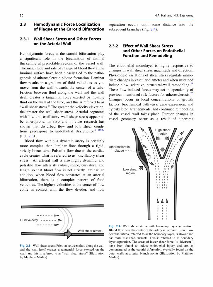
2.3 Hemodynamic Force Localizationof Plaque at the Carotid Bifurcation
2.3.1 Wall Shear Stress and Other Forceson the Arterial Wall
Hemodynamic forces at the carotid bifurcation play
a significant role in the localization of intimal
thickening at predictable regions of the vessel wall.
The magnitude and rate of change of blood flow at the
luminal surface have been closely tied to the patho-
genesis of atherosclerotic plaque formation. Laminar
flow results in a gradient of fluid velocities as you
move from the wall towards the center of a tube.
Friction between fluid along the wall and the wall
itself creates a tangential force exerted by flowing
fluid on the wall of the tube, and this is referred to as
“wall shear stress.” The greater the velocity elevation,
the greater the wall shear stress. Arterial segments
with low and oscillatory wall shear stress appear to
be atheroprone. In vivo and in vitro research has
shown that disturbed flow and low shear condi-
tions predispose to endothelial dysfunction7–10,22
(Fig. 2.3).
Blood flow within a dynamic artery is certainly
more complex than laminar flow through a rigid,
strictly linear tube. Pulsatile flow due to the cardiac
cycle creates what is referred to as “oscillatory shear
stress.” An arterial wall is also highly dynamic, and
pulsatile flow alters its radius, shape, curvature, and
length so that blood flow is not strictly laminar. In
addition, when blood flow separates at an arterial
bifurcation, there is a complex pattern of fluid
velocities. The highest velocities at the center of flow
come in contact with the flow divider, and flow
separation occurs until some distance into the
subsequent branches (Fig. 2.4).
2.3.2 Effect of Wall Shear Stressand Other Forces on EndothelialFunction and Remodeling
The endothelial monolayer is highly responsive to
changes in wall shear stress magnitude and direction.
Physiologic variations of shear stress regulate imme-
diate changes in vascular diameter and when sustained
induce slow, adaptive, structural-wall remodeling.21
These flow-induced forces may act independently of
previous mentioned risk factors for atherosclerosis.23
Changes occur in local concentrations of growth
factors, biochemical pathways, gene expression, and
cytoskeleton arrangements, and continued remodeling
of the vessel wall takes place. Further changes in
vessel geometry occur as a result of atheroma
Fluid velocity
Wall shear stress
Fig. 2.3 Wall shear stress. Friction between fluid along the wall
and the wall itself creates a tangential force exerted on the
wall, and this is referred to as “wall shear stress” (Illustration
by Matthew Maday)
High shearregion
Low shearregion
Atheroscleroticplaque
Fig. 2.4 Wall shear stress with boundary layer separation.
Blood flow near the center of the artery is laminar. Blood flow
near the intima, referred to as the boundary layer, is slower and
has more disturbed currents. This is referred to as boundary
layer separation. The areas of lower shear force (< 4dyn/cm2)
have been found to induce endothelial injury and are, as
demonstrated at the carotid bifurcation, typically found on the
outer walls at arterial branch points (Illustration by Matthew
Maday)
30 H.A. Hall and H.S. Bassiouny

formation, and further modify local near wall shear
stress direction, oscillation, and magnitude.
It has long been hypothesized that functional
alterations in the endothelial monolayer occur as a
result of low wall shear stress and increased residence
time of atherogenic blood particles.24 Wall shear stress
induces changes in endothelial cell morphology and
spatial orientation. Increased wall shear stress causes
endothelial cells to elongate and align in the direction
of flow, whereas endothelial cells exposed to low wall
shear stress remain more rounded and have no pre-
ferred orientation.25,26 Low wall shear stress may also
increase intercellular permeability and consequently
increase the vulnerability of these regions of the vessel
to atherosclerosis.27
Shear stress not only affects the shape and orienta-
tion of the cell, but alters the cell’s production and
release of vasoactive substances (i.e., prostacyclin,
nitric oxide, and endothelin-1). An acute increase in
wall shear stress in vitro elicits rapid cytoskeletal
remodeling and activates a signaling cascade in endo-
thelial cells, with the consequent acute release of
endothelial-derived relaxing factor (EDRF), i.e., nitric
oxide and prostacyclin.28 Nitric oxide in particular
appears to be a key mediator in the atheroprotective
effect of high wall shear stress.29 High laminar shear
stress sharply reduces endothelial cell levels of precur-
sor preproendothelin mRNA. This decreases the level
of endothelin-1 peptide, which exerts a constricting
and mitogenic effect on vascular smooth muscle
cells.30 Finally, prolonged oscillatory shear stress
induces expression of endothelial leukocyte adhesion
molecules, which are important in mediating leuko-
cyte localization in the arterial wall.31
In summary, high wall shear stress influences the
orientation of endothelial cells and the subsequent
production and release of factors that inhibit coagula-
tion, permit migration of leukocytes, and induce
smooth muscle proliferation, while simultaneously
promoting endothelial cell survival. Conversely, low
wall shear stress shifts the profile of secreted factors
and expressed surface molecules to one that favors the
opposite effects, thereby contributing to the develop-
ment of atherosclerosis.27 This complex endothelial
cell response to shear stress may also provide a mech-
anism by which known risk factors act to promote
atherosclerosis.30 In regions of moderate to high
shear stress, where flow remains unidirectional and
axially aligned, intimal thickening is limited. Intimal
thickening and atherosclerosis develop largely in
regions of relatively low wall shear stress, flow sepa-
ration, and departure from axially aligned, unidirec-
tional flow. Wall shear stress mapping has the
potential to become part of the multifactorial, multi-
disciplinary approach to early atherosclerosis detec-
tion, and following plaque progression.
2.4 Progression of Carotid BifurcationAtherosclerosis
Focal endothelial dysfunction and the formation of
fatty streaks within human arterial walls begin at a
young age. Many factors influence whether a plaque
will continue to grow and develop, become quiescent,
rupture, or thrombose. The composition of a plaque
and the ongoing presence of physical and biochemical
stress influence plaque vulnerability to disruption. The
interaction of the flow forces with plaque structural
components, such as hard or soft regions, will deter-
mine the degree in biomechanical stress on the fibrous
cap and likelihood of its structural failure, notwith-
standing the role of MMP’s in influencing the struc-
tural integrity of the fibrous cap extracellular matrix.
The composition and heterogeneity of atheroscle-
rotic plaque influences progression by virtue of ongo-
ing remodeling of the arterial wall.32 Modeling has
shown that stress distribution and magnitude are
influenced by the shape and the composition of the
fibrous plaque.33 In addition, vulnerable plaques
become more susceptible to rupture as the fibrous
cap thins with remodeling of the extracellular matrix
by metalloproteinases (MMPs) secreted by leukocytes
within the intima.34,35 The inflammatory response in
the juxtaluminal fibrous cap and necrotic core is a key
mechanism in human atherosclerotic plaque vulnera-
bility.36 In vulnerable atherosclerotic plaques, the
fibrous cap thins out and is more likely to disrupt,
resulting in thromboembolic events and cerebrovascu-
lar ischemia.37
Inflammation is a key element of atherosclerotic
plaque vulnerability and disruption, and fibrous cap
inflammation is more likely to occur in noncalcified
plaques as compared to calcified plaques, indicating
that plaque calcification is a marker of stability.38
Symptomatic plaques are less calcified and more
inflamed than asymptomatic plaques.39
2 Pathophysiology of Carotid Atherosclerosis 31

2.4.1 How to Assess Carotid Stenosis
2.4.1.1 ArteriographyArteriography is still considered the “gold standard”
for diagnosing carotid artery luminal stenosis against
which all other imaging modalities are compared.
With defined risks inherent in angiography, there
is a perpetual move toward noninvasive evaluation.
This shift culminated in investigations by Strandness
at the University of Washington, with criteria for esti-
mating carotid stenosis using velocity measure-
ments obtained by duplex ultrasound.40 Criteria for
carotid stenosis, intervention, and outcomes have
been validated in several large trials including the
North American Symptomatic Carotid Endarterec-
tomy Trial (NASCET),41 European Carotid Surgery
Trial (ECST),42 and the Asymptomatic Carotid Ath-
erosclerosis Trial (ACAS).43 These trials are the basis
of current indications for surgical intervention in
patients with carotid stenosis. However, it must be
mentioned that the data remain marginal for surgical
intervention on patients with asymptomatic plaques,
given that the number needed to treat is at least 20
patients to prevent one stroke in 5 years.43 It has
become clear that stenosis alone is not sufficient to
predict which asymptomatic plaques will progress to
become symptomatic. Plaque morphology and histo-
biochemical analysis have emerged as additional
factors in assessing plaque at risk. Imaging modalities
that remain central to the evaluation of carotid artery
disease include angiography, duplex ultrasound, com-
puted tomography angiography (CTA), and magnetic
resonance angiography (MRA). Further refinements in
these imaging modalities have further altered the data
obtainable from each exam. Newer ways of evaluating
carotid plaque structural characteristics continue to
evolve and allow for identification of features such
as extensive necrosis, fibrous cap thinning, and
intraplaque hemorrhage. These plaque characteristics
connote vulnerability and propensity to embolization,
transient ischemic attack (TIA), and stroke.
2.4.1.2 Duplex UltrasoundDuplex ultrasound has become the screening and diag-
nostic imaging modality of choice in carotid occlusive
disease largely because of its low cost, accuracy, and
noninvasiveness. Early studies have arrived at velocity
criteria for the diagnosis and classification of carotid
disease, and the most widely used in the 1980s and
1990s have been those developed by Strandness and
Zweibel40,44,45 (Table 2.1). It should be pointed out
that the early velocity criteria were developed for
stenosis expressed as a percentage of the bulb diameter
based on microcalcification on the arterial wall as seen
on angiograms and using mechanically rotating
transducers. It should also be noted that with modern
linear array transducers spectral broadening cannot be
used as a criterion. A recent meta-analysis showed that
ultrasound was best for more critical lesions, those at
least 70% or greater, with a sensitivity of 89% and
specificity of 84%. Examination of lesions between
50% and 69% using velocity criteria yielded a sensi-
tivity of only 36% and a specificity of 91%.46 Using
arteriography as the gold standard, the Strandness
criteria are less reliable in patients with contralateral
occlusion, high-grade contralateral stenosis, or less
than 70% ipsilateral stenosis.47 However, for plaques
producing moderate or mild stenosis B-mode com-
bined with color flow in cross-sectional views provide
accurate measurements of lumen and vessel diameters
from which the percentage diameter stenosis can be
calculated (see Chap. 28).
An analysis of the Strandness criteria for ICA ste-
nosis (European Carotid Surgery Trial [ECST]
method, i.e., in relation to bulb diameter) conducted
at the University of Chicago correlated ultrasono-
graphic velocity measurements with CT angiogram
measurements. The rationale for the study was to
better outline the boundary of the ICA plaque by CT
rather than using an estimated line as was done with
angiography in developing the Strandness criteria
when microcalcification in the arterial wall was not
present. The optimal threshold velocity to identify at
least a 50% stenosis of the ICA were a PSV of 155 cm/s
and an internal carotid artery/common carotid artery
(ICA/CCA) ratio of at least two.38 Velocity criteria
for a stenosis of at least 80% were found to be peak
systolic velocity (PSV) >370 cm/s, end diastolic velo-
city (EDV) >140 cm/s, and an ICA/CCA ratio of at
least 6.0.38
A full description of currently used techniques for
grading internal carotid stenosis will be found in
Chap. 28.
2.4.1.3 Computed Tomography (CT)The advancement of high-resolution multidetector CT
(MDCT) has allowed CT angiography to be performed
to evaluate the carotid bifurcation in a noninvasive
32 H.A. Hall and H.S. Bassiouny

fashion. Early studies evaluating this technology were
performed with single-slice scanners at a time when
resolution was inferior. Currently, however, high-
resolution CT scanners can not only evaluate the pres-
ence and degree of stenosis but also offer further
information regarding plaque morphology and compo-
sition. Cinat published a study of eight patients and
found a 72.6% agreement between CT assessment of
plaque composition and histological examination, and
a higher degree of calcification within the plaque
improved sensitivity to near 100%.48 Conversely,
noncalcified components such as necrotic core, lipid
volume, intraplaque hemorrhage, and connective tis-
sue confound the ability of CT to accurately determine
plaque histopathology. Further studies with larger
sample sizes looking at MDCT evaluation of plaque
composition may contribute to our understanding of
assessing overall plaque risk.
2.4.1.4 Magnetic Resonance Imaging (MRI)It has been shown that MRI is useful to accurately
evaluate plaque size and composition and can thus aid
in identifying vulnerable plaques. A study by Takaya
et al. prospectively followed 154 patients with initially
asymptomatic carotid stenosis by ultrasound for a
mean of 38.2 months. MRI was also performed at
baseline. Among this group of asymptomatic patients,
MRI plaque characteristics including a thin or rup-
tured fibrous cap, intraplaque hemorrhage, larger
lipid-rich necrotic core, and larger maximum wall
thickness were all associated with subsequent cerebro-
vascular events.49 This was a small study but does lay
the groundwork for larger prospective studies to exam-
ine the role of MRI in identifying atherosclerotic
plaques at higher risk of causing ischemic events.
Although MRI avoids the use of ionizing radiation,
the disadvantages associated with this imaging
Table 2.1 Summary of the Strandness and Zweibel duplex criteria for ICA stenosis
Strandness
Stenosis (%) in relation
to bulb diameter Duplex findings
Zweibel
Stenosis (%) in relation
to distal ICA diameter Duplex findings
0 PSV < 125 cm/s 0 PSV < 110 cm/s
No spectral broadening EDV < 40 cm/s
Bulb flow reversal PSV ICA/CCA <1.8
EDV ICA/CCA <2.4
Spectral broadening < 30 cm/s
1–15 PSV < 125 cm/s 1–39 PSV < 110 cm/s
No or minimal spectral broadening EDV < 40 cm/s
PSV ICA/CCA <1.8
EDV ICA/CCA <2.4Bulb flow reversal absent
Spectral broadening < 40 cm/s
16–49 PSV > 125 cm/s 40–59 PSV < 130 cm/s
Marked spectral broadening EDV < 40 cm/s
PSV ICA/CCA <1.8
EDV ICA/CCA <2.4
Spectral broadening < 40 cm/s
50–79 PSV > 125 cm/s 60–79 PSV > 130 cm/s
EDV < 140 cm/s EDV > 40 cm/s
PSV ICA/CCA >1.8
EDV ICA/CCA >2.4
Spectral broadening > 40 cm/s
80–99 PSV > 125 cm/s 80–99 PSV > 250 cm/s
EDV > 140 cm/s EDV > 100 cm/s
PSV ICA/CCA >3.7
EDV ICA/CCA >5.5
Spectral broadening > 80 cm/s
100 No flow 100 No flow
2 Pathophysiology of Carotid Atherosclerosis 33

modality make its usefulness in imaging the carotid
bifurcation limited in some patients. Long scanning
times increase overall motion artifact, and the risk
of gadolinium-induced nephrogenic systemic fibrosis
can occur in up to 3% of patients with renal
insufficiency.50
2.4.1.5 HistopathologyAdvancements in current imaging modalities are
allowing more detailed assessment of carotid plaque
characteristics in vivo. Numerous studies are attempt-
ing to standardize in vivo appearance on imaging to
ex vivo histology evaluation. The American Heart
Association has published various reports including
one by the Committee on Vascular Lesions of the
Council on Arteriosclerosis that defines and classifies
advanced types of atherosclerotic lesions based on
histology51 [Chap. 1]. The Committee on Vascular
Lesions also attempted to correlate the appearance of
lesions on clinical imaging studies with histological
lesion types (Table 2.2). Correlation of images to
ex vivo plaque is helping advance our understanding
of plaque morphology and vulnerability.
2.5 Stroke and Carotid BifurcationAtherosclerosis
2.5.1 Historical Perspective
One of the earliest reports linking stroke and carotid
artery disease was made by Savory in 1856 who
reported a case of a young woman with left monocular
symptoms and right hemiplegia attributed to occlusion
of the left cervical internal carotid artery and bilateral
subclavian arteries found at autopsy.6 Gowers in 1875
also described left carotid artery occlusion in a patient
with a right hemiplegia and loss of sight in the left
eye.52 Subsequent case reports increasingly linked
carotid artery occlusive disease to the development
of neurological symptoms, namely, stroke and tran-
sient ischemic attacks.53 In a major leap forward,
Edgar Moniz developed cerebral angiography in
1927, and in 1937, he described internal carotid occlu-
sion as documented by angiography.54,55 Even today,
arteriography of the carotid artery and its branches
remains the gold standard to which newer imaging
modalities are compared.
Table 2.2 Different types of human atherosclerotic lesions in pathology
Terms for atherosclerotic lesions in histological classificationsAppearance of lesions often based
on the unaided eyeType
I Initial lesion Early lesions
IIa Progression-prone type II lesion Fatty dot or streak
IIb Progression-resistant type II
III Intermediate lesion (preatheroma)
IV Atheroma Atheromatous plaque
Va Fibroatheroma (type V lesion) Fibrolipid plaque, Fibrous
plaque, plaque
Vb Calcific lesion (type VII lesion) Calcified plaque Advanced
lesions
Vc Fibrotic lesion (type VIII lesion) Fibrous plaque Raised
lesions
VI Lesion with surface defect, and/or hematoma-hemorrhage, and/or
thrombotic deposit
Complicated lesion,
complicated plaque
Source: Adapted from Stary et al.51
Note: Type I to type Va are early, type Vb advanced, and type Vc and VI are raised lesions
34 H.A. Hall and H.S. Bassiouny

In 1951, Dr. Fisher published a landmark paper in
the history of carotid artery disease in which he
described occlusion of the extracranial carotid artery
and its relation to cerebrovascular disease.56 This
included the first description of transient hemispheric
attack and monocular vision loss (now termed amau-
rosis fugax) as attributable to carotid disease and as
potential precursors of stroke. Prior to this, some 55%
of strokes were attributed to vasospasm. The idea of a
carotid bruit as an indicator of underlying carotid
disease, and its use as a screening tool for diagnosis
was also described by Fisher in 1957.57 Dr. Fisher
commented in his 1951 paper. . . “Some day vascular
surgery will find a way to by-pass the occluded portion
of the internal carotid artery during the period of
ominous fleeting symptoms.”56 Today, it is well
accepted that carotid artery occlusive disease is a risk
factor for transient ischemic attack (TIA) and stroke,
and medical disease modification as well as surgical
intervention, carotid endarterectomy, for carotid dis-
ease have been shown to provide benefit in reducing
the risk of stroke and stroke-related death.
The first successful carotid reconstruction was
completed by Carrea in Argentina in 1951, on a 51-
year-old man who presented with right hemiplegia and
left eye blindness. The patient was diagnosed with
severe left internal carotid artery stenosis on a percu-
taneous carotid angiogram. The stenotic segment of
the internal carotid artery was resected, and an end-to-
end anastomosis was performed between the external
carotid and the distal internal carotid artery. Patency
was confirmed by angiogram and the patient regained
strength in his right side over time.58 Reports of simi-
lar operations were reported by others in the coming
decade including Eastcott in 1954.59 Based on the idea
of endarterectomy introduced by Cid dos Santos in
1947 for aortoiliac atherosclerosis,60 the first carotid
endarterectomy was performed in 1953 by Strully,
Hurwitt, and Blankenberg on a patient 2 weeks after
the patient had a stroke. There was no back-bleeding
from the distal ICA however, and the vessel was
ligated.61 The first successful carotid endarterectomy
was performed by DeBakey in 1952, though that par-
ticular case was not published until 1975.62 Carotid
endarterectomy was performed by Cooley, Al-
Naaman, and Carton in 1956 and was the first to be
published in the literature.63 During this time, other
studies showed improvement in patients given
anticoagulants for cerebral thrombosis.64–66 The
story continues with improvement in medical preven-
tion and treatment of atherosclerotic disease and the
current debate over the use of carotid endarterectomy
versus carotid artery stenting in the interventional
treatment of both symptomatic and asymptomatic
carotid artery disease.
2.6 Role of Imaging in Identifying theVulnerable Asymptomatic Plaque
Stenosis of the carotid artery is noninvasively assessed
with duplex ultrasound, as well as by CT-angiography
or MR-angiography. However, despite large trials and
refinement of criteria, stenosis alone is inadequate in
predicting which asymptomatic plaques are at risk for
causing cerebrovascular symptoms. Recently, addi-
tional data garnered from standard imaging modalities
has been investigated to help identify those plaques at
risk for causing cerebrovascular symptoms. The devel-
opment of high-resolution B-mode ultrasound has
improved the ability of duplex scanning to evaluate
not only severity of stenosis but also morphology of
the plaque. Candidate descriptors of carotid plaque
morphology include echolucency, calcification, and
intraplaque hemorrhage, as well as other character-
istics such as plaque volume, surface irregularity,
fibrous cap thickness, and the size and location of the
necrotic core. Assessing additional plaque features
via ultrasound is important for the stratification of
high-risk patients.67,68
2.6.1 Echolucency/Gray Scale Median
Carotid atherosclerosis with echolucent plaque is
closely related to the occurrence of cerebrovascular
events.69,70 The more echolucent a plaque on ultra-
sound, the more likely it is to cause TIA or stroke in
the future. Initially, plaque echolucency was subjec-
tive and qualitative, thus making it difficult to corre-
late and attribute risk.71–74 Echolucency has been
further defined and quantified using the method of
image normalization and measurement of the gray
scale median (GSM). GSM is a computer-quantified
measurement of plaque echolucency,75–77 and several
studies have shown a correlation between low GSM
and plaque instability 78–80; (Chaps. 12, 15, 24, 29,
33, 36, 37).
2 Pathophysiology of Carotid Atherosclerosis 35

2.6.2 Calcification
Calcification is a relatively common structural feature
of the atherosclerotic plaque and is enhanced with
advanced age, chronic renal failure, diabetes, and
inflammation81 (Chap. 1). Calcified atherosclerotic
plaques are less prone to disrupt and result in
symptoms than noncalcified plaques.82 This implies
that calcification imparts structural stability to the
fibrous cap.38 A study of patients undergoing carotid
endarterectomy (CEA) found that those patients with
calcified carotid plaques had fewer cerebrovascular
events than those with noncalcified plaques.83 Grogan
et al. showed that using B-mode ultrasound, symptom-
atic plaques are more echolucent and less calcified
than asymptomatic plaques and are associated with a
greater degree of histopathologic plaque necrosis.84
Calcification, however, is not a normal feature of the
aging process, but a dynamic process in the progres-
sion of atherosclerosis.85,86 It is a result of a complex
interplay between inflammatory cytokines and the
activation of bone building cells.87
The presence and degree of carotid plaque calcifi-
cation can be accurately quantified with ex vivo CT
and is inversely related to plaque macrophage infiltra-
tion and symptomatic outcome.39 In vivo quantitative
assessment of carotid plaque calcification may help in
the future to identify patients with asymptomatic but
vulnerable carotid plaques who are at risk for devel-
opment of cerebrovascular events and benefit from
carotid interventions.
2.6.3 Intraplaque Hemorrhage
Intraplaque hemorrhage is a plaque characteristic
thought to correlate with symptomatology. The Amer-
ican Heart Association Type VI plaque is
characterized by surface irregularity, intraplaque hem-
orrhage, or thrombus, and is designated as a compli-
cated plaque. Although there is some controversy over
whether intraplaque hemorrhage alone is a predictor of
future ischemic events, it is a marker of plaque inflam-
mation and instability. A study by Hatsukami et al.
looked at 43 plaques from both symptomatic and
asymptomatic patients undergoing carotid endarterec-
tomy for highly stenotic lesions and compared histo-
logic findings to preoperative images.73 In this study,
they found no difference between symptomatic and
asymptomatic patients with regard to the presence or
volume of intraplaque hemorrhage, nor did they see a
difference in calcification, fibrous intimal tissue, lipid
core, or necrotic core. These findings show a limited
use for intraplaque hemorrhage alone as a surrogate
for plaque vulnerability. On the contrary, other studies
utilizing either ultrasonography or MRI to detect intra-
plaque hemorrhage have indeed shown intraplaque
hemorrhage to be a plaque characteristic that is
predictive of cerebrovascular events.88,89
References
1. Centers of Disease Control and Prevention [homepage on
the Internet]. Atlanta: Centers of Disease Control and Pre-
vention; [cited 2010 Mar 17]. Available from: http://www.
cdc.gov/
2. Roger VL et al. Heart disease and stroke statistics – 2010
update: a report from the American Heart Association. Cir-culation. 2010;121(7):e46–e215.
3. Heart and Stroke Foundation of Canada. The Growing Bur-den of Heart Disease and Stroke in Canada 2003. Ottawa:Heart and Stroke Foundation of Canada; 2003.
4. Donnan GA, Fisher M, Macleod M, Davis SM. Stroke.
Lancet. 2008;371(9624):12–23.5. Chaturvedi S et al. Carotid endarterectomy – an evidence-
based review: report of the Therapeutics and Technology
Assessment Subcommittee of the American Academy of
Neurology. Neurology. 2005;65:794–801.6. Savory WS. Case of a young woman in whom the main
arteries of both upper extremities and of the left side of the
neck were throughout completely obliterated. Med ChirTrans Lond. 1856;39:205–219.
7. Caro CG, Fitz-Gerald JM, Schroter RC. Arterial wall shear
stress and distribution of early atheroma in man. Nature.1969;223:1159–1161.
8. Friedman MH, Hutchins GM, Bargeron CB, Deters OJ,
Mark FF. Correlation between intimal thickness and fluid
shear in human arteries. Atherosclerosis. 1981;39:425–436.9. Ku DN, Giddens DP, Zarins CK, Glagov S. Pulsatile flow
and atherosclerosis in the human carotid bifurcation: posi-
tive correlation between plaque location and low and
oscillating shear stress. Arteriosclerosis. 1985;5:293–302.10. Zarins CK et al. Carotid bifurcation atherosclerosis: quanti-
tative correlation of plaque localization with flow velocity
profiles and wall shear stress. Circ Res. 1983;53:502–514.11. Clark J, Glagov S. Transmural organization of the arterial
wall. The lamellar unit revisted. Arteriosclerosis. 1985;5:19–34.
12. Baumgartner HR, Studer A. Consequences of vessel cathe-
terization in normal and hypercholesterolemic rabbits.
Pathol Microbiol. 1966;29:393–405.13. Bowen-Pope DF, Ross R, Seifert RA. Locally acting growth
factors for vascular smooth muscle cells: endogenous syn-
thesis and release from platelets. Circulation. 1985;72:
735–740.
36 H.A. Hall and H.S. Bassiouny

14. Faggiotto A, Ross R, Harker L. Studies of hypercholesterol-
emia in the nonhuman primate. Arteriosclerosis.1984;4:323–340.
15. Gerrity RG, Naito HK, Richardson M, Schwartz CJ. Dietary
induced atherogenesis in swine. Am J Pathol. 1979;95:
775–792.
16. Fowler S, Shio H, Haley WJ. Characterization of lipid-laden
aortic cells from cholesterol-fed rabbits. IV. Investigation of
macrophage-like properties of aortic cell populations. LabInvest. 1979;41:372–378.
17. Schaffner T et al. Arterial foam cells with distinctive
immunomorphologic and histochemical features of
macrophages. Am J Pathol. 1980;100:57–80.18. Haberland ME, Fong D, Cheng L. Malondialdehyde-altered
protein occurs in atheroma of Watanabe heritable
hyperlipidemic rabbits. Science. 1988;241:215–218.19. Clowes AW, Ryan GB, Breslow JL, Karnovsky MJ.
Absence of enhanced intimal thickening in the response of
the carotid arterial wall to endothelial injury in hypercholes-
terolemic rats. Lab Invest. 1976;35(1):6–17.20. Godin D, Ivan E, Johnson C, Magid R, Galis Z. Remodeling
of carotid artery is associated with increased expression of
matrix metalloproteinases in mouse blood flow cessation
model. Circulation. 2000;102:2861–2866.21. Glagov S. Intimal hyperplasia, vascular remodeling, and the
restenosis problem. Circulation. 1994;89:2888–2891.22. Lind L, Andersson J, Larsson A, Sandhagen B. Shear stress
in the common carotid artery is related to both intima-media
thickness and echogenecity. The Prospective Investigation
of the Vasculature in Uppsala Seniors study. ClinHemorheol Microcirc. 2009;43(4):299–308.
23. Gibson CM et al. Relationship of vessel wall shear stress to
atherosclerosis progression in human coronary arteries.
Arterioscler Thromb. 1993;13:310–315.24. Glagov S, Zarins C, Giddens DP, Ku DN. Hemodynamics
and atherosclerosis: insights and perspectives gained from
studies of human arteries. Arch Pathol Lab Med. 1988;112:1018–1031.
25. Levesque MJ, Nerem RM. The elongation and orientation
of cultured endothelial cells in response to shear stress.
J Biomech Eng. 1985;107:341–347.26. Levesque MJ, Liepsch D, Moravec S, Nerem RM. Correla-
tion of endothelial cell shape and wall shear stress in a
stenosed dog aorta. Arteriosclerosis. 1986;6:220–229.27. Okano M, Yoshida Y. Junction complexes of endothelial
cells in atherosclerosis-prone and atherosclerosis-resistant
regions on flow dividers of brachiocephalic bifurcations in
the rabbit aorta. Biorheology. 1994;31:155–161.28. Ballermann BJ, Dardik A, Eng E, Liu A. Shear stress and the
endothelium. Kidney Int Suppl. 1998;67:S100-S108.29. Traub O, Berk BC. Laminar shear stress: mechanisms by
which endothelial cells transduce an atheroprotective force.
Arterioscler Thromb Vasc Biol. 1988;18:677–685.30. Sharefkin JB, Diamond SL, Eskin SG, McIntire LV,
Dieffenbach CW. Fluid flow decreases preproendothelin
mRNA and suppresses endothelin-1 peptide release in
cultured human endothelial cells. J Vasc Surg. 1991;14:1–9.31. Chappell DC, Varner SE, Nerem RM, Medford RM, Alex-
ander RW. Oscillatory shear stress stimulates adhesion
molecule expression in cultured human endothelium. CircRes. 1998;82:532–539.
32. Glagov S, Bassiouny HS, Giddens DR, Zarins CK. Intimal
thickening: morphogenesis, functional significance and
detection. J Vasc Invest. 1995;1:1–14.33. Beattie D, Xu C, Vito R, Glagov S, Whang MC. Mechanical
analysis of heterogeneous, atherosclerotic human aorta.
Trans ASME. 1998;120:602–607.34. Welgus HG et al. Neutral metalloproteinases produced by
human mononuclear phagocytes. Enzyme profile, regula-
tion, and expression during cellular development. J ClinInvest. 1990;86(5):1496–1502.
35. Galis ZS, Sukhova GK, Lark MW, Libby P. Increased
expression of matrix metalloproteinases and matrix
degrading activity in vulnerable regions of human athero-
sclerotic plaques. J Clin Invest. 1994;94(6):2493–2503.36. Ross R. Atherosclerosis – an inflammatory disease. N Engl
J Med. 1999;340:115–126.37. Naghavi M et al. From vulnerable plaque to vulnerable
patient: a call for new definitions and risk assessment
strategies – part I. Circulation. 2003;108:1664–1672.38. Wahlgren CM, Zheng W, Shaalan W, Tang J, Bassiouny
HS. Human carotid plaque calcification and vulnerability.
Relationship between degree of plaque calcification, fibrous
cap inflammatory gene expression and symptomatology.
Cerebrovasc Dis. 2009;27:193–200.39. Shaalan WE et al. Degree of carotid plaque calcification in
relation to symptomatic outcome and plaque inflammation.
J Vasc Surg. 2004;40:262–269.40. Strandness D Jr. Extracranial arterial disease. In: Duplex
Scanning in Vascular Disorders. 2nd ed. New York:
Raven; 1993.
41. North American Symptomatic Carotid Endarterectomy Trial
Collaborators. Beneficial effect of carotid endarterectomy
in symptomatic patients with high-grade carotid stenosis.
N Engl J Med. 1991;325(7):445–453.42. European Carotid Surgery Trialists’ Collaborative Group.
MRC European Carotid Surgery Trial: interim results for
symptomatic patients with severe (70-99%) or with mild (0-
29%) carotid stenosis. Lancet. 1991;337(8752):1235–1243.43. The Asymptomatic Carotid Atherosclerosis Study Group.
Study design for randomized prospective trial of carotid
endarterectomy for asymptomatic atherosclerosis. Stroke.1989;20(7):844–849.
44. Zwiebel WJ. Spectrum analysis in carotid sonography.
Ultrasound Med Biol. 1987;13(10):625–636.45. Zwiebel WJ. Introduction to Vascular Ultrasonography.
Philadelphia: W.B. Saunders; 1992.
46. Wardlaw JM, Chappell FM, Best JJ,Wartolowska K, Berry E.
Non-invasive imaging compared with intra-arterial angio-
graphy in the diagnosis of symptomatic carotid stenosis:
a meta-analysis. Lancet. 2006;367(9521):1503–1512.47. AbuRahma AF et al. Effect of contralateral severe stenosis
or carotid occlusion on duplex criteria of ipsilateral stenoses:
comparative study of various duplex parameters. J VascSurg. 1995;22(6):751–761.
48. Cinat M et al. Helical CT angiography in the preoperative
evaluation of carotid artery stenosis. J Vasc Surg.1998;28:290–300.
2 Pathophysiology of Carotid Atherosclerosis 37

49. Takaya N et al. Association between carotid plaque
characteristics and subsequent ischemic cerebrovascular
events. A prospective assessment with MRI – initial results.
Stroke. 2006;37:818–823.50. Issa N et al. Nephrogenic systemic fibrosis and its
association with gadolinium exposure during MRI. CleveClin J Med. 2008;75:95-97. 103-4, 106 passim.
51. Stary HC et al. A definition of advanced types of atheroscle-
rotic lesions and a histologic classification of atherosclero-
sis, a report from the committee on vascular lesions of the
Council on Arteriosclerosis, American Heart Association.
Circulation. 1995;92:1355–1374.52. Gowers W. On a case of simultaneous embolism of central
retinal and middle cerebral arteries. Lancet. 1875;2:794.53. Hunt JR. The role of the carotid arteries, in the causation of
vascular lesions of the brain, with remarks on certain special
features of the sympomatology. Am J Med Sci. 1914;147(5):704–712.
54. Moniz E. L’encephalographie arterielle, son importance
dans la localisation des tumeurs cerebrales. Rev Neurol(Paris). 1927;2:72-90. [Translated from the French by
Espinosa RE and reprinted in: Bruwer AJ. Classic
descriptions in diagnostic roentgenology. Springfield:
Charles C. Thomas; 1964.]
55. Moniz E, Lima A, de Lacerda R. Par thrombose de la
carotide interne. Presse Med. 1937;45:977–980.56. Fisher CM. Occlusion of the internal carotid artery. Arch
Neurol Psychiatry. 1951;65:346–377.57. Fisher CM. Cranial bruit associated with occlusion of the
internal carotid artery. Neurology. 1957;7:299–306.58. Carrea R, Molins M, Murphy G. Surgical treatment of spon-
taneous thrombosis of the internal carotid artery in the neck:
carotid carotideal anastamosis. Acta Neurol Latinoam.1955;1:71–78.
59. Eastcott HHG, Pickering GW, Robb CG. Reconstruction of
internal carotid artery in a patient with intermittent attacks
of heiplegia. Lancet. 1954;2:994–996.60. dos Santos JC. Sur la deobstruction des thromboses
arterielles anciennes. Mem Acad Chir. 1947;73:409–411.61. Strully KJ, Hurwitt ES, Blankenberg HW. Thromboendar-
terectomy for thrombosis of the internal carotid artery in the
neck. J Neurosurg. 1953;10:474–482.62. DeBakey ME. Successful carotid endarterectomy for cere-
brovascular insufficiency; nineteen year follow-up. JAMA.1975;233:1083–1085.
63. Cooley DA, Al-Naaman YD, Carton CA. Surgical treatment
of arteriosclerotic occlusion of common carotid artery.
J Neurosurg. 1956;13:500–506.64. Hedenius P. The use of heparin in internal diseases. Acta
Med Scand. 1941;107:170–182.65. Millikan CH, Siekert RG, Shick RM. Studies in cerebrovas-
cular disease, III: the use of anticoagulant drugs in the
treatment of insufficiency or thrombosis within the basilar
arterial system. Proc Staff Meet Mayo Clin. 1955;30:
116–126.
66. Fisher CM. The use of anticoagulants in cerebral thrombo-
sis. Neurology. 1958;8:311–332.67. Grønholdt ML, Nordestgaard BG, Schroeder TV, Vorstrup
S, Sillesen H. Ultrasonic echolucent carotid plaques predict
future strokes. Circulation. 2001;104:68–73.
68. Hennerici M, Baezner H, Daffertshofer M. Ultrasound and
arterial disease. Cerebrovasc Dis. 2004;17(suppl 1):19–33.69. Polak JF et al. Hypoechoic plaque at US of the carotid artery:
an independent risk factor for incident stroke in adults aged
65 years or older. Radiology. 1998;208:649–654.70. Mathiesen EB, Bønaa KH, Joakimsen O. Echolucent
plaques are associated with high risk of ischemic cere-
brovascular events in carotid stenosis: the Tromsø study.
Circulation. 2001;103:2171–2175.71. Bassiouny HS et al. Juxtalumenal location of plaque necro-
sis and neoformation in symptomatic carotid stenosis.
J Vasc Surg. 1997;26:585–594.72. Gray-Weale AC, Graham JC, Burnett JR, Byrne K, Lusby
RJ. Carotid artery atheroma: comparison of preoperative
B-mode ultrasound appearance with carotid endarterectomy
specimen pathology. J Cardiovasc Surg (Torino).1988;29:676–681.
73. Hatsukami TS et al. Carotid plaque morphology and clinical
events. Stroke. 1997;28:95–100.74. Carr S, Farb A, Pearce WH, Virmani R, Yao JS. Atheroscle-
rotic plaque rupture in symptomatic carotid artery stenosis.
J Vasc Surg. 1996;23:755–765.75. el Barghouty N, Nicolaides A, Bahal V, Geroulakos G,
Androulakis A. The identification of the high risk carotid
plaque. Eur J Vasc Endovasc Surg. 1996;11:470–478.76. Sabetai MM et al. Reproducibility of computer-quantified
carotid plaque echogenicity. Can we overcome the subjec-
tivity? Stroke. 2000;31:2189–2196.77. Lal BK et al. Pixel distribution analysis of B-mode ultra-
sound scan images predicts histologic features of atheroscle-
rotic carotid plaques. J Vasc Surg. 2002;35:1210–1217.78. Tegos TJ et al. Echomorphologic and histopathologic
characteristics of unstable carotid plaques. Am JNeuroradiol. 2000;21:1937–1944.
79. Grønholdt MLM, Nordestgaard BG, Wiebe BM, Wilhjelm
JE, Sillesen H. Echo-lucency of computerized ultrasound
images of carotid atherosclerotic plaques are associated
with increased levels of triglyceride-rich lipoproteins as
well as increased plaque lipid content. Circulation. 1998;97:34–40.
80. Sabetai MM et al. Hemispheric symptoms and carotid
plaque echomorphology. J Vasc Surg. 2000;31:39S-49S.81. Pecovnik-Balon B. Cardiovascular calcification in patients
with end-stage renal disease. Ther Apher Dial. 2005;
9:208–210.
82. Nandalur KR et al. Calcified carotid atherosclerotic plaque
is associated less with ischemic symptoms than is
noncalcified plaque on MDCT. AJR Am J Roentgenol.2005;184:295–298.
83. Hunt JL et al. Bone formation in carotid plaques: a
clinicopathalogiccal study. Stroke. 2002;33:1214–1219.84. Grogan JK et al. B-mode ultrasonographic characterization
of carotid atherosclerotic plaques in symptomatic and
asymptomatic patients. J Vasc Surg. 2005;42:435–441.85. Doherty TM, Detrano RC. Coronary arterial calcification as
an active process: a new perspective on an old problem.
Calcif Tissue Int. 1994;54:224–230.86. Wexler L et al. Coronary artery calcification: pathophysiol-
ogy, epidemiology, imaging methods, and clinical
implications – a statement for health professionals from the
38 H.A. Hall and H.S. Bassiouny

American Heart Association Writing Group. Circulation.1996;94:1175–1192.
87. Doherty TM et al. Calcification in atherosclerosis: bone
biology and chronic inflammation at the arterial crossroads.
Proc Natl Acad Sci USA. 2003;100:11201–11206.88. Moody AR et al. Characterization of complicated carotid
plaque with magnetic resonance direct thrombus imaging in
patients with cerebral ischemia. Circulation. 2003;107(24):3047–3052.
89. AbuRahma AF, Kyer PD 3rd, Robinson PA, Hannay RS.
The correlation of ultrasonic carotid plaque morphology and
carotid plaque hemorrhage; clinical implications. Surgery.1998;124(4):721–726.
2 Pathophysiology of Carotid Atherosclerosis 39

http://www.springer.com/978-1-84882-687-8
Copyright © 2022 FDOKUMEN