
Role of thoughts during nocturnal awake times in the insomnia experience of older …

Carotid angioplasty with internal carotid arteryflow reversal is well tolerated in the awake patientEnrique Criado, MD,a Manuel Doblas, MD,b Juan Fontcuberta, MD,b Antonio Orgaz, MD,b AngelFlores, MD,b Paloma Lopez, MD,b and L Philipp Wall, MD,a Stony Brook, NY; and Toledo, Spain
Objective: The purpose of this study was to evaluate the neurologic tolerance and changes in ipsilateral hemispheric oxygensaturation during transcervical carotid artery stenting with internal carotid artery (ICA) flow reversal for embolicprotection.Patients and methods: This was a prospective study of 10 patients (mean age 68 years) undergoing transcervical carotidangioplasty and stenting. All ICA stenoses were greater that 70%. Seven patients had an ipsilateral hemispheric stroke (3)or transient ischemic attack (4), two patients had a contralateral stroke, and one patient was asymptomatic. Nineprocedures were done under local anesthesia. Cerebral protection was established through a cervical common carotid(CCA) cutdown to create an external fistula between the ICA and the internal jugular vein with temporally CCAocclusion. Venous oxygen saturation (SVO2) was continuously monitored through a catheter placed in the distal internaljugular vein. Mental status and motor-sensory changes were categorized and assessed throughout and after the procedure.Results: All procedures were technically successful without significant residual stenosis. Mean ICA flow reversal time was22 minutes (range, 15 to 32). Common carotid artery (CCA) occlusion produced a slight (SVO2 � 72.6%�/-9.4) butsignificant decrease (P � .012) in SVO2, compared with baseline (SVO2 � 77% �/-10.5). During ICA flow reversal(SVO2 � 72.4% �/-10.1) cerebral oxygen saturation did not change compared with CCA occlusion alone (P � .85).Transient balloon occlusion during angioplasty of the ICA (SVO2 � 64.6%�/-12.9) produced a significant decrease incerebral SVO2 compared with CCA occlusion (P � .015) and compared with CCA occlusion with ICA flow reversal(P � .018). No mental status changes or ipsilateral hemispheric focal symptoms occurred during CCA occlusion with ICAflow reversal. One patient with contralateral ICA occlusion sustained brief upper extremity weakness related to thecontralateral hemisphere. Five patients sustained a vasovagal response during balloon dilatation, four did not requiretreatment, and one had asystole requiring atropine injection. Mean SVO2 saturation was not different in these fivepatients compared with the five who did not sustain a vasovagal response. No deaths or neurologic deficits occurredwithin 30 days after the procedure.Conclusions: Our data suggest that transcervical carotid angioplasty and stenting with ICA flow reversal is well toleratedin the awake patient, even in the presence of symptomatic carotid artery disease. Cerebral oxygenation during ICA flowreversal is comparable to that during CCA occlusion. ICA angioplasty balloon inflation produces a decrease in cerebralSVO2 significantly greater than that occurring during ICA flow reversal. (J Vasc Surg 2004;40:92-7.)
The beneficial effect of cerebral protection during ca-rotid artery stenting (CAS) has not yet been proven inprospective, controlled studies. However, pooled data frommultiple single institutional experiences suggest that cere-bral protection during CAS may reduce the risk of cerebralembolization and neurologic events.1 The risk of cerebralembolization, detected by postoperative magnetic reso-nance imaging, is 15% in patients undergoing CAS withoutcerebral protection, 7% of whom experience a clinicallyapparent neurologic event.2 Current methods under eval-uation for cerebral protection during CAS include distalinternal carotid artery (ICA) balloon occlusion, distal ICAfiltering, and proximal common carotid artery (CCA) oc-clusion with or without carotid flow reversal. All these
From the Divisions of Vascular Surgery, Stony Brook University Hospital,a
Stony Brook, NY, and Complejo Hospitalario de Toledo,b Toledo, Spain.Competition of interest: none.Reprint requests: Enrique Criado, MD, Division of Vascular Surgery,
Health Sciences Center T19, Stony Brook University, Stony Brook, NY11794-8191 (e-mail: [email protected]).
0741-5214/$30.00Copyright © 2004 by The Society for Vascular Surgery.doi:10.1016/j.jvs.2004.03.034
92
methods aim at avoiding distal embolization of carotiddebris to the brain. The Parodi antiembolic femoral cathe-ter permits constant flow inversion in the ICA by connect-ing the aspiration port to the femoral vein, creating anarteriovenous fistula between the ICA and the deep venoussystem.3 Arteriovenous flow in this guiding catheter, how-ever, may be limited because of its high resistance, mainlywhen catheters occupy its lumen. Our transcervical tech-nique for CAS, using a short, larger bore carotid to internaljugular vein (IJV) fistula, maintains a constant, higher flowreversal in the ICA without external carotid occlusion.Preliminary clinical experience suggests that ICA flow re-versal is effective in preventing cerebral embolism and thatis well tolerated,3 but the physiologic mechanism for neu-rologic tolerance is not well understood. This study wasdesigned to evaluate changes in cerebral oxygen saturationand neurologic status during CCA occlusion and ICA flowreversal using a transcervical approach for CAS.
PATIENTS AND METHODS
Patient population. During September and October2003, 10 consecutive patients underwent CAS proceduresas part of a prospective study in accordance with the insti-

JOURNAL OF VASCULAR SURGERYVolume 40, Number 1 Criado et al 93
tutional board policies and regulations at the ComplejoHospitalario de Toledo, Spain. Informed consent was ob-tained from all patients. Data was prospectively collectedregarding medical history, comorbidities, symptomatic sta-tus, degree of carotid stenosis, and intraoperative and post-operative studies and events.
Patient mean age was 68 years (range, 59-80 years). Allpatients were men. Seven patients had symptomatic carotidstenosis, four with hemispheric transient ischemic attackand three with a previous ipsilateral hemispheric stroke.Three patients had asymptomatic carotid stenosis; two ofthem with previous contralateral hemispheric stroke. Theindications for carotid intervention and estimated degree ofcarotid stenosis are delineated in Table I. Associated med-ical comorbid conditions are summarized in Table II.
Transcervical CAS protocol and technique. All pa-tients underwent carotid duplex ultrasound scanning toconfirm the degree of carotid stenosis. All patients weretreated with aspirin, 325 mg/d, and clopidogrel, 75 mgonce a day for at least 4 days, or 300 mg a day for 2 daysbefore the procedure. Nine interventions were done underlocal anesthesia (1% lidocaine), and one patient receivedgeneral anesthesia upon his request at the initiation of theoperation. No sedation was given before or during theprocedure. One hundred units of heparin per kilogram ofbody weight were given intravenously before carotid punc-ture. Intra-arterial blood pressure and transcutaneous oxy-gen saturation were continuously monitored, and neuro-logic status was assessed at regular intervals by the
Table I. Indications and degree of ICA stenosis* in 10 pat
Patient Indication
1 Previous contralateral CVA2 Previous ipsilateral CVA3 Asymptomatic4 Hemispheric TIA5 Previous ipsilateral CVA6 Hemispheric TIA7 Previous contralateral CVA8 Hemispheric TIA9 Hemispheric TIA
10 Previous ipsilateral CVA
ICA, Internal carotid artery; CAS, carotid artery stenting; CVA, cerebrovas*The % of ICA stenosis was estimated based on duplex.
Table II. Medical comorbidities in 10 patientsundergoing CAS
Medical comorbidity No. %
CAD 4 40CAD with coronary intervention 3 30Hypercholesterolemia 4 40Hypertension 8 80Diabetes 2 20Smoking 2 20
CAD, Coronary artery disease.
anesthesiologist and surgeon. Ipsilateral, hemispheric ve-nous oxygen saturation (SVO2) was recorded after place-ment of all vessel introducer sheaths without vessel occlu-sion (baseline), after occlusion of the CCA maintainingexternal to internal carotid flow until SVO2 stabilizedwithin 1 to 2 minutes, after establishment of ICA flowreversal with CCA occlusion, after occlusion of the ICAduring angioplasty balloon inflation, and after returning tobaseline.
We used a technique for transcervical CAS that wepreviously developed through experience in 30 patients. Afull description of our technique has been published.4
Basically, the technique consists in accessing the commoncarotid artery through a mini-incision (3 to 4 cm) at thebase of the neck, encircling the proximal CCA with aRummel loop, and establishing an arteriovenous fistulabetween the CCA and the IJV by placing 8F by 11-cmpercutaneous introducer sheaths (Super Arrow Flex; ArrowInternational, Reading, Pa) in these vessels and connectingthe introducers with a short (15-cm) segment of tubing(Fig 1). For this study exclusively, ipsilateral hemisphericSVO2 was continuously measured with a pediatric oxymet-ric Swan-Ganz catheter, introduced through a 5F by 11-cmpercutaneous introducer sheath (Super Arrow Flex) fromthe neck incision into the IJV in a cephalad direction.
After the CCA was occluded with the Rummel loopand a carotid to jugular fistula was established, retrogradeflow in the ICA was ascertained with fluoroscopy by inject-ing a small amount of contrast in the CCA and immediatelyopening the fistula to observe contrast drain from the ICAinto the IJV (Fig 2). There is no need to balloon occludethe external carotid, because the resistance of the fistula islow enough to reverse flow in both the external and internalcarotids. Using hand injection, digital angiography wasperformed in the lateral and oblique planes to localize andquantitate the degree of ICA stenosis. With flow reversal inplace, a 0.014-inch guide wire (Platinum plus TM, ST,0.014-180 cm; Boston Scientific, Meditech, Miami, Fla) ina 4F by 40-cm-long Berenstein catheter (Angiodynamics,Inc, Queensbury, NY) was introduced through the CCAsheath for selective crossing of the ICA stenosis. The tip of
undergoing CAS
Ipsilateral stenosis(%)
Contralateral stenosis(%)
�70 �50�80 �50�80 �50�70 occluded�70 �70�70 �50�80 occluded�70 �50�80 �50�80 �80
accident; TIA, transient ischemic attack.
ients
cular

JOURNAL OF VASCULAR SURGERYJuly 200494 Criado et al
the wire was advanced to a position just proximal to thecarotid siphon.
No predilatation of the lesion was done in this group ofpatients. Self-expandable monorail stents 10 � 31 mm or10 � 37 mm (Wallstent; Boston Scientific, Maple Grove,Minn) were used in all patients. Post-stent dilation wasperformed for 5 to 10 seconds in all cases with 5- or 6-mm� 2-cm monorail balloon catheters (Ultra-soft SV Mono-rail balloon catheter; Boston Scientific, Maple Grove,Minn) inflated to 8 atm.
Ipsilateral hemispheric IJV oxygen saturation was re-corded at baseline with all sheaths in place, after occludingthe CCA without arteriovenous fistula for 1 to 2 minutes,after stabilization with the CCA occluded and the arterio-venous fistula functioning, immediately after occluding theICA during balloon dilatation, and upon return to baselineafter stent placement. Intra-arterial blood pressure, heartrate, and neurologic status were monitored throughout theprocedure.
A completion carotid angiography was performed toassess technical results and the presence of distal spasm.Intra-arterial papaverine solution (1 mg/mL) was selec-tively used to treat residual post-CAS spasm. Following
Fig 1. Diagram illustrating the arteriovenous fistula between thecommon carotid artery and the internal jugular vein.
removal of the sheaths, the vessel access sites were closedwith 6-0 polypropylene sutures, and the wound was closedwith absorbable sutures. After the procedure, all patientswere observed in the recovery room for 6 hours and thentransferred to a floor bed. The following day, after a phys-ical examination, all patients were discharged from thehospital.
Clopidogrel was continued at 75 mg/d by mouth for atleast 1 month, and aspirin was continued indefinitely. Onemonth following the procedure, a physical examination anda carotid duplex scan were repeated in all patients. Techni-cal failure was defined as inability to access or to cross thelesion or post-stenting residual stenosis equal or greaterthan 30%. Intraprocedural mental status changes were cat-egorized as none, somehow decreased, or unresponsive. Tran-sient ischemic attack was defined as a focal hemisphericdeficit that resolved within 24 hours. A focal deficit lastingmore than 24 hours was defined as a stroke. Recurrent orresidual in stent stenosis was defined as more than 50%diameter reduction as determined by duplex ultrasoundscanning.
Statistical analysis was done with a personal computerand an SPSS (version 11) statistical package. Paired groupsof data were compared using the paired Student t test.Unpaired data was compared using the independent vari-able Student t test.
RESULTS
All CAS procedures were accomplished successfully.No contrast extravasation, arterial disruption, or subintimaldissections were observed. Distal ICA spasm following CASwas found in one patient, and resolved with papaverineinjection. Mean duration of the procedure was 62 minutes(range, 45-80), and mean ICA flow reversal time was 22minutes (range, 15-32). Residual stenosis after CAS didnot exceed 15% in any case, and the external carotid arteryremained patent in all patients.
There were no intraoperative ipsilateral hemisphericfocal deficits. No mental status changes occurred in re-sponse to CCA occlusion or ICA flow reversal. One patientwith contralateral ICA occlusion and previous contralateralhemispheric stroke sustained transient upper extremityweakness related to the contralateral hemisphere and theend of the ICA flow reversal period, which resolved uponre-establishing antegrade carotid flow. Another patient hada seizure following injection of papaverine solution in ex-cessive concentration (30 mg/mL instead of 1 mg/mL) toresolve residual ICA spasm following CAS after antegradeICA was re-established. The patient was given intravenousbenzodiazepines, intubated, and extubated within 30 min-utes without sequelae. Five patients sustained a decrease inpulse rate and blood pressure in response to ICA balloondilatation. In four of these cases the carotid body responsewas mild and did not require treatment; one patient sus-tained asystole, recovering pulse and pressure after a pre-cordial thump and atropine administration, and subse-quently required a pacemaker.

the p
JOURNAL OF VASCULAR SURGERYVolume 40, Number 1 Criado et al 95
No neck access wound complications occurred. Allpatients were discharged the day after the procedure. Therewere no deaths or strokes at 30 days following surgery.
All patients were followed up within 1 to 2 monthsfollowing the procedure. At the time of follow-up, allpatients remained neurologically unchanged, and carotidultrasound revealed that all stents were patent withoutresidual or recurrent stenosis.
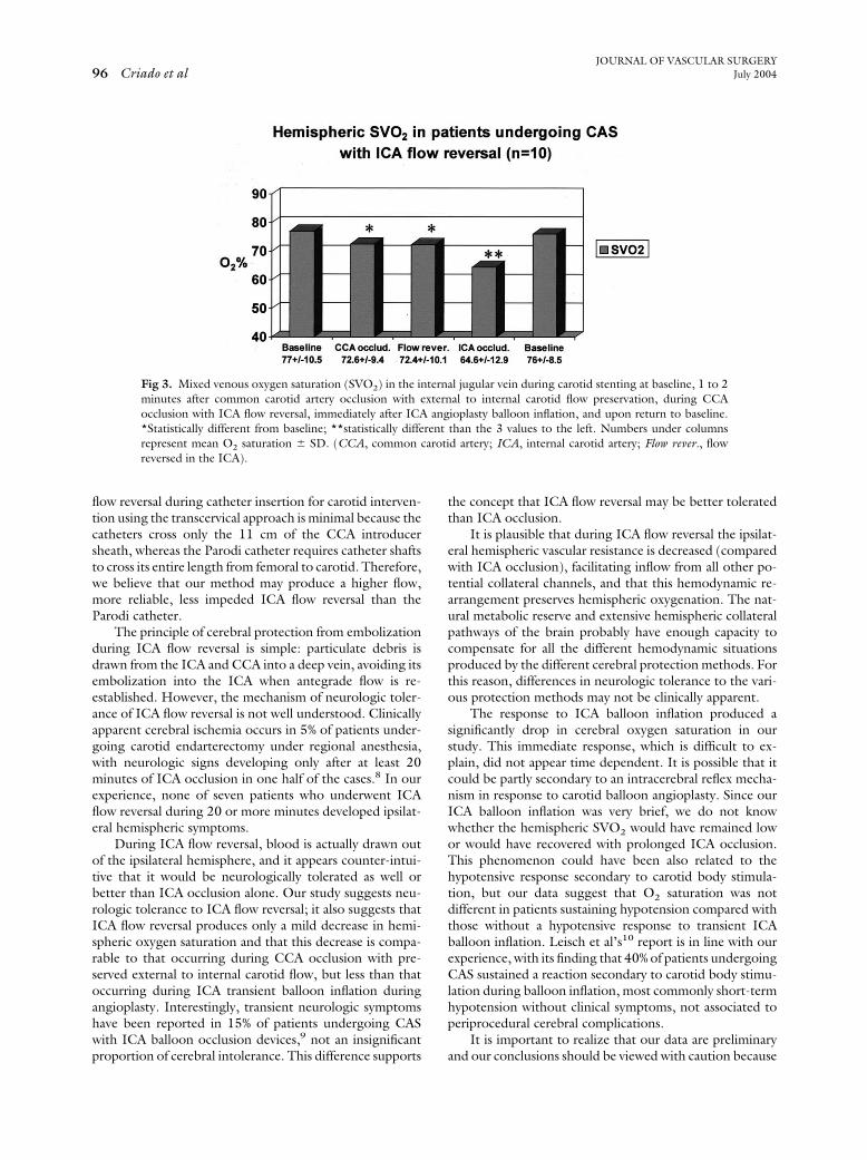
Hemispheric mixed venous oxygen saturation meanchanges with standard deviations during the procedure aredepicted in Fig 3. CCA occlusion with external to internalcarotid flow produced a mild drop in cerebral SVO2 com-pared with baseline (77% vs 72.6%); this difference, how-ever, was statistically significant (P � .012). During ICAflow reversal, the SVO2 remained unchanged (P � .85)compared with CCA occlusion with external to internalflow (72.6% vs 72.4%). A significant drop in SVO2 wasnoted after ICA occlusion during balloon inflation, and thiswas statistically significant in comparison with baseline (P �.003), in comparison with CCA occlusion (P � .018) andin comparison with ICA flow reversal (P � .015). Thedecrease in oxygen saturation during ICA occlusion in fivepatients who sustained a hypotensive carotid body responsewas not different (P � 1.0) from that occurring in fivepatients who did not sustain hypotension with ICA balloonocclusion during angioplasty.
Fig 2. Intraoperative substraction angiogram illustratinginternal jugular vein. Note (white arrows) a third introdmonitoring. With the common carotid artery occludedthrough the sheath and antegrade flow is seen in the ICopened, and digital substraction is maintained until contrthe tubing into the jugular vein (right, black arrows).cerebral protection with ICA flow reversal is in place for
DISCUSSION
Carotid artery angioplasty and stenting without cere-bral protection is associated with a significant risk of newcerebral infarction and neurologic events secondary to em-bolization.2 The initial guidewire crossing of the carotidlesion is associated with the highest risk of cerebral embo-lization during CAS.5 Therefore, a major limitation ofinternal carotid balloon occlusion and filtering methods ofcerebral protection is that they do not prevent emboliza-tion during the initial guidewire crossing of the carotidlesion. To resolve this problem, Parodi and colleagues3,6
developed a cerebral protection method using a guidingcatheter with an elastomeric balloon at the tip that occludesthe CCA and allows reversal of ICA flow by connecting thecatheter with the femoral vein, while the external carotidartery is occluded with another balloon. This method ofcerebral protection has been shown to eliminate cerebralembolization detected by transcranial Doppler scanningduring CAS.7 However, the Parodi catheter, because of itslength, may result in a high-resistance conduit that limitsthe arteriovenous gradient, mostly when balloon or stentshafts occupy the lumen of the guiding catheter. Ourtranscervical approach provides a shorter, larger caliberarteriovenous conduit that permits higher, unimpeded ICAflow reversal during the procedure. Obstruction of ICA
rsal of flow from the internal carotid artery (ICA) to thesheath in the internal jugular vein for cerebral oxygenthe Rummel loop, contrast is injected into the ICA
ft black arrow), the arteriovenous fistula is immediatelyseen flowing retrogradely in the ICA and finally throughsequence ascertains functioning of the fistula and thatrocedure.
reveucerwith
A (least isThis
JOURNAL OF VASCULAR SURGERYJuly 200496 Criado et al
flow reversal during catheter insertion for carotid interven-tion using the transcervical approach is minimal because thecatheters cross only the 11 cm of the CCA introducersheath, whereas the Parodi catheter requires catheter shaftsto cross its entire length from femoral to carotid. Therefore,we believe that our method may produce a higher flow,more reliable, less impeded ICA flow reversal than theParodi catheter.
The principle of cerebral protection from embolizationduring ICA flow reversal is simple: particulate debris isdrawn from the ICA and CCA into a deep vein, avoiding itsembolization into the ICA when antegrade flow is re-established. However, the mechanism of neurologic toler-ance of ICA flow reversal is not well understood. Clinicallyapparent cerebral ischemia occurs in 5% of patients under-going carotid endarterectomy under regional anesthesia,with neurologic signs developing only after at least 20minutes of ICA occlusion in one half of the cases.8 In ourexperience, none of seven patients who underwent ICAflow reversal during 20 or more minutes developed ipsilat-eral hemispheric symptoms.
During ICA flow reversal, blood is actually drawn outof the ipsilateral hemisphere, and it appears counter-intui-tive that it would be neurologically tolerated as well orbetter than ICA occlusion alone. Our study suggests neu-rologic tolerance to ICA flow reversal; it also suggests thatICA flow reversal produces only a mild decrease in hemi-spheric oxygen saturation and that this decrease is compa-rable to that occurring during CCA occlusion with pre-served external to internal carotid flow, but less than thatoccurring during ICA transient balloon inflation duringangioplasty. Interestingly, transient neurologic symptomshave been reported in 15% of patients undergoing CASwith ICA balloon occlusion devices,9 not an insignificantproportion of cerebral intolerance. This difference supports
Fig 3. Mixed venous oxygen saturation (SVO2) in the inminutes after common carotid artery occlusion with eocclusion with ICA flow reversal, immediately after ICA*Statistically different from baseline; **statistically differepresent mean O2 saturation � SD. (CCA, commonreversed in the ICA).
the concept that ICA flow reversal may be better toleratedthan ICA occlusion.
It is plausible that during ICA flow reversal the ipsilat-eral hemispheric vascular resistance is decreased (comparedwith ICA occlusion), facilitating inflow from all other po-tential collateral channels, and that this hemodynamic re-arrangement preserves hemispheric oxygenation. The nat-ural metabolic reserve and extensive hemispheric collateralpathways of the brain probably have enough capacity tocompensate for all the different hemodynamic situationsproduced by the different cerebral protection methods. Forthis reason, differences in neurologic tolerance to the vari-ous protection methods may not be clinically apparent.
The response to ICA balloon inflation produced asignificantly drop in cerebral oxygen saturation in ourstudy. This immediate response, which is difficult to ex-plain, did not appear time dependent. It is possible that itcould be partly secondary to an intracerebral reflex mecha-nism in response to carotid balloon angioplasty. Since ourICA balloon inflation was very brief, we do not knowwhether the hemispheric SVO2 would have remained lowor would have recovered with prolonged ICA occlusion.This phenomenon could have been also related to thehypotensive response secondary to carotid body stimula-tion, but our data suggest that O2 saturation was notdifferent in patients sustaining hypotension compared withthose without a hypotensive response to transient ICAballoon inflation. Leisch et al’s10 report is in line with ourexperience, with its finding that 40% of patients undergoingCAS sustained a reaction secondary to carotid body stimu-lation during balloon inflation, most commonly short-termhypotension without clinical symptoms, not associated toperiprocedural cerebral complications.
It is important to realize that our data are preliminaryand our conclusions should be viewed with caution because
al jugular vein during carotid stenting at baseline, 1 to 2al to internal carotid flow preservation, during CCAioplasty balloon inflation, and upon return to baseline.than the 3 values to the left. Numbers under columnsd artery; ICA, internal carotid artery; Flow rever., flow
ternxternang
rentcaroti
JOURNAL OF VASCULAR SURGERYVolume 40, Number 1 Criado et al 97
of the small number of patients involved in the study. It islikely that multiple factors are involved in neurologic toler-ance to ICA flow reversal; our study provides, however,limited information. Hemispheric cerebral perfusion andmetabolic studies will be required to elucidate subtle, butperhaps important, differences in cerebral physiology pro-duced by the different cerebral protection methods.
In summary, our data suggest that ICA flow reversal forcerebral protection during CAS is well tolerated becausehemispheric cerebral oxygenation is only mildly decreased,to a degree comparable to that occurring during CCAocclusion with preservation of any possible external tointernal carotid flow. Additionally, ICA balloon dilatationappears to be associated with the sharpest, immediate de-cline in cerebral oxygenation during CAS. Further investi-gation is necessary to better understand cerebral physiologyduring ICA flow reversal.
REFERENCES
1. Kastrup A, Groschel K, Krapf H, Brehm BR, Dichgans J, Schulz JB.Early outcome of carotid angioplasty and stenting with and withoutcerebral protection devices: a systematic review of the literature. Stroke2003;34:1941-3.
2. van Heesewijk HP, Vos JA, Louwerse ES, Van Den Berg JC, OvertoomTT, Ernst SM, et al. Carotid PTA and Stenting Collaborative ResearchGroup. New brain lesions at MR imaging after carotid angioplasty andstent placement. Radiology 2002;224:361-5.
3. Parodi JC, La Mura R, Ferreira LM, Mendez MV, Cersosimo H,Schonholz C, et al. Initial evaluation of carotid angioplasty and stentingwith three different cerebral protection devices. J Vasc Surg 2000;32:1127-36.
4. Criado E, Doblas M, Fontcuberta J, Orgaz A, Flores A. Transcervicalcarotid artery angioplasty and stenting: surgical technique. Ann VascSurg. In press 2004.
5. Orlandi G, Fanucchi S, Fioretti C, Acerbi G, Puglioli M, Padolecchia R,et al. Characteristics of cerebral microembolism during carotid stentingand angioplasty alone. Arch Neurol 2001 Sep;58:1410-3.
6. Ohki T, Parodi J, Veith F, Bates M, Bade M, Chang D, et al. Efficacy ofa proximal occlusion catheter with reversal of flow in the prevention ofembolic events during carotid artery stenting: an experimental analysis.J Vasc Surg 2001;33:504-9.
7. Muller-Hulsbeck S, Jahnke T, Liess C, Glass C, Paulsen F, Grimm J, etal. In vitro comparison of four cerebral protection filtres for preventinghuman plaque embolization during carotid interventions. J EndovascTher 2002;9:793-802.
8. Lawrence PF, Alves JC, Jicha D, Bhirangi K, Dobrin PB. Incidence,timing and causes of cerebral ischemia during carotid endarterectomywith regional anesthesia. J Vasc Surg 1998;27:329-37.
9. Cremonesi A, Manetti R, Setacci F, Setacci C, Castriota F. Protectedcarotid stenting: clinical advantages and complication of embolic pro-tection devices in 442 consecutive patients. Stroke 2003;34:1936-41.
10. Leisch F, Kerschner K, Hofmann R, Steinwender C, Grund M, Bibl D,et al. Carotid sinus reactions during carotid artery stenting: predictors,incidence, and influence on clinical outcome. Catheter CardiovascInterv 2003;58:516-23.
Submitted Dec 17, 2003; accepted Mar 19, 2004.Available online May 4, 2004.
Copyright © 2022 FDOKUMEN