Pathophysiology and epidemiology of virus-induced asthma
100
PATHOPHYSIOLOGY AND EPIDEMIOLOGY OF VIRUS-INDUCED ASTHMA Topic Editors Hirokazu Kimura and Akihide Ryo MICROBIOLOGY
-
Upload
khangminh22 -
Category
Documents
-
view
0 -
download
0
Transcript of Pathophysiology and epidemiology of virus-induced asthma

PATHOPHYSIOLOGY AND EPIDEMIOLOGY OF VIRUS-INDUCED ASTHMA
Topic EditorsHirokazu Kimura and Akihide Ryo
MICROBIOLOGY

Frontiers in Microbiology February 2015 | Pathophysiology and epidemiology of virus-induced asthma | 1
ABOUT FRONTIERSFrontiers is more than just an open-access publisher of scholarly articles: it is a pioneering approach to the world of academia, radically improving the way scholarly research is managed. The grand vision of Frontiers is a world where all people have an equal opportunity to seek, share and generate knowledge. Frontiers provides immediate and permanent online open access to all its publications, but this alone is not enough to realize our grand goals.
FRONTIERS JOURNAL SERIESThe Frontiers Journal Series is a multi-tier and interdisciplinary set of open-access, online journals, promising a paradigm shift from the current review, selection and dissemination processes in academic publishing. All Frontiers journals are driven by researchers for researchers; therefore, they constitute a service to the scholarly community. At the same time, the Frontiers Journal Series operates on a revo-lutionary invention, the tiered publishing system, initially addressing specific communities of scholars, and gradually climbing up to broader public understanding, thus serving the interests of the lay society, too.
DEDICATION TO QUALITYEach Frontiers article is a landmark of the highest quality, thanks to genuinely collaborative interac-tions between authors and review editors, who include some of the world’s best academicians. Research must be certified by peers before entering a stream of knowledge that may eventually reach the public - and shape society; therefore, Frontiers only applies the most rigorous and unbiased reviews.Frontiers revolutionizes research publishing by freely delivering the most outstanding research, evaluated with no bias from both the academic and social point of view.By applying the most advanced information technologies, Frontiers is catapulting scholarly publishing into a new generation.
WHAT ARE FRONTIERS RESEARCH TOPICS?Frontiers Research Topics are very popular trademarks of the Frontiers Journals Series: they are collections of at least ten articles, all centered on a particular subject. With their unique mix of varied contributions from Original Research to Review Articles, Frontiers Research Topics unify the most influential researchers, the latest key findings and historical advances in a hot research area! Find out more on how to host your own Frontiers Research Topic or contribute to one as an author by contacting the Frontiers Editorial Office: [email protected]
FRONTIERS COPYRIGHT STATEMENT© Copyright 2007-2015 Frontiers Media SA. All rights reserved.
All content included on this site, such as text, graphics, logos, button icons, images, video/audio clips, downloads, data compilations and software, is the property of or is licensed to Frontiers Media SA (“Frontiers”) or its licensees and/or subcontractors. The copyright in the text of individual articles is the property of their respective authors, subject to a license granted to Frontiers.
The compilation of articles constituting this e-book, wherever published, as well as the compilation of all other content on this site, is the exclusive property of Frontiers. For the conditions for downloading and copying of e-books from Frontiers’ website, please see the Terms for Website Use. If purchasing Frontiers e-books from other websites or sources, the conditions of the website concerned apply.
Images and graphics not forming part of user-contributed materials may not be downloaded or copied without permission.
Individual articles may be downloaded and reproduced in accordance with the principles of the CC-BY licence subject to any copyright or other notices. They may not be re-sold as an e-book.
As author or other contributor you grant a CC-BY licence to others to reproduce your articles, including any graphics and third-party materials supplied by you, in accordance with the Conditions for Website Use and subject to any copyright notices which you include in connection with your articles and materials.
All copyright, and all rights therein, are protected by national and international copyright laws.
The above represents a summary only. For the full conditions see the Conditions for Authors and the Conditions for Website Use.
Cover image provided by Ibbl sarl, Lausanne CH
ISSN 1664-8714ISBN 978-2-88919-410-0DOI 10.3389/978-2-88919-410-0

Frontiers in Microbiology February 2015 | Pathophysiology and epidemiology of virus-induced asthma | 2
Virus-caused asthma, we now call a phenotype of asthma. Regardless of the significance and popularity of this disease, the etiology of the virus-induced asthma have not well understood. In addition, a few effective vaccines have been applied to prevent respiratory virus infection. To solve the issues, it is essential to clarify and delineate both aspects of the virus and host defense systems including acute/chronic inflammation and airway tissue remodeling. To deeply review and discuss pathophysiology and epidemiology of virus-induced asthma, this topics includes new findings of the host immunity, pathology, epidemiology, and virology of asthma/chronic obstructive pulmonary disease (COPD). We believe that these works are well summarized and informative to glimpse the field of virus-associated asthma and COPD, and may help understanding the basic and clinical aspects of the diseases.
PATHOPHYSIOLOGY AND EPIDEMIOLOGY OF VIRUS-INDUCED ASTHMA
Respiratory viral infections and development of asthma. Host-pathogen interactions that determine the severity of respiratory illnesses, and risk for subsequent asthma was increased by respiratory virus infections including RS virus and human rhinovirus (HRV). Most acute wheezing may spontaneously resolve within a few days, a history of wheezing and host immunological conditions (e.g., atopic features) heightens the risk for asthma. Once asthma is established, various viruses (ie; HRV) induce asthma symptoms in humans.
Topic Editors: Hirokazu Kimura, National Institute of Infectious DiseasesAkihide Ryo, Yokohama City University Graduate School of Medicine

Frontiers in Microbiology February 2015 | Pathophysiology and epidemiology of virus-induced asthma | 3
Table of Contents
04 Pathophysiology and Epidemiology of Virus-Induced AsthmaHirokazu Kimura and Akihide Ryo
06 Pathology of AsthmaMakoto Kudo, Yoshiaki Ishigatsubo and Ichiro Aoki
22 Cellular and Humoral Immunity of Virus-Induced AsthmaYoshimichi Okayama
29 Cytokine Production and Signaling Pathways in Respiratory Virus Infection Hirokazu Kimura, Masakazu Yoshizumi, Haruyuki Ishii, Kazunori Oishi and Akihide Ryo
38 Molecular Epidemiology of Respiratory Viruses in Virus-Induced Asthma Hiroyuki Tsukagoshi, Taisei Ishioka, Masahiro Noda, Kunihisa Kozawa and Hirokazu Kimura
48 Epidemiology of Virus-Induced Wheezing/Asthma in ChildrenYuzaburo Inoue and Naoki Shimojo
53 Virus-Induced Exacerbations in Asthma and COPDDaisuke Kurai, Takeshi Saraya, Haruyuki Ishii and Hajime Takizawa
65 Epidemiology of Virus-Induced Asthma Exacerbations: With Special Reference to the Role of Human Rhinovirus Takeshi Saraya, Daisuke Kurai, Haruyuki Ishii, Anri Ito, Yoshiko Sasaki, Shoichi Niwa, Naoko Kiyota, Hiroyuki Tsukagoshi, Kunihisa Kozawa, Hajime Goto and Hajime Takizawa
75 Influenza A(H1N1)pdm09 Virus and AsthmaMasatsugu Obuchi, Yuichi Adachi, Takenori Takizawa and Tetsutaro Sata
80 Development of Oligomannose-Coated Liposome-Based Nasal Vaccine Against Human Parainfluenza Virus Type 3Kyosuke Senchi, Satoko Matsunaga, Hideki Hasegawa, Hirokazu Kimura and Akihide Ryo
89 Wheat Germ Cell-Free System-Based Production of Hemagglutinin-Neuraminidase Glycoprotein of Human Parainfluenza Virus Type 3 for Generation and Characterization of Monoclonal AntibodySatoko Matsunaga, Shiho Kawakami, Izumi Matsuo, Akiko Okayama, Hiroyuki Tsukagoshi, Ayumi Kudoh, Yuki Matsushima, Hideaki Shimizu, Nobuhiko Okabe, Hisashi Hirano, Naoki Yamamoto, Hirokazu Kimura and Akihide Ryo

EDITORIALpublished: 22 October 2014
doi: 10.3389/fmicb.2014.00562
Pathophysiology and epidemiology of virus-inducedasthmaHirokazu Kimura1* and Akihide Ryo2*1 Infectious Disease Surveillance Center, National Institute of Infectious Diseases, Tokyo, Japan2 Department of Molecular Biodefence Research, Yokohama City University Graduate School of Medicine, Kanagawa, Japan*Correspondence: [email protected]; [email protected]
Edited and reviewed by:Akio Adachi, The University of Tokushima Graduate School, Japan
Keywords: virus-induced asthma, epidemiology, pathology, respiratory virus, human immunity
Many respiratory viruses are mainly responsible for commoncold, bronchitis, bronchiolitis, and pneumonia. Furthermore,asthma and chronic obstructive pulmonary disease (COPD) aremajor cause of mortality. The prevalence of asthma in developedcountries is approximately 10% in adults and even higher in chil-dren (Barnes, 2008). Thus, the medical costs for these diseases area major burden in many countries. Respiratory virus infectionsalso cause the most of acute exacerbation of asthma (virus-induced asthma) or COPD. Among them, human rhinoviruses(HRV) are detected in the two thirds of the cases with asthmaexacerbations in children (Johnston et al., 1995). However, epi-demiology and pathophysiology of asthma and COPD is notknown. Furthermore, a few effective vaccines have been applied.Therefore, it may be important to better understand pathophys-iology of virus-induced asthma or virus-induced COPD exacer-bation. Both aspects of the virus agents and host defense systemsincluding acute/chronic inflammation and airway tissue remod-eling should be clarified. This e-book aims to review and discusspathophysiology and epidemiology of virus-induced asthma andCOPD focusing on new findings of the host immunity andvirology.
This Research Topic contains 7 review articles and 3 origi-nal articles regarding pathophysiology of virus-induced asthma.As the first article, Kudo et al. (2013) reviewed pathology ofasthma. This article globally covers from molecular histopathol-ogy involved in cytokine networks of asthma. The readers mayeasily understand molecular immunopathology of virus-inducedasthma. In the second issue, Okayama (2013) presents cellu-lar and humoral immunity of asthma. Accumulating evidenceimplicates that the genetic and environmental factors may beassociated with virus-induced asthma. This work focuses onthe immunological mechanisms that may explain why asthmais associated with RSV-and HRV-infection. As the third reviewarticle, Kimura et al. (2013) present the molecular mechanismsbetween various cytokines and innate immunity of viral respi-ratory infections including virus-induced asthma. The authorsalso show the signaling pathways with regard to them. In the 4threview article, Tsukagoshi et al. (2013) discuss the genetic char-acteristics and molecular evolution of respiratory viruses, andepidemiology of asthma. They also show phylogenetic analysisof the detected viruses in the children with respiratory syncy-tial virus- (RSV) and/or HRV-associated wheezing and asthma.
As the 5th review article, Inoue and Shimojo (2013) presentepidemiology and pathophysiology of virus-induced asthma inchildren. They summarize the previous findings and discuss howclinicians can effectively intervene in these viral infections to pre-vent the development of asthma. Next, Kurai et al. (2013) andSaraya et al. (2014) present pathophysiology of virus-inducedCOPD and asthma in adults. They summarize current knowl-edge concerning exacerbation of both COPD and asthma byfocusing on the clinical significance of associated respiratoryvirus infections. Furthermore, influenza A(H1N1)pdm09 virushave suddenly emerged in Mexico in the spring, 2009. Thevirus can cause influenza pandemy accompanying with pneumo-nia/wheezing. Obuchi et al. (2013) review essential reports withregard to asthma in patients infected with the virus, and theydiscuss the utility of influenza vaccines and antivirals. AlthoughHPIV3 is an etiological agent for respiratory disorders such aspneumonia and asthma, there is no prophylactic human vaccineagainst the virus infection. In the 9th issue as original article,Senchi et al. (2013) present the development of an oligomannose-coated liposome (OML) nasal vaccine against HPIV3 in combi-nation with an effective adjuvant Poly(I:C). They report that theirnewly-developed vaccine can successfully induce antigen-specificimmunity with a small amount of antigen via the nasal route.These results highlight the utility of combining sophisticated sys-tems in the development of a novel vaccine against HPIV3. Inthe final article, Matsunaga et al. (2014) present the develop-ment of monoclonal antibodies (MAbs) against hemagglutinin-neuraminidase (HN) of HPIV3. For synthesizing the antigenprotein, they utilized the wheat germ cell-free system. This newcell-free system-based protocol for antigen production enabled tocreate the MAbs that can be applicable in various immune assayssuch as flowcytometry and immunoprecipitation analyses. Thenewly-developed MAbs could thus be a valuable tool for the studyof HPIV3 infection as well as the several diagnostic tests of thisvirus.
In conclusion, we believe that these works are well summa-rized and informative to glimpse the field of virus-associatedasthma/COPD, and may help understanding the basic and clin-ical aspects of the disease. We would be happy if this collectionof papers will offer new stimuli and perspectives for not onlyresearchers but also clinicians working around the exciting andemerging the e-book.
www.frontiersin.org October 2014 | Volume 5 | Article 562 | 4

Kimura and Ryo Pathophysiology and epidemiology of virus-induced asthma
REFERENCESBarnes, P. J. (2008). Immunology of asthma and chronic obstructive pulmonary
disease. Nat. Rev. Immunol. 8, 183–192. doi: 10.1038/nri2254Inoue, Y., and Shimojo, N. (2013). Epidemiology of virus-induced wheez-
ing/asthma in children. Front. Microbiol. 4:391. doi: 10.3389/fmicb.2013.00391
Johnston, S. L., Pattemore, P. K., Sanderson, G., Smith, S., Lampe, F., Josephs, L.,et al. (1995). Community study of role of viral infections in exacerbations ofasthma in 9-11 year old children. BMJ 310, 1225–1229.
Kimura, H., Yoshizumi, M., Ishii, H., Oishi, K., and Ryo, A. (2013). Cytokine pro-duction and signaling pathways in respiratory virus infection. Front. Microbiol.4:276. doi: 10.3389/fmicb.2013.00276
Kudo, M., Ishigatsubo, Y., and Aoki, I. (2013). Pathology of asthma. Front.Microbiol. 4:263. doi: 10.3389/fmicb.2013.00263
Kurai, D., Saraya, T., Ishii, H., and Takizawa, H. (2013). Virus-inducedexacerbations in asthma and COPD. Front. Microbiol. 4:293. doi:10.3389/fmicb.2013.00293
Matsunaga, S., Kawakami, S., Matsuo, I., Okayama, A., Tsukagoshi, H., Kudoh, A.,et al. (2014). Wheat germ cell-free system-based production of hemagglutinin-neuraminidase glycoprotein of human parainfluenza virus type 3 for generationand characterization of monoclonal antibody. Front. Microbiol. 5:208. doi:10.3389/fmicb.2014.00208
Obuchi, M., Adachi, Y., Takizawa, T., and Sata, T. (2013). InfluenzaA(H1N1)pdm09 virus and asthma. Front. Microbiol. 4:307. doi:10.3389/fmicb.2013.00307
Okayama, Y. (2013). Cellular and humoral immunity of virus-induced asthma.Front. Microbiol. 4:252. doi: 10.3389/fmicb.2013.00252
Saraya, T., Kurai, D., Ishii, H., Ito, A., Sasaki, Y., Niwa, S., et al. (2014). Epidemiologyof virus-induced asthma exacerbations: with special reference to the role ofhuman rhinovirus. Front. Microbiol. 5:226. doi: 10.3389/fmicb.2014.00226
Senchi, K., Matsunaga, S., Hasegawa, H., Kimura, H., and Ryo, A. (2013).Development of oligomannose-coated liposome-based nasal vaccineagainst human parainfluenza virus type 3. Front. Microbiol. 4:346. doi:10.3389/fmicb.2013.00346
Tsukagoshi, H., Ishioka, T., Noda, M., Kozawa, K., and Kimura, H. (2013).Molecular epidemiology of respiratory viruses in virus-induced asthma. Front.Microbiol. 4:278. doi: 10.3389/fmicb.2013.00278
Conflict of Interest Statement: The authors declare that the research was con-ducted in the absence of any commercial or financial relationships that could beconstrued as a potential conflict of interest.
Received: 02 October 2014; accepted: 07 October 2014; published online: 22 October2014.Citation: Kimura H and Ryo A (2014) Pathophysiology and epidemiology of virus-induced asthma. Front. Microbiol. 5:562. doi: 10.3389/fmicb.2014.00562This article was submitted to Virology, a section of the journal Frontiers inMicrobiology.Copyright © 2014 Kimura and Ryo. This is an open-access article distributed underthe terms of the Creative Commons Attribution License (CC BY). The use, distribu-tion or reproduction in other forums is permitted, provided the original author(s)or licensor are credited and that the original publication in this journal is cited, inaccordance with accepted academic practice. No use, distribution or reproduction ispermitted which does not comply with these terms.
Frontiers in Microbiology | Virology October 2014 | Volume 5 | Article 562 | 5

“fmicb-04-00263” — 2013/9/7 — 14:50 — page 1 — #1
REVIEW ARTICLEpublished: 10 September 2013doi: 10.3389/fmicb.2013.00263
Pathology of asthmaMakoto Kudo1,Yoshiaki Ishigatsubo1 and Ichiro Aoki 2 *
1 Department of Clinical Immunology and Internal medicine, Graduate School of Medicine, Yokohama City University, Yokohama, Japan2 Department of Pathology, Graduate School of Medicine, Yokohama City University, Yokohama, Japan
Edited by:
Akihide Ryo, Yokohama CityUniversity, Japan
Reviewed by:
Masatoshi Nakazawa, Yokohama CityUniversity, JapanHiroyuki Tsukagoshi, GunmaPrefectural Institute of Public Healthand Environmental Sciences, Japan
*Correspondence:
Ichiro Aoki, Department of Pathology,Graduate School of Medicine,Yokohama City University, 3-9Fuku-ura, Kanazawa-ku Yokohama236-0004, Japane-mail: [email protected]
Asthma is a serious health and socioeconomic issue all over the world, affecting more than300 million individuals.The disease is considered as an inflammatory disease in the airway,leading to airway hyperresponsiveness, obstruction, mucus hyper-production and airwaywall remodeling.The presence of airway inflammation in asthmatic patients has been foundin the nineteenth century. As the information in patients with asthma increase, paradigmchange in immunology and molecular biology have resulted in an extensive evaluation ofinflammatory cells and mediators involved in the pathophysiology of asthma. Moreover,it is recognized that airway remodeling into detail, characterized by thickening of theairway wall, can be profound consequences on the mechanics of airway narrowing andcontribute to the chronic progression of the disease. Epithelial to mesenchymal transitionplays an important role in airway remodeling.These epithelial and mesenchymal cells causepersistence of the inflammatory infiltration and induce histological changes in the airwaywall, increasing thickness of the basement membrane, collagen deposition and smoothmuscle hypertrophy and hyperplasia. Resulting of airway inflammation, airway remodelingleads to the airway wall thickening and induces increased airway smooth muscle mass,which generate asthmatic symptoms. Asthma is classically recognized as the typical Th2disease, with increased IgE levels and eosinophilic inflammation in the airway. EmergingTh2 cytokines modulates the airway inflammation, which induces airway remodeling.Biological agents, which have specific molecular targets for these Th2 cytokines, areavailable and clinical trials for asthma are ongoing. However, the relatively simple paradigmhas been doubted because of the realization that strategies designed to suppress Th2function are not effective enough for all patients in the clinical trials. In the future, it isrequired to understand more details for phenotypes of asthma.
Keywords: asthma, remodeling, epithelial to mesenchymal transition,Th2 cells, cytokines,Th17 cells,Th9 cell
INTRODUCTIONAsthma is characterized by the action of airway leading toreversible airflow obstruction in association with airway hyper-responsiveness (AHR) and airway inflammation (Holgate, 2012).The disease is affecting more than 300 million persons all over theworld, with approximately 250,000 annual deaths (Bousquet et al.,2007). In the last couple of decades, as the inhaled corticosteroidhas become the major treatment agent for asthma, the mortality ofasthma has decreased (Wijesinghe et al., 2009). Meanwhile, allergicdiseases, such as asthma, have markedly increased in the past halfcenturies associated with urbanization (Alfvén et al., 2006). Chil-dren have the greatest percentage of asthma compared with othergeneration groups (Centers for Disease Control and Prevention,2011). Then, it is expected that the number of the patients willincrease by more than 100 million by 2025 (Masoli et al., 2004).
Generally, most asthma starts from childhood in relation tosensitization to common inhaled allergens, such as house dustmites, cockroaches, animal dander, fungi, and pollens. Theseinhaled allergens stimulate T helper type 2 (Th2) cell prolifer-ation, subsequently Th2 cytokines, interleukin (IL)-4, IL-5 andIL-13 production and release. Many basic and clinical stud-ies suggested that airway inflammation was a central key tothe disease pathophysiology. The existence of chronic airway
inflammation in asthma has been recognized for over a century.The inflammation is induced by the release of potent chemicalmediators from inflammatory cells. Resulted of chronic airwayinflammation, airway remodeling, characterized by thickening ofall compartments of the airway wall, is occurred and may haveprofound consequences on the mechanics of airway narrowing inasthma and contribute to the chronicity and progression of thedisease.
As allergic sensitization, allergen can be taken up by dendriticcells (DCs), which process antigenic molecules and present themto naïve T helper cells. Consequently the activation of allergen-specific Th2 cells is occurred, the cells play an important role indeveloping the asthma. Nowadays, it is known that Th17 cells andTh9 cells also modulate the disease. Th17 cells produce IL-17A,IL-17F, and IL-22. These cytokines induce airway inflammationand IL-17A enhance smooth muscle contractility.
Allergic diseases are caused by inappropriate immunologicalresponses to allergens without pathogenesis driven by a Th2-mediated immune response. The hygiene hypothesis has beenused to explain the increase in allergic diseases since industri-alization and urbanization, and the higher incidence of allergicdiseases in more developed countries. The hypothesis has nowexpanded to include exposure to symbiotic bacteria and parasites
www.frontiersin.org September 2013 | Volume 4 | Article 263 | 6

“fmicb-04-00263” — 2013/9/7 — 14:50 — page 2 — #2
Kudo et al. Pathology of asthma
as important modulators of immune system development, alongwith infectious agents (Grammatikos, 2008). Recently, asthma hasnot been recognized as a simple Th2 disease, which is charac-terized by IgE elevation and relatively eosinophilia. Th17 andTh9 cell subtype are known to contribute the inflammationor enhancing smooth muscle contraction or stimulating mastcells.
HISTOPATHOLOGY OF ASTHMATIC AIRWAYAsthma is considered in terms of its hallmarks of reversible airflowobstruction, non-specific bronchial hyperreactivity and chronicairway inflammation (American Thoracic Society, 1987). Osler(1892) mentioned in the classic textbook, the inflammatory pro-cess, affecting the conducting airways with relative sparing ofthe lung parenchyma. Huber and Koesser (1922) provided acomprehensive perspective of the histopathological features ofasthma. That is, the lungs are usually hyperinflated as a conse-quence of extensive mucous plugging in segmental, subsegmentalbronchus and peripheral airways, but the lung parenchyma ingeneral, remains relatively intact in subjects who die in exacer-bation, so-called status asthmatics. The composition of mucousincludes cellular debris from necrotic airway epithelial cells, aninflammatory cells including lymphocytes, eosinophils, and neu-trophils, plasma protein exudate, and mucin that is produced bygoblet cells (Unger, 1945; Bullen, 1952; Dunnill, 1960; Messeret al., 1960). The airway epithelium typically shows sloughing ofciliated columnar cells, with goblet cell and squamous cell meta-plasia as a sign of airway epithelial repair. There is increasedthickness of the subepithelial basement membrane, however, somestudies have established that the true basal lamina is of normalthickness, and the apparent increase in thickness is related to accu-mulation of other extracellular matrix components beneath thebasal lamina (Roche et al., 1989). The asthmatic airway showeda thickness with inflammatory cell infiltration consisting of anadmixture of T lymphocytes and eosinophils, mast cells (Car-roll et al., 1997; Hamid et al., 1997). Interestingly, prominentneutrophil infiltrates have been reported to be a specific featureof the clinical entity of sudden onset fatal asthma (Sur et al.,1993).
Nowadays investigators can easily obtain lung tissue and bron-choalveolar lavage (BAL) specimens from the patients with asthma(Salvato, 1968; Djukanovic et al., 1991). Results of studies of BAL(Robinson et al., 1992) and lung tissue specimens (Minshall et al.,1998) have strongly implicated a role for cytokines produced bythe Th2 subset of CD4+ T cells in the pathogenesis of asthma.For example, IL-13 plays an important role in regulating the air-way inflammation in asthma (Wills-Karp et al., 1998; Zhu et al.,1999).
In recent years, there has been increasing interest in the mecha-nism of airway wall remodeling in asthma, owing to the increasingrealization that airway inflammation alone is not enough toexplain the chronicity or progression of asthma (Holgate et al.,1999). The nature of airway remodeling may be considered interms of extracellular matrix deposition. It is postulated thatthe injured airway epithelium acts as a continuous stimulusfor airway remodeling (Holgate et al., 1999), and this is sup-ported by results of recent cell culture experiments examining
interactions of bronchial epithelial cells with myofibroblasts inresponse to injurious stimuli (Zhang et al., 1999). The remod-eling is predicted to have little effect on baseline respiratorymechanics, the physiological effects of extracellular matrix accu-mulation are predicted to result in an exaggerated degree ofnarrowing for a given amount of airway smooth muscle (ASM)contraction.
Airway wall thickening is greater in the asthmatic patients thannormal subjects, and severe patients have greater (Awadh et al.,1998). This thickness is due to an increase in ASM mass andmucous glands (Johns et al., 2000). The airflow limitation is alsocompounded by the presence of increased mucous secretion andinflammatory exudate (Chiappara et al., 2001). Thus, the resultsfrom many studies have supported that airway remodeling relatedto airway inflammation. Surprisingly, physical force generatedby ASM in bronchoconstriction without additional inflammationinduces airway remodeling in patients with asthma (Grainge et al.,2011). Despite these recent advances, further work is necessary toestablish a causal relationship between airway remodeling and theseverity of asthma (Bento and Hershenson, 1998).
AIRWAY EPITHELIUMThe structural changes in the asthmatic airway result from inter-dependent inflammatory and remodeling processes (Chiapparaet al., 2001). In the processes, inflammation occurs common fea-tures, vascular congestion, exudaution, and inflammatory cellrecruitment to the interstitial tissue. Furthermore mucus secretionand desquamation of epithelial cells are increased. The chronicinflammatory changes develop epithelium-mesenchymal interac-tions (Holgate et al., 2000). The number of myofibroblasts, whichdeposit collagens, increases in the understructure of epithelium,the proximity of the smooth muscle layer and the lamina reticu-laris in the patients. Subepithelial collagens cause thickening andincreasing density of the basement membrane.
The airway inflammation gives damage to the epithelium anddamaged epithelial cells will be repaired in the injury-repair cycle.Some studies showed that epithelial cells of untreated asthmaticpatients had low level expression of proliferating markers, despiteextensive damage, revealing a potential failure in the epithelialinjury-repair cycle in response to local inflammation and inhaledagents (Bousquet et al., 2000). Injury to the epithelium resultsin a localized and persistent increase in epidermal growth factor(EGF) receptor, a mechanism that may cause the epithelium to belocked in a repair phenotype (Puddicombe et al., 2000). Epithelialcells which are in repair phase produced some profibrotic media-tors, including transforming growth factor-β (TGF-β), fibroblastgrowth factor and endothelin, which regulate fibroblast andmyofibroblast to release collagen, elastic fiber, proteoglycan, andglycoprotein and these substances induce airway wall thickening(Holgate et al., 2000). Myofibroblast is a rich source of collagentypes I, II, and V, fibronectin and tenascin that also accumulate inthe airway wall and induce thickening lamina reticularis (Rocheet al., 1989; Brewster et al., 1990). This process may contribute phe-nomena by augmentation of airway narrowing because the innerairway wall volume increases.
Eosinophils seem to contribute to airway remodeling in sev-eral ways, including through release of eosinophil-derived TGF-β,
Frontiers in Microbiology | Virology September 2013 | Volume 4 | Article 263 | 7

“fmicb-04-00263” — 2013/9/7 — 14:50 — page 3 — #3
Kudo et al. Pathology of asthma
cationic proteins, and cytokines, as well as through interactionswith mast cell and epithelial cells. Many of these factors can directlyactivate epithelium and mesenchymal cells, deeply related to thedevelopment of airway remodeling (Kariyawasam and Robinson,2007; Aceves and Broide, 2008; Venge, 2010). Eosinophil-derivedcytokines are in the modulation of Th2 responses that triggermacrophage production of TGF-β1, which serves as a stimulus forextracellular matrix production (Fanta et al., 1999; Holgate, 2001).TGF-β1 induced epithelial to mesenchymal transition (EMT) inalveolar epithelial cells and could contribute to enhance fibrosisin idiopathic lung fibrosis (Wilson and Wynn, 2009). TGF-β1might also contribute to enhance airway remodeling throughEMT. Indeed, anti-TGF-β1 treatment inhibits EMT in airwayepithelial cells (Yasukawa et al., 2013).
Airway epithelium is a barrier in the frontline against stimulifrom the environment, but in asthmatic epithelium is defectivein barrier function with incomplete formation of tight junctions,that prevent allergen from penetrating into the airway tissue (Xiaoet al., 2011). The defect would induce that a proportion of theasthma-related had biological properties to infiltrate the epithelialbarrier and trigger a danger signal to DCs. Components of housedust mite, cockroach, animal, and fungal can disrupt epithelialtight junctions and activate protease-activated receptors (Jacquet,2011). The defective epithelial barrier function has also beendescribed in the pathophysiology of other allergic disease. There-fore, healthy barrier function is important to avoid sensitizationand development in allergic disease.
AIRWAY SMOOTH MUSCLEAbnormalities of asthmatic ASM structure and morphology havebeen described by Huber and Koesser (1922) in the first quarter oftwentieth century when they reported that smooth muscle fromthe patients who died by acute exacerbation was increase muchgreater than in those who died from another disease. Airflow lim-itation mainly due to reversible smooth muscle contraction is amost important symptom of the disease. Therefore, ASM plays amaterial role in asthma. Abnormal accumulation of smooth mus-cle cells is another mechanism of airway remodeling. Some invivo animal studies confirmed that prolonged allergen exposureincrease smooth muscle thickness in the airway (Salmon et al.,1999). It is still unknown whether the phenomenon is occurredby fundamental changes in the phenotype of the smooth musclecells, is caused by structural or mechanical changes in the non-contractile elements of the airway wall. There are two differentways by which cyclic generation of length and force could influ-ence ASM contracting and airway narrowing. The processes, whichare myosin binding and plasticity, have different biochemical andphysical mechanisms and consequences. They have the potentialto interact and to have a fundamental effect on the contractualcapacity of smooth muscle and its potential to cause excessiveairway narrowing (King et al., 1999).
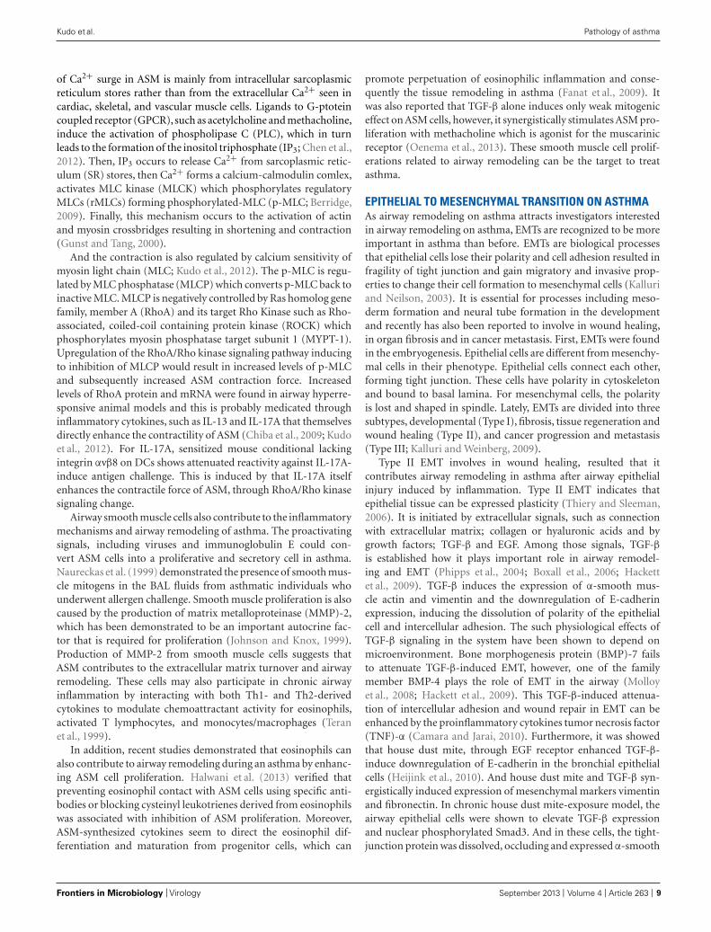
Like other muscles, ASM is also provoked to contract withintracellular calcium ions (Ca2+), which comes from the extra-cellular environment through voltage-dependent calcium channelor from the sarcoplasmic reticulum stores (Figure 1). The source
FIGURE 1 | Regulation of ASM contractility. ASM contraction isinduced by calcium, regulated two different pathways. First, ASM isevoked by intracellular calcium influx from SR depending on GPCRstimulation or from the extracellular environment through voltage-dependent calcium channel. Second, smooth muscle can be inducedcalcium sensitivity by RhoA/Rho kinase pathway. RhoA activates
Rho-kinase which phosphorylates MLCP. pMLC phosphatase failsto dephosphorylate MLC. KCl, potassium chloride; Ach, acetylcholine;5-HT, 5-hydroxytryptamine (serotonin); PIP, phosphatidylinositol 4-phos-phate; PIP2, phosphatidylinositol 4,5-bisphosphate; PIP5K, 1-phos-phatidylinositol-4-phosphate 5-kinase; DG, diacylglycerol; IP3, inositol1,4,5-trisphosphate.
www.frontiersin.org September 2013 | Volume 4 | Article 263 | 8
“fmicb-04-00263” — 2013/9/7 — 14:50 — page 4 — #4
Kudo et al. Pathology of asthma
of Ca2+ surge in ASM is mainly from intracellular sarcoplasmicreticulum stores rather than from the extracellular Ca2+ seen incardiac, skeletal, and vascular muscle cells. Ligands to G-ptoteincoupled receptor (GPCR), such as acetylcholine and methacholine,induce the activation of phospholipase C (PLC), which in turnleads to the formation of the inositol triphosphate (IP3; Chen et al.,2012). Then, IP3 occurs to release Ca2+ from sarcoplasmic retic-ulum (SR) stores, then Ca2+ forms a calcium-calmodulin comlex,activates MLC kinase (MLCK) which phosphorylates regulatoryMLCs (rMLCs) forming phosphorylated-MLC (p-MLC; Berridge,2009). Finally, this mechanism occurs to the activation of actinand myosin crossbridges resulting in shortening and contraction(Gunst and Tang, 2000).
And the contraction is also regulated by calcium sensitivity ofmyosin light chain (MLC; Kudo et al., 2012). The p-MLC is regu-lated by MLC phosphatase (MLCP) which converts p-MLC back toinactive MLC. MLCP is negatively controlled by Ras homolog genefamily, member A (RhoA) and its target Rho Kinase such as Rho-associated, coiled-coil containing protein kinase (ROCK) whichphosphorylates myosin phosphatase target subunit 1 (MYPT-1).Upregulation of the RhoA/Rho kinase signaling pathway inducingto inhibition of MLCP would result in increased levels of p-MLCand subsequently increased ASM contraction force. Increasedlevels of RhoA protein and mRNA were found in airway hyperre-sponsive animal models and this is probably medicated throughinflammatory cytokines, such as IL-13 and IL-17A that themselvesdirectly enhance the contractility of ASM (Chiba et al., 2009; Kudoet al., 2012). For IL-17A, sensitized mouse conditional lackingintegrin αvβ8 on DCs shows attenuated reactivity against IL-17A-induce antigen challenge. This is induced by that IL-17A itselfenhances the contractile force of ASM, through RhoA/Rho kinasesignaling change.
Airway smooth muscle cells also contribute to the inflammatorymechanisms and airway remodeling of asthma. The proactivatingsignals, including viruses and immunoglobulin E could con-vert ASM cells into a proliferative and secretory cell in asthma.Naureckas et al. (1999) demonstrated the presence of smooth mus-cle mitogens in the BAL fluids from asthmatic individuals whounderwent allergen challenge. Smooth muscle proliferation is alsocaused by the production of matrix metalloproteinase (MMP)-2,which has been demonstrated to be an important autocrine fac-tor that is required for proliferation (Johnson and Knox, 1999).Production of MMP-2 from smooth muscle cells suggests thatASM contributes to the extracellular matrix turnover and airwayremodeling. These cells may also participate in chronic airwayinflammation by interacting with both Th1- and Th2-derivedcytokines to modulate chemoattractant activity for eosinophils,activated T lymphocytes, and monocytes/macrophages (Teranet al., 1999).
In addition, recent studies demonstrated that eosinophils canalso contribute to airway remodeling during an asthma by enhanc-ing ASM cell proliferation. Halwani et al. (2013) verified thatpreventing eosinophil contact with ASM cells using specific anti-bodies or blocking cysteinyl leukotrienes derived from eosinophilswas associated with inhibition of ASM proliferation. Moreover,ASM-synthesized cytokines seem to direct the eosinophil dif-ferentiation and maturation from progenitor cells, which can
promote perpetuation of eosinophilic inflammation and conse-quently the tissue remodeling in asthma (Fanat et al., 2009). Itwas also reported that TGF-β alone induces only weak mitogeniceffect on ASM cells, however, it synergistically stimulates ASM pro-liferation with methacholine which is agonist for the muscarinicreceptor (Oenema et al., 2013). These smooth muscle cell prolif-erations related to airway remodeling can be the target to treatasthma.
EPITHELIAL TO MESENCHYMAL TRANSITION ON ASTHMAAs airway remodeling on asthma attracts investigators interestedin airway remodeling on asthma, EMTs are recognized to be moreimportant in asthma than before. EMTs are biological processesthat epithelial cells lose their polarity and cell adhesion resulted infragility of tight junction and gain migratory and invasive prop-erties to change their cell formation to mesenchymal cells (Kalluriand Neilson, 2003). It is essential for processes including meso-derm formation and neural tube formation in the developmentand recently has also been reported to involve in wound healing,in organ fibrosis and in cancer metastasis. First, EMTs were foundin the embryogenesis. Epithelial cells are different from mesenchy-mal cells in their phenotype. Epithelial cells connect each other,forming tight junction. These cells have polarity in cytoskeletonand bound to basal lamina. For mesenchymal cells, the polarityis lost and shaped in spindle. Lately, EMTs are divided into threesubtypes, developmental (Type I), fibrosis, tissue regeneration andwound healing (Type II), and cancer progression and metastasis(Type III; Kalluri and Weinberg, 2009).
Type II EMT involves in wound healing, resulted that itcontributes airway remodeling in asthma after airway epithelialinjury induced by inflammation. Type II EMT indicates thatepithelial tissue can be expressed plasticity (Thiery and Sleeman,2006). It is initiated by extracellular signals, such as connectionwith extracellular matrix; collagen or hyaluronic acids and bygrowth factors; TGF-β and EGF. Among those signals, TGF-βis established how it plays important role in airway remodel-ing and EMT (Phipps et al., 2004; Boxall et al., 2006; Hackettet al., 2009). TGF-β induces the expression of α-smooth mus-cle actin and vimentin and the downregulation of E-cadherinexpression, inducing the dissolution of polarity of the epithelialcell and intercellular adhesion. The such physiological effects ofTGF-β signaling in the system have been shown to depend onmicroenvironment. Bone morphogenesis protein (BMP)-7 failsto attenuate TGF-β-induced EMT, however, one of the familymember BMP-4 plays the role of EMT in the airway (Molloyet al., 2008; Hackett et al., 2009). This TGF-β-induced attenua-tion of intercellular adhesion and wound repair in EMT can beenhanced by the proinflammatory cytokines tumor necrosis factor(TNF)-α (Camara and Jarai, 2010). Furthermore, it was showedthat house dust mite, through EGF receptor enhanced TGF-β-induce downregulation of E-cadherin in the bronchial epithelialcells (Heijink et al., 2010). And house dust mite and TGF-β syn-ergistically induced expression of mesenchymal markers vimentinand fibronectin. In chronic house dust mite-exposure model, theairway epithelial cells were shown to elevate TGF-β expressionand nuclear phosphorylated Smad3. And in these cells, the tight-junction protein was dissolved, occluding and expressed α-smooth
Frontiers in Microbiology | Virology September 2013 | Volume 4 | Article 263 | 9

“fmicb-04-00263” — 2013/9/7 — 14:50 — page 5 — #5
Kudo et al. Pathology of asthma
muscle actin and collagen (Johnson et al., 2011). Inhaled aller-gens might modify EMT, cooperating with cytokines which alsopromote asthma.
MAST CELLS AND EOSINOPHILSMact cells can induce the activation of mesenchymal cells (Hol-gate, 2000). The serine protease, tryptase which is released fromdegranulating mast cells is a potent stimulant of fibroblast andsmooth muscle cell proliferation, and is capable of stimulatingsynthesis of type I collagen by human fibroblasts. A major mecha-nism involved in the regulation of fibroblast proliferation appearsto be cleavage and activation of protease activated receptor-2 onfibroblasts (Akers et al., 2000). Mast cells may also influence thedevelopment of airway remodeling in asthma by releasing largeamounts of plasminogen activator inhibitor type1. Moreover, Sug-imoto et al. (2012) have shown that other mast cell proteasesregulate airway hyperreactivity. Mice lacking αvβ6 integrin areprotected from exaggerated airway narrowing. Mast cell pro-teases are differentially expressed, in mouse mast cell protease 1(mMCP-1) induced by allergen challenge in wild-type (WT) miceand mMCP-4 increased at baseline in β6-deficient mice. MCPsfrom intraepithelial mast cell and their proteolytic substrates couldbe regulate airway hyperreactivity.
Eosinophils are circulating granulocytes and at relatively lowlevels in the bloodstream, upto 3% of white blood cells. These arethe major cell types that can be recruited to sites of inflamma-tory responses (Huang et al., 2009; Isobe et al., 2012; Uhm et al.,2012). The function of eosinophils in asthma is related to theirrelease of toxic granule proteins, reactive oxygen species (ROS),cytokines, and lipid mediators (Liu et al., 2006). The recruit ofeosinophils into the epithelium and eosinophilic inflammationis involved in the pathogenesis of asthma. The proinflamma-tory mediators derived by eosinophil are major contributors toinflammation in asthma, including airway epithelial cell dam-age and desquamation, airway dysfunction of cholinergic nervereceptors, AHR, mucus hypersecretion, and airway remodeling,characterized by fibrosis and collagen deposition (Kay, 2005; Wattet al., 2005; Kanda et al., 2009; Walsh, 2010). Eosinophils are likelyto contribute to airway remodeling with release of eosinophil-derived mediators such as TGF-β, secretion of cationic proteins,and cytokines, as well as having interactions with mast cell andepithelial cells. Those factors can directly activate epithelium andmesenchymal cells (Venge, 2010). Moreover, recent data demon-strated that eosinophils can also contribute to airway remodelingwith ASM cell proliferation.
EXTRACELLULAR MATRIXThe airways of asthmatic patients showed excess accumulationof extracellular matrix components, particularly collagen, in thesubepithelial connective tissue and adventitia of the airway wall(Kuwano et al., 1993; Gillis and Lutchen, 1999). The cellular inter-actions in mast cells and fibroblasts through protease activatedreceptor-2 may contribute an abnormal mesenchymal cell prolif-eration, and may account for the increased number of fibroblastsand myofibroblasts that are found in the airways of asthmaticsubjects. Fibroblasts retain the capacity for growth and regen-eration, and may evolve into various cell types, including smooth
muscle cells that subsequently become myofibroblasts. Myofibrob-lasts can contribute to tissue remodeling by releasing extracellularmatrix components such as elastin, fibronectin and laminin (Vig-nola et al., 2000). It was seen that the numbers of myofibroblastsin the airway of asthmatic subjects increased and their num-ber appeared to correlate with the size of the basement reticularmembrane (Holgate et al., 2000). Smooth muscle cells also havethe potential to alter the composition of the extracellular matrixenvironment. The reticular basement membrane thickening is acharacteristic typical feature of the asthmatic airways. It appearsto consist of a plexiform deposition of immunoglobulins, collagentypes I and III, tenascin and fibronectin (Jeffery et al., 2000), butnot of laminin.
Remodeling processes of the extracellular matrix are less knownthan the thickening of the lamina reticularis. Most asthmatic sub-jects present with an abnormal superficial elastic fiber network,with fragmented fibers (Bousquet et al., 2000). In the deeper layerof elastic fibers is also abnormal, the fibers often being often patchy,tangled, and thickened. Some studies using transmission electronmicroscopy have shown that an elastolytic process occurs in asth-matic patients, and in some patients disruption of fibers has beenobserved. In the case of fatal asthma, fragmentation of elasticfiber has also been found in central airways, and was associatedwith marked elastolysis (Mauad et al., 1999). These bundles areseen to be hypertrophied as a result of an increased amount ofcollagen and myofibroblast matrix deposition occurring duringexaggerated elastic fiber deposition (Carroll et al., 1997). Loss oflung elastic recoil force has been shown in adults with persistentasthma and irreversible expiratory airflow obstruction. Persistentasthmatic patients have severe abnormal flow-volume curves inexpiration at both high and low lung volumes, and hyperinfla-tion can be seen by residual volume, at forced residual capacityand total lung capacity (Gelb and Zamel, 2000). The increasedelastolysis is part of a more complex process that regulates thesize of a submucosal network formed by elastic fibers dispersedin a collagen and myofibroblast matrix (Chiappara et al., 2001).These features induce changes in airway, as demonstrated by air-way compliance, particularly in those patients who are sufferingfrom asthma for long period, supporting the concept that chronicinflammation and remodeling of the airway wall may result instiffer dynamic elastic properties of the asthmatic airway (Brackelet al., 2000). Furthermore, disruption of elastic fibers may con-tribute to a reduction in the preload and afterload for smoothmuscle contraction. Though it is difficult to associate aspects ofremodeling with disease severity or degree of airways obstructionand hyperresponsiveness (Mauad et al., 2007), some investigatorsindicated that smooth muscle remodeling is related to the severityof asthma (James et al., 2009). It has shown that the clinical expres-sion of asthma (Brightling et al., 2002), AHR (Siddiqui et al., 2008)and impaired airway relaxation (Slats et al., 2007) are associatedwith mast cell counts in the ASM layer in asthma. The depositionof extracellular matrix inside and outside the smooth muscle layerin asthma also seems to be related to its clinical severity and isaltered as compared to healthy controls (Araujo et al., 2008; Kla-gas et al., 2009). Yick et al. (2012) have shown that extracellularmatrix in ASM was related to the dynamics of airway function inasthma.
www.frontiersin.org September 2013 | Volume 4 | Article 263 | 10

“fmicb-04-00263” — 2013/9/7 — 14:50 — page 6 — #6
Kudo et al. Pathology of asthma
IMMUNE RESPONSEALLERGIC SENSITIZATIONRegarding to the immune system against allergy, it seems thathygiene hypothesis would provide the reason why the number ofthe patients with asthma is increasing, in relation with urban-ization. The hypothesis is that the Th1 cells polarized responseis not induced early in life leaving the body more susceptible todeveloping Th2 induced disease (Strachan, 2000). First, Strachan(1989) mentioned that the hypothesis was proposed to explainthe observation that hay fever and eczema were less common inchildren from larger families, which were presumably exposed tomore infectious agents through their siblings, than in childrenfrom small families, especially without siblings. Many bacteria andviruses derive a Th1-mediated immune response, which down-regulates Th2 responses. The urban-rural gradient in prevalencehas been demonstrated most strongly in children who grew up inenvironments with a wide range of microbial exposures, who areprotected from childhood asthma and atopy (the predispositionto develop IgE against common environmental allergens) in pro-portion to their level of exposure to bacterial and fungal microbes(Ege et al., 2011).
In association with the airway epithelium and underlingmucosa is a specialized population of antigen-presenting cells(APCs) called DCs (Holgate, 2012). As allergen sensitization, DCstake up the allergens and present small peptide from them. DCsexpress receptors of the innate immune system and process aller-gens into small peptides and then present them through the majorhistocompatibility complexes, MHC class I and MHC class II forrecognition by T cell receptors. In allergic individuals, it is pro-moted by interaction of the allergen with IgE attached to FcεRI,the high-affinity receptor for IgE (Sallmann et al., 2011). Whenindividual is born, there is no DCs in the airway. Damage to andactivation of the respiratory epithelium are the major stimuli thatinitiate the ingression of immature DCs from the bone marrow(McWilliam et al., 1994) and cause the release of C–C chemokineswhich direct DCs migration toward the epithelium and under-lying mucosa (Hammad et al., 2010). GM-CSF, which is releasedfrom epithelial cells and immune cells in the presence of IL-4 andTNF-α, leads to DCs maturation to a fully competent as APCs.During initial allergen entering to airways to sensitize, Th2 lym-phocyte differentiation from naïve T cells requires IL-4 release.The cellular source of the IL-4 is still unclear. There are somehypotheses to explain that (Holgate, 2012). Polarization to Th2cells subtype is also under epigenetic regulation. From the studywith mouse, microRNA-21 has been shown to exert a pivotal rolein setting a balance between Th1 and Th2 responses. It worksthrough binding the promoter of the gene encoding IL-12 p35and inhibiting its activation in favor of a Th2 profile. Conversely,reduced microRNA levels lead DCs to produce more IL-12, andallergen-stimulated T cells to produce more interferon-γ (IFN-γ)and less IL-4, enhancing Th1 delayed-type hypersensitivity (Luet al., 2011).
DENDRITIC CELL ACTIVATIONAs described above, DCs present small peptide from antigensthrough MHC class I and II/ T cell receptors. Once sensi-tized, T cells drive the allergic response in progress through
interactions with DCs (Veres et al., 2011). DCs spread their pro-cesses into the lumen between airway epithelial cells and candetect allergen by forming tight junctions, keeping the epithelialbarrier (Blank et al., 2011). In mouse, two distinct DC sub-sets have been described in accordance with their expressionof the CD11c as myeloid [conventional DCs (cDCs), CD11c+]or plasmacytoid DCs (pDCs, CD11c−; Lambrecht and Ham-mad, 2009). Similarly, human DCs are subdivided into CD11c−pDCs and CD11c+ myeloid DCs. Induced sputum from asth-matic airways and peripheral blood contain increased numbersof both pDCs and cDCs, which further increase in numberupon allergen challenge (Dua et al., 2010). Proteolytic activi-ties of allergens initiate to mature DCs. In a few hours aftercontact with allergen, pattern-recognition receptors activation,such as Toll-like receptors (TLRs) on DCs augments their hom-ing capacity by upregulating chemokine receptors. It is cDCsubtypes that are predominantly responsible for antigen presen-tation. Mature DCs shape an immunological synapse with theallergen-specific T lymphocytes to initiate a Th response (Hol-gate, 2012). Whereas some of the Th cells make their way tothe B-cell follicle to facilitate immunoglobulin class switchingfrom IgM to IgE, others move back to the airway mucosa toelicit the classical Th2 response through the secretion of theproallergic cytokines. Pattern-recognition receptors have a cru-cial adjuvant role in directing allergen sensitization. TLRs are keycomponents of the innate immune system that mediate recog-nition and response to pathogen-associated molecular patterns(PAMPs) in the form of microbial, fungal and viral products andtheir ligands, including endotoxin which is recognized by TLR4,lipoproteins (TLR2 and TLR6), viral double- and single-strandedRNA (TLR3 and TLR7/8) and bacterial CpG-containing DNA(TLR9) (Akira et al., 2006). Other pattern recognition receptorsrespond to endogenously generated damage-associated molecu-lar pattern molecules (DAMPs) produced during tissue damage.Inflammatory DCs have been suggested to be necessary and suf-ficient for the development of Th2 immunity to house dust miteallergen when the first exposure occurs by inhalation. For inhaledallergens, it is proposed that DCs amplify the Th2 immunitythrough basophiles and, in part, influenced by innate signalingthrough TLR4 and C-type lectin signaling on epithelial cells andDCs (Trompette et al., 2009). A cooperation of airway epitheliumand DCs controls asthma development Th2 activation requiresDCs-mediated antigen-presentation. Then, allergic sensitizationfails to develop in the absence of DCs (Hammad et al., 2010),while DCs remain inactive in the absence of TLR ligation (Per-ros et al., 2009). That is, TLRs activation on epithelial cellsenhances DCs motility and antigen sampling through the produc-tion of Th2-promoting chemokines and cytokines (IL-25, IL-33,GM-CSF).
VIRAL INFECTION TO PREDISPOSITIONThe fact that early-in-life sensitization to multiple allergens car-ries the greatest risk for developing asthma (Simpson et al., 2010)brings the question of what factors result in a predisposition tothis phenotype. Although infection with rhinovirus is the majorcause of acute exacerbation, in those genetically at risk of asthma,rhinovirus-induced wheezing in the first three years in the life is
Frontiers in Microbiology | Virology September 2013 | Volume 4 | Article 263 | 11

“fmicb-04-00263” — 2013/9/7 — 14:50 — page 7 — #7
Kudo et al. Pathology of asthma
also the greatest risk factor for developing asthma at 6 years ofage (Jackson et al., 2008). Impaired TLR3-mediated IFN-β and-λ production by asthmatic epithelial cells would make suscepti-ble to both viral infection and allergic sensitization (Wark et al.,2005; Contoli et al., 2006; Bosco et al., 2010; Jartti and Korppi,2011). Reduced primary IFN production by lower-airway epithe-lial cells enables some viruses to replicate, leading to cytotoxic cell,release of inflammatory products and enhanced viral shedding.Such events provide a strong stimulus for recruitment of immatureDCs and their priming for allergen sensitization (McWilliam et al.,1994, 1996). When asthmatic epithelial cells are received to dam-age by rhinovirus infection, the cells generate increased amountsof the pro-Th2 cytokine thymic stromal lympoietin (Uller et al.,2010), which stimulates DCs and increases allergic inflamma-tion, whereas exogenous IFN-b applied to asthmatic epitheliumexerts anti-Th2 as well as antiviral properties (Cakebread et al.,2011).
CELLULAR IMMUNITYAsthma is classically considered Th2 disease, with increased IgEand eosinophilic inflammation caused by increased levels of Th2-type cytokines. However, this paradigm has been challengedbecause of the realization that strategies designed to suppressTh2 function are not effective for all patients. The clinical phe-notype of asthma is notoriously heterogeneous. It is shown thatcellular immune process in the asthmatic airways in Figure 2.Th2 cells activation requires antigen-presentation by DCs. DCsplay a role both in the initiation and maintenance of allergic
airway inflammation and asthma, and control many aspects ofthe disease, including bronchial hyperresponsiveness and gobletcell metaplasia, by controlling the recruitment and activation ofTh2 cells (Lambrecht and Hammad, 2009; Schuijs et al., 2013).Researches in both mouse and human, mentioned the expres-sion of Th2-type cytokines, such as IL-4, IL-5, and IL-13, inthe allergic lung. Experimental asthma models indicate that thesecytokines, IL-13 in particular, are critical in driving key patho-logic features of the allergic response. Moreover, Th2 blockadeis very effective in suppressing these features of allergic dis-ease in mice (Finkelman et al., 2010). The classical asthmaticphenotype is one of eosinophilia concomitant with high IgElevels. However, a proportion of patients are not atopic anddo not have eosinophilic inflammation. In fact, it is estimatedthat as many as 50% of adult patients are encompassed bythis non-atopic, non-eosinophilic, non-IgE-dependent subgroup(Lloyd and Saglani, 2013). Molecular therapy data support anoverall Th2 association with phenotypes, such that they mightsatisfy a definition of Th2-associated asthma. However, eventhese distinctions are too simple, especially when disease sever-ity is considered. Although children with severe asthma haveeosinophilic inflammation, high-dose steroids effectively sup-press Th2-type cytokines, such as IL-13 and IL-5, but symptomsremain with persistent eosinophilia (Bossley et al., 2012), thusraising the importance of identifying other less steroidsensitive,non-Th2 mediators driving disease. Then, it is apparent thatasthma can no longer be considered simply a Th2-mediateddisease.
FIGURE 2 |T cell immune response in the asthmatic airways. NaïveT cell is received allergen presentation by DCs. The pathway begins withthe development of Th2 cells and their production of the cytokines IL-4,IL-5, and IL-13. These cytokines stimulate allergic and eosinophilicinflammation as well as epithelial and smooth-muscle changes thatcontribute to asthma pathobiology. Th9 cell can be induced and stimulate
mast cells by IL-9. Naïve T cell is also differentiated to Th1 or Th17cells depending on the existence of cytokines in the microenvironment.Th1 cell and Th17 cell stimulate and induce neutrophilicinflammation. EMT, epithelial-mesenchymal-myofibroblast transition;MMP, matrix metalloproteinase; MBP, major basic protein;LT, leukotriene.
www.frontiersin.org September 2013 | Volume 4 | Article 263 | 12

“fmicb-04-00263” — 2013/9/7 — 14:50 — page 8 — #8
Kudo et al. Pathology of asthma
FIGURE 3 |T helper cell subsets and cytokine profiles. Th1, Th2 and Th17 cells are a separate lineage of CD4+ T cells, distinct from other T cell subsets.Every specific T helper cells produce its specific cytokines (Lazarevic and Glimcher, 2011). T-bet, T-box expressed in T cells; FoxP3, forkhead box P3; ROR,retinoid-related orphan receptor.
Effector CD4 cells expressing IL-17A, IL-17F were firstdescribed in 2005 (Harrington et al., 2005; Park et al., 2005) andwere thought to represent a distinct T-cell lineage that promotedthe first revision of the Th1/Th2 paradigm of immunity. Differen-tiation of naive effector T cells in the presence of IL-6 and TGF-β,leading to the expression of the transcription factor RORγt, resultsin IL-17 expression through the transcription factors Smad 2/3,signal transducer and activation of transcription (STAT) 3, andnuclear factor κB. Naïve T cells can differentiate several cell typesand have specific immune response through the release of cell-type specific cytokines (Figure 3). Th17 cells have a role inregulating both neutrophilic and macrophage inflammation inautoimmune disease, and more recently they have been suggestedto be involved in asthma and corticosteroid insensitivity (Nem-brini et al., 2009). Conversely, their differentiation is restrictedby both Th1 and Th2 cytokines including IFN-γ, IL-4, and IL-13 (Park et al., 2005). Specifically, the induction of CXCL8, apotent neutrophil chemokine whose expression is elevated in air-way secretions in severe asthma, has directly implicated Th17 cellsin neutrophilic airway inflammation. IL-17A itself, but not IL-17For IL-22, enhances the contractile force of ASM. Sensitized micelacking the integrin αvβ8 on DCs show reduced activation of thisIL-17A-linked pathway with antigen challenge. This reduction insmooth muscle contraction in the airways is reversible by IL-17A,indicating involvement of this cytokine on allergen-induced AHRby acting directly on ASM (Kudo et al., 2012). Allergic inducesa strong Th17 response in association with airway neutrophiliaand hyperresponsiveness, and this response is abrogated in IL-17Fknockout mice (Yang et al., 2008). However, although a good
case can be made for IL-17A and IL-17F in mouse models ofneutrophilic and corticosteroid-refractory lung responses to aller-gens, evidence for IL-17 involvement in human asthma is lessrobust, despite some emerging genetic evidence and a potentialrole for IL-17A and IL-17F in moderate-to-severe disease (Chakiret al., 2003; Doe et al., 2010). In humans, a subset of Th2 mem-ory and effector cells has been recognized expressing both GATA3and RORγt and, as a consequence, producing both Th17 and Th2cytokines (Cosmi et al., 2010). Studies have reported that the num-ber of circulating Th17 cells as well as plasma concentrations ofIL-17 and IL-22 increase in proportion to disease severity. In abronchial biopsy in asthma vs. normal controls, there was no cor-relation between IL-17A or IL-17F expression and the extent ofneutrophilia, nor any link to asthma severity (Doe et al., 2010).The contribution of Th17 cells in human asthma has not beenestablished enough. It is required to clear association of Th17 cellsand subphenotype in human asthma.
CYTOKINE TARGETSIL-4/IL-13The key cytokines involved in Th2-type immunoreaction are thoseencoded in the IL-4 gene cluster on chromosome 5q31, contain-ing the genes encoding IL-3, IL-4, IL-5, IL-9, IL-13, and GM-CSF(Bowen et al., 2008). The fact that the Th2 pathway is crucial toasthma pathophysiology has been the driving force for a rangeof biologics targeting the specific cytokines. The signals of Th2-cell-associated cytokines, IL-4 and IL-13, transmit through theIL-4Ra/IL-13Ra1 complex. IL-4 promotes B-cell isotype switch-ing, the upregulation of adhesion molecules, eotaxin production,
Frontiers in Microbiology | Virology September 2013 | Volume 4 | Article 263 | 13

“fmicb-04-00263” — 2013/9/7 — 14:50 — page 9 — #9
Kudo et al. Pathology of asthma
and the development of AHR and goblet cell metaplasia. In animalmodel, IL-4 deficient mice were shown to be protected from devel-oping asthma (Brusselle et al., 1994). IL-13 can have most of thesefunctions (Wills-Karp et al., 1998; Webb et al., 2000). Furthermore,those cytokines have the potential to induce TSLP, GM-CSF, andCCL20 production by the airway epithelium (Reibman et al., 2003;Kato et al., 2007). Furthermore, IL-13 was shown to have directeffect to enhance ASM, upregulating RhoA protein which stimu-lates Rho-kinase inducing calcium sensitivity (Chiba et al., 2009).Therefore, a good example is the IL-4 and IL-13 pathway for anticytokine treatment against asthma.
Given the clear evidence for IL-4 and/or IL-13 in mouse modelsof disease were launched and a humanized anti-IL-4 neutralizingantibody (pascolizumab) was introduced and showed promisingresults in human-derived cell lines and monkeys (Hart et al., 2002).However, IL-4-specific antagonists used in clinical trials have failed(Wenzel et al., 2007). More recently, a human monoclonal anti-IL-4Ra antibody (AMG317) has been developed but did not showclinical efficacy (Corren et al., 2010). For IL-13, several neutral-izing antibodies have been developed, but trials are still in theirinfancy. The latter IL-13-antibody (CAT-354) has recently beenshown to be safe for use in humans in a phase I clinical trialbut its real clinical efficacy remains to be proven (Singh et al.,2010). Attempts to validate importance of IL-13 in human asthmarevealed that only 50% of individuals with asthma had elevatedIL-13 levels in sputum, irrespective of the severity of the disease(Berry et al., 2004). And Woodruff et al. (2009) have also shownthat only 50% of patients express IL-13-responsive genes in theairway epithelial cells, and this is linked to a strong Th2 responsein bronchial biopsies, as opposed to in other asthmatics, whoseIL-13-responsive gene expression was almost same level from thatof normal subjects. Th2-high subjects had greater expression ofIL-13 in bronchial biopsies along with greater AHR and higherserum IgE, blood and airway eosinophilia. It was suggested thatone IL-13 biomarker was periostin (Woodruff et al., 2009). In arecently published trial, the monoclonal antibody (mAb) to IL-13, lebrikizumab, when administered to patients with chronicmoderate-to-severe asthma for 12 weeks, significantly increasedbaseline spirometry (5.5%). This result was enhanced in those withelevated serum periostin (high periostin 8.2% vs. low periostin1.2%; Corren et al., 2011).
IL-5IL-5 is a key cytokine crucial to eosinophil growth, matura-tion, activation, and survival whose blockade in various animalmodels has a strong effect on acute and more sustained pul-monary eosinophilia and attendant changes in lung function. It ismainly produced by Th2-lymphocytes, mast cells and eosinophils.Interestingly, IL-5 regulates its own receptor expression duringeosinophil ontogeny consisting of an IL-5- specific receptor α-chain, and common β-chain. Because of its restriction to theeosinophil/basophil lineage in humans, IL-5 therapy may atten-uate key characteristics of allergic airway inflammation, suchas airway eosinophilia, airway remodeling, and AHR, withoutaffecting the function of other immune cells (Trifilieff et al.,2001; Flood-Page et al., 2003; Humbles et al., 2004). It has alsobeen implicated in the induction of AHR, as IL-5 inhalation by
asthmatic patients induces eosinophil influx and AHR (Leckieet al., 2000). However, despite markedly reducing both circulatingand sputum eosinophilia, two humanized mAbs, mepolizumaband reslizumab, when administered to patients with moderate-to-severe asthma, had no overall effect on any asthma outcomemeasures. Nonetheless, the studies of mepolizumab for patientswith severe asthma requiring oral corticosteroids and persistentsputum eosinophilia showed a good clinical response (Haldaret al., 2009; Nair et al., 2009), as also found in Churg-Strauss andother hypereosinophilic syndromes (Abonia and Putnam, 2011).Similar results have also been obtained with reslizumab (Castroet al., 2011; Spergel et al., 2012). Efficacy of mepolizumab has alsobeen described in severe eosinophilic nasal polyposis in proportionto nasal lavage IL-5 levels (Gevaert et al., 2006). A further devel-opment of this approach has been the introduction of a highlyactive mAb targeting IL-5Rα (benralizumab), which has beendefucosylated to enhance its antibody-dependent cell-mediatedcytotoxicity potential (Kolbeck et al., 2010). The studies demon-strate that anti-IL-5 therapy is effective in reducing exacerbationfrequency in severe asthma, with highest efficacy in subgroups ofpatients where eosinophils have a pathogenic role. A phase 1 studyin mild asthma has shown a strong dose-related reduction of circu-lating eosinophils lasting 8–12 weeks after a single injection (Busseet al., 2010). It seems, however, that for the majority of asthmaticpatients the anti-IL-5 treatment will need to be administered incombination with other therapies that suppress asthma featuresthrough other mechanisms. Results of clinical trials targeting theIL5Rα subunit to obtain long-term depletion of eosinophils andbasophils are eagerly awaited.
IL-17/IL-22The rapid emergence and characterization of the Th17 lineage(CD4 T cells producing IL-17 family; IL-17A, IL-17F, IL-22)refines the existing model and provides a more unified perspec-tive of allergic inflammation by CD4+ T cell subsets. Interestingly,some asthmatic individuals, especially those poorly responding tosteroid treatment, show airway infiltrations primarily composedof neutrophils. These cells are probably recruited to the airways byIL-17-producing cells that also produce IL-4 (Wang et al., 2010a).In mice, allergic sensitization followed by challenge of the air-ways induces a strong Th17 response and IL-17 controls bronchialhyperresponsiveness and airway remodeling, and some of theseeffects are mediated directly on bronchial smooth muscle cells(Pichavant et al., 2008; Wang et al., 2010b; Bellini et al., 2012; Kudoet al., 2012). Moreover, IL-17 can also induce steroid insensitivityin bronchial epithelial cells (Zijlstra et al., 2012). IL-22 can alsobe produced by Th17 cells. In mouse asthma models, IL-22 seemsto exert a dual role. Indeed, IL-22 blockade in Th2 sensitizationdramatically reduced eosinophil recruitment, Th2 cytokine andchemokine production, AHR, and mucus production. In contrast,IL-22 inhibition in allergen challenge induced lung inflammationand increased Th2 cytokine production. On epithelial cells, IL-22has the potential to induce the production of antimicrobial pep-tides and to promote epithelial repair as well as suppressing theproduction of proinflammatory chemokines and cytokines (Pen-nino et al., 2012). Despite these studies, our knowledge of IL-22 inasthma pathophysiology is still limited.
www.frontiersin.org September 2013 | Volume 4 | Article 263 | 14

“fmicb-04-00263” — 2013/9/7 — 14:50 — page 10 — #10
Kudo et al. Pathology of asthma
Table 1 | Monoclonal antibodies against IL-17 pathway clinical trials.
mAbs Description Phase Indications
Brodalumab Full human IgG2/anti IL-17RA II Asthma, Ps, PsA, RA
Secukinumab Full human IgG1K/ anti IL-17A III Ps, PsA, RA, AS
II veitis
Ixekizumab Humanized, hinge-modified IgG4/anti IL-17A III Ps
II RA
I PsA
Ustekinumab Full human IgG1/anti p40 of IL-12/23 III Crohn’s, PsA
II AS, sarcoidosis, cirrhosis
Approved Ps
CNTO 1959 Full human mAb/anti p19 of IL-23 II PsA
MK-3222 Humanized mAb/ anti p19 of IL-23 II Ps
AMG 139 Full human mAb/ anti IL-23 I Crohn’s, Ps
RG4934 Humanized mAb/ anti IL-17A I
NI 1401 Full human IgG1 mAb/ IL-17A/F I
SCH 900117 Humanized mAb/ IL-17A I
Ps: psoriasis; PsA: psoriatic arthritis, RA: rheumatoid arthritis; AS: ankylosing; spondylitis.
IL-17A has been considered as one of most important playerin asthma, however, clinical attempts for anti-IL-17A therapy toasthma has just begun (Table 1). Any data in anti-IL-17A trialsfor asthma are not available so far. Some clinical trials targetedat IL-17A have conducted and substantiated importance of IL-17A in autoimmune disorders. Phase II data on secukinumab,ixekizumab, and brodalumab in psoriasis indicate rapid and pro-nounced effects on measures of disease activity (Hueber et al.,2010). Early clinical trials in psoriatic arthritis, rheumatoid arthri-tis, and ankylosing spondylitis also support the therapeutic utilityof IL-17A inhibition.
In addition, whereas secukinumab and ixekizumab selectivelytarget and neutralize IL-17A, brodalumab binds the IL-17RA chainof the heteromeric IL-17 receptor, which is shared with multiplemembers of the IL-17 cytokine family and is therefore expectedto inhibit the biological activity of IL-17A and IL-17F as well asIL-17C (Ramirez-Carrozzi et al., 2011), IL-17E (IL-25) and poten-tially other not yet discovered IL-17 family members that utilizeIL-17RA (Papp et al., 2012). Considering with these data from clin-ical trials for autoimmune disease, this hypothetical advantage forIL-17A inhibitors against asthma can be expected to have clinicalbenefits. We have to wait that data from asthma studies becomesavailable.
IL-9Interleukin-9 produced from CD4+ T cell (Th9) has been iden-tified as a subset definite from the classical Th2 cells, requiringthe transcription factors IRF4, PU1, STAT6, Smad3, and Notchsignaling for development. The cells differentiate in response toIL-4 and TGF-β and are described to promote T cell proliferation,IgE and IgG production by B-cells, survival and maturation of
eosinophils, increasing the number of mast cell (Veldhoen et al.,2008; Staudt et al., 2010; Kearley et al., 2011; Elyaman et al., 2012;Goswami et al., 2012). Studies in human have also shown thatIL-9 expression increased markedly in response to allergen chal-lenge (Erpenbeck et al., 2003) and IL-9 is highly expressed andlocalized to tissue lymphocytes during intestinal parasite infection(Faulkner et al., 1998) and to CD3+ cells in bronchial submucosaand BAL (Shimbara et al., 2000). In studies using IL-9 transgenicand knockout mice, direct IL-9 instillation into the lungs andblocking mAbs, it has been shown that IL-9 drives mucus produc-tion, both by a direct effect on airway epithelia (Bryce, 2011) andalso by interacting with IL-13 (Steenwinckel et al., 2007). Micewith IL-9 overexpression in lung have increased airway inflam-mation and AHR (Bisgaard et al., 2007; Gern, 2011). IL-9 is alsomade by ILC2s and boosts production of IL-5 and IL-13 (Rabi-novitch et al., 2005). Along with IL-4 and stem cell factor, IL-9 isalso a potent stimulus for mast-cell development (Kearley et al.,2011). As IL-9 has been implicated in both inflammatory andremodeling components in mouse models of allergic airway dis-ease, it seems an attractive therapeutic target. Currently, clinicaldata on anti-IL-9 therapeutics are modest and larger clinical tri-als are eagerly awaited to conclude whether this form of therapycan be used in the treatment of asthma (Shalev et al., 2011). Twofirst-in-human, open-label dose-escalation trials of a monoclonalantibody against IL-9, MEDI-528, in normal subjects and subjectswith mild asthma have been successfully completed, showing someevidence of efficacy (Parker et al., 2011).
TNF-αTumor necrosis factor α, a multifunctional cytokine that exertsa variety of effects, such as growth promotion, apoptosis,
Frontiers in Microbiology | Virology September 2013 | Volume 4 | Article 263 | 15

“fmicb-04-00263” — 2013/9/7 — 14:50 — page 11 — #11
Kudo et al. Pathology of asthma
angiogenesis, cytotoxicity, inflammation, and immunomodula-tion, has been implicated in several inflammatory conditions.This cytokine is not only produced predominantly by activatedmacrophages but also by other immune (lymphocytes, natu-ral killer cells, mast cells) as well as stromal (endothelial cells,fibroblasts, microglial cells) cells and presents in increased con-centrations in bronchoalveolar fluid from the airways of patientswith asthma (Broide et al., 1992). Some studies mentioned arelationship between TNF-α and severity of asthma.
The rates of death and complications are high among patientswith refractory asthma and account for a disproportionate amountof the health resource burden attributed to asthma (Serra-Batlleset al., 1998). The airway abnormality in severe asthma is differentfrom that in more mild asthma in having a more heteroge-neous pattern of inflammatory response (Wenzel et al., 1999),with greater involvement of neutrophilic inflammation and thedistal lung (Berry et al., 2005) and increased airway remodeling(Busse et al., 1999). Interest in the role of TNF-α in refractoryasthma has been increased by a study showing increased con-centrations of TNF-α in BAL from patients with more severeasthma and by an uncontrolled study showing that treatmentwith the recombinant soluble TNF-α receptor etanercept markedlyimproved AHR in patients with refractory asthma (Howarth et al.,2005). On the other hand, targeting TNF-α in severe asthma withgolimumab yields responders and non-responders (Wenzel et al.,2009). And administration with infliximab for severe asthma alsodoes responders and non-responders (Taillé et al., 2013). There-fore, controlled studies have shown controversial results and therisk-benefit profile of TNF-blocking agents is still debated (Cox,2009).
The studies suggest that anti-TNF-α agents might improvethe condition of a subgroup of patients severe steroid-dependentasthma, who have life-threatening exacerbations and complica-tions of long-term steroid therapy. In the studies, the identificationof more neutrophilic asthma that is less dependent upon Th2mechanisms and, as a consequence, less responsive to corticos-teroids might help identify a responsive target subpopulation.
Such patients have been shown to have high circulating TNF-αand CXCL-8 as biomarkers (Silvestri et al., 2006). A transcriptomicanalysis applied to induced sputum has identified a unique signa-ture with prominence of TNF-α and nuclear factor-κB pathways(Baines et al., 2010). This stratification of asthma into pathway-selective phenotypes is likely to be a key driver for future drugdevelopment, as is proving so successful for cancer treatments(Holgate, 2012).
CONCLUSIONBronchial asthma is a world-wide common disease and charac-terized by reversible airflow limitation, with non-specific AHRrelated to airway inflammation. Airway inflammation induces notonly asthmatic symptoms which are the reversible airway obstruc-tion and ASM contraction but also airway remodeling. Lately,the information for airway remodeling is increasing, the numberof myofibroblasts increases in the understructure of epithelium,the proximity of the smooth muscle layer and the lamina retic-ularis. And it is more understood what EMT is. EMT can playa important role in airway remodeling. These epithelial and mes-enchymal cells cause persistence of the inflammatory infiltrate andinduce histological changes in the airway wall, increasing thick-ness of the basement membrane, collagen deposition and smoothmuscle hypertrophy and hyperplasia. Subepithelial collagens causethickening and increasing density of the basement membrane.
Classically, asthma is considered as Th2 disease, relating toincreased IgE and eosinophilic inflammation in the airway. Recentresults have shown that not only Th2, but Th17 and Th9 cells sub-set also contributed the disease, releasing their specific cytokines.These different cytokine give different biological effect. These canbe targeted as an anti-cytokine treatment in asthma and somemonoclonal antibodies against specific cytokines or their recep-tors are available. The results of those clinical trials have said thattrials failed to control disease, despite it was clearly confirmed thatthose cytokines contributed the disease in animal model studies.It is required that more information for subphenotype of humanasthma and its mechanism in more detail.
REFERENCESAbonia, J. P., and Putnam, P. E. (2011).
Mepolizumab in eosinophilic disor-ders. Expert Rev. Clin. Immunol. 7,411–417. doi: 10.1586/eci.11.27
Aceves, S. S., and Broide, D. H. (2008).Airway fibrosis and angiogenesis dueto eosinophil trafficking in chronicasthma. Curr. Mol. Med. 8, 350–358.doi: 10.2174/156652408785161023
Akers, I. A., Parsons, M., Hill, M.R., Hollenberg, M. D., Sanjar, S.,Laurent, G. J., et al. (2000). Mastcell tryptase stimulates human lungfibroblast proliferation via protease-activated receptor-2. Am. J. Physiol.Lung Cell. Mol. Physiol. 278, L193–L201.
Akira, S., Uematsu, S., and Takeuchi,O. (2006). Pathogen recognition andinnate immunity. Cell 124, 783–801.doi: 10.1016/j.cell.2006.02.015
Alfvén, T., Braun-Fahrländer, C.,Brunekreef, B., von Mutius, E.,Riedler, J., Scheynius, A., et al.(2006). Allergic diseases and atopicsensitization in children relatedto farming and anthroposophiclifestyle-the PARSIFAL study. Allergy61, 414–421. doi: 10.1111/j.1398-9995.2005.00939.x
American Thoracic Society. (1987).Standards for the diagnosis and careof patients with chronic obstruc-tive pulmonary disease (COPD) andasthma. Am. J. Respir. Crit. CareMed. 136, 225–244. doi: 10.1164/ajr-ccm/136.1.225
Araujo, B. B., Dolhnikoff, M., Silva,L. F., Elliot, J., Lindeman, J. H.,Ferreira, D. S., et al. (2008). Extra-cellular matrix components and reg-ulators in the airway smooth mus-cle in asthma. Eur. Respir. J. 32,
61–69. doi: 10.1183/09031936.00147807
Awadh, N., Muller, N. L., Park, C. S.,Abboud, R. T., and FitzGerald, J.M. (1998). Airway wall thickness inpatients with near fatal asthma andcontrol groups: assessment with highresolution computed tomographicscanning. Thorax 53, 248–253. doi:10.1136/thx.53.4.248
Baines, K. J., Simpson, J. L., Bowden,N. A., Scott, R. J., and Gibson, P.G. (2010). Differential gene expres-sion and cytokine production fromneutrophils in asthma phenotypes.Eur. Respir. J. 35, 522–531. doi:10.1183/09031936.00027409
Bellini, A., Marini, M. A., Bianchetti,L., Barczyk, M., Schmidt, M., andMattoli, S. (2012). Interleukin (IL)-4,IL-13, and IL-17A differentially affectthe profibrotic and proinflammatory
functions of fibrocytes from asth-matic patients. Mucosal Immunol.5, 140–149. doi: 10.1038/mi.2011.60
Bento, A. M., and Hershenson, M. B.(1998). Airway remodeling: potentialcontributions of subepithelial fibrosisand airway smooth muscle hyper-trophy/hyperplasia to airway nar-rowing in asthma. Allergy AsthmaProc. 19, 353–358. doi: 10.2500/108854198778612672
Berridge, M. J. (2009). Inositoltrisphosphate and calcium signal-ing mechanisms. Biochim. Biophys.Acta 1793, 933–940. doi: 10.1016/j.bbamcr.2008.10.005
Berry, M. A., Parker, D., Neale, N.,Woodman, L., Morgan, A., Monk, P.,et al. (2004). Sputum and bronchialsubmucosal IL-13 expression inasthma and eosinophilic bronchitis.
www.frontiersin.org September 2013 | Volume 4 | Article 263 | 16

“fmicb-04-00263” — 2013/9/7 — 14:50 — page 12 — #12
Kudo et al. Pathology of asthma
J. Allergy Clin. Immunol. 114, 1106–1109. doi: 10.1016/j.jaci.2004.08.032
Berry, M. A., Hargadon, B., Morgan,A., Shelley, M., Richter, J., Shaw, D.,et al. (2005). Alveolar nitric oxide inadults with asthma: evidence of dis-tal lung inflammation in refractoryasthma. Eur. Respir. J. 25, 986–991.doi: 10.1183/09031936.05.00132404
Bisgaard, H., Hermansen, M. N., Buch-vald, F., Loland, L., Halkjaer, L. B.,Bønnelykke, K., et al. (2007). Child-hood asthma after bacterial coloniza-tion of the airway in neonates. N.Engl. J. Med. 357, 1487–1495. doi:10.1056/NEJMoa052632
Blank, F., Wehrli, M., Lehmann, A.,Baum, O., Gehr, P., von Garnier,C., et al. (2011). Macrophages anddendritic cells express tight junctionproteins and exchange particles in anin vitro model of the human airwaywall. Immunobiology 216, 86–95. doi:10.1016/j.imbio.2010.02.006
Bosco, A., Ehteshami, S., Stern, D. A.,and Martinez, F. D. (2010). Decreasedactivation of inflammatory networksduring acute asthma exacerbationsis associated with chronic airflowobstruction. Mucosal Immunol. 3,399–409 doi: 10.1038/mi.2010.13
Bossley, C. J., Fleming, L., Gupta, A.,Regamey, N., Frith, J., Oates, T.,et al. (2012). Pediatric severe asthmais characterized by eosinophilia andremodeling without TH2 cytokines.J. Allergy Clin. Immunol. 129, 974–982. doi: 10.1016/j.jaci.2012.01.059
Bousquet, J., Clark, T. J., Hurd, S., Khal-taev, N., Lenfant, C., O’Byrne, P., et al.(2007). GINA guidelines on asthmaand beyond. Allergy 62, 102–112. doi:10.1111/j.1398-9995.2006.01305.x
Bousquet, J., Jeffery, P. K., Busse, W.W., Johnson, M., and Vignola, A. M.(2000). Asthma. From bronchocon-striction to airways inflammationand remodeling. Am. J. Respir. Crit.Care Med. 161, 1720–1745. doi:10.1164/ajrccm.161.5.9903102
Bowen, H., Kelly, A., Lee, T., andLavender, P. (2008). Control ofcytokine gene transcription in Th1and Th2 cells. Clin. Exp. Allergy38, 1422–1431. doi: 10.1111/j.1365-2222.2008.03067.x
Boxall, C., Holgate, S. T., and Davies,D. E. (2006). The contributionof transforming growth factor-betaand epidermal growth factor sig-nalling to airway remodelling inchronic asthma. Eur. Respir. J. 27,208–229. doi: 10.1183/09031936.06.00130004
Brackel, H. J., Pedersen, O. F., Mul-der, P. G., Overbeek, S. E., Kerrebjin,K. F., and Bogaard, J. M. (2000).Central airways behave more stiffly
during forced expiration in patientswith asthma. Am. J. Respir. Crit. CareMed. 162, 896–904. doi: 10.1164/ajr-ccm.162.3.9905034
Brewster, C. E., Howarth, P. H.,Djukanovic, R., Wilson, J., Holgate,S. T., and Roche, W. R. (1990). Myofi-broblasts and subepithelial fibrosisin bronchial asthma. Am. J. Respir.Cell Mol. Biol. 3, 507–511. doi:10.1165/ajrcmb/3.5.507
Brightling, C. E., Bradding, P., Symon,F. A., Holgate, S. T., Wardlaw, A. J.,and Pavord, I. D. (2002). Mast-cellinfiltration of airway smooth mus-cle in asthma. N. Engl. J. Med. 346,1699–1705. doi: 10.1056/NEJMoa012705
Broide, D. H., Lotz, M., Cuomo, A. J.,Coburn, D. A., Federman, E. C., andWasserman, S. I. (1992). Cytokinesin symptomatic asthma airways. J.Allergy Clin. Immunol. 89, 958–967.doi: 10.1016/0091-6749(92)90218-Q
Brusselle, G. G., Kips, J. C., Tavernier,J., van der Heyden, J. G., Cuve-lier, C. A., Pauwels, R. A., et al.(1994). Attenuation of allergic air-way inflammation in IL-4 deficientmice. Clin. Exp. Allergy 24, 73–80. doi: 10.1111/j.1365-2222.1994.tb00920.x
Bryce, P. J. (2011). Revolution 9: thebackwards and forwards evidencesurrounding interleukin-9. Am. J.Respir. Crit. Care Med. 183, 834–835.doi: 10.1164/rccm.201009-1464ED
Bullen, S. S. (1952). Correlation of clin-ical and autopsy findings in 176 casesof asthma. J. Allergy Clin. Immunol.23, 193–203.
Busse, W., Elias, J., Sheppard, D., andBanks-Schlegel, S. (1999). Airwayremodeling and repair. Am. J. Respir.Crit. Care Med. 160, 1035–1042. doi:10.1164/ajrccm.160.3.9902064
Busse, W. W., Katial, R., Gossage, D.,Sari, S., Wang, B., Kolbeck, R.,et al. (2010). Safety profile, pharma-cokinetics, and biologic activity ofMEDI-563, an anti-IL-5 receptor α
antibody, in a phase I study of sub-jects with mild asthma. J. AllergyClin. Immunol. 125, 1237–1244. doi:10.1016/j.jaci.2010.04.005
Cakebread, J. A., Xu, Y., Grainge, C.,Kehagia, V., Howarth, P. H., Holgate,S. T., et al. (2011). Exogenous IFN-βhas antiviral and anti-inflammatoryproperties in primary bronchialepithelial cells from asthmatic sub-jects exposed to rhinovirus. J. AllergyClin. Immunol. 127, 1148–1154. doi:10.1016/j.jaci.2011.01.023
Camara, J., and Jarai, G. (2010).Epithelial-mesenchymal transition inprimary human bronchial epithe-lial cells is Smad-dependent and
enhanced by fibronectin and TNF-alpha. Fibrogenesis Tissue Repair 3, 2.doi: 10.1186/1755-1536-3-2
Carroll, N., Cooke, C., and James,A. (1997). The distribution ofeosinophils and lymphocytes in thelarge and small airways of asthmat-ics. Eur. Respir. J. 10, 292–300. doi:10.1183/09031936.97.10020292
Castro, M., Mathur, S., Harg-reave, F., Boulet, L. P., Xie, F.,Young, J., et al. (2011). Reslizumabfor poorly controlled, eosinophilicasthma: a randomized, placebo-controlled study. Am. J. Respir. Crit.Care Med. 184, 1125–1132. doi:10.1164/rccm.201103-0396OC
Centers for Disease Control andPrevention. (2011). Vital signs:asthma prevalence, disease character-istics, and self-management educa-tion: United States, 2001–2009. Morb.Mortal. Wkly. Rep. 60, 547–552.
Chakir, J., Shannon, J., Molet, S.,Fukakusa, M., Elias, J., Laviolette,M., et al. (2003). Airway remodeling-associated mediators in moderate tosevere asthma: effect of steroids onTGF-β, IL-11, IL-17, and type I andtype III collagen expression. J. AllergyClin. Immunol. 111, 1293–1298. doi:10.1067/mai.2003.1557
Chen, C., Kudo, M., Rutaganira,F., Takano, H., Lee, C., Atakilit,A., et al. (2012). Integrin α9β1 inairway smooth muscle suppressesexaggerated airway narrowing. J.Clin. Invest. 122, 2916–2927. doi:10.1172/JCI60387
Chiappara, G., Gagliardo, R., Siena,A., Bonsignore, M. R., Bousquet, J.,Bonsignore, G., et al. (2001). Air-way remodelling in the pathogenesisof asthma. Curr. Opin. Allergy Clin.Immunol. 1, 85–93.
Chiba, Y., Nakazawa, S., Todoroki,M., Shinozaki, K., Sakai, H., andMisawa, M. (2009). Interleukin-13augments bronchial smooth musclecontractility with an up-regulationof RhoA protein. Am. J. Respir.Cell Mol. Biol. 40, 159–167. doi:10.1165/rcmb.2008-0162OC
Contoli, M., Message, S. D., Laza-Stanca, V., Edwards, M. R., Wark,P. A., Bartlett, N. W., et al. (2006).Role of deficient type III interferon-lambda production in asthma exac-erbations. Nat. Med. 12, 1023–1026.doi: 10.1038/nm1462
Corren, J., Busse, W., Meltzer, E. O.,Mansfield, L., Bensch, G., Fahren-holz, J., et al. (2010). A randomized,controlled, phase 2 study of AMG317, an IL-4Ralpha antagonist, inpatients with asthma. Am. J. Respir.Crit. Care Med. 181, 788–796. doi:10.1164/rccm.200909-1448OC
Corren, J., Lemanske, R. F., Hanania,N. A., Korenblat, P. E., Parsey, M.V., Arron, J. R., et al. (2011). Lebrik-izumab treatment in adults withasthma. N. Engl. J. Med. 365, 1088–1098. doi: 10.1056/NEJMoa1106469
Cosmi, L., Maggi, L., Santariasci, V.,Capone, M., Cardilicchia, E., Fros-ali, F., et al. (2010). Identificationof a novel subset of human circu-lating memory CD4+ T cells thatproduce both IL-17A and IL-4. J.Allergy Clin. Immunol. 125, 222–230.doi: 10.1016/j.jaci.2009.10.012
Cox, L. S. (2009). How safe are the bio-logicals in treating asthma and rhini-tis? Allergy Asthma Clin. Immunol. 5,4. doi: 10.1186/1710-1492-5-4
Djukanovic, R., Wilson, J. W., Lai, C. K.W., Holgate, S. T., and Howarth, P. H.(1991). The safety aspects of fiberop-tic bronchoscopy, bronchoalveolarlavage, and endobronchial biopsy inasthma. Am. J. Respir. Crit. CareMed. 143, 772–777. doi: 10.1164/ajr-ccm/143.4_Pt_1.772
Doe, C., Bafadhel, M., Siddiqui, S.,Desai, D., Mistry, V., Rugman, P.,et al. (2010). Expression of the Thelper 17–associated cytokines IL-17A and IL-17F in asthma andCOPD. Chest 138, 1140–1147. doi:10.1378/chest.09-3058
Dua, B., Watson, R. M., Gauvreau,G. M., and O’Byrne, P. M. (2010).Myeloid and plasmacytoid dendriticcells in induced sputum after allergeninhalation in subjects with asthma. J.Allergy Clin. Immunol. 126, 133–139.doi: 10.1016/j.jaci.2010.04.006
Dunnill, M. S. (1960). The pathol-ogy of asthma with special referenceto changes in the bronchial mucosa.J. Clin. Pathol. 13, 27–33. doi:10.1136/jcp.13.1.27
Ege, M. J., Mayer, M., Normand, A.C., Genuneit, J., Cookson, W. O.,Braun-Fahrländer, C., et al. (2011).Exposure to environmental microor-ganisms and childhood asthma. N.Engl. J. Med. 364, 701–709. doi:10.1056/NEJMoa1007302
Elyaman, W., Bassil, R., Bradshaw, E.M., Orent, W., Lahoud, Y., Zhu,B., et al. (2012). Notch receptorsand Smad3 signaling cooperate in theinduction of interleukin-9- produc-ing T cells. Immunity 36, 623–634.doi: 10.1016/j.immuni.2012.01.020
Erpenbeck, V. J., Hohlfeld, J. M., Volk-mann, B., Hagenberg, A., Geld-macher, H., Braun, A., et al.(2003). Segmental allergen challengein patients with atopic asthma leadsto increased IL-9 expression in bron-choalveolar lavage fluid lymphocytes.J. Allergy Clin. Immunol. 111, 1319–1327. doi: 10.1067/mai.2003.1485
Frontiers in Microbiology | Virology September 2013 | Volume 4 | Article 263 | 17

“fmicb-04-00263” — 2013/9/7 — 14:50 — page 13 — #13
Kudo et al. Pathology of asthma
Fanat, A. I., Thomson, J. V., Rad-ford, K., Nair, P., and Sehmi,R. (2009). Human airway smoothmuscle promotes eosinophil differ-entiation. Clin. Exp. Allergy 39,1009–1017. doi: 10.1111/j.1365-2222.2009.03246.x
Fanta, C., Bohle, B., Hirt, W., Sie-mann, U., Horak, F., Kraft, D.,et al. (1999). Systemic immuno-logical changes induced by admin-istration of grass pollen allergensvia the oral mucosa during sub-lingual immunotherapy. Int. Arch.Allergy Immunol. 120, 218–224. doi:10.1159/000024270
Faulkner, H., Renauld, J. C., VanSnick, J., and Grencis, R. K. (1998).Interleukin-9 enhances resistance tothe intestinal nematode Trichurismuris. Infect. Immun. 66, 3832–3840.
Finkelman, F. D., Hogan, S. P., Her-shey, G. K. K., Rothenberg, M. E.,and Wills-Karp, M. (2010). Impor-tance of cytokines in murine allergicairway disease and human asthma.J. Immunol. 184, 1663–1674. doi:10.4049/jimmunol.0902185
Flood-Page, P., Menzies-Gow, A.,Phipps, S., Ying, S., Wangoo, A.,Ludwig, M. S., et al. (2003). Anti-IL-5 treatment reduces deposition ofECM proteins in the bronchial subep-ithelial basement membrane of mildatopic asthmatics. J. Clin. Invest. 112,1029–1036.
Gelb, A. F., and Zamel, N. (2000).Unsuspected pseudophysiologicemphysema in chronic persis-tent asthma. Am. J. Respir. Crit.Care. Med. 162, 1778–1782. doi:10.1164/ajrccm.162.5.2001037
Gern, J. E. (2011). Barnyard microbesand childhood asthma. N. Engl.J. Med. 364, 769–770. doi:10.1056/NEJMe1013713
Gevaert, P., Lang-Loidolt, D., Lack-ner, A., Stammberger, H., Staudinger,H., Van Zele, T., et al. (2006). NasalIL-5 levels determine the responseto anti–IL-5 treatment in patientswith nasal polyps. J. Allergy Clin.Immunol. 118, 1133–1141. doi:10.1016/j.jaci.2006.05.031
Gillis, H. L., and Lutchen, K. R. (1999).Airway remodeling in asthma ampli-fies heterogeneities in smooth muscleshortening causing hyperresponsive-ness. J. Appl. Physiol. 86, 2001–2012.
Goswami, R., Jabeen, R., Yagi, R.,Pham, D., Zhu, J., Goenka, S., et al.(2012). STAT6-dependent regulationof Th9 development. J. Immunol. 188,968–975. doi: 10.4049/jimmunol.1102840
Grainge, C. L., Lau, L. C., Ward, J. A.,Dulay, V., Lahiff, G., Wilson, S., et al.(2011). Effect of bronchoconstriction
on airway remodeling in asthma. N.Engl. J. Med. 364, 2006–2015. doi:10.1056/NEJMoa1014350
Grammatikos, A. P. (2008). The geneticand environmental basis of atopicdiseases. Ann. Med. 40, 482–495 doi:10.1080/07853890802082096
Gunst, S. J., and Tang, D. D. (2000). Thecontractile apparatus and mechanicalproperties of airway smooth mus-cle. Eur. Respir. J. 15, 600–616. doi:10.1034/j.1399-3003.2000.15.29.x
Hackett, T. L., Warner, S. M., Stefanow-icz, D., Shaheen, F., Pechkovsky,D. V., Murray, L. A., et al. (2009).Induction of epithelial-mesenchymaltransition in primary airway epithe-lial cells from patients with asthmaby transforming growth factor-beta1.Am. J. Respir. Crit. Care Med. 180,122–133. doi: 10.1164/rccm.200811-1730OC
Haldar, P., Brightling, C. E., Hargadon,B., Gupta, S., Monteiro, W., Sousa, A.,et al. (2009). Mepolizumab and exac-erbations of refractory eosinophilicasthma. N. Engl. J. Med. 360, 973–984. doi: 10.1056/NEJMoa0808991
Halwani, R., Vazquez-Tello, A., Sumi, Y.,Pureza, M. A., Bahammam, A., Al-Jahdali, H., et al. (2013). Eosinophilsinduce airway smooth muscle cellproliferation. J. Clin. Immunol. 33,595–604. doi: 10.1007/s10875-012-9836-3
Hamid, Q., Song, Y., Kotsimbos, T.C., Minshall, E., Bai, T. R., Hegele,R. G., et al. (1997). Inflammation ofsmall airways in asthma. J. AllergyClin. Immunol. 100, 44–51. doi:10.1016/S0091-6749(97)70193-3
Hammad, H., Plantinga, M., Deswarte,K., Pouliot, P., Willart, M. A., Kool,M., et al. (2010). Inflammatory den-dritic cells – not basophils – arenecessary and sufficient for induc-tion of Th2 immunity to inhaledhouse dust mite allergen. J. Exp. Med.207, 2097–2111. doi: 10.1084/jem.20101563
Harrington, L. E., Hatton, R. D., Man-gan, P. R., Turner, H., Murphy, T. L.,Murphy, K. M., et al. (2005). Inter-leukin 17-producing CD41 effectorT cells develop via a lineage dis-tinct from the T helper type 1 and2 lineages 325. Nat. Immunol. 6,1123–1132. doi: 10.1038/ni1254
Hart, T. K., Blackburn, M. N.,Brigham-Burke, M., Dede, K., Al-Mahdi, N., Zia-Amirhosseini, P.,et al. (2002). Preclinical efficacy andsafety of pascolizumab (SB 240683):a humanized anti-interleukin-4 anti-body with therapeutic potential inasthma. Clin. Exp. Immunol. 130, 93–100. doi: 10.1046/j.1365-2249.2002.01973.x
Heijink, I. H., Postma, D. S.,Noordhoek, J. A., Broekema, M.,and Kapus, A. (2010). Housedust mite-promoted epithelial-to-mesenchymal transition in humanbronchial epithelium. Am. J. Respir.Cell Mol. Biol. 42, 69–79. doi:10.1165/rcmb.2008-0449OC
Holgate, S. (2012). Innate and adap-tive immune responses in asthma.Nat. Med. 18, 673–83. doi:10.1038/nm.2731
Holgate, S. T. (2000). The role of mastcells and basophils in inflammation.Clin. Exp. Allergy 1, 28–32. doi:10.1046/j.1365-2222.2000.00093.x
Holgate, S. T. (2001). Mechanisms ofallergy and adult asthma. Curr. Opin.Allergy Clin. Immunol. 1, 47–50.
Holgate, S. T., Davies, D. E., Lackie,P. M., Wilson, S. J., Puddicombe,S. M., and Lordan, J. L. (2000).Epithelial-mesenchymal interactionsin the pathogenesis of asthma. J.Allergy Clin. Immunol. 105, 193–204. doi: 10.1016/S0091-6749(00)90066-6
Holgate, S. T., Lackie, P. M., Davies,D. E., Roche, W. R., and Walls, A. F.(1999). The bronchial epithelium asa key regulator of airway inflamma-tion and remodelling in asthma. Clin.Exp. Allergy 29(Suppl. 2), 90–95. doi:10.1046/j.1365-2222.1999.00016.x
Howarth, P. H., Babu, K. S., Arshad, H.S., Lau, L., Buckley, M., McConnell,W., et al. (2005). Tumour necrosisfactor (TNF-α) as a novel therapeutictarget in symptomatic corticosteroiddependent asthma. Thorax 60, 1012–1018. doi: 10.1136/thx.2005.045260
Huang, F. Y., Wang, C. C., Zhou, S. L.,Huang, Y. H., Wang, H., Chen, F.,et al. (2009). Antisense interleukin-5 reduces eosinophil infiltration andhyper-responsiveness in an allergicasthma model. Asian Pac. J. AllergyImmunol. 27, 35–41.
Huber, H., and Koesser, K. (1922).The pathology of bronchialasthma. Arch. Int. Med. Exp. 30,689–760. doi: 10.1001/archinte.1922.00110120002001
Hueber, W., Patel, D. D., Dryja,T., Wright, A. M., Koroleva, I.,Bruin, G., et al. (2010). Effectsof AIN457, a fully human anti-body to interleukin-17A, on psoriasis,rheumatoid arthritis, and uveitis. Sci.Transl. Med. 2, 52ra72. doi: 10.1126/scitranslmed.3001107
Humbles, A. A., Lloyd, C. M., McMil-lan, S. J., Friend, D. S., Xanthou,G., McKenna, E. E., et al. (2004).A critical role for eosinophils inallergic airways remodeling. Science305, 1776–1779. doi: 10.1126/sci-ence.1100283
Isobe, Y., Kato, T., and Arita, M.(2012). Emerging roles of eosinophilsand eosinophil-derived lipid media-tors in the resolution of inflamma-tion. Front. Immunol. 3:270. doi:10.3389/fimmu.2012.00270
Jackson, D. J., Gangnon, R. E., Evans,M. D., Roberg, K. A., Anderson,E. L., Pappas, T. E., et al. (2008).Wheezing rhinovirus illnesses in earlylife predict asthma development inhigh-risk children. Am. J. Respir.Crit. Care Med. 178, 667–672 doi:10.1164/rccm.200802-309OC
Jacquet, A. (2011). Interactions of air-way epithelium with protease aller-gens in the allergic response. Clin.Exp. Allergy 41, 305–311. doi:10.1111/j.1365-2222.2010.03661.x
James, A. L., Bai, T. R., Mauad, T.,Abramson, M. J., Dolhnikoff, M.,McKay, K. O., et al. (2009). Airwaysmooth muscle thickness in asthma isrelated to severity but not duration ofasthma. Eur. Respir. J. 34, 1040–1045.doi: 10.1183/09031936.00181608
Jartti, T., and Korppi, M. (2011).Rhinovirus-induced bronchiolitisand asthma development. Pediatr.Allergy Immunol. 22, 350–355 doi:10.1111/j.1399-3038.2011.01170.x
Jeffery, P. K., Laitinen, A., and Venge,P. (2000). Biopsy markers of air-way inflammation and remodelling.Respir. Med. 94(Suppl. 6), S9–S15. doi: 10.1016/S0954-6111(00)90127-6
Johns, D. P., Wilson, J., Harding, R., andWalters, E. H. (2000). Airway disten-sibility in healthy and asthmatic sub-jects: effect of lung volume history. J.Appl. Physiol. 88, 1413–1420.
Johnson, J. R., Roos, A., Berg,T., Nord, M., and Fuxe, J.(2011). Chronic respiratory aeroal-lergen exposure in mice inducesepithelial-mesenchymal transition inthe large airways. PLoS ONE 6,e16175. doi: 10.1371/journal.pone.0016175
Johnson, S., and Knox, A. (1999).Autocrine production of matrixmetalloproteinase-2 is required forhuman airway smooth muscle prolif-eration. Am. J. Physiol. 277, L1110–L1117.
Kalluri, R., and Neilson, E. G. (2003).Epithelial-mesenchymal transitionand its implications for fibrosis. J.Clin. Invest. 112, 1776–1784.
Kalluri, R., and Weinberg, R. A. (2009).The basics of epithelial-mesenchymaltransition. J. Clin. Invest. 119, 1420–1428. doi: 10.1172/JCI39104
Kanda, A., Driss, V., Hornez, N.,Abdallah, M., Roumier, T., Abboud,G., et al. (2009). Eosinophil-derivedIFN-gamma induces airway hyper-
www.frontiersin.org September 2013 | Volume 4 | Article 263 | 18

“fmicb-04-00263” — 2013/9/7 — 14:50 — page 14 — #14
Kudo et al. Pathology of asthma
responsiveness and lung inflamma-tion in the absence of lymphocytes. J.Allergy Clin. Immunol. 124, 573–582.doi: 10.1016/j.jaci.2009.04.031
Kariyawasam, H. H., and Robinson, D.S. (2007). The role of eosinophils inairway tissue remodelling in asthma.Curr. Opin. Immunol. 19, 681–686.doi: 10.1016/j.coi.2007.07.021
Kato, A., Favoreto, S. Jr., Avila, P. C., andSchleimer, R. P. (2007). TLR3- andTh2 cytokine-dependent productionof thymic stromal lymphopoietinin human airway epithelial cells. J.Immunol. 179, 1080–1087.
Kay, A. B. (2005). The role ofeosinophils in the pathogenesis ofasthma. Trends Mol. Med. 11,148–152. doi: 10.1016/j.molmed.2005.02.002
Kearley, J., Erjefalt, J. S., Anders-son, C., Benjamin, E., Jones, C.P., Robichaud, A., et al. (2011). IL-9 governs allergen-induced mast cellnumbers in the lung and chronicremodeling of the airways. Am. J.Respir. Crit. Care Med. 183, 865–875.doi: 10.1164/rccm.200909-1462OC
King, G. G., Paré, P. D., and Seow, C.Y. (1999). The mechanics of exagger-ated airway narrowing in asthma: therole of smooth muscle. Respir. Phys-iol. 118, 1-13 doi: 10.1016/S0034-5687(99)00076-6
Klagas, I., Goulet, S., Karakiulakis, G.,Zhong, J., Baraket, M., Black, J. L.,et al. (2009). Decreased hyaluronanin airway smooth muscle cells frompatients with asthma and COPD.Eur. Respir. J. 34, 616–628. doi:10.1183/09031936.00070808
Kolbeck, R., Kozhich, A., Koike,M., Peng, L., Andersson, C.K., Damschroder, M. M., et al.(2010). MEDI-563, a humanizedanti-IL-5 receptor alpha mAb withenhanced antibody-dependent cell-mediated cytotoxicity function. J.Allergy Clin. Immunol. 125, 1344–1353. doi: 10.1016/j.jaci.2010.04.004
Kudo, M., Melton, A. C., Chen, C.,Engler, M. B., Huang, K. E., Ren,X., et al. (2012). IL-17A producedby αβ T cells drives airway hyper-responsiveness in mice and enhancesmouse and human airway smoothmuscle contraction. Nat. Med. 18,547–554. doi: 10.1038/nm.2684
Kuwano, K., Bosken, C. H., Pare, P. D.,Bai, T. R., Wiggs, B. R., and Hogg, J.C. (1993). Small airways dimensionsin asthma and in chronic obstruc-tive pulmonary disease. Am. J. Respir.Crit. Care Med. 148, 1220–1225. doi:10.1164/ajrccm/148.5.1220
Lambrecht, B. N., and Hammad,H. (2009). Biology of lung den-dritic cells at the origin of asthma.
Immunity 31, 412–424. doi:10.1016/j.immuni.2009.08.008
Lazarevic, V., and Glimcher, L. H.(2011). T-bet in disease. Nat.Immunol. 12, 597–606. doi: 10.1038/ni.2059
Leckie, M. J., ten Brinke, A., Khan,J., Diamant, Z., O’Connor, B.J., Walls, C. M., et al. (2000).Effects of an interleukin-5 blockingmonoclonal antibody on eosinophils,airway hyper-responsiveness, andthe late asthmatic response. Lancet356, 2144–2148. doi: 10.1016/S0140-6736(00)03496-6
Liu, L. Y., Mathur, S. K., Sedgwick, J. B.,Jarjour, N. N., Busse, W. W., and Kelly,E. (2006). Human airway and periph-eral blood eosinophils enhance Th1and Th2 cytokine secretion. Allergy61, 589–597. doi: 10.1111/j.1398-9995.2006.01060.x
Lloyd, C. M., and Saglani, S. (2013). Tcells in asthma: influences of genetics,environment, and T-cell plasticity. J.Allergy Clin. Immunol. 131, 1267–1274. doi: 10.1016/j.jaci.2013.02.016
Lu, T. X., Hartner, J., Lim, E. J.,Fabry, V., Mingler, M. K., Cole, E.T., et al. (2011). MicroRNA-21 limitsin vivo immune response-mediatedactivation of the IL-12/IFN-γ path-way, Th1 polarization, and the sever-ity of delayed-type hypersensitivity.J. Immunol. 187, 3362–3373. doi:10.4049/jimmunol.1101235
Masoli, M., Fabian, D., Holt, S.,Beasley, R., and Global Initiative forAsthma (GINA) Program. (2004).The global burden of asthma: exec-utive summary of the GINA dis-semination committee report. Allergy59, 469–478. doi: 10.1111/j.1398-9995.2004.00526.x
Mauad, T., Bel, E. H., and Sterk,P. J. (2007). Asthma therapy andairway remodeling. J. Allergy Clin.Immunol. 120, 997–1009. doi:10.1016/j.jaci.2007.06.031
Mauad, T., Xavier, A. C., Saldiva, P.H., and Dolhnikoff, M. (1999). Elas-tosis and fragmentation of fibers ofthe elastic system in fatal asthma.Am. J. Respir. Crit. Care Med. 160,968–975. doi: 10.1164/ajrccm.160.3.9809088
McWilliam, A. S., Napoli, S., Marsh,A. M., Pemper, F. L., Nelson, D. J.,Pimm, C. L., et al. (1996). Dendriticcells are recruited into the airwayepithelium during the inflammatoryresponse to a broad spectrum of stim-uli. J. Exp. Med. 184, 2429–2432. doi:10.1084/jem.184.6.2429
McWilliam, A. S., Nelson, D., Thomas,J. A., and Holt, P. G. (1994).Rapid dendritic cell recruitment is
a hallmark of the acute inflamma-tory response at mucosal surfaces.J. Exp. Med. 179, 1331–1336. doi:10.1084/jem.179.4.1331
Messer, J., Peters, G. A., and Ben-net, W. A. (1960). Cause of deathand pathological findings in 304 casesof bronchial asthma. Dis. Chest 38,616–624. doi: 10.1378/chest.38.6.616
Minshall, E., Hogg, J., and Hamid, Q.(1998). Cytokine mRNA expressionin asthma is not restricted to the largeairways. J. Allergy Clin. Immunol.101, 386–390. doi: 10.1016/S0091-6749(98)70252-0
Molloy, E. L., Adams, A., Moore,J. B., Masterson, J. C., Madrigal-Estebas, L., Mahon, B. P., et al.(2008). BMP4 induces an epithelial-mesenchymal transition-like res-ponse in adult airway epithelialcells. Growth Factors 26, 12–22. doi:10.1080/08977190801987166
Nair, P., Pizzichini, M. M., Kjarsgaard,M., Inman, M. D., Efthimiadis,A., Pizzichini, E., et al. (2009).Mepolizumab for prednisone-dependent asthma with sputumeosinophilia. N. Engl. J. Med.360, 985–993. doi: 10.1056/NEJMoa0805435
Naureckas, E. T., Ndukwu, I. M.,Halayko, A. J., Maxwell, C., Her-shenson, M. B., and Solway, J.(1999). Bronchoalveolar lavage fluidfrom asthmatic subjects is mito-genic for human airway smooth mus-cle. Am. J. Respir. Crit. Care Med.160, 2062–2066. doi: 10.1164/ajr-ccm.160.6.9903131
Nembrini, C., Marsland, B. J., and Kopf,M. (2009). IL-17–producing T cells inlung immunity and inflammation. J.Allergy Clin. Immunol. 123, 986–994.doi: 10.1016/j.jaci.2009.03.033
Oenema, T. A., Mensink, G., Smedinga,L., Halayko, A. J., Zaagsma, J., Meurs,H., et al. (2013). Cross-talk betweentransforming growth factor-β1 andmuscarinic M2 receptors augmentsairway smooth muscle proliferation.Am. J. Respir. Cell Mol. Biol. 49, 8–27.doi: 10.1165/rcmb.2012-0261OC
Osler, W. (1892). “Bronchial asthma,”in The Principles and Practice ofMedicine, 1st Edn. New York,NY: Appleton and Company,497–501.
Papp, K. A., Leonardi, C., Menter, A.,Ortonne, J. P., Krueger, J. G., Krico-rian, G., et al. (2012). Brodalumab,an anti-interleukin-17-receptor anti-body for psoriasis. N. Engl. J. Med.366, 1181–1189. doi: 10.1056/NEJ-Moa1109017
Park, H., Li, Z.,Yang, X. O., Chang, S. H.,Nurieva, R., Wang, Y. H., et al. (2005).A distinct lineage of CD4 T cells
regulates tissue inflammation by pro-ducing interleukin 17. Nat. Immunol.6, 1133–1141. doi: 10.1038/ni1261
Parker, J. M., Oh, C. K., LaForce, C.,Miller, S. D., Pearlman, D. S., Le, C.,et al. (2011). Safety profile and clini-cal activity of multiple subcutaneousdoses of MEDI-528, a humanizedanti–interleukin-9 monoclonal anti-body, in two randomized phase 2astudies in subjects with asthma. BMCPulm. Med. 11:14. doi: 10.1186/1471-2466-11-14
Pennino, D., Bhavsar, P. K., Effner,R., Avitabile, S., Venn, P., Quar-anta, M., et al. (2012). IL-22 sup-presses IFN-gamma-mediated lunginflammation in asthmatic patients.J. Allergy Clin. Immunol. 131, 562–570. doi: 10.1016/j.jaci.2012.09.036
Perros, F., Hoogsteden, H. C., Coyle, A.J., Lambrecht, B. N., and Hammad,H. (2009). Blockade of CCR4 ina humanized model of asthmareveals a critical role for DC-derivedCCL17 and CCL22 in attractingTH2 cells and inducing airwayinflammation. Allergy 64, 995–1002.doi: 10.1111/j.1398-9995.2009.02095.x
Phipps, S., Benyahia, F., Ou, T. T.,Barkans, J., Robinson, D. S., and Kay,A. B. (2004). Acute allergen-inducedairway remodeling in atopic asthma.Am. J. Respir. Cell Mol. Biol. 31,626–632. doi: 10.1165/rcmb.2004-0193OC
Pichavant, M., Goya, S., Meyer, E.H., Johnston, R. A., Kim, H. Y.,Matangkasombut, P., et al. (2008).Ozone exposure in a mouse modelinduces airway hyperreactivity thatrequires the presence of natural killerT cells and IL-17. J. Exp. Med.205, 385–393. doi: 10.1084/jem.20071507
Puddicombe, S. M., Polosa, R., Richter,A., Krishna, M. T., Howarth, P. H.,Holgate, S. T., et al. (2000). Involve-ment of the epidermal growth fac-tor receptor in epithelial repair inasthma. FASEB J. 14, 1362–1374. doi:10.1096/fj.14.10.1362
Rabinovitch, N., Liu, A. H., Zhang, L.,Rodes, C. E., Foarde, K., Dutton, S.J., et al. (2005). Importance of thepersonal endotoxin cloud in school-age children with asthma. J. AllergyClin. Immunol. 116, 1053–1057. doi:10.1016/j.jaci.2005.08.045
Ramirez-Carrozzi, V., Sambandam, A.,Luis, E., Lin, Z., Jeet, S., Lesch, J., et al.(2011). IL-17C regulates the innateimmune function of epithelial cells inan autocrine manner. Nat. Immunol.12, 1159–1166. doi: 10.1038/ni.2156
Reibman, J., Hsu, Y., Chen, L.C., Bleck, B., and Gordon, T.
Frontiers in Microbiology | Virology September 2013 | Volume 4 | Article 263 | 19

“fmicb-04-00263” — 2013/9/7 — 14:50 — page 15 — #15
Kudo et al. Pathology of asthma
(2003). Airway epithelial cells releaseMIP-3alpha/CCL20 in response tocytokines and ambient particu-late matter. Am. J. Respir. CellMol. Biol. 28, 648–654. doi:10.1165/rcmb.2002-0095OC
Robinson, D. S., Hamid, Q.,Ying, S., Tsi-copoulos, A., Barkans, J., Bentley, A.M., et al. (1992). Predominant TH2-like bronchoalveolar T-lymphocytepopulation in atopic asthma. N.Engl. J. Med. 326, 298–304. doi:10.1056/NEJM199201303260504
Roche, W. R., Beasley, R., Williams,J. H., and Holgate, S. T. (1989).Subepithelial fibrosis in the bronchiof asthmatics. Lancet 1, 520–524. doi:10.1016/S0140-6736(89)90067-6
Sallmann, E., Reininger, B., Brandt, S.,Duschek, N., Hoflehner, E., Gomer-Spitzer, E., et al. (2011). High-affinityIgE receptors on dendritic cells exac-erbate Th2-dependent inflammation.J. Immunol. 187, 164–171. doi:10.4049/jimmunol.1003392
Salmon, M., Walsh, D. A., Koto, H.,Barnes, P. J., and Chung, K. F. (1999).Repeated allergen exposure of sensi-tized Brown-Norway rats induce air-way cell DNA synthesis and remod-elling. Eur. Respir. J. 14, 633–641. doi:10.1034/j.1399-3003.1999.14c25.x
Salvato, G. (1968). Some histologicalchanges in chronic bronchitis andasthma. Thorax 23, 168–172. doi:10.1136/thx.23.2.168
Schuijs, M. J., Willart, M. A., Hammad,H., and Lambrecht, B. N. (2013).Cytokine targets in airway inflamma-tion. Curr. Opin. Pharmacol. 13, 351–361. doi: 10.1016/j.coph.2013.03.013
Serra-Batlles, J., Plaza, V., Morejon,E., Comella, A., and Brugues, J.(1998). Costs of asthma accord-ing to the degree of severity. Eur.Respir. J. 12, 1322–1326. doi:10.1183/09031936.98.12061322
Shalev, I., Schmelzle, M., Robson, S. C.,and Levy, G. (2011). Making senseof regulatory T cell suppressive func-tion. Semin. Immunol. 23, 282–292.doi: 10.1016/j.smim.2011.04.003
Shimbara, A., Christodoulopoulos, P.,Soussi-Gounni, A., Olivenstein, R.,Nakamura, Y., Levitt, R. C., et al.(2000). IL-9 and its receptor inallergic and nonallergic lung disease:increased expression in asthma. J.Allergy Clin. Immunol. 105, 108–115. doi: 10.1016/S0091-6749(00)90185-4
Siddiqui, S., Mistry, V., Doe, C.,Roach, K., Morgan, A., Wardlaw, A.,et al. (2008). Airway hyperrespon-siveness is dissociated from airwaywall structural remodeling. J. AllergyClin. Immunol. 122, 335–41 doi:10.1016/j.jaci.2008.05.020
Silvestri, M., Serpero, L., Petecchia,L., Sabatini, F., Cerasoli, F. Jr., andRossi, G. A. (2006). High serum lev-els of tumour necrosis factor-α andinterleukin-8 in severe asthma: mark-ers of systemic inflammation? Clin.Exp. Allergy 36, 1373–1381. doi:10.1111/j.1365-2222.2006.02502.x
Simpson, A., Tan, V. Y., Winn, J.,Svensén, M., Bishop, C. M., Heck-erman, D. E., et al. (2010). Beyondatopy: multiple patterns of sensiti-zation in relation to asthma in abirth cohort study. Am. J. Respir.Crit. Care Med. 181, 1200–1206. doi:10.1164/rccm.200907-1101OC
Singh, D., Kane, B., Molfino, N. A., Fag-gioni, R., Roskos, L., and Woodcock,A. (2010). A phase 1 study evaluat-ing the pharmacokinetics, safety andtolerability of repeat dosing with ahuman IL-13 antibody (CAT-354) insubjects with asthma. BMC Pulm.Med. 10:3. doi: 10.1186/1471-2466-10-3
Slats, A. M., Janssen, K., van Schadewijk,A., van der Plas, D. T., Schot,R., van den Aardweg, J. G., et al.(2007). Bronchial inflammation andairway responses to deep inspirationin asthma and chronic obstructivepulmonary disease. Am. J. Respir.Crit. Care Med. 176, 121–128. doi:10.1164/rccm.200612-1814OC
Spergel, J. M., Rothenberg, M. E.,Collins, M. H., Furuta, G. T.,Markowitz, J. E., Fuchs, G. 3rd.,et al. (2012). Reslizumab in childrenand adolescents with eosinophilicesophagitis: results of a double-blind,randomized, placebo-controlledtrial. J. Allergy Clin. Immunol.129, 456–463. doi: 10.1016/j.jaci.2011.11.044
Staudt, V., Bothur, E., Klein, M.,Lingnau, K., Reuter, S., Grebe, N.,et al. (2010). Interferon-regulatoryfactor 4 is essential for the devel-opmental program of T helper 9cells. Immunity 33, 192–202. doi:10.1016/j.immuni.2010.07.014
Steenwinckel, V., Louahed, J., Orabona,C., Huaux, F., Warnier, G., McKen-zie, A., et al. (2007). IL-13 mediates invivo IL-9 activities on lung epithelialcells but not on hematopoietic cells.J. Immunol. 178, 3244–3251.
Strachan, D. P. (1989). Hay fever,hygiene, and household size. Br.Med. J. 299, 1259–1260. doi:10.1136/bmj.299.6710.1259
Strachan, D. P. (2000). Family size,infection and atopy: the first decadeof the “hygiene hypothesis”. Tho-rax 55, S2–S10. doi: 10.1136/tho-rax.55.suppl_1.S2
Sugimoto, K., Kudo, M., Sundaram,A., Ren, X., Huang, K., Bernstein,
X., et al. (2012). The αvβ6 integrinmodulates airway hyperresponsive-ness in mice by regulating intraep-ithelial mast cells. J. Clin. Invest. 122,748–758. doi: 10.1172/JCI58815
Sur, S., Crotty, T. B., Kephart, G.M., Hyma, B. A., Colby, T. V.,Reed, C. E., et al. (1993). Suddenonset fatal asthma: a distinct entitywith few eosinophils and relativelymore neutrophils in the airway sub-mucosa? Am. J. Respir. Crit. CareMed. 148, 713–719. doi: 10.1164/ajr-ccm/148.3.713
Taillé, C., Poulet, C., Marchand-Adam,S., Borie, R., Dombret, M. C.,Crestani, B., et al. (2013). Mon-oclonal anti-TNF-α antibodies forsevere steroid-dependent asthma: acase series. Open Respir. Med. J.7, 21–25. doi: 10.2174/1874306401307010021
Teran, L. M., Mochizuki, M., Bartels, J.,Valencia, E. L., Nakajima, T., Hirai,K., et al. (1999). Th1- and Th2-typecytokines regulate the expression andproduction of eotaxin and RANTESby human lung fibroblasts. Am. J.Respir. Cell Mol. Biol. 20, 777–786.doi: 10.1165/ajrcmb.20.4.3508
Thiery, J. P., and Sleeman, J. P.(2006). Complex networks orches-trate epithelial-mesenchymal transi-tions. Nat. Rev. Mol. Cell Biol. 7,131–142. doi: 10.1038/nrm1835
Trifilieff, A., Fujitani, Y., Coyle, A.J., Kopf, M., and Bertrand, C.(2001). IL-5 deficiency abolishesaspects of airway remodelling in amurine model of lung inflamma-tion. Clin. Exp. Allergy 31, 934–942. doi: 10.1046/j.1365-2222.2001.01084.x
Trompette, A., Divanovic, S., Visintin,A., Blanchard, C., Hegde, R. S.,Madan, R., et al. (2009). Allergenic-ity resulting from functional mimicryof a Toll-like receptor complex pro-tein. Nature 457, 585–588. doi:10.1038/nature07548
Uhm, T. G., Kim, B. S., and Chung, I. Y.(2012). Eosinophil development, reg-ulation of eosinophil-specific genes,and role of eosinophils in the patho-genesis of asthma. Allergy AsthmaImmunol. Res. 4, 68–79. doi:10.4168/aair.2012.4.2.68
Uller, L., Leino, M., Bedke, N.,Sammut, D., Green, B., Lau, L.,et al. (2010). Double-stranded RNAinduces disproportionate expressionof thymic stromal lymphopoietinversus interferon-β in bronchialepithelial cells from donors withasthma. Thorax 65, 626–632. doi:10.1136/thx.2009.125930
Unger, L. (1945). The pathology ofbronchial asthma. South. Med. J.
38, 513–522. doi: 10.1097/00007611-194508000-00004
Veldhoen, M., Uyttenhove, C., vanSnick, J., Helmby, H., Westendorf, A.,Buer, J., et al. (2008). Transforminggrowth factor-beta ‘reprograms’ thedifferentiation of T helper 2 cells andpromotes an interleukin 9-producingsubset. Nat. Immunol. 9, 1341–1346.doi: 10.1038/ni.1659
Venge, P. (2010). The eosinophiland airway remodeling in asthma.Clin. Respir. J. 4, 15–19. doi:10.1111/j.1752-699X.2010.00192.x
Veres, T. Z., Voedisch, S., Spies,E., Tschernig, T., and Braun, A.(2011). Spatiotemporal and func-tional behavior of airway den-dritic cells visualized by two-photonmicroscopy. Am. J. Pathol. 179,603–609. doi: 10.1016/j.ajpath.2011.04.039
Vignola, A. M., Kips, J., and Bousquet,J. (2000). Tissue remodeling as a fea-ture of persistent asthma. J. AllergyClin. Immunol. 105, 1041–1053. doi:10.1067/mai.2000.107195
Walsh, G. M. (2010). Targetingeosinophils in asthma: currentand future state of cytokine-andchemokine-directed monoclonaltherapy. Expert Rev. Clin. Immunol.6, 701–704. doi: 10.1586/eci.10.58
Wang, Q., Li, H., Yao, Y., Xia, D.,and Zhou, J. (2010a). The over-expression of heparin-binding epi-dermal growth factor is responsiblefor Th17-induced airway remodelingin an experimental asthma model.J. Immunol. 185, 834–841. doi:10.4049/jimmunol.0901490
Wang, Y. H., Voo, K. S., Liu, B.,Chen, C. Y., Uygungil, B., Spoede,W., et al. (2010b). A novel subsetof CD4(+) T(H)2 memory/effectorcells that produce inflammatory IL-17 cytokine and promote the exac-erbation of chronic allergic asthma.J. Exp. Med. 207, 2479–2491. doi:10.1084/jem.20101376
Wark, P. A., Johnston, S. L., Buc-chieri, F., Powell, R., Puddicombe,S., Laza-Stanca, V., et al. (2005).Asthmatic bronchial epithelial cellshave a deficient innate immuneresponse to infection with rhinovirus.J. Exp. Med. 201, 937–947. doi:10.1084/jem.20041901
Watt, A. P., Schock, B. C., andEnnis, M. (2005). Neutrophils andeosinophils: clinical implications oftheir appearance, presence and dis-appearance in asthma and COPD.Curr. Drug Targets Inflamm. Allergy4, 415–423. doi: 10.2174/1568010054526313
Webb, D. C., McKenzie, A. N., Kosk-inen, A. M., Yang, M., Mattes, J.,
www.frontiersin.org September 2013 | Volume 4 | Article 263 | 20

“fmicb-04-00263” — 2013/9/7 — 14:50 — page 16 — #16
Kudo et al. Pathology of asthma
and Foster, P. S. (2000). Integratedsignals between IL-13, IL-4, and IL-5 regulate airways hyperreactivity. J.Immunol. 165, 108–113.
Wenzel, S., Wilbraham, D., Fuller, R.,Getz, E. B., and Longphre, M. (2007).Effect of an interleukin-4 variant onlate phase asthmatic response to aller-gen challenge in asthmatic patients:results of two phase 2a studies. Lancet370, 1422–1431. doi: 10.1016/S0140-6736(07)61600-6
Wenzel, S. E., Barnes, P. J., Bleecker,E. R., Bousquet, J., Busse, W.,Dahlén, S. E., et al. (2009). Arandomized, double-blind, placebo-controlled study of tumor necrosisfactor-α blockade in severe persis-tent asthma. Am. J. Respir. Crit.Care Med. 179, 549–558. doi:10.1164/rccm.200809-1512OC
Wenzel, S. E., Schwartz, L. B., Lang-mack, E. L., Halliday, J. L., Trudeau,J. B., Gibbs, R. L., et al. (1999).Evidence that severe asthma canbe divided pathologically into twoinflammatory subtypes with distinctphysiologic and clinical characteris-tics. Am. J. Respir. Crit. Care Med.160, 1001–1008. doi: 10.1164/ajr-ccm.160.3.9812110
Wijesinghe, M., Weatherall, M., Per-rin, K., Crane, J., and Beasley,R. (2009). International trends inasthma mortality rates in the 5- to
34-year age group: a call for closersurveillance. Chest 135, 1045–1049.doi: 10.1378/chest.08-2082
Wills-Karp, M., Luyimbazi, J., Xu, X.,Schofield, B., Neben, T. Y., Karp, C. L.,et al. (1998). Interleukin-13: centralmediator of allergic asthma. Science282, 2258–2261. doi: 10.1126/sci-ence.282.5397.2258
Wilson, M. S., and Wynn, T. A.(2009). Pulmonary fibrosis: patho-genesis, etiology and regulation.Mucosal Immunol. 2, 103–121. doi:10.1038/mi.2008.85
Woodruff, P. G., Modrek, B., Choy,D. F., Jia, G., Abbas, A. R., Ell-wanger, A., et al. (2009). T-helpertype 2–driven inflammation definesmajor subphenotypes of asthma. Am.J. Respir. Crit. Care Med. 180, 388–395. doi: 10.1164/rccm.200903-0392OC
Xiao, C., Puddicombe, S. M., Field,S., Haywood, J., Broughton-Head,V., Puxeddu, I., et al. (2011). Detec-tive epithelial barrier function inasthma. J. Allergy Clin. Immunol. 128,549–556. doi: 10.1016/j.jaci.2011.05.038
Yang, X. O., Chang, S. H., Park,H., Nurieva, R., Shah, B., Acero,L., et al. (2008). Regulation ofinflammatory responses by IL-17F.J. Exp. Med. 205, 1063–1075. doi:10.1084/jem.20071978
Yasukawa, A., Hosoki, K., Toda,M., Miyake, Y., Matsushima, Y.,Matsumoto, T., et al. (2013).Eosinophils promote epithelial tomesenchymal transition of bronchialepithelial cells. PLoS ONE 8:e64281. doi: 10.1371/journal.pone.0064281
Yick, C. Y., Ferreira, D. S., Annoni, R.,von der Thusen, J. H., Kunst, P. W.,Bel, E. H., et al. (2012). Extracellularmatrix in airway smooth muscleis associated with dynamics of air-way function in asthma. Allergy67, 552–559. doi: 10.1111/j.1398-9995.2011.02773.x
Zhang, S., Smartt, H., Holgate, S. T.,and Roche, W. R. (1999). Growth fac-tors secreted by bronchial epithelialcells control myofibroblast prolifer-ation: an in vitro co-culture modelof airway remodeling in asthma. Lab.Invest. 79, 395–405.
Zhu, Z., Homer, R. J., Wang, Z.,Chen, Q., Geba, G. P., Wang, J.,et al. (1999). Pulmonary expressionof interleukin-13 causes inflamma-tion, mucus hypersecretion, subep-ithelial fibrosis, physiologic abnor-malties, and eotaxin production. J.Clin. Invest. 103, 779–788. doi:10.1172/JCI5909
Zijlstra, G. J., Ten Hacken, N. H.,Hoffmann, R. F., van Ooster-hout, A. J., and Heijink, I. H.
(2012). Interleukin-17A inducesglucocorticoid insensitivity inhuman bronchial epithelial cells.Eur. Respir. J. 39, 439–445. doi:10.1183/09031936.00017911
Conflict of Interest Statement: Theauthors declare that the research wasconducted in the absence of any com-mercial or financial relationships thatcould be construed as a potential con-flict of interest.
Received: 30 July 2013; accepted: 16August 2013; published online: 10September 2013.Citation: Kudo M, Ishigatsubo Y and AokiI (2013) Pathology of asthma. Front.Microbiol. 4:263. doi: 10.3389/fmicb.2013.00263This article was submitted to Virology,a section of the journal Frontiers inMicrobiology.Copyright © 2013 Kudo, Ishigatsubo andAoki. This is an open-access article dis-tributed under the terms of the CreativeCommons Attribution License (CC BY).The use, distribution or reproduction inother forums is permitted, provided theoriginal author(s) or licensor are cred-ited and that the original publication inthis journal is cited, in accordance withaccepted academic practice. No use, dis-tribution or reproduction is permittedwhich does not comply with these terms.
Frontiers in Microbiology | Virology September 2013 | Volume 4 | Article 263 | 21

“fmicb-04-00252” — 2013/8/26 — 16:32 — page 1 — #1
MINI REVIEW ARTICLEpublished: 27 August 2013
doi: 10.3389/fmicb.2013.00252
Cellular and humoral immunity of virus-induced asthmaYoshimichi Okayama*
Allergy and Immunology Group, Research Institute of Medical Science, Nihon University School of Medicine, Tokyo, Japan
Edited by:
Hirokazu Kimura, National Institute ofInfectious Diseases, Japan
Reviewed by:
Ayumi Kudoh, Yokohama CityUniversity School of Medicine, JapanHiroto Matsuse, Nagasaki UniversitySchool of Medicine, Japan
*Correspondence:
Yoshimichi Okayama, Allergy andImmunology Group, ResearchInstitute of Medical Science, NihonUniversity School of Medicine, 30-1Oyaguchikami-machi, Itabashi-ku,Tokyo 173-8610, Japane-mail: [email protected]
Asthma inception is associated with respiratory viral infection, especially infection withrespiratory syncytial virus (RSV) and/or human rhinovirus (HRV), in the vast majority ofcases. However, the reason why RSV and HRV induce the majority of bronchiolitis casesduring early childhood and why only a small percentage of children with RSV- and HRV-induced bronchiolitis later develop asthma remains unclear. A genetic association study hasrevealed the important interaction between viral illness and genetic variants in patients withasthma. Severe RSV- and HRV-induced bronchiolitis may be associated with a deficiencyin the innate immune response to RSV and HRV. RSV and HRV infections in infants withdeficient innate immune response and the dysfunction of regulatoryT cells are consideredto be a risk factor for the development of asthma. Sensitization to aeroallergens, beginningin the first year of life, consistently predisposes children to HRV-induced wheezing illnesses,but the converse is not true. Some evidence of virus specificity exists, in that allergicsensitization specifically increased the risk of wheezing in individuals infected with HRV,but not RSV. Administration of Palivizumab, a humanized monoclonal antibody that targetsthe A antigenic site of the Fusion-protein of RSV, decreases the risk of hospitalization inhigh-risk infants and the risk of recurrent of wheezing. However, palivizumab did not haveany effect on subsequent recurrent wheezing in children with a family history of atopy.These findings suggest that infection with RSV and infection with HRV might predisposeindividuals to recurrent wheezing through an atopy-independent and an atopy-dependentmechanism, respectively. Respiratory virus-induced wheezing illnesses may encompassmultiple sub-phenotypes that relate to asthma in different ways.
Keywords: respiratory syncytial virus, human rhinovirus, virus-induced asthma, cellular immunity, humoral
immunity
INTRODUCTIONInfection with respiratory syncytial virus (RSV) and/or humanrhinovirus (HRV) is the most important cause of lower respira-tory tract disease in infants and young children (Heilman, 1990)and is the major cause of bronchiolitis in infants (Henderson et al.,1979; Rakes et al., 1999). RSV- and/or HRV-induced bronchioli-tis during early childhood is strongly linked to the subsequentdevelopment of allergies and asthma (Sigurs et al., 2000; Schaueret al., 2002; Lemanske et al., 2005; Kusel et al., 2007; Jackson et al.,2008, 2013; Sly et al., 2008; Stern et al., 2008). However, the rea-sons why RSV and HRV induce the majority of bronchiolitis casesduring early childhood and why only a small percentage of chil-dren with RSV- and/or HRV-induced bronchiolitis later developatopy or asthma remain unclear. Accumulating evidence suggeststhat both genetic and environmental factors determine the typeof immune response to infection with RSV and/or HRV and thatthe type of immune response may, in turn, affect the developmentof asthma (Martinez, 2003; Caliskan et al., 2013). This chapterfocuses on the immunological mechanisms that may explain whyasthma inception is associated with RSV- and HRV-infection.
INTERACTION BETWEEN VIRAL ILLNESS AND GENETICVARIANTS IN PATIENTS WITH ASTHMAAsthma inception and exacerbation are associated with respira-tory viral infection in the vast majority of cases (Kusel et al., 2007;
Jackson et al., 2008; Sly et al., 2008; Stern et al., 2008). Severebronchitis following infection with RSV can occur in a small subsetof infected infants (Sly et al., 2008). Viral infections of the lowerrespiratory tract are likely to result in wheezing if they induceinflammation and edema of the airway epithelium, decreasing theairway diameter (Sly et al., 2008). The frequency of lower res-piratory viral infection accompanied by wheezing influences therisk of asthma inception (Sly et al., 2008). A genetic associationstudy involving 470 children hospitalized for RSV-induced bron-chiolitis, their parents, and 1008 randomly selected populationcontrols showed that single-nucleotide polymorphisms (SNPs) ofVDR (vitamin D receptor), JUN, IFNA5, NOS2A, and FCER1Awere significantly associated with severe RSV-induced bronchi-olitis at both the allele and genotype levels (Janssen et al., 2007).Genetic polymorphisms of SFTPA (surfactant protein A), SFTPD(surfactant protein D), TLR (toll like receptor) 4, TNF (tumornecrosis factor), IL4 (interleukin 4), IL9, IL10, IL8, IL13, IL4RA,and CCL5 have been reportedly associated with a susceptibility toRSV-induced bronchiolitis (Huckabee and Peebles, 2009). Veryrecently, a significant interaction between the 17q21 genotypeand human RSV-induced wheezing illness was demonstrated intwo birth cohorts of children: the childhood origins of asthma(COAST), and the Copenhagen Prospective Study on Asthma inChildhood (COPSAC; Caliskan et al., 2013). The effects of 17q21variants on an increased susceptibility to asthma are restricted
www.frontiersin.org August 2013 | Volume 4 | Article 252 | 22

“fmicb-04-00252” — 2013/8/26 — 16:32 — page 2 — #2
Okayama Immunity of virus-induced asthma
to patients with a history of HRV-induced wheezing illness dur-ing early life (Caliskan et al., 2013). These studies emphasize theimportant interaction between virus-induced illness and geneticvariants in patients with asthma.
CELLULAR IMMUNITY OF VIRUS-INDUCED ASTHMA(1) Is RSV- and/or HRV-induced severe bronchiolitis in childrenassociated with a T helper type 2 (Th2)-predominant immuneresponse?
RSV-induced severe bronchiolitis in children is associatedwith a Th2-predominant immune response (Renzi et al., 1997;Roman et al., 1997; Bendelja et al., 2000; Pala et al., 2002; vander Sande et al., 2002; Joshi et al., 2003; Legge and Braciale,2003) or a decreased Th1 immune response (Joshi et al., 2003;Legge and Braciale, 2003). The concentration of Th2 cytokines washigher than that of interferon (IFN)-γ in nasopharyngeal secre-tions (NSP; Bermejo-Martin et al., 2007), particularly in infantsless than three months old (Kristjansson et al., 2005). On the otherhand, undetectable or very low Th2 cytokine concentrations havealso been reported (Bont et al., 2001; Garofalo et al., 2001; Bennettet al., 2007; Garcia et al., 2012).
The majority of virus-infected mouse studies have been per-formed using RSV because the major group of HRV (88%of known HRV serotypes) uses human intercellular adhesionmolecule-1 (ICAM-1) as a cellular receptor and cannot bind tomouse ICAM-1. Regarding HRV-induced asthma mouse models,three novel mouse models of HRV infection have been recentlydeveloped: infection with a minor-group HRV (the receptor is thelow-density lipoprotein receptor family) in BALB/c mice, infec-tion with a major-group HRV in transgenic BALB/c expressing amouse-human ICAM-1 chimera, and HRV-induced exacerbationof allergic airway inflammation (Bartlett et al., 2008). These mod-els are likely to be useful for the future development of therapiesfor asthma exacerbation.
In the majority of RSV-infected mouse studies, the inductionof Th2 cytokines (including IL-4 and IL-5) is not observed inbronchial alveolar lavage (BAL) fluid or the lung tissues of RSV-infected mice (Peebles et al., 2001; Chavez-Bueno et al., 2005; Leeet al., 2008). A comparison of mouse strains showed that BALB/cand DBA/2 mice had a significantly higher airway hyper-reactivityover almost the entire time course up to 20 days after RSVexposure, compared with C57BL/6 mice (Tekkanat et al., 2001).However, even the BALB/c mice required a very high intranasal orintratracheal inoculum (105 or 106 PFUs) to elicit airway hyper-responsiveness (AHR; Tekkanat et al., 2001; Wang et al., 2004).Therefore, both the mouse strains and the amounts of virus thatwere used should be taken into consideration when comparingdifferent mouse experiments.
(2) Is severe RSV- and/or HRV-induced bronchiolitis associ-ated with deficient IFN production?
The concentrations of IFN-γ in samples of blood mononuclearcells or nasopharyngeal aspirates from RSV- and/or HRV-infectedinfants were inversely correlated with disease severity (Aberle et al.,1999; Bont et al., 1999, 2001; Renzi et al., 1999; Legg et al., 2003;Bennett et al., 2007; Garcia et al., 2012). These clinical studies sug-gest that IFN-γ plays an important role in determining the severityof RSV- and/or HRV-induced bronchiolitis.
STAT1-knockout (STAT1−/−) BALB/c mice, which are inca-pable of responding to type II IFN (IFN-γ) or to type I IFN (IFN-αand β), showed markedly increased levels of inflammation, com-pared with wild-type (WT) mice, after infection with RSV, despitesimilar virus titers in the lung and similar rates of viral clearance(Durbin et al., 2002). STAT1−/− mice, but not WT or IFN-γ−/−-infected mice, exhibited eosinophilic and neutrophilic pulmonaryinfiltrates. Although IFN-γ had been induced in infected lung tis-sues from both STAT1−/− and WT mice, preferential IL-4, IL-5,and IL-13 induction was only seen in the STAT1−/− mice. Thesefindings suggest that both type I and type II IFNs play impor-tant roles in the Th1 antigen-specific immune response to RSVinfection (Durbin et al., 2002).
(3) Is infection with RSV and/or HRV during infancy arisk factor for the development of asthma and possible allergicsensitization?
Among the viral wheezing illnesses that occur in outpa-tients during infancy and early childhood, those caused by HRVinfections are the most significant predictors of the subsequentdevelopment of asthma at an age of 6 years, with an odds ratio(OR) of 9.8 for a high-risk birth cohort (Jackson et al., 2008).From birth to 3 years of age, wheezing accompanied by RSV infec-tion alone was associated with an increased asthma risk at an ageof 6 years, with an OR of 2.6 (Jackson et al., 2008). Two importantobservations must be considered. First, most severe RSV infectionsoccur between the 8th and 24th postnatal week. 80% of infantsexperience an HRV infection by the age of 1 year. Second, repeatedinfection is common at all ages, indicating the possibility of impor-tant interactions and outcomes in RSV- and/or HRV-re-infectedhosts.
Culley et al. (2002) compared the consequences of re-infectionwith RSV between neonatal mice (during the 1 week of life) andweanling mice (at 3 weeks of age) that had been initially infectedwith RSV. The primary infection of neonatal BALB/c mice withRSV was associated with a reduced and delayed IFN-γ response(Culley et al., 2002). Upon re-infection with RSV, a severer weightloss with the increased inflammatory cell recruitment of Th2 cellsand eosinophils was induced in the neonatal mice, comparedwith the weanling mice that had been initially infected with RSV(Culley et al., 2002). These results show the crucial importance ofage at the time of the first infection in determining the outcomeof re-infection and suggest that the environment of the neonatallung is a major determinant of cytokine production and diseasepatterns in later life. Neonatal RSV infection is thought to leadto a predisposition to the development of airway eosinophiliaand enhanced AHR via an IL-13-dependent mechanism duringre-infection, whereas infection at a later age protects against thedevelopment of these altered airway responses after re-infection(Dakhama et al., 2005).
Severe RSV bronchiolitis has been associated with deficientIFN-γ production in humans (Bont et al., 2001; Bennett et al.,2007; Garcia et al., 2012), but the role of this cytokine in deter-mining the outcome of re-infection is unknown. To define therole of IFN-γ in the development of RSV-induced AHR and lunghistopathology in mice, WT and IFN-γ−/− mice were infectedwith RSV at a newborn or weaning stage and were re-infected5 weeks later. (Lee et al., 2008). Both WT and IFN-γ−/− mice
Frontiers in Microbiology | Virology August 2013 | Volume 4 | Article 252 | 23

“fmicb-04-00252” — 2013/8/26 — 16:32 — page 3 — #3
Okayama Immunity of virus-induced asthma
developed similar levels of AHR and airway inflammation afterthe primary infection (Lee et al., 2008). After re-infection, theIFN-γ−/− mice, but not the WT mice, developed AHR, airwayeosinophilia, and mucus hyperproduction. The intranasal admin-istration of IFN-γ during the primary infection, but not duringthe re-infection, prevented the development of these altered airwayresponses upon re-infection in the IFN-γ−/− mice. IFN-γ produc-tion during primary RSV infection is critical to the development ofprotection against AHR and airway eosinophilia as well as mucushyperproduction during subsequent re-infection (Lee et al., 2008).
To define the mechanism underlying the enhanced responsive-ness in neonatally infected mice that were re-infected with RSV,the differences between dendritic cells (DCs) from neonatal miceand those from 5-week-old mice were examined. Neonatal lungMHCII-positive (+) CD11b+ DCs expressed higher baseline lev-els of OX40 ligand (OX40L) and lower cytoplasmic levels of IL-12than lung DCs from 5-week-old mice (Han et al., 2012). Follow-ing RSV infection, OX40L expression was increased in neonatalDCs. The administration of anti-OX40L neutralizing antibodyduring primary RSV infection in neonatal mice prevented thesubsequent enhancement of AHR and the development of airwayeosinophilia and mucus hyperproduction upon re-infection. Thebasal expression levels of thymic stromal lymphopoietin (TSLP) inthe lungs were higher in the neonates than in the 5-week-old mice(Han et al., 2012). RSV infection upregulates TSLP production inepithelial cells (Han et al., 2012). The administration of anti-TSLPneutralizing antibody before neonatal RSV infection reduced theaccumulation of lung DCs, decreased OX40L expression on lungDCs, and attenuated the enhancement of the airway responsesafter re-infection (Han et al., 2012).
Regulatory T (Treg) cells have an important role in immunetolerance as early as the embryonic stage (Mold et al., 2008, 2010).Treg cell-mediated protection from asthma is initiated at theneonatal stage. Airborne antigens and ovalbumin (OVA) are effi-ciently transferred from mother mice to neonatal mice throughtransforming growth factor (TGF)-β in lactated milk (Polte andHansen, 2008; Verhasselt et al., 2008). The repeated RSV infectionof infant mice impaired Treg cell function, leading to a mal-function in tolerance to OVA contained in lactated milk. As aresult, RSV increased allergic airway inflammation in responseto OVA sensitization and subsequent challenges, compared withinflammation in uninfected, tolerant control mice (Krishnamoor-thy et al., 2012). Virus infection induced GATA-3 expression andTh2 cytokine production in forkhead box P3 (FOXP3)+ Treg cellsand compromised the suppressive function of pulmonary Treg
cells. These findings highlight a mechanism by which viral infec-tion targets a host-protective mechanism during early life andincreases susceptibility to allergic disease (Krishnamoorthy et al.2012).
HUMORAL IMMUNITY OF VIRUS-INDUCED ASTHMAMost children experience primary RSV infection when they areinfants. The immune system is still immature, and maternallyderived antibodies are still present at relatively high levels dur-ing this period of life (Brandenburg et al., 1997). The titers ofmaternally derived neutralizing antibodies are reportedly inverselyassociated with RSV infection overall (Roca et al., 2002; Ochola
et al., 2009) and with the severity of RSV illness (Glezen et al., 1981;Holberg et al., 1991; Piedra et al., 2003; Stensballe et al., 2009). Pro-phylaxis by Palivizumab, a humanized monoclonal anti-body thattargets the A antigenic site of the F-protein of RSV, substantiallyreduces the risk of hospitalization in high-risk infants (Feltes et al.,2003). However, maternally derived neutralizing antibodies havea relatively weak ability to mount the antibody responses observedin infants (Murphy et al., 1986). Significant increases in serumor nasal wash concentrations of RSV glycoprotein (G protein) orfusion (F) protein-specific IgG and IgA are not seen in all infantsafter primary RSV infection (Brandenburg et al., 1997; De Alarconet al., 2001). The response of human neonatal B cells to RSV uses abiased antibody variable gene repertoire that lacks somatic muta-tions (Weitkamp et al., 2005; Williams et al., 2009). Because ofthe poor Toll-like receptor stimulation in B cells, antibody affin-ity maturation is not sufficient to elicit protection against RSVre-infection (Delgado et al., 2009).
Palivizumab prophylaxis decreases the risk of hospitalizationin high-risk infants (Feltes et al., 2003) and the risk of recurrentwheezing (Simoes et al., 2007). However, no effect of palivizumabon subsequent recurrent wheezing was seen in children with afamily history of atopy or food allergies, compared with untreatedinfants with an atopic family history (Simoes et al., 2010). Thisfinding suggests that RSV infection predisposes an individual torecurrent wheezing in an atopy-independent manner and thatRSV infections of the lower respiratory tract may have differentialeffects on the development of recurrent wheezing, depending onthe genetic predisposition.
Welliver et al. reported the presence of IgE antibodies recogniz-ing whole RSV and purified F and G protein in a high proportionof NPS samples from infants with RSV-induced bronchioli-tis, but rarely in samples from infants with other RSV-inducedillnesses (Welliver et al., 1981, 1989). Although others have con-firmed the presence of RSV-specific IgE serum and NPS fromsome RSV-infected infants, such findings are generally seen in asmall proportion of patients (Bui et al., 1987; Russi et al., 1993;Wilczynski et al., 1994; Rabatic et al., 1997; Aberle et al., 1999) ornot at all (De Alarcon et al., 2001).
In a mouse model, primary infection with RSV was capa-ble of leading to the production of RSV-specific IgE, which maycontribute to the development of exaggerated Th2-based airwayresponses upon reinfection in mice initially infected as neonates(Dakhama et al., 2009).
RELATIONSHIP BETWEEN INNATE IMMUNE RESPONSE TOINFECTION WITH RSV- AND/OR HRV-INDUCED ASTHMAWark et al. (2005) examined virus replication and innate responsesto RV-16 infection using primary bronchial epithelial cells fromasthmatic and healthy control subjects. Viral RNA expression andlate virus release into the supernatant was increased by 50- and7-fold, respectively, in cells from asthmatic individuals, com-pared with the levels in cells from healthy controls. The profoundimpairment of virus-induced IFN-β production in cultures ofcells from asthmatic individuals was observed. In infected asth-matic cells, exogenous IFN-β induced apoptosis and reduced virusreplication, demonstrating a causal link between deficient IFN-β,impaired apoptosis, and increased virus replication. Message
www.frontiersin.org August 2013 | Volume 4 | Article 252 | 24

“fmicb-04-00252” — 2013/8/26 — 16:32 — page 4 — #4
Okayama Immunity of virus-induced asthma
et al. (2008) reported that HRV inoculation induced significantlygreater lower respiratory symptoms and lung function impairmentand increases in AHR and eosinophilic lower airway inflammationin asthmatic individuals, compared with normal volunteers. Thevirologic and clinical outcomes were strongly related to deficientIFN-γ and IL-10 responses and to augmented IL-4, IL-5, and IL-13responses. HRV was detected in the lower airway tissue of patientswith asthma significantly more often than in non-asthmatic sub-jects, and its presence was associated with the clinical features ofseverer disease (Wos et al., 2008).
CONCLUSIONA prospective, repeated characterization of a birth cohort demon-strated that sensitization to aeroallergens, beginning in the firstyear of life, consistently predisposes children to viral wheezingillnesses and that the converse is not true (Jackson et al., 2013;Figure 1). Some evidence suggests virus specificity, in that allergicsensitization specifically increased the risk of wheezing in indi-viduals with HRV infections, but not those with RSV infections.This sequential relationship and the plausible mechanisms by
which allergic sensitization can lead to severer HRV-induced lowerrespiratory illnesses support a causal role for allergic sensitizationin this developmental pathway (Jackson et al., 2013). Subrata et al.(2009) suggested that interactions between innate antiviral andallergic inflammatory pathways may lead to severer viral illnessesin atopic children. Ongoing allergic inflammation in the airwaysmay directly lead to impairment of the epithelial cell barrier andthe antiviral response (Figure 1). Accordingly, respiratory viralinfection in atopic children may initiate an atopy-dependent cas-cade that amplifies and sustains airway inflammation initiated byinnate antiviral immunity via harnessing the underlying atopy-associated mechanisms (Subrata et al., 2009). Allergic asthmaticchildren had higher surface expression of FcεRIα on plasmacytoid(p) DCs and myeloid (m) DCs when compared with that seen innonallergic, nonasthmatic children. The percentage of FcεRIα+pDCs and mDCs in allergic asthmatic children was inversely cor-related with HRV-induced IFN-α and IFN-λ1, and IFN-α produc-tion levels, respectively (Figure 1). Following aggregation of FcεRI,HRV-induced IFN-α and IFN-λ1 production from peripheralblood mononuclear cells (PBMCs) was significantly reduced
FIGURE 1 | Possible immunological mechanisms of HRV-induced
bronchial asthma in infants. Sensitization to aeroallergens, beginning in thefirst year of life, consistently predisposes children to HRV-induced wheezingillnesses. Ongoing allergic inflammation in the airways may directly lead toimpairment of the epithelial cell barrier and antiviral response. Followingaggregation of FcεRI, HRV-induced IFN-α and IFN-λ1 production from PBMCsand DCs is significantly reduced compared to that by HRV stimulation alone.
Furthermore, impairment of virus-induced IFNs from epithelial cells due togenetic variants increases virus replication. HRV infection upregulates TSLPproduction in epithelial cells (Kato et al., 2007). Neonatal lung DCs expresshigher baseline levels of OX40 ligand (OX40L) and lower cytoplasmic levels ofIL-12 than lung DCs from adult lung DCs. TSLP upregulates the expression ofOX40L on DCs. Airway inflammation and AHR may be enhanced afterre-infection
Frontiers in Microbiology | Virology August 2013 | Volume 4 | Article 252 | 25

“fmicb-04-00252” — 2013/8/26 — 16:32 — page 5 — #5
Okayama Immunity of virus-induced asthma
compared to that by HRV stimulation alone (Durrani et al., 2012).These effects may explain why children with allergic asthma havemore frequent and severe HRV-induced wheezing and asthmaexacerbations. In contrast, the administration of palivizumabprophylaxis had no effect on subsequent recurrent wheezing inchildren with a family history of atopy (Simoes et al., 2010).This finding suggests that RSV infection predisposes individualsto recurrent wheezing through an atopy-independent mecha-nism. Several asthmatic or wheezing phenotypes exist in children(Fitzpatrick et al., 2011). Therefore, respiratory virus-induced
wheezing illnesses can encompass multiple sub-phenotypes thatrelate to asthma in different ways.
ACKNOWLEDGMENTSThis work was supported in part by Grants-in Aid for ScientificResearch from Ministry of Education, Culture, Sports, Scienceand Technology of the Japanese Government (Project No. (C)23591470, awarded toYoshimichi Okayama), the Nihon UniversityMultidisciplinary Research Grant for 2013 (awarded to YoshimichiOkayama).
REFERENCESAberle, J. H., Aberle, S. W., Dworzak,
M. N., Mandl, C. W., Rebhandl,W., Vollnhofer, G., et al. (1999).Reduced interferon-gamma expres-sion in peripheral blood mononu-clear cells of infants with severerespiratory syncytial virus disease.Am. J. Respir. Crit. Care Med.160, 1263–1268. doi: 10.1164/ajr-ccm.160.4.9812025
Bartlett, N. W., Walton, R. P., Edwards,M. R., Aniscenko, J., Caramori,G., Zhu, J., et al. (2008). Mousemodels of rhinovirus-induced dis-ease and exacerbation of allergic air-way inflammation. Nat. Med. 14,199–204. doi: 10.1038/nm1713
Bendelja, K., Gagro, A., Bace, A., Lokar-Kolbas, R., Krsulovic-Hresic, V.,Drazenovic, V., et al. (2000). Predom-inant type-2 response in infants withrespiratory syncytial virus (RSV)infection demonstrated by cytokineflow cytometry. Clin. Exp. Immunol.121, 332–338. doi: 10.1046/j.1365-2249.2000.01297.x
Bennett, B. L., Garofalo, R. P.,Cron, S. G., Hosakote, Y. M.,Atmar, R. L., Macias, C. G., et al.(2007). Immunopathogenesis of res-piratory syncytial virus bronchiolitis.J. Infect. Dis. 195, 1532–1540. doi:10.1086/515575
Bermejo-Martin, J. F., Garcia-Arevalo,M. C., De Lejarazu, R. O., Ardura, J.,Eiros, J. M., Alonso, A., et al. (2007).Predominance of Th2 cytokines,CXC chemokines and innate immu-nity mediators at the mucosal levelduring severe respiratory syncytialvirus infection in children. Eur.Cytokine Netw. 18, 162–167.
Bont, L., Heijnen, C. J., Kavelaars,A., van Aalderen, W. M., Brus, F.,Draaisma, J., et al. (1999). Periph-eral blood cytokine responses anddisease severity in respiratory syncy-tial virus bronchiolitis. Eur. Respir.J. 14, 144–149. doi: 10.1034/j.1399-3003.1999.14a24.x
Bont, L., Heijnen, C. J., Kave-laars, A., van Aalderen, W. M.,Brus, F., Draaisma, J., et al.(2001). Local interferon-gamma
levels during respiratory syncytialvirus lower respiratory tract infec-tion are associated with diseaseseverity. J. Infect. Dis. 184, 355–358.doi: 10.1086/322035
Brandenburg, A. H., Groen, J., vanSteensel-Moll, H. A., Claas, E.C., Rothbarth, P. H., Neijens, H.J., et al. (1997). Respiratory syn-cytial virus specific serum antibod-ies in infants under six monthsof age: limited serological responseupon infection. J. Med. Virol. 52,97–104. doi: 10.1002/(SICI)1096-9071(199705)52:1
Bui, R. H., Molinaro, G. A., Ketter-ing, J. D., Heiner, D. C., Imagawa,D. T., and St Geme, J. W. Jr. (1987).Virus-specific IgE and IgG4 antibod-ies in serum of children infected withrespiratory syncytial virus. J. Pedi-atr. 110, 87–90. doi: 10.1016/S0022-3476(87)80295-0
Caliskan, M., Bochkov, Y. A., Kreiner-Moller, E., Bonnelykke, K., Stein,M. M., Du, G., et al. (2013). Rhi-novirus wheezing illness and geneticrisk of childhood-onset asthma. N.Engl. J. Med. 368, 1398–1407. doi:10.1056/NEJMoa1211592
Chavez-Bueno, S., Mejias, A., Gomez,A. M., Olsen, K. D., Rios, A.M., Fonseca-Aten, M., et al. (2005).Respiratory syncytial virus-inducedacute and chronic airway disease isindependent of genetic background:an experimental murine model.Virol. J. 2, 46. doi: 10.1186/1743-422X-2-46
Culley, F. J., Pollott, J., and Openshaw,P. J. (2002). Age at first viral infec-tion determines the pattern of T cell-mediated disease during reinfectionin adulthood. J. Exp. Med. 196, 1381–1386. doi: 10.1084/jem.20020943
Dakhama, A., Lee, Y. M., Ohnishi,H., Jing, X., Balhorn, A., Takeda,K., et al. (2009). Virus-specific IgEenhances airway responsiveness onreinfection with respiratory syncy-tial virus in newborn mice. J. AllergyClin. Immunol. 123, 138–145.e135.doi: 10.1016/j.jaci.2008.10.012
Dakhama, A., Park, J. W., Taube, C.,Joetham, A., Balhorn, A., Miyahara,
N., et al. (2005). The enhancementor prevention of airway hyperrespon-siveness during reinfection with res-piratory syncytial virus is criticallydependent on the age at first infectionand IL-13 production. J. Immunol.175, 1876–1883.
De Alarcon, A., Walsh, E. E., Carper,H. T., La Russa, J. B., Evans, B. A.,Rakes, G. P., et al. (2001). Detectionof IgA and IgG but not IgE antibodyto respiratory syncytial virus in nasalwashes and sera from infants withwheezing. J. Pediatr. 138, 311–317.doi: 10.1067/mpd.2001.111277
Delgado, M. F., Coviello, S., Monsalvo,A. C., Melendi, G. A., Hernandez, J.Z., Batalle, J. P., et al. (2009). Lackof antibody affinity maturation dueto poor Toll-like receptor stimulationleads to enhanced respiratory syn-cytial virus disease. Nat. Med. 15,34–41. doi: 10.1038/nm.1894
Durbin, J. E., Johnson, T. R., Durbin,R. K., Mertz, S. E., Morotti, R. A.,Peebles, R. S., et al. (2002). Therole of IFN in respiratory syncytialvirus pathogenesis. J. Immunol. 168,2944–2952.
Durrani, S. R., Montville, D. J.,Pratt, A. S., Sahu, S., DeVries,M. K., Rajamanickam, V., et al.(2012). Innate immune responsesto rhinovirus are reduced by thehigh-affinity IgE receptor in aller-gic asthmatic children. J. AllergyClin. Immunol. 130, 489–495. doi:10.1016/j.jaci.2012.05.023
Feltes, T. F., Cabalka, A. K., Meissner,H. C., Piazza, F. M., Carlin, D. A.,Top, F. H., et al. (2003). Palivizumabprophylaxis reduces hospitalizationdue to respiratory syncytial virus inyoung children with hemodynami-cally significant congenital heart dis-ease. J. Pediatr. 143, 532–540. doi:10.1067/S0022-3476(03)00454-2
Fitzpatrick, A. M., Teague, W. G., Mey-ers, D. A., Peters, S. P., Li, X.,Li, H., et al. (2011). Heterogene-ity of severe asthma in childhood:confirmation by cluster analysis ofchildren in the National Institutesof Health/National Heart, Lung,and Blood Institute Severe Asthma
Research Program. J. Allergy Clin.Immunol. 127, 382–389.e381–313.doi: 10.1016/j.jaci.2010.11.015
Garcia, C., Soriano-Fallas, A., Lozano,J., Leos, N., Gomez, A. M., Ramilo,O., et al. (2012). Decreased innateimmune cytokine responses corre-late with disease severity in chil-dren with respiratory syncytial virusand human rhinovirus bronchiolitis.Pediatr. Infect. Dis. J. 31, 86–89. doi:10.1097/INF.0b013e31822dc8c1
Garofalo, R. P., Patti, J., Hintz, K. A.,Hill, V., Ogra, P. L., and Welliver,R. C. (2001). Macrophage inflam-matory protein-1alpha (not T helpertype 2 cytokines) is associated withsevere forms of respiratory syncytialvirus bronchiolitis. J. Infect. Dis. 184,393–399. doi: 10.1086/322788
Glezen, W. P., Paredes, A., Allison, J.E., Taber, L. H., and Frank, A. L.(1981). Risk of respiratory syncytialvirus infection for infants from low-income families in relationship toage, sex, ethnic group, and maternalantibody level. J. Pediatr. 98, 708–715. doi: 10.1016/S0022-3476(81)80829-3
Han, J., Dakhama, A., Jia, Y., Wang, M.,Zeng, W., Takeda, K., et al. (2012).Responsiveness to respiratory syncy-tial virus in neonates is mediatedthrough thymic stromal lymphopoi-etin and OX40 ligand. J. Allergy Clin.Immunol. 130, 1175–1186.e1179. doi:10.1016/j.jaci.2012.08.033
Heilman, C. A. (1990). From theNational Institute of Allergy andInfectious Diseases and the WorldHealth Organization. Respiratorysyncytial and parainfluenza viruses.J. Infect. Dis. 161, 402–406. doi:10.1093/infdis/161.3.402
Henderson, F. W., Clyde, W. A. Jr., Col-lier, A. M., Denny, F. W., Senior, R. J.,Sheaffer, C. I., et al. (1979). The eti-ologic and epidemiologic spectrumof bronchiolitis in pediatric prac-tice. J. Pediatr. 95, 183–190. doi:10.1016/S0022-3476(79)80647-2
Holberg, C. J., Wright, A. L., Mar-tinez, F. D., Ray, C. G., Taussig,L. M., and Lebowitz, M. D. (1991).Risk factors for respiratory syncytial
www.frontiersin.org August 2013 | Volume 4 | Article 252 | 26

“fmicb-04-00252” — 2013/8/26 — 16:32 — page 6 — #6
Okayama Immunity of virus-induced asthma
virus-associated lower respiratory ill-nesses in the first year of life. Am. J.Epidemiol. 133, 1135–1151.
Huckabee, M. M., and Peebles, R. S.Jr. (2009). Novel concepts in virallyinduced asthma. Clin. Mol. Allergy 7,2. doi: 10.1186/1476-7961-7-2
Jackson, D. J., Evans, M. D., Gangnon,R. E., Tisler, C. J., Pappas, T. E.,Lee, W., et al. (2013). Evidence fora causal relationship between allergicsensitization and rhinovirus wheez-ing in early life. Am. J. Respir.Crit. Care Med. 185, 281–285. doi:10.1164/rccm.201104-0660OC
Jackson, D. J., Gangnon, R. E., Evans,M. D., Roberg, K. A., Anderson,E. L., Pappas, T. E., et al. (2008).Wheezing rhinovirus illnesses in earlylife predict asthma development inhigh-risk children. Am. J. Respir.Crit. Care Med. 178, 667–672. doi:10.1164/rccm.200802-309OC
Janssen, R., Bont, L., Siezen, C.L., Hodemaekers, H. M., Ermers,M. J., Doornbos, G., et al. (2007).Genetic susceptibility to respiratorysyncytial virus bronchiolitis is pre-dominantly associated with innateimmune genes. J. Infect. Dis. 196,826–834. doi: 10.1086/520886
Joshi, P., Shaw, A., Kakakios, A., andIsaacs, D. (2003). Interferon-gammalevels in nasopharyngeal secretionsof infants with respiratory syncy-tial virus and other respiratory viralinfections. Clin. Exp. Immunol.131, 143–147. doi: 10.1046/j.1365-2249.2003.02039.x
Kato, A., Favoreto, S. Jr., Avila, P. C., andSchleimer, R. P. (2007). TLR3- andTh2 cytokine-dependent productionof thymic stromal lymphopoietinin human airway epithelial cells. J.Immunol. 179, 1080–1087.
Krishnamoorthy, N., Khare, A., Oriss,T. B., Raundhal, M., Morse, C., Yarla-gadda, M., et al. (2012). Early infec-tion with respiratory syncytial virusimpairs regulatory T cell functionand increases susceptibility to aller-gic asthma. Nat. Med. 18, 1525–1530.doi: 10.1038/nm.2896
Kristjansson, S., Bjarnarson, S. P.,Wennergren, G., Palsdottir, A. H.,Arnadottir, T., Haraldsson, A., et al.(2005). Respiratory syncytial virusand other respiratory viruses duringthe first 3 months of life promotea local TH2-like response. J. AllergyClin. Immunol. 116, 805–811. doi:10.1016/j.jaci.2005.07.012
Kusel, M. M., de Klerk, N. H.,Kebadze, T., Vohma, V., Holt, P.G., Johnston, S. L., et al. (2007).Early-life respiratory viral infections,atopic sensitization, and risk of sub-sequent development of persistent
asthma. J. Allergy Clin. Immunol. 119,1105–1110. doi: 10.1016/j.jaci.2006.12.669
Lee, Y. M., Miyahara, N., Takeda, K.,Prpich, J., Oh, A., Balhorn, A., et al.(2008). IFN-gamma production dur-ing initial infection determines theoutcome of reinfection with respira-tory syncytial virus. Am. J. Respir.Crit. Care Med. 177, 208–218. doi:10.1164/rccm.200612-1890OC
Legg, J. P., Hussain, I. R., Warner, J.A., Johnston, S. L., and Warner, J. O.(2003). Type 1 and type 2 cytokineimbalance in acute respiratory syncy-tial virus bronchiolitis. Am. J. Respir.Crit. Care Med. 168, 633–639. doi:10.1164/rccm.200210-1148OC
Legge, K. L., and Braciale, T. J. (2003).Accelerated migration of respiratorydendritic cells to the regional lymphnodes is limited to the early phaseof pulmonary infection. Immunity18, 265–277. doi: 10.1016/S1074-7613(03)00023-2
Lemanske, R. F. Jr., Jackson, D. J.,Gangnon, R. E., Evans, M. D., Li,Z., Shult, P. A., et al. (2005). Rhi-novirus illnesses during infancy pre-dict subsequent childhood wheezing.J. Allergy Clin. Immunol. 116, 571–577. doi: 10.1016/j.jaci.2005.06.024
Martinez, F. D. (2003). Respiratorysyncytial virus bronchiolitis and thepathogenesis of childhood asthma.Pediatr. Infect. Dis. J. 22(Suppl. 2),S76–S82. doi: 10.1097/01.inf.0000053889.39392.a7
Message, S. D., Laza-Stanca, V., Mallia,P., Parker, H. L., Zhu, J., Kebadze,T., et al. (2008). Rhinovirus-inducedlower respiratory illness is increasedin asthma and related to virusload and Th1/2 cytokine and IL-10 production. Proc. Natl. Acad.Sci. U.S.A. 105, 13562–13567. doi:10.1073/pnas.0804181105
Mold, J. E., Michaelsson, J., Burt, T.D., Muench, M. O., Beckerman, K. P.,Busch, M. P., et al. (2008). Maternalalloantigens promote the develop-ment of tolerogenic fetal regulatory Tcells in utero. Science 322, 1562–1565.doi: 10.1126/science.1164511
Mold, J. E., Venkatasubrahmanyam, S.,Burt, T. D., Michaelsson, J., Rivera,J. M., Galkina, S. A., et al. (2010).Fetal and adult hematopoietic stemcells give rise to distinct T cell lineagesin humans. Science 330, 1695–1699.doi: 10.1126/science.1196509
Murphy, B. R., Alling, D. W., Snyder,M. H., Walsh, E. E., Prince, G. A.,Chanock, R. M., et al. (1986). Effectof age and preexisting antibody onserum antibody response of infantsand children to the F and G glyco-proteins during respiratory syncytial
virus infection. J. Clin. Microbiol. 24,894–898.
Ochola, R., Sande, C., Fegan, G., Scott,P. D., Medley, G. F., Cane, P. A.,et al. (2009). The level and dura-tion of RSV-specific maternal IgG ininfants in Kilifi Kenya. PLoS ONE4:e8088. doi: 10.1371/journal.pone.0008088
Pala, P., Bjarnason, R., Sigurbergs-son, F., Metcalfe, C., Sigurs,N., and Openshaw, P. J. (2002).Enhanced IL-4 responses in childrenwith a history of respiratory syn-cytial virus bronchiolitis in infancy.Eur. Respir. J. 20, 376–382. doi:10.1183/09031936.02.00249902
Peebles, R. S. Jr., Sheller, J. R., Collins,R. D., Jarzecka, A. K., Mitchell, D.B., Parker, R. A., et al. (2001). Respi-ratory syncytial virus infection doesnot increase allergen-induced type2 cytokine production, yet increasesairway hyperresponsiveness in mice.J. Med. Virol. 63, 178–188. doi:10.1002/1096-9071(20000201)63:2
Piedra, P. A., Jewell, A. M., Cron,S. G., Atmar, R. L., and Glezen,W. P. (2003). Correlates of immu-nity to respiratory syncytial virus(RSV) associated-hospitalization:establishment of minimum pro-tective threshold levels of serumneutralizing antibodies. Vaccine 21,3479–3482. doi: 10.1016/S0264-410X(03)00355-4
Polte, T., and Hansen, G. (2008).Maternal tolerance achieved duringpregnancy is transferred to the off-spring via breast milk and persis-tently protects the offspring fromallergic asthma. Clin. Exp. Allergy38, 1950–1958. doi: 10.1111/j.1365-2222.2008.03096.x
Rabatic, S., Gagro, A., Lokar-Kolbas,R., Krsulovic-Hresic, V., Vrtar, Z.,Popow-Kraupp, T., et al. (1997).Increase in CD23+ B cells in infantswith bronchiolitis is accompanied byappearance of IgE and IgG4 antibod-ies specific for respiratory syncytialvirus. J. Infect. Dis. 175, 32–37. doi:10.1093/infdis/175.1.32
Rakes, G. P., Arruda, E., Ingram, J.M., Hoover, G. E., Zambrano, J.C., Hayden, F. G., et al. (1999).Rhinovirus and respiratory syncytialvirus in wheezing children requiringemergency care. IgE and eosinophilanalyses. Am. J. Respir. Crit. CareMed. 159, 785–790. doi: 10.1164/ajr-ccm.159.3.9801052
Renzi, P. M., Turgeon, J. P., Mar-cotte, J. E., Drblik, S. P., Berube,D., Gagnon, M. F., et al. (1999).Reduced interferon-gamma produc-tion in infants with bronchiolitis andasthma. Am. J. Respir. Crit. Care
Med. 159(5 Pt 1), 1417–1422. doi:10.1164/ajrccm.159.5.9805080
Renzi, P. M., Turgeon, J. P., Yang, J.P., Drblik, S. P., Marcotte, J. E.,Pedneault, L., et al. (1997). Cellularimmunity is activated and a TH-2 response is associated with earlywheezing in infants after bronchioli-tis. J. Pediatr. 130, 584–593. doi:10.1016/S0022-3476(97)70243-9
Roca, A., Abacassamo, F., Loscertales,M. P., Quinto, L., Gomez-Olive, X.,Fenwick, F., et al. (2002). Prevalenceof respiratory syncytial virus IgGantibodies in infants living in a ruralarea of Mozambique. J. Med. Virol.67, 616–623. doi: 10.1002/jmv.10148
Roman, M., Calhoun, W. J., Hinton, K.L., Avendano, L. F., Simon, V., Esco-bar, A. M., et al. (1997). Respiratorysyncytial virus infection in infants isassociated with predominant Th-2-like response. Am. J. Respir. Crit. CareMed. 156, 190–195. doi: 10.1164/ajr-ccm.156.1.9611050
Russi, J. C., Delfraro, A., Borthagaray,M. D., Velazquez, B., Garcia-Barreno,B., and Hortal, M. (1993). Evaluationof immunoglobulin E-specific anti-bodies and viral antigens in nasopha-ryngeal secretions of children withrespiratory syncytial virus infections.J. Clin. Microbiol. 31, 819–823.
Schauer, U., Hoffjan, S., Bittscheidt, J.,Kochling, A., Hemmis, S., Bongartz,S., et al. (2002). RSV bronchioli-tis and risk of wheeze and allergicsensitisation in the first year of life.Eur. Respir. J. 20, 1277–1283. doi:10.1183/09031936.02.00019902
Sigurs, N., Bjarnason, R., Sigurbergs-son, F., and Kjellman, B. (2000).Respiratory syncytial virus bronchi-olitis in infancy is an important riskfactor for asthma and allergy at age7. Am. J. Respir. Crit. Care Med.161, 1501–1507. doi: 10.1164/ajr-ccm.161.5.9906076
Simoes, E. A., Carbonell-Estrany, X.,Rieger, C. H., Mitchell, I., Fredrick,L., and Groothuis, J. R. (2010).The effect of respiratory syncytialvirus on subsequent recurrent wheez-ing in atopic and nonatopic chil-dren. J. Allergy Clin. Immunol. 126,256–262. doi: 10.1016/j.jaci.2010.05.026
Simoes, E. A., Groothuis, J. R.,Carbonell-Estrany, X., Rieger, C. H.,Mitchell, I., Fredrick, L. M., et al.(2007). Palivizumab prophylaxis, res-piratory syncytial virus, and subse-quent recurrent wheezing. J. Pediatr.151, 34–42, 42.e31.
Sly, P. D., Boner, A. L., Bjorksten, B.,Bush, A., Custovic, A., Eigenmann,P. A., et al. (2008). Early identifica-tion of atopy in the prediction of
Frontiers in Microbiology | Virology August 2013 | Volume 4 | Article 252 | 27

“fmicb-04-00252” — 2013/8/26 — 16:32 — page 7 — #7
Okayama Immunity of virus-induced asthma
persistent asthma in children. Lancet372, 1100–1106. doi: 10.1016/S0140-6736(08)61451-8
Stensballe, L. G., Ravn, H., Kris-tensen, K., Agerskov, K., Meakins,T., Aaby, P., et al. (2009). Res-piratory syncytial virus neutralizingantibodies in cord blood, respira-tory syncytial virus hospitalization,and recurrent wheeze. J. AllergyClin. Immunol. 123, 398–403. doi:10.1016/j.jaci.2008.10.043
Stern, D. A., Morgan, W. J., Halo-nen, M., Wright, A. L., andMartinez, F. D. (2008). Wheezingand bronchial hyper-responsivenessin early childhood as predictors ofnewly diagnosed asthma in earlyadulthood: a longitudinal birth-cohort study. Lancet 372, 1058–1064. doi: 10.1016/S0140-6736(08)61447-6
Subrata, L. S., Bizzintino, J., Mamessier,E., Bosco, A., McKenna, K. L.,Wikstrom, M. E., et al. (2009).Interactions between innate antiviraland atopic immunoinflammatorypathways precipitate and sustainasthma exacerbations in children.J. Immunol. 183, 2793–2800. doi:10.4049/jimmunol.0900695
Tekkanat, K. K., Maassab, H., Berlin,A. A., Lincoln, P. M., Evanoff, H. L.,Kaplan, M. H., et al. (2001). Role ofinterleukin-12 and stat-4 in the reg-ulation of airway inflammation andhyperreactivity in respiratory syncy-tial virus infection. Am. J. Pathol.
159, 631–638. doi: 10.1016/S0002-9440(10)61734-8
van der Sande, M. A., Kidd, I.M., Goetghebuer, T., Martynoga,R. A., Magnusen, A., Allen, S.,et al. (2002). Severe respiratory syn-cytial virus infection in early lifeis associated with increased type2 cytokine production in Gambianchildren. Clin. Exp. Allergy 32, 1430–1435. doi: 10.1046/j.1365-2745.2002.01521.x
Verhasselt, V., Milcent, V., Cazareth,J., Kanda, A., Fleury, S., Dom-browicz, D., et al. (2008). Breastmilk-mediated transfer of an anti-gen induces tolerance and protectionfrom allergic asthma. Nat. Med. 14,170–175. doi: 10.1038/nm1718
Wang, S. Z., Bao, Y. X., Rosen-berger, C. L., Tesfaigzi, Y., Stark,J. M., and Harrod, K. S. (2004).IL-12p40 and IL-18 modulate inflam-matory and immune responses torespiratory syncytial virus infection.J. Immunol. 173, 4040–4049.
Wark, P. A., Johnston, S. L., Buc-chieri, F., Powell, R., Puddicombe,S., Laza-Stanca, V., et al. (2005).Asthmatic bronchial epithelial cellshave a deficient innate immuneresponse to infection with rhinovirus.J. Exp. Med. 201, 937–947. doi:10.1084/jem.20041901
Weitkamp, J. H., Lafleur, B. J., Green-berg, H. B., and Crowe, J. E.Jr. (2005). Natural evolution of ahuman virus-specific antibody gene
repertoire by somatic hypermuta-tion requires both hotspot-directedand randomly-directed processes.Hum. Immunol. 66, 666–676. doi:10.1016/j.humimm.2005.02.008
Welliver, R. C., Sun, M., Hildreth,S. W., Arumugham, R., and Ogra,P. L. (1989). Respiratory syncytialvirus-specific antibody responses inimmunoglobulin A and E isotypes tothe F and G proteins and to intactvirus after natural infection. J. Clin.Microbiol. 27, 295–299.
Welliver, R. C., Wong, D. T., Sun,M., Middleton, E. Jr., Vaughan,R. S., and Ogra, P. L. (1981).The development of respiratory syn-cytial virus-specific IgE and therelease of histamine in nasopharyn-geal secretions after infection. N.Engl. J. Med. 305, 841–846. doi:10.1056/NEJM198110083051501
Wilczynski, J., Lukasik, B., Torbicka,E., Tranda, I., and Brzozowska-Binda,A. (1994). The immune response ofsmall children by antibodies of dif-ferent classes to respiratory syncytialvirus (RSV) proteins. Acta Microbiol.Pol. 43, 369–379.
Williams, J. V., Weitkamp, J. H.,Blum, D. L., LaFleur, B. J., andCrowe, J. E. Jr. (2009). Thehuman neonatal B cell response torespiratory syncytial virus uses abiased antibody variable gene reper-toire that lacks somatic mutations.Mol. Immunol. 47, 407–414. doi:10.1016/j.molimm.2009.08.024
Wos, M., Sanak, M., Soja, J., Olech-nowicz, H., Busse, W. W., andSzczeklik, A. (2008). The presenceof rhinovirus in lower airways ofpatients with bronchial asthma. Am.J. Respir. Crit. Care Med. 177, 1082–1089. doi: 10.1164/rccm.200607-973OC
Conflict of Interest Statement: Theauthor declares that the research wasconducted in the absence of any com-mercial or financial relationships thatcould be construed as a potential con-flict of interest.
Received: 26 July 2013; accepted: 07August 2013; published online: 27 August2013.Citation: Okayama Y (2013) Cellularand humoral immunity of virus-inducedasthma. Front. Microbiol. 4:252. doi:10.3389/fmicb.2013.00252This article was submitted to Virology,a section of the journal Frontiers inMicrobiology.Copyright © 2013 Okayama. This isan open-access article distributed underthe terms of the Creative CommonsAttribution License (CC BY). The use,distribution or reproduction in otherforums is permitted, provided the origi-nal author(s) or licensor are credited andthat the original publication in this jour-nal is cited, in accordance with acceptedacademic practice. No use, distribution orreproduction is permitted which does notcomply with these terms.
www.frontiersin.org August 2013 | Volume 4 | Article 252 | 28

“fmicb-04-00276” — 2013/9/12 — 17:55 — page 1 — #1
REVIEW ARTICLEpublished: 17 September 2013doi: 10.3389/fmicb.2013.00276
Cytokine production and signaling pathways in respiratoryvirus infectionHirokazu Kimura1,2,3*, MasakazuYoshizumi 2, Haruyuki Ishii 4, Kazunori Oishi 1 and Akihide Ryo 3
1 Infectious Disease Surveillance Center, National Institute of Infectious Diseases, Tokyo, Japan2 Gunma Prefectural Institute of Public Health and Environmental Sciences, Gunma, Japan3 Department of Molecular Biodefence Research, Graduate School of Medicine, Yokohama City University, Kanagawa, Japan4 Department of Respiratory Medicine, School of Medicine, Kyorin University, Tokyo, Japan
Edited by:
Hironori Sato, National Institute ofInfectious Diseases, Japan
Reviewed by:
Yuzaburo Inoue, Chiba University,JapanKaoru Okazaki, Shikoku MedicalCenter for Children and Adults, Japan
*Correspondence:
Hirokazu Kimura, Infectious DiseaseSurveillance Center, National Instituteof Infectious Diseases, 4-7-1 Gakuen,Musashimurayama-shi, Tokyo208-0011, Japane-mail: [email protected]
It has been confirmed that respiratory virus infections can induce abberant cytokineproduction in the host. These cytokines may be associated with both elimination of thevirus and complications in the host, such as virus-induced asthma. Representative hostdefense mechanisms against pathogens, including bacteria and viruses, are mediatedby the innate immune system. Cells of the innate immune system express essentialmolecules, namely pattern recognition receptors (PRRs), such as Toll-like receptors,nucleotide-binding oligomerization domain-like receptors, and retinoic acid-inducible gene-I-like receptors. These PRRs can recognize components of pathogens such as bacteriallipopolysaccharide, viral antigens, and their genomes (DNA and RNA). Furthermore, PRRsactivate various signaling pathways resulting in cytokine production against pathogeninfection. However, the exact mechanisms remain unknown. In this review, we mainlyfocus on the representative mechanisms of cytokine production through PRRs andsignaling pathways due to virus infections, including respiratory virus infections. In addition,we describe the relationships between respiratory infections and virus-induced asthma.
Keywords: cytokine, signaling pathway, respiratory virus, innate immunity, virus-induced asthma
INTRODUCTIONCoordination between innate and adaptive immunity againstpathogens is indispensable in higher organisms including humans(Medzhitov, 2007). In particular, innate immunity plays a criticalrole during primary infection with various bacteria and viruses(Barbalat et al., 2011; Jarchum and Pamer, 2011; Kumar et al.,2011). The specific recognition of microorganisms may representthe basis of innate immunity (Barbalat et al., 2011; Jarchum andPamer, 2011; Kumar et al., 2011). Specific recognition systems havegradually been clarified and the common platforms are Toll-likereceptors (TLRs), the NLR family (nucleotide-binding oligomer-ization domain-like receptors), and the RLR family [RIG (retinoicacid-inducible gene)-I-like receptors] (Kumar et al., 2011; Yu andLevine, 2011). These molecules are called pattern recognitionreceptors (PRRs). PRRs can recognize lipopolysaccharides (LPS),viral antigens, and bacterial/viral genomes, leading to the activa-tion of intrinsic signaling pathways (e.g., myeloid differentiationfactor 88; MyD88) and the production of various cytokines (Bar-balat et al., 2011; Jarchum and Pamer, 2011; Kumar et al., 2011;Ting Tan et al., 2013). The production of such cytokines may acti-vate leukocytes and eliminate the infective agents (Chehadeh andAlkhabbaz, 2013; Ting Tan et al., 2013).
At present, over 50 cytokines have been discovered. They formnetworks and play pivotal roles in infectious and allergic dis-eases (Barnes, 2008; Desai and Brightling, 2012; Holgate, 2012).These cytokines are mainly produced by blood cells, lymphoid tis-sues, and epithelial cells. For example, interferons (IFNs), whichare anti-viral cytokines produced by lymphocytes and epithelialcells, are dramatically induced by various viral infections such
as influenza (Qin et al., 2011b; Högner et al., 2013; Lopušnáet al., 2013). This induction may contribute to the elimination ofviruses in vivo. Indeed, we use recombinant IFNs to treat chronicviral infections such as hepatitis C (Nagao et al., 2012; Slim andAfridi, 2012). On the other hand, aberrant induction of othercytokines such as interleukin (IL)-4 may induce various aller-gic diseases, such as virus-induced asthma (Baraldo et al., 2012;Krishnamoorthy et al., 2012). In addition, aberrant induction andan imbalance of various proinflammatory cytokines, for example,IL-1β, IL-6, and tumor necrosis factor (TNF), may induce severesystemic inflammatory response syndrome (Watanabe et al., 2003;Xu et al., 2012). Thus, various cytokines may be associated with thepathophysiology of inflammation and remodeling of the airwayspost-infection.
Acute respiratory illnesses (ARI) are the most common diseasesin humans. Accumulating evidence suggests that around 80% ofthe causative agents of ARI may be respiratory viruses (Heymannet al., 2004; Fujitsuka et al., 2011). The prognosis is good in mostpatients with viral ARI; however, viruses causing ARI may beresponsible for more severe diseases like bronchitis, bronchioli-tis, and pneumonia (Domachowske and Rosenberg, 1999; Sigurs,2002; Kusel et al., 2007). Furthermore, representative respiratoryviruses such as respiratory syncytial virus (RSV) may induce bron-chiolitis or pneumonia with wheezing in infants (Stein et al., 1999;Sigurs et al., 2000).
To better understand host defense mechanisms against viruses,it is important to clarify these molecular mechanisms. In thisreview, we focus on cytokine production and signaling pathwaysduring viral infection. We also discuss the relationships between
www.frontiersin.org September 2013 | Volume 4 | Article 276 | 29

“fmicb-04-00276” — 2013/9/12 — 17:55 — page 2 — #2
Kimura et al. Cytokine/signaling in virus infection
cytokine profiles and virus-induced asthma under the main theme“virus-induced asthma.”
INFECTION AND INNATE IMMUNITYHost defense mechanisms against microbial infections constitutethe main purpose of innate immunity (an archaic term mean-ing natural resistance; Jarchum and Pamer, 2011; Kumar et al.,2011). The main platforms of the molecular groups against thepathogens include TLRs, the NLR family (nucleotide-bindingoligomerization domain-like receptors), and the RLR family(RIG-I-like receptors). These molecules/receptors can recognizevarious components including LPS derived from bacteria, viruses,and fungi, viral antigens, and the pathogen genomes (Barbalatet al., 2011; Jarchum and Pamer, 2011; Kumar et al., 2011; Yuand Levine, 2011). Subsequent events activate innate immu-nity involved in cytokine production in the host (Barbalat et al.,2011; Kumar et al., 2011; Yu and Levine, 2011; Ting Tan et al.,2013). The innate immune system initiates a different mech-anism against each pathogen (Chehadeh and Alkhabbaz, 2013;Kemp et al., 2013). Thus, these pathogen-associated receptorsare called “PRRs” (Kawai and Akira, 2007; Pang and Iwasaki,2012). Schematic illustrations of these families are shown inFigure 1.
TOLL-LIKE RECEPTORSAs already mentioned, virus infections can induce the productionof various cytokines (Yoshizumi et al., 2010; Ishioka et al., 2011;Kato et al., 2011). TLRs may be responsible for cytokine produc-tion in bacteria- or virus-infected epithelial cells and immune cells(Rudd et al., 2005; Barbalat et al., 2009). In general, it is thoughtthat TLRs play pivotal roles in innate immunity against viral andbacterial infections (Kawai and Akira, 2011; McIsaac et al., 2012).In humans, 10 types of TLRs have been identified (Akira et al.,2006; Takeuchi and Akira, 2009; Kumar et al., 2011). TLRs pos-sess an extracellular domain containing leucine-rich repeats and aToll/IL-1 receptor-like structure as the intercellular domain (TIRdomain; Janssens and Beyaert, 2003; Akira et al., 2006). TLRs canbe classified into three types: lipid ligands (TLR1, 2, 4, 6, and10), protein ligands (TLR5), and nucleic acid ligands (TLR3 and7–9; Janssens and Beyaert, 2003; Akira et al., 2006; Vandevenneet al., 2010). Thus, the TLR family can recognize various biologicalcomponents derived from microorganisms (Janssens and Beyaert,2003; Akira et al., 2006; Vandevenne et al., 2010). TLR1, 2, 4, 5, and6 are transmembrane proteins (Janssens and Beyaert, 2003; Akiraet al., 2006; Vandevenne et al., 2010), which mainly bind to bac-terial components such as bacterial triacylpolypeptides (TLR1),ribopeptides (TLR2), LPS (TLR4), and the bacterial protein
FIGURE 1 | A schematic illustration of microbial infection and innate
immunity-related molecules. TLR, RLR, NLR are all expressed on and in avariety of cells in host tissues that recognize a diverse range of microbialsignatures. The ectodomain of the TLR consists of leucine-rich repeats. Uponligand stimulation, all TLRs except TLR3 recruit the adaptor MyD88. TLR3 isactivated via the adaptor protein TRIF (Toll/IL-1 receptor domain containingadaptor protein inducing interferon-1β). RLRs consist of a group of cytosolicRNA helicases. RIG-I and MDA5 are composed of two amino-terminal CARDdomains and a central helicase domain. In contrast to RIG-I, MDA5 lacksthe C-terminal repressor domain. NLR are composed of a centralnucleotide-oligomerization domain and a C-terminal leucine-rich repeat
domain involved in the recognition of pathogens. The N-terminal domain canbe either a CARD or a PYD domain. The NLR subfamily is characterized by anN-terminal PYD domain. ASC, apoptosis-associated speck-like proteincontaining caspase recruitment domain; CARD, caspase recruit domains;MDA, melanoma differentiation-associated protein; MyD88, myeloiddifferentiation factor 88; NALP, NACHT, LRR and PYD domains-containingprotein; NOD, nucleotide-binding oligomerization domain; PYD, pyrin domain;RIG, retinoic acid-inducible gene; TIR, Toll/IL-1 receptor-like structure asintercellular domain; TIRAP, TIR domain-containing adaptor protein; TLR, Tolllike receptor; TRAM, TRIF-related adaptor molecule; TRIF, TIRdomain-containing adaptor including IFN-1β.
Frontiers in Microbiology | Virology September 2013 | Volume 4 | Article 276 | 30

“fmicb-04-00276” — 2013/9/12 — 17:55 — page 3 — #3
Kimura et al. Cytokine/signaling in virus infection
flagellin (TLR5; Janssens and Beyaert, 2003; Akira et al., 2006;Vandevenne et al., 2010). Interestingly, TLR4 proteins also bind toa major viral antigen of RSV F (fusion) protein (See and Wark,2008; Klein Klouwenberg et al., 2009). In addition, TLR3, 7, 8,and 9 reside in the endosomes in cells (Janssens and Beyaert,2003; Akira et al., 2006; Vandevenne et al., 2010). TLR7 and 8can recognize single strand viral RNA molecules, while TLR3can also recognize double strand RNA and poly I:C (polyinosinicpolycytidylic acid; Janssens and Beyaert, 2003; Akira et al., 2006;Vandevenne et al., 2010). Thus, TLR3, 7, and 8 are essentialreceptors for many types of RNA viruses including paramyx-oviruses (Sendaivirus) and orthomyxovirus (influenza viruses;Melchjorsen et al., 2005; Hammerbeck et al., 2007; Klein Klouwen-berg et al., 2009). Moreover, TLR9 recognizes CpG DNA (aphosphodiester bond within cytosine and guanine; Janssens andBeyaert, 2003; Akira et al., 2006; Vandevenne et al., 2010). Thus,TLRs can bind to various components of microorganisms includ-ing viruses, leading to cytokine production through activationof signaling pathways in pathogen-infected cells (Janssens andBeyaert, 2003; Akira et al., 2006; Vandevenne et al., 2010). Anillustrated summary is shown in Figure 2.
TLRs-ASSOCIATED SIGNALING PATHWAYS AND CYTOKINEPRODUCTIONA summarized illustration is shown in Figure 3. TLRs possess acommon TIR domain (Janssens and Beyaert, 2003; Akira et al.,2006). The TIR domain can bind an adaptor molecule, MyD88(Picard et al., 2011). MyD88 triggers downstream signaling path-ways such as IRAK (IL-1 receptor-associated kinase)-1/4, TRAF6
(TNF receptor associated factor 6), IRF (interferon regulatory fac-tor), and/or NF-κB (Akira, 2003; Takeda and Akira, 2004). Thesesignals may lead to the production of various cytokines such astype I IFN (IFN-α and -1β) and proinflammatory cytokines (TNF-α, IL-1, IL-6, and IL-8; Akira, 2003; McGettrick and O’Neill, 2004;Takeda and Akira, 2004). Thus, the pathways are called “MyD88-dependent pathways” (Akira, 2003; McGettrick and O’Neill, 2004;Takeda and Akira, 2004). Signaling pathways fromTLR1, 2, 5–10 may be dependent on MyD88 (Akira, 2003; McGettrick andO’Neill, 2004; Takeda and Akira, 2004). However, TLR3 signalingpathways appear independent of MyD88 (Akira, 2003; McGet-trick and O’Neill, 2004; Takeda and Akira, 2004). TLR4-mediatedpathway may involve both MyD88-dependent and -independentpathways. With the exception of MyD88, four types of moleculesin the cells have been confirmed as TIR domain-containingmolecules, including TIRAP (TIR domain-containing adaptorprotein), TRIF (TIR domain-containing adaptor including IFN-1β)/TICAM-1 (TIR domain containing adaptor molecule-1),TRAM (TIRF-related adaptor molecule), and SARM (sterile alphamotif and Armadillo motif domain-containing protein). Of these,TIRAP may be associated with MyD88, while IFN productionby TRIF/TICAM-1 of TLR4 is independent of MyD88 (Akira,2003; McGettrick and O’Neill, 2004; Takeda and Akira, 2004).These results suggest that the signaling pathways of each TLRare unique and complicated (Janssens and Beyaert, 2003; Akiraet al., 2006). Nucleic acids (DNA or RNA) derived from pathogensinduce the production of cytokines (Akira, 2003; Barbalat et al.,2011). Thus, TLRs can induce various cytokines against infectionsthrough activation of the signaling pathways. For example, TLR4
FIGURE 2 | A summarized illustration ofTLRs and their ligands. TLR arekey initiators of inflammation during host defense. Acting as dimers, the tendifferent TLRs display differential patterns of recognition of microbialproducts. TLR4 detects LPS, which is specific to Gram-negative species.TLR1/2 and TLR2/6 recognize triacylated and diacylated polypeptides,
respectively, from mycobacteria. TLR5 responds to flagellin from the flagellaof multiple bacteria. TLR7 and TLR8 detect single-stranded RNA (ss-RNA)from viruses, and TLR9 detects CpG DNA, which is common in bacterial andviral pathogens. TLR3 recognizes double-stranded RNA (ds-RNA) or syntheticcompounds such as Poly I:C.
www.frontiersin.org September 2013 | Volume 4 | Article 276 | 31

“fmicb-04-00276” — 2013/9/12 — 17:55 — page 4 — #4
Kimura et al. Cytokine/signaling in virus infection
FIGURE 3 | EssentialTLR-associated signaling pathways and cytokine
production. Upon ligand stimulation, all TLRs, except TLR3, recruit theadaptor MyD88. In turn, MyD88 binds to a protein complex composed ofIRAK4, IRAK1, and TRAF6. TRAF6 undergoes self-polyubiquitination resultingin the phosphorylation of TAK1. In turn, TAK1 activates IKK complex that leadsto the phosphorylation, ubiquitination, and degradation of IκBα. This allowsNF-κB to translocate into the nucleus. Simultaneously, the TAK1-containingcomplex activates the p38 MAP kinase pathway triggering the activation ofAP-1. Together, NF-κB and AP-1 induce the expression of pro-inflammatorycytokines. TLR4 and TLR2, in combination with TLR1 or TLR6, recruit TIRAPthat serves as a link adaptor for the recruitment of MyD88. Moreover, TLR4recruits a second link adaptor named TRAM that allows interaction with the
adaptor TRIF. Upon stimulation with an agonist, TLR3 recruits TRIF.TRIF-mediated activation of NF-κB and AP-1 also occurs through aTRAF6-dependent pathway. Upon stimulation, TRIF also binds TRAF3, whichactivates TBK. TBK phosphorylates IRF3 and permits its homodimerizationand nuclear translocation. IRF-3, along with NF-κB and AP-1, cooperate toinduce the expression of type I IFNs. AP-1, activator protein-1; CBP, cyclicAMP response element binding factor binding protein; IκB-α, inhibitor ofNF-κB -α; IKK, IκB kinase complex; IRF, interferon regulatory factor; IRAK, IL-1receptor-associated kinase; NF-κB, nuclear factor kappa light chain enhancer ofactivated B cells; p38MAPK, p38 mitogen activated protein kinase; TAB, TAK1binding protein; TBK, TANK(TRAF family member NF-κB activator) -bindingkinase; TAK1, TGF-1β activated kinase 1; TRAF, TNF receptor associated factor.
a ligand of F protein of RSV can activate MyD88-dependent sig-naling pathways resulting in the production of Th1 cytokines suchas TNF-α, IL-6, IL-8, IL-12, and IL-18 (See and Wark, 2008; KleinKlouwenberg et al., 2009). On the other hand, TLR-3 activatesMyD88 independent pathways leading to the production of IFN-1β (Akira, 2003; McGettrick and O’Neill, 2004; Takeda and Akira,2004).
NUCLEOTIDE-BINDING OLIGOMERIZATION DOMAIN FAMILYAND CYTOKINE PRODUCTIONIn macrophages and epithelial cells, NLRs play a pivotal rolein the recognition of bacteria and viruses as PRR molecules(Figure 1; Wells et al., 2011). At present, about 20 types ofNLRs have been confirmed in humans (Schroder and Tschopp,2010). The representative pathogen PRR-related NLRs are NLRP1,NLRP3 (cryopyrin), and NLRPC4 (Schroder and Tschopp, 2010).These molecules have both signal transduction domains in theN-terminal and leucine-rich repeats in the C-terminal (Schroderand Tschopp, 2010). Thus, NLRs show the properties of both PRRmolecules and signaling molecules (Schroder and Tschopp, 2010).
In addition, the N-terminal of NLRs acts as a caspase recruit-ment domain (CARD; Schroder and Tschopp, 2010). For example,NLRP3 binds pro-caspase-1 through activation of TLRs (TLR4)and forms “inflammasome” (Bauernfeind and Hornung, 2013).Activated NLRP3-pro-caspase-1 complex releases active caspase-1(Schroder and Tschopp, 2010; Bauernfeind and Hornung, 2013).Active type caspase-1 activates pro-IL-1β and pro-IL-18, lead-ing to their production in the cells (Schroder and Tschopp, 2010;Bauernfeind and Hornung, 2013).
RETINOIC ACID-INDUCIBLE GENE-I LIKE RECEPTORS FAMILYRetinoic acid-inducible gene-I and MDA5 (melanoma differen-tiation-associated protein 5) are localized in the cytosol and maybe able to bind to some ssRNA viruses such as RSV, influenza virus,dengue fever viruses (DFV), and hepatitis C virus, leading to theproduction of type I IFN (IFN-α/β) in fibroblasts (Breiman et al.,2005; Loo et al., 2008; Jamaluddin et al., 2009; Bustos-Arriaga et al.,2011). In particular, it is known that RIG-I binds to ssRNA (5′-triphosphate RNA) derived from influenza virus and induces typeI IFN (Loo et al., 2008). Furthermore, both RIG-I and MDA5 can
Frontiers in Microbiology | Virology September 2013 | Volume 4 | Article 276 | 32

“fmicb-04-00276” — 2013/9/12 — 17:55 — page 5 — #5
Kimura et al. Cytokine/signaling in virus infection
bind to DFV type 2 genome and induce the production of type IIFN (Qin et al., 2011a). However, the roles of these molecules ininnate immunity are not known at present.
INFLAMMASOME, RLR-ASSOCIATED SIGNALINGPATHWAYS, AND CYTOKINE PRODUCTIONInflammasome as a PRR is a concept of the inflammatoryreaction-associated protein complex (Schroder and Tschopp,2010). It is suggested that both RIG-I and MDA5 can bindto an adaptor molecule, IPS-1(interferon-1β promoter stimu-lator 1), and activate NF-κB, resulting in the production oftype I IFN (Schroder and Tschopp, 2010; Bauernfeind andHornung, 2013). Inflammasome is composed of some proteincomplexes such as Apaf-1(apoptotic protease-activating factor1), ASC (apoptosis-associated speck-like protein containing cas-pase recruitment domain), NOD (nucleotide-binding domain),and NALP (NACHT, LRR and PYD domain-containing protein;Schroder and Tschopp, 2010; Bauernfeind and Hornung, 2013).The complex recognizes various components of pathogens anduric acid as“danger signals”(Schroder and Tschopp, 2010; Bauern-feind and Hornung, 2013). After recognition of the signals, thesesignals activate ASC, leading to the conversion of procaspase-1 tocaspase-1 (Schroder and Tschopp, 2010; Bauernfeind and Hor-nung, 2013). The protease caspase-1 activates proinflammatorycytokine precursors such as pro-IL-1β and pro-IL-18, leadingto conversion to active forms of IL-1β and IL-18 (Schroder andTschopp, 2010; Bauernfeind and Hornung, 2013). Interestingly,very recent studies suggest that various inflammatory diseasessuch as atherosclerosis and rheumatoid arthritis are associatedwith inflammasome, although the precise mechanisms are notknown.
RELATIONSHIPS BETWEEN PRRs, SIGNALING PATHWAYS,AND CYTOKINE PRODUCTION IN RESPIRATORYVIRUS-INFECTED CELLSIn general, cytokine production in immunological cells suchas lymphocytes may be induced through each cytokine recep-tor on the cells (Salek-Ardakani and Croft, 2010; Rossol et al.,2011). Certainly, this process may occur in virus-infected cells(He and Greenberg, 2002). As mentioned previously, cytokineproduction may trigger innate immunity through PRRs includingTLRs, RLRs, and inflammasomes (NLRPs-pro-caspase-1 complex;Figure 1; Yu and Levine, 2011). These receptors and/or intracel-lular protein complexes induce phosphorylation of the signalingmolecules. Although the precise mechanisms are not known,the phosphorylation cascades of the molecules lead to cytokineproduction in virus-infected cells (Yu and Levine, 2011). The rep-resentative data of virus infection-associated signaling pathwaysis shown in Figure 4. Briefly, a previous report showed that RSVinfection in human fetal lung fibroblasts (MRC-5 cells) inducesvarious cytokines through the activation (phosphorylation) ofAkt (murine thymoma viral oncogene homolog/protein kinaseB), p38MAPK (mitogen activated protein kinase), ERK1/2 (extra-cellular signal-regulated kinase), and IκB-α (Seki et al., 2013).Human rhinovirus (HRV) infection in human bronchial epithe-lium cells (BEAS-2B cells) activated p38MAPK, ERK1/2, andNF-κB (nuclear factor kappa B protein). Human parainfluenza
virus (HPIV) infection in MRC-5 cells activated p38MAPK andIκB-α (Yoshizumi et al., 2010). However, it is not currently knownhow PRRs are associated with the production of these cytokines.
RESPIRATORY VIRUS INFECTION-ASSOCIATED CYTOKINEPRODUCTIONThe summarized data of the cytokine production profiles in somerespiratory virus-infected cells are shown in Table 1. Numerousreports show that most respiratory virus infections can inducethe production of various types of cytokines in vitro and in vivo(Khaitov et al., 2009; Koetzler et al., 2009; Martínez et al., 2009;Sharma et al., 2009; Ishioka et al., 2011; Lewis et al., 2012; Sekiet al., 2013). The findings of previous in vitro studies suggest thatinfluenza virus type A [subtype A(H1N1) virus]-infected humanairway epithelial cells produces significant amounts of IL-1, IL-6,and IL-8 (Hofmann et al., 1997). Production is associated withinflammasome (NLRP3-pro-caspase-1 complex; Pothlichet et al.,2013). HRV-infected airway epithelial cells produced IL-1, IL-6,IL-8, RANTES (regulated on activation normal T cell expressedand secreted), eotaxin, interferon-inducible protein (IP)-10, IL-11,TNF-α, granulocyte macrophage colony-stimulating factor (GM-CSF), IFN-1β, and IFN-λ (Yamaya, 2012). RSV-infected airwayepithelial cells produced IL-1, IL-4, PIV-3, IL-6, RANTES, IL-8, IL-11, GM-CSF, andTNF-α (Yamaya, 2012). HPIV-3 infectedhuman lung fibroblasts induced excessive expression of IL-1β,IL-1ra, IL-2, IL-4, IL-5, IL-6, IL-10, G-CSF, GM-CSF, IFN-γ,TNF-α, IL-8 IP-10, eotaxin, and RANTES (Yoshizumi et al.,2010).
Previous in vitro studies have demonstrated that elevatedIL-6, IL-8, and RANTES are found in sputum and serum ininfluenza virus infection (Yamaya, 2012). IL-6 and IL-8 were ele-vated in sputum and serum in HRV infection (Yamaya, 2012).Systemic avian influenza virus [subtype A(H5N1) virus] infec-tion induced excessive production of proinflammatory cytokine,namely a cytokine storm (Ramos and Fernandez-Sesma, 2012).These results imply that cytokine production profiles mayvary. Although the detailed information of the signaling path-ways is not yet known, these differences may be associatedwith the pathophysiology of each respiratory virus infection(Schwarze and Mackenzie, 2013).
RELATIONSHIP BETWEEN CYTOKINE PRODUCTION DUE TORESPIRATORY VIRUS INFECTION AND THEPATHOPHYSIOLOGY OF VIRUS-INDUCED ASTHMAViral infections clearly induce inflammation at infected sites. Avariety of complicated pathophysiological events occur at thesesites. In broad terms, these events may constitute convergedcell death and regeneration (Rennard and von Wachenfeldt,2011). The process of events has been named “remodeling”(Al-Muhsen et al., 2011). Cytokines derived from respiratoryvirus infections may be associated with airway remodeling(Kuo et al., 2011). It is suggested that the major productionsources of cytokines are airway epithelium, fibroblasts, myofi-broblasts, and leukocytes within infected regions (Westergren-Thorsson et al., 2010). These cytokines may be associatedwith remodeling processes following respiratory virus infections(Holtzman et al., 2002).
www.frontiersin.org September 2013 | Volume 4 | Article 276 | 33

“fmicb-04-00276” — 2013/9/12 — 17:55 — page 6 — #6
Kimura et al. Cytokine/signaling in virus infection
FIGURE 4 | PRRs and virus infection-associated signaling pathways.
The innate immune response to the fusion protein of an importantrespiratory pathogen of humans, RSV, is mediated by TLR4. The RSV Fprotein induces translocation of TLR4 to the endosome together withTRAM. TLR3 is expressed in intracellular endosomes and responds to thepresence of double-stranded RNA (dsRNA), which forms as a product of thereplication of the majority of RNA viruses such as RSV. TLR3 and TLR4activate TRIF-dependent signaling, which activates NF-κB and IRF-3,
and results in the induction of proinflammatory cytokines andtype I IFNs. Moreover, TLR4 is able to signal via both MyD88-dependentand -independent pathways and is able to activate a response via IRF-3,NF-κB, AP-1, ERK and IKK. These receptors recognize RSV and induce anappropriate antiviral innate immune response in the infected cells. Akt,protein kinase B; ERK1/2, extracellular signal regulating kinase1/2; PDK1,phosphoinositide-dependent kinase 1; PI3K: phosphatidylinositol-3kinase.
Table 1 | Cytokines and chemokines induced by virus infection.
Viruses Samples and
specimens
Cytokines and chemokines Reference
RSV A549 GM-CSF Ishioka et al. (2011)
MRC-5 IL-1β, IL-6, TNF-α, IL-1ra, IFN-γ, IFN-λ1a, IL-2, IL-12, IL-4, IL-5,
IL-10, IL-13, G-CSF, GM-CSF, eotaxin, RANTES, IL-8, IP-10,
MCP-1, MIP-1α, PDGF-bb, VEGF, FGF-basic
Seki et al. (2013)
HEp-2 IL-1β, MCP-1, MIP-1α, RANTES Martínez et al. (2009)
Primary BECs IFN-β, IFN-λ1 Khaitov et al. (2009)
HRV Nasal aspirates IFN-γ, IL-6, IL-8, IP-10, eotaxin, RANTES Lewis et al. (2012)
BEAS-2B IL-6, TNF-α, IL-8, IP-10 Koetzler et al. (2009),
Sharma et al. (2009)
PIV MRC-5 IL-1β, IL-6, TNF-α, IL-1ra, IFN-γ, IL-2, IL-4, IL-5, IL-10, G-CSF,
GM-CSF, eotaxin, RANTES, IL-8, IP-10, PDGF, VEGF
Yoshizumi et al. (2010)
RSV, respiratory syncytial virus; HRV, human rhinovirus; PIV, parainfluenza virus.
Frontiers in Microbiology | Virology September 2013 | Volume 4 | Article 276 | 34

“fmicb-04-00276” — 2013/9/12 — 17:55 — page 7 — #7
Kimura et al. Cytokine/signaling in virus infection
CONCLUSIONSince the discovery of PRRs, remarkable progress has beenmade toward understanding the role of innate immunity againstpathogens. However, the precise roles of PRRs, the mechanisms ofintrinsic signaling pathways, and cytokine production with regard
to PRRs are not fully understood. In addition, recent studiessuggest that PRRs may be associated with various inflammatorydiseases such as gout, rheumatoid arthritis, and atherosclerosis. Itwould be beneficial to clarify the functional relevancy of infectiousdiseases and other inflammatory diseases in the near future.
REFERENCESAkira, S. (2003). Toll-like receptor sig-
naling. J. Biol. Chem. 278, 38105–38108. doi: 10.1074/jbc.R300028200
Akira, S., Uematsu, S., and Takeuchi,O. (2006). Pathogen recognition andinnate immunity. Cell 124, 783–801.doi: 10.1016/j.cell.2006.02.015
Al-Muhsen, S., Johnson, J. R., andHamid, Q. (2011). Remodeling inasthma. J. Allergy Clin. Immunol. 128,451–462. doi: 10.1016/j.jaci.2011.04.047
Baraldo, S., Contoli, M., Bazzan, E.,Turato, G., Padovani, A., Marku,B., et al. (2012). Deficient antivi-ral immune responses in child-hood: distinct roles of atopy andasthma. J. Allergy Clin. Immunol. 130,1307–1314. doi: 10.1016/j.jaci.2012.08.005
Barbalat, R., Ewald, S. E., Mouchess,M. L., and Barton, G. M. (2011).Nucleic acid recognition by theinnate immune system. Annu.Rev. Immunol. 29, 185–214. doi:10.1146/annurev-immunol-031210-101340
Barbalat, R., Lau, L., Locksley, R. M.,and Barton, G. M. (2009). Toll-likereceptor 2 on inflammatory mono-cytes induces type I interferon inresponse to viral but not bacterial lig-ands. Nat. Immunol. 10, 1200–1207.doi: 10.1038/ni.1792
Barnes, P. J. (2008). The cytokinenetwork in asthma and chronicobstructive pulmonary disease. J.Clin. Invest. 118, 3546–3556. doi:10.1172/JCI36130
Bauernfeind, F., and Hornung, V.(2013). Of inflammasomes andpathogens - sensing of microbesby the inflammasome. EMBOMol. Med. 5, 814–826. doi:10.1002/emmm.201201771
Breiman, A., Grandvaux, N., Lin, R.,Ottone, C., Akira, S., Yoneyama,M., et al. (2005). Inhibition ofRIG-I-dependent signaling to theinterferon pathway during hepati-tis C virus expression and restora-tion of signaling by IKKepsilon.J. Virol. 79, 3969–3978. doi:10.1128/JVI.79.7.3969-3978.2005
Bustos-Arriaga, J., García-Machorro,J., León-Juárez, M., García-Cordero,J., Santos-Argumedo, L., Flores-Romo, L., et al. (2011). Activation ofthe innate immune response againstDENV in normal non-transformed
human fibroblasts. PLoS Negl. Trop.Dis. 5:e1420. doi: 10.1371/jour-nal.pntd.0001420
Chehadeh, W., and Alkhabbaz, M.(2013). Differential TLR7-mediatedexpression of proinflammatory andantiviral cytokines in response tolaboratory and clinical enterovirusstrains. Virus Res. 174, 88–94. doi:10.1016/j.virusres.2013.03.006
Desai, D., and Brightling, C. (2012).Cytokines and cytokine-specific ther-apy in asthma. Adv. Clin. Chem.57, 57–97. doi: 10.1016/B978-0-12-394384-2.00003-6
Domachowske, J. B., and Rosen-berg, H. F. (1999). Respiratorysyncytial virus infection: immuneresponse, immunopathogenesis, andtreatment. Clin. Microbiol. Rev. 12,298–309.
Fujitsuka, A., Tsukagoshi, H., Arakawa,M., Goto-Sugai, K., Ryo, A.,Okayama, Y., et al. (2011). A molec-ular epidemiological study of respi-ratory viruses detected in Japanesechildren with acute wheezing ill-ness. BMC Infect. Dis. 11:168. doi:10.1186/1471-2334-11-168
Hammerbeck, D. M., Burleson, G.R., Schuller, C. J., Vasilakos, J. P.,Tomai, M., Egging, E., et al. (2007).Administration of a dual toll-likereceptor 7 and toll-like receptor 8agonist protects against influenza inrats. Antiviral Res. 73, 1–11. doi:10.1016/j.antiviral.2006.07.011
He, X. S., and Greenberg, H. B.(2002). CD8+ T-cell responseagainst hepatitis C virus. ViralImmunol. 15, 121–131. doi:10.1089/088282402317340279
Heymann, P. W., Carper, H. T., Murphy,D. D., Platts-Mills, T. A., Patrie, J.,McLaughlin, A. P., et al. (2004). Viralinfections in relation to age, atopy,and season of admission among chil-dren hospitalized for wheezing. J.Allergy Clin. Immunol. 114, 239–247.doi: 10.1016/j.jaci.2004.04.006
Hofmann, P., Sprenger, H., Kaufmann,A., Bender, A., Hasse, C., Nain,M., et al. (1997). Susceptibility ofmononuclear phagocytes to influenzaA virus infection and possible role inthe antiviral response. J. Leukoc. Biol.61, 408–414.
Högner, K., Wolff, T., Pleschka, S.,Plog, S., Gruber, A. D., Kalinke, U.,et al. (2013). Macrophage-expressedIFN-β contributes to apoptotic
alveolar epithelial cell injury insevere influenza virus pneumo-nia. PLoS Pathog. 9:e1003188. doi:10.1371/journal.ppat.1003188
Holgate, S. T. (2012). Innate and adap-tive immune responses in asthma.Nat. Med. 18, 673–683. doi:10.1038/nm.2731
Holtzman, M. J., Morton, J. D., Shor-nick, L. P., Tyner, J. W., O’Sullivan, M.P., Antao, A., et al. (2002). Immunity,inflammation, and remodeling in theairway epithelial barrier: epithelial-viral-allergic paradigm. Physiol. Rev.82, 19–46.
Ishioka, T., Kimura, H., Kita, H.,Obuchi, M., Hoshino, H., Noda,M., et al. (2011). Effects of res-piratory syncytial virus infectionand major basic protein derivedfrom eosinophils in pulmonary alve-olar epithelial cells (A549). CellBiol. Int. 35, 467–474. doi:10.1042/CBI20100255
Jamaluddin, M., Tian, B., Boldogh,I., Garofalo, R. P., and Brasier,A. R. (2009). Respiratory syncytialvirus infection induces a reactive oxy-gen species-MSK1-phospho-Ser-276RelA pathway required for cytokineexpression. J. Virol. 83, 10605–10615.doi: 10.1128/JVI.01090-09
Janssens, S., and Beyaert, R.(2003). Role of Toll-like recep-tors in pathogen recognition.Clin. Microbiol. Rev. 16, 637–646. doi: 10.1128/CMR.16.4.637-646.2003
Jarchum, I., and Pamer, E. G. (2011).Regulation of innate and adaptiveimmunity by the commensal micro-biota. Curr. Opin. Immunol. 23,353–360. doi: 10.1016/j.coi.2011.03.001
Kato, M., Tsukagoshi, H., Yoshizumi,M., Saitoh, M., Kozawa, K., Yamada,Y., et al. (2011). Different cytokineprofile and eosinophil activationare involved in rhinovirus- andRS virus-induced acute exacerba-tion of childhood wheezing. PediatrAllergy Immunol. 22(1 Pt 2), e87–e94. doi: 10.1111/j.1399-3038.2010.01026.x
Kawai, T., and Akira, S. (2007).Antiviral signaling through patternrecognition receptors. J. Biochem.141, 137–145. doi: 10.1093/jb/mvm032
Kawai, T., and Akira, S. (2011).Toll-like receptors and their
crosstalk with other innate recep-tors in infection and immunity.Immunity 34, 637–650. doi:10.1016/j.immuni.2011.05.006
Kemp, C., Mueller, S., Goto, A., Bar-bier, V., Paro, S., Bonnay, F., et al.(2013). Broad RNA interference-mediated antiviral immunity andvirus-specific inducible responsesin Drosophila. J. Immunol. 190,650–658. doi: 10.4049/jimmunol.1102486
Khaitov, M. R., Laza-Stanca, V.,Edwards, M. R., Walton, R. P., Rohde,G., Contoli, M., et al. (2009). Respira-tory virus induction of alpha-, beta-and lambda-interferons in bronchialepithelial cells and peripheral bloodmononuclear cells. Allergy 64, 375–386. doi: 10.1111/j.1398-9995.2008.01826.x
Klein Klouwenberg, P., Tan, L., Werk-man, W., van Bleek, G. M., andCoenjaerts, F. (2009). The role ofToll-like receptors in regulating theimmune response against respiratorysyncytial virus. Crit. Rev. Immunol.29, 531–550. doi: 10.1615/CritRe-vImmunol.v29.i6.40
Koetzler, R., Zaheer, R. S., Wiehler,S., Holden, N. S., Giembycz, M. A.,and Proud, D. (2009). Nitric oxideinhibits human rhinovirus-inducedtranscriptional activation of CXCL10in airway epithelial cells. J. AllergyClin. Immunol. 123, 201–208. doi:10.1016/j.jaci.2008.09.041
Krishnamoorthy, N., Khare, A., Oriss,T. B., Raundhal, M., Morse, C., Yarla-gadda, M., et al. (2012). Early infec-tion with respiratory syncytial virusimpairs regulatory T cell functionand increases susceptibility to aller-gic asthma. Nat. Med. 18, 1525–1530.doi: 10.1038/nm.2896
Kumar, H., Kawai, T., and Akira,S. (2011). Pathogen recognitionby the innate immune system.Int. Rev. Immunol. 30, 16–34. doi: 10.3109/08830185.2010.529976
Kuo, C., Lim, S., King, N. J., Bartlett,N. W., Walton, R. P., Zhu, J., et al.(2011). Rhinovirus infection inducesexpression of airway remodelling fac-tors in vitro and in vivo. Respirology16, 367–377. doi: 10.1111/j.1440-1843.2010.01918.x
Kusel, M. M., de Klerk, N., Holt, P.G., Landau, L. I., and Sly, P. D.(2007). Occurrence and management
www.frontiersin.org September 2013 | Volume 4 | Article 276 | 35

“fmicb-04-00276” — 2013/9/12 — 17:55 — page 8 — #8
Kimura et al. Cytokine/signaling in virus infection
of acute respiratory illnesses in earlychildhood. J. Paediatr. Child Health43, 139–146. doi: 10.1111/j.1440-1754.2007.01033.x
Lewis, T. C., Henderson, T. A., Carpen-ter, A. R., Ramirez, I. A., McHenry, C.L., Goldsmith, A. M., et al. (2012).Nasal cytokine responses to natu-ral colds in asthmatic children. Clin.Exp. Allergy 42, 1734–1744. doi:10.1111/cea.12005
Loo, Y. M., Fornek, J., Crochet, N.,Bajwa, G., Perwitasari, O., Martinez-Sobrido, L., et al. (2008). Dis-tinct RIG-I and MDA5 signaling byRNA viruses in innate immunity. J.Virol. 82, 335–345. doi: 10.1128/JVI.01080-07
Lopušná, K., Režuchová, I., Betáková,T., Skovranová, L., Tomašková,J., Lukáèiková, L., et al. (2013).Interferons lambda, new cytokineswith antiviral activity. Acta Virol.57, 171–179. doi: 10.4149/av_2013_02_171
Martínez, I., Lombardía, L., Herranz,C., García-Barreno, B., Domínguez,O., and Melero, J. A. (2009). Culturesof HEp-2 cells persistently infectedby human respiratory syncytial virusdiffer in chemokine expression andresistance to apoptosis as comparedto lytic infections of the same celltype. Virology 388, 31–41. doi:10.1016/j.virol.2009.03.008
McGettrick, A. F., and O’Neill, L.A. (2004). The expanding familyof MyD88-like adaptors in Toll-like receptor signal transduction.Mol. Immunol. 41, 577–582. doi:10.1016/j.molimm.2004.04.006
McIsaac, S. M., Stadnyk, A. W., andLin, T. J. (2012). Toll-like recep-tors in the host defense againstPseudomonas aeruginosa respiratoryinfection and cystic fibrosis. J. Leukoc.Biol. 92, 977–985. doi: 10.1189/jlb.0811410
Medzhitov, R. (2007). Recognitionof microorganisms and activa-tion of the immune response.Nature 449, 819–826. doi: 10.1038/nature06246
Melchjorsen, J., Jensen, S. B., Malm-gaard, L., Rasmussen, S. B., Weber, F.,Bowie, A. G., et al. (2005). Activationof innate defense against a paramyx-ovirus is mediated by RIG-I andTLR7 and TLR8 in a cell-type-specificmanner. J. Virol. 79, 12944–12951.doi: 10.1128/JVI.79.20.12944-12951.2005
Nagao, Y., Kawaguchi, T., Ide, T., andSata, M. (2012). Effect of branched-chain amino acid-enriched nutri-tional supplementation on interferontherapy in Japanese patients withchronic hepatitis C virus infection: a
retrospective study. Virol. J. 9, 282.doi: 10.1186/1743-422X-9-282
Pang, I. K., and Iwasaki, A. (2012).Control of antiviral immunity bypattern recognition and the micro-biome. Immunol. Rev. 245, 209–226. doi: 10.1111/j.1600-065X.2011.01073.x
Picard, C., Casanova, J. L., andPuel, A. (2011). Infectious diseasesin patients with IRAK-4, MyD88,NEMO, or IκBα deficiency. Clin.Microbiol. Rev. 24, 490–497. doi:10.1128/CMR.00001-11
Pothlichet, J., Meunier, I., Davis, B.K., Ting, J. P., Skamene, E., vonMessling, V., et al. (2013). TypeI IFN triggers RIG-I/TLR3/NLRP3-dependent inflammasome activa-tion in influenza A virus infectedcells. PLoS Pathog. 9:e1003256. doi:10.1371/journal.ppat.1003256
Qin, C. F., Zhao, H., Liu, Z. Y., Jiang, T.,Deng, Y. Q., Yu, X. D., et al. (2011a).Retinoic acid inducible gene-I andmelanoma differentiation-associatedgene 5 are induced but not essen-tial for dengue virus induced type Iinterferon response. Mol. Biol. Rep.38, 3867–3873. doi: 10.1007/s11033-010-0502-7
Qin, L., Hu, C. P., Feng, J. T., and Xia, Q.(2011b). Activation of lymphocytesinduced by bronchial epithelial cellswith prolonged RSV infection. PLoSONE 6:e27113. doi: 10.1371/jour-nal.pone.0027113
Ramos, I., and Fernandez-Sesma,A. (2012). Innate immunity toH5N1 influenza viruses in humans.Viruses 4, 3363–3388. doi: 10.3390/v4123363
Rennard, S. I., and von Wachenfeldt,K. (2011). Rationale and emergingapproaches for targeting lung repairand regeneration in the treatment ofchronic obstructive pulmonary dis-ease. Proc. Am. Thorac. Soc. 8,368–375. doi: 10.1513/pats.201102-019RM
Rudd, B. D., Burstein, E., Duckett, C. S.,Li, X., and Lukacs, N. W. (2005). Dif-ferential role for TLR3 in respiratorysyncytial virus-induced chemokineexpression. J. Virol. 79, 3350–3357. doi: 10.1128/JVI.79.6.3350-3357.2005
Rossol, M., Heine, H., Meusch, U.,Quandt, D., Klein, C., Sweet, M. J.,et al. (2011). LPS-induced cytokineproduction in human monocytes andmacrophages. Crit. Rev. Immunol.31, 379–446. doi: 10.1615/CritRe-vImmunol.v31.i5.20
Salek-Ardakani, S., and Croft, M.(2010). Tumor necrosis factorreceptor/tumor necrosis factorfamily members in antiviral CD8
T-cell immunity. J. InterferonCytokine Res. 30, 205–218. doi:10.1089/jir.2010.0026
Schroder, K., and Tschopp, J. (2010).The inflammasomes. Cell 140,821–832. doi: 10.1016/j.cell.2010.01.040
Schwarze, J., and Mackenzie, K. J.(2013). Novel insights into immuneand inflammatory responses torespiratory viruses. Thorax 68, 108–110. doi: 10.1136/thoraxjnl-2012-202291
See, H., and Wark, P. (2008).Innate immune response to viralinfection of the lungs. Paediatr.Respir. Rev. 9, 243–250. doi:10.1016/j.prrv.2008.04.001
Seki, E., Yoshizumi, M., Tanaka, R.,Ryo, A., Ishioka, T., Tsukagoshi,H., et al. (2013). Cytokine profiles,signalling pathways and effects offluticasone propionate in respiratorysyncytial virus-infected human foetallung fibroblasts. Cell Biol. Int. doi:10.1002/cbin.10044 [Epub ahead ofprint].
Sharma, M., Anderson, S. A., Schoop,R., and Hudson, J. B. (2009). Induc-tion of multiple pro-inflammatorycytokines by respiratory virusesand reversal by standardized Echi-nacea, a potent antiviral herbalextract. Antiviral Res. 83, 165–170. doi: 10.1016/j.antiviral.2009.04.009
Sigurs, N. (2002). Clinical perspectiveson the association between respira-tory syncytial virus and reactive air-way disease. Respir. Res. 3(Suppl. 1),S8–S14. doi: 10.1186/rr186
Sigurs, N., Bjarnason, R., Sigurbergs-son, F., and Kjellman, B. (2000).Respiratory syncytial virus bronchi-olitis in infancy is an important riskfactor for asthma and allergy at age7. Am. J. Respir. Crit. Care Med.161, 1501–1507. doi: 10.1164/ajr-ccm.161.5.9906076
Slim, J., and Afridi, M. S. (2012).Managing adverse effects ofinterferon-alfa and ribavirin incombination therapy for HCV.Infect. Dis. Clin. North Am. 26,917–929. doi: 10.1016/j.idc.2012.08.006
Stein, R. T., Sherrill, D., Morgan, W.J., Holberg, C. J., Halonen, M.,Taussig, L. M., et al. (1999). Res-piratory syncytial virus in early lifeand risk of wheeze and allergy byage 13 years. Lancet 354, 541–545. doi: 10.1016/S0140-6736(98)10321-5
Takeda, K., and Akira, S.(2004). TLR signaling pathways.Semin. Immunol. 16, 3–9. doi:10.1016/j.smim.2003.10.003
Takeuchi, O., and Akira, S. (2009).Innate immunity to virus infec-tion. Immunol. Rev. 227, 75–86. doi: 10.1111/j.1600-065X.2008.00737.x
Ting Tan, R. S., Lin, B., Liu, Q., Tucker-Kellogg, L., Ho, B., Leung, B. P.,et al. (2013). The synergy in cytokineproduction through MyD88-TRIFpathways is co-ordinated with ERKphosphorylation in macrophages.Immunol. Cell Biol. 91, 377–387 doi:10.1038/icb.2013.13
Vandevenne, P., Sadzot-Delvaux,C., and Piette, J. (2010). Innateimmune response and viral inter-ference strategies developed byhuman herpesviruses. Biochem.Pharmacol. 80, 1955–1972. doi:10.1016/j.bcp.2010.07.001
Watanabe, T., Yoshikawa, H., Abe,Y., Yamazaki, S., Uehara, Y., andAbe, T. (2003). Renal involve-ment in children with influenza Avirus infection. Pediatr. Nephrol. 18,541–544. doi: 10.1007/s00467-003-1143-z
Wells, J. M., Rossi, O., Mei-jerink, M., and van Baarlen, P.(2011). Epithelial crosstalk at themicrobiota-mucosal interface. Proc.Natl. Acad. Sci. U.S.A. 108(Suppl.1), 4607–4614. doi: 10.1073/pnas.1000092107
Westergren-Thorsson, G., Larsen, K.,Nihlberg, K., Andersson-Sjöland,A., Hallgren, O., Marko-Varga,G., et al. (2010). Pathological air-way remodelling in inflammation.Clin. Respir. J. 4(Suppl. 1), 1–8. doi: 10.1111/j.1752-699X.2010.00190.x
Xu, P. B., Lou, J. S., Ren, Y., Miao,C. H., and Deng, X. M. (2012).Gene expression profiling revealsthe defining features of mono-cytes from septic patients withcompensatory anti-inflammatoryresponse syndrome. J. Infect. 65,380–391. doi: 10.1016/j.jinf.2012.08.001
Yamaya, M. (2012). Virus infection-induced bronchial asthma exacerba-tion. Pulm. Med. 2012, 834826. doi:10.1155/2012/834826
Yoshizumi, M., Kimura, H., Okayama,Y., Nishina, A., Noda, M.,Tsukagoshi, H., et al. (2010).Relationships between cytokineprofiles and signaling pathways(IκB kinase and p38 MAPK)in parainfluenza virus-infectedlung fibroblasts. Front. Microbiol.1:124. doi: 10.3389/fmicb.2010.00124
Yu, M., and Levine, S. J. (2011).Toll-like receptor, RIG-I-likereceptors and the NLRP3
Frontiers in Microbiology | Virology September 2013 | Volume 4 | Article 276 | 36

“fmicb-04-00276” — 2013/9/12 — 17:55 — page 9 — #9
Kimura et al. Cytokine/signaling in virus infection
inflammasome: key modulatorsof innate immune responses todouble-stranded RNA viruses.Cytokine Growth Factor Rev. 22,63–72. doi: 10.1016/j.cytogfr.2011.02.001
Conflict of Interest Statement: Theauthors declare that the research wasconducted in the absence of any
commercial or financial relationshipsthat could be construed as a potentialconflict of interest.
Received: 10 July 2013; paper pend-ing published: 31 July 2013; accepted:26 August 2013; published online: 17September 2013.Citation: Kimura H, Yoshizumi M,Ishii H, Oishi K and Ryo A (2013)
Cytokine production and signaling path-ways in respiratory virus infection.Front. Microbiol. 4:276. doi: 10.3389/fmicb.2013.00276This article was submitted to Virology,a section of the journal Frontiers inMicrobiology.Copyright © 2013 Kimura, Yoshizumi,Ishii, Oishi and Ryo. This is an open-access article distributed under the terms
of the Creative Commons AttributionLicense (CC BY). The use, distributionor reproduction in other forums is per-mitted, provided the original author(s)or licensor are credited and that the orig-inal publication in this journal is cited, inaccordance with accepted academic prac-tice. No use, distribution or reproductionis permitted which does not comply withthese terms.
www.frontiersin.org September 2013 | Volume 4 | Article 276 | 37

“fmicb-04-00278” — 2013/9/12 — 11:40 — page 1 — #1
REVIEW ARTICLEpublished: 12 September 2013doi: 10.3389/fmicb.2013.00278
Molecular epidemiology of respiratory viruses invirus-induced asthmaHiroyukiTsukagoshi1*,Taisei Ishioka 2, Masahiro Noda 2, Kunihisa Kozawa1 and Hirokazu Kimura 2
1 Gunma Prefectural Institute of Public Health and Environmental Sciences, Gunma, Japan2 Infectious Disease Surveillance Center, National Institute of Infectious Diseases, Tokyo, Japan
Edited by:
Akihide Ryo, Yokohama CityUniversity, Japan
Reviewed by:
Yukio Morita, Tokyo Kasei University,JapanKomei Shirabe, Yamaguchi PrefecturalInstitute of Public Health andEnvironment, Japan
*Correspondence:
Hiroyuki Tsukagoshi, GunmaPrefectural Institute of Public Healthand Environmental Sciences, 378Kamioki-machi, Maebashi-shi, Gunma371-0052, Japane-mail: [email protected]
Acute respiratory illness (ARI) due to various viruses is not only the most common cause ofupper respiratory infection in humans but is also a major cause of morbidity and mortality,leading to diseases such as bronchiolitis and pneumonia. Previous studies have shownthat respiratory syncytial virus (RSV), human rhinovirus (HRV), human metapneumovirus(HMPV), human parainfluenza virus (HPIV), and human enterovirus infections may beassociated with virus-induced asthma. For example, it has been suggested that HRVinfection is detected in the acute exacerbation of asthma and infection is prolonged. Thusit is believed that the main etiological cause of asthma is ARI viruses. Furthermore, thenumber of asthma patients in most industrial countries has greatly increased, resultingin a morbidity rate of around 10−15% of the population. However, the relationshipsbetween viral infections, host immune response, and host factors in the pathophysiologyof asthma remain unclear. To gain a better understanding of the epidemiology of virus-induced asthma, it is important to assess both the characteristics of the viruses and the hostdefense mechanisms. Molecular epidemiology enables us to understand the pathogenesisof microorganisms by identifying specific pathways, molecules, and genes that influencethe risk of developing a disease. However, the epidemiology of various respiratory virusesassociated with virus-induced asthma is not fully understood. Therefore, in this article,we review molecular epidemiological studies of RSV, HRV, HPIV, and HMPV infectionassociated with virus-induced asthma.
Keywords: molecular epidemiology, virus-induced asthma, respiratory syncytial virus, human rhinovirus, human
metapneumovirus, respiratory viruses
INTRODUCTIONAcute respiratory illness (ARI) is a major cause of morbidity andmortality worldwide (Williams et al., 2002; Sloots et al., 2008). ARIimposes a large burden on health, particularly in children. Forcommunity-based care, ARI has been estimated at a cost of overUS$100 per case (Ehlken et al., 2005). The disease burden for ARIis estimated at 94,037,000 disability-adjusted life years and 3.9million deaths (World Health Organization, 2002). Thus, ARI hasa huge impact on health and society.
Although severe lower respiratory tract infections have beenobserved, ARI is most often associated with mild upper respiratoryinfection (URI). Most ARI cases in early childhood are confirmedas URI, leading to symptoms of the common cold with coryza andcough. In contrast, around one-third of infants with ARI developlower respiratory tract symptoms such as tachypnea, wheezing,severe cough, breathlessness, and respiratory distress (Tregoningand Schwarze, 2010). In general, viruses are the most commoncausative agents of ARI. More than 200 different types of virusesare known to cause ARI, with respiratory syncytial virus (RSV),human rhinovirus (HRV), human metapneumovirus (HMPV),and human parainfluenza virus (HPIV) most commonly iden-tified in ARI patients. Indeed, together with these respiratoryviruses, human enterovirus (HEV), influenza virus (InfV), humancoronavirus (HCoV), adenovirus (AdV), and human bocavirus
(HBoV) account for around 70% of ARIs detected (Kusel et al.,2006). Respiratory viral infections can have severe adverse out-comes in patients with established asthma and are associated withnearly 80% of asthma exacerbation episodes (Nicholson et al.,1993; Johnston et al., 1995; Wark et al., 2002; Heymann et al., 2004;Grissell et al., 2005). Accumulating evidence indicates that the eti-ology of most cases of asthma, namely virus-induced asthma,is linked to such respiratory virus infections. In addition, RSVand HRV are the most frequently detected pathogens and mayplay an important role in viral induction and exacerbation ofasthma.
Molecular biology techniques have developed rapidly overrecent years. The application of molecular techniques to thestudy of virus-induced asthma enhances epidemiologic studies byimproving our ability to classify these pathogens into meaning-ful groups (Foxman and Riley, 2001). In this review, we focus onmolecular epidemiological studies of respiratory viruses, includ-ing RSV, HRV, HMPV, and HPIV infections, associated withvirus-induced asthma.
VIRAL INFECTION AND ASTHMAIn infancy, illnesses such as bronchiolitis share many clinicalfeatures with acute asthma, including wheezing, rapid breath-ing, prolonged expiratory phase inflammation, and respiratory
www.frontiersin.org September 2013 | Volume 4 | Article 278 | 38

“fmicb-04-00278” — 2013/9/12 — 11:40 — page 2 — #2
Tsukagoshi et al. Molecular epidemiology of virus-induced asthma
compromise. Respiratory viruses are detected in the majorityof asthma exacerbations in both children (80–85%) and adults(75–80%; Johnston et al., 1995; Grissell et al., 2005). In addition,wheezing illnesses are also closely associated with respiratory viralinfections in all age groups (Gern, 2010). Fujitsuka et al. (2011)attempted to detect various respiratory viruses in Japanese chil-dren with acute wheezing using PCR technology and found virusesin samples from 86.1% patients: RSV or HRV alone were detectedin 40.9 and 31.3% patients, respectively and both RSV and HRVwere detected in 12.2% patients. Other previous reports suggestedthat the prevalence of RSV and HRV is similar (36 and 42%, respec-tively) in children less than 2 years of age, but differs (27 and 66%)in older children (Johnston et al., 1995; Grissell et al., 2005). Inaddition, Fujitsuka et al. (2011) suggested that RSV was the dom-inant species detected in patients with no history of wheezingand/or asthma, while HRV was dominant in patients with such ahistory. Thus, the main causative viral agent of asthma dependson previous illness and age.
Around one-third of infants who have an acute wheezing illnessgo on to develop recurrent wheezing, indicating that viral respira-tory illnesses in early life promote asthma. Recently, the “two-hit”hypothesis has been proposed, whereby viral infections promoteasthma mainly in predisposed children (Gern, 2010). Infants whodevelop virus-induced wheezing episodes are at increased risk forsubsequent asthma, but most acute wheezing illnesses in infancyresolve with no long-term sequelae. It has been recognized foryears that RSV infections often produce the first episode of wheez-ing in children who go on to develop chronic asthma (Lemanske,2004). Indicators of heightened risk for developing asthma includewheezing episodes caused by HRV infections and the developmentof atopic features such as atopic dermatitis, allergen-specific IgEfor foods or aeroallergens (e.g., house dust, mites, or cat or dogdander), and blood eosinophilia (Figure 1). Once asthma hasbeen established, HRV infections are the most common cause of
FIGURE 1 | Relationship between respiratory viral infections and
development of asthma. Host-pathogen interactions that determine theseverity of respiratory illnesses, and risk for subsequent asthma wasincreased by respiratory virus infection, especially due to RSV, in infants.Although most acute wheezing resolves within a relatively short time, ahistory of wheezing and host immunological conditions (e.g., atopicfeatures) heightens the risk for asthma. Once asthma is established, HRVinfections are the most common causative agents of asthma in children.
acute exacerbations, especially in children. As in infancy, atopyis an important risk factor for acute episodes of virus-inducedwheezing (Kusel et al., 2007). Many previous reports have sug-gested that such respiratory virus infections are deeply associatedwith virus-induced asthma (Kusel et al., 2007; Pierangeli et al.,2007; Kuehni et al., 2009; Fujitsuka et al., 2011; Kato et al., 2011).Thus, it is entirely plausible that viral infections induction and/orexacerbation asthma in children.
MOLECULAR EPIDEMIOLOGY OF RSVRespiratory syncytial virus of genus Pneumovirus and familyParamyxoviridae causes ARI in children (Vardas et al., 1999; Peterand James, 2006). RSV infection may cause major problems ininfants less than 1 year of age and can lead to life-threateningARIs such as bronchiolitis and bronchopneumonia (Shay et al.,1999; Leung et al., 2005; Yorita et al., 2007). Epidemiological stud-ies suggest that around 70% of infants have experienced an RSVinfection by the age of 1 year, and 100% by the age of 2 years;host response to the virus varies greatly, but includes upper res-piratory tract infections, typical bronchiolitis, and RSV-inducedwheezy bronchitis (Cane, 2001; Kuehni et al., 2009). Long-termprospective case-control and cohort studies have also linked RSVbronchiolitis to the development of wheezing and asthma later inchildhood (Sigurs et al., 1995, 2005, 2010; Henderson et al., 2005).Thus, RSV infections may be associated with the initiation and/orexacerbation of asthma.
The RSV genome encodes 11 proteins (Peter and James, 2006).Among these, the attachment glycoprotein (G) is a major struc-tural protein that may be associated with both infectivity andantigenicity (Johnson et al., 1987; Rueda et al., 1991). Molecularepidemiological studies have shown that RSV can be classified intotwo phylogenetic subgroups, RSV-A and RSV-B (Mufson et al.,1985). The strains of subgroup A can be subclassified into eightgenotypes (GA1–GA7 and SAA1), as can those of subgroup B(BA, GB1–GB4, and SAB1–3; Parveen et al., 2006). From phy-logenetic analysis of the G gene of RSV, Martinello et al. (2002)showed that RSV belonging to GA3 genotype may be associatedwith greater severity of illness in, for example, bronchiolitis andpneumonia. Although GA3 genotype has been detected in theUnited Kingdom, Spain, and New Zealand, it is not the mostprevalent strain (Cane et al., 1991; Garcia et al., 1997; Mathesonet al., 2006). Martinello et al. (2002) therefore suggested that theassociation between greater severity of illness and GA3 genotypemay be solely due to a transient shift in genotype-specific immunestatus within the community. In addition, correlations betweencertain strains and/or genotypes of RSV and slight differences indisease severity have been described previously (Hall et al., 1990;Walsh et al., 1997). Some genotypes such as subgroup A genotypesGA1, GA2, GA5, GA7, and NA1 and subgroup B genotype BA havebeen detected throughout the world in recent years (Zlateva et al.,2004; Parveen et al., 2006; Zhang et al., 2007; Nakamura et al., 2009;Rebuffo-Scheer et al., 2011). Of these, NA1 is a novel genotypeknown to be genetically close to GA2 genotype, while GA2 geno-type and BA genotype are the most common genotypes of RSVsubgroups A and B around the world and have persisted for manyyears (Tran et al., 2013). Furthermore, a new genotype belongingto RSV-A, ON1, has been detected in some countries, including
Frontiers in Microbiology | Virology September 2013 | Volume 4 | Article 278 | 39

“fmicb-04-00278” — 2013/9/12 — 11:40 — page 3 — #3
Tsukagoshi et al. Molecular epidemiology of virus-induced asthma
Canada, Korea, Malaysia, South Africa, and Japan (Eshaghi et al.,2012; Lee et al., 2012; Khor et al., 2013; Tsukagoshi et al., 2013;Valley-Omar et al., 2013). This genotype contains a unique tan-dem repeat (72nt sequence duplication) in the C-terminal 3rdhypervariable region of the G gene, and may be classified as asubdivision of NA1 (Eshaghi et al., 2012). Some reports have sug-gested that the severity of illness is not linked to subgroups orgenotypes, but is associated with the quantity of RSV in nasopha-ryngeal aspirate (Sullender, 2000; Campanini et al., 2007). A largerpopulation study is needed to identify the different RSV genotypescirculating in different areas to gain a better understanding of therelationship between disease severity and RSV genotype.
The G protein is a major antigen of RSV and amino acid substi-tutions may be related to changes in antigenicity. There are somereports of amino acid substitutions, and some positively selectedsites in the C-terminal 3rd hypervariable region of the G genehave been estimated (Botosso et al., 2009; Yoshida et al., 2012;Kushibuchi et al., 2013). For example, Yoshida et al. (2012) esti-mated some sites under positive selection in the region (Asn250Ser,Met262Glu, Arg297Lys,and Arg297Glu substitutions in RSV-Astrains were estimated by the REL method, and Asn273Tyr andLeu274Pro substitutions of RSV-A, as well as Leu237Pro substi-tution of RSV-B, were estimated by the IFEL method). Botossoet al. (2009) found 29 and 23 amino acid sites under putativepositive selection in RSV-A and RSV-B, respectively. In addition,some unique positively selected sites were found in the G gene(Kushibuchi et al., 2013). These amino acid variations at thesesites might play a key role in severe respiratory infection, such asbronchiolitis (Goto-Sugai et al., 2010). Furthermore, the rate ofmolecular evolution of the region might be high. For example,Kushibuchi et al. (2013) estimated the evolutionary rate of RSV-Aat 3.63 × 10⁻³ substitutions/site/year, while that of RSV-B wasestimated at 4.56 × 10⁻³ substitutions/site/year. Thus, it is sug-gested that this C-terminal 3rd hypervariable region in the G geneof RSV-A and -B evolved rapidly (Kushibuchi et al., 2013). Basedon host immunological conditions, it is suggested that host immu-nity such as TLR4 polymorphism is linked to symptomatic RSVinfection (Delgado et al., 2009). Thus, both the antigenicity of theviruses and host immune conditions may play important roles inthe pathophysiology of severe respiratory infections such as bron-chiolitis, pneumonia, and virus-induced asthma (Awomoyi et al.,2007).
MOLECULAR EPIDEMIOLOGY OF HRVHuman rhinovirus are a group of positive-sense ssRNA virusesbelonging to genus Enterovirus in the family Picornaviridae(Turner and Couch, 2007). Although HRVs were previouslythought to be mainly associated with the common cold caus-ing mild respiratory symptoms, recent reports strongly suggestthat HRVs may induce and/or exacerbate asthma (virus-inducedasthma; Chung et al., 2007; Turner and Couch, 2007; Busse et al.,2010; Gern, 2010; Khadadah et al., 2010). One report suggestedthat HRV wheezing illness within the first three years of life issignificantly associated with the development of asthma at age6 years (Jackson et al., 2008). Another report suggested that HRVsare major agents in the induction of wheezing and exacerba-tion of asthma (Khadadah et al., 2010). Thus, HRVs are being
re-evaluated as important agents of ARI in humans (Imakita et al.,2000; Papadopoulos et al., 2002; Wos et al., 2008). The basis forthese lower respiratory symptoms has been a source of contro-versy in terms of the mechanisms of HRV pathogenesis. There area variety of potential barriers to HRV infection of the lungs, includ-ing temperature-sensitive replication of the virus. For this reason,it is thought that the optimum propagation temperature of HRVsmay be 32–35◦C in vitro (Papadopoulos et al., 1999; Schroth et al.,1999). However, a recent study suggested that HRVs can propa-gate in lower airway tissues and this may be an important factor inthe development of airway obstruction, coughing, and wheezingthat can lead to bronchiolitis and pneumonia (Mosser et al., 2005).HRV has been concomitantly isolated with bacterial pathogens in24–54% of children and 10–18% of adults with pneumonia (Juvenet al., 2004; Templeton et al., 2005; Jennings et al., 2008). Thus, it isnot clear whether HRV is ever the causative agent for the disease.
Human rhinovirus were previously classified into two species,HRV species A (HRV-A) and species B (HRV-B), containing over100 serotypes (Turner and Couch, 2007). However, a geneticallyheterogeneous third species, HRV species C (HRV-C), was discov-ered recently (Lamson et al., 2006; McErlean et al., 2007). Recentreports suggest that HRV-A, B, and C have a unique and widegenetic diversity (McIntyre et al., 2010; Simmonds et al., 2010;Arakawa et al., 2012). HRV-A and -C appear to be mainly asso-ciated with ARIs and virus-induced asthma, while HRV-B hasbeen detected in a relatively small number of patients with ARIs(Linsuwanon et al., 2009; Wisdom et al., 2009; Smuts et al., 2011).Our previous findings obtained from samples from children withARIs in Japan indicated that HRV-A and -C can be classified intomany clusters in the phylogenetic tree, with 30% nucleotide diver-gence of the VP4/VP2 coding region (Mizuta et al., 2010a; Arakawaet al., 2012; Kiyota et al., 2013). In addition, Kiyota et al. (2013)estimated that the rate of molecular evolution of the VP4/VP2 cod-ing region was rapid (3.07 × 10⁻³ substitutions/site/year) in HRV-C. These results suggest that HRV-A and -C detected in ARI casesare the predominant strains and have varied genetic properties(Wisdom et al., 2009; Mizuta et al., 2010a; Arakawa et al., 2012).Thus, the association between HRV type and disease severity isnot fully understood. There may be important differences in thesusceptibility of individuals to the replication of HRV in lowerairway tissues.
Parry et al. (2000) and Gern et al. (2000) found that weakperipheral blood mononuclear cell (PBMC) Th1 (IFN-γ) responseto HRV infection is associated with increased viral shedding,and decreased proliferative response of PBMCs to HRV is asso-ciated with increased severity of symptoms. In addition, it wasfound that weak Th1 responses (IFN-γ/IL-5 mRNA ratio) in spu-tum are also associated with greater severity of illness (Gernet al., 2000). Furthermore, weak Th1 responses to viral infec-tion in adults with asthma have been associated with decreasedlung function and greater airway responsiveness (Brooks et al.,2003). These results indicate that individuals with a weak Th1response to viruses, and perhaps individuals with asthma in gen-eral, may be more susceptible to HRV illnesses, and this associationmay be strongest in those with more severe disease (Parry et al.,2000; Gern et al., 2000; Brooks et al., 2003). Other epidemio-logical and biological factors, such as allergy, atopic dermatitis,
www.frontiersin.org September 2013 | Volume 4 | Article 278 | 40

“fmicb-04-00278” — 2013/9/12 — 11:40 — page 4 — #4
Tsukagoshi et al. Molecular epidemiology of virus-induced asthma
or a family history of allergy, may be related to virus-inducedasthma (Green et al., 2002; Singh et al., 2007). Recently it issuggested that variants at the 17q21 locus were associated withHRV induced asthma in children who had a history wheezing ill-nesses, although associations of 17q21 variants with asthma wererestricted to children who had a history of HRV wheezing illnesses(Calıskan et al., 2013).
MOLECULAR EPIDEMIOLOGY OF HMPVHuman metapneumovirus is a recently identified RNA virusbelonging to the Paramyxoviridae family, of genus Metapneu-movirus (Collins and Crowe, 2007). HMPV is a major pathogenthat causes ARI in all ages (Collins and Crowe, 2007). Thefirst HMPV infection appears to take place within the first sixmonths of life, after which infections may occur repeatedly andfrequently (Schildgen et al., 2011). The nosocomial impact ofHMPV is estimated to be as high as that for RSV. In an HMPVoutbreak in Japan, 34.8% of elderly patients who shared thesame day care room in a hospital were infected with HMPV(Honda et al., 2006). Higher morbidity is observed in youngchildren, the elderly, and immunocompromised adults (Boivinet al., 2002; Falsey et al., 2003; van den Hoogen et al., 2003; Sum-ino et al., 2005; Williams et al., 2005; O’Gorman et al., 2006).HMPV is classified into two genotypes (A and B) and four sub-groups (A1, A2, B1, and B2) by phylogenetic analysis, using theF and G genes (Biacchesi et al., 2003; van den Hoogen et al.,2004). Subgroup A2 has been subdivided into two lineages, sub-group A2a and A2b (Huck et al., 2006). It has been suggestedthat these genotypes circulate in variable proportions in someareas (Gerna et al., 2005; Mackay et al., 2006). Although themolecular epidemiological information on HMPV has graduallyaccumulated, the detailed epidemiology remains unclear (Mizutaet al., 2010b; Pitoiset et al., 2010; Omura et al., 2011). HMPVinfections can occur throughout the year, but seasonality hasbeen described in several studies, with the epidemiological peakoccurring several months later than that observed for RSV epi-demics (Robinson et al., 2005; Wilkesmann et al., 2006; Madhiet al., 2007; Aberle et al., 2008, 2010; Heininger et al., 2009). Itremains unclear whether different HMPV subgroups are asso-ciated with differences in the clinical course of disease. Severalgroups have suggested that HMPV subgroup A might be asso-ciated with more severe clinical disease (Martinello et al., 2002;Kaida et al., 2006; Vicente et al., 2006; Arnott et al., 2013), whileothers have reported that subgroup B may cause more severe ill-ness (Esper et al., 2004; Pitoiset et al., 2010), and still other groupshave found no evidence for differential severity caused by dif-ferent HMPV lineages (Agapov et al., 2006; Manoha et al., 2007;Larcher et al., 2008; Xiao et al., 2010). Previous reports suggestedthat the substitution rates for the G gene (3.5 × 10⁻³ substi-tution/site/year) and the F gene (7.1 × 10⁻⁴ to 8.5 × 10⁻⁴
substitution/site/year) are high, and some positively selected siteshave been found in the latter (de Graaf et al., 2008; Yang et al.,2009). It may be that there is a correlation between some posi-tively selected epitopes and disease severity. Thus, the associationbetween HMPV subgroup and disease severity is controversial. Togain a better understanding of host responses that may contributeto differences in clinical severity between HMPV subgroups, a
more detailed analysis that includes host immunological status isneeded.
MOLECULAR EPIDEMIOLOGY OF HPIVHuman parainfluenza virus belong to the Paramyxoviridae family.There are two genera of HPIV, Respirovirus (HPIV-1 and HPIV-3)and Rubulavirus (HPIV-2 and HPIV-4; Karron and Collins, 2007).HPIV is classified into four serotypes (HPIV1–4), all of whichcan cause various ARI in humans such as URI, croup, bronchitis,asthma, and pneumonia (Henrickson, 2003; Karron and Collins,2007). Although HPIV type 4 (HPIV4) is rarely reported, HPIV1-3are important causes of various ARI, including the common cold,croup, bronchitis, bronchiolitis, and pneumonia in children, andthey commonly re-infect both children and adults. While suchinfections are generally mild in healthy persons, they may causeserious diseases in children, such as asthma (Henrickson, 2003;Karron and Collins, 2007). Although fewer HPIV strains havebeen detected compared with other respiratory viruses such asRSV, HRV, and HMPV, previous reports suggest that HPIV1 and 3are the dominant viruses in children with ARI (Reed et al., 1997).Indeed, serological surveys indicate that at least 60% of childrenhave been infected with HPIV3 by 2 years of age, approximately80% have been infected by age 4, and at least 75% have beeninfected with HPIV1 by 5 years of age (Parrott et al., 1959, 1962).HPIV1 and 3 show high prevalence and are associated with up to12% of acute lower respiratory tract infections in adults (Azevedoet al., 2003; Matsuse et al., 2005). HPIV1 and HPIV3, may be majoragents of ARI throughout the world, along with other viruses suchas RSV, HRV, and HMPV (Laurichesse et al., 1999; Iwane et al.,2004; Monto, 2004; Do et al., 2011). In addition, it is suggestedthat HPIV is a major causative agent of virus-induced asthma(Henrickson and Savatski, 1997). Several previous studies havereported that HPIV1 infections demonstrate clear outbreaks inautumn, mostly in September and November, every 2 years (Knottet al., 1994; Hall, 2001; Counihan et al., 2001). Other studies havereported that HPIV3 causes yearly outbreaks around the globe,mainly in the spring-summer season (Knott et al., 1994; Couni-han et al., 2001; Hall, 2001; Mizuta et al., 2013). A recent studysuggested that four different types of HPIV cause similar clinicalmanifestations in patients, and the clinical presentation of HPIVinfection may differ depending on patient age (Liu et al., 2013).
Henrickson and Savatski (1996) analyzed the longitudinal evo-lution of the HN coding region in 13 strains of HPIV1 isolated inthe USA. These results showed that the antigenic and genetic sub-groups are very stable. In addition, Mizuta et al. (2011) suggestedthat the evolution of the HN gene in the present HPIV1 isolateswas relatively slow and that the gene is highly conserved. Only afew reports on the molecular epidemiology of HPIV1 are availableand it appears that the molecular epidemiology of HPIV is poorlyunderstood. Larger and more detailed studies on the associationof HPIV with asthma are needed.
MOLECULAR EPIDEMIOLOGY OF OTHER VIRUSESHEV68 was recently detected in asthmatic patients (Hasegawaet al., 2011). HEV68 was found to be relatively acid resistant andthus could be distinguished from acid-sensitive HRV87 (Schiebleet al., 1967; Kapikian et al., 1971). HRV87 was recently reclassified
Frontiers in Microbiology | Virology September 2013 | Volume 4 | Article 278 | 41

“fmicb-04-00278” — 2013/9/12 — 11:40 — page 5 — #5
Tsukagoshi et al. Molecular epidemiology of virus-induced asthma
as HEV68 based on phylogenetic analysis and neutralizationtest, and some laboratories have confirmed its acid sensitivity(Blomqvist et al., 2002; Ishiko et al., 2002; Savolainen et al., 2002).Distinguishing between HRV and HEV based on the acid sensitiv-ity of isolates is therefore not appropriate for HEV68. The numberof reports of an association between respiratory disease andHEV68 infection has recently increased. One report of the phy-logenetic analysis of HEV68 based on partial VP1 gene sequencesindicates wide genetic diversity (Linsuwanon et al., 2012). In addi-tion, Tokarz et al. (2012) showed the presence of multiple cladesamong the circulating strains, and that all strains are spreadingrapidly worldwide and contributing to the prevalence rates of res-piratory diseases. In addition, asthmatic individuals infected withHEV68 also have the propensity to develop unstable asthma or anacute attack (Hasegawa et al., 2011).
Influenza virus is also a major causative agent of ARI in bothchildren and adults. Furthermore, asthmatic patients were foundamong children and adults hospitalized with seasonal InfV (Daoet al., 2010; Dawood et al., 2010). Although it is recognized thatviral infections such as RSV or HRV may induce and/or exacerbateasthma, the effect of InfV on asthma remains arguable (Johnstonet al., 1995). Although one study suggested that A(H1N1)pdm09viruses impose greater risk factors on children than seasonal InfV(Tran et al., 2012), InfV vaccine was available before the influenzaseason since InfV causes more severe illness than other respi-ratory viruses. Therefore, it is suggested that InfV vaccine berecommended for children with asthma (Kloepfer et al., 2012).
Although the level of detection of HCoV, HBoV, or AdV is rela-tively low, these infections are also detected in children with acutewheezing (Chung et al., 2007; Jartti et al., 2007). Further studiesare needed to clarify the clinical roles of HCoV, HBoV, or AdVinfections and those of other respiratory viruses. In particular, theprevalence of HCoV, HBoV, or AdV infection in healthy controlsubjects, assessment of disease severity by other clinical variables,and the immunological effects should be investigated.
MOLECULAR EPIDEMIOLOGY OF CO-INFECTIONInfants with severe bronchiolitis have an increased risk of devel-oping recurrent wheezing later in life (Chung et al., 2007). HRVmay be detected concurrently with other viruses such as RSV,HMPV, InfV, or HCoV (Richard et al., 2008; Fujitsuka et al., 2011).Considering their ubiquity, it is interesting that the number ofrespiratory viruses detected concurrently with HRV strains is rel-atively low (Lambert et al., 2007; Mackay, 2007), supporting theconcept that HRVs have a direct role in the clinical outcome ofinfection (Miller et al., 2007). In fact, HRV strains are co-detectedwith other pathogens in reproducible, but clinically undefined,patterns (Brunstein et al., 2008). The HRV partnership with hostimmunity may be a mutualistic one, inadvertently imparting an
advantage to the host by protecting against more cytopathic res-piratory viral pathogens while the host provides a vessel for HRVreplication and transmission.
Respiratory viruses other than RSV and multiple viral infec-tions may contribute to the severity of bronchiolitis and asthma.Indeed, it was reported that dual infections of HMPV and RSV orHRV and RSV confer a 5- to 10-fold increase of severe disease inchildren admitted to pediatric intensive care units (Papadopou-los et al., 2002; Semple et al., 2005). In contrast, other studiesreported that co-infection with two respiratory viruses was notsignificantly associated with disease severity (van Woensel et al.,2006; Wolf et al., 2006). Thus, there is no consensus on the effectsof co-infection on disease severity. The effect of dual infectionmay depend upon which viruses co-infect together. For exam-ple, although there was no increase in severity when HRV and/orAdV were detected during RSV infection, co-infection with bothHMPV and RSV increased the rate of intensive care unit admis-sions (Aberle et al., 2005; Semple et al., 2005). Thus, although dualinfections and reinfections have been well documented in children,chronic infection with the development of quasispecies cannotbe ruled out without obtaining more complete data using highperformance detection methods (Hall and McCarthy, 1996).
CONCLUSIONRespiratory viral infections are a major cause of virus-inducedasthma in early life. Although antiviral therapy is not yet avail-able for patients infected with respiratory viruses, the detectionand identification of these viruses could help to explain seriousrespiratory illness, provide guidance for medical care, and preventunnecessary treatment with antibiotics. Based on the results ofmany related studies, we propose a two-step hypothesis of asthmadevelopment in children. The first step is mainly due to RSVinfection: when RSV infects bronchial cells, the bronchial cellsproduce various cytokines and chemokines. These responses causehyperresponsiveness in the bronchial cells. In other words, RSVinfection might create a preparatory step as the first step in thedevelopment of asthma. HRV infection might then bring aboutthe second step in the development of asthma. An infant witha history of wheezing caused by RSV infection may develop theheavy wheezing of asthma due to HRV infection followed by RSVinfection. To understand the cause of asthma, we need to examinethe larger complex picture of genetic susceptibility, immune com-ponents, environmental exposures, and the interactions betweenthese elements.
ACKNOWLEDGMENTSThis study was supported in part by Research on Emerging andRe-emerging Infectious Diseases, Labour, and Welfare Programsfrom the Ministry of Health, Labour, and Welfare, Japan.
REFERENCESAberle, J. H., Aberle, S. W., Pracher,
E., Hutter, H. P., Kundi, M., andPopow-Kraupp, T. (2005). Singleversus dual respiratory virus infec-tions in hospitalized infants: impacton clinical course of disease andinterferon-gamma response. Pediatr.
Infect. Dis. J. 24, 605–610. doi:10.1097/01.inf.0000168741.59747.2d
Aberle, J. H., Aberle, S. W., Redlberger-Fritz, M., Sandhofer, M. J., andPopow-Kraupp, T. (2010). Humanmetapneumovirus subgroup changesand seasonality during epidemics.Pediatr. Infect. Dis. J. 29, 1016–1018.
Aberle, S. W., Aberle, J. H., Sand-hofer, M. J., Pracher, E., andPopow-Kraupp, T. (2008). Bien-nial spring activity of humanmetapneumovirus in Austria.Pediatr. Infect. Dis. J. 27, 1065–1068. doi: 10.1097/INF.0b013e31817ef4fd
Agapov, E., Sumino, K. C., Gaudreault-Keener, M., Storch, G. A., and Holtz-man, M. J. (2006). Genetic variabilityof human metapneumovirus infec-tion: evidence of a shift in viralgenotype without a change in ill-ness. J. Infect. Dis. 193, 396–403. doi:10.1086/499310
www.frontiersin.org September 2013 | Volume 4 | Article 278 | 42

“fmicb-04-00278” — 2013/9/12 — 11:40 — page 6 — #6
Tsukagoshi et al. Molecular epidemiology of virus-induced asthma
Arakawa, M., Okamoto-Nakagawa,R., Toda, S., Tsukagoshi, H.,Kobayashi, M., Ryo, A., et al.(2012). Molecular epidemiologicalstudy of human rhinovirus speciesA, B and C from patients withacute respiratory illnesses in Japan.J. Med. Microbiol. 61, 410–419. doi:10.1099/jmm.0.035006-0
Arnott, A., Vong, S., Sek, M.,Naughtin, M., Beauté, J., Rith,S., et al. (2013). Genetic vari-ability of human metapneumovirusamongst an all ages population inCambodia between 2007 and 2009.Infect. Genet. Evol. 15, 43–52. doi:10.1016/j.meegid.2011.01.016
Awomoyi, A. A., Rallabhandi, P.,Pollin, T. I., Lorenz, E., Sztein,M. B., Boukhvalova, M. S., et al.(2007). Association of TLR4 poly-morphisms with symptomatic res-piratory syncytial virus infection inhigh-risk infants and young children.J. Immunol. 179, 3171–3177.
Azevedo, A. M., Durigon, E. L.,Okasima, V., Queiroz, D. A., deMoraes-Vasconcelos, D., Duarte, A. J.,et al. (2003). Detection of influenza,parainfluenza, adenovirus and respi-ratory syncytial virus during asthmaattacks in children older than 2 yearsold. Allergol. Immunopathol. (Madr.)31, 311–317.
Biacchesi, S., Skiadopoulos, M. H.,Boivin, G., Hanson, C. T., Murphy,B. R., Collins, P. L., et al. (2003).Genetic diversity between humanmetapneumovirus subgroups. Virol-ogy 315, 1–9. doi: 10.1016/S0042-6822(03)00528-2
Blomqvist, S., Savolainen, C., Råman,L., Roivainen, M., and Hovi,T. (2002). Human rhinovirus 87and enterovirus 68 represent aunique serotype with rhinovirusand enterovirus features. J. Clin.Microbiol. 40, 4218–4223. doi:10.1128/JCM.40.11.4218-4223.2002
Boivin, G., Abed, Y., Pelletier, G.,Ruel, L., Moisan, D., Côté, S., et al.(2002). Virological features and clin-ical manifestations associated withhuman metapneumovirus: a newparamyxovirus responsible for acuterespiratory-tract infections in all agegroups. J. Infect. Dis. 186, 1330–1334.doi: 10.1086/344319
Botosso, V. F., Zanotto, P. M., Ueda,M., Arruda, E., Gilio, A. E., Vieira,S. E., et al. (2009). Positive selectionresults in frequent reversible aminoacid replacements in the G proteingene of human respiratory syncytialvirus. PLoS Pathog. 5:e1000254. doi:10.1371/journal.ppat.1000254
Brooks, G. D., Buchta, K. A., Gern, J. E.,and Busse, W. W. (2003). Association
of rhinovirus induced IFN-γ withincreased asthma severity. Am. J.Respir. Crit. Care Med. 168, 1091–1094. doi: 10.1164/rccm.200306-737OC
Brunstein, J. D., Cline, C. L., McKinney,S., and Thomas, E. (2008). Evidencefrom multiplex molecular assays forcomplex multipathogen interactionsin acute respiratory infections. J.Clin. Microbiol. 46, 97–102. doi:10.1128/JCM.01117-07
Busse, W. W., Lemanske, R. F. Jr.,and Gern, J. E. (2010). Role ofviral respiratory infections in asthmaand asthma exacerbations. Lancet376, 826–834. doi: 10.1016/S0140-6736(10)61380-3
Calıskan, M., Bochkov, Y. A., Kreiner-Møller, E., Bønnelykke, K., Stein,M. M., Du, G., et al. (2013). Rhi-novirus wheezing illness and geneticrisk of childhood-onset asthma. N.Engl. J. Med. 368, 1398–1407. doi:10.1056/NEJMoa1211592
Campanini, G., Percivalle, E., Baldanti,F., Rovida, F., Bertaina, A., Marchi,A., et al. (2007). Human respiratorysyncytial virus (hRSV) RNA quan-tification in nasopharyngeal secre-tions identifies the hRSV etiologicrole in acute respiratory tract infec-tions of hospitalized infants. J.Clin. Virol. 39, 119–124. doi:10.1016/j.jcv.2007.03.009
Cane, P. A. (2001). Molecular epidemi-ology of respiratory syncytial virus.Rev. Med. Virol. 11, 103–116. doi:10.1002/rmv.305
Cane, P. A., Matthews, D. A., andPringle, C. R. (1991). Identifica-tion of variable domains of theattachment (G) protein of subgroupA respiratory syncytial viruses. J.Gen. Virol. 72, 2091–2096. doi:10.1099/0022-1317-72-9-2091
Chung, J. Y., Han, T. H., Kim, S.W., Kim, C. K., and Hwang, E. S.(2007). Detection of viruses identi-fied recently in children with acutewheezing. J. Med. Virol. 79, 1238–1243. doi: 10.1002/jmv.20926
Collins, P. L., and Crowe, J. E. Jr.(2007). “Respiratory syncytial virusand metapneumovirus,” in FieldsVirology, eds D. M. Knipe and P.M. Howley (Philadelphia: LippincottWilliams & Wilkins), 1601–1646.
Counihan, M. E., Shay, D. K., Holman,R. C., Lowther, S. A., and Anderson,L. J. (2001). Human parainfluenzavirus-associated hospitalizationsamong children less than five yearsof age in the United States. Pedi-atr. Infect. Dis. J. 20, 646–653. doi:10.1097/00006454-200107000-00003
Dao, C. N., Kamimoto, L., Nowell, M.,Reingold, A., Gershman, K., Meek,
J., et al. (2010). Adult hospitalizationsfor laboratory-positive influenza dur-ing the 2005–2006 through 2007–2008 seasons in the United States.J. Infect. Dis. 202, 881–888. doi:10.1086/655904
Dawood, F. S., Fiore, A., Kamimoto,L., Bramley, A. M., Reingold, A.,Gershman, K., et al. (2010). Burdenof seasonal influenza hospitalizationin children, United States, 2003 to2008. J. Pediatr. 157, 808–814. doi:10.1016/j.jpeds.2010.05.012
de Graaf, M., Osterhaus, A. D., Fouch-ier, R. A., and Holmes, E. C. (2008).Evolutionary dynamics of humanand avian metapneumoviruses. J.Gen. Virol. 89, 2933–2942. doi:10.1099/vir.0.2008/006957-0
Delgado, M. F., Coviello, S., and Mon-salvo, A. C. (2009). Lack of anti-body affinity maturation due to poorToll-like receptor stimulation leads toenhanced respiratory syncytial virusdisease. Nat. Med. 15, 34–41. doi:10.1038/nm.1894
Do, A. H., van Doorn, H. R., Nghiem,M. N., Bryant, J. E., Hoang, T. H., Do,Q. H., et al. (2011). Viral etiologiesof acute respiratory infections amonghospitalized Vietnamese children inHo Chi Minh City, 2004–2008. PLoSONE 6:e18176. doi: 10.1371/jour-nal.pone.0018176
Ehlken, B., Ihorst, G., Lippert, B.,Rohwedder, A., Petersen, G., Schu-macher, M., et al. (2005). Economicimpact of community-acquired andnosocomial lower respiratory tractinfections in young children in Ger-many. Eur. J. Pediatr. 164, 607–615.doi: 10.1007/s00431-005-1705-0
Eshaghi, A., Duvvuri, V. R., Lai, R.,Nadarajah, J. T., Li, A., Patel, S. N.,et al. (2012). Genetic variability ofhuman respiratory syncytial virus Astrains circulating in Ontario: a novelgenotype with a 72 nucleotide G geneduplication. PLoS ONE 7:e32807.doi: 10.1371/journal.pone.0032807
Esper, F., Martinello, R. A., Boucher, D.,Weibel, C., Ferguson, D., Landry, M.L., et al. (2004). A 1-year experiencewith human metapneumovirus inchildren aged <5 years. J. Infect. Dis.189, 1388–1396. doi: 10.1086/382482
Falsey, A. R., Erdman, D., Ander-son, L. J., and Walsh, E. E. (2003).Human metapneumovirus infectionsin young and elderly adults. J.Infect. Dis. 187, 785–790. doi:10.1086/367901
Foxman, B., and Riley, L. (2001). Molec-ular epidemiology: focus on infec-tion. Am. J. Epidemiol. 153, 1135–1141. doi: 10.1093/aje/153.12.1135
Fujitsuka, A., Tsukagoshi, H., Arakawa,M., Goto-Sugai, K., Ryo, A.,
Okayama, Y., et al. (2011). A molec-ular epidemiological study of respi-ratory viruses detected in Japanesechildren with acute wheezing ill-ness. BMC Infect. Dis. 11:168. doi:10.1186/1471-2334-11-168
Garcia, R., Raad, I., Abi-Said, D.,Bodey, G., Champlin, R., Tarrand,J., et al. (1997). Nosocomial respira-tory syncytial virus infections: pre-vention and control in bone marrowtransplant patients. Infect. ControlHosp. Epidemiol. 18, 412–416. doi:10.1086/647640
Gern, J. E. (2010). The ABCsof rhinoviruses, wheezing, andasthma. J. Virol. 84 7418–7426. doi:10.1128/JVI.02290-09
Gern, J. E., Vrtis, R., Grindle, K.A., Swenson, C., and Busse, W. W.(2000). Relationship of upper andlower airway cytokines to outcomeof experimental rhinovirus infec-tion. Am. J. Respir. Crit. Care Med.162, 2226–2231. doi: 10.1164/ajr-ccm.162.6.2003019
Gerna, G., Campanini, G., Rovida, F.,Sarasini, A., Lilleri, D., Paolucci, S.,et al. (2005). Changing circulationrate of human metapneumovirusstrains and types among hospitalizedpediatric patients during three con-secutive winter-spring seasons. Briefreport. Arch. Virol. 150, 2365–2375.doi: 10.1007/s00705-005-0581-2
Goto-Sugai, K., Tsukagoshi, H., Mizuta,K., Matsuda, S., Noda, M., Sugai, T.,et al. (2010). Genotyping and phylo-genetic analysis of major genes in res-piratory syncytial virus isolated frominfants with bronchiolitis in Japan.Jpn. J. Infect. Dis. 63, 393–400.
Green, R. M., Cusotvic, A., Sander-son, G., Hunter, J., Johnston,S. L., and Woodcock, A. (2002).Synergism between allergens andviruses and risk of hospital admis-sion with asthma: case-controlstudy. BMJ 324, 763A–766A. doi:10.1136/bmj.324.7340.763
Grissell, T. V., Powell, H., Shafren, D. R.,Boyle, M. J., Hensley, M. J., Jones, P.D., et al. (2005). IL-10 gene expres-sion in acute virus-induced asthma.Am. J. Respir. Crit. Care Med. 172,433–439. doi: 10.1164/rccm.200412-1621OC
Hall, C. B. (2001). Respiratory syncy-tial virus and parainfluenza virus. N.Engl. J. Med. 344, 1917–1928. doi:10.1056/NEJM200106213442507
Hall, C. B., and McCarthy, C. A. (1996).“Respiratory syncytial virus,” in Man-dell, Douglas and Bennett’s Principlesand Practice of Infectious Disease, edsG. Mandell, J. Bennett, and R. Dolin,(New York: Churchill Livingstone),1501–1519.
Frontiers in Microbiology | Virology September 2013 | Volume 4 | Article 278 | 43

“fmicb-04-00278” — 2013/9/12 — 11:40 — page 7 — #7
Tsukagoshi et al. Molecular epidemiology of virus-induced asthma
Hall, C. B., Walsh, E. E., Schnabel, K.C., Long, C. E., McConnochie, K.M., Hildreth, S. W., et al. (1990).Occurrence of groups A and B ofrespiratory syncytial virus over 15years: associated epidemiologic andclinical characteristics in hospital-ized and ambulatory children. J.Infect. Dis. 162, 1283–1290. doi:10.1093/infdis/162.6.1283
Hasegawa, S., Hirano, R., Okamoto-Nakagawa, R., Ichiyama, T., andShirabe, K. (2011). Enterovirus68 infection in children withasthma attacks: virus-inducedasthma in Japanese children.Allergy 66, 1618–1620. doi:10.1111/j.1398-9995.2011.02725.x
Heininger, U., Kruker, A. T., Bonhoeffer,J., and Schaad, U. B. (2009). Humanmetapneumovirus infections – bian-nual epidemics and clinical findingsin children in the region of Basel,Switzerland. Eur. J. Pediatr. 168,1455–1460. doi: 10.1007/s00431-009-0949-5
Henderson, J., Hilliard, T. N., Sher-riff, A., Stalker, D., Al Shammari,N., and Thomas, H. M. (2005).Hospitalization for RSV bronchiolitisbefore 12 months of age and sub-sequent asthma, atopy and wheeze:a longitudinal birth cohort study.Pediatr. Allergy Immunol. 16, 386–392. doi: 10.1111/j.1399-3038.2005.00298.x
Henrickson, K. J. (2003). Parainfluenzaviruses. Clin. Microbiol. Rev. 16, 242–264. doi: 10.1128/CMR.16.2.242-264.2003
Henrickson, K. J., and Savatski, L.L. (1996). Two distinct humanparainfluenza virus type 1 genotypesdetected during the 1991 Milwau-kee epidemic. J. Clin. Microbiol. 34,695–700.
Henrickson, K. J., and Savatski,L. L. (1997). Antigenic structure,function, and evolution of thehemagglutinin-neuraminidase pro-tein of human parainfluenza virustype 1. J. Infect. Dis. 176, 867–875.doi: 10.1086/516533
Heymann, P. W., Carper, H. T., Murphy,D. D., Platts-Mills, T. A., Patrie, J.,McLaughlin, A. P., et al. (2004). Viralinfections in relation to age, atopy,and season of admission amongchildren hospitalized for wheezing.J. Allergy Clin. Immunol. 114,239–247. doi: 10.1016/j.jaci.2004.04.006
Honda, H., Iwahashi, J., Kashi-wagi, T., Imamura, Y., Hamada,N., Anraku, T., et al. (2006).Outbreak of human metapneu-movirus infection in elderly inpa-tients in Japan. J. Am. Geriatr. Soc.
54, 177–180. doi: 10.1111/j.1532-5415.2005.00575_10.x
Huck, B., Scharf, G., Neumann-Haefelin, D., Puppe, W., Weigl,J., and Falcone, V. (2006). Novelhuman metapneumovirus sublin-eage. Emerg. Infect. Dis. 12, 147–150.doi: 10.3201/eid1201.050772
Imakita, M., Shiraki, K., Yutani,C., and Ishibashi-Ueda, H. (2000).Pneumonia caused by rhinovirus.Clin. Infect. Dis. 30, 611–612. doi:10.1086/313723
Ishiko, H., Miura, R., Shimada, Y.,Hayashi, A., Nakajima, H., Yamazaki,S., et al. (2002). Human rhinovirus87 identified as human enterovirus68 by VP4-based molecular diagno-sis. Intervirology 45, 136–141. doi:10.1159/000065866
Iwane, M. K., Edwards, K. M.,Szilagyi, P. G., Walker, F. J.,Griffin, M. R., Weinberg, G.A., et al. (2004). Population-basedsurveillance for hospitalizations asso-ciated with respiratory syncytialvirus, influenza virus, and parain-fluenza viruses among young chil-dren. Pediatrics 113, 1758–1764. doi:10.1542/peds.113.6.1758
Jackson, D. J., Gangnon, R. E., Evans,M. D., Roberg, K. A., Anderson,E. L., Pappas, T. E., et al. (2008).Wheezing rhinovirus illnesses in earlylife predict asthma development inhigh-risk children. Am. J. Respir.Crit. Care Med. 178, 667–672. doi:10.1164/rccm.200802-309OC
Jartti, T., Waris, M., Niesters, H.G., Allander, T., and Ruuskanen,O. (2007). Respiratory viruses andacute asthma in children. J. AllergyClin. Immunol. 120, 216. doi:10.1016/j.jaci.2007.02.025
Jennings, L. C., Anderson, T. P., Beynon,K. A., Chua, A., Laing, R. T.,Werno, A. M., et al. (2008). Inci-dence and characteristics of viralcommunity-acquired pneumonia inadults. Thorax 63, 42–48. doi:10.1136/thx.2006.075077
Johnson, P. R., Spriggs, M. K.,Olmsted, R. A., and Collins, P.L. (1987). The G glycoprotein ofhuman respiratory syncytial virusesof subgroups A, and B: extensivesequence divergence between anti-genically related proteins. Proc. Natl.Acad. Sci. U.S.A. 84, 5625–5629. doi:10.1073/pnas.84.16.5625
Johnston, S. L., Pattemore, P. K.,Sanderson, G., Smith, S., Lampe, F.,Josephs, L., et al. (1995). Commu-nity study of role of viral infectionsin exacerbations of asthma in 9–11year old children. BMJ 310, 1225–1229. doi: 10.1136/bmj.310.6989.1225
Juven, T., Mertsola, J., Waris, M.,Leinonen, M., and Ruuskanen,O. (2004). Clinical response toantibiotic therapy for community-acquired pneumonia. Eur. J. Pediatr.163, 140–144. doi: 10.1007/s00431-003-1397-2
Kaida, A., Iritani, N., Kubo, H.,Shiomi, M., Kohdera, U., andMurakami, T. (2006). Seasonal dis-tribution and phylogenetic analysisof human metapneumovirus amongchildren in Osaka City, Japan. J.Clin. Virol. 35, 394–399. doi:10.1016/j.jcv.2005.12.009
Kapikian, A. Z., Conant, R. M., Ham-parian, V. V., Chanock, R. M.,Dick, E. C., Gwaltney, J. M., et al.(1971). A collaborative report: rhi-noviruses - extension of the number-ing system. Virology 43, 524–526. doi:10.1016/0042-6822(71)90329-1
Karron, R. A., and Collins, P. L.(2007). “Parainfluenza viruses,” inFields Virology, Vol. 5, 1st Edn, edsM. Knipe and P. M. Howley (Philadel-phia: Lippincott Williams & Wilkins),1497–1526.
Kato, M., Tsukagoshi, H., Yoshizumi,M., Saitoh, M., Kozawa, K., Yamada,Y., et al. (2011). Different cytokineprofile and eosinophil activationare involved in rhinovirus- andRS virus-induced acute exacerbationof childhood wheezing. Pediatr.Allergy Immunol. 22, e87–94. doi:10.1111/j.1399-3038.2010.01026.x
Khadadah, M., Essa, S., Higazi, Z.,Behbehani, N., and Al-Nakib, W.(2010). Respiratory syncytial virusand human rhinoviruses are themajor causes of severe lower res-piratory tract infections in Kuwait.J. Med. Virol. 82, 1462–1467. doi:10.1002/jmv.21823
Khor, C. S., Sam, I. C., Hooi, P.S., and Chan, Y. F. (2013). Dis-placement of predominant respira-tory syncytial virus genotypes inMalaysia between 1989 and 2011.Infect. Genet. Evol. 14, 357–360. doi:10.1016/j.meegid.2012.12.017
Kiyota, N., Kushibuchi, I., Kobayashi,M., Tsukagoshi, H., Ryo, A.,Nishimura, K., et al. (2013). Geneticanalysis of the VP4/VP2 cod-ing region in human rhinovirusspecies C in patients with acuterespiratory infection in Japan. J.Med. Microbiol. 62, 610–617. doi:10.1099/jmm.0.049072-0
Kloepfer, K. M., Olenec, J. P., Lee, W.M., Liu, G., Vrtis, R. F., Roberg, K. A.,et al. (2012). Increased H1N1 infec-tion rate in children with asthma. Am.J. Respir. Crit. Care Med. 185, 1275–1279. doi: 10.1164/rccm.201109-1635OC
Knott, A. M., Long, C. E., and Hall,C. B. (1994). Parainfluenza viralinfections in pediatric outpatients:seasonal patterns and clinical char-acteristics. Pediatr. Infect. Dis. J.13, 269–273. doi: 10.1097/00006454-199404000-00005
Kuehni, C. E., Spycher, B. D.,and Silverman, M. (2009). Causallinks between RSV infection andasthma: no clear answers to anold question. Am. J. Respir. Crit.Care Med. 179, 1079–1080. doi:10.1164/rccm.200904-0567ED
Kusel, M. M., de Klerk, N. H., Holt,P. G., Kebadze, T., Johnston, S.L., and Sly, P. D. (2006). Role ofrespiratory viruses in acute upperand lower respiratory tract illness inthe first year of life: a birth cohortstudy. Pediatr. Infect. Dis. J. 25, 680–686. doi: 10.1097/01.inf.0000226912.88900.a3
Kusel, M. M., de Klerk, N. H., Kebadze,T., Vohma, V., Holt, P. G., John-ston, S. L., et al. (2007). Early-liferespiratory viral infections, atopicsensitization, and risk of subsequentdevelopment of persistent asthma. J.Allergy Clin. Immunol. 119, 1105–1110. doi: 10.1016/j.jaci.2006.12.669
Kushibuchi, I., Kobayashi, M., Kusaka,T., Tsukagoshi, H., Ryo, A., Yoshida,A., et al. (2013). Molecular evolu-tion of attachment glycoprotein (G)gene in human respiratory syncytialvirus detected in Japan 2008–2011.Infect. Genet. Evol. 18C, 168–173. doi:10.1016/j.meegid.2013.05.010
Lambert, S. B., Allen, K. M., Druce,J. D., Birch, C. J., Mackay, I.M., Carlin, J. B., et al. (2007).Community epidemiology of humanmetapneumovirus, human coron-avirus NL63, and other respiratoryviruses in healthy preschool-agedchildren using parent-collected speci-mens. Pediatrics 120, e929–e937. doi:10.1542/peds.2006-3703
Lamson, D., Renwick, N., Kapoor,V., Liu, Z., Palacios, G., Ju, J.,et al. (2006). MassTag polymerase-chain-reaction detection of respira-tory pathogens, including a newrhinovirus genotype, that causedinfluenza-like illness in New YorkState during 2004–2005. J. Infect. Dis.194, 1398–1402. doi: 10.1086/508551
Larcher, C., Pagani, E., Rossi, P.,Amato, B., Pescollderungg, L., Cam-panini, G., et al. (2008). Comparisonof human metapneumovirus geno-types from the province of Bolzano innorthern Italy with strains from sur-rounding regions in Italy and Austria.Jpn. J. Infect. Dis. 61, 154–156.
Laurichesse, H., Dedman, D., Wat-son, J. M., and Zambon, M.
www.frontiersin.org September 2013 | Volume 4 | Article 278 | 44

“fmicb-04-00278” — 2013/9/12 — 11:40 — page 8 — #8
Tsukagoshi et al. Molecular epidemiology of virus-induced asthma
C. (1999). Epidemiological fea-tures of parainfluenza virus infec-tions: laboratory surveillance inEngland and Wales, 1975–1997. Eur.J. Epidemiol. 15, 475–484. doi:10.1023/A:1007511018330
Lee, W. J., Kim, Y. J., Kim, D. W.,Lee, H. S., Lee, H. Y., and Kim, K.(2012). Complete genome sequenceof human respiratory syncytial virusgenotype A with a 72-nucleotideduplication in the attachment proteinG gene. J. Virol. 86, 13810–13811. doi:10.1128/JVI.02571-12
Lemanske, R. F. (2004). Viral infec-tions and asthma inception. J. AllergyClin. Immunol. 114, 1023–1026. doi:10.1016/j.jaci.2004.08.031
Leung, A. K., Kellner, J. D., and Davies,H. D. (2005). Respiratory syncytialvirus bronchiolitis. J. Natl. Med.Assoc. 97, 1708–1713.
Linsuwanon, P., Payungporn, S., Sam-ransamruajkit, R., Posuwan, N.,Makkoch, J., Theanboonlers, A., et al.(2009). High prevalence of humanrhinovirus C infection in Thai chil-dren with acute lower respiratorytract disease. J. Infect. 59, 115–121.doi: 10.1016/j.jinf.2009.05.009
Linsuwanon, P., Puenpa, J., Suwan-nakarn, K., Auksornkitti, V., Vichi-wattana, P., Korkong, S., et al. (2012).Molecular epidemiology and evolu-tion of human enterovirus serotype68 in Thailand, 2006–2011. PLoSONE 7:e35190. doi: 10.1371/jour-nal.pone.0035190
Liu, W. K., Liu, Q., Chen, D.H., Liang, H. X., Chen, X. K.,Huang, W. B., et al. (2013). Epidemi-ology and clinical presentation ofthe four human parainfluenza virustypes. BMC Infect. Dis. 13:28. doi:10.1186/1471-2334-13-28
Mackay, I. M. (2007). Humanbocavirus; multisystem detectionraises questions about infection. J.Infect. Dis. 196, 968–970. doi:10.1086/521311
Mackay, I. M., Bialasiewicz, S., Jacob, K.C., McQueen, E., Arden, K. E., Nissen,M. D., et al. (2006). Genetic diversityof human metapneumovirus over4 consecutive years in Australia. J.Infect. Dis. 193, 1630–1633. doi:10.1086/504260
Madhi, S. A., Ludewick, H., Kuwanda,L., van Niekerk, N., Cutland, C.,and Klugman, K. P. (2007). Season-ality, incidence, and repeat humanmetapneumovirus lower respiratorytract infections in an area with ahigh prevalence of human immun-odeficiency virus type-1 infection.Pediatr. Infect. Dis. J. 26, 693–699. doi: 10.1097/INF.0b013e3180621192
Manoha, C., Espinosa, S., Aho, S. L.,Huet, F., and Pothier, P. (2007). Epi-demiological and clinical features ofhMPV, RSV and RVs infections inyoung children. J. Clin. Virol. 38, 221–226. doi: 10.1016/j.jcv.2006.12.005
Matheson, J. W., Rich, F. J., Cohet, C.,Grimwood, K., Huang, Q. S., Penny,D., et al. (2006). Distinct patterns ofevolution between respiratory syncy-tial virus subgroups A and B fromNew Zealand isolates collected overthirty-seven years. J. Med. Virol. 78,1354–1364. doi: 10.1002/jmv.20702
Martinello, R. A., Chen, M. D., Weibel,C., and Kahn, J. S. (2002). Corre-lation between respiratory syncytialvirus genotype and severity of ill-ness. J. Infect. Dis. 186, 839–842. doi:10.1086/342414
Matsuse, H., Kondo, Y., Saeki, S.,Nakata, H., Fukushima, C., Mizuta,Y., et al. (2005). Naturally occur-ring parainfluenza virus 3 infec-tion in adults induces mild exac-erbation of asthma associated withincreased sputum concentrations ofcysteinyl leukotrienes. Int. Arch.Allergy Immunol. 138, 267–272. doi:10.1159/000088728
McErlean, P., Shackelton, L. A., Lam-bert, S. B., Nissen, M. D., Sloots,T. P., and Mackay, I. M. (2007).Characterisation of a newly identi-fied human rhinovirus, HRV-QPM,discovered in infants with bronchi-olitis. J. Clin. Virol. 39, 67–75 doi:10.1016/j.jcv.2007.03.012
McIntyre, C. L., McWilliam Leitch, E.C., Savolainen-Kopra, C., Hovi, T.,and Simmonds, P. (2010). Analysis ofgenetic diversity and sites of recom-bination in human rhinovirus speciesC. J. Virol. 84, 10297–10310. doi:10.1128/JVI.00962-10
Miller, E. K., Lu, X., Erdman, D. D.,Poehling, K. A., Zhu, Y., Griffin,M. R., et al. (2007). Rhinovirus-associated hospitalizations in youngchildren. J. Infect. Dis. 195, 773–781.doi: 10.1086/511821
Mizuta, K., Hirata, A., Suto, A., Aoki, Y.,Ahiko, T., Itagaki, T., et al. (2010a).Phylogenetic and cluster analysis ofhuman rhinovirus species A (HRV-A) isolated from children with acuterespiratory infections in Yamagata,Japan. Virus Res. 147, 265–274. doi:10.1016/j.virusres.2009.11.011
Mizuta, K., Abiko, C., Aoki, Y., Ikeda,T., Itagaki, T., Katsushima, N., et al.(2010b). Endemicity of humanmetapneumovirus subgenogroupsA2 and B2 in Yamagata, Japan,between 2004 and 2009. Microbiol.Immunol. 54, 634–638.
Mizuta, K., Abiko, C., Aoki, Y., Ikeda,T., Matsuzaki, Y., Itagaki, T., et al.
(2013). Seasonal patterns of respi-ratory syncytial virus, influenza Avirus, human metapneumovirus, andparainfluenza virus type 3 infectionson the basis of virus isolation databetween 2004 and 2011 in Yamagata,Japan. Jpn. J. Infect. Dis. 66, 140–145.doi: 10.7883/yoken.66.140
Mizuta, K., Saitoh, M., Kobayashi, M.,Tsukagoshi, H., Aoki, Y., Ikeda, T.,et al. (2011). Detailed genetic analy-sis of hemagglutinin-neuraminidaseglycoprotein gene in human parain-fluenza virus type 1 isolates frompatients with acute respiratory infec-tion between 2002 and 2009 in Yama-gata prefecture, Japan. Virol. J. 8, 533.doi: 10.1186/1743-422X-8-533
Monto, A. S. (2004). Occur-rence of respiratory virus: time,place and person. Pediatr.Infect. Dis. J. 23, S58–S64. doi:10.1097/01.inf.0000108193.91607.34
Mosser, A. G., Vrtis, R., Burchell, L.,Lee, W. M., Dick, C. R., Weisshaar, E.,et al. (2005). Quantitative and quali-tative analysis of rhinovirus infectionin bronchial tissues. Am. J. Respir.Crit. Care Med. 171, 645–651. doi:10.1164/rccm.200407-970OC
Mufson, M. A., Orvell, C., Rafnar,B., and Norrby, E. (1985). Two dis-tinct subtypes of human respiratorysyncytial virus. J. Gen. Virol. 66,2111–2124. doi: 10.1099/0022-1317-66-10-2111
Nakamura, M., Itokazu, K., Taira, K.,Kawaki, T., Kudaka, J., Nidaira, M.,et al. (2009). Genotypic and phy-logenetic analysis of the G gene ofrespiratory syncytial virus isolates inOkinawa, Japan, 2008. Jpn. J. Infect.Dis. 62, 326–327.
Nicholson, K. G., Kent, J., and Ire-land, D. C. (1993). Respiratoryviruses and exacerbations of asthmain adults. BMJ 307, 982–986. doi:10.1136/bmj.307.6910.982
O’Gorman, C., McHenry, E., and Coyle,P. V. (2006). Human metapneu-movirus in adults: a short case series.Eur. J. Clin. Microbiol. Infect. Dis. 25,190–192. doi: 10.1007/s10096-006-0116-0
Omura, T., Iizuka, S., Tabara, K., Tsuk-agoshi, H., Mizuta, K., Matsuda, S.,et al. (2011). Detection of humanmetapneumovirus genomes duringan outbreak of bronchitis and pneu-monia in a geriatric care home inShimane, Japan, in autumn 2009. Jpn.J. Infect. Dis. 64, 85–87.
Papadopoulos, N. G., Moustaki, M.,Tsolia, M., Bossios, A., Astra,E., Prezerakou, A., et al. (2002).Association of rhinovirus infec-tion with increased disease sever-ity in acute bronchiolitis. Am. J.
Respir. Crit. Care Med. 165, 1285–1289. doi: 10.1164/rccm.200112-118BC
Papadopoulos, N. G., Sanderson,G., Hunter, J., and Johnston, S.L. (1999). Rhinoviruses repli-cate effectively at lower airwaytemperatures. J. Med. Virol. 58,100–104. doi: 10.1002/(SICI)1096-9071(199905)58:1<100::AID-JMV16>3.0.CO;2-D
Papadopoulos, N. G., Stanciu, L. A.,Papi, A., Holgate, S. T., and Johnston,S. L. (2002). A defective type 1response to rhinovirus in atopicasthma. Thorax 57, 328–332. doi:10.1136/thorax.57.4.328
Parrott, R. H., Vargosko, A. J., Kim,H. W., Bell, J. A., and Chanock, R.M. (1962). Acute respiratory diseasesof viral etiology. III. parainfluenza.Myxoviruses. Am. J. Public HealthNations Health 52, 907–917. doi:10.2105/AJPH.52.6.907
Parrott, R. H., Vargosko, A., Luckey,A., Kim, H. W., Cumming, C.,and Chanock, R. (1959). Clin-ical features of infection withhemadsorption viruses. N. Engl.J. Med. 260, 731–738. doi:10.1056/NEJM195904092601501
Parry, D. E., Busse, W. W., Sukow,K. A., Dick, C. R., Swenson, C.,and Gern, J. E. (2000). Rhinovirus-induced PBMC responses and out-come of experimental infection inallergic subjects. J. Allergy Clin.Immunol. 105, 692–698. doi:10.1067/mai.2000.104785
Parveen, S., Sullender, W. M., Fowler,K., Lefkowitz, E. J., Kapoor, S. K.,and Broor, S. (2006). Genetic vari-ability in the G protein gene ofgroup A and B respiratory syncytialviruses from India. J. Clin. Microbiol.44, 3055–3064. doi: 10.1128/JCM.00187-06
Peter, L. C., and James, E. C. Jr.(2006). “Respiratory syncytial virusand metapneumovirus,” in FieldsVirology, eds D. M. Knipe, P. M.Howley, D. E. Griffin, M. A. Mar-tin, R. A. Lamb, B. Roizman, andS. E. Straus (Philadelphia: LippincottWillams &Wilkins), 1601–1646.
Pierangeli, A., Gentile, M., Di Marco,P., Pagnotti, P., Scagnolari, C.,Trombetti, S., et al. (2007). Detec-tion and typing by molecular tech-niques of respiratory viruses in chil-dren hospitalized for acute respira-tory infection in Rome, Italy. J. Med.Virol. 79, 463–468. doi: 10.1002/jmv.20832
Pitoiset, C., Darniot, M., Huet, F.,Aho, S. L., Pothier, P., and Manoha,C. (2010). Human metapneumovirusgenotypes and severity of disease in
Frontiers in Microbiology | Virology September 2013 | Volume 4 | Article 278 | 45

“fmicb-04-00278” — 2013/9/12 — 11:40 — page 9 — #9
Tsukagoshi et al. Molecular epidemiology of virus-induced asthma
young children (n=100) during a 7-year study in Dijon hospital, France.J. Med. Virol. 82, 1782–1789. doi:10.1002/jmv.21884
Rebuffo-Scheer, C., Bose, M., He, J.,Khaja, S., Ulatowski, M., Beck, E.T., et al. (2011). Whole genomesequencing and evolutionary analy-sis of human respiratory syncytialvirus A and B from Milwaukee, WI1998–2010. PLoS ONE 6:e25468. doi:10.1371/journal.pone.0025468
Reed, G., Jewett, P. H., Thompson, J.,Tollefson, S., and Wright, P. F. (1997).Epidemiology and clinical impact ofparainfluenza virus infections in oth-erwise healthy infants and young chil-dren <5 years old. J. Infect. Dis. 175,807–813. doi: 10.1086/513975
Richard, N., Komurian-Pradel, F.,Javouhey, E., Perret, M., Rajo-harison, A., Bagnaud, A., et al.(2008). The impact of dual viralinfection in infants admitted to apediatric intensive care unit associ-ated with severe bronchiolitis. Pedi-atr. Infect. Dis. J. 27, 1–5. doi:10.1097/INF.0b013e31815b4935
Robinson, J. L., Lee, B. E., Bastien, N.,and Li, Y. (2005). Seasonality andclinical features of human metap-neumovirus infection in children inNorthern Alberta. J. Med. Virol. 76,98–105. doi: 10.1002/jmv.20329
Rueda, P., Delgado, T., Portela, A.,Melero, J. A., and García-Barreno, B.(1991). Premature stop codons in theG glycoprotein of human respiratorysyncytial viruses resistant to neutral-ization by monoclonal antibodies. J.Virol. 65, 3374–3378.
Savolainen, C., Blomqvist, S., Mulders,M. N., and Hovi, T. (2002). Geneticclustering of all 102 human rhi-novirus prototype strains: serotype87 is close to human enterovirus 70.J. Gen. Virol. 83, 333–340.
Schieble, J. H., Fox, V. L., and Lennette,E. H. (1967). A probable new humanpicornavirus associated with respira-tory diseases. Am. J. Epidemiol. 85,297–310.
Schildgen, V., van den Hoogen, B.,Fouchier, R., Tripp, R. A., Alvarez,R., Manoha, C., et al. (2011).Human metapneumovirus: lessonslearned over the first decade. Clin.Microbiol. Rev. 24, 734–754. doi:10.1128/CMR.00015-11
Schroth, M. K., Grimm, E., Frindt, P.,Galagan, D. M., Konno, S. I., Love, R.,et al. (1999). Rhinovirus replicationcauses RANTES production in pri-mary bronchial epithelial cells. Am. J.Respir. Cell Mol. Biol. 20, 1220–1228.doi: 10.1165/ajrcmb.20.6.3261
Semple, M. G., Cowell, A., Dove,W., Greensill, J., McNamara, P. S.,
Halfhide, C., et al. (2005). Dual infec-tion of infants by human metapneu-movirus and human respiratory syn-cytial virus is strongly associated withsevere bronchiolitis. J. Infect. Dis. 191,382–386. doi: 10.1086/426457
Shay, D. K., Holman, R. C., New-man, R. D., Liu, L. L., Stout, J.W., and Anderson, L. J. (1999).Bronchiolitis-associated hospitaliza-tions among US children, 1980-1996. JAMA 282, 1440–1446. doi:10.1001/jama.282.15.1440
Sigurs, N., Aljassim, F., Kjellman, B.,Robinson, P. D., Sigurbergsson, F.,Bjarnason, R., et al. (2010). Asthmaand allergy patterns over 18 yearsafter severe RSV bronchiolitis in thefirst year of life. Thorax 65, 1045–1052. doi: 10.1136/thx.2009.121582
Sigurs, N., Bjarnason, R., Sig-urbergsson, F., Kjellman, B., andBjorksten, B. (1995). Asthma andimmunoglobulin E antibodies afterrespiratory syncytial virus bronchi-olitis: a prospective cohort study withmatched controls. Pediatrics 95, 500–505.
Sigurs, N., Gustafsson, P. M., Bjarnason,R., Lundberg, F., Schmidt, S., Sig-urbergsson, F., et al. (2005). Severerespiratory syncytial virus bronchi-olitis in infancy and asthma andallergy at age 13. Am. J. Respir.Crit. Care Med. 171, 137–141. doi:10.1164/rccm.200406-730OC
Simmonds, P., McIntyre, C.,Savolainen-Kopra, C., Tapparel,C., Mackay, I. M., and Hovi, T.(2010). Proposals for the classifica-tion of human rhinovirus speciesC into genotypically assigned types.J. Gen. Virol. 91, 2409–2419. doi:10.1099/vir.0.023994-0
Singh, A. M., Moore, P. E., Gern,J. E., Lemanske, R. F. Jr., andHartert, T. V. (2007). Bronchioli-tis to asthma: a review and callfor studies of gene-virus interactionsin asthma causation. Am. J. Respir.Crit. Care Med. 175, 108–119. doi:10.1164/rccm.200603-435PP
Smuts, H. E., Workman, L. J., andZar, H. J. (2011). Human rhinovirusinfection in young African childrenwith acute wheezing. BMC Infect.Dis. 11:65. doi: 10.1186/1471-2334-11-65
Sloots, T. P., Whiley, D. M., Lambert, S.B., and Nissen, M. D. (2008). Emerg-ing respiratory agents: new viruses forold diseases? J. Clin. Virol. 42, 233–243. doi: 10.1016/j.jcv.2008.03.002
Sullender, W. M. (2000). Respira-tory syncytial virus genetic and anti-genic diversity. Clin. Microbiol. Rev.13, 1–15. doi: 10.1128/CMR.13.1.1-15.2000
Sumino, K. C., Agapov, E., Pierce, R.A., Trulock, E. P., Pfeifer, J. D., Rit-ter, J. H., et al. (2005). Detectionof severe human metapneumovirusinfection by real-time polymerasechain reaction and histopathologi-cal assessment. J. Infect. Dis. 192,1052–1060. doi: 10.1086/432728
Templeton, K. E., Scheltinga, S. A.,van den Eeden, W. C., Graffelman,A. W., van den Broek, P. J., andClaas, E. C. (2005). Improved diag-nosis of the etiology of community-acquired pneumonia with real-timepolymerase chain reaction. Clin.Infect. Dis 41, 345–351. doi: 10.1086/431588
Tokarz, R., Firth, C., Madhi, S. A.,Howie, S. R., Wu, W., Sall, A. A.,et al. (2012). Worldwide emergenceof multiple clades of enterovirus 68.J. Gen. Virol. 93, 1952–1958. doi:10.1099/vir.0.043935-0
Tran, D., Vaudry, W., Moore, D. L., Bet-tinger, J. A., Halperin, S. A., Scheifele,D. W., et al. (2012). Comparison ofchildren hospitalized with seasonalversus pandemic influenza A, 2004–2009. Pediatrics 130, 397–406. doi:10.1542/peds.2011-3216
Tran, D. N., Pham, T. M., Ha, M. T.,Tran, T. T., Dang, T. K., Yoshida,L. M., et al. (2013). Molecular epi-demiology and disease severity ofhuman respiratory syncytial virus inVietnam. PLoS ONE 8:e45436. doi:10.1371/journal.pone.0045436
Tregoning, J. S., and Schwarze, J.(2010). Respiratory viral infectionsin infants: causes, clinical symp-toms, virology, and immunology.Clin. Microbiol. Rev. 23, 74–98. doi:10.1128/CMR.00032-09
Tsukagoshi, H., Yokoi, H., Kobayashi,M., Kushibuchi, I., Okamoto-Nakagawa, R., Yoshida, A., et al.(2013). Genetic analysis of attach-ment glycoprotein (G) gene in newgenotype ON1 of human respi-ratory syncytial virus detected inJapan. Microbiol Immunol. doi:10.1111/1348-0421.12075 [Epubahead of print].
Turner, R. B., and Couch, R. B.(2007). “Rhinovirus,” in Fields Virol-ogy, 5th Edn, eds D. M. Knipe,P. M. Howley, D. E. Griffin, R.A. Lamb, M. A. Martin, B. Roiz-man, and S. E. Straus (Philadel-phia: Lippincott Williams & Wilkins),895–909.
Valley-Omar, Z., Muloiwa, R., Hu,N. C., Eley, B., and Hsiao N. Y.(2013). Novel respiratory syncytialvirus subtype ON1 among children,Cape Town, South Africa, 2012.Emerg. Infect. Dis. 19, 668–669. doi:10.3201/eid1904.121465
van den Hoogen, B. G., Herfst, S.,Sprong, L., Cane, P. A., Forleo-Neto,E., de Swart, R. L., et al. (2004).Antigenic and genetic variability ofhuman metapneumoviruses. Emerg.Infect. Dis. 10, 658–666. doi:10.3201/eid1004.030393
van den Hoogen, B. G., van Doornum,G. J., Fockens, J. C., Cornelissen, J.J., Beyer, W. E., de Groot, R., et al.(2003). Prevalence and clinical symp-toms of human metapneumovirusinfection in hospitalized patients. J.Infect. Dis. 188, 1571–1577. doi:10.1086/379200
van Woensel, J. B., Bos, A. P., Lut-ter, R., Rossen, J. W., and Schuur-man, R. (2006). Absence of humanmetapneumovirus co-infection incases of severe respiratory syncytialvirus infection. Pediatr. Pulmonol. 41,872–874. doi: 10.1002/ppul.20459
Vardas, E., Blaauw, D., and McAnerney,J. (1999). The epidemiology of res-piratory syncytial virus (RSV) infec-tions in South African children. S. Afr.Med. J. 89, 1079–1084.
Vicente, D., Montes, M., Cilla, G., Perez-Yarza, E. G., and Perez-Trallero, E.(2006). Differences in clinical sever-ity between genotype A and genotypeB human metapneumovirus infec-tion in children. Clin. Infect. Dis. 42,e111–e113. doi: 10.1086/504378
Walsh, E. E., McConnochie, K. M.,Long, C. E., and Hall, C. B. (1997).Severity of respiratory syncytial virusinfection is related to virus strain.J. Infect. Dis. 175, 814–820. doi:10.1086/513976
Wark, P. A., Johnston, S. L., Moric,I., Simpson, J. L., Hensley, M.J., and Gibson, P. G. (2002).Neutrophil degranulation and celllysis is associated with clinicalseverity in virus-induced asthma.Eur. Respir. J. 19, 68–75. doi:10.1183/09031936.02.00226302
Wilkesmann, A., Schildgen, O., Eis-Hübinger, A. M., Geikowski, T.,Glatzel, T., Lentze, M. J., et al. (2006).Human metapneumovirus infectionscause similar symptoms and clini-cal severity as respiratory syncytialvirus infections. Eur. J. Pediatr. 165,467–475. doi: 10.1007/s00431-006-0105-4
Williams, B. G., Gouws, E., Boschi-Pinto, C., Bryce, J., and Dye, C.(2002). Estimates of worldwide dis-tribution of child deaths from acuterespiratory infections. Lancet Infect.Dis. 2, 25–32. doi: 10.1016/S1473-3099(01)00170-0
Williams, J. V., Crowe, J. E. Jr., Enriquez,R., Minton, P., Peebles, R. S. Jr.,Hamilton, R. G., et al. (2005). Humanmetapneumovirus infection plays an
www.frontiersin.org September 2013 | Volume 4 | Article 278 | 46

“fmicb-04-00278” — 2013/9/12 — 11:40 — page 10 — #10
Tsukagoshi et al. Molecular epidemiology of virus-induced asthma
etiologic role in acute asthma exac-erbations requiring hospitalization inadults. J. Infect. Dis. 192, 1149–1153.doi: 10.1086/444392
Wisdom, A., Kutkowska, A. E.,McWilliam Leitch, E. C., Gaunt, E.,Templeton, K., Harvala, H., et al.(2009). Genetics, recombination andclinical features of human rhinovirusspecies C (HRV-C) infections; inter-actions of HRV-C with other respira-tory viruses. PLoS ONE 4:e8518. doi:10.1371/journal.pone.0008518
Wolf, D. G., Greenberg, D., Kalkstein,D., Shemer-Avni, Y., Givon-Lavi, N.,Saleh, N., et al. (2006). Comparisonof human metapneumovirus, respi-ratory syncytial virus and influenzaA virus lower respiratory tract infec-tions in hospitalized young children.Pediatr. Infect. Dis. J. 25, 320–324. doi: 10.1097/01.inf.0000207395.80657.cf
World Health Organization. (2002).Burden of Disease in DALYs by Sex andMortality Stratum in WHO Regions,Estimates for 2001. The World HealthReport. Geneva: World Health Orga-nization, 192–197.
Wos, M., Sanak, M., Soja, J.,Olechnowicz, H., Busse, W. W., and
Szczeklik, A. (2008). The presenceof rhinovirus in lower airways ofpatients with bronchial asthma. Am.J. Respir. Crit. Care Med. 177, 1082–1089. doi: 10.1164/rccm.200607-973OC
Xiao, N. G., Xie, Z. P., Zhang, B.,Yuan, X. H., Song, J. R., Gao, H.C., et al. (2010). Prevalence and clin-ical and molecular characterizationof human metapneumovirus in chil-dren with acute respiratory infectionin China. Pediatr. Infect. Dis. J. 29,131–134. doi: 10.1097/INF.0b013e3181b56009
Yang, C. F., Wang, C. K., Tollef-son, S. J., Piyaratna, R., Lintao,L. D., Chu, M., et al. (2009).Genetic diversity and evolutionof human metapneumovirus fusionprotein over twenty years. Virol. J.6, 138. doi: 10.1186/1743-422X-6-138
Yorita, K. L., Holman, R. C., Steiner, C.A., Effler, P. V., Miyamura, J., Forbes,S., et al. (2007). Severe bronchioli-tis and respiratory syncytial virusamong young children in Hawaii.Pediatr. Infect. Dis. J. 26, 1081–1088. doi: 10.1097/INF.0b013e31812e62c2
Yoshida, A., Kiyota, N., Kobayashi,M., Nishimura, K., Tsutsui, R.,Tsukagoshi, H., et al. (2012).Molecular epidemiology of theattachment glycoprotein (G) genein respiratory syncytial virus inchildren with acute respiratoryinfection in Japan in 2009/2010. J.Med. Microbiol. 61, 820–829. doi:10.1099/jmm.0.041137-0
Zhang, Y., Xu, W., Shen, K.,Xie, Z., Sun, L., Lu, Q.,et al. (2007). Genetic variability ofgroup A and B respiratory syncy-tial viruses isolated from 3 provincesin China. Arch. Virol. 152, 1425–1434. doi: 10.1007/s00705-007-0984-3
Zlateva, K. T., Lemey, P., Vandamme,A. M., and Van Ranst, M. (2004).Molecular evolution and circulationpatterns of human respiratory syn-cytial virus subgroup a: positivelyselected sites in the attachment gglycoprotein. J. Virol. 78, 4675–4683.doi: 10.1128/JVI.78.9.4675-4683.2004
Conflict of Interest Statement: Theauthors declare that the research was
conducted in the absence of any com-mercial or financial relationships thatcould be construed as a potential con-flict of interest.
Received: 09 July 2013; accepted: 27August 2013; published online: 12September 2013.Citation: Tsukagoshi H, Ishioka T,Noda M, Kozawa K and Kimura H(2013) Molecular epidemiology of respi-ratory viruses in virus-induced asthma.Front. Microbiol. 4:278. doi: 10.3389/fmicb.2013.00278This article was submitted to Virology,a section of the journal Frontiers inMicrobiology.Copyright © 2013 Tsukagoshi, Ishioka,Noda, Kozawa and Kimura. This isan open-access article distributed underthe terms of the Creative CommonsAttribution License (CC BY). The use,distribution or reproduction in otherforums is permitted, provided the orig-inal author(s) or licensor are creditedand that the original publication inthis journal is cited, in accordance withaccepted academic practice. No use,distribution or reproduction is permit-ted which does not comply with theseterms.
Frontiers in Microbiology | Virology September 2013 | Volume 4 | Article 278 | 47

“fmicb-04-00391” — 2013/12/12 — 20:51 — page 1 — #1
REVIEW ARTICLEpublished: 16 December 2013
doi: 10.3389/fmicb.2013.00391
Epidemiology of virus-induced wheezing/asthma in childrenYuzaburo Inoue* and Naoki Shimojo
Department of Pediatrics, Graduate School of Medicine, Chiba University, Chiba, Japan
Edited by:
Hirokazu Kimura, National Institute ofInfectious Diseases, Japan
Reviewed by:
Linfa Wang, Commonwealth Scientificand Industrial Research OrganisationLivestock Industries, AustraliaTakeshi Saraya, Kyorin UniversitySchool of Medicine, Japan
*Correspondence:
Yuzaburo Inoue, Department ofPediatrics, Graduate School ofMedicine, Chiba University, 1-8-1Inohana, Chuou-ku, Chiba,Chiba 260-8670, Japane-mail: [email protected]
Wheezing is a lower respiratory tract symptom induced by various viral respiratoryinfections. Epidemiological studies have revealed the phenotypes of wheezing in earlychildhood which have different risk factors for the development of asthma among schoolage children. The major viral species causing wheezing in children include respiratorysyncytial virus, rhinovirus, human metapneumovirus and influenza viruses. It has beenshown that the impact on the development of asthma is different between those virusspecies. Moreover, recent studies have also focused on the interaction between virusinfection and other risk factors in the development of asthma, such as genetic factors orallergic sensitization. In this review, we summarize the previous findings and discuss howclinicians can effectively intervene in these viral infections to prevent the development ofasthma.
Keywords: wheezing, viral respiratory infection, cohort study, interferon, respiratory syncytial virus, rhinovirus
INTRODUCTIONWheezing is a lower respiratory tract symptom induced by var-ious viral respiratory infections. It is in common in children,with approximately one-third of the children having at least onewheezing episode by age nine. However, about 1 to 2% of affectedinfants need to be hospitalized due to respiratory distress. More-over, although the condition is transient in the majority of cases,some children develop recurrent wheezing and are diagnosed tohave asthma when they reach school age. In those children, thevirus-induced wheezing in early childhood may be associated withthe subsequent development of recurrent wheezing and/or asthmain connection to the pathology of asthma, including chronic air-way inflammation, thus leading to airway hyperresponsiveness andairway remodeling.
Epidemiological studies are therefore considered to be impor-tant for clarifying which populations are at risk for developingvirus-induced wheezing accompanied with other severe symp-toms, recurrent wheezing and especially asthma. Moreover, recentstudies have also focused on the interaction between virus infec-tion and other risk factors for the development of asthma, such asgenetic factors or allergic sensitization.
In this review, we summarize the previous findings and discusshow clinicians can effectively intervene in these viral infections toprevent the development of asthma.
PHENOTYPES OF VIRUS-INDUCED WHEEZINGBirth cohort studies have been conducted to clarify the naturalhistory of wheezing in early childhood and to assess the risk fac-tors for the development of wheezing and subsequent asthma.The first large prospective study focusing on the wheezing his-tory was performed by the Tucson Children’s Respiratory Studygroup (Halonen et al., 1999; Sherrill et al., 1999; Stein et al., 1999;Taussig et al., 2003; Morgan et al., 2005). They followed 828 infantsuntil the age of 6 years and identified four different patterns ofwheezing in early childhood (never wheeze, transient early wheeze,
late-onset wheeze, and persistent wheeze) on the basis of clini-cal observations. The “never wheeze” phenotype (51.5% of thecohort) was defined as children with no episodes of wheezingduring the first 6 years of the life. The “transient early wheeze”phenotype (19.9% of the cohort) was defined as children havingat least one lower respiratory tract illness with wheezing dur-ing the first 3 years of the life, but no wheezing at 6 years ofage. The children with this phenotype had a diminished air-way function both before the age of 1 year and at the age of6 years, were more likely than the other children to have moth-ers who smoked but not mothers with asthma, and did not haveelevated serum IgE levels or skin-test reactivity. The “late-onsetwheeze” phenotype (15.0% of the cohort) was defined as chil-dren having no wheezing before the age of the 3 years, but havingwheezing at 6 years of age. The “persistent wheeze” phenotype(13.7% of the cohort) was defined as children having wheezingboth before 3 years of age and at 6 years of age. The childrenof this phenotype were more likely than the “never wheeze” chil-dren to have mothers with a history of asthma, to have elevatedserum IgE levels and normal lung function in the first year oflife, and to have elevated serum IgE levels and diminished airwayfunction at 6 years of age. Interestingly, these phenotypes havebeen shown to be associated with different risk factors for thenumber of encountered viral infections in early childhood (Kuselet al., 2007) and the development of asthma (Taussig et al., 2003;Stein and Martinez, 2004).
Birth cohort studies from Europe using latent class analy-sis identified more complicated wheezing phenotypes, includingan intermediate-onset wheezing phenotype. A population-basedbirth cohort study of 6265 children in the United Kingdom (theALSPAC study) identified six wheezing phenotypes in childhood,from birth to age 7 years, and demonstrated that these pheno-types differed in the atopy prevalence and lung function levelsat 7–8 years of age (Henderson et al., 2008). Another multicenterbirth cohort study of 2810 children in the Netherlands (the PIAMA
www.frontiersin.org December 2013 | Volume 4 | Article 391 | 48

“fmicb-04-00391” — 2013/12/12 — 20:51 — page 2 — #2
Inoue and Shimojo Epidemiology of virus-induced wheezing/asthma in children
study) also identified five wheezing phenotypes in childhood frombirth to age 8 years. Interestingly, the wheezing phenotypes iden-tified by the two birth cohort studies were comparable (Savenijeet al., 2011).
VIRAL SPECIES CAUSING WHEEZINGThe major viral species causing wheezing in children includerespiratory syncytial virus (RSV), rhinovirus (HRV), humanmetapneumovirus (hMPV) and influenza viruses.
RSVRespiratory syncytial virus is a medium-sized negative-strandedRNA virus of the family Paramyxoviridae, which causes respiratoryinfections mainly in children. Interestingly, the clinical symptomsof RSV infection in infancy and early childhood are extremelyvariable. Most infants experience an RSV infection before 3 yearsof age (Ruuskanen and Ogra, 1993), normally escaping with onlyupper respiratory diseases, whereas approximately 1–2% of themrequire hospitalization because of severe RSV bronchiolitis (Greenet al., 1989; Stretton et al., 1992). This is particularly commonin those who are premature or who have chronic lung diseaseor congenital heart disease. Recently, a humanized monoclonalantibody designed to provide passive immunity against an epitopein the A antigenic site of the F protein of RSV has been widelyused for the prophylaxis of severe RSV lower respiratory infectionin those children.
HRVRhinovirus is a small-sized positive-strand RNA virus of the familyPicornaviridae, which is well known as the predominant cause ofthe common cold. Because of the development of PCR techniques,it has been recognized that HRVs cause not only upper respiratoryinfections, but also lower respiratory infections or asthma exacer-bation. HRVs consist of over 100 types classified into one of threespecies (A, B, and C) according to the phylogenetic sequence cri-teria. HRV C (HRV-C) is a recently classified group and has beenshown to be associated with severe asthma attacks more frequentlythan other groups of HRV. The prevalence of HRV-associatedwheezing increases by age, and it is significantly more commonin children with recurrent wheezing episodes than in first-timewheezers in age categories of <24 and <36 months (Jartti et al.,2009).
HMPVThe hMPV is a medium-sized negative-stranded RNA virus ofthe family Paramyxoviridae, which was recently discovered (vanden Hoogen et al., 2001), the clinical course of which resemblesRSV infection. Similar to RSV, it has been reported that hMPVinfection was associated with wheezing among children youngerthan 3 years, especially during the winter, while hMPV was notsignificantly associated with wheezing requiring hospitalizationamong children 3 years of age and older (Williams et al., 2005).
INFLUENZA VIRUSESInfluenza viruses are a medium-sized negative-stranded RNAvirus of the family of Orthomyxoviridae. Influenza viruses causesevere lower respiratory tract complications, such as bronchitis orpneumonia. In addition, influenza is significantly associated with
wheezing during the winter among children younger than 3 yearsof age although the detection percentage of the influenza virus islower than that of RSV (Heymann et al., 2004).
RISK FACTORS FOR THE DEVELOPMENT OF VIRUS-INDUCEDWHEEZINGBEHAVIORAL OR ENVIRONMENTAL FACTORSIn the Tucson study, it was reported that breast-feeding at earlyinfancy for at least 1 month was associated with lower ratesof virus-induced wheezing during the first 4 months of the life(Wright et al., 1989). However, the results of the subsequent stud-ies have been conflicting. A meta-analysis study finally showed thatthere was no association between any or exclusive breast feedingand wheezing illness (Brew et al., 2011).
Infants exposed to more children at home or day care experi-enced more frequent wheezing when they were 2 years old, but lessfrequent wheezing from years eight through year thirteen. There-fore, although exposure to children at home or in day care duringinfancy increased wheezing in early life, it appears to be protec-tive against the development of frequent wheezing in school agechildren (Ball et al., 2000).
HOST IMMUNOLOGICAL FEATURESInterferon (IFN) secretion is important in the clearance of viralpathogens. Therefore, IFN deficiency has been supposed to lead tolower respiratory viral infections. There are three types of inter-ferons: Type I (IFN-α/β), Type II (IFN-γ) and Type III (IFN-λ). Itwas shown that low IFN-γ production in cord bloods (Copenhaveret al., 2004) or PBMCs in the first year of life (Stern et al., 2007)was a risk factor for wheezing during childhood, in addition to arisk factor for the development of asthma and allergies (Tang et al.,1994). It has recently been clarified that the deficiency of IFN pro-duction is related to atopy. It was reported that allergic asthmaticchildren had an impaired HRV-induced IFN-α and IFN-λ1 pro-duction that correlated with an increased FcεRI expression on plas-macytoid dendritic cells in PBMCs, which were reduced by FcεRIcross-linking (Durrani et al., 2012). In addition, it was reportedthat bronchial epithelial cells from asthmatic individuals producedless IFN-β in response to HRV, leading to impaired apoptosisand increased HRV replication (Wark et al., 2005). Interestingly,it was revealed that allergic sensitization precedes HRV-inducedwheezing, but the converse is not true (Jackson et al., 2012). Theresults of that study suggested that allergic sensitization can leadto more severe HRV-induced lower respiratory illnesses, which isconsidered to be a risk factor for the subsequent development ofasthma.
GENETIC FACTORSThere have been many genetic risk factors reported to be associ-ated with the development of RSV bronchiolitis. Two large scalegenetic association studies were performed using a candidate geneapproach (Janssen et al., 2007; Siezen et al., 2009). They analyzed384 single-nucleotide polymorphisms (SNPs) in 220 candidategenes involved in the airway mucosal responses, innate immunity,chemotaxis, adaptive immunity, and allergic asthma. They foundthat SNPs in genes of the innate immune responses (the transcrip-tional regulator Jun, alpha interferon, IFN-α, nitric oxide synthase
Frontiers in Microbiology | Virology December 2013 | Volume 4 | Article 391 | 49

“fmicb-04-00391” — 2013/12/12 — 20:51 — page 3 — #3
Inoue and Shimojo Epidemiology of virus-induced wheezing/asthma in children
and the vitamin D receptor) are important for determining thesusceptibility to RSV bronchiolitis in term children. As RSV isrecognized by Toll-like receptor (TLR) 4, SNPs in the genes ofmolecules related to TLR4 signaling have also been studied (Talet al., 2004; Inoue et al., 2007).
In contrast, the genetic factors related to the development ofHRV-induced wheezing are less well known (Helminen et al., 2008;Caliskan et al., 2013). However, the 17q21 variants, which werefound to be related to childhood-onset asthma in a genomewideassociation study (Moffatt et al., 2007), were associated with HRVwheezing illnesses in early life, but not with RSV wheezing illnesses(Caliskan et al., 2013).
ASSOCIATION BETWEEN VIRUS-INDUCED WHEEZING ANDTHE DEVELOPMENT OF ASTHMA, AND EFFECTIVE TYPES OFINTERVENTION TO PREVENT THE SUBSEQUENTDEVELOPMENT OF ASTHMAIt is still unclear whether lower respiratory viral infections arecausal factors, or instead serve as indicators, of a predisposition toasthma. Moreover, recent studies have indicated that the impacton the development of subsequent recurrent wheezing or asthmais different between virus species.
It was reported that infant birth approximately 4 months beforethe winter virus peak, which is the peak of bronchiolitis hos-pitalizations for that winter season, carried the highest risk forthe development of asthma, thus suggesting that a lower res-piratory infection with winter viruses, including RSV, in earlychildhood may be an important factor in the development ofasthma (Wu et al., 2008). In a birth cohort study, Sigurs et al.(2010) followed 47 children aged <1 year hospitalized with RSVlower respiratory infection (RSV group) and 93 age- and gender-matched controls (Control group) for 18 years. They found thatthe RSV group had an increased prevalence of asthma/recurrentwheezing, clinical allergy and sensitization to perennial allergens,compared to the Control group (Sigurs et al., 2010). Meanwhile,it was shown that RSV prophylaxis using Palivizumab, a human-ized monoclonal antibody against the RSV fusion protein thatprevents severe RSV lower respiratory infection, in non-atopicchildren decreased the relative risk of recurrent wheezing by80%, but did not have any effect in infants with an atopicfamily history (Simoes et al., 2010). These results suggest thatRSV predisposes to recurrent wheezing via an atopy-independentmechanism.
Rhinovirus has been implicated as an important pathogen inasthma pathogenesis due to the improvement of PCR for HRVdetection. In the Childhood Origins of ASThma (COAST) cohort,HRV in nasal lavage samples were evaluated by PCR. They foundthat, by age 3 years, wheezing in those with HRV-positive sam-ples (OR, 25.6) was more strongly associated with asthma at age6 years than aeroallergen sensitization (OR, 3.4; Jackson et al.,2008). As IFN deficiency is related to both atopy and the suscep-tibility to HRV infection, the inhalation of IFN by HRV-infectedchildren with risk factors for asthma might thus help to preventthe development of asthma.
Recently, pandemic H1N1 influenza virus has been reported toincrease the risk of lower respiratory tract complications includ-ing asthma attack, pneumonia, and atelectasis even in atopic
children without any history of either an asthma attack or asthmatreatment, compared to the seasonal influenza virus (Hasegawaet al., 2011). This observation suggests that the pandemic H1N1influenza virus may be a strong risk factor contributing to thedevelopment or exacerbation of asthma.
LIMITATIONS OF EPIDEMIOLOGY STUDIESThe correct diagnosis of individual viral infections is necessaryfor assessing which virus infection is important for the devel-opment of wheezing or the subsequent development of asthma.The principal diagnostic methods for respiratory viruses are virusculture, serology, immunofluorescence/antigen detection, andnucleic acid/PCR-based tests (Tregoning and Schwarze, 2010).
Although virus culture proves that the virus detected in clin-ically obtained samples is able to infect human cells, viralculture is time-consuming and is not appropriate for analyzingmany samples in epidemiological studies. Viral serologic test-ing is also time-consuming, and generally requires at least tworounds of blood sampling, because viral serological testing candiagnose infections by an increase of a virus-specific antibodyin the blood, which usually takes 2 weeks to develop. Mostprevious epidemiological studies thus evaluated viral infectionsby immunofluorescence/antigen detection or nucleic acid/PCR-based tests. Antigen detection is based on the use of virus-specificmonoclonal antibodies. There are a variety of diagnostic test kitsthat use nasopharyngeal aspirate, nasopharyngeal wash or nasalswab specimens as the test material, and detect viral antigen byusing either a conjugated enzyme or fluorescence. Immunoflu-orescence/antigen detection is appropriate for epidemiologicalstudies because it is convenient, cheap and possible to use whenhandling for many samples. However, there is a limitation to thespecies of target viruses. Nucleic acid tests are significantly moresensitive than the other methods, and are now being multiplexed,allowing for the rapid detection of many viruses concurrently. ThePCR method has greatly increased the recovery rates of viruses(Johnston et al., 1995; Rakes et al., 1999). However, the PCR-baseddiagnosis of viruses, especially HRV, may not necessarily indicatethat the virus is causing the observed disease, because virus RNAscan be detected by PCR for several weeks after the onset of clinicalsymptoms (van Benten et al., 2003; Jartti et al., 2004; Wright et al.,2007).
Another limitation in epidemiological studies assessing wheez-ing/wheezes in early childhood is the difficulty of diagnosing theseconditions in young children based on the clinical assessment ofsymptoms by both guardians and clinicians. It was reported thatthe parents of children aged 4 months to 15 years and cliniciansagreed on only 45% of occasions that the patient was wheezy or hadasthma (Cane et al., 2000), thus suggesting that epidemiologicalstudies using symptom records kept by guardians may sometimeslead to a wrong conclusion. Moreover, it has been shown thateven specialists might not always correctly recognize wheezing(Bisgaard and Bonnelykke, 2010).
FUTURE PROSPECTIVETo our knowledge, previous epidemiological studies regardingthe association between viral infections and wheezing/asthmadid not evaluate all other risk factors for the development
www.frontiersin.org December 2013 | Volume 4 | Article 391 | 50

“fmicb-04-00391” — 2013/12/12 — 20:51 — page 4 — #4
Inoue and Shimojo Epidemiology of virus-induced wheezing/asthma in children
of wheezing/asthma in childhood, including behavioral fac-tors, environmental factors, host immunological features andgenetic factors. Large birth cohort studies evaluating viral infec-tions and these factors in childhood could thus better elucidatethe impact of viral infections on the development of wheez-ing/asthma. However, epidemiological studies only reveal anassociation, but not a causal relationship between some viral infec-tions and the development of wheezing or asthma. Therefore, inthe future, intervention trials with preventative intervention ortherapies on a specific virus would be needed to clearly iden-tify when and how clinicians should intervene in such viralinfections to thereby prevent the development of wheezing orasthma.
CONCLUSIONVirus-induced wheezing is not only a burden in early childhood,but also may be one of causes or signs of childhood asthma.Therefore, clarifying the risk factors for virus-induced wheez-ing in epidemiological studies can and have provided clues aboutthe pathogenesis of asthma. Further studies are needed to clarifywhich virus(es) in which population should be the major target ofearly intervention for preventing the subsequent development ofasthma.
ACKNOWLEDGMENTWe are grateful to Dr. Brian Quinn for reviewing the manuscript.
REFERENCESBall, T. M., Castro-Rodriguez, J. A., Griffith, K. A., Holberg, C. J., Martinez, F.
D., and Wright, A. L. (2000). Siblings, day-care attendance, and the risk ofasthma and wheezing during childhood. N. Engl. J. Med. 343, 538–543. doi:10.1056/NEJM200008243430803
Bisgaard, H., and Bonnelykke, K. (2010). Long-term studies of the natural his-tory of asthma in childhood. J. Allergy Clin. Immunol. 126, 187–197. doi:10.1016/j.jaci.2010.07.011
Brew, B. K., Allen, C. W., Toelle, B. G., and Marks, G. B. (2011). Systematic reviewand meta-analysis investigating breast feeding and childhood wheezing illness.Paediatr. Perinat. Epidemiol. 25, 507–518. doi: 10.1111/j.1365-3016.2011.01233.x
Caliskan, M., Bochkov, Y. A., Kreiner-Moller, E., Bonnelykke, K., Stein, M. M., Du,G., et al. (2013). Rhinovirus wheezing illness and genetic risk of childhood-onsetasthma. N. Engl. J. Med. 368, 1398–1407. doi: 10.1056/NEJMoa1211592
Cane, R. S., Ranganathan, S. C., and Mckenzie, S. A. (2000). What do parents ofwheezy children understand by “wheeze”? Arch. Dis. Child. 82, 327–332. doi:10.1136/adc.82.4.327
Copenhaver, C. C., Gern, J. E., Li, Z., Shult, P. A., Rosenthal, L. A., Mikus, L. D.,et al. (2004). Cytokine response patterns, exposure to viruses, and respiratoryinfections in the first year of life. Am. J. Respir. Crit. Care Med. 170, 175–180. doi:10.1164/rccm.200312-1647OC
Durrani, S. R., Montville, D. J., Pratt, A. S., Sahu, S., Devries, M. K., Rajamanickam,V., et al. (2012). Innate immune responses to rhinovirus are reduced by the high-affinity IgE receptor in allergic asthmatic children. J. Allergy Clin. Immunol. 130,489–495. doi: 10.1016/j.jaci.2012.05.023
Green, M., Brayer, A. F., Schenkman, K. A., and Wald, E. R. (1989). Duration of hos-pitalization in previously well infants with respiratory syncytial virus infection.Pediatr. Infect. Dis. J. 8, 601–605. doi: 10.1097/00006454-198909000-00007
Halonen, M., Stern, D. A., Lohman, C., Wright, A. L., Brown, M. A., and Martinez,F. D. (1999). Two subphenotypes of childhood asthma that differ in maternal andpaternal influences on asthma risk. Am. J. Respir. Crit. Care Med. 160, 564–570.doi: 10.1164/ajrccm.160.2.9809038
Hasegawa, S., Hirano, R., Hashimoto, K., Haneda, Y., Shirabe, K., and Ichiyama, T.(2011). Characteristics of atopic children with pandemic H1N1 influenza viralinfection: pandemic H1N1 influenza reveals ‘occult’ asthma of childhood. Pediatr.Allergy Immunol. 22, e119–e123. doi: 10.1111/j.1399-3038.2010.01090.x
Helminen, M., Nuolivirta, K., Virta, M., Halkosalo, A., Korppi, M., Vesikari,T., et al. (2008). IL-10 gene polymorphism at -1082 A/G is associated withsevere rhinovirus bronchiolitis in infants. Pediatr. Pulmonol. 43, 391–395. doi:10.1002/ppul.20793
Henderson, J., Granell, R., Heron, J., Sherriff, A., Simpson, A., Woodcock, A., et al.(2008). Associations of wheezing phenotypes in the first 6 years of life with atopy,lung function and airway responsiveness in mid-childhood. Thorax 63, 974–980.doi: 10.1136/thx.2007.093187
Heymann, P. W., Carper, H. T., Murphy, D. D., Platts-Mills, T. A., Patrie, J., Mclaugh-lin, A. P., et al. (2004). Viral infections in relation to age, atopy, and season ofadmission among children hospitalized for wheezing. J. Allergy Clin. Immunol.114, 239–247. doi: 10.1016/j.jaci.2004.04.006
Inoue, Y., Shimojo, N., Suzuki, Y., Campos Alberto, E. J., Yamaide, A., Suzuki, S.,et al. (2007). CD14 -550 C/T, which is related to the serum level of soluble CD14,is associated with the development of respiratory syncytial virus bronchiolitis inthe Japanese population. J. Infect. Dis. 195, 1618–1624. doi: 10.1086/516790
Jackson, D. J., Evans, M. D., Gangnon, R. E., Tisler, C. J., Pappas, T. E., Lee, W. M.,et al. (2012). Evidence for a causal relationship between allergic sensitization andrhinovirus wheezing in early life. Am. J. Respir. Crit. Care Med. 185, 281–285. doi:10.1164/rccm.201104-0660OC
Jackson, D. J., Gangnon, R. E., Evans, M. D., Roberg, K. A., Anderson, E. L., Pappas,T. E., et al. (2008). Wheezing rhinovirus illnesses in early life predict asthmadevelopment in high-risk children. Am. J. Respir. Crit. Care Med. 178, 667–672.doi: 10.1164/rccm.200802-309OC
Janssen, R., Bont, L., Siezen, C. L., Hodemaekers, H. M., Ermers, M. J., Doornbos, G.,et al. (2007). Genetic susceptibility to respiratory syncytial virus bronchiolitis ispredominantly associated with innate immune genes. J. Infect. Dis. 196, 826–834.doi: 10.1086/520886
Jartti, T., Lehtinen, P., Vuorinen, T., Koskenvuo, M., and Ruuskanen, O. (2004).Persistence of rhinovirus and enterovirus RNA after acute respiratory illness inchildren. J. Med. Virol. 72, 695–699. doi: 10.1002/jmv.20027
Jartti, T., Lehtinen, P.,Vuorinen, T., and Ruuskanen, O. (2009). Bronchiolitis: age andprevious wheezing episodes are linked to viral etiology and atopic characteristics.Pediatr. Infect. Dis. J. 28, 311–317. doi: 10.1097/INF.0b013e31818ee0c1
Johnston, S. L., Pattemore, P. K., Sanderson, G., Smith, S., Lampe, F., Josephs, L., et al.(1995). Community study of role of viral infections in exacerbations of asthmain 9-11 year old children. BMJ 310, 1225–1229. doi: 10.1136/bmj.310.6989.1225
Kusel, M. M., De Klerk, N. H., Kebadze, T., Vohma, V., Holt, P. G., Johnston, S. L.,et al. (2007). Early-life respiratory viral infections, atopic sensitization, and riskof subsequent development of persistent asthma. J. Allergy Clin. Immunol. 119,1105–1110. doi: 10.1016/j.jaci.2006.12.669
Moffatt, M. F., Kabesch, M., Liang, L., Dixon, A. L., Strachan, D., Heath, S., et al.(2007). Genetic variants regulating ORMDL3 expression contribute to the risk ofchildhood asthma. Nature 448, 470–473. doi: 10.1038/nature06014
Morgan, W. J., Stern, D. A., Sherrill, D. L., Guerra, S., Holberg, C. J., Guilbert, T.W., et al. (2005). Outcome of asthma and wheezing in the first 6 years of life:follow-up through adolescence. Am. J. Respir. Crit. Care Med. 172, 1253–1258.doi: 10.1164/rccm.200504-525OC
Rakes, G. P., Arruda, E., Ingram, J. M., Hoover, G. E., Zambrano, J. C., Hayden, F.G., et al. (1999). Rhinovirus and respiratory syncytial virus in wheezing childrenrequiring emergency care. IgE and eosinophil analyses. Am. J. Respir. Crit. CareMed. 159, 785–790. doi: 10.1164/ajrccm.159.3.9801052
Ruuskanen, O., and Ogra, P. L. (1993). Respiratory syncytial virus. Curr. Probl.Pediatr. 23, 50–79. doi: 10.1016/0045-9380(93)90003-U
Savenije, O. E., Granell, R., Caudri, D., Koppelman, G. H., Smit, H. A., Wijga, A.,et al. (2011). Comparison of childhood wheezing phenotypes in 2 birth cohorts:ALSPAC and PIAMA. J. Allergy Clin. Immunol. 127, 1505–1512, e1514. doi:10.1016/j.jaci.2011.02.002
Sherrill, D., Stein, R., Kurzius-Spencer, M., and Martinez, F. (1999). On earlysensitization to allergens and development of respiratory symptoms. Clin. Exp.Allergy 29, 905–911. doi: 10.1046/j.1365-2222.1999.00631.x
Siezen, C. L., Bont, L., Hodemaekers, H. M., Ermers, M. J., Doornbos, G.,Van’t Slot, R., et al. (2009). Genetic susceptibility to respiratory syncytialvirus bronchiolitis in preterm children is associated with airway remodelinggenes and innate immune genes. Pediatr. Infect. Dis. J. 28, 333–335. doi:10.1097/INF.0b013e31818e2aa9
Sigurs, N., Aljassim, F., Kjellman, B., Robinson, P. D., Sigurbergsson, F., Bjar-nason, R., et al. (2010). Asthma and allergy patterns over 18 years after
Frontiers in Microbiology | Virology December 2013 | Volume 4 | Article 391 | 51

“fmicb-04-00391” — 2013/12/12 — 20:51 — page 5 — #5
Inoue and Shimojo Epidemiology of virus-induced wheezing/asthma in children
severe RSV bronchiolitis in the first year of life. Thorax 65, 1045–1052. doi:10.1136/thx.2009.121582
Simoes, E. A., Carbonell-Estrany, X., Rieger, C. H., Mitchell, I., Fredrick, L., andGroothuis, J. R. (2010). The effect of respiratory syncytial virus on subsequentrecurrent wheezing in atopic and nonatopic children. J. Allergy Clin. Immunol.126, 256–262. doi: 10.1016/j.jaci.2010.05.026
Stein, R. T., and Martinez, F. D. (2004). Asthma phenotypes in childhood: lessonsfrom an epidemiological approach. Paediatr. Respir. Rev. 5, 155–161. doi:10.1016/j.prrv.2004.01.007
Stein, R. T., Sherrill, D., Morgan, W. J., Holberg, C. J., Halonen, M., Taussig, L. M.,et al. (1999). Respiratory syncytial virus in early life and risk of wheeze and allergyby age 13 years. Lancet 354, 541–545. doi: 10.1016/S0140-6736(98)10321-5
Stern, D. A., Guerra, S., Halonen, M., Wright, A. L., and Martinez, F. D. (2007). LowIFN-gamma production in the first year of life as a predictor of wheeze duringchildhood. J. Allergy Clin. Immunol. 120, 835–841. doi: 10.1016/j.jaci.2007.05.050
Stretton, M., Ajizian, S. J., Mitchell, I., and Newth, C. J. (1992). Intensive carecourse and outcome of patients infected with respiratory syncytial virus. Pediatr.Pulmonol. 13, 143–150. doi: 10.1002/ppul.1950130304
Tal, G., Mandelberg, A., Dalal, I., Cesar, K., Somekh, E., Tal, A., et al. (2004). Asso-ciation between common Toll-like receptor 4 mutations and severe respiratorysyncytial virus disease. J. Infect. Dis. 189, 2057–2063. doi: 10.1086/420830
Tang, M. L., Kemp, A. S., Thorburn, J., and Hill, D. J. (1994). Reduced interferon-gamma secretion in neonates and subsequent atopy. Lancet 344, 983–985. doi:10.1016/S0140-6736(94)91641-1
Taussig, L. M., Wright, A. L., Holberg, C. J., Halonen, M., Morgan, W. J., andMartinez, F. D. (2003). Tucson Children’s Respiratory Study: 1980 to present. J.Allergy Clin. Immunol. 111, 661–675; quiz 676. doi: 10.1067/mai.2003.162
Tregoning, J. S., and Schwarze, J. (2010). Respiratory viral infections in infants:causes, clinical symptoms, virology, and immunology. Clin. Microbiol. Rev. 23,74–98. doi: 10.1128/CMR.00032-09
van Benten, I., Koopman, L., Niesters, B., Hop, W., Van Middelkoop, B., DeWaal, L., et al. (2003). Predominance of rhinovirus in the nose of symp-tomatic and asymptomatic infants. Pediatr. Allergy Immunol. 14, 363–370. doi:10.1034/j.1399-3038.2003.00064.x
van den Hoogen, B. G., De Jong, J. C., Groen, J., Kuiken, T., De Groot, R., Fouchier,R. A., et al. (2001). A newly discovered human pneumovirus isolated from youngchildren with respiratory tract disease. Nat. Med. 7, 719–724. doi: 10.1038/89098
Wark, P. A., Johnston, S. L., Bucchieri, F., Powell, R., Puddicombe, S., Laza-Stanca, V., et al. (2005). Asthmatic bronchial epithelial cells have a deficient innateimmune response to infection with rhinovirus. J. Exp. Med. 201, 937–947. doi:10.1084/jem.20041901
Williams, J. V., Tollefson, S. J., Heymann, P. W., Carper, H. T., Patrie, J.,and Crowe, J. E. (2005). Human metapneumovirus infection in childrenhospitalized for wheezing. J. Allergy Clin. Immunol. 115, 1311–1312. doi:10.1016/j.jaci.2005.02.001
Wright, A. L., Holberg, C. J., Martinez, F. D., Morgan, W. J., and Taussig, L. M.(1989). Breast feeding and lower respiratory tract illness in the first year of life.Group Health Medical Associates. BMJ 299, 946–949. doi: 10.1136/bmj.299.6705.946
Wright, P. F., Deatly, A. M., Karron, R. A., Belshe, R. B., Shi, J. R., Gruber, W.C., et al. (2007). Comparison of results of detection of rhinovirus by PCR andviral culture in human nasal wash specimens from subjects with and withoutclinical symptoms of respiratory illness. J. Clin. Microbiol. 45, 2126–2129. doi:10.1128/JCM.02553-06
Wu, P., Dupont, W. D., Griffin, M. R., Carroll, K. N., Mitchel, E. F., Gebretsadik, T.,et al. (2008). Evidence of a causal role of winter virus infection during infancyin early childhood asthma. Am. J. Respir. Crit. Care Med. 178, 1123–1129. doi:10.1164/rccm.200804-579OC
Conflict of Interest Statement: The authors declare that the research was conductedin the absence of any commercial or financial relationships that could be construedas a potential conflict of interest.
Received: 30 July 2013; accepted: 02 December 2013; published online: 16 December2013.Citation: Inoue Y and Shimojo N (2013) Epidemiology of virus-induced wheez-ing/asthma in children. Front. Microbiol. 4:391. doi: 10.3389/fmicb.2013.00391This article was submitted to Virology, a section of the journal Frontiers in Microbiology.Copyright © 2013 Inoue and Shimojo. This is an open-access article distributed underthe terms of the Creative Commons Attribution License (CC BY). The use, distributionor reproduction in other forums is permitted, provided the original author(s) or licensorare credited and that the original publication in this journal is cited, in accordance withaccepted academic practice. No use, distribution or reproduction is permitted whichdoes not comply with these terms.
www.frontiersin.org December 2013 | Volume 4 | Article 391 | 52

“fmicb-04-00293” — 2013/9/27 — 20:00 — page 1 — #1
REVIEW ARTICLEpublished: 01 October 2013
doi: 10.3389/fmicb.2013.00293
Virus-induced exacerbations in asthma and COPDDaisuke Kurai*,Takeshi Saraya*, Haruyuki Ishii and HajimeTakizawa
Department of Respiratory Medicine, Kyorin University School of Medicine, Mitaka, Tokyo, Japan
Edited by:
Hirokazu Kimura, National Institute ofInfectious Diseases, Japan
Reviewed by:
Wei Zhang, Yele University, USAShinichi Sasaki, Juntendo UniversityUrayasu Hospital, JapanYoshihito Yamada, JR Tokyo GeneralHospital, Japan
*Correspondence:
Daisuke Kurai and Takeshi Saraya,Department of Respiratory Medicine,Kyorin University School of Medicine,6-20-2, Shinkawawa, Mitaka, Tokyo1818611, Japane-mail: [email protected];[email protected]
Chronic obstructive pulmonary disease (COPD) is characterized by chronic airway inflam-mation and/or airflow limitation due to pulmonary emphysema. Chronic bronchitis,pulmonary emphysema, and bronchial asthma may all be associated with airflow limitation;therefore, exacerbation of asthma may be associated with the pathophysiology of COPD.Furthermore, recent studies have suggested that the exacerbation of asthma, namelyvirus-induced asthma, may be associated with a wide variety of respiratory viruses.COPD and asthma have different underlying pathophysiological processes and thusrequire individual therapies. Exacerbation of both COPD and asthma, which are basicallydefined and diagnosed by clinical symptoms, is associated with a rapid decline in lungfunction and increased mortality. Similar pathogens, including human rhinovirus, respiratorysyncytial virus, influenza virus, parainfluenza virus, and coronavirus, are also frequentlydetected during exacerbation of asthma and/or COPD. Immune response to respiratory viralinfections, which may be related to the severity of exacerbation in each disease, varies inpatients with both COPD and asthma. In this regard, it is crucial to recognize and understandboth the similarities and differences of clinical features in patients with COPD and/or asthmaassociated with respiratory viral infections, especially in the exacerbative stage. In relationto definition, epidemiology, and pathophysiology, this review aims to summarize currentknowledge concerning exacerbation of both COPD and asthma by focusing on the clinicalsignificance of associated respiratory virus infections.
Keywords: asthma, COPD, respiratory virus, exacerbation, overlap syndrome, human rhinovirus, respiratory
syncytial virus
INTRODUCTIONAsthma and chronic obstructive pulmonary disease (COPD) arevery common inflammatory diseases of the airways. The WorldHealth Organization (WHO) estimates that asthma accounts for 1in every 250 deaths worldwide (O’Sullivan, 2005). The prevalenceof asthma in developed countries is approximately 10% in adultsand even higher in children, while in developing countries, theprevalence is lower but increasing rapidly (Barnes, 2008). In thecase of COPD, WHO consensus reports forecast that this disorderwill be ranked the third cause of mortality in the world by 2020(Global initiative for chronic obstructive lung disease [GOLD],20131). Acute deterioration of symptoms and lung function, whichoften results in respiratory failure, is a so-called“exacerbation,” andit is an important and severe social and medical burden in bothdiseases.
Abbreviations: ABPA, allergic bronchopulmonary aspergillosis; AdVs, aden-oviruses; BAL, bronchoalveolar lavage; COPD, chronic obstructive lung disease;CRP, C-reactive protein; FEV1.0, forced expiratory volume in 1 second; GOLD,Global initiative for chronic obstructive lung disease; HMPV, human metapneu-movirus; HRV, human rhinovirus; ICS, inhaled corticosteroid; IFN, interferon;IL, interleukin; LABA, long-acting β-agonist combination; LAMA, long-actingmuscarinic antagonist; LRT, lower respiratory tract; NPS, nasopharyngeal swabs;NPW, nasopharyngeal washings; OPS, oropharyngeal swabs; PEFR, peak expiratoryflow rate; PIV, parainfluenza virus; RSV, respiratory syncytial virus; RTI, respi-ratory tract infection; RT-PCR, reverse-transcriptase polymerase chain reaction;SABA, short-acting β-agonist inhaler; TLR, toll-like receptor; TLSP, thymic stromallymphopoietin; URT, upper respiratory tract.1http://www.goldcopd.org/uploads/users/files/GOLD_Report_2013_Feb20.pdf
Respiratory viral infections are common and usually self-limiting illnesses in healthy adults and a major cause ofexacerbations in patients with asthma (Figure 1) and/or COPD(Figure 2).
This review aims to summarize the clinical aspects of exacerba-tions in asthma and COPD from the perspective of the definitionof exacerbations, epidemiology, and pathophysiology, with a spe-cial focus on the clinical significance of the presence of respiratoryviruses.
PATHOPHYSIOLOGY OF ASTHMA AND COPDAsthma and COPD are prevalent chronic pulmonary diseasescharacterized by chronic airway inflammation and airflow limita-tion. The differences between the two diseases are mainly the cellu-lar and molecular features of airway inflammation and the degreeof reversibility of airway flow limitation. Generally, reversibilityof airflow limitation is incomplete in COPD, while that in asthmacan be complete. Airway inflammation in asthma is characterizedby allergic phenotypes, such as dense infiltration of eosinophilsand T helper type 2 lymphocytes, associated with atopic status,while that of COPD is mainly accumulation of neutrophils, CD8-positive cytotoxic T cells, and activated macrophages, which arecaused by inhalation of harmful substances, such as smoking. Withrespect to the site of inflammation, asthma involves predominantlylarger airways, while in COPD, inflammation affects predomi-nantly small airways and the lung parenchyma, characterized asirreversible airway narrowing because of fibrosis around the small
www.frontiersin.org October 2013 | Volume 4 | Article 293 | 53

“fmicb-04-00293” — 2013/9/27 — 20:00 — page 2 — #2
Kurai et al. Virus in asthma and CODP
FIGURE 1 | Human respiratory pathogens associated with asthma exacerbations. Pathogens are divided into three categories as bacteria, RNA viruses, orDNA viruses. Red and yellow columns are the most relevant pathogens in order.
FIGURE 2 | Human respiratory pathogens associated with exacerbations of COPD. Red and yellow columns are the most relevant pathogens in order.
airways or destruction of alveolar walls with protease-mediateddegradation (Barnes, 2008). Of note, neutrophilic infiltrationcould be recognized in bronchial biopsied specimens as well aseosinophils in severe refractory asthma (Wenzel et al., 1999).
DIFFERENCES AND SIMILARITIES BETWEEN ASTHMAAND COPDAs described above, asthma is typically characterized by chronicallergic inflammatory airway inflammation associated with airwayhyperresponsiveness that leads to recurrent episodes of bronchialobstruction. In contrast, COPD is characterized by persistentairflow limitation that is usually progressive and ultimately resultsin respiratory failure. Therefore, it is not difficult to differenti-ate clinically between the two disorders. However, determiningwhether a patient has asthma or an exacerbation of COPD is oftendifficult, because of their clinical similarity. The Table 1 summa-rizes the differences between these two diseases, and Tokuda and
Miyagi (2007) provided an excellent review of rapid physical diag-nosis for COPD patients that focused on inspection, palpation,percussion, auscultation, special maneuvers, and vital signs.
On physical examination, the sound of an expiratory wheeze isidentical in asthma, COPD, congestive heart failure, and pneumo-nia, and it cannot be used to distinguish among these conditions(Kaplan et al., 2009). Thus, physical examination is relativelyinsensitive for the diagnosis of asthma, but COPD has its char-acteristic physical findings (Tokuda and Miyagi, 2007) that couldbe useful in rapid differentiation from those of asthma.
Recent understanding of the innate immune system suggeststhat it may function independently of the adaptive immune sys-tem in some cases or synergistically in others, and the relativecontributions of the two systems may explain the disease hetero-geneity among asthmatic patients, which might occur in patientswith COPD (Holtzman, 2012). It has long been argued thatasthma, chronic bronchitis, and emphysema could be considered
Frontiers in Microbiology | Virology October 2013 | Volume 4 | Article 293 | 54

“fmicb-04-00293” — 2013/9/27 — 20:00 — page 3 — #3
Kurai et al. Virus in asthma and CODP
Table 1 | Differences between asthma and COPD.
Asthma COPD
Age at onset At any age (usually <40 years) Usually >40 years
Smoking history Possible Usually >10 pack-years
Cough at exacerbation Usually between 2 and 6 am Gradual increase
Sputum production Infrequent Common
Allergy(eczema or allergic rhinitis) Common Infrequent
Airway inflammation
Main portion Large airways Small airways
Pathophysiology Basement-membrane thickening
Increased airway smooth muscle
Fibrosis of small airways
Destruction of alveolar walls
Bronchial biopsies Th2-dominant T cells
Eosinophils, activated mast cells
Th1-dominant T cells and type1 CTL
Neutrophils and Mϕ
Reversibility (peak flow results) Normalize with time May improve, but do not normalize
Family history Common Uncommon
CTL, cytotoxic T cell; Mϕ, macrophage.
as different expressions of one disease entity. This view is calledthe “Dutch hypothesis” (Kraft, 2006), and it is still under debate,with no consensus about it. There are many similarities in asthmaand COPD (Bleecker, 2004), and previous studies suggested thatasthma may be a risk factor for the development of COPD (Silvaet al., 2004), while the coexistence of asthma and COPD, so-called“overlap syndrome,” has recently been attracting attention. Over-lap syndrome accounts for about 15–25% of obstructive airwaydisease (Louie et al., 2013) and shows more frequent or severeexacerbations and higher mortality than COPD alone (Hosperset al., 2000; Hardin et al., 2011). Furthermore, previous reportsnoted that exacerbations of asthma or COPD are associated withaccelerated loss of lung function and quality of life and increasedhealthcare costs and mortality.
Thus, it is crucial to recognize and understand the clinical fea-tures of asthma and COPD patients, not only in the stable phase,but also in exacerbated phases associated with respiratory viralinfections. Johnston and Sears (2006) reported that exacerbationsof asthma and COPD appear to have a seasonal predilection in asimilar fashion.
VIRUS-INDUCED EXACERBATIONS IN ASTHMA AND COPDVIRUS-INDUCED EXACERBATIONS IN ASTHMAIn bronchial asthma, acute exacerbation involves several issues(Figure 3), such as the definition of acute exacerbation of asthma,recognition of the clinical symptoms of respiratory tract infection(RTI), assessment of the risk factors for acute exacerbation, con-sidering the possibility of other diseases (differential diagnosis),diagnostic methods, appropriate sample collection, and treatmentor prevention. An older study showed that asthmatic patients hada 6.2 times greater chance of having viral RTIs than a controlgroup (Abramson et al., 1994), while Corne et al. (2002) foundthat the detection rates of human rhinovirus (HRV) in asthmatic(10.1%) and healthy participants (8.5%) were similar. The termvirus-induced exacerbation of asthma is not uncommon, but onlya small number of such studies were prospective (Nicholson et al.,1993; Johnston et al., 1995). Furthermore, RTIs do not always lead
to an exacerbation, and there is little evidence that treating orpreventing the infection may cure or prevent an exacerbation. Inthis regard, we mainly discuss the details of “viral infection andexacerbation of asthma,” focusing on the accumulation of usefulexpertise for understanding this unfavorable condition in adultasthmatic patients.
Definitions of acute exacerbation of asthmaThe diagnosis of asthma is usually defined based on historyand variability of the peak expiratory flow rate (PEFR) and/orof forced expiratory volume in 1 s (FEV1.0) of at least 20%,either with therapy or spontaneously. There is no clear con-sensus definition for asthma exacerbation; clinical trials usuallydefine a severe exacerbation as the need for treatment withsystemic corticosteroids, hospital admission, or emergency treat-ment for worsening asthma, or a decrease in morning peak flow>25% baseline on two consecutive days (O’Byrne et al., 2001).According to the latest NIH National Asthma Education andPrevention Guidelines, asthma exacerbations are acute or sub-acute episodes of progressively worsening shortness of breath,cough, wheezing, and chest tightness, or some combination ofthese symptoms, characterized by decreases in expiratory air-flow and objective measures of lung function (spirometry andpeak flow) (National Asthma Education and Prevention Pro-gram, 2007), identical to the definition of the Global Initiativefor Asthma guidelines (2012)2 (Figure 3). However, a joint taskforce of the American Thoracic Society and European Respira-tory Society has recently defined asthma exacerbations as eventscharacterized by a change from the patient’s previous status (Red-del et al., 2009). Severe exacerbations were defined as events thatrequire urgent action to prevent hospitalization or death, whereasmoderate exacerbations were defined as the status of an asthmaticpatient who required a prompt change in treatment due to beingoutside the patient’s usual range of day-to-day asthma variation.Mild exacerbations are not defined because such events can be
2http://www.ginasthma.org/local/uploads/files/GINA_Report_March13.pdf
www.frontiersin.org October 2013 | Volume 4 | Article 293 | 55

“fmicb-04-00293” — 2013/9/27 — 20:00 — page 4 — #4
Kurai et al. Virus in asthma and CODP
FIGURE 3 | Multidisciplinary assessment for asthma exacerbation. PCR,polymerase-chain reaction; RT-PCR, real-time PCR; BALF, bronchoalveolarlavage fluid; GERD, gastroesophageal reflux disease; ASA, acetylsalicylic acid;
NSAIDS, non-steroidal anti-inflammatory drugs; SABA, short-acting β-agonistinhalers; GINA, Global Initiative for Asthma; ABPA, allergic bronchopulmonaryaspergillosis; EPR, expert panel report.
indistinguishable from loss of asthma control (Reddel et al., 2009;Figure 3).
EpidemiologyAsthma exacerbations are more common in female than in malepatients, and the higher prevalence of asthma in adult womencontrasts with the higher prevalence of asthma in male children(Bjornson and Mitchell, 2000). Between 14 and 45% of acuteasthma exacerbations in children is thought to be related to viralRTIs. Although the incidence in adults is less clear, previous reportsshowed that RTIs associated with asthma exacerbation in adultsranged from 10–21% (Teichtahl et al., 1997) to 45–80% (Johnstonet al., 1995; Atmar et al., 1998), of which 60% have HRV (Johnstonet al., 1995; Atmar et al., 1998; Tan, 2005; Figure 1). Despite theirwidely differing designs, these studies suggest that viral infectionsare involved in about 50% of asthma exacerbations among adultsand in probably substantially more childhood asthma exacerba-tions. Another report also showed that the virus most commonlyimplicated in asthma exacerbations appears to be HRV (Murrayet al., 2004). In addition to HRV, other respiratory tract viruses,such as respiratory syncytial virus (RSV), influenza viruses, coro-naviruses, human metapneumoviruses (HMPVs), parainfluenza
viruses (PIVs), adenoviruses (AdVs), and bocaviruses, have allbeen detected in subjects with asthma exacerbations (Jackson andJohnston, 2010). In adults requiring hospital admission for anacute severe asthma exacerbation in a 1-year period, virus wasidentified in 29% of the subjects, with HRV and influenza Athe most commonly identified infectious agents (Teichtahl et al.,1997).
Diagnosis of viral infection: diagnostic methods and samplecollectionMolecular methods of viral detection have superior sensitivityand specificity compared to cell culture-based methods (McErleanet al., 2010). In the setting of acute exacerbation, the reverse-transcriptase polymerase chain reaction (RT-PCR) method candetect viruses in approximately 80% of wheezing episodes inschool-aged children and in approximately one-half to three-quarters of acute wheezing episodes in adults (Jackson andJohnston, 2010). With respect to sample collections for viral detec-tion, Xiang et al. (2002) reported that nasopharyngeal secretionsand induced sputum during acute exacerbations of asthma inadult patients were equal, while Falsey et al. (2012) found thatthe diagnostic yields using RT-PCR for detection of any virus
Frontiers in Microbiology | Virology October 2013 | Volume 4 | Article 293 | 56

“fmicb-04-00293” — 2013/9/27 — 20:00 — page 5 — #5
Kurai et al. Virus in asthma and CODP
in adults hospitalized with respiratory illness were superior insputum samples (36%) than in nose and throat swabs (23%).However, the study had some limitations in that no test for HRVor AdVs was used. Another report showed that the sensitivity ratesfor oropharyngeal swabs (OPS), nasopharyngeal swabs (NPS),and nasopharyngeal washings (NPW) obtained from hospitalizedpatients with acute febrile lower respiratory tract (LRT) infectionswere 54.2, 73.3, and 84.9%, respectively (for OPS vs. NPS andNPW, p < 0.00001; for NPS vs. NPW, p < 0.003; Lieberman et al.,2009). Taken together, these studies appear to suggest that inducedsputum and NPS/NPW are better methods for identifying respira-tory viruses. Regarding HRV, bronchoalveolar lavage (BAL) cellswere positive for HRV RNA during infection in 80% of samples,whereas nasal lavage fluid was positive in the same patients in100%, and BAL fluid was positive in only 37%. This suggests thatHRV is able to infect the lower airways, and that HRV RNA islargely cell-associated (Murray et al., 2004).
Causes of acute asthma exacerbationsEczema and a family history of asthma are the dominant non-infectious risk factors for pediatric asthma, while the triggers ofadult-onset disease are less well defined. The causes for asthmaexacerbation have been described and categorized. Of note, clin-icians should recognize the seasonal trends for exacerbations ofwheezing or asthma in adults, which occur 1–2 weeks later thanin children, suggesting household transmission of the same strain(Johnston et al., 1996). HRV can be documented throughout theyear, with a predilection for late spring and fall (Nagel et al., 2008),whereas RSV can be detected in late autumn to winter (Figure 1).
Viruses. The most important viruses relevant to asthma develop-ment are RSV and HRV. Jackson et al. (2008) demonstrated that, ina large, high-risk cohort, children had an increased risk of asthmaat 6 years of age if they wheezed in the first 3 years of life withRSV [odds ratio (OR) 2.6], HRV (OR 9.8), or both HRV and RSV(OR 10).
Respiratory syncytial virus. By 1 year of age, 50–65% of childrenwill have been infected with this virus, and by 2 years of age, nearly100% has been infected (Openshaw, 1995). The exact mechanismsby which respiratory viral infection causes asthma exacerbationremains to be determined, but the respiratory viruses implicated inexacerbations have themselves been largely identified (Figure 1).The role of severe RSV infection as a risk factor for asthma inadulthood is less certain, but it is still under study. RSV is animportant pathogen of young children and accounts for ∼70% ofsevere infantile viral bronchiolitis and/or pneumonia cases, mostof whom have wheezing, and it is the most common cause ofhospital admission in the winter season during the first year oflife (Blanken et al., 2013). Furthermore, this study showed thestrongest evidence that human RSV-mediated bronchiolitis haslong-term effects using palivizumab (a humanized monoclonalantibody against RSV F protein that prevents infection by RSV ininfancy). In children under 5 years, RSV and PIV are the mostcommon pathogens, whereas in older children, rhinovirus andinfluenza A virus are more prevalent (Beasley et al., 1988). Evenin elderly persons, RSV causes pneumonia (Falsey et al., 2006),exacerbations of COPD, and acute deterioration in those with
cardiac disease, and it contributes substantially to excess deaths inthe winter season (Olszewska and Openshaw, 2009).
Human rhinovirus. Recent studies have identified infection withHRV as a predominant respiratory pathogen associated withasthma later in life (Kusel et al., 2007). HRV is the most impor-tant virus type associated with exacerbations of asthma leadingto hospital admission in both adults and children (Johnstonet al., 1996). Tan et al. (2003) reported that picornaviruses (rhi-novirus/enterovirus) and AdV were most commonly identifiedin near-fatal asthma, whereas influenza virus predominated inCOPD. Corne et al. (2002) found that the detection rates of HRVin asthmatic (10.1%) and healthy participants (8.5%) were similar,but the LRT symptoms were significantly more severe and longer-lasting in the asthmatic group than in the healthy group. Thus,HRV is the most common and important cause of exacerbation inboth children and adults (Johnston et al., 1996; Rakes et al., 1999;Copenhaver et al., 2004; Message et al., 2008; Dougherty and Fahy,2009; Olenec et al., 2010; Jackson et al., 2012). HRV can now beclassified into three species (HRV-A, B, and C) based on theirgenetic properties (http://www.ictvonline.org/), while over 100serotypes have been identified. Molecular epidemiological stud-ies suggest that HRV-A and -C are the major prevalent species,with wide genetic divergence (Fujitsuka et al., 2011).
Adenoviruses. Adenoviruses are well known as a primary cause ofacute respiratory infection, particularly in young children. AdVsare associated with up to 7% of virus-related asthma exacerba-tions (McErlean et al., 2010), and they cause a wide variety ofclinical syndromes, such as diarrhea, keratoconjunctivitis, andhemorrhagic cystitis (Brodzinski and Ruddy, 2009). It has beendemonstrated that 94% of children with refractory asthma hasdetectable AdV antigens, compared with 0% of controls (Maceket al., 1994; Figure 1).
Parainfluenza virus. As previously noted, PIV is one of the mostcommon pathogens for asthma exacerbation in children under5 years. In adults with asthma, PIV infections have also been com-monly demonstrated in several longitudinal studies of RTIs, butthey have been identified less commonly in studies of patients seenin the hospital or emergency department (Atmar et al., 1998).
Other viruses. Most asthma studies describe relatively low levels ofinfluenza viruses in asthmatic patients with exacerbations, approx-imately 1–9% of all virus-related asthma exacerbations. Severalstudies indicated that human bocaviruses (Gendrel et al., 2007;Vallet et al., 2009) and HMPV (Williams et al., 2005; Ong et al.,2007) are associated with exacerbations of asthma, especially inchildren.
Bacteria. Mycoplasma pneumoniae and Chlamydophila pneumo-niae are found more frequently in the airways of patients withasthma than in healthy patients (Nisar et al., 2007), but their rolein exacerbations is less clear (Xepapadaki et al., 2008). In previousstudies, some have reported mycoplasmal infection in up to 25%of children with wheezing (Henderson et al., 1979) or identified itin 20% of exacerbations in asthmatic children requiring hospital-ization and in 50% of children experiencing their first asthmatic
www.frontiersin.org October 2013 | Volume 4 | Article 293 | 57

“fmicb-04-00293” — 2013/9/27 — 20:00 — page 6 — #6
Kurai et al. Virus in asthma and CODP
attack (Biscardi et al., 2004). However, others have not been ableto confirm these observations (Cunningham et al., 1998).
Other specific pathogens, including Haemophilus influenzae,Streptococcus pneumoniae, Pseudomonas aeruginosa, Moraxellacatarrhalis, HRV, and RSV, have been shown to increase mucussecretion, which is recognized in asthma or COPD by character-istic goblet cell hyperplasia or enhanced mucus secretion (Fahy,2002; Bisgaard et al., 2007; Kraft et al., 2008).
Fungus. Allergic bronchopulmonary aspergillosis (ABPA) is aunique form of asthma caused by colonization of the LRT (Vla-hakis and Aksamit, 2001). ABPA is caused by an exaggerated Thelper type 2 response to the ubiquitous mold Aspergillus spp.,by which colonization leads to allergic and/or asthma symptoms(Edwards et al., 2012).
Other factors. The diverse etiologies for asthma exacerbationare well known, including viruses, allergens (dust mite, pollen,animal dander), smoking, gastroesophageal reflux disease, obe-sity, rhinosinusitis, stress, occupational exposures, hormones(menstrual asthma), drugs (acetylsalicylic acid, non-steroidalanti-inflammatory drugs, beta-blockers), exercise, and air pollu-tants. Physicians should be aware of these risk factors for asthmaexacerbation (Dougherty and Fahy, 2009).
Mechanisms of viral-induced asthma exacerbationsRespiratory virus infection affects the pathogenesis of asthma.Bronchial epithelial cells are at the site of respiratory virus infectionand replication. Respiratory virus infection induces production ofvarious cytokines or chemokines and causes injury to epithelialcells or disruption of tight junctions. This inflammatory pro-cess may be amplified by intrinsic factors (susceptibility gene,family history of atopy, lung development) or environmental fac-tors (respiratory virus infection, allergen exposure, smoking, andair pollutants, etc.; Hashimoto et al., 2008; Dougherty and Fahy,2009). Some studies showing a deficiency in interferon (IFN)-βand IFN-λ production in response to HRV inoculation in airwayepithelial cells cultured from asthmatic versus normal subjects(Holtzman, 2012) suggested that asthmatic patients have deficientIFN-β, IFN-λ, and perhaps some of the IFN-αs, but the precisemechanism or mechanisms behind deficient IFN production inthese patients remain unknown.
Virus-associated clinical symptoms and exacerbationsof asthmaIn general, upper respiratory tract (URT) symptoms include rhi-norrhea, sneezing, blocked nose, sore throat, hoarse voice, head orface ache, chill, and fever, while LRT symptoms include symptomssuch as wheeze, cough, shortness of breath, and chest tightness(Corne et al., 2002). Tan et al. (2003) reported that virus-positivepatients had a significantly increased frequency of URT symptomsof rhinorrhea, sore throat, fever, chills, and malaise. Nicholsonet al. (1993) reported that, in adults with asthma, about a quarterof laboratory-confirmed viral and chlamydial acute upper res-piratory infections was associated with mean decreases in peakexpiratory flow of > 50 L/min, and half was associated withmean decreases of >25 L/min. The report also noted that res-piratory pathogens were implicated in almost half of the most
severe asthma exacerbations with a > 50 L/min mean decreasein peak expiratory flow. Viral infections have been shown toenhance both the reactivity of the lower airway and the magni-tude of bronchoconstriction in response to inhaled contractilesubstances in asthma. The latter effect can persist for several weeksafter infection, presenting as LRT symptoms (Cheung et al., 1995)accompanied by a decrease in peak expiratory flow. Thus, physi-cians should be aware of decreased peak expiratory flow, URT, orLRT symptoms associated with viral infections.
TreatmentThe term “virus-induced exacerbation” is not uncommon, butonly a small number of prospective studies have been conductedso far (Nicholson et al., 1993; Johnston et al., 1995). Importantly,respiratory infections do not always result in an exacerbation, andthere is little evidence that treating or preventing the infection maycure or prevent an exacerbation (Xepapadaki and Papadopou-los, 2010). However, another study found that URT infectionswere strongly associated with exacerbations of asthma leadingto hospital admission, in both adults and children (Johnstonet al., 1996), and they may have contributed to asthma mor-tality, especially in the setting of hospital admission. Specificanti-viral therapies have not been established except for influenzaviral infection, which have been recommended for persons withasthma or COPD. Furthermore, regarding preventive therapy forRSV, palivizumab as described above is now commercially avail-able, and it might be appropriate for infants and young childrenwith congenital heart disease, bronchopulmonary dysplasia, andprematurity before 35 weeks of gestation (Dawson-Caswell andMuncie, 2011). Blanken et al. (2013) stated that palivizumab treat-ment in healthy preterm infants born at a gestational age of 33–35 weeks reduced the number of wheezing days during the first yearof life.
In this regard, several therapeutic strategies would need tobe taken early in the course of infection to maximize theeffects of treatments such as systemic corticosteroids, antibioticsif necessary, and short-acting β-agonist inhalers (SABAs), fol-lowed by inhaled corticosteroid (ICS) and long-acting β-agonistcombination (LABA) therapy. Kerstjens et al. (2012) reportedthat additive long-acting muscarinic antagonist (LAMA) therapywith tiotropium (known as a cornerstone of COPD treatment)significantly increased the time to the first exacerbation andimproved FEV1.0 in poorly controlled asthmatic patients withstandard therapy (ICS and LABA). Similarly, tiotropium improvedlung function and reduced the chance of rescue inhaler (SABA) inpatients with overlap syndrome (Magnussen et al., 2008).
VIRUS-INDUCED EXACERBATIONS IN CHRONICOBSTRUCTIVE PULMONARY DISEASEDEFINITION OF EXACERBATION IN COPDExacerbation of COPD is an event characterized by an acuteincrease in respiratory symptoms beyond normal day-to-day vari-ation (Vestbo et al., 2013). Clinicians and researchers should alwayskeep in mind that exacerbations of COPD are neither definedconsistently nor matched in individual studies. Definitions ofexacerbations are roughly divided into two groups, event-basedexacerbations and symptom-based exacerbations, depending on
Frontiers in Microbiology | Virology October 2013 | Volume 4 | Article 293 | 58
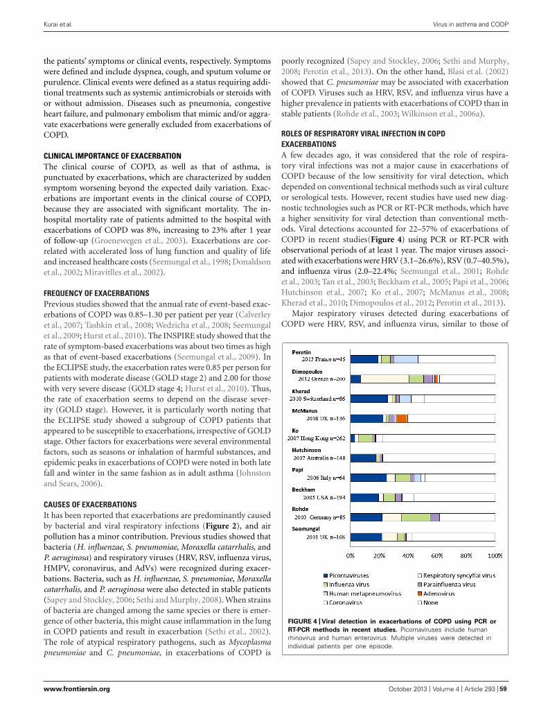
“fmicb-04-00293” — 2013/9/27 — 20:00 — page 7 — #7
Kurai et al. Virus in asthma and CODP
the patients’ symptoms or clinical events, respectively. Symptomswere defined and include dyspnea, cough, and sputum volume orpurulence. Clinical events were defined as a status requiring addi-tional treatments such as systemic antimicrobials or steroids withor without admission. Diseases such as pneumonia, congestiveheart failure, and pulmonary embolism that mimic and/or aggra-vate exacerbations were generally excluded from exacerbations ofCOPD.
CLINICAL IMPORTANCE OF EXACERBATIONThe clinical course of COPD, as well as that of asthma, ispunctuated by exacerbations, which are characterized by suddensymptom worsening beyond the expected daily variation. Exac-erbations are important events in the clinical course of COPD,because they are associated with significant mortality. The in-hospital mortality rate of patients admitted to the hospital withexacerbations of COPD was 8%, increasing to 23% after 1 yearof follow-up (Groenewegen et al., 2003). Exacerbations are cor-related with accelerated loss of lung function and quality of lifeand increased healthcare costs (Seemungal et al., 1998; Donaldsonet al., 2002; Miravitlles et al., 2002).
FREQUENCY OF EXACERBATIONSPrevious studies showed that the annual rate of event-based exac-erbations of COPD was 0.85–1.30 per patient per year (Calverleyet al., 2007; Tashkin et al., 2008; Wedzicha et al., 2008; Seemungalet al., 2009; Hurst et al., 2010). The INSPIRE study showed that therate of symptom-based exacerbations was about two times as highas that of event-based exacerbations (Seemungal et al., 2009). Inthe ECLIPSE study, the exacerbation rates were 0.85 per person forpatients with moderate disease (GOLD stage 2) and 2.00 for thosewith very severe disease (GOLD stage 4; Hurst et al., 2010). Thus,the rate of exacerbation seems to depend on the disease sever-ity (GOLD stage). However, it is particularly worth noting thatthe ECLIPSE study showed a subgroup of COPD patients thatappeared to be susceptible to exacerbations, irrespective of GOLDstage. Other factors for exacerbations were several environmentalfactors, such as seasons or inhalation of harmful substances, andepidemic peaks in exacerbations of COPD were noted in both latefall and winter in the same fashion as in adult asthma (Johnstonand Sears, 2006).
CAUSES OF EXACERBATIONSIt has been reported that exacerbations are predominantly causedby bacterial and viral respiratory infections (Figure 2), and airpollution has a minor contribution. Previous studies showed thatbacteria (H. influenzae, S. pneumoniae, Moraxella catarrhalis, andP. aeruginosa) and respiratory viruses (HRV, RSV, influenza virus,HMPV, coronavirus, and AdVs) were recognized during exacer-bations. Bacteria, such as H. influenzae, S. pneumoniae, Moraxellacatarrhalis, and P. aeruginosa were also detected in stable patients(Sapey and Stockley, 2006; Sethi and Murphy, 2008). When strainsof bacteria are changed among the same species or there is emer-gence of other bacteria, this might cause inflammation in the lungin COPD patients and result in exacerbation (Sethi et al., 2002).The role of atypical respiratory pathogens, such as Mycoplasmapneumoniae and C. pneumoniae, in exacerbations of COPD is
poorly recognized (Sapey and Stockley, 2006; Sethi and Murphy,2008; Perotin et al., 2013). On the other hand, Blasi et al. (2002)showed that C. pneumoniae may be associated with exacerbationof COPD. Viruses such as HRV, RSV, and influenza virus have ahigher prevalence in patients with exacerbations of COPD than instable patients (Rohde et al., 2003; Wilkinson et al., 2006a).
ROLES OF RESPIRATORY VIRAL INFECTION IN COPDEXACERBATIONSA few decades ago, it was considered that the role of respira-tory viral infections was not a major cause in exacerbations ofCOPD because of the low sensitivity for viral detection, whichdepended on conventional technical methods such as viral cultureor serological tests. However, recent studies have used new diag-nostic technologies such as PCR or RT-PCR methods, which havea higher sensitivity for viral detection than conventional meth-ods. Viral detections accounted for 22–57% of exacerbations ofCOPD in recent studies(Figure 4) using PCR or RT-PCR withobservational periods of at least 1 year. The major viruses associ-ated with exacerbations were HRV (3.1–26.6%), RSV (0.7–40.5%),and influenza virus (2.0–22.4%; Seemungal et al., 2001; Rohdeet al., 2003; Tan et al., 2003; Beckham et al., 2005; Papi et al., 2006;Hutchinson et al., 2007; Ko et al., 2007; McManus et al., 2008;Kherad et al., 2010; Dimopoulos et al., 2012; Perotin et al., 2013).
Major respiratory viruses detected during exacerbations ofCOPD were HRV, RSV, and influenza virus, similar to those of
FIGURE 4 | Viral detection in exacerbations of COPD using PCR or
RT-PCR methods in recent studies. Picornaviruses include humanrhinovirus and human enterovirus. Multiple viruses were detected inindividual patients per one episode.
www.frontiersin.org October 2013 | Volume 4 | Article 293 | 59

“fmicb-04-00293” — 2013/9/27 — 20:00 — page 8 — #8
Kurai et al. Virus in asthma and CODP
asthma (Tan et al., 2003). Other viruses, such as PIV, humanmetapneumovirus, AdV, and coronavirus, were also noted inpatients with COPD exacerbations. The proportion of viral-related exacerbations seemed to be similar among the vari-ous GOLD stages, while that of bacteria-related exacerbationsincreased with higher stage or a decrease in lung function(Dimopoulos et al., 2012).
HRVHuman rhinovirus was the most detected virus during exacer-bations of COPD, but not only HRV infection alone but alsoco-infection with HRV and bacteria may cause exacerbations(Wilkinson et al., 2006b). Mallia et al. (2011) reported that exper-imental HRV infection showed more severe and prolonged lowerrespiratory symptoms, airway obstruction, and neutrophilic air-way inflammation in COPD patients than in control subjects. Theystated that rhinovirus infection led to elevation of neutrophil elas-tase, which is associated with reduction of antimicrobial peptides,such as secretory leukoprotease inhibitor and elafin. This reduc-tion of antimicrobial peptides predisposes to secondary bacterialinfection (Mallia et al., 2011, 2012).
Previous reports showed that COPD exacerbations may beassociated with an impaired host response to HRV. For exam-ple, reduced IFN production was observed in COPD patientscompared to control subjects, which may be associated with themechanism of viral and subsequent bacterial infection relatedto severe exacerbations. Impaired humoral immunity was alsorelated to exacerbation of COPD. Patients hospitalized with COPDexacerbations had lower serum levels of rhinovirus-specific anti-bodies than those not hospitalized with COPD exacerbations(Yerkovich et al., 2012). Thymic stromal lymphopoietin (TSLP)is a key pro-allergic cytokine that has recently been linked toasthma(Redhu and Gounni, 2012). In addition, genome-wideassociation studies showed that polymorphisms near or within theTSLP gene were associated with various allergic diseases, includ-ing bronchial asthma (Hirota et al., 2011; Ober and Yao, 2011).TSLP may contribute to exacerbations of the pathogenic effects ofHRV infection via a Toll-like receptor (TLR)3-dependent pathway(Calven et al., 2012).
RSVRespiratory syncytial virus has been detected in both stable andexacerbated cases of COPD. RSV detection in stable COPDpatients might be associated with insidious airway inflammationand accelerated decline in FEV1.0 (Wilkinson et al., 2006a). How-ever, this was not confirmed by another study (Falsey et al., 2006).RSV increases the expression of TLR3 on the surface of airwayepithelial cells, which is associated with increased sensitizationto double-stranded RNA and its related infections (Groskreutzet al., 2006).
Influenza virusInfluenza virus has been associated with mortality and morbid-ity in chronic lung diseases (Harper et al., 2009). A meta-analysisshowed that influenza vaccination prevented exacerbations inCOPD patients (Poole et al., 2000) and reduced the mortality andmorbidity in elderly persons (Nichol et al., 2007). Anti-viral treat-ment such as neuraminidase inhibitors may reduce the severity of
disease (Kaiser et al., 2003). Thus, treatment to prevent influenzahas been recommended for COPD patients (Harper et al., 2009).
ARE PATIENTS WITH COPD SUSCEPTIBLE TO RESPIRATORY VIRALINFECTIONS?Greenberg et al. (2000) investigated viral infections with or with-out COPD in a longitudinal cohort study. They demonstrated thatannual symptomatic documented respiratory viral infection rateswere similar for COPD and age-matched controls (0.45/year and0.54/year, respectively). However, the COPD patients had moreoffice visits and hospitalization than controls.
Of note, Mallia et al. (2011, 2012) confirmed these findings in ahuman experimental study; they found no significant differencesin the frequencies of successful HRV infection between COPD andcontrol groups when these groups were experimentally inoculatedwith HRV. Respiratory symptoms and a decline in lung functionwere more severe in the COPD group than in the control group.
DIFFERENCES IN VIRAL AND NON-VIRAL EXACERBATIONSSeveral studies have suggested that respiratory virus-associatedexacerbations are more critical events than those due to othercauses, in that viral-detected exacerbations showed a larger declinein lung function and longer recovery time than non-viral exacer-bations (Seemungal et al., 2001; Bafadhel et al., 2011). As describedin the chapter on HRV, respiratory viral infections themselvesexacerbated COPD patients and tended to provoke secondarybacterial infections. Viral and sequential bacterial infections maybe associated with severe respiratory symptoms (Wilkinson et al.,2006b; Harper et al., 2009; Mallia et al., 2012).
MECHANISMS OF VIRUS-INDUCED COPD EXACERBATIONSAs shown in the Table 1, the pathological features of COPDare fibrosis around small airways involving several different cells(neutrophils, macrophage, CD8 lymphocytes) and destruction oflung parenchyma. Neutrophils have been found to be associatedwith both stable and exacerbated COPD (Hogg et al., 2004; Papiet al., 2006).
Changes in neutrophil counts during exacerbations in bothsputum and peripheral blood have been found to be related to theFEV1.0 value. Levels of tumor necrosis factor-alpha and interleukin(IL)-8 in sputum were associated with neutrophilic inflammation(Keatings et al., 1996). Especially in patients who suffered fromfrequent exacerbations, they had persistently higher systemic IL-6and C-reactive protein (CRP) levels, which may explain the greaterdecline in lung function (Perera et al., 2007).
Inflammatory cytokines in sputum during exacerbations havebeen found to be elevated regardless of whether the infectionis viral or bacterial (Aaron et al., 2001), and their levels werehigher with exacerbations than when stable. Eosinophils are con-sidered characteristic cells in asthma, but they are also detectedwith exacerbations of COPD (Saetta et al., 1994). Indeed, Papiet al. (2006) demonstrated that virus-associated exacerbations inCOPD patients were related to increased eosinophil counts andthe level of eosinophil cationic protein. Furthermore, Bafad-hel et al. (2011) showed that serum C-X-C motif chemokine 10(CXCL10) is implicated as a more potent predictive maker for
Frontiers in Microbiology | Virology October 2013 | Volume 4 | Article 293 | 60
“fmicb-04-00293” — 2013/9/27 — 20:00 — page 9 — #9
Kurai et al. Virus in asthma and CODP
virus-associated exacerbations, and it is known as IFN-λ-inducedprotein 10.
TREATMENT OF STABLE AND EXACERBATION STATESInhaled bronchodilators, LAMA and LABA, are the main pharma-cological therapies in stable COPD patients (Tashkin et al., 2008;Wedzicha et al., 2008; Vestbo et al., 2013). Although Vogelmeieret al. (2011) reported that the tiotropium (LAMA)-treated grouphad a lower exacerbation rate than the salmeterol (LABA)-treatedgroup in their head-to-head study, both LAMA and LABA treat-ments decreased exacerbation rates and improved lung function orhealth-related quality of life. Tashkin et al. (2009) found that com-bination LAMA/LABA therapy improved pulmonary function(FEV1.0) and respiratory symptoms better than LAMA therapyalone. ICS, the main treatment for asthma, is also prescribedin COPD patients and may reduce airway inflammation anddecrease exacerbation rates only in moderate and severe COPDpatients (Calverley et al., 2007). Treatment with macrolide antibi-otics has been reported to prevent COPD exacerbations andimprove patient quality of life and symptoms, especially in patientswho have frequent exacerbations (Albert et al., 2011; Yamaya et al.,2012a), although this intervention could lead to unfavorable eventssuch as increasing the prevalence of macrolide-resistant pathogensor cardiac toxicity.
It has been estimated that most exacerbations of COPD aredue to respiratory viral and/or bacterial infections. Thus, themajor pharmacological components of managing exacerbationsof COPD include SABAs, short-course systemic glucocorticoids,and antibiotics (Vestbo et al., 2013). However, anti-viral therapiesare rarely prescribed, because specific anti-viral therapies do notexist, except for influenza virus and RSV. Treatment for influenzaappears appropriate in patients with COPD (Harper et al., 2009),
while the utility of treatment for RSV has not been confirmed inadults. It is doubtful that systemic corticosteroid treatment affectsthe clinical course of respiratory viral infections. Lee et al. (2011)showed that short-course systemic steroid treatment did not affectviral load or shedding, and humoral immunity may be diminishedby steroid treatment.
Some research has shown that LAMA may affect viral infec-tions. Tiotropium, one of the LAMAs, may inhibit HRV andRSV infections by reducing the levels of intercellular adhesionmolecule-1, which is the binding site for most HRVs (Iesato et al.,2008; Yamaya et al., 2012b).
SUMMARY AT A GLANCEThe associations between virus infections and asthma and/orCOPD were reviewed, and the significance of viral infections, aswell as their effect on the clinical course, was discussed.
(1) HRV and RSV are major causes of exacerbations both inasthma and COPD patients.
(2) The frequency of viral detection in both asthma and COPDpatients appears to be similar to that of healthy subjects, butthe effect on their clinical course is different; asthma and COPDpatients tend to have more severe or persistent respiratorysymptoms or decreases in pulmonary function, and mortalitymay be increased.
(3) Since discrimination between asthma and COPD is difficult,especially during exacerbations, whenever clinicians encounterpatients in whom obstructive airway disease is suspected,multidisciplinary assessment is required for diagnosis.
(4) The clinical findings of both asthma and COPD, so-called“overlap syndrome,” are commonly recognized in general prac-tice, and virus-associated exacerbations in this disease may leadto a poor prognosis.
REFERENCESAaron, S. D., Angel, J. B., Lunau, M.,
Wright, K., Fex, C., Le Saux, N.,et al. (2001). Granulocyte inflam-matory markers and airway infec-tion during acute exacerbation ofchronic obstructive pulmonary dis-ease. Am. J. Respir. Crit. CareMed. 163, 349–355. doi: 10.1164/ajrccm.163.2.2003122
Abramson, M., Pearson, L., Kutin, J.,Czarny, D., Dziukas, L., and Bowes,G. (1994). Allergies, upper respira-tory tract infections, and asthma. J.Asthma 31, 367–374. doi: 10.3109/02770909409061316
Albert, R. K., Connett, J., Bailey, W. C.,Casaburi, R., Cooper, J. A. Jr., Criner,G. J., et al. (2011). Azithromycinfor prevention of exacerbations ofCOPD. N. Engl. J. Med. 365,689–698. doi: 10.1056/NEJMoa1104623
Atmar, R. L., Guy, E., Guntupalli, K.K., Zimmerman, J. L., Bandi, V. D.,Baxter, B. D., et al. (1998). Respira-tory tract viral infections in inner-cityasthmatic adults. Arch. Intern. Med.
158, 2453–2459. doi: 10.1001/arch-inte.158.22.2453
Bafadhel, M., Mckenna, S., Terry,S., Mistry, V., Reid, C., Hal-dar, P., et al. (2011). Acuteexacerbations of chronic obstructivepulmonary disease: identification ofbiologic clusters and their biomark-ers. Am. J. Respir. Crit. Care Med.184, 662–671. doi: 10.1164/rccm.201104-0597OC
Barnes, P. J. (2008). The cytokine net-work in asthma and chronic obstruc-tive pulmonary disease. J. Clin.Invest. 118, 3546–3556. doi: 10.1172/JCI36130
Beasley, R., Coleman, E. D., Hermon,Y., Holst, P. E., O’donnell, T. V.,and Tobias, M. (1988). Viral respira-tory tract infection and exacerbationsof asthma in adult patients. Thorax43, 679–683. doi: 10.1136/thx.43.9.679
Beckham, J. D., Cadena, A., Lin, J.,Piedra, P. A., Glezen, W. P., Green-berg, S. B., et al. (2005). Respi-ratory viral infections in patientswith chronic, obstructive pulmonary
disease. J. Infect. 50, 322–330. doi:10.1016/j.jinf.2004.07.011
Biscardi, S., Lorrot, M., Marc,E., Moulin, F., Boutonnat-Faucher,B., Heilbronner, C., et al. (2004).Mycoplasma pneumoniae and asthmain children. Clin. Infect. Dis 38,1341–1346. doi: 10.1086/392498
Bisgaard, H., Hermansen, M. N.,Buchvald, F., Loland, L., Halk-jaer, L. B., Bonnelykke, K., et al.(2007). Childhood asthma after bac-terial colonization of the airway inneonates. N. Engl. J. Med. 357,1487–1495. doi: 10.1056/NEJMoa052632
Bjornson, C. L., and Mitchell, I. (2000).Gender differences in asthma inchildhood and adolescence. J. Gend.Specif. Med. 3, 57–61.
Blanken, M. O., Rovers, M. M., Mole-naar, J. M., Winkler-Seinstra, P. L.,Meijer, A., Kimpen, J. L., et al.(2013). Respiratory syncytial virusand recurrent wheeze in healthypreterm infants. N. Engl. J. Med.368, 1791–1799. doi: 10.1056/NEJ-Moa1211917
Blasi, F., Damato, S., Cosentini, R., Tar-sia, P., Raccanelli, R., Centanni, S.,et al. (2002). Chlamydia pneumoniaeand chronic bronchitis: associationwith severity and bacterial clearancefollowing treatment. Thorax 57, 672–676. doi: 10.1136/thorax.57.8.672
Bleecker, E. R. (2004). Similari-ties and differences in asthma andCOPD. The Dutch hypothesis. Chest126, 93S–95S. doi: 10.1378/chest.126.2_suppl_1.93S
Brodzinski, H., and Ruddy, R. M.(2009). Review of new and newlydiscovered respiratory tract virusesin children. Pediatr. Emerg. Care25, 352–360. doi: 10.1097/PEC.0b013e3181a3497e
Calven, J., Yudina, Y., Hallgren, O.,Westergren-Thorsson, G., Davies, D.E., Brandelius, A., et al. (2012). Viralstimuli trigger exaggerated thymicstromal lymphopoietin expression bychronic obstructive pulmonary dis-ease epithelium: role of endosomalTLR3 and cytosolic RIG-I-like heli-cases. J. Innate Immun. 4, 86–99. doi:10.1159/000329131
www.frontiersin.org October 2013 | Volume 4 | Article 293 | 61
“fmicb-04-00293” — 2013/9/27 — 20:00 — page 10 — #10
Kurai et al. Virus in asthma and CODP
Calverley, P. M., Anderson, J. A., Celli,B., Ferguson, G. T., Jenkins, C.,Jones, P. W., et al. (2007). Salme-terol and fluticasone propionate andsurvival in chronic obstructive pul-monary disease. N. Engl. J. Med.356, 775–789. doi: 10.1056/NEJMoa063070
Cheung, D., Dick, E. C., Timmers, M.C., De Klerk, E. P., Spaan, W. J.,and Sterk, P. J. (1995). Rhinovirusinhalation causes long-lasting exces-sive airway narrowing in response tomethacholine in asthmatic subjects invivo. Am. J. Respir. Crit. Care Med.152, 1490–1496. doi: 10.1164/ajr-ccm.152.5.7582282
Copenhaver, C. C., Gern, J. E., Li, Z.,Shult, P. A., Rosenthal, L. A., Mikus,L. D., et al. (2004). Cytokine responsepatterns, exposure to viruses, and res-piratory infections in the first year oflife. Am. J. Respir. Crit. Care Med. 170,175–180. doi: 10.1164/rccm.200312-1647OC
Corne, J. M., Marshall, C., Smith,S., Schreiber, J., Sanderson, G.,Holgate, S. T., et al. (2002). Fre-quency, severity, and duration of rhi-novirus infections in asthmatic andnon-asthmatic individuals: a longitu-dinal cohort study. Lancet 359, 831–834. doi: 10.1016/S0140-6736(02)07953-9
Cunningham, A. F., Johnston, S. L.,Julious, S. A., Lampe, F. C., andWard, M. E. (1998). Chronic Chlamy-dia pneumoniae infection and asthmaexacerbations in children. Eur. Respir.J. 11, 345–349. doi: 10.1183/09031936.98.11020345
Dawson-Caswell, M., and Muncie, H.L. Jr. (2011). Respiratory syncytialvirus infection in children. Am. Fam.Physician 83, 141–146.
Dimopoulos, G., Lerikou, M., Tsiodras,S., Chranioti, A., Perros, E., Anag-nostopoulou, U., et al. (2012). Viralepidemiology of acute exacerbationsof chronic obstructive pulmonarydisease. Pulm. Pharmacol. Ther.25, 12–18. doi: 10.1016/j.pupt.2011.08.004
Donaldson, G. C., Seemungal, T.A., Bhowmik, A., and Wedzicha,J. A. (2002). Relationship betweenexacerbation frequency and lungfunction decline in chronic obstruc-tive pulmonary disease. Thorax 57,847–852. doi: 10.1136/thorax.57.10.847
Dougherty, R. H., and Fahy, J.V. (2009). Acute exacerbationsof asthma: epidemiology, biologyand the exacerbation-prone pheno-type. Clin. Exp. Allergy 39, 193–202. doi: 10.1111/j.1365-2222.2008.03157.x
Edwards, M. R., Bartlett, N. W.,Hussell, T., Openshaw, P., andJohnston, S. L. (2012). The micro-biology of asthma. Nat. Rev.Microbiol. 10, 459–471. doi: 10.1038/nrmicro2801
Fahy, J. V. (2002). Goblet cell andmucin gene abnormalities in asthma.Chest 122, 320S–326S. doi: 10.1378/chest.122.6_suppl.320S
Falsey, A. R., Formica, M. A., Hen-nessey, P. A., Criddle, M. M., Sul-lender, W. M., and Walsh, E. E.(2006). Detection of respiratory syn-cytial virus in adults with chronicobstructive pulmonary disease. Am.J. Respir. Crit. Care Med. 173,639–643. doi: 10.1164/rccm.200510-1681OC
Falsey, A. R., Formica, M. A., and Walsh,E. E. (2012). Yield of sputum forviral detection by reverse transcrip-tase PCR in adults hospitalized withrespiratory illness. J. Clin. Micro-biol. 50, 21–24. doi: 10.1128/JCM.05841-11
Fujitsuka, A., Tsukagoshi, H., Arakawa,M., Goto-Sugai, K., Ryo, A.,Okayama, Y., et al. (2011). A molec-ular epidemiological study of respi-ratory viruses detected in Japanesechildren with acute wheezing ill-ness. BMC Infect. Dis. 11:168. doi:10.1186/1471-2334-11-168
Gendrel, D., Guedj, R., Pons-Catalano,C., Emirian, A., Raymond, J., Rozen-berg, F., et al. (2007). Humanbocavirus in children with acuteasthma. Clin. Infect. Dis 45, 404–405.doi: 10.1086/519505
Greenberg, S. B., Allen, M., Wilson,J., and Atmar, R. L. (2000). Res-piratory viral infections in adultswith and without chronic obstruc-tive pulmonary disease. Am. J.Respir. Crit. Care Med. 162,167–173. doi: 10.1164/ajrccm.162.1.9911019
Groenewegen, K. H., Schols, A.M., and Wouters, E. F. (2003).Mortality and mortality-related fac-tors after hospitalization for acuteexacerbation of COPD. Chest 124,459–467. doi: 10.1378/chest.124.2.459
Groskreutz, D. J., Monick, M. M., Pow-ers, L. S., Yarovinsky, T. O., Look, D.C., and Hunninghake, G. W. (2006).Respiratory syncytial virus inducesTLR3 protein and protein kinase R,leading to increased double-strandedRNA responsiveness in airway epithe-lial cells. J. Immunol. 176, 1733–1740.
Hardin, M., Silverman, E. K., Barr,R. G., Hansel, N. N., Schroeder,J. D., Make, B. J., et al. (2011).The clinical features of the overlap
between COPD and asthma. Respir.Res. 12, 127. doi: 10.1186/1465-9921-12-127
Harper, S. A., Bradley, J. S., Englund,J. A., File, T. M., Gravenstein, S.,Hayden, F. G., et al. (2009). Seasonalinfluenza in adults and children–diagnosis, treatment, chemopro-phylaxis, and institutional outbreakmanagement: clinical practice guide-lines of the Infectious DiseasesSociety of America. Clin. Infect.Dis 48, 1003–1032. doi: 10.1086/598513
Hashimoto, S., Matsumoto, K., Gon,Y., Ichiwata, T., Takahashi, N., andKobayashi, T. (2008). Viral infec-tion in asthma. Allergol. Int. 57,21–31. doi: 10.2332/allergolint.R-07-156
Henderson, F. W., Clyde, W. A.Jr., Collier, A. M., Denny, F. W.,Senior, R. J., Sheaffer, C. I., et al.(1979). The etiologic and epidemi-ologic spectrum of bronchiolitis inpediatric practice. J. Pediatr. 95, 183–190. doi: 10.1016/S0022-3476(79)80647-2
Hirota, T., Takahashi, A., Kubo, M.,Tsunoda, T., Tomita, K., Doi, S.,et al. (2011). Genome-wide asso-ciation study identifies three newsusceptibility loci for adult asthmain the Japanese population. Nat.Genet. 43, 893–896. doi: 10.1038/ng.887
Hogg, J. C., Chu, F., Utokaparch,S., Woods, R., Elliott, W. M.,Buzatu, L., et al. (2004). Thenature of small-airway obstructionin chronic obstructive pulmonarydisease. N. Engl. J. Med. 350,2645–2653. doi: 10.1056/NEJMoa032158
Holtzman, M. J. (2012). Asthma asa chronic disease of the innate andadaptive immune systems respond-ing to viruses and allergens. J.Clin. Invest. 122, 2741–2748. doi:10.1172/JCI60325
Hospers, J. J., Postma, D. S., Rijcken,B., Weiss, S. T., and Schouten, J.P. (2000). Histamine airway hyper-responsiveness and mortality fromchronic obstructive pulmonary dis-ease: a cohort study. Lancet 356,1313–1317. doi: 10.1016/S0140-6736(00)02815-4
Hurst, J. R., Vestbo, J., Anzueto, A.,Locantore, N., Mullerova, H., Tal-Singer, R., et al. (2010). Susceptibilityto exacerbation in chronic obstruc-tive pulmonary disease. N. Engl. J.Med. 363, 1128–1138. doi: 10.1056/NEJMoa0909883
Hutchinson, A. F., Ghimire, A. K.,Thompson, M. A., Black, J. F.,Brand, C. A., Lowe, A. J., et al.
(2007). A community-based, time-matched, case-control study of res-piratory viruses and exacerbationsof COPD. Respir. Med. 101, 2472–2481. doi: 10.1016/j.rmed.2007.07.015
Iesato, K., Tatsumi, K., Saito, K.,Ogasawara, T., Sakao, S., Tada, Y.,et al. (2008). Tiotropium bromideattenuates respiratory syncytial virusreplication in epithelial cells. Respi-ration 76, 434–441. doi: 10.1159/000151729
Jackson, D. J., Evans, M. D., Gangnon,R. E., Tisler, C. J., Pappas, T.E., Lee, W. M., et al. (2012).Evidence for a causal relationshipbetween allergic sensitization andrhinovirus wheezing in early life.Am. J. Respir. Crit. Care Med. 185,281–285. doi: 10.1164/rccm.201104-0660OC
Jackson, D. J., Gangnon, R. E., Evans,M. D., Roberg, K. A., Ander-son, E. L., Pappas, T. E., et al.(2008). Wheezing rhinovirus ill-nesses in early life predict asthmadevelopment in high-risk children.Am. J. Respir. Crit. Care Med. 178,667–672. doi: 10.1164/rccm.200802-309OC
Jackson, D. J., and Johnston, S. L.(2010). The role of viruses in acuteexacerbations of asthma. J. AllergyClin. Immunol. 125, 1178–1187. doi:10.1016/j.jaci.2010.04.021
Johnston, N. W., and Sears, M. R.(2006). Asthma exacerbations. 1: epi-demiology. Thorax 61, 722–728. doi:10.1136/thx.2005.045161
Johnston, S. L., Pattemore, P. K., Sander-son, G., Smith, S., Campbell, M. J.,Josephs, L. K., et al. (1996). The rela-tionship between upper respiratoryinfections and hospital admissionsfor asthma: a time-trend analysis.Am. J. Respir. Crit. Care Med. 154,654–660. doi: 10.1164/ajrccm.154.3.8810601
Johnston, S. L., Pattemore, P. K.,Sanderson, G., Smith, S., Lampe, F.,Josephs, L., et al. (1995). Commu-nity study of role of viral infectionsin exacerbations of asthma in 9-11year old children. BMJ 310, 1225–1229. doi: 10.1136/bmj.310.6989.1225
Kaiser, L., Wat, C., Mills, T., Mahoney,P., Ward, P., and Hayden, F. (2003).Impact of oseltamivir treatment oninfluenza-related lower respiratorytract complications and hospitaliza-tions. Arch. Intern. Med. 163, 1667–1672. doi: 10.1001/archinte.163.14.1667
Kaplan, A. G., Balter, M. S., Bell, A. D.,Kim, H., and Mcivor, R. A. (2009).Diagnosis of asthma in adults. CMAJ,
Frontiers in Microbiology | Virology October 2013 | Volume 4 | Article 293 | 62

“fmicb-04-00293” — 2013/9/27 — 20:00 — page 11 — #11
Kurai et al. Virus in asthma and CODP
181, E210–E220. doi: 10.1503/cmaj.080006
Keatings, V. M., Collins, P. D., Scott,D. M., and Barnes, P. J. (1996).Differences in interleukin-8 andtumor necrosis factor-alpha ininduced sputum from patientswith chronic obstructive pul-monary disease or asthma. Am.J. Respir. Crit. Care Med. 153, 530–534. doi: 10.1164/ajrccm.153.2.8564092
Kerstjens, H. A., Engel, M., Dahl,R., Paggiaro, P., Beck, E., Van-dewalker, M., et al. (2012).Tiotropium in asthma poorly con-trolled with standard combinationtherapy. N. Engl. J. Med. 367,1198–1207. doi: 10.1056/NEJMoa1208606
Kherad, O., Kaiser, L., Bridevaux,P. O., Sarasin, F., Thomas, Y.,Janssens, J. P., et al. (2010). Upper-respiratory viral infection, biomark-ers, and COPD exacerbations. Chest138, 896–904. doi: 10.1378/chest.09-2225
Ko, F. W., Ip, M., Chan, P. K., Chan, M.C., To, K. W., Ng, S. S., et al. (2007).Viral etiology of acute exacerbationsof COPD in Hong Kong. Chest132, 900–908. doi: 10.1378/chest.07-0530
Kraft, M. (2006). Asthma and chronicobstructive pulmonary diseaseexhibit common origins in any coun-try! Am. J. Respir. Crit. Care Med. 174,238–240. doi: 10.1164/rccm.2604007
Kraft, M., Adler, K. B., Ingram, J. L.,Crews, A. L., Atkinson, T. P., Cairns,C. B., et al. (2008). Mycoplasmapneumoniae induces airway epithe-lial cell expression of MUC5AC inasthma. Eur. Respir. J. 31, 43–46. doi:10.1183/09031936.00103307
Kusel, M. M., De Klerk, N. H.,Kebadze, T., Vohma, V., Holt, P.G., Johnston, S. L., et al. (2007).Early-life respiratory viral infections,atopic sensitization, and risk of sub-sequent development of persistentasthma. J. Allergy Clin. Immunol. 119,1105–1110. doi: 10.1016/j.jaci.2006.12.669
Lee, F. E., Walsh, E. E., and Falsey,A. R. (2011). The effect of steroiduse in hospitalized adults with res-piratory syncytial virus-related ill-ness. Chest 140, 1155–1161. doi:10.1378/chest.11-0047
Lieberman, D., Shimoni, A., Keren-Naus, A., Steinberg, R., andShemer-Avni, Y. (2009). Identifica-tion of respiratory viruses in adults:nasopharyngeal versus oropharyn-geal sampling. J. Clin. Microbiol.47, 3439–3443. doi: 10.1128/JCM.00886-09
Louie, S., Zeki, A. A., Schivo, M.,Chan, A. L., Yoneda, K. Y., Avdalovic,M., et al. (2013). The asthma-chronic obstructive pulmonarydisease overlap syndrome: pharma-cotherapeutic considerations. ExpertRev. Clin. Pharmacol. 6, 197–219.doi: 10.1586/ecp.13.2
Macek, V., Sorli, J., Kopriva, S., andMarin, J. (1994). Persistent aden-oviral infection and chronic airwayobstruction in children. Am. J. Respir.Crit. Care Med. 150, 7–10. doi:10.1164/ajrccm.150.1.8025775
Magnussen, H., Bugnas, B., VanNoord, J., Schmidt, P., Gerken, F.,and Kesten, S. (2008). Improve-ments with tiotropium in COPDpatients with concomitant asthma.Respir. Med. 102, 50–56. doi:10.1016/j.rmed.2007.08.003
Mallia, P., Footitt, J., Sotero, R., Jepson,A., Contoli, M., Trujillo-Torralbo, M.B., et al. (2012). Rhinovirus infectioninduces degradation of antimicro-bial peptides and secondary bacte-rial infection in chronic obstructivepulmonary disease. Am. J. Respir.Crit. Care Med. 186, 1117–1124. doi:10.1164/rccm.201205-0806OC
Mallia, P., Message, S. D., Gie-len, V., Contoli, M., Gray, K.,Kebadze, T., et al. (2011). Exper-imental rhinovirus infection as ahuman model of chronic obstruc-tive pulmonary disease exacerbation.Am. J. Respir. Crit. Care Med. 183,734–742. doi: 10.1164/rccm.201006-0833OC
McErlean, P., Greiman, A., Favoreto,S. Jr., and Avila, P. C. (2010). Viraldiversity in asthma. Immunol. AllergyClin. North Am. 30, 481-95, v. doi:10.1016/j.iac.2010.08.001
McManus, T. E., Marley, A. M., Bax-ter, N., Christie, S. N., O’neill, H. J.,Elborn, J. S., et al. (2008). Respiratoryviral infection in exacerbations ofCOPD. Respir. Med. 102, 1575–1580.doi: 10.1016/j.rmed.2008.06.006
Message, S. D., Laza-Stanca, V., Mallia,P., Parker, H. L., Zhu, J., Kebadze,T., et al. (2008). Rhinovirus-inducedlower respiratory illness is increasedin asthma and related to virusload and Th1/2 cytokine and IL-10 production. Proc. Natl. Acad.Sci. U.S.A. 105, 13562–13567. doi:10.1073/pnas.0804181105
Miravitlles, M., Murio, C., Guerrero,T., and Gisbert, R. (2002). Phar-macoeconomic evaluation of acuteexacerbations of chronic bronchitisand COPD. Chest 121, 1449–1455.doi: 10.1378/chest.121.5.1449
Murray, C. S., Simpson, A., and Cus-tovic, A. (2004). Allergens, viruses,and asthma exacerbations. Proc.
Am. Thorac. Soc. 1, 99–104. doi:10.1513/pats.2306027
Nagel, M. A., Cohrs, R. J., Mahalingam,R., Wellish, M. C., Forghani, B.,Schiller, A., et al. (2008). The vari-cella zoster virus vasculopathies:clinical, CSF, imaging, and virologicfeatures. Neurology 70, 853–860.doi: 10.1212/01.wnl.0000304747.38502.e8
National Asthma Education and Pre-vention Program. (2007). ExpertPanel Report 3 (EPR-3): Guidelinesfor the Diagnosis and Managementof Asthma-Summary Report 2007. J.Allergy Clin. Immunol. 120(Suppl.5), S94–S138. doi: 10.1016/j.jaci.2007.09.029
Nichol, K. L., Nordin, J. D., Nelson, D.B., Mullooly, J. P., and Hak, E. (2007).Effectiveness of influenza vaccine inthe community-dwelling elderly. N.Engl. J. Med. 357, 1373–1381. doi:10.1056/NEJMoa070844
Nicholson, K. G., Kent, J., and Ire-land, D. C. (1993). Respiratoryviruses and exacerbations of asthmain adults. BMJ 307, 982–986. doi:10.1136/bmj.307.6910.982
Nisar, N., Guleria, R., Kumar, S., ChandChawla, T., and Ranjan Biswas,N. (2007). Mycoplasma pneumoniaeand its role in asthma. Postgrad.Med. J. 83, 100–104. doi: 10.1136/pgmj.2006.049023
Ober, C., and Yao, T. C. (2011).The genetics of asthma and aller-gic disease: a 21st century perspec-tive. Immunol. Rev. 242, 10–30. doi:10.1111/j.1600-065X.2011.01029.x
O’Byrne, P. M., Barnes, P. J., Rodriguez-Roisin, R., Runnerstrom, E., Sand-strom, T., Svensson, K., et al.(2001). Low dose inhaled budesonideand formoterol in mild persistentasthma: the OPTIMA randomizedtrial. Am. J. Respir. Crit. Care Med.164, 1392–1397. doi: 10.1164/ajr-ccm.164.8.2104102
Olenec, J. P., Kim, W. K., Lee, W.M., Vang, F., Pappas, T. E., Salazar,L. E., et al. (2010). Weekly moni-toring of children with asthma forinfections and illness during com-mon cold seasons. J. Allergy Clin.Immunol. 125, 1001–1006.e1. doi:10.1016/j.jaci.2010.01.059
Olszewska, W., and Openshaw, P.(2009). Emerging drugs for respira-tory syncytial virus infection. ExpertOpin. Emerg. Drugs 14, 207–217. doi:10.1517/14728210902946399
Ong, B. H., Gao, Q., Phoon, M.C., Chow, V. T., Tan, W. C., andVan Bever, H. P. (2007). Identifi-cation of human metapneumovirusand Chlamydophila pneumoniae inchildren with asthma and wheeze
in Singapore. Singapore Med. J. 48,291–293.
Openshaw, P. J. (1995). Immunopatho-logical mechanisms in respiratorysyncytial virus disease. SpringerSemin. Immunopathol. 17, 187–201.doi: 10.1007/BF00196165
O’Sullivan, S. M. (2005). Asthmadeath, CD8+ T cells, and viruses.Proc. Am. Thorac. Soc. 2, 162–165. doi: 10.1513/pats.200502-016AW
Papi, A., Bellettato, C. M., Braccioni,F., Romagnoli, M., Casolari, P.,Caramori, G., et al. (2006). Infec-tions and airway inflammation inchronic obstructive pulmonary dis-ease severe exacerbations. Am. J.Respir. Crit. Care Med. 173, 1114–1121. doi: 10.1164/rccm.200506-859OC
Perera, W. R., Hurst, J. R., Wilkin-son, T. M., Sapsford, R. J.,Mullerova, H., Donaldson, G. C.,et al. (2007). Inflammatory changes,recovery and recurrence at COPDexacerbation. Eur. Respir. J. 29,527–534. doi: 10.1183/09031936.00092506
Perotin, J. M., Dury, S., Renois, F.,Deslee, G., Wolak, A., Duval, V.,et al. (2013). Detection of mul-tiple viral and bacterial infectionsin acute exacerbation of chronicobstructive pulmonary disease: apilot prospective study. J. Med.Virol. 85, 866–873. doi: 10.1002/jmv.23495
Poole, P. J., Chacko, E., Wood-Baker, R. W., and Cates, C. J.(2000). Influenza vaccine forpatients with chronic obstruc-tive pulmonary disease. CochraneDatabase Syst. Rev. CD002733. doi:10.1002/14651858.CD002733
Rakes, G. P., Arruda, E., Ingram, J.M., Hoover, G. E., Zambrano, J.C., Hayden, F. G., et al. (1999).Rhinovirus and respiratory syncytialvirus in wheezing children requiringemergency care. IgE and eosinophilanalyses. Am. J. Respir. Crit. CareMed. 159, 785–790. doi: 10.1164/ajr-ccm.159.3.9801052
Reddel, H. K., Taylor, D. R.,Bateman, E. D., Boulet, L. P.,Boushey, H. A., Busse, W. W.,et al. (2009). An official AmericanThoracic Society/European Respira-tory Society statement: asthma con-trol and exacerbations: standardizingendpoints for clinical asthma trialsand clinical practice. Am. J. Respir.Crit. Care Med. 180, 59–99. doi:10.1164/rccm.200801-060ST
Redhu, N. S., and Gounni, A. S.(2012). Function and mechanisms ofTSLP/TSLPR complex in asthma and
www.frontiersin.org October 2013 | Volume 4 | Article 293 | 63

“fmicb-04-00293” — 2013/9/27 — 20:00 — page 12 — #12
Kurai et al. Virus in asthma and CODP
COPD. Clin. Exp. Allergy 42, 994–1005. doi: 10.1111/j.1365-2222.2011.03919.x
Rohde, G., Wiethege, A., Borg, I.,Kauth, M., Bauer, T. T., Gillissen, A.,et al. (2003). Respiratory viruses inexacerbations of chronic obstructivepulmonary disease requiring hospi-talisation: a case-control study. Tho-rax 58, 37–42. doi: 10.1136/thorax.58.1.37
Saetta, M., Di Stefano, A., Maestrelli,P., Turato, G., Ruggieri, M. P.,Roggeri, A., et al. (1994). Air-way eosinophilia in chronic bron-chitis during exacerbations. Am. J.Respir. Crit. Care Med. 150, 1646–1652. doi: 10.1164/ajrccm.150.6.7952628
Sapey, E., and Stockley, R. A. (2006).COPD exacerbations. 2: aetiology.Thorax 61, 250–258. doi: 10.1136/thx.2005.041822
Seemungal, T., Harper-Owen, R.,Bhowmik, A., Moric, I., Sander-son, G., Message, S., et al. (2001).Respiratory viruses, symptoms, andinflammatory markers in acute exac-erbations and stable chronic obstruc-tive pulmonary disease. Am. J.Respir. Crit. Care Med. 164, 1618–1623. doi: 10.1164/ajrccm.164.9.2105011
Seemungal, T. A., Donaldson, G. C.,Paul, E. A., Bestall, J. C., Jef-fries, D. J., and Wedzicha, J. A.(1998). Effect of exacerbation onquality of life in patients with chronicobstructive pulmonary disease. Am.J. Respir. Crit. Care Med. 157, 1418–1422. doi: 10.1164/ajrccm.157.5.9709032
Seemungal, T. A., Hurst, J. R., andWedzicha, J. A. (2009). Exacerbationrate, health status and mortality inCOPD–a review of potential inter-ventions. Int. J. Chron. Obstruct. Pul-mon. Dis. 4, 203–223. doi: 10.2147/COPD.S3385
Sethi, S., Evans, N., Grant, B. J.,and Murphy, T. F. (2002). Newstrains of bacteria and exacerba-tions of chronic obstructive pul-monary disease. N. Engl. J. Med.347, 465–471. doi: 10.1056/NEJMoa012561
Sethi, S., and Murphy, T. F. (2008).Infection in the pathogenesis andcourse of chronic obstructive pul-monary disease. N. Engl. J. Med.359, 2355–2365. doi: 10.1056/NEJMra0800353
Silva, G. E., Sherrill, D. L., Guerra,S., and Barbee, R. A. (2004).Asthma as a risk factor for COPDin a longitudinal study. Chest
126, 59–65. doi: 10.1378/chest.126.1.59
Tan, W. C. (2005). Viruses in asthmaexacerbations. Curr. Opin. Pulm.Med. 11, 21–26.
Tan, W. C., Xiang, X., Qiu, D.,Ng, T. P., Lam, S. F., andHegele, R. G. (2003). Epidemiol-ogy of respiratory viruses in patientshospitalized with near-fatal asthma,acute exacerbations of asthma, orchronic obstructive pulmonary dis-ease. Am. J. Med. 115, 272–277. doi: 10.1016/S0002-9343(03)00353-X
Tashkin, D. P., Celli, B., Senn, S.,Burkhart, D., Kesten, S., Menjoge,S., et al. (2008). A 4-year trial oftiotropium in chronic obstructivepulmonary disease. N. Engl. J. Med.359, 1543–1554. doi: 10.1056/NEJ-Moa0805800
Tashkin, D. P., Pearle, J., Iez-zoni, D., and Varghese, S. T.(2009). Formoterol and tiotropiumcompared with tiotropium alonefor treatment of COPD. COPD6, 17–25. doi: 10.1080/15412550902724073
Teichtahl, H., Buckmaster, N., andPertnikovs, E. (1997). The inci-dence of respiratory tract infectionin adults requiring hospitalization forasthma. Chest 112, 591–596. doi:10.1378/chest.112.3.591
Tokuda, Y., and Miyagi, S. (2007). Phys-ical diagnosis of chronic obstruc-tive pulmonary disease. Intern. Med.46, 1885–1891. doi: 10.2169/inter-nalmedicine.46.0455
Vallet, C., Pons-Catalano, C., Man-delcwajg, A., Wang, A., Raymond,J., Lebon, P., et al. (2009). Humanbocavirus: a cause of severe asthmaexacerbation in children. J. Pedi-atr. 155, 286–288. doi: 10.1016/j.jpeds.2009.01.069
Vestbo, J., Hurd, S. S., Agusti,A. G., Jones, P. W., Vogelmeier,C., Anzueto, A., et al. (2013).Global strategy for the diagnosis,management, and prevention ofchronic obstructive pulmonary dis-ease: GOLD executive summary. Am.J. Respir. Crit. Care Med. 187,347–365. doi: 10.1164/rccm.201204-0596PP
Vlahakis, N. E., and Aksamit, T.R. (2001). Diagnosis and treat-ment of allergic bronchopulmonaryaspergillosis. Mayo Clin. Proc.76, 930–938. doi: 10.1016/S0025-6196(11)62114-9
Vogelmeier, C., Hederer, B., Glaab, T.,Schmidt, H., Rutten-Van Molken,M. P., Beeh, K. M., et al. (2011).
Tiotropium versus salmeterol forthe prevention of exacerbations ofCOPD. N. Engl. J. Med. 364,1093–1103. doi: 10.1056/NEJMoa1008378
Wedzicha, J. A., Calverley, P. M.,Seemungal, T. A., Hagan, G.,Ansari, Z., and Stockley, R. A.(2008). The prevention of chronicobstructive pulmonary disease exac-erbations by salmeterol/fluticasonepropionate or tiotropium bromide.Am. J. Respir. Crit. Care Med. 177,19–26. doi: 10.1164/rccm.200707-973OC
Wenzel, S. E., Schwartz, L. B.,Langmack, E. L., Halliday, J. L.,Trudeau, J. B., Gibbs, R. L.,et al. (1999). Evidence that severeasthma can be divided patholo-gically into two inflammatory sub-types with distinct physiologic andclinical characteristics. Am. J. Respir.Crit. Care Med. 160, 1001–1008. doi: 10.1164/ajrccm.160.3.9812110
Wilkinson, T. M., Donaldson, G.C., Johnston, S. L., Openshaw, P.J., and Wedzicha, J. A. (2006a).Respiratory syncytial virus, airwayinflammation, and FEV1 declinein patients with chronic obstructivepulmonary disease. Am. J. Respir.Crit. Care Med. 173, 871–876. doi:10.1164/rccm.200509-1489OC
Wilkinson, T. M., Hurst, J. R., Per-era, W. R., Wilks, M., Don-aldson, G. C., and Wedzicha, J.A. (2006b). Effect of interactionsbetween lower airway bacterial andrhinoviral infection in exacerbationsof COPD. Chest 129, 317–324. doi:10.1378/chest.129.2.317
Williams, J. V., Crowe, J. E. Jr.,Enriquez, R., Minton, P., Peebles,R. S. Jr., Hamilton, R. G., et al.(2005). Human metapneumovirusinfection plays an etiologic role inacute asthma exacerbations requir-ing hospitalization in adults. J. Infect.Dis. 192, 1149–1153. doi: 10.1086/444392
Xepapadaki, P., Koutsoumpari, I.,Papaevagelou, V., Karagianni, C., andPapadopoulos, N. G. (2008). Atypicalbacteria and macrolides in asthma.Allergy Asthma Clin. Immunol. 4,111–116. doi: 10.1186/1710-1492-4-3-111
Xepapadaki, P., and Papadopou-los, N. G. (2010). Childhoodasthma and infection: virus-inducedexacerbations as determinants andmodifiers. Eur. Respir. J. 36,438–445. doi: 10.1183/09031936.00149009
Xiang, X., Qiu, D., Chan, K.P., Chan, S. H., Hegele, R. G.,and Tan, W. C. (2002). Com-parison of three methods for res-piratory virus detection betweeninduced sputum and nasopharyn-geal aspirate specimens in acuteasthma. J. Virol. Methods 101, 127–133. doi: 10.1016/S0166-0934(01)00431-1
Yamaya, M., Azuma, A., Takizawa, H.,Kadota, J., Tamaoki, J., and Kudoh,S. (2012a). Macrolide effects on theprevention of COPD exacerbations.Eur. Respir. J. 40, 485–494. doi:10.1183/09031936.00208011
Yamaya, M., Nishimura, H., Hat-achi, Y., Yasuda, H., Deng, X.,Sasaki, T., et al. (2012b). Inhibitoryeffects of tiotropium on rhinovirusinfection in human airway epithe-lial cells. Eur. Respir. J. 40,122–132. doi: 10.1183/09031936.00065111
Yerkovich, S. T., Hales, B. J., Carroll,M. L., Burel, J. G., Towers, M. A.,Smith, D. J., et al. (2012). Reducedrhinovirus-specific antibodies areassociated with acute exacerbationsof chronic obstructive pulmonarydisease requiring hospitalisation.BMC Pulm. Med. 12:37. doi:10.1186/1471-2466-12-37
Conflict of Interest Statement: Theauthors declare that the research wasconducted in the absence of any com-mercial or financial relationships thatcould be construed as a potential con-flict of interest.
Received: 30 June 2013; accepted: 10September 2013; published online: 01October 2013.Citation: Kurai D, Saraya T, Ishii Hand Takizawa H (2013) Virus-inducedexacerbations in asthma and COPD.Front. Microbiol. 4:293. doi: 10.3389/fmicb.2013.00293This article was submitted to Virology,a section of the journal Frontiers inMicrobiology.Copyright © 2013 Kurai, Saraya, Ishiiand Takizawa. This is an open-accessarticle distributed under the terms of theCreative Commons Attribution License(CC BY). The use, distribution or repro-duction in other forums is permitted,provided the original author(s) or licensorare credited and that the original publica-tion in this journal is cited, in accordancewith accepted academic practice. No use,distribution or reproduction is permit-ted which does not comply with theseterms.
Frontiers in Microbiology | Virology October 2013 | Volume 4 | Article 293 | 64

REVIEW ARTICLEpublished: 26 May 2014
doi: 10.3389/fmicb.2014.00226
Epidemiology of virus-induced asthma exacerbations: withspecial reference to the role of human rhinovirusTakeshi Saraya1, Daisuke Kurai 1, Haruyuki Ishii 1, Anri Ito1,Yoshiko Sasaki 2 , Shoichi Niwa 2,
Naoko Kiyota 3, HiroyukiTsukagoshi 2 , Kunihisa Kozawa 2, Hajime Goto1 and HajimeTakizawa1*
1 Department of Respiratory Medicine, School of Medicine, Kyorin University, Mitaka, Tokyo, Japan2 Gunma Prefectural Institute of Public Health and Environmental Sciences, Gunma, Japan3 Kumamoto Prefectural Institute of Public Health and Environmental Sciences, Kumamoto, Japan
Edited by:
Hirokazu Kimura, National Institute ofInfectious Diseases, Japan
Reviewed by:
Akihide Ryo, Yokohama CityUniversity, JapanKaoru Okazaki, Shikoku MedicalCenter for Children and Adults, JapanTomoo Kishaba, Okinawa ChubuHospital, Japan
*Correspondence:
Hajime Takizawa, Department ofRespiratory Medicine, School ofMedicine, Kyorin University, 6-20-2Shinkawa, Mitaka, Tokyo 181-8611,Japane-mail: [email protected]
Viral respiratory infections may be associated with the virus-induced asthma in adults aswell as children. Particularly, human rhinovirus is strongly suggested a major candidate forthe associations of the virus-induced asthma.Thus, in this review, we reviewed and focusedon the epidemiology, pathophysiology, and treatment of virus-induced asthma with specialreference on human rhinovirus. Furthermore, we added our preliminary data regarding theclinical and virological findings in the present review.
Keywords: epidemiology, pathophysiology, treatment, human rhinovirus, asthma exacerbation
INTRODUCTIONMore than 200 different types of viruses, such as human rhinovirus(HRV), human metapneumovirus (HMPV), respiratory syncytialvirus (RSV), and human parainfluenza virus (HPIV), are knownto cause acute respiratory illness (ARI; Tsukagoshi et al., 2013).We recently reported the issue of “virus-induced exacerbation inasthma and chronic obstructive pulmonary disease” (Kurai et al.,2013a), however, among these causative viruses, HRV is now rec-ognized to have a major impact on asthma pathogenesis (Fujitsukaet al., 2011). From this perspective, we reviewed the literatureregarding the epidemiology of HRV-induced asthma in adults,together with preliminary epidemiological data obtained at ourinstitution.
CLINICAL VIROLOGY OF HUMAN RHINOVIRUSHRV belongs to the genus Enterovirus and family Picornaviri-dae (Turner and Couch, 2007). HRV possesses a single strandpositive-sense RNA (ssRNA) genome of approximately 7.2 kb.The viral capsid is composed of four viral proteins (VP1-4) whichare assembled into 60 protomers, resulting in a small icosahe-dral structure with a diameter of about 28–30 nm (Turner andCouch, 2007). Genetically, HRV is classified into three species;HRV-A, -B, and -C (Simmonds et al., 2010). Furthermore, thesespecies of HRV have more than 150 genotypes (Andries et al., 1990;Arakawa et al., 2012; Kiyota et al., 2013, 2014). Molecular epidemi-ological studies suggest that the dominant species are HRV-A and-C, while HRV-B is relatively rarely detected (Arakawa et al., 2012;Kiyota et al., 2013). In particular, the VP1 and VP2 proteins havevariations in their amino acid sequences, accounting for the largenumber of viral serotypes (Turner and Couch, 2007). The host
receptor for HRV in respiratory epithelial cells is the intracellu-lar adhesion molecule 1 (ICAM-1, CD54) for the 84 major HRVserotypes (HRV-A and -B), or low-density lipoprotein receptor(LDLR) for the other minor HRV serotypes. The receptor forHRV-C is not yet known. It has been suggested that the optimaltemperature for replication of HRV is relatively cool (33–35◦C),which would limit infections to the upper airway; however, largeor medium sized airways lower in the respiratory tract are nowalso considered cool enough for HRV replication, in spite of thehigher temperature of the lung parenchyma (37◦C; McFaddenet al., 1985). Therefore, HRV is potentially a causative agent ofmore severe ARI such as bronchiolitis and pneumonia (Turner andCouch, 2007; Watanabe et al., 2010; Smuts et al., 2011; Arakawaet al., 2012), and may be associated with virus-induced asthma(Johnston et al., 1995; Linsuwanon et al., 2009; Fujitsuka et al.,2011; Smuts et al., 2011). HRV might therefore be involved in var-ious ARIs and additional respiratory complications (Kiyota et al.,2013). Lieberman et al. (2009) reported that the detection of anyvirus include HRV, the sensitivity rates for nasopharyngeal swab(73.3%) was superior than that of oropharyngeal swab (54.2%),respectively.
VIRUS-INDUCED COLDS AND THEIR NATURAL COURSEAMONG THE GENERAL POPULATIONThe common cold is the third most common primary diagnosisin office visits (Hsiao et al., 2010), and this disease is generally self-limiting, usually lasting up to 10 days (Fashner et al., 2012). Amongthe general population, HRV infection causes common coldsat a frequency of 25–53% (Makela et al., 1998; van Gageldonk-Lafeber et al., 2005). Tyrrell et al. (1993) reported that intranasal
www.frontiersin.org May 2014 | Volume 5 | Article 226 | 65

Saraya et al. Asthma exacerbations and human rhinovirus
inoculation with either HRV serotypes 2, 9, and 14, coronavirustype 229E, or RSV in healthy volunteers induced patterns of symp-tom development which were not substantially different from eachother. However, individual signs or symptoms occurred earli-est in HRV infections, then in coronavirus, and lastly in RSV,appearing up to 5 days after inoculation, which demonstratedthe long incubation periods of RSV in volunteers (Tyrrell et al.,1993).
HRV has been implicated in patients with acute otitis media,exacerbation of chronic obstructive pulmonary disease, commoncold, and lower respiratory tract infections in neonates, the elderlyand immunocompromised. Arruda et al. (1997) researching thefrequency and natural history of HRV infections in adults duringautumn, demonstrated that the first symptom noticed most oftenwas sore throat (40%) in HRV culture- or PCR-positive patients,and stuffy nose in HRV-negative patients (27%), using nasal washspecimens. Respiratory symptoms typically develop after 1–2 daysafter inoculation in studies, and uncomplicated HRV infectionsusually peak 2–4 days after inoculation. The median duration ofHRV colds is 1 week, but up to 25% last more than 2 weeks (Gwalt-ney et al., 1967; Rotbart and Hayden, 2000). It should be noted thatin illness caused by HRV, viral shedding occurs naturally for up to21 days, but predominantly over a 3–4 days period.
HRV INFECTION AMONG ASTHMATICS: IN VIVO OR IN VITROEXPERIMENTAL STUDIESHRV-A type16 (HRV-16), a major group virus commonly usedfor experimental human infection, and HRV-A type1 (HRV-1),which has been used in animal models of HRV infection, areclosely related. Grunberg et al. (1999) reported that experimentalHRV-16 infection via nasal inhalation leads to a transient decreaseof FEV1.0 in patients with asthma, and this decreased lung func-tion was correlated with enhanced cold symptoms and / or airwayhyperresponsiveness. Contoli et al. (2006) demonstrated that typeIII interferon (IFN-λ) production levels in ex vivo cell culturesderived from bronchial epithelial cells (BECs) and macrophagesobtained from asthmatic patients, were lower than in those derivedfrom healthy controls. Furthermore, deficient interferon-λ pro-duction was correlated with HRV viral load, severity of clinicalsymptoms and FEV1.0. Message et al. (2008) demonstrated thatthe severity of intranasally inoculated HRV-induced clinical ill-ness in asthmatic subjects was correlated to virus load and lowerairway virus-induced inflammation.
On the other hand, DeMore et al. (2009) reported that no dif-ference in clinical symptoms, and patterns of viral shedding, wasnoted between subjects with persistent allergic asthma and healthysubjects after experimental infection with HRV. These differentresults after experimental HRV infection in individual studies inasthmatic patients and healthy subjects might be dependent on theseverity of the asthma of those subjects who enrolled in the studies.Indeed, in several reports, neither defective IFN induction by HRV,nor increased HRV replication was observed in primary humanBECs derived from subjects with well controlled asthma (Lopez-Souza et al., 2009; Bochkov et al., 2010; Sykes et al., 2014). A fewanimal models for rhinovirus infection have been showed becausea major group of HRV (i.e., HRV-16) did not bind mouse ICAM-1. Only a minor group of HRV (i.e., HRV-1B) infected the mouse.
In this regard, Bartlett et al. (2008) generated a transgenic BALB/cmouse expressing a mouse-human ICAM-1 chimeric receptor forHRV-16 infection. This study also showed asthma exacerbationmodel by intraperitoneally sensitized with ovalbumin with alu-minum hydroxide followed by intranasal inoculation of HRV-1Bor UV-inactivated HRV-1B.
HRV AND ASTHMA EXACERBATIONS: CLINICAL FINDINGSAlthough data regarding virus respiratory infections (VRIs) as pre-cipitators of asthma attacks in adults are less clear, Nicholson et al.(1993) reported thatVRIs are as commonly linked to exacerbationsin adults as they are in children (Johnston et al., 1996; Fujitsukaet al., 2011). This study showed that viruses were detected in 44%of clinical exacerbative episodes with a decrease in peak expira-tory flow rate (PEFR) of 50 mL/minute or more, and the mostcommonly identified virus was HRV, followed by coronavirusesand parainfluenza viruses (Nicholson et al., 1993). Thus, the virusmost commonly detected in asthma exacerbations appears to beHRV.
Although HRV is well known as the most frequent cause ofthe common cold, the implications of HRV infection vary accord-ing to respiratory diseases. Table 1 shows the frequency of HRVinfection in various adult respiratory diseases such as exacerba-tion of asthma (Nicholson et al., 1993; Atmar et al., 1998; Tanet al., 2003), common cold (Makela et al., 1998; van Gageldonk-Lafeber et al., 2005), exacerbation of COPD (Seemungal et al.,2001; Rohde et al., 2003; Tan et al., 2003; Beckham et al., 2005;Papi et al., 2006; Hutchinson et al., 2007; Ko et al., 2007; McManuset al., 2008; Kherad et al., 2010; Dimopoulos et al., 2012; Per-otin et al., 2013), community acquired pneumonia (Jennings et al.,2008; Johnstone et al., 2008; Johansson et al., 2010; Liebermanet al., 2010; Fry et al., 2011; Wootton et al., 2011; Luchsinger et al.,2013; Takahashi et al., 2013; Huijskens et al., 2014), exacerba-tion of idiopathic pulmonary fibrosis (Wootton et al., 2011), andasymptomatic infection (Fry et al., 2011).
The risk of exacerbations of asthma in adults is elevated afterchildren return to school, and around December 25th (the Christ-mas holiday in westernized countries), and this is likely to be dueto social interactions with children at these times. Prospectivemonitoring studies using reverse transcription polymerase chainreaction (RT-PCR) indicate that as many as 85% of acute asthmaexacerbations in children, and about 60% in adults, were associ-ated with the presence of upper respiratory tract (URT) infections.Corne et al. (2002) found that the detection rates of HRV in asth-matic (10.1%) and healthy participants (8.5%) were similar, butthe LRT symptoms were significantly more severe and longer last-ing in the asthmatic group than in the healthy group based on onedefinition of URT and LRT symptoms (Table 2; Johnston et al.,1995).
There is no common antigen across all strains of HRVs;therefore, no reliable diagnostic method for HRV infection hasbeen established using HRV antigens or HRV-specific antibody.Although viral culture is the conventional method for HRV detec-tion, culture methods are not practical in clinical settings forthe detection of HRV, because of its slow growing characterand requirement for specific culture conditions. Furthermore,the diagnostic capability of molecular amplification techniques
Frontiers in Microbiology | Virology May 2014 | Volume 5 | Article 226 | 66

Saraya et al. Asthma exacerbations and human rhinovirus
Table 1 | HRV infection and its frequency in acute and chronic respiratory diseases in adults.
Frequency (%) Reference
Exacerbation of asthma 26–36 Nicholson et al. (1993), Tan et al. (2003), Atmar et al. (1998)
Common cold 25–53 Makela et al. (1998), van Gageldonk-Lafeber et al. (2005)
Exacerbation of COPD 3–27 Tan et al. (2003), Perotin et al. (2013), Rohde et al. (2003), Seemungal et al. (2001), Papi et al. (2006),
Hutchinson et al. (2007), Ko et al. (2007), McManus et al. (2008), Kherad et al. (2010), Dimopoulos
et al. (2012), Beckham et al. (2005)
Community-acquired
pneumonia
2–12 Johnstone et al. (2008), Jennings et al. (2008), Lieberman et al. (2010), Johansson et al. (2010),
Takahashi et al. (2013), Luchsinger et al. (2013), Huijskens et al. (2014)
Exacerbation of idiopathic
pulmonary fibrosis
5 Wootton et al. (2011)
Asymptomatic infection 2 Fry et al. (2011)
Table 2 | Respiratory symptoms.
Upper respiratory symtoms Lower respiratory symptoms
Runny nose Cough during the day
Sneezing Cough during the night
Blocked or stuffy nose Wheeze during the night
Itchy, sore, or watery eyes Difficulty breathing shortness of breath
Sore throat
Hoarse voice
Fever of shivery
Headaches or face aches
Aches or pains elsewhere
Cited and adapted from Johnston et al. (1995).
such as nucleic acid sequence-based amplification and RT-PCR issuperior to those of culture methods (Loens et al., 2006).
PATHOGENESIS OF HRV-ASSOCIATED ASTHMAEXACERBATIONSExperimental HRV infections have been shown to lead to a long-lasting excessive airway narrowing in volunteer subjects withasthma (Cheung et al., 1995; Grunberg et al., 1999). Of note,rhinovirus, unlike influenza and other viruses, causes minimalcytotoxicity (Fraenkel et al., 1995), and the amount of epithelialdamage does not correlate with the severity of the symptoms.HRV infection can cause additive or synergistic effects in exac-erbation of asthma via the influx of additional inflammatorycells in the airways with preexisted inflammation, resulting inairway cholinergic hyperresponsiveness (Nagarkar et al., 2010),as an allergic response. The effects of HRV infection such asenhanced contractility of airway smooth muscle (ASM) cell andimpaired relaxation to cholinergic or β-adrenaergic agonists areattributed solely to binding of the virus to its host receptorICAM-1 on the ASM cell surface. This proasthmatic-like effectwas recognized even in the situation of complete inhibition ofviral replication in vitro, but not in the setting of pretreat-ment of ASM with neutralizing antibody directed against for
ICAM-1 (Grunstein et al., 2001). Thus, the HRV attachmentto ICAM-1 itself can affects the contractility of ASM cells inthe absence of any cytopathic effects, and Chun et al. (2013)reported that A 549 cells infected with HRV in vitro produceda higher value of IL-8 and RANTES than those of RSV or ade-novirus. In addition, only the combination of HRV with Der f1(house dust mites antigen) acted synergistically to induce IL-8production. These findings are the reason why the HRV can be amajor pathogen for acute exacerbation of asthma. We present aschema for pathogenesis in HRV associated asthma exacerbations(Figure 1), which requires the following steps, (1) HRV attach-ment to airway epithelial cells, (2) an innate immune responsewhich leads to epithelial damage, (3) infection-related airwayremodeling.
ATTACHMENT TO AIRWAY EPITHELIAL CELLSWhen RT-PCR is used to either supplement or replace conven-tional culture techniques, viruses have been found in approx-imately one half to three quarters of adults experiencing anacute wheezing episode (Jackson and Johnston, 2010), andthe majority (59%) of viruses identified were HRVs (Nichol-son et al., 1993). However, the evidence is weak, and mecha-nisms are poorly understood. Initially, HRV-A and -B attachto airway epithelial cells via ICAM-1 or LDLR (Kennedy et al.,2012). The receptor or receptors for the recently identifiedgroup HRV-C have yet to be clarified. HRV-infected BECssecrete a wide range of cytokines and chemokines such asIL-1, IL-6, CCL5/RANTES (regulated on activation, normalT cell expressed and secreted), CXCL8/IL-8, GM-CSF, andCXCL10/interferon-inducible protein 10 (IP-10; Jackson andJohnston, 2010; Proud, 2011), which induce neutrophilic, lym-phocytic, and eosinophilic inflammation together with airwayhyperresponsiveness and airway remodeling (Wark et al., 2002;Proud, 2011).
THE INNATE IMMUNE RESPONSEClearance of viral pathogens begins with interferon secretion, andthe underproduction of these factors has been postulated to lead toviral-induced exacerbations. There are three types of interferons,based on the receptors they bind: type I (IFN-α/β), type II (IFN-γ),
www.frontiersin.org May 2014 | Volume 5 | Article 226 | 67

Saraya et al. Asthma exacerbations and human rhinovirus
FIGURE 1 | HRV attaches to airway epithelial cells via ICAM-1, LDLR, and
unknown receptors for HRV-C. HRV infected bronchial epithelial cellssecrete a wide range of cytokines (IFN-β, IFN-λ, IL-1, IL-6, GM-CSF) andchemokines (CXCL10/IP-10, CCL5/RANTES, CXCL8/IL-8) together with NO,VEGF, and EGF, FGF, amphiregulin and activin A. These cytokines andchemokines attract various inflammatory cells such as DCs with upregulationof FcεR1, lym, Eo, Neut, and Mφ. The VEGF, EGF, FGF, amphiregulin and activinA promote the release of matrix proteins from fibroblasts/myofibroblasts,which enhance the production of MMP9 from airway epithelial cells. These
phenomena could lead to airway remodeling (dotted area; i.e., thickening ofthe lamina reticularis). HRV infection induces secretion of MUC5AC, whichimpairs mucociliary clearance. DCs, dendritic cells; EGF, epidermal growthfactor; Eo, eosinophil; FGF, fibroblast growth factor; GM-CSF, granulocytemacrophage colony-stimulating factor; ICAM-1, intercellular adhesionmolecule 1; LDRL, low-density lipoprotein receptor; Lym, lymphocyte; MMP,matrix metalloproteinase; Mφ, macrophage; Neut, neutrophil; NO, nitric oxidesynthase; RANTES, regulated on activation, normal T cell expressed andsecreted; VEGF, vascular endothelial growth factor.
and type III (IFN-λ). HRV infection induced epithelial expres-sion of mRNA for both type I and type III IFNs, and it has beensuggested that impaired epithelial production of IFN-β and IFN-λ in asthmatic subjects may contribute to viral exacerbations ofasthma (Wark et al., 2005; Contoli et al., 2006). Contoli et al. (2006)showed significant inverse correlations between ex vivo produc-tion of IFN-λ and severity of symptoms, bronchoalveolar lavageviral load and airway inflammation, and a strong positive corre-lation with reductions in lung function during in vivo infection.Genome-wide association studies showed that single nucleotidepolymorphisms involve in various diseases. Interferon-λ polymor-phisms may effect on the incidence of HRV infection (Russell et al.,2014).
Message et al. (2008) reported virus load in asthmatic sub-jects as being related to increased lower airway inflamma-tion, and in turn increased lower airway inflammation beingrelated to increased symptoms, reductions in lung function,and increases in bronchial hyperreactivity. These data suggesta causal role for HRV infection in the pathogenesis of asthmaexacerbations.
Investigating virus-allergen interactions, Durrani et al. (2012)demonstrated that another mechanism that increased expressionand cross-linking of the high-affinity IgE receptor, FcεRI, on plas-macytoid dendritic cells is associated with reduced HRV-inducedIFN-α and IFN-λ1 secretion, and allergic asthmatic children havesignificantly reduced HRV-induced IFN-α and IFN-λ1 productionafter cross-linking of FcεRI.
Type 2, or inducible, nitric oxide synthase (iNOS) is the majorNOS isoform found in epithelial cells and can generate substantialamounts of nitric oxide (NO). The NO molecules both inhibitthe replication of HRV in airway epithelial cells, and suppressesHRV-induced cytokine production (Proud, 2005). Although themeasurement of fractional NO concentration in exhaled breath(FENO) may be used to support the diagnosis of asthma (Dweiket al., 2011), however, increasing of FENO seems to be not alwayscorrelated with viral load during the period of HRV infection(Sanders et al., 2004).
Other factors such as allergy, allergen exposure, tobaccosmoke, particulates, ozone, stress, and infections such as sinusitiscommonly contribute to exacerbations of asthma.
Frontiers in Microbiology | Virology May 2014 | Volume 5 | Article 226 | 68

Saraya et al. Asthma exacerbations and human rhinovirus
HRV INFECTION AND AIRWAY REMODELINGGrainge et al. (2011) reported that repeated bronchoconstric-tion in asthma promotes airway remodeling, and there is nowclear evidence that airway remodeling begins in early childhood,and can be present even before clinical diagnosis of asthma isestablished (Pohunek et al., 2005). Increasing evidence regard-ing HRV-induced wheezing or exacerbation of asthma raises thepossibility that HRV infections could contribute to the initiationand subsequent progression of airway remodeling, which involvesmultiple factors such as increased epithelial release of Mucin5AC(MU5AC), activin A, amphiregulin, matrix metalloproteinase9 (MMP9), epidermal growth factor (EGF), fibroblastgrowth factor (FGF), and vascular endothelial growth factor(VEGF).
HRV infection upregulates production of MUC5AC fromepithelial cells, which leads to airflow obstruction in asthma (Hew-son et al., 2010). Activin A is a member of the TGF-β superfamilyand amphiregulin, a member of the EGF family, alters repairprocesses (Leigh et al., 2008). Both activin A and amphiregulinhave been linked to subepithelial basement membrane thicken-ing in asthma. MMP9 appears to have important roles in asthmaexacerbation and airway remodeling (Sampsonas et al., 2007).Expression of VEGF and its receptors is increased in asthmatic sub-jects, and VEGF is the major proangiogenic activator in asthmaticairways (Feltis et al., 2006; Simcock et al., 2007).
IMPACTS OF VIRAL INFECTION ON ASTHMA EXACERBATION:PRELIMINARY DATA FROM THE KYORIN COHORT STUDYKuga et al. (2000) reported that 61.5% of adult asthmatic patientswith common cold suffered an asthma attack, and common coldwas significantly associated with acute exacerbations of asthma.They also stated that HRV infection might be important as thevirus was detected by RT-PCR in throat gargles (Kuga et al.,2000). Virus-induced exacerbation of asthma is a critical issue
for the general physician. However, among asthmatic patientswith exacerbative status, distinguishing between those patientswhich have VRIs, and those who do not, is difficult. Further-more, epidemiological data regarding adult asthma exacerbationshave been sparsely reported. To investigate the prevalence ofVRI in exacerbations of adult asthma in both hospitalized ornot-hospitalized patients, characterization of clinical and radi-ological findings was performed. A prospective observationalcohort study was conducted at Kyorin University Hospital, Tokyo,Japan from August 2012 to August 2013 (Kurai et al., 2013b).All patients with respiratory symptoms associated with exac-erbation of asthma were included, and samples were collectedby nasopharyngeal or oropharyngeal swab, and subjected to aPCR method to detect common respiratory viruses. The 44patients who were enrolled consisted of hospitalized (n = 15)or not-hospitalized patients (n = 29; Table 3). In these twogroups, the subject’s backgrounds were similar for age, sex,smoking rates, and duration of illness, however, the measuredvalue of SpO2 was significantly lower in hospitalized patients(87 ± 2.3%) than in non-hospitalized patients (96.2 ± 0.7%).The incidence of VRI was significantly higher in the formergroup (46.7%, n = 7) than in the latter group (6.9%, n = 2;p = 0.002). In the latter group, influenza virus alone was detectedin both patients. Furthermore, all hospitalized patients (100%,n = 15) had wheezing or severe exacerbation based on theATS (American Thoracic Society)/ERS (European RespiratorySociety) statement (Reddel et al., 2009), whereas, among non-hospitalized patients, only nine patients (31%) were consideredas having a severe exacerbation (p < 0.001), and 10 patients(38.4%) had wheezing (p < 0.001). These findings suggestedthat virus infection was certainly associated with the hypoxemiaand / or wheezing which resulted in a severe or serious asthmaattack, based on the Japanese guidelines (Ohta et al., 2011) or theATS/ERS statement (Reddel et al., 2009). Previous studies using
Table 3 | Comparison of the clinical characteristics of hospitalized and non-hospitalized asthma attack patients.
Hospitalized patients Non-hospitalized patients
Number of cases 15 29 Total 44
Number of virus positive cases 46.7% (n = 7) 6.9% (n = 2) p = 0.002**
Age 52 ± 5.8 60 ± 3.2 NS
Sex (M/F) 5/10 10/19 NS
Smoker 33.3% (n = 5) 24.1% (n = 7) NS
Duration of illness (years) 20.6 ± 4.7 18.0 ± 4.4 NS
SpO2(%) 87.0 ± .2.3 96.2 ± 0.7 p < 0.001***
Wheezing 100% (n = 15) 38.4% (n = 10) p < 0.001***
Severe or serious asthma attack on Japanese guideline† 80% (n = 12) 6.9% (n = 2) p < 0.001***
Severe attack on ATS/ERS statement†† 100% (n = 15) 31% (n = 9) p < 0.001***
WBC(/μl) 10,028 ± 1,568 9,850 ± 2,220 NS
CRP(mg/dL) 4.1 ± 1.5 1.1 ± 0.15 NS
lgE(IU/mL) 687 ± 191 545 ± 191 NS
†Defined by Ohta et al. (2011), ††defined by Reddel et al. (2009). ** p < 0.01, *** p < 0.001. All data are presented as (mean ± SD).
www.frontiersin.org May 2014 | Volume 5 | Article 226 | 69
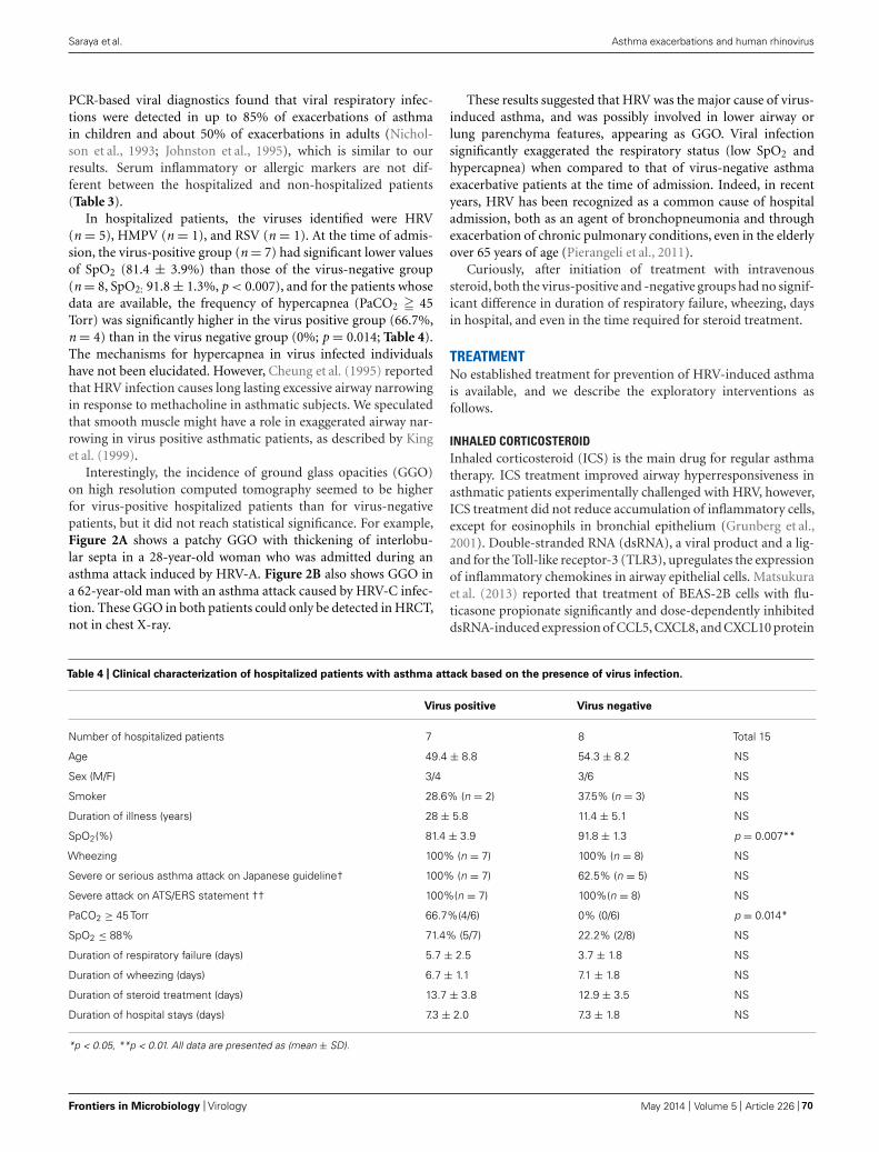
Saraya et al. Asthma exacerbations and human rhinovirus
PCR-based viral diagnostics found that viral respiratory infec-tions were detected in up to 85% of exacerbations of asthmain children and about 50% of exacerbations in adults (Nichol-son et al., 1993; Johnston et al., 1995), which is similar to ourresults. Serum inflammatory or allergic markers are not dif-ferent between the hospitalized and non-hospitalized patients(Table 3).
In hospitalized patients, the viruses identified were HRV(n = 5), HMPV (n = 1), and RSV (n = 1). At the time of admis-sion, the virus-positive group (n = 7) had significant lower valuesof SpO2 (81.4 ± 3.9%) than those of the virus-negative group(n = 8, SpO2: 91.8 ± 1.3%, p < 0.007), and for the patients whosedata are available, the frequency of hypercapnea (PaCO2 � 45Torr) was significantly higher in the virus positive group (66.7%,n = 4) than in the virus negative group (0%; p = 0.014; Table 4).The mechanisms for hypercapnea in virus infected individualshave not been elucidated. However, Cheung et al. (1995) reportedthat HRV infection causes long lasting excessive airway narrowingin response to methacholine in asthmatic subjects. We speculatedthat smooth muscle might have a role in exaggerated airway nar-rowing in virus positive asthmatic patients, as described by Kinget al. (1999).
Interestingly, the incidence of ground glass opacities (GGO)on high resolution computed tomography seemed to be higherfor virus-positive hospitalized patients than for virus-negativepatients, but it did not reach statistical significance. For example,Figure 2A shows a patchy GGO with thickening of interlobu-lar septa in a 28-year-old woman who was admitted during anasthma attack induced by HRV-A. Figure 2B also shows GGO ina 62-year-old man with an asthma attack caused by HRV-C infec-tion. These GGO in both patients could only be detected in HRCT,not in chest X-ray.
These results suggested that HRV was the major cause of virus-induced asthma, and was possibly involved in lower airway orlung parenchyma features, appearing as GGO. Viral infectionsignificantly exaggerated the respiratory status (low SpO2 andhypercapnea) when compared to that of virus-negative asthmaexacerbative patients at the time of admission. Indeed, in recentyears, HRV has been recognized as a common cause of hospitaladmission, both as an agent of bronchopneumonia and throughexacerbation of chronic pulmonary conditions, even in the elderlyover 65 years of age (Pierangeli et al., 2011).
Curiously, after initiation of treatment with intravenoussteroid, both the virus-positive and -negative groups had no signif-icant difference in duration of respiratory failure, wheezing, daysin hospital, and even in the time required for steroid treatment.
TREATMENTNo established treatment for prevention of HRV-induced asthmais available, and we describe the exploratory interventions asfollows.
INHALED CORTICOSTEROIDInhaled corticosteroid (ICS) is the main drug for regular asthmatherapy. ICS treatment improved airway hyperresponsiveness inasthmatic patients experimentally challenged with HRV, however,ICS treatment did not reduce accumulation of inflammatory cells,except for eosinophils in bronchial epithelium (Grunberg et al.,2001). Double-stranded RNA (dsRNA), a viral product and a lig-and for the Toll-like receptor-3 (TLR3), upregulates the expressionof inflammatory chemokines in airway epithelial cells. Matsukuraet al. (2013) reported that treatment of BEAS-2B cells with flu-ticasone propionate significantly and dose-dependently inhibiteddsRNA-induced expression of CCL5, CXCL8, and CXCL10 protein
Table 4 | Clinical characterization of hospitalized patients with asthma attack based on the presence of virus infection.
Virus positive Virus negative
Number of hospitalized patients 7 8 Total 15
Age 49.4 ± 8.8 54.3 ± 8.2 NS
Sex (M/F) 3/4 3/6 NS
Smoker 28.6% (n = 2) 37.5% (n = 3) NS
Duration of illness (years) 28 ± 5.8 11.4 ± 5.1 NS
SpO2(%) 81.4 ± 3.9 91.8 ± 1.3 p = 0.007**
Wheezing 100% (n = 7) 100% (n = 8) NS
Severe or serious asthma attack on Japanese guideline† 100% (n = 7) 62.5% (n = 5) NS
Severe attack on ATS/ERS statement †† 100%(n = 7) 100%(n = 8) NS
PaCO2 ≥ 45 Torr 66.7%(4/6) 0% (0/6) p = 0.014*
SpO2 ≤ 88% 71.4% (5/7) 22.2% (2/8) NS
Duration of respiratory failure (days) 5.7 ± 2.5 3.7 ± 1.8 NS
Duration of wheezing (days) 6.7 ± 1.1 7.1 ± 1.8 NS
Duration of steroid treatment (days) 13.7 ± 3.8 12.9 ± 3.5 NS
Duration of hospital stays (days) 7.3 ± 2.0 7.3 ± 1.8 NS
*p < 0.05, **p < 0.01. All data are presented as (mean ± SD).
Frontiers in Microbiology | Virology May 2014 | Volume 5 | Article 226 | 70

Saraya et al. Asthma exacerbations and human rhinovirus
FIGURE 2 | (A) Shows a patchy GGO with thickening of interlobular septa in a 28-year-old woman who was admitted during an asthma attack induced byHRV-A. (B) Also shows GGO in a 62-year-old man with an asthma attack caused by HRV-C infection.
and mRNA. To confirm the effect on ssRNA, such as that of HRV,would need further studies.
LEUKOTRIENE RECEPTOR ANTAGONISTLeukotriene receptor antagonist was prescribed in asthmaticpatients with or without ICS. Montelukast treatment didnot improve asthma control or cold symptom scores whenHRV were experimentally inoculated into mild asthmatics,or healthy subjects (Kloepfer et al., 2011). It is uncertainwhether leukotriene receptor antagonist treatment is effectivein the reduction of asthma symptoms associated with HRVinfection.
ANTI-IgE THERAPYZambrano et al. (2003) reported that high serum IgE levels inmildly asthmatic children with experimental HRV infection maybe associated with enhanced lower respiratory symptoms and ele-vation of inflammatory markers, such as nasal eosinophil cationicprotein and expired nitric oxide, than those of healthy subjectsand/or low IgE asthmatic patients. The prevalence of asthma wasclosely associated with the serum IgE levels standardized for ageand sex (Burrows et al., 1989), and airway hyperresponsivenessappears to be closely linked to the allergic diathesis, as reflectedby the serum total IgE level (Sears et al., 1991). Omalizumab,an anti-IgE monoclonal antibody, was indicated in inadequatelycontrolled moderate-to-severe persistent allergic asthma patientswho were treated with high dose ICS. Durrani et al. (2012)showed that the IgE receptor FcεRI is inversely associated withIFN-α and IFN-λ1 secretion when plasmacytoid dendritic cellsderived from allergic asthmatic children were challenged withHRV. Omalizumab downregulates FcεRI expression on dendriticcells (Prussin et al., 2003), which may reduce exacerbation ofasthma associated with increased production of IFNs, throughFcεRI.
ANTI-VIRAL TREATMENTNo drugs are clinically used in HRV infection, although sev-eral drugs have been tried for treatment and prevention of HRV
infection. These drugs are summarized in a review (Jacobs et al.,2013). IFNs had a potential protective role in viral inducedasthma (Cakebread et al., 2011; Gaajetaan et al., 2013). Becker et al.(2013) showed that exogenous IFN-α, IFN-β, IFN-λ1, and IFN-λ2inhibited HRV replication in BECs from healthy donors.
MACROLIDE THERAPYMacrolides are known to possess anti-inflammatory andimmunomodulatory actions extending beyond their antibacte-rial activity in pulmonary inflammatory disorders (Takizawa et al.,1995; Min and Jang, 2012). Erythromycin inhibits HRV infectionby reducing ICAM-1 expression on the surface of human trachealepithelial cells, and modulates inflammation by suppressing theproduction of proinflammatory cytokines (Suzuki et al., 2002).
OTHER AGENTSYamaya et al. (2014) reported that the mucolytic drug ambroxolhydrochloride, antibiotic drug of levofloxacin (Yamaya et al.,2012b), and bronchodilators (Tiotropium, Tulobuterol, and Pro-caterol) for asthma or COPD (Yamaya et al., 2011, 2012a, 2013)may have a beneficial effect in HRV infection, by inhibiting HRVreplication and partly reducing ICAM-1 expression and acidicendosome production, via the inhibition of NF-kappaB activation(Yamaya, 2012).
SUMMARYWe reviewed the previous reports regarding HRV-induced asthmaexacerbations, together with our results from an institutionalprospective study. HRV is a major pathogen for asthma exac-erbations, and certainly associated with more serious clinicalconditions such as hypoxemia or hypercapnea in hospitalizedpatients. Further accumulation of evidence of virus-inducedasthma for multidisciplinary assessment would be helpful forphysicians in recognizing the condition or understanding thepathogenic mechanisms.
REFERENCESAndries, K., Dewindt, B., Snoeks, J., Wouters, L., Moereels, H., Lewi, P. J., et al.
(1990). Two groups of rhinoviruses revealed by a panel of antiviral compounds
www.frontiersin.org May 2014 | Volume 5 | Article 226 | 71

Saraya et al. Asthma exacerbations and human rhinovirus
present sequence divergence and differential pathogenicity. J. Virol. 64, 1117–1123.
Arakawa, M., Okamoto-Nakagawa, R., Toda, S., Tsukagoshi, H., Kobayashi, M., Ryo,A., et al. (2012). Molecular epidemiological study of human rhinovirus species A,B and C from patients with acute respiratory illnesses in Japan. J. Med. Microbiol.61, 410–419. doi: 10.1099/jmm.0.035006-0
Arruda, E., Pitkaranta, A., Witek, T. J. Jr., Doyle, C. A., and Hayden, F. G. (1997).Frequency and natural history of rhinovirus infections in adults during autumn.J. Clin. Microbiol. 35, 2864–2868.
Atmar, R. L., Guy, E., Guntupalli, K. K., Zimmerman, J. L., Bandi, V. D., Baxter, B.D., et al. (1998). Respiratory tract viral infections in inner-city asthmatic adults.Arch. Intern. Med. 158, 2453–2459. doi: 10.1001/archinte.158.22.2453
Bartlett, N. W., Walton, R. P., Edwards, M. R., Aniscenko, J., Caramori, G., Zhu,J., et al. (2008). Mouse models of rhinovirus-induced disease and exacerba-tion of allergic airway inflammation. Nat. Med. 14, 199–204. doi: 10.1038/nm1713
Becker, T. M., Durrani, S. R., Bochkov, Y. A., Devries, M. K., Rajamanickam, V., andJackson, D. J. (2013). Effect of exogenous interferons on rhinovirus replicationand airway inflammatory responses. Ann. Allergy Asthma Immunol. 111, 397–401.doi: 10.1016/j.anai.2013.07.029
Beckham, J. D., Cadena, A., Lin, J., Piedra, P. A., Glezen, W. P., Green-berg, S. B., et al. (2005). Respiratory viral infections in patients with chronic,obstructive pulmonary disease. J. Infect. 50, 322–330. doi: 10.1016/j.jinf.2004.07.011
Bochkov, Y. A., Hanson, K. M., Keles, S., Brockman-Schneider, R. A., Jarjour, N.N., and Gern, J. E. (2010). Rhinovirus-induced modulation of gene expression inbronchial epithelial cells from subjects with asthma. Mucosal Immunol. 3, 69–80.doi: 10.1038/mi.2009.109
Burrows, B., Martinez, F. D., Halonen, M., Barbee, R. A., and Cline, M. G. (1989).Association of asthma with serum IgE levels and skin-test reactivity to allergens.N. Engl. J. Med. 320, 271–277. doi: 10.1056/NEJM198902023200502
Cakebread, J. A., Xu, Y., Grainge, C., Kehagia, V., Howarth, P. H., Holgate, S. T.,et al. (2011). Exogenous IFN-beta has antiviral and anti-inflammatory propertiesin primary bronchial epithelial cells from asthmatic subjects exposed to rhi-novirus. J. Allergy Clin. Immunol. 127, 1148e9–1154e9. doi: 10.1016/j.jaci.2011.01.023
Cheung, D., Dick, E. C., Timmers, M. C., De Klerk, E. P., Spaan, W. J., and Sterk, P. J.(1995). Rhinovirus inhalation causes long-lasting excessive airway narrowing inresponse to methacholine in asthmatic subjects in vivo. Am. J. Respir. Crit. CareMed. 152, 1490–1496. doi: 10.1164/ajrccm.152.5.7582282
Chun, Y. H., Park, J. Y., Lee, H., Kim, H. S., Won, S., Joe, H. J., et al. (2013).Rhinovirus-infected epithelial cells produce more IL-8 and rantes comparedwith other respiratory viruses. Allergy Asthma Immunol. Res. 5, 216–223. doi:10.4168/aair.2013.5.4.216
Contoli, M., Message, S. D., Laza-Stanca, V., Edwards, M. R., Wark, P. A.,Bartlett, N. W., et al. (2006). Role of deficient type III interferon-lambda pro-duction in asthma exacerbations. Nat. Med. 12, 1023–1026. doi: 10.1038/nm1462
Corne, J. M., Marshall, C., Smith, S., Schreiber, J., Sanderson, G., Holgate, S. T., et al.(2002). Frequency, severity, and duration of rhinovirus infections in asthmaticand non-asthmatic individuals: a longitudinal cohort study. Lancet 359, 831–834.doi: 10.1016/S0140-6736(02)07953-9
DeMore, J. P., Weisshaar, E. H., Vrtis, R. F., Swenson, C. A., Evans, M. D., Morin, A.,et al. (2009). Similar colds in subjects with allergic asthma and nonatopic subjectsafter inoculation with rhinovirus-16. J. Allergy Clin. Immunol. 124, 245–252. doi:10.1016/j.jaci.2009.05.030
Dimopoulos, G., Lerikou, M., Tsiodras, S., Chranioti, A., Perros, E., Anag-nostopoulou, U., et al. (2012). Viral epidemiology of acute exacerbations ofchronic obstructive pulmonary disease. Pulm. Pharmacol. Ther. 25, 12–18. doi:10.1016/j.pupt.2011.08.004
Durrani, S. R., Montville, D. J., Pratt, A. S., Sahu, S., Devries, M. K., Rajamanickam,V., et al. (2012). Innate immune responses to rhinovirus are reduced by the high-affinity IgE receptor in allergic asthmatic children. J. Allergy Clin. Immunol. 130,489–495. doi: 10.1016/j.jaci.2012.05.023
Dweik, R. A., Boggs, P. B., Erzurum, S. C., Irvin, C. G., Leigh, M. W., Lundberg, J. O.,et al. (2011). An official ATS clinical practice guideline: interpretation of exhalednitric oxide levels (FENO) for clinical applications. Am. J. Respir. Crit. Care Med.184, 602–615. doi: 10.1164/rccm.9120-11ST
Fashner, J., Ericson, K., and Werner, S. (2012). Treatment of the common cold inchildren and adults. Am. Fam. Phys. 86, 153–159.
Feltis, B. N., Wignarajah, D., Zheng, L., Ward, C., Reid, D., Harding, R., et al.(2006). Increased vascular endothelial growth factor and receptors: relationshipto angiogenesis in asthma. Am. J. Respir. Crit. Care Med. 173, 1201–1207. doi:10.1164/rccm.200507-1105OC
Fraenkel, D. J., Bardin, P. G., Sanderson, G., Lampe, F., Johnston, S. L., and Holgate,S. T. (1995). Lower airways inflammation during rhinovirus colds in normal andin asthmatic subjects. Am. J. Respir. Crit. Care Med. 151, 879–886.
Fry, A. M., Lu, X., Olsen, S. J., Chittaganpitch, M., Sawatwong, P., Chantra, S.,et al. (2011). Human rhinovirus infections in rural Thailand: epidemiologicalevidence for rhinovirus as both pathogen and bystander. PLoS ONE 6:e17780.doi: 10.1371/journal.pone.0017780
Fujitsuka, A., Tsukagoshi, H., Arakawa, M., Goto-Sugai, K., Ryo, A., Okayama, Y.,et al. (2011). A molecular epidemiological study of respiratory viruses detectedin Japanese children with acute wheezing illness. BMC Infect. Dis. 11:168. doi:10.1186/1471-2334-11-168
Gaajetaan, G. R., Geelen, T. H., Vernooy, J. H., Dentener, M. A., Reynaert, N.L., Rohde, G. G., et al. (2013). Interferon-beta induces a long-lasting antivi-ral state in human respiratory epithelial cells. J. Infect. 66, 163–169. doi:10.1016/j.jinf.2012.11.008
Grainge, C. L., Lau, L. C., Ward, J. A., Dulay, V., Lahiff, G., Wilson, S., et al. (2011).Effect of bronchoconstriction on airway remodeling in asthma. N. Engl. J. Med.364, 2006–2015. doi: 10.1056/NEJMoa1014350
Grunberg, K., Sharon, R. F., Sont, J. K., In ‘t Veen, J. C., Van Schadewijk, W. A., DeKlerk, E. P., et al. (2001). Rhinovirus-induced airway inflammation in asthma:effect of treatment with inhaled corticosteroids before and during experimentalinfection. Am. J. Respir. Crit. Care Med. 164, 1816–1822.
Grunberg, K., Timmers, M. C., De Klerk, E. P., Dick, E. C., and Sterk, P. J. (1999).Experimental rhinovirus 16 infection causes variable airway obstruction in sub-jects with atopic asthma. Am. J. Respir. Crit. Care Med. 160, 1375–1380. doi:10.1164/ajrccm.160.4.9810083
Grunstein, M. M., Hakonarson, H., Whelan, R., Yu, Z., Grunstein, J. S., andChuang, S. (2001). Rhinovirus elicits proasthmatic changes in airway responsive-ness independently of viral infection. J. Allergy Clin. Immunol. 108, 997–1004.doi: 10.1067/mai.2001.120276
Gwaltney, J. M. Jr., Hendley, J. O., Simon, G., and Jordan, W. S. Jr. (1967). Rhinovirusinfections in an industrial population. II. Characteristics of illness and antibodyresponse. JAMA 202, 494–500. doi: 10.1001/jama.1967.03130190100014
Hewson, C. A., Haas, J. J., Bartlett, N. W., Message, S. D., Laza-Stanca, V., Kebadze,T., et al. (2010). Rhinovirus induces Muc5ac in a human infection model andin vitro via NF-kappaB and EGFR pathways. Eur. Respir. J. 36, 1425–1435. doi:10.1183/09031936.00026910
Hsiao, C. J., Cherry, D. K., Beatty, P. C., and Rechtsteiner, E. A. (2010). Nationalambulatory medical care survey: 2007 summary. Natl. Health Stat. Report 1:32.
Huijskens, E. G., Koopmans, M., Palmen, F. M., Van Erkel, A. J., Mulder, P. G.,and Rossen, J. W. (2014). The value of signs and symptoms in differentiatingbetween bacterial, viral and mixed aetiology in patients with community-acquired pneumonia. J. Med. Microbiol. 63, 441–452. doi: 10.1099/jmm.0.067108-0
Hutchinson, A. F., Ghimire, A. K., Thompson, M. A., Black, J. F., Brand, C. A.,Lowe, A. J., et al. (2007). A community-based, time-matched, case-control studyof respiratory viruses and exacerbations of COPD. Respir. Med. 101, 2472–2481.doi: 10.1016/j.rmed.2007.07.015
Jackson, D. J., and Johnston, S. L. (2010). The role of viruses in acute exacerbations ofasthma. J. Allergy Clin. Immunol. 125, 1178-1187. doi: 10.1016/j.jaci.2010.04.021
Jacobs, S. E., Lamson, D. M., St George, K., and Walsh, T. J. (2013). Humanrhinoviruses. Clin. Microbiol. Rev. 26, 135–162. doi: 10.1128/CMR.00077-12
Jennings, L. C., Anderson, T. P., Beynon, K. A., Chua, A., Laing, R. T., Werno,A. M., et al. (2008). Incidence and characteristics of viral community-acquiredpneumonia in adults. Thorax 63, 42–48. doi: 10.1136/thx.2006.075077
Johansson, N., Kalin, M., Tiveljung-Lindell, A., Giske, C. G., and Hedlund, J. (2010).Etiology of community-acquired pneumonia: increased microbiological yieldwith new diagnostic methods. Clin. Infect. Dis. 50, 202–209. doi: 10.1086/648678
Johnstone, J., Majumdar, S. R., Fox, J. D., and Marrie, T. J. (2008). Viralinfection in adults hospitalized with community-acquired pneumonia: preva-lence, pathogens, and presentation. Chest 134, 1141–1148. doi: 10.1378/chest.08-0888
Frontiers in Microbiology | Virology May 2014 | Volume 5 | Article 226 | 72

Saraya et al. Asthma exacerbations and human rhinovirus
Johnston, S. L., Pattemore, P. K., Sanderson, G., Smith, S., Campbell, M. J., Josephs,L. K., et al. (1996). The relationship between upper respiratory infections andhospital admissions for asthma: a time-trend analysis. Am. J. Respir. Crit. CareMed. 154, 654–660. doi: 10.1164/ajrccm.154.3.8810601
Johnston, S. L., Pattemore, P. K., Sanderson, G., Smith, S., Lampe, F., Josephs, L., et al.(1995). Community study of role of viral infections in exacerbations of asthmain 9-11 year old children. BMJ 310, 1225–1229. doi: 10.1136/bmj.310.6989.1225
Kennedy, J. L., Turner, R. B., Braciale, T., Heymann, P. W., and Borish, L.(2012). Pathogenesis of rhinovirus infection. Curr. Opin. Virol. 2, 287–293. doi:10.1016/j.coviro.2012.03.008
Kherad, O., Kaiser, L., Bridevaux, P. O., Sarasin, F., Thomas, Y., Janssens, J. P., et al.(2010). Upper-respiratory viral infection, biomarkers, and COPD exacerbations.Chest 138, 896–904. doi: 10.1378/chest.09-2225
King, G. G., Pare, P. D., and Seow, C. Y. (1999). The mechanics of exaggerated airwaynarrowing in asthma: the role of smooth muscle. Respir. Physiol. 118, 1–13. doi:10.1016/S0034-5687(99)00076-6
Kiyota, N., Kobayashi, M., Tsukagoshi, H., Ryo, A., Harada, S., Kusaka, T., et al.(2014). Genetic analysis of human rhinovirus species A to C detected in patientswith acute respiratory infection in Kumamoto prefecture, Japan 2011–2012.Infect. Genet. Evol. 21, 90–102. doi: 10.1016/j.meegid.2013.10.024
Kiyota, N., Kushibuchi, I., Kobayashi, M., Tsukagoshi, H., Ryo, A., Nishimura, K.,et al. (2013). Genetic analysis of the VP4/VP2 coding region in human rhinovirusspecies C in patients with acute respiratory infection in Japan. J. Med. Microbiol.62, 610–617. doi: 10.1099/jmm.0.049072-0
Kloepfer, K. M., Demore, J. P., Vrtis, R. F., Swenson, C. A., Gaworski, K. L., Bork, J. A.,et al. (2011). Effects of montelukast on patients with asthma after experimentalinoculation with human rhinovirus 16. Ann. Allergy Asthma Immunol. 106, 252–257. doi: 10.1016/j.anai.2010.11.021
Ko, F. W., Ip, M., Chan, P. K., Chan, M. C., To, K. W., Ng, S. S., et al. (2007). Viraletiology of acute exacerbations of COPD in Hong Kong. Chest 132, 900–908. doi:10.1378/chest.07-0530
Kuga, H., Hoshiyama, Y., Kokubu, F., Imai, T., Tokunaga, H., Matsukura, S., et al.(2000). The correlation between the exacerbation of bronchial asthma and picor-navirus (human rhino virus) infection in throat gargles by RT-PCR. Arerugi 49,358–364.
Kurai, D., Saraya, T., Ishii, H., and Takizawa, H. (2013a). Virus-induced exacerbations in asthma and COPD. Front. Microbiol. 4:293. doi:10.3389/fmicb.2013.00293
Kurai, D., Saraya, T., Ishii, H., Wada, H., Tsukagoshi, H., Takizawa, H., et al. (2013b).Respiratory viral infection in admitted adult patients. Respirology 18, 1–81.
Leigh, R., Oyelusi, W., Wiehler, S., Koetzler, R., Zaheer, R. S., Newton, R., et al.(2008). Human rhinovirus infection enhances airway epithelial cell productionof growth factors involved in airway remodeling. J. Allergy Clin. Immunol. 121,1238e4–1245e4. doi: 10.1016/j.jaci.2008.01.067
Lieberman, D., Lieberman, D., Shimoni, A., Keren-Naus, A., Steinberg, R., andShemer-Avni, Y. (2009). Identification of respiratory viruses in adults: nasopha-ryngeal versus oropharyngeal sampling. J. Clin. Microbiol. 47, 3439–3443. doi:10.1128/JCM.00886-09
Lieberman, D., Shimoni, A., Shemer-Avni, Y., Keren-Naos, A., Shtainberg, R., andLieberman, D. (2010). Respiratory viruses in adults with community-acquiredpneumonia. Chest 138, 811–816. doi: 10.1378/chest.09-2717
Linsuwanon, P., Payungporn, S., Samransamruajkit, R., Posuwan, N., Makkoch,J., Theanboonlers, A., et al. (2009). High prevalence of human rhinovirus Cinfection in Thai children with acute lower respiratory tract disease. J. Infect. 59,115–121. doi: 10.1016/j.jinf.2009.05.009
Loens, K., Goossens, H., De Laat, C., Foolen, H., Oudshoorn, P., Pattyn, S.,et al. (2006). Detection of rhinoviruses by tissue culture and two independentamplification techniques, nucleic acid sequence-based amplification and reversetranscription-PCR, in children with acute respiratory infections during a winterseason. J. Clin. Microbiol. 44, 166–171. doi: 10.1128/JCM.44.1.166-171.2006
Lopez-Souza, N., Favoreto, S., Wong, H., Ward, T., Yagi, S., Schnurr, D., et al. (2009).In vitro susceptibility to rhinovirus infection is greater for bronchial than fornasal airway epithelial cells in human subjects. J. Allergy Clin. Immunol. 123,1384e2–1390e2. doi: 10.1016/j.jaci.2009.03.010
Luchsinger, V., Ruiz, M., Zunino, E., Martinez, M. A., Machado, C., Piedra, P. A.,et al. (2013). Community-acquired pneumonia in Chile: the clinical relevancein the detection of viruses and atypical bacteria. Thorax 68, 1000–1006. doi:10.1136/thoraxjnl-2013-203551
Makela, M. J., Puhakka, T., Ruuskanen, O., Leinonen, M., Saikku, P., Kimpimaki,M., et al. (1998). Viruses and bacteria in the etiology of the common cold. J. Clin.Microbiol. 36, 539–542.
Matsukura, S., Kurokawa, M., Homma, T., Watanabe, S., Suzuki, S., Ieki, K.,et al. (2013). Basic research on virus-induced asthma exacerbation: inhibition ofinflammatory chemokine expression by fluticasone propionate. Int. Arch. AllergyImmunol. 161(Suppl. 2), 84–92. doi: 10.1159/000350455
McFadden, E. R. Jr., Pichurko, B. M., Bowman, H. F., Ingenito, E., Burns, S., Dowling,N., et al. (1985). Thermal mapping of the airways in humans. J. Appl. Physiol. 58,564–570.
McManus, T. E., Marley, A. M., Baxter, N., Christie, S. N., O’Neill, H. J., Elborn, J. S.,et al. (2008). Respiratory viral infection in exacerbations of COPD. Respir. Med.102, 1575–1580. doi: 10.1016/j.rmed.2008.06.006
Message, S. D., Laza-Stanca, V., Mallia, P., Parker, H. L., Zhu, J., Kebadze, T., et al.(2008). Rhinovirus-induced lower respiratory illness is increased in asthma andrelated to virus load and Th1/2 cytokine and IL-10 production. Proc. Natl. Acad.Sci. U.S.A. 105, 13562–13567. doi: 10.1073/pnas.0804181105
Min, J. Y., and Jang, Y. J. (2012). Macrolide therapy in respiratory viral infections.Mediators Inflamm. 2012, 649570. doi: 10.1155/2012/649570
Nagarkar, D. R., Bowman, E. R., Schneider, D., Wang, Q., Shim, J., Zhao, Y.,et al. (2010). Rhinovirus infection of allergen-sensitized and -challenged miceinduces eotaxin release from functionally polarized macrophages. J. Immunol.185, 2525–2535. doi: 10.4049/jimmunol.1000286
Nicholson, K. G., Kent, J., and Ireland, D. C. (1993). Respiratory viruses and exacer-bations of asthma in adults. BMJ 307, 982–986. doi: 10.1136/bmj.307.6910.982
Ohta, K., Yamaguchi, M., Akiyama, K., Adachi, M., Ichinose, M., Takahashi, K.,et al. (2011). Japanese guideline for adult asthma. Allergol. Int. 60, 115–145. doi:10.2332/allergolint.11-RAI-0327
Papi, A., Bellettato, C. M., Braccioni, F., Romagnoli, M., Casolari, P., Caramori,G., et al. (2006). Infections and airway inflammation in chronic obstructivepulmonary disease severe exacerbations. Am. J. Respir. Crit. Care Med. 173,1114–1121. doi: 10.1164/rccm.200506-859OC
Perotin, J. M., Dury, S., Renois, F., Deslee, G., Wolak, A., Duval, V., et al. (2013).Detection of multiple viral and bacterial infections in acute exacerbation ofchronic obstructive pulmonary disease: a pilot prospective study. J. Med. Virol.85, 866–873. doi: 10.1002/jmv.23495
Pierangeli, A., Scagnolari, C., Selvaggi, C.,Verzaro, S., Spina, M. T., Bresciani, E., et al.(2011). Rhinovirus frequently detected in elderly adults attending an emergencydepartment. J. Med. Virol. 83, 2043–2047. doi: 10.1002/jmv.22205
Pohunek, P., Warner, J. O., Turzikova, J., Kudrmann, J., and Roche, W. R. (2005).Markers of eosinophilic inflammation and tissue re-modelling in children beforeclinically diagnosed bronchial asthma. Pediatr. Allergy Immunol. 16, 43–51. doi:10.1111/j.1399-3038.2005.00239.x
Proud, D. (2005). Nitric oxide and the common cold. Curr. Opin. Allergy Clin.Immunol. 5, 37–42. doi: 10.1097/00130832-200502000-00008
Proud, D. (2011). Role of rhinovirus infections in asthma. Asian Pac. J. AllergyImmunol. 29, 201–208.
Prussin, C., Griffith, D. T., Boesel, K. M., Lin, H., Foster, B., and Casale,T. B. (2003). Omalizumab treatment downregulates dendritic cell Fcepsilonriexpression. J. Allergy Clin. Immunol. 112, 1147–1154. doi: 10.1016/j.jaci.2003.10.003
Reddel, H. K., Taylor, D. R., Bateman, E. D., Boulet, L. P., Boushey, H. A., Busse,W. W., et al. (2009). An official American Thoracic Society/European RespiratorySociety statement: asthma control and exacerbations: standardizing endpointsfor clinical asthma trials and clinical practice. Am. J. Respir. Crit. Care Med. 180,59–99. doi: 10.1164/rccm.200801-060ST
Rohde, G., Wiethege, A., Borg, I., Kauth, M., Bauer, T. T., Gillissen, A., et al.(2003). Respiratory viruses in exacerbations of chronic obstructive pulmonarydisease requiring hospitalisation: a case-control study. Thorax 58, 37–42. doi:10.1136/thorax.58.1.37
Rotbart, H. A., and Hayden, F. G. (2000). Picornavirus infections: a primer for thepractitioner. Arch. Fam. Med. 9, 913–920. doi: 10.1001/archfami.9.9.913
Russell, C. D., Griffiths, S. J., and Haas, J. (2014). Interferon lambda genetic poly-morphisms and viral infection: the tip of the iceberg? DNA Cell Biol. 33, 60–63.doi: 10.1089/dna.2013.2261
Sampsonas, F., Kaparianos, A., Lykouras, D., Karkoulias, K., and Spiropoulos, K.(2007). DNA sequence variations of metalloproteinases: their role in asthma andCopd. Postgrad. Med. J. 83, 244–250. doi: 10.1136/pgmj.2006.052100
www.frontiersin.org May 2014 | Volume 5 | Article 226 | 73

Saraya et al. Asthma exacerbations and human rhinovirus
Sanders, S. P., Proud, D., Permutt, S., Siekierski, E. S., Yachechko, R., and Liu,M. C. (2004). Role of nasal nitric oxide in the resolution of experimental rhi-novirus infection. J. Allergy Clin. Immunol. 113, 697–702. doi: 10.1016/j.jaci.2004.01.755
Sears, M. R., Burrows, B., Flannery, E. M., Herbison, G. P., Hewitt, C. J., andHoldaway, M. D. (1991). Relation between airway responsiveness and serum IgEin children with asthma and in apparently normal children. N. Engl. J. Med. 325,1067–1071. doi: 10.1056/NEJM199110103251504
Seemungal, T., Harper-Owen, R., Bhowmik, A., Moric, I., Sanderson, G., Message,S., et al. (2001). Respiratory viruses, symptoms, and inflammatory markers inacute exacerbations and stable chronic obstructive pulmonary disease. Am. J.Respir. Crit. Care Med. 164, 1618–1623. doi: 10.1164/ajrccm.164.9.2105011
Simcock, D. E., Kanabar, V., Clarke, G. W., O’Connor, B. J., Lee, T. H., and Hirst, S. J.(2007). Proangiogenic activity in bronchoalveolar lavage fluid from patients withasthma. Am. J. Respir. Crit. Care Med. 176, 146–153. doi: 10.1164/rccm.200701-042OC
Simmonds, P., McIntyre, C., Savolainen-Kopra, C., Tapparel, C., MacKay, I.M., and Hovi, T. (2010). Proposals for the classification of human rhinovirusspecies C into genotypically assigned types. J. Gen. Virol. 91, 2409–2419. doi:10.1099/vir.0.023994-0
Smuts, H. E., Workman, L. J., and Zar, H. J. (2011). Human rhinovirus infectionin young African children with acute wheezing. BMC Infect. Dis. 11:65. doi:10.1186/1471-2334-11-65
Suzuki, T., Yamaya, M., Sekizawa, K., Hosoda, M., Yamada, N., Ishizuka, S.,et al. (2002). Erythromycin inhibits rhinovirus infection in cultured humantracheal epithelial cells. Am. J. Respir. Crit. Care Med. 165, 1113–1118. doi:10.1164/ajrccm.165.8.2103094
Sykes, A., MacIntyre, J., Edwards, M. R., Del Rosario, A., Haas, J., Gielen, V.,et al. (2014). Rhinovirus-induced interferon production is not deficient in wellcontrolled asthma. Thorax 69, 240–246. doi: 10.1136/thoraxjnl-2012-202909
Takahashi, K., Suzuki, M., Minh Le, N., Anh, N. H., Huong, L. T., Son, T. V., et al.(2013). The incidence and aetiology of hospitalised community-acquired pneu-monia among Vietnamese adults: a prospective surveillance in Central Vietnam.BMC Infect. Dis. 13:296. doi: 10.1186/1471-2334-13-296
Takizawa, H., Desaki, M., Ohtoshi, T., Kikutani, T., Okazaki, H., Sato, M., et al.(1995). Erythromycin suppresses interleukin 6 expression by human bronchialepithelial cells: a potential mechanism of its anti-inflammatory action. Biochem.Biophys. Res. Commun. 210, 781–786. doi: 10.1006/bbrc.1995.1727
Tan, W. C., Xiang, X., Qiu, D., Ng, T. P., Lam, S. F., and Hegele, R.G. (2003). Epidemiology of respiratory viruses in patients hospitalized withnear-fatal asthma, acute exacerbations of asthma, or chronic obstructive pul-monary disease. Am. J. Med. 115, 272–277. doi: 10.1016/S0002-9343(03)00353-X
Tsukagoshi, H., Ishioka, T., Noda, M., Kozawa, K., and Kimura, H. (2013). Molecularepidemiology of respiratory viruses in virus-induced asthma. Front. Microbiol.4:278. doi: 10.3389/fmicb.2013.00278
Turner, R. B., and Couch, R. B. (2007). “Rhinovirus,” in Fields Virology, 5th edn, edsD. M. Knipe, P. M. Howley, D. E. Griffin, R. A. Lamb, M. A. Martin, B. Roizman,and S. E. Straus (Philadelphia: Lippincott Williams and Wilkins), 895–909.
Tyrrell, D. A., Cohen, S., and Schlarb, J. E. (1993). Signs and symptoms incommon colds. Epidemiol. Infect. 111, 143–156. doi: 10.1017/S0950268800056764
van Gageldonk-Lafeber, A. B., Heijnen, M. L., Bartelds, A. I., Peters, M. F., Van DerPlas, S. M., and Wilbrink, B. (2005). A case-control study of acute respiratorytract infection in general practice patients in The Netherlands. Clin. Infect. Dis.41, 490–497. doi: 10.1086/431982
Wark, P. A., Johnston, S. L., Bucchieri, F., Powell, R., Puddicombe, S., Laza-Stanca, V., et al. (2005). Asthmatic bronchial epithelial cells have a deficient innateimmune response to infection with rhinovirus. J. Exp. Med. 201, 937–947. doi:10.1084/jem.20041901
Wark, P. A., Johnston, S. L., Moric, I., Simpson, J. L., Hensley, M. J., andGibson, P. G. (2002). Neutrophil degranulation and cell lysis is associatedwith clinical severity in virus-induced asthma. Eur. Respir. J. 19, 68–75. doi:10.1183/09031936.02.00226302
Watanabe, A., Carraro, E., Kamikawa, J., Leal, E., Granato, C., and Bellei, N. (2010).Rhinovirus species and their clinical presentation among different risk groups ofnon-hospitalized patients. J. Med. Virol. 82, 2110–2115. doi: 10.1002/jmv.21914
Wootton, S. C., Kim, D. S., Kondoh, Y., Chen, E., Lee, J. S., Song, J. W., et al. (2011).Viral infection in acute exacerbation of idiopathic pulmonary fibrosis. Am. J.Respir. Crit. Care Med. 183, 1698–1702. doi: 10.1164/rccm.201010-1752OC
Yamaya, M. (2012). Virus infection-induced bronchial asthma exacerbation. Pulm.Med. 2012, 834826. doi: 10.1155/2012/834826
Yamaya, M., Nishimura, H., Hatachi, Y., Yasuda, H., Deng, X., Sasaki, T., et al.(2012a). Inhibitory effects of tiotropium on rhinovirus infection in human airwayepithelial cells. Eur. Respir. J. 40, 122–132. doi: 10.1183/09031936.00065111
Yamaya, M., Nishimura, H., Hatachi, Y., Yasuda, H., Deng, X., Sasaki, T., et al.(2012b). Levofloxacin inhibits rhinovirus infection in primary cultures of humantracheal epithelial cells. Antimicrob. Agents Chemother. 56, 4052–4061. doi:10.1128/AAC.00259-12
Yamaya, M., Nishimura, H., Hatachi, Y., Yoshida, M., Fujiwara, H., Asada,M., et al. (2011). Procaterol inhibits rhinovirus infection in primary culturesof human tracheal epithelial cells. Eur. J. Pharmacol. 650, 431–444. doi:10.1016/j.ejphar.2010.09.056
Yamaya, M., Nishimura, H., Nadine, L., Kubo, H., and Ryoichi, N. (2013).Tulobuterol inhibits rhinovirus infection in primary cultures of human trachealepithelial cells. Physiol. Rep. 1, e00041. doi: 10.1002/phy2.41
Yamaya, M., Nishimura, H., Nadine, L. K., Ota, C., Kubo, H., and Nagatomi, R.(2014). Ambroxol inhibits rhinovirus infection in primary cultures of humantracheal epithelial cells. Arch. Pharm. Res. 37, 520–529. doi: 10.1007/s12272-013-0210-7
Zambrano, J. C., Carper, H. T., Rakes, G. P., Patrie, J., Murphy, D. D., Platts-Mills, T.A., et al. (2003). Experimental rhinovirus challenges in adults with mild asthma:response to infection in relation to IgE. J. Allergy Clin. Immunol. 111, 1008–1016.doi: 10.1067/mai.2003.1396
Conflict of Interest Statement: The authors declare that the research was conductedin the absence of any commercial or financial relationships that could be construedas a potential conflict of interest.
Received: 30 March 2014; accepted: 28 April 2014; published online: 26 May 2014.Citation: Saraya T, Kurai D, Ishii H, Ito A, Sasaki Y, Niwa S, Kiyota N, TsukagoshiH, Kozawa K, Goto H and Takizawa H (2014) Epidemiology of virus-induced asthmaexacerbations: with special reference to the role of human rhinovirus. Front. Microbiol.5:226. doi: 10.3389/fmicb.2014.00226This article was submitted to Virology, a section of the journal Frontiers in Microbiology.Copyright © 2014 Saraya, Kurai, Ishii, Ito, Sasaki, Niwa, Kiyota, Tsukagoshi, Kozawa,Goto and Takizawa. This is an open-access article distributed under the terms of theCreative Commons Attribution License (CC BY). The use, distribution or reproductionin other forums is permitted, provided the original author(s) or licensor are creditedand that the original publication in this journal is cited, in accordance with acceptedacademic practice. No use, distribution or reproduction is permitted which does notcomply with these terms.
Frontiers in Microbiology | Virology May 2014 | Volume 5 | Article 226 |74

“fmicb-04-00307” — 2013/10/10 — 17:54 — page 1 — #1
MINI REVIEW ARTICLEpublished: 14 October 2013
doi: 10.3389/fmicb.2013.00307
Influenza A(H1N1)pdm09 virus and asthmaMasatsugu Obuchi 1*, Yuichi Adachi 2 , TakenoriTakizawa1 and Tetsutaro Sata1
1 Department of Virology, Toyama Institute of Health, Toyama, Japan2 Department of Pediatrics, Faculty of Medicine, University of Toyama, Toyama, Japan
Edited by:
Hirokazu Kimura, National Institute ofInfectious Diseases, Japan
Reviewed by:
Jianwei Wang, Chinese Academy ofMedical Sciences, Peking UnionMedical College, ChinaWei Zhang, Yele University, USA
*Correspondence:
Masatsugu Obuchi, Department ofVirology, Toyama Institute of Health,17-1 Nakataikoyama, Imizu-shi,Toyama 939-0363, Japane-mail: [email protected]
Respiratory viral infection is a major cause of asthma exacerbations in both children andadults. Among the respiratory viruses, influenza virus is a particularly important pathogendue to its enormous morbidity and mortality in annual epidemics.The swine-origin influenzaA virus, designated as A(H1N1)pdm09, emerged in the spring of 2009 and caused the firstinfluenza pandemic in the 21st century. With the emergence of the novel A(H1N1)pdm09virus, numerous epidemiologic studies detected asthma as a frequent comorbid conditionin patients infected with this virus. Here we review recent reports regarding asthma inpatients infected with influenza A(H1N1)pdm09 virus, and we discuss the utility of influenzavaccines and antivirals.
Keywords: asthma, pandemic influenza, influenza A(H1N1)pdm09 virus, vaccine, antiviral drug
INTRODUCTIONAsthma is a chronic airway disease with the symptoms of repetitivecough, wheezing and dyspnea, with reversible airway narrowingaccompanied by airway hyper-responsiveness (Ohta et al., 2011).It is estimated that worldwide, approximately 300 million peo-ple including both children and adults have asthma (Masoli et al.,2004). Inhaled irritants, inhaled allergens, and microorganisminfections of the respiratory tract are common causes of asthmaexacerbations. Respiratory viral infection is closely associatedwith asthma (Jacoby, 2002; Papadopoulos et al., 2011). Humanrhinovirus (HRV) is the most common virus in asthmatics ofall ages (Papadopoulos et al., 2011). Respiratory syncytial virusand enterovirus are also frequently detected in infants, whereasinfluenza virus seems to induce severe exacerbations, mostly inadults (Papadopoulos et al., 2011).
Influenza virus causes influenza characterized by a suddenonset of high fever and respiratory symptoms such as cough, sorethroat and coryza, as well as systemic symptoms such as headache,muscle ache and fatigue. Influenza epidemics occur yearly dur-ing the autumn and winter in temperate regions, whereas thedisease patterns in tropical and subtropical regions are less wellestablished (World Health Organization, 2009). Annual epidemicsresult in approximately three to five million cases of severe ill-ness and approximately 250,000 to 500,000 deaths, which occurmostly among people age 65 or older (World Health Organization,2009).
INFLUENZA A(H1N1)pdm09 VIRUSInfluenza A viruses can be subtyped according to their two majorsurface glycoproteins, hemagglutinin (HA) and neuraminidase(NA). Currently, there are 16 subtypes of HA (H1–H16) and ninesubtypes of NA (N1–N9), and all have been found in wild aquaticbirds, which are the natural reservoir of influenza A viruses. Onlytwo subtypes of these viruses (H1N1 and H3N2) are currentlycirculating in humans, as seasonal influenza. Influenza A viruses
have negative-sense, single-stranded, and eight-segmented RNAsas the genome (Lamb and Choppin, 1983). It is known that simul-taneous infection of a single cell by two distinct influenza A virusescan lead to gene reassortment (Hause et al., 2012), which resultsin the generation of a novel influenza virus strain. It is believedthat most human pandemic influenza A viruses arose in thismanner.
In March and early April 2009, a novel swine-origin influenzaA(H1N1) virus, designated as A(H1N1)pdm09, emerged in Mex-ico and the United States (Centers for Disease Control andPrevention, 2009) and rapidly spread worldwide. Genetic and evo-lutionary analyses revealed that this pandemic virus contains acombination of gene segments which had not been reported pre-viously in swine or human influenza viruses in any part of theworld. In the late 1990s, reassortment among North Americanavian (unknown subtype), human A(H3N2), and classical swineA(H1N1) viruses resulted in triple reassortant swine A(H3N2) andA(H1N2) viruses.
A triple reassortant swine A(H1N2) virus then reassortedwith a Eurasian avian-like swine A(H1N1) virus, resulting inA(H1N1)pdm09 virus (Garten et al., 2009; Smith et al., 2009;Figure 1). The polymerase basic 2 (PB2) and polymerase acidic(PA) gene segments were derived from the avian virus lineage,whereas the polymerase basic 1 (PB1) gene segment was fromhuman A(H3N2) virus. The HA, nucleoprotein (NP), and non-structural protein (NS) gene segments were from classical swineA(H1N1) virus. The NA and matrix (M) gene segments were fromthe Eurasian avian-like swine A(H1N1) virus.
A(H1N1)pdm09 virus has none of the known hallmarks of vir-ulent influenza viruses such as highly pathogenic avian A(H5) andA(H7) viruses, except for an amino acid substitution of aspar-tic acid by glycine at position 222 (D222G) in the HA, whichwas observed in severe and fatal cases with high frequency. TheD222G substitution changes the receptor binding specificity of thevirus from α2–6 (mammalian type) to α2–3 (avian type) sialylated
www.frontiersin.org October 2013 | Volume 4 | Article 307 | 75

“fmicb-04-00307” — 2013/10/10 — 17:54 — page 2 — #2
Obuchi et al. Pandemic influenza and asthma
FIGURE 1 | A(H1N1)pdm09 virus and asthma. The reassortment of a triplereassortant swine A(H1N2) virus with a Eurasian avian-like swine A(H1N1)virus resulted in the pandemic A(H1N1) 2009 virus. The colored solid rodsrepresent the gene segments as follows. Classical swine A(H1N1) virus: red.
North American avian virus: yellow. Human A(H3N2) virus: green. Eurasianavian-like swine A(H1N1) virus: purple. Airway inflammation induced by theviral infection causes an exacerbation of asthma. Early treatment with antiviraldrugs and vaccination represents the mainstay of management.
glycans (Puzelli et al., 2010; World Health Organization, 2010;Belser et al., 2011). This amino acid substitution may result ina more efficient infection of human alveolar type II pneumocytes,which express avian type receptors, reducing the availability ofprogenitor cells for essential lung functions and thus leading tosevere pulmonary impairment.
We recently reported that A(H1N1)pdm09 viral isolates derivedfrom fatal cases manifested sporadic amino acid changes in the PB2and PA proteins (which are subunits of viral RNA polymerase)more frequently than those derived from mild cases (Obuchi et al.,2012). More recently, reassortant viruses generated by reversegenetics have shown that lysine or isoleucine at position 340 or649 of the PB2, respectively, and threonine at position 667 ofthe PB2 also contribute to virulence in a mouse model (Urakiet al., 2013). Further studies are needed to elucidate the role ofthe viral RNA polymerase of A(H1N1)pdm09 virus as a virulencefactor.
A(H1N1)pdm09 VIRAL INFECTION AND ASTHMAWidespread activity of pandemic A(H1N1) 2009 occurred andreached its peak a couple of months earlier than the usual sea-sonal influenza in the northern hemisphere, from April 2009 toJanuary 2010 (Amato-Gauci et al., 2011; Jhung et al., 2011). TheA(H1N1)pdm09 viral infection was considered a mild disease,similar to seasonal influenza. However, many severe and fatal caseswere observed not only in the high-risk groups, but also amonghealthy children and young adults during the pandemic waves(Athanasiou et al., 2010; Reichert et al., 2010).
Asthma was one of the most common underlying medical con-ditions among patients hospitalized with A(H1N1)pdm09 viralinfection in 2009 worldwide (Jain et al., 2009; Van Kerkhove et al.,2011). Kloepfer et al. (2012) reported that children with asthmahad increased susceptibility to A(H1N1)pdm09 viral infection.They collected weekly nasal samples from 161 children (95 withasthma and 66 without asthma) between September 5 and Octo-ber 24, 2009, and a total of 346 viral infections were detected.The majority were HRV (62%), followed by enterovirus (12%),A(H1N1)pdm09 virus (10%), adenovirus (2%), and others. Over-all, 34% of the children were infected with A(H1N1)pdm09 virusduring the study period. The incidence of A(H1N1)pdm09 viralinfection was significantly higher in the children with asthma(41%) than in the children without asthma (24%), whereas theincidences of HRV (95% each) and the other viral infections (47%vs. 41%) were similar.
A Canadian group reported that children admitted to a hospitalwith A(H1N1)pdm09 viral infection tended to have pre-existingasthma to a greater extent compared to the children with sea-sonal influenza A viral infection (15% vs. 5%), although therewas no significant difference in the severity of pre-existing asthmabetween the groups of children with these infections (Morris et al.,2012). An age-matched control study in Hong Kong demonstratedthat hospitalized children with A(H1N1)pdm09 viral infectionwere more susceptible to asthma exacerbations compared toseasonal A(H1N1) (8.1 vs. 1.0%) or A(H3N2) (8.1 vs. 1.0%)viral infection (Chiu et al., 2011). A Japanese group reportedsimilar findings (Hasegawa et al., 2011). It seems likely that
Frontiers in Microbiology | Virology October 2013 | Volume 4 | Article 307 | 76

“fmicb-04-00307” — 2013/10/10 — 17:54 — page 3 — #3
Obuchi et al. Pandemic influenza and asthma
A(H1N1)pdm09 viral infection rather than A(H1N1) or A(H3N2)viral infection may enhance the already elevated inflammatoryresponse and worsen the symptoms in asthma. The underly-ing mechanisms of increased susceptibility to A(H1N1)pdm09viral infection and the asthma exacerbation remain to beexplored.
In contrast, it is not clear whether A(H1N1)pdm09 viral infec-tion can frequently cause the development of asthma comparedto seasonal A(H1N1) or A(H3N3) viral infection. The study byHasegawa et al. (2011) mentioned above showed that 7 (31.8%)of 22 asthmatic children with A(H1N1)pdm09 viral infectionadmitted to a hospital between October and December 2009 werenot previously diagnosed with asthma. The sample size of thatstudy is small, and thus a larger patient population must bestudied.
Influenza A viral infection induces the production of inter-leukin 1 beta (IL-1β), IL-6, IL-8, tumor necrosis factor-alpha(TNF)-α, histamine, protease, interferon (IFN)-α, and IFN-gamma (IFN-γ) from airway epithelial cells and other cellsincluding peripheral blood basophils (Yamaya,2012). These proin-flammatory cytokines, monokines, and inflammatory substancesmay contribute to the development of airway inflammation, dam-aging the barrier function and leading to a subsequent asthmaattack (Figure 1).
Camp et al. (2013) examined the phenotypic differences in vir-ulence and immune response in A(H1N1)pdm09 virus isolatesobtained from hospitalized patients with severe pneumonia. Inthat study, all viral isolates showed high similarity in nucleic acidsequences in viral gene and replication levels in nasal turbinatesand lung, but the isolates’ virulence and host responses in micevaried. Proinflammatory cytokines such as IL-1β, TNF-α anda keratinocyte-derived chemokine (KC) were expressed early inmice infected with virulent isolates compared to avirulent isolates,including a vaccine strain of A(H1N1) virus in the 2008–2009season, A/Brisbane/59/2007. In vitro experiments demonstratedthat a virulent isolate – but not an avirulent isolate – was able toreplicate productively in macrophages, suggesting that viral sus-ceptibility to macrophages may be one of the key determinants oftheir pathogenicity (Camp et al., 2013).
UTILITY OF INFLUENZA VACCINES AND ANTIVIRAL DRUGSIN PATIENTS WITH ASTHMAMany respiratory viruses are associated with asthma exacerbations,among which the influenza virus is the only virus for which bothvaccines and antiviral drugs are available (Figure 1). Two types ofinfluenza vaccines are currently available; inactivated vaccine andlive, attenuated vaccine. The live, attenuated nasal-spray influenzavaccine has been approved for use in the United States since2003. However, it has not been recommended in high-risk groupsincluding asthmatics because its safety is not fully demonstrated.The widespread use of inactivated influenza vaccines containa trivalent mixture of strains of A(H1N1)pdm09, A(H3N2),and type B viruses likely to circulate during the next influenzaseason. Many studies indicated that no increase in asthma exac-erbations was reported for both vaccinated children and adults(American Lung Association Asthma Clinical Research Centers,2001; Kramarz et al., 2001; Hak et al., 2005).
A randomized, open-label study to investigate the safety andimmunogenicity of two administrations of an unadjuvanted, inac-tivated A(H1N1)pdm09 virus vaccine was conducted in the UnitedStates (Busse et al., 2011). The results indicated that both the 15-μg (standard dose) and 30-μg vaccine doses generally providedexcellent seroprotection against viral antigen 21 days after a singleimmunization in patients (12 to 79 years of age) with mild-to-moderate asthma. In patients with severe asthma, the responseto the 15-μg dose was lower than that to the 30-μg dose. Theauthors of that study did not identify any safety concerns withthe A(H1N1)pdm09 vaccine. Collectively, the findings describedabove indicate that inactivated influenza vaccines are well toleratedin patients with asthma.
Specific antiviral drugs against influenza viruses could beused for the treatment and prophylaxis for influenza. Based ontheir chemical properties and spectra of activity against influenzaviruses, the drugs can be classified into two categories: the M2 ionchannel inhibitors, i.e., adamantanes (amantadine and rimanta-dine) and the NA inhibitors, i.e., zanamivir, oseltamivir, peramivir,and laninamivir. Currently, the NA inhibitors are exclusively usedfor the treatment and prophylaxis of influenza because the cir-culating strains of A(H1N1)pdm09 and A(H3N2) viruses have aknown amino acid substitution of serine by asparagines at position31 in the M2 protein, which confers resistance to the adamantanes.
Few studies have examined the safety of NA inhibitorsin patients with asthma. A double-blind, placebo-controlledcrossover study indicated that zanamivir inhaled as a dry pow-der did not significantly affect the pulmonary function and airwayresponsiveness of subjects (19 to 49 years of age) with mild ormoderate asthma (Cass et al., 2000). However, a number of stud-ies suggested that the use of NA inhibitors was beneficial inhospitalized patients with A(H1N1)pdm09 viral infection, par-ticularly when they are started within 48 h after the onset ofillness (Domínguez-Cherit et al., 2009; Jain et al., 2009; Louieet al., 2010). Accordingly, WHO recommended that for patientsat increased risk for severe or complicated illness, treatment withoseltamivir or zanamivir should be started as soon as possibleafter the onset of illness (Writing Committee of the WHO Con-sultation on Clinical Aspects of Pandemic (H1N1) 2009 influenza,2010). Although A(H1N1)pdm09 virus resistance to NA inhibitorshas been detected at very low frequency among circulating viralstrains (World Health Organization, 2013b), there is concernabout the recent report that oseltamivir-resistant A(H1N1)pdm09viral mutants were detected in untreated patients and from a fewclusters in some countries (Samson et al., 2013).
CLOSING REMARKSEpidemiological studies as described above demonstrated thatA(H1N1)pdm09 viral infection is closely associated with asthmain both children and adults. Although A(H1N1)pdm09 virus hasnot shown a high mortality rate similar to that of the highlypathogenic avian influenza virus of the H5N1 subtype, patientswith A(H1N1)pdm09 viral infection were more susceptible toasthma exacerbation compared to A(H1N1) or A(H3N2) viralinfection. Detailed analyses of virus-host interactions are neededto elucidate the mechanism underlying A(H1N1)pdm09 viralinfection-induced asthma.
www.frontiersin.org October 2013 | Volume 4 | Article 307 | 77

“fmicb-04-00307” — 2013/10/10 — 17:54 — page 4 — #4
Obuchi et al. Pandemic influenza and asthma
Since March 31, 2013 when the public health authorities ofChina reported three cases of human infection with an avianinfluenza A(H7N9) virus, a total of 135 human cases includ-ing 44 fatal cases have been reported in China and Taiwan as ofAugust 12, 2013 (World Health Organization, 2013a). The currentavian influenza viral infections in humans present considerablepathogenic potential with high mortality rates, suggesting that thepandemic viruses, if they emerge in human beings, could alsopresent high pathogenicity and result in an excessive number of
deaths in high-risk groups, including asthmatics. It will thereforebe important to make preparations for drugs and vaccines foranti-influenza treatments and the prophylaxis of influenza.
ACKNOWLEDGMENTSThis work was supported in part by a Grant-in-Aid for Research onEmerging and Re-emerging Infectious Diseases, Labour and Wel-fare Programs from the Ministry of Health, Labour and Welfare ofJapan (H24-Shinko-Ippan-014).
REFERENCESAmato-Gauci, A., Zucs, P., Snacken,
R., Ciancio, B., Lopez, V., Broberg,E., et al. (2011). Surveillance trendsof the 2009 influenza A(H1N1) pan-demic in Europe. Euro Serveill. 16. pii:19903.
American Lung Association AsthmaClinical Research Centers. (2001).The safety of inactivated influenzavaccine in adults and children withasthma. N. Engl. J. Med. 345, 1529–1536. doi: 10.1056/NEJMoa011961
Athanasiou, M., Lytras, T., Spala, G.,Triantafyllou, E., Gkolfinopoulou, K.,Theocharopoulos, G., et al. (2010).Fatal cases associated with pandemicinfluenza A (H1N1) reported inGreece. PLoS Curr. 2:RRN1194. doi:10.1371/currents.RRN1194
Belser, J. A., Jayaraman, A., Raman,R., Pappas, C., Zeng, H., Cox, N. J.,et al. (2011). Effect of D222G muta-tion in the hemagglutinin proteinon receptor binding, pathogenesisand transmissibility of the 2009 pan-demic H1N1 influenza virus. PLoSONE 6:e25091. doi: 10.1371/jour-nal.pone.0025091
Busse, W. W., Peters, S. P., Fenton,M. J., Mitchell, H., Bleecker, E. R.,Castro, M., et al. (2011). Vaccina-tion of patients with mild and severeasthma with a 2009 pandemic H1N1influenza virus vaccine. J. AllergyClin. Immunol. 127, 130–137. doi:10.1016/j.jaci.2010.11.014
Camp, J. V., Chu, Y.-K., Chung, D.-H.,McAllister, R. C., Adcock, R. S., Ger-lach, R. L., et al. (2013). Phenotypicdifferences in virulence and immuneresponse in closely related clinicalisolates of influenza A 2009 H1N1pandemic viruses in mice. PLoSONE 8:e56602. doi: 10.1371/jour-nal.pone.0056602
Cass, L. M. R., Gunawardena, K.A., Macmahon, M. M., andBye, A. (2000). Pulmonary func-tion and airway responsiveness inmild to moderate asthmatics givenrepeated inhaled dose of zanamivir.Respir. Med. 94, 166–173. doi:10.1053/rmed.1999.0718
Centers for Disease Control and Pre-vention. (2009). Swine influenza
A (H1N1) infection in two chil-dren – southern California, March-April 2009. Morb. Mortal Wkly. Rep.58, 400–402.
Chiu, S. S., Chan, K.-H., Wong, W.H. S., Chan, E. L. Y., and Peiris,J. S. M. (2011). Age-matched com-parison of children hospitalized for2009 pandemic H1N1 influenza withthose hospitalized for seasonal H1N1and H3N2. PLoS ONE 6:e21837. doi:10.1371/journal.pone.0021837
Domínguez-Cherit, G., Lapinsky, S. E.,Macias, A. E., Pint, R., Espinosa-Perez, L., de la Torre, A., et al.(2009). Critically ill patients with2009 influenza A(H1N1) in Mex-ico. JAMA 302, 1880–1887. doi:10.1001/jama.2009.1536
Garten, R. J., Davis, C. T., Russell, C.A., Shu, B., Lindstrom, S., Balish, A.,et al. (2009). Antigenic and geneticcharacteristics of swine-origin 2009A(H1N1) influenza viruses circulat-ing in humans. Science 325, 197–201.doi: 10.1126/science.1176225
Hak, E., Buskens, E., van Essen, G. A., deBakker, D. H., Grobbee, D. E., Tacken,M. A. J. B., et al. (2005). Clinicaleffectiveness of influenza vaccinationin patients younger than 65 yearswith high-risk medical conditions.Arch. Intern. Med. 165, 274–280. doi:10.1001/archinte.165.3.274
Hasegawa, S., Hirano, R., Hashimoto,K., Haneda, Y., Shirabe, K., andIchiyama, T. (2011). Characteristicsof atopic children with pandemicH1N1 influenza viral infection: pan-demic H1N1 influenza reveals ‘occult’asthma of childhood. Pediatr. AllergyImmunol. 22, e119–e123. doi:10.1111/j.1399-3038.2010.01090.x
Hause, B. M., Collin, E. A., Ran, Z.,Zhu, L., Webby, R. J., Simonson,R. R., et al. (2012). In vitro reas-sortment between endemic H1N2and 2009 H1N1 pandemic swineinfluenza viruses generates attenu-ated viruses. PLoS ONE 7:e39177.doi: 10.1371/journal.pone.0039177
Jacoby, D. B. (2002). Virus-inducedasthma attacks. JAMA 287, 755–761.doi: 10.1001/jama.287.6.755
Jain, S., Kamimoto, L., Bramley, A.M., Schmitz, A. M., Benoit, S. R.,
Louie, J., et al. (2009). Hospitalizedpatients with 2009 H1N1 influenzain the United States, April-June 2009.N. Engl. J. Med. 361, 1935–1944. doi:10.1056/NEJMoa0906695
Jhung, M. A., Swerdlow, D., Olsen,S. J., Jernigan, D., Biggerstaff, M.,Kamimoto, L., et al. (2011). Epidemi-ology of 2009 pandemic influenzaA (H1N1) in the United States.Clin. Infect. Dis. 52, S13–S26. doi:10.1093/cid/ciq008
Kloepfer, K. M., Olenec, J. P., Lee, W.M., Liu, G., Vrtis, R. F., Roberg, K. A.,et al. (2012). Increased H1N1 infec-tion rate in children with asthma. Am.J. Respir. Crit. Care Med. 185, 1275–1279. doi: 10.1164/rccm.201109-1635OC
Kramarz, P., DeStefano, F., Gargiullo, P.M., Chen, R. T., Lieu, T. A., Davis,R. L., et al. (2001). Does influenzavaccination prevent asthma exacer-bations in children? J. Pediatr. 138,306–310. doi: 10.1067/mpd.2001.112168
Lamb, R. A., and Choppin, P. W.(1983). The gene structure and repli-cation of influenza virus. Annu. Rev.Biochem. 52, 467–506. doi: 10.1146/annurev.bi.52.070183.002343
Louie, J. K., Acosta, M., Jamieson,D. J., Honein, M. A., and Cali-fornia Pandemic (H1N1) WorkingGroup. (2010). Severe 2009 H1N1influenza in pregnant and postpar-tum women in California. N. Engl. J.Med. 362, 27–35. doi: 10.1056/NEJ-Moa0910444
Masoli, M., Fabian, D., Holt, S.,Beasley, R., and Global Initiative forAsthma (GINA) Program. (2004).The global burden of asthma: exec-utive summary of the GINA dis-semination committee report. Allergy59, 469–478. doi: 10.1111/j.1398-9995.2004.00526.x
Morris, S. K., Parkin, P., Science, M.,Subbarao, P., Yau, Y., O’Riordan, S.,et al. (2012). A retrospective cross-sectional study of risk factors andclinical spectrum of children admit-ted to hospital with pandemic H1N1influenza as compared to influenzaA. BMJ Open 2, e000310. doi:10.1136/bmjopen-2011–000310
Obuchi, M., Toda, S., Tsukagoshi, H.,Oogane, T., Abiko, C., Funatogawa,K., et al. (2012). Molecular analysisof genome of the pandemic influenzaA(H1N1) 2009 virus associated withfatal infections in Gunma, Tochigi,Yamagata, and Yamaguchi prefecturesin Japan during the first pandemicwave. Jpn. J. Infect. Dis. 65, 363–367.doi: 10.7883/yoken.65.363
Ohta, K., Yamaguchi, M., Akiyama, K.,Adachi, M., Ichinose, M., Takahashi,K., et al. (2011). Japanese guidelinefor adult asthma. Allergol. Int. 60,115–145. doi: 10.2332/allergolint.11-RAI-0327
Papadopoulos, N. G., Christodoulou, I.,Rohde, G., Agache, I., Almqvist, C.,Bruno, A., et al. (2011). Viruses andbacreria in acute asthma exac-erbations – a GA2LEN-DAREsystematic review. Allergy 66,458–468. doi: 10.1111/j.1398-9995.2010.02505.x
Puzelli, S., Facchini, M., De Marco,M. A., Palmieri, A., Spagnolo, D.,Boros, S., et al. (2010). Molecularsurveillance of pandemic influenzaA(H1N1) viruses circulating in Italyfrom May 2009 to February 2010:association between haemagglutininmutations and clinical outcome. EuroSurveill. 15, pii: 19696.
Reichert, T., Chowell, G., Nishiura, H.,Christensen, R. A., and McCullers,J. A. (2010). Does glycosylation asa modifier of original antigenic sinexplain the case age distribution andunusual toxicity in pandemic novelH1N1 influenza? BMC Infect. Dis.10:5. doi: 10.1186/1471-2334-10-5
Samson, M., Pizzorno, A., Abed, Y.,and Boivin, G. (2013). Influenzavirus resistance to neuraminidaseinhibitors. Antiviral Res. 98, 174–185.doi: 10.1016/j.antiviral.2013.03.014
Smith, G. J. D., Vijakrishna, D., Bahl,J., Lycett, S. J., Worobey, M., Pybus,O. G., et al. (2009). Origins andevolutionary genomics of the 2009swine-origin H1N1 influenza A epi-demic. Nature 459, 1122–1125. doi:10.1038/nature08182
Uraki, R., Kiso, M., Shinya, K., Goto, H.,Takano, R., Iwatsuki-Horimoto, K.,et al. (2013). Virulence determinants
Frontiers in Microbiology | Virology October 2013 | Volume 4 | Article 307 | 78

“fmicb-04-00307” — 2013/10/10 — 17:54 — page 5 — #5
Obuchi et al. Pandemic influenza and asthma
of pandemic A(H1N1)2009 influenzavirus in a mouse model. J. Virol.87, 2226–2233. doi: 10.1128/JVI.01565-12
Van Kerkhove, M. D., Vandemaele, K.A. H., Shinde,V., Jaramillo-Gutierrez,G., Koukounari, A., Donnelly, C. A.,et al. (2011). Risk factors for severeoutcomes following 2009 influenzaA (H1N1) infection: a global pooledanalysis. PLoS Med. 8:e1001053. doi:10.1371/journal.pmed.1001053
World Health Organization. (2009).Influenza (seasonal). Available at:http://www.who.int/mediacentre/factsheets/fs211/en/index.html
World Health Organization. (2010).Preliminary review of D222G aminoacid substitution in the haemag-glutinin of pandemic influenza A
(H1N1) 2009 viruses. Wkly. Epi-demiol. Rec. 85, 21–22.
World Health Organization. (2013a).Number of Confirmed Human Casesof Avian Influenza A(H7N9) Reportedto WHO. Available at: http://www.who.int/influenza/human_animal_interface/influenza_h7n9/Data_Reports/en/index.html
World Health Organization. (2013b).Recommended Composition ofInfluenza Virus Vaccines for Use inthe 2013–2014 Northern HemisphereInfluenza Season. Available at: http://www.who.int/influenza/vaccines/virus/recommendations/201302_recommendation.pdf
Writing Committee of the WHO Con-sultation on Clinical Aspects ofPandemic (H1N1) 2009 Influenza.
(2010). Clinical aspects of pan-demic 2009 influenza A (H1N1) virusinfection. N. Engl. J. Med. 362,1708–1719. doi: 10.1056/NEJMra1000449
Yamaya, M. (2012). Virus infection-induced bronchial asthma exacerba-tion. Pulm. Med. 2012, 834826. doi:10.1155/2012/834826
Conflict of Interest Statement: Theauthors declare that the research wasconducted in the absence of any com-mercial or financial relationships thatcould be construed as a potential con-flict of interest.
Received: 28 June 2013; accepted: 24September 2013; published online: 14October 2013.
Citation: Obuchi M, Adachi Y, Tak-izawa T and Sata T (2013) InfluenzaA(H1N1)pdm09 virus and asthma.Front. Microbiol. 4:307. doi: 10.3389/fmicb.2013.00307.This article was submitted to Virology,a section of the journal Frontiers inMicrobiology.Copyright © 2013 Obuchi, Adachi, Tak-izawa and Sata. This is an open-accessarticle distributed under the terms of theCreative Commons Attribution License(CC BY). The use, distribution or repro-duction in other forums is permitted,provided the original author(s) or licensorare credited and that the original publica-tion in this journal is cited, in accordancewith accepted academic practice. No use,distribution or reproduction is permittedwhich does not comply with these terms.
www.frontiersin.org October 2013 | Volume 4 | Article 307 | 79

“fmicb-04-00346” — 2013/11/26 — 15:22 — page 1 — #1
ORIGINAL RESEARCH ARTICLEpublished: 26 November 2013
doi: 10.3389/fmicb.2013.00346
Development of oligomannose-coated liposome-basednasal vaccine against human parainfluenza virus type 3Kyosuke Senchi 1, Satoko Matsunaga 1, Hideki Hasegawa 2, Hirokazu Kimura 3 and Akihide Ryo 1*
1 Department of Microbiology, Yokohama City University School of Medicine, Yokohama, Kanagawa, Japan2 Department of Pathology, National Institute of Infectious Diseases, Tokyo, Japan3 Infectious Disease Surveillance Center, National Institute of Infectious Diseases, Tokyo, Japan
Edited by:
Hironori Sato, National Institute ofInfectious Diseases, Japan
Reviewed by:
Komei Shirabe, Yamaguchi PrefecturalInstitute of Public Health andEnvironment, JapanIchiro Aoki, Yokohama City UniversitySchool of Medicine, Japan
*Correspondence:
Akihide Ryo, Department ofMicrobiology, Yokohama CityUniversity School of Medicine, 3-9Fuku-ura, Kanazawa-ku, Yokohama,Kanagawa 236-0004, Japane-mail: [email protected]
Human parainfluenza viruses (HPIVs) are the etiologic agents of lower respiratory infectionsand pneumonia in infants, young children and immunocompromised hosts.The overarchinggoal for the prevention of HPIV infection is the development of an effective vaccine againstHPIVs. In the present study, we investigated the effectiveness of oligomannose-coatedliposomes (OMLs) as an antigen-delivery system in combination with a synthetic double-stranded RNA analog for the induction of mucosal and systematic immunity against HPIV3.Full-length hemagglutinin-neuraminidase (HN) protein was synthesized using the wheatgerm cell-free protein production system and then encapsulated into OML to serve asthe antigen. Intranasal administration of the HN-filling OML (OML-HN) with the syntheticdouble-stranded RNA adjuvant, polyriboinosinic-polyribocytidylic acid [poly(I:C)] generatedsignificant viral-specific systemic and mucosal immune responses as evidenced by theprominent induction of serum IgG and nasal wash IgA, respectively. On the other hand, nosignificant immune responses were observed in mice immunized with OML-HN withoutthe adjuvant. Furthermore, serum from mice immunized with OML-HN plus poly(I:C)significantly suppressed viral infection in cell culture model. Our results provide the firstevidence that intranasal co-administration of OML-encapsulated HN with the poly(I:C)adjuvant augments the viral-specific immunity against HPIV3.
Keywords: HPIV3, HN, vaccine, oligomannose-coated liposome, adjuvant
INTRODUCTIONHuman parainfluenza viruses (HPIVs) belong to the Paramyx-oviridae family and are one of the major causes of acute res-piratory infections (ARIs) and asthma in infants and youngchildren (<5 years old). HPIVs were classified into four serotypesincluding HPIV1-4 (Henrickson, 2003; Mizuta et al., 2011). Inparticular, human parainfluenza virus type 3 (HPIV3) is animportant infectious agent, second only to respiratory syncytialvirus (RSV), that causes bronchiolitis and pneumonia in infants(Glezen et al., 1984; Counihan et al., 2001; Belshe et al., 2004;Schmidt, 2011). Therefore, the development of a practical vac-cine that can prohibit HPIV3 infection in infants is urgentlyneeded.
Currently, there is no prophylactic human vaccine againstHPIV3 infection. Several previous studies employed attenuatedviruses or recombinant viruses for vaccination by intranasaladministration (Haller et al., 2000; Karron et al., 2011; Schmidtet al., 2011; Mason et al., 2013). The HPIV3 cp45 is a practi-cal nasal vaccine that is derived from the JS wild-type strain ofHPIV3 through 45 passages in African green monkey cells ata low temperature. This vaccine has been evaluated in clinicalhuman trials and is known to induce the hemagglutination-inhibiting (HAI) antibody in seronegative children (Skiadopouloset al., 1999; Karron et al., 2003; Belshe et al., 2004). The rB/HPIV3bvaccine is a cDNA-derived chimeric HPIV3 in which the genomiccDNA is partially recombined with bovine PIV3 (BPIV3);the hemagglutinin-neuraminidase (HN) and F genes from
HPIV3 fused with BPIV3 whole genome (Schmidt et al., 2001;Karron et al., 2012). The rB/HPIV3 vaccine was shown to inducesignificantly higher titers of HAI antibodies against HPIV3 inseronegative children. A major limitation of these vaccines istheir potential to cause actual infection diseases in children orimmunocompromised hosts because they are live attenuated vac-cines. Therefore, it is necessary to develop a safer HPIV3 vaccinewith lower risks for infection that will be useful for infants andyoung children in clinics. In this regard, component vaccinesare desirable because they use non-infectious viral subunit pro-teins as antigens. A previous report demonstrated the efficacy ofsubunit vaccines that target the HPIV3 HN and F proteins in ananimal model (Ray et al., 1988). Other reports also demonstratedthe induction of protective antibodies that prohibit HPIV3 infec-tion in response to subunit vaccines that target HPIV3 antigens(Morein et al., 1983; Ray et al., 1985; Ambrose et al., 1991; Brideauet al., 1993). A caveat of subunit-based vaccination strategies istheir requirement for large amounts of antigens, thus renderingthem costly to produce. Therefore, it is important to develop aneffective subunit vaccine that utilizes lower quantities of antigen.
To circumvent the aforementioned problems, oligomannose-coated liposome (OML) was used as a natural and non-toxicantigen-delivery system. OML efficiently targets proteins to anti-gen presenting cells (APCs), such as macrophages or dendritic cells(Shimizu et al., 2007; Nishimura et al., 2013). Furthermore, pre-vious reports showed that antigens incorporated into OML wereefficiently delivered to APCs by intranasal administration (de Haan
www.frontiersin.org November 2013 | Volume 4 | Article 346 | 80

“fmicb-04-00346” — 2013/11/26 — 15:22 — page 2 — #2
Senchi et al. A liposome vaccine against HPIV3
et al., 1995; Ishii and Kojima, 2010; Giddam et al., 2012). The effectof OML was shown to be relatively ineffective at inducing humoralimmunity, while it preferentially activated cell-mediated immu-nity via cytotoxic T lymphocytes (CTLs). Therefore, for optimalinduction of both humoral and mucosal immunity it is neces-sary to use vaccination strategies that combine OML with otheradjuvant systems.
Herein, we sought to utilize OML in combination with anadjuvant double-stranded RNA polyinosinic-polycytidylic acid[Poly(I:C)] for the induction of effective humoral and mucosalimmunity against HPIV3. The overarching goal was to establisha vaccination strategy that required a small amount of antigenand a few doses. Poly(I:C) is an effective adjuvant for antibodyand multi-functional CD4+ T cell responses against viral infec-tion. Poly(I:C) was shown to be an effective mucosal adjuvantfor the development of antigen-specific immunity even whenhosts were immunized with a relatively small quantity of anti-gen (Ichinohe et al., 2005; Hasegawa et al., 2009). In addition, wealso took advantage of the wheat germ cell-free protein productionsystem to synthesize our antigen, full-length HPIV3-HN protein(Takai et al., 2010). Our results highlight the utility of combin-ing sophisticated systems in the development of a novel vaccineagainst HPIV3.
MATERIALS AND METHODSCONSTRUCTION OF WHEAT CELL-FREE EXPRESSION VECTORHPIV3 (C243) cDNA was kindly provided by Dr. Tsukakoshi. TheHN fragment was amplified by PCR using the primers BamHI-HNF (5′-GAGAGGATCCCATGGAATACTGGAAGCAT) and NotI-HN R (5′-GAGAGCGGCCGCTTAACTGCAGCTTTTTGGA).The amplified fragment was digested with BamHI and NotI andcloned into either pEU-His or pEU-GST vectors that were pre-viously digested with the same enzymes. GST-tagged HPIV3-HN(GST-HN) construct was mutated using the reagents of a PrimeS-TAR Mutagenesis Basal Kit (TakaraBio, Otsu, Japan) according tothe manufacturer’s instructions.
CELL-FREE PROTEIN SYNTHESIS AND PURIFICATIONIn vitro transcription and cell-free protein synthesis were per-formed as described (Takai et al., 2010). The bilayer translationmethod was used to synthesize His-tagged HPIV3-HN (His-HN)protein using wheat germ extract that was optimized for Ni-affinitypurification (WEPRO 7240H; Cellfree Sciences, Yokohama, Japan)and a robotic synthesizer (Protemist XE; Cellfree Sciences) accord-ing to the manufacturer’s instructions. The cell-free translationreaction (15 ml) was separated into soluble and insoluble frac-tions by centrifuged at 15000 rpm for 15 min. The insolublefraction was lysed using 8M Urea at room temperature for 6 hand then mixed with Ni-sepharose High Permormance beads (GEHelthcare, Hino, Japan) in the presence of 20 mM imidazole.The beads were washed three times with washing buffer (20 mMTris–HCl, pH 7.5, 500 mM NaCl) containing 40 mM imidazole.The His-HN protein was then eluted using washing buffer con-taining 8M Urea, 500 mM imidazole. Purified His-HN proteinswere concentrated approximately 10–20-fold using Amicon UltraCentrifugal Filters (Merck Millipore, Billerica, MA, USA). Full-length GST-HN protein and GST-HN deletion mutant proteins
synthesized using wheat germ extract optimized for GST-affinitypurification (WEPRO 1240G; Cellfree Science) according to themanufacturer’s instructions. Quantification of synthesized pro-teins was performed by densitometric scanning of the CoomassieBrilliant Blue® (CBB)-stained bands.
PREPARATION OF LIPOSOMESLiposomes were prepared as described previously (Giddam et al.,2012; Nishimura et al., 2013). Briefly, a chloroform:methanol (2:1,v/v) solution containing 1.5 μmol of DPPC, 1.5 μmol of choles-terol and varying amounts of Man3-DPPE (0.15–0.0015 μmol)was added to a flask and evaporated to prepare a lipid film. PBScontaining 1 or 0.5 mg/ml of full-length HPIV3-HN protein wasadded to the dried lipid film and multi-lamellar vesicles were pre-pared by intense vortex dispersion. The vesicles were extruded 10times through a 1 μm pore polycarbonate membrane (Nucleo-pore, Pleasanton, CA, USA). The amount of entrapped proteinwas measured using a Modified Lowry Protein Assay Kit (Pierce,Rockford, IL, USA) in the presence of 0.3% (w/v) sodium dodecylsulfate using HN as the standard. The particle size of the lipo-somes was determined using a dynamic light scattering particlesize analyzer (BioMedCore Inc., Yokohama, Japan).
IMMUNIZATION OF MICEFemale BALB/c mice (Japan SLC Inc., Hamamatsu, Japan), age6–8 weeks at the time of immunization, were used in all of theexperiments. All animal experiments were carried out in accor-dance with the Guides for Animal Experiments Performed atYokohama City University (YCU) and approved by the Interna-tional Animal Care and Use Committee of YCU. Three to six micefor each experimental group were anesthetized with isofluraneprior to being immunized. For the primary immunization, 13 μlof single-shot mixtures were prepared as containing OML-HN(0.1 or 1.0 μg) and/or poly(I:C) (10 μg), and administered 6.5 μlof mixtures into each nostril. Three weeks later, the secondaryimmunization was administered in the same manner. Two weeksafter the secondary immunization, as tertiary immunization, 16 μlsingle-shot mixtures were prepared as containing OML-HN (0.2or 2.0 μg) and/or Poly(I:C) (10 μg), and administered 8.0 μl ofmixtures into each nostril.
ENZYME-LINKED IMMUNOSORBENT ASSAYSerum was collected on days 7 and 14 after the secondaryimmunization and on days 7, 14, 21, and 28 after the ter-tiary immunization. On day 28 after the third immunization,all of the mice were sacrificed and nasal wash fluid was col-lected by washing the nasal cavity of the excised head with1 ml of PBS(-) containing 0.1% BSA. The levels of IgG andIgA antibodies against HPIV3-HN in the serum and nasal washfluid were determined by enzyme-linked immunosorbent assay(ELISA) as described previously. Briefly, ELISA was conductedsequentially from the solid phase (Anti-GST coated 96-well plate;Thermo, Waltham, USA) with a ladder of reagents consisting ofthe following: (1) GST-HN protein and GST protein as a con-trol; (2) serum or nasal wash fluid; (3) either anti-mouse IgGantibody-conjugated HRP (1:10000, Thermo) or anti-mouse IgAantibody-conjugated HRP (1:10000, BETHYL, Montgomery, TX,
Frontiers in Microbiology | Virology November 2013 | Volume 4 | Article 346 | 81

“fmicb-04-00346” — 2013/11/26 — 15:22 — page 3 — #3
Senchi et al. A liposome vaccine against HPIV3
USA); (4) TMB substrate buffer (Thermo); and (5) 2M sulfu-ric acid. The chromogen produced was measured by determiningthe absorbance at 450 nm with an ELISA reader. The relativelevels of IgG and IgA antibodies against HN were determined rel-ative values calculated as follows; Relative values = mean value inimmunized vaccine group/mean value in immunized OML-emptygroup. Each values were normalized with the optic values to GSTprotein.
IMMUNOBLOTTINGUsing standard immunoblotting methods, the presence of HN-specific IgG was detected using pooled serum from each groupof mice and incubated with anti-mouse IgG HRP-conjugated sec-ondary Ab (Thrmo) at a dilution of 1:10000 in TBST. Immobilonwas used for detection (Merck Millipore).
QUANTITATIVE REAL-TIME PCRWe performed an infection inhibitor assay by mouse serumusing immortalized MRC5 cells (pNifty cells). pNifty cells wereseeded in 24-well plates at a concentration of 2.5 × 10⁵ cellsper well, and after 12 h the cells were infected with pre-incubated HPIV3 virus (107TCID50) with or without 5 μlmouse serum in 200 μl DMEM supplemented with 5% fetalbovine serum (FBS) and 1% penicillin and streptomycin (PS).At 4 h post-infection, the cells were washed and replaced in200 μl of DMEM containing 10% FBS and 1% PS. At 48 hafter medium change, the cells were washed with PBS andtotal RNA was extracted using the RNeasy Mini Kit (QIAGEN,Hilden, Germany) according to the manufacturer’s protocol.cDNA was synthesized using a cDNA synthesis kit (Toyobo,Osaka, Japan) and subjected to RT-PCR analysis with theSYBR Premix Ex gent Kit TaqII (Takara Bio) using an AppliedBiosystems 7300 real-time PCR System. The primer sets usedwere as follows: HPIV3, 5′-CTCGAGGTTGTCAGGATATAG-3′ and 5′-CTTTGGGAGTTGAACACAGTT-3′; mGAPDH, 5′-CCATGGAGAAGGCTGGGG-3′ and 5′-CAAAGTTGTCATGGATGACC-3′.
RESULTSGENERATION OF OML VACCINE AGAINST HPIV3-HNTo produce the full-length HPIV3-HN antigen, we subclonedfull-length HN cDNA into two different cell-free expressionvectors, pEU-His and pEU-GST for the expression of N-terminally His-tagged or GST-tagged fusion proteins, respectively(Figure 1A). We found that both His-HN and GST-HN proteinswere efficiently synthesized using the wheat germ cell-free system(Figures 1B,C). His-HN proteins precipitated into the insolublefraction (Figure 1B) were purified using Ni-sepharose beads inthe presence of 8M Urea. GST-HN proteins were purified usingglutathione sepharose beads in a regular buffer (Figure 1C).
After the large scale preparation of His-HN, the protein wasincorporated into OML (Figure 1D). The particle diameter of HN-filling OML (OML-HN) and empty OML were 882 and 519 nm,respectively. The amount of carrier HN protein was approxi-mately 32 mg per 1 mg cholesterol (Figure 1E) and the molarratio of enclosed-OML to non-enclosed OML was found to beapproximately 7:3 (data not shown).
IMMUNIZATION OF MICE WITH OML-HNWe investigated whether intranasal administration of OML-HN could induce a humoral immune response against theHN. Figure 2A depicts the time course for the immuniza-tions and blood collection from the immunized mice. Micewere immunized intranasally with OML-HN (1 or 0.1 μg)with or without Poly(I:C), OML with or without Poly(I:C),Poly(I:C) only, or PBS. One week after the third immuniza-tion, HN-specific serum IgG was detected in mice immunizedwith OML-HN (1 μg) plus Poly(I:C). The serum IgG lev-els were increased between days 7 and 14 and reached thepeak at 21 days after the final immunization. Mice immunizedwith the lower amount of OML-HN (0.1 μg) plus Poly(I:C)also produced HN-specific serum IgG at 28 days after the lastimmunization (Figure 2B). In contrast, there was no signif-icant HN-specific serum IgG in mice immunized with OML-HN without Poly(I:C) or the other negative control groups(Figure 2B).
We next measured the levels of serum IgG and nasal washIgA in each individual mouse by ELISA and immunoblotting(Figures 3A–C). Mice immunized with OML-HN (1 μg) plusPoly(I:C) exhibited prominent induction of HN-specific IgG(Figures 3A,C). HN-specific IgA in nasal wash fluid was mostprominently induced in mice with OML-HN (1 μg) plus Poly(I:C)compared to other groups (Figure 3B). Interestingly, the inductionof the HN-specific IgA was higher in mice that were immu-nized with the lower amount of antigens, OML-HN (0.1 μg) plusPoly(I:C) (Figure 3B).
EPITOPE MAPPING OF INDUCED ANTIBODIESWe next determined the region of HN that was recognized bythe HN-specific serum IgG produced by the mice that wereimmunized with OML-HN (1 μg) plus Poly(I:C). Three domainmutants of HPIV3-HN, the N-terminal region (1-190), the middleregion (168-408) and C-terminal region (400-572) were syn-thesized using the wheat germ cell-free system (Figure 4A left)and protein production was confirmed by SDS-PAGE (Figure 4Aright). Based on ELISA analysis, all of the serum samples containedHN-specific antibodies that had high reactivity to the N-terminalregion (Figure 4B).
EFFECT OF OML-HN VACCINE ON HPIV3 INFECTION IN VITROWe asked whether sera from the immunized mice could inhibitthe HPIV3 infection of fibroblast cells. Infectious HPIV3 virionswere pre-incubated with mouse serum harvested from three micethat were immunized with OML-HN plus Poly(I:C) and then usedto infect MRC5 cells. Cellular HPIV3 mRNA was measured usingquantitative reverse transcription PCR (qRT-PCR). The levels ofHPIV3 mRNA were significantly reduced in cells pre-incubatedwith the immunized mouse serum compared to control serumthat immunized with OML-empty (Figure 4C). These results indi-cate that the immunization with OML-HN plus Poly(I:C) inducedserum antibodies that protect HPIV3 infection.
DISCUSSIONHerein, we developed a novel subunit vaccine against HPIV3-HNusing OML and a mucosal adjuvant Poly(I:C). Consequently we
www.frontiersin.org November 2013 | Volume 4 | Article 346 | 82

“fmicb-04-00346” — 2013/11/26 — 15:22 — page 4 — #4
Senchi et al. A liposome vaccine against HPIV3
FIGURE 1 | Production of full-length HPIV3-HN and its encapsulation
into OML. (A) Schematic representation of the expression vector forthe wheat germ cell-free system. Full-length HN cDNA was subclonedinto either pEU-His or pEU-GST vectors. (B) Expression and solubility ofHis-HN proteins that were synthesized using the wheat germ cell-freesystem. The translation reaction mixture (T) was subjected to centrifu-gation followed by the separation of the soluble supernatant (S) andinsoluble precipitate (P) fractions. These samples were subjected toSDS-PAGE and visualized with Coomassie Brilliant Blue (CBB) staining.The single asterisk indicates the band of His-HN. (C) Purification ofrecombinant GST-tagged HPIV3-HN proteins that were synthesized using
the wheat germ cell-free system. The translation mixture (T) wassubjected to the separation into supernatant (S) and precipitate (P)fractions by centrifugation. GST-HN proteins were purified usingglutathione sepharose bead and then resolved by SDS-PAGE and CBBstaining. The single asterisk indicates the band of GST-HN. FT, Flowthrough fraction; E, elute fraction. (D) Schematic of the production ofOML-encapsulated His-HN. The single asterisk indicates the band ofHis-HN. (E) The amount of encapsulated HN was quantitated bySDS-PAGE (right panel). The diameter of OML-HN or control OML weremeasured by dynamic light scattering particle size analyzer (left panel). Thearrow indicates the band of His-HN.
successfully induced antigen-specific immunoglobulin G and Awith the immunization of lower quantities of HN antigen via thenasal route. Furthermore, the immunized mouse serum exhibitedthe ability to suppress the virus infection in cell culture model.These results indicate that our newly developed OML vaccinecould offer a powerful means to protect HPIV3 infection.
In our current study, we used the wheat germ cell-free pro-tein production system to synthesize full-length HN protein as
an antigen (Takai et al., 2010). In comparison to cell-mediatedprocedures such as Escherichia coli and baculovirus systems, thewheat germ cell-free system is beneficial for the rapid and effi-cient preparation of high-quality proteins (Endo and Sawasaki,2006). Moreover, this cell-free system is suitable for the genera-tion of toxic viral proteins for immunization and beneficial for thepurification of naturally folded proteins, as well as scalability. Thissystem, however, may not be cost-effective for preparing large
Frontiers in Microbiology | Virology November 2013 | Volume 4 | Article 346 | 83

“fmicb-04-00346” — 2013/11/26 — 15:22 — page 5 — #5
Senchi et al. A liposome vaccine against HPIV3
FIGURE 2 | Serum antibody responses in mice immunized with
OML-HPIV3-HN. (A) Balb/c mice were immunized with the indicatedantigens and/or adjuvant on days 0, 21, 35 as described in the Materials andMethods. Serum was collected from each mouse on days 7, 14, 21, and 28
after the third immunization. The bottom panel summarizes the time coursefor the immunizations. (B) HN-specific IgG in pooled serum from each groupwas measured by ELISA. Data represents the mean ± SE. (t test:**P < 0.01, *P < 0.05)
amounts of viral antigens for vaccine development. Therefore,efforts were made to reduce the amount of antigen needed vaccina-tion. Herein, we utilized a OML and Poly(I:C) vaccination strategyin an attempt to reduce the amount of antigen required. OML is alipid vesicle that has mannose on its surface, which aids in efficienttargeting to APCs (Shimizu et al., 2007; Nishimura et al., 2013).In a previous report, antigenic proteins incorporated into OMLwere efficiently delivered to APCs via intranasal administration(Ishii and Kojima, 2010). In that report, intranasal administra-tion of 5 μg ovalbumin (OVA) incorporated into OML four timeseffectively induced immune responses in mice (Ishii and Kojima,2010). Poly(I:C) is a synthetic double-stranded RNA (dsRNA)molecule that induces effective mucosal immune responses bystimulating Toll-like receptor 3 (TLR3) as a molecular mimicof dsRNA, which is a byproduct of viral replication (Ichinohe
et al., 2005; Hasegawa et al., 2009). The efficacy of nasal vaccinesmade of subunit proteins in the combination with mucosal adju-vants was demonstrated for influenza virus and RSV (Ichinoheet al., 2005; Hasegawa et al., 2009; Ainai et al., 2010; Kamphuiset al., 2013). We utilized a mucosal adjuvant Poly(I:C) to induceHN-specific antibodies in serum and nasal wash fluid throughintranasal immunization with OML-HN. Using our vaccinationstrategy, we were able to decrease the amount of antigen requiredto 20% relative to previous reports (Mader et al., 2000; Ishii andKojima, 2010).
The mucosa of respiratory tracts is the site of defense againstvirus infection since respiratory viruses attack and infect the res-piratory mucosal tissues and cells (Tamura and Kurata, 2004).Mucosa is generally protected by mucin and defensin producedfrom goblet cells and Paneth cells. The TLR family members,
www.frontiersin.org November 2013 | Volume 4 | Article 346 | 84

“fmicb-04-00346” — 2013/11/26 — 15:22 — page 6 — #6
Senchi et al. A liposome vaccine against HPIV3
FIGURE 3 | Measurement of HPIV3-HN-specific serum IgG and nasal
wash IgA. (A) The levels of HN-specific IgG in the serum of each immunizedmouse on day 21 after the third immunization were determined by ELISA.(B) The levels of HN-specific IgA in nasal wash fluid on day 28 after the third
immunization were determined by ELISA. Each bar represents the mean ± SE(t test: *P < 0.05). (C) Immunoblot analysis of recombinant HN proteins inmouse sera. Recombinant GST or GST-HN proteins were subjected toSDS-PAGE followed by the immunoblotting with the indicated serum.
TLR3, TLR7, TLR8, and TLR9 can recognize viral nucleotides andinduces type I interferon (IFN-I) production if viruses intrudeinto tissues beyond the barrier. IFN-I activates the defense mech-anism against virus by promoting the maturation of DCs and theinduction of NK cells (Takeda et al., 2003; Akira et al., 2006). Onthe other hand, it is known that Microfold cells (M cells) promotesadherence and transport of antigens to APCs (Sato and Kiyono,2012). M cells reside in the follicle-associated epithelium of Peyer’spatches in the intestinal tract or nasal lymphoid tissue (NALT) ofrodents in the upper respiratory tract, and plays a pivotal rolein the induction of antigen-specific immunity (Nochi and Kiy-ono, 2006). The APCs promote adaptive immune responses bypresenting antigens to naïve B cells and activate it to differentiateinto antigen specific B cells. In the mucosa, secretory IgA is trans-ported to mucosal surface by polymeric Ig receptor (pIgR) andthe secreted IgA plays an important role in the protection of viralinfection in the respiratory tract (Mostov and Deitcher, 1986). Itis known that the intranasal immunization can activate mucosalimmunity thereby enhancing the induction of mucosal IgA inaddition to the generation of systemic IgG against viral antigen.Our current study employed OML as an effective tool to deliver theantigen to APCs and M cells in respiratory mucosa. A recent reportdemonstrated that OML-mediated intranasal immunization can
efficiently induce Th2 cytokines such as IL-5 and IL-6 that even-tually help produce secretory IgA in mucosal system in mousemodel (Ishii and Kojima, 2010). We further combined OML witha mucosal adjuvant Poly(I:C) to facilitate the specific mucosalimmunity against HPIV3-HN. Poly(I:C) has been shown to bean effective mucosal adjuvant stimulating TLR3 as a molecularmimic. A previous report indicated that a nasal influenza virus vac-cine combined with Poly(I:C) synergistically induced IFN-1 andTh2 cytokine leading to an effective humoral immunity includingsecretory IgA in mucosa (Ichinohe et al., 2005). Our current studyalso demonstrated that the combinatory use of nasal vaccine withPoly(I:C) has a profound effect in inducing mucosal immunityagainst viral antigen.
Our newly developed OML-HN vaccine has several advantagesas compared with previously developed vaccine methods includinglive attenuated vaccines. Although there is no practical prophy-lactic vaccine against HPIV3 infection, several previous studieshave indicated that attenuated vaccines created by reducing thevirulence of HPIV3 can indeed effectively induce the mucosalimmunity when treated by intranasal administration (Karronet al., 2003). However, a major problem of these vaccines is theirpotential to cause a live infection in infants and immunocompro-mised hosts. Furthermore, there is a small risk of reversion to
Frontiers in Microbiology | Virology November 2013 | Volume 4 | Article 346 | 85

“fmicb-04-00346” — 2013/11/26 — 15:22 — page 7 — #7
Senchi et al. A liposome vaccine against HPIV3
FIGURE 4 | Anti-infectious activity of mouse serum. (A,B) Epitopemapping of antibodies induced in the immunized mouse. We selectedarbitrary three representative sera from mice immunized with OML-HN (1 μg)plus Poly(I:C) that exhibited the highest HN-specific IgG induction (#1–#3). Thefull-length and three deletion mutants of GST-HN were produced using thewheat germ cell-free system. These purified proteins were separated bySDS-PAGE and visualized using CBB staining (A). Using the recombinant HN
proteins, the target region of the three sera (#1–#3) was analyzed by ELISA(B). (C) Schematic representation of the experimental procedure ofinfection-inhibitory assay (left panel). Immunized mouse sera (#1–#3) weretested for this assay. An OML-empty-treated mouse serum was used as acontrol. The anti-infection ability was measured using quantitative real-timePCR for HPIV3-HN mRNA. Each bar represents the mean ± SE of twoindependent experiments as normalized by control serum (t test: *P < 0.05).
virulence by genetic mutations that results in the onset of severedisease. Therefore, it is desirable to develop a safer HPIV3 vac-cine with lower risks of infection. On the other hand, intranasalsubunit vaccines against HPIV3 have been demonstrated to beeffective in animal models without the risk of viral replication andlive infection. According to a report by Ray et al., the intranasal
administration of HN and F proteins extracted from virions couldinduce significant anti-viral immunity in hamsters (Ray et al.,1988). However a drawback of subunit vaccines is their require-ments for large amounts of antigens and concomitant high cost.Therefore, it is important to develop a cost-effective subunit vac-cine that dispenses with substantial quantity of antigens. In order
www.frontiersin.org November 2013 | Volume 4 | Article 346 | 86

“fmicb-04-00346” — 2013/11/26 — 15:22 — page 8 — #8
Senchi et al. A liposome vaccine against HPIV3
to overcome this problem, we used OML and Poly (I:C) aimingfor efficient vaccine delivery and immune response, respectively.Indeed, our current study demonstrated that a combination ofOML-HN with poly(I:C) induced antigen-specific IgA and IgG bythree times more than the immunization without poly(I:C). Thesafety in the use of either OML or poly(I:C) has been reportedin previous studies. For OML-based vaccine, Fukasawa et al. havereported that OML has indeed no obvious toxicity and immuno-genicity by itself (Fukasawa et al., 1998). Furthermore, Poly(I:C)has shown to be non-toxic as compared with conventional vaccineadjuvant such as cholera toxin subunit B (Ichinohe et al., 2005).However, further careful analysis should be necessary to validatethe effectiveness and feasibility of our newly-developed vaccinestrategy using virus infection models with multiple genotypes ofHPIV3.
In our current study, we demonstrated that the OML-basedvaccine incorporated with full-length HN protein induced IgGthat targets the N-terminal region of HN protein. The N-terminalregion of HN contains the stalk region while the C-terminal regioncontains the globular head domain. The stalk region of HN isknown to play a crucial role in virion-host cell fusion via aninteraction with F protein while the globular head binds sialicacid and neuraminidase (Moscona, 2005; Porotto et al., 2012).Although effective antigenic epitopes for HPIV vaccine remainelusive (Henrickson, 2003), a monoclonal antibody targeting thestalk region of HPIV2-HN has been shown to have a profoundinhibitory activity against viral infection (Yuasa et al., 1995). Basedon the observation, it seems that the antibodies induced by ourvaccine system could also target the stalk region since they effec-tively blocked the viral infection in cell culture model. Furthercareful analysis will be required for the mapping of the epitopeaffecting virus infection in our current model.
In this study, we did not investigate other routes of antigenadministration besides the intranasal route. However, previousstudies have indicated that non-nasal immunization of HPIV3components failed to prohibit the infection of HPIV3 in a cot-ton rat model. Indeed, the intramuscular immunization with HNand F recombinant proteins could not protect virus infection inupper respiratory tract although it had some effects on the protec-tion of pneumonia and lower respiratory tract infection (Ambroseet al., 1991). It is generally believed that intranasal immunizationhas a great benefit for protecting virus infection itself by inducingantigen-specific secretory IgA in respiratory mucosa (Hirabayashiet al., 1990; Durrer et al., 2003). Our current study also confirmedthis advantage of nasal vaccination where the OML-based nasalvaccine provides high performance for the induction of antigen-specific secretory IgA in nasal wash fluids. Therefore, intranasaladministration of OML-based vaccine with poly(I:C) adjuvantcould be an effective way of vaccination against respiratory virusesincluding HPIV3.
ACKNOWLEDGMENTSWe thank Drs. A Kudoh, A Ainai, and Y Sato for discussion andcomments. This work was supported in part by grants from theMinistry of Education, Culture, Sports, Science and Technology ofJapan and Respiratory Infection Disease Research Grants from TheMinistry of Health Labour and Welfare of Japan to Akihide Ryo.
REFERENCESAinai, A., Ichinohe, T., Tamura, S., Kurata, T., Sata, T., Tashiro, M., et al. (2010).
Zymosan enhances the mucosal adjuvant activity of poly(I:C) in a nasal influenzavaccine. J. Med. Virol. 82, 476–484. doi: 10.1002/jmv.21694
Akira, S., Uematsu, S., and Takeuchi, O. (2006). Pathogen recognition and innateimmunity. Cell 124, 783–801. doi: 10.1016/j.cell.2006.02.015
Ambrose, M. W., Wyde, P. R., Ewasyshyn, M., Bonneau, A. M., Caplan, B., Meyer,H. L., et al. (1991). Evaluation of the immunogenicity and protective efficacy ofa candidate parainfluenza virus type 3 subunit vaccine in cotton rats. Vaccine 9,505–511. doi: 10.1016/0264-410X(91)90037-7
Belshe, R. B., Newman, F. K., Anderson, E. L., Wright, P. F., Karron, R. A., Tollefson,S., et al. (2004). Evaluation of combined live, attenuated respiratory syncytialvirus and parainfluenza 3 virus vaccines in infants and young children. J. Infect.Dis. 190, 2096–2103. doi: 10.1086/425981
Brideau, R. J., Oien, N. L., Lehman, D. J., Homa, F. L., and Wathen, M. W. (1993).Protection of cotton rats against human parainfluenza virus type 3 by vaccina-tion with a chimeric FHN subunit glycoprotein. J. Gen. Virol. 74, 471–477. doi:10.1099/0022-1317-74-3-471
Counihan, M. E., Shay, D. K., Holman, R. C., Lowther, S. A., and Anderson, L. J.(2001). Human parainfluenza virus-associated hospitalizations among childrenless than five years of age in the United States. Pediatr. Infect. Dis. J. 20, 646–653.doi: 10.1097/00006454-200107000-00003
de Haan, A., Geerligs, H. J., Huchshorn, J. P., Van Scharrenburg, G. J., Palache,A. M., and Wilschut, J. (1995). Mucosal immunoadjuvant activity of liposomes:induction of systemic IgG and secretory IgA responses in mice by intranasalimmunization with an influenza subunit vaccine and coadministered liposomes.Vaccine 13, 155–162. doi: 10.1016/0264-410X(95)93129-W
Durrer, P., Gluck, U., Spyr, C., Lang, A. B., Zurbriggen, R., Herzog, C., et al. (2003).Mucosal antibody response induced with a nasal virosome-based influenzavaccine. Vaccine 21, 4328–4334. doi: 10.1016/S0264-410X(03)00457-2
Endo, Y., and Sawasaki, T. (2006). Cell-free expression systems for eukary-otic protein production. Curr. Opin. Biotechnol 17, 373–380. doi:10.1016/j.copbio.2006.06.009
Fukasawa, M., Shimizu, Y., Shikata, K., Nakata, M., Sakakibara, R., Yamamoto, N.,et al. (1998). Liposome oligomannose-coated with neoglycolipid, a new candidatefor a safe adjuvant for induction of CD8+ cytotoxic T lymphocytes. FEBS Lett.441, 353–356. doi: 10.1016/S0014-5793(98)01577-4
Giddam, A. K., Zaman, M., Skwarczynski, M., and Toth, I. (2012). Liposome-baseddelivery system for vaccine candidates: constructing an effective formulation.Nanomedicine (Lond.) 7, 1877–1893. doi: 10.2217/nnm.12.157
Glezen, W. P., Frank, A. L., Taber, L. H., and Kasel, J. A. (1984). Parainfluenza virustype 3: seasonality and risk of infection and reinfection in young children. J.Infect. Dis. 150, 851–857. doi: 10.1093/infdis/150.6.851
Haller, A. A., Miller, T., Mitiku, M., and Coelingh, K. (2000). Expression of the sur-face glycoproteins of human parainfluenza virus type 3 by bovine parainfluenzavirus type 3, a novel attenuated virus vaccine vector. J. Virol. 74, 11626–11635.doi: 10.1128/JVI.74.24.11626-11635.2000
Hasegawa, H., Ichinohe, T., Ainai, A., Tamura, S., and Kurata, T. (2009). Devel-opment of mucosal adjuvants for intranasal vaccine for H5N1 influenza viruses.Ther. Clin. Risk Manag. 5, 125–132. doi: 10.2147/TCRM.S3297
Henrickson, K. J. (2003). Parainfluenza viruses. Clin. Microbiol. Rev. 16, 242–264.doi: 10.1128/CMR.16.2.242-264.2003
Hirabayashi, Y., Kurata, H., Funato, H., Nagamine, T., Aizawa, C., Tamura, S., et al.(1990). Comparison of intranasal inoculation of influenza HA vaccine combinedwith cholera toxin B subunit with oral or parenteral vaccination. Vaccine 8, 243–248. doi: 10.1016/0264-410X(90)90053-O
Ichinohe, T., Watanabe, I., Ito, S., Fujii, H., Moriyama, M., Tamura, S., et al.(2005). Synthetic double-stranded RNA poly(I:C) combined with mucosal vac-cine protects against influenza virus infection. J. Virol. 79, 2910–2919. doi:10.1128/JVI.79.5.2910-2919.2005
Ishii, M., and Kojima, N. (2010). Mucosal adjuvant activity of oligomannose-coated liposomes for nasal immunization. Glycoconj. J. 27, 115–123. doi:10.1007/s10719-009-9263-8
Kamphuis, T., Shafique, M., Meijerhof, T., Stegmann, T., Wilschut, J., andDe Haan, A. (2013). Efficacy and safety of an intranasal virosomal respi-ratory syncytial virus vaccine adjuvanted with monophosphoryl lipid A inmice and cotton rats. Vaccine 31, 2169–2176. doi: 10.1016/j.vaccine.2013.02.043
Frontiers in Microbiology | Virology November 2013 | Volume 4 | Article 346 | 87

“fmicb-04-00346” — 2013/11/26 — 15:22 — page 9 — #9
Senchi et al. A liposome vaccine against HPIV3
Karron, R. A., Belshe, R. B., Wright, P. F., Thumar, B., Burns, B., Newman, F.,et al. (2003). A live human parainfluenza type 3 virus vaccine is attenuatedand immunogenic in young infants. Pediatr. Infect. Dis. J. 22, 394–405. doi:10.1097/01.inf.0000066244.31769.83
Karron, R. A., Casey, R., Thumar, B., Surman, S., Murphy, B. R., Collins,P. L., et al. (2011). The cDNA-derived investigational human parainfluenzavirus type 3 vaccine rcp45 is well tolerated, infectious, and immunogenicin infants and young children. Pediatr. Infect. Dis. J. 30, e186–e191. doi:10.1097/INF.0b013e31822ea24f
Karron, R. A., Thumar, B., Schappell, E., Surman, S., Murphy, B. R., Collins,P. L., et al. (2012). Evaluation of two chimeric bovine-human parainfluenzavirus type 3 vaccines in infants and young children. Vaccine 30, 3975–3981. doi:10.1016/j.vaccine.2011.12.022
Mader, D., Huang, Y., Wang, C., Fraser, R., Issekutz, A. C., Stadnyk, A. W., et al.(2000). Liposome encapsulation of a soluble recombinant fragment of the respi-ratory syncytial virus (RSV) G protein enhances immune protection and reduceslung eosinophilia associated with virus challenge. Vaccine 18, 1110–1117. doi:10.1016/S0264-410X(99)00373-4
Mason, J. N., Elbahesh, H., and Russell, C. J. (2013). Influence of antigen insertionsite and vector dose on immunogenicity and protective capacity in Sendai virus-based human parainfluenza virus type 3 vaccines. J. Virol. 87, 5959–5969. doi:10.1128/JVI.00227-13
Mizuta, K., Saitoh, M., Kobayashi, M., Tsukagoshi, H., Aoki, Y., Ikeda, T., et al.(2011). Detailed genetic analysis of hemagglutinin-neuraminidase glycoproteingene in human parainfluenza virus type 1 isolates from patients with acute respi-ratory infection between 2002 and 2009 in Yamagata prefecture, Japan. Virol. J. 8,533. doi: 10.1186/1743-422X-8-533
Morein, B., Sharp, M., Sundquist, B., and Simons, K. (1983). Protein subunitvaccines of parainfluenza type 3 virus: immunogenic effect in lambs and mice. J.Gen. Virol. 64, 1557–1569. doi: 10.1099/0022-1317-64-7-1557
Moscona, A. (2005). Entry of parainfluenza virus into cells as a target for inter-rupting childhood respiratory disease. J. Clin. Invest. 115, 1688–1698. doi:10.1172/JCI25669
Mostov, K. E., and Deitcher, D. L. (1986). Polymeric immunoglobulin receptorexpressed in MDCK cells transcytoses IgA. Cell 46, 613–621. doi: 10.1016/0092-8674(86)90887-1
Nishimura, M., Kohara, J., Kuroda, Y., Hiasa, J., Tanaka, S., Muroi, Y., et al. (2013).Oligomannose-coated liposome-entrapped dense granule protein 7 induces pro-tective immune response to Neospora caninum in cattle. Vaccine 31, 3528–3535.doi: 10.1016/j.vaccine.2013.05.083
Nochi, T., and Kiyono, H. (2006). Innate immunity in the mucosal immune system.Curr. Pharm. Des 12, 4203–4213. doi: 10.2174/138161206778743457
Porotto, M., Palmer, S. G., Palermo, L. M., and Moscona, A. (2012). Mechanismof fusion triggering by human parainfluenza virus type III: communicationbetween viral glycoproteins during entry. J. Biol. Chem. 287, 778–793. doi:10.1074/jbc.M111.298059
Ray, R., Brown, V. E., and Compans, R. W. (1985). Glycoproteins of human parain-fluenza virus type 3: characterization and evaluation as a subunit vaccine. J. Infect.Dis. 152, 1219–1230. doi: 10.1093/infdis/152.6.1219
Ray, R., Glaze, B. J., Moldoveanu, Z., and Compans, R. W. (1988). Intranasalimmunization of hamsters with envelope glycoproteins of human parainfluenzavirus type 3. J. Infect. Dis. 157, 648–654. doi: 10.1093/infdis/157.4.648
Sato, S., and Kiyono, H. (2012). The mucosal immune system of the respiratorytract. Curr. Opin. Virol. 2, 225–232. doi: 10.1016/j.coviro.2012.03.009
Schmidt, A. C. (2011). Progress in respiratory virus vaccine development. Semin.Respir. Crit. Care Med. 32, 527–540. doi: 10.1055/s-0031-1283289
Schmidt, A. C., Mcauliffe, J. M., Murphy, B. R., and Collins, P. L. (2001). Recom-binant bovine/human parainfluenza virus type 3 (B/HPIV3) expressing therespiratory syncytial virus (RSV) G and F proteins can be used to achieve simul-taneous mucosal immunization against RSV and HPIV3. J. Virol. 75, 4594–4603.doi: 10.1128/JVI.75.10.4594-4603.2001
Schmidt, A. C., Schaap-Nutt, A., Bartlett, E. J., Schomacker, H., Boonyaratanako-rnkit, J., Karron, R. A., et al. (2011). Progress in the development of humanparainfluenza virus vaccines. Expert. Rev. Respir. Med. 5, 515–526. doi:10.1586/ers.11.32
Shimizu, Y., Takagi, H., Nakayama, T., Yamakami, K., Tadakuma, T., Yokoyama,N., et al. (2007). Intraperitoneal immunization with oligomannose-coatedliposome-entrapped soluble leishmanial antigen induces antigen-specific T-helper type immune response in BALB/c mice through uptake by peritonealmacrophages. Parasite Immunol. 29, 229–239. doi: 10.1111/j.1365-3024.2007.00937.x
Skiadopoulos, M. H., Surman, S., Tatem, J. M., Paschalis, M., Wu, S. L., Udem,S. A., et al. (1999). Identification of mutations contributing to the temperature-sensitive, cold-adapted, and attenuation phenotypes of the live-attenuated cold-passage 45 (cp45) human parainfluenza virus 3 candidate vaccine. J. Virol. 73,1374–1381.
Takai, K., Sawasaki, T., and Endo, Y. (2010). Practical cell-free protein syn-thesis system using purified wheat embryos. Nat Protoc. 5, 227–238. doi:10.1038/nprot.2009.207
Takeda, K., Kaisho, T., and Akira, S. (2003). Toll-like receptors. Annu. Rev. Immunol.21, 335–376. doi: 10.1146/annurev.immunol.21.120601.141126
Tamura, S., and Kurata, T. (2004). Defense mechanisms against influenza virusinfection in the respiratory tract mucosa. Jpn. J. Infect. Dis. 57, 236–247.
Yuasa, T., Kawano, M., Tabata, N., Nishio, M., Kusagawa, S., Komada, H., et al.(1995). A cell fusion-inhibiting monoclonal antibody binds to the presumed stalkdomain of the human parainfluenza type 2 virus hemagglutinin-neuraminidaseprotein. Virology 206, 1117–1125. doi: 10.1006/viro.1995.1035
Conflict of Interest Statement: The authors, editor, and chief editor declare thatwhile the authors Kyosuke Senchi, Satoko Matsunaga, Akihide Ryo, and the revieweditor Ichiro Aoki are currently employed by the same institution (Yokohama CityUniversity School of Medicine, Japan) and while Hirokazu Kimura and HironoriSato are currently employed by the same institution (National Institute of InfectiousDiseases, Japan) there has been no conflict of interest during the review and handlingof this manuscript.
Received: 09 August 2013; paper pending published: 26 August 2013; accepted: 30October 2013; published online: 26 November 2013.Citation: Senchi K, Matsunaga S, Hasegawa H, Kimura H and Ryo A (2013)Development of oligomannose-coated liposome-based nasal vaccine against humanparainfluenza virus type 3. Front. Microbiol. 4:346. doi: 10.3389/fmicb.2013.00346This article was submitted to Virology, a section of the journal Frontiers in Microbiology.Copyright © 2013 Senchi, Matsunaga, Hasegawa, Kimura and Ryo. This is an open-access article distributed under the terms of the Creative Commons Attribution License(CC BY). The use, distribution or reproduction in other forums is permitted, providedthe original author(s) or licensor are credited and that the original publication in thisjournal is cited, in accordance with accepted academic practice. No use, distribution orreproduction is permitted which does not comply with these terms.
www.frontiersin.org November 2013 | Volume 4 | Article 346 | 88

ORIGINAL RESEARCH ARTICLEpublished: 13 May 2014
doi: 10.3389/fmicb.2014.00208
Wheat germ cell-free system-based production ofhemagglutinin-neuraminidase glycoprotein of humanparainfluenza virus type 3 for generation andcharacterization of monoclonal antibodySatoko Matsunaga1, Shiho Kawakami 1, Izumi Matsuo1, Akiko Okayama 2, HiroyukiTsukagoshi 3 ,
Ayumi Kudoh1,Yuki Matsushima 4, Hideaki Shimizu 4, Nobuhiko Okabe 4, Hisashi Hirano 2 ,
Naoki Yamamoto 5, Hirokazu Kimura 6 and Akihide Ryo1*
1 Department of Microbiology, Yokohama City University School of Medicine, Kanagawa, Japan2 Proteome Analysis Center, Yokohama City University School of Medicine, Kanagawa, Japan3 Gunma Prefectural Institute of Public Health and Environmental Sciences, Gunma, Japan4 Kawasaki City Health and Safety Research Center, Kanagawa, Japan5 Department of Microbiology, Yong Loo Lin School of Medicine, National University of Singapore, Singapore6 Infectious Disease Surveillance Center, National Institute of Infectious Diseases, Tokyo, Japan
Edited by:
Yasuko Tsunetsugu Yokota, NationalInstitute of Infectious Diseases, Japan
Reviewed by:
Yoshimichi Okayama, Nihon UniversitySchool of Medicine, JapanMasatsugu Obuchi, Toyama Instituteof Health, JapanTakeshi Saraya, Kyorin UniversitySchool of Medicine, Japan
*Correspondence:
Akihide Ryo, Department ofMicrobiology, Yokohama CityUniversity School of Medicine,3-9 Fuku-ura, Kanazawa-ku,Yokohama,Kanagawa 236-0004, Japane-mail: [email protected]
Human parainfluenza virus 3 (HPIV3) commonly causes respiratory disorders in infants andyoung children. Monoclonal antibodies (MAbs) have been produced to several componentsof HPIV3 and commercially available. However, the utility of these antibodies for severalimmunological and proteomic assays for understanding the nature of HPIV3 infectionremain to be characterized. Herein, we report the development and characterization ofMAbs against hemagglutinin-neuraminidase (HN) of HPIV3. A recombinant full-lengthHPIV3-HN was successfully synthesized using the wheat-germ cell-free protein productionsystem. After immunization and cell fusion, 36 mouse hybridomas producing MAbsto HPIV3-HN were established. The MAbs obtained were fully characterized usingELISA, immunoblotting, and immunofluorescent analyses. Of the MAbs tested, singleclone was found to be applicable in both flow cytometry and immunoprecipitationprocedures. By utilizing the antibody, we identified HPIV3-HN binding host proteins viaimmunoprecipitation-based mass spectrometry analysis.The newly-developed MAbs couldthus be a valuable tool for the study of HPIV3 infection as well as the several diagnostictests of this virus.
Keywords: human parainfluenza virus 3, monoclonal antibody, cell-free protein synthesis, proteomics
INTRODUCTIONHuman parainfluenza viruses (HPIVs) are major causes of lowerrespiratory infections in infants, young children, the immuno-compromised, the chronically ill, and the elderly (Glezen et al.,1984; Counihan et al., 2001; Weinberg et al., 2009). HPIVs belongto the Paramyxoviridae family of medium-sized enveloped virusesand their genomes are organized on a single negative-sense strandof RNA. Of the four predominant serotypes of HPIV, the HPIV3is the most frequently detected in viral infections in respiratorytracts. In fact, HPIV3 is second only to respiratory syncytialvirus (RSV) as a cause of pneumonia and bronchiolitis in infantsand young children. The HPIV3 genome contains approximately15,000 nucleotides encoding at least six common structural pro-teins (3′-N-P-C-M-F-HN-L-5′; Storey et al., 1984; Tashiro andHomma, 1985; Wechsler et al., 1985). The two envelope glyco-proteins, hemagglutinin-neuraminidase (HN) and fusion (F), arenecessary for viral entry, cell-fusion and syncytium formation(Horvath et al., 1992; Hu et al., 1992; Takimoto et al., 2002; Porottoet al., 2012).
The HN protein is found on the lipid envelope of HPIVs, whereit likely exists as a tetramer. HN is important for HPIV3 infection
of host cells because it functions in virus-host cell attachment viasialic acid receptors and in virus release from cells with its neu-raminidase activity (Huberman et al., 1995; Porotto et al., 2001;Chu et al., 2013). HN can be recognized by the host immune sys-tem and antibodies against epitopes within HN can neutralizeits activity through the inhibition of the function and/or activityof either hemagglutinin or neuraminidase. Therefore, character-ization of the protein structure and function of HN is of greatimportance for the understanding of HPIV3 infection and thehost immunity against this virus.
Polyclonal antibodies and monoclonal antibodies (MAbs)against HPIV3 antigens have previously been generated, andanimal antiserum to HPIV3 HN is also commercially available.However, the antibodies demonstrated cross-reactivity to otherHPIV family viruses and exhibited relatively high non-specificbackground staining in immunoassays (Goswami and Russell,1983; Waner et al., 1985). MAbs have been also produced toseveral components of HPIV3 (van Wyke Coelingh et al., 1985;Rydbeck et al., 1986). Although these commercially availableMAbs have been shown to be specific, the utility of these anti-bodies for several immunological and/or proteomic analyses for
www.frontiersin.org May 2014 | Volume 5 | Article 208 | 89

Matsunaga et al. Monoclonal antibody against HPIV3-HN
understanding the nature of HPIV3 infection have not been fullydelineated.
In our current study, we utilized the innovative wheat germ cell-free protein production system to generate the antigen protein.The main advantage of the cell-free protein system is the synthe-sis of proteins that are properly folded and that possess biologicalactivity because the proteins are expressed in a eukaryotic cell sys-tem. Moreover, this system is capable of producing toxic proteins,such as viral antigens, that cause severe cytotoxicity or interfer-ence with host cellular physiology. By utilizing HPIV3-HN proteinsynthesized by the wheat cell-free system, we established multi-ple hybridoma clones producing MAbs that specifically targetedthe viral antigen and were applicable for several immunoas-says. Furthermore, we used the MAbs in proteomic analysesfor identifying host proteins that potentially act as HN bindingpartners.
MATERIALS AND METHODSCONSTRUCTION OF WHEAT GERM CELL-FREE EXPRESSION VECTORAmplification of HN fragment from HPIV3 (Strain C243)genome was performed with the following primers: for-ward (5′-AGGAGTAAAGTTACGCAATCCAA) and reverse (5′-ATATTTCCCTTTTGTCTATTGTCTG). For producing the expres-sion vector of wheat germ cell-free system, The HN open-reading frame was amplified by PCR using the forward primer(5′-GAGAGGATCCCATGGAATACTGGAAGCAT) and reverseprimer (5′-GAGAGCGGCCGCTTAACTGCAGCTTTTTGGA).The amplified fragment was subcloned using BamH I and NotI into pEU-His or pEU-bls-S1 (bls; biotin ligase site) vectors.Biotinylated HN mutants were generated using the PrimeSTARMutagenesis Basal kit (TakaraBio, Otsu, Japan) according to themanufacturer’s instructions.
HN protein fragments comprising CT, TM, and stalkregions were generated by the wheat cell-free system basedon the template cDNA amplified from clinical isolates ofHPIV1 (GeneBank No. JQ901977; Beck et al., 2012), HPIV2(GeneBank No. AF533010; Skiadopoulos et al., 2003), and Mumps(GeneBank No. AB699704; Momoki,2013) using following primersets. HPIV1: (5′-ATGCTTATACTCTGGAGTCAAGA) and (5′-TCTAGCAAAACRTGAAGTTGAG); HPIV2: (5′-AAAAACCTA-AAATAAGCACGAA); and (5′-CCATTCTGGCCTATATYATAAT),Mumps: (5′-TTACTTATAAGACTGCGGTGC) and (5′-CTTGCA-ATGAGTTCTACTCTGA). Synthetic cDNA encoding the CT,TM and stalk regions of Sendai virus (UniProtKB entry no.P04853; Miura et al., 1985) was generated by Operon Biotech-nologies (Huntsville, WI, USA). The amplified fragments weresubcloned into pEU-bls-S1 vectors using In-fusion cloning system(TakaraBio) according to the manufacturer’s instructions.
CELL-FREE PROTEIN SYNTHESIS AND PURIFICATIONIn vitro transcription and cell-free protein synthesis were per-formed as described (Takai and Endo, 2010; Takai et al., 2010). Forcell-free protein synthesis, the ENDEXT Wheat Germ ExpressionS Kit (CellFree Sciences, Yokohama, Japan) was used accord-ing to the manufacturer’s instructions for the bilayer translationmethod. GST fusion and Biotinylated proteins were produced aspreviously described (Sawasaki et al., 2008; Takahashi et al., 2012).
His-HPIV3-HN protein, used for the generation of hybridoma,was synthesized using the robotic synthesizer (Protemist XE; Cell-Free Sciences) according to the manufacturer’s instructions. Thecell-free translation reaction mixture (15 ml) was separated intosoluble and insoluble fractions by centrifugation at 15,000 rpmfor 15 min. The insoluble fraction was lysed by the addition of8 M Urea at room temperature for 6 h, then mixed with Ni-sepharose High Performance beads (GE Healthcare, Waukesha,WI, USA) in the presence of 20 mM imidazole. The beads werewashed three times with washing buffer (20 mM Tris–HCl pH7.5, 500 mM NaCl) containing 40 mM imidazole. His-HN pro-teins were then eluted with washing buffer containing 8 M Urea,500 mM imidazole. Amicon Ultra centrifugal filters (Millipore,Bedford, MA, USA) were used to concentrate the purified His-HNproteins by approximately 10- to 20-fold. The protein concen-tration was determined using the Bradford method with bovineserum albumin (BSA) as a protein standard.
IMMUNIZATIONS AND GENERATION OF HYBRIDOMASMonoclonal antibodies specific for HPIV3-HN were generatedusing the previously described hybridoma technology (Kimuraet al., 1994). In brief, 300 μg of N-terminal, His-tagged full-lengthHPIV3-HN protein was injected into the footpad of Balb/c miceusing keyhole limpet hemocyanin as an adjuvant. Four weeks later,spleen cells were isolated and fused to the myeloma cell line SP2/Ousing polyethylene glycol 1500 (PEG 1500) as previously described(Kimura et al., 1996). Isotype determination was performed withthe mouse MAb isotyping test kit (Bio-Rad, Hercules, CA, USA)according to the manufacturer’s protocol.
ELISAMicrotiter plates coated with HPIV3-HN were incubated withthreefold serial dilutions of each antibody (starting from 1:300dilution of a hybridoma culture supernatant). After incubationwith a peroxidase-conjugated secondary antibody and washingwith PBS, the colorimetric signal of tetramethylbenzidine wasdetected by measuring the absorbance at 405 nm (Abs) using aplate reader.
IMMUNOBLOTTINGRecombinant HPIV3-HN proteins (equivalent to ∼100 ng) orHPIV3-infected HeLa cell lysates were separated by 10% SDS-Geland transferred onto a PVDF membrane (Millipore). The mem-brane was then soaked in Tris-buffered saline (TBS) containing 5%(w/v) skim milk for 1 h and incubated with a MAb (hybridomasupernatant, 1:10 dilution) in TBS containing 0.1% (v/v) Tween-20 (TBST) overnight at 4◦C. After washing three times with TBST,the membrane was incubated for 1 h in TBST containing goat-antimouse IgG-HRP antibody (1:10000; GE Healthcare, Bucking-hamshire, UK). After washing three times in TBST, the blot wasdetected with ImmobilonWestern Chemiluminescent HRP Sub-strate (Millipore) using FluorChem FC2 (Alpha Innotech, SantaClara, CA, USA) in accordance to the manufacturer’s protocol.
IMMUNOPRECIPITATIONHeLa cells were infected with HPIV3 (Strain C243) at multiplicityof infection (MOI) of 100 for 48 h. The cells were lysed with
Frontiers in Microbiology | Virology May 2014 | Volume 5 | Article 208 | 90

Matsunaga et al. Monoclonal antibody against HPIV3-HN
immunoprecipitation buffer (50 mM Tris-HCl pH 7.5, 100 mMNaCl, 0.5% NP-40, 200 μM PMSF, 50 μM VO4, 2 μg/ml Aprotinin,5 μg/ml Leupeptin, 1 μg/ml Pepstatin A). For immunoprecip-itation assay, cell lysate or wheat germ cell extract generatingfull-length HPIV3-HN was incubated with the individual MAb(hybridoma supernatant), preclearing overnight using proteinA/Gsepharose beads, for 2 h at 4◦C. After washing three times withimmunoprecipitation buffer, immunocomplexes were eluted fromthe beads with 2x SDS sample buffer. Then, the bound protein wasanalyzed by immnoblotting.
IMMUNOFLUORESCENCEHeLa cells were grown on coverslips for 24 h and the cells wereinfected with HPIV3 (MOI = 100); mock-infected cells served asthe control. At 48 h post-infection, the cells were washed withPBS before fixation with 3% formalin in PBS at room temperaturefor 15 min. Cells were washed twice with PBS for 5 min followedby 100% methanol for 10 min at −20◦C. Cells were permeabi-lized with PBS containing 0.05% Triton X-100 for 10 min at roomtemperature. The cells were incubated with individual primaryantibodies for 1 h at room temperature. After being washed twicewith PBS, the cells were incubated with secondary antibodies for1 h at room temperature. The nucleus was counterstained with4′,6-diamidino-2-phenylindole (DAPI). Microscopic imaging wasperformed with an FV1000-D confocal laser scanning microscope(Olympus, Tokyo, Japan) equipped with a 60x oil-immersionobjective.
FLOW CYTOMETRYHeLa cells were seeded in 6-well plates at a concentration of 2 × 10⁵
per well 24 h before infection, and the cells were washed withmedium containing 0.0001% trypsin and infected with HPIV3(MOI = 100); mock-infected cells were used as a control. At4 days post-infection, cells were harvested in PBS containing 5 mMEDTA and washed twice with PBS. The cells were then incu-bated with either MAbs (hybridoma supernatant; 5X dilution)or non-immunized hybridoma supernatant in PBS containing 2%Blocking One (NACALAI TESQUE, INC., Kyoto, Japan) for 1 h atroom temperature. After being washed twice with PBS, the cellswere incubated with phycoerythrin (PE)-conjugated anti-mouseIgG antibodies (Beckman Coulter, Fullerton, CA, USA) for 1 h atroom temperature. Flow cytometric analysis of the cells (10,000cells per sample) was performed on a FACScanto II instrument(BD Biosciences, San Jose, CA, USA).
EPITOPE MAPPING AND SPECIFICITY OF MAbs USING ALPHASCREENASSAYThe AlphaScreen assay was performed using 384-well ProxiPlates(PerkinElmer, Boston, MA, USA). Biotinylated virus proteinsor GST (negative control) were incubated with a 40-fold dilu-tion of MAbs (hybridoma supernatant) in 15 μl of bindingmixture containing reaction buffer (100 mM Tris-HCl, pH 7.5,1 mg/ml BSA, 0.01% Tween-20) at 26◦C for 30 min. Then,combine 10 μl of the detection mixture containing 0.1 μl pro-tein G-conjugated acceptor beads and 0.1 μl streptavidin-coateddonor beads (AlphaScreen IgG detection kit, PerkinElmer) in reac-tion buffer were incubated at 26◦C for 1 h. Antigen-antibody
interactions were analyzed using an Envision microplate reader(PerkinElmer).
PROTEOMIC ANALYSISCell lysate from HPIV3-infected or mock-infected HeLa cellswere immunoprecipitated with #21 MAb. The binding proteinswere separated by SDS-PAGE and transferred to PVDF mem-branes. For LC-MS/MS analysis, the membranes digested withtrypsin. LC-MS/MS analysis was performed using a TripleTOFMS (TripleTOF 5600 system, AB SCIEX, Foster City, CA, USA)and the Analyst version 1.6 TF (AB SCIEX) coupled to an DiNa-AP (KYA Technologies, Tokyo, Japan). Prior to injection intothe mass spectrometer, the tryptic digests were filtered througha Ultrafree-MC, GV 0.22 μm filter (Millipore), then loaded ontoa reverse phase pre-column (HiQ sil C18W-3, 500 μm id × 1 mm,KYA Technologies) and resolved on a nanoscale HiQ sil C18W-3 (100 μm id × 10 cm; KYA Technologies) at a flow rate of200 nL/min with a gradient of acetonitrile/0.1% (v/v) formicacid. Peptides were separated using a 30 min gradient from 5to 100% solvent B [0.1% (v/v) formic acid/80% (v/v) acetoni-trile]. Solvent A was 0.1 % (v/v) formic acid/2% (v/v) acetonitrile.The obtained MS and tandem-MS data were searched against thehuman protein sequences in the Swiss-Prot database (version Jan2013, 20233sequences) using the Protein Pilot software 4.5 (ABSCIEX).
RESULTSPRODUCTION OF MAbsIn our current study, we attempted to produce the full-lengthHPIV3-HN by the wheat cell-free production system (Figure 1A).Complementary DNA encoding HPIV3-HN open-reading framewas sub-cloned into pEU-His, the expression vector designedspecifically for the wheat germ cell-free system for expressingHis-tagged protein. Consequently, His-tagged HPIV3-HN pro-tein was synthesized by this procedure in a large scale. SinceHPIV3-HN exhibited high insolubility in regular buffer, theprotein was suspended in the buffer including 8 M urea. Thissuspended His-tagged HPIV3-HN was further purified using Ni-sepharose followed by the elusion with imidazole. Balb/c micewere then immunized with purified full-length HPIV3-HN pro-tein. After 4 weeks, splenocytes were isolated and hybridomaswere created (Figure 1A). Finally, 36 stable hybridomas wereobtained and designated #1 to #36. The resulting hybridomaswere screened by ELISA with plates coated with HPIV3-HNprotein conjugated with BSA. Of the 36 hybridoma clones estab-lished, seven clones (#4, #5, #7, #10, #14, #21, #23) exhib-ited relatively high absorbances (Figure 1B). Titration analyseswith diluted hybridoma supernatants or antigenic HN proteinrevealed that these seven antibodies had higher specificities andintensities than control hybridoma supernatants (Figure 1C).These seven hybridomas were processed for further characteri-zation.
IMMUNOBLOTTING ANALYSES OF MAbsWe next tested the MAbs in immunoblotting analysis. First,recombinant HPIV3-HN protein was separated by SDS-PAGE fol-lowed by immunoblotting with MAbs isolated from the seven
www.frontiersin.org May 2014 | Volume 5 | Article 208 | 91

Matsunaga et al. Monoclonal antibody against HPIV3-HN
FIGURE 1 | Production of hybridoma cells generating anti-HPIV3-HN
antibodies. (A) Schematic diagram of hybridoma cells production generatinganti-HPIV3-HN monoclonal antibody (MAb). The recombinant Histidine-taggedrecombinant HPIV3-HN (His-HN) protein was produced by wheat germcell-free system and then purified by nickel-chelated sepharose beads in thepresence of 8 M urea. The purified protein was separated by SDS-PAGE andvisualized by CBB-staining. Purified His-HN protein was injected into thefootpad of Balb/c mice. After 4 weeks, immunized mouse splenocytes were
fused with myeloma cells and then 36 hybridoma cells were established.SP6, SP6 promoter sequence; E01, translation enhancer sequence; His,Histidine-tagged sequence; TEV; TEV protease recognized sequence. (B,C)
The specificity of MAbs (hybridoma supernatant) evaluated by ELISA. Thespecificity of 36 MAbs in 2700-fold dilution was determined (B). The blackarrows indicate the selected MAbs while the white arrow depicts a selectedclone as a negative control (clone no. #30). The selected eight MAbs werediluted at serial points and analyzed by ELISA (C).
hybridomas. As shown in Figure 2A, all seven MAbs recognized asingle band that corresponded to the recombinant protein. Next,immunoblotting analysis was performed with cell lysates fromHeLa cells either infected- or mock-infected with HPIV3. The allseven MAbs detected a 63 kDa protein band that was consistentwith the molecular mass of the HN protein (Figure 2B). No otherbands were detected by the MAbs indicating that they specificallyrecognized HN.
IMMUNOFLUORESCENT ANALYSISWe next performed an immunofluorescent (IF) analysis of HPIV3-infected HeLa cells with seven MAbs (Figure 2C). Three of theMAbs (#7, #21, and #23) exhibited prominent IF staining of theinfected cells, while the other four MAbs had no detectable IFstaining. The IF staining with the antibodies revealed that HNprotein exhibited granular staining pattern throughout cytoplasmand plasma membrane in HPIV3-infected cells (Figure 2D), which
Frontiers in Microbiology | Virology May 2014 | Volume 5 | Article 208 | 92

Matsunaga et al. Monoclonal antibody against HPIV3-HN
FIGURE 2 | Immunoblotting and immunofluorescent analysis.
(A,B) Detection sensitivity of the MAbs for recombinant His-HN (A) orHPIV3-infected cell lysate (B). Recombinant HPIV3-HN (100 ng) wasseparated using 12.5% SDS-gel and transferred to a PVDF membrane,followed by incubation with MAbs (hybridoma supernatants) at a 1:10dilution (A). HeLa cells were infected or mock-infected with HPIV3.After 48 h, cells were lysed with SDS-PAGE loading buffer. The total
protein was separated in 12.5% SDS-gel and immunobloted withindicated MAbs (B). (C,D) Immunofluoresent analysis of HN (red) inHPIV3-infected HeLa cells. HeLa cells were infected or mock-infectedwith HPIV3. After 48 h, cells were fixed, and then stained withMAbs (hybridoma supernatant; red) and DAPI (blue). Confocalmicroscopic analysis was performed at 40× (C) and at 600×magnifications (D).
was consistent with previous studies (Ali and Nayak, 2000; Stoneand Takimoto, 2013). The control mock-infected cells did notshow any signals when stained with the antibodies (Figure 2C).We thus selected the three MAbs (#7, #21, and #23) for furthercharacterization.
IMMUNOPRECIPITATIONWe next examined whether these selected antibodies were use-ful in immunoprecipitation analysis. The wheat germ extractcontaining full-length HPIV3-HN with glutathione-S-transferase(GST) tag was incubated with protein A/G-coated sepharose
beads (GE Helthcare) together with the three selected antibodiesor non-immunized mouse IgG antibody. The precipitated sam-ples were subjected to immunoblotting analysis with anti-GSTantibody. The GST tagged HN protein was precipitated by allselected antibody (#7, #21, and #23) but not non-immunized IgG(Figure 3A).
FLOW CYTOMETRY ANALYSISWe next addressed the usability of the antibodies #7, #21, and #23in flow cytometry analysis. HeLa cells infected with HPIV3 werestained with the antibodies and then subjected to FACS analysis.
www.frontiersin.org May 2014 | Volume 5 | Article 208 | 93

Matsunaga et al. Monoclonal antibody against HPIV3-HN
FIGURE 3 | Immunoprecipitation and flow cytometry assay.
(A) Recombinant GST-HPIV3-HN or GST protein was immnoprecipitatedwith either #7, #21, #23 MAbs, or IgG (negative control), respectively. Thenbound proteins were analyzed with immunoblotting using anti-GSTantibody. (B) HPIV3-infected or uninfected HeLa cells were harvested at4 days post-infection, followed by incubation with indicated MAbs. The cellswere then fixed and stained with anti-mouse secondary antibody. Thepopulation of stained cells was calculated by flow cytometry. The shadedhistogram shows negative hybridoma supernatant and the bold line showsspecific MAbs.
Our results demonstrated that only #21 MAbs could specificallydetect virus-infected cell populations, and distinguished betweenviral-infected and uninfected cell population with flow cytometory(Figure 3B).
EPITOPE MAPPING OF MAbsTo determine the binding domain of the MAbs within theHPIV3-HN protein, we synthesized six different deletionmutants of HPIV3-HN as depicted in Figure 4A. All ofthe deletion mutants contained N-terminal biotin tag were
incubated with the antibodies (#7, #21, or #23), followedby the addition of AlphaScreen streptavidin donor and pro-tein A acceptor beads, as depicted in Figure 4B. Thereactivity was measured and calculated by the level of theAlphaScreen luminescent signal. The results showed that twoMAbs (#7 and #23) reacted against the cytoplasmic tail(CT) of HPIV3-HN whereas #21 MAb detected extracellu-lar stalk region of the protein (Figures 4C–E). This resultis fully consistent with the result of flow cytometry analysis(Figure 3B).
SPECIFICITY OF MAbWe next investigate the specificity of MAbs using HN proteinfrangments derived from HPIV1, Sendai virus (SeV), HPIV2, andMumpus virus (MuV). Partial HN protein fragments containingcytoplasmic tail (CT), transmembrane domain (TM), and stalkregion were produced with a N-terminal biotin tag by wheat cell-free system, and then incubated with the antibodies (#7, #21,or #23) followed by the AlphaScreen (Figure 5A). The antigenreactivity was measured based on the level of the AlphaScreenluminescent signal (Figure 5B). Notably, there was no cross-reactivity to HN proteins derived from other Paramyxovirusesexcept for HPIV3. This was also confirmed by immunoblottinganalysis (Figure 5C). These results indicate the specificity of theantibodies for HPIV3-HN.
PROTEOMIC ANALYSISWe next utilized our newly developed #21 MAb for the iden-tification of host proteins that bind to HPIV3-HN duringHPIV3 infection. Cell lysate from HPIV3-infected or mock-infected cells were immunoprecipitated with #21 MAb. Precip-itated samples were collected and then digested with trypsinfollowed by LC-MS/MS analysis (Figure 6A). Annotation anal-ysis using the Swiss-Prot database revealed that 10 proteinswere putative HN binding proteins (Figure 6B). Based onthe number of corrected peptides, top four proteins (HSP70,HSP90, tubulin, alpha 1c, and SERPINA3) were selected asthe most likely candidates for association with HN and sub-jected to further binding analysis. The pull-down analysisof the host proteins with recombinant HPIV3-HN was per-formed. The subsequent immunoblotting analysis demonstratedthat HPIV3-HN could indeed interact with these four pro-teins (Figure 6C). These results demonstrate the availabilityof our newly-developed antibody in comprehensive proteomicanalysis.
DISCUSSIONHerein we produced HPIV3-HN proteins by the wheat cell-freesystem, and created MAbs that selectively target the HPIV3-HNprotein. Characterization of the most potent MAb confirmed theantigen-specificity and usability in various applications includ-ing immunoblotting, immnofluorescent, flow cytometry, andimmunoprecipitation analyses. Furthermore, the MAb could cap-ture the endogenous HN protein from HPIV3-infected cells toidentify HN-binding host proteins via mass spectrometry-basedproteomic analysis. Our current results demonstrated the genera-tion of useful antibody against HPIV3-HN and also shed new light
Frontiers in Microbiology | Virology May 2014 | Volume 5 | Article 208 | 94

Matsunaga et al. Monoclonal antibody against HPIV3-HN
FIGURE 4 | Epitope mapping for generated antibodies. (A) Schematicdiagram of seven deletion mutants of HPIV3-HN for epitope mapping. Theseproteins were produced as N-terminal biotinylated protein by wheat germcell-free system. (B) Schematic diagram of the AlphaScreen assay used todetect the binding of MAb to full-length or deletion mutants of HPIV3-HN. Theinteraction between antibodies and recombinant proteins was monitored byAlphaScreen with protein A-conjugated acceptor beads andstreptavidin-coated donor. Upon excitation at 680 nm, singlet oxygenmolecules were produced from the donor beads, which reacted with the
acceptor beads, resulting in light emission that was measured between 520and 620 nm. (C) In AlphaScreen assay, the binding activity was measured asthe level of the AlphaScreen luminescence signal. Error bars representstandard deviations from three independent experiments. (D) Thebiotinylated-full-length HN, its deletion mutants and GST proteins wereseparated by SDS-PAGE and transferred to PVDF membrane, followed byimmunoblotting with Strepavidin-HRP andibody (left panel) and indicated MAb(right panel). (E) Summary of properties of selected MAbs. Immunoglobulinisotyping was carried out with mouse monoclonal antibody isotyping test kit.
on the unexplored molecular link between the PIV3-HN and hostproteins.
Currently, cell-based production (e.g., E. coli system orbaculovirus-insect cell system) of recombinant virus proteinshas been widely used. However, it is often difficult to pro-duce sufficient quantities of viral antigens in the conventionalcell-based system because many viral antigens are usually insol-uble, cytotoxic, and are expressed in the inclusion body fraction.In contrast, the cell-fee protein production system enables the
synthesis of toxic proteins that are otherwise excluded fromproduction in live cells. Among the cell-free approaches, thewheat germ cell-free system employs a eukaryotic translationsystem that warrants the synthesis of properly folded and bio-logically active proteins similar to proteins that are expressedin living mammalian cells (Endo and Sawasaki, 2005, 2006).These advantages underscore the suitability and availability ofthe wheat germ cell-free system for the generation of antigenicproteins that can be used for animal immunizations to generate
www.frontiersin.org May 2014 | Volume 5 | Article 208 | 95

Matsunaga et al. Monoclonal antibody against HPIV3-HN
FIGURE 5 | No evident cross-reactivity of selected MAb to other
paramyxoviruses. (A) Schematic diagram of AlphaScreen assay. Thebiotinylated partial HN proteins derived from several paramyxovirusescontaining CT, TM and stalk region were produced by wheat germcell-free system. (B,C) Specificity of MAbs was varidated by AlphaScreenassay (B) and immunoblotting (C). In AlphaScreen assay, the binding
activity was measured as the level of the AlphaScreen luminescencesignal (B). Error bars represent standard deviations from threeindependent experiments. The biotinylated partial HN proteins wereseparated by SDS-PAGE and transferred to PVDF membrane, followed byimmunoblotting with either Strepavidin-HRP andibody (left panel) oranti-HN MAbs (right panel; C).
MAbs. Our current study clearly demonstrated the benefit ofusing viral proteins synthesized by the wheat germ cell-free sys-tem to efficiently produce the MAbs against the viral antigen.Using this approach, we have created MAbs against HPIV3-HNthat detected both denatured and native forms of the anti-gen. These MAbs were useful in various immunological assaysincluding ELISA, IF, immunoblotting, and immunoprecipita-tion. Further careful studies of structural aspects are neededto determine whether the MAb can affect the virus infectiv-ity.
We identified Hsp70 as a putative HN binding protein. Sev-eral previous studies demonstrated that Hsp70 was involved inthe regulation of other RNA viruses. Hsp70 is known to asso-ciate with viral PB1 and PB2 subunits of influenza A virus,and it negatively regulated the expression of viral proteins ininfected cells (Li et al., 2011). In another study, Hantavirusinfection induced the expression of HSP70 that interacted withnucleocapsid protein and its overexpression suppressed viral infec-tion in Vero E6 cells (Yu et al., 2009). In contrast, Hsp70 was
found to positively regulate rabies viral infection. Indeed, rabiesinfection induced the cellular expression of Hsp70 and accu-mulation in Negri body-like structures, which are the site ofviral transcription and replication. Inhibition of Hsp70 resultedin a significant decrease of viral mRNAs, viral proteins, andvirus particles (Lahaye et al., 2009, 2012). Taken together theseresults indicated a pivotal role of Hsp70 in viral replicationand the pathogenicity of viral infection. Hsp70 binds and reg-ulates many cellular proteins, as well as viral proteins (Prattand Toft, 2003; Mayer, 2005; Silver and Noble, 2012), andthe effects of Hsp70 on viral infection are diverse and uniquebetween different viral species or cell systems. Further studiesare required to investigate the precise molecular mechanism bywhich the association of Hsp70 and HN proteins mediate HPIV3replication.
We also found that PIV3-HN can interact with Serpin3a. Ser-pin3a, as also known as alpha-1-antichymotrypsin, is a memberof serpin proteins involved in the inhibition of serine and othertypes of proteases (Bauman et al., 2002). In humans, the majority
Frontiers in Microbiology | Virology May 2014 | Volume 5 | Article 208 | 96

Matsunaga et al. Monoclonal antibody against HPIV3-HN
FIGURE 6 | Proteomics analysis using selected MAb. (A) Schematicdiagram of proteomic analysis for the identification of PIV3-HN-bindingprotein. HPIV3-infected HeLa cell lysate was immunoprecipitated with#21 MAb. The bound proteins were separated by SDS-PAGE andanalyzed by LC-Ms/Ms. (B) The panel shows the list of putativeHN-binding proteins identified by mass spectrometry analysis.
(C) FLAG-tagged HSP70, HSP90, tubulin alpha 1C, or SERPINA3 proteinswere mixed with HPIV3-HN. Samples were pull-down with thestreptavidin magnetic beads and the collected proteins were separatedby SDS-PAGE. The bound protein detected by immunoblotting analysiswith anti-FLAG antibody. The right arrows indicated the position of eachprotein.
of serpins regulates the functions of proteases involved in theresponse against body’s injuries such as coagulation, fibrinol-ysis, inflammation, wound healing and tissue repair. Serpinshave been also implicated in various pathologies in respiratorysystem such as airway hyperresponsiveness (AHR) and asthma(Sivaprasad et al., 2011). The physical interaction of HPIV3-HN with serpin3a may be involved in hindering the functionof Serpin3a toward respiratory disorders. Further analysis mayshed new light on understanding the etiology of HPIV3-inducedasthma.
In summary, we utilized the wheat cell-free production systemto create and characterize MAbs that may be useful in variousimmunological applications. Our newly-developed MAbs couldthus provide a valuable means to explore HPIV3 infection inhuman cells.
ACKNOWLEDGMENTSWe thank Tomoe Suzuki for technical assistance, and Dr. KeiMiyakawa for FACS analysis. This work was partially supported bythe fund for Creation of Innovation Centers for Advanced Inter-disciplinary Research Areas Program in the Project for DevelopingInnovation Systems from the Ministry of Education, Culture,Sports, Science and Technology, and Research Grants from TheMinistry of Health Labour and Welfare of Japan to Akihide Ryo.
REFERENCESAli, A., and Nayak, D. P. (2000). Assembly of Sendai virus: M protein interacts with
F and HN proteins and with the cytoplasmic tail and transmembrane domain ofF protein. Virology 276, 289–303. doi: 10.1006/viro.2000.0556
Bauman, S. J., Whinna, H. C., and Church, F. C. (2002). Serpins (serineprotease inhibitors). Curr. Protoc. Protein Sci. Chap. 21, Unit 21.7. doi:10.1002/0471140864.ps2107s26
www.frontiersin.org May 2014 | Volume 5 | Article 208 | 97

Matsunaga et al. Monoclonal antibody against HPIV3-HN
Beck, E. T., He, J., Nelson, M. I., Bose, M. E., Fan, J., Kumar, S., et al.(2012). Genome sequencing and phylogenetic analysis of 39 human parain-fluenza virus type 1 strains isolated from 1997–2010. PLoS ONE 7:e46048. doi:10.1371/journal.pone.0046048
Chu, F. L., Wen, H. L., Hou, G. H., Lin, B., Zhang, W. Q., Song, Y. Y.,et al. (2013). Role of N-linked glycosylation of the human parainfluenza virustype 3 hemagglutinin-neuraminidase protein. Virus Res. 174, 137–147. doi:10.1016/j.virusres.2013.03.012
Counihan, M. E., Shay, D. K., Holman, R. C., Lowther, S. A., and Anderson, L. J.(2001). Human parainfluenza virus-associated hospitalizations among childrenless than five years of age in the United States. Pediatr. Infect. Dis. J. 20, 646–653.doi: 10.1097/00006454-200107000-00003
Endo, Y., and Sawasaki, T. (2005). Advances in genome-wide protein expressionusing the wheat germ cell-free system. Methods Mol. Biol. 310, 145–167. doi:10.1007/978-1-59259-948-6_11
Endo, Y., and Sawasaki, T. (2006). Cell-free expression systems for eukary-otic protein production. Curr. Opin. Biotechnol. 17, 373–380. doi:10.1016/j.copbio.2006.06.009
Glezen, W. P., Frank, A. L., Taber, L. H., and Kasel, J. A. (1984). Parain-fluenza virus type 3: seasonality and risk of infection and reinfectionin young children. J. Infect. Dis. 150, 851–857. doi: 10.1093/infdis/150.6.851
Goswami, K. K., and Russell, W. C. (1983). Monoclonal antibodies againsthuman paramyxovirus type 3 and against SV5 virus: preparation and prelimi-nary characterization. J. Gen. Virol. 64, 1663–1672. doi: 10.1099/0022-1317-64-8-1663
Horvath, C. M., Paterson, R. G., Shaughnessy, M. A., Wood, R., and Lamb, R. A.(1992). Biological activity of paramyxovirus fusion proteins: factors influencingformation of syncytia. J. Virol. 66, 4564–4569.
Hu, X. L., Ray, R., and Compans, R. W. (1992). Functional interactions betweenthe fusion protein and hemagglutinin-neuraminidase of human parainfluenzaviruses. J. Virol. 66, 1528–1534.
Huberman, K., Peluso, R. W., and Moscona, A. (1995). Hemagglutinin-neuraminidase of human parainfluenza 3: role of the neuraminidasein the viral life cycle. Virology 214, 294–300. doi: 10.1006/viro.1995.9925
Kimura, K., Nozaki, N., Enomoto, T., Tanaka, M., and Kikuchi, A. (1996). Analysisof M phase-specific phosphorylation of DNA topoisomerase II. J. Biol. Chem.271, 21439–21445. doi: 10.1074/jbc.271.35.21439
Kimura, K., Nozaki, N., Saijo, M., Kikuchi, A., Ui, M., and Enomoto, T. (1994).Identification of the nature of modification that causes the shift of DNA topoiso-merase II beta to apparent higher molecular weight forms in the M phase. J. Biol.Chem. 269, 24523–24526.
Lahaye, X., Vidy, A., Fouquet, B., and Blondel, D. (2012). Hsp70 protein posi-tively regulates rabies virus infection. J. Virol. 86, 4743–4751. doi: 10.1128/JVI.06501-11
Lahaye, X., Vidy, A., Pomier, C., Obiang, L., Harper, F., Gaudin, Y., et al. (2009).Functional characterization of Negri bodies (NBs) in rabies virus-infected cells:evidence that NBs are sites of viral transcription and replication. J. Virol. 83,7948–7958. doi: 10.1128/JVI.00554-09
Li, G., Zhang, J., Tong, X., Liu, W., and Ye, X. (2011). Heat shock protein 70 inhibitsthe activity of Influenza A virus ribonucleoprotein and blocks the replicationof virus in vitro and in vivo. PLoS ONE 6:e16546. doi: 10.1371/journal.pone.0016546
Mayer, M. P. (2005). Recruitment of Hsp70 chaperones: a crucial part ofviral survival strategies. Rev. Physiol. Biochem. Pharmacol. 153, 1–46. doi:10.1007/s10254-004-0025-5
Miura, N., Nakatani, Y., Ishiura, M., Uchida, T., and Okada, Y. (1985). Molecu-lar cloning of a full-length cDNA encoding the hemagglutinin-neuraminidaseglycoprotein of Sendai virus. FEBS Lett. 188, 112–116. doi: 10.1016/0014-5793(85)80885-1
Momoki, T. S. (2013). Genotyping of mumps virus detected in Yokohama Cityfrom 1999 to 2010. Jpn. J. Infect. Dis. 66, 226–231. doi: 10.7883/yoken.66.226
Porotto, M., Greengard, O., Poltoratskaia, N., Horga, M. A., and Moscona, A.(2001). Human parainfluenza virus type 3 HN-receptor interaction: effect of4-guanidino-Neu5Ac2en on a neuraminidase-deficient variant. J. Virol. 75, 7481–7488. doi: 10.1128/JVI.75.16.7481-7488.2001
Porotto, M., Palmer, S. G., Palermo, L. M., and Moscona, A. (2012). Mechanismof fusion triggering by human parainfluenza virus type III: communicationbetween viral glycoproteins during entry. J. Biol. Chem. 287, 778–793. doi:10.1074/jbc.M111.298059
Pratt, W. B., and Toft, D. O. (2003). Regulation of signaling protein functionand trafficking by the hsp90/hsp70-based chaperone machinery. Exp. Biol. Med.(Maywood) 228, 111–133.
Rydbeck, R., Orvell, C., Love, A., and Norrby, E. (1986). Characteriza-tion of four parainfluenza virus type 3 proteins by use of monoclonalantibodies. J. Gen. Virol. 67, 1531–1542. doi: 10.1099/0022-1317-67-8-1531
Sawasaki, T., Kamura, N., Matsunaga, S., Saeki, M., Tsuchimochi, M., Mor-ishita, R., et al. (2008). Arabidopsis HY5 protein functions as a DNA-binding tag for purification and functional immobilization of proteins onagarose/DNA microplate. FEBS Lett. 582, 221–228. doi: 10.1016/j.febslet.2007.12.004
Silver, J. T., and Noble, E. G. (2012). Regulation of survival gene hsp70. Cell StressChaperones 17, 1–9. doi: 10.1007/s12192-011-0290-6
Sivaprasad, U., Askew, D. J., Ericksen, M. B., Gibson, A. M., Stier, M. T., Brandt,E. B., et al. (2011). A nonredundant role for mouse Serpinb3a in the inductionof mucus production in asthma. J. Allergy Clin. Immunol. 127, 254–261. doi:10.1016/j.jaci.2010.10.009
Skiadopoulos, M. H., Vogel, L., Riggs, J. M., Surman, S. R., Collins, P.L., and Murphy, B. R. (2003). The genome length of human parainfluenzavirus type 2 follows the rule of six, and recombinant viruses recovered fromnon-polyhexameric-length antigenomic cDNAs contain a biased distributionof correcting mutations. J. Virol. 77, 270–279. doi: 10.1128/JVI.77.1.270-279.2003
Stone, R., and Takimoto, T. (2013). Critical role of the fusion protein cytoplas-mic tail sequence in parainfluenza virus assembly. PLoS ONE 8:e61281. doi:10.1371/journal.pone.0061281
Storey, D. G., Dimock, K., and Kang, C. Y. (1984). Structural characterization ofvirion proteins and genomic RNA of human parainfluenza virus 3. J. Virol. 52,761–766.
Takahashi, H., Takahashi, C., Moreland, N. J., Chang, Y. T., Sawasaki, T., Ryo, A.,et al. (2012). Establishment of a robust dengue virus NS3-NS5 binding assay foridentification of protein-protein interaction inhibitors. Antiviral Res. 96, 305–314.doi: 10.1016/j.antiviral.2012.09.023
Takai, K., and Endo, Y. (2010). The cell-free protein synthesis system from wheatgerm. Methods Mol. Biol. 607, 23–30. doi: 10.1007/978-1-60327-331-2_3
Takai, K., Sawasaki, T., and Endo, Y. (2010). The wheat-germ cell-free expres-sion system. Curr. Pharm. Biotechnol. 11, 272–278. doi: 10.2174/138920110791111933
Takimoto, T., Taylor, G. L., Connaris, H. C., Crennell, S. J., and Portner,A. (2002). Role of the hemagglutinin-neuraminidase protein in the mecha-nism of paramyxovirus-cell membrane fusion. J. Virol. 76, 13028–13033. doi:10.1128/JVI.76.24.13028-13033.2002
Tashiro, M., and Homma, M. (1985). Protection of mice from wild-typeSendai virus infection by a trypsin-resistant mutant, TR-2. J. Virol. 53,228–234.
van Wyke Coelingh, K. L., Winter, C., and Murphy, B. R. (1985). Anti-genic variation in the hemagglutinin-neuraminidase protein of human parain-fluenza type 3 virus. Virology 143, 569–582. doi: 10.1016/0042-6822(85)90395-2
Waner, J. L., Whitehurst, N. J., Downs, T., and Graves, D. G. (1985). Production ofmonoclonal antibodies against parainfluenza 3 virus and their use in diagnosisby immunofluorescence. J. Clin. Microbiol. 22, 535–538.
Wechsler, S. L., Lambert, D. M., Galinski, M. S., Heineke, B. E., and Pons, M.W. (1985). Human parainfluenza virus 3: purification and characterization ofsubviral components, viral proteins and viral RNA. Virus Res. 3, 339–351. doi:10.1016/0168-1702(85)90434-4
Weinberg, G. A., Hall, C. B., Iwane, M. K., Poehling, K. A., Edwards, K. M., Griffin,M. R., et al. (2009). Parainfluenza virus infection of young children: estimatesof the population-based burden of hospitalization. J. Pediatr. 154, 694–699. doi:10.1016/j.jpeds.2008.11.034
Yu, L., Ye, L., Zhao, R., Liu, Y.F., and Yang, S.J. (2009). HSP70 induced by Han-tavirus infection interacts with viral nucleocapsid protein and its overexpressionsuppresses virus infection in Vero E6 cells. Am. J. Transl. Res. 1, 367–380.
Frontiers in Microbiology | Virology May 2014 | Volume 5 | Article 208 | 98

Matsunaga et al. Monoclonal antibody against HPIV3-HN
Conflict of Interest Statement: The authors declare that the research was conductedin the absence of any commercial or financial relationships that could be construedas a potential conflict of interest.
Received: 24 February 2014; accepted: 18 April 2014; published online: 13 May 2014.Citation: Matsunaga S, Kawakami S, Matsuo I, Okayama A, Tsukagoshi H, kudoh A,Matsushima Y, Shimizu H, Okabe N, Hirano H, Yamamoto N, Kimura H and Ryo A(2014) Wheat germ cell-free system-based production of hemagglutinin-neuraminidaseglycoprotein of human parainfluenza virus type 3 for generation and characterizationof monoclonal antibody. Front. Microbiol. 5:208. doi: 10.3389/fmicb.2014.00208
This article was submitted to Virology, a section of the journal Frontiers inMicrobiology.Copyright © 2014 Matsunaga, Kawakami, Matsuo, Okayama, Tsukagoshi, kudoh,Matsushima, Shimizu, Okabe, Hirano, Yamamoto, Kimura and Ryo. This is anopen-access article distributed under the terms of the Creative Commons AttributionLicense (CC BY). The use, distribution or reproduction in other forums is permit-ted, provided the original author(s) or licensor are credited and that the originalpublication in this journal is cited, in accordance with accepted academic practice.No use, distribution or reproduction is permitted which does not comply with theseterms.
www.frontiersin.org May 2014 | Volume 5 | Article 208 | 99