THE PATHOPHYSIOLOGY OF CHRONIC FATIGUE ...
118
1 VEGARD BRUUN WYLLER THE PATHOPHYSIOLOGY OF CHRONIC FATIGUE SYNDROME IN ADOLESCENTS DEPARTMENT OF PHYSIOLOGY UNIVERSITY OF OSLO DEPARTMENT OF PAEDIATRICS RIKSHOSPITALET-RADIUMHOSPITALET MEDICAL CENTRE
-
Upload
khangminh22 -
Category
Documents
-
view
3 -
download
0
Transcript of THE PATHOPHYSIOLOGY OF CHRONIC FATIGUE ...
1
VEGARD BRUUN WYLLER
THE PATHOPHYSIOLOGY OF
CHRONIC FATIGUE SYNDROME
IN ADOLESCENTS
DEPARTMENT OF PHYSIOLOGY
UNIVERSITY OF OSLO
DEPARTMENT OF PAEDIATRICS
RIKSHOSPITALET-RADIUMHOSPITALET MEDICAL CENTRE

© Vegard Bruun Wyller, 2007
Series of dissertations submitted to the Faculty of Medicine, University of Oslo No. 522
ISBN 978-82-8072-246-1
All rights reserved. No part of this publication may be reproduced or transmitted, in any form or by any means, without permission.
Cover: Inger Sandved Anfinsen. Printed in Norway: AiT e-dit AS, Oslo, 2007.
Produced in co-operation with Unipub AS. The thesis is produced by Unipub AS merely in connection with the thesis defence. Kindly direct all inquiries regarding the thesis to the copyright holder or the unit which grants the doctorate.
Unipub AS is owned by The University Foundation for Student Life (SiO)
3
«FOR EVERY AFFECTION OF THE MIND THAT IS ATTENDED WITH EITHER PAIN OR PLEASURE,HOPE OR FEAR, IS THE CASE OF AN AGITATION WHOSE INFLUENCE EXTENDS TO THE HEART,AND THERE INDUCES CHANGES FROM THE NATURAL CONSTITUTION, IN THE TEMPERATURE,THE PULSE AND THE REST, WHICH IMPAIRING ALL NUTRITION IN ITS SOURCE AND ABATING
THE POWERS AT LARGE, IT IS NO WONDER THAT VARIOUS FORMS OF INCURABLE DISEASE IN
THE EXTREMITIES AND IN THE TRUNK ARE THE CONSEQUENCE, INASMUCH AS IN SUCH
CIRCUMSTANCE THE WHOLE BODY LABOURS UNDER THE EFFECTS OF VITIATED NUTRITION
AND A WANT OF NATIVE HEAT.»
FROM ‘EXERCITATIO ANATOMICA DE MOTU CORDIS ET SANGUINIS IN ANIMALIBUS’, BY
WILLIAM HARVEY (1578-1657), QUOTED FROM (159)

4
Abbreviations
ACI Acceleration index ASBF Acral skin blood flow CDC Centers for Disease Control and Prevention CFS Chronic fatigue syndrome CNS Central nervous system DBP Diastolic blood pressure DBPV Diastolic blood pressure variability EDVI End diastolic volume index HF High frequency HR Heart rate HRV Heart rate variability HUT Head-up tilt-test LBNP Lower body negative pressure LF Low frequency MBP Mean arterial blood pressure SBP Systolic blood pressure SI Stroke index TPRI Total peripheral resistance index TT Tympanic temperature
List of appended papers
Paper I. Wyller VB, Thaulow E, Amlie JP. Chronic fatigue and orthostaticintolerance effectively treated by propranolol. J Pediatr 2007, in press.
Paper II. Wyller VB, Due R, Saul JP, Amlie JP, Thaulow E. Usefulness of anabnormal cardiovascular response during low-grade head-up tilt-test fordiscriminating adolescents with chronic fatigue from healthy controls. Am J Cardiol2007; 99: 997-1001
Paper III. Wyller VB, Saul JP, Amlie JP, Thaulow E. Sympathetic predominance ofcardiovascular regulation during mild orthostatic stress in adolescents with chronicfatigue. Clin Physiol Funct Imaging 2007, in press.
Paper IV. Wyller VB, Saul JP, Walløe L, Thaulow E. Enhanced sympatheticresponse during orthostatic stress and attenuated sympathetic responses duringisometric exercise may account for clinical symptoms in adolescents with chronicfatigue. Eur J Appl Physiol 2007, in press.
Paper V. Wyller VB, Godang K, Mørkrid L, Saul JP, Thaulow E, Walløe L. Abnormalthermoregulatory responses in adolescents with chronic fatigue syndrome: relationto clinical symptoms. Pediatrics 2007, in press.
5
Contents
Preface and acknowledgements 7
1 Introduction 91.1 Clinical starting point – an overview of chronic fatigue syndrome 9 1.1.1 Definitions and terminology 9 1.1.2 Epidemiology and history 11 1.1.3 Clinical features 11 1.1.4 Treatment and prognosis 14
1.2 Basic science starting point – an overview of the autonomic nervous system
15
1.2.1 Structure and function of the autonomic nervous system 15 1.2.2 Homeostatic regulatory systems 20
1.3 The pathophysiology of chronic fatigue syndrome – epistemological issues, prior findings and research questions
24
1.3.1 Epistemological considerations and premises 24 1.3.2 Prior research on CFS pathophysiology 26 1.3.3 Aims and research questions 33
2 Material and methods 352.1 Material 35 2.1.1 CFS patients 35 2.1.2 Healthy controls 37
2.2 Methods 38 2.2.1 Experimental protocols 38 2.2.2 Data analyses 43 2.2.3 Ethical and legal considerations 45
3 Results 473.1 Subjects 47
3.2 Experimental results 47 3.2.1 Symptoms of altered cardiovascular and thermoregulatory
autonomic control 49
3.2.2 Cardiovascular, neuroendocrine and thermoregulatory variables during supine rest
51
3.2.3 Cardiovascular responses to orthostatic stress alone and combined with isometric exercise
51
3.2.4 Cardiovascular, neuroendocrine and thermoregulatory responses to local cold stress
52
3.2.5 Comparison of CFS patients with sedentary controls 53 3.2.6 Quality of data during LBNP with handgrip 54
6
4 Discussion 574.1 Answers to the research questions 57 4.1.1 Symptoms of altered cardiovascular and thermoregulatory
autonomic control 57
4.1.2 Cardiovascular, neuroendocrine and thermoregulatory variables during supine rest
58
4.1.3 Cardiovascular responses to orthostatic stress 59 4.1.4 Cardiovascular, neuroendocrine and thermoregulatory responses
to local cold stress 59
4.1.5 Concluding remarks 60
4.2 Possible explanations of altered sympathetic nerve activity in CFS 61 4.2.1 Hypovolemia 61 4.2.2 Oxidative stress 62 4.2.3 Postural orthostatic tachycardia syndrome (POTS) 62 4.2.4 Sedentary deconditioning 63 4.2.5 Gravitational deconditioning 65 4.2.6 Disturbances of CNS autonomic control 66 4.2.7 Concluding remarks 67
4.3 Methodological considerations and study limitations 68 4.3.1 Recruitment 68 4.3.2 Experimental protocols 68 4.3.3 Quality of data 70
5 Towards a unifying theory of the chronic fatigue syndrome 715.1. A conceptual framework 71 5.1.1 The stress theory of Goldstein 71 5.1.2 Supplemental stress theories 73 5.1.3 The concept of CFS as a disorder of sustained arousal 74
5.2 The CFS sustained arousal theory 75 5.2.1 Predisposing factors 75 5.2.2 Precipitating factors 77 5.2.3 Consequences of the disease process and perpetuating factors 79 5.2.4 A unifying model of CFS pathophysiology 86 5.2.5 Suggestions for further research 90
6 References 93
Appendix 119Paper I 121Paper II 127Paper III 139Paper IV 155Paper V 177

7
Preface and acknowledgements
This thesis has been inspired, adjusted and enriched by the clinical encounter withindividual CFS patients. In particular, I want to thank those who patiently participatedin the experiments, and thereby contributed to a better understanding of – and hopefullybetter care for – their successors. I also thank the healthy controls, each of themidealistically devoting one day to the benefit of medical research.However, inspiration and access to volunteers would not have been enough. Thelaunching of a research project exploring CFS pathophysiology required the joint effortof a visionary, enthusiastic clinical academic and a precise, realistic natural scientist.My supervisors, Erik Thaulow, Dept. of Paediatrics, Rikshospitalet and Lars Walløe,Dept. of Physiology, University of Oslo, are ‘archetypes’ in this regard, whereas PhilSaul, Dept. of Pediatrics, Medical University of South Carolina – being both a pediatriccardiologist and a physiologist – personify a synthesis. I am most grateful for theircontinuous support, supervision and advice.Every researcher meets an endless row of practical obstacles. Elisabeth Getz, Dept. ofPaediatrics, Rikshospitalet and Torun Flatebø, Dept. of Physiology, University of Oslohave provided invaluable technical assistance - my deepest thanks.Further, I am indebted to Jan P. Amlie, Medical Outpatient Clinic, Rikshospitalet, whotaught me about head-up tilt tests; Helene Gjone, Dept. of Child Psychiatry,Rikshospitalet, who performed psychiatric assessments of the CFS patients and providedme with supplemental perspectives of their complaints; Kristin Godang, Dept. ofEndocrinology and Lars Mørkrid, Dept. of Medical Biochemistry, Rikshospitalet, whoperformed and interpreted the neuroendocrine analyses; Reidar Due, Dept. of Paediatrics,Rikshospitalet, who participated in the assessment of some of the CFS patients; PerMorten Fredriksen, Dept. of Paediatrics, Rikshospitalet, who performed tilt tests andexercise tolerance tests in some individuals; Jonny Hisdal, Aker University Hospital,who introduced me to the LBNP technique; Kari Toverud, a Certified Medical Illustratorwho provided the graphic artwork in this thesis; John Fredriksen and wife, who providedfinancial support for the tilt-test experiments; Gunnar Nicolaysen, the Head of theDept. of Physiology, University of Oslo, and Sverre O. Lie and Terje Rootwelt, theformer and present Head of the Dept. of Paediatrics, Rikshospitalet, who have beenmost supportive of this project.

8
I am also grateful for the support from all colleagues and friends at the Dept. of Paedi-atrics, Rikshospitalet and Dept. of Physiology, University of Oslo. Finally, I thank alldear members of family at Wilhelmshøi for their continuous encouragement: My motherKari, my father Thomas, my brother (and colleague) Torgeir, my sister-in-law Liv, andtheir children Tuva Elisabeth, Guro Marie and Fredrik August.
Nordstrand, Oslo, February 2007
Vegard Bruun Wyller
9
1 Introduction
This thesis has two ‘starting points’ – one in the clinic, related to encounters withchronic fatigued patients (see paper I), and one in the basic sciences, related to a searchfor methods suited for exploring the pathophysiology of chronic fatigue syndrome.Each of these areas will be thoroughly elaborated on throughout the next two sections,followed by a paragraph addressing epistemological issues, prior findings, and researchquestions.
1.1 Clinical starting point – an overview of chronic fatiguesyndrome
1.1.1 Definitions and terminology
Chronic fatigue syndrome (CFS) is a common and – in many instances – severelydisabling disease (2, 318). Different case-definitions exists; most widespread – inresearch as well as in clinical practice - is the one developed by the US Centers forDisease Control and Prevention, commonly referred to as the CDC-definition (142)(Table 1). Here, the main criterion is persistent or relapsing fatigue of 6 months durationor more, severely affecting daily activities. In addition, patients should report at least 4of 8 specific accompanying symptoms.Other case-definitions in current use are the so-called Oxford-definition (360), theAustralian definition (248), and the Canadian definition (55). None of these deviatestrongly from the CDC-definition, but there are important nuances. More specifically,the Oxford-definition requires the presence of ‘mental fatigue’ and accepts symptomsthat might indicate a psychiatric disorder; the Australian definition does not require anew or definite onset of fatigue; whereas the Canadian definition excludes patientswith any symptoms of mental illness.

10
The different case-definitions – and their similiarities and differences - have been sub-stantially debated. Two questions are of particular importance in this thesis:
- Are the different definitions more or less interchangeable, or do they define dis-tinctly different subgroups of patients? To put it even more pointedly: Is there acorrespondence between a certain case definition and a particular mechanism ofdisease?
- How valid are these definitions? More specifically: is the CDC-definition validwhen applied to adolescents?
These problems will be addressed more in depth later (see 1.3.1).The complexity is even higher when it comes to terminology. Chronic fatigue syndrome(CFS) is the preferred term among most scientists and clinicians, and will also be usedin this thesis. Myalgic encephalomyelitis (ME) is commonly used among patientorganizations (318). Whether CFS and ME designate identical or different (thoughrelated) disorders, is widely disputed. It has been maintained that neurasthenia –primarily used within the field of psychiatry – is a synonymous term (434). Other lesscommon terms are post-infectious fatigue syndrome and chronic fatigue and immunedysfunction syndrome. Some argue that even entities such as gulf war-syndrome andmultiple chemical sensitivity should be added to this list (25).
Table 1. CDC-definition of chronic fatigue syndrome*
Main criteria (patients must adhere to all) Persistent or relapsing fatigue of 6 months duration or more Fatigue severly affects daily activities Fatigue is not explained by any concurrent somatic or psychiatric condition Fatigue is new or definite in onset Fatigue is not the result of ongoing exertion Fatigue is not alleviated by rest
Additional criteria (patients must adhere to at least 4)Impaired memory and/or concentration Sore throat Tender cervical and/or axillary lymph nodes Muscle pain Multi-joint painNew headachesUnrefreshing sleep Post-exertional malaise
* Adapted from (142)
11
1.1.2 Epidemiology and history
Epidemiological data on CFS are confusingly non-consistent. This is partly explainedby the varying case-definitions. However, two US community-based surveys using theCDC-definition found prevalences of 0.23 % and 0.42 % (198, 327), whereas a Britishprimary-care study, using the same case definition, found a prevalence of 2.6 % (438).Incidences have been estimated as high as 0.18 % and 0.37 % (237, 327).Less is known about the impact of different sociodemografic variables. Most studieshave reported the prevalence in women to be about 3 times higher than in men (318).CFS is relatively rare in children younger than 10 years, whereas the vulnerabilityseems to be much higher in adolescents 10-17 years. An Australian survey found aprevalence of 5.5/100 000 and 48/100 000 in these two age groups, respectively (248),whereas a British study indicates much higher numbers (125). CFS is often regarded asa condition typical of industrialized communities and Caucasian ethnicity, parallelingthe attitudes towards neurasthenia in the 19th century (254). Recent epidemiologicalsurveys indicate that CFS is equally common among Blacks and Hispanics as amongWhites in the US, excluding race as an important, independent factor (47, 198). However,the prevalence of fatigue syndromes seems to be higher in well-developed countriesthan in underdeveloped (371), at least partially justifying the notion of CFS as a diseaseof ‘modern civilization’. Still, a Chinese study recently reported a CFS prevalence of6.4 % (456), whereas the prevalence of chronic fatigue (not CFS) among Indian womenwas found to be as high as 12 % (302).Historically, descriptions of febricula - a CFS-like condition - can be traced back to the1750s (362). In a retrospective study of medical records, Jones and Wessely foundindications of CFS among British soldiers in the 1850s (200). The term neurastheniawas first introduced by the neurologist George Beard and the psychiatrist E Van Deusenin 1869 (435). The first recorded epidemic outbreak of a CFS like condition occurredin 1934 in Los Angeles, USA, among health care professionals from several hospitals(199). Similar outbreaks have been described later on; the most prominent in Akureyri,Iceland (1948); Adelaide, Australia (1949); the Royal Free Hospital, London, UK (1954)and Great Ormond Street Hospital, London, UK (1970). The term myalgicencephalomyelitis originated from these events, but an infectious agent was neverdetected. Retrospectively, it is impossible to determine whether all or some of thesemedical conditions correspond precisely to the modern definition of CFS.
1.1.3 Clinical features
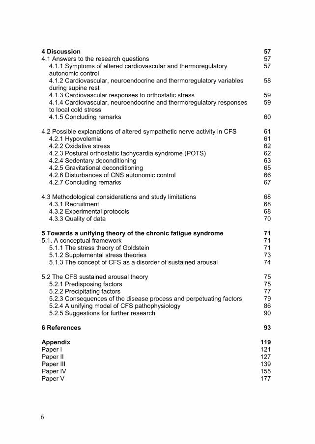
As indicated in the name, fatigue is the dominating complaint in patients with CFS (2,318). It is important to recognize this symptom as different from common tiredness orsleepiness, experienced by everyone from time to time (Figure 1). The patients usenotions like ‘overwhelmingly exhausted’, ‘totally empty of energy’ etc., and they
12
describe the fatigue as qualitatively different from earlier experiences (178, 375). Limitedexertions, whether mental or physical, disproportionately worsen the sensation of fatigue.Likewise, rest or sleep does not substantially relieve it.In addition, the patients are to a varying extent bothered by additional symptoms, someof which are required according to the CDC-definition (Table 2) (5, 123, 223, 318,375). However, none of them are specific to CFS. As far as we know, no comprehensivestudy has specifically addressed the frequency of each of these accompanying symptomsin a large cohort of CFS patients. In a majority of patients, the symptom intensity isfairly stable, but some report distinct fluctuations (128).The onset of CFS can be gradual or acute (123, 318). In the latter situation, the patientsoften report symptoms and signs indicative of a preceding infectious disease, likemononucleosis, influenza or gastroenteritis (20, 345). However, despite intensiveresearch, no infectious agent seems to be specifically related to CFS. In addition,evidence suggests that psychosocial stress might precipitate the disease in some patientsas well (180, 345). The relationship between these empirical findings and CFSpathophysiology theories will be further elaborated below (see 1.3.2).A diagnose of chronic fatigue syndrome requires a thorough clinical evaluation. Nosingle diagnostic test exists. Therefore, several guidelines have been developed, foradults (1, 55, 417), as well as children/adolescents (123). Although not identical, themain messages from these guidelines are common, prompting the practitioner to:
- Identify and recognize the patients’ characteristic symptoms, especially theirexperience of fatigue.
Figure 1. Schematic outline of how CFS should be differentiated from well-definedsomatic and mental diseases as well as other subjective complaints (like commontiredness and sleepiness). (Adapted from (100) and slightly modified, with permis-sion.)
Common tiredness or sleepiness
Fatigue
Chronic fatigue syndrome (CFS)
Totally disablingchronic fatigue
syndrome
Well defined somaticdiseases (e.g. multiplesclerosis, post-poliosyndrome, cancer,rheumatoid arthritis,hypothyroidism)
Well defined psychiatricdiseases(e.g. bipolar disorders)
13
- Rule out differential diagnoses by a standardized and comprehensive (but notexhaustive) set of investigations.
In addition, the practitioner should assess the patients’ functional impairments, whichmight be severe, causing school and work absenteeism, social isolation and eventuallya breakdown of normal family life (318). A 4-stage functional classification system hasbeen proposed (1): Mild designates mobile patients, who are able to carry out e.g.ordinary housework. Moderate means reduced mobility and limited ability to performdaily activities. Severe labels patients who use a wheel-chair and whose performance isrestricted to some very simple activities, like teeth-brushing. Very severe is the categoryfor completely disabled patients, who are bedridden and not able to take care of personalhygiene.The question of co-morbidity in CFS has been extensively debated. Three problemsare of particular interest. First, the CDC-definition of CFS requires the exclusion ofsomatic and/or psychiatric disorders that might explain the fatigue, like malignancies,rheumatic diseases and chronic infections (142) (see 1.1.1). Whereas this prerequisite
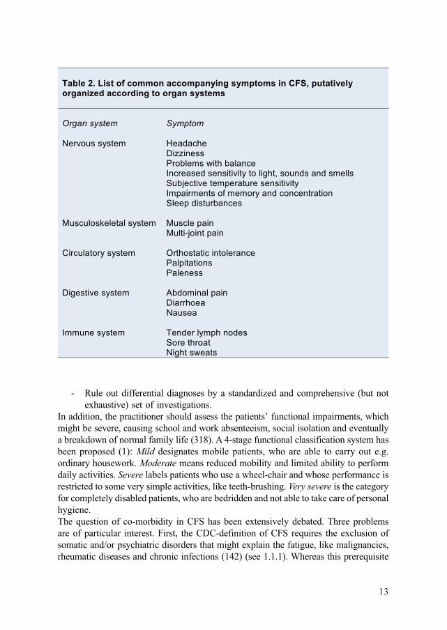
Table 2. List of common accompanying symptoms in CFS, putatively organized according to organ systems
Organ system Symptom
Nervous system Headache Dizziness Problems with balance
Increased sensitivity to light, sounds and smells Subjective temperature sensitivity Impairments of memory and concentration
Sleep disturbances
Musculoskeletal system Muscle pain Multi-joint pain
Circulatory system Orthostatic intolerance Palpitations Paleness
Digestive system Abdominal pain Diarrhoea Nausea
Immune system Tender lymph nodes Sore throat Night sweats

14
is usually unproblematic in a practical sense, some cases expose an inherent incon-sistency of the case definition. For instance, should a retracted course of EBV-infectionbe labelled ‘chronic mononucleosis’, ‘CFS’ or both? Second, there are several clinicalsimilarities between CFS and other so-called ‘functional somatic syndromes’, likeirritable bowel syndrome and fibromyalgia (17). This fact increases the diagnosticchallenges, as well as raises further fundamental concerns about the case definition.Third, there are different views concerning the relationship between CFS and well-defined psychiatric diagnoses. The evidence seems conflicting; for instance, some studiesreport increased prevalence of depression among CFS patients (2, 207), children andadolescents being particularly at risk (148), whereas findings in other studies disputesuch a relationship (316, 318). Despite all these challenges, recent evidence supportsthe notion of CFS as a distinct diagnostic entity (63).Qualitative research indicates that CFS patients might have problematic relationshipswith doctors and other health care professionals, feeling unaccepted, marginalized andnot prioritized (286). Doctors, on the other hand, report helplessness and scepticismconfronted with a condition of undetermined nature (8). These findings underscorehow CFS raises fundamental social and ethical challenges within the doctor-patient-relationship. Without neglecting the several complicated aspects of these issues, it seemspertinent to emphasize the doctors’ obligation to pay attention to and acknowledge thepatients’ subjective experience of symptoms, despite the lack of objective signs.
1.1.4 Treatment and prognosis
Various treatments of CFS have been subjected to randomized controlled trials. However,recent reviews conclude that only cognitive behavioral therapy (CBT) and gradedexercise therapy (GET) have a scientifically proven beneficial effect (10, 100, 114,280, 315, 335, 443). Important components of CBT are explanation of patho-physiological theories about CFS, challenging of fatigue-related cognitions and gradualincrease of physical activity (318). In this way, simply speaking, the patients learn toacquire control over their symptoms. CBT is also of proven value among adolescentswith CFS (401). Its success, however, does not necessarily imply a ‘psychological’ or‘mental’ etiology. GET exposes the patient to an individually adjusted and structuredexercise program (318). The aim is a gradual increase of activity level; thus GET mightbe regarded as a component of CBT. If the patients experience the exercise to be toostrenuous, compliance falls. Thus, a very careful and gradual approach seems to bemost beneficial (100). How these principles of treatment relate to subgroups of CFSpatients remains a question of debate. It is important to note that the severely disabledpatients are scarcely represented in the trials.Other therapeutic approaches that have been subjected to research include gluco-corticoids, mineralcorticoids, antidepressants, anticholinergic agents, antiviral drugs,growth hormone, immunoglobulins, dietary prescriptions and alternative/complementary
15
therapy. For all, the present evidence is inconclusive or indicates no beneficial effect(100).Management of CFS patients should also include attention to possible complications,like secondary depression and dietary deficiencies in the severely disabled. Further,patients need appropriate assistance with social and economical issues, as problemsrelated to these areas may constitute important perpetuating factors (37, 352). In childrenand adolescents with CFS, particular effort should be devoted to their situation at school,establishing courses adjusted to the patients’ individual capacity (123).The long term prognosis of CFS is disputed. A recent review reported a 5 % medianfull recovery and 40 % median improvement across different primary studies (51). Theprognosis of children and adolescents with CFS seems to be considerably better, withfull or partial recovery in 60-80 % (21, 337).
1.2 Basic science starting point – an overview of theautonomic nervous system
1.2.1 Structure and function of the autonomic nervous system
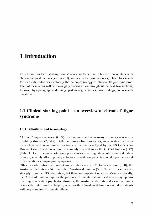
The autonomic nervous system (ANS) denotes those parts of the nervous system thatare related to involuntary and unconscious control of internal organs, in contrast to thesomatic nervous system which is devoted to conscious perception and voluntary action(43, 155). Generally speaking, the purpose of ANS is to maintain internal homeostasisby constantly adjusting organ function; at the tissue level, this regulatory task is carriedout through an effect on smooth muscles, heart muscles and glands.The sensory part of ANS mediates information to the central nervous system fromreceptors in the internal organs. These receptors have different properties; of particularinterest in regards to this thesis are the mechanical receptors located in the walls ofgreat veins/heart/pulmonary vessels and aorta/carotid arteries, commonly referred toas cardiopulmonary receptors and baroreceptors, respectively. The effector part ofANS consists of chains of two neurons which synapse in an autonomic ganglion; thus,they are commonly labeled preganglionic and postganglionic (Figure 2). The transmitterin the ganglia is acetylcholine, acting on nicotinic receptor proteins of the postganglionicneuron.Based upon both structural and functional characteristics, one can differentiate betweentwo branches of the effector part of ANS: the parasympathetic nervous system and thesympathetic nervous system (43, 155, 353). The former innervates only the visceralorgans proper, whereas the latter also has contact with blood vessels all over the body,as well as the skin and the musculoskeletal system (Table 3). Overall, the parasympathetic

16
Figure 2. Schematic overview of the autonomic nervous system (upper part), anddetailed outline of a noradrenergic sympathetic synapse (lower part, adapted andmodified from (155)). NE=norepinephrine, DOPA=dihydroxyphenylalanine,DHPG=dihydroxyphenylglycol, MAO=monoamine oxidase, COMT=catechol-O-methyl-transferase.
Brainstem
Thoracolumbar spinal cord
Sacral spinal cord
Preganglionicparasympathetic neuron
Postganglionicparasympathetic neuron
Ganglion
Acetylcholine
Acetylcholine
Norepinephrine
Postganglionicsympathetic neuron
Postganglionicsympathetic neuron
Adrenal gland
Adrenomedullary cell
Chain ofsympathetic ganglia
Nicotinic receptor protein
Muscarinic receptor protein
Preganglionicsympathetic neuron
Other brainareas
Target cell
Adrenoceptor
Epinephrine
Depolarization ofcell membrane
DOPA
Dopamine
DHPG
DHPG
MAO
NE
NE
NE
Uptake-1
Uptake-2
COMT
Normeta-nephrine
Normetanephrine
Capillary
Tyrosine
Target cell
Differentsecond
messengersand effects
12
1
2
NE
NE
K. Toverud

17
Table 3. Main effects of autonomic nerve activity and epinephrine in different organs. Overview*
Organ Parasympathetic nerve activity
Sympathetic nerve activity
Epinephrine
Heart Decreased rate Increased rate Increased contractility
Increased rate Increased contractility
Kidney Increased renin secretion Vasoconstriction
Vasoconstriction
Lungs Bronchoconstriction Bronchodilation Bronchodilation
Liver Increased glycogenolysis
Increased glycogenolysis
Adipose tissue Increased lipolysis
Gastrointestinal tract
Increased motility and secretion
Decreased motility and secretion Vasoconstriction
Decreased motility and secretion Vasoconstriction
Urinary bladder Detrusor muscle contraction
Sphincter muscle contraction
Genital organs Erection Ejaculation
Eye Miosis Accommodation Tear secretion
Mydriasis
Skeletal muscles Vasoconstriction Shivering
Vasodilation Shivering
Skin Piloerection Sweating Vasoconstriction
Vasoconstriction
Lymphatic organs Undetermined effects Undetermined effects
Central nervous system
Improved concentration Enhanced emotional experiences
* Adapted from (43, 155, 353)
18
nervous system is mainly responsible for conservative, vegetative processes, while thesympathetic nervous system is particularly important for ‘emergency’-reactions wheninternal homeostasis is threatened. Accordingly, parasympathetic and sympatheticnervous activity often has reciprocal effects on organ functions. However, this generalrule greatly oversimplifies the complex and dynamic interactions between the twodivisions of ANS.The preganglionic neurons of the parasympathetic nervous system originate in the nucleiin the brain stem and the sacral spinal cord (Figure 2) (43, 155). Many of them followthe vagus nerve; hence ‘vagal’ is commonly, though inaccurately, used as a synonymfor ‘parasympathetic’. The postganglionic neurons use acetylcholine as the maintransmitter, acting upon muscarinc receptor proteins of which there are several variantsrelated to different second messengers.The preganglionic neurons of the sympathetic nervous system emanate from theintermediolateral column of the thoracolumbar spinal cord, and end in the preaorticand paravertebral chains of ganglia (Figure 2) (43, 155). Most postganglionic neuronsuse norepinephrine (noradrenalin) as the main transmitter, but the neurons supplyingsweat glands use acetylcholine. In addition, other transmitter substances like ATP andneuropeptide Y have been described but their physiological effects remain largelyunknown (109, 308). Norepinephrine is synthesized from the amino acid tyrosine;intermediate products in this process are dihydroxyphenylalanine (DOPA) and dopamine(Figure 2) (155). The transmitter is stored in synaptic vesicles and released by exocytosisupon depolarization of the cell membrane. The receptor proteins binding to norepine-phrine, commonly referred to as adrenoceptors, are divided into two main categories,labeled alpha and beta. Numerous subtypes are described, having different functionalproperties. Alpha2-adrenoceptors, which are found presynaptically at sympatheticneurons and also in the central nervous system, are particularly important for negativefeedback-regulation, as the binding of norepinephrine to these proteins attenuatessympathetic nervous activity. Released norepinephrine is inactivated by two differentuptake-mechanisms (Figure 2) (155). Uptake-1 designates transport by a specificmembrane protein back into sympathetic nerve terminals. Here, the transmitter mightbe recycled into storage vesicles, or degraded by the enzyme monoamine oxidase (MAO)to form dihydroxy-phenylglycol (DHPG). Uptake-2 means transport into non-neuralcells, and subsequent degradation is mainly performed by the enzyme catechol-O-methyltransferase (COMT), forming normetanephrine.The adrenal medulla closely resembles a sympathetic ganglion. The adrenomedullarycells are controlled by preganglionic sympathetic neurons, and are thus themselvesanalogue with postganglionic neurons (43, 155). However, they secrete epinephrine(adrenalin) instead of norepinehrpine, and this chemical compound functions as ahormone, not a neurotransmitter. Although epinephrine binds to both alpha- and beta-adrenoceptors, the affinity differs from that of norepinephrine. This partly explainswhy increased adrenomedullary activity and generally enhanced sympathetic nerveactivity does not produce identical physiological effects (Table 3). The main break-down route of epinephrine is degradation by COMT to metanephrine.

19
Specific areas in the central nervous system receive afferent impulses from receptors inthe internal organs and control efferent impulses in the effector part of ANS (Figure 3).In addition, these areas communicate with other brain centers responsible for emotionaland cognitive processes, voluntary movements, conscious perception, and endocrinecontrol. For instance, an area in the reticular substance of the medulla oblongata (therostral ventrolateral medulla, RVLM) indirectly receives inputs from baroreceptors,and directly controls preganglionic sympathetic neurons, thus constituting a vital partof the baroreceptor reflex (se 1.2.2) (155). In addition, RVLM is reciprocally connectedwith the nearby raphe nuclei, with locus ceruleus in pons, with the paraventricularnucleus in hypothalamus, and with amygdala in the limbic system. The raphe nucleiprobably have a key role in processing painful stimuli (43). The neurons of locus ceruleus
Figure 3. Selected areas and connections within the central nervous system impor-tant for autonomic control. The figure is greatly simplified. (Adapted and modifiedfrom (43)).
Thalamus
Locus ceruleus
Raphe nuclei
Nucleus of the solitary tract
From baroreceptors andcardiopulmonary receptors
AmygdalaPituitary gland
Hypothalamus
Paraventricular nucleus
To preganglionic sympatheticneurons in spinal cord
Rostral ventrolateral medulla
20
project extensively to all brain areas (160). Being part of the brain stem ‘activationsystem’, the locus ceruleus probably participate in the regulation of sleep and conscious-ness; in addition, this area seems to be important for attention (43). Neurons in theparaventricular nucleus produce the hormones vasopressin and corticotropin-releasinghormone (CRH), which are secreted by axons projecting to the pituitary gland. CRHcontrols the synthesis of adrenocorticotropic hormone (ACTH), which in turn regulatesthe secretion of glucocorticoids from the adrenal cortex. Amygdala probably has a keyrole in emotional memory, relating sensory information to specific emotional states,and orchestrating appropriate behavioral and autonomic responses (43). In other words,amygdala seems to be important for various learning processes, in particularconditioning.
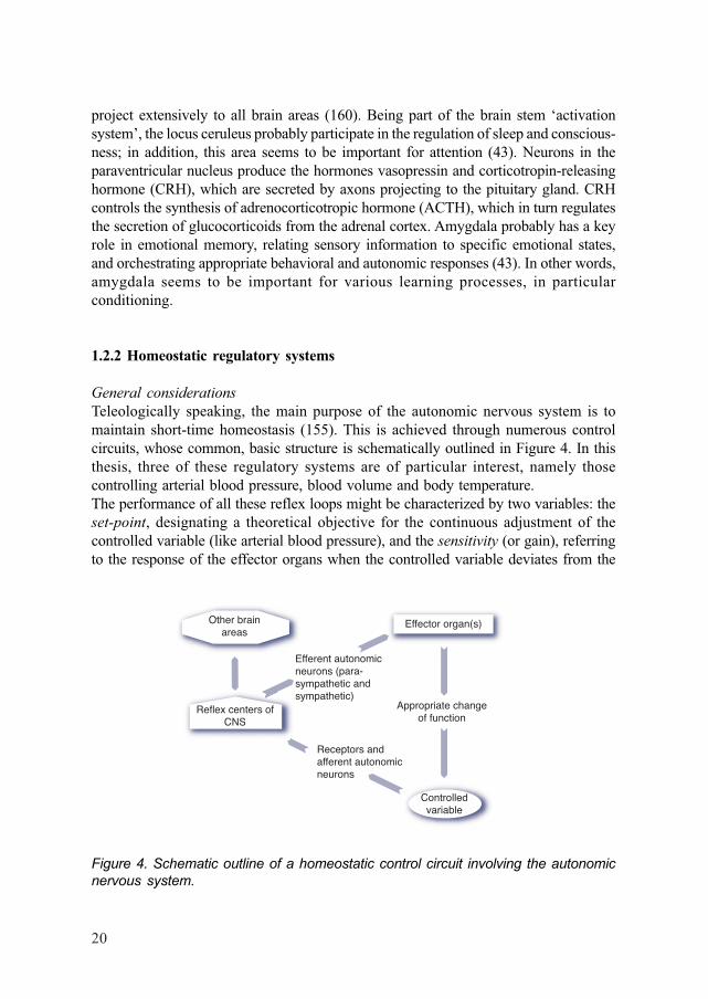
1.2.2 Homeostatic regulatory systems
General considerationsTeleologically speaking, the main purpose of the autonomic nervous system is tomaintain short-time homeostasis (155). This is achieved through numerous controlcircuits, whose common, basic structure is schematically outlined in Figure 4. In thisthesis, three of these regulatory systems are of particular interest, namely thosecontrolling arterial blood pressure, blood volume and body temperature.The performance of all these reflex loops might be characterized by two variables: theset-point, designating a theoretical objective for the continuous adjustment of thecontrolled variable (like arterial blood pressure), and the sensitivity (or gain), referringto the response of the effector organs when the controlled variable deviates from the
Figure 4. Schematic outline of a homeostatic control circuit involving the autonomicnervous system.
Appropriate changeof function
Controlledvariable
Receptors andafferent autonomicneurons
Effector organ(s)Other brainareas
Reflex centers ofCNS
Efferent autonomicneurons (para-sympathetic andsympathetic)
21
set-point (381). Both set-point and sensitivity might be altered in different physiologicaland pathophysiological states. For instance, during exercise, the set-point of the baro-receptor reflex is reset to a higher level, probably due to the combined effect of neuralinputs from higher brain centers (‘central command’) and from receptors in the workingmuscle (269). In addition, there is a decrease of sensitivity which seems to be causedby altering sympathetic and parasympathetic modulation of heart rate (292).Most control circuits, whether in physiology or engineering, expose dynamic behavior,characterized by the fluctuations of the controlled variable around a mean valuecorresponding to the set-point (7, 73, 416). This variability may be explored by sophisti-cated mathematical analyses, providing important information concerning the neuralcontrol mechanisms, as will be further elaborated below (see 2.2.2).
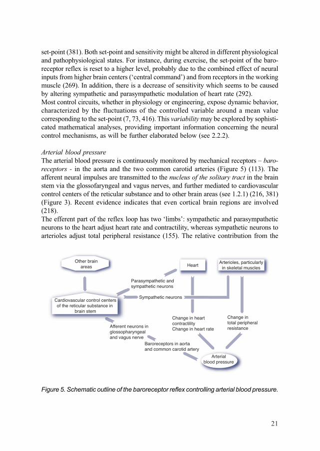
Arterial blood pressureThe arterial blood pressure is continuously monitored by mechanical receptors – baro-receptors - in the aorta and the two common carotid arteries (Figure 5) (113). Theafferent neural impulses are transmitted to the nucleus of the solitary tract in the brainstem via the glossofaryngeal and vagus nerves, and further mediated to cardiovascularcontrol centers of the reticular substance and to other brain areas (see 1.2.1) (216, 381)(Figure 3). Recent evidence indicates that even cortical brain regions are involved(218).The efferent part of the reflex loop has two ‘limbs’: sympathetic and parasympatheticneurons to the heart adjust heart rate and contractility, whereas sympathetic neurons toarterioles adjust total peripheral resistance (155). The relative contribution from the
Figure 5. Schematic outline of the baroreceptor reflex controlling arterial blood pressure.
Change in total peripheralresistance
Arterialblood pressure
Baroreceptors in aortaand common carotid artery
Afferent neurons inglossopharyngealand vagus nerve
Arterioles, particularlyin skeletal musclesHeart
Other brainareas
Change in heartcontractilityChange in heart rate
Cardiovascular control centersof the reticular substance in
brain stem
Parasympathetic andsympathetic neurons
Sympathetic neurons
22
different vascular beds for increasing total peripheral resistance in distinct physiologicaland pathophysiological states is still largely undetermined; however, skeletal musclevasoconstriction is usually considered the most important effector response (209, 353).Whether the baroreceptor reflex participates in long-term regulation of blood pressureis unknown. However, ample evidence indicates that baroreceptor resetting occursshortly after artificial manipulation of blood pressure, which raises questions concerningtheir role in long term regulation (259, 294).
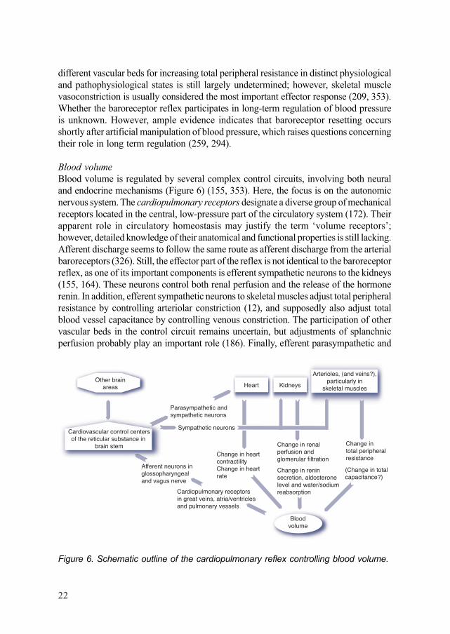
Blood volumeBlood volume is regulated by several complex control circuits, involving both neuraland endocrine mechanisms (Figure 6) (155, 353). Here, the focus is on the autonomicnervous system. The cardiopulmonary receptors designate a diverse group of mechanicalreceptors located in the central, low-pressure part of the circulatory system (172). Theirapparent role in circulatory homeostasis may justify the term ‘volume receptors’;however, detailed knowledge of their anatomical and functional properties is still lacking.Afferent discharge seems to follow the same route as afferent discharge from the arterialbaroreceptors (326). Still, the effector part of the reflex is not identical to the baroreceptorreflex, as one of its important components is efferent sympathetic neurons to the kidneys(155, 164). These neurons control both renal perfusion and the release of the hormonerenin. In addition, efferent sympathetic neurons to skeletal muscles adjust total peripheralresistance by controlling arteriolar constriction (12), and supposedly also adjust totalblood vessel capacitance by controlling venous constriction. The participation of othervascular beds in the control circuit remains uncertain, but adjustments of splanchnicperfusion probably play an important role (186). Finally, efferent parasympathetic and
Figure 6. Schematic outline of the cardiopulmonary reflex controlling blood volume.
Change in total peripheralresistance
Bloodvolume
Cardiopulmonary receptorsin great veins, atria/ventriclesand pulmonary vessels
Afferent neurons inglossopharyngealand vagus nerve
Arterioles, (and veins?),particularly in
skeletal musclesHeart KidneysOther brain
areas
Change in heartcontractilityChange in heartrate
Cardiovascular control centersof the reticular substance in
brain stem
Parasympathetic andsympathetic neurons
Sympathetic neurons
Change in renalperfusion andglomerular filtration
Change in reninsecretion, aldosteronelevel and water/sodiumreabsorption
(Change in totalcapacitance?)

23
sympathetic neurons adjust heart rate and contractility (144, 418). However, heart ratecontrol also differs from the baroreceptor reflex, as moderately augmented cardiacfilling may – in some situations – increase heart rate (the so-called ‘Bainbridge-reflex’)(15). To increase complexity further, evidence indicates that the cardiopulmonary andbaroreceptor reflexes interact (430).
Body temperatureDiscrete regions of the hypothalamus seem to be directly involved in humanthermoregulation, having specific populations of temperature-sensitive neurons (43,155) (Figure 7). In addition, these areas receive sensory information from thermo-receptors in the skin. A region in the posterior hypothalamus seems to be the principal‘conductor’ orchestrating the physiological responses to heat or cold (105). Areas inthe brain stem, in particular the raphe nuclei, are probably involved in the efferentpathways (54, 278). The effector organs involved with human thermoregulation aresweat glands, skin arterioles, skin arteriovenous anastomoses and skeletal muscles.Sweat glands are innervated by cholinergic sympathetic neurons, which also seem topromote vasodilation in nearby arterioles by a local release of NO and prostaglandinsfrom the endothelial cells (210, 211). However, the main control of skin arterioles, andalso of skin arteriovenous anastomoses, is exerted by vasoconstrictive noradrenergicsympathetic neurons (23). Skeletal muscles are controlled by motor neurons of thesomatic nervous system; however, enhanced sympathetic outflow and epinephrinesecretion promotes shivering (155, 391).Several other features of human thermoregulation add to its complexity. First, centraland peripheral thermoreceptors may give rise to conflicting afferent information (353).
Figure 7. Schematic outline of the thermoregulatory reflex controlling body temperature.
Shivering
Bodytemperature
Thermoreceptorsin skin
Thermoreceptors inhypothalamus
Afferent neurons insomatic nerves
Sweatglands
Skinarterioles
Skin arteriovenousanastomoses
Skeletalmuscles
Other brainareas
Change in sweating
Thermoregulatory controlcenter in hypothalamus
Cholinergicsympathetic neurons
Noradrenergicsympathetic neurons
Motor neurons
Endothelial cells?
Change in skin perfusion

24
Second, local temperature changes may call for differentiated responses to distinctparts of the body (23). Third, the thermoregulatory and cardiovascular control circuitsshare effector organs – the skin blood vessels – causing ‘conflicts of interest’ during forinstance strenuous exercise (155). Fourth, thermoregulation also includes appropriatebehavioral responses, such as taking on or off clothes, as well as endocrine responsesfrom the adrenals and the thyroid gland adjusting energy metabolism (391).
1.3 The pathophysiology of chronic fatigue syndrome –epistemological issues, prior findings and research questions
The approaching of a scientific problem is inevitably influenced by prior knowledgeand attitudes. There is – in other words – no true neutral position (147). A researcher isalways restricted by his ‘horizon of understanding’ even before he starts searching theliterature and formulating research questions. Therefore, in this section, I will start outby discussing some fundamental epistemological issues and clarifying my positions inregards to them. Thereafter I will present the body of knowledge concerning CFSpathophysiology and the specific aims of this thesis.
1.3.1 Epistemological considerations and premises
Fatigue is poorly defined both linguistically and biologically, despite how common itis in clinical practice (407). One attempt of classification is to differentiate between‘central’ fatigue, originating from the central nervous system (CNS) and ‘peripheral’fatigue, originating from peripheral nerves and muscles. However, the evidencejustifying such a theoretical construct seems sparse. Even the precise meaning of theword ‘fatigue’ is obscure, a consequence of its completely subjective nature. Does itdescribe identical experiences when used in relation to, e.g., CFS, liver disease andmarathon running, or is the sensation of ‘fatigue’ in these situations different (thoughrelated), indicating separate biological mechanisms?As fatigue is a subjective experience, it cannot be objectively measured. Severalpsychometric instruments have been developed (108, 228). However, the lack of a‘gold standard’ will always raise questions of validity, both in regards to the instrumentsthemselves, and the empirical research based upon them. Thus, from a very restrictivepoint of view, one might argue that fatigue is not ‘researchable’ within the paradigm ofthe natural sciences at all.Recent theories of fatigue focus primarily on mechanisms within CNS. Evidenceindicates the important role of corticotropin-releasing hormone (CRH), some cytokines(IL-1beta, IL-6), and the neurotransmitters serotonin (5-hydroxytryptamine), dopamine

25
and norepinephrine (56, 250, 407). These mediators may be influenced from processes– including diseases – outside the CNS, like chronic inflammatory conditions, but theyare also strongly related to nerve activity in limbic and cortical brain centers. Thus,fatigue is dependent upon both ‘mental’ and ‘somatic’ processes, as can be demonstratedempirically (97, 173).Exploring mechanisms of causality is a general aim within the natural sciences (147).However, given the complex nature of fatigue, cause-effect-relationships are difficultto establish. For instance, is the increased serotonin concentration within CNS, whichhas been reported in fatigued subjects (407), a cause or a consequence of fatigue?Similar uncertainties apply to most interpretations of empirical findings within thisfield. Further, the causes of fatigue can be multifactorial in each individual, and a 1:1-relation between cause and effect seems both empirically and theoretically unlikely(436).The challenges described above in relation to fatigue in general, also apply to CFSresearch. In addition, there is the problem of case definition, as mentioned in 1.1.1. Nofirm evidence supports a specific relationship between certain accompanying symptomsand pathophysiological mechanisms; thus, the symptom criteria in the CDC-definitionmay seem somewhat arbitrary (31, 436). Among children and adolescents, the validityof this definition is even less established (123, 265). On the other hand, grouping togetherconditions that do have different etiology or pathophysiology, although we do not knowthem yet, might obscure important scientific findings. As of now, this dilemma awaitsits solution.In conclusion, research surrounding CFS pathophysiology is challenged by:
- The subjective nature of fatigue, including the problems of how to measure it.- The limited knowledge concerning the biological mechanisms of fatigue,
including undetermined cause-effect-relationships, the possibility of multi-factoriality, and the likelihood of body-mind-interaction.
- The uncertain validity of the CFS case definition, especially within the pediatricpopulation.
In this thesis, the positions taken towards these challenges are as follows. First, althoughfatigue is a subjective experience, we maintain that it nevertheless constitutes aphenomenon which merits systematical, scientific investigation. That fatigue – andCFS – exists and constitutes a clinical entity is an assertion of high ‘face validity’,supported by empirical findings (178, 405). Furthermore, scientific investigations inthis field should certainly include, but not be restricted to, the use of hermeneuticmethods. In our view, the relative absence of measurable entities does not exclude anatural science approach a priori. Second, given the limited mechanistic knowledge,investigations could hardly be governed by hypotheses of causality. Rather, the aimmust be phenomenologic: to explore and describe aspects of the pathophysiology, andthereby – hopefully – be able to present hypotheses for further research. Third, thecomplexity of the matter, and in particular the possibility of body-mind-interactions,suggests that the results of the study should be interpreted within a biopsychosocial

26
framework, despite its biological starting point (117). There is a widespread acceptancethat fatigue cannot be understood within a traditional, dichotomous, Cartesian-inspiredclassification of medical phenomena as either ‘physical’ or ‘psychological’ (318, 436).Fourth, the ambiguity of the CDC-definition, specifically emphasized in a recent,authoritative review (63), suggests a modification of it. We therefore omitted thebicriterion (4 of 8 accompanying symptoms, see 1.1.1), and included patients solelybased upon the fulfillment of the main criterion, as will be further outlined below (see2.1.1).
1.3.2 Prior research on CFS pathophysiology
Prior research surrounding the pathophysiology of CFS has been conducted along severaltracks, reflecting the great uncertainty about the condition as well as the differentscientific traditions among the researchers. This has resulted in a vast amount of papers;a PubMed search using ‘chronic fatigue syndrome and pathogenesis’ as criteriongenerated more than 1600 hits. Still, there is at present no coherent theory, and CFS isoften labeled ‘mysterious’ or ‘controversial’ (155). In this brief review, all aspects ofthe pathophysiology will be considered, but with an emphasis on research relating CFSto the autonomic nervous system.
GeneticsTwin studies indicate a moderate heritability of CFS (63, 404). In a recent comprehensiveattempt to integrate clinical and epidemiological data with genomic and proteomicprofiles (429), findings suggest that chronic fatigue is related to polymorphisms ofgenes involved in CNS control of autonomic and endocrine effector systems, includingthe genes for monoamine oxidase (MAO) and catechol-O-methyltransferase (COMT)(154, 373). Further analyses of gene expressions in mononuclear blood cells revealedvery complex results, indicating altered activation of genes controlling both commonmetabolic pathways (gluconeogenesis, lipid metabolism) and signal transductionpathways involved in immune and neuroendocrine responses (124, 439). Other studieshave reported a similar complicated picture (215).
InfectionsCFS often has an acute onset with symptoms strongly resembling an infection (see1.1.3). Therefore, a substantial amount of research has tried to detect a possible infectiousagent. In the 1980s, much attention was given to the Epstein-Barr virus (EBV), asinfectious mononucleosis may have a prolonged course or – in the worst case – developinto CFS (52, 441). However, no specific role of EBV has been established (226);rather, an EBV-infection should be regarded as one of many possible precipitating andeventually perpetuating factors (2). The same view applies to several othermicroorganisms that may similarly elicit severe fatigue and a prolonged recovery in a
27
subset of patients; examples include cytomegalovirus, parvovirus B19, Brucella-species,Toxoplasma gondii, Coxiella burnetii, Mycyplasma-species and Chlamydia pneumoniae(61, 62, 243, 284). However, it should also be noted that common, non-specific infections(like upper respiratory tract infections) are not likely to trigger CFS (437).A possible pathogenetic role of enteroviruses has been thoroughly debated. Using thePCR technique, Gow and co-workers reported enteroviral RNA in skeletal musclebiopsies in a majority of adult CFS patients; however, enteroviral RNA was also detectedamong some controls (163). In a recent review, Chia concluded that enteroviruses mighthave a pathogenetic role in CFS patients, possibly causing chronic inflammatory changesin skeletal muscle (61).
ImmunityThe significance of immune system disturbances in CFS patients has been a matter ofcontroversy. Based on a systematic review of studies addressing T-cell function, B-cellfunction, NK-cell function, immunoglobulins and cytokines, Lyall and co-workersconcluded in that there is no consistent pattern of immunological abnormalities in CFSpatients, although they found a trend towards changes in T-cell activity (255). Recentstudies have reported a reduced level of the cytokine TGF-beta1, which normally inhibitsantibody production, increased levels of IL-6 (53), which stimulates the acute phaseresponse, and alterations in the 2-5A synthetase/ribonuclease L pathway (94, 403),which participate in intracellular defense against viruses. The latter abnormality alsoseems to correlate with exercise performance (289). More generally, there is evidenceof a bias towards Th2 immune responses (humoral) at the cost of Th1 immune responses(cellular) in CFS (63, 301, 372). This is consistent with frequent reports of reduced NKcell activity (2, 249), as these cells are important effectors in the Th1 immune reaction.However, a twin study did not report significant differences between CFS patients andtheir healthy siblings (344).Studies of autoimmunity have yielded conflicting results. Recently, researchers reportedthe presence of autoantibodies specifically directed against the muscarinic cholinergicreceptor protein (410) as well as autoantibodies against certain common cellular antigens(428). An association between CFS and distinct HLA antigens has also been reported(191).
Oxidative stressOne study found increased levels of methaemoglobin and other indicators of oxidativestress, correlating strongly with the patients’ complaints (328). Similar findings havelater been reported by others, providing evidence of free radical attacks on cell membranephospholipids in CFS patients (212). These results could be explained by immuneactivation in general, but could also be attributed to a persistent viral infection. Increasedoxidative stress has, in turn, been proposed as an explanation for altered skeletal muscleexcitability as well as muscle pain and postexertional malaise among CFS patients(197).
28
Skeletal muscle functionSeveral early studies concluded that CFS patients have perfectly normal muscle strength,endurance and recovery, as reviewed in the Australian CFS guidelines (64). However,others report that patients are weaker than sedentary controls as judged from maximumvoluntary contraction (143, 304), and that their performances are further attenuated 24hours later, indicating delayed recovery (143). Neurophysiological experiments suggestthat one probable explanation is an altered activation of cortical motor areas in thecentral nervous system of CFS patients; this phenomenon being even more pronouncedwhen the isometric exercise induced a subjective experience of fatigue (351, 367).Related findings of altered cortical excitability are reported during non-fatiguingmovements (380), and also immediately prior to motor performances, the latter indicatingdisturbances of attention (162). Interestingly, attention deficit has also been suggestedin an earlier neurophysiological study of different design (314), whereas altered planningof motor activities is adherent with recent findings by functional magnetic resonanceimaging (91).In 1991, Behan and co-workers reported intracellular lipid excess and morphologicalchanges of mitochondria in skeletal muscle biopsies from CFS patients; these findingscould perhaps be attributed to an intracellular infectious process, but are also similar toalterations found in hereditary mitochondrial myopathies (19). However, these findingswere not reproduced (234, 312). On the contrary, it was concluded that despite inactivity,skeletal muscle biopsies from CFS patients exhibit less fiber atrophy and related changesthan expected. A defect of oxidative metabolism and subsequent enhancement ofanaerobic glycolysis has been reported (447), but was not confirmed in recent research(197). Still, Vecchiet and co-workers demonstrated increased pain sensitivity in skeletalmuscles and related morphological abnormalities (426). These findings may also explainthe altered activation of central motor areas due to negative feedback, especially sinceexercise seems to lower pain threshold in CFS patients (442).
NeuroimagingNeuroimaging studies in CFS patients have yielded conflicting results. Using brainMRI, some investigators have reported subtle alterations of subcortical white matter,correlating with the patients’ complaints (77) and a reduction in total gray matter volume(90), whereas others did not find any differences between CFS patients and healthycontrols (80, 167). Functional MRI and SPECT techniques indicate an alteration ininformation processing (90, 236), planning of motor activities (91), cortical perfusionin general (457) and brain stem perfusion (83). However, a twin study indicated thatthe resting regional blood flow pattern in the brain is similar between patients and theirhealthy twins (244). A few PET scan studies have been undertaken in CFS patients.Tirelli and co-workers documented glucose hypometabolism in the frontal cortex andbrain stem (414), whereas Siessmeier and co-workers found alterations of brain glucosemetabolism among half of the included patients, though no clear pattern could be defined(368). Recently, two independent groups have reported a decreased number and/or
29
affinity for the receptor protein 5-HT1A in hippocampus (70) and the serotonin transporterprotein in the cingulate gyrus (449). These results are in accordance with resultssuggesting blunted serotonine activation of HPA axis (106), but contradict earlierindications of increased serotonergic neurotransmission (69).
SleepCFS patients regularly complain of altered sleep patterns, in particular difficultiesinitiating and maintaining sleep (224). Several researchers have provided evidence ofsleep disturbances in CFS (131, 261, 415), but no consistent pattern has emerged (2),and some trials even failed to demonstrate any significant alterations in EEG signalsduring sleep (13).
Cognitive functionCognitive tests of CFS patients have revealed disturbances of memory, attention andinformation processing, including those patients devoid of any psychiatric comorbidity(93). Albeit the evidence is not uniform, a recent review concluded that CFS patientsdo have modest, but significant, cognitive impairments (272). Some reports indicatethat cognitive performance deteriorates further during exercise (26, 232) but conflictingresults exist (78). Morris and co-workers exposed CFS patients to different mental andbodily stressors, and reported an improved speed in planning tasks during concomitantadministrations of the alpha2-adrenoceptor agonist clonidine in high doses (277). Thereare, however, also reports of a discrepancy between perceived and actual cognitiveperformance among CFS patients (271), and twin studies showed that the healthy twinshad similarly reduced information processing abilities when compared to the CFSpatients (256).
Psychology and psychiatryThe possible relationships between CFS and psychiatric disorders have been – and stillare – matters of great controversy. Partly, this can be explained by the ambiguity inherentin the different case definitions of CFS. Specifically, many CFS patients fulfill thediagnostic criteria for a somatization disorder. However, whether such a diagnosis ismade «is, to a considerable degree, dependent on the examiner’s attributions of chronicfatigue syndrome symptoms and is of limited use in understanding chronic fatiguesyndrome» (2). Also, the prevalence of panic disorders and generalized anxiety disordersis much higher among CFS patients than within the general population, both amongadults (130, 235) and adolescents (149), suggesting related pathophysiologies. Finally,depression is also common among CFS patients, but recent evidence confirms thatdepression and CFS are two distinct entities (2, 63, 318, 422).Although CFS often has an infection-like onset, research suggests that critical life events(e.g. loss of spouse), severe physical stressors (trauma, surgery) and perceived chronicdifficulties – in particular those described as dilemmas - may precipitate the disorder(180, 345, 412). Besides, some studies report that certain personality traits, like
30
perfectionism and conscientiousness, predispose for CFS (319, 440), but evidence isconflicting (448).Psychological and social issues are often regarded as important perpetuating factors inCFS (318). Certain illness perceptions, such as a poor sense of personal control oversymptoms and a strong focus on bodily sensations, are correlated to increasedimpairments in several studies (182, 187, 310). Likewise, CFS patients express a fearof physical exercise that does not correspond to their physical disability (288, 369),they perceive their cognitive performance as poorer than it is in reality (271), and theysleep better than what they subjectively report (13). Patients’ attributions also seem tocome into play, as a one-sided focus on somatic processes is related to a poorer outcome(187).These inappropriate cognitions may be strengthened by social interaction with family,friends and health care professionals (318, 352). Reduced self-esteem is a commoncomplaint among adults (440) as well as adolescents (149), and a lack of social support,which is often experienced by CFS patients, may further worsen the situation (317).Finally, the social role of being ill is – despite obvious undesirable consequences - alsopotentially rewarding, causing an unconscious cycle of reinforcement (318).
EndocrinologyThe hypothalamus-pituitary-adrenal axis (HPA axis) has been extensively exploredamong CFS patients, and there seems to be a general agreement concerning some subtlealterations, although the results are far from uniform (63, 68, 299, 318). Most researchersin this field report low basal levels of cortisol in urine, plasma and saliva as well asenhanced negative feedback, possibly due to increased sensitivity or number ofglucocorticoid receptors in the brain (68); some recent studies, however, failed toreproduce these findings (190, 208). The normal circadian rhythm of HPA activity isalso disturbed, particularly attenuating cortisol secretion during the morning hours (63,95, 103, 415). As for challenge tests, most studies indicate blunted HPA axis responsesto exercise, hypoglycemia and the administration of stimulating pharmaceuticals (68,295, 355); however, high doses of the alpha2-adrenoceptor agonist clonidine increaseplasma levels of cortisol under conditions of high arousal (277). The underlyingmechanisms for these disturbances as well as their functional consequences remainunresolved; however, a relationship to the documented immune abnormalities is anobvious possibility (63).Several researchers have focused on other endocrine systems. There are some reportsof increased levels of dehydroepiandrostenedione (DHEA) (71), whereas others foundthe opposite (68). Studies addressing endogenous opoid tone have also reportedconflicting results (68, 190). The GH and prolactin systems seem to be intact in CFSpatients (68, 103, 295), whereas melatonin levels appear to be higher than normal(222).Reports concerning catecholamines are sparse; existing evidence indicates increasedbasal levels of epinephrine, but normal plasma levels of norepinephrine (208, 413).

31
Compared with controls, CFS patients have lower plasma levels of tyrosine and higherplasma levels of tryptophan after strenuous physical exercise, indicating disturbancesof the noradrenergic and serotoninergic transmitter systems in the CNS (150).Demitrack and colleagues found reduced plasma levels of the norepinephrine breakdownproduct 3-methoxy-4-hydroxyphenylglycol (MHPG) and increased levels of theserotonine metabolite 5-hydroxyindoleacetic acid (5-HIAA), but the levels withincerebrospinal fluid were similar to those found in the healthy controls (96). Thesignificance of this finding is unclear, as MHPG in plasma derives from many sources(156).
Circulatory homeostasisThe first papers on cardiovascular disturbances in CFS patients emerged in the 1990s,reporting neurally mediated hypotension during head-up tilt tests (35, 339).Subsequently, variants of haemodynamic instability during orthostatic challenge – mostcommonly neurally mediated hypotension or orthostatic tachycardia - have beendescribed by many researchers in adult as well as pediatric patients (231, 306, 340,386, 387). Similar baseline abnormalities have also been reported (231), as well as arelationship between symptom severity and a decline in stroke volume and baroreceptorsensitivity during tilt (306, 307). More sophisticated analyses of cardiovascularvariability indicate a sympathetic predominance in the modulation of heart rate andtotal peripheral resistance during rest, orthostatic challenge and moderate exercise (81,92, 282, 370, 383). Recently, a shortened QT interval was reported among CFS patients,further indicating increased sympathetic and attenuated parasympathetic cardiacneurotransmission (283). A twin study, however, showed that CFS patients and theirhealthy twins had similar haemodynamic responses to tilt-table testing (313), and arecent population-based study failed to demonstrate orthostatic instability among CFSpatients (201), challenging the assumption that cardiovascular dysregulation is animportant aspect of CFS pathophysiology. Moreover, some studies of variability havebeen inconclusive (390, 458) or simply negative (111, 453).Hypovolemia has been proposed to underlie these regulatory disturbances, and wasindeed indicated in one study, although not to the level of statistical significance (126).Accordingly, Streeten and co-workers provided evidence of reduced erythrocyte volume(398) and impaired lower-limb venous innervation (396) in CFS patients, whereas Roweand colleagues found a strong association between CFS and the connective tissue diseaseEhlers-Danlos syndrome (338). Taken together, these results suggest that relativehypovolemia might be a cause of hemodynamic disturbances in CFS. However, twocontrolled trials of volume expansion treatment (fludrocortisone) in CFS were notsuccessful (309, 341).Hemodynamic disturbances have also been documented in other organ systems. Brainstem hypoperfusion was an early finding (83). A general reduction in cerebral bloodflow upon standing has been reported as well (409), but was not confirmed in subsequentexperiments (325), thus weakening a hypothesis of reduced brain perfusion as a direct

32
cause of the fatigue sensation. McCully and co-workers found normal oxidativemetabolism in working skeletal muscle, but subtle alterations in blood flow after dynamicexercise, possibly due to sympathetically induced vasoconstriction (267). Abnormaldilatation of renal and other intraabdominal veins has also been reported (408). Finally,there is evidence of altered skin circulation, as CFS patients are more sensitive to thevasodilative effect of locally applied acetylcholine (203, 378).
Temperature homeostasisAdolescents with CFS seem to have an altered circadian rhythm of core body temperature(415). Two studies in adult patients, however, found normal circadian variation (175,176). Pazderka-Robinson and colleagues reported increased skin temperature and lowerelectrodermal activity among CFS patients; the latter indicating attenuated sweating(305). There is also evidence of altered skin circulation, as outlined above (203, 378).
The autonomic nervous systemPagani and co-workers were among the first to suggest a relationship between autonomicdysfunction and unexplained chronic fatigue, based upon research on cardiovascularvariability (298). Subsequently, the same group put forward a hypothesis of autonomicdysfunction in CFS, characterized by enhanced sympathetic activity at rest and reducedresponsiveness to excitatory stimuli (297). Freeman and Komaroff reached similar con-clusions based upon thorough autonomic testing that indicated an increased sympatheticcontrol of cardiovascular variables at rest, but decreased sympathetic modulation ofthe same variables during orthostatic challenge (137). They proposed postviral auto-nomic neuropathy or cardiovascular deconditioning as possible underlying mechanismsfor the observations. The possible significance of autonomic disturbances and the needfor comprehensive research projects have been recognized in subsequent papers (151,159, 282). However, a unifying theory relating autonomic dysfunction to otherpathophysiologic aspects of CFS is still lacking.
Concluding remarksBased upon this review, the following concluding statements seem to be justified:
- There is a hereditary component in CFS.- Several different infectious agents may precipitate the disorder, but there is no
specific relationship.- There is a shift towards a Th2 immune response at the cost of a Th1 immune
response.- There is increased oxidative stress.- Muscle weakness can be explained by alteration in CNS; evidence concerning
distinct muscle pathology is uncertain.- Neuroimaging studies indicate reduced perfusion and metabolism in some brain
areas, as well as alterations in serotoninergic neurotransmission.- There are alterations of cognitive function, in particular memory, attention and
information processing.

33
- Psychological stress may precipitate CFS, whereas certain personality traitsconstitute predisposing factors.
- Diverse psychological and social mechanisms contribute to the perpetuation ofthe disorder.
- The activity and responsiveness within the HPA axis is decreased.- There are alterations of cardiovascular regulation, particularly during orthostatic
stress, indicating increased sympathetic neurotransmission; relative hypovolemiamay be an underlying mechanism.
- There are disturbances in skin circulation.- Autonomic dysfunction might constitute an important aspect of CFS
pathophysiology.
1.3.3 Aims and research questions
As evident from the review above, the scientific knowledge concerning CFSpathophysiology is regrettably non-coherent and bewildering. In other words, there isa great need for a unifying theory, bringing together empirical results which seemunrelated at first glance.As outlined in 1.2, the autonomic nervous system possesses strong ‘integrativeproperties’, not only controlling organ functions and internal homeostasis, but alsoconstituting an important substrate for body-mind-interactions. Thus, the reportedevidence of autonomic dysfunction seems to be a promising starting point whensearching for a unifying CFS theory. Consequently, the general aim of this thesis is tofurther explore the putative role of autonomic dysfunction in CFS pathophysiology.More specifically, we asked the following questions:
A. When comparing adolescent CFS patients with healthy controls, how is theexpression of symptoms indicative of altered cardiovascular and thermoregulatoryautonomic control?
B. During supine rest, when comparing adolescent CFS patients with healthy controls:a. How is the autonomic modulation of cardiovascular variables?b. What are the levels of catecholamines and metanephrines?c. How is the autonomic modulation of thermoregulatory variables?
C. During orthostatic stress, when comparing adolescent CFS patients with healthycontrols:
a. How is the autonomic modulation of cardiovascular variables?b. How is the autonomic modulation of cardiovascular variables when isometric
exercise is performed in addition?D. During local cold stress, when comparing adolescent CFS patients with healthy
controls:a. What are the levels of catecholamines and metanephrines?b. How is the autonomic modulation of thermoregulatory variables?

34
35
2 Material and methods
2.1 Material
2.1.1 CFS patients
In 2001, a 13-year old boy suffering from long-lasting fatigue was referred to the Dep.of Pediatrics at Rikshospitalet-Radiumhospitalet Medical Centre (a national tertiaryreferral hospital) (see paper I). The clinical experience gained during the managementof this patient and a couple of other related cases resulted in the establishment of thepresent research project as well as an outpatient service for children and adolescentssuffering from unexplained, chronic fatigue. In the latter, the patients were examinedaccording to an extensive, pre-defined program (Table 4), as recommended in theliterature (123). The program was revised in 2005, and due to resource constraints, asignificant portion of it was from this point on delegated to the referring unit. Still, allpatients underwent a thorough assessment at the referral centre.A diagnose of CFS was made if the fatigue had persisted for more than 3 months, andif the tests did not reveal any somatic or psychiatric disease which could explain it.Thus, we omitted the bicriteria in the CDC case definition and also required a shorterduration of fatigue than is required of adults, complying with recent recommendations(123, 136, 265) (see 1.1.1). Patients were consecutively asked to participate in theresearch project if they otherwise fulfilled criteria as outlined in Table 5. Both patientsand their parents/next-of-kin received oral and written information about the purposeand content of the experiments. Their right to withdraw at any time without riskingpoorer medical care was specifically emphasized. A written consent was obtained beforeinclusion. All participants received a payment of NOK 200.Before revising the clinical examination program, 15 patients were enrolled, constitutingthe basis for the analyses in paper IV and V. After the program was revised, another 14patients were included. However, due to concurrent alterations in experimental protocols(see 2.2.1), 2 patients from the first cohort had to be excluded from the analyses inpaper II and III. Thus, these papers are based upon 27 patients.
36
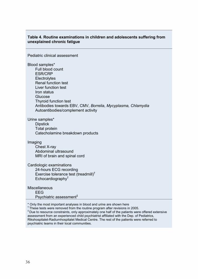
Table 4. Routine examinations in children and adolescents suffering from unexplained chronic fatigue
Pediatric clinical assessment
Blood samples* Full blood count ESR/CRP Electrolytes Renal function test Liver function test Iron status Glucose Thyroid function test
Antibodies towards EBV, CMV, Borrelia, Mycyplasma, Chlamydia Autoantibodies/complement activity
Urine samples* Dipstick Total protein Catecholamine breakdown products
Imaging Chest X-ray
Abdominal ultrasound MRI of brain and spinal cord
Cardiologic examinations 24-hours ECG recording Exercise tolerance test (treadmill)†
Echocardiography†
Miscellaneous EEG Psychiatric assessment‡
* Only the most important analyses in blood and urine are shown here † These tests were removed from the routine program after revisions in 2005. ‡ Due to resource constraints, only approximately one half of the patients were offered extensive assessment from an experienced child psychiatrist affiliated with the Dep. of Pediatrics, Rikshospitalet-Radiumhospitalet Medical Centre. The rest of the patients were referred to psychiatric teams in their local communities.
37
2.1.2 Healthy controls
In the period between August 2003 – June 2005, schools in the Oslo/Bærum area wereasked to recruit healthy controls for the study. Four schools responded positively, andspecific classes were selected in order to achieve a similar age distribution amongpatients and controls. These classes received both oral and written information, andindividuals then volunteered to participate. During the course of the study, the emergingproportion of males vs. females among the patients was used as a guide for therecruitment of volunteer controls, thus assuring a similar distribution of gender withinthe two groups.Prior to final inclusion, the volunteers and their parents/next-of-kin received additionalwritten information about the purpose and content of the experiments. Their right towithdraw at any time was specifically emphasized. A written consent was obtained. Allparticipants received a payment of NOK 200.In total, 57 healthy controls were included, constituting the basis for the analyses inpaper V. Paper IV builds upon 56 controls, as one had to be excluded due to experimentalfailure. However, parts of the experimental protocol were altered during the study period,as will be further outlined below (see 2.2.1). Therefore, some controls had to be excludedfrom the analyses in paper II and III, and the total number in these papers is only 33.
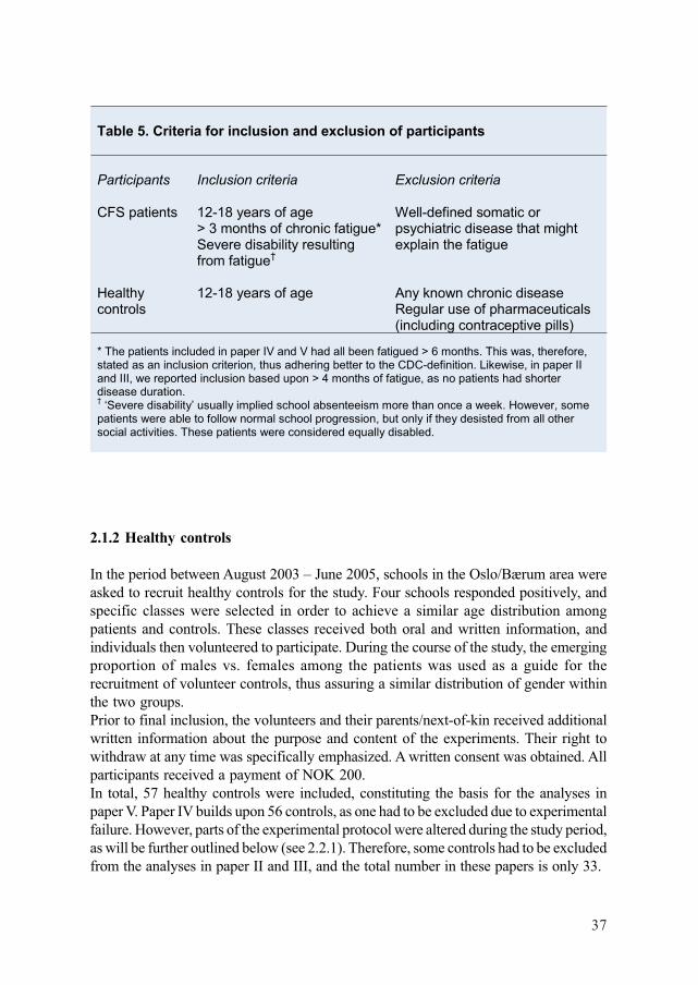
Table 5. Criteria for inclusion and exclusion of participants
Participants Inclusion criteria Exclusion criteria
12-18 years of age > 3 months of chronic fatigue*
CFS patients
Severe disability resulting from fatigue†
Well-defined somatic or psychiatric disease that might explain the fatigue
12-18 years of age Any known chronic disease Healthy controls Regular use of pharmaceuticals
(including contraceptive pills) * The patients included in paper IV and V had all been fatigued > 6 months. This was, therefore, stated as an inclusion criterion, thus adhering better to the CDC-definition. Likewise, in paper II and III, we reported inclusion based upon > 4 months of fatigue, as no patients had shorter disease duration. † ‘Severe disability’ usually implied school absenteeism more than once a week. However, some patients were able to follow normal school progression, but only if they desisted from all other social activities. These patients were considered equally disabled.

38
2.2 Methods
2.2.1 Experimental protocols
For each participant, all experiments were performed during a single day, according tothe program outlined in Table 6. During the course of the study, we alternately summonedCFS patients and controls, in order to minimize the risk of systematic errors. One weekprior to the experiments, the participants were instructed not to drink beveragescontaining alcohol or caffeine, not to take any drugs, and not to use tobacco products.On the day of the experiments, they were supposed to have fasted overnight. Two lightmeals, consisting of a maximum of 2 pieces of bread and 2 glasses of water/juice, wereoffered at certain hours, as specified in Table 6.
Neuroendocrine responses to strong cooling of one handAll participants were instructed to apply an ointment containing the local anestheticlidocaine (Emla®) on the skin of the antecubital fossa one hour prior to arrival in themorning. They rested supine for about 15 minutes, and a catheter was then placed in anantecubital vein (Figure 8). After supine rest for another 15 minutes, blood sampleswere collected on ice-cold vacutainer tubes containing gluthation-EGTA and heparinfor measurements of plasma catecholamines (norepinephrine, epinephrine, dopamine)
Table 6. Overview of experimental day
Time Experiment/Activity Reported in paper
07.30 am Check in 08.00 am Neuroendocrine responses to strong cooling of
one hand IV, V
08.45 am Light meal 09.00 am Questionnaire II, IV, V 09.30 am Head-up tilt-test (HUT) II, III 11.00 am Valsalva-maneuver Not reported 11.30 am Lower body negative pressure (LBNP) with
handgrip IV
01.00 pm Light meal 01.30 pm Cardiovascular responses to mental exercise Not reported 01.45 pm Cardiovascular responses to moderate cooling
of one hand V
03.30 pm End of experiments

39
and metanephrines (normetanephrine, metanephrine, 3-methoxytyramine), respectively.The opposite hand was then immersed in cold water (10 oC) for 2 minutes, after whichnew blood samples were collected immediately. Samples were then centrifuged at 4oC, and plasma was separated for storage at –80 oC until assayed.Metanephrines were extracted from the plasma using solid-phase ion exchange columns(Bond Elute-Accucat, Varian Medical Systems, California, USA) and a commercialmobile phase (Chromsystems, München, Germany) (240). Both catecholamines andmetanephrines were quantified by high performance liquid-chromatography with areverse phase column and glassy carbon electrochemical detector (Agilent Technologies,Colorado, USA). The intra- and interassays variations were 10.7 % and 14.5 % fornorepinephrine, 23.5 % and 10.5 % for epinephrine, 7.2 % and 6.8 % for dopamine,12.6 % and 4.3 % for normetanephrine, 11.7 % and 3.0 % for metanephrine, and 15.3% and 11.2 % for 3-methoxytyramine.Additional endocrine analyses were also carried out, including cardiac peptides, sexhormones, renin, aldosteron, HPA axis hormones, vasopressin and insulin. The resultsare not reported in this thesis.
QuestionnaireA questionnaire was developed based upon the translation of the Autonomic SymptomProfile, a validated instrument for assessing orthostatic intolerance and other variantsof autonomic dysfunction (402). Some items were slightly modified in order to fit ourparticular age group. Furthermore, the questionannire included a translated an slightlymodified version of the Fatigue Severity Scale (228), as well as questions focusing onfunctional consequences of CFS based upon personal clinical experience with patients.Finally, we added questions related to the CDC case definition and to demographiccharacteristics. The subjects answered the questions during an interview. Only someresults from this extensive questionnaire are reported in this thesis.During the course of the study, the questionnaire was slightly revised based upon personalexperience of its feasibility. Thus, a few items were not answered by all participants.
Time (min)40 45 50 55 60 65 70 7515 20 25 30 350 5 10
Application ofanesthetic ointment
Supine rest
Insertion ofintravenous catheter
Blood samplesfor baseline values
Blood samplesfor post-cooling values
Supine rest Immersion of one
hand in water (10 0C)
Figure 8. Neuroendocrine responses to strong cooling of one hand: schematic outlineof the experiment.

40
Head-up tilt-testConventional procedures of head-up tilt-test (HUT) involve tilting to 60o-80o for 30-45minutes (22). Heart rate (HR) is usually continuously recorded from the electro-cardiogram (ECG), whereas arterial blood pressure is measured at predefined intervals(eg. every second minute) using ausculatory or oscillometric techniques. Thus, thehemodynamic recordings are not very accurate. Correspondingly, the test focuses on‘hard’ endpoints, such as syncope or near-syncopal events. However, the tolerance fororthostatic stress is lower in adolescents than in children and adults (347, 444), andconventional HUTs are burdened by a high rate of false positive results in this agegroup, making the test less informative (89).Our first HUT protocol implied 30 minutes of 60o tilt, and was performed in 2 CFSpatients and 22 healthy controls. Of these participants, 2 patients (100 %) and 9 controls(41 %) experienced a syncope or a near-syncopal event (defined as more than 10 mmHg fall in either systolic or diastolic blood pressure, combined with orthostaticsymptoms). Faced with these results, we considered the protocol to be both uninformativeand possibly unethical, and therefore decided to revise it.In the revised HUT protocol, which was successfully undertaken by 27 CFS patientsand 33 healthy controls, the subjects lay supine on an electronically operated tilt tablewith foot-board support (Model 900-00, CNSystems Medizintechnik, Graz, Austria).They were attached to the Task Force Monitor® (Model 3040i, CNSystemsMedizintechnik, Graz, Austria); a combined hardware and software device for non-invasive recording of cardiovascular variables (133, 166). A 5 minute baseline recordingwas obtained. Subjects were then head-up tilted 20o over approximately 10 seconds(Figure 9). They were remained at 20o for 15 minutes, and then tilted back to horizontalposition after which the recording was continued for another 5 minutes, making thetotal experimental period 25 minutes. Subjects were asked not to speak or move duringthe recording period.Instantaneous HR was obtained from the R-R interval of the ECG.Photoplethysmography on the right middle finger was used to obtain a non-invasive,continuous recording of arterial blood pressure. This method correlates satisfactorilywith invasive pressure measurements (300), and has also been validated in adolescents
Time (min)16 18 20 22 24 26 28 306 8 10 12 140 2 4
20o tilted position Supine rest
Tilt downTilt up
Supine rest
Figure 9. Head-up tilt-test (revised protocol): schematic outline of the experiment.

41
and children (356). Approximately every minute, the recorded value was calibratedagainst conventional oscillometric measurement of arterial blood pressure on thesubjects’ left arm. The method of impedance cardiography, in which a small electricalpotential is applied between electrodes placed on the neck and upper abdomen, wasused to obtain a continuous recording of the temporal derivate of the transthoracicimpedance (dZ/dt) (98). All recorded signals were transferred online to the built-inrecording computer of the Task Force Monitor®, running software for real-time dataacquisition.
Lower body negative pressure with handgripThe technique of lower body negative pressure (LBNP) is a well-established tool forstudies of cardiovascular adjustments during orthostatic stress (382), whereas handgripis a common test for studies of cardiovascular adjustments during isometric exercise(161, 205). In our laboratory, we have developed a combined experimental set-up inorder to study complex regulatory adjustments (184).The participants lay supine with their lower body in a plastic chamber, in which aircould be evacuated very rapidly, thus reaching a pre-defined negative pressure withinmilliseconds (185). In order to prevent air leak, rubber devices were used to make atight seal around the subjects’ waist. They were lightly dressed, and the ambienttemperature was kept between 23 and 26 °C. They were familiarized with the testprocedure in two pilot experiments. By means of an electronic device (AB-detector,Gøteborg, Sweden), the force of the subjects’ left-sided handgrip was continuouslydisplayed to them. They were first asked to perform maximum isometric work forabout 10 seconds. Based on the mean value, the 30 % level was calculated, and theywere then asked to use a couple of minutes to become familiar with this amount offorce.Five minutes were used for baseline registration of cardiovascular variables (Figure10). Then, LBNP of –20 mm Hg was applied instantaneously. After 6 minutes of LBNP,the subjects were asked to perform left-sided handgrip with 30 % of maximum force
Time (min)8 9 10 11 12 13 14 15 16 173 4 5 6 70 1 2
LBNP-20 mm HgSupine rest Supinerest
LBNP onHandgrip, 30 %
of maximal force LBNP off
Figure 10. Lower body negative pressure with handgrip: schematic outline of theexperiment.
42
Instantaneous HR was obtained from the ECG. Photoplethysmography on the rightmiddle finger was used to obtain a non-invasive, continuous recording of arterial bloodpressure (2300 Finapres, Ohmeda, Madison, WI, US) (300, 356). A continuous recordingof maximum blood flow velocity in the ascending aorta was obtained by a bidirectionalultrasound Doppler velocimeter (SD-100, GE Vingmed Ultrasound, Horten, Norway),operating in pulsed mode at 2 MHz (119). Using a handheld transducer, the ultrasoundbeam was directed from the suprasternal notch towards the aortic root. The samplevolume range was adjusted so that measurements were made 1-2 cm above the aorticvalve. A continuous recording of mean blood flow velocity in the right brachial artery(BABF) was obtained by another bidirectional ultrasound Doppler velocimeter (SD-50, GE Vingmed Ultrasound, Horten, Norway), operating in pulsed mode at 10 MHz(251). A continuous recording of acral skin laser flux, which is a measure of acral skinblood flow (ASBF), was obtained by a laser-Doppler instrument (DRT4, Mooreinstruments, Milway, Devon, UK), with the probe firmly attached with adhesive stripsto the pulp of the right index finger (24). The BABF and ASBF results are not reportedin this thesis. All recorded signals, including the pressure in the LBNP-chamber andthe force of the handgrip, were transferred online to a recording computer running aprogram for real-time data acquisition (developed by Morten Eriksen, Dep. of Physi-ology, University of Oslo, Oslo, Norway).
Cardiovascular responses to moderate cooling of one handIn our laboratory, the general design of this experiment has been used in several otherstudies concerning normal physiology (23). The participants were lightly dressed andlay supine in a climatic chamber. Ambient temperature was maintained around 26 oC,assuring that subjects were within their thermoneutral zone. The left hand was immersedin a stirred thermostat-controlled water bath (CB 29-20e, Heto-Holtan, Åbyhøj, Den-mark) with a temperature of 35 oC. 30 minutes were used for acclimatization and another5 minutes for baseline registration, after which the water temperature was graduallylowered to 19 oC over approximately 50 minutes (Figure 11).A continuous recording of acral skin laser flux, which is a measure of acral skin bloodflow (ASBF), was obtained by a laser-Doppler instrument (DRT4, Moore instruments,Milway, Devon, UK) (24). The probes were firmly attached with adhesive strips to thepulp of the flexor surface of the distal phalanges of the right and left index fingers. Inthis position, the probes mainly detect blood flow in arteriovenous anastomoses (23).Tympanic temperature (TT) was continuously monitored by an electronic probe (D-
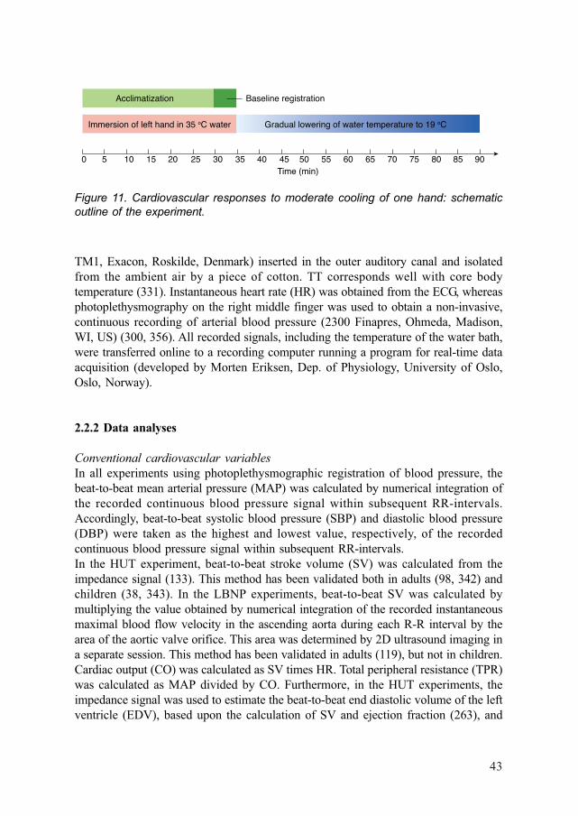
43
Time (min)40 45 50 55 60 65 70 75 80 85 9015 20 25 30 350 5 10
Gradual lowering of water temperature to 19 oCImmersion of left hand in 35 oC water
Acclimatization Baseline registration
Figure 11. Cardiovascular responses to moderate cooling of one hand: schematicoutline of the experiment.
TM1, Exacon, Roskilde, Denmark) inserted in the outer auditory canal and isolatedfrom the ambient air by a piece of cotton. TT corresponds well with core bodytemperature (331). Instantaneous heart rate (HR) was obtained from the ECG, whereasphotoplethysmography on the right middle finger was used to obtain a non-invasive,continuous recording of arterial blood pressure (2300 Finapres, Ohmeda, Madison,WI, US) (300, 356). All recorded signals, including the temperature of the water bath,were transferred online to a recording computer running a program for real-time dataacquisition (developed by Morten Eriksen, Dep. of Physiology, University of Oslo,Oslo, Norway).
2.2.2 Data analyses
Conventional cardiovascular variablesIn all experiments using photoplethysmographic registration of blood pressure, thebeat-to-beat mean arterial pressure (MAP) was calculated by numerical integration ofthe recorded continuous blood pressure signal within subsequent RR-intervals.Accordingly, beat-to-beat systolic blood pressure (SBP) and diastolic blood pressure(DBP) were taken as the highest and lowest value, respectively, of the recordedcontinuous blood pressure signal within subsequent RR-intervals.In the HUT experiment, beat-to-beat stroke volume (SV) was calculated from theimpedance signal (133). This method has been validated both in adults (98, 342) andchildren (38, 343). In the LBNP experiments, beat-to-beat SV was calculated bymultiplying the value obtained by numerical integration of the recorded instantaneousmaximal blood flow velocity in the ascending aorta during each R-R interval by thearea of the aortic valve orifice. This area was determined by 2D ultrasound imaging ina separate session. This method has been validated in adults (119), but not in children.Cardiac output (CO) was calculated as SV times HR. Total peripheral resistance (TPR)was calculated as MAP divided by CO. Furthermore, in the HUT experiments, theimpedance signal was used to estimate the beat-to-beat end diastolic volume of the leftventricle (EDV), based upon the calculation of SV and ejection fraction (263), and
44
acceleration index (ACI), defined as the highest positive slope of the impedance signal(d2Z/dt2
max). ACI corresponds to the maximal acceleration of blood flow during theejection phase, and is regarded as an indirect marker of cardiac contractility (213).All variables based upon volume measurements, i.e. CO, SV, TPR and EDV, werenormalized according to body surface area (BSA), using the following formula: BSA =0.00235 x (height in cm)0.423 x (weight in kg)0.515.
Heart rate and blood pressure variabilitySpontaneous variability is a characteristic feature of all cardiovascular variables, asdescribed above (see 1.2.2), reflecting the «homeodynamic interplay betweenperturbations to cardiovascular function and the dynamic responses of the cardiovascularregulatory systems» (7). Analyses of variability might be performed in the time domain,where the extent of variability within a recording of certain length is expressed usingcommon statistical terms (411). For instance, when analyzing variability of RR-intervals(inverse HR) in an ECG recording, SDNN is the standard deviation of all RR-intervals,RMSSD is the square root of the mean square differences of successive RR-intervals,and pNN50 is the proportion of successive RR-intervals with a difference greater than50 ms.Alternatively, analyses might be carried out in the frequency domain, in which thesignals are regarded as a mixture of sine waves of different amplitudes and frequencies(7). This spectral analysis approach requires more sophisticated mathematical proce-dures; the most common can be classified as either non-parametric methods (e.g. FastFourier Transform) or parametric methods (e.g. Autoregressive modelling) (57, 73). Inany case, the extent of variability is expressed as the power of different frequencies.Spectral analyses of short-time recordings of heart rate or blood pressure usually revealthree distinct peaks in the power spectrum (411). By convention, spectral power densitiesare computed in three frequency bands corresponding to these peaks, assigned verylow frequency (VLF, <0.04 Hz), low frequency (LF, 0.04-0.15 Hz) and high frequency(HF, 0.15-0.4 Hz), respectively.The RR-interval power spectral density in the HF-band might be considered an indexof the parasympathetic modulation of heart rate, whereas RR-interval power spectraldensity in the LF region is due to the combined effect of cardiac parasympathetic andsympathetic activity (73, 346). The LF/HF-ratio has often been regarded as a measureof ‘sympathovagal balance’ in the modulation of HR, particularly during orthostaticstress (112, 296). Blood pressure variability in the LF band (also known as Mayerwaves) is primarily a consequence of variations in efferent sympathetic neural activityto peripheral resistance vessels, whereas blood pressure variability in the HF band isdue to a combination of the mechanical influence of ventilation and the effects of heartrate variability on blood pressure (73, 258).In this thesis, variability analyses were performed on the HR and DBP recordings fromthe HUT experiments, using an adaptive autoregressive algorithm (see paper III) (132).This technique yields a time-varying spectrum in which spectral power densities are
45
computed from consecutive segments of the biosignals (16, 57). The lengths of thesesegments vary, as the biosignals are continuously monitored for departure fromstationarity, and the segment boundaries are placed accordingly. This approach isadvantageous in a situation where the variability is supposed to change as theconsequence of an intervention, e.g. the transition from supine to tilted position.
Mathematical processing of beat-to-beat recordingsPlots of the beat-to-beat recorded cardiovascular variables were inspected for eachparticipant. Obvious artifacts, e.g. due to temporary instrumental failure, were removedand replaced by linear interpolation between values appraised to be valid. For the mainanalyses in paper II-V, the median values within defined time periods of each recordingwere computed, thus reducing the influence of possible false outliers even more.In addition, in paper II-IV, the beat-to-beat recordings were converted to 4 Hz timeseries of equal length by linear interpolation. These time series were filtered by assigningthe median value within a sliding window to each time point, thus removing bothminor artifacts and stochastic variability. Furthermore, all time series were normalized,taking the mean value of the first time period as zero. Coherent averaging was thenperformed by calculating the arithmetical mean for each time point (333).In paper V, the beat-to-beat recordings of ASBF Left were searched for vasoconstrictorevents, defined to occur at the water temperature where the median value computedover 30 consecutive heart beats was permanently less than 50 % of the median valueprior to cooling.
Statistical analysesIn all experiments, data plots were routinely inspected to assess their empiricaldistribution. If approximating normality, the mean was taken as an estimate of theexpectancy of the population under study, and 95 % confidence intervals for the meanwas calculated according to parametric methods (see paper II and IV). If the empiricaldistribution of data deviated from normality, the median and non-parametric 95 %confidence intervals were used for the same purposes (see paper III and V).In all experiments, non-parametric statistical test were regarded as most ‘robust’, andtherefore used to explore the differences between the two groups of participants (188).In order to reduce the methodological problem of performing multiple statisticalcomparisons, we reserved the statistical tests for variables that were considered mostcentral to our research questions. Generally, a p < 0.05 was considered statisticallysignificant.
2.2.3 Ethical and legal considerations
The principle of informed consent is considered vital in all research involving humanbeings (121). Complying with this principle requires that the participants are fully
46
informed about all aspects of the experiments, including any possible side-effects, andthat they are able to make a totally independent decision concerning whether or not toparticipate. Therefore prior to inclusion, both patients and controls received thoroughinformation about the experiment orally and in written form.In practice, and particularly when children are involved, the ideal of informed consentare impossible to achieve (229). First, being ‘fully informed’ is, at least theoretically,an endless task; in addition, there are no means to assess whether the information hasbeen perceived correctly by each participant. Second, no process of decision is totallyfree of obligations; furthermore, children are not considered fully competent of makingdecisions about their own life.These obstacles were partly overcome by providing thorough information and requiringa written statement of consent from the participants’ parents/next-of-kin. Still, wemaintain that medical research should also adhere to the principle of primum non nocere(374). In other words, experiments on humans - and children in particular - cannot bejustified solely by the participants’ free, informed decision if the experiments imposetoo great a risk of harmful effects.Prior to the experimental phase of this study, none of the experiments were consideredto carry any element of risk. However, the following parts were addressed as potentiallyunpleasant: pain during insertion of intravenous catheter and immersion of hand incold water, and dizziness or near-syncope during HUT and LBNP. If near-syncopeoccurred, the protocols demanded the immediate termination of the experiment, inorder to minimize the risk of manifest syncope.Our initial HUT protocol resulted in a high incidence of near-syncope and even syncope,and was therefore abandoned as described above (see 2.2.1). During the revisedexperiment, one CFS patient complained of unpleasant dizziness; however, near-syncopeor syncope was never observed. LBNP precipitated a harmless cardiac arrhythmia in ahealthy control; the experiment was terminated immediately, and the participant wasreferred to an ordinary cardiologic assessment. Two other subjects (one CFS patient,one control) reported dizziness or other unpleasant experiences during LBNP. The useof anesthetic ointment seemed effective in reducing the pain related to catheter insertion.The study was approved by the Regional committee for ethics in medical research andthe Norwegian Data inspectorate. The authors of the papers are all qualified accordingto the Vancouver guidelines. None of them possesses any conflicts of interest related tothe subject matter of this thesis.
47
3 Results
3.1 Subjects
27 CFS patients and 33 healthy controls were included in the experiments reported inpaper II and III, whereas 15 CFS patients and, respectively, 56 and 57 healthy controlswere included in paper IV and V (Table 7). In all experiments, the two groups had acomparable distribution of gender, age, weight, height, and body surface area. Allparticipants, except one control, were of Caucasian ethnicity. The CFS patients weresedentary, had a high Fatigue severity scale sum score, and reported a substantial amountof functional impairment; however, no one was permanently bedridden. About 60 % fitthe CDC case definition of CFS.
3.2 Experimental results
The following research questions were presented in 1.3.3:
A. When comparing adolescent CFS patients with healthy controls, how is theexpression of symptoms indicative of altered cardiovascular and thermoregulatoryautonomic control?
B. During supine rest, when comparing adolescent CFS patients with healthy controls:a. How is the autonomic modulation of cardiovascular variables?b. What are the levels of catecholamines and metanephrines?c. How is the autonomic modulation of thermoregulatory variables?
C. During orthostatic stress, when comparing adolescent CFS patients with healthycontrols:
a. How is the autonomic modulation of cardiovascular variables?b. How is the autonomic modulation of cardiovascular variables when isometric
exercise is performed in addition?

48
Table 7. Subject characteristics
Variable Paper II and III Paper IV and V
Control CFS Control* CFS
Number 33 27 57 15
Female gender 19 (58 %) 18 (68 %) 34 (60 %) 10 (67 %)
Adherence to the CDC-criteria 15 (56 %) 9 (60 %) School absenteeism once a week or more†
0 (0 %) 23 (85 %) 0 (0 %) 10 (77 %)
Not able to attend leisure activities once a week or more†
0 (0 %) 24 (96 %) 0 (0 %) 13 (100 %)
Permanently bedridden 0 (0 %) 0 (0 %) 0 (0 %) 0 (0 %)
Mean (range)
Mean (range)
Mean (range)
Mean (range)
Age (years) 15 15 16 15(13-18) (13-18) (13-18) (12-18)
Weight (kg) 61 56 62 59(44-77) (37-92) (44-99) (43-92)
Height (cm) 172 169 172 171(149-195) (145-197) (149-195) (160-192)
Body surface area (m2) 1.7 1.6 1.7 1.7 (1.4-2.0) (1.3-2.3) (1.4-2.2) (1.4-2.2)
Duration of fatigue (months) 30 31(4-132) (6-60)
Fatigue severity scale, sum score‡
1.5 (1.0-2.3)
4.5 (2.9-5.0)
1.5 (1.0-3.6)
4.6 (3.8-5.0)
Reported physical exercise (hours/week)
6.7 (2-14)
0.5 (0-6)
6.2 (1-20)
0.7 (0-7)
* One female control included in paper V did not participate in the experiments of paper IV, due to the precipitation of cardiac arrhythmia during LBNP (see 2.2.3). Thus, in paper IV, some of the values (e.g. the numbers of female participants) deviate slightly from what is presented here. † Due to revisions in the questionnaire (see 2.2.1), these items were not answered by all participants, influencing the calculated percentage. ‡ The fatigue severity scale was slightly modified from the original publication, using a 1-5 Likert response scale for each item (228).

49
D. During local cold stress, when comparing adolescent CFS patients with healthycontrols:
a. What are the levels of catecholamines and metanephrines?b. How is the autonomic modulation of thermoregulatory variables?
Below, the results of the experiments are summarized in accordance with these questions
3.2.1 Symptoms of altered cardiovascular and thermoregulatory autonomic control
Palpitations, dizziness, skin flushing, pale skin and an increased tendency to sweatwere prominent symptoms in the patient described in paper I. Table 8 summarizes thequestionnaire results reported in paper II, IV and V. As for cardiovascular autonomiccontrol, there is evidence that CFS patients more frequently than controls feel dizzy
Table 8. Symptoms of altered cardiovascular and thermoregulatory autonomic control – summary of results comparing CFS patients to healthy controls
Variables Questionnaire
Cardiovascular autonomic control Experience fainting when rising quickly ( )Feeling dizzy just after a heavy meal ( )Feeling dizzy after standing upright for a long period Feeling dizzy during moderate physical exercise Feeling dizzy taking a hot bath or shower
Thermoregulatory autonomic controlExperience sudden changes in skin colour
- Sudden redness of skin - Sudden paleness of skin
Experience shivering hands Sweating more than others Feeling unusually warm after standing upright for a long period Feeling unusually warm after moderate physical exercise Feeling unusually warm after a hot bath or shower ( )
Arrow=significant difference, direction indicating how CFS patients deviate from controls. Arrow in parentheses=non-significant tendency.
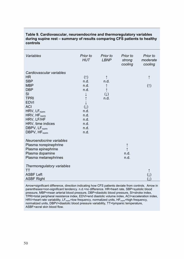
50
Table 9. Cardiovascular, neuroendocrine and thermoregulatory variables during supine rest – summary of results comparing CFS patients to healthy controls
Variables Prior to HUT
Prior to LBNP
Prior to strong cooling
Prior to moderate cooling
Cardiovascular variables HR ( )SBP n.d. n.d.MBP n.d. ( )DBP n.d. SI ( ) TPRI n.d. EDVI ACI ( ) HRV, LFnorm n.d. HRV, HFnorm n.d. HRV, LF/HF n.d. HRV, time indices n.d. DBPV, LFnorm n.d. DBPV, HFnorm n.d.
Neuroendocrine variables Plasma norepinephrine Plasma epinephrine Plasma dopamine n.d. Plasma metanephrines n.d.
Thermoregulatory variables TT ASBF Left ( )ASBF Right ( )
Arrow=significant difference, direction indicating how CFS patients deviate from controls. Arrow in parentheses=non-significant tendency. n.d.=no difference. HR=heart rate, SBP=systolic blood pressure, MBP=mean arterial blood pressure, DBP=diastolic blood pressure, SI=stroke index, TPRI=total peripheral resistance index, EDVI=end diastolic volume index, ACI=acceleration index, HRV=heart rate variability, LFnorm=low frequency, normalized units, HFnorm=high frequency, normalized units, DBPV=diastolic blood pressure variability, TT=tympanic temperature, ASBF=acral skin blood flow.

51
after standing upright, during moderate physical exercise and during a hot bath. Lessconvincing evidence indicates an increased tendency to faint and to feel dizzy afterheavy meals. As for thermoregulatory autonomic control, there is evidence that CFSpatients more often experience sudden changes in skin color, a sudden paleness ofskin, shivering hands, sweating, and feeling unusually warm after standing upright andafter moderate physical exercise. A sudden redness of skin is rarer among CFS patientsthan controls. Less convincing evidence indicates that CFS patients tend to feel unusuallywarm after a hot bath.
3.2.2 Cardiovascular, neuroendocrine and thermoregulatory variables duringsupine rest
Examination of the patient reported in paper I revealed supine tachycardia. Table 9summarizes the results during supine rest reported in paper II-V. As for cardiovascularvariables, there is firm evidence of increased HR and decreased SI among CFS patients,and also evidence of increased MBP, DBP, and TPRI and decreased EDVI. Lessconvincing evidence indicates decreased ACI among CFS patients. As for neuro-endocrine variables, there is evidence of increased plasma norepinephrine and epi-nephrine. As for thermoregulatory variables, there is evidence of increased TT, whereasless convincing evidence indicates decreased ASBF Left and Right.
3.2.3 Cardiovascular responses to orthostatic stress alone and combined withisometric exercise
Examination of the patient reported in paper I revealed orthostatic tachycardia andhypotension that normalized during treatment with propranolol. Table 10 summarizesthe results reported in paper II-IV. When orthostatic stress is applied in isolation, thereis firm evidence of a greater increase in HR, MBP, DBP, and TPRI and a greater decreasein SI among CFS patients. There is also evidence of altered heart rate variability, witha greater increase in LFnorm and LF/HF-ratio and a greater decrease in HFnorm amongCFS patients. Less convincing evidence indicates a greater increase in ACI amongCFS patients. When isometric exercise is perfomed in addition to orthostatic stress,there is evidence of a smaller increase in HR and DBP and a smaller decrease in SIamong CFS patients. There is less convincing evidence of a smaller increase in MBPand TPRI in this situation.

52
3.2.4 Cardiovascular, neuroendocrine and thermoregulatory responses to localcold stress
Table 11 summarizes the results reported in paper V. During strong cooling, evidenceindicates similar changes in the neuroendocrine variables among both CFS patientsand controls. During moderate cooling of the left hand, there is evidence of a greaterdecrease in TT and a smaller decrease in ASBF Left among CFS patients, and there isless convincing evidence of a smaller decrease in ASBF Right among CFS patients.
Table 10. Changes in cardiovascular variables during orthostatic stress – summary of results comparing CFS patients to healthy controls
Variables During HUT During LBNP During LBNP and handgrip
Cardiovascular variables HR SBP n.d. n.d. MBP ( ) DBP SI ( ) TPRI ( ) ( ) EDVI n.d. ACI ( ) HRV, LFnorm HRV, HFnorm HRV, LF/HF HRV, time indices n.d. DBPV, LFnorm n.d. DBPV, HFnorm n.d.
Arrow=significant difference, direction indicating how CFS patients deviate from controls. Arrow in parentheses=non-significant tendency. n.d.=no difference. HR=heart rate, SBP=systolic blood pressure, MBP=mean arterial blood pressure, DBP=diastolic blood pressure, SI=stroke index, TPRI=total peripheral resistance index, EDVI=end diastolic volume index, ACI=acceleration index, HRV=heart rate variability, LFnorm=low frequency, normalized units, HFnorm=high frequency, normalized units, DBPV=diastolic blood pressure variability.
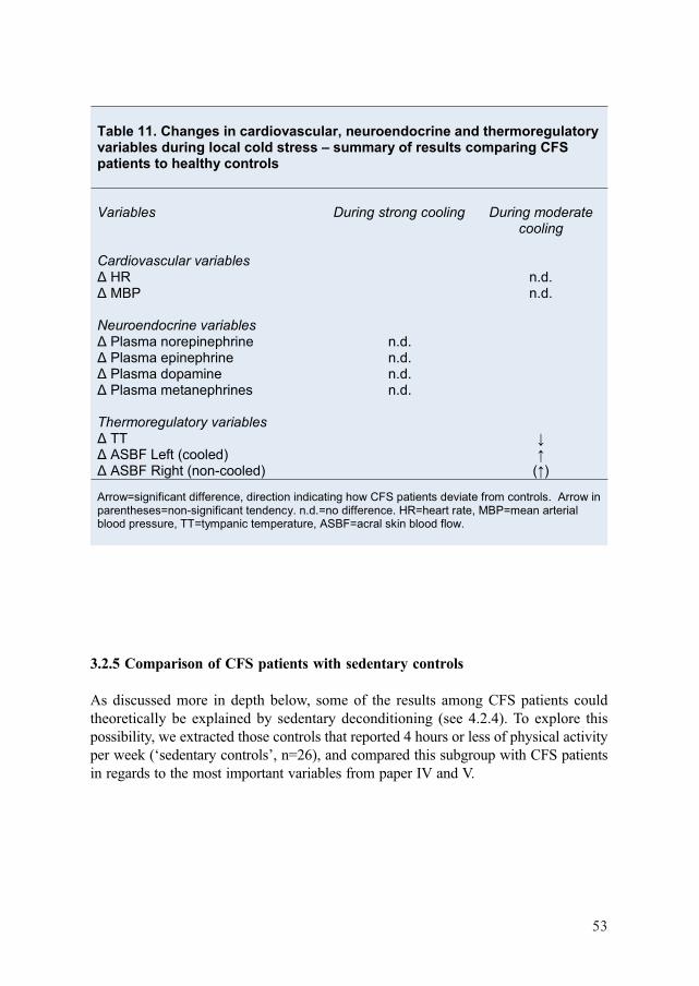
53
Table 11. Changes in cardiovascular, neuroendocrine and thermoregulatory variables during local cold stress – summary of results comparing CFS patients to healthy controls
Variables During strong cooling During moderate cooling
Cardiovascular variables HR n.d. MBP n.d.
Neuroendocrine variables Plasma norepinephrine n.d. Plasma epinephrine n.d. Plasma dopamine n.d. Plasma metanephrines n.d.
Thermoregulatory variables TT ASBF Left (cooled) ASBF Right (non-cooled) ( )
Arrow=significant difference, direction indicating how CFS patients deviate from controls. Arrow in parentheses=non-significant tendency. n.d.=no difference. HR=heart rate, MBP=mean arterial blood pressure, TT=tympanic temperature, ASBF=acral skin blood flow.
3.2.5 Comparison of CFS patients with sedentary controls
As discussed more in depth below, some of the results among CFS patients couldtheoretically be explained by sedentary deconditioning (see 4.2.4). To explore thispossibility, we extracted those controls that reported 4 hours or less of physical activityper week (‘sedentary controls’, n=26), and compared this subgroup with CFS patientsin regards to the most important variables from paper IV and V.
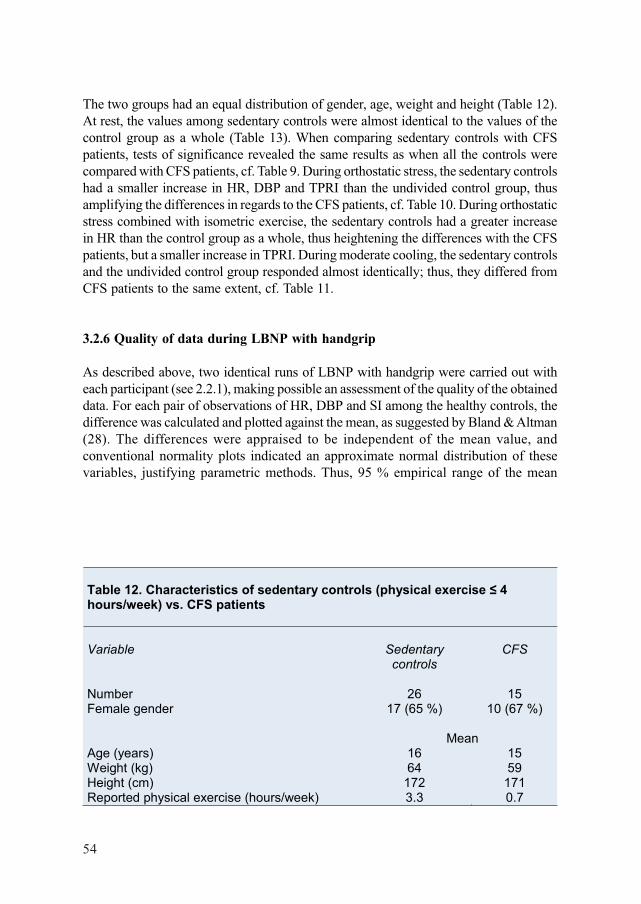
54
The two groups had an equal distribution of gender, age, weight and height (Table 12).At rest, the values among sedentary controls were almost identical to the values of thecontrol group as a whole (Table 13). When comparing sedentary controls with CFSpatients, tests of significance revealed the same results as when all the controls werecompared with CFS patients, cf. Table 9. During orthostatic stress, the sedentary controlshad a smaller increase in HR, DBP and TPRI than the undivided control group, thusamplifying the differences in regards to the CFS patients, cf. Table 10. During orthostaticstress combined with isometric exercise, the sedentary controls had a greater increasein HR than the control group as a whole, thus heightening the differences with the CFSpatients, but a smaller increase in TPRI. During moderate cooling, the sedentary controlsand the undivided control group responded almost identically; thus, they differed fromCFS patients to the same extent, cf. Table 11.
3.2.6 Quality of data during LBNP with handgrip
As described above, two identical runs of LBNP with handgrip were carried out witheach participant (see 2.2.1), making possible an assessment of the quality of the obtaineddata. For each pair of observations of HR, DBP and SI among the healthy controls, thedifference was calculated and plotted against the mean, as suggested by Bland & Altman(28). The differences were appraised to be independent of the mean value, andconventional normality plots indicated an approximate normal distribution of thesevariables, justifying parametric methods. Thus, 95 % empirical range of the mean
Table 12. Characteristics of sedentary controls (physical exercise 4 hours/week) vs. CFS patients
Variable Sedentary controls
CFS
Number 26 15Female gender 17 (65 %) 10 (67 %)
Mean Age (years) 16 15Weight (kg) 64 59Height (cm) 172 171Reported physical exercise (hours/week) 3.3 0.7

55
Table 13. Sedentary controls compared with CFS patients for selected variables (cf. paper IV and V)
Variable Controls (n=57)*
Sedentery controls (n=26)*
CFS(n=15)*
p-value, CFS vs
sedentary controls†
At rest
HR (beats/min), mean 65.7 66.7 75.8 0.03
DBP (mm Hg), mean 65.0 65.7 70.1 0.06
TPRI (mm Hg/l/min/m2), mean 11.1 11.0 11.6 0.59
Norepinephrine (pmol/L), median
1156 1263 1646 0.06
Epinephrine (pmol/L), median 171 164 216 0.03
ASBF Left (a.u.), median 322.2 331.6 249.9 0.19
ASBF Right (a.u.), median 271.2 273.5 122.3 0.17
TT (oC), median 35.96 36.00 36.41 0.06
Changes during orthostatic stress (LBNP)
HR (beats/min), mean 6.6 5.0 12.4 0.004
DBP (mm Hg), mean 2.2 2.0 4.8 0.03
TPRI (mm Hg/l/min/m2), mean
2.7 2.4 3.9 0.10
Changes during orthostatic stress + isometric exercise (handgrip)
HR (beats/min), mean 8.9 11.5 2.6 0.002
DBP (mm Hg), mean 8.4 8.0 5.3 0.03
TPRI (mm Hg/l/min/m2), mean
2.2 1.6 1.1 0.36
Changes during moderate cooling
ASBF Left (a.u.), median –267.6 –271.7 –132.7 0.03
ASBF Right (a.u.), median –39.0 –41.5 1.4 0.10
TT (oC), median 0.08 0.04 –0.22 0.19
* For some variables, the totals are slightly lower, cf. paper IV and V. † Wilcoxon-Mann-Whitney’s test, 2-sided

56
differences could be calculated from the standard deviations and gives an impressionof agreement between the two situations, whereas the mean differences withcorresponding 95 % confidence intervals reveal possible biases (28, 40). The resultsare displayed in Table 14. Except for HR during LBNP, the mean differences are smalland the confidence intervals include zero.
Table 14. Mean differences (95 % confidence intervals) [95 % empirical range]* of cardiovascular variables in healthy controls (n = 55)† during two consecutive runs of LBNP
Variables Baseline During LBNP During LBNP and handgrip
HR (beats/min) –0.5 2.5 1.0 (–1.7 - 0.7) (0.6 - 4.4) (–0.9-2.8) [–9.2 - 8.3] [–11.5 - 16.6] [–12.7 - 14.6]
DBP (mm Hg) –0.03 1.3 1.7 (–1.8 - 1.7) (–-0.2 - 2.9) (–0.5 - 3.9) [–12.8 - 12.8] [–10.4 - 13.1] [–14.5 - 17.9]
SI (ml/m2) –0.3 –0.5 0.1 (–1.6 - 1.1) (–1.8 - 0.7) (–1.2 - 1.5)
[–10.6 - 10.1] [–9.7 - 8.7] [–9.7 - 10.0] * Based upon parametric methods (40). † One subject underwent only one single run of LBNP, and was therefore excluded from this analysis (see 2.2.1).

57
4 Discussion
4.1 Answers to the research questions
4.1.1 Symptoms of altered cardiovascular and thermoregulatory autonomic control
In general, our questionnaire results confirm that CFS patients have symptoms of alteredcardiovascular and thermoregulatory autonomic control. As far as we know, these clinicalphenomena have never before been systematically explored and charted.Dizziness, which is reported more frequently among CFS patients, is an ambiguousterm; unknowingly people often (incorrectly) use it to describe vertigo (446). However,when it occurs in circumstances known to challenge cardiovascular regulation (uprightposition, physical exercise, hot surroundings, heavy meals), the most likely underlyingmechanism is a reduced brain perfusion due to either hypotension or cerebralvasoconstriction. The increased tendency to faint among these patients supports thisconclusion.The experience of feeling unusually warm is also difficult to interpret in terms ofunderlying mechanisms. This symptom may accompany hypotension, and should inthat case be interpreted as a sign of altered cardiovascular control (402). However, the‘hot flush’ characteristically affecting menopausal women is caused by cutaneousvasodilation and thereby increased skin temperature, possibly due to alterednoradrenergic control of skin blood vessels (9). A sudden paleness of skin, on the otherhand, directly indicates cutaneous vasoconstriction due to increased sympathetic outflow.Accordingly, increased tendency to sweat and shiver indicates enhanced sympatheticoutflow to sweat glands and skeletal muscles, respectively (155).

58
4.1.2 Cardiovascular, neuroendocrine and thermoregulatory variables duringsupine rest
Our results show substantial alterations of cardiovascular, neuroendocrine andthermoregulatory variables in CFS patients at rest. As far as we are aware, such anextensive investigation of baseline values has not been undertaken before; however,there are scattered reports of similar findings (208, 231, 305, 370, 413).As arrhythmia was ruled out during ordinary clinical examinations, the increased restingHR among CFS patients indicates enhanced sympathetic or attenuated parasympatheticcardiac nerve activity, possibly a combination (155). Alternatively, this finding couldbe explained by increased levels of circulating epinephrine. Decreased SI in CFS patientsmight suggest low contractility due to myocardial dysfunction; however, a moreimmediate explanation is reduced filling, supported by the observed tendency towardsdecreased EDVI. The latter, in turn, might signal reduced blood volume or increasedvenous capacitance, but could also be directly related to high HR and correspondinglylow filling time (116).The resistance in the arterioles of skeletal muscles is the main contributor to TPRI inmost physiological states (353). Thus, a tendency towards increased TPRI among CFSpatients indicates skeletal muscle vasoconstriction, which – in turn – is stronglysuggestive of increased sympathetic nerve activity, as circulating catecholamines havea vasodilative effect in skeletal muscles (155).Arterial blood pressure and body temperature are the controlled variables in the reflexcircuits ensuring cardiovascular and temperature homeostasis (see 1.2.2). Therefore, atendency towards increased MBP and DBP as well as TT among CFS patients directlysuggests altered set-point values for the cardiovascular and thermoregulatory controlsystems. According to Goldstein, such homeostat resetting is a hallmark of distress(155). All other factors being equal, the maintenance of cardiovascular homeostasis inthis situation requires increased sympathetic modulation of the heart and arterioles.Similarly, maintenance of thermoregulatory homeostasis requires enhanced sympatheticoutflow to skin blood vessels and skeletal muscles, causing vasoconstriction andshivering.A high level of norepinephrine in the antecubital vein plasma of CFS patients mightsuggest increased sympathetic nerve activity to forearm skin and skeletal muscle (156).Likewise, a high plasma level of epinephrine is most likely a result of increasedsympathetic nerve activity to the adrenals. However, because high levels of plasmacatecholamines could result from either increased spill-over or reduced removal, whichin turn depends upon both sympathetic nerve activity, the capacity of different reuptake-and breakdown-pathways, and local blood flow (155, 156), there are several alternativeexplanations as well.

59
4.1.3 Cardiovascular responses to orthostatic stress
Abnormal cardiovascular responses to orthostatic stress, as documented in this thesis,are perfectly in line with a number of other reports (35, 231, 306, 339, 340, 383, 386,387). However, an exeriermental set-up combining orthostatic stress and isometricexercise has not been undertaken before; thus, our findings of attenuated cardiovascularresponses among CFS patients in this particular situation appear to be novel.The greater increase in HR in relation to orthostatic stress in CFS patients suggestsenhanced sympathetic and/or attenuated parasympathetic cardiac nerve activity (155).This notion is further supported by the analyses of heart rate variability, in which thegreater increase in LFnorm and LF/HF-ratio and greater decrease in HFnorm among CFSpatients indicates a sympathetic predominance in the modulation of the sinoatrial node(258, 296, 346). As outlined above, a greater decrease in SI might be ascribed to theHR influence on filling time (116); reduced contractility due to myocardial dysfunctionseems unlikely, as ACI tended to be higher in CFS patients than in controls.Following the reasoning from 4.1.2, a greater increase in TPRI among CFS patientssuggests a stronger sympathetically mediated vasoconstriction in skeletal muscles.Furthermore, a greater increase in MBP and DBP might be a consequence of bringingthe set-point to an even higher level, thus redefining the range of homeostasis (155).The cardiovascular responses to combined orthostatic stress and isometric exercisemight be considered a ‘mirror image’. The smaller increase of HR among CFS patientsis consistent with attenuated sympathetic cardiac responses (184). Similarly, a tendencytowards a smaller increase of TPRI suggests attenuated sympathetic outflow to skeletalmuscle arterioles. Again, the differences in SI among the two groups might be aconsequence of the different HR responses, whereas a smaller increase of MBP andDBP among CFS patients suggests an inability to raise the set-point in this particularsituation.
4.1.4 Cardiovascular, neuroendocrine and thermoregulatory responses to localcold stress
Mechanisms of thermoregulation have hardly been explored among CFS patients, andwe are not aware of any study that has applied experimental methods similar to ours.Thus, our findings regarding responses to local skin cooling appear to be unique to thisthesis.The similar neuroendocrine response to strong cooling among CFS patients and controlsindicates that this stressor causes an identical general enhancement of sympatheticnerve activity in both groups. Thus, CFS patients have preserved reflex arches andresponse abilities within the sympathetic nervous system.

60
Blood flow through arteriovenous anastomoses (AVAs) is the main determinant of ASBF(23, 251). These vessels are sympathetically innervated. Thus, a smaller decrease ofASBF Left among CFS patients during cooling of the left hand (and a similar tendencytowards smaller decrease of ASBF Right) indicates attenuated sympathetic nerve activityto the AVAs. Alternatively, this result might be explained by a defect in localvasoconstrictor mechanisms. Furthermore, in CFS patients, TT decreased during themoderate cooling experiment, suggesting a normalization of the thermoregulatory set-point value. As outlined above, attenuated sympathetic outflow to skin blood vesselswould be an expected consequence of such resetting (see 4.1.2), fitting neatly with theobserved ASBF data.
4.1.5 Concluding remarks
Taken as a whole, we have provided firm evidence of altered sympathetic nerve activityamong CFS patients, supporting the hypotheses of sympathetic dysfunction as essentialin CFS pathophysiology (137, 151, 159, 282, 297). More specifically, and in relationto our research questions, we conclude that:
A. CFS patients have symptoms of altered cardiovascular and thermoregulatoryautonomic control. Some of the thermoregulatory symptoms directly suggestenhanced sympathetic nerve activity to the sweat glands, the skeletal muscles andthe skin vessels.
B. During supine rest, CFS patients seem to have increased sympathetic nerve activityto the heart, the skeletal muscle arterioles and the adrenals; the latter causing increasedplasma levels of epinephrine. In addition, there is evidence of increased set-pointvalues for arterial blood pressure and body temperature among CFS patients.
C. During orthostatic stress, CFS patients seem to have enhanced sympathetic nerveactivity to the heart and the skeletal muscle arterioles, as well as a greater increasein the arterial blood pressure set-point. However, when orthostatic stress is combinedwith isometric exercise, CFS patients seem to have attenuated sympatheticcardiovascular outflow and a smaller increase in the arterial blood pressure set-point.
D. During local cold stress, CFS patients and healthy controls have similar sympatheticresponses to strong cooling. However, during moderate cooling, CFS patients seemto have attenuated sympathetic outflow to skin arterioles, combined with anormalization of the set-point value for body temperature.

61
4.2 Possible explanations of altered sympathetic nerve activityin CFS
Altered sympathetic nerve activity, as documented among CFS patients in this thesis,might stem from several physiological or pathological states. In the following,hypovolemia, oxidative stress, the postural orthostatic tachycardia syndrome, sedentarydeconditioning, gravitational deconditioning, and disturbances of CNS autonomiccontrol will be discussed thoroughly in relation to our findings.
4.2.1 Hypovolemia
Two prior studies have indicated absolute (126) or relative (396) hypovolemia in CFSpatients (see 1.3.2). A reduction in central blood volume is registered by cardiopulmonaryreceptors, causing altered afferent neural transmission to the brain stem cardiovascularcontrol center (155). The reflex response consists of increased sympathetic outflow tothe heart, to the arterioles of the kidneys and skeletal muscles, and possibly to othervascular beds as well (including the splanchnic circulation), as outlined in detail above(see 1.2.2). As for alterations in arterial blood pressure, present evidence is somewhatconflicting; e.g., Zollai et al. reported increased SBP, MBP and DBP after blood donation(460), whereas Triedman et al reported the opposite (418). However, when hypovolemiais experimentally induced by diuretics or simulated by low-level LBNP, most studiesseem to find unaltered blood pressure values (141, 144, 219, 431). Hypovolemia tendsto increases the levels of plasma norepinephrine, but not plasma epinephrine (140,144).When orthostatic stress is applied in a hypovolemic state, the cardiovascular sympatheticresponses are generally augmented as compared to normovolemia (141, 193, 219, 220,418). Arterial blood pressure, however, tends to stay unaltered (141, 219, 418). Nostudies specifically address the influence of hypovolemia upon combined orthostaticstress and isometric exercise. However, a couple of studies indicate an upper limit ofvasoconstriction mediated by sympathetic nerve activity (140, 354). Such a ‘saturation’phenomenon implies that high resting sympathetic activity will influence negativelythe sympathetic vasoconstrictor responses to added stressors. Furthermore, norepine-phrine has negative feedback-effects on sympathetic neurotransmission within thecardiovascular system; the functional importance is probably most pronounced duringconditions of high sympathetic outflow (419). Whether these mechanisms apply tosympathetic control of all organs remains to be demonstrated.Whether hypovolemia influences thermoregulation has been only scarcely explored inexperimental studies. However, there is some evidence of an opposite causal relationship.For instance, a recent study concluded that hyperthermia attenuates the baroreflex-
62
controlled increase in cardiovascular sympathetic activity during transient hypotension(451). However, local cooling or heating of skin did non influence the baroreflexresponse (450). Others have found that local cold stress might provoke sympatho-inhibition during orthostatic stress (247), and that heat stress enhances the skeletalmuscle sympathetic response during orthostatism (87).In this thesis, hypovolemia might explain our assumption of increased sympatheticoutflow to the heart and the skeletal muscle arterioles when CFS patients are at rest, aswell as their enhanced sympathetic cardiovascular responses during orthostatic stress.Furthermore, hypovolemia is also compatible with attenuated sympathetic responsesto combined orthostatic stress and isometric exercise, due to the ‘saturation phenomenon’as described above. However, hypovolemia does not explain increased levels of epine-phrine nor increased set-point values for arterial blood pressure and body temperatureat rest. The further increase of blood pressure set-point during orthostatic stress is alsoinexplicable, as well as the observed response to moderate cooling. Finally, volumeexpansion treatment (fludrocortisone) in CFS has no documented effect (309, 341). Inconclusion, hypovolemia seems to be an unlikely cause of the autonomic alterationsdocumented in this thesis.
4.2.2 Oxidative stress
Several studies have documented increased oxidative stress among CFS patients,especially in the skeletal muscles (197, 425). This state may increase afferent neuralactivity, and thereby enhance resting sympathetic outflow to skeletal muscle arteriolesthrough a reflex mechanism (155, 324). During orthostatic stress, such a mechanismmight possibly cause enhanced sympathetic responses; interestingly, the attenuatedresponses to added isometric exercise are also compatible with such an explanation(264).However, oxidative stress in the skeletal muscles does not readily explain the alteredcardiac autonomic control, the set-point alterations, the increased baseline epinephrinelevel nor the skin responses to moderate cooling. Overall, we believe that increasedoxidative stress does not comply with the results presented in this thesis.
4.2.3 Postural orthostatic tachycardia syndrome (POTS)
As exemplified in paper I, there appears to be a relationship between the posturalorthostatic tachycardia syndrome (POTS) and CFS, both in adults (127) and adolescents(383), although this relationship is questioned in some later reports (201). POTSdesignates a condition characterized by an increase in HR of more than 30 beats/minuteduring a head-up tilt test, accompanied by unpleasant symptoms (such aslightheadedness, palpitations and nausea) (384). Hypotension may occur, but is not aprominent feature.
63
The pathophysiology of POTS is undetermined, but seems to involve efferentsympathetic neurons and/or adrenergic synapses. At rest, POTS patients tend to havean increased heart rate (33, 157, 383), and detailed studies of norepinephrine kineticsconfirmed increased sympathetic cardiac activity (158). Some researchers have reportedincreased arterial blood pressure as well (33, 158). Furthermore, there are reports ofenhanced resting sympathetic outflow to skeletal muscles (145) together with increasedmuscle blood flow (389), pointing towards a vasoconstrictor defect. However, theevidence is conflicting (33, 158).The level of plasma norepinephrine in upper limb venous blood is usually elevated(157, 359); some studies also report increased levels of epinephrine (157). In lowerlimb venous blood, however, norepinephrine levels tend to be lower than normal;furthermore, the transmitter turnover during different sympathoexcitatory stimuli isimpaired, suggesting reduced neuronal reuptake and a ‘patchy’ sympathetic denervationpredominantly affecting the legs (194, 195). However, a reuptake failure does notnecessarily imply neuronal degeneration, and a mutation in the norepinephrine reuptakeprotein has indeed been documented in a pair of twins suffering from POTS (358).A low level of LBNP does not seem to affect HR or MBP in POTS patients (158).However, in some studies, orthostatic stress is associated with attenuated sympatheticnerve activity to skeletal muscles (145) and blunted arterial vasoconstriction (385),causing ‘pooling’ in the dependent limb. Accordingly, similar results have been obtainedduring pharmacologically induced hypotension (33), further indicating some sort ofsympathetic denervation, although the evidence is conflicting (279). Thus, the markedHR increase in regards to more severe orthostatic stress is regarded as a physiologicalresponse to reduced cardiac filling due to a peripheral vasoconstrictive defect. Recentevidence indicates that the mechanisms causing reduced cardiac filling might vary,suggesting a subgrouping of POTS patients; however, the mechanism for the HRresponse, which constitutes the hallmark of the disease, seems to remain identical (388).Our results do not seem to support a close relationship between POTS and CFSpathophysiology. Our findings of increased TPRI at rest and enhanced TPRI duringorthostatic exercise among CFS patients is neither consistent with sympatheticdenervation nor any other vasoconstrictive defects. This notion is further supported bythe neuroendocrine responses to strong cooling, indicating preserved reflexes andresponse abilities within the sympathetic nervous system; importantly, a similar test inPOTS patients showed attenuated responses (194). Finally, the set-point alterations ofarterial blood pressure and body temperature do not seem to be a feature of POTS.
4.2.4 Sedentary deconditioning
The CNS patients included in this study reported a significantly lower level of physicalactivity than the healthy controls (Table 7). Sedentary deconditioning and reducedaerobic capacity should therefore be considered a possible cause of the experimental

64
results, as endurance training has a substantial influence on cardiovascular regulation(291). Recent evidence indicates that this is partly explained by alterations of CNSautonomic control, but other mechanisms are also involved; for instance, physicalexercise increases blood volume, which in turn exerts a tonic load on cardiopulmonaryreceptors (168).Regular exercise causes a substantial decrease in resting HR, which is probably acombined result of increased SV due to hypertrophy, and central attenuation of cardiacsympathetic drive (290). Furthermore, there is evidence of lowered TPR and musclesympathetic nerve activity, contributing to a small but significant decrease in restingblood pressure (82, 165, 291). Plasma levels of norepinephrine are reduced (82), as arepossibly also the levels of epinephrine (291, 329). Thus, physical exercise is establishedas an important therapy in patients with hypertension (82).Although lowering basal activity, endurance training seems to enhance theresponsiveness within the sympathetic nervous system, at least in some situations. Forinstance, Grassi et al. documented a greater increase in HR and muscle sympatheticnerve outflow during drug induced hypotension after a period of physical training (165).Kjær reported enhanced adrenal responsiveness in trained subjects compared tosedentary controls during non-exercise stimulation (221). However, the evidence issomewhat conflicting, as other researchers have documented attenuated sympatheticresponses to dynamic exercise after a training period (322). Interestingly, reducedresponsiveness among the well-trained seems to be even more pronounced duringisometric exercise; several studies have reported both attenuated HR increase (290)and attenuated sympathetic muscle nerve activity (377) in handgrip experiments.Unfortunately, there are contradicting findings in this area as well (323).A detailed study by Levine et al. compared the responses to LBNP among subjectshaving different fitness levels (241). The responses to low level LBNP was rather similarin high-fit, medium-fit and low-fit individuals; however, compared to the low-fit group,the medium-fit tended to have a greater increase in HR and a smaller increase in TPR.More or less identical results have been reported by others (246, 285), indicating thatmoderate endurance training does not significantly influence the responses to orthostaticstress. However, intense endurance training is associated with reduced orthostatictolerance, possibly due to structural alterations of muscle veins causing increased pooling(291).Physical training is reported to enhance both the sweating and the vasodilator responsesto a new bout of exercise (134). Accordingly, training might prevent the attenuation ofthermoregulatory responses that are associated with bed-rest or reduced gravity (86,363). However, the evidence is somewhat conflicting (181). Conversely, there is alsoevidence of reduced thermogenic threshold and elevated thermogenic sensitivity amongendurance-trained subjects when exposed to a cold environment (196, 348).Our findings of increased blood pressure, HR, TPRI and catecholamines at supine restamong CFS patients seem to fit with a hypothesis of sedentary deconditioning. Likewise,attenuated sympathetic skin response to moderate cooling might be explained by this
65
mechanism. However, the enhanced sympathetic responses to orthostatic challengeamong CFS patients and the equal responsiveness to strong cooling among the twogroups do not seem to fit this model. Furthermore, the attenuated responses to isometricexercise in CFS directly contradict what would be expected if sedentary deconditioningwas an important underlying cause. Finally, our subgroup analysis comparing CFSwith those controls that were least physically active (see 3.2.5) did not support such ahypothesis. However, it should be noted that even the most sedentary controls weremore physically active than the CFS patients.
4.2.5 Gravitational deconditioning
A discrete, though related deconditioning phenomenon takes place during conditionsof reduced gravity (179). This has been extensively studied in astronauts followingspace-flights. However, similar processes can also be observed during prolonged bed-rest.Resting HR tends to increase in individuals subjected to gravitational deconditioning(75). This is probably caused by attenuated parasympathetic modulation of the sinoatrialnode (274), but there are also indications of reduced cardiac sympathetic activity,although not to the same extent (75). Resting blood pressure values are unchanged (76,192), whereas increased resting TPR is reported by most researchers, as reviewed byWaters et al (433). Accordingly, gravitational deconditioning is related to increasedbaseline sympathetic muscle outflow (242). Little is known about possible baselinealterations in the nervous control of skin circulation. Resting plasma level ofnorepinephrine tends to be lower (76, 160, 433), whereas epinephrine level seems toremain unchanged (160, 433).Gravitational deconditioning attenuates the normal responses to orthostatism (75). Thus,strong orthostastic stress might precipitate syncope, whereas moderate orthostatic stressdoes not seem to influence blood pressure levels. The reduced orthostatic tolerance isexplained partly by a combination of hypovolemia and increased venous compliance,causing a substantial reduction in central venous pressure (75, 192). Another importantunderlying mechanism is probably a blunted increase in TPR, as has been demonstratedboth in healthy subjects following bed-rest (365), in astronauts following spaceflights(48), and in animal models (179). Accordingly, Kamiya et al reported an attenuatedincrease in skeletal muscle sympathetic outflow during orthostatic stress (204). However,the evidence is not uniform, as enhanced TPR responses have also been found by someresearchers (76). Furthermore, the HR increase connected to orthostatic stress has beenreported to be less pronounced after gravitational deconditioning due to a reducedresponsiveness of the baroreflex (75); however, enhanced HR responses have beendocumented in other studies (433).Ample evidence indicates that gravitational deconditioning influences the mechanismsinvolved with heat loss, in particular during physical activity (85, 239). Also, a higher
66
core temperature has been observed (273). The causes of this phenomenon seem to beimpaired sweating as well as vasodilator responses; more specifically, evidence indicatesboth an increased threshold and reduced gain (273). Hypovolemia seems to be animportant underlying explanation, but other mechanisms involving post-synapticalterations of effector tissue are probably involved as well (86, 135). Recently obtainedevidence suggests that prolonged bed-rest might cause attenuated cold-inducedvasoconstriction and shivering responses as well (270). In the study of Mekjavic et al.,the vasoconstrictor defect was attributed to the mechanical effects on vessels duringmicrogravity, whereas reduced skeletal muscle function due to atrophy and attenuatedcentral activation may account for less shivering. Accordingly, gravitationaldeconditioning seems to attenuate the response to the cold pressor test (432).Gravitational deconditioning might explain our observations of increased HR and TPRamong CFS patients at rest, and possibly also the increased body temperature andattenuated vasoconstrictor responses to moderate cooling. However, the neuroendocrinevalues at rest and during strong cooling, the changes in blood pressure set-point at restand during orthostatic challenge, and finally the enhanced sympathetic cardiovascularresponse during orthostatic stress does not support gravitational deconditiong as animportant underlying mechanism. In addition, none of the CFS patients werepermanently bedridden (Table 7), and even very limited gravitational stimuli seemsufficient for preserving normal cardiovascular reflexes (406).
4.2.6 Disturbances of CNS autonomic control
As outlined above (see 1.2.2), all homeostatic control circuits operate according to adefined set-point. In some situations, the set-point level is altered, thus redefining thehomeostatic range (155). For instance, both dynamic and isometric exercise is relatedto resetting of the baroreflex, whereas orthostasis normally is not. Fever is the mostthoroughly studied example of set-point resetting within the thermoregulatory system,caused by the effect of inflammatory mediators on the hypothalamic thermoregulatorycontrol areas (332). More generally, set-point changes should be considered vital forthe CNS control and modulation of organ physiology. However, the neuronal substratesresponsible for such changes remain largely unknown. A possible mechanism is relativealterations in activity among different brain centers. Thus, the set-point of a givencontrol circuit is not localized to a single anatomical structure, but depends upon thedynamic interplay within neuronal networks (155).The evidence presented in this thesis directly suggests altered resting set-point levelsfor blood pressure and body temperature among CFS patients. However, there arealternative explanations. The increased blood-pressure during orthostatic stress mightbe attributed to an overshooting-mechanism, causing a discrepancy between the realand the ‘desired’ value. Likewise, the attenuated blood pressure increase during isometricexercise might indicate an inability to raise the blood pressure to the desired level,
67
analogous to what occurs during some variants of orthostatic hypotension (159). Still,both findings are more readily explained by a resetting disturbance; in particular, thepermanently increased blood pressure level during orthostatic stress does not seem tofit well with an overshooting-mechanism, which would be anticipated as a more transientphenomenon.As homeostat resetting is a specific CNS phenomenon (155), our findings among CFSpatients indicate a disturbance of CNS autonomic control. In turn, such a disturbancemight explain other findings. An increased blood-pressure set-point at rest impliesincreased sympathetic outflow to the heart and the skeletal muscles, causing higherHR and TPR (see 1.2.2). Further elevation of the set-point during orthostatic stresselicits enhanced sympathetic cardiovascular activity, explaining a further increase inHR and TPR. A restricted set-point elevation during isometric exercise, on the otherhand, has the opposite effect on sympathetic efferents, resulting in attenuated HR andTPR-responses. Furthermore, an increased body temperature set-point at rest impliesincreased sympathetic nerve activity to the sweat glands, the skeletal muscles and theskin vessels, in accordance with the patient’s symptoms and their tendency towardsreduced perfusion of arteriovenous anastomoses. Normalization of the set-point duringmoderate cooling reduces the sympathetic outflow to arteriovenous anastomoses,resulting in higher acral skin blood flow.
4.2.7 Concluding remarks
When seen as a whole, we conclude that the altered sympathetic nerve activitydemonstrated among CFS patients most likely is caused by disturbances of CNSautonomic control characterized by abnormal homeostat resetting. In other words, alteredCNS autonomic responsiveness seems to be an important aspect of CFS patho-physiology. Using Goldstein’s terminology, CFS might be regarded a disease of ‘homeo-dynamics’ (155).Interestingly, this statement seems perfectly in line with Friedman’s publication from1945 describing ‘neurocirculatory asthenia’, in which he concludes (138): «Changesin the rate, rhythm, and force of cardiac contractions were observed […] to be proceededby, or associated with, excitation of the sympathetic nervous system. […] Evidencewas obtained which suggested that the excitation […] resulted from hypothalamicdischarge.»This conclusion has important implications. The homeostatic control circuits areintimately related to the concept of stress (see 5.1.1). According to Goldstein, distressdefines a specific kind of stress, characterized by homeostatic resetting for one thing(155). Increased plasma levels of epinephrine, also demonstrated among our CFSpatients, is another hallmark of distress. Thus, a possible relationship between CFSand stress/distress merits further consideration, as will be comprehensively discussedin section 5.
68
4.3 Methodological considerations and study limitations
4.3.1 Recruitment
The uncertainties related to the CDC definition of CFS and our reasons for not adheringstrictly to it have been extensively discussed elsewhere (see 1.3.1). Theoretically, themain disadvantage of our chosen approach seems to be the risk of grouping togetherconditions that have different etiology or pathophysiology, possibly obscuring importantscientific findings. In addition, only a subgroup of the patients underwent expertpsychiatric assessment at our institution (see Table 4), further increasing the possibilityof heterogeneity. In other words: we were at risk of performing a type II-error. However,we did find statistical significant differences between patients and controls, even thoughthe sample sizes were quite small. This fact seems to constitute an indirect justificationof our inclusion criteria, bringing the validity of the CDC definition into question.A logical consequence seems to be increased generalizability of our results. In fact,they might apply to all patients having long lasting, unexplainable and disabling fatigue,regardless of additional symptoms. However, this generalization is narrowed for twoother reasons; we only studied adolescents, and the patients were recruited from anational referral center. Thus, further research projects should be population based andinclude a wider age group.The possibility of sedentary deconditioning among CFS patients has been thoroughlyoutlined above (see 4.2.4). Although existing evidence does not support deconditioningas an explanation for the observed abnormalities, more sound proof might emergefrom research comparing CFS patients with controls who have the same level of physicalactivity, and this should therefore be considered in the planning of new research.
4.3.2 Experimental protocols
Generally, the dynamic function of the autonomic nervous system might be assessed inthree different ways (155):
- By microneurography, an invasive technique recording the electrical bursts inautonomic neurons.
- By neurochemical methods, implying invasive measurement of transmittersubstances and their metabolites.
- By recording end-organ adjustments, which might be performed invasively aswell as non-invasively.
Except for the experiment assessing neuroendocrine responses to strong cooling, weused the latter approach, applying non-invasive techniques exclusively. This is asubstantial advantage, as all invasive procedures affect autonomic function in itself,introducing a possible source of error. Besides, non-invasive methodology is usually

69
considered ethically unproblematic. However, the shortcomings are obvious: end-organrecordings provide only indirect information, resulting in interpretational caveats anduncertainties, as illustrated by the comprehensive discussion on cardiovascularvariability-indices (112, 258). Ideally, therefore, the results reported in this thesis shouldbe confirmed by supplemental approaches such as microneurography and neurochemicalmethods, as further discussed below (see 5.2.5).As a compensation for relying predominantly upon one methodological approach, wetested the same autonomic reflex mechanism by using two different experimentalprotocols: the head-up tilt-test and the lower body negative pressure-test. The similarresults in these two experiments strengthen the validity of our conclusions. Furthermore,alternating between patients and controls during the study period reduces the risk ofsystematic errors (see 2.2.1).Some particular problems related to the handgrip-part of the LBNP-experiment increasethe uncertainties of the obtained results. Substantial evidence indicates that thecardiovascular responses during isometric exercise are independent of muscle mass,but depend upon the relative tension of the contraction (189, 445). As the CFS patientsproduced significantly less force during handgrip than did the healthy controls, onepossible explanation for the hemodynamic differences is simply that the patients didnot reach 30 % of their maximum handgrip capacity. Furthermore, the cardiovascularadjustments during isometric exercise were not studied in isolation. Thus, we are unableto tell whether or not orthostatic stress is a prerequisite for the observed reducedsympathetic response during handgrip in the CFS group. Finally, the experimentalprotocol demanded the repetition of the isometric exercise after a one-minute restingperiod (see Figure 10), and long-lasting metabolic changes in the muscles during thefirst bout might have influenced the cardiovascular responses during the second bout.Other limitations of this study related to experimental set-up include:
- The participants were allowed to eat and drink on two occasions during theexperimental day (see Table 6). The intake of food and fluid might haveconsiderable influence on cardiovascular function (202, 366). However, sincethe meals were standardized and offered at predefined times, they should notcontribute to differences between patients and controls.
- Baseline blood volume/plasma volume was not measured, leaving the questionof hypovolemia in CFS patients unresolved.
- Plasma catecholamines were not measured during orthostatic stress or moderatecooling. A combined setup might have yielded additional information.
- Normetanephrine is the product of extraneuronal breakdown of catecholamines,whereas dihydroxyphenylethylene glycol (DHPG) mainly results fromintraneuronal metabolism (155, 156). Concomitant measurement of thismetabolite would have given better insight into sympathetic nerve function.
- Respiratory activity has been shown to change during orthostatic stress, andcould therefore influence cardiovascular variability (79); however, ventilationwas not measured nor controlled in the HUT experiment.

70
4.3.3 Quality of data
The technique of impedance cardiography applied during the HUT experiment mighthave limited validity, as indicated in several reports (38, 263, 303). The evidencesupporting the validity of indices derived from the impedance signal, such as EDVIand ACI, is even weaker, emphasizing the necessity for cautious interpretation.Otherwise, as outlined above, the validity of the recording methods used in the differentexperiments have been firmly established among healthy adults (see 2.2.1). However,apart from the photopletysmographic technique of blood pressure measurements (356),none of the methods have been thoroughly assessed when used with children andadolescents, underscoring the need for further research projects addressing this issue.The study design was aimed at minimizing influence of random error; most important,all experiments were carried out by the same researcher. A more formal analysis ofrandom error within the LBNP experiment was made possible because each subjectwas exposed to two identical consecutive experimental runs (see 2.2.1). As outlined in3.2.6, in healthy controls the 95 % empirical range of the mean differences is about 20-30 units wide for HR, DBP and SI, signaling quite low agreement between the twoexperimental runs, and a correspondingly high degree of random error (see Table 14).The quality of derived variables, such as TPRI, is even more reduced, as they areinfluenced by random errors from different sources. However, the main consequenceof this phenomenon is increased risk of performing type II-errors. As we did findsignificant differences among patients and controls, the presence of random errors seemto have had limited impact.Table 14 in 3.2.6 also visualizes a possible bias, as the mean difference of HR duringLBNP was significantly higher than zero. Thus, there seems to be a slight trend towardshigher HR in the second experimental run of LBNP as compared to the first one,indicating that they were not totally independent of each other. A likely explanationmight be long lasting hormonal responses. However, as the protocols were identicalamong patients and controls, this bias should not interfere with the observed differencesbetween the two groups.
71
5 Towards a unifying theory of the chronicfatigue syndrome
In the previous section, we concluded that the altered sympathetic nerve activitydemonstrated among CFS patients is most likely caused by disturbances of CNSautonomic control characterized by abnormal homeostat resetting (see 4.2.7). Thus,the pathophysiology of CFS seems to be intimately linked to the concept of stress/distress.The first part of this section introduces a conceptual framework defining stress/distressand rooting these conditions in mechanisms of biology and psychology. Thereafter, thepossible link between stress/distress and CFS will be thoroughly discussed in relationto other research findings as reviewed in the introductory part of this dissertation (see1.3.2). The aim is to develop a unifying, still comprehensive, theory on thepathophysiology of CFS. Notably, this intellectual process is inductive, moving froman empirical to a theoretical level. It will be followed by an analogous deductive process,resulting in hypotheses that may be subjected to further research.
5.1 A conceptual framework
5.1.1 The stress theory of Goldstein
The autonomic nervous system – and the sympathetic branch in particular – has a keyrole in the response to stress. ‘Stress’ might be considered an ambiguous term; however,it is generally accepted as denoting a fundamental aspect of both physiology/pathophysiology and psychology. Goldstein defines stress as «a condition in whichexpectations, whether genetically programmed, established by prior learning, or deducedfrom circumstances, do not match the current or anticipated perceptions of the internal
72
or external environment, and this discrepancy between what is observed or sensed andwhat is expected or programmed elicits patterned, compensatory responses» (155).In Goldstein’s view, stress is intimately related to the concept of homeostasis and theregulatory circuits described above (see 1.2.2) (155). In fact, stress occurs whenhomeostasis is threatened by any external perturbation. Thus, situations like orthostasis,exercise and cold exposure can all be considered ‘stressful’, eliciting a stress response,of which the characteristics – the ‘pattern’ - is determined by the control circuit. Forinstance, during orthostasis, the gravitational force will temporally disturb both arterialblood pressure and cardiac filling. However, the body has an ‘expectation’ of the idealcardiac filling and arterial blood pressure levels, commonly referred to as the set-points.In this case, such a discrepancy between the ideal and real values will elicit a stressresponse characterized by a sympathetically induced increase in heart rate, contractilityand total peripheral resistance. It is important to note, however, that most stress responsesare not restricted to increased sympathetic (or reduced parasympathetic) nerve activity;other important effector systems are the somatic nervous system (regulating behavior),the adrenomedullary system (regulating plasma epinephrine), the hypothalamo-pituitary-adrenocortical system (HPA axis, regulating plasma glucocorticoids), the renin-angiotensine-aldosterone-system, and endogenous opioids.It is evident that stress, as defined in this way, requires perception. In other words,stress and stress responses always involve the central nervous system. However,consciousness is not a prerequisite (155). In the example of orthostasis, the cardiovascularcontrol center, by means of the baroreceptors and cardiopulmonary receptors, perceivesa mismatch between the ideal and actual situation, but this mismatch is never experiencedby the conscious ‘self’. However, involvement of consciousness does not exclude acondition from being stressful, corresponding to our common usage of these terms.For instance, a trauma might be perceived – both consciously and unconsciously – athreat to homeostasis. Furthermore, involvement in a traffic accident might be aconsciously anticipated homeostatic catastrophe even before any trauma has occurred,eliciting a patterned stress response that includes behavior.A similar distinction is also made in regards to the term ‘expectations’. According tothe definitions, expectations or the ‘ideal levels’ might be genetically programmed notinvolving consciousness, as is presumably the set-point of the arterial baroreceptorreflex, or they might be ‘deduced from circumstances’, denoting a conscious, mentalprocess. A third alternative, of particular importance in this thesis, is that expectationsmight depend upon prior learning. In everyday language, ‘learning’ is usuallyunderstood as an intellectual, highly conscious and voluntary activity. However,numerous important learning processes, also in humans, can be classified as conditioning,not involving consciousness or will (43). In classical conditioning, first described byPavlov, a neutral stimulus (e.g. bell ringing) is repeatedly paired with a stimulus (e.g.presentation of food) that reflexively elicits a response (e.g. salivation). Associativelearning ensures that after a while, the neutral stimulus itself will give rise to the response.In operant (instrumental) conditioning, a certain behavior is followed by positive
73
reinforcement (reward) or negative reinforcement (punishment), which in turn – byassociative learning - increases or decreases the likelihood of the behavior. Thus, whethera situation is perceived as stressful, and how we eventually respond to it, is highlydependent upon the unconscious influences of our earlier experiences.Distress can be defined as a specific kind of stress, characterized by a consciousinterpretation of a homeostatic threat in a broad sense (155). Thus, there is a closeanalogy – or even an overlap – between a physiological and a psychological perspective,the latter often defining distress as an inability to cope or a lack of controllability.Furthermore, distress is related to aversiveness and therefore, by the means of the learningprocesses described above, promotes withdrawal, escape and avoidance behavior. Athird characteristic of distress is observable signs reflecting the emotional state of theorganism, often produced by changes in sympathetic nerve activity; one example isfacial paleness which accompanies fear and facial flushing when one is angry. Fourth,distress evokes a patterned endocrine response causing elevated plasma levels of steroidhormones and epinephrine due to adrenocortical and adrenomedullary activation,respectively. Some have proposed a sequence of endocrine responses, causing differenthormone levels in the early and late phases of distress (183). Finally, homoeostat resetting(i.e., change of set-points in the control circuits) is by Goldstein regarded as a hallmarkof distress, causing a redefinition of homeostasis (155). However, in contrast to earlierviews, the response of the sympathetic nervous system is not uniform rather it is tailoredto the homeostatic challenges of the specific circumstances.According to Goldstein, «situations evoking distress typically involve a complexinterplay of classically and operantly conditioned behaviors» (155). For instance, distressmight be percipitated by a neutral stimulus if it is associated with an event threateninghomeostasis (classical conditioning), leading to behavior that ensures avoidance of thestimulus (operant conditioning). Repeated exposure to a stimulus evoking distress maycause reduced responsiveness, a phenomenon known as habituation; however, theorganism might concurrently develop increased responsiveness to other potentiallystressful stimuli (dishabituation).
5.1.2 Supplemental stress theories
The cognitive activation theory of stress (CATS), developed by Ursin and co-workersand based on experimental research with both animals and humans (118, 421), doesnot seem to differ in any vital aspects from the theories formulated by Goldstein (155)(see 5.1.1). However, the emphasis is somewhat different. CATS primarily focuses onconditions that are consciously regarded a threat to homeostasis; thus, the term ‘stress’in this theory seems to correspond to Goldstein’s use of ‘distress’. Furthermore, CATSis concerned with the neuropsychological arousal elicited in stressful situations, andhow expectancies might modulate both the arousal itself and the subsequentcompensatory behavioral and autonomic/endocrine responses. In this regard, there are
74
two possible types of expectancies: stimulus expectancy, which modulates arousal andresponses in terms of classical conditioning, and response outcome expectancy, whichinfluence would be that of operant conditioning. In relation to the latter, CATS defines‘coping’ as an expectancy of possessing the ability to respond adequately; thus ‘coping’is not related to the response itself, nor to the factual outcome of a given situation(421). ‘Hopelessness’, on the other hand, occurs if the response abilities are expectedto be insufficient. ‘Helplessness’ is defined as a condition in which the response abilitiesare expected to be irrelevant; i.e., the subject does not possess any means of preventinga certain outcome.When applied to illnesses, CATS presumes that a persistent state of hopelessness andhelplessness will result in sustained arousal, which on the neurobiological level mightbe related to sensitization of certain neuronal circuits within the CNS (118, 421).Hypothetically, this sustained arousal may cause a vast range of subjective bodilycomplaints, as well as be responsible for detrimental effects on different organ systems,such as an increased tendency for gastric ulcers. A serous objection to this theory, asmentioned by the researchers themselves, is the problem of measuring sustained arousaldue to the constant interference of normal homeostatic reflexes; thus, the theory mighthave limited scientific value as it seems difficult to test experimentally.A related, though not identical concept has been promoted by McEwen, using ‘allostasis’to denote the constantly changing activity in the adaptive systems of the body in responseto internal or external demands (268). Thus, ‘allostasis’ has a more general meaningthan homoeostasis; it seems to include both behavioral responses, as well as alterationsof the homeostatic levels (i.e., ‘homeodynamics’ according to Goldstein (155)).‘Allostatic load’ is a general term for chronic altered activity in the adaptive systems,possibly contributing to disease processes; for instance, hypertension is regarded a‘shut down-failure’. Compared with CATS, this theory seems to be less concernedwith the cognitive aspects of stress, and focused more on the possible harmful effectsof chronic altered stress responses.
5.1.3 The concept of CFS as a disorder of sustained arousal
That CFS might be regarded a ‘stress disorder’ has been proposed earlier (72, 169),and similar models have recently been developed for other diseases of unknown, butpossibly related origin, such as fibromyalgia (424). In this thesis, we have providedevidence of altered sympathetic nerve activity among CFS patients, indicatingdisturbances of CNS autonomic control characterized by abnormal homeostat resetting(see 4.2.6). Thus, our results seem to support an intimate relationship between CFSand stress/distress. More specifically, we present the following postulate as a startingpoint for further elaboration:

75
Chronic fatigue syndrome is a condition of disproportionate compensatory CNSresponses to common stressors. Dependent on the theoretical framework, this conditionmight be labeled ‘chronic distress’ (155), ‘sustained arousal’ (421), or ‘allostatic loadshut-down failure’ (268).
Without intending to signal a preference for any particular theory, in the following wewill consequently use the term ‘sustained arousal’.
5.2 The CFS sustained arousal theory
In the following, we discuss the CFS sustained arousal theory in relation to prior researchon pathophysiology, as presented more in detail above (see 1.3.2). In accordance withPrins, we find it convenient to differentiate between predisposing factors, precipitatingfactors, and factors that might be regarded as perpetuating or as a mere consequence ofthe disease process (318). Thereafter, we propose a unifying model for the CFSpathophysiology, and discuss its implications for further research.
5.2.1 Predisposing factors
GeneticsThe functioning of the sympathetic nervous system is highly dependent upon severalgenes; at some loci, even single nucleotide polymorphism may considerably influenceits abilities, probably explaining - among other things – why individuals responddifferently to pharmacological antihypertensive treatment (459). In other words, ourstress reactions are to some extent genetically determined (155).A couple of twin studies indicate that several abnormalities described among CFSpatients are a consequence of inheritance, and not of the current disease process (244,256, 313, 344). Still, there is increasing evidence of a specific hereditary component tothis disease (63, 404). Interestingly, recent research indicates an association topolymorphisms of genes involved in CNS control of autonomic and endocrine effectorsystems, including the genes for monoamine oxidase (MAO) and catechol-O-methyltransferase (COMT) (154, 373). Thus, it is tempting to suggest a possiblerelationship between a specific genetic background, a certain stress responsiveness,and an increased risk of developing CFS.
76
PersonalitySome studies report that certain personality traits, like perfectionism and con-scientiousness, predispose for CFS (319, 440). Concurrently, it is widely accepted thatpersonality constitutes an important factor in how we respond to both physical (238)and mental (32) stressors; on a more theoretical level, personality seem to influenceboth the perception of stressful stimuli and the available coping strategies (155, 421).The influence of CNS noradrenergic systems on personality is generally recognized(102). Novel evidence suggests a relationship between certain alleles at the MAO-Agenetic locus and specific behavioral traits, such as ‘straightforwardness’, ‘con-scientiousness’, ‘novelty seeking’ and ‘reward dependence’ (334, 364). Likewise,polymorphisms of the alpha2-adrenoreceptor gene may have related implications (74).Unfortunately, the MAO-A gene was excluded from parts of the extensive geneticanalysis of CFS patients recently performed by CDC researchers (154). Thus, putativegenetic risk factors of CFS may be partly mediated through its effect on personality,making personality an interactive, or perhaps even confounding, variable.
Earlier experiences of stressIn humans as well as animals, the responses to a certain stress factor are highly dependentupon the unconscious influences of prior experience (118, 155) (see 5.1). Experiencesin early developmental phases seem to be of particular importance (59). In other words,the CNS centers controlling these responses are amenable to learning. The mechanismsof such learning might be that of classical conditioning, creating – for instance - anassociation between stress responses and symbolic cue that does not threaten homeo-stasis. Eventually, operant conditioning might establish a link between a stressor and acertain coping strategy.The additive effect of repeated exposure to different physical and psychosocial stressorsis highly unpredictable. Still, if CFS is regarded as a condition of disproportionatestress responsiveness, one should expect a high incidence of potentially stressfulsituations among CFS patients prior to the outbreak of the disease. Some preliminaryevidence seems to support this notion (412). Also, the close similarities between CFSand the Gulf war-syndrome, affecting veterans from the 1991 war in the Persian gulf,are notable in this regard (27), as well as the ‘neurocirculatory asthenia’ describedamong World war II soldiers (138). However, comprehensive research is still lacking.
Cultural issuesAlthough disputed, the prevalence of fatigue syndromes seems to be higher in well-developed countries than in underdeveloped (371), at least partially justifying the notionof CFS as a disease of ‘modern civilization’. In this context, the question is whethercertain aspects of the present Western culture might expose the population to a specifickind of stress or distress, increasing the risk of developing CFS in some individuals.Naturally, this problem seems more related to philosophy than physiology. Still, it is
77
not a novel perspective; in the 1800s, neurasthenia was partly attributed to the strain onthe nervous system caused by industrialization (400).Individualism seems to be a hallmark of postmodern culture (46). Western man has toa great extent emancipated himself from religious, moral, social and political authoritiesand traditions, and is – as never before – free to follow his own will and desire (122).This is often regarded as a positive situation of increased freedom, but it is also relatedto potential side effects, as recognized among medical academics (281) as well asreligious leaders (321). Paradoxically, his freedom also dooms the postmodern individualto elect everything; from his clothes, friends and leisure activities to his career, moralstandards and religious beliefs. To put it succinctly: he is free and forced to elect andconstruct his own self. John Donne seems to be outdated: in our time, man is (or at leastbehaves as) an island, entire of itself...It seems reasonable that this cultural phenomenon might promote an experience ofstress in some individuals. First, the unlimited freedom of choice might be perceived asa threatening one: a vast amount of choices implies a vast amount of possible wrongchoices as well. In other words, man is burdened by an overwhelming responsibility;the success in life, or eventually the lack of success, is entirely dependent upon his owndecisions. Second, whereas social support might strengthen our coping abilities (423),there is a short step from individualism to loneliness and helplessness, reducing ourpotential to handle stressors appropriately (420).There is at present no firm empirical evidence linking these cultural phenomena to thedevelopment of CFS. Interestingly though, some researchers do find that dilemmas –which normally flourish in a setting of unlimited possible choices - may be related tothe disorder (180).
5.2.2 Precipitating factors
InfectionThere are intriguing similarities between CFS and post-polio syndrome (44). In thelatter, fatigue is a common complaint, as well as cognitive impairments such as reducedmemory, concentration and attention. The clinical features of ‘epidemic’ outbreaks ofCFS (see 1.1.2) show strikingly similarities to epidemics of polio, and similardisturbances in the HPA axis, cardiovascular regulation and EEG activity have beendemonstrated in both groups of patients. Post-polio syndrome, like CFS, seems to beaggravated by physical as well as emotional stress (45). Autopsies of polio victimsreveal lesions in multiple brain stem areas, among others the reticular formation,paraventricular nucleus and locus ceruleus (44). Thus, Bruno and coworkers put forwarda common fatigue theory based upon brain stem dysfunction, in particular influencingthe ‘activation system’ in the reticular formationA similar way of reasoning was presented by Dickinson, putatively relating CFS toinfectious damage of the reticular activation system of the brain stem (101). Notably,

78
the microorganisms that seem most strongly associated with CFS outbreaks – Epstein-Barr virus and enteroviruses (2, 61) – commonly affect the central nervous system(446). Thus, in our setting, one might speculate that certain infectious agents mayspecifically distort the neuronal areas responsible for the compensatory responses tocommon stressors. To our knowledge, no research has addressed this possibility.Alternatively, the relationship between CFS and infections may be regarded as non-specific (2). Here, it should be emphasized that long-lasting infections, as well as otherstates of chronic inflammations, constitute considerable stressors, provoking patternedresponses from the CNS (357). Due to a combination of predisposing factors, someindividuals might be particularly prone to long-lasting stress responses. Moreover, insuch a situation, the responses may easily become unconsciously associated with non-stressful stimuli through the mechanisms of classical conditioning, resulting in sustainedarousal (169).It should be emphasized that altered stress responses may have profound influenceupon the immune system, as further outlined below. Thus, the findings of viralreactivation and increased antibody levels in CFS might be an effect of the diseaseprocess rather than a cause (50, 120). Eventually, one might speculate that viralreactivation leads to inflammation that further enhances the stress response, thusestablishing a vicious circle.
Strenuous physical exerciseStrenuous physical exercise, such as during high level competition, represent a greatchallenge to homeostasis, and therefore requires a comprehensive compensatoryresponse (155). Post-exertional malaise is an almost uniform complaint of all CFSpatients, and also included in the CDC definition (142, 318). Surprisingly, we have notfound any report specifically addressing strenuous physical exercise as a possibleprecipitating factor. There is, however, striking similarities between CFS and theovertraining syndrome, which is described by some researchers as a stress relateddisorder (6). In addition to fatigue and exercise intolerance, overtrained athletes haveincreased sympathetic cardiovascular modulation (18) increased oxidative stress (129),a predominance of Th2 vs. Th1 immune response (230), and a blunted HPA axisresponsiveness (6). Thus, there is strong cause to believe that CFS and overtrainingmight have some pathophysiologic mechanisms in common.
Severe psychosocial stressAccording to some reports, CFS might be precipitated by critical life events (e.g. lossof spouse) (345, 412). In addition, the similarities with asthenia resulting from combatexperience should be underlined in this paragraph as well (27, 138). Such eventsconstitute severe stressors, eliciting emotional and behavioral as well as autonomicand endocrine responses (155). As for unspecific infections, the dynamics of such aresponse depends on predisposing factors as well as the influence of classical and operantconditioning; in susceptible individuals, the end result may be sustained arousal.

79
5.2.3 Consequences of the disease process and perpetuating factors
Molecular biologyBinding of catecholamines to the beta-adrenoceptors of white blood cells activatesmainly the cAMP/protein kinase A-pathway, in turn controlling the activity of severalimportant transcription factors (115). Therefore, a profound change in gene expressionof peripheral mononuclear cells, as have been documented in recent CFS studies (124,439), is – in principle - fully coherent with a hypothesis of enhanced sympathetic andadreno-medullary activity as a central pathogenetic component.The detailed mechanisms possibly relating altered sympathetic nerve activity to theobserved alterations in gene transcription remain to be explored. However, recent reportsmay shed light on some possible links; e.g., activation of the estrogen receptor, as hasbeen reported among CFS patients (124), can be achieved through catecholaminergicstimulation of the cAMP/protein kinase A-pathway (293). Furthermore, acutepsychosocial stress in healthy subjects has been shown to have significant effects onthe transcription of several genes in circulating leukocytes (276).
Immunity and infectionVarious kinds of stress seem to have profound effects on the immune system; mainfindings include attenuated cellular immunity (including decreased NK cell activityand decreased T-cell responsiveness), as well as a tendency towards reactivation oflatent herpesviruses, including the Epstein-Barr virus (50, 152). Similar changes havebeen reported in the aftermath of strenuous physical exercise (233). Some of theseeffects might be attributed to increased sympathetic nerve activity and heightened levelsof catecholamines. For instance, during acute psychological stress, norepinephrine andepinephrine seem responsible for recruiting different subset of NK-cells (34). Duringexercise, increased levels of catecholamines are probably important for leukocyteredistribution and functional alteration (349, 361). When taken together, increasedsympathetic nerve activity seems to promote a shift towards Th2 immune responses atthe cost of Th1 immune responses.Intriguingly, such a shift corresponds neatly with the recent findings among CFS patients(see 1.3.2) (63, 301, 372). Furthermore, it has been reported that psychosocial stress inCFS patients attenuates the cytokine production induced by the infusion of lipo-polysaccarides (171). One underlying mechanism might be diminished responsivenessto catecholamines, as was indeed demonstrated by Kavelaars and co-workers whoreported reduced cytokine production by monocytes from CFS patients when stimulatedwith the beta2-adrenoreceptor agonist terbutaline (208). This finding, in turn, may beexplained by receptor downregulation due to permanently increased plasma levels ofcatecholamines.Thus, immune dysfunction in CFS may be regarded an epiphenomena rather than acasual factor, as has been pointed out by others (66). Furthermore, the findings ofincreased activity of intracellular microorganisms in CFS patients, most convincingly
80
reported for enteroviruses (61) and Mycoplasma-species (284), might be fully explainedby reactivation of latent infections due to immunological alterations (225).It has not escaped our attention that the immune dysfunction of CFS may also contributeto patients’ complaints and therefore should be regarded as a perpetuating factor. Forinstance, diverse cytokines have effects on CNS; e.g., IL-1 induces fever through aninfluence on the hypothalamus (301), and IL-6 may provoke fatigue (170). Cate-cholamines stimulate secretion of the latter, also within the CNS, by binding to thebeta-adrenoceptor of antigen presenting cells (115). Interestingly, Gupta and co-workersdid find experimental evidence of a correlation between IL-6 production and patients’complaints of fatigue (170).
Oxidative stressIncreased levels of catecholamines promote oxidative stress by an auto-oxidationmechanism resulting in reactive intermediate products, which in turn may give rise tofree oxygen radicals (99). This mechanism probably contributes significantly to myocytedamage after myocardial infarction, and explains some of the detrimental effects ofenhanced sympathetic nerve activity related to this condition. Interestingly, the cardiaccell disturbances – which are partly reversible - are characterized by increased lipidcontent and mitochondrial swelling (99, 257), similar to what has been reported inskeletal muscles of CFS patients (19). In fact, recent evidence suggests a direct linkbetween increased levels of catecholamines and skeletal muscle damage (49, 260, 287).Thus, the increased oxidative stress documented in CFS patients (212, 328) may be aconsequence of increased sympathetic outflow and increased catecholamine levels,probably contributing to symptoms such as muscle pain and postexertional malaise(197). There is also potential for a vicious circle, as pain and tissue damage caused byoxidative stress may enhance sympathetic outflow further through simple reflexmechanisms.
Skeletal muscle functionSince stress elicits a concomitant autonomic and behavioral response, there should bean intimate relationship between CNS autonomic centers and CNS areas involved inskeletal muscle control. Thus, in rats, experimental evidence indicates thatadrenoreceptor stimulation elicits behavioral activation through a coordinated influenceon motor areas and areas responsible for motivation and arousal (393, 395). Nor-epinephrinergic neurons in locus ceruleus, as well as epinephrinergic neurons in othersites, probably play important roles in this process (392, 394). Accordingly, in otherspecies, the activity in these neurons increases in response to salient events (320), andalso in relation to the decision to act (67). A related finding among rats is the presenceof neurons in the paraventricular nucleus of hypothalamus and other nearby areas thatseem to exert concomitant influence of motor neurons and sympathetic preganglionicneurons (214).

81
Therefore, if CFS is a state of sustained arousal and permanently altered CNS controlsystem, disturbances of skeletal muscle nerve control is an expected finding, and hasindeed been documented in several reports (91, 162, 314, 351, 367, 380). Suchdisturbances may be reinforced through the mechanisms of operant conditioning,explaining the ‘kinesiophobia’ documented in CFS patients (288, 369); i.e., repeatedexperiences of insufficient motor responses to common stressors will weaken theperceived coping abilities, possibly resulting in a state of ‘helplessness’ (421). TheseCNS mechanisms may also be directly involved in the development of fatigue sensations:in healthy subjects, fatigue induced by isometric exercise is related to an increase ofelectrical activity in brain motor areas prior to a repeated motor performance, possiblybeing a result of altered concentration or attention (350).However, peripheral interaction between stress responses and motoric systems mightalso contribute to the skeletal muscle dysfunction in CFS. Catecholamines stronglyinfluence the excitability of striated muscle cell membrane (177), probably explainingwhy psychological distress alters EMG records of skeletal muscle (330); these findingsalso constitute a possible explanation for the altered skeletal muscle excitability reportedby Jammes and co-workers in CFS patients (197). Moreover, the reported pathology ofskeletal muscles in CFS patients (197) may increase afferent neural activity, and therebyenhance resting sympathetic outflow through a reflex mechanism (324). Concurrently,such enhanced activity may contribute to muscle damage (49, 260, 287), thus establishinga vicious circle. Skeletal muscle atrophy due to inactivity might of course set up anegative self-reinforcing process as well.
NeuroimagingThe postulated disturbances of CNS autonomic control should be reflected inneuroimaging studies. Indeed, there are reports of altered perfusion (83), metabolism(414) and transmitter activity (70, 449) in brain areas responsible for stress responses.However, only a few of the functional neuroimaging studies among CFS patients haveexplored brain activity during mental stress, and none of them have investigated CNSresponses to bodily stress, like exercise or orthostasis.
SleepThere are indications of sleep disturbances among CFS patients (131, 261, 415), althoughfirm evidence is lacking. Importantly, sleep is controlled by brain stem areas that alsoparticipate in stress responses. For instance, norepinephrinergic neurons in the locusceruleus probably play a key role in the regulation of sleep rhythms (43), causingdifferent gene expressions in cortical neurons during sleep and wakefulness (65).Interestingly, in patients with cardiac failure, sleep disturbances seem to be correlatedwith enhanced sympathetic nerve activity (452), and similar findings have recentlybeen reported among CFS patients (174). Thus, we propose that distorted sleep amongCFS patients is a consequence of the disease process, possibly contributing to theirgeneral feeling of malaise.

82
Cognitive functionAccording to Goldstein, distress implies consciousness (155) and must therefore involvecortical brain areas (43); likewise, conscious mental processes are presupposed in theCATS theory of stress (118). Consequently, given our postulate of CFS as a disorder ofsustained arousal, it should follow that there are disturbances of cognitive functions.Although evidence is conflicting, there seems to be a general agreement of somecognitive impairment in CFS, particularly affecting memory, attention and informationprocessing (93, 272). Altered attention directly points to a disturbance of the reticularsubstance, which is important for the modulation and ‘gating’ of afferent impulsesfrom all kinds of receptors (43). In situations perceived as a threat to homeostasis, thereticular substance automatically focuses our attention to the sensory information ofparticular relevance, and concurrently inhibits transmission of irrelevant information.Thus, it is reasonable that a state of sustained arousal will have severe impacts uponone’s attention abilities.Reports of further weakening of cognitive abilities during stressful situations, such asexercise (26, 232), is in line with a sustained arousal theory of CFS as well. The reportedbeneficial effect of cognitive and endocrine effects of clonidine under conditions ofhigh arousal among CFS patients were interpreted as being indicative of supersensitivepostsynaptic alpha2-adrenoceptors in the brain (277), seeming to contradict our postulate.However, as central alpha2-stimulation primarily has a sympathoinhibitory effect (155),these results are equally adherent with a hypothesis of enhanced, centrally drivensympathetic neurotransmission in CFS.Cognitive dysfunction in CFS may also interact complexly with mechanisms ofconditioning. For instance, there is evidence of discrepancies between perceived andactual cognitive abilities among these patients (271), a phenomenon that seems to befully explicable within the CATS theory (118, 421). For instance, a limited cognitiveimpairment might cause repeated experiences of insufficient coping strategies, thusnegatively influencing the perceived coping abilities through operant conditioning.Importantly, this perceived impairment is a stronger predictor of outcome than the realimpairment, resulting in a self-fulfilling prophecy. Such processes seem to be of generalimportance, influencing healthy individuals as well. For instance, well-trained subjectshave a lower increase of epinephrine during bouts of exercise than sedentary controls(221). Experimental evidence indicates that the mechanism underlying this differentresponse is a training-status dependent change in perceived exertion, rather than themagnitude of exertion itself.Dysfunctional cognitive processes analogue to those described above might underliesimilar reports of discrepancies between perceived and real abilities in other areas (13,288, 369). Furthermore, they give a clue as to why patients’ attributions seem to berelated to actual outcome (187), and why cognitive therapy is proven effective (10,100, 114, 280, 315, 335, 443). Finally, they contribute to vicious circles: sustainedarousal inhibits cognitive functions, whereas cognitive impairments weaken copingstrategies and thereby contribute to sustained arousal.
83
Psychology and psychiatryThe state of anxiety, neuroanatomically related to limbic brain areas (43), elicits strongstress responses. Thus, it is reasonable to suppose similarities in the pathophysiologiesof anxiety disorders and CFS. Indeed, the enhancement of central noradrenergictransmission seems to be a key feature of the former, explaining findings of increasedplasma catecholamines (60). In panic disorder as well as post traumatic stress disorder,there is evidence of an alteration in alpha2-adrenoceptor sensitivity (14, 39). Amygdala,which is reciprocally connected with locus ceruleus and nearby brain stem centers,seems to be the most important brain area for conditioned fear responses, whereasother parts of the limbic system (e.g. anterior cingulate cortex) have a modulating role(60). Gupta recently proposed that there is similar amygdalar fear conditioning in CFSpatients (169), whereas Naschitz and colleagues reported a specific pattern of cardiacautonomic control in CFS patients that closely resembles what has been previouslyobserved in anxiety disorders (283).Epidemiologic research also seems to support a relationship between CFS and anxietydisorders, demonstrating much higher prevalence of panic disorders and generalizedanxiety among CFS patients than in the general population, both in adults (130, 235)and adolescents (149).
EndocrinologyIt is universally accepted that stress has profound endocrine consequences, mainlyinfluencing plasma levels of adrenal hormones (155). During acute distress, enhancedsympathetic outflow to the adrenal marrow releases epinephrine, whereas the activationof the HPA axis ensures a release of glucocorticoids from the adrenal cortex. However,several other endocrine mechanisms are involved as well; for instance, in one study,marital conflict was related to increased plasma levels of epinephrine, norepinephrine,growth hormone and ACTH (217).The observations of increased plasma levels of epinephrine among CFS patients in thisthesis and in other reports (208, 413) comply with our postulate of sustained arousal.The documented attenuation of the HPA axis might seem more conflicting. However,whereas acute stress is related to increased levels of glucocorticoids, chronic stressmight have the opposite effect, as thoroughly reviewed by Yehuda (454). The mechanismseems to be one of habituation, involving the adrenal cortex and the pituitary, as well asthe hypothalamus. As for the latter, there is evidence of increased cortisol receptordensity, possibly amplifying the negative feedback control and reducing plasma cortisolconcentration (455). It is important to note, however, that a possible alteration of HPAresponsiveness may be highly dependent on both the quality and the dynamics of thestressors. Thus, in experimental settings, HPA responsiveness is more affected whenanimals are exposed to repetitions of the same stimulus, whereas a regimen thatconstantly changes the nature and severity of the stimulus may not cause a bluntedresponse. In some experiments, the attenuation of the HPA axis also blunted the responseto a novel, acute stressor. In humans, similar findings have been reported among Vietnam
84
soldiers (36), parents of fatally ill children (139) and subjects having highly stressfuloccupational tasks (427). Furthermore, there are reports of reduced HPA responsivenessin posttraumatic stress disorder (454), although the evidence at this point is somewhatconflicting (245, 311).When viewed as a whole, we conclude that attenuated HPA responsiveness is coherentwith our postulate of CFS as a disorder of sustained arousal. Furthermore, blunted HPAactivity during new, acute stressors may contribute to patients’ complaints. Of interesthere is a study by Streeten, reporting a high prevalence of fatigue among patients withhypocortisolism and ‘delayed orthostatic hypotension’ (397). The characteristics of thelatter group are not well outlined in terms of physiology, but there seem to be severalsimilarities to the cardiovascular responses demonstrated among CFS patients in thisthesis.
CirculationAs discussed thoroughly above, the findings of cardiovascular disturbances in CFSpatients reported in this thesis directly suggest sustained arousal. This complies withstudies reporting higher resting blood pressure as well as increased sympatheticmodulation of heart rate and total peripheral resistance among healthy humans duringacute and chronic distress, both in test situations (252, 253) and during sleep (174). Inmice, there is even experimental evidence of subtle myocardial damage during conditionsof chronic psychosocial stress (84).We found only two studies that were specifically aimed at exploring the cardiovascularautonomic responses during exercise in CFS patients (81, 376). Neither the experimentaldesign nor the analytic methods were highly sophisticated. Nevertheless, their resultsare in line with ours: Cordero and co-workers found attenuated parasympatheticmodulation of heart rate during dynamic exercise (81), whereas Soetekouw andcolleagues reported attenuated cardiovascular responses during isometric exercise (376).As outlined above, different stressors elicit discrete, patterned CNS responses (155).Therefore, these reports – indicating opposite sympathetic responses to dynamic andisometric exercise - are not surprising in a state of presumed dysfunctional CNS control,and should not be interpreted as conflicting evidence.Cardiovascular disturbances may readily explain some complaints among CFS patients,as demonstrated in this thesis. Also, it should be noted that vicious circles may beestablished within the cardiovascular system as well. As outlined above, the fatigue-induced inactivity causes deconditioning characterized by slight hypovolemia, whichin turn amplifies the stress responses to orthostasis and supposedly contributes to astate of sustained arousal (see 4.2.1).
ThermoregulationIncreased body temperature, as demonstrated in this thesis, is consistent with a state ofsustained arousal, as is evident in both animal (42, 104) and human research (41). The
85
mechanism seems to be the heightening of the set-point and a subsequently increasedsympathetic outflow to thermoregulatory effector organs, complying with our resultsdiscussed above (see 4.1.5).Recent reports have demonstrated increased acetylcholine-induced vasodilatation amongCFS patients (203, 378). This result can have many explanations (379), but does notrule out enhanced sympathetic neurotransmission to skin arterioles. The vasodilativeeffect of acetylcholine is not caused by a direct effect on vascular smooth muscles, butis mediated through increased NO production in the nearby endothelial cells (155). NOis a potent vasodilator; in addition to having a direct effect on smooth muscle cells, thischemical compound also antagonizes the effect of norepinephrine. Thus, in subjectswith high resting sympathetic tone, an increased NO production should cause a greaterrelative increase in blood flow as compared to subjects with low resting sympathetictone, fitting neatly with the reported experimental results (203, 378).
FatigueA CFS theory should, of course, also address the hallmark of this disease, namely theexperience of disabling fatigue. Although still sparse, there is evidence suggesting apossible relationship to the state of sustained arousal. For instance, strenuous, fatiguingphysical exercise causes a long-lasting increase in plasma norepinephrine andsympathetic heart rate modulation during the recovery period (146, 399). However,more firm evidence seems to have emerged from animal studies. In rats, painful andinescapable stimuli alter the serotonin (5-HT) neurotransmission in different brain stemand limbic areas, such as the raphe nucleus (266), the amygdala (4) and the hippocampus(3). Interestingly, the nature of the stress influences the responses; the less controllability,the greater the increase in serotoninergic outflow (30). Furthermore, the experience ofincontrollable stress strongly influences the neurotransmitter as well as the behavioralresponse to novel stressors (29). Comparable experiments have demonstrated thatchronic stress alters the brain level of CRH and norepinephrine as well (58, 275, 407).Concurrently, neurons utilizing these transmitters mutually influence each other. Forinstance, serotoninergic neurons from the dorsal raphe nucleus activate CRH neuronsin the paraventricular nucleus (107, 336), whereas these CRH neurons in turn seemcapable of activating noradrenergic neurons in the locus ceruleus (227).Interestingly, alterations in the levels of these three neurotransmitters have also beenregarded as a potential element of a neuronal substrate underlying the experience offatigue (407). In particular, an increased level of serotonin has been attributed to theexperience of fatigue after exercise (11, 88), and was indeed demonstrated among CFSpatients in one study (69). Thus, in conclusion, there is scientific evidence, althoughstill immature, suggesting a possible causal relationship between sustained arousal andthe experience of fatigue.
86
5.2.4 A unifying model of CFS pathophysiology
In summing up the above discussion, we present the following unifying model for thepathophysiology of CFS (Figure 12):
A. Predisposing factors of CFS are factors that increase the risk of developing sustainedarousal; thus they are related to the CNS mechanisms concerning stress responses.The most important are:
a. Genetic polymorphisms whose products are involved in stress-inducedneurotransmission, such as MAO and COMT polymorphism.
b. Personality traits that influence the interpretation of, experience of, andresponse to stressful situations, such as perfectionism.
c. Prior experiences of stressful situations that modulate CNS stress responsesthrough classical and operant conditioning, such as combat experience.
d. Cultural phenomena that might be perceived as stressful or interfere withcoping abilities, such as ‘choice overload’ and loneliness in postmodernsocieties.
B. Precipitating factors of CFS are factors that elicit strong CNS stress responses,eventually developing into sustained arousal due to either neuronal damage or themechanisms of classical and operant conditioning. The most important are:
a. Infections involving microorganisms such as Epstein-Barr virus and entero-viruses.
b. Strenuous physical exercise, such as high-level competition.c. Severe psychosocial stress, such as loss of a spouse.
C. Consequences of sustained arousal and perpetuating factors are phenomena thatare caused by sustained arousal and eventually contribute to its persistence. Themost important are:
a. Transcriptional activation in leukocytes due to peripheral catecholaminergicstimulation.
b. A Th2 vs Th1 immune response predominance due to peripheral cate-cholaminergic stimulation. In turn, this might lead to:
i. Altered cytokine production, contributing to fatigue and thermo-regulatory disturbances.
ii.Reactivation of intracellular microorganisms.c. Oxidative stress due to peripheral catecholaminergic stimulation. In turn,
this might lead to:i. Muscle damage, contributing to pain and postexertional malaise.ii.Enhanced afferent neurotransmission, amplifying stress responses
(vicious circle).
87
d. Skeletal muscle dysfunction, due to both altered CNS neurotransmission inmotor areas and peripheral catecholaminergic stimulation. In turn, this mightlead to:
i. Muscle atrophy, amplifying stress responses when moving (viciouscircle).
e. Altered perfusion, metabolism and transmitter activity in all brain areasinvolved in stress responses, due to altered CNS neurotransmission.
f. Sleep disturbances, due to altered CNS neurotransmission in brain stem areas.In turn, this might lead to:
i. General feeling of malaise.g. Cognitive impairments, due to altered CNS neurotransmission in cortical
areas. In turn, this might lead to:i. Discrepancies between perceived and real abilities due to mechanisms
of conditioning, contributing to feelings of helplessness.ii.Weakened coping abilities, amplifying stress responses (vicious
circle).h. Anxiety-like pathophysiology, due to altered CNS neurotransmission in
limbic areas.i. Attenuated HPA axis activity, due to alterations in hypothalalamic, pituitary
and adrenal regulatory mechanisms. In turn, this might lead to:i. Blunted HPA axis responsiveness to acute stressors, contributing to
patients’ complaints.j. Circulatory abnormalities due to altered set-points of homeostatic control
circuits and subsequently altered peripheral catecholaminergic stimulation.In turn, this might lead to:
i. Orthostatic intolerance and related symptoms.ii.Sedentary deconditioning, amplifying stress responses during
orthostatis (vicious circle).k. Thermoregulatory abnormalities due to altered set-points of homeostatic
control circuits and subsequent altered peripheral catecholaminergicstimulation. In turn, this might lead to:
i. Symptoms of thermoregulatory disturbances.l. Fatigue, due to altered CNS neurotransmission in brain stem/limbic areas.
It is important to emphasize that this model allows heterogeneity of causal factorsamong individuals. In other words: sustained arousal is seen as a ‘final commonpathway’, whereas the combination of predisposing, precipitating and perpetuatingfactors may vary from patients to patient. Thus, in some subjects, the most importantcausal factors may be, for instance, a certain genetic trait combined with acute infections,
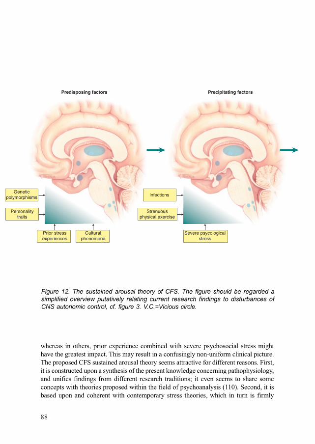
88
Predisposing factors Precipitating factors
Geneticpolymorphisms
Prior stressexperiences
Culturalphenomena
Personalitytraits
Infections
Severe psycologicalstress
Strenuousphysical exercise
Figure 12. The sustained arousal theory of CFS. The figure should be regarded asimplified overview putatively relating current research findings to disturbances ofCNS autonomic control, cf. figure 3. V.C.=Vicious circle.
whereas in others, prior experience combined with severe psychosocial stress mighthave the greatest impact. This may result in a confusingly non-uniform clinical picture.The proposed CFS sustained arousal theory seems attractive for different reasons. First,it is constructed upon a synthesis of the present knowledge concerning pathophysiology,and unifies findings from different research traditions; it even seems to share someconcepts with theories proposed within the field of psychoanalysis (110). Second, it isbased upon and coherent with contemporary stress theories, which in turn is firmly

89
Discrepancies betweenperceived and real abilities
Anxiety-likepathophysiology
Cognitiveimpairments
V.C.
V.C.
V.C. V.C.
Malaise
Sleep disturbances
Altered perfusion,metabolism,
transmitter activity
Fatigue
Blunted responseto acute stressor
AttenuatedHPA axis
Alteredcytokine
production
Pain,malaise
Skeletal muscledysfunction
Other organs
Th2 vs. Th1immune response
predominance
Reactivation ofintracellular organisms
Oxidativestress
Circulatoryabnormalities
Orthostaticintolerance
Transcriptionalactivation inleukocytes
Thermoregulatoryabnormalities
Symptoms ofthermoregulatory
disturbances
Consequences of sustained arousal and perpetuating factors
rooted in empirical research. Third, it seems to possess a kind of ‘face validity’ whenappraised in relation to patients’ uniform experience of an inability to respond properlyto the physical and mental challenges of daily life (178, 375). Fourth, it allows for thededuction of testable hypotheses that might be subjected to further research, as outlinedbelow (see 5.2.5).However, there are also some objections. The CFS sustained arousal theory implies abiopsychosocial construct that – although widely accepted by most experts - has been
90
met with skepticism from patient organizations as well as some practitioners andresearchers (318, 436). At a more general level, our suggested model challenges astable, 1:1 relationship between cause and effect, and thereby seems to oppose a moremechanistic approach to biology and medicine. As stated by Manu: «… more than anyother issue in contemporary medicine, chronic fatigue syndrome reflects the unresolvedconflict between the mechanistic and the biopsychosocial construct of illness» (262).The model proposed here infer heterogeneity of causality as well as clinical presentation,and allow symptom severity to be relative to the extent of stressor exposure. Thus, themodel views CFS as a kind of maladaption, in many respects analogous to thephenomenon of ‘loudspeaker feedback’: When a microphone is placed in the vicinityof a loudspeaker, even small noises will be amplified through positive feedback, resultingin an earsplitting sound and functional ‘breakdown’. However, there are no specificfailures of the different electronic devices per se; no ‘pathologies’ of cables orconnections. The problem results from maladaption; the equipment set-up isinappropriate relative to the initial noise recorded by the microphone. The problemmight be solved by lowering surrounding sounds. In terms of CFS, reducing all externalstimuli that might provoke a stress response. However, a more definite solution wouldbe to change the abilities within the feedback loops, by moving the microphone awayfrom the loudspeaker or reducing the gain of the electronic amplifier device; the latterwould correspond to a ‘down-tuning’ of the stress responses within the brain.
5.2.5 Suggestions for further research
Based upon the CFS sustained arousal theory explained above, we will in the followingpresent a few hypotheses and shortly outline possible methods for testing them. Thelist is not complete, but indicates some natural directions for further research.
1) Certain polymorphisms of genes whose products are involved in stress-inducedneurotransmission are more common among CFS patients than healthy controls.Leukocyte DNA must be collected from well-defined populations of patients andcontrols. A search for polymorphisms should be governed by updated knowledge ofthe genetic basis for stress responses. Commercial kits for such genomic studies aregenerally easily available.
2) CFS patients have experienced more stressful situations prior to disease onset thanpatients suffering from other long-lasting disease. A prospective, population-baseddesign would be preferable due to possible recall bias, but is inconvenient on the practicalside. Stressful situations should be operationalized, and might include for instanceprematurity, early long-time kindergarden affiliation, other serious diseases, trauma,death of a family member and abuse.
91
3) Acute Epstein-Barr virus infection elicits a more comprehensive stress responsethan other viral infections of similar clinical presentation. Patients with verified acuteEBV infection and patients with acute viral pharyngitis of other origin could undergoexperiments (e.g. head-up tilt-test) to assess their autonomic nerve activity when exposedto stressors. Subjects from the two groups should be matched according to their CRPlevel, to ensure an equally extensive inflammatory reaction.
4) When exposed to physical stress (such as orthostasis or physical exercise), perfusion,metabolism and transmitter activity in brain areas responsible for stress responsesdiffers among CFS patients and controls. Of particular interest are the serotoninergicand norepinephrinergic pathways in the brain stem and limbic structures. Modernneuroimaging technology makes such studies feasible.
5) Cognitive therapy specifically designed to abolish sustained arousal are more effectivein the treatment of CFS than unspecific cognitive therapy. To our knowledge, theeffectiveness of different cognitive treatment approaches has not been subjected toresearch. The design should be randomized and controlled.
6) The characteristics of the baroreflex and the cardiopulmonary reflex differ amongCFS patients and controls during rest and orthostatic stress due to set-point alterations.A logical continuation of our own research is to explore further the characteristics ofthe homeostatic control circuits responsible for volume and blood pressure regulationsin CFS patients and controls. A possible analytic tool would be the mathematicaltechnique of transfer function analyses, which could be applied to already recordeddata from the LBNP and HUT experiments (416).
7) The blood volume and the central venous pressure are comparable among CFSpatients and controls, but the renin-angiogensin-aldosteron activity is not. As the renalsecretion of renin is controlled through sympathetic efferents, one should expectincreased activity in a state of sustained arousal, but no alterations of blood volume orcentral venous pressure. Such an experiment would require invasive recordings.
8) When comparing CFS patients to controls, acute physical stress (orthostasis, physicalexercise) and mental stress will cause a greater increase in directly recorded sympatheticoutflow to skin and skeletal muscles as well as plasma turnover of epinephrine andnorepinephrine. Measurements of cardiovascular and thermoregulatory variables, asperformed in this thesis, provide only indirect information on sympathetic nerve activity,and are therefore prone to several interpretational caveats. To the best of our knowledge,no study has applied the technique of microneurography which allows direct recordingsof sympathetic skin and skeletal muscle activity. Likewise, only a few researchers haveperformed catecholamine measurements (208, 413), none of them using recentlyavailable, advanced methods of kinetics.

92
9) Pharmaceutical inhibition of brain centers eliciting the stress response will improvethe functional abilities of CFS patients and normalize the hemodynamics in stressfulsituations. The alpha2-adrenoceptor agonist clonidine is an effective antihypertensivedrug due to central inhibition of sympathetic outflow. Anxiolytic drugs, like benzo-diazepines, also exert an inhibitory effect on the relevant brain stem centers. As far aswe know, their effect in CFS patients has not been subjected to systematic trials. Suchtrials should focus on the changes of functional capacities, but should also assess whetherthe stress responses have normalized, possibly using cardiovascular function as anindicator.
Finally, it should be pointed out that the CFS sustained arousal theory – if provencorrect – might have implications beyond its present application. Related diseases,such as fibromyalgia, might be fully explicable within the same theoretical framework(421). Furthermore, similar models seem valuable for providing new insight into totallydifferent areas, for instance hypertension and the mechanisms underlying placebo effects.Finally, it might even contribute to a deeper understanding of body-mind interactionsin general.Thus, we have come full circle, recalling the introductory quotation of Harvey: «Forevery affection of the mind that is attended with either pain or pleasure, hope or fear, isthe case of an agitation whose influence extends to the heart…» Or, to cite hiscontemporary Blaise Pascal (1623-62):
«Le coeur a ses raisons que la raison ne connaît point.»
93
6 References
1. A Report of the CFS/ME Working group: Report to the Chief Medical Officer of anIndependent Working Group. London: Department of Health, 2002
2. Afari N, Buchwald D. Chronic fatigue syndrome – a review. Am J Psychiatry 2003; 160:221-36.
3. Amat J, Matus-Amat P, Watkins, LR, Maier SF. Escapable and inescapable stressdifferentially and selectively alter extracellular levels of 5-HT in the ventral hippocampusand dorsal periaqueductal grey of the rat. Brain Res 1998; 797: 12-22.
4. Amat J, Matus-Amat P, Watkins, LR, Maier SF. Escapable and inescapable stressdifferentially alter extracellular levels of 5-HT in the basolateral amygdala of the rat.Brain Res 1998; 812: 113-20.
5. Anderson JS, Ferrans CE. The quality of life of persons with chronic fatigue syndrome.J Nerv Ment Dis 1997; 185: 359-67.
6. Angeli A, Minetto M, Dovio A, Paccarotti P. The overtraining syndrome in athletes: astress-related disorder. J Endocrinol Invest 2004; 27: 603-12.
7. Appel ML, Berger RD, Saul JP, Smith JM, Cohen RJ. Beat to beat variability incardiovascular variables: noise or music? J Am Coll Cardiol 1989; 14: 1139-48.
8. Asbring P, Narvanen AL. Ideal versus reality: physicians perspectives on patients withchronic fatigue syndrome (CFS) and fibromyalgia. Soc Sci Med 2003; 57: 711-20.
9. Backmann GA. Vasomotor flushes in menopausal women. Am J Obstet Gynecol 1999;180: S312-6.
10. Bagnall AM, Whiting P, Wright K, Sowden AJ. The effectiveness of interventions usedin the treatment/management of chronic fatigue syndrome and/or myalgicencephalomyelitis in adults and children. York: NHS Centre for Reviews andDissemination, 2002.
11. Bailey SP, Davis JM, Ahlborn EN. Neuroendocrine and substrate responses to alteredbrain 5-HT activity during prolonged exercise to fatigue. J Appl Physiol 1993; 74: 3006-12.
12. Baily RG, Prophet SA, Shenberger JS, Zelis R, Sinoway LI. Direct neurohumoralevidence for isolated nervous system activation to skeletal muscle in response tocardiopulmonary baroreceptor unloading. Circ Res 1990; 66: 1720-8.
13. Ball N, Buchwald DS, Schmidt D, Goldberg J, Ashton S, Armitage R. Monozygotictwins discordant for chronic fatigue syndrome: objective measures of sleep. J PsychosomRes 2004; 56: 207-12.
14. Bandelow B, Sengos G, Wedekind D, Huether G, Pilz J, Broocks A, Hajak G, Ruther E.Urinary excretion of cortisol, norepinephrine, testosterone, and melatonin in panicdisorder. Pharmacopsychiatry 1997; 30: 113–7.

94
15. Barbieri R, Triedman JK, Saul JP. Heart rate control and mechanical cardiopulmonarycopling to assess central volume: a systems analysis. Am J Physiol Integr Comp Physiol2002; 283: R1210-20.
16. Barlow JS. Methods of analysis of nonstationary EEGs, with emphasis on segmentationtechniques: a comparative review. J Clin Neurophysiol 1985; 2: 267-304.
17. Barsky AJ, Borus JF. Functional somatic syndromes. Ann Intern Med 2000; 130: 910-21.
18. Baumert M, Brechtel L, Lock J, Hermsdorf M, Wolff R, Baier V, Voss A. Heart ratevariability, blood pressure variability, and baroreflex sensitivity in overtrained athletes.Clin J Sport Med 2006; 16: 412-7.
19. Behan WMH, More IAR, Behan PO. Mitochondrial abnormalities in the postviral fatiguesyndrome. Acta Neuropathol 1991; 83: 61-5.
20. Bell DS. Illness onset characteristics in children with chronic fatigue syndrome andidiopathic chronic fatigue. J Chron Fatigue Syndr 1997; 3: 43-51.
21. Bell DS, Jordan K, Robinson M. Thirteen-year follow-up of children and adolescentswith chronic fatigue syndrome. Pediatrics 2001; 107: 994-8
22. Benditt DG, Ferguson DW, Grubb BP, Kapoor WN, Kugler J, Lerman BB, Maloney JD,Raviele A, Ross B, Sutton R, Wolk MJ, Wood D. Tilt table testing for assessing syncope.J Am Coll Cardiol 1996; 28: 263-75.
23. Bergersen TK. A study of arteriovenous anastomoses in human skin with specialreference to their response to local temperature. Oslo: Department of Physiology,University of Oslo, 1999. (PhD dissertation).
24. Bergersen TK, Eriksen M, Walløe L. Local constriction of arteriovenous anastomosesin the cooled finger. Am J Physiol Reg Int Comp Physiol 1997; 273: R880-6.
25. Binder LM, Campbell KA. Medically unexplained symptoms and neuropsychologicalassessment. J Clin Exp Neuropsychol 2004; 26: 369-92.
26. Blackwood SK, McHale SM, Power MJ, Goodwin GM, Lawrie SM. Effects of exerciseon cognitive and motor function in chronic fatigue syndrome and depression. J NeurolNeurosurg Psychiatry 1998; 65: 541-6.
27. Blanchard MS, Eisen SA, Alpern R, Karlinsky J, Toomey R, Reda DJ, Murphy FM,Jackson LW, Kang HK. Chronic multisymptom illness complex in Gulf War I veterans10 years later. Am J Epidemiol 2006; 163: 66-75.
28. Bland JM, Altman DG. Stastistical methods for assessing agreement between twomethods of clinical measurement. Lancet 1986; 1(8476): 307-10.
29. Bland ST, Twining C, Schmid MJ, Der-Aviakan A, Watkins LR, Maier SF. Stresspotentiation of morphine-induced dopamine efflux in the nucleus accumbens shell isdependent upon stressor uncontrollability and is mediated by the dorsal raphe nucleus.Neuroscience 2004; 126: 705-15.
30. Bland ST, Twining C, Watkins LR, Maier SF. Stressor controllability modulates stress-induced serotonin but not dopamine efflux in the nucleus accumbens shell. Synapse2003; 49: 206-8.
31. Bleijenberg G, Vercoulen JHMM, van der Meer JWN. How important are symptom criteriain the definition of CFS? J Intern Med 2004; 256: 268-9.
32. Bongard S, al’Absi M, Lovallo WR. Interactive effects of trait hostility and angerexpression on cardiovascular reactivity in young men. In J Psychophysiol 1998; 28:181-91.
33. Bonyhay I, Freeman R. Sympathetic nerve activity in response to hypotensive stress inthe postural orthostatic tachycardia syndrome. Circulation 2004; 110: 3193-8.
95
34. Bosch JA, Berntson GG, Cacioppo JT, Marucha PT. Differential mobilization offunctionally distinct natural killer subsets during acute psychological stress. PsychosomMed 2005; 67: 366-75.
35. Bou-Holaigah I, Rowe PC, Kan JS, Calkins H. The relationship between neurallymediated hypotension and the chronic fatigue syndrome. JAMA 1995; 274: 961-7.
36. Bourne PB, Rose RM, Mason JW. Urinary 17-OHCA levels. Data on seven helicopterambulance medics in combat. Arch Gen Psychiatry 1967; 17: 104-10.
37. Brace MJ, Scott Smith M, McCauley E, Sherry DD. Family reinforcement of illnessbehavior: a comparison of adolescents with chronic fatigue, juvenile arthritis and healthycontrols. J Dev Behav Pediatr 2000; 21: 332-9.
38. Braden DS, Leatherbury L, Treiber FA, Strong WB. Noninvasive assessment of cardiacoutput in children using impedance cardiography. Am Heart J 1990; 120: 1166-72.
39. Bremner JD, Innis RB, Ng CK, Staib LH, Salomon RM, Bronen RA, Krystal JH, DuncanJ, Rich D, Price LH, Malison R, Dey H, Soufer R, Charney DS. Positron emissiontomography measurement of cerebral metabolic correlates of yohimbine administrationin combat-related posttraumatic stress disorder. Arch Gen Psychiatry 1997; 54: 246–54.
40. Brennan P, Silman A. Statistical methods for assessing observer variability in clinicalmeasures. BMJ 1992; 304: 1491-4.
41. Briese E. Emotional hyperthermia and performance in humans. Physiol Behav 1995;58: 615-8.
42. Briese E, Cabanac M. Stress hyperthermia: physiological arguments that it is a fever.Physiol Behav 1991; 49: 1153-7.
43. Brodal P. Sentralnervesystemet. Bygning og funksjon. Oslo: Tano, 1995.44. Bruno RL, Creange SJ, Frick NM. Parallels between post-polio fatigue and chronic
fatigue syndrome: a commen pathophysiology? Am J Med 1998; 105: 66S-73S.45. Bruno RL, Frick NM. Stress and ‘type A’ behavior as precipitans of post-polio sequelae:
the Felician/Columbia survey. Birth Defects Orig Artic Ser 1987; 23: 145-55.46. Brunstad PO. Seierens melankoli: et kulturanalytisk essay. Oslo: Gyldendal akademisk,
2003.47. Buchwald D, Manson SM, Pearlman T, Umali J, Kith P. Race and ethnicity in patients
with chronic fatigue. J Chronic Fatigue Syndr 1996; 2: 53-66.48. Buckey JC, Lane LD, Levine BD. Orthostatic intolerance after space-flights. J Appl
Physiol 1996; 81: 7-18.49. Burniston JG, Tan LB, Goldspink DF. Beta2-adrenergic receptor stimulation in vivo
induces apoptosis in the rat heart and soleus muscle. J Appl Physiol 2005; 98: 1379-86.
50. Cacioppo JT, Berntson GG, Malarkey WB, Kiecolt-Glaser JK, Sheridan JF, PoehlmannKM, Burleson MH, Ernst JM, Hawkley LC, Glaser R. Autonomic, neuroendocrine, andimmune responses to psychological stress: the reactivity hypothesis. Ann N Y Acad Sci1998; 840: 664-73.
51. Cairns R, Hotopf M. A systematic review describing the prognosis of chronic fatiguesyndrome. Occup Med 2005; 55: 20-31.
52. Candy B, Chalder T, Cleare AJ, Wessely S, White PD, Hotopf M. Recovery from infectiousmononucleosis: a case for more than symptomatic therapy? Br J Gen Pract 2002; 52:844-51.
53. Cannon JG, Angel JB, Ball RW, Abad LW, Fagioli L, Komaroff AL. Acute phase responseand cytokine secretion in chronic fatigue syndrome. J Clin Immunol 1999; 19: 414-21.
96
54. Cao WH, Fan W, Morrison SF. Medullary pathways mediating specific sympatheticresponses to activation of dorsomedial hypothalamus. Neuroscience 2004; 126: 229-40.
55. Carruthers BM, Jain AK, Meirleir KLD, Peterson DL, Klimas NG, Lerner AM, BestedAC, Flor-Henry P, Joshi P, Powles ACP, Sherkey JA, van de Sande MI. MyalgicEncephalomyelitis/Chronic Fatigue Syndrome: Clinical working case definition,diagnostic and treatment protocols. J Chronic Fatigue Syndr 2003; 11: 7-36.
56. Castell LM, Yamamoto T, Phoenix J, Newsholme EA. The role of tryptophan in fatigueconditions of stress. Adv Exp Med Biol 1999; 467: 697-704.
57. Cerutti S, Bianchi AM, Mainardi LT. Advanced spectral methods for detecting dynamicbehaviour. Auton Neurosci 2001; 90: 3-12.
58. Chappell PB, Smith MA, Kilts CD, Bissette G, Ritchie J, Anderson C, Nemeroff CB.Alterations in corticotrophin-releasing factor-like immunoreactivity in discrete rat brainregions after acute and chronic stress. J Neurosci 1986; 6: 2908-14.
59. Charmandari E, Kino T, Souvatzoglou E, Chrousos GP. Pediatric stress: hormonalmediators and human development. Horm Res 2003; 59: 161-79.
60. Charney DS. Neuroanatomical circuits modulating fear and anxiety behaviors. ActaPsychiatr Scand 2003; 108: S38-S50.
61. Chia JKS. The role of enterovirus in chronic fatigue syndrome. J Clin Pathol 2005; 58:1126-32.
62. Chia JKS, Chia LY. Chronic Chlamydia pneumonia infection, a treatable cause of chronicfatigue syndrome. Clin Infect Dis 1999; 29: 452-3.
63. Cho HJ, Skowera A, Cleare A, Wessely S. Chronic fatigue syndrome: an update focusingon phenomenology and pathophysiology. Curr Opin Psychiatry 2006; 19: 67-73.
64. Chronic fatigue syndrome. Clinical practice gudelines – 2002. Med J Austr 2002; 176:S17-S55.
65. Cirelli C, Tononi G. Locus ceruleus control of state-dependent gene expression. JNeurosci 2004; 24: 5410-9.
66. Clauw DJ, Chrousos GP. Chronic pain and fatigue syndromes: overlapping clinical andneuroendcrine features and potential pathogenic mechanisms. Neuroimmunomodulation1997; 4: 134-53.
67. Clayton EC, Rajkowski J, Cohen JD, Aston-Jones G. Decision-related activation ofmonkey locus coeruleus neurons in a forced choice task. Soc Neurosci Abstr 2003: 29:722-7.
68. Cleare AJ. The neuroendocrinology of chronic fatigue syndrome. Endocr Rev 2003;24: 236-52.
69. Cleare AJ, Bearn J, Allain T, McGregor A, Wessely S, Murray RM, O’Keane V. Contrastingneuroendocrine responses in depression and chronic fatigue syndrome. J Affect Disord1995; 34: 283-9.
70. Cleare AJ, Messa C, Rabbiner EA, Grasby PM. Brain 5-HT1A receptor binding in chronicfatigue syndrome measured using positron emission tomography and [11C]WAY-100635.Biol Psychiatry 2005; 57: 239-46.
71. Cleare AJ, O’Keane V, Miell JP. Levels of DHEA and DHEAS and responses to CRHstimulation and hydrocortisone treatment in chronic fatigue syndrome.Psychoneuroendocrinology 2004; 29: 724-32.
72. Cleare AJ, Wessely SC. Chronic fatigue syndrome: a stress disorder? Br J Hosp Med1996; 55: 571-4.
73. Cohen MA, Taylor JA. Short-term cardiovascular oscillations in man: measuring andmodeling the physiologies. J Physiol 2002; 542: 669-83.

97
74. Comings DE, Johnson JP, Gonzales NS, Huss M, Saucier G, McGue M, MacMurray J.Association between the adrenergic alpha 2A receptor gene (ADRA2A) and measuresof irritability, hostility, impulsivity and memory in normal subjects. Psychiatr Genet 2000;10: 39-42.
75. Convertino VA. Conditions of reduced gravity. In: Low PA, ed. Clinical autonomicdisorders. Philadelphia: Lippincott-Raven, 1997: 429-40.
76. Convertino VA, Doerr DF, Ludwig DA, Vernikos J. Effect of simulated microgravity oncardiopulmonary baroreflex control of forearm vascular resistance. Am J Physiol 1994;266: R1962-9.
77. Cook DB, Lange G, DeLuca J, Natelson BH. Relationship of brain MRI abnormalitiesand physical functional status in chronic fatigue syndrome. Int J Neurosci 2001; 107: 1-6.
78. Cook DB, Nagelkirk PR, Peckerman A, Poluri A, Mores J, Natelson BH. Exercise andcognitive performance in chronic fatigue syndrome. Med Sci Sports Exerc 2005; 37:1460-7.
79. Cooke WH, Hoag JB, Crossman AA, Kuusela TA, Tahvanainen KUO, Eckberg DL.Human responses to upright tilt: a window on central autonomic integration. J Physiol1999; 517: 617-28.
80. Cope H, Pernet A, Kendall B, David A. Cognitive functioning and magnetic resonanceimaging in chronic fatigue. Br J Psychiatry 1995; 167: 86-94.
81. Cordero DL, Sisto SA, Tapp WN, LaManca JJ, Pareja JG, Natelson BH. Decreasedvagal power during treadmill walking in patients with chronic fatigue syndrome. ClinAuton Res 1996; 6: 329-33.
82. Cornelissen VA, Fagard RH. Effects of endurance training on blood pressure, blood-pressure regulating mechanism, and cardiovascular risk factors. Hypertension 2005;46: 667-75.
83. Costa DC, Tannock C, Brostoff J. Brainstem perfusion is impaired in chronic fatiguesyndrome. QJM 1995; 88: 767-73.
84. Costoli T, Barolomucci A, Graiani G, Stilli D, Laviola G, Sgoifo A. Effects of chronicpsychosocial stress on cardiac autonomic responsiveness and myocardial structure inmice. Am J Physiol Heart Circ Physiol 2004; 286; H2133-40.
85. Crandall CG, Johnson JM, Convertino VA, Raven PB, Engelke KA. Alteredthermoregulatory responses after 15 days of head-down tilt. J Appl Physiol 1994; 77:1863-7.
86. Crandall CG, Shibasaki M, Wilson TE, Cui J, Levine BD. Prolonged head-down tiltexposure reduces maximal cutaneous vasodilator and sweating capacity in humans. JAppl Physiol 2003; 94: 2330-6.
87. Cui J, Wilson TE, Crandall CG. Muscle sympathetic nerve activity during lower bodynegative pressure is accentuated in heat-stressed humans. J Appl Physiol 2004; 96:2103-8.
88. Davis JM, Bailey SP. Possible mechanisms of central nervous system fatigue duringexercise. Med Sci Sports Exerc 1997; 29: 45-57.
89. De Jong-de Vos van Steenwijk CC, Wieling W, Johannes JM, Harms MP, Kuis W,Wesseling KH. Incidence and hemodynamic characteristics of near-fainting in healthy6- to 16-year old subjects. J Am Coll Cardiol 1995; 25: 1615-21.
90. De Lange FP, Kalkman JS, Bleijenberg G, Haqoort P, van der Meer JW, Toni I. Graymatter volume reduction in the chronic fatigue syndrome. Neuroimage 2005; 26: 777-81.
98
91. De Lange FP, Kalkman JS, Bleijenberg G, Haqoort P, van der Werf SP, van der MeerJW, Toni I. Neural correlates of the chronic fatigue syndrome – an fMRI study. Brain2004; 127: 1948-57.
92. De Becker P, Dendale P, De Meirleir K, Campine I, Vandenborne K, Hagers Y. Autonomictesting in patients with chronic fatigue syndrom. Am J Med 1998; 105: 22S-26S.
93. De Luca J, Johnson SK, Ellis SP, Natelson BH. Cognitive functioning is impaired inpatients with chronic fatigue syndrome devoid of psychiatric disease. J Neurol NeurosurgPsychiatry 1997; 62: 151-5.
94. De Meirleir K, Bisbal C, Campine I, De Becker P, Salehzada T, Demettre E, Lebleu B.A 37 kDa 2-5A binding protein as a potential biochemical marker for chronic fatiguesyndrome. Am J Med 2000; 108: 99-105.
95. Demitrack MA, Crofford LJ. Evidence for and pathophysiologic implications ofhypothalamic-pituitary-adrenalin axis dysregulation in fibromyalgia and chronic fatiguesyndrome. Ann N Y Acad Sci 1998; 840: 684.
96. Demitrack MA, Gold PW, Dale JK, Krahn DD, Kling MA, Straus SE. Plasma andcerebrospinal fluid monoamine metabolism in patients with chronic fatigue syndrome:preliminary findings. Biol Psychiatry 1992; 32: 1065-77.
97. Demyttenaere K, De Fruyt J, Stahl SM. The many faces of fatigue in major depressivedisorders. Int J Neuropsychopharmacol 2005; 8: 93-105.
98. Denniston JC, Maher JT, Reeves JT, Cruz JC, Cymerman A, Grover RF. Measurementof cardiac output by electrical impedance at rest and during exercise. J Appl Physiol1976; 40: 91-5.
99. Dhalla NS, Temsah RM, Netticadan T. Role of oxidative stress in cardiovasculardiseases. J Hypertens 2000; 18: 655-73.
100. Diagnostisering og behandling av kronisk utmattelsessyndrom / myalgisk encefalopati(CFS/ME). Rapport fra Kunnskapssenteret Nr 9 – 2006. Oslo: Nasjonalt kunnskaps-senter for helsetjenesten, 2006.
101. Dickinson CJ. Chronic fatigue syndrome – aetiological aspects. Eur J Clin Invest 1997;27: 257-67.
102. Dienstbier RA. Arousal and physiological toughness: implications for mental and physicalhealth. Psychol Rev 1989; 96: 84-100.
103. Di Giorgio A, Hudson M, Jerjes W, Cleare AJ. 24-hour pituitary and adrenalin hormoneprofiles in chronic fatigue syndrome. Psychosom Med 2005; 67: 433-40.
104. Di Micco JA, Sarkar S, Zaratskaia MV, Zaretsky DV. Stress-induced cardiac stimulationand fever: common hypothalamic origins and brainstem mechanisms. Auton Neurosci2006; 126/127: 106-19.
105. Di Micco JA, Zaretsky D. The dorsomedial hypothalamus: A new player inthermoregulation. Am J Physiol Regul Integr Comp Physiol 2006; sept 7 [Epub aheadof print]
106. Dinan TG, Majeed T, Lavelle E, Scott LV, Berti C, Behan P. Blunted serotonin-mediatedactivation of the hypothalamic-pituitary-adrenalin axis in chronic fatigue syndrome.Psychoneuroendocrinology 1997; 22: 261-7.
107. Dinan TG. Glucocorticoids and the genesis of depressive illness. A psychobiologicalmodel. Br J Psychiatry 1994; 164: 365-71.
108. Dittner AJ, Wessely S, Brown RG. The assessment of fatigue. A practical guide forclinicians and researchers. J Psychosom Res 2004; 56: 157-70.
109. Doods HN, Wieland HA, Engel W, Eberlein W, Willim KD, Entzeroth M, Wienen W,Rudolf K. BIBP3226, the first selective neuropeptide Y1 receptor antagonist: a reviewof its pharmacological properties. Regul Pept 1996; 65: 71-7.
99
110. Driver C. An under-active or over-active internal world? An exploration of paralleldynamics within psyche and soma, and the difficulty of internal regulation, in patientswith chronic fatigue syndrome and myalgic encephalomyelitis. J Anal Psychol 2005;50: 155-73.
111. Duprez DA, DeBuyzere ML, Drieghe B, van Haverbeke F, Taes Y, Michielsen W, ClementDL. Long- and short-term blood pressure and RR-interval variability and psychosomaticdistress in chronic fatigue syndrome. Clin Sci 1998; 94: 57-63.
112. Eckberg DL. Sympathovagal balance. A critical appraisal. Circulation 1997; 96: 3224-32.
113. Eckberg DL, Sleight P. Human Baroreflexes in Health and Disease. Oxford: ClarendonPress, 1992.
114. Edmonds M, McGuire H, Price J. Exercise therapy for chronic fatigue syndrome.Cochrane Database of Systematic Reviews, 2004.
115. Elenkov IJ, Wilswe RL, Chrousos GP, Vizi ES. The sympathetic nerve – an integrativeinterface between two supersystems: the brain and the immune system. PharmacolRev 2000; 52: 595-638.
116. Elstad M, Toska K, Chon KH, Raeder EA, Cohen RJ. Respiratory sinus arrhythmia:opposite effects on systolic and mean arterial pressure in supine humans. J Physiol2001; 536: 251-9.
117. Engel GL. The need for a new medical model: a challenge for biomedicine. Science1977; 196: 129-36.
118. Eriksen HR, Murision R, Pensgaard AM, Ursin H. Cognitive activation theory of stress(CATS): From fish brains to the Olympics. Psychoneuroendrocrinology 2005; 30: 933-8.
119. Eriksen M, Walløe L. Improved method for cardiac output determination in man usingultrasound Doppler technique. Med Biol Eng Comput 1990; 28: 555-60.
120. Evans AS. Chronic fatigue syndrome: Thoughts on pathogenesis. Rev Infect Dis 1991;13: S56-9.
121. Evans JG, Beck P. Informed consent in medical research. Clin Med 2002; 2: 267-72.122. Evans RW. How then should we die? California’s “Death with dignity” act. Med Etika
Bioet 2000; 7: 3-9.123. Evidence based guidelines for the management of CFS/ME (chronic fatigue syndrome/
myalgic encephalopathy) in children and young adults. London: Royal College ofPaediatrics and Child Health, 2004.
124. Fang H, Xie Q, Boneva R, Fostel J, Perkins R, Tong W. Gene expression profileexploration of a large dataset on chronic fatigue syndrome. Pharmacogenomics 2006;7: 429-40.
125. Farmer A. Prevalence of chronic disabling fatigue in children and adolescents. Br JPsychiatry 2004; 184: 477-81.
126. Farquhar WB, Hunt BE, Taylor JA, Darling SE, Freeman R. Blood volume and its relationto peak O2 consumption and physical activity in patients with chronic fatigue. Am JPhysiol Heart Circ Physiol 2002; 282: H66-71.
127. Farquhar WB, Taylor JA, Darling SE, Chase KP, Freeman R. Abnormal baroreflexresponses in patients with idiopathic orthostatic intolerance. Circulation 2000; 102: 3086-91.
128. Feder HM, Dworkin PH, Orkin C. Outcome of 48 pediatric patients with chronic fatigue.A clinical experience. Arch Fam Med 1994; 3: 1049-55.
129. Finaud J, Lac G, Filaire E. Oxidative stress: relationship with exercise and training.Sports Med 2006; 36: 327-58.
100
130. Fischler B, Cluydts R, De Gucht V, Kaufman L, De Meirleir K. Generalized anxiteydisorders in chronic fatigue syndrome. Acta Psychiatr Scand 1997; 95: 405-13.
131. Fischler B, LeBon O, Hoffman G, Cluydts R, Kaufman L, De Meirleir K. Sleep anomaliesin the chronic fatigue syndrome – a comorbidity study. Neuropsychobiology 1997; 35:115-22.
132. Fortin J, Habenbacher W, Gruellenberger R, Wach P, Skrabal F. Real-time monitor forhemodynamic beat-to-beat parameteres and power spectra analysis of the biosignals.Conf Proc IEEE Eng Med Biol Soc 1998; 20: 360-3.
133. Fortin J, Habenbacher W, Heller A, Hacker A, Grullenberger R, Innerhofer J, PassathH, Wagner CH, Haitchi G, Flotzinger D, Pacher R, Wach P. Non-invasive beat-to-beatcardiac output monitoring by an improved method of transthoracic bioimpedancemeasurement. Comput Biol Med 2005, Aug 27 [Epub ahead of print]
134. Fortney SM, Vroman NB. Exercise, performance and temperature control: temperatureregulation during exercise and implications for sports performance and training. SportsMed 1985; 2: 8-20.
135. Fortney SM. Thermoregulatory adaptations to inactivity. In: Samueloff S, Yousef MK,eds. Adaptive physiology to stressful environments. Boca Raton: CRC press, 1987:75-83.
136. Franklin A. How I manage chronic fatigue syndrome. Arch Dis Child 1998; 79: 375-8.137. Freeman R, Komaroff AL. Does the chronic fatigue syndrome involve the autonomic
nervous system? Am J Med 1997; 102: 357-64.138. Friedman M. Studies concerning the etiology and pathogenesis of neurocirculatory
asthenia. Am Heart J 1945; 30: 478-91.139. Friedman SM, Masjon JW, Hanburg DA. Urinary 17-hydroxycorticosteroid levels in
parents of children with neoplastic disease: A study of chronic psychological stress.Psychosom Med 1963; 25: 364-76.
140. Fu Q, Witowski S, Levine BD. Vasoconstrictor reserve and sympathetic neural controlof orthostasis. Circulation 2004; 110: 2931-7.
141. Fu Q, Witowski S, Okazaki K, Levine BD. Effects of gender and hypovolemia onsympathetic neural responses to orthostatic stress. Am J Physiol Regul Integr CompPhysiol 2005; 289: R109-16.
142. Fukuda K, Straus SE, Hickie I, Sharpe MC, Dobbins JG, Komaroff A. The chronic fatiguesyndrome: a comprehensive approach to its definition and study. Ann Intern Med 1994;121: 953-9.
143. Fulcher KY, White PD. Strength and physiological response to exercise in patients withchronic fatigue syndrome. J Neurol Neurosurg Psychiatry 2000; 69: 302-7.
144. Furlan R, Jacob G, Palazzolo L, Rimoldi A, Diedrich A, Harris PA, Porta A, Malliani A,Mosqueda-Garcia R, Robertson D. Sequential modulation of cardiac autonomic controlinduced by cardiopulmonary and arterial baroreflex mechanisms. Circulation 2001; 104:2932-7.
145. Furlan R, Jacob G, Snell M, Robertson D, Harris P, Mosqueda-Garcia R. Chronicorthostatic intolerance. A disorder with discordant cardiac and vascular sympatheticcontrol. Circulation 1998; 98: 2154-9.
146. Furlan R, Piazza S, Dell’Orto S, Gentile E, Cerutti S, Pagani M, Malliani A. Early andlate effects of exercise and athletic training on neural mechanisms controlling heartrate. Cardiovasc Res 1993; 27: 482-8.
147. Føllesdal D, Walløe L, Elster J. Argumentasjonsteori, språk og vitenskapsfilosofi. Oslo:Universitetsforlaget, 1986.
148. Garralda E, Rangel L. Annotation: Chronic Fatigue Syndrome in children andadolescents. J Child Psychol Psychiatry 2002; 43: 169-76

101
149. Garralda E, Rangel L, Levin M, Roberts H, Ukoumunne O. Psychiatric adjustment inadolescents with a history of chronic fatigue syndrome. J Am Acad Child AdolescPsychiatry 1999; 38: 1515-21.
150. Georgiades E, Behan WM, Kilduff LP, Hadjicharalambous M, Mackie E, Wilson J, WardSA, Pitsiladis YP. Chronic fatigue syndrome; new evidence for a central fatigue disorder.Clin Sci 2003; 105: 213-8.
151. Gerrity TR, Bates J, Bell DS, Chrousos G, Furst G, Hedrick T, Hurwitz B, Kula RW,Levine SM, Moore RC, Schondorf R. Chronic fatigue syndrome: what role does theautonomic nervous system play in the pathophysiology of this complex illness?Neuroimmunomodulation 2002; 10: 134-41.
152. Glaser R, Kiecolt-Glaser JK. Stress-associated immune modulation: relevance to viralinfections and chronic fatigue syndrome. Am J Med 1998; 105: 35S-42S.
153. Glaser R, Kiecolt-Glaser JK, Malarkey WB, Sheridan JF. The influence of psychologicalstress on the immune responses to vaccines. Ann N Y Acad Sci 1998; 840: 649.
154. Goertzel BN, Pennachin C, Coelho LS, Gurbaxani B, Maloney EM, Jones JF.Combination of single nucleotide polymorphisms in neuroendocrine effector and receptorgenes predict chronic fatigue syndrome. Pharmacogenomics 2006; 7: 475-83.
155. Goldstein DS. The autonomic nervous system in health and disease. New York: MarcelDekker, 2001.
156. Goldstein DS, Eisenhofer G, Kopin IJ. Sources and significance of plasma levels ofcatechols and their metabolites in humans. J Pharmacol Exp Ther 2003; 305: 800-11.
157. Goldstein DS, Eldadah B, Holmes C, Pechnik S, Moak J, Sharabi Y. Neurocirulatoryabnormalities in chronic orthostatic intolerance. Circulation 2005; 111: 839-45.
158. Goldstein DS, Holmes C, Frank SM, Dendi R, Cannon RO, Sharabi Y, Esler MD,Eisenhofer G. Cardiac sympathetic dysautonomia in chronic orthostatic intolerancesyndrome. Circulation 2002; 106: 2358-65.
159. Goldstein DS, Robertson D, Esler M, Straus SE, Eisenhofer G. Dysautonomias: clinicaldisorderes of the autonomic nervous system. Ann Intern Med 2002; 137: 753-63.
160. Goldstein DS, Vernikos J, Holmes C, Convertino VA. Catecholaminergic effects ofprolonged head-down bed rest. J Appl Physiol 1995; 78: 1023-9.
161. Goodwin GM, McCloskey DI, Mitchell JH. Cardiovascular and respiratory responses tochanges in central command during isometric exercise at constant muscle tension. JPhysiol 1972; 226: 173-90.
162. Gordon R, Michalewski HJ, Nguyen T, Gupta S, Starr A. Cortical motor potential alterationin chronic fatigue syndrome. Int J Mol Med 1999; 4: 493-9.
163. Gow JW, Behan WM, Clements GB, Woodall C, Riding M, Behan PO. Enteroviral RNAsequences detected by polymerase chain reaction in muscle of patients with postviralfatigue syndrome. BMJ 1991; 302: 692-6.
164. Grassi G, Giannattasio C, Cuspidi C, Bolla GB, Clerouox J, Ferrazzi P, Fiocchi R, ManciaG. Cardiopulmonary receptor regulation of renin release. Am J Med 1988; 84: 97-104.
165. Grassi G, Serevalle G, Calhoun DA, Mancia G. Physical training and baroreceptor controlof sympathetic nerve activity in humans. Hypertension 1994; 23: 294-301.
166. Gratze G, Fortin J, Holler A, Grasenick K, Pfurtscheller G, Watch P, Schonegger J,Kotanko P, Skrabal F. A software package for non-invasive, real-time beat-to-beatmonitoring of stroke volume, blood pressure, total peripheral resistance and forassessment of autonomic function. Comput Biol Med 1998; 28: 121-42.
167. Greco A, Tannock C, Brostoff J, Costa DC. Brain MR in chronic fatigue syndrome. AmJ Neuroradiol 1997; 18: 1265-9.
168. Greenleaf JE, Sciaraffa D, Shvartz E, Keil LC, Brock PJ. Exercise training hypotension:implications for plasma volume, renin and vasopression. J Appl Physiol 1981; 51: 531-5.
102
169. Gupta A. Unconscious amygdalar fear conditioning in a subset of chronic fatiguesyndrome patients. Med Hypotheses 2002; 59: 727-35.
170. Gupta S, Aggarwal S, Starr A. Increased production of interleukin-6 by adherent andnon-adherent mononuclear cells during ‘natural fatigue’ but not following ‘experimentalfatigue’ in patients with chronic fatigue syndrome. Int J Mol Med 1999; 3: 209-13.
171. Gaab J, Rohleder N, Heitz V, Engert V, Schad T, Schurmeyer TH, Ehlert U. Stress-induced changes in LPS-induced pro-inflammatory cytokine production in chronic fatiguesyndrome. Psychoneuroendocrinology 2005; 30: 188-98.
172. Hainsworth R. Reflexes from the heart. Physiol Rev 1991; 71: 617-58.173. Hall GM, Salmon P. Physiological and psychological influences on postoperative fatigue.
Anesth Analg 2002; 95: 1446-50.174. Hall M, Vasko R, Buysse D, Ombao H, Chen Q, Cashmere JD, Kupfer D, Thayer JF.
Acute stress affects heart rate variability during sleep. Psychosom Med 2004; 66: 56-62.
175. Hamilos DL, Nutter D, Gershtenson J, Ikle D, Hamilos SS, Redmond DP, Di ClementiJD, Schmaling KB, Jones JF. Circadian rhythm of core body temperature in subjectswith chronic fatigue syndrome. Clin Physiol 2001; 21: 184-95.
176. Hamilos DL, Nutter D, Gershtenson J, Redmond DP, Di Clementi JD, Schmaling KB,Make BJ, Jones JF. Core body temperature is normal in chronic fatigue syndrome. BiolPsychiatry 1998; 43: 293-302.
177. Hansen AK, Clausen T, Nielsen OB. Effects of lactic acid and catecholamines oncontractility in fast-twitch muscles exposed to hyperkalemia. Am J Physiol Cell Physiol2005; 289: C104-12.
178. Hart B, Grace VM. Fatigue in chronic fatigue syndrome: a discourse analysis of women’sexperiential narratives. Health Care for Women International 2000; 21: 187-201.
179. Hasser EM, Moffitt JA. Regulation of sympathetic nervous system function aftercardiovascular deconditioning. Ann N Y Acad Sci 2001; 940: 454-68.
180. Hatcher S, House A. Life events, difficulties and dilemmas in the onset of chronic fatiguesyndrome: a case-control study. Psychol Med 2003; 33: 1185-92.
181. Havenith G, Middendorp HV. The relative influence of physical fitness, acclimatizationstate, anthropometric measures and gender on individual reactions to heat stress. EurJ Appl Physiol 1990; 61: 419-27.
182. Heijmans MJ. Coping and adaptive outcome in chronic fatigue syndrome: importanceof illness cognitions. J Psychosom Res 1998; 45: 39-51.
183. Henry JP. Biological basis of the stress response. Integr Physiol Behav Sci 1992; 27:66-83.
184. Hisdal J, Toska K, Flatebo T, Waaler B, Walloe L. Regulation of arterial blood pressurein humans during isometric muscle contraction and lower body negative pressure. EurJ Appl Physiol 2004; 91: 336-41.
185. Hisdal J, Toska K, Walloe L. Design of a chamber for lower body negative pressurewith controlled onset rate. Aviat Space Environ Med 2003; 74: 874-8.
186. Hughson RL, Shoemaker JK, Arbeille P, O’Leary DD, Pizzolitto KS, Hughes MD.Splanchnic and peripheral vascular resistance during lower body negative pressure(LBNP) and tilt. J Gravit Physiol 2004; 11: P95-6.
187. Huibers MJ, Bleijenberg G, van Amelsvoort LG, Beurskens AJ, van Schayck, BazelmansE, Knottnerus JA. Predictors of outcome in fatigued employees on sick leave: resultsfrom a randomised trial. J Psychosom Res 2004; 57: 443-9.
188. Høyland A, Walløe L. Elementær statistikk. Trondheim: Tapir, 1977.189. Imms FJ, Mehta D. Respiratory responses to sustained isometric muscle contractions
in man: the effect of muscle mass. J Physiol 1989; 419: 1-14.
103
190. Inder WJ, Prickett TC, Mulder RT. Normal opioid tone and hypothalamic-pituitary-adrenalaxis function in chronic fatigue syndrome despite marked functional impairments. ClinEndocrinol 2005; 62: 343-8.
191. Itoh Y, Igarashi T, Tatsuma N, Imai T, Yoshida J, Tsuchiya M, Muaramaki M, Fukunga Y.Immunogenetic background of patients with autoimmune fatigue syndrome.Autoimmunity 2000; 32: 193-7.
192. Iwasaki K-I, Zhang R, Perhonen MA, Zuckerman JH, Levine BD. Rerduced baroreflexcontrol of heart period after bed rest is normalized by acute plasma volume restoration.Am J Physiol Regul Integr Comp Physiol 2004; 287: R1256-62.
193. Iwase S, Sugiyama Y, Miwa C, Kamiya A, Mano T, Ohira Y, Shenkman B, Egorov AI,Kozlovskaya IB. Effects of three days of dry immersion on muscle sympathetic nerveactivity and arterial blood pressure in humans. J Auton Nerv Syst 2002; 15: 156-64.
194. Jacob G, Costa F, Shannon JR, Robertson RM, Wathen M, Stein M, Biaggioni I, Ertl A,Black B, Robertson D. The neuropathic postural tachycardia syndrome. N Engl J Med2000; 343: 1008-14.
195. Jacob G, Shannon JR, Costa F, Furlan R, Biaggioni I, Mosqueda-Garcia R, RobertsonRM, Robertson D. Abnormal norepinephrine clearance and adrenergic receptorsensitivity in idiopathic orthostatic intolerance. Circulation 1999; 99: 1706-12.
196. Jacobs I, Romet T, Frim J, Hynes A. Effects of endurance fitness on responses to coldwater immersion. Aviat Space Environ Med 1984; 55: 715-20.
197. Jammes Y, Steinberg JG, Mambrini O, Bregeon F, Delliaux S. Chronic fatigue syndrome:assessment of increased oxidative stress and altered muscle excitability in responseto incremental exercise. J Intern Med 2005; 257: 299-310.
198. Jason LA, Richman JA, Rademaker AW, Jordan KM, Plioplys AV, Taylor RR, McCreadyW, Huang CF, Plioplys S. A community-based study of chronic fatigue syndrome. ArchInt Med 1999; 159: 2129-37.
199. Jenkins R. Epidemiology: Lessons from the past. Br Med Bull 1991; 47: 952-65.200. Jones E, Wessely S. Case of chronic fatigue syndrome after Crimean war and Indian
mutiny. BMJ 1999; 319: 1645-7.201. Jones JF, Nicholson A, Nisenbaum R, Papanicolaou DA, Solomon L, Bonneva R, Heim
C, Reeves WC. Orthostatic instability in a population based study of chronic fatiguesyndrome. Am J Med 2005; 118: 1415.
202. Jordan J. Effect of water drinking on sympathetic nervous activity and blood pressure.Curr Hypertens Rep 2005; 7: 17-20.
203. Kahn F, Spence V, Kennedy G, Belch JJF. Prolonged acetylcholine-induced vasodilationin the peripheral microcirculation of patients with chronic fatigue syndrome. Clin PhysFunct Imaging 2003; 23: 282-5.
204. Kamiya A, Michikami D, Fu Q, Iwase S, Hayano J, Kawada T, Mano T, Sunagawa K.Pathophysiology of orthostatic hypotension after bed rest: paradoxical sympatheticwithdrawal. Am J Physiol Heart Circ Physiol 2003; 285: H1158-67.
205. Kamiya A, Michikami D, Fu Q, Niimi Y, Iwase S. Muscle sympathetic nerve activity(MSNA) during high-intensity, isometric exercise in humans. Environ Med 2000; 44:49-52.
206. Kant GJ, Leu JR, Anderson SM, Mougey EH. Effects of chronic stress on plasmacorticosterone, ACTH and prolactin. Physiol Behav 1987; 40: 775–9.
207. Katon WJ, Buchwald D, Simon G, Russo JE, Mease PJ. Psychiatric illness in patientswith chronic fatigue and those with rheumatoid arthritis. J Gen Intern Med 1991; 6:277-85.
208. Kavelaars A, Kuis W, Knook L, Sinnema G, Heijnen CJ. Disturbed neuroendocrine-immune interactions in chronic fatigue syndrome. J Clin Endocrinol Metab 2000; 85:692-6.

104
209. Keller DM, Wasmund WL, Wray DW, Ogoh S, Fadel PJ, Smith ML, Raven PB. Carotidbaroreflex control of leg vascular conductance at rest and during exercise. J Appl Physiol2003; 94: 542-8.
210. Kellogg DL jr, Pergola PE, Piest KL, Kosiba WA, Crandall CG, Grossmann M, JohnsonJM. Cutaneous active vasodilation in humans is mediated by cholinergic nervecotransmission. Circ Res 1995; 77: 1222-8.
211. Kellogg DL jr, Zhao JL, Coey U, Green JV. Acetylcholine-induced vasodilation is mediatedby nitric oxide and prostaglandins in human skin. J Appl Physiol 2005; 98: 629-32.
212. Kennedy G, Spence VA, McLaren M, Hill A, Underwood C, Belch JJF. Oxidative stresslevels are raised in chronic fatigue syndrome and are associated with clinical symptoms.Free Radical Biol Med 2005; 39: 584-9.
213. Kerkkamp HJ, Heethaar RM. A comparison of bioimpedance and echocardiography inmeasuring systolic heart function in cardiac patients. Ann N Y Acad Sci 1999; 873: 149-54.
214. Kerman IA, Akil H, Watson SJ. Rostral elements of sympatho-motor circuitry: a virallymediated transsynaptic tracing study. J Neurosci 2006; 26: 3423-33.
215. Kerr JR, Christian P, Hodgetts A, Langford PR, Devanur LD, Petty R, Burke B, SinclairLI, Richards SC, Montgomery J, McDermott C, Harrison TH, Kellam P, Nutt DJ, HolgateST. Current research priorities in chronic fatigue syndrome / myalgic encephalomyelitis(CFS/ME): disease mechanisms, a diagnostic test and specific treatments. J Clin Pathol2006; Aug. 25 [e-pub ahead of print].
216. Ketch T, Biaggioni I, Robertson R, Robertson D. Four faces of baroreceptor failure:hypertensive crisis, volatile hypertension, orthostatic tachycardia and malignantvagotonia. Circulation 2002; 105: 2518-23.
217. Kiecolt-Glaser JK, Glaser R, Cacioppo JT, Malarkey WB. Marital stress: immunologic,neuroendocrine, and autonomic correlates. Ann N Y Acad Sci 1998; 840: 656.
218. Kimmerly DS, O’Leary DD, Menon RS, Gati JS, Shoemaker JK. Cortical regionsassociated with autonomic cardiovascular regulation during lower body negativepressure in humans. J Physiol 2005; 569: 331-45.
219. Kimmerly DS, Shoemaker JK. Hypovolemia and neurovascular control during orthostaticstress. Am J Physiol Heart Circ Physiol 2002; 282: H645-55.
220. Kimmerly DS, Shoemaker JK. Hypovolemia and MSNA discharge pattern: assessingand interpreting sympathetic responses. Am J Physiol Heart Circ Physiol 2003; 284:H1198-204.
221. Kjær M. Adrenal medulla and exercise traning. Eur J Appl Physiol Occup Physiol 1998;77: 195-9.
222. Knook L, Kavelaars A, Sinnema G, Kuis W, Heijnen CJ. High noctural melatonin inadolescents with chronic fatigue syndrome. J Clin Endocrin Metabol 2000; 85: 3690-2.
223. Komaroff AL. Clinical presentation of chronic fatigue syndrome. Ciba Found Symp 1993;173: 43-54.
224. Komaroff AL, Buchwald DS. Symptoms and signs of chronic fatigue syndrome. RevInfect Dis 1991; 13: S8-11.
225. Komaroff AL, Gupta S, Salit IE. Post-viral fatigue syndrome. In: Ablashi DV, Faggioni A,Keiger GRF, et al, eds. Epstein-Barr virus and human disease. Clifton, New Jersey:Humana Press, 1989: 235–53.
226. Koo D. Chronic fatigue syndrome. A critical appraisal of the role of Epstein-Barr virus.West J Med 1989; 150: 590-6.
227. Koob GF. Corticotropin-releasing factor, norepinephrine and stress. Biol Psychiatry 1999;46: 1167-80.

105
228. Krupp LB, LaRocca NG, Muir-Nash J, Steinberg AD. The fatigue severity scale.Application to patients with multiple sclerosis and systemic lupus erythematosus. ArchNeurol 1989; 46: 1121-3.
229. Kuther TL, Posada M. Children and adolescents’ capacity to provide informed consentfor participation in research. Adv Psychol Res 2004; 32: 163-73.
230. Lakier Smith L. Overtraining, excessive exercise, and altered immunity: is this a Thelper-1 versus T helper-2 lymphocyte response? Sports Med 2003; 33: 347-64.
231. LaManca JJ, Peckerman A, Walker J, Kesil W, Cook S, Taylor A, Natelson BH.Cardiovascular response during head-up tilt in chronic fatigue syndrome. Clin Physiol1999; 19: 107-10.
232. LaManca JJ, Sisto SA, DeLuca J, Johnson SK, Lange G, Pareja J, Cook S, NatelsonBH. Influence of exhaustive treadmill exercise on cognitive functioning in chronic fatiguesyndrome. Am J Med 1998; 28: 59S-65S.
233. Lancaster GI, Halson SL, Khan O, Drysdale P, Wallace F, Jeukendrup AE, DraysonMT, Gleeson M. Effects of acute exhaustive exercise and chronic exercise training ontype 1 and type 2 T lymphocytes. Exerc Immunol Rev 2004; 10: 91-106.
234. Lane RJ, Barrett MC, Woodrow D, Moss J, Fletcher R, Archard LC. Muscle fibrecharacteristics and lactated reponses to exercise in chronic fatigue syndrome. J NeurolNeurosurg Psychiatry 1998; 64: 362-7.
235. Lane TJ, Manu P, Matthews DA. Depression and somatization in the chronic fatiguesyndrome. Am J Med 1991; 91: 3355-44.
236. Lange G, Steffener J, Cook DB, Bly BM, Christodoulou C, Liu WC, Deluca J, NatelsonBH. Objective evidence of cognitive complaints in chronic fatigue syndrome: a BOLDfMRI study of verbal working memory. Neuroimage 2005; 26: 513-24.
237. Lawrie SM, Mander DN, Geddes JR, Pelosi AJ. A population-based incidence study ofchronic fatigue. Psychol Med 1997; 27: 343-53.
238. LeBlanc J, Ducharme MB, Pasto L, Thompson M. Responses to thermal stress andpersonality. Physiol Behav 2003; 80: 69-74.
239. Lee SMC, Williams WJ, Schneider SM. Role of skin blood flow and sweating rate inexercise thermoregulation after bed-rest. J Appl Physiol 2002; 92: 2026-34.
240. Lenders JWM, Eisenhofer G, Armando I, Keiser HR, Goldstein DS, Kopin IJ.Determination of metanephrines in plasma by liquid chromatography with electrocemicaldetection. Clin Chem 1993; 39: 97-103.
241. Levine BD, Buckey JC, Fritsch JM, Yancy CW jr, Watenpaugh DE, Snell PG, Lane LD,Eckberg DL, Blomqvist CG Physical fitness and cardiovascular regulation: mechanismof orthostatic intolerance. J Appl Physiol 1991; 70: 112-22.
242. Levine BD, Pawelczyk JA, Ertl AC, Cox JF, Zuckerman JH, Diedrich A, Biaggioni I, RayCA, Smith ML, Iwase S, Saito M, Sugiyama Y, Mano T, Zhang R, Iwasaki K, Lane LD,Buckey JC jr, Cooke WH, Baisch FJ, Eckberg DL, Blomqvist CG. Human musclesympathetic neural and haemodynamic responses to tilt following spaceflight. J Physiol2002; 538: 331-40.
243. Levy JA. Viral studies of chronic fatigue syndrome: introduction. Clin Infect Dis 1994;18: S117-22.
244. Lewis DH, Mayberg HS, Fisher ME, Goldberg J, Ashton S, Graham MM, Buchwald D.Monozygotic twins discordant for chronic fatigue syndrome: regional cerebral bloodflow SPECT. Radiology 2001; 219: 766-73.
245. Liberzon I, Abelson JL, Flagel SB, Raz J, Young EA. Neuroendocrine and psycho-physiological responses in PTSD: a symptom provocation study. Neuropsycho-pharmacology 1999; 21: 40–50.

106
246. Lightfoot JT, Claytor RP, Torok DJ, Journell TW, Fortney SM. Ten weeks of aerobictraning do not affect lover body negative pressure responses. J Appl Physiol 1989; 67:894-901.
247. Ligtenberg G, Blankestijn PJ, Oey PL, Wieneke GH, van Huffelen AC, Koomans HA.Cold stress provokes sympathoinhibitory presyncope in healthy subjects andhemodialysis patients with low cardiac output. Circulation 1997; 95: 2271-6.
248. Lloyd AR, Hickie I, Boughton CR, Spencer O, Wakefield D. Prevalence of chronic fatiguesyndrome in an Australian population. Med J Aust 1990; 153: 522-8
249. Lloyd AR, Wakefield D, Hickie I. Immunity and the pathophysiology for chronic fatiguesyndrome. Ciba Found Symp 1993; 173: 176-87.
250. Lorrist MM, Boksem MA, Ridderinkhof KR. Impaired cognitive control and reducedcingulate activity during mental fatigue. Brain Res Cogn Brain Res 2005; 24: 199-205.
251. Lossius K, Eriksen M. Spontaneous flow waves detected by laser Doppler in humanskin. Microvasc Res 1995; 50: 94-104.
252. Lucini D, Di Fede G, Parati G, Pagani M. Impact of chronic psychosocial stress onautonomic cardiovascular regulation in otherwise healthy subjects. Hypertension 2005;46: 1201.
253. Lucini D, Norbiato G, Clerici M, Pagani M. Hemodynamic and autonomic adjustmentsto real live stress conditions in humans. Hypertension 2002; 39: 184.
254. Luthra A, Wessely S. Unloading the trunk: neurasthenia, CFS and race. Soc Sci Med2004; 58: 2363-9.
255. Lyall M, Peakman M, Wessely S. A systematic review and critical evaluation of theimmunology of chronic fatigue syndrome. J Psychosom Res 2003; 55: 79-90.
256. Mahurin RK, Claypoole KH, Goldberg JH, Arquelles L, Ashton S, Buchwald D. Cognitiveprocessing in monozygotic twins discordant for chronic fatigue syndrome.Neuropsychology 2004; 18: 232-9.
257. Maling HM, Highman B, Thompson EC. Some similar effects after large doses ofcatecholamine and myocardial infarction in dogs. Am J Cardiol 1960; 5: 628–33.
258. Malpas S. Neural influences on cardiovascular variability: possibilities and pitfalls. AmJ Physiol Heart Circ Physiol 2002; 282: H6-20.
259. Malpas S. What sets the long-term level of sympathetic nerve activity: is there a role forarterial baroreceptors? Am J Physiol Regul Interg Comp Physiol 2004; 286: R1-12.
260. Manoli I, Le H, Alesci S, McFann KK, Su YA, Kino T, Chrousos GP, Blackman MR.Monoamine oxidase-A is a major target gene for glucocorticoids in human skeletalmuscle cells. FASEB J 2005; 19: 1359-61.
261. Manu F, Lane TJ, Matthews DA, Castriotta RJ, Watson RK, Abeles M. Alpha-deltasleep in patients with a chief complaint of chronic fatigue. South Med J 1994; 87: 465-70.
262. Manu P. Chronic fatigue syndrome: the fundamentals still apply. Am J Med 2000; 108:172-3.
263. Marik PE, Pendelton JE, Smith R. A comparison of hemodynamic parameters derivedfrom transthoracic electrical bioimpedance with those parameters obtained bythermodilution and ventricular angiography. Crit Care Med 1997; 25: 1545-50.
264. Marqueste T, Decherchi P, Messan F, Kipson N, Grelot L, Jammes Y. Eccentric exercisealters muscle sensory motor control through the release of inflammatory mediators.Brain Res 2004; 1023: 222-30.
265. Marshall GS. Report of a workshop on the epidemiology, natural history, andpathogenesis of chronic fatigue syndrome in adolescents. J Pediatr 1999; 134: 395-405.

107
266. Maswood S, Barter JE, Watkins LR, Maier SF. Exposure to inescapable but notescapable shock increases extracellular levels of 5-HT in the dorsal raphe nucleus ofthe rat. Brain Res 1998; 783: 115-20.
267. McCully KK, Smith S, Rajaei S, Leigh JS, Natelson BH. Muscle metabolism with bloodflow restriction in chronic fatigue syndrome. J Appl Physiol 2004; 96: 871-8.
268. McEwen BS. Stress, adaption, and disease. Allostasis and allostatic load. Ann N YAcad Sci 1998; 840: 33-44.
269. McIlveen SA, Hayes SG, Kaufman MP. Both central and exercise pressor reflex resetcarotid sinus baroreflex. Am J Physiol Heart Circ Physiol 2001; 280: H1454-63.
270. Mekjavic IB, Golja P, Tipton MJ, Eiken O. Human thermoregulatory function duringexercise and immersion after 35 days of horizontal bed-rest and recovery. Eur J ApplPhysiol 2005; 95: 163-71.
271. Metzeger FA, Denney DR. Perception of cognitive performance in patients with chronicfatigue syndrome. Ann Behav Med 2002; 24: 106-12.
272. Michiels V, Cluydts R. Neuropsychological functioning in chronic fatigue syndrome: areview. Acta Psychiatr Scand 2001; 103: 84-93.
273. Michikami D, Kamiya A, Fu Q, Iwase S, Mano T, Sunagawa K. Attenuated thermo-regulatory sweating and cutaneous vasodilation after 14-day bed reste in humans. JAppl Physiol 2004; 97: 107-14.
274. Migeotte P-F, Prisk GK, Paiva M. Microgravity alters respiratory sinus arrhythmia andshort-term heart rate variability in humans. Am J Physiol Heart Circ Physiol 2003; 284:H1995-2006.
275. Miner LH, Jedema HP, Mopore FW, Blakely RD, Grace AA, Sesack SR. Chronic stressincreases the plasmalemmal distribution of the norepinephrine transporter and thecoexpression of tyrosine hydroxylase in norepinephrine axons in the prefrontal cortex.J Neurosci 2006; 26: 1571-8.
276. Morita K, Saito T, Ohta M, Ohmori T, Kawai K, Teshima-Kondo S, Rokutan K. Expressionanalysis of psychosocial stress-associated genes in peripheral blood leukocytes.Neurosci Lett 2005; 381: 57-62.
277. Morris RK, Robson MJ, Deakin JF. Neuropsychological performance and noradrenalinefunction in chronic fatigue syndrome under conditions of high arousal.Psychopharmacology 2002; 163: 166-73.
278. Morrison SF. Differential regulation of sympathetic outflows to vasoconstrictor andthermoregulatory effectors. Ann N Y Acad Sci 2001; 940: 286-98.
279. Muenter SN, Charkoudian N, Dotson RM, Suarez GA, Low PA. Baroreflex control ofmuscle sympathetic nerve activity in the postural orthostatic tachycardia syndrome.Am J Physiol Heart Circ Physiol 2005; 289: H1226-33.
280. Mulrow CD, Ramirez G, Cornell JE, Allsup K. Defining and managing chronic fatiguesyndrome. Evidence Report: Technology Assessment. Rockville, MD: Agency forHealthcare Research and Quality, 2001.
281. Müller-Leimkuhler AM. The gender gap in suicide and premature death or: why aremen so vulnerable. Eur Arch Psychiatry Clin Neurosci 2003; 253: 1-8
282. Naschitz J. Dysautonomia in chronic fatigue syndrome: facts, hypotheses, implications.Med Hypotheses 2004; 62: 203-6.
283. Naschitz J, Fields M, Isseroff H, Sharif D, Sabo E, Rosner I. Shortened QT interval: adistinctive feature of the dysautonomia of chronic fatigue syndrome. J Electrocardiol2006: Feb 28 [E-pub ahead of print].
284. Nasralla M, Haier J, Nicolson GL. Multiple Mycoplasma infections detected in blood ofpatients with chronic fatigue syndrome and/or fibromyalgia syndrome. Eur J ClinMicrobiol Infect Dis 1999; 18: 859-65.

108
285. Nazar K, Gasiorowska A, Mikulski T, Cybulski G, Niewiadomski W, Smorawinski J,Krzeminski K, Ziemba AW, Dorsz A, Kaciuba-Uscilko H. Effect of 6-week endurancetraining on hemodynamic and neurohumoral responses to lower body negative pressure(LBNP) in healthy humans. J Physiol Pharmacol 2006; 57: 177-88.
286. Nettleton S. ‘I just want permission to be ill’: towards a sociology of medically unexplainedsymptoms. Soc Sci Med 2006; 62: 1167-78.
287. Ng Y, Goldspink DF, Burniston JG, Clark WA, Colver J, Tan LB. Characterisation ofisoprenaline myotoxicity on slow-twitch skeletal versus cardiac muscle. Int J Cardiol2002; 86: 299-309.
288. Nijs J, De Meirleir K, Duquet W. Kinesiophobia in chronic fatigue syndrome: assessmentand associations with disability. Arch Phys Med Rehabil 2004; 85: 1586-92.
289. Nijs J, Meeus M, McGregor NR, Meeusen R, De Schutter G, van der Hoof E, De MeirleirK. Chronic fatigue syndrome: exercise performance related to immune dysfunction.Med Sci Sports Exerc 2005; 37: 1647-54.
290. O’Sullivan SE, Bell C. The effects of exercise training on cardiac reflexes. J Physiol1999; 518: 99P-100P.
291. O’Sullivan SE, Bell C. The effects of exercise training on human cardiovascular reflexcontrol. J Auton Nerv Syst 2000; 81: 16-24.
292. Ogoh S, Fisher JP, Dawson EA, White MJ, Secher NH, Raven PB. Autonomic nervoussystem influence on arterial baroreflex control of heart rate during exercise in humans.J Physiol 2005; 566: 599-611.
293. Olesen KM, Jessen HM, Auger CJ, Auger AP. Dopaminergic activation of estrogenreceptors in neonatal brain alters progestin receptor expression and juvenile socialplay behaviour. Endocrinology 2005; 146: 3705-12.
294. Osborn JW, Jacob F, Guzman P. A neural set point for the long-term control of arterialblood pressure: beyond the arterial baroreceptor reflex. Am J Physiol Regul IntergComp Physiol 2005; 288: R846-55.
295. Ottenweller JF, Sisto SA, McCarty RC, Natelson BH. Hormonal response to exercise inchronic fatigue syndrome. Neuropsychobiology 2001; 43: 34-41.
296. Pagani M, Lombardi F, Guzzetti S, Rimoldi O, Furlan R, Pizzinelli P, Sandrone G, MalfattoG, Dell’Orto S, Piccaluga E, et al. Power spectral analyses of heart rate and arterialpressure variabilities as a marker of sympathovagal interaction in man and consciousdog. Circ Res 1986; 58: 178-93.
297. Pagani M, Lucini D. Chronic fatigue syndrome: a hypothesis focusing on the autonomicnervous system. Clin Sci 1999; 96: 117-25.
298. Pagani M, Lucini D, Mela GS, Langewitz W, Malliani A. Sympathetic overactivity insubjects complaining of unexplained fatigue. Clin Sci 1994; 87: 655-61.
299. Papanicolaouo DA, Amsterdam JD, Levine S, McCann SM, Moore RC, Newbrand CH,Allen G, Nisenbaum R, Pfaff DW, Tsokos GC, Vqontzas AN, Kales A. Neuroendocrineaspects of chronic fatigue syndrome. Neuroimmunomodulation 2004; 11: 65-74.
300. Parati, G, Casadei R, Groppelli A, di Rienzo M, Mancia G. Comparison of finger andintra-arterial blood pressure monitoring at rest and during laboratory testing.Hypertension 1989; 13: 647-55.
301. Patarca-Montero R, Antoni M, Fletcher MA, Klimas NG. Cytokine and other immunologicmarkers in chronic fatigue syndrome and their relation to neuropsychological factors.Appl Neuropsychol 2001; 8: 51-64.
302. Patel V, Kirkwood BR, Weiss H, Pednekar S, Fernandez J, Pereira B, Upadhye M,Mabey D. Chronic fatigue in developing countries: population based survey of womenin India. BMJ 2005; 330: 1190.
109
303. Patterson RP, Witsoe DA, From A. Impedance stroke volume compared with dye andelectromagnetic flowmeter values during drug-induced inotropic and vascular changesin dogs. Ann N Y Acad Sci 1999; 873: 143-8.
304. Paul L, Wood L, Behan WMH, McLaren WM. Demonstration of delayed recovery fromfatiguing exercise in chronic fatigue syndrome. Eur J Neurol 1999; 6: 63-9.
305. Pazderka-Robinson H, Morrison JW, Flor-Henry P. Electrodermal dissociation of chronicfatigue and depression: evidence for distinct physiological mechanisms. Int JPsychophysiol 2004; 53: 171-82.
306. Peckerman A, LaManca JJ, Dahl KA, Chemitiganti R, Qureishi B, Natelson BH. Abnormalimpedance cardiography predicts symptom severity in chronic fatigue syndrome. Am JMed Sci 2003; 326: 55-60.
307. Peckerman A, LaManca JJ, Qureishi B, Dahl KA, Golfetti R, Yamamoto Y, NatelsonBH. Baroreceptor reflex and integrative stress responses in chronic fatigue syndrome.Psychosom Med 2003; 65: 889-95.
308. Pelleg A, Burnstock G. Physiological importance of ATP released from nerve terminalsand its degradation to adenosine in humans. Circulation 1990; 82: 2269-72.
309. Peterson PK, Pheley A, Schroeppel J, Schenck C, Marshall P, Kind A, Haugland JM,Lambrecht LJ, Swan S, Goldsmith S. A preliminary placebo-controlled crossover trialof fludrocortisone for chronic fatigue syndrome. Arch Intern Med 1998; 158: 908-14.
310. Petrie K, Moss-Morris R, Weinman J. The impact of catastrophic beliefs on functioningin chronic fatigue syndrome. J Psychosom Res 1995; 39: 31-7.
311. Pitman RK, Orr SB. Twenty-four hour urinary cortisol and catecholamine excretion incombat-related posttraumatic stress disorder. Biol Psychiatry 1990; 27: 245–7.
312. Plioplys AV, Plioplys S. Electron microscopic investigation of muscle mitochondria inthe chronic fatigue syndrome. Neuropsychobiology 1995; 32: 175-81.
313. Poole J, Herrell R, Ashton S, Goldberg J, Buchwald D. Results of isproterenol tilt tabletesting in monozygotic twins discordant for chronic fatigue syndrome. Arch Intern Med2000; 160: 3461-8.
314. Prasher D, Smith A, Findley L. Sensory and cognitive event-related potentials in myalgicencephalomyelitis. J Neurol Neurosurg Psychiatry 1990; 53: 247-53.
315. Price JR, Couper J. Cognitive behaviour therapy for adults with chronic fatigue syndrome.Cochrane Database of Systematic Reviews, 1998.
316. Prins JB, Bleijenberg G, Rouweler EK, van der Meer JW. Psychiatric disorders amongpatients with chronic fatigue syndrome in a randomized controlled trial for theeffectiveness of cognitive behaviour therapy. Br J Psychiatry 2005; 187: 184-5.
317. Prins JB, Bos E, Huibers MJ, Servaes P, van der Werf SP, van der Meer JW, BleijenbergG. Social support and the persistence of complaints in chronic fatigue syndrome.Psychother Psychosom 2004; 73: 174-82.
318. Prins JB, van der Meer JW, Bleijenberg G. Chronic fatigue syndrome. Lancet 2006;367: 346-55.
319. Rangel L, Garralda E, Levin M, Roberts H. Personality in adolescents with chronicfatigue syndrome. Eur Child Adolesc Psychiatry 2000; 9: 39-45.
320. Rasmussen K, Morilak DA, Jacobs BL. Single unit activity of locus coeruleus neuronsin the freely moving cat. I. During naturalistic behavior and in response to simple andcomplex stimuli. Brain Res 1986: 371; 324-34.
321. Ratzinger J. Homily of his eminence Card. Joseph Ratzinger, dean of the College ofCardinals, 18. April 2005. [http://www.vatican.va/gpII/documents/homily-pro-eligendo-pontifice_20050418_en.html]
322. Ray CA. Sympathetic adaptations to one-legged training. J Appl Physiol 1999; 86:1583-7.

110
323. Ray CA, Carrasco DI. Isometric handgrip training reduces arterial pressure at restwithout changes in sympathetic nerve activity. Am J Physiol Heart Circ Physiol 2000;279: H245-9.
324. Ray CA, Mark AL. Augmentation of muscle sympathetic nerve activity during fatiguingisometric leg exercise. J Appl Physiol 1993; 75: 228-32.
325. Razumovsky AY, DeBusk K, Calkins H, Snader S, Lucas KE, Vyas P, Hanley DF, RowePC. Cerebral and systemic hemodynamics changes during upright tilt in chronic fatiguesyndrome. J Neuroimaging 2003; 13: 57-67.
326. Reis DJ, Granata AR, Joh TH, Ross CA, Ruggiero DA, Park DH. Brain stemcatecholamine mechanisms in tonic and reflex control of blood pressure. Hypertension1984; 6: 7-15.
327. Reyes M, Nisenbaum R, Hoaglin DC, Unger ER, Emmons C, Randall B, Stewart JA,Abbey S, Jones JF, Gantz N, Minden S, Reeves WC. Prevalence and incidence ofchronic fatigue syndrome in Wichita, Kansas. Arch Intern Med 2003; 163: 1530-36.
328. Richards RS, Roberts TK, McGregor NR, Dunstan RH, Butt HL. Blood parametersindicative of oxidative stress are associated with symptom expression in chronic fatiguesyndrome. Redox Report 2000; 5: 35-41.
329. Richterova B, Stich V, Moro C, Polak J, Klimcakova E, Majercik M, Harant I, Viguerie N,Crampes F, Langin D, Lafontan M, Berlan M. Effect of endurance training on adrenergiccontrol of lipolysis in adipose tissue of obese women. J Clin Endocrinol Metab 2004;89: 1325-31.
330. Rissen D, Melin B, Sandsjø L, Dohns I, Lundberg U. Surface EMG and psycho-physiological stress reactions in women during repetitive work. Eur J Appl Physiol 2000;83: 215-22.
331. Robinson J, Charlton J, Seal R, Spady D, Joffres MR. Oesophageal, rectal, axillary,tympanic and pulmonary artery temperatures during cardiac surgery. Can J Anaesth1998; 45: 317-23.
332. Romanovsky AA, Almeida MC, Aronoff DM, Ivanov AI, Konsman JP, Steiner AA, TurekVF. Fever and hypothermia in systemic inflammation: recent discoveries and revisions.Front Biosci 2005; 10: 2193-216.
333. Rompelman O, Ros HH. Coherent averaging technique: a tutorial review. Part 1: noisereduction and the equivalent filter. J Biomed Eng 1986; 8: 24-9.
334. Rosenberg S, Templeton AR, Feigin PD, Lancet D, Beckmann JS, Seliq S, Hamer DH,Skorecki K. The association of DNA sequence variation at the MAOA genetic locuswith quantitative behavioral traits in normal males. Hum Genet 2006; Aug 2 [E-pubahead of print].
335. Ross SD, Levine C, Ganz N, Frame D, Estok R, Stone L, Ludensky V. Systematicreview of the current literature related to disability and chronic fatigue syndrome.Evidence Report: Technology Assessment. Rockville, MD: Agency for HealthcareResearch and Quality, 2002.
336. Roth BL. Multiple serotonin receptors: clinical and experimental aspects. Ann ClinPsychiatry 1994; 6: 67-78.
337. Rowe KS. Five-year follow-up of young people with chronic fatigue syndrome followingthe double-blind randomised controlled intravenous gammaglobulin trial. J ChronicFatigue Syndr 1999; 5: 97-107.
338. Rowe PC, Barron DE, Calkins H, Maumenee IH, Tong PY, Geraghty MT. Orthostaticintolerance and chronic fatigue syndrome associated with Ehlers-Danlos syndrome. JPediatr 1999; 135: 494-9.
339. Rowe PC, Bou-Holaigah I, Kan JS, Calkins H. Is neurally mediated hypotension anunrecognised cause of chronic fatigue? Lancet 1995; 345: 623–4.

111
340. Rowe PC, Calkins H. Neurally mediated hypotension and chronic fatigue syndrome.Am J Med 1998; 105: 15S-21S.
341. Rowe PC, Calkins H, DeBusk K, McKenzie R, Anand R, Sharma G, Cuccherini BA,Soto N, Hohman P, Snader S, Lucas KE, Wolff M, Straus SE. Fludrocortisone acetateto treat neurally mediated hypotension in chronic fatigue syndrome: a randomizedcontrolled trial. JAMA 2001; 285: 52-9.
342. Raaijmakers E, Faes TJ, Scholten RJ, Goovaerts HG, Heethaar RM. A meta-analysisof published studies concerning the validity of thoracic impedance cardiography. Ann NY Acad Sci 1999; 873: 121-7.
343. Pianosi P, Garros D. Comparison of impedance cardiography with indirect Fick (CO2)method of measuring cardiac output in healthy children during exercise. Am J Cardiol1996; 77: 745-9.
344. Sabath DE, Barcy S, Koelle DM, Zeh J, Ashton S, Buchwald. Cellular immunity inmonozygotic twins discordant for chronic fatigue syndrome. J Infect Dis 2002; 15: 828-32.
345. Salit IE. Precipitating factors for the chronic fatigue syndrome. J Psychiatr Res 1997;31: 59-65.
346. Saul JP. Beat to beat variations of heart rate reflect modulation of cardiac autonomicoutflow. News Physiol Sci 1990; 5: 32-7.
347. Saul JP, Alexander ME. Reflex and mechanical aspects of cardiovascular development.J Electrocardiol 1998; 30: 57-63.
348. Savourey G, Bittel J. Thermoregulatory changes in the cold induced by physical trainingin humans. Eur J Appl Physiol Occup Physiol 1998; 78: 379-84.
349. Saxton JM, Claxton D, Winter E, Pockley AG. Peripheral blood leukocyte functionalresponses to acute eccentric exercise in humans are influenced by systemic stress,but not exercise-induced muscle damage. Clin Sci 2003; 104: 69-77.
350. Schillings ML, Kalkman JS, van der Werf SP, Bleijenberg G, van Engelen BGM, ZwartsMJ. Central adaption during repetitive contractions assessed by the readiness potential.Eur J Appl Physiol 2006; 97: 521-6.
351. Schillings ML, Kalkman JS, van der Werf SP, van Engelen BG, Bleijenberg G, ZwartsMJ. Diminished central activation duyring maximal voluntary contraction in chronic fatiguesyndrome. Clin Neurophysiol 2004; 115: 2518-24.
352. Schmalling KB, Smith WR, Buchwald DS. Significant other responses are associatedwith fatigue and functional status among patients with chronic fatigue syndrome.Psychosom Med 2000; 62: 444-50.
353. Schmidt RF, Thews G. Human physiology. Berlin: Springer, 1989.354. Schobel HP, Oren RM, Mark Al, Ferguson DW. Influences of resting sympathetic activity
on reflex sympathetic responses in normal man. Clin Auton Res 1995; 5: 71-80.355. Segal TY, Hindmarsh PC, Viner RM. Disturbed adrenal function in adolescents with
chronic fatigue syndrome. J Pediatr Endocrinol Metab 2005; 18: 295-301.356. Seifer CM, Kenny RA. Head-up tilt testing in children. Eur Heart J 2001; 22: 1968-71.357. Shanks N, Harbuz MS, Jessop DS, Perks P, Moore PM, Lightman SL. Inflammatory
disease as chronic stress. Ann N Y Acad Sci 1998; 840: 599-607.358. Shannon JR, Flattem NL, Jordan J, Jacob G, Black BK, Biaggioni I, Blakely RD,
Robertson D. Orthostatic intolerance and tachycardia associated with norepinephrine-transporter deficiency. N Engl J Med 2000; 342: 541-9.
359. Shannon JR, Jordan J, Black BK, Dietrich A, Biaggioni I, Robertson D. Sympatheticsupport of the circulation in idiopathic orthostatic intolerance. Circulation 1998; 98:Suppl I:I-336.
112
360. Sharpe MC. A report - Chronic fatigue syndrome: Guidelines for research. J R Soc Med1991; 84: 118-21.
361. Shephard RJ. Adhesion molecules, catecholamines and leukocyte redistribution duringand following exercise. Sports Med 2003; 33: 261-84.
362. Shepherd C. Living with ME: the chronic/post-viral fatigue syndrome. London: Vermilion,1998.
363. Shibasaki M, Wilson TE, Cui J, Levine BD, Crandall CG. Exercise throughout 6 degreeshead-down tilt bed rest preserves thermoregulatory responses. J Appl Physiol 2003;95: 1817-23.
364. Shiraishi H, Suzuki A, Fukasawa T, Aoshima T, Ujiie Y, Ishii G, Otani K. Monoamineoxidase A gene promoter polymorphism affects novelty seeking and reward dependencein healthy study participants. Psychiatr Genet 2006; 16: 55-8.
365. Shoemaker JK, Hogeman CS, Sinoway L. Contributions of MSNA and stroke volumeto orthostatic intolerance following bed rest. Am J Physiol Regul Integr Comp Physiol2000; 277: R1084-90.
366. Sidery MB, MacDonald IA. The effect of time of day on orthostatic tolerance and thecardiovascular effects of a high carbohydrate meal in healthy young subjects. ClinAuton Res 1992; 2: 271-6.
367. Siemionow V, Fang Y, Calabrese L, Sahgal V, Yue GH. Altered central nervous systemsignal during motor performance in chronic fatigue syndrome. Clin Neurophysiol 2004;115: 2372-81.
368. Siessmeier T, Nix WA, Hardt J, Schreckenberger M, Egle UT, Bartenstein P. Observerindependent analysis of cerebral glucose metabolism in patients with chronic fatiguesyndrome. J Neurol Neurosurg Psychiatry 2003; 74: 922-8.
369. Silver A, Haeney M, Vijavadurai P, Wilks D, Pattrick M, Main CJ. The role of fear ofphysical movement and activity in chronic fatigue syndrome. J Psychosom Res 2002;52: 485-93.
370. Sisto SA, Tapp W, Drastal S, Bergen M, DeMasi I, Cordero D, Natelson BH. Vagal toneis reduced during paced breathing in patients with the chronic fatigue syndrome. ClinAuton Res 1995; 5: 139-43.
371. Skapinakis P, Lewis G, Mavreas V. Cross-cultural differences in the epidemiology ofunexplained fatigue syndromes in primary care. Br J Psychiatry 2003; 182: 205-9
372. Skowera A, Cleare A, Blair D, Bevis L, Wessely S, Peakman M. High levels of type 2cytokine producing cells in chronic fatigue syndrome. Clin Exp Immunol 2004; 135:294-303.
373. Smith AK, White PD, Aslakson E, Vollmer-Conna U, Rajeevan MS. Pyolymorphism ingenes regulating the HPA axis associated with empirically delineated classes ofunexplained chronic fatigue. Pharmacogenomics 2006; 7: 387-94.
374. Smith CM. Origin and uses of primum non nocere - above all, do not harm! J ClinPharmacol 2005; 45: 371-7.
375. Soderlund A, Skoge AM, Malterud K. “I could not lift my arm holding the fork...”. Livingwith chronic fatigue syndrome. Scand J Prim Health Care 2000; 18: 165-9.
376. Soetekouw PM, Lenders JW, Bleijenberg G, Thien T, van der Meer JW. Autonomicfunction on patients with chronic fatigue syndrome. Clin Auton Res 1999; 9: 334-40.
377. Somers VK, Leo KC, Shields R, Clary M, Mark AL. Forearm endurance trainingattenuates sympathetic nerve responses to isometric handgrip in normal humans. JAppl Physiol 1992; 72: 1039-43.
378. Spence VA, Khan F, Belch JJ. Enhanced sensitivity of the peripheral cholinergic vascularresponse in patients with chronic fatigue syndrome. Am J Med 2000; 108: 736-9.

113
379. Spence VA, Khan F, Kennedy G, Abbot NC, Belch JJF. Acetylcholine mediatedvasodilatation in the microcirculation of patients with chronic fatigue syndrome.Prostaglandins Leukot Essent Fatty Acids 2004; 70: 403-7.
380. Starr A, Scalise A, Gordon R, Michalewski HJ, Caramia MD. Motor cortex excitability inchronic fatigue syndrome. J Clin Neurophysiol 2000; 111: 2025-31.
381. Stauss HM. Baroreceptor reflex function. Am J Physiol Regul Interg Comp Physiol2002; 283: R284-6.
382. Stevens PM, Lamb LE. Effects of lower body negative pressure on the cardiovascularsystem. Am J Cardiol 1965; 16: 506-15.
383. Stewart JM. Autonomic nervous system dysfunction in adolescents with posturalorthostatic tachycardia syndrome and chronic fatigue syndrome is characterized byattenuated vagal baroreflex and potentiated sympathetic vasomotion. Pediatr Res 2000;48: 218-26.
384. Stewart JM. Orthostatic intolerance in pediatrics. J Pediatr 2002; 140: 404-11.385. Stewart JM. Pooling in chronic orthostatic intolerance: arterial vasoconstrictive but not
venous compliance defects. Circulation 2002; 105: 2274-81.386. Stewart JM, Gewitz MH, Weldon A, Arlievsky N, Li K, Munoz J. Orthostatic intolerance
in adolescent chronic fatigue. Pediatrics 1999; 103: 116-21.387. Stewart JM, Gewitz MH, Weldon A, Munoz J. Patterns of orthostatic intolerance: the
orthostatic tachycardia syndrome and adolescent chronic fatigue. J Pediatr 1999; 135:218-25.
388. Stewart JM, Montgomery LD. Regional blood volume and peripheral blood flow inpostural tachycardia syndrome. Am J Physiol Heart Circ Physiol 2004; 287: H1319-27.
389. Stewart JM, Weldon A. Vascular perturbations in the chronic orthostatic intolerance ofthe postural orthostatic tachycardia syndrome. J Appl Physiol 2000; 89: 1505-12.
390. Stewart JM, Weldon A, Arlievsky N, Li K, Munoz J. Neurally mediated hypotension andautonomic dysfunction measured by heart-rate variability during head-up tilt testing inchildren with chronic fatigue syndrome. Clin Auton Res 1998; 8: 221-30.
391. Stocks JM, Taylor NAS, Tipton MJ, Greenleaf JE. Human physiological responses tocold exposure. Aviat Space & Env Med 2004; 75: 444-57.
392. Stone EA, Grunewald GL, Lin Y, Ahsan R, Rosenquarten H, Kramer HJ, QuartermainD. Role of epinephrine stimulation of CNS alpha1-adrenoceptors in motor activity inmice. Synapse 2003; 49: 67-76.
393. Stone EA, Lin Y, Ahsan R, Quartermain D. Gross mapping of alpha1-adrenoceptorsthat regulate behavioral activation in the mouse brain. Behav Brain Res 2004; 152:167-75.
394. Stone EA, Lin Y, Ahsan R, Quartermain D. Role of locus ceruleus alpha1-adrenoreceptorsin motor activity in rats. Synapse 2004; 54: 164-72.
395. Stone EA, Yan L, Ahsan R, Lehmann ML, Yeretsian J, Quartermain D. Role of CNSalpha1-adrenoceptor activity in central fos responses to novelty. Synapse 2006; 59:299-307.
396. Streeten DHP. Role of impaired lower-limb venous innervation in the pathogenesis ofthe chronic fatigue syndrome. Am J Med Sci 2001; 321: 163-7.
397. Streeten DHP, Anderson GH. The role of delayed orthostatic hypotension in thepathogenesis of chronic fatigue. Clin Aut Res 1998; 8: 119-24.
398. Streeten DHP, Bell DS. Circulating blood volume in chronic fatigue syndrome. J ChronicFatigue Syndr 1998; 4: 3-11.
399. Strobel G, Hack V, Kinscherf R, Weicher H. Sustained noradrenaline sulphate responsein long-distance runners and untrained subjects up to 2 h after exhausting exercise.Eur J Appl Physiol Occup Physiol 1993; 66: 421-6.
114
400. Stubhaug B, Tveito T, Eriksen HR, Ursin H. Neurasthenia, subjective health complaintsand senitization. Psychoneuroendocrinology 2005; 30: 1003-9.
401. Stulemeijer M, de Jong LW, Fiselier TJ, Hoogveld SW, Bleijenberg G. Cognitive behaviourtherapy for adolescents with chronic fatigue syndrome: randomised controlled trial.BMJ 2005; 330: 14.
402. Suarez GA, Opfer-Gehrking TL, Offord KP, Atkinson EJ, O’Brien PC, Low PA. TheAutonomic Symptom Profile: a new instrument to assess autonomic symptoms.Neurology 1999; 52: 523-8.
403. Suhadolnik RJ, Reichenbach NL, Hitzges P, Sobol RW, Peterson DL, Henry B, AblashiDV, Muller WE, Schroder HC, Carter WA, et al. Upregulation of the 2-5A synthetase/Rnase L antiviral pathway associated with chronic fatigue syndrome. Clin Infect Dis1994; 18: S96-104.
404. Sullivan PF, Evengard B, Jacks A, Pedersen NL. Twin analyses of chronic fatigue in aSwedish national sample. Psychol Med 2005; 35: 1327-36.
405. Sullivan PF, Pedersen NL, Jacks A, Evengard B. Chronic fatigue in a population sample:definitions and heterogeneity. Psychol Med 2005; 35: 1337-48.
406. Sun XQ, Yao YJ, Yang CB, Jiang CL, Jiang SZ, Liang WB. Effect of lower body negativepressure on orthostatic tolerance and cardiac function during 21 days head-down tiltbed rest. J Gravit Physiol 2003; 10: 11-7.
407. Swain MG. Fatigue in chronic disease. Clin Sci 2000; 99: 1-8.408. Takahashi Y, Ohta S, Sano A, Kuroda Y, Kaji Y, Matsuki M, Matsuo M. Does severe
nutcracker phenomenon cause pediatric chronic fatigue? Clin Nephrol 2000; 55: 174-81.
409. Tanaka H, Matsushima R, Tamai H, Kajimoto Y. Impaired postural cerebral hemo-dynamics in young patients with chronic fatigue with and without orthostatic intolerance.J Pediatr 2002; 140: 412-7.
410. Tanaka S, Kuratsune H, Hidaka Y, Hakariya Y, Tatsumi KI, Takano T, Kanakura Y, AminoN. Autoantibodies against muscarinic cholinergic receptor in chronic fatigue syndrome.Int J Mol Med 2003; 12: 225–30.
411. Task force of the European society of cardiology and the North American society ofpacing electrophysiology. Heart rate variability. Standards of measurement, physiologicalinterpretation, and clinical use. Circulation 1996; 93: 1043-65.
412. Theorell T, Blomkvist V, Lindh G, Evengard B. Critical life events, infections, andsymptoms during the year preceding chronic fatigue syndrome (CFS): an examinationof CFS patients and subjects with a nonspecific life crisis. Psychosom Med 1999; 61:304-10.
413. Timmers HJ, Wieling W, Soetekouw PM, Bleijenberg G, van der Meer JW, Lenders JW.Hemodynamic and neurohumoral responses to head-up tilt in patients with chronicfatigue syndrome. Clin Auton Res 2002; 12: 273-80.
414. Tirelli U, Chierichetti F, Tavio M, Simonelli C, Bianchin G, Zanco P, Ferlin G. Brainpositron emission tomography (PET) in chronic fatigue syndrome: preliminary data.Am J Med 1998; 28: 54S-58S.
415. Tomoda A, Jhodoi T, Miike T. Chronic fatigue syndrome and abnormal biological rhythmsin school children. J Chronic Fatigue Syndr 2001; 8: 29-36.
416. Toska K. Short-term cardiovascular control in humans. Oslo: Department of Physiology,University of Oslo, 1995 (PhD dissertation).
417. Toulkidis V, Loblay R, Stewart G, Bertouch J, Cistulli P, Darveniza P, et al. Chronicfatigue syndrome: Clinical practice guidelines - 2002. Med J Aust 2002; 176: S17-55.
418. Triedman JK, Cohen RJ, Saul JP. Mild hypovolemic stress alters autonomic modulationof heart rate. Hypertension 1993; 21: 236-47.

115
419. Tulppo MP, Huikuri HV, Tutungi E, Kimmerly DS, Gelb AW, Hughson RL, Makikallio TH,Shoemaker JK. Feedback effectors of circulating norepinephrine on sympathetic outflowin healthy subjects. Am J Physiol Heart Circ Physiol 2005; 288: H710-5.
420. Twenge JM, Zhang L, Im C. It’s beyond my control: a cross-temporal meta-analysis ofincreasing externality in locus of control, 1960-2002. Pers Soc Psychol Rev 2004; 8:308-19.
421. Ursin H, Eriksen HR. Sensitization, subjective health complaints, and sustained arousal.Ann N Y Acad Sci 2001; 933: 119-29.
422. Van der Linden G, Chalder T, Hickie I, Koschera A, Sham P, Wessely S. Fatigue andpsychiatric disorder: different or the same? Psychol Med 1999; 29: 863-8.
423. Van Hook M. Family response to the farm crisis: a study in coping. Soc Work 1990: 35:425-31.
424. Van Houedenhove B, Egle UT. Fibromyalgia: a stress disorder? Piecing the bio-psychosocial puzzle together. Psychother Psychosom 2004; 73: 267-75.
425. Vecchiet J, Cipollone F, Falasca K, Mezzetti A, Pizzigallo E, Bucciarelli T, De LaurentisS, Affaitati G, De Cesare D, Giamberardino MA. Relationship between musculoskeletalsymptoms and blood markers of oxidative stress in patients with chronic fatiguesyndrome. Neurosci Lett 2003; 335: 151-4.
426. Vecchiet L, Montanari G, Pizziqallo E, Iezzi S, de Bigontina P, Dragani L, Vecchiet J,Giamberardino MA. Sensory characterization of somatic parietal tissues in humanswith chronic fatigue syndrome. Neurosci Lett 1996; 208: 117-20.
427. Vernikos-Danelis J, Goldenrath WL, Dolkas CM. The physiological cost of flight stressand flight fatigue. US Navy Med J 1975; 66: 12-6.
428. Vernon SD, Reeves WC. Evaulation of autoantibodies to common and neuronal cellantigens in chronic fatigue syndrome. J Autoimmun Dis 2005; 25: 2-5.
429. Vernon SD, Reeves WC. The challenge of integrating disparate high-content data:epidemiological, clinical and laboratory data collected during an in-hospital study ofchronic fatigue syndrome. Pharmacogenomics 2006; 7: 345-54.
430. Victor RG, Mark AL. Interaction of cardiopulmonary and carotid baroreflex control ofvascular resistance in humans. J Clin Invest 1985; 76: 1592-8.
431. Vissing SF, Scherrer U, Victor RG. Relation between sympathetic outflow and vascularresistance in the calf during perturbations in central venous pressure. Circ Res 1989;65: 1710-7.
432. Watanabe F, Takenaka K, Suzuki Y, Haruna Y, Kuriyama K, Iwamoto S, Kawakubo K,Igarashi T, Omata M, Gunji A. Prolonged bed rest impairs response of the small arteriesto cold pressor test. J Gravit Physiol 1997; 4: S72-4..
433. Waters WW, Platts SH, Mitchell BM, Whitson PA, Meck JV. Plasma volume restorationwith salt tablets and water after bed rest prevents orthostatic hypotension and changesin supine hemodynamic and endocrine variables. Am J Physiol Heart Circ Physiol 2005;288: H839-47.
434. Wessely S. Old wine in new bottles: neurasthenia and ME. Psychol Med 1990; 20: 35-53.
435. Wessely S. History of postiviral fatigue syndrome. Br Med Bull 1991; 47: 919-41.436. Wessely S. Chronic fatigue: Symptom and syndrome. Ann Intern Med 2001; 134: 838-
43.437. Wessely S, Chalder T, Hirsch S, Pawlikowska T, Wallace P, Wright DJ. Postinfectious
fatigue: prospective cohort study in primary care. Lancet 1995; 345: 1333-8.438. Wessely S, Chalder T, Hirsch S, Wallace P, Wright D. The prevalence and morbidity of
chronic fatigue and chronic fatigue syndrome: a prospective primary care study. Am JPublic Health 1997; 87: 1449-55.

116
439. Whistler T, Taylor R, Craddock RC, Broderick G, Klimas N, Unger ER. Gene expressioncorrelates of unexplained fatigue. Pharmacogenomics 2006; 7: 395-405.
440. White C, Schweitzer R. The role of personality in the development and perpetuation ofchronic fatigue syndrome. J Psychosom Res 2000; 48: 515-24.
441. White PD, Thomas JM, Kangro HO, Bruce-Jones WD, Amess J, Crawford DH, GroverSA, Clare AW. Predictions and associations of fatigue syndromes and mood disordersthat occur after infectious mononucleosis. Lancet 2001; 358: 1946-54.
442. Whiteside A, Hansen S, Chaudri A. Exercise lowers pain threshold in chronic fatiguesyndrome. Pain 2004; 109: 497-9.
443. Whiting P, Bagnall AM, Sowden AJ, Cornell JE, Mulrow CD, Ramirez G. Interventionsfor the treatment and management of chronic fatigue syndrome: a systematic review.JAMA 2001; 286: 1360-8.
444. Wieling W, Ganzeboom KS, Saul JP. Reflex syncope in children and adolescents. Heart2004; 90: 1094-1100.
445. Williams C. Effect of muscle mass on the pressor response in man during isometriccontractions. J Physiol 1991; 435: 573-84.
446. Wilson JD, Braunwald E, Isselbacher KJ, Petersdorf RG, Martin JB, Fauci AS, RootRK, eds. Harrison’s principles of internal medicine. New York: McGraw-Hill, 1991.
447. Wong R, Loaschuk G, Zhu G, Walker D, Catellier D, Burton D, Teo K, Collins-Nakai R,Montague T. Skeletal muscle metabolism in the chronic fatigue syndrome. Chest 1992;102: 1716-22.
448. Wood B, Wessely S. Personality and social attitudes in chronic fatigue syndrome. JPsychosom Res 1999; 47: 385-97.
449. Yamamoto S, Ouchi Y, Onoe H, Yoshikawa E, Tsukuda H, Takahashi H, Iwase M,Yamaquti K, Kuratsune H, Watanabe Y. Reduction of serotonin transporters of patientswith chronic fatigue syndrome. Neuroreport 2004; 15: 2571-4.
450. Yamazaki F, Sone R. Modulation of arterial baroreflex control of heart rate by skincooling and heating in humans. J Appl Physiol 2000; 88: 393-400.
451. Yamazaki F, Yamauchi K, Tsutsui Y, Endo Y, Sagawe S, Shiraki K. Whole body heatingreduces the baroreflex response of sympathetic nerve activity during Valsalva straining.Auton Neurosci 2003; 103: 93-9.
452. Yamazaki T, Asanoi H, Ueno H, Yamada K, Takaqawa J, Kameyama T, Hirai T, IshizakaS, Nozawa T, Inoue H. Central sympathetic inhibition augments sleep-related ultradianrhythm of parasympathetic tone in patients with chronic heart failure. Circ J 2005; 1052-6.
453. Yataco A, Talo H, Rowe P, Kass DA, Berger RD, Calkins H. Comparison of heart ratevariability in patients with chronic fatigue syndrome and controls. Clin Auton Res 1997;7: 293-7.
454. Yehuda R, Giller EL, Southwick SM, Lowy MT, Mason JW. Hypothalamic-pitutitary-adrenal dysfunction in posttraumatic stress disorder. Biol Psychiatry 1991; 30: 1031-48.
455. Yehuda R, Teicher MH, Trestman RL, Levengood RA, Siever LJ. Cortisol regulation inposttraumatic stress disorder and major depression: a chronobiological analysis. BiolPsychiatry 1996; 40: 79–88.
456. Yiu YM, Qiu MY. A preliminary epidemiological study and discussion on traditionalChinese medicine pathogenesis of chronic fatigue syndrome in Hong Kong. Zhong XiYi Jie He Xue Bao 2005; 3: 359-62.
457. Yoshiuchi K, Farkas J, Natelson BH. Patients with chronic fatigue syndrome havereduced absolute cortical blood flow. Clin Physiol Funct Imaging 2006; 26: 83-6.
117
458. Yoshiuchi K, Quigley KS, Ohashi K, Yamamoto Y, Natelson BH. Use of time-frequencyanalysis to investigate temporal patterns of cardiac autonomic response during head-up tilt in chronic fatigue. Auton Neurosci 2004; 113: 55-62.
459. Zhu H, Poole J, Lu Y, Harshfield GA, Treiber TA, Snieder H, Dong Y. Sympatheticnervous system, genes and human essential hypertension. Curr Neurovasc Res 2005;2: 303-17.
460. Zollei E, Paprika D, Makra P, Gingi Z, Vezenedi K, Rudas L. Human autonomic responsesto blood donation. Auton Neurosci 2004; 110: 114-20.

118