Advances in the Molecular Pathophysiology, Genetics, and ...
20
Review Advances in the Molecular Pathophysiology, Genetics, and Treatment of Primary Ovarian Insufficiency Ilpo Huhtaniemi, 1 Outi Hovatta, 2 Antonio La Marca, 3 Gabriel Livera, 4 Danielle Monniaux, 5 Luca Persani, 6, * Abdelkader Heddar, 7 Katarzyna Jarzabek, 8 Triin Laisk-Podar, 9,10 Andres Salumets, 9,10 Juha S. Tapanainen, 11,12 Reiner A. Veitia, 13 Jenny A. Visser, 14 Peter Wieacker, 15 Slawomir Wolczynski, 16 and Micheline Misrahi 7, * Primary ovarian insufficiency (POI) affects 1% of women before 40 years of age. The recent leap in genetic knowledge obtained by next generation sequencing (NGS) together with animal models has further elucidated its molecular pathogenesis, identifying novel genes/pathways. Mutations of >60 genes emphasize high genetic heterogeneity. Genome-wide association studies have revealed a shared genetic background between POI and repro- ductive aging. NGS will provide a genetic diagnosis leading to genetic/thera- peutic counseling: first, defects in meiosis or DNA repair genes may predispose to tumors; and second, specific gene defects may predict the risk of rapid loss of a persistent ovarian reserve, an important determinant in fertility preserva- tion. Indeed, a recent innovative treatment of POI by in vitro activation of dormant follicles proved to be successful. Introduction Primary ovarian insufficiency (POI), affects 1% of women before 40 years of age, thus being a relatively frequent syndrome [1]. POI is often diagnosed too late, causing irreversible impairment to the fertility and well-being of the affected women. Recent data indicate that POI is associated with significant morbidity and mortality. Several of these risks are direct consequences of extra-ovarian defects, generated by the gene mutations that underlie some forms of POI [2]. The clinical relevance of POI has exponentially increased only very recently, particularly in economically advanced countries, due to the frequent choice of women to conceive after 30 years of age and the increased life expectancy. POI can manifest as pubertal delay and primary amenorrhea (PA), secondary amenorrhea (SA), or oligomenorrhea of 4 months. Recurrence of menses and pregnancies can occur in up to 22% of cases with SA for up to 4 months [3], but spontaneous resumption of follicle activity is exceptional in cases with long-lasting SA. The POI-associated hypergonadotropic hypogo- nadisms is defined as elevation of follicle-stimulating hormone (FSH) (see Glossary) 25 IU/L confirmed twice, 30 days apart, in women with SA [1]. The ovarian reserve (OR) can be evaluated by transvaginal ovarian ultrasound (US) with antral follicular count and/or by anti- Müllerian hormone (AMH) determination. In both PA and SA cases, it is possible to uncover a certain OR by US and AMH measurement [4]. Highlights The mechanisms underlying the for- mation of the ovarian reserve are gen- erally well conserved, from Drosophila to mammals. Owing to this high degree of conservation, factors shown to regulate the ovarian reserve in mouse models are all potential candi- dates for identifying mutations asso- ciated with POI in humans. With the generation of genetically modified mice, much insight has been gained into the mechanisms that con- trol the formation of the ovarian reserve and trigger the activation of primordial follicles. Comparison with animal models is complicated by the fact that the phe- notype of complete gene deletion in knockout models may not be mimicked by single gene mutations. Recently innovative treatment for POI based on in vitro activation of the dor- mant primordial follicular pool has been developed. 1 Institute of Reproductive and Developmental Biology, Department of Surgery & Cancer, Imperial College London, Hammersmith Campus, London W12 0NN, UK 2 Karolinska Institute, Stockholm, Sweden, Nova Southeastern University, Fort Lauderdale, FL, USA 3 Mother-Infant Department, University of Modena and Reggio Emilia, Modena 41100, Italy 4 Laboratory of Development of the TEM 1308 No. of Pages 20 Trends in Endocrinology & Metabolism, Month Year, Vol. xx, No. yy https://doi.org/10.1016/j.tem.2018.03.010 1 Crown Copyright © 2018 Published by Elsevier Ltd. All rights reserved.
-
Upload
khangminh22 -
Category
Documents
-
view
4 -
download
0
Transcript of Advances in the Molecular Pathophysiology, Genetics, and ...
TEM 1308 No. of Pages 20
Review
Advances in the Molecular Pathophysiology,Genetics, and Treatment of Primary OvarianInsufficiency
Ilpo Huhtaniemi,1 Outi Hovatta,2 Antonio La Marca,3 Gabriel Livera,4 Danielle Monniaux,5
Luca Persani,6,* Abdelkader Heddar,7 Katarzyna Jarzabek,8 Triin Laisk-Podar,9,10
Andres Salumets,9,10 Juha S. Tapanainen,11,12 Reiner A. Veitia,13 Jenny A. Visser,14
Peter Wieacker,15 Slawomir Wolczynski,16 and Micheline Misrahi7,*
HighlightsThe mechanisms underlying the for-mation of the ovarian reserve are gen-erally well conserved, from Drosophilato mammals. Owing to this highdegree of conservation, factors shownto regulate the ovarian reserve inmouse models are all potential candi-dates for identifying mutations asso-ciated with POI in humans.
With the generation of geneticallymodified mice, much insight has beengained into the mechanisms that con-trol the formation of the ovarian reserveand trigger the activation of primordialfollicles.
Primary ovarian insufficiency (POI) affects �1% of women before 40 years ofage. The recent leap in genetic knowledge obtained by next generationsequencing (NGS) together with animal models has further elucidated itsmolecular pathogenesis, identifying novel genes/pathways. Mutations of>60 genes emphasize high genetic heterogeneity. Genome-wide associationstudies have revealed a shared genetic background between POI and repro-ductive aging. NGS will provide a genetic diagnosis leading to genetic/thera-peutic counseling: first, defects inmeiosis or DNA repair genesmay predisposeto tumors; and second, specific gene defects may predict the risk of rapid lossof a persistent ovarian reserve, an important determinant in fertility preserva-tion. Indeed, a recent innovative treatment of POI by in vitro activation ofdormant follicles proved to be successful.
Comparison with animal models iscomplicated by the fact that the phe-notype of complete gene deletion inknockout models may not bemimicked by single gene mutations.
Recently innovative treatment for POIbased on in vitro activation of the dor-mant primordial follicular pool hasbeen developed.
1Institute of Reproductive andDevelopmental Biology, Department ofSurgery & Cancer, Imperial CollegeLondon, Hammersmith Campus,London W12 0NN, UK2Karolinska Institute, Stockholm,Sweden, Nova SoutheasternUniversity, Fort Lauderdale, FL, USA3Mother-Infant Department, Universityof Modena and Reggio Emilia,Modena 41100, Italy4Laboratory of Development of the
IntroductionPrimary ovarian insufficiency (POI), affects�1% of women before 40 years of age, thus being arelatively frequent syndrome [1]. POI is often diagnosed too late, causing irreversibleimpairment to the fertility and well-being of the affected women. Recent data indicate thatPOI is associated with significant morbidity and mortality. Several of these risks are directconsequences of extra-ovarian defects, generated by the gene mutations that underlie someforms of POI [2]. The clinical relevance of POI has exponentially increased only very recently,particularly in economically advanced countries, due to the frequent choice of women toconceive after 30 years of age and the increased life expectancy.
POI can manifest as pubertal delay and primary amenorrhea (PA), secondary amenorrhea (SA),or oligomenorrhea of �4 months. Recurrence of menses and pregnancies can occur in up to22% of cases with SA for up to 4 months [3], but spontaneous resumption of follicle activity isexceptional in cases with long-lasting SA. The POI-associated hypergonadotropic hypogo-nadisms is defined as elevation of follicle-stimulating hormone (FSH) (see Glossary) �25 IU/L confirmed twice, 30 days apart, in women with SA [1]. The ovarian reserve (OR) canbe evaluated by transvaginal ovarian ultrasound (US) with antral follicular count and/or by anti-Müllerian hormone (AMH) determination. In both PA and SA cases, it is possible to uncover acertain OR by US and AMH measurement [4].
Trends in Endocrinology & Metabolism, Month Year, Vol. xx, No. yy https://doi.org/10.1016/j.tem.2018.03.010 1Crown Copyright © 2018 Published by Elsevier Ltd. All rights reserved.
TEM 1308 No. of Pages 20
Gonads, Unit of Genetic Stability,Stem Cells and Radiation: UMR 967,INSERM; CEA/DRF/iRCM/SCSR; Univ.Paris Diderot, Sorbonne Paris Cité;Univ. Paris-Sud, Université Paris-Saclay, Fontenay aux Roses, F-92265,France5UMR85 PRC, Physiology ofReproduction and Behavior, INRA,CNRS, IFCE, University of Tours,37380 Nouzilly, France6Department of Clinical Sciences &Community Health, University ofMilan, Milan 20122, Division ofEndocrine and Metabolic Diseases,Istituto Auxologico Italiano, Milan20149, Italy7Medical Faculty, Univ. Paris Sud andParis Saclay, Bicetre Hospital 94275,Le Kremlin Bicêtre, France8Department of Biology and Pathologyof Human Reproduction, Institute ofAnimal Reproduction and FoodResearch, Polish Academy ofSciences, 10-748 Olsztyn, Poland9Women’s Clinic, Institute of ClinicalMedicine, University of Tartu, L.Puusepa 8, Tartu, Estonia10Competence Centre on HealthTechnologies, 50410, Estonia11Department of Obstetrics andGynecology, University of Helsinki andHelsinki University, Hospital, Helsinki00029, Finland12Department of Obstetrics andGynecology, University Hospital ofOulu, University of Oulu, MedicalResearch Center Oulu and PEDEGOResearch Unit, P.O BOX 23, FI-90029OYS, Oulu, Finland13Molecular Oncology and OvarianPathologies Université Paris-Diderot/Paris 7, Institut Jacques Monod, 15Rue Hélène Brion, Paris Cedex 13,France14Dept. of Internal Medicine, ErasmusUniversity Medical Center, P.O. Box2040, 3000 CA, Rotterdam, TheNetherlands15Institute of Human Genetics,University Hospital of Münster,Vesaliusweg 12-14 D48149 Münster,Germany16Department of Reproduction andGynecological Endocrinology, MedicalUniversity of Bialystok, Sklodowskiej24A, 15-276 Bialystok, Poland
*Correspondence:[email protected],[email protected] (M. Misrahi).
Low/undetectable AMH indicates a dramatic diminution of the OR, predicting poor success offertility preservation. However, follicular activity and pregnancy were rescued in POI patientswith undetectable serum AMH after in vitro activation (IVA) and autotransplantation of freshtissue [5].
Menstrual irregularities, such as oligoamenorrhea or polymenorrhea, can anticipate the onset ofSA, but not as a rule.
Many clinicians are unaware of the advantages of early POI diagnosis and fail to provideintegrated personal care to address all the clinical needs. Here, we review the pathophysiology,genetics and treatment of POI in order to shed light on: (i) the manifestations that should alertclinicians, and (ii) the novel multidisciplinary approaches for improved clinical management.
Iatrogenic POI frequently occurs in cancer survivors of young age. A variety of environmentalfactors, such as infections or pollutants like phthalates, bisphenol A, and polycyclic aromatichydrocarbons from cigarette smoke, have a harmful impact on reproduction and are implicatedin about 10% of POI [6]. Pollutants can affect ovarian follicles mainly by binding to estrogen oraryl hydrocarbon receptors, severely affecting follicle growth and viability [7]. Moreover,pollutants can cause germline epigenetic modifications, thereby accounting for transgenera-tional inheritance of reduced OR [8]. About 5%–30% of POI may have autoimmune origin [9],which is of potential interest because an early diagnosis may allow prompt treatment andeventually prevent damage to the OR.
The incidence of familial cases of premature ovarian failure was reported to vary from 4% to31% [10,11]. Thorough evaluation of alleged affected relatives showed a lower incidence(12.7%) than the original family history suggested [12]. Pedigree studies on affected familiesshowed a mode of inheritance suggestive of autosomal dominant or recessive transmissionwith highly variable expressivity or X-linked inheritance with incomplete penetrance [13].Approximately 2–6% of women with sporadic POI have a premutation of the FMR1 gene[14]. Other known genetic causes are responsible for a small proportion of POIs.
Most causes of POI are unknown. Understanding the underlying molecular mechanisms isessential to develop strategies for prevention, early diagnosis, and improved management ofPOI.
A great leap in the genetics of POI was achieved by the major methodological progress of nextgeneration sequencing (NGS) and in particular whole exome sequencing (WES). Theknowledge of more than 60 genes has enabled genetic diagnosis by NGS and provided a flowchart for the diagnosis and treatment of POI (see below). A novel innovative treatment of theinfertility of these patients has recently emerged.
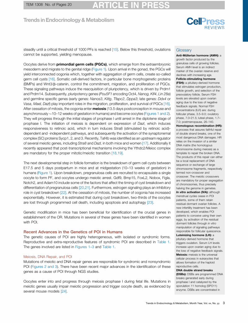
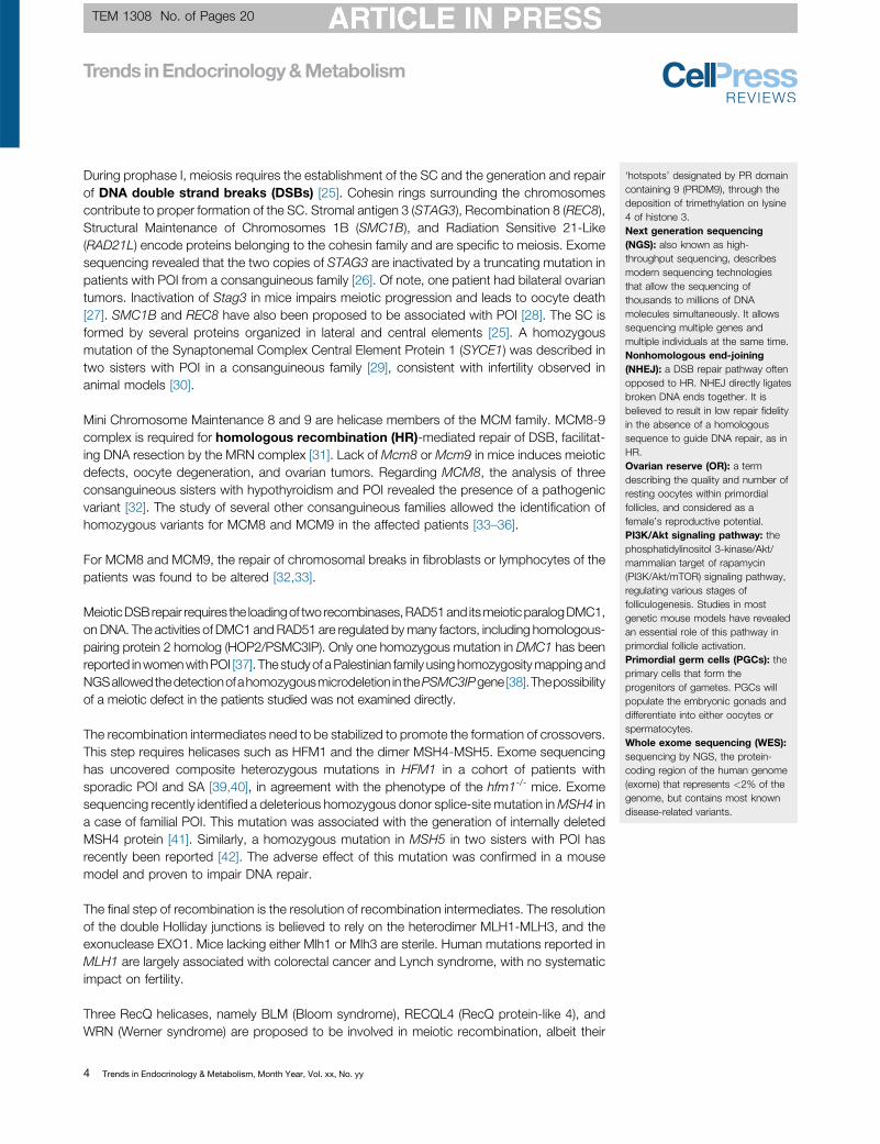
Establishment of the ORPrimordial follicles (PFs) constitute the entire OR. Mechanisms that regulate the formation of thePF pool and the rate by which it is used will determine the duration of the fertile lifespan(Figures 1 and 2). OR formation is a similar process in humans and mice, and in recent years,much insight has been gained into the molecular mechanisms involved.
At 20 weeks of gestation, a pair of human ovaries contains close to 7 million germ cells [15]. Arapid loss of follicles in fetal life results in about 1–2 million oocytes at birth. Prior to puberty, thenumber declines further, to 300 000–400 000. During reproductive life, the number declines
2 Trends in Endocrinology & Metabolism, Month Year, Vol. xx, No. yy
TEM 1308 No. of Pages 20
GlossaryAnti-Müllerian hormone (AMH): agrowth factor produced by thegranulosa cells of growing follicles.Serum AMH level is an indirectmarker of the ovarian reserve anddeclines with increasing age.Follicle-stimulating hormone(FSH): a pituitary-derived hormonethat stimulates estrogen production,follicle growth, and selection of thepreovulatory follicle. Serum FSHlevels are elevated upon ovarianaging due to the loss of negativefeedback signals. Normal FSHconcentrations (IU/l) are: duringfollicular phase, 3.5–9.0; ovulatoryphase, 7.0–21.5; luteal phase, 1.7–7.0; postmenopause, 26–140.Homologous recombination (HR):a process that assures faithful repairof double strand breaks, one of themost dangerous DNA damages. HRrelies on the invasion of a similarDNA matrix (the homologouschromosome during meiosis) as atemplate to repair the broken DNA.The products of this repair can eitherbe a local replacement of DNAsequence or exchange of largechromosome fragments, respectivelytermed non-crossover andcrossover. The meiotic crossoversare mandatory for proper segregationof chromosomes, thus preciselyhalving the genome in gametes.In vitro activation (IVA): althoughmenstrual cycles cease in POIpatients, some of them retainresidual dormant ovarian follicles. Anew infertility treatment has beendeveloped, which enables POIpatients to conceive using their owneggs, by activation of the residualdormant follicles through in vitromanipulation of signaling pathwaysresponsible for follicular quiescence.Luteinizing hormone (LH): apituitary-derived hormone thattriggers ovulation. Serum LH levelsincrease upon ovarian aging due tothe loss of negative feedback signals.Meiosis: meiosis is the universalcellular process in eukaryotes thatallows formation of the haploidreproductive cells.DNA double strand breaks(DSBs): DSBs are programmed DNAbreaks generated early duringprophase I and catalyzed by thesporulation 11 homolog (SPO11)enzyme. DSBs are concentrated in
steadily until a critical threshold of 1000 PFs is reached [15]. Below this threshold, ovulationscannot be supported, yielding menopause.
Oocytes derive from primordial germ cells (PGCs), which emerge from the extraembryonicmesoderm and migrate to the genital ridge (Figure 1). Upon arrival in the gonad, the PGCs willyield interconnected oogonia which, together with aggregation of germ cells, create so-calledgerm cell cysts [16]. Somatic cell-derived factors, in particular bone morphogenetic proteins(BMPs) and Wnt3/b-catenin, control the commitment, migration, and proliferation of PGCs.These signaling pathways induce the reacquisition of pluripotency, which is driven by Prdm1and Prdm14. Subsequently, pluripotency genes (Pou5F1 encoding Oct4,Nanog, Klf4, Lin 28a)and germline specific genes (early genes: Nanos3, Kitlg, Tfapc2, Dppa3; late genes: Ddx4 orVasa, Mael, Dazl) play important roles in the migration, proliferation, and survival of PGCs [16].After cessation of mitosis, the oogonia entermeiosis (13.5 days postconception in mouse andasynchronously�10–12 weeks of gestation in humans) and become oocytes (Figures 1 and 2).They will progress through the initial stages of prophase I until arrest in the diplotene stage ofprophase I. The initiation of meiosis is dependent on expression of Dazl, which inducesresponsiveness to retinoic acid, which in turn induces Stra8 (stimulated by retinoic acid)-dependent and -independent pathways, and subsequently the activation of the synaptonemalcomplex (SC) proteins Sycp1, 2, and 3. Recently, Taf4b was identified as an upstream regulatorof several meiotic genes, including Stra8 and Dazl, in both mice and women [17]. Additionally itrecently appeared that post-transcriptional mechanisms involving the Ythdc2/Meioc complexare mandatory for the proper mitotic/meiotic transition [18,19].
The next developmental step in follicle formation is the breakdown of germ cell cysts betweenE17.5 and 5 days postpartum in mice and at midgestation (10–13 weeks of gestation) inhumans (Figure 1). Upon breakdown, pregranulosa cells are recruited to encapsulate a singleoocyte to form PF, and oocytes undergo meiotic arrest. Gdf9, Bmp15, FoxL2, Nobox, Figla,Notch2, and Adam10 include some of the factors that affect the timing of cyst breakdown anddifferentiation of pregranulosa cells [20,21]. Furthermore, estrogen signaling plays an inhibitoryrole in cyst breakdown [22]. At the cessation of mitosis, the number of oogonia has increasedexponentially. However, it is estimated that during cyst breakdown, two-thirds of the oocytesare lost through programmed cell death, including apoptosis and autophagy [23].
Genetic modification in mice has been beneficial for identification of the crucial genes inestablishment of the OR. Mutations in several of these genes have been identified in womenwith POI.
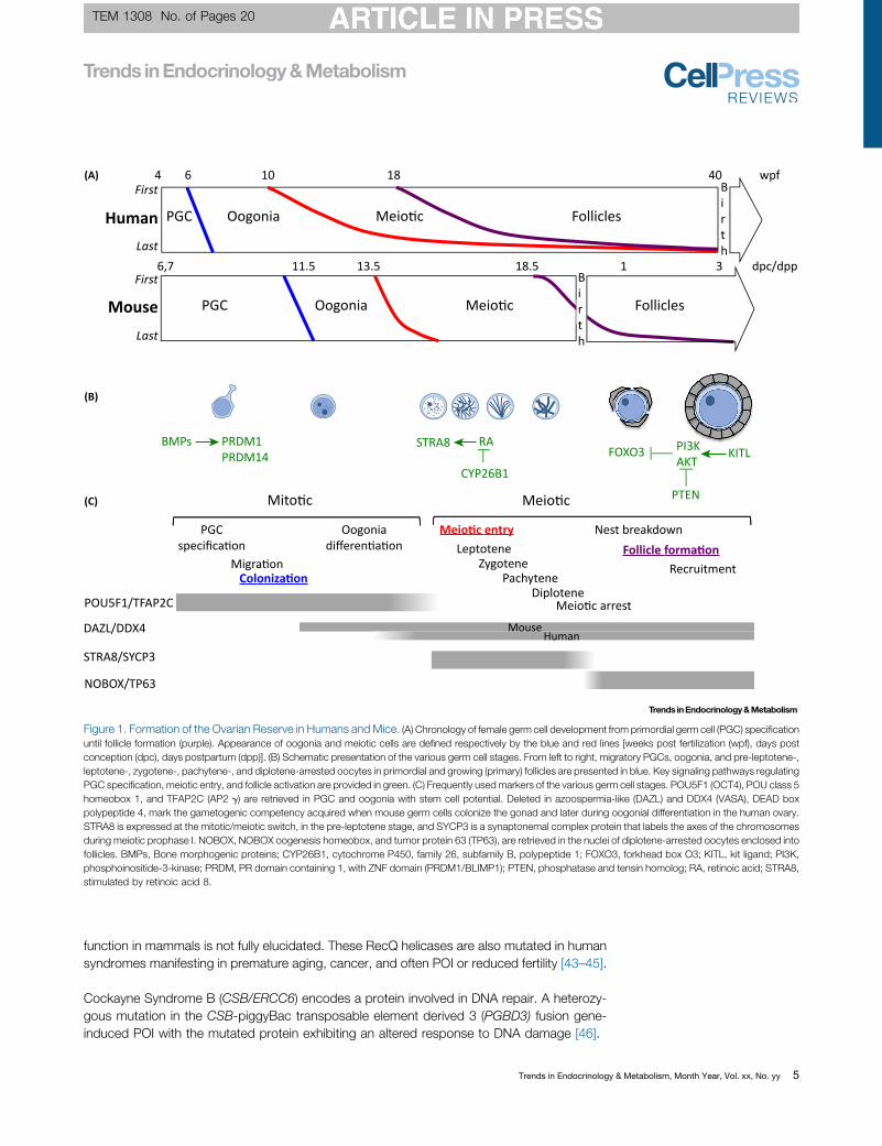
Recent Advances in the Genetics of POI in HumansThe genetic causes of POI are highly heterogeneous, with isolated or syndromic forms.Reproductive and extra-reproductive features of syndromic POI are described in Table 1.The genes involved are listed in Figures 1–3 and Table 1.
Meiosis, DNA Repair, and POIMutations of meiotic and DNA repair genes are responsible for syndromic and nonsyndromicPOI (Figures 2 and 3). There have been recent major advances in the identification of thesegenes as a cause of POI through NGS studies.
Oocytes enter into and progress through meiosis prophase I during fetal life. Mutations inmeiotic genes usually impair meiotic progression and trigger oocyte death, as evidenced byseveral mouse models [24].
Trends in Endocrinology & Metabolism, Month Year, Vol. xx, No. yy 3
TEM 1308 No. of Pages 20
‘hotspots’ designated by PR domaincontaining 9 (PRDM9), through thedeposition of trimethylation on lysine4 of histone 3.Next generation sequencing(NGS): also known as high-throughput sequencing, describesmodern sequencing technologiesthat allow the sequencing ofthousands to millions of DNAmolecules simultaneously. It allowssequencing multiple genes andmultiple individuals at the same time.Nonhomologous end-joining(NHEJ): a DSB repair pathway oftenopposed to HR. NHEJ directly ligatesbroken DNA ends together. It isbelieved to result in low repair fidelityin the absence of a homologoussequence to guide DNA repair, as inHR.Ovarian reserve (OR): a termdescribing the quality and number ofresting oocytes within primordialfollicles, and considered as afemale’s reproductive potential.PI3K/Akt signaling pathway: thephosphatidylinositol 3-kinase/Akt/mammalian target of rapamycin(PI3K/Akt/mTOR) signaling pathway,regulating various stages offolliculogenesis. Studies in mostgenetic mouse models have revealedan essential role of this pathway inprimordial follicle activation.Primordial germ cells (PGCs): theprimary cells that form theprogenitors of gametes. PGCs willpopulate the embryonic gonads anddifferentiate into either oocytes orspermatocytes.Whole exome sequencing (WES):sequencing by NGS, the protein-coding region of the human genome(exome) that represents <2% of thegenome, but contains most knowndisease-related variants.
During prophase I, meiosis requires the establishment of the SC and the generation and repairof DNA double strand breaks (DSBs) [25]. Cohesin rings surrounding the chromosomescontribute to proper formation of the SC. Stromal antigen 3 (STAG3), Recombination 8 (REC8),Structural Maintenance of Chromosomes 1B (SMC1B), and Radiation Sensitive 21-Like(RAD21L) encode proteins belonging to the cohesin family and are specific to meiosis. Exomesequencing revealed that the two copies of STAG3 are inactivated by a truncating mutation inpatients with POI from a consanguineous family [26]. Of note, one patient had bilateral ovariantumors. Inactivation of Stag3 in mice impairs meiotic progression and leads to oocyte death[27]. SMC1B and REC8 have also been proposed to be associated with POI [28]. The SC isformed by several proteins organized in lateral and central elements [25]. A homozygousmutation of the Synaptonemal Complex Central Element Protein 1 (SYCE1) was described intwo sisters with POI in a consanguineous family [29], consistent with infertility observed inanimal models [30].
Mini Chromosome Maintenance 8 and 9 are helicase members of the MCM family. MCM8-9complex is required for homologous recombination (HR)-mediated repair of DSB, facilitat-ing DNA resection by the MRN complex [31]. Lack ofMcm8 orMcm9 in mice induces meioticdefects, oocyte degeneration, and ovarian tumors. Regarding MCM8, the analysis of threeconsanguineous sisters with hypothyroidism and POI revealed the presence of a pathogenicvariant [32]. The study of several other consanguineous families allowed the identification ofhomozygous variants for MCM8 and MCM9 in the affected patients [33–36].
For MCM8 and MCM9, the repair of chromosomal breaks in fibroblasts or lymphocytes of thepatients was found to be altered [32,33].
MeioticDSBrepair requires the loadingof two recombinases,RAD51and itsmeioticparalogDMC1,onDNA. The activities of DMC1 andRAD51 are regulated bymany factors, including homologous-pairing protein 2 homolog (HOP2/PSMC3IP). Only one homozygous mutation in DMC1 has beenreported inwomenwithPOI [37]. Thestudyof aPalestinian family usinghomozygositymappingandNGSallowedthedetectionofahomozygousmicrodeletion in thePSMC3IPgene[38].Thepossibilityof a meiotic defect in the patients studied was not examined directly.
The recombination intermediates need to be stabilized to promote the formation of crossovers.This step requires helicases such as HFM1 and the dimer MSH4-MSH5. Exome sequencinghas uncovered composite heterozygous mutations in HFM1 in a cohort of patients withsporadic POI and SA [39,40], in agreement with the phenotype of the hfm1-/- mice. Exomesequencing recently identified a deleterious homozygous donor splice-site mutation inMSH4 ina case of familial POI. This mutation was associated with the generation of internally deletedMSH4 protein [41]. Similarly, a homozygous mutation in MSH5 in two sisters with POI hasrecently been reported [42]. The adverse effect of this mutation was confirmed in a mousemodel and proven to impair DNA repair.
The final step of recombination is the resolution of recombination intermediates. The resolutionof the double Holliday junctions is believed to rely on the heterodimer MLH1-MLH3, and theexonuclease EXO1. Mice lacking either Mlh1 or Mlh3 are sterile. Human mutations reported inMLH1 are largely associated with colorectal cancer and Lynch syndrome, with no systematicimpact on fertility.
Three RecQ helicases, namely BLM (Bloom syndrome), RECQL4 (RecQ protein-like 4), andWRN (Werner syndrome) are proposed to be involved in meiotic recombination, albeit their
4 Trends in Endocrinology & Metabolism, Month Year, Vol. xx, No. yy
TEM 1308 No. of Pages 20
6,7 11.5 13.5 18.5 dpc/dpp1 3
PGC Oogonia Meio c
Meio c
Follicles
4 406 10 18 wpf
PGC Oogonia Follicles
POU5F1/TFAP2C
DAZL/DDX4
STRA8/SYCP3
NOBOX/TP63
Mito c Meio c
PGCspecifica on
Oogoniadifferen a on
Meio c entryLeptotene
ZygotenePachytene
Diplotene
Nest breakdownFollicle forma on
Coloniza on
MouseHuman
Human
(A)
(B)
(C)
Mouse
Migra on Recruitment
Meio c arrest
BMPs RA PI3KAKT
STRA8PRDM1PRDM14 FOXO3
PTEN
CYP26B1
First
Last
KITL
Birth
Birth
First
Last
Figure 1. Formation of the Ovarian Reserve in Humans and Mice. (A) Chronology of female germ cell development from primordial germ cell (PGC) specificationuntil follicle formation (purple). Appearance of oogonia and meiotic cells are defined respectively by the blue and red lines [weeks post fertilization (wpf), days postconception (dpc), days postpartum (dpp)]. (B) Schematic presentation of the various germ cell stages. From left to right, migratory PGCs, oogonia, and pre-leptotene-,leptotene-, zygotene-, pachytene-, and diplotene-arrested oocytes in primordial and growing (primary) follicles are presented in blue. Key signaling pathways regulatingPGC specification, meiotic entry, and follicle activation are provided in green. (C) Frequently usedmarkers of the various germ cell stages. POU5F1 (OCT4), POU class 5homeobox 1, and TFAP2C (AP2 g) are retrieved in PGC and oogonia with stem cell potential. Deleted in azoospermia-like (DAZL) and DDX4 (VASA), DEAD boxpolypeptide 4, mark the gametogenic competency acquired when mouse germ cells colonize the gonad and later during oogonial differentiation in the human ovary.STRA8 is expressed at the mitotic/meiotic switch, in the pre-leptotene stage, and SYCP3 is a synaptonemal complex protein that labels the axes of the chromosomesduring meiotic prophase I. NOBOX, NOBOX oogenesis homeobox, and tumor protein 63 (TP63), are retrieved in the nuclei of diplotene-arrested oocytes enclosed intofollicles. BMPs, Bone morphogenic proteins; CYP26B1, cytochrome P450, family 26, subfamily B, polypeptide 1; FOXO3, forkhead box O3; KITL, kit ligand; PI3K,phosphoinositide-3-kinase; PRDM, PR domain containing 1, with ZNF domain (PRDM1/BLIMP1); PTEN, phosphatase and tensin homolog; RA, retinoic acid; STRA8,stimulated by retinoic acid 8.
function in mammals is not fully elucidated. These RecQ helicases are also mutated in humansyndromes manifesting in premature aging, cancer, and often POI or reduced fertility [43–45].
Cockayne Syndrome B (CSB/ERCC6) encodes a protein involved in DNA repair. A heterozy-gous mutation in the CSB-piggyBac transposable element derived 3 (PGBD3) fusion gene-induced POI with the mutated protein exhibiting an altered response to DNA damage [46].
Trends in Endocrinology & Metabolism, Month Year, Vol. xx, No. yy 5

TEM 1308 No. of Pages 20
Folliculargrowth
Meiosis Follicularassembly
Follicle forma on
Ovaryforma or
Follicularac va on
Prolifera onsurvival
Primordial follicles PGC oogonia Primary follicles Antral follicles
NANOS3FANCA FANCC
FANCG FANCM
FIGLA BMPR1B FOXL2CYP19A1
NR5A1 SALL4 WT1
GDF9 BMP15BMPR2 BMPR1B FSHR
INHA AMH AMHR2
Mitosis
BMP15 BMPR2 BMPR1B NOG FSHR GJA4LHCGR STAR CYP17A1 PGRMC1 LMNA WRNGNAS GALT FMR1 ESR1 AR AIRE NR5A1 CLPP
INHA TGFBR3 AMH AMHR2 FOXO1
Oocytematura on
and ovula on
Follicularmatura on
MSH4 MSH5 HFM1 REC8 SMC1BSYCE1 CPEB1 STAG3 BLM
PSMC3IP DMC1 ATMMCM8 MCM9 SGO2
Ovula on
SOHLH1 SOHLH2NOBOX FOXL2FOXO3 LHX8
Cell a achment and migration: ANTXR1 Nuclear func ons: NUP107 DNA damage, repair, replication: NBN, RECQL4, XRCC4, CSB-PGBD3, SPIDR, POF1B Transla onal and post-transla onal regula ons: EIF2B, RCBTB1, EIF4ENIF1 Mitochondrial func on: AARS2, HARS2, LARS2, MT-ATP6/8, C10ORF2, KIAA0391, ERAL1, POLG Peroxisomal func on: HSD17B4 Metabolic defect: PMM2
LHCGR
Figure 2. Human Genes Associated with Primary Ovarian Insufficiency and Their Physiological Importance in Oogenesis, Folliculogenesis, andOther Functions. Genes with in vivo mutations associated with primary ovarian insufficiency (POI) in humans and the physiological importance of these genes inovarian function are indicated. Oogenesis and folliculogenic processes are represented by a transit between different compartmental stages, depicted as boxescontaining the cell populations. At each stage of oogenesis and folliculogenesis, an important part of the germ cell will die by apoptosis, depicted by a concomitantdecrease in box sizes. The first compartment (yellow box) corresponds to the primordial germ cells (PGCs) when they differentiate into oogonia, the secondcompartment (green box) corresponds to follicle formation, which involvesmeiosis and follicular assembly processes and endswith establishment of the ovarian reserveof primordial follicles (pink box). From this reserve, follicular activation leads to the formation of primary follicles (light blue box). Then the growing follicles can reach theantral stage (dark blue box) and ovulate, or degenerate by atresia. Genes whose mutations are associated with POI are indicated at each stage of these developmentalprocesses. The involvement of each gene at a specific stage of ovarian or follicular development is based on in vivo (presence/absence of follicles in biopsy samples,detection of antral follicles using ovarian ultrasound scanning or AMH measurements in women carrying mutations) or/and in vitro observations (culture experimentsusing human ovarian cortex or granulosa cells). For genes depicted in italic, information on their stage specific role is only available from mouse models. Genesassociated with POI for which the stage-specific role is unknown are listed in the box, with their biological function (green font). The stimulating and inhibiting factors aredepicted in black and red font, respectively. See text for references and [13].
Lastly, althoughmeiotic DSB repair appears to rely on HR, a second process for DSB repair, thenonhomologous end-joining (NHEJ), allows the direct ligation of broken DNA ends to eachother. X-ray repair cross-complementing protein 4 (XRCC4) and Ligase 4 (LIG4) are twoproteins absolutely required for NHEJ. Syndromic POI was reported in a female patient with
6 Trends in Endocrinology & Metabolism, Month Year, Vol. xx, No. yy
TEM 1308 No. of Pages 20
Table 1. Clinical Presentations of Syndromic POI.a
Diagnosis Menstrualhistory
Ovarian phenotype Particular features OMIM # Gene(s) involved
WT1-related XX-DSD PA or SA Streak gonads,partial ovarian dysgenesis
Nephropathy, diaphragmatic hernia 194070 WT1
SF1-related XX-DSD PA or SA Streak gonads,partial ovarian dysgenesis
Adrenal insufficiency 612964 NR5A1/SF1
BPES PA or SA Rare or absent follicles Blepharophimosis, ptosis, epicanthusinversus
110100 FOXL2
FMR1 premutation SA Follicle depletion X-linked mental retardation in family. Fragile Xtremor/ataxia syndrome
300624 FMR1
Autoimmunepolyendocrinopathysyndrome. APS-PGAtype 1
PA or SA Autoimmune oophoritis Addison disease, candidiasis, vitiligo,hypoparathyroidism, diabetes mellitus,hepatitis, malabsorption, keratopathy,alopecia
240300 AIRE
Autoimmune APS-PGA type 3
Autoimmune oophoritis Autoimmune thyroid disease, atrophicgastritis, vitiligo
Pseudohypoparathyroidism
SA Follicular cysts but nocorpora lutea in one case
Brachydactyly, short stature, hypocalcemiaand hyperphosphatemia, hypothyroidism,obesity
#103580 GNAS
Galactosemia PA Streak ovaries or fewnonmaturated follicles
Neonatal jaundice, failure to thrive, cirrhosis,cataract, intellectual disability, foodintolerance, hypoglycemia, renal dysfunction.
230400 GALT
Disorders ofglycosylation (CDG1A)
PA Absent ovaries in somepatients by US orlaparoscopy
Growth retardation, microcephaly,encephalopathy, peripheral neuropathy,retinitis pigmentosa, cardiac myopathy,hepatomegaly, nephrotic syndrome,psychomotor retardation
212065 PMM2
Ataxia telangiectasia PA Cerebellar ataxia, telangiectasia, recurrentinfections, malignancies, and increased levelsof alpha fetoprotein
208900 ATM
Nijmegen breakagesyndrome
PA or SA Streak gonads, small ovaries Prenatal growth retardation, progressivemental deterioration, microcephaly, recurrentinfections, increased risk for neoplasias suchas lymphoma
251260 NBN
Fanconi anemia PA or SA Decreased number ofprimordial follicles
Pancytopenia, small stature, microcephaly,ear anomalies, heart defects, kidneymalformations, radial aplasia and thumbdeformities, intellectual disability, café-au lait-spots
#227650#227645#614082
FANCAFANCCFANCG
XRCC4-relateddisorder
SA Short stature, microcephaly, developmentaldelay, diabetes mellitus
#616541 XRCC4
Bloom syndrome SA Possibly accelerated follicularatresia
Premature aging with chromosomalinstability, short stature, skin rashes andtelangiectatic skin on sun-exposed areas,increased risk for neoplasias,immunodeficiency
#210900 BLM
Werner syndrome SA Possibly accelerated follicularatresia
Premature aging with chromosomalinstability, pre- and postnatal growthdeficiency, sclerodermic skin changes,cataract, arteriosclerosis, increased cancerrisk, diabetes mellitus
#277700 WRN
Rothmund-Thomsonsyndrome
SA Gonadotropin resistance Short stature, cataract, saddle nose, teethanomalies, premature graying of hair
#268400 RECQL4
Trends in Endocrinology & Metabolism, Month Year, Vol. xx, No. yy 7
TEM 1308 No. of Pages 20
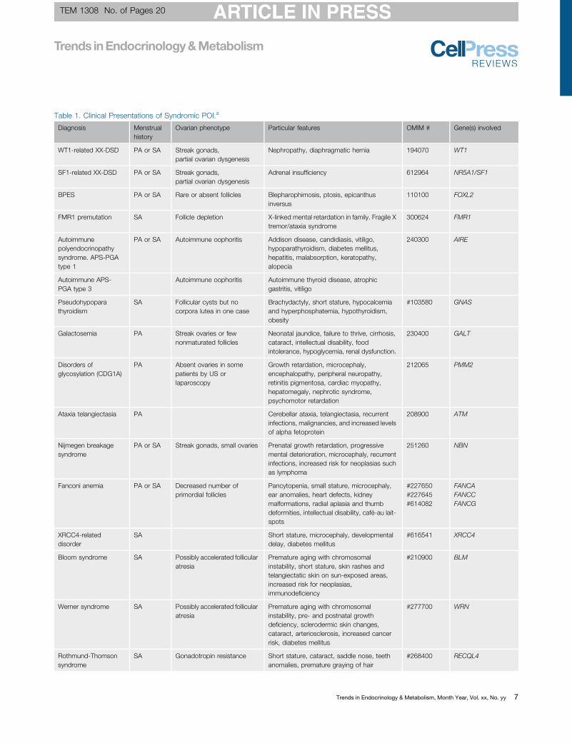
Table 1. (continued)
Diagnosis Menstrualhistory
Ovarian phenotype Particular features OMIM # Gene(s) involved
Hutchinson-Gilfordprogeria
SA Diminished follicular reserve Progeria, short stature, low body weight, earlyloss of hair, lipodystrophy, scleroderma,decreased joint mobility, osteolysis,cardiomyopathy
#176670 LMNA
GAPO SA Follicle depletion Growth retardation, alopecia,pseudoanodontia, optic atrophy, highforehead, midface hypoplasia
#230740 ANTXR1
Perrault syndrome PA, SA Streak ovaries,lack of ovaries,small ovaries
DeafnessNeurologic symptoms in PRLTS1, PRLTS3,and PRLTS5
#233400#614926#614129#615300#616138
HSD17B4, HARS2,LARS2 CLPPC10orf2, CLDN14+SGO2KIAA0391ERAL1
Woodhouse-Sakatisyndrome
PA Streak ovaries Alopecia, deafness, hypogonadism, diabetes,intellectual disability
#241080 C2orf37
Vanishing white matterdiseaseOvarioleukodystrophy
SA Ovarioleukodystrophystreak ovaries
Progressive cerebellar ataxia, spasticity,cognitive impairment with white matterlesions on brain imaging. Onset from earlyinfancy to adulthood
#603896#615889
EIF2BAARS2
Retinal dystrophy withor without extraocularanomalies
SA Retinal dystrophy, goiter, intellectualdisability, hypogonadism
#617175 RCBTB1
Progressive externalophthalmoplegia
SA Diminished follicle reserve Ptosis, progressive external ophthalmoplegia,sensorineural hearing loss, axonalneuropathy, muscle weakness, ataxia,dysarthria, dysphagia and late onsetParkinsonism
#157640 POLG
Acromesomelicchondrodysplasia withgenital anomalies
PA Severe brachydactyly with radial deviation ofthe fingers, ulnar deviation of the hands,fusion of the carpal/tarsal bones, aplasia ofthe fibula, bilateral clubfeet with small broadfeet and short toes
#609441 BMPR1B
Interphalangeal jointsynostosis
SA Symphalangism, hearing loss #185800 NOG
aDSD, Disorder of sexual differentiation; PA, primary amenorrhea; SA, secondary amenorrhea; US, ultrasound. See text for references and [13].
homozygous single nucleotide variant in the XRCC4 gene [47]. POI was reported in two patientswith biallelic truncating mutations in the LIG4 gene [48]. These patients display short stature,microcephaly, and genomic instability or hypersensitivity to radiation. Similarly, another impor-tant DNA repair pathway, the Fanconi anemia (FANC) pathway, exists in numerous progenitorcells, including the germline. This pathway employs at least 20 proteins, including thoseencoded by the FANCA, FANC, and FANCG genes, and has been associated with POI[49]. Mouse models for several Fanc genes (a, c, d, e, f, g, i, m, n, o, p) evidenced gonadalhypoplasia with ovaries showing follicle depletion [50]. This appears to be due to reduced PGCnumbers, though meiotic roles are also possible. Very recently a homozygous FANCM muta-tion was shown to underlie a familial case of nonsyndromic POI [51]. FANCM biallelic mutationspredispose to cancer, in particular early-onset breast cancer in females and chemosensitivity
8 Trends in Endocrinology & Metabolism, Month Year, Vol. xx, No. yy
TEM 1308 No. of Pages 20
Crossover
Double strandbreaks
Homology searchRAD51, DMC1, RPA,MEIOB, SPATA22,BRCA2, RAD54, MND1,PSMC3IP, RAD52,FMR1, BRCA1, ATR,ATM
PRDM9, SPO11,TOPVIBL, MEI1, MEI4,REC114, IOH1,HORMAD1
MCM8, MCM9, RNF212,HEI10, MSH4, MSH5,HFM1, TEX11
Stabilisa on
Non-crossover
BLM,TOP3,RMI1
MRE11, RAD50,NBN, RBBP8, EXO1
MLH1, MLH3, EXO1
Synaptonemal complex
Central element Lateral elements
SYCE1,SYCP1,SIX6OS1,SYCE2,TEX12
Cohesins
SYCP3,SYCP2
STAG3,RAD21L,REC8,SMC1B,SMC3
(A)
(B)
(C)
(D)
(E)
FANCM
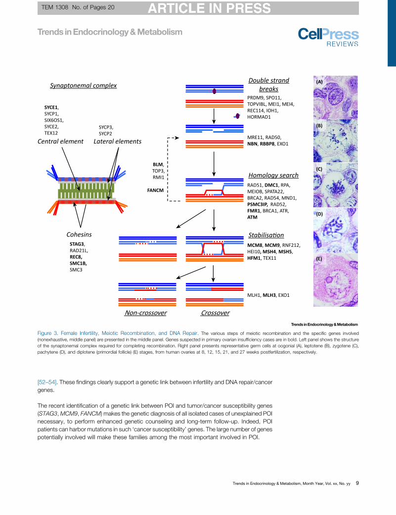
Figure 3. Female Infertility, Meiotic Recombination, and DNA Repair. The various steps of meiotic recombination and the specific genes involved(nonexhaustive, middle panel) are presented in the middle panel. Genes suspected in primary ovarian insufficiency cases are in bold. Left panel shows the structureof the synaptonemal complex required for completing recombination. Right panel presents representative germ cells at oogonial (A), leptotene (B), zygotene (C),pachytene (D), and diplotene (primordial follicle) (E) stages, from human ovaries at 8, 12, 15, 21, and 27 weeks postfertilization, respectively.
[52–54]. These findings clearly support a genetic link between infertility and DNA repair/cancergenes.
The recent identification of a genetic link between POI and tumor/cancer susceptibility genes(STAG3,MCM9, FANCM) makes the genetic diagnosis of all isolated cases of unexplained POInecessary, to perform enhanced genetic counseling and long-term follow-up. Indeed, POIpatients can harbor mutations in such ‘cancer susceptibility’ genes. The large number of genespotentially involved will make these families among the most important involved in POI.
Trends in Endocrinology & Metabolism, Month Year, Vol. xx, No. yy 9
TEM 1308 No. of Pages 20
Genes Involved in Syndromic POI (Figures 2 and 3 and Table 1)The clinical presentations of syndromic POI are highly variable and are presented in Table 1.
Perrault Syndrome (PS)Perrault syndrome (PS) is a genetically heterogeneous autosomal recessive syndrome, mainlycharacterized by ovarian dysfunction and sensorineural deafness (Table 1). Recently, agrowing number of genes involved in PS were identified by NGS. These genes are implicatedin mitochondrial functions or metabolism. In mouse models, genetic changes that causeperturbation in mitochondrial protein translation lead to hearing loss as a result of tissue-specific apoptosis [55]. Given the role of apoptosis in ovarian development, inappropriatelytimed apoptosis may also lead to POI. HARS2 [56] and LARS2 [57,58] encode mitochondrialhistidyl or leucyl-tRNA synthetases involved in translation of mitochondrially encoded genes.CLPP encodes a highly conserved endopeptidase component of a mitochondrial ATP-dependent proteolytic complex, involved in degradation of unfolded or misfolded polypep-tides [59–61]. C10orf2 encodes Twinkle, a mitochondrial primase-helicase essential formitochondrial DNA replication [62,63], yielding a mitochondrial DNA depletion syndromeand progressive external ophthalmoplegia. Very recently, mutations of ERAL1 and KIAA0391were involved in PS. ERAL1 protein binds to the mitochondrial 12S rRNA and is involved inassembly of the small mitochondrial ribosomal subunit affecting mitochondrial respiration andfunction [64]. KIAA0391 encodes RNase P (PRORP) the metallonuclease subunit of themitochondrial RNase P complex responsible for the 50-end processing of mitochondrialprecursor tRNAs [65].
Apart frommitochondrial functions, mutations in amultifunctional peroxisomal enzyme involvedin fatty acid ß-oxidation and steroid metabolism, 17b-hydroxysteroid dehydrogenase type 4[HSD17B4, also known as D-bifunctional protein (DBP)] also cause PS [66–68]. Mutations ofthis gene were already identified in autosomal recessive mode in a severe disorder of peroxi-somal fatty acid b-oxidation.
A combination of two homozygous mutations leading to a coincidental PS, one in CLDN14involved in deafness and the other in shugoshin-like 2a (SGO2) encoding shugoshin2, likelyinvolved in POI, have been described [69]. During meiosis in the mouse, SGO2 maintains theintegrity of the cohesion complex that tethers sister chromatids. Unsolved cases of PS persist,indicating that novel genes will still be discovered [70,71].
Premature Aging SyndromesLaminopathy due to mutations in LMNA encoding a nuclear envelope protein includes ovarianfailure and premature aging. Malouf syndrome belongs to this condition [72]. Hutchinson-Gilford progeria Syndrome (HGPS) [73], caused by aberrant splicing of the LMNA gene andexpression of a mutant product called progerin, comprises premature aging and lipodystro-phies. Sometimes both syndromes occur together [73].
GAPO syndrome, another form of premature aging and premature follicle depletion, is causedby mutations of the anthrax toxin receptor 1 gene ANTXR1 [74]. The protein has been involvedin cell attachment and migration. Additionally, it allows the interaction of cells and severalcomponents of the extracellular matrix by binding extracellular ligands with the actin of thecytoskeleton.
10 Trends in Endocrinology & Metabolism, Month Year, Vol. xx, No. yy
TEM 1308 No. of Pages 20
Neurosensory SyndromesLeukoencephalopathies are also a heterogeneous group of disorders associated with vanish-ing white matter and are seen in a subset of POI, yielding ovarioleukodystrophy. Mutations of aspecific mitochondrial alanine aminoacyl-tRNA synthetase, AARS2, have been involved[75,76]. Another group of genes involved is EIF2B1 to EIF2B5 which encode the five subunitsof the eukaryotic initiation factor 2B [77].
Mutations in RCBTB1 [78] are present in syndromes including inherited retinal dystrophy andPOI. RCBTB1 is involved in ubiquitination, more specifically as a CUL3 substrate adaptorinvolved in stress-response to combat oxidative or electrophilic insults.
Mutations of the nuclear gene POLG encoding a mitochondrial DNA polymerase gamma canlead to POI with autosomal dominant progressive external ophthalmoplegia [79].
Defects in the respiratory chain or mitochondrial ATP synthase (complex V) result in mitochon-drial dysfunction and defective energy production. Mutations of MT-ATP6/8 encoding two ofthe subunits of complex V are associated with syndromes including cerebellar ataxia, peripheralneuropathy, diabetes mellitus, and POI [80].
Skeletal SyndromesPOI can occur in some skeletal syndromes, such as Demirhan syndrome, which is caused bymutations in BMPR1B [81].
Another condition including proximal symphalangism and POI is caused by mutations in NOG[82]. NOG protein is expressed in the ovaries and interacts with BMP, which plays an importantrole in ovarian function.
Genes Associated With Nonsyndromic POISee Figures 2 and 3.
Regulation of PF RecruitmentThe majority of PF will remain dormant until stimulatory signals or a break from inhibitory signalsinduces activation. PF recruitment is initiated in mice after birth at postnatal day 4–5, and inhumans at 17 weeks of gestation (Figure 1).
Various oocyte-expressed signaling and/or transcription factors have been identified to main-tain the quiescent state (Foxo3, Lhx8) or, in contrast, to activate PF growth (Sohlh1, Sohlh2,Nobox) [83] (Figure 2). Interestingly, Foxo3 and Lhx8 are the effectors of the PI3K/Aktsignaling pathway [84]. Targeted (oocyte-specific) deletion of stimulating factors (Kit, Pdpk1,Rptor, Rps6) of this pathway blocks follicular activation and induces PF apoptosis, whereasloss of the inhibiting factors (Pten, Cdkn1b, Tsc1, Tsc2, Stk11) results in premature and globalactivation of PF [85] (Figure 2).
A characteristic of PF activation is the transition of squamous pregranulosa cells to cuboidalgranulosa cells. Failure thereof results in an arrest at the primordial stage, followed by oocytedeath and follicular depletion, as shown in FoxL2 knockout mice [86]. AMH, expressed duringthis transition, inhibits PF activation since Amh knockout mice display an accelerated exhaus-tion of the pool [87]. Several additional growth factors have been shown to activate PFrecruitment (Figure 2).
Trends in Endocrinology & Metabolism, Month Year, Vol. xx, No. yy 11
TEM 1308 No. of Pages 20
Regulation of Follicle GrowthGonadotropin-Independent Phase: Role of Ovarian Growth Factors; Early follicle growth up tothe large preantral stage is independent of gonadotropins in rodents and relies on intraovarianfactors (Figure 2). It requires a coordinated dialog between the oocyte and granulosa cells, inwhich gap junctions and SMAD and PI3K/Akt pathways are important. The discovery thatfollicles in ovaries of Gdf9 knockout mice fail to develop beyond the primary stage was the firstof a series showing the importance of factors involving SMAD signaling pathway in follicledevelopment [88]. Furthermore, in the oocyte-knockout of Furin, a prohormone convertaseresponsible for proteolytic cleavage of TGFb family members, follicle growth is arrested at thesecondary stage [89]. Likewise, inhibition of the PI3K/Akt pathway by Kit or Kitlg deletions leadsto the blockage of follicular growth at the primary follicle stage. Using targeted deletion oractivation of Igf1, Igf1r, Irs2, Rictor, or Foxo3, it was shown that the PI3K/Akt signaling pathwaynot only plays a role in PF activation, but also in follicle survival and development beyond theprimary stage [90,91] (Figure 2).Gonadotropin-Dependent Phase: Role of Gonadotropins; The progression through final stagesof follicle development depends on the gonadotropins FSH and luteinizing hormone (LH)(Figure 2). The threshold for FSH sensitivity is determined by interplay between variousstimulatory and inhibitory growth factors, such as IGF1 and various TGFb family memberstipping the balance to either follicle survival or atresia. Deletion of the Fshr yields an enhancedrate of atresia and follicles fail to progress to the antral stage [92]. Targeted deletion of thenoncanonical progesterone receptor Pgrmc1 in granulosa cells suppressed antral follicledevelopment and increased atresia [93]. Finally, LH action is indispensable for ovulation,meiotic resumption of the oocytes, and cumulus expansion. Loss of LH action therefore alsoresults in infertility as follicle development is blocked at the antral stage [94]. In the absence ofsex steroid action, the final stages of follicle development show abnormalities leading tofollicular arrest, as illustrated in mouse models lacking (cell-specific) androgen or estrogenfunction [95,96].
Defects in Human Genes in Nonsyndromic POIInterestingly, there is an overlap between genes involved in the onset of puberty, normalreproductive aging, and POI [97]. We will present only recent data or selected examples ofgenes that illustrate the precaution that must be taken in the interpretation of genetic data andcomparison with animal models.
Genes Involved in Establishment of the PF Pool and Maturation to Primary Follicles;
Heterozygous variants of SOHLH1 and SOHLH2 have been found in POI [98]. Interestingly, twofamilies harboring a homozygous single-base deletion in the coding region or a premature stopcodon of SOHLH1 [99] had PA, lack of secondary sex characteristics, and nonvisualizedovaries.
A recessivemissensemutation inNucleoporin-107was identified in a consanguineous family ofPalestinian origin [100]. NUP107 is a component of the nuclear pore complex, and theNUP107-associated protein SEH1 is required for oogenesis in Drosophila. In Drosophila,Nup107 knockdown in somatic gonadal cells resulted in female sterility, whereas males werefully fertile. Nup107mutations may compromise the meiotic DNA damage response, leading tooocyte death.
A heterozygous stop codon was identified in the eukaryotic translation initiation factor 4Enuclear import factor 1 gene eIF4ENIF1 in familial POI with dominant inheritance in three
12 Trends in Endocrinology & Metabolism, Month Year, Vol. xx, No. yy
TEM 1308 No. of Pages 20
generations [101]. The gene plays an important role in oocyte development in organisms fromDrosophila to mice.
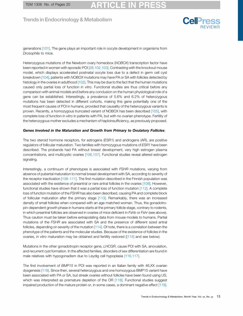
Heterozygous mutations of the Newborn ovary homeobox (NOBOX) transcription factor havebeen reported in womenwith sporadic POI [28,102,103]. Contrasting with the knockout mousemodel, which displays accelerated postnatal oocyte loss due to a defect in germ cell cystbreakdown [104], patients withNOBOXmutations may have PA or SA with follicles detected byhistology in the ovaries in adulthood [102]. This may be due to the fact that the humanmutationscaused only partial loss of function in vitro. Functional studies are thus critical before anycomparison with animal models and before any conclusion on the human physiological role of agene can be established. Interestingly, a prevalence of 5.6% and 6.2% of heterozygousmutations has been detected in different cohorts, making this gene potentially one of themost frequent causes of POI in humans, provided that causality of the heterozygous variants isproven. Recently, a homozygous truncated variant of NOBOX has been described [105], withcomplete loss of function in vitro in patients with PA, but with no ovarian phenotype. Fertility ofthe heterozygous mother excludes amechanism of haploinsufficiency, as previously proposed.
Genes Involved in the Maturation and Growth from Primary to Ovulatory Follicles;
The two steroid hormone receptors, for estrogens (ESR1) and androgens (AR), are positiveregulators of follicular maturation. Two families with homozygous mutations of ESR1 have beendescribed. The probands had PA without breast development, very high estrogen plasmaconcentrations, and multicystic ovaries [106,107]. Functional studies reveal altered estrogensignaling.
Interestingly, a continuum of phenotypes is associated with FSHR mutations, varying fromabsence of pubertal maturation to normal breast development with SA, according to severity ofthe receptor inactivation [108–111]. The first mutation described in the Finnish population wasassociated with the existence of preantral or rare antral follicles in the ovaries [108]. However,functional studies have shown that it was a partial loss of function mutation [112]. A completeloss of functionmutation of the FSHR has also been described, causing PA and complete blockof follicular maturation after the primary stage [113]. Remarkably, there was an increaseddensity of small follicles when compared with an age-matched woman. Thus, the gonadotro-pin-dependent growth phase in humans starts at the primary follicle stage, contrary to rodents,in which preantral follicles are observed in ovaries of mice deficient in Fshb or Fshr (see above).Thus caution must be taken before extrapolating data from mouse models to humans. Partialmutations of the FSHR are associated with SA and the presence of different sized antralfollicles, depending on severity of the mutation [114]. Of note, there is a correlation between thephenotype of the patients and the molecular studies. Because of the existence of follicles in theovaries, in vitro maturation may be obtained and fertility restored ([115] and see below).
Mutations in the other gonadotropin receptor gene, LHCGR, cause POI with SA, anovulation,and recurrent cyst formation. In the affected families, disorders of sex differentiation are found inmale relatives with hypogonadism due to Leydig cell hypoplasia [116,117].
The first involvement of BMP15 in POI was reported in an Italian family with 46,XX ovariandysgenesis [118]. Since then, several heterozygous and one homozygous BMP15 variant havebeen associated with PA or SA, but streak ovaries without follicles have been found using US,which was interpreted as premature depletion of the OR [118]. Functional studies suggestimpaired production of the mature protein or, in some cases, a dominant negative effect [118].
Trends in Endocrinology & Metabolism, Month Year, Vol. xx, No. yy 13
TEM 1308 No. of Pages 20
However, most of the variants detected occur in the heterozygous state, and BMP15 hap-loinsufficiency was proposed to have a predisposing impact for POI. It was also proposed thatreduced BMP15 dosage would contribute to the ovarian phenotype of Turner syndromepatients [119]. These conclusions were challenged by a very recent work on a family with aBMP15 knockout-like effect [120], with both parents bearing deletions in the proregion of theBMP15 precursor. The heterozygous mother conceived normally and had three children. Thus,it seems that haploinsufficiency is not involved in humans. Most of the mutations of BMP15described were heterozygous and a mechanism of haploinsufficiency or a dominant negativeeffect was suspected but most often not demonstrated, making it impossible to implicate thecorresponding gene as the unique cause of POI. Additional genetic mutations in an oligogenicmode of inheritance and/or environmental factors must be involved. Despite streak ovaries,AMHwas initially detectable in the two POI sisters bearing both deletions of BMP15, supportingthe presence of an OR [120]. Five years later, however, AMH was not detected in both sisters,probably because of exhaustion of the PF pool, and one sister had received an egg donation.
In case POI is due to a block in follicular maturation, urgent fertility preservation is needed toavoid follicular atresia.
A recent study showed a homozygous single-base deletion in the coding region ofGDF9 in POIwith PA [121], confirming the causative role of this gene.
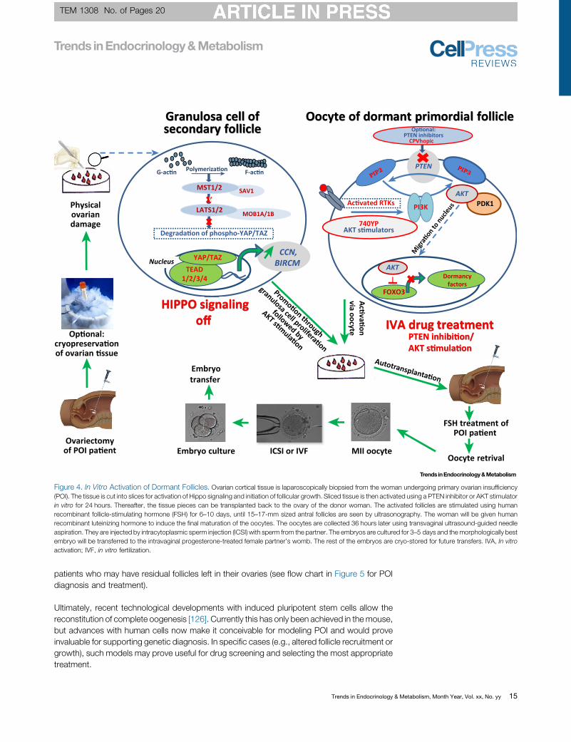
Innovative Treatments for POIThe most frequent therapeutic approach of infertility of POI patients is embryo transfer fromdonated oocytes. Given the complexity of this therapeutic approach, couples requiring oocytedonation should discuss its medical, ethical, legal, and psychological aspects with medicalexperts. Recently, a new innovative fertility treatment has been developed for POI (Figure 4).
Premature activation of PFs caused by chemotherapy, particularly cyclophosphamide, is asignificant cause of the disappearance of follicles from the ovaries. Fertility preservation throughtissue cryopreservation before chemotherapy is therefore an important method for preventingPOI [122]. Post-treatment, the tissue can be autotransplanted. Infants have been born as aresult of the technique.
For cancer patients at high risk of reintroduction of the malignancy, such as leukemia, in vitromaturation of follicles all the way to metaphase II oocytes is a much needed therapy that stillremains to be developed.
In early stages of ovarian insufficiency there are PFs left in the ovaries. Hence, cryopreservationof ovarian tissue as fertility preservation should be carried out as soon as the risk of folliculardecrease has been identified. Although these PFs are inactive, Hovatta et al. [123] showed thathuman ovarian follicles can be activated when ovarian tissue is cut into small pieces and placedin organ culture. Recently, Hippo signaling was identified as the regulatory factor in thisactivation [5,124]. When residual follicles in ovarian tissue from POI patients were stimulatedby cutting the tissue into small pieces, and subsequently exposed to phosphatase and tensinhomolog (PTEN) inhibitors and protein kinase B (Akt) activators prior to transplantation, fulloocyte maturation can be achieved [125] (Figure 4). Of note, PTEN is also an important tumorsuppressor, and therefore its inactivation in vivomight be risky. After IVA, the follicles have to bestimulated to grow, and FSH stimulation is used in a similar manner as in ovulation induction orbefore in vitro fertilization treatments. The ovarian tissue has been transplanted back to patientsafter IVA and healthy infants have been born (Figure 4). This IVA method is useful for those
14 Trends in Endocrinology & Metabolism, Month Year, Vol. xx, No. yy
TEM 1308 No. of Pages 20
Ac vated RTKsPhysicalovariandamage
Ovariectomyof POI pa ent
Op onal:cryopreserva onof ovarian ssue
FSH treatment ofPOI pa ent
Granulosa cell ofsecondary follicle
Oocyte of dormant primordial follicle
Acva
onvia oocyte
Polymeriza on
HIPPO signalingoff
Degrada on of phospho-YAP/TAZ
C
CCN,BIRCM Nucleus
TEAD1/2/3/4
YAP/TAZ
G-ac n F-ac nPTEN
PI3K PDK1AKT
Op onal:PTEN inhibitors
CPVhopic
FOXO3
Dormancyfactors
AKT
Oocyte retrivalEmbryo culture
Embryotransfer
740YPAKT s mulators
ICSI or IVF MII oocyte
SAV1MST1/2
MOB1A/1BLATS1/2
IVA drug treatment gPTEN inhibi on/AKT s mula on
Migr
aon
to nu
cleus
Promoon through
granulosa cell proliferaon
followed by
AKT smula
on
Autotransplanta on
Figure 4. In Vitro Activation of Dormant Follicles. Ovarian cortical tissue is laparoscopically biopsied from the woman undergoing primary ovarian insufficiency(POI). The tissue is cut into slices for activation of Hippo signaling and initiation of follicular growth. Sliced tissue is then activated using a PTEN inhibitor or AKT stimulatorin vitro for 24 hours. Thereafter, the tissue pieces can be transplanted back to the ovary of the donor woman. The activated follicles are stimulated using humanrecombinant follicle-stimulating hormone (FSH) for 6–10 days, until 15–17-mm sized antral follicles are seen by ultrasonography. The woman will be given humanrecombinant luteinizing hormone to induce the final maturation of the oocytes. The oocytes are collected 36 hours later using transvaginal ultrasound-guided needleaspiration. They are injected by intracytoplasmic sperm injection (ICSI) with sperm from the partner. The embryos are cultured for 3–5 days and themorphologically bestembryo will be transferred to the intravaginal progesterone-treated female partner’s womb. The rest of the embryos are cryo-stored for future transfers. IVA, In vitroactivation; IVF, in vitro fertilization.
patients who may have residual follicles left in their ovaries (see flow chart in Figure 5 for POIdiagnosis and treatment).
Ultimately, recent technological developments with induced pluripotent stem cells allow thereconstitution of complete oogenesis [126]. Currently this has only been achieved in themouse,but advances with human cells now make it conceivable for modeling POI and would proveinvaluable for supporting genetic diagnosis. In specific cases (e.g., altered follicle recruitment orgrowth), such models may prove useful for drug screening and selecting the most appropriatetreatment.
Trends in Endocrinology & Metabolism, Month Year, Vol. xx, No. yy 15
TEM 1308 No. of Pages 20
POI diagnosisPA or SA or spaniomenorrhea > 4 months, two FSH values >25 U/l, low E2, normal PRL, normal TSH
Karyotype, FMR1
Isolated POI Syndromic POI
Unexplained POIAbnormal karyotype, Xfra syndrome
AMH, US: ovaries, AFC -
+/- aCGHNGS
Hormonaltreatment
+ Cardiomyopathy
+ Ptosis, epicanthus: BPES
+ Neurosensorial symptoms
+ Cancer, leukemia, small size,hypothyroidism,
chromosome instability
+ Goiter, vi ligoauto-an bodies
+ Metabolic syndrome,galactosemia
+/- aCGH, NGSspecific gene(s)
Specific treatment of associated symptomsEgg dona on Fer lity preserva onIVA in the future
Defects in genes involved inovarian differen a on,
oogenesis, establishment ofthe follicular pool
- Gene c and therapeu c counseling, pa ent and rela ves - mul disciplinary team-
+ Familial DSD
+ Symphalangism
+ Candidiasis,addison
+ Brachydactyly, short stature,high PTH/TSH, obesity
+ VWM syndromeovarioleukodystrophy
+ Cerebellar syndrome,ataxia
+ Ataxia telangiectasia
+ Ophthalmoplegia,tremor
+ Re nal dystrophy,intellectual disability
+ Deafnessperrault syndrome
+ GAPO syndrome
Specific cause: toxic, surgery, irradia on, infec on..
Personal history, physical examina on, ovarian reserve: US: ovary and AFC + AMH Familial study: age menopause, 46XY sex reversal, others
Defects in genes involved infollicular growth and
func on: possible ovarianreserve
AMH, US: ovaries, AFC +
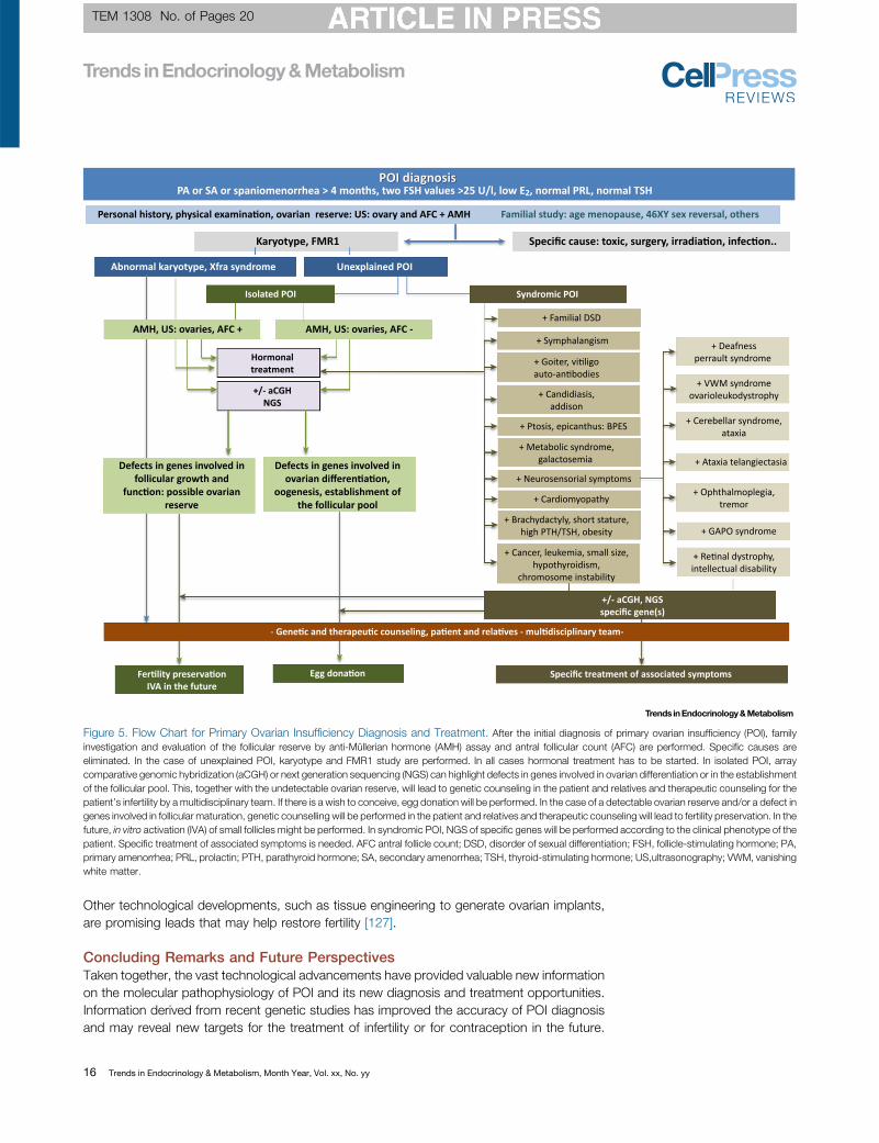
Figure 5. Flow Chart for Primary Ovarian Insufficiency Diagnosis and Treatment. After the initial diagnosis of primary ovarian insufficiency (POI), familyinvestigation and evaluation of the follicular reserve by anti-Müllerian hormone (AMH) assay and antral follicular count (AFC) are performed. Specific causes areeliminated. In the case of unexplained POI, karyotype and FMR1 study are performed. In all cases hormonal treatment has to be started. In isolated POI, arraycomparative genomic hybridization (aCGH) or next generation sequencing (NGS) can highlight defects in genes involved in ovarian differentiation or in the establishmentof the follicular pool. This, together with the undetectable ovarian reserve, will lead to genetic counseling in the patient and relatives and therapeutic counseling for thepatient’s infertility by a multidisciplinary team. If there is a wish to conceive, egg donation will be performed. In the case of a detectable ovarian reserve and/or a defect ingenes involved in follicular maturation, genetic counselling will be performed in the patient and relatives and therapeutic counseling will lead to fertility preservation. In thefuture, in vitro activation (IVA) of small follicles might be performed. In syndromic POI, NGS of specific genes will be performed according to the clinical phenotype of thepatient. Specific treatment of associated symptoms is needed. AFC antral follicle count; DSD, disorder of sexual differentiation; FSH, follicle-stimulating hormone; PA,primary amenorrhea; PRL, prolactin; PTH, parathyroid hormone; SA, secondary amenorrhea; TSH, thyroid-stimulating hormone; US,ultrasonography; VWM, vanishingwhite matter.
Other technological developments, such as tissue engineering to generate ovarian implants,are promising leads that may help restore fertility [127].
Concluding Remarks and Future PerspectivesTaken together, the vast technological advancements have provided valuable new informationon the molecular pathophysiology of POI and its new diagnosis and treatment opportunities.Information derived from recent genetic studies has improved the accuracy of POI diagnosisand may reveal new targets for the treatment of infertility or for contraception in the future.
16 Trends in Endocrinology & Metabolism, Month Year, Vol. xx, No. yy
TEM 1308 No. of Pages 20
Outstanding QuestionsDoes investigation of the geneticcauses of POI improve our under-standing of the regulation of meiosisin mammals? The vast majority of mei-otic genes are shared by rodents andprimates.
What is the interplay between DNArepair in cancer and infertility? Severalgenes involved in genomic stability arealso associated with infertility. Basedon the massive generation of hundredsof DSBs that need repair in each mei-otic cell, this might not appear as asurprise. We may even consider thatthis extraordinary requirement of DNArepair could explain why genetic var-iants that only mildly impact geneticstability in somatic cells are unraveled
Because of its increased nonreproductive morbidity and mortality (e.g., autoimmunity andtumors) POI should be followed by a multidisciplinary team. The very recent identification of alink between POI and tumor susceptibility makes the genetic diagnosis of all isolated cases ofunexplained POI necessary. Also, POI as a genetic disorder becomes amenable to innovativetherapies, unlike most other genetic diseases. This obviously necessitates the presence ofremnant OR that has to be evaluated besides conventional methods by genetic studies(Figure 5). Indeed, the key question is: what is the state of the follicular pool in the POI patient?The mutated gene may provide important information on the OR, depending on its level ofaction during either establishment and/or maintenance of the follicular pool, or follicular growth.This belongs to the questions that will need to be answered in the future (see OutstandingQuestions).
AcknowledgementsApologies to those whose related publications were not cited due to space limitations. We thank Alain Gougeon for
providing photographs of the different steps of follicular maturation.
References
in the case of infertility. In this case, abroader consideration of cancer pre-disposition in families with cases ofinfertility is likely a wise option.Can we demonstrate systematicallythe genetic etiology of POI? It has beenproposed that POI might frequently bea multigenic disease. Additionally, thefunctional demonstration of the patho-genicity of variants can be tedious andalso depends on the genetic back-ground. Therefore, induced pluripotentcells could offer a fantastic possibilityto ascertain the genetic diagnosis.
Although mouse knockout studies inparticular have identified several path-ways that play a crucial role in PFrecruitment, we still lack full under-standing of the mechanisms that con-trol gradual recruitment, as loss offunction of the majority of these factorscauses global activation of PFs.
How efficient is the in vitro activation ofdormant follicles on a larger scale? Wemay reach the answer in the future,when more centers apply and test this.A question has been raised: how dowe distinguish such pregnancies fromthe extremely rare, but not impossible,spontaneous pregnancies among thewomen with resumptive POI?
Can genetics provide information onthe existence of a persistent follicularpool that is small, or comprises onlyPFs? Recent studies using near-infra-red imaging of FSH receptors allowedthe monitoring of undetectable
1. European Society for Human Reproduction and Embryology(ESHRE) Guideline Group on POI et al. (2016) ESHRE Guideline:management of women with premature ovarian insufficiency.Hum. Reprod. 31, 926–937
2. Rossetti, R. et al. (2017) Genetics of primary ovarian insuffi-ciency. Clin. Genet. 91, 183–198
3. Nelson, L.M. (2009) Clinical practice. Primary ovarian insuffi-ciency. N. Engl. J. Med. 360, 606–614
4. La Marca, A. et al. (2013) Prediction of age at menopause fromassessment of ovarian reserve may be improved by using bodymass index and smoking status. PLoS One 8, e57005
5. Zhai, J. et al. (2016) In vitro activation of follicles and fresh tissueauto-transplantation in primary ovarian insufficiency patients. J.Clin. Endocrinol. Metab. 101, 4405–4412
6. Vabre, P. et al. (2017) Environmental pollutants, a possibleetiology for premature ovarian insufficiency: a narrative reviewof animal and human data. Environ. Health 16, 37
7. Craig, Z.R. et al. (2011) Endocrine-disrupting chemicals in ovar-ian function: effects on steroidogenesis, metabolism andnuclear receptor signaling. Reproduction 142, 633–646
8. Nilsson, E. et al. (2012) Environmentally induced epigenetictransgenerational inheritance of ovarian disease. PLoS One 7,e36129
9. Silva, C.A. et al. (2014) Autoimmune primary ovarian insuffi-ciency. Autoimmun. Rev. 13, 427–430
10. Tibiletti, M.G. et al. (1999) The idiopathic forms of prematuremenopause and early menopause show the same genetic pat-tern. Hum. Reprod. 14, 2731–2734
11. Vegetti, W. et al. (1998) Inheritance in idiopathic prematureovarian failure: analysis of 71 cases. Hum. Reprod. 13, 1796–1800
12. van Kasteren, Y.M. et al. (1999) Familial idiopathic prematureovarian failure: an overrated and underestimated genetic dis-ease? Hum. Reprod. 14, 2455–2459
13. Qin, Y. et al. (2015) Genetics of primary ovarian insufficiency:new developments and opportunities. Hum. Reprod. Update21, 787–808
14. Man, L. et al. (2017) Fragile X-associated diminished ovarianreserve and primary ovarian insufficiency from molecular mech-anisms to clinical manifestations. Front. Mol. Neurosci. 10, 290
15. Baker, T.G. (1963) A quantitative and cytological study of germcells in human ovaries. Proc. R. Soc. Lond. B Biol. Sci. 158,417–433
16. Guo, F. et al. (2015) The transcriptome and DNA methylomelandscapes of human primordial germ cells. Cell 161, 1437–1452
17. Grive, K.J. et al. (2016) TAF4b regulates oocyte-specific genesessential for meiosis. PLoS Genet. 12, e1006128
18. Abby, E. et al. (2016) Implementation of meiosis prophase Iprogramme requires a conserved retinoid-independent stabi-lizer of meiotic transcripts. Nat. Commun. 7, 10324
19. Bailey, A.S. et al. (2017) The conserved RNA helicase YTHDC2regulates the transition from proliferation to differentiation in thegermline. eLife 6, e26116
20. Pepling, M.E. (2006) From primordial germ cell to primordialfollicle: mammalian female germ cell development. Genesis 44,622–632
21. Grive, K.J. and Freiman, R.N. (2015) The developmental originsof the mammalian ovarian reserve. Development 142, 2554–2563
22. Chen, Y. et al. (2007) Estradiol, progesterone, and genisteininhibit oocyte nest breakdown and primordial follicle assembly inthe neonatal mouse ovary in vitro and in vivo. Endocrinology148, 3580–3590
23. Pepling, M.E. and Spradling, A.C. (2001) Mouse ovarian germcell cysts undergo programmed breakdown to form primordialfollicles. Dev. Biol. 234, 339–351
24. Matzuk, M.M. and Lamb, D.J. (2002) Genetic dissection ofmammalian fertility pathways. Nat. Cell Biol. 4, s41–s49
25. Baudat, F. et al. (2013) Meiotic recombination in mammals:localization and regulation. Nat. Rev. Genet. 14, 794–806
26. Caburet, S. et al. (2014) Mutant cohesin in premature ovarianfailure. N. Engl. J. Med. 370, 943–949
27. Winters, T. et al. (2014) Meiotic cohesin STAG3 is required forchromosome axis formation and sister chromatid cohesion.EMBO J. 33, 1256–1270
28. Bouilly, J. et al. (2016) Identification of multiple gene mutationsaccounts for a new genetic architecture of primary ovarianinsufficiency. J. Clin. Endocrinol. Metab. 101, 4541–4550
29. de Vries, L. et al. (2014) Exome sequencing reveals SYCE1mutation associated with autosomal recessive primary ovarianinsufficiency. J. Clin. Endocrinol. Metab. 99, E2129–E2132
30. Bolcun-Filas, E. et al. (2009) Mutation of the mouse Syce1 genedisrupts synapsis and suggests a link between synaptonemalcomplex structural components and DNA repair. PLoS Genet.5, e1000393
31. Lee, K.Y. et al. (2015) MCM8-9 complex promotes resection ofdouble-strand break ends by MRE11-RAD50-NBS1 complex.Nat. Commun. 6, 7744
32. AlAsiri, S. et al. (2015) Exome sequencing reveals MCM8 muta-tion underlies ovarian failure and chromosomal instability. J. Clin.Invest. 125, 258–262
Trends in Endocrinology & Metabolism, Month Year, Vol. xx, No. yy 17
TEM 1308 No. of Pages 20
secondary follicles using US [128].Identification of the genetic causecould be a possible predictor of anOR if a gene involved in folliculargrowth is identified. On the contrary,mutations in meiosis or DNA repairgenes will most often exclude such apossibility (see flow chart in Figure 5 fordiagnosis and treatment of POI).
33. Wood-Trageser, M.A. et al. (2014) MCM9 mutations are asso-ciated with ovarian failure, short stature, and chromosomalinstability. Am. J. Hum. Genet. 95, 754–762
34. Tenenbaum-Rakover, Y. et al. (2015) Minichromosome mainte-nance complex component 8 (MCM8) gene mutations result inprimary gonadal failure. J. Med. Genet. 52, 391–399
35. Fauchereau,F.et al. (2016)Anon-senseMCM9mutation ina familialcase of primary ovarian insufficiency. Clin. Genet. 89, 603–607
36. Goldberg, Y. et al. (2015) Mutated MCM9 is associated withpredisposition to hereditary mixed polyposis and colorectalcancer in addition to primary ovarian failure. Cancer Genet.208, 621–624
37. Mandon-Pépin, B. et al. (2008) Genetic investigation of fourmeiotic genes in women with premature ovarian failure. Eur.J. Endocrinol. 158, 107–115
38. Zangen, D. et al. (2011) XX ovarian dysgenesis is caused by aPSMC3IP/HOP2 mutation that abolishes coactivation of estro-gen-driven transcription. Am. J. Hum. Genet. 89, 572–579
39. Wang, J. et al. (2014) Mutations in HFM1 in recessive primaryovarian insufficiency. N. Engl. J. Med. 370, 972–974
40. Pu, D. et al. (2016) Association analysis between HFM1 variationand primary ovarian insufficiency in Chinese women. Clin.Genet. 89, 597–602
41. Carlosama, C. et al. (2017) A homozygous donor splice-sitemutation in the meiotic gene MSH4 causes primary ovarianinsufficiency. Hum. Mol. Genet. 26, 3161–3166
42. Guo, T. et al. (2017) Mutations in MSH5 in primary ovarianinsufficiency. Hum. Mol. Genet. 26, 1452–1457
43. Fu, W. et al. (2017) Human RECQ helicase pathogenic variants,population variation and “missing” diseases. Hum. Mutat. 38,193–203
44. Lu, L. et al. (2017) Aging in Rothmund-Thomson syndrome andrelated RECQL4 genetic disorders. Ageing Res. Rev. 33, 30–35
45. Wu, P.-F. et al. (2017) A novel splice-site mutation of WRN (c.IVS28+2T>C) identified in a consanguineous family with WernerSyndrome. Mol. Med. Rep. 15, 3735–3738
46. Qin, Y. et al. (2015) CSB-PGBD3 mutations cause prematureovarian failure. PLoS Genet. 11, e1005419
47. de Bruin, C. et al. (2015) An XRCC4 splice mutation associatedwith severe short stature, gonadal failure, and early-onset met-abolic syndrome. J. Clin. Endocrinol. Metab. 100, E789–E798
48. Murray, J.E. et al. (2014) Extreme growth failure is a commonpresentation of ligase IV deficiency. Hum. Mutat. 35, 76–85
49. Giri, N. et al. (2007) Endocrine abnormalities in patients withFanconi anemia. J. Clin. Endocrinol. Metab. 92, 2624–2631
50. Fu, C. et al. (2016) Primary ovarian insufficiency induced byFanconi anemia E mutation in a mouse model. PLoS One 11,e0144285
51. Fouquet, B. et al. (2017) A homozygous FANCM mutationunderlies a familial case of non-syndromic primary ovarian insuf-ficiency. eLife 6, e30490
52. Michl, J. et al. (2016) Interplay between Fanconi anemia andhomologous recombination pathways in genome integrity.EMBO J. 35, 909–923
53. Bogliolo, M. et al. (2017) Biallelic truncating FANCM mutationscause early-onset cancer but not Fanconi anemia. Genet. Med.Published online August 24, 2017. http://dx.doi.org/10.1038/gim.2017.124
54. Catucci, I. et al. (2017) Individuals with FANCM biallelic muta-tions do not develop Fanconi anemia, but show risk for breastcancer, chemotherapy toxicity and may display chromosomefragility. Genet. Med. Published online August 24, 2017. http://dx.doi.org/10.1038/gim.2017.123
55. Raimundo, N. et al. (2012) Mitochondrial stress engages E2F1apoptotic signaling to cause deafness. Cell 148, 716–726
56. Pierce, S.B. et al. (2011) Mutations inmitochondrial histidyl tRNAsynthetase HARS2 cause ovarian dysgenesis and sensorineuralhearing loss of Perrault syndrome. Proc. Natl. Acad. Sci. U. S. A.108, 6543–6548
18 Trends in Endocrinology & Metabolism, Month Year, Vol. xx, N
57. Pierce, S.B. et al. (2013) Mutations in LARS2, encoding mito-chondrial leucyl-tRNA synthetase, lead to premature ovarianfailure and hearing loss in Perrault syndrome. Am. J. Hum.Genet. 92, 614–620
58. Soldà, G. et al. (2016) First independent replication of theinvolvement of LARS2 in Perrault syndrome by whole-exomesequencing of an Italian family. J. Hum. Genet. 61, 295–300
59. Jenkinson, E.M. et al. (2013) Perrault syndrome is caused byrecessive mutations in CLPP, encoding a mitochondrial ATP-dependent chambered protease. Am. J. Hum. Genet. 92, 605–613
60. Ahmed, S. et al. (2015) Exome analysis identified a novel mis-sense mutation in the CLPP gene in a consanguineous Saudifamily expanding the clinical spectrum of Perrault Syndrometype-3. J. Neurol. Sci. 353, 149–154
61. Dursun, F. et al. (2016) A novel missense mutation in the CLPPgene causing Perrault syndrome type 3 in a Turkish family. J.Clin. Res. Pediatr. Endocrinol. 8, 472–477
62. Morino, H. et al. (2014) Mutations in Twinkle primase-helicasecause Perrault syndrome with neurologic features. Neurology83, 2054–2061
63. Ołdak, M. et al. (2017) Novel neuro-audiological findings andfurther evidence for TWNK involvement in Perrault syndrome. J.Transl. Med. 15, 25
64. Chatzispyrou, I.A. et al. (2017) A homozygous missense muta-tion in ERAL1, encoding a mitochondrial rRNA chaperone,causes Perrault syndrome. Hum. Mol. Genet. 26, 2541–2550
65. Hochberg, I. et al. (2017) A homozygous variant in mitochondrialRNase P subunit PRORP is associated with Perrault syndromecharacterized by hearing loss and primary ovarian insufficiency.bioRxiv Published online July 27, 2017. http://dx.doi.org/10.1101/168252
66. Pierce, S.B. et al. (2010) Mutations in the DBP-deficiency proteinHSD17B4 cause ovarian dysgenesis, hearing loss, and ataxia ofPerrault Syndrome. Am. J. Hum. Genet. 87, 282–288
67. Amor, D.J. et al. (2016) Heterozygous mutations in HSD17B4cause juvenile peroxisomal D-bifunctional protein deficiency.Neurol. Genet. 2, e114
68. Chen, K. et al. (2017) A homozygous missense variant inHSD17B4 identified in a consanguineous Chinese Han familywith type II Perrault syndrome. BMC Med. Genet. 18, 91
69. Faridi, R. et al. (2017) Mutations of SGO2 and CLDN14 collec-tively cause coincidental Perrault syndrome. Clin. Genet. 91,328–332
70. Lerat, J. et al. (2016) An application of NGS for molecularinvestigations in Perrault syndrome: study of 14 families andreview of the literature. Hum. Mutat. 37, 1354–1362
71. Demain, L.A. et al. (2017) Genetics of mitochondrial dysfunctionand infertility. Clin. Genet. 91, 199–207
72. Şilfeler, D.B. et al. (2014) Malouf syndrome with hypergonado-tropic hypogonadism and cardiomyopathy: two-case report andliterature review. Case Rep. Obstet. Gynecol. 2014, 275710
73. Gonzalo, S. et al. (2017) Hutchinson-Gilford progeria syndrome:a premature aging disease caused by LMNA gene mutations.Ageing Res. Rev. 33, 18–29
74. Benetti-Pinto, C.L. et al. (2016) GAPO syndrome: a new syn-dromic cause of premature ovarian insufficiency.Climacteric 19,594–598
75. Dallabona, C. et al. (2014) Novel (ovario) leukodystrophy relatedto AARS2 mutations. Neurology 82, 2063–2071
76. Lakshmanan, R. et al. (2017) Redefining the phenotype of ALSPand AARS2 mutation-related leukodystrophy. Neurol. Genet. 3,e135
77. Lynch, D.S. et al. (2017) Clinical and genetic characterization ofleukoencephalopathies in adults. Brain J. Neurol. 140, 1204–1211
78. Coppieters, F. et al. (2016) Isolated and syndromic retinal dys-trophy caused by biallelic mutations in RCBTB1, a gene impli-cated in ubiquitination. Am. J. Hum. Genet. 99, 470–480
o. yy

TEM 1308 No. of Pages 20
79. Luoma, P. et al. (2004) Parkinsonism, premature menopause,and mitochondrial DNA polymerase gamma mutations: clinicaland molecular genetic study. Lancet 364, 875–882
80. Kytövuori, L. et al. (2016) A novel mutation m.8561C>G in MT-ATP6/8 causing a mitochondrial syndrome with ataxia, periph-eral neuropathy, diabetes mellitus, and hypergonadotropichypogonadism. J. Neurol. 263, 2188–2195
81. Demirhan, O. et al. (2005) A homozygous BMPR1B mutationcauses a new subtype of acromesomelic chondrodysplasia withgenital anomalies. J. Med. Genet. 42, 314–317
82. Kadi, N. et al. (2012) Proximal symphalangism and prematureovarian failure. Joint Bone Spine 79, 83–84
83. Jagarlamudi, K. and Rajkovic, A. (2012) Oogenesis: transcrip-tional regulators and mouse models.Mol. Cell. Endocrinol. 356,31–39
84. Ren, Y. et al. (2015) Lhx8 regulates primordial follicle activationand postnatal folliculogenesis. BMC Biol. 13, 39
85. Reddy, P. et al. (2010) Mechanisms maintaining the dormancyand survival of mammalian primordial follicles. Trends Endocri-nol. Metab. 21, 96–103
86. Schmidt, D. et al. (2004) The murine winged-helix transcriptionfactor Foxl2 is required for granulosa cell differentiation andovary maintenance. Development 131, 933–942
87. Visser, J.A. et al. (2006) Anti-Müllerian hormone: a new markerfor ovarian function. Reproduction 131, 1–9
88. Otsuka, F. et al. (2011) Integral role of GDF-9 and BMP-15 inovarian function. Mol. Reprod. Dev. 78, 9–21
89. Meng, T.-G. et al. (2017) Oocyte-specific deletion of furin leadsto female infertility by causing early secondary follicle arrest inmice. Cell Death Dis. 8, e2846
90. Chen, Z. et al. (2015) Rictor/mTORC2 pathway in oocytesregulates folliculogenesis, and its inactivation causes prematureovarian failure. J. Biol. Chem. 290, 6387–6396
91. Liu, L. et al. (2007) Infertility caused by retardation of folliculardevelopment in mice with oocyte-specific expression of Foxo3a.Dev. Camb. Engl. 134, 199–209
92. Abel, M.H. et al. (2000) The effect of a null mutation in the follicle-stimulating hormone receptor gene on mouse reproduction.Endocrinology 141, 1795–1803
93. Peluso, J.J. and Pru, J.K. (2014) Non-canonical progesteronesignaling in granulosa cell function. Reproduction 147, R169–R178
94. Pakarainen, T. et al. (2005) Knockout of luteinizing hormonereceptor abolishes the effects of follicle-stimulating hormone onpreovulatory maturation and ovulation of mouse graafian fol-licles. Mol. Endocrinol. 19, 2591–2602
95. Hamilton, K.J. et al. (2014) Estrogen hormone physiology:reproductive findings from estrogen receptor mutant mice.Reprod. Biol. 14, 3–8
96. Walters, K.A. (2015) Role of androgens in normal and pathologi-cal ovarian function. Reproduction 149, R193–R218
97. Perry, J.R.B. et al. (2015) Molecular insights into the aetiology offemale reproductive ageing. Nat. Rev. Endocrinol. 11, 725–734
98. Qin, Y. et al. (2014) Novel variants in the SOHLH2 gene areimplicated in human premature ovarian failure. Fertil. Steril. 101,1104–1109
99. Bayram, Y. et al. (2015) Homozygous loss-of-functionmutationsin SOHLH1 in patients with nonsyndromic hypergonadotropichypogonadism. J. Clin. Endocrinol. Metab. 100, E808–E814
100. Weinberg-Shukron, A. et al. (2015) A mutation in the nucleo-porin-107 gene causes XX gonadal dysgenesis. J. Clin. Invest.125, 4295–4304
101. Kasippillai, T. et al. (2013) Mutations in eIF4ENIF1 are associatedwith primary ovarian insufficiency. J. Clin. Endocrinol. Metab. 98,E1534–E1539
102. Bouilly, J. et al. (2011) Novel NOBOX loss-of-function mutationsaccount for 6.2% of cases in a large primary ovarian insufficiencycohort. Hum. Mutat. 32, 1108–1113
103. Ferrari, I. et al. (2016) Impaired protein stability and nuclearlocalization of NOBOX variants associated with premature ovar-ian insufficiency. Hum. Mol. Genet. 25, 5223–5233
104. Rajkovic, A. et al. (2004) NOBOX deficiency disrupts early fol-liculogenesis and oocyte-specific gene expression. Science305, 1157–1159
105. Li, L. et al. (2017) A homozygous NOBOX truncating variantcauses defective transcriptional activation and leads to primaryovarian insufficiency. Hum. Reprod. 32, 248–255
106. Bernard, V. et al. (2017) Familial multiplicity of estrogen insensi-tivity associated with a loss-of-function ESR1 mutation. J. Clin.Endocrinol. Metab. 102, 93–99
107. Quaynor, S.D. et al. (2013) Delayed puberty and estrogen resis-tance in a woman with estrogen receptor a variant. N. Engl. J.Med. 369, 164–171
108. Aittomäki, K. et al. (1995) Mutation in the follicle-stimulatinghormone receptor gene causes hereditary hypergonadotropicovarian failure. Cell 82, 959–968
109. Bramble, M.S. et al. (2016) A novel follicle-stimulating hormonereceptor mutation causing primary ovarian failure: a fertilityapplication of whole exome sequencing. Hum. Reprod. 31,905–914
110. Katari, S. et al. (2015) Novel inactivating mutation of the FSHreceptor in two siblings of Indian origin with premature ovarianfailure. J. Clin. Endocrinol. Metab. 100, 2154–2157
111. França, M.M. et al. (2017) A novel homozygous missense FSHRvariant associated with hypergonadotropic hypogonadism intwo siblings from a Brazilian family. Sex. Dev. 11, 137–142
112. Rannikko, A. et al. (2002) Functional characterization of thehuman FSH receptor with an inactivating Ala189Val mutation.Mol. Hum. Reprod. 8, 311–317
113. Meduri, G. et al. (2003) Delayed puberty and primary amenor-rhea associated with a novel mutation of the human follicle-stimulating hormone receptor: clinical, histological, and molec-ular studies. J. Clin. Endocrinol. Metab. 88, 3491–3498
114. Meduri, G. et al. (2008) Molecular pathology of the FSH receptor:new insights into FSH physiology. Mol. Cell. Endocrinol. 282,130–142
115. Grynberg, M. et al. (2013) First birth achieved after in vitromaturation of oocytes from a woman endowed with multipleantral follicles unresponsive to follicle-stimulating hormone. J.Clin. Endocrinol. Metab. 98, 4493–4498
116. Fonseca, D.J. et al. (2015) Next generation sequencing inwomen affected by nonsyndromic premature ovarian failuredisplays new potential causative genes and mutations. Fertil.Steril. 104, 154–162
117. Bentov, Y. et al. (2012) A novel luteinizing hormone/chorionicgonadotropin receptor mutation associated with amenorrhea,low oocyte yield, and recurrent pregnancy loss. Fertil. Steril. 97,1165–1168
118. Persani, L. et al. (2014) The fundamental role of bone morpho-genetic protein 15 in ovarian function and its involvement infemale fertility disorders. Hum. Reprod. Update 20, 869–883
119. Castronovo, C. et al. (2014) Gene dosage as a relevant mecha-nism contributing to the determination of ovarian function inTurner syndrome. Hum. Reprod. 29, 368–379
120. Mayer, A. et al. (2017) BMP15 “knockout-like” effect in familialpremature ovarian insufficiency with persistent ovarian reserve.Clin. Genet. 92, 208–212
121. França, M.M. et al. (2017) Identification of the first homozygous1-bp deletion in GDF9 gene leading to primary ovarian insuffi-ciency by using targeted massively parallel sequencing. Clin.Genet. 93, 408–411
122. Donnez, J. and Dolmans, M.-M. (2017) Fertility preservation inwomen. N. Engl. J. Med. 377, 1657–1665
123. Hovatta, O. et al. (1997) Extracellular matrix improves survival ofboth stored and fresh human primordial and primary ovarianfollicles in long-term culture. Hum. Reprod. 12, 1032–1036
Trends in Endocrinology & Metabolism, Month Year, Vol. xx, No. yy 19
TEM 1308 No. of Pages 20
124. Kawamura, K. et al. (2013) Hippo signaling disruption and Aktstimulation of ovarian follicles for infertility treatment. Proc. Natl.Acad. Sci. U. S. A. 110, 17474–17479
125. Adhikari, D. et al. (2012) The safe use of a PTEN inhibitor for theactivation of dormant mouse primordial follicles and generationof fertilizable eggs. PLoS One 7, e39034
126. Hikabe, O. et al. (2016) Reconstitution in vitro of the entire cycleof the mouse female germ line. Nature 539, 299–303
20 Trends in Endocrinology & Metabolism, Month Year, Vol. xx, N
127. Laronda, M.M. et al. (2017) A bioprosthetic ovary created using3D printed microporous scaffolds restores ovarian function insterilized mice. Nat. Commun. 8, 15261
128. Feng, Y. et al. (2017) Live imaging of follicle stimulating hormonereceptors in gonads and bones using near infrared II fluoro-phore. Chem. Sci. 8, 3703–3711
o. yy