EMBRYONAL TUMOUR OF THE OVARY WITH THE ...
10
Vol 7, No. I. SINGAPORE MEDICAL JOURNAL 51 March, 1966. EMBRYONAL TUMOUR OF THE OVARY WITH THE ENDODERMAL SINUS PATTERN By Tan Keng Khoo (Department of Pathology, General Hospital, Singapore) and Donald P. C. Chan (Department of Gynaecology & Obstetrics, University of Singapore) This is in fact a malignant embryonal tumour of the ovary that arises from germ cells. The gonadal germ cell tumours include dysgerminoma, seminoma, teratoma, terato- carcinoma, primary gonadal choriocarcinoma and embryonal carcinoma. The endodermal sinus tumour is a very undifferentiated form of embryonal carcinoma. It is not uncommon to find an admixture of other varieties of germinal tumours (dysgerminoma and teratoid structures) in these embryonal tumours. The annotation of carcinoma is a little unfortunate in that the tumour is composed mainly of mesoblastic tissue and therefore not overtly carcinomatous. Histologically the malignant tissue resembles structures found in early embryonic development, and it is Schiller's stressing of the unique glomeruloid structure that led Teilum to compare it with the yolk sac endodermal sinuses of Duval in the rat placenta. In 1939, Schiller described a new type of ovarian tumour and called it "Mesonephroma Ovarii". This name was coined from the his- tological identification of the "glomeruloid" unit, which to him was identical to the foetal glomerulus. Therefore, this ovarian tumour, according to him, was derived from misplaced remnants of the mesonephros. However, under this same name, he included two varieties of ovarian tumours: One of these was the germi- nal tumour under discussion, and the other was a non -germinal group which was mostly benign, and was derived either from the meso- Ihelial lining of the coelom (Stowe, 1955), or from mesonephric remnants (Novak et al, 1954), as postulated by Schiller. The so-called clear cell (hypernephroid) adenocarcinoma described by Saphir & Lackner (1944) was the malignant counterpart of this latter group of tumour (mesometanephroma). Kazancigil et al in 1940 described three endodermal sinus tumours in young women, and regarded them as "angioendotheliomatous" tumours originating in a gonadal enlage. They could not agree that this class of tumours was derived from the mesonephros. In 1950, Teilum postulated that this tumour was an "extra -embryonic mesoplastoma of germ cell origin". He followed up his arguments in another paper in 1959 comparing the mor- phology of this tumour with the "Yolk -sac -Allantoic structures of the Rat's Placenta". Santesson and Marrubini reported a ser- ies of 17 cases of ovarian embryonal carcinomas in 1957 and recanted the pertinent literature. Neubecker & Breen described 27 embryonal tumours of the ovary in young women in 1962. The prognosis in this series was very poor, and histologically they found tumours that included dysgerminomatous, teratomatous and choriocarcinomatous areas. In 1963, Hunting- ton et al described 10 germinal tumours in children: 6 testicular, 2 ovarian and 2 retro - peritoneal. They pointed out that children with testicular germinal tumours had a better prognosis than the ovarian ones. It is our intention to report 2 of these rare ovarian tumours that occurred within six months of each other in Singapore. HISTORY Case I. W. A. K., Chinese, aged 24, single, was seen on 3rd April, 1964, at the Outpatient Department, Kandang Kerbau Ma- ternity Hospital with the complaint of having a slowly growing mass in the abdomen for one month. There was no abdominal pain or any menstrual disturbance, On examination, the patient was well developed and nourished. Abdominal examination revealed the presence of a cystic swelling in the lower abdomen aris- ing from the pelvis up to the level of the umbili- cus. The swelling was dull on percussion, but both flanks were resonant. Vaginal examina- tion showed that the uterus was displaced backwards and to the left, that the cystic
-
Upload
khangminh22 -
Category
Documents
-
view
1 -
download
0
Transcript of EMBRYONAL TUMOUR OF THE OVARY WITH THE ...

Vol 7, No. I. SINGAPORE MEDICAL JOURNAL 51 March, 1966.
EMBRYONAL TUMOUR OF THE OVARY WITH THE ENDODERMAL SINUS PATTERN
By Tan Keng Khoo (Department of Pathology, General Hospital, Singapore)
and Donald P. C. Chan (Department of Gynaecology & Obstetrics, University of Singapore)
This is in fact a malignant embryonal tumour of the ovary that arises from germ cells. The gonadal germ cell tumours include dysgerminoma, seminoma, teratoma, terato- carcinoma, primary gonadal choriocarcinoma and embryonal carcinoma. The endodermal sinus tumour is a very undifferentiated form of embryonal carcinoma. It is not uncommon to find an admixture of other varieties of germinal tumours (dysgerminoma and teratoid structures) in these embryonal tumours. The annotation of carcinoma is a little unfortunate in that the tumour is composed mainly of mesoblastic tissue and therefore not overtly carcinomatous. Histologically the malignant tissue resembles structures found in early embryonic development, and it is Schiller's stressing of the unique glomeruloid structure that led Teilum to compare it with the yolk sac endodermal sinuses of Duval in the rat placenta.
In 1939, Schiller described a new type of ovarian tumour and called it "Mesonephroma Ovarii". This name was coined from the his- tological identification of the "glomeruloid" unit, which to him was identical to the foetal glomerulus. Therefore, this ovarian tumour, according to him, was derived from misplaced remnants of the mesonephros. However, under this same name, he included two varieties of ovarian tumours: One of these was the germi- nal tumour under discussion, and the other was a non -germinal group which was mostly benign, and was derived either from the meso- Ihelial lining of the coelom (Stowe, 1955), or from mesonephric remnants (Novak et al, 1954), as postulated by Schiller. The so-called clear cell (hypernephroid) adenocarcinoma described by Saphir & Lackner (1944) was the malignant counterpart of this latter group of tumour (mesometanephroma).
Kazancigil et al in 1940 described three endodermal sinus tumours in young women, and regarded them as "angioendotheliomatous"
tumours originating in a gonadal enlage. They could not agree that this class of tumours was derived from the mesonephros.
In 1950, Teilum postulated that this tumour was an "extra -embryonic mesoplastoma of germ cell origin". He followed up his arguments in another paper in 1959 comparing the mor- phology of this tumour with the "Yolk -sac -Allantoic structures of the Rat's Placenta".
Santesson and Marrubini reported a ser- ies of 17 cases of ovarian embryonal carcinomas in 1957 and recanted the pertinent literature. Neubecker & Breen described 27 embryonal tumours of the ovary in young women in 1962. The prognosis in this series was very poor, and histologically they found tumours that included dysgerminomatous, teratomatous and choriocarcinomatous areas. In 1963, Hunting- ton et al described 10 germinal tumours in children: 6 testicular, 2 ovarian and 2 retro - peritoneal. They pointed out that children with testicular germinal tumours had a better prognosis than the ovarian ones.
It is our intention to report 2 of these rare ovarian tumours that occurred within six months of each other in Singapore.
HISTORY
Case I. W. A. K., Chinese, aged 24, single, was seen on 3rd April, 1964, at the Outpatient Department, Kandang Kerbau Ma- ternity Hospital with the complaint of having a slowly growing mass in the abdomen for one month. There was no abdominal pain or any menstrual disturbance, On examination, the patient was well developed and nourished. Abdominal examination revealed the presence of a cystic swelling in the lower abdomen aris- ing from the pelvis up to the level of the umbili- cus. The swelling was dull on percussion, but both flanks were resonant. Vaginal examina- tion showed that the uterus was displaced backwards and to the left, that the cystic

52 SINGAPORE MEDICAL JOURNAL
swelling felt per abdomen was palpable in the right and posterior fornices and that another smaller cystic swelling was palpable in the left and anterior fornices. A diagnosis of ovarian cysts was made and the patient admitted for laparotomy. A soft tissue shadow was seen in the plain X-ray of abdomen, but the X-ray of chest was normal.
Laparotomy was performed on 14th April, 1964 and 750 ml. of straw-coloured fluid were removed from the peritoneal cavity. The uterus was normal in size and shape but found to be deviated to the left. The right ovary was re- placed by a multilocular tumour, 16 x 13
x 8 cm. in size, which was rather dark due to congestion. Its lower pole was slightly adherent to the pouch of Douglas; but the cyst was delivered intact without difficulty, and a right salpingo-oophorectomy was done. The left ovary contained a cyst 9 x 7 x 5 cm. in size. Cystectomy was performed. The paraaortic glands were not palpable and there was no evidence of secondary deposits in the periton- eum, omentum and abdominal and pelvic visc- erae. The left ovarian cyst was obviously a
benign teratoma. But the nature of the right cyst was uncertain. In view of the fact that she was young and single (and contemplat- ing getting married soon after the operation) nothing further was done and the abdomen was closed.
Convalescence was uneventful and the patient was discharged well on the 21st April, 1964.
After the diagnosis of a malignant germinal tumour was made, total hysterectomy and left salpingo-oophorectomy were advised, but refused by the patient and her fiance. She was well and healthy nine months later at the time of writing this paper.
Case 2. Patient was a 32 -year -old married Chinese woman. She was admitted into Gen- eral Hospital on the 12th September, 1964 with complaint of abdominal distension, es- pecially around the umbilical region, and loss of weight. Her appetite was poor. Her men- struation had been regular, the last menstrual period being on 1st August, 1964. There had been no vaginal bleeding or discharge.
On examination, patient's general condi- tion was good. There was a mobile mass, about 3" in diameter in the umbilical region,
and there was evidence of a mild ascitis. P.V. showed a large cystic mass attached to the uterus.
At operation it was found that all the abdo- minal organs were normal except that there was a right ovarian cystic tumour measuring 21 x 18 x 8 cm. The uterus and the left adnexae however were normal. Furthermore, there were two deposits, one at the posterior vaginal wall and the other one at the greater omental tip near the utero-vesical region. Both these deposits were biopsied and were found to be metastases. A total hysterectomy and bilateral slapingo-oophorectomy were done. Patient gradually deteriorated and died on 12th November, 1964, exactly 2 months after admission.
Pig. I. Note glistening capsule and lobulated surface (Case 1).
GROSS PATHOLOGY
Case 1 (B.3959/64). The right ovarian tu- mour measured 16 x 13 x 8 cm. It had a smooth capsular covering with lobulated pro- trusions (Fig. I). Distended vessels were seen coursing through the capsule and multicoloured areas could also be seen through the capsule due to dark purplish haemorrhages and yel- lowish friable tumour. The cut surface of the tumour was seen to be composed of a central cystic space with soft solid, yellowish white tumour at the periphery (Fig. 2). Cystic changes within the solid areas were also seen. Haemor- rhage and necrosis completed the variegated appearance. On close scrutiny the soft solid tumour had a gelatinous background which

MARCH, 1966 53
accumulate into pools. From these pools the cystic cavities were formed (Fig. 3). The solid tumour was friable in consistency. The left smaller cyst was a teratoma and measured 9 x 7 x 5 cm. The cut surface showed sebace- ous material, hair and other soft solid structures: typical of a teratoma.
t u?
v é 91
tel
1' --L.x e
,.;:)g2 =
' .
l?.. . I.
, _ - i3
Fig. 2. Note central cystic degeneration and semi - cystic solid tumour at periphery (Case 1).
i a p'
Fig. 3. Note yellowish white tumour with gelatinous material within solid areas. The latter go to form the
cystic structures seen in Fig. 2 (Case 1).
Case 2. (B.9935/64). Specimen consisted of uterus, 7 x 3 x 2 cm., with normal -looking left ovary and tube. The right ovarian tumour measured 21 x 18 x 8 cm., and had a smooth surface mainly on one half and on the other half it was hosselated, although no breach of surface can be seen. There was thinning out of capsule in these areas of bosselation due to the tumour nodules beneath. Some of these lobulations were cystic.
Fig. 4. Note central necrotic and cystic degeneration with whitish tumour at periphery. In the latter gelati-
nous cysts are again being formed (Case 2).
Fig. 5. Note highly vascularized mesoblastic tissue with myxomatous stroma. Haematoxylìn and eosin
x 150.

54 SINGAPORE MEDICAL JOURNAL
Numerous vessels were seen coursing just beneath and within the capsule of the tumour. Cross-section showed that the tumour was centrally cystic and into this rarefied space irregular nodules of tumour protruded inwards from the capsule (Fig. 4). Most of the solid portions occupied the peripheral half of the entire tumour mass (Fig. 4). Both the large and small tumour nodules showed cystic changes. On sectioning there were irregular areas of haemorrhage seen within the solid areas.
The necrotic areas were seen mainly in the central portions of the tumour. The viable areas of tumour were friable, yellowish white and on closer inspection appear somewhat granular. The central cystic area contained formalinised gelatinous material, heavily tinged by haemorrhage.
Tumour nodules measuring about 2 cm. across each were received (omental and utero- vesical secondaries). Microscopic examination showed metastasis.
HISTOLOGY
Both tumours showed an almost identical picture except the cytology was less pleomor- phic in the first case.
There was a thin rim of compressed normal ovarian tissue at the periphery of both tumours, forming a capsule as it were. Haemorrhage, necrosis and inflammatory cells were present in both. Both were very vascular and extra- vasation of erythrocytes was generalised (Fig. 5).
The basic structure was composed of a myxo.matous, fibroblastic background tunnelled by innumerable channels and spaces of vary- ing sizes (Figs. 6, 7, 8). All the channels or spaces were lined by the same malignant cell -"mesoblast". There appeared to be only one malignant cell in different guises and form. Papillary structures with vascular fibrous core and direct mantling by tumour cells of a ves- sel with no intervening connective tissue were seen..In other words, one or more layers of tumour cells rested directly on the endothelium of the vessels (Fig. 9).
The tumour cell had a cellular outline only on rare occasions. Otherwise, the classi- cal cell type had either very little tattered cytoplasm (Fig. 10), or the cytoplasm was
vacuolated (Figs. 10 & 11), A fibrillary tex- ture could sometimes be detected in the cyto- plasm (Fig. 12). These fibrils could be seen issuing out into the spaces from the cells, and they could also be seen to join the central fibrillary mesh, which was well brought out by the PTAH stain (Figs. 12 & 19).
The nucleus of the tumour cell was round, oval, pearshaped, indented, renal or irregularly - shaped (Figs. 10, 11 & 13). It had fine chroma- tinic granules with one to several nucleoli. Otherwise the nucleoplasm was usually clear. At times a single nucleolus was seen hanging from a distinct and bold nuclear membrane by radial chromatinic strands, like a spider.
The cells in the first case were fairly uniform except in some pleomorphic areas. Pleomor- phism was seen throughout the second case. Here, the nuclei varied greatly in sizes, and cells containing two to four adherent nuclei (Fig. 10) were not infrequent. When degenerate, they formed pyknotic masses of basophilic chromatin (Fig. 17). These densely, hypercho- matic, multinucleated giant cells with bizarre mitotic figures produced the prevailing pleo- morphism in the second case.
Not only did the cells line the spaces and channels, they also tended to amass in sheets between the channels (Figs. 13 & 14). These sheets form the predominant picture in adult testicular embryonal carcinoma, and they were not commonly seen in these two tumours. It was through clefting of these sheets that the channels were formed.
At times, a row or a circle of them produced a very real epithelial look and a glandular structure or an "epithelial" acinus was pre- sented (Fig. 15).
Mitotic figures (normal and abnormal) were plentiful in the second case (Figs. 10 & 11).
The Schiller -Duval "glomeruloid" bodies (Figs. 9, 16 & 17) were frequently seen in both specimens. The blood vessel running through the central core might or might not be separa- ted from the covering layer of malignant cells by a loose fibrous layer. This "glomeru- loid" structure protruded into a space, which itself was lined by the same type of cell, and in places, the covering "visceral" layer was continuous with the outer, lining "parietal" layer at the "hilum" (Fig. 9 & 17). These
MARCH, 1966 55
`3Y° ' Y yf` {i 2'Ku, N C r .e .t
tee,. e :V f, st;
n/iá,, '.{s.;í y., ï t .s`. ÿ rt . : :,. :R Y. i !r(vr ^ ..1'; tn, > r, J. 1 Y Ìre(i,'...s--l.y/Jq,¡ eiaX_ f
. /!t Y:i!`,!! \i._G.fr?'.ra e}. _. Fig. 6. More solid areas with a myxomatous fibro- blastic background. Haematoxylin and eosin x 150.
Fig. 7. Large channels containing granular, eosino- philic and fibrinoid fluid. Haematoxylin and eosin
x 75.
+A" ,>;.rer,-...tii.. r -9 _..4R.;.2
l '.t,,>s` ü}'; r ? a " %Kar"4,íK-eat,_- pC.w,lé.} 1 e.i e; M `Y <T+ .C _a.A.!'M«...ii s.is_i r:L. ..fi_. 3,
Fig. 8. Typical mesoblastic channels which have been interpreted as angioblastic tissue. Haematoxylin and
eosin x 150.
Fig. 9. Note glomeruloid structure and tumour cells mantling central vascular channel. Also note typical nuclear structure. Haematoxylin and eosin x 500.
Fig. 10. Typical pleomorphic cells with tattered and vacuolated cytoplasm. Mitotic figures and several nucleoli are seen within nuclei. Haematoxylin and
eosin x 500.
Fig. 11. Another typical cellular area in Case 2. Note mitotic figure. Haematoxylin and eosin x 500,

56 SINGAPORE MEDICAL JOURNAL
Fig. 12. Note intracystic fibrillary meshwork conti- nuous with cytoplasm of cells at the bottom and one in the centre of mesh. Haematoxylin and eosin x 500.
Fig. 13. Note solid areas which is more frequently seen in adult testicular embryonal carcinoma. Hae-
matoxylin and eosin x 150.
Fig. 14. Note cellular sheet in centre with angioblastic tissue at bottom. Haematoxylin and eosin x 150.
t . ¡WV% re. ì A. aY
t.~'j , . +c
S , i'. t, ì 2(134;f -to
efe'
recest.y, } .t { '.;...
a 4 - :sew -ma®
Fig. 15. Note two "epithelial" acini and eosin x 500.
r Hacmatoxylin
Fie. 16. Note 3 glomeruloid structures with several layers of cells mantling one vascular structure each.
Haematoxylin and eosin x 150.

MARCH, 1966 57
Fig. 17. High power view of a glomeruloid structure whose "visceral" layer is continuous with the outer "parietal" layer. Nuclei are pyknotic. Haematoxylin
and eosin x 500.
Fig. 18. Note papillary process at bottom right. Hae- matoxylin and eosin x 150.
Fig. 19. Note PTAH positive fibres within fibrillary mesh. Phosphotungstic-acid haematoxylin stain x 500.
Fig. 20. Note 3 clusters of PAS -positive -diastase - resistant globules at upper margin and individual glo- bules throughout rest of picture. Periodic acid Schiff -
diastase x 150.
Fig. 21. Note PAS -diastase -resistant globules at peri- phery of picture. Most of them are intracellular.
Periodic acid Schiff -diastase x 500.

58 SINGAPORE MEDICAL JOURNAL
Fig. 22. Enlargement of globoid bodies. Again intra- cellular at centre of picture. Periodic acid Schiff x 500.
Fig.
and 23. Note PAS -positive -diastase -resistant globules blotches within large cyst. Periodic -acid Schiff -
diastase x 75.
"glomeruloid" bodies were really cross sec-
tions of elongated papillary processes (Fig. 18).
The channels and spaces were at times empty (Fig. 8). They quite often contained faintly, eosinophilic fluid with fibrillary back- ground, or a densely, eosinophilic fibrinoid fluid (Fig. 7). Red blood cells and serum were the other contents. A bluish myxomatous fluid was less frequently seen. A meshwork of fibres alone might be the only contents in some spaces, and sometimes a cell was seen caught up within this meshwork and the fibrillary cytoplasm of this cell seemed to flow uninter- ruptedly into the central meshwork, blurring the cellular outline into extinction (Fig. 12).
The PTAH strains only showed some of the
Fig. 24. Note paucity of reticulin fibres surrounding malignant cells. 1-laematoxylin and eosin x 150.
Fig. 25. Note reticulin mesh within mesoblastic chan- nels. Retie x 500.
thicker fibres of the intracystic meshwork, and most of the fine fibrils were not brought out at all (Fig. 19).
The PAS stain picked out tiny "glycogen" granules which were digested by diastase, and hyaline globoid bodies which were dia- stase -resistant. These latter varied greatly in sizes (1 to 15 times that of an erythrocyte). They were oily, translucent drops which were either round or oval, shiny and homogeneous- ly eosinophilic. These globoid bodies were just a shade more purplish and a little duller than the erythrocyte in the H & E. They tend- ed to occur in clusters, especially when they were small (Fig. 20). The larger ones usually occurred singly. More of them occurred intra- cellularly (Figs. 21 & 22). than extracellularly.
MARCH, 1966 59
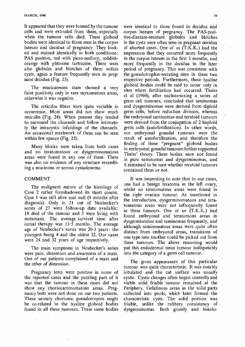
It appeared that they were formed by the tumour cells and were extruded from them, especially when the tumour cells died. These globoid bodies were identical to those seen in the corpus luteum and decidua of pregnancy. They look- ed and stained identically in both conditions: PAS positive, red with picro-mallory, reddish - orange with phloxine tartrazine. There were also globules and blotches of them within cysts, again a feature frequently seen in preg- nant decidua (Fig. 23).
The mucicarmine stain showed a very faint positivity only in rare myxomatous areas, otherwise it was negative.
The reticulin fibres were quite variable in occurrence. Most areas did not show much reticulin (Fig. 24). When present they tended to surround the channels and follow intimate- ly the intracystic infoldings of the channels. An occasional meshwork of them can be seen within few spaces (Fig. 25).
Many blocks were taken from both cases and no teratomatous or dysgerminomatous areas were found in any one of them. There was also no evidence of any structure resembl- ing a mutinous or serous cystadenoma.
COMMENT
The malignant nature of the histology of Case 2 rather foreshadowed its short course. Case 1 was still alive and well (9 months after diagnosis). Only in 21 out of Neubecker's series of 27 were follow-up data available: 18 died of the tumour and 3 were living with metastasis. The average survival time after initial therapy was 13.5 months. The average age of Neubecker's series was 20.3 years: the youngest being 4 and the oldest 32. Our cases were 24 and 32 years of age respectively.
The main symptoms in Neubecker's series were pain, distension and awareness of a mass. One of our patients complained of a mass and the other of distension.
Pregnancy tests were positive in some of the reported cases and the puzzling part of it was that the tumour in these cases did not show any choriocarcinomatous areas. Preg- nancy tests were not done on our two patients. These urinary chorionic gonadotropins might be co -related to the hyaline globoid bodies found in all these tumours. These same bodies
were identical to those found in decidua and corpus luteum of pregnancy. The PAS -posi- tive -diastase -resistant globules and blotches in the cysts were often seen in pregnant decidua of aborted cases. One of us (T.K.K.) had the impression that they occurred more frequently in the corpus luteum in the first 3 months, and more frequently in the decidua in the later period of pregnancy.. This was consistent with the gonadotrophin-secreting sites in these two respective periods. Furthermore, these hyaline globoid bodies could be said to occur only in sites where fertilization had occurred. Theiss et al (1960), after nuclear -sexing a series of germ cell tumours, concluded that seminomas and dysgerminomas were derived from diploid germ cells, before reduction division, whereas the embryonal carcinomas and teratoid tumours were derived from the conjugation of 2 haploid germ cells (autofertilization). In other words, our embryonal gonadal tumours were the result of autofertilization, and therefore the finding of these "pregnant" globoid bodies in embryonal gonadal tumours further supported Theiss' theory. These bodies were not found in pure seminomas and dysgerminomas, and it remained to be seen whether teratoid tumours contained them or not.
It was interesting to note that in our cases, one had a benign teratoma in the left ovary, whilst no teratomatous areas were found in the right ovarian tumour. As mentioned in the introduction, dysgerminomatous and tera- tomatous areas were not infrequently found in these tumours. One of us (T.K.K.) had found embryonal and teratomous areas in dysgerminomas and seminomas frequently, and although seminomatous areas were quite often distinct from embryonal areas, transitions of one type into another could be picked out from these tumours. The above reasoning would put this endodermal sinus tumour indisputably into the category of a germ cell tumour.
The gross appearances of this particular tumour was quite characteristic. It was notably lobulated and the cut surface was usually cystic. Cystic changes often began centrally and viable solid friable tumour remained at the Periphery. Gelatinous areas in the solid parts collected into pools, which later formed the characteristic cysts. The solid portion was friable, unlike the rubbery consistency of dysgerminomas. Both grossly and histolo-

60 SINGAPORE MEDICAL JOURNAL
gically the appearances were akin more to the infantile type of testicular embryonal carcinoma than the solid adult variety.
The so-called glomeruloid bodies were
cross sections of papillary processes, which were due to proliferation of malignant cells
around vascular channels. The resemblance to the foetal glomerulus was deemed to be fortuitous rather than an actual attempt to differentiate into a glomerulus by the tumour.
The fibrillary meshwork both within and without the channels and the flowing of the cellular cytoplasm into this meshwork sup- ported the contention that this tumour was mainly mesoblastic. Some of the thick fibril- lary meshwork stained positively with PTAH and reticulin within and without the channels. They were more frequently outside the chan- nels and were part and parcel of the surroun- ding stromal background. With the existence of numerous capillary tubes, the epithet "endo- mesoblastic" could aptly be applied to this tumour.
Except for that two "acinus -looking epi- thelial" structures, there were no other epithelial or teratomatous areas seen. This put both tumours into a very early stage of embryo - genesis.
SUMMARY
A brief historical survey was made, and 2 cases of embryonal tumour with the endoder- mal sinus pattern were described. The charac- teristic gross pathology was a cystic lobulated tumour. The histopathology was elaborated in great detail, and histological diagnosis was distinguished by the glomeruloid pattern, PAS - positive globules and a mosaic of mesoblastic channels. The concept that this tumour was
of germinal origin was further reinforced by our observations. The presence of the "preg- nant" globoid bodies (invariably found in the active pregnant corpus luteum) lent support to the concept that this tumour arose from autofertilization of the germ cells.
ACKNOWLEDGEMENTS
We wish to thank Dr. Hazel Core of the Havard Medical School for histological help, Mr. T. C. Tan for the photographs, and Mr. R. Nambiar of General Hospital, Singapore, for the second case. We are most grateful to Mrs. T. H. Koh for typing the script.
REFERENCES
I. Huntington, R.W., et al. (1963): "Germinal tum- ours exhibiting the endodermal sinus pattern of Teilum in young children", Cancer, 16, 34-47.
2. Kazancigil, T.R., et al. (1940): "Papillo-endothe- lioma ovarii; Report of 3 cases and discussion of Schiller's "mesonephroma ovarii", Am. J. Cancer, 40, 199-212.
3. Neubecker, R.D., & Breen, J.L. (1962): "Embryo- nal carcinoma of the ovary", Cancer, 15, 546-556.
4. Novak, E., et al. (1954): "Probable mesonephric origin of certain female genital tumours", Am. J. Obstet. & Gynec., 68, 1222-1239.
5. Santesson, L. & Marrubini, G. (1957): "Clinical & Pathological survey of ovarian embryonal carcino- mas, including so-called "mesonephromas" (Schil- ler) or "mesoblastomas" (Teilum) treated at Radiumhemmet", Acta Obstet. et Gynec. Scan- dinay., 36, 399-419.
6. Saphir, O., & Lackner, J.E. (1944): "Adenocar- cinoma with clear cells (hypernephroid) of ovary", Surg. Gynec. & Obst., 79, 539-543.
7. Schiller, W. (1939): "Mesonephroma ovarii", Am. J. Cancer, 35, 1-21.
8. Schiller, W. (1942): "Histogenesis of ovarian mesonephroma", Arch. Path., 33, 443-451.
9. Simard, L.C. (1957): "Polyembryonic embryoma of the ovary of Parthenogenetic origin", Cancer, 10, 215-223.
10. Stowe, L.M. (1955): "On the genesis of the so- called mesonephroma ovarii", Cancer, 8, 446-452.
Il. Teilum, G. (1959): "Endodermal Sinus Tumours of the ovary & testis", Cancer, 12, 1092-1105.
12. Teilum, G. (1950): "Mesonephroma ovarii" (Schil- Iler); Extraembryonic mesoblastoma of germ cell origin in ovary and testis", Acta. Path et Microbiol. Scandinay., 27 :249-261.
13. Theiss, E.A. Ashley, D.J.B. & Mostopi, F.K. (1960): "Nuclear sex of testicular tumours and some related ovarian and extragonadal neoplasms", Cancer, 13, 323-327.