Multiscale modelling and nonlinear simulation of vascular tumour growth
Upload
independentCategory
view
0download
0
JPP 2008, 60: 671–688© 2008 The AuthorsReceived September 21, 2007Accepted March 3, 2008DOI 10.1211/jpp.60.6.0001ISSN 0022-3573
671
Review Article
Tumour and dendrimers: a review on drug delivery aspects
Abhinav Agarwal, Abhay Asthana, Umesh Gupta and Narendra K. Jain
Abstract
Tumour is a morbid state, characterized by spontaneous outgrowth of an abnormal mass of cells.The evolution of tumours is random, disorganized, a condition of numerous mutations. The propertiesare biased and incompletely comprehended. It is a malignant or benign condition that encompassesits own rules of morphogenesis, an immortal state that elucidates different physiology. It is a patho-logical crisis that still haunts the minds of scientists, physicians and patients, a complete cure ofwhich is still a dream to be realized. The unpredictable microenvironment of cancerous cells in all ofits existing forms i.e. leukaemic cells, solid tumours and sarcomas is well documented. This phenomenonexpressed by cancerous sites in the body poses various obstacles towards drug efficacy. Thus, it hasbecome necessary to address briefly the issues relating to tumour physiology, its vasculature andangiogenesis. The information could provide insight towards the development of tumour-targeteddrug delivery. The salient features regarding these have been discussed.
Tumour physiology and vasculature
Under normal conditions, cells reproduce, grow, divide, multiply and eventually undergoapoptosis. This maintains proper balance and functioning of the organs. However, tumourcells are subjected to uncontrolled proliferation of cells, evade apoptosis and hence theydevelop as an abnormal mass of cells that can be life-threatening if untreated at an earlystage. Tumours bear highly irregular chaotic architectural vasculature. ‘The genesis ofblood vessels is an abnormal state’ in tumours as there is abrupt formation of newer vesselsand existing vessels tend to become disorganized. However, ‘the genesis of blood vesselsoccurring elsewhere in the body is not necessarily abnormal’. Such eruptions occur as aresult of genetic aberrations from pre-existing vessels or in a specialized microenvironmentfrom endothelial cell progenitors of bone marrow (Jain et al 1987; Haroon et al 1999;Carmeliet & Jain 2000; Ribbati et al 2001; Peppas & Blanchette 2004; Gjini 2005). Uponundergoing change in the organizational pattern of a gene, the normal cell cycle is disrupted.The gene (proto-oncogene) is thus converted into an oncogene and loses its normal growthregulation. As a consequence of mutations, abnormal growth and proliferation of cells beginalong with the expression of various surface markers and proteins (Table 1), which facilitatetheir growth and supplement the cells with necessary nutrients and oxygen at the expense ofnormal cells. The abnormal signal proteins may induce faster proliferation of cells via tran-scription. Thus, a gene in a normal cell on being converted into an oncogene proliferatesunder the stimuli of these agents and leads to development of tumours (Folkman 2002;Siemann 2006). There is a change in chromosomal structure and the genetic setup begins todevelop in such a way so as to delay senescence. Pericytes are the connective tissues thatgrow around basement membrane. This growth of pericytes around tumours provides themwith adequate protection against tumour targeting. They have been known to facilitatetumours by promoting cell-to-cell attachment and hence their stabilization (Banfi et al 2005;Molema 2005).
Blood from the capillaries may diffuse or drain out into a newer network consisting ofvessels and lymph nodes called the lymphatic system, which circulates like blood in varioustissues and organs. Blood and lymph communicate to each other at lymph nodes and thelymph, via the thoracic vein, finally enters the general circulation. Lymphatic metastasis ischaracteristic of tumours. Metastasis is the movement of tumour cells to other sites and is ahighly hazardous state of the biosystem. Every single cell that moves to another place may
Dr H. S. Gour University, Sagar, M.P., India
Abhinav Agarwal, Abhay Asthana, Umesh Gupta, Narendra K. Jain
Correspondence: Professor N. K. Jain, Pharmaceutics Research Laboratory, Department of Pharmaceutical Sciences, Dr H. S. Gour University, Sagar, M.P., 470003, India. E-mail: [email protected]
JPP60(6).book Page 671 Tuesday, April 29, 2008 3:27 PM

672 Abhinav Agarwal et al
develop into a new tumour, giving rise to various necroticregions. Upon treatment even if a single cell or colony is leftout, it can again lead to an entire tumour (Mareel 2004; Arspe2005). Present cancer therapy should be based on the philosophy,‘even a single cancer cell should not remain untreated in thebody and nothing less than complete elimination of tumourfrom an individual can be accepted’. This forms the objectivebehind chemotherapy. Tumours invade the stroma of anorgan or tissue and metastasize to lymph vessels promotingformation of newer vessels (lymphangiogenesis). Lymph nodesinuses can be the dominant regions for tumour metastasis. Cancergenerally proceeds in the direction of lymph flow, but canalso be in the reverse direction. Tumours may move from onesite to another mostly via lymphatics giving rise to newernecrotic regions. As the cells grow at tumour sites they generatemechanical stress. The compromised vasculature and poorlymphatic drainage create interstitial stress, which hinders theblood supply and obstructs transport of bioactives (Alitalo &Carmeliet 2002; Kaul et al 2003). Irregular development ofbasement membrane, the presence of fenestrae (400–800 nm),widened inter-endothelial junctions, vasodilatation and formationof small blood vessels to nourish the tumour cells makes themmore porous. Hence the permeability of tumour vessels isseveral times higher than normal vessels (Modi et al 2004;Sevenson & Tomalia 2005).
Rapid proliferation of cells develops an oxygen crisis,causing an alteration in the genome of expressing cellsthat exhibit resistance to hypoxia. Survival of tumour cells inanaerobic conditions makes them glycolytic. Further, tumourcells transcribe hypoxic inducible factor (HIF-1) that causeschange in the programme of gene expression. HIF-1 inducespyruvate dehydrogenase kinase (PDK-1), a chemical thatprevents apoptosis (Kim etal 2003; Mennon etal 2003). Normalcells lacking HIF-1 fail to generate PDK-1 and succumb to apop-tosis. Hypoxia induces macrophage infiltration at tumoursites. Macrophages induce formation of new vessels (angio-genesis) via induction of various prognostic mitogens
(Crowther et al 2001) (Figure 1). Another important aspect ofischaemic tumour microenvironment is that the extracellularpH (~7.0 or less than 7.0) (Engin et al 1995) is slightly acidiccompared with the normal tissues (pH~7.4). Although tumourcells may maintain intracellular pH, the acidic extracellularenvironment is an important aspect of mutagenesis. Thisforms the basis for systems having pH-based delivery. Itinduces expression of various signals that lead to oncogenicchanges enabling cells to survive under unfavourable condi-tions. The cells generate various pumps so as to remove intra-cellular H+ ions, preventing intracellular acidification anddeveloping slightly alkaline conditions optimum for cellulargrowth. The growing tumour is a rapidly expanding capillarynetwork that helps to defy the laws of mortality (Kraus &Wolf 1996; Wahl et al 2002).
Angiogenesis
The word angiogenesis elucidates development, growth andexpansion of new vessels, which is necessary for properfunctioning of the body. However, its malfunction may leadto severe pathological crisis such as cancer or degradationof the body. As the angiogenic switch is turned on (Figure 2),it facilitates growth of numerous vessels that supportstumour growth (Ribatti & Presta 2002; Bergers & Benjamin2003).
Gjini (2005) reported a new concept of ‘tumour vasculaturenormalization’ that aimed to carefully monitor the dosageregimen of anti-angiogenic therapy, maintaining proper equi-librium between angiogenic stimulators and inhibitors. Theconcept aimed at the treatment of tumours via inhibiting produc-tion and release of various chemical factors that promotedrapid proliferation of newer vessels, at the same time facilitatingthe production and release of various chemical factors thatpromoted apoptosis. Various factors that play an importantrole in tumour angiogenesis are summarized below.
Vascular endothelial growth factor (VEGF) This is the key factor involved in controlling disposition char-acteristics of cancer. Binding of VEGF to its tyrosine kinasereceptors facilitates triggering of a cascade of events thatleads to transcription, receptor mediated growth and prolifer-ation of cells. Oncogenesis leads to production of VEGF,which is an important mitogen in cancer angiogenesis, and its
Table 1 Surface markers and proteins over-expressed in tumours
Carcinoma Surface markers and proteins
Breast C-myc gene (Peng & Hsu 2001;Aref et al 2001) amplification
MAPK-1 (Arspe 2005) Activator protein-1 (Asthana et al 2005) MLH-1 (Aulenta et al 2003) Colorectal MSH-II (Aulenta et al 2003) Ki-ras2 genes (Azzam & Domb 2004) CD-95 L (Backer et al 2005) Hepatocellular Activator protein-1 (Banfi et al 2005) a-fetoprotein (Barth et al 1994) Lung PIK3CA gene (Barth et al 2004) Laminin-10/11 (Behr 1997) Ki-ras2 genes (Azzam & Domb 2004) hG-CSF (Benns et al 2000) Leukaemic CD-95 L (Berger & Benjmain 2003) Abl kinase (Bhadra et al 2004) CD-44 (Bhadra et al 2003) Ovarian P185 HER2 (Bhadra et al 2003) (P16/P15) (Bhadra et al 2005)
Figure 1 Avascular tumour growth.
Hypoxic tumour
Angiogenesis
Metastasis
JPP60(6).book Page 672 Tuesday, April 29, 2008 3:27 PM
Tumour and dendrimers 673
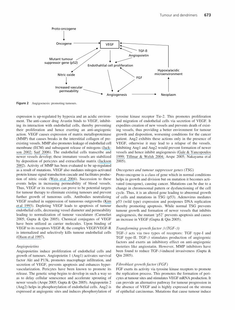
expression is up-regulated by hypoxia and an acidic environ-ment. The anti-cancer drug Avastin binds to VEGF, inhibit-ing its interaction with endothelial cells, thereby preventingtheir proliferation and hence exerting an anti-angiogenicaction. VEGF causes expression of matrix metalloproteinase(MMP) that causes breaks in the interstitial collagen of pre-existing vessels. MMP also promotes leakage of endothelial cellmembrane (ECM) and subsequent release of mitogens (Jack-son 2002; Saif 2006). The endothelial cells transcribe andnewer vessels develop; these immature vessels are stabilizedby deposition of pericytes and extracellular matrix (Jackson2002). Activity of MMP has been evaluated to be up-regulatedas a result of mutations. VEGF also mediates mitogen-activatedprotein kinase signal transduction cascade and facilitates produc-tion of nitric oxide (Weis et al 2004). Succession to theseevents helps in increasing permeability of blood vessels.Thus, VEGF or its receptors can prove to be potential targetsfor tumour therapy to eliminate existing tumours and preventfurther growth of tumorous cells. Antibodies neutralizingVEGF resulted in suppression of tumorous outgrowths (Kimet al 1993). Depleting VEGF leads to apoptosis of tumourendothelial cells, decreasing vessel diameter and permeabilityleading to normalization of tumour vasculature (Carmeliet2005; Gupta & Qin 2005). Chemical conjugates of VEGFhave been utilized as carrier molecules. Upon binding ofVEGF to its receptors VEGF-R, the complex VEGF/VEGF-Ris internalized and selectively kills tumour endothelial cells(Olson et al 1997).
Angiopoietins Angiopoietins induce proliferation of endothelial cells andgrowth of tumours. Angiopoietin 1 (Ang1) activates survivalfactor Akt and P13k, promotes macrophage infiltration, andsecretion of VEGF, prevents apoptosis and enhances hyper-vascularization. Pericytes have been known to promote itsrelease. The genetic setup begins to develop in such a way soas to delay cellular senescence and accelerate sprouting ofnewer vessels (Arspe 2005; Gupta & Qin 2005). Angiopoietin 2(Ang2) helps in phosphorylation of endothelial cells. Ang2 isexpressed at angiogenic sites and induces phosphorylation of
tyrosine kinase receptor Tie-2. This promotes proliferationand migration of endothelial cells via secretion of VEGF. Itexpedites creation of new vessels and prevents death of exist-ing vessels, thus providing a better environment for tumourgrowth and disposition, worsening conditions for the cancerpatient. Ang2 exhibits these actions only in the presence ofVEGF, otherwise it may lead to a relapse of the vessels.Inhibiting Ang1 and Ang2 would prevent formation of newervessels and hence inhibit angiogenesis (Gale & Yancopoulos1999; Tillmar & Welsh 2004; Arspe 2005; Nakayama et al2005).
Oncogenes and tumour suppressor genes (TSG) Proto-oncogene is a class of gene which in normal conditionshelps in growth and division but on mutation it becomes acti-vated (oncogene), causing cancer. Mutations can be due to achange in chromosomal pattern or dysfunctioning of the cellcycle. Thus, it is an altered gene leading to abnormal growthof cells and mutations in TSG (p53). Adenovirus mediatesp53 (wild type) expression and postpones DNA replicationthereby promoting apoptosis. While normal TSG preventstumour growth and formation of newer vessels that inhibitsangiogenesis, the mutant ‘p52’ prevents apoptosis and causesan increase in VEGF (Gupta & Qin 2005).
Transforming growth factor b (TGF-b) TGF-b acts via two types of receptors: TGF type-I andTGF type-II. TGF-b stimulates production of angiogenicfactors and exerts an inhibitory effect on anti-angiogenicmoieties like angiostatin. However, MMP inhibitors havebeen found to reduce TGF-b-induced invasiveness (Gupta &Qin 2005).
Fibroblast growth factor (FGF) FGF exerts its activity via tyrosine kinase receptors to promotethe replication process. This promotes the formation of peri-cytes at tumour sites and stimulates VEGF mRNA production. Itcan provide an alternative pathway for tumour progression inthe absence of VEGF and is highly expressed on the stromaof epithelial carcinomas. Mutations that cause tumour induce
Figure 2 Angigoenesis: promoting tumours.
Angiopoietin
Neoplasm
Hypoxia/acidicmicroenvironment
Endothahlial cell proliferation
Angiogenesis
FGF
TGF-ß
VEGF
Nitric oxide
Increased vascularpermeability
Proto-oncogene
Oncogene
Mutant tumoursuppressor gene (p52)
JPP60(6).book Page 673 Tuesday, April 29, 2008 3:27 PM

674 Abhinav Agarwal et al
genetic signals that help in expression of FGF. It providessynergistic effects for tumour growth and metastasis. Mono-clonal antibodies (mAbs) that target Flk-1 receptors promoteFGF-mediated tumour regression (Arspe 2005; Gupta & Qin2005).
Approaches for tumour specific delivery
It is clear from the previous section that because of the associatedirregularities concerning tumour, its growth within the bodyis widespread. Thus, providing justification on its proposedtherapy becomes a very challenging task. However, there area few strategies (Figure 3) that can be focused on to proceedin the elimination of the tumour. A large number of carriershave been reported using the fundamentals of one or more ofthese approaches, which have proved to be quite successful(Table 2).
Intracellular communication and tumour specific delivery The intrinsic activity of the drug carrier system should besuch that it causes intra-tumoral delivery of drug, thus givinga high concentration of drug at the target site and minimizingthe possible side effects, reducing dosing frequency andincreasing patient compliance. The active moiety or the drugcoupled to the carrier can access the tumour tissues or cellsvia various mechanisms such as simple diffusion, facilitateddiffusion, enhanced permeation and retention (EPR) effect,active transport or specific receptor-mediated endocytosis(RME) (Peppas & Blanchette 2004). The drug needs to trafficvarious membrane barriers to reach the tumour tissue. Todeliver the drug within the tumour site it is required that thebioactive be either attached to various linkers that cleave inthe acidic environment of the tumour or encapsulated withinsome vesicular carrier, or coupled via such bond, which
Figure 3 Targeting tumour: active and passive approaches.
EPR
Ligands
Endosomal escape
SizeMolecular
weight
ChargeLong
circulating
pH sensitive
Avoiding MPS
ProdrugReceptormediated
Passive targeting
Activetargeting
Neoplastic cells
Table 2 Various carrier systems used in tumour therapy
Carrier systems Formulation Remarks
Nanoparticles (Carmeliet & Jain 2000) PEG modified gelatin nanoparticlesloaded with DNA
Intracellular trafficking to tumours and nuclear delivery
Liposomes (Fischer et al 1999) FA-PEG-DOPE modified liposomes containing DNA
Efficient for pH sensitive endosomal DNA delivery
HPMA polymer (Folkman 2002) HPMA-(Gly-Phe-Leu-Gly) linker-TNP 470 complex
Selective accumulation in tumour vasculature and prevents TNP-470 from crossing blood–brain barrier
Liposomes (Friend & Pangburn 1987) Folate conjugated liposomes Efficient delivery of cytotoxic drugto tumours
Nanospheres (Fujita et al 1997) Gelatin nanospheres complexed to DNA
Efficient vehicle for gene delivery
Nanoparticles (Gale & Yancopoulos 1999) RGD-modified nanoparticles Selective localization of drugs in tumours
Nanoparticles (Galle & Krammer 1998) Dextran–drug conjugate incorporated in chitosan nanoparticles
Efficient vehicles to suppress tumour volume
Nanoparticles (Gaur et al 2000) PLGA nanoparticle conjugated with anti-cancer drug
High-loading efficiency and comparable activity to official formulations
Microspheres (Gillies & Frechet 2004) Alginate microspheres containing naked DNA
Gene delivery for tumours
Polylysine (Gillies & Frecht 2005) Transferrin conjugated polylysine Gene transfer agent
JPP60(6).book Page 674 Tuesday, April 29, 2008 3:27 PM

Tumour and dendrimers 675
cleaves in the tumour microenvironment. The strategy mayapply a pro-drug concept or an active targeting approach, sothat the drug is converted into an inactive moiety or attachedto a linker that liberates drug only on encountering tumourand not the normal cells. This exploits the pathophysiologicaldifference between normal and malignant cells, such as acidicpH and expression of various tumour specific antigens (Patriet al 2002a; Gillies & Frechet 2004) (Figure 4). Micelles ofPLLA-b-PEO polysulfadimethoxine are pH sensitive due toacidic sulfonamide groups and have been reported to be use-ful anti-cancer agents (Lee et al 2003).
Drug or drug carrier systems, once internalized via phago-cytosis, are encapsulated in endosomes when acidic pH (5.5–6.5) and endolytic enzymes (proteases, lipase, glycolases) setto release the drug, which is metabolized before eliciting thedesired action. In this regard positively-charged carriers havebeen proved to undergo endosomal escape consequent to desta-bilizing endosomal membrane, preventing release of drug inthe endosomes (Tkachenko et al 2004). Christiano (1998)reported the use of synthetic viruses and molecular conjugatesas non-viral agents for endosomal escape. Delivery of protein/DNA polyplex along with adenovirus led to the adenovirusaccompanying the polyplex entering the same endosome byreceptor-mediated endocytosis, and subsequent lysis of theendosome by virus, facilitating endosomal release of DNA.
Enhanced permeation and retention (EPR) effect Blood vessels in tumours bear leaky vasculature due to angio-genesis, presence of fenestrae (400–800 nm) and widenedinter-endothelial junctions (Sevenson & Tomalia 2005). Thisprovides a good opportunity for intracellular access of drugs.Although particles up to a size range of 800 nm will enter thesevessels, they will eventually escape the capillaries. However,the macromolecules within this size range will enter these ves-sels, where they will be retained or lodged in capillaries due totheir relatively ‘high molecular weight’. Thus leaky tumourvasculature, as well as the poor lymphatic drainage, providesenhanced residence time for macromolecules in tumours. Thisphenomenon is termed the EPR effect. The small moleculesmay easily escape the capillaries due to insufficient pore cut-off size (400–800 nm); the macromolecules may prove to be an
engineered design for being effectively localized at the site pre-venting their escape (Kaul et al 2003). These macromoleculesmay be taken inside as a consequence of extravasation of thevesicles, where enzymes work in co-ordination to release thedrug (Modi et al 2004). N-(2-Hydroxy propyl) methacrylamide(HPMA) copolymer conjugated to doxorubicin has beenshown to reduce cardiotoxicity by 4–5-times due to efficientlocalization mediated via the EPR effect (Duncan et al 2005).Thus, a drug conjugated to a macromolecule may lead to a farbetter distribution of the drug (Friend & Pangburn 1987) andEPR provides a passive transport mechanism for the entry ofdrug into malignant cells (Kaul et al 2003). Macromoleculessuch as albumins (Lowenthal et al 2005), globulins (Selby1990) and synthetic polymer accumulate in the tumour tissuesbecause these tissues have a vascular network characterized byboth enhanced permeability of the neovasculature and a lack ofthe lymphatic recovery system (Matsumura & Maeda 1986).Also, increasing molecular weight of carriers may prevent theirelimination by the kidneys (Michallet et al 2004). Thus, theycirculate in the body for a prolonged duration, providing sus-tained delivery until they are eventually degraded or metabo-lized by the liver (Orive et al 2005) (Figure 5).
Avoiding mononuclear phagocytic system (MPS) Uptake of drug by liver, spleen and macrophages is a greatsetback in delivery of drugs to tumour sites. Hydrophobicparticles have a greater tendency to be eliminated; largersized particles are more prone to reticulo-endothelial system(RES) uptake compared with smaller size particles such asnanometric particles. They may be opsonized by serum proteinsand then cleared. On the other hand, carriers or active moie-ties possessing hydrophilic surfaces prevent their recognitionby the MPS. Copolymer micellar carriers, having a particle sizerange from 20 to 100 nm, are small enough to avoid RESuptake (Kwon & Okano 1996). Biodistribution studies ofhydrophilic nanoparticles prepared from poly(vinyl pyrol-lidone) have been shown to bypass uptake by liver and spleen(Gaur et al 2000). Coating with specific barriers that promotestearic hindrance for drug uptake by liver and spleen is yetanother strategy to develop long circulating drug deliveryunits avoiding MPS clearance. Preparing water soluble derivatives
Figure 4 Site-specific delivery: ligand–receptor interaction.
Targetingmoiety
Receptors on the cell
Receptor–mediatedendocytosis
LysosomeEndosome
Endo-lysosomeformation
Release of free drugat the target site
Ligand–receptorinteraction
Disruption ofendo-lysosome
Drug–carriercomplex
JPP60(6).book Page 675 Tuesday, April 29, 2008 3:27 PM

676 Abhinav Agarwal et al
and a reduction in particle size also provide suitable strategies toescape the RES (Peppas & Blanchette 2004); reported use ofpolyethylene glycol (PEG; macrogol) (fluoroalkylated PEG)as surfactant (Peng & Hsu 2001) has been shown to preventRES uptake by increasing the hydrophilic character; nega-tively-charged carriers are more rapidly opsonized than posi-tively-charged ones. Uptake of drugs by the RES followsMichaelis–Menten kinetics (Wood & Thakker 1982), there-fore the critical point of saturation concentration specifies thelimits for doses. Increasing the dose of moieties may preventescape from the MPS due to saturation of uptake sites, buttherapeutic indices, unwanted side effects and dose-relateddrug toxicity need to be considered (Davis 2000).
Gene therapy This can be considered a new visionary for the treatment ofimmunogenic diseases. Tumours are caused as a consequenceof mutations in the genome by, addition, deletion or change inchromosomal pattern. Suitable vectors such as anionic lipo-somes (Rosenberg et al 1990), poly-L-lysine (Ledley 1995),polyethylene oxide (PEO) (Benns et al 2000), retrovirus(Fischer et al 1999) etc. have been reported to access thenucleus and rectify the condition (Figure 6). Viral and non-viral vectors can be aimed at treating this condition. Viralvectors can be immunogenic; the random insertion in the hostgenome can possibly cause oncogene activation, tumour sup-pressor gene inactivation or nonspecific inflammation (Bordi-gnon et al 1995), also the manufacturing of viral vectors isdifficult and they have limited capacity to carry DNA. Thus,the safety concern may circumvent the use of non-viral vec-tors. However, non-viral vectors also possess the major draw-back of having inefficient and comparatively low transfectionability (Abaan & Criss 2002; Azzam & Domb 2004; Khalilet al 2006). Recognizing the gene (Patri et al 2002b) anddelivery of a specific gene into the target site can play acrucial role in life-threatening diseases (Wang et al 1996;Eliyahu et al 2005).
Targeting tumour angiogenesis Tumour angiogenesis is a consequence of disturbance in theactivity of angiogenic promoters and angiogenic inhibitors.The strategy is based on two approaches, inhibition of ang-iogenic factors and their receptors, or anti-vascular therapy(Siemann 2006). It aims to prevent formation of new vesselsand damage the existing ones. Angiogenesis inhibitors seemto interrupt endothelial cell function. The strategy up-regulatesthe activity and amount of angiogenic inhibitors, preventing
endothelial cells from giving rise to new vessels. Endothelialcells can be better subjected to drug targeting than tumourcells as they are less prone to become resistant to drug ther-apy. Destroying endothelial cells employs only a few cells fordeath of a large tumour area. Gaining knowledge of antigenicmarkers on the surface of tumorous endothelial cells can ena-ble choice of suitable ligands. As a result tumour vesselsrelapse and apoptosis of the endothelial cell is encountered(Peppas & Blanchette 2004; Arspe 2005). Combretastatins(CA4P), a class of anti-cancer drug, have been found to alterthe shape of endothelial cells consequent to membrane bleb-bing causing necrosis of tumour vessels (Tozer et al 2001).Angiogenic inhibitor o-(chloracetyl-carbamoyl) fumagillol(TNP-470) conjugated to HPMA promoted regression ofLewis lung carcinoma by effectively inhibiting endothelialcell production and proliferation (Folkman 2002; Fainaro et al2004).
This approach extends to delivery of agents that preventsynthesis and release of inhibitors for VEGF, FGF andMMP. Targeting VEGF inhibits supply of nutrients and oxygento tumour cells and prevents further growth. Down-regulatingtumour receptors hinders tumour growth and promotespericyte destruction. This prevents significant side effectsas compared with other strategies. However, it is a difficultconcept due to variation in temporal and spatial expressionof various growth factors (Ribbati et al 2001; Fainaro et al2004).
Dendrimer-based controlled/targeted anti-tumour delivery and diagnosis
In just a couple of decades dendrimers have attained recog-nition among the few leading systems utilized as nanoscaleunits or carriers of active moieties such as gene, oligonu-cleotides (ODNs), DNA and bioactives for wide-rangingapplications.
Dendrimers are highly branched polymeric nano-carrierssynthesized in a reiterative fashion. They are the most recentuni-molecular, polymeric, non-immunogenic system that hasattracted the scientific community worldwide for tumour specificdelivery. These nanoparticulate carrier systems are globular
Figure 5 Enhanced permeation and retention.
Lower permeability ofnormal vessels preventsaccess of macromoleculardrugs
Tumour vessels permitmacromolecular drugsto enter these vessels
Figure 6 PLL and PEO (non-viral vectors).
NH
O
O
OPoly ethylene oxide (PEO)
Poly-L-lysine (PLL)
O
O
O
O
O
NH NH
NHNH CO CONHCO
JPP60(6).book Page 676 Tuesday, April 29, 2008 3:27 PM

Tumour and dendrimers 677
in shape, monodispersive in nature, and have unique organi-zation and highly-controlled architecture. The synthesis can beoptimized to control their size, properties, composition and reac-tivity. However, a number of repetitive synthesis steps posesthe greatest challenge in large-scale production of dendrimers(Holister et al 2003; Majoros et al 2004).
Dendrimers consist of an initiator core, interior generationscomposed of repeating units attached to the initiator core, andterminal functionality attached to the outermost generation(Jain & Khopade 2001). Based on the cores, repeating unitsand surface functionalities, a number of dendrimers havebeen reported: poly propylene imine (PPI) dendrimer; polyamidoamine (PAMAM) dendrimer; poly-L-lysine (PLL) den-drimer; melamine dendrimer; and citric acid dendrimer etc.
Characterization of dendrimers, in-depth analysis andimperfections can be assessed by: infrared spectrometry (IR),nuclear magnetic resonance (NMR), matrix-associated laserdesorption ionization-time of flight (MALDI-TOF), electronspray ionization (ESI), vapour phase osmometry (VPO), laserlight scattering (LLS) and sodium dodecyl sulfate-poly acry-lamide gel electrophoresis (SDS-PAGE) etc. (Erichmann et al2000; Jain & Khopade 2001; Shi et al 2005). Dendrimerscould prove to be efficient archers in the battle against cancer.The terminal groups can be fabricated in such a way so as toprovide specific charge and affinity to bind the drug andrelease it at the desired pH or on encountering a specificenzyme or microenvironment. The exo-groups can be embed-ded to provide attachment to a large number of drug moieties,ligands, and mAbs for specific delivery. Also, dendrimers canbe tailored in such a way that few branches uphold the drugand remaining ones can be engineered with targeting moietyor ligands (Dian 2002; Kannan et al 2004; Khandare et al2005) (Figure 7). The presence of multiple groups providesthem with the inherent capability of targeting the drugs totumour sites, to deliver therapeutic payload of drug as well asa marker for diagnostic imaging of the tumour regression(Kolhe et al 2006). Shukla et al (2006) synthesized Alex-aFluor (AF)-tagged G5 PAMAM dendrimers conjugated toanti-HER2 monoclonal antibody (G5-AF-HER2). In-vitrostudies were performed on MCA-207 control and MCA-207HER2 cells. Flow cytometric studies revealed the uptake ofconjugate by HER2 expressing cells, while no such affinitywas found for MCA-207 control cells that did not express
HER2 (Shukla et al 2006). Similarly, Thomas et al (2004) tai-lored G5 PAMAM dendrimers with 60 bca and J591 antibod-ies that bound to CD14 and prostate specific membraneantigen (PSMA), respectively, and labelled with fluoresceinisothiocyanate (G5-FI-60B, G5-FI-PA). Flow cytometric andconfocal studies on HL60 (from human myeloblastic leukae-mia) and LNCaP (from prostate cancer) cell lines that expressCD-14 and PSMA antigen showed the receptor specificity asthe conjugates bound to specific antigen expressing cells andthe control G5-FI lacked specific affinity for any of the celllines (Thomas et al 2004). With the rise in subsequent genera-tions, an exponential rise in density of functional groups pro-vides increased sites of attachment (poly-valency) (Horanet al 1999; Liang et al 2000). As the size increases with thegeneration there is also an increase in encapsulation effi-ciency. Significantly PAMAM dendrimers have a size rangefrom 2.3 nm in G-2 to 5.3 nm in G-5 (Zeng & Zimmerman1997). In this regard dendrimers can prove to be as importantcarriers for the delivery of anti-cancer drugs. Various poly-mers and other hyperbranched structures have randomly dis-tributed functional groups, polydispersive nature, nocharacteristic shape, are coiled in on themselves and lack uni-form molecular weight distribution. Various carrier systemssuch as micro-emulsions tend to be unstable, whereas den-drimers are highly stable carriers and can be stored for longerperiods. Also, the exceptionally high drug loading capacity ofdendrimers adds to their properties and ultimately provides agreater accumulation of drug at tumour site (Asthana et al2005). Certain large sized liposomes and microspheres areunable to enter tumorous vasculature and the presence ofliposomes at the tumour site does not ensure the delivery ofthe drug, and the threat of bypassing the site cannot be ruledout (Mastrobattista et al 1999).
One advantage of macromolecular drug delivery vehicles isderived from the nature of the tumour tissue vasculature (Maedaetal 1992; Ambade etal 2005). The nanometric size of dendrim-ers is beneficial for their entry into hyper-permeable vasculature,while their high molecular weight and lymphatic dysfunctioningcauses their localization as well as preventing escape via theEPR effect (Klajnert & Bryszewska 2000; Gillies & Frechet2005). The unique core shell design of dendrimers helps in theincorporation of both hydrophilic and hydrophobic moieties andthe presence of hydrophilic terminal surface groups preventstheir opsonization or their recognition by macrophages and elim-ination by the RES (Sevenson & Tomalia 2005). Their surfacescan be elegantly modified so that the drugs can be physicallyentrapped, encapsulated, or conjugated by covalent bonds,hydrogen bonds, or ionic interactions (Dian 2002; Kannan etal2004) (Figure 8).
Dendrimers cross the highly permeable tumour membrane(the cell wall of those cells that are tumorous in nature) byendocytosis, paracellular transport and extravasation. Bindingthe dendrimers with other high molecular weight moietiesmight increase their circulation time as these units bypass theglomerular filtration limit, thereby decreasing renal elimina-tion. With an increase in dendrimer generation there is anincrease in molecular weight of dendrimers, thus its hydrody-namic volume increases causing slower elimination andlonger circulation time (Chen et al 2000; Piehler et al 2000;Lin et al 2004). Use of dendrimers in tumours can be as
Figure 7 Dendrimers have precisely placed functional groups that canbe simultaneously conjugated with fluorescent probe, imaging agent andligand.
Fluorescentprobe
Drug
Imagingagent
H2N H2N
H2NH2N
H2N
H2N
H2N
H2N
NH2NH2
NH2
NH2
NH2
NH2
NH2H2N
N
N
NN
N
N
N
N N N
NN
NN
Ligand
JPP60(6).book Page 677 Tuesday, April 29, 2008 3:27 PM

678 Abhinav Agarwal et al
potential non-viral vectors for gene therapy, taking advantageof controlled immunogenicity and transfection capability ofdendrimers (Mislick et al 2000). Cationic dendrimers can beused to deliver DNA into cells effectively. The encapsulatedDNA is also prevented from being recognized by enzymesystems that degrade it. The interaction of amine groups ofPAMAM dendrimers with phosphate groups of DNA elicitstheir transcription property. Dendrimers aid in crossing thebiological barriers, which helps to increase transfection effi-ciency (Cloninger 2002; Aulenta et al 2003).
It is also important to note that dendrimers bear the capa-bility of solubilizing some insoluble anti-cancer drugs such aspaclitaxel (Ooya et al 2003), 5-fluorouracil and methotrexate.The increased solubility leads to increased loading, and bytailoring the surface of dendrimers targeting can be achieved(Chauhan et al 2003; Gupta et al 2006a) (e.g. methotrexate,doxorubicin HCl). This helps to prevent side effects andincrease the therapeutic effect of the drug (Kojima et al2000; Gupta et al 2006b). Melamine-based dendrimers modi-fied on their periphery were loaded with methotrexate and6-mercaptopurine. When administered to C3H mice intraperi-toneally, the drug–dendrimer formulation was found to exertreduced hepatotoxic action as compared with free drug. Alaninetransaminase (ALT) level was used to probe liver damage.Forty-eight hours after dosing it was found that the ALT levelsof drug-loaded dendrimers were 27% (methotrexate) and 36%(6-mercaptopurine) lower than those of animals treated withthe drug alone (Neerman et al 2004).
Cancer therapy necessitates targeting to a specific onco-genic cellular mass called a tumour. Different strategies havebeen reported earlier to target tumours such as pH-based drugrelease, ligand attached to dendrimer periphery etc. The useof dendritic polymers as drug delivery vectors and as pharma-ceutical actives has received increasing interest (McCarthy et al2005). Potentially numerous dendrimers can be designed toovercome pathological as well as physiological barriers. Den-drimers can be aimed for selective and effective localization ofpharmacologically active therapeutics at pre-identified sitesand restricting access to non-target sites. Dendrimers have been
reported for their ability to transfect cells and can be designedas carriers for anti-cancer agents to tumour sites. Dendrimershave also been known to sustain drug release (e.g. methotrex-ate, doxorubicin HCl) and therefore increase the therapeuticeffect of the drug (Kojima etal 2000; Quintana et al 2002;Kukowska Latallo et al 2005).
Folate conjugated dendrimers Conjugating drug-loaded dendrimer to a specific targetingligand can be an effective method to deliver drug at thedesired site. Ligands possess some functional moieties thatare recognized by cell surface receptors that bear affinity tobind them. The ligand is then taken internally to the site ofaction to release the therapeutic moiety attached to it. Thus,these ligands can serve as potent stimulators or inhibitors of apharmacological, pathological response. Folic acid (FA) is avital nutrient for the growth of cancer cells and can be used asa ligand due to high binding affinity and non-immunogenicity.Cell surface receptors of folic acid are over-expressed onmany types of cancer cells. Thus folate-conjugated dendrimerscan be effective anti-cancer agents having high affinity forthese cancer cells (Quintana etal 2002). Methotrexate, a struc-tural analogue of folic acid, can be incorporated into dendrim-ers to be effectively delivered to the cancer tissues withreduced toxicity. To gain access to the tumour surface, via thecirculatory system, the drug needs to be hydrophilic, and topenetrate the cell membrane the lipid solubility must be high.The simultaneous occurrence of both properties in the drug isdifficult. Folate-conjugated dendrimers enter the cell byRME, bypassing cellular barriers, allowing hydrophilic drugsto enter cancer cells of tumour xenografts (Leamon & Low1994; Lee et al 1996; Caliceti et al 2003). Folate conjugateshave been found to be quite stable even after endocytosis intumours and remain functional for quite some time. Bindingof folic acid to folate receptors (FR) is pH sensitive and oncethey reach acidic endocytic vesicles release the conjugated moi-ety, and the folate receptors move back to the tumour cell sur-face. Conjugation of dendrimers to folic acid has been widelyexplored. Biodistribution studies show encapsulating methotrexate
Figure 8 Dendrimers; (A) physical entrapment of drugs, (B) conjugation of drugs, (C) poly-valency and (D) resist RES uptake.
Interior functionalities(endo-receptors)
A
BC
D
Exterior functionalities(exo-receptors)
Hydrophilic exterior
Numerous sites of attachmentTherapeutic moieties
JPP60(6).book Page 678 Tuesday, April 29, 2008 3:27 PM

Tumour and dendrimers 679
into folate-conjugated dendrimers has better tumour targetingpotential with minimum side effects as compared with freedrug, regressing tumours to a greater degree; similar resultswere obtained with doxorubicin (Neerman 2006; Reddy &Low 1998) (Figure 9).
Glyco-dendrimers The unusual course of glycosylation on the cancer cell surfaceover-expresses specific antigens that can serve as potentialtargets for immune recognition through lectin-like receptorsexpressed on the surface of immune cells, that include naturalkiller (NK) cells, CD8+ and CD4+ lymphocytes (Bezouskaet al 1994; Pospisil et al 1995). The glyco-dendrimers find useas antigens, raising mAbs against tumour tissues selectively.The dendrimer formulation can be processed in three ways:fully carbohydrate-coated dendrimers; dendrimers with car-bohydrate moieties at periphery; and carbohydrate-centreddendrimers (Roy 1996). Surface modifications of PPI, PAMAM,and PLL dendrimers to glyco-dendrimers have shown theirtumour-targeting potential (Lindhorst & Kieburg 1995; Glabuset al l996). These carriers closely resemble the natural carbo-hydrate ligands. The carbohydrate-coated system adds toselectively localizing the drug at the desired site. The macro-molecular system provides longer circulation and precisedelivery and seems to surpass various ligand-binding systems.Weak interaction chemistry between carbohydrate and receptorproteins needs a large amount of carbohydrate molecules toattain significant binding and elicit the desired effect. Glyco-dendrimers provide enhanced interaction between endogenousproteins and carbohydrates. The glyco-dendrimers can elicittwofold actions: they can themselves act as therapeutic agent;and act as vehicles for an active moiety. With inherent modi-fications and fine-tuning they can be used in gene therapy (Davis& Robinson 2002; Wang et al 2002). They can be modified tointeract with asialoglycoprotein receptors present on hepato-carcinoma cell surface. T-cell antigen GalNAc has been attachedto glyco-dendrimers and has been proved to be directed totumours. RME provides intracellular trafficking of these den-drimers and they have been found to be highly effectiveagainst colorectal carcinomas, melanomas and breast tumours(Vannucci et al 1994; Roy & Baek 2002).
PEGylated dendrimers PEG has a polyether skeleton, which is non-toxic andbypasses non-specific recognition from macrophages, proteinadsorption and the MPS system, as a result of increased
stearic hindrance, shielding positive charge and increasedhydrophillicity (Gref et al 1995). PEGylation of drugs alsoaids the sustained and controlled delivery properties, helpingdelivery of potent drugs of low half-life. PEG-coated den-drimers can also be used to deliver proteins, as in the case ofleukaemia, increasing their plasma half-life, stability, reducing thefrequency of dosing, potentially reducing hypersensitivityreactions, and increasing tumour selective uptake (Yang et al2004). Also, these provide scope as biocompatible dendrim-ers, reducing cytotoxicity and immunogenicity, a commondrawback in PAMAM and PPI dendrimers. These systemsalter pharmacokinetics of the drug increasing blood circula-tion half-life, sustaining delivery and reducing burst release.Increase in molecular weight provides EPR effect once thesystem enters the tumour via extravasation (Davis 2000;Modi et al 2004). The dendrimers have enhanced loadingcapability and greater disposition of drug at the tumour site.Kojima et al (2000) reported the effect of varying chain lengthof PEG on solubility. MPEG 550 G-3, MPEG 2000 G-3,MPEG 550 G-4 and MPEG 2000 G-4 PAMAM dendrimersencapsulating methotrexate and doxorubicin were evaluated.An increase in encapsulation efficiency with increase in chainlength and dendrimer generation was predicted (Figure 10).Similarly, Yang et al (2004) coupled PEG with molecularweights 750, 2000 and 5000 to StarburstTM G3.0 PAMAM den-drimers to solubilize the hydrophobic compound, pyrene.They demonstrated that micelles tended to dissolve morepyrene as compared with free pyrene in water.
Peptide dendrimers These consist of amino acids acting as a core and the terminalfunctionalities can be either of amino-acid group or a peptidemoiety, and possess highly controlled architecture, size andcomposition. Peptides have shown affinity to tumour cells,inhibiting endothelial cells and triggering their apoptosis. Dox-orubicin HCl conjugated to peptides showed increased tumour
Figure 9 Dendrimer branching structure drugs and other moietiesattached.
Dendrimer
Fluorescein(imaging)
Folic acid(ligand)
Methotrexate(drug)
Figure 10 PEGylated dendrimer.
N N N
NN
N
NNN
N
NN N
N
NNN
N
N
NN
N
N
OH–2HC[–CH2–O–CH2–]nCH2–OH
NN
N
N
NN
N
JPP60(6).book Page 679 Tuesday, April 29, 2008 3:27 PM

680 Abhinav Agarwal et al
regression. Peptide dendrimers are a novel development in pol-ymer science; these can mimic as proteinomimetics and finduse in biomedical application. L-Lysine dendrimers can be usedas molecular inhibitors for various angiogenic factors and canwork as multiple antigen peptides to stimulate immuneresponse generating antibodies against tumours (Bhadra et al2005; Gillies & Frechet 2005). Peptide dendrimers can altercell–cell interactions and cellular adhesion and they can befashioned at their branching points (multivalency) as genedelivery carriers to enhance transfection capability. The pep-tide dendrimers have affinity for integrin receptors, which is aprotein controlling cell–cell interaction in cancer cells. Thedendrimer can prove to be efficient in non-covalent binding ofDNA with site-specific cleavage. Non-covalent delivery canprevent metabolism mediated via enzyme nuclease, when com-pared with DNA delivered by other means, and helps crossingbiological membranes more efficiently. Thus it can be a prom-ising approach in DNA nanotech providing greater specificityand selectivity. Melamine dendrimers are potentially a newerclass of peptide polymers that can be meticulously fabricatedand explored for anti-tumour properties (Saddler & Tam 2002;Bhadra et al 2004; Fairman & Akerfeldt 2005) (Figure 11).
pH-sensitive dendrimers The dendrimers are large enough to enter normal vessels butare of optimal size to enter the more porous tumour. The den-drimers can be formulated to make them pH sensitive. Thechange in hydrodynamic radii and conformation with pH canbe an important concept to sustain and control drug release.Various dendrimers are sensitive to pH. At normal physiolog-ical pH (i.e. pH 7.4) the terminal amine groups in PPI den-drimers are not protonated and the branches converge to thecentral core. However, once engrossing the tumour site andentering the tumour vessels the dendrimers are subjected tolower pH; the groups at the exterior are protonated and repeleach other, thus undergoing a change in 3-D conformation,moving branches outward and depositing the drug at thetumour site. This prevents the release of high potency drug atnon-specific sites, preventing toxic effects and unwantedallergies (Patri et al 2002a; Gillies & Frechet 2004; Sevenson& Tomalia 2005).
pH-sensitive dendrimers can be fabricated by tailoringfunctional groups at the surface of dendrimers. This can bedone by incorporating specific groups such as amine (NH2)group, as in the case of PAMAM and PPI dendrimers, at thesurface. The group protonates and becomes charged at lowerpH causing structural changes in the nanostructure and releas-ing the drug (Zeng & Zimmerman 1997). Sideratou et al
(2000) studied the pH-dependent release of pyrene fromquaternized PPI dendrimers. Dendrimers having terminal −COOH moieties are less cytotoxic and hence cause sideeffects to a lesser degree. Acidic moieties such as the carboxy-lic acid (−COOH) group in citric acid dendrimers aggregate atlower pH and are charged only at basic pH (Namazi & Adeli2005). The pH of physiological body fluids is 7.4, which canact as a buffer and maintain this pH. However, the intracellularpH at the tumour site is acidic, thus the dendrimers can be sodesigned to release the drug specifically on encountering thedesired pH (Vasir et al 2005). Ester-terminated dendrimers arenon-toxic, biocompatible and have shown tumour-selectiveaffinity. Peptides such as L-lysine are also pH sensitive andcharged at lower pH. Thus peptide-based dendrimers havepotential for pH-sensitive delivery. Tailoring the surface withacetic-anhydride prevents non-specific interaction and can beattached to ligand to direct dendrimers to neoplastic cells ofprostate. Also a linkage or spacer can be attached betweendendrimer and drug that is pH sensitive. Thus hydrazone link-age and aconityl linkage can be used that are acid labile. Theamide and ester bonds can be developed that undergo hydroly-sis at lower pH (vanHest et al 1995).
Boron neutron capture therapy (BNCT) Dendrimers are one of the most attractive polymers that havebeen used as boron carriers due to their well-defined structureand large number of reactive terminal groups, and can bindup to 1000 boron atoms per molecule of dendrimer (Wu et al2006). Although the mAbs can be used as carriers of boro-nated compounds, lower loading capacity has been a majorlimitation. As a matter of fact, a large number of 10B atoms(109) must be delivered per tumour cell (Alam et al 1989) andhence polyhedral cage-like structures of borane anions areneeded to be linked to mAbs. However, the attempt reducesthe solubility of the formulation, which ultimately leads to itsprecipitation or could result in decreased/diminished immu-nogenicity of the compound (Sneath 1976). However, mAbcan be conjugated to a boronated dendrimer. This would pro-mote site-specific delivery and prevent random distribution(Wu et al 2006). Backer et al (2005) reacted 5.0 G PAMAMdendrimers with decaborate molecules to produce a macro-molecule with 1050–1100 boron atoms per dendrimer. Thiswas conjugated to thiol groups of VEGF (a 121-amino-acidisoform of human VEGF with NH2-terminal Hu-tag (a 1–15-amino-acid fragment of human RNase I)) at a 4:1 molarratio using the hetero-bifunctional cross-linker sulfo-LC-SPDP. The dendrimeric formulation was tagged with a near-IR Cy5 dye to allow fluorescent imaging of the bioconjugate(VEGF-BD/Cy5) in-vitro and in-vivo. Internalization ofVEGF-BD/Cy5 by PAE cells expressing 2.5 × 10⁵ VEGFR-2per cell was inhibited by excess VEGF, indicating a VEGFR-2-mediated mechanism of uptake. Fluorescent imaging of4T1 mouse breast carcinoma revealed selective accumulationof VEGF-BD/Cy5 particularly at the periphery of tumoursites where angiogenesis was most active. Accumulation ofVEGF-BD/Cy5 in 4T1 breast carcinoma was diminished inmice pretreated with a toxin–VEGF fusion protein that selec-tively killed VEGFR-2-overexpressing endothelial cells.Further the authors also predicted that VEGF-BD/Cy5 and itsmore advanced boron containing bioconjugates would be ableFigure 11 Peptide dendrimers.
Peptides
JPP60(6).book Page 680 Tuesday, April 29, 2008 3:27 PM

Tumour and dendrimers 681
to kill endothelial cells in the tumour vasculature and promoteocclusion and necrosis of the tumour mass.
Similarly, 4.0G PAMAM dendrimer was reacted with theisocyanato polyhedral borane Na(CH3)3NB10H8NCO. EGFwas derivatized with m-maleimidobenzoyl-N-hydroxysulfo-succinimide ester (sMBS). The reaction of boronated den-drimer with maleimide groups produced stable bioconjugates,which contained ~ 960 atoms of boron per molecule of EGF.The BSD–EGF initially was bound to the cell surface mem-brane and then was endocytosed, which resulted in accumula-tion of boron in lysosomes and showed high potential as aboron delivery agent for BNCT against tumour mass (Capalaet al 1996).
Gene therapy Genetic therapies required for the treatment of tumours andmetastasic sites still remain a challenge despite the develop-ment of various viral and synthetic vector systems. However,dendrimers can prove to be a useful tool in this area and helpin the elimination of necrotic mass. Boyd et al (2006) formu-lated 3.0 G and 4.0 G, cationic PLL based 3H-dendrimers(BHALys [Lys]4 [3H-Lys]8 [NH2]16 and BHALys [Lys]8 [3H-Lys]16 [NH2]32) with terminal amine groups. They studied theeffect of dendrimer size (molecular weight or generation),surface charge, and surface functionality on the pharmacoki-netics and biodistribution of PLL-based dendrimers afterintravenous administration. They proved the utility of den-dritic systems as drugs or drug delivery systems. Dufes et al(2005) incorporated tumour necrosis factor-a (TNF-a) in PPIdendrimers, which was found to be effective in the treatmentof A431 epidermoid carcinoma, C33a cervix carcinoma, andLS174T colorectal adenocarcinoma. Dendrimers by them-selves have been shown to exhibit plasmid-independent anti-tumour activity, ranging from pronounced growth retardationto complete tumour regression. Dendrimers incorporatingtranscriptionally targeted TNF-a were found to be more effi-cacious than treatment alone of either dendrimer or TNF-a.
Dendrimers also elicit an important property, termed ‘pro-ton-sponge effect’. Cationic dendrimers have high bufferingcapacity due to the presence of protonable amine groups.Thus dendrimers can act as a weak base and exhibit resistanceto acidification by endosomes. The decrease in pH may resultin rapid osmotic swelling and increased osmotic pressureleading to endosomal rupture that facilitates translocation ofDNA into the nucleus without any degradation (Behr 1997;Godbey et al 1999). Lai et al (2005) demonstrated that intrac-ellular trafficking of G4 PAMAM dendrimers conjugated toFITC in various cell organelles was mediated by the proton-sponge effect. The combination of synthetic transfectionagent and targeted anti-tumour gene thus can be a highlypromising approach for the systemic treatment of experimen-tal solid tumours.
Magnetic resonance imaging (MRI) MRI is an important tool for visualization of tumours andhelpful in improving breast cancer diagnosis. These agentscan be useful to detect small tumours, providing much neededpathological information because of the high resolution withvery high sensitivity (Aref et al 2001). Ability to simultane-ously attach a targeting ligand, contrast agents and carry a
high payload of anti-cancer drugs in the same dendritic mole-cule provides a platform for multifunctional nano-scale drugdelivery devices that can serve as minimally-invasive diag-nostic agents (Hudde et al 1999). PAMAM dendrimers (3.0,4.0, 5.0 and 6.0 G) were conjugated with a bifunctional dieth-ylenetriaminepentaacetic acid derivative to produce fournovel macromolecular magnetic resonance imaging (MRI)contrast agents. Size-dependent changes in the pharmacoki-netic properties were observed in biodistribution studies.153Gd-labelled 6.0 G PAMAM-conjugates remained in theblood significantly longer than all of the other preparations.High quality and detailed 3D-micro MR images and angiog-raphy of mice were obtained using PAMAM-(1B4M-Gd)192as a macro-molecular MRI contrast agent. Numerous finevessels were visualized on subtracted 3D-MR angiogramswith G6D-(1B4M-Gd)192. The results showed that dendrimercould be effective to perform 3D-micro-MR angiographyof mice so as to estimate the microvasculature of canceroustissue for anti-angiogenesis therapy (Kobayashi et al 2001).Sato et al (2001) conjugated PAMAM dendrimers (3.0, 4.0,5.0 and 6.0 G) to chelated gadolinium (Gd). The resultsshowed that as the molecular size increased, the excretion ofthe 153Gd-dendrimer conjugates was retarded. In conclusion,Gd-dendrimers could be retained in the fine vessels with highquality and detail, and could be adequate for visualizing smalltumour vasculature. Kobayashi & Brechbiel (2004) formu-lated PPI dendrimer-gadolinium-based MRI contrast agents.Contrast agents were found to have a spherical shape and themolecular size altered the route of excretion. Contrast agentsof less than 60 kDa molecular weight were excreted throughthe kidney, resulting in these agents being potentially suitableas functional renal contrast agents. Larger hydrophilic agentswere found to be useful for lymphatic imaging. Finally, contrastagents conjugated with either mAbs or with avidin were able tofunction as tumour-specific contrast agents. They might also beemployed as therapeutic drugs for either gadolinium neutroncapture therapy or in conjunction with radio-immunotherapy(Kobayashi & Brechbiel 2004).
Photodynamic therapy (PDT) The concept of PDT involves administration of a drug thathas high affinity for tumour cells, followed by exposing thetumour cells to the moderate intensity light from an adequatesource, such as a laser. This causes destruction of the tumourcells, which is thought to be mediated by oxygen (Kaestner2003). Nishiyama et al (2003) studied the role of dendrimersas novel carriers of photosensitizer molecules for photody-namic therapy (PDT). 3.0G aryl ether dendrimer porphyrins(DP) were complexed with 32 quaternary ammonium groups(32(+)DPZn) and 32 carboxylic groups (32(−)DPZn). Pro-toporphyrin IX (PIX), which is a hydrophobic and relativelylow molecular weight photosensitizer, was used as a controlin that study. Confocal studies indicated that PIX inducedsevere photodamage to disrupt membranes and cellorganelles (plasma membrane, mitochondrion and lysosome).At the same time the cells treated with dendrimer porphyrinsshowed least photodamage, as shown by the characteristicfluorescent pattern of such organelles even after photo-irradiation. However, notably 32(+)DPZn achieved remarka-bly higher (1)O(2)-induced cytotoxicity against Lewis lung
JPP60(6).book Page 681 Tuesday, April 29, 2008 3:27 PM

682 Abhinav Agarwal et al
carcinoma cells than PIX. Further, both dendrimer porphyrinshad far lower toxicity as compared with PIX, demonstratingtheir highly selective photosensitizing effect in combinationwith a reduced systemic toxicity. Zhang et al (2003) devel-oped porphyrin core based dendrimers for PDT. They provedthat this dendritic system was a suitable module against thetumour cells. The dendrimers were encapsulated withinpoly(ethylene glycol)-b-poly (aspartic acid) micelles toreduce the toxicity and were found to be quite stable undernormal physiological states of pH 6.2–7.4.
Recent scientific outlook
Dendrimers are one of the most recent carrier systems thathave influenced the scientific community. The decade hasseen a remarkable development in the aspect of drug delivery,where the cascade molecule dendrimer has substantially revolu-tionized the era of novel drug delivery. Since the innovationof PAMAM dendrimers by Tomalia et al (2001), this drugdelivery application has opened novel avenues toward cancerchemotherapy (Table 3).
In this context Kukowska Latallo et al (2005) extensivelystudied acetylated PAMAM dendrimers conjugated to folicacid and methotrexate on its surface. After administration tomice, the animals gained weight and tumour growth wasdelayed for 30 days as seen against the control group.
Surface tailored dendrimers have proved to be an efficientvehicle for controlled drug delivery. Kojima et al (2000) usedM-PEG to PEGylate PAMAM dendrimers and the loaded
drug, adriamycin, showed sustained release properties. Ooyaet al (2003) used polygycerol dendrimers ((polyethylene gly-col) methacrylate) loaded with paclitaxel, which also dis-played sustained-release characteristics. Tripathi et al (2002)explored the oral delivery aspect of dendrimers by modifyingthe surface of dendrimers with palmitoyl chloride, encapsu-lating 5-fluorouracil. The system sustained the release andimproved oral bioavailability compared with free drug. Simi-larly, Shukla et al (2003) used dendrimers for BNCT oftumours. A lower generation G-3 PAMAM dendrimer wasconjugated to boronated PEG with folic acid attached to thedistal end. PEG of varying chain length was used. Biodistri-bution studies were performed on mice bearing folate recep-tor (+) murine 24 JK-FBP sarcoma and showed tumourselective uptake.
Dendrimer have been used successfully to provide temporaland spatial control over drug delivery. In this context Kannanetal (2004) studied unmodified PAMAM and hydroxyl termi-nated PAMAM complexed with ibuprofen in A549 lung carci-noma and found them to enter tumour cells faster than hyper-branched polyols. Roy & Baek (2002) provided evidence thatglycodendrimers bearing immunodominant T antigen Gal NAchelped in receptor crosslinking and entry in tumours by receptor-mediated endocytosis. Padilla De Jesús etal (2002) synthesizedPEO-dendrimer hybrid with doxorubicin covalently attached toit, and demonstrated pH-triggered release. Emanuele etal (2004)conjugated propranolol to lauroyl-PAMAM dendrimers, show-ing it to be delivered as a prodrug bypassing P-glycoproteinefflux via endocytosis-mediated epithelial transport.
Table 3 Various dendritic systems reported for tumour management
Dendrimer Formulation Cell lines
PAMAM Anti-PSMA antibody conjugated to dendrimers (Kojima et al 2000). LNCaP cells Modified to −OH terminal functionality and complexed
with ibuprofen (Liang et al 2000) A549 cells
PAMAM – mPEG encapsulating methotraxate/doxorubicin (Olson et al 1997)
PAMAM-FITC-folic acid complex conjugated to methotraxate via amide/ester linkage (Peng et al 1993)
KB cells
Acetylated PAMAM dendrimer conjugated to folic acid and methotrexate (Peppas & Blanchette 2004)
KB cells
Surface modification of PAMAM dendrimers with palmitoyl chloride and loading 5-fluorouracil (Wu et al 2004)
G3 PAMAM dendrimer conjugate to Boronated PEG and folic acid (Wu et al 2006)
24 JK-FBP
Conjugation of propranolol to lauroyl G3 PAMAM dendrimers (Yang et al 2004a)
CACo-2 cells
Pegylation of PAMAM dendrimers encapsulated with 5-fluorouracil (Yang et al 2004b)
[111In]Oligonucleotide DNA conjugated to G4 PAMAM dendrimer (Zeng & Zimmerman 1997)
Glycodendrimer Glycodendrimer bearing T antigen disaccharide ß-gal-(1-3)-a-GalNAc(Selby 1990)
G3.5 PAMAM dendrimer conjugated to glucosamine/glucosamine-6-sulphate(Zhang et al 2003)
Conjugation of PAMAM dendrimers with GlcNAc8 (Vannucci et al 2003) B16F10 Polyglycerol (Polyethylene glycol) methacrylate encapsulating paclitaxel (Namazi &
Adeli 2005)
Polyester PEO-dendrimer hybrid with doxorubicin covalently attached by hydrazone linkage (Padilla de Jesús et al 2002)
JPP60(6).book Page 682 Tuesday, April 29, 2008 3:27 PM

Tumour and dendrimers 683
Quintana et al (2002) have explored PAMAM dendrimeras multifunctional nanoconstructs for delivery of anti-canceragents, methotrexate and paclitaxel. While developing thedendritic units, the group also studied the various functionalmoieties such as carboxyl, hydroxyl and acetamide termi-nated dendrimers and their effect on the free availability offolic acid ligand. The molecular modelling studies providedthe data that clearly indicated drastic difference of the meandiameter and the distance of the folic acid from the dendriticcore. The results showed by flowcytometric studies on KBcells confirmed the greater access of acetamide capped den-drimers inclusion through folate mediation as compared withhydroxyl and carboxyl groups. All the folic acid moieties inthe acetamide derivative appeared to extend away from thesurface of the dendrimer. This further emphasized the effectsof conformational changes leading to back folding of the folicacid ligands due to the repulsive forces from charged aminesamong the freely available amine terminals, and finallyresulting in insignificance of the ligand attachment. Bhadraet al (2003) encapsulated 5-fluorouracil in PEGylatedPAMAM dendrimers. The results showed reduced haemo-lytic toxicity compared with non-PEGylated systems. Also,the PEGylated system displayed high loading efficiency andsustained release of the drug. Shaunak et al (2004) formulatedwater-soluble conjugates of glucosamine/glucosamine-6-sulfate with G3.5 PAMAM dendrimer. The conjugates wereshown to inhibit FGF mediated endothelial cell proliferationand angiogenesis in Matrigel and placental angiogenesisassays. Shaunak et al (2004) predicted that the dendritic sys-tem could also be fashioned for immuno-modulatory andanti-angiogenic activity. Vannucci et al (2003) tailored octav-alent PAMAM dendrimers with N-acetyl-glucosamine resi-dues (PAMAM-GlcNAc8). C57BL/6 mice were inoculatedwith B16F10 melanoma cells and PAMAM-GlcNAc8 wasadministered. A dose-dependent increase in tumour growthwas observed and CD4+ number increased simultaneously.The multivalent glycoconjugates were able to generate anti-tumour response via innate and acquired immunity.
Dendrimer coupled to various markers (mAbs, antigens,ODNs etc.) have been used in gene therapy. Patri et al(2002b) showed that dendrimers were capable of beingdirected to antigens and receptors on the tumour’s surface.They conjugated antibody to PAMAM dendrimers andfound it to target to prostate cells. Sato et al (2001b) conju-gated antisense oligonucleotide-DNA (111[In]oligo) with G4PAMAM dendrimer. The complex enhanced tumour deliv-ery by 24.4% of injected dose per gram of tissue (ID g−1) at24 h compared with 111[In]oligo without carrier (0.8% ID g−1).Thus the carrier proved to be an efficient agent for tumourspecific delivery and has potential as a tracer for imagingand gene therapy.
The preferred routes for the delivery of dendritic deliverysystems have been the intravenous (Barth et al 1994, 2004;Malik et al 1999; Bhadra et al 2003; Padilla De Jesús et al2002; Yang et al 2004) or the intraperitoneal (Barth et al1994) routes. Recent studies suggested their utilization astransdermal (Modi et al 2004), oral (Wagner et al 1991; Wanget al 2002), and lastly as an ophthalmic (Vandamme &Brobeck 2005) delivery vehicles which can be exploited fordelivery of anti-cancer drugs (Table 4).
Conclusion
The treatment of cancer in the majority of medical applica-tions involves either a higher dose of anti-cancer agent or vianormalization of the vasculature to enhance access to thesesites. Both approaches lead to untoward side effects on thenormal organization of the body due to narrow therapeuticindex as well as interception of adverse effects at these doses.Thus, it was envisaged that this article would review pre-ferred approaches by various scientific groups world-wide,via exploitation of abnormal vasculature at the tumour sites,making them sensitive targets for therapy with these highlytoxic bioactives. Also it would be essential to evaluate vari-ous factors before development of any novel carrier thatparticipates in the unique story of tumour vasculature devel-opment, its porosity, blood perfusion and permeability.
It is observed that if nano-carriers are hydrophilic innature they prevent RES uptake, prolonging their systemiccirculation time. Also as blood perfusion is enhanced intumours, hydrophilic carriers provide greater chances ofaccess and retention in these sites. On the other hand cationiccarriers bearing net positive charge interact with cell mem-brane to a greater extent. They are more prone to be sur-rounded by albumins and proteins, which are anionic and arerecognized by macrophages and hence cleared to a greaterextent than neutral and negatively-charged systems.
Macromolecular carriers reported previously showedsome deficiencies relating to delivery. Dendrimers providethe opportunity to explore the utilization of size and molec-ular weight-based delivery systems. Dendrimers arehydrophilic moieties that can be fabricated for tumour tar-geting and upon entry in tumours undergo conformationalchanges to deliver the therapeutic payload. PAMAM, PPI,PLL and melamine dendrimers have been explored and arestill being investigated, as the results have been positiverelating to tumour specific delivery. PAMAM dendrimersare the ones that have been most widely explored as carriersfor tumour drug delivery. PPI and PLL dendrimers havealso been tried for gene therapy of tumours and have provedto be very successful.
In conclusion, the potential of dendritic carriers for thetreatment of tumours of various origins and anatomical sitesneeds vigorous research attempts to arrive at unambiguousgeneralization. The issues relating to safety and toxicity, aswell as efficacy, need to be addressed simultaneously.
Table 4 Various routes for administration of dendrimers
Dendritic system Routes of delivery
PAMAM-4.0 G /4.5G (Neerman 2006) Transdermal PAMAM Wu et al 2004; Yang et al 2004a) Oral PAMAM-PEG (Yang et al 2004b) Intravenous Polyester (Pudilla de Jesús et al 2002) Intravenous PAMAM-3.5 G (Pt) (Malik et al 1994) Intravenous Boronated dendrimers (Barth et al 1994) Interperitoneal,
intravenous PAMAM (Barth et al 2004; Yang et al 2004) Intravenous PAMAM (Vandamme et al 2005) Ophthalmic
JPP60(6).book Page 683 Tuesday, April 29, 2008 3:27 PM
684 Abhinav Agarwal et al
Future prospects
The future of dendritic drug delivery lies in fine tuning of thecarrier to incorporate the variety of anti-cancer agents, pro-teins and ligands to ensure the vectorization of the dendrimer-complex/conjugate to a tumour mass. The major emphasiswill be on biomimetism, with enhanced plasma stability andcapability to target the neoplastic mass of tissues. A detailedstudy of these nanostructures would help in the evolution ofnewer concepts that could be intimately involved in sustainingthe life process.
Abaan, O. D., Criss, W. E. (2002) Gene therapy in human breast cancer.Turk. J. Med. Sci. 32: 283–291
Alam, F., Barth, R. F., Soloway, A. H. (1989) Boron containingimmunoconjugates for neutron capture therapy of cancer and forimmunocytochemistry. Antibody Immunoconjugates Radiophar-maceuticals 2: 145–163
Alitalo, K., Carmeliet, P. (2002) Molecular mechanisms for lym-phangiogenesis in health disease. Cancer Cell 1: 219–227
Ambade, A. V., Savariar, E. N., Thayumanavan, S. (2005)Dendrimeric micelles for controlled drug release and targeteddelivery. Mol. Pharmacol. 2: 264–272
Aref, M., Brechbiel, M., Wiener, E. (2001) Identifying tumor permeabilityheterogeneity with MRI contrast agents all time points. Proc. Intl.Soc. Mag. Reson. Med. 9:2246
Arspe, M. L. I. (2005) The cell biology of angiogenesis. Angiogen-esis 1–30
Aulenta, F., Hayes, W., Rannard, S. (2003) Dendrimers a new classof nanoscopic containers and delivery devices. Eur. Poly. J. 39:1741–1771
Azzam, T., Domb, A. J. (2004) Current development in gene trans-fection agents. Curr. Drug. Deliv. 1: 165–173
Backer, M. V., Gaynutdinov, T. I., Patel, V., Bandyopadhyaya,A. K., Thirumamagal, B. T. S., Tjarks, W., Barth, R. F., Claffey, K.,Backer, L. M. (2005) Vascular endothelial growth factor selectivelytargets boronated dendrimers to tumor vasculature. Mol. CancerTher. 4: 1423–1429
Banfi, A., Degenfeld, G., von., Blau, H. M. (2005) Critical role ofmicroenvironmental factors in angiogenesis. Curr. AtherosclerosisReports 7: 227–234
Barth, R. F., Adams, D. M., Soloway, A. H., Alam, F., Darby, M. V.(1994) Boronated starburst-monoclonal antibody immuno-conjugates:evaluation as a potential delivery system for neutron capture therapy.Bioconjugate Chem. 5: 58–66
Barth, R. F., Wu, G., Yang, W. L., Binns, P. J., Riley, K. J., Patel, H.,Coderre, J. A., Tjarks, W., Bandyopadhyaya, A. K., Thirumamagal,B. T. S., Ciesielski, M. J., Fenstermaker, R. A. (2004) Neutroncapture therapy of epidermal growth factor (+) gliomas usingboronated cetuximab (IMC-C225) as a delivery agent. Appl.Radiat. Isot. 61: 899–903
Behr, J. P. (1997) The proton sponge: a trick to enter cells the virusesdid not exploit. CHIMIA 51: 34–36
Benns, J. M., Choi, J. S., Mahato, R. I., Park, J. S., Kim, S. W.(2000) pH-sensitive cationic polymer gene delivery vehicle:N-Ac-poly (L-histidine)-graft-poly(L-lysine) comb shaped poly-mer. Bioconjugate Chem. 11: 637–645
Bergers, G., Benjamin, L. E. (2003) Tumorigenesis and theangiogenic switch. Nature Rev. Cancer 3: 401–410
Bezouska, K., Yuen, C. T., Brien, J. O., Childs, R. A., Chai, W.,Lawson, A. M., Drbal, K., Fiserova, A., Pospisil, M., Feizi, T. K.,Lindhorst, T. K. (1994) Oligosaccharide ligands for NKR-P1
protein activate NK cells by post-source decay and cytotoxicity.Nature 372: 150–157
Bhadra, D., Bhadra, S., Jain, S., Jain, N., K. (2003) A PEGylateddendritic nanoparticulate carrier of fluorouracil. Int. J. Pharm.257: 111–124
Bhadra, D., Bhadra, S., Jain, N. K. (2004) PEGlyated poly-L-lysinedendrimers for delivery of chlorquin phosphate. Proceedings ofthe 2004 Int. Conference on MEM NANO, Smart systems.
Bhadra, D., Yadav, A. K., Bhadra, S., Jain, N. K. (2005) Glycoden-drimeric nanoparticulate carriers of Primaquin phosphate for livertargeting. Int. J. Pharm. 295: 221–233
Bordignon, C., Notarangelo, L. D., Nobili, N., Ferrari, G., Casorati, G.,Panina, P., Mazzolari, E., Maggioni, D., Rossi, C., Servida, P.,Ugazio, A. G., Mavilio, F. (1995) Gene therapy in peripheralblood lymphocytes and bone marrow of ADA-immunodeficientpatients. Science 270: 470–475
Boyd, B. J., Kaminskas, L. M., Karellas, P., Krippner, G., Lessene. R.,Porter, C. J. H. (2006) Cationic poly-L-lysine dendrimers: phar-macokinetics, bio-distribution and evidence for metabolism andbioresorption after intravenous administration to rats. Mol. Pharm.3: 614–617
Caliceti, P., Salmaso, S., Semenzato, A., Carofiglio, T., For-nasier, R., Fermeglia, M., Ferrone, M., Pricl, S. (2003) Synthe-sis and physiochemical characterization of folate-cyclodextrinbioconjugate for active drug delivery. Bioconjugate. Chem. 14:899–908
Capala, J., Barth, R. F., Bendayan, M., Lauzon, M., Adams, D. M.,Soloway, A. H., Fernstermaker, R. A., Carlssen, J. (1996) Boronatedepidermal growth factor as a potential targeting agent for boronneutron capture therapy of brain tumors. Bioconjugate Chem. 7:7–15
Carmeliet, P. (2005) VEGF as a key mediator of angiogenesis in can-cer. Int. J. Cancer Res. Treatment 69: 4–10
Carmeliet, P., Jain, R. K. (2000) Angiogenesis in cancer and otherdiseases. Nature 407: 249–257
Chauhan, A. S., Sridevi, S., Chalasani, K. B., Jain, A. K., Jain, S. K.,Jain, N. K. (2003) Dendrimer-mediated transdermal delivery:enhanced bioavailability of indomethacin. J. Control. Release 90:335–343
Chen, W., Turro, N. J., Tomalia, D. A. (2000) Using ethidiumbromide to probe the interactions between DNA and dendrimer.Langmuir 16: 15–29
Christiano, R. J. (1998) Targeted non-viral gene delivery for cancertherapy. Front. In. Biosci. 3: 1161–1170
Cloninger, M. J. (2002) Biological applications of dendrimers. Curr.Opin. Chem. Biol. 6: 742–748
Crowther, M., Brown, N. J., Bishop, E. T., Lewis, C. E. (2001)Microenvironmental influence on macrophage regulation ofangiogenesis in wounds and in malignant tumor. J. LeukocyteBiol. 70: 478–490
Davis, B. G., Robinson, M. A. (2002) Drug delivery system based onsugar-macromolecule conjugates. Curr. Opin. Drug Discov.Develop. 5: 279–286
Davis, S. S. (2000) 25 Years of drug targeting. Pharm. J. 265: 491–492 Dian, H. (2002) Studies of PPI dendrimers—structure properties and
potential applications. Thesis MS, pp 1–149, Marshall University,WV, USA
Dufes, C., Keith, W. N., Bilsland, A., Proutski, I., Uchegbu, I. F.,Schatzlein, A. G. (2005) Synthetic anticancer gene medicineexploits intrinsic antitumor activity of cationic vector to cureestablished tumors. Cancer Res. 65: 8079–8084
Duncan, R., Vincent, M. J., Greco, F., Nicholson, R. I. (2005) Polymer–drug conjugates: towards a novel approach for the treatment ofendrocine-related cancer. Endocr. Rel. Cancer 12: 189–199
Eliyahu, H., Barenholz, Y., Domb, A. J. (2005) Polymers for DNAdelivery. Molecules 10: 34–64
References
JPP60(6).book Page 684 Tuesday, April 29, 2008 3:27 PM

Tumour and dendrimers 685
Emanuele, A. D., Jevprasesphant, J. P., Penny, J., Attwood, D.(2004) The use of a dendrimer propranolol prodrug to bypassefflux transporters and enhance oral bioavailability. J. Control.Release 95: 447–453
Engin, K., Leeper, D. B., Cater, J. R., Thistlethwaite, A. J.,Tupchong, L., Farlane, J. D., Mc. (1995) Tumor extracellular pHas a prognostic factor in thermoradiotherapy. Int. J. Hypertherm.11: 211–216
Erichmann, J. D., Bielinska, A. U., Kukowska Latallo, J. F., Baker,J. R. Jr. (2000) The use of PAMAM dendrimers in efficient trans-fer of genetic material into cells. PSTT 7: 232–235
Fainaro, R. S., Puder, M., Davies, J. W., Tran, H. T., Sampson,D. A., Greene, A. K., Corfas, A., Folkman, J. (2004) Targetingangiogenesis with a conjugate of HPMA copolymer and TNP-70.Nature 10: 255–261
Fairman, R., Akerfeldt, K. S. (2005) Peptides as novel smart materials.Curr. Opin. Struct. Biol. 15: 453–563
Fischer, D., Bieber, T., Li, Y., Elsasser, H. P., Kissel, T. (1999) Anovel non-viral vector for DNA delivery based on low molecularweight, branched polyethylenimine: effect of molecular weighton transfection efficiency and cytotoxicity. Pharm. Res. 16:1273–1279
Folkman, J. (2002) Incipient angiogenesis. J. Natl. Cancer Inst. 92:94–95
Friend, D. R., Pangburn, S. (1987) Site-specific drug delivery. Med.Res. Rev. 7: 153–206
Fujita, M., Enomoto, T., Haba, T., Nakashima, R., Sasaki, M.,Yoshino, K., Wada, H., Buzard, G. S., Matsuzaki, N., Wakasa, K.,Murata, Y. (1997) Alteration of p15 and p16 genes in commonepithelial ovarian tumors. Int. J. Cancer 74: 148–155
Gale, N. W., Yancopoulos, G. D. (1999) Growth factors actingvia endothelial cell-specific receptor tyrosine kinases, VEGFs,angiopoietins, and ephrins in vascular development. Genes Dev.13: 1055–1066
Galle, P. R., Krammer, P. H. (1998) CD-95 induced apoptosis inhuman liver disease. Semin. Liver Dis. 18: 141–151
Gaur, U., Sahoo, S. K., De, T. K., Ghosh, P. C., Maitra, A., Ghosh,P. K. (2000) Biodistribution of fluoresceinated dextran usingnovel nanoparticles evading reticuloendothelial system. Int. J.Pharm. 2: 1–10
Gillies, E. R., Frechet, J. M. J. (2004) Development of acid-sensitivecopolymer micelles for drug delivery. Pure Appl. Chem. 76:1295–1307
Gillies, E. R., Frechet, J. M. J. (2005) Dendrimers and dendriticpolymers in drug delivery. Drug Discov. Today 10: 35–43
Gjini, E. (2005) Tumor angiogenesis; a review on established andinnovative concepts in anti-angiogenic therapy. MSc. thesio, Facultyof Pharma Sci, Utrecht, pp 1–56
Glabus, S., Kayser, K., Bovin, N. V., Yamazaki, N., Kojima, S.,Kaltner, H., Gabius, H. J. (1996) Endogenous lectins and neo-glycoconjugates: a sweet approach to tumor diagnosis and targeteddrug delivery. Eur. J. Pharm. Biopharm. 42: 250–251
Godbey, W. T., Wu, K. K., Mikos, A. G. (1999) Poly(ethylenimine)and its role in gene delivery. J. Control. Release 60: 149–160
Gref, R., Quellec, P., Domb, A., Blunk, T., Muller, R. H., Verbavatz,J. M., Langer, R. (1995) The controlled intravenous delivery ofdrug using PEGylated sterically stabilized nanospheres. Adv. DrugDeliv. Rev. 16: 215–223
Gupta, M. K., Qin, R. Y. (2005) Mechanism and its regulation oftumor induced angiogenesis. World J. Gastroenterol. 9: 1144–1155
Gupta, U., Agashe, H. B., Asthana, A., Jain, N. K. (2006a) A review ofin vitro–in vivo investigations on dendrimers: the novel nanoscopicdrug carriers. Nanomed: Nanotechnol. Bio. & Medicin. 2: 66–73
Gupta, U., Agashe, H. B., Asthana, A., Jain, N. K. (2006b) Dendrimers:novel polymeric nanoarchitectures for solubility enhancement.Biomacromolecules 7: 649–658
Haroon, Z., Peters, K. G., Greenberg, C. S., Dewhirst, M. W. (1999)Anti-angiogenic agents in cancer therapy. In: Teicher, B. (ed.) pp3–21
Holister, P., Vas, C. R., Harper, T. (2003) Dendrimers, technologywhite papers nr-6. Centifica www.centifica.com 6: 2–15
Horan, N., Yan, L., Isobe, H., Whitesides, G. M., Kahne, D. (1999)Nonstatistical binding of a protein to clustered carbohydrates.Proc. Natl. Acaol. Sci. U S A 96: 11782–11786
Hudde, T., Rayner, S. A., Comer, R. M., Weber, M., Isaacs, J. D.,Waldmann, H., Larkin, D. F., George, A. J. (1999) Activatedpolyamidoamine dendrimers, a non-viral vector for gene transferto the corneal endothelium. Gene. Ther. 6: 939–943
Jackson, C. (2002) Matrix metalloproteinases and angiogenesis.Curr. Opin. Nephrol. Hypertens. 11: 295–299
Jain, N. K., Khopade, A. J. (2001) Dendrimers as potential deliverysystems for bioactives. In: Advances in controlled and novel drugdelivery. Chapter 15, 7th edn, CBS Publishers and Distributors,India, pp 369–380
Jain, R. K. (1987) Transport of molecules across tumor vasculature.Cancer Metastasis Rev. 6: 559–564
Kaestner, L. (2003) Evaluation of human erythrocytes as model cells inphotodynamic therapy. Gen. Physiol. Biophys. 22: 455–465
Kannan, S., Kolhe, P., Rakova, V., Glibatec, M., Lai, M. L., Kannan, R.,Barssett, D. (2004) Dynamics of cellular entry and drug deliveryby dendritic polymers into human lung epithelial carcinoma cells.J. Biomat. Sci. Polymers 15: 311–330
Kaul, G., Parsons, C. L., Amiji, M. (2003) Poly (ethylene glycol)-modified gelatin nanoparticles for intracellular delivery. Pharm.Engineering 23: 1–5
Khalil, I. A., Kogure, K., Hkita, H., Harashima H., (2006) Uptakepathways and subsequent intracellular trafficking in nonviral genedelivery. Pharmacol. Rev. 58: 32–45
Khandare, J., Kolhe, P., Pillai, O., Kannan, S., Lieh-Lai, M., Kannan,R. M. (2005) Synthesis, cellular transport and activity of polyami-doamine dendrimer-methylprednisolone conjugates. Bioconju-gate Chem. 16: 330–337
Kim, J. G., Zhao, D., Song, Y., Constantinescus, A., Mason, R. P.,Liu, H. (2003) Interplay of tumor vascular oxygenation and tumorpO2 observed using near infra-red spectroscopy and O2 needleelectrode and 19-F MR pO2 mapping. J. Biomed. Optics 8:753–762
Kim, K. J., Li, B., Winer, J., Armanini, M., Gille, N., Phillips, H. S.,Ferrara, N. (1993) Inhibition of vascular endothelial growthfactor-induced angiogenesis suppresses tumour growth in vivo.Nature 362: 841–844
Klajnert, B., Bryszewska, M. (2000) Dendrimers; properties andapplications. Acta Biochim. Polon. 48: 199–208
Kobayashi, H., Brechbiel, M. W. (2004) Dendrimer-based nanosizedMRI contrast agents. Curr. Pharm. Biotech. 5: 539–549
Kobayashi, H., Sato, N., Hiraga, A., Saga, T., Nakamoto, Y., Ueda, H.,Konishi, J., Togashi, K., Brechbiel, M. W. (2001) 3D-micro-MR angiography of mice using macromolecular MR contrastagents with polyamidoamine dendrimer core with referenceto their pharmacokinetic properties. Magn. Reson. Med. 45:454–460
Kojima, C., Kono, K., Maruyama, K., Takagishi, T. (2000) Synthesisof polyamidoamine dendrimers having poly (ethylene glycol)grafts and their ability to encapsulate anti-cancer drugs. Bioconj.Chem. 11: 910–917
Kolhe, P., Khandare, J., Pillai, O., Kannan, S., Lai, M. L., Kannan,R. M. (2006) Preparation, cellular transport and activity of poly-amidoamine based dendritic nanodevices with high drug payload.Biomaterials 27: 660–669
Kraus, M., Wolf, B. (1996) Implications of acidic tumor microenvi-ronment for neoplastic growth and cancer treatment; a computeranalysis. Tumor Biol. 17: 133–154
JPP60(6).book Page 685 Tuesday, April 29, 2008 3:27 PM
686 Abhinav Agarwal et al
Kukowska Latallo, J. F., Candido, K. A., Cao, Z., Shraddha, S.,Nigavekar, S., Majoros, I. J., Thomas, T. P., Balogh, L. P., Khan,M. K., Baker, J. R. Jr. (2005) Nanoparticles of anti-cancer drugimproves therapeutic response in animal model of human epithelialcancer. Cancer Res. 65: 5317–5324
Kwon, G. S., Okano, T. (1996) Polymeric micelles as new drugcarriers. Adv. Drug Deliv. Rev. 21: 107–116
Lai, P. S., Shieh, M. J., Pai, C. L., Wang, C. Y., Lou, P. J. (2005)Studies on intracellular trafficking of PAMAM dendrimer. NSTINanotech. 1: 232–235
Leamon, C. P., Low, P. S. (1994) Selective targeting of malignantcells with cytotoxin folate conjugates. J. Drug Target. 2: 101–102
Ledley, F. D. (1995) Nonviral gene therapy: the promise of genes aspharmaceutical products. Hum. Gene Ther. 6: 1129–1144
Lee, E. S., Shin, H. J., Na, K., Bae, Y. H. (2003) Poly (L) PEG blockcopolymer molecule and pH-induced destabilization. J. Control.Release 90: 363–374
Lee, R. J., Wang, S., Low, P. S. (1996) Measurement of endocrinepH following folate receptor mediated endocytosis. Biochim.Biophys. Acta. 1312: 237–242
Liang, M. N., Smith, S. P., Metallo, S. J., Choi, I. S., Prentiss, M.,Whitesides, G. M. (2000) Measuring the forces involved inpolyvalent adhesion of uropathogenic Escherichia coli to mannose-presenting surfaces. Proc. Natl. Acad. Sci. USA 97: 13092–13096
Lin, W., Galletto, P., Borkovec, M. (2004) Charging and aggregationof latex particles by oppositely charged dendrimers. Langmuir 20:7465–7473
Lindhorst, T. K., Kieburg, C. (1995) Glycoating of oligovalentamines—synthesis of thio-urea bridged cluster glycosides fromglycosyl isothio-cyanates. Angew. Chem. Int. Ed. Engl. 35: 1953–1956
Lowenthal, M. S., Mehta, A. I., Frogale, K., Bandle, R. W., Araujo,R. P., Hood, B. L., Veenstra, T. D., Conrads, T. P., Goldsmith, P.,Fishman, D., Petricoin, E. F., Liotta, L. A. (2005) Analysis ofalbumin-associated peptides and proteins from ovarian cancerpatients. Clin. Chem. 51: 1933–1945
Maeda, H., Seymour, L. W., Miyamoto, Y. (1992) Conjugates ofanticancer agents and polymers: advantages of macromoleculartherapeutics in vivo. Bioconj. Chem. 3: 351–362
Majoros, I. J., Metha, C. B., Baker, J. R., Jr. (2004) Mathematicaldescription of dendrimer structure, Centre for Biologic Nanotechwww. nanomed. umich. Edu 1–22
Malik, N., Evagorou, E. G., Duncan, R. (1999) Dendrimer-platinate:a novel approach to cancer chemotherapy. Anticancer Drugs 10:767–776
Mareel, M. (2004) Molecular cell biology and cancer metastasis. Int.J. Dev. Biol. 48: 355–363
Mastrobattista, E., Koning, G. A., Storm, G. (1999) Immunolipo-somes for the targeted delivery of anti-tumor drugs. Adv. DrugDeliv. Rev. 40: 103–127
Matsumura, Y., Maeda, H. (1986) A new concept for macromoleculartherapeutics in cancer chemotherapy: mechanism of tumoritropicaccumulation of proteins and the antitumor agents Smancs. CancerRes. 46: 6387–6392
McCarthy, T. D., Karellas, P., Henderson, S. A., Giannis, M.,O’Keefe, D. F., Heery, G., Paull, J. R. A., Matthews, B. R., Holan, G.(2005) Dendrimers as drugs: discovery and preclinical and clinicaldevelopment of dendrimer-based microbicides for HIV and STIprevention. Mol. Pharm. 2: 312–318
Mennon, C., Polin, G. M., Prabakaran, I., His, A., Cheung, C., Culver,J. P., Pingpang, J. F., Seghal, C. S., Yodh, A. G., Buerk, D. G,Franker, D. J. (2003) An integrated approach to measuring tumoroxygen status using human melanoma xenografts as a model.Cancer Res. 63: 7232–7240
Michallet, M., Maloisel, F., Delain, M., Hellmann, A., Rosas, A.,Silver, R. T., Tendler, C. (2004) Pegylated recombinant interferon
alpha 2b vs recombinant interferon alpha n2b for the initial treat-ment of chronic phase chronic myelogenous leukemia: a phase IIIstudy. Leukemia 18: 309–315
Mislick, K. A., Baldeschewieler, J. D., Kayyem, J. F., Meade, T. J.(2000) Transfection of folate Poly-lysine DNA complex: Evi-dence for liposome delivery. Bioconjugate Chem. 6: 512–515
Modi, S., Jain, J. P., Kumar, N. (2004) Polymer drug conjugates;recent development for anti-cancer drugs. C.R.I.P.S. 5: 2–8
Molema, G. (2005) Design of vascular endothelium specific drugtargeting strategies for the treatment of cancer. Acta Biochim.Polon. 52: 301–310
Nakayama, T., Hatachi, G., Wen, C. Y., Yoshizaki, A., Yamajumi, K.,Niino, D., Sekine, I. (2005) Expression and significance of Tie-1and Tie-2 receptors and angipoietins1, 2 and 4 in colorectal adeno-carcinomas: immunohistochemical analysis and correlation withclinicopathological factors. World J. Gastroenterol. 11: 964–969
Namazi, H., Adeli, M. (2005) Dendrimers of citric acid and poly eth-ylene glycol in the new drug delivery agents. Biomaterials 26:1175–1183
Neerman, M. F. (2006) Enhancing the site specific targeting of mac-romolecular anti-cancer drug delivery system. Curr. Drug Targets7: 229–235
Neerman, M. F., Chen, Hui-T., Parrish, A. R., Simanek, E. E. (2004)Attenuation of drug toxicity using dendrimers based on melamine,candidate vehicles for drug delivery. Mol. Pharm. 1: 390–393
Nishiyama, N., Stapert, H. R, Zhang, G. D., Takasu, D., Jiang, D. L.,Nagano, T., Aida, T., Kataoka, K. (2003) Light harvesting ionicdendrimer porphyrins as new photosynthesizers for photodynamictherapy. Bioconjugate Chem. 14: 58–66
Olson, T. A., Mohanraj, D., Roy, S., Ramakrishnan, S. (1997)Targeting the tumor vasculature: inhibition of tumor growth by avascular endothelial growth factor-toxin conjugate. Int. J. Cancer73: 865–870
Ooya, T., Lee, J., Park, K. (2003) Effects of ethylene glycol-basedgraft, star-shaped, and dendritic polymers on solubilization andcontrolled release of paclitaxel. J. Control. Release 93: 121–127
Orive, G., Hernandez, R. M., Gascon, A. R., Pedraz, J. L. (2005)Micro and nano drug delivery systems in cancer therapy. CancerTherapy 3:131–138
Padilla De Jesús, O. L., Ihre, H. R., Gagne, L., Frechet, J. M., Szoka,F. C. (2002) Polyester dendritic systems for drug deliveryapplications: in vitro and in vivo evaluation. BioconjugateChem. 13: 453–461
Patri, A. K., Majoros, I. J., Baker, J. R. Jr. (2002a) Dendritic polymermacromolecular carriers for drug delivery. Curr. Opin. Chem.Biol. 6: 466–471
Patri, A. K., Thomas, T., Baker, J. R., Bander, N. H. (2002b) Anti-body-dendrimer conjugate for targeted prostate cancer therapy.Polym. Mater. Sci. Eng. 86: 130
Peng, C. A., Hsu, Y. C. (2001) Fluoroalkylated polyethylene glycolas potential surfactant for perfluorocarbon emulsion. In: Artificialcells, blood substitutes, and biotechnology. Taylor and Francisgroup, 29: 483–492
Peppas, L. B., Blanchette, J. O. (2004) Nanoparticles and targetedsystems for cancer therapy. Adv. Drug, Deliv. Rev. 56: 1649–1659
Piehler, J. L. L. T., Qin, D., Baker, J. R. Jr., Tomalia, D. A. (2000)Visualization and characterization of poly (amidoamine) dendrimerby atomic force microscopy. Langmuir 16: 5613–5616
Pospisil, M., Bezouska, K., Campa, M., Fiserova, A., Kubrycht, J.,Huan, N., Chan, S., Trinchieri, G. (1995) Mechanisms of NKrecognition and activation based on lectin-saccharide interactions.Adv. Exp. Med. Biol. 371A: 307–401
Quintana, A., Raczka, E., Pienler, L., Lee, I., Myc, A., Majoros, I. J.,Patri, A. K., Thomas, T., Mule, J., Baker, J. R. Jr. (2002) Designand function of dendrimer based therapeutic nanodevice targeted totumor cells through folate receptor. Pharm. Res. 19: 1310–1316
JPP60(6).book Page 686 Tuesday, April 29, 2008 3:27 PM

Tumour and dendrimers 687
Reddy, J. A., Low, P. S. (1998) Folate mediated targeting of thera-peutic and imaging agents to cancers. Crit. Rev. Ther. DrugCarrier Syst. 15: 587–627
Ribatti, D., Presta, M. (2002) Pietro M. Gullino and angiogenesis.Endothelium 9: 227–230
Ribbati, D., Vacca, A., Presta, M. (2001) An Italian pioneer inthe study of tumor angiogenesis. www.haematologic.it/2001_12/1234.htm 86: 1234–1235
Rosenberg, S. A., Aebersold, P., Cornetta, K., Kasid, A., Morgan,R. A., Moen, R. (1990) Gene transfer into humans—immuno-therapy of patients with advanced melanoma, using tumor-infil-trating lymphocytes modified by retroviral gene transduction. N.Engl J. Med. 323: 570–578
Roy, R. (1996) Syntheses and some applications of chemicallydefined multivalent glycoconjugates. Curr. Opin. Struct. Biol. 6:692–702
Roy, R., Baek, M. G. (2002) Glycodendrimers: novel glycotype iso-steres unmasking sugar coding. Case study with T-antigen mark-ers from breast cancer MUC-1, glycoprotein. Rev. Mol.Biotechnol. 90: 291–309
Saddler, K., Tam, J. P. (2002) Peptide dendrimers: applications andsynthesis. Rev. Mol. Biotech. 90: 195–229
Saif, M. W. (2006) Anti-angiogenic therapy in pancreatic carcinoma.JOP J. Pancreas 7: 163–173
Sato, N., Kobayashi, H., Hiraga, A., Saga, T., Togashi, K., Konishi, J.,Brechbiel, M. W. (2001a) Pharmacokinetics and enhancementpatterns of macromolecular MR contrast agents with various sizesof polyamidoamine dendrimer cores. Magn. Reson. Med. 46:1169–1173
Sato, N., Kobayashi, H., Saga, T., Nakamoto, Y., Ishimori, T.,Togashi, K., Fujibayashi, Y., Konishi, J., Brechbiel, Martin, W.(2001b) Tumor targeting and imaging of intraperitoneal tumors byuse of antisense oligo-DNA complexed with dendrimers and/oravidin in mice. Clin. Cancer Res. 7: 3606–3612
Selby, C. (1990) Sex hormone binding globulin: origin, function andclinical significance. Ann. Clin. Biochem. 27: 532–541
Sevenson, S., Tomalia, D. (2005) Dendrimers in biomedical applica-tions-reflections on the field. Adv. Drug Deliv. Rev. 57: 2106–2129
Shaunak, S., Thomas, S., Gianasi, E., Godwin, A., Jones, E., Teo,I., Mireskandari, K., Luthert, P., Duncan, R., Patterson, S.,Khaw, P., Brocchini, S. (2004) Polyvalent dendrimer glu-cosamine conjugates prevent scar tissue formation. Nat. Biotech-nol. 22: 977–984
Shi, X., Banyai, I. N., Lesniak, W. G., Islam, M. T., Ogh, I. N.,Balogh, P., Baker, J., R., Jr., Balogh, L. P. (2005) Capillaryelectrophoresis of polycationic poly (amidoamine) dendrimers.Electrophoresis 26: 2949–2959
Shukla, R., Thomas, T. P., Peters, J. L., Desai, A. M., Kukowska-Latallo, J., Patri, A. K., Kotlyar, A., Baker, J. R. (2006) HER2specific tumor targeting with dendrimer conjugated anti-HER2mAb. Bioconjugate Chem. 17: 1109–1115
Shukla, S., Wu, G., Chatterjee, M., Yang, W., Sekido, M., Dio, L., A.,Muller, R., Sudimack, J., J., Lee, R., J., Barth, R., F., Tjarks, W.(2003) Synthesis and biological evaluation of folate receptor-targetedboronated PAMAM dendrimers as potential agents for neutroncapture therapy. Bioconjugate Chem. 14: 158–167
Sideratou, A., Tsiourvas, D., Paleos, C. M. (2000) Quaternized poly(propylene imine) dendrimers as novel pH-sensitive controlled-release systems. Langmuir 16: 1766–1769
Siemann, D. W. (2006) Tumor vasculature; a target for anticancertherapies. In: Vascular targeted therapies in oncology. John Wiley& Sons Ltd, pp 1–8
Singh, B., Reddy, P. G., Goberdhan, A., Walsh, C., Dao, S., Ngai, I.,Chou, T. C., O-charoenrat, P., Levine, A. J., Rao, P. H., Stoffel, A.(2002) p53 regulates cell survival by inhibiting PIK3CA in squa-mous cell carcinomas. Genes Dev. 16: 984–993
Sneath, R. L., Jr., Wright, J. E., Soloway, A. H., O’Keefe, S. M.,Dey, A. S., Smolnycki, W. D. (1976) Protein-binding polyhedralboranes. J. Med. Chem. 19: 1290–1294
Thomas, T. P., Patri, A. K., Myc, A., Myaing, M. T., Ye, J. Y.,Norris, T. B., Baker, J. R. (2004) In vitro targeting of synthesizedantibody-conjugated dendrimer nanoparticles. Biomacromolecules5: 2269–2274
Tillmar, L., Welsh, N. (2004) In-vitro cultured rats islets expressgenes that both prevent and promote angiogenesis. JOP J. Pan-creas 5: 81–91
Tkachenko, A. G., Xie, H., Liu, Y., Coleman, D., Ryan, J.,Glomm, W. R., Shipton, M. K., Franzen, S., Feldheim, D. L.(2004) Cellular trajectories of peptide modified Gold particlecomplexes: comparison of nuclear of nuclear localization sig-nals and peptide transduction domains. Bioconj. Chem. 15:482–490
Tomalia, D. A., Naylor, A. M., Goddard III, W. A. (2001) Starburstdendrimes: molecular level control of size, shape, surface chemis-try, topology, and flexiblility from atoms to macroscopic matter.Agneut. Chem. Int. Ed. Engl. 29: 138
Tozer, G. M., Prise, V. E, Wilson, J. (2001) Mechanisms associatedwith tumor vascular shut-down induced by combretastatin A-4phosphate: intravital microscopy and measurement of vascularpermeability. Cancer Res. 61: 6413–6422
Tripathi, P. R., Khopade, A. J., Nagaich, S., Shrivastava, S., Jain, S.,Jain, N. K. (2002) Dendrimer grafts for delivery of 5-fluorouracil.Pharmazie 57: 261–264
Vandamme, T. F., Brobeck, L. (2005) Poly (amidoamine) dendrimersas ophthalmic vehicles for ocular delivery of pilocarpine nitrateand tropicamide. J. Control. Release 102: 23–28
vanHest, J. C. M., Baars, M. W. P. L., Elissen, R. C., Genderen,M. H. P. V., Meijer, E. W. (1995) Acid functionalizedampiphiles derived from polystyrene-poly (propyleneimine)dendrimers with pH dependent aggregation. Macromolecules28: 6689–6691
Vannucci, L., Huggins, C. B., Mosca, F. (1994) A new experimentalmodel for colorectal carcinogenesis in rat. J. Environ. Pathol.Toxicol. Oncol. 13: 39–61
Vannucci, L., Fiserova, A., Sadalapure, K., Lindhorst, T. K.,Kuldova, M., Rossmann, P., Horvath, O., Kren, V., Krist, P.,Bezouska, K., Luptovcova, M., Mosca, F., Pospisil, M. (2003)Effects of N-acetyl-glucosamine-coated glycodendrimers as bio-logical modulators in the B16F10 melanoma model in vivo. Int. J.Oncol. 23: 285–266
Vasir, J. K., Reddy, M. K., Labhasetwar, V. D. (2005) Nanosystemsin drug targeting: opportunities and challenges. Curr. Nanosci. 1:47–54
Wagner, E., Cotton, M., Mechtler, K., Kirlappos, H., Birnsteil, M. L.(1991) DNA-binding transferrin conjugates as functional genedelivery agents: synthesis by linkage of polylysine or ethidiumhomodimer to the transferrin carbohydrate moiety. Bioconj.Chem. 2: 226–231
Wahl, M. L., Owen, J. A., Burd, R., Herlands, R. A., Nogami, S. S.,Rodeck, U., Berd, D., Leeper, D. B., Owen, C. S. (2002) Regula-tion of intracellular pH in human melanoma: potential therapeuticimplications. Mol. Cancer. Ther. 1: 617–618
Wang, Q., Dordick, J. S., Linhardt, R. J. (2002) Synthesis and appli-cation of carbohydrate containing polymers. Chem. Mater. 14:3232–3244
Wang, Y. M., Sato, H., Dachi, I., Horikoshi, I. (1996) Preparationand characterization of poly(lactic-co-glycolic acid) microspheresfor targeted delivery of novel anticancer agent, taxol. Chem.Pharm. Bull. 44: 1935–1940
Weis, S., Shintani, S., Weber, A., Kirchmair, R., Wood, M., Cravens, A.,McSharry, H., Iwakura, A., Yoon, Y. S., Himes, N., Burstein, D.,Doukas, J., Soll, R., Losordo, D., Cheresh, D. (2004) Src blockade
JPP60(6).book Page 687 Tuesday, April 29, 2008 3:27 PM

688 Abhinav Agarwal et al
stabilizes a Flk/cadherin complex, reducing edema and tissue injuryfollowing myocardial infarction. J. Clin. Invest. 113: 885–894
Wood, J. H., Thakker, K. M. (1982) Michaelis-Menten absorptionkinetics in drugs: examples and implications. Eur. J. Clin. Phar-macol. 23: 1424–1429
Wu, G., Barth, R. F., Yang, W., Chatterjee, M., Tjarks, W., Ciesielski,M. J., Fenstermaker, R. A. (2004) Site-specific conjugation ofboron-containing dendrimers to anti-EGF receptor monoclonal anti-body cetuximab (IMC-C225) and its evaluation as a potential deliv-ery agent for neutron capture therapy. Bioconj. Chem. 15: 185–194
Wu, G., Barth, R. F., Yang, W., Lee, R. J., Tjarks, W., Backer,M. V., Backer, J. M. (2006) Boron containing macromoleculesand nanovehicles as delivery agents for neutron capture therapy.Anticancer Agents Med. Chem. 6: 167–184
Yang, H., Morris, J. J., Lopina, S. T. (2004a) Polyethylene glycol–polyamidoamine dendritic micelle as solubility enhancer and the
effect of the length of polyethylene glycol arms on the solubilityof pyrene in water. J. Colloid Interface Sci. 273: 148–154
Yang, W., Barth, R. F., Wu, G., Bandyopadhyaya, A. K.,Thirumamagal, B. T., Tjarks, W., Binns, P. J., Riley, K., Patel, H.,Coderre, J. A., Ciesielski, M. J., Fenstermaker, R. A. (2004b)Boronated epidermal growth factor as a delivery agent for neutroncapture therapy of EGF receptor positive gliomas. Appl. Radiat.Isot. 61: 981–985
Zeng, F., Zimmerman, S. C. (1997) Dendrimers in supramolecularchemistry: from molecular recognition to self-assembly. Chem.Rev. 97: 1681–1712
Zhang, G. D., Harada, A., Nishiyama, N., Jiang, D. L., Koyama,H., Aida, T., Kataoka, K. (2003) Polyion complex micellesentrapping cationic dendrimer porphyrin: effective photosen-sitizer for photodynamic therapy of cancer. J. Control.Release 93: 141–150
JPP60(6).book Page 688 Tuesday, April 29, 2008 3:27 PM
Copyright © 2022 FDOKUMEN