Human pancreatic duct cells can produce tumour necrosis ...
12
See discussions, stats, and author profiles for this publication at: https://www.researchgate.net/publication/8522059 Human pancreatic duct cells can produce tumour necrosis factor-α that damages neighbouring beta cells and activates dendritic cells Article in Diabetologia · July 2004 Impact Factor: 6.67 · DOI: 10.1007/s00125-004-1426-3 · Source: PubMed CITATIONS 34 READS 23 9 authors, including: Babak Movahedi University of Massachusetts Medical School 18 PUBLICATIONS 322 CITATIONS SEE PROFILE Geert Stange Vrije Universiteit Brussel 44 PUBLICATIONS 1,510 CITATIONS SEE PROFILE Karine Breckpot Vrije Universiteit Brussel 139 PUBLICATIONS 2,359 CITATIONS SEE PROFILE Kris Thielemans Vrije Universiteit Brussel 288 PUBLICATIONS 12,104 CITATIONS SEE PROFILE Available from: Mark Van de Casteele Retrieved on: 17 May 2016
-
Upload
khangminh22 -
Category
Documents
-
view
3 -
download
0
Transcript of Human pancreatic duct cells can produce tumour necrosis ...

Seediscussions,stats,andauthorprofilesforthispublicationat:https://www.researchgate.net/publication/8522059
Humanpancreaticductcellscanproducetumournecrosisfactor-αthatdamagesneighbouringbetacellsandactivatesdendriticcells
ArticleinDiabetologia·July2004
ImpactFactor:6.67·DOI:10.1007/s00125-004-1426-3·Source:PubMed
CITATIONS
34
READS
23
9authors,including:
BabakMovahedi
UniversityofMassachusettsMedicalSchool
18PUBLICATIONS322CITATIONS
SEEPROFILE
GeertStange
VrijeUniversiteitBrussel
44PUBLICATIONS1,510CITATIONS
SEEPROFILE
KarineBreckpot
VrijeUniversiteitBrussel
139PUBLICATIONS2,359CITATIONS
SEEPROFILE
KrisThielemans
VrijeUniversiteitBrussel
288PUBLICATIONS12,104CITATIONS
SEEPROFILE
Availablefrom:MarkVandeCasteele
Retrievedon:17May2016
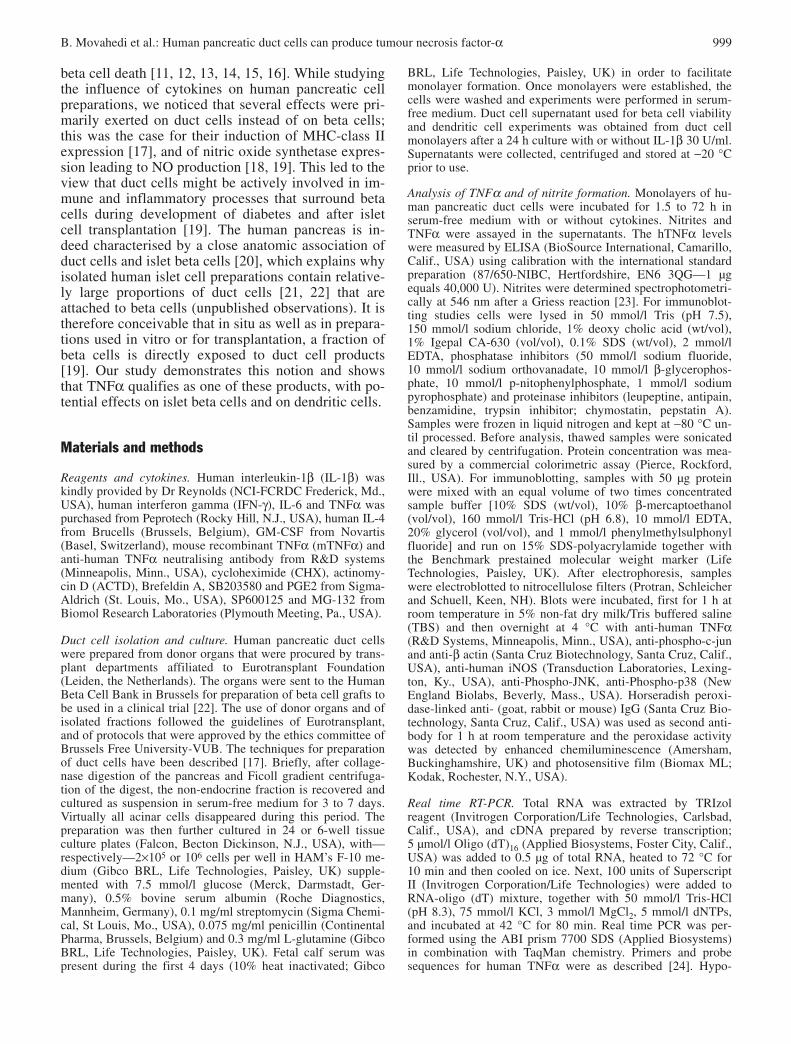
Abstract
Aims/hypothesis. In the human pancreas, a close topo-graphic relationship exists between duct cells and betacells. This explains the high proportion of duct cells inisolated human islet preparations. We investigatedwhether human duct cells are a source of TNFα-medi-ated interactions with beta cells and immune cells.This cytokine has been implicated in the developmentof autoimmune diabetes in mice.Methods. Human duct cells were isolated from donorpancreases and examined for their ability to produceTNFα following a stress-signalling pathway. Duct-cell-released TNFα was tested for its in vitro effectson survival of human beta cells and on activation ofhuman dendritic cells.Results. Exposure of human pancreatic duct cells tointerleukin-1β (IL-1β) induces TNFα gene expres-
sion, synthesis of the 26,000 Mr TNFα precursor andconversion to the 17,000 Mr mature form, which israpidly released. This effect is NO-independent andinvolves p38 MAPK and NF-κB signalling. Duct-cell-released TNFα contributed to cytokine-induced apop-tosis of isolated human beta cells. It also induced acti-vation of human dendritic cells.Conclusions/interpretation. Human pancreatic ductcells are a potential source of TNFα that can cause apoptosis of neighbouring beta cells and initiate animmune response through activation of dendritic cells.They may thus actively participate in inflammatoryand immune processes that threaten beta cells duringdevelopment of diabetes or after human islet cellgrafts have been implanted.
Keywords Apoptosis · Diabetes mellitus · Insulin · Islets of Langerhans · Pancreas
Received: 16 December 2003 / Accepted: 17 April 2004Published online: 8 June 2004© Springer-Verlag 2004
D. Pipeleers (✉)Diabetes Research Centre, Brussels Free University—VUB and JDRF Centrefor Beta Cell Therapy in Europe, Laarbeeklaan 103, 1090 Brussels, BelgiumE-mail: [email protected].: +32-2-4774541, Fax: +32-2-4774545
Abbreviations: CK-19, Cytokeratin 19 · DuC, duct cell · iNOS,inducible nitric oxide synthetase · JNK, jun N-terminal kinase ·MAPK, mitogen activated protein kinase · NF-κB, nuclear factor kappa B · NOD mouse, non-obese diabetic mouse
Diabetologia (2004) 47:998–1008DOI 10.1007/s00125-004-1426-3
Human pancreatic duct cells can produce tumour necrosis factor-α that damages neighbouring beta cells and activates dendritic cellsB. Movahedi1 · M. Van de Casteele1 · N. Caluwé1 · G. Stangé1 · K. Breckpot2 · K. Thielemans2 · G. Vreugdenhil3 · C. Mathieu4 · D. Pipeleers1
1 Diabetes Research Centre, Brussels Free University—VUB and JDRF Centre for Beta Cell Therapy in Europe, Brussels, Belgium
2 Laboratory of Molecular and Cellular Therapy, Department of Physiology and Immunology, Brussels Free University-VUB, Belgium
3 Department of Medical Microbiology, University Medical Centre Nijmegen, The Netherlands4 Laboratory of Experimental Medicine and Endocrinology, Catholic University of Leuven-KUL, Belgium
Introduction
Inflammatory infiltration of pancreatic islets is consid-ered a major pathogenic event in the development ofType 1 diabetes. Insulitis is noticed at clinical onset ofthe disease [1], particularly in patients younger than15 years [2], and found to contain a mixture of reac-tive lymphocytes [3]. A form of insulitis was also de-tected in rodents developing autoimmune diabetes [4].In (pre)diabetic non-obese diabetic mice (NOD), infil-trating lymphocytes and macrophages were shown toproduce cytokines [5, 6, 7, 8] that can destroy pancre-atic beta cells in vitro [9, 10]. Cytokines were there-fore proposed as potential mediators of beta cell deathin vivo. Studies in NOD mice have suggested thatTNFα is involved in the in vivo process leading to
beta cell death [11, 12, 13, 14, 15, 16]. While studyingthe influence of cytokines on human pancreatic cellpreparations, we noticed that several effects were pri-marily exerted on duct cells instead of on beta cells;this was the case for their induction of MHC-class IIexpression [17], and of nitric oxide synthetase expres-sion leading to NO production [18, 19]. This led to theview that duct cells might be actively involved in im-mune and inflammatory processes that surround betacells during development of diabetes and after isletcell transplantation [19]. The human pancreas is in-deed characterised by a close anatomic association ofduct cells and islet beta cells [20], which explains whyisolated human islet cell preparations contain relative-ly large proportions of duct cells [21, 22] that are attached to beta cells (unpublished observations). It istherefore conceivable that in situ as well as in prepara-tions used in vitro or for transplantation, a fraction ofbeta cells is directly exposed to duct cell products[19]. Our study demonstrates this notion and showsthat TNFα qualifies as one of these products, with po-tential effects on islet beta cells and on dendritic cells.
Materials and methods
Reagents and cytokines. Human interleukin-1β (IL-1β) waskindly provided by Dr Reynolds (NCI-FCRDC Frederick, Md.,USA), human interferon gamma (IFN-γ), IL-6 and TNFα waspurchased from Peprotech (Rocky Hill, N.J., USA), human IL-4from Brucells (Brussels, Belgium), GM-CSF from Novartis(Basel, Switzerland), mouse recombinant TNFα (mTNFα) andanti-human TNFα neutralising antibody from R&D systems(Minneapolis, Minn., USA), cycloheximide (CHX), actinomy-cin D (ACTD), Brefeldin A, SB203580 and PGE2 from Sigma-Aldrich (St. Louis, Mo., USA), SP600125 and MG-132 fromBiomol Research Laboratories (Plymouth Meeting, Pa., USA).
Duct cell isolation and culture. Human pancreatic duct cellswere prepared from donor organs that were procured by trans-plant departments affiliated to Eurotransplant Foundation (Leiden, the Netherlands). The organs were sent to the HumanBeta Cell Bank in Brussels for preparation of beta cell grafts tobe used in a clinical trial [22]. The use of donor organs and ofisolated fractions followed the guidelines of Eurotransplant,and of protocols that were approved by the ethics committee ofBrussels Free University-VUB. The techniques for preparationof duct cells have been described [17]. Briefly, after collage-nase digestion of the pancreas and Ficoll gradient centrifuga-tion of the digest, the non-endocrine fraction is recovered andcultured as suspension in serum-free medium for 3 to 7 days.Virtually all acinar cells disappeared during this period. Thepreparation was then further cultured in 24 or 6-well tissue culture plates (Falcon, Becton Dickinson, N.J., USA), with—respectively—2×10⁵ or 106 cells per well in HAM’s F-10 me-dium (Gibco BRL, Life Technologies, Paisley, UK) supple-mented with 7.5 mmol/l glucose (Merck, Darmstadt, Ger-many), 0.5% bovine serum albumin (Roche Diagnostics,Mannheim, Germany), 0.1 mg/ml streptomycin (Sigma Chemi-cal, St Louis, Mo., USA), 0.075 mg/ml penicillin (ContinentalPharma, Brussels, Belgium) and 0.3 mg/ml L-glutamine (GibcoBRL, Life Technologies, Paisley, UK). Fetal calf serum waspresent during the first 4 days (10% heat inactivated; Gibco
BRL, Life Technologies, Paisley, UK) in order to facilitatemonolayer formation. Once monolayers were established, thecells were washed and experiments were performed in serum-free medium. Duct cell supernatant used for beta cell viabilityand dendritic cell experiments was obtained from duct cellmonolayers after a 24 h culture with or without IL-1β 30 U/ml.Supernatants were collected, centrifuged and stored at −20 °Cprior to use.
Analysis of TNFα and of nitrite formation. Monolayers of hu-man pancreatic duct cells were incubated for 1.5 to 72 h in serum-free medium with or without cytokines. Nitrites andTNFα were assayed in the supernatants. The hTNFα levelswere measured by ELISA (BioSource International, Camarillo,Calif., USA) using calibration with the international standardpreparation (87/650-NIBC, Hertfordshire, EN6 3QG—1 µgequals 40,000 U). Nitrites were determined spectrophotometri-cally at 546 nm after a Griess reaction [23]. For immunoblot-ting studies cells were lysed in 50 mmol/l Tris (pH 7.5),150 mmol/l sodium chloride, 1% deoxy cholic acid (wt/vol),1% Igepal CA-630 (vol/vol), 0.1% SDS (wt/vol), 2 mmol/lEDTA, phosphatase inhibitors (50 mmol/l sodium fluoride,10 mmol/l sodium orthovanadate, 10 mmol/l β-glycerophos-phate, 10 mmol/l p-nitophenylphosphate, 1 mmol/l sodium pyrophosphate) and proteinase inhibitors (leupeptine, antipain,benzamidine, trypsin inhibitor; chymostatin, pepstatin A).Samples were frozen in liquid nitrogen and kept at −80 °C un-til processed. Before analysis, thawed samples were sonicatedand cleared by centrifugation. Protein concentration was mea-sured by a commercial colorimetric assay (Pierce, Rockford,Ill., USA). For immunoblotting, samples with 50 µg proteinwere mixed with an equal volume of two times concentratedsample buffer [10% SDS (wt/vol), 10% β-mercaptoethanol(vol/vol), 160 mmol/l Tris-HCl (pH 6.8), 10 mmol/l EDTA,20% glycerol (vol/vol), and 1 mmol/l phenylmethylsulphonylfluoride] and run on 15% SDS-polyacrylamide together withthe Benchmark prestained molecular weight marker (LifeTechnologies, Paisley, UK). After electrophoresis, sampleswere electroblotted to nitrocellulose filters (Protran, Schleicherand Schuell, Keen, NH). Blots were incubated, first for 1 h atroom temperature in 5% non-fat dry milk/Tris buffered saline(TBS) and then overnight at 4 °C with anti-human TNFα(R&D Systems, Minneapolis, Minn., USA), anti-phospho-c-junand anti-β actin (Santa Cruz Biotechnology, Santa Cruz, Calif.,USA), anti-human iNOS (Transduction Laboratories, Lexing-ton, Ky., USA), anti-Phospho-JNK, anti-Phospho-p38 (NewEngland Biolabs, Beverly, Mass., USA). Horseradish peroxi-dase-linked anti- (goat, rabbit or mouse) IgG (Santa Cruz Bio-technology, Santa Cruz, Calif., USA) was used as second anti-body for 1 h at room temperature and the peroxidase activitywas detected by enhanced chemiluminescence (Amersham,Buckinghamshire, UK) and photosensitive film (Biomax ML;Kodak, Rochester, N.Y., USA).
Real time RT-PCR. Total RNA was extracted by TRIzol reagent (Invitrogen Corporation/Life Technologies, Carlsbad,Calif., USA), and cDNA prepared by reverse transcription;5 µmol/l Oligo (dT)16 (Applied Biosystems, Foster City, Calif.,USA) was added to 0.5 µg of total RNA, heated to 72 °C for10 min and then cooled on ice. Next, 100 units of SuperscriptII (Invitrogen Corporation/Life Technologies) were added toRNA-oligo (dT) mixture, together with 50 mmol/l Tris-HCl(pH 8.3), 75 mmol/l KCl, 3 mmol/l MgCl2, 5 mmol/l dNTPs,and incubated at 42 °C for 80 min. Real time PCR was per-formed using the ABI prism 7700 SDS (Applied Biosystems)in combination with TaqMan chemistry. Primers and probe sequences for human TNFα were as described [24]. Hypo-
B. Movahedi et al.: Human pancreatic duct cells can produce tumour necrosis factor-α 999

xanthine phosphoribosyltransferase 1 (HPRT) was used ashousekeeping gene to normalise TNFα values. HPRT primersand probe sequences are as follows: F 5′-TGTAGGATATGCC-CTTGACTATA-3′ R 5′-CAATAGGACTCCAGATGTTTCCA-3′ P 5′-TGGAAAAGCAAAATACAAAGCCTAAGATGAG-3′. PCR amplifications were carried out in duplicate in a totalvolume of 25 µl containing 0.5 µl cDNA sample, 50 mmol/lKCl, 10 mmol/l Tris-HCl (pH 8.3), 10 mmol/l EDTA,60 nmol/l Passive Reference, 1200 µmol/l dNTPs, 3 to9 mmol/l MgCl2, 100 to 900 nmol/l of each primer,100 mmol/l of TaqMan probe and 0.625 U AmpliTaqGold(Applied Biosystems). Thermal conditions: 10 min at 94 °C,followed by 45 two temperature cycles (15 s at 94 °C and1 min at 60 °C). cDNA plasmid standards were used for eachtarget to quantify relative expression [24].
Immunocytochemistry. For TNFα and CK-19 staining, ductcell monolayers were fixed at room temperature in 4%buffered formaldehyde and then permeabilised with Triton-X100 before incubation in 10 mmol/l EDTA at 70 °C for10 min. Non-specific binding sites were blocked by 10% nor-mal goat serum or 2% normal donkey serum prior to, respec-tively, TNFα and CK19 immunostaining using mouse anti-human TNFα mAb (HyCult biotechnology b.v., The Nether-lands) and sheep anti-human CK19 Ab (The Binding Site,Birmingham, UK). The cells were then washed in PBS and in-cubated with Texas Red conjugated secondary donkey anti-mouse mAb or Cy2-conjugated secondary donkey anti-sheepAb (both from Jackson ImmunoResearch Laboratories, WestGrove, Pa., USA). After washing, the preparations weremounted, covered by Dako fluorescence mounting medium(Dako Corporation, Carpinteria, Calif., USA) and analysed bya Leica TCS SP confocal laser-scanning microscope (CLSM).The CLSM is equipped with Ar/HeNe-lasers and Leica TCSNT software (version 1.6.587). Fluoresbrite grade micro-spheres, (∅ 3.0 µm, Polylab BVBA, Belgium) were used tocalibrate the magnification. Images were transferred to AdobePhotoShop 5.5 software for multicolour channel analysis andfigure assembly. For subcellular localisation of the P65 subunitof NF-κB, monolayers were fixed and permeabilised by ice-cold acetone before incubation with primary rabbit anti-humanNF-κB-P65 antibody (Santa Cruz Biotechnology, Santa Cruz,Calif., USA). After washing in PBS, the preparations were in-cubated with Cy3-conjugated secondary donkey anti-rabbit Ab(Jackson ImmunoResearch Laboratories, West Grove, Pa.,USA), washed again, mounted and analysed in a Axioplan 2fluorescence microscope (Carl Zeiss Jena, Jena, Germany)equipped with Photometrics SenSys 1401 digital Camera(vysys, France) and Smart Capture VP (version 1.4) software(Digital Scientific, UK).
Beta cell toxicity assay. Human beta cell preparations were ob-tained from the Human Beta Cell Bank. Methods for isolation,dissociation, purification and culture have been described else-where [21, 22]. The cultured endocrine fraction was dissociat-ed and enriched in single beta cells by flow cytometry (FACSsorting) according to forward scatter and autofluorescence in-tensity at 530 nm [25]; the degree of purity is lower than thatfor rat beta cells but routinely exceeds 60%. The purified betacell preparation was cultured in micro titer cups at 4000 cellsper well in a HAM F-10 basis of unconditioned or duct cell(DuC) conditioned medium with replacement of half the vol-ume every 2 days. Cells cultured in unconditioned HAM F10without cytokines served as controls. The effect of IL-1β wasalso tested in unconditioned medium with or without recombi-nant human TNFα added. An anti-human TNFα neutralisingantibody was added in two conditions. Each beta cell prepara-
tion was incubated with conditioned media of three differentduct cell preparations and each condition was carried out induplicate. After 10 days of culture, the percentages of apoptot-ic and necrotic cells were determined as described previously[26]. The apoptosis index was calculated as: (% apoptotic cellsin test−% apoptotic cells in control condition)/(100−% apop-totic cells in control condition)×100. The necrosis index wascalculated by replacing the percentage of apoptotic cells by thepercentage of necrotic cells [27].
Dendritic cell activation assay. Dendritic cells were isolatedas described [28] with minor modifications to increase den-dritic cell yields [29]. Briefly, peripheral blood mononuclearcells were isolated from buffy coat preparations of healthydonors by gradient centrifugation (Lymphoprep, NycomedPharma AS, Oslo, Norway). The cells were seeded inRPMI 1640 (Gibco, Invitrogen, Merelbeke, Belgium) contain-ing 1% human AB serum (PAA Laboratories, Linz, Austria),and incubated for 2 h to allow adherence of monocytes. Afterwashout of non-adherent cells, adherent cells were further cul-tured for 5 days in the presence of GM-CSF (1000 U/ml) andIL-4 (100 U/ml). They were then transferred to culture condi-tions in DuC-conditioned medium or with a cytokine mixturethat is known to activate dendritic cells [30] namely IL-1β(100 U/ml), IL-6 (1000 U/ml), TNFα (100 U/ml) plus Pros-taglandin E2 (PGE2, 1 µg/ml). Dendritic cell activation wasanalysed by flow cytometry [31] after staining with PE con-jugated monoclonal antibodies against CD25, CD80 andCD83 or isotype controls (BD Pharmingen, Erembodegem,Belgium).
Statistical methods. Results are expressed as means ± SEM ofn independent experiments each using cells from a differentdonor. Statistical analysis was done using the SPSS computerprogram. Student’s t test was used to compare means of twogroups, one way ANOVA with post hoc LSD to comparemeans of three or more groups, and Friedman test and Mann-Whitney U test to compare results that were normalised to thecontrol. Significant differences were based on a p value of lessthan 0.05.
Results
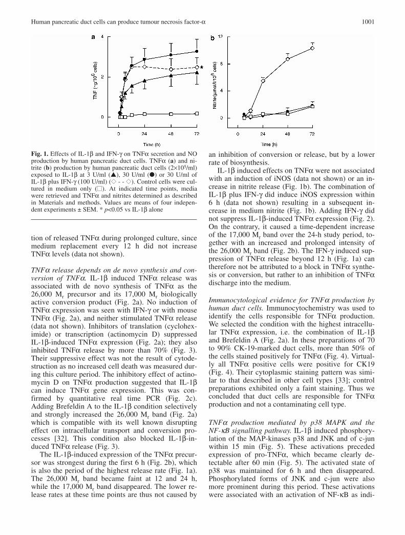
IL-1β-induction of TNFα release from human ductcells. Human duct cell monolayers released marginallydetectable TNFα levels, i.e. 100 to 200 pg TNF106·cells−1·72 h−1. Addition of IL-1β (30 U/ml) in-creased TNF-levels 20 fold (Fig. 1a), while no stimu-lation was seen with human IFN-γ (1-1000 U/ml, datanot shown). IL-1β induced TNFα release is detectedfrom 60 min on, proceeds linearly during the first 6 hand then levels off to slower increment rates (Fig. 1a).In the period between 12 and 72 h, the rate of TNFαrelease was only 20% of that during the first 12 h. Thehalf-maximal effect was reached after 5 h exposure(Fig. 1a). A similar curve was obtained with 3 U/mlIL-1β with values that were, on average, 40% lowerthan those at 30 U/ml (Fig. 1a). The lower TNFα in-crement beyond 12 h of IL-1β exposure was notcaused by inactivation of the stimulus, since addingfresh IL-1β every 12 h did not further increase TNFαlevels in the medium. Nor was it caused by degrada-
1000 B. Movahedi et al.:
Human pancreatic duct cells can produce tumour necrosis factor-α 1001
tion of released TNFα during prolonged culture, sincemedium replacement every 12 h did not increaseTNFα levels (data not shown).
TNFα release depends on de novo synthesis and con-version of TNFα. IL-1β induced TNFα release wasassociated with de novo synthesis of TNFα as the26,000 Mr precursor and its 17,000 Mr biologicallyactive conversion product (Fig. 2a). No induction ofTNFα expression was seen with IFN-γ or with mouseTNFα (Fig. 2a), and neither stimulated TNFα release(data not shown). Inhibitors of translation (cyclohex-imide) or transcription (actinomycin D) suppressedIL-1β-induced TNFα expression (Fig. 2a); they alsoinhibited TNFα release by more than 70% (Fig. 3).Their suppressive effect was not the result of cytode-struction as no increased cell death was measured dur-ing this culture period. The inhibitory effect of actino-mycin D on TNFα production suggested that IL-1βcan induce TNFα gene expression. This was con-firmed by quantitative real time PCR (Fig. 2c).Adding Brefeldin A to the IL-1β condition selectivelyand strongly increased the 26,000 Mr band (Fig. 2a)which is compatible with its well known disrupting effect on intracellular transport and conversion pro-cesses [32]. This condition also blocked IL-1β-in-duced TNFα release (Fig. 3).
The IL-1β-induced expression of the TNFα precur-sor was strongest during the first 6 h (Fig. 2b), whichis also the period of the highest release rate (Fig. 1a).The 26,000 Mr band became faint at 12 and 24 h,while the 17,000 Mr band disappeared. The lower re-lease rates at these time points are thus not caused by
Fig. 1. Effects of IL-1β and IFN-γ on TNFα secretion and NOproduction by human pancreatic duct cells. TNFα (a) and ni-trite (b) production by human pancreatic duct cells (2×10⁵/ml)exposed to IL-1β at 3 U/ml (▲), 30 U/ml (●) or 30 U/ml ofIL-1β plus IFN-γ (100 U/ml) (◆◆ - - ◆◆). Control cells were cul-tured in medium only (■■). At indicated time points, mediawere retrieved and TNFα and nitrites determined as describedin Materials and methods. Values are means of four indepen-dent experiments ± SEM. * p<0.05 vs IL-1β alone
an inhibition of conversion or release, but by a lowerrate of biosynthesis.
IL-1β induced effects on TNFα were not associatedwith an induction of iNOS (data not shown) or an in-crease in nitrite release (Fig. 1b). The combination ofIL-1β plus IFN-γ did induce iNOS expression within6 h (data not shown) resulting in a subsequent in-crease in medium nitrite (Fig. 1b). Adding IFN-γ didnot suppress IL-1β-induced TNFα expression (Fig. 2).On the contrary, it caused a time-dependent increaseof the 17,000 Mr band over the 24-h study period, to-gether with an increased and prolonged intensity ofthe 26,000 Mr band (Fig. 2b). The IFN-γ induced sup-pression of TNFα release beyond 12 h (Fig. 1a) cantherefore not be attributed to a block in TNFα synthe-sis or conversion, but rather to an inhibition of TNFαdischarge into the medium.
Immunocytological evidence for TNFα production byhuman duct cells. Immunocytochemistry was used toidentify the cells responsible for TNFα production.We selected the condition with the highest intracellu-lar TNFα expression, i.e. the combination of IL-1βand Brefeldin A (Fig. 2a). In these preparations of 70to 90% CK-19-marked duct cells, more than 50% ofthe cells stained positively for TNFα (Fig. 4). Virtual-ly all TNFα positive cells were positive for CK19(Fig. 4). Their cytoplasmic staining pattern was simi-lar to that described in other cell types [33]; controlpreparations exhibited only a faint staining. Thus weconcluded that duct cells are responsible for TNFαproduction and not a contaminating cell type.
TNFα production mediated by p38 MAPK and theNF-κB signalling pathway. IL-1β induced phosphory-lation of the MAP-kinases p38 and JNK and of c-junwithin 15 min (Fig. 5). These activations preceded expression of pro-TNFα, which became clearly de-tectable after 60 min (Fig. 5). The activated state ofp38 was maintained for 6 h and then disappeared.Phosphorylated forms of JNK and c-jun were alsomore prominent during this period. These activationswere associated with an activation of NF-κB as indi-

1002 B. Movahedi et al.:
cated by its nuclear translocation after 15 to 30 min(data not shown). Adding SB203580, a specific inhib-itor of p38 MAPK [34], suppressed IL-1β-inducedTNFα expression and secretion by more than 75%(Fig. 6). TNFα release was also inhibited by MG-132,
an inhibitor of IκB degradation and hence of NF-κBactivation [35] (Fig. 6). On the other hand, SP600125,a specific inhibitor of JNK [36] did not decreaseTNFα production (Fig. 6).
Cytotoxic effect of duct-cell-released TNFα on humanbeta cell preparations. Medium was collected from24-h duct-cell cultures in the absence (DuC-Co) orpresence of 30 U/ml of IL-1β (DuC-IL-1) (DuC,Fig. 7). The TNFα concentration in DuC-IL-1 variedbetween 300 and 600 pg/ml, while that in DuC-Cowas either undetectable (<15 pg/ml) or lower than50 pg/ml. These media were added to cultures of human beta cells to investigate their effect on cell survival. Parallel cultures were done in medium with-out DuC-supernatant added (no-DuC, Fig. 7), eitherwith or without IL-1β (30 U/ml) or IL-1β (30 U/ml)plus human TNFα (400 pg/ml). The condition withoutsupernatant and cytokines served as control. The cyto-toxic effect of the test conditions was normalised tothis control [26].
Duct cell medium (DuC-Co) did not exert a cyto-toxic effect, as the percentages of apoptotic and ne-crotic cells were comparable to control values (Fig. 7).On the other hand, DuC-IL-1 medium induced apop-tosis whereas this was not the case when only IL-1was added (no DuC-IL-1, Fig. 7). This apoptotic ef-fect was partially suppressed by a neutralising anti-TNFα antibody, suggesting its dependency on TNFαthat is present in DuC-IL-1 medium. Apoptosis alsooccurred in control medium containing IL-1 plusTNFα at 400 pg/ml (no DuC-IL-1+TNF), the con-centration that was measured in DuC-IL-1 medium.Apoptosis was partially suppressed by the anti-TNFα
Fig. 2. Effects of cytokines on cellular expression of TNFα.Monolayers of human pancreatic duct cells were exposed forthe indicated periods to IL-1β (IL-1; 30 U/ml), IFN-γ (IFN;100 U/ml), or murine TNFα (mTNF; 100 U/ml) (a). The effectof IL-1β (30 U/ml) was examined in the absence and presenceof cycloheximide (CHX; 5 µg/ml), actinomycin D (AMD;1 µg/ml) or Brefeldin A (BFA; 5 µg/ml) (a), or IFN-γ(100 U/ml) (b). Whole cell lysates were separated by SDS-PAGE and immunoblotted with TNFα antibody. c. Real timePCR of TNFα expression after extraction of total RNA and re-verse transcription to cDNA. TNFα values were normalised toHPRT levels. The figure represents three independent experi-ments
Fig. 3. Effects of cycloheximide (CHX), actinomycin D (AMD),and Brefeldin A (BFA) on IL-1β induced TNFα secretion.Monolayers of human pancreatic duct cells were exposed for6 h to 30 U/ml of IL-1β with or without cycloheximide(5 µg/ml), actinomycin D (1 µg/ml) or Brefeldin A (5 µg/ml).Values are means ± SEM of four independent experiments. *** p<0.001 vs IL-1β alone

Human pancreatic duct cells can produce tumour necrosis factor-α 1003
antibody (Fig. 7). None of the conditions increased thepercentage of necrotic cells (Fig. 7).
Dendritic cell activation by duct-cell-released TNF.We examined whether duct cell medium was able toinduce dendritic cell activation, which is known to beTNFα-dependent. Immature dendritic cells were cul-tured for 24 h in DuC-IL-1 or DuC-Co and then evalu-
Fig. 4. Immunolocalisation of cellular Cytokeratin 19 (CK19)and TNFα by confocal laser scanning microscopy. Maximalprojection images of human pancreatic duct cell monolayersincubated during 4 h with IL-1β (30 U/ml) plus Brefeldin A(5 µg/ml) (d–f) or medium alone (Control) (a–c). Cells werestained for TNFα (Texas Red) (b, c, e, f) and CK19 (CY2:green) (a, c, d, f). Scale bar: 50 µm
ated for their expression of the activation markersCD25 (IL-2 receptor), CD80 (co-stimulatory moleculeB7.1) and CD83 (maturation marker). Each experi-ment contained a positive control consisting of imma-ture dendritic cells that were cultured with a mixtureof cytokines (IL-1β, IL-6, TNFα and PGE2); this con-dition induced at least 30% positive cells above con-trol for these three markers (data not shown). In medi-um without cytokines, less than 6% positive cellswere detected. Culture of immature dendritic cells inDuC-Co did not result in their activation as judgedfrom the percentage of CD25, CD80 and CD83 cells(<6%, NS vs negative control). On the other hand,DuC-IL-1 medium induced a significant increase inCD25, CD80 and CD83 expressing cells (Fig. 8). Thiseffect was significantly suppressed by the anti-TNFαantibody (Fig. 8).

1004 B. Movahedi et al.:
Discussion
In the developing pancreas, the juxtaposition of betacells and duct cells is often noticed, and considered asindirect evidence for a ductal origin of beta cells. Asimilar topographic relationship exists in the adult hu-man pancreas where 15% of insulin-producing cellsoccur as single units along ductules, and where betacell aggregates are often directly juxtaposed to ductcells [20]. This anatomic characteristic explains whyhigh numbers of duct cells co-migrate with endocrine
cells during human islet isolation [21, 22]. The pres-ence of 20 to 60% duct cells in human beta cell graftsled us to examine whether they might be involved ininflammatory and immune reactions around islet cellimplants. We previously reported that human ductcells can respond to cytokines by expressing MHC-class II and iNOS with subsequent NO production[17, 18]. Our data show that these cells can also re-lease TNFα at levels that affect survival of neighbour-ing beta cells and activate dendritic cells.
Duct cell release of TNFα was induced by interleu-kin-1ß. While this in vitro condition only serves as amodel to demonstrate the existence and the mecha-nism of this duct cell property, it may occur in vivo
Fig. 5. Effect of IL-1β on MAP kinase phosphorylation.Monolayers of human pancreatic duct cells were incubatedwith IL-1β (30 U/ml) for the indicated times. Whole cell lysates were separated by SDS-PAGE and immunoblotted bythe indicated antibodies. The figure represents three indepen-dent experiments
Fig. 6. Effects of SP600125, SB203580, and MG-132 onTNFα production and secretion. Monolayers of human pancre-atic duct cells were pre-treated for 1 h with SP600125(10 µmol/l) or SB203580 (5 µmol/l) or for 30 min with MG-132 (50 µmol/l) before exposure to IL-1β (30 U/ml) for6 h. Whole cell lysates were separated by SDS-PAGE and im-munoblotted by the indicated antibodies. Supernatants wereharvested and assayed for TNFα. Data are expressed as per-cent of IL-1β condition and represent means ± SEM of three tosix independent experiments. ** p<0.01 vs IL-1β alone
Fig. 7. Effect of duct cell medium on viability of cultured hu-man pancreatic beta cells. FACS purified human pancreaticbeta cells were cultured with medium (DuC) that was collectedfrom human duct cell monolayers, which had been incubatedfor 24 h in the absence (Co) or presence of IL-1β at 30 U/ml(IL-1). After 10 days of culture the number of apoptotic andnecrotic cells were counted and expressed relative to the num-bers in control preparations cultured in unconditioned medium.For each condition an apoptosis and necrosis index was cal-culated as defined in reference [27]. The right panel showsdata for cells cultured in unconditioned medium (no DuC) inthe presence of IL-1β (30 U/ml) with or without TNFα(400 pg/ml). The role of TNFα was assessed by adding aTNFα neutralising antibody (TNFAB) to the conditions DuC-IL-1 and IL-1β + TNFα. Data represent means ± SEM of threeindependent experiments. *** p<0.001 vs control, * p<0.05 vsDuC IL-1β, § p<0.05 vs IL-1β + TNFα

Human pancreatic duct cells can produce tumour necrosis factor-α 1005
Fig. 8. Effect of duct cell medium on the activation of dendrit-ic cells. Immature human dendritic cells were incubated for24 h with medium that was collected from human duct cellmonolayers, which had been incubated for 24 h in the absence(DuC-Co) or presence of IL-1β at 30 U/ml (DuC-IL-1). Theirdegree of activation was measured by FACS analysis of thepercentage of CD25, CD80 and CD83 positive cells relative to the percentages in the negative control condition (a). Datarepresent means ± SEM for three to five independent experi-ments. ** p<0.01 compared to control; * p<0.05; *** p<0.001;§ p<0.05 compared to DuC-IL-1. b. A representative flow dia-gram which also shows the degree of activation in the positivecontrol condition (stimulation by IL-1β, IL-6, TNFα andPGE2)
when IL-1β is released by islet cells or by islet infil-trating cells [37]. TNFα release was dependent on theinduction of TNFα precursor synthesis and conver-sion, rather than on discharge from a cellular pool ofthe converted mature form. The mechanism throughwhich IL-1β induces TNFα-expression in humanpancreatic duct cells is not fully understood. As inother cell types [38], this IL-1β effect depends on ac-tivation of MAP kinase p38 and seems mediated, atleast in part, by NF-κB, which is known to bind to thepromoter region of TNFα. On the other hand, it pro-ceeds irrespective of iNOS induction or NO-produc-tion, and seems independent of JNK-activation,which contrasts with the mechanism in human CD4+cells [36].
TNFα produced by human duct cells was shown toexert effects on neighbouring endocrine and immune

cells. The cytokine is released in a biologically activeform and rapidly reaches in vitro concentrations in therange used in numerous in vitro studies [10, 39, 40].Among the functions known to be TNFα sensitive,survival of beta cells [41, 42] and activation of den-dritic cells [43, 44, 45] seem particularly relevant inthe context of the pathogenesis of Type 1 diabetes andstrategies to prevent or cure the disease. Medium con-taining duct-cell-released TNFα induced apoptosis inhuman beta cell preparations, probably in synergywith IL-1β that was added to stimulate TNFα produc-tion. Studies on rodent and human beta cells have in-deed indicated that IL-1β alone does not cause apop-tosis unless it is combined with IFN-γ and/or TNFα[42, 46]. A neutralising anti-TNFα antibody partiallysuppressed the apoptotic effect of the duct cell medi-um; the lack of complete suppression may result frominsufficient neutralisation by the antibody or by anoth-er factor that is induced by IL-1β.
Pro-inflammatory cytokines such as IL-1β andTNFα are also known to stimulate migration and mat-uration of dendritic cells [43, 44, 45]. Our studyshows that duct-cell-released TNFα can activate hu-man dendritic cells, suggesting that these cells mayplay a role in the development of (auto)immune pro-cesses. Our data are compatible with earlier workshowing TNFα involvement in the in vivo process ofimmune beta cell destruction. Neutralising TNFα anti-bodies were shown to protect NOD mice against dia-betes, whereas treatment with recombinant TNFα ac-celerated the disease [11]. TNFα receptor 1-deficientNOD mice did not develop diabetes although insulitiswas present [12], whereas induction of TNFα expres-sion in islets accelerated progression to diabetes [13].The latter effect was attributed to an immune modula-tion at an earlier age, following apoptosis of a fewbeta cells and subsequent activation of islet dendriticcells [14]. When islet TNF expression was regulatedby a tetracycline-driven on–off switch, progression todiabetes depended on the duration of exposure toTNFα [15]. Another study showed the importance oftiming islet-specific TNFα expression with respect tothe autoimmune process [16]. These studies explainwhy the diabetogenic effects of TNFα were not ob-served when islet-specific TNFα expression was re-stricted to later stages [47]. It is also conceivable thatlocal release of this cytokine alters the phenotype ofneighbouring beta cells and thus varies their suscepti-bility to cytotoxic conditions as previously observedwith interleukin-1 [48]. In the presence of other cy-tokines, TNFα may itself contribute to the death ofbeta cells [42]; in this respect, production of interleu-kin-1 by beta cells could be considered as a potentiallocal trigger [37].
In conclusion, human pancreatic duct cells shouldbe considered as potential participants in (auto)im-mune processes that occur during development ofType 1 diabetes or after islet transplantation. Their
exposure to cytokines can induce expression of MHC-class II [17], iNOS [18] and/or TNFα precursor. It isstill unknown whether these diverse responses can begenerated by all duct cells or whether they charac-terise a particular subpopulation. Our in vitro observa-tion that duct-cell-released TNFα can affect survivalof human beta cells and activate human dendritic cellsmight bear clinical relevance, in particular since thesedifferent cell types are closely associated during de-velopment of Type 1 diabetes [1, 2] as well as duringislet allograft reactivity.
Acknowledgements. This work was supported by grants fromthe Belgian Fund for Scientific Research, Flanders(G.0375.00), the Belgian Interuniversity Attraction Poles(IUAP P5/17), European Foundation for the Study of Diabetesand The Juvenile Diabetes Research Foundation (4-2001-434).B. Movahedi is a research fellow (aspirant) of the BelgianFund for Scientific Research, Flanders. The authors thank thestaff of the Beta Cell Bank for providing the (non-)endocrinetissue fractions, Nicole Buelens and Maarten Timmers for theirexpert contribution to the immunocytochemical studies, LutOverbergh and Dirk Valckx for providing primers and probesfor real time PCR, and Johan Guns for excellent technical as-sistance.
References
1. Gepts W (1965) Pathologic anatomy of the pancreas in juvenile diabetes mellitus. Diabetes 14:619–633
2. Pipeleers D, Ling Z (1992) Pancreatic beta cells in insulin-dependent diabetes. Diabetes Metab Rev 8:209–227
3. Bottazzo GF, Dean BM, McNally JM, MacKay EH, Swift PG, Gamble DR (1985) In situ characterization ofautoimmune phenomena and expression of HLA moleculesin the pancreas in diabetic insulitis. N Engl J Med313:353–360
4. Mori Y, Suko M, Okudaira H et al. (1986) Preventive effects of cyclosporine on diabetes in NOD mice. Dia-betologia 29:244–247
5. Held W, MacDonald HR, Weissman IL, Hess MW, MuellerC (1990) Genes encoding tumor necrosis factor alpha and granzyme A are expressed during development of au-toimmune diabetes. Proc Natl Acad Sci USA 87:2239–2243
6. Rabinovitch A (1994) Immunoregulatory and cytokine imbalances in the pathogenesis of IDDM. Therapeutic intervention by immunostimulation? Diabetes 43:613–621
7. Welsh M, Welsh N, Bendtzen K et al. (1995) Comparisonof mRNA contents of interleukin-1 beta and nitric oxidesynthase in pancreatic islets isolated from female and malenonobese diabetic mice. Diabetologia 38:153–160
8. Pilstrom B, Bjork L, Bohme J (1997) Monokine-producingcells predominate in the recruitment phase of NOD insuli-tis while cells producing Th1-type cytokines characterizethe effector phase. J Autoimmun 10:147–155
9. Mandrup-Poulsen T (1996) The role of interleukin-1 in thepathogenesis of IDDM. Diabetologia 39:1005–1029
10. McDaniel ML, Kwon G, Hill JR, Marshall CA, Corbett JA(1996) Cytokines and nitric oxide in islet inflammation anddiabetes. Proc Soc Exp Biol Med 211:24–32
1006 B. Movahedi et al.:

Human pancreatic duct cells can produce tumour necrosis factor-α 1007
11. Yang XD, Tisch R, Singer SM et al. (1994) Effect of tumornecrosis factor alpha on insulin-dependent diabetes mellitusin NOD mice. I. The early development of autoimmunityand the diabetogenic process. J Exp Med 180:995–1004
12. Kagi D, Ho A, Odermatt B, Zakarian A, Ohashi PS, MakTW (1999) TNF receptor 1-dependent beta cell toxicity asan effector pathway in autoimmune diabetes. J Immunol162:4598–4605
13. Green EA, Eynon EE, Flavell RA (1998) Local expressionof TNFalpha in neonatal NOD mice promotes diabetes byenhancing presentation of islet antigens. Immunity 9:733–743
14. Green EA, Flavell RA (1999) Tumor necrosis factor-alphaand the progression of diabetes in non- obese diabeticmice. Immunol Rev 169:11–22
15. Green EA, Flavell RA (2000) The temporal importance ofTNFalpha expression in the development of diabetes. Im-munity 12:459–469
16. Christen U, Wolfe T, Mohrle U et al. (2001) A dual role forTNF-alpha in type 1 diabetes: islet-specific expression ab-rogates the ongoing autoimmune process when induced latebut not early during pathogenesis. J Immunol 166:7023–7032
17. Pavlovic D, Winkel M van de, Auwera B van der et al.(1997) Effect of interferon-gamma and glucose on majorhistocompatibility complex class I and class II expressionby pancreatic beta- and non-beta-cells. J Clin EndocrinolMetab 82:2329–2336
18. Pavlovic D, Chen MC, Bouwens L, Eizirik DL, PipeleersD (1999) Contribution of ductal cells to cytokine responsesby human pancreatic islets. Diabetes 48:29–33
19. Pipeleers D, Hoorens A, Marichal-Pipeleers M, Van deCasteele M, Bouwens L, Ling Z (2001) Role of pancreaticbeta-cells in the process of beta-cell death. Diabetes 50[Suppl 1]:S52–S57
20. Bouwens L, Pipeleers DG (1998) Extra-insular beta cellsassociated with ductules are frequent in adult human pan-creas. Diabetologia 41:629–633
21. Ling Z, Pipeleers DG (1996) Prolonged exposure of humanbeta cells to elevated glucose levels results in sustained cellular activation leading to a loss of glucose regulation. J Clin Invest 98:2805–2812
22. Keymeulen B, Ling Z, Gorus FK et al. (1998) Implantationof standardized beta-cell grafts in a liver segment of IDDMpatients: graft and recipients characteristics in two cases ofinsulin-independence under maintenance immunosuppres-sion for prior kidney graft. Diabetologia 41:452–459
23. Green LC, Wagner DA, Glogowski J, Skipper PL, WishnokJS, Tannenbaum SR (1982) Analysis of nitrate, nitrite, and[15N]nitrate in biological fluids. Anal Biochem 126:131–138
24. Overbergh L, Giulietti A, Valckx D, Decallonne R, Bouillon R, Mathieu C (2003) The use of real-time reversetranscriptase PCR for the quantification of cytokine geneexpression. J Biomol Tech 14:33–43
25. Pipeleers DG, in’t Veld PA, Van de Winkel M, Maes E,Schuit FC, Gepts W (1985) A new in vitro model for thestudy of pancreatic A and B cells. Endocrinology 117:806–816
26. Hoorens A, Van de Casteele M, Kloppel G, Pipeleers D(1996) Glucose promotes survival of rat pancreatic betacells by activating synthesis of proteins which suppress aconstitutive apoptotic program. J Clin Invest 98:1568–1574
27. Pipeleers D, Van de Winkel M (1986) Pancreatic B cellspossess defense mechanisms against cell-specific toxicity.Proc Natl Acad Sci USA 83:5267–5271
28. Romani N, Reider D, Heuer M et al. (1996) Generation ofmature dendritic cells from human blood. An improvedmethod with special regard to clinical applicability. J Im-munol Methods 196:137–151
29. Tuyaerts S, Noppe SM, Corthals J et al. (2002) Generationof large numbers of dendritic cells in a closed system usingCell Factories. J Immunol Methods 264:135–151
30. Jonuleit H, Kuhn U, Muller G et al. (1997) Pro-inflam-matory cytokines and prostaglandins induce maturation of potent immunostimulatory dendritic cells under fetalcalf serum-free conditions. Eur J Immunol 27:3135–3142
31. Breckpot K, Dullaers M, Bonehill A et al. (2003) Lentivi-rally transduced dendritic cells as a tool for cancer immu-notherapy. J Gene Med 5:654–667
32. Dinter A, Berger EG (1998) Golgi-disturbing agents. Histochem Cell Biol 109:571–590
33. Shurety W, Merino-Trigo A, Brown D, Hume DA, Stow JL(2000) Localization and post-Golgi trafficking of tumor necrosis factor-alpha in macrophages. J Interferon Cyto-kine Res 20:427–438
34. Cuenda A, Rouse J, Doza YN et al. (1995) SB 203580 is aspecific inhibitor of a MAP kinase homologue which isstimulated by cellular stresses and interleukin-1. FEBS Lett364:229–233
35. Kwon G, Corbett JA, Hauser S, Hill JR, Turk J, McDanielML (1998) Evidence for involvement of the proteasomecomplex (26S) and NfkappaB in IL-1beta-induced nitricoxide and prostaglandin production by rat islets andRINm5F cells. Diabetes 47:583–591
36. Bennett BL, Sasaki DT, Murray BW et al. (2001)SP600125, an anthrapyrazolone inhibitor of Jun N-terminalkinase. Proc Natl Acad Sci USA 98:13681–13686
37. Heitmeier MR, Arnush M, Scarim AL, Corbett JA (2001)Pancreatic beta-cell damage mediated by beta-cell produc-tion of interleukin-1. A novel mechanism for virus-induceddiabetes. J Biol Chem 276:11151–11158
38. Raingeaud J, Gupta S, Rogers JS et al. (1995) Pro-inflam-matory cytokines and environmental stress cause p38 mito-gen- activated protein kinase activation by dual phosphory-lation on tyrosine and threonine. J Biol Chem 270:7420–7426
39. Hoorens A, Pipeleers D (1999) Nicotinamide protects human beta cells against chemically-induced necrosis, butnot against cytokine-induced apoptosis. Diabetologia 42:55–59
40. Mandrup-Poulsen T, Helqvist S, Wogensen LD et al.(1990) Cytokine and free radicals as effector molecules inthe destruction of pancreatic beta cells. Curr Top MicrobiolImmunol 164:169–193
41. Delaney CA, Pavlovic D, Hoorens A, Pipeleers DG, EizirikDL (1997) Cytokines induce deoxyribonucleic acid strandbreaks and apoptosis in human pancreatic islet cells. Endo-crinology 138:2610–2614
42. Mandrup-Poulsen T, Bendtzen K, Dinarello CA, Nerup J(1987) Human tumor necrosis factor potentiates human interleukin 1-mediated rat pancreatic beta-cell cytotoxicity.J Immunol 139:4077–4082
43. Yamaguchi Y, Tsumura H, Miwa M, Inaba K (1997) Contrasting effects of TGF-beta 1 and TNF-alpha on thedevelopment of dendritic cells from progenitors in mousebone marrow. Stem Cells 15:144–153
44. Rieser C, Bock G, Klocker H, Bartsch G, Thurnher M(1997) Prostaglandin E2 and tumor necrosis factor alphacooperate to activate human dendritic cells: synergistic activation of interleukin 12 production. J Exp Med186:1603–1608
1008 B. Movahedi et al.: Human pancreatic duct cells can produce tumour necrosis factor-α
45. Zhou LJ, Tedder TF (1996) CD14+ blood monocytes candifferentiate into functionally mature CD83+ dendriticcells. Proc Natl Acad Sci USA 93:2588–2592
46. Pukel C, Baquerizo H, Rabinovitch A (1988) Destructionof rat islet cell monolayers by cytokines. Synergistic inter-actions of interferon-gamma, tumor necrosis factor, lym-photoxin, and interleukin 1. Diabetes 37:133–136
47. Grewal IS, Grewal KD, Wong FS, Picarella DE, JanewayCA Jr, Flavell RA (1996) Local expression of transgene
encoded TNF alpha in islets prevents autoimmune diabetesin nonobese diabetic (NOD) mice by preventing the devel-opment of auto-reactive islet-specific T cells. J Exp Med184:1963–1974
48. Ling Z, Van de Casteele M, Eizirik DL, Pipeleers DG(2000) Interleukin-1beta-induced alteration in a beta-cellphenotype can reduce cellular sensitivity to conditions thatcause necrosis but not to cytokine-induced apoptosis. Dia-betes 49:340–345