I oxidase: a putative - The University of Liverpool Repository

Expression of KiSS-1 in Rat Ovary: Putative LocalRegulator of Ovulation?
J. M. Castellano,* M. Gaytan,* J. Roa, E. Vigo, V. M. Navarro, C. Bellido, C. Dieguez, E. Aguilar,J. E. Sanchez-Criado, A. Pellicer, L. Pinilla, F. Gaytan, and M. Tena-Sempere
Department of Cell Biology, Physiology, and Immunology (J.M.C., M.G., J.R., E.V., V.M.N., C.B., E.A., J.E.S.-C., L.P., F.G.,M.T.-S.), University of Cordoba, 14004 Cordoba, Spain; Department of Physiology (C.D.), University of Santiago deCompostela, 15705 Santiago de Compostela, Spain; and Instituto Valenciano de Infertilidad (A.P.), 46015 Valencia, Spain
Kisspeptins, the products of KiSS-1 gene, and their receptor,GPR54, have recently emerged as essential gatekeepers ofreproduction, mainly through regulation of GnRH secretionat the hypothalamus. However, the profound hypogonadot-ropism linked to GPR54 inactivation is likely to mask addi-tional functions of this system at other levels of the gonadalaxis, in which expression of KiSS-1 and GPR54 has been pre-liminarily reported. We describe herein the expression ofKiSS-1 gene and kisspeptin immunoreactivity (IR) in ratovary and evaluate its developmental and hormonal regula-tion. KiSS-1 and GPR54 mRNAs were persistently detected inadult ovary along estrous cycle. Yet, contrary to GPR54, ovar-ian KiSS-1 levels fluctuated in a cyclic-dependent manner,with a robust increase in the afternoon of proestrus, i.e. pre-ceding ovulation. In addition, kisspeptin-IR was observed inrat ovary, with strong signals in theca layers of growing fol-
licles, corpora lutea, and interstitial gland, compartments inwhich modest GPR54-IR was also detected. Interestingly, therise in ovarian KiSS-1 mRNA at proestrus was prevented byblockade of preovulatory gonadotropin surge and restored byreplacement with human chorionic gonadotropin as super-agonist of LH. In addition, immature ovaries showed low tonegligible levels of KiSS-1 mRNA, which were significantlyenhanced by gonadotropin priming. In summary, we presentnovel evidence for the developmental and hormonally regu-lated expression of the KiSS-1 gene, and the presence ofkisspeptin-IR, in rat ovary. The ability of the LH surge totimely induce ovarian expression of KiSS-1 at the preovula-tory period strongly suggests a previously unsuspected role oflocally produced kisspeptin in the control of ovulation. (En-docrinology 147: 4852–4862, 2006)
KISSPEPTINS WERE originally identified as the prod-ucts of the metastasis suppressor gene KiSS-1 (1–3).
These are structurally related peptides (that include metastinand kisspeptin-10) that share their C-terminal region con-taining an Arg-Phe-NH2 motif, distinctive of the RF-amidepeptide family. Kisspeptins are ligands of the previouslyorphan G protein-coupled receptor GPR54, which was ini-tially identified in the rat on the basis of its partial sequencesimilarity to galanin receptors (4). The first biological func-tion assigned to kisspeptins was metastasis suppression be-cause metastin was demonstrated to display a potent anti-metastasis activity in several tumors, such as breastcarcinoma and melanoma (1, 5–7), and loss of KiSS-1 geneexpression was linked to tumor progression and metastasisin different malignancies (8, 9). Thereafter additional bio-logical roles, such as regulation of trophoblast invasion (10),have been suggested for kisspeptin. However, our knowl-edge of KiSS-1 function was recently revolutionized by theseminal observations of de Roux et al. (11) and Seminara etal. (12), who independently reported that deletions and in-
activating mutations in the gene encoding GPR54 are asso-ciated with hypogonadotropic hypogonadism in humansand mice. Phenotypic analyses of these models, as well assubsequent physiological and pharmacological studies, haveset the contention that the KiSS-1/GPR54 system is primarilyacting at the hypothalamus, in which kisspeptins operate asessential gatekeepers of GnRH function (13, 14). Nonetheless,although central KiSS-1 has been proven as an indispensableelement in the cascade of signals controlling reproduction,additional potential actions of kisspeptins at other levels ofthe gonadal axis remain ill defined.
The ovary is a complex endocrine organ responsible foroocyte production and hormonogenesis (15). The cellularstructures supporting those functions are arranged into sev-eral tissue compartments including ovarian follicles at dif-ferent stages of development, corpora lutea, ovarian stroma,and hilus cells (15, 16). The hallmark of ovarian function isthe cyclic release of fertilizable oocytes at ovulation. Thiscomplex process is the end point of follicular growth, de-velopment, and selection, and it is triggered by the preovu-latory LH surge, which initiates the sequential activation ofa series of events including maturation of the oocyte, expan-sion of the cumulus, rupture of the follicle wall, and finalrelease of the cumulus-oocyte complex to the periovarianspace.
Rupture of the follicular apex at ovulation requires activetissue remodeling and intensive proteolytic degradation ofthe extracellular matrix in several tissue compartments of theovary, such as granulosa and theca layers, tunica albuginea,
First Published Online July 6, 2006* J.M.C. and M.G. contributed equally to this work and should be
considered as joint first authors.Abbreviations: GnRH-ANT, Antagonist of GnRH; GPR54, G protein-
coupled receptor 54; hCG, human chorionic gonadotropin; IR, immu-noreactivity; PMS-G, pregnant mare serum gonadotropin; MMP-9, ma-trix metalloproteinase-9.Endocrinology is published monthly by The Endocrine Society (http://www.endo-society.org), the foremost professional society serving theendocrine community.
0013-7227/06/$15.00/0 Endocrinology 147(10):4852–4862Printed in U.S.A. Copyright © 2006 by The Endocrine Society
doi: 10.1210/en.2006-0117
4852

and ovarian surface epithelium. In this phenomenon, severalclasses of proteolytic enzymes, such as the plasminogen ac-tivator-plasmin and matrix metalloproteinase (MMP) sys-tems, as well as the progesterone-induced A disintegrin andmetalloproteinase with thrombospondin-like repeats-1(ADAMTS) and cathepsin L proteases, have been involved(17, 18). Of note, ovarian proteolytic activity is highly timelyand spatially regulated to selectively target follicular break-down at the apex during the periovulatory period and pre-vent uncontrolled proteolysis of the remaining ovarian tis-sue. Thus, protease inhibitors such as plasminogen activatorinhibitors and tissue inhibitors of MMPs are also concomi-tantly expressed in the ovary during ovulation (19, 20). Yetthe mechanism and signals ultimately responsible for timeand spatial targeting of follicular rupture at the apex remainunknown. Similarly, the cellular sources for the proteolyticand antiproteolytic factors operating at the ovary are yet tobe fully unraveled.
Among peripheral tissues, preliminary evidence recentlysuggested that KiSS-1 and GPR54 genes are prominentlyexpressed in rat ovary (21), yet, their pattern of expressionand potential roles at this site have not been explored. No-tably, the mechanism whereby kisspeptin prevents tumormetastasis and, eventually, uncontrolled trophoblast inva-sion likely involves inhibition of proteases, such as MMP-9(22), which has been implicated in tissue remodeling andfollicular rupture (23). This raises the intriguing possibilitythat, in addition to its essential role in the hypothalamiccontrol of preovulatory LH surge (24), locally producedkisspeptin might directly contribute to the regulation of ovu-lation. To provide a basis for such a hypothesis, we reportherein the first detailed characterization of the expression ofthe KiSS-1 gene and kisspeptin immunoreactivity in the ratovary, and provide evidence for its developmental and hor-monally regulated pattern of expression.
Materials and MethodsAnimals and drugs
Wistar female rats bred in the vivarium of the University of Cordobawere used. The day the litters were born was considered as d 1 of age.The animals were maintained under constant conditions of light (14 hof light, from 0700 h) and temperature (22 C) and were weaned at d 21of age in groups of four rats per cage with free access to pelleted foodand tap water. Experimental procedures were approved by the CordobaUniversity Ethical Committee for animal experimentation and wereconducted in accordance with the European Union normative for careand use of experimental animals. Human chorionic gonadotropin (hCG)and pregnant mare serum gonadotropin (PMS-G) were purchased fromSigma (St. Louis, MO). The potent antagonist of GnRH (GnRH-ANT)Org 30276 (Ac-d-pClPhe-d-pClPhe-d-Trp-Ser-Tyr-d-Arg-Leu-Arg-Pro-d-Ala-NH2CH3COOH) was generously supplied by Organon (Oss, TheNetherlands).
Experimental designs
In experiment 1, expression of the genes encoding kisspeptin andtheir putative receptor, GPR54, were explored in the cyclic rat ovary. Tothis end, adult virgin female rats, weighing 235 � 17 g, were monitoredfor estrous cyclicity by daily vaginal cytology. Only rats with at least twoconsecutive regular 4-d estrous cycles were used in the subsequentexpression studies. Groups of cyclic females, at estrus, metestrus, ordiestrus-1, diestrus-2, and proestrus were used as ovarian donors. Foreach phase, tissue sampling was conducted between 0900 and 1000 h.However, to target specific changes in expression of KiSS-1 and GPR54
genes associated with ovulation, sampling of ovarian tissue was alsoconducted at additional time points along the proestrus-to-estrus tran-sition: proestrus at 1800 h, proestrus at 2100 h, and estrus at 0200 h.Ovarian tissue sampling was conducted immediately after decapitationof the experimental animals, when the tissues were snap frozen in liquidnitrogen and stored at �80 C until used for RNA analysis. Each groupwas composed of at least five animals.
Based in our initial observation of ovarian expression of KiSS-1 gene,in experiment 2 we aimed to define the presence and pattern of kisspep-tin immunoreactivity (IR) in the cyclic rat ovary. To this end, cyclic virginfemale rats, checked for vaginal cyclicity as indicated in experiment 1,were used. Groups of females (n � 4) were sampled at 1000 h estrus,metestrus, diestrus, and proestrus. An additional sampling was set at2100 h of proestrus. In addition, in selected ovarian samples, the pres-ence of GPR54 immunostaining was also screened. Ovaries were excisedimmediately after decapitation of experimental animals and processedfor kisspeptin or GPR54 immunohistochemistry, as described below.
In the next series of experiments, the hormonal regulation of KiSS-1gene expression in the adult rat ovary was explored. Because a prom-inent increase in KiSS-1 mRNA levels was observed in the afternoon ofproestrus, this phase of the cycle was selectively targeted. In experiment3, groups of cyclic virgin female rats (n � 5) received, at 1200 h ofproestrus, a single ip injection of an effective dose of GnRH-ANT (5mg/kg) because this regimen is known to prevent the preovulatory riseof gonadotropins and ovulation (25). Vehicle-injected animals served ascontrols. Ovarian samples were obtained, upon decapitation of the an-imals, at 1300 and 1800 h of proestrus (i.e. 1 and 6 h after administrationof GnRH-ANT). Immediately after sampling, the tissues were frozen inliquid nitrogen and stored at �80 C until used for RNA analysis.
Because results from experiment 3 suggested that the preovulatorygonadotropin surge is responsible for the increase in ovarian KiSS-1mRNA levels at the afternoon of proestrus, the specific contribution ofLH (as major driving signal for ovulation) to this phenomenon wasevaluated. To this end, in experiment 4, groups of cyclic female rats (n �5–6) were injected at 1200 h with GnRH-ANT (5 mg/kg) and an effectivedose of hCG (25 IU/rat) as superagonist of LH. Animals injected withvehicle served as controls. Ovarian samples were obtained, upon de-capitation of the animals, at 1300, 1500, and 1800 h of proestrus and1200 h of next estrus (i.e. 1, 3, 6, and 24 h after administration of GnRH-ANT and hCG). Immediately after sampling, the tissues were frozen inliquid nitrogen and stored at �80 C until used for RNA analysis.
In the final set of experiments, developmental changes in the ovarianexpression of KiSS-1 gene were explored. Thus, in experiment 5, groupsof immature female rats (n � 6) were used for sampling of ovarian tissueat the following postnatal ages: 20, 23, 26, 29, and 32 d. In addition, agroup of cyclic females at metestrus were sampled for reference pur-poses. All tissue samples were obtained between 900 and 1000 h, im-mediately after decapitation of the animals, when ovaries were excised,snap frozen in liquid nitrogen, and stored at �80 C until use for RNAanalysis. Because results from this experiment evidenced that expressionof KiSS-1 in the immature ovary is low to negligible, we sought todetermine whether such expression can be induced by hCG, as dem-onstrated in the adult ovary. To this end, in experiment 6, immaturefemale rats (n � 10/group) were subjected to a standard protocol ofgonadotropin priming, consisting of a single sc injection of PMS-G (10IU/rat) on postnatal d 22, followed 48 h later by injection of hCG (10IU/rat), in keeping with previous references (26). The animals weredecapitated 3 h after injection of hCG, except for a subset (n � 3–4) ofimmature rats, which were killed 24 h after hCG treatment (i.e. on d 25postpartum) to check for successful ovulation induction. At the end ofthe experimental procedures, the ovaries were excised and snap frozenin liquid nitrogen. Vehicle-injected animals served as controls.
RNA analysis by semiquantitative RT-PCR
Total RNA was isolated from ovarian samples using the single-step,acid guanidinium thiocyanate-phenol-chloroform extraction method.Ovarian expression of KiSS-1 and GPR54 mRNAs was assessed byRT-PCR, optimized for semiquantitative detection, using previouslydefined primer pairs and conditions (13, 27). As internal control forreverse transcription and reaction efficiency, amplification of a 290-bpfragment of L19 ribosomal protein mRNA was carried out in parallel ineach sample (28). PCRs consisted in a first denaturing cycle at 97 C for
Castellano et al. • KiSS-1 Expression in the Ovary Endocrinology, October 2006, 147(10):4852–4862 4853

5 min, followed by a variable number of cycles of amplification definedby denaturation at 96 C for 30 sec, annealing for 30 sec, and extensionat 72 C for 1 min. A final extension cycle of 72 C for 15 min was included.Annealing temperature was adjusted for each target and primer pair:62.5 C for KiSS-1 and GPR54 and 58 C for RP-L19 transcripts. Based inour previous optimization curves testing different numbers of PCRcycles to select exponential amplification conditions, 34 and 24 PCRcycles were chosen for semiquantitative analysis of specific targets(KiSS-1 and GPR54) and RP-L19, respectively. Specificity of PCR prod-ucts was confirmed by direct sequencing (Central Sequencing Service,University of Cordoba, Cordoba, Spain). Quantification of intensity ofRT-PCR signals was carried out by densitometric scanning using animage analysis system (1-D Manager, TDI Ltd., Madrid, Spain), andvalues of the specific targets were normalized to those of internal con-trols to express arbitrary units of relative expression. In all assays, liquidcontrols and reactions without reverse transcription resulted in negativeamplification.
Kisspeptin and GPR54 immunohistochemistry
Rat ovarian tissue was collected from adult cyclic female rats atdifferent stages of the estrous cycle, as described in experiment 2. Inaddition, human placental samples from gestational month 3 (gener-ously supplied from the archives of the Pathology Department of theUniversity of Cordoba) and rat placental tissue (obtained at d 13.5 ofpregnancy) were used as positive control for kisspeptin immunolabelingbecause human and rodent placenta has been previously proven toexpress kisspeptin-IR (10, 21). Ovarian samples were fixed overnight inneutral-buffered formalin (4%) and routinely processed for paraffinembedding, in keeping with previous references (29). For kisspeptinimmunohistochemistry, ovarian sections (5 �m thick) were cut and
placed on poly-l-lysine-coated slides and, after dewaxing in xylene andrehydration in graded ethanol series, were incubated in 2% hydrogenperoxide in methanol for 30 min to quench endogenous peroxidase.After washing in PBS, the sections were blocked with normal serum andincubated overnight with the primary antihuman kisspeptin-13 (4–13)antibody (Bachem AG, Bubendorf, Switzerland). Of note, this antibodypotentially recognizes all forms of kisspeptins sharing the common 10amino acid terminal region. Final dilution of the antibody was 1:800. Thesections were then processed according to the avidin-biotin-peroxidasecomplex technique as previously described (28). Immunohistochemicalcontrols included omission of the primary antikisspeptin antibody (neg-ative control) and the use of sections of rat and human placenta aspositive control. As additional control for the specificity of the primaryantibody, immunohistochemical reactions were carried out in slices ofplacental and ovarian tissue after preabsorption of the antiserum over-night at 4 C with 1 �g/ml of mouse kisspeptin [KiSS-1 (112–121)-NH2;Phoenix Pharmaceuticals Ltd., Belmont, CA], a procedure that com-pletely abolished kisspeptin immunolabeling. Tissue sections werecounterstained with hematoxylin following standard procedures.
In addition, the presence of GPR54 immunoreactivity in ovariantissue was analyzed using commercially available antimouse GPR54antiserum [anti-GPR54 (375–396) antibody; Phoenix Pharmaceuticals] ata final dilution of 1:300 and standard immunohistochemical procedures,as described above. Of note, due to the constitutive pattern of GPR54mRNA expression in the rat ovary along the estrus cycle (experiment 1),GPR54 immunoreactivity was selectively studied in a limited set ofovarian samples, including those at the preovulatory period (proestrus)and during the luteal phase (diestrus). Omission of the primary anti-GPR54 antibody was used as negative control, whereas sections of ratand human placenta served as positive controls Tissue sections werecounterstained with hematoxylin following standard procedures.
FIG. 1. Profiles of expression of KiSS-1 and GPR54 genes inadult cyclic rat ovaries at different stages of the estrouscycle. For each phase, at least two representative indepen-dent samples, obtained at 1000 h of proestrus (P), estrus (E),metestrus (M), and diestrus (D), are presented. In addition,to characterize in more detail potential changes in theproestrus-to-estrus transition, additional samples, ob-tained at 1800 and 2100 h of proestrus and 0200 h of estrus,are also shown. Parallel amplification of L-19 ribosomalprotein mRNA served as internal control. In the lower pan-els, semiquantitative values of KiSS-1 mRNA levels are themean � SEM of at least four to six independent determina-tions. Groups with different superscript letters are statisti-cally different (P � 0.05; ANOVA followed by Student-New-man-Keuls multiple range test).
4854 Endocrinology, October 2006, 147(10):4852–4862 Castellano et al. • KiSS-1 Expression in the Ovary

Presentation of data and statistics
Semiquantitative RT-PCR analyses were carried out in duplicate fromat least four independent RNA samples of each experimental group. Forgeneration of RNA samples, individual ovarian specimens were used.Semiquantitative data are presented as mean � sem. Results were an-alyzed for statistically significant differences using one- or two-wayANOVA, followed by Student-Newman-Keuls multiple range test(SigmaStat 2.0; Jandel Corp., San Rafael, CA). P � 0.05 was consideredsignificant.
ResultsExpression of KiSS-1 and GPR54 genes in the cyclicrat ovary
Preliminary published data (21), and our initial screeningassays, suggested expression of KiSS-1 and GPR54 genes inthe adult rat ovary. On this basis, the expression profiles ofthese mRNAs were thoroughly explored in rat ovary alongthe estrous cycle. Semiquantitative RT-PCR assays were con-ducted in ovarian samples obtained in the morning (1000 h)of each of the stages of the cycle: estrus, metestrus, diestrus,and proestrus. In addition to target potential changes inexpression around ovulation, the proestrus-to-estrus transi-tion was analyzed in further detail by assessing KiSS-1 andGPR54 mRNA levels at intermediate time points: 1800 and2100 h of proestrus evening, and 0200 h of estrus. Our anal-yses revealed that KiSS-1 and GPR54 genes are expressed inrat ovarian tissue throughout the estrous cycle. However,whereas relative mRNA levels of GPR54 remained low anddid not significantly fluctuate along the cycle, ovarian ex-pression of KiSS-1 mRNA showed a distinctive stage-specificpattern of expression. Thus, low levels were detected at themorning of proestrus, which sharply increased by the after-noon of that phase, with maximal levels at 1800 h proestrus.
Expression of KiSS-1 mRNA abruptly decreased thereafterand returned to moderate to low levels throughout the estruscycle, except for a transient modest increase observed atdiestrus (Fig. 1).
Kisspeptin and GPR54 immunoreactivity in the cyclic ovaryand control (placental) tissue
Gene expression analyses were complemented by immu-nohistochemical assays, aiming to provide evidence for theexpression and pattern of distribution of kisspeptin IR inadult rat ovarian tissue. To this end, an antiserum raisedagainst human kisspeptin-10, able to recognize all forms ofkisspeptin sharing the 10-amino-acid terminal region, wasused. As control for the immunohistochemical procedure,human and rat placental tissues, as proven sources ofkisspeptin-IR (10, 21), were used in parallel. As shown in Fig.2A, strong cytoplasmic immunostaining for kisspeptin waspresent in the villi of 3-month-old human placental samples.Immunolabeled cells corresponded to the syncytiotropho-blast, whereas the cytotrophoblast and the villous stromawere negative, in keeping with previous references (10).Kisspeptin IR was also detected in the cytoplasm of tropho-blastic giant cells of 13.5-d-old rat placenta (Fig. 2C), in goodagreement with previous references (21). Yet immunostain-ing was fainter and more irregular than in human placentaltissue. In both human and rat control samples, preadsorptionof the primary antibody with kisspeptin-10 abolished IR,thus proving specificity of reaction (Fig. 2, B and D).
In the cyclic rat ovary, strong kisspeptin-IR was detectedin several compartments of the tissue, including growing andpreovulatory follicles, corpora lutea and interstitial gland.Yet kisspeptin immunolabeling showed a cell- and stage-
FIG. 2. Validation of the immunohisto-chemical procedures with the poly-clonal antiserum against humankisspeptin-13 (4–13), i.e. kisspeptin-10,using human and rat placenta as posi-tive control tissues. Sections from hu-man (from 3-month gestation) and rat(from 13.5-d pregnancy) placentas wereimmunostained with the specific pri-mary antibody and counterstained withhematoxylin. In human placenta (A),strong cytoplasmic staining waspresent in the syncytiotrophoblast (ST),whereas kisspeptin-IR was absent inthe cytotrophoblast (CT) and villousstroma. In rat placenta (C), cytoplasmicimmunostaining was also detected inthe giant trophoblast cells, yet IR wasfainter than in human sections. Pread-sorption of the primary antibody withkisspeptin-10 totally abolished immu-nostaining in human and rat tissues,used as negative controls (B and D).
Castellano et al. • KiSS-1 Expression in the Ovary Endocrinology, October 2006, 147(10):4852–4862 4855

specific pattern of distribution along the estrous cycle. Thus,in growing and preovulatory follicles, the theca cells wereintensely immunostained, whereas granulosa cells were to-tally negative from early estrus to early proestrus (Fig. 3, Aand B). On late proestrus, however, kisspeptin-IR began toappear at the basal cells of the granulosa layer of preovula-tory follicles (Fig. 3C). In addition, immunostaining forkisspeptin was also apparent in oocyte cytoplasm. After ovu-lation, kisspeptin-IR was clearly detected in theca-luteincells, whereas fainter but detectable immunolabeling wasalso observed in the granulosa-lutein cells of newly formedcorpora lutea (Fig. 4A). Of note, the intensity of kisspeptin-IRapparently increased in parallel to corpus luteum develop-ment. Thus, kisspeptin immunostaining was intense in thecorpus luteum of the current cycle in proestrus (see Fig. 3B),whereas it progressively decreased in regressing corporalutea (Fig. 4B), in which kisspeptin-IR was confined to theremaining scattered steroidogenic cells present in corpusluteum remnants (Fig. 3B). In addition, interstitial steroido-
genic cells showed strong kisspeptin immunolabelingthroughout the estrous cycle (Fig. 3, A and C). Finally, theovarian surface epithelium also showed clear-cut immuno-staining for kisspeptin (Fig. 4B).
In addition to kisspeptin, the presence GPR54-IR was alsoscreened in rat ovarian tissue using a commercially availableantimouse GPR54 antiserum. However, due to the appar-ently constitutive pattern of expression of GPR54 mRNAalong the cycle, this analysis was selectively conducted in asubset of ovarian samples, including those at the preovula-tory period (proestrus) and luteal phase (diestrus). As pos-itive controls, human and rat placental tissues were used.Unambiguous staining for GPR54 was detected in the villi ofhuman placenta, in which cells from cytotrophoblast andsyncytiotrophoblast were immunolabeled, the stroma beingnegative (Fig. 5, A and B). Likewise, faint but detectableGPR54-IR was observed in trophoblastic giant cells of 13.5-d-old rat placenta (data not shown). In the cyclic rat ovary,GPR54 immunostaining was detected in the theca layer of
FIG. 3. Kisspeptin-IR in sections fromcyclic rat ovaries from early diestrus (A),early proestrus (B and D), and lateproestrus (C). Strong cytoplasmic immu-nostaining was present in the theca layerof growing (GF) and preovulatory folli-cles (A–C); oocytes also showed apparentkisspeptin-IR (A). In addition, immuno-labeling was also detected in interstitialgland cells (A and C) and steroidogeniccells of the corpus luteum of the currentcycle (B), whereas kisspeptin-IR wasnearly absent in regressing corpus lu-teum remnants (B). Granulosa cells werenegative for kisspeptin immunostainingthroughout follicular development (Aand B). Yet kisspeptin-IR appeared atthe basal zone of preovulatory follicles onlate proestrus (C, arrows). Immuno-staining was absent in negative controlsections (D).
4856 Endocrinology, October 2006, 147(10):4852–4862 Castellano et al. • KiSS-1 Expression in the Ovary

preovulatory follicles, whereas granulosa cells were negative(Fig. 5C). In addition, GPR54-IR was also observed in thecorpus luteum, with positive staining being detected in mor-phologically discernible steroidogenic luteal cells (Fig. 5D).GPR54 immunolabeling was also noticed at the interstitialcompartment (gland) of the rat ovary.
The preovulatory LH surge elicits ovarian KiSS-1 mRNAexpression at proestrus
Our initial RNA analysis evidenced a sharp increase inKiSS-1 gene expression preceding ovulation. Thus, wesought to determine whether such a rise in KiSS-1 mRNAlevels was driven by the preovulatory surge of gonadotro-
pins. As first approach, we assayed KiSS-1 mRNA in ovariansamples at different time intervals after administration of apotent GnRH antagonist at proestrus, a protocol that isknown to block the preovulatory rise of LH and preventovulation (25, 29). As shown in Fig. 6, whereas controlsinjected with vehicle showed the expected increase in relativeKiSS-1 mRNA levels at 1800 h on proestrus, injection of aneffective dose of GnRH-ANT totally prevented such a rise,therefore suggesting that this phenomenon is gonadotropindependent. To further explore the hormonal signals in-volved, female rats injected with GnRH-ANT at proestruswere cotreated with an effective dose of hCG (25 IU/rat) assuperagonist of LH, and ovarian samples were obtained at
FIG. 4. Kisspeptin-IR in newly formed(A) and regressing (B) corpora lutea atearly estrus. Newly formed corporalutea showed intense immunostainingin theca-lutein cells (TLC) and weakerbut increasing kisspeptin-IR in granu-losa-lutein cells (GLC; arrows in inset). Inregressing corpora lutea of the previouscycle, immunostaining was present in re-maining steroidogenic cells (arrows ininset). Strong kisspeptin-IR was also de-tected in the ovarian surface epithelium(OSE).
FIG. 5. GPR54-IR in control (human)placental tissue and cyclic rat ovary.Sections from human placenta were im-munostained with the specific primaryantibody and counterstained with he-matoxylin (A). Staining for GPR54 wasdetected in the syncytiotrophoblast(ST) and cytotrophoblast (CT), denotedby long arrows, whereas the villousstroma was negative. As negative con-trol, omission of the primary antibodytotally abolished immunostaining inplacental tissues (B). In the cyclic ratovary, discernible GPR54-IR was de-tected in the theca layer of preovulatoryfollicles, whereas granulosa cells (indi-cated by arrowheads) were negative (C).In addition, GPR54-IR was also ob-served in the corpus luteum in morpho-logically discernible steroidogenic lu-teal cells (D, arrows).
Castellano et al. • KiSS-1 Expression in the Ovary Endocrinology, October 2006, 147(10):4852–4862 4857

different time intervals (from 1–24 h) after treatment. Vehi-cle-injected animals, serving as controls, showed low, un-changed levels of KiSS-1 mRNA along the study period; thus,representative values at 3 h after injection are shown in Fig.7. In contrast, hCG administration evoked a significantlyincrease in relative KiSS-1 mRNA levels over control values,which peaked at 3 h and progressively declined thereafter(Fig. 7).
Negligible expression of KiSS-1 gene in the immature ovaryand induction by PMS-G and hCG
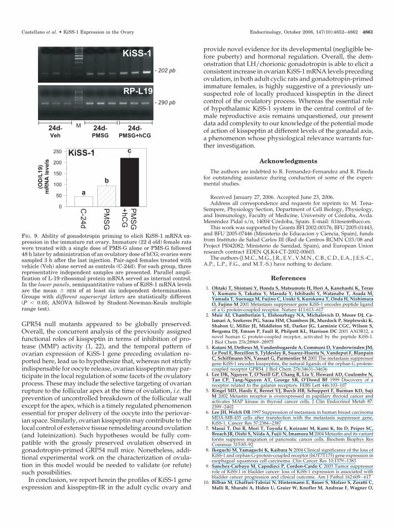
Collectively considered, previous data evidenced thatKiSS-1 is expressed in cyclic rat ovary, and its mRNA levelsdramatically increase preceding ovulation under the controlof pituitary LH. In this context, we found it relevant to definethe profile of expression and potential hormonal (gonado-tropin) regulation of KiSS-1 gene in the immature ovary.First, we assayed KiSS-1 mRNA levels in ovarian tissue fromjuvenile to peripubertal female rats. Our data showed that,in contrast to clearly detectable expression in adult ovariantissue at metestrus (taken for reference purposes), relativelevels of KiSS-1 mRNA were very low along the period ofprepubertal maturation studied, with faint expression be-tween d 20 and 26 postpartum and virtually negligible levelson d 29 and 32 of age (Fig. 8). Second, we investigatedwhether (low) expression of KiSS-1 gene in the immatureovary could be enhanced by a standard protocol of gonad-otropin priming, known to elicit ovulation in the prepubertalfemale (26). Immature rats (d 22 postpartum) were injectedwith an effective dose of PMS-G to induce follicular matu-ration followed 48 h later by an effective dose of hCG totrigger ovulation, assessment of KiSS-1 mRNA levels beingconducted 3 h after hCG injection. As shown in Fig. 9,whereas ovarian expression of KiSS-1 in 24-d-old femalesinjected with vehicle remained barely detectable (in keeping
with our previous results), gonadotropin priming by PMS-Gevoked a significant increase in ovarian KiSS-1 mRNA levelsthat was further enhanced by injection of an ovulatory doseof hCG.
Discussion
Compelling evidence has now defined the indispensablerole of the KiSS-1/GPR54 system in the control of develop-ment and function of male and female reproductive axis. Onthe basis of genetic, molecular, and pharmacological ap-proaches, this essential function is assumed to be primarily(if not exclusively) conducted at central hypothalamic levels,in which KiSS-1 neurons have been proposed as gatekeepersof the GnRH system (30). Although this contention is nowundisputed, a conspicuous finding is that expression ofKiSS-1 and/or GPR54 genes has been reported in differentperipheral tissues, including placenta and other reproduc-tive organs, such as the gonads (1, 21), for which the potentialfunctions of this system remain mostly unexplored. Based onpreliminary evidence showing prominent expression ofKiSS-1 gene (and to a lesser extent of GPR54) in the rat ovary(21), in the present study, we aimed to define the profile ofovarian KiSS-1 gene expression and kisspeptin-IR and iden-tify its potential regulation during development and by keyhormonal factors as a mean to initiate characterization of theputative role(s), if any, of locally produced KiSS-1 in ovarianphysiology.
Our analyses conclusively demonstrated, for the first time,that the genes encoding kisspeptin and its putative receptorGPR54 are persistently expressed in the adult rat ovarythroughout the different phases of the estrous cycle. Moreinterestingly, our current data evidenced that the profiles ofovarian expression of both targets are clearly distinct. Thus,whereas GPR54 mRNA levels remained low along the cycle,without significant fluctuations, KiSS-1 mRNA sharply in-
FIG. 6. Expression levels of KiSS-1 mRNA in ovaries fromrats treated with an effective dose of GnRH-ANT (5 mg/kg)or vehicle (Veh) at 1200 h on proestrus (P) and sampled at1 h (1300 h) and 6 h (1800 h) after treatment. For eachgroup, three representative independent samples are pre-sented. Parallel amplification of L-19 ribosomal proteinmRNA served as internal control. In the lower panels, semi-quantitative values of KiSS-1 mRNA levels are the mean �SEM of at least five independent determinations. Groupswith different superscript letters are statistically different(P � 0.05; ANOVA followed by Student-Newman-Keulsmultiple range test).
4858 Endocrinology, October 2006, 147(10):4852–4862 Castellano et al. • KiSS-1 Expression in the Ovary

creased in the ovary at 1800 h on proestrus and abruptlydecreased thereafter; only a moderate increase in KiSS-1mRNA (of much lower magnitude) was also detected ondiestrus. Of note, this sharp rise in ovarian KiSS-1 mRNA atproestrus appeared tightly coincident with the preovulatorysurge of gonadotropins, responsible for triggering ovulation,which in our animal stock takes place from approximately1600 h proestrus onward (our personal observation). Thistimely regulated profile of expression, in the context of theproposed role of kisspeptin in the control of proteases, suchas MMP-9, previously involved in the ovulatory process (23),is strongly suggestive of a potential, previously unsuspectedrole of locally derived KiSS-1 in the direct control of ovula-tion (see below). Yet persistent expression of KiSS-1 gene atother phases of the ovarian cycle pointed out that, unlikeother factors such as cyclooxygenase-2 and the epidermalgrowth factor family member epiregulin (see Ref. 31),kisspeptin might also be involved in ovarian functions otherthan ovulation.
Such a contention (i.e. potential role of KiSS-1 in ovulationas well as in other ovarian functions) appeared to be in good
agreement with our immunohistochemical analyses, whichdemonstrated specific kisspeptin-IR in different tissue com-partments of the cyclic ovary, including the theca layer ofgrowing and preovulatory follicles, theca- and granulosa-lutein cells of the corpus luteum, and the interstitial gland aswell as the ovarian surface epithelium and, apparently, theoocyte. Of particular note, follicular kisspeptin immuno-staining showed a striking cell- and stage-specific pattern ofdistribution along the estrous cycle, with granulosa cellsbeing totally negative for kisspeptin-IR from early estrus toearly proestrus. However, in late proestrus, kisspeptin signalbegan to appear also at the basal cells of the granulosa layerof preovulatory follicles. This phenomenon grossly coincideswith the observed rise in KiSS-1 mRNA levels at the time ofthe preovulatory LH surge, yet its functional relevance re-mains to be determined. In addition, clear-cut kisspeptin-IRwas detected at the corpus luteum in the cytoplasm of ste-roidogenic cells of theca and granulosa origin. Such lutealexpression of kisspeptin-IR may reflect the observed mod-erate increase in KiSS-1 mRNA levels during the diestrusphase. Considering the proven functional roles of kisspeptinsin other cellular backgrounds, it is tempting to propose thatexpression of kisspeptin at this site might be related to theintense tissue remodeling during formation and/or regres-sion of corpora lutea. Alternatively, locally producedkisspeptin might contribute to regulation of hormone secre-tion by steroidogenic luteal (and eventually, interstitial) cells,which are LH/chorionic gonadotropin responsive. Finally,the presence of kisspeptin in the oocyte, and its eventualfunctional relevance, merit further investigation, which ispresently in progress at our laboratory.
Mandatory for direct biological actions of kisspeptin, theexpression of GPR54 gene (albeit at apparently low levels)and the presence of GPR54-like IR were observed in the cyclicrat ovary. However, in contrast to KiSS-1, relative levels ofGPR54 mRNA appeared rather constitutively detected alongthe estrous cycle. This fact prevented us to conduct detailedimmunohistochemical analyses of GPR54 at all phases of theovarian cycle. Moreover, the incomplete homology (�68%)between the mouse and rat GPR54 sequences used for gen-eration of the (mouse) antiserum used in this study bringssome caution in the interpretation of our immunohistochem-ical results. Nonetheless, although it is assumed that addi-tional localization data (by means of in situ hybridizationand/or highly specific anti-GPR54 antisera, when available)would be ideally required to fully unravel the precise dis-tribution of GPR54, our current mRNA and protein resultsare highly suggestive of the presence of kisspeptin receptorsin the rat ovary, with a pattern of distribution that is grosslycoincident with that of the ligand itself. These observationssupport the plausibility of direct kisspeptin effects at thosesites of the ovary. Furthermore, potential indirect actions ofkisspeptins at other ovarian compartments (via intermediatecells expressing GPR54) cannot be excluded on the basis ofour present data.
The salient expression of KiSS-1 gene (and kisspeptin-IR)during the preovulatory period prompted us to study inmore detail the hormonal factors controlling ovarian KiSS-1mRNA levels during the proestrus-to-estrus transition.Blockade of the preovulatory surge of gonadotropins by
FIG. 7. Expression levels of KiSS-1 mRNA in ovaries from ratstreated with an effective dose of GnRH-ANT (5 mg/kg) at 1200 h onproestrus and coinjected with hCG (25 IU/rat) or vehicle. Ovariansamples were obtained at 1, 3, 6, and 24 h after hCG or vehicleinjection. Animals treated with GnRH-ANT alone showed unchangedKiSS-1 mRNA levels along the study period; three representativesamples obtained 3 h after injection of the antagonist (ANT) arepresented. In addition, for each time point after hCG treatment, threerepresentative independent samples are presented. Parallel ampli-fication of L-19 ribosomal protein mRNA served as internal control.In the lower panels, semiquantitative values of KiSS-1 mRNA levelsare the mean � SEM of at least five independent determinations.Groups with different superscript letters are statistically different(P � 0.05; ANOVA followed by Student-Newman-Keuls multiplerange test).
Castellano et al. • KiSS-1 Expression in the Ovary Endocrinology, October 2006, 147(10):4852–4862 4859

means of administration of GnRH-ANT, in a regimen knownto prevent ovulation (25, 29), was able to totally block the risein KiSS-1 mRNA levels at 1800 h on proestrus. In turn, ex-ogenous administration of hCG (as superagonist of LH) toGnRH-ANT-treated females elicited a significant increase inovarian KiSS-1 mRNA, which reached maximal levels 3 hafter hCG injection. These data indicate that, among otherbiochemical and cellular events leading to ovulation, the risein serum LH levels during proestrous afternoon drives atransient increase in ovarian expression of KiSS-1 gene. Yetwhether this is a direct LH action on KiSS-1-expressing cellsor it stems from LH-induced release of intermediate factorsawaits to be unveiled. Likewise, the hierarchical role of sucha phenomenon (LH-evoked KiSS-1 expression) in the cascadeof processes linked to ovulation cannot be ascertained on thebasis of our current data.
Further evidence for the potential involvement of KiSS-1in the local control of ovulation was provided by our studiesin immature female rats. These expression analyses indicatedthat during the prepubertal period, relative mRNA levels ofKiSS-1 in the rat ovary were low, whereas they significantlyincreased in the transition to the reproductive (cyclic) stateafter puberty. Of note, such a rise in KiSS-1 expression wasapparently preceded by a significant suppression of KiSS-1mRNA to virtually undetectable levels immediately beforepuberty onset (i.e. between d 29 and 32 postpartum); a phe-nomenon whose functional relevance merits further inves-tigation. Interestingly, implementation of a standard proce-dure for gonadotropin priming and induction of ovulationby administration of PMS-G and hCG to immature femalesnot only resulted in successful oocyte release (data notshown) but also evoked a significant elevation of ovarianKiSS-1 mRNA levels, whose magnitude was maximal aftercombined PMS-G and hCG treatment. Altogether, these find-
ings demonstrate that the immature ovary is capable to re-spond to appropriate gonadotropin priming with an increasein KiSS-1 gene expression preceding ovulation (as is appar-ently the case in adult rats), therefore suggesting that locallyproduced KiSS-1 might be involved in not only the ovulatoryprocess of adult cycling rats but also the induction of ovu-lation in immature females.
The questions arising from our present data are to whatextent ovarian KiSS-1 might play a role, distinct to that ofcentral kisspeptin, in the tuning of ovulation and the phys-iological relevance of that role, if any. Notably, the possibilityof potential peripheral actions of kisspeptin in the control ofthe reproductive axis has remained largely neglected, mostlydue to the fact that gonadotropin replacement appeared suf-ficient to rescue the profound hypogonadal state of GPR54null mouse models. Indeed, GPR54�/� female mice wereinduced to ovulate after standard gonadotropin priming(12), suggesting that KiSS-1 signaling at the ovary is notabsolutely mandatory for ovulation. Whereas this contentionis not disputed by our present data, our results stronglysuggest that kisspeptin might be involved in the local mod-ulation of some aspects of the ovulatory process. In thissense, it is to be noted that the qualitative and quantitativecharacteristics of ovulation in gonadotropin-primed GPR54null mice have not been apparently explored in detail (seeRef. 12). Thus, it remains possible that, despite successfuloocyte release after PMS-G and hCG treatment, gonado-tropin-primed GPR54�/� females might bear subtle (quali-tative and/or quantitative) alterations in ovulation.
A similar situation may apply also for the potential role ofKiSS-1 in placental physiology because, despite a wealth ofexperimental evidence having been presented suggesting arole of kisspeptin in trophoblast invasion (10, 21), placentalformation in fetal and gonadotropin-replaced maternal
FIG. 8. Profile of expression of KiSS-1 gene in immature ratovaries at different age points of prepubertal maturation.For each age point, three representative independent sam-ples are presented: 20-, 23-, 26-, 29-, and 32-d-old femalerats. For reference purposes, triplicate samples from adultcyclic ovaries at metestrus (M) are also shown. Parallelamplification of L-19 ribosomal protein mRNA served asinternal control. In the lower panels, semiquantitative val-ues of KiSS-1 mRNA levels are the mean � SEM of at leastsix independent determinations. Groups with different su-perscript letters are statistically different (P � 0.05; ANOVAfollowed by Student-Newman-Keuls multiple range test).
4860 Endocrinology, October 2006, 147(10):4852–4862 Castellano et al. • KiSS-1 Expression in the Ovary
GPR54 null mutants appeared to be globally preserved.Overall, the concurrent analysis of the previously assignedfunctional roles of kisspeptin in terms of inhibition of pro-tease (MMP) activity (1, 22), and the temporal pattern ofovarian expression of KiSS-1 gene preceding ovulation re-ported here, lead us to hypothesize that, whereas not strictlyindispensable for oocyte release, ovarian kisspeptin may par-ticipate in the local regulation of some facets of the ovulatoryprocess. These may include the selective targeting of ovarianrupture to the follicular apex at the time of ovulation, i.e. theprevention of uncontrolled breakdown of the follicular wallexcept for the apex, which is a timely regulated phenomenonessential for proper delivery of the oocyte into the periovar-ian space. Similarly, ovarian kisspeptin may contribute to thelocal control of extensive tissue remodeling around ovulation(and luteinization). Such hypotheses would be fully com-patible with the grossly preserved ovulation observed ingonadotropin-primed GRP54 null mice. Nonetheless, addi-tional experimental work on the characterization of ovula-tion in this model would be needed to validate (or refute)such possibilities.
In conclusion, we report herein the profiles of KiSS-1 geneexpression and kisspeptin-IR in the adult cyclic ovary and
provide novel evidence for its developmental (negligible be-fore puberty) and hormonal regulation. Overall, the dem-onstration that LH/chorionic gonadotropin is able to elicit aconsistent increase in ovarian KiSS-1 mRNA levels precedingovulation, in both adult cyclic rats and gonadotropin-primedimmature females, is highly suggestive of a previously un-suspected role of locally produced kisspeptin in the directcontrol of the ovulatory process. Whereas the essential roleof hypothalamic KiSS-1 system in the central control of fe-male reproductive axis remains unquestioned, our presentdata add complexity to our knowledge of the potential modeof action of kisspeptin at different levels of the gonadal axis,a phenomenon whose physiological relevance warrants fur-ther investigation.
Acknowledgments
The authors are indebted to R. Fernandez-Fernandez and R. Pinedafor outstanding assistance during conduction of some of the experi-mental studies.
Received January 27, 2006. Accepted June 23, 2006.Address all correspondence and requests for reprints to: M. Tena-
Sempere, Physiology Section, Department of Cell Biology, Physiology,and Immunology, Faculty of Medicine, University of Cordoba, Avda.Menendez Pidal s/n, 14004 Cordoba, Spain. E-mail: [email protected].
This work was supported by Grants BFI 2002-00176, BFU 2005-01443,and BFU 2005-07446 (Ministerio de Educacion y Ciencia, Spain), fundsfrom Instituto de Salud Carlos III (Red de Centros RCMN C03/08 andProject PI042082; Ministerio de Sanidad, Spain), and European Unionresearch contract EDEN QLK4-CT-2002-00603.
The authors (J.M.C., M.G., J.R., E.V., V.M.N., C.B., C.D., E.A., J.E.S.-C.,A.P., L.P., F.G., and M.T.-S.) have nothing to declare.
References
1. Ohtaki T, Shintani Y, Honda S, Matsumoto H, Hori A, Kanehashi K, TeraoY, Kumano S, Takatsu Y, Masuda Y, Ishibashi Y, Watanabe T, Asada M,Yamada T, Suenaga M, Fujino C, Usuki S, Kurokawa T, Onda H, NishimuraO, Fujino M 2001 Metastasis suppressor gene KiSS-1 encodes peptide ligandof a G protein-coupled receptor. Nature 411:613–617
2. Muir AI, Chamberlain L, Elshourbagy NA, Michalovich D, Moore DJ, Ca-lamari A, Szekeres PG, Sarau HM, Chambers JK, Murdock P, Steplewski K,Shabon U, Miller JE, Middleton SE, Darker JG, Larminie CGC, Wilson S,Bergsma DJ, Emson P, Faull R, Philpott KL, Harrison DC 2001 AXOR12, anovel human G protein-coupled receptor, activated by the peptide KiSS-1.J Biol Chem 276:28969–28975
3. Kotani M, Detheux M, Vandenbogaerde A, Communi D, Vanderwinden JM,Le Poul E, Brezillon S, Tyldesley R, Suarez-Huerta N, Vandeput F, BlanpainC, Schiffmann SN, Vassart G, Parmentier M 2001 The metastasis suppressorgene KiSS-1 encodes kisspeptins, the natural ligands of the orphan G protein-coupled receptor GPR54. J Biol Chem 276:34631–34636
4. Lee DK, Nguyen T, O’Neill GP, Chang R, Liu Y, Howard AD, Coulombe N,Tan CP, Tang-Nguyen AT, George SR, O’Dowd BF 1999 Discovery of areceptor related to the galanin receptors. FEBS Lett 446:103–107
5. Ringel MD, Hardy E, Bernet VJ, Burch HB, Schuppert F, Burman KD, SajiM 2002 Metastin receptor is overexpressed in papillary thyroid cancer andactivates MAP kinase in thyroid cancer cells. J Clin Endocrinol Metab 87:2399–2402
6. Lee JH, Welch DR 1997 Suppression of metastasis in human breast carcinomaMDA-MB-435 cells after transfection with the metastasis suppressor gene,KiSS-1. Cancer Res 57:2384–2387
7. Masui T, Doi R, Mori T, Toyoda E, Koizumi M, Kami K, Ito D, Peiper SC,Broach JR, Oishi S, Niida A, Fujii N, Imamura M 2004 Metastin and its variantforms suppress migration of pancreatic cancer cells. Biochem Biophys ResCommun 315:85–92
8. Ikeguchi M, Yamaguchi K, Kaibara N 2004 Clinical significance of the loss ofKiSS-1 and orphan G-protein-coupled receptor (hOT7T175) gene expression inesophageal squamous cell carcinoma. Clin Cancer Res 10:1379–1383
9. Sanchez-Carbayo M, Capodieci P, Cordon-Cardo C 2003 Tumor suppressorrole of KiSS-1 in bladder cancer: loss of KiSS-1 expression is associated withbladder cancer progression and clinical outcome. Am J Pathol 162:609–617
10. Bilban M, Ghaffari-Tabrizi N, Hintermann E, Bauer S, Molzer S, Zoratti C,Malli R, Sharabi A, Hiden U, Graier W, Knofler M, Andreae F, Wagner O,
FIG. 9. Ability of gonadotropin priming to elicit KiSS-1 mRNA ex-pression in the immature rat ovary. Immature (22 d old) female ratswere treated with a single dose of PMS-G alone or PMS-G followed48 h later by administration of an ovulatory dose of hCG; ovaries weresampled 3 h after the last injection. Pair-aged females treated withvehicle (Veh) alone served as controls (C-24d). For each group, threerepresentative independent samples are presented. Parallel ampli-fication of L-19 ribosomal protein mRNA served as internal control.In the lower panels, semiquantitative values of KiSS-1 mRNA levelsare the mean � SEM of at least six independent determinations.Groups with different superscript letters are statistically different(P � 0.05; ANOVA followed by Student-Newman-Keuls multiplerange test).
Castellano et al. • KiSS-1 Expression in the Ovary Endocrinology, October 2006, 147(10):4852–4862 4861

Quaranta V, Desoye G 2004 Kisspeptin-10, a KiSS-1/metastin-derived de-capeptide, is a physiological invasion inhibitor of primary human tropho-blasts. J Cell Sci 117:1319–1328
11. de Roux N, Genin E, Carel JC, Matsuda F, Chaussain JL, Milgrom E 2003Hypogonadotropic hypogonadism due to loss of function of the KiSS1-derivedpeptide receptor GPR54. Proc Natl Acad Sci USA 100:10972–10976
12. Seminara SB, Messager S, Chatzidaki EE, Thresher RR, Acierno JS, Sha-goury JK, Bo-Abbas Y, Kuohung W, Schwinof KM, Hendrick AG, Zahn D,Dixon J, Kaiser UB, Slaugenhaupt SA, Gusella JF, O’Rahilly S, Carlton MB,Crowley WF, Aparicio SA, Colledge WH 2003 The GPR54 gene as a regulatorof puberty. N Engl J Med 349:1614–1627
13. Navarro VM, Castellano JM, Fernandez-Fernandez R, Barreiro ML, Roa J,Sanchez-Criado JE, Aguilar E, Dieguez C, Pinilla L, Tena-Sempere M 2004Developmental and hormonally regulated messenger ribonucleic acid expres-sion of KiSS-1 and its putative receptor GPR54 in rat hypothalamus and potentLH releasing activity of KiSS-1 peptide. Endocrinology 145:4565–4574
14. Seminara SB 2005 Metastin and its G protein-coupled receptor, GPR54: criticalpathway modulating GnRH secretion. Front Neuroendocrinol 26:131–138
15. Yao HH, Bahr JM 1998 The ovary—overview. In: Knobil E, Neill JD, eds.Encyclopedia of reproduction. San Diego: Academic Press; 590–597
16. Greenwald GS, Roy SK 1994 Follicular development and its control. In: KnobilE, Neill JD, eds. The physiology of reproduction. New York: Raven Press;629–724
17. Ny T, Wahlberg P, Brandstrom IJM 2002 Matrix remodeling in the ovary:regulation and functional role of the plasminogen activator and matrix met-alloproteinase systems. Mol Cell Endocrinol 187:29–38
18. Robker RL, Russell DL, Espey LL, Lydon JP, O’Malley BW, Richards JS 2000Progesterone-regulated genes in the ovulation process: ADAMTS-1 and ca-thepsin L proteases. Proc Natl Acad Sci USA 97:4689–4694
19. Curry Jr TE, Osteen KG 2001 Cyclic changes in the matrix metalloproteinasesystem in the ovary and uterus. Biol Reprod 64:1285–1296
20. Young KA, Stouffer RL 2004 Gonadotropin and steroid regulation of matrixmetalloproteinases and their endogenous tissue inhibitors in the developedcorpus luteum of the rhesus monkey during the menstrual cycle. Biol Reprod70:244–252
21. Terao Y, Kumano S, Takatsu Y, Hattori M, Nishimura A, Ohtaki T, Shintani
Y 2004 Expression of KiSS-1, a metastasis suppressor gene, in trophoblast giantcells of the rat placenta. Biochim Biophys Acta 1678:102–110
22. Yan C, Wang H, Boyd DD 2001 KiSS-1 represses 92-kDa type IV collagenaseexpression by down-regulating NF-�B binding to the promoter as a conse-quence of I�B�-induced block of p65/p50 nuclear translocation. J Biol Chem276:1164–1172
23. Curry Jr TE, Song L, Wheeler SE 2001 Cellular localization of gelatinases andtissue inhibitors of metalloproteinases during follicular growth, ovulation, andearly luteal formation in the rat. Biol Reprod 65:855–865
24. Kinoshita M, Tsukamura H, Adachi S, Matsui H, Uenoyama Y, Iwata K,Yamada S, Inoue K, Ohtaki T, Matsumoto H, Maeda K 2005 Involvement ofcentral metastin in the regulation of preovulatory luteinizing hormone surgeand estrous cyclicity in female rats. Endocrinology 146:4431–4436
25. Nekola MV, Coy DH 1985 Direct and indirect inhibition of ovulation in ratsby an antagonist of luteinizing hormone-releasing hormone. Endocrinology116:756–760
26. Yoshioka S, Fujii S, Richards JS, Espey LL 2002 Gonadotropin-induced ex-pression of pancreatitis-associated protein-III mRNA in the rat ovary at thetime of ovulation. J Endocrinol 174:485–492
27. Castellano JM, Navarro VM, Fernandez-Fernandez R, Nogueiras R, Tovar S,Roa J, Vazquez MJ, Vigo E, Casanueva FF, Aguilar E, Pinilla L, Dieguez C,Tena-Sempere M 2005 Changes in hypothalamic KiSS-1 system and restora-tion of pubertal activation of the reproductive axis by kisspeptin in under-nutrition. Endocrinology 146:3917–3925
28. Tena-Sempere M, Barreiro ML, Gonzalez LC, Gaytan F, Zhang FP, CaminosJE, Pinilla L, Casanueva FF, Dieguez C, Aguilar E 2002 Novel expression andfunctional role of ghrelin in rat testis. Endocrinology 143:717–725
29. Caminos JE, Tena-Sempere M, Gaytan F, Sanchez-Criado JE, Barreiro ML,Nogueiras R, Casanueva FF, Aguilar E, Dieguez C 2003 Expression of ghrelinin the cyclic and pregnant rat ovary. Endocrinology 144:1594–1602
30. Dungan HM, Clifton DK, Steiner RA 2006 Kisspeptin neurons as centralprocessors in the regulation of GnRH secretion. Endocrinology 147:1154–1158
31. Sekiguchi T, Mizutani T, Yamada K, Kajitani T, Yazawa T, Yoshino M,Miyamoto K 2004 Expression of epiregulin and amphiregulin in the rat ovary.J Mol Endocrinol 33:281–291
Endocrinology is published monthly by The Endocrine Society (http://www.endo-society.org), the foremost professional society serving theendocrine community.
4862 Endocrinology, October 2006, 147(10):4852–4862 Castellano et al. • KiSS-1 Expression in the Ovary
Copyright © 2022 FDOKUMEN