OVULATION A SIGN OF HEALTH
35
OVULATION A SIGN OF HEALTH UNDERSTANDING REPRODUCTIVE HEALTH IN A NEW WAY Author Pilar Vigil MD, PhD, FACOG Contributors Juan Pablo del Río MD Natalia Molina BMed Pedro Gutiérrez MD Carolina Lyon Nrs Yanara Bernal Nrs Felipe G. Serrano BSc,. MSc Illustrations & Design Felipe G. Serrano For author correspondance: [email protected] Keywords: Women’s health, Reproductive health, Ovar- ian continuum, Ovulation, Biomarkers, Fertile window and Neurosteroids
-
Upload
khangminh22 -
Category
Documents
-
view
0 -
download
0
Transcript of OVULATION A SIGN OF HEALTH

OVULATION A SIGN OF HEALTHUNDERSTANDING REPRODUCTIVE HEALTH IN A NEW WAY
AuthorPilar Vigil MD, PhD, FACOG
ContributorsJuan Pablo del Río MDNatalia Molina BMedPedro Gutiérrez MDCarolina Lyon NrsYanara Bernal NrsFelipe G. Serrano BSc,. MSc
Illustrations & DesignFelipe G. Serrano
For author correspondance: [email protected]
Keywords: Women’s health,Reproductive health, Ovar-ian continuum, Ovulation, Biomarkers, Fertile window and Neurosteroids
World Youth Alliance
TM

2
TABLE OF CONTENTS
1. Introduction 2. Anatomy of ovulation
Cortex & medulla The ovarian follicles 3. Role of central nervous system in ovulation Hypothalamus Pituitary gland Kisspeptin and GnRH 4. Ovarian activity in the lifetime of woman Intrauterine life Puberty Reproductive age: The ovulatory menstrual cycle 5. Biomarkers and knowledge of the fertile window Estradiol LH Progesterone Cervical mucus Temperature Ultrasound monitoring 6. The cervical mucus in women’s health Cervical mucus as biomarker Cervical mucus as a tool to recognize ovulation
7. Ovulation as a marker of health status Ovulatory dysfunctions Hormones and the brain 8. Conclusion: the need for Reproductive Health
Research Institute (RHRI) 9. Summary

3
Ovulation a sign of health
1. IntroductionOvulation is the major event of the men-strual cycle. It requires a series of coor-dinated hormonal events to occur and it shows an adequate activity of the hypo-thalamic-pituitary-gonadal axis (HPG). In this sense, women should care about it as a sign of health (Vigil et al., 2017). The occurrence of ovulation is an activity that begins in adolescence and continues throughout a woman’s reproductive life until menopause. Thus, the study of ovu-lation is a powerful tool in order to assess women’s health status. The pattern of ovarian activity can un-dergo relevant changes during a wom-an’s life: this is known as the ovarian continuum (Brown, 2010). This term ex-plains the different variations in ovarian activity that start during intrauterine life, and are observed throughout the life of a woman in response to different physi-ological, behavioral and environmental conditions. According to this concept, under certain physiological conditions, such as pregnancy or breastfeeding, an anovulatory state is considered normal within the ovarian continuum (Brown, 2010; Pérez, 1998). Also, some periods of irregular ovulation, such as puberty and perimenopause, can be considered as part of a physiological transition. None-theless, certain pathological conditions can disrupt ovulation, such as unhealthy lifestyle habits, stress, endocrine abnor-malities, gynecological disorders, auto-immune disorders, genetic disorders, drugs (such as hormonal contraceptives) and iatrogenic causes (Vigil et al., 2017).Women can use different approaches in
order to identify the occurrence of ovula-tion. The use of biomarkers, such as cervi-cal mucus, helps women to identify ovu-lation and, therefore, their health status (Billings et al., 1972). The following paper explains the physiol-ogy of ovulation and how it can be un-derstood as a sign of hormonal balance and adequate health status that women can easily recognize. First, the paper ex-plores the anatomy and function of the main structures involved in a woman’s reproductive system. Second, it presents the coordinated interaction of these or-gans, with an emphasis in the role of the central nervous system as the main reg-ulator of the HPG axis. Third, it discuss-es what should be the hormonal balance and ovarian activity during the different periods of a woman’s lifetime, consider-ing the necessary steps needed for the physiological occurrence of ovulation. It continues by analyzing the different bio-markers that women can use in order to recognize ovulation. Then, it highlights the importance of cervical mucus within the different biomarkers women can use to recognize their ovulation and health status. In the section on ovulatory dys-functions, the paper considers the most common causes that can lead to impair-ment of ovulation. It also analyzes how endocrine abnormalities are the most common cause of ovulatory dysfunctions and the possible consequences of this physiopathological processes in other sys-tems, such as the central nervous system. Finally, the paper emphasizes the notion that there is an unmet need for research and development in this area of women’s

4
health, and how RHRI has contributed to the development of medical protocols that consider the timely diagnosis and proper treatment of ovulatory dysfunc-tions as a priority in order to improve the health and well-being of women. RHRI has been able to meet this need, but fur-ther support is needed in order to contin-ue research and spread this knowledge among healthcare providers.
2. Anatomy of ovulationA series of complex structures interact with each other in order to achieve ovula-tion. The most important anatomical and histological aspects involved in this pro-cess will be analyzed.The ovaries, in conjunction with the uterus and the oviducts (uterine tubes or Fallopian tubes), are part of the in-ternal female genitals, located in the pelvis minor. These are the female gonads and are responsible for the production of sex hormones. They are two organs located on either side of the uterus. In the ovaries, we find the follicles that contain the oocytes, the female germ cell involved in re-production. The development of the ovaries comes from the genital ridg-es (gonadal ridges) of the embryo, to-wards the end of the fourth week of gestation. Considering their histologi-cal characteristics we can distinguish a cortex and a medulla and the pres-ence of ovarian follicles on their dif-ferent stages of development.
Cortex & medulaThe cortex, the external layer of the ova-
ry, is called the superficial epithelium or germinal epithelium. Immediately under the superficial epithelium lies a small lay-er of collagen fibers, the tunica albuginea, which gives the ovarian surface its char-acteristic pearl white color. On the inside, there is a lax connective tissue, and the follicles appear in different developmen-tal stages. During the reproductive years, the cortex constitutes more than 50% of the total ovarian volume.The medulla is formed by a connective tissue called stroma that is less fibrous than the cortex. It is also constituted by abundant spiral, thick-walled, blood and lymphatic vessels, as well as nerves, all of which enter and exit the ovary through a region called hilum. The medulla also presents fibroblasts, mastocytes, elastic fi-bers and myocytes (Kühnel, 2004).
The ovarian folliclesA follicle is a structure formed by the oocyte (the germinal cell) surround-ed by somatic cells (granulosa cells). On the outside of these cells, and sep-arated by a basal lamina, are the the-ca cells (Schoenwolf & Larsen, 2009). Surrounding the oocyte we find a cellular structure called the zona pel-lucida, which is formed by glycopro-teins. The zona pellucida will play a very important role in fertilization and early development in the cycle.Ovarian follicles experience changes in their function and in their mor-phology, a process of development called folliculogenesis. According to their developmental stages, folli-cles can be classified as: primordial,
During reproduc-tive age, the cortex takes up more than 50% of the total ovarian volume. The cortex contains pri-mordial follicles.
Follicular atresia: is the degeneration of the germ cell and the follicular epithelium with its subsequent replacement for con-nective tissue (Lun-enfeld & Insler, 1993)
In an advanced phase of develop-ment, a follicle about to ovulate is formed by the theca externa, the theca interna, a basal membrane, several layers of gran-ulosa cells, the corona radiata, the zona pel-licida, the oocyte and an antral cavity.

5
Ovulation a sign of health
I. Primordial Follicle: It is an
oocyte surrounded by a single layer of
flat epithelial cells. It is located close to the
cortex.
III. Secondary Follicle: Also called preant-ral follicle. It contains an oocyte surrounded by a developed granulosa cells layers which synthesize estrogens and external theca cells layers which produce androgens. It is located closer to the vascularized medulla.
IV. Tertiary Follicle: It is characterized by the presence of an antrum rich in steroid and peptide hormones. The granulosa cells are subdivided into the parietal cells arranged in several layers and the cumulus oophorus. The surrounding connective tissue forms inter and external layers rich in blood capillaries and nerve endings.
V. Mature Follicle (or Graafian follicle): Also called the preovulatory follicle, it is significantly larger and has several layers of granulosa cells. It develops the corona radiata (granulosa cells around the zona pellucida).
VI. Corpus Luteum: It is formed by the luteinization process (inva-sion of the theca’s blood capillaries in the granulosa cells). It produces high levels of progesterone and medium levels of estrogen.
II. Primary Follicle: It is an oocyte surrounded by cuboi-dal granulosa cells that owes its growth to the influence of
androgens, independent of gonadotrophs.
VII. Corpus Albicans: It corresponds to the degradation of the corpus luteum in the absence of pregnancy, when the cells are replaced by connective tissue.
Figure 1: The ovarian follicles. According to their developmental stages, follicles can be classified as: primordial, primary, second-ary, tertiary, mature (Graafian), corpus luteum and albicans (Skinner, 2005; Gleicher, 2011).

6
primary, secondary, tertiary, mature (Graafian), corpus luteum and albi-cans (Figure 1) (Popa et al., 2008).Folliculogenesis has two stages: the early stage, called initial folliculogen-esis, which is independent of gonad-otropin hormones, and a secondary stage, which is cyclical and depen-dent on gonadotropin hormones. The initial folliculogenesis continuously occurs and involves the development of a primordial follicle until it reach-es the antral stage. It occurs from in-trauterine life until menopause when the ovary is depleted from ovarian follicles. The normal development of the first follicular process takes about 10 weeks. The second, or dependant stage, take place from the first ovu-lation through menopause. It occurs during the so-called follicular phase of the menstrual cycle and involves the growth of a follicle from the an-tral stage (tertiary follicle) until ovu-lation (the rupture of a Graafian fol-licle). This secondary process takes between 10 to 22 days, with an aver-age of 14 days.
3. Role of the central nervous system in ovulationThe central nervous system (CNS) is a specialized structure that controls the functions related to motor, behavioral, cognitive and neuroendocrine coordina-tion of the organism (Table 1). The sys-tem can be divided into two parts: the spinal cord and the brain. Hormones have an important effect upon the wir-ing of different brain areas, including
the hypothalamus and pituitary gland, which have an essential role in reproduc-tion (Figure 2). The brain is regulated by hormone feedback (Kandel et al., 2000). The main structures of the CNS involved in the regulation of ovulation are the hy-pothalamus and pituitary gland.
The hypothalamusThe hypothalamus is the region of the brain that helps to maintain the homeo-stasis of the body. It is located in the low-er part of the brain, and it is composed of different nuclei, of which the paraven-tricular, dorsomedial and infundibular nuclei are related to reproduction (Table 1) (Hrabovszky, 2014; Hrabovszky et al., 2010; Skorupskaite et al., 2014).
The pituitary glandThe pituitary gland is located at the base of the hypothalamus in the base of the brain, and it is surrounded by a bony cavity called the “sella turcica”. It is com-posed of two lobes, anterior and poste-rior. The anterior lobe (adenohypophy-sis) regulates processes linked to stress, growth, reproduction, and lactation. The posterior lobe (neurohypophysis) regu-lates lactation and ion balance in the kid-ney (Herbison, 2016; Kandel et al., 2000).Regarding ovulation, communication between the hypothalamus and the pi-tuitary gland depends upon kisspeptin and Gonadotropin Releasing Hormone (GnRH) neurons (Figure 3) (Herbison, 2016; Oakley et al., 2009).
Kisspeptin and GnRHKisspeptin is a neuropeptide recently identify (Popa et al., 2008). In humans, it is
The hypothalamus and pituitary gland are among the most important structures in the control of reproduction.

7
Ovulation a sign of health
synthesized by a group of hypothalamic neurons. These group of neurons, called the kisspeptinergic system, have signif-icant relevance in the onset of puberty (Herbison, 2016). The kisspeptinergic neu-rons communicate with the GnRH neu-rons in the hypothalamus. These neu-rons induce the secretion of GnRH into the hypophyseal portal system where it is transported to the anterior pitu-
Figure 2: Interaction between hormones and the brain. The brain controls functions related to motor, behavioral, cognitive and neuroendocrine coordination of the organism. Hormones contribute to induce changes in the wiring of different brain areas. These areas are regulated by hormones that provide feedback, enabling so there is a permanent communication between the nervous and endocrine systems.
Table 1: Hypothalamic regions related to control of the endocrine and reproductive axis.
itary gland, stimulating the production of gonadotropic hormones: Luteinizing Hormone (LH) and Follicle Stimulating Hormone (FSH). The gonadotropic hor-mones act upon the gonads: ovaries and testes (Oakley et al., 2009; Popa et al., 2008).
4. Ovarian activity during the life-time of womanThe ovarian continuum begins at the
The hypothalamic -pituitary axis is the essential connection between the various glands in the body with the nervous sys-tem, and it contributes to the regulation of the reproductive cycle.
HORMONES
Hypothalamic Nucleus Function
Periventricular NucleusRelated to thermoregulation and sexual
behavior.
Paraventricular Nucleus Commands the regulation of metabolic
processes in the body. Infundibular
Nucleus Regulates the onset of puberty.

8
Figure 3: Schematic illustration of the hypophysis. The hypophysis is composed of two lobes, anterior and posterior. The anterior lobe (adenohypophysis) regulates processes linked to stress, growth, reproduction and lactation. The posterior lobe (neurohypophysis) regulates lactation and ion balance in the kidney.
moment of fertilization when the zy-gote begins to develop. It continues throughout the entire life of a woman.
Intrauterine lifeWhen the embryo is approximately two months old, the primordial germ cells (precursor cells of the oocytes (PGC)) leave the embryo and migrate to a structure that is known as the vi-telline sac (yolk sac). This event occurs to avoid experiencing the process of cellular differentiation undergone by the rest of the embryo cells (differen-tiation into other tissues such as bone, liver or skin). PGCs remain in the vi-telline sac for about four weeks and then return to the region where the future ovary will develop: the genital or gonadal ridge. Here they transform into oogonia, cells that proliferate by mitosis (Schoenwolf & Larsen, 2009).The oogonias that are surrounded by ovarian somatic cells (pre-granulosa cells) will form primordial follicles and will differentiate into primary
oocytes. The remaining oogonias degenerate via an apoptotic process (programmed cell death), called atresia. During the seventh month of intrauterine life, the prima-ry oocytes begin meiosis, arresting this process at the stage of meiosis I (Arrau et al., 1981). Meiosis will be arrested until ovulation, when the meiotic process will be reinitiated. The ovarian follicles, with the oocytes inside, create a population of “reserve” or “resting” primordial folli-cles with which a girl will be born. Thus, a newborn girl will have 1 to 2 million follicles containing primary oocytes. This initial reserve will give origin to the follicles observed along the ovary vari-ous developmental stages (Lunenfeld & Insler, 1993). The oocytes meiosis will be arrested until ovulation when the meiot-ic process will be reinitiated. However, from the moment when the primordial follicles are formed, and during postnatal life, this initial follicle reserve will experience a remarkable de-crease in numbers: the primordial folli-
Zygote: Cell formed by the union of the male and the female gametes.
Gamete: Male or fe-male reproductive cell of a living being.
Mitosis: Type of division of the cell nucleus in which the genetic information contained in the chro-mosomes is preserved intact, which thus passes unchanged to the two resulting daughter cells.
Meiosis: Type of division that reduces the chrosomosome number by half, creating four haploid cells, each genetical-ly distinct from the parent cells that gave rise to them.
AN
TERI
OR LOBE POSTERIOR LOBE
Lactation
Ion balance in the kidney
Stress
Reproduction
Lactation
Growth

9
Ovulation a sign of health
cles that a girl has at birth (1 to 2 million) will be reduced to about 400 thousand in puberty, with only about 500 fully com-pleting folliculogenesis and ovulation. In other words, from the total primor-dial follicles developed, only less than 0.01% will ovulate (Figure 4).
PubertyPuberty is a process of physical, psy-chological and social changes through which a child matures into adolescence and adulthood. In general, female pu-berty starts at the age of 8 to 10, and its accompanying hormonal changes cul-minate with the expulsion of a mature oocyte from the ovary (i.e. the first ovu-lation), which generally leads to men-arche, the first menses (Grumbach, 2002).At the beginning of puberty (approxi-mately 8 to 10 years of age) girls should experience the physiological process known as adrenarche, an increase in ad-renal androgen secretion. This process
can be recognized by the appearance of axillary odor. Along with this, when a girl approaches puberty, the blood con-centration of leptin rises, due to a nat-ural increase in fat tissue at this age. At the onset of puberty, higher leptin levels promote kisspeptin secretion, which in turn stimulates GnRH secretion, with a consequent rise in the release of gonado-tropins (FSH and LH). As a result of the hormonal stimulation by gonadotropins, the gonads undergo a process of growth, development, and maturation called go-nadarche, which leads to higher secre-tions of sex steroid hormones (estrogens, progestogens and androgens) from the ovaries (Cortés et al., 2015).
Reproductive age: The ovulatory men-strual cycleOnce the reproductive system fully ma-tures, women between 12 and 50 years
Leptin: Protein hormone, mainly produced by white fat cells, whose main function is the regulation of energy balance, through the control of food intake (Warren et al., 1999; Hoff et al., 1983).
FSH (Follicle Stimulating Hor-mone): A gonado-tropin, synthesized and secreted by the gonadotrophic cells of the anterior pituitary gland. It regulates the development, growth, pubertal maturation, and reproductive processes of the body (Warren et al., 1999; Hoff et al., 1983).
Ova
rian
Rese
rve
Time (years)1 30 60
Figure 4: The ovarian reserve over the years. A girl will be born with 1 to 2 million primordial follicles. This initial follicular reserve will experience a remarkable decrease in numbers: the primordial follicles that a girl has at birth will be reduced to about 400 thousand in puberty, of which only about 500 will ovulate. In other words, from the total primordial follicles developed, less than 0.01% will ovulate.

10
of age normally exhibit regular ovula-tions characterized by 24- to 36-days cycles with fluctuating plasma estradi-ol and progesterone values according to the different phases of the cycle. For this, a series of sequential events have to occur in a highly synchronized man-ner, ovulation being the most important event during the menstrual cycle (Vigil et al., 2017). · The menstrual cycleThe first day of the menstrual cycle is considered to be the first day of menstru-ation and the last day is the one preced-ing the next menses (Brown, 2010; Cortés et al., 2015). It has been shown that 85% to 90% of healthy young women have a menstrual cycle duration that ranges
from 24 to 36 days, but the most frequent length is 27 ± 1 days (Fraser et al., 2007). Despite variability in one woman and among different women, the phases of the menstrual cycle are common for all women during reproductive years.The menstrual cycle can be divided into two phases (see Figure 10): follicular (es-trogenic or proliferative phase) and lu-teal (progestational or secretory phase). The first one is characterized by an in-crease in estradiol secretion by growing follicles. It starts with menstruation and lasts until ovulation. This phase is the most variable of the cycle, with an aver-age duration of 10 to 22 days for normal cycles. The length can be altered as a re-sult of different pathophysiological con-ditions (Vigil, 2017).
LH (Luteinizing Hormone): Is synthesized and secreted by the gonadotrphic cells of the anterior pituitary gland. In females, an acute rise of LH triggers ovulation and the development of the corpus luteum (Warren et al., 1999; Hoff et al., 1983).
Gonadarche: Gonadal changes during puberty and adolescence in response to pituitary gonadotropins.
Low frecuencycc /y Hi/ gh amplitude
OVULATION
GnRHpulsatility
High frequency cc / L/ ow amplitude
Rela
tive
hor
mon
e co
ncen
trat
ion
chan
ges
duri
ng t
he c
ycle
FOLLICULAR PHASE LUTEAL PHASE
OV
ULA
TIO
N
SELECTIOND OMINANCERECRUITMENT CORPUS LUTEUM FORMATION AND FUNCTIONL UTEOLYSIS
MENSES PROLIFERATIVE ENDOMETRIUM (E) SECRETORY ENDOMETRIUM (P) (ischemial)
E2
LH
Inhibin
Progesterone
FSH
Day 1 2 3 4 5 6 7 8 9 10 11 12 13 14 15 16 17 18 19 20 21 2 2 23 2 4 25 2 6 27 2 8
Figure 5: Ovulatory menstrual cycle. Changes in relative levels for each hormone according to cycle phases and endometrial characteristics are shown.

11
Ovulation a sign of health
The luteal phase begins after ovulation and goes until the day before next men-ses. It is characterized by an increase in progesterone production by the corpus luteum. This phase shows less variability, having a duration between 11 to 17 days (Blackwell et al., 2013; Brown, 2010).The coordinated hormonal events re-quired for ovulation are reviewed next (Figure 6). · First event:At the beginning of each cycle, there is an increase in FSH levels that cause recruit-ment and development of antral follicles (early tertiary follicles). This recruitment and further development takes place due to the induction of hormone receptors in the follicular cells. FSH also triggers the expression of various enzymes and pro-teins involved in the biosynthesis of sex steroids (steroidogenesis) which leads to a progressive rise in estrogen production and secretion.
· Second event:After follicular recruitment, estradiol along with inhibin (produced by the de-veloping follicles) exert a negative feed-back mechanism upon the HPG axis that causes a decrease in FSH levels. During this period, estradiol also inhib-its kisspeptin expression in the arcuate nucleus of the hypothalamus. Therefore GnRH and gonadotropin production diminishes, enabling the follicle that is able to survive under these unfavor-able hormonal conditions, to become the dominant follicle. The rest of the follicles degenerate (Lunnenfeld & Insler, 1993). By this mechanism, a dominant follicle is
selected from the follicular cohort that was recruited at the beginning of the fol-licular phase of the cycle. This dominant follicle will continue to produce estrogen and inhibin in higher concentrations.
· Third event:The dominant follicle produces in-creasingly higher levels of estradiol, which stimulate kisspeptinergic neu-rons in the anteroventral periven-tricular nucleus of the hypothala-mus, thus switching the negative feedback mechanism to a positive one. Kisspeptin induces GnRH secre-tion and the pre-ovulatory LH peak, which initiates follicular luteiniza-tion leading to the formation of the corpus luteum. Before the initiation of the midcycle gonadotropin surge a pre-ovulatory rise in progesterone occurs. This early progesterone rise produced by the pre-ovulatory folli-cle is critical for: a) follicular rupture, a necessary process for ovulation and b) development of a functional corpus luteum. Through positive feedback, progesterone maintains the LH peak, triggers meiosis resumption and the rupture of the follicle, with the con-sequent release of the oocyte (Hoff et al., 1983). This last event is known as ovulation. The released oocyte will typically survive 12 to 24 hours (Oak-ley et al., 2009).
· Fourth event:The period after ovulation is known as the “luteal phase”. Normally it lasts for 11 to 17 days (Brown, 2010). During this period, LH and progesterone contribute

12
Figure 6: The five events of ovulatory menstrual cycles. Women between 12 and 50 years of age normally exhibit regular ovu-lations characterized by 24- to 36-days cycles with fluctuating plasma FSH, LH, estradiol and progesterone values according to the different phases of the cycle.
FSH Progesterone
Days
Hor
mon
e Co
ncen
trat
ion
Hor
mon
e Co
ncen
trat
ion
Hor
mon
e Co
ncen
trat
ion
Hor
mon
e Co
ncen
trat
ion
Hor
mon
e Co
ncen
trat
ion
Estrogen LH
Four
th e
vent
Thir
d ev
ent
Seco
nd e
vent
Firs
t eve
ntFi
fth
eve
nt

13
Ovulation a sign of health
to the development and maintenance of the corpus luteum, which continues to produce progesterone and estrogen during the luteal phase. The levels of estrogen and progesterone produced by the corpus luteum will exert a negative feedback upon the HPG axis.
· Fifth event:If fertilization does not occur, the cor-pus luteum starts to regress after 6 days, lasting for 11 to 17 days. This regression causes a drop in estrogen and proges-terone levels. The decrease in both hor-mones eliminates the suppression ex-erted on the HPG axis and a new cycle begins (Vigil et al., 2006, 2017).
5. Biomarkers and knowledge of the fertile windowThe fertile window is the period of the menstrual cycle during which concep-tion is most likely to occur. This period usually begins about 6 days before ovu-lation and extends past the day of ovu-lation (Wilcox et al., 2000, 1995). This is determined by the lifetime of gametes. Spermatozoa can survive for at least six days in the cervix when estrogenic mu-cus is present. Mature human ova have a more limited lifespan of 12 to 24 hours (Royston, 1982). Recognizing ovulation enables women and couples to identify the day when the probability of conception reaches its peak. Nonetheless, this knowledge does not allow them to identify the beginning of their fertile window. For this purpose women need to use biomarkers associat-ed with the opening of the fertile win-dow. To recognize both the beginning
and the end (including the day of ovu-lation) of the fertility window enables a woman and her partner to use the infor-mation to achieve a pregnancy, postpone a pregnancy or to track her health.Biomarkers can be used by most wom-en to identify their ovulation and in this way determine the fertile and infertile periods of the menstrual cycle (Figure 7). This process is often referred to as fertility awareness. Biomarkers such as cervical mucus, basal body temperature (BBT) and estradiol, LH, and progester-one measured in plasma or urine can be used for this purpose. There are also dif-ferent apps available that use their own algorithms, but not all of them consider the great variability of the fertile win-dow (Duane et al., 2017; Gross, 1989).
EstradiolThe first significant increase either in plasma estradiol or its urinary me-tabolites (including estrone glucuro-nide) from baseline values is taken as a biochemical marker for the begin-ning of the potentially fertile phase of the menstrual cycle (Blackwell & Brown, 1992). A logarithmically increasing rate of plasma estradiol excretion (or of its urinary metabolites) indicates that the dominant follicle has entered its rapid growth phase (Blackwell & Brown, 1992).Estradiol has a logarithmic increase from baseline of approximately 1.5 times per day for 5 days to reach the estrogen peak, which occurs 24 to 36 hours before ovulation (Boyers, 1980; Vigil 2012). The fall after the peak is a very clear signal for timing ovulation (Brown et al., 1991). It is important to remark that it is possible
The fertile window is the period of the menstrual cycle dur-ing which conception is most likely to occur.

14
to find this type of estrogen peak with-out ovulation.
LHThe LH peak is responsible for trigger-ing the mechanisms that will cause folli-cle rupture and therefore ovulation. The LH surge lasts 48 hours, and ovulation occurs 32 to 35 hours after its initiation, 17 hours after the LH peak in plasma and generally the same day or the next day of its peak in urine (Vigil et al., 1992).It is important to consider that the LH surge may be missed with the use of some urinary LH kits (Ecochard, 2001) as its configuration, amplitude, and dura-
tion are variable (Alliende, 2002; Park et al., 2007). LH is an important tool for predict-ing ovulation, but it doesn’t identify the beginning of the fertile window and can-not be used to confirm ovulation.
ProgesteroneProgesterone secretion from follicular cells increases by a factor of 4 before the LH surge (Hoff et al., 1983). This initial in-crease in progesterone maintains the LH plateau during the LH peak. Progesterone also contributes to the final sequence of follicular events, ending in follicular rup-ture and ovulation (Baranczuk & Fainstat, 1976).
LHPeak
F st mucussympt s
Pre- ulat y
BBBB TBB rirr sii ess
PeakDay
Progest e rise
E2Peak
E2EE rirr sii essPrPP orr goo egg see tsstt e rirr sii ess
PrPP orr goo egg see tsstt e
Es adiLH
F liculrupt e by
US
BBT nad
Figure 7: Biomarkers used to determine the fertile window. Biomarkers can be used by most women to identify their ovulation and in this way determine the fertile and infertile periods of the menstrual cycle. Biomarkers such as cervical mucus, basal body temperature (BBT) and urine or plasma measurements of estradiol, LH, and progesterone can be used for this purpose.
It is always impor-tant to consider that the fertile window and ovulation vary in different women and in different cycles in a woman. For this reason, ovulation day should not be calcu-lated based on the first day of menstrual bleeding.
Biomarkers such as cervical mucus, basal body temperature (BBT), estradiol, LH and progesterone can be used by women to identify their fertile window and ovula-tion.

15
Ovulation a sign of health
This small rise in progesterone is also useful to predict ovulation and find the best moment of the cycle to achieve a pregnancy (Blackwell et al., 1998).After ovulation takes place, a massive rise in progesterone occurs, so ovulation can be confirmed measuring plasma pro-gesterone or pregnanediol glucuronide (PdG) in urine. The rise in the PdG excre-tion rate over a universal threshold val-ue of 7.0 mmol/24h (Blackwell et al., 1998, 2003, 2013, 2016; Brown et al, 1991; Brown, 2010) is a hormonal marker of the begin-ning of the post-ovulatory infertile phase and that the fertile window is closed. The PdG cutoff value frequently is reached on the day after ovulation, but this can hap-pen 2 or 3 days after (Brown et al., 1991). The specificity and accuracy of PdG val-ues when used to confirm ovulation are nearly 99% (Blackwell et al., 2003).
Cervical mucusThe beginning of the fertile window oc-curs with the first statistically significant rise in estrogen levels. This causes the secretion of estrogenic cervical mucus with the characteristic changes in the vaginal discharge (Blackwell & Brown, 1992; Billings et al., 1972). During this pe-riod, the mucus is aqueous, transparent, fluid and crystalline, giving the woman a slippery sensation at the vulva.The ultrastructure of cervical mucus var-ies during the menstrual cycle, exhibiting a lax network with channels that increase in size as ovulation nears (Chretien & Du-bois, 1991; Poon & McCoshen, 1985). In the peri-ovulatory period, this network al-lows sperm selection and ascent (Vigil et al., 1991, 2008, 2009, 2011). The post-ovu-
latory rise in progesterone has an anti-es-trogenic effect on the cervix, changing the cervical mucus to a form inappropriate for sperm ascent through the cervix (Vigil et al., 1991; Brown, 2010). The fertile window defined by the mucus peak symptoms starts on the first day of the presence of cervical mucus observed or felt at the vul-va and ends on the third day after the last day of slippery sensation at the vulva. This day closely relates to ovulation day. There is a 96% sensitivity of the cervical mucus symptoms in identifying the entire fertile window (Ecochard et al., 2015).
TemperatureDuring the menstrual cycle, BBT rises after ovulation as a result of ovarian ac-tivity, associated with progesterone ac-tion (de Mouzon et al., 1984). During the follicular phase of the menstrual cycle, BBT remains in a lower range until ap-proximately one day before ovulation, when the BBT often reaches the lowest point (nadir) (Hsiu-wei et al., 2017). Ovu-lation is confirmed when three consec-utive temperatures are 0.5-1.0 degree F above the highest point of the six previ-ous basal temperatures (Royston, 1982). After these three days, the fertile win-dow is considered closed.Measuring her BBT allows a woman to confirm that the fertile window has ended. This method doesn’t predict ovulation or the beginning of the fertile window.
UltrasoundUltrasound assesses the anatomy of fol-licular development and its subsequent rupture. There are some ultrasound indi-
Useful markers to determine the be-ginning of the fertile window are estradiol and cervical mucus.
Useful markers to predict ovulation are LH, estradiol, progesterone, cervical mucus and ultra-sound.
Useful markers to determine the end of the fertile window are progesterone, cervical mucus, ultrasound and BBT.

16
cators of ovulation, such as the disappear-ance of the follicle or decrease in follicular size, free fluid in the pelvis, or a change in endometrial characteristics (Hsiu-wei et al., 2017). The combination of ultrasonog-raphy and hormone assays, the one pro-viding anatomical information and the other providing functional information, has been a powerful tool for investigat-ing ovarian activity (Brown, 2010).
6. The cervical mucus in women’s healthCervical mucus is a crucial biological se-cretion that performs several functions related to reproductive processes. It is a hydrogel formed by mucins, which are proteins that have the ability to form gels. It is produced in the epithelial cells of the cervix of the uterus, and it appears with different rheological properties: sticky or slippery, different viscosities and translu-cent or opaque (Morales et al., 1993; Ceric et al., 2005).As a hydrogel, cervical mucus has two phases: an aqueous phase and a gel-like phase. The aqueous phase, also known as
soluble fraction, is composed of water and other compounds such as electrolytes, fructose, glucose, proteins, and lipids. Its hydration varies between 90 to 99% de-pending on the levels of estradiol (Vigil, et al. 1991). The gel phase, also called solid or insoluble fraction, is made up of mucins, the high molecular mass glycoproteins that give the cervical mucus its character-istic structural and biophysical properties (Ceric et al., 2005; Gipson, 2005; Sheehan & Carlstedt, 1990).
Cervical Mucus as a biomarkerThe observation of changes in cervical mucus is considered a reliable biomark-er (Gibbons, 1981), as it has been demon-strated that recognizing mucus patterns can help women to identify the different stages of the ovarian continuum (Figure 8) (Billings et al., 1972; Vigil et al., 2006).Cervical mucus undergoes several mod-ifications during the phases of the repro-ductive cycle. Increased estrogen levels halfway through the follicular phase re-sult in a noticeable rise in the secretion of estrogenic mucus.
Cervical mucus performs critical functions in the reproductive health of women, such as:
Protecting a woman’s reproductive tract, by maintaining a moist and lubricated environment.
Participating in sperm transport, facilitating their ascent to the fertilization site.
Acting as a selective barrier, selecting only the morphologically normal sperm.
Inhibiting the ascent, invasion and proliferation of microorganisms.
Modulating the acrosome reaction.
Table 2: Functions of cervical mucus (Vigil, 2006, 2007, 2009, 2014, 2011; Barros, 1983, 1984; Ceric et al, 2005)
Cervical mucus is a crucial biological se-cretion that performs several functions re-lated to reproductive processes.
The hydration of the cervical mucus will vary depending on estradiol levels.
The observation of changes in cervical mucus is considered a reliable biomarker.

17
Ovulation a sign of health
The last day of clear, slippery and lu-bricative mucus is defined as the peak day (PD) (Billings et al., 1972). Forty to fifty percent of women ovulate within 24 hours of the PD, but ovulation can occur from three days before to three days after the peak day in 95% of wom-en (Ecochard et al., 2015). After the peak day, the luteal phase begins.Progesterone has the opposite effect of estradiol upon cervical mucus (antiestro-genic action). It inhibits production and changes the characteristics of the mucus to an opaque and less fluid mucus, with-out the ability to crystallize into palm leaf patterns (Odeblad et al., 1994). Due to the rise in progesterone levels, a denser network is observed in the luteal phase, with small pore diameters compared to estrogenic cervical mucus. Spermatozoa, in the absence of estrogenic mucus, will
die within hours or even minutes when placed in the vagina. Cervical mucus as a tool to recognize ovulationAs we have seen, two main types of cervical mucus have been described: estrogenic and progestagenic. The first one predominates in the follicular phase. The second one is characteris-tic of the luteal phase, which normally lasts 11 to 17 days (Brown, 2010).It has been shown that the luteal phase extends for 11 to 17 days when ovula-tion is calculated from the estrogen peak to the day before the ensuing bleeding (Brown, 2010; Blackwell et al., 2013). When the luteal phase is calculat-ed from the PD, and knowing the vari-ation of ± three days between PD and ovulation, the luteal phase length could
Days 1 2 3 4 5 6 7 8 9 10 1 1 12 1 3 14 1 5 16 1 7 18 1 9 20 2 1 22 2 3 24 2 5 26 2 7 28
Figure 8: Cervical mucus. Changes in cervical mucus, as perceived by women, during the menstrual cycle (Vigil et al., 2006, 2007, 2009, 2014, 2011; Barros et al., 1983, 1984).
Ovulation
The last day of clear, slippery and lubricative mucus is defined as the peak day.
Progesterone changes the charac-teristics of the mucus to opaque, less fluid and without its abil-ity to crystallize into palm leaf patterns.
When estradiol levels are high, mucus is aqueous, transparent, fluid and crystalline, tending to form geometric pat-terns with fern leaves at crystallization.

18
have an estimated length between 9 to 19 days. In this last scenario, it is im-portant to consider that this variation in the calculated length of the luteal phase is given by the difference that methods have in the accuracy of identifying ovu-lation, not in the luteal phase per se. The pre-ovulatory rise and fall of estrone glucuronide followed by a pregnanedi-ol glucuronide rise is the most accurate way to identify ovulation and the luteal phase (Dunson et al., 2002).By noting the progression from the ba-sic infertile pattern of post-menstrual dryness to stickiness, wetness, and ulti-mate lubrication (“peak day”), a woman can recognize her ovulation (Billings et al., 1972; Vigil et al., 2006). When using cervical mucus as a biomarker of ovu-lation the indicator that ovulation has occurred is the identification of a lute-al phase according to the peak mucus symptom.
7. Ovulation as a marker of health statusOften, healthcare providers have focused on regularizing bleeding patterns, with-out paying attention to ovulation in re-productive age women. It has been shown that varying cycle lengths, whether short or long, are associated with decreased fe-cundity, and that menstrual cycle patterns may predict whether a pregnancy will survive (Kolstad et al., 1999). However, menstrual cycles with a normal length are not an indicator of proper ovarian func-tion, because these women can also pres-ent anovulatory cycles (Prior et al., 2015). Therefore, it is regular ovulation and not regular menstruation which provides ev-
idence of good health. Normal ovulatory activity during reproductive years implies adequate endocrine and gonadal function. However, women who are breastfeeding or pregnant should also identify their anovulatory state as a healthy part of the ovarian continuum. Periods of transition from anovulation to regular ovulation, such as those found during puberty and perimenopause, can also be identified as a physiological part of the continuum.Monitoring the ovulatory cycle should begin in puberty and adolescence. The absence of normal ovulatory cycles can be the first manifestation of some underlying pathology. For example, precocious or de-layed puberty can be linked to endocrine abnormalities (Stanhope & Brook, 1986). Because the conditions that alter ovula-tion during adolescence will only wors-en with time, early diagnosis and proper treatment are important for the future prognosis of the underlying health prob-lems (Vigil et al., 2006, 2007; Popat et al. 2008). Importantly, it has been shown that perimenarcheal girls from diverse eth-nic and socioeconomic groups are able to learn how to recognize their cervical mu-cus patterns and to use this information to distinguish normal from abnormal cycles (Klaus & Martin 1989).Normal cycles are those that last 24 to 36 days with an identified luteal phase ac-cording to the mucus peak symptom. Ab-normal cycles are short cycles (less than 24 days), long cycles (more than 36 days), or normal length cycles with a short luteal phase (less than 9 days), or its absence.A woman should be referred to a spe-cialist if she identifies three or more abnormal cycles in a year or two con-
When using cervical mucus as a biomarker of ovulation the indi-cator that ovulation has occurred is the identification of a lu-teal phase according to the peak mucus symp-tom.
Normal ovulato-ry activity during reproductive years implies adequate en-docrine and gonad-al function. Regular ovulation is evidence of good health.

19
Ovulation a sign of health
secutive abnormal cycles. As previously mentioned, the first sign of an underly-ing health problem may be a long follic-ular phase, or anovulation, with normal bleeding patterns, followed later-on by regular menses or amenorrhea (Vigil et al., 2006, 2017).The normal ovulatory activity that should be present during reproductive years will affect the body as a whole, including the central nervous system (CNS). In this way, the same hormones that influence ovula-tion have an impact upon the brain. This means that a proper hormonal balance during the different stages of life will give women and health care providers an im-portant tool for improving their health, in-cluding mental health and well-being. In the following sections, the most im-portant causes of ovulatory dysfunction and the relationship between hormones and the brain will be introduced.
Ovulatory dysfunctionsThe first sign of an underlying health problem a woman may experience is ovulatory dysfunction, followed by ir-regular cycles or amenorrhea. Indeed, when pregnancy, lactation, or meno-pause are not the causes, persistent irregularities in the ovulatory cycle can be associated with lifestyle, stress, drugs, and endocrine, gynecological, autoimmune, nutritional, genetic, and iatrogenic disorders (Vigil et al., 2006).The most frequent causes of menstrual irregularities associated with ovulato-ry dysfunctions are hormonal abnor-malities. These can be hypothalamic, pituitary, thyroid, adrenal, ovarian or metabolic disorders. The most com-
mon causes of endocrine abnormalities that lead to ovulatory dysfunction will be analyzed.
· Insulin resistanceHyperinsulinemia is commonly linked to insulin resistance and obesity. Elevat-ed insulin levels generate an increase in
Monitoring the ovu-latory cycle should begin in puberty and adolescence. The ab-sence of normal ovu-latory cycles can be the first manifestation of some underlying pa-thology.
Abnormal cycles are short cycles (less than 24 days), long cycles (more than 36 days), or normal length cycles with a short luteal phase or its absence.
A proper hormonal balance during the different stages of life will give women and health care providers an important tool for improving their health, including mental health and well-being.
The first sign of an underlying health problem a woman may experience is ovulatory dysfunc-tion, followed by irregular cycles or amenorrhea.
The most frequent causes of ovu-latory dysfunction are hormonal abnormalities. Of cohort of 125 young women between 18 and 35 years presenting with ovulatory dysfunction the diagnosed endo-crine abnormalities were: insulin resistance 36%, hyperandrogene-mia 23%, hyperprolactinemia 21%, hypothyroidism 16%, premature ovarian senescence 3 % and hypo-thalamic amenorrhea 1% (Vigil, unpublished data).
androgen production at the ovary, lead-ing to follicular atresia and the formation of multiple ovarian follicular cysts (Dia-manti-Kandarakis, 2006). In conjunction, high levels of insulin and androgens will decrease sex hormone-binding globulin (SHBG) levels and, as a consequence, the free fraction of steroid hormones, such as estrogens, will increase (Kalme et al., 2003). The combination of these alterations produces a hyperestrogenic and hyperandrogenic environment that characterizes hyperinsulinemia. Ele-vated estradiol levels may inhibit kiss-peptin, decreasing GnRH and gonado-tropins (FSH and LH). · HyperandrogenemiaAndrogens are a group of hormones

20
usually linked to development and maintenance of male sex characteristics. In women, androgens also exert import-ant physiological functions related to bone mineralization (Notelovitz, 2002), muscle development (Notelovitz, 2002), cognition and memory (Hirshman et al., 2004) as well as the appearance of libi-do (Basson et al., 2010). Nowadays, it has been shown that androgens have an important role in the gonadotropin-in-dependent phase of early follicular de-velopment (Gleicher, 2011). The main causes of hyperandrogenemia are func-tional (an increase in the production of androgens by the adrenal gland or the ovaries), peripheral (for example as in hyperinsulinemia), tumoral (as adrenal or ovarian tumours (Azziz et al., 2004; Rosenfield, 1996) and secondary to phar-macological treatments (Azziz et al., 2004). Polycystic ovary syndrome (PCOS) is the most common endocrine disorder in women (Amer, 2009), and it is con-sidered a functional cause of hyperan-drogenemia. These patients may exhib-it acne, hirsutism, alopecia, increased body weight, and mood changes. But the most common perceived symptom is the presence of irregular menstrual cycles and an atypical pattern of cervi-cal mucus. Both symptoms are associat-ed to ovulatory dysfunction (Vigil et al., 2009). Obesity, insulin resistance, and consequent hyperinsulinemia are high-ly prevalent comorbidities of PCOS and can impair ovulation (Vigil et al., 2007; Pauli et al., 2011). Elevated insulin lev-els are present in about half of these pa-tients and are correlated to body mass
index (BMI), but not all PCOS patients are insulin resistant (Vigil et al., 2007). High insulin levels further increase androgen production by stimulating ovarian theca cells to produce more androgens, which lead to premature follicular atresia and even anovulation (Diamanti-Kandarakis, 2006). High lev-els of testosterone and insulin will de-crease SHBG, increasing the free estradiol fraction. This, together with an increase in peripheral production of estrogens by adipose tissue may inhibit the kisspep-tinergic system decreasing GnRH and gonadotropins. PCOS is also associated to an increased risk of type 2 diabetes, met-abolic syndrome (Ranasinha et al., 2015), cardiovascular disease, and endometrial, ovarian, and/or breast cancer (Fauser et al., 2012).Another functional cause of hyper-androgenemia is congenital adrenal hyperplasia (CAH). This is a family of disorders caused by mutations in genes that encode for enzymes in-volved in one of the various steps of adrenal steroid synthesis, leading to an overproduction of androgens (Merke & Bornstein, 2005; Lekarev et al., 2015).Finally, certain drugs can lead to hy-perandrogenemia. The exogenous ad-ministration of androgenic derivatives (exogenous testosterone) is the most common cause, but the use of psychiat-ric medication can also lead to andro-gen excess. Antiepileptics drugs such as valproate (Rasgon et al., 2005), mood stabilizing agents such as valproate or lithium (Rauchenzauner et al., 2014) and antipsychotic agents such as risperi-done and quetiapine, promote the ad-
Insulin is a peptide hormone produced by pancreatic β-cells. It is one of the main modulators of carbo-hydrate and lipid me-tabolism, promoting the entry of glucose to tissues such as mus-cle, liver and adipose tissue.
Androgens in wom-en are mainly pro-duced in the ovaries and in the adrenal glands.
Polycystic ovary syndrome (PCOS) is the most common endocrine disorder in women. These pa-tients present ovu-latory dysfunction associated with high androgen levels. Symptoms such as acne, hirsutism, alo-pecia, increased body weight, and mood changes are usually observed.

21
Ovulation a sign of health
renal and ovarian androgen synthesis. This is due to an induced-hyperinsu-linemia and insulin-resistant state, to-gether with an enzymatic disruption (Bahtiyar & Weiss, 2007). · HyperprolactinemiaProlactin is produced by the pituitary gland and is under hypothalamic con-trol. Stress (Johansson et al., 1983), pituitary tumors (prolactinomas) and some drugs (such as antidepressants, antipsychotics, and proton-pump inhibitors) are causes of increased prolactin levels. Hyperpro-lactinemia inhibits GnRH by negative modulation of kisspeptinergic neurons. Common symptoms and signs of hy-perprolactinemia are short luteal phases, anovulation, menstrual irregularities, amenorrhea, galactorrhea, skin dryness, immunological disorders, low libido, hot flashes, and sweaty hands (Higuchi et al., 1984; Araujo-Lopes et al., 2014). High pro-lactin levels also activate adrenal andro-gen secretion, causing higher androgen and lower estradiol levels.It is useful to know that this hormone has an immunostimulatory effect, promot-ing autoimmunity (Orbach, & Shoenfeld, 2007). The association between hyper-prolactinemia and certain immune dis-eases such as lupus erythematosus and rheumatoid arthritis has been described (Jara et al., 2011). In this way, ovulatory dysfunctions can be an early signal of autoimmune diseases.
· Hypo and HyperthyroidismThyroid hormones also influence ovula-tion, mainly acting upon folliculogen-esis and steroidogenesis at the ovarian
level, and by affecting SHBG and GnRH secretion.Hypothyroidism, an insufficient amount of thyroid hormones, leads to irregular menses, hypermenorrhea, metrorrha-gia, spotting and breakthrough bleeding (Krassas et al., 1994, 1999), usually asso-ciated with lethargy, depressive symp-toms, weight gain, cold intolerance, and hair loss. Hypothyroid women have lower levels of testosterone and estro-gens (Krassas et al., 2010). SHBG levels are also diminished in these patients, leading to an increased free fraction of sex hormones. The increased estrogen free fraction triggers negative feedback on kisspeptinergic neurons and the pi-tuitary gland. Additionally, lower levels of thyroid hormones increase thyrotro-pin-releasing hormone (TRH), which in turn stimulates prolactin, thus inhibit-ing GnRH (Henderson et al., 2008).Hyperthyroidism is caused by the over-production of thyroid hormones, and it is linked to irregular menstrual periods and anovulation. In these patients, there is an increase of total testosterone and es-trogens, but a decrease in their free frac-tions due to augmented levels of SHBG. The decrease of free estradiol would cause lack of estrogen negative feedback on kisspeptingergic neurons, augmen-tin gonadotropins levels (LH and FSH) (Poppe et al., 2007).
· Premature Ovarian SenescenceUp to 10 % of all women may be affect-ed by premature ovarian senescence, being occult primary ovarian insuffi-ciency (OPOI) the most common cause
Thyroid hormones are secreted by the thyroid gland, an or-gan located at the base of the neck. Thyroid hormones exert their influence on all cells, tissues and organs, regulating metabo-lism, growth and cell differentiation.
Prolactin is a pep-tide hormone that is mostly produced by the pituitary. Pro-lactin’s best known function is to promote milk production, par-ticularly during lac-tation.
22
in this group of women. These women have low androgen and estrogen pro-duction, evidenced by a dry mucus pattern. The three principal etiologies are autoimmune, genetic and iatrogen-ic diseases (Gleicher et al., 2013).
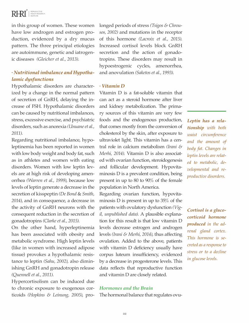
· Nutritional imbalance and Hypotha-lamic dysfunctionsHypothalamic disorders are character-ized by a change in the normal pattern of secretion of GnRH, delaying the in-crease of FSH. Hypothalamic disorders can be caused by nutritional imbalances, stress, excessive exercise, and psychiatric disorders, such as anorexia (Unuane et al., 2011).Regarding nutritional imbalance, hypo-leptinemia has been reported in women with low body weight and body fat, such as in athletes and women with eating disorders. Women with low leptin lev-els are at high risk of developing amen-orrhea (Warren et al., 1999), because low levels of leptin generate a decrease in the secretion of kisspeptin (De Bond & Smith, 2014), and in consequence, a decrease in the activity of GnRH neurons with the consequent reduction in the secretion of gonadotropins (Clarke et al., 2015).On the other hand, hyperleptinemia has been associated with obesity and metabolic syndrome. High leptin levels (like in women with increased adipose tissue) provokes a hypothalamic resis-tance to leptin (Sahu, 2002), also dimin-ishing GnRH and gonadotropin release (Quennell et al., 2011).Hypercortisolism can be induced due to chronic exposure to exogenous cor-ticoids (Hopkins & Leinung, 2005), pro-
longed periods of stress (Tsigos & Chrou-sos, 2002) and mutations in the receptor of this hormone (Lacroix et al., 2015). Increased cortisol levels block GnRH secretion and the action of gonado-tropins. These disorders may result in hypoestrogenic cycles, amenorrhea, and anovulation (Saketos et al., 1993).
· Vitamin DVitamin D is a fat-soluble vitamin that can act as a steroid hormone after liver and kidney metabolization. The prima-ry sources of this vitamin are very few foods and the endogenous production, that comes mostly from the conversion of cholesterol by the skin, after exposure to ultraviolet light. This vitamin has a cen-tral role in calcium metabolism (Irani & Merhi, 2014). Vitamin D is also associat-ed with ovarian function, steroidogenesis and follicular development. Hypovita-minosis D is a prevalent condition, being present in up to 80 to 90% of the female population in North America.Regarding ovarian function, hypovita-minosis D is present in up to 35% of the patients with ovulatory dysfunction (Vig-il, unpublished data). A plausible explana-tion for this result is that low vitamin D levels decrease estrogen and androgen levels (Irani & Merhi, 2014), thus affecting ovulation. Added to the above, patients with vitamin D deficiency usually have corpus luteum insufficiency, evidenced by a decrease in progesterone levels. This data reflects that reproductive function and vitamin D are closely related.
Hormones and the BrainThe hormonal balance that regulates ovu-
Leptin has a rela-tionship with both waist circumference and the amount of body fat. Changes in leptin levels are relat-ed to metabolic, de-velopmental and re-productive disorders.
Cortisol is a gluco-corticoid hormone produced in the ad-renal gland cortex. This hormone is se-creted as a response to stress or to a decline in glucose levels.

23
Ovulation a sign of health
lation acts throughout our body, includ-ing the brain, where steroid hormones have a very active role in the regulation of brain cells (Figure 9) (Schulz & Sisk, 2016).Estrogen and progesterone have an effect on the central nervous system and the peripheral nervous system, especially on neurotransmitters such as GABA, sero-tonin, dopamine, and glutamate (Irwin et al., 2008).Steroid hormones, also known as neu-rosteroids, regulate different brain areas
involved in mood, behavior, and cogni-tion (Magnaghi et al., 2010; Schiller et al., 2016). Therefore, the fluctuation of sex hormones during specific reproductive stages of a woman’s life correlates with an increased susceptibility to develop mood disorders such as premenstrual dysphoric disorder, postpartum depres-sion, and perimenopausal depression. Endogenous estrogen and progesterone levels also may affect different cognitive processes such as decision-making, emo-tion recognition, consolidation of emo-tional memory, and fear extinction. For example, women show improved verbal abilities and decreased visual-spatial abilities when estradiol and progesterone levels are high and the opposite occurs when estradiol and progesterone levels are low. These differences can be partially explained via the role that neurosteroids play in the physiological regulation of neurogenesis, neuronal survival, synaptic function and myelin formation, therefore influencing neuronal plasticity. Further studies may make use of them to treat different disorders of the CNS, as recent studies have shown that neurosteroids could be effective in treating psychiatric disorders, such as schizophrenia, depres-sion, and also against neurodegenerative disorders, such as Alzheimer’s, Parkin-son’s, and multiple sclerosis.
In the same way that endogenous ste-roids influence CNS functionality, steroid hormones administered exogenously also exert their actions on the brain. Two of the most common ways in which hor-mones are administered exogenously to women are: 1) hormonal therapy during
Figure 9: The hormonal balance that regulates ovulation acts throughout all our body in a highly coordinated way.
The main function of vitamin D is maintaining calcium and phosphate home-ostasis. Vitamin D also exerts its influ-ence on the immune, endocrine, cardiovas-cular and reproduc-tive systems.
Neurosteroids reg-ulate different brain areas involved in mood, behavior, and cognition.
Estrogen and pro-gesterone have an effect on the central nervous system and the peripheral nerv-ous system, especial-ly on neurotransmit-ters such as GABA, serotonin, dopamine, and glutamate.

24
menopause and 2) hormonal contracep-tives. When facing a need for the admin-istration of exogenous hormones, consid-eration should be given to the stage of life each woman finds herself in, since exoge-nous hormones will have different effects on the brain depending on the stage. For example, when treating adolescents, spe-cial consideration must be given to the temporal plasticity window of their de-veloping brain, since it is a period when exogenous hormones may produce both activational and organizational changes in the brain that may have long-term ef-fects. At the other extreme, women that are over 10 years past menopause must also take precaution when initiating hor-mone replacement therapy (HRT), since they have been shown to have negative effects on the CNS, increasing the risk of pathologies such as Alzheimer’s disease or stroke (Rapp, 2003; Yen et al., 2012). However, it is important to consider that there are many situations when HRT and the administration of exogenous hor-mones are beneficial. For example, cases such as anorexia nervosa will require, as part of the treatment, the administra-tion of hormones. Similarly, as women age, steroidal hormones decline, and this could have negative consequences, such as hot flashes, osteoporosis, a decrease in libido, and depressive mood. Thus, spe-cial consideration for these individuals needs to be addressed. The activity exerted by steroid hormones on the nervous system emphasizes the notion that achieving hormonal balance is a useful tool in seeking the mental health of women (Del Río et al., 2018).
8. Conclusion: ovulation as a sign of reproductive healthEvery woman should comprehend and learn how to read her own special signs. Biomarkers, such as cervical mucus, will help them identify in which phase of the ovarian continuum they are, whether having ovulatory cycles, anovulatory cy-cles or both. Understanding the concept of the ovarian continuum allows women and health care providers to recognize if ovulation is occurring or, on the contrary, to recognize an ovulatory dysfunction (Vigil et al., 2017). As the hormonal bal-ance that regulates ovulation influences the entire body, the presence of normal ovulatory cycles is a good indicator of a woman’s overall health.It must be kept in mind that anovulation may be a normal part of the ovarian con-tinuum. Women should learn that this is a normal condition in circumstances such as breastfeeding. But, when no physio-logical condition explains an anovula-tory state, anovulation may be caused by pathological conditions such as un-healthy lifestyle habits, stress, endocrine abnormalities, drugs and gynecological, autoimmune, nutritional, genetic, and/or iatrogenic disorders. As the first sign of an ovulatory dysfunction is anovulation (followed later-on by irregular menses or amenorrhea), women who know how to recognize their ovulation can receive timely diagnosis and early treatment.Nowadays there is still an unmet need for further research on how to diagnose and treat ovulatory dysfunctions. The mission of the Reproductive Health Re-
Steroid hormones administered ex-ogenously also exert their actions on the brain. Two of the most common ways in which hormones are administered to women exogenous-ly are: 1) hormonal therapy during men-opause and 2) hormo-nal contraceptives.
Understanding the concept of the ovari-an continuum allows women and health care providers to rec-ognize if ovulation is occurring or, on the contrary, to recognize an ovulatory dys-function

25
Ovulation a sign of health
search Institute (RHRI) is to conduct new research on women’s health, with a particular focus on reproductive endo-crinology. It works to improve women’s health by developing research, creating clinical protocols and advancing scien-tific knowledge. RHRI was founded in 2014. It is composed of a multidisciplinary group of researchers that include medical doctors, reproductive endocrinologists, neurobiologists, ecologists, nurses, and psychologists. The Institute is led by Dr. Pilar Vigil (MD, Ph.D., OB/GYN, FACOG) and Dr. Patricio Contreras (MD, Medical Endocrinologist).As seen in the present paper, women with ovulatory dysfunction may pres-ent symptoms such as irregular cycles, weight gain, hirsutism, acne, pain, head-ache, anxiety, and fatigue. The medical protocols developed by the RHRI offer sensitive diagnosis and treatment for the underlying hormonal issues causing their symptoms, and they cover conditions such as ovulatory dysfunction, polycystic ovaries, thyroid problems, endometriosis, infertility, menopausal disorders, and de-pression. These protocols are scientifically based and standardize the latest advanc-es in biomedical research for clinical use.Treatment plans help women recover healthy hormonal balance. Depending on the measured deficiencies, treatment protocols can range from a significant emphasis on diet, exercise and lifestyle changes to pharmacological interven-tions or immunological support. FEMM collaborates with RHRI in the training of healthcare providers in these protocols around the world.
9. SummaryThis text analyzed the anatomy of ovu-lation and the structures involved in this process. It reviewed the role of the cen-tral nervous system and its communi-cation through the neuroendocrine axis with the gonads and endocrine glands involved in ovulation. The concept of ovarian activity in the lifetime of wom-en, the events that occur in intrauterine life, puberty, and during the reproduc-tive age were discussed. It analyzed the concept of the fertile window and the processes that surround it, mentioning certain biomarkers. Special relevance was given to the role of cervical mucus as a tool for recognizing ovulation. Also, the relationship between hormones and the brain was mentioned. Finally, in relation to future perspectives, we have exposed the need for more research in order to improve women’s health and well-being, recognizing ovulation as a sign of health.
References
Alliende, M. Mean versus individual hor-monal profiles in the menstrual cy-cle. Fertil Steril. 78 (1): 90-5 (2002).
Amer, S. Polycystic ovarian syndrome: Diagnosis and management of re-lated infertility. Obstet Gynaecol Re-prod Med. 19: 263–70 (2009).
Anderson, G. M. Leptin deficiency and diet-induced obesity reduce hypo-thalamic kisspeptin expression in mice. Endocrinology. 152: 1541–1550 (2011).
Araujo-Lopes, R., Crampton, J.R., Aqui-no, N.S., Miranda, R.M., Kokay, I.C., Reis, A.M., Franci, C.R., Grattan, D.R.,
The mission of the Reproductive Health Research Institute (RHRI) is to con-duct new research on women’s health, with a particular focus on reproductive endocri-nology.
The medical pro-tocols developed by RHRI offer sensitive diagnosis and treat-ment for underlying hormonal issues and they cover conditions such as ovulatory dysfunction, polycys-tic ovaries, thyroid problems, endometri-osis, infertility, men-opausal disorders, and depression.

26
& Szawka, R.E. Prolactin regulates kisspeptin neurons in the arcuate nucleus to suppress LH secretion in female rats. Endocrinology. 155: 1010–20 (2014).
Arrau, J., Roblero L, Cury M. New ob-servations on the onset and duration of the meiotic prophase in the femal golden hamster (Mesocricetus aura-tus). J Anat 132: 627-33 (1981).
Azziz, R., Sanchez, L.A., Knochenhauer, E.S., Moran, C., Lazenby, J., Stephens, K.C., Taylor, K. & Boots, L.R. An-drogen excess in women: Experience with over 1000 consecutive patients. J Clin Endocrinol Metab. 89: 453–462 (2004).
Bahtiyar, G., Weiss, K., Sacerdote, A.MD, Novel Endocrine Disruptor Effects. Endocr Pract. 6: 13 (2007).
Baranczuk, R.J. & Fainstat T. Progester-one-induced ovulation of the ham-ster ovary in vitro. J Endocrinol. 70(2): 317-8 (1976).
Barron, M.L. Proactive management of menstrual cycle abnormalities in young women. J Perinat Neonatal Nurs. 18: 81–92 (2004).
Barros, C., Vigil, P., Herrera, E., Arguello, B., Walker, R. Selection of morpho-logically abnormal sperm by human cervical mucus. Arch Androl. 12: 95-107 (1984).
Basson, R. Testosterone therapy for re-duced libido in women. Ther. Adv. Endocrinol. Metab. 1, 155–164 (2010).
Billings, E.L., Brown, J.B., Billings, J.J. & Burger, H.G. Symptoms and hor-monal changes accompanying ovu-lation. Lancet. 1(7745): 282-4 (1972).
Blackwell, L.F. & Brown, J.B. Application
of time-series analysis for the recog-nition of increases in urinary estro-gens as markers for the beginnings of the potentially fertile period. Ste-roids. 57: 554-62 (1992).
Blackwell, L.F., Brown, J.B. & Cooke, D. Definition of the potentially fertile period from urinary steroid excre-tion rates. Part II. A threshold value for pregnanediol glucuronide as a marker for the end of the potentially fertile period in the human menstru-al cycle. Steroids. 63(1): 5-13 (1998).
Blackwell, L.F., Brown, J.B., Vigil, P., Gross, B., Sufi, S. & d’ Arcangues, C. Hor-monal monitoring of ovarian activ-ity using the Ovarian Monitor, part I. Validation of home and laboratory results obtained during ovulatory cycles by comparison with radio-immunoassay. Steroids. 68 (5):465-76 (2003).
Blackwell, L.F., Vigil, P., Cooke, D.G., d’Arcangues, C., & Brown, J.B. Mon-itoring of ovarian activity by daily measurement of urinary excretion rates of oestrone glucuronide and pregnanediol glucuronide using the Ovarian Monitor, Part III: Variability of normal menstrual cycle profiles. Hum Reprod. 28: 3306–15 (2013).
Blackwell, L.F., Vigil, P., Alliende, M.E., Brown, S., Festin, M. & Cooke, D.G. Monitoring of ovarian activity by measurement of urinary excretion rates using the Ovarian Monitor, Part IV: the relationship of the preg-nanediol glucuronide threshold to basal body temperature and cervical mucus as markers for the beginning of the post-ovulatory infertile peri-

27
Ovulation a sign of health
od. Hum Reprod. 31(2):445-53 (2016).Boyers, S.P. Temporal relationships be-
tween ovulation and defined chang-es in the concentration of plasma estradiol-17 beta, luteinizing hor-mone, follicle-stimulating hormone, and progesterone. I. Probit analysis. World Health Organization, Task Force on Methods for the Determi-nation of the Fertile Period, Special Programme of Research, Develop-ment and Research Training in Hu-man Reproduction. Am J Obstet Gy-necol. 138(4):383-90 (1980).
Brown, J., Holmes, J., Barker, G. Use of the Home Ovarian Monitor in pregnan-cy avoidance. Am J Obstet Gynecol. 165:(6 Pt 2): 2008-11 (1991).
Brown, J.B. Types of ovarian activity in women and their significance: The continuum (a reinterpretation of ear-ly findings). Hum Reprod Update. 17: 141–158 (2010).
Ceric, F., Silva, D. & Vigil, P. Ultrastructure of the human periovulatory cervical mucus. J Electro Microsc. 54: 479-484 (2005).
Chretien, F., Dubois, R. Effect of nomege-strol acetate on spinability, ferning and mesh dimension of midcycle cervical mucus. Contraception. 43 (1): 55-65 (1991).
Clarke, H., Dhillo, W.S., Jayasena, C.N. Comprehensive review on kiss-peptin and its role in reproductive disorders. Endocrinology & Metabo-lism (Seoul) 30: 124–41 (2015).
Coker, L.H., Dailey, M., Bowen D., Effect of estrogen plus progestin on glob-al cognitive function in postmeno-pausal women: the Women’s Health
Initiative Memory Study: a random-ized controlled trial. JAMA May 28; 289(20): 2663-72 (2003)
Cortés, M. E., Carrera, B., Rioseco, H., del Río, J.P. & Vigil, P. The role of kisspeptin in the onset of puberty and in the ovulatory mechanism: A mini-review. J Pediatr Adolesc Gyne-col. 28: 286–291 (2015).
De Bond, J.A.P. & Smith, J.T. Kisspeptin and energy balance in reproduction. Reproduction. 147: 53–63 (2014).
de Mouzon, J., Testart, J., Lefevre, B., Pouly, J.L. & Frydman, R. Time rela-tionships between basal body tem-perature and ovulation or plasma progestins. Fertil Steril. 41(2): 254-9 (1984).
Del Río, J.P., Alliende, M.I., Molina, N., Serrano, F., Molina, S., Vigil, P. Ste-roid Hormones and Their Action in Women’s Brains: The Importance of Hormonal Balance. Frontiers in Pub-lic Health. 141(6): 1-15 (2018).
Diamanti-Kandarakis, E. Insulin resis-tance in PCOS. Endocrine. 30: 13–17 (2006).
Duane, M., Contreras, A., Jensen, ET. & White, A. The Performance of Fertil-ity Awareness-based Method Apps Marketed to Avoid Pregnancy. J Am Board Fam Med. 29(4): 508-11 (2016).
Dunson DB, Colombo B, & Baird DD. Changes with age in the level and duration of fertility in the menstrual cycle. Hum Reprod. 17 (5): 1399-403 (2002).
Ecochard, R., Boehringer, H., Rabilloud, M., Marret, H., Chronological as-pects of ultrasonic, hormonal, and other indirect indices of ovulation.

28
BJOG. Aug;108(8):822-9. (2001)Ecochard, R., Duterque, O., Leiva, R.,
Bouchard, T., & Vigil, P. Self-identifi-cation of the clinical fertile window and the ovulation period. Fertil Ster-il, 103(5), 1319–1325.e3 (2015).
Fauser, B.C., Tarlatzis, B.C., Rebar, R.W., Legro, R.S., Balen, A.H., Lobo, R., Carmina, E., Chang, J., Yildiz, B.O., Laven, J.S., Boivin, J., Petraglia, F., Wi-jeyeratne, C.N., Norman, R.J., Dunaif, A., Franks, S., Wild, R.A., Dumesic, D., & Barnhart. Consensus on wom-en’s health aspects of polycystic ova-ry syndrome (PCOS): The Amster-dam ESHRE/ ASRM-Sponsored 3rd PCOS Consensus Workshop Group. Fertil Steril. 97: 28–38 e25 (2012).
Fraser, I.S., Critchley, H.O. Munro, M.G., & Broder, M. Can we achieve inter-national agreement on terminologies and definitions used to describe ab-normalities of menstrual bleeding? Hum Reprod. 22(3): 635–43 (2007).
Gibbons, W. et al. A prospective multi-centre trial of the ovulation of the ovulation method of natural fami-ly planning I. The teaching phase. World Health Organization. Fertil Steril. 36 (2):152-7 (1980).
Gipson IK. Human endocervical mucins. Ernst Schering Res Found Work-shop. 52: 219 (2005).
Gleicher, N., Weghofer, A., Barad, D. The role of androgens in follicle matura-tion and ovulation induction: friend or foe of infertility treatment?. Re-prod Biol Endocrinol. 9: 116 (2011).
Gleicher, N., Kim, A., Weghofer, A., Kush-nir, V.A., Shohat-Tal, A., Lazzaroni, E., Lee,H.J., & Barad, D.H. Hypoan-
drogenism in association with di-minished functional ovarian reserve. Hum Reprod. 28: 1084–91 (2013).
Gregoire, A., Kumar, R., Everitt, B., Hen-derson, A., Studd, J. Transdermal es-trogen for treatment of severe post-natal depression. Lancet. 347: 930–3 (1996).
Gross, B. Clinical indicators of the fertile period. Suppl Int J Gynecol Obstet. 1: 45-51 (1989).
Grumbach, M. The Neuroendocrinology of Human Puberty Revisited. Horm Res. 2: 2–14 (2002)
Guida, M., Tommaselli, G.A., Palomba, S., Pellicano, M., Moccia, G., Di Carlo, C., & Nappi, C. Efficacy of methods for determining ovulation in a natu-ral family planning program. Fertil Steril. 72(5): 900-4 (1999).
Halbreich, U. & Kahn, L. Role of estro-gen in the aetiology and treatment of mood disorders. CNS Drugs. 15 : 797–817 (2001) .
Henderson, H.L., Townsend, J. & Torton-ese, D.J. Direct effects of prolactin and dopamine on the gonadotroph response to GnRH. J Endocrinol. 197: 343–350 (2008).
Herbison, A. E. Control of puberty onset and fertility by gonadotropin-releas-ing hormone neurons. Nat Rev En-docrinol. 12: 452–466 (2016).
Higuchi, K., Nawata, H., Maki, T., Hi-gashizima, M., Kato, K. & Ibayashi, H. Prolactin has a direct effect on ad-renal androgen secretion. J Clin En-docrinol Metab. 59: 714–18 (1984).
Hilgers, T.W., Abraham, G.E. & Cavana-gh, D. Natural family planning. I. The peak symptom and estimated

29
Ovulation a sign of health
time of ovulation. Obstet Gynecol. 52(5): 575-82 (1978).
Hirshman, E., Merritt, P., Wang, C. C., Wi-erman, M., Budescu, D. V., Kohrt, W., Templin, J. L. & Bhasin, S. Evidence that androgenic and estrogenic me-tabolites contribute to the effects of dehydroepiandrosterone on cog-nition in postmenopausal women. Horm. Behav. 45: 144–155 (2004).
Hoff, J.D., Quigley, M.E. & Yen, S.S.C. Hor-monal dynamics at midcycle: A re-evaluation. J Clin Endocrinol Metab. 57: 792–796 (1983).
Hopkins, R. L. & Leinung, M.C. Exoge-nous Cushing’s syndrome and glu-cocorticoid withdrawal. Endocrinol Metab Clin North Am. 34: 371–384 (2005).
Hrabovszky, E., Ciofi, P., Vida, B., Horvath, M. C., Keller, E., Caraty, A., Bloom, S. R., Ghatei, M.A., Dhillo, W.S., Liposits Z. & Kallo, I. The kisspeptin system of the human hypothalamus: Sexual dimorphism and relationship with gonadotropin-releasing hormone and neurokinin B neurons. Eur J Neurosci. 31: 1984–1998 (2010).
Hrabovszky, E. Neuroanatomy of the hu-man hypothalamic kisspeptin sys-tem. Neuroendocrinology. 99: 33–48 (2014).
Hsiu-wei S., Yu-chiao, Y., Ting-yen, W., Ting-chang, C. & Chao-min, C. De-tection of ovulation, a review of cur-rently available methods. Bioengi-neering & Translational Medicine. 2 (3): 1-9 (2017).
Irani, M., and Z. Merhi. Role of vitamin D in ovarian physiology and its im-plication in reproduction: A system-
atic review. Fertility & Sterility 102: 460–68e3. (2014).
Irwin R.W., Yao J., Hamilton R..T, Cadenas E., Brinton R.D. & Nilsen J. Progester-one and estrogen regulate oxidative metabolism in brain mitochondria. Endocrinology. 149:3167–75 (2008).
Jara, L.J., Medina, G., Saavedra, M.A., Ve-ra-Lastra, O. & Navarro, C. Prolactin and autoimmunity. Clin. Rev. Aller-gy Immunol. 40: 50–59 (2011).
Johansson, G.G., Karonen, S.L., & Laakso, M.L. Reversal of an elevated plasma level of prolactin during prolonged psychological stress. Acta Physiol Scand. 119: 463–64 (1983).
Kalme, T., Koistinen, H., Loukovaara, M., Koistinen, R. & Leinonen, P. Com-parative studies on the regulation of insulin-like growth factor-bind-ing protein-1 (IGFBP-1) and sex hor-mone-binding globulin (SHBG) pro-duction by insulin and insulin-like growth factors in human hepatoma cells. J Steroid Biochem Mol Biol. 86: 197–200 (2003).
Kandel, E.R., Schwarz, J. H., Jessell, T. M., Siegelbaum, S. A. & Hudspeth, A. J. Principles of neural science. 5th edn. McGraw Companies, (2000).
Klaus, H., Martin, J.L., Recognition of ovu-latory/anovulatory cycle pattern in adolescents by mucus self-detection. J Adolesc Health Care. Mar;10(2):93-6.(1989)
Koike, K., Miyake, A., Aono, A., T.,Saku-moto,T., Ohmichi, M., Yamaguchi, M., & Tanizawa, O. Effect of prolac-tin on the secretion of hypothalamic GnRH and pituitary gonadotropins. Horm Res. 35: Suppl 1: 5–12 (1991).

30
Kolstad, H.A., Bonde, J.P., Hjollund, N.H., Jensen, T.K., Henriksen, T.B., Ernst, E., Giwercman, A., Skakkebaek, N.E., Olsen, J., Menstrual cycle pattern and fertility: a prospective follow-up study of pregnancy and early em-bryonal loss in 295 couples who were planning their first pregnancy. Fertil Steril. Mar;71(3):490-6. (1999).
Krasnow, J.S., Hickey, G.J. & Richards, J.S. Regulation of aromatase mRNA and estradiol biosynthesis in rat ovarian granulosa and luteal cells by prolac-tin. Mol Endocrinol. 4: 13–21 (1990).
Krassas, G.E., Pontikides, N., Kaltsas, T., Papadopoulou, P., & Batrinos, M. Menstrual disturbances in thyro-toxicosis. Clin Endocrinol (Oxf). 40: 641–44 (1994).
Krassas, G.E., Pontikides, N., Kaltsas, T., Papadopoulou, P., Paunkovic, J. Paunkovic, N., & Duntas, L.H. Dis-turbances of menstruation in hypo-thyroidism. Clin Endocrinol (Oxf). 50: 655–59 (1999).
Krassas, G.E., Poppe, K. & Glinoer, D. Thyroid function and human repro-ductive health. Endocr. Rev. 31: 702–755 (2010).
Kühnel, W. Color Atlas of Cytology, His-tology and Microscopic Anatomy. Thieme Medical Publishers. 4th Edi-tion (2004).
Lacroix, A., Feelders, R.A., Stratakis, C.A., Nieman, L.K., Cushing’s Syndrome. Lancet. 2015 Aug 29;386(9996):913-27. (2015)
Lekarev, O., Lin-Su, K., & Vogiatzi, M.G. Infertility and reproductive function in patients with congenital adre-nal hyperplasia. Endocrinol. Metab.
Clin. North Am. 44: 705–722 (2015).
Lunenfeld, B. & Insler, V. Follicular devel-opment and its control. Gynecol. En-docrinol. 7: 285–291 (1993).
Magnaghi V., Parducz A., Frasca A., Bal-labio M., Procacci P., Racagni G., Bonanno G. & Fumagalli, F. GABA synthesis in Schwann cells is in-duced by the neuroactive steroid al-lopregnanolone. J Neurochem. 112: 980–90 (2010).
Merke, D.P. & Bornstein, S.R. Congeni-tal adrenal hyperplasia. Lancet. 365: 2125–36 (2005).
Morales, P., Roco, M., Vigil, P. Human cer-vical mucus: relationship between biochemical characteristics and abil-ity to allow migration of spermato-zoa. Hum Reprod. 8 (1): 78-83 (1993)
Morrell, M., Hayes, F.J., Sluss, P.M., Ad-ams, J.M., Bhatt, M., Ozkara, C., War-nock, C.R. & Isojärvi, J. Hyperandro-genism, Ovulatory Dysfunction, and Polycystic Ovary Syndrome with Valproate versus Lamotrigine. Ann Neurol. 64: 200–11 (2008)
Nimkarn, S. & New, M. I. Congenital ad-renal hyperplasia due to 21-hydroxy-lase deficiency: A paradigm for pre-natal diagnosis and treatment. Ann N Y Acad Sci. 1192: 5–11 (2010).
Notelovitz, M. Androgen effects on bone and muscle. Fertil. Steril. 77, 34–41 (2002).
Oakley, A.E., Clifton, D.K. & Steiner, R.A. Kisspeptin signaling in the brain. Endocr. Rev. 30: 713–743 (2009).
Odeblad, E. The discovery of different types of cervical mucus and the Bill-

31
Ovulation a sign of health
ings ovulation method. Bull Nat Fam Plan Counc Vic. 1–31 (1994).
Orbach, H. & Shoenfeld, Y. Hyperprolac-tinemia and autoimmune diseases. Autoimmun Rev. 6: 537–42 (2007).
Panitsa-Faflia, C., & Batrinos, M.L. Late-Onset Congenital Adrenal Hy-perplasia. Ann N Y Acad Sci. 816: 230–234 (1997).
Park, S., Goldsmith, L., Skurnick, J., Wo-jtczuk, A., Weiss, G.. Characteristics of the urinary luteinizing hormone surge in young ovulatory women. Fertil Steril. 88 (3): 684-90 (2007).
Pauli, J.M., Raja-Khan, N., Wu, X., & Le-gro V. Current perspectives of insu-lin resistance and polycystic ovary syndrome. Diabetic Medicine. 28: 1445–54 (2011).
Pérez A. General Overview of Natural Family Planning. Genus. 54: 75-93 (1998).
Poon WW, McCoshen JA. Variances in mucus architecture as a cause of cer-vical factor infertility. Fertil Steril. 44 (3): 361-5 (1985).
Popa, S.M., Clifton, D.K. & Steiner, R.A. The role of kisspeptins and GPR54 in the neuroendocrine regulation of reproduction. Annu Rev Physiol. 70: 213–238 (2008).
Popat, V.B., Prodanov, T., Calis, K.A., Nel-son, L.M., The menstrual cycle: a biological marker of general health in adolescents. Ann N Y Acad Sci. 1135:43-51. (2008).
Poppe, K., Velkeniers B., & Glinoer D. Thyroid disease and female repro-duction. Clin Endocrinol (Oxf). 66: 309–21 (2007).
Prior, J.C., Naess, M., Langhammer, A., Forsmo, S. Ovulation Prevalence in Women with Spontaneous Nor-mal-Length Menstrual Cycles - A Population-Based Cohort from HUNT3, Norway. PLoS One. Aug 20;10(8):e0134473. (2015).
Quennell, J.H., Howell, C. S., Roa, J., Au-gustine, R. A., Grattan, D. R. & An-derson, G.M. Leptin deficiency and diet-induced obesity reduce hypo-thalamic kisspeptin expression in mice. Endocrinology. 152: 1541–1550 (2011).
Quinkler, M., Sinha, B., Tomlinson, J. W., Bujalska, I. J., Stewart, P. M. & Arlt, W. Androgen generation in adipose tissue in women with simple obesi-ty - a site- specific role for 17beta-hy-droxysteroid dehydrogenase type 5. J Endocrinol. 183: 331–342 (2004).
Ranasinha, S., Joham, A.E., Norman, R.J., Shaw, J.E., Zoungas, S., Boyle, J., Mo-ran, L., & Teede, H.J. The association between Polycystic Ovary Syndrome (PCOS) and metabolic syndrome: A statistical modelling approach. Clin Endocrinol (Oxf). 83: 879–87 (2015).
Rapp, S.R., Espeland, M.A., Shumaker, S.A., Henderson, V.W., Brunner, R.L., Manson, J.E., Gass, M.L., Stefanick, M.L., Lane, D.S., Hays, J., Johnson, K.C.,
Rasgon N., Reynolds, M.F., Elman, S., Saad, M., Frye, M.A., Bauer, M. & Alt-shuler, L.L. Longitudinal evaluation of reproductive function in women treated for bipolar disorder. Journal of Affective Disorders. 89: 217–225

32
(2005).Rauchenzauner, M., Roscia, S., Prieschl,
M., Wildt, L., Haberlandt, E., Bau-mann, M., Rostasy, K., Agostinelli, S., Pizzolorusso, A., Luef, G. & Verrotti, A. Reproductive Endocrine Health in Pubertal Females with Epilep-sy on Antiepileptic Drugs: Time to Screen?. Neuropediatrics. 45 (4): 226-233 (2014).
Rosenfield, R. L. Editorial: Evidence that idiopathic functional adrenal hyper-androgenism is caused by dysregu-lation of adrenal steroidogenesis and that hyperinsulinemia may be in-volved. J Clin Endocrinol Metab. 81: 878–880 (1996).
Rosenfield, R.L., Ehrmann, D.A. & Little-john, E.E. Adolescent polycystic ova-ry syndrome due to functional ovar-ian hyperandrogenism persists into adulthood. J Clin Endocrinol Metab. 100: 1537–1543 (2015).
Royston, J.P. Basal body temperature, ovulation and the risk of conception, with special reference to the lifetimes of sperm and egg. Biometrics. 38(2): 397-406 (1982).
Sahu, A. Resistance to the satiety action of leptin following chronic central leptin infusion is associated with the development of leptin resistance in neuropeptide Y neurones. J Neuro-endocrinol. 14: 796–804 (2002).
Saketos, M., Sharma, N. & Santoro, N.F. Suppression of the hypothalamicpi-tuitary-ovarian axis in normal wom-en by glucocorticoids. Biol Reprod. 49: 1270–1276 (1993).
Samuel, V.T. & Shulman, G.I. Integrating mechanisms for insulin resistance:
Common threads and missing links. Cell. 148: 852–871 (2013).
Schiller C.E., Johnson S.L., Abate A.C., Schmidt P.J., Rubinow D.R. & Schiller C.E. Reproductive steroid regulation of mood and behavior. Compr Physi-ol. 6:1135–60 (2016).
Schoenwolf, Gary C. & William J. Larsen. Larsen’s Human Embryology. Phila-delphia: Churchill Livingstone/Else-vier (2009).
Schulz K.M. & Sisk, C.L. The organizing actions of adolescent gonadal steroid hormones on brain and behavioral development. Neurosci Biobehav Rev. 70:148–58 (2016).
Sheehan, J.K. & Carlstedt, I. Electron mi-croscopy of cervical-mucus glyco-proteins and fragments therefrom. The use of colloidal gold to make vis-ible‘naked’ protein regions. Biochem J. 265(1):169 (1990).
Skinner, M.K. Regulation of primordial follicle assembly and development. Human Reproduction Update 11: 461–71 (2005).
Skorupskaite, K., George, J. T. & Ander-son, R.A. The kisspeptin GnRH path-way in human reproductive health and disease. Hum Reprod Update. 20: 485–500 (2014).
Stanhope, R., Brook, C.G., Clinical di-agnosis of disorders of puberty. r J Hosp Med. Jan;35(1):57-8 (1986)
Thompson, I. R., Ciccone, N. A., Xu, S., Zaytseva, S., Carroll, R. S. & Kaiser, U. B. GnRH pulse frequency-depen-dent stimulation of FSHβ transcrip-tion is mediated via activation of PKA and CREB. Mol Endocrinol. 27: 1–13 (2013).

33
Ovulation a sign of health
Tsigos, C. & Chrousos, G. P. Hypothalam-ic- pituitary- adrenal axis, neuroen-docrine factors and stress. J Psycho-som Res. 53: 865–871 (2002).
Unuane, D., Tournaye, H., Velkeniers, B., & Poppe, K. Endocrine disorders & female infertility. Best Pract Res Clin Endocrinol Metab. 25: 861–73 (2011).
Vigil, P., Perez, A., Neira, J. & Morales, P. Post-partum cervical mucus: Biolog-ical and rheological properties. Hum Reprod. 6: 475-479 (1991).
Vigil, P., Garcia, M., Villalobos, T., Cor-dova, A. & Morales, P. Predicción de Ovulación en Mujeres Normales. Rev Lat Ester Fert. 6(3): 87-91 (1992).
Vigil, P., Del Río, P., Kolbach, M. & Riquelme, R. Nuevas Metodologías y Avances en Planificación Natural de la Familia. Rev Chi Obstet Ginecol. 66(2): 132-7 (2001).
Vigil, P., Ceric, F., Cortés, M.E., & Klaus, H. Usefulness of Monitoring Fertili-ty from Menarche. J Pediatr Adolesc Gynecol. 19: 3, 173–179 (2006).
Vigil, P., Contreras, P., Alvarado, J.L., Godoy, A., Salgado, A.M., & Cortes, M.E. Evidence of subpopulations with different levels of insulin re-sistance in women with polycystic ovary syndrome. Hum Reprod. 22: 2974–80 (2007).
Vigil, P., Toro, A., Godoy, A., Physiological action of oestradiol on the acrosome reaction in human spermatozoa. An-drologia. Jun;40(3):146-51 (2008).
Vigil, P., Cortes , M., Zuniga, A., Riquelme, J. & Ceric, F. Scanning electron and light microscopy study of the cervi-cal mucus in women with polycystic ovary syndrome. J Electron Microsc.
58: 21-27 (2009).Vigil, P., Orellana RF, Cortes ME. Modula-
tion of spermatozoon acrosome reac-tion. Biol Res. 44 (2): 151-9 (2011).
Vigil, P., Blackwell, L.F., Cortés, M.E., The Importance of Fertility Awareness in the Assessment of a Woman’s Health a Review. Linacre Q. 2012 Nov;79(4):426-450 (2012)
Vigil, P., Croxatto, H.B. & Cortes, M.E. Ciclo Menstrual. In Ginecología (ed. Pérez-Sánchez, A.) 37–60. Editorial Mediterráneo (2014).
Vigil, P., Lyon, C., Flores, B., Rioseco, H., & Serrano, F.G. Ovulation, a sign of health. Linacre Q. 84: 4, 343-55 (2017).
Walf A.A. & Frye C.A.. A review and up-date of mechanisms of estrogen in the hippocampus and amygdala for anxiety and depression behavior. Neuropsychopharmacology. 31:1097–111 (2006).
Warren, M.P., Voussoughian F., Geer, E.B., Hyle, E.P., Adberg, C.L. & Ra-mos, R.H. Functional hypothalamic amenorrhea: Hypoleptinemia and disordered eating. J Clin Endocrinol Metab. 84: 873–877 (1999).
Wilcox, A.J., Weinberg, C.R. & Baird, D.D. Timing of sexual intercourse in rela-tion to ovulation. Effects on the prob-ability of conception, survival of the pregnancy, and sex of the baby. N Engl J Med. 333(23):1517-21 (1995).
Wilcox, A.J., Dunson D., & Baird, D.D. The timing of the “fertile window” in the menstrual cycle: day specific estimates from a prospective study. BMJ. 321(7271):1259-62 (2000).
Yen, J., Chang, S.J., Long, C.Y., Tang, T.C., Chen, C.C. & Yen C.F. Working

34
memory deficit in premenstrual dys-phoric disorder and its associations with difficulty in concentrating and irritability. Compr Psychiatry. 53: 540–5 (2012).
Zegers-Hochschild, F., Gomez Lira, C., Parada, M. & Altieri Lorenzini, E. A comparative study of the follicular growth profile in conception and non-conception cycles. Fertil Steril. 41(2) : 244-7 (1984).
Zerouni, C., Kummerow, E., Martinez, M., Diaz, A., Ezequiel, U. & Wix-Ra-mos R. Affective Disorder and Hy-perandrogenism. Recent Patents on Endocrine, Metabolic & Immune Drug Discovery. 7: 77-79 (2013).

35
Ovulation a sign of health