Embryogenesis, Histology, and Organology of the Ovary of ...
102
Gulf and Caribbean Research Gulf and Caribbean Research Volume 2 Issue 4 January 1969 Embryogenesis, Histology, and Organology of the Ovary of Embryogenesis, Histology, and Organology of the Ovary of Brevoortia patronus Brevoortia patronus Ralph M. Combs Gulf Coast Research Laboratory Follow this and additional works at: https://aquila.usm.edu/gcr Part of the Marine Biology Commons Recommended Citation Recommended Citation Combs, R. M. 1969. Embryogenesis, Histology, and Organology of the Ovary of Brevoortia patronus. Gulf Research Reports 2 (4): 333-434. Retrieved from https://aquila.usm.edu/gcr/vol2/iss4/1 DOI: https://doi.org/10.18785/grr.0204.01 This Article is brought to you for free and open access by The Aquila Digital Community. It has been accepted for inclusion in Gulf and Caribbean Research by an authorized editor of The Aquila Digital Community. For more information, please contact [email protected].
-
Upload
khangminh22 -
Category
Documents
-
view
2 -
download
0
Transcript of Embryogenesis, Histology, and Organology of the Ovary of ...

Gulf and Caribbean Research Gulf and Caribbean Research
Volume 2 Issue 4
January 1969
Embryogenesis, Histology, and Organology of the Ovary of Embryogenesis, Histology, and Organology of the Ovary of
Brevoortia patronus Brevoortia patronus
Ralph M. Combs Gulf Coast Research Laboratory
Follow this and additional works at: https://aquila.usm.edu/gcr
Part of the Marine Biology Commons
Recommended Citation Recommended Citation Combs, R. M. 1969. Embryogenesis, Histology, and Organology of the Ovary of Brevoortia patronus. Gulf Research Reports 2 (4): 333-434. Retrieved from https://aquila.usm.edu/gcr/vol2/iss4/1 DOI: https://doi.org/10.18785/grr.0204.01
This Article is brought to you for free and open access by The Aquila Digital Community. It has been accepted for inclusion in Gulf and Caribbean Research by an authorized editor of The Aquila Digital Community. For more information, please contact [email protected].

EMBRYOGENESIS, HISTOLOGY AND ORGANOLOGY
OF
THE OVARY OF BREVOORTIA PATRONUS
Ralph M. Combs
Gulf Coast Research Laboratory Ocean Springs, Mississippi
This work was financed by the Bureau of Commercial Fisheries under Con- tract No. 14-19-008-9335 and by stipends available through Contract No. G-22883 of the National Science Foundation with the Gulf Coast Research Laboratory, Ocean Springs, Mississippi.
333

TABLE OF CONTENTS
Page Embryogenesis. Histology and Oranology of the Ovary
Abstract ................................................................................................. 337
of Brevoortia patronus - Ralph M . Combs ................................ 333
I . I1 .
I11 .
IV . V .
VI .
Introduction ......................................................................................... 337
Materials and Methods .................................................................... 338
Origin and Early Differentiation of the Gonad ........................ 340
(1) Inception and Early Organization of Larval Gonads (17 to 29 mm Fish) ................................................................ 340
(2) Histogenesis of Somatic Elements in Post-larval Gonads (30 to 70 mm Fish) .................................................. 346
(3) Size Increments in Primitive Gonads ............................... 355
(4) The Gonia in Larval and Post-larval Gonads ................ 356
Sex Differentiation in the Larvae and Post-larvae .................... 358
Organology and Histology of the Non-germinal Elements of the Definitive Ovary .................................................................... 364
Histology and Cytology of the Germinal Elements of the Definitive Ovary ................................................................................ 373
. . .
. . . Stage I ................................................................................................. 373 Stage I1 .................................................................................................... 376 Stage I11 .................................................................................................. 380 Stage IV .................................................................................................. 385 Stage V .................................................................................................... 390 Stage VI .................................................................................................. 395
VI1 . Atresia of Oocytes .. .................. 400
(1) Stage I11 Atresia ...................................................................... 402 (2) Atresia-Yolk Stages ......................................................... 406
VI11 . Investments of the Oocytes ............................................................ 411 Stage I ................................................................................................ 412 Stage I1 ................................................................................................. 413 Stage I11 ................................................................................................ 413 Stage IV ............................................................................................... 414 Stage V ............................................................................................. 415
335

TABLEOFCONTENTS
(Continued)
Page IX . Summary .............................................................................................. 418
X . Literature Cited .................................................................................. 425
XI . Explanation of Plates ........................................................................ 429 Plate I ..................................................................................................... 429 Plate I1 .................................................................................................. 429 Plate 111 ............................................................................................... 430 Plate IV ................................................................................................ 431 Plate V ................................................................................................. 432 Plate VI ................................................................................................ 433
TABLES
Table I .............................................................................................................. 339
Table I1 ............................................................................................................ 342
Table I11 ............................................................................................................ 357
Table IV ......................................................................................................... 359
Table V ............................................................................................................ 377
PLATES
Plate 1 .............................................................................................................. 349 Plate 2 .............................................................................................................. 350 Plate 3 .............................................................................................................. 371 Plate 4 ............................................................................................................... 372 Plate 5 .............................................................................................................. 397 Plate 6 .............................................................................................................. 419
.
336

ABSTRACT
One hundred ninety-one large scale menhaden, Brevoortia pa- tronus, ranging in age from larvae to sexually mature females were used in this study. Collections were made in the littoral and shallow off shore waters of the Gulf of Mexico from Dauphin Island, Alabama, westward to West Bay, Louisiana, at intervals throughout a five year period. Using standard paraffin techniques a number of staining methods were employed. Cytological and histological accounts are presented of the tissue elements of the gonads beginning with in- ception in the larvae, sexual differentiation to the sexes, and the cyclic changes associated with oogenesis and spawning in the mature fish. The microscopic developments occurring during these periods are correlated with gross features of the organ, the ages of specimens, and with seasonal periods. An account of the morphology and physi- ology of atretic oocytes and ovulated follicles is presented. Using the ovarian components as an index, the time and duration of the spawn- ing season is established as occurring from late October to February or early March with some variance due to environmental factors. From the study it is possible to postulate that this species exhibits intermittent total spawning in the Gulf.
I. INTRODUCTION
This study was originally initiated for the purpose of correlating the cyclic histological changes in the ovaries of mature females of Brevoortia patronus with the temporal and spatial aspects of spawn- ing. During its progress, the scope of the investigations was expanded to include the changes involved from the time of their origin to the stage where sex differentiation was complete.
From the time of His (1873), Semper (1875), and Balfour (1878), considerable literature has accumulated on the gonads of fish. These include a number of investigations of the histologic cycles of the ovaries of several of the Clupeidae. Particularly significant is the account provided by Naumov (1956) of oogenesis and the sex cycle of mature females of the Murmansk herring, Clupea h. harengus. As might be anticipated from the taxonomic relationship of this species and B. patronus, there exists a considerable degree of similarity in the oogenetic processes. Yamamoto (1958) has contributed a description of the manner of yolk formation in Clupea pallasi including an ac- count of its cytochemistry. His conclusions regarding vitellogenesis coincide closely with those reached by the author in the case of the large scale Gulf menhaden. Studies by Suttkus and Sundararaj (1961) on B. patronus and by Nagasaki (1958) on the Pacific herring, Clupea pallasi involved analyses of the fecundity of the species. No refer- ence was made to the oogenetic activities. Clark (1934) published a brief description of the growth stages of the intra-ovarian in the Cali- fornia sardine Sardina caerulea utilizing diameter differences of the oocytes rather than cytological detail to establish their degrees of maturity. He also provided an excellent summary from the liter- ature of the scales or criteria relating to ova size which had been em- ployed by various investigators to evaluate degrees of maturity. A more extensive review of the literature pertaining to the maturity
337

stages of intra-ovarian development of fish ova was published by Naumov (1956).
Nothing has been published on the origin and histogenesis of menhaden gonads. The interest which investigators have given to the larvae of this species has been directed to their number and ex- ternal morphogenesis. Reintjes (1962) has given a description of cleavage and external changes which occur in the yolk-sac larvae of B. tyrannus and B. smithi in the Atlantic between North Carolina and Florida. In an attempt to determine the time of spawning and to sug- gest the locale of its occurrence, Suttkus (1956) analyzed data obtained from collections of B. patronus larvae. A relationship was found be- tween larval development and their inshore movement. A somewhat similar approach using larvae to indicate the reproductive activities of B. patronus was carried out by Arnold (1958) in the Galveston, Texas area.
11. MATERIALS AND METHODS
A total of 191 larval, post-larval, immature, and mature fish collected during the period from 1959 through 1960 were utilized in this study. The author is indebted to Mr. J. Y. Christmas, Gulf Coast Research Laboratory, Ocean Springs, Mississippi, for his assistance in collecting most of the material, determining fork lengths, and treat- ing it with killing and fixing agents.
Collections were made from West Bay, Louisiana (89O24' W long- itude, 29O32' N latitude), eastward to Mobile Bay, Alabama (86" W longitude, 31°32' N latitude). Relevant data pertaining to the specific areas from which samples were taken is recorded in Table I.
It will be noticed from the stations given in the table that in most instances the fish were taken from euryhaline habitats receiving dis- charges from the Mississippi, Pearl, and Pascagoula Rivers and many small streams as well as from the Biloxi and Mobile Bays. Salinities varied according to the seasons, tides and continental run-off from from about 2 to 25 o/oo in the shoreward waters of the Mississippi and its inlets to approximately 30 to 35 o/oo at stations in the passes between islands and the continental shelf beyond.
In all cases fork length measurements were made while the fish were in a fresh state. Ovarian measurements were made after the gonad was excised, killed and fixed except in the larval and post- larval material, which by reason of the extreme smallness or absence of an organized gonad did not lend itself to a method of direct mea- surement. In these cases dimensions were computed from serially .
sectioned material. In view of the prior treatment of the organs with killing and fixing agents, the measurements thus obtained are slight- ly less than would have been secured from living specimens.
The excised gonads of the larger fish were fixed in Bouin's solu- tion. In the remaining cases fixation was by 10% formalin, except in a few cases where gonads were removed from intact specimens which had previously been preserved or frozen. Larvae and post- larvae were fixed by transecting the body at points immediately an-
338
-

TABLE I
months the numbers, sizes, and areas from which made. Seven collections were made in 1959 during February to December. In 1960 twenty-five collec- from January to December.
Showing by collections were the period from tions were made
Fork No. Length
Month Fish (mm) Area
February
June August December December December January January March April April April
May
May May May May May June June June August August August August August October October October November December December
1 188
1 198 4 201-217
7 225-237 31 164-226 1 176
11 187-225 14 144192 1 165 1 207 9 78-177 1 222 2 17- 32 9 18- 38 5 189-194 4 144-204 1 186
11 23-338 3 47- 89 3 80- 86 9 102-143
14 64-113 5 63- 70 7 109-141 2 93-141 9 91-166 1 131 5 159-200 3 160-199 3 116-170
11 82-125 2 181-197
Total 191
Sand Island Light, Mobile Bay, Ala. Sand Point, La. Cat Island, Miss. Chandeleur Island, La. Horn Island, Biloxi, Miss. S. E. Chandeleur Light, La. Horn Island, Miss. Fort Bayou, Ocean Springs, Missi/ Dog Keys Pass, Miss. Gulf Coast Research Laboratory1/ 2/
Gulf Coast Research Laboratory1/ 2/
Biloxi Bay, Miss. Gulf Coast Research Laboratory1/ 2 /
Gulf Coast Research Laboratory1/ 2 /
Empire Bar, La. West Bay, La. Gulf Coast Research Laboratory1/ 2 /
Gulf Coast Research Laboratory1/ 2 /
Gulf Coast Research Laboratory1/ 2 /
Gulf Coast Research Laboratory1/ 2/
Biloxi Bay, Miss.l/ Gulf Coast Research Laboratory1/ 2/
Gulf Coast Research Laboratory1/ 2/
Belle Fontaine Beach, Ocean Springs, Miss.1’ Biloxi Bay, Miss.l/ Biloxi Bay, Miss?’ Belle Fontaine Beach, Ocean Springs, Miss.11 Pascagoula Bay, Miss.l/ Mobile Bay, Ala.l/ Biloxi Bay, Miss>/ Dog Keys Pass, Miss.l/ 27 miles East of North Pass of Miss. River1/ No. Collections - 32
l/-Waters of Mississippi Sound or tributaries thereof. 2/-Harbor of Gulf Coast Research Laboratory, Ocean Springs, Miss.
339

terior and posterior to the organ and subjecting the entire segment to formalin.
Paraffin embedding was employed throughout. Since the gonads in larvae and post-larvae were too undeveloped to handle if excised, the entire segment of the body prepared as mentioned above was serially sectioned. In older fish the ovaries were either embedded entire or if they were too large to handle in this manner, a trans- verse segment from their mid region was utilized. Cross sections were made of all gonads, and in addition sagittal sections were prepared from a representative number of the organs from juvenile and ma- ture fish. Sections were cut at 10 microns.
All cross sections from the excised organs from juvenile and ma- ture stages were stained with Heidenhain’s iron hematoxylin and eosin. Of the excised ovaries selected for the preparation of sagittal sections, one section each was stained by Heidenhain’s, Cajal’s trich- rome connective tissue, and Flemming’s safranin-gentian violet-orange G techniques, respectively. The slides derived from each serially sec- tioned larva and post-larva were alternately stained by the three designated methods.
Ocular grids were used in making oocyte counts.
111. ORIGIN AND EARLY DIFFERENTIATION OF THE GONAD Gonadogenesis is initiated only after the recently hatched fish
have entered estuarine waters. The origin and early differentiation of the elements of the gonad takes place in menhaden having a fork length of between 17 and 70 mm. During the initial part of this growth period, i. e. from 17 to 50 mm, they are embryonically in the larval state although Reintjes (1961) considers as larvae only fish hav- ing a length of from 4 to 22 mm from the snout to the end of the verl tebral column. Suttkus (1956) considers them as being in the larval stage during the period when their total body lengths are between 20 and 30 mm. The basis for considering fish up to 50 mm as larvae will be given later. It is during this period that the initial events of em- bryogenesis of the gonad olccur including the incorporation and in- tegration of the primordial cellular elements. The subsequent post- larval period is characterized by the differentiation and organization of the cellular components into the definitive arrangement of the immature just before the onset of oogenesis.
(1 1 Inception and Early Organization of Larval Gonads (17 to 29 mm Fish)
The form of the future organ is not discernible in fish of 17-18 mm (Plate I, Fig. 1). At this period the area of the parietal mesothelium lying at the upper right and left quadrants of the coelom, which is destined to give rise to the germinal epithelium of the organ, portrays a morphology comparable to the outlying areas. At the loci where the gonads will appear, it is constituted of the same low type of simple cuboidal epithelium that occurs throughout this coelomic surface. In the figure it lies to the left of the dark chromatophores as an indis- tinct thin gray investment. Its indistinctness is due to the proaounc-
340

ed chromophobia of its cells. The coelom is the lighter area occurring between the mesothelium and the wall of the gut. The superficial cells thus appear contiguous with the underlying irregular layer con- stituted of numerous melanophores between which pass a few weakly developed fibrous connective tissue elements. Retro-peritoneally to the pigment cells there is a noticeable paucity of cellular and fibrous elements which diminish to an even greater extent in areas beyond the region of incipient gonad formation. No sharp delineation exists between the two regions. Because of the absence of a sharp trans- ition between the aggregation of cells that will become involved in gonad formation and the more distal elements, the exact limits of the germinal region is difficult to determine. As near as could be deter- mined, it occupies an area of about 15 to 19 microns along a body wall transect while its linear extent is approximately 65 microns. The com- ponents constituting this retro-peritoneal contribution to the future gonads includes occasional rather delicate connective tissue fibers having a somewhat areolar arrangement, a moderate number of small mesenchymal or fibroblast cells, and a very few primordial germ cells.
In these larvae the primordial germ cells, although confined to the gonadogenic area, are usually few in number and are relatively widely separated from each other. At this period they typically oc- cupy a position nearby or in contact with the melanophores. The proximity of three, possibly four, of the cells (the larger spheroid elements) to the melanophores is shown in Plate I, Fig. 1. Due to the irregular spacing of the pigment cells, there exists between them occasional interstices through which the fibers and cells of the outly- ing connective tissue pass into the area immediately contiguous with the bases of the presumptive germinal epithelium. Also situated in such crevices an occasional primordial germ cell can be observed, apparently in the process of migration toward the potential germinal epithelium.
The cytological aspects of the primordial germ cells in general conform to the descriptions given by numerous authors for various vertebrates. The somewhat spherical oxyphilic cytosome is relatively large (7 to 9 microns) and displays an absence of formed elements or specific limiting membrane. At its approximate center the prominent spherical nuclear body (mean diameter 4 microns) encloses a moder- ate or minimal quantity of fine chromatin material dispersed uni- formly throughout. Nucleoli are not visible. The karyotheca is quite distinct. Since younger material was not available for study, the origin of the sex cells was not elucidated and their migrations, if any, could not be traced. They have been described as arising in fish from the lateral (intermediate?) mesoderm (in partis) by Balfour (1878) and Rabl (1896), in toto from intermediate mesoderm by MacLeod (1881), or in the splanchnic mesoderm of the gut wall or its mesen- tery (Woods, 1902, and Moore, 1937). An insight as to their origin in the Atlantic menhaden, B. tyrannus, was not furnished by Kuntz and Radcliffe (1918) in their study of its embryology and early develop- ment. Regardless of the point of origin of these cells, it was found in the present study that only about 9 to 15 of them are found in the body wall immediately prior to their migration into the presumptive gonad.
The mesenchymal or fibroblast elements which are destined to be
341

TABLE I1
Data Relating to Size of Gonads for Fish Between 17 mm and 70 mm Fork Length.
Gonad Dimensions (mm) Fork Length Date Length Vertical Transverse
(1) (2) (3) (4) (5)
17 mm April 11 11 11
18
23
24
24
24
29
29
32
32
34
47
61
63
64
66
68
70
April
April
May
May
May
May
May
May
May
April
June
June
August
August
August
August
August
11
0.99
0.97
1.29
1.26
1.60
1.80
1.27
1.31
1.22
1.92
3.10
2.20
3.50
3.40
3.80
6.00
11
-001
.002
.008
.006
.091
.050
.126
.lo5
,112
.250
.290
.300
.300
.300
.800
.goo
.040
.039
.054
.050
.039
.028
.036
..039
,042
.078
.090
.133
.141
.127
.161
.132
- l/-Gonadal anlage in too primitive a stage to permit measurement
because marginal limits are not distinctly defined.
342

incorporated in the gonad are derived from either the intermediate or lateral mesoderm cells of the body wall. Prior to their migration into the organ they are cytologically similar to other fibroblasts in this area which are destined to remain in the body wall. A 10 micron transverse section shows from 15 to 25 of these cells loosely aggre- gated so as to form a poorly organized cord extending anteriorly- posteriorly close to the coelomic epithelium which will shortly thicken to create a germinal ridge. A gradual transition occurs between these condensed fibroblasts and those which extend out into the body wall. Their size, i. e. 3 microns, is about one half the diameter of the sex cells with which they are integrated. Their shape varies from spher- ical to stelliform, triangular, or rectangular. Each is constituted of a relatively small amount of homogeneous oxyphilic cytoplasm. Their open face nuclei are generally oval and are composed of a consider- able quantity of fine chromophobic material enclosed by a delicate moderately basophilic membrane.
When the larvae attain a length of 23-24 mm, the initial morph- ogenesis of the gonads becomes evident. This is marked by the ap- pearance of a barely perceptible thickening of the presumptive germ- inal epithelium which produces a slight substention into the dorsal- lateral regions of the coelom (Plate I, Fig. 2). In the illustration, it appears to the left of the dark melanophores as a gray thickening con- taining a row of black nuclei. Although this thickening is in the form of an anterior-posterior ridge, its exact dimensions can only be ap- proximated since along its marginal areas there occurs a gradual trans- ition between the somewhat cuboidal cells of the ridge and those of the adjacent peritoneum. That gonadogenesis is initiated and pro- ceeds at a greater rate at the head of the organ is indicated by the greater degree of coelomic distention which occurs at that point. As shown in Table 11, the length of this anlage in fish of the age being considered varies from 0.97 to 1.29 mm with a mean length of 1.13 mm. Posteriorly it extends to within approximately 1.4 mm of the anterior margin of the anus. The maximum thickness of the epithelial cells constituting the ridge is in the order of 5 microns as related to 0.5 to 1.0 microns for the outlying parietal epithelium of the coelom. The increased thickness of the layev in the germinal region is due in part to a stratification of the earlier single layer into two imperfectly oriented layers and in part to a differentiation of the original low cuboidal elements into higher cuboidal forms.
Not until slightly later does the germinal epithelium of the ridge undergo sufficient coelomic distention to permit the entrance of the primitive germ cells together with some of all of the associated con- nective tissue elements. As in the case of younger larvae, occasional primitive sex cells together with mesenchymal and fibroblast ele- ments are found to lie more or less contiguous to the base of the layer of germinal ridge cells. While the number and cytology of the germ cells show no apparent change from the condition existing in younger fish, the fibroblastic elements have become somewhat more abund- ant. Whereas the presumptive gonadal connective tissue cells in the younger larvae evidence a moderate dispersion in the environs of the forming gonad, in the 24 mm fish they are more densely aggregated in a mass which is coextensive with the lateral margins of the germ- inal ridge. This cellulan condensation can be seen in Fig. 2 in which
343

the fibroblasts are shown as the numerous dark entities. Mitotic ac- tivity could not be established in either the sex cells or the fibroblastic components. The increase which appears to occur in the latter ele- ment may be accounted for in part as a continuance of a minimal de- gree of migration of these cells from outlying areas of the body wall mesoderm into the zone of tissue organization. Reduplication of the primordial germ cells apparently does not occur at this period, or if so at least to no significant degree as substantiated by the constance of the number present.
Specific morphogenesis of the gonad becomes evident in larvae having a length of 24 to 29 mm. During this period, it becomes more and more subtended into the coelom first by a broad base which pro- gressively becomes relatively narrower as it gives rise to the primi- tive mesentery. During this interval, the primitive germ cells and many of the associated fibroblasts pass thnough the interstices be- tween the melanophores so as to enter the early gonad. The pigment cells are unaffected by these movements, and no selectivity is observ- ed in the sequence by which the germ cells and fibroblasts enter the protruding gonad.
When the larvae reach a length of 29 mm the reproductive or- gans are solidly packed with cells and are distinctly subtended into the coelom from the dorsal-lateral quadrants by a well established mesentery. This condition is illustrated in Plate I, Fig. 3. Their three dimensional conformation is suggestive of a pea pod or canoe in that they are most robust in the mid two-thirds of their length from which they gradually taper to the anterior and posterior terminations. The degree to which the organ subtends into the coelom also decreases progressively as its ends are approached. In cross section profile at the mid region, the gonadal vertical axis has progressively increased from the condition in 24 mm fish so that now it is about twice as great as the transverse axis, c. f. Table 11. Also at this age, the max- imum transverse diameter of the organ, unlike adolescent and mature gonads, occurs about midway between its dorsal mesenteric attach- ment and its lower free margin. In the older gonads the widest tuans- verse extent is in the upper region of the organ. However, as the ends of the structure are approached in the larval state, the maxi- mum width progressively shifts to a more dorsal position until at its terminations the mesentery represents its greatest lateral axis so that in cross section it assumes a V-shape, the apex of which projects to- wards the coelom.
Comparison with gonads of 24 mm fish reveals that there has oc- curred during this interval a mean length growth of 0.53 mm, an in- crement of 0.065 mm along the vertical axis, but little or no change transversely. In the sample studied the transverse extent was act- ually 0.015 mm less than in the previous age.
Larval forms of this age present a provisional intra-gonadal or-
and the epithelium of the germinal ridge. The various elements now become organized in the establishment of the primitive organ (Plate I Fig. 3) which is characteristically solid due to the massive crowding of the cells, although a few minute irregularly shaped cavities or sinusoidal spaces of variable size are present. The presence in them
ganization of the cell types derived from the retroperitoneal complex -
344

of a few hemal elements suggests that they are provisional vascular channels. The manner in which these fissures are randomly dis- posed implies that vascularization involves a tortuous extension of the dorsally situated gonadal artery and veins into the more peripheral areas of the organ. It is not possible to identify a specific endothelial investment of the vascular components.
The marginal epithelium investing the gonads is not definitely delineated from the internal stromal cells. It consists of a low cuboi- dal form of cell which in some areas transcends to a squamous state. This change in the epithelium from the double layered pronounced cuboidal form of the 24 mm larvae is the result of a rapid stretching of the layer attributable to the sudden entrance of the retroperitoneal elements. The typically flattened vesicular nuclei are distributed with remarkable spatial irregularity and also exhibit considerable variation in their apical-basal positions in the cells. Cell boundaries or basement membranes cannot be perceived. The moderately oxy- philic cytosomes are homogeneously, finely granular.
The stromal elements constitute the principal part of the internal tissues of the organs. Although many of the great numbers of these elements present are derived from the fibroblastic or mesenchymal cell type which was originally present in the retroperitoneal area ad- jacent to the gonad and which migrated into the gonad with the sex cells, other cells of the stroma are believed to have been derived from the epithelium of the germinal ridge in view of the marked cytological similarity which exists in the transition between the cells of the epith- elial investment and the contiguous stromal cells. Gradually, as the deeper parts of the organ are approached, the cells manifest so many morphological modifications that the transition from the more peri- pheral cell types to those present internally becomes less perceptible. The stromal elements do not reveal cell boundaries, and because of their compactness an amorphous intercellular matrix appears to be absent. Using the form of the prominent vesicular nuclei as a criter- ion of cell morphology, the stromal or fibroblast cells appear to exist as spheres, short or elongated ovals, or spindle shaped. Some degree or qganization occurs with respect to the distribution of these types in that the spindle shaped cells are more intimately associated with the gonia and are therefore referred to as capsule cells. The remain- ing stromal cells, representing the greatest number, tend to indiscrim- inately occupy the interstitial areas. Since many transitional forms are present in those areas where stromal cells lie near the outer ex- tent of the capsule cells, a distinct departmentalization of the units does not exist. In such locations the more distal, spherical or oval form of stromal cell progressively changes to elongated, spindle-like forms, and as the gonia are approached they develop a crescentric curvature conforming with the margin of the enclosed germ cell. This transition is first noticeable at a distance of about 8 microns from the margin of each germ cell. Two to four layers of these cells nor- mally constitute the gonia1 capsule. The length of the cells of the innermost layer is such that only two to three of the cells are neces- sary to completely circumscribe each of the prominent germinal ele- ments.
Connective tissue fibers in the gonad are either absent or so deli- cate they cannot be detected with the staining methods used, includ-
345

ing Cajal’s connective tissue stain. Their existence is suggested, how- ever, by the appearance of an indistinct pattern of undulations in the stromal substance, particularly in the environs of the germ cells. It is possible, however, that this is an artifact caused by a condensation of other materials at the cell boundaries of the capsule cells.
Considerable deviation occurs in the number of gonia present in various segments of the organ, Their frequency varies between 0.8 and 1.7 cells per 100 microns linear distance near the mid-area of the organ. No correlation appears to exist between the numbers present in the right and left gonad. They lie approximately equidistant from the margins of the gonad and are quite widely separated from each other. The early gonia are relatively large spherical cells with a uniform mean diameter of 6.3 microns enclosing a sharply defined, open faced nucleus of a little less than 5 microns which is constituted of a minimal quantity of delicate chromatin strands and a prominent centrally located nucleolus. Their cytosomes are homogeneous and slightly acidophilic.
(2) Histogenesis of Somatic Elements in Post-larval Gonads (30 to 70 mm Fish)
The early provisional somatic constituents described above pro- gressively differentiate as growth of the fish occurs, and with the de- velopment of fibrous elements the organ attains the histological ap- pearance of early adolescent gonads.
The stromal tissues in gonads of fish between 30 and 38 mm in length have increased proportionally with the continued enlargement of the organ. It thus retains its solid appearance due to the density of the replicated cells, except in areas where it is permeated by the few small vascular channels previously described. The degree of vascularization does not noticeably increase, and the margins of the sinusoidal spaces remain void of endothelium. Various forms of early fibroblasts occur in the dorsal one-third to one-half of the organ where they are present to the exclusion of the gonia. Below this area the solid massing of the fibroblasts is irregularly interrupted by the pres- ence of the germ cells. In the environs of the mesenteric attachment, the stromal fibroblasts exist as spheres, ovals, or similar robust forms with an occasional elongated or spindle type. They are not separated from each other by any visible intercellular matrix, and their cytology is similar to those found in 29 mm gonads. The ventral two-thirds of the organs contain a considerable number of gonia which have a modifying influence on the form of the associated fibroblasts. The connective elements occurring as ovals or spheres in this area are greatly diminished, and their presence is limited to the interstices be- tween and at some distance from gonial capsule cells. The capsular cells evidence a greater degree of development than earlier. They have increased to some five or six layers and, although not sharply set off marginally from the outlying fibroblasts, the separation of the cap- sular elements from the general stroma is more pronounced than pre- viously. The cells of the capsule lie in contact with each other, and their long axis is curved to conform to the gonial margin. They ex- tend around each sex cell in numerous orbital planes so that they lie
346

at various angles to each other. A few indiscrete fibers oriented cir- cumferentially have appeared at the outer and inner extents of the capsular investment thus tending to define somewhat the limits of the structure. Similar fibrous elements also exist in varying num- bers throughout the organ, in general presenting no observable organ- ization except that many are arranged in radial directions between the internum and the periphery. A principal cytological distinction be- tween the capsular cells and the general stromal type of fibroblast is that the former possesses a cytosome which is noticeably more chrom- ophobic than the latter type.
The epithelium investing the surface of the organ has not chang- ed noticeably since the earlier stage. Because its cellular differentia- tion is static in this respect there still occurs the transitional pattern between its superficial cells and those subtending into the adjacent stroma as o'bserved earlier.
A continuation of the organization and differentiation of the pre- viously initiated events is manifest in gonads of post-larvae of 47 mm. Particularly noticeable is the substantial increase in the fibrous ele- ments, and in some areas of the organs a possible precocious appear- ance of the initial stages of ovigerous lamellae, although the latter oc- currence is normally delayed until later.
Because gonia1 cells now begin to appear in greater abundance in the more dorsal part of the gonad, which was originally occupied by large numbers of stromal fibroblasts, the population of these cells in this region has relatively materially diminished. Also, but to a lesser degree, the introduction of new generations of lightly staining gonia in the remainder of the organ, which is not equaled by an aug- mentation of the smaller, more intensely-staining stromal cells, caus- es the complex to appear considerably less dense than previously. The reduplication of the gonia has resulted in a considerable volume of the organ being occupied by these cells. In some parts crowding of such cells occurs so that they lie in close proximity with each other in the form of nests. The capsular cells associated with each go- nium within a nest, although still concentrically arranged, are few- er than previously and usually are reduced to form one to three layers. The lesser number occurs particularly in the interstices be- tween two or more closely associated gonia, while the surfaces of iso- lated gonia or the peripheral faces of a nest may be comprised of from three to four capsular cells. In this manner, gonia which are not closely associated with others possess a complete capsular struc- ture of rather uniform thickness, but in other cases the thickness of the layer is inversely proportional to the degree of crowding. The orientation of the capsular cells is such that they encircle the enclosed gonium in diverse directions as heretofore. The capsule cells are not definitely stratified into distinct layers, and no organization occurs with respect to a stratification of the orbital directions of the individ- ual cells.
Although the fibrils in gonads of 47 mm fish have increased nu- merically, they have not increased appreciably in thickness. Their diameters range from 0.4 microns to invisibility. Under these cir- cumstances many cannot be readily identified and could not be clear- ly demonstrated with Cajal's connective tissue stain. The inability
347

to differentiate the fibers by this method suggests that they are em- bryonic or immature. Their frequency is greatest in the area of the gonia1 capsules and the vascular channels than elsewhere. In the capsular areas they tend to be oriented in a somewhat circum- ferential manner, conforming to the directions of the investing cells, although individual fibers are observed to pass from the internal to the external surface of the capsule and thence into the stromal material. There is no noticeable difference in the number or thickness of the fibers adjacent to the margin of the gonia in contrast to those at the outer extent of the capsule. At the margins of the gonad, in the zone underlying the superficial epithelium, they present no evidence of becoming organized into the tunica albuginea.
The weak connective tissue fibers associated with the small, ir- regularly shaped vascular channels permeating the stroma are rough- ly disposed in a vaguely circular investment with respect to what ap- pears to be indistinct and incomplete endothelial walls. They are loosely arranged and since many of them continue outward into the general stroma, they do not constitute a recognizable theca. Although the existence of an endothelium is suggested, its cellular elements escaped confirming identification.
The primitive intragonadal cavity which will ultimately give rise to the discharge tubules for the sex products may or may not ap- pear in gonads of 47 mm fish (Plate I, Fig. 4). When present, it con- sists of a medial vertical longitudinal cleft, which appears to have originated by delamination of the connective tissues. It extends from the gonoduct at the dorsal-cephalic end to about the posterior two- fifths of the organ. Thus in the female, the creation later of the or- iginal inter-lamellar spaces appears to represent caudal and trans- versely-directed extensions of this cavity. The mean transverse diam- eter of this cavity at its widest point is approximately 8 microns. The margins of the cleft consist of a layer of somewhat flattened, irregu- larly-shaped epithelial cells which may have many of the character- istics of the underlying stromal fibroblasts, between which no line of demarcation exists, including the interposition of basement mem- branes. The cells bounding these surfaces are nocticeably irregular in their pattern of distribution, thus establishing an illusion that in local areas the surface is denuded. Although very small nuclei can be recognized, cytological details of these cells were for the most part not evident, and because of the obscurity of limiting cell membranes, their surfaces are not definitely discernible. The cytosome is greatly reduced, a state which is particularly pronounced in the apical zone lying above the oval or flattened nuclei.
The epithelium comprising the external surface of the gonad has not undergone further differentiation from the condition present in younger fish.
Except for the increase in numbers of stromal and capsular cells which occurs in gonads of 61 to 68 mm fish as an accompaniment of their growth, they are cytologically similar to those occurring in 47 mm fish. The most significant morphogenic development in these older gonads is the occurrence in some of the preparations of numer- ous cavitations. These cavities, the spaces of which are not connected or associated with the intragonadal sex ducts described above, and
348

Fig. 1 Fig. 2
Fig. 3 Fig. 4
Fig. 5
PLATE 1
349

OCS
Z 3lVtd

which are not vascular in origin, have a somewhat random pattern of distribution, although a slight preponderance is evident near the mid and ventral area of the organ. From a maximum of four to six found in sections from the mid anterior-posterior region of the gonad, their frequency diminished toward the terminal extents of the organs. The form of these irregular spaces varies from triangular to flattened compressed types, and includes numerous intermediate patterns. The compressed spaces have approximate mean widths of 6 microns and vertical lengths of 22 microns. Their anterior-posterior limits were not determined, although it was observed that a single cleft continues through a considerable number of sections. The cross-sectional area of the associated, less compressed, cavities varies from 8 to 16 microns for the larger forms to occasional smaller ones of about 5 microns in diameter. All of the cavities are bounded by a noticeably sharp a- cellular membrane constructed of a sheet of connective tissue fibers 0.4 to 0.6 microns thick, the sheet lying concentric with the margin. Here and there along its course, some of the fibrous components can be seen to extend into the stromal substance of the organ.
Noticeable deviations are manifest in the nature of the enclosed contents of these cavities. In some locations they appear to be devoid of formed material, while in other areas the lumen is occupied more or less by a finely granular oxyphilic substance which may or may not enclose a single spheroidal object. The size of these objects, when present, varies from three to five microns. In still other cavities, the spheroidal bodies are absent and the entire lumen is occupied by the granular substance. The origin of the granules could not be specific- ally ascertained, but there is reason to believe that they are the term- inal stage of degeneration of the spheroidal elements. This is sub- stantiated by the variability of the cytological structure of the spher- odial bodies, and by the nature of the association of the granules with the larger bodies. It is found when large numbers of the spherodial bodies are studied that a small number possess a central inclusion of a pycnotic nature, which is thought to be degenerate nuclei. Regard- less of whether or not the spheroidal mass contains an atretic baso- philic nucleus, the predominant material evidences the characteristics of a granular oxyphilic cytosome. The cytosomal material is in no instance surrounded by a visible limiting membrane. Due to the ab- sence of a distinct boundary, there occurs an imperceptible transition between the oxyphilic granular substance of the cytosome and the ad- jacent granules that extend outward to more or less occupy the lumen of the cavity. In certain preparations, it is found that there occurs among the oxyphilic granules lying adjacent to the cytosomal margin a small number of scattered basophilic granules. These are consider- ed to be of nuclear origin.
The origin of the spheroidal objects could not be established with certainty. Those evidencing the least effects of atresia do not show a recognizable cytological relationship with the ichthyoid erythrocyte. In some respects they show a similarity with the gonia. On the other hand their smaller size and structure suggest that they may be de- rived from stromal fibroblasts which have become detached from the general stroma during the process of cavitation described below, and which then undergo lysis and degeneration. In the final stage after absorption of the oxyphilic granular residue, the cavity becomes oc-
351

cupied by tissue fluid. The derivation and significance of the cavities is subject to var-
ious possible interpretations. Although they are suggestive of vascular channels, their size (in general, greater than the blood vessels), their shape (typically irregular), their position (particularly the medial laterally-compressed cavities), the complete absence of contained blood cells, and their inclusion of unusual cell types, constitute spe- cific criteria distinguishing them from hemal channels. Since these spaces do not exist in earlier or later stage organs, they are not con- sidered as artifacts due to the techniques used. Also their walls are of a nature that would preclude the existence of delamination. The evidence, on the other hand, postulates that their origin is precipitated by the atresia of individual gonia or entire nests of these cells. If such is the case, at least some of the associated capsular cells appar- ently become modified and migrate into the general stroma, where they cannot be distinguished from other fibroblasts while those most closely associated with the gonial surfaces come to occupy the cavity created by the degeneration of the gonia. In this process it appears that the connective tissue fibers originally associated with the gonial capsule remain intact and with some reorganization come to consti- tute the boundary of the cavities. A slight to moderate hypertrophy of the original intra-capsular lumen is also found to occur by the in- fusion of tissue fluids.
The fate of these gonadal cavities will digress later during the sexual transformation of the organ. Since they do not persist as such in the male they will not become involved in the establishment of the primitive semeniferous spaces while in the female they will contrib- ute, by their coalescence with the medial cavity, to creating the ovar- ian lumens between the ovigerous lamellae.
Considerable marginal development occurs in the organs at this stage. The number and coarseness of the infra-epithelial investing fibers is materially greater than in earlier gonads. The constituent fibers are fundamentally arranged parallel to each other to form a moderately dense sheet of about 1.5 microns thick that sub-marginally circumscribe the organ. This constitutes the earliest definitely recog- nizable stage of the tunica albuginea. Throughout its course numer- ous individual fibers depart from this layer usually at rather abrupt angles to enter the general stroma, where they become unidentifiable from other fibers present. The provisional tunic is invested external- ly by a layer of simple epithelium which presents the appearance of incompletely covering the surface. The inadequacy of the layer is further indicated by the regressive condition of its cells, which are flatter than in some of the younger stages. The oval or spherical open- faced nuclei contain only minimal amounts of iinely granular chrom- atin and for this reason appear as clear vesicles enclosed by the eosino- philic cytoplasmic background. Because apical cytoplasm is almost non-existent, the nuclei often protrude above the general surface of the layer.
Only one gonad was examined from a 70 mm fish. No appreci- able histological advance was observed in the development of the potential gonoducts. Although no new cavities arising from the dis- integration of gonia appear, many of the remnants of the older spaces
352

persist which are now devoid of particulate material. The medial vertical longitudinal cleft or space is somewhat wedge-shaped in a cross-sectioned profile, the greatest width occurring dorsally. In fe- males the partial or complete separation of the ovarian stroma into separate lamellar folds occurs in gonads of older fish, but the positions where they will develop are fore-shadowed in 70 mm fish by shallow, somewhat triangular indentations along the right and left margins of the central cavity (Plate I, Fig. 5). Between adjacent indentations the medial faces of the solid gonadal tissues tend to round-up and project slightly into the central lumen in the configuration of a scal- loped border. Thus, they resemble in a primitive manner the free or distal area of fully developed lamellae of adolescent fish. The delicate investment applied to the margins of the central cavity and its lamel- lar indentations is continuous and sharply defined. It consists of a few fine fibers intertwined with or associated with greatly flat- tened cells which cytologically are similar to some forms of fibroblasts in the stroma proper. Cytosomes of the cells are greatly reduced and their nuclei in sectioned material were frequently dense and rod-like. In the absence of a basement membrane the covering cells together with the fibrous components seem to merge with and continue into the general tissues of the stroma.
In addition to the interruption of the solidity of the gonad by the development of the above lumens, other events continue to contribute to diminishing the apparent density of the stroma characteristic of the younger stages. The change, which was initiated in 47 mm fish, is largely due to continued dispersion of the densely packed connective tissue cells that constitute the general undifferentiated stroma of the organ. The principal factor involved in bringing about the rearrange- ment and scattering the stromal fibroblasts is the continued increment of the gonia1 and capsule cells, which show a relatively greater chrom- ophobia than the dense stromal elements. Since the gonia at this age occupy approximately half the volume of the gonad, the stromal fib- roblasts are disposed either singly or more often in clumps of from three to five, each group thus being separated more or less from others by the nests of gonia and their capsule cells.
Although the total capsule cell content in these older gonads has increased materially, the relative rate of increase is proportionally less than the rate of increment of the germ cells. This state is reflect- ed in a further diminution of the number of capsular elements associ- ated with each gonium so that where as many as three to four layers of these cells were applied to the surface of gonia during the younger stages, the majority of the germ cells in 70 mm fish are invested with only one or occasionally two layers. Rarely is a germ cell encountered which has a three-layered capsular covering, but on the other hand, it is not unusual to observe gonia, which are crowded in nests, lacking capsular cells entirely at the interstices where two or more of them are intimately associated. The investing capsule cells and the barely discernible associated fibers cannot be said to constitute a follicle, although their conformation is comparable to the basic components that characterize the early stages of cells undergoing oogenesis in functional ovaries.
While a diminution is evident regarding the number of capsule cells which are associated with individual gonia, a reciprocal increase
353

occurs with respect to the number constituting the investment enclos- ing nests of gonia. Each such aggregate, which in sections may be observed to contain from two to five gonia, is imperfectly surrounded by three to more irregularly arranged layers of the typical capsular type cells. The layering condition is subject to many irregularities both regarding the relative position of the cells with respect to each other and the direction of their longest axis. In general, the diriection of the long axis of these spindle-shaped cells conforms with the outer curvature of the gonial aggregate, although numerous exceptions oc- cur, Thus, occasional cells may project outward at various angles into the unmodified stromal tissues, while other tend to penetrate toward the interior of the nest and become more or less involved in the cap- sular structure of individual gonia. Cytologically no distinction can be made between the capsular type cell constituting the outer bound- ary of gonial nests and those applied to the surface of a specific gon- ium.
Except for some increase in thickness, no marked development of the outer gonadal tunic has occurred in 70 mm fish. The super- ficial epithelial cells show a greater degree of discontinuity than earl- ier, so that the areas which appear to be acellular are more extens- ive. Existing cells are greatly flattened and have a minimum of cyto- plasm. Their sharply defined nuclei have become dense and, as seen in sections, vary in shape from relatively long, straight rods to undulate forms. Cells of the outer layer are not noticeably separated from ad- jacent underlying fibroblasts by any structure resembling a base- ment membrane.
While regression is occurring in the epithelial layer, the over-all thickness of the gonadal covering is greater, however, than in youn- er forms as the result of a considerable increase in numbers of the fibers constituting the tunica albuginea. Because the interstices be- tween individual fibers are generally greater than the thickness of the individual elements, the resultant arrangement suggests a loose type of irregular, coarse fibrous connective tissue. The peripheral face of this layer is fairly uniform and the internal surface is mark- ed by the reflection of numerous fibers into the deeper gonadal tissues. Many of these elements leave the tunic individually or in poorly de- fined bundles of only a few fibers each which soon become lost in the stromal complex. More rarely, at somewhat irregular intervals, con- siderable numbers of the marginal fibers become organized into a re- latively large, compact bundle or sheet, which projects a short dis- tance into the deeper tissues. These bundles rapidly diminish in di- ameter and cannot be followed very far from their points of origin in the tunic. When encountered they usually appear as a tuft whose apex is inwardly directed along a radial axis. Their distribution in the gonad and their histological organization suggests that they re- present an early stage in the formation of the core complex of the future ovigerous lamella or in the male the walls of the semeniferous channels.
Between the fibcrs of the tunica albuginea there occur two types of cells distributed at random and displaying various transitional states. Of one type, which represents the minority of the total, the thin-walled nuclei are characteristically elongated ovals or occasional- ly spindle shaped. In these chromatin material is not evident. The
354

other cellular moiety is marked by the presence of a heavy, spherical or broadly oval nuclear membrane enclosing an appreciable quantity of granular chromatin. The cyto-differentiation presented in the latter type is comparable to that occurring in certain fibroblast-like cells in the stroma. It is postulated, therefore, that these cells are of the nature of fibroblasts, while the other forms are presumptive myo- blasts which, as stated subsequently, are known to occur in this area. No marked differences are observable in the size, density, form or tinctorial characteristics of the cytosomes of the two types of cells. The oxyphilic cytoplasm is finely granular or amorphous and evi- dences a marked similarity to the associated fibers. To a considerable extent the evaluation of their size and morphology is precluded by the obscuring effect of the fibrous elements.
(3) Size Increments in Primitive Gonads After the germinative and supporting tissues become organized
into a recognizable gonadal structure in larvae of less than 20 mm, the organ rapidly increases in size. The increments which occur present two interesting aspects, namely the overall relative rates of growth along the major axis of the organ in 23 to 70 mm fish, and also the sequential increments that appear during this period.
Because the minuteness of the organs found in fish of less than 60 mm constituted a problem in measuring intact and subsequent- ly embedding and sectioning, their dimensions were determined from serial sections of the entire segment of the specimen bearing the gonads. In the larger fish, measurements of the organ were taken prior to sectioning, but after fixation. The sizes thus obtained in both types of cases, while less than they would have been in the fresh state, still provide precise data to compare relative growth trends.
Table I1 contains measurements of the longitudinal, vertical and transverse extents of fish whose fork lengths are between 23-70 mm. While the sample used was not large, a considerable degree of agree- ment exists in the data. From the data it is found that the overall rates of increment along each axis for the entire period for which measurements were made is 900% vertically, 606% longitudinally, and 330% transversally. In terms of changes in the size of the organ along its respective axes as a percentage of the fork length of the fish, the data shows the following: length 4.30 and 8.57; vertical 0.0043 and 1.3; transverse 0.17 and 0.17 where the first amount in each case is applicable to 23 mm fish and the second to 70 mm specimens. With respect to the magnitude of these gonad-fork length differentials and also the previously stated rates of increment of the respective axes show that the vertical growth of the organ not only exceeds the in- crement along its other axes, but that its development in this plane is relatively more accelerated than the body growth. The data also indicates that between the younger and older fish a constant relation- ship is attained in the transverse gonadal enlargement as related to fork lengths throughout the period, i. e. 0.17 per cent of body length in the case of both the 23 mm and the 70 mm specimens. The increase in its length relative to the body growth occurs at a rate intermediate between the rates applicable to the other axes.
Futher consideration of the growth of the organs during this per- iod from the aspect of the phasic rates of development shows that
355
r

the length and transverse enlargements, when plotted as semi-logs, occur at a constant exponential rate relative to the increase in body length (Chart 1). Their vertical rate of increment, however, mani- fests two distinct segments. During the larval phase, i. e. in fish be- tween 23-30 mm, the progression of vertical enhancement relative to the elongation of the body is considerably more accelerated than in fish in the 30-70 mm range. This diphasic rate of development is at- tributable to two factors. In part it is the result of the precipitative substention of the originally flattened germinal ridge ventrally into the coelom to establish the form of the organ. Also, in part, its early accelerated growth along this axis is due to dorsal-ventral elongation of the coelom as a result of similar changes in body form. This mod- ification is observable in cross section profiles of larval bodies where- in it is seen in the youngest stages that its contours are more of a sub- cylindrical nature and that considerable relative flattening occurs when the specimens have grown no more than 10-15 mm in length. Suttkus (1956) has shown by plotting percentages of the depth of the body to the standard length for fish between 23 mm and 70 mm that a rapid increase in the dorsal-ventral axis apparently terminates at about thirty millimeters. As a result of this rather sudden extension in the depth of the coelom, room is provided for the gonad to push ventrally at a rapid pace as described above.
(4) The Gonia in Larval and Post-larval Gonads The transformation of the primitive germ cells derived from the
retroperitoneal areas into the gonial state appears to occur during the larval stage. The cytomorphosis of the gonia into early stage oocytes is, in general, held in abeyance throughout the post larval period, except for rare occurrences of a few young oocytes appearing precociously in the gonads of occasional 70 mm fish. In these in- stances, the maturation of the gonial cell into a characteristic first stage oocyte is incomplete and the cells involved appear to be inter- mediate between the original gonia and the young oocyte. Since the components of these gonads are not sufficiently developed and organ- ized to sustain cells of the degree of maturity of an oocyte, it is be- lieved that these occasional transitional cells either abort or that they are retained in this intermediate stage pending further maturation of the organ. The cytology of these metamorphosing cells is described in Part VI in conjunction with the gametogenesis of the cells in the functional ovary.
Following their initial appearance in the primitive gonad, repli- cation of the gonial components progresses rapidly. Table I11 con- tains an approximation of the numbers present in a 100 micron seg- ment at the mid-region of the organ, and the estimated numbers in the entire gonad for fish measuring between 29 and 70 mm. The method used in arriving at the number of cells/100 microns involved tallying all cells appearing in sections equaling a linear distance of 100 microns and adjusting this total by a factor reflecting the per- centage of cells that would in probability be counted in more than one section. Estimation of the entire gonadal complement of the gonia, while not as accurate as would be obtained by an actual count of all the cells in the organ, is sufficiently reliable to demonstrate their rates of increment during these developmental stages. The form-
356

TABLE I11
Number of gonia related to age of the post-larvae.
MEAN NO. ESTIMATED
MONTH LENGTH (MM) LENGTH (MM) MICRONS IN GONAD MEAN FORK MEAN GONAD GONIA/100 TOT. NO. GONIA
(1) (2) (3) (4) (5)
May 29 (2)* 1.74 5.6 487
April 32 (2) 1.24 9.7 626
May 34 (1) 1.22 8.0 488 June 47 (1) 1.92 20.9 2006 June 61 (1) 3.10 24.1 3735
August 64 (1) 3.50 106.7 18,672
August 66 (1) 3.40 182.9 62,086
August 70 (1) 6.00 219.1 65,730
*-Number of gonads included in sample.
ula L (in microns)/lO x No. cells per 100 microns x 0.5 provides a reli- able approximation for the purpose for which the data is used. In the formula L represents the length of the organ; the denominator 10 ad- justs the total micron length to the proportionate distance that cells were computed in the 100 micron segment; and the factor 0.5 is used to reflect the tapering of the organ from its mid region to its ends. While this factor may be slightly more or less than actually occurs, it nevertheless does provide consideration of the fact that the counts of gonia were made in the mid region of the organ where its vertical and transverse axes are greatest, and that these diminish to zero at the ends of the gonad. This computation also rests upon the assump- tion that gonial replication occurs at the same rate throughout the extent of the organ, which condition was verified by observation.
Taking into consideration the sample size, there appears to be a continuous augmention of gonial numbers during the interim from May to August. In 70 mm fish there occurs approximately thirty- nine times the number found per 100 microns in 29 mm fish, or if the estimated total number present in the gonad is employed to indicate the rate of replication of these cells, it is found that at the final stage for which data was accumulated the increase over the initial stage is about 135 fold. Since production of new generations of gonia is not confined to a 100 micron segment of the gonad but involves the entire organ, the percent of increase which can be derived from data in col- umn 5 of Table I11 provides a much more representative picture of the events which occur during this period.
When the mean numbers of gonia per 100 microns, column 4, or
357

the total complement of the organ, column 5, of Table 111, are plotted as semi-log functions of the fork length of the larvae and post-larvae (Chart 2), although some scatter exists, it is obvious that the rate of enhancement of gonial numbers equals or exceeds the rate of elonga- tion of the fish. Initially, in fish ranging from 29 mm to approximate- ly 54 mm, the production of new gonia appears to occur at approxi- mately the same exponential rates as increase in body length. There- after gonial augmentation becomes moderately accelerated.
In summary, the data suggests that gonadogenesis during the larv- al period and the younger stages of the post-larvae especially involves the accelerated origin and organization of the non-germinal elements of the gonad that are necessary to sustain the germinal units. While these preparatory activities are in progress, the organ is incapable of producing or maintaining a large population of gonia. A visual study of the organs from the time they are formed by the entrance of the retroperitoneal elements substantiates the concept that their initial enlargement is principally the result of the creation of an abundance of stromal tissues coincidental with the establishment of blood chan- nels and the intra-gonadal cavities. It is also logical to assume that gonial production is controlled by hormones derived from the capsule cells or from elsewhere, and that at this time they have not attained a sufficient level to implement rapid gonia production. Thus it ap- pears that both morphological and physiological activities in the initial growth phases are primarily directed toward organizational activities and that subsequently in the older post-larvae they have reached a potential capable of inciting gonial production. Likewise the endo- crine status in these older forms being further developed is more capable of responding to the tropic influences of temperature, salin- ity, etc., which in turn exerts an influence on the rate of gonia pro- duction. The assumption that the environmental factors regulate the rates of increment of gonia in these young fish is supported by a comparable situation regarding the induction of gametogenesis in mature fish, in which gametogenesis is somewhat held in abeyance during the spring and summer but accelerates during the fall and winter.
IV. SEX DIFFERENTIATION IN THE LARVAE AND POST-LARVAE
The transformation of the indifferent gonad into male and fe- male entities occurs shortly after the larvae have entered an estuarine environment. In the Mississippi Sound, this event transpires normal- ly in the months of April and May. 'It seems to require only a period of twenty to twenty-five days for its elementary completion after it is initiated. The entire interim from the inception of the organs to the establishment of the primitive sexual state is believed to occupy about four to six weeks as reflected by the growth rates of the fish. Relatively few exceptions to this schedule were observed in the 57 larvae and post larvae which constituted the sample for which data was accumulated. In one instance, a single June caught specimen appeared not to have initiated gonadal differentiation, and one larvae obtained in August was found to have gonads in essentially an indif- ferent form but evidencing a few characteristics of a young testis
358
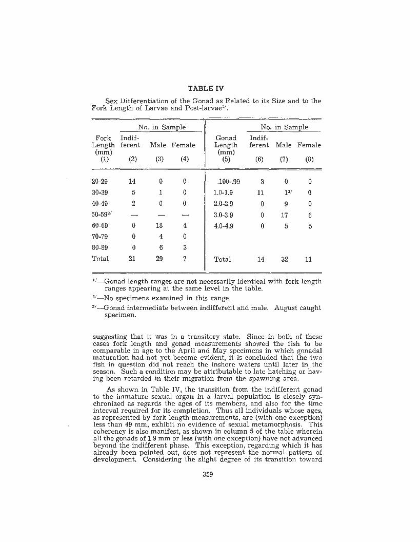
TABLE IV Sex Differentiation of the Gonad as Related to its Size and to the
Fork Length of Larvae and Post-larvael'.
40-49 2 0 0 I
No. in Sample Fork Indif-
Length ferent Male Female (")
(1) (2) (3) (4)
0 9 0 2.0-2.9
No. in Sample Gonad Indif- Length ferent Male Female ("1
(5) (6) (7) (8)
80-89 0 6 3 1 Total 21 29 14 32 11 I
50-5g2/ - - - 60-69 0 18 4 70-79 0 4 0
3.0-3.9 0 17 6 4.0-4.9 0 5 5
suggesting that it was in a transitory state. Since in both of these cases fork length and gonad measurements showed the fish to be comparable in age to the April and May specimens in which gonadal maturation had not yet become evident, it is concluded that the two fish in question did not reach the inshore waters until later in the season. Such a condition may be attributable to late hatching or hav- ing been retarded in their migration from the spawning area.
As shown in Table IV, the transition from the indifferent gonad to the immature sexual organ in a larval population is closely syn- chronized as regards the ages of its members, and also for the time interval required for its completion. Thus all individuals whose ages, as represented by fork length measurements, are (with one exception) less than 49 mm, exhibit no evidence of sexual metamorphosis. This coherency is also manifest, as sho'wn in column 5 of the table wherein all the gonads of 1.9 mm or less (with one exception) have not advanced beyond the indifferent phase. This exception, regarding which it has already been pointed out, does not represent the normal pattern of development. Considering the slight degree of its transition toward
359
testicular tissue, one could justifiably consider it as being undifferen- tiated. The above data if used as a means of expressing the ages of the fish at this period, would indicate that, because of the immaturity and absence of sex differentiation, embryologically speaking, fish of 49 mm or less should be considered as being larvae.
Ovaries and testes are first recognizable in fish comprising the present sample in the 60-69 mm fork length range. Also as shown in the table, the ovaries have grown to between 2.0 and 2.9 mm in length. Embryologically this represents the post-larval phase of development that will continue for a relatively short period until gametogenesis is initiated. Because of the composition of the sample employed, there remains unanswered the question of the status of the organs in the 50-59 fork length group, which as shown by Table IV was not repre- sented in the sample. However, from the pattern presented by the data, a postulation is permissible that sex first becomes manifest dur- ing the interval that the fish are in the 50-59 mm fork length range. This inference is derived from the conclusive break that separates the sexless gonadal state (40-49 mm range) and the inclusive appearance of males and females in the 60-69 mm class. Logically there should exist an interval intermediate between these two ranges in which the population should show transitional conditions of advancement, some members having undifferentiated organs while others exhibit male and female properties.
At the time that the larvae become post-larvae a number of gross and histological changes occur in the undifferentiated gonad that mark the onset of its metamorphosis into the primary ovary or testis. Immediately preceding this activity, the organ resembles that shown in Plate I, Fig. 3, but would evidence from three to eight gonia1 cells per section. Because of the variation which exists in the temporal occurrence of some of these activities, the events are more compre- hensible if they are presented primarily in accordance with the spe- cific changes involved rather than from the standpoint of the time sequence.
Although most of the significant changes that occur during this transitory phase are internal, some alteration in the contour of the organ is noticeable particularly if it is destined to become a testis. The more or less rotund undifferentiated gonad in this case under- goes a dorsal-ventral elongation so that in cross section it typically assumes a blade or arrow-head shape with the point or apex directed downward. Simultaneously its surface assumes a uniform entirety thus obliterating the irregularities and undulations that generally are present in the undifferentiated organ. In contrast, the ovary initial- ly continues to remain somewhat globose and evidence an undulating surface. Although the ovary will later show a dorsal-ventral elonga- tion, it is not as precocious as the testes in this respect. At the time that the internal changes indicate an ovarian transformation is in progress, its cross sectional profile more nearly resembles the condi- tions shown in Plate I, Fig. 4, rather than Fig. 5, which is from an older ovary. The gross changes in the ovary and testes which have been described are often not detectable in an individual organ possibly due to modifications introduced by fixation or embedding. They are most successfully demonstrated if a number of gonads are observed.
360

The internal histological modifications which occur at the time the ovaries and testes are evolving tend to fall into one or more of five criteria. Both the stromal substance and the sex cells become involved in these activities. Although the elements are undergoing an almost simultaneous transformation, they will be presented indi- vidually to simplify their consideration.
Very early in the transition from the indifferent gonad there oc- cur noticeable changes in the relative amount and distribution of the connective tissues. In contrast to the testes in which these ele- ments become relatively less prevalent and more widely dispersed, the incipient ovary possesses an abundance of very apparent connect- ive tissue fibers and fibroblasts. This condition imparts to the ovary an appearance of coarse solidity whereas the structure of the testis is suggestive of a finer, more open nature. In this respect, the ovary seems to be less precocious than the testis in that it retains for a longer interval the characteristics of the undifferentiated organ. The compact arrangement of the ovarian stroma remains substantially unchanged throughout larval development and undergoes modifica- tion only after the definitive ovigerous lamellae make their appear- ance. Its density then decreases greatly as it becomes arranged in strands in the lamellar cores and as investments around developing follicles. This diminution of connective tissue fibers is accompanied by a material decrease in the fibroblast population. In contrast, fol- lowing the initial relative decrease in the abundance of fibers and fibroblasts in the testis, there occurs no further diminution as devel- opment proceeds. Instead, a slight to moderate increase in their num- bers accompanies the formation of the walls and the interstitial tis- sues of the semeniferous cavities in late post-larvae and afterwards.
Concurrent with the events described above involving an altera- tion of the quantity of connective tissues during the transition from the indifferent gonad, changes are also occurring in the arrangement and distribution of the components. Prior to the inception of sex in the organs, the stromal connective tissues appear typically as a solid mass. Fibroblasts are much more in evidence than the associated fibers. For the most part the cells and fibers are randomly arranged in the organ except in areas closely adjacent to the gonia where they form a weak circular investment. In the earliest phase of ovarian metamorphosis, some of the fibers seem to coalesce and undergo a vague rearrangement to form what appears, in a cross section, to be radial strands. When first initiated, only one or two such radial strands occur in a section. Their number soon increases to as many as six to ten per section. Considerable variability exists regarding the number of fibers, and therefore, the thickness, that constitute the several strands or for that matter the thickness of a single strand along its course. Although they are in general oriented radially, many deviations in direction occur along their course. Frequently they appear to be discontinuous. These strands are originally very poorly delineated from the general mass of unorganized stromal tis- sues which separates them. Other fine fibers present in the stroma pass into and out of the strands. The significance of these events is that they foreshadow the subsequent origin of the ovarian lamellae and will constitute the source of the connective tissues for those struc- tures. In these later activities a radial form of delamination will seg-
361

regate the general pool of stromal elements, which still exists when the ovary comes into existence, into the separate lamellar compon- ents. In the case of the evolving testis, these radial strands never make their appearance.
Not only do the connective tissue components undergo a degree of radial orientation as described above, but concurrently with the onset of transition of the indifferent organ they show a distinctly different pattern in their relationship to the oogonia and the sperma- togonia. Prior to gonadal metamorphosis, the gonia were encapsulat- ed in the manner shown in Plate I, Fig. 3. In the case of ovarian evolvement, an increase occurs in the number and density of encircling connective tissue fibers without a comparable augmentation of fibro- blasts. The primitive follicle wall thus formed becomes relatively thick. Also, because of some increase in the size of the enclosed oogonium, enlargement of the follicular investment occurs so that its diameter approximates 11 microns. The picture thus presented of the follicles in the differentiating ovary is one of prominence of size and follicle structure. The connective tissues of the early differen- tiating testis which are associated with the spermatogonia exhibit an entirely different arrangement than occurs in the ovary. In the male organ delicate fibers ensheath the numerous gonia, which because of their much greater numbers lie in closer proximity to each other than do the oogonia. Often only one or two layers occur between adjacent spermatogonia. Instead of each germ cell having its own complement of investing fibers as occurs in the differentiating ovary, the filamentous elements in the testis ramify throughout the organ and become associated with numerous spermatogonia. Thus with re- spect to a specific germ cell, the fibers present may encircle it for a greater or less distance and then singularly or in small strands of two to three extend outward between the interstices of other near-by gonia where they may become incorporated temporarily into their investments. Under these conditions it is impossible to locate the beginning and ends of the fibers. The general impression presented by these ensheathments is that of a compact hexagonal lattice work in which the inner faces are circular where the fibers lie on the sur- face of the gonial cells. Shortly after the origin of the investments of the individual gonia, the testis produces a number of slightly heav- ier fibrous encasements each of which encloses an aggregation of from six to twelve spermatogonia. Only three or four of these encasements may appear in a cross section of a testis in its earliest phase9 of dif- ferentiation, but the number increases rapidly thereafter. Each is constituted of fibers which are cytologically similar to and of the same size or very slightly heavier than those applied to the surfaces of the individual spermatogonia. The encasements lie directly on the surfaces and in contact with the investments that surround the more peripheral spermatogonia. In this position, they receive fibers more and give off these elements to the nearby gonial investments. The events occurring in the testis at this period represent a foreshadow- ing of the subsequent transformation of these pockets or nests of gonia into the semeniferous spaces containing the sex cells and the contribution of the surrounding encasements to the interstitial tis- sues and the walls of the semeniferous cavities.
An important consistent feature appearing very early in the dif-
362

ferentiation of the gonads is the number, arrangement, and cytological properties of the oogonia and spermatogonia. Their frequency in both sexes shows some increase with the onset of sex transformation. In sections of the indifferent gonad, from three to eight of the sex cells are visible. Computations derived from data obtained from counts of the cells present in sections of ovaries undergoing transition indi- cate that their frequency is between one hundred and one hundred twenty-five per square millimeter. In a comparable age testis their numbers are approximately 3400 per square millimeter. As a result of the phenomenal increase in the male, the cells lie closely adjacent to each other to the exclusion of most of the stromal elements, a con- dition which is quite distinctive to the wider spacing of the cells in the ovary and the inclusion of considerable amounts of connective tis- sues in the intervening spaces. Cytologically, the chromophobia of the spermatogonia in the presumptive testis is more pronounced than occurs in the oogonia. This factor associated with the greater number of cells present in the testis imparts to it a kind of translucency that is not observed in the female organ. Other than the modification of the staining properties, no other distinguishing cytological features can be detected in the gonia1 cells of the two sexes.
Transitional ovaries furthermore consistently exhibit a pattern of stromal cavitation that is not found in the testes. Their numbers and arrangement show considerable variation. Initially only from one to four are visible in a tissue section but with the continued growth of the organs new ones arise at irregular intervals. As many as twenty- one such spaces have been counted in a section of a 3.2 mm ovary. When first detectable, they are about the same size or somewhat smaller than the oogonia. Their slightly irregular margins are bound- ed only by fibroblasts and rarely by a stromal fiber that incomplete- ly encompasses the surface. Shortly after the cavities have made their appearance, they increase in size at rates exceeding that of the growth of the organ as a whole so that they impart to the tissues a sieve-like aspect. This conditilon is illustrated in Plate I, Figs. 4 and 5, in which views only a moderate degree of cavitation is evident. As the spaces enlarge, they assume various shapes and their margins become more irregular. The peripheries of the more advanced ones become invested with a detectable but not pronounced fibrous connective wall. The contribution which these spaces make to later ovarian morphogen- esis is considered below.
While the stromal cavities are arising in the manner described, a second form of ovarian cavitation originates along the medial long- itudinal axis. This activity is initiated near the dorsal cephalic pole of the organ, a little below the insertion of the mesentery. It rapidly extends ventrally and caudally in the mid-plane resulting in the sep- aration of the ovary into right and left components except in an area of its ventral margin (Plate I, Figs. 4 and 5) . This separation occurs along a poorly organized vertical longitudinal sheet of connective tissue, the elements of which remain adherent to the newly created faces of the stroma. Almost as soon as it comes into existence, its margins begin to evidence a few indentations in the direction of the lateral stromal material. Both the number of these recesses and the amplitude of stromal invasion increase as morphogenesis proceeds. Whereas the size of the apertures and the channels of the original
363

indentations are noticeably broad, they progressively narrow as the gonad becomes more completely differentiated. Ultimately they come into contact with and open into the numerous cavities described above that occur in the compact stroma. By this means the original chan- nels are extended to the ovarian tunic. (Plate 11, Fig. 6). In actuality what appears to be channels in sectioned material, exist in the form of clefts. The faces of these transverse clefts are not particularly parallel to each other, so that the enclosed spaces are of variable widths. New generations of clefts arise from the previously establish- ed spaces and thence extend toward the margins of the organ in a root-like manner. The various clefts intercept each other thus par- titioning the stroma into irregularly shaped sheets and finger-like columns. The bases of the stromal columns thus produced lie on the tunica albuginea and their free ends extend into the central lumen of the organ. Each of these stromal columns contains several oogonia in addition to the connective tissue elements. The resultant structure constitutes a primitive ovigerous lamella, which from its manner of origin is not of uniform thickness throughout. The original lamellae are somewhat massive. However, due to some continued production of secondary and tertiary clefts from the primary channels, the early massive type of lamella becomes converted later into more elongate forms. Two additional factors which contribute to their elongation is their extension by terminal growth into the central cavity of the ovary, and by growth increments of their entire lengths made possible by lateral expansion of the gonad.
Except where otherwise stated, the events described in the pre- ceding paragraphs have their origin in the earliest phases of the dif- ferentiating gonad. After they have resulted in the establishment of the basic architecture of the ovary, the oogonia, which to this time have played a passive role, begin to evidence activity in the direction of gametogenesis. This normally occurs during the early summer while the fish are still late larvae or post-larvae. Plate I, Fig. 5 of a very young juvenile shows the advancement of the oogonia into Stage I and Stage I1 oocytes. In this illustration, the Stage I cells are barely visible while the Stage I1 oocytes, of which there are only two or three present, are the larger cells with a fairly distinct profile. The number of these cells is greater in the figure than in the younger fish.
With the completion of the activities described, a definitive func- tional ovary is brought into existence. Its further development in- volves a continuation and refinement of the connective tissues and the arrangement of these components into a functional pattern, the continued growth and organization of the ovigerous lamellae concur- rent with movements or migrations of the sex cells to superficial positions along their surfaces, and the augmentation to the earlier population of oogonia.
V. ORGANOLOGY AND HISTOLOGY OF THE NON-GERMINAL ELEMENTS OF THE DEFINITIVE OVARY
Continuing morphogenesis and progressive enlargement of the organs which occur subsequent to the initial organization described above culminate in the establishment of the functional adult ovary.
364

Fundamentally these changes in the non-germinal components consist of refinements associated with growth of the ovarian tunica aEbuginea (Plate 11, Fig. 8) and further supplementation and growth of the lam- ellar folds.
The outer tunic becomes quadripartite comprised of a superficial epithelial investment, a prominent connective tissue sheath, an intern- al muscular layer, and an internal facing. The entire complex increas- es in thickness from about .04 mm in gonads ranging between 15 to 20 mm in length to about .08 mm in 25 to 35 mm gonads. At matur- ity in gravid ovaries this thickness diminishes slightly, due to stretch- ing in response to internal pressures developed by the burden of mature ova.
The outermost component of the theca consists of a layer of chrom- ophobic simple squamous mesothelial cells whose mean thickness is 4 microns or less. The presence of this layer in illustrated in Plate 11, Fig. 8, as a thin undulating homogenous line lying on the surface. The small, widely spaced pycnotic karysomes characteristic of the epithelium of 15 to 20 mm organs become progressively smaller and less frequently encountered by the time the organs have attained a length of 25 to 30 mm. Still later, as in gravid ovaries, the cellular elements appear to be completely absent, and in their place there is present material that suggests a fibrous origin. In small ovaries a moderately distinct basement membrane of about 0.2 microns thick underlies the superficial cells, but with further growth of the organs it progressively diminishes until it becomes no longer distinguishable.
The sub-epithelial connective tissue layer constitutes the principal component of the tunic. As in the case of the superficial epithelium, its maximum development occurs during summer and fall while oogenesis is in its earlier stages, and diminishes thereafter as the organ attains the gravid state. It is comprised entirely of relatively coarse fibers the majority of which encircle the ovary in a plane perpendicular to its long axis as shown in Plate 11, Fig. 8. This illustration was made from a longitudinal section and therefore presents the cut ends of the elements. The layer is not of uniform thickness in all areas. In some places it may represent as much as three-fourths or four-fifths of the overall thickness of the tunic while at other places it may be reduced to about a fourth of the thickness. Often it reaches its highest state of development at points where inter-lamellar septa arise from the tunic as seen in the figure. Although the general pattern of the fibers is one of gonad encirclement, it can be seen in sections passing through the tunic in the manner of a chord through a circle that many of the fibers extend around the organ in oblique directions. The ir- regularity of fiber orientation is also pronounced in its inner face at points where the inter-lamellar septa arise. Here many fibers deviate from the normal circumferential pattern to enter the septa. Their entrance into the latter structure may or may not be organized in the mannp of parallel strands. A heterogeneous arrangement is illus- trated in Plate 11, Fig. 8, where the fibers appear as colorless elements intermingled with the darker muscle cells. Many fibroblasts are as- sociated with the fibrosa1 investment and are depicted as small black dots in the photograph.
In addition to its participation in the creation of the inter-lamellar
365

septa, it gives rise to rather delicate collections of fibers that pass into the cores of the lamellae so as to constitute the principal compon- ent of the intra-lamellar connective tissue. Two complete strands are visible in Plate 11, Fig. 7. These intra-lamellar entities become less indentifiable as the ovary matures.
Lying under the fibrous investment there occurs the third layer of the tunic. It is comprised principally of smooth muscle with a minor contribution of fibrous elements. The identity of the con- tractile elements was established only in sections stained with Cajal’s or Flemming’s. This tissue is present in all ages but appears to reach its zenith in juvenile, adolescent, and young mature ovaries when its contribution is relatively greater to the other tissues than in gravid and spent organs. Although the muscle cells do not present a unified alignment with each other, the majority are oriented so that their length coincides with the anterior-posterior axis of the organ. This pattern, as shown in the longitudinal section (Plate 11, Fig. 7) is inter- rupted where the inter-lamellar septa arise. At the point of origin considerable disorganization usually occurs which is regained some- what in the deeper sections of the membrane. The rigid, spindle shaped cells are oriented in the usual overlapping pattern character- istic of organized smooth muscle. As far as could be determined, they are similar in size whether they reside in the tunic, the inter- lamellar septa or in the core of the lamella. They have a long axis of about 35 to 40 microns and a mid-point transverse diameter of about 4 microns. The acidophilic cytosome is homogeneous and it not sur- rounded by a distinct membrane. Elongated rod shaped nuclei meas- uring about 8 microns in length lie in the cell center. Enclosed in a distinct nuclear membrane is a moderate quantity of finely granular chromatin suspended in a colorless nuclear sap.
The principal vascular elements of the ovarian circulation lie in the outer areas of the tunica albuginea and consist of right and left longitudinal channels situated parallel to and slightly below the line of insertion of the mesovarium in which approximate position they were present in the differentiating ovary. From these many arterial and venous tributaries arise and pass in a general ventral direction in the superficial part of the tunic meanwhile giving rise to prominent segments which invade the inter-lamellar septa, and also numerous units that ramify throughout the tunic or enter the bases of the lamel- lae where they ultimately contribute to the capillary system that has an extensive distribution in the cortical and sub-cortical areas of its investment. The largest segments normally consist of closely asociat- ed venous and arterial channels.
Unlike the other elements of the tunica albuginea that show var- iable degrees of regression with the advancement of the sex cycle, the hemal system with two exceptions is maintained in a state of mod- erate to high development throughout the year. Its prominence in- creases in all areas of the ovary from a little after the beginning of oocyte maturation to about the onset of yolk formation. At that period many of the major channels lying on the ovarian surface appear to become more displaced toward the exterior as the tunica1 tissues be- come thinner. The condition with respect to these vessels is main- tained throughout the remainder of the period that the eggs remain in the ovary. Likewise the vessels that enter the inter-lamellar septa
366

do not show signs of regression. On the other hand, the smaller ves- sels that course through the tissues of the tunic and those that enter the cores of the lamellae are either lost from view or diminished in numbers shortly after the beginning of yolk formation until the com- pletion of ovulation after which they begin to reappear as absorption of the empty follicles progresses. This follicular invasion by the term- inal segments of the system is illustrated in Plate V, Fig. 20.
The arterial channels that course through the tunic and the inter- lamellar septa are invested with a dense outer layer of what appears to be smooth muscle cells which are associated with an inner layer of coarse wavy connective tissue suggestive of elastic fibers. This fibro- muscular complex is oriented circumferentially in from two to six in- distinct layers according to the caliber of the channel. The pattern becomes less regular as tributaries leave the larger channels and ultimately disappears entirely. Histologically the arrangement does not establish a true vascular wall as normally recognized in higher forms since it is not distinctly separated from the tissue elements of the adjacent tissues, and because cytologically its components are similar to others comprising the tunic or inter-lamellar septa although the vascular contractile elements appear to be shorter than those pres- ent in adjacent non-vascular areas.
The fourth and innermost layer of the tunica albuginea is not of constant occurrence. In young adolescent ovaries it consists of a very delicate layer, the entire thickness of which is not more than 0.2 mic- rons, comprised of a few greatly flattened, fibroblast-like cells asso- ciated with fragile fibers. When present it is continuous along the inner face of the tunic except at points where intra-septal and intra- lamellar connective tissue arise from the tunic. In these areas, it be- comes applied to the surface of the reflections, but soon becomes in- discernible. At this stage its cells are smaller than the contiguous contractile entities that lie above them in the tunic. They do not possess definite margins and evidence no cytoplasmic architecture. Their dense small nuclei are flattened in the plane parallel to the sur- face of the membrane. Later, in ovaries which are approaching ma- turity, this membrane undergoes radical alterations at the interstices between the basal origins of the adjacent lamellae. This transforma- tion brings about a progressive reduction of the fibrous material ac- companied by varying degrees of fragmentation and a superficial in- clusion of some of the parts in the adjacent muscular layer. Recog- nizable cells are now characteristically absent in these regions, the areas being occupied by a peculiar form of vacuolated tissue. The ovate-quadrate vacuoles, which become more numerous as ovarian maturity is attained, enclose a minimal amout of unidentifiable ma- terial which stains faintly with eosin. They are approximately 3 to 5 microns in size. The vacuolated structures lie adjacent to each other and increase in depth from one row during the initial period to about three imperfect rows at the completion of the process. The material incorporated in their margins is moderately acidophilic and presents the appearance of being shredded. The mean thickness of the vac- uole walls is about 0.5 microns. Where these vacuolar entities lie adjacent to the muscular layer, fiber fragments of varying length and thickness are randomly disposed, forming a loose bond between the two. Their aspect is not of a regressive nature. Because of the
367

correlation of their development with maturation of the oocytes, they apparently play a role in the process.
Arising from the inner face of the tunica albuginea are the ovig- erous lamellae characteristic of teleosts having a cavitated ovary. An account of their origin and early development in young differentiat- ing ovaries has been given above. Their subsequent numbers and morphology are related to the age and sexual phase of the ovary. In adolescent organs the few lamellae present arise from the tunica al- buginea by a relatively broad base and thence extend inward toward the ovarian lumen in the form of a hyperbola whose margins are relatively free of undulations or indentations. Their maximum in- ward excursion varies from about one and a half to two and a half times the distance across their bases. With growth of the ovary, lamellar elongation is evident, a condition which, together with an increase in their number, greatly modifies their form. They char- acteristically extend as much as two-thirds to three-fourths the trans- verse diameter of the organ so that their terminal portions interdig- itate with each other. At the basal attachment of older lamellae to the tunica albuginea, the transverse extents become relatively nar- rower in proportion to their length so that they appear to arise more abruptly and have a greater uniformity in diameter throughout their length. Their profiles in sectioned material are marked by undula- tions and indentations on their lateral margins and by the impinge- ment of neighboring lamellae (Plate 11, Fig.7). Due to extreme disten- tion of the lamellae in gravid ovaries, inter-lamellar spaces are re- duced or obliterated so that their margins become applied to each other in a great variety of patterns dependent upon the conformation of adjacent lamellae. This condition also occurs during an interval following ovulation prior to absorption of the follicular remnants.
Lamellar surfaces are basically invested with varying amounts of loosely associated fibrous connective tissue, fibroblasts, and occas- ional contractile elements which are without specific organization. The abundance of these components is inversely proportional to the size of the lamellae. During late spring and early summer ovarian activity is at a seasonal minimum and it is at this period that the lamellar investment attains its greatest degree of development (3 to 5 microns thick). As the organ approaches the climax of its seasonal cycle the lining becomes less apparent, and in scattered areas only occasional strands of loose connective tissue are present. The reduc- tion and disappearance of the lamellar investment is probably an im- portant part of the morphogenic changes designed to weaken the su- perficial tissues constituting these folds to facilitate the escape of the eggs at the time of spawning. The principal factor involved in the reduction of the covering membrane during this period appears to be of mechanical origin resulting from the pressure hypertrophy of the ovigerous lamellae brought about by the burden of the enclosed ova.
Small vascular channels ramify throughout the marginal lamellar surfaces of maturing ovaries. Present are capillaries, terminal divi- sions of small arteries, and collecting veinules. In non-gravid organs the smallest of these channels occupy a more superficial position than the larger channels and are covered only by the thin investing lamel- lar membrane. Capillary abundance is such that they are seldom
368

separated from each other by more than 25 to 30 microns. The larger channels occur at various levels a little below the lamellar surface and in the proximity of developing oocytes. They are circumscribed by poorly organized triangular or irregular condensations of connect- ive tissue fibers, some of which continue into the deeper stromal ma- terial of the lamellae either individually or as coarse strands. In gravid and newly-spawned ovaries the fibrous investment becomes reduced and evidences less organization. At the same time, a number of new channels appear within the lamellae and many of the superfi- cial ones disappear. It could not be determined with certainty whether the new ones are of recent origin or whether they represent the previous channels that have moved deeper into the stromal sub- stance. The lamellar blood supply is derived from major arteries and veins located in the tunica albuginea, from which ramifications continue through the core of the lamella and thence to their more peripheral areas.
The general stroma (Plate 11, Fig. 7, and Plate VI, Fig. 22) of the lamella is comprised of a minimal quantity of loose, irregular connect- ive tissue (except where it is associated with follicles), a central sheet or band of smooth muscle which also contains elements of fibrous tissue and occasional fibroblasts. The tissue elements comprising the lamellar core are continuous with the muscular and fibrous layers of the tunica albuginea. This intra-lamellar reflection attains its great- est development during the initial stages of oogenesis at which period it transverses from one-half to three-fourths of the length of the lamel- la. As it extends distally into the lamella it displays considerable variability in thickness and often presents an undulating appearance. These undulations, in general, conform to the folding or bending of the entire lamella, and are thus more evident in ovaries in which a crop of eggs are maturing than in juvenile or quiescent organs. Also, during the period that oocytes are ripening, the muscular core can be seen to develop on its lateral surfaces tangential tongue-like projec- tions comprised of one or more muscle cells in which the long axes are oriented in the general direction of the periphery of the lamella. The number of these muscular projections in an individual lamella varies from none to four per oil immersion field, the increased fre- quency usually being associated with enlarged but not gravid lamel- lae. The extent to which these laterally directed processes project per- ipherally, although variable, is considerably short of the lamellar margin and in most instances they terminate between a fifth and a half of this distance. Great diversity exists as to the profile of these bundles so that they are encountered in sections as compact, simple, flame-like projections tapering distally, or irregular, branched struc- tures whose thickness may vary along their course. At their extrem- ity the individual muscular elements lose their intimate association with each other, in which case their distal ends project individually or in small groups of two to six cells each into the adjacent, loose connective tissue. The terminal arrangement thus presents a consider- ably frayed conformation whose individual entities may be rigid and straight or arched, the free ends of which became more or less in- timately associated with the nearby fibers of the connective tissue. In gravid lamellae, morphological distortions of their central muscular cores have culminated in altering the arrangement of the components
369

to a state in which the cells are disassociated to a greater extent. The connective tissues between the central core tissues and the
lamella margins occupy the entire internum which is not devoted to the oocytes, vascular channels and the delicate muscular strands. The fragile fibrous elements usually appear singly, coursing in- discriminately and irregularly for short distances in the manner of a primitive form of areolar connective tissue. A few are visible in Plate TI, Fig. 8, in the clear area near the inter-lamellar septum. Be- cause of their paucity, the areas surrounding the oocytes and the internal musculature contain considerable quantities of tissue fluids. The abundance of this fluid is of material significance in providing an exchange mechanism between the cells in the lamella and the capillaries which are for the most part in the superficial areas. The frenquency of the fibers and the associated fibroblasts is variable according to the age and sexual phase of the organ. Their density is relatively greater in young or inactive organs than in gravid or spawn- ed ovaries. Although the numbers and arrangement of the ramifying fibers are such that a direct and continuous relationship cannot be established between them and the contractile elements residing in the lamellar core or those extending peripherally from the core, their presence and general disposition is such that they appear to be cap- able of diffusively conveying contractile tensions originating in the muscular tissues to the various areas of the entire structure and its contents. The associated fibroblasts of the areolated connective tissue are small, usually irregular in shape and are promiscuously disposed. A minimal quantity of homogeneous acidophilic cytoplasm invests their elongated, oval and compact nuclei.
The dynamics of the muscular and fibrous elements of the ovary appear to be directed toward the effective performance of ovulation. The culmination of this activity is accomplished by the rupture of the follicle, at least in part, attendant to the application of external pres- sures. These forces originate through the simultaneous interaction of the muscles of the tunica albuginea synchronized with contractions of the cells embodied within each lamella, augmented by a shortening of the transverse axis of the ovary resulting from contractions of the cells in the inter-lamellar septa. This culminates in compression of the intra-ovarian tissue fluid and more significantly the fluids of the areolar spaces within the lamellae, thus assisting the rupture of the follicles. Contributing to release of the ova it also appears that the activity of the intra-lamellar muscles at this time is transmitted to the connective tissue investment of individual follicles so as to de- crease its effectiveness in retaining the mature eggs. This system, in which a generalized pressure is exerted throughout the lamella and thence applied to the follicle and the enclosed egg, plus the se- lective action of loosening the follicular investment by the contrac- tion of intra-lamellar muscle cells, permits considerable selectivity in the intermittent spawning of ova from the different lamellae or areas within a single lamella over a substantial period of time, as is thought to occur in B. patronus in the Gulf of Mexico.
370

Fig. 10 Fig. I 1
I
Fig. 12 Fig. 13
PLATE 3
371

Fig. 74
-b
E,
Fig. 15
81 . I -
l -
--w a
Fig. 16 Fig. 17
PLATE 4
372

VI. HISTOLOGY AND CYTOLOGY OF THE GERMINAL ELEMENTS OF THE DEFINITIVE OVARY
Many methods have been used by various investigators in classi- fying the developmental stages of fish oocytes. In the studies con- ducted by the author, six maturation intervals are recognized. These are based on nuclear and cytosomal characteristics which are specific for each interval. The principal distinguishing features of each stage are:
Stage I. Undifferentiated oocytes not far removed from the oogonia.
Stage 11. Oocytes evidencing meiotic differentiation. Stage 111. Oocytes undergoing initial lipogenesis. Stage IV. Oocytes in which provisional yolk is being produced. Stage V. Oocytes evidencing nuclear absorption, and concurrent
Stage VI. Mature oocytes with a central oil globule. Stage I: The cells constituting this stage are interphase or prolif-
erating first phase oocytes. A number of constant characteristics specifically identify this period of development, principally the fre- quent disposition of the cells to be organized in groups or nests, the presence of a single prominent nucleolus, vague or indistinguishable cell outlines, a delicate serrate karyotheca, and their size. Cells of this age are shown in Plate 11, Figs. 7 and 9. Figure 4 which is from a June specimen with an ovary length of 1.92 mm shows large num- bers of these cells. Figure 9 shows seven of the oocytes in more detail.
These oocytes are always oriented near the margin of the oviger- ous lamella, where they are more or less encapsulated by a delicate, fibrous form of connective tissue. In adolescent ovaries and those of mature fish which are in a state of reproductive dormancy, they are typically organized into aggregations at locations where the mar- ginal investing lamellar connective tissue is reflected into the inter- ior of the ovigerous lamella. From three to ten such cells present themselves in each nest-like location in sectioned material. As the ovary begins to respond to oogenesis, the number of these early stage cells within a nest progressively diminishes as they migrate into the internal lamellar matrix or under the margin of the lamella. Early oocytes may also occur singly at irregular intervals in the lamellar investment which, due to its thiness at such points may be deflected somewhat as a slight elevation above their outer surfaces.
Stage I cells are present in greatest numbers in non-functional ovaries decreasing progressively throughout oogenesis until they can- not be found after the completion of yolk formation. In functional gonads of the large scale menhaden of the Gulf of Mexico, they do not appear during the period from October to February. A similar rhyth- mic behavior of the early sex cells has been identified by a number of investigators, and raises the question of the source of the next generation of oocytes. This matter has escaped elucidation although
production and maturation of vitelline elements.
They are the smallest cells visible.
373

many concepts have been advanced. It does not appear to be involved in the viviparous teleost, Neotoca bilineata, in which Mendoza (1939) found that nests of these cells occurred abundantly at all times, al- though a considerable decrease in numbers developed during the latter part of gestation. Wheeler (1924) admits that he is uncertain of the annual origin of a new crop of eggs in the dab, PZeuTonectes Zimanda. He was unable to find any trace of mitotic division of oogon- ia, of oocytes or stages in the formation of oocytes, although, as soon as mature eggs begin to shed, small oocytes begin to appear. The yearly periodicity in the appearance of these cells was considered by Wallace (1903) to be from the ovarian epithelium and that the whole nest is derived from a single mother cell. His postulation is based upon his interpretation of the studies by Forchammer, Rathke, and Stuhlmann in the absence of his own direct observations. Some ambiguity also is introduced by his failure to identify specifically the nature or location of the terminology “ovarian epithelium.’’ Hickling and Rutenberg (1936), in considering the ovary as an indicator of the spawning period, elude the question of the periodic origin of a new generation of oocytes by assuming there exists a general egg stock from which a quota is withdrawn each season. Their cytological description of the “egg stock” establishes a much later phase of matur- ity than my definition of Stage I oocytes. Naumov’s (1956) studies on ovogenesis in the herring also discharge the cyclic appearance of new germinal elements with the comment, “The earliest phases (ovo- cytes are . . . always present in the ovary.” The cells to which he refers, although present at all times in the ovaries of the clupeoid fishes, are oocytes in the process of maturation as shown by his des- cription and figures. They are comparable to Stage 111 oocytes in the present paper and as such are definitely post mitotic. Under these circumstances, the paper does not provide an answer as to the source of the primitive cells from which the herring ovocytes arise. In addition to the possible sources cited above from which a new crop of eggs may arise, Wheeler (1924) states that the mature ovum is sur- rounded by a follicle comprised of two distinguishable cell types, and that he feels fairly certain that from one of these types new oocytes will develop. The author is inclined to hold that the cyclic origin of these cells is from the lamellar epithelium, a postulation which gains support from their initial intimacy with the investment. He also purports that only a few cells are derived from this source and that through the intervention of mitosis there is produced the ag- gregate comprising the nests. His conclusions are open to question for two reasons. First is the enigma of the apparent necessity of the organ to incorporate a number of the sex cells during its origin if it possesses the capacity to develop them later from a tissue type de- rived from the somatic stromal cells. By following the embryology of the organ, the oocytes are seen not only to be present at all times but their constant replication insures an adequate supply for the first and succeeding crop of eggs. The second aspect of his conclu- sion that is unique is that the existence of such a condition would represent a major departure of the conservation of stem sex cells in the vertebrates. It is my opinion that these primitive cells are al- ways present in the organ although they may be reduced to such minimal numbers during the sex cycle that they are not observed.
These primitive cells are markedly uniform in size and morph-
374

ology. They may easily be distinguished from the numerous fibro- blasts with which they are usually associated by their greater size and pronounced differences in nuclear structure. While exact meas- urment of individual cells is difficult due to their indistinct margins, the mean diameter is approximately 7.3 microns and their volume 137-1° cubic mm. Spherical forms predominate where they occur singly, but in nests their vague margins often seem to be flattened or slightly concave due to the effect of crowding. A thin complex of delicate connective tissue fibers enclosing an amorphic matrix completely invest the individual cells as well as the nests. Fibroblasts are rarely encountered.
The oocyte cytosome possesses little or no affinity for any of the dyes in the staining techniques used (Plate 111, Fig. 9), a condition which has been observed by numerous authors. Particularly pertin- ent in describing this cellular entity is Hickling’s (1935) comment that in the immature hake, Merluccius merluccius, the cytoplasm is in- visible. In the menhaden it is only in the general region of the nu- cleus that the cytoplasm retains an almost infinitesimal amount of the acid dyes, but progressively, as the periphery is approached, the sub- stance becomes paler until all color is lost. In the figure the cyto- some is represented as the colorless area surrounding the dark nuclear margin. The cytosome is devoid of vacuoles or other inclusions, ex- cept that with a magnification of 1450 discrete pale granules can be observed in great numbers. Their number continuously decreases from the nucleus outward. A cell membrane, if present, cannot be seen, although the form of the cell can be closely approximated by the encapsulation of the investing fibers.
Karyosomes are easily recognized. Their diameters are about 4.3 microns and they represent about 685% of the diameter of the cell and about 30% of their volumes. In later stages, as maturation of the oocyte progresses the nuclear diameter and volume will greatly in- crease but will at the same time become proportionally smaller with respect to the enlarged cell. Structurally it is comprised of a con- spicuous karyotheca, circular in form, and a colorless internal matrix. The limiting membrane possesses a thickness of about 0.5 microns and is smooth on its outer face, but somewhat serrate internally. It stains with the basic dyes, thus suggesting a minimal condensation of chromatin on its inner face. In the inter-mitotic phase the internal components of the nucleus consist of a gossamer-like reticulum, the components of which are frequently disposed in a radial pattern, and which is composed of minute granules. The granules are either very faintly basophilic or neutrophilic. A sharply defined nucleolus some- what less than 1 micron in diameter occupies the center of the nu- cleus. Its substance has a pronounced affinity for basic dyes for which reason it is opaque.
It is not unusual to find considerable mitotic activity in these cells particularly in young gonads or at the onset in the spring and sum- mer of a new cycle in older ovaries. At such times synchronization of the event seems to occur in all or most of the cells constituting a specific nest. It is characterized by the disappearance of chromatin material from the nuclear membrane and the development of distinct intra-nuclear chromosomes.
375

Stage 11: The transition of the reproductive cells from the Stage I oocytes to Stage I1 is apparently rapid since intermediate forms are encountered only occasionally. The principal maturational activity occuring in oocytes of this age involves the nucleus within which meiotic activities are in progress. These cells are apparently unable to reproduce mitotically.
Larger or smaller numbers of these cells may be present through- out the year. In adolescent ovaries which are preparing to mature their first crop of eggs a pronounced increase in their numbers oc- curs during the fall and early winter months. At this time these ovaries are densely packed with such cells and with cells of the earl- ier stage but contain few or no older stage oocytes. These will not complete oogenesis during the impending spawning season but will attain maturity of Stage I11 oocytes during the winter and remain in this state until the following season. Plate 111, Fig. 10, from an ovary which is about to enter this phase, depicts three cells of this age in- terspersed among the numerous smaller Stage I oocytes. This con- dition of partial precocious development postulates that these ovaries, although not capable of completing the cycle of oogenesis within the present season are responding to the same factors which cause the complete maturation of eggs in the older organs. When the tropistic stimuli are changed as with the onset of winter, development will stop until the return of favorable environmental conditions at the follow- ing season. The situation in ovaries which are mature and which have produced a previous crop of eggs is considerably different. Here the annual rhythm involving the appearance in large numbers of a new crop of Stage I1 cells during any seasonal period is confused or absent as shown in Table V. If the data is smoothed by using a moving average of three, it is found that the production of oocytes of this stage in the older ovaries is markedly constant from January to Sep- tember, thereafter progressively decreasing until December, at which period the numbers present are reduced to about one-half that which existed earlier. The retarded pace of production of Stage I1 cells in ovaries which have previously matured eggs is due in part to the con- siderable supply of Stage I11 oocytes present in the ovary, cells that have been carried over from the preceding spawning season and which will be utilized for the new crop of eggs, and in part due to the fact that in such ovaries there occurs a slow transformation of Stage I cells into Stage I1 oocytes throughout most of the year.
These cells are seldom found in aggregations characteristic of the youngest oocytes. Those which earlier were contained in the nests begin to migrate to the interior of the ovigerous lamella accompanied and surrounded by varying amounts of connective tissue (Plate 111, Fig. lo), while cells originally disposed singly sub-adjacent to the su- perficial covering of the lamella tend to retain that position. In the latter situation, the cellular enlargement is directed toward the in- terior of the ovigerous lamella, and such cells carry on their inner face a reflection of the marginal lamellar connective tissues. In this manner their enlargement does not produce elevations of the surface of the lamellae such as occurred in some Stage I oocytes.
Cells comprising this stage are distinguishable by a disproportion- ally large nucleus, a heavy limiting nuclear membrane, a visible com-
376

TABLE V Percentages of oocyte stages by fish and months.
STAGE STAGE STAGE STAGE STAGE STAGE I I I V 111 IV VI FISH SIZE MONTH
( M M . F. L.) CAUGHT ( % I ( % ) ( % ) ( % I ( 9 % ) ( % )
50- 74 75- 99
100-124
125-149
150-174
175-199
200-224
225-249
August 96
June 46 August 48 December 8 April 64 June 36 August 35 November December
January April 36 June 58 August 21 October November December
January April 62 August 10 October 2 December January 10 February April 36 May 36 June 2 October 4 December
March 7 April 57 May 25 October December August 10 December
April 100 3
18 18 25 15 14 16
6 5 8
12 12 6 3
10
8 15 17 8
7 6
11
2
1
36 34 37 21 50 49 8
77 54 58 37 71
100 82 83 47 25 84 75 76 51 42 56 45 81 58 45 34 37 64 83 47 81 35
92 8
23
18 9
18 1
20 14 23 6
4
21 18 33
17 17 7
23
15 15
8 18
10 3
12
5 19 19
19
18
8
5
3 40
4 18
17
24
377

plement of chromatin which in many cells is organized into meiotic chromosomes, the eccentric position of the prominent nucleolus in those cells not in a state of meiosis, and the presence of a distinct cel- lular membrane. Plate 11, Fig. 9, shows three of these cells photo- graphed at the same magnification. The darker oocyte which has been added to the picture from another photograph is slightly more advanced than the remaining two. These oocytes are seldom encount- ered as true spheres, especially in older ovaries containing many cells in the more mature stages. This crowding effect results in the deeper oocytes assuming pear- or oval-shaped configurations, or, in the case of isolated sub-marginal cells, a flattening along their outer surface.
The mean diameter of late Stage I1 cells is 22 microns, represent- ing a growth increase of about 350 per cent of the previous stage. The effect of this increment is to materially enlarge their volumes to about 4200 per cent of the Stage I oocytes. Concurrent with cellular en- largement, the nucleus has materially increased its size from 4 microns to about 13 microns in diameter or about 350 per cent, but its compara- tive volume increase amounts to about 530 per cent.
Specific follicular thecal investments are not present, although the nearby connective tissue fibers are more compactly applied to the surface of the cells than previously. A delicate and moderately well- defined cellular membrane of a neutrophilic character develops as the stage progresses. Cytoplasmic transformations from the previous condition are minor. A continuing increase in its granular content is noted and due to the uniform distribution of the material, the cytosome appears homogeneous throughout. Also, as a result of the greater abundance of this matrix and its enhancement for plasma stains, it appears moderately denser than earlier. Organelles or meta- plastic materials were not demonstrated by any of the stains used.
With the onset of Stage 11, nuclear enlargement occurs very rapidly so that the body soon comes to occupy most of the cell. Be- cause the cells at this period tend to be more isolated from each other than formerly so that they are subject to less compression, the nucleus assumes a more central location and spherical shape. The enclosed chromatin meanwhile migrates away from the nuclear membrane, and as it does so it rapidly loses its affinity for the basic stains. For a short interval it is neutrophilic but then becomes weakly aciodphilic in which state it remains for the duration of this stage. In the inter- phase condition, the chromatin is distributed throughout the nucleus but because of its fineness and poorly developed tinctorial properties it is indistinct. The inter-phasic character of the oocytes may be ob- served in at least some cells in all functional ovaries. The state is most pronounced in the few Stage I1 oocytes that are present during the period of yolk formation and the final maturation of the egg. These cells will lie dormant until the subsequent reproductive cycle. On the other hand in ovaries that are in the process of preparing a new crop of oocytes most of the cells are seen to be in meiosis. At these times the chromatin appears as rather intensely stained threads or chromosomes which are sharply set off against a somewhat color- less background. The material is in this condition in the darker cell in Plate 111, Fig. 10. During the early phases of Stage I1 the nucleo- lus constitutes a prominent body measuring about 2 microns in diam-
378

eter and occupying a central nuclear position. It stains intensely with chromatin dyes for a short time after the initial phase of this stage following which it rapidly becomes oxyphilic. Prior to or during the initiation of the meiotic events it moves peripherally to a position against the nuclear membrane. There it may undergo a degree of flattening against the karyotheca and thereafter tends to retain its position throughout the remaining activity of this stage.
Certain significant nuclear bodies first become evident during the mid and final phases of Stage 11. At the completion of this stage, their numbers may vary from one to four per cell. The fact that they morphologically and tinctorially resemble the nucleolus has led to considerable confusion, although it is my opinion that their origin and fate is quite different. Speaking of these bodies Wallace (1903), after discussing the role of the germinal vesicle in the formation of oil droplets, stated “ . . . that the nucleoli are not transformed either into oil globules or yolk spherules.” Similarly, in Neotoca bilineata the multiplicity of nucleoli is noted by Mendoza (1939), and in 1940 he reported that “Numerous vacuolated nucleoli may appear within the nucleus.” In his description of the nucleus of the yolkless ova of the plaice, Cunningham (1894) recognized that “The nucleus or germinal vesicle is enclosed by a membrane and contains the nucleoli, . . . ” and according to Craig-Bennett (1930), the commencement of maturation in Gasterosteus aculeatus is described as being marked by the appearance of numerous small nucleoli. Wheeler (1924) also observed the existence of these bodies in the nucleus of the reproduc- tive cells of Pleuronectes Zimanda, which apparently are of universal occurrence in the early stages of oocyte development. Referring to the nuclei of these cells he reported, “ . . . they contain irregular masses of material staining deeply with hematoxylin. These masses tend to stay just inside of the nuclear membrane and then form a dark, irregular border with occasional inward projections.’’ The bod- ies to which he and the other authors cited above refer are compar- able to those occurring in the menhaden shown in Plate 111, Figs. 11 through 13. Wheeler (op. cit.) refrains from identfying them as nu- cleoli.
These nuclear organelles in Brevoortia patronus arise early in Stage 11, but their growth and functional activity does not become evident until later. When first observed they appear as minute, acid- ophilic, irregular masses lying in conjunction with the inner face of the karyotheca. The presubstance involved cannot be identified nor can its origin be stated with certainty. It is possible that the bodies are an elaboration of chromatin material which becomes condensed on the serrate internal surface of the nuclear membrane. No evidence was presented in the material studied that the bodies are true nucleo- li nor that they were derived from that body. The early eosinophilic nature of the masses is distinctly different from the basophilia shown by the nucleolus, and their relationship to the nucleolus precludes 1 an intimate relation between the two. Thus, the bodies appear to arise simultaneously in all areas during which period the nucleolus ‘is con- fined to a local area. In view of their nuclear origin and the inter- pretation placed by the author on their role in yolk formation, they are hereafter identified as vitello-nucleoli to distinguish them from the single nucleolus whose identity is ultimately lost. Since they are
379

not at this period actively involved in vitellogenesis, they are appro- priately referred to as proto-vitellonucleoli, whereas they will be designated as eu-vitellonucleoli when they participate in yolk pro- duction.
Comparative counts of oocytes in various stages of development in ovaries of all ages show a surprisingly small number of Stage I1 cells relative to the numbers of earlier and later stages. This is indicative that the stage is quite transitory and that the cells quickly pass into Stage 111.
Stage 111: In adult ovaries, cells of this stage are present through- out the year. As shown in Table V, their relative abundance increases during the fall months preceding winter spawning and reaches a min- imum at the completion of the period. In juvenile menhaden that have recently arrived in the estaurine habitat from the offshore spawn- ing grounds, cells of this stage do not occur. In such fish the ovary may contain only the Stage I and/or Stage I1 oocytes. Nevertheless, during late summer and early fall large numbers of Stage In cells begin to appear in these ovaries.
Oocytes attaining the final phase of Stage I11 are stable in the sense that they persist in the ovary from one spawning season t o the next. This conclusion is verified by the continued presence of such cells during the entire year both in fish that have spawned and in larvae whose sexual development during the period prior to the time spawning is initiated is of insufficient duration to complete the matur- ation of the oocytes during that season. Furthermore, the absence of extensive atresia of these cells at any period of the year, particular- ly following the reproductive climacteric, provides conclusive evidence of their retention. The significance of this mechanism is to create an initial reserve supply of partially matured reproductive cells capable of attaining maturity during the early part of the period that the ovaries are becoming gravid, and hence capable of being spawned early during the running period. Since spawning in this species is in- termittent, this initial stock will be oviposited first and will be fol- lowed by generations of eggs derived from younger oocytes.
The principal diagnostic features of the sex cells at this stage are an intense cytoplasmic basophilia, an increase in number and size of the proto-vitellonucleoli and the initiation of lipogenesis (Plate 111, Fig. 12). The inward migration of the oocytes toward the internum of the lamellae which began earlier tends to continue during this stage. Plate 11, Fig. 7, taken from an 8 mm December specimen shows many of these dark staining cells most of which are in the deeper areas or centers of the lamellae. This is particularly evident in ovaries which have never spawned. In these instances, the variability in the ampli- tude of their movements results in some cells being located in the proximity to the surface of the ovigerous lamella, while others are packed throughout the internal stroma. With increase in size of the lamella in more mature ovaries the characteristic internal massing of the cells becomes less obvious and they appear somewhat dispersed among the more mature oocytes.
Considerable growth of the oocytes occurs during this stage, re- sulting in an increase of their mean diameters at its close to 78 mi- crons, an increase of about 1100 per cent relative to their size at the
380

beginning of this period. Simultaneously the volume increase com- parable to Stage I1 amounts to approximately 7300 per cent. Cells of this stage are seldom spherical. Typically they show the effects of compression from adjacent cells to a considerably greater degree then observed in Stage 11, the forms being roughly cuboidal, trapezo- idal, or pyramidal (Plate 111, Figs. 11 and 12). Occasional cells occur which are elongated.
A limiting cell membrane cannot be seen, although the intimacy of the investing delicate connective tissue may be erroneously identi- fied as such. The cytosome at the onset of this stage is rapidly trans- formed from the previous pronounced acidophilic state to a transitory neutrophilia, which is rather rapidly replaced by a weak basophilia during which the cytoplasm is a light blue-grey and translucent (Plate 111, Fig. 12), which ultimately culminates in an intense basophilic state marked by a deep blue or purple-black cytoplasm that is more or less opaque (Plate 111, Fig. 11, Plate IV, Fig. 17, and Plate VI, Figs. 22 and 23). Evidence that the activating mechanism resulting in the reversal of tinctorial properties of the cytosome is of nuclear origin, is in part based on the immaturity of the follicular theca at this time, which negates its capacity of contributing to the change. Further sup- port for this concept is the contiguousness of the areas of the emerg- ence of the basophilic state at its inception with the nuclear environs. With the onset of the peri-nuclear basophilic state, it appears that material of nuclear origin is being transferred to the contingent cyto- plasm. In the affected zones, the basophilic material is in the begin- ning sharply separated from the more peripheral cytoplasm. These circu-nuclear conformations lie against the outer face of the nuclear wall and assume numerous shapes from a single, unbroken, irregu- lar, encircling mass, to several closely-associated bodies about 0.5 to 1 micron in diameter to finally rough, plaque-like elements. It is assumed that this condition is extremely transitory because of the infrequency of this form of oocyte, and that it is rapidly superseded by a situation in which the substance extends peripherally through the cytoplasm. The substance is characterized by having a greater degree of basophilia than the unaltered areas of the cytoplasm that now occur in a transitory neutrophilic state. This peripheral involve- ment may be confined to a single area of greater or lesser extent at one side of the cytosome, or it may occur simultaneously from a num- ber of separate regions at the nuclear margin (star burst effect), or again its radial dispersion may occur as an outward extension of a continuous band completely encircling the nucleus. In any case, either irregular, finger-like, frequently-branched extensions may pro- ject from the outer nuclear surface in the general direction of the cell margin (Plate 111, Fig. 12), or it may give rise to an expanding anastomosing wave enclosing temporarily a few or many areas of un- altered acidophilic or neutrophilic cytoplasm. Because of the great variability inherent to its diffusion through the cytosome, it may be precociously advanced in one or more areas or again it may develop with some regularity throughout. Regardless of these details, the ultimate transformation of the cytoplasmic mass to the basic condition is occasioned by the hypertrophy of its substance so as to completely occupy the oocyte. Culmination of these events occurs during the initial growth of the oocyte and concurrent with the appearance of the first fat globules.
381

While the role of the nucleus cannot be seriously questioned in precipitating the events described, the exact source and chemical nature of the material may be open to question. It is possible that the initial paranuclear basophilia may consist of ribonucleic acid de- rived from either the true nucleolus, the proto-vitellonucleoli, or from products released indirectly from the chromosomes. Because of lack of evidence to support the view that chromosomal fractions are in- volved and because such entities do not normally pass through an intact nuclear membrane into the cytosome, this possibility is subject to further inquiry. The role of the nucleolus is unsupportable be- cause of its earlier disappearance. As regards the role of the proto- vitellonucleoli, a number of factors point to their involvement, includ- ing their attainment of a state of m a x i m development with the ap- pearance of the cytoplasmic condition and their spatial relationship to the points of origin of the basophilic processes. I t is assumed there- fore that the proto-vitellonucleoli are the source of the regulatory material, which engages the perinuclear cytoplasm by karyotheca1 passage. I t then follows that they are discharging transfer RNA into the areas of the cytosome adjacent to the nuclear wall, thus giv- ing rise to the pattern of localized basophilia described above, which in turn, by diffusion or cytoplasmic extension, is conveyed peripheral- ly. This transfer may exert one or two possible responses in the cy td some, either of which can result in the alteration of the cytosome from a moderately neutrophilic or slightly basophilic condition to an in- tensely basophilic state. Thus, the nuclear material may become im- plicated with the Golgi complex, in which case, as has been shown by Gatenby and Woodger (1920) and others, it enchances hypertrophy of this substance and a reversal of the staining properties of the cyto- some. The hypertropic manifestations of the Golgi substance in these oocytes would be reflected in an increase of basophilia. Furthermore, since lipogenesis is considered to be linked with Golgi functioning and because the activity is initiated shortly after the development of the basic state of the cytoplasm, it may be concluded that the release of RNA by the proto-vitellonucleoli stimulates Golgi development, which in turn precipitates the development of pronounced basophilia and concurrently introduces the process of lipogenesis. An entirely different interpretation may, however, be formulated to explain the development and intensification of the cytoplasmic basophilia in which it is assumed that the RNA derived from the proto-vitellonucleoli is utilized, as evidence has established, by the ribosomes in the produc- tion of RNP. Since these substances react with basic dyes, the ap- pearance and change in the cytoplasmic basophilia may reflect the cumulative activity of the ribosomes. The author is inclined to ac- cept the concept of the role of the Golgi substance in establishing the basophilia because of its accepted function in the formation of lipids, which occurs simultaneously with its appearance, and because ribo- somes have been shown to be involved in the production of proteins. The spatial relationship that occurs between the assumption of the basic state and the production of oil is shown in Plate 111, Fig. 12, in which the globules are almost exclusively associated with or embedd- ed in the deeper staining basophilic areas. After the attainment of the cytoplasmic basophilia, the possible existence of cytoplasmic in- clusions could not be determined by the techniques employed. The duration of this state may be days, weeks, or months since cells in
382

this condition remain in a dormant state which is known to persist from one to another reproductive cycle.
Regardless of the interim of the basophilic phase, the resumption of activity of the cytosome is manifested by the sudden appearance of a subcortical layer of minute liposomes or fat globules. This develop- ment is contingent upon the attainment of the state of extreme baso- philia and may or may not be accompanied by a restricted loss of baso- philia in the region involved. The initial liposomes are small bodies not more than 1 micron in diameter, spherical and disposed in funda- mentally a single layer. Generally, they develop in all areas of the periphery simultaneously, but occasionally they occur in one or more isolated zones. In their primitive condition they appear to be invest- ed by a delicate pellicle which, with Heidenhain’s iron hematoxylin and eosin, assumes a more intense basophilia than the adjacent cyto- plasm.
Due to the rapidity of lipogenesis, large numbers of the oil bodies are progressively produced internally and concentrically to the origin- al ones. During this activity many of the small liposomes become associated to form aggregates consisting of a relatively small number. At their points of contact with each other, their margins become flat- tened. It is indicated that shortly thereafter some coalescence occurs. This interpretation is supported by the presence of liposomes of larger sizes, many of which have a number of the smaller types adhering to their surfaces. The production of the additional larger species of fat globules through the union of the smaller forms culminates in the es- tablishment of a circular, sub-cortical band varying in extent from one to four globules aligned along the axes of the cell radii. The aver- age total number observable in a cross section passing through the cell center ranges from 30 to 55, depending on the degree of develop- ment of the oocyte. Each globule at this time has a diameter ranging from 2 to 6 microns.
Nuclear enlargement is accelerated throughout this stage, and con- stitutes a characteristic feature. Lying at the approximate cell cen- ter it attains by the close of the period a size of about 51 microns and constitutes 56 per cent of the cell volume. Contrasted to nuclei of Stage 11, its volume has increased by approximately 2500 per cent. It is sharply circumscribed by a well-developed intensely basophilic membrane of uniform thickness of about 0.5 micron (Plate 111, Figs. 11 and 12). The difference in its appearance in the two figures is in part due to the staining technique employed. Flemming’s method was employed in Figure 11 and iron hematoxylin for Figure 12. The membrane is not always easily distinguished from the cytoplasm (Plate 111, Fig. 11) , particularly in the later phases, because of its sim- ilar reaction to dyes. The nuclear lymph is colorless and homogen- eous. It supports the protovitellogenic bodies and the chromatin, the latter of which has metamorphosed from the distinct chromosomes frequently encountered in the previous stage to a finely granular, eos- inophilic meshwork. This heterochromatin network extends through- out the interior but tends to concentrate slightly near the nuclear center.
Prior to the beginning of Stage I11 and simultaneous with the first appearance of the proto-vitellonuceloli, the true nucleolus, which
383

earlier had assumed a position at the nuclear membrane, undergoes hypertrophy, ultimately attaining a diameter between 11 to 13 mi- crons. In this state it is easily distinguishable from the proto-vitel- lonucleoli. Concurrent with and subsequent to its enlargement it manifests a progressive loss of affinity for the basic stains. In many preparations it assumes hyaline characteristics and a translucent blueness, and its outline becomes more and more indistinct. These reabsorptive processes appear to continue until it becomes completely dispersed.
With the disappearance of the nucleolus, nuclear synthesis of con- siderable numbers of proto-vitellonucleoli occurs. The earliest gen- eration appears to mature from the granular elements which are dis- posed around the inner karyotheca1 surface and which are believed to have been present as minute eosinophilic particles in the previous stage. These progressively enlarge in situ, meanwhile momentarily becoming neutrophilic and ultimately, intensely basophilic. While these changes are in progress at the margin of the nucleus, additional generations of the bodies appear to arise from the substance of the eosinophilic granular reticulum in the internum of the nucleus. At numerous points in this complex, granules occur which are slightly larger than those constituting the reticulum proper and which at first react identically with the same dyes as the reticular material. With further enlargement they round up and their peripheral limits be- come sharply defined. One or two such inclusions are visible in the darkest oocyte in Plate 111, Fig 11. The centrally located granules are at this period from 0.2 to 0.4 microns in diameter. A short inter- val intervenes during which they develop a more intense eosinophilia before a transitory neutrophilia takes over. This state is followed by a further increase in size and the appearance of basophilic character- istics, culminating in the attainment of a tinctorial status identical with the sub-karyotheca1 bodies. Because of temporal differences which are involved in the origin of these bodies, there is present in the typical Stage I11 nucleus from 15 to 30 or more of the minute de- velopmental stages, a smaller number of intermediate forms and re- latively few of the final stages which have a diameter between 7 and 8 microns. In the terminal phase of this stage approximately 90 per cent of these supplemental proto-vitellonuclei of internal reticular origin are definitely basophilic, and the formative activity continues at a progressively decreasing rate into the following stage.
Although during the period of inception and growth of these pro- to-vitellonucleoli they are dispersed in the reticulated nuclear com- plex, there seems to occur a progressive, uncoordinated movement of the larger forms toward the nuclear wall in such a manner that some have not reached the nuclear membrane, others have merely engaged the membrane, while still others are in various stages of affixing themselves to the karyotheca during which their outer face becomes flattened and tends to conform to its curvature. These are now in- distinguishable from the earlier type which arose directly in conjunc- tion with the nuclear wall. These developments collectively have re- sulted in the application to the karyotheca of a more or less uninter- rupted investment of these elements around its internal face.
There are reasons to assume that functionally the proto-vitello-
384

nucleoli of peripheral and internal origin are identical. They are mor- phologically and tinctorially indistinguishable in their final state; both pass through a stereotyped metamorphosis, involving only temporal and spatial differences, and no selectivity is involved in their ultimate distribution on the karyotheca1 surface. Their synthesis, mediated by the chromatin, could readily be accomplished at any erea within the nucleus since this material is widely distributed at this period.
Stage IV: The principal distinguishing characteristics of Stage IV oocytes are a loss of cytoplasmic basophilia, the appearance of small provisional yolk granules, a relatively minor increase in cell size, a beginning regression of the nuclear wall, the transformation of proto-vitellonucleoli into eu-vitellonucleoli, and the reassumption of a spheroid state. These cells are never encountered in immature ovaries during any season of the year nor are they present in mature gonads except in preparation of the spawning season, during that in- terval, or for an abbreviated period immediately thereafter. Unlike the retention of oocytes in the previous stage, oocytes in Stage IV which fail to reach maturity and ovulate by the time the annual spawning period is complete are reabsorbed. In B. patronus during the interim from January to March they constitute more than a fourth of all oocytes in the ovary (Table V). The maximum frequency of 38 per cent occurs in February, whereas in April their numbers re- present about 12 per cent of oocytes present. Except for a rare in- clusion of an isolated one during the late spring and early summer, they do not appear again until August. During this month they a- mount to 4 per cent of all stages, increasing thereafter to 5 per cent in October, 9 per cent in November and 13 per cent in December. Those present from December to March apparently reach maturity during the current winter spawning season, although under favorable conditions resulting in a prolongation of the running period a few present in April may also complete their maturation and ovulate. Considerable atresia of this stage oocyte is observed in April and May, indicating a prior termination of the annual period of oogenesis.
The ovary presents evidence that Stage IV is quite transitory and that little time is required for these cells to complete the transforma- tion from Stage 111 to Stage V. This is borne out in part by the dis- proportionally small number of these cells in the gonad during fall and early winter, a condition from which it is assumed that a reverse of these forms is not established considerably in advance of final ma- turation, but that they continuously progress from Stage I11 to Stage V. Further substantiation is based on the degrees of development within the stage. The cells in this stage, unlike Stage 111, do not evi- dence a modal point in which they remain in a stage of status quo throughout the year.
These oocytes occupy all possible positions in the ovigerous lam- ellae, being disposed at random with respect to the younger and older oocytes present. The youngest of these cells are reminiscent of the irregular forms characteristic of the previous stage, but prior to en- tering the subsequent stage they assume a more rotund appearance. The rounding up of the oocytes is due to a greater degree of isolation of the cells, less plasticity of the cytosome, and to the greater rigidity of the more highly developed follicle.
385

Terminal Stage IV oocytes attain an average diameter of about 0.15 mm, an increment of 190 per‘ cent of Stage IrI, while their volume has enlarged by approximately 400 per cent. This rate of increase is much less than that occurring in the younger stages.
The primordium of the vitelline membrane is thought to be pres- ent as an eosinophilic investment, approximately 0.3 microns thick, constituted of a dense, finely granular material with acidophilic pro- perties lying at the surface of the cortical cytoplasm and not sharply separated from the cellular substance.
Events within the cytosome at the onset of this stage are charac- terized by a reversal of its staining reaction from the prior basophilia to a pronounced eosinophilia. This transformation in the majority of oocytes seems to occur initially in the subcortical areas (Plate IV, Fig. 16) from which it spreads irregularly toward the cell center. It poses numerous variations in the manner of its origin. Thus, it may be initiated at more than one position with subsequent cortical coales- cence of the several areas or it may develop simultaneously through- out the entire marginal zone. The inward face of this oxyphilic cyto- plasm is markedly ragged in nature and is often associated with the development of finger- or root-like processes which branch and met- astisize in its inward extension into the basophilic material. This may so occur as to establish an encircling, finely granular, acidophilic reti- culum, the elements of which surround and enclose variously sized areas of the basophilic substance. These detached basophilic entities soon assume the same form of eosinophilia as the invading reticulum. By the continuation of this process the whole of the cytosome lis soon converted to the acidophilic condition. After these changes have been accomplished, the cytoplasm assumes a homogeneous, finely granular, acidophilic condition which, however, is altered by the development of a few or numerous colorless crevices of unknown significance and which may be artifacts. These may be simple or branched, of a great- er or lesser length, and disposed in various directions with the major- ity, however, conforming roughly with the cell radii. The presence of these crevices, together with a slight loss of affinity for stains, im- parts to the cytosome a washed-out appearance. These changes usual- ly occur during the later phases of this stage and may constitute areas of cytoplasmic streaming.
The rate of oil production diminishes during this stage. Globules produced previously increase in size either by coalescence or by sup- plemental lipogenesis. Since only a minimal number of small, new liposomes are established, the cell as it approaches the final phases presents a circumferential band of prominent lipoid bodies irregularly arranged and from one to two in depth as shown in Plate 111, Fig. 13.
The production of provisional yolk granules is instituted following the loss of cytoplasmic basophilia. Morphologically this form of yolk is distinguishable from the so-called cubical yolk which appears sub- sequently and has been designated by Craig-Bennett (1930) as “reti- cular yolk.” In the large scale menhaden the synthesis of the earlier type yolk directly involves incipient material present in the oocyte nucleus as well as specific formation elements occuring in the cyto- plasm. Although the literature on yolk production is extensive and assigns to various cytoplasmic organelles the role of its formation,
386

direct nuclear participation has been both affirmed and disclaimed. In many fish the so-called yolk nucleus has repeatedly been implicated with deutogenesis. Thus, in Zoarces a cytoplasmic complex identified as the yolk nucleus is considered by Wallace (1903) as the point of origin of these granules. In Zoarces, Syngnathus, and Zeus faber he describes the yolk nucleus to have “ . . . the form of a cap of deeply staining substance directly applied to one side of the germinal ves- icle. . . ” which he states later breaks up into small pieces in the cyto- plasm. Similar nuclei have been observed by Cunningham (1898) in the flounder, the pipefish, in which he states there may exist from three to four, and in the Pleuronectidae. A few specimens of men- haden ovaries used by the author exhibited similar, more or less com- pact bodies, but in each instance the organs were obtained from fish that were preserved whole prior to the ovariectomy which suggests that their appearance may result from poor fixation. Wheeler (1924) has commented on the significance of the yolk nucleus in oocytes of Pleuronectes Zimanda, which he concludes is an “inactive body; at least it does not contribute visibly to the formation of yolk . . . ,” add- ing the observation that it finally diminishes and disappears. With further reference to the possible progenitor of the yolk substance, he calls attention to Wallace’s (op. cit.) observation that the yolk nucleus as usually described is the centrosphere and that the body described by Franz also corresponds to the centro-sphere of Wallace. Regard- ing the concept of centrosphere contribution in deutogenesis, it has been assumed by Gatenby and Woodger (1920) that the centrosomal milieu, the archoplasm, is involved, and by Meves (c. f. Bolognari, 1958) that the idiosomal substance is the progenitor of the yolk. Centrospheres and associated cytoplasmic components should, how- ever, be held suspect since they are characteristically fiber forming bodies. Possible additional cytoplasmic deutogenic organelles are also implicated by Wheeler (1924) in his statement, “It seems prob- able that yolk formation is intimately connected with inclusions of the cytoplasm known as the Golgi apparatus and mitochondria.” In partial support of this conclusion, Malone and Hisaoka (1961) con- tend that the yolk of the zebra fish, Brachydario rerio, is of two types, i. e. extra-vascular and intra-vascular, and that the former appears to he derived from mitochondria. The synthesizing agent of the lat- ter could not be established. On the other hand, developing yolk globules have been described as being encased with a thin sheath of argentophilic material leading to the interpretation that they are form- ed at the surface of the Golgi complex. Bolognari (1958) has noted this condition, which he states is discernible with the light microscope, but does not correspond to the ultra structure demonstrated by the E/M. He holds that vitellogenesis is a function of submicroscopic filaments constituting a portion of the ergastoplasm. Since these com- ponents were shown to contain ribonuclear proteins, they may con- sist of or be comparable to ribosomes.
In Brevoortia patronus, yolk formation is not only under the con- trol of the nucleus, but particulate, karyotheca1 material is actually utilized in some manner in the process. After the oocyte cytoplasm has attained its oxyphilic status, the proto-vitellonucleoli lying at the periphery have increased in numbers to the extent that they become crowded and more or less confluent (Plate ITI, Fig. 13), thus establish-
387
ing a peripheral ring, which is rarely interrupted in its course. The circular, composite entity now appears after iron hematoxylin as a non-transparent, purple-black, compact investment, intimately affixed to the nuclear membrane but with a clearly defined internal face. While these events are in progress and continuing for a period after- ward, there begins to appear on the cytoplasmic surface of the nuclear membrane at restricted or more extensive loci one or several rudiment- ary deposits of basophilic material. When first observed, their outer surfaces are ragged and sharply delineated from the adjacent cyto- plasm. The nuclear face of this cytoplasmic material is for the most part smooth and follows the curvature of the nucleus in such a man- ner that separation of the intra- and extra-nuclear substance is some- times impossible. Shortly after the initial appearance of the extra- nuclear basophilia, it spreads rapidly around the nuclear periphery and by coalescence or otherwise gives rise to a continuous, somewhat serrate investment. The original thickness of this layer usually var- ies from 1 to 3 microns, although not infrequently it is much more precocious at one side of the nucleus than elsewhere. Repeated ob- servations of the sections established the existence of local continui- ties between the intra-nuclear condensation and the material on ex- terior of the nuclear membrane during the initial elaboration of the peri-nuclear material. Later, due to the increase in densities and a- bundance of the materials, the phenomena become obscured. Where these small, extra-nuclear granules or masses occur, it is occasionally possible to observe a tenuous aspect or a discontinuity of the karyo- theca and a direct transfer of material which constitutes the proto- vitellonucleoli to the adjacent cytoplasm. With the institution of this exchange, the discharged nuclear components become identified as the eu-vitellonucleoli by reason of their extra-nuclear position and their impending role in yolk formation. It is not known what factors are involved in the regression of lysis of the nuclear membrane at the points where proto-vitellonucleoli are transformed into eu-vitellonu- cleoli. With the progression of the stage, the occurrence of the eu- vitellonucleoli masses increases.
The significance of these observations is to establish that in men- haden oocytes a direct transfer of material of nuclear origin to the cytoplasm begins immediately prior to the appearance of yolk. This phenomenon either is not involved in the majority of fish or it has been overlooked, as indicated by a review of the literature. The only instance involving fish oocytes known to the author where the mat- ter has received comment is in the case of Pleuronectes Zimanda. Of the nuclei of these cells Wheeler (1924) reports that “They contain irregular masses of material staining deeply with haematoxylin. These masses tend to stay just inside the nuclear membrane and thus form a dark, irregular border with occasional inward projections.” (Italics are those of the present author.) However, the question of the ability of a nucleus to extrude particulate material has been es- tablished in other organisms. Thus, Wheeler (op. cit.) relates that in the annelid Sarccocirrus, Hemplemann observed the nucleolus to break up and its fragments to pass into the plasma thus marking the areas at which yolk production is initiated. This observation had been earlier affirmed by Gatenby (1922) , who concluded that in the same annelid the “nucleolar deutoplasm” is produced from extruded ma-
388

terial of the nucleolus. Further evidence of nucleolar fragmentation and subsequential discharge of its fractions into the cytoplasm as progenitors of yolk has, according to Wheeler, been observed in Lum- bricus by Calkins. These views are discounted, however, by Wheeler (1924) in Pleuronectes Zimanda in his statement, “There is no evidence in the dab of the liberation of nuclear pockets, each containing a nu- cleolus.” Cunningham (1898) called attention to observations of Schraff that vitelline elements are derived from nuclear substance, with which Cunningham did not agree. That the nuclear proto-vitel- lonucleoli are the precursors of the cytoplasmic mechanism involved in yolk formation in the menhaden must be postulated from the pres- ent study. As such they may act directly as foci of yolk production or contribute to the organization of the Golgi substance, which is be- lieved by Wheeler (1924) to be directly involved, and which present day knowledge would tend to rule out, or again they may become as- sociated with the development and functioning of the mitochondria or ribosomes. The role of the mitochondria has been examined by Malone and Hisaoka (1961) in the case of the zebra fish, in which they found that two forms of yolk appeared, one of which was derived from mitochondria and was called extra-vesicular. It is not within the scope of the present study to establish which of the cytoplasmic or- ganelles are involved in yolk production although it is thought that the ribosomes may well be implicated.
While nuclear extrusion of the proto-vitellonucleoli is still in progress, those which initially entered the cytoplasmic zone as eu- vitellonucleoli become disorganized and their substance disassociates locally into the cytoplasm resulting in a reversal of its tinctorial pro- perties from oxyphilic to basophilic. This transformation, which orig- inally occurs in the immediate vicinity of the nucleus, is active throughout most of the remaining part of this stage. Through sup- plemental augmentation of eu-vitellonucleoli material the zone is progressively extended toward the cortex of the cell. Its outer mar- gins appear in the form of tufts, roots, or streamers (Plate 111, Fig. 13). During this peripheral advance, the distal extents of the processes begin to lose their basophilia and assume a strong affinity for the acid dyes. Along their courses, at the points where changes in the staining properties are occurring the transitional areas show a mo- mentary neutrophilia. Although these terminal arborizations and the cytoplasmic background in which they lie are both acidophilic at this time, the morphology of the processes is quite evident because of their greater affinity for the dyes. This alteration of staining properties may not occur uniformly in all parts of the cell, but in any area where it is instituted, a progressive wave of neutrophilia followed by acido- philia advances inward. These events culminate rather early during this stage in the complete dissolution of the basophilic complex, at which time the nucleus also appears devoid of the marginal massive form of proto-vitellonucleoli.
A recapitulation of alternations that are presented in the stain- ing properties of the oocyte cytoplasm up to this period shows a chromophobia at Stage I; a weak to moderate acidophilia at Stage 11; an initial slight basophilia followed by intense basophilia (associat- ed with the onset of oil production) at Stage 111; a transitory return to an acidophilic condition (similar in degree to Stage 11) at the begin-
389
ning of Stage IV, succeeded by a radial outburst from the nuclear margin of basophilia that is replaced by a rather strong acidophilia (presaging the period of yolk production). It is significant in gain- ing an insight into the cytoplasmic synthetic activities of the cells that basophilia developed at only two intervals each of which im- mediately preceded the appearance of metaplasmic components, i. e. oil and yolk. Furthermore it has been shown that the basophilia arose at the nuclear surface thus invoking a transfer of nuclear regulatory substances to the cytoplasm.
From the inception of the cytosomal extensions of the basophilic processes, there appear along their inter-faces great numbers of sharply delineated neutrophilic and eosinophilic granules which, when first evident, are between the limits of visibility and 1 micron. The earliest of these granules are discernible in the environs of the nu- cleus, after which their appearance is correlated with the outward growth of the basophilic projections. These are recognized as the provisional yolk granules. While the first generations are increasing in size, sequential generations are contributing to an augmented sup- ply. During Stage IV, the provisional yolk granules attain a diameter of a little more than a micron and retain their neutrophilic or eosino- philic character. As the oocyte passes into the following stage, sev- eral thousands of these granules have been formed.
It is sharply defined by reason of its membrane and the condensation of the deutogenic bodies. In the final phases its diameter is about 37.5 microns and its volume equivalent to 16 per cent of the cell. Its size is also significantly indicated in that its volume has increased by ap- proximately 1100 per cent relative to Stage I11 nuclei. Thus while it continues to increase somewhat in size, its rate of enlargement is superseded by the rate of cytoplasmic growth. The nuclear enlarge- ment is correlated with the initiation of yolk formation, and consti- tutes the final material increase in its size to occur during maturation of the egg.
Most of the details of nuclear morphology and function have al- ready been described in connection with its role in the production of the cytoplasmic yolk. Its internum is constituted, as in the previous stage, of a finely granular material arranged in a delicate network suspended in a chromophobic nuclear sap. Basi-chromatin or dis- crete chromosomes are never present.
Stage V: The activities of oocytes at this developmental stage in- volve primarily the maturation of the deutogenic elements and the initiation of a second phase of lipogenesis. In fish of less than 100 mm fork length this stage does not occur during any month of the year. In larger females these oocytes are present in the ovary from October (this observation being based on a single specimen which contained a few in the early phase of this stage) to March inclusive, although none were present in two specimens taken in November. Their numbers reached a peak in February. During March and April a residual number of oocytes of this stage are also present, most of them evidencing some degree of atresia, as is likewise the case of an oc- casional one in February. In such cases, the temporary retention of
The centrally-located nucleus is fundamentally spherical.
390

.-
the oocytes is occasioned by the termination of the spawning season prior to their complete maturation.
The duration of this stage could not be accurately established, although it was evident that the period shows marked variability with respect to individual ovaries and single oocytes. In view of their frequencies during the seasons given in the account above, those that begin this phase of maturation during the late summer or early fall show a leisurely progression of development, as compared to an accelerated rate for the crop of cells entering this stage at the approachment of the spawning period. Under such circumstances, individual oocytes are thought to remain at this maturation level for a period of a week or two as to as long as a month or more. As a corollary, oocytes remaining in Stage V for the longer period originate from the crop of Stage I11 and IV cells which were present in August and September, whereas the abbreviated condition is associated with the rapid transformation of Stages I and II cells into the Stage V oocyte in the interval between summer and the onset of spawning.
These oocytes are disposed throughout the interior of the oviger- ous lamellae. The most significant cytological features of these cells include a material increase in cytoplasmic volume, an intensification of nuclear oxyphilia, the appearance of nuclear degenerative activities, the maturation of the yolk bodies, and the addition of a second gen- eration of fat globules (Plate IV, Fig. 14). The mean diameter of the cytosome is in the order of 0.36 mm and its volume has increased by 1300 per cent from the previous stage. The growth of the oocyte is accompanied by a comparable enlargement of the nucleus so that ratio of nuclear to cell size remains as one to four which relationship also existed in oocytes of the previous stage. Oocytes at this time are markedly spheroidal and are surrounded by a well-developed follicle which is described beyond.
The final events pertaining to fat production in the egg occur at this period. During most of this stage fat globules produced earlier in the peripheral areas of the oocyte have greatly increased in size by interstitial enlargement or by coalescence, or both, so that forms of varying sizes lie more or less irregularly contiguous with each other, both radially and circumferentially so thus imparting a frothy appearance to the region. During their enlargement the more intern- ally-located sub-cortical globules begin to migrate toward the deeper parts of the cytoplasm and assume a position in the environs of the nucleus leaving a residual number in the cortical layer which persist until ovulation. Coincidental with this inward migration there sud- denly appear considerable numbers of minute, newly-organized glob- lues at the periphery of the nucleus. The youngest of these liposomes are from 1 to 2 microns in diameter, colorless, and not surrounded by a visible limiting membrane. Because of the intimacy of the nucleo- plasm and the cytoplasm at their points of origin, the source of the formative material could not be established. A rapid growth incre- ment soon transforms the original minute vesicles into an encircling layer at the nuclear margin. Their mean diameter increases to about 8 microns during this organizational phase, but by further growth and coalescence a considerable number will essentially double their size by the time the oocyte passes into the next stage. In the process of
391

establishing these circum-nuclear aggregates through coalescence, many of the components are seen to consist of a central globule which bears on its surfaces a single layer of globules of equal or smaller size in the pattern of a single-layered rosette. In this condition, deli- cate pellicles become evident at the interfaces. Single layered ros- ettes are progressively transformed into types possessing two or more rows of peripheral globules surrounding the central lipid body. Se- quentially the centrum of the globule shows further enlargement by the contribution of oil from those lying on its margin. During most of this period, many strands of nucleoplasm extend outward between the encircling globules and establish a continuity with the cytoplasm (Plate IV, Fig. 15).
The source and manner of production of fat during oogenesis of the fish egg is in need of further clarification. Although it has been established by numerous investigations that the quantity and organi- zation of the material in these oocytes follows no consistent pattern between the different major groups, and in some cases between close- ly related species, it would appear that the underlying mechanism for its production should evidence some predictable consistency. Wallace (1903) has tentatively considered that the yolk nucleus may, at least in some cases, be the locus of fat synthesis. He associates the dis- appearance of this body with the simultaneous appearance of oil in the oocytes of a number of fishes, and he contrasts this condition with other fish such as the plaice and dab, whose eggs do not contain the lipid bodies and which do not develop the so-called yolk nucleus. The mechanism of oil production by a body considered by numerous inves- tigators to be specifically organized for yolk formation is interpreted by Wallace to involve a form of protein degradation in which oil is an end product. He cites evidence supporting this concept by calling attention to the appearance of fatty degeneration products derived from yolk bodies in atretic eggs. He does not infer, however, that oil must be derived from yolk material, but proposes that a protein anlage must exist whether it be within the yolk nucleus or in the yolk itself. However, due to the absence of a recognizable yolk nucleus in menhaden oocytes, this body must be disregarded as an oil produc- ing mechanism. Wheeler (1924) gives an excellent description of the role of the Golgi material in the production of yolk but eludes the problem of lipogenesis. Regardless of the point of origin of the oil in the oocyte, whether peri-nuclear, adjacent to or within the yolk nucleus (which Cunningham (1898) has identified as being the centro- sphere), or from visibly unorganized regions of the cytoplasm (as oc- curs in the menhaden during the early phase of the homogeneously basophilic yolkless oocyte), and which condition has also been referred to by Wheeler (1924), Wallace (1903), and Cunningham (1898), sub- stantial evidence compiled from many studies involving an array of different material tends to affirm that the formative apparatus is the Golgi substance. If this is the case, Wheeler’s conclusions are apropos when he states that the oil producing substance can be dem- onstrated during different phases of the growth cycle and that whole series of images exist. It may thus be incorporated in the so-called yolk nucleus or be diffused throughout the cytoplasmic matrix. If this is the case, fat may appear in the various forms of oocytes at diverse locations in the cells, according to the conformation of the
392

generative material. Yolk is both produced de novo and matured at this time so that
the oocyte passing into the final stage has essentially its full comple- ment of the material. The appearance and types of yolk bodies have been variously described by investigators. Naumov (1956) recognizes two forms, a small type which he describes as oval rounded and char- acterized by staining poorly with Heidenhain’s hematoxylin, and an irregularly cuboid type which he calls cubical yolk and which stains intensely. He does not make clear whether one of the types is a de- velopmental stage of the other. Menhaden yolk is constituted of large and small globules, the former of which in its development pass- es through a somewhat cubical condition as in herring oocytes. Num- erous intergrades exist between the large and small bodies, and their separation in the menhaden into inner and outer zones is not pro- nounced as has been indicated by Naumov (1956) in herring when he described “a small layer with round-oval yolk globules” lying adja- cent to the nucleus and that “ . . . then follows cubical yolk, occupy- ing nearly the whole oocyte.” The smaller yolk bodies in the men- haden oocyte appear to develop directly from the small eosinophilic and neutrophilic granules which arose in Stage IV. These and the additional generations which are produced in the same manner in the present stage are not associated with pellicles or a vacuole, but after attaining a size of about 2 to 3 microns develop an intense basophilia. Thereafter they do not increase in size.
The larger or cubical type of yolk arises more distal to the nucleus. The mode of its production is best observed early in this stage, at which time the details are not obscured by a preponderance of yolk. In some preparations at this period, many rather dense, flame-like strands of eosinophilic material which originated during the prior stage can be distinguished from the weaker staining cytoplasm. A- long the margins of this material large numbers of minute, basophilic yolk bodies suddenly appear, each of which is enclosed in a delicate pellicle which is separated from the enclosed yolk granule by a chrom- ophobic refractive investment or a small space. Staining of the yolk material at this time is not uniform so that it often presents a moder- ately light hyaline internum which is surrounded by a cortical zone of greater density. In other cases variable sized sectors are frequent- ly observed that stain more intensely than the intervening segments. While the yolk granules are in this state they continue to increase in size and staining intensity. Having attained a diameter of about 3 microns, the yolk bodies still enclosed in their investing pellicle ap- pear to move away from their points of origin into areas of the cyto- somes not occupied by the deutogenic strands. At its point of form- ation there remains a semi-circular lacuna whose smooth surface is of the size and contour of the pellicle. Since many of these bay-like depressions can be found along the faces of the deutoplastic strands, they import to them a distinctly serrate appearance. As the process of yolk production continues, the repeated erosion of the radially oriented deutogenic strands frequently causes a complete severance of their substance at one or more points, thus giving rise to the irreg- ular shaped islands of various sizes that are distinguished by an en- circling marginal presence of the lacuna between which narrow fray- ed filaments of the eosinophilic matrix project a short distance out-
393

ward. Each of these isolated island fragments continues to carry on yolk formation in the manner described until the yolk producing mat- rix is completely consumed.
As the yolk granules further mature, their diameters increase to about 5 microns and they tend to stain uniformly in contrast to the heterogeneous condition present earlier. The investing pellicles be- come a little thicker and more distinct, Sooner or later during this and the following stage, each enclosed yolk body begins to show evi- dences of regional achromia. When this occurs the basophilic yolk body appears to fragment in situ into from two to five or six portions by the appearance of narrow achromatic crevices whose margins are smooth and which may be oriented as radii, chords, or combinations of the two. The particles thus produced continue to remain together as a single entity. This represents the cubical condition of yolk as it occurs in the menhaden. The degree of chromasia is progressively reduced as the achromic interfaces or crevices slightly enlarge, and ultimately all the fragments within each aggregate assume a slightly acidophilic state. These terminal changes may occur in a relatively small proportion of yolk granules in Stage V, although the greater part of the activity is delayed until later. The pellicle is retained during most of the period of transformation, but with its ultimate disappearance the yolk granule occurs in a clear cavity not sharply delineated from the granular cytoplasm.
Because of the peri-nuclear encroachment of oil globules from the outer cytoplasmic areas, the outpouring of the eosinophilic nucle- oplasm is channeled between the margins of the prominent fat mass- es (Plate IV, Fig. 15). Its exodus is thus in the form of radial strands that are limited marginally by the fat bodies. Morphologically or tinctorially no distinct line of demarcation exists between nuclear and cytoplasmic extents although it imperceptibly shows less chrom- asia as it continues into the cytosome. Finally the material becomes indistinguishable from the ground substance of the cytoplasm. As if swept along by the discharge of the nucleoplasm, there occurs an outward movement of great numbers of small, basophilic, spherical granules. The particles were first visible in Stage IV (Plate IV, Fig. 13). Unlike the proto-vitellonucleoli they always remain free in the nuclear sap rather than being condensed on its membrane. They, are also different in the respect that when first seen they have an af- finity for the acid dyes (which was not the case with the proto-vitel- lonucleoli), following which they increase in size and become baso- philic. At the time they escape into the cytoplasm they have a diam- eter of approximately 1.3 to 1.6 microns. Shortly after their appear- ance in the nuclear matrix a clear anea or pellicle arise on their peri- phery. This enlarges slightly as they move out of the nucleus and continues to adhere to the yolk material as it is transported into the cytoplasm.
Upon entering the cytosome, the nuclear bodies become indis- tinguishable from the older yolk granules with which they commingle. This phenomenon points to the probability that the pre-substance of yolk is produced in the karyosome as regards both the earlier and the final forms. In the first case the process involved the streaming or escape through a sieve-like nuclear membrane of a basophilic pre-sub-
394
stance derived from the proto-vitellonucleoli, and in the second in- stance a direct discharge of the same or comparable material into the cytoplasm.
The final dissolution of the nucleus occurs when all of its sub- stance has been liberated into the cytoplasm. During this terminal period the space formerly occupied by the body becomes progressively given over to yolk until ultimately no distinction can be made be- tween the material that earlier was present between the karyotheca and the cortical zone of the oocyte, and the new yolk that occupies the area where the nucleus resided.
The so-called lamp brush chromosomes are present in the nuclei throughout most of Stage V. They disappear after partial regression of the nucleus has occurred. In the menhaden oocyte, the numbers observed ranged from none to eight per nucleus. They are of diverse lengths some being in the order of 12 or more microns in length bq?t their widths were uniformly about 0.2 microns thick. Characteristic- ally, they appear in a serpentine or loosely coiled pattern with vague margins and are not vividly distinguishable from the ground sub- stance. Cajal’s method was most effective in their demonstration.
Stage VI: Oocytes which have completed the maturation events of this stage are in a condition for spawning. From the data available (Table V) it is therefore concluded that spawning occurs in the lo- calities from which collections were made between the latter part of October to March. During this interval the relative numbers of mature cells in the ovaries increased from about 4 per cent in October to a mode of 40 per cent in February. The spawning pattern of B. patronus, while total in the sense that it is completed during a single season, occurs at frequent intervals during the running period. This postulation is based upon the observation that the ovarian ova at this season are comprised of a composite of spawnable eggs, eggs which are almost ready to ovulate, eggs in Stage V which will com- plete their maturity prior to the cessation of spawning, and a number of Stage IV oocytes of which many are thought to mature before spawning is complete. Intermittent spawning is essential in this fish since the number of eggs released is of such magnitude that they could not be contained within the ovary if they reached maturity simultaneously. Apparently the pattern occurs rather widely among the clupeids. Naumov (1956) has reported the condition in the her- ring, and in the case of the Atlantic menhaden similar conclusions were given by Reintjes (1961).
During this stage all nuclear and cytoplasmic changes are ter- minated in the final transformation of the oocyte into an ovum. The events that characterize oocytes of this stage are the absence of a visible nucleus, the occurrence of a peripheral layer and a central mass of lipoid globules, and the presence of massive numbers of ma- ture yolk bodies (Plate V, Fig. 16).
The final stage oocyte is spheroidal with a diameter of slightly more than 0.5 mm in paraffin sectioned material, thus constituting a diameter increment of 134 per cent of Stage V oocytes. The cell volume has simultaneously enlarged by 241 per cent. This represents the smallest increment change in diameter and volume between suc-
395

cessive stages that occurs during oogenesis. In the final stage the cell is devoid of a recognizable nucleus and
consists entirely of cytoplasm and its inclusions. Yolk material is distributed throughout the cytosomes, except in areas occupied by the oil globules and in the thin cortical zone. Yolk particles in the sub- cortical regions are large and occur in such a great abundance that they lie in contact with each other so that little or no cytoplasm is visible. The more internal elements, by reason of their lesser num- ber and size, are more or less isolated from neighboring entities by irregular reticulated strands of faintly acidophilic hyaloplasm. Ir- respective of size or position of the yolk granules, each continues to be surrounded by the clear pellicle present in Stage V oocytes.
According to their tinctorial response, the deutoplasmic compon- ents fall into two general categories. One type manifests a moderate polychromasia wherein the affinity for acid dyes is dominant. After Heidenhain and phloxine, their color deviates from red or lavender to deeper tones of reddish-purple; after Flemming they are generally red but occasionally may incorporate small areas of violet, and after Cajal they display different intensities of green. This type of yolk granule is structureless internally. A second group develops a strong affinity for the basic dyes, appearing deep purple or purple-black after Heidenhain, purple after Flemming, and blue after Cajal. This group of bodies is not only distinguishable by the individual colors, but their substance sequently appears to fragment within the encom- passing pellicle into from two to ten fractions whose varied sizes and shapes bear a likeness to the pieces of broken discs. This change is followed by the development of a marginal chromophobia that direct- ly progresses to the internum. When the loss of color is complete, the yolk granule appears to have been removed. Plate V, Fig 16, shows a generalized view of the appearance of the yolk after the loss of an affinity for stains. Lack of synchronization of the development of the achromia in the individual fragments comprising a single yolk granule is a frequent occurrence. There remains for a variable per- iod following the attainment of the achromatic condition a finely granular, almost colorless sphere of the dimensions of the original vitelline body. The faint vacuolar halo persists although the pellicle could not be identified in the final phase of this maturation process. With the techniques employed, the achromatic body could not be readily distinguished from the minimal quantity of adjacent cyto- plasm because of their similarities in staining and because definite margination of the yolk body no longer existed.
Other vitelline bodies lose their tinctorial properties in a more direct manner, in which case their deep basophilic substance does not fractionate but, by a generalized loss of staining affinity initiated simultaneously at all areas of the yolk periphery and progressing uniformly toward the internum, is progressively reduced until it dis- appears. The remaining terminal stage of the achromatic vitelline element thus derived is enclosed in a colorless halo (without visible pellicle), and cannot be distinguished from those previously described.
Regardless of which of the above methods are involved in the final maturation of the yolk bodies, their terminal condition is iden- tical. Since large numbers of separate colorless yolk granules were
396


- E E r v
c (3 Z W -I
Q Z 0 (3
n
1o.c
1 .c
10o.c
398
X
Chart 1
Correlation of Gonad Growth to Body Growth.
X Vertical measurements 0 Transverse measurements . Length measurements
0.1 J I I I I - 20 30 40 50 60 I
FORK LENGTH (mm)
1 .o
0.1
0.01
3.001 )

not observed to occur in the cytoplasmic field, it is postulated that, having attained this condition, they become more or less confluent with each other and are in this condition inseparable from the hyalo- plasm. If such is the case, at least a quantity of the yolk present in the mature ovum is in an amorphous state, but it does not present the appearance described by Naumov (1956) in the herring. In the ripe oocyte of this fish he observed large, transparent lumps of the mater- ial which had sometimes fused together, a joining which he consider- ed might have been due to fixation. Although this condition was not apparent in the menhaden, it may occur in some types of oocytes; for example, in the dab oocytes about which Wheeler (1924) reports that at maturity the yolk becomes more fluid and globules of the ma- terial coalesce to form larger drops. Fusion of the yolk globules at maturity has also been reported by Cunningham (1894) in the plaice, whereas in Zoarces the yolk is present as large spheres according to Mendoza (1939), who also describes a granular flocculent form in Neotoca bilineata. He also reports that in Jenysia and Xiphophorus the yolk is present as a single, large, solid mass. Craig-Bennett (1930) directs attention to the terms “primary” and “secondary” yolk as used by Hann, who recognized differences following the employment of Masson’s Trichrome stain.
Insofar as could be determined, lipogenesis does not occur in Stage VI. At this period, the centripetal migration of the cortical fat globules which was in progress earlier has ceased. Since this in- ward movement was not total in its effect, the mature ovarian ova contain a relatively small number of sub-cortical large (8 to 12 mic- rons), oval or spherical oil globules, some of which lie in conjunction with neighboring globules while others are more widely separated. They appear as colorless vacuoles in Plate V, Fig. 16. The intimate association of one or more smaller globules at the margins of some of the larger types suggests that a residual degree of coalescence is oc- curring. Internal to the sub-cortical layer of oil globules, a few glob- ules are indiscriminately distributed in the yolk. Although variable in size, they are, with rare exceptions, smaller than those in the per- ipherally-located aggregate. In this deployment some occur adjacent to or in the evirons of the peri-nuclear globules which appeared dur- ing nuclear regression in Stage V. It is not thought that these residual globules occupying the intermediate zone between the cortex and the central oil mass will become permanently established at these points, but that they will slowly move centrally and ultimately associate themselves with the interior fat body. The vitelline areas of the ovum would then be entirely devoid of lipid elements.
The prominent internal lipid body (Plate V, Fig. 16) is typically located at the cell center in the area formerly occupied by the nucleus although it is occasionally eccentrically situated, a condition which may be caused by currents set up within the cell by the unequal rates of penetration of the fixing reagents. This is indicated by the fact that this condition was most common in menhaden that had been pre- served intact or frozen prior to killing and fixing the ovaries. On the other hand, it is quite probable that its eccentricity is a transi- tory carry over of its displaced position at one side of the nucleus as commonly occurs in Stage V oocytes (Plate IV, Fig. 14). The oil body is comprised of many scores of individual oil globules which are
399

organized into a solid aggregate. Each of the elements generally re- tains its fundamental spherical shape, although this configuration is frequently modified by pressure to form polygonial types. Delicate membranes surround each entity, but the surface of the aggregate is devoid of any specialized investment. In the absence of such mem- brane, the spherical entities at the marginal interface collectively impart a scalloped configuration, and the entire body is suggestive of a morula. The individual moieties comprising the oil globule evi- dence little fluctuation in size, which may be interpreted as meaning that their subsequent coalescence or fusion will not occur.
VII. ATRESIA OF OOCYTES
Atresia or the absorption of teleost oocytes has repeatedly been described in the literature. Although its general occurrence is known, diverse opinions have been held regarding the maturity status of the ovary at the time it occurs, its functional significance, the factors which precipitate it, and the mechanics of the absorptive process. Stuhlmann (1887) has described its existence in immature follicles of Zoarces, which he designated as “Zelldetritus” or “Galbertartigen Masse.” In the Pacific gunnard, Leuresthes tenuis, Clark (1925) hav- ing divided the sequential phases of oocyte development into im- mature, intermediate, and mature stages states that degeneration of unspent mature eggs occurs following the spawning season, while numbers of oocytes in the intermediate condition abort throughout the year. Wallace (1903) and Ghost and Kar (1952), working with Zoarces and Heteropneustes fossilis respectively, have reported its occurrence in all developmental stages, including quite immature oocytes.
Complete abortion of an entire crop of oocytes was not observed in Brevoortia patronus, and reference to the condition in other clu- peids was not encountered in the literature, except for a statement by Naumov (1956) that in spent ovaries of the herring a reabsorption of unspawned cells occurred, a circumstance which, without further qualification, would include the incipient stages which in many fish, including the large scale menhaden, constitute a reserve for the fol- lowing season. The extent to which catastrophic abortion occurs in other fish is not fully established, although it is apparently an uncom- mon phenomenon. It has been noted however in maturing oocytes and eggs in a spawning condition in Zoarces by Wallace (1903), Men- doza (1939) in the viviparous teleost Neotoca bilineata, and by James (1946) in Lepomis macrochirus and the large mouth bass, Huro sal- moides. Wheeler (1924) was uncertain of the occurrence of total atre- sia in developing oocytes of the dab, although he observed the activity in numbers of residual ova remaining after spawning. Extensive, but not total mortality of unspawned mature ova has been observed by Cunningham (1894 and 1898) in ovaries of the plaice, although he considers it as an unnatural occurrence, and by Barfurth (1886) in unruptured follicles of the trout. Barfurth concludes that in such cases the degenerate masses may hinder the production of ova for the next spawning season and that the unripe condition may persist for two years or more.
400

Since most of the descriptions of atresia consider the frequency of its occurrence, the state of maturity of the cells involved, and the mechanics of reabsorption, little information has appeared regarding the specific factors which determine its onset. Since the activity has been shown to occur in cells during all developmental stages beyond the oogonia and first stage oocytes, and its extent to be partial or com- plete, it may be presumed that its precipitation may be effected either by generalized or localized factors respectively. In the former case, it must be presumed that a generalized “stimulus” is involved that has a broad range of influence on oocytes in every stage of develop- ment. This assumption would reduce the problem to two possibilities. Either the intraovarian vascular flow is diminished, which is not an uncommon condition in vertebrate organs, and would result in a phys- iological depression of the affected follicles and death of the con- tained oocytes and their subsequent reabsorption, or death is the result of an imbalance of the hormonal system. Of these alternatives an imbalance of hormones is indicated because of their established role in follicle, and therefore oocyte maintenance, and also because any reduction of the vascular flow sufficient to deprive the oocytes of materials essential to their existence would simultaneously hinder the removal of degenerative metabolic products. On the other hand selective atresia may be attributed to pressure atrophy which is prev- alent in the ovary during the period that large numbers of oocytes are simultaneously undergoing substantial enlargement. This has been suggested as a contributing factor by Mendoza (1939), who ob- served that for the ovary to accomodate the number of eggs reaching maturity, there must occur a numerical decrease in some of the cells in the developmental stages. On the other hand, local: pressure atresia cannot logically be implicated in those instances where large num- bers of unspawned ova are reabsorbed subsequent to the termination of a spawning season. Because of the extensive mortality character- istic of these cases, it must be inferred that the precipitating mechan- ism originates in such a manner that its action is deployed to all sus- ceptible oocytes. In the case of juvenile or adolescent fish that in- itiate the maturation process too late to be completed within the sea- son, catastrophic atresia of a crop of immature oocytes results from the precocious onset of seasonal environmental conditions that are unfavorable to the continued activation of the pertinent endocrine organs to produce sufficient quantities of hormonal substances to force the maturation of the cells to the terminal state. This condition has been demonstrated repeatedly in many of the higher vertebrates.
The mechanics and physiology of reabsorption of atretic oocytes has received the attention of numerous investigators. In the inter- vening years since Barfurth (op . cit.) set forth his views on the events involved in their removal, the process has diversely been attributed to the activity of the follicle cells, cells of connective tissue origin, and to phagocytic cells derived from the vascular system. Prior to 1897 all of these criteria have been considered as playing a role of the re- moval of the aborted oocytes, which according to Cunningham (1898) includes the researches conducted by Brock, Emory, Owsiannikov, and others. The reabsorptive role of the follicle cells as the principal mediating factor has been emphasized by Cunningham (op. cit.). In the plaice he found in the younger atretic oocytes an increased man-
..
40 1

ifestation in basophilia, the fusion of nuclei with the matrix of the cytoplasm. He attributed the removal of the detritus as being per- formed by these nuclei and identified them as arising from the follic- ular epithelium which had been derived from the adjacent connective tissue cells of the lamellar stroma. Further support of this interpreta- tion has been provided by Wallace (1903), who observed that the follicular cells in Zoarces penetrate the zonal membrane and become phagocytes, sequentially instituting the reabsorptive process. He con- sidered the role of the vascular leucocytes to be insignificant except possibly in the terminal stages. This interpretation agrees substan- tially with Ruge’s concept (1889) that oocyte removal is due to an interaction of both leucocytes and follicular cells. On the other hand, considerable importance has been attributed to the leucocyte by Bar- furth (1886) and Mendoza (1940). In Neotoca Mendoza (1940) ob- served that evidences of oocyte abortion is first evident within the atretic cell and that normally the follicle cells retain their peripheral position for some time while the cytoplasm of the egg breaks up. He concluded that it is likely the debris is removed in part by absorption, (the origin of the necessary enzymes not indicated), and in part by leucocytic phagocytosis. Since there continues to exist a diversity of opinion as to the cellular and bio-chemical aspects involved in these regressive activities, Wallace’s comments in 1903 on this uncertainty remains to a large extent still pertinent when he stated that “ . . . the earlier attempts to explain this occurrence . . . were very wide of the mark.” In Brevoortia patronus abortion of oocytes in Stages I and I1 was not observed. It is held that because of their immatur- ity they exist as stable, undifferentiated cells that have not responded to the oogenetic control mechanisms involved in he maturation pro- cess but which at the same time culminate in the establishment of the unbalanced conditions associated with specialization of the oocyte. Under these conditions Stage I and I1 oocytes exist indefinitely as primitive cells.
Because of the cytological diversity of the oocytes in Stages I11 through VI and the differences in the degree of follicular develop- ment during the period, considerable variations are manifest in the images presented by the aborted cells and the events,which transpire during their dissolution. These factors require that separate con- sideration be given to the atretic activities as they appear in Stage I11 oocytes in contrast with the cells in later stages after the vitel- line materials have made their appearance.
(1) Stage I11 Atresia
Catastrophic atresia of Stage I11 oocytes rarely occurs in the ma- ture, large-scale menhaden. The author has observed substantial atresia of these cells in a few juvenile ovaries which had initiated a slight degree of maturation activities during the late summer and autumn but were not able to stabilize or maintain the cells to the on- set of the ensuing spawning season. In these ovaries the oocytes ap- peared to be in an early phase of Stage 111, and the abortive activities assumed a dominance in December and January. Most juvenile ovar- ies, however, pass through this phase without the involvement of any significant degenerative changes, although an occasional aborted cell
402

may appear in the ovary during any month of the year. In contrast to the season and the magnitude of cells aborted in juvenile fish as described above, females which have spawned abort Stage I11 ova most frequently in the interval between approximately April and September, during which time the ovary is in a relative quiescent state. The minimal atretic activity of oocytes in functional ovaries occurs during the spawning period and for a short period thereafter.
Absence of yolk, the presence of few or no lipid masses, and a follicle devoid of highly organized epithelium in this stage provides a regressive picture unlike later stages. The initial indication of the onset of atresia involves a rapid change of the cytoplasm of the early phase oocyte from its usual hyaline basophilia to a flocculated, dusty blue or greyish lavender aspect as illustrated in Plates V and VI, Figs. 21 and 22 (after Heidenhain). In cells which are more advanced and which are normally characterized by a pattern of intensely staining material embedded within the less basic cytoplasm (Plate 111, Fig. 12), the contained substance loses its pronounced affinity for these stains. Sequential to a considerable depletion of the basophilic condition, the cytosome is comprised in all aborted Stage I11 cells of a myriad num- ber of flocculated, grey-blue granules which lie in a colorless ground matrix, and are remarkably of a uniform size (0.1 to 0.2 microns in diameter). Although they tend to be homogeneously distributed, fre- quent clumping occurs which gives rise to small masses of less than 1 micron. Oil globules, if present, disappear abruptly by a process of progressive diminution, as though actuated by inter-cellular enzymes.
A series of nuclear changes are initiated concurrently with or soon after the onset of the cytoplasmic disorganization. These are introduced by the disappearance of all chromatin; a profound enlarge- ment of the nucleolus (if present); the development of a state of con- fluency of the proto-vitellonucleoli; and a transition of the original nuclear sap into a somewhat acidophilic substance. Very early the proto-vitellonucleoli lose their form and their substance runs together to create a continuous slightly hypertrophied ring on the inner face of the nucleus. The structure is strongly basophilic and has an initial wall thickness of from 3 to 5 microns. Occasionally it presents an en- larged trilaminate shell, of which the outer layer becomes rough-sur- faced and either presents a basophilia or occasionally an acidophilia. In the latter form the middle layer is somewhat chromophobic and slightly thicker than the adjacent layers, while the innermost stratum assumes basic properties to a variable degree. The internal margin of the deeper layer is either uniform or slightly undulating, but rag- ged or serrate on its opposite face which is in juxtaposition with the mid layer. Regardless of the form it assumes, the modified nuclear wall remains intact as regards size and position for only a brief per- iod as indicated by the small numbers of cells having nuclei in this condition. By rapid hypertrophication it expands both as to its di- ameter and the thickness of its wall into the state illustrated in Plate V, Fig. 21. The subsequent disintegration of the ring does not occur uniformly as indicated by the presence of places where its substance is interrupted and incomplete. In such cases, it develops in its inter- num a few small achromatic areas, which increase both in number and size until the body completely disappears. The initial events
403

are shown in Plate V, Fig. 21. Plate VI, Fig. 22, shows the oocyte after the absorption of this body is complete.
The nucleolus, if present, enlarges materially so as to approach the size of the original nucleus. For a period it retains a basophilia. Very soon, however, an achromatic area appears in its center which spreads to the periphery resulting in the disappearance of the body. Since at this time external cellular elements have not penetrated the aborting Oocyte, the lysis of the nuclear bodies is effected entirely by enzymes, contained with the oocyte. The events terminating in the precocious removal of the nucleus now presents a state in which the cytoplasmic matrix is enclosed in a quite undeveloped follicle.
After the completion of nuclear assimilation, the absorption of the cytosome progresses in a more leisurely manner. Its removal is introduced by a peripheral invasion of a small number of slightly modified fibroblast-like cells. They are best recognized immediately after their appearance near the periphery of the oocyte since their shape and internal structure ultimately becomes so highly modified that their ancestry cannot be definitely identified. In their earliest form they have a mean diameter of about 5 microns. The centrally- located, spherical nuclei, measuring about 2.5 microns, contain a single, small chromatin granule at the mid-point, from which a few delicate fibers radiate through a colorless nuclear sap to the distinct, but deli- cate karyotheca. The achromatic cytoplasm is distinguishable from the nuclear sap only by the presence of the nuclear wall. Jn favorable preparations, from two to six of these cells occur in the periphery of the oocyte cytoplasm immediately under the somewhat flattened fol- licle membrane, which, however, appears to remain intact.
A number of possible alternatives may be advanced to account for the origin of the invading elements. Thus it may be theorized that they are derived either from the vascular system, the primitive follicle cells, or the stromal fibroblasts. That they are a type of vas- cular leucocyte would presuppose their migration through the in- vesting follicle. Although this may undetectibly occur, the cytology of the elements in the regressing cells is quite unlike the ichthyoid forms of leucocyte. Secondly, their origin may be attributed to a detachment of some of the follicle cells and their migration into the mass. This would produce interruption of the follicle membrane at the points of detachment although it is conceivable that a shifting of cells adjacent to their points of departure would reconstruct a continu- ous investment. This possibility is tentatively supported by the occas- ional occurrence in the follicle of a few hypertrophied cells which may be considered as the progenitors of the elements invading the oocyte, although it is contra-indicated by the persistence of an intact follicle throughout the period of absorption (Plate VI, Figs. 21 and 22). Fin- ally some evidence suggests that these entities may be derived from fibroblasts of the ovigerous lamella. Thus, there occurs on the outer face of the follicle a few cells which are transitional between the typ- ical fibroblasts and the phagocytic cells in the aborted oocyte. In- volvement of these cells in the absorptive processes would pre-as- sume their penetration of the follicle membrane. However, consider- ing the comparative cytology of the contingent stromal cells and those found at the surface of the aborted oocyte, it is the author’s opinion
404

that the phagocytic cells originate from fibroblasts. Although this represents a view which was not encountered in the literature, it appears to possess validity in view of the stability of the follicular components, as previously stated, the justification given for excluding vascular leucocytes, and the known potentialities of the fibroblastic cell types to redifferentiate. The mode of entrance of these cells into the atretic material was not established with certainty. Although in the terminal state of absorption the follicular membrane exhibits some discontinuity,the phagocytic cells in the oocyte usually occur in areas where the membrane appears intact. It is to be pointed out, however, that at Stage I11 the follicular investment is very primitive so that it is not completely organized on all surfaces of the oocyte. Under these curcumstances the entrance of the lytic cells occurred at openings which were not visible in the sections studies. Redupli- cation of the phagocytic cells after entrance into the oocyte was not observed.
Reabsorption of the atretic mass occurs only at its periphery and is furthermore limited to the environs of the phagocytic cells. In such regions the cells lie in conforming lacunae which intrude into the surface of the granular oocyte substance. This pattern of encroach- ment is repeated at several points around the oocyte margin. With the establishment of this pattern, the achromatic cytoplasm of the in- vading cells almost immediately begins to display an intense baso- philia. This transformation may be limited to one or more particles measuring from 1 to 2 microns in size, or the change may occur as a generalized state throughout the cytosome. The enclosed substance is amorphic and with iron hematoxylin it stains a deep blue-black so that its intensity is many times greater than the adjacent grey-blue cytoplasmic remnants of the oocyte (Plate VI, Fig. 21). This suggests the intra-cellular elaboration of a product the source of which may be the oocyte ribosomes, or the associated intermediate or degraded RNA or RNP materials. Regardless of its origin, it soon occupies the entire interior of the phagocytic cells and effectively occludes their nuclear and other internal structure. In a few instances, a spherical or oval, light blue internum is visible, that may be con- strued as being the phagocyte nucleus. As this activity continues, it is accompanied by a transformation of the originally spheroid cell to oval, rod-shaped, or irregular patterns. The visible state of these bodies is questionable in that they may have been destroyed as a re- sult of their activity.
Although proliferation of these cells was not observed, the in- crease in numbers of deeply staining peripheral entities of the ap- proximate size, form and position with respect to the earlier units suggests its possible occurrence, although it is also recognized that the increment may also be due to a supplemental addition of elements from the stromal connective tissue cells which is obscured by the presence of the existing dark (cellular?) masses. As peripheral ab- sorption proceeds, the basophilic masses become more extensive over the diminishing oocyte surface and assume a more or less confluence with each other. Since the rate of penetration of these masses into the internum is not synchronized, deep metastases develop which divide and subdivide the remaining atretic mass into smaller and smaller areas until it is completely obliterated.
405

Near the termination of the phagocytic activity the connective tissue follicular investment becomes thinner and disorganized. Its cells round up somewhat and become incorporated in the ovigerous stroma, where they are lost among the fibroblasts. The ultimate fate of the phagocytic cells burdened with the deeply basophilic material derived from the disorganized cytosome of the oocyte was not de- termined. During the mid or late phases of regression, many of the phagocytic elements develop a strong eosinophilia. Although a de- crease in the staining intensity of both basophilic and eosinophilic types and their subsequent migration into the adjacent stromal com- plex was noted their further history could not be determined.
(2) Atresia - Yolk Stages
The form of atresia for which an account is given in the follow- ing paragraphs occurs in eggs of Stages IV, V, and VI. The regressive activities not only involve the cytosome and the nucleus, as was the case in Stage 111 oocytes, but must include a consideration of the fate of the vitelline and oil bodies.
Oocytes in these stages abort from late summer until after the termination of spawning. The process is rarely observed in eggs in which yolk formation was just beginning (Stage IV), since, as men- tioned above, the interval during which the egg remains in this con- dition is quite abbreviated so that the oocytes quickly pass to the fol- lowing stage. It is difficult to distinguish Stage V and Stage VI eggs after absorption has progressed somewhat, for which reason its rela- tive occurrence in each of these stages cannot be stated with certain- ty. The study presented some indication, however, that it occurred more frequently in Stage V oocytes prior to spawning, but that dur- ing and after ovulation cells in Stage VI became more involved. The period required for the complete removal of these aborted oocytes is of greater duration than in yolkless cells. In ovaries which were known to have ovulated during the winter, remnants of aborted fol- licles can be found as late as May or June. It seems that after the process has been precipitated the initial phases proceed with great rapidity, but that the rate progressively diminishes.
The onset of atresia is marked by a precipitous modification of the zona radiata and by a hypertrophy and cytological alteration of the cells constituting the follicular coat of the follicle. During the earlier phase of this event, the follicle cells remain in a single layer, but after a short interval they become transformed into a wall from two to three cells in depth. With this occurrence the thickness of the investment may become as much as 10 to 15 microns, or com- paratively two or three times the normal membrane. A marked cytological similarity exists between the cells in the various layers. Compared to normal follicle cells the nuclear changes are marked by some dilation, an almost complete disappearance of their contents so that they appear empty with only one or two small, occasional basophilic inclusions which may be vitellonucleoli, and an increase in the distinctness of the karyotheca. Coincidentally with these trans- formations the cell enlarges from about 5 to 9 microns. This in- crease is not the product of the accumulation of more cytoplasm but of a form of vacuolation. Whereas the cytosome in its original state
406

was homogeneously finely granular, the effect of impending atresia is the establishment of a reticulated or shredded matrix which en- compasses many variously sized and shaped vacuolate components. As a result of the development of the cytoplasmic strands and in the absence of a visible, limited cell membrane, the layer or layers soon assume the form of a continuous, uninterrupted mass of shredded ma- terial containing occasional vague nuclei.
While the events described above are progressing, retrogressive activity is evidenced by the zona radiata externa and the zona radiata vera. The latter component exhibits a marked swelling which may double its thickness. Because it is erratically initiated throughout the extent of this sheath, disassociated areas of its inner face assume the shape of a few or many inwardly-directed mounds or nipple-like elevations which may be contiguous with each other or irregularly separated. Such elevations force the underlying vitelline membrane into the substance of the cortical cytoplasm of the oocyte. At first, the interfaces between these layers is sharply defined, but eventual- ly they become vaguely confluent. The hypertrophication of the radiate membrane not only displays an increase in its width but is also reflected by a marked circumferential increase in size. The cir- cumferential increment causes the entire membrane to be thrown into undulating folds as illustrated in the large central cell in Plate VI, Fig. 24, the inward extremities of which push somewhat into the cortical substance of the oocyte. Occurring concurrently or immed- iately following the events just described the oocytes display many minor differences in their further regression. In nucleated oocytes (Stage V) , the body precipitously undergoes disruption and it con- tents are quickly disseminated in the cytosome and become no longer evident. Because of the early disappearance of the nucleus, oocytes of Stage V are not always distinguishable from those of Stage VI, and after the disappearance of the central oil globule that characterizes the latter stage their separate identities are not distinguishable.
While the zonal membranes are in the condition described above, the zona radiata vera for a short interval retains its normal acidophilic response to stains. Soon, however, disassociated areas of basophilia begin to appear within its internum. The size and morphology of the basophilic zones are extremely variable. In general, they appear in sectioned material as arcs which occupy about the mid third of the width of the zona radiata vera and are surrounded internally and ex- ternally by a thin layer of unaltered eosinophilic material. By the time the zona radiata vera has initiated the development of the in- ternal basophilia, or in other instances where the process is incom- plete, the membrane appears in sectioned material to break apart. The mechanics of membrane disjunction is thought to involve phys- ical rather than chemical forces. This is suggested by the appearance in areas which have not yet shown a shift to the basophilic state of a considerable number of oval or spindle-like cavities of one to three microns in length. The long axis of these vesicles conforms to the circumferential direction of the membrane. Since their occurrence coincides with the hypertrophy and in some cases the folding --I lI e m brane, it is probable they are derived from the system of radial canals which, due to the effect of distorting forces acting on the membrane at this time is causing the micro-tubules to be stretched along planes
407

coinciding with the zonal layer. Their presence is transitory. Because they disappear during the interval when the zona radiata fragments, they are thought to play an important role in disruption of the mem- brane by establishing points of fragility.
The basophilic internum in the various segments continues to en- large so that it progressively replaces all of the original eosinophilic material of the zona radiata vera. The two components of the zonal membrane then become indistinguishable since they are still in con- junction with each other and evidence the same degree of basophilia. As they deploy toward the internum of the aborted oocyte, they irreg- ularly fold upon themselves in such a manner that the entities assume the appearance of irregular basophilic masses which are disposed prom- iscuously around the atretic cytosome (dark bodies at center right in Plate VI, Fig. 23, also Fig. 25). These sink into the deeper layers of the cortical cytoplasm and commingle with the vitelline substance. During their inward migration they pass through the region occupied by the vitelline membrane. The fate of this membrane is uncertain, although its distinct features disappear rather abruptly and it becomes structurally and tinctorially identical with the cortical cytoplasm.
The breaking apart of the zona radiata is of utmost significance in the further absorption of the oocyte. At the interstices created by its disassociation, there will soon occur massive invasions of the cells of the follicular membrane. Within a short time they become dis- tributed either in groups or singly among the more peripheral yolk globules or along the faces of the zonal fragments. This situation is illustrated in Plate VI, Fig. 26, which includes only a small part of the oocyte. The follicular epithelium is seen as a dark band of cells in the lower part of the photograph just above the light space that separates the aborted cell from a Stage I11 oocyte at the bottom. Because the atretic cell contour was somewhat “U” shaped in the area photographed due to compression by other near-by oocytes, the fol- licular surface of the same cell is visible again at the upper left hand corner. Within the expanse of the cytoplasmic matrix, two irregular shaped homogeneous gray bodies can be seen. These are fragmnts of the zona radiata. Lying throughout the cytoplasm are two or three score dark circular nuclei of the follicular cells that have migrated to the internum. These cells are dispersed so that some of them lie adjacent to the zonal fragments and the yolk bodies. (The yolk gran- ules are represented as moderately large light grey bodies, cf. infra). A few of the invading cells are shown to be contiguous to or within the zonal masses. The figure also shows the continuity that occurs between the cells of the follicle layer and the free cells in the interior of the oocyte.
With the completion of the disorganization of the zona radiata and the intrusion of the follicular cells, the aborted oocyte presents a complex comprised of a mass of indistinct cells of follicular origin, irregular bodies derived from the zona radiata, a moderate quantity of granular, acidophilic cytoplasm, and a mixture of vitelline bodies. The oil globules precipitously disappear in the same manner as in aborted Stage I11 oocytes, and without the apparent intervention of phagocytic activity. The vitelline bodies show evidence of a marginal indistinctiveness and a confluence with each other. They stain var-

iously weakly acidophilic or basophilic. Often their internum, which may be entire or broken into two or four fragments, is for a time basophilic, while the cortical zone responds to the acid dyes. In the process of removal of these bodies, their final absorption can occur only when their substance is in an acidophilic condition.
Selective absorption of the bodies within the oocyte was not ob- served. Rather there appears to be a simultaneous removal of yolk bodies together with the submerged portions of the zona radiata. In late stages of oocyte removal, parts of both of these materials may be present, although in some instances the zonal fragments are absent. The exact mechanism of the removal of the debris could not be es- tablished. Since vascular buds do not enter the remnants of the fol- licular investment until after the appearance of large numbers of scavenger cells in the detritus, the hemal system does not appear to be implicated as a source of phagocytes during most of the period of oocyte removal. Phagocytosis by the cells derived from the follicle was not suggested on the basis of an absence of atretic material with- in their cytosomes, although the possibility exists they are involved, but that the process is obscured by chemical or physical alteration of the debris during ingestion. Rather it appears that enzymes derived from the follicle cells act extra-cellularly on the yolk, cytoplasm, and fragments of the zona radiata. The validity of this assumption rests upon 1) the juxtaposition of the follicle cells to the yolk bodies or the masses originating from the zona radiata; 2) that at these points the substance of the detritus is always in a comparable acidophilic condition; 3) that with respect to the particles of zonal origin the follicle cells frequently occur in bays or lacunae conforming to the shape of the cell; 4) the cytosome of the follicle cell retains its vesi- cular morphology throughout the process; and 5) the not infrequent occurrence in the detritus of yellow-brown pigmented objects having irregular shapes and being slightly larger than the contained cells. These are recognized as metabolic by-products of extra-cellular lysis.
Sooner or later the above described activities progress to a stage in which the volume of the aborted oocyte is greatly reduced. A few dozen yolk granules occur in a section instead of hundreds. When this phase is attained, numerous capillary buds or aggregates derived from the lamellar stroma push into the connective tissue investments which originally were present on the outer surface of the cellular layer of the follicle. Some shrinkage now occurs in the diameter of the connective tissue tunic, but since the change is not at rapid as the rate of diminution of the oocyte debris, a considerable space exists between them. The blood cells are at first confined to these new channels. In the final phases of absorption a few leucocytes are found in the space between the peripheral capillaries and the residual de- bris, and in rare instances an occasional hemal element is encountered in the detritus. It is doubtful from their cytological appearance and distribution that their role is one of actual phagocytosis, although they may be engaged in some form of extra-cellular digestion of the oocyte substance.
Shortly after the leucocytic invasion, there gradually develops in the area occupied by the residual complex comprised of the re- maining follicle cells, the oocyte debris, and the hematic elements
409

an irregular, reticular network of fine connective tissue fibers. These elements are not numerous and are disposed throughout the detritus without any specific organization. Associated with the fibers is a cell type that was not previously present. These occur in varying numbers. They are oval or elongated with a distinct eosinophilic nu- cleus and a finely granular cytosome possessing a less intense but similar staining matrix. The approximate cell size is four microns. In all respects they closely resemble the fibroblasts present in other areas of the ovigerous lamellae. Because the outer investment of connective tissue remains at some distance from the residual atretic complex, and therefore is not spatially in a position to contribute these cells, it is believed that their origin is either from the leucocytes or the follicle cells which initially entered the substance of the abort- ed oocyte. While transitional forms were not specifically identified, their temporal appearance closely follows the leucocytic invasion, thus providing indirect evidence that the progenitor is the white blood cell. This, however, leaves unexplained the fate of the follicle cell whose numbers gradually diminish as the remnants of the oocyte are absorbed. This uncertainty is furthermore complicated by the ap- pearance in the mass during the final stages of a number of cells in- termediate in morphology between the earlier hypertrophied follicle cell and the fibroblast. If it is assumed that the fibroblasts present in the terminal, internal connective tissue mass are all derived from the numerous follicle cells which were originally associated with the absorptive process, it becomes necessary to account for the substantial reduction in their numbers, since at the time of complete absorption of the oocyte the total of cells present in this confined area is ma- terially less than those originally contributed by the follicle. Be- cause cells of follicular ancestry were never observed to leave the aborted mass, an activity which might otherwise explain their dimin- ution, it appears that the most valid explanation of the roles played by the cells contributed by the follicle and the leucocytes is that the follicle cell is entirely responsible for all activities involved in the digestion of the zona radiata and other constituents of the aborted cell, but that the leucocyte possesses the dual potentiality of destroying the degenerating follicle cells, after which a part of the leucocytes differentiate into fibroblasts while others return to the vascular sys- tem.
In summary, it has been shown that in the menhaden ovary the process of absorption of atretic oocytes is basically identical regardless of the age of the oocyte. In this respect, the first evidence of regres- sion is always manifested by nuclear changes that soon result in the liberation of its substance into the cytosome. The chemical disinte- gration of the cytoplasm and its enclosures in all aborted cells is ac- complished by extracellular enzymes rather than by phagocytosis. The cells implicated in the lytic removal of the oocyte remnants are always of fibroblast origin, i. e., they are either invading fibroblasts as in the younger stages or modified fibroblasts serving as follicle cells in the case of older oocytes. The role performed by leucocytes is confined to the terminal re-establishment of stromal tissues.
For an interval subsequent to the complete removal of the sub- stance of the aborted oocyte there occurs in the area it formerly oc- cupied an island of newly formed connective tissue which remains
410

separated from the stromal matrix of the lamella by a contiguous, empty zone which is in turn surrounded by a loosely organized layer of fibrous connective tissue as described above. The cavity separating the internal fibroblastic mass from the externum is progressively ob- literated by the encroachment of the blood vessels and the outer con- nective tissue tunic and also in part by a supplementation of the ele- ments of the central connective tissue body. Ultimately the external and internal tissue become applied to each other in such a manner that the fibers of one become continuous with those of the other. At this stage the follicle has been completely absorbed and the elements present are to be considered as comprising a portion of the connective tissue stroma of the ovigerous lamella. Nevertheless, it is possible to some degree to identify for prolonged periods the Connective tissue elements which arose in the residual oocyte detritus from the type derived by stromal invasion because of the greater density of the for- mer. Following an unknown lapse of time, the distinction becomes progressively more vague until the dual origin of the elements is obliterated and the area assumes the aspect of the general stroma.
VIII. INVESTMENTS OF THE OOCYTES
A number of investments are associated with oogenesis of fish oocytes. These show some variability in number and structure in the different species of fish but possess two characteristics which are universally applicable. In the first instance, the greater number of the membranes present are functionally concerned with the transmis- sion of synthesized and unsynthesized substances essential to the growth and differentiation of the oocytes. Additionally, one of the investments later becomes involved in the formation of the fertiliza- tion membrane.
The variable terminologies and classifications which have been assigned to these structures by Mark, Eigenmann, Emory, Schraff, Heape, Caldwell, and Fulton have been reviewed by Wallace (1903). In the case of the Gulf menhaden, the following classification ap- pears to be the most descriptive of the types and relationships of the oocyte investments:
A. Primary membranes. (Those which are retained in whole or in part by the ovum after ovulation.) 1. Plasma membrane. (An interface cytoplasmic membrane
lying external to the vitelline membrane.) 2. Vitelline membrane. (Occurring at the oocyte surface.)
B. Secondary membranes. (Those which are retained within the ovary as remnants of the follicle after ovulation, and identi- fied in sequence from the exterior to the interior.) 1. Follicular connective tissue fiber tunic. (A loosely organ-
ized association of connective tissue fibers.) 2. Epithelial follicular membrane. (The principal investment
as regards synthesis and transport of materials essential to oogensis.)
3. Zona radiata ezterna. (An outer component of the zona radiata. Basophilic.)
41 1

4. Zona radiata vera. (The inner component of the zona
Considering the temporal development of these investments, it can be stated that the primary structures make their appearance prior to the secondary membranes. This condition may not be particularly obvious in the youngest oocytes because of the feeble tinctorial re- sponses of the elements and because the components are so delicate they are difficult to identify. In the following sections, follicle de- velopment will be correlated with the maturation stages of the oocytes.
Stage I: A true vitelline membrane does not occur at the sur- face of these oocytes. There is present, however, a poorly defined, peripheral condensation which may or may not be discernible in dif- ferent preparations. When present, it varies in thickness from the limits of visibility to 0.2 microns in thickness. It is most effectively demonstrated by Cajal’s trichrome method, following which its sub- stance seems to be of a finely granular nature, Because of its im- maturity, it cannot be specifically recognized as either cortical cyto- plasm or vitelline membrane material. It is logical to assume, in view of the primitive state of the cell, that the vitelline membrane is incapable of precociously differentiating and that the substance is therefore cortical cytoplasm.
Lying on the margins of the oocyte, the stromal connective tissue becomes loosely associated with the germ cell (Plate 11, Fig. 9) to es- tablish the primordium of the follicle. Although no distinct demarca- tion exists between the general connective tissue of the lamellar stroma and the elements which lie in contact with the oocyte, slight cytological differences can be observed. Despite the delicacy of the fibers in both areas, they tend to attain predominance near the oocyte where they are roughly oriented in the form of a concentric, undulat- ing pattern wherein the fibers frequently intertwine with each other. Since the circumferential fibrous zone gradually integrates with the randomly disposed fibers of the stroma, its thickness can be stated only in a general way to be between 0.3 and 0.5 microns.
Cellular differences occur between the fibroblasts of the stroma and those lying in the environs of the oocyte, but as in the case of the related connective tissue fibers, the transition is gradual. Typical stroma fibroblasts are multi-shaped, have rather imperceptible cell boundaries, and are usually from 3.0 to 5.0 microns in diameter. En- closed in the weakly stained cytosomes are well-defined, uniform sized nuclei containing unstained or dimly stained karyoplasm em- bedded in which are six to twelve or more small and irregular chrom- atin particles. In contrast, the connective tissue cells as they ap- proach the periphery of a Stage I oocyte, begin to elongate, a t first slightly and subsequently more so as they come to lie in closer con- tact with the germ cell. Their long axis becomes noticeably arched as they approach the oocyte. In conformance with the shape of these cells, the prominent nuclei are elongated and crescent-like. The in- ternal aspects of these karyosomes are similar to those of the outlying fibroblasts .
In summary, Stage I is passed over without formation of any of the membranes of the oocyte. The principal activities in this direc-
radiata. Acidophilic.)
412

tion are confined to the provisional organization of the connective tissue entities which are destined to form the follicle.
Stage 11: Since this stage is transitory and does not involve ex- tensive cytoplasmic maturation, very little progression occurs in the elaboration of the oocyte investments. The encapsulating connective tissue fibers become somewhat more abundant (Plate 111, Fig. 9, darker cell), although their fundamental arrangement remains un- changed. While the thickness of the layer may increase to about one and one-half of its original extent, there is no suggestion of its sequen- tial differentiation into the characteristic follicular investments. I The cytosome of the oocyte also remains passive as regards the formation of a vitelline membrane.
Stage 111: Actual follicular differentiation begins during the early part of this stage and is correlated with the initiation of cytosomal differentiation, i. e. the assumption of a generalized basophilia and lipogenesis. Before the oocyte enters the subsequent stage, the young follicle has become a two-layered structure consisting of the incipient epithelial follicular membrane and the primordium of the zona radi- ata. Follicular epithelium is not present on the surface of the oocyte during the earlier part of Stage 111. The period of its organization begins at about the time the germinal component has attained an in- tense basophilia. The first indication of its development is the appear- ance on the outer surface of the oocyte of an occasional cell that is closely adherent to the surface. These cells are cytologically similar to the fibroblasts that occur in moderate numbers in the environs of the oocyte and from which they were derived. Almost immediately the presumptive follicle cell becomes greatly flattened against the oocyte surface although it is not unusual to see its margin reflected from the oocyte surface in the manner of a tangent. This imperfect attachment is only a temporary condition that occurs while it is still in the process of conforming to the curvature of the oocyte. During this interim there are considerable areas of the germ cell that are devoid of the external investment. Soon, however, other cells appear at the surface of the oocyte so that it becomes completely ensheathed. It appears from the material that the supplemental cells are derived from the stock of fibroblasts rather than by replication of the original adherent cells. With the completion of these events, a primitive fol- licle has been created.
At the time the components of the follicle have reached the stage of organization described above, they appear as a continuous entity in which cell boundaries cannot be found. The thickness of their cy- tosomes is hardly within the limits of visibility. Their basophilic nuclei are compact and although flattened against the oocyte rise slightly above the cytosomes. Because of this the outer surface of the follicle appears slightly undulate.
Very little further development of the investment occurs during Stage 111. The cells become slightly less flattened and the nuclei lose their basophilia so that their internal structure becomes visible. Within the distinct nuclear membrane there occurs a vesicular ar- rangement of the components and numerous small chromatin masses. The achromatic cytosomes are finely granular. Plate IV, Fig. 17,
*
413

shows a general view of the follicle on the oocvte in the umer Dart of the i l lhration. The oocyte represented is ;bout midway *thr&gh Stage 111.
Underlying all areas of the epithelial investment a very delicate non-cellular layer is present which represents the appearance of the zona radiata. I t is amorphous and reacts weakly to acid dyes. At the termination of Stage 111, it attains a uniform thickness of slightly more than 2 microns. In some preparations suggestive evidence is presented that the structure is composed of circumferentially ar- ranged, masked parallel bands of broad fibers or layers of a material physically resembling regular dense collagenic connective tissue. In areas where the techniques caused its separation from the margin of the oocyte, it appears to possess on its inner face a thin layer of denser material. This may be the result of the separation of the deli- cate vitelline membrane from the oocyte and its adherence to or fusion with the zonal anlage. In other areas where this has not oc- curred, the vitelline membrane is barely discernible as it lies on the surface of the cortical cytoplasm. At this period this membrane does not yet evidence a recognizable difference in staining properties from the cortical substance, except for a minimal increase in chromophilia.
Stage IV: Throughout this abbreviated stage the oocyte invest- ments undergo progressive, minor differentiations of the layers de- veloped in the preceding stage. Correlated with the institution of the process of the provisional yolk elaboration, the single layered epithel- ial follicular membrane increases in thickness to a maximum of about 6 microns, which represents a two to threefold increment (Plate IV, Fig. 17, lower and upper left oocytes.) With this development, the cytology of the cytosomes is unaltered, and their intercellular mem- branes are only suggested. The internal faces of the follicle cells which rest upon the incompletely differentiated zona radiata show some irregularity. The limiting membranes at this surface are com- paratively more distinct than those separating one cell from another. The superficial cellular margin is represented as a thin layer of cor- tical cytoplasm rather than a well defined membrane. The interiors of the cytosomes are somewhat finely vacuolated and present a “wash- ed-out” appearance.
Progressive nuclear enlargement in the follicular cells occurs so that they attain a diameter of about 6 microns, meanwhile under- going a change from the earlier flattened discs to ovate discs. They generally lie at the cell center. Cytological changes from the earlier forms are insignificant.
The developing zona radiata at this period shows tinctorial evi- dence of differentiating into two inseparable stratifications, the zona radiata externa and the zona radiata vera. The entire membrane in- creases in thickness from about 2 microns to 3 or more microns. The zona radiata externa now develops a basophilia which completely en- circles and encloses an inner ring of the acidophilic material consti- tuting the zona radiata vera. Individual follicles evidence consider- able variation in the respective thickness of each of the zonal compon- ents. The extreme condition, which is encountered rarely, involves complete replacement of the acidophilic part by the basophilic counter-
414

part. Under these circumstances the condition seems to be tempor- ary and reversion of the tinctorial pattern to the original condition occurs during subsequent stages. Both components are amorphic throughout Stage IV and fail to display any suggestions of the intern- al structure.
The presence of the plasma membrane is not yet indicated. Al- though the vitelline membrane is still vaguely foreshadowed by its greater density, it does not occur as a distinct structure.
Correlated with the accelerated rate of deutogenesis and lipogenesis characteristic of this stage, the follicular epithelial and zonal layers show a marked development.
The unstratified layer of follicular epithelium increases in thick- ness as the stage progresses and at its termination varies between 8 to 10 microns. Its cellular elements assume a cuboidal aspect as il- lustrated in Plate V, Fig. 18. The large oval or spherical nuclei are slightly apical in position, seeming to lie almost in contact with the cell membrane. Occasional small masses of chromatin lie against the prominent nuclear walls, otherwise they are structureless. The cyto- somes appear foamy and vacuolated the elements of which progress- ively increase in size toward the basal regions of the cells adjacent to the zona radiata. Because this margin of the cells is adherent to the zona, and due to the apparent absence of basal membranes, con- siderable numbers of the vacuolations appear to be in contact with the zonal substance.
Zonal membrane development occurs so rapidly at the onset of this stage that the structure becomes fully functional in the earliest phase. Three views at different periods during the interim of the fifth stage are shown in Plate IV, Fig. 14, and Plate V, Fig. 18. The superficial zona radiata externa is extremely basophilic and of a con- stant thickness of about 1.6 microns. It is structureless internally ex- cept for an abundance of radial striations which pass through the matrix and which have been shown by E/M studies to be canals. These canals are less than 0.2 microns in diameter and in sectioned material are seen to be spaced uniformly at intervals of 0.6 microns. Their external apertures are intimately associated with the zonal faces of the cells of the follicular epithelium. Sections which happen to pass tangentially through the zonal layer so as to cut through the canals in a cross-sectional aspect reveal a regular geometrical ar- rangement of a polygonial nature. The zona radiata externa is bound firmly to the zona radiata vera. It consists of an amorphic hyaline acidophilic matrix, uniformly about 5.5 microns thick, and is permeat- ed by indistinct radiations representing a continuation of the canal system of the zona radiata vera.
Internal to the zona radiata veru there begins to appear during the later interim of this stage a circumferential zone whose width is essentially 1 micron or slightly more. Its appearance in fixed ma- terial is such as to suggest the inclusion of a viscous, semi-fluid in vivo. Not infrequently a small number of minute granules are oriented in a radial pattern that suggests the existnce of diffusion currents between the canals of the zona radiata vera and the vitelline membrane of the oocyte. This morphological entity is the plasma membrane, which
Stage V:
415

because of its apparent high degree of viscosity is retained in whole or in part by the ovum at ovulation.
The vitelline membrane is definitely established during this stage, although its terminal differentiation is not accomplished until later. As shown in Plate IV, Fig. 18, the demarcation is rather sharp be- tween this membrane (black line) and both the adjacent outer plas- ma membrane and the inner cortical cytoplasm. Whereas in the previous stage its lack of definition precluded exact measurements, it now presents a uniform thickness of about 0.8 microns. Structural- ly it seems to consist of a homogeneous basophilic ground substance which, however, does not suggest a fibrous texture.
Stage VI: Because deutogenic and lipogenic activities during this stage become progressively less pronounced, the investing membranes remain in a static condition, except for the intervention of some de- generative changes involving the fibrous connective tissues that lie on the surface of the follicular epithelium and also in the cells of the follicular epithelium. These regressions are manifest shortly before ovulation begins. Nuclei of the fibroblasts of the outer fibrous tunic decrease in size and their morphology is altered so that they often ap- pear as splinter-like elements with respect to which a cytosome could not be identified. The apparent absence of the cytosomes results in a condition in which the naked nuclei seem to lie along the margins of the fibrous elements. The distribution and abundance of the coarse undulating fibers become altered from the previous pattern so that the filaments are often reduced to a single strand that incompletely surrounds the underlying follicular membrane, while at other surfaces the fibers are considerably more numerous. The result of these ac- tivities, which are obviously of a degenerative nature, appears to be directed toward a weakening of this particular investment to facili- tate the escape of the ovum.
The follicular epithelium evidences a state of deterioration by a transformation of the nuclei from a vesicular form to a dense or pyc- notic condition as compared to its Stage V status. Although the cells do not further increase in size, their internal vacuolation becomes more pronounced.
The zona radiata externa throughout this stage retains its earlier morphology and tinctorial properties. The zona radiata vera on the other hand evidences an increasing basophilia accompanied by a di- minution in its thickness. By the time the oocyte has reached ma- turity the staining response of the zona radiata vera becomes more basophilic so that it approximates that of the zona radiata externa although of a lesser intensity. A concurrent diminution of the thick- ness of the sub-zonal membrane of such magnitude that it ultimately is reduced to one-half its previous thickness accompanies the change in its staining properties. Its system of radial canals can no longer be detected due either to their obliteration or to having become ob- scured due to the increase in the staining intensity of the zonal sub- stances. The significance of the regression of the layer may be inter- preted on the basis that having completed its role in bringing the oocyte to maturity it becomes hypotrophied with loss of function, and that the changes are specifically designated to facilitate the es-
416

cape of the egg by minimizing the restraining capacity of the mem- brane.
Except for a slight increase in the quantity of the matrix of the plasma membrane, it has undergone no detectable change following its origin during the previous stage. The vitelline membrane, having completed its differentiation during stage VI, lies on the thin layer of faintly acidophilic cortical cytoplasm. No increase has occurred in its thickness, but its margins have attained a greater degree of definition.
Spent Follicle: With the discharge of the ovum, the follicular in- vestments retained in the ovary consist of the fibrous connective tis- sue components, the cellular layer of the follicle, the zona radiata vera, and the zona radiata externa. A number of empty follicles are shown in Plate VI, Fig. 19, at a period immediately after ovulation. Separa- tion of the outer fibrous together with the adherent follicular layer from the underlying zona radiata occurs either during the rupture of the follicle or immediately thereafter. The composite outer layers are visible in the figure as gray undulating lines. This separation is initiated by localized elevations of the epithelial layer with respect to the underlying zona radiata externa. In these areas numerous circumferential clefts arise and disjunction of the membranes occurs. Through an extension of the process, it soon involves all areas of the follicle.
Concurrent with the sloughing off of the follicle epithelium and its transformation into the corpus albicans, the zona radiata externa diminishes to a thickness of 1 micron or less, and the zona radiata vera to 1 to 1.5 microns. Disappearance of the canals which were present when the follicle was functional occurs immediately with egg re- lease. Following a short interval the two layers, the components of which soon become indistinguishable from each other, become further reduced in thickness to less than 1 micron. The few or numerous fractions thus created gradually lose their basophilic properties and finally become achromatic and disappear. Since these changes oc- curred uniformly in the material, and because of the absence of medi- ating cells, the responsible enzymes must be elaborated elsewhere in the lamella and be generally distributed throughout its tissues. The absorptive process is very rapid, probably not requiring more than one or two weeks.
The epithelial layer of the follicle becomes materially thinner, a change which is accompanied by, or is the result of, a transformation of its cellular components into a low cuboidal form. The faces of most of these cells which were in contact with the zona radiata ex- terna present a shredded aspect, due possibly to the tensions exerted in the separation of the layers. Some nuclear degeneration is indicat- ed by the prevalence of dense or pycnotic types having a mean di- ameter of 1.3 microns and which are slightly elongated and strongly basophilic. The vacuolated cytoplasm is converted during the initial period of nuclear transformation into a homogeneous mass of mod- erate density consisting of fine acidophilic granules. Further re- gression of the follicular epithelial layer and the establishment of a corpus albicans are accomplished by a shrinkage of the follicular layer, thereby decreasing its diameter, and an inward extension or
417

movement of the cells into the central cavity. As the layer moves inward, it is accompanied by the superficial fibrous connective tissue investment which begins to show evidence of regeneration. As a result of the inward extension of the epithelial cells, the central cavity ultimately becomes obliterated and a corpus albicans is pro- duced. Subsequently the cellular components appear to undergo a form of regression in which they become dilated and devoid of cyto- plasm. Their nuclei shrink in size, assume a pycnotic state, and ulti- mately disappear. At this period the corpus appears as illustrated in Plate V, Fig. 20.
During the early phases of regression, the connective tissue ele- ments lying on the surface of the body begin to receive numerous vas- cular buds so that they soon occupy almost the entire peripheral area. A number of these channels can be identified in the figure by the in- clusion of the dark nucleated blood cells. As the vascular flow in- creases, the corpus aZbicuns diminishes in size until it completely dis- appears. It is believed that leucocytic action is responsible for the absorption of its substance although this point was not definitely established.
In the final stages the corpus is reduced to a complex consisting only of the fibrous elements and a few blood vessels. Ultimately the connective tissue becomes very tenuous so that it can not be dis- tinguished from the general stromal material. During this time, the vascular entities decrease in numbers so that they, too, are finally obliterated.
The removal of the corpus dbicans apparently occurs gradually requiring several weeks for its completion. Residual bodies have been encountered in spent ovaries as late as mid June.
IX. SUMMARY
In the Gulf of Mexico menhaden, Brevoortia patronus, events re- lating to gonadogenesis have not previously been described. In the present study it was found that the process first becomes discernible in 17 to 18 mm larvae with the appearance of small numbers of primor- dial germ cells in the retroperitoneal dorsal-lateral areas of the body wall. Shortly thereafter, when the fish have attained a length of about 23 mm, the germinal ridge becomes evident. With this develop- ment, the germ cells begin a leisurely migration toward the epithelial thickening to become incorporated in the germinal fold as occurs in other vetebrates. This migratory activity also includes a comple- ment of associated body wall mesenchymal or fibroblast elements from which the stromal and connective tissues of the organ will arise. With further substention of the germinal fold, the cells of retroperi- toneal origin pass into it. By the time the larvae have attained a length of 29 mm, the gonad has attained its definitive form and is suspended in the coelom by a broad mesentery.
Gonadogenesis was found to occur only in larvae that had arrived in a euryhaline littoral habitat. This relationship makes it appear that the activity is incapable of being initiated in waters of high salin-
418

I
-4
Fig. 24
b Fig. 22
e-
c
Fig. 25
Fig. 26
PLATE 6
419

426
ity. It furthermore appears that only a relatively brief interim is required in the estuarine habitat to induce gonadal inception. This conclusion is derived from the known larval size at the time of their entrance into this environment. Although some variation can be ex- pected regarding the beginning and termination of the period of go- nadogenesis due to the intermittency with which the larvae reach the low salinity habitat, it is confined to April and May.
During the remainder of the larval period the organ rapidly in- creases in size but remains in the primitive state of organization. Its enlargement is in large part due to the intense activity of gonia1 pro- duction. Whereas the number of these cells in the newly formed gonad is less than one hundred, subsequent replication in the nature of geometrical rates of increments provides a population of more than 60,000 by the time the organ has attained a length of 66 mm. Ulti- mately with the appearance of sexual characteristics in the gonads, the rate of production of new generations in the female is greatly diminished, while in the male it is maintained at a relatively high level. The accelerated growth of the organ and the augmentation of the supply of gonia is most pronounced during the mid- and late sum- mer.
As in other vertebrates, the gonad is histologically indifferent in its earlier stages. Sex differentiation in the material studied had definitely occurred in post-larvae having a length of 60 mm. It is be- lieved, however, that the initial transitional events probably occur in fish of about 50 mm, representatives of which were not available for study. The conclusion that the change occurs in the 50 mm range is supported by the complete absence of the sexual condition in, all specimens smaller than this size as related to the established condi- tion in all of the 60 mm fish. Thus the intermediate phases in which evidence of its appearance would be manifest in some post-larvae and not in others were not observed. The earliest characteristics which differentiate the ovary from the testis include its more rotund shape in cross sections, the development of a central and a number of strom- al cavities, the heavier connective tissue investment of its gonia, the greater density of its stromal components, the relatively smaller num- ber of gonia present, and the appearance of a number of bays or in- dentations of the central cavity that foreshadow the development of the ovigerous lamella. The transformation of the indifferent gonad into a primitive ovary in which the pattern of the ovigerous lamella and the arrangement of the gonia become established appears to oc- cupy several weeks. In most of the specimens the process initially became apparent in June, although in others it was delayed until July or the first part of August. The differential between the earlier and later dates is considered to rePlect variations in time of hatching and rates of migration of the larvae into the estuarine waters.
Zero year fish are incapable of completing gametogenesis prior to the onset of the ensuing spawning season. Until that time most of the sex cells of the ovary are oogonia or transitional first stage oocytes. Occasional cells, howover, reach a phase of maturity that immediately precedes the elaboration of oil. Such oocytes remain in this condition until the beginning of the following cycle or are aborted. The latter condition is not prevalent. Fish evidencing incomplete gametogenesis
421

do not participate in the annual migration to the spawning grounds in the offshore waters. It is only after mature ova appear in fish hav- ing a body length of 100 mm or more that they are encountered dup- ing the spawning season in higher salinity environments.
In mature females, the gametogenic cycle has been divided into specific phases or stages by various investigators on the basis of the sequential occurrence of histologic changes in the oocytes. In these cases, there exists considerable variability between the classifications. As regards the clupeids, Naumov (1956) recognizes in the Murmansk herring five phases which, however, in the case of Brevoortia patronus appear to be inadequate to account for all the transformations en- countered. For this reason a total of six stages are dealt with in the present paper. The earliest stage is the undifferentiated mitotic cell that occurs in variable numbers in all functional gonads. Although Naumov states that these cells are present in the ovary at all times, the present study shows that their numbers are inversely proportional to the degree of maturity of other oocytes in the organ. For a con- siderable period while most of the oocytes are passing through the final maturational phases, these first stage cells could not be seen in any part of the ovary, although they reappear at the beginning of subsequent cycles. Transformation of these oocytes into the next stage occurs principally at the onset of a new cycle. At this time the author considers them as being in Stage 11, which is characterized by cellular and nuclear enlargement accompanied by the development of a cytoplasmic acidophilia. These cells are still capable of mitotic activity. Oocytes which have not advanced beyond the first two stages are capable of being maintained from year to year in a status quo condition as evidenced by the non-occurrence of atresia.
The following phase, Stage 111, is marked by the intra-nuclear ap- pearance of scores of nucleoli-like bodies that lie against the inner face of the nuclear membrane and which are responsible for lipogenesis and deutogenesis. They exhibit several characteristics that preclude their being ordinary nucleoli including their persistence after the disappearance of the original nucleolus, differences in their behavior during the early stages of atresia, the release of their substance into the cytoplasm, and in the final stages of oocyte maturation, the sudden discharge of large numbers of them into the cytosome concurrently with the absorption of the nuclear membrane, and their specialized role in fat and yolk formation as stated above. Since they induce and regulate the production of metaplasmic materials, they are identified in this study by the terms proto-vitellonucleoli and eu-vitellonucleoli according to whether they are operating within or outside the nuclear membrane, respectively. The so-called yolk nuclei do not occur in menhaden oocytes. Although maturation activities are sufficiently advanced in this stage that oil production is initiated, they retain the potentiality of remaining in the ovary from one cycle to the next without undergoing atresia.
After oil production has been initiated as described above the oocyte becomes radically altered as regards both the nucleus and the cytosome. These changes reflect functional adjustments in the oocyte that presage the onset of yolk formation. The cells at this time are in an early phase of Stage IV. During the later phases provisional
422

yolk will make its appearance. Oocytes which have reached this stage of maturation have lost all potentialities to continue to reside in the ovary from season to season and must complete their develop- ment prior to spawning or be aborted. The length of the period that they remain at this level of maturation is short, probably not exceed- ing a few days. They normally begin to appear in ovaries during October. From that time until their disappearance in January they represent about 24% of the total number of all oocytes in the organ.
The fifth maturational stage is principally devoted to yolk form- ation although some oil continues to be produced during this period. At about mid-phase, the prominent nucleus shifts to a somewhat ec- centric position in the cytosome as the peripheral oil globules begin migrating to the center of the cell. The displacement of the nucleus marks the beginning of its dissolution. This event is characterized by the absorption of the nuclear membrane and the discharge of its contents into the cytosome. Included in the effluent material are hundreds of proto-vitellonucleoli of many sizes. In the areas where nuclear and cytoplasmic matrices are confluent, these bodies are trans- formed, either directly or by a series of indistinguishable metabolic changes into yolk granules which are indistinguishable from that pro- duced earlier. At the close of the fifth stage no visible components of the nucleus remain. Although in occasional instances a small num- ber of oocytes at this phase of maturation occur in ovaries as early as October and as late as March, they reach a modal point in Decem- ber. In January, their population is about one-half of the December numbers.
In the absence of a nuclear control mechanism, the final stage of maturation of the oocyte is limited to the completion of activities that had their origin in the previous stages. A residual migration of the oil globules ultimately brings these bodies together to constitute a large centrally located mass in which the separate components under- go a degree of coalescence. The yolk granules show evidence of un- dergoing changes as indicated by a modfication in their staining pro- perties. These modifications give rise to a condition in which some confluence develops between the separate granules, Many of the oocytes complete these events in advance of other cells and are retain- ed in a “resting” state until the initiating of spawning activities.
The histology and cytology of the oocyte follicle is similar to that encountered in other members of the Clupeidae. The structure arises during the third stage of maturation through the application of a small number of fibroblast cells from the adjacent lamellar stroma to the surface of the oocyte. Enlargement of the follicle during the ensuing stages of oocyte maturation is accomplished by the mitotic activity of the original investing fibroblasts rather than by the intro- duction of additional stromal cells. The original squamous form of the investment progressively becomes transformed into a simple cuboidal condition. During the period of this transition and prior to the elabor- ation of yolk, the follicle lays down the zona radiata. Although fre- quent references occur in the literature describing it as having a uni- form structure throughout, the present study shows that in the Gulf menhaden it consists of outer and inner layers which possess dif- ferent staining properties. The external layer has been designated
423

in this study as the zona radiata externa while the internal division is appropriately referred to as the zona radiata vera. Both layers are permeated by the zonal canals. The functional significance of each of the layers, other than serving as an exchange mechanism, is not known.
Atresia is not uncommon in menhaden oocytes although the activ- ity is limited to the phases of maturation following the introduction of lipogenesis, i. e. Stages I11 through VI. Although certain ovaries may show a rather high rate of atresia, the condition of mass mortad- ity was never observed. It most commonly occurs from early fall to late winter and is associated with the crowding of the oocytes in the organ, and in the case of spent ovaries, the removal of yolk laden oocytes which were not spawned. In a few aberrant cases consider- able atresia was found in yolk containing cells prior to the beginning of the running period. It appears in this situation that some form of hormonal imbalance is the precipitating factor. While considerable controversy exists as to the nature of the mechanism involved in the removal of the atretic cells, the evidence provided in this study shows that the follicle cells are implicated. This is particularly noticeable in the case of atretic yolk bearing oocytes which are invaded by numbers of cells of follicular origin. The chemical degradation of the detritus and its ultimate removal is brought about by extra-cellular enzymes produced by the transformed follicle cells. With the establishment of vascular elements at the periphery of the detritus during the term- inal phases of the absorptive process, the cells which have been re- sponsible for the digestion of the debris disappear and a few leucocytes appear in the residuum. Evidence suggests that the disappearance of the lytic cells is brought about by their de-differentiation into fibroblasts and incorporation into the connective tissues of the lamel- la.
Earlier studies have indicated that spawning of Brevoortia pa- tronus in the Gulf of Mexico occurs during the winter months. This is confirmed by the present work in which it is shown that the ovaries contain ripe oocytes from October to March. Although the study did not delineate the exact geographical areas in which the activity oc- curs, it provides evidence that it transpires only in high salinity en- vironments.
Spawning is initiated in one year fish, irrespective of the seasonal period that catches were made. Ovaries taken from females of less than 100 mm fork length contained only the earliest stages of oocytes. These have been shown to be stable and capable of being carried over from year to year.
Spawning in this species is total but intermittent. At the close of the running season the organ is depleted of mature oocytes. The intermittent nature of the activity is established by the continued presence over a period of months of spawnable oocytes together with numbers of advanced stages which are potentially spawnable. Be- ginning with October, the earliest date that spawnable eggs are pres- ent in the ovaries, the potentially spawnable oocytes (Stages IV and V) concurrently present range from 8% in that month to a mode of 28% in November, thereafter decreasing to 9% in February. Therefore spawning is not only intermittent, but the process occupies an extend- ed period.
424

X. LITERATURE CITED
ARNOLD, EDGAR L., JR. 1958. Age and growth of menhaden. Annual Report Gulf Fish-
eries Investigation, Fish and Wildlife Service, United States Department of the Interior, Circular 62: 59-60.
BALFOUR, F. M. 1878. Development of elasmobranch fishes. Chapter VI.
BARFURTH, D. 1886. Giologische untersuchungen uber die Bachforelle. Archiv
fur Mikroskopische Anatomie, Bd. 27: 128-179.
BOLOGNARI, ARTURO 1958. Osservazioni a1 microscopic elettronico sulla formazione
del vitello negli ovociti de Patella coerulea L. con qualche considerazione sulle granulazione ribonucleoproteiche del cito- plasma e sulle apparato del Golgi. Bollettino di Zoologia, 25: 155-169.
CLARK, FRANCES N. 1934. Maturity of the California sardine (Sardina caerulea), de-
termined by ova diameter measurements. Division of Fish and Game of California, Bureau of Commercial Fisheries (California) , Fish Bulletin 42, Contribution 119, 1-52.
CRAIG-BENNETT, A. 1930. The reproductive cycle of the three spined stickleback,
Philosophical Transactions of Gasterosteus aculeatus Linn. the Royal Society of London, Series B, 219: 197-279.
CUNNINGHAM, J. T. 1894. The ovaries of fishes. Journal of the Marine Biological
1898. On the histology of ovarian ova of certain marine fishes. Association of the United Kingdom 3 (2) :
Quarterly Journal of Microscopical Science. 40: 101-163.
154-165.
GATENBY, J. B. 1922. Gametogenesis of Saccocirrus. Quarterly Journal of Micro-
scopical Science. 66: 74-96. GATENBY, J. B. and J. H. WOODGER
1920. On the relationship between the formation of yolk and the mitochondria and the Golgi apparatus during oogenesis. Journal of the Royal Microscopical Society. : p. 129.
GHOST, ASOK and AMIYA B. KAR. 1952. fish (Heteropneustes fossilis Bloch) .
Society of Bengal. 5 (1) : 29-50.
Seasonal changes in the gonads of the common Indian cat- Proceedings of the Royal
425

HICKLING, C. F. 1935. Seasonal changes in the ovary of the immature hake, Mer-
luccius merluccius L. Journal of the Marine Biological As- sociation of the United Kingdom. 20: 443-461.
HICKLING, C. F. and E. RUTENBERG.
1936. The ovary as an indicator of the spawning period in fishes. Journal of Marine Biological Association of the United King- dom. 21 (1) : 311-317.
HIS, W. 1873. Untersuchungen uber das Ei und Eiertwicklung bei Knoch-
enfischen. Leipzig.
JAMES, MARIAN F. 1946. Histology of gonadal changes in the bluegill, Lepomis
macrochirus Rafinesque, and the large mouth bass, Huro salmoides (LacCpilde) . Journal of Morphology 79: 63-92.
KUNTZ, ALBERT and LEWIS RADCLIFFE. 1917. Notes on the embryology and larval development of twelve
teleostean fishes. Bulletin of the U. S. Bureau of Fisheries, 35: 87-134.
MacLEOD, J. 1881. Recherches sur la structure et la dkvellopement de l’ap-
pareil reproducteur femelle des teleostbens. Archives de Biologie, 2: 497-518.
MALONE, THOMAS E. and K. KENNETH HISAOKA. 1961. The formation of deutoplasmic components in the zebra-
fish ovary. American Zoologist l ( 3 ) : 371-372.
MENDOZA, G. 1939. The reproductive cycle of the viviparous teleost, Neotoca
bilineata, a member of the family Goodeidae. IV. The germ- inal Tissue. Biological Bulletin 76: 87-97.
The reproductive cycle of the viviparous teleost, Neotoca bilineata, a member of the family Goodeidae. 11. The cycle changes in the ovarian soma during gestation. Biological Bulletin 78: 349-365.
1939.
MOORE, G. A. 1937. Germ cells of trout (Salmo irideus Gibbons). Transactions
of the American Microscopical Society, 56 (1) : 105-112. NAGASAKI, FUZUKO
1958. The fecundity of Pacific herring (Clupea pallasi) in British Columbia Coastal Waters. Journal of the Fisheries Research Board of Canada, 15 (3) : 313-330.
426

NAUMOV, V. M. 1956. The herring of the North European -Basin and adjacent
seas. (Ovogenesis and ecology of the sexual cycle of the Mur- mansk herring (Clupea h. harengus L.). U. S. Fish and Wild- life Service Special Scientific Report-Fisheries No. 327 (Trans- lation 1959) pp. 203-261.
NELSEN, M. A. 1953. Comparative Embryology of the Vertebrates. Blakiston
Co., New York, 1-982 pp.
RABL, C. 1896. Ueber die Entwickelung das urogenital systems der Sala-
chier. Morphologische Jahrbucher, 24: 632-767.
REINTJES, J. W.
1961. Menhaden eggs and larva from M/V Theodore N. Gill cruises South Atlantic Coast of the United States. 1953-1954. U. S. Fish and Wildlife Service Special Scientific Report- Fisheries No. 393, pp. 1-7.
1962. Development of eggs and yolk-sac larvae of yellowfin men- haden. u . s. Fish and Wildlife Service Fishery Bulletin 202, 62: 93-102.
SEMPER, C.
1875. Das urogenital system der Plagiostomum und seine Bedeu- tung fur das der Wirbeltheire. Arbeit Zoologie. Zoologische Institut der Wurzburg. Bd. 11, pp. 195-509.
STUHLMANN, F. L.
1887. Zur kenntnis des ovariums der Aalmutter (Zoarces vivi- parus Guv.) Abhandlungen and Verhandlungen des Natur- wissenschaftlichen Vereins in Hamburg, 10: 1-36.
SUTTKUS, ROYAL D.
1956. Early life history of the Gulf menhaden, Brevoortia pa- tronus, in Louisiana. Transactions of the Twenty-first North American Wildlife Conference, pp. 390-406.
SUTTKUS, ROYAL D. and BANGALORE I. SUNDARARAJ 1961. Fecundity and reproduction in the large scale menhaden,
Brevoortia patronus Goode. Tulane Studies in Zoology 8 (6) : 177-182.
WALLACE, W.
_ -
1903. Observations on ovarian ova and follicles in certain teleo- Quartely Journal of Micro- tean and elasmobranch fishes.
scopical Science 47 (2): 161-213.
427

WHEELER, J. F. G. 1924. The growth of the egg in the Dab (Pleuronectes Zimancla).
Quarterly Journal of Microscopical Science, N. S. No. 272, (London), 68: 47-81.
WOODS, F. A. 1902. Origin and migration of the germ cells in Acunthias. Amer-
ican Journal of Anatomy 1: 307-320. YAMAMOTO, KUCHIRO
1958. Studies on the formation of fish eggs. XII. On the non- massed yolk in the egg of the herring, Clupea pallusii. Bul- letin of the Faculty of Fisheries, Hokkaido University, Japan, 8 (4) : 270-275.
428

XI. EXPLANATION OF PLATES
PLATE I
Fig. 1. Area of gonad formation in an 18 mm larva. The body wall is separated from the gut by the diagonal clear area. Lying above the band of melanophores are two primitive germ cells, a number of fibroblasts, and a quantity of aerolated connective tissue. The paerital epithelium that occurs on the coelomic face of the melanophores is not visible in the figure, but its position coincides with the numerous small pigment granules that have been discharged by the melano- phores. A fibroblast is seen entering the interstice between two pigment cells. (FeH & E x.s; 15x-9Ox obj.) a. Germ cell, b. Parietal epithelium, c. Melanophores, d. Coelom, e. Fibro- blasts, f. Pigment granules, g. Viscera. Area of gonad formation from a 24 mm larva showing the origin of the germinal ridge and the aggregation of retroper- itoneal cellular elements that will soon pass through the spac- es between the melanophores so as to be incorporated in the gonad as the epithelium continues to distend into the coelom. Three primitive germ cells occur in the upper area of the retroperitoneal cellular complex. (FeH & E, x.s; 1Ox-9Ox obj.) a. Germ cells, b. Germinal epithelium, c. Parietal epithelium, d. Melanophores, e. Fibroblasts, f. Coelom, g. Gut.
Fig. 3. Primitive gonad from a 29 mm fish shortly after the subten- tion of the germinal ridge and the entrance of the retroper- itoneal elements. A single centrally located oogonium is en- closed in a capsule of circularly arranged fibroblasts. The organ at this stage has a robust profile and a relatively broad mesentery. (Cajal: x.s; 12x-43x obj.). a. Germ cell, b. Capsular layer, c. Stroma, d. Gut, e. Blood cells, f . Gonadal epithelium, g. Coelomic layers of body wall, h. Coelom. Older gonad from a 47 mm fish showing the precocious origin of the central cavity and numerous spaces. These features are characteristic of the metamorphosis of the indifferent gonad into an ovary. A number of spherical oogonia occur among the stromal fibroblasts. The form of the organ is less robust than earlier. (Cajal; x.s; 5x-43x obj.). a. Germ cells, b. Central cavity, c. Stromal cavities, d. Gut, e. Coelomic layers of body wall, f. Coelom, g. Air Bladder.
Fig. 5. Ovary from a 70 mm fish showing enlargement of the central ovigerous cavity. The oviducal space lies at the upper left. The stromal spaces are more prominent than earlier. The rate of replication of the oogonia is evident by comparison with Fig. 4 (FeH & E; x.s; 1Ox-lox). a. Germ cells, b. Stroma, c. Central cavity, d. Stromal cavities.
Fig. 2.
Fig. 4.
PLATE I1 Fig. 6. Ovary from an 84 mm fish caught in August. The radial in-
dentations leading from the central cavity into the stroma,
429

Fig. 7.
Fig. 8.
Fig. 9.
Which was slightly foreshadowed in Figure 5, is well under- way. These transverse clefts represent the manner by which the stroma becomes divided to give rise to the ovigerous lamella. The inter-ovigerous clefts become continuous with the stromal spaces which existed earlier so as to extend them radially. The visible germ cells are in the earliest stages of maturation. Oogonia are not visible. (FeH & E; sag. sec; 12x-lox obj.) . a. Inter-ovigerous clefts, b. Central cavity, c. Oocyte transitional from Stage I to Stage 11, d. Stromal cavity, e. Stage I Oocyte. A 14 mm ovary from an adolescent fish showing the funda- mental early form of the ovigerous lamellae before their sub- sequent modifications in which they become tortuous, sub- divided, and of unequal thickness due to the burden of the maturing oocytes. The paucity of the intra-lamellar con- nective tissue characteristic of these structures is evident. (Cajal; x.s; 1Ox-43x obj.). a. Three Stage I oocytes entering Stage 11, b. Inter-ovigerous clefts, c. Intra-lamellar connective tissue, d. Early Stage 111 oocytes, e. Stage I1 oocytes. Detail of the tunica albuginea of a 26 mm ovary from a female caught in September. The morphology of the intra-lamellar septum as it arises from the tunica albuginea is shown. The tunic, here seen as a broad vertical band of tissue, consists of four layers at this age, the two internal layers of which are poorly separated from each other. From right to left they are a thin reflection of the visceral epithelium; a fib- rous layer which has been cut transversely in the figure so that the fibers appear as almost colorless dots and among which numerous dark fibroblasts occur; a layer of darkly staining smooth muscle; and an internal investing layer of epithelial-like cells. The latter is shown at the bottom ex- tent of the tunic as a light grey amorphous band. (Flem- ming; l.s, 12x-43x obj.). a. Visceral epithelium, b. Circular fibrous layer, c. Smooth muscle, d. Position of internal layer of epithelial-like cells, e. Intra-lamellar septum. Adult ovary from a fish caught in May. Two nests contain- ing a total of seven Stage I oocytes are represented. The dark nucleolus is centrally located within the large nucleus. The thick serrate walled condition of the nucleus is due to the peripheral deposition of chromatin. The cytoplasm is achromatic. Cells at this stage typically occur near the mar- gin of the lamellae and lack a well organized capsule or fol- licle investment. (FeH & E; x.s; 15x-9Ox obj.). a. Inter- lamellar cavity, b. Nests of Stage I oocytes, c. Connective tis- sue associated with nests.
PLATE I11 Fig. 10. A composite figure showing three Stage I1 oocytes taken at a
magnification slightly less than Figure 5. A number of these cells are also visible in Figure 7 where they appear as hav- ing large clear duclei surrounded by a thin grey layer of cytoplasm. The cytosome at this time has developed a mod- erate eosinophilia that makes it more evident than earlier. The nucleus has assumed a greater prominence and encloses
430

varying amounts of chromatin or occasionally clearly de- fined chromosomes if the cells are mitotically active. (FeH & E; x.s; 12x-9Ox obj.). a. Stage I oocytes, b. Inter-lamellar cavity, c. Typical Stage I1 oocyte, d. Late Stage I1 oocyte.
Fig. 11. A group of Stage I11 oocytes showing their characteristic an- gular shapes. The darkness of the cytosomes is due to the extreme basophilia which occurs at this time. The large cell at the right contains within its nucleus the remnant of the nucleolus. Numbers of the proto-vitellonucleoli are arranged around the inner face of the nuclear membrane. These oocytes are present in all mature ovaries throughout the year. (Flemming; x.s; 15x-lox obj.) . a. Proto-vitellonucleoli, b. Remnant of nucleolus.
Fig. 12. An early phase of Stage I11 oocyte. The slight cytosomal baso- philia which occurs throughout is being replaced by an in- tense basophilia which is initiated and controlled by the proto-vitellonucleoli. It will be observed that the points of origin of the basophilic strands co-incide with the distribution of the proto-vitellonucleoli. Ultimately the strands become confluent giving rise to the state shown in Figure 11. (FeH & E; x.s; lox-43x obj.). a. Basophilic strands, b. Fat globule, c. Proto-vitellonucleoli, d. Shrunken mass of karyoplasm, e.Eosinophilic cytoplasm.
Fig. 13. An early Stage IV oocyte showing the loss of peripheral basophilia, the appearance of provisional yolk granules (which imparts a granular aspect to the cytosome), and continued formation and coalescence of the oil globules. The proto- vitellonucleoli continue to increase in numbers. The thin black line lying at the margin of the oocyte represents the inception of the zona radiata. (Flemming; x.s; 5x-43x obj.). a. Region of provisional yolk granules, b. Aggregations of oil globules, c. Layer of proto-vitellonucleoli, d. Developing zona radiata.
PLATE IV
Fig. 14. A typical late Stage V oocyte. The nucleus has assumed an eccentric position and its former location is becoming oc- cupied by a central oil body. Considerable discontinuance of the peripheral and central oil globules also occurs at this period. The nuclear membrane is in the process of regres- sion and the nuclear matrix contains great numbers of the final generations of the proto-vitellonucleoli. The epithelial follicular investment is well-developed, but only slightly visible in the figure, while the zona radiata appears quite distinctly as a homogeneous circular band. (FeH & E; x.s; 1Ox-lox obj.) . a. Nucleus, b. Oil body, c. Zona radiata, d. De- finitive yolk, e. Residual proto-vitellonucleoli.
Fig. 15. Detail of the nucleus and its environs of a late Stage V oocyte. The proto-vitellonucleoli have been discharged and the nu- cleus is in the process of freeing the eu-vitellonucleoli into the cytosome which is now confluent with the nuclear matrix.
43 1

The extruded vitellonucleoli appear to be capable of being directly transformed into yolk. This is shown in the figure by the gradual transition in size of the bodies as they ap- proach the cytosomal area so that no distinction can be made between the yolk and the vitellonucleoli. Following the lib- eration of these bodies, the nuclear matrix becomes incor- porated in the cytosome and it is lost from view. (FeH & E; x.s; 1Ox-43x obj.) . a. Karyoplasm, b. Eu-vitellonucleoli, c. Proto-vitellonucleoli, d. Oil globules, e. Layer of provision- al yolk, f . Definitive yolk.
Fig. 16. An early stage VI oocyte shortly after the absorption of the nucleus illustrating the form of the central oil mass and the chromophobia of the yolk which develops at this time. With some further coalescence of the components of the central oil mass, the oocyte is in a condition for spawning. The fol- licular investments remain substantially the same as in Stage V. (FeH & E; x.s; 12x-lox obj.).
Fig. 17. A composite figure showing the origin of the follicle. Its in- ception involves the application of a few fibroblasts to widely separated areas of the surface of the oocyte. The Stage 111 oocyte at the upper right shows the condition slightly after the initiation of the activity. In this case about seven prim- itive follicle cells lie on the surface of the oocyte. The oocytes at the bottom and upper right are in early Stage IV. They are now completely covered by a follicular investment from which additional investing cells will be derived by mitosis. The zona radiata is represented by a dark line. (Cajal; x.s; 15x-43x obj.). a. Fibroblasts, b. Zona radiata.
PLATE V Fig. 18. A composite view of Stage V follicles showing the character-
istic cuboidal nature of the epithelium and its outer fibrous connective tissue investment. Lying under the epithelial covering is the zona radiata which, in the case of the oocyte at the lower left, shows its typical differentiation into an outer zona radiata externa and an inner zona radiata vera. A suggestion of the presence of the radial canals which pen- etrate this structure is visible. Under the zonal structures lies the vitelline membrane which in the figures shows a de- tachment from the zonal investment. The follicle remains in this condition until spawning. (FeH & E; x.s; 15x-9Ox obj.). a. Vitelline membrane, b. Epithelial layer, c. Zona radiata externa, d. Zona radiata vera.
Fig. 19. A number of follicles shortly after spawning. The zona radiata (black margins) enclose a follicular fluid which has formed an amorphous precipitate due to the techniques used in preparing the sections. External to the zona radiata the grey wavy lines represent the remains of the epithelial por- tion of the follicle. The darker cells at the bottom and to right are Stage I11 and Stage IV oocytes that show evidence of atresia. a. Zona radiata, b. Epithelial follicular investment.
(FeH & E; x.s; 1Ox-lox obj.).
432

Fig. 20. An early stage in the absoption of a spawned follicle. The vascular system soon invades the follicle wall as shown by the numerous channels containing hemal elements at the per- iphery of the follicle. With the removal of the internal coa- gulum shown, the follicular layer will move inward folding upon itself meanwhile. In this manner it will give rise to the corpus albicans which will ultimately during its regres- sion contribute its cells to the stroma in the form of fibro- blasts. (FeH & E; x.s; 1 2 x 9 0 ~ obj.). a. Vascular channels, b. Regressing zona radiata, c. Hypertrophied fat globules, d. Degenerating yolk granules, e. Intra-lamellar fibroblasts.
Fig. 21. An early phase of atresia of a Stage 111 oocyte. While this epoch has many manifestations, it is usually accompanied by an exaggerated enlargement of the nucleolus with disruption of its morphology, and the concurrent outward migration of the proto-vitellonucleoli after they have become confluent with each other. Cytosomal basophilia is materially reduced with the onset of the condition and the cytoplasm becomes noticeably granular. The dark peripheral masses apparently represent changes in the zona radiata which becomes first visible during the later phases of this stage. They are not always present. (FeH & E; x.s; 15x-43x obj.). a. Confluent proto-vitellonucleoli, b. Hypertrophied nucleolus, c. Zona radiata, d. Granulating cytoplasm.
PLATE VI
Fig, 22. A later phase of a Stage I11 oocyte in atresia. The nuclear components are the first elements in the cell to disappear. In the figure a remnant of the nucleolus remains. The cell continues to decrease in size until it can no longer be seen. The cells of the partially formed follicle return to the stroma as fibroblasts. (FeH & E; x.s; 1 5 x 4 3 ~ obj.). a. Nucleolar remnant, b. Granulated cytoplasm, c. Zona radiata.
Fig. 23. A view of several Stage V oocytes undergoing atresia. The earliest signs of regression in cells of this age is a sudden dilation of the nucleus (not shown) and its almost immediate disappearance as seen in the two oocytes in the center of the picture. Concurrently the zona radiata separates from the overlying cellular follicle and irregularly begins to sink into the cytosome. There it later segments as shown in the oocyte at center right. Immediately under the central large oocyte, and also in the upper right hand area of the figure can be seen the remnants of two cells which are reaching the term- inal phase of reabsorption. (FeH & E; x.s; 5x-lox obj.). a. Normal oocytes, b. Early phase of regression, c. Later phase, d. Oocytes undergoing terminal absorption, e. Zona radiata.
Fig. 24. A more detailed view of a Stage V oocyte in atresia showing the characteristic manner in which the zona radiata folds up- on itself as it sinks deeper in the cytoplasm. A slight frag- mentation of the structure is evident. With the movement of the zona radiata into the interior of the oocyte, the layers of
433

follicle cells are brought into juxtaposition with the surface of the oocyte. Until this is accomplished, absorption of the oocyte cannot occur. (Flemming; x.s; 15x-lox obj.). a. Zona radi- ata, b. Follicle layer, c. Aggregation of fat globules, d. Yolk granules.
Fig. 25. A partially absorbed Stage V oocyte. The zona radiata has become discontinuous in numerous places as result of the ac- tion of digestive enzymes. Similarly the yolk is transformed from a granular state to a somewhat amorphous matrix that has affinity for stains. The fat globules are disrupted so that the material lies free among the yolk. (FeH & E; x.s; 15x- lox obj.). a. Normal late Stage I11 oocyte, b. Late Stage I1 oocyte, c. Yolk becoming amorphous, d. Zona radiata, e. Un- organized fat.
Fig. 26. Detail of a portion of a Stage V oocyte in about the same stage of absorption as shown in Figure 25. Some evidence of the layer of marginal follicle cells is visible at the bottom of the photograph. Two large amorphic segments of the zona radiata occupy a more internal position. Large numbers of small spherical cells occur throughout the internum. These bodies originated from the follicle and are producing extra- cellular digestive enzymes essential to the removal of the oocyte detritus. The gray irregular masses of yolk evidence the lytic action of enzymes, while the oil (lighter circular areas) is affected to a lesser degree. (FeH & E; x.s; 15x-43x obj.) . a. Amorphous yolk material, b. Zona radiata, c. Cellu- lar layer of follicle, d. Invading cellular elements, e. Fat mass.
434