Trends in the Presentation of Cell Biology and Histology in ...
9
Introduction: The concept of learning is changing in the present day world. People are inclining towards practically oriented learning. Medical education is also in this streamline, and education in Anatomy is no exception. Cell Biology and Histology are integral parts of the medical undergraduate Anatomy curriculum of Bangladesh and of many other countries of the world. A total of 96 hours has been allotted to these subjects in the existing undergraduate medical curriculum of Bangladesh, 1 and the assessment system has kept separate slots for them. Trends in the Presentation of Cell Biology and Histology in Contemporary Histology Books as Revealed in Their ‘Preface’s Nahid Farhana Amin 1 , Khondker Manzare Shamim 2 1 Assistant Professor, Anatomy, Anwer Khan Modern Medical College, Dhaka, 2 Professor & Chairman of the Department of Anatomy, Bangabandhu Sheikh Mujib Medical University (BSMMU), Bangladesh Abstract Baground: Teaching and learning of Cell Biology and Histology have gone through remarkable changes in recent times. The undergraduate medical courses in Bangladesh have incorporated some of these changes but seem to have some more way to go. Good Histology books are supposed to be reflectors of the desirable changes and of the present trends in teaching-learning and assessment in the subject in good institutions. Analyses of the ‘Preface’s of these books can help in developing an insight into the approaches taken and means applied in presenting Cell Biology and Histology in the books for meeting the demands of time. This insight, in turn, would guide the curriculum planners of the country and others seeking for changes in taking necessary steps. Objectives: To analyse the ‘Preface’s of contemporary Histology books, qualitatively, for noting the approaches taken and means applied in presenting Cell Biology and Histology in the books. Methods: The study was descriptive, observational and qualitative in nature, carried out in the Department of Anatomy, Bangabandhu Sheikh Mujib Medical University (BSMMU), Dhaka, between July 2007 and June 2008. The study materials were the ‘Preface’s of fifteen Histology books dealing with Cell Biology and Histology, published since 1984. The ‘Preface’s were analysed qualitatively to identify the information provided and comments made by their authors/editors on the approaches taken and means applied in the books in selecting and presenting topics, text and illustrations. The observations were organised into specific ‘theme’s. Results: Sixteen ‘theme’s could be identified. These include, among others, special emphasis in the books on following curricular reforms, incorporation of new information and special focus on the relationship of structures with functions. Additions and improvements regarding tables and illustrations are also highlighted. Conclusion: By incorporating the findings of the present study with the present-day ideas and trends in Cell Biology and Histology teaching-learning and assessment in the renowned institutions of the world, as evident from the available literature, suggestions can be formulated on improving the methods of teaching and assessment in Cell Biology and Histology in Bangladesh. Key words: Preface, Histology books, Cell Biology and Histology, trends. [BSMMU J 2012; 5(1):46-54] Address for Correspondence: Dr. Nahid Farhana Amin, Assistant Professor, Anatomy, Anwer Khan Modern Medical College, Dhaka A broad overview of where these subjects lie in the broader context of medical science and how they are seen from a perspective of modern medical education would reveal some “points to ponder”. Those are: i) Body functions and dysfunctions are tried to be understood by developing understanding of body structures at microscopic, molecular and developmental levels. ii) Addressing the understanding and critical thinking levels rather than mere factual levels of cognitive domain is advocated in the context of learning. iii) Efforts are made to guarantee that students gain learning experience about things and events as they occur in real life situations.
-
Upload
khangminh22 -
Category
Documents
-
view
0 -
download
0
Transcript of Trends in the Presentation of Cell Biology and Histology in ...
Introduction:
The concept of learning is changing in the present dayworld. People are inclining towards practically orientedlearning. Medical education is also in this streamline, andeducation in Anatomy is no exception. Cell Biology andHistology are integral parts of the medical undergraduateAnatomy curriculum of Bangladesh and of many othercountries of the world. A total of 96 hours has been allottedto these subjects in the existing undergraduate medicalcurriculum of Bangladesh,1 and the assessment systemhas kept separate slots for them.
Trends in the Presentation of Cell Biology and Histology in Contemporary
Histology Books as Revealed in Their ‘Preface’s
Nahid Farhana Amin 1, Khondker Manzare Shamim2
1Assistant Professor, Anatomy, Anwer Khan Modern Medical College, Dhaka, 2Professor & Chairman of the Department of Anatomy,Bangabandhu Sheikh Mujib Medical University (BSMMU), Bangladesh
Abstract
Baground: Teaching and learning of Cell Biology and Histology have gone through remarkable changes in recent
times. The undergraduate medical courses in Bangladesh have incorporated some of these changes but seem to havesome more way to go. Good Histology books are supposed to be reflectors of the desirable changes and of the
present trends in teaching-learning and assessment in the subject in good institutions. Analyses of the ‘Preface’s of
these books can help in developing an insight into the approaches taken and means applied in presenting CellBiology and Histology in the books for meeting the demands of time. This insight, in turn, would guide the curriculum
planners of the country and others seeking for changes in taking necessary steps. Objectives: To analyse the
‘Preface’s of contemporary Histology books, qualitatively, for noting the approaches taken and means applied inpresenting Cell Biology and Histology in the books. Methods: The study was descriptive, observational and qualitative
in nature, carried out in the Department of Anatomy, Bangabandhu Sheikh Mujib Medical University (BSMMU),
Dhaka, between July 2007 and June 2008. The study materials were the ‘Preface’s of fifteen Histology books dealingwith Cell Biology and Histology, published since 1984. The ‘Preface’s were analysed qualitatively to identify the
information provided and comments made by their authors/editors on the approaches taken and means applied in the
books in selecting and presenting topics, text and illustrations. The observations were organised into specific‘theme’s. Results: Sixteen ‘theme’s could be identified. These include, among others, special emphasis in the books
on following curricular reforms, incorporation of new information and special focus on the relationship of structures
with functions. Additions and improvements regarding tables and illustrations are also highlighted. Conclusion: Byincorporating the findings of the present study with the present-day ideas and trends in Cell Biology and Histology
teaching-learning and assessment in the renowned institutions of the world, as evident from the available literature,
suggestions can be formulated on improving the methods of teaching and assessment in Cell Biology and Histologyin Bangladesh.
Key words: Preface, Histology books, Cell Biology and Histology, trends.
[BSMMU J 2012; 5(1):46-54]
Address for Correspondence: Dr. Nahid Farhana Amin, AssistantProfessor, Anatomy, Anwer Khan Modern Medical College, Dhaka
A broad overview of where these subjects lie in the broadercontext of medical science and how they are seen from aperspective of modern medical education would revealsome “points to ponder”. Those are:
i) Body functions and dysfunctions are tried to beunderstood by developing understanding of bodystructures at microscopic, molecular anddevelopmental levels.
ii) Addressing the understanding and critical thinkinglevels rather than mere factual levels of cognitivedomain is advocated in the context of learning.
iii) Efforts are made to guarantee that students gainlearning experience about things and events as theyoccur in real life situations.

iv) The visual nature of the two subjects is highlightedand the need for ensuring visual experience usingvarieties of tools in learning the subjects isemphasized.
v) Different newer methods of studying cells and tissuesare emerging to open up new horizons ofunderstanding.
vi) New information and concepts are being addedexponentially to the fields of Cell Biology andHistology, and this vast amount of information has tobe accommodated into our knowledge by selectionon the basis of importance.
vii) Time allocated for Anatomy, and thereby for CellBiology and Histology, has been reduced in manyuniversities.
However, although no systematic study has been done toprovide useful evidence, it is felt that the undergraduatecourses of Bangladesh are not being able to meet thedemands of time in every aspect of Cell Biology andHistology. Teachers often complain that the studentsseldom develop proper understanding of the subjects andresort instead to rote memory and non-visual theoreticalinformation-gathering. As “Histology sets the stage forsubsequent studies of abnormal structure and function”2,the situation raises serious concerns. There are alsovariations among the teachers regarding the weights givento different topics of the subjects and about the ways thetopics are dealt with in teaching and assessment. Moreover,teachers vary on how they should use various sorts ofteaching-learning tools during instruction and assessment.
In recent years, some positive developments have beenmade in the organisation of teaching-learning andassessment of Cell Biology and Histology in Bangladesh.Apart from a new curriculum, some guidelines have beenformulated for assessment that have been published asthe “Module on the 1st Professional MBBS Examinationsystem to be held in January 2008 Based on NewCurriculum”3. A formal OSPE has been introducedsubstituting the “spotting exam” (that had no procedurestation) and some amount of structuring of the oral andpractical examinations have been implemented.
But the nature and diffusion of the philosophicalunderstanding that should govern these changes is noteasy to measure. Moreover, translating this understandinginto practical measures for providing appropriate learningexperiences to students or in assessing their competenciesmay be difficult to achieve in many situations and formany teachers. Being exposed to the experiences of the
teachers of renowned institutions of the world that aresetting the new trends in teaching and assessment in CellBiology and Histology is a good way of solving thisproblem. However, it is not easy to have such an exposurefirst hand.
Journal articles on the newer trends can providesecondary ways of getting close to such experiences.Another way of getting insight into the issue is analysisof Histology textbooks, especially those recommendedby the renowned institutions of the world. This is becausethese textbooks should reflect not only the philosophyfollowed and learning objectives set by these institutionsfor the subjects but also the topics to be learned and themethods to be used for ensuring that learning.
Thus, it may be assumed that analyses of the ‘approachestaken’ and ‘means applied’ in presenting Cell Biology andHistology in contemporary Histology textbooks shouldindirectly be able to provide an understanding of themodern-day teaching-learning and assessment trends inthe subjects. This understanding, in turn, can guide thecurriculum planners in bringing about necessarymodifications in the curriculum. It is again understandablethat the ‘Preface’ of a book is the gateway to theunderstanding of the nature of the book. It usually tries toexplain in brief why the book has been written and targetingwhom, what philosophical standpoint has been chosen toapproach the subject, how the text and illustrations havebeen organised to materialise the philosophical objectivesand what are new in a particular edition to meet thedemands of time.
Therefore, the present study was designed to examinethe available ‘Preface’s of the contemporary Histologybooks:
• To analyse the approaches taken in presenting CellBiology and Histology
• To analyse the means applied in presenting CellBiology and Histology
Methods:
The study was descriptive, observational and qualitativein nature, carried out in the Department of Anatomy,Bangabandhu Sheikh Mujib Medical University(BSMMU), Dhaka, between July 2007 and June 2008. Thestudy materials were the ‘Preface’s of the latest editionsof the Histology books dealing with Cell Biology andHistology relevant to medical undergraduate courses andavailable in the medical libraries of Dhaka or through onlinesources. Fifteen such books2, 4-17 were available foranalysis after excluding the following:
Trends in the Presentation of Cell Biology and Histology in Contemporary Histology Books Nahid Farhana Amin et al
47

• Books titled purely as atlases
• Books on Cell Biology only (not including Histology)
• Books published before 1984
The following operational definitions were set for thestudy:
‘Preface’: “A preface is defined as an introduction to abook” 18. In the present study, the introductory notes ofeach selected book designated as ‘Preface’ wereconsidered as a ‘Preface’.
‘Cluster’: A collection of similar information or commentsthat could be identified in one or more ‘Preface’s was termeda ‘cluster’. From each cluster, one ‘theme’ could bededucted (Figure 1).
‘Theme’: A statement phrased by the present researcherthat expressed the essence of the information and/orcomments presented in a ‘cluster’.
‘Approaches taken’ (in presenting Cell biology andHistology): This term meant
what, according to the author(s)/editor(s), determined theway the book has been written and organised. Thus, theapproaches for which evidences were looked for in the‘Preface’s in the present study included the philosophythat determined which way the book would deal with thepresentation of its contents. This was guided usually bythe demands of the curricular form to be addressed (e.g.,system-based Anatomy or region-based Anatomy) andthe characteristics of the courses to be catered (e.g., shorteror longer) by the book. Also considered as the approacheswere the strategies followed in the book to fulfill thedemands of the underlying philosophy (e.g., the amountof clinically-oriented and/or problem-based material andthe relative proportions of material addressing theunderstanding and critical thinking levels and the recalllevel of cognitive domain).
‘Means applied’ (in presenting Cell Biology andHistology): This term meant what specific ways, accordingto the author(s)/editor(s), have been used in the book tomeet the demands of the underlying philosophy followingthe strategy selected. For example, use of clinical
BSMMU J Vol. 5, Issue 1, January 2012
48
descriptions or discussions, problems, problem-basedquestions and answers and/or illustrations to make thecontents more clinically oriented. Relative proportions ofphotomicrographs, electron micrographs, schematicdiagrams etc. in presenting information on varioushistological structures were also considered as ‘meansapplied’.
Methods of analysis of ‘Preface’s to determine the
‘theme’s:
The ‘Preface’ of each of the selected Histology books wasread repeatedly and thoroughly. Important notes weretaken on the principal information provided, explanationsgiven and comments made by the author(s) or editor(s)regarding the approaches taken and means applied inpresenting the text and illustrations in the book. The next
step was to sort the notes taken on all the ‘Preface’s onthe basis of their similarities. Thus, several ‘cluster’s ofsimilar Preface-material were formed. Then, each clusterwas analysed to come up with a ‘theme’. These ‘theme’stogether represented all the main points made in the‘Preface’s studied. Figure 1 shows the procedure ofanalysis of the ‘Prefaces’.
Results:
On qualitative analysis of the ‘Preface’s of fifteen Histologybooks, sixteen ‘theme’s could be identified from the claimsmade by the author(s)/editor(s). The themes are shown inTable-I. From these sixteen themes, some important pointsevolved that deserve some elaboration. The details of theresults are incorporated into the Discussion section foreasier understanding.
Table-I
The main ‘theme’s emerging from the observations made through the analysis of ‘Preface’s of fifteen Histology
books on the ‘approaches taken’ and ‘means applied’ in presenting Cell Biology and Histology.
Sl. Theme (observation) No. of
no. books
1. Attempts have been made to design the content and amount of Cell Biology and Histology suited 14
to the contemporary reforms in the course curricula of medical institutions
2. The volume of the book has been compressed 9
3. Attempts have been made to incorporate contemporary and new information 11
4. Attention has been focused on better understanding of function based on the structure 12of cells and tissues
5. Presentation of the information on Cell Biology and Histology has been directed towards their 10application in medical science
6. A developmental segment has been introduced to discuss how tissues appear the way they do 3
7. A short summary or an ‘outline of major headings’ has been added/updated 3
8. The format or organisation of the textual or illustrative material has been logically devised 1
9. New text design has been used to ease navigation of the text 1
10. Symbols (icons) or bold types have been used for indicating the key points of the text 3
11. New tables have been included to assist readers in reviewing the learned material and for 3making comparison
12. New illustrations have been added for greater clarity and conceptual focus 6
13. Some illustrations have been improved either in presentation or by the use of newer techniques 6
14 Some parts of the illustrations are left unlabelled allowing readers to locate and identify them 1
15 Questions have been provided with or without cross-references to the text and answers 4
16 A CD ROM has been provided with the book 1
Trends in the Presentation of Cell Biology and Histology in Contemporary Histology Books Nahid Farhana Amin et al
49
Discussion:
Incorporation of curricular changes and reduction of text
At least fourteen of the ‘Preface’s of the books analysedindicate that while writing the books, the author(s)/editor(s) have tried to incorporate contemporarycurricular changes into their designs. 2,7,8,17 Other goodbooks in the world, it is assumed, have also tried to dothe same. Most of the author(s)/editor(s) pointed outthat the volume of the Histology books has beencompressed keeping in view the reduction of timeallocated for Histology in the course curricula for basicmedical sciences in recent years. Some reduction hasbeen made in the latest undergraduate medical curriculumof Bangladesh as well. However, some ‘Preface’s haveclaimed that despite the deduction of contents, thematerial has been kept comprehensive, sometimes usingsome new, and revised illustrations.2 Similarly, Cormack2
asserts that the coverage for the clinical relevance of thecontents has not diminished in the book.
A major shift in the learning method in recent times is theincreasing use of computer-aided learning of Histology inincreasing number of institutions. It is observed that manyforeign medical universities are using this ‘virtualmicroscope’ instead of traditional light microscope. Severalauthors.19,20,21,22 claim that virtual microscope mayimprove student’s learning efficiency and also reduce timerequired for learning.
To overcome the problems of rote memorisation, reducedtime allotted to Histology and concise text, some newapproaches have been suggested by Drake et al.23 forbetter understanding of Cell Biology and Histology. Theseinclude steps like decreased large group lectures, increasedsmall group activities, fewer scheduled activities,increased problem solving approaches and increased useof computer technology in the classroom. Not all of themare always feasible in our country. Yet some steps may betaken as far as practicable. Teaching can address the issuesand topics that deal with the understanding andapplication levels of cognitive domain. This would turnthe students’ minds away from rote memorisation of merelists of information.
An expanded knowledge of Cell and Molecular Biology
Much importance has been attached to Cell and MolecularBiology in the good Histology books in recent years. Thisis revealed in at least nine ‘Preface’s out of those analysedin the present study.2,4,8,9, 10,11,14,15,17
Because the study of Histology requires a firm foundationin Cell Biology, Junqueira and Carneiro 10 have emphasizedon the study of Cell Biology for being the “mostfundamental approach to the study of structure andfunction”. Incorporation of a significant amount of CellBiology into textbooks and courses has been an importantphenomenon since the later part of the 20th century24 Drakeet al.23 also point out that dramatic advances in Cell andMolecular Biology (in recent times) have increased ourunderstanding of basic physiological processes and theuse of this information in the development of betterpharmaceuticals for the treatment of diseases. For betterunderstanding of Cell and Molecular Biology, studentsneed to deal with the cells at the electron microscopic andchemical levels. As electron microscopes are not availablefor the undergraduate medical students of our country,use of electron micrographs for the purpose of learningCell and Molecular Biology is a viable alternative.Illustrations, especially schematic drawings andanimations, are also tremendously useful in understandingcells at the chemical level.
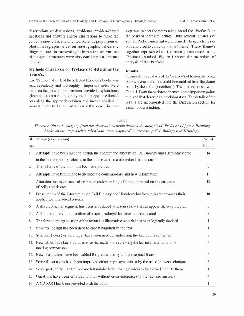
Fig.-2: Different forms of illustration: A:
‘photomicrograph-routine stain-low power’ showing
foliate papillae of the tongue; B: ‘schematic and three-
dimensional diagram’ showing relative locations, by
colour, of the four zones of the prostate gland; C:
‘photomicrograph- unconventional technique’ showing
actin (green), mitochondria (red) and nuclei (blue); D:
‘scanning electron micrograph’ showing a portion of a
glomerulus of human kidney depicting iterdigitations of
podocyte cell processes over the surfaces of capillary
loops.
(Sources: Ross and Pawlina 2006- A: p. 480; B: p.754;
C: p.59; Kelly et al 1984- D: p. 655)
BSMMU J Vol. 5, Issue 1, January 2012
50
The teaching and learning of Cell and Molecular Biologyshould be practically oriented. It may be noted here thatthe practical part of the undergraduate exams in ourcountry is almost entirely based on light microscopicviewing and light micrographs. But theoretical exposureto Cell Biology remains an integral part of our curriculum.It is essential that students get accustomed to the visualimage of what they learn theoretically. Thus, use of high-power microscopy and specialised microscopy, as far aspracticable, should be used. In addition, electronmicrographs and schematic diagrams related tofunctionally oriented Cell and Molecular Biology shouldbe added to our assessment toolbox so that the studentsare also encouraged in using such tools in their learningprocess.
Importance of structure-function correlation for betterunderstanding of Cell Biology and Histology
The tremendous importance of structure-function correlationin the field of Histology can be realised by understanding thefollowing statement from Wheater et al. 25
Histology has bored generations of students almostcertainly because it has been regarded as the study ofstructure in isolation from function.
The importance of this correlation in the field of Histologyhas been highlighted in several of the ‘Preface’sstudied2,4,6,7,9,10,12,13,14,15,16,17. Highlighting theimportance of understanding the organisation and functionof tissues, Wood and Schechter26 point out that in orderto achieve it, students require to be able to interpretmicroscopic images. Leeson et al13 assert that “thecorrelation between structure and function is essentialand perhaps provides the reason that histology is suchan intriguing and readily understandable subject”. Theauthors also believe that if the students examine thestructure of an organ or tissue, they can deduce muchabout its function. Conversely, if they know its function,they can forecast more easily much of its microscopicstructures. Thus, “Histology stands at the crossroadsbetween gross anatomy and physiology and acts as anintegrative element between them”. 6
The importance of incorporation of histophysiology intohistology began to be understood in the later part of the20th century.24 Universities in various parts of the worldhave addressed this important realisation while designingtheir courses. In the University of Iowa College ofMedicine, the departmentally based courses, GeneralHistology for Medical Students and Medical Physiology,have been folded into a single structure/function course
entitled ‘Human organ systems’. The ‘strength’ of suchcourses has been claimed to be the integrated presentation,in one setting, of structure/function concepts and theapplication of factual material within a clinical context withthe aid of clinicians/scientists lecturing in the courses.21
Therefore, it may be suggested that the teaching-learningof Cell Biology and Histology should be approached insuch a way that functional aspects are understood beingrelated to the corresponding structural aspects, and thisapproach should be reflected properly in our assessmentsystem as well.
Importance of clinical correlation in Cell Biology andHistology
The study and practice of medicine focuses on what goeswrong in the body and how to restore it to a normal, healthy,functional state. For a physician to succeed in thisendeavour, considerable knowledge of both the normaland abnormal structures of the body’s cells, tissues andorgans is needed. Histology is a branch of medical sciencethat describes the normal appearance and structure ofcells, tissues and organs at the microscopic level. StudyingHistology with a curious eye and mind will improve thediagnostic and clinical abilities of medical studentsaccordingly. In this regard, Fawcett4 believes that a firmfoundation in Histology continues to be essential forrecognising and interpreting their changes in disease.Most (ten) of the ‘Preface’s analysed in the present studyhave specifically highlighted clinical correlation of CellBiology and/or Histology 2,4,6,7,10,12,13,15,16,17 , thusemphasizing the application of the subjects in medicalscience.
Clinically oriented Histology teaching is not a new conceptin most medical schools of the medically advancedcountries of the world. Clinical orientation has been aphenomenon of the undergraduate courses since the laterpart of the 20th century.24 This is also reflected in theHistology textbooks commonly used by theundergraduate medical students of Bangladesh. Integratedteaching of Histology, Physiology and Pathology are notuncommon in world’s renowned universities. In theUniversity of New South Wales, Australia, learning ofhistology has been successfully integrated withhistopathology.26
It is well known that adults learn topics that they feelrelevant. If Cell Biology and Histology can be clinicallycorrelated in teaching and assessment, students wouldhopefully take more interest in the subjects. This is becausestudents then would realise where these subjects stand
Trends in the Presentation of Cell Biology and Histology in Contemporary Histology Books Nahid Farhana Amin et al
51
in the context of their future clinical careers as has beenexperienced by the students of the University of NewSouth Wales.27 It is difficult to ensure asking questionsrelated to clinical Histology in the oral exams of ourcourses. But some amount of clinical Histology and clinicalaspects of Cell Biology may be incorporated in the writtenquestions. Objective Structured Practical Exams (OSPE)may also be designed to incorporate clinical aspects ofHistology.
Incorporation of questions in some Histology booksSeveral of the ‘Preface’s analysed in the present studymention the use of questions in the book for selfassessment.7,8,9,12 Observations made in the availablebooks suggest that many of these questions are at theunderstanding level. Self-assessment using suchquestions increases students’ understanding of thesubjects and makes reading of the book interesting. Self-assessment helps them in the process of becoming self-directed learners as well. Like many universities, theUniversity at Buffalo in the US uses multiple choicequestions constructed from images.28 Use of printedimages in constructing questions may be an effective buteconomic way of assessment.
It is generally observed that the undergraduate medicalstudents in Bangladesh rarely practice answering thequestions provided in their textbooks. Teachers are oftenso overburdened with the administrative aspects of runningthe course (due to the poverty of workforce) that theyvery often complain of not getting enough time to constructgood innovative new questions. Questions selected fromthe Histology books or online sources may be a good toolin keeping Bangladeshi students in practice of beingassessed and thus being benefited by developing betterunderstanding of Cell Biology and Histology.
The importance of the use of illustrationAn illustration is a picture that depicts or enhances a pieceof text. Illustrations are used for the decoration orexplanation of a text, or to reinforce concepts and thoughtsdelivered in written composition.29 The analyses of‘Preface’s revealed that there is a tendency in the Histologybooks to increase the number of illustrations with eachedition. Special mention about this has been made inCormak,2 Gartner and Hiatt6 and Leeson et al.14 The newillustrations include clinical pictures, photomicrographs(e.g., high resolution digital photomicrographs, fluorescentmicroscographs), scanning and transmission electronsmicrographs, micrographs of freeze-etch material and alsonew diagrams (schematic diagrams).
These illustrations have been claimed to summarise thematerials of the text17 and make the presentation of CellBiology and Histology comprehensive.7 Several authors/editors mention in the ‘Preface’s of newer editions thatmost of the figures in their books have been redrawn orimproved either in presentation or by the use of newertechnique in the newer editions. Some of them.10, 15, 17
state that all their existing illustrations have been revised,updated or improved. Two ‘Preface’s10, 17 draw the reader’sattention by saying that the clarity of their illustrationshas been improved by changing them into the full colourformat. Mentions are also there regarding improvedpresentation of illustration by virtue of the developmentof new technique or equipments such as autoradiography,freeze fracture method, electron microscopy, highresolution digital photomicrography. Telser et al.16 hasleft some parts of the illustration unlabelled while labellingthe other parts. Thus, the readers can perform self-drivenexercises.
It is understandable that several ‘form’s of illustration arerequired for developing a proper understanding of thestructure and function of cells, tissues and organs. Someexamples of illustrations of different forms are shown inFigure 2. There are some structural components that arenot visualised clearly without using special fixation andstaining methods or other special techniques. Many ofthese techniques and methods are not yet easily availablein our country. But it is important for the students to haveclear ideas of these structures. Such type of Illustration asshown in Figure 2C can serve as alternatives to histologicaland cytological specimens prepared using special fixativesand stains or other special techniques. ‘Schematic andthree-dimensional diagram’s is a form of illustration thatmay not be able to show the morphological details, butcan express mechanisms, phenomena, functional aspectsand pathogenesis or even can express concepts or ideasthat are more abstract than visible. Flow charts are alsoincluded in recent books to show sequences of events incellular differentiation, activity etc. In Histology classes,the Bangladeshi students are usually habituated with two-dimensional view of a structure in section under a lowpower light microscope (as shown in Figure 2A). Theyoften forget that the structures they are looking at areactually three-dimensional ones. For teaching Cell Biology,probably more than for Histology, schematic and three-dimensional diagrams (as shown in Figure 2B) is the toolof choice along with electron micrographs (as shown inFigure 2D). As electron microscopes are not yet widelyavailable in our country, institutions can resort to usingelectron micrographs. This is especially true for CellBiology.
BSMMU J Vol. 5, Issue 1, January 2012
52
It is needless to say that among the different disciplinesof basic sciences, the importance of illustrations in CellBiology and Histology has special implication becauseCell Biology and Histology is an image-intensivediscipline. Illustrations aid in the recognition ofhistological structures in the field of microscope (Miller1891, cited in Cotter28). Ross and Pawlina15 believe thatillustrations act as a visual memory tool to facilitatelearning. The present researcher has noted that theHistology textbook most commonly recommended for theundergraduates of Bangladesh, Junqueira and Carneirohas used more than 60% of its printed area for illustrations.
As revealed from informal communications with theteachers in Anatomy, the majority of students of mostmedical colleges of Bangladesh are interested in‘identifying’ histological slides not through the lens ofthe microscope but form outside. In ‘describing’ thehistological structures of different organs, they tend toprepare a list of characters or components supposed to bepresent in a section of the organ one after another, notrecognising their logical interrelationship, structuralorientation or their recognisability at a particular level ofmagnification. Moreover, in Bangladesh, there is no useof illustrations in the oral part of the First ProfesionalMBBS Histology exams as it is not specified in thecurriculum. Although use of illustrations has recently beenintroduced in the practical part of Histology exams, but
the proportion does not seem to be sufficient and the
directives are not well structured.
It as well understood that Cell Biology and Histology are
structure-oriented subjects where appearances of cellular
and tissue structure speak for their functions. Therefore,
better use of illustrations in the teaching and learning
process as well as in assessment would add to better
understanding of those subjects. Introduction of the use
of illustrations in the oral part of the undergraduate exams
and increase in their use in the practical exams have every
likelihood of improving the quality of exams because by
this the exams can thus be raised to a more understanding
level. At the same time, an enhanced use of illustration
would motivate students to make better use of illustration
in their self-learning of Cell Biology and Histology from
books and other sources. As there is paucity of good
slides in many medical colleges of the country and because
the use of high magnification is a rarity in practice, use of
printed illustrations in classes as well as in exams becomesa necessity to be outlined and ensured by the relevantauthorities.
Conclusion:
The present study was undertaken to contribute to theimprovement of teaching-learning and assessment in CellBiology and Histology in the undergraduate medicalcourses of Bangladesh. It may have been able to makesome important points and create an evidence-basedfoundation upon which positive steps may be taken infuture towards fulfilling the objectives set in theundergraduate curriculum.
By incorporating the findings of the present study withthe present-day ideas and trends in Cell Biology andHistology teaching-learning and assessment in therenowned universities of the world, as evident from theavailable literature, suggestions can be formulated onimproving the methods of teaching and assessment inCell Biology and Histology in Bangladesh.
Acknowledgement:
The study was partly supported by a grant from theMinistry of Science and Technology of the Govt. of thePeople’s Republic of Bangladesh.
References:
1. Curriculum of Anatomy course for undergraduate medicalstudents of Bangladesh- Compiled and Edited by Center for
Medical Education (CME). Dhaka: Bangladesh Medical and
Dental council; 2002.
2. Cormack DH, ed. Ham’s Histology. 9th ed. Philadelphia: J.B.
Lippincott Company; 1987.
3. Module on 1st professional MBBS examination system to be
held on January 2008 based on new curriculum. Dhaka: Core
group, Faculty of Undergraduate Medicine, University of
Dhaka.
4. Fawcett DW, ed. Bloom and Fawcett- a text- book of histology.
12th ed. New York: Chapman and Hall; 1994.
5. Grag K, Bahl I, Kaul M. A text book of Histology. 2nd ed.
Delhi: CBS Publishers and Distributors; 1987.
6. Gartner LP, Hiatt JL. Color textbook of histology. 2nd ed.
Baltimore: Saunders; 2001.
7. Gartner LP, Hiatt JL, Strum JM. Cell biology and histology.
5th ed. Philadelphia: Lippincott Williams and Wilkins; 2007.
8. Henrikson RC, Kaye GI, Mazurkiewicz JE. Histology.
Baltimore: Williams and Wilkins; 1997.
9. Johnson KE. Histology and cell biology. 2nd ed. Baltimore:
Williams and Wilkins; 1991.
10. Junqueira LJ, Carneiro J. Basic histology- text and atlas. 11th
ed. New York: McGraw-Hill; 2006.
11. Kelly DE, Wood RL. Enders AC, editors. Bailey’s textbook
of microscopic anatomy. 18th ed. Baltimore: Williams and
Wilkins; 1984.
Trends in the Presentation of Cell Biology and Histology in Contemporary Histology Books Nahid Farhana Amin et al
53
12. Krause WJ, Cutts JH. Essentials of histology- text / atlas /review. Boston: Little, Brown and Company; 1994.
13. Leeson TS, Leeson CR, Paparo AA. Text/atlas of histology.
Philadelphia: W.B. Saunders Company; 1988.
14. Leeson TS, Leeson CR, Paparo AA. Textbook of histology.
5th ed. Philadelphia: W.B. Saunders Company; 1985.
15. Ross MH, Pawlina W. Histology- a text and atlas with
correlated cell and molecular biology. 5th ed. Baltimore:Lippincott Williams and Wilkins; 2006.
16. Telser AG, Young JK, Baldwin KM. Elsevier’s integrated
histology. Philadelphia: Mosby Elsevier; 2007.
17. Young B, Heath JW, editors. Wheater’s functional histology-
a text and colour atlas. 4th ed. Edinburgh: Churchill Livingstone;
2000.
18. Oxford learner’s pocket dictionary.4 th ed. Oxford: OxfordUniversity press; 2011. Preface; p. 346.
19. Bloodgood RA, Ogilvie RW. Trends in histology laboratory
teaching in United States medical schools. The Anatomical
Record (Part B: New Anatomist), 2006; 289B: 169-175.
20. Harris T, Leaven T, Heidger P, Kreiter C, Duncan J, Dick
F. Comparison of a virtual microscope laboratory to a regularmicroscope laboratory for teaching histology. The Anatomical
Record ((New Anatomist), 2001; 265: 10-14.
21. Heidger JR, PM, Dee F, Consoer D, Leaven T, Duncan J,
Kreiter C. Integrated approach to teaching and testing in
histology with real and virtual imaging. The Anatomical
Record (New Anatomist). 269: 107-112.
22. Krippendorf BB , Lough J. Complete and rapid switch fromlight microscopy to virtual microscopy for teaching medical
histology. The Anatomical Records (Part B: New Anatomist).
2005; 285: 19-25.
23. Drake RL, Lowrie DJ, Prewitt CM. Survey of gross anatomy,
microscopic anatomy, neuroscience, and embryology courses
in medical school curricula in the United States. The Anatomical
Record (New Anatomist), 2002; 269: 118-112.
24. Blake CA, Lavaie HA, Millette CF. Teaching medical histology
at the University of South Carolina school of Medicine
transition to virtual slides and virtual microscopes. The
Anatomical Record (Part B; New Anatomist) [Internet]. 2003,
275B(1); 196-206. Available from: Onlinelibrary.wiley.com/
doi/10.1002/ar.b.10037/ful
25. Wheater PR, Burkitt HG , Daniels V G, Wheater’s functional
histology- a text and colour atlas. 1st ed. Edinburgh: Churchill
Livingstone; 1979.
26. Wood RI, Schechter, JE. Histology- an interactive virtual
microscope [Internet]. 2002. Available from:http://
icb.oxfordjournals.org/content/43/2/360.full
27. Kumar RK, Fruman B, Vclan GM, D, Permontier PJ.
Integrating histology and histopathology teaching in practical
classes using virtual slide. The Anatomical Record (Part B;
New Anatomist) [Internet]. 2006,289B: 128-133. Available
from: virtualslides, unsw. edu. au/docs/integrated_
virtual_microscopy.
28. Cotter JR. Laboratory instruction in histology at the
University at Buffalo: recent replacement of microscope
exercises with computer applications. The Anatomical Record(New Anatomist), 2001; 256: 212-221.
29. Top: Arts: Illustration [Internet] [Cited 2008 Nov13]. Availablefrom: http://opensite.org/arts/illustration.
Trends in the Presentation of Cell Biology and Histology in Contemporary Histology Books Nahid Farhana Amin et al
54