Jonqueira's basic histology text and atlas
557
Transcript of Jonqueira's basic histology text and atlas


CONTENTS
Chapter 1 Histology & Its Methods of Study
Chapter 2 The Cytoplasm
Chapter 3 The Cell Nucleus
Chapter 4 Epithelial Tissue
Chapter 5 Connective Tissue
Chapter 6 Adipose Tissue
Chapter 7 Cartilage
Chapter 8 Bone
Chapter 9 Nerve Tissue & the Nervous System
Chapter 10 Muscle Tissue
Chapter 11 The Circulatory System
Chapter 12 Blood
Chapter 13 Hemopoiesis
Chapter 14 The Immune System & Lymphoid Organs
Chapter 15 Digestive Tract
Chapter 16 Organs Associated with the Digestive Tract
Chapter 17 The Respiratory System
Chapter 18 Skin
Chapter 19 The Urinary System
Chapter 20 Endocrine Glands
Chapter 21 The Male Reproductive System
Chapter 22 The Female Reproductive System
Chapter 23 The Eye and Ear: Special Sense Organs
Appendix: Light Microscopy Stains

Print Close Window
Note: Large images and tables on this page may necessitate printing in landscape mode.
Copyright © The McGraw-Hill Companies. All rights reserved.
Junqueira's Basic Histology: Text & Atlas, 12e > Chapter 1. Histology & Its Methods of Study >
HISTOLOGY & ITS METHODS OF STUDY: INTRODUCTION
Histology is the study of the tissues of the body and how these tissues are arranged to constitute organs. The Greek root histo can be translated as either "tissue" or
"web" and both translations are appropriate because most tissues are webs of interwoven filaments and fibers, both cellular and noncellular, with membranous
linings. Histology involves all aspects of tissue biology, with the focus on how cells' structure and arrangement optimize functions specific to each organ.
Tissues are made of two interacting components: cells and extracellular matrix. The extracellular matrix consists of many kinds of molecules, most of which are
highly organized and form complex structures, such as collagen fibrils and basement membranes. The main functions once attributed to the extracellular matrix
were to furnish mechanical support for the cells, to transport nutrients to the cells, and to carry away catabolites and secretory products. We now know that,
although the cells produce the extracellular matrix, they are also influenced and sometimes controlled by molecules of the matrix. There is, thus, an intense
interaction between cells and matrix, with many components of the matrix recognized by and attaching to receptors present on cell surfaces. Most of these
receptors are molecules that cross the cell membranes and connect to structural components of the intracellular cytoplasm. Thus, cells and extracellular matrix
form a continuum that functions together and reacts to stimuli and inhibitors together.
Each of the fundamental tissues is formed by several types of cells and typically by specific associations of cells and extracellular matrix. These characteristic
associations facilitate the recognition of the many subtypes of tissues by students. Most organs are formed by an orderly combination of several tissues, except the
central nervous system, which is formed almost solely by nervous tissue. The precise combination of these tissues allows the functioning of each organ and of the
organism as a whole.
The small size of cells and matrix components makes histology dependent on the use of microscopes. Advances in chemistry, molecular biology, physiology,
immunology, and pathology—and the interactions among these fields—are essential for a better knowledge of tissue biology. Familiarity with the tools and methods
of any branch of science is essential for a proper understanding of the subject. This chapter reviews several of the more common methods used to study cells and
tissues and the principles involved in these methods.
PREPARATION OF TISSUES FOR STUDY
The most common procedure used in the study of tissues is the preparation of histological sections or tissue slices that can be studied with the aid of the light
microscope. Under the light microscope, tissues are examined via a light beam that is transmitted through the tissue. Because tissues and organs are usually too
thick for light to pass through them, they must be sectioned to obtain thin, translucent sections and then attached to glass slides before they can be examined.
The ideal microscope tissue preparation should be preserved so that the tissue on the slide has the same structure and molecular composition as it had in the body.
However, as a practical matter this is seldom feasible and artifacts, distortions, and loss of components due to the preparation process are almost always present.
The basic steps used in tissue preparation for histology are shown in Figure 1–1.
Figure 1–1.
Sectioning fixed and embedded tissue.
Most tissues studied histologically are prepared as shown. (a): Small pieces of fresh tissue are placed in fixative solutions which generally cross-link proteins,inactivating degradative enzymes and preserving cell structures. The fixed pieces then undergo "dehydration" by being transferred through a series of increasingly moreconcentrated alcohol solutions, ending in 100% which effectively removes all water from the tissue. The alcohol is then removed in a clearing solution miscible in bothalcohol and melted paraffin. When the tissue is then placed in melted paraffin at 58°C it becomes completely infiltrated with this substance. All steps to this point arecommonly done today by robotic devices in active histology or pathology laboratories. After infiltration the tissue is placed in a small mold containing melted paraffin,which is then allowed to harden. The resulting paraffin block is trimmed to expose the tissue for sectioning (slicing). Similar steps are used in preparing tissue for
AccessMedicine | Print: Chapter 1. Histology & Its Methods of Study http://accessmedicine.com/popup.aspx?aID=6180002&print=yes_chapter
1 of 18 22/05/2011 18:42
transmission electron microscopy, except that smaller tissue samples are fixed in special fixatives and dehydrating solutions are used that are appropriate for embeddingin epoxy resins which become much harder than paraffin to allow very thin sectioning. (b): A microtome is used for sectioning paraffin-embedded tissues for lightmicroscopy. After mounting a trimmed block with the tissue specimen, rotating the drive wheel moves the tissue-block holder up and down. Each turn of the drive wheeladvances the specimen holder a controlled distance, generally between 1 and 10 m, and after each forward move the tissue block passes over the steel knife edge,which cuts the sections at a thickness equal to the distance the block advanced. Paraffin sections are then adhered to glass slides, deparaffinized, and stained formicroscopic examination. For transmission electron microscopy sections less than 1 m thick are prepared from resin-embedded cells using an ultramicrotome with aglass or diamond knife.
FixationIf a permanent section is desired, tissues must be fixed. To avoid tissue digestion by enzymes present within the cells (autolysis) or by bacteria and to preserve the
structure and molecular composition, pieces of organs should be promptly and adequately treated before, or as soon as possible after, removal from the animal's
body. This treatment—fixation—can be done by chemical or, less frequently, physical methods. In chemical fixation, the tissues are usually immersed in solutions
of stabilizing or cross-linking agents called fixatives. Because the fixative needs some time to fully diffuse into the tissues, the tissues are usually cut into small
fragments before fixation to facilitate the penetration of the fixative and to guarantee preservation of the tissue. Intravascular perfusion of fixatives can be used.
Because the fixative in this case rapidly reaches the tissues through the blood vessels, fixation is greatly improved.
One of the best fixatives for routine light microscopy is formalin, a buffered isotonic solution of 37% formaldehyde. The chemistry of the process involved in fixation
is complex and not always well understood. Formaldehyde and glutaraldehyde, another widely used fixative, are known to react with the amine groups (NH2) of
tissue proteins. In the case of glutaraldehyde, the fixing action is reinforced by virtue of its being a dialdehyde, which can cross-link proteins.
In view of the high resolution afforded by the electron microscope, greater care in fixation is necessary to preserve ultrastructural detail. Toward that end, a double
fixation procedure, using a buffered glutaraldehyde solution followed by a second fixation in buffered osmium tetroxide, is a standard procedure in preparations for
fine structural studies. The effect of osmium tetroxide is to preserve and stain lipids and proteins.
Embedding & SectioningTissues are usually embedded in a solid medium to facilitate sectioning. To obtain thin sections with the microtome, tissues must be infiltrated after fixation with
embedding substances that impart a rigid consistency to the tissue. Embedding materials include paraffin and plastic resins. Paraffin is used routinely for light
microscopy; resins are used for both light and electron microscopy.
The process of paraffin embedding, or tissue impregnation, is ordinarily preceded by two main steps: dehydration and clearing. The water is first extracted from
the fragments to be embedded by bathing them successively in a graded series of mixtures of ethanol and water, usually from 70% to 100% ethanol (dehydration).
The ethanol is then replaced with a solvent miscible with both alcohol and the embedding medium. As the tissues are infiltrated with this solvent, they generally
become transparent (clearing). Once the tissue is impregnated with the solvent, it is placed in melted paraffin in an oven, typically at 52–60°C. The heat causes the
solvent to evaporate, and the spaces within the tissues become filled with paraffin. The tissue together with its impregnating paraffin hardens after removal from
the oven. Tissues to be embedded with plastic resin are also dehydrated in ethanol and—depending on the kind of resin used—subsequently infiltrated with plastic
solvents. The ethanol or the solvents are later replaced by plastic solutions that are hardened by means of cross-linking polymerizers. Plastic embedding prevents
the shrinking effect of the high temperatures needed for paraffin embedding and gives little or no distortion to the cells.
The hard blocks containing the tissues are then placed in an instrument called a microtome (Figure 1–1) and are sliced by the microtome's steel or glass blade into
sections 1 to10 micrometers thick. Remember that one micrometer (1 m) equals 1/1,000 of a millimeter (mm) = 10–6 m. Other units of distance commonly used
in histology are the nanometer (1 nm = 0.001 m = 10–6 mm = 10–9 m) and angstrom (1 Å = 0.1 nm or 10–4 m). The sections are floated on water and then
transferred to glass slides to be stained.
An alternate way to prepare tissue sections is to submit the tissues to rapid freezing. In this process, the tissues are fixed by freezing (physical, not chemical
fixation) and at the same time become hard and thus ready to be sectioned. A freezing microtome— the cryostat—is then used to section the frozen block with
tissue. Because this method allows the rapid preparation of sections without going through the long embedding procedure described above, it is routinely used in
hospitals to study specimens during surgical procedures. Freezing of tissues is also effective in the histochemical study of very sensitive enzymes or small
molecules, since freezing, unlike fixation, does not inactivate most enzymes. Finally, because immersion in solvents such as xylene dissolves cell lipids in fixed
tissues, frozen sections are also useful when structures containing lipids are to be studied.
StainingTo be studied microscopically sections must typically be stained or dyed because most tissues are colorless. Methods of staining tissues have therefore been devised
that not only make the various tissue components conspicuous but also permit distinctions to be made between them. The dyes stain tissue components more or
less selectively. Most of these dyes behave like acidic or basic compounds and have a tendency to form electrostatic (salt) linkages with ionizable radicals of the
tissues. Tissue components with a net negative charge (anionic) stain more readily with basic dyes and are termed basophilic; cationic components, such as
proteins with many ionized amino groups, have affinity for acidic dyes and are termed acidophilic.
Examples of basic dyes are toluidine blue, alcian blue, and methylene blue. Hematoxylin behaves like a basic dye, that is, it stains the basophilic tissue
components. The main tissue components that ionize and react with basic dyes do so because of acids in their composition (nucleic acids, glycosaminoglycans, and
acid glycoproteins). Acid dyes (eg, orange G, eosin, acid fuchsin) stain the acidophilic components of tissues such as mitochondria, secretory granules, and collagen.
Of all dyes, the simple combination of hematoxylin and eosin (H&E) is used most commonly. Hematoxylin stains DNA of the cell nucleus and other acidic
structures (such as RNA-rich portions of the cytoplasm and the matrix of cartilage) blue. In contrast, eosin stains other cytoplasmic components and collagen pink
(Figure 1–2). Many other dyes, such as the trichromes (eg, Mallory stain, Masson stain), are used in different histologic procedures. The trichromes, besides
showing the nuclei and cytoplasm very well, help to distinguish extracellular tissue components better than H&E. A good technique for differentiating collagen is the
use of picrosirius, especially when associated with polarized light (see Polarizing Microscopy).
Figure 1–2.
AccessMedicine | Print: Chapter 1. Histology & Its Methods of Study http://accessmedicine.com/popup.aspx?aID=6180002&print=yes_chapter
2 of 18 22/05/2011 18:42

Hematoxylin & Eosin (H&E) and Periodic acid-Schiff (PAS) staining.
Micrographs of the columnar epithelium lining the small intestine. (a): Micrograph stained with hematoxylin and eosin (H&E). (b): Micrograph stained by the periodicacid-Schiff (PAS) reaction for glycoproteins. With H&E, basophilic cell nuclei are stained purple while cytoplasm stains pink. Cell regions with abundant oligosaccharideson glycoproteins, such as the apical ends of the cells or the scattered mucus-secreting goblet cells in the layer are poorly stained. With PAS, staining is most intense atthe cell surface, where projecting microvilli have a prominent layer of glycoproteins (arrow head) and in the mucin-rich secretory granules of goblet cells. Cell surfaceglycoproteins and mucin are PAS-positive due to their high content of oligosaccharides and polysaccharides. The PAS-stained tissue was counterstained with hematoxylinto show the cell nuclei. Both X300.
The chemical basis of other staining procedures is more complicated than the electrostatic interactions underlying basophilia and acidophilia. DNA can be specifically
identified and quantified in nuclei using the Feulgen reaction, in which deoxyribose sugars are hydrolyzed by mild hydrochloric acid, followed by treatment with
periodic acid and Schiff reagent (PAS). The PAS technique is based on the transformation of 1,2-glycol groups present in the sugars into aldehyde residues,
which then react with Schiff reagent to produce a purple or magenta color.
Polysaccharides constitute an extremely heterogeneous group in tissues and occur either in a free state or combined with proteins and lipids. Because of their
hexose sugar content, many polysaccharides can also be demonstrated by the PAS reaction. A ubiquitous free polysaccharide in animal cells is glycogen, which can
be demonstrated by PAS in liver, striated muscle, and other tissues where it accumulates.
Short branched chains of sugars (oligosaccharides) are attached to specific amino acids of glycoproteins, making most glycoproteins PAS-positive. Figure 1–2b
shows an example of cells stained by the PAS reaction. Glycosaminoglycans (GAGs) are anionic, unbranched long-chain polysaccharides containing aminated
sugars. Many glycosaminoglycans are synthesized while attached to a core protein and constitute a class of macromolecules called proteoglycans, which upon
secretion make up important parts of the extracellular matrix (ECM) (see Chapters 5 and 7). Unlike a glycoprotein, a proteoglycan's carbohydrate chains are
greater in weight and volume than the protein core of the molecule. GAGs and many acidic glycoproteins do not undergo the PAS reaction, but because of their high
content of anionic carboxyl and sulfate groups show a strong electrostatic interaction with alcian blue and other basic stains.
Basophilic or PAS-positive material can be further identified by enzyme digestion pretreatment of a tissue section with an enzyme that specifically digests one
AccessMedicine | Print: Chapter 1. Histology & Its Methods of Study http://accessmedicine.com/popup.aspx?aID=6180002&print=yes_chapter
3 of 18 22/05/2011 18:42

substrate, leaving other adjacent sections untreated. For example, pretreatment with ribonuclease will greatly reduce cytoplasmic basophilia with little effect on
chromosomes, indicating the importance of RNA for the cytoplasmic staining. Similarly, free polysaccharides are digested by amylase, which can therefore be used
to distinguish glycogen from glycoproteins in PAS-positive material.
In many staining procedures certain structures such nuclei become labeled, but other parts of cells are often not visible. In this case a counterstain is used to give
additional information. A counterstain is usually a single stain that is applied to a section by another method to allow better recognition of nuclei or other structures.
Lipid-rich structures are best revealed with lipid-soluble dyes to avoid the steps of slide preparation that remove lipids such as treatment with heat, xylene, or
paraffin. Typically frozen sections are stained in alcohol solutions saturated with a lipophilic dye such as Sudan black. The stain dissolves in cellular lipid droplets and
other lipid-rich structures, which become stained in black. Specialized methods for the localization of cholesterol, phospholipids, and glycolipids are useful in
diagnosis of metabolic diseases in which there are intracellular accumulations of different kinds of lipids. In addition to tissue staining with dyes, metal
impregnation techniques usually using silver salts are a common method of visualizing certain ECM fibers and specific cellular elements in nervous tissue.
The whole procedure, from fixation to observing a tissue in a light microscope, may take from 12 hours to 21/2 days, depending on the size of the tissue, the
fixative, the embedding medium, and the method of staining. The final step before observation is mounting a protective glass coverslip on the slide with adhesive
mounting media.
LIGHT MICROSCOPY
Conventional bright-field microscopy, as well as fluorescence, phase-contrast, differential interference, confocal, and polarizing microscopy are all based on the
interaction of light and tissue components and can be used to reveal and study tissue features.
Bright-Field MicroscopyWith the bright-field microscope, widely used by students of histology, stained preparations are examined by means of ordinary light that passes through the
specimen. The microscope is composed of mechanical and optical parts (Figure 1–3). The optical components consist of three systems of lenses. The condenser
collects and focuses light, producing a cone of light that illuminates the object to be observed. The objective lenses enlarge and project the illuminated image of
the object in the direction of the eyepiece. The eyepiece or ocular lens further magnifies this image and projects it onto the viewer's retina, photographic film, or
(to obtain a digital image) a detector such as a charge-coupled device (CCD) camera. The total magnification is obtained by multiplying the magnifying power of the
objective and ocular lenses.
Figure 1–3.
Components and light path of a bright-field microscope.
Photograph of a bright-field light microscope showing its main components and the pathway of light from the substage lamp to the eye of the observer. The opticalsystem has three sets of lenses: a condenser, a set of objectives, and either one or two eyepieces. The condenser collects and focuses light, producing a cone of lightthat illuminates the tissue slide on the stage. Objective lenses enlarge and project the illuminated image of the object in the direction of the eyepiece. For routinehistological studies objectives having three different magnifications are generally used: X4 for low magnification observations of a large area (field) of the tissue; X10 formedium magnification of a smaller field; and X40 for high magnification of more detailed areas. The eyepiece or ocular further magnifies this image another X10 andprojects it onto the viewer's retina, yielding a total magnification of X40, X100, or X400. (With permission, from Nikon Instruments.)
The critical factor in obtaining a crisp, detailed image with a light microscope is its resolving power, defined as the smallest distance between two particles at
which they can be seen as separate objects. The maximal resolving power of the light microscope is approximately 0.2 m; this power permits good images
magnified 1000–1500 times. Objects smaller or thinner than 0.2 m (such as a ribosome, a membrane, or a filament of actin) cannot be distinguished with this
instrument. Likewise, two objects such as mitochondria will be seen as only one object if they are separated by less than 0.2 m. The quality of the image—its
clarity and richness of detail—depends on the microscope's resolving power. The magnification is of value only when accompanied by high resolution. The resolving
power of a microscope depends mainly on the quality of its objective lens. The eyepiece lens enlarges only the image obtained by the objective; it does not improve
AccessMedicine | Print: Chapter 1. Histology & Its Methods of Study http://accessmedicine.com/popup.aspx?aID=6180002&print=yes_chapter
4 of 18 22/05/2011 18:42

resolution. For this reason, when comparing objectives of different magnifications, those that provide higher magnification also have higher resolving power.
Video cameras highly sensitive to light enhance the power of the bright-field and other light microscopes and allow the capture of digitized images suitable for
computerized image analysis and printing. The frontiers of light microscopy have been redefined by the use of such cameras. With digital cameras and image-
enhancement programs (to enhance contrast, for example), objects that may not be visible when viewed directly through the ocular may be made visible in the
video screen. These video systems are also useful for studying living cells for long periods of time, because they use low-intensity light and thus avoid the cellular
damage from heat that can result from intense illumination. Moreover, software developed for image analysis allows rapid measurements and quantitative study of
microscopic structures.
Fluorescence MicroscopyWhen certain substances are irradiated by light of a proper wavelength, they emit light with a longer wavelength. This phenomenon is called fluorescence. In
fluorescence microscopy, tissue sections are usually irradiated with ultraviolet (UV) light and the emission is in the visible portion of the spectrum. The
fluorescent substances appear brilliant on a dark background. For this method, the microscope has a strong UV light source and special filters that select rays of
different wavelengths emitted by the substances.
Fluorescent compounds with affinity for specific cell macromolecules may be used as fluorescent stains. Acridine orange, which binds both DNA and RNA, is an
example. When observed in the fluorescence microscope, these nucleic acids emit slightly different fluorescence, allowing them to be localized separately in cells
(Figure 1–4a). Other compounds such as Hoechst stain and DAPI specifically bind DNA and are used to stain cell nuclei, emitting a characteristic blue fluorescence
under UV. Another important application of fluorescence microscopy is achieved by coupling fluorescent compounds to molecules that will specifically bind to certain
cellular components and thus allow the identification of these structures under the microscope (Figure 1–4b). Antibodies labeled with fluorescent compounds are
extremely important in immunohistological staining. (See Detection Methods Using Specific Interactions Between Molecules).
Figure 1–4.
Appearance of cells with fluorescent microscopy.
Components of cells in culture are often stained with compounds visible by fluorescence microscopy. (a): Kidney cells stained with acridine orange, which binds nucleicacids. Under a fluorescence microscope, nuclear DNA emits yellow light and the RNA-rich cytoplasm appears reddish or orange. (b): The less dense culture of kidney
AccessMedicine | Print: Chapter 1. Histology & Its Methods of Study http://accessmedicine.com/popup.aspx?aID=6180002&print=yes_chapter
5 of 18 22/05/2011 18:42

cells stained with DAPI (4',6-diamino-2-phenylindole) which binds DNA, and with phalloidin, which binds actin filaments. Nuclei of these cells show a blue fluorescenceand actin filaments appear green. Important information such as the greater density of microfilaments at the cell periphery is readily apparent. (Figure 1–4b, withpermission, from Drs. Claire E. Walczak and Rania Risk, Indiana University School of Medicine, Bloomington.)
Phase-Contrast Microscopy & Differential Interference MicroscopySome optical arrangements allow the observation of unstained cells and tissue sections. Unstained biological specimens are usually transparent and difficult to view
in detail, because all parts of the specimen have almost the same optical density. Phase-contrast microscopy, however, uses a lens system that produces visible
images from transparent objects (Figure 1–5).
Figure 1–5.
AccessMedicine | Print: Chapter 1. Histology & Its Methods of Study http://accessmedicine.com/popup.aspx?aID=6180002&print=yes_chapter
6 of 18 22/05/2011 18:42

Unstained cells' appearance in three types of light microscopy.
Neural crest cells growing as a single layer in culture appear differently with various techniques of light microscopy. These cells are unstained and the same field of cells,including two differentiating pigment cells, is shown in each photo. (a): Bright-field microscopy: without fixation and staining, only the two pigment cells can be seen.(b): Phase-contrast microscopy: cell boundaries, nuclei, and cytoplasmic structures with different refractive indices affect in-phase light differently and produce animage of these features in all the cells. (c): Differential interference microscopy: cellular details are highlighted in a different manner using Nomarski optics. Phase-contrast microscopy, with or without differential interference, is widely used to observe live cells grown in tissue culture. All X200. (With permission, from Sherry Rogers,Department of Cell Biology and Physiology, University of New Mexico.)
Phase-contrast microscopy is based on the principle that light changes its speed when passing through cellular and extracellular structures with different refractive
indices. These changes are used by the phase-contrast system to cause the structures to appear lighter or darker in relation to each other. Because it does not
require fixation or staining, phase-contrast microscopy allows observation of living cells and tissue cultures, and such microscopes are prominent tools in all cell
culture labs. A related method of observing unstained cells or tissue sections is the Nomarski differential interference microscopy, which produces an image
with a more apparent three-dimensional aspect than in routine phase-contrast microscopy (Figure 1–5).
Confocal MicroscopyWith a regular bright-field microscope the beam of light is relatively large and fills the specimen. Stray light reduces contrast within the image and compromises the
resolving power of the objective lens. Confocal microscopy avoids stray light and achieves greater resolution by using (1) a small point of high-intensity light
provided by a laser and (2) a plate with a pinhole aperture in front of the image detector. The point light source, the focal point of the lens, and the detector's
pinpoint aperture are all optically conjugated or aligned to each other in the focal plane (confocal) and unfocused light does not pass through the pinhole. This
greatly improves resolution of the object in focus and allows the localization of specimen components with much greater precision than with the bright-field
microscope.
Most confocal microscopes include a computer-driven mirror system (the beam splitter) to move the point of illumination across the specimen automatically and
rapidly. Digital images captured at many individual spots in a very thin plane-of-focus are used to produce an "optical section" of that plane. Moreover, creating
optical sections at a series of focal planes through the specimen allows them to be digitally reconstructed into a three-dimensional image. Important features of
confocal microscopes are shown in Figure 1–6.
Figure 1–6.
AccessMedicine | Print: Chapter 1. Histology & Its Methods of Study http://accessmedicine.com/popup.aspx?aID=6180002&print=yes_chapter
7 of 18 22/05/2011 18:42

Principle of confocal microscopy.
Although a very small spot of light originating from one plane of the section crosses the pinhole and reaches the detector, rays originating from other planes are blockedby the blind. Thus, only one very thin plane of the specimen is focused at a time. The diagram shows the practical arrangement of a confocal microscope. Light from alaser source hits the specimen and is reflected. A beam splitter directs the reflected light to a pinhole and a detector. Light from components of the specimen that areabove or below the focused plane is blocked by the blind. The laser scans the specimen so that a larger area of the specimen can be observed.
Polarizing MicroscopyPolarizing microscopy allows the recognition of structures made of highly organized molecules. When normal light passes through a polarizing filter (such as a
Polaroid), it exits vibrating in only one direction. If a second filter is placed in the microscope above the first one, with its main axis perpendicular to the first filter,
no light passes through. If, however, tissue structures containing oriented macromolecules are located between the two polarizing filters, their repetitive structure
rotates the axis of the light emerging from the polarizer and they appear as bright structures against a dark background (Figure 1–7). The ability to rotate the
direction of vibration of polarized light is called birefringence and is a feature of crystalline substances or substances containing highly oriented molecules, such as
cellulose, collagen, microtubules, and microfilaments.
Figure 1–7.
AccessMedicine | Print: Chapter 1. Histology & Its Methods of Study http://accessmedicine.com/popup.aspx?aID=6180002&print=yes_chapter
8 of 18 22/05/2011 18:42

Tissue appearance with bright-field and polarizing microscopy.
Polarizing light microscopy produces an image only of material having repetitive, periodic macromolecular structure; features without such structure are not seen. Shownhere is a piece of thin mesentery that was stained with red picrosirius, orcein, and hematoxylin, and was then placed directly on a slide and observed by bright-field andpolarizing microscopy. (a): Under routine bright-field microscopy collagen fibers appear red, along with thin dark elastic fibers and cell nuclei. (b): Under polarizing lightmicroscopy, only collagen fibers are visible and these exhibit intense birefringence and appear bright red or yellow; elastic fibers and nuclei lack oriented macromolecularstructure and are not visible.
ELECTRON MICROSCOPY
Transmission and scanning electron microscopes are based on the interaction of electrons and tissue components. The wavelength in the electron beam is much
shorter than of light, allowing a thousand-fold increase in resolution.
Transmission Electron MicroscopyThe transmission electron microscope (TEM) is an imaging system that permits resolution around 3 mm (Figure 1–8a). This high resolution allows magnifications of
up to 400,000 times to be viewed with details. Unfortunately, this level of magnification applies only to isolated molecules or particles. Very thin tissue sections can
be observed with details at magnifications of up to about 120,000 times.
Figure 1–8.
AccessMedicine | Print: Chapter 1. Histology & Its Methods of Study http://accessmedicine.com/popup.aspx?aID=6180002&print=yes_chapter
9 of 18 22/05/2011 18:42

Electron microscopes.
Electron microscopes are large instruments generally housed in a specialized EM facility. (a): Schematic view of a transmission electron microscope (TEM) with its lensesand the pathway of the electrons. With the microscope's entire column in a vacuum, electrons are released by heating a very thin metallic (usually tungsten) filament(cathode). The released electrons are then submitted to a voltage difference of 60–120 kV between the cathode and the anode, which is a metallic plate with a hole in itscenter. Electrons are thus attracted to the anode, accelerated to high speeds, and form a beam of electrons as they pass through the central opening in the anode.Passing through electric coils the beam is deflected in a way roughly analogous to the effect of optical lenses on light because electrons change their path whensubmitted to electromagnetic fields.
The configuration of the TEM is similar to that of an upside-down light microscope. The first lens is a condenser that focuses the beam of electrons on the section. Someelectrons interact with atoms of the section and continue their course, while others simply cross the specimen without interacting. Most electrons reach the objectivelens, which forms a magnified image that is then projected through other magnifying lenses. Because the human eye is not sensitive to electrons, the image is finallyprojected on a fluorescent screen or is registered by photographic plates or a CCD camera.
In a TEM image areas of the specimen through which electrons passed appear bright (electron lucent), while those areas which are naturally dense or which bind heavymetals during specimen preparation or "staining" absorb or deflect electrons and appear dark (electron dense). Such images are therefore always black, white, andshades of gray.
(b): Schematic view of a scanning electron microscope (SEM) with many similarities to a TEM. However, here the electron beam focused by electromagnetic lenses doesnot pass through the specimen, but rather is moved sequentially (scanned) from point to point across its surface similar to the way an electron beam is scanned across atelevision tube. The specimen was coated previously with a very thin coating of metal atoms and the beam interacts with these atoms, and produces reflected electronsand newly emitted secondary electrons. All of these are captured by a detector and transmitted to amplifiers and other devices which produce a signal to a cathode raytube monitor, resulting in a black-and-white image. The SEM shows only surface views of the coated specimen but with a striking three-dimensional quality. The insideof organs or cells can be analyzed by sectioning them to expose their internal surfaces.
The TEM functions on the principle that a beam of electrons can be deflected by electromagnetic fields in a manner similar to light deflection in glass lenses. The
beam is produced by a cathode at the top of the instrument and passes down through the chamber in a vacuum. Because electrons change their path when
submitted to electromagnetic fields, the beam can be focused by passing through electric coils which can be considered electromagnetic lenses.
The first lens is a condenser focusing the beam of electrons on the specimen section. Some electrons interact with atoms in the section and their course is modified,
while others simply cross the specimen without interacting. Electrons passing through the specimen reach the objective lens, which forms a focused, magnified
image that is then magnified further through other lenses and captured on a viewing screen. The image of the specimen shows areas of white, black, and shades of
gray corresponding to areas through which electrons readily passed (appearing brighter or electron lucent) and areas where electrons were absorbed or deflected
(appearing darker or more electron dense).
To provide a useful interaction between the specimen and the electrons, TEM requires very thin sections (40–90 nm); therefore, embedding is performed with a
hard epoxy and sectioning is done with a glass or diamond knife. The extremely thin sections are collected on small metal grids and transferred to the interior of the
microscope to be analyzed.
Freezing techniques (freeze fracture, cryofracture, freeze etched) combined with electron microscopy have been very useful for examining membrane
structure. Very small tissue specimens are rapidly frozen in liquid nitrogen and fractured in a vacuum with a knife. A replica of the still frozen exposed surface is
produced by applying thin coats of vaporized carbon, platinum, or other atoms. Tissue is then dissolved away and the replica of the surface is examined by SEM.
The random fracture planes often split the lipid bilayers of membranes, exposing protein components whose size, shape, and distribution can then be studied.
Scanning Electron MicroscopyScanning electron microscopy (SEM) permits pseudo–three-dimensional views of the surfaces of cells, tissues, and organs. Like the TEM this microscope produces
and focuses a very narrow beam of electrons, but in this instrument the beam does not pass through the specimen (Figure 1-8b). Instead the surface of the
specimen is first dried and coated with a very thin layer of metal atoms through which electrons do not pass readily. When the beam is scanned from point to point
across the specimen it interacts with the metal atoms and produces reflected electrons or secondary electrons emitted from the metal. These are captured by a
detector and the resulting signal is processed to produce a black-and-white image on a monitor. SEM images are usually easily understood, because they present a
view that appears to be illuminated from above, just as our ordinary macroscopic world is filled with highlights and shadows caused by illumination from above.
AccessMedicine | Print: Chapter 1. Histology & Its Methods of Study http://accessmedicine.com/popup.aspx?aID=6180002&print=yes_chapter
10 of 18 22/05/2011 18:42

AUTORADIOGRAPHY
Autoradiography is a method of localizing newly synthesized macromolecules (DNA, RNA, protein, glycoproteins, and polysaccharides) in cells or tissue sections.
Radioactively labeled metabolites (nucleotides, amino acids) incorporated into the macromolecules emit weak radiation that is restricted to the cellular regions
where the molecules are located. Radiolabeled cells or mounted tissue sections are coated in a darkroom with photographic emulsion containing silver bromide
crystals, which act as microdetectors of this radiation in the same way that they respond to light in common photographic film. After an adequate exposure time in
lightproof boxes the slides are developed photographically. The silver bromide crystals reduced by the radiation are reduced to small black grains of metallic silver,
indicating locations of radiolabeled macromolecules in the tissue. This general procedure can be used in preparations for both light microscopy and TEM (Figure
1–9).
Figure 1–9.
Autoradiography.
Autoradiographs are tissue preparations in which particles called silver grains indicate the regions of cells in which specific macromolecules were synthesized just priorto fixation. Precursors such as nucleotides, amino acids, or sugars with isotopes substituted for specific atoms are provided to the tissues and after a period ofincorporation, tissues are fixed, sectioned, and mounted on slides or TEM grids as usual. This processing removes all radiolabeled precursors, leaving only the isotope inthe fixed macromolecules. In a darkroom the slides are coated with a thin layer of chemicals like those in photographic film and dried. In a black box the isotope in newlysynthesized macromolecules emits radiation exposing the layer of photographic chemicals immediately adjacent to the isotopes' location. The minute regions of exposedchemicals in the photographic layer are revealed as silver grains by "developing" the preparation as if it were film, followed by microscopic examination. Shown here are
autoradiographs from the salivary gland of a mouse injected with 3H-fucose 8 h before tissue fixation. Fucose is incorporated into oligosaccharides and the results reveallocation of newly synthesized glycoproteins containing such sugars. (a): Black "silver grains" are visible over regions with secretory granules and the duct indicatingglycoprotein locations. X1500. (b): The same tissue prepared for TEM autoradiography shows silver grains with a coiled or amorphous appearance again localized mainlyover the granules (G) and in the gland lumen (L). X7500. (Figure 1–9b, with permission, from Ticiano G. Lima and A. Antonio Haddad, School of Medicine, Ribeirão Preto,Brazil.)
Much information becomes available by autoradiography of cells or tissues. Thus, if a radioactive amino acid is used, it is possible to know which cells in a tissue
produce more protein and which cells produce less, because the number of silver grains formed over the cells is proportional to the intensity of protein synthesis. If
a radioactive precursor of DNA (such as tritium-labeled thymidine) is used, it is possible to know which cells in a tissue (and how many) are preparing to divide.
Dynamic events may also be analyzed. For example, if one wishes to know where in the cell protein is produced, if it is secreted, and which path it follows in the cell
before being secreted, several animals are injected with a radioactive amino acid and tissues collected at different times after the injections. Autoradiography of the
tissues representing the various times throughout the experiment will indicate the migration of the radioactive proteins. If one wishes to know where new cells are
produced in an organ and where they migrate, several animals are injected with radioactive thymidine and tissues collected at different times after the injection.
Autoradiographs of the sections will show the location of the dividing cells and where they migrate.
CELL & TISSUE CULTURE
Live cells and tissues can be maintained and studied outside the body. In a complex organism, tissues and organs are formed by several kinds of cells. These cells
are bathed in fluid derived from blood plasma, which contains many different molecules required for growth. Cell culture has been very helpful in isolating the
effects of single molecules on specific types of cells. It also allows the direct observation of the behavior of living cells under a phase contrast microscope. Many
experiments that cannot be performed in the living animal can be accomplished in vitro.
The cells and tissues are grown in complex solutions of known composition (salts, amino acids, vitamins) to which serum components or specific growth factors are
AccessMedicine | Print: Chapter 1. Histology & Its Methods of Study http://accessmedicine.com/popup.aspx?aID=6180002&print=yes_chapter
11 of 18 22/05/2011 18:42

added. In preparing cultures from a tissue or organ, cells must be initially dispersed mechanically or enzymatically. Once isolated, the cells can be cultivated in a
clear dish to which they adhere, usually as a single layer of cells (Figure 1–5). Cultures of cells that are isolated in this way are called primary cell cultures. Many
cell types once isolated from normal or pathologic tissue have been maintained in vitro ever since because they have been immortalized and now constitute a
permanent cell line. Most cells obtained from normal tissues have a finite, genetically programmed life span. Certain changes, however (some related to
oncogenes; see Chapter 3), can promote cell immortality, a process called transformation, which are similar to the initial changes in a normal cell's becoming a
cancer cell. Because of improvements in culture technology, most cell types can now be maintained in the laboratory. All procedures with living cells and tissues
must be performed in a sterile area, using sterile solutions and equipment, to avoid contamination with microorganisms.
As shown in the next chapter, incubation of living cells in vitro with a variety of new fluorescent compounds that are sequestered and metabolized in specific
compartments of the cell provides a new approach to understanding these compartments both structurally and physiologically. Other histological techniques applied
to cultured cells have been particularly important for understanding the locations and functions of microtubules, microfilaments, and other components of the
cytoskeleton.
MEDICAL APPLICATION
Cell culture has been widely used for the study of the metabolism of normal and cancerous cells and for the development of new drugs. This technique is
also useful in the study of parasites that grow only within cells, such as viruses, mycoplasma, and some protozoa. In cytogenetic research, determination of
human karyotypes (the number and morphology of an individual's chromosomes) is accomplished by short-term cultivation of blood cells or fibroblasts and
by examining the chromosomes during mitotic division. In addition, cell culture is central to contemporary techniques of molecular biology and recombinant
DNA technology.
HISTOCHEMISTRY & CYTOCHEMISTRY
The terms histochemistry and cytochemistry indicate methods for localizing cellular structures in tissue sections using unique enzymatic activity present in those
structures. To preserve these enzymes histochemical procedures are usually applied to unfixed or mildly fixed tissue, often sectioned on a cryostat to avoid adverse
effects of heat and paraffin on enzymatic activity. Enzyme histochemistry usually works in the following way: (1) tissue sections are immersed in a solution that
contains the substrate of the enzyme to be localized; (2) the enzyme is allowed to act on its substrate; (3) at this stage or later, the section is put in contact with a
marker compound; (4) this compound reacts with a molecule produced by enzymatic action on the substrate; (5) the final reaction product, which must be insoluble
and which is visible by light or electron microscopy only if it is colored or electron-dense, precipitates over the site that contains the enzyme. When examining such
a section in the microscope, one can see the cell regions (or organelles) covered with a colored or electron-dense material.
Examples of enzymes that can be detected histochemically include the following:
Phosphatases split the bond between a phosphate group and an alcohol residue of phosphorylated molecules. The visible, insoluble reaction product ofphosphatases is usually lead phosphate or lead sulfide. Both alkaline phosphatases which have their maximum activity at an alkaline pH and acid phosphatasescan be detected (Figure 1–10).
Dehydrogenases remove hydrogen from one substrate and transfer it to another. Like phosphatases, dehydrogenases play an important role in severalmetabolic processes. They are detected histochemically by incubating nonfixed tissue sections in a substrate solution containing a molecule that receiveshydrogen and precipitates as an insoluble colored compound. Mitochondria can be specifically identified by this method, since dehydrogenases are key enzymesin the citric acid (Krebs) cycle of this organelle.
Peroxidase, which is present in several types of cells, promotes the oxidation of certain substrates with the transfer of hydrogen ions to hydrogen peroxide,forming molecules of water. In this method, sections of adequately fixed tissue are incubated in a solution containing hydrogen peroxide and 3,3'-diamino-azobenzidine (DAB). The latter compound is oxidized in the presence of peroxidase, resulting in an insoluble, brown, electron-dense precipitate that permits thelocalization of peroxidase activity by light and electron microscopy. Peroxidase staining in white blood cells is important in the diagnosis of certain leukemias.
Figure 1–10.
AccessMedicine | Print: Chapter 1. Histology & Its Methods of Study http://accessmedicine.com/popup.aspx?aID=6180002&print=yes_chapter
12 of 18 22/05/2011 18:42

Enzyme histochemistry.
(a): Micrograph of cross sections of kidney tubules treated histochemically by the Gomori method for alkaline phosphatases show strong activity of this enzyme at theapical surfaces of the cells at the lumen of the tubules (arrows). (b): TEM image of a kidney cell in which acid phosphatase has been localized histochemically in threelysosomes (Ly) near the nucleus (N). The dark material within these structures is lead phosphate that precipitated in places with acid phosphatase activity. X25,000.(Figure 1–10b, with permission, from Eduardo Katchburian, Department of Morphology, Federal University of Sao Paulo, Brazil.)
Because peroxidase is extremely active and rapidly produces an appreciable amount of insoluble precipitate, it is also widely used for an important practical
application: tagging other proteins as described in the next section.
MEDICAL APPLICATION
Many histochemical procedures are used frequently in laboratory diagnosis, including Perls' Prussian blue reaction for iron (used to detect the iron storage
diseases hemochromatosis and, hemosiderosis), the PAS-amylase and alcian blue reactions for glycogen and glycosaminoglycans (to detect glycogenosis
and mucopolysaccharidosis), and reactions for lipids and sphingolipids (to detect sphingolipidosis).
DETECTION METHODS USING SPECIFIC INTERACTIONS BETWEEN MOLECULES
A specific macromolecule present in a tissue section may sometimes be identified by using tagged compounds or macromolecules that specifically interact with the
material of interest (Figure 1–11). The compounds that will interact with the molecule must be tagged with a label that can be detected under the light or electron
microscope. The most commonly used labels are fluorescent compounds (which can be seen with a fluorescence or laser microscope), radioactive atoms (which can
be detected with autoradiography), molecules of peroxidase or other enzymes (which can be detected with histochemistry), and metal (usually gold) particles that
can be observed with light and electron microscopy. These methods can be used for detecting and localizing specific sugars, proteins, and nucleic acids.
Figure 1–11.
AccessMedicine | Print: Chapter 1. Histology & Its Methods of Study http://accessmedicine.com/popup.aspx?aID=6180002&print=yes_chapter
13 of 18 22/05/2011 18:42

Labeling by specific, high-affinity interactions.
Compounds or macromolecules that have specific affinity toward certain cell or tissue macromolecules can be tagged with a label and used to identify that componentand determine its location in cells and tissues. (1) Molecule A has a high and specific affinity toward a portion of molecule B. Examples of such interactingmacromolecules are an antibody that recognizes specific antigens, usually proteins, or a segment of single-stranded DNA with sequence-specific complementarity to RNAmolecules in a cell. Molecule A can also be a small compound like phalloidin, which specifically binds actin filaments, or a protein such as "protein A" which binds allimmunoglobulins. (2) When A and B are mixed, A binds to the portion of B it recognizes. (3) Molecule A may be tagged with a label that can be visualized with a light orelectron microscope. The label can be a fluorescent compound, an enzyme such as peroxidase, an electron-dense particle, or a radioisotope. (4) If molecule B is presentin a cell or extracellular matrix that is incubated with labeled molecule A, molecule B can be detected and localized by visualizing the labeled molecule A bound to it.
Examples of molecules that interact specifically with other molecules include the following:
Phalloidin is a compound extracted from the mushroom Amanita phalloides and interacts strongly with actin. Tagged with fluorescent dyes, phalloidin iscommonly used to demonstrate actin filaments in cells.
Protein A is obtained from Staphylococcus aureus and binds to the Fc region of immunoglobulin (antibody) molecules. Labeled protein A can therefore be usedto localize naturally occurring or applied antibodies bound to cell structures.
Lectins are proteins or glycoproteins, derived mainly from plant seeds and that bind to carbohydrates with high affinity and specificity. Different lectins bind tospecific sugars or sequences of sugar residues. Fluorescently labeled lectins are used to stain specific glycoproteins, proteoglycans, and glycolipids and are usedto characterize membrane components with specific sequences of sugar residues.
ImmunohistochemistryA highly specific interaction between molecules is that between an antigen and its antibody. For this reason, methods using labeled antibodies have become
extremely useful in identifying and localizing many specific proteins, not just those with enzymatic activity that can be demonstrated by histochemistry.
The body's immune cells are able to discriminate its own molecules (self) from foreign ones. When exposed to foreign molecules—called antigens—the body
responds by producing antibodies that react specifically and bind to the antigen, thus helping to eliminate the foreign substance. Antibodies belong to the
immunoglobulin family of glycoproteins, produced by lymphocytes.
In immunohistochemistry, a tissue section (or cells in culture) that one believes contains the protein of interest is incubated in a solution containing an antibody to
this protein. The antibody binds specifically to the protein, whose location in the tissue or cell can then be seen with either the light or electron microscope,
depending on the type of compound used to label the antibody. Antibodies are commonly tagged with fluorescent compounds, with peroxidase or alkaline
phosphatase for histochemical detection, or with electron-dense gold particles.
For immunocytochemistry one must have an antibody against the protein that is to be detected. This means that the protein must have been previously purified
using biochemical or molecular approaches so that antibodies against it can be produced. To produce antibodies against protein x of a certain animal species (eg, a
human or rat), the protein is first isolated and then injected into an animal of another species (eg, a rabbit or a goat). If the protein's amino acid sequence is
sufficiently different for this animal to recognize it as foreign—that is, as an antigen—the animal will produce antibodies against the protein.
Different groups (clones) of lymphocytes in the animal that was injected recognize different parts of protein x and each clone produces an antibody against that
part. These antibodies are collected from the animal's plasma and constitute a mixture of polyclonal antibodies, each capable of binding a different region of
protein x.
It is also possible, however, to inject protein x into a mouse and then days later to isolate the activated lymphocytes and place them into culture. Growth and
activity of these cells can be prolonged indefinitely by fusing them with lymphocytic tumor cells to produce hybridoma cells. Different hybridoma clones produce
different antibodies against the several parts of protein x and each clone can be isolated and cultured separately so that the different antibodies against protein x
can be collected separately. Each of these antibodies is a monoclonal antibody. An advantage to using a monoclonal antibody rather than polyclonal antibodies is
that it can be selected to be highly specific and to bind strongly to the protein to be detected, producing less nonspecific binding to other proteins similar to the one
AccessMedicine | Print: Chapter 1. Histology & Its Methods of Study http://accessmedicine.com/popup.aspx?aID=6180002&print=yes_chapter
14 of 18 22/05/2011 18:42

of interest.
In the direct method of immunocytochemistry, the antibody (either monoclonal or polyclonal) is tagged itself with an appropriate label. A tissue section is
incubated with the antibody for some time so that the antibody interacts with and binds to protein x. The section is then washed to remove the unbound antibody,
processed by the appropriate method and examined microscopically to study the location or other aspects of protein x (Figure 1–12).
Figure 1–12.
Immunocytochemistry.
Immunocytochemistry (or immunohistochemistry) can be direct or indirect. Direct immunocytochemistry uses an antibody made against the tissue protein of interestand tagged directly with a label such as a fluorescent compound or peroxidase. When placed with the tissue section on a slide, these labeled antibodies bind specificallyto the protein (antigen) against which they were produced and can be visualized by the appropriate method. The more widely used technique of indirectimmunocytochemistry uses two different antibodies. A primary antibody is made against the protein (antigen) of interest and applied to the tissue section first tobind its specific antigen. Then a labeled secondary antibody is obtained that was (1) made in another vertebrate species against immunoglobulin proteins (antibodies)from the species in which the primary antibodies were made and then (2) labeled with a fluorescent compound or peroxidase. When this labeled secondary antibody isapplied to the tissue section it specifically binds the primary antibodies, indirectly labeling the protein of interest on the slide. Since more than one labeled secondaryantibody can bind each primary antibody molecule, labeling of the protein of interest is amplified by the indirect method.
The indirect method of immunocytochemistry is more sensitive but requires two antibodies and additional steps. Instead of labeling the (primary) antibody
specific for protein x, the detectible tag is conjugated to a secondary antibody made in a different "foreign" species against the immunoglobulin class to which the
primary antibody belongs. For example, primary antibodies made by mouse lymphocytes (such as most monoclonal antibodies) are specifically bound by rabbit
anti-mouse antibodies.
The indirect immunocytochemical detection is performed by initially incubating a section of a human tissue believed to contain protein x with mouse anti-x antibody.
After washing, the tissue sections are incubated with labeled rabbit or goat antibody against mouse antibodies. These secondary antibodies will recognize the mouse
antibody that had recognized protein x (Figure 1–12). Protein x can then be detected by using a microscopic technique appropriate for the label used for the
secondary antibody. There are other indirect methods that involve the use of other intermediate molecules, such as the biotin-avidin technique.
Examples of indirect immunocytochemistry are shown in Figure 1–13, demonstrating the use of labeling methods with cells in culture or after sectioning for both
light microscopy and TEM.
Figure 1–13.
AccessMedicine | Print: Chapter 1. Histology & Its Methods of Study http://accessmedicine.com/popup.aspx?aID=6180002&print=yes_chapter
15 of 18 22/05/2011 18:42

Cells and tissues stained by immunohistochemistry.
Immunocytochemical methods to localize specific proteins in cells can be applied to either light microscopic or TEM preparations using a variety of labels. (a): A decidualcell grown in vitro stained to reveal a mesh of intermediate filaments throughout the cytoplasm. Primary antibodies against the protein desmin, which forms theseintermediate filaments, and FITC-labeled secondary antibodies were used in an indirect immunofluorescence technique. The nucleus is counterstained light blue withDAPI. (b): A section of small intestine stained with an antibody against the enzyme lysozyme. The secondary antibody labeled with peroxidase was then applied and thelocalized brown color produced histochemically with the peroxidase substrate DAB. The method demonstrates lysozyme-containing structures in scattered macrophagesand in the clustered Paneth cells. Nuclei were counterstained with hematoxylin. (c): A section of pancreatic acinar cells in a TEM preparation incubated with an antibodyagainst the enzyme amylase antibody and then with protein A coupled with gold particles. Protein A has high affinity toward antibody molecules and the resulting imagereveals the presence of amylase with the gold particles localized as very small black dots over dense secretory granules and developing granules (left). With specificityfor immunoglobulin molecules, labeled protein A can be used to localize any primary antibody. (Figure 1–13c, with permission, from Moise Bendayan, Departments ofPathology and Cell Biology, University of Montreal.)
MEDICAL APPLICATION
Immunocytochemistry has contributed significantly to research in cell biology and to the improvement of medical diagnostic procedures. Table 1–1 shows
some of the routine applications of immunocytochemical procedures in clinical practice.
Table 1–1. Many pathologic conditions are diagnosed by localizing specific markers of the disorder using antibodies againstthose antigens in immuno-histochemical staining.
Antigens Diagnosis
Specific cytokeratins Tumors of epithelial origin
Protein and polypeptide hormones Protein or polypeptide hormone–producing endocrine tumors
Carcinoembryonic antigen (CEA) Glandular tumors, mainly of the digestive tract and breast
Steroid hormone receptors Breast duct cell tumors
Antigens produced by viruses Specific virus infections
Hybridization TechniquesThe central challenge in modern cell biology is to understand the workings of the cell in molecular detail. This goal requires techniques that permit analysis of the
molecules involved in the process of information flow from DNA to protein. Many techniques are based on hybridization. Hybridization is the binding between two
single strands of nucleic acids (DNA with DNA, RNA with RNA, or RNA with DNA) that recognize each other if the strands are complementary. The greater the
similarities of the sequences, the more readily complementary strands form "hybrid" double-strand molecules. Hybridization thus allows the specific identification of
sequences of DNA or RNA. This is commonly performed with nucleic acids in solution, but hybridization also occurs when solution of nucleic acid are applied directly
to cells and tissue sections, a procedure called in situ hybridization (ISH).
This technique is ideal for (1) determining if a cell has a specific sequence of DNA (such as a gene or part of a gene), (2) identifying the cells containing specific
mRNAs (in which the corresponding gene is being transcribed), or (3) determining the localization of a gene in a specific chromosome. DNA and RNA of the cells
must be initially denatured by heat or other agents to become completely single-stranded. They are then ready to be hybridized with a segment of single-stranded
DNA or RNA (called a probe) that is complementary to the sequence one wishes to detect. The probe may be obtained by cloning, by PCR amplification of the
AccessMedicine | Print: Chapter 1. Histology & Its Methods of Study http://accessmedicine.com/popup.aspx?aID=6180002&print=yes_chapter
16 of 18 22/05/2011 18:42

target sequence, or by chemical synthesis if the desired sequence is short. The probe is tagged with nucleotides containing a radioactive isotope (which can be
localized by autoradiography) or modified with a small compound such as digoxygenin (which can be identified by immunocytochemistry). A solution containing the
probe is placed over the specimen for a period of time necessary for hybridization. After washing off the excess unbound probe, the localization of the hybridized
probe is revealed through its label (Figure 1–14).
Figure 1–14.
Cells stained by in situ hybridization.
In situ hybridization shows that many of the epithelial cells in this section of a genital wart contain the human papillomavirus (HPV), which causes this benignproliferative condition. The section was incubated with a solution containing a digoxygenin-labeled cDNA probe for the HPV DNA. The probe was then visualized by directimmunohistochemistry using peroxidase-labeled antibodies against digoxygenin. This procedure stains brown only those cells containing HPV. X400. H&E counterstain.(With permission, from Jose E. Levi, Virology Lab, Institute of Tropical Medicine, University of Sao Pãulo, Brazil.)
PROBLEMS IN THE STUDY OF TISSUE SECTIONS
A key point to be remembered in studying and interpreting stained tissue sections is that microscope preparations are the end result of a series of processes that
began with collecting the tissue and ended with mounting a coverslip on the slide. Several steps of this procedure may distort the tissues, producing minor structural
abnormalities called artifacts. Structures seen microscopically then may differ slightly from the structures present when they were alive.
One such distortion is minor shrinkage of cells or tissue regions produced by the fixative, by the ethanol, or by the heat needed for paraffin embedding. Shrinkage
can produce the appearance of artificial spaces between cells and other tissue components. Another source of artificial spaces is the loss of molecules such as lipids,
glycogen, or low molecular weight substances that are not kept in the tissues by the fixative or removed by the dehydrating and clearing fluids. Slight cracks in
sections also appear as large spaces in the tissues.
Other artifacts may include wrinkles of the section (which may be confused with linear structures such as blood capillaries) and precipitates of stain (which may be
confused with cellular structures such as cytoplasmic granules). Students must be aware of the existence of artifacts and able to recognize them.
Another point to remember in studying histological sections is the impossibility of differentially staining all tissue components on a slide stained by a single
procedure. With the light microscope it is necessary to examine several preparations stained by different methods to obtain an idea of the tissue's complete
composition and structure. The TEM, on the other hand, allows the observation of cells with all organelles and inclusions, surrounded by the components of the ECM.
Finally, when a three-dimensional tissue volume is cut into very thin sections, the sections appear microscopically to have only two dimensions: length and
width. When examining a section under the microscope, one must always keep in mind that something may be missing in front of or behind that section because
many tissue structures are thicker than the section. Round structures seen microscopically may be sections through spheres or cylinders and tubes in cross-section
look like rings (Figure 1–15). Also since structures within a tissue have different orientations, their two-dimensional appearance will vary depending on the plane of
section. A single convoluted tube will appear histologically as several rounded structures.
Figure 1–15.
AccessMedicine | Print: Chapter 1. Histology & Its Methods of Study http://accessmedicine.com/popup.aspx?aID=6180002&print=yes_chapter
17 of 18 22/05/2011 18:42

Copyright © The McGraw-Hill Companies. All rights reserved.Privacy Notice. Any use is subject to the Terms of Use and Notice.
Interpretation of 3-D structures in 2-D tissue sections.
Three-dimensional structures appear to have only two dimensions in thin sections. (a): Sections through a hollow swelling on a tube produce large and small circles,oblique sections through bent regions of the tube produce ovals of various dimensions. (b): A single section through a highly coiled tube shows many small, separateround or oval sections. On first observation it may be difficult to realize that these represent a coiled tube, but it is important to develop such interpretive skill inunderstanding histological preparations. (c): Round structures in sections may be portions of either spheres or cylinders. Additional sections or the appearance of similarnearby structures help reveal a more complete picture.
To understand the architecture of an organ, one often must study sections made in different planes. Examining many parallel sections (serial sections) and
reconstructing the images three-dimensionally provides better understanding of a complex organ or organism.
AccessMedicine | Print: Chapter 1. Histology & Its Methods of Study http://accessmedicine.com/popup.aspx?aID=6180002&print=yes_chapter
18 of 18 22/05/2011 18:42

Print Close Window
Note: Large images and tables on this page may necessitate printing in landscape mode.
Copyright © The McGraw-Hill Companies. All rights reserved.
Junqueira's Basic Histology: Text & Atlas, 12e > Chapter 2. The Cytoplasm >
THE CYTOPLASM: INTRODUCTION
Cells and extracellular material together comprise all the tissues that make up the organs of multicellular animals. In all tissues, cells themselves are the basic
structural and functional units, the smallest living parts of the body. Animal cells are eukaryotic (Gr. eu, good, + karyon, nucleus), with distinct membrane-limited
nuclei surrounded by cytoplasm containing many varied membrane-limited organelles. In contrast the small prokaryotic cells of bacteria typically have a cell wall
around the plasmalemma, lack other membranous structures including an envelope around the genetic material (DNA). Different cells of the animal become specialized
by concentrating specific organelles and greatly developing specific cellular activities which can generally be found to more limited extents in all animal cells.
CELL DIFFERENTIATION
The human organism presents about 200 different cell types, all derived from the zygote, the single cell formed by fertilization of an oocyte with a spermatozoon. The
first cellular divisions of the zygote produce cells called blastomeres and as part of the inner cell mass blastomeres give rise to all tissue types of the adult.
Explanted to tissue culture such cells have been termed embryonic stem cells. During their specialization process, called cell differentiation, the cells synthesize
specific proteins, change their shape, and become very efficient in specialized functions. For example, muscle cell precursors elongate into fiber-like cells that
synthesize and accumulate large arrays of actin and myosin. The resulting cell is specialized to efficiently convert chemical energy into contractile force.
The main cellular functions performed by specialized cells in the body are listed in Table 2–1. It is important to understand that the functions listed there can be
performed by most cells of the body; specialized cells have greatly expanded their capacity for one or more functions during differentiation.
Table 2–1. Cellular functions in some specialized cells.
Function Specialized Cell(s)
Movement Muscle and other contractile cells
Form adhesive and tight junctions between cells Epithelial cells
Synthesize and secrete components of the extracellular matrix Fibroblasts, cells of bone and cartilage
Convert physical and chemical stimuli into action potentials Neurons and sensory cells
Synthesis and secretion of enzymes Cells of digestive glands
Synthesis and secretion of mucous substances Mucous-gland cells
Synthesis and secretion of steroids Some adrenal gland, testis, and ovary cells
Ion transport Cells of the kidney and salivary gland ducts
Intracellular digestion Macrophages and some white blood cells
Lipid storage Fat cells
Metabolite absorption Cells lining the intestine
The body's cells can experience various environments under both normal and pathological conditions and the same cell type can exhibit different characteristics and
behaviors in different regions and circumstances. Thus, macrophages and neutrophils (both of which are phagocytic defense cells) will shift from oxidative metabolism
to glycolysis in an anoxic, inflammatory environment. Cells that appear to be structurally similar may react in different ways because they have different families of
receptors for signaling molecules such as hormones and extracellular matrix macromolecules. For example, because of their diverse library of receptors, breast
fibroblasts and uterine smooth muscle cells are exceptionally sensitive to female sex hormones while most other fibroblasts and smooth muscle cells are insensitive.
CYTOPLASMIC ORGANELLES
The cell is composed of two basic parts: cytoplasm (Gr. kytos, cell, + plasma, thing formed) and nucleus (L. nux, nut). Individual cytoplasmic components are
usually not clearly distinguishable in common hematoxylin-and-eosin–stained preparations; the nucleus, however, appears intensely stained dark blue or black.
The outermost component of the cell, separating the cytoplasm from its extracellular environment, is the plasma membrane (plasmalemma). However, although
the plasma membrane defines the external limit of the cell, a continuum exists between the interior of the cell and extracellular macromolecules. The plasma
membrane contains proteins called integrins that are linked to both cytoplasmic cytoskeletal filaments and extracellular matrix components. Through these linkages
there is a constant exchange of influences, in both directions, between the extracellular matrix and the cytoplasm. The cytoplasm itself is composed of a fluid
component, or cytosol, in which are contained metabolically active structures, the organelles, which can be membranous (such as mitochondria) or non-membranous
protein complexes (such as ribosomes and proteasomes). The shape and motility of eukaryotic cells are determined by components of the cytoskeleton. Other minor
cytoplasmic structures are inclusions which are generally deposits of carbohydrates, lipids, or pigments.
The cytosol contains hundreds of enzymes, such as those of the glycolytic pathway, that produce building blocks for larger molecules and break down small molecules
to liberate energy. All the machinery converging on the ribosomes for protein synthesis (mRNA, transfer RNA, enzymes, and other factors) is also contained within the
cytosol. Oxygen, CO2, electrolytic ions, low molecular weight substrates, metabolites, waste products, etc all diffuse through the cytosol, either freely or bound to
proteins, passing to or leaving the organelles where they are used or produced.
Plasma MembraneAll eukaryotic cells are enveloped by a limiting membrane composed of phospholipids, cholesterol, proteins, and chains of oligosaccharides covalently linked to
phospholipid and protein molecules. The plasma, or cell, membrane functions as a selective barrier that regulates the passage of certain materials into and out of the
cell and facilitates the transport of specific molecules. One important role of the cell membrane is to keep constant the ion content of cytoplasm, which is different from
that of extracellular fluid. Membranes also carry out a number of specific recognition and regulatory functions (discussed later in this section), playing an important role
in the interactions of the cell with its environment.
Membranes range from 7.5 to 10 nm in thickness and consequently are visible only in the electron microscope. The line between adjacent cells sometimes seen with the
light microscope is formed by plasma membrane proteins of the cells plus extracellular material, which together can reach a dimension visible by light microscopy.
Electron micrographs reveal that the plasmalemma—-and, for that matter, all other organellar membranes—-exhibit a trilaminar structure after fixation in osmium
tetroxide (Figure 2–1). Because all membranes have this appearance, the 3-layered structure was designated the unit membrane (Figure 2–1).
Figure 2–1.
AccessMedicine | Print: Chapter 2. The Cytoplasm http://accessmedicine.com/popup.aspx?aID=6180141&print=yes_chapter
1 of 36 22/05/2011 18:44

Membrane structure.
(a): A TEM of a sectioned cell surface shows the trilaminar unit membrane with two dark (electron-dense) lines enclosing a clear (electron-lucent) band. These three layersof the unit membrane correspond to reduced osmium deposited on the hydrophilic phosphate groups present on each side of the internal bilayer of fatty acids where osmiumis not deposited. The "fuzzy" material on the outer surface of the membrane represents the glycocalyx of oligosaccharides attached to phospholipids and proteins.Components of the glycocalyx are important for cell-cell recognition in many biological processes and for adsorption and uptake of many molecules by cells. X100,000. (b):Schematic drawing depicts the trilaminar ultrastructure (left) and molecular organization (right) of the lipid bilayer in a cell membrane. The shaded bands at left representthe two dense layers observed in the TEM caused by the deposit of osmium in the hydrophilic portions of the phospholipid molecules. The right side of the diagram shows theorientation of the phospholipids that form the bilayer of biological membranes. The hydrophilic polar heads of the phospholipids are directed toward each surface of themembrane, in direct contact with water, and the hydrophobic nonpolar fatty acid chains of the phospholipids are buried in the middle, away from water. Cholesterolmolecules are interspersed throughout the lipid bilayer, affecting the packing and fluidity of the fatty acid chains.
Membrane phospholipids, such as phosphatidylcholine (lecithin), consist of two non-polar (hydrophobic or water-repelling) long-chain fatty acids linked to a charged
polar (hydrophilic or water-attracting) head group. Cholesterol is also present, often at nearly a 1:1 ratio with the phospholipids in plasma membranes. Membrane
phospholipids are most stable when organized into a double layer (bilayer) with their hydrophobic fatty acid chains directed toward the middle away from water and
their hydrophilic polar heads directed outward to contact water on both sides (Figure 2–1). Cholesterol molecules insert among the close packed the phospholipid fatty
acids, restricting their movement, and thus modulate the fluidity and movement of all membrane components. The lipid composition of each half of the bilayer is
different. For example, in red blood cells phosphatidylcholine and sphingomyelin are more abundant in the outer half of the membrane, whereas phosphatidylserine and
phosphatidylethanolamine are more concentrated in the inner half. Some of the lipids, known as glycolipids, possess oligosaccharide chains that extend outward from
the surface of the cell membrane and thus contribute to the lipid asymmetry (Figures 2–2a and 2–3).
Figure 2–2.
AccessMedicine | Print: Chapter 2. The Cytoplasm http://accessmedicine.com/popup.aspx?aID=6180141&print=yes_chapter
2 of 36 22/05/2011 18:44

The fluid mosaic model of membrane structure.
(a): The fluid mosaic model emphasizes that a membrane consisting of a phospholipid bilayer also contains proteins inserted in it or bound to the cytoplasmic surface(peripheral proteins) and that many of these proteins move within the fluid lipid phase. Integral proteins are firmly embedded in the lipid layers. Other proteins completelyspan the bilayer and are called transmembrane proteins. Hydrophobic amino acids of the integral membrane protein interact with the hydrophobic fatty acid portions ofthe membrane. Both the proteins and lipids may have externally exposed oligosaccharide chains. When cells are frozen and fractured (cryofracture), the lipid bilayer ofmembranes is often cleaved along the hydrophobic center.
(b): Membrane splitting occurs along the line of weakness formed by the fatty acid tails of membrane phospholipids, since only weak hydrophobic interactions bind thehalves of the membrane along this line. Electron microscopy of cryofracture preparation replicas is a useful method of studying membranous structures. Most of theprotruding membrane particles seen (1) are proteins or aggregates of proteins that remain attached to the half of the membrane adjacent to the cytoplasm (the P orprotoplasmic face). Fewer particles are found attached to the outer half of the membrane (E or extracellular face). For every protein particle that bulges on one surface, acorresponding depression (2) appears in the opposite surface.
Figure 2–3.
AccessMedicine | Print: Chapter 2. The Cytoplasm http://accessmedicine.com/popup.aspx?aID=6180141&print=yes_chapter
3 of 36 22/05/2011 18:44

Membrane proteins.
Schematic drawing of plasma membrane structure shows a globular peripheral protein on the external face of the membrane and two integral transmembrane proteins.One-pass transmembrane proteins have single hydrophobic regions along the length of amino acids and for maximal stability this becomes buried in the internal region ofthe lipid bilayer. Multipass transmembrane proteins have several hydrophobic amino acid sequences all buried in the bilayer, with terminal and intervening hydrophilicsequences exposed at either the external or cytoplasmic face of the membrane. Many physiologically important membrane proteins, including ion pumps and channels, aremultipass proteins.
Proteins, which are a major molecular constituent of membranes (~50% w/w in the plasma membrane), can be divided into two groups. Integral proteins are
directly incorporated within the lipid bilayer itself, whereas peripheral proteins exhibit a looser association with one of the two membrane surfaces. The loosely
bound peripheral proteins can be easily extracted from cell membranes with salt solutions, whereas integral proteins can be extracted only by drastic methods using
detergents to disrupt lipids. Some integral proteins span the membrane one or more times, from one side to the other. Accordingly, they are called one-pass or
multipass transmembrane proteins (Figure 2–3). Many integral and peripheral proteins that function as components of large enzyme complexes are located in
specialized patches of membrane having higher concentrations of cholesterol. Within these regions called lipid rafts membrane fluidity is reduced, allowing the
associated proteins to remain in closer proximity and interact more efficiently.
Freeze-fracture electron microscope studies of membranes show that many integral proteins are only partially embedded in the lipid bilayer and protrude from either
the outer or inner surface (Figure 2–2b). Transmembrane proteins are large enough to extend across the two lipid layers and may protrude from both membrane
surfaces. The carbohydrate moieties of the glycoproteins and glycolipids project from the external surface of the plasma membrane; they are important components of
specific molecules called receptors that participate in important interactions such as cell adhesion, recognition, and response to protein hormones. As with lipids, the
distribution of membrane proteins is different in the two surfaces of the cell membranes. Therefore, all membranes in the cell are asymmetric.
Integration of the proteins within the lipid bilayer is mainly the result of hydrophobic interactions between the lipids and nonpolar amino acids present on the outer
region of the proteins. Some membrane proteins are not bound rigidly in place and are able to move within the plane of the cell membrane (Figure 2–4). However,
unlike lipids, most membrane proteins are restricted in their lateral diffusion by attachment to cytoskeletal components. In most epithelial cells, tight junctions (see
Chapter 4) also restrict lateral diffusion of unattached transmembrane proteins and outer layer lipids to specific membrane domains.
Figure 2–4.
AccessMedicine | Print: Chapter 2. The Cytoplasm http://accessmedicine.com/popup.aspx?aID=6180141&print=yes_chapter
4 of 36 22/05/2011 18:44

Experiment demonstrating the fluidity of membrane proteins.
(a, b): Two types of cells were grown in tissue cultures, one with fluorescently labeled transmembrane proteins in the plasmalemma (right) and one without. Cells of eachtype were fused together into hybrid cells through the action of Sendai virus. (c): Minutes after the fusion of the cell membranes, the fluorescent proteins of the labeled cellspreads to the entire surface of the hybrid cells. Such experiments provide important data in support of the fluid mosaic model. However, in many cells most transmembraneproteins show very restricted lateral movements along the cell membrane and are anchored in place by other proteins linking them to the cytoskeleton.
Such observations and data from biochemical, electron microscopic and other studies showed both the mosaic disposition of membrane proteins and the fluid nature of
the lipid bilayer and led to the well-established fluid mosaic model for membrane structure (Figure 2–2a). Membrane proteins are synthesized in the rough
endoplasm reticulum, are modified and completed in the Golgi apparatus, and transported in vesicles to the cell surface (Figure 2–5).
Figure 2–5.
AccessMedicine | Print: Chapter 2. The Cytoplasm http://accessmedicine.com/popup.aspx?aID=6180141&print=yes_chapter
5 of 36 22/05/2011 18:44

Formation and maturation of cell membrane proteins.
Membrane proteins of the plasmalemma are synthesized in the rough endoplasmic reticulum and then move in transport vesicles to a Golgi apparatus, anothercytoplasmic structure with several flattened membrane saccules or cisternae. While in the Golgi apparatus, the oligosaccharide chains are added (glycosylation) to manymembrane proteins by enzymes in the Golgi saccules. When glycosylation and other posttranslational modifications are complete, the mature membrane proteins are isolatedwithin vesicles that leave the Golgi apparatus. These vesicles move to the cell membrane and fuse with it, thus incorporating the new membrane proteins (along with thelipid bilayer of the vesicle) into the cell membrane.
In the TEM the external surface of the cell shows a fuzzy carbohydrate-rich region called the glycocalyx (Figure 2–1). This layer is made of carbohydrate chains linked
to membrane proteins and lipids and of cell-secreted glycoproteins and proteoglycans. The glycocalyx has a role in cell recognition and attachment to other cells and to
extracellular molecules. The plasma membrane is the site where materials are exchanged between the cell and its environment. Some ions, such as Na+, K+, and
Ca2+, cross the cell membrane by passing through integral membrane proteins. This can involve passive diffusion through ion channels or active transport via
ion pumps using energy from the breakdown of adenosine triphosphate (ATP).
ENDOCYTOSIS
Bulk uptake of material also occurs across the plasma membrane in a general process called endocytosis, which involves folding and fusion of this membrane to form
vesicles which enclose the material transported. Cells show three general types of endocytosis (Figure 2-6).
Figure 2–6.
AccessMedicine | Print: Chapter 2. The Cytoplasm http://accessmedicine.com/popup.aspx?aID=6180141&print=yes_chapter
6 of 36 22/05/2011 18:44

Three major forms of endocytosis.
Endocytosis is a process in which a cell takes in material from the extracellular fluid using dynamic movements and fusion of the cell membrane to form cytoplasmic,membrane-enclosed structures containing the material. Such cytoplasmic structures formed during endocytosis fall into the general category of vesicles or vacuoles. (a):Phagocytosis involves the extension from the cell of large folds called pseudopodia which engulf particles, for example bacteria, and then internalize this material into acytoplasmic vacuole or phagosome. (b): In pinocytosis the cell membrane invaginates (dimples inward) to form a pit containing a drop of extracellular fluid. The pitpinches off inside the cell when the cell membrane fuses and forms a pinocytotic vesicle containing the fluid. (c): Receptor-mediated endocytosis includes membraneproteins called receptors which bind specific molecules (ligands). When many such receptors are bound by their ligands, they aggregate in one membrane region which theninvaginates and pinches off to create vesicle or endosome containing both the receptors and the bound ligands.
Phagocytosis. Phagocytosis literally means "cell eating." Certain white blood cells, such as macrophages and neutrophils, are specialized for engulfing andremoving particulate matter such as bacteria, protozoa, dead cells, and unneeded extracellular constituents. When a bacterium becomes bound to the surface ofa neutrophil, cytoplasmic processes of the cell are extended and ultimately surround the bacterium. The membranes of these processes meet and fuse, enclosingthe bacterium in an intracellular vacuole, a phagosome.
1.
Fluid-phase Endocytosis. In fluid-phase pinocytosis ("cell drinking"), with a mechanism comparable to that of phagocytosis, smaller invaginations of the cellmembrane form and entrap extracellular fluid and anything it has in solution. Pinocytotic vesicles (about 80 nm in diameter) pinch off inwardly from the cellsurface. In most cells such vesicles usually fuse with lysosomes (see the section on lysosomes later in this chapter). In the lining cells of capillaries (endothelialcells), however, pinocytotic vesicles may move to the cell surface opposite their origin. There they fuse with the plasma membrane and release their contentsoutside the cell, thus accomplishing bulk transfer of material across the cell. This process is termed transcytosis.
2.
Receptor-mediated Endocytosis. Receptors for many substances, such as low-density lipoproteins and protein hormones, are integral proteins of the cellmembrane. Binding of the ligand (a molecule with high affinity for a receptor) to its receptor causes widely dispersed receptors to aggregate in specialmembrane regions called coated pits. The electron-dense coating on the cytoplasmic surface of the membrane is composed of several polypeptides, the majorone being clathrin. In a developing coated pit clathrin molecules interact like the struts in a geodesic dome, forming that region of cell membrane into a cage-likeinvagination that is pinched off into the cytoplasm, forming a coated vesicle (Figure 2–7) carrying the ligand and its receptor.
3.
Figure 2–7.
AccessMedicine | Print: Chapter 2. The Cytoplasm http://accessmedicine.com/popup.aspx?aID=6180141&print=yes_chapter
7 of 36 22/05/2011 18:44

Endocytosis and membrane trafficking.
Ligands, such as hormones and growth factors, are internalized by receptor-mediated endocytosis, which is mediated by the cytoplasmic peripheral membrane proteinclathrin or other proteins which promote invagination and temporarily coat the newly formed vesicles. Such coated vesicles can be identified by TEM. After detachment ofthe coating molecules, the vesicle fuses with one or more vesicles of the endosomal compartment, where the ligands detach from their receptors and are sorted into othervesicles. Vesicles of membrane with empty receptors return to the cell surface and after fusion the receptors are ready for reuse. Vesicles containing the free ligands typicallyfuse with lysosomes, as discussed below. The cytoskeleton with associated motor proteins is responsible for all such directional movements of vesicles.
In all these endocytotic processes, the vesicles or vacuoles produced quickly enter and fuse with the endosomal compartment, a dynamic system of membranous
vesicles (Figure 2–7) and tubules located in the cytoplasm near the cell surface (early endosomes) or deeper in the cytoplasm (late endosomes). The clathrin
molecules separated from the coated vesicles recycle to the cell membrane to participate in the formation of new coated pits. The membrane of endosomes contains
ATP-driven H+ pumps that acidify their interior.
While phagosomes and pinocytotic vesicles soon fuse with lysosomes, molecules penetrating the endosomal compartment after receptor-mediated endocytosis may
take more than one pathway (Figure 2–7). The acidic pH of early endosomes causes many ligands to uncouple from their receptors, after which the two molecules are
sorted into separate vesicles. The receptors may be returned to the cell membrane to be reused. Low-density lipoprotein receptors for example (Figure 2–8) are
recycled several times. The ligands typically are transferred to late endosomes. However, some ligands are returned to the extracellular milieu with their receptors and
both are used again. An example of this activity is the iron-transport protein transferrin: ferric atoms dissociate from the carrier at low endosomal pH and both
apotransferrin and the receptor return to the cell surface. Late endosomes most commonly fuse with lysosomes for degradation of their contents.
Figure 2–8.
AccessMedicine | Print: Chapter 2. The Cytoplasm http://accessmedicine.com/popup.aspx?aID=6180141&print=yes_chapter
8 of 36 22/05/2011 18:44

Internalization of low-density lipoproteins.
Endocytosis of low-density lipoproteins (LDL) is an important mechanism that keeps the concentration of LDL in extracellular body fluids low and is a well-studied example ofendocytosis and membrane trafficking. LDL, which is often rich in cholesterol, binds with high affinity to its specific receptors in the cell membranes. This binding activatesthe formation of clathrin-coated endocytotic pits that form coated vesicles. The vesicles soon lose their coat proteins, which return to the inner surface of the plasmalemma.The uncoated vesicles fuse with endosomes and the free LDL and the receptors are sorted into separate vesicles. Receptors are returned to the cell surface and the LDL istransferred to lysosomes for digestion and separation of their components to be utilized by the cell.
EXOCYTOSIS
In exocytosis a membrane-limited cytoplasmic vesicle fuses with the plasma membrane, resulting in the release of its contents into the extracellular space without
compromising the integrity of the plasma membrane (Figure 2–6). Often exocytosis of stored products from epithelial cells occurs specifically at the apical domains of
cells, such as in the exocrine pancreas and the salivary glands (see Chapter 4). The fusion of membranes during exocytosis is a highly regulated process involving
interactions between several specific membrane proteins. Exocytosis is triggered in many cells by transient increase in cytosolic Ca2+.
During endocytosis, portions of the cell membrane become endocytotic vesicles; during exocytosis, the membrane is returned to the cell surface. This process of
membrane movement and recycling is called membrane trafficking (Figures 2–7 and 2–8). Trafficking and sorting of membrane components occur continuously in
most cells and are not only crucial for cell maintenance but also physiologically important in processes such as reducing blood lipid levels.
SIGNAL RECEPTION AND TRANSDUCTION
Cells in a multicellular organism need to communicate with one another to regulate their development into tissues, to control their growth and division, and to
coordinate their functions. Many cells form communicating junctions that couple adjacent cells and allow the exchange of ions and small molecules (see Chapter 4).
Through these channels, also called gap junctions, signals may pass directly from cell to cell without reaching the extracellular fluid.
Soluble extracellular signaling molecules bind receptor proteins only found on their target cells. Each cell type in the body contains a distinctive set of receptor proteins
that enable it to respond to a complementary set of signaling molecules in a specific, programmed way (Figure 2–9). Such signaling can take different routes:
In endocrine signaling, the signal molecules (called hormones) are carried in the blood to target cells throughout the body.
In paracrine signaling, the chemical mediators are rapidly metabolized so that they act only on local cells very close to the source.
In synaptic signaling, a special kind of paracrine interaction, neurotransmitters act only on adjacent cells through special contact areas called synapses(see Chapter 9).
In autocrine signaling, signals bind receptors on the same cell type that produced the messenger molecule.
In juxtacrine signaling, important in early embryonic tissue interactions, signaling molecules remain part of a cell's surface and bind surface receptors ofthe target cell when the two cells make direct physical contact.
Figure 2–9.
AccessMedicine | Print: Chapter 2. The Cytoplasm http://accessmedicine.com/popup.aspx?aID=6180141&print=yes_chapter
9 of 36 22/05/2011 18:44

Receptors and their ligands.
Cells respond to certain external chemical signals that act as ligands, such as hormones and lipoproteins, according to the library of receptors they have. Such receptors arealways proteins, typically transmembrane proteins. In this schematic representation, three cells appear with different receptors. The extracellular environment is shown tocontain several ligands, which can interact only with the appropriate specific receptors. Considering that the extracellular environment contains a multitude of molecules, it isimportant that ligands and the respective receptors exhibit complementary morphology and great binding affinity.
Hydrophilic signaling molecules, including most hormones, local chemical mediators (paracrine signals), and neurotransmitters activate receptor proteins on the
surface of target cells. These receptors, often transmembrane proteins, relay information to a series of intracellular intermediaries that ultimately pass the signal (first
messenger) to its final destination in either the cytoplasm or the nucleus in a process called signal transduction. One of the best-studied classes of such
intermediary proteins, the G proteins, binds guanine nucleotides and acts on other membrane-bound intermediaries called effectors which propagate the signal
further into the cell (Figure 2–10). Effector proteins are usually ion channels or enzymes that generate large quantities of small second messenger molecules, such as
1,2-diacyglycerol (DAG), cyclic adenosine monophosphate (cAMP), and inositol 1,4,5-triphosphate (IP3). The ions or second messengers diffuse through the cytoplasm,
amplifying the first signal and triggering a cascade of molecular reactions that lead to changes in gene expression or cell behavior.
Figure 2–10.
G proteins and initiation of signal transduction.
When a hormone or other signal binds to a membrane receptor, the hormone can begin to cause changes in the cell's activities after a signal transduction process initiatedby the bound receptor. The first step in receptor signaling often involves G proteins which bind guanosine diphosphate (GDP) when inactive and are activated when GDP isexchanged for GTP. A simplified version of G protein activity is shown here. Conformational changes occur in the receptor when it binds its ligand and the changed receptor
AccessMedicine | Print: Chapter 2. The Cytoplasm http://accessmedicine.com/popup.aspx?aID=6180141&print=yes_chapter
10 of 36 22/05/2011 18:44

activates the G protein–GDP complex. A GDP-GTP exchange releases the subunit of the G protein, which then moves laterally to bind with a transmembrane effectorprotein, activating it to propagate the signal further by various mechanisms. The subunit GTP is rapidly converted back to GDP, allowing the polypeptide to reassociate withthe rest of the G protein complex, ready to be activated again when the receptor is again bound by hormone.
MEDICAL APPLICATION
Several diseases have been shown to be caused by defective receptors. For example, pseudohypoparathyroidism and a type of dwarfism are caused by
nonfunctioning parathyroid and growth hormone receptors. In these two conditions the glands produce the respective hormones, but the target cells do not
respond because they lack normal receptors.
Signaling molecules differ in their water solubility. Hydrophobic signaling molecules, such as small steroid and thyroid hormones, bind reversibly to carrier proteins
in the plasma for transport through the body. Such hormones are lipophilic and once released from their carrier proteins, they diffuse directly through the plasma
membrane lipid bilayer of the target cell and bind to specific intracellular receptor proteins. With many steroid hormones, receptor binding activates that protein,
enabling the complex to move into the nucleus and bind with high affinity to specific DNA sequences. This generally increases the level of transcription from specific
genes. Each steroid hormone is recognized by a different member of a family of homologous receptor proteins.
MitochondriaMitochondria (Gr. mitos, thread, + chondros, granule) are membrane-enclosed organelles with enzyme arrays specialized for aerobic respiration and production of
adenosine triphosphate (ATP), which contains energy stored in high-energy phosphate bonds and is used in most energy-requiring cellular activities. Glycolysis
converts glucose anaerobically to pyruvate in the cytoplasm, releasing some energy. The rest of the energy is captured when pyruvate is imported into mitochondria
and oxidized to CO2 and H2O. Mitochondrial enzymes yield 15 times more ATP than is produced by glycolysis alone. Some of the energy released in mitochondria is not
stored in ATP but is dissipated as heat which maintains body temperature.
Mitochondria are usually elongated structures 0.5–1 m in diameter and lengths up to ten times greater (Figure 2–11). They are highly plastic, rapidly changing shape,
fusing with one another and dividing, and are moved through the cytoplasm along microtubules. The number of mitochondria is related to the cell's energy needs. Thus,
cells with a high-energy metabolism (eg, cardiac muscle, cells of some kidney tubules) have abundant mitochondria, whereas cells with a low-energy metabolism have
few mitochondria. Mitochondria also accumulate in cytoplasmic regions where energy utilization is more intense.
Figure 2–11.
AccessMedicine | Print: Chapter 2. The Cytoplasm http://accessmedicine.com/popup.aspx?aID=6180141&print=yes_chapter
11 of 36 22/05/2011 18:44

Mitochondria in the light microscope.
(a): In sectioned cells stained with H&E, such as certain cells of the stomach inner lining, mitochondria typically appear as numerous eosinophilic structures throughout thecytoplasm. The mitochondria usually appear round or slightly elongated and are more numerous in cytoplasmic regions with higher energy demands, such as near the cellmembrane in cells undergoing much active transport. The central nuclei are also clearly seen in these cells. (b): Entire mitochondria can be shown in cultured cells, such asthe endothelial cells shown here and often appear as the elongated structures (shown in yellow or orange here), usually arrayed in parallel along microtubules. Thesepreparations along with TEM studies indicate that the elongated shape is typical of mitochondria and that their shape can be quite plastic and variable. Specific mitochondrialstaining such as that shown here involves incubating living cells with specific fluorescent compounds that are specifically sequestered into these organelles, followed byfixation and immunocytochemical staining of the microtubules. In this preparation, microtubules are stained green and mitochondria appear yellow or orange, depending ontheir association with the green microtubules. The cell nucleus was stained with DAPI. (Figure 2–11b, with permission, from Invitrogen.)
Mitochondria are often large enough to be visible with the light microscope as numerous discrete eosinophilic organelles. Under the TEM each mitochondrion is seen to
have two separated and very different membranes which together create two compartments: the innermost matrix and a narrow intermembrane space (Figure
2–12). Both mitochondrial membranes contain a large number of protein molecules compared with other membranes in the cell and have reduced fluidity. The outer
membrane is sieve-like, containing many transmembrane proteins called porins that form channels through which small molecules (<5000 daltons) readily pass to
enter the intermembrane space from the cytoplasm.
Figure 2–12.
Mitochondria.
The two mitochondrial membranes and central matrix can be seen here in the diagram and the TEM. The outer membrane is smooth and the inner membrane, shown atleft, has many sharp folds called cristae which increase its surface area greatly. Cristae are most numerous in mitochondria of highly active cells. The matrix is a gelcontaining numerous enzymes. The inner membrane surface in contact with the matrix is studded with many multimeric protein complexes resembling globular units onshort stalks. These contain the ATP synthase complexes that generate most of the cell's ATP.
The inner membrane is folded to form a series of long infoldings called cristae, which project into the matrix and greatly increase the membrane's surface area
(Figure 2–12). The number of cristae in mitochondria also corresponds to the energy needs of the cell. The lipid bilayer of the inner membrane contains unusual
phospholipids and is highly impermeable to ions (Figure 2–13). Integral proteins include various transport proteins that make the inner membrane selectively
permeable to the small molecules required by mitochondrial enzymes in the matrix. Matrix enzymes include those that oxidize pyruvate and fatty acids to form acetyl
coenzyme A (CoA) and those of the citric acid cycle that oxidize acetyl CoA, releasing CO2 as waste and small energy-rich molecules which provide electrons for
transport along the respiratory chain or electron transport chain. Enzymes and other components of this chain are embedded in the inner membrane and allow
oxidative phosphorylation, which produces most of the ATP in animal cells.
Figure 2–13.
AccessMedicine | Print: Chapter 2. The Cytoplasm http://accessmedicine.com/popup.aspx?aID=6180141&print=yes_chapter
12 of 36 22/05/2011 18:44

The chemiosmotic process of energy transduction.
The movement of electrons along the units of the inner mitochondrial membrane electron transport system (middle) is accompanied by the directed movement of protons
(H+) from the matrix into the intermembranous space. The inner membrane is highly impermeable to protons and the result is an electrochemical gradient across themembrane. The other membrane-associated proteins (left) make up the ATP synthase systems, each of which forms a 10 nm, multisubunit globular complex on a stalk-likestructure projecting from the matrix side of the inner membrane (Figure 2–12). A channel runs through this enzyme complex and specifically allows protons to flow throughit, down the electrochemical gradient and across the membrane back into the matrix. Passage of protons through this narrow path causes rapid spinning of specificpolypeptides in the globular ATP synthase complex. In this manner the energy of proton flow is converted into the mechanical energy of protein movement. Other subunitproteins of the complex store this energy in the new phosphate bond of ATP which leaves the mitochondrion for use throughout the cell. It is estimated that each ATPsynthase complex produces more than 100 molecules of ATP per second. In some mitochondria, particularly those in cells of multilocular adipose tissue, another innermembrane protein called thermogenin forms a shunt for the return of protons into the matrix (right). This reflux of protons does not produce ATP, but instead dissipatesenergy as heat which warms blood flowing through the tissue (see Chapter 6).
Formation of ATP by oxidative phosphorylation enzymes of the respiratory chain occurs in a chemiosmotic process. Membrane proteins guide the small electron
carrier molecules through closely packed enzyme complexes so that the electrons move sequentially along the chain. Electron transfer is coupled with oriented proton
uptake and release which causes protons to accumulate in the intermembrane space (Figure 2–13). This produces an electrochemical gradient across the inner
membrane. Other membrane-associated proteins make up the ATP synthase system, forming 10 nm, multisubunit globular complexes on stalk-like structures densely
packed on the matrix side of the inner membrane (Figure 2–12). Through this enzyme complex runs a hydrophilic pathway that allows protons to flow down the
electrochemical gradient, crossing the membrane back into the matrix. Passage of protons through this narrow channel causes rapid spinning of specific polypeptides in
the globular ATP synthase complex, converting the energy of proton flow into the mechanical energy of protein movement. Mechanical energy is stored in the new
phosphate bond of ATP by other subunit polypeptides that bind ADP and inorganic phosphate (Figure 2–13). A steady torrent of protons along the gradient allows each
of these remarkable synthase complexes to produce more than 100 molecules of ATP per second.
The mitochondrial matrix also contains a small circular chromosome of DNA (like that of prokaryotic organisms), ribosomes, messenger RNA (mRNA) and transfer RNA,
all with similarities to the corresponding bacterial components. Protein synthesis occurs in mitochondria, but because of the reduced amount of mitochondrial DNA, only
a small proportion of the mitochondrial proteins is produced locally. Most are encoded by nuclear DNA and synthesized in the cytoplasm. These proteins have a small
amino acid sequence that is a signal for their uptake across the mitochondrial membranes. The fact that mitochondria have certain bacterial characteristics has led to
the hypothesis that mitochondria originated from an ancestral aerobic prokaryote that adapted to a symbiotic life within an ancestral eukaryotic host cell.
During cell mitosis each daughter cell receives approximately half the mitochondria in the parent cell. New mitochondria originate from preexisting mitochondria by
growth and subsequent division (fission) of the organelle itself.
MEDICAL APPLICATION
Several mitochondrial deficiency diseases have been described, and most of them are characterized by muscular dysfunction. Because of their high-energy
metabolism, skeletal muscle fibers are very sensitive to mitochondrial defects. These diseases typically begin with drooping of the upper eyelid and progress to
difficulties in swallowing and limb weakness. DNA mutations or defects that can occur in the mitochondria or the cell nucleus cause them. Mitochondrial
inheritance is maternal, because few, if any, mitochondria from the sperm nucleus remain in the cytoplasm of the zygote. In the case of nuclear DNA defects,
inheritance may be from either parent or both parents. Generally, in these diseases the mitochondria show morphological changes.
RibosomesRibosomes are small electron-dense particles, about 20 x 30 nm in size. Ribosomes found in the cytosol are composed of four segments of rRNA and approximately 80
different proteins. Those of the mitochondria (and chloroplasts), like prokaryotic ribosomes, are somewhat smaller with fewer constituents. All ribosomes are composed
of two different-sized subunits.
In eukaryotic cells, the RNA molecules of both subunits are synthesized within the nucleus. Their numerous proteins are synthesized in the cytoplasm but then enter the
nucleus and associate with rRNAs. The assembled large and small subunits then leave the nucleus and enter the cytoplasm to participate in protein synthesis.
Ribosomes are intensely basophilic because of the numerous phosphate groups of the constituent rRNAs which act as polyanions. Thus, sites in the cytoplasm rich in
ribosomes stain intensely with hematoxylin and basic dyes, such as methylene and toluidine blue.
The large and small ribosomal subunits come together by binding an mRNA strand (Figure 2–14a) and typically numerous ribosomes are present on an mRNA as
polyribosomes (or polysomes). The nucleotide sequence of the mRNA determines the amino acid sequence of the protein synthesized, with ribosomes assembling
AccessMedicine | Print: Chapter 2. The Cytoplasm http://accessmedicine.com/popup.aspx?aID=6180141&print=yes_chapter
13 of 36 22/05/2011 18:44

the polypeptide from amino acids ferried in by transfer RNA (tRNA). The compact core of each ribosome contains the rRNA molecules which not only provide structural
support but also position tRNAs in the correct "reading frame" and as ribozymes catalyze the formation of the covalent peptide bonds. The more peripheral proteins of
the ribosome seem to function primarily to stabilize the catalytic RNA core.
Figure 2–14.
Polyribosomes.
Proteins that are to remain freely soluble within the cytoplasm are synthesized on (free) polyribosomes (ie, not attached to the endoplasmic reticulum). (a): Many ribosomesattach to the same mRNA and move along it during translation, with each ribosome producing and at the end of the mRNA releasing one copy of the protein encoded by thatmessage.
(b): Proteins that are to be incorporated into membranes, or eventually to be extruded from the cytoplasm (secreted proteins) or sequestered into lysosomes, are made onpolysomes that adhere to the membranes of endoplasmic reticulum. The proteins produced by these ribosomes are segregated during translation into the interior of theendoplasmic reticulum's membrane cisternae.
Proteins synthesized for use within the cell cytosol (eg, glycolytic enzymes) are synthesized on polyribosomes existing as isolated clusters within the cytoplasm.
Polyribosomes that are attached to the membranes of the endoplasmic reticulum (via their large subunits) translate mRNAs that code for proteins that are sequestered
across the membranes of this organelle (Figure 2–14b).
Endoplasmic ReticulumThe cytoplasm of eukaryotic cells contains an anastomosing network of intercommunicating channels and sacs formed by a continuous membrane which encloses a
space called a cisterna. In sections cisternae appear separated, but high-resolution microscopy of whole cells reveals that they are continuous. This membrane system
is called the endoplasmic reticulum (ER) (Figure 2–15). In many places the cytosolic side of the membrane is covered by polyribosomes synthesizing protein
molecules which are injected into the cisternae. This permits the distinction between the two types of endoplasmic reticulum: rough and smooth.
Figure 2–15.
AccessMedicine | Print: Chapter 2. The Cytoplasm http://accessmedicine.com/popup.aspx?aID=6180141&print=yes_chapter
14 of 36 22/05/2011 18:44

Endoplasmic reticulum.
The endoplasmic reticulum is an anastomosing network of intercommunicating channels or cisternae formed by a continuous membrane. (a): Electron microscopy showsthat some regions of endoplasmic reticulum, called smooth ER (foreground), are devoid of ribosomes, the small granules that are present in the rough ER (background). Bothtypes of ER are continuous with one another. The interconnected membranous cisternae of the smooth ER are frequently tubular, whereas those in the rough ER are flattenedsacs. (b): In a very thin cultured endothelial cell, both ER (green) and mitochondria (orange) can be visualized with vital fluorescent dyes that are sequestered specificallyinto those organelles. This staining method with intact cells clearly reveals the continuous, lacelike ER present in all regions of the cytoplasm. (Figure 2-15b, withpermission, from Invitrogen.)
ROUGH ENDOPLASMIC RETICULUM
Rough endoplasmic reticulum (RER) is prominent in cells specialized for protein secretion, such as pancreatic acinar cells (digestive enzymes), fibroblasts (collagen),
and plasma cells (immunoglobulins). The RER consists of saclike as well as parallel stacks of flattened cisternae (Figure 2–15), limited by membranes that are
continuous with the outer membrane of the nuclear envelope. The name "rough endoplasmic reticulum" refers to the presence of polyribosomes on the cytosolic surface
of this structure's membrane (Figures 2–15 and 2–16). The presence of polyribosomes also confers basophilic staining properties on this organelle when viewed with the
light microscope.
Figure 2–16.
Functions of rough and smooth ER.
As seen with the TEM the cisternae of rough ER are flattened, with polyribosomes on their outer surfaces and concentrated material in their lumens. Such cisternae appearseparated in sections made for electron microscopy, but they actually form a continuous channel or compartment in the cytoplasm. Smooth ER is continuous with rough ERbut is involved with a much more diverse range of functions. Three major activities associated with smooth ER are (1) lipid biosynthesis, (2) detoxification of potentially
harmful compounds, and (3) sequestration of Ca++ ions. Specific cell types with well-developed smooth ER are usually specialized for one of these functions.
The principal function of the RER is to segregate proteins not destined for the cytosol. Additional functions include the initial (core) glycosylation of glycoproteins, the
AccessMedicine | Print: Chapter 2. The Cytoplasm http://accessmedicine.com/popup.aspx?aID=6180141&print=yes_chapter
15 of 36 22/05/2011 18:44

synthesis of phospholipids, the assembly of multichain proteins, and certain posttranslational modifications of newly formed polypeptides. All protein synthesis begins on
polyribosomes that are not attached to the ER. Messenger RNA for proteins destined to be segregated in the ER contain an additional sequence of bases at their 5' end
that codes for 20–50 mainly hydrophobic amino acids comprising the protein's ER signal sequence (Figure 2–17). Upon translation, the signal sequence interacts with
a complex of six different polypeptides bound to a small RNA molecule, a complex referred to as the signal-recognition particle (SRP). The SRP inhibits further
polypeptide elongation until the SRP-polyribosome complex binds to receptors in the ER membrane. When the complex is bound, the SRP is released from the
polyribosomes, allowing translation to continue (Figure 2–17). The growing polypeptide chain is translocated across the membrane through a pore formed by another
protein complex.
Figure 2–17.
Movement of polypeptides into the RER.
Proteins to be incorporated into membranes or sequestered into vesicles contain 20 to 25 hydrophobic amino acids comprising the signal sequence or signal peptide in theregion translated first. This sequence is bound by a cytoplasmic signal-recognition particle (SRP). The bound SRP then recognizes and binds to a receptor on the ER. Anotherreceptor in the ER membrane binds a structural protein of the large ribosomal subunit, more firmly attaching the ribosome to the ER. The hydrophobic signal peptide istranslocated through a protein pore (translocon) in the ER membrane and the SRP is freed for reuse. The signal peptide is removed from the growing protein by a peptidaseand translocation of the growing polypeptide continues until it is completely segregated into the ER cisterna.
Once inside the lumen of the RER, the signal sequence is removed by an enzyme, signal peptidase. Translation of the protein continues, accompanied by intracisternal
secondary and tertiary structural changes as well as certain posttranslational modifications of the polypeptides.
Proteins synthesized in the RER can have several destinations: intracellular storage (eg, in lysosomes and specific granules of leukocytes), provisional intracellular
storage of proteins before exocytosis (eg, in the pancreas, some endocrine cells), and as integral membrane proteins. Figure 2–18 shows several cell types with distinct
differences in the destinations of their major protein products and how these differences can determine histological features of the cells.
Figure 2–18.
AccessMedicine | Print: Chapter 2. The Cytoplasm http://accessmedicine.com/popup.aspx?aID=6180141&print=yes_chapter
16 of 36 22/05/2011 18:44

Protein synthesis and cell morphology.
The ultrastructure and many general histological aspects of a cell are determined by the nature of the most prominent proteins the cell is making. Schematic representationsshow cell types that illustrate this idea. (a): Cells that make few or no proteins for secretion have very little rough ER, with essentially all polyribosomes free in thecytoplasm. (b): Cells that synthesize, segregate, and store various proteins in specific secretory granules or vesicles always have rough ER, a Golgi apparatus, and a supplyof granules containing the proteins ready to be secreted. (c): Cells with extensive RER and a well-developed Golgi apparatus show few secretory granules because theproteins undergo exocytosis immediately after Golgi processing is complete. Many cells, especially those of epithelia, are polarized, meaning that the distribution of RER andsecretory vesicles is different in various regions or poles of the cell. (d): Epithelial cells specialized for secretion have distinct polarity, with RER abundant at their basal endsand mature secretory granules at the apical poles undergoing exocytosis into an enclosed extracellular compartment, the lumen of a gland.
AccessMedicine | Print: Chapter 2. The Cytoplasm http://accessmedicine.com/popup.aspx?aID=6180141&print=yes_chapter
17 of 36 22/05/2011 18:44

SMOOTH ENDOPLASMIC RETICULUM
Regions of ER that lack bound polyribosomes make up the smooth endoplasmic reticulum (SER), which in most cells is less abundant that RER but is continuous with it
(Figures 2–15 and 2–16). SER cisternae are often more tubular and more likely to appear as a profusion of interconnected channels of various shapes and sizes than as
stacks of flattened cisternae (Figure 2–15).
SER contains enzymes associated with a wide variety of specialized functions. A major role of SER is the synthesis of the various phospholipid molecules that constitute
all cellular membranes. The phospholipids are transferred to other membranes from the SER (1) by direct communication with the RER allowing lateral diffusion, (2) by
vesicles that detach, move to and fuse with other membranous organelles, or (3) by being carried individually by phospholipid transfer proteins (Figure 2–19).
Figure 2–19.
Phospholipid transport.
Phospholipids or more complex lipids such as cholesterol are generally synthesized by enzymes located in smooth ER. The products are inserted into the lipid bilayers of thatorganelle and are distributed in membranes throughout the cell by movement through the ER, Golgi apparatus, secretory vesicles, and other organelles. As shown herehowever, individual phospholipids can also be transported from smooth ER directly to membranes elsewhere in the cell but only after binding a water-soluble transportingprotein. There is a specific transfer protein (also called exchange proteins) for each specific type of phospholipid and each can be reused many times. Phospholipid transferproteins are an important mechanism for redistributing lipids between different membrane-enclosed compartments or organelles, such as the ER and mitochondria.
In cells that synthesize steroid hormones (eg, cells of the adrenal cortex), SER occupies a large portion of the cytoplasm and contains some of the enzymes required for
steroid synthesis. SER is abundant in liver cells, where it contains enzymes responsible for the oxidation, conjugation, and methylation processes that degrade certain
hormones and neutralize noxious substances such as alcohol and barbiturates. An important example of such detoxification reactions are those catalyzed by the
cytochrome P-450 family of enzymes. The SER of liver cells also contains the enzyme glucose-6-phosphatase, which is involved in the utilization of glucose originating
from glycogen. This enzyme is also found in the RER, an example of the lack of absolute partitioning of functions between these regions.
Another function of the SER is to sequester and release Ca2+ in a controlled manner, which is part of the rapid response of cells to various external stimuli. This function
is well-developed in muscle cells, where the SER participates in the contraction process and assumes a specialized form called the sarcoplasmic reticulum (see
Chapter 10).
Golgi ApparatusThe highly dynamic Golgi apparatus, or Golgi complex, completes posttranslational modifications and then packages and addresses proteins synthesized in the RER.
This organelle, named for histologist Camillo Golgi who discovered it in 1898, is composed of smooth membranous saccules in which these functions occur (Figures
2–20, 2–21, and 2–22). In polarized secretory cells with apical and basal ends, such as mucus-secreting goblet cells, the Golgi apparatus occupies a characteristic
position between the nucleus and the apical plasma membrane.
Figure 2–20.
AccessMedicine | Print: Chapter 2. The Cytoplasm http://accessmedicine.com/popup.aspx?aID=6180141&print=yes_chapter
18 of 36 22/05/2011 18:44

Golgi apparatus.
The Golgi apparatus is a highly plastic, morphologically complex system of membrane vesicles and cisternae in which proteins and other molecules made in the ER undergomodification and maturation and then are sorted into specific vesicles destined for different roles in the cell. (a): Transport vesicles emerging from the RER move toward andfuse at the cis, entry, or forming face of the Golgi, merging with the first of several flattened Golgi cisternae. Movement through the Golgi remains a subject of intenseinvestigation, but data suggest that other transport vesicles move proteins serially through the cisternae until at the trans, exit, or maturing face larger vesicles and vacuolesemerge to carry fully modified proteins elsewhere in the cell. Formation and fusion of the vesicles through the Golgi apparatus is controlled by specific membrane proteins.Depending on their protein contents, vesicles are directed toward different regions of the Golgi by specific interactions of these proteins with other membrane proteins.Peripheral membrane proteins important for directed vesicle fusion are the golgins. These are an important Golgi-specific family of proteins, characterized by centralcoiled-coil domains, which interact with GTPases and many other binding proteins to organize, shape, and specify Golgi membranes. Golgi vesicles may become lysosomes,secretory vesicles that undergo exocytosis, and portions of the plasma membrane. (b): Morphological aspects of the Golgi apparatus are revealed by the SEM, which shows athree-dimensional snapshot of the region between RER and the Golgi membrane compartments. Cells may have multiple Golgi apparatuses, each with stacks of cisternaeand dynamic cis and trans faces, and these typically are situated near the cell nucleus. This has been shown in careful TEM studies but is also clearly seen in intact cultured
AccessMedicine | Print: Chapter 2. The Cytoplasm http://accessmedicine.com/popup.aspx?aID=6180141&print=yes_chapter
19 of 36 22/05/2011 18:44

cells. (c): The fibroblast was processed by immunocytochemistry using an antibody against golgin-97 to show many complexes of Golgi vesicles (green), all near thenucleus, against a background of microfilaments organized as stress fibers and stained with fluorescent phalloidin (violet). Because of the abundance of lipids in its manymembranes, the Golgi apparatus is difficult to visualize by light microscopy in typical paraffin-embedded, H&E stained sections. In cells with very active Golgi complexeshowever, such as developing white blood cells, the organelle can sometimes be seen as a faint unstained juxtanuclear region (sometimes called a "Golgi ghost") surroundedby basophilic cytoplasm. (Figure 2–20b reproduced, with permission, from T. Naguro and A. Iino: Prog. Clin. Biol. Res. 1989;295:250. Copyright ©1989 by Wiley-Liss, Inc.,a subsidiary of John Wiley & Sons, Inc. Figure 2–20c, with permission, from Invitrogen.)
Figure 2–21.
Golgi apparatus.
Though only snapshots of this highly dynamic organelle, electron micrographs of the Golgi apparatus provided early evidence about how this organelle functions, evidencethat has now been strengthened by biochemical and other studies. To the right is a cisterna (arrow) of the rough ER containing granular material. Close to it are smallvesicles containing apparently similar material. These are very close to the cis face of the Golgi apparatus. In the center are the characteristic flattened, curved, and stackedmedial cisternae of the complex. Dilatations (upper left arrow) are seen extending from the ends of the cisternae. Similar dilatations gradually detach themselves from thecisternae and fuse at the trans face, forming the secretory granules (1, 2, and 3). Near the plasma membranes of two neighboring cells is more rough ER and smooth ER.X30,000. Inset: a small region of a Golgi apparatus in a 1- m section impregnated with silver, which demonstrates the abundance of glycoproteins within some cisternae.X1200.
Figure 2–22.
AccessMedicine | Print: Chapter 2. The Cytoplasm http://accessmedicine.com/popup.aspx?aID=6180141&print=yes_chapter
20 of 36 22/05/2011 18:44

Summary of Golgi apparatus structure and function.
Summary of the main events occurring during protein trafficking and sorting from the rough ER through the Golgi complex. Numbered at the left are the main molecularprocesses that take place in the compartments shown. In the trans Golgi network, the proteins and glycoproteins combine with specific receptors that guide them to the nextstages toward their destinations. On the left side of the drawing is the returning flux of membrane, from the Golgi to the endoplasmic reticulum.
The Golgi apparatus generally shows two distinct sides structurally and functionally, which reflects the complex traffic of vesicles within cells. Near the Golgi, the RER
can be seen budding off small transport vesicles that shuttle newly synthesized proteins to the Golgi apparatus for further processing. The Golgi saccules nearest this
point make up the entry or cis face. On the opposite side of the Golgi network, which is the exit or trans face, larger saccules sometimes called condensing vacuoles
can be seen to accumulate (Figure 2–20). These structures bud from the maturing saccules and generate vesicles that carry completed protein products to organelles
away from the Golgi. Vesicle formation is driven by assembly of various coat proteins (including clathrin). Such proteins help regulate vesicular traffic to, through, and
beyond the Golgi apparatus in conjunction with specific receptor and fusion-promoting proteins that mark the membrane at the vesicles' destinations.
The TEM and cytochemical methods have shown that Golgi saccules contain different enzymes at different cis-trans levels and that the Golgi apparatus is important for
glycosylation, sulfation, phosphorylation, and limited proteolysis of proteins. Furthermore, the Golgi apparatus initiates packing, concentration, and storage of secretory
products. Figure 2–22 gives an overall view of the transit of material through this organelle.
Secretory Vesicles or GranulesOriginating in the Golgi apparatus, secretory vesicles are found in those cells that store a product until its release by exocytosis is signaled by a metabolic, hormonal, or
neural message (regulated secretion). These vesicles are surrounded by a membrane and contain a concentrated form of the secretory product (Figure 2–23). The
contents of some secretory vesicles may be up to 200 times more concentrated than those in the cisternae of the RER. Secretory vesicles with dense contents of
digestive enzymes are referred to as zymogen granules.
Figure 2–23.
AccessMedicine | Print: Chapter 2. The Cytoplasm http://accessmedicine.com/popup.aspx?aID=6180141&print=yes_chapter
21 of 36 22/05/2011 18:44

Secretory granules.
TEM of one area of a pancreatic acinar cell shows numerous mature, electron-dense secretory granules (S) in association with condensing vacuoles (C) of the Golgiapparatus (G). Such granules form as the contents of the Golgi vacuoles becomes more condensed. In H&E stained sections secretory granules are often shown as intenselyeosinophilic structures, which in polarized epithelial cells are concentrated at the apical region prior to exocytosis. X18,900.
LysosomesLysosomes are sites of intracellular digestion and turnover of cellular components. Lysosomes (Gr. lysis, solution, + soma, body) are membrane-limited vesicles that
contain about 40 different hydrolytic enzymes and are particularly abundant in cells with great phagocytic activity (eg, macrophages, neutrophils). Although the nature
and activity of lysosomal enzymes vary depending on the cell type, the most common are acid hydrolyases such as proteases, nucleases, phosphatase, phospholipases,
sulfatases, and -glucuronidase. As can be seen from this list, lysosomal enzymes are capable of breaking down most macromolecules.
Cytosolic components are protected from these enzymes by the membrane surrounding lysosomes and because the enzymes have optimal activity at an acidic pH
(~5.0). Any leaked lysosomal enzymes are practically inactive at the pH of cytosol (~7.2) and harmless to the cell.
Lysosomes, which are usually spherical, range in diameter from 0.05 to 0.5 m and present a uniformly granular, electron-dense appearance in the transmission
electron microscope (TEM) (Figure 2–24). In macrophages and neutrophils, lysosomes are slightly larger and thus visible with the light microscope.
Figure 2–24.
AccessMedicine | Print: Chapter 2. The Cytoplasm http://accessmedicine.com/popup.aspx?aID=6180141&print=yes_chapter
22 of 36 22/05/2011 18:44

AccessMedicine | Print: Chapter 2. The Cytoplasm http://accessmedicine.com/popup.aspx?aID=6180141&print=yes_chapter
23 of 36 22/05/2011 18:44

Lysosomes.
Lysosomes are large, generally spherical membrane-enclosed vesicles that function as sites of intracellular digestion and are particularly numerous in cells active in varioustypes of endocytosis. Lysosomes are not well-shown on H&E stained cells, but can be visualized by light microscopy after staining with toluidine blue. (a): Cells in a kidneytubule show numerous purple lysosomes (L) in the cytoplasmic area between the basally located nuclei (N) and apical ends of the cells at the center of the tubule. Usingendocytosis, these cells actively take up small proteins in the lumen of the tubule, degrade the proteins in lysosomes, and then release the resulting amino acids for reuse.X300 toluidine blue. (b): Lysosomes in cultured vascular endothelial cells can be specifically stained using fluorescent dyes sequestered into these organelles (green), whichare abundant around the blue Hoechst-stained nucleus. Mitochondria (red) are scattered among the lysosomes. (c): In the TEM lysosomes (L) have a characteristic veryelectron-dense appearance and are shown here near groups of Golgi cisternae (G) and a centriole (C). Less electron-dense lysosomes represent heterolysosomes in whichdigestion of the contents is underway. The cell is a macrophage with numerous fine cytoplasmic extensions (arrows). X15,000. (Figure 2–24b, with permission, fromInvitrogen.)
Lysosomal hydrolases are synthesized and segregated in the RER and subsequently transferred to the Golgi apparatus, where the enzymes are further modified and
packaged in vacuoles that form lysosomes. The marker mannose-6-phosphate (M6P) is added by a phosphotransferase in the cis Golgi only to the N-linked
oligosaccharides of the hydrolases destined for lysosomes. Membrane receptors for M6P-containing proteins in the trans Golgi network then bind these proteins and
divert them from the main secretory pathway for segregation into lysosomes.
Material taken from the cellular environment by endocytosis is digested when lysosomes fuse with the membrane of the phagosome or pinocytotic vesicle. The
endocytosed material mixes with the hydrolytic enzymes, a proton pump in the lysosomal membrane is activated to lower the internal pH, and digestion follows. The
composite structure is now termed a secondary or heterolysosome. Heterolysosomes are generally 0.2–2 m in diameter and present a heterogeneous appearance in
the TEM because of the wide variety of materials they may be digesting (Figure 2–24c).
During this digestion of macromolecules, released nutrients diffuse into the cytosol through the lysosomal membrane. Indigestible material is retained within the
vacuoles, which are now called residual bodies or telolysosomes (Figure 2–25). In some long-lived cells (eg, neurons, heart muscle), residual bodies can accumulate
and are referred to as lipofuscin granules.
Figure 2–25.
AccessMedicine | Print: Chapter 2. The Cytoplasm http://accessmedicine.com/popup.aspx?aID=6180141&print=yes_chapter
24 of 36 22/05/2011 18:44

Lysosomal functions.
Synthesis of the digestive enzymes occurs in the rough ER, and the enzymes are packaged in the Golgi apparatus. Heterophagosomes, in which bacteria are beingdestroyed, are formed by the fusion of the phagosomes and lysosomes. Autophagosomes, such as those depicted here with ER and mitochondria in the process ofdigestion, are formed after nonfunctional or surplus organelles become enclosed with membrane and the resulting structure fuses with a lysosome. The products of digestioncan be excreted from the cell by exocytosis, but may remain in a membrane-enclosed residual body, containing remnants of indigestible molecules. Residual bodies canaccumulate in long-lived cells and be visualized as lipofuscin granules. In some cells, such as osteoclasts, the lysosomal enzymes are secreted to a restricted extracellularcompartment.
Lysosomes also function in the removal of nonfunctional organelles or excess cytoplasmic structures, a process called autophagy (Figure 2–26). A membrane forms
around those organelles or portions of cytoplasm that are to be removed, producing an autophagosome (Gr. autos, self, + phagein, to eat, + soma). These fuse with
lysosomes which initiate lysis of the enclosed cytoplasm. Autophagy is enhanced in secretory cells that have accumulated excess secretory granules. Digested products
from autophagosomes are reutilized in the cytoplasm.
Figure 2–26.
Autophagosomes.
Autophagy is a process in which the cell uses lysosomes to dispose of obsolete or non-functioning organelles or membranes. Details of the process are highly regulated butnot well-understood. Membrane of unknown origin encloses the organelles to be destroyed, forming an autophagosome which then fuses with a lysosome for digestion of thecontents. In the TEM autophagosomes can sometimes be recognized by their contents, as shown here. Upper right: Two autophagosomes containing portions of the RERthat are slightly more electron-dense than neighboring normal RER. Center: An autophagosome containing what may be mitochondrial membranes (arrow) plus RER. Left: Avesicle that may represent a residual body with indigestible material. X20,000.
MEDICAL APPLICATION
In some cases, lysosomes release their contents extracellularly, and their enzymes act in the extracellular milieu. An example is the destruction of bone matrix
AccessMedicine | Print: Chapter 2. The Cytoplasm http://accessmedicine.com/popup.aspx?aID=6180141&print=yes_chapter
25 of 36 22/05/2011 18:44

by the collagenases synthesized and released by osteoclasts during normal bone tissue formation (see Chapter 8). Lysosomal enzymes acting in the
extracellular milieu also play a significant role in the response to inflammation or injury. Several possible pathways relating to lysosome activities are
schematically illustrated in Figure 2–25.
Lysosomes play an important role in the metabolism of several substances in the human body, and consequently many diseases have been ascribed to
deficiencies of lysosomal enzymes. In metachromatic leukodystrophy, there is an intracellular accumulation of sulfated cerebrosides caused by lack of
lysosomal sulfatases. In most of these diseases, a specific lysosomal enzyme is absent or inactive, and certain molecules (eg, glycogen, cerebrosides,
gangliosides, sphingomyelin, glycosaminoglycans) are not digested. As a result, these substances accumulate in the cells, interfering with their normal
functions. This diversity of affected cell types explains the variety of clinical symptoms observed in lysosomal diseases (Table 2–2).
I-cell disease (inclusion cell disease) is a rare inherited condition clinically characterized by defective physical growth and mental retardation and is caused by a
deficiency in a phosphorylating enzyme normally present in the Golgi apparatus. Lysosomal enzymes coming from the RER are not phosphorylated in the Golgi
apparatus. Nonphosphorylated protein molecules are not separated to form lysosomes, but instead follow the main secretory pathway. The secreted lysosomal
enzymes are present in the blood of patients with I-cell disease, whereas their lysosomes are empty. Cells of these patients show large inclusion granules that
interfere with normal cellular metabolism.
Table 2–2. Examples of diseases caused by lysosomal enzyme failure and accumulation of undigested material in different celltypes.
Disease Faulty Enzyme Main Organs Affected
Hurler -L-Iduronidase Skeleton and nervous system
Sanfilippo syndrome A Heparan sulfate sulfamidase Skeleton and nervous system
Tay-Sachs Hexosaminidase-A Nervous system
Gaucher -D-glycosidase Liver and spleen
I-cell disease Phosphotransferase Skeleton and nervous system
ProteasomesProteasomes are abundant cytoplasmic protein complexes not associated with membrane, each approximately the size of the small ribosomal subunit. They function
to degrade denatured or otherwise nonfunctional polypeptides. Proteasomes also remove proteins no longer needed by the cell and provide an important mechanism
for restricting activity of a specific protein to a certain window of time. Whereas lysosomes digest bulk material introduced into the cell, or whole organelles and
vesicles, proteasomes deal primarily with proteins as individual molecules.
The proteasome is a cylindrical structure made of four stacked rings, each composed of seven proteins including proteases. At each end of the cylinder is a regulatory
particle that contains ATPase and recognizes proteins with ubiquitin molecules attached. Ubiquitin is an abundant cytosolic 76-amino acid protein found in all cells and
is highly conserved during evolution—it has virtually the same structure from bacteria to humans. Denatured proteins or proteins with oxidized amino acids are
targeted for destruction after recognition by enzyme complexes which conjugate a molecule of ubiquitin to a lysine residue in the protein, followed by formation of a
multiubiquitin chain. A ubiquinated protein is recognized by the regulatory particle of proteasomes, unfolded by the ATPase using energy from ATP, and then
translocated into the core particle, where it is broken into short peptides. These peptides are transferred to the cytosol and the ubiquitin molecules are released by the
regulatory particles for reuse.
The peptides may be broken down further to amino acids or they may have other specialized destinations, such as the antigen-presenting complexes of cells activating
an immune response.
MEDICAL APPLICATION
Failure of proteasomes or other aspects of a cell's protein quality control can allow large aggregates of protein to accumulate in affected cells. Such aggregates
may adsorb other macromolecules to them and damage or kill cells. Aggregates released from dead cells can accumulate in the extracellular matrix of the
tissue. In the brain this can interfere directly with cell function and lead to neurodegeneration. Alzheimer disease and Huntington disease are two neurologic
disorders caused initially by such protein aggregates.
Peroxisomes or MicrobodiesPeroxisomes (peroxide + soma) are spherical membrane-limited organelles approximately 0.5 m in diameter (Figure 2–27). They utilize oxygen but do not produce
ATP and do not participate directly in cellular metabolism. Peroxisomes oxidize specific organic substrates by removing hydrogen atoms that are transferred to
molecular oxygen (O2). This produces hydrogen peroxide (H2O2), a substance potentially damaging to the cell which is immediately broken down by catalase, another
enzyme in all peroxisomes. The transfer by catalase of oxygen atoms from H2O2 to other compounds has clinical implications: it oxidizes various potentially toxic
molecules as well as prescription drugs, particularly in the large and abundant peroxisomes of liver and kidney cells. For example, 50% of ingested ethyl alcohol is
degraded to acetic aldehyde in peroxisomes of these cells. Peroxisomes of these cells contain additional enzymes, including D- and L-amino acid oxidases, and
hydroxyacid oxidase. In most animals except humans, urate oxidase is also present and can become highly concentrated, appearing ultrastructurally as a crystalloid
core in an otherwise homogeneous matrix.
Figure 2–27.
AccessMedicine | Print: Chapter 2. The Cytoplasm http://accessmedicine.com/popup.aspx?aID=6180141&print=yes_chapter
26 of 36 22/05/2011 18:44

Peroxisomes.
Peroxisomes (or microbodies) are small spherical, membranous organelles, containing enzymes that use O2 to remove hydrogen atoms from substrates, typically fattyacids, in a reaction that produces hydrogen peroxide (H2O2) which must be broken down to water and O2 by another enzyme, catalase. (a): By TEM peroxisomes generallyshow a homogenous matrix of moderate electron-density, but may include darker crystalloid internal structures representing very dense concentrations of enzymes. Thearrows indicate small aggregates of glycogen. (x30,000) (b): A cultured endothelial cell processed by immunocytochemistry shows many peroxisomes (green) distributedthroughout the cytoplasm among the vitally stained elongate mitochondria (red) around the DAPI-stained nucleus (blue). Peroxisomes shown here were specifically stainedusing an antibody against the membrane protein PMP70. (Figure 2–27b, with permission, from Invitrogen.)
Peroxisomes also contain enzymes involved in lipid metabolism. Thus, the -oxidation of long-chain fatty acids (18 carbons and longer) is preferentially accomplished
by peroxisomal enzymes that differ from their mitochondrial counterparts. Certain reactions leading to the formation of bile acids and cholesterol also have been
localized in highly purified peroxisomal fractions.
Peroxisome formation is not well understood, but involves precursor vesicles that appear to bud off the ER. Many peroxisomal enzymes are synthesized on free
cytosolic polyribosomes, with a small sequence of amino acids near the carboxyl terminus that functions as a specific import signal. Proteins with this signal are
recognized by receptors located in the membrane of peroxisomes and internalized by the organelle.
MEDICAL APPLICATION
A large number of disorders arise from defective peroxisomal proteins, because this organelle is involved in several metabolic pathways. Probably the most
common peroxisomal disorder is X-chromosome-linked adrenoleukodystrophy, caused by a defective integral membrane protein that participates in
transporting very long-chain fatty acids into the peroxisome for -oxidation. Accumulation of these fatty acids in body fluids destroys the myelin sheaths in
nerve tissue, causing severe neurologic symptoms. Deficiency in peroxisomal enzymes causes the fatal Zellweger syndrome, with severe muscular impairment,
liver and kidney lesions, and disorganization of the central and peripheral nervous systems. Electron microscopy reveals empty peroxisomes in liver and kidney
cells of these patients.
THE CYTOSKELETON
The cytoplasmic cytoskeleton is a complex network of (1) microtubules, (2) microfilaments (actin filaments), and (3) intermediate filaments. These protein structures
determine the shape of cells, play an important role in the movements of organelles and cytoplasmic vesicles, and also allow the movement of entire cells.
MicrotubulesWithin the cytoplasmic matrix of eukaryotic, cells are fine tubular structures known as microtubules (Figures 2–28 and 2–29). Microtubules are also found in
cytoplasmic processes called cilia (Figure 2–30) and flagella. They have an outer diameter of 24 nm, with a dense wall 5 nm thick and a hollow lumen. Microtubules are
variable in length, but they can become many micrometers long. Occasionally, two or more microtubules are linked by protein arms or bridges, which are particularly
important in cilia and flagella (Figure 2–31).
Figure 2–28.
Molecular organization of a microtubule.
Microtubules are rigid structures which assemble from heterodimers of and tubulin. Microtubules have an outer diameter of 24 nm and a hollow lumen 14 nm wide.Tubulin molecules are arranged to form 13 protofilaments, as seen in the cross section in the upper part of the drawing. The specific orientation of the tubulin dimersresults in structural polarity of the microtubule. Microtubules elongate or rapidly shorten by the addition or removal of tubulin at the ends of individual protofilaments. Thelengths and locations of cytoplasmic microtubules vary greatly during different phases of cell activity, with assembly dependent on shifting balances between polymerizedand unpolymerized tubulin and other factors in "dynamic instability."
Figure 2–29.
AccessMedicine | Print: Chapter 2. The Cytoplasm http://accessmedicine.com/popup.aspx?aID=6180141&print=yes_chapter
27 of 36 22/05/2011 18:44

Microtubules and actin filaments in cytoplasm.
(a): Actin micro filaments (MF) and microtubules (MT) can both be clearly distinguished in this TEM photo of fibroblast cytoplasm. The image also provides a goodcomparison of the relative diameters of these two cytoskeletal components. X60,000.
(b): The ultrastructural view can be compared to the appearance of microfilaments and microtubules in a cultured cell stained by immunocytochemistry. Actin filaments(red) are most concentrated at the cell periphery, forming prominent circumferential bundles from which finer filaments project into the transient cellular extensions at theedge of the cell and push against the cell membrane. Such an arrangement of actin filaments forms a dynamic network important for cell shape changes such as thoseduring cell division, locomotion, and formation of cellular processes, folds, pseudopodia, lamellipodia, veils, microvilli, etc. which serve to change a cell's surface area or givedirection to a cell's crawling movements. Microtubules (green/yellow) are present throughout the cytoplasm and are oriented in arrays which generally extend from the areaaround the nucleus into the most peripheral extensions. Besides serving to stabilize cell shape, microtubules form the tracks for kinesin-based transport of vesicles andorganelles into the cell periphery and dynein-based transport toward the cell nucleus. Variations of these arrangements of microfilaments and microtubules can be seen inFigure 2–20c and Figure 2–11b, respectively. (Figure 2–29b, with permission, from Albert Tousson, University of Alabama—Birmingham High Resolution Imaging Facility.)
Figure 2–30.
AccessMedicine | Print: Chapter 2. The Cytoplasm http://accessmedicine.com/popup.aspx?aID=6180141&print=yes_chapter
28 of 36 22/05/2011 18:44

Cilia.
Cilia are motile structures projecting from a cell, typically the apical end of epithelial cells. Each cilium is covered by the cell membrane and contains cytoplasm dominatedby a specialized assembly of unusually stable microtubules, the axoneme. Shifting movements between microtubules of an axoneme produce whip-like motions of the cilia.Most epithelial cells lining the respiratory tract, such as those shown in the three micrographs here, have numerous cilia which move to propel mucus along the tract towardthe pharynx. Between the ciliated cells are mucus-producing, non-ciliated goblet cells (G) with basal nuclei and apical cytoplasm filled with mucus granules. The relative sizeand spacing of the ciliated cells and goblet cells is seen in micrographs. (a): Light micrograph. X400. Pararosaniline-toluidine blue, PT. (b): SEM. X300. (c): TEM shows theaxonemes of cilia cut in different orientations and their basal bodies in the apical cytoplasm. X9200. (Figure 2–30b reproduced, with permission from P. Andrews: Am J Anat1974; 139:421. Copyright ©1974 by Wiley-Liss, Inc., a subsidiary of John Wiley & Sons, Inc.)
In addition to the numerous cilia on specialized cells such as these, many (perhaps most) other cell types have a single, short primary cilium with similar axoneme structure.Primary cilia lack dynein and are nonmobile, but serve as sensory structures receiving mechanical and chemical signals which are transduced by the cell to generate anappropriate response. Many signaling proteins, including those of developmentally important pathways, are concentrated in primary cilia which have various functions,including specific cell interactions during embryonic development.
Figure 2–31.
AccessMedicine | Print: Chapter 2. The Cytoplasm http://accessmedicine.com/popup.aspx?aID=6180141&print=yes_chapter
29 of 36 22/05/2011 18:44

AccessMedicine | Print: Chapter 2. The Cytoplasm http://accessmedicine.com/popup.aspx?aID=6180141&print=yes_chapter
30 of 36 22/05/2011 18:44

Microtubules, cilia, and centrioles.
Microtubules are seen (a): cross-section by TEM after fixation with tannic acid in glutaraldehyde, which leaves the unstained tubulin subunits delineated by the dense tannicacid. Cross sections of tubules reveal the ring of 13 subunits of dimeric tubulin which are arranged lengthwise as protofilaments. Changes in microtubule length are causedby the addition or loss of individual tubulin subunits from protofilaments. (b): A diagrammatic cross-section through a cilium reveals a cytoplasmic core of microtubulescalled an axoneme. The axoneme consists of two central microtubules surrounded by nine peripheral microtubular doublets associated with several other proteins. Inthe doublets, microtubule A is complete, consisting of 13 protofilaments, whereas microtubule B shares some of A's protofilament heterodimers. A series of proteincomplexes containing ciliary dynein, the inner and outer dynein arms, are bound to microtubule A along its length. When activated by ATP, the dynein arms briefly linkmicrotubule B of the adjacent doublet and provide for slight sliding of the doublets against each other, which is then immediately reversed. This rapid back-and-forth shiftbetween adjacent doublets, produced by the ciliary dynein motors, causes the rhythmic changes of axonemal shape that bring about the flailing motion of the entire cilium.
Each axoneme is continuous with a basal body located at the base of the cilium. Basal bodies are structurally very similar to centrioles, which nucleate and organize thegrowth of microtubules during formation of the mitotic spindle. (c): Each centriole consists of nine relatively short microtubular triplets linked together in a pinwheel-likearrangement. In the triplets, microtubule A is complete and consists of 13 protofilaments, whereas microtubules B and C share protofilaments. Under normal circumstances,these organelles are found in pairs and are oriented at right angles to one another. The pair of centrioles is called a centrosome.
The protein subunit of a microtubule is a heterodimer composed of and tubulin molecules of closely related amino acid composition, each with a molecular mass of
about 50 kDa.
Under appropriate conditions (in vivo or in vitro), tubulin heterodimers polymerize to form microtubules, which have a slight spiral organization visible with special EM
preparations. A total of 13 units is present in one complete turn of the spiral (Figure 2–28). Longitudinally aligned subunits make up protofilaments and 13 parallel
protofilaments constitute a microtubule.
Polymerization of tubulins to form microtubules in vivo is directed by microtubule organizing centers (MTOCs), which contain -tubulin ring complexes that act as
nucleating sites for polymerization. MTOCs include centrosomes and the basal bodies of cilia. Microtubules are polarized structures and growth, via tubulin
polymerization, occurs more rapidly at one end of existing microtubules (Figure 2-31a). This end is referred to as the plus (+) end, and the other is the minus (–) end.
Microtubules show dynamic instability, with tubulin polymerization and depolymerization dependent on concentrations of Ca2+, Mg2+, GTP and specific microtubule-
associated proteins (MAPs). Microtubule stability is variable; for example, microtubules of cilia are very stable, whereas microtubules of the mitotic spindle have a
short duration. The antimitotic alkaloid colchicine binds specifically to tubulin, and when the complex tubulin-colchicine binds to microtubules, it prevents the addition of
more tubulin in the plus (+) extremity. Mitotic microtubules are broken down because the depolymerization continues, mainly at the minus (–) end, and the lost tubulin
units are not replaced.
MEDICAL APPLICATION
The antimitotic alkaloids are useful tools in cell biology (eg, colchicine is used to arrest chromosomes in metaphase and to prepare karyotypes) and in cancer
chemotherapy (eg, vinblastine, vincristine, and taxol are used to arrest cell proliferation in tumors). Because tumor cells proliferate rapidly, they are more
affected by antimitotic drugs than are normal cells. However, chemotherapy has many undesirable consequences. For example, some normal blood-forming
cells and the epithelial cells that cover the digestive tract also show a high rate of proliferation and are adversely affected by chemotherapy.
Cytoplasmic microtubules are stiff structures that play a significant role in the formation and maintenance of cell shape. Procedures that disrupt microtubules result in
the loss of cellular asymmetry.
Complex microtubule networks also participate in the intracellular transport of organelles and vesicles. Examples include axoplasmic transport in neurons, melanin
transport in pigment cells, chromosome movements by the mitotic spindle, and vesicle movements among different cell compartments. In each of these examples,
movement is suspended if microtubules are disrupted. Transport along microtubules is under the control of special MAPs called motor proteins, which use ATP to move
molecules and vesicles. Kinesins carry organelles away from the MTOC toward the plus end of microtubules; cytoplasmic dyneins carry vesicles in the opposite
direction.
Microtubules provide the basis for several complex cytoplasmic components, including centrioles, basal bodies, cilia, and flagella (Figure 2–31b and c). Centrioles are
cylindrical structures (0.15 m in diameter and 0.3–0.5 m in length) composed primarily of short, highly organized microtubules (Figure 2–31c). Each centriole has nine
microtubular triplets and adjacent microtubules share some protofilaments. A pair of centrioles surrounded by a matrix of tubulin subunits close to the nucleus of
nondividing cells constitutes a centrosome (Figure 2–32). In each pair the long axes of the centrioles are at right angles to each other. Before cell division, more
specifically during the S period of the interphase, each centrosome duplicates itself so that now each centrosome has two pairs of centrioles. During mitosis, the
centrosome divide into halves, which move to opposite poles of the cell, and become organizing centers for the microtubules of the mitotic spindle.
Figure 2–32.
AccessMedicine | Print: Chapter 2. The Cytoplasm http://accessmedicine.com/popup.aspx?aID=6180141&print=yes_chapter
31 of 36 22/05/2011 18:44

Centrosome.
The centrosome is the microtubule-organizing center for the mitotic spindle and consists of paired centrioles. The TEM reveals that the two centrioles in a centrosome existat right angles to one another in a dense matrix of free tubulin subunits and other proteins. Each centriole consists of nine microtubular triplets. In a poorly understoodprocess, the centrosome duplicates itself and is divided equally during a cell's interphase, each half having a duplicated centriole pair. At the onset of mitosis, the twodaughter centrosomes move to opposite sides of the nucleus and become the two poles of the mitotic spindle of microtubules attaching to chromosomes.
Cilia and flagella (singular: cilium, flagellum) are motile processes, covered by cell membrane, with a highly organized microtubule core. Ciliated cells typically
possess a large number of cilia, each about 2–3 m in length. The main function of cilia is to sweep fluid along the surface of cell sheets. In humans, the spermatozoa
are the only cell type with a flagellum, with a length close to 100 m, used for motility.
Both cilia and flagella possess the same core structure, consisting of nine peripheral microtubular doublets surrounding two central microtubules. This assembly of
microtubules with the 9 + 2 pattern is called an axoneme (Gr. axon, axis, + nema, thread). Microtubules of the nine peripheral doublets each share a few
protofilaments (Figure 2–31b). The microtubules of the peripheral doublets are identified as A (complete with 13 protofilaments), and B (with only 10 protofilaments).
Adjacent peripheral doublets are linked to each other by protein bridges called nexins and each doublet has a radial spoke projecting toward the center. Extending
from the surface of microtubule A are inner and outer arms of axonemal dynein, which project toward the B microtubule of the next doublet. ATP-dependent
interactions of the dyneins with the neighboring microtubule cause repetitive conformational changes that are coordinated to produce a repeated beating motion of the
entire axoneme. At the base of each cilium or flagellum is a basal body, essentially similar to a centriole, which controls the assembly of the axoneme.
MEDICAL APPLICATION
Several mutations have been described in the proteins of the cilia and flagella. They are responsible for the immotile cilia syndrome, the symptoms of which
are immotile spermatozoa, male infertility, and chronic respiratory infections caused by the lack of the cleansing action of cilia in the respiratory tract.
Microfilaments (Actin Filaments)Contractile activity in cells results primarily from an interaction between actin and its associated protein, myosin. Actin is present as thin (5–7 nm diameter) polarized
microfilaments composed of globular subunits organized into a double-stranded helix (Figures 2–33 and 2–29). There are several types of actin and this protein is
present in all cells. Actin is usually found in cells as polymerized filaments of F-actin mingled with free globular G-actin subunits.
Figure 2–33.
AccessMedicine | Print: Chapter 2. The Cytoplasm http://accessmedicine.com/popup.aspx?aID=6180141&print=yes_chapter
32 of 36 22/05/2011 18:44

Actin filament treadmilling.
Actin filaments or microfilaments are helical two-stranded polymers assembled from globular actin subunits. The filaments are flexible structures, with diameters invarious cells of 5-9 nm, depending on associated proteins. Assembly of actin filaments (F-actin) results in their polarity, with actin subunits (G-actin) added to the plus (+)end and removed at the minus (–) end. Even actin filaments of a constant length are highly dynamic structures, balancing G-actin assembly and disassembly at the oppositeends, with a net movement or flow along the polymer known as treadmilling.
Within cells, actin microfilaments (F-actin) can be organized in several forms.
In skeletal muscle, they assume a stable array integrated with thick (16-nm) myosin filaments.1.
In most cells, microfilaments form a thin sheath or network just beneath the plasmalemma. These filaments are involved in all cell shape changes such as thoseduring endocytosis, exocytosis, and cell locomotion.
2.
Microfilaments are intimately associated with several cytoplasmic organelles, vesicles, and granules and play a role in moving or shifting cytoplasmic components(cytoplasmic streaming).
3.
Microfilaments are associated with myosin and form a "purse-string" ring of filaments whose constriction results in the cleavage of mitotic cells.4.
In crawling cells actin filaments are organized into parallel contractile bundles called stress fibers (Figure 2–20C).5.
Although actin filaments in muscle cells are structurally stable, in nonmuscle cells they readily dissociate and reassemble. Actin filament polymerization appears to be
under the direct control of minute changes in Ca2+ and cyclic AMP levels. A large number of actin-binding proteins with different activities have been demonstrated
in various cells and include:
actin motor proteins such as the myosins, which carry other molecules or vesicles along microfilaments,
actin-capping proteins such as tropomyosin, which bind the free end and stabilize microfilaments,
actin filament-severing proteins such as gelsolin, which break microfilaments into short pieces,
actin-bundling proteins such as fimbrin, villin, and -actinin, which crosslink microfilaments, and
actin-branching proteins such as formin, which produce branch points along a microfilament.
Intermediate FilamentsIn addition to microtubules and the thin actin filaments, eukaryotic cells contain a class of filaments intermediate in size between the other two cytoskeletal components
and with a more variable diameter averaging 10–12 nm (Figure 2–34). In comparison with microtubules and actin filaments, intermediate filaments are much more
stable and vary in their protein subunit structure in different cell types. A dozen or more heterogeneous protein classes that form such intermediate filaments have
been identified and localized immunocytochemically, some of which are listed in Table 2–3. The size of these intermediate filament subunits ranges from 40 to 240 kDa.
All are essentially rod-like rather than globular proteins that form coiled tetramers which self-assemble into large cable-like arrays stabilized by further interactions
laterally.
Figure 2–34.
AccessMedicine | Print: Chapter 2. The Cytoplasm http://accessmedicine.com/popup.aspx?aID=6180141&print=yes_chapter
33 of 36 22/05/2011 18:44

Intermediate filaments of keratin.
Intermediate filaments display an average diameter of 10-12 nm, between that of actin filaments and microtubules, and serve to provide mechanical strength or stabilityto cells. Unlike the other two cytoskeletal polymers, intermediate filaments are composed of various protein subunits in different types of cells. All such subunits appear to berodlike rather than globular and undergo step-wise assembly into a structure resembling a cable with many strands. A large and important class of intermediate filaments iscomposed of keratin subunits, which are prominent in epithelial cells. Bundles of keratin filaments associate with certain classes of intercellular junctions common inepithelial cells and are easily seen with the TEM, as shown here in two extensions in an epidermal cell bound to a neighboring cell.
Table 2–3. Examples of intermediate filaments found in eukaryotic cells.
Filament Type Cell Type Examples
Cytokeratins Epithelium Both keratinizing and nonkeratinizing epithelia
Vimentin Mesenchymal Fibroblasts, cells chondroblasts, macrophages, endothelial cells, vascular smooth muscle
Desmin Muscle Striated and smooth muscle (except vascular smooth muscle)
Glial fibrillary acidic proteins Glial cells Astrocytes
Neurofilaments Neurons Nerve cell body and processes
Intermediate filament proteins have been organized chemically and genetically into four major groups:
Keratins (Gr. keras, horn) or cytokeratins are a diverse family of more than 20 proteins found in all epithelial cells and in the hard structures produced byepidermal cells (eg, nails, horns, feathers and scales). They are encoded by related genes but have different chemical and immunologic properties and playvarious roles. In epidermal cells (Figure 2–35) keratins strengthen the tissue and provide protection against abrasion and water loss.
Figure 2–35.
AccessMedicine | Print: Chapter 2. The Cytoplasm http://accessmedicine.com/popup.aspx?aID=6180141&print=yes_chapter
34 of 36 22/05/2011 18:44

Cellular inclusions.
Inclusions are cytoplasmic structures or deposits filled with stored macromolecules and are not present in all cells. (a): Lipid droplets are abundant in cells of the adrenalcortex, and appear with the TEM as small spherical structures with homogenous matrices (L). Mitochondria are also seen here. As aggregates of hydrophobic lipid moleculesthese inclusions are enclosed by a single monolayer of phospholipids with various peripheral proteins, including enzymes for lipid metabolism. In routine processing of tissuefor paraffin sections fat droplets are generally removed, leaving empty spaces in the cells. Common fat cells have cytoplasm essentially filled with one large lipid droplet.X19,000.
(b): TEM of a liver cell cytoplasm shows numerous individual or clustered electron-dense particles representing glycogen granules, although these granules lackmembrane. Glycogen granules usually form characteristic aggregates such as those shown. Glycogen is a ready source of energy and such granules are often abundant incells with high metabolic activity. X30,000. (c): Pigment deposits (PD) occur in many cell types and may contain various complex substances, such as lipofuscin ormelanin. Lipofuscin granules represent an accumulating by-product of lysosomal digestion in long-lived cells, but melanin granules serve to protect cell nuclei from damageto DNA caused by light. Many cells contain pigmented deposits of hemosiderin granules containing the protein ferritin, which forms a storage complex for iron.Hemosiderin granules are very electron-dense, but with the light microscope appear brownish and resemble lipofuscin. The liver cells shown have large cytoplasmic regionsfilled with pigment deposits which probably represent iron-containing hemosiderin. X400. Giemsa.
MEDICAL APPLICATION
As discussed throughout this chapter, many diseases are related to molecular alterations in organelles or other specific cytoplasmic components. In several of
these diseases, structural changes can be detected by light or electron microscopy or by cytochemical techniques. Table 2–4 lists a few more such diseases and
emphasizes the importance of understanding the many cell components in pathobiology.
Table 2–4. Some human and animal diseases related to specific alterations of organelles.
Organelle Disease Molecular Defect Morphologic Change Clinical Consequence
Mitochondrion Mitochondrial cytopathy Defect of oxidativephosphorylation
Increase in size and number ofmuscle mitochondria
High basal metabolism without hyperthyroidism
Microtubule Immotile cilia syndrome Lack of dynein in cilia andflagella
Lack of arms of the doubletmicrotubules
Immotile cilia and flagella with male sterility andchronic respiratory infection
Mouse (Acomys)diabetes
Reduction of tubulin inpancreatic cells
Reduction of microtubules in cells High blood sugar content (diabetes)
Lysosome Metachromaticleukodystrophy
Lack of lysosomal sulfatase Accumulation of lipid (cerebroside) intissues
Motor and mental impairment
AccessMedicine | Print: Chapter 2. The Cytoplasm http://accessmedicine.com/popup.aspx?aID=6180141&print=yes_chapter
35 of 36 22/05/2011 18:44

Copyright © The McGraw-Hill Companies. All rights reserved.Privacy Notice. Any use is subject to the Terms of Use and Notice.
Organelle Disease Molecular Defect Morphologic Change Clinical Consequence
Hurler disease Lack of lysosomal -L-iduronidase
Accumulation of dermatan sulfate intissues
Growth and mental retardation
Golgiapparatus
I-cell disease Phosphotransferase deficiency Inclusion-particle storage in severalcells
Psychomotor retardation, bone abnormalities
Vimentin is a single protein (56–58 kDa) and is the most common intermediate filament protein in mesenchymal cells derived from the middle layer of theearly embryo. Important vimentin-like proteins are desmin found in almost all muscle cells and glial fibrillar acidic protein (GFAP) found in astrocytes,supporting cells of the central nervous system tissues. The desmin filaments of a cultured cell are shown after immunocytochemistry in Figure 1–13.
Neurofilaments consist of at least three high-molecular-weight polypeptides (68, 140, and 210 kDa) with different chemical structures and different roles.All are restricted to neurons.
Lamins consist of three proteins averaging about 70 kDa in size present in the nucleus of animal cells. They form a structural framework just inside thenuclear envelope.
MEDICAL APPLICATION
The presence of a specific type of intermediate filament in tumors can reveal which cell originated the tumor, information important for diagnosis and treatment
of the cancer. Identification of intermediate filament proteins by means of immunocytochemical methods is a routine procedure.
INCLUSIONS
Unlike organelles, cytoplasmic inclusions are composed mainly of accumulated metabolites or other substances and are often transitory components of the cytoplasm.
Nonmotile and with little or no metabolic activity, inclusions are not considered organelles. Important and commonly seen inclusions include:
Fat droplets, accumulations of lipid molecules that are prominent in adipocytes (fat cells), adrenal cortex cells, liver and other cells (Figure 2–35).
Glycogen granules, aggregates of a carbohydrate polymer in which glucose is stored and are also visible in several cell types, mainly liver cells, in theform of irregular clumps of PAS-positive or electron-dense material (Figure 2–35). They are not enclosed with membrane.
Lipofuscin granules, small pigmented (golden-brown) bodies present in many cells, but which accumulate with age in stable nondividing cells (eg,neurons, cardiac muscle). Lipofuscin granules contain a complex mixture of material derived from residual bodies after lysosomal digestion.
AccessMedicine | Print: Chapter 2. The Cytoplasm http://accessmedicine.com/popup.aspx?aID=6180141&print=yes_chapter
36 of 36 22/05/2011 18:44

Print Close Window
Note: Large images and tables on this page may necessitate printing in landscape mode.
Copyright © The McGraw-Hill Companies. All rights reserved.
Junqueira's Basic Histology: Text & Atlas, 12e > Chapter 3. The Cell Nucleus >
THE CELL NUCLEUS: INTRODUCTION
The nucleus contains a blueprint for all cell structures and activities encoded in the DNA of the chromosomes. It also contains the molecular machinery to replicate its DNA and to synthesize an
RNA. Macromolecular transfer between the nuclear and cytoplasmic compartments is regulated. Because functional ribosomes do not occur in the nucleus, no proteins are produced there. The
molecules needed for the activities of the nucleus are imported from the cytoplasm.
COMPONENTS OF THE NUCLEUS
The nucleus frequently appears as a rounded or oval structure, usually in the center of the cell (Figure 3–1). Its main components are the nuclear envelope, chromatin consisting of DNA an
and a specialized region of chromatin called the nucleolus (Figures 3–2 and 3–3). The size and morphologic features of nuclei in a specific normal tissue tend to be uniform. In contrast, the nu
often have irregular shapes, variable sizes, and atypical chromatin patterns.
Figure 3–1.
Nuclei of large, active cells.
Liver cells (hepatocytes) have large, well-stained nuclei located in the center of the cytoplasm. One or more nucleoli are seen inside each nucleus, indicating intense protein synthesis by these cells. Mis light-staining or euchromatic, with small areas of more darkly stained heterochromatin scattered throughout the nucleus and just inside the nuclear envelope. This superficial heterochromatin alloworganelle to be seen more easily by light microscopy. One cell here has two nuclei, which is fairly common in the liver. X500. Pararosaniline–toluidine blue.
Figure 3–2.
AccessMedicine | Print: Chapter 3. The Cell Nucleus http://accessmedicine.com/popup.aspx?aID=6180370&print=yes_chapter
1 of 17 22/05/2011 23:29

Structural components of the nucleus.
(a): TEM of a typical cell nucleus clearly shows the electron-dense heterochromatin (HC) and the more diffuse euchromatin (EC). The arrows indicate the nucleolus-associated heterochromatin aroundArrowheads indicate areas where the perinuclear space between the two membranes of the nuclear envelope is clearly seen. Just inside the nuclear envelope is a thin electron-dense region containingand more heterochromatin. X26,000. (b): Schematic representation of a cell nucleus shows that the nuclear envelope is made of two membranes separated by the perinuclear space. The outer membbound to it and is continuous with the ER. The two membranes fuse at many places to form nuclear pores. Heterochromatin clumps (HC) are associated with the meshwork of the nuclear lamina just envelope, whereas the euchromatin (EC) appears dispersed in the interior of the nucleus. The nucleolus contains distinct regions called the pars granulosa (G) and the pars fibrosa (F).
Figure 3–3.
Relationship of nuclear envelope to the rough ER.
Three-dimensional representation of a cell nucleus shows a single large nucleolus and the distribution of the nuclear pores in the envelope. The number of nuclear pores varies greatly from cell to cell,actively involved in protein synthesis.
Nuclear EnvelopeElectron microscopy shows that the nucleus is surrounded by two parallel unit membranes separated by a narrow (30–50 nm) perinuclear space (Figure 3–2). Together, the paired membran
intervening space make up the nuclear envelope. Polyribosomes are attached to the outer nuclear membrane, indicating continuity of the nuclear envelope with the endoplasmic reticulum. C
with the inner nuclear membrane is a meshwork of fibrous proteins called the nuclear lamina (Figure 3–4), which helps to stabilize the nuclear envelope. Major components of this lamina are
filament proteins called lamins which bind to membrane proteins and associate with chromatin in nondividing cells. The pattern of association is regular from cell to cell within a tissue, suppor
that chromosomes have a definite localization within the nucleus. (Whether nuclei contain a matrix of proteins in addition to lamins for organizing and moving chromatin, proteins and ribonucle
area of dispute among cell biologists.)
Figure 3–4.
AccessMedicine | Print: Chapter 3. The Cell Nucleus http://accessmedicine.com/popup.aspx?aID=6180370&print=yes_chapter
2 of 17 22/05/2011 23:29

Nuclear lamina.
The nuclear lamina is formed from a class of intermediate filaments proteins, the lamins, which assemble as a lattice adjacent to the inner nuclear membrane. When the nuclear envelope disperses of cell division, at least some lamin proteins remain attached to the membrane fragments and reassembly of the nuclear lamina immediately after cell division facilitates re-formation of the nuclear ennew nuclei. The nuclear lamina also contains binding sites for chromatin, helping to organize this material in the nucleus. Chromatin is not present at the openings through the nuclear envelope callecomplexes.
At sites where the inner and outer membranes of the nuclear envelope fuse, the resulting lipid-free spaces contain nuclear pore complexes or NPCs (Figures 3–5, 3–6, and 3–7), which conta
regulates most bidirectional transport between the nucleus and the cytoplasm. The nucleus of a typical mammalian cell contains 3000–4000 such pore complexes, each composed of subunits w
NPC proteins or nucleoporins (Figure 3–7).
Figure 3–5.
Nuclear pores.
TEM micrographs show nuclear envelopes and nuclear pores between nucleus (N) and cytoplasm (C). (a): Section through the nuclear envelope and the two-membrane structure of the nuclear enveloelectron-dense proteins that make up the nuclear pore complexes can also be seen (arrows). Immediately beneath the nuclear envelope is the nuclear lamina and heterochromatin, material that is nothe nuclear pores. (b): Tangential section through a nuclear envelope shows the electron-dense nuclear pore complexes (arrows) and the electron-lucent patches in the peripheral heterochromatin whareas just inside the pores. X80,000.
Figure 3–6.
AccessMedicine | Print: Chapter 3. The Cell Nucleus http://accessmedicine.com/popup.aspx?aID=6180370&print=yes_chapter
3 of 17 22/05/2011 23:29

Cryofracture of nuclear envelop showing nuclear pores.
Electron micrograph obtained by freeze-fracture of an intestinal cell shows the two components of the nuclear envelope and the nuclear pores. The fracture plane occurs partly between the two nucleamembranes (left) but mostly just inside the envelope with the chromatin falling away. The size and distribution of the nuclear pore complexes are clearly seen. The same nuclear pore complexes can import and export of macromolecules between the nucleus and cytoplasm using tightly controlled processes in each direction.
Figure 3–7.
Nuclear pore complexes.
A nuclear pore complex (NPC) is made of transmembrane proteins and other proteins which form an octagonal annulus or ring, with filaments extending into both the cytoplasm and the nucleus. Eachabout 30 different proteins, which have been referred to as nucleoporins. Multiple copies of many nucleoporins are assembled to form each octagonal NPC. The nuclear envelope is impermeable to ioall sizes and the exchange of substances between the nucleus and the cytoplasm occur only through the nuclear pores. Ions and small molecules pass through the nuclear pore by passive diffusion. Lmolecular complexes are imported by a two-stage process. First proteins with certain amino acid sequences called nuclear localization signals are bound by soluble import receptor proteins and thethen attach to filaments of nucleoporins on the cytoplasmic face of the NPC. Translocation of the protein across the envelop appears to occur by repeated low-affinity interactions with a series of discrethese nucleoporin filaments, initially on the cytoplasmic face, then in the pore itself, and finally on the nucleoplasmic side of the NPC. Release of the protein cargo from nucleoporins inside the nucleusGTP hydrolysis. Export of RNA and ribosomal subunits from the nucleus depends on a similar system of nuclear export signals and export receptor proteins binding the nucleoporins.
ChromatinIn nondividing nuclei, chromatin is the chromosomal material in a largely uncoiled state. Two types of chromatin can be distinguished with both the light and electron microscopes, which reflec
chromosomal condensation (Figures 3–2 and 3–3). Heterochromatin (Gr. heteros, other, + chroma, color), which is electron dense, appears as coarse granules in the electron microscope an
in the light microscope. Euchromatin is the less coiled portion of the chromosomes, visible as finely dispersed granular material in the electron microscope and as lightly stained basophilic are
microscope. The regions of heterochromatin and euchromatin account for the patchy light-and-dark appearance of nuclei in tissue sections as seen by both light and electron microscopy. The in
staining of the chromatin is frequently used to distinguish and identify different tissues and cell types in the light microscope.
Chromatin is composed mainly of coiled strands of DNA bound to basic proteins called histones and to various nonhistone proteins. The basic structural unit of chromatin and histones is the nu
3–8), which has a core of eight small histones (two copies each of histones H2A, H2B, H3, and H4), around which is wrapped DNA with about 150 base pairs. Each nucleosome also has a larger
that binds both wrapped DNA and the surface of the core. The series of nucleosomes in chromatin is also associated with many diverse nonhistone proteins with a wide variety of enzymatic fun
Figure 3–8.
AccessMedicine | Print: Chapter 3. The Cell Nucleus http://accessmedicine.com/popup.aspx?aID=6180370&print=yes_chapter
4 of 17 22/05/2011 23:29

Components of a nucleosome.
Nucleosome is a structure that produces the initial organization of free double-stranded DNA into chromatin. Each nucleosome has an octomeric core complex made up of four types of histones, twoH2B, H3, and H4. Around this core is wound DNA approximately 150 base pairs in length. One H1 histone is located outside the DNA on the surface of each nucleosome. DNA associated with nucleosoresembles a long string of beads. Nucleosomes are very dynamic structures, with H1 loosening and DNA unwrapping at least once every second to allow other proteins, including transcription factors to the DNA.
DNA bound to nucleosomes is then folded further in the next order of chromatin organization which is the 30-nm fiber, but the mechanism of this folding is less well understood. Higher orders o
into microscopically visible stained structures, the chromosomes, also occur, which are especially important during the condensation of chromatin for mitosis and meiosis (Figure 3–9).
Figure 3–9.
From DNA to chromatin.
Several orders of chromatin packing are believed to occur during condensation of chromatin during mitotic prophase, although the protein associations involved at each stage are not well understooalmost completely shut down during this process and histones are chemically modified in various ways. The top drawing shows the 2-nm DNA double helix, followed by the association of DNA with hisfilaments of nucleosomes connected by the DNA ("beads on a string"). Nucleosomes on the DNA then interact in a manner not well understood to form a more compact 30-nm fiber. Through further c
AccessMedicine | Print: Chapter 3. The Cell Nucleus http://accessmedicine.com/popup.aspx?aID=6180370&print=yes_chapter
5 of 17 22/05/2011 23:29

filaments with diameters of 300 nm and 700 nm are formed. The highly folded loops of chromatin at these stages are stabilized by interactions with protein complexes made of condensins which evecentral framework at the long axis of each chromatid. The bottom drawing shows a metaphase chromosome, which exhibits the maximum packing of DNA. The chromosome consists of two chromatidnarrow point called the centromere.
The chromatin pattern of a nucleus is a guide to the cell's activity. Generally cells with lightly stained nuclei are more active in protein synthesis than those with condensed, dark nuclei. In light
much euchromatin and few heterochromatic clumps, more DNA surface is available for the transcription of RNA. In dark-stained nuclei rich in highly condensed heterochromatin, the tightly coi
accessible for transcription.
Careful study of the chromatin of mammalian cell nuclei reveals a mass of heterochromatin that is frequently observed in somatic cells of females but not males. This chromatin clump is the se
one of the two X chromosomes present in female cells (Figure 3–10). The X chromosome that constitutes the sex chromatin remains tightly coiled and visible between mitotic cycles, whereas t
chromosome is uncoiled and not visible. The heterochromatic sex chromatin is transcriptionally inactive. The male cell has one X chromosome and one Y chromosome; like the other chromoso
chromosome is uncoiled and therefore no sex chromatin is visible in males. X chromosome inactivation involves a number of specific chemical modifications of its histones.
Figure 3–10.
Sex chromatin.
Either X chromosome in cells from females can undergo inactivation and clumping to form heterochromatic sex chromatin. Morphologic features of sex chromatin can be seen in human female epithemouth and neutrophils. Left: In the oral epithelial cells, heterochromatic sex chromatin appears as a small granule adhering to the nuclear envelope. These superficial buccal cells lining the cheeks arstudy sex chromatin or as a very convenient source of nucleated cells for DNA analyses. Right: In neutrophils, chromatin often has the shape of a drumstick projecting from the multilobed nucleus thcells. The genetically inactive, heterochromatic X chromosome comprising the sex chromatin is sometimes called a Barr body, after the cytologist who first discovered it in the cells of females.
MEDICAL APPLICATION
The study of sex chromatin discloses the genetic sex in patients whose external sex organs do not permit determination of gender, as in hermaphroditism and pseudohermaphroditism. Sex
analysis also helps the study of other anomalies involving the sex chromosomes—eg, Klinefelter syndrome, in which testicular abnormalities, azoospermia (absence of spermatozoa), and o
are associated with the presence of XXY chromosomes.
The X and Y chromosomes contain genes which determine whether an individual will develop as a female or a male. In humans most cells of the body, the somatic cells, contain 22 pairs of aut
to the pair of sex chromosomes. Each of these 23 pairs of chromosomes contains one chromosome originally derived from the mother and one derived from the father. The members of each c
called homologous because although from different parents they contain forms (alleles) of the same genes. Somatic cells are considered diploid because they contain paired chromosomes. G
diploid cells as 2n, where n is the number of unique chromosomes in cells of a species, 23 in humans. Sperm cells and mature oocytes are haploid, with half the diploid number of chromosom
chromosomes having been separated during meiosis (described below).
Study of chromosomes themselves usually uses cells grown in vitro and the arrest of mitotic cells during metaphase using colchicine which binds tubulin and disrupts microtubules. Arrested cel
in a hypotonic solution, which causes swelling, stained in various ways, and then flattened between a glass slide and a coverslip. The mitotic chromosomes from one nucleus are then photogra
microscope, cut individually from the photograph, and arranged to produce a karyotype in which the stained chromosomal bands can be analyzed (Figure 3–11).
Figure 3–11.
Karyotyping.
Human karyotype preparations are made by staining and then photographing the chromosomes of cells disrupted after mitotic arrest with colchicine. Nuclei are chosen for analysis in which the individmaximally condensed. The individual chromosomes are cut from the photograph and pasted together in various ways for study. With certain stains each chromosome has a particular pattern of bandinidentification and shows the relationship of the banding pattern to genetic anomalies. The 22 pairs of autosomes are numbered in order of decreasing size; the pair of X and Y chromosomes differ in bmorphology.
MEDICAL APPLICATION
The number and characteristics of chromosomes encountered in an individual are known as the karyotype (Figure 3–11). The study of karyotypes has revealed chromosomal alterations as
tumors, leukemias, and several types of genetic diseases.
The development of techniques that reveal segmentation of chromosomes in transverse, differentially stained bands permitted a more precise identification of individual chromosomes and
deletions and translocations. These techniques are based mainly on the study of chromosomes previously treated with saline or enzyme solution and stained with fluorescent dyes or Giems
staining technique. Fluorescent in situ hybridization (FISH) is a valuable technique for localizing specific DNA sequences (genes) in chromosomes.
NucleolusThe nucleolus is a generally spherical, highly basophilic structure present in the nuclei of cells active in protein synthesis (Figure 3–12). The intense basophilia of nucleoli is due not to heterochr
presence of densely concentrated rRNA which is transcribed, processed, and complexed into ribosomal subunits in that nuclear region. Nucleoli are always associated with nuclei of cells that ar
synthesizing proteins for growth or secretion. As seen with the transmission electron microscope (TEM), the nucleolus consists of distinct subregions with different staining characteristics (Figur
molecules of rRNA synthesized and modified in the nucleolus very quickly associate with the many ribosomal proteins which are imported from the cytoplasm through the nuclear pore complex
AccessMedicine | Print: Chapter 3. The Cell Nucleus http://accessmedicine.com/popup.aspx?aID=6180370&print=yes_chapter
6 of 17 22/05/2011 23:29

organized small and large ribosomal subunits are then exported back to the cytoplasm through those same nuclear pores.
Figure 3–12.
Nucleoli.
Primary oocytes are very large cells with very large round euchromatic nuclei. The cells are actively increasing in volume, synthesizing much protein and many ribosomes, and each nucleus has one wintensely basophilic nucleolus. The strong basophilia reflects the high concentration of rRNA being processes in this small region of nucleoplasm. Other cells may each have one large nucleolus or a feare involved in transcription and processing of rRNA. Primary oocytes arrest for a prolonged period during prophase of the first meiotic division, when the chromosomes have already begun to condenscondensed chromosomes are seen as the stained material in the sectioned nuclei shown here. Meiosis in oocytes will proceed just before they are ovulated (extruded from the ovary; see Chapter 22).
Figure 3–13.
Regions within a nucleolus.
Different regions of the nucleolus can often be seen in sections of cell nuclei examined by TEM. The major parts of the nucleolus identified in this way are one or more pale-staining regions containing (NO) DNA—sequences of bases coding for rRNA. In the human genome, five pairs of chromosomes contain nucleolar organizers. Closely associated with the nucleolar organizers are densely packed 5ribonucleoprotein fibers of the pars fibrosa (PF), which consists of primary rRNA transcripts. The pars granulose (PG) consists of 15- to 20-nm granules that represent maturing ribosomal subunitssynthesized in the cytoplasm, become associated with rRNAs in the nucleolus. The resulting ribosomal subunits are then exported to the cytoplasm. A small amount of heterochromatic nucleolus-ass(NAC) is also sometimes part of the nucleolus, but its functional significance is unknown. X30,000.
MEDICAL APPLICATION
Large nucleoli are encountered in cells that are actively synthesizing proteins and in cells of rapidly growing malignant tumors. The nucleolus disperses during the prophase of cell division bu
the telophase stage of mitosis.
CELL DIVISION
Cell division, or mitosis (Gr. mitos, a thread), can be observed with the light microscope. During this process, the parent cell divides, and each of the daughter cells receives a chromosomal se
the parent cell. Essentially, a longitudinal duplication of the chromosomes takes place, and these chromosomes are distributed to the daughter cells. The period between mitoses is called inter
the DNA is replicated and the nucleus appears as it is most commonly seen in histological preparations. The process of mitosis is subdivided into four phases (Figures 3–14 and 3–15).
Figure 3–14.
AccessMedicine | Print: Chapter 3. The Cell Nucleus http://accessmedicine.com/popup.aspx?aID=6180370&print=yes_chapter
7 of 17 22/05/2011 23:29

Phases of mitosis.
Chromosomal changes during mitosis are easily seen and most commonly studied in large cultured cells or in the large cells in the very early embryos of invertebrates or primitive vertebrates after seare cells in sections of a fish blastodisc. a. During the relatively long prophase the centrosomes move to opposite poles, the nuclear envelope fragments, and chromosomes condense and become visundergone DNA replication, each chromosome consists of two chromatids joined at their centromere regions by a kinetochore protein complex. b. At the short metaphase the chromosomes have becequatorial plate as a result of their attachments to the dynamic microtubules which run from the kinetochores to the centrosomes. c. During anaphase the kinetochores come apart and the chromatidchromosomes themselves) are pulled on microtubules toward the two centrosomes. d. In telophase the cell pinches in two by constriction of bundled actin filaments in the cell cortex and the chromotranscription resumes, nucleoli reappear, and the nuclear lamina and nuclear envelopes reassemble. X600. H&E.
Figure 3–15.
AccessMedicine | Print: Chapter 3. The Cell Nucleus http://accessmedicine.com/popup.aspx?aID=6180370&print=yes_chapter
8 of 17 22/05/2011 23:29

AccessMedicine | Print: Chapter 3. The Cell Nucleus http://accessmedicine.com/popup.aspx?aID=6180370&print=yes_chapter
9 of 17 22/05/2011 23:29

Confocal immunofluorescent images of mitotic cells.
Images obtained with a confocal laser scanning microscope from cultured cells in various phases of mitosis. Chromosomes are stained orange and microtubules, green. (a): Prophase: The chromosomDNA replication and each consists of two very close sister chromatids. Two microtubule-organizing centers, the centrosomes, have moved apart and each is associated with microtubules forming the mPrometaphase: Chromosomes attach to spindle microtubules at their kinetochores and begin to be moved. (c): Metaphase: Chromosomes have become aligned at the middle of the spindle, near the Kinetochore microtubules attach to each sister chromatid and to opposite poles of the spindle. (d): Anaphase: Sister chromatids separate from each other to become individual chromosomes which arspindle poles. The poles move apart and the kinetochore microtubules get shorter. (e): Telophase: The two sets of daughter chromosomes arrive at the spindle poles. (f): Late telophase and cytokineof myosin-associated actin filaments forms a cleavage furrow that pinches the cell into two daughter cells, each with one nucleus and a complete set of chromosomes ready to undergo another round (With permission, from Julie C. Canman and Ted Salmon, Department of Biology, University of North Carolina at Chapel Hill.)
In prophase of mitosis the replicated chromatin condenses into discrete rod-shaped bodies, the chromosomes, each consisting of duplicate sister chromatids closely associated longitudinally.
the centrosomes with their centrioles separate and migrate to opposite poles of the cell. The duplication of the centrosomes and centrioles occurs during interphase. Simultaneously with the ce
the microtubules of the mitotic spindle appear between the two centrosomes and the nucleolus disappears as transcriptional activity there stops. Late in prophase, the nuclear envelope breaks
of the nuclear lamina and inner membrane are phosphorylated (PO43– groups added). The nuclear lamina and pore complexes disassemble and these proteins along with membrane vesicles d
cytosol and ER.
During metaphase, the condensed chromosomes attach to microtubules of the mitotic spindle (Figures 3–14 and 3–16) at large electron-dense protein complexes called kinetochores (Gr. ki
chora, central region), which are located at a constricted region of each chromatid called the centromere (Gr. kentron, center, + meros, part). The chromosomes are moved to the equatoria
more spherical cell. Kinetochore microtubules bound to sister chromatids are continuous with centrosomes at opposite poles of the mitotic spindle.
Figure 3–16.
Chromosomes in metaphase.
TEM of a sectioned metaphase cell shows several features of the mitotic apparatus, including the very electron-dense chromosomes bound at their kinetochores (arrows) to microtubules of the spindleare seen to converge on the centrosomes, in each of which centriole-like structures are found. Large flattened membrane vesicles near the mitotic spindle may represent the fragmented nuclear envelto reform during late telophase. X19,000. (With permission, from Richard McIntosh, Department of Molecular, Cellular and Developmental Biology, University of Colorado at Boulder.)
In anaphase, the sister chromatids separate from each other and are slowly pulled at their kinetochores toward opposite spindle poles by kinesin motors moving along the microtubules. Duri
spindle poles also move farther apart.
At telophase the two sets of chromosomes are at the spindle poles and begin reverting to their decondensed state. Microtubules of the spindle depolymerize and the nuclear envelope begins t
each set of daughter chromosomes. A belt-like contractile ring, containing actin filaments associated with myosins, develops in the peripheral cytoplasm at the equator of the parent cell. Du
the end of telophase, constriction of this ring produces a cleavage furrow and progresses until the cytoplasm and its organelles are divided in two daughter cells, each with one nucleus.
Most tissues undergo constant cell turnover because of continuous cell division and the ongoing death of cells. Nerve and cardiac muscle cells are exceptions, because they do not multiply post
have greatly reduced potential for regeneration. The turnover rate of cells varies greatly from one tissue to another—rapid in the epithelium of the digestive tract and the epidermis, slow in th
thyroid gland. Mitotic cells are often difficult to identify conclusively in sectioned adult organs, but can be recognized in rapidly growing tissues by their condensed chromatin (Figure 3–17).
Figure 3–17.
AccessMedicine | Print: Chapter 3. The Cell Nucleus http://accessmedicine.com/popup.aspx?aID=6180370&print=yes_chapter
10 of 17 22/05/2011 23:29

AccessMedicine | Print: Chapter 3. The Cell Nucleus http://accessmedicine.com/popup.aspx?aID=6180370&print=yes_chapter
11 of 17 22/05/2011 23:29

Mitotic cells in adult tissues.
Dividing cells in recognizable stages of mitosis are rarely observed in adult tissues but can sometimes be identified as mitotic figures are shown here in various rapidly renewing tissues. (a): In the linintestine, many mitotic transit amplifying cells, progeny of nearby stem cells that are not yet fully differentiated, can be found in the area above the most basal region of the intestinal crypts. Condencells in late anaphase and telophase phase can be distinguished. (b): Metaphase cells in a gland of proliferating uterine endometrium. (c): Telophase cells in the esophagus lining. (d): Metaphase in epidermis. Mitotic figures are normally difficult to identify in most animal tissues, both because they are rare and because the various cell shapes and locations seldom allow specific phases of mitosisMost commonly, mitotic figures in organs appear simply as nuclei with clumped, darkly stained chromatin. X400. H&E.
THE CELL CYCLE
Mitosis is the visible manifestation of cell division, but other processes, less easily observed with the microscope, play fundamental roles in cell multiplication. Principal among these is the phas
replicated. This process can be analyzed by introducing labeled DNA precursors (eg, [3H] thymidine or thymidine analogs) into the cell and tracing them by means of biochemical, autoradiogra
immunocytochemical methods. DNA replication occurs during interphase. The cyclic alternation between mitosis and interphase, known as the cell cycle, occurs in all tissues with cell turnover
The cell cycle has four distinct phases: mitosis, and three interphase periods termed G1 (the time gap between mitosis and DNA replication), S (the period of DNA synthesis), and G2 (the gap b
duplication and the next mitosis). The approximate times of these phases in rapidly dividing human cells are illustrated in Figures 3–18 and 3–19. During the G1 phase there is active synthesis
including proteins that control the cell cycle, and the cell volume, reduced to one-half by mitosis, grows to its previous size. The S phase is characterized by the synthesis of DNA and histones a
of centrosome duplication. In the relatively short G2 phase, proteins required for mitosis accumulate. As postmitotic cells begin to specialize and differentiate, cell cycle activities may be tempo
suspended and the cells are referred to as being in the G0 phase. Some differentiated cells, such as those of the liver, renew cycling under certain conditions; others, including most muscle and
terminally differentiated.
Figure 3–18.
The cell cycle.
The ability to recognize microscopically cells during both mitosis and DNA replication (by autoradiography after administering radiolabeled thymidine) led to the concept of the cell "cycle." In this concells undergo a period after leaving mitosis and before starting DNA synthesis called the first gap or G1. Another gap, G2, occurs after DNA replication and before the next mitotic prophase. After mitorepeat this cycle. In rapidly dividing cells, G1 is a period in which cells accumulate the enzymes and nucleotides required for DNA replication, S is the period devoted primarily to DNA replication, G2 iperiod of preparation for mitosis, and M includes all phases of mitosis itself. In rapidly growing human tissues the cell cycle varies from 24 to 36 hours. The length of G1 depends on many factors and and most variable period; the length of S is largely a function of the genome size. G2 and mitosis together normally last only 2-3 hours.
Figure 3–19.
AccessMedicine | Print: Chapter 3. The Cell Nucleus http://accessmedicine.com/popup.aspx?aID=6180370&print=yes_chapter
12 of 17 22/05/2011 23:29

Control of the cell cycle.
One factor determining the time a cell spends in G1 is the cell's state of differentiation, or how much time it spends expressing gene products specific to its cell type before resuming DNA replication. growing tissues may have very long G1 periods and such cells are often said to be "in a G0 phase" of the cell cycle. From this phase many differentiated cells can return to the cycle, but some stay in even for their entire lifetime. Entry into each phase of the cell cycle is controlled by proteins called cyclins and cyclin-dependent kinases which phosphorylate/activate many proteins needed for phCyclin activity produces an important restriction point (R) late in G1 and a similar G2/M checkpoint which are important for the maintenance of chromosome stability and cell viability. These contrcycle under conditions unfavorable to the cell and help insure that neither the DNA replication nor the mitotic phases occurs prematurely. For example, at the G2/M checkpoint the cell pauses while enDNA has been replicated properly.
Cycling in postmitotic cells (bypassing the G0 state) is triggered by protein signals from the extracellular environment called mitogens or growth factors, which activate cell surface receptors
proteins required for DNA replication accumulate and when all is ready (at the restriction point) DNA synthesis begins. Entry or progression through each phase of the cycle is controlled by spe
the cyclins and cyclin-dependent kinases (CDKs), each of which phosphorylates proteins in various other complexes (such as the nuclear lamins at the beginning of mitosis). In this way d
activities are coordinated with specific phases of the cell cycle.
MEDICAL APPLICATION
Some growth factors are being used in medicine. One example is erythropoietin, which stimulates proliferation, differentiation, and survival of red blood cell precursors in the bone marrow
Progression through the cell cycle is also regulated by various signals which halt cycling under adverse conditions. DNA damage can arrest the cell cycle not only at the G1 restriction point, but
checkpoint in G2 (Figure 3–19). G1 arrest may permit repair of the damage to occur before the cell enters S phase, so that the damaged DNA is not replicated. If the problem encountered at a
be corrected while cycling is halted, tumor suppressor genes or proteins (such as p53) are activated and the cell's activity is redirected toward cell suicide or apoptosis. The gene encoding p5
cancer cells, thus reducing the cell's ability to detect and repair damaged DNA. Inheritance of damaged DNA by daughter cells results in a greater frequency of mutations and general instability
which may contribute to the development of the cancer.
MEDICAL APPLICATION
Rapidly growing tissues (eg, intestinal epithelium) frequently contain cells in mitosis, whereas slowly growing tissues do not. The increased number of mitotic figures and abnormal mitoses
important characteristics that distinguish rapidly growing malignant tumors from benign tumors. Cell proliferation and differentiation are controlled by a group of genes called proto-oncog
the structure or expression of these genes promotes the production of tumors. Proto-oncogenes can be changed into oncogenes by a mutation in their DNA sequences or by DNA translocat
genes to active promoter sites causing them to be inappropriately or permanently expressed. Altered protooncogenes have been associated with several tumors and hematologic cancers. P
encode almost any protein involved in the control of mitotic activity, including various specific growth factors, the receptors for growth factors, and various kinases and other proteins involv
intracellular signaling of growth factors. There is an extensive and growing list of such proto-oncogenes.
Various cancer-causing factors (eg, certain chemical substances, certain types of radiation, and certain viral infections) can induce DNA damage or mutations which can lead to abnormal cel
that bypasses normal regulation mechanisms for controlled growth and results in the formation of tumors.
The term tumor, initially used to denote any localized swelling in the body caused by inflammation or abnormal cell proliferation, is now usually used as a synonym for neoplasm (Gr. neo
plasma, thing formed). Neoplasm can be defined as an abnormal mass of tissue formed by uncoordinated cell proliferation. Neoplasms are either benign or malignant according to their cha
slow growth and no invasiveness (benign) or rapid growth and great capacity to invade other tissues and organs (malignant). Cancer is the common term for all malignant tumors.
STEM CELLS AND TISSUE RENEWAL
Throughout an individual's lifetime many tissues and organs contain a small population of undifferentiated stem cells whose cycling serves to renew the differentiated cells of the tissues as ne
divide infrequently but the divisions are always asymmetric, that is, one daughter cell remains as a stem cell while the other becomes committed to a path that leads to differentiation. Stem c
are found in specific locations or niches where the microenvironment helps maintain their uniquely undifferentiated properties; they are often rare and inconspicuous by routine histological met
The commitment to differentiation may quickly yield a new specialized cell that is functionally integrated into the tissue or organ. This seems to be the case in tissues with highly stable or stati
which normally show little or no mitotic activity. Many tissues are maintained by occasional mitotic activity among the functionally differentiated cells. In other tissues, such as the blood and e
become terminally differentiated, meaning they cannot renew cycling and exist for a short period of time. Such tissues have rapidly renewing cell populations and many more cells with mitotic
these dividing cells are not the stem cells but the more rapidly dividing progeny of the cells committed to differentiation (Figure 3–20). They are commonly called progenitor cells or transit
because they are in transit along the path from the stem cell niche to a differentiated state, while still amplifying by mitosis the number of new cells available for the differentiated tissue.
Figure 3–20.
AccessMedicine | Print: Chapter 3. The Cell Nucleus http://accessmedicine.com/popup.aspx?aID=6180370&print=yes_chapter
13 of 17 22/05/2011 23:29

Stem cells.
In rapidly growing adult tissues and perhaps in other tissues there are slowly dividing populations of stem cells. Stem cells divide asymmetrically, producing one cell that remains as a stem cell and becomes committed to a differentiative pathway but divides a few more times at a more rapid rate. Such cells have been termed "transit amplifying cells," each of which eventually stops dividing anddifferentiated.
MEIOSIS
Meiosis is a specialized process involving two closely associated cell divisions that occurs only in the cells that will form sperm and egg cells in the gonads. Differentiation of these two forms of
gametes is discussed fully in Chapters 21 and 22, but the chromosomal aspects of meiosis are mentioned here for better comparison with the events of mitosis. Two key features characterize
produced are haploid, with just one chromosome from each pair present in the rest of the body's (somatic) cells. Union of haploid eggs and sperm cells at fertilization forms a new diploid cell
can develop into a new individual. (2) Early in the process the homologous chromosomes of each pair (one from the mother, one from the father) physically associate along their lengths in an
synapsis. During synapsis double-stranded breaks and repairs occur in the DNA, some of which result in reciprocal DNA exchanges called crossovers between the aligned maternal and pater
Crossing over produces new combinations of genes in the chromosomes in the germ cells so that few if any chromosomes are exactly the same as those from the mother and father.
As shown in Figure 3–21, the important events of meiosis unfold as follows:
The cell entering meiosis has just completed DNA replication in a typical S phase so that each of its chromosomes contains the two identical copies referred to as sister chromatids.
During a greatly elongated prophase of the first meiotic division (prophase I) chromatin condenses as usual, but early in condensation homologous chromosomes begin to come togetsynapsis. Because each chromosome has two sister chromatids at this point geneticists refer to paired synaptic chromosomes as tetrads, emphasizing that the structures have four csequence. During synapsis crossing over occurs among these DNA filaments which mixes up the genes inherited from each parent and yields a new and different set of genes to be pageneration. Though not well-understood at the molecular level, the events of synapsis and crossing over are obviously under tight controls. Prophase I is normally extended for 3 weegametogenesis in humans, whereas oocytes arrest in this meiotic phase from the time of their formation in the fetal ovary through the woman's reproductive maturity, that is, for abdecades!
When synapsis and crossing over are completed, the chromosomes condense further and undergo microscopically typical metaphase, anaphase, and telophase events as the cell dividImportantly, the anaphase I separation involves the homologous chromosomes that came together during synapsis. Each of the separated chromosomes still contains two chromatidscentromere.
Each of the two new cells now divides again, much more rapidly and without a new phase of DNA replication. In this division the chromatids now separate at the centromere and are poles as individual chromosomes. In each new cell a nuclear envelope forms around this new haploid set of chromosomes.
Figure 3–21.
AccessMedicine | Print: Chapter 3. The Cell Nucleus http://accessmedicine.com/popup.aspx?aID=6180370&print=yes_chapter
14 of 17 22/05/2011 23:29

Mitosis and meiosis.
Mitosis and meiosis share many aspects of chromatin condensation and separation, but differ in various key ways. As chromosomal condensation begins in meiosis, the two homologous maternal and chromosomes physically align in synapsis and regions are exchanged during crossing over or recombination. This is followed by two meiotic divisions with no intervening S phase. Mitosis producwhich are the same genetically. Meiosis with its two successive cell divisions produces four haploid cells. During meiotic crossing over, new combinations of genes arise so that every haploid cell is ge
In summary, meiosis and mitosis share many aspects of chromatin condensation and separation (Figure 3–21), but differ in key ways:
Mitosis is a cell division that produces two diploid cells. Meiosis consists of two connected cell divisions and produces four haploid cells.
During meiotic crossing over, new combinations of gene alleles are produced and every haploid cell is genetically unique. Lacking synapsis and the opportunity for DNA recombinationcells that are the same genetically.
APOPTOSIS
Less evident, but no less important than cell proliferation for body functions, is the process of cell suicide or programmed cell death called apoptosis (Gr. apo, off + ptosis, a falling). Apoptosis
cellular activity that occurs rapidly and produces small membrane-enclosed apoptotic bodies, which quickly undergo phagocytosis by neighboring cells or macrophages specialized for debris rem
undergoing necrosis as a result of accidental injury, apoptotic cells do not rupture and release none of their contents. This difference is highly significant because release of cellular component
series of local reactions and immigration of leukocytes in an elaborate reaction called an inflammatory response. Such a response is unwanted when cells are routinely eliminated following DNA
of a normal development process. These routine cell eliminations therefore occur rapidly and without repercussions by apoptosis.
A few examples of apoptosis will illustrate its significance. Inside the thymus, T lymphocytes with the potential to react against self-antigens receive signals that activate the apoptotic program
leaving the thymus (see Chapter 14). In the mature ovary, apoptosis is the mechanism of both the monthly loss of luteal cells and the removal of excess oocytes and their follicles. Programme
discovered in developing embryos, where apoptosis is an essential process for shaping various developing organs or body regions (morphogenesis), such as the tissue between the digits on a d
Apoptosis also plays an important role in formation of the central nervous system.
Apoptosis is an important means of eliminating cells whose survival is blocked by lack of nutrients, by damage caused by free radicals or radiation, or by the action of tumor suppressor protein
studied apoptosis occurs very rapidly, in less time than required for mitosis, and the affected cells are removed without a trace.
MEDICAL APPLICATION
Most cells of the body can activate their apoptotic program when major changes occur in their DNA—eg, when a number of mutations accumulate in the DNA. In this way, apoptosis prevent
proliferation of such cells to form a clone and develop into a tumor. Malignant cells sometimes deactivate the genes that control the apoptotic process, thus avoiding death and allowing canc
Whether apoptosis is induced by external signals or by adverse internal conditions that cannot be remedied, the process involves the following features:
Loss of mitochondrial function: Mitochondrial membrane integrity is not maintained, causing the end of normal activity and release of cytochrome c into the cytoplasm where it activaenzymes called caspases. The initial caspases activate a cascade of other caspases, resulting in protein degradation throughout the cell.
Fragmentation of DNA: Endonucleases are activated which cleave DNA between nucleosomes into small fragments. (The new ends produced in the fragmented DNA allow specific hof apoptotic cells using an appropriate enzyme that adds labeled nucleotides at these sites.)
Shrinkage of nuclear and cell volumes: Small dark-stained (pyknotic) nuclei can sometimes be identified with the light microscope (Figure 3–22).
Cell membrane changes: The integrity of the plasmalemma is maintained, but the cell undergoes dramatic shape changes, such as "blebbing" (Figure 3–23), as membrane proteins adegraded. Phospholipids normally found only in the inner layer move to the outer layer, serving as signals to induce phagocytosis.
Formation and phagocytic removal of these apoptotic bodies.
Figure 3–22.
AccessMedicine | Print: Chapter 3. The Cell Nucleus http://accessmedicine.com/popup.aspx?aID=6180370&print=yes_chapter
15 of 17 22/05/2011 23:29

AccessMedicine | Print: Chapter 3. The Cell Nucleus http://accessmedicine.com/popup.aspx?aID=6180370&print=yes_chapter
16 of 17 22/05/2011 23:29

Copyright © The McGraw-Hill Companies. All rights reserved.Privacy Notice. Any use is subject to the Terms of Use and Notice.
Apoptotic cells.
Apoptotic cells in adult tissues are also rarely observed because the process is completed very rapidly. Moreover, with their condensed nuclear chromatin, such cells may superficially resemble some mhere are apoptotic cells (A) in epithelium of a villus from the lining of the small intestine (a), in a corpus luteum beginning to undergo involution (b), and epithelium of a uterine endometrial gland atmenstruation (c). X400. H&E.
Figure 3–23.
Late apoptosis—formation of apoptotic bodies.
TEM of a cell in late apoptosis shows that during this process the cell's shape changes radically and large cytoplasmic vesicles (blebs) are formed. These detach from the cell and often separate one frremain contained within plasma membrane so that no cytoplasmic contents are released into the extracellular space. The membrane surrounding such apoptotic bodies is changed in such a way that recognized by neighboring cells or macrophages and are very rapidly phagocytosed. The rapid formation and engulfment of such blebs without their disruption allow apoptosis to occur without elicitinreaction. X10,000.
MEDICAL APPLICATION
The accidental death of cells, a pathologic process, is called necrosis. Necrosis can be caused by microorganisms, viruses, chemicals, and other harmful agents. Necrotic cells swell; their o
increase in volume; and finally they burst, releasing their contents into the extracellular space. Macrophages engulf the debris of necrotic cells by phagocytosis and then secrete molecules t
other immunodefensive cells to promote inflammation.
AccessMedicine | Print: Chapter 3. The Cell Nucleus http://accessmedicine.com/popup.aspx?aID=6180370&print=yes_chapter
17 of 17 22/05/2011 23:29

Print Close Window
Note: Large images and tables on this page may necessitate printing in landscape mode.
Copyright © The McGraw-Hill Companies. All rights reserved.
Junqueira's Basic Histology: Text & Atlas, 12e > Chapter 4. Epithelial Tissue >
EPITHELIAL TISSUE: INTRODUCTION
Despite its complexity, the human body is composed of only four basic types of tissue: epithelial, connective, muscular, and nervous. These tissues, which are
formed by cells and molecules of the extracellular matrix, exist not as isolated units but rather in association with one another and in variable proportions, forming
different organs and systems of the body. The main characteristics of these basic types of tissue are shown in Table 4–1. Also of great functional importance are the
free cells found in body fluids such as blood and lymph.
Table 4–1. Main characteristics of the four basic types of tissues.
Tissue Cells Extracellular Matrix Main Functions
Nervous Intertwining elongated processes None Transmission of nervous impulses
Epithelial Aggregated polyhedral cells Small amount Lining of surface or body cavities, glandular secretion
Muscle Elongated contractile cells Moderate amount Movement
Connective Several types of fixed and wandering cells Abundant amount Support and protection
Connective tissue is characterized by the abundance of extracellular material produced by its cells; muscle tissue is composed of elongated cells specialized for
contraction and movement; and nerve tissue is composed of cells with elongated processes extending from the cell body that have the specialized functions of
receiving, generating, and transmitting nerve impulses. Organs can be divided into parenchyma, which is composed of the cells responsible for the main functions
typical of the organ, and stroma, which is the supporting tissue. Except in the brain and spinal cord, the stroma is made of connective tissue.
Epithelial tissues are composed of closely aggregated polyhedral cells with very little extracellular substance. These cells have strong adhesion and form cellular sheets
that cover the surface of the body and line its cavities.
The principal functions of epithelial (Gr. epi, upon, + thele, nipple) tissues are:
Covering, lining, and protecting surfaces (eg, skin)
Absorption (eg, the intestines)
Secretion (eg, the epithelial cells of glands)
Contractility (eg, myoepithelial cells).
Specific cells of certain epithelia are also highly specialized sensory cells, such as those of taste buds or the olfactory epithelium. Because epithelial cells line all
external and internal surfaces of the body, everything that enters or leaves the body must cross an epithelial sheet.
CHARACTERISTIC FEATURES OF EPITHELIAL CELLS
The forms and dimensions of epithelial cells range from high columnar to cuboidal to low squamous cells. Their common polyhedral form results from their close
juxtaposition in cellular layers or masses and is similar to what would be observed if a large number of inflated balloons were compressed into a limited space.
Epithelial cell nuclei have a distinctive shape, varying from spherical to elongated or elliptic. The nuclear form often corresponds roughly to the cell shape; thus,
cuboidal cells have spherical nuclei, and squamous cells have flattened nuclei. The long axis of the nucleus is always parallel to the main axis of the cell.
Because the lipid-rich membranes between cells are frequently indistinguishable with the light microscope, the stained cell nucleus is a clue to the shape and number of
cells. Nuclear form is also useful to determine whether the cells are arranged in layers, a primary morphologic criterion for classifying epithelia.
Most epithelia rest on connective tissue. In the case of epithelia lining the cavity of internal organs (especially in the digestive, respiratory, and urinary systems) this
layer of connective tissue is often called the lamina propria. The lamina propria not only serves to support the epithelium but also provides nutrition and binds it to
underlying structures. The area of contact between epithelium and lamina propria is increased by irregularities in the connective tissue surface in the form of small
evaginations called papillae (L. diminutive of papula, nipple; singular papilla). Papillae occur most frequently in epithelial tissues subject to friction, such as the
covering of the skin or tongue.
Epithelial cells generally show polarity, with organelles and membrane proteins distributed unevenly in different parts of the cell. The region of the cell that faces the
connective tissue is called the basal pole, whereas the opposite pole, usually facing a space, is the apical pole and the intervening sides apposed in neighboring cells
are the lateral surfaces. The membranes on the lateral surfaces of adjoining cells often have numerous infoldings to increase the area of that surface, increasing its
functional capacity. The different regions of polarized cells may have different functions.
Basal Laminae & Basement MembranesAll epithelial cells in contact with subjacent connective tissue have at their basal surfaces a felt-like sheet of extracellular material called the basal lamina (Figure
4–1). This structure is visible only with the electron microscope, where it appears as an electron-dense layer, 20–100 nm thick, consisting of a network of fine fibrils,
the dense layer or lamina densa (Figure 4–2). In addition, basal laminae may have electron-lucent layers on one or both sides of the dense layer, called clear
layers or laminae lucida. Between epithelia with no intervening connective tissue, such as in lung alveoli and renal glomeruli, the basal lamina is often thicker due to
the fusion of the basal laminae from each epithelial layer.
Figure 4–1.
AccessMedicine | Print: Chapter 4. Epithelial Tissue http://accessmedicine.com/popup.aspx?aID=6180490&print=yes_chapter
1 of 26 22/05/2011 19:05

Basal laminae.
An extracellular basal lamina always lies at the interface of epithelial cells and connective tissue. The basal laminae to two neighboring epithelia can fuse or appear to fusein places where there is no intervening connective tissue. Nutrients for epithelial cells must diffuse across the basal lamina. Nerve fibers normally penetrate this structure,but small blood capillaries (being epithelial themselves) never enter an epithelium across a basal lamina. When components of a basal lamina are resolved with the lightmicroscope, the structure is often called a basement membrane.
The macromolecular components of basal laminae form precise three-dimensional arrays and are described individually in the next chapter. The best known of these
include:
Laminin: These are large glycoprotein molecules that self-assemble to form a lace-like sheet immediately below the cells' basal poles where they are heldin place by the transmembrane integrins.
Type IV collagen: Monomers of type IV collagen contain three polypeptide chains and self-assemble further to form a felt-like sheet associated with thelaminin layer.
Entactin (nidogen), a glycoprotein, and perlecan, a proteoglycan with heparan sulfate side chains: these glycosylated proteins and others serve to linktogether the laminin and type IV collagen sheets.
All these components are secreted at the basal poles of the epithelial cells. Their precise proportions in basal laminae vary between and within tissues. Basal laminae
are attached to reticular fibers made of type III collagen in the underlying connective tissues by anchoring fibrils of type VII collagen. These proteins are
produced by cells of the connective tissue and form a layer below the basal lamina called the reticular lamina that is also visible by TEM (Figure 4–2).
Figure 4–2.
AccessMedicine | Print: Chapter 4. Epithelial Tissue http://accessmedicine.com/popup.aspx?aID=6180490&print=yes_chapter
2 of 26 22/05/2011 19:05

Ultrastructural components of the basal lamina.
Details of the basal lamina are revealed by two TEM of sectioned human skin. (a): The basal lamina (BL) is shown to have a dense layer with a clear layer on each side. Theunderlying dermis contains anchoring fibrils (arrows) of collagen which help anchor the epithelium to the underlying connective tissue. Hemidesmosomes (H) occur at theepithelial–connective tissue junction. X54,000. (b): The basal lamina, hemidesmosomes (arrows), and underlying reticular fibers of the reticular lamina typically comprisea basement membrane sometimes visible with the light microscope. X80,000.
Basal laminae are found not only in epithelial tissues but also where other cell types come into contact with connective tissue. Muscle cells, adipocytes, and Schwann
cells secrete laminin, type IV collagen, and other components that provide a barrier limiting or regulating exchanges of macromolecules between these cells and
connective tissue.
Basal laminae have many functions. In addition to simple structural and filtering functions, they are also able to influence cell polarity; regulate cell proliferation and
differentiation by binding and concentrating growth factors; influence cell metabolism and survival; organize the proteins in the adjacent plasma membrane (affecting
signal transduction); and serve as pathways for cell migration. The basal lamina seems to contain the information necessary for many cell-to-cell interactions, such as
the reinnervation of denervated muscle cells. The presence of the basal lamina around a muscle cell is necessary for the establishment of new neuromuscular
junctions.
The term basement membrane is used to specify a periodic acid–Schiff (PAS)-positive layer, visible with the light microscope beneath epithelia (Figure 4–3). The
basement membrane is formed by the combination of a basal lamina and a reticular lamina and is therefore thicker. The terms basement membrane and basal lamina
are often used indiscriminately, causing confusion. In this book, "basal lamina" is used to denote the lamina densa and its adjacent layers and structures seen with the
TEM. "Basement membrane" is used to denote the structures seen with the light microscope.
Figure 4–3.
AccessMedicine | Print: Chapter 4. Epithelial Tissue http://accessmedicine.com/popup.aspx?aID=6180490&print=yes_chapter
3 of 26 22/05/2011 19:05

Basement membranes.
This section of kidney shows the typical basement membranes (arrows) of several tubules and of structures within the single glomerulus included here. In renal glomerulithe basement membrane, besides having a supporting function, has an important role as a filter. X100. Picrosirius-hematoxylin (PSH).
Intercellular Adhesion & Other JunctionsSeveral membrane-associated structures contribute to adhesion and communication between cells. They are present in most tissues but are particularly numerous and
prominent in epithelia and will be described here. Epithelial cells are extremely cohesive and relatively strong mechanical forces are necessary to separate them.
Intercellular adhesion is especially marked in epithelial tissues that are subjected to traction and pressure (eg, in the skin).
The lateral membranes of epithelial cells exhibit several specialized intercellular junctions. Various junctions serve to function as:
Seals to prevent the flow of materials between the cells (occluding junctions)
Sites of adhesion (adhesive or anchoring junctions)
Channels for communication between adjacent cells (gap junctions).
In several epithelia such junctions are present in a definite order from the apical to the basal ends of the cells.
Tight junctions, or zonulae occludens (singular, zonula occludens), are the most apical of the junctions. The Latin terminology gives important information
about the geometry of the junction. "Zonula" indicates that the junctions form bands completely encircling each cell, and "occludens" refers to the membrane fusions
that close off the space between the cells. In properly stained thin sections viewed in the TEM, the adjacent membranes appear tightly apposed or fused (Figures 4–4
and 4–5). The seal between the membranes is due primarily to direct interactions between the transmembrane protein claudin on each cell. After cryofracture (Figure
4–6), the replicas show these fusion sites as a band of branching strands around each cell. The number of these sealing strands or fusion sites is inversely correlated
with the leakiness of the epithelium. Epithelia with one or very few fusion sites (eg, proximal renal tubule) are more permeable to water and solutes than are epithelia
with numerous fusion sites (eg, the lining of the urinary bladder). Thus, the principal function of the tight junction is to form a seal that prevents the flow of materials
between epithelial cells (the paracellular pathway) in either direction. In this way, zonulae occludens in sheets of epithelial cells help form two functional
compartments: an apical compartment that is composed of an organ cavity (such as the lumen of a secretory unit or the gut) and a basal compartment that begins at
the junctions and encompasses the underlying tissue.
Figure 4–4.
AccessMedicine | Print: Chapter 4. Epithelial Tissue http://accessmedicine.com/popup.aspx?aID=6180490&print=yes_chapter
4 of 26 22/05/2011 19:05

Junctional complexes of epithelial cells.
Three cuboidal epithelial cells, emptied of their contents, show the four major types of junctional complexes between cells. The tight junction (zonula occludens) andadherent junction (zonula adherens) are typically close together and each forms a continuous ribbon around the cell's apical end. Multiple ridges of the tight, occludingjunctions prevent passive flow of material between the cells, but are not very strong; the adhering junctions immediately below them serve to stabilize and strengthenthese circular bands around the cells and help hold the layer of cells together. Both desmosomes and gap junctions make spotlike plaques between two cells. Bound tointermediate filaments inside the cells, desmosomes form very strong attachment points which supplement the role of the zonulae adherens and play a major role tomaintain the integrity of an epithelium. Gap junctions, each a patch of many connexons in the adjacent cell membranes, have little strength but serve as intercellularchannels for flow of molecules. All of these junctional types are also found in certain other cell types besides epithelia.
Figure 4–5.
Junctional complex as seen in the TEM.
A section showing the apical regions of two epithelial cells reveals a junctional complex with its zonula occludens (ZO), zonula adherens (ZA), and a desmosome (D). Themajor components of zonula occludens are each cell's transmembrane proteins called claudins which make tight contact across the intercellular space, creating a seal. Thecytoplasmic electron-dense material at the zonula adherens includes cadherins, catenin, actin-binding proteins and actin filaments, but that of the desmosomes consists ofa plaque of "anchoring proteins," such as plakophilin, plakoglobin, and desmoplakin, which are bound by intermediate filaments primarily those composed of keratins.X80,000.
Figure 4–6.
AccessMedicine | Print: Chapter 4. Epithelial Tissue http://accessmedicine.com/popup.aspx?aID=6180490&print=yes_chapter
5 of 26 22/05/2011 19:05
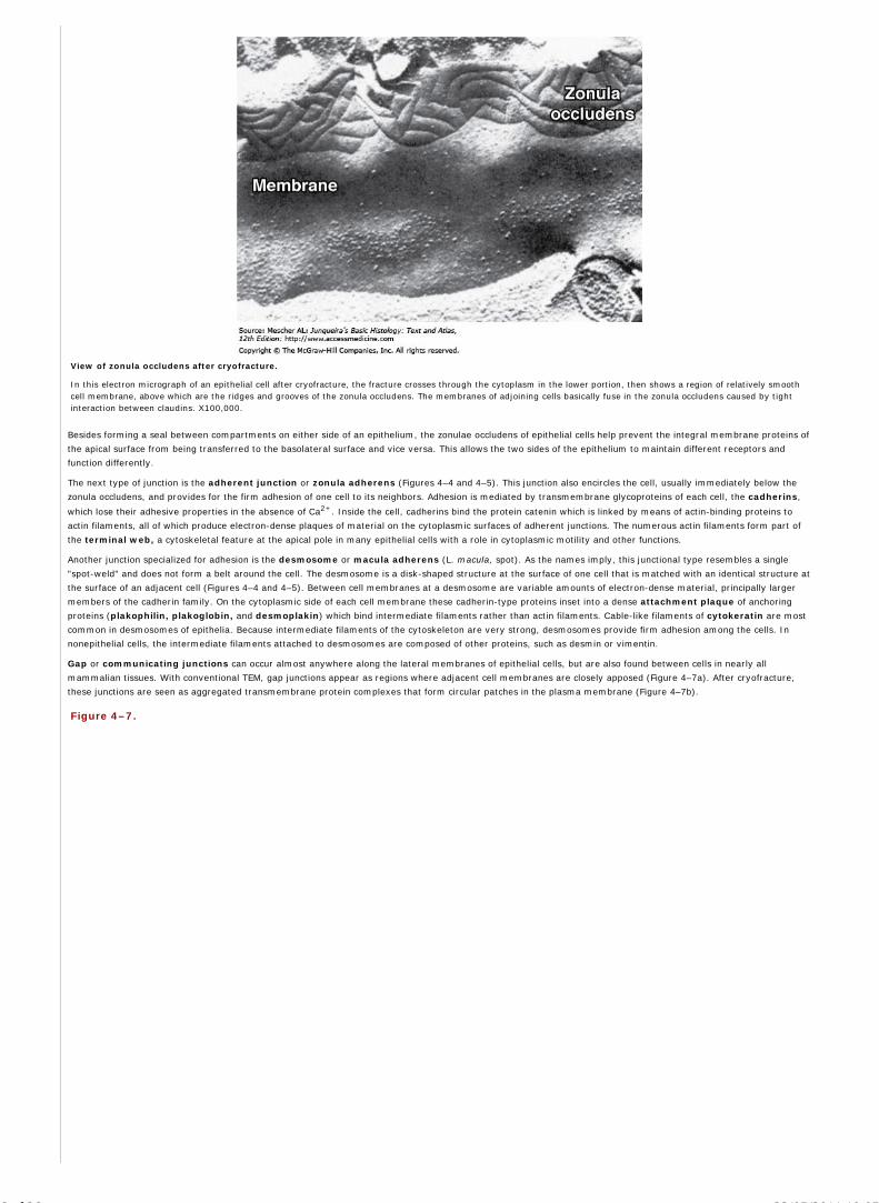
View of zonula occludens after cryofracture.
In this electron micrograph of an epithelial cell after cryofracture, the fracture crosses through the cytoplasm in the lower portion, then shows a region of relatively smoothcell membrane, above which are the ridges and grooves of the zonula occludens. The membranes of adjoining cells basically fuse in the zonula occludens caused by tightinteraction between claudins. X100,000.
Besides forming a seal between compartments on either side of an epithelium, the zonulae occludens of epithelial cells help prevent the integral membrane proteins of
the apical surface from being transferred to the basolateral surface and vice versa. This allows the two sides of the epithelium to maintain different receptors and
function differently.
The next type of junction is the adherent junction or zonula adherens (Figures 4–4 and 4–5). This junction also encircles the cell, usually immediately below the
zonula occludens, and provides for the firm adhesion of one cell to its neighbors. Adhesion is mediated by transmembrane glycoproteins of each cell, the cadherins,
which lose their adhesive properties in the absence of Ca2+. Inside the cell, cadherins bind the protein catenin which is linked by means of actin-binding proteins to
actin filaments, all of which produce electron-dense plaques of material on the cytoplasmic surfaces of adherent junctions. The numerous actin filaments form part of
the terminal web, a cytoskeletal feature at the apical pole in many epithelial cells with a role in cytoplasmic motility and other functions.
Another junction specialized for adhesion is the desmosome or macula adherens (L. macula, spot). As the names imply, this junctional type resembles a single
"spot-weld" and does not form a belt around the cell. The desmosome is a disk-shaped structure at the surface of one cell that is matched with an identical structure at
the surface of an adjacent cell (Figures 4–4 and 4–5). Between cell membranes at a desmosome are variable amounts of electron-dense material, principally larger
members of the cadherin family. On the cytoplasmic side of each cell membrane these cadherin-type proteins inset into a dense attachment plaque of anchoring
proteins (plakophilin, plakoglobin, and desmoplakin) which bind intermediate filaments rather than actin filaments. Cable-like filaments of cytokeratin are most
common in desmosomes of epithelia. Because intermediate filaments of the cytoskeleton are very strong, desmosomes provide firm adhesion among the cells. In
nonepithelial cells, the intermediate filaments attached to desmosomes are composed of other proteins, such as desmin or vimentin.
Gap or communicating junctions can occur almost anywhere along the lateral membranes of epithelial cells, but are also found between cells in nearly all
mammalian tissues. With conventional TEM, gap junctions appear as regions where adjacent cell membranes are closely apposed (Figure 4–7a). After cryofracture,
these junctions are seen as aggregated transmembrane protein complexes that form circular patches in the plasma membrane (Figure 4–7b).
Figure 4–7.
AccessMedicine | Print: Chapter 4. Epithelial Tissue http://accessmedicine.com/popup.aspx?aID=6180490&print=yes_chapter
6 of 26 22/05/2011 19:05

AccessMedicine | Print: Chapter 4. Epithelial Tissue http://accessmedicine.com/popup.aspx?aID=6180490&print=yes_chapter
7 of 26 22/05/2011 19:05

Gap junctions.
(a): The diagram of a gap junction (oblique view) depicts the structural elements that allow the exchange of nutrients and signal molecules between cells without loss ofmaterial into the intercellular space. The communicating channels are formed by pairs of abutting particles (connexons), which are in turn composed of six dumbbell-shaped protein subunits (connexins) that span the lipid bilayer of each cell membrane. The channel passing through the cylindrical bridges (arrow) is about 1.5 nm indiameter, limiting the size of the molecules that can pass through it. (b): A cryofracture preparation shows a gap junction between epithelial cells. The junction appears asa plaquelike agglomeration of intramembrane protein particles, the connexons. X45,000. (c): A section through a gap junction between two cells shows that the two cellmembranes are very closely apposed, separated only by a 2-nm-wide electron-dense space. Individual connexons are not resolved in cell sections. X193,000. (Figure 4–7c,with permission, from Mary C. Williams, Pulmonary Center, Boston University School of Medicine.)
The proteins of gap junctions, called connexins, form hexameric complexes called connexons, each of which has a central hydrophilic pore about 1.5 nm in
diameter. When two cells attach, connexins in the adjacent cell membranes move laterally and align to form connexons between the two cells (Figure 4–4), with each
gap junction having dozens or hundreds of aligned pairs of connexons. Gap junctions permit the rapid exchange between cells of molecules with small (<1.5 nm)
diameters. Some molecules mediating signal transduction, such as cyclic AMP, cyclic GMP, and ions, move readily through gap junctions, allowing cells in many tissues
to act in a coordinated manner rather than as independent units. A good example is heart muscle, where abundant gap junctions are greatly responsible for the heart's
coordinated beat.
In the contact area between epithelial cells and the subjacent basal lamina, hemidesmosomes (Gr. hemi, half, + desmos + soma) can often be observed
ultrastructurally. These adhesive structures resemble a half-desmosome and bind the cell to the basal lamina (Figure 4–2). However, while in desmosomes the
attachment plaques contain cadherins, in hemidesmosomes the plaques contain abundant integrins, transmembrane proteins that are receptor sites for the
extracellular macromolecules laminin and collagen type IV.
Blood vessels do not normally penetrate an epithelium and nutrients for the epithelial cells must pass out of the capillaries in the underlying lamina propria. These
nutrients then diffuse across the basal lamina and are taken up through the basolateral surfaces of the epithelial cell, usually by an energy-dependent process.
Receptors for chemical messengers (eg, hormones, neurotransmitters) that influence the activity of epithelial cells are localized in the basolateral membranes. In
absorptive epithelial cells, the apical cell membrane contains, as integral membrane proteins, enzymes such as disaccharidases and peptidases, which complete the
digestion of molecules to be absorbed.
SPECIALIZATIONS OF THE APICALCELL SURFACE
The free or apical surface of many types of epithelial cells has specialized structures to increase the cell surface area or to move substances or particles bound to the
epithelium.
MicrovilliWhen viewed in the electron microscope, many cells are seen to have cytoplasmic projections. These projections may be short or long fingerlike extensions or folds
that pursue a sinuous course, and they range in number from a few to many. Most are temporary, reflecting cytoplasmic movements and the activity of actin
filaments.
In absorptive cells, such as the lining epithelium of the small intestine, the apical surface presents orderly arrays of many hundreds of more permanent microvilli (L.
villus, tuft) (Figure 4–8). The average microvillus is only about 1 m high and 0.08 m wide, but with hundreds or thousands present on the end of each absorptive cell,
the total surface area can be increased as much as 20- or 30-fold. In these absorptive cells the glycocalyx is thicker than that of most cells and includes enzymes for
the final stages of certain macromolecules' breakdown. The complex of microvilli and glycocalyx is easily seen in the light microscope and is called the brush or
striated border.
Figure 4–8.
AccessMedicine | Print: Chapter 4. Epithelial Tissue http://accessmedicine.com/popup.aspx?aID=6180490&print=yes_chapter
8 of 26 22/05/2011 19:05

AccessMedicine | Print: Chapter 4. Epithelial Tissue http://accessmedicine.com/popup.aspx?aID=6180490&print=yes_chapter
9 of 26 22/05/2011 19:05

Microvilli.
Absorptive cells lining the small intestine demonstrate microvilli particularly well. (a): With the light microscope microvilli at the apical side of the epithelium are usuallyfaintly visible and make up the so-called striated border of the cells. (b): Individual microvilli are better seen by the TEM with a slightly higher magnification. Scatteredendocrine cells (E) in this epithelium do not extend to the apical surface and lack microvilli. (c): At higher magnification the bundles of vertical microfilaments constitutingthe core of each microvillus are clearly seen. Below the microvilli is the terminal web, a horizontal network of actin microfilaments and associated proteins includingmyosins. On the plasmalemma of the microvilli is a thick extracellular cell coat (glycocalyx) containing glycoproteins and enzymes that allow the final stages of digestion tobe linked to the uptake of digestion products across the cell membrane. The inset of cross-sectioned microvilli shows the internal disposition of the bundled actin filaments,the surrounding cell membrane, and the glycocalyx. X45,000. (d): The diagram indicates important proteins in a microvillus: the actin filaments cross-linked to oneanother by proteins such as fimbrin and villin and bound to the plasma membrane by proteins such as myosin I. The actin filaments are oriented in the same direction,with their plus ends associated with amorphous material at the tip of the microvillus.
Within each microvillus are bundles of actin filaments (Figure 4–8c,d) cross-linked to each other and to the surrounding plasma membrane by other proteins. These
filaments insert into the actin filaments of the terminal web. The array of microfilaments stabilizes the microvillus and allows it to contract slightly and intermittently
which helps maintain optimal conditions for absorption across its plasmalemma.
StereociliaStereocilia are long apical processes of cells in other absorptive epithelia such as that lining the epididymis (Figure 4–9) and ductus deferens. These structures are
much longer and less motile than microvilli, are branched, and should not be confused with true cilia. Like microvilli, stereocilia also increase the cells' surface area,
facilitating the movement of molecules into and out of the cell.
Figure 4–9.
Stereocilia.
At the apical ends of the tall epithelial cells lining organs such as the epididymis (shown here) are numerous very long stereocilia, which increase the surface area availablefor cellular absorption. Each stereocilium is typically much longer than a microvillus and may show a branching structure. Stereocilia have cytoplasmic actin filamentbundles and external cell coats similar to those of microvilli. X400. H&E.
CiliaCilia are elongated, highly motile structures on the surface of some epithelial cells, 5–10 m long and 0.2 m in diameter, which is much longer and two times wider
than a typical microvillus. As discussed in Chapter 2, each cilium is bounded by the cell membrane and contains an axoneme with a central pair of microtubules
surrounded by nine peripheral microtubular pairs (Figure 4–10). Cilia are inserted into basal bodies, which are electron-dense structures at the apical pole just below
the cell membrane (Figure 4–10). Basal bodies have a structure similar to that of centrioles. In living organisms, cilia exhibit rapid back-and-forth movements
coordinated to propel a current of fluid and suspended matter in one direction over the ciliated epithelium. The motion occurs due to activity of ciliary dynein present
on the peripheral microtubular doublets of the axoneme, with adenosine triphosphate (ATP) as the energy source. A ciliated cell of the trachea lining is estimated to
have about 250 cilia. Flagella, present in the human body only in spermatozoa (Chapter 21), are similar in structure to cilia but are much longer and are normally
AccessMedicine | Print: Chapter 4. Epithelial Tissue http://accessmedicine.com/popup.aspx?aID=6180490&print=yes_chapter
10 of 26 22/05/2011 19:05
limited to one flagellum per cell.
Figure 4–10.
Cilia.
TEMs of the apical portions of cells lining the respiratory tract show very well-developed cilia. (a): By light microscopy cilia usually appear as long, somewhat tangledprojections. X400. Mallory trichrome. (b): TEM of cilia sectioned longitudinally reveals the axoneme of each, with arrowheads on the left side showing the central andperipheral microtubules. The arrowhead at right indicates the plasma membrane surrounding a cilium. At the base of each cilium is a basal body (B) from which it grows.Much shorter microvilli (MV) can be seen between the cilia. X59,000. Inset: Cilia seen in cross section clearly show the 9 + 2 array of the axoneme microtubules in eachcilium. X80,000.
TYPES OF EPITHELIA
Epithelia can be divided into two main groups according to their structure and function: covering (or lining) epithelia and glandular epithelia. This is an arbitrary
division, for there are lining epithelia in which all the cells secrete (eg, the lining of the stomach) or in which glandular cells are distributed among the lining cells (eg,
mucous cells in the small intestine or trachea).
Covering or Lining EpitheliaCovering epithelia are tissues in which the cells are organized in layers that cover the external surface or line the cavities of the body. They are classified according to
the number of cell layers and the morphologic features of the cells in the surface layer (Table 4–2). Simple epithelia contain only one layer of cells and stratified
epithelia contain more than one layer.
Table 4–2. Common types of covering epithelia in the human body.
Number of Cell Layers Cell Form Examples of Distribution Main Function
Simple (one layer) Squamous Lining of vessels (endothelium). Serouslining of cavities; pericardium, pleura,peritoneum (mesothelium).
Facilitates the movement of the viscera (mesothelium),active transport by pinocytosis (mesothelium andendothelium), secretion of biologically active molecules(mesothelium).
Cuboidal Covering the ovary, thyroid. Covering, secretion.
Columnar Lining of intestine, gallbladder. Protection, lubrication, absorption, secretion.
Pseudostratified (layers of cells with nucleiat different levels; not all cells reachsurface but all adhere to basal lamina)
Lining of trachea, bronchi, nasal cavity. Protection, secretion; cilia-mediated transport of particlestrapped in mucus out of the air passages.
Stratified (two or more layers) Squamouskeratinized (dry)
Epidermis. Protection; prevents water loss.
AccessMedicine | Print: Chapter 4. Epithelial Tissue http://accessmedicine.com/popup.aspx?aID=6180490&print=yes_chapter
11 of 26 22/05/2011 19:05

Number of Cell Layers Cell Form Examples of Distribution Main Function
Squamousnonkeratinized(moist)
Mouth, esophagus, larynx, vagina, analcanal.
Protection, secretion; prevents water loss.
Cuboidal Sweat glands, developing ovarianfollicles.
Protection, secretion.
Transitional Bladder, ureters, renal calyces. Protection, distensibility.
Columnar Conjunctiva. Protection.
Based on cell shape, simple epithelia are classified as squamous (thin cells), cuboidal (cells roughly as thick as they are wide) or columnar (cells taller than they
are wide) Examples of simple epithelia are shown in Figures 4–11, 4–12, and 4–13.
Figure 4–11
Simple squamous epithelia.
In simple squamous epithelium, cells of the single layer are flat and usually very thin, with only the thicker cell nucleus appearing as a bulge to denote the cell. Simpleepithelia are typically specialized as lining of vessels and cavities and regulate substances which can enter underlying tissue from the vessel or cavity. The thin cells oftenexhibit transcytosis. Examples shown here are those lining the renal loops of Henle (a), the mesothelium lining a mesentery (b), and the endothelium lining the innersurface of the cornea (c). Endothelium and mesothelium are nearly always simple squamous. All X400. H&E.
Figure 4–12.
AccessMedicine | Print: Chapter 4. Epithelial Tissue http://accessmedicine.com/popup.aspx?aID=6180490&print=yes_chapter
12 of 26 22/05/2011 19:05

Simple cuboidal epithelium.
Cells of simple cuboidal epithelia vary in their height but are roughly as tall as they are wide. Their greater thickness often includes cytoplasm rich in mitochondria providingenergy for a high level of active transport of substances across the epithelium. Examples of simple cuboidal epithelia shown here are from a renal collecting tubule (a), apancreatic duct (b), and the mesothelium covering an ovary (c). All X400. H&E.
Figure 4–13.
AccessMedicine | Print: Chapter 4. Epithelial Tissue http://accessmedicine.com/popup.aspx?aID=6180490&print=yes_chapter
13 of 26 22/05/2011 19:05

Simple columnar epithelium.
Cells of simple columnar epithelia are taller than they are wide. Such cells are usually highly specialized for absorption, with microvilli, and often have interspersed secretorycells or ciliated cells. Such epithelial cells always have tight and adherent junctional complexes at their apical ends, but are often loosely associated in more basolateralareas. This allows for rapid transfer of absorbed material to the space between the cells rather than transport the full length of the cells. The additional cytoplasm incolumnar cells allows additional mitochondria and other organelles needed for absorption and processing. The examples shown here are from a renal collecting duct (a), theoviduct lining, with both secretory and ciliated cells (b), and the lining of the gall bladder (c). All X400. H&E.
Stratified epithelia are classified according to the cell shape of the superficial layer(s): squamous, cuboidal, columnar, and transitional.
The very thin surface cells of stratified squamous epithelia can be "keratinized" (rich in keratin intermediate filaments) or "nonkeratinized" (with relatively sparse
amounts of keratin). Stratified squamous keratinized epithelium is found mainly in the epidermis of skin. Its cells form many layers, and the cells closer to the
underlying connective tissue are usually cuboidal or low columnar. The cells become irregular in shape and flatten as they accumulate keratin in the process of
keratinization and are moved progressively closer to the surface, where they become thin, metabolically inactive packets (squames) of keratin lacking nuclei. This
surface layer of cells helps protect against water loss across this epithelium. (See Chapter 18 for more detailed information on skin.) Stratified squamous
nonkeratinized epithelium (Figure 4–14) lines wet cavities (eg, mouth, esophagus, and vagina). In such areas where water loss is not a problem, the flattened cells
of the epithelial surface layer are living cells containing much less keratin and retaining their nuclei.
Figure 4–14.
AccessMedicine | Print: Chapter 4. Epithelial Tissue http://accessmedicine.com/popup.aspx?aID=6180490&print=yes_chapter
14 of 26 22/05/2011 19:05

Stratified epithelia.
Stratified squamous epithelia have protective functions: protection against easy invasion of underlying tissue by microorganisms and protection against water loss. In theskin, protection against water loss and desiccation is particularly important and the epithelium is keratinized. As epidermal cells of the skin (a) differentiate they becomefilled with keratin and other substances and eventually lose their nuclei and other organelles. The superficial flattened squames form a layer which impedes water loss andeventually slough off and are replaced from below. Keratinization will be discussed fully in Chapter 18. Epithelia lining many internal surfaces such as the esophagus (b), orcovering the cornea (c) are considered nonkeratinized because the differentiating cells accumulate much less keratin and retain their nuclei. Such epithelia still provideprotection against microorganisms, but do not fill with keratin because water loss is less of an issue. Stratified cuboidal or columnar epithelia are fairly rare, but are found inexcretory ducts of some glands (d) where the double layer of cells apparently provides a more robust lining than a simple epithelium would. All X400; (b) PT, (a, c, and d)H&E.
Stratified cuboidal and stratified columnar epithelia are rare. Stratified columnar epithelium can be found in the conjunctiva lining the eyelids, where it is both
protective and mucus secreting. Stratified cuboidal epithelium is restricted to large excretory ducts of sweat and salivary glands, where it apparently provides a lining
more robust than that of a simple epithelium.
Transitional epithelium or urothelium, which lines only the urinary bladder, the ureter, and the upper part of the urethra, is characterized by a superficial layer of
domelike cells that are neither squamous nor columnar (Figure 4–15). These cells, sometimes called umbrella cells, are essentially protective against the hypertonic
and potentially cytotoxic effects of urine. Importantly, the form of the surface cells changes according to the degree of distention of the bladder wall. This type of
epithelium is discussed in detail in Chapter 19.
Figure 4–15.
AccessMedicine | Print: Chapter 4. Epithelial Tissue http://accessmedicine.com/popup.aspx?aID=6180490&print=yes_chapter
15 of 26 22/05/2011 19:05

Transitional epithelium or urothelium.
Stratified transitional epithelium lining the urinary bladder has rounded or dome-shaped superficial cells with two unusual features. The surface cells have specializedmembranes and are able to withstand the hypertonic effects of urine and protect underlying cells from this toxic solution. Cells of transitional epithelium are also able toadjust their relationships with one another as the bladder fills and the wall is stretched, so that the transitional epithelium of a full, distended bladder seems to have fewercell layers than that of an empty bladder. These unique features of urothelium will be discussed more fully in Chapter 19. X400. H&E.
In addition to these various stratified epithelia, there is another type classified as pseudostratified columnar epithelium, so called because all cells are attached to
the basal lamina even though their nuclei lie at different levels in the epithelium and the height of some cells does not extend to the surface. The best-known example
of pseudostratified columnar epithelium is that lining the passages of the upper respiratory tract (Figure 4–16). The columnar cells of this epithelium are also heavily
ciliated.
Figure 4–16.
Pseudostratified epithelium.
Cells of pseudo-stratified epithelia appear to be in layers, but the basal ends of the cells are all in contact with the basement membrane, which is often very thick in theseepithelia. The best example of this epithelial type is the pseudostratified ciliated columnar epithelium of the upper respiratory tract, which contains cell types with theirnuclei at different levels that give the false appearance of cellular stratification. This epithelium is discussed in detail in Chapter 17. X400. H&E.
Glandular EpitheliaGlandular epithelia are formed by cells specialized to secrete. The molecules to be secreted are generally stored in the cells in small membrane-bound vesicles called
secretory granules.
Glandular epithelial cells may synthesize, store, and secrete proteins (eg, in the pancreas), lipids (eg, adrenal, sebaceous glands), or complexes of carbohydrates and
proteins (eg, salivary glands). Mammary glands secrete all three substances. The cells of some glands have low synthetic activity (eg, sweat glands) and secrete
AccessMedicine | Print: Chapter 4. Epithelial Tissue http://accessmedicine.com/popup.aspx?aID=6180490&print=yes_chapter
16 of 26 22/05/2011 19:05

mostly water and electrolytes transferred into the gland from the blood.
The epithelia that form glands can be classified according to various criteria. Unicellular glands consist of large isolated secretory cells and multicellular glands have
clusters of cells. The classic unicellular gland is the goblet cell in the lining of the small intestine (Figure 4–17) or respiratory tract. The term "gland," however, is
usually used to designate large aggregates of secretory epithelial cells, such as in the salivary glands and the pancreas.
Figure 4–17.
Goblet cells: unicellular glands.
A section of epithelial lining of the large intestine shows scattered goblet cells secreting mucus to the extracellular space (a): With the stain for glycoproteins used here,both the mucus precursor stored in cytoplasmic graules of the goblet cells as well as the secreted mucus are stained dark blue. X400. PAS-PT. (b): Ultrastructurally a gobletcell shows a basal nucleus surrounded by RER (R), a large Golgi complex (G) just above the nucleus, and an apical end filed with large secretory granules (SG) containingmucins. This highly viscous material is secreted by exocytosis and is then hydrated to form mucus in the lumen lined by microvilli (M). X17,000.
Glands develop during fetal life from covering epithelia by means of cell proliferation and invasion of the subjacent connective tissue, followed by further differentiation
(Figure 4–18). Exocrine glands retain their connection with the surface epithelium, the connection taking the form of tubular ducts lined with epithelial cells through
which the secretions pass to the surface. Endocrine glands have lost their connection to the surface from which they originated during development. These glands
are therefore ductless and their secretions are picked up and transported to their sites of action by the bloodstream rather than by a duct system. Multicellular glands,
whether exocrine or endocrine, also have connective tissue in a surrounding capsule and in septa that divide the gland into lobules. These lobules then subdivide, and
in this way the connective tissue separates and binds the glandular components together (Figure 4–19).
Figure 4–18.
AccessMedicine | Print: Chapter 4. Epithelial Tissue http://accessmedicine.com/popup.aspx?aID=6180490&print=yes_chapter
17 of 26 22/05/2011 19:05

Formation of glands from covering epithelia.
During fetal development epithelial cells proliferate and penetrate the underlying connective tissue. They may—or may not—maintain a connection with the surfaceepithelium. When the connection is maintained, exocrine glands are formed; with the connection lost, endocrine glands are formed. Exocrine glands secrete to the bodysurface or gut via duct systems formed from the epithelial connection. The cells of endocrine glands, which secrete hormones (see Chapter 20) can be arranged in cords orin follicles with lumens for storing the secretory product. From either the cords (left) or follicles (right) of endocrine cells, the secretory product is released outside the cellsand picked up by the blood vessels for distribution throughout the body.
Figure 4–19.
AccessMedicine | Print: Chapter 4. Epithelial Tissue http://accessmedicine.com/popup.aspx?aID=6180490&print=yes_chapter
18 of 26 22/05/2011 19:05

General structure of exocrine glands.
Exocrine glands by definition have ducts that lead to an organ or body surface. Inside the gland the duct runs through connecting septa and branches repeatedly, until itssmallest branches end in the secretory portions of the gland.
Exocrine glands have a secretory portion, which contains the cells specialized for secretion, and ducts, which transport the secretion out of the gland. The
morphology of these components allows the glands to be classified according to the scheme shown in Figure 4–20 and summarized as follows:
Ducts can be simple (unbranched) or compound (with two or more branches).
Secretory portions can be tubular (either short or long and coiled) or acinar (round or globular).
Either type of secretory portion may be branched.
Compound glands can have tubular, acinar, or tubuloacinar secretory portions.
Figure 4–20.
Structural classes of exocrine glands.
(a): Simple glands have unbranched ducts, although the ducts may be short or long and coiled. The secretory portions attached to these ducts may themselves bebranched. The secretory portions are either tubular, if more or less cylindrical in shape, or acinar, if bulbous or saclike. (b): If the ducts branch to serve multiple secretoryunits, the gland is compound. On compound glands, the secretory units may be all tubular, all acinar, or a combination of the two shapes.
AccessMedicine | Print: Chapter 4. Epithelial Tissue http://accessmedicine.com/popup.aspx?aID=6180490&print=yes_chapter
19 of 26 22/05/2011 19:05

Exocrine glands are also classified functionally according to the way the secretory products leave the cell (Figure 4–21):
Merocrine secretion (sometimes called eccrine) involves typical exocytosis of proteins or glycoproteins. This is the most common mode of secretion.
Holocrinesecretion involves the cell filling with secretory product and then the whole cell being disrupted and shed. This is best seen in the sebaceousglands of skin (Figure 4–22).
In an intermediate type, apocrinesecretion, the secretory product is typically a large lipid droplet and is discharged together with some of the apicalcytoplasm and plasmalemma (Figure 4–23).
Figure 4–21.
Functional classification of exocrine glands.
Different cellular secretion processes are used in exocrine glands, depending on what substance is being secreted. (a): Merocrine glands secrete products, usuallycontaining proteins, by means of exocytosis at the apical end of the secretory cells. Most exocrine glands are merocrine. (b): Holocrine gland secretion is produced by thedisintegration of the secretory cells themselves as they complete differentiation which involves becoming filled with product. Sebaceous glands of hair follicles are the bestexamples of holocrine glands. (c): Apocrine gland secretion involves loss of a large membrane-enclosed portion of apical cytoplasm, usually containing one or more lipiddroplets. This apical portion of the cell may subsequently break down to release its contents during passage into the duct. Apocrine secretion, along with merocrinesecretion, is seen in mammary glands.
Figure 4–22.
AccessMedicine | Print: Chapter 4. Epithelial Tissue http://accessmedicine.com/popup.aspx?aID=6180490&print=yes_chapter
20 of 26 22/05/2011 19:05

Holocrine secretion in a sebaceous gland.
In holocrine secretion, best seen in the sebaceous gland adjacent to hair follicles, entire cells fill with a product and are released during secretion. Undifferentiated cellsdeep and peripheral in the gland fill with lipid-rich granules and become metabolically inactive as they mature and move upward and toward the gland's center. Whenterminally differentiated, the cells separate and quickly disintegrate to form the secretion which serves to protect and lubricate adjacent skin and hair. Sebaceous glandslack myoepithelial cells; cell proliferation inside a dense, inelastic connective tissue capsule continuously forces product into the duct. X200. H&E.
Figure 4–23.
Apocrine secretion in the mammary gland.
The secreting portions of a mammary gland demonstrate apocrine secretion and are characterized by the discharge of the secretion product with a pinched off portion of theapical cytoplasm (arrows). The released portion of cell contains lipid droplet(s). Merocrine secretion also occurs from the same and other cells of the secretory units. X400.PSH.
Exocrine glands with merocrine secretion can be further categorized as either serous or mucous according to the nature of the proteins or glycoproteins secreted and
the resulting staining properties of the secretory cells. The acinar cells of the pancreas and parotid salivary glands are examples of the serous type which secrete
digestive enzymes. The basal ends of serous cells have well-developed RER and Golgi complexes and the cells are filled apically with secretory granules in different
stages of maturation (Figure 4–24). Serous cells therefore stain intensely with any basophilic or acidophilic stain.
AccessMedicine | Print: Chapter 4. Epithelial Tissue http://accessmedicine.com/popup.aspx?aID=6180490&print=yes_chapter
21 of 26 22/05/2011 19:05

Figure 4–24.
Serous cells.
Serous acinar cells of the exocrine pancreas are arranged in small acini of 5-10 cells with a very small central lumen. Each acinar cell is roughly pyramid-shaped, with itsapex at the lumen. (a): As seen by light microscopy, the apical ends are eosinophilic due to the abundant immature and mature secretory granules present there. The cells'basal ends contain the large rounded nuclei and an abundance of rough ER, making the cells highly basophilic basally. X200. PT. (b): A portion of one acinar cell is shownultrastructurally, indicating the abundant RER, Golgi complexes, and secretory granules and the very small size of the acinus lumen. X13,000. Secretion here is merocrineand typically the mature zymogen granules, filled with digestive enzymes, remain in the apical cell region until the cell is stimulated to secrete. Other cells secreteconstitutively, with small granules undergoing exocytosis as soon as they emerge fully formed from the Golgi apparatus.
Mucous cells, such as goblet cells, while also rich in RER and Golgi complexes are filled apically with secretory granules containing strongly hydrophilic glycoproteins
called mucins. When mucins are released from the cell, they become hydrated and form mucus, a viscous, elastic, protective lubricant material. Mucin-containing
granules stain well with the periodic acid-Schiff (PAS) method for glycoproteins (Figure 4–17a), but are not intensely acidophilic like zymogen granules of serous cells
(Figure 4–25). Mucous cells of large glands are organized as secretory tubules and in mixed seromucous salivary glands crescent-shaped clumps of serous cells
frequently share the ends of the tubules as serous demilunes (Figure 4–26).
Figure 4–25.
AccessMedicine | Print: Chapter 4. Epithelial Tissue http://accessmedicine.com/popup.aspx?aID=6180490&print=yes_chapter
22 of 26 22/05/2011 19:05

Mucous cells.
Mucous cells are typically larger than serous cells, with more flattened basal nuclei. The apical region and most of the other cytoplasm of each mucous cell is filled withsecretory granules containing mucin like that of goblet cells. The basal region contains the RER, nucleus, and a well-developed Golgi apparatus. The RER and Golgi are veryrich in enzymes called glycosyltransferases, which attach sugars to polypeptide chains to make glycoproteins. Mucus contains many glycoproteins with important water-binding properties. The lumens (small arrows) of mucous tubules are larger than those of serous acini. The large arrow indicates a secretory duct. X200. PT. Other types ofmucous cells are found in the stomach, the various salivary glands, the respiratory tract, and the genital tract. These cells show great variability in both their morphologicfeatures and in the chemical nature of their secretions.
Figure 4–26.
AccessMedicine | Print: Chapter 4. Epithelial Tissue http://accessmedicine.com/popup.aspx?aID=6180490&print=yes_chapter
23 of 26 22/05/2011 19:05

Seromucous, compound tubuloacinar gland.
The submandibular salivary glands have both mucous and serous secretory units, typically shaped as acini and tubules respectively. Clumps of serous cells at the ends ofsome mucous tubules appear as crescent-shaped structures called serous demilunes. At the left is seen a striated duct whose cells' basal membranes are folded intolong folds with many mitochondria, an arrangement specialized for ion transport across the epithelium. X400. PT.
Several exocrine glands (eg, sweat, lachrymal, salivary, and mammary glands) contain stellate or spindle-shaped myoepithelial cells located between the basal
lamina and the basal pole of secretory or duct cells (Figure 4–27). Long processes of these cells embrace an acinus as an octopus might embrace a rounded boulder.
Along ducts they are more longitudinally arranged. Myoepithelial cells are connected to each other and to the epithelial cells by both gap junctions and desmosomes.
These cells are specialized for contraction, containing myosin and a large number of actin filaments. Their major function is to contract around the secretory or
conducting portion of the gland and thus help propel secretory products into the duct.
Figure 4–27.
Myoepithelial cells.
(a): Portion of a salivary gland acinus shows two secretory cells with secretory granules. A myoepithelial cell (M) embraces the acinus with contractile processes. X20,000.(b): A myoepithelial cell immunostained against smooth muscle actin shows its association with an entire acinus. Contraction of the myoepithelial cell compresses theacinus and aids in the expulsion of secretory products into the duct. X200. H&E counterstain.
Endocrine glands are the producers of hormones, which are generally polypeptide or lipid-derived factors that are released into the interstitial fluid. Hormones diffuse
into the blood for circulation and bind specific receptors on target cells elsewhere in the body, often within other endocrine glands. The receptors may also be on cells
very close to the hormone-secreting cells or on the secreting cell itself; in these cases the cellular signaling is termed paracrine or autocrine, respectively.
Hormones can be secreted from single cells that are sparsely distributed or from cells with other major functions, such as certain cardiac muscle cells. In the large
endocrine glands the parenchymal cells form strands or cords interspersed between dilated capillaries (eg, the adrenal cortex; see Figure 4–18) or can line a follicle
filled with stored secretory product (eg, the thyroid gland; Figure 4–18). Some endocrine glands have cells releasing more than one hormone.
Some organs such as the pancreas have both endocrine and exocrine functions, and in the liver one cell type may function both ways, secreting bile components into a
duct system, as well as releasing other products into the bloodstream.
TRANSPORT ACROSS EPITHELIA
As discussed in Chapter 2, all cells have the ability to actively transport certain ions against a concentration and electrical-potential gradient. An important example is
the active extrusion of Na+ by means of Mg2+-activated Na+/K+-ATPase (sodium pump), by which cells maintain the required low intracellular sodium concentration
(5-15 mmol/L vs. ~140 mmol/L in extracellular fluid).
Some epithelial cells actively transfer ions and fluid across the epithelium, from its apex to its base or vice-versa; this is known as transcellular transport (Figure
4–28). For transport in either direction, the tight junctions play an important role in the transport process, sealing the apical portions of the epithelium and preventing
back-diffusion of materials already transported across the epithelium. A well-studied site of epithelial transport is the proximal renal tubule cell, where the apical
surface is freely permeable to Na+ in the lumen. To maintain electrical and osmotic balance, equimolar amounts of chloride and water follow the Na+ ion into the cell.
The basal surfaces of these cells are elaborately folded and many long invaginations of the basolateral membrane are seen in electron micrographs (Figure 4–29). In
addition, there is interdigitation of membrane folds between adjacent cells, all of which increase the surface area for transport. Sodium pumps are localized in both the
basal and the lateral plasma membranes and located between the folds are vertically oriented mitochondria that supply the ATP for the active extrusion of Na+ from
the cell basally. Chloride and water again follow passively. In this way, sodium is returned to the circulation and is not lost in massive amounts in the urine.
Figure 4–28.
AccessMedicine | Print: Chapter 4. Epithelial Tissue http://accessmedicine.com/popup.aspx?aID=6180490&print=yes_chapter
24 of 26 22/05/2011 19:05

Ion and water absorption and secretion.
Ion and water transport across epithelia can occur in different directions, depending on which tissue is involved. (a): The direction of transport is from the lumen to theblood vessel, as in the gallbladder and intestine. This process is called absorption, and serves to concentrate bile and obtain water and ions in these organs. (b): Transportin the opposite direction, as in the choroid plexus, ciliary body, and sweat glands, is called secretion and serves to expel water from the interstitial fluid into specializedaqueous fluids in these tissues. Whether the epithelia are absorbing or secreting water, the presence of apical occluding junctions is necessary to maintain tightcompartmentalization and consequent control over ion distribution.
Figure 4–29.
AccessMedicine | Print: Chapter 4. Epithelial Tissue http://accessmedicine.com/popup.aspx?aID=6180490&print=yes_chapter
25 of 26 22/05/2011 19:05

Copyright © The McGraw-Hill Companies. All rights reserved.Privacy Notice. Any use is subject to the Terms of Use and Notice.
Absorptive cells.
An ultrastructural diagram and TEM of epithelial cells highly specialized for absorption: cells of proximal convoluted tubule of the kidney. Long invaginations of the basal cellmembrane outline regions filled with vertically oriented mitochondria, a typical disposition present in ion-transporting cells. Interdigitations from neighboring cells interlockwith those of this cell. Immediately below the microvilli are junctional complexes between individual cells. The basolateral membranes can be discerned in continuity withthe junctional complexes. Apically are vesicles that have undergone pinocytosis, soon to fuse with lysosomes, as shown in the upper left portion of the diagram. Sodium
ions diffuse passively through the apical membranes of renal epithelial cells and are then actively transported out of the cells by Na+/K+ -ATPase located in the basolateralmembranes of the cells. Energy for this sodium pump is supplied by the nearby mitochondria. Immediately below the basal lamina is a capillary for removal of the waterabsorbed across this part of the epithelium. X9600.
Extracellular molecules and fluid are also internalized in the cytoplasm of most cells by pinocytotic vesicles that form abundantly at the plasmalemma. This activity is
clearly observed in the simple squamous epithelia that line the blood and lymphatic capillaries (endothelia) or the body cavities (mesothelia). These cells have few
organelles other than the abundant pinocytotic vesicles, which cross the thin cells in both directions and secrete their contents on the opposite side by exocytosis. This
process, termed transcytosis, is not restricted to simple squamous epithelia. Uptake of material at the apical epithelial pole followed by exocytosis at the basolateral
surface occurs actively in many simple cuboidal and columnar epithelia and is important in various physiological processes.
RENEWAL OF EPITHELIAL CELLS
Epithelial tissues are relatively labile structures whose cells are renewed continuously by mitotic activity. The renewal rate is variable; it can be fast in tissues such as
the intestinal epithelium, which is replaced every week, or slow, as in the large glands. In stratified epithelial tissues, mitosis only occurs within the basal layer in
contact with the basal lamina. In some functionally complex epithelia, stem cells have been identified only in restricted niches some distance from the transit
amplifying cells and differentiating cells. For example, the epithelium lining the small intestine is derived completely from stem cells found in the simple glands
between the intestinal villi. In the epidermis, stem cells are located at a characteristic position along the wall of hair follicles.
MEDICAL APPLICATION
Both benign and malignant tumors can arise from most types of epithelial cells. A carcinoma (Gr. karkinos, cancer, + oma, tumor) is a malignant tumor of
epithelial cell origin. Malignant tumors derived from glandular epithelial tissue are usually called adenocarcinomas (Gr. adenos, gland, + karkinos); these
are by far the most common tumors in adults. In children up to age 10 years, most tumors develop (in decreasing order) from hematopoietic organs, nerve
tissues, connective tissues, and epithelial tissues. This proportion gradually changes, and after age 45 years, more than 90% of all tumors are of epithelial
origin.
Carcinomas composed of differentiated cells reflect cell-specific morphologic features and behaviors (eg, the production of keratins, mucins, and hormones).
Undifferentiated carcinomas are often difficult to diagnose by morphologic analysis alone. Since these carcinomas usually contain keratins, the detection of
keratins by immunocytochemistry often helps to determine the diagnosis and treatment of these tumors.
Epithelia are normally capable of rapid repair and replacement of apoptotic or damaged cells. In some large glands, most notably the liver, mitotic activity is normally
rare but is actively renewed following major damage to the organ. When a portion of liver tissue is removed surgically or lost by the acute effects of toxic substances,
cells of undamaged regions quickly begin active proliferation and normal functional mass of liver tissue is soon regenerated.
MEDICAL APPLICATION
Some epithelial cells are prone to abnormal growth called neoplasia that may lead to cancers. Neoplastic growth is reversible and does not always result in
cancer.
Under certain abnormal conditions, one type of epithelial tissue may undergo transformation into another type in another reversible process called
metaplasia, which is illustrated by the following examples.
In heavy cigarette smokers, the ciliated pseudo-stratified epithelium lining the bronchi can be transformed into stratified squamous epithelium.
In individuals with chronic vitamin A deficiency, epithelial tissues of the type found in the bronchi and urinary bladder are gradually replaced by stratified
squamous epithelium.
Metaplasia is not restricted to epithelial tissue; it may also occur in connective tissue.
AccessMedicine | Print: Chapter 4. Epithelial Tissue http://accessmedicine.com/popup.aspx?aID=6180490&print=yes_chapter
26 of 26 22/05/2011 19:05

Print Close Window
Note: Large images and tables on this page may necessitate printing in landscape mode.
Copyright © The McGraw-Hill Companies. All rights reserved.
Junqueira's Basic Histology: Text & Atlas, 12e > Chapter 5. Connective Tissue >
CONNECTIVE TISSUE: INTRODUCTION
The different types of connective tissue are responsible for providing and maintaining the form of organs throughout the body. Functioning in a mechanical role,
they provide a matrix that connects and binds other tissues and cells in organs and gives metabolic support to cells as the medium for diffusion of nutrients and
waste products.
Structurally, connective tissue is formed by three classes of components: cells, fibers, and ground substance. Unlike the other tissue types (epithelium, muscle,
and nerve), which consist mainly of cells, the major constituent of connective tissue is the extracellular matrix (ECM). Extracellular matrices consist of
different combinations of protein fibers (collagen, reticular, and elastic fibers) and ground substance. Ground substance is a highly hydrophilic, viscous
complex of anionic macromolecules (glycosaminoglycans and proteoglycans) and multiadhesive glycoproteins (laminin, fibronectin, and others) that stabilizes
the ECM by binding to receptor proteins (integrins) on the surface of cells and to the other matrix components. In addition to its major structural role,
molecules of connective tissue serve other important biological functions, such as forming a reservoir of factors controlling cell growth and differentiation. The
hydrated nature of much connective tissue provides the medium through which nutrients and metabolic wastes are exchanged between cells and their blood
supply.
The wide variety of connective tissue types in the body reflects variations in the composition and amount of the cells, fibers, and ground substance which
together are responsible for the remarkable structural, functional, and pathologic diversity of connective tissue.
The connective tissues originate from the mesenchyme, an embryonic tissue formed by elongated undifferentiated cells, the mesenchymal cells (Figure
5–1). These cells are characterized by oval nuclei with prominent nucleoli and fine chromatin. They possess many thin cytoplasmic processes and are
immersed in an abundant and viscous extracellular substance containing few fibers. The mesenchyme develops mainly from the middle layer of the embryo,
the mesoderm. Mesodermal cells migrate from their site of origin in the embryo, surrounding and penetrating developing organs. In addition to being the
point of origin of all types of connective tissue cells, mesenchyme develops into other types of structures, such as blood cells, endothelial cells, and muscle
cells.
Figure 5–1.
Embryonic mesenchyme.
Mesenchyme consists of a population of undifferentiated cells, generally elongated but with many shapes, having large euchromatic nuclei and prominent nucleoliwhich indicate high levels of synthetic activity. These cells are called mesenchymal cells. Mesenchymal cells are surrounded by an extracellular matrix which theyproduced and which consists largely of a simple ground substance rich in hyaluronan (hyaluronic acid). This section is stained with Masson trichrome which stainscollagen fibers blue and the lack of collagen in mesenchyme is apparent. X200.
CELLS OF CONNECTIVE TISSUE
A variety of cells with different origins and functions are present in connective tissue (Figure 5–2 and Table 5–1). Fibroblasts originate locally from
undifferentiated mesenchymal cells and spend all their life in connective tissue; other cells such as mast cells, macrophages, and plasma cells originate
from hematopoietic stem cells in bone marrow, circulate in the blood, and then move into connective tissue where they remain and execute their functions.
White blood cells (leukocytes) are transient cells of most connective tissues; they also originate in the bone marrow and move to the connective tissue where
they reside for a few days, then usually die by apoptosis.
Figure 5–2.
AccessMedicine | Print: Chapter 5. Connective Tissue http://accessmedicine.com/popup.aspx?aID=6180657&print=yes_chapter
1 of 28 22/05/2011 19:12

Lineages of connective tissue cells.
This simplified representation of the connective tissue cell lineage includes cells from the multipotential embryonic mesenchyme cells and hematopoietic stemcells of bone marrow. Dotted arrows indicate that one or more intermediate cell types exist between the examples illustrated. The cells are not drawn in proportionto actual sizes, eg, adipocyte, megakaryocyte, and osteoclast cells are significantly larger than the other cells illustrated.
Table 5–1. Functions of connective tissue cells.
Cell Type Representative Product or Activity Representative Function
Fibroblast, chondroblast,osteoblast, odontoblast
Production of fibers and ground substance Structural
Plasma cell Production of antibodies Immunologic (defense)
Lymphocyte (several types) Production of immunocompetent cells Immunologic (defense)
Eosinophilic leukocyte Participation in allergic and vasoactive reactions, modulation of mast cell activities and theinflammatory process
Immunologic (defense)
Neutrophilic leukocyte Phagocytosis of foreign substances, bacteria Defense
Macrophage Secretion of cytokines and other molecules, phagocytosis of foreign substances andbacteria, antigen processing and presentation to other cells
Defense
Mast cell and basophilic leukocyte Liberation of pharmacologically active molecules (eg, histamine) Defense (participate in allergicreactions)
Adipocyte Storage of neutral fats Energy reservoir, heatproduction
FibroblastsFibroblasts synthesize collagen, elastin, glycosaminoglycans, proteoglycans and multiadhesive glycoproteins. Fibroblasts are the most common cells in
AccessMedicine | Print: Chapter 5. Connective Tissue http://accessmedicine.com/popup.aspx?aID=6180657&print=yes_chapter
2 of 28 22/05/2011 19:12

connective tissue (Figure 5–3) and are responsible for the synthesis of extracellular matrix components. Two stages of activity—active and quiescent—are often
observed in these cells (Figure 5–3b). Cells with intense synthetic activity are morphologically distinct from the quiescent fibroblasts that are scattered within
the matrix they have already synthesized. Some histologists reserve the term fibroblast to denote the active cell and fibrocyte to denote the quiescent cell.
Figure 5–3.
Fibroblasts.
Connective tissue where parallel bundles of collagen are being formed. (a): Fibroblasts typically show large active nuclei and eosinophilic cytoplasm tapering off inboth directions along the axis of the nucleus, a morphology usually called "spindle-shaped." The nuclei (arrows) are clearly seen, but the cytoplasmic processesresemble the collagen bundles (C) that fill the extracellular matrix and are difficult to distinguish in H&E-stained sections. (b): Both active and quiescent fibroblastsmay sometimes be distinguished, as in this section of dermis. Active fibroblasts are large cells with large, euchromatic nuclei and basophilic cytoplasm, whereasinactive fibroblast or fibrocytes are smaller with less prominent, heterochromatic nuclei. The very basophilic round cells in (b) are leukocytes. Both X400. H&E.
The active fibroblast has an abundant and irregularly branched cytoplasm. Its nucleus is ovoid, large, and pale-staining, with fine chromatin and a prominent
AccessMedicine | Print: Chapter 5. Connective Tissue http://accessmedicine.com/popup.aspx?aID=6180657&print=yes_chapter
3 of 28 22/05/2011 19:12

nucleolus. The cytoplasm is rich in rough ER, and the Golgi apparatus is well developed. The quiescent fibroblast or fibrocyte is smaller than the active
fibroblast and is usually spindle-shaped. It has fewer processes; a smaller, darker, elongated nucleus; and more acidophilic cytoplasm with much less RER.
Fibroblasts synthesize most components of connective tissue ECM, including proteins, such as collagen and elastin, which upon secretion form collagen,
reticular, and elastic fibers, and the glycosaminoglycans, proteoglycans, and glycoproteins of the ground substance. Fibroblasts are targets of various growth
factors that influence cell growth and differentiation. In adults, fibroblasts in connective tissue rarely undergo division; mitosis can resume when the organ
requires additional fibroblasts as in wound healing.
MEDICAL APPLICATION
The regenerative capacity of the connective tissue is clearly observed when tissues are destroyed by inflammation or traumatic injury. In these cases,
the spaces left after injury to tissues whose cells do not divide (eg, cardiac muscle) are filled by connective tissue, which forms a scar. The healing of
surgical incisions depends on the reparative capacity of connective tissue. The main cell type involved in repair is the fibroblast.
When it is adequately stimulated, such as during wound healing, the fibrocyte reverts to the fibroblast state, and its synthetic activities are reactivated.
In such instances the cell reassumes the form and appearance of a fibroblast. The myofibroblast, a cell with features of both fibroblasts and smooth
muscle cells, is also observed during wound healing. These cells have most of the morphological characteristics of fibroblasts but contain increased
amounts of actin microfilaments and myosin and behave much like smooth muscle cells. Their activity is responsible for wound closure after tissue
injury, a process called wound contraction.
AdipocytesAdipocytes (L. adeps, fat, + Gr. kytos, cell) are connective tissue cells that have become specialized for storage of neutral fats or for the production of heat.
Often called fat cells, they have considerable metabolic significance and are discussed in detail in Chapter 6.
Macrophages & the Mononuclearphagocyte SystemMacrophages were discovered and initially characterized by their phagocytic ability. They have a wide spectrum of morphologic features that correspond to
their state of functional activity and to the tissue they inhabit.
In the electron microscope, macrophages are characterized by an irregular surface with pleats, protrusions, and indentations, a morphologic expression of
their active pinocytotic and phagocytic activities. They generally have a well-developed Golgi apparatus, many lysosomes, and rough ER (Figure 5–4).
Figure 5–4.
Macrophage ultrastructure.
Characteristic features of macrophages seen in this TEM of one such cell are the prominent nucleus (N) and the nucleolus (Nu) and the numerous secondarylysosomes (L). The arrows indicate phagocytic vacuoles near the protrusions and indentations of the cell surface. X10,000.
Macrophages derive from bone marrow precursor cells that divide, producing monocytes which circulate in the blood. These cells cross the wall of venules and
capillaries to penetrate the connective tissue, where they mature and acquire the morphologic features of macrophages. Therefore, monocytes and
macrophages are the same cell in different stages of maturation. Macrophages are also sometimes referred to as "histiocytes."
Macrophages are distributed throughout the body and are present in most organs. Along with other monocyte-derived cells, they comprise a family of cells
called the mononuclearphagocyte system (Table 5–2). All are long-living cells and may survive for months in the tissues. In most organs these cells are
highly important for the up-take, processing, and presentation of antigens for lymphocyte activation. The macrophage-like cells have been given different
names in different organs, eg, Kupffer cells in the liver, microglial cells in the central nervous system, Langerhans cells in the skin, and osteoclasts in bone
tissue. However, all are derived from monocytes. The transformation from monocytes to macrophages in connective tissue involves increases in cell size,
increased protein synthesis, and increases in the number of Golgi complexes and lysosomes. A typical macrophage measures between 10 and 30 m in
diameter and has an oval or kidney-shaped nucleus located eccentrically.
Table 5–2. Distribution and main functions of the cells of the mononuclear phagocyte system.
AccessMedicine | Print: Chapter 5. Connective Tissue http://accessmedicine.com/popup.aspx?aID=6180657&print=yes_chapter
4 of 28 22/05/2011 19:12

Cell Type Location Main Function
Monocyte Blood Precursor of macrophages
Macrophage Connective tissue, lymphoid organs,lungs, bone marrow
Production of cytokines, chemotactic factors, and several other molecules that participate ininflammation (defense), antigen processing and presentation
Kupffer cell Liver Same as macrophages
Microglia cell Nerve tissue of the central nervoussystem
Same as macrophages
Langerhans cell Skin Antigen processing and presentation
Dendritic cell Lymph nodes Antigen processing and presentation
Osteoclast Bone (fusion of several macrophages) Digestion of bone
Multinuclear giantcell
Connective tissue (fusion of severalmacrophages)
Segregation and digestion of foreign bodies
MEDICAL APPLICATION
When adequately stimulated, macrophages may increase in size and are arranged in clusters forming epithelioid cells (named for their vague
resemblance to epithelial cells), or several may fuse to form multinuclear giant cells. Both cell types are usually found only in pathological
conditions.
Macrophages act as defense elements. They phagocytose cell debris, abnormal extracellular matrix elements, neoplastic cells, bacteria, and inert
elements that penetrate the organism. Macrophages are also antigen-presenting cells that participate in the processes of partial digestion and
presentation of antigen to other cells (see Chapter 14). A typical example of an antigen-processing cell is the macrophage present in the skin
epidermis, called the Langerhans cell (see Chapter 18). Although macrophages are the main antigen presenting cells, under certain circumstances
many other cell types, such as fibroblasts, endothelial cells, astrocytes, and thyroid epithelial cells, are also able to perform this function. Macrophages
also participate in cell-mediated resistance to infection by bacteria, viruses, protozoans, fungi, and metazoans (eg, parasitic worms); in cell-mediated
resistance to tumors; and in extrahepatic bile production, iron and fat metabolism, and the destruction of aged erythrocytes.
When macrophages are stimulated (by injection of foreign substances or by infection), they change their morphological characteristics and metabolism.
They are then called activated macrophages and acquire characteristics not present in their nonactivated state. These activated macrophages, in
addition to showing an increase in their capacity for phagocytosis and intracellular digestion, exhibit enhanced metabolic and lysosomal enzyme
activity. Macrophages also have an important role in removing cell debris and damaged extracellular components formed during the physiological
involution process. For example, during pregnancy the uterus increases in size. Immediately after parturition, the uterus suffers an involution during
which some of its tissues are destroyed by the action of macrophages. Macrophages are also secretory cells that produce an impressive array of
substances, including enzymes (eg, collagenase) and cytokines that participate in defensive and reparative functions, and they exhibit increased tumor
cell–killing capacity.
Mast CellsMast cells are large, oval or round connective tissue cells, 20–30 m in diameter, whose cytoplasm is filled with basophilic secretory granules. The rather small,
spherical nucleus is centrally situated and may be obscured by the cytoplasmic granules (Figure 5–5).
Figure 5–5.
AccessMedicine | Print: Chapter 5. Connective Tissue http://accessmedicine.com/popup.aspx?aID=6180657&print=yes_chapter
5 of 28 22/05/2011 19:12
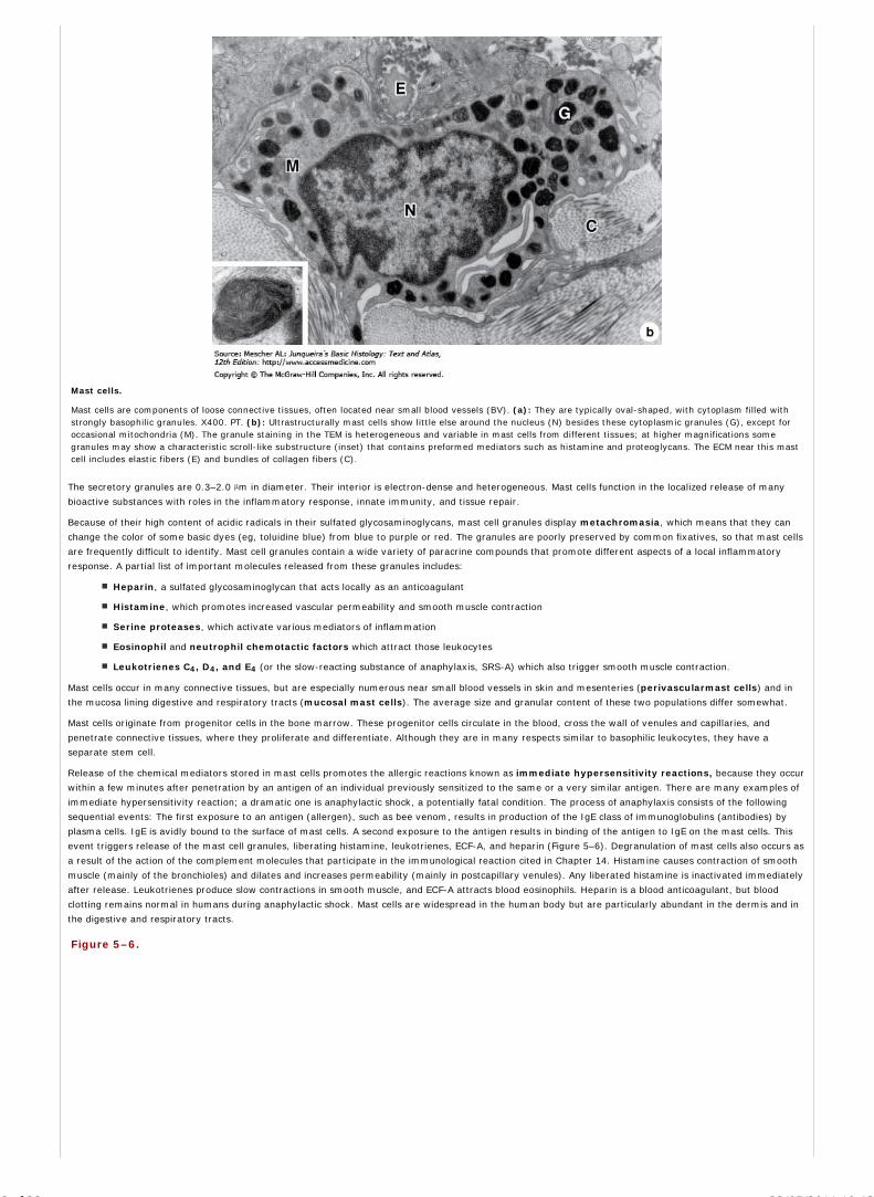
Mast cells.
Mast cells are components of loose connective tissues, often located near small blood vessels (BV). (a): They are typically oval-shaped, with cytoplasm filled withstrongly basophilic granules. X400. PT. (b): Ultrastructurally mast cells show little else around the nucleus (N) besides these cytoplasmic granules (G), except foroccasional mitochondria (M). The granule staining in the TEM is heterogeneous and variable in mast cells from different tissues; at higher magnifications somegranules may show a characteristic scroll-like substructure (inset) that contains preformed mediators such as histamine and proteoglycans. The ECM near this mastcell includes elastic fibers (E) and bundles of collagen fibers (C).
The secretory granules are 0.3–2.0 m in diameter. Their interior is electron-dense and heterogeneous. Mast cells function in the localized release of many
bioactive substances with roles in the inflammatory response, innate immunity, and tissue repair.
Because of their high content of acidic radicals in their sulfated glycosaminoglycans, mast cell granules display metachromasia, which means that they can
change the color of some basic dyes (eg, toluidine blue) from blue to purple or red. The granules are poorly preserved by common fixatives, so that mast cells
are frequently difficult to identify. Mast cell granules contain a wide variety of paracrine compounds that promote different aspects of a local inflammatory
response. A partial list of important molecules released from these granules includes:
Heparin, a sulfated glycosaminoglycan that acts locally as an anticoagulant
Histamine, which promotes increased vascular permeability and smooth muscle contraction
Serine proteases, which activate various mediators of inflammation
Eosinophil and neutrophil chemotactic factors which attract those leukocytes
Leukotrienes C4, D4, and E4 (or the slow-reacting substance of anaphylaxis, SRS-A) which also trigger smooth muscle contraction.
Mast cells occur in many connective tissues, but are especially numerous near small blood vessels in skin and mesenteries (perivascularmast cells) and in
the mucosa lining digestive and respiratory tracts (mucosal mast cells). The average size and granular content of these two populations differ somewhat.
Mast cells originate from progenitor cells in the bone marrow. These progenitor cells circulate in the blood, cross the wall of venules and capillaries, and
penetrate connective tissues, where they proliferate and differentiate. Although they are in many respects similar to basophilic leukocytes, they have a
separate stem cell.
Release of the chemical mediators stored in mast cells promotes the allergic reactions known as immediate hypersensitivity reactions, because they occur
within a few minutes after penetration by an antigen of an individual previously sensitized to the same or a very similar antigen. There are many examples of
immediate hypersensitivity reaction; a dramatic one is anaphylactic shock, a potentially fatal condition. The process of anaphylaxis consists of the following
sequential events: The first exposure to an antigen (allergen), such as bee venom, results in production of the IgE class of immunoglobulins (antibodies) by
plasma cells. IgE is avidly bound to the surface of mast cells. A second exposure to the antigen results in binding of the antigen to IgE on the mast cells. This
event triggers release of the mast cell granules, liberating histamine, leukotrienes, ECF-A, and heparin (Figure 5–6). Degranulation of mast cells also occurs as
a result of the action of the complement molecules that participate in the immunological reaction cited in Chapter 14. Histamine causes contraction of smooth
muscle (mainly of the bronchioles) and dilates and increases permeability (mainly in postcapillary venules). Any liberated histamine is inactivated immediately
after release. Leukotrienes produce slow contractions in smooth muscle, and ECF-A attracts blood eosinophils. Heparin is a blood anticoagulant, but blood
clotting remains normal in humans during anaphylactic shock. Mast cells are widespread in the human body but are particularly abundant in the dermis and in
the digestive and respiratory tracts.
Figure 5–6.
AccessMedicine | Print: Chapter 5. Connective Tissue http://accessmedicine.com/popup.aspx?aID=6180657&print=yes_chapter
6 of 28 22/05/2011 19:12

Mast cell secretion.
Mast cell secretion is triggered by re-exposure to certain antigens and allergens. Molecules of IgE antibody produced in an initial response to an allergen such aspollen or bee venom are bound to surface receptors for IgE (1), of which 300,000 are present per mast cell. When a second exposure to the allergen occurs, IgEmolecules bind this antigen and a few IgE receptors very rapidly become cross-linked (2). This activates adenylate cyclase, leading to phosphorylation of specific
proteins and (3) entry of Ca2+ and rapid exocytosis of some granules (4). In addition, phospholipases act on specific membrane phospholipids, leading toproduction and release of leukotrienes (5). The components released from granules, as well as the leukotrienes, are immediately active in the localmicroenvironment and promote a variety of controlled local reactions which together normally comprise part of the inflammatory process called the immediatehypersensitivity reaction. ECF-A, eosinophil chemotactic factor of anaphylaxis.
Plasma CellsPlasma cells are large, ovoid cells that have a basophilic cytoplasm due to their richness in rough ER. The juxtanuclear Golgi apparatus and the centrioles
occupy a region that appears pale in regular histologic preparations (Figure 5–7).
Figure 5–7.
Plasma cells.
Plasma cells are abundant in this portion of an inflamed intestinal villus. The plasma cells are characterized by their abundant basophilic cytoplasm involved in thesynthesis of antibodies. A large pale Golgi apparatus (arrows) near each nucleus is the site of the terminal glycosylation of the antibodies (glycoproteins). Plasmacells can leave their sites of origin in lymphoid tissues, move to connective tissue, and produce the antibodies that mediate immunity. X400 PT.
The nucleus of the plasma cell is generally spherical but eccentrically placed. Many of these nuclei contain compact, peripheral regions of heterochromatin
alternating with lighter areas of euchromatin, a configuration that can give the nucleus of a plasma cell the appearance of a clock-face. There are few plasma
cells in most connective tissues. Their average lifespan is short, 10–20 days.
AccessMedicine | Print: Chapter 5. Connective Tissue http://accessmedicine.com/popup.aspx?aID=6180657&print=yes_chapter
7 of 28 22/05/2011 19:12

MEDICAL APPLICATION
Plasma cells are derived from B lymphocytes and are responsible for the synthesis of antibodies. Antibodies are immunoglobulins produced in response
to penetration by antigens. Each antibody is specific for the one antigen that gave rise to its production and reacts specifically with molecules
possessing similar epitopes (see Chapter 14). The results of the antibody-antigen reaction are variable. The capacity of the reaction to neutralize
harmful effects caused by antigens is important. An antigen that is a toxin (eg, tetanus, diphtheria) may lose its capacity to do harm when it combines
with its respective antibody.
LeukocytesConnective tissue normally contains leukocytes that migrate from the blood vessels by diapedesis. Leukocytes (Gr. leukos, white, + kytos), or white blood
corpuscles, are the wandering cells of the connective tissue. They leave blood by migrating between the endothelial cells lining capillaries and postcapillary
venules to enter connective tissue by a process called diapedesis. This process increases greatly during inflammation, which is a vascular and cellular
defensive reaction against foreign substances, in most cases pathogenic bacteria or irritating chemical substances. The classic signs of inflammation were first
described by Celsus in the first century as redness and swelling with heat and pain (rubor et tumor cum calore et dolore).
Inflammation begins with the local release of chemical mediators of inflammation, substances of various origin (mainly from local cells and blood plasma
proteins) that induce some of the events characteristic of inflammation, eg, increase of blood flow and vascular permeability, chemotaxis, and
phagocytosis.
MEDICAL APPLICATION
Increased vascular permeability is caused by the action of vasoactive substances; an example is histamine, which is liberated from mast cells and
basophilic leukocytes. Increases in blood flow and vascular permeability are responsible for local swelling (edema), redness, and heat. Pain is due
mainly to the action of chemical mediators on nerve endings. Chemotaxis (Gr. chemeia, alchemy, + taxis, orderly arrangement), the phenomenon
by which specific cell types are attracted by some molecules, is responsible for the migration of large quantities of specific cell types to regions of
inflammation. As a consequence of chemotaxis, leukocytes cross the walls of venules and capillaries by diapedesis, invading the inflamed areas.
Leukocytes do not return to the blood after arriving in connective tissue except for the lymphocytes. These cells circulate continuously in various compartments
of the body: blood, lymph, lymphatic organs, and the interstitial fluid of connective tissue. Lymphocytes are particularly abundant in the connective tissue of
the digestive tract. A detailed analysis of the structure and functions of leukocytes and lymphocytes is presented in Chapters 12 and 14.
FIBERS
The connective tissue fibers are formed by proteins that polymerize into elongated structures. The three main types of connective tissue fibers are collagen,
reticular, and elastic fibers. Collagen and reticular fibers are both formed by the protein collagen, and elastic fibers are composed mainly of the protein
elastin. These fibers are distributed unequally among the types of connective tissue and the predominant fiber type is usually responsible for conferring
specific properties on the tissue.
CollagenThe collagens constitute a family of proteins selected during evolution for the execution of several (mainly structural) functions. During the process of evolution
of multicellular organisms, a family of structural proteins was selected by both environmental influences and the functional requirements of the animal
organism and developed to acquire varying degrees of rigidity, elasticity, and strength. These proteins are known collectively as collagen, and the chief
examples among its various types are present in the skin, bone, cartilage, smooth muscle, and basal lamina.
Collagen is the most abundant protein in the human body, representing 30% of its dry weight. The collagens are produced by several cell types and are
distinguishable by their molecular compositions, morphologic characteristics, distribution, functions, and pathologies. More than 20 types of collagen have been
identified and designated with Roman numerals; the most important of these are listed in Table 5–3. They are classified into the following four categories
according to their structure and general functions.
Table 5–3. Collagen types.
Type MoleculeComposition
Structure Optical Microscopy Representative Tissues Main Function
Collagens that form fibrils
I [ 1 (I)]2[ 2 (I)]
300-nm molecule, 67-nmbanded fibrils
Thick, highly picrosiriusbirefringent, nonargyrophilicfibers
Skin, tendon, bone, dentin Resistance to tension
II [ 1 (II)]3
300-nm molecule, 67-nmbanded fibrils
Loose aggregates of fibrils,birefringent
Cartilage, vitreous body Resistance to pressure
III [ 1 (III)]3
67-nm banded fibrils Thin, weakly birefringent,argyrophilic fibers
Skin, muscle, blood vessels,frequently together with type I
Structural maintenance inexpansible organs
V [ 1 (V)]3
390-nm molecule, N-terminalglobular domain
Frequently forms fiber togetherwith type I
Fetal tissues, skin, bone,placenta, most interstitialtissues
Participates in type I collagenfunction
XI [ 1 (XI)] [ 2(XI)] [ 3 (XI)]
300-nm molecule Small fibers Cartilage Participates in type II collagenfunction
Fibril-associated collagens
IX [ 1 (IX)] [ 2(IX)] [ 3 (IX)]
200-nm molecule Not visible, detected byimmunocytochemistry
Cartilage, vitreous body Bound glycosaminoglycans;associated with type II collagen
XII [ 1 (XII)]3
Large N-terminal domain;interacts with type I collagen
Not visible, detected byimmunocytochemistry
Embryonic tendon and skin Interacts with type I collagen
XIV [ 1 (XIV)]3
Large N-terminal domain;cross-shaped molecule
Not visible; detected byimmunocytochemistry
Fetal skin and tendon
Collagen that forms anchoring fibrils
VII [ 1 (VII)]3
450 nm, globular domain ateach end
Not visible, detected byimmunocytochemistry
Epithelia Anchors skin epidermal basallamina to underlying stroma
Collagen that forms networks
AccessMedicine | Print: Chapter 5. Connective Tissue http://accessmedicine.com/popup.aspx?aID=6180657&print=yes_chapter
8 of 28 22/05/2011 19:12

Type MoleculeComposition
Structure Optical Microscopy Representative Tissues Main Function
Collagens that form fibrils
IV [ 1 (VII)]2 [ 1(IV)]
Two-dimensional cross-linkednetwork
Not visible, detected byimmunocytochemistry
All basement membranes Support of delicate structures,filtration
COLLAGENS THAT FORM LONG FIBRILS
The molecules of long fibril–forming collagens aggregate to form fibrils clearly visible in the electron or light microscope (Figure 5–8). Collagen type I is the
most abundant and has a widespread distribution. It occurs in tissues as structures that are classically designated as collagen fibers forming structures such
as tendons, organ capsules, and dermis.
Figure 5–8.
Type I collagen.
Molecules of type I collagen, the most abundant type, assemble to form much larger structures. (a): TEM shows fibrils cut longitudinally and transversely. Inlongitudinal sections the fibrils display alternating dark and light bands that are further divided by cross-striations and in cross-section the cut ends of individualcollagen molecules can be seen. Ground substance completely surrounds the fibrils. X100,000. (b):In H&E stained tissues, type I collagen fibrils can often be seento aggregate further into large collagen bundles (C) of very eosinophilic fibers. Subunits for these fibers were secreted by fibroblasts (arrows) associated with them.X 400.
FIBRIL-ASSOCIATED COLLAGENS
Fibril-associated collagens are short structures that bind the surfaces of collagen fibrils to one another and to other components of the ECM. Molecules in this
AccessMedicine | Print: Chapter 5. Connective Tissue http://accessmedicine.com/popup.aspx?aID=6180657&print=yes_chapter
9 of 28 22/05/2011 19:12

category are also known as FACIT collagens, an acronym for "fibril-associated collagens with interrupted triple helices."
COLLAGENS THAT FORM ANCHORING FIBRILS
Anchoring collagen is type VII collagen, present in the anchoring fibrils that bind the basal lamina to reticular fibers in the underlying connective tissue (Figure
4–2).
COLLAGENS THAT FORM NETWORKS
An important network-forming collagen is type IV collagen, whose molecules assemble in a meshwork that constitutes a major structural component of the
basal lamina.
Collagen synthesis, an activity once thought restricted to fibroblasts, chondroblasts, osteoblasts, and odontoblasts, has now been shown to occur in many
cell types. The polypeptides initially formed on ribosomes of the rough ER are called procollagen chains, which intertwine in ER cisternae to make triple
helices. Every third amino acids in the chains is glycine; two other small amino acids abundant in collagen are hydroxylated post-translationally to form
hydroxyproline and hydroxylysine. Many different chains have been identified, encoded by related genes and varying in length and amino acid sequence.
The triple helix of chains forms a rod-like procollagen molecule, which in type I and II collagen measures 300 nm in length and 1.5 nm in width. Procollagen
molecules may be homotrimeric, with all three chains identical, or heterotrimeric, with two or all three chains having different sequences (Figure 5–9).
Different combinations of the many procollagen chains in procollagen molecules are largely responsible for the various types of collagen with different
structures and functional properties. In collagen types I, II, and III, collagen molecules aggregate and become packed together to form fibrils. Hydrogen
bonds and hydrophobic interactions are important in the aggregation and packing of these subunits. In a subsequent step, this structure is reinforced by the
formation of covalent cross-links, a process catalyzed by lysyl oxidases.
Figure 5–9.
Procollagen.
In the most abundant form of collagen, type I, each procollagen molecule is composed of two 1 and one 2 peptide chains, each with a molecular mass ofapproximately 100 kDa, intertwined in a right-handed helix and held together by hydrogen bonds and hydrophobic interactions. Each complete turn of the helixspans a distance of 8.6 nm. The length of each tropocollagen molecule is 300 nm, and its width is 1.5 nm.
Collagen fibrils are thin, elongated structures with diameters ranging from 20 to 90 nm and can be several micrometers in length; they have transverse
striations with a characteristic periodicity of 64-68 nm (Figure 5–10). The striations are caused by the regular, overlapping arrangement of the collagen
molecule subunits (Figure 5–10). The dark (electron-dense) bands retain more of the lead-based stain used in TEM studies because their more numerous free
chemical groups react more intensely with the lead solution than do the light bands. In some collagen types these fibrils associate further with FACIT collagens
to form fibers. In collagen type I, the fibers can form large bundles (Figure 5–10). Collagen type II (present in cartilage) occurs as fibrils but does not form
fibers or bundles. Collagen type IV, present in all basement membranes, assembles as a lattice-like network in the basal lamina.
Figure 5–10.
Assembly of collagen molecules into collagen fibers.
This diagram shows an aggregate of collagen molecules, fibrils, fibers, and bundles. There is a stepwise overlapping arrangement of rodlike collagen molecules, eachmeasuring 300 nm (1). This arrangement results in the production of alternating spaces and overlapping regions (2), which cause the cross-striations characteristicof collagen fibrils and confer a 67-nm periodicity of dark and light bands when the fibril is observed in the electron microscope (3). Fibrils aggregate and arecovalently cross-linked to form fibers (4), which in collagen type I aggregate further to form bundles (5) routinely called collagen fibers when seen by lightmicroscopy.
AccessMedicine | Print: Chapter 5. Connective Tissue http://accessmedicine.com/popup.aspx?aID=6180657&print=yes_chapter
10 of 28 22/05/2011 19:12

Because collagen type I is so abundant, its synthesis has been studied most thoroughly. Synthesis of this important protein involves several steps, which are
summarized in Figure 5–11:
Polypeptides called procollagen chains are produced on polyribosomes bound to membranes of RER and translocated into the cisternae and the signalpeptide is clipped off.
1.
Hydroxylation of proline and lysine begins after the peptide chain has reached a certain minimum length and is still bound to the ribosomes. The
enzymes involved are prolyl hydroxylases and lysyl hydroxylase and the reactions require O2, Fe2+, and ascorbic acid (vitamin C) as co-factors.
2.
Glycosylation of some hydroxylysine residues occurs, with the various collagen types having different amounts of galactose linked to hydroxylysines.3.
Both the amino- and carboxyl-terminal ends of each chain make up nonhelical portions of the polypeptides, sometimes called the extensionpropeptides, which may help ensure that the appropriate chains ( 1, 2) assemble in the correct position as a triple helix. In addition, the nonhelicalpropeptides make the resulting procollagen molecule soluble and prevent its premature intracellular assembly and precipitation as collagen fibrils.Procollagen is transported through the Golgi network and undergoes exocytosis to the extracellular environment.
4.
Outside the cell, specific proteases called procollagen peptidases remove the extending propeptides, converting the procollagen molecules to collagenmolecules. These are now capable of self-assembly into polymeric collagen fibrils, usually in specialized niches near the cell surface.
5.
In some collagen types, fibrils aggregate to form fibers. Certain proteoglycans and types of collagen (types V and XI) participate in the aggregation ofcollagen molecules to form fibrils and in the formation of fibers from fibrils. FACIT collagens help stabilize the molecules in collagen fibrils and fibers andbind these structures to other components of the ECM.
6.
Fibrillar structure is reinforced further by the formation of covalent cross-links between assembled collagen molecules, a process catalyzed by theextracellular enzyme lysyl oxidase.
7.
Figure 5–11.
Collagen synthesis.
Hydroxylation and glycosylation of procollagen chains and their assembly into triple helices occurs in the RER and further assembly into fibrils occurs in the ECMafter secretion of procollagen. Because there are many slightly different genes for procollagen chains and collagen production depends on severalpost-translational events involving several other enzymes, many diseases involving defective collagen synthesis have been described.
The other fibrillar collagens are formed in processes similar to that described for collagen type I. In summary, collagen synthesis involves a cascade of unique
AccessMedicine | Print: Chapter 5. Connective Tissue http://accessmedicine.com/popup.aspx?aID=6180657&print=yes_chapter
11 of 28 22/05/2011 19:12

post-translational modifications of the original procollagen polypeptides. All these modifications are critical to the structure and function of normal mature
collagen. Because there are so many steps in collagen biosynthesis, there are many points at which the process can be interrupted or changed by defective
enzymes or by disease processes.
Although fresh collagen fibers are colorless strands, when present in large numbers (eg, in tendons) they appear white. The highly regular orientation of
subunits in collagen fibers makes them birefringent under the polarizing microscope (Figure 1–7). In the light microscope, collagen fibers are acidophilic; they
stain pink with eosin, blue with Mallory trichrome stain, green with Masson trichrome stain, and red with Sirius red. Because of the long and tortuous course of
collagen bundles, their length and diameter are better studied in spread preparations than in histologic sections (Figure 1–7). Mesentery is frequently used for
this purpose; when spread on a slide, this structure is sufficiently thin to let the light pass through; it can be stained and examined directly under the
microscope. Mesentery consists of a central portion of connective tissue lined on both surfaces by a simple squamous epithelium, the mesothelium. The
collagen fibers in a spread preparation appear as elongated and tortuous cylindrical structures of indefinite length, with a diameter that varies from 1 to 20 m.
MEDICAL APPLICATION
Collagen synthesis depends on the expression of several genes and several posttranslational events. It should not be surprising, therefore, that a large
number of pathological conditions are directly attributable to insufficient or abnormal collagen synthesis.
Certain mutations in the 1 (I) or 2 (I) genes lead to osteogenesis imperfecta. Many cases of osteogenesis imperfecta are due to deletions of all or
part of the 1 (I) gene. However, a single amino acid change is sufficient to cause certain forms of this disease, particularly mutations involving
glycine. Glycine must be at every third position for the collagen triple helix to form.
In addition to these disorders, several diseases result from an over-accumulation of collagen. In progressive systemic sclerosis, almost all organs
may present an excessive accumulation of collagen (fibrosis). This occurs mainly in the skin, digestive tract, muscles, and kidneys, causing hardening
and functional impairment of the implicated organs.
Keloid is a local swelling caused by abnormal amounts of collagen that form in scars of the skin. Keloids, which occur most often in individuals of black
African descent, can be a troublesome clinical problem to manage; not only can they be disfiguring, but excision is almost always followed by
recurrence.
Vitamin C (ascorbic acid) deficiency leads to scurvy, a disease characterized by the degeneration of connective tissue. Without this vitamin, fibroblasts
synthesize defective collagen, and the defective fibers are not replaced. This process leads to a general degeneration of connective tissue that becomes
more pronounced in areas in which collagen renewal takes place at a faster rate. The periodontal ligament that holds teeth in their sockets has a
relatively high collagen turnover; consequently, this ligament is markedly affected by scurvy, which leads to a loss of teeth. Ascorbic acid is a cofactor
for proline hydroxylase, which is essential for the normal synthesis of collagen. Table 5–4 lists a few examples of the many disorders caused by failure
of collagen biosynthesis.
Table 5–4. Examples of clinical disorders resulting from defects in collagen synthesis.
Disorder Defect Symptoms
Ehlers-Danlos type IV Faulty transcription or translation of collagen type III Aortic and/or intestinal rupture
Ehlers-Danlos type VI Faulty lysine hydroxylation Augmented skin elasticity, rupture of eyeball
Ehlers-Danlos type VII Decrease in procollagen peptidase activity Increased articular mobility, frequent luxation
Scurvy Lack of vitamin C (cofactor for prolyl hydroxylase) Ulceration of gums, hemorrhages
Osteogenesis imperfecta Change of one nucleotide in genes for collagen type I Spontaneous fractures, cardiac insufficiency
Collagen turnover and renewal in normal connective tissue is generally a very slow process. In some organs, such as tendons and ligaments, the collagen is
very stable, whereas in others, as in the periodontal ligament surrounding teeth, the collagen turnover rate is very high. To be renewed, the collagen must
first be degraded. Degradation is initiated by specific enzymes called collagenases, which are members of an enzyme class called matrix metalloproteinases
or MMPs. Collagenases clip collagen molecules in such a way that they are then susceptible to further degradation by nonspecific proteases.
Reticular FibersOnce thought to be uniquely distinct from collagen, reticular fibers are now known to consist mainly of collagen type III, which forms extensive networks of
extremely thin (diameters 0.5–2 m) and heavily glycosylated fibers in certain organs. They are not visible in hematoxylin-and-eosin (H&E) preparations but
can be easily stained black by impregnation with silver salts (Figure 5–12). Because of their affinity for silver salts, these fibers are called argyrophilic (Gr.
argyros, silver). Reticular fibers are also periodic acid–Schiff (PAS)-positive, which like argyrophilia is due to the high content of sugar chains associated with
these fibers. Reticular fibers contain 6–12% hexoses as opposed to 1% in most collagen fibers.
Figure 5–12.
AccessMedicine | Print: Chapter 5. Connective Tissue http://accessmedicine.com/popup.aspx?aID=6180657&print=yes_chapter
12 of 28 22/05/2011 19:12
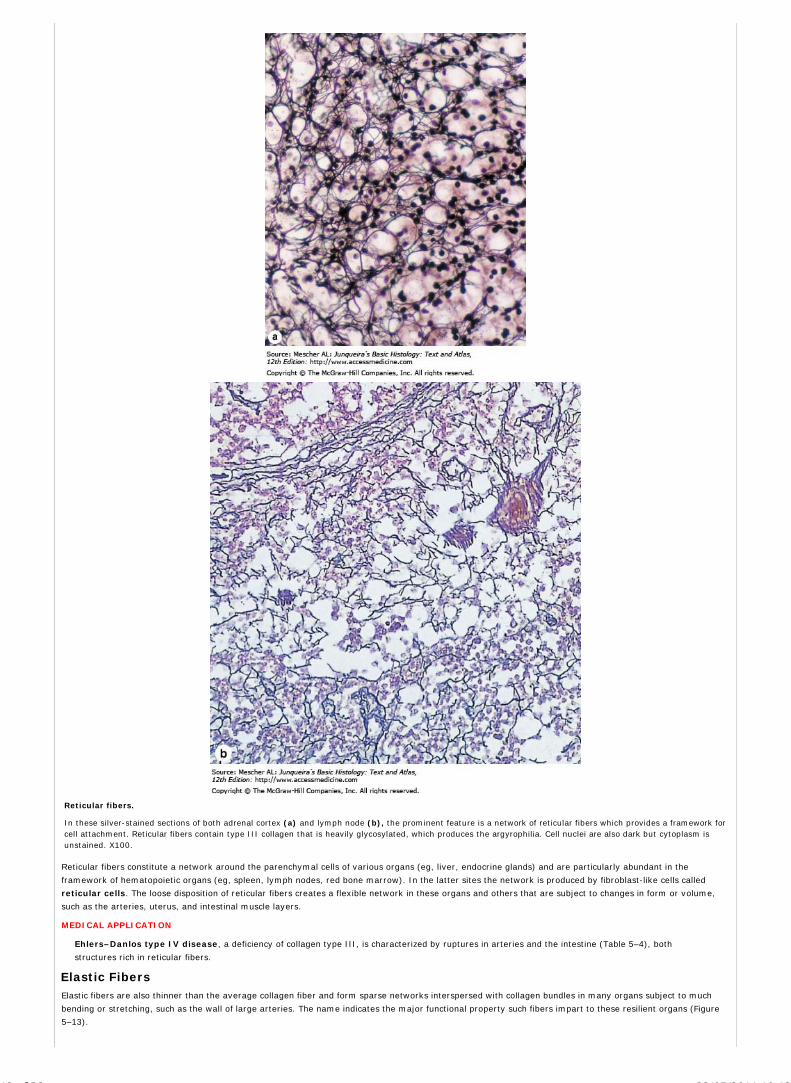
Reticular fibers.
In these silver-stained sections of both adrenal cortex (a) and lymph node (b), the prominent feature is a network of reticular fibers which provides a framework forcell attachment. Reticular fibers contain type III collagen that is heavily glycosylated, which produces the argyrophilia. Cell nuclei are also dark but cytoplasm isunstained. X100.
Reticular fibers constitute a network around the parenchymal cells of various organs (eg, liver, endocrine glands) and are particularly abundant in the
framework of hematopoietic organs (eg, spleen, lymph nodes, red bone marrow). In the latter sites the network is produced by fibroblast-like cells called
reticular cells. The loose disposition of reticular fibers creates a flexible network in these organs and others that are subject to changes in form or volume,
such as the arteries, uterus, and intestinal muscle layers.
MEDICAL APPLICATION
Ehlers–Danlos type IV disease, a deficiency of collagen type III, is characterized by ruptures in arteries and the intestine (Table 5–4), both
structures rich in reticular fibers.
Elastic FibersElastic fibers are also thinner than the average collagen fiber and form sparse networks interspersed with collagen bundles in many organs subject to much
bending or stretching, such as the wall of large arteries. The name indicates the major functional property such fibers impart to these resilient organs (Figure
5–13).
AccessMedicine | Print: Chapter 5. Connective Tissue http://accessmedicine.com/popup.aspx?aID=6180657&print=yes_chapter
13 of 28 22/05/2011 19:12

Figure 5–13.
AccessMedicine | Print: Chapter 5. Connective Tissue http://accessmedicine.com/popup.aspx?aID=6180657&print=yes_chapter
14 of 28 22/05/2011 19:12

Elastic fibers.
Elastic fibers or lamellae (sheets) add the resiliency to connective tissue. They are difficult to discern in H&E stained material and are usually demonstrated inpreparations made using compounds such as aldehyde fuscin which stains elastin a dark magenta. (a): The length and density of fine elastic fibers is best seen inspread preparation of connective tissue in a thin mesentery. X200. Orcein-H&E. (b): At higher magnification, sectioned elastic fibers can be seen among theeosinophilic collagen bundles in dermis. X400. Aldehyde fuscin & eosin. (c): Elastic fibers and lamellae are abundant between layers of smooth muscle in the wall ofelastic arteries such as the aorta. X200. Van Gieson-H&E.
Elastic fibers develop through successive stages. In the first stage, a core of 10-nm microfibrils forms from several different glycoproteins, notably the large
glycoprotein called fibrillin (350kDa). Fibrillin binds elastin and forms the scaffolding necessary for the deposition of elastin. Defective fibrillin results in the
formation of fragmented elastic fibrils. In the second stage of development, the protein elastin is deposited between the microfibrils, forming larger fibers.
During the third stage, elastin gradually accumulates until it comprises most of the fiber bundles, which are further surrounded by a thin sheath of microfibrils.
These are the mature elastic fibers, the most numerous component of the elastic fiber system. Stages of elastic fiber formation are shown in Figure 5–14. In
the wall of large blood vessels, especially arteries, elastin also occurs as fenestrated sheets called elastic lamellae.
Figure 5–14
AccessMedicine | Print: Chapter 5. Connective Tissue http://accessmedicine.com/popup.aspx?aID=6180657&print=yes_chapter
15 of 28 22/05/2011 19:12

Formation of elastic fibers.
Stages in the formation of elastic fibers can be seen by TEM. (a): Initially a developing fiber consists of many small microfibrils composed of the glycoproteinfibrillin secreted by fibroblasts, smooth muscle cells or other cells. (b): With further development, to the microfibrils are added amorphous deposits of elastin.Elastin is secreted by the cells and like procollagen molecules quickly polymerizes. (c): Elastin accumulates and ultimately occupies the center of an elastic fiber,which retains fibrillin microfibrils at the surface. Collagen fibrils, seen in cross section, are also present. X50,000.
Microfibrils of fibrillin alone are used in some organs, such as to hold in place the lens of the eye. Such microfibrils are not elastic but are highly resistant to
pulling forces, whereas the mature elastic fibers stretch easily in response to tension. By using different proportions of fibrillin and elastin, a family of fibers can
be formed whose variable functional characteristics are adapted to local tissue requirements.
Like collagen elastin matures in the ECM. Elastin molecules are globular (molecular mass 70 kDa) and are secreted by fibro-blasts in connective tissue and by
smooth muscle cells in the walls of blood vessels. Elastin molecules are rich in glycine and proline, with many regions having random-coil conformations (like
that of natural rubber). Elastin molecules polymerize to form fibers or sheet-like structures, both of which can be stretched by external forces. Elastin contains
AccessMedicine | Print: Chapter 5. Connective Tissue http://accessmedicine.com/popup.aspx?aID=6180657&print=yes_chapter
16 of 28 22/05/2011 19:12

two unusual amino acids, desmosine and isodesmosine, which are produced when covalent cross-links are formed among four lysine residues in different
elastin molecules (Figure 5–15). These effectively cross-link the subunits of elastin and help account for the rubberlike qualities of this protein. Elastin is
resistant to digestion by most proteases, but is easily hydrolyzed by pancreatic elastase.
Figure 5–15.
Molecular basis of elasticity.
Subunits of the glycoprotein elastin are joined by covalent bonds formed among lysine residues of different subunits, catalyzed by lysyl oxidase. This produces anextensive and durable cross-linked network of elastin. (Such bonds give rise to the unusual amino acids desmosine and isodesmosine.) Each elastin molecule in thenetwork has multiple random-coil domains which expand and contract; this allows the entire network to stretch and recoil like a rubber band.
MEDICAL APPLICATION
Fibrillin is a family of proteins related to the scaffolding necessary for the deposition of elastin. Mutations in the fibrillin gene result in Marfan
syndrome, a disease characterized by a lack of resistance in the tissues rich in elastic fibers. Because the large arteries are rich in components of the
elastic system and because the blood pressure is high in the aorta, patients with this disease often experience aortic swellings called aneurysms, a
life-threatening condition.
GROUND SUBSTANCE
The ground substance of the ECM is a highly hydrated, transparent, complex mixture of macromolecules, principally in three classes: glycosaminoglycans
(or GAGs), proteoglycans, and multiadhesive glycoproteins. The complex molecular mixture of the ground substance is transparent and rich in bound
water. It fills the space between cells and fibers of connective tissue and, because it is viscous, acts as both a lubricant and a barrier to the penetration of
invaders. When adequately fixed for histologic analysis, its components aggregate and precipitate in the tissues as granular material that is observed in TEM
preparations as electron-dense filaments or granules (Figure 5–16).
Figure 5–16.
AccessMedicine | Print: Chapter 5. Connective Tissue http://accessmedicine.com/popup.aspx?aID=6180657&print=yes_chapter
17 of 28 22/05/2011 19:12

Ultrastructure of the extracellular matrix (ECM).
TEM of the connective tissue extracellular matrix reveals ground substance as either empty or containing fine granular material that fills spaces between thecollagen (C) and elastic (E) fibers and surrounds fibroblast cells and processes (F). The granularity of ground substance is an artifact of the glutaraldehyde–tannicacid fixation procedure. X100,000.
GAGs (originally called mucopolysaccharides) are linear polysaccharides formed by repeating disaccharide units usually composed of a uronic acid and a
hexosamine. The hexosamine can be glucosamine or galactosamine, and the uronic acid can be glucuronic or iduronic acid. The largest, most unique,
and most ubiquitous GAG is hyaluronic acid (or hyaluronan). With a molecular weight from 100s to 1000s kDa, hyaluronic acid is a long polymer of the
disaccharide glucosamine – glucuronate. It is synthesized directly into the ECM by an enzyme complex, hyaluronate synthase, located in the cell membrane
of many cells. Hyaluronic acid forms a dense, viscous network of polymers which binds a considerable amount of water, giving it an important role in allowing
diffusion of molecules in connective tissue and in lubricating various organs and joints.
All other GAGs are much smaller (10–40 kDa), are covalently attached to proteins (as parts of proteoglycans), are synthesized in Golgi complexes, and are rich
in sulfate. The four main GAGs found in proteoglycans are dermatan sulfate, chondroitin sulfates, keratan sulfate, and heparan sulfate all of which
have different disaccharide units and tissue distributions (Table 5–5). Like hyaluronic acid these GAGs are intensely hydrophilic, are highly viscous, and are
polyanions, binding a great number of cations (usually sodium) by electrostatic (ionic) bonds.
Table 5–5. Composition and distribution of glycosaminoglycans in connective tissue and their interactions with collagenfibers.
Repeating Disaccharides
Glycosaminoglycan Hexuronic Acid Hexosamine Distribution Electrostatic Interaction with Collagen
Hyaluronic acid D-glucuronic acid D-glucosamine Umbilical cord, synovial fluid, vitreoushumor, cartilage
Chondroitin 4-sulfate D-glucuronic acid D-galactosamine Cartilage, bone, cornea, skin,notochord, aorta
High levels of interaction, mainly with collagentype II
Chondroitin 6-sulfate D-glucuronic acid D-galactosamine Cartilage, umbilical cord, skin, aorta(media)
High levels of interaction, mainly with collagentype II
Dermatan sulfate L-iduronic acid orD-glucuronic acid
D-galactosamine Skin, tendon, aorta (adventitia) Low levels of interaction, mainly with collagentype I
Heparan sulfate D-glucuronic acid orL-iduronic acid
D-galactosamine Aorta, lung, liver, basal laminae Intermediate levels of interaction, mainly withcollagen types III and IV
Keratan sulfate(cornea)
D-galactose D-galactosamine Cornea None
Keratan sulfate(skeleton)
D-galactose D-glucosamine Cartilage, nucleus pulposus, annulusfibrosus
None
Proteoglycans are composed of a core protein to which are covalently attached various numbers and combinations of the sulfated GAGs just mentioned. Like
glycoproteins they are synthesized on RER, mature in the Golgi and secreted from cells by exocytosis. The main structural differences between proteoglycans
and glycoproteins are shown in Figure 5–17. In cartilage, the core proteins of secreted proteoglycans are bound via small link proteins to a hyaluronic acid
chain, forming much larger structures—proteoglycan aggregates. The acidic groups of proteoglycans cause these molecules to bind to the basic amino acid
residues of collagen. Proteoglycans are distinguished for their diversity and include cell-surface and ECM families. A given matrix may contain several different
types of core proteins, each with different numbers of GAGs of different lengths and composition. One of the most important ECM proteoglycans is aggrecan,
the dominant proteoglycan in cartilage. In aggrecan the core protein bears several chondroitin sulfate and keratan sulfate chains and is in turn bound via a link
protein to hyaluronic acid. Cell-surface proteoglycans such as syndecan are present on many types of cells, particularly epithelial cells. The core protein of
cell-surface proteoglycans spans the plasma membrane, with a short cytoplasmic extension. A small number of heparan sulfate chains are attached to the
extracellular extension of the core protein.
Figure 5–17.
AccessMedicine | Print: Chapter 5. Connective Tissue http://accessmedicine.com/popup.aspx?aID=6180657&print=yes_chapter
18 of 28 22/05/2011 19:12

Proteoglycans and glycoproteins.
The diagram indicates the major structural features of proteoglycans and glycoproteins. (a): Proteoglycans contain a core of protein (vertical rod in drawing) towhich molecules of sulfated glycosaminoglycans (GAGs) are covalently bound. A GAG is an unbranched polysaccharide made up of repeating disaccharides; onecomponent is an amino sugar, and the other is uronic acid. Proteoglycans contain a greater amount of carbohydrate than do glycoproteins. In general the three-dimensional structure of proteoglycans can be pictured as a test tube brush, with the wire stem representing the core protein and the bristles representing thesulfated GAGs. (b): Glycoproteins are globular protein molecules to which branched chains of monosaccharides are covalently attached. Their polypeptide content isgreater than their polysaccharide content. (Reproduced, with permission, from Junqueira LCU, Carneiro J: Biologia Celular e Molecular, 8th ed. Editora GuanabaraKoogan. Rio de Janeiro, 2000.)
Besides acting as structural components of the ECM and anchoring cells to the matrix, both extracellular and surface proteoglycans also bind and sequester
certain signaling proteins eg, fibroblast growth factor (FGF). Degradation of proteoglycans releases these stored growth factors which then stimulate new cell
growth and ECM synthesis.
MEDICAL APPLICATION
The degradation of proteoglycans is carried out by several cell types and depends on the presence of several lysosomal enzymes. Several disorders
have been described in which a deficiency in lysosomal enzymes causes glycosaminoglycan degradation to be blocked, with the consequent
accumulation of these compounds in tissues. The lack of specific hydrolases in the lysosomes has been found to be the cause of several disorders in
humans, including Hurler, Hunter, sanfilippo, and Morquio syndromes.
Because of their high viscosity, intercellular substances act as a barrier to the penetration of bacteria and other microorganisms. Bacteria that produce
hyaluronidase, an enzyme that hydrolyzes hyaluronic acid and other glycosaminoglycans, have greater invasive power because they reduce the
viscosity of the connective tissue ground substance.
Multiadhesive glycoproteins have carbohydrates attached, but in contrast to proteoglycans the protein moiety usually predominates. The carbohydrate
moiety of glycoproteins is frequently a branched structure. Several such glycoproteins have important roles in the adhesion of cells to their substrate.
Fibronectin (L. fibra, fiber, + nexus, interconnection) is an important example synthesized by fibroblasts and some epithelial cells. This dimeric molecule,
with a molecular mass of 222–240 kDa, has binding sites for collagens, certain GAGs, and integrins of cell membranes, ie, it is multiadhesive. Interactions at
these sites help to mediate normal cell adhesion and migration and cause fibronection to be distributed as a network in the intercellular spaces of many tissues
(Figure 5–18a). Another multiadhesive glycoprotein, laminin is a larger, trimeric, cross-shaped glycoprotein that participates in the adhesion of epithelial cells
to the basal lamina, with binding sites for type IV collagen, GAGs, and integrins. All basal laminae are rich in laminin (Figure 5–18b).
Figure 5–18.
AccessMedicine | Print: Chapter 5. Connective Tissue http://accessmedicine.com/popup.aspx?aID=6180657&print=yes_chapter
19 of 28 22/05/2011 19:12

Fibronectin and laminin localization.
Immunohistochemistry of sections with connective tissue shows that fibronectin (a) is ubiquitous throughout the ECM, while laminin (b) is restricted to the basallamina of the epithelium (top of the picture) and of cross-sectioned muscle fibers, nerves, and small blood vessels (lower half of picture). Both glycoproteins (andmany other similar glycoproteins) are multiadhesive, with binding sites for collagens and other ECM components and for integrins of cell surfaces. They playimportant roles in cell migration, in embryonic tissue formation, and in maintaining tissue structure. (a): X400, (b): X200, both hematoxylin counterstain.(Reproduced, with permission, from Junqueira LCU, Carneiro J: Biologia Celular e Molecular, 8th ed. Editora Guanabara Koogan. Rio de Janeiro, 2000.)
Cells interact with extracellular matrix components by using cell-surface molecules (matrix receptors) that bind to collagen, fibronectin, and laminin. These
receptors are the integrins, a family of transmembrane linker proteins (Figures 5–19). Integrins bind their ligands in the ECM with relatively low affinity,
allowing cells to explore their environment without losing attachment to it or becoming glued to it. Integrins also interact with the cytoskeleton, usually the
actin microfilaments, an interaction mediated by several intracellular proteins, such as talin and vinculin. The interactions that integrins mediate between the
ECM and the cytoskeleton exert effects in both directions and play an important role in orienting both the cells and the ECM in tissues (Figure 5–19).
Figure 5–19.
AccessMedicine | Print: Chapter 5. Connective Tissue http://accessmedicine.com/popup.aspx?aID=6180657&print=yes_chapter
20 of 28 22/05/2011 19:12

Integrin cell-surface matrix receptor.
By binding to a matrix protein and to the actin cytoskeleton (via talin) inside the cell, integrins serves as transmembrane links by which cells adhere to componentsof the ECM. The molecule is a heterodimer, with and chains. The head portion may protrude some 20 nm from the surface of the cell membrane into the ECMwhere it interacts with fibronectin, laminin, or collagens.
MEDICAL APPLICATION
The participation of fibronectin and laminin in both embryonic development and the increased ability of cancer cells to invade other tissues has been
well-studied. The importance of fibronectin is shown by the fact that mice whose fibronectin gene has been inactivated die during early embryogenesis.
In connective tissue, in addition to the hydrated ground substance, there is a small quantity of free fluid—called interstitial or tissue fluid—that is similar to
blood plasma in its content of ions and diffusible substances. Tissue fluid contains a small percentage of plasma proteins of low molecular weight that pass
through the capillary walls as a result of the hydrostatic pressure of the blood. Although only a small proportion of connective tissue proteins are plasma
proteins, it is estimated that because of its volume and wide distribution, as much as one third of the plasma proteins of the body are stored in the intercellular
connective tissue matrix.
MEDICAL APPLICATION
Edema is promoted by the accumulation of water in the extracellular spaces. Water in the extracellular compartment of connective tissue comes from
the blood, passing through the capillary walls into the extracellular compartment of the tissue. The capillary wall is only slightly permeable to
macromolecules but permits the passage of water and small molecules, including low-molecular-weight proteins. In several pathologic conditions, the
quantity of tissue fluid may increase considerably, causing edema.
Edema may result from venous or lymphatic obstruction or from a decrease in venous blood flow (eg, congestive heart failure). It may also be caused
by the obstruction of lymphatic vessels due to parasitic plugs or tumor cells and chronic starvation; protein deficiency results in a lack of plasma
proteins and a decrease in colloid osmotic pressure. Water therefore accumulates in the connective tissue and is not drawn back into the capillaries.
Another possible cause of edema is increased permeability of the blood capillary endothelium resulting from chemical or mechanical injury or the
release of certain substances produced in the body (eg, histamine).
Blood vessels bring to connective tissue the various nutrients required by its cells and carry away metabolic waste products to the detoxifying and excretory
organs, the liver and kidneys.
Two forces act on the water contained in the capillaries: the hydrostatic pressure of the blood caused by the pumping action of the heart, which forces water
out across the capillary wall; and the colloid osmotic pressure of the blood plasma, which draws water back into the capillaries (Figure 5–20). Osmotic
pressure is due mainly to plasma proteins. (Because the ions and low-molecular-weight compounds that pass easily through the capillary walls have
approximately the same concentration inside and outside these blood vessels, the osmotic pressures they exert are approximately equal on either side of the
capillaries and cancel each other.) The colloid osmotic pressure exerted by the blood proteins—which are unable to pass through the capillary walls—is not
counterbalanced by outside pressure and tends to bring water back into the blood vessel (Figure 5–20).
Figure 5–20.
AccessMedicine | Print: Chapter 5. Connective Tissue http://accessmedicine.com/popup.aspx?aID=6180657&print=yes_chapter
21 of 28 22/05/2011 19:12

Movement of fluid in connective tissue.
Normally, water passes through capillary walls to the ECM of the surrounding connective tissues primarily at the arterial end of a capillary, because the hydrostaticpressure there is greater than the colloid osmotic pressure. As indicated at the top of the figure, however, hydrostatic pressure decreases along the length of thecapillary toward the venous end. As the hydrostatic pressure falls, osmotic pressure of the fluid in the capillary rises because the protein concentration is increasingdue to the passage of water from the capillaries. As a result of the increased protein concentration and decreased hydrostatic pressure, osmotic pressure becomesgreater than hydrostatic pressure at the venous end and water is drawn back into the capillary. In this way, metabolites circulate in the connective tissue, feedingits cells. Not all water that leaves capillaries by hydrostatic pressure is drawn back in by osmotic pressure. This excess tissue fluid is normally drained by thelymphatic capillaries, blind-ended vessels that arise in connective tissue and enter the one-way lymphatic system which eventually delivers the fluid (now calledlymph) back to veins.
The quantity of water drawn back is less than that which passes out through the capillaries. Rather than accumulating in connective tissue, this excess fluid is
continuously drained by lymphatic capillaries and eventually returned to the blood. The smallest lymphatic vessels, the lymphatic capillaries originate in
connective tissue as delicate, blind-ending, endothelium-lined tubes that join increasingly larger lymphatic vessels that drain into veins at the base of the neck
(see Chapter 11).
TYPES OF CONNECTIVE TISSUE
Connective tissues composed of the cells, fibers, and ground substance components already described nevertheless are quite variable in histological structure.
This has led to the use of descriptive names or classifications for various connective tissue types, denoting either the major component or a structural
characteristic of the tissue. Table 5-6 shows one classification commonly used for the main types of connective tissue.
Table 5–6. Types of connective tissue.
Connective Tissue Proper
AccessMedicine | Print: Chapter 5. Connective Tissue http://accessmedicine.com/popup.aspx?aID=6180657&print=yes_chapter
22 of 28 22/05/2011 19:12

There are two general classes of connective tissue proper: loose and dense (Figure 5–21).
Figure 5–21.
AccessMedicine | Print: Chapter 5. Connective Tissue http://accessmedicine.com/popup.aspx?aID=6180657&print=yes_chapter
23 of 28 22/05/2011 19:12

Loose connective tissue and dense irregular connective tissue.
Shown here are three examples of connective tissue which display the types designated loose (or areolar) and dense irregular. Loose connective tissue (L) containsfaint ground substance with fine fibers of collagen and is adjacent to epithelium in the examples shown here. Dense irregular connective tissue (D) underlies thethinner layer of loose connective tissue and is invariably much richer in larger bundles of collagen. (a): Micrograph of a mammary gland, showing a duct at the topof the figure. In the dense irregular connective tissue can be seen scattered leukocytes, and the irregular spaces of two lymphatic vessels (left). X100. H&E. (b):Trichrome staining of the skin demonstrates the blue staining of collagen with this method. X100. Mallory Trichrome. (c): Loose and dense irregular connective
AccessMedicine | Print: Chapter 5. Connective Tissue http://accessmedicine.com/popup.aspx?aID=6180657&print=yes_chapter
24 of 28 22/05/2011 19:12

tissue within the esophagus is seen below the stratified squamous epithelium. X100. H&E. (d): At higher magnification ground substance (GS) is more clearly seenaround small blood vessels (V) and collagen bundles (C). X200. H&E. (e): The dense irregular connective tissue (D) capsule that surrounds the testis is shown here.Similar capsules are found around many organs and large glands. That of the testis is covered by serous mesothelial cells (S), which produce a hyaluronate-richlubricant around the organ. X200. H&E.
Loose connective tissue is a very common type of connective tissue that supports many structures which are normally under some pressure and low friction.
It usually supports epithelial tissue, forms a layer around small blood and lymphatic vessels, and fills the spaces between muscle and nerve fibers. Loose
connective tissue is also found in the papillary layer of the dermis, in the hypodermis, in the linings of the peritoneal and pleural cavities, in glands, and in the
mucous membranes (wet membranes that line the hollow organs) supporting the epithelial cells.
Loose connective tissue, sometimes called areolar tissue, has all the main components of connective tissue (cells, fibers, and ground substance) in roughly
equal parts. The most numerous cells are fibroblasts and macrophages, but other types of connective tissue cells are also present. Collagen, elastic, and
reticular fibers also appear in this tissue. With a moderate amount of ground substance, loose connective tissue has a delicate consistency; it is flexible, well
vascularized, and not very resistant to stress.
Dense connective tissue is adapted to offer resistance and protection. It has the same components found in loose connective tissue, but there are fewer cells
and a clear predominance of collagen fibers over ground substance. Dense connective tissue is less flexible and far more resistant to stress than is loose
connective tissue. It is known as dense irregular connective tissue when the collagen fibers are arranged in bundles without a definite orientation. The
collagen fibers form a 3-dimensional network in dense irregular tissue, providing resistance to stress from all directions. Dense irregular connective tissue is
often found closely associated with loose connective tissue. The two types frequently grade into one another and distinctions between them are often arbitrary
(Figure 5–21).
The collagen bundles of dense regular connective tissue are arranged according to a definite pattern, with collagen fibers aligned with the linear orientation of
fibroblasts in response to prolonged stresses exerted in the same direction (Figure 5–22). This arrangement offers great resistance to traction forces.
Figure 5–22.
AccessMedicine | Print: Chapter 5. Connective Tissue http://accessmedicine.com/popup.aspx?aID=6180657&print=yes_chapter
25 of 28 22/05/2011 19:12

Dense regular connective tissue.
(a): Micrograph shows a longitudinal section of dense regular connective tissue of a tendon. Long, parallel bundles of collagen fibers fill the spaces between theelongated nuclei of fibrocytes. X100. H&E stain. (b): The electron micrograph shows one fibrocyte in a cross-section of tendon, revealing that the sparse cytoplasmof the fibrocytes is divided into numerous thin cytoplasmic processes extending among adjacent collagen fibers. X25,000.
Tendons and ligaments are the most common examples of dense regular connective tissue. These elongated cylindrical structures hold together components of
the musculoskeletal system; by virtue of their richness in collagen fibers, they are white and inextensible. They have parallel, closely packed bundles of
collagen separated by a very small quantity of ground substance (Figure 5–22a). Their fibrocytes contain elongated nuclei parallel to the fibers and sparse
cytoplasmic folds that envelop portions of the collagen bundles (Figure 5–22b). The cytoplasm of these fibrocytes is rarely revealed in H&E stains, not only
because it is sparse but also because it stains the same color as the fibers. Some ligaments, such as the yellow ligaments of the vertebral column, also contain
abundant parallel elastic fiber bundles.
The collagen bundles of tendons vary in size and are enveloped by small amounts of loose connective tissue containing small blood vessels and nerves. Overall,
however, tendons are poorly vascularized and repair of damaged tendons is very slow. Externally, the tendon is surrounded by a sheath of dense irregular
connective tissue. In some tendons, this sheath is made up of two layers, both lined by flattened synovial cells of mesenchymal origin. One layer is attached
to the tendon, and the other lines neighboring structures. The space between these linings contains a viscous fluid (similar to the fluid of synovial joints)
composed of water, proteins, hyaluronate and other GAGs. This synovial secretion acts as a lubricant permitting easy sliding movements of the tendon within
its sheath.
Reticular TissueIndividual reticular fibers form delicate three-dimensional networks that support cells in reticular tissue. This specialized connective tissue consists of
reticular fibers of type III collagen produced by specialized fibroblasts called reticular cells (Figure 5–23). The heavily glycosylated reticular fibers provide the
architectural framework that creates special microenvironments for hematopoietic organs and lymphoid organs (bone marrow, lymph nodes, and spleen). The
reticular cells are dispersed along this framework and partially cover the reticular fibers and ground substance with cytoplasmic processes. The resulting
cell-lined system creates a spongelike structure (Figure 5–12) within which cells and fluids are freely mobile.
Figure 5–23.
AccessMedicine | Print: Chapter 5. Connective Tissue http://accessmedicine.com/popup.aspx?aID=6180657&print=yes_chapter
26 of 28 22/05/2011 19:12

Reticular tissue.
(a): The diagram shows only the fibers and attached reticular cells (free, transient cells are not represented). Reticular fibers of type III collagen are produced andenveloped by the reticular cells, forming an elaborate network through which interstitial fluid or lymph and wandering cells from blood pass continuously. (b): Themicrograph shows a silver-stained section of lymph node in which reticular fibers are seen as irregular black lines. Reticular cells are also heavily stained and dark.Most of the smaller, more lightly stained cells are lymphocytes passing through the lymph node. X200. Silver.
In addition to the reticular cells, cells of the mononuclear phagocyte system are strategically dispersed along the trabeculae. These cells monitor the slow flow
of materials through the sinuslike spaces and remove invaders by phagocytosis.
Mucous TissueMucous tissue is found mainly in the umbilical cord and fetal tissues. Mucous tissue has an abundance of ground substance composed chiefly of hyaluronic
acid, making it a jellylike tissue containing very few collagen fibers with scattered fibroblasts (Figure 5–24). Mucous tissue is the principal component of the
umbilical cord, where it is referred to as Wharton's jelly. A similar form of connective tissue is also found in the pulp cavity of young teeth.
AccessMedicine | Print: Chapter 5. Connective Tissue http://accessmedicine.com/popup.aspx?aID=6180657&print=yes_chapter
27 of 28 22/05/2011 19:12

Copyright © The McGraw-Hill Companies. All rights reserved.Privacy Notice. Any use is subject to the Terms of Use and Notice.
Figure 5–24.
Mucous tissue.
A section of umbilical cord show large fibroblasts surrounded by a large amount of very loose ECM containing mainly ground substances very rich in hyaluronan,with wisps of collagen. Histologically mucous connective tissue resembles embryonic mesenchyme in many respects and is rarely found in adult organs. X200. H&E
AccessMedicine | Print: Chapter 5. Connective Tissue http://accessmedicine.com/popup.aspx?aID=6180657&print=yes_chapter
28 of 28 22/05/2011 19:12

Print Close Window
Note: Large images and tables on this page may necessitate printing in landscape mode.
Copyright © The McGraw-Hill Companies. All rights reserved.
Junqueira's Basic Histology: Text & Atlas, 12e > Chapter 6. Adipose Tissue >
ADIPOSE TISSUE: INTRODUCTION
Adipose tissue is a specialized type of connective tissue in which adipocytes or fat cells predominate. These cells can be found
isolated or in groups within loose or irregular connective tissue, often in large aggregates where they are the major component of
adipose tissue. Located in many areas throughout the body, adipose tissue represents 15–20% of the body weight in men of normal
weight; in women of normal weight, 20–25% of body weight. Long considered little more than inert masses of energy stored as fat,
adipocytes are now recognized as key regulators of the body’s energy metabolism. Because of a growing worldwide epidemic of
obesity and its associated problems, including diabetes and heart disease, adipocytes are now the most widely studied cell of
connective tissue.
Adipose tissue is the largest repository of energy (in the form of triglycerides, the neutral fats) in the body. The other organs that
store energy, notably the liver and skeletal muscle, do so in the form of glycogen. However the supply of glycogen is limited and a
large store of calories must be mobilized between meals. Because triglycerides are of lower density than glycogen and have a
higher caloric value (9.3 kcal/g for triglycerides versus 4.1 kcal/g for carbohydrates), adipose tissue has evolved as a very efficient
storage tissue. It is in a state of continuous turnover and is sensitive to both nervous and hormonal stimuli. Moreover, adipocytes
themselves release hormones and a number of important factors, and adipose tissue is now recognized as a major endocrine and
signaling organ. With its unique physical properties, adipose tissue or fat is a poor heat conductor and it contributes to the thermal
insulation of the body. Adipose tissue also fills up spaces between other tissues and helps to keep some organs in place.
Subcutaneous layers of adipose tissue help to shape the surface of the body, whereas deposits in the form of pads act as shock
absorbers, chiefly in the soles and palms.
There are two types of adipose tissue with different locations, structures, colors, and pathologic characteristics. White adipose
tissue, the more common type, is composed of cells that, when completely developed, contain one large central droplet of whitish-
yellow fat in their cytoplasm. Brown adipose tissue contains cells with multiple lipid droplets interspersed among abundant
mitochondria, which give these cells the darker appearance. Both types of adipose tissue have a rich blood supply.
WHITE ADIPOSE TISSUE
Specialized for long-term energy storage, white adipose cells are spherical when isolated but are polyhedral when closely packed in
adipose tissue. Each cell is very large, between 50 and 150 m in diameter and contains one huge droplet of lipid that makes up
85% of the cell's weight. White adipocytes are called unilocular because triglycerides are stored in a single locus. Since lipid is
removed from cells by the alcohol and xylene used in routine histological techniques, a unilocular adipocyte appears in standard
microscope preparations as a thin ring of cytoplasm surrounding the empty vacuole left by the dissolved lipid droplet, sometimes
referred to as the signet ring cell. The large droplet causes these cells to have eccentric and flattened nuclei (Figure 6–1). The rim
of cytoplasm that remains after removal of the stored triglycerides may rupture and collapse, distorting the tissue structure.
Figure 6–1.
AccessMedicine | Print: Chapter 6. Adipose Tissue http://accessmedicine.com/popup.aspx?aID=6180850&print=yes_chapter
1 of 9 22/05/2011 23:30

AccessMedicine | Print: Chapter 6. Adipose Tissue http://accessmedicine.com/popup.aspx?aID=6180850&print=yes_chapter
2 of 9 22/05/2011 23:30

AccessMedicine | Print: Chapter 6. Adipose Tissue http://accessmedicine.com/popup.aspx?aID=6180850&print=yes_chapter
3 of 9 22/05/2011 23:30

White adipose tissue.
White or unilocular adipose tissue is commonly seen in sections of many human organs. (a): In this photomicrograph adipocytes (A)are seen in the connective tissue associated with a blood vessel (V) in striated muscle (S). The fat cells are very large, many with nucleinot present in the section, and are empty because lipid was dissolved away in slide preparation. X100. H&E. (b): The specimen herewas from a young mammal and the adipocytes marked with asterisks are not yet unilocular, having many small lipid droplets in theircytoplasm, indicating that their differentiation is not yet complete. Nuclei of unilocular cells are indicated by arrowheads. X200. PT. (c):The rim of cytoplasm around the adipocyte (A) fat droplet can barely be detected, but arrows denote their nuclei. X200. Azure. (d):Tissue was fixed here with osmium tetroxide, which preserves lipid (L) and stains it black. Many adipocytes (A) in the preparation retainat least part of their lipid droplet. X440. Osmium tetroxide. (e): Another method of studying intact adipocytes of white fat is to preparefrozen sections on a cryostat and stain them with fat soluble dyes such as oil red O, as shown in the lipid-filled (L) cells in thismicrograph. X450. Oil red O.
The thickest portion of the cytoplasm surrounds the nucleus of these cells and contains a Golgi apparatus, mitochondria, poorly
developed cisternae of the rough ER, and free polyribosomes. The rim of cytoplasm surrounding the lipid droplet contains cisternae
of smooth ER and numerous pinocytotic vesicles. TEM studies reveal that each adipose cell usually possesses minute lipid droplets in
addition to the single large droplet seen with the light microscope; the droplets are not enveloped by a membrane but show many
vimentin intermediate filaments in their periphery. Each adipose cell is surrounded by a thin external or basal lamina.
White adipose tissue is subdivided into incomplete lobules by a partition of connective tissue containing a rich vascular bed and
nerve network. Fibroblasts, macrophages, and other cells make up about half the total number of cells. Reticular fibers form a fine
interwoven network that supports individual fat cells and binds them together. Although blood vessels are not always apparent in
tissue sections, adipose tissue is richly vascularized.
The color of freshly dissected white adipose tissue depends on the diet and varies from white to bright yellow, due mainly to the
presence of carotenoids dissolved in the fat droplets. Almost all adipose tissue in adults is of this type and it is found in many organs
throughout the body. Age and gender determine the distribution and density of adipose deposits.
In the newborn, white adipose tissue has a more uniform thickness throughout the body. As the child matures, the tissue tends to
disappear from some parts of the body and increase in others. Its distribution is partly regulated by sex hormones, which control
adipose deposition in the breasts and thighs.
Storage & Mobilization of LipidsThe white adipose tissue is a large depot of energy for the organism. The lipids stored in adipose cells are chiefly triglycerides, ie,
esters of fatty acids and glycerol. Triglycerides stored by these cells originate in dietary fats brought to adipocytes as circulating
chylomicrons, in triglycerides synthesized in the liver and transported to adipose tissue in the form of very low-density
lipoproteins (VLDLs), and by the local synthesis of free fatty acids and glycerol from glucose to form triglycerides.
Chylomicrons (Gr. chylos, juice, + micros, small) are particles up to 3 m in diameter, formed in intestinal epithelial cells and
transported in blood plasma and mesenteric lymph. They consist of a central core, composed mainly of triglycerides and a small
quantity of cholesterol esters, surrounded by a stabilizing monolayer consisting of apolipoproteins, cholesterol, and phospholipids.
VLDL are smaller than chylomicrons (providing a greater surface-to-volume ratio) and have proportionately more lipid in their
AccessMedicine | Print: Chapter 6. Adipose Tissue http://accessmedicine.com/popup.aspx?aID=6180850&print=yes_chapter
4 of 9 22/05/2011 23:30

surface layer. VLDL also have different apolipoproteins at the surface and contain a higher proportion of cholesterol esters to
triglycerides than do chylomicrons. Chylomicrons and VLDL are both hydrolyzed at the luminal surfaces of blood capillaries in
adipose tissue by lipoprotein lipase, an enzyme synthesized by the adipocyte and transferred to the capillary cell membrane. Free
fatty acids enter the adipocyte by both active transport and diffusion (Figure 6–2). Pinocytotic vesicles seen ultrastructurally at the
surfaces of adipocytes are probably not involved. Within the adipocyte, the fatty acids combine with glycerol phosphate, supplied by
glucose metabolism, to form triglycerides once again. These are then deposited in the triglyceride droplets. Mitochondria and
smooth ER participate actively in the process of lipid uptake and storage.
Figure 6–2.
Lipid storage and mobilization from adipocytes.
The main features of the process by which lipid is stored and released by the adipocyte are shown. Triglycerides are transported bylymph and blood from the intestine and liver in lipoprotein complexes known as chylomicrons (Chylo) and very low-densitylipoproteins (VLDLs). In capillary endothelial cells of adipose tissue, these complexes are partly broken down by lipoprotein lipase,releasing free fatty acids and glycerol. The free fatty acids diffuse from the capillary into the adipocyte, where they are re-esterified toglycerol phosphate, forming triglycerides. These resulting triglycerides are stored in droplets until needed. Norepinephrine from nerveendings stimulates the cyclic AMP (cAMP) system, which activates hormone-sensitive lipase to hydrolyze the stored triglycerides to freefatty acids and glycerol. These substances diffuse into the capillary, where free fatty acids bind to albumin for transport to distant sitesfor use as an energy source.
Adipose cells can synthesize fatty acids from glucose, a process accelerated by insulin. Insulin also stimulates the uptake of glucose
into adipocytes and increases the synthesis of lipoprotein lipase.
When adipocytes are stimulated by nerves or hormones, stored lipids are mobilized and fatty acids and glycerol are released into
the blood. Norepinephrine is liberated at the endings of the postganglionic sympathetic nerves present in adipose tissue. This
neurotransmitter triggers the activation of hormone-sensitive lipase which breaks down triglycerides at the surface of the stored
lipid droplets. The free fatty acids diffuse across the membranes of the adipocyte and the capillary endothelium and then they bind
the carrier protein albumin in blood for transport throughout the body. The more water-soluble glycerol remains free and is taken
up by the liver. Several hormones are important in regulating lipid synthesis and mobilization in adipocytes. Insulin inhibits the
hormone-sensitive lipase, reducing fatty acid release and also stimulates enzymes for lipid synthesis. Glucagon and growth
hormone promote triglyceride breakdown and release of fatty acids.
Adipose tissue also functions as an important endocrine organ. Adipocytes are the sole source of the 16 kDa polypeptide
hormone leptin (Gr. leptos, thin), a "satiety factor" with target cells in the hypothalamus and other organs, which regulates the
appetite under normal conditions and participates in regulating the amount of adipose tissue.
Although all white adipose tissue appears histologically and physiologically similar, differences in gene expression have been noted
between visceral deposits (in the abdomen) and subcutaneous deposits of white fat. Such differences may be important in the
medical risks of obesity; it is well-established that increased visceral adipose tissue raises the risk of diabetes and cardiovascular
disease whereas increased subcutaneous fat does not. The release of visceral fat products directly to the portal circulation and liver
may also influence the medical importance of this form of obesity.
In response to body needs, lipids are mobilized uniformly in all parts of the body, although adipose tissue in the palms, soles, and
AccessMedicine | Print: Chapter 6. Adipose Tissue http://accessmedicine.com/popup.aspx?aID=6180850&print=yes_chapter
5 of 9 22/05/2011 23:30

retroorbital fat pads resists long periods of starvation. After periods of starvation, most unilocular adipocytes lose nearly all their fat
and become polyhedral or spindle-shaped cells with very few lipid droplets.
Histogenesis of White Adipose TissueLike the fiber-producing cells of connective tissue, adipocytes undergo differentiation from embryonic mesenchymal cells. Such
differentiation is first seen with the appearance of lipoblasts. Early lipoblasts have the appearance of fibroblasts but are able to
accumulate fat in their cytoplasm. Lipid accumulations are isolated from one another at first but soon fuse to form the single larger
droplet that is characteristic of unilocular adipose tissue cells (Figure 6–3).
Figure 6–3.
Development of white and brown fat cells.
Undifferentiated mesenchymal cells differentiate as preadipocytes and are transformed into lipoblasts as they accumulate fat and thusgive rise to mature fat cells. The mature fat cell is larger than that shown here in relation to the other cell types. Undifferentiatedmesenchymal cells also give rise to a variety of other cell types, including fibroblasts. When a large amount of lipid is mobilized by thebody, mature unilocular fat cells may return to the lipoblast stage.
MEDICAL APPLICATION
In addition to leptin, white adipose tissue secretes numerous other cytokines and other factors with paracrine and autocrine
activity, including many proinflammatory cytokines. It is not clear whether these are produced by adipocytes or other cells
of the tissue such as macrophages or fibroblasts. With its increased amounts of white adipose tissue, obesity is
characterized by a state of chronic mild inflammation. Cytokines and other factors released from visceral fat are being
investigated for links to the inflammation-related disorders associated with obesity such as diabetes and heart disease.
Humans are one of the few mammals born with fat stores, which begin to accumulate at week 30 of gestation and are
well-developed by birth in both the visceral and subcutaneous compartments. After birth, the development of new adipocytes is
common around small blood vessels, where undifferentiated mesenchymal cells are fairly abundant.
Excessive formation of adipose tissue, or obesity, occurs when energy intake exceeds energy expenditure. Although fat cells can
differentiate from mesenchymal stem cells throughout life, adult-onset obesity is generally believed to involve largely increased size
or hypertrophy in existing adipocytes (hypertrophic obesity). Childhood obesity can involve both increased adipocyte size and
formation of new adipocytes by differentiation and hyperplasia of preadipocytes from mesenchymal cells. This early increase in the
number of adipocytes may predispose an individual to hyperplastic obesity in later life.
BROWN ADIPOSE TISSUE
The color of brown adipose tissue or brown fat is due to both the numerous mitochondria (containing colored cytochromes)
AccessMedicine | Print: Chapter 6. Adipose Tissue http://accessmedicine.com/popup.aspx?aID=6180850&print=yes_chapter
6 of 9 22/05/2011 23:30

scattered through the adipocytes and the large number of blood capillaries in this tissue. Adipocytes of brown fat contain many
small lipid inclusions and are therefore called multilocular (Figure 6-3). The many small lipid droplets, abundant mitochondria, and
rich vasculature all help mediate this tissue's principal function of heat production. In comparison with white adipose tissue, which
is present throughout the body, brown adipose tissue has a much more limited distribution.
Cells of brown adipose tissue cells are polygonal and generally smaller than cells of white adipose tissue but their cytoplasm
contains a great number of lipid droplets of various sizes (Figure 6–4). These adipocytes have spherical and central nuclei and the
numerous mitochondria have abundant long cristae.
Figure 6–4.
AccessMedicine | Print: Chapter 6. Adipose Tissue http://accessmedicine.com/popup.aspx?aID=6180850&print=yes_chapter
7 of 9 22/05/2011 23:30

Brown adipose tissue.
(a): The photomicrograph shows brown adipose tissue around a small blood vessel (BV) and adjacent white adipose tissue. Brownadipocytes are slightly smaller and characteristically contain many small lipid droplets and central spherical nuclei. If the lipid has beendissolved from the cells, as shown here, the many mitochondria among the lipid spaces are retained and can be easily discerned. X200.PT. (b): The diagram of a multilocular adipocyte shows the relationship between the multiple fat droplets and the mitochondria. Alsoshown is a sympathetic nerve ending which releases norepinephrine to induce the mitochondrial production of heat via thermogeninactivity.
Brown adipose tissue resembles an endocrine gland in that its cells assume an almost epithelial arrangement closely associated with
blood capillaries. The tissue is subdivided by partitions of connective tissue into lobules that are better delineated than the lobules of
white adipose tissue. Cells of this tissue receive direct sympathetic innervation.
Function of Brown AdipocytesThe main function of the multilocular adipose cells is to produce heat by nonshivering thermogenesis. The physiology of multilocular
adipose tissue is best understood in the study of hibernating species. Because it is more abundant in hibernating animals, it was at
one time called the hibernating gland.
In animals ending their hibernation period, or in newborn mammals (including humans) that are exposed to an environment colder
than the mother's uterus, nerve impulses liberate norepinephrine into brown adipose tissue. As in white fat, this neurotransmitter
activates the hormone-sensitive lipase present in adipose cells, promoting hydrolysis of triglycerides to fatty acids and glycerol.
However, unlike white fat, liberated fatty acids of multilocular adipocytes are quickly metabolized, with a consequent increase in
oxygen consumption and heat production, elevating the temperature of the tissue and warming the blood passing through it. Heat
production is increased in these cells because the mitochondria have in their inner membrane a transmembrane protein called
thermogenin or uncoupling protein (UCP-1), a marker unique to brown fat. Thermogenin permits the backflow of protons
previously transported to the intermembranous space without passing through the ATP-synthetase system in the mitochondrial
globular units (Figure 2–13). Consequently, the energy generated by proton flow is not used to synthesize ATP and is dissipated as
heat. Warmed blood circulates throughout the body, distributing the heat and carrying fatty acids not metabolized in the adipose
tissue for use elsewhere.
Histogenesis of Brown Adipose TissueBrown adipose tissue also develops from embryonic mesenchyme, that emerges earlier than white fat during fetal development. Its
cells become arranged differently from white adipose tissue. The mesenchymal cells and lipoblasts that constitute this tissue may
resemble epithelium (thus suggesting an endocrine gland) before accumulating much fat.
In humans the amount of brown fat is maximal relative to body weight at birth, when nonshivering thermogenesis is most needed.
AccessMedicine | Print: Chapter 6. Adipose Tissue http://accessmedicine.com/popup.aspx?aID=6180850&print=yes_chapter
8 of 9 22/05/2011 23:30

Copyright © The McGraw-Hill Companies. All rights reserved.Privacy Notice. Any use is subject to the Terms of Use and Notice.
The tissue largely disappears (by involution) or is replaced by white fat during childhood. In adults it is found only in scattered
areas, especially around the kidneys and adrenal glands, the aorta, and mediastinum.
The number of brown adipocytes increases again during cold adaptation, usually appearing as clusters of multilocular cells in white
adipose tissue. This likely represents differentiation of mesenchymal stem cells within the white adipose tissue. Transformation of
one type of adipose tissue directly to the other apparently does not occur. Besides stimulating thermogenic activity, autonomic
nerves also promote brown adipocyte differentiation and prevent apoptosis in mature brown cells.
MEDICAL APPLICATION
Unilocular adipocytes can generate very common benign tumors called lipomas. Malignant adipocyte- derived tumors
(liposarcomas) are infrequent in humans.
AccessMedicine | Print: Chapter 6. Adipose Tissue http://accessmedicine.com/popup.aspx?aID=6180850&print=yes_chapter
9 of 9 22/05/2011 23:30

Print Close Window
Note: Large images and tables on this page may necessitate printing in landscape mode.
Copyright © The McGraw-Hill Companies. All rights reserved.
Junqueira's Basic Histology: Text & Atlas, 12e > Chapter 7. Cartilage >
CARTILAGE: INTRODUCTION
Cartilage is characterized by an extracellular matrix (ECM) enriched with glycosaminoglycans and proteoglycans, macromolecules that interact with collagen
and elastic fibers. Variations in the composition of these matrix components produce three types of cartilage adapted to local biomechanical needs.
Cartilage is a specialized form of connective tissue in which the firm consistency of the ECM allows the tissue to bear mechanical stresses without permanent
distortion. In the respiratory system cartilage forms a framework supporting soft tissues. Because it is smooth-surfaced and resilient, cartilage provides a
shock-absorbing and sliding area for joints and facilitates bone movements. Cartilage is also essential for the development and growth of long bones, both
before and after birth (see Chapter 8).
Cartilage consists of cells called chondrocytes (Gr. chondros, cartilage + kytos, cell) and an extensive extracellular matrix composed of fibers and
ground substance. Chondrocytes synthesize and secrete the ECM and the cells themselves are located in matrix cavities called lacunae. Collagen, hyaluronic
acid, proteoglycans, and small amounts of several glycoproteins are the principal macromolecules present in all types of cartilage matrix.
Because collagen and elastin are flexible, the firm gel-like consistency of cartilage depends on electrostatic bonds between collagen fibers and the
glycosaminoglycan side chains of matrix proteoglycans. It also depends on the binding of water (solvation water) to the negatively charged
glycosaminoglycan chains that extend from the proteoglycan core proteins.
As a consequence of different functional requirements, three forms of cartilage have evolved, each exhibiting variation in matrix composition. In the matrix
of hyaline cartilage, the most common form, type II collagen is the principal collagen type (Figure 7–1). The more pliable and distensible elastic
cartilage possesses, in addition to collagen type II, an abundance of elastic fibers within its matrix. Fibrocartilage, present in regions of the body
subjected to pulling forces, is characterized by a matrix containing a dense network of coarse type I collagen fibers.
Figure 7–1.
Distribution of cartilage in adults.
(a): There are three types of adult cartilage distributed in many areas of the skeleton, particularly in joints and where pliable support is useful, as in the ribs,ears, and nose. Cartilage support of other tissues throughout the respiratory system is also prominent. The photomicrographs show the main features of (b)hyaline cartilage, (c) fibrocartilage, and (d) elastic cartilage.
In all three forms, cartilage is avascular and is nourished by the diffusion of nutrients from capillaries in adjacent connective tissue (perichondrium) or from
synovial fluid in joint cavities. In some instances, large blood vessels traverse cartilage to nourish other tissues, but these vessels do not supply nutrients to
AccessMedicine | Print: Chapter 7. Cartilage http://accessmedicine.com/popup.aspx?aID=6180897&print=yes_chapter
1 of 8 22/05/2011 23:31

the cartilage. As might be expected of cells in an avascular tissue, chondrocytes exhibit low metabolic activity. Cartilage also lacks lymphatic vessels and
nerves.
The perichondrium (Figure 7–2) is a sheath of dense connective tissue that surrounds cartilage in most places, forming an interface between the cartilage
and the tissue supported by the cartilage. The perichondrium harbors the vascular supply for the avascular cartilage and also contains nerves and lymphatic
vessels. Articular cartilage, which covers the surfaces of the bones in movable joints, is devoid of perichondrium and is sustained by the diffusion of oxygen
and nutrients from the synovial fluid.
Figure 7–2.
Hyaline cartilage and perichondrium.
(a): A section of hyaline cartilage shows chondrocytes located in matrix lacunae. Preparation for sectioning usually causes shrinkage of the matrix which maycause the chondrocytes to pull away from the matrix and become distorted. The upper part of the figure shows the more eosinophilic perichondrium, an exampleof dense connective tissue consisting largely of type I collagen. There is a gradual transition and differentiation of cells from the perichondrium to the cartilage,with elongated fibroblastic cells becoming larger and more rounded chondrocytes with irregular surfaces contacting the matrix secreted by the cells. X200. H&E.(b): Diagram of the area of transition between the perichondrium and the hyaline cartilage. In living cartilage chondrocytes essentially fill their lacunae. Closelyassociated groups of two or four lacunae indicate isogenous groups, or clones of chondrocytes derived from the same cell. Staining differences are apparentbetween the matrix immediately around each lacuna, called the territorial matrix, and that more distant from lacunae, the interterritorial matrix. Collagen is moreabundant in the interterritorial parts of the matrix.
HYALINE CARTILAGE
AccessMedicine | Print: Chapter 7. Cartilage http://accessmedicine.com/popup.aspx?aID=6180897&print=yes_chapter
2 of 8 22/05/2011 23:31

Hyaline cartilage (Figure 7–2) is the most common and best studied of the three forms. Fresh hyaline cartilage is bluish-white and translucent. In the
embryo, it serves as a temporary skeleton until it is gradually replaced by bone.
In adult mammals, hyaline cartilage is located in the articular surfaces of the movable joints, in the walls of larger respiratory passages (nose, larynx,
trachea, bronchi), in the ventral ends of ribs, where they articulate with the sternum, and in the epiphyseal plate, where it is responsible for the
longitudinal growth of bone (see Chapter 8).
MatrixForty percent of the dry weight of hyaline cartilage consists of collagen embedded in a firm, hydrated gel of proteoglycans and structural glycoproteins. In
routine histology preparations, the collagen is indiscernible for two reasons: the collagen is in the form of fibrils which have submicroscopic dimensions and
the refractive index of the fibrils is almost the same as that of the surrounding substances. Hyaline cartilage contains primarily type II collagen, although
small amounts of collagen types VI and IX are also present.
Cartilage proteoglycans contain chondroitin 4-sulfate, chondroitin 6-sulfate, and keratan sulfate, covalently linked to core proteins. Hundreds of these
proteoglycans are bound noncovalently to long molecules of hyaluronic acid by link proteins, forming very large proteoglycan aggregates such as
aggrecan that interact with collagen (Figure 7–3). Structurally, proteoglycans resemble bottle brushes, the protein core being the stem and the radiating
glycosaminoglycan (GAG) chains the bristles.
Figure 7–3.
Molecular organization of hyaline cartilage ECM.
Schematic representation of the most abundant molecules in cartilage matrix shows the interaction between type II collagen fibrils and proteoglycans linked tohyaluronic acid. Link proteins noncovalently bind the protein core of proteoglycans to the linear hyaluronic acid molecules. The chondroitin sulfate side chains ofthe proteoglycan electrostatically bind to the collagen fibrils, forming a cross-linked matrix. The oval outlines the area shown larger in the lower part of the figure.Physical properties of these matrix components produce a highly hydrated, pliable material with great strength. Approximately 75% of the wet weight of hyalinecartilage is water.
The high content of solvation water bound to the negative charges of the GAGs acts as a shock absorber or biomechanical spring; this is of great functional
importance, especially in articular cartilages (see Chapter 8).
In addition to type II collagen and proteoglycan, an important component of cartilage matrix is the structural multiadhesive glycoprotein chondronectin.
Like fibronectin in connective tissue, this macromolecule binds specifically to GAGs, collagen type II and integrins, mediating the adherence of chondrocytes
to the ECM.
Cartilage matrix is generally basophilic due to the high concentration of sulfated GAGs and staining variations within the matrix reflect differences in the
molecular composition. Immediately surrounding each chondrocyte the ECM is richer in GAGs and poor in collagen. These areas comprise the territorial
matrix and usually stain differently from the rest of the matrix (Figure 7–2).
ChondrocytesAt the periphery of hyaline cartilage, young chondrocytes have an elliptic shape, with the long axis parallel to the surface. Farther in, they are round and
may appear in groups of up to eight cells originating from mitotic divisions of a single chondrocyte. These groups are called isogenous aggregates (Gr.
isos, equal, + genos, family). Chondrocytes synthesize collagens and the other matrix molecules. As matrix is produced, cells in the aggregates are moved
apart and occupy separate lacunae.
Cartilage cells and the matrix often shrink during routine histologic preparation, resulting in both the irregular shape of the chondrocytes and their retraction
from the matrix. In living tissue, and in properly prepared sections, the chondrocytes fill the lacunae completely.
Because cartilage is devoid of blood capillaries, chondrocytes respire under low oxygen tension. Hyaline cartilage cells metabolize glucose mainly by
anaerobic glycolysis to produce lactic acid as the end product. Nutrients from the blood diffuse through the perichondrium to reach the more deeply placed
cartilage cells. Transport of water and solutes is promoted by the pumping action of intermittent cartilage compression and decompression. Because of the
limits of diffusion, the maximum width of the cartilage is limited and cartilage usually is found as small, thin plates of tissue.
Chondrocyte function is hormone dependent. Synthesis of sulfated GAGs is accelerated by growth hormone, thyroxin, and testosterone and is slowed by
cortisone, hydrocortisone, and estradiol. Cartilage growth depends mainly on the pituitary-derived growth hormone somatotropin. This hormone does not
act on cartilage cells directly but promotes the endocrine release in the liver of insulin-like growth factor-1 (IGF-1), sometimes called somatomedin C. IGF-1
AccessMedicine | Print: Chapter 7. Cartilage http://accessmedicine.com/popup.aspx?aID=6180897&print=yes_chapter
3 of 8 22/05/2011 23:31

acts directly on cartilage cells, promoting their growth.
MEDICAL APPLICATION
Cartilage cells can give rise to benign (chondroma) or malignant (chondrosarcoma) tumors.
PerichondriumExcept in the articular cartilage of joints, all hyaline cartilage is covered by a layer of dense connective tissue, the perichondrium, which is essential for the
growth and maintenance of cartilage (Figure 7–2). It consists largely of collagen type I fibers and contains numerous fibroblasts. Although cells in the inner
layer of the perichondrium resemble fibroblasts, they are precursors for chondroblasts which divide and differentiate into chondrocytes.
ELASTIC CARTILAGE
Elastic cartilage is essentially very similar to hyaline cartilage except that it contains an abundant network of fine elastic fibers in addition to collagen type II
fibrils (Figure 7–4). Fresh elastic cartilage has a yellowish color owing to the presence of elastin in the elastic fibers.
Figure 7–4.
Elastic cartilage.
Photomicrograph of elastic cartilage from the epiglottis shows perichondrium (P) on both surfaces. Cell size and distribution in elastic cartilage is very similar tothat of hyaline cartilage. With special staining for elastic fibers however, the matrix is seen to be filled with this material (arrows), providing greater flexibility tothis form of cartilage. X100. Weigert resorcin-fuscin.
Elastic cartilage is frequently found to be gradually continuous with hyaline cartilage. Like hyaline cartilage, elastic cartilage possesses a perichondrium.
Elastic cartilage is found in the auricle of the ear, the walls of the external auditory canals, the auditory (eustachian) tubes, the epiglottis, and the cuneiform
cartilage in the larynx.
FIBROCARTILAGE
Fibrocartilage is a tissue intermediate between dense connective tissue and hyaline cartilage. It is found in intervertebral disks, in attachments of certain
ligaments, and in the pubic symphysis (Figure 7–1). Fibrocartilage is always associated with dense connective tissue and the border between these two
tissues is not clear-cut, showing a gradual transition.
Fibrocartilage contains chondrocytes, either singly or in isogenous aggregates, usually arranged axially, in long rows separated by coarse collagen type I
fibers and less proteoglycans than other forms of cartilage (Figure 7–5). Because it is richer in collagen type I, the fibrocartilage matrix is more acidophilic.
Figure 7–5.
AccessMedicine | Print: Chapter 7. Cartilage http://accessmedicine.com/popup.aspx?aID=6180897&print=yes_chapter
4 of 8 22/05/2011 23:31

Fibrocartilage.
Fibrocartilage shows rows of chondrocytes aligned parallel to the direction of greatest stress and separated by parallel or irregular bundles of type I collagenfibers. No separate perichondrium is present on fibrocartilage. (a): Micrograph of pubic symphysis shows staining variations in the matrix caused by varyingconcentrations of collagen (C). Lacunae (arrows) of chondrocytes are also seen. A section of intervertebral disk. X100. Masson trichrome. (b): The axialaggregates of chondrocytes are separated by collagen. Fibrocartilage is also frequently found in the insertion of tendons on the epiphyseal hyaline cartilage. X400.Picrosirius-hematoxylin.
In fibrocartilage dense collagen fibers can form either irregular or parallel bundles between the axial aggregates of chondrocytes (Figure 7–5). The general
orientation of the collagen depends on the stresses on fibrocartilage, since the collagen bundles take up a direction parallel to those stresses. There is no
distinct perichondrium in fibrocartilage.
Intervertebral disks are composed of fibrocartilage primarily. They are situated between the vertebrae and are held to them by ligaments. Each disk has two
major histological components: the peripheral annulus fibrosus rich in bundles of type I collagen and the central nucleus pulposus with a gel-like matrix
rich in hyaluronic acid (see Chapter 8). Intervertebral disks act as lubricated cushions and shock absorbers preventing adjacent vertebrae from being
damaged by abrasive forces or impact during movement of the spinal column.
CARTILAGE FORMATION,GROWTH AND REPAIR
All cartilage derives from the embryonic mesenchyme in the process of chondrogenesis (Figure 7–6). The first indication of cell differentiation is the
rounding up of the mesenchymal cells, which retract their extensions, multiply rapidly, and form cellular condensations. The cells formed by this direct
differentiation of mesenchymal cells, now called chondroblasts, have a ribosome-rich basophilic cytoplasm. Synthesis and deposition of the matrix then
begin to separate the chondroblasts from one another (Figure 7–7). During embryonic development, the differentiation of cartilage takes place primarily
from the center outward; therefore, the more central cells have the characteristics of chondrocytes, whereas the peripheral cells are typical chondroblasts.
The superficial mesenchyme develops into the perichondrium.
Figure 7–6.
AccessMedicine | Print: Chapter 7. Cartilage http://accessmedicine.com/popup.aspx?aID=6180897&print=yes_chapter
5 of 8 22/05/2011 23:31

AccessMedicine | Print: Chapter 7. Cartilage http://accessmedicine.com/popup.aspx?aID=6180897&print=yes_chapter
6 of 8 22/05/2011 23:31

Chondrogenesis.
Diagrams of the major stages by which cartilage is formed. (a): Embryonic mesenchyme is the precursor tissue of all types of cartilage. (b): Mitotic proliferationof mesenchymal cells and early differentiation gives rise to a tissue with condensations of rounded cells called chondroblasts. (c): Chondroblasts are separatedfrom one another by their own production of various matrix components which collectively swell with water and form a great amount of ECM. (d): Multiplication ofcartilage cells gives rise to isogenous aggregates, each surrounded by a condensation of territorial matrix. In mature cartilage this interstitial mitotic activityceases and all chondrocytes typically become more widely separated by their production of matrix.
Figure 7–7.
AccessMedicine | Print: Chapter 7. Cartilage http://accessmedicine.com/popup.aspx?aID=6180897&print=yes_chapter
7 of 8 22/05/2011 23:31

Copyright © The McGraw-Hill Companies. All rights reserved.Privacy Notice. Any use is subject to the Terms of Use and Notice.
Chondrocytes in growing fibrocartilage.
This TEM of fibrocartilage from a young animal shows three chondrocytes in their lacunae. RER is abundant in the cells, which are actively secreting theircollagen-rich matrix. Fine collagen fibers, sectioned in several orientations, are prominent around the chondrocytes of fibrocartilage. Growing chondrocytes inhyaline and elastic cartilage have more prominent Golgi complexes and synthesize abundant proteoglycans in addition to collagens. X3750.
Further growth of cartilage is attributable to two processes: interstitial growth, resulting from the mitotic division of preexisting chondrocytes; and
appositional growth, resulting from the differentiation of perichondrial cells. In both cases, the synthesis of matrix contributes greatly to the growth of the
cartilage. Interstitial growth is the less important of the two processes postnatally. It occurs during the early phases of cartilage formation, when it increases
tissue mass by expanding the cartilage matrix from within. Interstitial growth also occurs in the epiphyseal plates of long bones and within articular cartilage.
In the epiphyseal plates, interstitial growth is important in increasing the length of long bones (see Chapter 8). In articular cartilage, as the cells and matrix
near the articulating surface are gradually worn away, the cartilage must be replaced from within, since there is no perichondrium there to add cells by
appositional growth. In cartilage found elsewhere in the body, interstitial growth becomes less pronounced, as the matrix becomes increasingly rigid from
the cross-linking of matrix molecules. Cartilage then increases in girth only by appositional growth. Chondro-blasts differentiate in the inner layers of the
perichondrium, proliferate, and become chondrocytes once they have surrounded themselves with cartilaginous matrix and are incorporated into the existing
cartilage (Figure 7–2).
Except in young children, damaged cartilage undergoes slow and often incomplete regeneration, by activity of cells in the perichondrium which invade the
injured area and generate new cartilage. In extensively damaged areas—and occasionally in small areas—the perichondrium produces a scar of dense
connective tissue instead of forming new cartilage. The poor regenerative capacity of cartilage is due in part to the avascularity of this tissue.
MEDICAL APPLICATION
In contrast to other tissues, hyaline cartilage is more susceptible to degenerative aging processes. Calcification of the matrix, preceded by an
increase in the size and volume of the chondrocytes and followed by their death, is a common process in some cartilage. "Asbestiform"
degeneration, frequent in aged cartilage, is due to the formation of localized aggregates of thick, abnormal collagen fibrils
AccessMedicine | Print: Chapter 7. Cartilage http://accessmedicine.com/popup.aspx?aID=6180897&print=yes_chapter
8 of 8 22/05/2011 23:31

Print Close Window
Note: Large images and tables on this page may necessitate printing in landscape mode.
Copyright © The McGraw-Hill Companies. All rights reserved.
Junqueira's Basic Histology: Text & Atlas, 12e > Chapter 8. Bone >
BONE: INTRODUCTION
As the main constituent of the adult skeleton, bone tissue supports fleshy structures, protects vital organs such as those in the cranial and thoracic cavities, and
harbors the bone marrow, where blood cells are formed. Bone also serves as a reservoir of calcium, phosphate, and other ions that can be released or stored in a
controlled fashion to maintain constant concentrations of these important ions in body fluids.
In addition, bones form a system of levers that multiply the forces generated during skeletal muscle contraction and transform them into bodily movements. This
mineralized tissue therefore confers mechanical and metabolic functions to the skeleton.
Bone is a specialized connective tissue composed of calcified intercellular material, the bone matrix, and three cell types:
Osteocytes (Gr. osteon, bone + kytos, cell), which are found in cavities (lacunae) between layers (lamellae) of bone matrix (Figure 8–1)
Osteoblasts (osteon + Gr. blastos, germ), which synthesize the organic components of the matrix
Osteoclasts (osteon + Gr. klastos, broken), which are multi-nucleated giant cells involved in the resorption and remodeling of bone tissue.
Figure 8–1.
AccessMedicine | Print: Chapter 8. Bone http://accessmedicine.com/popup.aspx?aID=6180958&print=yes_chapter
1 of 22 22/05/2011 23:33

Components of bone.
The diagram shows an overview of the basic features of bone, including the three key cell types osteocytes, osteoblasts, and osteoclasts; their usual locations; and thetypical lamellar organizations of bone. Osteoblasts secrete the matrix which then hardens by calcification, trapping the differentiating cells now called osteocytes inindividual lacunae. Osteocytes maintain the calcified matrix and receive nutrients from blood vessels via very small channels through the matrix called canaliculi.Osteoclasts are monocyte-derived cells in bone which are important in bone remodeling.
The periosteum consists of dense connective tissue, with a primarily fibrous layer covering a more cellular layer. Bone is vascularized by small vessels that penetrate thematrix from the periosteum.
Because metabolites are unable to diffuse through the calcified matrix of bone, the exchanges between osteocytes and blood capillaries depend on communication
through the canaliculi (L. canalis, canal), which are very thin, cylindrical spaces that perforate the matrix (Figure 8–1).
All bones are lined on both internal and external surfaces by layers of connective tissue containing osteogenic cells—endosteum on the internal surface and
periosteum on the external surface.
Because of its hardness, bone cannot be sectioned with the microtome, and special steps must be taken to study it histologically. A common technique that permits
the observation of the cells and the organic part of the matrix is based on the decalcification of bone preserved by standard fixatives. The mineral is removed by
immersion in a solution containing a calcium-chelating substance such as ethylenediaminetetraacetic acid (EDTA). The decalcified tissue is then embedded, sectioned,
and stained as usual.
BONE CELLS
OsteoblastsOsteoblasts are responsible for the synthesis of the organic components of bone matrix, consisting of type I collagen fibers, proteoglycans, and several glycoproteins
including osteonectin. Deposition of the inorganic components of bone also depends on viable osteoblasts. Osteoblasts are located exclusively at the surfaces of bone
matrix, usually side by side in a layer somewhat resembling a simple epithelium (Figure 8–2). When they are actively engaged in matrix synthesis, osteoblasts have a
cuboidal to columnar shape and basophilic cytoplasm. When their synthesizing activity declines, they flatten and cytoplasmic basophilia is reduced. Osteoblast activity
is stimulated by parathyroid hormone (PTH).
Figure 8–2.
AccessMedicine | Print: Chapter 8. Bone http://accessmedicine.com/popup.aspx?aID=6180958&print=yes_chapter
2 of 22 22/05/2011 23:33

Osteoblasts and osteocytes.
(a): The photomicrograph of developing bone shows the location and morphological differences between osteoblasts (OB) and osteocytes (O). Rounded osteoblasts, derivedfrom the mesenchymal cells nearby, appear as a simple row of cells adjacent to a very thin layer of lightly stained matrix covering the more heavily stained matrix. Thelightly stained matrix is osteoid. Osteocytes are less rounded and located within lacunae. In thin spicules of bone like those seen here, canaliculi are usually not present.X300. H&E. (b): Schematic diagram shows the relationship of osteoblasts to osteoid, bone matrix, and osteocytes.
During matrix synthesis, osteoblasts have the ultrastructure of cells actively synthesizing proteins for secretion. Osteoblasts are polarized cells: matrix components
are secreted at the cell surface in contact with older bone matrix, producing a layer of new (but not yet calcified) material called osteoid between the osteoblast layer
and the bone formed earlier (Figure 8–2). This process of bone appositional growth is completed by subsequent deposition of calcium salts into the newly formed
matrix.
Calcification of the matrix is not completely understood, but basic aspects of the process are shown in Figure 8–3. Prominent among the noncollagen proteins secreted
by osteoblasts is the small, vitamin K-dependent polypeptide osteocalcin, which together with various glycoproteins binds Ca2+ ions and raises their concentration
locally. Osteoblasts also release membrane-enclosed vesicles rich in alkaline phosphatase and other enzymes whose activity raises the local concentration of PO4-
ions. With high concentrations of both ions, these matrix vesicles serve as foci for the formation of hydroxyapatite [Ca10(PO4)6(OH)2] crystals, the first visible step
in calcification. These crystals grow rapidly by accretion of more mineral and eventually form a confluent mass of calcified material embedding the collagen fibers and
proteoglycans.
Figure 8–3.
Mineralization in bone matrix.
From their ends adjacent to the matrix, osteoblasts secrete type I collagen, several glycoproteins, and proteoglycans. Some of these factors, notably osteocalcin and certain
glycoproteins, bind Ca2+ with high affinity, thus raising the local concentration of these ions. Osteoblasts also release very small membrane-enclosed matrix vesicles withwhich alkaline phosphatase and other enzymes are associated. These enzymes hydrolyze PO4 ions from various macromolecules, creating a high concentration of these ionslocally. The high ion concentrations cause crystals of CaPO4 to form on the matrix vesicles. The crystals grow and mineralize further with formation of small growing massesof hydroxyapatite [Ca10(PO4)6(OH)2] which surround the collagen fibers and all other macromolecules. Eventually the masses of hydroxyapatite merge as a confluent solidbony matrix as calcification of the matrix is completed.
OsteocytesIndividual osteoblasts are gradually surrounded by their own secretion and become osteocytes enclosed singly within spaces called lacunae. In the transition from
osteoblasts to osteocytes the cells extend many long cytoplasmic processes, which also become surrounded by calcifying matrix. An osteocyte and its processes
occupy each lacuna and the canaliculi radiating from it (Figures 8–4 and 8–1).
Figure 8–4.
AccessMedicine | Print: Chapter 8. Bone http://accessmedicine.com/popup.aspx?aID=6180958&print=yes_chapter
3 of 22 22/05/2011 23:33

Osteocytes in lacunae.
(a): TEM section of bone showing an osteocyte with its cytoplasmic processes surrounded by matrix. Such processes are extended as osteoid is being secreted and thismaterial calcifies around the processes giving rise to canaliculi in the bony matrix. The ultrastructure of the cell nucleus and cytoplasm is that of a cell no longer active inmatrix synthesis. (b): Photomicrograph of bone, not decalcified and sectioned, but dried and ground very thin for demonstration of lacunae and canaliculi, but not cells.The lacunae and canaliculi appear dark and show the communication between these structures through which nutrients derived from blood vessels diffuse and are passedfrom cell to cell in living bone. X400. Ground bone.
Processes of adjacent cells make contact via gap junctions, and molecules are passed via these structures from cell to cell. The exchange via gap junctions can provide
nourishment for a chain of about ten cells. Some molecular exchange between osteocytes and blood vessels also takes place through the small amount of
extracellular fluid located between osteocytes and the bone matrix.
When compared with osteoblasts, the flat, almond-shaped osteocytes exhibit a significantly reduced RER and Golgi apparatus and more condensed nuclear chromatin
(Figure 8–4a). These cells are involved in maintaining the bony matrix and their death is followed by resorption of this matrix.
MEDICAL APPLICATION
The fluorescent antibiotic tetracycline interacts with great affinity with recently deposited mineralized bone matrix. Based on this interaction, a method was
developed to measure the rate of bone apposition—an important parameter in the study of bone growth and the diagnosis of bone growth diseases.
Tetracycline is administered twice to patients, with an interval of 5 days between injections. A bone biopsy is then performed, and the sections are studied by
means of fluorescence microscopy. The distance between the two fluorescent layers is proportional to the rate of bone apposition. This procedure is of
AccessMedicine | Print: Chapter 8. Bone http://accessmedicine.com/popup.aspx?aID=6180958&print=yes_chapter
4 of 22 22/05/2011 23:33

diagnostic importance in such diseases as osteomalacia, in which mineralization is impaired, and osteitis fibrosa cystica, in which increased osteoclast
activity results in removal of bone matrix and fibrous degeneration.
OsteoclastsOsteoclasts are very large, motile cells with multiple nuclei (Figure 8–5). The large size and multinucleated condition of osteoclasts is due to their origin from the
fusion of bone marrow-derived cells. In areas of bone undergoing resorption, osteoclasts lie within enzymatically etched depressions or crypts in the matrix known as
resorption bays (formerly called Howship lacunae).
Figure 8–5.
AccessMedicine | Print: Chapter 8. Bone http://accessmedicine.com/popup.aspx?aID=6180958&print=yes_chapter
5 of 22 22/05/2011 23:33

Osteoclasts and their activity.
The osteoclast is a large cell with several nuclei derived by the fusion in bone of several blood-derived monocytes. (a): Microscopic section showing two osteoclasts(arrows) digesting or resorbing bone matrix in resorption bays on the matrix surface. X400. H&E. (b): Diagram showing each osteoclast has a circumferential zone whereintegrins tightly bind the matrix and surround a ruffled border of cytoplasmic projections close to this matrix. The sealed space between the cell and the matrix isacidified by a proton pump localized in the osteoclast membrane and receives hydrolytic enzymes secreted by the cell. It is the place of decalcification and matrix digestionand can be compared to a giant extracellular lysosome. Acidification of this confined space facilitates the dissolution of CaPO4 from bone and creates the optimal pH foractivity of the lysosomal hydrolases. Bone matrix is thus resorbed and ions and products of matrix digestion are released for re-use. (c): SEM showing an active osteoclastcultured on a flat substrate of bone. A trench is formed on the bone surface as the osteoclast crawls along. X5000. (Figure 8–5c, with permission, from Alan Boyde, Centrefor Oral Growth and Development, University of London.)
In active osteoclasts, the surface against the bone matrix is folded into irregular projections, which form a ruffled border. Formation of the ruffled borders is related
to the activity of osteoclasts. Surrounding the ruffled border is a clear cytoplasmic zone rich in actin filaments which is the site of adhesion to the bone matrix. This
circumferential adhesion zone creates a microenvironment between the osteoclast and the matrix in which bone resorption occurs (Figure 8–5).
Into this subcellular pocket the osteoclast secretes collagenase and other enzymes and pumps protons, forming an acidic environment locally for dissolving
hydroxyapatite and promoting the localized digestion of collagen. Osteoclast activity is controlled by local signaling factors and hormones. Osteoclasts have receptors
for calcitonin, a thyroid hormone, but not for parathyroid hormone. Osteoblasts activated by PTH produce a cytokine called osteoclast stimulating factor. Thus, activity
of these two cells is coordinated and both are essential in bone remodeling.
BONE MATRIX
Inorganic material represents about 50% of the dry weight of bone matrix. Hydroxyapatite is most abundant, but bicarbonate, citrate, magnesium, potassium, and
sodium are also found. Significant quantities of amorphous (noncrystalline) CaPO4 are also present. The surface ions of hydroxyapatite are hydrated and a layer of
water and ions forms around this crystal. This layer, the hydration shell, facilitates the exchange of ions between the crystal and the body fluids.
MEDICAL APPLICATION
In the genetic disease osteopetrosis, which is characterized by dense, heavy bones ("marble bones"), the osteoclasts lack ruffled borders, and bone
resorption is defective.
The organic matter embedded in the calcified matrix is type I collagen and ground substance, which contains proteoglycan aggregates and several specific
multiadhesive glycoproteins, including osteonectin. Calcium-binding glycoproteins, notably osteocalcin, and the phosphatases released in matrix vesicles by
osteoblasts promote calcification of the matrix. Other tissues containing type I collagen do not contain these glycoproteins or matrix vesicles and are not normally
calcified. Because of its high collagen content, decalcified bone matrix is usually acidophilic.
The association of minerals with collagen fibers is responsible for the hardness and resistance of bone tissue. After a bone is decalcified, its shape is preserved, but it
becomes as flexible as a tendon. Removal of the organic part of the matrix—which is mainly collagenous—also leaves the bone with its original shape; however, it
becomes fragile, breaking and crumbling easily when handled.
PERIOSTEUM & ENDOSTEUM
External and internal surfaces of bone are covered by layers of bone-forming cells and vascularized connective tissue called periosteum and endosteum.
The periosteum consists of a dense fibrous outer layer of collagen bundles and fibroblasts (Figures 8–1 and 8–6). Bundles of periosteal collagen fibers, called
perforating(or Sharpey's) fibers, penetrate the bone matrix, binding the periosteum to bone. The innermost cellular layer of the periosteum contains
mesenchymal stem cells called osteoprogenitor cells, with the potential to divide by mitosis and differentiate into osteoblasts. Osteoprogenitor cells play a
prominent role in bone growth and repair.
Figure 8–6.
AccessMedicine | Print: Chapter 8. Bone http://accessmedicine.com/popup.aspx?aID=6180958&print=yes_chapter
6 of 22 22/05/2011 23:33
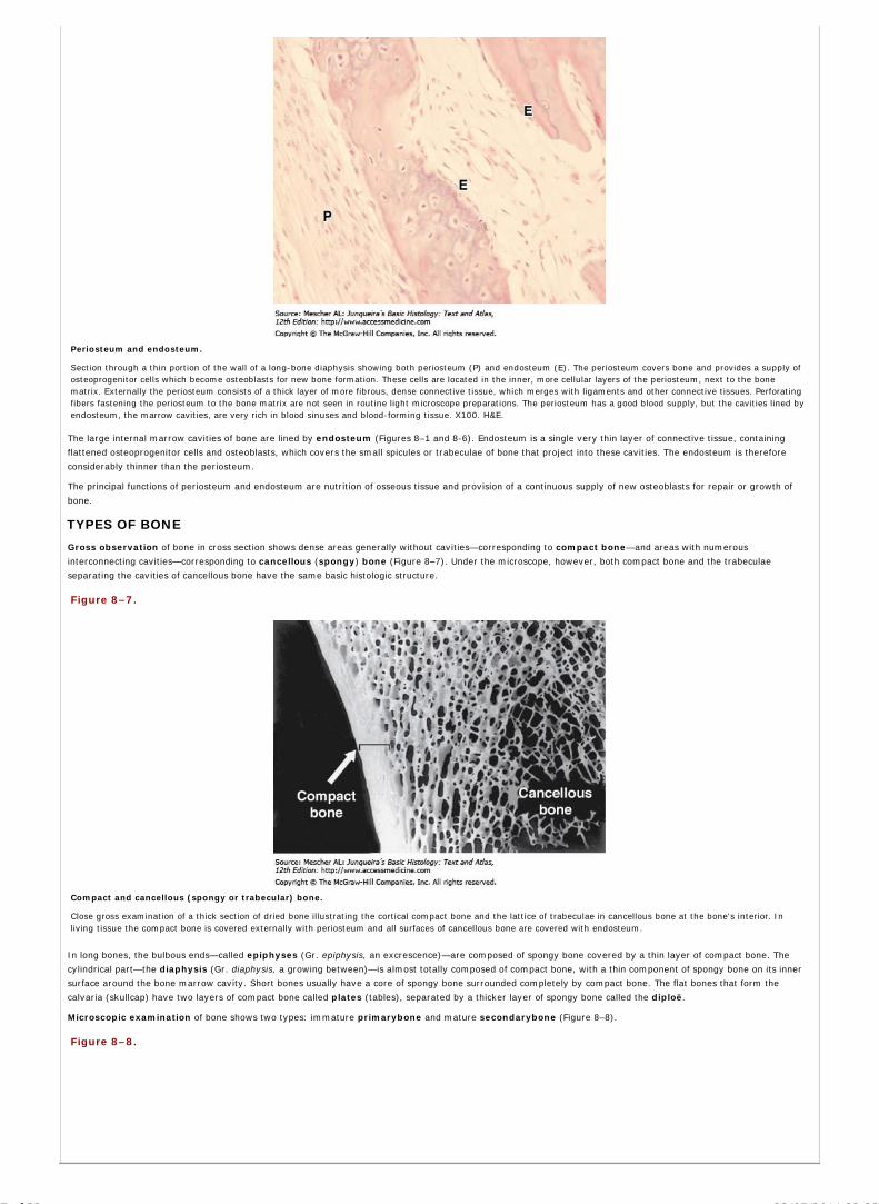
Periosteum and endosteum.
Section through a thin portion of the wall of a long-bone diaphysis showing both periosteum (P) and endosteum (E). The periosteum covers bone and provides a supply ofosteoprogenitor cells which become osteoblasts for new bone formation. These cells are located in the inner, more cellular layers of the periosteum, next to the bonematrix. Externally the periosteum consists of a thick layer of more fibrous, dense connective tissue, which merges with ligaments and other connective tissues. Perforatingfibers fastening the periosteum to the bone matrix are not seen in routine light microscope preparations. The periosteum has a good blood supply, but the cavities lined byendosteum, the marrow cavities, are very rich in blood sinuses and blood-forming tissue. X100. H&E.
The large internal marrow cavities of bone are lined by endosteum (Figures 8–1 and 8-6). Endosteum is a single very thin layer of connective tissue, containing
flattened osteoprogenitor cells and osteoblasts, which covers the small spicules or trabeculae of bone that project into these cavities. The endosteum is therefore
considerably thinner than the periosteum.
The principal functions of periosteum and endosteum are nutrition of osseous tissue and provision of a continuous supply of new osteoblasts for repair or growth of
bone.
TYPES OF BONE
Gross observation of bone in cross section shows dense areas generally without cavities—corresponding to compact bone—and areas with numerous
interconnecting cavities—corresponding to cancellous (spongy) bone (Figure 8–7). Under the microscope, however, both compact bone and the trabeculae
separating the cavities of cancellous bone have the same basic histologic structure.
Figure 8–7.
Compact and cancellous (spongy or trabecular) bone.
Close gross examination of a thick section of dried bone illustrating the cortical compact bone and the lattice of trabeculae in cancellous bone at the bone's interior. Inliving tissue the compact bone is covered externally with periosteum and all surfaces of cancellous bone are covered with endosteum.
In long bones, the bulbous ends—called epiphyses (Gr. epiphysis, an excrescence)—are composed of spongy bone covered by a thin layer of compact bone. The
cylindrical part—the diaphysis (Gr. diaphysis, a growing between)—is almost totally composed of compact bone, with a thin component of spongy bone on its inner
surface around the bone marrow cavity. Short bones usually have a core of spongy bone surrounded completely by compact bone. The flat bones that form the
calvaria (skullcap) have two layers of compact bone called plates (tables), separated by a thicker layer of spongy bone called the diploë.
Microscopic examination of bone shows two types: immature primarybone and mature secondarybone (Figure 8–8).
Figure 8–8.
AccessMedicine | Print: Chapter 8. Bone http://accessmedicine.com/popup.aspx?aID=6180958&print=yes_chapter
7 of 22 22/05/2011 23:33

Primary (woven) bone and secondary (lamellar) bone.
(a): Micrograph of a fractured bone undergoing repair. Primary bone is newly formed, immature bone, rich in osteocytes, with randomly arranged bundles of calcifiedcollagen. Osteoclasts and osteoblasts are numerous in the surrounding endosteum. X200. H&E. (b): Secondary or mature bone shows matrix organized as lamellae, seenfaintly here as concentric lines surrounding osteonic canals. X100. H&E.
Primary Bone TissuePrimary bone is the first bone tissue to appear in embryonic development and in fracture repair. It is characterized by random disposition of fine collagen fibers and is
therefore often called woven bone (Figure 8–8). Primary bone tissue is usually temporary and is replaced in adults by secondary bone tissue except in a very few
places in the body, eg, near the sutures of the calvaria, in tooth sockets, and in the insertions of some tendons.
In addition to the irregular array of collagen fibers, other characteristics of primary bone tissue are a lower mineral content (it is more easily penetrated by x-rays)
and a higher proportion of osteocytes than that in secondary bones.
Secondary Bone TissueSecondary bone tissue is the type usually found in adults. It characteristically shows multiple layers of calcified matrix (each 3–7 m thick) and is often referred to as
lamellar bone. The lamellae are quite organized, either parallel to each other or concentrically around a vascular canal. Each complex of concentric bony lamellae
surrounding a small canal containing blood vessels, nerves, and loose connective tissue is called an osteon (formerly known as an haversian system) (Figures 8–1
and 8–9). Lacunae with osteocytes are found between the lamellae, interconnected by canaliculi which allow all cells to be in contact with the source of nutrients and
oxygen in the osteonic canal (Figure 8–9). The outer boundary of each osteon is a more collagen-rich layer called the cement line.
Figure 8–9.
AccessMedicine | Print: Chapter 8. Bone http://accessmedicine.com/popup.aspx?aID=6180958&print=yes_chapter
8 of 22 22/05/2011 23:33

An osteon.
In preparations of dried, ground bone osteons can be seen with lacunae (L) situated between concentric lamellae and interconnected by fine canaliculi (C). Although it isnot apparent by light microscopy, each lamella consists of multiple parallel arrays of collagen fibers. In adjacent lamellae, the collagen fibers are oriented in differentdirections. The presence of large numbers of lamellae with differing fiber orientations provides the bone with great strength, despite its light weight. Only remnants of theosteocytes (O) in some lacunae and of the osteonic canal's contents are seen in ground bone. In living tissue osteocytic processes connected via gap junctions are presentin successive canaliculi, making cells in all the lamellae in communication with the blood vessels in the central canal. X500.
In each lamella, type I collagen fibers are aligned in parallel and follow a helical course. The pitch of the helix is, however, different for different lamellae, so that at
any given point, fibers from adjacent lamellae intersect at approximately right angles (Figure 8–1). The specific organization of collagen fibers in successive lamellae
of each osteon is highly important for the great strength of secondary bone.
In compact bone (eg, the diaphysis of long bones) besides forming osteons, the lamellae also exhibit a typical organization consisting of multiple external
circumferential lamellae (Figure 8–1) and often some inner circumferential lamellae. Inner circumferential lamellae are located around the marrow cavity and
external circumferential lamellae are located immediately beneath the periosteum.
Each osteon is a long, often bifurcated cylinder generally parallel to the long axis of the diaphysis. It consists of a central canal surrounded by 4–10 concentric
lamellae. Each endosteum-lined canal contains blood vessels, nerves, and loose connective tissue. The central canals communicate with the marrow cavity and the
periosteum and with one another through transverse or oblique perforating canals (formerly known as Volkmann canals) (Figures 8–1 and 8–10). The transverse
canals do not have concentric lamellae; instead, they perforate the lamellae. All osteonic and perforating canals in bone tissue come into existence when matrix is laid
down around preexisting blood vessels.
Figure 8–10.
AccessMedicine | Print: Chapter 8. Bone http://accessmedicine.com/popup.aspx?aID=6180958&print=yes_chapter
9 of 22 22/05/2011 23:33

Lamellar bone: Perforating canals and interstitial lamellae.
(a): Transverse perforating canals (P) connecting adjacent osteons are shown at the left side of the micrograph. Such canals "perforate" lamellae and provide anothersource of microvasculature for the central canals of osteons. Among the intact osteons are also found remnants of eroded osteons, seen as irregular interstitial orintermediate lamellae (I). X100. (b): Schematic diagram shows remodeling of compact lamellar bone showing three generations of osteonic haversian systems and theirsuccessive contributions to the formation of interstitial lamellae. Remodeling is a continuous process that involves the coordinated activity of osteoblasts and osteoclasts,and is responsible for adaptation of bone to changes in stress, especially during the body's growth.
Among the osteons between the two circumferential systems are numerous irregularly shaped groups of parallel lamellae called interstitial lamellae. These
structures are lamellae remaining from osteons partially destroyed by osteoclasts during growth and remodeling of bone (Figure 8–10).
Bone remodeling is continuous throughout life and involves a combination of bone synthesis and removal. In compact bone, remodeling resorbs parts of old osteons
and produces new ones. Resorption involves the actions of osteoclasts, often working in groups to remove old bone in tunnel-like cavities having the approximate
diameter of new osteons. Such tunnels are quickly invaded by many osteoprogenitor cells and sprouting loops of blood capillaries, both derived from the endosteum
or periosteum. Osteoblasts develop, line the wall of the tunnels, and begin to secrete osteoid in a cyclic manner, forming concentric lamellae of bone with trapped
osteocytes (Figure 8–11). In healthy adults 5–10% of the bone turns over annually.
Figure 8–11.
Development of osteons.
During remodeling of compact bone, a group of osteoclasts acts as a boring cone to make a tunnel into bone matrix. Behind these cells a population of osteoblasts entersthe tunnel and lines its walls. As the osteoblasts secrete osteoid in a cyclic manner, they produce layers of new matrix with cells trapped in lacunae. The cells in lacunae arenow osteocytes. The tunnel becomes constricted with multiple concentric layers of new matrix and its lumen finally exists as only a narrow central canal with small bloodvessels.
Variations in the remodeling activity produce great variability in the sizes of osteons, osteonic canals, and interstitial lamellae. As osteons form by successive
deposition of lamellae by osteoblasts, moving inward from the periphery, younger osteons usually have larger canals. In mature osteons the most recently formed
lamella is the one closest to the central canal.
OSTEOGENESIS
Bone can be formed initially by either of two ways:
Intramembranous ossification, in which osteoblasts differentiate directly from mesenchyme and begin secreting osteoid
Endochondral ossification, in which the matrix of preexisting hyaline cartilage is eroded and replaced by osteoblasts producing osteoid.
In both processes, the bone tissue that appears first is primary or woven. Primary bone is a temporary and is soon replaced by the definitive secondary lamellar
bone. During bone growth, areas of primary bone, areas of resorption, and areas of secondary bone all appear side by side.
Intramembranous Ossification
AccessMedicine | Print: Chapter 8. Bone http://accessmedicine.com/popup.aspx?aID=6180958&print=yes_chapter
10 of 22 22/05/2011 23:33

Intramembranous ossification, by which most flat bones are produced, is so called because it takes place within condensations of embryonic mesenchymal tissue. The
frontal and parietal bones of the skull—as well as parts of the occipital and temporal bones and the mandible and maxilla—are formed by intramembranous
ossification. The process is summarized in Figure 8–12.
Figure 8–12.
Intramembranous ossification.
Developmental process by which most bones of the skull are formed. (a): Groups of mesenchymal cells in a "membrane" or sheet of this embryonic tissue, round up anddifferentiate as osteoblasts producing osteoid. (b): Cells trapped in the calcifying matrix differentiate as osteocytes. (c): Woven bone is produced in this manner, with
AccessMedicine | Print: Chapter 8. Bone http://accessmedicine.com/popup.aspx?aID=6180958&print=yes_chapter
11 of 22 22/05/2011 23:33

vascularized internal spaces that will form the marrow cavity and surrounded on both sides by developing periosteum. (d): Remodeling of the woven bone produces thetwo layers of compact lamellar bone with cancellous bone in between, which is characteristic of these flat bones.
In the mesenchymal condensation layer or "membrane," the starting point for bone formation is called an ossification center. The process begins when groups of
mesenchymal cells differentiate into osteoblasts. Osteoblasts produce osteoid matrix and calcification follows, resulting in the encapsulation of some osteoblasts,
which then become osteocytes. These islands of developing bone form walls that delineate elongated cavities containing capillaries, bone marrow cells, and
undifferentiated cells. Several such groups arise almost simultaneously at the ossification center, and their fusion between the walls gives the bone a spongy
appearance. The connective tissue that remains among the bone walls is penetrated by growing blood vessels and additional undifferentiated mesenchymal cells,
giving rise to the bone marrow. The ossification centers of a bone grow radially and finally fuse together, replacing the original connective tissue (Figures 8–12 and
8–13).
Figure 8–13.
Intramembranous ossification.
A section of jaw from a fetal pig undergoing intramembranous ossification. (a): Areas of typical mesenchyme (M), condensed mesenchyme (CM) adjacent to aggregates ofnew osteoblasts (O). Some osteoblasts have secreted matrices of bone (B) which remain covered by osteoblasts. Between these trabeculae of newly formed primary boneare vascularized areas (V) that will form marrow cavities. X40. H&E. (b): Higher magnification shows the developing periosteum (P) that covers masses primary bone thatwill soon merge to form a continuous plate of bone. The larger mesenchyme-filled region at the top is the developing marrow cavity. X100. H&E.
In cranial flat bones there is a marked predominance of bone formation over bone resorption at both the internal and external surfaces. Thus, two layers of compact
bone (internal and external plates) arise, while the central portion (diploë) maintains its spongy nature. The fontanelles or "soft spots" on the heads of newborn
infants are areas in the skull that correspond to parts of the connective tissue that are not yet ossified. The portions of the connective tissue layer that do not undergo
ossification give rise to the endosteum and the periosteum of the new bone.
Endochondral OssificationEndochondral (Gr. endon, within, + chondros, cartilage) ossification takes place within a piece of hyaline cartilage whose shape resembles a small version, or model,
of the bone to be formed. This type of ossification is principally responsible for the formation of short and long bones.
AccessMedicine | Print: Chapter 8. Bone http://accessmedicine.com/popup.aspx?aID=6180958&print=yes_chapter
12 of 22 22/05/2011 23:33

Endochondral ossification of a long bone consists of the sequence of events shown schematically in Figure 8–14. Initially, the first bone tissue appears as a collar
surrounding the diaphysis of the cartilage model. This bone collar is produced by local osteoblast activity within the surrounding perichondrium. The collar now
impedes diffusion of oxygen and nutrients into the underlying cartilage, promoting degenerative changes there. The chondrocytes begin to produce alkaline
phosphatase and swell up (hypertrophy), enlarging their lacunae. These changes both compress the matrix into narrower trabeculae and lead to calcification in these
structures. Death of the chondrocytes results in a porous three-dimensional structure formed by the remnants of the calcified cartilage matrix (Figure 8–15). Blood
vessels from the former perichondrium now the periosteum penetrate through the bone collar previously perforated by osteoclasts, bringing osteoprogenitor cells to
the porous central region. Next, osteoblasts adhere to the calcified cartilage matrix and produce continuous layers of primary bone that surround the cartilaginous
matrix remnants. At this stage, the calcified cartilage appears basophilic, and the primary bone is eosinophilic (Figure 8–15).
Figure 8–14.
Osteogenesis of long bones by endochondral ossification.
Endochondral ossification forms most bones of the skeleton and occurs in the fetus in models made of hyaline cartilage (1). The process takes many weeks and majordevelopmental stages include: formation of a bone collar around the middle of the cartilage model and degeneration of the underlying cartilage (2), followed by invasion ofthe resulting ossification center by capillaries and osteoprogenitor cells from the periosteum (3), osteoid deposition by the new osteoblasts, calcification of woven bone,and its remodeling as compact bone (4). This primary ossification center develops in the diaphysis, along the middle of each developing bone. Secondary ossificationcenters develop somewhat later by a similar process in the epiphyses. The primary and secondary ossification centers are separated by the epiphyseal plate (5) whichprovides for continued bone elongation. The two ossification centers do not merge until the epiphyseal plate disappears (6) when full stature is achieved.
Figure 8–15.
AccessMedicine | Print: Chapter 8. Bone http://accessmedicine.com/popup.aspx?aID=6180958&print=yes_chapter
13 of 22 22/05/2011 23:33

Cells and matrices of a primary ossification center.
A small region of a primary ossification center showing key features of endochondral ossification. Compressed remnants of calcified cartilage matrix (dark purple), nowdevoid of chondrocytes, are enclosed by more lightly stained osteoid or bone matrix. This newly formed bone is surrounded by a layer of large, active osteoblasts. Someosteoblasts that were captured by the matrix have become smaller osteocytes (arrowheads). X200. Pararosaniline–toluidine blue.
This process in the diaphysis forms the primary ossification center (Figure 8–14). Secondary ossification centers appear slightly later at the epiphyses of the
cartilage model and develop in a similar manner. During their expansion and remodeling, the primary and secondary ossification centers produce cavities that are
gradually filled with bone marrow.
In the secondary ossification centers, cartilage remains in two regions: the articular cartilage (Figure 8–14), which persists throughout adult life and does not
contribute to bone growth in length, and the epiphyseal cartilage (also called epiphyseal plate or growth plate), which connects each epiphysis to the diaphysis
(Figures 8–16 and 8–17). The epiphyseal cartilage is responsible for the growth in length of the bone and disappears in adults, which is why bone growth ceases in
adulthood. Elimination of the epiphyseal plates ("epiphyseal closure") occurs at different times with different bones and is complete in all bones by about age twenty.
In forensics or through X-ray examination of the growing skeleton, it is possible to determine the "bone age" of a young person, noting which epiphyses are open and
which are closed. Once the epiphyses have closed, growth in length of bones becomes impossible, although bone widening may still occur.
Figure 8–16.
AccessMedicine | Print: Chapter 8. Bone http://accessmedicine.com/popup.aspx?aID=6180958&print=yes_chapter
14 of 22 22/05/2011 23:33

Epiphyseal growth plate: locations and zones of activity.
The large and growing primary ossification center in long bone diaphyses and the secondary ossification centers in epiphyses are separated in each developing bone by aplate of cartilage called the epiphyseal plate. (a): Epiphyseal plates can be identified in an x-ray of a child's hand as marrow regions of lower density between the denserossification centers. Cells in epiphyseal growth plates are responsible for continued elongation of bones until the body's full size is reached. Developmental activities in theepiphyseal growth plate occur in overlapping zones with distinct histological appearances. (b): Moving from the epiphysis to the diaphysis, these zones include cellsspecialized for the following: (1) normally appearing hyaline cartilage, (2) cartilage with proliferating chondroblasts aligned in lacunae as axial aggregates, (3)degenerating cartilage in which the aligned cells are hypertrophic and the matrix condensed, (4) an area in which the chondrocytes have disappeared and the matrix isundergoing calcification, and (5) a zone in which blood vessels and osteoblasts have invaded the lacunae of the old cartilage, producing marrow cavities and osteoid fornew bone. X100. H&E.
Figure 8–17.
AccessMedicine | Print: Chapter 8. Bone http://accessmedicine.com/popup.aspx?aID=6180958&print=yes_chapter
15 of 22 22/05/2011 23:33

Cells and matrices of the epiphyseal growth plate.
(a): At the top of the micrograph the growth plate (GP) shows its zones of hyaline cartilage with cells undergoing rest (R), proliferation (P), and hypertrophy (H). As thechondrocytes swell and degenerate they release phosphatase, activities which compress the matrix and cause an initial deposition of CaPO4. This produces calcified spicules(C) in the former cartilage matrix. The tunnel-like lacunae in which the chondrocytes have undergone apoptosis are invaded from the diaphysis by large, thin-walled bloodvessels which begin to convert these spaces into marrow (M) cavities. Endosteum with osteoblasts also moves in from the diaphyseal primary ossification center and thesecells cover the spicules of calcified cartilage and lay down layers of osteoid, forming a supportive matrix that is now largely primary woven bone (B). X40. H&E. (b): Highermagnification shows more detail of the cells and matrix spicules in the zones undergoing hypertrophy (H) and ossification. Staining properties of the matrix clearly changein this process: first when it is compressed and begins to calcify (C), and then when osteoid and bone (B) are laid down. The large spaces between the ossifying matrixspicules become the marrow cavity (M), in which sinuses of eosinophilic red blood cells and aggregates of basophilic white blood cell precursors can be distinguished. Themarrow is the major site of blood cell formation in adults. X100. H&E.
A plate of epiphyseal cartilage is divided into five zones (Figure 8–16), starting from the epiphyseal side of cartilage:
The resting zone consists of hyaline cartilage with typical chondrocytes.1.
In the proliferative zone, chondrocytes begin to divide rapidly and form columns of stacked cells parallel to the long axis of the bone.2.
The hypertrophic cartilage zone contains swollen chondrocytes whose cytoplasm has accumulated glycogen. Hypertrophy compresses the matrix into thinsepta between the chondrocytes.
3.
In the calcified cartilage zone, loss of the chondrocytes by apoptosis is accompanied by calcification of the septa of cartilage matrix by the formation ofhydroxyapatite crystals (Figure 8–17).
4.
In the ossification zone, bone tissue first appears. Capillaries and osteoprogenitor cells originating from the periosteum invade the cavities left by thechondrocytes. Many of these cavities will be merged and become the marrow cavity. The osteoprogenitor cells form osteoblasts, which settle in a discontinuouslayer over the septa of calcified cartilage matrix. The osteoblasts deposit osteoid over the spicules of calcified cartilage matrix, forming woven bone (Figure8–17).
5.
In summary, growth in length of a long bone occurs by proliferation of chondrocytes in the epiphyseal plate adjacent to the epiphysis. At the same time, chondrocytes
in the diaphyseal side of the plate hypertrophy, their matrix becomes calcified, and the cells die. Osteoblasts lay down a layer of primary bone on the calcified
cartilage matrix. Because the rates of these two opposing events (proliferation and destruction) are approximately equal, the epiphyseal plate does not change
thickness. Instead, it is displaced away from the middle of the diaphysis, resulting in growth in length of the bone.
BONE GROWTH, REMODELING, & REPAIR
Bone growth is generally associated with partial resorption of preformed tissue and the simultaneous laying down of new bone (exceeding the rate of bone loss). This
process permits the shape of the bone to be maintained as it grows. The rate of bone remodeling (bone turnover) is very active in young children, where it can be
200 times faster than that in adults. Bone remodeling in adults is a dynamic physiologic process that occurs simultaneously in multiple locations of the skeleton and is
not always related to bone growth.
Despite its hardness, the constant remodeling makes bone very plastic and capable of internal structural changes according to the various stresses to which it is
subjected. A well-known example of bone plasticity is the ability of the positions of teeth in the jawbone to be modified by the lateral pressures produced by
orthodontic appliances. Bone is formed on the side where traction is applied and is resorbed on the opposite side where pressure is exerted. In this way, teeth are
moved within the jaw while the bone is being remodeled.
Cranial bones grow mainly because of the formation of bone tissue by the periosteum between the sutures and on the external bone surface. At the same time,
resorption takes place on the internal surface. The plasticity of bone allows it to respond to the growth of the brain and form a skull of adequate size. The skull will be
small if the brain does not develop completely and will be larger than normal in a person suffering from hydrocephalus, a disorder characterized by abnormal
accumulation of spinal fluid and dilatation of the cerebral ventricles.
Because it contains osteoprogenitor stem cells throughout the endosteum and periosteum and has an extensive blood supply, bone has an excellent capacity for repair
and regeneration. Bone fractures and other damage are repaired efficiently using cells and processes already active in bone remodeling. Surgically created gaps in
bone can be filled with new bone, especially when periosteal tissue remains nearby.
MEDICAL APPLICATION
AccessMedicine | Print: Chapter 8. Bone http://accessmedicine.com/popup.aspx?aID=6180958&print=yes_chapter
16 of 22 22/05/2011 23:33

When a bone is fractured, blood vessels are disrupted and bone cells adjoining the fracture die. The damaged blood vessels produce a localized hemorrhage
and form a blood clot.
Soon the blood clot is removed by macrophages and the adjacent matrix of bone is resorbed by osteoclasts. The periosteum and the endosteum at the site of
the fracture respond with intense proliferation producing a soft callus of fibrocartilage-like tissue that surrounds the fracture and covers the extremities of the
fractured bone (Figure 8–18).
Primary bone is then formed by a combination of endochondral and intramembranous ossification. Further repair produces irregularly formed trabeculae of
primary bone that temporarily unite the extremities of the fractured bone, forming a hard bone callus (Figure 8–18).
Stresses imposed on the bone during repair and during the patient's gradual return to activity serve to remodel the bone callus. The primary bone of the
callus is gradually resorbed and replaced by secondary bone, remodeling and restoring the original bone structure. Unlike other connective tissues, bone
tissue heals without forming a scar.
Figure 8–18.
Main features of bone fracture repair.
Repair of a fractured bone occurs through several stages, but utilizes mechanisms already in place for bone remodeling. (1): Blood vessels torn within the fracture releaseblood which clots to produce a large fracture hematoma. (2): This is gradually removed by macrophages and replaced by a soft fibrocartilage-like mass of procallus tissuerich in collagen and fibroblasts. If broken, the periosteum re-establishes continuity over this tissue. (3): This soft procallus is invaded by regrowing blood vessels andosteoblasts. In the next few weeks the fibrocartilage is gradually replaced by trabeculae of primary bone, forming a hard callus throughout the original area of fracture. (4):The primary bone is then remodeled as compact and cancellous bone in continuity with the adjacent uninjured areas and fully functional vasculature is re-established.
METABOLIC ROLE OF BONE
Calcium ions are required for the activity of many enzymes and other proteins mediating cell adhesion, cytoskeletal movements, exocytosis, membrane permeability,
and other functions in cells throughout the body. The skeleton serves as the calcium reservoir and contains 99% of the body's total calcium in crystals of
hydroxyapatite. The concentration of calcium in the blood and tissues is generally quite stable because of a continuous interchange between blood calcium and bone
calcium.
The principal mechanism for raising blood calcium levels is the mobilization of ions from hydroxyapatite crystals to interstitial fluid. This takes place mainly in
cancellous bone. The younger, more lightly calcified lamellae that exist even in adult bone (because of continuous remodeling) receive and lose calcium more readily.
These lamellae are more important for the maintenance of calcium concentration in the blood than are the older, more densely calcified lamellae, whose role is
mainly that of support and protection.
The action of two key hormones on cells in bone regulates the process of calcium mobilization from hydroxyapatite. Parathyroid hormone (PTH) from the
parathyroid glands raises low blood calcium levels. The principal target cells of this polypeptide are osteoblasts, which stop production of osteoid and matrix vesicles
and instead secrete a paracrine protein, osteoclast stimulating factor. This factor promotes osteoclastic resorption of the bone matrix, liberating calcium. Osteoclast
activity is inhibited by another hormone, calcitonin, which is synthesized by the parafollicular cells of the thyroid gland. This slows matrix resorption and thereby
gradually lowers blood calcium levels.
MEDICAL APPLICATION
Because the concentration of calcium in tissues and blood must be kept constant, nutritional deficiency of calcium results in decalcification of bones. Severely
decalcified bones are more likely to fracture.
Decalcification of bone may also be caused by excessive production of PTH (hyperparathyroidism), which can cause increased osteoclastic activity, intense
resorption of bone, elevation of blood Ca2+ and PO3– 4 levels, and abnormal deposits of calcium in the kidneys and arterial walls.
The opposite occurs in osteopetrosis (L. petra, stone), a disease caused by defective osteoclast function that results in overgrowth, thickening, and
hardening of bones. This process can obliterate the bone marrow cavities, depressing blood cell formation and causing anemia and the loss of white blood
cells.
Nutritional Deficiencies and Bone Remodeling
Especially during growth, bone is sensitive to nutritional factors. Calcium deficiency, which leads to incomplete calcification of the organic bone matrix, can be
due either to a lack of calcium in the diet or a failure to produce the steroid prohormone vitamin D, which is important for the absorption of Ca2+ and>
PO3–4 by the small intestine.
Calcium deficiency in children causes rickets, a disease in which the bone matrix does not calcify normally and the epiphyseal plate becomes distorted by the
normal strains of body weight and muscular activity. Ossification processes at this level are consequently hindered, and the bones not only grow more slowly
but also become deformed.
Calcium deficiency in adults gives rise to osteomalacia (osteon + Gr. malakia, softness), which is characterized by deficient calcification of recently formed
bone and partial decalcification of already calcified matrix. Osteomalacia should not be confused with osteoporosis. In osteomalacia, there is a decrease in
the amount of calcium per unit of bone matrix. Osteoporosis, frequently found in immobilized patients and in postmenopausal women, is an imbalance in
skeletal turnover so that bone resorption exceeds bone formation.
AccessMedicine | Print: Chapter 8. Bone http://accessmedicine.com/popup.aspx?aID=6180958&print=yes_chapter
17 of 22 22/05/2011 23:33

Hormones Acting on Bone Tissue
In addition to PTH and calcitonin, several other hormones act on bone. The anterior lobe of the pituitary synthesizes growth hormone (GH or somatotropin),
which stimulates the liver to produce insulin-like growth factor-1 (IGF-1 or somatomedin). IGF has an overall growth effect, especially on the epiphyseal
cartilage. Consequently, lack of growth hormone during the growing years causes pituitary dwarfism; an excess of growth hormone causes excessive
growth of the long bones, resulting in gigantism. Adult bones cannot increase in length even with excess IGF because they lack epiphyseal cartilage, but they
do increase in width by periosteal growth. In adults, an increase in GH causes acromegaly, a disease in which the bones—mainly the long ones—become
very thick.
The sex hormones, both male (androgens) and female (estrogens), have a complex effect on bones and are, in a general way, stimulators of bone formation.
They influence the time of appearance and development of ossification centers and accelerate the closure of epiphyses.
Bone Tumors
Cancer originating directly from bone cells is fairly uncommon (0.5% of all cancer deaths) but a form called osteosarcoma can arise in osteoblasts. The
skeleton is often the site of metastases from tumors originating from malignancies in other organs, most commonly from breast, lung, prostate, kidney, and
thyroid tumors.
JOINTS
Joints are regions where bones are capped and surrounded by connective tissues that firmly hold the bones together and determine the type and degree of movement
between them. Joints may be classified as diarthroses, which permit free bone movement, and synarthroses (Gr. syn, together, + arthrosis, articulation), in which
very limited or no movement occurs. There are three types of synarthroses, based on the type of tissue uniting the bone surfaces:
Synostoses, in which bones are united by bone tissue and no movement takes place. In older adults, synostoses unite the skull bones, which, in childrenand young adults, are united only by dense connective tissue
Synchondroses, in which the bones are joined by hyaline cartilage. The epiphyseal plates of growing bones are one example and in adults asynchondrosis unites the first rib to the sternum, with little movement
Syndesmoses, in which bones are joined by an interosseous ligament of dense connective tissue or fibrocartilage (eg, the pubic symphysis, Figure 7–1),again with very limited movement.
Diarthroses (Figure 8–19) are joints that generally unite long bones and have great mobility, such as the elbow and knee joints. In a diarthrosis, ligaments and a
capsule of dense connective tissue maintain proper alignment of the bones. The capsule encloses a sealed joint cavity that contains synovial fluid, a colorless,
transparent, viscous fluid. The joint cavity is not lined by epithelium, but by a specialized connective tissue called the synovial membrane which extends folds and
villi into the cavity and secretes the lubricant synovial fluid. Synovial fluid is derived from blood plasma, but with a high concentration of hyaluronic acid produced by
cells of the synovial membrane.
Figure 8–19.
AccessMedicine | Print: Chapter 8. Bone http://accessmedicine.com/popup.aspx?aID=6180958&print=yes_chapter
18 of 22 22/05/2011 23:33

Diarthroses, or synovial joints.
Diarthroses are joints that allow free movement of the attached bones, such as knuckles, knees, and elbows. (a): Diagram shows the components of a diarthrosis thatinclude (1) a capsule continuous with a covering ligament which inserts into the periosteum of both bones, (2) a synovial or joint cavity lined by synovial membrane andcontaining synovial fluid as a lubricant, and (3) the ends of epiphyses covered by articular cartilage. (b): Longitudinal section through a diarthrosis of growing long bonesshows the position near the boundaries of the capsule (C) of the epiphyseal growth plates (E) where endochondral ossification occurs. Also shown here are the articularcartilage (A) and the folds of synovial membrane (SM) which extend prominently into the joint cavity from connective tissue of the capsule for production of synovial fluid.X10. PSH stain.
The synovial membrane or layer may have prominent regions with various types of connective tissue (areolar, fibrous, or adipose) in different diarthrotic joints. At
the surface contacting the synovial fluid the tissue is usually well-vascularized, with many porous (fenestrated) capillaries, and contains two specialized cells
(synoviocytes) with distinctly different functions and origins (Figure 8–20). Rounded synoviocytes in contact with the synovial cavity are phagocytic and remove
wear-and-tear debris from the synovial fluid. Among the capillaries are many more fibroblastic synoviocytes specialized to produce the long, nonsulfated
glycosaminoglycan (GAG) hyaluronic acid and secrete other components of ground substance. These GAGs along with plasma from the capillaries leave the synovial
membrane, oozing into the synovial fluid. This viscous, gel-like fluid lubricates the joint, reducing friction on all internal surfaces, and supplies nutrients and oxygen to
the articular cartilage.
Figure 8–20.
AccessMedicine | Print: Chapter 8. Bone http://accessmedicine.com/popup.aspx?aID=6180958&print=yes_chapter
19 of 22 22/05/2011 23:33

Synovial membrane.
The synovial membrane is a specialized connective tissue that lines capsules of synovial joints and contacts the synovial fluid lubricant, which it is primarily responsible formaintaining. (a): Micrograph showing this membrane projects folds into the joint cavity (JC) and is highly vascularized (V). The joint cavity surrounds the articularcartilage (AC). X100. Mallory trichrome. (b): Higher magnification of the fold showing a high density of capillaries and two specialized types of cells called synoviocytes.Contacting the synovial fluid at the tissue surface are many rounded macrophage-like synoviocytes (type A) derived from blood monocytes. These cells bind, engulf,and remove tissue debris from synovial fluid. These cells often form a layer at the tissue surface (A) and can superficially resemble an epithelium, but there is no basallamina and the cells are not joined together by cell junctions. Fibroblast-like (type B) synoviocytes (B) are mesenchymally derived and specialized for synthesis ofhyaluronic acid and other components of ground substance. Together with plasma leaking from the many porous fenestrated capillaries, this GAG-rich ground substanceenters the synovial fluid, replenishing it. X400. (c): Schematic representation of ultrastructural features of the synovial membrane. Among the macrophage-like andfibroblast-like synoviocytes, are collagen fibers and other typical components of connective tissue. There is no basal lamina or other ultrastructural features of epithelium,despite the superficial resemblance. Blood capillaries are of the fenestrated type, which facilitates exchange of substances between blood and synovial fluid.
The collagen fibers of the hyaline articular cartilage are disposed as arches with their tops at the exposed surface, which unlike most cartilage is not covered by
perichondrium (Figure 8–21). This arrangement of collagen helps to distribute more evenly the forces generated by pressure on joints. The resilient articular cartilage
is also an efficient absorber of the intermittent mechanical pressures to which many joints are subjected.
Figure 8–21.
AccessMedicine | Print: Chapter 8. Bone http://accessmedicine.com/popup.aspx?aID=6180958&print=yes_chapter
20 of 22 22/05/2011 23:33

Articular cartilage.
(a): Articular surfaces of a diarthrosis are made of hyaline cartilage that lacks the usual perichondrium covering. X40. H&E. (b): The upper drawing shows that in suchcartilage, collagen fibers are first perpendicular and then bend gradually, forming a broad arch parallel to the cartilage surface. More deeply located chondrocytes arearranged in vertical rows. Superficially placed chondrocytes are flattened and are no longer organized in aggregates. The lower left drawing shows a more three-dimensional view of collagen fibers in articular cartilage. Proteoglycan aggregates bound to hyaluronic acid and collagen fill the space among the collagen fiber network andbind a large amount of water. the large assembly of gags functions as a biomechanical spring in articular cartilage. When pressure is applied, water is forced out of thecartilage matrix into the synovial fluid. When water is expelled, electrostatic repulsion of the negatively charged carboxyl and sulfate groups in the gags occurs, separatingthe gags again and thus creating spaces for the return of water. When pressure is released, water is attracted back into the interstices of the gag matrix. These watermovements are brought about constantly by using the joint and are essential for nutrition of the cartilage and for facilitating the interchange of O2, CO2, and othermolecules between the synovial fluid and the articular cartilage.
A similar mechanism is seen in intervertebral disks (Figure 8–22) which are thick disks of fibrocartilage between the articular surfaces of successive bony vertebrae.
The annulus fibrosus of each disk has an external layer of dense connective tissue, but is mainly composed of overlapping laminae of fibrocartilage in which
collagen bundles are orthogonally arranged in adjacent layers. The multiple lamellae, with the 90-degree registration of type I collagen fibers in adjacent layers,
provide the disk with unusual resilience that enables it to withstand the pressures generated by the impinging vertebrae.
Figure 8–22.
AccessMedicine | Print: Chapter 8. Bone http://accessmedicine.com/popup.aspx?aID=6180958&print=yes_chapter
21 of 22 22/05/2011 23:33
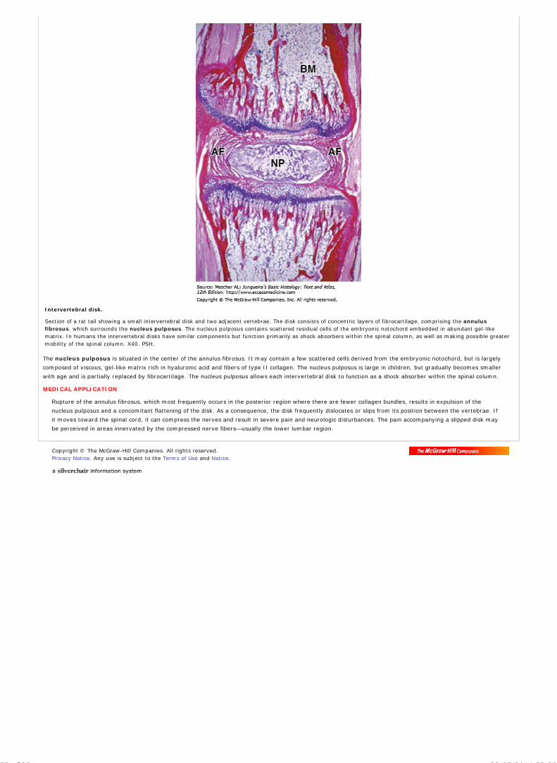
Copyright © The McGraw-Hill Companies. All rights reserved.Privacy Notice. Any use is subject to the Terms of Use and Notice.
Intervertebral disk.
Section of a rat tail showing a small intervertebral disk and two adjacent vertebrae. The disk consists of concentric layers of fibrocartilage, comprising the annulusfibrosus, which surrounds the nucleus pulposus. The nucleus pulposus contains scattered residual cells of the embryonic notochord embedded in abundant gel-likematrix. In humans the intervertebral disks have similar components but function primarily as shock absorbers within the spinal column, as well as making possible greatermobility of the spinal column. X40. PSH.
The nucleus pulposus is situated in the center of the annulus fibrosus. It may contain a few scattered cells derived from the embryonic notochord, but is largely
composed of viscous, gel-like matrix rich in hyaluronic acid and fibers of type II collagen. The nucleus pulposus is large in children, but gradually becomes smaller
with age and is partially replaced by fibrocartilage. The nucleus pulposus allows each intervertebral disk to function as a shock absorber within the spinal column.
MEDICAL APPLICATION
Rupture of the annulus fibrosus, which most frequently occurs in the posterior region where there are fewer collagen bundles, results in expulsion of the
nucleus pulposus and a concomitant flattening of the disk. As a consequence, the disk frequently dislocates or slips from its position between the vertebrae. If
it moves toward the spinal cord, it can compress the nerves and result in severe pain and neurologic disturbances. The pain accompanying a slipped disk may
be perceived in areas innervated by the compressed nerve fibers—usually the lower lumbar region.
AccessMedicine | Print: Chapter 8. Bone http://accessmedicine.com/popup.aspx?aID=6180958&print=yes_chapter
22 of 22 22/05/2011 23:33
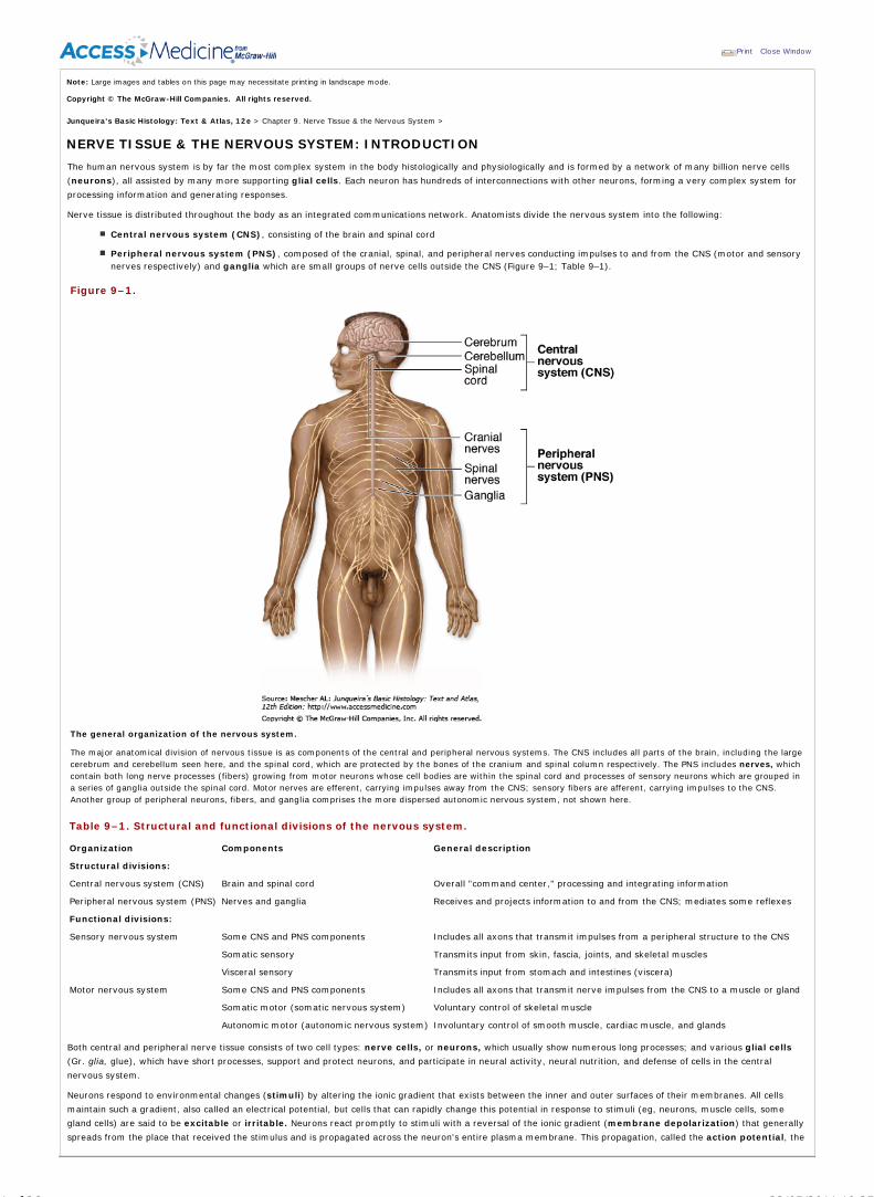
Print Close Window
Note: Large images and tables on this page may necessitate printing in landscape mode.
Copyright © The McGraw-Hill Companies. All rights reserved.
Junqueira's Basic Histology: Text & Atlas, 12e > Chapter 9. Nerve Tissue & the Nervous System >
NERVE TISSUE & THE NERVOUS SYSTEM: INTRODUCTION
The human nervous system is by far the most complex system in the body histologically and physiologically and is formed by a network of many billion nerve cells
(neurons), all assisted by many more supporting glial cells. Each neuron has hundreds of interconnections with other neurons, forming a very complex system for
processing information and generating responses.
Nerve tissue is distributed throughout the body as an integrated communications network. Anatomists divide the nervous system into the following:
Central nervous system (CNS), consisting of the brain and spinal cord
Peripheral nervous system (PNS), composed of the cranial, spinal, and peripheral nerves conducting impulses to and from the CNS (motor and sensorynerves respectively) and ganglia which are small groups of nerve cells outside the CNS (Figure 9–1; Table 9–1).
Figure 9–1.
The general organization of the nervous system.
The major anatomical division of nervous tissue is as components of the central and peripheral nervous systems. The CNS includes all parts of the brain, including the largecerebrum and cerebellum seen here, and the spinal cord, which are protected by the bones of the cranium and spinal column respectively. The PNS includes nerves, whichcontain both long nerve processes (fibers) growing from motor neurons whose cell bodies are within the spinal cord and processes of sensory neurons which are grouped ina series of ganglia outside the spinal cord. Motor nerves are efferent, carrying impulses away from the CNS; sensory fibers are afferent, carrying impulses to the CNS.Another group of peripheral neurons, fibers, and ganglia comprises the more dispersed autonomic nervous system, not shown here.
Table 9–1. Structural and functional divisions of the nervous system.
Organization Components General description
Structural divisions:
Central nervous system (CNS) Brain and spinal cord Overall "command center," processing and integrating information
Peripheral nervous system (PNS) Nerves and ganglia Receives and projects information to and from the CNS; mediates some reflexes
Functional divisions:
Sensory nervous system Some CNS and PNS components Includes all axons that transmit impulses from a peripheral structure to the CNS
Somatic sensory Transmits input from skin, fascia, joints, and skeletal muscles
Visceral sensory Transmits input from stomach and intestines (viscera)
Motor nervous system Some CNS and PNS components Includes all axons that transmit nerve impulses from the CNS to a muscle or gland
Somatic motor (somatic nervous system) Voluntary control of skeletal muscle
Autonomic motor (autonomic nervous system) Involuntary control of smooth muscle, cardiac muscle, and glands
Both central and peripheral nerve tissue consists of two cell types: nerve cells, or neurons, which usually show numerous long processes; and various glial cells
(Gr. glia, glue), which have short processes, support and protect neurons, and participate in neural activity, neural nutrition, and defense of cells in the central
nervous system.
Neurons respond to environmental changes (stimuli) by altering the ionic gradient that exists between the inner and outer surfaces of their membranes. All cells
maintain such a gradient, also called an electrical potential, but cells that can rapidly change this potential in response to stimuli (eg, neurons, muscle cells, some
gland cells) are said to be excitable or irritable. Neurons react promptly to stimuli with a reversal of the ionic gradient (membrane depolarization) that generally
spreads from the place that received the stimulus and is propagated across the neuron's entire plasma membrane. This propagation, called the action potential, the
AccessMedicine | Print: Chapter 9. Nerve Tissue & the Nervous System http://accessmedicine.com/popup.aspx?aID=6181123&print=yes_chapter
1 of 36 22/05/2011 19:25

depolarization wave, or the nerve impulse, is capable of traveling long distances along neuronal processes, transmitting such signals to other neurons, muscles,
and glands.
By creating, analyzing, identifying, and integrating information in such signals, the nervous system continuously stabilizes the intrinsic conditions of the body (eg,
blood pressure, O2 and CO2 content, pH, blood glucose levels, and hormone levels) within normal ranges and maintains behavioral patterns (eg, feeding,
reproduction, defense, interaction with other living creatures).
DEVELOPMENT OF NERVE TISSUE
The nervous system develops from the outer embryonic layer, the ectoderm, beginning in the third week of human embryonic life (Figure 9–2). With signals from the
notochord, the underlying axial structure, ectoderm along the mid-dorsal side of the embryo thickens to form the epithelial neural plate. The lateral sides of this
plate fold upward, bend and grow toward each other medially and within a few days fuse to form the neural tube. Cells of this tube give rise to the entire CNS,
including neurons, most glial cells, ependymal cells, and the epithelial cells of the choroid plexus.
Figure 9–2.
AccessMedicine | Print: Chapter 9. Nerve Tissue & the Nervous System http://accessmedicine.com/popup.aspx?aID=6181123&print=yes_chapter
2 of 36 22/05/2011 19:25

Neurulation in the early embryo.
The top diagram shows a cross-section of a 21-day human embryo, when it is approximately 1 mm in length, with the surrounding amnion membrane removed. Cross-sections through the embryo at four times during the following week show stages in the process of neurulation, the embryonic process by which the CNS and PNS initiallyform. Under an inductive influence from the axial notochord, the overlying layer of ectodermal cells thickens to become the neural plate. All other ectoderm will becomeepidermis.
The neural plate forms two lateral folds, separated by the neural groove (1). These folds rise and fuse at the midline (2), converting the neural groove into the neuraltube. The neural tube, which is large at the cranial end of the embryo and much narrower caudally, will give rise to the CNS. As the neural folds fuse and the resulting tubedetaches from the now overlying ectoderm (3), a population of neural cells separates and becomes a mass of mesenchymal cells called the neural crest.
Located initially between the neural tube and the epidermis (4), neural crest cells immediately begin migrating laterally. Neural crest cells form the sensory ganglia and allother cells of the PNS, as well as contribute to many other developing structures, many non-neural, including melanocytes, meningeal layers around the brain, the adrenalmedulla, cells of the teeth, and cartilage of the head.
As the folds fuse and the neural tube separates from the now overlying ectoderm that will form epidermis, a large population of important cells called the neural
crest separates from the neuroepithelium and becomes mesenchymal. Neural crest cells migrate extensively and differentiate as all the cells of the PNS, as well as a
number of other nonneuronal cell types.
NEURONS
The functional unit in both the CNS and PNS is the neuron or nerve cell. Most neurons consist of three parts (Figure 9–3): the cell body, or perikaryon, which is the
synthetic or trophic center for the entire nerve cell and is receptive to stimuli; the dendrites, many elongated processes specialized to receive stimuli from the
environment, sensory epithelial cells, or other neurons; and the axon (Gr. axon, axis), which is a single process specialized in generating and conducting nerve
impulses to other cells (nerve, muscle, and gland cells). Axons may also receive information from other neurons, information that mainly modifies the transmission of
action potentials to those neurons. The distal portion of the axon is usually branched as the terminal arborization. Each branch terminates on the next cell in
dilatations called end bulbs (boutons), which interact with other neurons or nonnerve cells at structures called synapses. Synapses initiate impulses in the next cell
of the circuit.
Figure 9–3.
AccessMedicine | Print: Chapter 9. Nerve Tissue & the Nervous System http://accessmedicine.com/popup.aspx?aID=6181123&print=yes_chapter
3 of 36 22/05/2011 19:25

AccessMedicine | Print: Chapter 9. Nerve Tissue & the Nervous System http://accessmedicine.com/popup.aspx?aID=6181123&print=yes_chapter
4 of 36 22/05/2011 19:25

Structures of neuron.
(a): The diagram of a "typical" neuron has many features of a motor neuron, but shows the three major parts of every neuron. The cell body is large and has a large,euchromatic nucleus with a well-developed nucleolus. The perikaryon also contains chromatophilic substance or Nissl bodies, which are large masses of free polysomesand RER and indicate the cell's rate of protein synthesis. Numerous short dendrites extend from the perikaryon, carrying input from other neurons. A long axon carriesimpulses from the cell body and is covered by a myelin sheath composed of other cells. Arrows show the direction of the nerve impulse. The ends of axons usually havemany small branches called terminal arborizations, each of which usually has a swollen end called bouton which forms a functional connection (synapse) with anotherneuron or other cell. Axons can also branch closer to the cell bodies and form collateral branches that connect to other groups of cells. (b): Micrograph of a large motorneuron showing the large cell body with a long axon and several dendrites emerging from it. Evenly dispersed chromatophilic substance can be seen throughout the cellbody and cytoskeletal elements can be detected in the processes. X100. H&E.
Neurons and their processes are extremely variable in size and shape. Cell bodies can be very large, measuring up to 150 m in diameter. Other cells are among the
smallest cells in the body; for example, the cell bodies of granule cells of the cerebellum are only 4–5 m in diameter.
Neurons can be classified according to the number of processes extending from the cell body (Figure 9–4):
Multipolar neurons, which have one axon and two or many dendrites;
Bipolar neurons, with one dendrite and one axon; and
Unipolar or pseudounipolar neurons, which have a single process that bifurcates close to the perikaryon, with the longer branch extending to aperipheral ending and the other toward the CNS.
Figure 9–4.
AccessMedicine | Print: Chapter 9. Nerve Tissue & the Nervous System http://accessmedicine.com/popup.aspx?aID=6181123&print=yes_chapter
5 of 36 22/05/2011 19:25

Structural classes of neurons.
Simplified views of the three main types of neurons, according to their number of processes. (a): Multipolar neurons have one axon but a large number of branchingdendrites. (b): Bipolar neurons have one axon and one dendrite emerging from the cell body. (c): (Pseudo)unipolar neurons have one short process coming off theperikaryon, but this immediately bifurcates into a long process extending peripherally and a shorter branch extending toward the CNS. Both of these processes haveterminal arborizations (branches) and those of the peripheral process serve as dendrites, receiving stimuli that travel directly to the terminals at the other end of the axonwithout passing through the perikaryon. Pseudounipolar neurons form when two initial processes move together and fuse, becoming one single fiber. In these neurons, thecell body does not seem to be involved in impulse conduction, but remains as the synthetic center for the entire cell.
Most neurons are multipolar. Bipolar neurons are found in the retina, olfactory mucosa, and the (inner ear) cochlear and vestibular ganglia, where they serve the
senses of sight, smell and balance respectively. Pseudounipolar neurons are found in the spinal ganglia (the sensory ganglia found with the spinal nerves) and in most
cranial ganglia. Since processes emerging from perikarya are seldom seen in sections of nervous tissue, neurons cannot be classified by visual inspection and it is
simpler to remember the major locations of these structural types.
Neurons can also be subdivided according to their functional roles (Table 9-1). Motor (efferent) neurons control effector organs such as muscle fibers and exocrine
and endocrine glands. Sensory (afferent) neurons are involved in the reception of sensory stimuli from the environment and from within the body.
Interneurons establish relationships among other neurons, forming complex functional networks or circuits (as in the retina). During mammalian evolution the
number and complexity of interneurons have increased greatly. Highly developed functions of the nervous system cannot be ascribed to simple circuits of two or three
neurons; rather, they depend on complex interactions established by the integrated functions of many neurons.
In the CNS nerve cell bodies are present only in the gray matter; neuronal processes but no cell bodies are found in the white matter. These names refer to the
appearance of dissected but unstained tissue. In the PNS cell bodies are found in ganglia and in some sensory regions, such as the olfactory mucosa.
Cell Body (Perikaryon)The cell body, or perikaryon, is the part of the neuron that contains the nucleus and surrounding cytoplasm, exclusive of the cell processes (Figure 9–3). It is
primarily a trophic center, although most neurons perikarya also receive a great number of nerve endings that convey excitatory or inhibitory stimuli generated in
other nerve cells.
Most nerve cells have a spherical, unusually large, euchromatic (pale-staining) nucleus with a prominent nucleolus. Bi-nuclear nerve cells are sometimes seen in
sympathetic and sensory ganglia. The chromatin is finely dispersed, reflecting the intense synthetic activity of these cells.
Cell bodies often contain a highly developed rough ER organized into aggregates of parallel cisternae. In the cytoplasm between the cisternae are numerous
polyribosomes, suggesting that these cells synthesize both structural proteins and proteins for transport and secretion. When appropriate stains are used, RER and
free ribosomes appear under the light microscope as clumps of basophilic material called chromatophilic substance (often called Nissl bodies) (Figure 9–3). The
amount of chromatophilic substance varies according to the type and functional state of the neuron and is particularly abundant in large nerve cells such as motor
neurons (Figure 9–3b). The Golgi apparatus is located only in the cell body, but mitochondria can be found throughout the cell and are usually abundant in the axon
terminals.
Intermediate filaments are abundant both in perikarya and processes and are called neurofilaments in this cell. Neurofilaments become cross-linked with certain
fixatives and when impregnated with silver stains, they form neurofibrils visible with the light microscope. The neurons also contain microtubules identical to those
found in other cells. Nerve cells occasionally contain inclusions of pigmented material, such as lipofuscin, which consists of residual bodies left from lysosomal
digestion.
DendritesDendrites (Gr. dendron, tree) are usually short and divide like the branches of a tree (Figure 9–3). They are often covered with many synapses and are the principal
signal reception and processing sites on neurons. Most nerve cells have numerous dendrites, which considerably increase the receptive area of the cell. The
arborization of dendrites makes it possible for one neuron to receive and integrate a great number of axon terminals from other nerve cells. It has been estimated
that up to 200,000 axonal terminations establish functional contact with the dendrites of a single large Purkinje cell of the cerebellum.
Unlike axons, which maintain a constant diameter, dendrites become much thinner as they subdivide into branches. The cytoplasmic composition of the dendrite base,
close to the neuron body, is similar to that of the perikaryon but is devoid of Golgi complexes. Most synapses impinging on neurons are located on dendritic spines,
AccessMedicine | Print: Chapter 9. Nerve Tissue & the Nervous System http://accessmedicine.com/popup.aspx?aID=6181123&print=yes_chapter
6 of 36 22/05/2011 19:25

which are usually short blunt structures 1 to 3 m long projecting from dendrites, visible with silver staining methods (Figure 9–5). These spines occur in vast
numbers, estimated to be on the order of 1014 for cells of the human cerebral cortex, and serve as the first processing locale for synaptic signals arriving on a neuron.
The processing apparatus is contained in a complex of proteins attached to the cytosolic surface of the postsynaptic membrane, which is visible under the transmission
electron microscope (TEM). The morphology of such spines is based on actin filaments and can be highly plastic; dendritic spines participate widely in the constant
changes that make up neuronal plasticity which underlies adaptation, learning, and memory.
Figure 9–5.
Dendrites and dendritic spines.
In this silver-stained preparation of cells in a section of cerebellum, the many dendrites emerging from a single stellate neuron are clearly seen. Each dendrite can be seenfurther to have many dendritic spines (DS) along its surface. The dendritic spines are sites of synapses with other neurons. Their length and morphology are dependent onactin filaments and are highly plastic. Arrow indicates the cell's axon. X500. Golgi.
AxonsMost neurons have only one axon, a cylindrical process that varies in length and diameter according to the type of neuron. Axons are usually very long processes. For
example, axons of the motor cells of the spinal cord that innervate the foot muscles may have a length of up to 100 cm (~40 inches). All axons originate from a
pyramid-shaped region, the axon hillock, arising from the perikaryon (Figure 9–3). The plasma membrane of the axon is often called the axolemma and its
contents are known as axoplasm.
Just beyond the axon hillock, at an area called the initial segment, is the site where various excitatory and inhibitory stimuli impinging on the neuron are
algebraically summed, resulting in the decision to propagate—or not to propagate—a nerve impulse. Several types of ion channels are localized in the initial segment
and these channels are important in generating the action potential. In contrast to dendrites, axons have a constant diameter and do not branch profusely.
Occasionally, the axon, shortly after its departure from the cell body, gives rise to a branch that returns to the area of the nerve cell body. All axon branches are
known as collateral branches (Figure 9–3). Axoplasm contains mitochondria, microtubules, neurofilaments, and some cisternae of smooth ER. The absence of
polyribosomes and rough ER emphasizes the dependence of the axon on the perikaryon for its maintenance. If an axon is severed, its peripheral parts quickly
degenerate.
There is a lively bidirectional transport of small and large molecules along the axon. Organelles and macromolecules synthesized in the cell body move by
anterograde transport along the axon from the perikaryon to the synaptic terminals. Retrograde transport in the opposite direction carries certain other
macromolecules, such as material taken up by endocytosis (including viruses and toxins), from the periphery to the cell body. Retrograde transport can be used to
study the pathways of neurons: if peroxidase or another marker is injected into regions with axon terminals, its distribution along the entire axon after a period of
time can be followed histochemically.
Axonal transport in both directions utilizes motor proteins attached to microtubules, as discussed in Chapter 2. Kinesin, a microtubule-activated ATPase, attaches to
vesicles and allows them to move along microtubules in axons away from the perikarya. Dynein is a similar ATPase that allows retrograde transport in axons, toward
the cell bodies.
Anterograde and retrograde transport both occur fairly rapidly, at rates of 50 to 400 mm/day. A much slower anterograde stream (only a few millimeters per day)
involves movement of the axonal cytoskeleton itself. This slow transport system corresponds roughly to the rate of axon growth.
Membrane PotentialsMany integral membrane proteins of neuronal cell membrane act as pumps and channels that transport or allow diffusion of ions into and out of the cytoplasm. The
axolemma or limiting membrane of the axon pumps Na+ out of the axoplasm, maintaining a concentration of Na+ that is only a tenth of that in the extracellular fluid.
In contrast, the concentration of K+ is maintained at a level many times greater than that in the extracellular environment. This produces a potential difference across
the axolemma of about –65 mV with the inside negative to the outside: the resting membrane potential. When a neuron is stimulated, ion channels open and there
is a sudden influx of extracellular Na+ that changes the resting potential from –65 mV to +30 mV and makes the cell interior positive in relation to the extracellular
environment provided by insulating glial cells. This shift is the beginning of the action potential or nerve impulse. The +30 mV potential rapidly closes the sodium
channels and opens the K+ channels, allowing this ion to leave the axon by diffusion and returning the membrane potential to –65 mV. The duration of these local
events is very short, only about 5 milliseconds.
However, the action potential propagates along the axonal membrane, producing the nerve impulse. The electrical disturbance opens neighboring sodium channels
and, in sequence, potassium channels. In this way the action potential propagates at a high speed along the axon, with several occurring per second. When an action
potential arrives at the nerve ending, it promotes discharge of stored neurotransmitter that stimulates or inhibits another neuron or a non-neural cell, such as a
muscle or gland cell.
MEDICAL APPLICATION
Local anesthetics are hydrophobic molecules that bind to sodium channels, inhibiting sodium transport and, consequently, also the action potential responsible
for the nerve impulse.
Synaptic CommunicationThe synapse (Gr. synapsis, union) is responsible for the transmission of nerve impulses from neuron to another cell and insures that transmission is unidirectional.
Synapses are sites of functional contact between neurons or between neurons and other effector cells. The function of the synapse is to convert an electrical signal
(impulse) from the presynaptic cell into a chemical signal that acts on the postsynaptic cell. Most synapses transmit information by releasing neurotransmitters
AccessMedicine | Print: Chapter 9. Nerve Tissue & the Nervous System http://accessmedicine.com/popup.aspx?aID=6181123&print=yes_chapter
7 of 36 22/05/2011 19:25
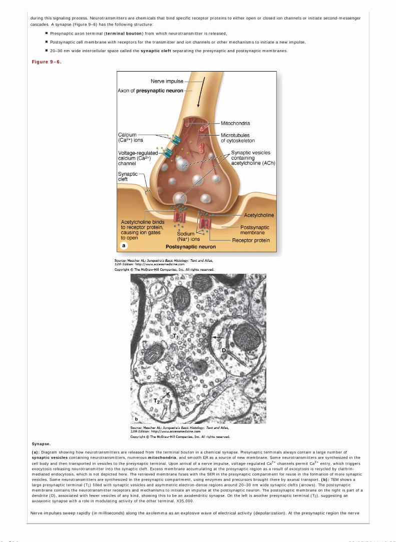
during this signaling process. Neurotransmitters are chemicals that bind specific receptor proteins to either open or closed ion channels or initiate second-messenger
cascades. A synapse (Figure 9–6) has the following structure:
Presynaptic axon terminal (terminal bouton) from which neurotransmitter is released,
Postsynaptic cell membrane with receptors for the transmitter and ion channels or other mechanisms to initiate a new impulse,
20–30 nm wide intercellular space called the synaptic cleft separating the presynaptic and postsynaptic membranes.
Figure 9–6.
Synapse.
(a): Diagram showing how neurotransmitters are released from the terminal bouton in a chemical synapse. Presynaptic terminals always contain a large number ofsynaptic vesicles containing neurotransmitters, numerous mitochondria, and smooth ER as a source of new membrane. Some neurotransmitters are synthesized in the
cell body and then transported in vesicles to the presynaptic terminal. Upon arrival of a nerve impulse, voltage-regulated Ca2+ channels permit Ca2+ entry, which triggersexocytosis releasing neurotransmitter into the synaptic cleft. Excess membrane accumulating at the presynaptic region as a result of exocytosis is recycled by clathrin-mediated endocytosis, which is not depicted here. The retrieved membrane fuses with the SER in the presynaptic compartment for reuse in the formation of more synapticvesicles. Some neurotransmitters are synthesized in the presynaptic compartment, using enzymes and precursors brought there by axonal transport. (b): TEM shows alarge presynaptic terminal (T1) filled with synaptic vesicles and asymmetric electron-dense regions around 20–30 nm wide synaptic clefts (arrows). The postsynapticmembrane contains the neurotransmitter receptors and mechanisms to initiate an impulse at the postsynaptic neuron. The postsynaptic membrane on the right is part of adendrite (D), associated with fewer vesicles of any kind, showing this to be an axodendritic synapse. On the left is another presynaptic terminal (T2), suggesting anaxoaxonic synapse with a role in modulating activity of the other terminal. X35,000.
Nerve impulses sweep rapidly (in milliseconds) along the axolemma as an explosive wave of electrical activity (depolarization). At the presynaptic region the nerve
AccessMedicine | Print: Chapter 9. Nerve Tissue & the Nervous System http://accessmedicine.com/popup.aspx?aID=6181123&print=yes_chapter
8 of 36 22/05/2011 19:25
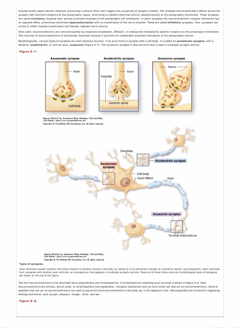
impulse briefly opens calcium channels, promoting a calcium influx that triggers the exocytosis of synaptic vesicles. The released neurotransmitters diffuse across the
synaptic cleft and bind receptors at the postsynaptic region, promoting a transient electrical activity (depolarization) at the postsynaptic membrane. These synapses
are called excitatory, because their activity promotes impulses in the postsynaptic cell membrane. In some synapses the neurotransmitter-receptor interaction has
an opposite effect, promoting membrane hyperpolarization with no transmission of the nerve impulse. These are called inhibitory synapses. Thus, synapses can
excite or inhibit impulse transmission and thereby regulate nerve activity.
Once used, neurotransmitters are removed quickly by enzymatic breakdown, diffusion, or endocytosis mediated by specific receptors on the presynaptic membrane.
This removal of neurotransmitters is functionally important because it prevents an undesirable sustained stimulation of the postsynaptic neuron.
Morphologically, various types of synapses are seen between neurons. If an axon forms a synapse with a cell body, it is called an axosomatic synapse; with a
dendrite, axodendritic; or with an axon, axoaxonic (Figure 9–7). The axoaxonic synapse is less common and is used to modulate synaptic activity.
Figure 9–7.
Types of synapses.
Axon terminals usually transmit the nerve impulse to another neuron's cell body (or soma) or to its dendrites (usually at a dendritic spine). Less frequently, axon terminalsform synapses with another axon terminal, an arrangement that appears to modulate synaptic activity. Features of these three common morphological types of synapsesare shown at the top of the figure.
The first neurotransmitters to be described were acetylcholine and norepinephrine. A norepinephrine-releasing axon terminal is shown in Figure 9–8. Most
neurotransmitters are amines, amino acids, or small peptides (neuropeptides). Inorganic substances such as nitric oxide can also act as neurotransmitters. Several
peptides that can act as neurotransmitters are used as paracrine hormones elsewhere in the body eg, in the digestive tract. Neuropeptides are involved in regulating
feelings and drives, such as pain, pleasure, hunger, thirst, and sex.
Figure 9–8.
AccessMedicine | Print: Chapter 9. Nerve Tissue & the Nervous System http://accessmedicine.com/popup.aspx?aID=6181123&print=yes_chapter
9 of 36 22/05/2011 19:25

Adrenergic nerve ending.
Many 50-nm-diameter vesicles, with electron-dense cores containing norepinephrine, fill the axon terminal shown here. X40,000. (Machado AB: Straight OsO4 versusglutaraldehyde-OsO4 in sequence as fixatives for the granular vesicles in sympathetic axons of the rat pineal body. Stain Technology 1967, 42(6):293. Reproduced, withpermission, from Taylor & Francis Group, http://www.informaworld.com, and Angelo B. Machado, Department of Morphology, Federal University of Minas Gerais, BeloHorizonte, Brazil)
Neuromodulators are chemical messengers that modify neuron sensitivity to synaptic stimulation or inhibition, without acting directly on synapses. Some
neuromodulators are neuropeptides or steroids produced in the nerve tissue, others are circulating steroids.
Although most synapses are chemical synapses and use chemical neurotransmitters, some synapses transmit ionic signals through gap junctions between the pre-
and postsynaptic membranes, thereby conducting neuronal signals directly. These synapses are electrical synapses and are prominent in cardiac and smooth
muscle.
GLIAL CELLS & NEURONAL ACTIVITY
Glial cells are 10 times more abundant in the mammalian brain than neurons. In the CNS glial cells surround most of the neuronal cell bodies, which are usually much
larger than glial cells, and the processes of axons and dendrites that occupy the spaces between neurons. Except around the larger blood vessels, the CNS has only a
very small amount of connective tissue or ECM. Glial cells (Table 9–2) furnish a microenvironment ideal for neuronal activity. A dense network of fibers from
processes of both neurons and glial cells fills the interneuronal space of the CNS and is called the neuropil (Figure 9–9).
Table 9–2. Origin and principal functions of neuroglial cells.
Glial Cell Type Origin Location Main Functions
Oligodendrocyte Neural tube Central nervous system Myelin production, electric insulation
Neurolemmocyte Neural crest Peripheral nerves Myelin production, electric insulation
Astrocyte Neural tube Central nervous system Structural support, repair processes
Blood-brain barrier, metabolic exchanges
Ependymal cell Neural tube Central nervous system Lining cavities of central nervous system
Microglia Bone marrow Central nervous system Immune-related activity
Figure 9–9.
AccessMedicine | Print: Chapter 9. Nerve Tissue & the Nervous System http://accessmedicine.com/popup.aspx?aID=6181123&print=yes_chapter
10 of 36 22/05/2011 19:25
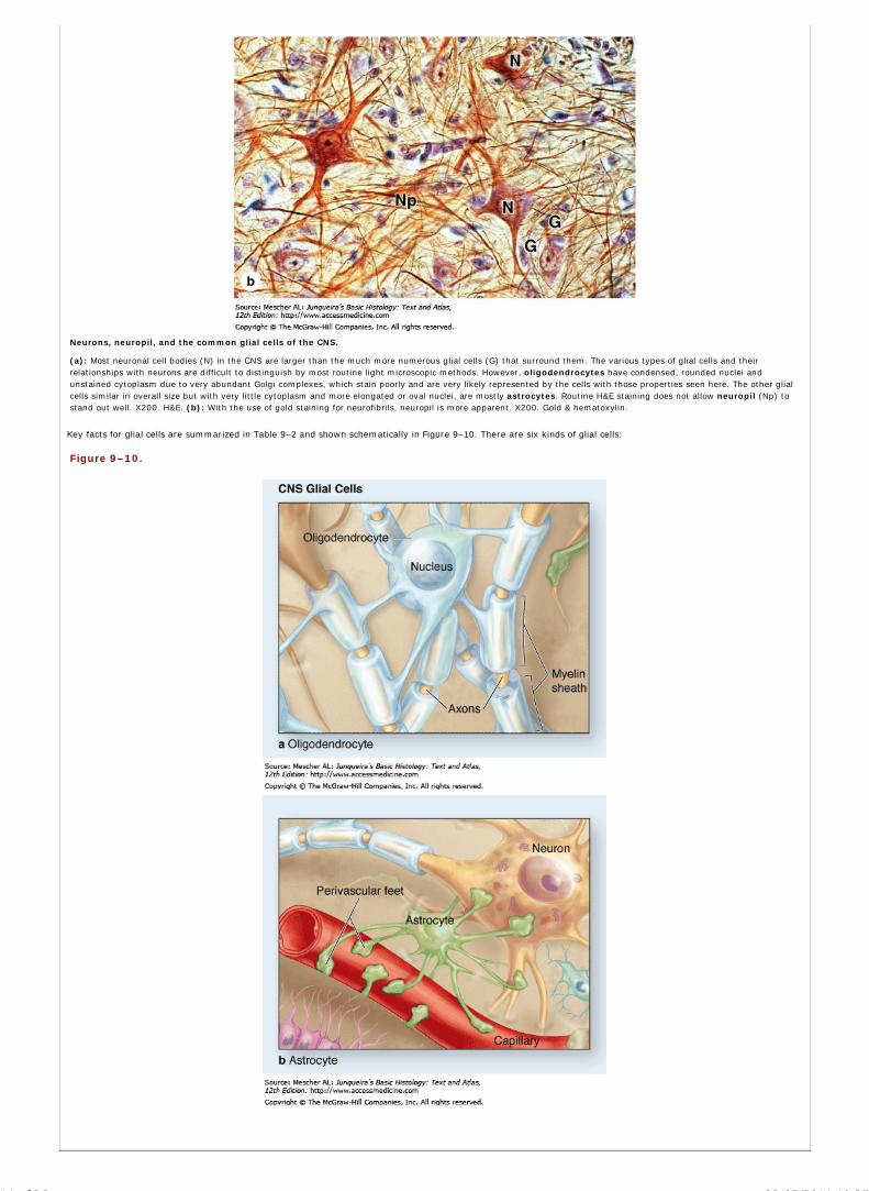
Neurons, neuropil, and the common glial cells of the CNS.
(a): Most neuronal cell bodies (N) in the CNS are larger than the much more numerous glial cells (G) that surround them. The various types of glial cells and theirrelationships with neurons are difficult to distinguish by most routine light microscopic methods. However, oligodendrocytes have condensed, rounded nuclei andunstained cytoplasm due to very abundant Golgi complexes, which stain poorly and are very likely represented by the cells with those properties seen here. The other glialcells similar in overall size but with very little cytoplasm and more elongated or oval nuclei, are mostly astrocytes. Routine H&E staining does not allow neuropil (Np) tostand out well. X200. H&E. (b): With the use of gold staining for neurofibrils, neuropil is more apparent. X200. Gold & hematoxylin.
Key facts for glial cells are summarized in Table 9–2 and shown schematically in Figure 9–10. There are six kinds of glial cells:
Figure 9–10.
AccessMedicine | Print: Chapter 9. Nerve Tissue & the Nervous System http://accessmedicine.com/popup.aspx?aID=6181123&print=yes_chapter
11 of 36 22/05/2011 19:25

AccessMedicine | Print: Chapter 9. Nerve Tissue & the Nervous System http://accessmedicine.com/popup.aspx?aID=6181123&print=yes_chapter
12 of 36 22/05/2011 19:25

Glial cells of the CNS and PNS.
Glial cell in the CNS. (a): Oligodendrocytes myelinate parts of several axons. (b): Astrocytes have multiple processes and form perivascular feet that completely encloseall capillaries (only a few such feet are shown here to allow their morphology to be seen). (c): Ependymal cells are epithelial-like cells that line the ventricles and centralcanal. (d): Microglial cells have a protective, phagocytic, immune-related function. Glial cells in the PNS. (e): Neurolemmocytes, commonly called Schwann cells, forma series enclosing axons. (f): Satellite cells are restricted to ganglia where they cover and support the large neuronal cell bodies.
OligodendrocytesOligodendrocytes (Gr. oligos, small, few + dendron, tree + kytos, cell) produce the myelin sheath that provides the electrical insulation for neurons in the CNS.
Oligodendrocytes extend processes that wrap around parts of several axons, producing a myelin sheath as shown in Figure 9–10a. They are the predominant glial cell
in CNS white matter. The processes are not visible by routine light microscope staining, in which oligodendrocytes usually appear as small cells with rounded,
condensed nuclei and unstained cytoplasm (Figures 9–9a and 9–10a).
AstrocytesAstrocytes (Gr. astron, star, + kytos) have a large number of radiating processes (Figures 9–10b and 9–11) and are unique to the CNS. Astrocytes with relatively few
long processes are called fibrous astrocytes and are located in the white matter; protoplasmic astrocytes, with many short, branched processes, are found in the
gray matter. Astrocytes have supportive roles for neurons and are very important for proper formation of the CNS during embryonic and fetal development. Located
mainly in gray matter, astrocytes are by far the most numerous glial cells and exhibit considerable morphologic and functional diversity.
Figure 9–11.
AccessMedicine | Print: Chapter 9. Nerve Tissue & the Nervous System http://accessmedicine.com/popup.aspx?aID=6181123&print=yes_chapter
13 of 36 22/05/2011 19:25

Astrocytes.
(a): Astrocytes are the most numerous glial cells of the CNS and are characterized by numerous cytoplasmic processes (P) radiating from the glial cell body or soma (S).Astrocytic processes are not seen with routine light microscope staining, but are easily seen after gold staining. Morphology of the processes allows astrocytes to beclassified as fibrous (relatively few and straight processes) or protoplasmic (numerous branching processes), but functional differences between these types are not clear.X500. Gold chloride. (b): All astrocytic processes contain intermediate filaments of glial fibrillary acidic protein (GFAP) and antibodies against this protein provide a simplemethod to stain these cells, as seen here in a fibrous astrocyte (A) and its processes. The small pieces of other GFAP-positive processes in the neuropil around this cell givean idea of the density of this glial cell and its processes in the CNS. Astrocytes are an important part of the blood-brain barrier regulating entry of molecules and ions fromblood into CNS tissue. Capillaries at the extreme upper right and lower left corners of (b) are enclosed by GFAP-positive perivascular feet (PF) at the ends of numerousastrocytic processes. X500. Anti-GFAP immunoperoxidase and hematoxylin counterstain. (c): A length of capillary is shown here completely enclosed within stainedprocesses of astrocytes. X400. Rio Hortega silver.
In addition to their supporting function, astrocytes have major roles in controlling the ionic environment of neurons. Some astrocytes develop processes with
expanded perivascular feet that cover capillary endothelial cells and contribute to the blood-brain barrier. The perivascular feet are important for the ability of
astrocytes to regulate vasodilation and transfer of O2, ions and other substances from the blood to the neurons. Other expanded processes form a layer, the
superficial glial limiting membrane which lines the pia mater, the innermost meningial layer at the external surface of the CNS. Furthermore, when the CNS is
damaged, astrocytes proliferate to form cellular scar tissue (which often interferes with neuronal regeneration).
Astrocytic functions are essential for neuronal survival. They regulate constituents of the extracellular environment, absorb local excess of neurotransmitters, and
AccessMedicine | Print: Chapter 9. Nerve Tissue & the Nervous System http://accessmedicine.com/popup.aspx?aID=6181123&print=yes_chapter
14 of 36 22/05/2011 19:25

secrete numerous metabolites and factors regulating neuronal activities. Finally, astrocytes are in direct communication with one another via gap junctions, forming a
network through which information can flow from one point to another, reaching distant sites.
The processes of all astrocytes are reinforced with bundles of intermediate filaments made of glial fibrillary acid protein (GFAP), which serves as a unique marker
for astrocytes, the most common source of brain tumors.
Ependymal CellsEpendymal cells are low columnar or cuboidal cells that line the ventricles of the brain and central canal of the spinal cord (Figures 9–10c and 9–12). In some CNS
locations, the apical ends of ependymal cells have cilia, which facilitate the movement of cerebrospinal fluid (CSF), or long microvilli, which are likely involved in
absorption.
Figure 9–12.
Ependymal cells.
Ependymal cells are epithelial-like cells that form a single layer lining the fluid-filled ventricles of (a) the cerebrum and (b) the central canal of the spinal cord. Cuboidal orlow columnar, ependymal cells in various CNS regions may be clearly ciliated or have long microvilli at the apical surfaces. These modifications help circulate the CSF andmonitor its contents. Also at their apical ends ependymal cells have junctional complexes like those of epithelial cells. However, they lack a basal lamina. The cells' basalends are elongated and tapered, extending processes that branch and penetrate some distance into the adjacent neuropil. Ependymal cells not only help move and absorbCSF, they are responsible for production of this fluid in specialized ventricular tissues of the choroid plexus. Both: X200. H&E.
Ependymal cells are joined apically by junctional complexes similar to those of epithelia. However, unlike a true epithelium there is no basal lamina. Instead, the
basal ends of ependymal cells are elongated and extend branching processes into the adjacent neuropil.
MicrogliaSomewhat less numerous than oligodendrocytes or astrocytes but more evenly distributed throughout gray and white matter, microglia are small cells with short
irregular processes (Figures 9–10d and 9–13). Unlike other glial cells microglia migrate through the neuropil, analyzing the tissue for damaged cells and invading
microorganisms. They secrete a number of immunoregulatory cytokines and constitute the major mechanism of immune defense in CNS tissues. Microglia originate
AccessMedicine | Print: Chapter 9. Nerve Tissue & the Nervous System http://accessmedicine.com/popup.aspx?aID=6181123&print=yes_chapter
15 of 36 22/05/2011 19:25

not from the embryonic neural tube but from circulating blood monocytes, belonging to the same family as macrophages and other antigen-presenting cells.
Figure 9–13.
Microglial cells.
Microglia are the monocyte-derived, antigen-presenting immune cells of the CNS and are evenly distributed in both gray and white matter. By immunohistochemistry, hereusing a monoclonal antibody against HLA antigens found on many immune-related cells, the short branching processes of microglia can be seen. Routine staining does notdemonstrate the processes, but only the small dark nuclei of the cells. Microglia move around and are constantly employed in immune surveillance of CNS tissues. Whenactivated by products of cell damage or microorganisms, the cells retract their processes, begin phagocytosing the damage- or danger-related material, and behave asantigen-presenting cells. X500. Antibody against HLA-DR and peroxidase. (Used, with permission, from Wolfgang Streit, Department of Neuroscience, University of FloridaCollege of Medicine, Gainesville.)
Nuclei of microglial cells can be recognized in routine H&E preparations by their dense elongated structure, which contrasts with the spherical, more lightly stained
nuclei of other glial cells. Immunohistochemistry using antibodies against cell surface antigens of immune cells demonstrates microglial processes. When activated,
microglia retract their processes and assume the morphologic characteristics of macrophages, become phagocytic and act as antigen-presenting cells (see Chapter
14).
MEDICAL APPLICATION
In multiple sclerosis, the myelin sheath is damaged by an autoimmune mechanism with various neurologic consequences. In this disease, microglia
phagocytose and degrade myelin debris by receptor-mediated phagocytosis and lysosomal activity. In addition, AIDS dementia complex is caused by HIV-1
infection of the central nervous system. Overwhelming experimental evidence indicates that microglia are infected by HIV-1. A number of cytokines, such as
interleukin-1 and tumor necrosis factor- , activate and enhance HIV replication in microglia.
Schwann Cells (Neurolemmocytes)Schwann cells, also called neurolemmocytes, are found only in the PNS and have trophic interactions with axons and allow for their myelination like the
oligodendrocytes of the CNS. One neurolemmocyte forms myelin around a segment of one axon, in contrast to the ability of oligodendrocytes to branch and sheath
parts of more than one axon. Figure 9–10e shows how a series of Schwann cells covers the full length of an axon.
Satellite Cells of GangliaDerived from the embryonic neural crest like neurolemmocytes, small satellite cells form a covering layer over the large neuronal cell bodies in PNS ganglia (Figure
9–10f). Closely associated with the neurons, the satellite cells exert a trophic or supportive role, but the molecular basis of their support is poorly understood.
CENTRAL NERVOUS SYSTEM
The principal structures of the CNS are the cerebrum, cerebellum, and spinal cord. It has virtually no connective tissue and is therefore a relatively soft, gel-like
organ.
When sectioned, the cerebrum, cerebellum, and spinal cord show regions of white (white matter) and gray (gray matter), differences caused by the differential
distribution of myelin. The main components of white matter are myelinated axons (Figure 9–14) and the myelin-producing oligodendrocytes. White matter does not
contain neuronal cell bodies, but microglia are present.
Figure 9–14.
AccessMedicine | Print: Chapter 9. Nerve Tissue & the Nervous System http://accessmedicine.com/popup.aspx?aID=6181123&print=yes_chapter
16 of 36 22/05/2011 19:25

White versus gray matter, stained.
A cross section of spinal cord shows the transition between white matter (left) and gray matter (right). The white matter consists mainly of nerve fibers whose myelinsheaths were dissolved in the preparation procedure, leaving the round empty spaces shown. Each such space surrounds a dark-stained spot which is the axon. Neuronalcell bodies, astrocytes, and abundant cell processes predominate in the gray matter. X400. PT.
Gray matter contains abundant neuronal cell bodies, dendrites, the initial unmyelinated portions of axons, astrocytes, and microglial cells. This is the region where
synapses occur. Gray matter is prevalent at the surface or cortex of the cerebrum and cerebellum, whereas white matter is present in more central regions.
Aggregates of neuronal cell bodies forming islands of gray matter embedded in the white matter are called nuclei. Neuroscientists recognize six layers in the
cerebral cortex with most neurons arranged vertically. The most abundant neurons are the efferent pyramidal neurons which come in many sizes (Figure 9–15).
Cells of the cerebral cortex function in the integration of sensory information and the initiation of voluntary motor responses.
Figure 9–15.
AccessMedicine | Print: Chapter 9. Nerve Tissue & the Nervous System http://accessmedicine.com/popup.aspx?aID=6181123&print=yes_chapter
17 of 36 22/05/2011 19:25

Cerebral cortex.
(a): Important neurons of the cerebrum are pyramidal neurons (P), which are arranged vertically and interspersed with numerous glial cells in the eosinophilic neuropil.X200. H&E. (b): From the apical ends of pyramidal neuron, long dendrites extend in the direction of the cortical surface, which can be best seen in thick silver-stainedsections in which only a few other protoplasmic glial cells are seen. X200. Silver.
The cerebellar cortex, which coordinates muscular activity throughout the body, has three layers (Figure 9–16): an outer molecular layer, a central layer of very
large neurons called Purkinje cells, and an inner granule layer. The Purkinje cell bodies are conspicuous even in H&E stained material and their dendrites extend
throughout the molecular layer as a branching basket of nerve fibers (Figure 9-16). The granule layer is formed by very small neurons (the smallest in the body),
which are packed together densely, in contrast to the neuronal cell bodies in the molecular layer which are sparse (Figure 9–16).
Figure 9–16.
AccessMedicine | Print: Chapter 9. Nerve Tissue & the Nervous System http://accessmedicine.com/popup.aspx?aID=6181123&print=yes_chapter
18 of 36 22/05/2011 19:25

Cerebellum.
(a): The cerebellar cortex is convoluted with many distinctive small folds, each supported at its center by cerebellar medulla (M), which is white matter consisting of large
AccessMedicine | Print: Chapter 9. Nerve Tissue & the Nervous System http://accessmedicine.com/popup.aspx?aID=6181123&print=yes_chapter
19 of 36 22/05/2011 19:25

tracts of axons. X6. Cresyl violet. (b): Immediately surrounding the white matter of the medulla is the granular layer (GL) of the cortex, which is densely packed with verysmall, rounded neuronal cell bodies. The outer, "molecular layer" (ML) consists of neuropil with fewer, more scattered small neurons. X20. H&E. (c): At the interfacebetween the granular and molecular layers is a single layer with very large neuronal cell bodies of unique Purkinje cells (P), whose axons pass through the granular layer(Gr) to join tracts in the medulla and whose multiple branching dendrites ramify throughout the molecular layer (Mol). X40. H&E. (d): Although not seen until well afterH&E staining, dendrites of Purkinje cells have hundreds of small branches, each covered with dendritic spines, which can be demonstrated with silver stains. Axons fromthe small neurons of the granular layer are unmyelinated and run together into the molecular layer where they form synapses with the dendrites spines of Purkinje cells.The molecular layer of the cerebellar cortex contains relatively few neurons or other cells. X40. Silver.
In cross sections of the spinal cord, white matter is peripheral and gray matter is internal and has the general shape of an H (Figure 9–17). In the center is an
opening, the central canal, which develops from the lumen of the embryonic neural tube and is lined by ependymal cells. The gray matter forms the anterior horns,
which contain motor neurons whose axons make up the ventral roots of spinal nerves, and the posterior horns, which receive sensory fibers from neurons in the spinal
ganglia (dorsal roots). Spinal cord neurons are large and multipolar, especially the motor neurons in the anterior horns (Figure 9–17).
Figure 9–17.
AccessMedicine | Print: Chapter 9. Nerve Tissue & the Nervous System http://accessmedicine.com/popup.aspx?aID=6181123&print=yes_chapter
20 of 36 22/05/2011 19:25

Spinal cord.
The spinal cord varies slightly in diameter along its length, but in cross-section always shows bilateral symmetry around the small, CSF-filled central canal. Unlike thecerebrum and cerebellum, in the spinal cord the gray matter is internal, forming a roughly H-shaped structure that consists of two posterior (P) horns (sensory) and twoanterior (A) (motor) horns all joined by the gray commissure around the central canal. (a): The gray matter contains abundant astrocytes and large neuronal cell bodies,especially those of motor neurons in the ventral horns. (b): The white matter surrounds the gray matter and contains primarily oligodendrocytes and tracts of myelinatedaxons running along the length of the cord.
(c): Micrograph of the large motor neurons of the ventral horns show large nuclei, prominent nucleoli, and cytoplasm rich in chromatophilic substance (Nissl substance), allof which indicate extensive protein synthesis to maintain the axons of these cells which extend great distances. (d): In the white commissure ventral to the central canal,tracts run lengthwise along the cord, seen here in cross-section with empty myelin sheaths surrounding axons, as well as tracts running from one side of the cord to theother, seen here as several longitudinally sectioned tracts of eosinophilic axons. Center: X5; a–d: X200.; Center, a, b: silver; c, d: H&E.
MeningesThe skull and the vertebral column protect the CNS. Between the bone and nervous tissue are membranes of connective tissue called the meninges (Figures 9–18
and 9–19). Three meningial layers are distinguished:
Figure 9–18.
AccessMedicine | Print: Chapter 9. Nerve Tissue & the Nervous System http://accessmedicine.com/popup.aspx?aID=6181123&print=yes_chapter
21 of 36 22/05/2011 19:25
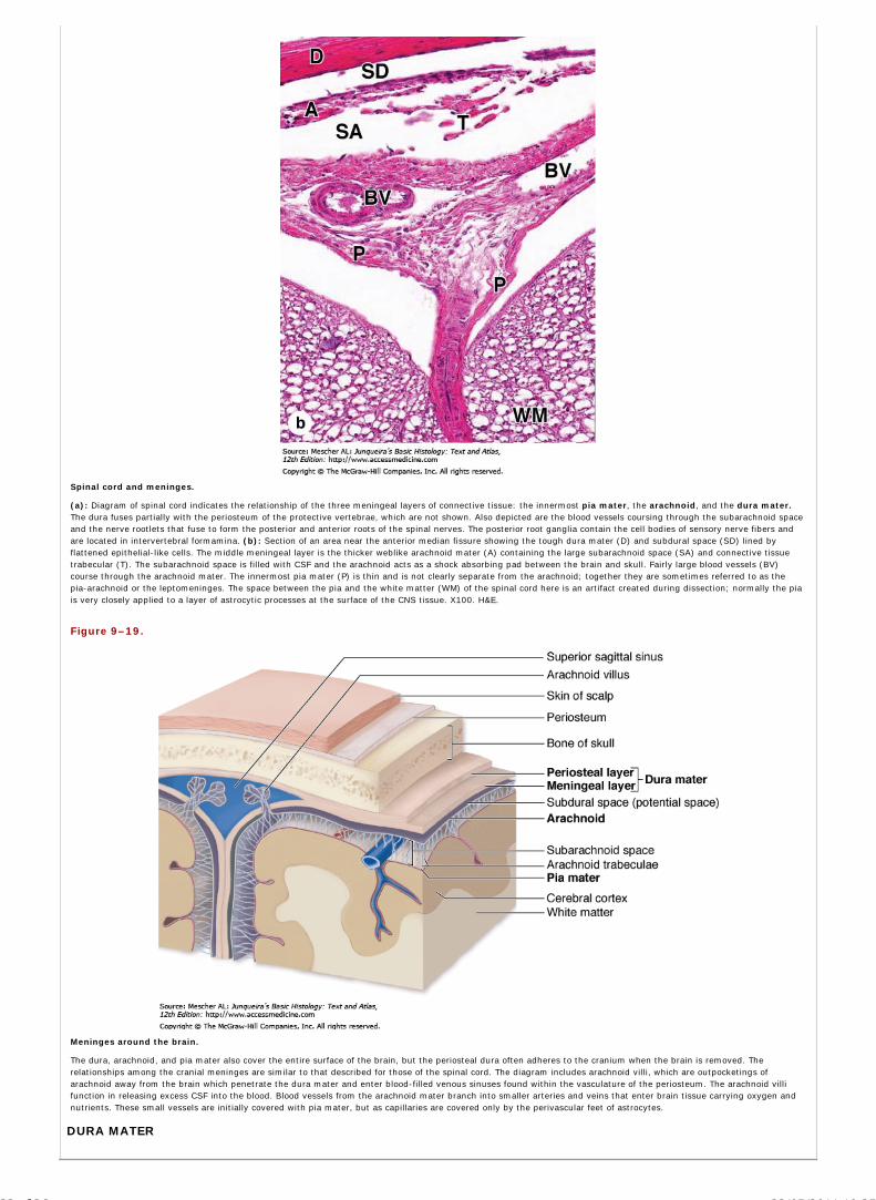
Spinal cord and meninges.
(a): Diagram of spinal cord indicates the relationship of the three meningeal layers of connective tissue: the innermost pia mater, the arachnoid, and the dura mater.The dura fuses partially with the periosteum of the protective vertebrae, which are not shown. Also depicted are the blood vessels coursing through the subarachnoid spaceand the nerve rootlets that fuse to form the posterior and anterior roots of the spinal nerves. The posterior root ganglia contain the cell bodies of sensory nerve fibers andare located in intervertebral formamina. (b): Section of an area near the anterior median fissure showing the tough dura mater (D) and subdural space (SD) lined byflattened epithelial-like cells. The middle meningeal layer is the thicker weblike arachnoid mater (A) containing the large subarachnoid space (SA) and connective tissuetrabecular (T). The subarachnoid space is filled with CSF and the arachnoid acts as a shock absorbing pad between the brain and skull. Fairly large blood vessels (BV)course through the arachnoid mater. The innermost pia mater (P) is thin and is not clearly separate from the arachnoid; together they are sometimes referred to as thepia-arachnoid or the leptomeninges. The space between the pia and the white matter (WM) of the spinal cord here is an artifact created during dissection; normally the piais very closely applied to a layer of astrocytic processes at the surface of the CNS tissue. X100. H&E.
Figure 9–19.
Meninges around the brain.
The dura, arachnoid, and pia mater also cover the entire surface of the brain, but the periosteal dura often adheres to the cranium when the brain is removed. Therelationships among the cranial meninges are similar to that described for those of the spinal cord. The diagram includes arachnoid villi, which are outpocketings ofarachnoid away from the brain which penetrate the dura mater and enter blood-filled venous sinuses found within the vasculature of the periosteum. The arachnoid villifunction in releasing excess CSF into the blood. Blood vessels from the arachnoid mater branch into smaller arteries and veins that enter brain tissue carrying oxygen andnutrients. These small vessels are initially covered with pia mater, but as capillaries are covered only by the perivascular feet of astrocytes.
DURA MATER
AccessMedicine | Print: Chapter 9. Nerve Tissue & the Nervous System http://accessmedicine.com/popup.aspx?aID=6181123&print=yes_chapter
22 of 36 22/05/2011 19:25

The dura mater is the thick external layer consisting of dense, fibroelastic connective tissue continuous with the periosteum of the skull. Around the spinal cord the
dura mater is separated from the periosteum of the vertebrae by the epidural space, which contains a plexus of thin-walled veins and areolar connective tissue.
The dura mater is always separated from the arachnoid by the thin subdural space. The internal surface of all dura mater, as well as its external surface in the
spinal cord, is covered by simple squamous epithelium of mesenchymal origin (Figure 9–18).
ARACHNOID
The arachnoid (Gr. arachnoeides, spiderweblike) has two components: (1) a sheet of connective tissue in contact with the dura mater and (2) a system of loosely
arranged trabeculae containing fibroblasts and collagen. This trabecular system is continuous with the deeper pia mater. Surrounding the trabeculae is a large,
sponge-like cavity, the subarachnoid space, filled with CSF. This space forms a hydraulic cushion that protects the CNS from trauma. The subarachnoid space
communicates with the ventricles of the brain.
The connective tissue of the arachnoid is said to be avascular because it lacks nutritive capillaries, but larger blood vessels run through it (Figure 9–18). Because the
arachnoid has fewer trabeculae in the spinal cord, it can be more clearly distinguished from the pia mater in that area. The arachnoid and the pia mater are intimately
associated and are often considered a single membrane called the pia-arachnoid.
In some areas, the arachnoid perforates the dura mater and protrudes into blood-filled venous sinuses within the dura mater. These CSF-filled protrusions, which are
covered by vascular endothelial cells, are called arachnoid villi. Their function is to transport CSF from the subarachnoid space into venous sinuses.
PIA MATER
The innermost pia mater is lined internally by flattened, mesenchymally derived cells closely applied to the entire surface of the CNS tissue, but this layer does not
directly contact nerve cells or fibers. Between the pia mater and the neural elements is a thin limiting layer of astrocytic processes, which adheres firmly to the pia
mater. Together the pia mater and glial layer form a physical barrier at the CNS periphery. This barrier separates the CNS tissue from the CSF in the subarachnoid
space (Figure 9–19).
Blood vessels penetrate the CNS through tunnels covered by pia mater—the perivascular spaces. The pia mater disappears when the blood vessels branch to the
smallest capillaries. However, these capillaries remain completely covered by expanded perivascular processes of astrocytes (Figure 9–11).
Blood-Brain BarrierThe blood-brain barrier (BBB) is a functional barrier that allows much tighter control than that in most tissues over the passage of substances moving from blood into
the CNS tissue, protecting the nature of the neuronal microenvironment. The main structural component of the BBB is the capillary endothelium, in which the cells
are tightly sealed together with well-developed occluding junctions and show little or no transcytosis. Moreover, the basal lamina of capillaries in most CNS regions is
enveloped by the perivascular feet of astrocytes (Figure 9–11) which further regulate passage of molecules and ions from blood to brain.
The BBB allows the stable composition and constant balance of ions in the interstitial fluid surrounding neurons and glial cells that is required for their function and
protects these cells from potential toxins and infectious agents. The components of the BBB are not found in the choroid plexus where CSF is produced, in the posterior
pituitary which releases hormones, or in regions of the hypothalamus where plasma components are monitored.
Choroid PlexusThe choroid plexus is a highly specialized tissue that projects as elaborate folds with many villi into the four large ventricles of the brain (Figure 9–20). It is found in
the roofs of the third and fourth ventricles and in parts of the walls of the two lateral ventricles, all regions in which the ependymal lining directly contacts the pia
mater.
Figure 9–20.
AccessMedicine | Print: Chapter 9. Nerve Tissue & the Nervous System http://accessmedicine.com/popup.aspx?aID=6181123&print=yes_chapter
23 of 36 22/05/2011 19:25
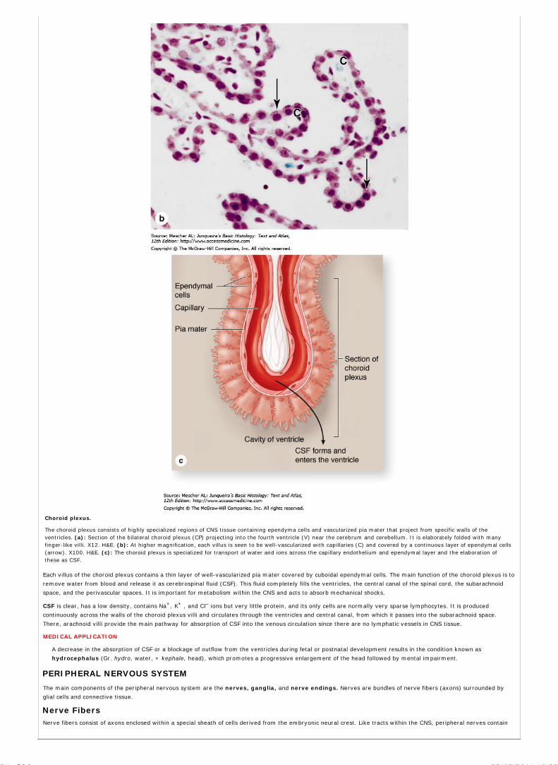
Choroid plexus.
The choroid plexus consists of highly specialized regions of CNS tissue containing ependyma cells and vascularized pia mater that project from specific walls of theventricles. (a): Section of the bilateral choroid plexus (CP) projecting into the fourth ventricle (V) near the cerebrum and cerebellum. It is elaborately folded with manyfinger-like villi. X12. H&E. (b): At higher magnification, each villus is seen to be well-vascularized with capillaries (C) and covered by a continuous layer of ependymal cells(arrow). X100. H&E. (c): The choroid plexus is specialized for transport of water and ions across the capillary endothelium and ependymal layer and the elaboration ofthese as CSF.
Each villus of the choroid plexus contains a thin layer of well-vascularized pia mater covered by cuboidal ependymal cells. The main function of the choroid plexus is to
remove water from blood and release it as cerebrospinal fluid (CSF). This fluid completely fills the ventricles, the central canal of the spinal cord, the subarachnoid
space, and the perivascular spaces. It is important for metabolism within the CNS and acts to absorb mechanical shocks.
CSF is clear, has a low density, contains Na+, K+ , and Cl– ions but very little protein, and its only cells are normally very sparse lymphocytes. It is produced
continuously across the walls of the choroid plexus villi and circulates through the ventricles and central canal, from which it passes into the subarachnoid space.
There, arachnoid villi provide the main pathway for absorption of CSF into the venous circulation since there are no lymphatic vessels in CNS tissue.
MEDICAL APPLICATION
A decrease in the absorption of CSF or a blockage of outflow from the ventricles during fetal or postnatal development results in the condition known as
hydrocephalus (Gr. hydro, water, + kephale, head), which promotes a progressive enlargement of the head followed by mental impairment.
PERIPHERAL NERVOUS SYSTEM
The main components of the peripheral nervous system are the nerves, ganglia, and nerve endings. Nerves are bundles of nerve fibers (axons) surrounded by
glial cells and connective tissue.
Nerve FibersNerve fibers consist of axons enclosed within a special sheath of cells derived from the embryonic neural crest. Like tracts within the CNS, peripheral nerves contain
AccessMedicine | Print: Chapter 9. Nerve Tissue & the Nervous System http://accessmedicine.com/popup.aspx?aID=6181123&print=yes_chapter
24 of 36 22/05/2011 19:25

groups of nerve fibers. In peripheral nerve fibers, axons are sheathed by Schwann cells, also called neurolemmocytes (Figure 9–10e). The sheath may or may
not form myelin around the axons, depending on their diameter.
Axons of small diameter are usually unmyelinated nerve fibers (Figures 9–22 and 9–25). Progressively thicker axons are generally sheathed by increasingly
numerous concentric wrappings of the enveloping cell, forming the myelin sheaths. These fibers are known as myelinated nerve fibers (Figures 9–21, 9–22, and
9–23).
Figure 9–21.
Myelination of large diameter PNS axons.
A neurolemmocyte (Schwann cell) engulfs a portion along the axon. The Schwann cell membrane fuses around the axon and elongates as it becomes wrapped around theaxon while the cell body moves around the axon many times. The neurolemmocyte membrane wrappings constitute the myelin sheath, with the cell body on its outer
AccessMedicine | Print: Chapter 9. Nerve Tissue & the Nervous System http://accessmedicine.com/popup.aspx?aID=6181123&print=yes_chapter
25 of 36 22/05/2011 19:25

surface. The myelin layers are very rich in lipid and provide insulation and facilitate formation of action potentials along the axolemma.
Figure 9–22.
Ultrastructure of myelinated and unmyelinated fibers.
Cross-section of PNS fibers in the TEM reveals differences between myelinated and unmyelinated axons. Large axons are wrapped in a thick myelin sheath (M) of multiplelayers of Schwann cell membrane. The inset shows a portion of myelin in which individual membrane layers can be distinguished easily, as well as the neurofilaments (NF)and microtubules (MT) in the axoplasm (A). At the center of the photo is a Schwann cell showing its active nucleus (SN) and Golgi-rich cytoplasm (SC). At the right is anaxon around which myelin is still forming (FM). Unmyelinated axons (UM) are much smaller in diameter and many such fibers may be engulfed by a single Schwann cell(SC). The glial cell does not form myelin wrappings around such small axons, but simply encloses them. Whether it forms myelin or not, each Schwann cell is surrounded,as shown, by an external lamina containing type IV collagen and laminin like the basal laminae of epithelial cells. X70,000. (Used, with permission, from Mary BartlettBunge, The Miami Project to Cure Paralysis, University of Miami Miller School of Medicine.)
Figure 9–23.
Nodes of Ranvier and endoneurium.
A longitudinal section of myelinated nerve fibers reveals the nodes of Ranvier, or nodal gaps, which are small, physiologically important gaps in the myelin sheath betweenadjacent Schwann cells. The axon can be seen spanning each nodal gap. Schwann cells produce an external lamina that surrounded their external surface. Like the basallaminae of epithelia, this structure contains type IV collagen and is continuous with the surrounding connective tissue rich in reticular fibers. This connective tissue makesup the endoneurium around the Schwann cells of all peripheral nerve fibers and is stained blue in this preparation. X400. Mallory trichrome.
MYELINATED FIBERS
As axons of large diameter grow in the PNS, they are engulfed along their length by many undifferentiated neurolemmocytes and become myelinated nerve fibers.
The plasma membrane of the covering neurolemmocyte (Schwann cell) fuses around the axon and becomes wrapped around the nerve fiber as the glial cell body
moves around and around the axon many times (Figure 9–21). The multiple layers of Schwann cell membrane unite as a layer myelin, a whitish lipoprotein complex
whose abundant lipid component is partly removed by standard histologic procedures, as in all cell membranes (Figures 9–14 and 9–17). With the TEM the myelin
sheath can appear as a thick electron-dense cover in which individual membrane layers are seen (Figure 9–22).
Membranes of Schwann cells have a higher proportion of lipids than do other cell membranes and the myelin sheath serves to protect axons and maintain a constant
ionic microenvironment required for action potentials. Between adjacent Schwann cells the myelin sheath shows small nodal gaps along the axon, also called nodes
AccessMedicine | Print: Chapter 9. Nerve Tissue & the Nervous System http://accessmedicine.com/popup.aspx?aID=6181123&print=yes_chapter
26 of 36 22/05/2011 19:25

of Ranvier (Figures 9–10e and 9–23). Interdigitating processes of Schwann cells partially cover each node (Figure 9–24). The length of axon covered by one Schwann
cell is called the internodal segment and may be more than 1 millimeter. Unlike oligodendrocytes of the CNS, Schwann cells only form myelin around a portion of
one axon.
Figure 9–24.
Myelin maintenance and nodal gaps (of Ranvier).
The center drawing shows a myelinated peripheral nerve fiber as seen under the light microscope. The axon is enveloped by the myelin sheath, which, in addition tomembrane, contains some Schwann cell cytoplasm in spaces between the membranes called myelin clefts (Schmidt-Lanterman clefts). The upper drawing shows one setof clefts ultrastructurally. The clefts contain Schwann cell cytoplasm that was not displaced to the cell body during myelin formation. This cytoplasm moves slowly alongthe myelin sheath, opening temporary spaces (the clefts) between the membrane layers, which allows renewal of some membrane components as needed andmaintenance of the sheath.
The lower drawing shows the ultrastructure of a nodal gap or node of Ranvier. Interdigitating processes extending from the outer layers of the Schwann cells (SC) partlycover and contact the axolemma at the nodal gap. This contact acts as a partial barrier to the movement of materials in and out of the periaxonal space between theaxolemma and the Schwann sheath. The basal or external lamina around Schwann cells is continuous over the nodal gap. Covering the nerve fiber is a thin connectivetissue layer that belongs to the endoneurial sheath of the peripheral nerve fibers.
UNMYELINATED FIBERS
The CNS is rich in unmyelinated axons which are not sheathed at all but run free among the other neuronal and glial processes. However in the PNS, even all
unmyelinated axons are enveloped within simple folds of Schwann cells (Figure 9–25). In this situation the glial cell does not form multiple wrapping of itself as
myelin. Unlike their association with individual myelinated axons, each Schwann cell can enclose portions of many unmyelinated axons with small diameters. Adjacent
Schwann cells along unmyelinated nerve fibers do not form nodes of Ranvier.
Figure 9–25.
AccessMedicine | Print: Chapter 9. Nerve Tissue & the Nervous System http://accessmedicine.com/popup.aspx?aID=6181123&print=yes_chapter
27 of 36 22/05/2011 19:25

Unmyelinated nerves.
During development portions of several small diameter axons are engulfed by one neurolemmocyte (Schwann cell). Subsequently the axons are separated and eachbecomes enclosed within its own fold or pocket of Schwann cell surface. No myelin is formed. Small diameter axons utilize action potentials whose formation andmaintenance do not depend on the insulation provided by the myelin sheath required by large diameter axons.
NervesIn the PNS nerve fibers are grouped into bundles to form nerves. Except for very thin nerves containing only unmyelinated fibers, nerves have a whitish, glistening
appearance because of their myelin and collagen content.
Axons and Schwann cells of nerves are enclosed within connective tissue layers (Figures 9–26 and 9–27). Externally is a dense, irregular fibrous coat called
epineurium, which continues more deeply to also fill the space between bundles of nerve fibers. Each such bundle or fascicle is surrounded by the perineurium, a
sleeve of specialized connective tissue formed by layers of flattened epithelial-like cells. The cells of each layer of the perineurium are joined at their edges by tight
junctions, an arrangement that makes the perineurium a barrier to the passage of most macromolecules and has the important function of protecting the nerve fibers
and helping maintain the internal microenvironment. Within the perineurial sheath run the Schwann cell–covered axons and their enveloping connective tissue, the
endoneurium (Figure 9–27). The endoneurium consists of a sparse layer of loose connective tissue that merges with an external lamina of type IV collagen, laminin,
and other proteins produced by the Schwann cells.
Figure 9–26.
AccessMedicine | Print: Chapter 9. Nerve Tissue & the Nervous System http://accessmedicine.com/popup.aspx?aID=6181123&print=yes_chapter
28 of 36 22/05/2011 19:25

Peripheral nerve connective tissue.
Peripheral nerves are protected by three layers of connective tissue, as depicted in the diagram (a). (b): The outer epineurium (E) consists of a dense superficial regionand a looser deep region that contains large blood vessels (A,V) and fascicles in which nerve fibers (N) are bundled. Each fascicle is surrounded by the perineurium (P),consisting of a few layers of unusual epithelial-like fibroblastic cells which are all joined at the peripheries by tight junctions to form a blood-nerve barrier that helpsregulate the microenvironment inside the fascicle. Axons and Schwann cells are in turn surrounded by a thin layer of endoneurium. X140. H&E. (c): As shown here theperineurium can extend as septa (S) into larger fascicles. X200. PT. (d): This micrograph shows a longitudinally oriented nerve. Within fascicles is the endoneurium (En)which surrounds capillaries (C) and is continuous with the external lamina produced by the Schwann cells. Collagen of the endoneurium is stained blue and a node ofRanvier (N) and a Schwann cell nucleus (S) are also clearly seen. X400. Mallory trichome.
Figure 9–27.
AccessMedicine | Print: Chapter 9. Nerve Tissue & the Nervous System http://accessmedicine.com/popup.aspx?aID=6181123&print=yes_chapter
29 of 36 22/05/2011 19:25

Peripheral nerve ultrastructure.
(a): SEM of transverse sections of a large peripheral nerve showing several fascicles, each surrounded by perineurium and packed with endoneurium which surrounds theindividual myelin sheathes. Each fascicle contains at least one capillary. Endothelial cells of these capillaries are tightly joined as part of the blood-nerve barrier andregulate the kinds of plasma substances released to the endoneurium. Larger blood vessels course through the deep epineurium which fills the space around theperineurium and fascicles. X450. (b): TEM shows a fibroblast (arrow) surrounded by collagen in the epineurium and three or four layers of highly flattened cells in theperineurium which form another part of the blood-nerve barrier. Inside the perineurium the endoneurium is rich in reticulin fibers (RF) which surround all Schwann cells.Nuclei of two schwann cells (SC) of myelinated fibers are visible. Many unmyelinated axons within two schwann cells are also present. X1200.
Very small nerves consist of one fascicle. Small nerves can be found in sections of many organs and often show a winding disposition in connective tissue (Figure
9–28).
Figure 9–28.
AccessMedicine | Print: Chapter 9. Nerve Tissue & the Nervous System http://accessmedicine.com/popup.aspx?aID=6181123&print=yes_chapter
30 of 36 22/05/2011 19:25

AccessMedicine | Print: Chapter 9. Nerve Tissue & the Nervous System http://accessmedicine.com/popup.aspx?aID=6181123&print=yes_chapter
31 of 36 22/05/2011 19:25

Small nerves.
Small nerves can be seen in sections from most organs. (a): In cross-section an isolated, resin-embedded nerve is seen to have a thin perineurium, one capillary (C), andmany large axons (arrows) associated with Schwann cells (arrowheads). A few nuclei of fibroblasts can be seen in the endoneurium between the myelinated fibers. X400.PT. (b): In longitudinal sections the flattened nuclei of endoneurial fibroblasts (F) and more oval nuclei of Schwann cells (S) can be distinguished. Nerve fibers are heldrather loosely in the endoneurium and in low-magnification longitudinal section are seen to be wavy rather than straight. This indicates a slackness of fibers within thenerve which allows nerves to stretch slightly during body movements with no potentially damaging tension on the fibers. X200. H&E. (c): In sections of mesentery andother tissues, a highly wavy or tortuous disposition of a single small nerve (N) will be seen as multiple oblique or transverse pieces as the nerve enters and leaves the areain the section. X200. H&E. (d): Often a section of small nerve will have some fibers cut transversely and others cut obliquely within the same fascicle, again suggesting therelatively unrestrained nature of the fibers within the endoneurium (E) and perineurium (P). X300. H&E.
The nerves establish communication between centers in the brain and spinal cord and the sense organs and effectors (muscles, glands, etc). They generally contain
both afferent and efferent fibers. Afferent fibers carry information from the interior of the body and the environment to the CNS. Efferent fibers carry impulses from
the CNS to effector organs commanded by these centers. Nerves possessing only sensory fibers are called sensory nerves; those composed only of fibers carrying
impulses to the effectors are called motor nerves. Most nerves have both sensory and motor fibers and are called mixed nerves which usually have both
myelinated and unmyelinated axons (Figure 9–27b).
GangliaGanglia are typically ovoid structures containing neuronal cell bodies and glial cells supported by connective tissue. Because they serve as relay stations to transmit
nerve impulses, one nerve enters and another exits from each ganglion. The direction of the nerve impulse determines whether the ganglion will be a sensory or an
autonomic ganglion.
SENSORY GANGLIA
Sensory ganglia receive afferent impulses that go to the CNS. Sensory ganglia are associated with both cranial nerves (cranial ganglia) and the dorsal root of the
spinal nerves (spinal ganglia). The large neuronal cell bodies of ganglia (Figure 9–29) are associated with thin, sheet-like extensions of small glial cells called satellite
cells (Figure 9–10f). These neural crest-derived cells create the microenvironments of the perikarya, allowing the production of membrane action potentials and
regulating metabolic exchange.
Figure 9–29.
AccessMedicine | Print: Chapter 9. Nerve Tissue & the Nervous System http://accessmedicine.com/popup.aspx?aID=6181123&print=yes_chapter
32 of 36 22/05/2011 19:25
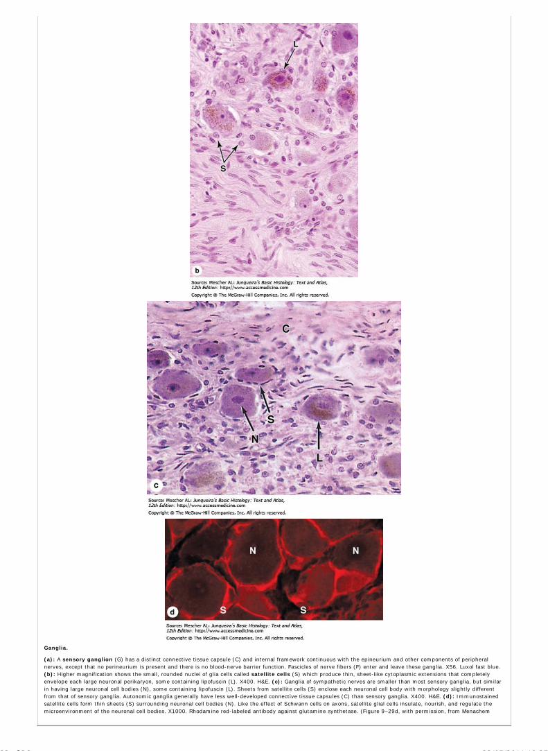
Ganglia.
(a): A sensory ganglion (G) has a distinct connective tissue capsule (C) and internal framework continuous with the epineurium and other components of peripheralnerves, except that no perineurium is present and there is no blood-nerve barrier function. Fascicles of nerve fibers (F) enter and leave these ganglia. X56. Luxol fast blue.(b): Higher magnification shows the small, rounded nuclei of glia cells called satellite cells (S) which produce thin, sheet-like cytoplasmic extensions that completelyenvelope each large neuronal perikaryon, some containing lipofuscin (L). X400. H&E. (c): Ganglia of sympathetic nerves are smaller than most sensory ganglia, but similarin having large neuronal cell bodies (N), some containing lipofuscin (L). Sheets from satellite cells (S) enclose each neuronal cell body with morphology slightly differentfrom that of sensory ganglia. Autonomic ganglia generally have less well-developed connective tissue capsules (C) than sensory ganglia. X400. H&E. (d): Immunostainedsatellite cells form thin sheets (S) surrounding neuronal cell bodies (N). Like the effect of Schwann cells on axons, satellite glial cells insulate, nourish, and regulate themicroenvironment of the neuronal cell bodies. X1000. Rhodamine red-labeled antibody against glutamine synthetase. (Figure 9–29d, with permission, from Menachem
AccessMedicine | Print: Chapter 9. Nerve Tissue & the Nervous System http://accessmedicine.com/popup.aspx?aID=6181123&print=yes_chapter
33 of 36 22/05/2011 19:25

Hanani, Laboratory of Experimental Surgery, Hadassah University Hospital, Jerusalem.)
Sensory ganglia are supported by a distinct connective tissue capsule and framework continuous with the connective tissue layers of the nerves. The neurons of these
ganglia are pseudounipolar and relay information from the ganglion's nerve endings to the gray matter of the spinal cord via synapses with local neurons.
AUTONOMIC GANGLIA
Autonomic (Gr. autos, self, + nomos, law) nerves effect the activity of smooth muscle, the secretion of some glands, modulate cardiac rhythm and other involuntary
activities by which the body maintains a constant internal environment (homeostasis).
Autonomic ganglia are small bulbous dilatations in autonomic nerves. Some are located within certain organs, especially in the walls of the digestive tract, where they
constitute the intramural ganglia (Chapter 15). The capsules of these ganglia are less well-defined. Autonomic ganglia usually have multipolar neurons. A layer of
satellite cells also envelops the neurons of autonomic ganglia (Figure 9-29), although in intramural ganglia, only a few satellite cells may be seen around each neuron.
Autonomic nerves use two-neuron circuits. The first neuron of the chain, with the preganglionic fiber, is located in the CNS. Its axon forms a synapse with
postganglionic fibers of the second multipolar neuron in the chain located in a peripheral ganglion system. The chemical mediator present in the synaptic vesicles of all
preganglionic axons is acetylcholine.
Autonomic nerves comprise an autonomic nervous system with two parts, called the sympathetic and the parasympathetic divisions. Neuronal cell bodies of
preganglionic sympathetic nerves are located in the thoracic and lumbar segments of the spinal cord and those of the parasympathetic division are in the medulla and
midbrain and in the sacral portion of the spinal cord. Sympathetic second neurons are located in small ganglia along the vertebral column, while second neurons of the
parasympathetic series are found in very small ganglia always located near or within the effector organs, for example in the walls of the stomach and intestines.
Parasympathetic ganglia may lack distinct capsules altogether, perikarya and associated satellite cells simply forming a loosely organized plexus within the
surrounding connective tissue.
NEURAL PLASTICITY & REGENERATION
Despite its general stability, the nervous system exhibits plasticity even in adults. Plasticity is very high during embryonic development, when an excess of nerve cells
is formed, and the cells that do not establish correct synapses with other neurons are eliminated by apoptosis. In adult mammals after an injury, the neuronal circuits
may be reorganized by the growth of neuronal processes, forming new synapses to replace the ones lost by injury. Thus, new communications are established with
some degree of functional recovery. This neural plasticity and reformation of processes are controlled by several growth factors produced by both neurons and glial
cells in a family of growth factors called neurotrophins.
Neuronal stem cells are present in adult CNS, located in part among the cells of the ependyma, which can supply new neurons, astrocytes and oligodendrocytes.
Because neurons cannot divide to replace those lost by injury or disease, the potential of neural stem cells to allow regeneration of CNS components is a subject of
intense investigation.
Injured fibers in peripheral nerves have a good capacity for regeneration and return of function. In a wounded nerve fiber, it is important to distinguish changes
occurring proximal to the injury from those in the distal segment. The proximal segment maintains its continuity with the trophic center in the perikaryon and can
regenerate, while the distal segment, separated from the nerve cell body, degenerates (Figure 9–30). The onset of regeneration is accompanied by changes in the
perikaryon: chromatolysis or dissolution of the RER and a consequent decrease in cytoplasmic basophilia; an increase in the volume of the perikaryon; and
migration of the nucleus to a peripheral position in the perikaryon. The proximal segment of the axon degenerates close to the wound for a short distance, but growth
starts as soon as debris is removed by macrophages. Macrophages produce cytokines which stimulate Schwann cells to secrete neurotrophins.
Figure 9–30.
AccessMedicine | Print: Chapter 9. Nerve Tissue & the Nervous System http://accessmedicine.com/popup.aspx?aID=6181123&print=yes_chapter
34 of 36 22/05/2011 19:25

AccessMedicine | Print: Chapter 9. Nerve Tissue & the Nervous System http://accessmedicine.com/popup.aspx?aID=6181123&print=yes_chapter
35 of 36 22/05/2011 19:25

Copyright © The McGraw-Hill Companies. All rights reserved.Privacy Notice. Any use is subject to the Terms of Use and Notice.
Regeneration in peripheral nerves.
In an injured or cut peripheral nerve, segments of axons distal to the injury lose their support from the cell bodies and degenerate completely. The proximal segments canregenerate from their cut ends after a delay. The main changes that take place in an injured nerve fiber are shown here. (a): Normal nerve fiber, with its perikaryon andeffector cell (skeletal muscle). The cell body has much well-developed RER. (b): When the axon is injured, the neuronal nucleus moves to the cell periphery, and the RERis greatly reduced. The nerve fiber distal to the injury degenerates along with its myelin sheath. Debris is phagocytosed by macrophages. (c): The muscle fiber showsdenervation atrophy. Schwann cells proliferate, forming a compact cord penetrated by the regrowing axon. The axon grows at the rate of 0.5–3 mm/day. (d): Here, thenerve fiber regeneration was successful and the muscle fiber was also regenerated after receiving nerve stimuli.
In the nerve segment distal to the injury the axon and myelin, but not the connective tissue, degenerate completely and are removed by macrophages. While these
regressive changes take place, Schwann cells proliferate within the connective tissue sleeve, giving rise to rows of cells that serve as guides for the sprouting axons
formed during the reparative phase.
MEDICAL APPLICATION
When there is an extensive gap between the distal and proximal segments of cut or injured peripheral nerves, or when the distal segment disappears
altogether (as in the case of amputation of a limb), the newly growing axons may form a swelling, or neuroma, that can be the source of spontaneous pain.
Regeneration is functionally efficient only when the fibers and the columns of Schwann cells are directed to the correct place. In an injured mixed nerve, if
regenerating sensory fibers grow into columns connected to motor end-plates that were occupied by motor fibers, the function of the muscle will not be
reestablished.
AccessMedicine | Print: Chapter 9. Nerve Tissue & the Nervous System http://accessmedicine.com/popup.aspx?aID=6181123&print=yes_chapter
36 of 36 22/05/2011 19:25

Print Close Window
Note: Large images and tables on this page may necessitate printing in landscape mode.
Copyright © The McGraw-Hill Companies. All rights reserved.
Junqueira's Basic Histology: Text & Atlas, 12e > Chapter 10. Muscle Tissue >
MUSCLE TISSUE: INTRODUCTION
Muscle tissue is composed of cells differentiated for optimal use of the universal cell property termed contractility. Microfilaments and associated proteins together
generate the forces necessary for cellular contraction, which drives movement within certain organs and the body as a whole. Nearly all muscle cells are of
mesodermal origin and they differentiate mainly by a gradual process of cell lengthening with simultaneous synthesis of myofibrillar proteins.
Three types of muscle tissue can be distinguished on the basis of morphologic and functional characteristics (Figure 10–1) and the structure of each type is adapted
to its physiologic role. Skeletal muscle is composed of bundles of very long, cylindrical, multinucleated cells that show cross-striations. Their contraction is quick,
forceful, and usually under voluntary control. It is caused by the interaction of thin actin filaments and thick myosin filaments whose molecular configuration allows
them to slide upon one another. The forces necessary for sliding are generated by weak interactions in the bridges between actin and myosin. Cardiac muscle
also has cross-striations and is composed of elongated, branched individual cells that lie parallel to each other. At sites of end-to-end contact are the intercalated
disks, structures found only in cardiac muscle. Contraction of cardiac muscle is involuntary, vigorous, and rhythmic. Smooth muscle consists of collections of
fusiform cells that do not show striations. Their contraction process is slow and not subject to voluntary control.
Figure 10–1.
AccessMedicine | Print: Chapter 10. Muscle Tissue http://accessmedicine.com/popup.aspx?aID=6181338&print=yes_chapter
1 of 24 22/05/2011 23:34

The three types of muscle.
Light micrographs of each type, accompanied by labeled drawings. (a): Skeletal muscle is composed of large, elongated, multinucleated fibers that show strong, quick,voluntary contractions. (b): Cardiac muscle is composed of irregular branched cells bound together longitudinally by intercalated disks and shows strong, involuntarycontractions. (c): Smooth muscle is composed of grouped, fusiform cells with weak, involuntary contractions. The density of intercellular packing seen reflects thesmall amount of extracellular connective tissue present. (a, b): X200. (c): X300. All H&E.
Some muscle cell organelles have names that differ from their counterparts in other cells. The cytoplasm of muscle cells is called sarcoplasm (Gr. sarkos, flesh, +
plasma, thing formed) and the smooth ER is called sarcoplasmic reticulum. The sarcolemma (sarkos + Gr. lemma, husk) is the cell membrane, or
plasmalemma.
SKELETAL MUSCLE
Skeletal muscle consists of muscle fibers, which are long, cylindrical multinucleated cells with diameters of 10–100 m. Multinucleation results from the fusion of
embryonic mesenchymal cells called myoblasts (Figure 10–2). The long oval nuclei are usually found at the periphery of the cell under the cell membrane. This
characteristic nuclear location is helpful in discriminating skeletal muscle from cardiac and smooth muscle, both of which have centrally located nuclei.
Figure 10–2.
AccessMedicine | Print: Chapter 10. Muscle Tissue http://accessmedicine.com/popup.aspx?aID=6181338&print=yes_chapter
2 of 24 22/05/2011 23:34

Development of skeletal muscle.
Skeletal muscle begins to differentiate when mesenchymal cells called myoblasts align and fuse together to make longer, multinucleated tubes called myotubes.Myotubes synthesize the proteins to make up myofilaments and gradually begin to show cross striations by light microscopy. Myotubes continue differentiating to formfunctional myofilaments and the nuclei are displaced against the sarcolemma. Part of the myoblast population does not fuse and differentiate, but remains as a group ofmesenchymal cells called muscle satellite cells located on the external surface of muscle fibers inside the developing external lamina. Satellite cells proliferate andproduce new muscle fibers following muscle injury.
MEDICAL APPLICATION
The variation in diameter of skeletal muscle fibers depends on factors such as the specific muscle and the age and sex, state of nutrition, and physical
training of the individual. It is a common observation that exercise enlarges the musculature and decreases fat depots. The increase in muscle thus
obtained is caused by formation of new myofibrils and a pronounced growth in the diameter of individual muscle fibers. This process, characterized by
increased of cell volume, is called hypertrophy (Gr. hyper, above, + trophe, nourishment). Tissue growth by an increase in the number of cells is termed
hyperplasia (hyper + Gr. plasis, molding), which takes place most readily in smooth muscle, whose cells have not lost the capacity to divide by mitosis.
OrganizationThe masses of fibers that make up the various types of muscle are arranged in regular bundles surrounded by the epimysium, an external sheath of dense
connective tissue surrounding the entire muscle (Figures 10–3 and 10–4). From the epimysium, thin septa of connective tissue extend inward, surrounding the
fascicles or bundles of fibers within a muscle. The connective tissue around each fascicle is called the perimysium. Each muscle fiber is itself surrounded by a
more delicate connective tissue, the endomysium, composed of a basal lamina synthesized by the multinucleated fibers themselves as well as reticular fibers and
fibroblasts. Within each fiber the nuclei are displaced peripherally against the sarcolemma.
Figure 10–3.
AccessMedicine | Print: Chapter 10. Muscle Tissue http://accessmedicine.com/popup.aspx?aID=6181338&print=yes_chapter
3 of 24 22/05/2011 23:34

Organization of skeletal muscle.
(a): An entire skeletal muscle is enclosed within a dense connective tissue layer called the epimysium continuous with the tendon binding it to bone. (b): Each fascicleof muscle fibers is wrapped in another connective tissue layer called the perimysium. (c): Individual muscle fibers (elongated multinuclear cells) is surrounded by avery delicate layer called the endomysium, which includes an external lamina produced by the muscle fiber (and enclosing the satellite cells) and ECM produced byfibroblasts.
Figure 10–4.
AccessMedicine | Print: Chapter 10. Muscle Tissue http://accessmedicine.com/popup.aspx?aID=6181338&print=yes_chapter
4 of 24 22/05/2011 23:34

Skeletal muscle.
(a): Micrograph shows a cross section of striated muscle demonstrating connective tissue and cell nuclei. The endomysium around individual muscle fibers is indicatedby arrowheads. At left is a portion of the epimysium. All three of these tissues contain collagen types I and III (reticulin). X200. H&E. (b): Adjacent sectionimmunohistochemically stained for laminin, which specifically stains the external lamina part of the endomysium produced by the muscle fibers themselves. X400.Anti-laminin.
One of the most important roles of this connective tissue is to transmit the mechanical forces generated by the contracting muscle cell/fibers because individual
muscle cells seldom extend from one end of a muscle to the other.
Blood vessels penetrate the muscle within the connective tissue septa and form a rich capillary network in the endomysium (Figure 10–5). Lymphatic vessels and
larger blood vessels are found in the other connective tissue layers.
Figure 10–5.
AccessMedicine | Print: Chapter 10. Muscle Tissue http://accessmedicine.com/popup.aspx?aID=6181338&print=yes_chapter
5 of 24 22/05/2011 23:34
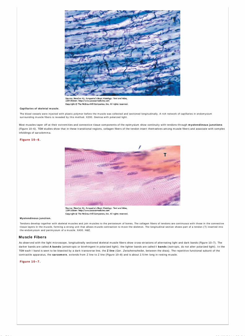
Capillaries of skeletal muscle.
The blood vessels were injected with plastic polymer before the muscle was collected and sectioned longitudinally. A rich network of capillaries in endomysiumsurrounding muscle fibers is revealed by this method. X200. Giemsa with polarized light.
Most muscles taper off at their extremities and connective tissue components of the epimysium show continuity with tendons through myotendinous junctions
(Figure 10–6). TEM studies show that in these transitional regions, collagen fibers of the tendon insert themselves among muscle fibers and associate with complex
infoldings of sarcolemma.
Figure 10–6.
Myotendinous junction.
Tendons develop together with skeletal muscles and join muscles to the periosteum of bones. The collagen fibers of tendons are continuous with those in the connectivetissue layers in the muscle, forming a strong unit that allows muscle contraction to move the skeleton. The longitudinal section shows part of a tendon (T) inserted intothe endomysium and perimysium of a muscle. X400. H&E.
Muscle FibersAs observed with the light microscope, longitudinally sectioned skeletal muscle fibers show cross-striations of alternating light and dark bands (Figure 10–7). The
darker bands are called A bands (anisotropic or birefringent in polarized light); the lighter bands are called I bands (isotropic, do not alter polarized light). In the
TEM each I band is seen to be bisected by a dark transverse line, the Z line (Ger. Zwischenscheibe, between the discs). The repetitive functional subunit of the
contractile apparatus, the sarcomere, extends from Z line to Z line (Figure 10–8) and is about 2.5 m long in resting muscle.
Figure 10–7.
AccessMedicine | Print: Chapter 10. Muscle Tissue http://accessmedicine.com/popup.aspx?aID=6181338&print=yes_chapter
6 of 24 22/05/2011 23:34

Striated skeletal muscle in longitudinal section.
Longitudinal sections reveal the striations characteristic of skeletal muscle. (a): Parts of three muscle fibers separated by very small amounts of endomysium. Onefibroblast nucleus (F) is shown. Muscle nuclei (N) are found against the sarcolemma. Along each fiber thousands of dark-staining A bands alternate with lighter I bands.X200. H&E. (b): At higher magnification, each fiber can be seen to have three or four myofibrils, with their striations slightly out-of-alignment with one another.Myofibrils are cylindrical bundles of thick and thin myofilaments which fill most of each muscle fiber. The middle of each I band can be seen to have a darker Z line (ordisk). X500. Giemsa. (c): TEM showing the more electron-dense A bands bisected by a narrow, less electron-dense region called the H zone and in the I bands thepresence of sarcoplasm with mitochondria (M), glycogen granules, and small cisternae of SER around the Z line. X24,000. (Figure 10–7c, with permission, from Mikel H.Snow, Department of Cell and Neurobiology, Keck School of Medicine at the University of Southern California.)
Figure 10–8.
AccessMedicine | Print: Chapter 10. Muscle Tissue http://accessmedicine.com/popup.aspx?aID=6181338&print=yes_chapter
7 of 24 22/05/2011 23:34

Structure of a myofibril: a series of sarcomeres.
(a): Diagram indicates that each muscle fiber contains several parallel bundles called myofibrils. (b): Each myofibril consists of a long series of sarcomeres whichcontain thick and thin filaments and are separated from one another by Z discs. (c): Thin filaments are actin filaments with one end bound to -actinin, the majorprotein of the Z disc. Thick filaments are bundles of myosin, which span the entire A band and are bound to proteins of the M line and to the Z disc across the I bandsby a very large protein called titin, which has spring-like domains. (d): The molecular organization of the sarcomeres has bands of greater and lesser protein density,resulting in staining differences that produce the dark and light-staining bands seen by light microscopy and TEM. (e): TEM cross-sections through different regions ofthe sarcomere, as shown here, were useful in determining the relationships between thin and thick myofilaments and other proteins, as shown in part b of this figure.Thin and thick filaments are arranged so that each myosin bundle contacts six actin filaments.
The sarcoplasm has little RER or free ribosomes and is filled primarily with long cylindrical filamentous bundles called myofibrils running parallel to the long axis of
the fiber. The myofibrils have a diameter of 1–2 m and consist of an end-to-end repetitive arrangement of sarcomeres (Figure 10–8). The lateral registration of
AccessMedicine | Print: Chapter 10. Muscle Tissue http://accessmedicine.com/popup.aspx?aID=6181338&print=yes_chapter
8 of 24 22/05/2011 23:34

sarcomeres in adjacent myofibrils causes the entire muscle fiber to exhibit a characteristic pattern of transverse striations.
The A and I banding pattern in sarcomeres is due mainly to the regular arrangement of two types of myofilaments—thick and thin—that lie parallel to the long
axis of the myofibrils in a symmetric pattern.
The thick filaments are 1.6 m long and 15 nm wide; they occupy the A band, the central portion of the sarcomere. The thin filaments run between and parallel to
the thick filaments and have one end attached to the Z line (Figure 10–8). Thin filaments are 1.0 m long and 8 nm wide. As a result of this arrangement, the I
bands consist of the portions of the thin filaments that do not overlap the thick filaments (which is why they are lighter staining). The A bands are composed mainly
of thick filaments in addition to overlapping portions of thin filaments. Close observation of the A band shows the presence of a lighter zone in its center, the H
zone, that corresponds to a region consisting only of the rod-like portions of the myosin molecule with no thin filaments present (Figure 10–8). Bisecting the H zone
is the M line (Ger. Mitte, middle), a region where lateral connections are made between adjacent thick filaments (Figure 10–8). Major proteins present in the M
line region are myomesin, a myosin-binding protein which holds the thick filaments in place, and creatine kinase, which catalyzes the transfer of phosphate groups
from phosphocreatine (a storage form of high-energy phosphate groups) to adenosine diphosphate (ADP), thus helping to supply adenosine triphosphate (ATP) for
muscle contraction.
Thin and thick filaments overlap for some distance within the A band. As a consequence, a cross section in the region of filament overlap shows each thick filament
surrounded by six thin filaments in the form of a hexagon (Figure 10–8).
Thin filaments are composed of F-actin, associated with tropomyosin, which also forms a long fine polymer, and troponin, a globular complex of three subunits.
Thick filaments consist primarily of myosin. Myosin and actin together represent 55% of the total protein of striated muscle.
F-actin consists of long filamentous polymers containing two strands of globular (G-actin) monomers, 5.6 nm in diameter, twisted around each other in a double
helical formation (Figure 10–9). G-actin molecules are asymmetric and polymerize to produce a filament with polarity. Each G-actin monomer contains a binding
site for myosin (Figure 10–9). Actin filaments, which are anchored perpendicularly on the Z line by the actin-binding protein -actinin, exhibit opposite polarity on
each side of the line (Figure 10–8c).
Figure 10–9.
AccessMedicine | Print: Chapter 10. Muscle Tissue http://accessmedicine.com/popup.aspx?aID=6181338&print=yes_chapter
9 of 24 22/05/2011 23:34

Molecules composing thin and thick filaments.
The contractile proteins are the thin and thick myofilaments within myofibrils. (a): Each thin filament is composed of F-actin, tropomyosin, and troponin complexes. (b):Each thick filament consists of many myosin heavy chain molecules bundled together along their rod-like tails, with their heads exposed and directed towardneighboring thin filaments. (c): Besides interacting with the neighboring thin filaments, thick myofilament bundles are held in place by less well-characterized myosin-binding proteins within the M line.
Each tropomyosin subunit is a long, thin molecule about 40 nm in length containing two polypeptide chains, which assembles to form a long polymer located in the
groove between the two twisted actin strands (Figure 10–9).
Troponin is a complex of three subunits: TnT, which attaches to tropomyosin; TnC, which binds calcium ions; and TnI, which inhibits the actin-myosin interaction.
Troponin complexes are attached at specific sites at regular intervals along each tropomyosin molecule (Figure 10–9).
Myosin is a much larger complex (molecular mass ~500 kDa). Myosin can be dissociated into two identical heavy chains and two pairs of light chains. Myosin
heavy chains are thin, rod-like molecules (150 nm long and 2–3 nm thick) made up of two heavy chains twisted together as myosin tails. Small globular projections
at one end of each heavy chain form the heads, which have ATP binding sites as well as the enzymatic capacity to hydrolyze ATP (ATPase activity) and the ability to
bind actin. The four light chains are associated with the head. Several hundred myosin molecules are arranged within each thick filament with their rodlike portions
overlapping and their globular heads directed toward either end (Figure 10–9).
Analysis of thin sections of striated muscle shows the presence of crossbridges between thin and thick filaments. These bridges are formed by the head of the
myosin molecule plus a short part of its rodlike portion. These bridges are involved in the conversion of chemical energy into mechanical energy.
Sarcoplasmic Reticulum & Transverse Tubule SystemIn muscle the smooth endoplasmic reticulum (SER) is specialized for Ca2+ ion sequestration. The depolarization of this sarcoplasmic reticulum membrane, which
results in the release of Ca2+ ions, is initiated at the specialized myoneural junction on the surface of the muscle cell. Surface-initiated depolarization signals would
have to diffuse throughout the cell to produce Ca2+ release from internal sarcoplasmic reticulum cisternae. In larger muscle cells, such diffusion of the
depolarization signal would lead to a wave of contraction with peripheral myofibrils contracting before those more centrally positioned. To provide for a uniform
contraction, skeletal muscle fibers have a system of transverse (T) tubules (Figure 10–10). These fingerlike invaginations of the sarcolemma form a complex
network of tubules that encircles every myofibril near the A-I band boundaries of each sarcomere (Figures 10–10 and 10–11).
Figure 10–10.
AccessMedicine | Print: Chapter 10. Muscle Tissue http://accessmedicine.com/popup.aspx?aID=6181338&print=yes_chapter
10 of 24 22/05/2011 23:34

Transverse tubule system.
Transverse tubules are invaginations of the sarcolemma that penetrate deeply into the muscle fiber around all myofibrils. (a): TEM shows portions of two fibers in cross-section and the intercellular space, and includes several transverse or T tubules cut lengthwise (arrows). X60,000. (b): TEM of a longitudinal section of skeletal muscleshows T tubules cut transversely (arrowheads) near the A-I interface, the most common location of T tubules in muscles of primates. Between the three myofibrils seenshown here is sarcoplasm containing mitochondria (M) and sarcoplasmic reticulum. Cisternae of this reticulum usually lie on each side of the transverse tubules, forming
the triad of structures responsible for the cyclic release of Ca2+ from the cisternae and its sequestration again which occurs during muscle contraction and relaxation.The association between SR cisternae and T tubules is shown diagrammatically in the next figure. X40,000.
Figure 10–11.
AccessMedicine | Print: Chapter 10. Muscle Tissue http://accessmedicine.com/popup.aspx?aID=6181338&print=yes_chapter
11 of 24 22/05/2011 23:34

AccessMedicine | Print: Chapter 10. Muscle Tissue http://accessmedicine.com/popup.aspx?aID=6181338&print=yes_chapter
12 of 24 22/05/2011 23:34

Events of muscle contraction.
Adjacent to opposite sides of each T tubule are expanded terminal cisternae of the sarcoplasmic reticulum. This specialized complex, consisting of a T tubule and
usually two small cisternae of sarcoplasmic reticulum, is known as a triad (Figures 10–10 and 10–11). At the triad, depolarization of the sarcolemma-derived T
tubules is transmitted to the sarcoplasmic reticulum membrane.
Muscle contraction depends on the availability of Ca2+ ions, and muscle relaxation is related to an absence of Ca2+. The sarcoplasmic reticulum specifically
regulates calcium flow, which is necessary for rapid contraction and relaxation cycles. The sarcoplasmic reticulum system consists of a branching network of small
cisternae surrounding each myofibril (Figure 10–10). After a neurally mediated depolarization of the sarcoplasmic reticulum membrane, Ca2+ ions concentrated
within these cisternae are passively released into the vicinity of the overlapping thick and thin filaments, whereupon they bind to troponin and allow bridging
between actin and myosin molecules. When the membrane depolarization ends, the sarcoplasmic reticulum actively transports the Ca2+ back into the cisternae,
ending contractile activity.
Mechanism of ContractionResting sarcomeres consist of partially overlapping thick and thin filaments. During contraction, neither the thick nor thin filaments changes their length.
Contraction is the result of an increase in the amount of overlap between the filaments caused by the sliding of thin and thick filaments past one another.
Contraction is induced by an action potential produced at a synapse, the neuromuscular junction, between the muscle fiber and a terminus of a motor axon. Key
molecular events in muscle contraction are summarized in Figure 10–11.
Although a large number of myosin heads extend from the thick filament, at any one time during the contraction only a small number of heads align with available
actin-binding sites. As the bound myosin heads move the actin, however, they provide for alignment of new actin-myosin crossbridges. The old actin-myosin
bridges detach only after the myosin binds a new ATP molecule; this action also resets the myosin head and prepares it for another contraction cycle. If no ATP is
available, the actin-myosin complex becomes stable, which accounts for the extreme muscular rigidity (rigor mortis) that occurs after death. A single muscle
contraction is the result of hundreds of bridge-forming and bridge-breaking cycles. The contraction activity that leads to a complete overlap between thin and thick
filaments continues until Ca2+ ions are removed and the troponin–tropomyosin complex again covers the myosin-binding site.
During contraction, the I band decreases in size as thin filaments penetrate the A band. The H band—the part of the A band with only thick filaments—diminishes in
width as the thin filaments completely overlap the thick filaments. A net result is that each sarcomere, and consequently the whole cell (fiber), is greatly shortened
(Figure 10–12).
Figure 10–12.
AccessMedicine | Print: Chapter 10. Muscle Tissue http://accessmedicine.com/popup.aspx?aID=6181338&print=yes_chapter
13 of 24 22/05/2011 23:34

Sliding filaments and sarcomere shortening in contraction.
Diagrams and TEM micrographs compare changes in the striations of skeletal muscle fibers according to the sliding filament mechanism. (a): In their relaxed state thesarcomere, I band and H zone are at their expanded length. The spring-like action of titin molecules, which span the I band, help pull thin and thick filaments past oneanother in relaxed muscle. (b): The Z discs at the sarcomere boundaries are drawn closer together during contraction as they move toward the ends of thick filaments inthe A band. Titin molecules are compressed during contraction. (c): At maximal contraction, the H zone and I bands narrow and may disappear altogether.
InnervationMyelinated motor nerves branch out within the perimysium connective tissue, where each nerve gives rise to several terminal twigs. At the site of innervation, the
axon loses its myelin sheath and forms a dilated termination situated within a trough on the muscle cell surface. This structure is called the motor end-plate, or
the neuromuscular junction (Figure 10–13). At this site, the axon is covered only by a thin cytoplasmic extension from a Schwann cell. Within the axon terminal
are numerous mitochondria and synaptic vesicles, the latter containing the neurotransmitter acetylcholine. Between the axon and the muscle is a space, the
synaptic cleft, in which lies an amorphous basal lamina matrix from the muscle fiber. At the junction, the sarcolemma is thrown into numerous deep junctional
folds, which provide for greater surface area. In the sarcoplasm below the folds lie several nuclei and numerous mitochondria, ribosomes, and glycogen granules.
Figure 10–13.
AccessMedicine | Print: Chapter 10. Muscle Tissue http://accessmedicine.com/popup.aspx?aID=6181338&print=yes_chapter
14 of 24 22/05/2011 23:34

The neuromuscular junction (NMJ).
Before it terminates in a skeletal muscle, each motor axon bundled in the nerve forms many branches, each of which forms a synapse with a muscle fiber. (a): Silverstaining can reveal the nerve bundle (NB), the terminal axonal twigs, and the motor end plates (MEP) on striated muscle fibers (S). X1200. (b): A SEM shows thebranching ends of a motor axon, each covered by an extension of the last Schwann cell and expanded terminally as a motor end plate embedded in a groove in theexternal lamina of the muscle fiber. (c): Diagram indicating key features of a typical neuromuscular junction: synaptic vesicles of acetylcholine (ACh), a synaptic cleft,and a postsynaptic membrane. This membrane, the sarcolemma, is highly folded to increase the number of Ach receptors at the NMJ. Receptor binding initiates musclefiber depolarization, which is carried to the deeper myofibrils by the T tubules.
When an action potential reaches the motor end plate, acetylcholine is liberated from the axon terminal, diffuses across the cleft, and binds to acetylcholine
receptors in the folded sarcolemma. Binding of the transmitter opens Na+ channels in the sarcolemma, producing membrane depolarization. Excess
acetylcholine is hydrolyzed by the enzyme cholinesterase bound to the synaptic cleft basal lamina. Acetylcholine breakdown is necessary to avoid prolonged contact
of the transmitter with its receptors.
As shown in Figure 10–11, the depolarization initiated at the motor end-plate is propagated along the surface of the muscle cell and deep into the fibers via the
transverse tubule system. At each triad, the depolarization signal is passed to the sarcoplasmic reticulum and results in the release of Ca2+, which initiates the
contraction cycle. When depolarization ceases, the Ca2+ is actively transported back into the sarcoplasmic reticulum cisternae, and the muscle relaxes.
A single nerve fiber (axon) can innervate one muscle fiber, or it may branch and be responsible for innervating 160 or more muscle fibers. In the case of multiple
innervation, a single nerve fiber and all the muscle fibers it innervates are called a motor unit. Individual striated muscle fibers do not show graded
contraction—they contract either all the way or not at all. To vary the force of contraction, the fibers within a muscle bundle do not all contract at the same time.
Since muscles are composed of many motor units, the firing of a single motor axon will generate tension proportional to the number of muscle fibers innervated by
that axon. Thus, the number of motor units and the variable size of each unit can control the intensity of a muscle contraction. The ability of a muscle to perform
delicate movements depends on the size of its motor units. For example, because of the fine control required by eye muscles, each of their fibers is innervated by a
different nerve fiber. In larger muscles exhibiting coarser movements, such as those of the limb, a single, profusely branched axon innervates a motor unit that
AccessMedicine | Print: Chapter 10. Muscle Tissue http://accessmedicine.com/popup.aspx?aID=6181338&print=yes_chapter
15 of 24 22/05/2011 23:34

consists of more than 100 individual muscle fibers.
MEDICAL APPLICATION
Myasthenia gravis is an autoimmune disorder characterized by progressive muscular weakness caused by a reduction in the number of functionally active
acetylcholine receptors in the sarcolemma of the myoneural junction. This reduction is caused by circulating antibodies that bind to the acetylcholine
receptors in the junctional folds and inhibit normal nerve-muscle communication. As the body attempts to correct the condition, membrane segments with
affected receptors are internalized, digested by lysosomes, and replaced by newly formed receptors. These receptors, however, are again made
unresponsive to acetylcholine by similar antibodies, and the disease follows its progressive course.
Muscle Spindles & Tendon OrgansStriated muscles and myotendinous junctions contain sensory receptors that are encapsulated proprioceptors (L. proprius, one's own, + capio, to take). Among the
muscle fascicles are stretch detectors known as muscle spindles (Figure 10–14). These structures consist of a connective tissue capsule surrounding a fluid-filled
space that contains a few thin, nonstriated muscle fibers densely filled with nuclei and called intrafusal fibers. Several sensory nerve axons penetrate each
muscle spindle and wrap around individual intrafusal fibers. Changes in length (usually stretch) of the surrounding striated (extrafusal) fibers caused by body
movements are detected by the muscle spindles and the sensory nerves relay this information to the spinal cord. Different types of sensory and intrafusal fibers
mediate reflexes of varying complexity to maintain posture and to regulate the activity of opposing muscle groups involved in motor activities such as walking.
Figure 10–14.
AccessMedicine | Print: Chapter 10. Muscle Tissue http://accessmedicine.com/popup.aspx?aID=6181338&print=yes_chapter
16 of 24 22/05/2011 23:34

Sensory receptors associated with skeletal muscle.
(a): Diagram shows both a muscle spindle and a tendon organ. Muscle spindles have afferent sensory and efferent motor nerve fibers associated with the intrafusalfibers, which are modified muscle fibers. The size of the spindle is exaggerated relative to the extrafusal fibers to show better the nuclei in the intrafusal fibers. (b): TEMcross-section near the end of a muscle spindle shows the capsule (C), sensory myelinated axons (MA), and the intrafusal muscle fibers (MF). These thin fibers differ fromthe ordinary skeletal muscle fibers in having essentially no myofibrils. Their many nuclei can either be closely aligned (nuclear chain fibers) or piled in a central dilatation(nuclear bag fibers). Satellite cells (SC) are also present within the external lamina of intrafusal fibers. Muscle spindles detect contraction of neighboring (extrafusal)muscle fibers during body movement and participate in the nervous control of body posture and the coordinate action of opposing muscles. The tendon organ collectsinformation about the degree of tension among tendons and relays this data to the CNS, where the information is processed with that from muscle spindles to protectmyotendinous junctions and help coordinate fine muscular contractions.
In tendons, near the insertion sites of muscle fibers, a connective tissue sheath encapsulates the large collagen bundles of the myotendinous junction. Sensory
nerves penetrate this capsule and form another sensory receptor known as (Golgi) tendon organs (Figure 10–14). Tendon organs detect changes in tension within
tendons produced by muscle contraction and act to inhibit motor nerve activity if tension becomes excessive.
Because both of these sensory receptors detect increases in tension, they help to regulate the amount of effort required to perform movements that call for
variable amounts of muscular force.
Muscle Fiber TypesSkeletal muscle cells are highly adapted for discontinuous production of intense work through the release of chemical energy. Muscle fibers have depots of energy
to cope with bursts of activity. The most readily available forms of energy are ATP and phosphocreatine, both of which are energy-rich phosphate compounds.
Chemical energy is also stored in glycogen particles, which constitute about 0.5–1% of muscle weight (Figure 10–7c). Muscle tissue obtains energy as ATP and
phosphocreatine from the aerobic metabolism of fatty acids and glucose. Fatty acids are broken down to acetate by the enzymes of -oxidation in the
mitochondrial matrix. Acetate is then further oxidized by the citric acid cycle, with the resulting energy being conserved in the form of ATP. When skeletal muscles
are subjected to a short-term (sprint) exercise, they use anaerobic metabolism of glucose (coming mainly from glycogen stores), producing lactate and causing an
oxygen debt that is repaid during the recovery period. The lactate formed during this type of exercise is the cause of cramping and pain in skeletal muscles.
Skeletal muscle fibers of humans are classified into three types based on their physiological, biochemical, and histochemical characteristics (Figure 10–15). All three
fiber types are normally found throughout most muscles.
Type I or slow, red oxidative fibers contain many mitochondria and abundant myoglobin, a protein with iron groups that bind O2 and produce adark red color. Red fibers derive energy primarily from aerobic oxidative phosphorylation of fatty acids and are adapted for slow, continuouscontractions over prolonged periods, as required for example in the postural muscles of the back.
Type IIa or fast, intermediate oxidative-glycolytic fibers have many mitochondria and much myoglobin, but also have considerable glycogen.They utilize both oxidative metabolism and anaerobic glycolysis and are intermediate between the other fiber types both in color and in energymetabolism. They are adapted for rapid contractions and short bursts of activity, such as those required for athletics.
Type IIb or fast, white glycolytic fibers have fewer mitochondria and less myoglobin, but abundant glycogen, making them very pale in color. Theydepend largely on glycolysis for energy and are adapted for rapid contractions, but fatigue quickly. They are typically small muscles with a relativelylarge number of neuromuscular junctions, such as the muscles that move the eyes and digits.
Figure 10–15.
AccessMedicine | Print: Chapter 10. Muscle Tissue http://accessmedicine.com/popup.aspx?aID=6181338&print=yes_chapter
17 of 24 22/05/2011 23:34
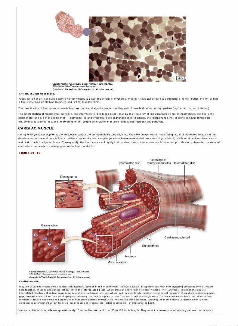
Skeletal muscle fiber types.
Cross-section of skeletal muscle stained histochemically to detect the density of myofibrillar myosin-ATPase can be used to demonstrate the distribution of slow (S) typeI fibers, intermediate (I) type IIa fibers, and fast (F) type IIb fibers.
The classification of fiber types in muscle biopsies has clinical significance for the diagnosis of muscle diseases, or myopathies (myo + Gr. pathos, suffering).
The differentiation of muscle into red, white, and intermediate fiber types is controlled by the frequency of impulses from its motor innervations, and fibers of a
single motor unit are of the same type. If nerves to red and white fibers are exchanged experimentally, the fibers change their morphologic and physiologic
characteristics to conform to the innervating nerve. Simple denervation of muscle leads to fiber atrophy and paralysis.
CARDIAC MUSCLE
During embryonic development, the mesoderm cells of the primitive heart tube align into chainlike arrays. Rather than fusing into multinucleated cells, as in the
development of skeletal muscle fibers, cardiac muscle cells form complex junctions between extended processes (Figure 10–16). Cells within a fiber often branch
and bind to cells in adjacent fibers. Consequently, the heart consists of tightly knit bundles of cells, interwoven in a fashion that provides for a characteristic wave of
contraction that leads to a wringing out of the heart ventricles.
Figure 10–16.
Cardiac muscle.
Diagram of cardiac muscle cells indicates characteristic features of this muscle type. The fibers consist of separate cells with interdigitating processes where they areheld together. These regions of contact are called the intercalated discs, which cross an entire fiber between two cells. The transverse regions of the steplikeintercalated disc have abundant desmosomes and other adherent junctions which hold the cells firmly together. Longitudinal regions of these discs contain abundantgap junctions, which form "electrical synapses" allowing contraction signals to pass from cell to cell as a single wave. Cardiac muscle cells have central nuclei andmyofibrils that are less dense and organized than those of skeletal muscle. Also the cells are often branched, allowing the muscle fibers to interweave in a morecomplicated arrangement within fascicles that produces an efficient contraction mechanism for emptying the heart.
Mature cardiac muscle cells are approximately 15 m in diameter and from 85 to 100 m in length. They exhibit a cross-striated banding pattern comparable to
AccessMedicine | Print: Chapter 10. Muscle Tissue http://accessmedicine.com/popup.aspx?aID=6181338&print=yes_chapter
18 of 24 22/05/2011 23:34
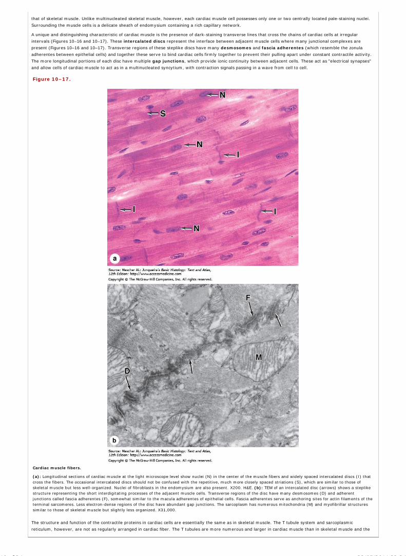
that of skeletal muscle. Unlike multinucleated skeletal muscle, however, each cardiac muscle cell possesses only one or two centrally located pale-staining nuclei.
Surrounding the muscle cells is a delicate sheath of endomysium containing a rich capillary network.
A unique and distinguishing characteristic of cardiac muscle is the presence of dark-staining transverse lines that cross the chains of cardiac cells at irregular
intervals (Figures 10–16 and 10–17). These intercalated discs represent the interface between adjacent muscle cells where many junctional complexes are
present (Figures 10–16 and 10–17). Transverse regions of these steplike discs have many desmosomes and fascia adherentes (which resemble the zonula
adherentes between epithelial cells) and together these serve to bind cardiac cells firmly together to prevent their pulling apart under constant contractile activity.
The more longitudinal portions of each disc have multiple gap junctions, which provide ionic continuity between adjacent cells. These act as "electrical synapses"
and allow cells of cardiac muscle to act as in a multinucleated syncytium, with contraction signals passing in a wave from cell to cell.
Figure 10–17.
Cardiac muscle fibers.
(a): Longitudinal sections of cardiac muscle at the light microscope level show nuclei (N) in the center of the muscle fibers and widely spaced intercalated discs (I) thatcross the fibers. The occasional intercalated discs should not be confused with the repetitive, much more closely spaced striations (S), which are similar to those ofskeletal muscle but less well-organized. Nuclei of fibroblasts in the endomysium are also present. X200. H&E. (b): TEM of an intercalated disc (arrows) shows a steplikestructure representing the short interdigitating processes of the adjacent muscle cells. Transverse regions of the disc have many desmosomes (D) and adherentjunctions called fascia adherentes (F), somewhat similar to the macula adherentes of epithelial cells. Fascia adherentes serve as anchoring sites for actin filaments of theterminal sarcomeres. Less electron-dense regions of the disc have abundant gap junctions. The sarcoplasm has numerous mitochondria (M) and myofibrillar structuressimilar to those of skeletal muscle but slightly less organized. X31,000.
The structure and function of the contractile proteins in cardiac cells are essentially the same as in skeletal muscle. The T tubule system and sarcoplasmic
reticulum, however, are not as regularly arranged in cardiac fiber. The T tubules are more numerous and larger in cardiac muscle than in skeletal muscle and the
AccessMedicine | Print: Chapter 10. Muscle Tissue http://accessmedicine.com/popup.aspx?aID=6181338&print=yes_chapter
19 of 24 22/05/2011 23:34

sarcoplasmic reticulum is less well developed (Figure 10–18). Cardiac muscle cells contain numerous mitochondria, which occupy 40% or more of the cytoplasmic
volume (Figure 10–18), reflecting the need for continuous aerobic metabolism in heart muscle. By comparison, only about 2% of skeletal muscle fiber is occupied
by mitochondria. Fatty acids, transported to cardiac muscle cells by lipoproteins, are the major fuel of the heart and are stored as triglycerides in numerous lipid
droplets seen in many cardiac muscle cells. Glycogen particles may also be present. Lipofuscin pigment granules are often found near the nuclei of cardiac muscle
cells.
Figure 10–18.
Cardiac muscle ultrastructure.
(a): TEM of cardiac muscle shows an abundance of mitochondria (M) and rather sparse sarcoplasmic reticulum (SR) in the areas between myofibrils. T tubules are lesswell-organized and are usually associated with one expanded terminal cisterna of SR, forming dyads (D) rather than the triads of skeletal muscle. Functionally thesestructures are similar in these two muscle types. X30,000. (b): Muscle cell from the cardiac atrium shows the presence of membrane-bound granules aggregated at thenuclear poles. These granules are most abundant in muscle cells of the right atrium (~600 per cell), but smaller quantities are also found in the left atrium and theventricles. The atrial granules contain the precursor of a polypeptide hormone, atrial natriuretic factor (ANF). ANF targets cells of the kidneys to bring about sodiumand water loss (natriuresis and diuresis). This hormone thus opposes the actions of aldosterone and antidiuretic hormone, whose effects on kidneys result in sodium andwater conservation. X10,000. x, (Figure 10-18c, with permission, from Dr. J. C. Nogueira, Department of Morphology, Federal University of Minas Gerais, Belo Horizonte,Brazil.)
A few differences in structure exist between atrial and ventricular muscle. The arrangement of myofilaments is the same in both, but atrial muscle has markedly
fewer T tubules, and the cells are somewhat smaller. Membrane-limited granules, each about 0.2–0.3 m in diameter, are found at the poles of atrial muscle nuclei
and are associated with Golgi complexes in this region (Figure 10–18). These granules release the peptide hormone atrial natriuretic factor (ANF) which acts on
target cells in the kidney to affect Na+ excretion and water balance. The contractile cells of the heart's atria thus also serve an endocrine function.
The rich autonomic nerve supply to the heart and the rhythmic impulse-generating and conducting structures are discussed in Chapter 11.
AccessMedicine | Print: Chapter 10. Muscle Tissue http://accessmedicine.com/popup.aspx?aID=6181338&print=yes_chapter
20 of 24 22/05/2011 23:34

SMOOTH MUSCLE
Smooth muscle fibers are elongated, tapering, and nonstriated cells, each of which is enclosed by a thin basal lamina and a fine network of reticular fibers (Figure
10–19). The connective tissues serve to combine the forces generated by each smooth muscle fiber into a concerted action, eg, peristalsis in the intestine.
Figure 10–19.
AccessMedicine | Print: Chapter 10. Muscle Tissue http://accessmedicine.com/popup.aspx?aID=6181338&print=yes_chapter
21 of 24 22/05/2011 23:34

Smooth muscle.
Cells or fibers of smooth muscle are long, tapering structures with elongated nuclei centrally located at the cell's widest part. (a): In a cross-section of smooth muscle inthe wall of the small intestine, cells of the inner circular (IC) layer are cut lengthwise and cells of the outer longitudinal layer (OL) cross transversely. Only some nuclei(arrows) of the latter cells are in the plane of section, so that many cells appear to be devoid of nuclei. X140. H&E. (b): Section of smooth muscle in bladder, showsfibers in cross-section (XS) and longitudinal section (LS) with the same fascicle. There is much collagen in the branching perimysium (P), but very little evidence ofendomysium is apparent. X140. Mallory trichrome. (c): Section stained only for reticulin reveals a thin endomysium around each fiber, with more reticulin in theconnective tissue of small arteries (A). Reticulin fibers in the basal laminae of smooth muscle cells help hold the cells together as a functional unit during the slow,rhythmic contractions of this tissue. X200. Silver.
Smooth muscle cells may range in length from 20 m in small blood vessels to 500 m in the pregnant uterus. Each cell has a single nucleus located in the center of
the cell's broadest part. To achieve the tightest packing, the narrow part of one cell lies adjacent to the broad parts of neighboring cells. Such an arrangement
viewed in cross section shows a range of diameters, with only the largest profiles containing a nucleus (Figure 10–20a). The borders of the cell become scalloped
when smooth muscle contracts and the nucleus becomes distorted.
Figure 10–20.
AccessMedicine | Print: Chapter 10. Muscle Tissue http://accessmedicine.com/popup.aspx?aID=6181338&print=yes_chapter
22 of 24 22/05/2011 23:34

Smooth muscle ultrastructure.
(a): TEM of a transverse section of smooth muscle showing six or seven cells sectioned at various points along their lengths, yielding profiles of various diameters withonly the largest containing a nucleus. Thick and thin filaments are not organized into myofibril bundles and there are few mitochondria (M). There is evidence of a sparseexternal lamina around each cell and reticular fibers are abundant in the ECM. A small unmyelinated nerve (N) is also seen between the cells. X6650. (b): Longitudinalsection showing several dense bodies in the cytoplasm (arrows) and at the cell membrane. Thin filaments and intermediate filaments both attach to the dense bodies. Inthe cytoplasm near the nucleus (N) are mitochondria, glycogen particles, and Golgi complexes. In the area shown at the lower right, the cell membrane showsinvaginations called caveoli (C) (L. caveoli, little cavities), which in many cells are indicative of endocytosis, but in smooth muscle cells, where they are particularly
numerous, may also function as the T tubules of skeletal muscle fibers and regulate release of Ca2+ from sarcoplasmic reticulum. X9,000.
Concentrated near the nucleus are mitochondria, polyribosomes, cisternae of rough ER, and the Golgi apparatus. Pinocytotic vesicles are frequent near the cell
surface.
A rudimentary sarcoplasmic reticulum is present in smooth muscle cells, but T tubules are not. The characteristic contractile activity of smooth muscle is related to
the structure and organization of its actin and myosin filaments, which do not exhibit the organization present in striated muscles. In smooth muscle cells, bundles
of thin and thick myofilaments crisscross obliquely through the cell, forming a latticelike network. Smooth muscle actin and myosin contract by a sliding filament
mechanism similar to that in striated muscles. However, myosin proteins are bundled differently and the cross-bridges interact with fewer F-actin filaments.
The thin filaments of smooth muscle cells lack troponin complexes and instead utilize calmodulin, a calcium-binding protein that is also involved in the contraction
of non-muscle cells. As in all muscle, an influx of Ca2+ is involved in initiating contraction in smooth muscle cells. However in these cells the Ca2+ calmodulin
complex activates myosin light chain kinase (MLCK), the enzyme that phosphorylates myosin, which is required for myosin's interaction with F-actin. A number
of hormones and other factors affect the activity of MLCK and thus influence the degree of contraction of smooth muscle cells.
Smooth muscle cells have an elaborate array of 10-nm intermediate filaments. Desmin is the major intermediate filament protein in all smooth muscles and
vimentin is an additional component in vascular smooth muscle. Both intermediate filaments and F-actin filaments insert into dense bodies (Figure 10–20) which
can be membrane-associated or cytoplasmic. Dense bodies contain -actinin and are thus functionally similar to the Z discs of striated and cardiac muscles. The
attachments of thin and intermediate filaments to the dense bodies helps transmit contractile force to adjacent smooth muscle cells and their surrounding network
of reticular fibers (Figure 10–21).
Figure 10–21.
AccessMedicine | Print: Chapter 10. Muscle Tissue http://accessmedicine.com/popup.aspx?aID=6181338&print=yes_chapter
23 of 24 22/05/2011 23:34

Copyright © The McGraw-Hill Companies. All rights reserved.Privacy Notice. Any use is subject to the Terms of Use and Notice.
Smooth muscle contraction.
Most molecules that allow contraction are similar in the three types of muscle, but the filaments of smooth muscle are arranged differently and appear less organized.(a): The diagram shows thin filaments attach to dense bodies located in the cell membrane and deep in the cytoplasm. Dense bodies contain -actinin for thin filamentattachment. Dense bodies at the membrane are also attachment sites for intermediate filaments and for adhesive junctions between cells. This arrangement of both thecytoskeleton and contractile apparatus allows the multicellular tissue to contract as a unit, providing better efficiency and force. (b): Contraction decreases the length ofthe cell, deforming the nucleus and promoting contraction of the whole muscle. The micrograph shows a region of contracted tissue in the wall of a urinary bladder. Thelong nuclei of individual fibers assume a cork-screw shape when the fibers contract, reflecting the reduced cell length at this time. X240. Mallory trichrome.
Contraction of smooth muscle is not under voluntary control, but is regulated by autonomic nerves, certain hormones, and local physiological conditions such as the
degree of stretch. The cells occur either as multiunit smooth muscle, in which each cell is innervated and can contract independently, or more commonly as
unitary smooth muscle, in which only a few cells are innervated but all cells are interconnected by gap junctions. Gap junctions allow the stimulus for contraction
to spread as a synchronized wave among adjacent cells. Smooth muscle lacks neuromuscular junctions like those in skeletal muscle. Instead axonal swellings with
synaptic vesicles simply lie in close contact with the sarcolemma, with little or no specialized structure to the junctions.
Because smooth muscle is usually spontaneously active without nervous stimuli, its nerve supply serves primarily to modify activity rather than initiate it. Smooth
muscle receives both adrenergic and cholinergic nerve endings that act antagonistically, stimulating or depressing its activity. In some organs, the cholinergic
endings activate and the adrenergic nerves depress; in others, the reverse occurs.
In addition to contractile activity, smooth muscle cells also synthesize collagen, elastin, and proteoglycans, extracellular matrix (ECM) components normally
synthesized by fibroblasts.
REGENERATION OF MUSCLE TISSUE
The three types of adult muscle have different potentials for regeneration after injury.
In skeletal muscle, although the nuclei are incapable of undergoing mitosis, the tissue can undergo limited regeneration. The source of regenerating cells is the
sparse population of mesenchymal satellite cells that lies within the external lamina of each mature muscle fiber. Satellite cells are inactive, reserve myoblasts
that persist after muscle differentiation. After injury or certain other stimuli, the normally quiescent satellite cells become activated, proliferating and fusing to form
new skeletal muscle fibers. A similar activity of satellite cells has been implicated in muscle growth after extensive exercise, a process in which they fuse with their
parent fibers to increase muscle mass beyond that occurring by cell hypertrophy. The regenerative capacity of skeletal muscle is limited, however, after major
muscle trauma or degeneration.
Cardiac muscle lacks satellite cells and has virtually no regenerative capacity beyond early childhood. Defects or damage (eg, infarcts) in heart muscle are
generally replaced by fibroblast proliferation and growth of connective tissue, forming myocardial scars. Smooth muscle, composed of simpler, mononucleated
cells, is capable of a more active regenerative response. After injury, viable smooth muscle cells undergo mitosis and replace the damaged tissue. Contractile
pericytes from the walls of small blood vessels (see Chapter 11) participate in the repair of vascular smooth muscle.
AccessMedicine | Print: Chapter 10. Muscle Tissue http://accessmedicine.com/popup.aspx?aID=6181338&print=yes_chapter
24 of 24 22/05/2011 23:34

Print Close Window
Note: Large images and tables on this page may necessitate printing in landscape mode.
Copyright © The McGraw-Hill Companies. All rights reserved.
Junqueira's Basic Histology: Text & Atlas, 12e > Chapter 11. The Circulatory System >
THE CIRCULATORY SYSTEM: INTRODUCTION
The circulatory system includes both the blood and lymphatic vascular systems. The blood vascular system (Figure 11–1) is composed of the following
structures:
The heart, an organ whose function is to pump the blood.
The arteries, a series of efferent vessels that become smaller as they branch, and whose function is to carry the blood, with its nutrients andoxygen, to the tissues.
The capillaries, the smallest blood vessels, constituting a complex network of thin tubules that branch profusely in almost every organ and throughwhose walls the interchange between blood and tissues takes place.
The veins, which result from the convergence of capillaries into a system of larger channels that continue enlarging as they approach the heart,toward which they convey the blood to be pumped again.
Figure 11–1.
Vessels of the blood circulatory system.
The heart is the principal organ of the blood circulatory system, pumping blood throughout the body and providing the force by which nutrients leave the capillariesand enter tissues. Large elastic arteries leave the heart and branch to form muscular arteries. These arteries branch further and enter organs, where they branchmuch further to form arterioles. These arterioles branch into the smallest vessels, the capillaries, the site of exchange between blood and surrounding tissue.Capillaries then merge to form venules, which merge further into small and then medium-sized veins. These veins leave organs, form larger veins which eventuallybring blood back to the heart.
The lymphatic vascular system, introduced with the discussion of interstitial fluid in Chapter 5, begins with the lymphatic capillaries, which are
AccessMedicine | Print: Chapter 11. The Circulatory System http://accessmedicine.com/popup.aspx?aID=6181469&print=yes_chapter
1 of 23 22/05/2011 23:35

closed-ended tubules that merge to form vessels of steadily increasing size; these vessels terminate in the blood vascular system emptying into the large veins
near the heart. One of the functions of the lymphatic system is to return the fluid of the tissue spaces to the blood. The internal surface of all components of
the blood and lymphatic systems is lined by a single layer of a squamous epithelium, called endothelium.
The circulatory system is considered to consist of the macrovasculature, vessels that are more than 0.1 mm in diameter (large arterioles, muscular and elastic
arteries, and muscular veins), and the microvasculature (arterioles, capillaries, and postcapillary venules) visible only with a microscope (Figure 11–2). The
microvasculature is particularly important functionally, being the site of interchanges between blood and the surrounding tissues both under normal conditions
and during inflammatory processes.
Figure 11–2.
Vessels of the microvasculature.
Arterioles (A), small capillaries (C) and venules (V) make up the microvasculature where, in almost every organ, exchange takes place between blood and theinterstitial fluid of the tissues. X200. Masson trichrome.
HEART
The heart is a muscular organ that contracts rhythmically, pumping the blood through the circulatory system (Figure 11–3). The right and left ventricles pump
blood to the lungs and the rest of the body respectively; right and left atria receive blood from the body and the pulmonary veins respectively. The walls of all
four heart chambers consist of three major layers or tunics: the internal endocardium; the middle myocardium; and the external epicardium.
Figure 11–3.
Major histological features of the heart.
Longitudinal view of human heart showing the two atria and two ventricles. The ventricular walls are thicker than those of the atria, principally because of the much
AccessMedicine | Print: Chapter 11. The Circulatory System http://accessmedicine.com/popup.aspx?aID=6181469&print=yes_chapter
2 of 23 22/05/2011 23:35

thicker myocardium. The valves are basically flaps of connective tissue anchored in the heart's dense fibrous skeleton region, shown in white. Other parts of thefibrous skeleton are the chordae tendinae, cords of dense connective tissue extending from the valves and attached to papillary muscles that help prevent valvesfrom turning inside-out during ventricular contraction. All these parts of the fibrous skeleton are covered by endothelium. Shown in yellow are parts of the cardiacconducting system, which initiates the electrical impulse for heart's contraction (heartbeat) and spreads it through the ventricular myocardium. Both the sinoatrial(SA) node (pacemaker), in the posterior wall of the right atrium, and the atrioventricular (AV) node in the floor of the right atrium consist of myocardial tissue thatis difficult to distinguish histologically from surrounding cardiac muscle. The AV node is continuous with specialized bundles of cardiac muscle fibers, the AV bundle(of His) which run along the interventricular septum to the apex of the heart, where they branch further as conducting (Purkinje) fibers which extend intomyocardium of both ventricles.
The endocardium consists of a single layer of squamous endothelial cells on a thin layer of loose connective tissue containing elastic and collagen fibers as
well as some smooth muscle cells. Connecting this subendothelial layer to the myocardium is additional connective tissue (often called the subendocardial
layer) containing veins, nerves, and branches of the impulse-conducting system of the heart (Figure 11–4).
Figure 11–4.
AccessMedicine | Print: Chapter 11. The Circulatory System http://accessmedicine.com/popup.aspx?aID=6181469&print=yes_chapter
3 of 23 22/05/2011 23:35

Endocardium & subendocardial conducting network.
The endocardium (En) is a thin layer of connective tissue lined by simple squamous endothelium. Between the endocardium and myocardium is a layer of variablethickness called the subendocardial layer (SEn) containing small nerves and in the ventricles the conducting (Purkinje) fibers (P) of the subendocardial conductingnetwork. These fibers are cardiac muscle cells joined by intercalated disks but specialized for impulse conduction rather than contraction. Purkinje fibers are usuallylarger than contractile cardiac muscle fibers with large amounts of lightly stained glycogen filling most of the cytoplasm and displacing sparse myofibrils to theperiphery. (a): Purkinje fibers running separately within the subendocardial layer. (b): Purkinje fibers intermingling with contractile fibers within the myocardium(M). Along with the nodes of specialized cardiac muscle in the right atrium which generate the electrical impulse, the network of conducting fibers comprises theconducting system of the heart. Both X200. H&E.
The myocardium is the thickest of the tunics and consists of cardiac muscle cells (see Chapter 10) arranged in layers that surround the heart chambers in a
complex spiral. The myocardium is much thicker in the ventricles than in the atria. The arrangement of these muscle cells is extremely varied, so that in
sections cells are seen to be oriented in many directions.
The heart is covered externally by simple squamous epithelium (mesothelium) supported by a thin layer of connective tissue that constitutes the epicardium.
A subepicardial layer of loose connective tissue contains veins, nerves, and many adipocytes (Figure 11–5). The epicardium corresponds to the visceral layer of
the pericardium, the serous membrane in which the heart lies. In the space between the pericardium's visceral layer (epicardium) and its parietal layer is a
small amount of lubricant fluid that facilitates the heart's movements.
Figure 11–5.
Epicardium or visceral pericardium.
The external tunic of the heart, the epicardium, is the site of the coronary vessels and contains considerable adipose tissue. This section of atrium shows part of themyocardium (M) and epicardium (Ep). The epicardium consists of loose connective tissue (CT) containing both autonomic nerves (N) and fat (F). The epicardium isthe visceral layer of the pericardium and is covered by the simple squamous-to-cuboidal epithelium (arrows) that also lines the pericardial space. These mesothelialcells secrete a lubricate fluid that prevents friction as the beating heart contacts the parietal pericardium on the other side of the pericardial cavity. X100. H&E.
The cardiac valves consist of a central core of dense fibrous connective tissue (containing both collagen and elastic fibers), lined on both sides by endothelial
layers. The bases of the valves are attached to strong fibrous rings that are part of the fibrous skeleton. This dense, fibrous region around the heart valves
anchors the base of the valves and is the site of origin and insertion of the cardiac muscle fibers (Figure 11–6).
Figure 11–6.
AccessMedicine | Print: Chapter 11. The Circulatory System http://accessmedicine.com/popup.aspx?aID=6181469&print=yes_chapter
4 of 23 22/05/2011 23:35
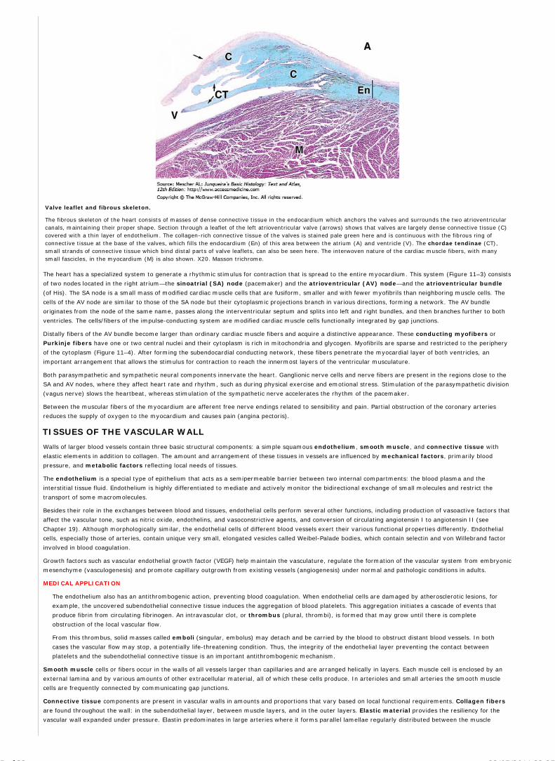
Valve leaflet and fibrous skeleton.
The fibrous skeleton of the heart consists of masses of dense connective tissue in the endocardium which anchors the valves and surrounds the two atrioventricularcanals, maintaining their proper shape. Section through a leaflet of the left atrioventricular valve (arrows) shows that valves are largely dense connective tissue (C)covered with a thin layer of endothelium. The collagen-rich connective tissue of the valves is stained pale green here and is continuous with the fibrous ring ofconnective tissue at the base of the valves, which fills the endocardium (En) of this area between the atrium (A) and ventricle (V). The chordae tendinae (CT),small strands of connective tissue which bind distal parts of valve leaflets, can also be seen here. The interwoven nature of the cardiac muscle fibers, with manysmall fascicles, in the myocardium (M) is also shown. X20. Masson trichrome.
The heart has a specialized system to generate a rhythmic stimulus for contraction that is spread to the entire myocardium. This system (Figure 11–3) consists
of two nodes located in the right atrium—the sinoatrial (SA) node (pacemaker) and the atrioventricular (AV) node—and the atrioventricular bundle
(of His). The SA node is a small mass of modified cardiac muscle cells that are fusiform, smaller and with fewer myofibrils than neighboring muscle cells. The
cells of the AV node are similar to those of the SA node but their cytoplasmic projections branch in various directions, forming a network. The AV bundle
originates from the node of the same name, passes along the interventricular septum and splits into left and right bundles, and then branches further to both
ventricles. The cells/fibers of the impulse-conducting system are modified cardiac muscle cells functionally integrated by gap junctions.
Distally fibers of the AV bundle become larger than ordinary cardiac muscle fibers and acquire a distinctive appearance. These conducting myofibers or
Purkinje fibers have one or two central nuclei and their cytoplasm is rich in mitochondria and glycogen. Myofibrils are sparse and restricted to the periphery
of the cytoplasm (Figure 11–4). After forming the subendocardial conducting network, these fibers penetrate the myocardial layer of both ventricles, an
important arrangement that allows the stimulus for contraction to reach the innermost layers of the ventricular musculature.
Both parasympathetic and sympathetic neural components innervate the heart. Ganglionic nerve cells and nerve fibers are present in the regions close to the
SA and AV nodes, where they affect heart rate and rhythm, such as during physical exercise and emotional stress. Stimulation of the parasympathetic division
(vagus nerve) slows the heartbeat, whereas stimulation of the sympathetic nerve accelerates the rhythm of the pacemaker.
Between the muscular fibers of the myocardium are afferent free nerve endings related to sensibility and pain. Partial obstruction of the coronary arteries
reduces the supply of oxygen to the myocardium and causes pain (angina pectoris).
TISSUES OF THE VASCULAR WALL
Walls of larger blood vessels contain three basic structural components: a simple squamous endothelium, smooth muscle, and connective tissue with
elastic elements in addition to collagen. The amount and arrangement of these tissues in vessels are influenced by mechanical factors, primarily blood
pressure, and metabolic factors reflecting local needs of tissues.
The endothelium is a special type of epithelium that acts as a semipermeable barrier between two internal compartments: the blood plasma and the
interstitial tissue fluid. Endothelium is highly differentiated to mediate and actively monitor the bidirectional exchange of small molecules and restrict the
transport of some macromolecules.
Besides their role in the exchanges between blood and tissues, endothelial cells perform several other functions, including production of vasoactive factors that
affect the vascular tone, such as nitric oxide, endothelins, and vasoconstrictive agents, and conversion of circulating angiotensin I to angiotensin II (see
Chapter 19). Although morphologically similar, the endothelial cells of different blood vessels exert their various functional properties differently. Endothelial
cells, especially those of arteries, contain unique very small, elongated vesicles called Weibel-Palade bodies, which contain selectin and von Willebrand factor
involved in blood coagulation.
Growth factors such as vascular endothelial growth factor (VEGF) help maintain the vasculature, regulate the formation of the vascular system from embryonic
mesenchyme (vasculogenesis) and promote capillary outgrowth from existing vessels (angiogenesis) under normal and pathologic conditions in adults.
MEDICAL APPLICATION
The endothelium also has an antithrombogenic action, preventing blood coagulation. When endothelial cells are damaged by atherosclerotic lesions, for
example, the uncovered subendothelial connective tissue induces the aggregation of blood platelets. This aggregation initiates a cascade of events that
produce fibrin from circulating fibrinogen. An intravascular clot, or thrombus (plural, thrombi), is formed that may grow until there is complete
obstruction of the local vascular flow.
From this thrombus, solid masses called emboli (singular, embolus) may detach and be carried by the blood to obstruct distant blood vessels. In both
cases the vascular flow may stop, a potentially life-threatening condition. Thus, the integrity of the endothelial layer preventing the contact between
platelets and the subendothelial connective tissue is an important antithrombogenic mechanism.
Smooth muscle cells or fibers occur in the walls of all vessels larger than capillaries and are arranged helically in layers. Each muscle cell is enclosed by an
external lamina and by various amounts of other extracellular material, all of which these cells produce. In arterioles and small arteries the smooth muscle
cells are frequently connected by communicating gap junctions.
Connective tissue components are present in vascular walls in amounts and proportions that vary based on local functional requirements. Collagen fibers
are found throughout the wall: in the subendothelial layer, between muscle layers, and in the outer layers. Elastic material provides the resiliency for the
vascular wall expanded under pressure. Elastin predominates in large arteries where it forms parallel lamellae regularly distributed between the muscle
AccessMedicine | Print: Chapter 11. The Circulatory System http://accessmedicine.com/popup.aspx?aID=6181469&print=yes_chapter
5 of 23 22/05/2011 23:35
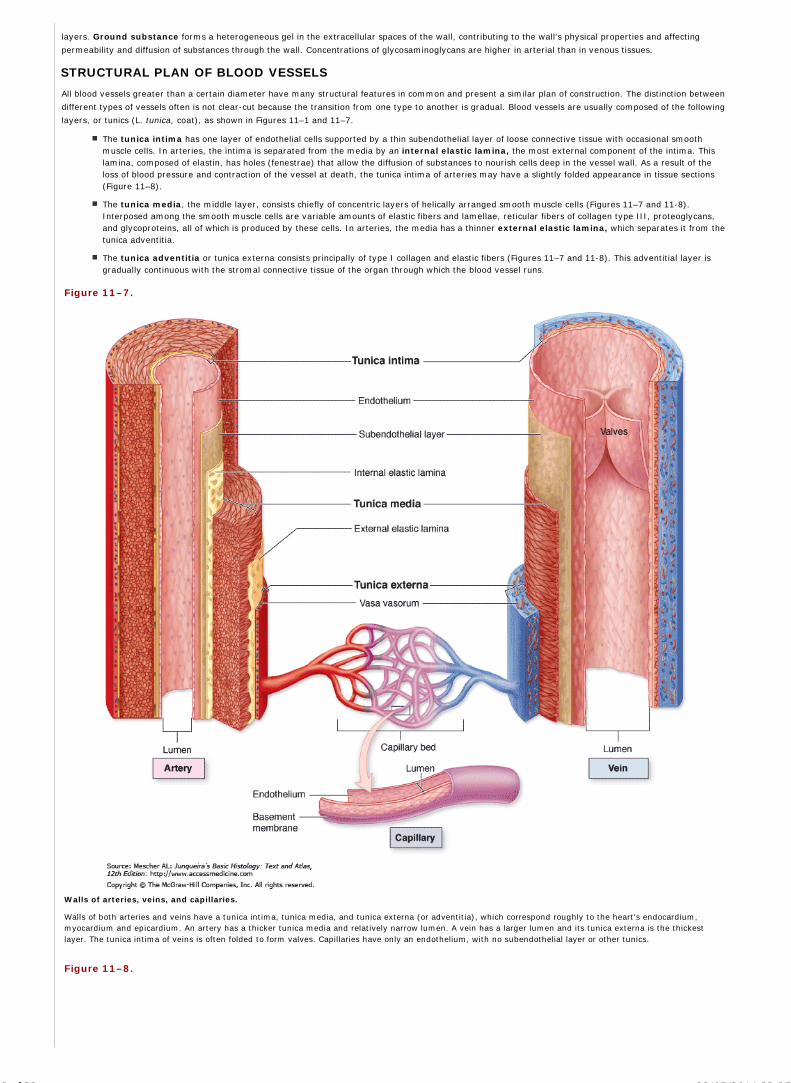
layers. Ground substance forms a heterogeneous gel in the extracellular spaces of the wall, contributing to the wall's physical properties and affecting
permeability and diffusion of substances through the wall. Concentrations of glycosaminoglycans are higher in arterial than in venous tissues.
STRUCTURAL PLAN OF BLOOD VESSELS
All blood vessels greater than a certain diameter have many structural features in common and present a similar plan of construction. The distinction between
different types of vessels often is not clear-cut because the transition from one type to another is gradual. Blood vessels are usually composed of the following
layers, or tunics (L. tunica, coat), as shown in Figures 11–1 and 11–7.
The tunica intima has one layer of endothelial cells supported by a thin subendothelial layer of loose connective tissue with occasional smoothmuscle cells. In arteries, the intima is separated from the media by an internal elastic lamina, the most external component of the intima. Thislamina, composed of elastin, has holes (fenestrae) that allow the diffusion of substances to nourish cells deep in the vessel wall. As a result of theloss of blood pressure and contraction of the vessel at death, the tunica intima of arteries may have a slightly folded appearance in tissue sections(Figure 11–8).
The tunica media, the middle layer, consists chiefly of concentric layers of helically arranged smooth muscle cells (Figures 11–7 and 11-8).Interposed among the smooth muscle cells are variable amounts of elastic fibers and lamellae, reticular fibers of collagen type III, proteoglycans,and glycoproteins, all of which is produced by these cells. In arteries, the media has a thinner external elastic lamina, which separates it from thetunica adventitia.
The tunica adventitia or tunica externa consists principally of type I collagen and elastic fibers (Figures 11–7 and 11-8). This adventitial layer isgradually continuous with the stromal connective tissue of the organ through which the blood vessel runs.
Figure 11–7.
Walls of arteries, veins, and capillaries.
Walls of both arteries and veins have a tunica intima, tunica media, and tunica externa (or adventitia), which correspond roughly to the heart's endocardium,myocardium and epicardium. An artery has a thicker tunica media and relatively narrow lumen. A vein has a larger lumen and its tunica externa is the thickestlayer. The tunica intima of veins is often folded to form valves. Capillaries have only an endothelium, with no subendothelial layer or other tunics.
Figure 11–8.
AccessMedicine | Print: Chapter 11. The Circulatory System http://accessmedicine.com/popup.aspx?aID=6181469&print=yes_chapter
6 of 23 22/05/2011 23:35

Tunics of the vascular wall.
Comparison of the three major layers or tunics in the largest artery and vein. (a): aorta (b): vena cava. Simple squamous endothelial cells (arrows) line the tunicaintima (I) which has subendothelial loose connective tissue and is separated from the tunica media by the internal elastic lamina (IEL), a prominent sheet ofelastin. The media (M) contains elastic lamellae and fibers (EF) and multiple layers of smooth muscle not seen well here. The tunica media is much thicker in largearteries than veins, with relatively more elastin. Elastic fibers are also present in the outer tunica adventitia (A), which is relatively thicker in large veins. Vasavasorum (V) are seen in the adventitia of the aorta. The connective tissue of the adventitia always merges with the less dense connective tissue around it. BothX122. Elastic.
Large vessels usually have vasa vasorum ("vessels of the vessel"), which consist of arterioles, capillaries, and venules in the tunica adventitia and the outer
part of the media (Figure 11–9). The vasa vasorum provide metabolites to cells of those layers, since in larger vessels the wall is too thick to be nourished
solely by diffusion from the blood in the lumen. Luminal blood alone does provide nutrients and oxygen for cells of the tunica intima. Since they carry
deoxygenated blood, large veins typically have more vasa vasorum than arteries.
AccessMedicine | Print: Chapter 11. The Circulatory System http://accessmedicine.com/popup.aspx?aID=6181469&print=yes_chapter
7 of 23 22/05/2011 23:35

Figure 11–9.
Vasa vasorum.
Walls of the larger vessels, as the aorta, contain in the tunica adventitia a supply of microvasculature to bring O2 and nutrients to local cells too far from the lumento be nourished by blood there. These arterioles (A), capillaries and venules (V) constitute the vasa vasorum (vessels of vessels). The adventitia of large arteries isalso supplied more sparsely with small sympathetic nerves (N) for control of vasoconstriction. X100. H&E.
Larger vessels are supplied with a network of unmyelinated sympathetic nerve fibers (vasomotor nerves) whose neurotransmitter is norepinephrine (Figure
11–9). Discharge of norepinephrine from these nerves produces vasoconstriction. Because these efferent nerves generally do not enter the media of arteries,
the neurotransmitter must diffuse for several micrometers to affect smooth muscle cells, where gap junctions propagate the response to the inner layers of
muscle cells. In thinner-walled veins, nerve endings are found in both the adventitia and the media, but the overall density of innervation is less than that
encountered in arteries. In skeletal muscle, arteries also receive a cholinergic vasodilator nerve supply. Acetylcholine released by these nerves acts on the
endothelium to produce nitric oxide, which diffuses into the smooth muscle cells. The muscle cells then relax and the vessel lumen is dilated.
VASCULATURE
For didactic purposes vessels of the macrovasculature are classified arbitrarily as the types indicated in the following discussion.
Large Elastic ArteriesLarge elastic arteries help to stabilize the blood flow. The elastic arteries include the aorta and its large branches. Freshly dissected, they have a yellowish
color from the elastin in the media. The intima is thicker than the corresponding tunic of a muscular artery. An internal elastic lamina, although present, may
not be easily discerned, since it is similar to the elastic laminae of the next layer (Figures 11–8 and 11–10). The media consists of elastic fibers and a series of
concentrically arranged, perforated elastic laminae whose number increases with age (there are about 40 in the newborn, 70 in the adult). Between the elastic
laminae are smooth muscle cells, reticular fibers, proteoglycans, and glycoproteins. The tunica adventitia is relatively underdeveloped.
Figure 11–10.
AccessMedicine | Print: Chapter 11. The Circulatory System http://accessmedicine.com/popup.aspx?aID=6181469&print=yes_chapter
8 of 23 22/05/2011 23:35

Elastic artery.
The largest arteries contain considerable elastic material and expand with blood when the heart contracts. A transverse section through part of a large elastic arteryshows a thick tunica media (M) consisting largely of many well-developed elastic lamellae. Strong pressure of blood pulsating into such arteries during systoleexpands the arterial wall, reducing the pressure and allowing strong blood flow to continue during diastole. The intima (I) of the empty aorta is typically folded andthe adventitia (A) contains vasa vasorum. X200. PT.
The several elastic laminae contribute to the important function of making blood flow more uniform. During ventricular contraction (systole), the elastic
laminae of large arteries are stretched, reducing the force of the pressure somewhat. During ventricular relaxation (diastole), ventricular pressure drops to a
low level, but the elastic rebound of large arteries helps to maintain arterial pressure. As a consequence, arterial pressure and blood velocity decrease and
become less variable as the distance from the heart increases.
MEDICAL APPLICATION
Arterial Degenerative Alterations
Arteries undergo progressive and gradual changes from birth to death, and it is difficult to say where the normal growth processes end and the
processes of involution begin. Each artery exhibits its own aging pattern.
Atherosclerotic lesions are characterized by focal thickening of the intima, proliferation of smooth muscle cells and connective tissue elements, and the
deposit of cholesterol in smooth muscle cells and macrophages. When heavily loaded with lipid, these cells may be referred to as foam cells and form
the macroscopically visible fatty streaks and plaques that characterize atherosclerosis. These changes may extend to the inner part of the tunica
media, and the thickening may become so great as to occlude the vessel. Coronary arteries are among those most predisposed to atherosclerosis.
Uniform thickening of the intima is believed to be a normal phenomenon of aging.
Certain arteries irrigate only specific areas of specific organs and obstruction of this blood supply results in necrosis (death of tissues from a lack of
metabolites). These infarcts occur commonly in the heart, kidneys, cerebrum, and certain other organs. In other regions (such as the skin), arteries
anastomose frequently, and the obstruction of one artery does not lead to tissue necrosis, because the blood flow is maintained.
When the tunica media of an artery is weakened by an embryonic defect, disease, or lesion, the wall of the artery may dilate extensively. As this
process of dilatation progresses, it becomes an aneurysm. Rupture of the aneurysm brings severe consequences and may cause death.
Muscular ArteriesThe muscular arteries can control blood flow to organs by contracting or relaxing the smooth muscle cells of the tunica media. The intima has a very thin
subendothelial layer and the internal elastic lamina, the most external component of the intima, is prominent (Figure 11–11). The tunica media may contain up
to 40 layers of more prominent smooth muscle cells which are intermingled with a variable number of elastic lamellae (depending on the size of the vessel) as
well as reticular fibers and proteoglycans. An external elastic lamina, the last component of the media, is present only in the larger muscular arteries. The
adventitia consists of connective tissue. Lymphatic capillaries, vasa vasorum, and nerves are also found in the adventitia and these structures may penetrate
to the outer part of the media.
Figure 11–11.
AccessMedicine | Print: Chapter 11. The Circulatory System http://accessmedicine.com/popup.aspx?aID=6181469&print=yes_chapter
9 of 23 22/05/2011 23:35

Muscular artery.
With distance from the heart arteries gradually have relatively less elastin and more smooth muscle in their walls. Most arteries large enough to have names are ofthis muscular type. A transverse section through a muscular (medium caliber) artery shows multiple layers of smooth muscle in the media (M). The smooth musclelayers are more prominent than the elastic lamellae and fibers with which they intersperse. Vasa vasorum are seen in the tunica adventitia. X200. PT.
Arterial Sensory StructuresCarotid sinuses are slight dilatations of the internal carotid arteries which contain baroreceptors detecting increases in blood pressure. The tunica media of
each carotid sinus is thinner, allowing greater distension when blood pressure rises, and the intima and adventitia are rich in sensory nerve endings from
cranial nerve IX, the glossopharyngeal nerve. The afferent nerve impulses are processed in the brain to trigger adjustments in vasoconstriction that return
pressure to normal. Similar baroreceptors occur in aortic arches and other large arteries.
The carotid bodies are small, ganglia-like structures (paraganglia) near the bifurcation of the common carotid arteries that contain chemoreceptors
sensitive to blood CO2 and O2 concentrations. A network of sinusoidal capillaries is intermixed with glomus (type I) cells containing numerous dense-core
vesicles with dopamine, serotonin, and adrenaline (Figure 11–12). Dendritic fibers of cranial nerve IX, the glossopharyngeal nerve, synapse with the glomus
cells. The sensory nerve is activated by neurotransmitter release from glomus cells in response to changes in the sinusoidal blood: increased CO2, decreased
O2, or increased H+ levels. Aortic bodies located on the arch of the aorta are similar in structure and function to carotid bodies.
Figure 11–12.
AccessMedicine | Print: Chapter 11. The Circulatory System http://accessmedicine.com/popup.aspx?aID=6181469&print=yes_chapter
10 of 23 22/05/2011 23:35

Glomus body.
(a) and (b): Specialized regions in the walls of specific arteries contain cells that act of chemoreceptors providing information to the brain regarding bloodchemistry. The glomus bodies are two small (0.5–5 mm diameter) ganglion-like structures found near the bifurcations of the common carotid arteries. They containmany large sinusoidal capillaries (C) intermingled with clusters of large glomus cells (G) with round nuclei and cytoplasm filled with vesicles of variousneurotransmitters that are clearly seen in (b). Supportive sheath cells (S) with elongated nuclei are associated with the groups of glomus cells. Glomus cells form
synaptic connections with dendritic fibers of the glossopharyngeal nerve. Changes in the CO2, O2 and H+ concentrations in the sinusoidal blood are detected by thechemoreceptive glomus cells, which then release neurotransmitter that activates the sensory nerve to relay this information to the brain. a: X200; b: X400. BothPT.
ArteriolesMuscular arteries branch repeatedly into smaller and smaller arteries, until reaching a size with only two or three medial layers of muscle. The smallest
arteries branch as arterioles, which have one or two smooth muscle layers and indicate the beginning of an organ's microvasculature (Figure 11–13) where
exchanges between blood and tissue fluid occur. Arterioles are generally less than 0.5 mm in diameter, with lumens approximately as wide as the wall is thick
AccessMedicine | Print: Chapter 11. The Circulatory System http://accessmedicine.com/popup.aspx?aID=6181469&print=yes_chapter
11 of 23 22/05/2011 23:35

(Figures 11–2 and 11–14). The subendothelial layer is very thin, the elastic laminae are absent and the media is generally composed of circularly arranged
smooth muscle cells. In both small arteries and arterioles, the tunica adventitia is very thin and inconspicuous.
Figure 11–13.
Structure of microvasculature.
Microvasculature arises to meet nutritional needs of one organ or parts of one organ and consists of blood vessels of less than 0.5 mm diameter. Microvesselsinclude arterioles and their smaller branches called metarterioles in which the layer of smooth muscle cells is dispersed as bands of cells that act as precapillarysphincters. The distal portion of the metarteriole, sometimes called a thoroughfare channel, lacks any smooth muscle cells. The wall of capillaries lacks smoothmuscle cells altogether. The precapillary sphincters allow blood to enter the bed of capillaries in a pulsatile manner for maximally efficient exchange of nutrients,wastes, O2, and CO2 across the capillary wall. Capillaries and the metarteriole converge as postcapillary venules, the last component of the microvasculature.Blood enters microvasculature well-oxygenated and leaves poorly oxygenated.
Figure 11–14.
AccessMedicine | Print: Chapter 11. The Circulatory System http://accessmedicine.com/popup.aspx?aID=6181469&print=yes_chapter
12 of 23 22/05/2011 23:35

Arterioles.
(a): Arterioles are microvessels with a tunica intima (I) that consists only of the endothelium (E), in which the cells may have rounded nuclei. They have tunicamedia (M) with only one or two layers of smooth muscle, and usually thin, inconspicuous adventitia (Ad). X350. Masson trichrome. (b): Three arterioles of varioussizes are shown here and a capillary. X400. H&E. (c): A large mesenteric arteriole is cut obliquely and longitudinally and clearly shows the endothelial cells (arrowheads) and one or two layers of smooth muscle cells (M) cut transversely. Adventitia merges imperceptibly with neighboring connective tissue. X300. PT.
In certain tissues and organs arteriovenous shunts or anastomoses regulate blood flow by allowing direct communication between arterioles and venules.
Arterioles in such shunts have a relatively thick, capsule-like adventitia and a thick smooth muscle layer. Arteriovenous shunts are richly innervated by the
sympathetic and parasympathetic nervous systems. These interconnections are abundant in skeletal muscle and in the skin of the hands and feet. When
vessels of the arteriovenous anastomosis contract, all the blood must pass through the capillary network. When they relax, some blood flows directly to a
venule instead of circulating in the capillaries. Their luminal diameters vary with the physiologic condition of the organ. Changes in diameter of these vessels
regulate blood pressure, blood flow, temperature and heat conservation in affected areas.
CapillariesCapillaries permit different levels of metabolic exchange between blood and surrounding tissues. They are composed of a single layer of endothelial cells
rolled up in the form of a tube. The average diameter of capillaries varies from 5 to 10 m and their individual length is usually not more than 50 m.
Altogether capillaries comprise over 90% of all blood vessels in the body, with a total length of nearly 96,000 km (60,000 miles). The total diameter of the
capillaries is approximately 800 times larger than that of the aorta. The velocity of blood in the aorta averages 320 mm/s, but in capillaries blood flows only
about 0.3 mm/s. Because of their thin walls and slow blood flow, capillaries are a favorable place for the exchange of water, solutes, and macromolecules
between blood and tissues.
Endothelial cells are functionally diverse according to the vessel they line. The capillaries are often referred to as exchange vessels, since it is at these sites
that O2, CO2, substrates, and metabolites are transferred from blood to the tissues and from the tissues to blood. The mechanisms responsible for the
AccessMedicine | Print: Chapter 11. The Circulatory System http://accessmedicine.com/popup.aspx?aID=6181469&print=yes_chapter
13 of 23 22/05/2011 23:35

interchange of materials between blood and tissue are not completely known. They depend on the kind of molecule and also on the structural characteristics
and arrangement of endothelial cells in each type of capillary.
Small molecules, both hydrophobic and hydrophilic can diffuse or be actively transported across the plasmalemma of capillary endothelial cells. These
substances are then transported by diffusion through the endothelial cytoplasm to the opposite cell surface, where they are discharged into the extracellular
space. Water and some other hydrophilic molecules, less than 1.5 nm in diameter and below 10 kDa in molecular mass, can cross the capillary wall by
diffusing through the intercellular junctions (paracellular pathway). The pores of fenestrated capillaries, the spaces between endothelial cells of sinusoidal
capillaries, and the pinocytotic vesicles are other pathways for the passage of large molecules.
In general, endothelial cells are polygonal and elongated in the direction of blood flow (Figure 11–7). The nucleus causes that part of the cell to bulge into the
capillary lumen. The cytoplasm contains a small Golgi appraratus, mitochondria, free ribosomes, and sparse cisternae of RER. Junctions of the tight zonula
occludentes type are present between most endothelial cells, conferring the wall with variable permeability to macromolecules that plays significant roles in
both normal and pathologic conditions.
MEDICAL APPLICATION
Junctions between endothelial cells of postcapillary venules are the loosest of the microvasculature. At these locations there is a characteristic loss of
fluid from the circulatory system during the inflammatory response, leading to edema.
At various locations along capillaries and postcapillary venules are cells of mesenchymal origin with long cytoplasmic processes partly surrounding the
endothelial layer. These cells are called pericytes (Gr. peri, around, + kytos, cell). They are enclosed in their own basal lamina, which may fuse with that of
the endothelial cells (Figure 11–15). Well-developed networks of myosin, actin, and tropomyosin in pericytes indicate these cells' primary contractile function.
After tissue injuries, pericytes proliferate and differentiate to form both tunica media of new blood vessels and cells with various other functions in
re-establishing the microvasculature and its ECM.
Figure 11–15.
Capillary with pericyte.
Capillaries consist only of an endothelium rolled as a tube, across which molecular exchange occurs between blood and tissue fluid. (a): Capillaries are normallyassociated with perivascular contractile cells called pericytes (P) which have a variety of functions. The more flattened nuclei belong to endothelial cells. X400. H&E.(b): TEM of a capillary cut transversely, showing the thin wall of one endothelial cell covered by an external lamina (arrows). Endothelial cells have numeroustranscytotic vesicles and their edges overlap and are bound tightly together with occluding junctions (J). One pericyte (P) is shown, surrounded by its own external
AccessMedicine | Print: Chapter 11. The Circulatory System http://accessmedicine.com/popup.aspx?aID=6181469&print=yes_chapter
14 of 23 22/05/2011 23:35

lamina. Pericytes can proliferate to form smooth muscle cells when a capillary is transformed into an arteriole or venule after tissue injury and repair. X13,000.(Figure 11–15b, reproduced, with permission, from Kelly DE, Wood RL, Enders AC (eds): Bailey's Textbook of Microscopic Anatomy, 18th ed. Williams & Wilkins,1984. Reproduced, with permission, from Kelly D. E., Wood R.L., and Enders AC (eds): Bailey's Textbook of Microscopic Anatomy, 18th ed. Williams & Wilkins,1984.)
Capillaries have structural variations which permit different levels of metabolic exchange between blood and surrounding tissue. They can be grouped into
three types, depending on the continuity of the endothelial cells and the external lamina (Figure 11–16).
The continuous, or tight, capillary (Figure 11–17) allows regulated exchange of material and is characterized by the distinct continuity of theendothelial cells in its wall. This is the most common type of capillary and is found in all kinds of muscle tissue, connective tissue, exocrine glands, andnervous tissue. In some places, but not in the nervous system, numerous pinocytotic vesicles are present on both endothelial cell surfaces. Vesicles alsoappear as isolated vesicles in the cytoplasm of these cells and are responsible for transcytosis of macromolecules in both directions across theendothelial cytoplasm.
1.
The fenestrated capillary allows more extensive molecular exchange across the endothelium and is characterized by the presence of small circularfenestrae (L, fenestra, perforation) through the very thin squamous endothelial cells. Each fenestra is usually covered by a very thin diaphragmcontaining heparan proteoglycans but no lipid bilayer (Figure 11–18). The basal lamina of the fenestrated capillaries is continuous, covering thefenestrae. Fenestrated capillaries are found in tissues where rapid interchange of substances occurs between the tissues and the blood, as in the kidney,the intestine, the choroid plexus and the endocrine glands. Macromolecules experimentally injected into the bloodstream can cross the capillary wallthrough the fenestrae to enter tissue spaces.
2.
The sinusoid or discontinuous capillary permits maximal exchange of macromolecules as well as cells between tissues and blood and has thefollowing characteristics: endothelial cells have large fenestrae without diaphragms; the cells form a discontinuous layer and are separated from oneanother by wide spaces; the basal lamina is also discontinuous. Sinusoids are irregularly shaped and have diameters as large as 30–40 m, muchgreater than those of other capillaries, properties which further slow blood flow at this site. Sinusoidal capillaries are found in the liver, spleen, someendocrine organs, and bone marrow (Figure 11–19).
3.
Figure 11–16.
AccessMedicine | Print: Chapter 11. The Circulatory System http://accessmedicine.com/popup.aspx?aID=6181469&print=yes_chapter
15 of 23 22/05/2011 23:35

Types of capillaries.
The vessels between arterioles and venules can be any of three types. (a): Continuous capillaries, the most common type, have tight, occluding junctions sealingthe intercellular clefts between all the endothelial cells to produce minimal fluid leakage. All molecules exchanged across the endothelium must cross the cells bydiffusion or transcytosis. (b): Fenestrated capillaries also have tight junctions, but perforations (fenestrae) through the endothelial cells allow greater exchangeacross the endothelium. The external lamina is continuous in both these capillary types. Fenestrated capillaries are found in organs where molecular exchange withthe blood is important, such as endocrine organs, intestinal walls, and choroid plexus. (c): Sinusoids usually have a wider diameter than the other types ofcapillaries and have discontinuities between the endothelial cells, large fenestrae through the cells, and a partial, discontinuous basement membrane. Sinusoids arefound in organs where exchange of macromolecules and cells occurs readily between tissue and blood, such as in bone marrow, liver, and spleen.
Figure 11–17.
Continuous capillary.
Continuous capillaries exert the tightest control over what molecules leave across their walls. TEM shows a continuous capillary in transverse section. A nucleus (N)is prominent, but tight or occluding junctions along overlapping folds between two cells can also be seen (arrowheads). Numerous transcytotic vesicles are evident(small arrows). The long arrows show extensions of broad cytoplasmic sheets suggesting phagocytosis, which is consistent with the presence of vacuoles andelectron-dense lysosomes. All material that crosses continuous capillary endothelium must pass through the cells, usually by diffusion or transcytosis. X10,000.
Figure 11–18.
Fenestrated capillary.
Fenestrated capillaries are specialized for uptake of molecules such as hormones in endocrine glands or for outflow of molecules such as in the kidney's filtrationsystem. TEM of a transversely sectioned fenestrated capillary in the peritubular region of the kidney shows many typical fenestrae closed by diaphragms (arrows),with a continuous external lamina on the outer surface of the endothelial cell (double arrows). The diaphragms contain heparan sulfate proteoglycans, but theirfunctional role is poorly understood at the molecular level. In this cell the Golgi apparatus (G), nucleus (N), and centrioles (C) can be seen. Fenestrated capillariesallow a freer exchange of molecules than continuous capillaries and are found in the intestinal wall, kidneys and endocrine glands. X10,000. (With permission, fromJohannes Rhodin, Department of Cell Biology, New York University School of Medicine.)
Figure 11–19.
AccessMedicine | Print: Chapter 11. The Circulatory System http://accessmedicine.com/popup.aspx?aID=6181469&print=yes_chapter
16 of 23 22/05/2011 23:35

Figure 11-19. Sinusoidal capillary.
Sinusoidal capillaries or sinusoids generally have much greater diameters than most capillaries and are specialized not only for maximal molecular exchangebetween blood and surrounding tissue, but also for easy movement of blood cells across the endothelium. The sinusoid (S) shown here is in bone marrow and issurrounded by tissue containing adipocytes (A) and masses of hematopoietic cells (H). The endothelium is very thin and cell nuclei are more difficult to find than insmaller capillaries. Ultrastructurally sinusoidal capillaries are seen to have large fenestrae through the cells and large discontinuities between the cells and throughthe basal lamina. X200. H&E.
Capillaries anastomose freely, forming a rich network or bed that interconnects the arterioles and venules (Figure 11–13). The arterioles may first branch into
smaller vessels with a sparse layer of smooth muscle called metarterioles, which branch further into capillaries. Metarterioles often form a preferential
channel for blood flow though a microcirculatory bed and help to regulate the circulation in capillaries. The richness of the capillary network is related to the
metabolic activity of the tissues. Tissues with high metabolic rates, such as the kidney, liver, and cardiac and skeletal muscle, have an abundant capillary
network; the opposite is true of tissues with low metabolic rates, such as smooth muscle and dense connective tissue.
VenulesThe transition from capillaries to venules occurs gradually. The immediate postcapillary venules are similar structurally to capillaries, with pericytes, but
range in diameter from 15 to 20 m. Postcapillary venules participate in the exchanges between the blood and the tissues and, as described in Chapter 12, are
the primary site at which white blood cells leave the circulation at sites of infection or tissue damage. These venules converge into larger collecting venules
which have more contractile cells. With greater size the venules become surrounded by recognizable tunica media with two or three smooth muscle layers and
are called muscular venules. A characteristic feature of all venules is the large diameter of the lumen compared to the overall thinness of the wall (Figure
11–20).
Figure 11–20
AccessMedicine | Print: Chapter 11. The Circulatory System http://accessmedicine.com/popup.aspx?aID=6181469&print=yes_chapter
17 of 23 22/05/2011 23:35

Venules.
A series of increasingly larger and more organized venules lie between capillaries and veins. (a): Postcapillary venules resemble large capillaries, having only anendothelium with occasional pericytes (arrowhead). Their lumens and overall diameters are greater than those of nearby arterioles. X400. TB. (b): Large collectingvenules have much greater diameters than arterioles but the wall is still very thin, consisting of an endothelium with more numerous pericytes or smooth musclecells. X200. TB. (c): Muscular venule has a better defined tunica media, with as many as three layers of smooth muscle (M) in some areas, a very thin intima (I)of endothelial cells (E), and a more distinct tunica adventitia (Ad). Part of an arteriole (A) is included for comparison. Venules are the site in the vasculature wherewhite blood cells leave the circulation to become functional in the interstitial space of surrounding tissues when such tissues are inflamed or infected. Suchconditions cause endothelial cells of venules to loosen intercellular junctions and express new protein receptors on their luminal surface. Surface proteins on passingleukocytes bind these receptors, causing the cells to stick to the endothelial cells in a process termed margination. This adhesion is quickly followed by emigrationfrom the venule between endothelial cells. X200. Masson trichrome. (d): Venule (V) from an infected small intestine shows several leukocytes adhering to and
AccessMedicine | Print: Chapter 11. The Circulatory System http://accessmedicine.com/popup.aspx?aID=6181469&print=yes_chapter
18 of 23 22/05/2011 23:35

migrating across the endothelium. X200. H&E. (Figure 11–20a, with permission, from Telma M.T. Zorn, Department of Cell and Developmental Biology, University ofSão Paulo, Brazil.)
VeinsBlood entering veins is under very low pressure and moves toward the heart by contraction of the tunica media and external compressions from surrounding
muscles and other organs. Valves project from the tunica intima to prevent back-flow of blood. Most veins are small or medium veins (Figure 11–21), with
diameters less than one centimeter. Such veins are usually located in parallel with corresponding muscular arteries. The intima usually has a thin
subendothelial layer and the media consists of small bundles of smooth muscle cells intermixed with reticular fibers and a delicate network of elastic fibers. The
collagenous adventitial layer is well-developed.
Figure 11–21.
AccessMedicine | Print: Chapter 11. The Circulatory System http://accessmedicine.com/popup.aspx?aID=6181469&print=yes_chapter
19 of 23 22/05/2011 23:35

Veins.
Veins usually travel near arteries and are classified as small, medium, or large based on size and development of the tunics. (a): Micrograph of small vein (V)shows a relatively large lumen compared to the small muscular artery (A) with its thick media (M) and adventitia (Ad). The wall of a small vein is very thin,containing only two or three layers of smooth muscle. X200. H&E. (b): Micrograph of a convergence between two small veins showing valves (arrow). Valves arethin folds of tunica intima projecting well into the lumen which act to prevent backflow of blood. X200. H&E. (c): Micrograph of a medium vein (MV) showing athicker wall, but still less prominent than that of the accompanying muscular artery (MA). Both the media and adventitia are better developed, but the wall is oftenfolded around the relatively large lumen. X100. H&E. (d): Micrograph of a medium vein containing blood and showing valve folds (arrows). X200. Masson trichrome.
The big venous trunks, paired with elastic arteries close to the heart, are large veins (Figure 11–8). Large veins have a well-developed tunica intima, but the
tunica media is relatively thin, with few layers of smooth muscle and abundant connective tissue. The adventitial layer is thick in large veins and frequently
contains longitudinal bundles of smooth muscle. Both the media and adventitia contain elastic fibers, but elastic laminae like those of arteries are not present.
Most veins have valves, but these are most prominent in large veins. Valves consist of paired semilunar folds of the tunica intima projecting across part of the
lumen (Figures 11–21 and 11–22). They are rich in elastic fibers and are lined on both sides by endothelium. The valves, which are especially numerous in
veins of the legs, help keep the flow of venous blood directed toward the heart.
Figure 11–22.
AccessMedicine | Print: Chapter 11. The Circulatory System http://accessmedicine.com/popup.aspx?aID=6181469&print=yes_chapter
20 of 23 22/05/2011 23:35
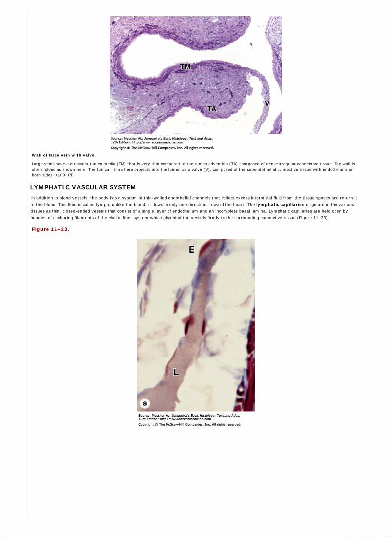
Wall of large vein with valve.
Large veins have a muscular tunica media (TM) that is very thin compared to the tunica adventitia (TA) composed of dense irregular connective tissue. The wall isoften folded as shown here. The tunica intima here projects into the lumen as a valve (V), composed of the subendothelial connective tissue with endothelium onboth sides. X100, PT.
LYMPHATIC VASCULAR SYSTEM
In addition to blood vessels, the body has a system of thin-walled endothelial channels that collect excess interstitial fluid from the tissue spaces and return it
to the blood. This fluid is called lymph; unlike the blood, it flows in only one direction, toward the heart. The lymphatic capillaries originate in the various
tissues as thin, closed-ended vessels that consist of a single layer of endothelium and an incomplete basal lamina. Lymphatic capillaries are held open by
bundles of anchoring filaments of the elastic fiber system which also bind the vessels firmly to the surrounding connective tissue (Figure 11–23).
Figure 11–23.
AccessMedicine | Print: Chapter 11. The Circulatory System http://accessmedicine.com/popup.aspx?aID=6181469&print=yes_chapter
21 of 23 22/05/2011 23:35

Lymphatic capillary.
Lymphatic capillaries drain interstitial fluid produced when the plasma forced from the microvasculature by hydrostatic pressure does not all return to blood by theaction of osmotic pressure. (a): Micrograph showing a lymphatic capillary filled with this fluid called lymph (L). Lymphatics are blind-ended vessels with a wall ofvery thin endothelial cells (E) and are quite variable in diameter (10-50 m). Lymph is rich in proteins and other material and often stains somewhat better thanthe surrounding ground substance, as seen here. X200. Mallory trichrome. (b): Diagram indicating details of lymphatics, including the openings between theendothelial cells. The openings are held in place by anchoring filaments containing elastin and are covered by flaps of endothelium. Interstitial fluid enters primarilyvia these openings and the endothelial folds prevent backflow of lymph into tissue spaces. Lymphatic endothelial cells are typically larger than those of bloodvessels.
The thin lymphatic capillaries converge into larger lymphatic vessels. Interposed in the path of these lymphatics are lymph nodes, whose morphologic
characteristics and functions are discussed in Chapter 14. With rare exceptions, such as the CNS and the bone marrow, lymphatic are found in almost all
organs.
The larger lymphatics have a structure similar to that of veins except that they have thinner walls and lack a clear-cut separation between tunics. They also
have more numerous internal valves (Figure 11–24). The lymphatic vessels are often dilated and assume a nodular, or beaded, appearance between the
valves. As in veins, lymphatic circulation is aided by external forces (eg, contraction of surrounding skeletal muscle) and unidirectional lymph flow is mainly a
result of the many valves. Contraction of smooth muscle in the walls of larger lymphatic vessels also helps to propel lymph toward the heart.
Figure 11–24.
AccessMedicine | Print: Chapter 11. The Circulatory System http://accessmedicine.com/popup.aspx?aID=6181469&print=yes_chapter
22 of 23 22/05/2011 23:35

Copyright © The McGraw-Hill Companies. All rights reserved.Privacy Notice. Any use is subject to the Terms of Use and Notice.
Lymphatic vessels and valve.
Lymphatic vessels are formed by the merger of lymphatic capillaries, but their walls remain extremely thin. (a): Cross-section showing a lymphatic vessel (LV) neara venule (V), whose wall is thick by comparison. Lymphatic vessels normally do not contain red blood cells, which provides another characteristic distinguishingthem from venules. X200. Mallory trichrome. (b): Lymphatic vessel (LV) in muscle is cut longitudinally showing a valve, the structure responsible for theunidirectional flow of lymph. The solid arrow shows the direction of the lymph flow, and the dotted arrows show how the valves prevent lymph backflow. The lowersmall lymphatic vessel is a lymphatic capillary with a wall consisting only of endothelium. X200. PT.
Lymphatic vessels ultimately end up as two large trunks: the thoracic duct and the right lymphatic duct, which respectively empty lymph into the junction
of the left internal jugular vein with the left subclavian vein and into the confluence of the right subclavian vein and the right internal jugular vein. The structure
of these lymphatic ducts is similar to that of large veins, with reinforced smooth muscle in the middle layer. In this layer, the muscle bundles are longitudinally
and circularly arranged, with longitudinal fibers predominating. The adventitia is relatively underdeveloped, but contains vasa vasorum and a neural network.
Besides gathering interstitial fluid as lymph and returning it to the blood, the lymphatic system of vessels is a major distributor of lymphocytes, antibodies, and
other immune components which it picks up at lymph nodes and other lymphoid tissues.
AccessMedicine | Print: Chapter 11. The Circulatory System http://accessmedicine.com/popup.aspx?aID=6181469&print=yes_chapter
23 of 23 22/05/2011 23:35

Print Close Window
Note: Large images and tables on this page may necessitate printing in landscape mode.
Copyright © The McGraw-Hill Companies. All rights reserved.
Junqueira's Basic Histology: Text & Atlas, 12e > Chapter 12. Blood >
BLOOD: INTRODUCTION
Blood is a specialized connective tissue in which cells are suspended in fluid extracellular material called plasma. Propelled mainly by rhythmic contractions of the
heart, about five liters of blood in an average adult moves unidirectionally within the closed circulatory system. The so-called formed elements circulating in the
plasma are erythrocytes (red blood cells), leukocytes (white blood cells) and platelets.
When blood leaves the circulatory system, either in a test tube or in the ECM surrounding blood vessels, plasma proteins react with one another to produce a clot,
which includes formed elements and a yellowish liquid called serum. Serum contains growth factors and other proteins released from platelets during clot formation,
which confer biological properties very different from those of plasma.
Collected blood in which clotting is prevented by the addition of anticoagulants (eg, heparin, citrate) can be separated by centrifugation into layers that reflect its
heterogeneity (Figure 12–1). Erythrocytes make up the bottom layer and their volume, normally about 45% of the total blood volume in healthy adults, is called the
hematocrit.
Figure 12–1.
Composition of whole blood.
A tube of blood after centrifugation (center) has about 43% of its volume represented by erythrocytes in the bottom half of the tube, a volume called the hematocrit.Between the sedimented erythrocytes and the supernatant light-colored plasma is a thin layer of leukocytes and platelets called the buffy coat. The average concentrationsof erythrocytes, platelets and leukocytes in normal blood are included here, along with the percentage each type of leukocyte represent in the buffy coat. A cubic millimeterof blood is equivalent to a microliter ( L).
The yellowish translucent, slightly viscous supernatant comprising 55% at the top half of the centrifugation tube is the plasma. A thin layer between the plasma and
the hematocrit, about 1% of the volume, is white or grayish in color and consists of leukocytes and platelets, both less dense than erythrocytes.
Blood is a distributing vehicle, transporting O2 (Figure 12–2), CO2, metabolites, hormones, and other substances to cells throughout the body. O2 is bound mainly to
hemoglobin in erythrocytes, while CO2 is carried in solution as CO2 or HCO3–, in addition to being hemoglobin bound. Nutrients are distributed from their sites of
synthesis or absorption in the gut and metabolic residues are collected from all cells and removed from the blood by the excretory organs. Hormone distribution in
blood permits the exchange of chemical messages between distant organs for normal cellular function. Blood further participates in heat distribution, the regulation of
body temperature, and the maintenance of acid-base and osmotic balance.
Figure 12–2.
AccessMedicine | Print: Chapter 12. Blood http://accessmedicine.com/popup.aspx?aID=6181612&print=yes_chapter
1 of 16 22/05/2011 23:37

Blood O2 content in each type of blood vessel.
The amount of O2 in blood (the O2 pressure) is highest in arteries and lung capillaries and decreases in tissue capillaries, where exchange takes place between blood andtissues.
Leukocytes have diversified functions and are one of the body's chief defenses against infection. These cells are generally spherical and inactive while suspended in
circulating blood, but when called to sites of infection or inflammation they cross the wall of venules, migrate into the tissues, and display their defensive capabilities.
COMPOSITION OF PLASMA
Plasma is an aqueous solution, pH 7.4, containing substances of low or high molecular weight that make up 8–10% of its volume. Plasma proteins account for
approximately 7% of the dissolved components, with the remainder including nutrients, nitrogenous waste products, hormones, and many inorganic ions collectively
called electrolytes. Through the capillary walls, the low-molecular-weight components of plasma are in equilibrium with the interstitial fluid of the tissues. The
composition of plasma is usually an indicator of the mean composition of the extracellular fluids in tissues.
The major plasma proteins include the following:
Albumin, the most abundant plasma protein, is made in the liver and serves primarily in maintaining the osmotic pressure of the blood.
- and -globulins, made by liver and other cells, include transferrin and other transport factors; fibronectin; prothrombin and other coagulation factors;lipoproteins and other proteins entering blood from tissues.
-globulins, which are immunoglobulins (antibodies) secreted by lymphocytes in many locations.
Complement proteins, a system of factors important in inflammation and destruction of microorganisms.
Fibrinogen, the largest plasma protein (340 kD), also made in the liver, which during clotting polymerizes as insoluble, cross-linked fibers which blockblood loss from small vessels.
BLOOD CELLS
Blood cells are generally studied in smears or films prepared by spreading a drop of blood in a thin layer on a microscope slide (Figure 12–3). In such films the cells
are clearly visible and distinct from one another, facilitating observation of their nuclei and cytoplasmic characteristics. Blood smears are routinely stained with special
mixtures of acidic (eosin) and basic (methylene blue) dyes. These mixtures also contain dyes called azures that are more useful in staining cytoplasmic granules
containing charged proteins and proteoglycans. Azurophilic granules produce metachromasia in stained leukocytes like that seen with mast cells. Some of these special
stains, such as Giemsa and Wright stain, are named for hematologists who introduced their own modifications into the original mixture.
Figure 12–3.
Preparing a blood smear.
Erythrocytes
AccessMedicine | Print: Chapter 12. Blood http://accessmedicine.com/popup.aspx?aID=6181612&print=yes_chapter
2 of 16 22/05/2011 23:37

Erythrocytes (red blood cells) are terminally differentiated, lack nuclei, and are packed with the O2-carrying protein hemoglobin. Under normal conditions, these
corpuscles never leave the circulatory system.
Like most mammalian red blood cells, human erythrocytes suspended in an isotonic medium are flexible biconcave disks (Figure 12–4). They are approximately 7.5
m in diameter, 2.6 m thick at the rim, and only 0.75 m thick in the center. This biconcave shape provides a large surface-to-volume ratio and facilitates gas
exchange. The normal concentration of erythrocytes in blood is approximately 3.9–5.5 million per microliter in women and 4.1–6 million per microliter in men.
Figure 12–4.
Normal human erythrocytes.
(a): Colorized SEM of normal erythrocytes with each side concave. X3000. (b): Diagram of an erythrocyte giving the cell's dimensions. The biconcave shape gives the cellsa very high surface-to-volume ratio and places most hemoglobin within a short distance from the cell surface, both qualities which provide maximally efficient O2 transport.Erythrocytes are also quite flexible and can easily bend to pass through small capillaries. (c): In small vessels red blood cells also often stack up in aggregates calledrouleaux. X250. H&E.
MEDICAL APPLICATION
A decreased number of erythrocytes in the blood is usually associated with anemia. An increased number of erythrocytes (erythrocytosis, or
polycythemia) may be a physiologic adaptation found, for example, in individuals who live at high altitudes, where O2 tension is low. Polycythemia (Gr.
polys, many + kytos, cell + haima, blood), usually an increase in hematocrit, is often associated with diseases of varying degrees of severity and increases
blood viscosity; when severe, it can impair circulation through the capillaries.
Abnormal erythrocytes with diameters greater than 9 m are called macrocytes, and those with diameters less than 6 m are called microcytes. The
presence of a high percentage of erythrocytes with great variations in size is called anisocytosis (Gr. aniso, uneven, + kytos).
AccessMedicine | Print: Chapter 12. Blood http://accessmedicine.com/popup.aspx?aID=6181612&print=yes_chapter
3 of 16 22/05/2011 23:37

Erythrocytes are normally quite flexible, which permits them to adapt to the irregular bends and small diameters of capillaries. Observations in vivo show that when
traversing the angles of capillary bifurcations, erythrocytes with normal adult hemoglobin (HbA) are easily deformed and frequently assume a cuplike shape.
The plasmalemma of the erythrocyte, because of its ready availability, is the best-known membrane of any cell. It consists of about 40% lipid, 10% carbohydrate, and
50% protein. Most of the latter are integral membrane proteins (see Chapter 2), including ion channels, the anion transporter called band 3 protein, and
glycophorin A. The glycosylated extracellular domains of these proteins include antigenic sites that form the basis for blood typing. Several peripheral proteins are
associated with the inner surface of the membrane, including spectrin, which forms a lattice reinforcing the membrane, and ankyrin, which anchors the lattice to
band 3 proteins. This meshwork permits the membrane and cell flexibility required for passage through capillaries and important for the normal low viscosity of blood.
Erythrocyte cytoplasm is densely filled with hemoglobin, the tetrameric O2-carrying protein that accounts for the cells' uniform acidophilia. When combined with O2
or CO2, hemoglobin forms oxyhemoglobin or carbaminohemoglobin, respectively. The reversibility of these combinations is the basis for the gas-transporting
capability of hemoglobin. The combination of hemoglobin with carbon monoxide (CO) is irreversible, however, reducing the cells' capacity to transport O2.
MEDICAL APPLICATION
Inherited alterations in hemoglobin molecules are responsible for several pathologic conditions, an example of which is sickle cell disease. This inherited
disorder is caused by a mutation of one nucleotide (a point mutation) in the gene for the chain of hemoglobin. The triplet GAA (for glutamic acid) is
changed to GUA, which specifies valine. The consequences of this substitution of a single amino acid are profound. When the altered hemoglobin (called HbS) is
deoxygenated in venous capillaries, it polymerizes and forms rigid aggregates that give the erythrocyte a characteristic sickle shape (Figure 12–5). The sickled
erythrocyte is inflexible and fragile and has a shortened life span leading to anemia. It increases the blood viscosity and can damage the wall of blood vessels,
promoting blood coagulation. Blood flow through the capillaries is retarded or even stopped, leading to severe O2 shortage (anoxia) in tissues.
Anemia is a pathologic condition characterized by blood concentrations of hemoglobin below normal values. Although anemias are usually associated with a
decreased number of erythrocytes, it is also possible for the number of cells to be normal but for each cell to contain a reduced amount of hemoglobin
(hypochromic anemia). Anemia may be caused by loss of blood (hemorrhage); insufficient production of erythrocytes by the bone marrow; production of
erythrocytes with insufficient hemoglobin, usually related to iron deficiency in the diet; or accelerated destruction of blood cells.
Figure 12–5.
Sickle cell erythrocyte.
A single nucleotide substitute in the hemoglobin gene produces a version of the protein that polymerizes to form rigid aggregates, leading to greatly misshapen cells withreduced flexibility. In individuals homozygous for the mutated HbS gene, this can lead to greater blood viscosity, and poor microvascular circulation, both features of sicklecell disease. X6500.
Erythrocyte differentiation (presented in Chapter 13) includes loss of the nucleus and all organelles shortly before the cells are released by bone marrow into the
circulation. Lacking mitochondria, mature erythrocytes rely on anaerobic glycolysis for their minimal energy needs. Lacking nuclei, they cannot replace defective
proteins.
Human erythrocytes normally survive in the circulation for about 120 days. By this time defects in the membrane's cytoskeletal lattice or ion transport systems begin
to produce swelling or other shape abnormalities, as well as changes in the cells' surface oligosaccharide complexes. Senescent or worn-out erythrocytes displaying
such changes are removed from the circulation, mainly by macrophages of the spleen, liver, and bone marrow.
LeukocytesLeukocytes (white blood cells) migrate to the tissues where they become functional and perform various activities (Figure 12–6). According to the type of cytoplasmic
granules and the shape of their nuclei, leukocytes are divided into two groups: polymorphonuclear granulocytes and mononuclear agranulocytes. Both types are
spherical while suspended in blood plasma, but become amoeboid and motile after leaving the blood vessels and invading the tissues. Their estimated sizes mentioned
below refer to observations in blood smears in which the cells are spread and appear slightly larger than they are in the circulation.
Figure 12–6.
AccessMedicine | Print: Chapter 12. Blood http://accessmedicine.com/popup.aspx?aID=6181612&print=yes_chapter
4 of 16 22/05/2011 23:37

Five types of human leukocytes.
Neutrophils, eosinophils, and basophils have granules that stain specifically with certain dyes and are called granulocytes. Lymphocytes and monocytes are consideredagranulocytes, even though they may show azurophilic granules (lysosomes), which are also present in other leukocytes.
Granulocytes possess two types of granules: the specific granules that bind neutral, basic, or acidic stains and have specific functions and the azurophilic
granules, which are specialized lysosomes, stain darkly, and are present at some level in all leukocytes. When the cells phagocytose microorganisms, several
azurophilic granule proteins act collectively to kill and then digest them. The bactericidal proteins include myeloperoxidase, which generates hypochlorite and other
reactive agents toxic to bacteria; cationic polypeptides called defensins which bind and produce holes in cell membranes of microorganisms; and lysozyme, which
dissolves bacterial cell wall components. The major protein components of specific and azurophilic granules are listed in Table 12–1.
Table 12–1. Granule composition in human granulocytes.
Cell Type Specific Granules Azurophilic Granules
Neutrophil Alkaline phosphatase Acid phosphatase
Collagenase -Mannosidase
Lactoferrin Arylsulfatase
Lysozyme -Galactosidase
Several nonenzymatic antibacterial basic proteins -Glucuronidase
Cathepsin
5'-Nucleotidase
Elastase
Collagenase
Myeloperoxidase
Lysozyme
Defensins
Eosinophil Acid phosphatase
Arylsulfatase
-Glucuronidase
Cathepsin
Phospholipase
RNAase
Eosinophilic peroxidase
Major basic protein
Basophil Eosinophilic chemotactic factor
Heparin
Histamine
Peroxidase
AccessMedicine | Print: Chapter 12. Blood http://accessmedicine.com/popup.aspx?aID=6181612&print=yes_chapter
5 of 16 22/05/2011 23:37

Granulocytes have polymorphic nuclei with two or more lobes and include the neutrophils, eosinophils, and basophils (Figures 12–1 and 12–6). All granulocytes
are terminally differentiated cells with a life span of only a few days. Their Golgi complexes and rough ER are poorly developed. They have few mitochondria and
depend largely on glycolysis for their low energy needs, containing glycogen that allows them to function in tissue with little O2, such as inflamed areas. Granulocytes
normally die by apoptosis in the connective tissue and billions of neutrophils alone die by apoptosis each day in the adult human. The resulting cellular debris is
removed by macrophages and like all apoptotic cell death does not elicit an inflammatory response.
Agranulocytes do not have specific granules, but they do contain azurophilic granules (lysosomes). The nucleus is round or indented. This group includes
lymphocytes and monocytes (Figures 12–1 and 12–6). The differential count of all types of leukocytes is presented in Table 12–2.
Table 12–2. Number and percentage of blood corpuscles (blood count).
Corpuscle Type Approximate Number per L Approximate Percentage
Erythrocyte Female:
3.9– 5.5 x 10⁶/ L
Male:
4.1– 6 x 10⁶/ L
Reticulocyte 1% of the erythrocyte count
Leukocyte 6000–10,000
Neutrophil 5000 60-70%
Eosinophil 150 2-4%
Basophil 30 0.5%
Lymphocyte 2400 28%
Monocyte 350 5%
Platelet 300,000
All leukocytes are key players in the defense against invading microorganisms, and in the repair of injured tissues. How they leave the circulation and become active
at the specific sites where needed has been especially well-studied for neutrophils, the most abundant leukocyte specially adapted for bacteria removal. At sites of
injury or infection, various substances termed cytokines are released that trigger loosening of intercellular junctions in the endothelial cells of local postcapillary
venules and the rapid appearance of P-selectin on their luminal surfaces from Weibel-Palade bodies. Neutrophils and other leukocytes have on their surfaces ligands
for P-selectin and interaction between these proteins causes cells flowing through the venules to slow down, like rolling tennis balls arriving at a patch of velcro. Other
cytokines stimulate the now slowly rolling leukocytes to express integrins and other adhesion factors that produce firm attachment to the endothelium (Figure
11–20d). In a process called diapedesis (Gr. dia, through, + pedesis, to leap) the leukocytes quickly send extensions into the new intercellular openings, migrate out
of the venules into surrounding tissue spaces, and head directly for the bacterial cells. The attraction of neutrophils to bacteria involves chemical mediators in a
process of chemotaxis, which causes leukocytes to rapidly concentrate where their defensive actions are specifically needed.
The number of leukocytes in the blood varies according to age, sex, and physiologic conditions. In healthy adults, there are roughly 6000–10,000 leukocytes per
microliter of blood (Table 12–2).
NEUTROPHILS (POLYMORPHONUCLEAR LEUKOCYTES)
Neutrophils constitute 60–70% of circulating leukocytes. They are 12–15 m in diameter in blood smears, with nuclei having two to five lobes linked by thin nuclear
extensions (Figures 12–1, 12–6, and 12–7). In females, the inactive X chromosome may appear as a drumstick-like appendage on one of the lobes of the nucleus
(Figure 12–7c) although this characteristic is not obvious in every neutrophil. Neutrophils are inactive and spherical while circulating but become actively amoeboid
during diapedesis and upon adhering to solid substrates such as collagen in the ECM.
Figure 12–7.
AccessMedicine | Print: Chapter 12. Blood http://accessmedicine.com/popup.aspx?aID=6181612&print=yes_chapter
6 of 16 22/05/2011 23:37

Neutrophils.
(a): In blood smears neutrophils can be identified by their multilobulated nuclei, with lobules held together by thin strands. With this feature the cells are often calledpolymorphonuclear leukocytes, or just polymorphs. The cells are dynamic and the nuclear shape changes frequently. X1500. Giemsa. (b): Other identifying features ofneutrophils include overall diameter of 12–15 m, approximately twice that of the surrounding erythrocytes. The cytoplasmic granules are relatively sparse andheterogeneous in their staining properties, although generally pale and not obscuring the nucleus. X1500. Giemsa. (c): Micrograph shows a neutrophil from a female inwhich the condensed X chromosome appears as a drumstick appendage to a nuclear lobe (arrow). X1500. Wright.
MEDICAL APPLICATION
Immature neutrophils that have recently entered the blood circulation have a nonsegmented nucleus in the shape of a horseshoe (band forms). An increased
number of band neutrophils in the blood indicates a higher production of neutrophils, probably in response to a bacterial infection.
The cytoplasm of the neutrophil contains two main types of granules: the more abundant specific granules, which are very small and near the limit of light
microscope resolution (Figure 12–7), and azurophilic granules, which are specialized lysosomes with components to kill ingested bacteria (Figure 12–8). Neutrophils
are active phagocytes of bacteria and other small particles and are usually the first leukocytes to arrive at sites of infection, where they actively pursue bacterial cells
using chemotaxis.
Figure 12–8.
AccessMedicine | Print: Chapter 12. Blood http://accessmedicine.com/popup.aspx?aID=6181612&print=yes_chapter
7 of 16 22/05/2011 23:37

Neutrophil ultrastructure.
TEM of a sectioned human neutrophil immunostained for peroxidase reveals the two types of cytoplasmic granules: the small, pale, peroxidase-negative specific granulesand the larger, dense, peroxidase-positive azurophilic granules. Specific granules undergo exocytosis during and after diapedesis, releasing many factors with variousactivities, including enzymes to digest ECM components and bacteriostatic factors. Azurophilic granules are modified lysosomes with components to kill engulfed bacteria.The nucleus is lobulated and the central Golgi apparatus is small. Rough ER and mitochondria are not abundant, because this cell utilizes glycolysis and is in the terminalstage of its differentiation. X27,000. (Reproduced, with permission, from Bainton D.F.: Fed. Proc. 1981;40:1443.)
Neutrophils also contain glycogen, which is broken down into glucose to yield energy via the glycolytic pathway. The citric acid cycle is less important, as might be
expected in view of the paucity of mitochondria in these cells. The ability of neutrophils to survive in an anaerobic environment is highly advantageous, since they can
kill bacteria and help clean up debris in poorly oxygenated regions, eg, inflamed or necrotic tissue.
Neutrophils are short-lived cells with a half-life of 6–7 hours in blood and a life span of 1–4 days in connective tissues before dying by apoptosis.
MEDICAL APPLICATION
Neutrophils look for bacteria to engulf by pseudopodia and internalize them in vacuoles called phagosomes. Immediately thereafter, specific granules fuse with
and discharge their contents into the phagosomes. By means of proton pumps in the phagosome membrane, the pH of the vacuole is lowered to about 5.0, a
favorable pH for maximal activity of lysosomal enzymes. Azurophilic granules then discharge their enzymes into the acid environment, killing and digesting
the engulfed microorganisms.
During phagocytosis, a burst of O2 consumption leads to the formation of superoxide anions and hydrogen peroxide (H2O2). O–2 is a short-lived free radical
formed by the gain of one electron by O2. It is a highly reactive radical that kills microorganisms ingested by neutrophils. Together with myeloperoxidase and
halide ions, it forms a powerful killing system. Other strong oxidizing agents (eg, hypochlorite) can inactivate proteins. Lysozyme has the function of
specifically cleaving a bond in the peptidoglycan that forms the cell wall of some gram-positive bacteria, thus causing their death. Lactoferrin avidly binds iron;
because iron is a crucial element in bacterial nutrition, lack of its availability leads to bacterial death. The acid environment of phagocytic vacuoles can itself
cause the death of certain microorganisms. A combination of these mechanisms will kill most microorganisms, which are then digested by lysosomal enzymes.
Apoptotic neutrophils, bacteria, semidigested material, and tissue fluid form a viscous, usually yellow collection of fluid called pus.
Several neutrophil hereditary dysfunctions have been described. In one of them, actin does not polymerize normally, and the neutrophils are sluggish. In
another, there is a failure to produce , H2O2 and hypochlorite, and microbial killing power is reduced. This dysfunction results from a deficiency of NADPH
oxidase, leading to a deficient respiratory burst. Children with these dysfunctions are subject to persistent bacterial infections. More severe infections result
when neutrophil dysfunction and macrophage dysfunction occur simultaneously.
EOSINOPHILS
Eosinophils are far less numerous than neutrophils, constituting only 2–4% of leukocytes in normal blood. In blood smears, this cell is about the same size as a
neutrophil, but with a characteristic bilobed nucleus (Figures 12–1, 12–6, and 12–9). The main identifying characteristic is the abundance of large, red specific granules
(about 200 per cell) that are stained by eosin.
Figure 12–9.
AccessMedicine | Print: Chapter 12. Blood http://accessmedicine.com/popup.aspx?aID=6181612&print=yes_chapter
8 of 16 22/05/2011 23:37

Eosinophils.
Eosinophils are about the same size as neutrophils but have bilobed nuclei and abundant coarse cytoplasmic granules. The cytoplasm is often filled with brightlyeosinophilic specific granules, but also includes some azurophilic granules. (a): Micrograph shows an eosinophil next to a neutrophil for comparison with its nucleus andgranules. X1500. Wright. (b): Even with granules filling the cytoplasm, the two nuclear lobes of eosinophils are usually clear. X1500. Giemsa. (c): TEM of a sectionedeosinophil clearly shows the unique specific granules, as oval structures with disk-shaped electron-dense crystalline cores (EG). These along with lysosomes and a fewmitochondria (M) fill the cytoplasm around the bilobed nucleus (N). X20,000.
Ultrastructurally the eosinophilic specific granules are seen to be oval in shape, with many having a flattened crystalline core (Figure 12–9) containing major basic
protein, an arginine-rich factor accounting for the granule's intense acidophilia. This protein constitutes 50% of the total granule protein. The major basic protein,
along with eosinophilic peroxidase, other enzymes and toxins, have cytotoxic effects on parasites such as helminthic worms and protozoa. Eosinophils also
phagocytose antigen-antibody complexes and modulate inflammatory responses in many ways. They are an important source of the factors mediating allergic
reactions and asthma.
MEDICAL APPLICATION
An increase in the number of eosinophils in blood (eosinophilia) is associated with allergic reactions and helminthic (parasitic) infections. In tissues,
eosinophils are found in the connective tissues underlying epithelia of the bronchi, gastrointestinal tract, uterus, and vagina, and surrounding any parasitic
AccessMedicine | Print: Chapter 12. Blood http://accessmedicine.com/popup.aspx?aID=6181612&print=yes_chapter
9 of 16 22/05/2011 23:37
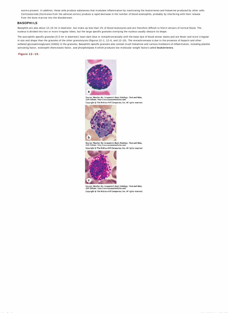
worms present. In addition, these cells produce substances that modulate inflammation by inactivating the leukotrienes and histamine produced by other cells.
Corticosteroids (hormones from the adrenal cortex) produce a rapid decrease in the number of blood eosinophils, probably by interfering with their release
from the bone marrow into the bloodstream.
BASOPHILS
Basophils are also about 12–15 m in diameter, but make up less than 1% of blood leukocytes and are therefore difficult to find in smears of normal blood. The
nucleus is divided into two or more irregular lobes, but the large specific granules overlying the nucleus usually obscure its shape.
The azurophilic specific granules (0.5 m in diameter) stain dark blue or metachromatically with the basic dye of blood smear stains and are fewer and more irregular
in size and shape than the granules of the other granulocytes (Figures 12–1, 12–6, and 12–10). The metachromasia is due to the presence of heparin and other
sulfated glycosaminoglycans (GAGs) in the granules. Basophilic specific granules also contain much histamine and various mediators of inflammation, including platelet
activating factor, eosinophil chemotactic factor, and phospholipase A which produces low molecular weight factors called leukotrienes.
Figure 12–10.
AccessMedicine | Print: Chapter 12. Blood http://accessmedicine.com/popup.aspx?aID=6181612&print=yes_chapter
10 of 16 22/05/2011 23:37

Basophils.
(a, b, c): Basophils are approximately the same size as neutrophils and eosinophils, but have large, strongly basophilic specific granules which usually obstruct theappearance of the nucleus having two or three irregular lobes. a and b: X1500, Wright; c: X1500, Giemsa. (d): TEM of a sectioned basophil reveals the lobulated nucleus(N), appearing as three separated portions, the large specific basophilic granules (B), mitochondria (M), and Golgi complex (G). Basophils exert many activities modulatingthe immune response and inflammation and share many functions with mast cells, which are normal, longer term residents of connective tissue. X16,000. (Figure 12–10dreproduced, with permission, from Terry R.W. et al: Lab. Invest 1969;21:65.)
By migrating into connective tissues, basophils may supplement the functions of mast cells, with which they share a common progenitor cell origin. Both basophils and
mast cells have metachromatic granules containing heparin and histamine, have IgE bound to surface receptors, and secrete their granular components in response to
certain antigens (see Chapter 5).
MEDICAL APPLICATION
In some individuals a second exposure to a strong allergen, such as that delivered in a bee sting, may produce an intense, adverse systemic response.
Basophils and mast cells may rapidly degranulate, producing vasodilation in many organs, a sudden drop in blood pressure, and other effects comprising a
potentially lethal condition called anaphylaxis or anaphylactic shock.
In the dermatologic disease called cutaneous basophil hypersensitivity, basophils are the major cell type at the site of inflammation.
LYMPHOCYTES
Lymphocytes constitute a family of leukocytes with spherical nuclei (Figures 12–1 and 12–6). They can be subdivided into functional groups according to distinctive
surface molecules (markers) that can best be distinguished immunocytochemically, notably T lymphocytes, B lymphocytes, and natural killer (NK) cells.
Lymphocytes have diverse functional roles related to immune defense against invading microorganisms, foreign or abnormal antigens, and cancer cells. Additional
information on the different types of lymphocytes and the functional characteristics in immune responses is presented in Chapter 14.
Most lymphocytes in the blood are small with diameters of 6–8 m; medium and large lymphocytes range in size from 9 to 18 m in diameter. Some larger
lymphocytes may be cells that have been activated by specific antigens. The small lymphocytes that predominate in the blood are characterized by spherical nuclei,
sometimes indented, and condensed, very basophilic chromatin, making them easily distinguishable from granulocytes.
The cytoplasm of the small lymphocyte is scanty, and in blood smears it appears as only a thin rim around the nucleus. It is slightly basophilic and may contain a few
azurophilic granules, along with a few mitochondria and a small Golgi apparatus; it contains free polyribosomes (Figure 12–11).
Figure 12–11.
AccessMedicine | Print: Chapter 12. Blood http://accessmedicine.com/popup.aspx?aID=6181612&print=yes_chapter
11 of 16 22/05/2011 23:37

Lymphocytes.
Lymphocytes are agranulocytes and lack the specific granules characteristic of granulocytes. Lymphocytes circulating in blood range in size from 6 to 15 m in diameter andare sometimes classified arbitrarily as small, medium, and large. (a): The most numerous small lymphocytes shown here are slightly larger than the neighboringerythrocytes and often have only a thin rim of cytoplasm surrounding the spherical nucleus. X1500. Giemsa. (b): Medium lymphocytes are distinctly larger thanerythrocytes. X1500. Wright. (c): Large lymphocytes, much larger than erythrocytes, may represent activated cells that have returned to the circulation. X1500. Giemsa.(d): Ultrastructurally a medium-sized lymphocytes is seen to be mostly filled with a euchromatic nucleus (N), with a nucleolus (Nu), surrounded by cytoplasm containingmitochondria (M), free polysomes, and a few lysosomes (azurophilic granules). X22,000.
Lymphocytes vary in life span according to their specific functions; some live only a few days and others survive in the circulating blood or other tissues for many
years. They are the only type of leukocytes that, following diapedesis, can return from the tissues back to the blood.
MONOCYTES
Monocytes are bone marrow–derived agranulocytes with diameters varying from 12 to 20 m. The nucleus is large, off-center, and may be oval, kidney-shaped, or
distinctly U-shaped (Figure 12–12). The chromatin is less condensed than in lymphocytes and stains lighter than that of large lymphocytes.
Figure 12–12.
AccessMedicine | Print: Chapter 12. Blood http://accessmedicine.com/popup.aspx?aID=6181612&print=yes_chapter
12 of 16 22/05/2011 23:37

AccessMedicine | Print: Chapter 12. Blood http://accessmedicine.com/popup.aspx?aID=6181612&print=yes_chapter
13 of 16 22/05/2011 23:37

Monocytes.
Monocytes are large agranulocytes with diameters from 12 to 20 m that circulate as precursors to macrophages and other cells of the mononuclear phagocyte system. (a,b, c, d): Micrographs of monocytes that show their eccentric nuclei indented, kidney-shaped, or U-shaped. a: X1500, Giemsa; b–d: X1500, Wright. (e): TEM of thecytoplasm of a monocyte shows a Golgi apparatus (G), mitochondria (M), and lysosomes or azurophilic granules (A). Rough ER is poorly developed and there are some freeribosomes (R). X22,000. (Figure 12-12e, with permission, from D.F. Bainton and M.G. Farquhar, Department of Pathology, University of California at San Francisco.).
The cytoplasm of the monocyte is basophilic and contains very small azurophilic granules (lysosomes), some of which are at the limit of the light microscope's
resolution. These granules are distributed through the cytoplasm, giving it a bluish-gray color in stained smears. In the electron microscope, nucleoli may be seen in
the nucleus, and a small quantity of rough ER, free polyribosomes, and many small mitochondria are observed. A Golgi apparatus involved in the formation of
lysosomes is present and many microvilli and pinocytotic vesicles are found at the cell surface (Figure 12–12).
Circulating monocytes are precursor cells of the mononuclear phagocyte system (see Chapter 5). After crossing the walls of postcapillary venules, monocytes
differentiate into macrophages in connective tissues, microglia in the CNS, osteoclasts in bone, etc.
PLATELETS
Blood platelets (thrombocytes) are nonnucleated, disklike cell fragments 2–4 m in diameter. Platelets originate by fragmentation at the ends of cytoplasmic
processes extending from giant polyploid cells called megakaryocytes in the bone marrow (Chapter 13). Platelets promote blood clotting and help repair minor tears
or leaks in the walls of blood vessels, preventing loss of blood. Normal platelet counts range from 200,000 to 400,000 per microliter of blood. Platelets have a life span
of about 10 days.
In stained blood smears, platelets often appear in clumps. Each platelet has a lightly stained peripheral zone, the hyalomere, and a central zone containing darker-
staining granules, called the granulomere (Figure 12–13).
Figure 12–13.
AccessMedicine | Print: Chapter 12. Blood http://accessmedicine.com/popup.aspx?aID=6181612&print=yes_chapter
14 of 16 22/05/2011 23:37

AccessMedicine | Print: Chapter 12. Blood http://accessmedicine.com/popup.aspx?aID=6181612&print=yes_chapter
15 of 16 22/05/2011 23:37

Copyright © The McGraw-Hill Companies. All rights reserved.Privacy Notice. Any use is subject to the Terms of Use and Notice.
Platelets.
Platelets are cell fragments 2–4 m in diameter derived from megakaryocytes of bone marrow. Their primary function is to rapidly release the content of their granules uponcontact with collagen (or other materials outside of the endothelium) to begin the process of clot formation and reduce blood loss from the vasculature. (a): In a bloodsmear, platelets (arrows) are often found as aggregates. Individually they show a lightly stained hyalomere region surrounding a more darkly stained central granulomerecontaining membrane-enclosed granules. X1500. Wright. (b): Ultrastructurally a platelet typically shows a system of microtubules and actin filaments near the periphery tohelp maintain its shape and an open canalicular system of vesicles continuous with the plasmalemma. The central granulomere region contains glycogen and secretorygranules of different types. X40,000. (c): TEM section shows platelets adhering to collagen (C). Upon adhesion to collagen, platelets exocytose their granules into thecanalicular system, which allows the very rapid secretion of factors involved in blood coagulation. Degranulating platelets (arrows) remain as an aggregate until theircontents are exhausted. Other proteins involved in coagulation come from the plasma and from processes of adjacent endothelial cells (EP). The electron-dense structure onthe right is part of an erythrocyte. X;7500. (Figure 12-13b, with permission, from Dr. M. J. G. Harrison, Middlesex Hospital and University College London.).
A coat rich in GAGs and glycoproteins, 15–20 nm thick, lies outside the plasmalemma and is involved in platelet adhesion. Ultrastructural analysis (Figure 12–13)
reveals around the platelet periphery a marginal bundle of microtubules and microfilaments which helps to maintain the platelet's ovoid shape. Also in the
hyalomere are two systems of membrane channels. An open canalicular system of vesicles connected to invaginations of the plasma membrane, which may
facilitate platelets' uptake of factors such as fibrinogen and serotonin from plasma. Another set of irregular tubular vesicles comprising the dense tubular system is
derived from the ER and stores Ca2+ ions. Together these two membranous systems facilitate the extremely rapid exocytosis of proteins from platelets
(degranulation) upon adhesion to collagen or other substrates outside the vascular endothelium.
The central granulomere possesses a variety of membrane-bound granules and a sparse population of mitochondria and glycogen particles (Figure 12–13).
Electron-dense delta granules, 250–300 nm in diameter, contain adenosine diphosphate (ADP), adenosine triphosphate (ATP), and serotonin (5-hydroxytryptamine)
taken up from plasma. Alpha granules are larger (300–500 nm in diameter) and contain platelet-derived growth factor, platelet factor 4, and several other platelet-
specific proteins. Most of the stained granules seen with the light microscope in platelets are alpha granules. Small vesicles, 175–250 nm in diameter, have been
shown to contain only lysosomal enzymes and have been termed lambda granules.
The role of platelets in controlling hemorrhage can be summarized as follows:
Primary aggregation. Disruptions in the microvascular endothelium, which are common, allow platelet aggregation to collagen via collagen-bindingprotein in the platelet membrane. Thus, a platelet plug is formed as a first step to stop bleeding (Figure 12–13c).
Secondary aggregation. Platelets in the plug release an adhesive glycoprotein and ADP, both of which are potent inducers of platelet aggregation,increasing the size of the platelet plug.
Blood coagulation. During platelet aggregation, fibrinogen from plasma, von Willebrand factor and others from damaged endothelium, and variousfactors from platelets promote the sequential interaction (cascade) of plasma proteins, giving rise to a fibrin polymer that forms a three-dimensionalnetwork of fibers trapping red blood cells and more platelets to form a blood clot, or thrombus (Figure 12–14).
Clot retraction. The clot that initially bulges into the blood vessel lumen contracts slightly because of the interaction of platelet actin and myosin.
Clot removal. Protected by the clot, the vessel wall is restored by new tissue, and the clot is then removed, mainly by the proteolytic enzyme plasmin,formed continuously through the local action of plasminogen activators from the endothelium on plasminogen from plasma. Enzymes released fromplatelet lambda granules also contribute to clot removal.
Figure 12–14.
Fibrin clot.
Minor trauma to vessels of the microvasculature is a routine occurrence in active individuals and quickly results in a fibrin clot, shown here by SEM. A meshwork ofpolymeric proteins composed largely of fibrin traps erythrocytes and more degranulating platelets. Platelets in various states of degranulation are shown. Such a clot growsuntil blood loss from the vasculature stops. After repair of the vessel wall, fibrin clots are removed by proteolysis due primarily to locally generated plasmin, a nonspecificprotease.
MEDICAL APPLICATION
Hemophilia A and B are clinically identical, differing only in the deficient factor. Both are due to sex-linked recessive inherited disorders. Blood from
hemophiliac patients does not coagulate normally: the blood clotting time is prolonged. Persons with this disease bleed severely even after mild injuries, such
as a skin cut, and may bleed to death after more severe injuries. The blood plasma of patients with hemophilia A is deficient in clotting factor VIII or contains
a defective factor VIII, one of the plasma proteins involved in fibrin generation; in hemophilia B, the defect is in factor IX. In severe cases the blood is
incoagulable. There are spontaneous hemorrhages in body cavities, such as major joints and the urinary tract. Generally, only males are affected by
hemophilia A, because the recessive gene to factor VIII is on the X chromosome. Females may have one defective X chromosome, but the other one is usually
normal. Females develop hemophilia only when they have the abnormal gene in both X chromosomes, a rare event. However, women with a defective X
chromosome may transmit the disease to their male children.
AccessMedicine | Print: Chapter 12. Blood http://accessmedicine.com/popup.aspx?aID=6181612&print=yes_chapter
16 of 16 22/05/2011 23:37

Print Close Window
Note: Large images and tables on this page may necessitate printing in landscape mode.
Copyright © The McGraw-Hill Companies. All rights reserved.
Junqueira's Basic Histology: Text & Atlas, 12e > Chapter 13. Hemopoiesis >
HEMOPOIESIS: INTRODUCTION
Mature blood cells have a relatively short life span and must be continuously replaced with stem cell progeny produced in the hemopoietic (Gr. haima, blood, +
poiesis, a making) organs. In the earliest phase of human embryogenesis, blood cells arise from the yolk sac mesoderm. In the second trimester, hemopoiesis (also
called hematopoiesis) occurs primarily in the developing liver, with the spleen also playing a role. Skeletal elements begin to ossify and bone marrow develops in their
medullary cavities, so that, in the third trimester, bone marrow increasingly becomes the major hemopoietic organ.
After birth and on into childhood, erythrocytes, granulocytes, monocytes, and platelets are derived from stem cells located in bone marrow. The origin and maturation
of these cells are termed, respectively, erythropoiesis (Gr. erythros, red, + poiesis), granulopoiesis, monocytopoiesis, and thrombocytopoiesis. Development
of the major types of lymphocytes by lymphopoiesis occurs in the marrow and in the lymphoid organs to which precursor cells migrate, as discussed in Chapter 14.
Before reaching maturity and being released into the circulation, blood cells go through specific stages of differentiation and maturation. Because these processes are
continuous, cells with characteristics that lie between the various stages are frequently encountered in smears of blood or bone marrow.
STEM CELLS, GROWTH FACTORS, & DIFFERENTIATION
Stem cells are pluripotent cells capable of asymmetric division and self-renewal. Some of their daughter cells form specific, irreversibly differentiated cell types, and
other daughter cells remain stem cells. A constant number of pluripotent stem cells is maintained in a pool and cells recruited for differentiation are replaced with
daughter cells from the pool.
Hemopoietic stem cells can be isolated by using fluorescence-labeled antibodies to mark specific cell-surface antigens and a fluorescence-activated cell-sorting (FACS)
instrument. Stem cells are studied using experimental techniques that permit analysis of hemopoiesis in vivo and in vitro.
In vivo techniques include injecting the bone marrow of normal donor mice into lethally irradiated mice whose hematopoietic cells have been destroyed. In these
animals, the transplanted bone marrow cells develop colonies of hematopoietic cells in the marrow cavities and spleen. This work led to the development of bone
marrow transplants now used clinically to treat potentially lethal hemopoietic disorders.
In vitro techniques involve the use of semisolid tissue culture media made with a layer of cells derived from bone marrow stroma or purified protein growth factors
produced by marrow stromal cells. Such methods create microenvironmental conditions sufficiently similar to the normal in vivo conditions to favor hemopoietic stem
cell growth and differentiation.
Pluripotent Hemopoietic Stem CellsIt is believed that all blood cells arise from a single type of stem cell in the bone marrow called a pluripotent stem cell because it can produce all blood cell types
(Figure 13–1). The pluripotent stem cells proliferate and form two major cell lineages: one for lymphoid cells (lymphocytes) and another for myeloid cells (Gr.
myelos, marrow) that develop in bone marrow. Myeloid cells include granulocytes, monocytes, erythrocytes, and megakaryocytes. Early in their development,
lymphoid cells migrate from the bone marrow to the thymus or to the lymph nodes, spleen, and other lymphoid structures, where they proliferate and differentiate
(see Chapter 14).
Figure 13–1.
AccessMedicine | Print: Chapter 13. Hemopoiesis http://accessmedicine.com/popup.aspx?aID=6181780&print=yes_chapter
1 of 11 22/05/2011 23:38

Origin and differentiative stages of blood cells. Rare pluripotent stem cells divide slowly, maintain their own population, and give rise to two major cell lineages ofprogenitor cells: the myeloid and lymphoid stem cells. The myeloid lineage includes precursor cells (blasts) for erythropoiesis, thrombopoiesis, granulopoiesis, andmonocytopoiesis, all in the bone marrow. The lymphoid lineage forms lymphopoietic cells, partly in the bone marrow and partly in lymphoid organs.
Progenitor & Precursor CellsThe pluripotent stem cells give rise to daughter cells with restricted potentials called progenitor cells or colony-forming units (CFUs), since they give rise to
colonies of only one cell type when cultured or injected into a spleen. There are four types of progenitors/CFUs:
Erythroid lineage of CFU-erythrocytes (CFU-E)
Thrombocytic lineage of CFU-megakaryocytes (CFU-Meg)
Granulocyte-monocyte lineage of CFU-granulocytes-monocytes (CFU-GM)
Lymphoid lineage of CFU-lymphocytes of all types (CFU-L).
All four progenitor/CFUs produce precursor cells or blasts in which the cells' morphologic characteristics begin to differentiate, suggesting the mature cell types they
will become (Figure 13–1). In contrast, stem and progenitor cells cannot be morphologically distinguished and resemble large lymphocytes. Stem cells divide at a rate
only sufficient to maintain their relatively small population. The rate of cell division is accelerated in progenitor and precursor cells, and large numbers of differentiated,
mature cells are produced (3 x 10⁹ erythrocytes and 0.85 x 10⁹ granulocytes/kg/day in human bone marrow). Whereas progenitor cells divide asymmetrically to
produce both progenitor and precursor cells, precursor cells produce only cells on the path to differentiation.
Hemopoiesis depends on favorable microenvironmental conditions and the presence of paracrine or endocrine growth factors. This microenvironment in hematopoietic
organs is furnished largely by stromal cells and their extracellular matrix (ECM), which together create the niche in which stem cells are maintained. A general view of
hemopoiesis shows that during this process, both the potential for various outcomes of differentiation and the self-renewing capacity of the cells gradually decrease. In
contrast, the mitotic response to growth factors increases, attaining its maximum in the middle of the process. From that point on, mitotic activity decreases,
morphologic characteristics and functional activity develop, and mature cells are formed (Table 13–1).
AccessMedicine | Print: Chapter 13. Hemopoiesis http://accessmedicine.com/popup.aspx?aID=6181780&print=yes_chapter
2 of 11 22/05/2011 23:38

Table 13–1. Changes in properties of hematopoietic cells during differentiation.
Hemopoietic growth factors, called colony-stimulating factors (CSF) or hematopoietins (poietins), are proteins with complex, overlapping functions in stimulating
proliferation (mitogenic activity) of immature (mostly progenitor and precursor) cells, supporting differentiation of maturing cells, and enhancing the functions of
mature cells. These three functions may all occur in the same growth factor or they may be expressed with different levels of intensity in different growth factors. The
isolation and cloning of genes for several growth factors has permitted both the mass production of these proteins and tremendously advanced study of their effects in
vivo and in vitro. The main characteristics of five well-characterized growth factors are presented in Table 13–2.
Table 13–2. Main characteristics of five hemopoietic growth factors (colony-stimulating factors, CSF).
Name Human Gene Location and Producing Cells Main Biologic Activity
Granulocyte (G-CSF) Chromosome 17 macrophages
Endothelium fibroblasts
Stimulates formation (in vitro and in vivo) of granulocytes.
Enhances metabolism of granulocytes.
Stimulates malignant (leukemic) cells.
Granulocyte + macrophage (GM-CSF) Chromosome 5 T lymphocytes
Endothelium fibroblasts
Stimulates in vitro and in vivo production of granulocytes and macrophages.
Macrophage (M-CSF) Chromosome 5 macrophages
Endothelium fibroblasts
Stimulates formation of macrophages in vitro.
Increases antitumor activity of macrophages.
Interleukin 3 (IL-3) Chromosome 5 T lymphocytes Stimulates in vivo and in vitro production of all myeloid cells.
Erythropoietin (EPO) Chromosome 7 renal interstitial Stimulates red blood cell formation in vivo and cells (outer cortex) in vitro.
MEDICAL APPLICATION
Growth factors have been used clinically to increase marrow cellularity and blood cell counts. Potential therapeutic uses of growth factors include increasing the
number of blood cells in diseases or induced conditions (eg, chemotherapy, irradiation) that result in low blood counts; increasing the efficiency of marrow
transplants by enhancing cell proliferation; enhancing host defenses in patients with malignancies and infectious and immunodeficient diseases; and enhancing
the treatment of parasitic diseases.
BONE MARROW
Under normal conditions, the production of blood cells by the bone marrow is adjusted to the body's needs, increasing its activity several-fold in a very short time.
Bone marrow and adipocytes are found in the medullary canals of long bones and in the cavities of cancellous bones. There are two types of bone marrow based on
their appearance at gross examination: blood-forming red bone marrow, whose color is produced by an abundance of blood and hemopoietic cells, and yellow bone
marrow, which is filled with adipocytes and essentially excludes hemopoietic cells. In the newborn, all bone marrow is red and active in blood cell production, but as
the child grows most of the marrow changes gradually to the yellow variety. Under certain conditions, such as severe bleeding or hypoxia, yellow marrow reverts to
red.
Red bone marrow (Figure 13–2) is composed of a stroma (Gr: stroma, bed), hemopoietic cords or islands of cells, and sinusoidal capillaries. The stroma is a
meshwork of specialized fibroblastic cells called reticular or adventitial cells and a delicate web of reticular fibers supporting hemopoietic cells and macrophages. The
matrix of bone marrow also contains collagen type I, proteoglycans, fibronectin, and laminin, the latter glycoproteins interacting with integrins to bind cells to the
matrix.
Figure 13–2.
AccessMedicine | Print: Chapter 13. Hemopoiesis http://accessmedicine.com/popup.aspx?aID=6181780&print=yes_chapter
3 of 11 22/05/2011 23:38

Red bone marrow (active in hemopoiesis).
Red bone marrow contains adipocytes but is also active in hemopoiesis, with several cell lineages usually present. It can be examined histologically in sections of bones or inbiopsies, but its cells can also be studied in smears. Marrow consists of capillary sinusoids running through a stroma of specialized, fibroblastic reticular cells and an ECMmeshwork. Reticular cells secrete various colony-stimulating factors and the stroma forms the microenvironment for hemopoietic stem cell maintenance, proliferation, anddifferentiation. This section of red bone marrow shows some of its components. Sinusoid capillaries (S) containing erythrocytes are surrounded by stroma containingadipocytes (A) and islands or cords (C) of hemopoietic cells. Sinusoidal endothelial cells (one nucleus at E) are very thin. Most reticular cells and cells of the hemopoieticlineages are difficult to identify with certainty in routinely stained sections of marrow. X400. H&E.
Sinusoids are formed by a thin layer of endothelial cells. Differentiated blood cells from the hemopoietic cords enter the circulation by passing through openings in the
endothelium (Figure 13–3). Red bone marrow is also a site where macrophages phagocytose worn-out erythrocytes and store iron derived from hemoglobin
breakdown.
Figure 13–3.
Sinusoidal endothelium in active marrow.
Diagram shows that mature, newly formed erythrocytes, leukocytes, and platelets in marrow enter the circulation by passing through the sinusoid capillary endothelium.Because erythrocytes (unlike leukocytes) cannot migrate through the wall of the sinusoid actively, they are believed to enter the sinusoid by a pressure gradient across itswall. Leukocytes cross the wall of the sinusoid by their own activity. All blood cells apparently penetrate through apertures and between the endothelial cells.Megakaryocytes form thin processes (proplatelets) that also pass through such apertures and liberate platelets at their tips.
MEDICAL APPLICATION
Red bone marrow also contains stem cells that can produce other tissues in addition to blood cells. With their great potential for differentiation, these cells
make it possible to generate specialized cells that are not rejected by the body because they are produced from stem cells from the marrow of the same
person. The procedure is to collect bone marrow stem cells, cultivate them in appropriate medium to direct their differentiation to the cell type needed for
transplantation, and then use the cells originating in tissue culture to replace certain defective cells. In this case the donor and the recipient are the same
individual and histocompatibility is complete, excluding the possibility of rejection. These studies are at early stages, but results with animal models so far are
promising.
AccessMedicine | Print: Chapter 13. Hemopoiesis http://accessmedicine.com/popup.aspx?aID=6181780&print=yes_chapter
4 of 11 22/05/2011 23:38

MATURATION OF ERYTHROCYTES
A mature cell is one that has differentiated to the stage at which it can carry out all its specific functions. Erythrocyte maturation involves hemoglobin synthesis and
formation of a small, enucleated, biconcave corpuscle. Several major changes take place during erythrocyte maturation (Figure 13–4). Cell and nuclear volume
decrease, and the nucleoli diminish in size and disappear. The chromatin becomes increasingly denser until the nucleus presents a pyknotic appearance and is finally
extruded from the cell. There is a gradual decrease in the number of polyribosomes (basophilia decreases), with a simultaneous increase in the amount of hemoglobin
(an acidophilic protein) within the cytoplasm. Mitochondria and other organelles gradually disappear.
Figure 13–4.
Summary of erythrocyte maturation.
The color change in the cytoplasm shows the continuous decrease in basophilia and the increase in hemoglobin concentration from proerythroblast to erythrocyte. There isalso a gradual decrease in nuclear volume and an increase in chromatin condensation, followed by extrusion of a pyknotic nucleus. The times are the average duration ofeach cell type. In the graph, 100% represents the highest recorded concentrations of hemoglobin and RNA.
There are three to five intervening cell divisions between the proerythroblast and the mature erythrocyte. The development of an erythrocyte from the first
recognizable cell of the series to the release of reticulocytes into the blood takes approximately a week. The glycoprotein erythropoietin (Epo), a growth factor
produced in the kidneys, stimulates production of mRNA for globin, the protein component of hemoglobin and is essential for the production of erythrocytes.
The first recognizable cell in the erythroid series (Figure 13–5) is the proerythroblast, a large cell with loose, lacy chromatin, nucleoli, and basophilic cytoplasm. The
next stage is represented by the basophilic erythroblast, with more strongly basophilic cytoplasm and a condensed nucleus with no visible nucleolus. The basophilia
of these two cell types is caused by the large number of polyribosomes synthesizing hemoglobin. During the next stage cell volume is reduced, polyribosomes decrease
and some cytoplasmic areas begin to be filled with hemoglobin, producing regions of both basophilia and acidophilia in the cell, now called a polychromatophilic
erythroblast. In the next stage, the cell and nuclear volumes continue to condense and no basophilia is evident, resulting in a uniformly acidophilic cytoplasm—the
orthochromatophilic erythroblast. Late in this stage, this cell ejects its nucleus which is phagocytosed by macrophages. The cell still has a small number of
polyribosomes that, when treated with the dye brilliant cresyl blue, form a faintly stained network and the cell is called the reticulocyte. Reticulocytes pass to the
circulation, where they may constitute 1% of the red blood cells, lose the polyribosomes and quickly mature as erythrocytes.
Figure 13–5.
AccessMedicine | Print: Chapter 13. Hemopoiesis http://accessmedicine.com/popup.aspx?aID=6181780&print=yes_chapter
5 of 11 22/05/2011 23:38

Erythropoiesis: Major erythrocyte precursors.
(a): Micrographs showing a very large and scarce proerythro-blast (P), a slightly smaller basophilic erythroblast (B) with very basophilic cytoplasm, typical and latepolychromatophilic erythroblasts (Pe and LPe) with both basophilic and acidophilic cytoplasmic regions, and a small orthochromatophilic erythroblast (Oe) with cytoplasmnearly like that of the mature erythrocytes in the field. All X1400. Wright. (b): Micrograph containing reticulocytes (arrows) that have not yet completely lost thepolyribosomes used to synthesize globin, as demonstrated by a stain for RNA. X1400. Brilliant cresyl blue.
MATURATION OF GRANULOCYTES
Granulopoiesis involves cytoplasmic changes dominated by synthesis of proteins for the azurophilic granules and specific granules. These proteins are produced in
the rough endoplasmic reticulum and the prominent Golgi apparatus in two successive stages (Figure 13–6). The azurophilic granules, which contain lysosomal
hydrolases, stain with basic dyes, and are somewhat similar in all three types of granulocytes, are made first. Golgi activity then changes to produce proteins for the
specific granules, whose contents differ in each of the three types of granulocytes and endow each type with certain different properties. In sections of bone marrow,
cords of granulopoietic cells can be distinguished by their granule-filled cytoplasm from erythropoietic cords (Figure 13–7).
Figure 13–6.
Granulopoiesis: Formation of granules.
Diagram illustrating the sequence of cytoplasmic events in the maturation of granulocytes from myeloblasts. Modified lysosomes or azurophilic granules form first at thepromyelocyte stage and are shown in blue; the specific granules of the particular cell type form at the myelocyte stage and are shown in pink. All granules are fullydispersed at the metamyelocyte stage, when indentation of the nucleus begins.
Figure 13–7.
AccessMedicine | Print: Chapter 13. Hemopoiesis http://accessmedicine.com/popup.aspx?aID=6181780&print=yes_chapter
6 of 11 22/05/2011 23:38

Developing erythrocytes and granulocytes in marrow.
Precursor cells of different hemopoietic lineages develop side by side with some intermingling as various cell islands or cords in the bone marrow. Plastic section of red bonemarrow showing mitotic figures (arrows), a plasma cell (arrowhead), and fairly distinct regions of erythropoiesis and granulopoiesis. Most immature granulocytes are in themyelocyte stage: their cytoplasm contains large, dark-stained azurophilic granules and small, less darkly stained specific granules. X400. Giemsa.
The myeloblast is the most immature recognizable cell in the myeloid series (Figure 13–8). It has a finely dispersed chromatin, and faint nucleoli. In the next stage,
the promyelocyte is characterized by its basophilic cytoplasm and azurophilic granules containing lysosomal enzymes and myeloperoxidase. Different promyelocytes
activate different sets of genes, resulting in lineages for the three types of granulocytes. The first visible sign of differentiation appears in the myelocytes (Figure
13–9), in which specific granules gradually increase in number and eventually occupy most of the cytoplasm at the metamyelocyte stage. These neutrophilic,
basophilic, and eosinophilic metamyelocytes mature with further condensations of the nuclei. Before its complete maturation the neutrophilic granulocyte passes
through an intermediate stage, the stab or band cell, in which its nucleus is elongated but not yet polymorphic.
Figure 13–8.
Granulopoiesis: Major granulocyte precursors.
Two micrographs from smears of bone marrow showing the major cells of the neutrophilic granulocyte lineage. Typical precursor cells shown are labeled as follows:myeloblast (MB); promyelocyte (1); myelocytes (2); late myelocyte (3); metamyelocytes (4); stab or band cells (5); nearly mature segmented neutrophil (6). Some of theearly stages show faint nucleoli (N). Inset: Eosinophilic myelocytes (EM) and metamytelocytes (EMm) with their specific granules having distinctly different staining. Theseand cells of the basophilic lineage are similar to developing neutrophils, except for their specific staining granules and lack of the stab cell form. Also seen among theerythrocytes of these marrow smears are some orthochromatophilic erythroblasts (Oe), a small lymphocyte (L), and a cell in mitosis (arrow). All X1400. Wright.
Figure 13–9.
AccessMedicine | Print: Chapter 13. Hemopoiesis http://accessmedicine.com/popup.aspx?aID=6181780&print=yes_chapter
7 of 11 22/05/2011 23:38

Neutrophilic myelocyte.
At the myelocyte stage lysosomes (azurophilic granules) have formed and production of specific secretory granules is underway. This micrograph shows ultrastructurally aperoxidase-stained section of a neutrophilic myelocyte with cytoplasm containing both large, peroxidase-positive azurophilic granules (AG) and smaller specific granules(SG), which do not stain for peroxidase. The peroxidase reaction product is present only in mature azurophilic granules and is not seen in the rough ER (RER) or Golgicisternae (GC), which are located around the centriole (C) near the nucleus (N). X15,000. (With permission, from Dr. Dorothy F. Bainton, Department of Pathology,University of California at San Francisco.)
MEDICAL APPLICATION
The appearance of large numbers of immature neutrophils (band cells) in the blood, sometimes called a "shift to the left", is clinically significant, usually
indicating bacterial infection.
The vast majority of granulocytes are neutrophils. The total time taken for a myeloblast to emerge as a mature, circulating neutrophil is about 11 days. Under normal
circumstances, five mitotic divisions occur in the myeloblast, promyelocyte, and neutrophilic myelocyte stages. Developing and mature neutrophils can be considered
to exist in four functionally and anatomically defined compartments: the granulopoietic compartment in marrow; storage as mature cells in marrow until release; the
circulating population; and a marginating population of cells adhering to endothelial cells of postcapillary venules and small veins (Figure 13–10). Margination of
neutrophils in some organs can persist for several hours and is not always immediately followed by the cells' emigration from the microvasculature.
Figure 13–10.
Functional compartments of neutrophils.
Neutrophils exist in at least four anatomically and functionally distinct compartments, the sizes of which are proportional to the number of cells. (1) Granulopoietic
AccessMedicine | Print: Chapter 13. Hemopoiesis http://accessmedicine.com/popup.aspx?aID=6181780&print=yes_chapter
8 of 11 22/05/2011 23:38

compartment subdivided into a mitotic part and a maturation part. (2) Storage (reserve) compartment, also in red marrow, acts as a buffer system, capable of releasinglarge numbers of mature neutrophils on demand. (3) Circulating compartment throughout the blood. (4) Marginating compartment, in which cells temporarily do notcirculate, but rather adhere via selectins to vascular endothelial cells of postcapillary venules, particularly in the lungs. The marginating and circulating compartments are ofabout equal size, and there is a constant interchange of cells between them. The half-life of a neutrophil in these two compartments is less than 10 hours. The granulopoieticand storage compartments together include cells in the first 11 days of their existence and are about 10 times larger than the circulating and marginating compartments.
Neutrophils and other granulocytes enter the connective tissues by migrating through intercellular junctions between endothelial cells of postcapillary venules in
diapedesis. The connective tissues thus form a fifth terminal compartment for neutrophils, where the cells reside for a few days and then die by apoptosis, regardless of
whether they have performed their major function of phagocytosis.
MEDICAL APPLICATION
Changes in the number of neutrophils in the blood must be evaluated by taking all these compartments into consideration. Thus, neutrophilia, an increase in
the number of neutrophils in the circulation, does not necessarily imply an increase in neutrophil production. Intense muscular activity or the administration of
epinephrine can cause neutrophils in the marginating compartment to move into the circulating compartment, producing an apparent neutrophilia even though
neutrophil production has not increased. However, glucocorticoids (adrenal gland hormones) increase the mitotic activity of neutrophil precursors in the
marrow and do increase the blood count of neutrophils.
Neutrophilia may also result from liberation of greater numbers of neutrophils from the medullary storage compartment. This type of neutrophilia is transitory
and is followed by a recovery period during which no neutrophils are released.
The neutrophilia that occurs during bacterial infections is due to an increase in production of neutrophils and a shorter duration of these cells in the medullary
storage compartment. In such cases, immature forms such as band cells, neutrophilic metamyelocytes, and even myelocytes may appear in the bloodstream.
The neutrophilia that occurs during infection is of longer duration than that which occurs as a result of intense muscular activity.
MATURATION OF AGRANULOCYTES
Study of the precursor cells of monocytes and lymphocytes is difficult, because these cells do not show specific cytoplasmic granules or nuclear lobulation, both of which
facilitate the distinction between young and mature forms of granulocytes. Monocytes and lymphocytes in smear preparations are discriminated mainly on the basis of
size, chromatin structure, and the presence of nucleoli.
MonocytesThe monoblast is a committed progenitor cell that is virtually identical to the myeloblast in its morphologic characteristics. Further differentiation leads to the
promonocyte, a large cell (up to 18 m in diameter) with basophilic cytoplasm and a large, slightly indented nucleus. The chromatin is lacy and nucleoli are evident.
Promonocytes divide twice as they develop into monocytes (Figure 13–1). A large amount of rough ER is present, as is an extensive Golgi apparatus in which granule
condensation occurs. These granules are primary lysosomes, which are observed as fine azurophilic granules in blood monocytes. Mature monocytes enter the
bloodstream, circulate for about eight hours, and then enter tissues where they mature as macrophages (or other phagocytic cells) and function for several months.
LymphocytesAs discussed in Chapter 14, circulating lymphocytes originate mainly in the thymus and the peripheral lymphoid organs (eg, spleen, lymph nodes, tonsils etc.).
However, all lymphocyte progenitor cells originate in the bone marrow. Some of these lymphocytes migrate to the thymus, where they acquire the full attributes of T
lymphocytes. Subsequently, T lymphocytes populate specific regions of peripheral lymphoid organs. Other bone marrow lymphocytes differentiate into B lymphocytes
in the bone marrow and then migrate to peripheral lymphoid organs, where they inhabit and multiply in their own special compartments.
As lymphocytes mature, their chromatin becomes more compact, nucleoli become less visible, and the cells decrease in size. In addition, subsets of the lymphocyte
series acquire distinctive cell-surface receptors during differentiation that can be detected by immunocytochemical techniques. The first identifiable progenitor of
lymphoid cells is the lymphoblast, a large cell capable of dividing two or three times to form prolymphocytes. Prolymphocytes are smaller and have relatively more
condensed chromatin but none of the cell-surface antigens that mark T or B lymphocytes. In the bone marrow and in the thymus, these cells synthesize cell-surface
receptors characteristic of the B or T lymphocyte lineages. In routine histological procedures B and T lymphocytes cannot be distinguished; immunocytochemical
techniques using cell-specific markers are required to make the distinction.
MEDICAL APPLICATION
Abnormal stem cells in bone marrow can produce diseases based on cells derived from that tissue. Leukemias are malignant clones of leukocyte precursors.
They occur in lymphoid tissue (lymphocytic leukemias) and in bone marrow (myelogenous and monocytic leukemias). In these diseases, there is usually
a release of large numbers of immature cells into the blood. Some symptoms of leukemias are a consequence of this shift in cell proliferation, with a lack of
some cell types and excessive production of others. The patient is usually anemic and prone to infection.
A clinical technique that is helpful in the study of leukemias and other bone marrow disturbances is bone marrow aspiration. A needle is introduced through
compact bone and a sample of marrow is withdrawn. The use of labeled monoclonal antibodies specific to membrane proteins of precursor blood cells aids in
identifying cell types derived from these stem cells and contributes to a more precise diagnosis of the leukemia.
ORIGIN OF PLATELETS
In adults, the membrane-enclosed cell fragments called platelets originate in the red bone marrow by dissociating from mature megakaryocytes (Gr. megas, big, +
karyon, nucleus, + kytos), which in turn differentiate from megakaryoblasts in a process driven by thrombopoietin. The megakaryoblast is 25–50 m in diameter
and has a large ovoid or kidney-shaped nucleus (Figure 13–11) with numerous small nucleoli. Before differentiating, these cells undergo endomitosis, with repeated
rounds of DNA replication not separated by cell divisions, resulting in a nucleus that is highly polyploid (ie, 64N or more than thirty times more DNA than a normal cell).
The cytoplasm of this cell is homogeneous and intensely basophilic.
Figure 13–11.
AccessMedicine | Print: Chapter 13. Hemopoiesis http://accessmedicine.com/popup.aspx?aID=6181780&print=yes_chapter
9 of 11 22/05/2011 23:38

Megakaryoblast and megakaryocytes.
(a): Megakaryoblasts (Mb) are very large, fairly rare cells in bone marrow, with very basophilic cytoplasm. X1400. Wright. (b): Megakaryoblasts undergo endomitosis (DNAreplication without intervening cell divisions), becoming polyploid as they differentiate into megakaryocytes (M). These cells are even larger, but with cytoplasm that is lessintensely basophilic. X1400. Wright. (c): Micrograph section of bone marrow megakaryocyte (M) shown near sinusoids (S). X400. Giemsa. Megakaryocytes produce all thecharacteristic components of platelets (membrane vesicles, specific granules, marginal microtubule bundles, etc) and in a complex process extend many long, branchingpseudopodia-like projections called proplatelets, from the ends of which platelets are pinched off almost fully formed.
Megakaryocytes are giant cells, 35–150 m in diameter, with irregularly lobulated polyploid nuclei, coarse chromatin, and no visible nucleoli. Their cytoplasm contains
numerous mitochondria, a well-developed rough ER, and an extensive Golgi apparatus from which arise the conspicuous specific granules of platelets, or thrombocytes
(Chapter 12). They are widely scattered in marrow, typically near sinusoidal capillaries.
To form platelets, megakaryocytes extend several long (>100 m), wide (2–4 m), branching processes called proplatelets. These extending proplatelets penetrate
the sinusoidal endothelium and appear as long processes disposed lengthwise with the blood flow in these vessels (Figure 13–3). The proplatelet framework consists of
actin filaments and a loose bundle of mixed polarity microtubules along which organelles, membrane vesicles, and specific granules are transported. A loop of
microtubules forms a teardrop-shaped enlargement at the distal end of the proplatelet and cytoplasm within these loops is pinched off to form platelets with their
characteristic marginal bundles of microtubules, vesicles and granules (Figure 12–13b).
During proplatelet growth microtubules polymerize in both directions. Proplatelet elongation does not depend on this polymerization, but on a dynein-based
microtubule sliding mechanism similar to that of extension ladders. Mature megakaryocytes have numerous invaginations of plasma membrane ramifying throughout
the cytoplasm, called demarcation membranes (Figure 13–12) which were formerly considered "fracture lines" or "perforations" for release of platelets, but are now
thought to represent a membrane reservoir that facilitates continuous rapid proplatelet elongation. Each megakaryocyte produces a few thousand platelets, after which
the remainder of the cell shows apoptotic changes and is removed by macrophages.
Figure 13–12.
AccessMedicine | Print: Chapter 13. Hemopoiesis http://accessmedicine.com/popup.aspx?aID=6181780&print=yes_chapter
10 of 11 22/05/2011 23:38

Copyright © The McGraw-Hill Companies. All rights reserved.Privacy Notice. Any use is subject to the Terms of Use and Notice.
Megakaryocyte ultrastructure.
TEM analysis of a megakaryocyte during platelet formation showing a lobulated nucleus (N), numerous cytoplasmic granules (G), and an extensive system of demarcationmembranes (D) through the cytoplasm. Once believed to be perforations along which platelets were shed from the cells, this membrane system is now considered areservoir of membrane used during elongation of the numerous proplatelets which extend from the megakaryocyte surface. X10,000.
MEDICAL APPLICATION
In certain forms of thrombocytopenic purpura, a disease in which the number of blood platelets is reduced, the platelets appear to be bound to the
cytoplasm of the megakaryocytes, indicating a defect in the liberation mechanism of these corpuscles. The life span of platelets is approximately 10 days.
AccessMedicine | Print: Chapter 13. Hemopoiesis http://accessmedicine.com/popup.aspx?aID=6181780&print=yes_chapter
11 of 11 22/05/2011 23:38

Print Close Window
Note: Large images and tables on this page may necessitate printing in landscape mode.
Copyright © The McGraw-Hill Companies. All rights reserved.
Junqueira's Basic Histology: Text & Atlas, 12e > Chapter 14. The Immune System & Lymphoid Organs >
THE IMMUNE SYSTEM & LYMPHOID ORGANS: INTRODUCTION
The body has a system of cells—the immune system—that has the ability to distinguish "self" (the organism's own molecules)
from "non-self" (foreign substances). This system has the ability to neutralize or inactivate foreign molecules (such as soluble
molecules as well as those present in viruses, bacteria, and parasites) and to destroy microorganisms or other cells (such as virus-
infected cells, cells of transplanted organs, and cancer cells). On occasion, the immune system of an individual reacts against its
own normal body tissues or molecules, causing autoimmune diseases.
The cells of the immune system (1) are distributed throughout the body in the blood, lymph, and epithelial and connective tissues;
(2) are arranged in small spherical nodules called lymphoid nodules found in connective tissues and inside several organs; and
(3) are organized in larger lymphoid organs—the lymph nodes, the spleen, the thymus, and the bone marrow. Lymphoid
nodules are isolated cells of the immune system found in the mucosa of the digestive system (including the tonsils, Peyer's
patches, and appendix), the respiratory system, the reproductive system, and the urinary system are collectively known as
mucosa-associated lymphoid tissue (MALT) and may be considered a lymphoid organ. The wide distribution of immune
system cells and the constant traffic of lymphocytes through the blood, lymph, connective tissues, and lymphoid organs provide
the body with an elaborate and efficient system of surveillance and defense (Figure 14–1).
Figure 14–1.
AccessMedicine | Print: Chapter 14. The Immune System & Lymphoid ... http://accessmedicine.com/popup.aspx?aID=6181868&print=yes_chapter
1 of 37 22/05/2011 19:27

The lymphoid organs and main paths of lymphatic vessels.
The lymphatic system is comprised of lymphatic vessels, which transport interstitial fluid (lymph) back to the blood circulation, andthe lymphoid organs which house lymphocytes and other cells of the body's immune defense system. Primary lymphoid organs are thebone marrow and thymus, where B and T lymphocytes are formed respectively. The secondary lymphoid organs include the lymphnodes, mucosa-associated lymphoid tissue (MALT), and spleen.
ANTIGENS
A molecule that is recognized by cells of the immune system is called an antigen and may elicit a response from these cells.
Antigens may consist of soluble molecules (such as proteins, polysaccharides, and nucleoproteins) or molecules belonging to whole
cells (bacteria, protozoa, tumor cells, or virus-infected cells). The cells of the immune system do not recognize and react to the
whole antigen molecule but instead react to small molecular domains of the antigen known as antigenic determinants or
epitopes. The response of the organism to antigens may be cellular (in which lymphocytes are primarily in charge of eliminating
the antigen) or humoral (in which antibodies secreted by plasma cells are primarily responsible for the response). Some epitopes
(eg, polysaccharides of bacterial walls or lipids) usually elicit a humoral response whereas proteins elicit both a cellular and
humoral response. More details on cellular and humoral immune responses are provided below.
ANTIBODIES
An antibody is a glycoprotein that interacts specifically with an antigenic determinant. Antibodies belong to the immunoglobulin
protein family. Free molecules of antibodies are secreted by plasma cells that arise by proliferation and terminal differentiation of
clones of B lymphocytes whose receptors recognize and bind specific epitopes. These secreted antibodies either circulate in the
plasma and may leave the blood vessels reaching the tissues or are present in the secretion of some epithelia (eg, of the
AccessMedicine | Print: Chapter 14. The Immune System & Lymphoid ... http://accessmedicine.com/popup.aspx?aID=6181868&print=yes_chapter
2 of 37 22/05/2011 19:27

mammary gland and salivary glands). Other antibodies are not free molecules, but are integral membrane proteins of the surface
of lymphocytes. In any case, each antibody combines with the epitope that it specifically recognizes.
There are several classes of antibody molecules but all have a common design: they consist of two identical light chains and two
identical heavy chains bound by disulfide bonds and noncovalent forces (Figure 14–2). The isolated carboxyl-terminal portion of the
heavy chain molecules is called the Fc region. The Fc regions of some immunoglobulins are recognized by receptors on the
membranes of several cell types and for this reason antibodies bind to the surface of these cells. The first 110 amino acids near the
amino-terminal part of the light and heavy chains vary widely among different antibody molecules and this region is called the
variable region. The antigen-binding site of an antibody consists of the variable regions of one heavy and one light chain.
Thus, each antibody has two antigen-binding sites, both for the same antigen. The molecules in some immunoglobulin classes may
form dimers, trimers, or pentamers.
Figure 14–2.
Basic structure of an immunoglobulin.
Two light chains and two heavy chains form an antibody molecule ("monomer"). The chains are linked by disulfide bonds. The variableportions near the amino end of the light and heavy chains bind the antigen. The Fc region of the molecule may bind to surfacereceptors of several cell types.
Classes of AntibodiesThe main classes of immunoglobulins in humans are immunoglobulin G (IgG), IgA, IgM, IgE, and IgD (Table 14–1).
Table 14–1. Summary of classes of antibodies.
IgG IgM IgA IgD IgE
Structure
Monomer
Pentamer
Dimer or trimer withsecretorycomponent
MonomerMonomer
Antibody percentagein the serum
75–80% 5–10% 10–15% 0.001% 0.002%
Presence in sitesother than blood,connective tissue,and lymphoidorgans
Fetal circulation inpregnant women
B lymphocyte surface (as amonomer)
Secretions (saliva,milk, tears, etc)
Surface of Blymphocytes
Bound to thesurface of mastcells andbasophils
Known functions Activatesphagocytosis,neutralizesantigens
First antibody produced ininitial immune response;activates complement
Protects mucosae Antigen receptortriggering initial Bcell activation
Destroys parasiticworms andparticipates inallergies
IgG is the most abundant class representing 75–80% of serum immunoglobulins. It is produced in large amounts during immune
responses. IgG is the only immunoglobulin that crosses the placental barrier and is transported to the circulatory system of the
fetus, protecting the newborn against infections for a certain period of time.
AccessMedicine | Print: Chapter 14. The Immune System & Lymphoid ... http://accessmedicine.com/popup.aspx?aID=6181868&print=yes_chapter
3 of 37 22/05/2011 19:27

IgA is the main immunoglobulin found in secretions, such as nasal, bronchial, intestinal, and prostatic, as well as in tears,
colostrum, saliva, and vaginal fluid. It is present in secretions as a dimer or trimer called secretory IgA, composed of two or
three molecules of monomeric IgA united by a polypeptide chain called protein J and combined with another protein, the
secretory component. Because it is resistant to several enzymes, secretory IgA persists in the secretions where it provides
protection against the proliferation of microorganisms. IgA monomers and protein J are secreted by plasma cells in the lamina
propria of the epithelium of the digestive, respiratory, and urinary passages; the secretory component is synthesized by the
mucosal epithelial cells and is added to the IgA polymer as it is transported through the epithelial cells.
IgM constitutes about 10% of blood immunoglobulins and usually exits as a pentamer. Together with IgD, it is the major
immunoglobulin found on the surface of B lymphocytes. These two classes of immunoglobulins have both membrane-bound and
circulating forms. IgM bound to the membrane of a B lymphocyte functions as its specific receptor for antigens. The result of this
interaction is the proliferation of B lymphocytes into antibody-secreting plasma cells. Secreted IgM, when bound to antigen, is very
effective in activating the complement system.
IgE is much less abundant than the other classes and usually exists as a monomer. As its Fc region has a great affinity for
receptors present on the surfaces of mast cells and basophils, it attaches to these cells after being secreted by plasma cells and
only small amounts are found in the blood. When IgE molecules present on the surface of mast cells or basophils encounter the
antigen that elicited the production of this specific IgE, the antigen–antibody complex triggers the liberation of several biologically
active substances, such as histamine, heparin, leukotrienes, and eosinophil-chemotactic factor of anaphylaxis. This characterizes
an allergic reaction, which is thus mediated by the binding of cell-bound IgE with the antigens (allergens) that stimulated its
production (see Mast Cells in Chapter 5).
The properties and activities of IgD are not completely understood. It is monomeric and is even less abundant than IgE,
constituting only 0.001% of the immunoglobulin in plasma. IgD is found on the cell membrane of B lymphocytes.
Actions of AntibodiesSome antibodies are able to agglutinate cells and to precipitate soluble antigens, thus neutralizing their harmful effects on the body
(Figure 14–3). Although phagocytosis of microorganisms and other particles occurs spontaneously, this event is greatly stimulated
when they are covered by antibodies produced against them, a phenomenon called opsonization (Figure 14–3). Because
macrophages, neutrophils, and eosinophils have receptors for the Fc region of IgG on their surfaces, they phagocytose items that
have been opsonized much more readily.
Figure 14–3.
Mechanisms of antigen inactivation.
Several mechanisms are used by components of the immune system to inactivate or remove potentially harmful material. (1)
AccessMedicine | Print: Chapter 14. The Immune System & Lymphoid ... http://accessmedicine.com/popup.aspx?aID=6181868&print=yes_chapter
4 of 37 22/05/2011 19:27

Agglutination, in which antibodies bind to antigens, forming aggregates and reducing the amount of free antigens; aggregates may beingested by phagocytes; (2) opsonization of antigens by complement stimulates their phagocytosis; (3) opsonization of antigens byantibodies stimulates phagocytosis; (4) neutralization, in which the binding of antibody to microorganisms blocks their adhesion tocells and inactivates toxins; (5) cytotoxicity mediated by cells, which involves antibodies adhering to the surface of worms activatingcells of the immune system (macrophages and eosinophils) and inducing them to liberate molecules that attack the surface of theanimal; (6) complement activation, in which the binding of antibodies to the initial protein of the complement system triggers thecomplement cascade and causes cell lysis.
Antigen–antibody complexes and some antigens activate the complement system, a group of around 20 plasma proteins
produced mainly in the liver and activated through a cascade of reactions. One of the most important proteins of this system is the
component called C3. To defend the body against foreign molecules or cells, the complement system may (1) stimulate
phagocytosis of microorganisms because of opsonization due to the binding of C3 fragments to specific C3 receptors on the surface
of phagocytic cells (Figure 14–3) and (2) induce lysis of microorganisms by acting on their cell membranes (Figure 14–3).
CYTOKINES
The functions of cells in the immune system are regulated by a large number of molecules, mainly cytokines, which are peptides
or glycoproteins usually with low molecular masses (between 8 and 80 kDa). They influence both the cellular and humoral immune
responses (Table 14–2). Cytokines act on many cells that have receptors for them—not only cells of the immune system, but also
cells of other systems, such as the nervous system and endocrine system. They are primarily produced by cells of the immune
system, mainly lymphocytes, macrophages, and other leukocytes, but may also be synthesized by other cell types, such as
endothelial cells and fibroblasts. Chemotaxins, or chemokines, are cytokines that induce diapedesis of leukocytes and migration
to sites of inflammation (Chapter 12).
Table 14–2. Examples of cytokines, grouped by main function.
Cytokine1
Main Function
GM-CSF, M-CSF Growth and differentiation factors for bone marrow cells
TNF-, IL-1, IL-6 Inflammation and fever
IL-12 Stimulation of innate and specific response
IL-2, IL-4, IL-3 Growth factors for T and B cells
IL-5 Eosinophil differentiation and activation
Interferon- Activation of macrophages
IL-10, TGF- Regulation of the immune response
Interferon- , Antiviral activity
Interferon-
1GM-CSF, granulocyte-macrophage colony stimulating factor; M-CSF, macrophage colony-stimulating factor; TNF, tumor necrosisfactor; IL, interleukin; TGF, transforming growth factor.
CELLS OF THE IMMUNE SYSTEM
The primary cells that participate in the immune response are lymphocytes, plasma cells, mast cells, neutrophils, eosinophils, and
cells of the mononuclear phagocyte system. Antigen-presenting cells, a group of diverse cell types, assist other cells in the immune
response. This group includes, among other cells, lymphocytes, macrophages, and dendritic cells.
LymphocytesLymphocytes are classified as B, T, or natural killer (NK) cells. The B and T cells are the only cells that have the ability to
selectively recognize a specific epitope among a vast number of different epitopes (on the order of 1018). B and T cells differ based
on their life history, surface receptors, and behavior during an immune response. Although B and T cells are morphologically
indistinguishable in either the light or electron microscope, different surface proteins (markers) allow then to be distinguished by
immunocytochemical methods. The precursors of all lymphocyte types originate in the bone marrow; some lymphocytes mature
and become functional in the bone marrow, and after leaving the bone marrow enter the blood circulation to colonize connective
tissues, epithelia, lymphoid nodules, and lymphoid organs. These are the B lymphocytes (Figure 14–4). T lymphocyte
precursors, on the other hand, leave the bone marrow, and through the blood circulation reach the thymus where they undergo
intense proliferation and differentiation or die by apoptosis. After their final maturation, T cells leave the thymus and are
distributed throughout the body in connective tissues and lymphoid organs (Figure 14–4). Because of their function in lymphocyte
production and maturation, the bone marrow and the thymus are called the primary or central lymphoid organs. The other
lymphoid structures are all secondary or peripheral lymphoid organs (spleen, lymph nodes, solitary lymphoid nodules, tonsils,
appendix, and Peyer's patches of the ileum). B and T cells are not anchored in the lymphoid organs; instead, they continuously
move from one location to another, a process known as lymphocyte recirculation so that the cellular composition and
microscopic anatomy of lymphoid tissues differ from one day to the next. B and T cells are not distributed uniformly throughout the
lymphoid organs but occupy specific organs preferentially (Table 14–3).
AccessMedicine | Print: Chapter 14. The Immune System & Lymphoid ... http://accessmedicine.com/popup.aspx?aID=6181868&print=yes_chapter
5 of 37 22/05/2011 19:27

Figure 14–4.
Origin of the main types of lymphocytes.
B lymphocytes and natural killer (NK) lymphocytes are formed and become mature in the bone marrow and leave that compartment to
seed the secondary lymphoid organs and transit through the blood to epithelia and connective tissues. Immature CD4– and CD8– Tlymphocyte precursors are transported by the circulation from the bone marrow to the thymus, where they complete their maturation
and leave as either CD4+or CD8+ cells.
Table 14–3. Approximate percentages of B and T cells in lymphoid organs.
Lymphoid Organ T lymphocytes (%) B lymphocytes (%)
Thymus 100 0
Bone marrow 10 90
Spleen 45 55
Lymph nodes 60 40
Blood 70 30
Key features of B and T lymphocytes include the receptors they have on their surface. These receptors are fundamental for
recognition of antigen epitopes and, thus, for triggering an immune response. T cells recognize a linear sequence of amino acids
AccessMedicine | Print: Chapter 14. The Immune System & Lymphoid ... http://accessmedicine.com/popup.aspx?aID=6181868&print=yes_chapter
6 of 37 22/05/2011 19:27

whereas in B cells the spatial arrangement (ie, the molecular conformation) of proteins, nucleic acids, polysaccharides, or lipids is
also important. Each B lymphocyte that leaves the bone marrow or each T lymphocyte that leaves the thymus has just one type of
surface receptor that recognizes a specific epitope. As a result of gene rearrangement during the maturation of B and T cells, many
millions of different cells, each carrying identical surface receptors able to recognize one specific epitope, are produced. Thus, each
lymphocyte recognizes only one epitope.
In an organism not yet exposed to antigens, very few individual lymphocytes are able to recognize each of the millions of epitopes
that exist, perhaps only from one to a few hundred such cells for each. Soon after a lymphocyte is first exposed to the epitope it
recognizes, a stimulus to cell proliferation occurs, leading to an amplification of that particular lymphocyte population, and thereby
producing an expanded clone of lymphocytes able to recognize that epitope.
B LYMPHOCYTES
In B lymphocytes, the surface receptors able to recognize antigens are monomeric molecules of IgM; each B cell is covered by
about 150,000 molecules of IgM. The encounter of a B lymphocyte with the epitope it recognizes leads to several cycles of cell
proliferation, followed by a redifferentiation of most of these lymphocytes into plasma cells. This population of plasma cells
secretes antibodies against the same epitope as that recognized by the B cell from which it arose. In most cases, the activation of
B cells requires the assistance of a subclass of T lymphocytes known as T helper cells. Not all activated B cells, however, become
plasma cells; some remain as long-lived B memory cells, which are able to react very rapidly to a second exposure to the same
epitope.
T LYMPHOCYTES
T cells constitute 65–75% of blood lymphocytes. To recognize epitopes, all T cells have on their surfaces a molecule called a T cell
receptor (TCR). In contrast to B cells, which recognize soluble antigens or antigens present on cell surfaces, T lymphocytes
recognize only epitopes (mostly small peptides) that form complexes with special proteins on the cell surface of other cells
(proteins of the major histocompatibility complex, see below).
Three important subpopulations of T cells are the following:
Helper cells, which produce cytokines that promote differentiation of B cells into plasma cells, activate macrophages tobecome phagocytic, activate cytotoxic T lymphocytes, and induce many parts of an inflammatory reaction. Helper cells
have a marker called CD4 on their surfaces and are, hence, called CD4+ T cells.
Cytotoxic T cells are CD8+ and act directly against foreign cells or virus-infected cells by two main mechanisms. Inone, they attach to the cells to be killed and release proteins called perforins that create holes in the cell membrane ofthe target cell, with consequent cell lysis. In the other, they attach to a cell and kill it by triggering mechanisms thatinduce programmed cell death, or apoptosis.
Regulatory T cells are CD4+CD25+ and play crucial roles in allowing immune tolerance, maintaining unresponsivenessto self-antigens and suppressing excessive immune responses. These cells produce peripheral tolerance, which backsup the central tolerance emerging in the thymus.
The first encounter of a CD4 or CD8 T cell with its specific epitope is followed by amplification of that clone; some of the cells of this
increased population become effector cells, doing the job for which they are specialized, and some remain memory helper or
memory cytotoxic T cells, reacting rapidly to the next presentation of the same epitope.
NATURAL KILLER CELLS
The natural killer lymphocytes lack the marker molecules characteristic of B and T cells. They comprise about 10–15% of the
lymphocytes of circulating blood. Their name derives from the fact that they attack virus-infected cells, transplanted cells, and
cancer cells without previous stimulation; for this reason they are involved in what is called an innate immune response.
MEDICAL APPLICATION
One of the primary causes of the immunodeficiency syndrome known as AIDS involves the killing of helper T cells by the
infecting retrovirus. This cripples the patients' immune system rendering them susceptible to opportunistic infections by
microorganisms that usually do not cause disease in immunocompetent individuals.
Major Histocompatibility Complex (MHC) & Antigen PresentationThe major histocompatibility complex (MHC) is a complex of chromosomal loci encoding several proteins known as class I and
class II MHC molecules. Because a great many alleles exist within each of the loci, there is great variation of these molecules
among the general population. One individual, however, expresses only one set of class I proteins and one set of class II proteins;
these proteins are unique to that person. All nucleated cells have class I proteins, but class II proteins exist on only a small group
of cells operationally called antigen-presenting cells (APCs).
MHC molecules are integral membrane proteins present on the cell surface. They are synthesized by the rough ER like regular
membrane proteins. However, on their way to the cell surface, they couple with small peptides of 10–30 amino acids whose origin
differs depending on whether class I or class II molecules are involved.
In most cases, class I MHC molecules form complexes with peptides derived from cytosolic proteins synthesized in that cell.
Proteins encoded by virus nucleic acid in an infected cell are an important example of this kind of cytosolic protein. The proteins
are targeted by ubiquitin to be degraded by proteasomes, producing small peptides. These peptides are transported to
AccessMedicine | Print: Chapter 14. The Immune System & Lymphoid ... http://accessmedicine.com/popup.aspx?aID=6181868&print=yes_chapter
7 of 37 22/05/2011 19:27

membranes of the ER where they bind class I MHC molecules; the resulting complex is moved to the cell surface, exposing the
peptides to the extracellular space (Figure 14–5).
Figure 14–5.
Binding of class I and class II MHC molecules to antigens.
Left: The sequence of events by which antigens made in a cell (eg, in a virus-infected cell) are processed, bound to class I MHCproteins, and displayed at the cell surface. (1) Proteins in the cell are continuously digested by proteasomes and antigenic fragmentsare transferred to the rough ER where they associate with class I MHC proteins synthesized there. (2) The class I MHC–antigencomplex is transferred to the Golgi region. (3) Golgi vesicles transport the complex to the cell membrane presenting the antigen at theouter surface. Right: Formation of complexes between class II MHC proteins and antigens internalized by the cell. (1) Synthesis ofclass II MHC molecules. (2) Transfer of class II MHC molecules to the Golgi region and formation of Golgi vesicles. The Golgi vesiclesfuse with a lysosome containing antigens processed after endocytosis and digestion of microorganisms and debris by lysosomalenzymes (a, b, c). (3) Antigens form complexes with class II MHC molecules. (4) The class II MHC–antigen complexes are exposed atthe cell surface.
The peptides that join class II MHC molecules result mostly from endocytosis and digestion in lysosomes. The vesicles that contain
these peptides fuse with Golgi-derived vesicles that have class II MHC embedded in their membranes. The peptides form
complexes with the MHC proteins and, as in the case of class I molecules, the complex is transported to the cell surface exposing
the peptides to the exterior of the cell (Figure 14–5).
Contrary to B cells, which recognize soluble antigens or antigens present on cell surfaces, T lymphocytes recognize only small
peptides displayed by MHC molecules. However, the T cell receptor does not interact only with the peptide; instead it interacts
with the complex formed by the peptide and the portion of the MHC protein exposed on the surface of the antigen-presenting cell.
AccessMedicine | Print: Chapter 14. The Immune System & Lymphoid ... http://accessmedicine.com/popup.aspx?aID=6181868&print=yes_chapter
8 of 37 22/05/2011 19:27

T cells from an individual recognize this complex only if the MHC molecule belongs to the same individual (self MHC molecules).
This happens because during T cell development in the thymus, the T cell precursors whose receptors did not recognize self MHC
molecules were induced to die. The display of peptides on the surface of APCs is known as antigen presentation.
Because different individuals express different MHC molecules, cell or organ transplantation between genetically distinct individuals
normally induces an intense immune reaction that leads to rejection of the transplant.
The cytosolic peptides displayed by class I MHC molecules may derive from (1) the cells' own proteins, in which case the T cell will
recognize them as self-proteins, or (2) foreign proteins produced by virus-infected cells, tumor cells, or transplanted cells and
organs. The peptides displayed by class II MHC molecules are mostly foreign proteins internalized by the cells through
phagocytosis (Figure 14–5).
MEDICAL APPLICATION
Tissue grafts and organ transplants are classified as autografts when the transplanted tissues are taken from the same
individual receiving them, isografts when taken from an identical twin, homografts or allografts when taken from an
individual (related or unrelated) of the same species, and heterografts or xenografts when taken from an animal of a
different species.
The body readily accepts autografts and isografts as long as an efficient blood supply is established for the organ. There is
no rejection in such cases, because the transplanted cells are genetically identical to those of the host and present the
same MHC on their surfaces. The organism recognizes the grafted cells as self (same MHC) and does not react with an
immune response.
Homografts and heterografts, on the other hand, contain cells whose membranes have class I and class II MHC molecules
that are foreign to the host; they are therefore recognized and treated as such. Transplant rejection is a complex process
due to the activity of T lymphocytes and antibodies that react to and destroy the transplanted cells.
Antigen-Presenting Cells (APCs)APCs are found in many tissues and constitute a heterogeneous population of cells that includes dendritic cells, macrophages,
and B lymphocytes. Dendritic cells (not to be confused with cells of nervous tissue) occur not only in the lymphoid organs, but are
also abundant in epidermis and many mucosae, where they are called Langerhans cells. A common feature of APCs is the
presence of class II MHC molecules on their surfaces. CD4+ T (helper) cells interact with complexes formed by peptides and class
II MHC molecules on APCs. However, CD8+ T (cytotoxic) cells interact with peptides complexed with class I MHC molecules which
can be presented by any nucleated cell. APCs, being recognized by helper lymphocytes, are essential for triggering and
development of a complex immune response.
The Langerhans cells of the epidermis constitute a very efficient system for trapping antigens that enter the epidermis (Figure
14–6). Like APCs of other organs, these cells have many processes and upon capturing antigens, they retract the processes, move
toward the dermis, and enter a lymphatic vessel.
Figure 14–6.
AccessMedicine | Print: Chapter 14. The Immune System & Lymphoid ... http://accessmedicine.com/popup.aspx?aID=6181868&print=yes_chapter
9 of 37 22/05/2011 19:27

Langerhans cells.
Langerhans cells are dendritic antigen-presenting cells of the epidermis and other epithelia of body surfaces, where they comprise animportant defense against pathogens and environmental insults. Like other APCs they develop in the bone marrow, move into theblood circulation, and finally migrate into stratified squamous epithelia where they can be difficult to identify in routinely stainedsections. (a): Section of immunostained skin showing Langerhans cells (yellow) abundant in hair follicles (F), where manymicroorganisms live, and throughout the epidermis (E). Keratin of the epidermis and follicles is stained green. X40. Antibody againstlangerin/CD207 and counterstained with anti- keratin. (b): Face-on view of an epidermal sheet stained using the same antibodyshowing that Langerhans cells form a network among the other epidermal cells which is difficult for invading microorganisms to avoid.Having sampled the invaders' antigens, Langerhans cells emerge from the epidermis and travel to the nearest lymph node to elicitlymphocytes that can mount a collective immune response. X200. Anti-langerin/CD207. (Reproduced, with permission, from N.Romani et al: Acta Path. Micro. Immunol. Scandinavica. 2003;111:725.)
TYPES OF IMMUNE RESPONSES
The two basic types of immune responses are the innate response and the adaptive response. The innate response, which can
include the action of the complement system, defensins, and cells such as neutrophils, macrophages, mast cells, and natural killer
cells, is fast, nonspecific, and older from an evolutionary point of view. It does not produce memory cells. The adaptive response,
which depends on the initial recognition of antigens by B and T cells, is more complex, is slower and specific, produces memory
cells, and is a more recent evolutionary development.
The adaptive mechanisms that lead to the elimination of antigens are classified as humoral or cellular responses. Humoral
immunity is accomplished by antibodies produced by plasma cells derived from clones of activated B lymphocytes. Cellular
immunity is mediated by T lymphocytes that (1) secrete cytokines that act on B lymphocytes, on other T cells, and on
inflammatory cells such as macrophages and neutrophils, and (2) attack foreign cells or cells that exhibit foreign epitopes on their
surfaces, such as cells infected by viruses or parasites, and some tumor cells. With few exceptions, both types of immune response
are activated when foreign epitopes are recognized by lymphocytes.
Antigens such as molecules derived from microorganisms breaching the skin or a mucosa (or the connective tissue in the case of
an injected vaccine) have two main fates. The antigen is phagocytosed either by a macrophage or by a dendritic cell and is then
transported by these cells through the lymphatic vessels to the lymph node that drains that region of the body (satellite lymph
node). Alternatively, the antigenic material is transported by lymph to the lymph nodes where macrophages or other APCs
phagocytose them. Antigens that reach the lymph node are recognized by B lymphocytes. APCs that arrived from the skin or
mucosa as well as APCs that processed antigens within the lymph node display the antigens to helper T lymphocytes as complexes
with class II MHC molecules (Figure 14–7). B lymphocytes that recognize antigens are activated by helper cells to enter several
cycles of cell division. Many of the daughter cells of B lymphocytes differentiate into plasma cells that secrete antibodies against
the antigen recognized by the first B lymphocyte. The plasma cells secrete most of the antibodies into the lymph and the
antibodies eventually reach the blood circulation and act on antigens in different ways (Figure 14–3). B cells that are not
transformed into plasma cells remain as B memory cells.
Figure 14–7.
AccessMedicine | Print: Chapter 14. The Immune System & Lymphoid ... http://accessmedicine.com/popup.aspx?aID=6181868&print=yes_chapter
10 of 37 22/05/2011 19:27

Basic events of the immune response to a microorganism.
B lymphocytes recognize antigen and are activated by helper T lymphocytes which were presented that antigen by antigen-presentingcells. Using other cytokines, helper cells also stimulate B cells to enter several cycles of cell division followed by differentiation of manydaughter cells into plasma cells that secrete antibodies to the antigen recognized by the first B lymphocyte. In practice, severaldifferent B cells recognize different epitopes, so that several different antibodies are produced. After the microorganism has beeneliminated, some of the lymphocytes remain as long-lived memory cells.
Intracellular antigens, such as those synthesized in the cytosol of virus-infected cells, tumor cells, or transplanted cells, are
presented to cytotoxic T lymphocytes bound to class I MHC molecules (Figure 14–8). Concurrently, APCs that phagocytose
fragments of viruses, tumor cells, or transplanted cells display the antigens to helper T lymphocytes, bound to class II MHC
molecules. Cytotoxic T cells, activated by helper cells, enter several cycles of proliferation and some of these cells become effector
AccessMedicine | Print: Chapter 14. The Immune System & Lymphoid ... http://accessmedicine.com/popup.aspx?aID=6181868&print=yes_chapter
11 of 37 22/05/2011 19:27

cytotoxic T cells that will destroy the cells that hold the antigens (Figure 14–8). Some cells, instead of becoming effector cells,
remain as memory cytotoxic T cells. A humoral response resulting from recognition of antigens by B lymphocytes generally occurs
simultaneously.
Figure 14–8.
Basic events of the immune response to a virus infection.
Virus-infected cells present antigens as complexes with class I MHC cells. The complexes are recognized by cytotoxic T lymphocytesthat are stimulated by helper T lymphocytes to enter several cycles of cell division. Many daughter cytotoxic cells turn into effectorcells that kill the infected cells. A population of helper and cytotoxic T cells remains as memory cells. A simultaneous humoral responseis usually launched by B lymphocytes that recognized the antigen.
MEDICAL APPLICATION
Diseases of immune system can be broadly grouped into three types:
Some individuals develop abnormal and intense reactions in an attempt to neutralize the effects of some antigens.
This exaggerated intolerance produces the events comprising allergic reactions.
1.
The immune response can be impaired, a condition generally called immunodeficiency; this may have several
causes, such as genetic or infectious factors (eg, by measles and human immunodeficiency virus).
Immunodeficiencies of genetic origin may be caused by mutations or deletions in genes that code for molecules that
participate in effector immune mechanisms or that are involved in the differentiation of T, B, and APC cell
populations. As such, immunodeficiencies may affect the complement system, phagocytic activity, and the
development and function of B and T lymphocytes
2.
Autoimmune diseases are caused by T or B cell responses directed to self molecules. Tissues are affected or even
destroyed by cytotoxic T lymphocytes or by autoantibodies.
3.
LYMPHOID TISSUE
Lymphoid tissue is connective tissue characterized by a rich supply of lymphocytes. It exists free within the regular connective
tissue or is surrounded by capsules, forming the lymphoid organs. Because lymphocytes have very little cytoplasm, lymphoid
tissue filled densely with such cells stains dark blue in H&E-stained sections. Lymphoid tissues are basically made up of free cells,
typically with a rich network of reticular fibers of type III collagen supporting the cells (Figure 14–9). In most lymphoid organs, the
reticular fibers are produced by a fibroblastic cell called a reticular cell, whose many processes rest on the fibers (Figure 5–23
and Figure 14–9). The thymus is an exception in that its cells are supported by a reticulum of unusual epithelial cells.
Figure 14–9.
Reticular fibers and cells of lymphoid tissue.
(a): A three-dimensional framework of reticular fibers (collagen type III) supports the cells of most lymphoid tissues and organs(except the thymus). Areas with larger spaces between the fibers offer more mobility to cells than areas in which the fiber mesh is
AccessMedicine | Print: Chapter 14. The Immune System & Lymphoid ... http://accessmedicine.com/popup.aspx?aID=6181868&print=yes_chapter
12 of 37 22/05/2011 19:27

tight, such as in trabeculae (T) and the cells here are generally more stationary. X10. Silver impregnation. (b): Cells of typicallymphoid tissue include the fibroblast-like reticular cells (R) which produce and maintain the trabeculae (T) and reticulin framework.Many cells are loosely attached to the reticulin fibers, including macrophages (M) and many lymphocytes. X240. H&E. (Withpermission, from Paulo A. Abrahamsohn, Institute of Biomedical Sciences, University of São Paulo, Brazil.)
The network of reticular fibers of the lymphoid tissue may be relatively dense and thus able to hold many free lymphocytes,
macrophages, and plasma cells. Areas of more loosely organized lymphoid tissue, with fewer and larger spaces, allow easy
movement of these cells (Figure 14–9).
In nodular lymphoid tissue groups of lymphocytes are arranged as spherical masses called lymphoid nodules or lymphoid
follicles, containing primarily B lymphocytes. When lymphoid nodules become activated as a result of the arrival of antigen-
carrying APCs and recognition of the antigens by B lymphocytes, these lymphocytes proliferate in the central portion of the nodule,
which then stains lighter and is called a germinal center. After completion of the initial immune response, the germinal center
may disappear. Germinal centers contain a special cell, the follicular dendritic cell (FDC, distinct from the dendritic APCs), with
surfaces that have many extremely fine processes. Antigens bind surface proteins of FDCs in several ways (not including MHC
proteins) but are not internalized or degraded by these cells. Rather antigen is retained by FDCs for extended periods (months to
years) for interaction with B lymphocytes.
Lymphoid nodules vary widely in size, typically measuring a few hundred micrometers to one mm in diameter. They are found free
in many connective tissues in the body and within lymph nodes, spleen, and tonsils, but not in the thymus which contains only T
cells. Free lymphoid nodules are commonly present in the connective tissue of mucosal linings, where, together with free
lymphocytes, they constitute the mucosa-associated lymphoid tissue (MALT). Individual lymphoid follicles are not encapsulated
with connective tissue.
THYMUS
The thymus is a bilateral organ located in the mediastinum; it attains its peak development during youth. Like bone marrow and B
cells, the thymus is considered a central or primary lymphoid organ because T lymphocytes form there. Whereas all other
lymphoid organs originate exclusively from mesenchyme (mesoderm), the thymus has a dual embryonic origin. Its precursor
lymphoblasts originate in the bone marrow, but then move to invade a unique epithelium that developed from the endoderm of
the embryo's third and fourth pharyngeal pouches.
The thymus has a connective tissue capsule that penetrates the parenchyma and divides it into incomplete lobules, with continuity
between the cortex and medulla of adjoining lobules (Figure 14–10). Each lobule has a peripheral darkly stained zone known as the
cortex and a central light zone called the medulla. The cortex is richer in small lymphocytes than the medulla and therefore it
stains more darkly.
Figure 14–10.
AccessMedicine | Print: Chapter 14. The Immune System & Lymphoid ... http://accessmedicine.com/popup.aspx?aID=6181868&print=yes_chapter
13 of 37 22/05/2011 19:27

Thymus.
The thymus, an encapsulated, bilateral organ in the mediastinum, is subdivided by connective tissue (CT) septa into connected lobes.Lobes of an active thymus shown have peripheral regions of cortex (C), where basophilic lymphocytes are fairly dense, and morecentral medulla (M) regions with fewer lymphocytes. Besides the differences in location and cell density, the medulla region ischaracterized by the scattered presence of distinct thymic corpuscles (arrow). X140. H&E.
The thymic cortex is composed of an extensive population of T lymphoblasts (also called thymocytes) and macrophages in a
stroma of epithelial reticular cells. The epithelial reticular cells usually have large euchromatic nuclei and are diverse
morphologically, but generally either squamous or stellate with long processes. They are typically joined to similar adjacent cells
by desmosomes (Figure 14–11), forming an unusual cytoreticulum. Cytoplasmic bundles of intermediate keratin filaments
(tonofilaments) give evidence of these cells' epithelial origin. Occluding junctions between flattened epithelial reticular cells at the
boundary between cortex and medulla help to separate these two regions.
Figure 14–11.
AccessMedicine | Print: Chapter 14. The Immune System & Lymphoid ... http://accessmedicine.com/popup.aspx?aID=6181868&print=yes_chapter
14 of 37 22/05/2011 19:27

Cortex of the thymus.
(a): The cortical zone of an active thymus is packed with lymphoblasts that proliferate as well as undergo positive and negativeselection in that region. The lymphoblasts are supported on a meshwork of epithelial reticular cells (arrowheads). X400. PT. (b): Theepithelial reticular cells extend long processes bound together by desmosomes to make the framework for the lymphocytes. Theepithelial reticular cells also secrete polypeptide factors that promote T cell maturation.
The thymic medulla also contains a cytoreticulum of epithelial reticular cells, many less densely packed differentiated T
lymphocytes, and structures called thymic (Hassall's) corpuscles, which are characteristic of this region (Figure 14–12). Thymic
corpuscles consist of epithelial reticular cells arranged concentrically, filled with keratin filaments, and sometimes calcified. They
are absent in mice and a unique function for these structures in humans has not been established.
Figure 14–12.
AccessMedicine | Print: Chapter 14. The Immune System & Lymphoid ... http://accessmedicine.com/popup.aspx?aID=6181868&print=yes_chapter
15 of 37 22/05/2011 19:27

Medulla of the thymus.
The thymic medulla contains fewer lymphocytes than the cortex and the epithelial reticular cells (arrowheads) have differentmorphology and function. The most characteristic feature of the medulla in humans is the presence of thymic (Hassall's) corpuscles(H). These are of variable size and contain layers of epithelial reticular cells undergoing keratinization and degeneration. X200. H&E.
Arterioles and capillaries in the thymic cortex are sheathed by flattened epithelial reticular cells with tight junctions. The capillary
endothelium is continuous and has a thick basal lamina. These features create a blood-thymus barrier and prevent most
circulating antigens from leaving the microvasculature and entering the thymus cortex. No such barrier is present in the medulla
and mature T lymphocytes exit the thymus via venules in this zone.
The thymus has no afferent lymphatic vessels and does not constitute a lymph filter, as do lymph nodes. The few lymphatic vessels
of the thymus are in the connective tissue of the capsule, septa and blood vessel walls and are all efferent.
Role of the Thymus in T Cell MaturationThe thymus is the site of T lymphocyte differentiation and removal of T lymphocytes reactive against self-antigens, an important
part of central self-tolerance induction.
T lymphoblast surfaces do not yet exhibit the T cell receptor (TCR) or the CD4 and CD8 markers. Their progenitor cells arise in the
fetal liver or bone marrow and migrate to the thymus during both fetal and postnatal life. After entering the thymus, T
lymphoblasts populate the cortex where they proliferate extensively. As thymocytes mature and express T cell markers, they
undergo thymic selection, a stringent quality control process, as they pass through a succession of microenvironments created
by different mixes of the stromal epithelial reticular cells.
Differentiating thymocytes in the cortex are presented with antigens bound to class I and class II MHC proteins on the epithelial
reticular cells, macrophages, and dendritic cells. Thymocytes whose TCRs cannot bind MHC molecules at all are nonfunctional and
have no future as T cells; these cells (as many as 80% of the total) are induced to undergo apoptosis. Similarly, those thymocytes
that strongly bind MHCs containing self-peptides are also deleted since such T cells could cause a damaging autoimmune response.
Only 2–3% of the thymocytes pass both these positive and negative selection tests and survive to migrate into the thymic medulla.
The others die by apoptosis and are removed by the numerous local macrophages. Movement into the medulla depends on the
action of chemokines and on the interaction of thymocytes with the ECM and cytoreticulum. Mature, functional T cells enter the
blood circulation by passing through the walls of venules in the medulla and are distributed throughout the body (Figure 14–4).
Besides their structural roles, the epithelial reticular cells produce a number of paracrine factors required for differentiation,
selection and migration of mature T lymphocytes, notably thymopoietin and thymosins. Other polypeptides secreted by these cells,
including thymulin and thymus humoral factor, also affect target cells outside the thymus.
The thymus reaches its maximum development in relation to body weight immediately after birth; it undergoes involution after
attaining its greatest size in puberty, but continues to produce lymphocytes until old age (Figure 14–13).
Figure 14–13.
AccessMedicine | Print: Chapter 14. The Immune System & Lymphoid ... http://accessmedicine.com/popup.aspx?aID=6181868&print=yes_chapter
16 of 37 22/05/2011 19:27

Adult thymus.
The thymus is highly active at birth and remains so through early childhood, during which time most central tolerance is established.After puberty thymic activities decline and with many fewer lymphocytes present, its structure is reduced in a process of involution. Inthe adult thymus shown here, cortex and medulla regions are difficult to distinguish within the connective tissue (CT) capsule and onlyremnants of lymphoid tissue (L) remain, surrounded by much adipose tissue (A). Compare with Figure 14–10 to contrast thefunctional states of the active and involuted thymus. T lymphocytes continue to be produced in thymic tissue throughout adult life, butat a greatly reduced rate. X140. H&E.
MUCOSA-ASSOCIATED LYMPHOIDTISSUE (MALT)
The digestive, respiratory, and genitourinary tracts are common sites of invasion by pathogens because their lumens are open to
the external environment. To protect the organism, the mucosal connective tissue of these tracts contains large and diffuse
collections of dendritic cells, lymphocytes, IgA-secreting plasma cells, APCs, and lymphoid nodules. Lymphocytes and dendritic
cells are also present within the epithelia lining the lumens. Most of the lymphocytes are B cells; among T cells, CD4 helper cells
predominate. In some places, these aggregates form large, conspicuous structures such as the tonsils and the Peyer patches in
the ileum. Similar aggregates with lymphoid follicles are found in the appendix. Collectively the mucosa-associated lymphoid
tissue (MALT) is one of the largest lymphoid organs, containing up to 70% of all the body's immune cells.
Tonsils are partially encapsulated lymphoid tissue lying beneath and in contact with the epithelium of the oral cavity and pharynx.
According to their location they are called palatine, pharyngeal, or lingual tonsils (Figure 14–14).
Figure 14–14.
Tonsils.
Masses of lymphoid nodules comprising the tonsils are collected in three general locations in the wall of the pharynx. Palatine tonsilsare located in the posterior lateral walls of the oral cavity and lingual tonsils are situated along the surface of the posterior third of thetongue. Both are covered with stratified squamous epithelium. The pharyngeal tonsil is a single tonsil situated in the posterior wall ofthe nasopharynx. It is usually covered by ciliated pseudostratified columnar epithelium typical of the upper respiratory tract, but areasof stratified epithelium can also be observed. Hypertrophied pharyngeal tonsils resulting from chronic inflammation are calledadenoids.
Palatine tonsils, in the posterior parts of the soft palate, are covered by stratified squamous epithelium. Each has 10–20 epithelial
invaginations that penetrate the tonsil deeply, forming crypts (Figure 14–15). The lymphoid tissue in these tonsils forms a band
that contains free lymphocytes and lymphoid nodules, generally with germinal centers. The epithelium covering palatine tonsils
can become so densely infiltrated by dendritic cells and lymphocytes that it may be difficult to recognize (Figure 14–15).
Separating the lymphoid tissue from subjacent structures is a band of dense connective tissue that acts as a capsule or barrier
against spreading tonsil infections.
Figure 14–15.
AccessMedicine | Print: Chapter 14. The Immune System & Lymphoid ... http://accessmedicine.com/popup.aspx?aID=6181868&print=yes_chapter
17 of 37 22/05/2011 19:27
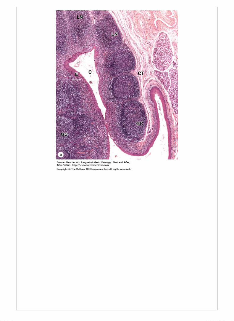
AccessMedicine | Print: Chapter 14. The Immune System & Lymphoid ... http://accessmedicine.com/popup.aspx?aID=6181868&print=yes_chapter
18 of 37 22/05/2011 19:27

Detail of a tonsil.
Palatine tonsils are aggregates of lymphoid tissue, usually with nodules, in the mucosa of the posterior soft palate. (a): Micrographshowing several lymphoid nodules (LN), collectively covered by stratified squamous epithelium (E) on one side and a connective tissuecapsule (CT) on the other. Some nodules show lighter staining germinal centers (GC). Infoldings of the mucosa in some tonsils formcrypts (C), along which nodules are especially numerous. Lumens of crypts contain desquamated epithelial cells, live and deadlymphocytes, and bacteria. X140. H&E. (b): Epithelium surrounding crypts often becomes infiltrated with lymphocytes and neutrophilsand can become difficult to recognize histologically. Lymphocytes abundant in the underlying connective tissue are seen on the left.X200. H&E. (Reproduced, with permission, from Paulo A. Abrahamsohn, Institute of Biomedical Sciences, University of São Paulo,Brazil.)
The pharyngeal tonsil is situated in the posterior wall of the nasopharynx (Figure 14–14) and is usually covered by ciliated
pseudostratified columnar epithelium, although areas of stratified epithelium can also be observed. The pharyngeal tonsil is
composed of pleated mucosa containing diffuse lymphoid tissue and lymphoid nodules and a capsule thinner than that of the
palatine tonsils. The lingual tonsils are situated along the posterior surface of the tongue (Figure 14–14) and are covered by
stratified squamous epithelium with crypts. The lymphoid tissue of these tonsils has many of the same basic features as that of
palatine tonsils (Figure 14–15). All these epithelia contain intraepithelial lymphocytes and dendritic cells.
MALT extends along the entire gastrointestinal tract, but in the wall of the ileum are particularly large aggregates of lymphoid
follicles called Peyer patches, each consisting of 10–200 nodules and bulging into the gut lumen with no connective tissue capsule
(Figure 14–16). The simple follicle-associated epithelium (FAE) covering these nodules contains specialized M cells with apical
microfolds rather than the brush border and glycocalyx typical of enterocytes. M cells continuously sample antigens and
microorganisms in the intestinal lumen. Each is characterized by a large basal intraepithelial pocket open to the underlying
lymphoid tissue through a porous basement membrane (Figure 14–16), discussed further in Chapter 15. Antigenic material bound
to the apical surface of M cells is rapidly translocated via transcytosis from the intestinal lumen to APCs and lymphocytes located in
the pocket. T helper cells and B cells derived from these lymphocytes move away from the FAE and initiate adaptive responses to
the antigens. These B cells give rise to plasma cells secreting IgA, which is transported by enterocytes into the intestinal lumen to
bind and neutralize potentially harmful antigens.
Figure 14–16.
AccessMedicine | Print: Chapter 14. The Immune System & Lymphoid ... http://accessmedicine.com/popup.aspx?aID=6181868&print=yes_chapter
19 of 37 22/05/2011 19:27

AccessMedicine | Print: Chapter 14. The Immune System & Lymphoid ... http://accessmedicine.com/popup.aspx?aID=6181868&print=yes_chapter
20 of 37 22/05/2011 19:27
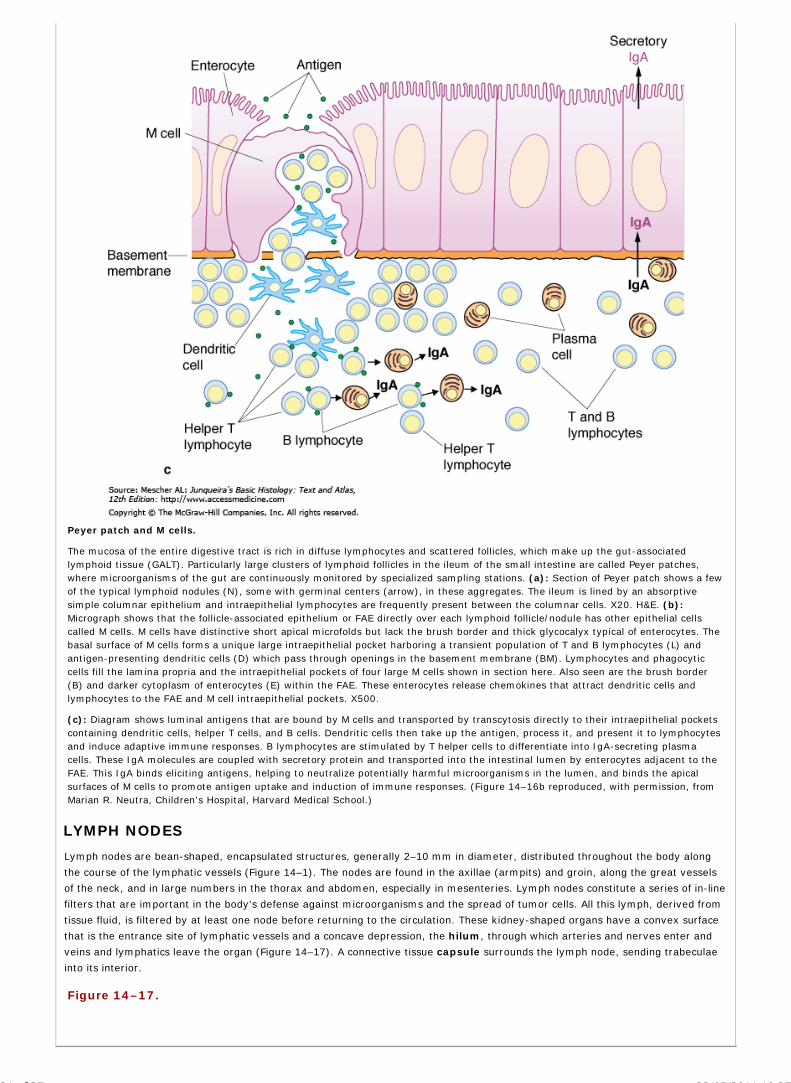
Peyer patch and M cells.
The mucosa of the entire digestive tract is rich in diffuse lymphocytes and scattered follicles, which make up the gut-associatedlymphoid tissue (GALT). Particularly large clusters of lymphoid follicles in the ileum of the small intestine are called Peyer patches,where microorganisms of the gut are continuously monitored by specialized sampling stations. (a): Section of Peyer patch shows a fewof the typical lymphoid nodules (N), some with germinal centers (arrow), in these aggregates. The ileum is lined by an absorptivesimple columnar epithelium and intraepithelial lymphocytes are frequently present between the columnar cells. X20. H&E. (b):Micrograph shows that the follicle-associated epithelium or FAE directly over each lymphoid follicle/nodule has other epithelial cellscalled M cells. M cells have distinctive short apical microfolds but lack the brush border and thick glycocalyx typical of enterocytes. Thebasal surface of M cells forms a unique large intraepithelial pocket harboring a transient population of T and B lymphocytes (L) andantigen-presenting dendritic cells (D) which pass through openings in the basement membrane (BM). Lymphocytes and phagocyticcells fill the lamina propria and the intraepithelial pockets of four large M cells shown in section here. Also seen are the brush border(B) and darker cytoplasm of enterocytes (E) within the FAE. These enterocytes release chemokines that attract dendritic cells andlymphocytes to the FAE and M cell intraepithelial pockets. X500.
(c): Diagram shows luminal antigens that are bound by M cells and transported by transcytosis directly to their intraepithelial pocketscontaining dendritic cells, helper T cells, and B cells. Dendritic cells then take up the antigen, process it, and present it to lymphocytesand induce adaptive immune responses. B lymphocytes are stimulated by T helper cells to differentiate into IgA-secreting plasmacells. These IgA molecules are coupled with secretory protein and transported into the intestinal lumen by enterocytes adjacent to theFAE. This IgA binds eliciting antigens, helping to neutralize potentially harmful microorganisms in the lumen, and binds the apicalsurfaces of M cells to promote antigen uptake and induction of immune responses. (Figure 14–16b reproduced, with permission, fromMarian R. Neutra, Children's Hospital, Harvard Medical School.)
LYMPH NODES
Lymph nodes are bean-shaped, encapsulated structures, generally 2–10 mm in diameter, distributed throughout the body along
the course of the lymphatic vessels (Figure 14–1). The nodes are found in the axillae (armpits) and groin, along the great vessels
of the neck, and in large numbers in the thorax and abdomen, especially in mesenteries. Lymph nodes constitute a series of in-line
filters that are important in the body's defense against microorganisms and the spread of tumor cells. All this lymph, derived from
tissue fluid, is filtered by at least one node before returning to the circulation. These kidney-shaped organs have a convex surface
that is the entrance site of lymphatic vessels and a concave depression, the hilum, through which arteries and nerves enter and
veins and lymphatics leave the organ (Figure 14–17). A connective tissue capsule surrounds the lymph node, sending trabeculae
into its interior.
Figure 14–17.
AccessMedicine | Print: Chapter 14. The Immune System & Lymphoid ... http://accessmedicine.com/popup.aspx?aID=6181868&print=yes_chapter
21 of 37 22/05/2011 19:27

Schematic structure of a lymph node.
The left half of the figure shows the major regions and structural components of a lymph node and the flow of lymph within theseorgans, entering via afferent lymphatics on the convex side of the node, passing through unlined sinuses (shown in pink) in thelymphoid tissue, and leaving through an efferent lymphatic at the hilum. Valves in the lymphatic vessels assure the one-way flow oflymph. The right half depicts part of the blood circulation, with a small artery and vein both entering and leaving at the hilum.
The most common cells of lymph nodes are lymphocytes, macrophages and other APCs, plasma cells, and reticular cells; follicular
dendritic cells are present within the lymphoid nodules. The different arrangement of the cells and of the reticular fiber stroma
supporting the cells creates a cortex, a medulla, and an intervening paracortex (Figures 14–17, 14–18, 14–19).
Figure 14–18.
AccessMedicine | Print: Chapter 14. The Immune System & Lymphoid ... http://accessmedicine.com/popup.aspx?aID=6181868&print=yes_chapter
22 of 37 22/05/2011 19:27

Regions of a lymph node.
A low-magnification section of a lymph node showing the three functional regions: the cortex (C), the paracortex (P), and the medulla(M). Connective tissue of the capsule (CT) completely surrounds each lymph node and extends as several trabeculae (T) throughoutthe lymphoid tissue. Major spaces for lymph flow are present in this tissue under the capsule and along the trabeculae. A changingpopulation of immune cells is suspended on reticular fibers throughout the cortex, paracortex, and medulla. Lymphoid nodules (LN) arenormally restricted to the cortex and the medulla is characterized by sinuses (MS) and cords (MC) of lymphoid tissue. X40. H&E. (Withpermission, from Paulo A. Abrahamsohn, Institute of Biomedical Sciences, University of São Paulo, Brazil.)
Figure 14–19.
AccessMedicine | Print: Chapter 14. The Immune System & Lymphoid ... http://accessmedicine.com/popup.aspx?aID=6181868&print=yes_chapter
23 of 37 22/05/2011 19:27

Lymph node cortex.
The outer regions on the convex sides of a lymph node include the capsule (C), subcapsular sinuses (S), and diffuse lymphoid tissuewith lymphoid nodules (N). Afferent lymphatic vessels (which are only rarely shown well in sections) penetrate this capsule, dumpinglymph into the sinus where its contents are processed by lymphocytes and APCs. X140. H&E. (With permission, from Paulo A.Abrahamsohn, Institute of Biomedical Sciences, University of São Paulo, Brazil.)
The cortex, situated under the capsule, consists of the following components:
Many reticular cells, macrophages, APCs, and lymphocytes (Figure 14–18).
Lymphoid nodules, with or without germinal centers, formed mainly of B lymphocytes, embedded within the diffusepopulation of other cells (Figure 14–18).
Areas immediately beneath the capsule, called the subcapsular sinuses, where the lymphoid tissue has wide reticularfiber meshes (Figures 14–18 and 14–19). Lymph containing antigens, lymphocytes, and APCs circulates around the widespaces of these sinuses after being delivered there by the afferent lymphatic vessels.
Cortical sinuses, running between the lymphoid nodules, which arise from and share the structural features of thesubcapsular sinuses. They communicate with the subcapsular sinuses through spaces similar to those present in themedulla (Figures 14–17, 14–18, 14–19, and 14–20).
Figure 14–20.
AccessMedicine | Print: Chapter 14. The Immune System & Lymphoid ... http://accessmedicine.com/popup.aspx?aID=6181868&print=yes_chapter
24 of 37 22/05/2011 19:27

Lymph node cortex and paracortex.
The region just inside the cortex is called the paracortex. Although most lymphocytes in the cortex are B cells, many located innodules, the lymphocytes of the paracortex are largely T cells. This separation is indicated in the fluorescence micrographs here usingimmunohistochemistry on adjacent sections of lymph node. (a): Antibody against a B cell surface marker labels nearly all thelymphocytes in the cortex (C), as well as many cells around a high endothelial venule (V) in the paracortex, but few cells in theparacortex proper (P). (b): Stained with an antibody against a T cell marker, the paracortex is heavily labeled, but only a few cells inthe cortex are stained, possibly T helper cells. X200. (Reproduced and modified, with permission, from IL Weissman: Transplant Rev.1975;24:159.)
The paracortex does not have precise boundaries with the cortex and medulla. It can be distinguished from the outer cortex by
its lack of B cell lymphoid nodules (Figure 14–18) and its accumulation of T cells, which can be determined by
immunohistochemistry (Figure 14–20). Venules in the paracortex comprise an important entry point for lymphocytes moving from
blood into lymph nodes.
The lymph node medulla has two major components:
Medullary cords (Figure 14–21) are branched cordlike extensions of lymphoid tissue arising from the paracortex. Theycontain primarily B lymphocytes and often plasma cells and macrophages (Figures 14–19 and 14–21).
Medullary cords are separated by dilated spaces, frequently bridged by reticular cells and fibers, called medullarysinuses (Figure 14–21). They contain lymph, lymphocytes, often many macrophages, and sometimes evengranulocytes if the lymph node is draining an infected region. These sinuses are continuous with the cortical sinuses andjoin at the hilum to deliver lymph to the efferent lymph vessel of the lymph node (Figure 14–17).
Figure 14–21.
AccessMedicine | Print: Chapter 14. The Immune System & Lymphoid ... http://accessmedicine.com/popup.aspx?aID=6181868&print=yes_chapter
25 of 37 22/05/2011 19:27

AccessMedicine | Print: Chapter 14. The Immune System & Lymphoid ... http://accessmedicine.com/popup.aspx?aID=6181868&print=yes_chapter
26 of 37 22/05/2011 19:27

Lymph node medulla.
(a): The medulla of a lymph node consists mainly of the medullary sinuses (MS) separated by intervening medullary cords (MC).Lymphocytes and plasma are abundant and predominate in number over other cell types. A blood vessel within a medullary cord isalso seen. X200. H&E. (b): Higher magnification of a medullary cord (MC) shows plasma cells (arrows) with spherical, eccentric nucleiand much more cytoplasm than lymphocytes. Efferent lymph is rich in newly synthesized antibodies. A medullary sinus (MS) is alsoseen. X400. H&E. (With permission, from Paulo A. Abrahamsohn, Institute of Biomedical Sciences, University of São Paulo, Brazil.)
Afferent lymphatic vessels cross the capsule and pour lymph into the subcapsular sinus (Figure 14–17). From there, lymph
passes through the cortical sinuses and then into the medullary sinuses. During this passage, the lymph infiltrates the cortex and
the medullary cords and is filtered and modified by immune cells. The lymph is collected by efferent lymphatics at the hilum and
valves in both lymphatics assure the unidirectional flow of lymph.
Role of Lymph Nodes in the Immune ResponseLymph nodes are distributed throughout the body and lymph formed in tissues must pass through at least one node before
entering the bloodstream. The lymph that arrives at a lymph node contains antigens as soluble molecules, portions of
semidestroyed microorganisms, or antigens already internalized and being transported by macrophages and other APCs. It may
also contain microorganisms and cytokines, particularly if it is coming from a region with an infection or inflammation. Antigens
that had not been phagocytosed before may be internalized by APCs in the lymph nodes. All antigens have the opportunity to be
presented to B lymphocytes, to T helper cells, and to T cytotoxic lymphocytes for these cells to initiate an immune response.
The lymph node is an important site of lymphocyte proliferation (especially of B cells in the germinal centers) as well as of
transformation of B lymphocytes into plasma cells. Because of this, the lymph that leaves a lymph node may be enriched in
antibodies. When the lymph is returned to the blood circulation, these antibodies will be delivered to the entire body.
MEDICAL APPLICATION
As each satellite node receives lymph from a limited region of the body, malignant tumor cells often reach lymph nodes
and are distributed to other parts of the body via the efferent lymph vessels and blood vessels, a process known as
metastasis.
Infection and antigenic stimulation often cause lymph nodes to enlarge. These swollen nodules, which may be palpated
AccessMedicine | Print: Chapter 14. The Immune System & Lymphoid ... http://accessmedicine.com/popup.aspx?aID=6181868&print=yes_chapter
27 of 37 22/05/2011 19:27

under the skin as indicators of inflammation, have multiple germinal centers with active cell proliferation. Although plasma
cells constitute only 1–3% of the cell population in resting nodes, their numbers increase greatly in stimulated lymph
nodes.
Recirculation of LymphocytesBecause all lymph formed in the body normally drains back into the blood, lymphocytes that leave the lymph nodes by efferent
lymphatics eventually reach the bloodstream. These lymphocytes may then leave the blood vessels by entering the tissues and
return with other lymph to another lymph node. However, most (90%) lymphocytes return to a lymph node by crossing the walls
of specific postcapillary venules in the paracortex, the high endothelial venules (HEVs) (Figure 14–22). These vessels have an
unusual endothelial lining of tall cuboidal cells, whose apical surface glycoproteins and integrins facilitate rapid diapedesis of
lymphocytes out of the blood into the paracortex of the lymph node. High endothelial venules are also present in other lymphoid
organs, such as the tonsils, Peyer patches, and appendix, but not in the spleen.
Figure 14–22.
AccessMedicine | Print: Chapter 14. The Immune System & Lymphoid ... http://accessmedicine.com/popup.aspx?aID=6181868&print=yes_chapter
28 of 37 22/05/2011 19:27

High endothelial venules (HEV).
(a): High endothelial venules are found in the paracortex of lymph nodes, as shown, as well as in tonsils and Peyer patches. Theirendothelial cells are unusually shaped but generally cuboidal and facilitate rapid translocation of lymphocytes into the lymphoid tissue.L-selectin on the lymphocytes recognizes sugar-rich ligands on the surfaces of these endothelial cells and as a consequence thelymphocytes stop there. Integrins promote adhesion between lymphocytes and the endothelial cells and the lymphocytes cross thevessel wall into the lymph node parenchyma. HEVs can be difficult to identify in H&E-stained paraffin sections. X400. H&E. (b): Plasticsections more clearly reveal the high endothelial cells (arrowheads) and the lymphocytes passing between them (arrows). X400. PT.(c): SEM of a sectioned HEV showing five typical lymphocytes adhering to endothelial cells (arrowheads) prior to migrating betweenthem and joining other lymphocytes in the surrounding paracortex. X500. (Figure 14–22c reproduced, with permission from Fujita, T.,1989, Prog. Clin. Biol. Res., 295:493.)
The continuous recirculation of lymphocytes enables most parts of the body to be constantly monitored, increasing the opportunity
for lymphocytes to encounter activated APCs that have migrated to lymph nodes.
SPLEEN
The spleen is the largest single accumulation of lymphoid tissue in the body and the only one involved in filtration of blood, making
it an important organ in defense against blood-borne antigens. It is also the main site of destruction of aged erythrocytes. As is
true of other secondary lymphoid organs, the spleen is a production site of antibodies and activated lymphocytes, which are
delivered to the blood. Any inert particles in blood are actively phagocytosed by spleen macrophages.
The spleen is surrounded by a capsule of dense connective tissue from which emerge trabeculae, which partially subdivide the
parenchyma or splenic pulp (Figure 14–23). Large trabeculae originate at the hilum, on the medial surface of the spleen; these
trabeculae carry nerves and arteries into the splenic pulp as well as veins that bring blood back into the circulation. Lymphatic
vessels that arise in the splenic pulp also leave through the hilum via the trabeculae.
Figure 14–23.
AccessMedicine | Print: Chapter 14. The Immune System & Lymphoid ... http://accessmedicine.com/popup.aspx?aID=6181868&print=yes_chapter
29 of 37 22/05/2011 19:27

Spleen.
The capsule (C) of the spleen connects to trabeculae (T) which partially subdivide the pulp-like interior of the organ. The red pulp (R)occupies most of the parenchyma, with white pulp (W) restricted to smaller areas, mainly around the central arterioles. Names ofthese splenic areas refer to their color in the fresh state: red pulp is filled with blood cells of all types, located both in cords andsinuses; white pulp is lymphoid tissue. Large blood vessels and lymphatics enter and leave the spleen at a hilum. X20. PSH.
Splenic PulpThe spleen is composed of reticular tissue containing reticular cells, many lymphocytes and other blood cells, macrophages, and
APCs. The splenic pulp has two components, the white pulp and the red pulp (Figure 14–23). The small masses of white pulp
consist of lymphoid nodules and the periarteriolar lymphoid sheathes, while the red pulp consists of blood-filled sinusoids
and splenic cords (of Bilroth).
As expected of an organ specialized to process blood, the microvasculature of the spleen is important, although much about it
remains to be learned. The splenic artery divides inside the hilum, branching into small trabecular arteries following this
connective tissue. They leave the trabeculae and enter the parenchyma as arterioles enveloped by a sheath of T lymphocytes, the
periarteriolar lymphoid sheath (PALS), which is part of the white pulp (Figure 14–24). Surrounded by the PALS, these vessels are
known as central arterioles (Figure 14–25). After coursing through the parenchyma for variable stretches, the PALS receive large
numbers of lymphocytes, mostly B cells, and may form lymphoid nodules (Figure 14–25). In these nodules the arteriole occupies
an eccentric position but is still called the central arteriole. During its passage through the white pulp, this arteriole sends off
smaller branches that supply the surrounding lymphoid tissue (Figure 14–24).
Figure 14–24.
AccessMedicine | Print: Chapter 14. The Immune System & Lymphoid ... http://accessmedicine.com/popup.aspx?aID=6181868&print=yes_chapter
30 of 37 22/05/2011 19:27

Blood flow in the spleen.
Schematic view of the blood circulation and the structure of the spleen, from the trabecular artery to the trabecular vein. Smallbranches of these arteries are called central arteries and become enclosed within a sheath of lymphoid cells, the periarteriolarlymphoid sheath (PALS) in white pulp. B cells in these sheathes can form nodules as the largest masses of white pulp, and aroundthese nodules are located the marginal sinuses. Emerging from the white pulp, the central arteriole branches as the penicillararterioles, which lead to sheathed capillaries. From these, blood flows into either a closed circulation passing directly into splenicsinuses (S) or an open circulation, being dumped from the vasculature into the lymphoid tissue of the red pulp's splenic cords.
Figure 14–25.
AccessMedicine | Print: Chapter 14. The Immune System & Lymphoid ... http://accessmedicine.com/popup.aspx?aID=6181868&print=yes_chapter
31 of 37 22/05/2011 19:27

White pulp of the spleen.
The splenic white pulp consists of lymphoid tissue surrounding the central arterioles as the periarteriolar lymphoid sheath (PALS) andthe nodules of proliferating B cells in this sheath. (a): Longitudinal section of white pulp (W) in a PALS and the central arteriole(arrowhead) it surrounds. Surrounding the PALS is much red pulp (R). (b): A large nodule with a germinal center forms in the PALS.and the central arteriole (arrowhead) is displaced to the nodule's periphery. Small sinuses can be seen at the margin between whiteand red pulp. Both X20. H&E. (With permission, from Paulo A. Abrahamsohn, Institute of Biomedical Sciences, University of São Paulo,Brazil.)
Surrounding the lymphoid nodules is a marginal zone consisting of many blood sinuses and lymphoid tissue (Figures 14–24 and
14–25b). The marginal zone contains lymphocytes, many macrophages, and an abundance of blood antigens and thus plays an
important role in the immunological activities of the spleen.
After leaving the white pulp, the sheath of lymphocytes slowly thins and the central arteriole subdivides to form straight penicillar
arterioles (Figure 14–24). Some of the capillaries that emerge from these penicillar arterioles are sheathed with reticular cells,
macrophages and lymphocytes, whose functional significance is not clear.
The red pulp is composed almost entirely of splenic cords and venous sinusoids (Figure 14–26). The splenic cords contain a network
of reticular cells or reticular fibers that support T and B lymphocytes, macrophages, plasma cells, and many blood cells
(erythrocytes, platelets, and granulocytes). The splenic cords are separated by wide, irregularly shaped sinusoids (Figures 14–26
and 14–27). Unusual elongated endothelial cells, called stave cells, line the splenic sinusoids, oriented in parallel with the
sinusoid's blood flow. These cells are sparsely wrapped in reticular fibers set in a transverse direction, much like the hoops
surrounding the staves of a wooden keg (Figure 14–28).
Figure 14–26.
AccessMedicine | Print: Chapter 14. The Immune System & Lymphoid ... http://accessmedicine.com/popup.aspx?aID=6181868&print=yes_chapter
32 of 37 22/05/2011 19:27

Red pulp of the spleen.
The splenic red pulp is composed of splenic venous sinusoids (S) and splenic cords (C), both of which contain blood cells of all types.The cords, often called cords of Bilroth, are reticular tissue rich in lymphocytes. The sinuses are lined by unusual, nonsquamousendothelial cells. X40.H&E. (With permission, from Paulo A. Abrahamsohn, Institute of Biomedical Sciences, University of São Paulo,Brazil.)
Figure 14–27.
AccessMedicine | Print: Chapter 14. The Immune System & Lymphoid ... http://accessmedicine.com/popup.aspx?aID=6181868&print=yes_chapter
33 of 37 22/05/2011 19:27

Splenic venous sinuses and stave cells.
Higher magnification of splenic red pulp shows that the venous sinuses (S) are lined by endothelial cells (arrows) with large nucleibulging into the sinusoidal lumens. The unusual endothelial cells are called stave cells and have special properties that allow selectionof healthy red blood cells in the splenic cords (C). X100. H&E. (With permission, from Paulo A. Abrahamsohn, Institute of BiomedicalSciences, University of São Paulo, Brazil.)
Figure 14–28.
AccessMedicine | Print: Chapter 14. The Immune System & Lymphoid ... http://accessmedicine.com/popup.aspx?aID=6181868&print=yes_chapter
34 of 37 22/05/2011 19:27

AccessMedicine | Print: Chapter 14. The Immune System & Lymphoid ... http://accessmedicine.com/popup.aspx?aID=6181868&print=yes_chapter
35 of 37 22/05/2011 19:27

Structure and function of splenic sinusoids.
The endothelial stave cells that line venous sinuses in red pulp are long cells oriented lengthwise along the sinuses. The elongatedshape of the cells is difficult to appreciate from light micrographs (see Figure 14–26). (a): SEM clearly shows the parallel alignment ofthe stave cells (S), as well as many macrophages (M) in the surrounding red pulp (R). X500. (b): Silver-stained sections of spleenshow black reticular fibers surrounding the sinuses, oriented 90 degrees to the long axis of the sinuses (S). These fibers appear similarto those in the surrounding splenic cords (C). The basement membrane of the stave cells is incomplete and open to the passage ofcells. X400. Silver. (c): Diagram showing these components of splenic sinuses schematically, with the structures resembling a looselyorganized wooden barrel. In the open circulation mode of blood flow, blood cells dumped into the cords of the red pulp move underpressure or by their own activity through the spaces between stave cells, reentering the vasculature and soon leaving the spleen viathe splenic vein. Cells that cannot squeeze between the stave cells, mainly effete erythrocytes, are removed by macrophages. (Figure14-28a reproduced, with permission, from Fujita T:, 1989, Prog. Clin. Biol. Res. 1989;295:493)
The highly permeable splenic sinusoids are surrounded by very incomplete basal laminae. The spaces between the endothelial cells
of the splenic sinusoids are 2–3 m or smaller and only flexible cells are able to pass easily from the red pulp cords into the lumen
of the sinusoids. Because the lumen of splenic sinusoids is often blood-filled and very small and because the splenic cords are
infiltrated with red blood cells, microscopic distinctions between splenic cords and sinusoids may be difficult.
Blood Flow in the Red PulpBlood flow through the splenic red pulp can take two routes (Figure 14–24). In the closed circulation, the penicillar arterioles or
capillaries branching from them connect directly to the sinusoids, so that blood is always enclosed by the vascular endothelium.
Alternatively, other penicillar arterioles are open-ended, dumping blood into the stroma of the splenic cords in a unique example of
open circulation. In this route plasma and formed elements of blood reenter the vasculature by passing between the stave cells
of sinusoids, which presents no problem for platelets, leukocytes, and healthy flexible erythrocytes. However, after their normal
lifespan of 120 days effete erythrocytes undergo membrane changes, becoming swollen and less flexible, signalling their selective
engulfment by macrophages in the splenic cords (Figure 14–29). From the sinusoids, blood proceeds to the red pulp veins that join
together and enter the trabeculae, forming the trabecular veins (Figure 14–24). The splenic vein originates from these vessels
and emerges from the hilum of the spleen. The trabecular veins do not have muscle in their wall and resemble channels hollowed
out in the trabecular connective tissue and lined by endothelium.
Figure 14–29.
AccessMedicine | Print: Chapter 14. The Immune System & Lymphoid ... http://accessmedicine.com/popup.aspx?aID=6181868&print=yes_chapter
36 of 37 22/05/2011 19:27

Copyright © The McGraw-Hill Companies. All rights reserved.Privacy Notice. Any use is subject to the Terms of Use and Notice.
Erythrocyte removal by splenic macrophages.
A micrograph of five macrophages in a splenic cord shows active phagocytosis of effete erythrocytes. After about 120 days of useerythrocytes undergo membrane changes and swell, signals for their engulfment by macrophages in the cords of reticular tissuebetween the venous sinuses. Healthy, flexible erythrocytes are pushed between stave cells and enter the sinuses. Phagocytosederythrocytes are completely degraded within lysosomes. The iron released from hemoglobin binds its transport protein transferrin,returns to the circulation, and is reused primarily for erythropoiesis in bone marrow. Iron-free heme either binds its transport protein,hemopexin, or is metabolized to bilirubin and excreted in the bile by liver cells. After surgical removal of the spleen (splenectomy) thenumber of abnormal erythrocytes in the circulation increases, although many such cells are now removed in the bone marrow andliver. X400. PT.
MEDICAL APPLICATION
Although the spleen has numerous important functions in the body, it is not essential to life. In some situations the spleen
must be removed (eg, an abdominal injury that results in rupture of the spleen capsule, certain anemias, and platelet
disorders). In this case other organs such as the liver and bone marrow can take over many of the functions of the spleen,
although the risk of infection may be higher in a splenectomized individual.
AccessMedicine | Print: Chapter 14. The Immune System & Lymphoid ... http://accessmedicine.com/popup.aspx?aID=6181868&print=yes_chapter
37 of 37 22/05/2011 19:27

Print Close Window
Note: Large images and tables on this page may necessitate printing in landscape mode.
Copyright © The McGraw-Hill Companies. All rights reserved.
Junqueira's Basic Histology: Text & Atlas, 12e > Chapter 15. Digestive Tract >
DIGESTIVE TRACT: INTRODUCTION
The digestive system consists of the digestive tract—oral cavity, esophagus, stomach, small and large intestines, rectum, and anus—and its associated
glands—salivary glands, liver, and pancreas (Figure 15–1). Its function is to obtain from ingested food the molecules necessary for the maintenance,
growth, and energy needs of the body. Macromolecules such as proteins, fats, complex carbohydrates, and nucleic acids are broken down into small
molecules that are more easily absorbed through the lining of the digestive tract, mostly in the small intestine. Water, vitamins, and minerals from
ingested food are also absorbed. In addition, the inner layer of the digestive tract is a protective barrier between the content of the tract's lumen and the
internal milieu of the body.
Figure 15–1.
The digestive system.
The digestive system consists of the tract from the mouth (oral cavity) to the anus, as well as the digestive glands emptying into this tract, primarily thesalivary glands, liver, and pancreas. These accessory digestive glands are described in the next chapter.
The first step in digestion occurs in the mouth, where food is moistened by saliva and ground by the teeth into smaller pieces; saliva also initiates the
breakdown of carbohydrates. Digestion continues in the stomach and small intestine, where the food's basic components (eg, amino acids,
monosaccharides, free fatty acids) are absorbed. Water absorption occurs in the large intestine, causing undigested material to become semisolid.
GENERAL STRUCTURE OF THE DIGESTIVE TRACT
The entire gastrointestinal tract has certain common structural characteristics. It is a hollow tube with a lumen of variable diameter and a wall made up of
four main layers: the mucosa, submucosa, muscularis, and serosa. The structure of these layers is summarized below and is illustrated for the small
intestine in Figure 15–2.
Figure 15–2.
AccessMedicine | Print: Chapter 15. Digestive Tract http://accessmedicine.com/popup.aspx?aID=6182071&print=yes_chapter
1 of 42 22/05/2011 19:18

Major layers and organization of the digestive tract.
Schematic diagram showing the structure of the small intestine portion of the digestive tract, with the four layers and their major components listed on the left.The intestines are suspended by mesenteries which are the sites of blood vessels and lymphatics from the intestines.
The mucosa comprises an epithelial lining; an underlying lamina propria of loose connective tissue rich in blood vessels, lymphatics, lymphocytes and
smooth muscle cells, sometimes also containing glands; and a thin layer of smooth muscle called the muscularis mucosae usually separating mucosa
from submucosa. The mucosa is frequently called a mucous membrane.
The submucosa contains denser connective tissue with many blood and lymph vessels and the submucosal plexus of autonomic nerves. It may also
contain glands and lymphoid tissue.
The thick muscularis is composed of smooth muscle cells that are spirally oriented and divided into two sublayers. In the internal sublayer (closer to the
lumen), the orientation is generally circular; in the external sublayer, it is mostly longitudinal. In the connective tissue between the muscle sublayers are
blood and lymph vessels, as well as another autonomic myenteric nerve plexus. This and the submucosal plexus together comprise the local enteric
nervous system of the digestive tract, containing largely autonomic neurons functioning independently of the central nervous system (CNS).
The serosa is a thin layer of loose connective tissue, rich in blood vessels, lymphatics, and adipose tissue, with a simple squamous covering epithelium
(mesothelium). In the abdominal cavity, the serosa is continuous with the mesenteries (thin membranes covered by mesothelium on both sides), which
support the intestines, and with the peritoneum, a serous membrane that lines the cavity. In places where the digestive tract is not suspended in a cavity
but bound to other structures, such as in the esophagus (Figure 15–1), the serosa is replaced by a thick adventitia, consisting of connective tissue
containing vessels and nerves, lacking mesothelium.
The main functions of the digestive tract's epithelial lining are to:
Provide a selectively permeable barrier between the contents of the tract and the tissues of the body,
Facilitate the transport and digestion of food,
Promote the absorption of the products of this digestion,
Produce hormones that affect the activity of the digestive system,
Produce mucus for lubrication and protection.
The abundant lymphoid nodules in the lamina propria and the submucosal layer protect the organism (in association with the epithelium) from bacterial
invasion, as described in Chapter 14. The necessity for this immunologic support is obvious, because the entire digestive tract—with the exception of the
oral cavity, esophagus, and anal canal—is lined by a simple thin, vulnerable epithelium. The lamina propria, located just below the epithelium, is a zone
rich in macrophages and lymphocytes, some of which actively produce antibodies. These antibodies are mainly immunoglobulin A (IgA) and are secreted
into the intestinal lumen bound to a secretory protein produced by the epithelial cells. This complex protects against viral and bacterial invasion. IgA is
AccessMedicine | Print: Chapter 15. Digestive Tract http://accessmedicine.com/popup.aspx?aID=6182071&print=yes_chapter
2 of 42 22/05/2011 19:18

resistant to proteolytic enzymes and can therefore coexist with the proteases present in the lumen.
The muscularis mucosae allows local movements of the mucosa independent of other movements of the digestive tract, increasing contact of the lining
with food. The contractions of the muscularis, generated and coordinated by autonomic nerve plexuses, propel and mix the food in the digestive tract.
These plexuses are composed mainly of nerve cell aggregates (multipolar visceral neurons) that form small parasympathetic ganglia. A rich network of
pre- and postganglionic fibers of the autonomic nervous system and some visceral sensory fibers in these ganglia permit communication between them.
The number of these ganglia along the digestive tract is variable; they are most numerous in the regions of greatest motility.
MEDICAL APPLICATION
In certain diseases, such as Hirschsprung disease (congenital megacolon) or Chagas disease (Trypanosoma cruzi infection), the plexuses in
the digestive tract are severely injured and most of their neurons are destroyed. This results in disturbances of digestive tract motility, with
frequent dilatations in some areas. The abundant innervation from the autonomic nervous system that the digestive tract receives provides an
anatomic explanation of the widely observed action of emotional stress on this tract.
ORAL CAVITY
The oral cavity (Figure 15–1) is lined with stratified squamous epithelium, keratinized or nonkeratinized, depending on the region. The keratin layer
protects the oral mucosa from damage during masticatory function and is best developed on the gingiva (gum) and hard palate. The lamina propria in
these regions has many papillae and rests directly on bony tissue. Nonkeratinized squamous epithelium covers the soft palate, lips, cheeks, and the floor
of the mouth. Surface cells are shed continuously and replaced by progeny of stem cells in the basal epithelial layer. The lamina propria has papillae
similar to those in the dermis of the skin and is continuous with a submucosa containing diffuse small salivary glands. The soft palate also has a core of
skeletal muscle and lymphoid nodules. In the lips, there is also striated muscle and a transition from the oral nonkeratinized epithelium to the keratinized
epithelium of the skin (Figure 15–3).
Figure 15–3.
Lip.
Low-magnification micrograph of a lip section showing one side covered by typical oral mucosa (OM), the opposite side covered by skin (S) containing hairfollicles (F) and associated glands. Between the oral portion of the lips and normal skin is the vermilion (V), or the vermilion zone, where epidermis is very thin,lightly keratinized, and transparent to blood in the rich microvasculature of the underlying connective tissue. Because this region lacks the glands for oil andsweat, it is prone to excessive dryness and chapping in cold, dry weather. Internally, the lips contain much-striated muscle (M) and many minor salivary glands(G). X10. H&E.
TongueThe tongue is a mass of striated muscle covered by a mucous membrane whose structure varies according to the region. The muscle fibers cross one
another in three planes and are grouped in bundles separated by connective tissue. Because the connective tissue of the lamina propria penetrates the
spaces between the muscular bundles, the mucous membrane is strongly adherent to the muscle. The mucous membrane is smooth on the lower surface
of the tongue. The tongue's dorsal surface is irregular, covered anteriorly by a great number of small eminences called papillae. The posterior third of the
tongue's dorsal surface is separated from the anterior two thirds by a V-shaped groove, the terminal sulcus. Behind this boundary is the root of the
tongue, whose surface shows the many bulges of the lingual tonsils and smaller collections of lymphoid nodules (Figure 15–4).
Figure 15–4.
AccessMedicine | Print: Chapter 15. Digestive Tract http://accessmedicine.com/popup.aspx?aID=6182071&print=yes_chapter
3 of 42 22/05/2011 19:18

Tongue and lingual papillae.
The posterior third of the tongue is the root and the anterior two-thirds the body of the tongue. The mucosa of the root is filled with masses of lymphoidnodules separated by crypts, all of which comprise the lingual tonsils. On the body of the tongue are papillae of four types, all containing cores of connectivetissue covered by stratified squamous epithelium. Pointed filiform papillae provide friction to help move food during chewing. Ridge-like foliate papillae on thesides of the tongue are best-developed in young children. Fungiform papillae are scattered across the dorsal surface and 6-12 very large vallate papillae arepresent in a V-shaped line near the terminal sulcus. Taste buds are present on fungiform and foliate papillae but are much more abundant on vallate papillae.
The numerous papillae on the anterior portion of the tongue are elevations of the mucous membrane that assume various forms and functions. Four types
are recognized (Figure 15–4):
Filiform papillae (Figure 15–5) are very numerous, have an elongated conical shape, and are heavily keratinized, which gives their surface agray or whitish appearance. Their epithelium lacks taste buds (described below) and their role is mechanical in providing a rough surface thatfacilitates food movement during chewing.
Fungiform papillae (Figure 15–5) are less numerous, lightly keratinized, and mushroom-shaped with connective tissue cores and scatteredtaste buds on their upper surfaces. They are irregularly interspersed among the filiform papillae.
Foliate papillae are poorly developed in adults, but consist of parallel ridges and furrows on the sides of the tongue, with taste buds.
Vallate (or circumvallate) papillae (Figure 15–5) are the least numerous and largest lingual papillae, and have over half the taste buds on thehuman tongue. With diameters of one to three mm, seven to twelve circular vallate papillae normally form a V-shaped line just before theterminal sulcus. Ducts from several serous salivary (von Ebner) glands empty into the deep groove that surrounds each vallate papilla. Thismoatlike arrangement provides a continuous flow of fluid over the taste buds abundant on the sides of these papillae, which washes foodparticles from the vicinity so that the taste buds can receive and process new gustatory stimuli. These glands also secrete a lipase that preventsthe formation of a hydrophobic film over the taste buds that would hinder their function.
Figure 15–5.
AccessMedicine | Print: Chapter 15. Digestive Tract http://accessmedicine.com/popup.aspx?aID=6182071&print=yes_chapter
4 of 42 22/05/2011 19:18
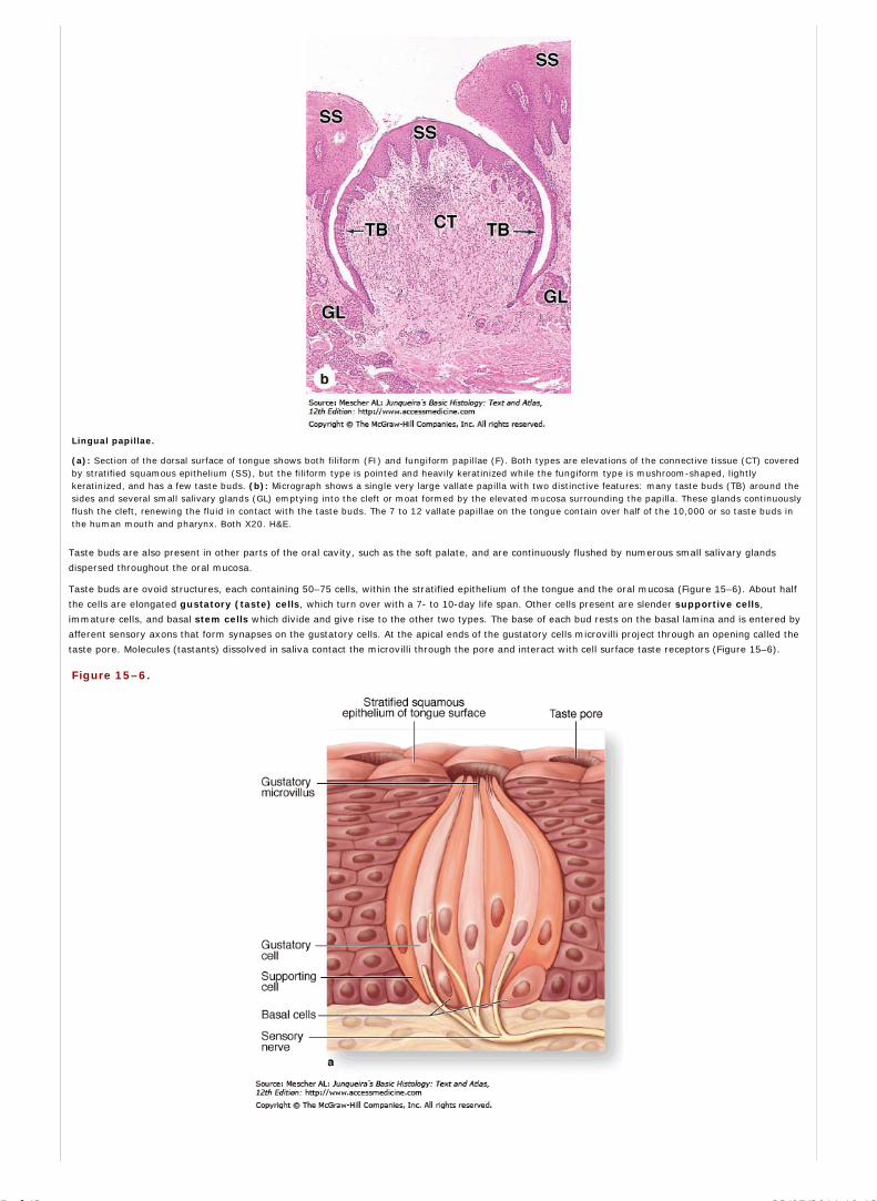
Lingual papillae.
(a): Section of the dorsal surface of tongue shows both filiform (FI) and fungiform papillae (F). Both types are elevations of the connective tissue (CT) coveredby stratified squamous epithelium (SS), but the filiform type is pointed and heavily keratinized while the fungiform type is mushroom-shaped, lightlykeratinized, and has a few taste buds. (b): Micrograph shows a single very large vallate papilla with two distinctive features: many taste buds (TB) around thesides and several small salivary glands (GL) emptying into the cleft or moat formed by the elevated mucosa surrounding the papilla. These glands continuouslyflush the cleft, renewing the fluid in contact with the taste buds. The 7 to 12 vallate papillae on the tongue contain over half of the 10,000 or so taste buds inthe human mouth and pharynx. Both X20. H&E.
Taste buds are also present in other parts of the oral cavity, such as the soft palate, and are continuously flushed by numerous small salivary glands
dispersed throughout the oral mucosa.
Taste buds are ovoid structures, each containing 50–75 cells, within the stratified epithelium of the tongue and the oral mucosa (Figure 15–6). About half
the cells are elongated gustatory (taste) cells, which turn over with a 7- to 10-day life span. Other cells present are slender supportive cells,
immature cells, and basal stem cells which divide and give rise to the other two types. The base of each bud rests on the basal lamina and is entered by
afferent sensory axons that form synapses on the gustatory cells. At the apical ends of the gustatory cells microvilli project through an opening called the
taste pore. Molecules (tastants) dissolved in saliva contact the microvilli through the pore and interact with cell surface taste receptors (Figure 15–6).
Figure 15–6.
AccessMedicine | Print: Chapter 15. Digestive Tract http://accessmedicine.com/popup.aspx?aID=6182071&print=yes_chapter
5 of 42 22/05/2011 19:18

Taste buds.
(a): Drawing of a single taste bud shows the gustatory (taste) cells, the supporting cells whose function is not well-understood, and the basal stem cells.Microvilli at the ends of the gustatory cells project through an opening in the epithelium, the taste pore. Afferent sensory axons enter the basal end of tastebuds and synapse with the gustatory cells. (b): In the stratified squamous epithelium of the tongue surface or oral mucusa, taste buds form as distinct clustersof cells that recognizable histologically even at low magnification. At higher power the taste pore may be visible, as well as the elongated nuclei of gustatoryand supporting cells and the fewer, round nuclei of basal stem cells. 140X and 500X. H&E.
Taste buds detect at least five broad categories of tastants: metal ions (salty); hydrogen ions from acids (sour); sugars and related organic compounds
(sweet); alkaloids and certain toxins (bitter); and certain amino acids such as glutamate (umami; Jap. umami, savory). Salty and sour tastes are
produced by ion channels; the other taste categories are mediated by G-protein-coupled receptors. Receptor binding produces depolarization of the
gustatory cells, stimulating the sensory nerve fibers which send information to the brain for processing. Conscious perception of tastes in food requires
olfactory and other sensations in addition to taste bud activity.
PharynxThe pharynx, a transitional space between the oral cavity and the respiratory and digestive systems, forms an area of communication between the nasal
region and the larynx (Figure 15–1). The pharynx is lined by stratified nonkeratinized squamous epithelium in the region continuous with the esophagus
and by ciliated pseudostratified columnar epithelium containing goblet cells in the regions close to the nasal cavity.
The pharynx contains tonsils (described in Chapter 14) and the mucosa also has many small mucous salivary glands in its lamina propria. The constrictor
and longitudinal muscles of the pharynx are located outside this layer.
TeethIn the adult human there are normally 32 permanent teeth, arranged in two bilaterally symmetric arches in the maxillary and mandibular bones (Figure
15–7). Each quadrant has eight teeth: two incisors, one canine, two premolars, and three permanent molars. Twenty of the permanent teeth are preceded
by deciduous (baby) teeth which are shed; the others are permanent molars with no deciduous precursors. Each tooth has a crown exposed above the
gingiva, a constricted neck at the gum, and one or more roots below the gingiva that hold the teeth in bony sockets called alveoli, one for each tooth
(Figure 15–7).
Figure 15–7.
AccessMedicine | Print: Chapter 15. Digestive Tract http://accessmedicine.com/popup.aspx?aID=6182071&print=yes_chapter
6 of 42 22/05/2011 19:18
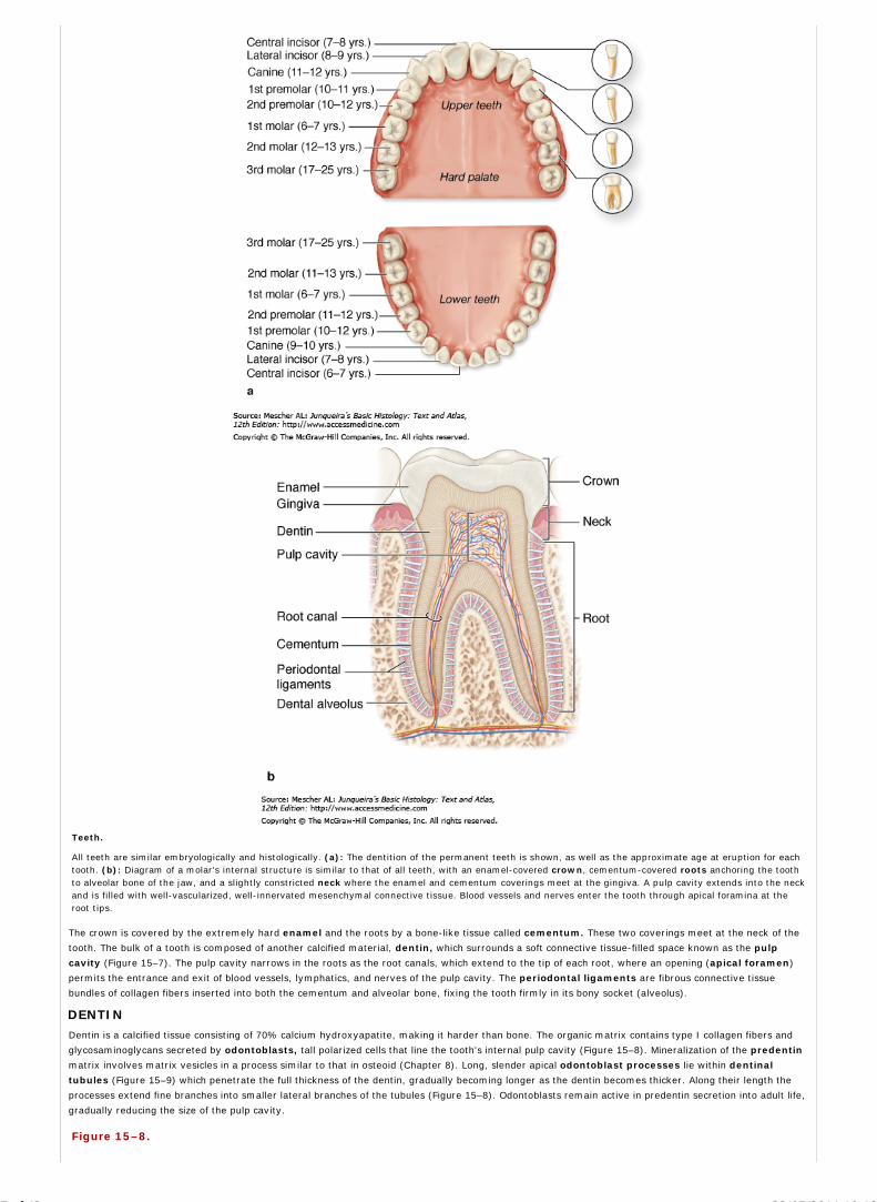
Teeth.
All teeth are similar embryologically and histologically. (a): The dentition of the permanent teeth is shown, as well as the approximate age at eruption for eachtooth. (b): Diagram of a molar's internal structure is similar to that of all teeth, with an enamel-covered crown, cementum-covered roots anchoring the toothto alveolar bone of the jaw, and a slightly constricted neck where the enamel and cementum coverings meet at the gingiva. A pulp cavity extends into the neckand is filled with well-vascularized, well-innervated mesenchymal connective tissue. Blood vessels and nerves enter the tooth through apical foramina at theroot tips.
The crown is covered by the extremely hard enamel and the roots by a bone-like tissue called cementum. These two coverings meet at the neck of the
tooth. The bulk of a tooth is composed of another calcified material, dentin, which surrounds a soft connective tissue-filled space known as the pulp
cavity (Figure 15–7). The pulp cavity narrows in the roots as the root canals, which extend to the tip of each root, where an opening (apical foramen)
permits the entrance and exit of blood vessels, lymphatics, and nerves of the pulp cavity. The periodontal ligaments are fibrous connective tissue
bundles of collagen fibers inserted into both the cementum and alveolar bone, fixing the tooth firmly in its bony socket (alveolus).
DENTIN
Dentin is a calcified tissue consisting of 70% calcium hydroxyapatite, making it harder than bone. The organic matrix contains type I collagen fibers and
glycosaminoglycans secreted by odontoblasts, tall polarized cells that line the tooth's internal pulp cavity (Figure 15–8). Mineralization of the predentin
matrix involves matrix vesicles in a process similar to that in osteoid (Chapter 8). Long, slender apical odontoblast processes lie within dentinal
tubules (Figure 15–9) which penetrate the full thickness of the dentin, gradually becoming longer as the dentin becomes thicker. Along their length the
processes extend fine branches into smaller lateral branches of the tubules (Figure 15–8). Odontoblasts remain active in predentin secretion into adult life,
gradually reducing the size of the pulp cavity.
Figure 15–8.
AccessMedicine | Print: Chapter 15. Digestive Tract http://accessmedicine.com/popup.aspx?aID=6182071&print=yes_chapter
7 of 42 22/05/2011 19:18

AccessMedicine | Print: Chapter 15. Digestive Tract http://accessmedicine.com/popup.aspx?aID=6182071&print=yes_chapter
8 of 42 22/05/2011 19:18

Dentin and odontoblasts.
(a): Odontoblasts (O) are long polarized cells derived from mesenchyme of the developing pulp cavity (PC). Odontoblasts are specialized for collagen and GAGsynthesis and are bound together by junctional complexes as a layer, with no basal lamina, so that a collagen-rich matrix called predentin (P) is secreted onlyfrom their apical ends at the dentinal surface. Within approximately one day of secretion predentin mineralizes to become dentin (D) as hydroxyapatite crystalsform in a process similar to that occurring in osteoid of developing bones (Chapter 8). In this process the collagen is masked and calcified matrix becomesmuch more acidophilic and stains quite differently than that of predentin. When predentin secretion begins an apical extension from each cell, the odontoblastprocess (OP), forms and is surrounded by new matrix. As the dentin-predentin layer thickens, these processes lengthen. When tooth formation is completeodontoblasts persist and their processes are maintained in canals called dentinal tubules which run through the full thickness of the dentin. X400. Mallorytrichrome. (b, c): Odontoblast processes can be silver-stained and shown to branch near the junction of dentin with enamel (E) and along their length closer totheir source (c), with the lateral branches occupying smaller canaliculi within dentin. Both X400. Silver. (d): These odontoblast process (OP) connections to theodontoblasts (O), shown with stained nuclei here, are important for the maintenance of dentin in adult teeth. X400. Mallory trichrome. (Figure 15-8b, c and dused, with permission, from M.F. Santos, Department of Histology and Embryology, Institute of Biomedical Sciences, University of São Paulo, Brazil.)
Figure 15–9.
AccessMedicine | Print: Chapter 15. Digestive Tract http://accessmedicine.com/popup.aspx?aID=6182071&print=yes_chapter
9 of 42 22/05/2011 19:18

Ultrastructure of dentinal tubule.
(a): TEM shows the calcification of dentin (D) at its border with not-yet- calcified predentin (P). An odontoblast process (OP) with microtubules and a fewsecretory vesicles occupies much of the space (S) in the dentinal tubule. A process extends from each odontoblast and the tubules continue completely acrossthe dentin layer. X32,000. (b): Cross-section of an odontoblast process (OP) near predentin (P) shows its close association with an unmyelinated nerve fiber(N) extending there from fibers in the pulp cavity. These nerves respond to various stimuli, such as cold temperatures, reaching the nerve fibers through thedentinal tubules. X61,000.
Teeth are sensitive to stimuli such as cold, heat, and acidic pH, all of which can be perceived as pain. Pulp is highly innervated and some unmyelinated
nerve fibers extend into the dental tubules near the pulp cavity (Figure 15–9). The different stimuli can affect fluid inside dentinal tubules, stimulating
these nerve fibers located near odontoblast processes.
MEDICAL APPLICATION
Unlike bone, dentin does not turn over or get remodeled, persisting as a mineralized tissue long after loss of the odontoblasts. It is therefore
possible to maintain teeth whose pulp and odontoblasts have been destroyed by infection (canal treatment). In adult teeth, destruction of the
covering enamel by erosion from use or dental caries (tooth decay) usually triggers a reaction in odontoblasts that causes them to resume the
synthesis of dentin components.
ENAMEL
Enamel is the hardest component of the human body, consisting of nearly 98% hydroxyapatite and the rest organic material including at least two unique
proteins, amelogenin and enamelin, but no collagen. Other ions, such as fluoride, can be incorporated or adsorbed by the hydroxyapatite crystals;
enamel containing fluorapatite is more resistant to acidic dissolution caused by microorganisms, hence the addition of fluoride to toothpaste and water
supplies.
Enamel consists of interlocking rods or columns, enamel rods (prisms), bound together by other enamel. Each rod extends through the entire thickness
of the enamel layer; the precise arrangement of rods in groups is very important for enamel's strength and mechanical properties.
In developing teeth enamel matrix is secreted by a layer of cells called ameloblasts, each of which produces one enamel prism (Figure 15–10). An
ameloblast is a long, polarized cell with numerous mitochondria, well-developed RER and Golgi apparatus, and an apical extension, the ameloblast
process, containing numerous secretory granules with proteins for the enamel matrix. After finishing the synthesis of enamel, ameloblasts form a
protective epithelium that covers the crown until the eruption of the tooth, a function important in preventing several enamel defects.
Figure 15–10.
AccessMedicine | Print: Chapter 15. Digestive Tract http://accessmedicine.com/popup.aspx?aID=6182071&print=yes_chapter
10 of 42 22/05/2011 19:18

Ameloblasts and enamel.
Ameloblasts (A) are tall polarized cells whose apical ends initially contact dentin (D). Ameloblasts are joined to form a cell layer surrounded basally byconnective tissue (CT). As odontoblasts secrete predentin, ameloblasts secrete a matrix lacking collagens, but rich in a few glycoproteins which quickly initiatecalcium hydroxyapatite formation to make enamel (E), the hardest material in the body. Enamel forms a layer, but consists of enamel rods or prisms, solidlyfused together by more enamel. Each enamel rod represents the product of one ameloblast. No cellular processes occur in enamel and the layer of ameloblastssurrounding the developing crown is completely lost during tooth eruption. Teeth that have been decalcified for histological sectioning typically lose theirenamel layer completely. X400. H&E.
(b): Micrograph of a thin preparation of a tooth prepared by grinding. Fine tubules can be observed in the dentin (D) and rods aligned the same way can befaintly observed (arrows) in the enamel (E). The more prominent lines that cross enamel diagonally represent incremental growth lines produced as the enamelmatrix was secreted cyclically by the ameloblast layer. X400. Unstained.
Enamel is produced by cells of ectodermal origin, whereas most of the other structures of teeth derive from mesodermal and neural crest cells. Together
these cells produce a series of structures around the developing oral cavity, the enamel organs, each of which forms one tooth (Figure 15–11).
Figure 15–11.
AccessMedicine | Print: Chapter 15. Digestive Tract http://accessmedicine.com/popup.aspx?aID=6182071&print=yes_chapter
11 of 42 22/05/2011 19:18

Tooth formation
Tooth formation begins in the sixth week of human development when ectodermal epithelium lining the oral cavity begins to grow into the underlyingmesenchyme of the developing jaws. At a series of sites corresponding to each future tooth, these epithelial cells proliferate extensively and become organizedas enamel organs, each shaped rather like a wine glass with its stem initially still attached to the oral lining. Ameloblasts form from the innermost layer ofcells in the enamel organ. Mesenchymal cells inside the concave portion of the enamel organ include neural crest cells which differentiate as the layer ofodontoblasts with their apical ends in contact with the apical ends of the ameloblasts. (a): When production of dentin and enamel has begun, the enamel organappears as shown in this micrograph. The ameloblast layer (A) is separated from the outer enamel epithelium (OEE) by a thick intervening region rich in GAGsbut having fewer, widely separated cells. Surrounding the enamel organ is mesenchyme, some parts of which begin to undergo intramembranous boneformation (B) and form the jaws. Inside the cavity of each enamel organ, mesenchymal cells comprise the dental papilla (DP), in which the outermost cells arethe layer of odontoblasts (O) facing the ameloblasts. These two cell layers begin to move apart as the odontoblasts begin to produce the layer of predentin(PD). Contact with dentin induces each ameloblast to begin secretion of a rod or prism of enamel matrix. More slowly calcifying interprismatic enamel fuses allthe enamel rods into a very strong, solid mass. X20. H&E.
(b): Detail of an enamel organ at a later stage showing the layers of predentin (PD) and dentin (D) and a layer of enamel (E), along with the organized celllayers that produced this material. Odontoblasts (O) are in contact with the very cellular mesenchyme of the dental papilla (DP) which will become the pulpcavity. Ameloblasts (A) are prominent in the now much thinner enamel organ, which is very close to developing bone (B). Details of these cell layers arepresented further in Figures 15-8, 15-9 and 15-10. Enamel formation continues until shortly before tooth eruption; formation of dentin continues after eruptionuntil the tooth is fully formed. Odontoblasts persist around the pulp cavity, with processes penetrating the dental layer, producing factors to help maintaindentin. Mesenchymal cells immediately around the enamel organ differentiate into the cells of cementum and other periodontal tissues. X120. H&E.
PULP
Tooth pulp consists of connective tissue resembling mesenchyme. Its main components are the layer of odontoblasts, many fibroblasts, thin collagen
fibrils, and ground substance (Figure 15–11). Pulp is a highly innervated and vascularized tissue. Blood vessels and myelinated nerve fibers enter the
AccessMedicine | Print: Chapter 15. Digestive Tract http://accessmedicine.com/popup.aspx?aID=6182071&print=yes_chapter
12 of 42 22/05/2011 19:18

apical foramen and divide into numerous branches. Some nerve fibers lose their myelin sheaths and extend into the dentinal tubules. Pulp fibers are
sensitive to pain.
GPERIODONTIUM
The periodontium comprises the structures responsible for maintaining the teeth in the maxillary and mandibular bones. It consists of the cementum,
periodontal ligament, alveolar bone, and gingiva.
Cementum covers the dentin of the root and is similar in composition to bone, although osteons and blood vessels are absent. It is thicker in the apical
region around the root, where there are cementocytes, cells resembling osteocytes, in lacunae. Unlike osteocytes, however, cementocytes do not
communicate via canaliculi and their nourishment comes from external tissues. Like bone, cementum is labile and reacts to the stresses to which it is
subjected by resorbing old tissue or producing new tissue. Continuous production of cementum in the apex compensates for the physiologic wear of the
teeth and maintains close contact between the roots of the teeth and their sockets.
MEDICAL APPLICATION
In comparison with bone, the cementum has lower metabolic activity because it is not irrigated by blood vessels. This feature allows the
movement of teeth within alveolar bone by orthodontic appliances without significant root resorption.
The periodontal ligament is connective tissue 150 to 350 m thick with collagen fiber bundles connecting the cementum and the alveolar bone of the
tooth socket (Figure 15–12). It permits limited movement of the tooth within the socket and the fibers are organized to support the pressures exerted
during mastication. This avoids transmission of pressure directly to the bone which would cause localized bone resorption. Unlike typical ligaments, it is
highly cellular and has a rich supply of blood vessels and nerves, giving the periodontal ligament supportive, protective, sensory, and nutritive functions.
Collagen of the periodontal ligament has an unusually high turnover rate (as demonstrated by autoradiography) and a high content of soluble collagens,
with the space between its fibers filled with glycosaminoglycans (GAGs).
Figure 15–12.
Dental Pulp.
The periphery of the dental pulp contains the organized odontoblasts (O) contacting the surrounding dentin (D). Centrally the pulp consists of delicate, highlycellular connective tissue resembling undifferentiated mesenchyme but with many thin-walled venules (V) and capillaries. Pulp has reticulin fibers and otherfine collagen fibers, with much ground substance. Nerves fibers are also present. The blood and nerve supplies enter the pulp cavity via the apical foramen atthe apex of the roots. X150. H&E.
MEDICAL APPLICATION
The high rate of collagen renewal in the periodontal ligament allows processes affecting protein or collagen synthesis, eg, protein or vitamin C
deficiency (scurvy), to cause atrophy of this ligament. As a consequence, teeth become loose in their sockets; in extreme cases they fall out.
The alveolar bone is in immediate contact with the periodontal ligament, which serves as its periosteum. It is primary (immature) bone, with the
collagen fibers not arranged in the typical lamellar pattern of adult bone. Many of the collagen fiber bundles of the periodontal ligament penetrate this
bone and bind it to the cementum (Figure 15–12). The bone closest to the roots of the teeth forms the socket. Vessels run through the alveolar bone and
penetrate the periodontal ligament along the root, with some vessels and nerves entering the pulp at the apical foramen of each root.
The gingiva is a mucous membrane firmly bound to the periosteum of the maxillary and mandibular bones (Figure 15–13). It is composed of stratified
squamous epithelium and lamina propria with numerous connective tissue papillae. A specialized part of this epithelium, named junctional epithelium, is
bound to the tooth enamel by means of a cuticle resembling a thick basal lamina. The epithelial cells are attached to this cuticle by numerous
hemidesmosomes. Between the enamel and the epithelium is the gingival sulcus, a groove up to 3 mm deep surrounding the neck (Figure 15–13a).
Figure 15–13.
AccessMedicine | Print: Chapter 15. Digestive Tract http://accessmedicine.com/popup.aspx?aID=6182071&print=yes_chapter
13 of 42 22/05/2011 19:18

AccessMedicine | Print: Chapter 15. Digestive Tract http://accessmedicine.com/popup.aspx?aID=6182071&print=yes_chapter
14 of 42 22/05/2011 19:18

Periodontium.
The periodontium of each tooth consists of the cementum, periodontal ligament, alveolar bone, and gingiva. (a): Micrograph of decalcified tooth showingthe gingiva. The free gingiva (FG) is against the dentin (D), with little of the gingival sulcus apparent. Gingiva has many layers of stratified epithelial cellscovering the connective tissue of the lamina propria (LP). The connective tissue is continuous with that of the periosteum (P) covering the alveolar bone (B) andwith the periodontal ligament (PL). X10. H&E.
(b): Micrograph shows the periodontal ligament (L) with its many blood vessels (V) and insertions into the alveolar bone (B). This ligament serves as theperiosteum of the alveolar in tooth sockets and is also continuous with developing layers of cementum (C) that covers the dentin. Cementum forms a thin layerof bone-like material secreted by large, elongated cells called cementoblasts. X100. H&E. (c): Micrograph shows the continuity of collagen fibers in alveolarbone (B) with the bundles in the periodontal ligament (L). X200. Picrosirius in polarized light.
MEDICAL APPLICATION
The depth of the gingival sulcus, measured during clinical dental examinations, is an important indicator of potential periodontal disease.
ESOPHAGUS
The part of the gastrointestinal tract called the esophagus is a muscular tube whose function is to transport food from the mouth to the stomach. It is
lined by nonkeratinized stratified squamous epithelium with stem cells scattered throughout the basal layer (Figure 15–14). In general, the esophagus has
the same major layers as the rest of the digestive tract. In the submucosa are groups of small mucus-secreting glands, the esophageal glands,
secretions of which facilitate the transport of foodstuffs and protect the mucosa. In the lamina propria of the region near the stomach are groups of glands,
the esophageal cardiac glands, which also secrete mucus.
Figure 15–14.
AccessMedicine | Print: Chapter 15. Digestive Tract http://accessmedicine.com/popup.aspx?aID=6182071&print=yes_chapter
15 of 42 22/05/2011 19:18

Esophagus.
(a): Longitudinal section of esophagus shows mucosa consisting of nonkeratinized stratified squamous epithelium (SS), lamina propria (LP), and smoothmuscles of the muscularis mucosae (MM). Beneath the mucosa is the submucosa containing esophageal mucous glands (GL) which empty via ducts (D) ontothe luminal surface. X40. H&E. (b): Transverse section showing the muscularis halfway along the esophagus reveals a combination of skeletal muscle (right)and smooth muscle fibers (left) in the outer layer, which are cut both longitudinally and transversely here. This transition from muscles under voluntary controlto the type controlled autonomically is important in the swallowing mechanism. X200. H&E.
Swallowing begins with controllable motion, but finishes with involuntary peristalsis. In the proximal third of the esophagus the muscularis is exclusively
skeletal muscle like that of the tongue. The middle third contains a combination of skeletal and smooth muscle fibers (Figure 15–14) and in the distal third
the muscularis contains only smooth muscle. Also, only the most distal portion of the esophagus, in the peritoneal cavity, is covered by serosa. The rest is
enclosed by a layer of loose connective tissue, the adventitia, which blends into the surrounding tissue.
STOMACH
The stomach, like the small intestine, is a mixed exocrine-endocrine organ that digests food and secretes hormones. It is a dilated segment of the
digestive tract whose main functions are to continue the digestion of carbohydrates initiated in the mouth, add an acidic fluid to the ingested food,
transform it by muscular activity into a viscous mass (chyme), and promote the initial digestion of proteins with the enzyme pepsin. It also produces a
gastric lipase that digests triglycerides . Gross inspection reveals four regions: cardia, fundus, body, and pylorus (Figure 15–15). The fundus and body
are identical in microscopic structure so that only three histologically distinct regions are recognized. The mucosa and submucosa of the empty stomach
have longitudinally directed folds known as rugae, which flatten when the stomach is filled with food. The wall in all regions of the stomach is made up of
all four major layers (Figure 15–16).
Figure 15–15.
AccessMedicine | Print: Chapter 15. Digestive Tract http://accessmedicine.com/popup.aspx?aID=6182071&print=yes_chapter
16 of 42 22/05/2011 19:18

Regions of the stomach.
The stomach is a muscular dilation of the digestive tract where mechanical and chemical digestion occurs. The muscularis consists of three layers for thoroughmixing of the stomach contents as chyme: an outer longitudinal layer, a middle circular layer, and an inner oblique layer. The stomach mucosa shows distincthistological differences in the cardia, the fundus/body, and the pylorus. Cells that secrete HCl and pepsin are restricted mainly to the body and fundus regions.Glands of the cardia and pylorus produce primarily mucus.
Figure 15–16.
AccessMedicine | Print: Chapter 15. Digestive Tract http://accessmedicine.com/popup.aspx?aID=6182071&print=yes_chapter
17 of 42 22/05/2011 19:18

Wall of the stomach with rugae.
Low magnification micrograph of the stomach wall at the fundus shows the relative thickness of the four major layers: the mucosa (M), the submucosa (SM),the muscularis externa (ME), and the serosa (S). Two rugae (folds) cut transversely and consisting of mucosa and submucosa are included. The mucosa ispacked with branched tubular glands penetrating the full thickness of the lamina propria so that this sublayer cannot be distinguished at this magnification.The muscularis mucosae (arrows), immediately beneath the basal ends of the gastric glands, is shown. The submucosa is largely loose connective tissue, withblood vessels (V) and lymphatics. X12. H&E.
MucosaChanging abruptly at the esophago-gastric junction, the mucosa of the stomach consists of a simple columnar surface epithelium that invaginates into
the lamina propria, forming gastric pits (Figures 15–17 and 15–18). Emptying into the gastric pits are branched, tubular glands characteristic of the
stomach region (cardiac, gastric, and pyloric). Stem cells for the entire epithelial lining of the stomach are located in the upper regions of these glands
near the gastric pits. The vascularized lamina propria that surrounds and supports these pits and glands contains smooth muscle fibers and lymphoid
cells. Separating the mucosa from the underlying submucosa is a layer of smooth muscle, the muscularis mucosae (Figure 15–16).
Figure 15–17.
AccessMedicine | Print: Chapter 15. Digestive Tract http://accessmedicine.com/popup.aspx?aID=6182071&print=yes_chapter
18 of 42 22/05/2011 19:18

Esophagogastric junction.
At the junction of the esophagus (E) and the cardiac region of the stomach (C) there is an abrupt change in the mucosa from stratified squamous epithelium tosimple columnar epithelium invaginating as gastric pits (GP). The mucosa contains many mucus-secreting esophageal cardiac glands (ECG), whose function issupplemented by mucous cardiac glands (CG) opening into the superficial gastric pits. Strands of muscularis mucosae (arrow) separate the mucosa andsubmucosa (SM). X60. H&E.
Figure 15–18.
AccessMedicine | Print: Chapter 15. Digestive Tract http://accessmedicine.com/popup.aspx?aID=6182071&print=yes_chapter
19 of 42 22/05/2011 19:18

Gastric pits and glands.
(a): SEM of the stomach lining cleared of its mucus layer reveals closely placed gastric pits (P) surrounded by polygonal apical ends of surface mucous cells.X600. (b): Micrograph of the same lining shows that these surface mucous cells are part of a simple columnar epithelium continuous with the lining of the pits(P). Each pit extends into the lamina propria and then branches into several tubular glands. These glands branch further, coil slightly, and fill most of thevolume of the mucosa. Around the glands, which contain other cells besides columnar cells, a small amount of connective tissue comprising the lamina propriais also seen. X200. H&E.
When the luminal surface of the stomach is viewed under low magnification, numerous small circular or ovoid invaginations of the epithelial lining are
observed. These are the openings of the gastric pits (Figures 15–17 and 15–18). The epithelium covering the surface and lining the pits is a simple
columnar epithelium, the cells of which produce a protective mucus layer. Glycoproteins secreted by the epithelial cells are hydrated and mix with lipids
and bicarbonate ions also released from the epithelium to form a thick, hydrophobic layer of gel with a pH gradient from almost 1 at the luminal surface to
7 at the epithelial cells. The mucus firmly adherent to the epithelial surface is very effective in protection, while the superficial luminal mucus layer is more
soluble, partially digested by pepsin and mixed with the luminal contents. Hydrochloric acid, pepsin, lipases, and bile in the stomach lumen must all be
considered as potential endogenous aggressors to the epithelial lining. Surface epithelial cells also form an important line of defense due to their mucus
production, their tight intercellular junctions, and ion transporters to maintain intracellular pH and bicarbonate production. A third line of defense is the
underlying circulatory bed, which provides bicarbonate ions, nutrients, and oxygen to the mucosal cells, while removing toxic metabolic products. The rich
vasculature also favors the rapid healing of superficial wounds to the mucosa.
MEDICAL APPLICATION
Stress and other psychosomatic factors; ingested substances such as aspirin, nonsteroidal anti-inflammatory drugs or ethanol; the hyperosmolality
of meals; and some microorganisms (eg, Helicobacter pylori) can disrupt this epithelial layer and lead to ulceration. The initial ulceration may
heal, or it may be further aggravated by the local aggressive agents, leading to additional gastric and duodenal ulcers. Processes that enable the
gastric mucosa to rapidly repair superficial damage incurred by several factors play a very important role in the defense mechanism, as does an
adequate blood flow that supports gastric physiologic activity. Any imbalance between aggression and protection may lead to pathologic
alterations. As an example, aspirin and ethanol irritate the mucosa partly by reducing mucosal blood flow. Several anti-inflammatory drugs inhibit
the production of prostaglandins of the E type, which are very important substances for the alkalinization of the mucus layer and, consequently,
important for protection.
REGIONAL DIFFERENCES IN THE STOMACH MUCOSA
The cardia is a narrow circular region, only 1.5–3 cm in width, at the transition between the esophagus and the stomach (Figure 15–15). The pylorus is
the funnel-shaped region opening into the small intestine. The mucosa of these two stomach regions contains tubular glands, usually branched, with coiled
secretory portions called cardial glands and pyloric glands (Figure 15–19). The pits leading to these glands are longer in the pylorus. In both regions
the glands secrete abundant mucus, as well as lysozyme, an enzyme that attacks bacterial walls.
Figure 15–19.
AccessMedicine | Print: Chapter 15. Digestive Tract http://accessmedicine.com/popup.aspx?aID=6182071&print=yes_chapter
20 of 42 22/05/2011 19:18

Pyloric glands.
The pyloric region of the stomach has deep gastric pits (P) leading to short, coiled pyloric glands (G) in the lamina propria. Cardial glands are rather similarhistologically and functionally. Cells of these glands secrete mucus and lysozyme primarily, with a few G cells also present. The glands and pits are surroundedby cells of the lamina propria (LP), connective tissue also containing lymphatics and MALT. Immediately beneath the glands is the smooth muscle layer of themuscularis mucosae. X140. H&E.
In the fundus and body, the mucosa's lamina propria is filled with branched, tubular gastricglands, three to seven of which open into the bottom of
each gastric pit. Each gastric gland has an isthmus, a neck, and a base; the distribution of epithelial cells in the glands is not uniform (Figures 15–15 and
15–20). The isthmus, near the gastric pit, contains differentiating mucous cells that migrate and replace surface mucous cells, a few undifferentiated stem
cells, and a few parietal (oxyntic) cells; the neck of the glands consists of stem cells, mucous neck cells (different from the isthmus mucous cells), and
parietal cells (Figure 15–20); the base of the glands contains parietal cells and chief (zymogenic) cells. Various enteroendocrine cells are dispersed in the
neck and the base of the glands.
Figure 15–20.
AccessMedicine | Print: Chapter 15. Digestive Tract http://accessmedicine.com/popup.aspx?aID=6182071&print=yes_chapter
21 of 42 22/05/2011 19:18

Gastric glands.
Throughout the fundus and body regions of the stomach the gastric pits lead to glands with various cell types. (a): In the neck of the glands are mucous neckcells (MN), scattered or present as clusters of irregular, low columnar cells with basophilic, granular cytoplasm and basal nuclei. These cells produce mucus witha higher content of glycoproteins than that made by surface mucous cells. Among the neck mucous cells are stem cells that give rise to all epithelial cells of theglands. In the upper half of the glands are also numerous distinctive parietal cells (P), large rounded cells often bulging from the tubules, with large centralnuclei surrounded by intensely eosinophilic cytoplasm with unusual ultrastructure. These cells produce HCl and the numerous mitochondria required for thisprocess cause the eosinophilia. Around these tubular glands are various cells and microvasculature in connective tissue.
(b): Near the muscularis mucosae (MM), the bases of these glands contain fewer parietal cells, but another cell type, chief cells (C), is abundant here. Chiefcells are also known as peptic or zymogenic cells. They are seen as clusters of cells with condensed, basal nuclei and basophilic cytoplasm. From their apicalends chief cells secrete pepsinogen, the zymogen precursor for pepsin, a major protease. Zymogen granules are often removed or stain poorly in routinepreparations. Both X200. H&E.
These cells of the gastric glands provide key stomach functions. Important properties of each are as follows:
Mucous neck cells are present in clusters or as single cells between parietal cells in the necks of gastric glands (Figure 15–20a). They areirregular in shape, with the nucleus at the base of the cell and the secretory granules near the apical surface. Their mucus secretion is lessalkaline and quite different from that of the surface epithelial mucous cells.
Parietal cells are present mainly in the upper half of gastric glands, with fewer in the base. They are large rounded or pyramidal cells, eachwith one central spherical nucleus and cytoplasm that is intensely eosinophilic due to the high density of mitochondria (Figures 15–20 and15-21). A striking feature of the active secreting cell seen in the electron microscope is a deep, circular invagination of the apical plasmamembrane, forming an intracellular canaliculus (Figure 15–22). Parietal cells secrete both hydrochloric acid (HCl) and intrinsic factor, aglycoprotein required for uptake of vitamin B12 in the small intestine. Carbonic anhydrase produces H2CO3 which dissociates in the cytoplasm
into H+ and HCO3+ (Figure 15–23). The active cell also releases K+ and Cl– and the Cl– ions combine with H+ to form HCl. The abundant
mitochondria provide energy for the ion pumps located mainly in the extensive cell membrane of the microvilli projecting into the canaliculi.Secretory activity of parietal cells is stimulated both through cholinergic nerve endings (parasympathetic stimulation) and by histamine and apolypeptide called gastrin, both secreted by local enteroendocrine cells.
Figure 15–21.
Parietal cells and chief cells.
A plastic section of a gastric gland's basal portion shows better detail of parietal and chief cells than routine histological sections often allow. The large parietalcells' characteristic intracellular canaliculi (arrowheads) can be seen, along with the numerous acidophilic mitochondria. The smaller chief cells have cytoplasmcontaining numerous large red secretory (zymogen) granules. X400. PT.
Figure 15–22.
AccessMedicine | Print: Chapter 15. Digestive Tract http://accessmedicine.com/popup.aspx?aID=6182071&print=yes_chapter
22 of 42 22/05/2011 19:18

Ultrastructure of parietal cells.
(a): A TEM of an active parietal cell shows abundant microvilli (MV) protruding into the intracellular canaliculi, near the lumen and deep in the cell. Theremaining cytoplasm is filled with mitochondria (M). X10,200. (b): Composite diagram of a parietal cell, showing the ultrastructural differences between aresting cell (left) and an active cell (right). In the resting cell, a number of tubulovesicular structures can be seen in the apical region just below theplasmalemma (left), but the cell has few microvilli. When stimulated to produce hydrochloric acid (right), these vesicles fuse with the cell membrane to formthe canaliculus and microvilli, thus providing a generous increase in the surface of the cell membrane for diffusion and ion pumps. (Figure 15-22a, withpermission, from Dr. Susumu Ito, Department of Cell Biology, Harvard Medical School.)
Figure 15–23.
AccessMedicine | Print: Chapter 15. Digestive Tract http://accessmedicine.com/popup.aspx?aID=6182071&print=yes_chapter
23 of 42 22/05/2011 19:18

Synthesis of HCl by parietal cells.
Diagram showing the main steps in the synthesis of hydrochloric acid. Active transport by ATPase is indicated by arrows and diffusion is indicated by dotted
arrows. Under the action of carbonic anhydrase, carbonic acid is produced from CO2. Carbonic acid dissociates into a bicarbonate ion and a proton (H+), which is
pumped into the stomach lumen in exchange for K+. A high concentration of intracellular K+ is maintained by the Na+, K+ ATPase, while HCO3– is exchanged
for Cl– by an antiport. The tubulovesicles of the cell apex are seen to be related to hydrochloric acid secretion, because their number decreases after parietal cellstimulation as microvilli increase. Most of the bicarbonate ion returns to the blood and is responsible for a measurable increase in blood pH during digestion,but some is taken up by surface mucous cells and used to raise the pH of mucus.
MEDICAL APPLICATION
In cases of atrophic gastritis, both parietal and chief cells are much less numerous, and the gastric juice has little or no acid or pepsin activity. In
humans, parietal cells are the site of production of intrinsic factor, a glycoprotein that binds avidly to vitamin B12 . In other species, however, the
intrinsic factor may be produced by the chief cells.
The complex of vitamin B12 with intrinsic factor is absorbed by pinocytosis into the cells in the ileum; this explains why a lack of intrinsic factor can
lead to vitamin B12 deficiency. This condition results in a disorder of the erythrocyte-forming mechanism known as pernicious anemia, usually
caused by atrophic gastritis. In a certain percentage of cases, pernicious anemia seems to be an autoimmune disease, because antibodies against
parietal cell proteins are often detected in the blood of patients with the disease.
Chief (zymogenic) cells predominate in the lower region of the tubular glands (Figure 15–24) and have all the characteristics of protein-synthesizing and -exporting cells. The cytoplasmic granules contain the inactive enzyme pepsinogen. This precursor is rapidly converted intothe highly active proteolytic enzyme pepsin after being released into the acid environment of the stomach. Pepsins are aspartateendoproteinases of relatively broad specificity, active at pH <5. In humans chief cells also produce the enzyme lipase and the hormone leptin.
Enteroendocrine cells are an epithelial cell type in the mucosa throughout the digestive tract, but are difficult to detect by routine H&Estaining. Different enteroendocrine cells secrete a variety of hormones, almost all short polypeptides (Table 15–1). They can be distinguished byTEM but are most easily identified by immunohistochemistry. In the fundus enterochromaffin cells (EC cells) are found on the basal laminaof gastric glands (Figure 15–24) and secrete principally serotonin (5-hydroxytryptamine). In the pylorus and lower body of the stomachother enteroendocrine cells are located in contact with the glandular lumens, including Gcells which produce the polypeptide gastrin. Gastrinstimulates the secretion of acid by parietal cells and has a trophic effect on gastric mucosa.
Stem cells are few in number and found in the neck region of the glands. They are low columnar cells with basal nuclei and divideasymmetrically (Figure 3–20). Some of the daughter cells move upward to replace the pit and surface mucous cells, which have a turnover timeof 4–7 days. Other daughter cells migrate more deeply into the glands and differentiate into mucous neck cells and parietal, chief, andenteroendocrine cells. These cells are replaced much more slowly than are surface mucous cells.
Figure 15–24.
AccessMedicine | Print: Chapter 15. Digestive Tract http://accessmedicine.com/popup.aspx?aID=6182071&print=yes_chapter
24 of 42 22/05/2011 19:18

Ultrastructure of parietal, chief, and enteroendocrine cells.
TEM of a transversely sectioned gastric gland shows the ultrastructure of three major cell types. Parietal cells (P) contain abundant mitochondria andintracellular canaliculi (IC). Most of the cells are chief cells (C), which have extensive rough ER and apical secretory granules near the lumen (L). Anenteroendocrine cell (E) shows dense basal secretory granules. This example is an enterochromaffin cell (EC cell) secreting serotonin. It is a closed typeenteroendocrine cell, ie, it has no contact with the gland's lumen, and secretes product in an endocrine/paracrine manner. X5300.
Table 15–1. Principal enteroendocrine cells in the gastrointestinal tract.
Cell Type and Location Hormone Produced Major Action
X/A-like—stomach Ghrelin Increase sense of hunger
G—pylorus Gastrin Stimulation of gastric acid secretion
S—small intestine Secretin Pancreatic and biliary bicarbonate and water secretion
K—small intestine Gastric inhibitory polypeptide Inhibition of gastric acid secretion
L—small intestine Glucagon-like peptide 1 (GLP-1) Decrease sense of hunger
I—small intestine Cholecystokinin (CCK) Pancreatic enzyme secretion, gallbladder contraction
D—pylorus, duodenum Somatostatin Local inhibition of other endocrine cells
Mo—small intestine Motilin Increased gut motility
EC—digestive tract Serotonin, substance P Increased gut motility
D1—digestive tract
Vasoactive intestinal polypeptide (VIP) Ion and water gut motility secretion, increased
MEDICAL APPLICATION
Tumors called carcinoids, which arise from the EC cells, are responsible for the clinical symptoms caused by overproduction of serotonin.
Serotonin increases gut motility, but high levels of this hormone/neurotransmitter have been related to mucosal vasoconstriction and damage.
Other Layers of the StomachThe submucosa is composed of connective tissue containing blood and lymph vessels; it is infiltrated by lymphoid cells, macrophages, and mast cells. The
muscularis is composed of smooth muscle fibers oriented in three main directions. The external layer is longitudinal, the middle layer is circular, and the
internal layer is oblique. Rhythmic contractions of the muscularis serve to mix ingested food and chyme with the secretions from the gastric mucosa. At
the pylorus, the middle layer is greatly thickened to form the pyloric sphincter. The stomach is covered by a thin serosa.
SMALL INTESTINE
The small intestine is the site of terminal food digestion, nutrient absorption, and endocrine secretion. The processes of digestion are completed in the
small intestine, where the nutrients (products of digestion) are absorbed by cells of the epithelial lining. The small intestine is relatively
long—approximately 5 m—and consists of three segments: duodenum, jejunum, and ileum. These segments have many characteristics in common and
will be discussed together.
Mucous MembraneViewed with the naked eye, the lining of the small intestine shows a series of permanent circular or semilunar folds (plicae circulares), consisting of
mucosa and submucosa (Figures 15–25 and 15–26), which are best developed in the jejunum. Intestinal villi are 0.5- to 1.5-mm-long mucosal outgrowths
(epithelium plus lamina propria) and project into the lumen (Figure 15–25). In the duodenum they are leaf-shaped, but gradually assume fingerlike shapes
moving toward the ileum. Villi are covered by a simple columnar epithelium of absorptive cells and goblet cells.
Figure 15–25.
AccessMedicine | Print: Chapter 15. Digestive Tract http://accessmedicine.com/popup.aspx?aID=6182071&print=yes_chapter
25 of 42 22/05/2011 19:18

Absorptive surface of the small intestine.
(a): The mucosa and submucosa are the inner two of the gut's four concentric layers. (b): They form circular folds or plicae circulares, which increase theabsorptive area. (c, d): They are lined by a dense covering of finger-like projections called villi. Internally each villus contains lamina propria connective tissuewith microvasculature and lymphatics called lacteals. Villi are covered with a simple columnar epithelium composed of absorptive enterocytes and goblet cells.(e): At the apical cell membrane of each enterocyte are located dense microvilli, which serve to increase greatly the absorptive surface of the cell. Betweenthe villi the covering epithelium invaginates to form short tubular intestinal glands or crypts, which include stem cells for the epithelium and Paneth cells whichprevent intestinal flora from becoming concentrated in these glands where damage to the stem cells could occur.
Figure 15–26.
AccessMedicine | Print: Chapter 15. Digestive Tract http://accessmedicine.com/popup.aspx?aID=6182071&print=yes_chapter
26 of 42 22/05/2011 19:18

Plicae ciculares (folds) of the jejunum.
The mucosa and submucosa (SM) of the small intestine form distinct projecting folds called plicae (P), which encircle or spiral around the inner circumferenceand are best developed in the jejunum. On each fold the mucosa forms a dense covering of projecting structures called villi (V). In this longitudinal section thetwo layers of the muscularis (M) are clearly distinguished. The inner layer has smooth muscle encircling the submucosa; the outer layer runs lengthwise justinside the serosa (S), the gut's outer layer. This arrangement of smooth muscle provides for strong peristaltic movement of the gut's contents. X12. H&E.
Between the villi are small openings of short tubular glands called intestinal crypts or crypts of Lieberkühn (Figure 15–27). The epithelium of each
villus is continuous with that of the intervening glands, which contain differentiating absorptive and goblet cells, Paneth cells, enteroendocrine cells, and
stem cells that give rise to all these cell types.
Figure 15–27.
AccessMedicine | Print: Chapter 15. Digestive Tract http://accessmedicine.com/popup.aspx?aID=6182071&print=yes_chapter
27 of 42 22/05/2011 19:18

Intestinal crypts or glands.
(a): Between villi (V) throughout the small intestine, as seen in this micrograph, the covering epithelium invaginates into the lamina propria (LP) to form shorttubular glands called intestinal glands or intestinal crypts (IC). The lining near the openings of the crypts contains a population of stem cells for the entireepithelial lining of the small intestine. Daughter cells slowly move with the growing epithelium out of the crypts, differentiating as goblet cells, enterocytes, andenteroendocrine cells. These cells continue to move up each villus and within a week are shed at the tip, with billions shed throughout the small intestine eachday. At the base of the crypts are Paneth cells, derived from the same stem cell population, which contain eosinophilic secretory granules. X200. H&E. (b):Micrograph of a plastic section showing two crypts in which other cells can be distinguished, including a paler staining enteroendocrine cell, cells in mitosis, andcells differentiating as enterocytes and goblet cells. X400. PT.
Enterocytes, the absorptive cells, are tall columnar cells, each with an oval nucleus in the basal half of the cell (Figure 15–28). At the apex of each cell is
a homogeneous layer called the striated (or brush) border. When viewed with the electron microscope, the striated border is seen to be a layer of
densely packed microvilli (Figures 15–25e and 15–28c). As discussed in Chapter 4, each microvillus is a cylindrical protrusion of the apical cytoplasm
approximately 1 m tall and 0.1 m in diameter containing actin filaments and enclosed by the cell membrane. Each absorptive cell is estimated to have
an average of 3000 microvilli and 1 mm2 of mucosa contains about 200 million of these structures. Microvilli greatly increase the area of contact between
the intestinal surface and the nutrients, a function also of the plicae and villi, which is an important feature in an organ specialized for absorption. It is
estimated that plicae increase the intestinal surface three-fold, the villi increase it 10-fold, and the microvilli increase it 20-fold. Together, these processes
are responsible for a 600-fold increase in the intestinal surface, resulting in a total absorptive area of 200 m2!
Figure 15–28.
AccessMedicine | Print: Chapter 15. Digestive Tract http://accessmedicine.com/popup.aspx?aID=6182071&print=yes_chapter
28 of 42 22/05/2011 19:18

Cells covering the villi.
(a): The columnar epithelium that covers intestinal villi consists mainly of the tall absorptive enterocytes (E). The apical ends of these cells are joined andcovered by a brush border of microvilli. Covered by a coating of glycoproteins, the brush border, along with the mucus-secreting goblet cells (G), stains withcarbohydrate staining methods. Other cells of the epithelium are scattered enteroendocrine cells, which are difficult to identify in routine preparations, andvarious immune cells such as intraepithelial lymphocytes. The small spherical nuclei of lymphocytes can be seen between the enterocytes. X200.PAS-hematoxylin. (b): At higher magnification individual microvilli of enterocytes are better seen and the striated appearance of the border is apparent. (c):TEM shows microvilli and densely packed mitochondria of enterocytes, and enteroendocrine cells (EC) with secretory granules can be distinguished along thebasal lamina. X1850.
Enterocytes absorb the nutrient molecules produced by digestion. Disaccharidases and peptidases secreted by these cells and bound to the microvilli
hydrolyze the disaccharides and dipeptides into monosaccharides and amino acids that are easily absorbed through active transport. Digestion of fats
results from the action of pancreatic lipase and bile. In humans, most of the lipid absorption takes place in the duodenum and upper jejunum and Figure
15–29 illustrates basic aspects of lipid absorption.
Figure 15–29.
AccessMedicine | Print: Chapter 15. Digestive Tract http://accessmedicine.com/popup.aspx?aID=6182071&print=yes_chapter
29 of 42 22/05/2011 19:18

AccessMedicine | Print: Chapter 15. Digestive Tract http://accessmedicine.com/popup.aspx?aID=6182071&print=yes_chapter
30 of 42 22/05/2011 19:18

Lipid absorption and processing by enterocytes.
(a): TEM showing that enterocytes involved in lipid absorption accumulate many small lipid droplets in vesicles of the smooth ER. These vesicles fuse near thenucleus, forming larger globules that are moved laterally and cross the cell membrane to the extracellular space (arrows) for eventual uptake by lymphaticcapillaries (lacteals) in the lamina propria. X5000.
(b): Diagram explaining how lipids are processed by enterocytes. Bile components in the lumen emulsify fats into lipid droplets, which are broken down furtherby lipases to monoglycerides and fatty acids. These compounds are stabilized in an emulsion by the action of bile acids. The products of hydrolysis diffusepassively across the microvilli membranes and are collected in the cisternae of the smooth ER, where they are resynthesized as triglycerides. Processed throughthe RER and Golgi, these triglycerides are surrounded by a thin layer of proteins and packaged in vesicles containing chylomicrons (0.2–1 m in diameter) oflipid complexed with protein. Chylomicrons are transferred to the lateral cell membrane, secreted by exocytosis, and flow into the extracellular space in thedirection of the lamina propria, where most enter the lymph in lacteals. (Figure 15–29a, with permission, from Robert R. Cardell, Jr, Department of Cancer andCell Biology, University of Cincinnati College of Medicine.)
MEDICAL APPLICATION
Deficiencies of disaccharidases have been described in human diseases characterized by digestive disturbances. Some of the enzymatic deficiencies
seem to be of genetic origin.
The absorption of nutrients is also greatly hindered in disorders marked by atrophy of the intestinal mucosa caused by infections or nutritional
deficiencies, producing the malabsorption syndrome.
Goblet cells are interspersed between the absorptive cells (Figures 15–25 and 15–28). They are less abundant in the duodenum and more numerous in
the ileum. These cells produce glycoprotein mucins that are hydrated and cross-linked to form mucus, whose main function is to protect and lubricate the
lining of the intestine.
Paneth cells, located in the basal portion of the intestinal crypts below the stem cells, are exocrine cells with large, eosinophilic secretory granules in
their apical cytoplasm (Figures 15–27 and 15–30). Paneth cell granules undergo exocytosis to release lysozyme, phospholipase A2, and hydrophobic
peptides called defensins, all of which bind and breakdown membranes of microorganisms and bacterial walls. Paneth cells have an important role in
innate immunity and in regulating the microenvironment of the intestinal crypts.
Figure 15–30.
AccessMedicine | Print: Chapter 15. Digestive Tract http://accessmedicine.com/popup.aspx?aID=6182071&print=yes_chapter
31 of 42 22/05/2011 19:18

Paneth cells.
Secretory Paneth cells lie at the base of each intestinal crypt, below the level of the stem cells from which they and cells covering the villi all arise. (a):Micrograph of Paneth cell granules are shown immunohistochemically to contain lysozyme (arrowheads), which is also present in macrophages (M). X100.
(b): TEM shows that a Paneth cell has a prominent nucleolus in the basal nucleus (N), abundant rough ER, and large secretory granules in which the proteincores are surrounded by halos of polysaccharide-rich material. Paneth cells are key components of the gut's innate immunity, secreting into the crypt lumens
AccessMedicine | Print: Chapter 15. Digestive Tract http://accessmedicine.com/popup.aspx?aID=6182071&print=yes_chapter
32 of 42 22/05/2011 19:18

enzymes and peptides called defensins that prevent microorganisms from permanently lodging in the crypts and affecting stem cell and differentiativeactivities. X3000.
Enteroendocrine cells are present in varying numbers throughout the length of the small intestine, secreting various peptides (Table 15–1) and
representing part of the widely distributed diffuse neuroendocrine system (Chapter 20). Upon stimulation these cells release their secretory granules
by exocytosis and the hormones may then exert paracrine (local) or endocrine (blood-borne) effects. Polypeptide-secreting cells of the digestive tract fall
into two classes: a "closed" type, in which the cellular apex is covered by neighboring epithelial cells (Figures 15–24 and 15–28) and an "open" type, in
which the apex of the cell has microvilli and contacts the lumen (Figure 15–31). Peptides produced have both endocrine and paracrine effects, which
include the control of peristalsis, regulation of secretions necessary for food digestion, and the sense of being satiated after eating.
Figure 15–31.
Enteroendocrine cell.
TEM of an open type enteroendocrine cell in the epithelium of the duodenum showing microvilli at its apical end in contact with the lumen. The microvilli havecomponents of nutrient-sensing and signal transduction systems similar in some components to those of taste bud gustatory cells. Activation of these cells bynutrients triggers the release at the basolateral membranes of peptide factors, including satiation peptides, which diffuse through extracellular fluid to entercapillaries (endocrine) or to bind receptors on nearby nerve terminals, smooth muscle fibers, or other cells (paracrine). Hormones from the variousenteroendocrine cells act in a coordinated manner to control gut motility, regulate secretion of enzymes, HCl, bile and other components for digestion, andproduce the sense of satiety in the brain. X6900. (With permission, from A.G.E. Pearse, Department of Histochemistry, Royal Postgraduate Medical School,London.)
MEDICAL APPLICATION
The hormone secretin, produced by enteroendocrine cells of the small intestine, was the very first hormone to be discovered. Two brothers-in-law,
William Bayliss and Ernest Starling, working at University College London in 1900 observed that the factor caused the pancreas to secrete its
alkaline digestive fluid and they named it "secretin." They decided further to call secretin a "hormone," from the Greek verb hormaein, "to excite
or set in motion." Since that time hormones purified from tissue or made synthetically have had an enormous impact in treating innumerable
medical disorders.
M (microfold) cells are specialized epithelial cells in the ileum overlying the lymphoid follicles of Peyer patches. As discussed in Chapter 14, these cells
are characterized by the presence of basal membrane invaginations or pockets containing many intraepithelial lymphocytes and antigen-presenting cells
(Figure 14–16). M cells selectively endocytose antigens and transport them to the underlying macrophages and lymphocytes, which then migrate to lymph
nodes where immune responses to foreign antigens are initiated. M cells thus serve as sampling stations where material in the lumen of the gut is
transferred to immune cells of the MALT in the lamina propria. The basement membrane under the M cells is porous, facilitating transit of cells between
the lamina propria and the pockets of M cells (Figure 15–32).
Figure 15–32.
AccessMedicine | Print: Chapter 15. Digestive Tract http://accessmedicine.com/popup.aspx?aID=6182071&print=yes_chapter
33 of 42 22/05/2011 19:18

Ultrastructure of basement membrane at Peyer patches.
In the ileum of the small intestine are specialized regions of mucosa called Peyer patches, which are important parts of the mucosa-associated lymphoid tissue(MALT) in the wall of the gut. Between the villi in these regions are groups of M cells which selectively sample the lumen's contents and transfer antigens tounderlying cells of the MALT. SEM of a Peyer patch after removal of the epithelial cells reveals the basement membrane. Over the broad villi the basementmembrane is continuous, but over lymphoid follicles it is sieve-like, which facilitates movement of immune cells to and from the intraepithelial pockets of Mcells (see Chapter 14). X1000. (With permission, from Samuel G. McClugage, Department of Cell Biology and Anatomy, Louisiana State University HealthSciences Center.)
Lamina Propria through SerosaThe lamina propria of the small intestine is composed of loose connective tissue with blood and lymph vessels, nerve fibers, and smooth muscle cells. The
lamina propria penetrates the core of each intestinal villus, bringing with it microvasculature, lymphatics, and nerves (Figures 15–25 and 15–33). Smooth
muscle fibers inside the villi are responsible for their rhythmic movements, which are important for efficient absorption. The muscularis mucosae also
produces local movements of the villi and plicae circulares.
Figure 15–33.
Microvasculature, lymphatics, and muscle in villi.
The villi of the small intestine contain blood microvasculature (left), lymphatic capillaries called lacteals (center), and both innervation and smooth musclefibers (right).
The proximal part of the duodenum has, primarily in its submucosa but extending into the mucosa, large clusters of branched tubular mucous glands, the
duodenal (or Brunner) glands, with small excretory ducts opening among the intestinal crypts (Figure 15–34). The product of the glands is distinctly
alkaline (pH 8.1–9.3), which neutralizes chyme entering the duodenum from the pylorus, protecting the mucous membrane and bringing the intestinal
contents to the optimum pH for pancreatic enzyme action. In the ileum both the lamina propria and submucosa contain the lymphoid nodule aggregates
AccessMedicine | Print: Chapter 15. Digestive Tract http://accessmedicine.com/popup.aspx?aID=6182071&print=yes_chapter
34 of 42 22/05/2011 19:18

known as Peyer patches, an important component of the MALT.
Figure 15–34.
Duodenal (Brunner) glands.
Concentrated mainly in the upper duodenum are large masses of compound branched mucous glands, the duodenal glands (DG), with many lobules thatoccupy much of the submucosa and may extend above the muscularis mucosae (MM) into the mucosa. Many small excretory ducts (D) extend from theselobules through the lamina propria and empty into the lumen among the small intestinal crypts (IC). Alkaline mucus from duodenal glands neutralizes the pHof material entering the duodenum and supplements the mucus from goblet cells in lubricating and protecting the lining of the small intestine. X100. H&E.
The muscularis is well developed in the small intestine, composed of an internal circular layer and an external longitudinal layer, and is covered by a thin
serosa with mesothelium (Figures 15–25, 15–26 and 15–35).
Figure 15–35.
AccessMedicine | Print: Chapter 15. Digestive Tract http://accessmedicine.com/popup.aspx?aID=6182071&print=yes_chapter
35 of 42 22/05/2011 19:18

Small intestine muscularis and myenteric plexus.
(a): Transverse sections of the small intestinal wall show the orientation of the internal (IM) and external (EM) smooth muscle layers. The inner layer ispredominantly circular while the outer layer is longitudinal. The serosa (S) is a thin connective tissue covered here by a mesothelium of cuboidal cells. X200.PT. (b): Pale-staining neurons and other cells in one myenteric plexus (MP) are seen between the two muscle layers. X100. H&E. Along the entire digestivetract autonomic neurons from the numerous myenteric ganglia and smaller submucosal ganglia innervate the wall and comprise the enteric nervous system,which is of key importance for gut function and involved in many digestive problems. Local activity of these neurons is controlled by sensory neurons andeffector neurons supplying the musculature, both of which are intrinsic to the enteric nervous system. Extrinsic innervation in this system includesparasympathetic cholinergic nerve fibers that stimulate the activity of smooth muscle and sympathetic adrenergic nerve fibers that depress muscle activity.
Vessels & NervesThe blood vessels that nourish the intestine and remove absorbed products of digestion penetrate the muscularis and form a large plexus in the submucosa
(Figure 15–34). From the submucosa, branches extend through the muscularis mucosae and lamina propria and into the villi. Each villus receives,
according to its size, one or more branches that form a capillary network just below its epithelium. At the tips of the villi, one or more venules arise from
these capillaries and run in the opposite direction, reaching the veins of the submucosal plexus. The lymph vessels of the intestine begin as closed tubes in
the cores of villi. These capillaries (lacteals), despite being larger than the blood capillaries, are often difficult to observe because their walls are so close
together that they appear to be collapsed. Lacteals run to the region of lamina propria above the muscularis mucosae, where they form a plexus. From
AccessMedicine | Print: Chapter 15. Digestive Tract http://accessmedicine.com/popup.aspx?aID=6182071&print=yes_chapter
36 of 42 22/05/2011 19:18

there they are directed to the submucosa, where they surround lymphoid nodules. Lacteals anastomose repeatedly and leave the intestine along with the
blood vessels. They are especially important for lipid absorption; chylomicrons of lipoprotein are preferentially taken up by lacteals rather than blood
capillaries.
Another process important for intestinal function is the rhythmic movement of the villi. This movement is the result of the contraction of smooth muscle
fibers running vertically from the muscularis mucosae to the tip of the villi (Figure 15–33). These contractions occur at the rate of several strokes per
minute and have a pumping action on the villi that propel the lymph to the mesenteric lymphatics.
The innervation of the intestines is formed by intrinsic and extrinsic components comprising the enteric nervous system. The intrinsic component comprises
many small and diffuse groups of neurons that form the myenteric (Auerbach) nerve plexus (Figures 15–33 and 15–35) between the outer longitudinal
and inner circular layers of the muscularis and the smaller submucosal (Meissner) plexus in the submucosa. The enteric nervous system contains some
sensory neurons that receive information from nerve endings near the epithelial layer and in the muscularis regarding the intestinal content
(chemoreceptors) and the degree of intestinal wall expansion (mechanoreceptors). Other nerve cells are effectors innervating the muscle layers and
hormone-secreting cells. The intrinsic innervation formed by these plexuses is responsible for the intestinal contractions that occur even in the absence of
the extrinsic innervation that modulates the activity.
LARGE INTESTINE
The large intestine or bowel consists of a mucosal membrane with no folds except in its distal (rectal) portion and no villi (Figure 15–36). The mucosa is
penetrated throughout its area by tubular intestinal glands lined by goblet and absorptive cells, with a small number of enteroendocrine cells (Figures
15–37 and 15–38). The absorptive cells or colonocytes are columnar and have short, irregular microvilli (Figure 15–38d). Stem cells for the epithelium of
the large bowel are located in the bottom third of each gland. The large intestine is well suited to its main functions: absorption of water, formation of the
fecal mass from undigestible material, and production of mucus that lubricates the intestinal surface.
Figure 15–36.
AccessMedicine | Print: Chapter 15. Digestive Tract http://accessmedicine.com/popup.aspx?aID=6182071&print=yes_chapter
37 of 42 22/05/2011 19:18

Large intestine (colon).
As shown at the top, the large intestine consists of the cecum; the ascending, transverse, descending, and sigmoid regions of the colon; and the rectum. (a):Anterior view of the large intestine with the proximal end exposed shows the ileocecal valve at its attachment to the ileum, along with the blind sac called thececum and its extension, the appendix. The mucosa has shallow plicae but no villi. The muscularis has two layers, but the outer longitudinal layer consists onlyof three distinct bundles of muscle fibers called taeniae coli (ribbons of the colon). These bands cause the colon wall to form a series of sacs called haustra.The serosa of the colon is continuous with that of the supporting mesenteries and displays a series of suspended masses of adipose tissue called omentalappendages.
(b): At the distal end of the rectum, the anal canal, the mucosa and submucosa are highly vascularized, with venous sinuses, and are folded as a series oflongitudinal anal folds with intervening anal sinuses. Fecal material accumulates in the rectum is eliminated by muscular contraction, including action of aninternal anal sphincter of smooth (involuntary) muscle and an external sphincter of striated (voluntary) muscle.
Figure 15–37.
Wall of the large intestine.
(a): Diagram showing the wall of the large intestine composed of the four typical layers. (b): The mucosa is occupied mostly by tubular intestinal glandsextending as deep as the muscularis mucosae and by lamina propria rich in MALT. The submucosa is well vascularized. The muscularis has a typical innercircular layer, but the outer longitudinal muscle is only present in three equally spaced bands, the taeniae coli (a). b: X80, H&E
Figure 15–38.
AccessMedicine | Print: Chapter 15. Digestive Tract http://accessmedicine.com/popup.aspx?aID=6182071&print=yes_chapter
38 of 42 22/05/2011 19:18

AccessMedicine | Print: Chapter 15. Digestive Tract http://accessmedicine.com/popup.aspx?aID=6182071&print=yes_chapter
39 of 42 22/05/2011 19:18
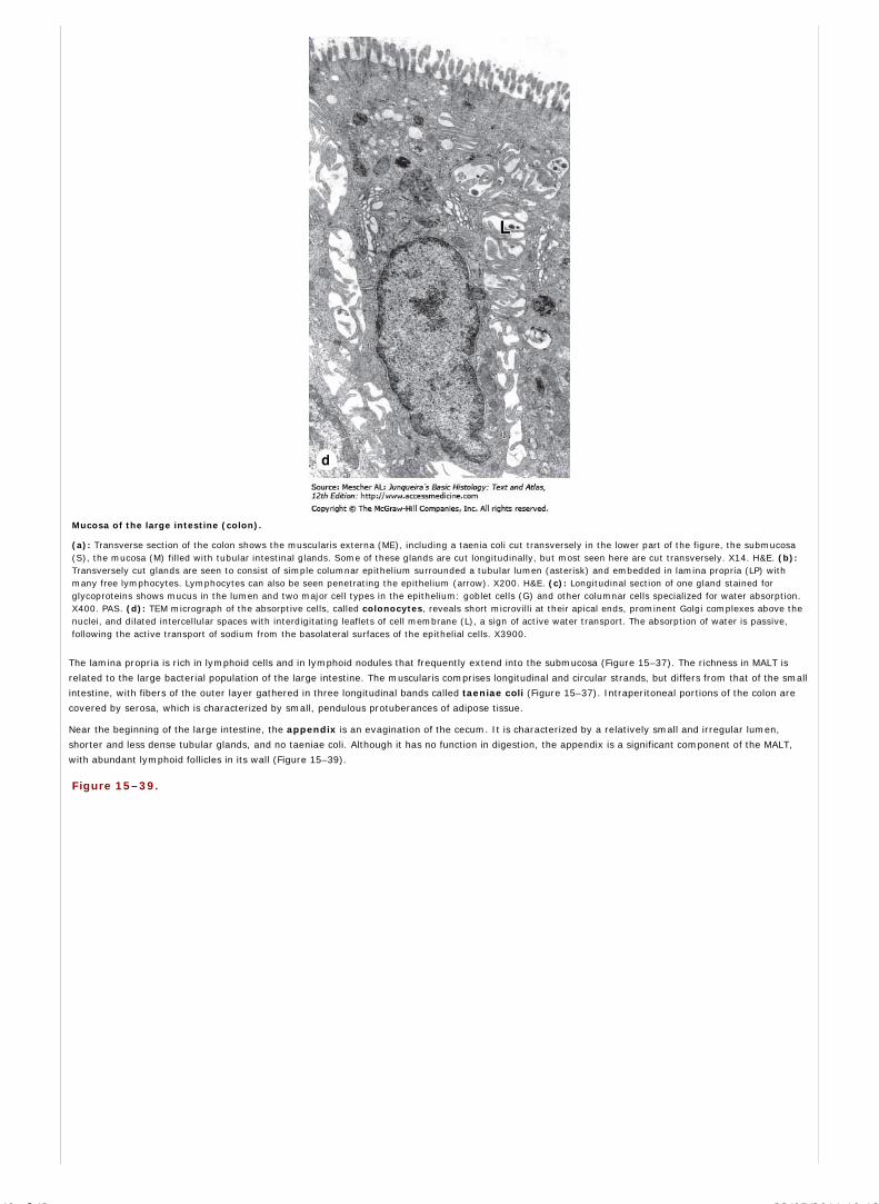
Mucosa of the large intestine (colon).
(a): Transverse section of the colon shows the muscularis externa (ME), including a taenia coli cut transversely in the lower part of the figure, the submucosa(S), the mucosa (M) filled with tubular intestinal glands. Some of these glands are cut longitudinally, but most seen here are cut transversely. X14. H&E. (b):Transversely cut glands are seen to consist of simple columnar epithelium surrounded a tubular lumen (asterisk) and embedded in lamina propria (LP) withmany free lymphocytes. Lymphocytes can also be seen penetrating the epithelium (arrow). X200. H&E. (c): Longitudinal section of one gland stained forglycoproteins shows mucus in the lumen and two major cell types in the epithelium: goblet cells (G) and other columnar cells specialized for water absorption.X400. PAS. (d): TEM micrograph of the absorptive cells, called colonocytes, reveals short microvilli at their apical ends, prominent Golgi complexes above thenuclei, and dilated intercellular spaces with interdigitating leaflets of cell membrane (L), a sign of active water transport. The absorption of water is passive,following the active transport of sodium from the basolateral surfaces of the epithelial cells. X3900.
The lamina propria is rich in lymphoid cells and in lymphoid nodules that frequently extend into the submucosa (Figure 15–37). The richness in MALT is
related to the large bacterial population of the large intestine. The muscularis comprises longitudinal and circular strands, but differs from that of the small
intestine, with fibers of the outer layer gathered in three longitudinal bands called taeniae coli (Figure 15–37). Intraperitoneal portions of the colon are
covered by serosa, which is characterized by small, pendulous protuberances of adipose tissue.
Near the beginning of the large intestine, the appendix is an evagination of the cecum. It is characterized by a relatively small and irregular lumen,
shorter and less dense tubular glands, and no taeniae coli. Although it has no function in digestion, the appendix is a significant component of the MALT,
with abundant lymphoid follicles in its wall (Figure 15–39).
Figure 15–39.
AccessMedicine | Print: Chapter 15. Digestive Tract http://accessmedicine.com/popup.aspx?aID=6182071&print=yes_chapter
40 of 42 22/05/2011 19:18

Appendix.
A blind evagination off the cecum, the appendix, has a very small lumen, fewer glands in its mucosa, and no taeniae coli. The laminar propria and submucosaare generally filled with lymphocytes and lymphoid follicles, making the appendix a significant part of the MALT. X40. H&E.
MEDICAL APPLICATION
Because the appendix is a closed sac and its contents are relatively static, it can easily become a site of inflammation (appendicitis). With the
small lumen and relatively thin wall of the appendix, inflammation and the growth of lymphoid follicles in the wall can produce swelling that can
lead to bursting of the appendix. Severe appendicitis is a medical emergency since a burst appendix will produce infection of the peritoneal cavity.
In the anal region, the mucous membrane forms a series of longitudinal folds, the anal columns (Figure 15–36). About two cm above the anal opening,
at the recto-anal junction, the lining of the mucosa is replaced by stratified squamous epithelium (Figure 15–40). In this region, the lamina propria
contains a plexus of large veins that, when excessively dilated and varicose, can produce hemorrhoids.
Figure 15–40.
Mucosa of the recto-anal junction.
The simple columnar epithelium with tubular glands that lines the rectum (left side) changes abruptly to stratified squamous epithelium in the anal canal (rightside), as seen in this longitudinal section. The connective tissue of the lamina propria is seen to contain many free lymphocytes. X40. H&E.
MEDICAL APPLICATION
Approximately 90–95% of malignant tumors of the digestive system are derived from gastric or intestinal epithelial cells, usually in the large
intestine. Malignant tumors of the colon are derived almost exclusively from its glandular epithelium (adenocarcinomas) and are the second
AccessMedicine | Print: Chapter 15. Digestive Tract http://accessmedicine.com/popup.aspx?aID=6182071&print=yes_chapter
41 of 42 22/05/2011 19:18

Copyright © The McGraw-Hill Companies. All rights reserved.Privacy Notice. Any use is subject to the Terms of Use and Notice.
most common cause of cancer deaths in the United States.
AccessMedicine | Print: Chapter 15. Digestive Tract http://accessmedicine.com/popup.aspx?aID=6182071&print=yes_chapter
42 of 42 22/05/2011 19:18

Print Close Window
Note: Large images and tables on this page may necessitate printing in landscape mode.
Copyright © The McGraw-Hill Companies. All rights reserved.
Junqueira's Basic Histology: Text & Atlas, 12e > Chapter 16. Organs Associated with the Digestive Tract >
ORGANS ASSOCIATED WITH THE DIGESTIVE TRACT: INTRODUCTION
The organs associated with the digestive tract include the salivary glands, the pancreas, the liver, and the gallbladder. Products of these organs facilitate transport
and digestion of food within the gastrointestinal tract. The main functions of the salivary glands are to wet and lubricate ingested food and the oral mucosa, to
initiate the digestion of carbohydrates and lipids with amylase and lipase, and to secrete protective bacteriostatic substances such as the immunoglobulin IgA,
lysozyme, and lactoferrin.
The pancreas produces digestive enzymes that act in the small intestine and hormones important for the metabolism of the absorbed nutrients. The liver produces
bile, an important fluid in the digestion of fats. The gallbladder absorbs water from the bile and stores it in a concentrated form. The liver also plays a major role in
carbohydrate and protein metabolism and inactivates and metabolizes many toxic substances and drugs. It also synthesizes most blood plasma proteins and the
factors necessary for blood coagulation.
SALIVARY GLANDS
Exocrine glands in the mouth produce saliva, which has digestive, lubricating, and protective functions. With a usual pH of 6.5–6.9, saliva also has an important
buffering function and in many nonhuman species is also very important for evaporative cooling. There are three pairs of large salivary glands: the parotid,
submandibular, and sublingual glands (Figure 16–1), in addition to minor glands in mucosa and submucosa throughout the oral cavity which secrete 10% of
the total volume of saliva.
Figure 16–1.
Major salivary glands.
About 90% of saliva is produced by three bilateral pairs of salivary glands: the parotid, submandibular, and sublingual glands. Locations and relative sizes of theseglands are shown here diagrammatically. These glands plus microscopic salivary glands throughout the oral mucosa produce 0.75 to 1.50 L of saliva daily.
MEDICAL APPLICATION
Reduced function of the major salivary glands due to diseases or radiotherapy is associated with caries, atrophy of the oral mucosa and speech difficulties.
A capsule of connective tissue surrounds each major salivary gland. The parenchyma of each consists of secretory endpieces and a branching duct system arranged
in lobules, separated by connective tissue septa originating from the capsule. The secretion of each gland is either serous, seromucous, or mucous, depending on
its glycoprotein mucin content. Saliva from the parotids is serous and watery. The submandibular and sublingual glands produce a seromucous secretion, with
mostly mucus from the minor glands. Saliva is modified by the cells of the duct system draining the secretory units, with much Na+ and Cl– reabsorbed while
certain growth factors and digestive enzymes are added.
Two major kinds of secretory cells occur, arranged in separate units. Serous cells are polarized protein-secreting cells, usually pyramidal in shape, with a broad
base resting on the basal lamina and a narrow apical surface facing the lumen (Figures 16–2 and 16-3). Adjacent cells are joined together by junctional complexes
and usually form a spherical mass of cells called an acinus, with a very small lumen in the center (Figure 16–2). Acini and their duct system resemble grapes
attached to a stem. Serous acinar cells largely produce digestive enzymes and other proteins.
Figure 16–2.
AccessMedicine | Print: Chapter 16. Organs Associated with the Digesti... http://accessmedicine.com/popup.aspx?aID=6182289&print=yes_chapter
1 of 21 23/05/2011 13:25

Epithelial components of a submandibular gland lobule.
The secretory portions are composed of pyramidal serous (light blue) and mucous (light brown) cells. Serous cells are typical protein-secreting cells, with roundednuclei, accumulation of rough ER in the basal third, and an apex filled with protein-rich secretory granules. The nuclei of mucous cells, flattened with condensedchromatin, are located near the bases of the cells. The short intercalated ducts are lined with cuboidal epithelium. The striated ducts are composed of columnar cellswith characteristics of ion-transporting cells: basal membrane invaginations with mitochondrial accumulations. Myoepithelial cells are shown in the serous acini.
Figure 16–3.
AccessMedicine | Print: Chapter 16. Organs Associated with the Digesti... http://accessmedicine.com/popup.aspx?aID=6182289&print=yes_chapter
2 of 21 23/05/2011 13:25

Parotid gland.
The large parotid gland consists entirely of serous acini with cells producing amylase and other proteins for storage in secretory granules. (a): Micrograph of a parotidgland shows densely packed serous acini (A) with ducts. Secretory granules of serous cells are clearly shown in this plastic section, as well as both an intercalated duct(ID) and striated duct (SD), both cut transversely. X400. PT. (b): Striations of a duct (SD) are better seen here, along with a septum (CT) and numerous serous acini(A). The connective tissue often includes adipocytes. X200. H&E.
Mucous cells are somewhat more cuboidal or columnar in shape, with nuclei pressed toward the bases of the cells. They exhibit the characteristics of mucus-
secreting cells (Figures 16–3 and 16–4), containing hydrophilic glycoprotein mucins which are important for the moistening and lubricating functions of the saliva.
Mucous cells are most often organized as tubules rather than acini and produce mostly mucins.
Figure 16–4.
AccessMedicine | Print: Chapter 16. Organs Associated with the Digesti... http://accessmedicine.com/popup.aspx?aID=6182289&print=yes_chapter
3 of 21 23/05/2011 13:25

Ultrastructure of serous and mucous cells.
A micrograph of a mixed acinus from a submandibular gland shows both serous and mucous cells. Mucous cells (upper area shown here) have large, hydrophilicgranules like those of goblet cells. Serous cells (lower area) have small, dense granules that stain more intensely with most stains. X2500. (With permission, from JohnD. Harrison, Department of Oral Pathology, King's College, London.)
Myoepithelial cells, described in Chapter 4, are found inside the basal lamina of the secretory units and (to a lesser extent) the initial part of the duct system
(Figure 16–2). Surrounding the secretory portion myoepithelial cells are well developed and branched (and are sometimes called basket cells), whereas those
associated with the initial ducts are spindle-shaped and lie parallel to the duct's length. Myoepithelial cells prevent distention of the endpiece when the lumen fills
with saliva and their contraction accelerates secretion of the product.
In the intralobular duct system, secretory endpieces empty into intercalated ducts, lined by cuboidal epithelial cells, and several of these short ducts join to
form striated ducts (Figure 16–2). The columnar cells of striated ducts often show radial striations extending from the cell bases to the level of the nuclei.
Ultrastructurally the striations consist of infoldings of the basal plasma membrane (Figure 16–5). Numerous mitochondria are aligned parallel to the infolded
membranes which contain ion transporters. Such folds greatly increase the cell surface area, facilitating ion absorption, and are characteristic of cells specialized
for ion transport.
Figure 16–5.
AccessMedicine | Print: Chapter 16. Organs Associated with the Digesti... http://accessmedicine.com/popup.aspx?aID=6182289&print=yes_chapter
4 of 21 23/05/2011 13:25

Striated ducts.
(a): This light micrograph of a striated duct (SD) shows very faint pink striations in the basal half of the columnar cells. The striations are produced by mitochondrialocated in the folds of the lateral cell membrane. X200. H&E. (b): SEM indicates that the apical ends of the cells are joined together near the small lumen (L), withinterdigitating folds of cell membrane best developed at the basal end (B). X4000. (c): This SEM shows the bases (B) of several such cells with the basal laminaremoved, revealing the extensive interlocking of folded membrane between neighboring cells. Mitochondria between the folds supply energy for membrane ion pumpsand ion uptake from saliva is rapid and efficient. X4000
In the large salivary glands, the connective tissue contains many lymphocytes and plasma cells. The plasma cells release IgA, which forms a complex with a
secretory component synthesized by the epithelial cells of serous acini and intralobular ducts. The IgA-secretory complex released into the saliva resists enzymatic
digestion and constitutes an immunologic defense mechanism against pathogens in the oral cavity.
The striated ducts of each lobule converge and drain into ducts located in the connective tissue septa separating lobules, where they become interlobular, or
excretory, ducts. They are initially lined with pseudostratified or stratified cuboidal epithelium, but more distal parts of the excretory ducts are lined with
stratified columnar epithelium containing a few mucous cells. The main duct of each large salivary gland ultimately empties into the oral cavity and is lined with
nonkeratinized-stratified squamous epithelium.
Vessels and nerves enter the large salivary glands at a hilum and gradually branch into the lobules. A rich vascular and nerve plexus surrounds the secretory and
ductal components of each lobule. The capillaries surrounding the secretory endpieces are very important for the secretion of saliva, which is stimulated by the
autonomic nervous system. Parasympathetic stimulation, usually elicited through the smell or taste of food, provokes a copious watery secretion with relatively
little organic content. Sympathetic stimulation inhibits such secretion, and produces the potential for dry mouth often associated with anxiety.
Features specific to each group of major salivary glands include the following:
Parotid gland, located in each cheek near the ear, is a branched acinar gland with secretory portions composed exclusively of serous cells surroundingvery small lumens (Figure 16–3). Serous cells contain secretory granules with abundant -amylase and proline-rich proteins. Amylase activity isresponsible for most of the hydrolysis of ingested carbohydrates which begins in the mouth. Proline-rich proteins, the most abundant factors in parotid
saliva, have antimicrobial properties and Ca2+ binding properties that may help maintain the surface of enamel.
Submandibular gland is a branched tubuloacinar gland (Figures 16–4 and 16–6), with secretory portions containing both mucous and serous cells. Theserous cells are the main component of this gland and are easily distinguished from mucous cells by their rounded nuclei and basophilic cytoplasm. Mostof the secretory units in this gland are serous acinar, with about 10% consisting of mucous tubules capped with serous cells. Such caps are calledserous demilunes (Figure 16–6a). Lateral and basal membrane infoldings of the serous cells increase the ion-transporting surface area and facilitateelectrolyte and water transport. In addition to -amylase and proline-rich proteins, serous cells of the submandibular gland secrete other enzymes,including lysozyme, which hydrolyzes the walls in many types of bacteria.
Sublingual gland, like the submandibular gland, is a branched tubuloacinar gland formed of serous and mucous cells. Here mucous cells predominate(Figure 16–6b), with serous cells only present in demilunes on mucous tubules. The major salivary product is mucus, but cells of the serous demilunes inthis gland secrete amylase and lysozyme.
Figure 16–6.
AccessMedicine | Print: Chapter 16. Organs Associated with the Digesti... http://accessmedicine.com/popup.aspx?aID=6182289&print=yes_chapter
5 of 21 23/05/2011 13:25

Submandibular gland and sublingual gland.
(a): Submandibular gland is a mixed serous and mucous gland (serous cells predominate) and shows well-stained cells in serous acini (A) and in serous demilunes (S)and pale-staining mucous cells (M) grouped as tubules in this tubuloacinar gland. Small intralobular ducts (ID) drain each lobule, but these are not composed ofcolumnar cells with well-developed striations. X340. H&E. (b): Sublingual gland is a mixed but largely mucous gland with a tubuloacinar arrangement of poorly stainedmucous cells (M). Small intralobular ducts (ID) are seen in connective tissue, as well as small fascicles of lingual striated muscle (SM). X140. H&E.
Small, nonencapsulated salivary glands are distributed throughout the oral mucosa and submucosa with short ducts to the oral cavity. Minor salivary glands are
usually mucous, an exception being the small serous glands at the bases of circumvallate papillae on the back of the tongue (Chapter 15). B lymphocytes releasing
IgA are common within the minor salivary glands.
AccessMedicine | Print: Chapter 16. Organs Associated with the Digesti... http://accessmedicine.com/popup.aspx?aID=6182289&print=yes_chapter
6 of 21 23/05/2011 13:25

PANCREAS
The pancreas is a mixed exocrine-endocrine gland that produces both digestive enzymes and hormones (Figures 16–7 and 16–8). A thin capsule of connective
tissue covers the pancreas and sends septa into it, separating the pancreatic lobules. The secretory acini are surrounded by a basal lamina that is supported by a
delicate sheath of reticular fibers and a rich capillary network.
Figure 16–7.
Pancreas and duodenum.
(a): The main regions of the pancreas are shown in relation to the two pancreatic ducts and the duodenum. (b): Micrographs show a pancreatic islet and severalpancreatic acini. X75 and X200. H&E.
Figure 16–8.
Pancreas.
Low power view of pancreas includes several islets (I) surrounded by many serous acini (A). The larger interlobular ducts (D) are lined by simple columnar epithelium.The ducts and blood vessels (V) are located in connective tissue, which also provides a thin capsule to the entire gland and thin septa separating the lobules of
AccessMedicine | Print: Chapter 16. Organs Associated with the Digesti... http://accessmedicine.com/popup.aspx?aID=6182289&print=yes_chapter
7 of 21 23/05/2011 13:25

secretory acini. X20. H&E.
The digestive enzymes are produced by cells of the larger exocrine portion and the hormones are synthesized in clusters of endocrine epithelial cells known as
pancreatic islets (islets of Langerhans) (see Chapter 20). The exocrine portion of the pancreas is a compound acinar gland, similar in structure to the parotid
gland. The two glands can be distinguished histologically by the absence of striated ducts and the presence of the islets in the pancreas. Another characteristic
detail is that in the pancreas the initial portions of intercalated ducts penetrate the lumens of the acini (Figure 16–9). Small pale-staining centroacinar cells
constitute the intraacinar portion of the intercalated duct and are found only in pancreatic acini. Intercalated ducts merge to form larger interlobular ducts lined
by columnar epithelium. No ducts in the pancreas are striated.
Figure 16–9.
Pancreatic acini.
(a): Micrograph of exocrine pancreas shows the serous, enzyme-producing cells arranged in small acini (A) with very small lumens. Acini are surrounded by smallamounts of connective tissue with fibroblasts (F). Each acinus is drained by an intercalated duct with its initial cells, the centroacinar cells (arrow), inserted into theacinar lumen. X200. H&E. (b): The diagram shows the arrangement of cells more clearly. Under the influence of secretin, the centroacinar and other cells of these small
ducts secrete a copious HCO3– - rich fluid that hydrates and alkalinizes the enzymatic secretions of the acinar cells. Pancreatic acini lack myoepithelial cells and their
intercalated ducts lack striations.
Each exocrine acinus of the pancreas is composed of several serous cells surrounding a very small lumen (Figure 16–9). The acinar cells are highly polarized, with
a spherical nucleus, and are typical protein-secreting cells (Figure 16–10). The number of zymogen granules present in each cell varies and is maximal in animals
that have fasted.
Figure 16–10.
AccessMedicine | Print: Chapter 16. Organs Associated with the Digesti... http://accessmedicine.com/popup.aspx?aID=6182289&print=yes_chapter
8 of 21 23/05/2011 13:25
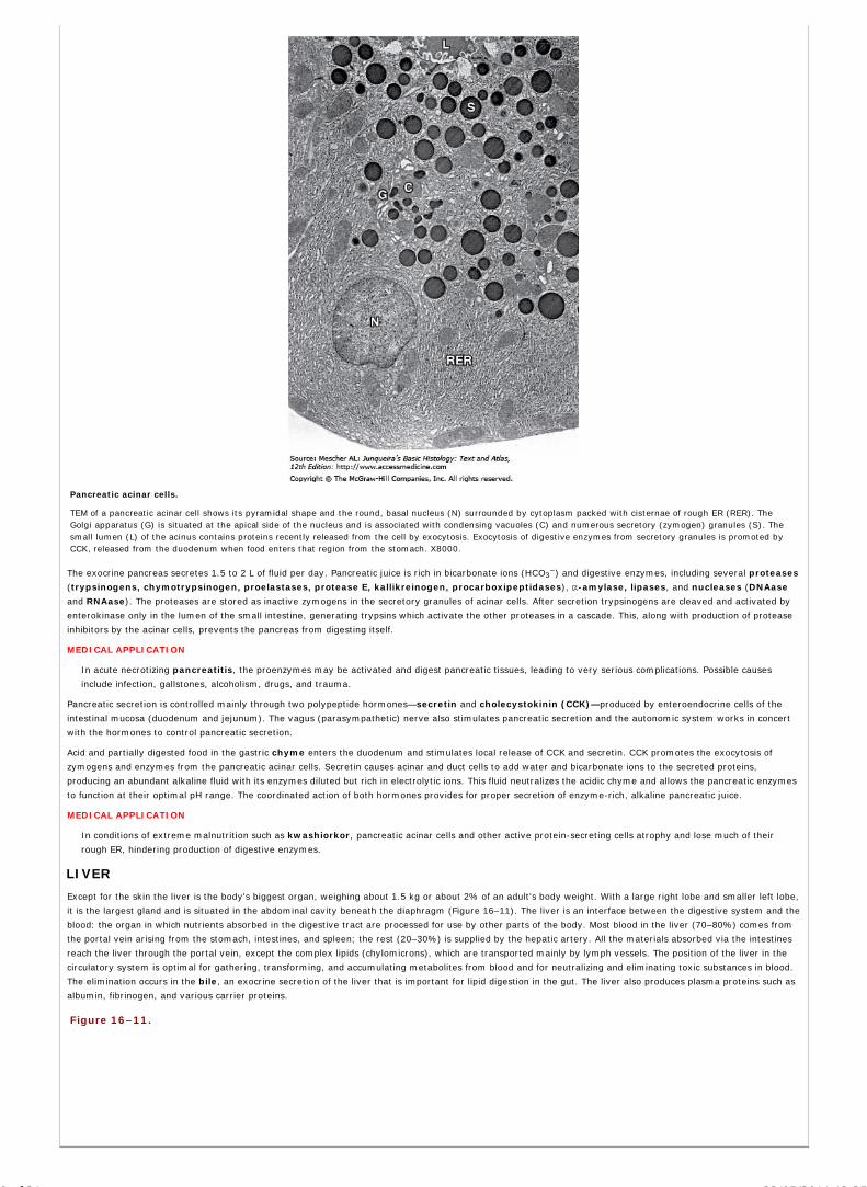
Pancreatic acinar cells.
TEM of a pancreatic acinar cell shows its pyramidal shape and the round, basal nucleus (N) surrounded by cytoplasm packed with cisternae of rough ER (RER). TheGolgi apparatus (G) is situated at the apical side of the nucleus and is associated with condensing vacuoles (C) and numerous secretory (zymogen) granules (S). Thesmall lumen (L) of the acinus contains proteins recently released from the cell by exocytosis. Exocytosis of digestive enzymes from secretory granules is promoted byCCK, released from the duodenum when food enters that region from the stomach. X8000.
The exocrine pancreas secretes 1.5 to 2 L of fluid per day. Pancreatic juice is rich in bicarbonate ions (HCO3–) and digestive enzymes, including several proteases
(trypsinogens, chymotrypsinogen, proelastases, protease E, kallikreinogen, procarboxipeptidases), -amylase, lipases, and nucleases (DNAase
and RNAase). The proteases are stored as inactive zymogens in the secretory granules of acinar cells. After secretion trypsinogens are cleaved and activated by
enterokinase only in the lumen of the small intestine, generating trypsins which activate the other proteases in a cascade. This, along with production of protease
inhibitors by the acinar cells, prevents the pancreas from digesting itself.
MEDICAL APPLICATION
In acute necrotizing pancreatitis, the proenzymes may be activated and digest pancreatic tissues, leading to very serious complications. Possible causes
include infection, gallstones, alcoholism, drugs, and trauma.
Pancreatic secretion is controlled mainly through two polypeptide hormones—secretin and cholecystokinin (CCK)—produced by enteroendocrine cells of the
intestinal mucosa (duodenum and jejunum). The vagus (parasympathetic) nerve also stimulates pancreatic secretion and the autonomic system works in concert
with the hormones to control pancreatic secretion.
Acid and partially digested food in the gastric chyme enters the duodenum and stimulates local release of CCK and secretin. CCK promotes the exocytosis of
zymogens and enzymes from the pancreatic acinar cells. Secretin causes acinar and duct cells to add water and bicarbonate ions to the secreted proteins,
producing an abundant alkaline fluid with its enzymes diluted but rich in electrolytic ions. This fluid neutralizes the acidic chyme and allows the pancreatic enzymes
to function at their optimal pH range. The coordinated action of both hormones provides for proper secretion of enzyme-rich, alkaline pancreatic juice.
MEDICAL APPLICATION
In conditions of extreme malnutrition such as kwashiorkor, pancreatic acinar cells and other active protein-secreting cells atrophy and lose much of their
rough ER, hindering production of digestive enzymes.
LIVER
Except for the skin the liver is the body's biggest organ, weighing about 1.5 kg or about 2% of an adult's body weight. With a large right lobe and smaller left lobe,
it is the largest gland and is situated in the abdominal cavity beneath the diaphragm (Figure 16–11). The liver is an interface between the digestive system and the
blood: the organ in which nutrients absorbed in the digestive tract are processed for use by other parts of the body. Most blood in the liver (70–80%) comes from
the portal vein arising from the stomach, intestines, and spleen; the rest (20–30%) is supplied by the hepatic artery. All the materials absorbed via the intestines
reach the liver through the portal vein, except the complex lipids (chylomicrons), which are transported mainly by lymph vessels. The position of the liver in the
circulatory system is optimal for gathering, transforming, and accumulating metabolites from blood and for neutralizing and eliminating toxic substances in blood.
The elimination occurs in the bile, an exocrine secretion of the liver that is important for lipid digestion in the gut. The liver also produces plasma proteins such as
albumin, fibrinogen, and various carrier proteins.
Figure 16–11.
AccessMedicine | Print: Chapter 16. Organs Associated with the Digesti... http://accessmedicine.com/popup.aspx?aID=6182289&print=yes_chapter
9 of 21 23/05/2011 13:25

Liver.
A large organ in the upper right quadrant of the abdomen, immediately below the diaphragm, the liver is composed of thousands of polygonal structures called hepaticlobules, which are the basic functional units of the organ. (a): Diagram shows a small central vein projecting through the center of each hepatic lobule and several setsof blood vessels defining the periphery. The peripheral vessels are grouped primarily in connective tissue comprising the portal tracts, which usually include a branch ofthe portal vein and a branch of the hepatic artery, as well as a branch of the bile duct. These comprise the portal triad. (b): Both blood vessels to each lobule give offsinusoids, which run between plates of hepatocytes and drain into the central vein. (c): Micrograph showing components of the portal triad. X220. H&E.
StromaThe liver is covered by a thin fibrous capsule of connective tissue that becomes thicker at the hilum, where the portal vein and the hepatic artery enter the organ
and where the right and left hepatic ducts and lymphatics exit. These vessels and ducts are surrounded by connective tissue all the way to their termination (or
origin) in the portal spaces between the liver lobules. At this point,, a delicate reticular fiber network surrounds and supports the liver cells and the sinusoidal
endothelial cells of the liver lobules (Figures 16–12 and 16–13).
Figure 16–12.
AccessMedicine | Print: Chapter 16. Organs Associated with the Digesti... http://accessmedicine.com/popup.aspx?aID=6182289&print=yes_chapter
10 of 21 23/05/2011 13:25

Hepatic lobule.
Cut transversely hepatic lobules are polygonal units showing plates of epithelial cells called hepatocytes radiating from a central venule (C). (a): Hepatic lobule of somemammals, such as the pig, are delimited on all sides by connective tissue. (b): Hepatic units of humans have much less connective tissue and their boundaries aremore difficult to distinguish. In all cases peripheral connective tissue of portal areas with microvasculature and small bile duct (D) branches can be seen and in humansas in other mammals these are present at the boundaries between two or more hepatic lobules. The vessels near the bile ducts branches are a venule (V) off the portalvein and an arteriole (A) off the hepatic artery. Both X150. H&E.
Figure 16–13.
AccessMedicine | Print: Chapter 16. Organs Associated with the Digesti... http://accessmedicine.com/popup.aspx?aID=6182289&print=yes_chapter
11 of 21 23/05/2011 13:25

Hepatic lobule microvasculature.
(a): The hepatic lobule’s central vein (CV) is actually a venule consisting of little more than an endothelial tube with smaller sinusoids (S) coming in from all directions.X200. H&E. (b): Peripheral portal areas contain more connective tissue and are the sites of the portal triad: a portal venule (PV), an arteriole (A) branching off thehepatic artery, and one or two branches of the bile duct (D). Blood flows (arrow) from these arterioles and venules into the sinusoids between the plates of hepatocytesthat run to the central venule. In the portal area the bile ductules are lined by simple cuboidal epithelium. X400. H&E. (c): Reticulin (collagen type III) fibers runningalong the plates of hepatocytes are the major support for the sinusoids and central venules. Most connective tissue in the liver is found in the septa and portal tracts.X400. Silver.
Hepatic LobulesLiver cells or hepatocytes (Gr. hepar, liver, + kytos, cell) are epithelial cells grouped in interconnected plates. Hepatocytes are arranged into thousands of small
(~0.7 x 2 mm), polyhedral hepatic lobules which are the classic structural and functional units of the liver (Figure 16–11). Each lobule has three to six portal
areas at its periphery and a venule called a central vein in its center (Figures 16–11, 16–12, and 16–13). The portal zones at the corners of the lobules consist of
connective tissue in which are embedded a venule (a branch of the portal vein), an arteriole (a branch of the hepatic artery), and a duct of cuboidal epithelium (a
branch of the bile duct system) – three structures called the portal triad (Figure 16–12). The venule contains blood from the superior and inferior mesenteric and
splenic veins. The arteriole contains blood from the celiac trunk of the abdominal aorta. The duct carries bile synthesized by the parenchymal cells (hepatocytes)
and eventually empties into the hepatic duct. Portal areas also have nerve fibers and lymphatics. In some animals (eg, pigs), the lobules are separated from each
other by a layer of connective tissue, making them easy to distinguish. In humans the lobules are in close contact along most of their length and it is more difficult
to establish the exact limits between different lobules (Figure 16–12).
Hepatocytes make up each of the interconnected plates like the bricks of a wall and the plates are arranged radially around the central vein (Figure 16–11). From
the periphery of the lobule to its center, the plates of hepatocytes branch and anastomose freely, forming a rather sponge-like structure (Figure 16–12). The
spaces between these plates contain important microvascular components, the liver sinusoids (Figures 16–11, 16–12, and 16–13). These irregularly dilated
sinusoids consist only of a discontinuous layer of fenestrated endothelial cells (Figures 16–14 and 16–15). The endothelial cells are separated from the underlying
hepatocytes by a thin, discontinuous basal lamina and a very narrow perisinusoidal space (the space of Disse), into which project microvilli of the hepatocytes
AccessMedicine | Print: Chapter 16. Organs Associated with the Digesti... http://accessmedicine.com/popup.aspx?aID=6182289&print=yes_chapter
12 of 21 23/05/2011 13:25
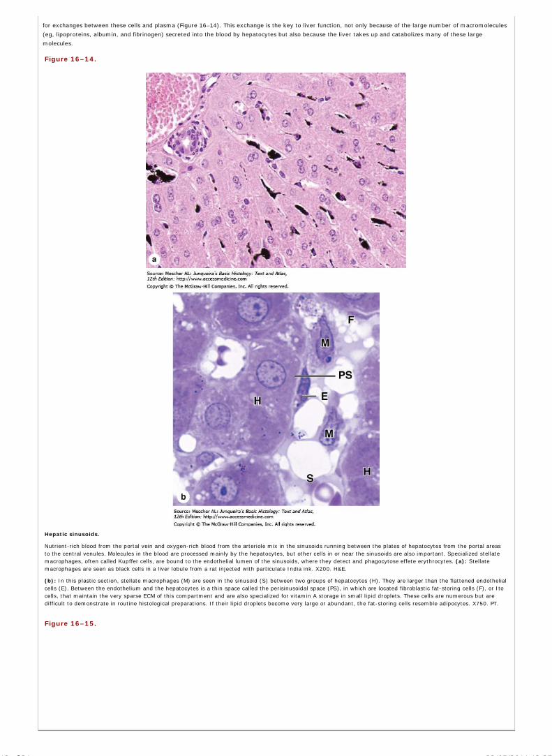
for exchanges between these cells and plasma (Figure 16–14). This exchange is the key to liver function, not only because of the large number of macromolecules
(eg, lipoproteins, albumin, and fibrinogen) secreted into the blood by hepatocytes but also because the liver takes up and catabolizes many of these large
molecules.
Figure 16–14.
Hepatic sinusoids.
Nutrient-rich blood from the portal vein and oxygen-rich blood from the arteriole mix in the sinusoids running between the plates of hepatocytes from the portal areasto the central venules. Molecules in the blood are processed mainly by the hepatocytes, but other cells in or near the sinusoids are also important. Specialized stellatemacrophages, often called Kupffer cells, are bound to the endothelial lumen of the sinusoids, where they detect and phagocytose effete erythrocytes. (a): Stellatemacrophages are seen as black cells in a liver lobule from a rat injected with particulate India ink. X200. H&E.
(b): In this plastic section, stellate macrophages (M) are seen in the sinusoid (S) between two groups of hepatocytes (H). They are larger than the flattened endothelialcells (E). Between the endothelium and the hepatocytes is a thin space called the perisinusoidal space (PS), in which are located fibroblastic fat-storing cells (F), or Itocells, that maintain the very sparse ECM of this compartment and are also specialized for vitamin A storage in small lipid droplets. These cells are numerous but aredifficult to demonstrate in routine histological preparations. If their lipid droplets become very large or abundant, the fat-storing cells resemble adipocytes. X750. PT.
Figure 16–15.
AccessMedicine | Print: Chapter 16. Organs Associated with the Digesti... http://accessmedicine.com/popup.aspx?aID=6182289&print=yes_chapter
13 of 21 23/05/2011 13:25

Ultrastructure of the sinusoid wall.
SEM of the luminal surface of the endothelium lining a sinusoid in liver shows grouped fenestrations. At the border are seen cut edges of the endothelial cell (E) in thisdiscontinuous sinusoid and hepatocytes (H). Between these two cells is the thin perisinusoidal space (PS), into which project microvilli from the hepatocytes surface.Blood plasma passes freely through the fenestrations into the perisinusoidal space, where the voluminous membrane of hepatocytes acts to remove many high and lowmolecular weight blood components and nutrients for storage and processing. Proteins synthesized and secreted from hepatocytes, such as albumin, fibrinogen, andother blood proteins, are released into the perisinusoidal space. X6500. (With permission, from Eddie Wisse, Electron Microscopy Unit, Department of Pathology,University of Maastricht, The Netherlands.)
Liver sinusoids are surrounded and supported by delicate sheathes of reticular fibers (Figure 16–13). Two noteworthy cells are associated with these sinusoids in
addition to the endothelial cells:
Abundant specialized stellate macrophages, also known as Kupffer cells, are found between sinusoidal endothelial cells and on the luminal surfacewithin the sinusoids, mainly near the portal areas (Figure 16–14). Their main functions are to break down aged erythrocytes and free heme for re-use,remove bacteria or debris that may enter the portal blood from the gut, and act as antigen-presenting cells in adaptive immunity.
In the perisinusoidal space (not the lumen) are stellate fat storing cells (or Ito cells) with small lipid droplets containing vitamin A (Figure 16–14).These cells, which make up about 8% of the cells in a liver but are difficult to see in routine preparations, store much of the body's vitamin A, produceECM components, and have a regulatory role in local immunity.
MEDICAL APPLICATION
In chronically diseased liver, Ito cells proliferate and acquire the features of myofibroblasts, with or without the lipid droplets. Under these conditions,
these cells are found close to the damaged hepatocytes and play a major role in the development of fibrosis, including the fibrosis secondary to alcoholic
liver disease.
Blood SupplyAs an important interface for processing blood from the digestive system, the liver gets most of its blood from the portal vein, which carries nutrient-rich but
oxygen-poor blood from the abdominal viscera. Oxygenated blood is brought in with the smaller portion derived from the hepatic artery (Figure 16–11).
The portal system conveys blood from the pancreas, spleen, and the intestines. Nutrients are accumulated and transformed in the liver and toxic substances are
neutralized and eliminated there. In the liver the portal vein branches repeatedly and sends small portal venules to the portal spaces. The portal venules branch
into smaller distributing venules that run around the periphery of each lobule and lead into the sinusoids. The sinusoids run radially, converging in the center of the
lobule to form the central or centrolobular vein (Figures 16–11, 16-12, and 16–13). This vessel, like the sinusoids, has very thin walls consisting only of
endothelial cells supported by a sparse population of collagen fibers (Figure 16–13). Central venules from each lobule converge into veins, which eventually form
two or more large hepatic veins that empty into the inferior vena cava.
The hepatic artery branches repeatedly and forms arterioles in the portal areas, some of which lead directly into the sinusoids at various distances from the portal
spaces, thus adding oxygen-rich arterial blood to the portal venous blood in the sinusoids.
Blood always flows from the periphery to the center of each hepatic lobule. Consequently, oxygen and metabolites, as well as all other toxic or nontoxic
substances absorbed in the intestines, reach the lobule's peripheral cells first and then the more central cells. This direction of blood flow partly explains why the
properties and function of the periportal hepatocytes differ from that of the centrolobular cells. Hepatocytes near the portal areas can rely on aerobic metabolism
and are often more active in protein synthesis, while the more central cells are exposed to lower concentrations of nutrients and oxygen and are more involved
with detoxification and glycogen metabolism.
The HepatocyteHepatocytes are large polyhedral cells, with six or more surfaces, and typical diameters of 20–30 m. In H&E-stained sections their cytoplasm is usually
eosinophilic because of the large number of mitochondria, up to 2000 per cell. Hepatocytes have large spherical nuclei with nucleoli. The cells frequently have two
or more nuclei and about 50% of them are polyploid, with two, four, eight or more times the normal diploid chromosome number. Polyploid nuclei are
characterized by greater size, which is proportional to their ploidy.
The surface of each hepatocyte is in contact with the wall of a sinusoid, through the perisinusoidal space, and with the surfaces of other hepatocytes (Figure
16–16). Where two hepatocytes abut, they delimit a tubular space between them known as the bile canaliculus (Figure 16–16).
Figure 16–16.
AccessMedicine | Print: Chapter 16. Organs Associated with the Digesti... http://accessmedicine.com/popup.aspx?aID=6182289&print=yes_chapter
14 of 21 23/05/2011 13:25

Ultrastructure of hepatocytes and bile canaliculi.
(a): TEM of hepatocytes show small bile canaliculi (BC) between two cells, with junctional complexes binding the cells firmly and tightly at these sites. The bilecanaliculus is the site of exocrine secretion by hepatocytes. The two adjoining hepatocytes extend short microvilli and secrete bile components into this space. Thehepatocytes have many mitochondria (M), small electron-dense glycogen granules, and Golgi complexes (G) and extend more numerous microvilli into theperisinusoidal space (PS), which is the site where hepatocytes remove and add components in plasma. The endothelial cell (E) lining the sinusoid (S) is also seen.X9500. (b): SEM of hepatocytes (H) broken apart from one another reveals the length of a bile canaliculus (BC) along the cell's surface. Such canaliculi run between thecells of the hepatocyte plates in the hepatic lobules and carry bile toward the portal areas where the canaliculi join cuboidal bile ductules. (Figure 16–16a, withpermission, from Douglas L. Schmucker, Department of Anatomy, University of California, San Francisco.)
The canaliculi, the first portions of the bile duct system, are long spaces 1–2 m in diameter. They are limited only by the plasma membranes of two hepatocytes,
which extend a small number of microvilli into their interiors (Figure 16–16). The cell membranes near these canaliculi are firmly joined by tight junctions. Gap
junctions also occur between hepatocytes, allowing intercellular communication and coordination of the cells' activities. The bile canaliculi form a complex
anastomosing network progressing along the plates of the hepatic lobule and terminating in the region of the portal spaces (Figure 16–11). The bile flow therefore
progresses in a direction opposite to that of the blood, ie, from the center of the lobule to its periphery. Near the peripheral portal areas, bile canaliculi empty into
bile ductules (Figure 16–17) composed of cuboidal epithelial cells called cholangiocytes (Figures 16–18 and 16–19). After a short distance, these ductules cross
AccessMedicine | Print: Chapter 16. Organs Associated with the Digesti... http://accessmedicine.com/popup.aspx?aID=6182289&print=yes_chapter
15 of 21 23/05/2011 13:25

the limiting hepatocytes of the lobule and end in the bile ducts in the portal spaces. Bile ducts are lined by cuboidal or columnar epithelium and have a distinct
connective tissue sheath. They gradually enlarge and fuse, forming right and left hepatic ducts, which subsequently leave the liver.
Figure 16–17.
Bile ductules.
Near the periphery of hepatic lobules, the bile canaliculi join with bile ductules lined by cuboidal epithelial cells called cholangiocytes. The ductules soon merge withbranches of the bile ducts in the portal spaces. The branches merge from all over the liver and give rise to the left and right hepatic ducts which leave the organ at thehilum.
Figure 16–18.
Secretion of bile acids.
Bile acids are organic molecules, primarily cholic acid and its derivatives, found in bile excreted from hepatocytes into the system of bile canaliculi and ducts. Thesecompounds are often deprotonated, becoming the bile salts. Stored in the gall bladder and released into the duodenum after a meal, bile acids emulsify fats, facilitatinglipid breakdown and absorption. About 90% of bile acids are absorbed from the intestinal epithelium and are transported by the hepatocyte from the blood to bilecanaliculi (enterohepatic recirculation). About 10% of bile acids are synthesized in the smooth ER of hepatocytes by conjugation of cholic acid (synthesized inhepatocytes from cholesterol) with the amino acid glycine or taurine, producing glycocholic and taurocholic acids.
Figure 16–19.
AccessMedicine | Print: Chapter 16. Organs Associated with the Digesti... http://accessmedicine.com/popup.aspx?aID=6182289&print=yes_chapter
16 of 21 23/05/2011 13:25

Concepts of structure-function relationships in liver.
Studies of liver microanatomy, physiology, and pathology have given rise to three related ways to view the liver's organization which emphasize different aspects ofhepatocyte activity. (a): The classic lobule concept offers a basic understanding of the structure-function relationship in liver organization and emphasizes theendocrine function of hepatocytes as blood flows past them toward the central vein. (b): The portal lobule emphasizes the hepatocytes' exocrine function and the flowof bile from regions of three classic lobules toward the bile duct in the portal triad at the center here. The area drained by each bile duct is roughly triangular.
(c): The liver acinus concept emphasizes the different oxygen and nutrient contents of blood at different distances along the sinusoids, with blood from each portalarea supplying cells in two or more classic lobules. Each hepatocyte's major activity is determined by its location along the oxygen/nutrient gradient: periportal cells ofzone I get the most oxygen and nutrients and show metabolic activity generally different from the pericentral hepatocytes of zone III, exposed to the lowest oxygenand nutrient concentrations. Many pathological changes in liver are best understood from the point-of-view of liver acini. (Reproduced, with permission from Boron WF,Boulpaep EL: Medical Physiology: A Cellular and Molecular Approach, Saunders. Copyright Elsevier, 2005.)
Functionally the hepatocyte may be the most versatile cell in the body. The hepatocyte has an abundant endoplasmic reticulum—both smooth and rough (Figure
16–18). Rough ER for synthesis of plasma proteins causes cytoplasmic basophilia, which is often more pronounced in hepatocytes near the portal areas (Figure
16–12). Various important processes take place in the smooth ER, which is distributed diffusely throughout the cytoplasm. This organelle is responsible for the
processes of oxidation, methylation, and conjugation required for inactivation or detoxification of various substances before their excretion. The smooth ER is a
labile system that reacts promptly to the molecules received by the hepatocyte.
MEDICAL APPLICATION
One of the main processes that occurs in the smooth ER is the conjugation of hydrophobic (water-insoluble) toxic bilirubin by glucuronyltransferase to form
a water-soluble nontoxic bilirubin glucuronide. This conjugate is excreted by hepatocytes into the bile. When bilirubin or bilirubin glucuronide is not
excreted properly, various diseases characterized by jaundice can result.
One of the frequent causes of jaundice in newborns is the often underdeveloped state of the smooth ER in their hepatocytes (neonatal
hyperbilirubinemia). The current treatment for these cases is exposure to blue light from ordinary fluorescent tubes, which transforms unconjugated
bilirubin into a water-soluble photoisomer that can be excreted by the kidneys.
The hepatocyte frequently contains deposits of glycogen, which appear ultrastructurally as coarse, electron-dense granules in the cytosol, often close to the
smooth ER (Figure 16–16). Liver glycogen is a depot for glucose and is mobilized if the blood glucose level falls below normal. In this way, hepatocytes maintain a
steady level of blood glucose, one of the main sources of energy for the body. Hepatocytes also normally store triglycerides in small lipid droplets. This capacity to
store metabolites is important, because it supplies the body with energy between meals.
Hepatocytes do not usually store proteins in secretory granules but continuously release them into the bloodstream. About 5% of the protein exported by the liver
is produced by the sinusoidal stellate macrophages.
The hepatocyte is responsible for converting lipids and amino acids into glucose by means of a complex enzymatic process called gluconeogenesis (Gr. glykys,
sweet, + neos, new, + genesis, production). It is also the main site of amino acid deamination, resulting in the production of urea which is transported in blood to
the kidney and is excreted there.
Hepatocyte lysosomes are important in the turnover and degradation of intracellular organelles. Peroxisomes are also abundant and are important for oxidation of
excess fatty acids, breakdown of the hydrogen peroxide generated by this oxidation (by means of catalase activity), breakdown of excess purines to uric acid, and
participation in the synthesis of cholesterol, bile acids, and some lipids used by neurons to make myelin. Each hepatocyte may contain up to 50 Golgi complexes
involved in the formation of lysosomes and the secretion of proteins, glycoproteins, and lipoproteins into plasma.
MEDICAL APPLICATION
A variety of rare inherited disorders of peroxisome function occur in humans, most involving mutations of the enzymes found within peroxisomes. For
example, X-linked adrenoleukodystrophy (X-ALD) results from a failure to metabolize fatty acids properly, resulting in the deterioration of the myelin
sheaths of neurons.
Bile secretion is an exocrine function in the sense that hepatocytes are involved in the uptake, transformation, and excretion of blood components into the bile
canaliculi. Bile has several essential components in addition to water and electrolytes: bile acids (low molecular weight organic acids such as cholic acid, and their
deprotonated forms called bile salts), phospholipids, cholesterol, and heme-containing bile pigments such as yellowish-green bilirubin. The secretion of bile acids is
illustrated in Figure 16–18. Bile acids have an important function in emulsifying the lipids in the digestive tract, promoting easier digestion by lipases and
subsequent absorption.
MEDICAL APPLICATION
AccessMedicine | Print: Chapter 16. Organs Associated with the Digesti... http://accessmedicine.com/popup.aspx?aID=6182289&print=yes_chapter
17 of 21 23/05/2011 13:25

Abnormal proportions of bile acids may lead to the formation of gallstones (cholelithiasis). Gallstones can block bile flow and cause jaundice—the
presence of bile pigments such as bilirubin in blood—from the rupture of tight junctions around the bile canaliculi. The presence of bilirubin in blood leads to
the temporary yellowing of the skin and sclera of the eyes characteristic of jaundice.
Most of the bile pigment bilirubin is derived from the degradation of the hemoglobin in senescent erythrocytes, which occurs mainly in macrophages of the spleen,
but also in other macrophages including those in the liver sinusoids. Released from macrophages bilirubin binds albumen, circulates, and is taken up from plasma
by hepatocytes. In the smooth ER hydrophobic bilirubin is conjugated to glucuronate, forming water-soluble bilirubin glucuronide, which is then secreted into the
bile canaliculi. Released into the gut with bile, some bilirubin is metabolized by bacteria to other pigments which give feces its characteristic color. Other bilirubin is
absorbed in the gut and removed from blood in the kidneys, giving urine its yellow color.
Various drugs and potentially toxic substances can be inactivated by oxidation, methylation, or conjugation. The enzymes participating in these processes are
located mainly in the smooth ER of hepatocytes. Glucuronyltransferase, an enzyme that conjugates glucuronate to bilirubin, also causes conjugation of several
other compounds such as steroids, barbiturates, antihistamines, and anticonvulsants. Under certain conditions, drugs that are inactivated in the liver can induce an
increase in the hepatocyte's smooth ER, thus improving the detoxification capacity of the organ.
MEDICAL APPLICATION
The administration of barbiturates to laboratory animals results in rapid development of smooth ER in hepatocytes. Barbiturates can also increase
synthesis of glucuronyltransferase. This finding has led to the use of barbiturates in the treatment of glucuronyltransferase deficiencies.
HEPATIC LOBULE STRUCTURE & FUNCTION
The different categories of hepatocyte functions—including secretion of protein factors into blood, the secretion of bile components, and the removal of oxygen and
small compounds of all kinds from blood—has led to three ways to think of liver lobule structure, which are summarized in Figure 16–19. The classic hepatic
lobule, with blood flowing past hepatocytes from up to six portal triad areas to a central venule, emphasizes the endocrine function of the structure producing
factors for uptake by plasma. The concept of portal lobules of hepatocytes is more useful when considering the exocrine function of these cells, ie, bile secretion.
The portal area has the bile ductule at the center and bile, moving in the opposite direction as the blood, flows toward it from all the surrounding hepatocytes. The
tissue draining bile into each portal area duct is roughly triangular in shape, with the central veins of three classic lobules at its angles.
The liver acinus, a third way of viewing liver cells, emphasizes the nature of the blood supply to the hepatocytes and the oxygen gradient from the hepatic artery
branch to the central vein. The acinus contains the hepatocytes in an irregular oval or diamond-shaped area extending from two portal triads to the two closest
central veins (Figure 16–19). Hepatocytes nearest the hepatic arteriole, zone I in this concept, get the most oxygen and nutrients and can most readily carry out
functions requiring oxidative metabolism such as protein synthesis. Hepatocytes in zone III, near the central vein, get the least oxygen and nutrients. They are the
preferential sites of glycolysis, lipid formation, and drug biotransformations and are the first hepatocytes to undergo fatty accumulation and ischemic necrosis. In
the intervening zone II, hepatocytes have an intermediate range of metabolic functions between those in zones I and III. The major activities in any given
hepatocyte result from the cell adapting to the microenvironment produced by the contents of the blood to which it is exposed.
Liver RegenerationUnlike the salivary glands and pancreas, the liver has a strong capacity for regeneration despite its slow rate of cell renewal. The loss of hepatic tissue from the
action of toxic substances triggers a mechanism by which the remaining healthy hepatocytes begin to divide, in a process of compensatory hyperplasia,
continuing until the original mass of tissue is restored. Surgical removal of a liver portion produces a similar response in the hepatocytes of the remaining lobe(s).
The regenerated liver tissue is usually well organized, with the typical lobular arrangement, and replaces the functions of the destroyed tissue. In humans this
capacity is important, because one liver lobe can often be donated by a living relative for surgical transplantation and full liver function restored in both donor and
recipient.
Besides proliferation of existing hepatocytes, a role for liver stem cells in regeneration has been shown in some experimental models. Stem cells called oval cells
are present in the initial epithelium of bile ductules near the portal areas and these can give rise to both hepatocytes and cholangiocytes.
MEDICAL APPLICATION
When there is continuous or repeated damage to hepatocytes over a long period of time, the multiplication of liver cells is followed by a pronounced
increase in the amount of connective tissue. Instead of normal liver tissue there is the formation of various sized nodules composed of a central mass of
disorganized hepatocytes surrounded by a great amount of connective tissue. This disorder, called cirrhosis, is a progressive and irreversible process,
causes liver failure, and is usually fatal. The fibrosis is diffuse, affecting the entire liver.
Cirrhosis is a consequence of any sustained injury to hepatocytes produced by various agents, such as ethanol, drugs or other chemicals, hepatitis virus
(mainly types B, C, or D), parasites, and autoimmune liver disease.
Alcohol-induced liver damage is responsible for most cases of cirrhosis, because ethanol is metabolized primarily in the liver. Ethanol also alters hepatic
regeneration through an unknown mechanism, favoring the development of cirrhosis.
BILIARY TRACT & GALLBLADDER
The bile produced by the hepatocytes flows through the bile canaliculi, bile ductules, and bile ducts. These structures gradually merge, forming a network
that converges to form the hepatic duct. The hepatic duct, after receiving the cystic duct from the gallbladder, continues to the duodenum as the common bile
duct (Figure 16–20).
Figure 16–20.
AccessMedicine | Print: Chapter 16. Organs Associated with the Digesti... http://accessmedicine.com/popup.aspx?aID=6182289&print=yes_chapter
18 of 21 23/05/2011 13:25

Biliary tract and gall bladder.
Bile leaves the liver in the left and right hepatic ducts, which merge (1) to form the common hepatic duct, which connects to the cystic duct serving the gall bladder.The latter two ducts merge (2) to form a common bile duct. The main pancreatic duct merges with the common bile duct at the hepatopancreatic ampulla (3) whichenters the wall of the duodenum. Bile and pancreatic juices together are secreted from the major duodenal papilla (of Vater) into the duodenal lumen (4). All theseducts carrying bile are lined by cuboidal or low columnar cells called cholangiocytes, similar to those of the small bile ductules in the liver.
The hepatic, cystic, and common bile ducts are lined with a mucous membrane having a simple columnar epithelium of cholangiocytes. The lamina propria and
submucosa are relatively thin, with mucous glands in some areas of the cystic duct, and surrounded by a thin muscularis. This muscle layer becomes thicker near
the duodenum and finally, in the portion within the duodenal wall, forms a sphincter that regulates bile flow.
MEDICAL APPLICATION
Most malignant tumors of the liver derive from hepatocytes or cholangiocytes of the hepatic ducts. The pathogenesis of liver carcinoma is associated with a
variety of acquired disorders, such as chronic viral hepatitis (B or C) and cirrhosis. In the exocrine pancreas, most tumors also arise from duct epithelial
cells; the mortality rate from pancreatic tumors is high.
The gallbladder is a hollow, pear-shaped organ (Figure 16–20) attached to the lower surface of the liver, capable of storing 30–50 mL of bile. The wall of the
gallbladder consists of a mucosa composed of simple columnar epithelium and lamina propria, a thin muscularis with bundles of muscle fibers oriented in several
directions, and an external adventitia or serosa (Figure 16–21). The mucosa has abundant folds that are particularly evident when the gallbladder is empty.
Figure 16–21.
AccessMedicine | Print: Chapter 16. Organs Associated with the Digesti... http://accessmedicine.com/popup.aspx?aID=6182289&print=yes_chapter
19 of 21 23/05/2011 13:25

Gallbladder.
The gallbladder is a saclike structure that stores and concentrates bile, and releases it into the duodenum after a meal. (a): Its wall consists largely of a highly foldedmucosa, with a simple columnar epithelium (arrows) overlying a typical lamina propria (LP); a muscularis (M) with bundles of muscle fibers oriented in all directions tofacilitate emptying of the organ; an external adventitia (A) where it is against the liver and a serosa where it is exposed. X60. H&E. (b): TEM of the epithelium showscells specialized for water uptake across apical microvilli (MV) and release into the intercellular spaces (arrows) along the folded basolateral cell membranes. Abundantmitochondria provide the energy for this pumping process. Scattered apical secretory granules (G) contain mucus. X5600.
The lining epithelial cells have prominent mitochondria, microvilli, and intercellular spaces, all indicative of active absorptive cells (Figure 16–21). The main
function of the gallbladder is to store bile, concentrate it by absorbing its water, and release it when necessary into the digestive tract. This process depends on an
active sodium-transporting mechanism in the gallbladder's epithelium, with water absorption from bile an osmotic consequence of the sodium pump. Contraction
AccessMedicine | Print: Chapter 16. Organs Associated with the Digesti... http://accessmedicine.com/popup.aspx?aID=6182289&print=yes_chapter
20 of 21 23/05/2011 13:25

Copyright © The McGraw-Hill Companies. All rights reserved.Privacy Notice. Any use is subject to the Terms of Use and Notice.
of the smooth muscle of the gallbladder is induced by cholecystokinin (CCK) released from enteroendocrine cells of the small intestine. Release of CCK is, in turn,
stimulated by the presence of dietary fats in the small intestine. Removal of the gallbladder due to obstruction or chronic inflammation leads to direct flow of bile
from liver to gut, with few major consequences on digestion.
AccessMedicine | Print: Chapter 16. Organs Associated with the Digesti... http://accessmedicine.com/popup.aspx?aID=6182289&print=yes_chapter
21 of 21 23/05/2011 13:25

Print Close Window
Note: Large images and tables on this page may necessitate printing in landscape mode.
Copyright © The McGraw-Hill Companies. All rights reserved.
Junqueira's Basic Histology: Text & Atlas, 12e > Chapter 17. The Respiratory System >
THE RESPIRATORY SYSTEM: INTRODUCTION
The respiratory system includes the lungs and a branching system of tubes that link the sites of gas exchange with the external environment. Air is moved through the
lungs by a ventilating mechanism, consisting of the thoracic cage, intercostal muscles, diaphragm, and elastic components of the lung tissue. The respiratory system is
divided anatomically into structures of the upper and lower respiratory tracts (Figure 17–1). Functionally, these structures make up the system's conducting portion,
which consists of the nasal cavities, nasopharynx, larynx, trachea, bronchi (Gr. bronchos, windpipe), bronchioles, and terminal bronchioles; and a respiratory portion
(where gas exchange takes place), consisting of respiratory bronchioles, alveolar ducts, and alveoli. Alveoli are saclike structures that make up the greater part of the
lungs. They are the main sites for the principal function of the lungs—the exchange of O2 and CO2 between inspired air and blood.
Figure 17–1.
Anatomy of the respiratory system.
Anatomically, the respiratory tract has upper and lower parts. Histologically and functionally, the respiratory system has a conducting portion, which consists of all thecomponents that condition air and bring it into the lungs, and a respiratory portion, where gas exchange actually occurs, consisting of respiratory bronchioles, alveolarducts, and alveoli in the lungs. Portions of two sets of paranasal sinuses are shown here.
The conducting portion serves two main functions: to provide a conduit through which air moves to and from the lungs and to condition the inspired air. To ensure an
uninterrupted supply of air, a combination of cartilage, elastic and collagen fibers, and smooth muscle provides the conducting portion with rigid structural support and
the necessary flexibility and extensibility.
RESPIRATORY EPITHELIUM
Most of the conducting portion is lined with ciliated pseudostratified columnar epithelium known as respiratory epithelium (Figure 17–2). This epithelium has at least
five cell types, all of which touch the thick basement membrane:
Ciliated columnar cells (described in Chapter 2) are the most abundant, each with about 300 cilia on its apical surface (Figure 17–2).
MEDICAL APPLICATION
Immotile cilia syndrome, a disorder that causes infertility in men and chronic respiratory tract infections in both sexes, is caused by immobility of cilia and
flagella induced, in some cases, by deficiency of dynein, a protein normally present in the cilia. Dynein participates in the ciliary movement (see Chapter 2).
Goblet cells are also abundant in some areas of the respiratory epithelium (Figure 17–2), filled in their apical portions with granules of mucin glycoproteins.
Brush cells are a much more sparsely scattered and less easily found, columnar cell type, which has a small apical surface bearing a tuft of many short,blunt microvilli (Figure 17–2c). Brush cells express some signal transduction components like those of gustatory cells and have afferent nerve endings ontheir basal surfaces and are considered to be chemosensory receptors.
Small granule cells are also difficult to distinguish in routine preparations, but possess numerous dense core granules 100–300 nm in diameter. Like brushcells, they represent about 3% of the total cells and are part of the diffuse neuroendocrine system (Chapter 20).
Basal cells, small rounded cells on the basement membrane and not extending to the luminal surface, are stem cells that give rise to the other cell types.
Figure 17–2.
AccessMedicine | Print: Chapter 17. The Respiratory System http://accessmedicine.com/popup.aspx?aID=6182423&print=yes_chapter
1 of 20 22/05/2011 23:52

AccessMedicine | Print: Chapter 17. The Respiratory System http://accessmedicine.com/popup.aspx?aID=6182423&print=yes_chapter
2 of 20 22/05/2011 23:52

Respiratory epithelium.
Respiratory epithelium is the classic example of pseudostratified ciliated columnar epithelium. (a): Details of its structure vary in different regions of the respiratory tract, butit usually rests on a very thick basement membrane (BM) and has several cell types, some columnar, some basal and all contacting the basement membrane. Ciliatedcolumnar cells are the most abundant, with hundreds of long robust cilia (C) on each of their bulging apical ends which provide a lush cover of cilia on the luminal surface.Most of the small rounded cells at the basement membrane are stem cells and their differentiating progeny, which together make up about 30% of the epithelium.Intraepithelial lymphocytes and dendritic cells are also present in respiratory epithelium. Mucus-secreting goblet cells (G) are also present. The lamina propria iswell-vascularized (V). X400. Mallory trichrome. (b): SEM shows the luminal surface of goblet cells (G) among the numerous ciliated cells. X2500. (c): As shown by SEM ofanother region, goblet cells (G) predominate in some areas, with subsurface accumulations of mucus evident in some (arrows). The film of mucus traps most airborne dustparticles and microorganisms and the ciliary movements continuously propel the sheet of mucus toward the esophagus for elimination. Other columnar cells, representingonly about 3% of the cells in respiratory epithelium, are brush cells (B) with small apical surfaces bearing a tuft of short, blunt microvilli. Brush cells have features ofchemosensory receptors but their physiological significance is highly uncertain. X3000. (Figure 17–2b and 17–2c reprinted, with permission, from John Wiley & Sons, Inc.,Am. J. Anat. 1974;139:421. Copyright © 1974.)
MEDICAL APPLICATION
From the nasal cavities through the larynx, portions of the epithelial lining are stratified squamous. This type of epithelium is evident in regions exposed to
direct airflow or physical abrasion (eg, oropharynx, epiglottis, vocal folds); it provides more protection from wear and abrasion than does respiratory
epithelium. In smokers the proportion of ciliated cells to goblet cells is altered to aid in clearing the increased particulate and gaseous pollutants (eg, CO, SO2).
Although the greater numbers of goblet cells in a smoker's epithelium provide for a more rapid clearance of pollutants, the reduction in ciliated cells caused by
excessive intake of CO results in decreased movement of the mucus layer and frequently leads to congestion of the smaller airways.
NASAL CAVITIES
The left and right nasal cavity each has two components: the external vestibule and the internal nasal cavities (or fossae). The vestibule is the most anterior and
dilated portion of each nasal cavity. Skin of the nose enters the nares (nostrils) partway up the vestibule and has sweat glands, sebaceous glands, and short coarse
vibrissae (hairs) that filter out particulate material from the inspired air. Within the vestibule, the epithelium loses its keratinized nature and undergoes a transition
into typical respiratory epithelium before entering the nasal fossae.
The nasal cavities lie within the skull as two cavernous chambers separated by the osseous nasal septum. Extending from each lateral wall are three bony shelflike
projections (Figure 17–1) called conchae. The middle and inferior conchae are covered with respiratory epithelium; the superior conchae are covered with a specialized
olfactory epithelium. The narrow passages between the conchae improve the conditioning of the inspired air by increasing the surface area of moist, warm
respiratory epithelium and by slowing and increasing turbulence in the airflow. The result is increased contact between air streams and the mucous layer. Within the
lamina propria of the conchae are large venous plexuses known as swell bodies. Every 20–30 minutes, the swell bodies on one side become temporarily engorged
with blood, resulting in distension of the conchal mucosa and a concomitant decrease in the flow of air. During this time, most of the air is directed through the other
nasal fossa, allowing the engorged respiratory mucosa to recover from dehydration.
MEDICAL APPLICATION
Allergic reactions and inflammation can cause abnormal engorgement of swell bodies in both fossae, severely restricting the air flow. The abundance of
thin-walled venules in the lining of the nasal cavities and their proximity to the epithelial surface explains why nosebleeds occur so commonly.
In addition to swell bodies, the nasal cavities' mucosa has a rich vascular system with a complex organization. Large vessels form a close-meshed latticework next to
the underlying periosteum, from which arcading branches lead toward the surface. Blood in these vessels flows from the rear of the cavities in a direction opposite to
that of the inspired air, transferring heat to warm it quickly.
A major function of the entire conducting portion is to condition inspired air by cleaning, moistening, and warming it before it enters the lungs. In addition to the moist
vibrissae, the rich vasculature in the lamina propria, and the ciliated and mucus-secreting cells of respiratory epithelium, conditioning also involves numerous mucous
and serous glands in the mucosa. Once the air reaches the nasal fossae, particulate and gaseous impurities are trapped in a layer of mucus. This mucus, in conjunction
with serous secretions, also serves to moisten the incoming air, protecting the delicate alveoli of the lungs from desiccation.
Smell (Olfaction)The olfactory chemoreceptors are located in the olfactory epithelium, a specialized region of the mucous membrane covering the superior conchae at the roof of the
nasal cavity. In humans, it is about 10 cm2 in area and up to 100 m in thickness. It is a pseudostratified columnar epithelium composed of three types of cells (Figure
17–3):
Basal cells are small, spherical or cone-shaped and form a layer at the basal lamina. They are the stem cells for the other two types.
Supporting cells are columnar, with broad, cylindrical apexes and narrower bases. On their free surface are microvilli submerged in a fluid layer.
AccessMedicine | Print: Chapter 17. The Respiratory System http://accessmedicine.com/popup.aspx?aID=6182423&print=yes_chapter
3 of 20 22/05/2011 23:52

Well-developed junctional complexes bind the supporting cells to the adjacent olfactory cells. The supportive role of these cells is not well-understood, butthey express abundant ion channels whose function appears to be required to maintain a microenvironment conducive to olfactory function and survival.
Olfactory neurons are bipolar neurons present throughout this epithelium. They are distinguished from supporting cells by the position of their nuclei, whichlie between those of the supporting cells and the basal cells. The dendrite end of each olfactory neuron is the apical (luminal) pole of the cell and has aknoblike swelling with about a dozen basal bodies. From the basal bodies emerge long nonmotile cilia with defective axonemes but a considerable surfacearea for membrane chemoreceptors. These receptors respond to odoriferous substances by generating an action potential along the (basal) axons of theseneurons, which leave the epithelium and unite in the lamina propria as very small nerves which then pass through foramina in the cribriform plate of theethmoid bone to the brain (Figure 17–3). There they form cranial nerve I, the olfactory nerve, and eventually synapse with other neurons in the olfactorybulb.
Figure 17–3.
Olfactory epithelium.
(a, b): The olfactory epithelium covers the superior conchae bilaterally and sends axons from throughout its entire 10 cm2 area to the brain via small openings in thecribriform plate of the ethmoid bone. It is a pseudostratified epithelium, containing basal stem cells and columnar support cells in addition to the bipolar olfactory neurons.The dendrites of these neurons are at the luminal ends and have cilia specialized with many membrane receptors for odor molecules. Binding such ligands causesdepolarization which passes along basal axons to the olfactory bulb of the brain X200. H&E.
The lamina propria of the olfactory epithelium possesses large serous glands (glands of Bowman), which produce a flow of fluid surrounding the olfactory cilia and
facilitating the access of new odoriferous substances.
MEDICAL APPLICATION
AccessMedicine | Print: Chapter 17. The Respiratory System http://accessmedicine.com/popup.aspx?aID=6182423&print=yes_chapter
4 of 20 22/05/2011 23:52

The olfactory neurons are some of the only neurons to be replaced regularly and constantly due to regenerative activity of the epithelial stem cells from which
they arise. For this reason loss of the sense of smell due to toxic fumes or physical injury to the epithelium is usually temporary. However damage to the
ethmoid bone at the base of the skull can shear the olfactory axons and lead to more permanent loss of olfaction if axonal regeneration through the cribriform
plate is also blocked.
SINUSES & NASOPHARYNX
The paranasal sinuses are bilateral cavities in the frontal, maxillary, ethmoid, and sphenoid bones of the skull (Figure 17–1). They are lined with a thinner respiratory
epithelium with fewer goblet cells. The lamina propria contains only a few small glands and is continuous with the underlying periosteum. The paranasal sinuses
communicate with the nasal cavities through small openings and mucus produced in the sinuses is moved into the nasal passages by the activity of the ciliated epithelial
cells.
MEDICAL APPLICATION
Sinusitis is an inflammatory process of the sinuses that may persist for long periods of time, mainly because of obstruction of drainage orifices. Chronic
sinusitis and bronchitis are components of immotile cilia syndrome, which is characterized by defective ciliary action.
Posterior to the nasal cavities, the nasopharynx is the first part of the pharynx, continuing caudally with the oropharynx, the posterior part of the oral cavity (Figure
17–1). It is lined with respiratory epithelium and contains the medial pharyngeal tonsil and the bilateral openings of the auditory tubes to each middle ear.
LARYNX
The larynx is a rigid, short (4 cm x 4 cm) passage for air between the pharynx and the trachea (Figure 7–1). Its wall is reinforced by hyaline cartilage (in the thyroid,
cricoid, and the inferior arytenoid cartilages) and smaller elastic cartilages (in the epiglottis, cuneiform, corniculate, and the superior arytenoid cartilages), all connected
by ligaments. In addition to maintaining an open airway, movements of these cartilages by skeletal muscles participate in sound production during phonation and the
epiglottis serves as a valve to prevent swallowed food or fluid from entering the trachea.
The epiglottis, which projects from the upper rim of the larynx, extends into the pharynx and has lingual and laryngeal surfaces. The entire lingual surface and the
apical portion of the laryngeal surface are covered with stratified squamous epithelium. At variable points on the laryngeal surface of the epiglottis the epithelium
undergoes a transition to ciliated pseudostratified columnar epithelium. Mixed mucous and serous glands are found in the lamina propria beneath the epithelium.
Below the epiglottis, the mucosa of the larynx extends two pairs of folds bilaterally into the lumen (Figure 17–4). The upper pair, the vestibular folds or false vocal
cords, is partly covered with typical respiratory epithelium beneath which lie numerous seromucous glands. The lower pair of folds constitutes the vocal folds or vocal
cords. These are covered with stratified squamous epithelium and contain bundles of parallel elastic fibers (vocal ligament) and large bundles of striated vocalis
muscles. The muscles regulate the tension of each vocal fold and its ligaments. As expelled air is forced between the folds, variable tension in these vocal cords
produces different sounds. All structures and spaces in the respiratory tract above the vocal folds are involved in modifying the resonance of the sounds.
Figure 17–4.
Larynx.
The larynx is a short passageway for air between the pharynx and trachea. Its wall contains skeletal muscles and pieces of cartilage, all of which make the larynx specializedfor sound production. The low-power micrograph shows the upper laryngeal vestibule (LV), which is surrounded by seromucous glands (G). The lateral walls of this regionbulge as a pair of broad folds, the vestibular folds (VF). These also contain seromucous glands and areolar tissue with MALT, often with lymphoid nodules (L) and are largelycovered by respiratory epithelium, with regions near the epiglottis having stratified squamous epithelium. Below each large vestibular fold is a narrow space or ventricle (V),below which is another pair of lateral folds, the vocal folds or cords (VC). These are covered by stratified squamous epithelium and project more sharply into the lumen,defining the rim of the opening into the larynx itself. Each contains a large striated vocalis muscle (VM) and nearer the surface a small ligament, which is cut transverselyand therefore difficult to see here. Variable tension of these ligaments caused by the muscles produces different sounds as air is expelled across the vocal cords. All thestructures and spaces above these folds add resonance to the sounds and assist in phonation. X15. H&E.
TRACHEA
The trachea is 12-14 cm long and lined with a typical respiratory mucosa (Figure 17–5). In the lamina propria numerous seromucous glands produce watery mucus and
in the submucosa 16–20 C-shaped rings of hyaline cartilage keep the tracheal lumen open (Figure 17–6). The open ends of the cartilage rings are on the posterior
surface, against the esophagus, and are bridged by a bundle of smooth muscle (trachealis muscle) and a sheet of fibroelastic tissue attached to the perichondrium.
The entire organ is surrounded by adventitia.
Figure 17–5.
AccessMedicine | Print: Chapter 17. The Respiratory System http://accessmedicine.com/popup.aspx?aID=6182423&print=yes_chapter
5 of 20 22/05/2011 23:52

Trachea.
The wall of the trachea is lined by typical respiratory epithelium (E) underlain by connective tissue (CT) and seromucous glands (G) in the lamina propria. The submucosacontains C-shaped rings of hyaline cartilage (C) covered by perichondrium (P). The watery mucous fluid produced by goblet cells and by the glands forms a layer that permitsthe ciliary movement to propel foreign particles continuously out of the respiratory system in the mucociliary escalator. The openings in the cartilage rings are on theposterior surface, against the esophagus, and contain smooth muscle and elastic tissue. These allow distention of the tracheal lumen when large pieces of food pass throughthe esophagus. The trachealis muscle in the opening of the C also contracts during the cough reflex to narrow the tracheal lumen and produce stronger expulsion of air anddislodged mucus in the air passages. X50. H&E.
Figure 17–6.
AccessMedicine | Print: Chapter 17. The Respiratory System http://accessmedicine.com/popup.aspx?aID=6182423&print=yes_chapter
6 of 20 22/05/2011 23:52

Bronchial tree.
The trachea bifurcates as right and left primary bronchi that enter the hilum on the posterior side of each lung along with the pulmonary vessels, lymphatics, and nerves.(a): Within each lung bronchi subdivide further to form the bronchial tree, the last component of the air conducting system. (b): Diagram shows color-coding of the majorbranches of the bronchial tree.
The trachealis relaxes during swallowing to facilitate the passage of food by allowing the esophagus to bulge into the lumen of the trachea, with the elastic layer
preventing excessive distention of the lumen. In the cough reflex the muscle contracts to narrow the tracheal lumen and provide for increased velocity of the expelled
air and better loosening of material in the air passage.
BRONCHIAL TREE & LUNG
The trachea divides into two primary bronchi that enter the lungs at the hilum, along with arteries, veins, and lymphatic vessels. After entering the lungs, the primary
bronchi course downward and outward, giving rise to three secondary (lobar) bronchi in the right lung and two in the left lung (Figure 17–6), each of which supplies a
pulmonary lobe. These lobar bronchi again divide, forming tertiary (segmental) bronchi. Each of these tertiary bronchi, together with the smaller branches it
supplies, constitutes a bronchopulmonary segment—approximately 10–12% of each lung with its own connective tissue capsule and blood supply. The existence of
such lung segments facilitates the specific surgical resection of diseased lung tissue without affecting nearby healthy tissue.
The tertiary bronchi give rise to smaller and smaller bronchi, whose terminal branches are called bronchioles. Each bronchiole enters a pulmonary lobule, where it
branches to form five to seven terminal bronchioles. The pulmonary lobules are pyramid-shaped, with the apex directed toward the pulmonary hilum. Each lobule is
delineated by a thin connective tissue septum, best seen in the fetus. In adults these septa are frequently incomplete, resulting in a poor delineation of the lobules.
Moving through the smaller bronchi and bronchioles toward the respiratory portion, the histologic organization of both the epithelium and the underlying lamina propria
gradually becomes more simplified.
BronchiEach primary bronchus branches repeatedly, with each branch becoming progressively smaller until it reaches a diameter of about 5 mm. The mucosa of the larger
bronchi is structurally similar to the tracheal mucosa except for the organization of cartilage and smooth muscle (Figure 17–7). In the primary bronchi most cartilage
rings completely encircle the lumen, but as the bronchial diameter decreases, cartilage rings are gradually replaced with isolated plates of hyaline cartilage. Abundant
mucous and serous glands are also present, with ducts opening into the bronchial lumen. In the bronchial lamina propria is a layer of crisscrossing bundles of spirally
arranged smooth muscle (Figures 17–7 and 17–8), which become more prominent in the smaller bronchial branches. Contraction of this muscle layer is responsible for
the folded appearance of the bronchial mucosa observed in histologic section.
Figure 17–7.
AccessMedicine | Print: Chapter 17. The Respiratory System http://accessmedicine.com/popup.aspx?aID=6182423&print=yes_chapter
7 of 20 22/05/2011 23:52

Tertiary (segmental) bronchus.
In a cross-section of a large bronchus the lining of respiratory epithelium (E) and the mucosa are folded due to contraction of its smooth muscle (SM). At this stage in thebronchial tree, the wall is also surrounded by many pieces of hyaline cartilage (C) and contains many seromucous glands (G) in the submucosa which drain into the lumen.In the connective tissue surrounding the bronchi can be seen arteries and veins (V), which are also branching as smaller and smaller vessels in the approach to therespiratory bronchioles. All bronchi are surrounded by distinctive lung tissue (LT) showing the many empty spaces of pulmonary alveoli. X56. H&E.
Figure 17–8.
AccessMedicine | Print: Chapter 17. The Respiratory System http://accessmedicine.com/popup.aspx?aID=6182423&print=yes_chapter
8 of 20 22/05/2011 23:52

Bronchial wall.
(a): A higher power view of the bronchus shows the epithelium (E) of mainly pseudostratified ciliated columnar cells with a few goblet cells. The lamina propria (LP) containsthe distinct layer of smooth muscle (SM) surrounding the entire bronchus. The submucosa is the site of the supporting cartilage (C) and the adventitia includes blood vessels(V) and nerves (N). Lung tissue (LT) directly surrounds the adventitia of bronchi. X140. H&E. (b): This micrograph shows the epithelium of a smaller bronchus, in which theepithelium is primarily of columnar cells with cilia (arrows), with fewer goblet cells. The lamina propria has both smooth muscle (SM) and small serous glands (G) nearcartilage (C). X400. H&E.
The lamina propria also contains elastic fibers and abundant mucous and serous glands (Figure 17–8) whose ducts open into the bronchial lumen. Numerous
lymphocytes are found both within the lamina propria and among the epithelial cells. Lymphatic nodules are present and are particularly numerous at the branching
points of the bronchial tree. Elastic fibers, smooth muscle, and MALT become relatively more abundant as bronchi become smaller and cartilage and other connective
tissue are reduced.
BronchiolesBronchioles are the intralobular airways with diameters of 5 mm or less, formed after about the tenth generation of branching, and have neither cartilage nor glands in
their mucosa (Figure 17–9). In the larger bronchioles, the epithelium is still ciliated pseudostratified columnar, but this decreases in height and complexity to become
ciliated simple columnar or cuboidal epithelium in the smaller terminal bronchioles. Goblet cells disappear during this transition, but the epithelium of terminal
bronchioles instead contains other numerous columnar cells: the exocrine bronchiolar cells, commonly called Clara cells (Figure 17–10). These mitotically active cells
secrete surfactant components and have various important defensive roles. Scattered neuroendocrine cells (Chapter 20) are also present, producing serotonin and other
peptides that help control the tone of the local smooth muscle. Groups of similar cells, called neuroepithelial bodies, occur in some bronchioles and at higher levels in
the bronchial tree. These are innervated by autonomic and sensory fibers and some of the cells appear to function as chemosensory receptors in monitoring air O2
levels. Epithelial stem cells are also present in these groups of cells.
Figure 17–9.
AccessMedicine | Print: Chapter 17. The Respiratory System http://accessmedicine.com/popup.aspx?aID=6182423&print=yes_chapter
9 of 20 22/05/2011 23:52

AccessMedicine | Print: Chapter 17. The Respiratory System http://accessmedicine.com/popup.aspx?aID=6182423&print=yes_chapter
10 of 20 22/05/2011 23:52

Bronchioles.
Bronchial branches less than about 5 mm in diameter lack supporting cartilage and are called bronchioles. (a): A large bronchiole has the characteristically folded respiratoryepithelium (E) and prominent smooth muscle (arrows), but is supported only by fibrous connective tissue (C) with no glands. X140. H&E. (b): Staining for elastic fibersreveals the high elastic content of the smooth muscle (arrowhead) associated with the muscle of a smaller bronchiole (B) in which the epithelium is simple columnar. Darklystained elastic fibers are also present in the tunica media of a large arteriole (A) nearby and to a lesser extent in the accompanying venule (V). The connective tissue includesmany lymphocytes (L) of MALT and lymphoid nodules are also common at this level. X180. Elastic stain. (c): In very small bronchioles the epithelium (E) is reduced tosimple low columnar and the several layers of smooth muscle cells (arrows) comprise a high proportion of the wall. X300. H&E.
Figure 17–10.
AccessMedicine | Print: Chapter 17. The Respiratory System http://accessmedicine.com/popup.aspx?aID=6182423&print=yes_chapter
11 of 20 22/05/2011 23:52

Terminal bronchiole and Clara cells.
The last parts of the air conducting system before the sites of gas exchange appear are called the terminal bronchioles, which generally have diameters of one to two mm.(a): Cross-section shows that a terminal bronchiole has only one or two layers of smooth muscle cells. The epithelium contains ciliated cuboidal cells and many low columnarnonciliated cells. X300. PT. (b): The nonciliated Clara cells with bulging domes of apical cytoplasm contain granules, as seen better in a plastic section. Named for Dr. MaxClara, the histologist who first described them in 1937, these cells have several important functions. They secrete components of surfactant which reduces surface tensionand helps prevent collapse of the bronchioles. In addition, Clara cells produce enzymes that help break down mucus locally. The P450 enzyme system of their smooth ERdetoxifies potentially harmful compounds in air. In other defensive functions, Clara cells also produce the secretory component for the transfer of IgA into the bronchiolarlumen; lysozyme and other enzymes active against bacteria and viruses; and several cytokines that regulate local inflammatory responses. Mitotically active cells are alsopresent and include the stem cells for the bronchiolar epithelium. X500. PT.
The bronchiolar lamina propria is composed largely of smooth muscle and elastic fibers. The musculature of both the bronchi and the bronchioles is under the control of
the vagus nerve and the sympathetic nervous system, in addition to the influence of neuroendocrine peptides. Stimulation of the vagus nerve decreases the diameter of
these structures; sympathetic stimulation produces the opposite effect.
MEDICAL APPLICATION
The increase in bronchiole diameter in response to stimulation of the sympathetic nervous system explains why epinephrine and other sympathomimetic drugs
are frequently used to relax smooth muscle during asthma attacks. When the thickness of the bronchial walls is compared with that of the bronchiolar walls, the
bronchiolar muscle layer is seen to be proportionately greater. Increased airway resistance in asthma is believed to be due mainly to contraction of bronchiolar
smooth muscle.
Respiratory BronchiolesEach terminal bronchiole subdivides into two or more respiratory bronchioles that serve as regions of transition between the conducting and respiratory portions of the
respiratory system (Figure 17–11). The respiratory bronchiolar mucosa is structurally identical to that of the terminal bronchioles, except that their walls are interrupted
by the openings to saclike alveoli where gas exchange occurs. Portions of the respiratory bronchioles are lined with ciliated cuboidal epithelial cells and Clara cells, but
at the rim of the alveolar openings the bronchiolar epithelium becomes continuous with the squamous alveolar lining cells (type I alveolar cells; see below). Proceeding
distally along these bronchioles, the alveoli increase in number, and the distance between them is reduced. Between alveoli the bronchiolar epithelium consists of
ciliated cuboidal epithelium, although cilia may be absent in more distal portions. Smooth muscle and elastic connective tissue lie beneath the epithelium of respiratory
bronchioles.
Figure 17–11.
AccessMedicine | Print: Chapter 17. The Respiratory System http://accessmedicine.com/popup.aspx?aID=6182423&print=yes_chapter
12 of 20 22/05/2011 23:52

AccessMedicine | Print: Chapter 17. The Respiratory System http://accessmedicine.com/popup.aspx?aID=6182423&print=yes_chapter
13 of 20 22/05/2011 23:52

Terminal bronchioles, respiratory bronchioles, and alveoli.
Terminal bronchioles branch into respiratory bronchioles, which then branch further into alveolar ducts and individual alveoli. Respiratory bronchioles are similar in mostrespects to terminal bronchioles except for the presence of scattered alveoli along their length.
(a): Diagram shows the branching relationship, as well as the pulmonary blood vessels that travel with the bronchioles and the dense layer of branching capillaries thatsurrounds each alveolus for gas exchange between blood and air. (b): The micrograph shows the branching nature of the bronchioles in two dimensions. X60. H&E. (c): SEMshows in three dimensions the relationship of alveoli to terminal and respiratory bronchioles. X180.
Alveolar DuctsProceeding distally along the respiratory bronchioles, the number of alveolar openings in the bronchiolar wall slowly increases. Respiratory bronchioles branch into tubes
called alveolar ducts that are completely lined by the openings of alveoli (Figure 17–12). Both the alveolar ducts and the alveoli are lined with extremely attenuated
squamous alveolar cells. In the lamina propria surrounding the rim of the alveoli is a thin network of smooth muscle cells, which disappears at the distal ends of alveolar
ducts. A rich matrix of elastic and collagen fibers provides the only support of the duct and its alveoli.
Figure 17–12.
AccessMedicine | Print: Chapter 17. The Respiratory System http://accessmedicine.com/popup.aspx?aID=6182423&print=yes_chapter
14 of 20 22/05/2011 23:52

Respiratory bronchioles, alveolar ducts, and alveoli.
Lung tissue has a spongy structure because of the abundant air passages and pockets called alveoli. (a): Typical section of lung tissue including many bronchioles, some ofwhich are respiratory bronchioles (RB) cut lengthwise, and showing the branching continuity with alveolar ducts (AD) and sacs (AS). Respiratory bronchioles still have a layerof smooth muscle and some regions of cuboidal epithelium, but alveolar ducts have only sparse strands of smooth muscle and an epithelium consisting of only a series ofneighboring alveoli. The smooth muscle fibers are sphincter-like and appear as knobs between adjacent alveoli. Individual alveoli (A) all open to the sacs or ducts. Therespiratory bronchiole runs along a thin-walled branch of the pulmonary artery (PA), which has a relatively thin wall, while branches of the pulmonary vein (V) courseelsewhere in the parenchyma. X14. H&E.
(b): Higher magnification shows the relationship of the many rounded, thin-walled alveoli (A) to alveolar ducts (AD). Alveolar ducts end in two or more clusters of alveolicalled alveolar sacs (AS). Those alveoli shown here that do not show openings to the ducts or the sacs have their connections in adjacent planes of other sections. X140. H&E.
Alveolar ducts open into atria of two or more alveolar sacs (Figure 17–12). Elastic and reticular fibers form a network encircling the openings of atria, alveolar sacs,
and alveoli. The elastic fibers enable the alveoli to expand with inspiration and to contract passively with expiration. The reticular fibers serve as a support that prevents
overdistention and damage to the delicate capillaries and thin alveolar septa. Both fibers contribute to the connective tissue housing the network of capillaries around
each alveolus.
AlveoliAlveoli are saclike evaginations (about 200 m in diameter) of the respiratory bronchioles, alveolar ducts, and alveolar sacs. Alveoli are responsible for the spongy
structure of the lungs (Figures 17–11 and 17–12). Structurally, alveoli resemble small pockets that are open on one side, similar to the honeycombs of a beehive. Within
these cuplike structures, O2 and CO2 are exchanged between the air and the blood. The structure of alveolar walls is specialized to enhance diffusion between the
external and internal environments. Generally, each wall lies between two neighboring alveoli and is therefore called an interalveolar septum. These septa contain
the cells and ECM of connective tissue, notably the elastic and collagen fibers, which is vascularized with the richest capillary network in the body (Figure 17–11).
Air in the alveoli is separated from capillary blood by three components referred to collectively as the respiratory membrane or blood-air barrier:
Surface lining and cytoplasm of the alveolar cells,
Fused basal laminae of the closely apposed alveolar cells and capillary endothelial cells, and
Cytoplasm of the endothelial cells (Figures 17–13, 17–14, and 17–15).
Figure 17–13.
Alveoli and the blood-air barrier.
Gas exchange between air and blood occurs at a membranous barrier between each alveolus and the capillaries surrounding it. The total area of this air-blood barrier in each
lung has been calculated at approximately 70 m2. (a): Diagram shows the relationship between capillaries and two or more saclike alveoli. (b): The air-blood barrier consistsof an alveolar type I cell, a capillary endothelial cell, and their fused basement membranes. Oxygen diffuses from alveolar air into capillary blood and carbon dioxide moves inthe opposite direction. The inner lining of alveoli is covered by a layer of surfactant, not depicted here, which lowers fluid surface tension and helps prevent collapse of alveoli.
Figure 17–14.
AccessMedicine | Print: Chapter 17. The Respiratory System http://accessmedicine.com/popup.aspx?aID=6182423&print=yes_chapter
15 of 20 22/05/2011 23:52

Alveolar walls.
The wall between alveoli (A) contains several cell types. As seen here the capillaries (C) contain erythrocytes and leukocytes. The alveoli are lined mainly by squamous type Ialveolar cells (I), which line almost the entire alveolus surface and across which gas exchange occurs. Type II alveolar cells line a bit of each alveolus and are large roundedcells, often bulging into the alveolus (II). These type II cells have many functions of Clara cells, including production of surfactant. Also present are alveolar macrophages(M), sometimes called dust cells, which may be in the alveoli or in the interalveolar septa.
Figure 17–15.
Ultrastructure of the blood-air barrier.
TEM of a transversely sectioned capillary (C) in an interalveolar septum shows areas for gas exchange between blood and air in three alveoli (A). The endothelium isextremely thin but not fenestrated and its basal lamina fuses with that of the alveolar cells. A fibroblast (F) can be seen in the septum and the thickened nuclear regions oftwo endothelial cells (E) are also included. The nucleus at the bottom belongs to an endothelial cell or a circulating leukocyte. X30,000.
The total thickness of these layers varies from 0.1 to 1.5 m. Within the interalveolar septum, densely anastomosing pulmonary capillaries are supported by the
meshwork of reticular and elastic fibers, which are the primary structural support of the alveoli. Macrophages and other leukocytes can also be found within the
interstitium of the septum (Figures 17–13 and 17-14). The basal laminae of the capillary endothelial cells and the epithelial (alveolar) cells fuse as a single membranous
structure (Figures 17–13 and 17–15).
Pores 10–15 m in diameter occur in the interalveolar septum (Figure 17–13) and connect neighboring alveoli opening to different bronchioles. These pores equalize air
pressure in the alveoli and promote collateral circulation of air when a bronchiole is obstructed.
O2 from the alveolar air passes into the capillary blood through the blood-air barrier; CO2 diffuses in the opposite direction. Liberation of CO2 from H2CO3 is catalyzed
by the enzyme carbonic anhydrase present in erythrocytes. The approximately 300 million alveoli in the lungs provide a vast internal surface for gas exchange, which
has been calculated to be approximately 140 m2.
Capillary endothelial cells are extremely thin and can be easily confused with type I alveolar epithelial cells. The endothelial lining of the capillaries is continuous and
not fenestrated (Figure 17–15). Clustering of the nuclei and other organelles allows the remaining areas of the cell to become extremely thin, increasing the efficiency of
gas exchange. The most prominent feature of the cytoplasm in the flattened portions of the cell are numerous pinocytotic vesicles.
Type I alveolar cells (also called type I pneumocytes or squamous alveolar cells) are extremely attenuated cells that line the alveolar surfaces. Type I cells cover
97% of the alveolar surface (type II cells covering the remainder). These cells are so thin (sometimes only 25 nm) that the electron microscope was needed to prove
AccessMedicine | Print: Chapter 17. The Respiratory System http://accessmedicine.com/popup.aspx?aID=6182423&print=yes_chapter
16 of 20 22/05/2011 23:52

that all alveoli are covered with an epithelial lining (Figure 17–15). Organelles such as the ER, Golgi apparatus, and mitochondria are grouped around the nucleus,
leaving large areas of cytoplasm virtually free of organelles and reducing the thickness of the blood-air barrier. The cytoplasm in the thin portion contains pinocytotic
vesicles, which may play a role in the turnover of surfactant and the removal of small particulate contaminants from the outer surface. In addition to desmosomes, all
type I epithelial cells have occluding junctions that prevent the leakage of tissue fluid into the alveolar air space (Figure 17–16). The main role of these cells is to
provide a barrier of minimal thickness that is readily permeable to gases.
Figure 17–16.
Ultrastructure of type II alveolar cells.
TEM of a type II alveolar cell protruding into the alveolar lumen shows unusual cytoplasmic features. Arrows indicate lamellar bodies which store newly synthesizedpulmonary surfactant after processing of its components in rough ER (RER) and the Golgi apparatus (G). Smaller multivesicular bodies with intralumenal vesicles are alsooften present. Short microvilli are present and the type II cell is attached via junctional complexes (JC) with the adjacent, very thin type I epithelial cell. The ECM containsprominent reticular fibers (RF). X17,000. (Reproduced, with permission from Dr. Mary C. Williams, Pulmonary Center and Department of Anatomy, Boston University Schoolof Medicine.)
Type II alveolar cells (type II pneumocytes) are interspersed among the type I alveolar cells with which they have occluding and desmosomal junctions (Figure
17–16). Type II cells are rounded cells that often occur in groups of two or three along the alveolar surface at points where the alveolar walls unite. These cells rest on
the basement membrane and are part of the epithelium, with the same origin as the type I cells that line the alveolar walls. They divide by mitosis to replace their own
population and also the type I population. In histologic sections, they exhibit a characteristic vesicular or foamy cytoplasm. These vesicles are caused by the presence of
lamellar bodies (Figures 17–16 and 17–17) that are best preserved and evident in tissue prepared for electron microscopy. Lamellar bodies, which average 1–2 m in
diameter, contain concentric or parallel lamellae limited by a unit membrane. Histochemical studies show that these bodies, which contain phospholipids,
glycosaminoglycans, and proteins, are continuously synthesized and released at the apical surface of the cells. The lamellar bodies give rise to a material that spreads
over the alveolar surfaces as the pulmonary surfactant, providing an extracellular coating that lowers surface tension.
Figure 17–17.
AccessMedicine | Print: Chapter 17. The Respiratory System http://accessmedicine.com/popup.aspx?aID=6182423&print=yes_chapter
17 of 20 22/05/2011 23:52

Type II alveolar cell function.
Diagram illustrates secretion of surfactant by a type II cell. Surfactant contains protein-lipid complexes synthesized initially in the ER and Golgi, with further processing andstorage in large organelles called lamellar bodies. Multivesicular bodies are organelles smaller than most lamellar bodies that are frequently seen in type II alveolar cells.Such bodies form when the membrane components of an early endosome are sorted, invaginate, and pinch off into smaller vesicles inside the endosome's lumen.Multivesicular bodies interact with Golgi complexes, with some or most intralumenal vesicle components being ubiquinated for degradation while other components and thesurrounding membrane are recycled to the cell membrane, or in the case of type II alveolar cells, first added to the content of lamellar bodies. Surfactant is secretedcontinuously by exocytosis and forms an oily monomolecular film of lipid over an aqueous hypophase containing proteins. Occluding junctions around the margins of theepithelial cells prevent leakage of tissue fluid into the alveolar lumen.
Surfactant consists of an aqueous hypophase covered with a monomolecular phospholipid film composed mainly of dipalmitoyl phosphatidylcholine and
phosphatidylglycerol (Figure 17–17). Surfactant also contains several specific proteins. Pulmonary surfactant serves several major functions in the economy of the
lung, but acts primarily to reduce the surface tension on the alveoli. The reduction of surface tension means that less inspiratory force is needed to inflate the alveoli,
easing the work of breathing. Without surfactant, alveoli would tend to collapse during expiration. In fetal development, surfactant appears in the last weeks of
gestation as lamellar bodies develop in the type II cells.
MEDICAL APPLICATION
Respiratory distress syndrome (RDS) of the newborn is a life-threatening disorder of the lungs caused by a deficiency of surfactant. It is principally
associated with prematurity and is the leading cause of mortality among premature infants. The incidence of respiratory distress syndrome varies inversely with
gestation age. The immature lung is deficient in both the amount and composition of surfactant. In the normal newborn, the onset of breathing is associated
with a massive release of stored surfactant, which reduces the surface tension of the alveolar cells. This means that less inspiratory force is needed to inflate
the alveoli, and thus the work of breathing is reduced. Microscopically, the alveoli are collapsed and the respiratory bronchioles and alveolar ducts are dilated
and contain edema fluid. RDS is treated by introducing synthetic or animal-derived surfactant into the lungs through a tube. Surfactant also has bactericidal
effects, aiding in the removal of potentially dangerous bacteria that reach the alveoli.
The surfactant layer is not static but is constantly being turned over. The lipoproteins are gradually removed by pinocytosis in both types of alveolar cells and by
macrophages.
Alveolar macrophages, also called dust cells, are found in alveoli and in the interalveolar septum (Figures 17–13 and 17–14). Tens of millions of monocytes migrate
daily from the microvasculature into the lung tissue, where they phagocytose erythrocytes lost from damaged capillaries and air-borne particulate matter that has
entered alveoli. Some debris within these cells was most likely passed from the alveolar lumen into the interstitium following the pinocytotic activity of type I alveolar
cells. Active macrophages in lung are often slightly darker due to their content of dust and carbon from air and complexed iron (hemosiderin) from erythrocytes (Figure
17–14). Filled macrophages have various fates: most migrate into bronchioles where they move up the mucociliary escalator for removal in the pharynx; others exit
the lungs in the lymphatic drainage, while some remain in the interalveolar septa connective tissue for years.
Alveolar lining fluids are also removed via the conducting passages as a result of ciliary activity. As the secretions pass up through the airways, they combine with
bronchial mucus, forming bronchoalveolar fluid, which aids in the removal of particulate components brought in with inspired air. The bronchoalveolar fluid contains
several lytic enzymes (eg, lysozyme, collagenase, -glucuronidase) that are derived from Clara cells, type II cells, and alveolar macrophages.
MEDICAL APPLICATION
In congestive heart failure, the lungs become congested with blood, and erythrocytes pass into the alveoli, where they are phagocytized by alveolar
macrophages. In such cases, these macrophages are called heart failure cells when present in the lung and sputum; they are identified by a positive
histochemical reaction for iron pigment (hemosiderin).
Increased production of type I collagen is common, and many diseases that lead to respiratory distress are known to be associated with lung fibrosis.
Regeneration in the Alveolar LiningInhalation of toxic gases or similar materials can kill type I and type II cells lining pulmonary alveoli. Death of the first cells results in increased mitotic activity in the
remaining type II cells, the progeny of which become both cell types. The normal turnover rate of type II cells is estimated to be 1% per day and results in a continuous
renewal of both alveolar cells. With increased toxic stress Clara cells can also divide and give rise to alveolar cells.
MEDICAL APPLICATION
Emphysema is a chronic lung disease characterized by enlargement of the air space distal to the bronchioles, with destruction of the interalveolar wall.
Emphysema usually develops gradually and results in respiratory insufficiency. The major cause of emphysema is cigarette smoking. Even moderate
emphysema is rare in nonsmokers. Irritation produced by cigarette smoking stimulates the destruction, or impairs the synthesis, of elastic fibers and other
components of the interalveolar septum.
PULMONARY VASCULATURE & NERVES
AccessMedicine | Print: Chapter 17. The Respiratory System http://accessmedicine.com/popup.aspx?aID=6182423&print=yes_chapter
18 of 20 22/05/2011 23:52

Circulation in the lungs includes both nutrient (systemic) and functional (pulmonary) vessels. Pulmonary arteries and veins represent the functional circulation and the
arteries are relatively thin-walled as a result of the low pressures (25 mm Hg systolic, 5 mm Hg diastolic) encountered in the pulmonary circuit. Within the lung the
pulmonary artery branches and accompanies the bronchial tree (Figures 17–11 and 17–12), with its branches surrounded by adventitia of the bronchi and bronchioles.
At the level of the alveolar duct, the branches of this artery form the capillary network in the interalveolar septum and in close contact with the alveoli. The lung has the
best-developed capillary network of any organ, with capillaries between all alveoli, including those in the respiratory bronchioles.
Venules that originate in the capillary network are found singly in the parenchyma, somewhat removed from the airways (Figures 17–11 and 17–12), supported by a
thin covering of connective tissue. After veins leave a lobule, they follow the bronchial tree toward the hilum.
Nutrient vessels follow the bronchial tree and distribute blood to most of the lung up to the respiratory bronchioles, at which point they anastomose with small branches
of the pulmonary artery.
The lymphatic vessels originate in the connective tissue of bronchioles. They follow the bronchioles, bronchi, and pulmonary vessels and all drain into lymph nodes in
the region of the hilum. This lymphatic network is called the deep network to distinguish it from the superficial network of lymphatic vessels in the visceral pleura.
Both networks drain toward the hilum, either following the entire length of the pleura or after entering lung tissue via the interlobular septa. Lymphatic vessels are not
found in the terminal portions of the bronchial tree beyond the alveolar ducts.
Both parasympathetic and sympathetic efferent fibers innervate the lungs and general visceral afferent fibers, carrying poorly localized pain sensations, are also
present. Most of the nerves are found in the connective tissue surrounding the larger airways.
PLEURA
The lung's outer surface and the internal wall of the thoracic cavity are covered by a serous membrane called the pleura (Figure 17–18). The membrane attached to
lung tissue is called the visceral pleura and the membrane lining the thoracic walls is the parietal pleura. The two layers are continuous at the hilum and are both
composed of simple squamous mesothelial cells on a thin connective tissue layer containing collagen and elastic fibers. The elastic fibers of the visceral pleura are
continuous with those of the pulmonary parenchyma.
Figure 17–18.
Pleura.
The pleura are serous membranes (serosa) associated with each lung and thoracic cavity. (a): Diagram illustrates the parietal pleura lining the inner surface of the thoraciccavity and the visceral pleura covering the outer surface of the lung. Between these layers is the narrow space of the pleural cavity. (b): Both layers are similar histogicallyand consist of a simple squamous mesothelium (M) on a thin layer of connective tissue, as shown here for visceral pleura covering alveoli (A). The connective tissue is rich inboth collagen and elastic fibers and contains both blood vessels (V) and lymphatics (L). X140.
The narrow pleural cavity (Figure 17–18) between the parietal and visceral layers is entirely lined with mesothelial cells that normally produce a thin film of serous
AccessMedicine | Print: Chapter 17. The Respiratory System http://accessmedicine.com/popup.aspx?aID=6182423&print=yes_chapter
19 of 20 22/05/2011 23:52

Copyright © The McGraw-Hill Companies. All rights reserved.Privacy Notice. Any use is subject to the Terms of Use and Notice.
fluid that acts as a lubricant, facilitating the smooth sliding of one surface over the other during respiratory movements.
In certain pathologic states, the pleural cavity may contain liquid or air. Like the walls of the peritoneal and pericardial cavities, the serosa of the pleural cavity is quite
permeable to water and fluid exuded from blood plasma commonly accumulates (as a pleural effusion) in this cavity under abnormal conditions.
RESPIRATORY MOVEMENTS
During inhalation, contraction of the intercostal muscles elevates the ribs and contraction of the diaphragm lowers the bottom of the thoracic cavity, increasing its
diameter and resulting in pulmonary expansion. The bronchi and bronchioles increase in diameter and length during inhalation. The respiratory portion also enlarges,
mainly as a result of expansion of the alveolar ducts. Individual alveoli enlarge only slightly. The elastic fibers of the pulmonary parenchyma are stretched by this
expansion. During exhalation, the lungs retract passively due to muscle relaxation and the elastic fibers' return to the unstretched condition.
MEDICAL APPLICATION
Squamous cell carcinoma, the principal lung tumor type, usually results from the effects of cigarette smoking on the bronchial and bronchiolar epithelial lining.
Chronic smoking induces the transformation of the respiratory epithelium into a stratified squamous epithelium, an initial step in its eventual differentiation into
a tumor.
AccessMedicine | Print: Chapter 17. The Respiratory System http://accessmedicine.com/popup.aspx?aID=6182423&print=yes_chapter
20 of 20 22/05/2011 23:52

Print Close Window
Note: Large images and tables on this page may necessitate printing in landscape mode.
Copyright © The McGraw-Hill Companies. All rights reserved.
Junqueira's Basic Histology: Text & Atlas, 12e > Chapter 18. Skin >
SKIN: INTRODUCTION
The skin is the largest single organ of the body, typically accounting for 15–20% of total body weight and, in adults, presenting 1.5–2 m2 of surface to the external
environment. Also known as the integument (L. integumentum, covering) or cutaneous layer, the skin is composed of the epidermis, an epithelial layer of
ectodermal origin, and the dermis, a layer of mesodermal connective tissue (Figure 18–1). The junction of dermis and epidermis is irregular, and projections of the
dermis called papillae interdigitate with evaginations of the epidermis known as epidermal ridges. Epidermal derivatives include hairs, nails, and sebaceous and sweat
glands. Beneath the dermis lies the subcutaneous tissue or hypodermis (Gr. hypo, under, + derma, skin), a loose connective tissue that may contain pads of
adipocytes. The subcutaneous tissue binds skin loosely to the underlying tissues and corresponds to the superficial fascia of gross anatomy.
Figure 18–1.
Layers and appendages of skin.
Diagram of skin layers shows their interrelationships and the locations of the epidermal appendages (hair follicles, sweat and sebaceous glands), the vasculature, and themajor sensory receptors.
The specific functions of the skin fall into several broad categories.
Protective. It provides a physical barrier against thermal and mechanical insults such as frictional forces and against most potential pathogens and othermaterial. Microorganisms that do penetrate skin alert resident lymphocytes and antigen-presenting cells of skin (Figure 14–6) and an immune response ismounted. The dark pigment melanin in epidermis protects cells against ultraviolet radiation. Skin is also a permeability barrier against excessive loss oruptake of water, which has allowed for terrestrial life. Skin's selective permeability allows some lipophilic drugs such as certain steroid hormones andmedications to be administered via skin patches.
Sensory. Many types of sensory receptors allow skin to constantly monitor the environment and various mechanoreceptors with specific locations in skin areimportant for the body's interactions with physical objects.
Thermoregulatory. A constant body temperature is normally more easily maintained thanks to the skin's insulating components (eg, the fatty layer and hairon the head) and its mechanisms for accelerating heat loss (sweat production and a dense superficial microvasculature).
Metabolic. Cells of skin synthesize vitamin D3, needed in calcium metabolism and proper bone formation, through the local action of UV light on the vitamin'sprecursor. Excess electrolytes can be removed in sweat and the subcutaneous layer stores a significant amount of energy in the form of fat.
Sexual signaling. Many features of skin, such as pigmentation and hair, are visual indicators of health involved in attraction between the sexes in allvertebrate species, including humans. The effects of sex pheromones produced by the apocrine sweat glands and other glands of skin are also important forthis attraction.
The dermal-epidermal interdigitations are of the peg-and-socket variety in most skin, but occur as well-formed ridges and grooves in the thick skin of the palms and
AccessMedicine | Print: Chapter 18. Skin http://accessmedicine.com/popup.aspx?aID=6182554&print=yes_chapter
1 of 20 23/05/2011 13:26

soles which is more subject to friction. These ridges and the intervening sulci form distinctive patterns unique for each individual, appearing as combinations of loops,
arches, and whorls, called dermatoglyphs, also known as fingerprints and footprints. Skin is elastic and can expand rapidly to cover swollen areas and like the gut lining
is self-renewing throughout life. In healthy individuals injured skin is repaired rapidly. The molecular basis of skin healing is increasingly well-understood and provides a
basis for better understanding of repair and regeneration in other organs.
EPIDERMIS
The epidermis consists mainly of a stratified squamous keratinized epithelium composed of cells called keratinocytes. Three less abundant epidermal cell types are also
present: pigment-producing melanocytes, antigen-presenting Langerhans cells, and tactile epithelial cells or Merkel cells (Figure 18–2).
Figure 18–2.
Layers (strata) of epidermis in thick skin.
(a): Micrograph shows the sequence of the epidermal layers in thick skin and the approximate sizes and shape of keratinocytes in these layers. Also shown are the coarsebundles of collagen in the dermis and on the far left, the duct from a sweat gland entering the epidermis from a dermal papilla and coiling to a surface pore through all thestrata. X100. H&E. (b): Diagram illustrating the sequence of the epidermal layers also indicates the normal locations of three important nonkeratinocyte cells in the epidermis:melanocytes, a dendritic (Langerhans) cell, and a tactile cell.
The epidermis forms the major distinction between thick skin (Figure 18–2), found on the palms and soles, and thin skin (Figure 18–3) found elsewhere on the body.
The designations "thick" and "thin" refer to the thickness of the epidermal layer, which varies from 75 to 150 m for thin skin and from 400 to 1400 m (1.4 mm) for thick
skin. Total skin thickness (epidermis plus dermis) also varies according to site. For example, skin on the back is about 4 mm thick, whereas that of the scalp is about 1.5
mm thick.
Figure 18–3.
Layers of epidermis in thin skin.
The interface between dermis and epidermis in thin skin is held together firmly by interlocking epidermal pegs (EP) and dermal papillae (DP). The dermis (D) is more cellularand well vascularized than that of thick skin, with elastin and less coarse bundles of collagen. The epidermis usually shows only four layers in thin skin: the one-cell thickstratum basale (B) containing most mitotic cells; the stratum spinosum (S) where synthesis of much keratin and other proteins takes places; the stratum granulosum (G);and the stratum corneum (C), consisting of dead squames composed mostly of keratin. X240. H&E.
From the dermis outward, the epidermis consists of four layers of keratinocytes, five layers in thick skin (Figure 18–2):
AccessMedicine | Print: Chapter 18. Skin http://accessmedicine.com/popup.aspx?aID=6182554&print=yes_chapter
2 of 20 23/05/2011 13:26

The basal layer (stratum basale) is a single layer of basophilic columnar or cuboidal cells on the basement membrane at the dermal-epidermal junction(Figures 18–2 and 18–3). Hemidesmosomes in the basal plasmalemma help bind these cells to the basal lamina and desmosomes bind the cells of this layertogether in their lateral and upper surfaces. The stratum basale is characterized by intense mitotic activity and is responsible, in conjunction with the initialportion of the next layer, for constant production of epidermal cells. Although stem cells for keratinocytes are found in the basal layer, a niche for such cellsalso occurs in a specialized bulge in the hair follicle sheath continuous with the epidermis. The human epidermis is renewed about every 15–30 days,depending on age, the region of the body, and other factors. All keratinocytes in the stratum basale contain intermediate filaments about 10 nm in diametercomposed of keratins. As the cells progress upward, the amount and types of keratin filaments increase until they represent half the total protein in theoutermost layer.
The spinous layer (stratum spinosum), normally the thickest epidermal layer (Figures 18–2 and 18–3), consists of polyhedral or slightly flattened cellshaving central nuclei with nucleoli and cytoplasm actively synthesizing keratin filaments. Just above the basal layer some cells may still divide and thiscombined zone is sometimes called the stratum germinativum. The keratin filaments form microscopically visible bundles called tonofibrils which convergeand terminate at the numerous desmosomes, by which the cells are joined together strongly to resist friction. Cytoplasm is drawn into short cellularextensions around the tonofibrils on both sides of each desmosome (and these are elongated if the cells shrink slightly when processed histologically), leadingto the appearance of many short spines or prickles at the cell surfaces (Figure 18–4). The epidermis of areas subjected to continuous friction and pressure(such as the soles of the feet) has a thicker stratum spinosum with more abundant tonofibrils and desmosomes.
Figure 18–4.
AccessMedicine | Print: Chapter 18. Skin http://accessmedicine.com/popup.aspx?aID=6182554&print=yes_chapter
3 of 20 23/05/2011 13:26

Keratinocytes of the stratum spinosum.
(a): Light micrograph of a section of skin from the sole of the foot (thick skin), showing only the stratum spinosum, highlights cells with numerous, short cytoplasmicprojections (arrow). X400. PT. (b): TEMs show a single spinous keratinocyte with arrows marking some desmosomes at the projections. X8400. (c): Detail of the desmosomesjoining two cells showing intermediate filaments associated with desmosomes. Keratinocytes of the stratum spinosum undergo considerable protein synthesis, primarilymaking keratins which form large bundles called tonofibrils. These terminate at the desmosomes linking the cells (arrows) and form the short cellular extensions characteristicof this layer when the cells shrink slightly. The black granules next to the nucleus are melanin granules. X40,000.
MEDICAL APPLICATION
In adults, one third of all cancers are of the skin. Most of these derive from cells of the basal or spinous layers, producing, respectively, basal cell carcinomas and
squamous cell carcinomas. Fortunately both types of tumors can be diagnosed and excised early and consequently are rarely lethal. Skin cancer shows an
increased incidence in fair-skinned individuals residing in regions with high amounts of solar radiation.
The granular layer (stratum granulosum) consists of 3–5 layers of flattened polygonal cells undergoing terminal differentiation. Their cytoplasm is filledwith intensely basophilic masses (Figures 18–2, 18–3, and 18–5) called keratohyaline granules. These structures are not membrane-bound and consist ofdense masses of filaggrin and other proteins that associate with the keratins of tonofibrils, linking them into large cytoplasmic structures in the importantprocess of keratinization. Other characteristic features visible only with the TEM in cells of the granular layer are the membrane-coated lamellar granules,small (0.1–0.3 m) ovoid structures containing many lamellae composed of various lipids. Lamellar granules undergo exocytosis, discharging their contentsinto the intercellular spaces of the stratum granulosum. There this lipid-rich material produces sheets that envelop the cells, which are now little more thanflattened sacs filled with keratins and associated proteins. The layer of lipid envelopes is a major component of the epidermal barrier against the loss of waterfrom skin. Formation of this barrier, which appeared first in reptiles, was one of the important evolutionary events that permitted animals to develop on land.Together, keratinization and production of the lipid-rich layer also have a crucial sealing effect in skin, forming the barrier to penetration by most foreignmaterials.
The stratum lucidum is only seen in thick skin, where it consists of a thin, translucent layer of extremely flattened eosinophilic cells (Figures 18–1 and 18-5).The nuclei and organelles have been lost and the cytoplasm consists almost only of densely packed keratin filaments embedded in an electron-dense matrix.Desmosomes are still evident between adjacent cells.
The stratum corneum (Figures 18–2 and 18–3) consists of 15–20 layers of flattened, nonnucleated keratinized cells whose cytoplasm is filled withbirefringent filamentous keratins. Keratin filaments contain at least six different polypeptides, with molecular mass ranging from 40 to 70 kDa, theircomposition changing as the epidermal cells differentiate and when the tonofibrils become heavily massed with other proteins from the keratohyalinegranules. After keratinization, the cells contain only fibrillar and amorphous proteins with thickened plasma membranes and are called squames or horny,cornified cells. These cells are continuously shed at the surface of the stratum corneum.
Figure 18–5.
AccessMedicine | Print: Chapter 18. Skin http://accessmedicine.com/popup.aspx?aID=6182554&print=yes_chapter
4 of 20 23/05/2011 13:26

Stratum granulosum and stratum lucidum: thick skin.
In keratinocytes moving upward from the stratum spinosum (S) differentiation proceeds with the cells becoming filled with numerous large, amorphous masses of proteincalled keratohyaline granules which are highly basophilic. Cells that contain such granules form a stratum granulosum (G) only three to five cells thick, where keratin filamentsare cross-linked with filaggrin and other proteins from these granules to produce tight bundles filling the cytoplasm and flattening the cells. Smaller organelles called lamellargranules undergo exocytosis in this layer, secreting a lipid-rich layer around the cells which makes the epidermis impermeable to water. Together the lipid envelope and thekeratin-filled cells determine most of the physical properties of the epidermis. The cells leaving the stratum granulosum, still bound together by desmosomes, undergoterminal differentiation and in thick skin appear as a dense, thin layer called the stratum lucidum (L). The acidic proteins in the granular, basophilic masses are dispersedthrough the tonofibril bundles, giving the cells of this new layer an eosinophilic, clear appearance. In the most superficial layers, the stratum corneum (C), the cells are fullydifferentiated and have lost nuclei and cytoplasm. They consist only of flattened, keratinized structures called squames bound by hydrophobic, lipid-rich intercellular cementand at the surface they are worn away (thick skin) or flake off (thin skin). X560. H&E.
MEDICAL APPLICATION
In psoriasis, a common skin disease, there is an increase in the number of proliferating cells in the stratum basale and the stratum spinosum as well as a
decrease in the cycle time of these cells. This results in greater epidermal thickness and more rapid renewal of epidermis, but also can produce abnormal
keratinization with a defective skin barrier.
MelanocytesThe color of the skin is the result of several factors, the most important of which are the keratinocytes' content of melanin and carotene and the number of blood
vessels in the dermis.
Eumelanin is a brownish black pigment produced by the melanocyte (Figures 18–6 and 18–7), a specialized cell of the epidermis found among the cells of the basal
layer and in the hair follicles. The similar pigment found in red hair is called pheomelanin (Gr. phaios, dusky, + melas, black). Melanocytes are neural crest derivatives
which migrate into the developing epidermis' stratum basale, where eventually one melanocyte accumulates for every five or six basal keratinocytes (600–1200/mm2 of
skin). They have rounded cell bodies and form hemidesmosomes with the basal lamina, but no desmosomes with adjacent keratinocytes. Long irregular dendritic
extensions from each melanocyte branch into the epidermis, running between the cells of the basal and spinous layers and terminating in invaginations of the neighboring
five to ten keratinocytes. Ultrastructurally, a melanocyte is a pale-staining cell with numerous small mitochondria, short cisternae of rough endoplasmic reticulum (RER),
and a well-developed Golgi apparatus (Figure 18–6).
Figure 18–6.
AccessMedicine | Print: Chapter 18. Skin http://accessmedicine.com/popup.aspx?aID=6182554&print=yes_chapter
5 of 20 23/05/2011 13:26

Melanocytes.
(a): Micrograph showing melanocytes (M) in the epidermal basal layer which synthesize melanin granules and transfer them into neighboring keratinocytes of the basal andspinous layers. Typically melanocytes are pale-staining cells on the basement membrane, with lower total melanin content than the keratinocytes. X400. H&E. (b): Diagram ofa melanocyte. It sends irregular dendritic processes between neighboring keratinocytes for transfer of melanin to those cells. (c): Ultrastructurally, a melanocyte is located onthe basal lamina (BL) and has well-developed Golgi complexes (G) producing the vesicles in which melanin is synthesized. As they fill, these vesicles become melanin granules(MG), which accumulate at the tips of the dendritic cytoplasmic extensions (CE) before transfer to keratinocytes (K). X14,000.
Figure 18–7.
AccessMedicine | Print: Chapter 18. Skin http://accessmedicine.com/popup.aspx?aID=6182554&print=yes_chapter
6 of 20 23/05/2011 13:26

Melanosome formation.
(Upper part): Diagram of a melanocyte, illustrating the main features of melanin formation. (Lower part): Maturation of the granules shown ultrastructurally. Tyrosinase issynthesized in the rough ER, is processed through the Golgi apparatus, and accumulates in vesicles that also have a fine granular matrix of other proteins (stage Imelanosomes). Melanin synthesis begins in the ovoid stage II melanosomes, in which the matrix has been organized into parallel filaments on which polymerized melanin isdeposited. Melanin accumulates on the matrix, forming a stage III melanosome and finally a mature melanin granule (stage IV) in which melanin completely fills the vesicle.This structure loses its tyrosinase activity and the internal matrix appears completely filled with melanin. The mature granules are ellipsoid, approximately 0.5 by 1 m in size,and visible by light microscopy. Melanin granules are transported to the tips of the melanocyte’s processes and are then transferred to the neighboring keratinocytes of thebasal and spinous layers. In keratinocytes the melanin granules are transported to a region near the nucleus, where they accumulate as a supranuclear cap shading the DNAagainst the harmful effects of UV radiation.
MEDICAL APPLICATION
Malignant melanoma is an invasive tumor of melanocytes. Dividing rapidly, malignantly transformed melanocytes penetrate the basal lamina, enter the
dermis, and invade the blood and lymphatic vessels to gain wide distribution throughout the body.
Melanin is synthesized in the melanocyte, with tyrosinase playing an important role in the process. Tyrosinase and tyrosinase-related proteins are transmembrane
proteins synthesized in the rough ER, which accumulate in vesicles formed in the Golgi complex (Figure 18–7). Tyrosinase activity converts tyrosine first into
3,4-dihydroxyphenylalanine (DOPA), which is then further transformed and polymerized into melanin. This pigment is then linked to a matrix of structural proteins in
the vesicles. Melanin accumulates in these vesicles until they form mature granules called melanosomes, which are elliptical structures about 1 m in length.
Once formed, melanin granules are transported via kinesin along microtubules to the actin-rich tips of the melanocyte's dendrites. The associated keratinocytes in both
the basal and spinous layers phagocytose the tips of these dendrites and the ingested material fuses with lysosomes. These are transported along keratinocyte
microtubules via dynein to the region near the nucleus, where the melanosomes are released. Within each keratinocyte they accumulate as a supranuclear cap which
absorbs and scatters sunlight, protecting nuclear DNA from the deleterious effects of UV radiation.
Although melanocytes synthesize melanin, the keratinocytes act as a depot and contain more of this pigment than the cells that make it. One melanocyte plus the
keratinocytes into which it transfers melanosomes make up an epidermal-melanin unit. The density of such units is similar in all individuals. Melanocytes of people
with ancestral origins near the equator, where the need for protection against the sun is greatest, produce melanin granules more rapidly and accumulate them in
keratinocytes more abundantly. UV radiation causes keratinocytes to secrete various paracrine factors that stimulate melanocyte activity.
Darkening of the skin (tanning) after exposure to solar radiation (wavelength of 290–320 nm) is the result of a two-step process. First, a physicochemical reaction
darkens preexisting melanin. Next, the rates of melanin synthesis in the melanocytes and transfer to keratinocytes accelerate, increasing the amount of this pigment.
MEDICAL APPLICATION
In humans, lack of cortisol from the adrenal cortex causes overproduction of adrenocorticotropic hormone (ACTH), which can increase the pigmentation of the
skin. An example of this is Addison disease, which is caused by dysfunction of the adrenal glands.
Albinism, a hereditary inability of the melanocytes to synthesize melanin, is caused by the absence of tyrosinase activity or the inability of cells to take up
tyrosine. As a result, the skin is not protected from solar radiation by melanin, and there is a greater incidence of basal and squamous cell carcinomas (skin
cancers).
The degeneration and disappearance of entire melanocytes causes a patchy loss of pigment in the skin disorder called vitiligo.
Dendritic (Langerhans) CellsAntigen-presenting dendritic cells (Langerhans cells), which are usually most clearly seen in the spinous layer, represent 2–8% of the epidermal cells. Cytoplasmic
processes extend from these dendritic cells between keratinocytes of all the layers, forming a fairly dense network in the epidermis (Figure 14–6). They are bone
marrow–derived, blood-borne cells, capable of binding, processing, and presenting antigens to T lymphocytes in the same manner as immune dendritic cells in other
organs. Microorganisms cannot penetrate the epidermis without alerting its dendritic cells and triggering an immune response. Langerhans cells, along with more
scattered epidermal lymphocytes and similar immune cells in the dermis, make up a major component of the skin's adaptive immunity.
Because of its location the skin is continuously in close contact with many antigenic molecules. Various epidermal features participate in both innate and adaptive
immunity (Chapter 14), providing an immunological component to the skin's overall protective function.
Tactile (Merkel) CellsEpithelial tactile cells (commonly called Merkel cells) are mechanoreceptors that resemble pale-staining keratinocytes with keratin filaments in their cytoplasm but
few if any melanosomes. Small, Golgi-derived dense-core neurosecretory granules containing peptides like those of neuroendocrine cells are a characteristic feature.
AccessMedicine | Print: Chapter 18. Skin http://accessmedicine.com/popup.aspx?aID=6182554&print=yes_chapter
7 of 20 23/05/2011 13:26

Derived from neural crest cells, Merkel cells are located in the basal epidermal layer (Figure 18–2) in areas of high tactile sensitivity and at the bases of hair follicles. The
basolateral surfaces of the cells contact expanded terminal discs of unmyelinated sensory fibers that penetrate the basal lamina (Figure 18–8). Tactile cells have functions
related to the diffuse neuroendocrine system (Chapter 20) in addition to their contributions as mechanoreceptors in the sense of touch.
Figure 18–8.
Tactile (Merkel) cell.
Epithelial tactile cells in the basal epidermal layer of skin with high tactile sensitivity are neural crest-derived cells that function as mechanoreceptors. TEM of a tactile cell fromthe finger epidermis of a 21-week fetus shows a mass of dense-core cytoplasmic granules (G) near the basolateral cell membrane, which is in direct contact with the expandedending of a nerve (N). X14,000. Inset: Granules are similar in morphology and content to the granules of many neuroendocrine cells. X61,500. (Reproduced, with permissionfrom Fitzpatrick TB et al: Dermatology in General Medicine. The McGraw-Hill Companies, 2008.)
MEDICAL APPLICATION
Merkel cells are of clinical importance because an uncommon carcinoma derived from them is very aggressive and difficult to treat. Merkel cell carcinoma is 40
times less common than malignant melanoma, but has twice the mortality of that disease.
DERMIS
The dermis is the connective tissue (Figures 18–1 and 18–2) that supports the epidermis and binds it to the subcutaneous tissue (hypodermis). The thickness of the
dermis varies according to the region of the body, and reaches its maximum of 4 mm on the back. The surface of the dermis is very irregular and has many projections
(dermal papillae) that interdigitate with projections (epidermal pegs or ridges) of the epidermis (Figure 18–1). Dermal papillae are more numerous in skin that is
subjected to frequent pressure, where they reinforce the dermal-epidermal junction. During embryonic development, the dermal mesenchyme determines the
differentiative fate of the overlying epidermis. For example, in mouse experiments, dermis obtained from the fetal foot sole always induces the formation of a thick,
heavily keratinized epidermis irrespective of the site of origin of the epidermal cells.
A basement membrane is always found between the stratum basale and the papillary layer of the dermis and follows the contour of the interdigitations between these
layers. The basement membrane is a composite structure consisting of the basal lamina and the reticular lamina and can usually be seen with the light microscope.
Nutrients for keratinocytes must diffuse into the avascular epidermis from the dermis vasculature through this basement membrane.
MEDICAL APPLICATION
Abnormalities of the dermal-epidermal junction can lead to one type of blistering disorder (bullous pemphigoid). Another type of blistering disorder
(pemphigus) is caused by autoimmune damage to intercellular junctions between keratinocytes.
The dermis contains two layers with rather indistinct boundaries—the outermost papillary layer and the deeper reticular layer (Figure 18–1). The thin papillary layer,
which constitutes the major part of the dermal papillae, is composed of loose connective tissue, with fibroblasts and other connective tissue cells, such as mast cells and
macrophages. Extravasated leukocytes are also seen. From this layer, anchoring fibrils of type VII collagen insert into the basal lamina and bind the dermis to the
epidermis. The reticular layer is thicker, composed of irregular dense connective tissue (mainly bundles of type I collagen), and has more fibers and fewer cells than
the papillary layer. A network of elastic fibers is also present (Figure 18–9), providing elasticity to the skin. Spaces between the collagen and elastic fibers are filled with
proteoglycans rich in dermatan sulfate.
Figure 18–9.
AccessMedicine | Print: Chapter 18. Skin http://accessmedicine.com/popup.aspx?aID=6182554&print=yes_chapter
8 of 20 23/05/2011 13:26

Elastic fibers of dermis.
Section of thin skin stained for elastic fibers shows the extensive distribution of darkly stained elastic fibers among the eosinophilic collagen bundles. In the dermal papillarylayer, the diameter of fibers decreases as they approach the epidermis and insert into the basement membrane. X100. Weigert elastic stain.
MEDICAL APPLICATION
With age, collagen fibers thicken and collagen synthesis decreases. Elastic fibers steadily increase in number and thickness, so the elastin content of human skin
increases approximately fivefold from fetal to adult life. In old age, extensive cross-linking of collagen fibers, the loss of elastic fibers, and degeneration of these
fibers due to excessive exposure to the sun (solar elastosis) cause the skin to become more fragile, lose its suppleness, and develop wrinkles.
In several disorders, such as cutis laxa and Ehlers-Danlos syndromes, there is a considerable increase in skin and ligament extensibility caused by defective
collagen-fibril processing.
The dermis is the site of such epidermal derivatives as the hair follicles and glands. There is also a rich supply of nerves in the dermis. The effector nerves to dermal
structures are postganglionic fibers of sympathetic ganglia; no parasympathetic innervation is present. Sensory afferent nerve fibers form a network in the papillary
dermis and around hair follicles, ending at epithelial tactile cells, at the encapsulated sensory receptors in dermis, and as free (uncovered) nerve endings among cells of
the epidermis.
SUBCUTANEOUS TISSUE
The subcutaneous layer (Figure 18–1) consists of loose connective tissue that binds the skin loosely to the subjacent organs, making it possible for the skin to slide
over them. This layer, also called the hypodermis or superficial fascia, often contains fat cells that vary in number in different regions of the body and vary in size
according to nutritional state. An extensive vascular supply in the subcutaneous layer promotes rapid uptake of insulin or drugs injected into this tissue.
VESSELS & SENSORY RECEPTORS
The connective tissue of the skin contains a rich network of blood and lymphatic vessels. Blood vessels that nourish the cells of skin form two major plexuses (Figure
18–1): deep at the interface between hypodermis and dermis, and superficially between the papillary and reticular dermal layers. The latter subpapillary plexus sends
branches into the dermal papillae and supplies a rich, nutritive capillary network just below the epidermis.
In addition to the nutritive function, dermal vasculature has a thermoregulatory function which involves numerous arteriovenous anastomoses or shunts (Chapter 11)
located between the two major plexuses. The shunts decrease blood flow in the papillary layer to minimize heat loss in cold conditions and increase this flow to facilitate
heat loss when it is hot, thus helping maintain a constant body temperature. Lymphatic vessels begin as closed sacs in the dermal papillae and converge to form two
plexuses located with the blood vessels.
With its large surface and external location, the skin functions as an extensive receiver for certain stimuli from the environment. A variety of sensory receptors are
present in skin, including both simple nerve endings with no glial or collagenous covering and more complex structures with sensory fibers enclosed by glia and delicate
connective tissue capsules (Figure 18–10). The unencapsulated receptors include the following:
Tactile discs associated with the epidermal tactile cells (Figure 18–8), which function as receptors for light touch.
Free nerve endings in the papillary dermis and extending into lower epidermal layers, which respond primarily to high and low temperatures, pain, anditching, but also function as tactile receptors.
Root hair plexuses, a web of sensory fibers surrounding the bases of hair follicles in the reticular dermis that detects movements of the hairs.
Figure 18–10.
AccessMedicine | Print: Chapter 18. Skin http://accessmedicine.com/popup.aspx?aID=6182554&print=yes_chapter
9 of 20 23/05/2011 13:26
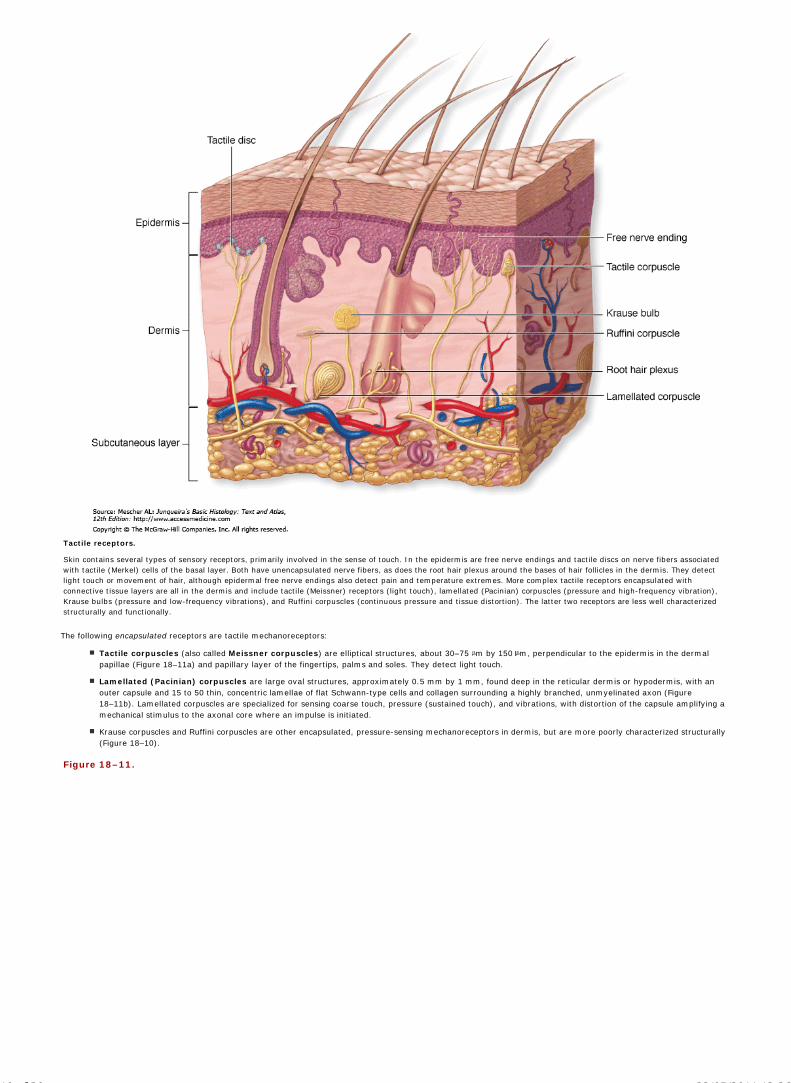
Tactile receptors.
Skin contains several types of sensory receptors, primarily involved in the sense of touch. In the epidermis are free nerve endings and tactile discs on nerve fibers associatedwith tactile (Merkel) cells of the basal layer. Both have unencapsulated nerve fibers, as does the root hair plexus around the bases of hair follicles in the dermis. They detectlight touch or movement of hair, although epidermal free nerve endings also detect pain and temperature extremes. More complex tactile receptors encapsulated withconnective tissue layers are all in the dermis and include tactile (Meissner) receptors (light touch), lamellated (Pacinian) corpuscles (pressure and high-frequency vibration),Krause bulbs (pressure and low-frequency vibrations), and Ruffini corpuscles (continuous pressure and tissue distortion). The latter two receptors are less well characterizedstructurally and functionally.
The following encapsulated receptors are tactile mechanoreceptors:
Tactile corpuscles (also called Meissner corpuscles) are elliptical structures, about 30–75 m by 150 m, perpendicular to the epidermis in the dermalpapillae (Figure 18–11a) and papillary layer of the fingertips, palms and soles. They detect light touch.
Lamellated (Pacinian) corpuscles are large oval structures, approximately 0.5 mm by 1 mm, found deep in the reticular dermis or hypodermis, with anouter capsule and 15 to 50 thin, concentric lamellae of flat Schwann-type cells and collagen surrounding a highly branched, unmyelinated axon (Figure18–11b). Lamellated corpuscles are specialized for sensing coarse touch, pressure (sustained touch), and vibrations, with distortion of the capsule amplifying amechanical stimulus to the axonal core where an impulse is initiated.
Krause corpuscles and Ruffini corpuscles are other encapsulated, pressure-sensing mechanoreceptors in dermis, but are more poorly characterized structurally(Figure 18–10).
Figure 18–11.
AccessMedicine | Print: Chapter 18. Skin http://accessmedicine.com/popup.aspx?aID=6182554&print=yes_chapter
10 of 20 23/05/2011 13:26

Tactile and lamellated corpuscles.
Micrographs showing the two most commonly seen sensory receptors of skin. (a): Tactile (Meissner) corpuscle. X400. H&E. (b): Lamellated (Pacinian) corpuscle. X40. H&E.Tactile corpuscles (TC) are specialized to detect light touch and are frequently located in dermal papillae (DP), very close to the epidermis (E). They are elliptical in shape,approximately 150 m long, with an outer capsule (from the perineurium) and thin, stacked inner layers of modified Schwann cells, around which course several nerve fibers.
Lamellated corpuscles detect coarse touch or pressure and are much larger oval structures, frequently 1 mm in length, found deep in the reticular dermis near thesubcutaneous tissue. Here the outer connective tissue capsule surrounds 15 to 50 thin, concentric layers of modified Schwann cells, each separated by slightly viscousinterstitial fluid. Several axons enter one end of the corpuscle and lie in the cylindrical, inner core of the structure. Movement or pressure of this corpuscle from any directiondisplaces the inner core, leading to a nerve impulse.
The encapsulated, lamellated mechanoreceptors are also found in the connective tissue of organs located deep in the body, including the wall of the rectum and urinary
bladder, where they also produce the sensation of pressure when the surrounding tissue is distorted.
HAIR
Hairs are elongated keratinized structures derived from invaginations of the epidermal epithelium called hair follicles (Figure 18–12). The color, size, shape and
texture of hairs vary according to age, genetic background, and region of the body. All skin has at least minimal hair except that of the palms, soles, lips, glans penis,
clitoris, and labia minora. The face has about 600 hairs/cm2 and the remainder of the body has about 60/cm2. Hairs grow discontinuously, with periods of growth
followed by periods of rest, and this growth does not occur synchronously in all regions of the body or even in the same area. The duration of the growth and rest periods
AccessMedicine | Print: Chapter 18. Skin http://accessmedicine.com/popup.aspx?aID=6182554&print=yes_chapter
11 of 20 23/05/2011 13:26
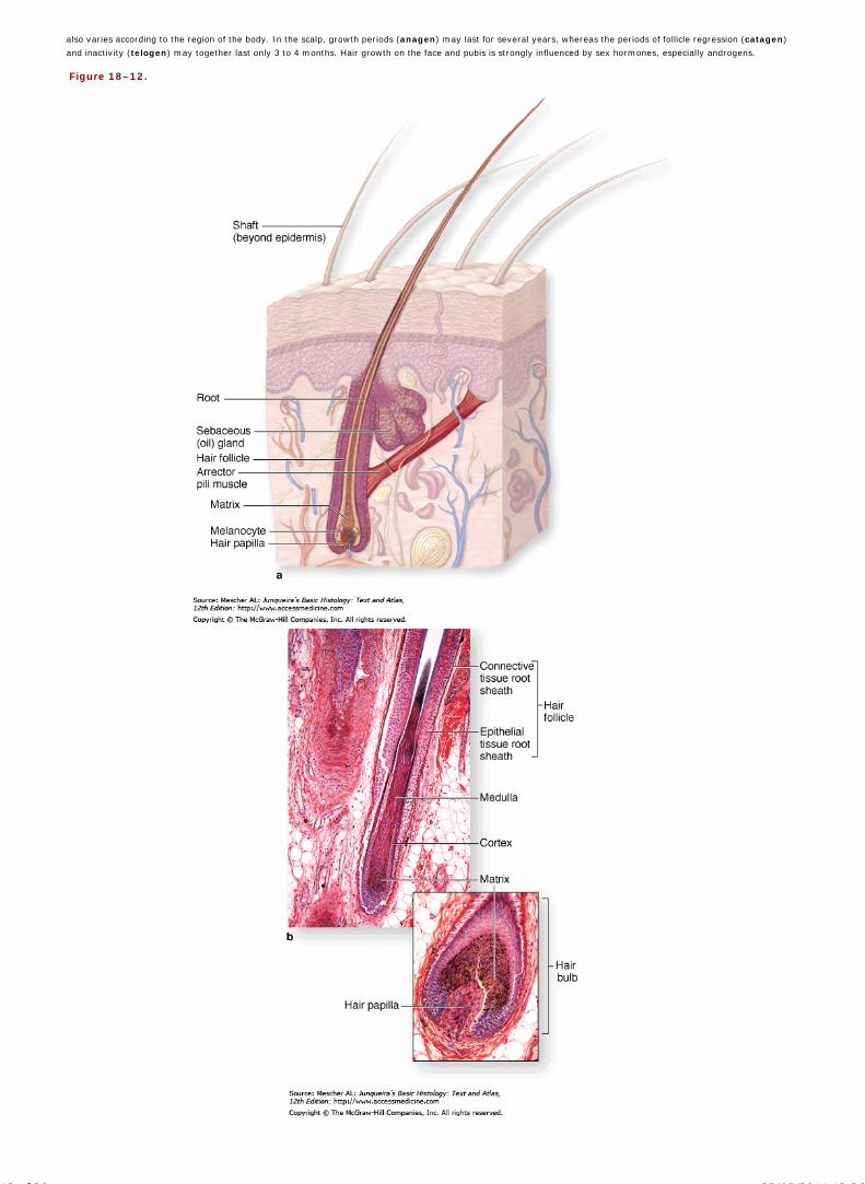
also varies according to the region of the body. In the scalp, growth periods (anagen) may last for several years, whereas the periods of follicle regression (catagen)
and inactivity (telogen) may together last only 3 to 4 months. Hair growth on the face and pubis is strongly influenced by sex hormones, especially androgens.
Figure 18–12.
AccessMedicine | Print: Chapter 18. Skin http://accessmedicine.com/popup.aspx?aID=6182554&print=yes_chapter
12 of 20 23/05/2011 13:26

Hair.
All types of body hair have a similar composition and form in hair follicles derived from the epidermis but extending deep into the dermis. One or more sebaceous glands formfrom the same epidermal down-growth and the entire structure is referred to as a pilosebaceous unit, which contains a specialized niche with stem cells for keratinocytes ofthe unit and neighboring epidermis. (a): Schematic diagram shows major parts of a hair follicle, including the arrector pili muscle which pulls the hair erect and sebaceousglands which empty into the follicle near the epidermis.
(b): Micrograph shows the medulla and cortex in the root of a hair cut longitudinally and the epithelial and connective tissue sheaths surrounding the growing hair. Bothfigures show the dermal hair papilla with microvasculature entering the base of the follicle. This nutritive papilla is surrounded by a matrix of epithelial cells similar to those ofthe stratum germinativum. Cells of the matrix proliferate, take up melanin granules, and undergo keratinization to differentiate as the three concentric layers of the hair. Theoutermost layer of the hair is the thin cuticle, composed of shingle-like cells. X70 and X180. H&E. (c): SEM shows the cuticle on a shaft of hair emerging at the stratumcorneum from its follicle. X260.
During anagen the hair follicle has a terminal dilatation called a hair bulb (Figure 18–12). A dermal papilla inserts into the base of the hair bulb and contains a capillary
network required to sustain the hair follicle. Loss of this blood flow results in death of the follicle. The epidermal cells covering this dermal papilla form the hair root that
produces and is continuous with the hair shaft protruding beyond the skin surface.
The epithelial cells (keratinocytes) that make up the hair bulb are similar to those in the basal and spinous layers of epidermis. They divide constantly and then undergo
keratinization, differentiating into specific cell types. In certain types of thick hairs, the cells of the central region of the root at the apex of the dermal papilla produce
large, vacuolated, and moderately keratinized cells that form the medulla of the hair (Figures 18–12b and 18–13). Other cells differentiate into heavily keratinized,
compactly grouped fusiform cells that form the hair cortex. The most peripheral cells produce the hair cuticle, a thin layer of heavily keratinized, shingle-like cells
covering the cortex (Figures 18–12c and 18–13). Melanocytes in the hair bulb transfer melanin granules into the epithelial cells that will later differentiate to form the
hair.
Figure 18–13.
AccessMedicine | Print: Chapter 18. Skin http://accessmedicine.com/popup.aspx?aID=6182554&print=yes_chapter
13 of 20 23/05/2011 13:26

Layers of a hair and its follicle.
(a): The base of a hair follicle sectioned obliquely shows the vascularized dermal papilla (DP) continuous with the surrounding connective tissue sheath (CTS). The papilla issurrounded by the deepest part of the epithelial sheath, which is continuous with both the internal root sheath (IRS) and external root sheath (ERS). Both of these layers arein turn continuous with the stratified epidermis. Just outside the ERS is the glassy membrane (G) which is continuous with the basement membrane of the epidermis. Theepithelial cells (keratinocytes) around the papilla proliferate and differentiate as the root of the hair itself. Above the papilla only the cortex (CO) of the hair is clearly seen inthis section. X140. H&E. (b): A hair root sectioned more transversely shows the same layers of the follicular sheath, but the layers of the hair root are now seen to include themedulla (M), cortex (CO), and cuticle (CU). X140. H&E. (c): SEM of a similar specimen gives a different perspective on these layers, including the shingle-like nature of thethin cuticle surface, and the small blood vessel (BV) and collagen bundles (CB) in the surrounding dermis. X2600. (Figure 18–13c, with permission, from W.H. Freeman & Co.,Kessel, R.G. and Kardon, R.H., 1979, Tissues and Organs: A Text-Atlas of Scanning Electron Microscopy.)
The outermost cells of the hair bulb are continuous with the epithelial root sheath, in which two layers can be recognized. The internal root sheath completely
surrounds the initial part of the hair shaft but degenerates above the level of the attached sebaceous glands. The external root sheath covers the internal sheath and
extends all the way to the epidermis, where it is continuous with the basal and spinous layers.
Separating the hair follicle from the dermis is an acellular hyaline layer, the thickened basement membrane called the glassy membrane (Figure 18–13). The
surrounding dermis forms a connective tissue sheath. Running from a midpoint on this sheath and to the dermal papillary layer is a small bundle of smooth muscle cells,
the arrector pilimuscle (Figures 18–1 and 18–12). Contraction of these muscles pulls the hair shafts to a more erect position, usually when it is cold in an effort to trap
a layer of warm air near the skin. In regions where hair is fine, contraction of arrector pili muscles is seen to produce tiny bumps on the skin surface ("goose bumps")
where each contracting muscle distorts the attached dermis.
AccessMedicine | Print: Chapter 18. Skin http://accessmedicine.com/popup.aspx?aID=6182554&print=yes_chapter
14 of 20 23/05/2011 13:26

Hair color is produced by the activity of melanocytes located between the papilla and the epithelial cells of the hair root. The melanocytes produce and transfer melanin
granules to these keratinocytes by a mechanism that is generally similar to that described for the epidermis. However, keratinization to produce hair does differ in some
respects. Unlike epidermal keratinization where terminal differentiation of all cells gives rise to the stratum corneum, cells in the hair root differentiate into the cell types
of the hair medulla, cortex, and cuticle which differ somewhat in ultrastructure, histochemical characteristics, and function. Keratin of hair has a harder and more
compact nature that that of stratum corneum, maintaining its structure much longer. Finally, although keratinization in the epidermis occurs continuously and over the
entire surface, it is intermittent in the hair and occurs only in the hair root.
NAILS
A similar process of keratinization produces the nails, which are hard, flexible plates of keratin on the dorsal surface of each distal phalanx (Figure 18–14). The proximal
part of the nail is the nail root and is covered by the proximal skin fold which is thin and lacks both hair and glands. The epidermal stratum corneum extending from the
proximal nail fold forms the cuticle, or eponychium. The keratinized nail plate is bound to a bed of epidermis called the nail bed, which contains only the basal and
spinous layers (Figure 18–14). The nail plate arises from the nail matrix, which extends from the nail root. Cells of the matrix divide, move distally, and become
keratinized, forming the nail root. This matures as the nail plate, which continuous growth in the matrix pushes forward over the nail bed (which makes no contribution to
the plate) at about 3 mm/month for fingernails and 1 mm/month for toenails. The distal end of the plate becomes free of the nail bed at the epidermal fold called the
hyponychium and is worn away or cut off. The nearly transparent nail plate and the thin epithelium of the nail bed provide a useful window on the amount of oxygen in
the blood by showing the color of blood in the dermal vessels.
Figure 18–14.
AccessMedicine | Print: Chapter 18. Skin http://accessmedicine.com/popup.aspx?aID=6182554&print=yes_chapter
15 of 20 23/05/2011 13:26

Nails.
Nails are hard, keratinized derivatives formed in a process similar to that of the stratum corneum and hair. (a): Surface view of a finger shows the nail's major parts, includingthe crescent-shaped white area called the lunula, which derives its color from the opaque nail matrix and immature nail plate below it. (b): Diagram of a sagittal sectionincludes major internal details and shows the hyponychium at which the free end of the nail plate is bound to epidermis.
(c): A micrograph of a sagittal section from a fetal finger shows of the proximal nail fold (PNF) and its epidermal extension, the eponychium (E) or cuticle. The nail root (NR),the most proximal region of the nail plate (NP), is formed like the hair root by a matrix of proliferating, differentiating keratinocytes. These cells make up the dorsal nail matrix(DNM) and ventral nail matrix (VNM), which contribute keratinized cells to the nail root. The mature nail plate remains attached to the nail bed (NB), which consists of basaland spinous epidermal layers over dermis (D), but is pushed forward on this bed by continuous growth in the nail matrix. X100. Mallory trichrome.
GLANDS OF THE SKIN
Sebaceous GlandsSebaceous glands are embedded in the dermis over most of the body surface, except the thick, hairless (glabrous) skin of the palms and soles. There is an average of
about 100 such glands per square centimeter of skin, but the frequency increases to 400–900/cm2 in the face and scalp. Sebaceous glands are branched acinar glands
with several acini converging at a short duct which usually empties into the upper portion of a hair follicle (Figure 18–12). The bulge region of the follicle is a stem cell
niche generating cells of the hair follicle and matrix, the neighboring epidermis, and associated sebaceous glands. In certain hairless regions, such as the genital glands,
eyelids, and nipples, sebaceous ducts open directly onto the epidermal surface. The acini consist of a basal layer of undifferentiated flattened epithelial cells on the basal
lamina. These cells proliferate and are displaced toward the middle of the acinus, undergoing terminal differentiation as distinctly large, lipid-producing sebocytes which
have their cytoplasm filled with small fat droplets (Figure 18–15). Their nuclei shrink and undergo autophagy along with other organelles and near the duct the cells
disintegrate and release the lipids via holocrine secretion. The product of this process is sebum, which is gradually moved to the surface of the skin along the hair follicle
or duct.
Figure 18–15.
AccessMedicine | Print: Chapter 18. Skin http://accessmedicine.com/popup.aspx?aID=6182554&print=yes_chapter
16 of 20 23/05/2011 13:26

Sebaceous glands.
Sebaceous glands secrete a complex mixture of lipids called sebum into short ducts which generally open into hair follicles (a): Micrograph shows small cells near theconnective tissue capsule which proliferate and give rise to an acinus composed of large sebocytes (S), which undergo terminal differentiation by filling with small lipiddroplets and then disintegrating at the ducts (D) near the hair (H) shaft, with the loss of nuclei and other organelles. X122. H&E. (b): Micrograph showing the gland's capsuleand differentiating sebocytes at higher magnification X400. H&E. Sebum production is the classic example of holocrine secretion, in which the entire cell dies and contributesto the secretory product. Steady proliferation of the peripheral cells inside the capsule pushes sebum slowly and continuously into the ducts. Myoepithelial cells are notpresent.
Sebum is a complex mixture of lipids that includes wax esters, squalene, cholesterol and triglycerides which are hydrolyzed by bacterial enzymes after secretion.
Secretion from sebaceous glands greatly increases at puberty, stimulated primarily by testosterone in men and by ovarian and adrenal androgens in women. Specific
functions of sebum appear to include helping maintain the stratum corneum and hair, as well as exerting weak antibacterial and antifungal properties on the skin surface.
MEDICAL APPLICATION
The flow of sebum is continuous, and a disturbance in the normal secretion and flow of sebum is one of the reasons for the development of acne, a chronic
inflammation of obstructed sebaceous glands common during and after puberty.
Sweat GlandsSweat glands are epithelial derivatives embedded in the dermis which open to the skin surface (Figure 18–1) or into hair follicles. Eccrine sweat glands and apocrine
sweat glands have different distributions, functions, and structural details.
AccessMedicine | Print: Chapter 18. Skin http://accessmedicine.com/popup.aspx?aID=6182554&print=yes_chapter
17 of 20 23/05/2011 13:26

Eccrine sweat glands (Figures 18–16 and 18–17) are widely distributed in the skin and are most numerous on the soles of the feet (620/cm2). Collectively the 3 million
eccrine sweat glands of the average person roughly equal the mass of a kidney and can produce as much as 10 L/day, a secretory rate far exceeding that of other
exocrine glands. Sweating is the physiological response to increased body temperature during physical exercise or thermal stress and in humans the most effective
means of temperature regulation.
Figure 18–16.
Eccrine and apocrine sweat glands.
(a): Micrograph of an eccrine sweat gland which plays an important thermoregulatory function in production of fluid which evaporates on the body surface, thereby coolingthat surface. Histologically eccrine glands have small lumens in the secretory portions (S) and the ducts (D), both of which have an irregular stratified cuboidal appearance.(b): Apocrine sweat glands are restricted mainly to the axillae and perineum and produce a more protein-rich secretion with pheromonal properties. The lumens of apocrinegland secretory portion (S) are much larger than those of eccrine glands and their ducts open into hair follicles (H) rather than to the epidermal surface. Both X200. Mallorytrichrome.
Figure 18–17.
AccessMedicine | Print: Chapter 18. Skin http://accessmedicine.com/popup.aspx?aID=6182554&print=yes_chapter
18 of 20 23/05/2011 13:26

Eccrine sweat gland secretory cells.
(a): The secretory portions of eccrine sweat glands have a stratified cuboidal epithelium, containing cell types with different staining properties. Cells closest to the lumencontain eosinophilic granules. X400. Mallory trichrome.
(b): TEM reveals three cell types. Myoepithelial cells (M) are thin cells present at the basal lamina (BL). Irregular pyramidal cells called dark cells (D) border the lumen (L) andare filled with the eosinophilic secretory granules which are electron-dense. Among the products released from these granules are bactericidal peptides and other componentsof innate immunity. Clear cells (C) are columnar and have their basal ends on the basal lamina and function in the rapid transport of water from interstitial fluid in the
capillary-rich dermis directly into the lumen or into intercellular canaliculi (IC) which are continuous with the lumen. Na+ ions are recovered from this fluid through the actionof cells in the ducts, which are seen in the lower left corner of (a). X6500.
Both the secretory portions and ducts of eccrine sweat glands are coiled and have small lumens. The secretory part is generally more pale-staining than the ducts and
has stratified cuboidal epithelium consisting of three cell types (Figure 18–17). Pale pyramidal or columnar clear cells produce the sweat, having abundant mitochondria
and microvilli to provide large surface areas. Interstitial fluid from the capillary-rich dermis around the gland is transported through the clear cells, either directly into the
lumen or into intercellular canaliculi that open to the lumen. As numerous as the clear cells are pyramidal dark cells which line most of the luminal surface and do not
touch the basal lamina (Figure 18–17). Dark cells are mucoid and filled with glycoprotein-containing granules whose functions are not well-understood but include
components of innate immunity with bactericidal activity. Myoepithelial cells on the basal lamina (Figure 4–27) produce contractions that help discharge secretion into
the duct.
The ducts of eccrine sweat glands consist of two layers of more acidophilic epithelial cells filled with mitochondria and having membranes rich in Na+, K+-ATPase. These
duct cells absorb Na+ ions to prevent excessive loss of this electrolyte. After its release on the surface of the skin sweat evaporates, cooling the skin. Besides its
important cooling role, sweat glands also function as an auxiliary excretory organ, eliminating small amounts of nitrogenous waste and excess salts.
Apocrine sweat glands are largely confined to skin of the axillary and perineal regions. Their development (but not functional activity) depends on sex hormones and
is not complete until puberty. The most obvious histological difference between the two kinds of sweat glands is the much larger lumen of apocrine glands (Figure 18–16).
The secretory portions of apocrine sweat glands consist of simple cuboidal, eosinophilic cells with numerous apical secretory granules that undergo exocytosis. Thus the
AccessMedicine | Print: Chapter 18. Skin http://accessmedicine.com/popup.aspx?aID=6182554&print=yes_chapter
19 of 20 23/05/2011 13:26

Copyright © The McGraw-Hill Companies. All rights reserved.Privacy Notice. Any use is subject to the Terms of Use and Notice.
glands are misnamed: their cells show merocrine, not apocrine, secretion. Lumens of apocrine glands often show stored, protein-rich product, which myoepithelial cells
help move into ducts opening into hair follicles. The wall of the ducts is similar to that of the eccrine glands. The slightly viscous secretion is initially odorless but may
acquire a distinctive odor as a result of bacterial activity. The production of pheromones by apocrine glands is well-established in many mammals and likely in humans,
although in a reduced or vestigial capacity. Apocrine sweat glands are innervated by adrenergic nerve endings, whereas eccrine sweat glands receive cholinergic fibers.
AccessMedicine | Print: Chapter 18. Skin http://accessmedicine.com/popup.aspx?aID=6182554&print=yes_chapter
20 of 20 23/05/2011 13:26

Print Close Window
Note: Large images and tables on this page may necessitate printing in landscape mode.
Copyright © The McGraw-Hill Companies. All rights reserved.
Junqueira's Basic Histology: Text & Atlas, 12e > Chapter 19. The Urinary System >
THE URINARY SYSTEM: INTRODUCTION
The urinary system consists of the paired kidneys and ureters, the bladder, and the urethra. This system helps maintain homeostasis by a complex combination of
processes that involves the following:
Filtration of cellular wastes from blood
Selective reabsorption of water and solutes
Excretion of the wastes and excess water as urine.
Urine produced in the kidneys passes through the ureters to the bladder for temporary storage and is then released to the exterior through the urethra. The two
kidneys produce about 125 mL of filtrate per minute, of which 124 mL is reabsorbed in these organs and 1 mL is released into the ureters as urine. About 1500 mL of
urine is formed every 24 hours. The kidneys also regulate the fluid and electrolyte balance of the body and are the site of production of renin, a protease that
participates in the regulation of blood pressure by cleaving circulating angiotensinogen to angiotensin I. Erythropoietin, a glycoprotein that stimulates the production
of erythrocytes, is also produced in the kidneys. The steroid prohormone vitamin D, initially produced in skin keratinocytes, is hydroxylated in kidneys to an active
form (1,25-dihydroxyvitamin D3 or calcitriol) involved in regulating calcium balance.
KIDNEYS
Each kidney has a concave medial border, the hilum—where nerves enter, the ureter exits, and blood and lymph vessels enter and exit—and a convex lateral surface,
both covered by a thin fibrous capsule (Figure 19–1). The expanded upper end of the ureter, called the renal pelvis, divides into two or three major calyces. Smaller
branches, the minor calyces, arise from each major calyx. The area surrounding the calyces, called the renal sinus, usually contains considerable adipose tissue.
Figure 19–1.
Kidney.
Each kidney is bean-shaped, with a concave hilum where the ureter and the renal artery and veins enter. The ureter divides and subdivides into several major and minorcalyces, around which is located the renal sinus containing adipose tissue. Division of the parenchyma into cortex and medulla can also be seen grossly. Attached to eachminor calyx is a renal pyramid, a conical region of medulla delimited by extensions of cortex. A renal pyramid with associated cortex constitutes a renal lobe. The cortex andhilum are covered with a fibrous capsule.
The kidney has an outer cortex and an inner medulla (Figures 19–1 and 19–2). In humans, the renal medulla consists of 8–15 conical structures called renal
pyramids, which are separated by cortical extensions called renal columns. Each medullary pyramid plus the cortical tissue at its base and along its sides constitutes
a renal lobe (Figure 19–1).
Figure 19–2.
AccessMedicine | Print: Chapter 19. The Urinary System http://accessmedicine.com/popup.aspx?aID=6182670&print=yes_chapter
1 of 19 22/05/2011 19:13

Nephrons.
Within each renal lobe are hundreds of thousands of nephrons, the function unit of the kidney. Each nephron originates in the cortex, at the renal corpuscle associated withglomerular capillaries. Extending from the corpuscle is the proximal convoluted tubule, then the nephron loop (of Henle) into the medulla and back to the cortex, then thedistal convoluted tubule and collecting tubule which merges into a collecting duct for urine transport to the calyx. All nephrons are located completely within the cortexexcept for their medullary loops. Juxtamedullary nephrons usually have much longer loops than cortical nephrons.
Each kidney contains 1–1.4 million functional units called nephrons (Figure 19–2). The major divisions of each nephron are:
Renal corpuscle, an initial dilated portion in the cortex
Proximal convoluted tubule, located primarily in the cortex
Thin and thick limbs of the nephron loop (loop of Henle), which descend into the medulla, then ascend back to the cortex
Distal convoluted tubule
Collecting tubule.
Collecting tubules from several nephrons converge into collecting ducts which carry urine to the calyces and the ureter. Cortical nephrons are located almost
completely in the cortex while juxtamedullary nephrons close to the medulla have long loops in the medulla (Figure 19–2).
BLOOD CIRCULATION
As expected for an organ specialized to process the blood, the anatomical organization of the kidney vasculature and its associations with nephron components are very
important. Blood vessels of the kidney are named according to their precise locations or shapes (Figure 19–3).
Figure 19–3.
AccessMedicine | Print: Chapter 19. The Urinary System http://accessmedicine.com/popup.aspx?aID=6182670&print=yes_chapter
2 of 19 22/05/2011 19:13

Blood supply to the kidney.
A coronal view (left) shows the major blood vessels of the kidney. The microvascular components extending into the cortex and medulla from the interlobular vessels areshown on the right. Pink boxes indicate vessels with arterial blood and blue indicate the venous return. The intervening lavender boxes and vessels are intermediate siteswhere most reabsorbed material re-enters the blood.
Each kidney receives blood from a renal artery, which divides into two or more segmental arteries at the hilum. In the renal sinus these branch further to form the
interlobar arteries extending between the renal pyramids toward the corticomedullary junction (Figure 19–3). Here the interlobar arteries branch further to form the
arcuate arteries which travel in an arc along this junction at the base of each renal pyramid. Smaller interlobular arteries branch off at right angles from the
arcuate arteries and enter the cortex.
From the interlobular arteries arise the microvascular afferent arterioles, which supply blood to a tuft of capillaries called the glomerulus, each of which is
associated with a renal corpuscle (Figures 19–3 and 19–4). Blood leaves the glomerular capillaries, not via venules, but via the efferent arterioles, which at once
branch again to form another capillary network, the peritubular capillaries that nourish cells of the proximal and distal tubules and carry away reabsorbed
substances. The efferent arterioles associated with glomeruli near the medulla continue as long, straight vessels directly into the medulla providing nutrients and
oxygen there, and then loop back into the cortex as venules. These small medullary vessels and their intervening capillary plexuses comprise the vasa recta (L. recta,
straight).
Figure 19–4.
AccessMedicine | Print: Chapter 19. The Urinary System http://accessmedicine.com/popup.aspx?aID=6182670&print=yes_chapter
3 of 19 22/05/2011 19:13

Microvasculature of the renal cortex.
(a): Cortical vasculature is revealed in a section of kidney with the renal artery injected with carmine dye before fixation. Having branched at right angles off the arcuatearteries, small interlobular arteries (I) run straight out through the cortex and give off the afferent arterioles (A) which bring blood to the glomerular capillaries. Eachglomerulus (G) contains a loose mass of capillaries with nearly a centimeter in total length. These drain into an efferent arteriole which then branches as a large, diffusenetwork of peritubular capillaries (PT) throughout the cortex. X125. (b): A section of one glomerulus shows many capillaries and the closely associated cells of the renalcorpuscle's internal visceral layer. The thick basement membrane of these glomerular capillaries contains much type IV collagen and is visible around the cut capillaries(arrows). Also shown are the simple squamous external parietal layer of the capsule and the vascular pole where the arterioles enter the corpuscle and the macula densa(arrowhead) is located. X400. PSH.
Blood leaves the kidney in veins that follow the same courses as arteries and have the same names (Figure 19–3). The outermost peritubular capillaries and capillaries
in the kidney capsule converge into small stellate veins which empty into the interlobular veins.
Renal Corpuscles & Blood Filtration
AccessMedicine | Print: Chapter 19. The Urinary System http://accessmedicine.com/popup.aspx?aID=6182670&print=yes_chapter
4 of 19 22/05/2011 19:13

At the beginning of each nephron is a renal corpuscle, about 200 m in diameter and containing a loose knot of capillaries, the glomerulus, surrounded by a double-
walled epithelial capsule called the glomerular (Bowman's) capsule (Figures 19–2 and 19–5). The internal layer (visceral layer) of the capsule closely envelops
the glomerular capillaries . The external parietal layer forms the outer surface of the capsule. Between the two capsular layers is the urinary or capsular space,
which receives the fluid filtered through the capillary wall and the visceral layer. Each renal corpuscle has a vascular pole, where the afferent arteriole enters and the
efferent arteriole leaves, and a urinary or tubular pole, where the proximal convoluted tubule begins (Figure 19–5). After entering the renal corpuscle, the afferent
arteriole usually divides and subdivides into the two to five capillaries of the renal glomerulus.
Figure 19–5.
Renal corpuscles.
(a): The renal corpuscle is a small mass of capillaries called the glomerulus housed within a bulbous glomerular capsule. The internal lining of the capsule is composed ofcomplex epithelial cells called podocytes, which cover each capillary, forming filtration slits between interdigitating processes called pedicels. Blood enters and leaves theglomerulus through the afferent and efferent arterioles respectively. (b): The micrograph shows the major histological features of a renal corpuscle. H&E. X300. (c): Filtrateis produced in the corpuscle when blood plasma is forced under pressure across the filtration membrane of the glomerular capillary wall and through the filtration slitsbetween the pedicels of podocyte processes. (d): The SEM shows the distinctive appearance of podocytes and their processes covering glomerular capillaries. X800
The parietal layer of a glomerular capsule consists of a simple squamous epithelium supported externally by a basal lamina and a thin layer of reticular fibers. At the
tubular pole, this epithelium changes to the simple cuboidal epithelium characteristic of the proximal tubule (Figure 19–5).
During embryonic development, the simple epithelium of the parietal layer remains relatively unchanged, whereas the internal or visceral layer is greatly modified.
The cells of this layer, the podocytes (Figures 19–5d and 19–6), have a cell body from which arise several primary processes. Each primary process gives rise to
numerous secondary (foot) processes or pedicels (L. pedicellus, little foot) that embrace a portion of one glomerular capillary (Figures 19–5d and 19–6). The cell
bodies of podocytes do not contact the basement membrane of the capillary, but each pedicel is in direct contact with this structure (Figure 19–6).
Figure 19–6.
AccessMedicine | Print: Chapter 19. The Urinary System http://accessmedicine.com/popup.aspx?aID=6182670&print=yes_chapter
5 of 19 22/05/2011 19:13

Glomerular filtration barrier.
The glomerular filtration barrier consists of three layered components: the fenestrated capillary endothelium, the glomerular basement membrane, and filtration slitsbetween podocyte processes. The major component of the filter is formed by fusion of the basal laminae of a podocyte and a capillary endothelial cell. (a): TEM showing cellbodies of two podocytes (PC) and the series of pedicels on the glomerular basement membrane separated by the filtration slits (arrows). On the other side of the membraneis the thin lining of a capillary (C) endothelial cell, with fenestrations. Together these openings allow filtration of liquid from plasma into the urinary space (US) of Bowman'scapsule. X10,000. (b): At higher magnification, both the fenestrations (arrowhead) in the capillary endothelium (E) and the filtration slits (arrows) separating the pedicels(P) are better seen on the two sides of the fused basal laminae (BL). The endothelial fenestrations in glomeruli lack diaphragms, but very thin slit diaphragms cross thespace between pedicels and play an important role in filtration. X45,750.
The pedicels interdigitate, defining elongated spaces 30–40 nm wide—the filtration slits (Figure 19–6). Spanning adjacent processes (and thus bridging the filtration
slits) is a thin semipermeable diaphragm of uniform thickness (Figure 19–6). These slit diaphragms are a highly specialized type of intercellular junction in which the
large transmembrane protein nephrin is important both structurally and functionally. Projecting from the cell membrane on each side of the slit, nephrin molecules
interact to form a porous structure within the diaphragm.
Between the highly fenestrated endothelial cells of the capillaries and the covering podocytes is the thick (~0.1 m) glomerular basement membrane (Figure 19–6). This
membrane is the most substantial part of the filtration barrier separating the blood in the capillaries from the capsular space. It is formed by the fusion of capillary- and
podocyte-produced basal laminae and is maintained by the podocytes. Laminin and fibronectin in this fused basement membrane bind integrins of both the podocyte
and endothelial cell membranes. The meshwork of type IV collagen cross-linked in a matrix of negatively charged proteoglycans may help restrict the passage of
cationic molecules. Thus, the glomerular basement membrane (GBM) is a selective macromolecular barrier which acts as a physical filter and as a barrier against
negatively charged molecules.
The initial glomerular filtrate has a chemical composition similar to that of the blood plasma except that it contains very little protein because macromolecules do not
readily cross the glomerular filter. Proteins and other particles greater than 10 nm in diameter or exceeding 70 kDa, the approximate molecular mass of albumin, do
not readily cross the glomerular barrier.
MEDICAL APPLICATION
In diseases such as diabetes mellitus and glomerulonephritis, the glomerular filter is altered and becomes much more permeable to proteins, with the
subsequent release of protein into the urine (proteinuria). Proteinuria is an indicator of many potential kidney disorders.
Glomerular capillaries are uniquely situated between two arterioles—afferent and efferent—the muscle of which allows increased hydrostatic pressure in these vessels,
AccessMedicine | Print: Chapter 19. The Urinary System http://accessmedicine.com/popup.aspx?aID=6182670&print=yes_chapter
6 of 19 22/05/2011 19:13

favoring movement of plasma across the glomerular filter. The glomerular filtration rate (GFR) is constantly regulated by neural and hormonal inputs affecting the
degree of constriction in each of these arterioles. The total glomerular filtration area of an average adult has been estimated at 500 cm2 and the average GFR at 125
mL per minute or 180 liters per day. Because the total amount of circulating plasma averages 3 L, it follows that the kidneys typically filter the entire blood volume 60
times every day.
In addition to capillary endothelial cells and podocytes, renal corpuscles also contain mesangial cells (Gr. mesos, in the midst, + angeion, vessel) (Figure 19–7),
which resemble pericytes in producing components of an enveloping external lamina. Mesangial cells are difficult to distinguish in routine sections form podocytes, but
often stain more darkly. They and their surrounding matrix comprise the mesangium (Figure 19–7), which fills interstices between capillaries that lack podocytes.
Functions of the mesangium are many and varied, and include the following:
Physical support and contraction—the mesangium provides internal structural support to the glomerulus and like pericytes, its cells respond to vasoactivesubstances to help maintain hydrostatic pressure for the optimal rate of filtration.
Phagocytosis—mesangial cells phagocytose protein aggregates that adhere to the glomerular filter, including antibody-antigen complexes abundant in manypathological conditions.
Secretion—the cells synthesize and secrete several cytokines, prostaglandins, and other factors important for immune defense and repair in the glomerulus.
Figure 19–7.
Mesangium.
(a): Diagram showing that mesangial cells in renal corpuscles are located between capillaries and are enveloped by a dense extracellular matrix like that of the basementmembrane around the capillaries. (b): The TEM shows one mesangial cell (MC) and the amorphous mesangial matrix (MM) surrounding it. This matrix appears similar and inmany places continuous with basement membrane (BM). The matrix helps support capillary loops where podocytes are lacking. Mesangial cells extend processes (arrows)around capillaries which may affect their state of contraction. Some mesangial processes appear to pass between endothelial cells (EC) into the capillary lumen (asterisks)where they may help remove or endocytose adherent protein aggregates. The capillary at the left contains an erythrocyte (E) and a leukocyte (L). Podocytes (P) and theirpedicels (PD) open to the urinary space (US) and associate with the capillary surfaces not covered by mesangial cells.
Proximal Convoluted TubuleAt the tubular pole of the renal corpuscle, the squamous epithelium of the capsule's parietal layer is continuous with the cuboidal epithelium of the proximal
convoluted tubule (Figures 19–8 and 19–9). This very tortuous tubule is longer than the distal convoluted tubule and is therefore more frequently seen in sections of
renal cortex. Cells of the proximal tubule reabsorb 60–65% of the water filtered in the renal corpuscle, along with almost all of the nutrients, ions, vitamins, and small
plasma proteins. The water and its solutes are transferred directly across the tubular wall and immediately taken up by the peritubular capillaries.
Figure 19–8.
AccessMedicine | Print: Chapter 19. The Urinary System http://accessmedicine.com/popup.aspx?aID=6182670&print=yes_chapter
7 of 19 22/05/2011 19:13
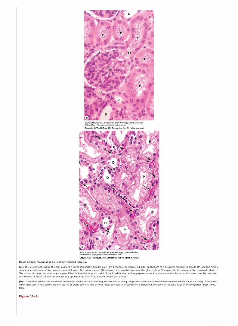
Renal cortex: Proximal and distal convoluted tubules.
(a): The micrograph shows the continuity at a renal corpuscle's tubular pole (TP) between the simple cuboidal epithelium of a proximal convoluted tubule (P) and the simplesquamous epithelium of the capsule's parietal layer. The urinary space (U) between the parietal layer and the glomerulus (G) drains into the lumen of the proximal tubule.The lumen of the proximal tubules appear filled, due to the long microvilli of the brush border and aggregates of small plasma proteins bound to this structure. By contrast,the lumens of distal convoluted tubules (D) appear empty, lacking a brush border and protein.
(b): In another section the abundant peritubular capillaries and draining venules surrounding the proximal and distal convoluted tubules are indicated (arrows). Fibroblasticinterstitial cells of the cortex are the source of erythropoietin, the growth factor secreted in response to a prolonged decrease in the local oxygen concentration. Both X400.H&E.
Figure 19–9.
AccessMedicine | Print: Chapter 19. The Urinary System http://accessmedicine.com/popup.aspx?aID=6182670&print=yes_chapter
8 of 19 22/05/2011 19:13

Convoluted tubules, nephron loops, and collecting ducts.
(a): In this diagram major regions of the nephron are distinguished by different colors. (b): A micrograph of the renal cortex allows comparison of the wide, eosinophilicproximal convoluted tubules with the smaller, less well-stained distal convoluted tubules. X160. H&E. (c): Diagram shows the differences in size and in microvilli betweenthe cuboidal cells of proximal and distal tubules. Cells of both tubules have basal membrane invaginations associated with mitochondria. (d): Micrograph shows the simplesquamous and cuboidal epithelia of the nephron loop thin limbs and thick limbs respectively, as well as the pale columnar cells of the collecting ducts. X160. Mallorytrichrome.
The cells of the proximal tubules have acidophilic cytoplasm (Figures 19–8 and 19–9) because of the presence of numerous mitochondria. The cell apex has abundant
long microvilli which form a prominent brush border for reabsorption (Figures 19–8, 19–9, and 19–10). Because the cells are large, each transverse section of a
proximal tubule typically contains only three to five rounded nuclei. In routine histologic preparations, the brush border may be disorganized and give the lumens a
fuzz-filled appearance. Capillaries and other microvascular components are abundant in the sparse surrounding connective tissue (Figure 19–8).
Figure 19–10.
AccessMedicine | Print: Chapter 19. The Urinary System http://accessmedicine.com/popup.aspx?aID=6182670&print=yes_chapter
9 of 19 22/05/2011 19:13

Ultrastructure of proximal convoluted tubule cells.
TEM reveals important features of the cuboidal cells of the proximal convoluted epithelium: the long, dense apical microvilli (MV), the abundant pinocytotic pits and vesicles(V) in the apical regions near lysosomes (L). Small proteins brought into the cells nonspecifically by pinocytosis are degraded in lysosomes and the amino acids releasedbasally. Apical ends of adjacent cells are sealed with zonula occludens, but the basolateral sides are characterized by long invaginating folds of membrane along which manylong mitochondria (M) are situated. These folds provide a greatly increased surface area for pumping of ions across the membrane. Water and the small molecules releasedfrom the proximal convoluted tubules are taken up immediately by the adjacent peritubular capillaries (C). Between the basement membranes of the tubule and thecapillary shown here is an extension of a fibroblast (F). X10,500.
Ultrastructurally the apical cytoplasm of these cells has numerous pits and vesicles near the bases of the microvilli, indicating active pinocytosis (Figure 19–10). The
pinocytotic vesicles contain small plasma proteins (with a molecular mass less than 70 kDa) that passed through the glomerular filter. The vesicles fuse with
lysosomes for proteolysis and amino acids are released to the circulation. The cells also have many long basal membrane invaginations and lateral interdigitations
with neighboring cells (Figure 19–10). The Na+/K+-ATPase (sodium pump) responsible for actively transporting sodium ions out of the cells is localized in these
basolateral membranes. Long mitochondria are concentrated along the basal invaginations (Figure 19–9), characteristically for cells engaged in active ion transport.
Because of the extensive interdigitations of the lateral membranes, discrete limits between cells of the proximal tubule are difficult to see in the light microscope. The
proximal convoluted tubules actively reabsorb all the glucose and amino acids in the filtrate and about 85% of the sodium chloride and other ions. This absorption
involves the membrane sodium pumps. Water diffuses passively, following the osmotic gradient. When the amount of glucose in the filtrate exceeds the absorbing
capacity of the proximal tubule, as in diabetes, urine becomes more abundant and contains glucose.
In addition to these activities, cells of the proximal convoluted tubules can also move substances from the peritubular capillaries into the tubular lumen, an active
process referred to as tubular secretion. Organic anions such as choline and creatinine and many foreign compounds such as penicillin are excreted in this manner,
which allows kidneys to dispose of such substances at a higher rate than by glomerular filtration alone. The cells of the proximal tubule are also involved in vitamin D
hydroxylation.
Nephron Loop (of Henle)The proximal convoluted tubule continues as a much shorter proximal straight tubule which enters the medulla and becomes the nephron loop. This is a U-shaped
structure with a descending limb and an ascending limb, both composed of simple epithelia, cuboidal near the cortex, but squamous deeper in the medulla (Figure
19–2). In the outer medulla, the straight portion of the proximal tubule, with an outer diameter of about 60 m, narrows abruptly to about 12 m and continues as the
nephron loop's thin descending limb. The lumen of this segment of the nephron is wide and the wall consists of squamous epithelial cells whose nuclei protrude slightly
into the lumen (Figures 19–9 and 19–11).
Figure 19–11.
AccessMedicine | Print: Chapter 19. The Urinary System http://accessmedicine.com/popup.aspx?aID=6182670&print=yes_chapter
10 of 19 22/05/2011 19:13

Renal medulla: Nephron loops and collecting ducts.
(a): A micrograph of a medullary pyramid cut transversely shows closely packed cross sections of the many nephron loops' thin descending limbs (T) and thick ascendinglimbs (A), intermingled with parallel vasa recta capillaries (C). All these structures are embedded in the interstitium (I) which contains sparse myofibroblast-like cells in amatrix very rich in hydrophilic hyaluronate. The specialized nature of the interstitial tissue helps maintain the osmolarity gradient established by differential salt and watertransport across the wall of the nephron loop which is required to concentrate urine and conserve body water. X400. Mallory trichrome. (b): The TEM reveals the slightlyfibrous nature of the interstitium (I) and shows that the simple squamous epithelium of the thin limbs (T) is slightly thicker than that of the nearby vasa recta capillaries(C). X3300. (Figure 19–11b, with permission, from Johannes Rhodin, Department of Cell Biology and Anatomy, University of South Florida College of Medicine.)
Approximately one seventh of all nephrons are located near the corticomedullary junction and are therefore called juxtamedullary nephrons, which are of prime
importance in the mechanism that allows the kidneys to produce concentrated, hypertonic urine. Juxtamedullary nephrons usually have long loops, extending deep into
the medulla, with short thick proximal straight segments, long thin descending and ascending limbs, and long thick ascending limbs (Figure 19–2).
The nephron loop and surrounding tissue are involved in making urine hypertonic and conserving water; only animals with such loops are capable of concentrating urine
and thus conserving body water. Cuboidal cells of the loops' thick ascending limbs actively transport sodium chloride out of the tubule against a concentration gradient
into the hyaluronate-rich interstitial connective tissue, making that compartment hyperosmotic. Squamous cells of the loops' thin descending limbs are freely
permeable to water but not salts, while the thin ascending limbs are permeable to NaCl but impermeable to water. Flow of the filtrate in opposite directions
(countercurrent flow) in the two parallel limbs of nephron loops establishes a gradient of osmolarity in the interstitium of the medullary pyramids and countercurrent
AccessMedicine | Print: Chapter 19. The Urinary System http://accessmedicine.com/popup.aspx?aID=6182670&print=yes_chapter
11 of 19 22/05/2011 19:13

blood flow in the loops of the vasa recta help maintain this gradient. The interstitial osmolarity at the pyramid tips is about four times that of the blood. The high
interstitial osmolarity draws water passively from the collecting ducts in the medullary pyramids (Figures 19–2 and 19–11), concentrating the urine. Water permeability
of these ducts is increased by antidiuretic hormone (ADH), which is released from the pituitary when body water is low. The water thus saved immediately enters the
blood in the adjacent capillaries of the vasa recta. The role of the nephron loop and vasa recta in establishing the conditions for urine concentration is called the
countercurrent multiplier effect.
Distal Convoluted Tubule& Juxtaglomerular ApparatusThe thick ascending limb of the nephron loop is straight as it enters the cortex, and then becomes tortuous as the distal convoluted tubule (Figure 19–2). The simple
cuboidal cells of these tubules differ from those of the proximal convoluted tubules in being smaller and having no brush border (Figure 19–9). Because distal tubule
cells are flatter and smaller than those of the proximal tubule, more nuclei are typically seen in sections of distal tubules than in those of proximal tubules (Figure
19–8). Cells of the distal convoluted tubule do have basal membrane invaginations and associated mitochondria similar to those of proximal tubules, indicating their
similar ion-transporting function (Figure 19–9). The rate of Na+ absorption and K+ secretion by the sodium pumps is regulated by aldosterone from the adrenal glands
and is important for the body's water-salt balance. The distal tubule also secretes H+ and NH4+ into tubular urine, an activity essential for maintenance of the acid-base
balance in the blood.
The initial, straight part of the distal tubule makes contact with the vascular pole of the renal corpuscle of its parent nephron and forms part of a specialized structure,
the juxtaglomerular apparatus (JGA) (Figures 19–9, 19–5, and 19-12). Cells of this structure establish a feedback mechanism that allows autoregulation of renal
blood flow and keeps the rate of glomerular filtration relatively constant. At the point of contact with the arterioles, the cells of the distal tubule become columnar and
more closely packed, with apical nuclei, basal Golgi complexes, and a more elaborate and varied system of ion channels and transporters. This thickened spot of the
distal tubule wall is called the macula densa (Figures 19–5 and 19–12). Adjacent to the macula densa, the tunica media of the afferent arteriole is also modified. The
smooth muscle cells develop a secretory phenotype with more rounded nuclei, rough ER, Golgi complexes and zymogen granules and are called juxtaglomerular
granular (JG) cells (Figures 19–5 and 19–12). Also at the vascular pole are lacis cells (Fr. lacis, meshwork), which are extraglomerular mesangial cells that probably
have many of the same supportive functions as these cells inside the glomerulus. Lacis cells may also transmit signals from the macula densa into the glomerulus,
affecting vasoconstriction there.
Figure 19–12.
AccessMedicine | Print: Chapter 19. The Urinary System http://accessmedicine.com/popup.aspx?aID=6182670&print=yes_chapter
12 of 19 22/05/2011 19:13

Juxtaglomerular apparatus or JGA.
(a): Micrograph shows that the juxtaglomerular apparatus forms at the point of contact between a nephron's distal tubule (D) and the vascular pole of its glomerulus (G). Atthat point cells of the distal tubule become columnar as a thickened region called the macula densa (MD). Smooth muscle cells of the afferent arteriole's (AA) tunica mediaare converted from a contractile to a secretory morphology as juxtaglomerular granule cells (JG). Also present are lacis cells (L), which are extraglomerular mesangial cellsadjacent to the macula densa, the afferent arteriole, and the efferent arteriole (EA). In this specimen the lumens of proximal tubules (P) appear filled and the urinary space(US) is somewhat swollen. X400. Mallory trichrome.
(b): A plastic section through an afferent arteriole of a JGA shows the JG cells (arrowheads) with secretory granules containing renin. Activities of the macula densa andrenin released by JG cells together produce an incompletely understood autoregulatory tubuloglomerular feedback loop that not only helps control arterial blood pressure,but also helps maintain a relatively constant rate of glomerular filtration despite changes in blood pressure. X500.
Basic functions of the JGA in the autoregulation of the glomerular filtration rate (GFR) and in controlling blood pressure are thought to include the following. Elevated
arterial pressure increases glomerular-capillary pressure, which increases the GFR. Higher GFR leads to higher luminal concentrations of Na+ and Cl– in nephrons which
are monitored by cells of the macula densa. Increased ion levels cause these cells to release ATP, adenosine, and other vasoactive compounds that trigger contraction
of the afferent arteriole, which lowers glomerular pressure and decreases the GFR. This lowers tubular ion concentrations, which turns off the release of
vasoconstrictors from the macula densa.
Decreased arterial pressure leads to increased autonomic stimulation to the JGA as a result of baroreceptor function, including local baroreceptors in the afferent
arteriole, possibly the JG cells themselves. This causes the JG cells to release their major secretory product renin, an aspartyl protease, into the blood. There renin
cleaves the plasma protein angiotensinogen into the inactive decapeptide angiotensin I. Angiotensin converting enzyme (ACE) on lung capillaries clips this further
to angiotensin II, a potent vasoconstrictor which directly raises systemic blood pressure and stimulates the adrenals to secrete aldosterone. Aldosterone promotes
Na+ and water reabsorption in the distal convoluted tubules, which raises blood volume to help increase blood pressure. The return of normal blood pressure turns off
secretion of renin by JG cells.
MEDICAL APPLICATION
A significant hemorrhage decreases blood volume, which promotes decreased blood pressure and increased renin secretion. Angiotensin II and aldosterone act
in concert to increase blood pressure and to help restore blood volume. Other factors (eg, sodium depletion, dehydration) that lower blood pressure by
decreasing blood volume also activate the renin–angiotensin II–aldosterone mechanism that contributes to the maintenance of normal blood pressure.
Collecting Tubules & DuctsUrine passes from the distal convoluted tubules to collecting tubules, the last part of each nephron, which join each other to form larger, straight collecting ducts
that run to the tips of the medullary pyramids and empty into the minor calyces (Figure 19–2). The collecting tubules are lined with cuboidal epithelium and have a
diameter of approximately 40 m. Cells of the converging collecting ducts are more columnar and the duct diameters reach 200 m near the tips of the medullary
pyramids (Figures 19–13 and 19–14).
Figure 19–13.
AccessMedicine | Print: Chapter 19. The Urinary System http://accessmedicine.com/popup.aspx?aID=6182670&print=yes_chapter
13 of 19 22/05/2011 19:13

Collecting tubules and ducts.
(a): Longitudinally sectioned renal pyramid showing two collecting ducts (CD) and their distinct lateral cell boundaries (arrows), with interstitial connective tissue. X400.H&E. (b): Similar features are seen in transversely sectioned collecting ducts, with vasa recta (VR) present in the interstitium. X600. PT. Weakly stained duct principal cellsare initially cuboidal and become increasingly columnar along the ducts. Collecting ducts adjust the ionic composition of urine in their lumens and allow increased waterreabsorption from this urine when fluid levels in the body are low. This occurs under the influence of the posterior pituitary hormone ADH, which causes a greatly increasednumber of aquaporin water channels to be temporarily inserted into the apical cell membranes of these cells.
Figure 19–14.
AccessMedicine | Print: Chapter 19. The Urinary System http://accessmedicine.com/popup.aspx?aID=6182670&print=yes_chapter
14 of 19 22/05/2011 19:13

Renal papilla, collecting ducts, and minor calyx.
A sagittal section of a renal papilla shows numerous collecting ducts (sometimes called ducts of Bellini at this level of the renal pyramid) converging at the end of the renalpapilla (RP) where they empty into the minor calyx (MC). The mucosa of the calyx contains dense connective tissue stained blue here and adipose tissue (A). The ducts areembedded in interstitial tissue that also contains thin limbs of the nephron loops. X50. Mallory trichrome. Inset: An enlarged area shows the columnar epithelium of thecollecting ducts (CD), the interstitium (I) and thin limbs (T), and the protective urothelium (U) that lines the minor calyx. X200.
Along their entire extent, collecting tubules and ducts are composed mainly of weakly staining principal cells with few organelles and scanty microvilli (Figure 19–13).
The intercellular limits of the cells are clearly visible in the light microscope. Ultrastructurally the principal cells can be seen to have basal membrane infoldings,
consistent with their role in ion transport. Scattered among the principal cells are variably darker intercalated cells with more abundant mitochondria which help
regulate the acid-base balance by secreting H+ and absorbing HCO3–.
In the medulla, collecting ducts are a major component of the urine-concentrating mechanism. Cells of collecting ducts are particularly rich in aquaporins, integral
proteins found in most cell membranes that function as selective pores for passage of water molecules. Here aquaporins are sequestered in membranous cytoplasmic
vesicles. As mentioned earlier, the pituitary hormone ADH (also known as arginine vasopressin) makes collecting ducts more permeable to water, increasing the rate
at which water molecules are pulled osmotically from their lumens and transferred to the vasa recta, and thus retained in the body. This effect is produced when
activated ADH receptors on the basolateral cell membrane stimulate the movement of vesicles with specific aquaporins and their insertion into either the apical or
basolateral membranes, increasing the number of membrane channels for water movement through the cells.
URETERS, BLADDER, & URETHRA
Urine is transported by the ureters to the bladder where it is stored until emptied during micturition via the urethra. The calyces, renal pelvis, ureter, and bladder
have the same basic histologic structure, with the walls becoming gradually thicker closer to the bladder. The mucosa of these organs is lined by unique stratified
transitional epithelium or urothelium (Figures 19–14). This is surrounded by a folded lamina propria and submucosa, followed by a dense sheath of interwoven
smooth muscle layers and adventitia (Figure 19–15). Urine moves from the renal pelvises to the bladder by peristaltic contractions.
Figure 19–15.
AccessMedicine | Print: Chapter 19. The Urinary System http://accessmedicine.com/popup.aspx?aID=6182670&print=yes_chapter
15 of 19 22/05/2011 19:13

Ureters.
Each ureter carries urine from the renal pelvis of the kidney to the urinary bladder for storage prior to voiding via the urethra. (a): Diagram of a ureter in cross section showsa characteristic pattern of longitudinally folded mucosa, surrounded by a thick muscularis that moves urine by regular waves of peristalsis. The lamina propria is lined by aunique stratified epithelium called transitional epithelium or urothelium resistant to the potentially deleterious effects of contact with hypertonic urine. (b:) Histologically themuscularis is much thicker than the mucosa and an adventitia is also present. X18. H&E.
The urothelium is composed of the following three layers:
a single layer of small basal cells resting on a very thin basement membrane,
an intermediate region containing from one to several layers of more columnar cells,
a superficial layer of very large, polyhedral or bulbous cells called umbrella cells which are occasionally bi- or multinucleated and are highly differentiatedto protect underlying cells against the cytotoxic effects of hypertonic urine.
Umbrella cells are especially well developed in the bladder (Figures 19–16 and 19–17) where contact with urine is the greatest. These cells, up to 100 m in diameter,
have extensive intercellular junctional complexes surrounding unique apical membranes. Most of the apical surface consists of asymmetric unit membranes, in
which regions of the outer lipid layer appear twice as thick as the inner leaflet. These regions are lipid rafts containing mostly integral membrane proteins called
uroplakins which assemble into paracrystalline arrays of stiffened plaques 16 nm in diameter. Urine contacts primarily these membranous plaques, which are
impermeable and protect cytoplasm and underlying cells from its hyperosmotic effects. Plaques are hinged together by more narrow regions of typical membrane.
When the bladder is emptied, not only does the mucosa fold extensively, but individual umbrella cells decrease their apical surface area by folding the membrane at its
hinge domains and internalizing the folded plaques in discoidal vesicles. As the bladder fills again the discoidal vesicles rejoin the apical membrane, increasing its
surface area as the cell shape changes from round to flat. The urothelium becomes thinner, apparently the result of the intermediate cells being pushed and pulled
laterally to accommodate the increased volume of urine.
Figure 19–16.
AccessMedicine | Print: Chapter 19. The Urinary System http://accessmedicine.com/popup.aspx?aID=6182670&print=yes_chapter
16 of 19 22/05/2011 19:13

Urinary bladder.
The bladder is a muscular sac that is expandable as it fills with urine. (a): The micrograph shows the folded mucosa, submucosa, and muscularis of the empty bladder wall,with an enlargement showing the stratified transitional epithelium (urothelium) and lamina propria. X18 and X80. H&E. (b): The diagram depicts a sagittal view showingthat the bladder expands primarily upward and becomes more oval in shape as it fills with urine. The bladder in an average adult can hold 400–600 mL of urine, with theurge to empty appearing at a volume of 150–200 mL.
Figure 19–17.
AccessMedicine | Print: Chapter 19. The Urinary System http://accessmedicine.com/popup.aspx?aID=6182670&print=yes_chapter
17 of 19 22/05/2011 19:13

Bladder wall and urothelium.
(a): In the neck of the bladder, near the urethra, the wall shows four layers: the mucosa with urothelium (U) and lamina propria (LP); the thin submucosa (S); inner,middle, and outer layers of smooth muscle (IL, ML, and OL), and the adventitia (A). X15. H&E. (b): When the bladder is empty, the mucosa is highly folded and theurothelium has bulbous umbrella cells. X250. PSH. (c): when the bladder is full, the mucosa is pulled smooth, the urothelium is thinner, and the umbrella cells are flatter.X250. H&E.
The bladder's lamina propria and dense irregular connective tissue of the submucosa are highly vascularized. The muscularis consists of three poorly delineated layers,
collectively called the detrusor muscle, which contract to empty the bladder (Figure 19–16). Three muscular layers are seen most distinctly at the neck of the bladder
near the urethra (Figure 19–17). The ureters pass through the wall of the bladder obliquely, forming a valve that prevents the backflow of urine into the ureters. All the
urinary passages are covered externally by an adventitial layer, except for the upper part of the bladder which is covered by serous peritoneum.
The urethra is a tube that carries the urine from the bladder to the exterior. The urethral mucosa has large longitudinal folds, giving it a distinctive appearance in cross
section. In men, the two ducts for sperm transport during ejaculation join the urethra at the prostate gland (Chapter 21). The male urethra is longer and consists of
three segments:
The prostatic urethra, 3–4 cm long, extends through the prostate gland and is lined by urothelium
The membranous urethra, a short segment, passes through an external sphincter of striated muscle and is lined by stratified columnar andpseudostratified epithelium
The spongy urethra, 15 cm in length, is enclosed within erectile tissue of the penis (Chapter 21) and is lined by stratified columnar and pseudostratifiedcolumnar epithelium (Figure 19–18), with stratified squamous distally.
Figure 19–18.
AccessMedicine | Print: Chapter 19. The Urinary System http://accessmedicine.com/popup.aspx?aID=6182670&print=yes_chapter
18 of 19 22/05/2011 19:13

Copyright © The McGraw-Hill Companies. All rights reserved.Privacy Notice. Any use is subject to the Terms of Use and Notice.
Urethra.
The urethra is a fibromuscular tube that carries urine from the bladder to the exterior of the body. (a): A transverse section shows that the mucosa has large longitudinalfolds around the lumen (L). X50. H&E. (b): A higher magnification of the urethral epithelium is shown in this micrograph. The thick epithelial lining is stratified columnar insome areas and pseudostratified columnar elsewhere, but becomes stratified squamous at the distal end of the urethra. X250. H&E.
In women, the urethra is exclusively a urinary organ. The female urethra is a tube 4 to 5 cm long, lined initially with transitional epithelium, then by stratified
squamous epithelium and some areas of pseudostratified columnar epithelium. The middle part of the female urethra is surrounded by the external striated muscle
sphincter.
AccessMedicine | Print: Chapter 19. The Urinary System http://accessmedicine.com/popup.aspx?aID=6182670&print=yes_chapter
19 of 19 22/05/2011 19:13

Print Close Window
Note: Large images and tables on this page may necessitate printing in landscape mode.
Copyright © The McGraw-Hill Companies. All rights reserved.
Junqueira's Basic Histology: Text & Atlas, 12e > Chapter 20. Endocrine Glands >
ENDOCRINE GLANDS: INTRODUCTION
Secretory cells of endocrine glands release their products, signaling molecules called hormones, into a neighboring vascularized compartment for uptake by capillaries
and distribution throughout the body, rather than directly into an epithelial duct like the cells of exocrine glands. Endocrine cells typically aggregate as cords, or as follicles
in the case of the thyroid gland. Besides the specialized endocrine glands discussed in this chapter, many other organs specialized for other functions, such as the heart,
thymus, gut, kidneys, testis, and ovaries contain various endocrine cells (Figure 20–1).
Figure 20–1.
Endocrine system.
The endocrine glands and major hormones they secrete are listed with their locations. In parentheses are shown other organs, including the heart, kidney, thymus, gut, andgonads, that contain endocrine cells and have important endocrine functions. In addition many widely distributed tissues and cells throughout the body have endocrinefunctions but are not shown here. These include adipose cells which secrete the hormone leptin and vascular endothelial cells which produce polypeptides called endothelinswhich promote vasoconstriction.
Distribution by the circulation allows hormones to act on target cells with receptor proteins for those hormones at a distance from the site of their secretion. As discussed
briefly in Chapter 2, other endocrine cells produce hormones that act very quickly at only a short distance. This may involve paracrine secretion, with localized dispersal
in interstitial fluid or through short loops of blood vessels, as when gastrin made by pyloric G cells reaches target cells in the fundic glands, or juxtacrine secretion, in
which a signaling molecule remains on the secreting cell's surface or adjacent extracellular matrix and effects target cells when the cells make contact. Juxtacrine
signaling is particularly important in developmental tissue interactions. In autocrine secretion, cells may produce molecules that act on themselves or on cells of the
same type. Insulin-like growth factor (IGF) produced by several cell types may act on the same cells that produced it. Frequently, endocrine glands are also target organs
for other hormones. This helps allow the body to control hormone secretion through a feedback mechanism and to keep blood hormonal levels within strict limits.
AccessMedicine | Print: Chapter 20. Endocrine Glands http://accessmedicine.com/popup.aspx?aID=6182774&print=yes_chapter
1 of 27 22/05/2011 19:42

Hormones, like neurotransmitters, are frequently hydrophilic molecules such proteins, glycoproteins, peptides, or modified amino acids. Their receptor proteins are on the
surface of target cells. Alternatively, hydrophobic steroid and thyroid hormones must circulate on transport proteins but can diffuse through the membranes of cells and
activate cytoplasmic protein receptors in target cells (Chapter 2).
PITUITARY GLAND (HYPOPHYSIS)
The pituitary gland, or hypophysis (Gr. hypo, under, + physis, growth), weighs about 0.5 g in adults and has dimensions of about 10 x 13 x 6 mm. It lies below the
brain in a cavity of the sphenoid bone—the sella turcica (Figure 20–2). The pituitary develops in the embryo partly from oral ectoderm and partly from the developing
brain (Figure 20–3). The neural component arises as a bud growing down from the floor of the diencephalon and caudally as a stalk or infundibulum still attached to the
brain. The oral component arises as an outpocketing of ectoderm from the roof of the primitive mouth and grows cranially, forming a structure called the hypophyseal
(Rathke's) pouch. Later, the base of this pouch constricts and separates it from the pharynx. The anterior wall then thickens greatly, reducing the pouch lumen to a
small fissure (Figure 20–3).
Figure 20–2.
Pituitary gland.
The pituitary gland is composed of an anterior part and a posterior part, which is directly attached to the hypothalamus region of the brain by a stalk called the infundibulum.The gland occupies a fossa of the sphenoid bone called the sella turcica (L. Turkish saddle), which is also used as a radiological landmark.
Figure 20–3.
AccessMedicine | Print: Chapter 20. Endocrine Glands http://accessmedicine.com/popup.aspx?aID=6182774&print=yes_chapter
2 of 27 22/05/2011 19:42

Formation of the pituitary gland.
The pituitary gland forms from two separate embryonic structures. (a): During the third week of development, a hypophyseal pouch (or Rathke's pouch, the future anteriorpituitary) grows from the roof of the pharynx, while a neurohypophyseal bud (future posterior pituitary) forms from the diencephalon. (b): By late in the second month, thehypophyseal pouch detaches from the roof of the pharynx and merges with the neurohypophyseal bud. (c): During the fetal period, the anterior and posterior parts of thepituitary complete development.
Because of its dual origin, the pituitary actually consists of two glands—the posterior neurohypophysis and the anterior adenohypophysis—united anatomically but
with different functions. The neurohypophysis retains features of CNS tissue from which it developed and consists of a large part, the pars nervosa, and the smaller
infundibulum with its stalk attached to the hypothalamus at the median eminence (Figures 20–2 and 20–4). The adenohypophysis, derived from the oral ectoderm,
has three parts: a large pars distalis (or the anterior lobe); the pars tuberalis, which wraps around the infundibulum; and the thin pars intermedia (Figures 20–2
and 20–4).
Figure 20–4.
Pituitary gland.
Histologically the two parts of the pituitary gland reflect their origins, as seen in this low magnification section of an entire gland. The infundibular stalk (IS) and pars nervosa(PN) of the neurohypophysis resemble CNS tissue, while the adenohypophysis' pars distalis (PD), pars intermediate (PI), and pars tuberalis (PT) are typically glandular in theirlevel of staining. X15. H&E.
Blood Supply & the Hypothalamo-Hypophyseal Portal SystemStudy of the pituitary gland's blood supply is important to understand its function. The blood supply derives from two groups of vessels coming off the internal carotid
artery. The superior hypophyseal arteries supply the median eminence and the infundibular stalk; the inferior hypophyseal arteries provide blood mainly for the
neurohypophysis, with a small supply to the stalk. The superior hypophyseal arteries form a primary capillary network irrigating the stalk and median eminence. The
capillaries then rejoin to form venules that branch again as a larger secondary capillary network in the adenohypophysis (Figure 20–5). Capillaries of both networks
are fenestrated and this hypothalamo-hypophyseal portal system is of utmost importance because it carries neuropeptides from the median eminence a short
distance to the adenohypophysis where they either stimulate or inhibit hormone release by the endocrine cells there.
Figure 20–5.
AccessMedicine | Print: Chapter 20. Endocrine Glands http://accessmedicine.com/popup.aspx?aID=6182774&print=yes_chapter
3 of 27 22/05/2011 19:42

The hypothalamo-hypophyseal portal system and hormone release in the pituitary.
The hypothalamo-hypophyseal portal system, with blood from the superior and inferior hypophyseal arteries, consists of two successive capillary networks: one in the parsnervosa around the infundibulum and median eminence, and the second throughout the pars distalis which drains into the collecting hypophyseal veins. Also shown in thediagram are (yellow) neurons extending axons to the median eminence, where they secrete peptides that are then carried in the capillaries to the pars distalis to regulatehormone release from cells there, and (green) neurons from the supraoptic and paraventricular nuclei in the hypothalamus extending axons to the pars nervosa, where theysecrete peptides that are picked up by capillaries and carried to distal target cells.
Embryologically, anatomically, and functionally, the pituitary gland is connected to the hypothalamus at the base of the brain. In the hypothalamo-hypophyseal system
there are three groups of hormones released at three sites (Figure 20–5):
Peptide hormones synthesized by neurons in specific aggregates (nuclei) in the hypothalamus, the supraoptic and the paraventricular nuclei, undergo axonaltransport and accumulate distally in these axons, which are situated in the neurohypophysis' pars nervosa (Figure 20–5).
1.
Another group of peptides is produced by neurons in other hypothalamic nuclei and carried in axons for temporary axonal storage and secretion in the medianeminence. There these peptides enter the capillaries of the primary plexus and are transported to the adenohypophysis where they diffuse among endocrine cellsand control hormone release from their target cells (Figure 20–5).
2.
The third group of hormones consists of proteins and glycoproteins released from the endocrine cells of the adenohypophysis' pars distalis (under the control of theneuropeptides just mentioned) and picked up by capillaries of the second portal system plexus, from which they enter the general circulation (Figure 20–5).
3.
Adenohypophysis (Anterior Pituitary)The three parts of the adenohypophysis are derived from the hypophyseal pouch off the embryonic pharynx.
PARS DISTALIS
The pars distalis accounts for 75% of the adenohypophysis and is covered by a thin fibrous capsule. The main components are cords of epithelial cells interspersed with
fenestrated capillaries (Figures 20–4 and 20–6). Fibroblasts are present and produce reticular fibers supporting the cords of hormone-secreting cells. Common stains
suggest two broad groups of cells in the pars distalis based on staining affinity: chromophils and chromophobes. Chromophils are secretory cells in which hormone is
stored in cytoplasmic granules. They are also called basophils and acidophils according to their affinity for basic and acidic dyes, respectively (Figures 20–6 and 20–7).
Subtypes of basophilic and acidophilic cells are identified by TEM or more easily by immunohistochemistry and are named for their specific hormones or target cells (Table
20–1). Acidophils include the somatotropic and mammotropic cells, while the basophilic cells are the gonadotropic, corticotropic, and thyrotropic cells.
Somatotropic cells typically constitute about half the cells of the pars distalis in humans, with thyrotropic cells the least abundant. Chromophobes stain weakly, with few
or no secretory granules, and also represent a heterogeneous group, including stem and undifferentiated progenitor cells as well as any degranulated cells present. Each
granular cell makes one kind of hormone, except gonadotropic cells which produce two proteins and corticotropic cells, in which the major gene product,
proopiomelanocortin (POMC), is cleaved posttranslationally into the smaller polypeptide hormones adrenocortical trophic hormone (ACTH) and -lipotropin ( -LPH).
Hormones produced by the pars distalis have widespread functional activities; they regulate almost all other endocrine glands, milk secretion, melanocyte activity, and
the metabolism of muscle, bone, and adipose tissue (Figure 20–8 and Table 20–1).
Figure 20–6.
AccessMedicine | Print: Chapter 20. Endocrine Glands http://accessmedicine.com/popup.aspx?aID=6182774&print=yes_chapter
4 of 27 22/05/2011 19:42

Pars distalis: Acidophils, basophils, and chromophobes.
(a,b): Most general staining methods simply allow the parenchymal cells of the pars distalis to be subdivided into acidophil cells (A), basophils (B), and chromophobes (C) inwhich the cytoplasm is poorly stained. X400. H&E. (c): Gomori trichrome provides similar information. X400. Cords of acidophils and basophils vary in distribution and numberin different regions of the pars distalis, but are always closely associated with capillaries and sinusoids (S) in the second capillary plexus of the portal system. The vascularplexus carries off secreted hormones into the general circulation. Specific acidophil or basophil cells can be identified immunohistologically with antibodies against theirhormone products. Chromophobes are less numerous and represent various undifferentiated parenchymal cells. Their number and density also vary in different regions.
AccessMedicine | Print: Chapter 20. Endocrine Glands http://accessmedicine.com/popup.aspx?aID=6182774&print=yes_chapter
5 of 27 22/05/2011 19:42

Figure 20–7.
Ultrastructure & immunohistochemistry of somatotropic cells.
(a): Ultrastructurally, cytoplasm of all chromophil cells is shown to have well-developed Golgi complexes (G), euchromatic nuclei (N), and cytoplasm filled with secretorygranules, as shown here for a somatotropic cell, the most common acidophil. The arrow indicates the cell membrane. Granules of somatotropic cells can be distinguished fromthose of other chromophils by TEM, but all of the various chromophils are more easily identified using immunohistochemistry and antibodies against the hormone products.X10,000. (b): The micrograph shows somatotropic cells (S) stained using an antibody against somatotropin. X400. Hematoxylin counterstain.
Table 20–1. Secretory cells of the pars distalis.
Cell Type StainAffinity
% ofTotalCells
Hormone Produced Main Physiologic Activity
Somatotropic cell Acidophilic 50 Somatotropin (growth hormone,GH)
Acts on growth of long bones via insulin-like growth factors synthesized in liver
Mammotropic cell(or actotropic cell)
Acidophilic 15–20 Prolactin (PRL) Promotes milk secretion
Gonadotropic cell Basophilic 10 Follicle-stimulating hormone (FSH)luteinizing hormone (LH) in thesame cell type
FSH promotes ovarian follicle and development and estrogen secretion in women andspermatogenesis in men. LH promotes ovarian follicle maturation and progesteronesecretion in women and interstitial cell androgen secretion in men.
Thyrotropic cell Basophilic 5 Thyrotropin (TSH) Stimulates thyroid hormone synthesis, storage, and liberation
AccessMedicine | Print: Chapter 20. Endocrine Glands http://accessmedicine.com/popup.aspx?aID=6182774&print=yes_chapter
6 of 27 22/05/2011 19:42

Cell Type StainAffinity
% ofTotalCells
Hormone Produced Main Physiologic Activity
Corticotropic cell Basophilic 15–20 Adrenal corticotropin (ACTH)
Lipotrophins
Stimulates secretion of adrenal cortex hormones
Lipid metabolism regulation
Figure 20–8.
Hormones of the pars distalis and their targets.
The diagram summarizes the major hormones of the anterior pituitary and indicates their most important targets.
PARS TUBERALIS
The pars tuberalis is a funnel-shaped region surrounding the infundibulum of the neurohypophysis (Figure 20–2). Most of the cells of the pars tuberalis are basophilic
gonadotropic cells that secrete follicle-stimulating hormone (FSH) and luteinizing hormone (LH).
PARS INTERMEDIA
The pars intermedia is a thin zone of basophilic cells between the pars distalis and the pars nervosa of the neurohypophysis, which is often invaded by these basophils
(Figure 20–9). The pars intermedia develops from the dorsal wall of the hypophyseal pouch and usually contains colloid-filled cysts that represent remnants of that
structure's lumen (Figures 20–3 and 20–9). During fetal life parenchymal cells of this region, like the corticotropic cells of the pars distalis, express POMC. However in
these cells POMC is cleaved by a different protease to produce smaller peptide hormones, including two forms of melanocyte-stimulating hormone (MSH), -LPH, and
-endorphin. MSH increases melanocyte activity and cells of the pars intermedia are often referred to as melanotropic cells, but the overall physiological significance of this
region remains uncertain, especially in adults.
Figure 20–9.
AccessMedicine | Print: Chapter 20. Endocrine Glands http://accessmedicine.com/popup.aspx?aID=6182774&print=yes_chapter
7 of 27 22/05/2011 19:42

Pars intermedia.
The pars intermedia (PI) lies between the pars distalis (PD) and the pars nervosa (PN), with many of its basophilic cells (B) usually invading the latter. Remnants of theembryonic hypophyseal pouch's lumen are usually present in this region as colloid-filled cysts (C) of various sizes. Function of this region in adults is not clear, but in the fetusthe basophils produce melanocyte-stimulating hormones important for melanocyte activity. X56. H&E.
CONTROL OF SECRETION IN THE ADENOHYPOPHYSIS
The activities of the cells of the anterior pituitary are controlled primarily by peptide hormones produced by specialized neurons in certain hypothalamic nuclei and stored
in their axons that run to the median eminence (Table 20–2). Most of these hormones are hypothalamic-releasing hormones; liberated from the axons, they are
transported by capillaries to the pars distalis (Figure 20–5) where they stimulate hormone synthesis and/or release. Two of the hypothalamic factors, however, act to
inhibit hormone release by specific cells of the pars distalis (hypothalamic-inhibiting hormones; Table 20–2). Because of the strategic position of the hypothalamic
neurons and the control they exert on the hypophysis and therefore on many bodily functions, many sensory stimuli coming to the brain, as well as stimuli arising within
the CNS, can affect the function of the pituitary gland and then quickly also affect the function of many other organs and tissues.
Table 20–2. Hypothalamic hormones regulating the anterior pituitary.
Hormone Chemical Form Functions
Thyrotropin-releasing hormone (TRH) 3-amino acid peptide Stimulates synthesis and release of both thyrotropin (TSH) and prolactin
Gonadotropin-releasing (GnRH) 10-amino acid peptide Stimulates the release of both FSH and LH hormone
Somatostatin 14-amino acid Inhibits release of both somatotropin (GH) and thyrotropin (TSH)
Growth hormone–releasinghormone(GHRH)
40- or 44-amino acid polypeptides (2forms)
Stimulates synthesis and release of somatotropin (GH)
Dopamine (prolactin-inhibiting hormone) Modified amino acid Inhibits release of prolactin
Corticotropin releasing hormone (CRH) 41-amino acid polypeptide Stimulates synthesis of POMC and release of both -lipotropin ( -LPH) andcorticotropin (ACTH)
Another mechanism controlling cells of the anterior pituitary is negative feedback by hormones from the target organs on secretion of the relevant hypothalamic factors
and on hormone secretion by the relevant pituitary cells. Figure 20–10 illustrates this mechanism, using the thyroid as an example, and shows the complex chain of
events that begins with the action of neural stimuli on the neurosecretory cells of the hypothalamic nuclei and ends with the effects of hormones from the pituitary's target
organs.
Figure 20–10.
AccessMedicine | Print: Chapter 20. Endocrine Glands http://accessmedicine.com/popup.aspx?aID=6182774&print=yes_chapter
8 of 27 22/05/2011 19:42

Negative feedback loops affecting anterior pituitary secretion.
Relationship between the hypothalamus, the anterior pituitary, and its target organs is shown, using the thyroid as an example. Hypothalamic thyrotropin-releasing hormone(TRH) stimulates secretion of thyrotropin (TSH), which stimulates synthesis and secretion of thyroid hormone (TH). In addition to their effects on target organs, TH inhibits TSHsecretion from the pars distalis and TRH secretion from the hypothalamus by negative-feedback.
Finally, hormone secretion in the pars distalis is affected by other hormones from outside the feedback loop or even outside the major target tissues. Examples include:
the proteins inhibin and activin, members of the transforming growth factor- family produced in the gonads, which control release of FSH and LH; the 28-amino acid
polypeptide ghrelin produced mainly in the stomach mucosa, which stimulates GH secretion; and oxytocin, liberated in the posterior pituitary in the course of breast-
feeding, which increases secretion of prolactin.
All these mechanisms allow the fine-tuning of hormone secretion by cells of the anterior pituitary.
Neurohypophysis (Posterior Pituitary)The neurohypophysis consists of the pars nervosa and the infundibular stalk (Figure 20–2). The pars nervosa, unlike the adenohypophysis, does not contain secretory
cells. It is composed of neural tissue, containing some 100,000 unmyelinated axons of secretory neurons situated in the supraoptic and paraventricular nuclei of the
hypothalamus (Figure 20–5). Also present are highly branched glial cells called pituicytes that resemble astrocytes and are the most abundant cell type in the posterior
pituitary (Figure 20–11).
Figure 20–11.
AccessMedicine | Print: Chapter 20. Endocrine Glands http://accessmedicine.com/popup.aspx?aID=6182774&print=yes_chapter
9 of 27 22/05/2011 19:42

Pars nervosa: Neurosecretory bodies and pituicytes.
The pars nervosa of the posterior pituitary consists of modified neural tissues containing unmyelinated axons supported and ensheathed by glia cells called pituicytes (P), themost numerous cell type present. The axons run from the supraoptic and paraventricular hypothalamic nuclei and have swellings called neurosecretory (Herring) bodies (NB)from which either oxytocin or vasopressin is released upon neural stimulation. The released hormones are picked up by capillaries (C) for distribution throughout the body.X400. H&E.
MEDICAL APPLICATION
Tumors of the adenohypophysis are usually benign. About two thirds of them produce hormones that cause clinical symptoms. These tumors can produce
excessive amounts of growth hormone, prolactin, adrenocorticotropin, and thyroid-stimulating hormone. Clinical diagnosis of these tumors can be confirmed by
immunocytochemical methods after surgical removal.
The secretory neurons have all the characteristics of typical neurons, including the ability to conduct an action potential, but have larger diameter axons and
well-developed synthetic components related to the production of the 9-amino acid peptide hormones vasopressin—also called antidiuretic hormone (ADH)—and
oxytocin. These hormones are transported axonally into the pars nervosa and accumulate in axonal dilations called neurosecretory bodies or Herring bodies, visible
in the light microscope as faintly eosinophilic structures (Figure 20–11). The neurosecretory bodies contain numerous membrane-enclosed granules with either oxytocin or
vasopressin bound to 10 kDa carrier proteins called neurophysin I and II respectively. The hormone-neurophysin complex is synthesized as a single polypeptide and
then cleaved to produce the peptide hormone and its specific binding protein. Nerve impulses along the axons trigger the release of the peptides from the neurosecretory
bodies for uptake by the fenestrated capillaries of the pars nervosa and the hormones are then distributed to the general circulation. Axons from the supraoptic nuclei are
mainly concerned with vasopressin/ADH secretion, whereas most of the fibers from the paraventricular nuclei are concerned with oxytocin secretion.
ADH is released in response to increased tonicity of the blood, recognized by osmoreceptor cells in the hypothalamus, which then stimulate synthesis of the hormone in
supraoptic neurons. The main effect of ADH is to increase the permeability of the collecting ducts of the kidney to water (Chapter 19). As a result, more water is absorbed
by these tubules and replaced into blood instead of being eliminated in the urine (Table 20–3). Thus, ADH helps to regulate the osmotic balance of body fluids.
Table 20–3. Hormones of the posterior pituitary.
Hormone Function
Vasopressin/antidiuretic hormone (ADH) Increases water permeability of renal collecting ducts
Oxytocin Stimulates contraction of mammary gland myoepithelial cells and uterine smooth muscle
Oxytocin stimulates contraction of the myoepithelial cells around the alveoli and ducts of the mammary glands during nursing and of uterine smooth muscle during
childbirth (Table 20–3). The secretion of oxytocin is stimulated by breast-feeding via sensory tracts that act on the hypothalamus in a neurohormonal reflex called the
milk-ejection reflex.
ADRENAL GLANDS
The adrenal (suprarenal) glands are paired organs that lie near the superior poles of the kidneys, embedded in the perirenal adipose tissue (Figures 20–1 and 20–12).
They are flattened structures with a half-moon shape, about 4–6 cm long, 1–2 cm wide, and 4–6 mm thick in adults. Together they weigh about 8 g, but their weight and
size vary with the age and physiologic condition of the individual. Adrenal glands are each covered by a dense connective tissue capsule that sends thin septa to the
interior of the gland as trabeculae. The stroma consists mainly of a rich network of reticular fibers that support the secretory cells. The gland consists of two concentric
layers: a yellowish peripheral layer, the adrenal cortex, and a reddish-brown central layer, the adrenal medulla.
Figure 20–12.
AccessMedicine | Print: Chapter 20. Endocrine Glands http://accessmedicine.com/popup.aspx?aID=6182774&print=yes_chapter
10 of 27 22/05/2011 19:42

Location and blood supply of the adrenal glands.
The paired adrenal glands are located at the superior pole of each kidney and each consists of an outer cortex that produces a variety of steroid hormones and an inner medullathat produces epinephrine and norepinephrine. This anterior view shows the relationship of the adrenal to the kidneys and the vasculature supplying these glands.
The adrenal cortex and the adrenal medulla can be considered two organs with distinct origins, functions, and morphologic characteristics that become united during
embryonic development. They arise from different embryonic germ layers. The cortex arises from mesoderm and the medulla consists of cells derived from the neural
crest, from which sympathetic ganglion cells also originate.
The general histological appearance of the adrenal gland is typical of an endocrine gland in which cells of both cortex and medulla are grouped in cords along capillaries.
Blood SupplyThe adrenal glands are supplied by several arteries that enter at various points around their periphery (Figure 20–12). The branches of these arteries form three groups:
those supplying the capsule; the cortical arterioles, which quickly form capillaries and sinusoids that irrigate all cells of the cortex and eventually join the medullary
capillaries; and medullary arterioles, which pass directly through the cortex and form an extensive capillary network in the medulla.
The cells of the adrenal medulla, thus, receive both arterial blood from the medullary arteries and venous blood originating from capillaries of the cortex. The capillary
and sinusoidal endothelium is highly attenuated and fenestrated. Capillaries of both the cortex and the medulla form the central medullary veins, which join to leave the
gland as the adrenal or suprarenal vein (Figure 20–12).
Adrenal CortexCells of the adrenal cortex have characteristic features of steroid-secreting cells. These include central nuclei and acidophilic cytoplasm, usually rich in lipid droplets. As
shown in Figure 20–13, their cytoplasm can be seen ultrastructurally to have an exceptionally profuse smooth ER of interconnected tubules, which contain the enzymes for
cholesterol synthesis and conversion of the steroid prohormone pregnenolone into specific active steroid hormones. The mitochondria are often spherical, with tubular
rather than shelflike cristae (Figure 20–13). Besides being the sites of ATP production, these mitochondria contain the enzymatic equipment for converting cholesterol to
pregnenolone and for some steps in steroid hormone synthesis. The differentiated function of steroid-producing cells results, therefore, from close collaboration between
smooth ER and mitochondria.
Figure 20–13.
AccessMedicine | Print: Chapter 20. Endocrine Glands http://accessmedicine.com/popup.aspx?aID=6182774&print=yes_chapter
11 of 27 22/05/2011 19:42
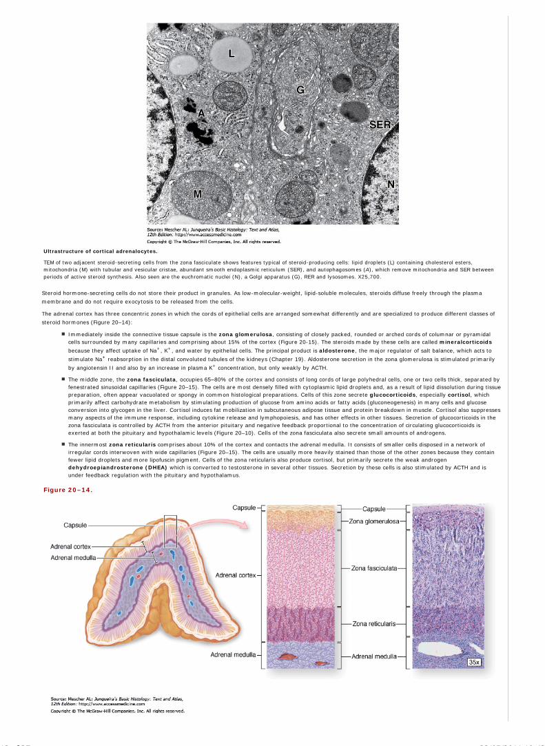
Ultrastructure of cortical adrenalocytes.
TEM of two adjacent steroid-secreting cells from the zona fasciculate shows features typical of steroid-producing cells: lipid droplets (L) containing cholesterol esters,mitochondria (M) with tubular and vesicular cristae, abundant smooth endoplasmic reticulum (SER), and autophagosomes (A), which remove mitochondria and SER betweenperiods of active steroid synthesis. Also seen are the euchromatic nuclei (N), a Golgi apparatus (G), RER and lysosomes. X25,700.
Steroid hormone-secreting cells do not store their product in granules. As low-molecular-weight, lipid-soluble molecules, steroids diffuse freely through the plasma
membrane and do not require exocytosis to be released from the cells.
The adrenal cortex has three concentric zones in which the cords of epithelial cells are arranged somewhat differently and are specialized to produce different classes of
steroid hormones (Figure 20–14):
Immediately inside the connective tissue capsule is the zona glomerulosa, consisting of closely packed, rounded or arched cords of columnar or pyramidalcells surrounded by many capillaries and comprising about 15% of the cortex (Figure 20-15). The steroids made by these cells are called mineralcorticoids
because they affect uptake of Na+, K+, and water by epithelial cells. The principal product is aldosterone, the major regulator of salt balance, which acts to
stimulate Na+ reabsorption in the distal convoluted tubules of the kidneys (Chapter 19). Aldosterone secretion in the zona glomerulosa is stimulated primarily
by angiotensin II and also by an increase in plasma K+ concentration, but only weakly by ACTH.
The middle zone, the zona fasciculata, occupies 65–80% of the cortex and consists of long cords of large polyhedral cells, one or two cells thick, separated byfenestrated sinusoidal capillaries (Figure 20–15). The cells are most densely filled with cytoplasmic lipid droplets and, as a result of lipid dissolution during tissuepreparation, often appear vacuolated or spongy in common histological preparations. Cells of this zone secrete glucocorticoids, especially cortisol, whichprimarily affect carbohydrate metabolism by stimulating production of glucose from amino acids or fatty acids (gluconeogenesis) in many cells and glucoseconversion into glycogen in the liver. Cortisol induces fat mobilization in subcutaneous adipose tissue and protein breakdown in muscle. Cortisol also suppressesmany aspects of the immune response, including cytokine release and lymphopoiesis, and has other effects in other tissues. Secretion of glucocorticoids in thezona fasciculata is controlled by ACTH from the anterior pituitary and negative feedback proportional to the concentration of circulating glucocorticoids isexerted at both the pituitary and hypothalamic levels (Figure 20–10). Cells of the zona fasciculata also secrete small amounts of androgens.
The innermost zona reticularis comprises about 10% of the cortex and contacts the adrenal medulla. It consists of smaller cells disposed in a network ofirregular cords interwoven with wide capillaries (Figure 20–15). The cells are usually more heavily stained than those of the other zones because they containfewer lipid droplets and more lipofuscin pigment. Cells of the zona reticularis also produce cortisol, but primarily secrete the weak androgendehydroepiandrosterone (DHEA) which is converted to testosterone in several other tissues. Secretion by these cells is also stimulated by ACTH and isunder feedback regulation with the pituitary and hypothalamus.
Figure 20–14.
AccessMedicine | Print: Chapter 20. Endocrine Glands http://accessmedicine.com/popup.aspx?aID=6182774&print=yes_chapter
12 of 27 22/05/2011 19:42

Adrenal gland.
Inside the capsule of each adrenal gland is an adrenal cortex, formed from embryonic mesodermal cells, which completely surrounds an innermost adrenal medulla derivedembryologically from neural crest cells. Both regions are very well vascularized with fenestrated sinusoidal capillaries. Cortical cells are arranged as three layers: the zonaglomerulosa near the capsule, the zona fasciculata (the thickest layer), and the zona reticularis.
Figure 20–15.
AccessMedicine | Print: Chapter 20. Endocrine Glands http://accessmedicine.com/popup.aspx?aID=6182774&print=yes_chapter
13 of 27 22/05/2011 19:42

Adrenal cortex.
The steroid-secreting cells of the adrenal cortex are arranged differently to form three fairly distinct concentric layers surrounding the medulla (M). Shown here are sectionsfrom two adrenal glands, stained with H&E (left) and Mallory trichrome, in which the sparse collagen appears blue (right). (a, b): Immediately beneath the capsule (C) thezona glomerulosa consists of rounded clusters of columnar cells principally secreting the mineralcorticoid aldosterone. (c, d): The thick middle layer, the zona fasciculata,consists of long cords of large, spongy-looking cells mainly secreting glucocorticoids such as cortisol. (e, f): Cells of the innermost layer are small, better stained, arranged in aclose network and secrete mainly sex steroids. Cells of all the layers are closely associated with sinusoidal capillaries. Left: X20 H&E.; a, c, e: X200. H&E. b, d, f: X200. Mallorytrichrome.
MEDICAL APPLICATION
Because of the feedback mechanism controlling the adrenal cortex, patients who are treated with corticoids for long periods should never stop taking these
hormones suddenly: secretion of ACTH in these patients is inhibited, and thus the cortex will not be induced to produce corticoids, causing severe drops in the
levels of sodium and potassium.
Fetal Adrenal CortexAt birth in humans (but not most other mammals) the adrenal gland is larger than that of the adult and produces up to 200 mg of corticosteroids per day, twice that of an
adult. At this age, a layer known as the fetal or provisional cortex, comprising 80% of the total gland, is present between the thin permanent cortex and an under-
developed medulla. The fetal cortex is thick and contains mostly cords of large, steroid-secreting cells under the control of the fetal pituitary. The principal function of the
cells is secretion of sulfated DHEA which is converted in the placenta to active estrogens (and androgens), which mostly enter the maternal circulation. The fetal adrenal
cortex is an important part of a fetoplacental unit which affects both endocrine systems during pregnancy but whose physiological significance remains largely unclear.
After birth, the provisional cortex undergoes involution while the permanent cortex organizes the three layers (zones) described above.
Adrenal MedullaThe adrenal medulla is composed of large, pale-staining polyhedral cells arranged in cords or clumps and supported by a reticular fiber network (Figure 20–16). A profuse
supply of sinusoidal capillaries intervenes between adjacent cords and a few parasympathetic ganglion cells are present. Medullary parenchymal cells, known as
chromaffin cells, arise from neural crest cells, as do the postganglionic neurons of sympathetic and parasympathetic ganglia. Chromaffin cells can be considered
modified sympathetic postganglionic neurons, lacking axons and dendrites and specialized as secretory cells.
Figure 20–16.
AccessMedicine | Print: Chapter 20. Endocrine Glands http://accessmedicine.com/popup.aspx?aID=6182774&print=yes_chapter
14 of 27 22/05/2011 19:42

Adrenal medulla.
The hormone-secreting cells of the adrenal medulla are chromaffin cells, which resemble sympathetic neurons. (a): The micrograph shows they are large pale-staining cells,arranged in cords interspersed with wide capillaries. Faintly stained cytoplasmic granules can be seen in most chromaffin cells. X200. H&E. (b): TEM reveals that the granulesof norepinephrine-secreting cells (NE) are more electron-dense than those of cells secreting epinephrine (E), which is a function of the chromogranins to which thecatecholamines are bound in the granules. Most of the hormone produced is epinephrine, which is only made in the adrenal medulla. X33,000.
Unlike cells of the cortex, medullary chromaffin cells contain many electron-dense granules, 150–350 nm in diameter, for hormone storage and secretion. These granules
contain one or the other of the catecholamines, epinephrine or norepinephrine. Ultrastructurally the granules of epinephrine-secreting cells are less electron-dense
and generally smaller than those of norepinephrine-secreting cells. Catecholamine, together with Ca2+ and ATP, are bound in a granular storage complex with 49 kDa
proteins called chromogranins.
Norepinephrine-secreting cells are also found in paraganglia (collections of catecholamine-secreting cells adjacent to the autonomic ganglia) and in various viscera. The
conversion of norepinephrine to epinephrine (adrenalin) occurs only in chromaffin cells of the adrenal medulla. About 80% of the catecholamine secreted from the adrenal
is epinephrine.
Medullary chromaffin cells are innervated by cholinergic endings of preganglionic sympathetic neurons, from which impulses trigger hormone release by exocytosis.
Epinephrine and norepinephrine are released to the blood in large quantities during intense emotional reactions, such as fright, and produce vasoconstriction, increased
blood pressure, changes in heart rate, and metabolic effects such as elevated blood glucose. These effects facilitate various defensive reactions to the stressor (the fight-
or-flight response). During normal activity, the adrenal medulla continuously secretes small quantities of the hormones.
MEDICAL APPLICATION
One disorder of the adrenal medulla is pheochromocytoma, a tumor of its cells that causes hyperglycemia and transient elevations of blood pressure.
Disorders of the adrenal cortex can be classified as hyperfunctional or hypofunctional. Tumors of the adrenal cortex can result in excessive production of
glucocorticoids (Cushing syndrome) or aldosterone (Conn syndrome). Cushing syndrome is most often (90%) due to a pituitary adenoma that results in
excessive production of ACTH; it is rarely caused by adrenal hyperplasia or an adrenal tumor. Excessive production of adrenal androgens has little effect in men,
but precocious puberty (in boys) and hirsutism (abnormal hair growth) and virilization (in girls) are encountered in prepubertal children.
Adrenocortical insufficiency (Addison disease) is caused by destruction of the adrenal cortex in some diseases. The signs and symptoms suggest failure of
secretion of both glucocorticoids and mineralocorticoids by the adrenal cortex.
Carcinomas of the adrenal cortex are rare, but most are highly malignant. About 90% of these tumors produce steroids associated with endocrine glands.
PANCREATIC ISLETS
AccessMedicine | Print: Chapter 20. Endocrine Glands http://accessmedicine.com/popup.aspx?aID=6182774&print=yes_chapter
15 of 27 22/05/2011 19:42

The pancreatic islets (islets of Langerhans) are compact spherical or egg-shaped masses of endocrine tissue embedded within the acinar exocrine tissue of the pancreas
(Figure 20–17). Most islets are 100–200 m in diameter and contain several hundred cells, but some are much smaller with only a few cells. There are more than 1 million
islets in the human pancreas, with the gland's narrow tail region most enriched for islets, but they only constitute 1–2% of the organ's volume. A very thin capsule of
reticular fibers surrounds each islet, separating it from the adjacent acinar tissue. Pancreatic islets have the same embryonic origin as the pancreatic acinar tissue:
masses of cells in epithelial outgrowths from the intestinal lining (endoderm) near the common bile duct.
Figure 20–17.
AccessMedicine | Print: Chapter 20. Endocrine Glands http://accessmedicine.com/popup.aspx?aID=6182774&print=yes_chapter
16 of 27 22/05/2011 19:42

Pancreatic islets.
Pancreatic islets are clumped masses of pale-staining endocrine cells embedded in the exocrine acinar tissue of the pancreas. (a): A low-magnification micrograph through thetail of the pancreas reveals many islets stained lighter than the surrounding tissue. The white spots are adipocytes in fine pancreatic septa, like those of the adipose tissueoutside the gland. X12.5. H&E. (b): Micrograph of an enlarged islet showing its capillary system. Several arterioles enter each islet, branch into fenestrated capillaries (C)among the peripheral islet cells, then converge centrally before leaving the islet as efferent capillaries carrying blood to the acini surrounding the islet. This local vascularsystem allows specific islet hormones to help control secretion of other islet cells and the neighboring acini. X40. H&E. (c): With H&E staining all cells of an islet appear similar,although slight differences in cell size and basophilia may be apparent. Capillaries (C) are also apparent. X55. H&E. (d): An islet prepared with a modified aldehyde fuchsinstain shows granules in the peripheral cells are a deep brownish purple and the central cells granules are brownish orange. Reticulin connective tissue of the islet capsuleand along the capillaries stains green in this preparation. Immunohistochemistry with antibodies against the various islet polypeptide hormones allows definitive identificationof each islet cell type. X300. Modified aldehyde fuchsin and light green. (e): Diagram shows the four major islet hormones and the cells secreting them: cells makingglucagon, cells making insulin, cells making somatostatin, and F (PP) cells making pancreatic polypeptide. (Figure 20–17d, with permission, from Arthur A. Like,Department of Pathology, University of Massachusetts Medical School, Worcester.)
Each islet consists of polygonal or rounded cells, smaller and more lightly stained than the surrounding acinar cells, arranged in cords that are separated by a network of
fenestrated capillaries (Figure 20–17). Autonomic nerve fibers contact some of the endocrine cells and the blood vessels.
Routine stains or trichrome stains show that most islet cells are acidophilic or basophilic with fine cytoplasmic granules (Figure 20–17). Ultrastructural analysis reveals
features of active polypeptide-secreting cells and secretory granules that vary in size, morphology, and electron density from cell to cell. The major hormone-producing
islet cells are most easily identified and studied by immunohistochemistry:
or A cells secrete primarily glucagon and are usually located near the periphery of islets.
or B cells produce insulin (L. insula, island), are located centrally in islets and are the most numerous cell type.
or D cells, secreting somatostatin, are scattered and much less abundant.
Insulin is a heterodimeric protein and the other two hormones are smaller single-chain polypeptides. A minor fourth cell type, more common in islets located within the
head of the pancreas, are F or PP cells, which secrete pancreatic polypeptide. Table 20–4 summarizes the types, quantities, and main functions of the major
hormones produced by islet cells. Pancreatic islets also normally contain a few enterochromaffin cells, like those of the digestive tract, which secrete other polypeptide
hormones having other effects within the digestive system and which are also scattered in the pancreatic acini and ducts.
Table 20–4. Major cell types and hormones of pancreatic islets.
CellType
Quantity HormoneProduced
Hormone Structure and Size Hormone Function
AccessMedicine | Print: Chapter 20. Endocrine Glands http://accessmedicine.com/popup.aspx?aID=6182774&print=yes_chapter
17 of 27 22/05/2011 19:42

CellType
Quantity HormoneProduced
Hormone Structure and Size Hormone Function
~20% Glucagon Polypeptide; 3500 Da Acts on several tissues to make energy stored in glycogen and fat available throughglycogenolysis and lipolysis; increases blood glucose content
~70% Insulin Dimer of and chains with S-Sbridges; 5700-6000 Da
Acts on several tissues to cause entry of glucose into cells and promotes decrease of bloodglucose content
or D 5–10% Somatostatin Polypeptide; 1650 Da Inhibits release of other islet cell hormones through local paracrine action; inhibits release ofGH and TSH in anterior pituitary and HCl secretion by gastric parietal cells
F or PP Rare Pancreaticpolypeptide
Polypeptide; 4200 Da Stimulates activity of gastric chief cells; inhibits bile secretion, -pancreatic enzyme andbicarbonate secretion, and intestinal motility
Activity of the two major islet cells, and cells, is regulated largely by blood glucose levels above or below the normal level of 70 mg/dL. Increased glucose levels
stimulate cells to release insulin and inhibit cells from releasing glucagon; decreased glucose levels stimulate cells to release glucagon. Opposing actions of these
hormones (Table 20–4) help to precisely control blood glucose concentration, an important factor in body homeostasis. Increased secretion of these hormones or
somatostatin also acts in a paracrine manner to affect hormone release within an islet as well as activity of the neighboring acinar cells.
Sympathetic and parasympathetic nerve endings are closely associated with about 10% of the , , and cells and can also function as part of the control system for
insulin and glucagon secretion. Gap junctions transfer the autonomic neural stimulus to the other cells. Sympathetic fibers increase glucagon release and inhibit insulin
release; parasympathetic fibers increase secretion of both glucagon and insulin.
MEDICAL APPLICATION
Insulin-dependent or type 1 diabetes (juvenile diabetes) results from partial or total autoimmune destruction of cells and subsequent lack of insulin.
Insulin-independent diabetes or type 2 diabetes occurs later in life, results from a failure of cells to respond to insulin, and is frequently associated with
obesity.
Tumors arising from islet cells may produce insulin, glucagon, somatostatin, and pancreatic polypeptide. Some pancreatic tumors produce two or more of these
hormones simultaneously, generating complex clinical symptoms.
DIFFUSE NEUROENDOCRINE SYSTEM
The enterochromaffin cells scattered in both the islets and small ducts of the pancreas are similar to those of the digestive tract (Chapter 15). Collectively these dispersed
cells, as well as similar cells in the respiratory mucosa, make up the diffuse neuroendocrine system (DNES). Like the pancreatic islets, most of these cells are
derived from endodermal cells of the embryonic gut. Such secretory cells are considered neuroendocrine because they produce many of the same polypeptides and
neurotransmitter-like molecules such as serotonin (5-hydroxytryptamine) also released by neurosecretory cells in the CNS. Cells of the DNES are also referred to as
gastroenteropancreatic (GEP) endocrine cells. Several such cells, along with their hormones and major functions, are summarized in Table 15–1. Most of these
hormones are polypeptides and many act in a paracrine manner, affecting primarily the activities of neighboring contractile cells and secretory cells (both exocrine and
endocrine). GEP endocrine or enteroendocrine cells of the stomach and small bowel are shown in Figures 15–24, 15–28, and 15–31.
Many cells of the DNES are stained by solutions of chromium salts and have therefore been called enterochromaffin cells. Those cells that stain with silver nitrate are
sometimes called argentaffin cells. Those DNES cells secreting serotonin or certain other amine derivatives demonstrate amine precursor uptake and decarboxylation
and are often referred to acronymically as APUD cells. Such names are still widely used but have been largely replaced by letter designations like those used for
pancreatic islet cells (Table 15–1). Whatever name is used, cells of the DNES are highly important due to their role in regulating motility and secretions of all types within
the digestive system.
THYROID GLAND
The thyroid gland, located in the cervical region anterior to the larynx, consists of two lobes united by an isthmus (Figure 20–18). It originates in early embryonic life from
the foregut endoderm near the base of the future tongue. Its function is to synthesize the thyroid hormones: thyroxine (tetra-iodothyronine or T4) and
tri-iodothyronine (T3), which are important for growth, for cell differentiation, and for the control of the basal metabolic rate and oxygen consumption in cells
throughout the body. Thyroid hormones affect protein, lipid, and carbohydrate metabolism.
Figure 20–18.
AccessMedicine | Print: Chapter 20. Endocrine Glands http://accessmedicine.com/popup.aspx?aID=6182774&print=yes_chapter
18 of 27 22/05/2011 19:42

Thyroid gland and its development.
(a): The thyroid is a highly vascular, butterfly-shaped gland, approximately 5 cm x 5 cm and weighing 20–30 g in adults, surrounding the anterior surface of the trachea justbelow the larynx. Immigrating neural crest cells infiltrate the epithelium as precursors to the thyroid's parafollicular C cells. (b): Thyroid development begins in the fourth weekas an epithelial diverticulum growing down from the endodermal lining of the foregut. (c): The thyroid diverticulum continues to grow in an inferior direction and its connectionto the developing pharynx, the thyroglossal duct, later regresses. (d): By fetal stages, the thyroid has attained its normal adult position.
The parenchyma of the thyroid is composed of millions of rounded epithelial structures called thyroid follicles. Each follicle consists of a simple epithelium and a central
lumen filled with a gelatinous substance called colloid (Figure 20–19). The thyroid is the only endocrine gland in which a large quantity of secretory product is stored.
Moreover, the accumulation is outside the cells, in the colloid of the follicles, which is also unusual. In humans there is sufficient hormone in follicles to supply the body for
up to three months with no additional synthesis. Thyroid colloid contains the large glycoprotein thyroglobulin (660 kDa), the precursor for the active thyroid hormones.
Figure 20–19.
AccessMedicine | Print: Chapter 20. Endocrine Glands http://accessmedicine.com/popup.aspx?aID=6182774&print=yes_chapter
19 of 27 22/05/2011 19:42

AccessMedicine | Print: Chapter 20. Endocrine Glands http://accessmedicine.com/popup.aspx?aID=6182774&print=yes_chapter
20 of 27 22/05/2011 19:42

Thyroid follicular cells and parafollicular cells.
(a): A low-power micrograph of thyroid gland shows the thin capsule (C), from which septa (S) with the larger blood vessels, lymphatics, and nerves enter the gland. Theparenchyma of the organ is distinctive, consisting of colloid-filled epithelial follicles of many sizes. The lumen of each follicle is filled with a lightly staining colloid of a largegelatinous protein called thyroglobulin. X12. H&E. (b): The lumen (L) of each follicle is surrounded by a simple epithelium in which the cell height ranges from squamous to lowcolumnar. Also present are large pale-staining parafollicular or C cells (C) which secrete calcitonin, a polypeptide involved with calcium metabolism. X200. H&E. (c,d,e): C cellsmay be part of the follicular epithelium or present singly or in groups outside of follicles. Follicular cells (F) can usually be distinguished from C cells (C) by the smaller size anddarker staining properties. Unlike follicular cells, C cells seldom vary in their size or pale staining characteristics. C cells are somewhat easier to locate in or between smallfollicles. c and d: X400. H&E; e: X400. Mallory trichrome.
The thyroid gland is covered by a fibrous capsule from which septa extend into the parenchyma, dividing it into lobules and carrying blood vessels, nerves, and
lymphatics. Follicles are densely packed together, separated from one another only by sparse reticular connective tissue (Figure 20–19). This stroma is very well
vascularized with an extensive network of fenestrated capillaries closely surrounding the follicles, which facilitates molecular transfer between the follicular cells and
blood.
Follicular cells range in shape from squamous to low columnar and the follicles are quite variable in diameter (Figure 20–19). The size and cellular features of thyroid
follicles vary with their functional activity. Active glands have more follicles of low columnar epithelium; glands with mostly squamous follicular cells are considered
hypoactive.
The follicular epithelial cells have typical junctional complexes apically and rest on a basal lamina. The cells exhibit organelles indicating active protein synthesis and
secretion, as well as phagocytosis and digestion. The nucleus is generally round and in the center of the cell. Basally the cells are rich in rough ER and apically, facing the
follicular lumen, are Golgi complexes, secretory granules filled with colloidal material, large phagosomes and abundant lysosomes. The cell membrane of the apical pole
has a moderate number of microvilli. Mitochondria and other cisternae of rough ER are dispersed throughout the cytoplasm.
Another endocrine cell type, the parafollicular, or C, cell, is also found inside the basal lamina of the follicular epithelium or as isolated clusters between follicles (Figure
20–20). Parafollicular cells, derived from neural crest cells migrating into the area of the embryonic foregut, are usually somewhat larger than follicular cells and stain less
intensely. They have a smaller amount of rough ER, large Golgi complexes, and numerous small (100–180 nm in diameter) granules containing polypeptide hormone
(Figure 20–20). These cells synthesize and secrete calcitonin, one function of which is to suppress bone resorption by osteoclasts. Calcitonin secretion is triggered by
elevated blood Ca2+ levels.
Figure 20–20.
AccessMedicine | Print: Chapter 20. Endocrine Glands http://accessmedicine.com/popup.aspx?aID=6182774&print=yes_chapter
21 of 27 22/05/2011 19:42

Ultrastructure of thyroid follicular and parafollicular cells.
(a): TEM of the follicular epithelium shows pseudopodia and microvilli extending from the follicular cells (F) into the colloid of the lumen (L). The cells have apical junctionalcomplexes, much RER, well-developed Golgi complexes, and many lysosomes. Inside the basement membrane (BM) of the follicle, but often not contacting the colloid in thelumen, are occasional C or parafollicular cells (C). To the left and right of the two C cells seen here are capillaries intimately associated with the follicular cells, but outside thebasement membrane. X2000. (b): A TEM of a C cell, with its large Golgi apparatus (G), extensive RER, and cytoplasm filled with small secretory granules containing calcitonin.X5000.
Control of Thyroid FunctionThe major regulator of the anatomic and functional state of thyroid follicles is thyroid-stimulating hormone (TSH; thyrotropin), which is secreted by the anterior pituitary
(Figure 20–8). TSH increases the height of the follicular epithelium and stimulates all stages of thyroid hormone production and release. Thyroid hormones inhibit the
release of TSH, maintaining an adequate quantity of T4 and T3 in the organism (Figure 20–10). TSH receptors are abundant on the basal cell membrane of follicular cells.
Secretion of TSH is also increased by exposure to cold and decreased by heat and stressful stimuli.
Storage & Release of Thyroid HormoneProduction, storage, and release of thyroid hormones involve an unusual, multistage process with both an exocrine phase and an endocrine phase in the follicular cells.
Both phases are promoted by TSH and can be occurring in the same cell, as summarized in Figure 20–21. The major activities of this process include the following:
The production of thyroglobulin, which is similar to that in other glycoprotein-exporting cells, with synthesis in the rough ER and the addition of carbohydrate1.
AccessMedicine | Print: Chapter 20. Endocrine Glands http://accessmedicine.com/popup.aspx?aID=6182774&print=yes_chapter
22 of 27 22/05/2011 19:42

there and in the Golgi apparatus. This large glycoprotein has no hormonal activity itself but contains 140 tyrosyl residues used to make thyroid hormone. As part ofthe exocrine phase of cellular activity, thyroglobulin is released from large vesicles at the apical surface of the cell into the lumen of the follicle.
The uptake of circulating iodide is accomplished in follicular cells by the Na/I symporter or cotransporter in the basolateral cell membrane, which allows for30-fold concentration of dietary iodide in the normal thyroid relative to the plasma. Low levels of circulating iodide trigger synthesis of the Na/I symporter,increasing the uptake of iodide and compensating for the lower serum concentration.
2.
At the apical surface of the cells, iodide is transferred to the follicular lumen by the anion transport protein pendrin and there it undergoes oxidationto activeiodine by membrane-bound thyroid peroxidase at the cell surface.
3.
In the lumen tyrosine residuesof thyroglobulin are iodinated covalently with either one or two iodine atoms. Following this, two iodinated tyrosines, still partof thyroglobulin, are conjugated by oxidative coupling reactions to form T3 or T4 .
4.
Immediately or after a delay, in the endocrine phase of the process the follicular cells take up the iodinated thyroglobulin in colloid by endocytosis or pinocytosis.The large endocytic vesicles fuse with lysosomes and move to the basolateral cell membrane while the thyroglobulin inside is thoroughly degraded by lysosomalproteases. T4 and T3, freed in this manner from thyroglobulin, then cross the cell membrane and basement membrane, and are taken up into the capillaries.
5.
Figure 20–21.
Thyroid follicular cell functions.
The diagram shows the multistep process by which thyroid hormones are produced via the stored thyroglobulin intermediate. In an exocrine phase of the process, theglycoprotein thyroglobulin is made and secreted into the follicular lumen and iodide is pumped across the cells into the lumen. In the lumen tyrosine residues of thyroglobulinare iodinated and then covalently coupled to form T3 and T4 still within the glycoprotein. The iodinated thyroglobulin is then endocytosed by the follicular cells and degraded bylysosomes, releasing free active T3 and T4 to the adjacent capillaries in an endocrine manner. Both phases are promoted by TSH and may occur simultaneously in the same cell.
Nearly all of both thyroid hormones are carried in blood tightly bound to plasma proteins. T4 is the more abundant compound, constituting 90% of the circulating thyroid
hormone. Both molecules bind the same intracellular receptors of target cells, but T3 is two- to tenfold more active than T4. The half-life of T3 is 1.5 days in comparison
with a week for T4. Both thyroid hormones increase the number of mitochondria and their cristae and stimulate mitochondrial protein synthesis.
MEDICAL APPLICATION
A diet low in iodide hinders the synthesis of thyroid hormones, causing increased secretion of TSH and compensatory growth of the thyroid gland, a condition
known as iodine deficiency goiter. Goiters are endemic in some regions of the world, where dietary iodide is scarce and addition of iodide to table salt is not
required. Hypothyroidism in the fetus may present at birth as cretinism, characterized by arrested or retarded physical and mental development.
Adult hypothyroidism may result from diseases of the thyroid gland (eg, due to defects in hormone synthesis or release) or may be secondary to pituitary or
hypothalamic failure. Autoimmune diseases of the thyroid, such as Hashimoto disease, may impair its function, with consequent hypothyroidism.
Hyperthyroidism may be caused by a variety of thyroid diseases, of which the most common form is Graves' disease, characterized by inflammation and growth
of the extraocular adipose tissue, which leads to the bulging of the eyes (exophthalmos). In this thyroid disorder hyperfunction is due to an autoimmune response
involving antibodies to TSH receptors. These antibodies can bind the receptors on follicular cells and act as long-lasting thyroid stimulators, continuously
stimulating thyroid hormone secretion and producing many effects of hyperthyroidism such as decreased body weight and accelerated heart rate.
PARATHYROID GLANDS
The parathyroid glands are four small oval masses—each 3 x 6 mm—with a total weight of about 0.4 g. They are located on the back of the thyroid gland, one at each
end of the upper and lower poles, usually embedded in the larger gland's capsule (Figure 20–22). The parathyroid glands are derived from the pharyngeal pouches—the
AccessMedicine | Print: Chapter 20. Endocrine Glands http://accessmedicine.com/popup.aspx?aID=6182774&print=yes_chapter
23 of 27 22/05/2011 19:42

superior glands from the fourth pouch and the inferior glands from the third pouch. Their embryonic migration to the developing thyroid gland is sometimes misdirected
so that the number and locations of the glands are somewhat variable. Up to 10% of individuals may have parathyroid tissue attached to the thymus, which originates
from the same pharyngeal pouches.
Figure 20–22.
Parathyroid glands.
The parathyroid glands are four small nodules normally embedded in the capsule on the posterior surface of the thyroid gland. They arise embryologically from the third andfourth pharyngeal pouches and migrate to the developing thyroid, a process that frequently leads to ectopic or additional parathyroid glands, often associated with the thymus.
Each parathyroid gland is contained within a capsule which sends septa into the gland, where they merge with reticular fibers that support elongated cordlike clusters of
secretory cells. With increasing age many secretory cells are replaced with adipocytes, which may constitute more than 50% of the gland in older people.
Two types of cells are present in parathyroid glands: chief (or principal) cells and oxyphil cells (Figure 20–23). The chief cells are small polygonal cells with round nuclei
and pale-staining, slightly acidophilic cytoplasm. Ultrastructurally the cytoplasm is seen to be filled with irregularly shaped granules 200–400 nm in diameter. These are
secretory granules containing the polypeptide parathyroid hormone (PTH), a major regulator of blood calcium levels. Much smaller, often clustered, populations of
oxyphil cells are sometimes present, more commonly in older individuals. These are much larger than the principal cells and are characterized by acidophilic cytoplasm
filled with abnormally shaped mitochondria. Some oxyphil cells show low levels of PTH synthesis, suggesting these cells are transitional derivatives from chief cells.
Parathyroid hormone targets osteoblasts, which respond by producing an osteoclast-stimulating factor to increase the number and activity of osteoclasts. This promotes
resorption of the calcified bone matrix and the release of Ca2+, increasing the concentration of Ca2+ in the blood, which suppresses parathyroid hormone production.
Calcitonin from the thyroid gland inhibits osteoclast activity, lowering the blood Ca2+ concentration and promoting osteogenesis. Parathyroid hormone and calcitonin thus
have opposing effects and constitute a dual mechanism to regulate blood levels of Ca2+, an important factor in homeostasis. Parathyroid hormone also indirectly
increases the absorption of Ca2+ from the gastrointestinal tract by stimulating the synthesis of vitamin D, which is necessary for this absorption.
Figure 20–23.
AccessMedicine | Print: Chapter 20. Endocrine Glands http://accessmedicine.com/popup.aspx?aID=6182774&print=yes_chapter
24 of 27 22/05/2011 19:42

Parathyroid principal cells.
(a): A small lobe of parathyroid gland, surrounded by connective tissue septa (S), shows mainly densely packed cords of small principal cells (P), also called chief cells. Olderparathyroid glands show increasing numbers of much larger and acidophilic nonfunctional oxyphil cells (O) which may occur singly or in clumps of varying sizes. X60. H&E. (b):The micrograph shows that principal cells are slightly eosinophilic, with round central nuclei, present in cords separated by capillaries (C). These cells secrete the polypeptideparathyroid hormone (PTH). X300. H&E.
In addition to increasing the concentration of Ca2+, parathyroid hormone reduces blood phosphate levels. This effect results from another target cell of parathyroid
hormone, renal tubule cells, which reduce their absorption of phosphate and allow more phosphate excretion in urine.
MEDICAL APPLICATION
In hyperparathyroidism, concentrations of blood phosphate are decreased and concentrations of blood Ca2+ are increased. This condition frequently produces
pathologic deposits of calcium in several organs, such as the kidneys and arteries. The bone disease caused by hyperparathyroidism, which is characterized by an
increased number of osteoclasts and multiple bone cavities, is known as osteitis fibrosa cystica. Bones from patients with this disorder are less stress-resistant
and prone to fractures.
In hypoparathyroidism, concentrations of blood phosphate are increased and concentrations of blood Ca2+ are decreased. The bones become denser and more
mineralized. This condition causes spastic contractions of the skeletal muscles and generalized convulsions called tetany. These symptoms are caused by the
exaggerated excitability of the nervous system, due to the lack of Ca2+ in the blood. Patients with hypoparathyroidism are treated with calcium salts and vitamin
D to promote Ca2+ uptake in the gut.
PINEAL GLAND
The pineal gland, also known as the pineal body or epiphysis cerebri, regulates the daily rhythms of bodily activities. It is a very small, pine cone-shaped organ in the
brain measuring approximately 5–8 mm in length and 3–5 mm at its greatest width and weighing about 150 mg. The pineal develops with the brain from neuroectoderm
in the roof of the diencephalon and is found in the posterior of the third ventricle, attached to the brain by a short stalk.
The pineal gland is covered by connective tissue of the pia mater, from which emerge septa containing small blood vessels and subdividing various sized groups of
secretory cells as lobules. The prominent and abundant secretory cells are the pinealocytes, which have slightly basophilic cytoplasm and large, irregular euchromatic
nuclei and nucleoli (Figure 20–24). Ultrastructurally pinealocytes are seen to have secretory vesicles, many mitochondria, and long cytoplasmic processes extending to
the vascularized septa, where they end in dilatations near capillaries, indicating an endocrine function. These cells produce melatonin, a low molecular-weight tryptophan
derivative. Unmyelinated sympathetic nerve fibers enter the pineal gland and end among pinealocytes, with some forming synapses.
Figure 20–24.
AccessMedicine | Print: Chapter 20. Endocrine Glands http://accessmedicine.com/popup.aspx?aID=6182774&print=yes_chapter
25 of 27 22/05/2011 19:42

Pineal gland.
The pineal gland is a very small neuroendocrine gland attached to the brain in the roof of the third ventricle. It is covered by the pia mater, which sends septa of connectivetissue into the glands, subdividing groups of parenchymal cells called pinealocytes. (a): The micrograph shows a group of pinealocytes surrounded by septa (S) containingvenules (V) and capillaries (arrows). Also seen is an extracellular mineral deposit called a corpus arenaceum (CA) of unknown physiological significance but an excellent markerfor the pineal. X40. H&E.
(b): At higher magnification the numerous large pinealocytes (P) with euchromatic nuclei can be compared to the relatively few astrocytes (A) which have darker, moreelongated nuclei. Astrocytes are located mainly within septa and near small blood vessels (V). Capillaries (arrow) are not nearly as numerous as in other endocrine glands. Atthe lower left is a port of a very large corpus arenaceum (CA), the calcified structures also known as brain sand. Along the septa run unmyelinated tracts of sympathetic fibers,associated indirectly with photoreceptive neurons in the retinas and running to the pinealocytes to stimulate melatonin release in periods of darkness. Levels of circulatingmelatonin are one factor determining the diurnal rhythms of hormone release and physiological activities throughout the body. X100. H&E.
Interstitial glial cells of the pineal gland stain positively for glial fibrillary acidic protein and thus most closely resemble astrocytes. They have elongated nuclei more
heavily stained than those of pinealocytes, long cytoplasmic processes, and are usually found in perivascular areas and between the groups of pinealocytes. Pineal
astrocytes represent only about 5% of the cells in the gland.
A characteristic feature of the pineal gland is the presence of variously sized concretions of calcium and magnesium salts called corpora arenacea or brain sand, which
form by precipitation around extracellular protein deposits. Such concretions appear during childhood and gradually increase in number and size with age, with no
apparent effect on the gland's function. Accumulations of brain sand are opaque to x-rays and allow the pineal to serve as a good midline marker in radiological and
AccessMedicine | Print: Chapter 20. Endocrine Glands http://accessmedicine.com/popup.aspx?aID=6182774&print=yes_chapter
26 of 27 22/05/2011 19:42

Copyright © The McGraw-Hill Companies. All rights reserved.Privacy Notice. Any use is subject to the Terms of Use and Notice.
computer-assisted tomography studies of the brain.
Melatonin release from pinealocytes is promoted by darkness and inhibited by daylight and the resulting diurnal fluctuation in blood melatonin levels induces rhythmic
changes in the activity of the hypothalamus, pituitary gland, and other endocrine tissues that characterize the circadian (24 hours, day/night) rhythm of physiological
functions and behaviors. In humans and other mammals the cycle of light and darkness is detected within the retinas and transmitted to the pinealocytes via the
retinohypothalamic tract, the suprachiasmatic nucleus, and the tracts of sympathetic fibers entering the pineal. The pineal gland acts therefore as a neuroendocrine
transducer, converting nerve input regarding light and darkness into variations in many hormonal functions.
AccessMedicine | Print: Chapter 20. Endocrine Glands http://accessmedicine.com/popup.aspx?aID=6182774&print=yes_chapter
27 of 27 22/05/2011 19:42

Print Close Window
Note: Large images and tables on this page may necessitate printing in landscape mode.
Copyright © The McGraw-Hill Companies. All rights reserved.
Junqueira's Basic Histology: Text & Atlas, 12e > Chapter 21. The Male Reproductive System >
THE MALE REPRODUCTIVE SYSTEM: INTRODUCTION
The male reproductive system is composed of the testes, genital ducts, accessory glands, and penis (Figure 21–1). Testes function in the production of hormones and
spermatozoa. Although testosterone is the main hormone produced in the testes, both testosterone and its metabolite, dihydrotestosterone, are necessary for male
reproductive physiology. Testosterone is important for spermatogenesis, sexual differentiation during embryonic and fetal development, and control of gonadotropin
secretion. Dihydrotestosterone acts on many organs and tissues of the body during puberty and adulthood (eg, muscle, hair pattern, and hair growth).
Figure 21–1.
The male reproductive system.
The diagram shows the locations and relationships of the testes, epididymis, glands, and the ductus deferens running from the scrotum to the urethra. The ductus deferensis located along the anterior and superior sides of the bladder as a result of the testes descending into the scrotum from the abdominal cavity during fetal development.
The genital ducts and accessory glands produce secretions and aided by smooth muscle contractions, propel spermatozoa toward the exterior. These secretions also
provide nutrients for spermatozoa while they are confined to the male reproductive tract. Spermatozoa and the secretions of the genital ducts and accessory glands
make up the semen (L, semen, seed), which is introduced into the female reproductive tract through the penis.
TESTES
Each testis (testicle) is surrounded by a capsule of dense connective tissue, the tunica albuginea. The tunica albuginea is thickened on the posterior side of the
testis to form the mediastinum testis, from which fibrous septa penetrate the organ and divide it into about 250 pyramidal compartments or testicular lobules
(Figure 21–2). The septa are incomplete, and there is frequently intercommunication between lobules. Each lobule is occupied by one to four seminiferous tubules
that are surrounded by interstitial loose connective tissue rich in blood and lymphatic vessels, nerves, and endocrine interstitial cells (Leydig cells) which secrete
testosterone. Seminiferous tubules produce male reproductive cells, the spermatozoa, whereas interstitial cells secrete testicular androgens.
Figure 21–2.
AccessMedicine | Print: Chapter 21. The Male Reproductive System http://accessmedicine.com/popup.aspx?aID=6182951&print=yes_chapter
1 of 23 23/05/2011 13:28

Testes and seminiferous tubules.
The anatomy of a testis is shown. (a): The diagram shows a partially cut-away sagittal section. (b): The micrograph shows a cross section of one seminiferous tubule.X250. H & light green.
The testes develop retroperitoneally in the dorsal wall of the embryonic abdominal cavity. They are moved during fetal development and eventually are suspended
within the two sides of the scrotum at the ends of the spermatic cords. Because of the migration from the abdominal cavity, each testis carries with it a serous sac,
the tunica vaginalis (Figure 21–2), derived from the peritoneum. The tunic consists of an outer parietal layer lining the scrotum and an inner visceral layer, covering
the tunica albuginea on the anterior and lateral sides of the testis.
Temperature is important in the regulation of spermatogenesis, which occurs only below the core body temperature of 37°C. A testicular temperature of about 34°C
is maintained in the scrotal sac by various mechanisms. Each testicular artery is surrounded by a rich pampiniform venous plexus with cooler blood from the testis
which can draw heat from the arterial blood by a countercurrent heat-exchange system. Evaporation of sweat from the scrotum also contributes to heat loss.
Relaxation or contraction of the dartos muscle of the scrotum and the cremaster muscles of the spermatic cords move the testes away from, or closer to, the body
respectively, allowing further control on testicular temperature.
MEDICAL APPLICATION
Failure of descent of the testes into the scrotum (cryptorchidism [Gr. kryptos, hidden, + orchis, testis]) maintains the testes at the core body temperature
which inhibits spermatogenesis. In some cases, spermatogenesis can occur normally if the testes are moved surgically to the scrotum. For this reason, it is
important to examine male newborns to check if the testicles are present in the scrotum. Although germ cell proliferation is inhibited by abdominal
temperature, testosterone synthesis by interstitial cells is not affected. Therefore men with cryptorchidism can be sterile but still develop secondary male
characteristics.
Malnutrition, alcoholism, and the action of certain classes of drugs lead to alterations in spermatogonia, with a resulting decrease in production of
spermatozoa. X-irradiation is quite toxic to cells of the spermatogenic lineage, causing the death of those cells and sterility in animals.
Androgen-producing interstitial cell tumors can cause precocious puberty in males.
Seminiferous TubulesSperm are produced in the seminiferous tubules at a rate of about 2 x 10⁸ per day in the adult male. Each testicle has 250–1000 seminiferous tubules in its lobules,
with each tubule measuring about 150–250 m in diameter and 30–70 cm in length. The combined length of the tubules of one testis is about 250 m. Each tubule is a
convoluted loop linked via a short, narrower segment, the straight tubule, to the rete testis, a labyrinth of epithelium-lined channels embedded in the
mediastinum testis (Figures 21–2 and 21–3). Ten to twenty efferent ductules connect the rete testis to the head of the epididymis (Figure 21–2).
Figure 21–3.
AccessMedicine | Print: Chapter 21. The Male Reproductive System http://accessmedicine.com/popup.aspx?aID=6182951&print=yes_chapter
2 of 23 23/05/2011 13:28

Lobules converging at rete testis.
The capsule of the testis, the tunica albuginea, thickens on the posterior surface as the mediastinum (M) testis, from which many thin septa (S) subdivide the organ intoabout 250 lobules. Each lobule contains from one to four convoluted seminiferous tubules (ST) in a sparse connective tissue interstitium. Each tubule is a loop attached bymeans of a short straight tubule to the rete testis (RT), a maze of channels embedded in the mediastinum testis. From the rete testis sperm move into the epididymis.X60. H&E
Each seminiferous tubule is lined with a complex, specialized stratified epithelium called germinal or seminiferous epithelium (Figure 21–2). The basement
membrane of this epithelium is covered by fibrous connective tissue, with an innermost layer containing flattened, smooth muscle-like myoid cells (Figure 21–4),
which allow weak contractions of the tubule. Interstitial cells occur in the connective tissue between the seminiferous tubules (Figures 21–4).
Figure 21–4.
AccessMedicine | Print: Chapter 21. The Male Reproductive System http://accessmedicine.com/popup.aspx?aID=6182951&print=yes_chapter
3 of 23 23/05/2011 13:28

Seminiferous tubule and interstitial cells.
(a): The micrograph shows seminiferous tubules surrounded by connective tissue (CT), containing many large rounded or polygonal interstitial cells (IC) secretingandrogens. Immediately surrounding each tubule are flattened myoid cells (M), which contract to help move sperm out of the tubule, and layers of fibroblasts (F). Insidethe tubule itself is a unique seminiferous epithelium composed of columnar supporting cells called Sertoli cells (SC), which usually have oval nuclei and distinct nucleoli,and germ cells of the spermatogenic lineage. Prominent among the latter are spermatogonia (SG), diploid cells always located near the basement membrane, and primaryspermatocytes (PS) which are undergoing meiosis closer to the lumen of the tubule. At the upper left corner is a portion of a straight tubule, which lacks germ cells andconsists solely of Sertoli cells. X300. H&E. (b): A higher magnification of a plastic section shows lipid droplets filling the cytoplasm of the clumped interstitial cells (IC) inthe connective tissue (CT) between tubules, which is typical of steroid-secreting endocrine cells. The epithelium of the seminiferous tubules (ST) is immediatelysurrounded by myoid cells (M). X400. PT.
The seminiferous epithelium consists of two types of cells: nondividing supporting or sustentacular cells (Sertoli cells) and proliferative cells of the
AccessMedicine | Print: Chapter 21. The Male Reproductive System http://accessmedicine.com/popup.aspx?aID=6182951&print=yes_chapter
4 of 23 23/05/2011 13:28

spermatogenic lineage (Figure 21–5). The cells of the spermatogenic lineage comprise four to eight concentric cell layers and their function is to produce the cells
that become sperm. The part of sperm production that includes cell division through mitosis and meiosis is called spermatogenesis. The final differentiation of the
haploid male germ cells is called spermiogenesis.
Figure 21–5.
Spermatogenesis and spermiogenesis.
(a): The diagram shows a small part of a seminiferous tubule with its surrounding tissue. The seminiferous epithelium is formed by two cell populations: the supporting orSertoli cells and the cells of the spermatogenic lineage. The latter are intimately associated with and almost completely surrounded by the surface of the columnar
AccessMedicine | Print: Chapter 21. The Male Reproductive System http://accessmedicine.com/popup.aspx?aID=6182951&print=yes_chapter
5 of 23 23/05/2011 13:28

supporting Sertoli cells. Nearest the basement membrane are spermatogonia, which divide by mitosis to produce more spermatogonia and cells committed to meiosis. Thelatter cells grow to become primary spermatocytes while arrested in the first meiotic division. They then divide to become secondary spermatocytes, which divide quicklyagain to become haploid spermatids, which differentiate as sperm cells. These last events take place near the apical ends of the Sertoli cells at the lumen of the tubules.The small dots at the junction of the Sertoli cells represent occluding junctions which form part of the blood-testis barrier and which are opened transiently as the germcells move across those areas, forming new junctions between themselves and the support cells to maintain the barrier function. Usually all stages of this process, calledspermatogenesis, do not take place within one small area of tubule as shown schematically here.
(b): The diagram depicts the major morphological changes that occur within spermatids as they undergo the differentiation process, called spermiogenesis, and becomehighly specialized sperm cells. These changes involve flattening of the nucleus, formation of an acrosome which resembles a large lysosome, growth of a flagellum (tail)from the basal body, reorganization of the mitochondria in the midpiece region, and shedding of unneeded cytoplasm as a residual body.
SpermatogenesisSpermatogenesis begins at puberty with a primitive germ cell, the spermatogonium (Gr. sperma + gone, generation), a relatively small round cell, about 12 m in
diameter. These cells are located basally in the epithelium next to the basement membrane (Figures 21–5 and 21–6) and different stages of their development are
recognized mainly by the shape and staining properties of their nuclei. Spermatogonia with dark, ovoid nuclei act as stem cells, dividing infrequently and giving rise
both to new stem cells and to cells with more pale-staining, ovoid nuclei that divide more rapidly as transit amplifying (progenitor) cells (Figure 21–7). These type A
spermatogonia each undergo several unique clonal divisions, remaining interconnected as a syncytium (see below), and form type B spermatogonia, which have
more spherical pale nuclei.
Figure 21–6.
AccessMedicine | Print: Chapter 21. The Male Reproductive System http://accessmedicine.com/popup.aspx?aID=6182951&print=yes_chapter
6 of 23 23/05/2011 13:28

Seminiferous tubules: Sertoli cells and spermatogenesis.
In the two cross-sections of seminiferous tubules shown, most of the associated cell types can be seen. Outside the tubules are myoid cells (M) and fibroblasts (F). Insidenear the basement membrane are many prominent spermatogonia (SG), small cells which divide mitotically but give rise to a population that enters meiosis. The meioticcells grow and undergo chromosomal synapsis to become primary spermatocytes (PS), arrested for 3 weeks in prophase of the first meiotic division during whichrecombination occurs. Primary spermatocytes are the largest spermatogenic cells and are usually abundant at all levels between the basement membrane and the lumen.Each divides to form two secondary spermatocytes, which are seldom seen in sections because they undergo the second meiotic division almost immediately to form twohaploid spermatids. Newly formed round spermatids (RS) differentiate and lose volume in becoming late spermatids (LS) and finally motile, highly specialized sperm cells.All stages of spermatogenesis and spermiogenesis occur with the cells intimately associated with the surfaces of adjacent Sertoli cells (SC) which perform severalsupportive functions. Both X750. H&E.
Figure 21–7.
AccessMedicine | Print: Chapter 21. The Male Reproductive System http://accessmedicine.com/popup.aspx?aID=6182951&print=yes_chapter
7 of 23 23/05/2011 13:28
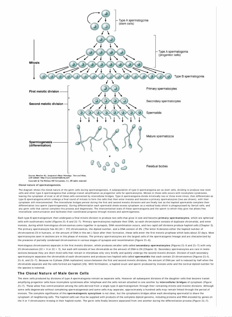
Clonal nature of spermatogenesis.
The diagram shows the clonal nature of the germ cells during spermatogenesis. A subpopulation of type A spermatogonia act as stem cells, dividing to produce new stemcells and other type A spermatogonia that undergo transit amplification as progenitor cells for spermatocytes. Mitosis in these cells occurs with incomplete cytokinesis,leaving the cytoplasm of most or all of these cells connected by intercellular bridges. Type A spermatogonia divide mitotically two or three more times, then differentiate astype B spermatogonia which undergo a final round of mitosis to form the cells that then enter meiosis and become a primary spermatocytes (two are shown), with theircytoplasm still interconnected. The intercellular bridges persist during the first and second meiotic divisions and are finally lost as the haploid spermatids complete theirdifferentiation into sperm (spermiogenesis). During differentiation each spermatid sheds excess cytoplasm as a residual body which is phagocytosed by Sertoli cells, andany germ cells that cannot complete this process and degenerate. The interconnected state of these spermatogonia and the sperm to which they give rise allows freeintercellular communication and facilitates their coordinated progress through meiosis and spermiogenesis.
Each type B spermatogonium then undergoes a final mitotic division to produce two cells that grow in size and become primary spermatocytes, which are spherical
cells with euchromatic nuclei (Figures 21–6 and 21–7). Primary spermatocytes replicate their DNA, so each chromosome consists of duplicate chromatids, and enter
meiosis, during which homologous chromosomes come together in synapsis, DNA recombination occurs, and two rapid cell divisions produce haploid cells (Chapter 3).
The primary spermatocyte has 46 (44 + XY) chromosomes, the diploid number, and a DNA content of 4N. (The letter N denotes either the haploid number of
chromosomes 23 in humans, or the amount of DNA in this set.) Soon after their formation, these cells enter the first meiotic prophase which lasts about 22 days. Most
spermatocytes seen in sections are in this phase of meiosis. The primary spermatocytes are the largest cells of the spermatogenic lineage and are characterized by
the presence of partially condensed chromosomes in various stages of synapsis and recombination (Figure 21–6).
Homologous chromosomes separate in the first meiotic division, which produces smaller cells called secondary spermatocytes (Figures 21–5 and 21–7) with only
23 chromosomes (22 + X or 22 + Y), but each still consists of two chromatids so the amount of DNA is 2N (Chapter 3). Secondary spermatocytes are rare in testis
sections because they are short-lived cells that remain in interphase only very briefly and quickly undergo the second meiotic division. Division of each secondary
spermatocyte separates the chromatids of each chromosome and produces two haploid cells called spermatids that each contain 23 chromosomes (Figures 21–5,
21–6, and 21–7). Because no S phase (DNA replication) occurs between the first and second meiotic divisions, the amount of DNA per cell is reduced by half when the
chromatids separate and the cells formed are haploid (1N). With fertilization, a haploid ovum and sperm produced by meiosis unite and the normal diploid number for
the species is restored.
The Clonal Nature of Male Germ CellsThe stem cells produced by divisions of type A spermatogonia remain as separate cells. However all subsequent divisions of the daughter cells that become transit
amplifying progenitor cells have incomplete cytokinesis after telophase and the cells remain attached to one another by intercellular bridges of cytoplasm (Figure
21–7). These allow free communication among the cells derived from a single type A spermatogonium through their remaining mitotic and meiotic divisions. Although
some cells degenerate without completing spermatogenesis and some cells may separate, approximately a hundred cells may remain linked through the period of
meiosis. The complete significance of this spermatogenic syncytium is not clear, but the cytoplasmic bridges allow each developing spermatid to share the
cytoplasm of neighboring cells. The haploid cells can thus be supplied with products of the complete diploid genome, including proteins and RNA encoded by genes on
the X or Y chromosome missing in their haploid nuclei. The germ cells finally become separated from one another during the differentiation process (Figure 21–7).
AccessMedicine | Print: Chapter 21. The Male Reproductive System http://accessmedicine.com/popup.aspx?aID=6182951&print=yes_chapter
8 of 23 23/05/2011 13:28
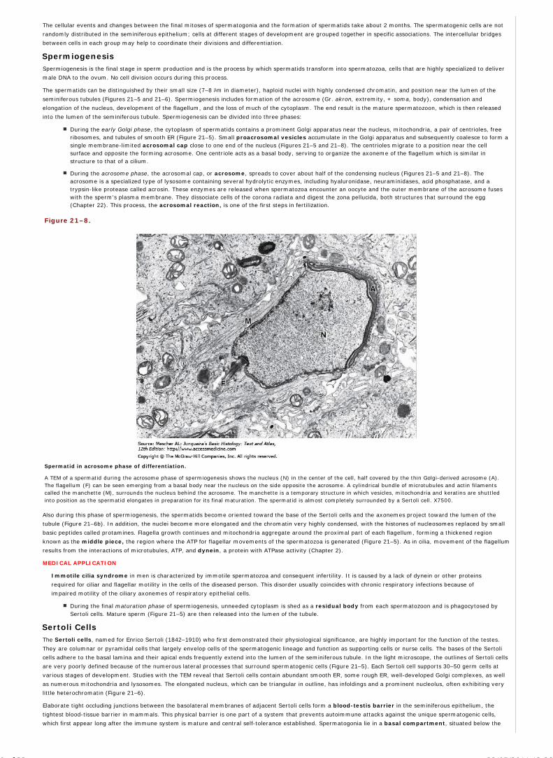
The cellular events and changes between the final mitoses of spermatogonia and the formation of spermatids take about 2 months. The spermatogenic cells are not
randomly distributed in the seminiferous epithelium; cells at different stages of development are grouped together in specific associations. The intercellular bridges
between cells in each group may help to coordinate their divisions and differentiation.
SpermiogenesisSpermiogenesis is the final stage in sperm production and is the process by which spermatids transform into spermatozoa, cells that are highly specialized to deliver
male DNA to the ovum. No cell division occurs during this process.
The spermatids can be distinguished by their small size (7–8 m in diameter), haploid nuclei with highly condensed chromatin, and position near the lumen of the
seminiferous tubules (Figures 21–5 and 21–6). Spermiogenesis includes formation of the acrosome (Gr. akron, extremity, + soma, body), condensation and
elongation of the nucleus, development of the flagellum, and the loss of much of the cytoplasm. The end result is the mature spermatozoon, which is then released
into the lumen of the seminiferous tubule. Spermiogenesis can be divided into three phases:
During the early Golgi phase, the cytoplasm of spermatids contains a prominent Golgi apparatus near the nucleus, mitochondria, a pair of centrioles, freeribosomes, and tubules of smooth ER (Figure 21–5). Small proacrosomal vesicles accumulate in the Golgi apparatus and subsequently coalesce to form asingle membrane-limited acrosomal cap close to one end of the nucleus (Figures 21–5 and 21–8). The centrioles migrate to a position near the cellsurface and opposite the forming acrosome. One centriole acts as a basal body, serving to organize the axoneme of the flagellum which is similar instructure to that of a cilium.
During the acrosome phase, the acrosomal cap, or acrosome, spreads to cover about half of the condensing nucleus (Figures 21–5 and 21–8). Theacrosome is a specialized type of lysosome containing several hydrolytic enzymes, including hyaluronidase, neuraminidases, acid phosphatase, and atrypsin-like protease called acrosin. These enzymes are released when spermatozoa encounter an oocyte and the outer membrane of the acrosome fuseswith the sperm's plasma membrane. They dissociate cells of the corona radiata and digest the zona pellucida, both structures that surround the egg(Chapter 22). This process, the acrosomal reaction, is one of the first steps in fertilization.
Figure 21–8.
Spermatid in acrosome phase of differentiation.
A TEM of a spermatid during the acrosome phase of spermiogenesis shows the nucleus (N) in the center of the cell, half covered by the thin Golgi-derived acrosome (A).The flagellum (F) can be seen emerging from a basal body near the nucleus on the side opposite the acrosome. A cylindrical bundle of microtubules and actin filamentscalled the manchette (M), surrounds the nucleus behind the acrosome. The manchette is a temporary structure in which vesicles, mitochondria and keratins are shuttledinto position as the spermatid elongates in preparation for its final maturation. The spermatid is almost completely surrounded by a Sertoli cell. X7500.
Also during this phase of spermiogenesis, the spermatids become oriented toward the base of the Sertoli cells and the axonemes project toward the lumen of the
tubule (Figure 21–6b). In addition, the nuclei become more elongated and the chromatin very highly condensed, with the histones of nucleosomes replaced by small
basic peptides called protamines. Flagella growth continues and mitochondria aggregate around the proximal part of each flagellum, forming a thickened region
known as the middle piece, the region where the ATP for flagellar movements of the spermatozoa is generated (Figure 21–5). As in cilia, movement of the flagellum
results from the interactions of microtubules, ATP, and dynein, a protein with ATPase activity (Chapter 2).
MEDICAL APPLICATION
Immotile cilia syndrome in men is characterized by immotile spermatozoa and consequent infertility. It is caused by a lack of dynein or other proteins
required for ciliar and flagellar motility in the cells of the diseased person. This disorder usually coincides with chronic respiratory infections because of
impaired motility of the ciliary axonemes of respiratory epithelial cells.
During the final maturation phase of spermiogenesis, unneeded cytoplasm is shed as a residual body from each spermatozoon and is phagocytosed bySertoli cells. Mature sperm (Figure 21–5) are then released into the lumen of the tubule.
Sertoli CellsThe Sertoli cells, named for Enrico Sertoli (1842–1910) who first demonstrated their physiological significance, are highly important for the function of the testes.
They are columnar or pyramidal cells that largely envelop cells of the spermatogenic lineage and function as supporting cells or nurse cells. The bases of the Sertoli
cells adhere to the basal lamina and their apical ends frequently extend into the lumen of the seminiferous tubule. In the light microscope, the outlines of Sertoli cells
are very poorly defined because of the numerous lateral processes that surround spermatogenic cells (Figure 21–5). Each Sertoli cell supports 30–50 germ cells at
various stages of development. Studies with the TEM reveal that Sertoli cells contain abundant smooth ER, some rough ER, well-developed Golgi complexes, as well
as numerous mitochondria and lysosomes. The elongated nucleus, which can be triangular in outline, has infoldings and a prominent nucleolus, often exhibiting very
little heterochromatin (Figure 21–6).
Elaborate tight occluding junctions between the basolateral membranes of adjacent Sertoli cells form a blood-testis barrier in the seminiferous epithelium, the
tightest blood-tissue barrier in mammals. This physical barrier is one part of a system that prevents autoimmune attacks against the unique spermatogenic cells,
which first appear long after the immune system is mature and central self-tolerance established. Spermatogonia lie in a basal compartment, situated below the
AccessMedicine | Print: Chapter 21. The Male Reproductive System http://accessmedicine.com/popup.aspx?aID=6182951&print=yes_chapter
9 of 23 23/05/2011 13:28

junctions and open to the vascularized interstitial tissue which contains lymphocytes and antigen-presenting cells. Early in meiosis, newly formed spermatocytes
temporarily disrupt the cell adhesion molecules of the most basal junctions, transiently establishing new junctions between adhesion factors in their own membranes
and those of Sertoli cells, and move into the adluminal compartment without compromising the blood-testis barrier. Spermatocytes and spermatids adhere closely
to the Sertoli cells, lying within deep invaginations of these cells' lateral and apical membranes, above the barrier. Movement of the spermatogenic cells between the
support cells while maintaining effective occluding junctions between all the cells is all the more impressive when one remembers that the germ cells remain linked by
intercellular bridges. As the flagellar tails of the spermatids develop, they appear as tufts extending from the apical ends of the Sertoli cells. Sertoli cells are also
connected and coupled ionically by numerous gap junctions, which may help regulate the transient changes in the occluding junctions and in coordinating the cycle of
the seminiferous epithelium described above.
Sertoli cells have several specific functions within the seminiferous epithelium, mostly involving the blood-testis barrier:
Support, protection, and nutrition of the developing spermatogenic cells. Because spermatocytes, spermatids, and sperm are isolated fromplasma proteins and nutrients by the blood-testis barrier, these spermatogenic cells depend on the Sertoli cells for production or transport into the lumen ofmetabolites and nutritive factors such as the iron-transport protein transferrin. Thus, while protecting spermatogenic cells from immune components inplasma, Sertoli cells must supply the plasma factors needed for growth and differentiation.
Exocrine and endocrine secretion. Sertoli cells continuously secrete into the seminiferous tubules a fluid used for sperm transport in the direction of thegenital ducts. Secretion of nutrients and androgen-binding protein (ABP), which concentrates testosterone to a level required for spermiogenesis, ispromoted by follicle-stimulating hormone (FSH). In an endocrine manner, Sertoli cells release the steroid estradiol derived from testosterone and secretethe 39 kDa glycoprotein inhibin, which in a feedback loop with the anterior pituitary gland suppresses synthesis and release of FSH. In the fetus Sertolicells also secrete a 140 kDa glycoprotein called müllerian-inhibiting substance (MIS) that causes regression of the embryonic müllerian(paramesonephric) ducts; without MIS these ducts persist and become parts of the female reproductive tract.
Phagocytosis. During spermiogenesis, excess cytoplasm shed as residual bodies is phagocytosed and digested by Sertoli cell lysosomes. No proteins fromsperm normally pass back across the blood-testis barrier.
MEDICAL APPLICATION
Spermiogenesis produces sperm-specific proteins. Because sexual maturity occurs long after the development of immunocompetence, differentiating sperm
cells could be recognized as foreign and provoke an autoimmune response that would damage germ cells. The blood-testis barrier, together with various
mechanisms to ensure a local immunotolerant environment, eliminates any interaction between developing sperm and the immune system. This barrier
prevents the passage of immunoglobulins into the seminiferous tubule and accounts for the lack of impaired fertility even in men whose serum contains high
levels of antibodies against sperm antigens.
INTERSTITIAL TISSUE
The interstitial tissue of the testis is the site of androgen production. The spaces between the seminiferous tubules are filled with connective tissue that contains mast
cells, macrophages, nerves, lymphatics, and blood vessels including fenestrated capillaries. During puberty, interstitial, or Leydig, cells become apparent as either
rounded or polygonal cells with central nuclei and eosinophilic cytoplasm rich in small lipid droplets (Figures 21–4). These cells produce the male
hormone testosterone, which is responsible for the development of the secondary male sex characteristics. Testosterone is synthesized by enzymes present in
mitochondria and the smooth ER in a system similar to that of adrenal cortical cells.
Just as Sertoli cells are stimulated by FSH, testosterone secretion by interstitial cells is promoted by the other gonadotropic hormone of the pituitary, luteinizing
hormone (LH), which is also called interstitial cell stimulating hormone (ICSH). Testosterone synthesis thus begins at puberty, when the hypothalamus begins
producing gonadotropin-releasing hormone. In the late embryonic testes gonadotropic hormone from the placenta stimulates interstitial cells to synthesize the
testosterone needed for development of the ducts and other parts of the male reproductive system. These fetal interstitial cells are very active during the third and
fourth months of pregnancy, then regress and become quiescent cells resembling fibroblasts until puberty when they resume testosterone synthesis in response to the
pituitary gonadotropin.
INTRATESTICULAR DUCTS
The intratesticular genital ducts are the straight tubules (tubuli recti), the rete testis, and the efferent ductules (Figure 21–2). These ducts carry spermatozoa
and liquid from the seminiferous tubules to the duct of the epididymis.
Most seminiferous tubules are in the form of loops, both ends of which join the rete testis by the short straight tubules. These tubules are recognized by the gradual
loss of spermatogenic cells, with an initial segment in which the walls are lined only by Sertoli cells (Figures 21–4a and 21–9), followed by a main segment consisting
of cuboidal epithelium supported by a dense connective tissue sheath. All the straight tubules empty into the rete testis, an interconnected network of channels lined
with cuboidal epithelium. The channels of the rete testis are embedded within the connective tissue of the mediastinum (Figure 21–9).
Figure 21–9.
AccessMedicine | Print: Chapter 21. The Male Reproductive System http://accessmedicine.com/popup.aspx?aID=6182951&print=yes_chapter
10 of 23 23/05/2011 13:28

Straight tubules and rete testis.
(a): The micrograph shows the long, looping seminiferous tubule (S) that drains into a short straight tubule (T), called a tubulus rectus. X120. H&E. (b): A highermagnification of one such junction shows that the transition to the straight tubule (T) is characterized by many tall Sertoli cells devoid of germ cells. The straight tubulesall empty into the rete testis (R), a network of interconnected channels embedded along with blood vessels (V) in the connective tissue (CT) of the mediastinum. Channelsof the rete testis are lined with simple cuboidal epithelium. X300. H&E.
The rete testis drains into about 20 efferent ductules (Figure 21–10). They are lined by an unusual epithelium with groups of nonciliated cuboidal cells alternating
with groups of taller ciliated cells. This gives the epithelium a characteristic scalloped appearance (Figure 21–10c). The nonciliated cells absorb most of the fluid
secreted by the seminiferous tubules. This absorption and the ciliary activity create a fluid flow that sweeps sperm toward the epididymis. A thin layer of circularly
oriented smooth muscle cells is seen outside the basal lamina of the epithelium which aids movement of the sperm. The efferent ductules empty into the duct of the
epididymis.
AccessMedicine | Print: Chapter 21. The Male Reproductive System http://accessmedicine.com/popup.aspx?aID=6182951&print=yes_chapter
11 of 23 23/05/2011 13:28

Figure 21–10.
AccessMedicine | Print: Chapter 21. The Male Reproductive System http://accessmedicine.com/popup.aspx?aID=6182951&print=yes_chapter
12 of 23 23/05/2011 13:28

Rete testis and efferent ductules.
(a): The micrograph shows the channels of the rete testis (R) drained by efferent ductules (E). A transition from rete testis to an efferent ductule is seen (arrows) andblood vessels (V) are seen in the mediastinum connective tissue (CT). X150. H&E. (b): The micrograph shows simple cuboidal epithelium that lines the rete testis (R).X350. Mallory trichrome. (c): The efferent ductules (E) are lined by a simple epithelium with a characteristic scalloped appearance in section, consisting of patches ofcuboidal cells with water-absorbing microvilli alternating with patches of taller cells with cilia. This epithelium creates a fluid flow that, together with contractile activity ofthe thin muscularis around the efferent ductules, carries sperm toward the epididymis. X350. H&E.
EXCRETORY GENITAL DUCTS
Excretory genital ducts are those of the epididymis, the ductus (vas) deferens, and the urethra. They transport sperm from the epididymis to the penis during
ejaculation.
The duct of the epididymis is a single highly coiled tube about 4–5 m in length. Together with a connective tissue capsule and blood vessels, this long duct forms the
head, body, and tail of the epididymis, which lies along the superior and posterior sides of each testis (Figure 21–2). Sperm are stored in the epididymis and attain
there various final characteristics including motility, membrane receptors for zona pellucida proteins, maturation of the acrosome, and ability to fertilize. The efferent
ductules join the duct in the head of the epididymis and it opens into the ductus (vas) deferens at the tail. The epididymal duct is lined with pseudostratified
columnar epithelium composed of rounded basal cells and columnar cells with long, branched, irregular microvilli called stereocilia. (Figure 21–11). The epithelial
cells of the epididymal duct absorb water and participate in the uptake and digestion of residual bodies produced during spermiogenesis. These cells are supported on
a basal lamina surrounded by smooth muscle cells, whose peristaltic contractions move the sperm along the duct, and by loose connective tissue rich in capillaries.
Figure 21–11.
AccessMedicine | Print: Chapter 21. The Male Reproductive System http://accessmedicine.com/popup.aspx?aID=6182951&print=yes_chapter
13 of 23 23/05/2011 13:28

Epididymis.
(a): The epididymis contains a long, highly coiled duct in which sperm are temporarily stored and undergo the final maturation steps required for their ability to fertilize anegg. The duct of the epididymis (DE) is enclosed by connective tissue that contain many blood vessels (V) and the connective tissue is covered by a capsule and the tunicavaginalis (TV). The duct is lined by a pseudostratified columnar epithelium with long stereocilia (arrows). The columnar cells are very tall and the stereocilia very long inthe head of the epididymis and both gradually shorten toward the tail. X140. H&E.
(b): The higher power micrograph shows the duct is surrounded by a thin circular layer of smooth muscle cells and the lumen is seen to contain sperm (S). The smoothmuscle becomes thicker and a longitudinal layer develops in the body and tail of the epididymis. X400. H&E. (c): Another section of the smooth muscle (SM) and wall ofthe duct shows two abundant cell types in the epithelium: the tall principal cells with stereocilia and small basal cells (B) on the basal lamina. Macrophages andintraepithelial lymphocytes are also commonly seen in the epididymal duct. X500. H&E.
From the epididymis the ductus (vas) deferens, a long straight tube with a thick, muscular wall, continues toward the prostatic urethra and empties into it (Figure
21–1). It is characterized by a narrow lumen and a thick layer of smooth muscle (Figure 21–12). Its mucosa is folded longitudinally and is lined along most of its
length by pseudostratified columnar epithelium with sparse stereocilia. The lamina propria is rich in elastic fibers and the very thick muscularis consists of longitudinal
inner and outer layers and a middle circular layer. The muscles produce strong peristaltic contractions during ejaculation which rapidly move sperm along this duct
from the epididymis.
Figure 21–12.
AccessMedicine | Print: Chapter 21. The Male Reproductive System http://accessmedicine.com/popup.aspx?aID=6182951&print=yes_chapter
14 of 23 23/05/2011 13:28

AccessMedicine | Print: Chapter 21. The Male Reproductive System http://accessmedicine.com/popup.aspx?aID=6182951&print=yes_chapter
15 of 23 23/05/2011 13:28

Ductus deferens.
The ductus (vas) deferens transports sperm from the epididymis during ejaculation and is contained within the spermatic cord. (a): The micrograph of a vas deferens incross-section shows that it consists of a mucosa (M), a thick muscularis with inner and outer layers of longitudinal smooth muscle (L-SM) and an intervening layer ofcircular smooth muscle (C-SM), and an external adventitia (A). The muscularis is specialized for powerful peristaltic movement of sperm at ejaculation. X60. H&E. (b): Thelamina propria (LP) is rich in elastic fibers and the thick epithelial lining (E) shows longitudinal folds. X150. Mallory trichrome. (c): Higher magnification of the mucosashows that the epithelium is pseudostratified with basal cells and many columnar cells with some stereocilia. X400. H&E.
The ductus deferens forms part of the spermatic cord, which includes the testicular artery, the pampiniform plexus, and nerves (Figure 21–2). After passing over the
urinary bladder, the ductus deferens dilates to form an ampulla (Figure 21–1), in which the epithelium is thicker and more extensively folded. At the final portion of
the ampulla, the seminal vesicles join the duct. From there on, the ductus deferens enters the prostate gland and opens into the prostatic urethra. The segment
entering the prostate is called the ejaculatory duct. The mucosa of the ductus deferens continues through the ejaculatory duct, but the muscle layers disappear
beyond the ampulla.
ACCESSORY GLANDS
The accessory glands of the male reproductive tract produce secretions that are added to sperm during ejaculation to produce semen and which are essential for
reproduction. The accessory genital glands are the seminal vesicles, the prostate gland, and the bulbourethral glands (Figure 21–13).
Figure 21–13.
Accessory glands of the male reproductive tract.
Three sets of glands connect to the ductus deferens or urethra: the paired seminal vesicles, the prostate, and the paired bulbourethral glands. The first two types of glandscontribute the major volume to semen and the latter produces a secretion that lubricates the urethra before ejaculation. (a): Micrograph shows the characteristically foldedmucosa of the seminal vesicles. X25. H&E. (b): Micrograph shows the characteristic individual tubuloalveolar glands of the prostate. X80. H&E.
The two seminal vesicles consist of highly tortuous tubes about 15 cm in length. The unusual mucosa displays a great number of thin, complex folds that fill most of
the lumen (Figure 21–14). The folds are lined with simple or pseudostratified columnar epithelial cells rich in secretory granules. The lamina propria contains elastic
fibers and is surrounded by smooth muscle with inner circular and outer longitudinal layers. The seminal vesicles are exocrine glands that produce a viscid, yellowish
secretion containing fructose, citrate, inositol, prostaglandins, fibrinogen, as well as enzymes and other proteins. These semen components, which typically
make up about 70% of the ejaculate, provide nutrient energy sources for the sperm, coagulate semen after ejaculation, and affect activity of the female reproductive
tract. The height of the seminal vesicle epithelial cells and their degree of secretory activity are dependent on adequate levels of testosterone.
Figure 21–14.
AccessMedicine | Print: Chapter 21. The Male Reproductive System http://accessmedicine.com/popup.aspx?aID=6182951&print=yes_chapter
16 of 23 23/05/2011 13:28

AccessMedicine | Print: Chapter 21. The Male Reproductive System http://accessmedicine.com/popup.aspx?aID=6182951&print=yes_chapter
17 of 23 23/05/2011 13:28

Seminal vesicles.
The seminal vesicles are paired exocrine glands that secrete most seminal fluid, including sperm nutrients. (a): A low-power micrograph shows that each consists of ahighly coiled duct surrounded by two layers of smooth muscle (SM) that expel the luminal contents during ejaculation. The mucosa characteristically displays thin primary,secondary, and tertiary folds (arrows) that give the lumen (L) a distinctive appearance. X20. H&E. (b, c): Both micrographs show that the folds include smooth muscle(SM) covered by a thin lamina propria (LP) and an epithelium. The epithelial cells are simple or pseudostratified columnar, varying with activity and location in the gland,and contain lipid droplets, secretory granules, and also commonly lipofuscin (L). Both: X300. b: H&E; c: PSH.
The prostate gland is a dense organ surrounding the urethra below the bladder. It is approximately 2 cm x 3 cm x 4 cm in size and weighs about 20 g. The prostate
is a collection of 30–50 branched tubuloalveolar glands, all surrounded by a dense fibromuscular stroma covered by a capsule. The glands are arranged in concentric
layers around the urethra: the inner layer of mucosal glands, an intermediate layer of submucosal glands, and a peripheral layer with the prostate's main
glands (Figure 21–15). Ducts from individual glands may converge but all empty directly into the prostatic urethra, which runs through the center of the prostate
(Figures 21–1). The prostate has three zones, corresponding to the glandular layers:
The transition zone occupies about 5% of the prostate volume, surrounds the prostatic urethra, and contains the mucosal glands emptying directly intothe urethra.
The central zone occupies 25% of the gland's volume and contains the submucosal glands with longer ducts.
The peripheral zone occupies about 70% of the prostate and contains the main glands with still longer ducts. Glands of this area are the most commonlocation of both inflammation and cancer.
Figure 21–15.
Organization of the prostate gland.
The prostate secretes and stores a significant contribution to the seminal fluid that is released at ejaculation. It consists of 30–50 branched tubuloalveolar glands organizedinto three layers, shown here schematically. Around the prostatic urethra is the transition zone which contains the mucosal glands. Surrounding most of that zone is theintermediate central zone, which contains the submucosal glands. The outermost and largest layer is the peripheral zone, which contains the most numerous main glands.Glands of all the layers contribute to the prostatic secretion.
The tubuloalveolar glands of the prostate are lined by a simple or pseudostratified columnar epithelium. The glands produce prostatic fluid containing various
glycoproteins and enzymes and store this fluid for expulsion during ejaculation. An exceptionally rich fibromuscular stroma surrounds the glands (Figure 21–16). The
prostate is surrounded by a fibroelastic capsule. Septa from this capsule penetrate the gland and divide it into indistinct lobes. As with the seminal vesicle, the
structure and function of the prostate depend on the level of testosterone.
AccessMedicine | Print: Chapter 21. The Male Reproductive System http://accessmedicine.com/popup.aspx?aID=6182951&print=yes_chapter
18 of 23 23/05/2011 13:28

Figure 21–16.
AccessMedicine | Print: Chapter 21. The Male Reproductive System http://accessmedicine.com/popup.aspx?aID=6182951&print=yes_chapter
19 of 23 23/05/2011 13:28

Prostate gland.
(a): The prostate has a dense fibromuscular stroma (S) in which are embedded a large number of small tubuloalveolar glands (G). The arrows indicate sites of calcifiedconcretions that have been lost during sectioning. X20. H&E. (b): A micrograph of one gland, including a corpus amylaceum (CA) concretion, shows a secretory simple orpseudostratified columnar epithelium (E) surrounded by lamina propria (LP), which is in turn surrounded by smooth muscle (M). X122. H&E. (c): Higher magnificationshows the lamellar nature of a corpus amylaceum (CA) and the columnar epithelium underlain by sparse lamina propria (LP). X300. Mallory trichrome.
MEDICAL APPLICATION
Benign prostatic hypertrophy is present in 50% of men older than 50 years and in 95% of men older than 70 years. It generally occurs in the transition
zone surrounding the urethra and can lead to compression of the urethra with clinical symptoms.
Prostatic cancer is one of the most common forms of cancer in men. A secretory product of the prostate, a serine protease known clinically as prostate-
specific antigen (PSA), leaks into the blood in the glandular disruption caused by prostate cancer. Low levels of circulating PSA are also produced in the liver,
but an increased PSA concentration in the blood is routinely used to diagnose and monitor prostate cancer.
Small spherical concretions, 0.2–2 mm in diameter and often calcified, are frequently observed in the lumens of the prostatic glands (Figure 21–16). They are called
corpora amylacea, or prostatic concretions and contain primarily deposited glycoproteins and sulfated glycosaminoglycans (GAGs), particularly keratan sulfate.
Their number often increases with age, but they seem to have no physiological or clinical significance.
The paired round bulbourethral glands (Cowper's glands), 3–5 mm in diameter, are located in the urogenital diaphragm (Figure 21–1) and empty into the proximal
part of the penile urethra. Each gland has several lobules with tubuloalveolar secretory units lined by a mucus-secreting simple columnar epithelium dependent on
testosterone. The septa between lobules contain smooth muscle cells. During erection the bulbourethral glands, as well as numerous, small, and histologically similar
urethral glands along the urethra, release a clear mucus-like secretion containing various small carbohydrates, which coats and lubricates the urethral lining in
preparation for the imminent passage of sperm.
PENIS
The main components of the penis are three cylindrical masses of erectile tissue, plus the penile urethra, surrounded by skin (Figure 21–1). Two of these
cylinders—the corpora cavernosa—are placed dorsally. The other—the corpus spongiosum—is ventral and surrounds the urethra (Figures 21–1 and 21–17). At its
end the corpus spongiosum expands, forming the glans (Figure 21–1). Most of the penile urethra is lined with pseudostratified columnar epithelium. In the glans, it
becomes stratified squamous epithelium continuous with that of the thin epidermis covering the glans. Small mucus-secreting urethra glands (glands of Littre) are
found along the length of the penile urethra. In uncircumcised men the surface of the glans is covered by the prepuce or foreskin, a retractile fold of thin skin with
sebaceous glands in the internal fold.
Figure 21–17.
AccessMedicine | Print: Chapter 21. The Male Reproductive System http://accessmedicine.com/popup.aspx?aID=6182951&print=yes_chapter
20 of 23 23/05/2011 13:28

Structure of the penis.
A diagram of a transverse section of the penis shows the relationships of the three erectile bodies, tunica albuginea, and major blood vessels. Compare this section to thediagram in Figure 21–1.
The corpora cavernosa are covered by a resistant layer of dense connective tissue, the tunica albuginea (Figures 21–17 and 21–18). The corpora cavernosa and the
corpus spongiosum are both composed of erectile tissue, which contains a large number of venous cavernous spaces lined with endothelial cells and separated by
trabeculae of connective tissue fibers and smooth muscle cells.
Figure 21–18.
Penis.
The corpus spongiosum (CS) is on the ventral side of the penis and surrounds the urethra (U). Two corpora cavernosa (CC) fill the dorsal side and all three bodies ofcavernous or erectile tissue are surrounded by dense, fibrous tunica albuginea (TA). Along the dorsal side run the major blood vessels (V) and deep in each mass of erectiletissue are smaller blood vessels (V), including the central arteries. Externally the penis is covered by skin (S) attached to the tunica albuginea or neighboring connectivetissue. X15. H&E.
Figure 21–19.
AccessMedicine | Print: Chapter 21. The Male Reproductive System http://accessmedicine.com/popup.aspx?aID=6182951&print=yes_chapter
21 of 23 23/05/2011 13:28

Penile urethra and erectile tissue.
(a): The micrograph shows the corpus spongiosum (CS) surrounding the penile urethra (PU) with its longitudinally folded wall. Near the penile urethra are small urethralglands (UG) with short ducts for the release of a mucus-like secretion into the urethra during erection. These glands supplement the similar function of the larger, pairedbulbourethral glands. In one of the two dorsal corpora cavernosa (CC) small helicine arteries (HA) are visible. The bodies of erectile tissue are ensheathed by dense,nonelastic tunica albuginea (TA) connective tissue. X100. H&E.
(b): A higher magnification of erectile tissue is shown with a small portion of tunica albuginea (TA). The erectile bodies are composed of fibrous, elastic connective tissuecompletely infiltrated with hundreds of small, flaccid vascular cavernous spaces (CS) lined by endothelium. Very little blood normally passes through this vasculature dueto constriction of the helicine arteries (HA) serving them. During the erection process, smooth muscle of the helicine arteries relaxes, allowing rapid blood flow into thecavernous spaces, filling them and causing compression of their venous drainage in the tunica albuginea and fibroelastic trabeculae. This leads to full capacity filling of thecavernous spaces, swelling of the tissue volume, and turgidity of the erectile tissue bodies. X200. H&E.
The arterial supply of the penis derives from the internal pudendal arteries, which give rise to the deep arteries and the dorsal arteries of the penis. Deep arteries
branch to form nutritive arteries to the trabeculae and coiling helicine arteries, which empty directly into the cavernous spaces of erectile tissue. There are
arteriovenous shunts between the helicine arteries and the deep dorsal vein.
Penile erection involves filling the cavernous spaces of the corpora cavernosa and corpus spongiosum with blood. This is initiated by external stimuli to the CNS and is
controlled by autonomic nerve input to smooth muscle in vascular walls of the penis. Parasympathetic stimulation relaxes muscle in the trabeculae and dilates the
AccessMedicine | Print: Chapter 21. The Male Reproductive System http://accessmedicine.com/popup.aspx?aID=6182951&print=yes_chapter
22 of 23 23/05/2011 13:28

Copyright © The McGraw-Hill Companies. All rights reserved.Privacy Notice. Any use is subject to the Terms of Use and Notice.
helicine arteries, leading to increased blood flow into the cavernous spaces. The filled spaces compress the venules and veins against the dense tunica albuginea,
blocking outflow of the blood and producing tumescence and rigidity in the cylinders of erectile tissue. Beginning with ejaculation, the firing of sympathetic nerves
stimulates constriction of the helicine arteries, decreasing blood flow into the spaces, lowering the pressure there and allowing the veins to open and drain most blood
from the erectile tissue.
MEDICAL APPLICATION
In producing an erection parasympathetic nerves in the vasculature release nitric oxide (NO) which leads to increased NO synthesis by endothelial cells. NO
stimulates cGMP (cyclic guanosine monophosphate) production in adjacent smooth muscle cells and this intracellular messenger triggers sequestration of Ca2+
ions in smooth ER, causing the muscle cells to relax. Diffusion of NO and cGMP throughout the smooth muscle via gap junctions produces relaxation and
vasodilatation of the entire local vasculature and rapid in-flow of blood.
Arterial or neural insufficiency to tissue of the penis can lead to erectile dysfunction which can be treated effectively in many cases with compounds like
sildenafil, which inhibit the phosphodiesterase that degrades cGMP and thereby enhance and prolong the effect of NO on the smooth muscle.
AccessMedicine | Print: Chapter 21. The Male Reproductive System http://accessmedicine.com/popup.aspx?aID=6182951&print=yes_chapter
23 of 23 23/05/2011 13:28

Print Close Window
Note: Large images and tables on this page may necessitate printing in landscape mode.
Copyright © The McGraw-Hill Companies. All rights reserved.
Junqueira's Basic Histology: Text & Atlas, 12e > Chapter 22. The Female Reproductive System >
THE FEMALE REPRODUCTIVE SYSTEM: INTRODUCTION
The female reproductive system consists of two ovaries and oviducts (or uterine tubes), the uterus, the vagina, and the external genitalia (Figure 22–1). The
functions of this system are to produce female gametes (oocytes), provide the environment for fertilization, and to hold the embryo during its complete
development through the fetal stage until birth. As in the male, the female reproductive system produces steroidal sex hormones that control organs of the
reproductive system and influence other organs of the body. Beginning at menarche, when the first menses occurs, the reproductive system undergoes cyclic
changes in structure and functional activity. These modifications are controlled by neurohumoral mechanisms. Menopause is a variable period during which the
cyclic changes become irregular and eventually disappear. In the postmenopausal period there is a slow involution of the reproductive organs. Although the
mammary glands do not belong to the genital system, they are included here because they undergo changes directly connected to the functional state of the
reproductive system.
Figure 22–1.
The female reproductive system and overview of ovary.
(a): The diagram shows the internal organs of the female reproductive system, which includes as the principal organs the ovaries, uterine tubes, uterus, and vagina.(b): A lateral sectional view of an ovary shows the ovary and the relationship of its main supporting mesenteries, the mesovarium and the mesosalpinx of the broadligament. (c): Micrograph of a sectioned ovary, indicating the medullary and cortical regions, with follicles of several different sizes in the cortex. X15. H&E.
OVARIES
Ovaries are almond-shaped bodies approximately 3 cm long, 1.5 cm wide, and 1 cm thick. Each ovary is covered by a simple cuboidal epithelium, the
germinal epithelium, continuous with the mesothelium and overlying a layer of dense connective tissue capsule, the tunica albuginea, like that of the testis
and responsible for the whitish color of the ovary. Most of the ovary consists of the cortex, a region filled with a highly cellular connective tissue stroma and
many ovarian follicles, which in the adult ovary vary greatly in size (Figure 22–1). The most internal part of the ovary is the medulla, which contains loose
connective tissue and blood vessels entering the organ through the hilum from mesenteries suspending the ovary (Figures 22–1 and 22–2). There are no sharp
AccessMedicine | Print: Chapter 22. The Female Reproductive System http://accessmedicine.com/popup.aspx?aID=6183071&print=yes_chapter
1 of 34 23/05/2011 13:29

limits between the ovarian cortical and medullary regions.
Figure 22–2.
Follicle development and changes within the ovary.
The ovary produces both oocytes and sex hormones. A diagram of a sectioned ovary (a), shows the different stages of follicle maturation, ovulation, and corpusluteum formation and degeneration. All of the stages and structures shown in this diagram actually would appear at different times during the ovarian cycle and donot occur simultaneously. Follicles are arranged here for easy comparisons. The primordial follicles shown are greatly enlarged. The histological sections identifyprimordial follicles (b), a primary follicle (c), a secondary follicle (d), and a large vesicular follicle (e). After ovulation, the portion of the follicle left behind forms thecorpus luteum (f), which then degenerates into the corpus albicans (g). All H&E.
Early Development of the OvaryIn the first month of embryonic life, a small population of primordial germ cells migrates from the yolk sac to the gonadal primordia. In the gonads these
cells divide extensively and differentiate as oogonia. In developing ovaries of a two-month embryo there are about 600,000 oogonia which produce more than
7 million by the fifth month. Beginning in the third month, oogonia begin to enter the prophase of the first meiotic division but arrest after completing synapsis
and recombination, without progressing to later stages of meiosis (see Chapter 3). These cells arrested in meiosis are called primary oocytes (Gr. oon, egg, +
kytos, cell). Each primary oocyte becomes surrounded by flattened supportive cells called follicular cells within an ovarian follicle. By the seventh month of
development, most oogonia have transformed into primary oocytes within follicles. Many primary oocytes, however, are lost through a slow, continuous
degenerative process called atresia, which continues through a woman's reproductive life. At puberty the ovaries contain about 300,000 oocytes. Because
generally only one oocyte resumes meiosis with ovulation during each menstrual cycle (average duration, 28 days) and the reproductive life of a woman lasts
about 30–40 years, only about 450 oocytes are liberated from ovaries by ovulation. All others degenerate through atresia.
Ovarian FolliclesAn ovarian follicle consists of an oocyte surrounded by one or more layers of epithelial cells. The follicles that are formed during fetal life—primordial follicles
—consist of a primary oocyte enveloped by a single layer of the flattened follicular cells (Figures 22–2, 22–3, and 22–4). These follicles are found in the
superficial areas of the cortex. The oocyte in the primordial follicle is a spherical cell about 25 m in diameter, with a large nucleus and mostly uncoiled
AccessMedicine | Print: Chapter 22. The Female Reproductive System http://accessmedicine.com/popup.aspx?aID=6183071&print=yes_chapter
2 of 34 23/05/2011 13:29

chromosomes in the first meiotic prophase. The organelles tend to be concentrated near the nucleus and include numerous mitochondria, several Golgi
complexes, and extensive ER cisternae. A basal lamina surrounds the follicular cells, marking a clear boundary between the follicle and the vascularized stroma.
Figure 22–3.
Stages of ovarian follicles, from primordial to mature.
Diagrams of sectioned ovarian follicles show the changing size and morphology of follicular/granulosa cells at each stage and the disposition of the surroundingthecal cells. However, the relative proportions of the follicles are not maintained in the series of drawings: mature follicles are much larger relative to the earlyfollicles.
Figure 22–4.
AccessMedicine | Print: Chapter 22. The Female Reproductive System http://accessmedicine.com/popup.aspx?aID=6183071&print=yes_chapter
3 of 34 23/05/2011 13:29

Primordial ovarian follicles.
The cortical region of an ovary is surrounded by the surface epithelium (SE), a mesothelium with usually cuboidal cells. This layer is sometimes called the germinalepithelium because of an early erroneous view that it was the source of oogonia precursor cells. Underlying the epithelium is a connective tissue layer, the tunicaalbuginea (TA). Groups of primordial follicles, each formed by an oocyte (O) surrounded by a layer of flat epithelial follicular cells (arrows), are present in the ovarianconnective tissue (stroma). X200. H&E.
Follicular GrowthBeginning in puberty with the release of follicle-stimulating hormone (FSH) from the pituitary, a small group of primordial follicles each month begins a process
of follicular growth. This involves growth of the oocyte, proliferation and changes in the follicular cells, and proliferation and differentiation of the stromal
fibroblasts around each follicle. Selection of the primordial follicles that undergo growth and recruitment early in each cycle and of the dominant follicle destined
to ovulate that month both involve complex hormonal balances and subtle differences among follicles in FSH receptor numbers, aromatase activity and estrogen
synthesis, and other variables.
Prompted by FSH, an oocyte grows most rapidly during the first part of follicular development, reaching a maximum diameter of about 120 m. The nucleus
enlarges; the mitochondria become more numerous and uniformly distributed; the ER becomes much more extensive; and the Golgi complexes enlarge and
move peripherally. Follicular cells undergo mitosis and form a simple cuboidal epithelium around the growing oocyte. The follicle is now called a unilaminar
primary follicle (Figures 22–3 and 22–5a). The follicular cells continue to proliferate, forming a stratified follicular epithelium, the granulosa, in which the
cells communicate through gap junctions. Follicle cells are now termed granulosa cells and the follicle is a multilayered primary follicle (Figures 22–3 and
22–5b) still surrounded by basement membrane. Between the oocyte and granulosa cells, a layer of extracellular material called the zona pellucida develops,
5 to 10 m thick and consisting of four glycoproteins secreted by the oocyte (Figures 22–5 and 22–6). Zona pellucida components, ZP1 through 4, bind proteins
on the surfaces of sperm and induce acrosomal activation. Filopodia of follicular cells and microvilli of the oocyte penetrate the zona pellucida, allowing
communication between these cells via gap junctions.
Figure 22–5.
AccessMedicine | Print: Chapter 22. The Female Reproductive System http://accessmedicine.com/popup.aspx?aID=6183071&print=yes_chapter
4 of 34 23/05/2011 13:29

Primary follicles.
(a): A micrograph of ovarian cortex shows several primordial follicles (PF) and their flattened follicles cells (arrows), and two unilaminar primary follicles (UF) inwhich the follicles cells or granulosa cells (G) form a single cuboidal layer around the large primary oocyte (O). X200. PT. (b): This micrograph was taken at thesame magnification and shows a larger multilayered primary follicle. Granulosa cells (G) have now proliferated to form several layers. Between them and the oocyte(O) is the 5–10 m thick zona pellucida (ZP), a glycoprotein layer produced by the oocyte that is required for sperm binding and fertilization. The primary oocyte isnow a very large cell. With this stain the basement membrane (BM) that separates the follicle from the surrounding stroma can also be seen. X200. PSH.
Figure 22–6.
AccessMedicine | Print: Chapter 22. The Female Reproductive System http://accessmedicine.com/popup.aspx?aID=6183071&print=yes_chapter
5 of 34 23/05/2011 13:29

Ultrastructure of primary follicle and zona pellucida.
An SEM of a fractured primary follicle shows the oocyte surrounded by granulosa cells (GC). Between the very large oocyte surface and the granulosa cells is a fibrouslayer of extracellular material, the zona pellucida (ZP), which contains four related glycoproteins that bind sperm and form an irregular meshwork. X3000.
As the follicles grow with increasing oocyte size and numbers of granulosa cells, they move deeper in the ovarian cortex. Small spaces develop within the
granulosa layer as the cells secrete follicular fluid (or liquor folliculi). This fluid accumulates, the spaces gradually coalesce, and the granulosa cells
reorganize themselves around a larger cavity, the antrum (Figures 22–3 and 22–7a), producing follicles now called secondary or antral follicles. Follicular
fluid contains hyaluronate, growth factors, plasminogen, fibrinogen, the anticoagulant heparan sulfate proteoglycan, and high concentrations of steroids
(progesterone, androstenedione, and estrogens) with binding proteins.
Figure 22–7.
AccessMedicine | Print: Chapter 22. The Female Reproductive System http://accessmedicine.com/popup.aspx?aID=6183071&print=yes_chapter
6 of 34 23/05/2011 13:29

Antral follicles.
(a): A micrograph with part of an antral follicle shows the large, fluid-filled antral cavities or vesicles (A) that appear in the granulosa layer as the cells producefollicular fluid. The oocyte (O) is surrounded by the zona pellucida (ZP) and granulosa cells (G), which also line the wall of the follicle. Fibroblastic cells immediatelyoutside the growing follicles have developed as a steroid-secreting theca interna (TI) and a covering theca externa (TE). X100. H&E.
(b): A slightly more developed antral follicle shows a verge large single antrum (A) filled with follicular fluid in which the proteins formed a thin film during fixation.The oocyte (O) now projects into this fluid-filled cavity, still surrounded by granulosa cells which now make up the corona radiata (CR). Cells of the corona radiata arelinked to the oocyte via gap junctions between processes that cross the zona pellucida. The corona radiata and oocyte are attached to the side of the follicle via a lessdense mass of granulosa cells called the cumulus oophorus (CO) which is continuous with the remaining granulosa cells that form the wall of the follicle and surroundthe antrum. Thecae interna (TI) and externa (TE) surround the whole follicle. X100. PT.
During the reorganization of the granulosa layer to form the antrum, some cells form a small hillock, the cumulus oophorus, surrounding the oocyte and
protruding into the antrum (Figures 22–3 and 22–7b). The granulosa cells immediately around and linked to the oocyte make up the corona radiata and
accompany the oocyte when it leaves the ovary.
While the follicle develops, the stromal cells immediately around the follicle differentiate to form the follicular theca (Gr. theca, outer covering). This layer
subsequently differentiates further as two tissues around the follicle: a well-vascularized endocrine tissue, the theca interna, and a more fibrous outer theca
externa containing smooth muscle and fibroblasts (Figures 22–3, 22–7, and 22–8). The cells of the theca interna differentiate as steroid-producing cells, with
abundant smooth ER, mitochondria with tubular cristae, and numerous lipid droplets. These cells secrete a steroid hormone—androstenedione—that is
transported to the granulosa where, under the influence of FSH, the cells synthesize an enzyme, aromatase, that transforms the steroid into estradiol. This
estrogen returns to the thecae and stroma around the follicle, enters capillaries, and is distributed throughout the body. The boundary between the two thecal
layers is not clear and neither is there a sharp boundary between the theca externa and the rest of the stroma. On the other hand, the boundary between the
theca interna and the granulosa layer is well-defined, since their cells are quite different and there is a thick basement membrane between them (Figure 22–8).
Figure 22–8.
AccessMedicine | Print: Chapter 22. The Female Reproductive System http://accessmedicine.com/popup.aspx?aID=6183071&print=yes_chapter
7 of 34 23/05/2011 13:29

Wall of antral follicle.
At higher magnification, a small part of the wall of an antral follicle shows the cell layers of the granulosa (G) next to the antrum (A), in which proteins haveaggregated on cells in contact with the follicular fluid. The theca interna (TI) surrounds the follicle, its cells appearing vacuolated and lightly stained because of theircytoplasmic lipid droplets, a characteristic of steroid-producing cells. The overlying theca externa (TE) contains fibroblasts and smooth muscle cells and merges withthe stroma (S). A basement membrane (BM) separates the theca interna from the granulosa, blocking vascularization of the latter. X400. PT.
During each menstrual cycle, usually one follicle grows much more than the others and becomes the dominant follicle, while many of the other follicles
eventually enter atresia. The dominant follicle usually reaches the most developed stage of follicular growth and may undergo ovulation. This mature or
preovulatory follicle (sometimes called the graafian follicle after the 17th century Dutch reproductive biologist Regnier De Graaf) reaches a diameter of
20-30 mm or more prior to ovulation, large enough to protrude from the surface of the ovary and be detected by ultrasound imaging. The antrum increases
greatly in size by accumulating follicular fluid and the oocyte adheres to the wall of the follicle through the cumulus oophorus of granulosa cells (Figure 22–3).
Since the granulosa cells of the follicle wall do not multiply in proportion to the growth of the antrum, the granulosa layer becomes thinner. A mature follicle has
very thick thecal layers and normally develops from a primordial follicle over a period of about 90 days.
Follicular AtresiaMost ovarian follicles undergo the degenerative process called atresia, in which follicular cells and oocytes die and are disposed of by phagocytic cells. Follicles
at any stage of development, including nearly mature follicles, may become atretic (Figure 22–9). Atresia involves apoptosis and detachment of the granulosa
cells, autolysis of the oocyte and collapse of the zona pellucida. Early in this process, macrophages invade the degenerating follicle and phagocytose the debris.
Later fibroblasts occupy the area of the follicle and produce a collagen scar that may persist for a long time. Although follicular atresia takes place from before
birth until a few years after menopause, it is most prominent just after birth, when levels of maternal hormones decline rapidly, and during both puberty and
pregnancy, when qualitative and quantitative hormonal changes occur again.
Figure 22–9.
Atresia.
Atresia or degeneration of a follicle can occur at any stage of its development and is shown here in a follicle that had developed a large antrum. Atresia ischaracterized by apoptosis of granulosa cells (G) and autolysis of the oocyte, with macrophages entering the degenerating structure to clean up debris. Manyapoptotic cells are seen loose in the antrum (A) here and the cells of the corona radiata have already disappeared, leaving the degenerative oocyte (O) free withinthe antrum. X200. PT.
OvulationAt ovulation the large mature primary oocyte escapes from the ovary and is caught by the dilated end of the uterine tube which is closely applied to the
ovarian surface at that time. Ovulation normally occurs midway through the menstrual cycle, ie, around the fourteenth day of a typical 28-day cycle. In humans
AccessMedicine | Print: Chapter 22. The Female Reproductive System http://accessmedicine.com/popup.aspx?aID=6183071&print=yes_chapter
8 of 34 23/05/2011 13:29
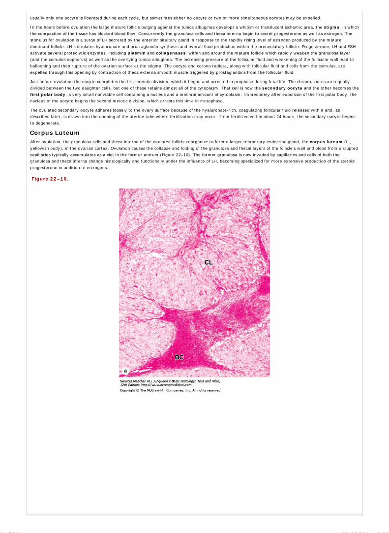
usually only one oocyte is liberated during each cycle, but sometimes either no oocyte or two or more simultaneous oocytes may be expelled.
In the hours before ovulation the large mature follicle bulging against the tunica albuginea develops a whitish or translucent ischemic area, the stigma, in which
the compaction of the tissue has blocked blood flow. Concurrently the granulosa cells and theca interna begin to secret progesterone as well as estrogen. The
stimulus for ovulation is a surge of LH secreted by the anterior pituitary gland in response to the rapidly rising level of estrogen produced by the mature
dominant follicle. LH stimulates hyaluronate and prostaglandin synthesis and overall fluid production within the preovulatory follicle. Progesterone, LH and FSH
activate several proteolytic enzymes, including plasmin and collagenases, within and around the mature follicle which rapidly weaken the granulosa layer
(and the cumulus oophorus) as well as the overlying tunica albuginea. The increasing pressure of the follicular fluid and weakening of the follicular wall lead to
ballooning and then rupture of the ovarian surface at the stigma. The oocyte and corona radiata, along with follicular fluid and cells from the cumulus, are
expelled through this opening by contraction of theca externa smooth muscle triggered by prostaglandins from the follicular fluid.
Just before ovulation the oocyte completes the first meiotic division, which it began and arrested in prophase during fetal life. The chromosomes are equally
divided between the two daughter cells, but one of these retains almost all of the cytoplasm. That cell is now the secondary oocyte and the other becomes the
first polar body, a very small nonviable cell containing a nucleus and a minimal amount of cytoplasm. Immediately after expulsion of the first polar body, the
nucleus of the oocyte begins the second meiotic division, which arrests this time in metaphase.
The ovulated secondary oocyte adheres loosely to the ovary surface because of the hyaluronate-rich, coagulating follicular fluid released with it and, as
described later, is drawn into the opening of the uterine tube where fertilization may occur. If not fertilized within about 24 hours, the secondary oocyte begins
to degenerate.
Corpus LuteumAfter ovulation, the granulosa cells and theca interna of the ovulated follicle reorganize to form a larger temporary endocrine gland, the corpus luteum (L.,
yellowish body), in the ovarian cortex. Ovulation causes the collapse and folding of the granulosa and thecal layers of the follicle's wall and blood from disrupted
capillaries typically accumulates as a clot in the former antrum (Figure 22–10). The former granulosa is now invaded by capillaries and cells of both the
granulosa and theca interna change histologically and functionally under the influence of LH, becoming specialized for more extensive production of the steroid
progesterone in addition to estrogens.
Figure 22–10.
AccessMedicine | Print: Chapter 22. The Female Reproductive System http://accessmedicine.com/popup.aspx?aID=6183071&print=yes_chapter
9 of 34 23/05/2011 13:29

Corpus luteum.
The corpus luteum is a large endocrine structure formed from the remains of the large dominant follicle after it undergoes ovulation. (a): The low-power micrographshows the corpus luteum (CL), characterized by folds of the former granulosa which collapses as the theca externa contracts at ovulation. The former antrum oftencontains a blood clot (BC) from vessels in the thecal layers disrupted during ovulation. Cells of the granulosa and theca interna become reorganized under theinfluence of pituitary LH and their names are changed. X15. H&E. (b): Granulosa lutein cells (GL), seen at higher magnification here, undergo significanthypertrophy, producing most of the corpus luteum's increased size, and begin producing progesterone. The theca lutein cells (TL) increase only slightly in size, aresomewhat darker-staining than the granulosa lutein cells, and continue to produce estrogens. Theca lutein cells, derived from the theca interna, are typically locatedwithin the folds that comprise the bulk of this tissue. X100. H&E.
Granulosa cells increase greatly in size (20–35 m in diameter), without dividing, and eventually comprise about 80% of the parenchyma of the corpus luteum.
They are called granulosa lutein cells (Figure 22–10) and now have lost many features of protein-secreting cells to expand their role in aromatase conversion
of androstenedione into estradiol. The former theca interna contributes the other component of the corpus luteum, giving rise to theca lutein cells (Figure
22–10). These cells are less than half the size of the granulosa lutein cells and often stain more darkly, with cytoplasmic ultrastructural features of steroid-
synthesizing cells. LH causes these cells to produce large amounts of progesterone as well as androstenedione. Theca lutein cells are typically aggregated in the
folds of the wall of the corpus luteum, which like all endocrine glands becomes well vascularized.
The fate of the corpus luteum depends on whether a pregnancy occurs. Following the LH surge, the corpus luteum is programmed to secrete progesterone for
10–12 days. Without further LH stimulation and in the absence of pregnancy, both major cell types of the corpus luteum cease steroid production and undergo
apoptosis while the tissue regresses. A consequence of the decreased secretion of progesterone is menstruation, the shedding of part of the uterine mucosa.
Estrogen produced by the active corpus luteum inhibits FSH release from the pituitary. However, after the corpus luteum degenerates, the concentration of
blood steroids decreases and FSH secretion increases again, stimulating the growth of another group of follicles and beginning the next menstrual cycle. The
corpus luteum that persists for part of only one menstrual cycle is called a corpus luteum of menstruation. Remnants from its degeneration and regression
are phagocytosed by macrophages, after which fibroblasts invade the area and produce a scar of dense connective tissue called corpus albicans (L., white
body) (Figure 22–11).
Figure 22–11.
AccessMedicine | Print: Chapter 22. The Female Reproductive System http://accessmedicine.com/popup.aspx?aID=6183071&print=yes_chapter
10 of 34 23/05/2011 13:29

Corpus albicans.
A corpus albicans (CA) is the scar of connective tissue that forms at the site of a corpus luteum after its involution. It contains mostly collagen, with few fibroblastsor other cells, and gradually becomes very small and lost in the ovarian stroma. Involution of the corpus luteum does not involve atresia. X60. H&E.
If pregnancy occurs, the uterine mucosa cannot be allowed to undergo menstruation since the embryo would be lost. To prevent the drop in circulating
progesterone, trophoblast cells of the implanting embryo produce a glycoprotein hormone called human chorionic gonadotropin (HCG) whose action is
similar to that of LH. HCG targets the corpus luteum, maintaining it and promoting further growth of this endocrine gland and stimulating secretion of
progesterone to maintain the uterine mucosa. Progesterone also stimulates secretion in the uterine mucosal glands, which is important for embryonic nutrition
before the placenta is functional. This corpus luteum of pregnancy becomes very large and is maintained by HCG for 4–5 months, by which time the
placenta itself produces progesterone (and estrogens) at levels adequate to maintain the uterine mucosa. It then degenerates and is replaced by a large corpus
albicans.
UTERINE TUBES
The uterine tubes, or oviducts are two muscular tubes (Figure 22–12) with considerable mobility, each measuring about 12 cm in length. Each has a funnel-
shaped end, the infundibulum, opening into the peritoneal cavity next to the ovary and with a fringe of fingerlike extensions called fimbriae (L., fringes). The
sequence of regions along each tube is as follows:
the infundibulum,
the ampulla, the longest and expanded area where fertilization normally occurs,
the isthmus, a more narrow region nearer the uterus, and
the uterine or intramural part, which passes through the wall of the uterus and opens into the interior of this organ.
Figure 22–12.
AccessMedicine | Print: Chapter 22. The Female Reproductive System http://accessmedicine.com/popup.aspx?aID=6183071&print=yes_chapter
11 of 34 23/05/2011 13:29

Uterine tubes and uterus.
The uterine tubes or oviducts are paired ducts that catch the ovulated secondary oocyte, nourish both the oocyte and sperm, provide the microenvironment forfertilization, and transport the embryo undergoing cleavage to the uterus. (a): The diagram shows the relationship between the uterine tubes and the uterus in anintact posterior view (left) and in a cut-away view (right). (b): The micrograph shows a uterine tube in cross-section. H&E. (c): Micrograph shows the layers of theendometrium and myometrium in the wall of the uterus. X45. H&E.
The wall of the oviduct consists of a folded mucosa, a thick muscularis with somewhat interwoven circular (or spiral) and longitudinal layers of smooth muscle
(Figure 22–13), and a thin serosa covered by visceral peritoneum with mesothelium.
Figure 22–13.
AccessMedicine | Print: Chapter 22. The Female Reproductive System http://accessmedicine.com/popup.aspx?aID=6183071&print=yes_chapter
12 of 34 23/05/2011 13:29

AccessMedicine | Print: Chapter 22. The Female Reproductive System http://accessmedicine.com/popup.aspx?aID=6183071&print=yes_chapter
13 of 34 23/05/2011 13:29

Mucosa of the uterine tube wall.
The muscularis of the uterine tube contracts to move the embryo toward the uterus and its mucosa facilitates sperm and oocyte movement and provides a nutritiveand protective environment for fertilization and early embryonic development. (a): A cross-section of the uterine tube at the antrum shows the interwoven circular(C) and longitudinal (L) layers of smooth muscle in the muscularis and in the complex of folded mucosa, the lamina propria (LP) underlying a simple columnarepithelium (arrows). X140. H&E. (b): The micrograph shows the epithelium (E) contains primarily two columnar cell types, ciliated and nonciliated, with the lattershowing darker staining apical pegs bulging into the lumen (L). X200. PT. (c, d): Higher magnification of the epithelium shows the ciliated cells (CC) interspersedwith the secretory cells (SC), which produce the nutritive fluid covering the epithelium. All aspects of the epithelial cells are controlled by hormones, mainlyestrogens, and the cells' histological and functional features vary during the ovarian cycle due to fluctuations in these hormones. In (d) the secretory cells shown areat their most developed and most active state in the period shortly after ovulation when an embryo might be present. c: X400, PT; d: X400, Mallory trichrome.
MEDICAL APPLICATION
Uterine tubes are commonly called fallopian tubes, after the 16th century anatomist Fallopius, and in medical terminology are frequently denoted by the
prefix "salping-" (Gr., salpinx, trumpet), as in "salpingitis" for inflammation of the tubal lining and "salpingectomy" for surgical removal of these
AccessMedicine | Print: Chapter 22. The Female Reproductive System http://accessmedicine.com/popup.aspx?aID=6183071&print=yes_chapter
14 of 34 23/05/2011 13:29

structures.
The mucosa has numerous branching, longitudinal folds that are most prominent in the ampulla, which in cross section resembles a labyrinth (Figure 22–12).
These mucosal folds become smaller in the segments of the tube closer to the uterus and are not present in the intramural portion of the tube.
The mucosa is composed of a simple columnar epithelium on a lamina propria of loose connective tissue (Figure 22–12 and Figure 22–13). The epithelium
contains two interspersed, functionally important cell types: ciliated cells and darker staining secretory cells, or peg cells, whose apical ends typically bulge
into the lumen (Figure 22–13). The cilia beat toward the uterus, causing movement of the viscous liquid film that covers the epithelial surface and contains
glycoproteins and nutritive components produced by the secretory cells. Triggered primarily by estrogens, the cilia elongate and both cell types undergo
hypertrophy during the follicular growth phase of the ovarian cycle and undergo atrophy with loss of cilia during the late luteal phase.
At the time of ovulation, the uterine tube shows active movement. The fringed infundibulum moves very close to the ovary and partially covers its surface. This
favors the transport of the ovulated secondary oocyte into the tube. Promoted by sweeping muscular contractions of the fimbriae and ciliary activity of the
epithelium, the oocyte enters the infundibulum and moves to the ampulla. The secretion covering the mucosa has nutrient and protective functions for the
oocyte and the sperm, including factors that promote sperm activation (capacitation). The oocyte typically remains viable for a maximum of about 24 hours if
it is not fertilized.
Usually occurring in the ampulla, fertilization culminates with fusion of the haploid sperm and egg nuclei and reconstitutes the diploid number of chromosomes
typical of the species. First however, fertilization triggers the oocyte to complete the second meiotic division, with formation of the ovum and release of a
second polar body. The corona radiata is generally still present when the sperm fertilizes the oocyte and its cells gradually detach over the next several hours.
The diploid cell formed during fertilization, the zygote (Gr. zygotos, yoked together), begins cell division and is transported to the uterus, which takes about 5
days. Contractions of the oviduct muscle layers, together with ciliary movement of the film covering the mucosa, transport the early embryo toward the uterus.
The ciliary activity is apparently not essential, since the transport process occurs normally in women with immotile cilia syndrome.
MEDICAL APPLICATION
In a woman whose uterine tube is blocked by postinflammation scar tissue, the embryo cannot reach the uterus and may implant itself in the oviduct
wall (ectopic or tubal pregnancy). In this case, the lamina propria may react like the uterine endometrium and form decidual cells. Because of its
small diameter and inability to expand, the tube cannot contain the growing embryo and will rupture, causing extensive hemorrhage that can be fatal if
not treated immediately.
UTERUS
As shown in Figure 22–12, the uterus is a pear-shaped organ with thick, muscular walls. Its largest part, the body, is entered by the left and right uterine tubes
and the curved, superior area between the tubes is called the fundus. The uterus narrows in the isthmus and ends in a lower cylindrical structure, the cervix,
with the lumen in these regions termed the internal os (L. os, mouth) and cervical canal, respectively.
Supported by the set of ligaments and mesenteries also associated with the ovaries and uterine tubes (Figure 22–1), the uterine wall has three major layers
(Figure 22–12):
an outer connective tissue layer, the perimetrium, continuous with the ligaments, which is adventitial in some areas, but largely a serosa coveredby mesothelium,
a thick tunic of highly vascular smooth muscle, the myometrium (Figure 22–14), and
a mucosa, the endometrium, lined by simple columnar epithelium.
Figure 22–14.
AccessMedicine | Print: Chapter 22. The Female Reproductive System http://accessmedicine.com/popup.aspx?aID=6183071&print=yes_chapter
15 of 34 23/05/2011 13:29

Uterus.
Most of the uterine wall is composed of the myometrium, consisting of multiple interwoven layers of well-vascularized smooth muscle. The inner layer of the uterus,corresponding to a mucosa, is the endometrium. (a): The micrograph shows the basal layer (B) of the endometrium, bordering the myometrium (M). The basal layercontains the basal ends of the uterine glands (G) and many small arteries (A) embedded in a distinctive connective tissue stroma with many fibroblasts, groundsubstance and primarily fine type III collagen, but no adipocytes. X100. Mallory trichrome. (b): Superficial to the basal layer of the endometrium is its functionallayer, the part that changes histologically and functionally depending on estrogen levels. This micrograph shows only functional layer and includes parts of the longuterine glands (G) as well as one spiral artery (A). X100. Mallory trichrome. (c): The surface epithelium (SE) lining the endometrium is simple columnar, with manycells having cilia (arrow). The underlying stroma (S) has an extensive microvasculature, much ground substance, and fibroblastic cells with large, active nuclei.X400. Mallory trichrome.
The three layers are continuous with their counterparts in the uterine tubes. The thickness and structure of the endometrium, even more than that of the tubal
mucosa, are influenced cyclically by the shifting levels of ovarian hormones (Figure 22–15).
AccessMedicine | Print: Chapter 22. The Female Reproductive System http://accessmedicine.com/popup.aspx?aID=6183071&print=yes_chapter
16 of 34 23/05/2011 13:29

Figure 22–15.
Correlation of ovarian and menstrual cycles with levels of their controlling hormones.
The cyclic development of ovarian follicles and the corpus luteum, controlled by the pituitary gonadotropins FSH and LH, lead to cyclic shifts in the levels of the majorovarian hormones: steroidal estrogens and progesterone. Estrogen stimulates the proliferative phase of the uterine cycle and its level peaks near the day ofovulation, which marks the midpoint of the ovarian cycle. After ovulation the corpus luteum forms and produces both progesterone and estrogens, which togetherpromote growth and development of the endometrial functional layer. Unless fertilization and implantation of an embryo occur, regression of the corpus luteum leadsto declining levels of the steroid hormones and failure of the new endometrial tissue to be maintained. This tissue sloughs off as the menstrual flow, the first day ofwhich is taken to mark day 1 of both the ovarian cycle and the uterine cycle. The basal layer of endometrium is not sensitive to the loss of progesterone and isretained during menstruation, serving to regenerate the functional layer during the ensuing proliferative phase.
MyometriumThe myometrium (Gr. myo, muscle, + metra, uterus), the thickest tunic of the uterus (Figure 22–14), is composed of bundles of smooth muscle fibers
separated by connective tissue containing many blood vessels. The bundles of smooth muscle form four interwoven, poorly defined layers. The first and fourth
AccessMedicine | Print: Chapter 22. The Female Reproductive System http://accessmedicine.com/popup.aspx?aID=6183071&print=yes_chapter
17 of 34 23/05/2011 13:29

layers are composed mainly of fibers disposed generally parallel to the long axis of the organ, with the middle layers circularly disposed and containing the
larger blood vessels.
During pregnancy, the myometrium goes through a period of extensive growth involving both hyperplasia (increasing the number of smooth muscle cells) and
hypertrophy (increasing cell size). During this growth, many of the smooth muscle cells also actively synthesize collagen, strengthening the uterine wall. After
pregnancy, uterine smooth muscle cells shrink and many undergo apoptosis, with removal of unneeded collagen, and the uterus returns almost to its
prepregnancy size.
EndometriumThe lamina propria or stromal connective tissue of the endometrium contains primarily type III collagen fibers with abundant fibroblasts and ground substance.
Its covering simple columnar epithelium has both ciliated and secretory cells, the latter forming the lining of the numerous tubular uterine glands, which
penetrate the full thickness of the endometrium (Figure 22–14).
The endometrium can be subdivided into two zones. (1) The basal layer adjacent to the myometrium, contains highly cellular lamina propria and the deep
basal ends of the uterine glands. (2) The superficial functional layer (or functionalis) contains more spongy and less cellular lamina propria, richer in ground
substance, most of the length of the glands, as well as the surface epithelium (Figure 22–14). The functional layer undergoes profound changes during the
menstrual cycles, but the basal layer remains relatively unchanged (Figure 22–15).
The blood vessels supplying the endometrium are of special significance in the periodic sloughing of the functional layer during menses. Arcuate arteries in the
middle layers of the myometrium send two sets of smaller arteries to the endometrium (Figures 22–14 and 22–16): straight arteries, which supply only the
basal layer, and long, progesterone-sensitive spiral arteries, which extend farther and bring blood throughout the functional layer. Spiral arteries branch with
numerous arterioles supplying a rich capillary bed that includes many dilated, thin-walled vessels called vascular lacunae.
Figure 22–16.
Arterial supply to the endometrium.
The basal and functional layers of the endometrium are supplied by different sets of small arteries emerging from the uterine arcuate arteries in the myometrium:the straight arteries and spiral arteries respectively. The spiral arteries are uniquely sensitive to progesterone, growing rapidly in a spiral fashion as the functionallayer thickens under the influence of that luteal steroid and providing blood to a microvasculature that includes many lacunae lined by thin endothelium. This bloodsupply brings oxygen and nutrients to cells of the functionalis and to an embryo implanting itself into that tissue. If no embryo is present to produce thegonadotropin replacing LH, the corpus luteum undergoes regression 8–10 days after ovulation. The rapid decline in the level of progesterone causes constriction ofthe spiral arteries and other changes that quickly lead to local ischemia in the functional layer and its separation from the basal layer during menstruation.
Menstrual CycleThroughout the female reproductive system, estrogens and progesterone control growth and differentiation of epithelial cells and the associated connective
tissue. Even before birth, these cells are influenced by circulating maternal estrogen and progesterone that reach the fetus through the placenta. After
menopause, diminished synthesis of these hormones results in a general involution of tissues in the reproductive organs.
From the time of puberty until menopause at about age 45–50, the pituitary gonadotropins produce cyclic changes in levels og the ovarian hormones, which
cause the endometrium to undergo cyclic structural modifications during the menstrual cycle (Figures 22–15 and 22–17). The duration of the menstrual cycle is
variable but averages 28 days. Because menstrual cycles are a consequence of ovarian follicle changes related to oocyte production, a woman is fertile only
during the years when she is having menstrual cycles.
Figure 22–17.
AccessMedicine | Print: Chapter 22. The Female Reproductive System http://accessmedicine.com/popup.aspx?aID=6183071&print=yes_chapter
18 of 34 23/05/2011 13:29

AccessMedicine | Print: Chapter 22. The Female Reproductive System http://accessmedicine.com/popup.aspx?aID=6183071&print=yes_chapter
19 of 34 23/05/2011 13:29

AccessMedicine | Print: Chapter 22. The Female Reproductive System http://accessmedicine.com/popup.aspx?aID=6183071&print=yes_chapter
20 of 34 23/05/2011 13:29

Proliferative, secretory, and premenstrual phases in the uterus.
The major phases of the uterine cycle overlap, but produce distinctly different and characteristic changes in the functional layer (F) closest to the lumen (L) with littleeffect on the basal layer (B) and myometrium (M). Characteristic features of each phase include the following. During most of the proliferative phase (a and d) thefunctional layer is still relatively thin, the stroma is more cellular and the glands (G) are relatively straight, narrow, and empty. In the secretory phase (b and e) thefunctional layer is less heavily cellular and perhaps four times thicker than the basal layer. The tubular glands have wider lumens containing secretory product andcoil tightly up through the stroma, giving a zig-zag or folded appearance histologically. Superficially in the functional layer, lacunae (La) are widespread and filledwith blood. The short premenstrual phase (c and f) begins with constriction of the spiral arteries, which produces hypoxia that causes swelling and dissolution of theglands (G). The stroma of the peripheral functionalis is more compact and that near the basal layer typically appears more sponge-like during this time of bloodstasis, apoptosis and breakdown of the stromal matrix. a: X20; b and c: X12; d, e, and f: X50. All H&E.
Day one of the menstrual cycle is usually taken as the day when menstrual bleeding appears. The menstrual discharge consists of degenerating endometrium
mixed with blood from its ruptured blood vessels. The menstrual period of the cycle lasts 3–4 days on average. The next phase, the proliferative phase, is
of variable length, 8–10 days on average, and the secretory phase begins at ovulation and lasts about 14 days (Figure 22–15). The structural changes that
occur during the cycle are gradual, and the activities characterizing these phases overlap to some extent.
PROLIFERATIVE PHASE
After the menstrual phase, the uterine mucosa is relatively thin (about 0.5 mm). The beginning of the proliferative phase, also called the follicular or
estrogenic phase, coincides with the rapid growth of a small group of ovarian follicles undergoing the transition from preantral to antral follicles. With
development of their thecae interna, these follicles actively secrete estrogen, increasing its plasma concentrations.
Estrogens act on the endometrium, inducing cell proliferation and reconstituting the functional layer lost during menstruation. Cells in the basal ends of glands
proliferate, migrate, and form the new epithelial covering over the endometrial surface exposed during the preceding menstruation. During the proliferative
phase, the endometrial lining is a simple columnar surface epithelium and the uterine glands are relatively straight tubules with narrow, nearly empty lumens
(Figure 22–17). Mitotic figures can be found among both the epithelial cells and fibroblasts. Cells of the glands gradually accumulate cisternae of rough ER and
larger Golgi complexes in preparation for their secretory activity. Spiral arteries lengthen as the functional layer is reestablished and grows (Figure 22–14) and
AccessMedicine | Print: Chapter 22. The Female Reproductive System http://accessmedicine.com/popup.aspx?aID=6183071&print=yes_chapter
21 of 34 23/05/2011 13:29

extensive microvasculature forms near the surface of the functional layer. At the end of the proliferative phase, the endometrium is 2–3 mm thick.
SECRETORY PHASE
After ovulation, the secretory or luteal phase starts as a result of the progesterone secreted by the corpus luteum. Progesterone stimulates epithelial cells of
the uterine glands that formed during the proliferative phase and these cells begin to accumulate glycogen, which then undergoes apocrine secretion. Soon a
thick secretion rich in glycogen and glycoproteins dilates the glandular lumens. Histologically the glands become highly coiled during this period. The superficial
microvasculature now includes thin-walled, blood-filled lacunae (Figures 22–16 and 22–17). The endometrium reaches its maximum thickness (5 mm) during
the secretory phase as a result of the accumulation of secretions and edema in the stroma.
If fertilization has taken place in the day following ovulation, the embryo has been transported to the uterus about 5 days later and now attaches to the uterine
epithelium when endometrial thickness and secretory activity are optimal for embryo implantation and nutrition. The uterine gland secretion is the major source
of embryonic nutrition before and during implantation. In addition to promoting secretion, progesterone inhibits strong contractions of the myometrium that
might interfere with embryo implantation.
MENSTRUAL PHASE
When fertilization of the oocyte and embryonic implantation do not occur, the corpus luteum regresses and circulating levels of progesterone and estrogens
begin to decrease 8–10 days after ovulation, which leads to the onset of menstruation (Figure 22–15). The drop-off in progesterone produces (1) spasms of
muscle contraction in the small spiral arteries of the functional layer, interrupting normal blood flow, and (2) increased synthesis by arterial cells of
prostaglandins, which produce strong vasoconstriction and local hypoxia. Cells undergoing hypoxic injury release cytokines that increase vascular permeability
and immigration of leukocytes. The leukocytes release collagenase and several other matrix metalloproteinases (MMPs) which degrade basement membranes
and other ECM components (Figure 22–17). The basal layer of the endometrium, not dependent on the progesterone-sensitive spiral arteries, is relatively
unaffected by these activities. However, major portions of the functional layer, including the surface epithelium, most of each gland, the stroma and blood-filled
lacunae, detach from the endometrium and slough away as the menstrual flow or menses. Arterial constriction normally limits blood loss during menstruation,
but some blood does emerge from the open ends of venules. The amount of endometrium and blood lost in menstruation varies between women and in the
same woman at different times.
At the end of the menstrual phase, the endometrium is usually reduced to a thin layer and is ready to begin a new cycle as its cells begin dividing to reconstitute
the mucosa. Table 22–1 summarizes of the main events of the menstrual cycle.
Table 22–1. Summary of events of the menstrual cycle.
Stage of Cycle
Proliferative Secretory or Luteal Menstrual
Main actions ofpituitary hormones
Follicle-stimulating hormonestimulates rapid growth ofovarian follicles.
Peak of luteinizing hormone at the beginning of secretory stage, secretedby stimulation of estrogen, induces ovulation and development of thecorpus luteum.
Main events in theovary
Growth of ovarian follicles;dominant follicle reachespreovulatory stage.
Ovulation. Development of the corpusluteum.
Degeneration of thecorpus luteum.
Dominant ovarianhormone
Estrogens, produced by thegrowing follicles, act on vagina,tubes, and uterus.
Progesterone, produced by the corpus luteum,acts mainly on the uterus.
Progesterone productionceases.
Main events in theendometrium
Growth of the mucosa aftermenstruation.
Further growth of the mucosa, coiling of glands, secretion. Shedding of part of themucosa about 14 daysafter ovulation.
Medical Application
Endometriosis is a fairly common disorder in which viable cells of the endometrium, displaced during menstruation, reflux into one or both uterine
tubes, move upward and grow outside the uterus rather than undergoing vaginal discharge. Common sites of such growth are the tubes themselves,
the ovarian surface, or peritoneal lining. Under the influence of estrogen and progesterone, the ectopic tissue grows and degenerates monthly without
effective removal, leading to pain, inflammation, cysts, adhesions, and scar tissue that can result in infertility.
Embryonic Implantation, Decidua, & the PlacentaThe oocyte is fertilized in the ampulla of the uterine tube and the resulting zygote undergoes mitotic cleavages as it is moved passively toward the uterus.
During this time the embryo remains within the covering zona pellucida and is about the same size as the oocyte at fertilization. The cells that result from
segmentation of the zygote are called blastomeres (Gr. blastos, germ, + meros, part) and the compact aggregate of blastomeres is the morula (L. morum,
mulberry). Because the zygote is not growing in size, the blastomeres become smaller with each division.
The embryo reaches the uterine cavity 4–5 days after fertilization and loses the zona pellucida. At this time a cavity develops in the center of the morula and
the embryo enters the blastocyst stage of development. The blastomeres arrange themselves as a peripheral layer called the trophoblast around the cavity,
while a few cells collect just inside this layer, forming the embryoblast or inner cell mass (Figure 22–18). The blastocyst remains in the lumen of the uterus
for two or three days, immersed in the endometrial glands' secretion.
Figure 22–18.
AccessMedicine | Print: Chapter 22. The Female Reproductive System http://accessmedicine.com/popup.aspx?aID=6183071&print=yes_chapter
22 of 34 23/05/2011 13:29

Embryo implantation.
The coordination between ovulation and endometrial development results in the embryo arriving as a blastocyst about 5 days after ovulation or fertilization, when theuterus is in the late secretory phase and best prepared for implantation. After the zona pellucida is shed, receptor proteins on embryonic trophoblast cells bindligands and proteoglycans on the endometrial epithelial cells. The trophoblast sends processes between the latter cells and promotes their apoptotic destruction. Thetrophoblast now also forms an invasive, outer syncytial layer called the syncytiotrophoblast. MMPs are activated and/or released locally to digest the basal lamina andother stroma components, allowing the developing embryo to become enclosed within the stroma. Until chorionic villi of the early placenta are formed, the implantedembryo absorbs nutrients and oxygen from the local endometrial tissue and lacunae of blood.
Implantation, or nidation, involves the blastocyst attaching to the surface epithelial cells of the late secretory phase endometrium and its proteolytic
penetration through this epithelium into the underlying stroma (Figure 22–18), a process that lasts about three days. Cells of the trophoblast drive the events of
implantation, during which time cells of the embryoblast rearrange as two new cavities, the amnion and the yolk sac. Where the cells lining these cavities
make contact, the bilaminar embryonic disc develops with its epiblast layer continuous with the amnion and its hypoblast layer continuous with the yolk
sac (Figure 22–18).
All parts of the embryo develop from this early embryonic disc. The yolk sac and amnion form extraembryonic structures, but only the latter persists throughout
pregnancy. The trophoblast differentiates during implantation into a cytotrophoblast and a more superficial syncytial trophoblast (an invasive,
multinucleated mass), both of which contribute to the embryonic portion of the placenta. By about the ninth day after ovulation, the embryo is totally implanted
in the endometrium and derives nutrients from blood and secretions there. Trophoblast cells release anti-inflammatory cytokines to prevent an adverse reaction
of the uterus to the implanted embryo and these are supplemented later by various embryonic factors that produce local immune tolerance for the embryo
throughout the pregnancy.
The endometrial stroma goes through profound changes in the days following implantation. The fibroblasts become enlarged and polygonal, more active in
protein synthesis, and are now called decidual cells. The whole endometrium is now referred to as the decidua (L. deciduus, falling off, shedding). The
decidua include the decidua basalis, situated between the embryo itself and the myometrium; decidua capsularis, between the embryo and the lumen of
the uterus; and decidua parietalis, the remainder of the decidua (Figure 22–19).
Figure 22–19.
AccessMedicine | Print: Chapter 22. The Female Reproductive System http://accessmedicine.com/popup.aspx?aID=6183071&print=yes_chapter
23 of 34 23/05/2011 13:29

Decidua, early placenta, and extraembryonic membranes.
After implantation and throughout pregnancy the endometrial connective tissue cells exist as large, synthetically active decidual cells. The endometrium is called thedecidua, with three regions recognized in the locations indicated in the diagrams: decidua basalis, capsularis, and parietalis.
The placenta is the site of nutrient, waste, O2, and CO2 exchanges between the mother and the fetus and contains tissues from both individuals. The
embryonic part is the chorion, derived from the former trophoblast, and the maternal part is from the decidua basalis. The two trophoblast layers together
form chorionic villi that project into the blood-filled lacunae of the decidua and provide a larger surface for nutrient and O2 absorption. The villi are invaded by
embryonic mesenchyme and by the end of the third week of pregnancy wide capillaries continuous with the embryonic vasculature develop in the villi.
Eventually the placenta contains thousands of chorionic villi, each branching many times and each branch containing one or more capillary loops. Suspended in
the pools of maternal blood, the chorionic villi have an enormous area for metabolite exchange (Figure 22–20). Exchange of gases, nutrients, and wastes occurs
between fetal blood in the capillaries and maternal blood bathing the villi, with diffusion occurring across the trophoblast layer, the connective tissue of the
villus, and the capillary endothelium.
Figure 22–20.
AccessMedicine | Print: Chapter 22. The Female Reproductive System http://accessmedicine.com/popup.aspx?aID=6183071&print=yes_chapter
24 of 34 23/05/2011 13:29

Term placenta.
The placenta contains chorionic villi of the fetus and maternal blood pooled in spaces of the decidua. (a): At low magnification, a full-term placenta includes sectionsof many villus stems, containing arteries (A) and (V) of the extraembryonic vasculature, and hundreds of smaller villus branches (arrows) which contain connectivetissue and microvasculature. Maternal blood (MB) normally fills the space around all the villi. X50. H&E. (b): At higher magnification, the villus connective tissue(CT) can be seen to still resemble mesenchyme and to be surrounded by epithelial cells of the trophoblast, including both the inner cytotrophoblast epithelium andthe overlying syncytial trophoblast. In many areas nuclei of the syncytiotrophoblast layer have formed clusters or knots (K) on the surfaces of villi. The trophoblastseparates the sinusoids (S) and other vessels containing fetal blood from the maternal blood (MB) in the intervillus space. X200. H&E. (c): Still higher magnificationof the same section shows that the villus branches each contain several capillaries (C) and wide sinusoids (S) filled with fetal blood. By the end of pregnancy cells ofthe cytotrophoblast have greatly decreased in number in many areas of the villi and only a thin syncytiotrophoblast underlain by basement membrane surrounds thevillus in these regions (arrows). The external syncytiotrophoblast surface is densely covered with microvilli which increase the absorptive surface and have manyreceptors and transporters for uptake of material from maternal blood. The extraembryonic blood vessels become closely associated with these areas of thintrophoblast for maximal diffusion of material between the two pools of blood. X400. H&E.
The placenta is also an endocrine organ, producing HCG, chorionic thyrotropin, chorionic corticotropin, estrogens, progesterone, and other hormones. More
detailed information on the developing embryo and on the formation and structure of the placenta should be sought in embryology textbooks.
MEDICAL APPLICATION
The initial attachment of the embryo usually occurs on the ventral or dorsal walls of the body of the uterus. Sometimes the embryo attaches close to
the internal os. In this case the placenta will be interposed between the fetus and the vagina, obstructing the passage of the fetus at parturition. This
situation, called placenta previa, must be recognized by the physician, and the fetus must be delivered by cesarean section; otherwise, the fetus may
die. Sometimes, as already mentioned, the embryo attaches to the epithelium of the uterine tube. Very rarely the zygote may enter the abdominal
cavity, attach to the peritoneum, and develop there.
Uterine CervixThe cervix is the lower, more cylindrical part of the uterus (Figure 22–1) and it differs in histologic structure from the rest of the uterus. The mucosal lining of
AccessMedicine | Print: Chapter 22. The Female Reproductive System http://accessmedicine.com/popup.aspx?aID=6183071&print=yes_chapter
25 of 34 23/05/2011 13:29

the endocervix is a mucus-secreting simple columnar epithelium on a thick lamina propria. The region of the cervix where the endocervical canal opens into the
vagina is called the external os, which bulges into the upper vagina and is covered by the exocervical mucosa which has stratified squamous epithelium. A
distinct junction, or transformation zone, occurs where the simple columnar epithelium undergoes an abrupt transition to stratified squamous epithelium
(Figure 22–21). The deeper, middle layer of the cervix has little smooth muscle and consists mainly of dense connective tissue. From this stroma many
lymphocytes and other leukocytes penetrate the stratified epithelium to reinforce the local immune defense against microorganisms. Before parturition the
cervix dilates greatly and softens due to intense collagenolytic activity in the stroma.
Figure 22–21.
AccessMedicine | Print: Chapter 22. The Female Reproductive System http://accessmedicine.com/popup.aspx?aID=6183071&print=yes_chapter
26 of 34 23/05/2011 13:29

Cervix.
The cervix is the lower part of the uterus, which extends into the upper vagina. (a): Micrograph shows that the mucosa of the endocervical canal (EC) is continuouswith the endometrium and like that tissue is lined by simple columnar epithelium (SC). The endocervical mucosa has folds and many large branched cervical glands(arrows) secreting mucus under the influence of the ovarian hormones and often becoming quite dilated. At the external os, the point at which this canal opens intothe vagina (V), there is an abrupt junction (J) between this simple epithelium and the stratified squamous epithelium (SS) covering the exocervix and vagina. X15.H&E.
(b): Higher magnification shows the junction (arrow) and the lining of the endocervical canal (EC) more clearly. X50. H&E. (c): Micrograph shows exfoliative cytologyof cells scraped from a normal exocervix in a routine cervical smear. The squamous cells are stained on a slide by the Papanicolaou procedure using hematoxylin,orange G, and eosin and stain differently according to their content of keratins. Surface cells have denser cytoplasmic keratin and stain pinkish orange, while lessfully differentiated subsurface cells have blue-green cytoplasm and larger nuclei. Unusually high numbers of blue-green cells, cells with atypical nuclei, or othercytological abnormalities detected by this method prompt further tests for the possibility of cervical carcinoma, which is not uncommon. X200. Papanicolaou stain.(d): Micrograph shows a higher magnification of the endocervical lining. Exposed to a relatively high population of microorganisms, this mucosa normally has a largenumber of neutrophils and other leukocytes which form an important part of the innate immune defense in this region. Such cells are seen here in the lamina propriaand epithelium (arrows), but are also numerous and readily apparent in the layer of mucus (M) that was fixed in place on this specimen. X400. H&E.
The endocervical mucosa contains many branched cervical glands, which produce mucus and are often dilated. Hormonal shifts during the uterine cycle cause
the mucosa to swell periodically and influence activity of the cervical glands. However, this mucosa is affected histologically much less than the endometrium
and it does not desquamate during menstruation.
Cervical secretions change cyclically, however, and play a significant role in fertilization and early pregnancy. At the time of ovulation, the mucous secretions
are maximal, watery, and facilitate movement through the uterus by sperm. In the luteal phase high progesterone levels cause the mucous secretions to
become viscous and hinder the passage of both sperm and microorganisms into the body of the uterus. During pregnancy, the cervical glands proliferate and
secrete abundant, highly viscous mucus which forms a plug in the endocervical canal.
MEDICAL APPLICATION
Cancer of the cervix (cervical carcinoma) is derived from its stratified squamous epithelium. Although it is observed fairly frequently, the mortality
rate is decreasing rapidly worldwide because this carcinoma is now usually detected in its early stages by routine screening programs. For such tests
cervical cells are studied by exfoliative cytology, using cells that have been scraped with a brush or wooden spatula from the cervix, especially from
the region of the junction between the simple columnar and stratified epithelia, stained on a slide and examined microscopically. Abnormal cells
suggestive of precancerous changes in the epithelium are detected in such "cervical smears" or "Pap smears," named after their developer, George
AccessMedicine | Print: Chapter 22. The Female Reproductive System http://accessmedicine.com/popup.aspx?aID=6183071&print=yes_chapter
27 of 34 23/05/2011 13:29

Papanicolaou, who introduced the diagnostic technique in the 1920s.
Abnormal cell growth (dysplasia) with the possibility of progressing to carcinoma (neoplasia) occurs most commonly at the transformation zone
between the columnar and stratified epithelia, especially when the cells also contain the human papillomavirus (HPV), which is very common.
VAGINA
The wall of the vagina (L., vagina, sheath, scabbard) lacks glands and consists of three layers: a mucosa, a muscular layer, and an adventitia. Mucus
covering the lumen of the vagina is produced by the glands of the uterine cervix. During intercourse additional, lubricating mucus is provided by a pair of large
and many small vestibular glands opening into the vestibule, a space enclosed within the labia minora that also contains the vaginal and urethral orifices and
the anterior erectile tissue of the clitoris. The stratified squamous epithelium covering these various components of the vestibule, which together make up the
external genitalia, merges with epidermis of the surrounding skin. The mucosa of these structures is abundantly supplied with sensory nerves and the range
of tactile receptors normally found in skin (Chapter 18), which are important in the physiology of sexual arousal.
The epithelium of the vaginal mucosa is stratified squamous, with a thickness of 150–200 m in adults (Figure 22–22). Its cells contain a small amount of
keratohyaline, but do undergo keratinization to form keratin plates as in the epidermis. Stimulated by estrogens, the epithelial cells synthesize and accumulate
glycogen. When the cells desquamate, bacteria metabolize glycogen to lactic acid, causing a relatively low pH within the vagina which helps provide protection
against pathogenic microorganisms.
Figure 22–22.
Vagina.
The vagina has mucosal, muscular, and adventitial layers. There are no secretory glands, but the cells of the thick, nonkeratinized stratified squamous epitheliumbecome filled with glycogen before desquamation and thin-walled veins of the mucosa and muscular layers exude fluid into the epithelium. (a): The micrographshows the lamina propria (L) is highly cellular and extends narrow papillae into the epithelium (E). The papillae and entire lamina propria are very rich in protectivelymphocytes and neutrophils. The muscular layer (M) has bundles of smooth muscle arranged in a circular manner near the mucosa and longitudinally near theadventitia. X60. H&E. (b): Higher magnification of the epithelium and lamina propria (LP) shows invasion of leukocytes (arrows) between epithelial cells from theconnective tissue. X200. PSH.
The lamina propria of the mucosa is rich in elastic fibers and has numerous narrow papillae projecting into the epithelial layer (Figure 22–22). The vaginal
connective tissue normally contains lymphocytes and neutrophils in relatively large quantities. During the premenstrual and menstrual phases of the cycle,
leukocytes are particularly numerous throughout the mucosa and in the lumen of the vagina. The vaginal mucosa itself has few sensory nerve endings.
The muscular layer of the vagina is composed mainly of two indistinct layers of smooth muscle, disposed as circular bundles next to the mucosa and as thicker
longitudinal bundles near the adventitial layer. The dense connective tissue of the adventitia is rich in elastic fibers, making the vaginal wall strong and elastic
AccessMedicine | Print: Chapter 22. The Female Reproductive System http://accessmedicine.com/popup.aspx?aID=6183071&print=yes_chapter
28 of 34 23/05/2011 13:29

while binding it the surrounding tissues. This outer layer also contains an extensive venous plexus, lymphatics, and nerves.
MAMMARY GLANDS
The mammary glands of the breasts develop embryologically as invaginations of surface ectoderm along two ventral lines, the milk lines, from the axillae to
the groin. In humans one set of glands resembling highly modified apocrine sweat glands persists on each side of the chest. Each mammary gland consists of
15–25 lobes of the compound tubuloalveolar type whose function is to secrete milk to nourish newborns. Each lobe, separated from the others by dense
connective tissue with much adipose tissue, is a separate gland with its own excretory lactiferous duct (Figure 22–23). These ducts, each 2–4.5 cm long,
emerge independently in the nipple, which has 15–25 pore-like openings, each about 0.5 mm in diameter. The histologic structure of the mammary glands
varies according to sex, age, and physiologic status.
Figure 22–23.
Mammary gland.
Shown here are the major tissues and structures in a breast, along with the sequence of changes that occur in the duct system and secretory units before, during,and after pregnancy and lactation. (1) Before pregnancy, the gland is inactive, with small ducts and only a few small secretory alveoli. (2) Alveoli develop and beginto grow early in a pregnancy. (3) By mid-pregnancy, the alveoli and ducts have become large and have dilated lumens. (4) At parturition and during the time oflactation, the alveoli are greatly dilated and maximally active in production of milk components. (5) After weaning, the alveoli and ducts regress with apoptotic celldeath.
Breast Development during PubertyBefore puberty, the mammary glands in both sexes are composed only of lactiferous sinuses near the nipple, with small, branching ducts emerging from
these sinuses. In girls undergoing puberty and having higher levels of circulating estrogens, the breasts increase in size as a result of adipocyte accumulation in
the connective tissue and increased growth and branching of the duct system. The nipple enlarges with growth of the lactiferous sinuses.
In nonpregnant adult women the characteristic parenchymal structure of the gland, the lobe, consists of many lobules, sometimes called terminal duct
lobular units (TDLU). Each lobule has several small, branching ducts, but the attached secretory units are small and rudimentary (Figure 22–23). The duct
system is embedded in loose, vascular connective tissue and a denser, less cellular connective tissue separates the lobes.
The lactiferous sinuses are lined with stratified cuboidal epithelium and the lining of the lactiferous ducts and terminal ducts is simple cuboidal epithelium
covered by closely packed myoepithelial cells. Sparse fibers of smooth muscle also encircle the larger ducts. Epithelial cells of the ducts become slightly more
columnar at the time of peak estrogen levels around ovulation and in the premenstrual phase of the cycle connective tissue of the breast becomes somewhat
edematous, making the breasts slightly larger.
Skin covering the nipple constituted the areola and is fairly typical thin skin with sebaceous glands (Chapter 18). The epidermis is continuous with the lining of
the lactiferous sinuses. The areola contains more melanin than skin elsewhere on the breast and darkens further during pregnancy. Skin of the nipple is
abundantly supplied with sensory nerve endings. Connective tissue of the nipple is rich in smooth muscle fibers that run parallel to the lactiferous sinuses and
produce nipple erection when they contract.
Breasts during Pregnancy & LactationThe mammary glands undergo growth during pregnancy as a result of the synergistic action of several hormones, mainly estrogen, progesterone, prolactin, and
human placental lactogen. One result of these hormones is the proliferation of secretory alveoli at the ends of the intralobular ducts (Figures 22–24 and 22–25).
The spherical alveoli are composed of cuboidal epithelium, with stellate myoepithelial cells between the secretory cells and the basal lamina. The degree of
glandular development varies among lobules and even within a single lobule.
Figure 22–24.
AccessMedicine | Print: Chapter 22. The Female Reproductive System http://accessmedicine.com/popup.aspx?aID=6183071&print=yes_chapter
29 of 34 23/05/2011 13:29

AccessMedicine | Print: Chapter 22. The Female Reproductive System http://accessmedicine.com/popup.aspx?aID=6183071&print=yes_chapter
30 of 34 23/05/2011 13:29

Alveolar development in the breast during pregnancy.
The micrographs show the major changes in the mammary glands during pregnancy and lactation that involve mainly the secretory alveoli, lobules and ducts. (a):The mammary glands of adult, nonpregnant women are inactive, with small ducts and few lobules (L) having secretory alveoli which are not well-developed. Thestructure with the large lumen in each lobule is part of the duct; the smaller structures are the small, undeveloped alveoli. The breasts are composed largely ofconnective tissue (CT), having considerable fat. (b): The glands become active during pregnancy, with the duct system growing rapidly and the secretory units ofeach lobule becoming much larger and more extensively branched. In this micrograph adipocytes (A) are included but these are only a small fraction of thosepresent. (c): During lactation, the lobules are greatly enlarged and the lumens of both the numerous glandular alveoli (A) and the excretory ducts (D) are filled withmilk. The protein content of milk makes it eosinophilic in histological sections. At this time the intralobular connective tissue is more sparse and difficult to see,except for small septa (arrows). All X60, H&E.
Figure 22–25.
AccessMedicine | Print: Chapter 22. The Female Reproductive System http://accessmedicine.com/popup.aspx?aID=6183071&print=yes_chapter
31 of 34 23/05/2011 13:29

Active versus lactating alveoli.
Glandular alveoli develop completely only during pregnancy and begin milk production near the end of pregnancy. (a): Micrograph shows alveoli (A) develop asspherical structures composed of cuboidal epithelial cells surrounded by the contractile processes of myoepithelial cells (M). Development occurs at different ratesthroughout the breast. Late in pregnancy lymphocytes (L) leave venules (V), accumulate in the intralobular connective tissue and differentiate as plasma cells (P).IgA secreted by these cells is transferred into milk and helps confer passive immunity from the mother to the nursing infant. A small amount of milk is beginning toaccumulate in the lumen of the duct (arrow). X400. H&E. (b): Micrograph from a plastic section shows secretory cells of the lactating gland,to be more columnar andcontain variously sized lipid droplets, which are also visible in the milk (LD). Connective tissue appears less cellular, although blood vessels (V) and lymphocytesremain present. Secretory cells in the enclosed area are shown diagrammatically in Figure 22–26. X400. PT.
While alveoli and the system of ducts grow and develop during pregnancy in preparation for lactation, the stroma becomes less prominent. The loose connective
tissue within lobules is infiltrated in lymphocytes and plasma cells, the latter becoming more numerous late in pregnancy when they begin to produce
immunoglobulins (secretory IgA).
Late in pregnancy the glandular alveoli and ducts are dilated by an accumulation of colostrum, a fluid rich in proteins, vitamin A, and certain electrolytes that is
produced under the influence of prolactin. Antibodies are synthesized abundantly by plasma cells and transferred into colostrum, from which passive acquired
immunity is conferred on the breast-fed newborn.
Following parturition levels of circulating estrogens and progesterone decline and the glandular alveoli of the breasts become very active in milk production,
primarily influenced by prolactin from the anterior pituitary (Chapter 20). Epithelial cells of the alveoli enlarge and engage actively in synthesis of proteins and
lipids for secretion. Large amounts of protein are made on rough ER, processed through the Golgi apparatus and packaged into secretory vesicles, which
undergo exocytosis during merocrine secretion into the lumen (Figure 22–26). Spherical lipid droplets, containing primarily neutral triglycerides and
cholesterol, form in the cytoplasm of the alveolar cells, grow greatly in size by accretion of more lipids, and eventually pass out of the cells into the lumen by
the process of apocrine secretion, during which the droplets become enveloped with a portion of the apical cell membrane (Figure 22–26).
Figure 22–26.
AccessMedicine | Print: Chapter 22. The Female Reproductive System http://accessmedicine.com/popup.aspx?aID=6183071&print=yes_chapter
32 of 34 23/05/2011 13:29

Secretion in the mammary gland.
Alveolar cells of the lactating mammary gland are highly active in protein synthesis on rough ER and lipid synthesis. Most proteins are packaged into secretoryvesicles in the Golgi apparatus and secreted at the apical end of the cells by typical exocytosis or merocrine secretion. Lipids coalesce as free cytoplasmic droplets.These grow in size and eventually undergo aprcrine secretion, in which they are extruded from the cell along a portion of the apical cell membrane (and often a smallamount of attached cytoplasm.) Both types of secretion are shown here in a sequence moving from left to right. Similar cells are seen in the enclosed area in thelight micrograph shown in Figure 22–25.
Throughout lactation secretion of proteins, membrane-bound lipid droplets, and other components is on-going, with the products accumulating as milk in the
lumens of the duct system (Figure 22–25). Proteins normally constitute approximately 1.5% of human milk and include mainly various caseins, which aggregate
as micelles, as well as soluble -lactoglobulin and -lactoalbumin, all of which are digested as a source of amino acids by the infant. Less abundant proteins in
milk include many which assist digestion and use of other milk nutrients, immunoglobulins and several proteins with antimicrobial activity, and various
mitogenic growth factors. Lipids normally constitute about 4% of milk in humans, while the major sugar, lactose, makes up as much as 7–8% and is a major
source of energy. Lactose is synthesized in the Golgi apparatus and also serves to help draw water osmotically into the protein secretory vesicles, which adds
greatly to the volume of milk.
MEDICAL APPLICATION
When a woman is breast-feeding, the nursing action of the child stimulates tactile receptors in the nipple, resulting in liberation of the posterior pituitary
hormone oxytocin. This hormone causes contraction of the smooth muscle of the lactiferous sinuses and ducts, as well as the myoepithelial cells of
alveoli, resulting in the milk-ejection reflex. Negative emotional stimuli, such as frustration, anxiety, or anger, can inhibit the liberation of oxytocin
and thus prevent the reflex.
Postlactational Regression in the Mammary GlandsWhen breast-feeding is stopped (weaning), most alveoli that developed secretory properties during pregnancy degenerate. There is apoptosis and sloughing of
whole cells (Figure 22–27), with dead cells and debris removed by macrophages, as well as autophagy in most other epithelial cells. The duct system of the
gland returns to its general appearance in the inactive state before pregnancy. After menopause, alveoli and ducts of the mammary glands are reduced further
in size and there is some loss of fibro-blasts, collagen, and elastic fibers in the stroma.
Figure 22–27.
Apoptosis during postlactational mammary gland regression.
After weaning, all glandular alveoli of the breast regress, as shown in this plastic section of a single alveolus. The secretory cells now have a low cuboidal structureand many cells are undergoing apoptosis and have sloughed into the lumen. Milk with lipid droplets is also still present there. The dead cells and other tissue debrisare removed by macrophages. X400. PT.
AccessMedicine | Print: Chapter 22. The Female Reproductive System http://accessmedicine.com/popup.aspx?aID=6183071&print=yes_chapter
33 of 34 23/05/2011 13:29

Copyright © The McGraw-Hill Companies. All rights reserved.Privacy Notice. Any use is subject to the Terms of Use and Notice.
MEDICAL APPLICATION
Most instances of breast cancer (mammary carcinomas) arise from epithelial cells of the lactiferous ducts. Cell spreading (or metastasizing) from the
carcinoma via the circulatory or lymphatic vessels to critical organs such as the lungs or brain are responsible for the mortality associated with breast
cancer. Axillary lymph nodes are removed surgically and examined histologically for the presence of metastatic mammary carcinoma cells. Early
detection (eg, through self-examination, mammography, ultrasound, and other techniques) and consequent early treatment have significantly reduced
the mortality rate.
AccessMedicine | Print: Chapter 22. The Female Reproductive System http://accessmedicine.com/popup.aspx?aID=6183071&print=yes_chapter
34 of 34 23/05/2011 13:29

Print Close Window
Note: Large images and tables on this page may necessitate printing in landscape mode.
Copyright © The McGraw-Hill Companies. All rights reserved.
Junqueira's Basic Histology: Text & Atlas, 12e > Chapter 23. The Eye and Ear: Special Sense Organs >
THE EYE AND EAR: SPECIAL SENSE ORGANS: INTRODUCTION
Information about the external world is conveyed to the central nervous system from sensory receptors. Chemoreceptor units for the senses of taste and smell
were discussed in Chapters 15 and 17, respectively, and the range of mechanoreceptors that mediate the sense of touch in its various components was presented
in Chapter 18. This chapter will examine the systems responsible for vision via photoreceptors of the eye and for the senses of equilibrium and hearing that
involve mechanoreceptors in the vestibulocochlear apparatus of the ear.
EYES: THE PHOTORECEPTOR SYSTEM
The eye (Figure 23–1) is a complex and highly developed photosensitive organ that analyses the form, intensity, and color of light reflected from objects,
providing the sense of sight. The eyes are located in protective areas of the skull, the orbits, which also contain cushions of adipose tissue. Each eyeball includes
a tough, fibrous globe to maintain its shape, a system of transparent tissues that refract light to focus the image, a layer of photosensitive cells, and a system of
neurons whose function it is to collect, process, and transmit visual information to the brain. Each eye is composed of three concentric tunics or layers (Figure
23–2): a tough external layer consisting of the sclera and the cornea; a more vascular middle layer consisting of the choroid, ciliary body, and iris; and an
inner sensory layer, the retina, which consists of an outer pigmented epithelium and an inner retina proper. The photosensitive inner layer of the retina
communicates with the cerebrum through the optic nerve on the eye's posterior side; its anterior edge is called the ora serrata (Figure 23–1).
Figure 23–1.
Internal anatomy of the eye.
The sagittal section of an eye shows the inter-relationships among the major ocular structures, the three major layers or tunics of the wall, important regions withinthose layers and the refractive elements (cornea, lens, and vitreous).
Figure 23–2.
AccessMedicine | Print: Chapter 23. The Eye and Ear: Special Sense O... http://accessmedicine.com/popup.aspx?aID=6183264&print=yes_chapter
1 of 36 23/05/2011 13:30

Layers of the eye.
The sagittal view of an eye shows its three major layers or tunics, with the lens in the anterior opening of the vascular layer and retina.
The lens of the eye is a biconvex transparent structure held in place by a circular system of zonular fibers, which extend from the lens into a thickening of the
middle layer, the ciliary body, and by close apposition to the vitreous body on its posterior side (Figure 23–1). Partly covering the anterior surface of the lens is
an opaque pigmented expansion of the middle layer called the iris. The round hole in the middle of the iris is the pupil (Figure 23–1).
The eye contains two fluid-filled cavities: the anterior chamber, which occupies the space between the cornea and the iris and the posterior chamber,
between the iris, ciliary processes, zonular attachments, and lens (Figure 23–1). Interconnected at the pupil, these contain a clear fluid called aqueous humor.
The vitreous chamber lies behind the lens and its zonular attachments and is surrounded by the retina. This chamber is filled with a transparent, gelatinous mass
of connective tissue called the vitreous body.
Eye formation begins in the early embryo with epithelial optic vesicles bulging bilaterally from the developing forebrain. These elongate and form optic stalks
bearing optic cups (Figure 23–3). Interaction between the optic cups and the overlying surface ectoderm causes the latter to invaginate and detach bilaterally,
forming the lens vesicles. In the ensuing weeks, head mesenchyme differentiates to form most of the tissue in the eye's three layers and the vitreous, with the
ectoderm of the optic cup and optic stalk giving rise to the retina and optic nerve, respectively, and with surface ectoderm contributing to the cornea (Figure
23–3).
Figure 23–3.
AccessMedicine | Print: Chapter 23. The Eye and Ear: Special Sense O... http://accessmedicine.com/popup.aspx?aID=6183264&print=yes_chapter
2 of 36 23/05/2011 13:30

AccessMedicine | Print: Chapter 23. The Eye and Ear: Special Sense O... http://accessmedicine.com/popup.aspx?aID=6183264&print=yes_chapter
3 of 36 23/05/2011 13:30

Development of eye.
Eyes begin to form early in development when the optic vesicles bulge bilaterally from the diencephalic region of the forebrain (prosencephalon). These grow,remaining connected to the developing brain by the optic stalks, and approach the surface ectoderm. At this point each vesicle folds in on itself to form the inner andouter layers of the optic cup and inducing surface ectoderm to invaginate into the cup as the lens vesicle, which soon detaches from the surface and lies in the openingof the optic cup. Blood vessels, called the hyaloid vessels, grow along the optic stalk, enter the optic cup and grow toward the developing lens. In the following weeks,head mesenchyme associates with the developing optic cup, which is forming the two major layers of the retina. The mesenchymal cells differentiate around thepigmented layer of the developing retina as the iris, ciliary body and choroid of the vascular layer. Other mesenchymal cells give rise to the more external fibrouslayer. The hyaloid vessels regress, leaving a space called the hyaloid canal, in the vitreous body. Folds of skin develop features of the eyelids and conjunctiva. Theepidermis lining the latter structures develops in continuity with the surface epithelium of the cornea.
Fibrous Layer
SCLERA
The fibrous, external layer of the eyeball protects the more delicate internal structures and provides sites for muscle insertion. (In reference to the eye, the
terms "external/outer" and "internal/ inner" refer to structures closer to the eyeball's surface or in its interior, respectively.) The opaque white posterior
five-sixths of the external layer is the sclera (Figure 23–1); this forms a segment of a sphere with a diameter of approximately 22 mm in adults. The sclera
averages 0.5 mm in thickness, is relatively avascular, and consists of tough, dense connective tissue containing flat type I collagen bundles which intersect in
various directions while remaining parallel to the surface of the organ, with a moderate amount of ground substance and scattered fibroblasts. Tendons of the
extraocular muscles that move the eyes insert into anterior areas of the sclera. Posteriorly the sclera thickens to approximately 1 mm and joins with the
epineurium covering the optic nerve. A thin inner region of the sclera, adjacent to the choroid, is slightly less dense, with thinner collagen fibers, more fibroblasts,
elastic fibers, and melanocytes.
CORNEA
In contrast to the sclera, the anterior one-sixth of the eye—the cornea—is colorless, transparent, and completely avascular (Figure 23–1). A section of the
cornea shows that it consists of five layers:
an external stratified squamous epithelium,
an anterior limiting membrane (Bowman's membrane, the basement membrane of the stratified epithelium),
the stroma,
a posterior limiting membrane (Descemet's membrane, the basement membrane of the endothelium), and
an inner simple squamous endothelium.
The stratified surface epithelium is nonkeratinized, with five or six cell layers of cells comprising about 10% of the corneal thickness (Figure 23–4). Numerous
mitotic figures are present in the basal layers, particularly near the periphery of the cornea, reflecting the epithelium's high capacity for cell renewal and repair.
The flattened surface cells have microvilli and folds protruding into a protective layer or tear film of lipid, glycoprotein, and water about 7 m thick. As another
protective adaptation, the corneal epithelium also has one of the richest sensory nerve supplies of any tissue. The basement membrane of this epithelium is very
thick (8–12 m) and contributes to the stability and strength of the cornea, helping to protect against infection of the underlying stroma.
Figure 23–4.
AccessMedicine | Print: Chapter 23. The Eye and Ear: Special Sense O... http://accessmedicine.com/popup.aspx?aID=6183264&print=yes_chapter
4 of 36 23/05/2011 13:30

Cornea.
The anterior structure of the eye, the cornea has five layers. (a): The micrograph shows the external stratified squamous epithelium (E), which is nonkeratinized andfive or six cells thick. It is densely supplied with sensory free nerve endings that trigger the blinking reflex and its surface is covered with a tear film produced byglands in the eyelids and superior orbit. The stroma (S) comprises approximately 90% of the cornea's thickness, consisting of some 60 layers of long type I collagenfibers arranged in a precise orthogonal array and alternating with flattened cells called keratocytes. The stroma is lined internally by endothelium (EN). X100. H&E.(b): The corneal epithelium rests firmly on the thick homogeneous Bowman's membrane (arrow). The stroma is completely avascular and nutrients reach thekeratocytes and epithelial cells by diffusion from the surrounding limbus and aqueous humor behind the cornea. X400. H&E. (c): The posterior surface of the cornea iscovered by simple squamous epithelium (endothelium) that rests on another thick, strong layer of collagen and other extracellular material called Descemet's
membrane (arrow). Na/K ATPase of the endothelial cells is responsible for pumping Na+ and drawing water out of the cornea, maintaining its proper state of hydration.In this state the cornea is perfectly transparent and with its curvature is a major refractive structure of the eye. X400. H&E.
The thick stroma, or substantia propria, is formed of approximately 60 layers of parallel collagen bundles that align at approximately right angles to each other
and may cross the complete corneal diameter. The uniform orthogonal array of collagen fibrils contributes to the transparency of this avascular tissue. Between
the collagen lamellae are cytoplasmic extensions of flattened fibroblast-like cells called keratocytes (Figure 23–4). The ground substance surrounding these cells
AccessMedicine | Print: Chapter 23. The Eye and Ear: Special Sense O... http://accessmedicine.com/popup.aspx?aID=6183264&print=yes_chapter
5 of 36 23/05/2011 13:30

is rich in proteoglycans such as lumican, containing keratan sulfate and chondroitin sulfate, which help maintain the precise organization and spacing of the
collagen fibrils.
The posterior surface of the stroma is bounded by another thick (~10 m) structure (Descemet's membrane) composed of fine, interwoven collagen fibers, upon
which lies the corneal endothelium (Figure 23–4). Cells of this simple squamous epithelium are active in protein synthesis to maintain this basement membrane
and in pumping sodium ions into the adjacent anterior chamber. Chloride ions and water follow passively from the corneal stroma. In this way, the endothelium
is largely responsible for maintaining a state of hydration within the cornea that helps provide maximum transparency and optimal light refraction.
MEDICAL APPLICATION
The shape or curvature of the cornea can be changed surgically to improve certain visual abnormalities involving the ability to focus. In the common
ophthalmological procedure, laser-assisted in situ keratomileusis (LASIK) surgery), the corneal epithelium is displaced as a flap and the stroma
reshaped by an excimer laser which vaporizes collagen and keratocytes in a highly controlled manner with no damage to adjacent cells or ECM. After
reshaping the stroma, the epithelial flap is repositioned and a relatively rapid regenerative response reestablishes normal corneal physiology.
LIMBUS
Encircling the cornea is the corneoscleral junction, or limbus, a transitional area where the transparent stroma merges with the opaque sclera (Figures 23–1
and 23–5). This region does have microvasculature which, along with aqueous humor in the anterior chamber, provides metabolites for the corneal cells by
diffusion. Stem cells for the stratified epithelium are concentrated at the limbus, from which rapidly dividing transit amplifying cells move centripetally into the
corneal epithelium.
Figure 23–5.
Corneoscleral junction (limbus) and ciliary body.
At the circumference of the cornea is the limbus or corneoscleral junction (CSJ), where the transparent corneal stroma merges with the opaque, vascular sclera (S).The epithelium of the limbus is slightly thicker than the corneal epithelium, containing stem cells for the latter, and is continuous with the conjunctive (C) covering theanterior part of the sclera and lining the eyelids. The stroma of the limbus contains the scleral venous sinus (SVS), or canal of Schlemm, which receives aqueoushumor from an adjacent trabecular meshwork at the surface of the anterior chamber (AC). Internal to the limbus, the middle layer of the eye consists of the ciliarybody and its anterior extension, the iris (I). The thick ring of the ciliary body includes loose connective tissue containing melanocytes, smooth ciliary muscle (CM),numerous extensions covered by epithelium called the ciliary processes (CP), and the ciliary zonule (CZ), a system of fibrillin-rich fibers that attach to the capsule ofthe lens (L) in the center of the ciliary body. Pieces of one zonular fiber can be seen (arrow). Projecting into the posterior chamber (PC), the ciliary processes produceaqueous humor which then flows into the anterior chamber through the pupil. Changes in tension on the zonular fibers produced by contraction and relaxation of theciliary muscles change the shape of the lens and allow visual accommodation. Behind the ciliary zonule and lens a thin, transparent membrane (not shown) surroundsthe vitreous body and separates the posterior chamber from the vitreous chamber (VC). X12.5. H&E.
At the corneoscleral junction Descemet's membrane and its simple endothelium are replaced with a system of irregular endothelium-lined channels called the
trabecular meshwork, which penetrate the stroma and allow slow, continuous drainage of aqueous humor from the anterior cavity (Figure 23–6). The fluid is
pumped from these channels into the adjacent larger space of the scleral venous sinus, or canal of Schlemm (Figures 23–1, 23–5, and 23–6), from which it
drains into the aqueous and episcleral veins of the sclera.
Figure 23–6.
AccessMedicine | Print: Chapter 23. The Eye and Ear: Special Sense O... http://accessmedicine.com/popup.aspx?aID=6183264&print=yes_chapter
6 of 36 23/05/2011 13:30

Trabecular meshwork and scleral venous sinus.
(a): At the corneoscleral junction (CSJ), or limbus, encircling the cornea, the posterior endothelium and its thick underlying (Descemet's) membrane are replaced by ameshwork of irregular channels lined by endothelium and supported by trabeculae of connective tissue. Aqueous humor from the anterior cavity (AC) fills the channelsof this trabecular meshwork (TM) and is pumped by endothelial cells into an adjacent space, the scleral venous sinus (SVS). X50. H&E. (b): The SEM surface viewshows that the transition from corneal endothelium (CE) to trabecular meshwork (TM) is gradual and the channels formed are large. Movement of aqueous humor intothis corner formed by the iris (I) and the trabecular meshwork—the iridocorneal angle—for removal via the scleral venous sinus is of major importance in regulatingintraocular pressure. Factors causing impaired aqueous removal lead to glaucoma, a condition in which elevated pressure affects proper function of the retina andvision. X300.
Vascular LayerThe eye's more vascular middle layer, also known as the uvea, consists of three parts, from posterior to anterior: the choroid, the ciliary body, and the iris
(Figure 23–2).
CHOROID
The choroid (Figure 23–7) is a highly vascular tunic in the posterior two-thirds of the eye, with loose, well-vascularized connective tissue rich in collagen and
elastic fibers, fibroblasts, melanocytes, macrophages, lymphocytes, mast cells, and plasma cells. The abundant melanocytes give the layer its characteristic black
color and block light from entering the eye except through the pupil.
AccessMedicine | Print: Chapter 23. The Eye and Ear: Special Sense O... http://accessmedicine.com/popup.aspx?aID=6183264&print=yes_chapter
7 of 36 23/05/2011 13:30

Figure 23–7.
Sclera, choroid, and retina.
This section of the wall of an eye shows the dense connective tissue of the sclera (S) and the loose, vascular connective tissue of the choroid (C). Melanocytes areprominent in the choroid, especially in its outer region, the suprachoroidal lamina (SCL). The choroid's inner region, the choroidocapillary lamina (CCL), has a richmicrovasculature which helps provide O2 and nutrients to the adjacent retina. Between the choroid and the retina is a thin layer of extracellular material known asBruch's layer (B). The outer layer of the retina is the pigmented layer (P) of cuboidal epithelium containing melanin. Adjacent to this are the packed photoreceptorcomponents of the rods and cones (R&C), the cell bodies of which make up the outer nuclear layer (ONL). Junctional complexes between these cells are aligned andcan be seen as a thin line called the outer limiting layer (OLL). Axons of the rods and cones extend into the outer plexiform layer (OPL) forming synapses there withdendrites of the neurons in the inner nuclear layer (INL). These neurons send axons into the inner plexiform layer (IPL), where they synapse with dendrites of cells inthe ganglionic layer (GL). Axons from these cells fill most of the nerve fiber layer (NFL) which is separated by the inner limiting layer (ILL) from the gelatin-likeconnective tissue of the vitreous body (VB). X200. H&E.
The outer part of the choroid bound to the sclera is the suprachoroidal lamina. The inner region is richer than the outer layer in microvasculature and is called
the choriocapillary lamina. Its microvasculature is important for nutrition and normal maintenance of the retina. A thin (2–4 m) amorphous hyaline sheet
known as Bruch's membrane separates the choriocapillary layer from the retina (Figure 23–7). This membrane extends from the ora serrata back to the optic
nerve (Figure 23–1).
Ultrastructurally, Bruch's membrane is seen to consist of several layers. In the middle is a network of elastic fibers, sandwiched on both sides by collagen fibers,
which are in turn covered on one side by the basal laminae of the choriocapillary endothelial cells and on the other side by the basal lamina of the pigmented
retina epithelium.
CILIARY BODY
The ciliary body, an anterior expansion of the choroid at the level of the lens (Figures 23–1, 23–2, and 23–5), is a thickened ring of tissue lying just inside the
anterior portion of the sclera. In transverse section the ciliary body is roughly a triangle, with its long base contacting the sclera, another side in contact with the
vitreous body, and the third facing the posterior chamber (Figure 23–5). The ciliary body has a stroma of loose connective tissue, rich in microvasculature, elastic
fibers, and melanocytes, surrounding much smooth muscle (Figure 23–5). The ciliary muscle has small fascicles of muscle that insert on the sclera and are
arranged in such a way that their contraction in response to parasympathetic nerves decreases the internal diameter of the ciliary body ring, reducing tension on
the fibers that run from this body to the lens. This allows the lens to become more rounded and better focus light from nearby objects onto the retina. The ciliary
muscles are therefore important in visual accommodation (see Lens, below).
The surfaces of the ciliary body that face the vitreous body, posterior chamber, and lens are covered by a double layer of low columnar epithelial cells, the
ciliary epithelium, formed from the rim of the embryonic optic cup (Figure 23–3). The epithelial cells directly covering the ciliary stroma are rich in melanin
(Figure 23-8) and correspond to the anterior projection of the pigmented retina epithelium. The surface layer of cells lacks melanin and is contiguous with the
sensory layer of the retina.
Figure 23–8.
AccessMedicine | Print: Chapter 23. The Eye and Ear: Special Sense O... http://accessmedicine.com/popup.aspx?aID=6183264&print=yes_chapter
8 of 36 23/05/2011 13:30

Epithelium of ciliary processes.
This section of ciliary processes shows that their surface epithelium is a double layer of pigmented (PE) and nonpigmented epithelial (NE) low columnar or cuboidalcells. The two layers are derived developmentally from the folded rim of the embryonic optic cup, so that the exposed surface of the nonpigmented layer is actually thebasal surface of the cells. No true basal lamina is present, but instead these cells produce the components that give rise to the fibers of the ciliary zonule in theembryo. Beneath the double epithelium is a core of connective tissue with many small blood vessels (V). Fluid from these vessels is pumped by the epithelial cells outof the ciliary processes as aqueous humor. X200. PT.
This stratified columnar epithelium covers the ciliary processes, a series of about 75 radial ridges extending from the surface of the ciliary body. Cells of the
nonpigmented layer have tight junctions and extensive basal infoldings characteristic of ion-transporting cells, with Na+/K+-ATPase in their lateral plasma
membranes. These cells actively transport fluid from the vascular stroma into the posterior chamber, thus forming the aqueous humor. This fluid has an
inorganic ion composition similar to that of plasma but contains less than 0.1% protein (plasma has about 7% protein). Secreted into the posterior chamber,
aqueous humor flows toward the lens, passing between it and the iris to reach the anterior chamber through the pupil (Figure 23–9). The aqueous then flows into
the angle formed by the cornea with the basal part of the iris and penetrates the channels of the trabecular meshwork at the corneoscleral junction (limbus), from
which it is pumped into the scleral venous sinus.
Figure 23–9.
AccessMedicine | Print: Chapter 23. The Eye and Ear: Special Sense O... http://accessmedicine.com/popup.aspx?aID=6183264&print=yes_chapter
9 of 36 23/05/2011 13:30

Production and removal of aqueous humor.
Aqueous humor is a continuously flowing liquid that carries metabolites to and from cells and helps maintain an optimal microenvironment within the anterior cavity ofthe eye. Epithelial cells covering the ciliary body secrete the aqueous into the posterior chamber of the anterior cavity (1), from which it flows past the lens andthrough the pupil into the anterior chamber of that cavity (2). The fluid then drains into the iridocorneal angle and is removed at the scleral venous sinus (3), which iscontinuous with veins in the sclera.
MEDICAL APPLICATION
Aqueous humor is produced continuously. If its drainage from the anterior chamber is impeded, typically by obstruction of the trabecular meshwork or
scleral venous sinus, intraocular pressure can increase, causing the condition called glaucoma. Untreated glaucoma can cause pressing of the vitreous
body against the retina, affecting visual function and possibly leading to neuropathy in that tissue.
The surface epithelial cells in the grooves between the ciliary processes secrete elastin, fibrillin, and proteoglycans, which assemble as thin fibers attached to the
surface of the lens capsule (Figure 23–10). The lens is thus anchored within the lumen of the ciliary body by this circular system of zonular fibers, which
together comprise the ciliary zonule (also called the suspensory ligament of the lens.)
Figure 23–10
AccessMedicine | Print: Chapter 23. The Eye and Ear: Special Sense O... http://accessmedicine.com/popup.aspx?aID=6183264&print=yes_chapter
10 of 36 23/05/2011 13:30

Ciliary zonule fibers.
The structure of the ciliary zonule is best studied by scanning EM. (a): This micrograph shows the surface of the ciliary body (CB) and its projecting ciliary processes,between which emerge delicate zonular fibers (ZF). A large array of these fibers constitutes the zonule which anchors the lens in the center of the ciliary body. X400.(b): This micrograph shows the surface of the lens with the firm, tangential attachment of the zonular fibers to the fibrous ECM of the lens capsule (LC). X500.
IRIS
The iris is the most anterior extension of the uvea (middle layer) that partially covers the lens, leaving a round opening in the center called the pupil (Figures
23–1 and 23–2). The anterior surface of the iris, exposed to the anterior chamber, is not covered by epithelium, but consists of an irregular, discontinuous layer
of fibroblasts and melanocytes, densely packed and with interdigitating processes. Deeper in the iris the stroma is more typical loose connective tissue with
microvasculature (Figure 23–11). The posterior surface of the iris is smooth, with a two-layered epithelium continuous with that covering the ciliary body and its
processes. Here however, the epithelial cells in direct contact with the posterior chamber are filled with melanin granules, which obscure most cellular features.
The heavy pigmented epithelium of the iris prevents light from entering the interior of the eye except through the pupil. The underlying epithelial layer is
composed of myoepithelial cells which are also at least partially pigmented. Radially extended processes from these myoepithelial cells make up the very thin
AccessMedicine | Print: Chapter 23. The Eye and Ear: Special Sense O... http://accessmedicine.com/popup.aspx?aID=6183264&print=yes_chapter
11 of 36 23/05/2011 13:30

dilator pupillae muscle along the posterior side of the iris (Figure 23–11).
Figure 23–11.
Iris.
The iris regulates the amount of light to which the retina is exposed. (a): The low-power micrograph shows a section of the central iris, near the pupil (P). The anteriorsurface, exposed to aqueous humor in the anterior chamber (AC), has no epithelium and consists only of a matted layer of interdigitating fibroblasts and melanocytes.X140. H&E. The underlying stroma (S) contains many melanocytes with varying amounts of melanin. (b): The SEM shows the non-epithelial anterior surface of theiris. X900. (c): This micrograph shows that the deep stroma also is richly vascularized (arrowheads). The epithelium on the posterior side of the iris, adjoining theposterior chamber (PC), consists of two layers of cuboidal cells. Cells of the external pigmented epithelium (PE) are very rich in melanin granules to protect the eye'sinterior from an excess of light. Cells of the other layer are myoepithelial, less heavily pigmented, and comprise the dilator pupillae muscle (DPM) which extends alongmost of the iris. Near the pupil, fascicles of smooth muscle make up the sphincter pupillae muscle (SPM). Together the two muscles control the diameter of the pupil.
AccessMedicine | Print: Chapter 23. The Eye and Ear: Special Sense O... http://accessmedicine.com/popup.aspx?aID=6183264&print=yes_chapter
12 of 36 23/05/2011 13:30

X100. PT.
The abundant melanocytes in the vascular layer of the eye act collectively to keep stray light rays from interfering with image formation. Melanocytes of the iris
stroma also provide the color of the eyes. In individuals with very few lightly pigmented cells in the stroma, light with a blue color is reflected back from the black
pigmented epithelium on the posterior iris surface. As the number of melanocytes and amount of collagen increases in the stroma, the iris color changes through
various shades of green, gray, and brown. Individuals with albinism have almost no pigment and the pink color of their irises is due to the reflection of incident
light from the blood vessels of the stroma.
The iris contains smooth muscle bundles disposed in a circular array near the pupillary margin as the sphincter pupillae muscle. The dilator and sphincter
muscles of the iris have sympathetic and parasympathetic innervation, respectively, for enlarging and constricting the pupil.
LensThe lens is a transparent biconvex structure immediately behind the iris, used to focus light on the retina (Figure 23–1). Derived from an invagination of the
embryonic surface epithelium (ectoderm), the lens is a unique avascular tissue (Figure 23–3). It is highly elastic, a feature that is lost with age as lens tissue
hardens. The lens has three principal components.
LENS CAPSULE
The lens is covered by a thick (10–20 m), homogeneous capsule rich in proteoglycans and type IV collagen (Figure 23–12). Originally the basement membrane
of embryonic surface ectoderm, the lens capsule protects the underlying cells and provides the place of attachment for zonular fibers (Figure 23–10).
Figure 23–12.
Lens.
The lens is a transparent, elastic tissue that focuses light on the retina. Surrounding the entire lens, the lens capsule (LC) is a thick, homogenous external laminaformed by the epithelial cells and fibers. The anterior surface of the lens, beneath the capsule, is covered by a simple columnar lens epithelium (LE). Because of itsorigin as an embryonic vesicle pinching off of surface ectoderm, the basal ends of the lens epithelial cells rest on the capsule and the apical regions are directed intothe lens interior. At the equator of the lens, near the ciliary zonule, the epithelial cells proliferate and give rise to cells that align parallel to the epithelium and becomethe lens fibers. Differentiating lens fibers (DLF) still have their nuclei, but are greatly elongating and filling their cytoplasm with proteins called crystallins. The maturelens fibers (MLF) have lost their nuclei and become densely packed to produce a unique transparent structure. The lens is difficult to process histologically and sectionsusually have cracks or blebs among the lens fibers. X200. H&E.
LENS EPITHELIUM
Subcapsular lens epithelium consists of a single layer of cuboidal epithelial cells and is present only on the anterior surface of the lens. The basal ends of the
epithelial cells attach to the lens capsule and their apical surfaces have interdigitations that bind the epithelium to the internal lens fibers (Figure 23–12). At the
posterior edge of this epithelium, near the equator of the lens, the cells divide to provide new cells that differentiate as lens fibers. This process allows for growth
of the lens and continues at a slow, decreasing rate near the equator of the lens throughout adult life.
LENS FIBERS
Lens fibers are highly elongated and appear as thin, flattened structures (Figure 23–12). Developing from stem cells in the lens epithelium, the differentiating
lens fibers eventually lose their nuclei and other organelles, fill the cytoplasm with a group of proteins called crystallins, and become very long. Mature lens
fibers are typically 7–10 mm long, 8–10 m wide, and 2 m thick. The fibers are densely packed together forming a perfectly transparent tissue highly specialized
for light refraction.
The lens is held in place by a radially oriented group of fibers, the elastic ciliary zonule, which inserts on both the lens capsule and on the ciliary body (Figures
23–1 and 23–10). This system is important in the process known as accommodation, which permits focusing on near and far objects by changing the curvature
of the lens. When the eye is at rest or gazing at distant objects, the lens is kept stretched by the zonule in a plane perpendicular to the optical axis. To focus on a
near object, the ciliary muscles contract, causing forward displacement of the choroid and ciliary body. This relieves some of the tension exerted by the zonule on
the lens, allowing the latter to round up and become thicker, keeping the object in focus.
AccessMedicine | Print: Chapter 23. The Eye and Ear: Special Sense O... http://accessmedicine.com/popup.aspx?aID=6183264&print=yes_chapter
13 of 36 23/05/2011 13:30

MEDICAL APPLICATION
Advancing age reduces the elasticity of the lens, making accommodation for near objects difficult. This is a normal process (presbyopia, Gr. eyes of
elders), which can be corrected by wearing glasses with convex lenses (reading glasses). In older individuals, denaturation of crystallins commonly
begins to occur in lens fibers, making them less transparent. When areas of the lens become opaque or cloudy and vision is impaired, the condition is
termed cataract. Causes of cataract include excessive exposure to ultraviolet light or other radiation, trauma, and as secondary effects in diseases such
as diabetes mellitus and hypertension.
Vitreous BodyThe vitreous body occupies the vitreous chamber behind the lens (Figure 23–1). It is composed of transparent connective tissue containing mostly (99%) water
(vitreous humor), bound to hyaluronate, and a small amount of collagen. This gel-like connective tissue is contained within the vitreous membrane composed of
type IV collagen and other proteins of external laminae. The only cells in the vitreous body are a few macrophages and a small population of cells near the
membrane called hyalocytes, which synthesize the hyaluronate and collagen.
RetinaThe retina, the inner layer of the eye, is derived from the embryonic optic cup (Figure 23–3). Like the optic cup, the retina consists of two major layers (Figure
23–2). The inner one, the neural retina, contains the neurons and photoreceptors. This layer's visual region extends anterior only as far as the ora serrata
(Figure 23–1), but it continues as a cuboidal epithelium lining the surface of the ciliary body and posterior iris. The outer pigmented layer is an epithelium
resting on Bruch's membrane just inside the choroid (Figure 23–7). This pigmented, cuboidal epithelium also lines the ciliary body and posterior iris, contributing
to the double epithelium described with those structures.
The pigmented epithelium consists of low columnar cells with basal nuclei. The cells have well-developed junctional complexes, gap junctions, and numerous
invaginations of the basal membranes associated with mitochondria. The apical ends of the cells extend processes and sheath-like projections that surround the
tips of the photoreceptors. Melanin granules are numerous in the extensions and apical cytoplasm (Figure 23–13). This cellular region also contains numerous
phagocytic vacuoles and secondary lysosomes, peroxisomes, and abundant smooth ER, with specialized regions in these cells for isomerization of all-trans-retinal
(derived from vitamin A) and its transport to the photoreceptors. The diverse functions of the cells in the retinal pigmented epithelium include the following:
serve as an important part of the blood-retina barrier,
absorb light passing through the retina to prevent its reflection,
phagocytose shed components from the adjacent rods and cones,
remove free radicals, and
isomerize and regenerate the retinoids used as chromophores by the rods and cones.
Figure 23–13.
AccessMedicine | Print: Chapter 23. The Eye and Ear: Special Sense O... http://accessmedicine.com/popup.aspx?aID=6183264&print=yes_chapter
14 of 36 23/05/2011 13:30

Pigmented epithelium of retina.
The two distinct layers of the retina are the pigmented epithelium and the photosensitive layer, which are derived from the outer and inner layers of the optic cuprespectively. (a): The light micrograph shows the interface between the two layers. The pigmented epithelium (PE) is of simple cuboidal cells resting on Bruch'smembrane inside the choroid (C). Rod cells and cone cells are neurons with their nuclei collected in the outer nuclear layer (ONL) and with axons of one end formingsynapses in an area called the outer plexiform layer (OPL) and modified dendrites at the other end serving as photosensitive structures. These structures havemitochondria-rich inner segments (IS) and photosensitive outer segments (OS) with stacks of folded membranes where the visual pigments are located. The innersegments of the rod and cone cells are attached to elongated glial cells called Muller cells, which are modified astrocytes of the retina. The junctional complexes ofthese attachments can be seen in light micrographs as the outer limiting layer (OLL). X500. H&E.
(b): The TEM shows an ultrastructural view of the interface between the pigmented epithelial cells and the outer segments of the photoreceptive cells. Junctionalcomplexes (J) occur between lateral membranes of the epithelial cells. Above these cells are the tips of five outer segments of rod cells that interdigitate with apicalprocesses (P) of the epithelial cells. The large vacuoles contain folded membrane stacks (arrows) that have been shed from the tips of the rods. Contents of thesevacuoles are digested after fusion with secondary lysosomes (L). Also seen are mitochondria and segments of rough and smooth ER. X24,000.
MEDICAL APPLICATION
The pigmented epithelium and the photoreceptor layer of the retina, derived from the two layers of the optic cup, are not firmly joined to one another.
Head trauma or other conditions can cause the two layers to separate with an intervening space. In such regions of detached retina the photoreceptor
cells no longer have access to metabolic support from the pigmented layer and choroid and will eventually die. Prompt repositioning of the retina and
reattaching it with laser surgery is an effective treatment.
The posterior, photosensitive part of the retina is a complex structure containing more than 30 subtypes of neurons interconnected via synapses. The neural
retina has three major layers of neurons (Figures 23–7 and 23–14): an outer layer of photosensitive cells, the rodandcone cells; an intermediate layer of
bipolar neurons, which connects to the rod and cone cells; and an internal layer of ganglion cells, which synapse with the bipolar cells through their dendrites
and send out axons that converge to form the optic nerve which leaves the eye and passes to the brain.
Figure 23–14.
AccessMedicine | Print: Chapter 23. The Eye and Ear: Special Sense O... http://accessmedicine.com/popup.aspx?aID=6183264&print=yes_chapter
15 of 36 23/05/2011 13:30

General structure and organization of the retina.
The retina is the thick layer of the eye inside the choroid. (a): The diagram shows the central retinal artery and vein that pass through the optic nerve and enter theeye at the optic disc. These vessels initially lie between the vitreous body and the inner limiting layer of the retina, but their smaller lateral branches penetrate thislayer and enter the retina, forming capillaries that extend as far as the inner nuclear layer. Nutrients and O2 for the outer retinal layers diffuse from capillaries in thechoroid. (b): This diagram illustrates the major neurons and their general organization. The long glial cells that help organize the neurons are not shown.
Between the rod and cone cell layer and the bipolar cells is a region called the outer plexiform layer that contains fibers and synapses connecting the neurons in
these two cellular layers. The similar region of synapses between the bipolar and ganglion cells is called the inner plexiform layer (Figures 23–7 and 23–15).
The retina has an inverted structure, with the light first passing through the ganglion layer and then the bipolar layer to reach the rod and cone cells (Figure
23–14).
Figure 23–15.
AccessMedicine | Print: Chapter 23. The Eye and Ear: Special Sense O... http://accessmedicine.com/popup.aspx?aID=6183264&print=yes_chapter
16 of 36 23/05/2011 13:30

Layers of the retina.
Between the vitreous body (VB) and the choroid (C), the retina can usually be seen to have ten distinct layers. Following the path of the light, these are: the innerlimiting layer (ILL); the nerve fiber layer (NFL), containing the ganglionic cell axons that converge at the optic disc and form the optic nerve; the ganglionic layer (GL),containing cell bodies of the ganglion cells and of somewhat variable thickness throughout the retina; the inner plexiform layer (IPL), containing fibers and synapses ofthe ganglion cells and the bipolar neurons of the next layer; the inner nuclear layer (INL), with the cell bodies of several types of bipolar neurons which begin tointegrate signals from the rod and cone cells; the outer plexiform layer (OPL), containing fibers and synapses of bipolar neurons and rod and cone cells; the outernuclear layer (ONL), with the cell bodies and nuclei of the photosensitive rod and cone cells; the outer limiting layer (OLL), which is a fine line formed by the junctionalcomplexes holding the rod and cone cells to the intervening glia called Müller cells; the rod and cone cell layer (RCL), which contains the outer segments of these cellswhere the photoreceptors are located; and the pigmented layer (PL) which is not sensory, but has several supportive functions important for maintenance of the neuralretina. X150. H&E.
The rods and cones, named for the shape of their outer segments, are polarized neurons. At one pole is a single photosensitive dendrite and at the other are
synapses with cells of the bipolar layer. The rod and cone cells can be divided into outer and inner segments, a nuclear region, and a synaptic region (Figures
23–13 and 23–15). The outer segments are modified primary cilia and contain stacks of membranous saccules shaped as flattened disks. The photosensitive
pigments of the retina are located in the membranes of these saccules. Both rod cells and cone cells pass through a thin layer, the outerlimiting layer, which
consists of a series of junctional complexes between the photoreceptors and the organizing glial cells of the retina called Müller cells (see below).
ROD CELLS
The human retina has approximately 120 million rod cells. They are extremely sensitive to light, responding to a single photon, and allow some vision even with
light low levels, such as at dusk or nighttime. Rod cells are thin, elongated cells (50 m x 3 m), composed of two distinct segments (Figure 23–16). The outer
segment is photosensitive; the inner segment contains the metabolic machinery for the cell's biosynthetic and energy-producing processes. The outer rod-shaped
segment consists mainly of 600–1000 flattened membranous discs stacked like coins and surrounded by the plasma membrane. Between this outer segment
and the cell's inner segment is a constriction, the connecting stalk, which is a modified cilium arising from a basal body (Figure 23–17). The inner segment is
rich in glycogen and mitochondria near the base of this cilium (Figures 23–16 and 23–17). Abundant polyribosomes located inside the mitochondrial region
produce proteins that are transported to the outer segment, where they are incorporated into the membranous discs. These proteins include the visual pigment
rhodopsin (visual purple) which is bleached by light and initiates the visual stimulus.
Figure 23–16.
AccessMedicine | Print: Chapter 23. The Eye and Ear: Special Sense O... http://accessmedicine.com/popup.aspx?aID=6183264&print=yes_chapter
17 of 36 23/05/2011 13:30

Rod and cone cells.
Rod cells (right) and cone cells (left) all have the general shapes and important cytoplasmic features shown diagrammatically here. The outer limiting layer is the lineof adherent junctions that attach the rod and cone inner segments to the distal ends of the Müller glial cells (not shown). The membranous discs of the cone outersegments are continuous with the cell membrane; those of the rods are not.
Figure 23–17.
AccessMedicine | Print: Chapter 23. The Eye and Ear: Special Sense O... http://accessmedicine.com/popup.aspx?aID=6183264&print=yes_chapter
18 of 36 23/05/2011 13:30

Connection between inner and outer segments.
A TEM of a sectioned retina shows the inner segments (IS) with mitochondria in the upper part of the figure and the outer photosensitive segment (OS) consisting offlat, parallel membranous disks. The cell in the middle of the figure shows a basal body giving rise to a cilium that forms the connecting stalk (CS) and is furthermodified distally as the outer segment. The stacked membranes of the discs are very distinct and electron-dense due to the high density of proteins they contain.X24,000.
The membranous discs form by repetitive in-folding of the plasma membrane near the connecting stalk and inserting into the lipid bilayers proteins transported
there from the inner segment. The newly assembled discs in rod cells detach from the plasma membrane and are displaced distally as new discs form. Eventually
discs arrive at the end of the rod, where they are shed, phagocytosed, and digested by the cells of the pigmented epithelium (Figure 23–13). Each day
approximately 90 membranous discs are produced and 90 are lost from each rod, with the process of assembly, distal movement, and apical shedding taking
about 10 days.
CONE CELLS
The human retina has 6 or 7 million cone cells, which are less sensitive to low light than rod cells and are specialized for color vision in bright light. Three
functional types of cone cells, not distinguishable morphologically, contain variations of the visual pigment iodopsin with maximal sensitivities in the red, blue, or
green regions of the visible spectrum, which enables these cells to detect those colors in reflected light.
Cone cells (Figure 23–16) are also elongated, with outer and inner segments, a modified cilium connecting stalk, and an accumulation of mitochondria and
polyribosomes. The outer segments of cones differ from those of rods in their shorter, more conical form and in the structure of their stacked membranous disks,
which in cones remain as continuous invaginations of the plasma membrane along one side (Figure 23–16). Also, newly synthesized membrane proteins are
distributed uniformly throughout the outer segment of cones and although iodopsin turns over, the discs are shed much less frequently than in rods.
PHOTOTRANSDUCTION
The stacked membranous discs of the rod and cone cell outer segments are arranged in parallel with the retinal surface, which maximizes their exposure to light.
The membranes in the discs of rods and cones are very densely packed with rhodopsin or one of the iodopsin proteins respectively, with each rod containing
about a billion rhodopsin molecules. Each of these visual pigments contains a transmembrane protein, the opsin, with a bound molecule of retinal, the light-
sensitive chromophore. Rhodopsin and each of the three iodopsins absorb light most effectively at different specific wavelengths in the visible spectrum.
Phototransduction involves a cascade of changes in the cells triggered when light hits and activates retinal and is basically similar in both rod and cone cells.
As diagrammed for a rod in Figure 23–18, in darkness rhodopsin is not active and cation channels in the cell membrane are open. The cell is depolarized and
continuously releases neurotransmitter at the synapse with the bipolar neurons. When photons of light are absorbed by the retinal of rhodopsin, the molecule
changes its conformation from 11-cis-retinal to all-trans-retinal. This activates the opsin which in turn activates the adjacent molecules of transducin, a G
protein to which opsin is coupled. The activity of transducin then indirectly stimulates many of the sodium channels to close, producing hyperpolarization which
reduces the synaptic release of neurotransmitter. This change in turn depolarizes sets of bipolar neurons, which send action potentials to the various ganglion
cells of the optic nerve.
Figure 23–18.
AccessMedicine | Print: Chapter 23. The Eye and Ear: Special Sense O... http://accessmedicine.com/popup.aspx?aID=6183264&print=yes_chapter
19 of 36 23/05/2011 13:30

Rod cell phototransduction.
Phototransduction involves a series of changes in rod and cone cells that begins when light hits the stacked membranous discs. The main parts of the process aresimilar in both rods and cones, but have been better studied in the more abundant rod cells, as shown here. Membranes of the discs are densely packed with proteins,although only one of each major type is shown here. In the dark rhodopsin and its 11-cis-retinal are inactive and the intracellular concentration of the secondmessenger cyclic GMP (cGMP) is high. One effect of cGMP is to keep open the abundant cation channels in the cell membrane and therefore the cell is depolarized,continuously releasing its neurotransmitter (glutamate) at the synapse with the bipolar neurons.
When photons of light are absorbed by the retinal of rhodopsin, the molecule isomerizes from 11-cis-retinal to all-trans-retinal and this change activates the opsin.This in turn activates the adjacent peripheral membrane protein transducin, a trimeric G protein, allowing it to release its subunit, which moves laterally andstimulates another membrane protein, phosphodiesterase, to hydrolyze cGMP. With less cGMP, many of the sodium channels now close, producing hyperpolarization ofthe cell which decreases the release of neurotransmitter at the synapses. This change at the synapse depolarizes sets of bipolar neurons which then send actionpotentials to the various ganglion cells of the optic nerve that will allow the brain to produce an image. When retinal is activated by light it also dissociates fromrhodopsin, leaving a more pale-colored (bleached) opsin. The free retinal moves into the surrounding pigmented epithelial cells, where the all-trans isomer isregenerated. It is then transported back into a rod or cone cell to again bind opsin and be used in another round of phototransduction.
The conformation change induced by light in retinal which initiates the cascade of events producing neural activity also causes the chromophore to dissociate from
the opsin. This is called bleaching of the protein (Figure 23–18). The free all-trans-retinal diffuses into the pigmented epithelium where it is converted back to
11-cis-retinal, then transported back into a rod or cone to combine again with opsin. This cycle of retinal regeneration and rhodopsin recovery from bleaching
may take a minute or more and is part of the slow adaptation of the eyes that occurs when moving from bright to dim light.
OTHER NEURONS AND GLIA
The inner nuclear layer of bipolar cells consists mainly of various bipolar neurons which have processes extending into the inner and outer plexiform layers and
forming synaptic connections with neurons in all layers of the retina. Also having their nuclei in the inner nuclear layer are horizontal cells and amacrine cells,
both with processes that mainly spread horizontally in the plexiform layers and which integrate signals from photoreceptors over a wide area of the retina.
The major supportive neuroglial cells of the retina are large, ramified Müller cells which provide scaffolding for the neurons of the entire retina. The nuclei of
these cells are located in the inner nuclear layer and their processes extend from the inner to the outer limiting layers. The latter layer is a thin zone of adherent
and occluding junctions between the photoreceptors and Müller cells. Microglial cells are also scattered throughout the retina.
The ganglion cells in the innermost ganglionic layer are typical nerve cells, containing large euchromatic nuclei and basophilic Nissl bodies. These cells synapse
with both bipolar and amacrine cells and project their axons to the nerve fiber layer, where they come together to form the optic nerve (Figures 23–1 and
23–14). A subset of ganglion cells are themselves photoreceptors, containing 11-cis-retinal bound to the protein melanopsin, involved not with vision but in
detecting the changes in light quantity and quality during each 24 hr dawn/dusk cycle. Signals from these cells pass via axons of the retinohypothalamic tract to
the suprachiasmatic nuclei of the hypothalamus and are important in establishing the body's physiological circadian rhythms (see Chapter 20).
SPECIALIZED AREAS OF THE RETINA
The posterior area of the retina where the optic nerve leaves the retina is devoid of photoreceptors and is known as the blind spot of the retina, or the optic disc
(Figure 23–14).
On the temporal side of the optic disc, at the posterior pole of the optical axis, lies a specialized area of the retina called the fovea centralis (Figure 23–14). The
fovea (L. fovea, a small pit) is a shallow depression having only cone cells at its center, with the bipolar and ganglion cells located only at the periphery. Cone
cells in the fovea are long and narrow, an adaptation that permits closer packing of the cones and thereby increases visual acuity. Blood vessels do not cross over
this area and light falls directly on the cones in the central part of the fovea, which helps account for the extremely precise visual acuity of this region.
Surrounding the fovea centralis is the macula lutea (L. macula, spot; lutea, yellow), or macula, 5.5 mm in diameter (Figure 23–14). Here all layers of the retina
are present and the two plexiform layers are rich in various carotenoids, which give this area its yellowish color. The carotenoids have antioxidant properties and
filter potentially damaging short-wavelength light, thus helping to protect the cone cells of the fovea.
Medical Application
AccessMedicine | Print: Chapter 23. The Eye and Ear: Special Sense O... http://accessmedicine.com/popup.aspx?aID=6183264&print=yes_chapter
20 of 36 23/05/2011 13:30

A leading cause of blindness in elderly individuals of developed countries is age-related macular degeneration, which causes blindness in the center of
the visual field. Degenerative changes in the retina around the macula include depigmentation of the posterior epithelium, focal thickening of the adjacent
Bruch's membrane, major changes and blood loss in the capillaries in the choroid and retina, and eventual loss of the photoreceptor cells producing blind
spots. There appears to be a genetic predisposition to the disorder, along with environmental triggers such as excessive exposure to ultraviolet radiation.
Progression of the disease can be slowed by laser surgery to destroy the abnormal and excessive retinal capillaries.
Accessory Structures of the Eye
CONJUNCTIVA
The conjunctiva is a thin, transparent mucosa that covers the exposed, anterior portion of the sclera and continues as the lining on the internal surface of the
eyelids. It consists of a stratified columnar epithelium, with numerous small cells resembling goblet cells, supported by a thin lamina propria of loose vascular
connective tissue (Figure 23–19). Mucous secretions from epithelial cells of the conjunctiva are added to the tear film coating this epithelium and the cornea.
Figure 23–19.
AccessMedicine | Print: Chapter 23. The Eye and Ear: Special Sense O... http://accessmedicine.com/popup.aspx?aID=6183264&print=yes_chapter
21 of 36 23/05/2011 13:30

Eyelid.
(a): The eyelid is a pliable tissue with skin (S) covering its external surface and smooth conjunctiva (C) lining its inner surface. At the outer rim of the eyelid are aseries of large hair follicles (F) for the eyelashes. Associated with these hair follicles are small sebaceous glands and modified apocrine sweat glands. Internally eyelidscontain fascicles of striated muscle (M) comprising the orbicularis oculi muscle and closer to the conjunctiva a thick plate of fibroelastic connective tissue called thetarsus (T). This tarsal plate provides structural support for the eyelid and surrounds a series of large sebaceous glands, the tarsal glands (TG) (aka Meibomian glands),with acini secreting into long central ducts (D) that empty at the free edge of the eyelids. X12.5. H&E. (b): At higher magnification, only the inner aspect of the eyelidis seen and it shows that the conjunctiva is a mucous membrane consisting of a stratified columnar epithelium with small cells resembling goblet cells and resting ona thin lamina propria (LP). Large cells undergoing typical holocrine secretion are shown in the tarsal gland acini (TG), and the fibrous connective tissue in the tarsus(T) surrounding the acini. Sebum from these glands is added to the tear film and helps lubricate the ocular surface. X200. H&E.
EYELIDS
Eyelids (Figure 23–19) are pliable structures containing skin, muscle, and conjunctiva that protect the eyes. The skin is present only on the external surface. It is
loose and elastic, lacking fat, and has very small hair follicles and fine hair, except at the distal edge of the eyelid, where large follicles forming eyelashes are
present. Associated with the follicles of eyelashes are sebaceous glands and modified apocrine sweat glands.
Deep to the skin are fascicles of striated muscle that make up the orbicularis oculi and levator palpebrae muscles which fold the eyelids. Adjacent to the
conjunctiva is a dense fibroelastic plate of connective tissue called the tarsus which provides support for the other tissues of the eyelids. This tissue also contains
a series of 20–25 large sebaceous glands, each with many acini secreting into a long central duct that opens among the eyelashes at the eyelid's distal margin
(Figure 23–19). Oils in the sebum produced by these tarsal glands, commonly called Meibomian glands, form a surface layer on the tear film, reducing its
rate of evaporation, and help lubricate the ocular surface.
MEDICAL APPLICATION
Infections near an opening of the tarsal gland ducts, generally caused by Staphylococcus aureus, are called styes. They are most common in infants, but
can occur at any age and can be quite painful. Like certain other infections, styes can occur in periods of immunosuppression caused by poor nutrition or
stress.
LACRIMAL GLANDS
The lacrimal glands produce fluid continuously for the tear film that moisturizes and lubricates the cornea and conjunctiva and supplies O2 to the corneal
epithelial cells. Tear fluid also contains various metabolites, electrolytes, and proteins, including lysozyme, an enzyme that hydrolyzes the cell walls of certain
species of bacteria, facilitating their destruction. The main lacrimal glands are located in the upper temporal portion of the orbit and have several lobes that drain
separately through excretory ducts into the superior fornix, the conjunctiva-lined recess between the eyelids and the eye. After moving across the ocular surface,
the fluid secreted by these glands collects in other parts of the bilateral lacrimal apparatus: flowing into two small round openings (0.5 mm in diameter) to
canaliculi at the medial margins of the upper and lower eyelids and then passing into the lacrimal sac and finally draining into the nasal cavity via the
nasolacrimal duct. The canaliculi are lined by stratified squamous epithelium, but the more distal sac and duct are lined by pseudostratified ciliated epithelium like
that of the nasal cavity.
The lacrimal glands have tubuloalveolar acini composed of tall serous cells with basal nuclei and lightly stained secretory granules, resembling histologically the
acinar cells of the parotid gland (Figure 23–20). Each acinus is surrounded by well-developed myoepithelial cells and a basal lamina and empties into a duct
system leading to the excretory ducts.
Figure 23–20.
AccessMedicine | Print: Chapter 23. The Eye and Ear: Special Sense O... http://accessmedicine.com/popup.aspx?aID=6183264&print=yes_chapter
22 of 36 23/05/2011 13:30

Lacrimal gland.
Lacrimal glands secrete most components of the tear film that moisturizes, lubricates, and helps protect the eyes. The glands have tubuloalveolar acini (A) composedof secretory cells filled with small, light-staining granules and myoepithelial cells (M). Connective tissue surrounding the acini contains blood vessels (V) of themicrovasculature and intra- and interlobular ducts (D) converging as excretory ducts that empty into the superior conjunctival fornix between the upper eyelid and theeye. X400. H&E.
EARS: THE VESTIBULOAUDITORY SYSTEM
The functions of the ear are related to both maintaining equilibrium and hearing. Ears consist of three major parts (Figure 23–21): the external ear, which
receives sound waves; the middle ear, in which sound waves are transmitted from air to fluids of the internal ear via a set of small bones; and the internal ear,
in which these fluid movements are transduced to nerve impulses that pass via the acoustic nerve to the CNS. In addition to the auditory organ, the internal ear
also contains the vestibular organ which allows the body to maintain equilibrium.
Figure 23–21.
AccessMedicine | Print: Chapter 23. The Eye and Ear: Special Sense O... http://accessmedicine.com/popup.aspx?aID=6183264&print=yes_chapter
23 of 36 23/05/2011 13:30
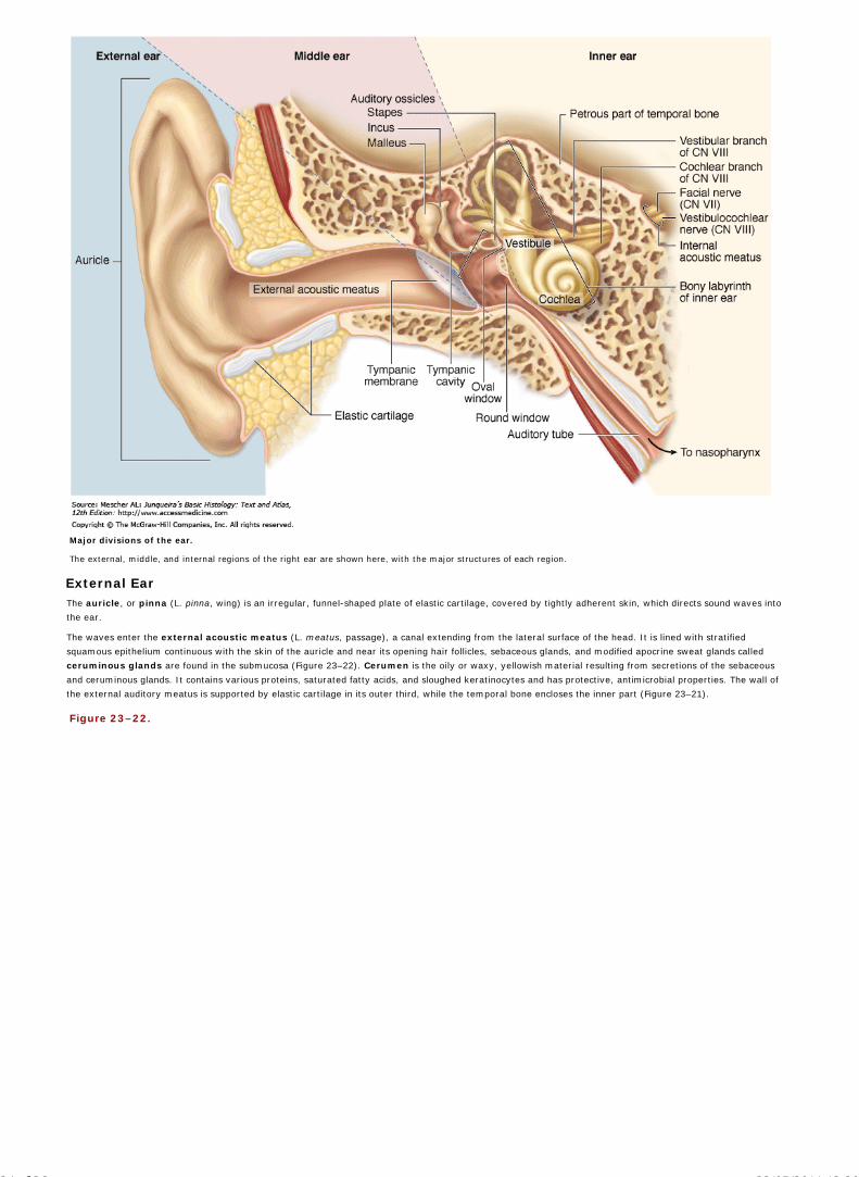
Major divisions of the ear.
The external, middle, and internal regions of the right ear are shown here, with the major structures of each region.
External EarThe auricle, or pinna (L. pinna, wing) is an irregular, funnel-shaped plate of elastic cartilage, covered by tightly adherent skin, which directs sound waves into
the ear.
The waves enter the external acoustic meatus (L. meatus, passage), a canal extending from the lateral surface of the head. It is lined with stratified
squamous epithelium continuous with the skin of the auricle and near its opening hair follicles, sebaceous glands, and modified apocrine sweat glands called
ceruminous glands are found in the submucosa (Figure 23–22). Cerumen is the oily or waxy, yellowish material resulting from secretions of the sebaceous
and ceruminous glands. It contains various proteins, saturated fatty acids, and sloughed keratinocytes and has protective, antimicrobial properties. The wall of
the external auditory meatus is supported by elastic cartilage in its outer third, while the temporal bone encloses the inner part (Figure 23–21).
Figure 23–22.
AccessMedicine | Print: Chapter 23. The Eye and Ear: Special Sense O... http://accessmedicine.com/popup.aspx?aID=6183264&print=yes_chapter
24 of 36 23/05/2011 13:30

External acoustic meatus.
The external acoustic meatus leads from the opening in the auricle to the tympanic membrane (eardrum). This section of the wall in the outer third of the acousticmeatus shows the lining of skin containing small hair follicles (F), sebaceous glands (SG), and modified apocrine sweat glands called ceruminous glands (CG).Secretions from these two glands form a yellowish, oily or waxy product called cerumen (C), which contains antimicrobial factors that help make the meatus uninvitingfor microorganisms. X50. H&E.
Across the deep end of the external acoustic meatus lies an epithelial sheet called the tympanic membrane or eardrum. Its external side is covered with
epidermis and its inner surface is covered with simple cuboidal epithelium continuous with the lining of the tympanic cavity in the middle ear (see below).
Between the two epithelial coverings is a thin sheet of fibrous connective tissue layer composed of collagen, elastic fibers and fibroblasts. Vibrations of the
tympanic membrane produced by sound waves transmit sound wave energy to the middle and inner ear (Figure 23–21).
Middle EarThe middle ear contains the air-filled tympanic cavity, an irregular space that lies within the temporal bone between the tympanic membrane and the bony
surface of the internal ear (Figure 23–21). Anteriorly this cavity communicates with the pharynx via the auditory tube (Eustachian or pharyngotympanic
tube) and posteriorly with the smaller, air-filled mastoid cavities of the temporal bone. The tympanic cavity is lined mainly with simple cuboidal epithelium
resting on a thin lamina propria that is strongly adherent to periosteum. Near the auditory tube, this simple epithelium is gradually replaced by the ciliated
pseudostratified columnar epithelium lining the tube. Although the walls of the tube are usually collapsed, it opens during the swallowing process, which serves to
balance the air pressure in the middle ear with atmospheric pressure. In the medial bony wall of the middle ear are two membrane-covered regions devoid of
bone: the oval and round windows (Figure 23–21).
The tympanic membrane is connected to the oval window by a series of three small bones, the auditory ossicles, which transmit the mechanical vibrations of
the tympanic membrane to the internal ear (Figure 23–21). The ossicles are named the malleus, incus, and stapes, the Latin words respectively for "hammer,"
"anvil," and "stirrup," which reflect each bone's general shape. The malleus is attached to connective tissue of the tympanic membrane and the stapes to that of
the membrane in the oval window. The ossicles articulate at synovial joints, which along with periosteum are completely covered with simple squamous
epithelium. Two small skeletal muscles insert into the malleus and stapes, restricting movement of the ossicles and helping to protect the internal ear from
extremely loud noises.
Internal EarThe internal ear is located completely within the temporal bone, where an intricate set of interconnected spaces, the bony labyrinth, houses a set of continuous
fluid-filled, epithelium-lined tubes and chambers that make up the smaller membranous labyrinth (Figure 23–21). The membranous labyrinth is derived from
an ectodermal vesicle, the otocyst, which invaginates into the subjacent connective tissue during the fourth week of embryonic development, loses contact with
the surface ectoderm, and becomes embedded in the rudiments of the future temporal bone. During this process, the otic vesicle changes shape, giving rise to
two major divisions of the membranous labyrinth:
the vestibular labyrinth, which mediates the sense of equilibrium and consists of two connected sacs (the utricle and the saccule) and threesemicircular ducts arising from the utricle, and
the cochlear labyrinth, which provides for hearing and contains the cochlear duct connected to the saccule.
In each of these structures the epithelial lining contains large areas of columnar sensory mechanoreceptors called hair cells in specialized regions:
two maculae of the utricle and saccule,
three cristae ampullaris in the enlarged ampullary regions of each semicircular duct, and
the long spiral organ of Corti in the cochlear duct.
The bony labyrinth has an irregular central cavity, the vestibule, where the saccule and the utricle are located. Behind this, three osseous semicircular canals
enclose the semicircular ducts. On the other side of the vestibule, the cochlea (L. cochlea, snail, screw) contains the cochlear duct (Figure 23–23). The cochlea is
about 35 mm in length and makes two-and-one-half turns around a bony core called the modiolus. The modiolus contains blood vessels and surrounds the cell
bodies and processes of the acoustic branch of the eighth cranial nerve in the large spiral or cochlear ganglion.
AccessMedicine | Print: Chapter 23. The Eye and Ear: Special Sense O... http://accessmedicine.com/popup.aspx?aID=6183264&print=yes_chapter
25 of 36 23/05/2011 13:30

Figure 23–23.
Internal ear.
The internal region of the ear is composed of a cavity in the temporal bone, the bony labyrinth, which houses a fluid-filled membranous labyrinth. The membranouslabyrinth includes the vestibular organs for the sense of equilibrium and balance (the saccule, utricle, and semicircular ducts) and the cochlea for the sense of hearing.
All regions of the bony labyrinth are filled with perilymph, which is similar in ionic composition to cerebrospinal fluid and the extracellular fluid of other tissues,
but contains little protein. Perilymph emerges from the microvasculature of the periosteum and is drained by a perilymphatic duct into the adjoining subarachnoid
space. This fluid suspends and supports the closed membranous labyrinth, protecting it from the hard wall of the bony labyrinth. The membranous labyrinth is
filled with endolymph, which also contains few proteins and is further characterized by a high potassium (150 mM) and low sodium (16 mM) content, similar to
that of intracellular fluid. Endolymph is generated largely by capillaries in the stria vascularis in the wall of the cochlear duct and is drained from the vestibule into
venous sinuses of the dura mater by the small endolymphatic duct.
SACCULE AND UTRICLE
The saccule and the utricle are composed of a very thin connective tissue sheath lined with simple squamous epithelium. The membranous labyrinth is bound to
the periosteum of the bony labyrinth by strands of connective tissue that contain microvasculature supplying the tissues of the membranous labyrinth. The two
maculae in the walls of the saccule and utricle are small areas of columnar neuroepithelial cells that are innervated by branches of the vestibular nerve (Figure
23–24). The macula of the saccule lies in a plane perpendicular to the macula of the utricle, but both are similar histologically. Each consists of a thickening of the
wall containing several thousand mechanosensitive hair cells, along with columnar supporting cells with basal nuclei, and nerve endings.
Figure 23–24.
AccessMedicine | Print: Chapter 23. The Eye and Ear: Special Sense O... http://accessmedicine.com/popup.aspx?aID=6183264&print=yes_chapter
26 of 36 23/05/2011 13:30

Vestibular maculae and their cells.
(a): Two sensory areas, the maculae, are located in the epithelial walls of the utricle and saccule in the vestibular complex. Both maculae are similar histologically andcontain mechanoreceptor cells called hair cells which use gravity and endolymph movement to detect the orientation of the stationary head and linear acceleration ofthe moving head. (b): A detailed view of a macular wall shows that it is composed of hair cells, supporting cells, and endings of the vestibular branch of the eighthcranial nerve. The apical surface of the air cells is covered by a gelatinous otolithic layer or membrane and the basal ends of the cells have synaptic connections withthe nerve fibers. (c): A diagram of a single generalized hair cell shows the numerous straight stereocilia, which contain bundled actin, and a longer single kinocilium, amodified cilium whose tip may be slightly enlarged.
The apical end of each hair cell has a single kinocilium with a basal body and a modified axoneme of microtubule doublets (Chapter 2) and a bundle of 60–100
long, rigid, unbranched stereocilia. The stereocilia arise from an actin-rich apical region, the cuticular plate, which serves to return these rigid projecting
structures to a normal upright position after bending. They are arranged in rows of increasing length, with the longest—about 100 m—located adjacent to the
kinocilium (Figure 23–24). The tips of the stereocilia and kinocilium are embedded in a thick, gelatinous layer of proteoglycans called the otolithic membrane,
the outer part of which is filled with calcified structures called otoliths (or otoconia) (Figure 23–25).
Figure 23–25.
Otoliths.
Otoliths are crystalline structures in the outer part of the otolithic membrane. Each otolith is a slightly elongated structure, up to 5 by 10 m in size, and is composedof calcium carbonate on a matrix of proteoglycans. Their presence makes the otolithic membrane considerably heavier than endolymph alone, which facilitates bendingof the kilocilia and stereocilia embedded in this membrane by gravity or movement of the head. X600. SEM. (With permission, from David J. Lim, House Ear Instituteand Department of Cell & Neurobiology, University of Southern California, Los Angeles.)
At their basal ends all hair cell have synapses with afferent (to the brain) nerve endings (Figure 23–26). Some hair cells (type I) have rounded basal ends
AccessMedicine | Print: Chapter 23. The Eye and Ear: Special Sense O... http://accessmedicine.com/popup.aspx?aID=6183264&print=yes_chapter
27 of 36 23/05/2011 13:30

surrounded by an afferent terminal calyx (L, calyx, cup, chalice). The basal ends of most hair cells (type II) are cylindrical and have more typical bouton endings
from afferent nerves. Both types of hair cells, or their afferents, also have synaptic connections with efferent (from the brain) fibers that modulate the sensitivity
of these mechanoreceptors (Figure 23–26). Each hair cell is also surrounded by supporting cells, which may have various functions besides providing physical
support for the mechanoreceptors.
Figure 23–26.
Hair cells and hair bundles.
(a): This diagram shows the two types of hair cells in the maculae and cristae ampullares. Basal ends of type I hair cells are rounded and enclosed within a nervecalyx on the afferent fiber. Type II hair cells are columnar and associated with typical bouton synaptic connections to their afferents. Both types are also associatedwith efferent fibers.
(b): A more detailed diagram of the stereocilia hair bundle of the hair cells showing that stereocilia occur in rows of increasing height, with the tallest next to thesingle kinocilium on one side of the cell's apical end. By TEM the end of each stereocilium shows an electron-dense region containing cation channels and proteinsinvolved in mechanoelectric transduction (MET) that convert mechanical activity of the stereocilia to electric activity within the hair cell. Neighboring stereocilia areconnected by various side links composed of proteins; the most well-understood of these are the tip links which connect the tips of stereocilia and contain very longtypes of cadherin proteins. Changes in the tension of the tip links caused by bending of the hair bundle open or close the adjacent cation channels and change theafferent synaptic activity of the hair cells.
AccessMedicine | Print: Chapter 23. The Eye and Ear: Special Sense O... http://accessmedicine.com/popup.aspx?aID=6183264&print=yes_chapter
28 of 36 23/05/2011 13:30

SEMICIRCULAR DUCTS
The three semicircular ducts are parts of the membranous labyrinth having the same general form as the semicircular canals in the bony labyrinth. Each
extends from and returns to the wall of the utricle. They lie in three different spatial planes, at approximately right angles to one another (Figure 23–23).
The enlarged ampulla end of each semicircular duct has an elongated ridgelike area of mechanoreceptors called the crista ampullaris (Figure 23–27). The
ridge of each crista ampullaris is perpendicular to the long axis of the duct. Cristae are histologically similar to maculae, with hair cells, supporting cells, and
nerve endings. However, the proteoglycan layer called the cupola attached to the sensory cells' hair bundles is thicker and does not contain otoliths. The cupula
extends completely across the ampulla, contacting the opposite non-sensory wall (Figure 23–27).
Figure 23–27.
Ampullae and cristae of the semicircular ducts.
Each of the semicircular ducts has an expanded end called the ampulla. The wall of each ampulla is raised as a ridge called the crista ampullaris, a section of which isshown here diagrammatically. Hair cells of the crista epithelium resemble the two types found in the maculae, with hair bundles projecting into a dome-shapeoverlying layer of proteoglycan called the cupula. The cupula is attached to the wall opposite the crista and is moved by endolymph movement within the semicircularduct.
VESTIBULAR FUNCTIONS OF THE EAR
Sensory information from the vestibular labyrinth is used, primarily in reflex mechanisms, for maintaining upright posture and balance and in allowing the eyes to
stay fixed on the same point despite changes in head position. Movements of the head cause movement of the endolymph, which moves the otolith membrane of
each macula and the cupula over each crista ampullaris. The sensory cell hair bundles embedded in these proteoglycan layers bend with the movement of this
material, changing these cells' resting potential and their rate of neurotransmitter release to the afferent nerves. When the hair bundle is deflected toward the
kinocilium, very small strands of protein called tip links that connect the stereocilia are pulled and cation channels open to allow an influx of K+ ions (the major
cation in endolymph). The resulting depolarization of the hair cell opens Ca2+ channels near the base of the cell and Ca2+ entry stimulates release of
neurotransmitter (Figure 23–28). When the head stops moving, stereocilia bundles straighten to the normal position and hair cells quickly repolarize and
reestablish the resting potential. Head movements that bend the stereocilia away from the kinocilium cause the tip links to be slack, allowing closure of the apical
cation channels and hyperpolarization of the cell. This in turn closes Ca2+ channels at the base of the cell and reduces neurotransmitter release (Figure 23–28).
Figure 23–28.
AccessMedicine | Print: Chapter 23. The Eye and Ear: Special Sense O... http://accessmedicine.com/popup.aspx?aID=6183264&print=yes_chapter
29 of 36 23/05/2011 13:30

AccessMedicine | Print: Chapter 23. The Eye and Ear: Special Sense O... http://accessmedicine.com/popup.aspx?aID=6183264&print=yes_chapter
30 of 36 23/05/2011 13:30

Mechanotransduction in hair cells.
Hair cells and supporting cells are part of an epithelium with tight junctions. The apical ends of the cells are exposed to endolymph with a high concentration of K+ and
perilymph with a much lower K+ concentration bathes their basolateral surface. At rest hair cells are polarized with a small amount of K+ entry and a low level ofneurotransmitter release to afferent nerve fibers at the basal ends of the cells. (a): As shown here head movements that cause the stereocilia bundle to be deflected
toward the kinocilium produce tension in the tip links which is transduced to electrical activity by opening of adjacent cation channels. Entry of K+ depolarizes the cell,
opening Ca2+ at the basal end of the cell which stimulates release of neurotransmitter. When this movement stops, the cells quickly repolarize.
(b): Movements in the opposite direction, away from the kinocilium, produce slackness on the tip links, allowing the apical K+ to close completely, leading tohyperpolarization and reduced transmitter release. With different numbers of afferent and efferent fibers on the hair cells and with various hair cells respondingdifferently to endolymph movements due to their positions within the maculae and cristae ampullares, the sensory information produced collectively by these cells canbe processed by the vestibular regions of the brain and used to help maintain equilibrium.
The hair cells of the cristae ampullares detect rotational or angular movements of the head. On each side of the head these hair cells are oriented with
opposite polarity, so that turning the head causes hair cell depolarization on one side and hyperpolarization on the other. Neurons of the vestibular nuclei in the
CNS receive input from the sets of semicircular ducts on each side simultaneously and interpret head rotation on the basis of the relative transmitter discharge
rates of the two sides.
The hair cells in the maculae of the saccules and the utricles respond to linear acceleration, gravity, and tilt of the head. Because the otoliths are heavier
than endolymph, the stereocilia bundles are deflected by gravity when the head is not moving, when the head is tilted with respect to gravity, and when the
individual is moving in a straight line and inertia causes drag on the otolithic membrane.
Inputs from all regions of the vestibular labyrinth travel along the eighth cranial nerve to vestibular nuclei in the CNS. There they are interpreted together with
inputs from mechanoreceptors of the musculoskeletal system to provide the basis for perceiving movement and orientation in space and for maintaining
equilibrium or balance.
MEDICAL APPLICATION
Problems of the vestibular system can result in vertigo, or dizziness, a sense of the body rotating and lack of equilibrium. This can be caused by certain
infections, drugs, or tumors near the vestibular nerve. Spinning the body can produce vertigo due to overstimulation of the cristae ampullares of the
semicircular ducts. Overstimulation of the maculae of the utricle caused by repetitive changes in linear acceleration and directional changes can normally
lead to motion sickness (seasickness).
COCHLEAR DUCT AND AUDITORY FUNCTIONS
The cochlear duct, a part of the membranous labyrinth connected to the saccule, is highly specialized as a sound receptor. It is about 35 mm long, coiled two and
one-half times, and is surrounded by specialized perilymphatic spaces. When observed in sections, the cochlea appears to contain three spaces: the scala
vestibuli, the middle cochlear duct (or scala media), and the scala tympani (Figure 23–29). The cochlear duct contains endolymph and ends at the apex of
AccessMedicine | Print: Chapter 23. The Eye and Ear: Special Sense O... http://accessmedicine.com/popup.aspx?aID=6183264&print=yes_chapter
31 of 36 23/05/2011 13:30
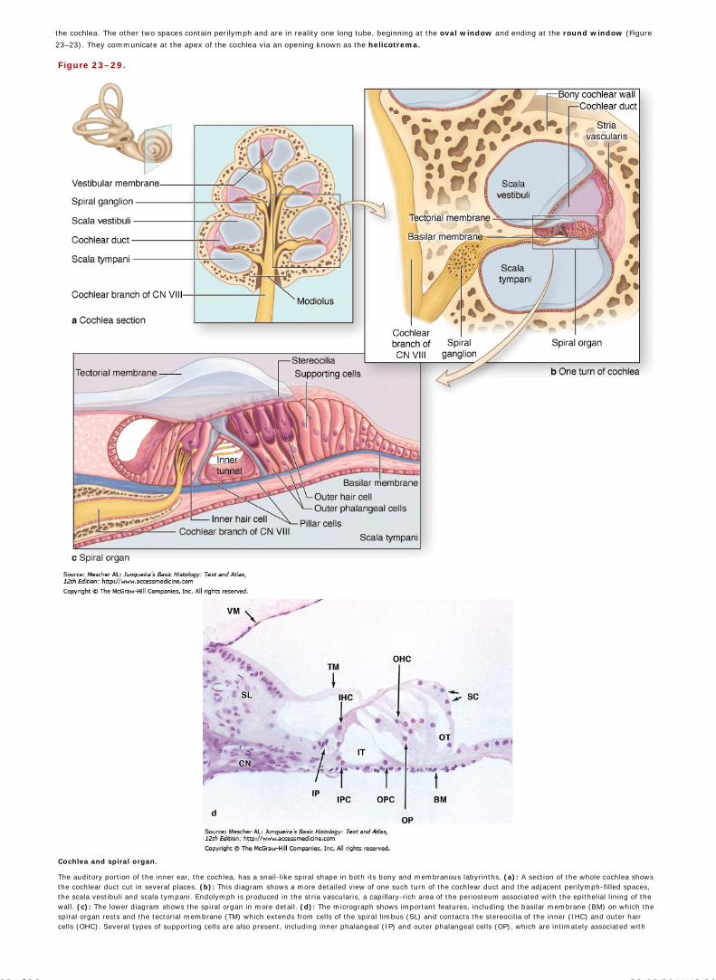
the cochlea. The other two spaces contain perilymph and are in reality one long tube, beginning at the oval window and ending at the round window (Figure
23–23). They communicate at the apex of the cochlea via an opening known as the helicotrema.
Figure 23–29.
Cochlea and spiral organ.
The auditory portion of the inner ear, the cochlea, has a snail-like spiral shape in both its bony and membranous labyrinths. (a): A section of the whole cochlea showsthe cochlear duct cut in several places. (b): This diagram shows a more detailed view of one such turn of the cochlear duct and the adjacent perilymph-filled spaces,the scala vestibuli and scala tympani. Endolymph is produced in the stria vascularis, a capillary-rich area of the periosteum associated with the epithelial lining of thewall. (c): The lower diagram shows the spiral organ in more detail. (d): The micrograph shows important features, including the basilar membrane (BM) on which thespiral organ rests and the tectorial membrane (TM) which extends from cells of the spiral limbus (SL) and contacts the stereocilia of the inner (IHC) and outer haircells (OHC). Several types of supporting cells are also present, including inner phalangeal (IP) and outer phalangeal cells (OP), which are intimately associated with
AccessMedicine | Print: Chapter 23. The Eye and Ear: Special Sense O... http://accessmedicine.com/popup.aspx?aID=6183264&print=yes_chapter
32 of 36 23/05/2011 13:30

the hair cells and contribute to the tight epithelium separating endolymph from perilymph in the scala tympani. Other supporting cells form various structural featuresof the organ important for converting vibrations into subtle stimuli to the hair cells. These include the inner (IPC) and outer pillar cells (OPC) which surround a spacecalled the inner tunnel (IT) and other supporting cells (SC) which border the outer tunnel (OT). Afferent nerve fibers from the hair cells comprise the cochlear nerve(CN), a branch of the eighth cranial nerve. X75. H&E.
Along its length, the cochlear duct is separated from the scala vestibuli by the vestibular membrane (Figure 23–30). This very thin structure consists of a
basement membrane with simple squamous epithelium on each side: one mesothelium facing the scala vestibuli and the other part of the cochlear duct's lining.
Cells of both layers have extensive tight junctions that help preserve the very high ionic gradients across this membrane between endolymph and perilymph.
Figure 23–30.
Cochlear duct and spiral ganglion.
The spiral organ (SO) is located on the basal wall of the cochlear duct (CD). This duct is filled with endolymph produced in the stria vascularis (STV), an unusualassociation between the columnar epithelial cells which have numerous basal infoldings and the capillaries in the periosteum of the bone (B). On either side of thecochlear duct are the scala vestibuli (SV) and scala tympani (ST), which are filled with perilymph and are continuous at the apex of the cochlea. Cell bodies of bipolarneurons in the spiral ganglion (SG) send dendrites to the hair cells of the spiral organ and axons to the cochlear nuclei of the CNS. X25. H&E.
In the lateral wall of the cochlear duct is the stria vascularis (Figure 23–30), a unique epithelium responsible for production and maintenance of the endolymph
for the entire membranous labyrinth. The stria vascularis encloses a network of capillaries and consists of cells with many deep basal infoldings of their plasma
membranes, where numerous mitochondria are located. Fluid and K+ ions pumped from the capillaries by these epithelial cells are released in the cochlear duct
as endolymph.
In the wall that separates the cochlear duct from the scala tympani is the complex structure called the spiral organ (organ of Corti) which contains special
auditory receptors in the form of hair cells that respond to different sound frequencies. The spiral organ rests on a thick basal lamina—the basilar membrane. Two
major types of hair cells are present (Figure 23–29). Outer hair cells (OHC) occur in three rows near the oval window, increasing to five rows near the apex of
the cochlea. There is a single row of inner hair cells (IHC). The latter have one linear array of short stereocilia, while OHC each have a curved row of longer
stereocilia (Figure 23–31). No kinocilium is present on cochlear hair cells, allowing symmetry on the cells that is important for their role in sensory transduction.
Figure 23–31.
AccessMedicine | Print: Chapter 23. The Eye and Ear: Special Sense O... http://accessmedicine.com/popup.aspx?aID=6183264&print=yes_chapter
33 of 36 23/05/2011 13:30

Stereocilia of cochlear hair cells.
With the tectorial membrane removed, SEM shows the morphology of the three rows of outer hair cells (a), and the single row of inner hair cells (b) in the middle turnof a cochlea. X2700. (With permission, from Patricia A. Leake, Epstein Hearing Research Laboratory, University of California at San Francisco.)
The tips of the tallest stereocilia of the OHC are embedded in the tectorial membrane, an acellular layer extending over the spiral organ from the modiolus
(Figures 23–29 and 23–30). The tectorial membrane consists of fine bundles of collagen (types II, V, IX, and XI), associated proteoglycans and other proteins and
is formed during the embryonic period from secretions of cells that come to line the adjacent region called the spiral limbus.
Both outer and inner hair cells have afferent and efferent nerve endings, with IHC much more heavily innervated. The cell bodies of the afferent bipolar neurons
are located in a bony core of the modiolus and constitute the spiral ganglion (Figures 23–29 and 23–30).
Two major types of columnar supporting cells are associated with the hair cells of the spiral organ (Figure 23–29). Pillar cells are stiffened by bundles of keratin
and outline a triangular, tunnel-like space between the outer and inner hair cells—another structure important in sound transduction. Phalangeal cells
intimately surround and directly support both inner and outer hair cells, almost completely enclosing each IHC but only the basal ends of the OHC.
Stereocilia of cochlear hair cells detect movements of the spiral organ. Sound waves collected by the auricle of the external ear cause the tympanic membrane to
vibrate, which causes movement of the ossicles in the middle ear (Figure 23–32). The large size of the tympanic membrane compared to the oval window and the
mechanical properties of the ossicle chain connecting these two membranes allow for optimal transfer of energy between air and perilymph, from sound waves to
vibrations of tissues and fluid-filled chambers.
Figure 23–32.
AccessMedicine | Print: Chapter 23. The Eye and Ear: Special Sense O... http://accessmedicine.com/popup.aspx?aID=6183264&print=yes_chapter
34 of 36 23/05/2011 13:30

Sound waves and movements in the ear.
Sound waves are funneled to the tympanic membrane by the external ear and conducted across the middle ear by movements of the three ossicles. Movements of thestapes produce pressure waves in the perilymph on the other side of the attached oval window. In these diagrams, the spiral shape of the cochlea has beenstraightened to better show how pressure waves affect the spiral organ. The pressure waves produce movements within the spiral organ that cause themechanoreceptor hair cells to depolarize/hyperpolarize and release neurotransmitters to afferents of the cochlear nerve, producing signals interpreted in the CNS assounds. Pressure waves crossing the cochlear duct are transferred to the scala tympani and dissipate at the round window. Sounds waves of different frequencies aredetected by hair cells at specific sites along the spiral organ. Low frequency sounds produce pressure waves that move the spiral organ only near the end of thecochlea, near the helicotrema. High frequency sounds affect the organ close to the oval window and sounds of intermediate frequencies displace the spiral organsomewhere in between the extremes.
Pressure waves within the perilymph begin at the oval window and move along the scala vestibuli. Each pressure wave causes momentary movement of the
vestibular and/or basilar membranes and the endolymph surrounding the spiral organ. The width, rigidity, and other physical properties of the basilar membrane
which supports the spiral organ vary along its length. This causes the region of maximal displacement within the vibrating spiral organ to vary with the sound
waves' frequency, ie, the number of waves moving past a point per unit of time (measured in hertz). High-frequency sounds produce maximal movement of the
spiral organ nearest the oval window. Sounds of progressively lower frequency produce pressure waves that move farther along the scala vestibuli and displace
the spiral organ at a point farther from the oval window (Figure 23–32). The sounds of the lowest frequency that can be detected produce movement of the
basilar membrane at the apex or helicotrema of the cochlea. After crossing the cochlear duct and spiral organ at these various points, pressure waves are
transferred to the scala tympani and exit the inner ear at the round window (Figure 23–32).
The true receptors for the sense of hearing are the more heavily innervated IHC of the cochlea's spiral organ. The OHC, with the ends of their stereocilia
embedded in the tectorial membrane, are depolarized when these mechanotransducers are deflected in a process similar to that of vestibular hair cells described
above. Depolarization of the OHC very rapidly produces a slight shortening of these columnar cells, which is mediated by an unusual transmembrane protein
called prestin (It. presto, very fast) abundant in the lateral cell membranes. Prestin undergoes a voltage-dependent conformational change which affects the
cytoskeleton, with the cells rapidly becoming shorter when the membrane is depolarized and elongating with membrane hyperpolarization. Piston-like
movements of the OHC produce vibrations of the tectorial membrane against the stereocilia of the nearby IHC (Figure 23–29), amplifying the signals that these
cells then send to the CNS for processing as sounds.
MEDICAL APPLICATION
Deafness can result from many factors, which usually fall into two categories. (1) Conductive hearing loss involves various problems in the middle ear
which can reduce conduction of vibrations by the chain of ossicles from the tympanic membrane to the oval window. A common example is otosclerosis,
in which scar-like lesions develop on the bony labyrinth near the stapes which inhibit its movement of the oval window. Infection of the middle ear (otitis
media) is common in young children, usually progressing from an upper respiratory infection, and can reduce sound conduction due to fluid accumulation
in that cavity. (2) Sensorineural deafness can be congenital or acquired and due to defects in any structure or cell from the cochlea to auditory centers
of the brain, but commonly involves loss of hair cells or nerve degeneration.
Some hearing loss patients can be helped by cochlear implants. These consist of a small device worn behind the ear which contains a microphone, a
AccessMedicine | Print: Chapter 23. The Eye and Ear: Special Sense O... http://accessmedicine.com/popup.aspx?aID=6183264&print=yes_chapter
35 of 36 23/05/2011 13:30

Copyright © The McGraw-Hill Companies. All rights reserved.Privacy Notice. Any use is subject to the Terms of Use and Notice.
sound converter, and a transmitter that sends electrical impulses to a receiver implanted under the skin of that region. The receiver is connected to a
small cable with many electrodes. The cable is inserted into the internal ear and threaded into the scala tympani along the wall containing branches of the
cochlear nerve. Electrical signals produced by the transmitter in response to sounds of selected frequencies stimulate the nerve directly and are sent to
the brain where they are interpreted as sounds. Cochlear implants do not restore normal hearing but can provide the deaf patient with a usable range of
sounds and the potential for direct participation in speech.
AccessMedicine | Print: Chapter 23. The Eye and Ear: Special Sense O... http://accessmedicine.com/popup.aspx?aID=6183264&print=yes_chapter
36 of 36 23/05/2011 13:30

Print Close Window
Note: Large images and tables on this page may necessitate printing in landscape mode.
Copyright © The McGraw-Hill Companies. All rights reserved.
Junqueira's Basic Histology: Text & Atlas, 12e > Appendix: Light Microscopy Stains >
HEMATOXYLIN AND EOSIN (H&E)
Hematoxylin stains cellular regions rich in basophilic macromolecules (DNA or RNA) a purplish blue or
blue-black color. It is the most common stain for demonstrating cell nuclei and cytoplasm rich in rough ER.
Usually used as the contrasting "counterstain" with hematoxylin, eosin is an acidic stain that binds to basic
macromolecules such as collagen and most cytoplasmic proteins, especially those of mitochondria. Eosin
stains regions rich in such structures a pinkish red color. Tissue sections showing only structures with shades
of purple and pink are stained with H&E.
PARAROSANILINE-TOLUIDINE BLUE (PT)
This dye combination stains chromatin shades of purple and cytoplasm and collagen a lighter violet. These
stains penetrate plastic sections more readily than H&E and are used here primarily with acrylic resin-
embedded sections to provide better detail of cell and tissue structures. Toluidine blue is also commonly used
for differential staining of cellular components, particularly cytoplasmic granules.
MALLORY TRICHROME
This procedure employs a combination of stains applied in series which results in nuclei staining purple;
cytoplasm, keratin, and erythrocytes staining bright red or orange; and collagen bright or light blue. Mallory
trichrome is particularly useful in demonstrating cells and small blood vessels of connective tissue. Similar
stains, such as Masson trichrome and Gomori trichrome, yield comparable results except that collagen stains
blue-green or green.
PICRO-SIRIUS-HEMATOXYLIN (PSH)
The dye Sirius red in a solution of picric acid stains collagen red and cytoplasm a lighter violet or pink, with
nuclei purple if first stained with hematoxylin. Under the polarizing microscope, collagen stained with picro-
sirius red is birefringent and can be detected specifically.
PERIODIC ACID–SCHIFF REACTION (PAS)
This histochemical procedure stains complex carbohydrate-containing cell components, which become
magenta (shades of purplish pink). PAS is commonly used to demonstrate cells filled with mucin granules,
glycogen deposits, or the glycocalyx.
WRIGHT-GIEMSA STAIN
These are two similar combinations of stains that are widely used on fixed cells of blood or bone marrow
smears to demonstrate types of blood cells. Granules in leukocytes are seen to have differential affinity for
the stain components. Nuclei stain purple and erythrocytes stain uniformly pink or pinkish orange.
SILVER OR GOLD STAINS
Various procedures employing solutions of silver or gold salts have been developed to demonstrate
filamentous structures in neurons and fibers of reticulin (type III collagen). By these "metal impregnation"
techniques these filaments stain dark brown or black. Such stains have been largely replaced now by
immunohistochemical procedures.
STAINS FOR ELASTIN
AccessMedicine | Print: Appendix: Light Microscopy Stains http://accessmedicine.com/popup.aspx?aID=6183502&print=yes_chapter
1 of 2 23/05/2011 13:32

Copyright © The McGraw-Hill Companies. All rights reserved.Privacy Notice. Any use is subject to the Terms of Use and Notice.
Several staining methods have been developed to distinguish elastic structures from collagen, most of which
stain the elastin-rich structures brown or shades of purple. Examples of such stains are Weigert's resorcin
fuchsin, aldehyde fuchsin, and orcein Van Gieson stains.
STAINS FOR LIPID
When special preparation techniques are used to retain lipids of cells, such as in frozen sections, lipophilic
dyes are used to demonstrate lipid droplets and myelin. Oil red O and Sudan black stain lipid-rich structures
as their names suggest. Osmium tetroxide (osmic acid), which is used as a fixative for TEM, is reduced to a
black substance by unsaturated fatty acids and is also used to demonstrate lipids.
OTHER COMMON STAINS
Many basic aniline dyes, including azures, cresyl violet, brilliant cresyl blue, luxol fast blue, and light green,
are used because of the permanence and brightness of the colors they impart to cellular and extracellular
structures in paraffin sections. Many such stains were initially developed for use in the textile industry.
AccessMedicine | Print: Appendix: Light Microscopy Stains http://accessmedicine.com/popup.aspx?aID=6183502&print=yes_chapter
2 of 2 23/05/2011 13:32