Observations concerning the mechanics of ovulation in the fowl
43
OBSERVATIONS CONCERNING THE MECHANICS OP OVULATION IN THE FOWL by ROBERT EMMETT PHILLIPS, JR. B. S«, Kansas State College of Agriculture and Applied Science, 1935 A THESIS submitted in partial fulfillment of the requirements for the degree of MASTER OF SCIENCE KANSAS STATE COLLEGE OP AGRICULTURE AND APPLIED SCIENCE 1936 brought to you by CORE View metadata, citation and similar papers at core.ac.uk provided by K-State Research Exchange
-
Upload
khangminh22 -
Category
Documents
-
view
0 -
download
0
Transcript of Observations concerning the mechanics of ovulation in the fowl

OBSERVATIONS CONCERNING THE MECHANICS OP OVULATION
IN THE FOWL
by
ROBERT EMMETT PHILLIPS, JR.
B. S«, Kansas State Collegeof Agriculture and Applied Science, 1935
A THESIS
submitted in partial fulfillment of the
requirements for the degree of
MASTER OF SCIENCE
KANSAS STATE COLLEGE
OP AGRICULTURE AND APPLIED SCIENCE
1936
brought to you by COREView metadata, citation and similar papers at core.ac.uk
provided by K-State Research Exchange

Da
f-m'\S * <?*"•.
TABLE OP CONTENTS
Page
INTRODUCTION 1
REVIEW OP THE LITERATURE 1
MATERIAL AND TECHNIQUE 3
OBSERVATIONS AND EXPERIMENTAL RESULTS 7
Changes in the Appearance of the FollicularBlood Vessels 7
Muscle Fibers in the Follicular Wall 10Expulsion of the Ovum from the Follicle 12Effects of Different Techniques on Ovulation . 16Relation of Ovulation Time to Interval 20
BEARIHO OF THE RESULTS ON PROPOSED THEORIES 22
Internal Pressure Theory of Ovulation 22Smooth Muscle Theory of Ovulation 26Chemical Change Theory of Ovulation 27
DISCUSSION 29
SUMMARY 31
ACKNOWLEDGMENT 33
LITERATURE CITED 34
ii
INTRODUCTIOa
The physiological causation of ovulation in the verte-
brates is a phenomenon which is not well understood. Many
theories have been proposed as to the cause of the follicu-
lar rupture, but until recently few direct observations
have been made in support of the theories advanced*
The fowl is an excellent subject for the study of the
phenomenon of ovulation since the ova carry a large amount
of deutoplasa and when necessary, they can be adjusted into
full view, by virtue of the fact that the follicles are
suspended from the ovary by means of a follicular stalk.
This eliminates many of the handicaps encountered in the
study of the microscopic mammalian ova which are embedded
in an ovarian stroma.
The purpose of this study is to Investigate by observa-
tion and varied experimental technique, the mechanics of
ovulation in the fowl. The bearing of the observations on
theories proposed by workers on other vertebrates, was kept
In mind during the course of the study.
REVIEW OP THE LITERATURE
Bartelmes (1912) observed the act of ovulation in the
pigeon, and by measurements of the follicle before ovula-
tion and of the ovum after it was liberated, concluded there

2
was a great pressure inside of the follicle resulting from
yolk secretion which caused the follicular rupture* Al-
though egg laying, and thus ovulation, does not normally
occur in the pigeon until mating has taken place, Craig
(1913) caused laying to start in the pigeon, without pre-
vious mating, by merely stroking the head of the bird.
This psychological effect as a cause of ovulation, has
later been reported in two mammals (rabbit - cat) which
normally do not ovulate until after copulation has occurred*
Patterson (1910) reported that ovulation in the fowl
resulted from the muscular exertion of the infundibulum
which grasped either mature or immature Graafian follicles
and caused them to rupture* However, Pearl and Curtis
(1914) showed ovulation could occur when the oviduct was
entirely removed*
The most detailed study of the process of ovulation in
the fowl is the work of Warren and Scott (1935). This work,
however, was concerned primarily with the process of egg
formation in relation to the different divisions of the
oviduct rather than the direct causes of the liberation of
the ovum from the follicle. The findings of these workers
also substantiate the view of Pearl and Curtis concerning
the role of the funnel in the follicular rupture*
A review of the different ovulation theories by Hart-
man (1932) is one of the most comprehensive recent sources
of the literature In the field*
MATERIAL AND TECHNIQUE
The 77 birds used In this study were white Leghorn fe-
males In their first or second year of production. The
birds were trapnested hourly so that the characteristics of
each bird's clutch could be studied before it was selected
for the observation • In the operative work only those
birds were used, the laying record of which gave fairly
good assurance that the next ovum would be ovulated without
any delay such as occurs between dutches. The hens avail-
able for the celiotomy had a wide variation in Intensity of
laying so a study of the relationship between the time
elapsing between ovlposition and ovulation and length of In-
terval between eggs was possible.
Mormally, all of the birds were allowed to lay In their
usual quarters while under close observation. Immediately
after laying they were taken to the operating room and
anaesthetized with liquid nembutal (pentobarbital sodium
1 gr. per cc). All injections were made intra-venously
utilising the vena humeri profunda of the wing. For the
initial injection three-fourths of a cubic centimeter of
nembutal was usually sufficient to completely anaesthetise
the bird. Succeeding injections varied from one-fourth to
one-half cubic centimeter depending somewhat upon the size
of the bird* Respiratory shock resulted in the death of 12
hens when the dosage was In excess, or when the Injection
of nembutal was made too rapidly.
As soon as the bird was under the influence of the
anaesthetic, the feathers were removed from the left lateral
surface of the abdomen* The incision (approximately 5 in-
ches In length) was made parallel to the back slightly above
the mid-line between the vertebral column and the sternum.
By making the incision at this locus, only two blood vessels
needed to be clamped off. One was a small blood vessel lo-
cated on the underneath surface of the skin, and the other
a large artery ramifying the surface of the abdominal mus-
cles* Serious hemorrhage resulted if this larger blood ves-
sel was not clamped off near the vertebral column before it
was cut* Since the fowl is only slightly susceptible to in-
fection and few were desired after the operation, no special
antiseptic technique was used for the operations. Approxi-
mately 10 to 15 minutes were required to anaesthetize and
open the body cavity of a bird after oviposition had oc-
curred*
With a little experience one can readily recognise the
next ovum to be liberated provided this is to take place
within an hour* Not always was the mature follicle visible
from the incision on the left side* If an immature follicle
was uppermost, the mature follicle was located and then
gradually adjusted into full view for observation from the
left side. The presence of a follicular stalk and the lack
of ovarian stroma permit considerable adjustment of the po-»
sltion of the follicle without much difficulty and the
change in position seemed to have no effect on ovulation,
(telly when the mature follicle was in the extreme cephalic
end of the coelom near the heart did the adjustment tend to
subject the bird to a shock. Sometimes to facilitate the
observation of follicles in the anterior part of the body
cavity the usual incision was lengthened by disarticulating
the diarthrodial joint between the dorsal and ventral parts
of the last rib. Because of the location of the lung, the
sixth rib marked the most anterior point of the incision.
The only deviation from this operative routine was
that nine hens were removed from the nest, anaesthetised,
and opened before laying occurred. This was done so that
more time would be available for studying the visible
changes in the mature follicle and the peristaltic movement
of the oviduct before and after laying. Provided the in-
cision through the lateral muscles did not extend too far
posteriorly the anaesthetized hen had little difficulty in
the expulsion of the egg at the expected time. It was of
interest to learn that the passage of the fully formed egg
through the vagina required only about one minute.
Since blood pressure has been considered as one of the

6
factors influencing ovulation, care was taken when making
the abdominal incision to ligate the two large blood ves-
sels in the abdominal wall before cutting across them. By
employing this technique, only a very small amount of blood
was lost, so ovulation could not have been influenced by
this factor*
When the incision was to be kept open for a consider-
able period, the opening into the body cavity was covered
with cellophane and sealed with vaseline to conserve the
humidity and body heat* Usually, however, after the mature
follicle was located and adjusted into view if necessary*
the coelomic incision was only opened intermittently to ob-
serve the follicle until the rupture appeared ready to occur.
Exposure of the reproductive organs to low tempera-
tures was believed to have an effect in delaying or in-
hibiting the follicular rupture, so all of the operations
were performed in a warm room* Usually the birds were
killed after the observations were made, but in seven indi-
viduals, the incision was sutured and they were returned to
the flock* Of these, six lived for several months and two
(1919 and 1967, table 1) were operated upon the second time*

OBSERVATIONS AND EXPERIMENTAL RESULTS
Several different methods were used in this study to
investigate the cause of the follicular rupture. The fol-
lowing list gives a brief summary of the number of oases
that were used in the various techniques for testing the
different theories offered as an explanation of the follicu-
lar rupture
x
Ovulations recorded • 40
Birds anaesthetised before laying 9
Excised follicles 4
Follicles clamped off but attached to the ovary . ... 4
Follicles ruptured by applying pressure (squeezing). 4
Pitultrin injections 2
Electrical stimulation of follicle 2
Removal of a portion of the yolk 2
Intra-follicular injections of water 2
in the Appearance of the FollicularBlood Vessels
Lately after gaining entrance into the coelom, the
slightly vascular serous ovarian pocket was torn in order
that the ovary could be seen more easily. The oviduct
usually exhibited slight peristaltic movement when first
exposed (about 15 minutes after laying), but as the time of
ovulation approached, greater motility was observed, es-
pecially in the Infundibular region*
The stlgaa has been referred to as the non-vascular
area of the follicle, but there is a great variation in the
width of this non-vascular area due to the changes which
occur in the blood vessels of the follicle within an hour
previous to ovulation. Many small finger-like blood ves-
sels (branches from two large blood vessels that parallel
the stigma on either side) extending out to this non-vascu-
lar area, gradually became obliterated before the follicular
rupture occurred. The blurring out of these blood vessels
caused an apparent widening of the stigma. This was the
characteristic by which identification was made of the next
follicle to be ruptured. The wide non-vascular area is re-
ferred to as the stigma in this study.
Two indications of the Impending ovulation were the
changes in the appearance of the blood vessels of the fol-
licle and the slight to prominent bulged area or areas which
appeared on the stigma. Practically all of the blood ves-
sels of the mature follicle were very red and conspicuous
when the ovary was first exposed, provided ovulation was not
to occur immediately. Later the small blood vessels through-
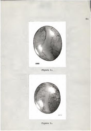
out the follicle became blurred or obliterated. Figures 1
and 2 show the difference in the appearance between a fol-
licle that would have ruptured within a few minutes and a
Figures 1 and 2.

Pig. 1. Appearance of a follicle im-mediately before rupturing. Note the widestigma and the blurred blood Teasels*
Pig. 2. A follicle approximately 26hours before the follicular rupture is tooccur. The stigma is narrow and the bloodvessels are distinct and prominent.
9a
^)\\¥
Figure 1,
M*3J
Figure 2.

10
follicle that would not have ruptured before approximately
26 hours.
VThen the tension in the follicular wall was released,
due to the expulsion of the ovum, all of the blood vessels
in the follicle again became prominent and had a dark red
color. This would Indicate that there had not been a col-
lapse of the smaller blood vessels, but that on the con-
trary they had been compressed to the extent that they tea*
porarlly had become invisible. Since there was no apparent
change in the appearance of the blood vessels of the remain-
ing follicles before or after ovulation it was believed
that the tension on the follicular membranes was responsible
for the reduction in the blood supply to these tissues
rather than some external factor. Tension artificially ap-
plied caused the blood vessels to disappear in the same
manner that they did immediately before a normal ovulation.
Muscle Fibers in the Follicular Sail
Histological study of the follicular wall showed that
muscle fibers were present in this tissue (Pigs. 3, 4, and
5). The long, slender fibers were very compact in the
region of the stigma, slightly more so than in the region
of the follicular stalk.
There seemed to be a slight periodic variation in the
turgldity of the follicle previous to the time of ovulation.

11
but since the change seemed slight and this impression was
gained by palpation of the follicle, there was the possi-
bility that the observation was incorrect.
The contraction of the muscle fibers was apparently the
cause of the follicular turgidity Which resulted in the
blurring or obliteration of the smaller blood vessels, i.vi-
dence supporting this view was found in observations made on
three hens that died just before ovulation was about to oc-
cur. «vith the loss of muscle tonus just before death, the
follicles became flaccid and the small blood vessels again
became dark red and prominent. Thus the accumulation of ad-
ditional yolk material inside the follicle was not respon-
sible for the visible changes that occur in the appearance
of the follicular wall, but instead served only as a body on
which the muscle fibers can contract and increase the ten-
sion* In two cases ovulation was prevented by removing, by
means of a syringe, approximately three-fourths of a cubic
centimeter of deutoplasm from the follicle* This caused
the follicle to become flaccid and as a result the tension
in the follicular wall was lowered. Apparently this tension
is essential before ovulation can occur since the birds that
died just previous to ovulation were observed three hours
afterward and the follicular rupture had not occurred.

12
Expulsion of the Ovum from the Follicle
Histological evidence that there is an inhibition in
the growth of the follicular tissue at the non-vascular
stigma region is shown in figures 3, 4, and 5. In figure 3
is shown the stigma region of a follicle that was expected
to rupture within a few minutes* Figure 4 shows the same
region of a follicle approximately 26 hours before ovula-
tion was expected to occur and figure 5 shows the appear-
ance of a follicle which was estimated would not have rup-
tured before 10 days*
Vhen the mature follicle was about to rupture, the
stigma area was very thin and the muscle fibers were under
great tension* It was thought that the decrease in the
thickness of the follicle had not resulted from a proteo-
lytic digestion, because the corona radiata was Intact in
all places* If enzymatic action had taken place it was ex-
pected that this layer of cells would have been destroyed*
The follicular rupture practically always began as a
tiny point at one of the stigma ends* In only one of the
cases observed did the point of rupture occur about midway
between the ends of the stigma*
Exposure of the ovary to the air resulted in a drying
out of the follicular tissue, and because of this there was
a variation in the time required for the actual follicular

13
Figures 3 and 4.

Fig* 3« A transverse section of thestigB* region of s follicle laraediatoly be-fore rupturing* The inside of the follicleis uppermost* The corona radiate is in-tact and the xauscle fibers are stretched*Magnification 240x.
Fig* 4* A transverse section of thestigna region of a follicle about 86 hoursbefore ovulation* Particles of deutoplasacan be seen next to the corona radiate*The rauscle fibers are not fully stretchedand the follicular wall la thicker thenthat of the nature follicle. Magnification

13a
i IFigure 3,
-"<",** it-
.* -*.*
*Jh
Figure 4,

14
Figure 5*

Pig. 5. A tranverse section of aportion of a follicle which was estimatedwould not have ruptured before 10 days.The vitelline membrane can be seen nextto the poorly defined corona radiata.Magnification 240x.

14a
Figure 5.

15
split* If lymph was removed from the coelom by means of a
pipette and dropped on the follicle when there was evidence
of the tissue drying, the rupture of the well-moistened
follicle was practically instantaneous, and very little
could be observed concerning the nature of the rupture.
However, during this study.motion pictures were taken of
ovulation and under these conditions, with a marked drying
out of the follicle as a result of the heat given off from
the arc lights, the follicular rupture was considerably
slower and a more critical observation could be made.
The Inside layers of the follicular membrane began to
break first and as a consequence a bulged area appeared at
the point of rupture. If the rupture was exceptionally
slow it was not uncommon for the entire stigma region to be
bulged. The germinal epithelium was firmly attached to the
theca cells at the stigma, but at other places on the fol-
licle the attachment between the two cell layers was very
loose. Thus, the germinal epithelium at the stigma region
was actually torn when the theca cells below it had broken
apart.
As soon as the tiny rupture point was visible, the
ovum was rapidly expelled from the follicle as a result of
the slit-like tearing of the follicular wall through the
center of the stigma.
1C
effects of Different Techniques on Ovulation
it erne found that ovulation wee not prevented
(excepting one oeee that will be discussed later) In three
birds by clamping off the follicle at the stalk, it was ap-
parent there was not a last minute deposition of yolk which
was responsible for ovulation* In all cases immedlately
after the follicular stalk was damped, the bird was not
disturbed until the ovum was observed in the body cavity*
Because of this, the actual rupture of the follicle was not
observed, but the time between laying and ovulation was re*
corded for one of these birds in Tsble 1* The follicle of
this hen (2063) was damped off U minutes before the ac-
tual rupture occurred*
In all oases after ovulation had taken place, the
clamp was not unclasped until after the bird had been kill-
ed and the sternum removed to make certain that the entire
follicular stalk had bsen between the jaws of the clamp*
To make certain that the clamp was definitely inhibiting
the blood supply and nerve impulses, a follicle which was
expected to rupture within a few minutes was clamped off
and then completely exolsed from the ovary* The normal
turgldity of the follicle was maintained as nearly as pos-
sible by the application of a second clamp just Inside of
the first one* Then the follicle was returned to the

17
moist body cavity of the hen from which it had been taken,
but had otherwise been handled in the usual manner. Pour
of these excised follicles ruptured normally at the stigma.
No accurate record was made of the time elapsing between
oviposition and these follicular ruptures because the fol-
licles did not always retract enough to see the ovum just
as it was released and sometimes the follicles were placed
under loops of the intestine so that they would be warm and
moist. The time of observation of the recently released
ova, however, closely approximated the time estimated for
the follicular rupture had the follicle not been molested.
Two of these mature follicles were excised from the ovaries
before the hens had laid so the minimum time, before ovu-
lation was expected to have occurred normally, was estimated
at one hour or longer. Prom this evidence it would sees
that all of the deutoplasra must have been deposited in the
ova at least an hour before ovulation.
Applying this same technique to follicles which were
not expected to rupture normally before 26 hours did not
result in a follicular rupture. The lack of a prolonged
tension in the follicular wall might have been one cause
that the follicles did not rupture, because enough pressure
could not be applied to obliterate the smaller blood ves-
sels, and the pressure was not applied for a long time.
The muscle fibers of the follicle could not be made to

18
contract sufficiently to cause ovulation by applying a
tetanic current to the aides of the follicle. Two eaaea
were used to study this technique. By palpation there was
no indication of an increase in the turgidity of the fol-
licle at the time of application of the current although
any of the other muscles of the body reacted violently.
These results would seem to cast doubt on the view that
ovulation is due to any sudden nerve impulse reacting on
the muscle fibers of the follicular wall, since a point
electrode was used, it is possible that not enough of these
fibers were simultaneously stimulated to give a noticeable
contraction.
The mature follicles of four hens that died Just be*
fore ovulating were caused to rupture at the stigma by
squeezing them with the fingers. The pressure was applied
at the region of the follicular stalk. However, not all of
the mature follicles rupturednormally when this technique
was used. This would indicate that some change in the fol-
licular wall might have taken place just previous to ovu-
lation although other observations did not support this
view. It was thought that perhaps the pressure was Increased
too rapidly for the membranes to tear normally at the stigma
region. Application of the same methods to follicles in
which it was estimated that rupture would not take place be-
fore 26 hours usually caused them to rupture at pointa other

19
than the stigma.
Intra-follicular injections of water into the mature
follicles of two hens did not result in ovulation. The
rapid injection of 3 to 4 cc. of the fluid would cause the
obliteration of all the blood vessels in the follicular wall
and yet ovulation would not occur even though the turgidity
of the follicle was far greater than that observed in any
normal ovulation. Prom other evidence it seems probable
that the follicular rupture would have occurred under these
conditions if the internal pressure could have been pro*
longed, but it was impossible to maintain this pressure be-
cause of the expulsion of the deutoplasm and water.
Two birds were each injected with five international
units of obstetrical pituitrin. It was thought that this
might contract the muscle fibers in the follicular wall
enough to cause the liberation of two or more ova at one
time, but multiple ovulations did not result. It was ap-
parent that this dosage had a tremendous effect upon the
smooth muscles of the uterus, because one of the hens was
given the intravenous injection before laying had occurred,
and within two minutes after the injection was completed
the fully-formed egg was expelled from the uterus with
great force. There was indication in one bird that the in-
jections diminished the normal length of the clutch to a
slight degree.

Halation of Ovulation Time to Interval
In Table 1 aire presented the data for the 40 ovulations
recorded in this study. The recorded time for the 7 hens
that had ovulated before the ovary could be exposed was de-
termined from the time of laying until the recently liber-
ated ovum was observed in the coelonu The birds were ar-
ranged according to the length of interval between eggs.
Where possible 10 eggs were considered in obtaining the
mean for this interval, and in all cases these data were
obtained from successive eggs in the clutch. The mean of
the time elapsing between the laying of the previous egg
and ovulation was 32,2 minutes. This closely approximates
the time recorded by Warren and Scott (1935) and MeHally
and Byerly (1935). The mean length of interval was 26*3
hours. The 20 birds that ovulated the most rapidly (mean
25.7 minutes) after oviposition had a mean interval length
of 25*8 hours. Fourteen birds that were celiotomized be-
tween November 1 and March 1 ovulated 33.1 minutes after
laying the previous egg and the mean length of Interval was
27.1 hours. The mean time recorded on the 26 birds opera-
ted on during the period of March 1 to June 1 was 27.4
minutes for the time elapsing between oviposition and ovu-
lation, with a mean length of interval of 25.9 hours, so
there was some indication that length of interval as well as

21
u
H
sOa>©<CtCQ?Os©t©C'>-fc-fr-e-C-E,-£»C0C0CI CM 01 01 0101 CM 01 01 01 WN M03 0J M« M
gSSS^SSSSSSg^SSS
5
6
fa
5
» C
o»o5Sa>cftoa5»-»o5t»po$»»0»fl»
tOtO«Oe»COCD'-lr-ICicQ<4, l0lOt0*»®OftOHCa• • • • • • • • • •••••••••• •
CM CM CM CM CM CM 01 01 CM CM 01 01 01 CM CM CI 01 CM 01 01
OO>OtOiO**»CUi6t0t0WOHC&HCMOHCJeOHCMOKOCMOHOH
H tflCfcCft t© 05
CM 0» * OS
StQ 0»HCB CMOft £ii§£
f>r* JO «-l O H iPliil
I
3
u
o•H
o
«
avi cxu £• o
5J © ©
o © oH *©OHX! -H H*> f-l OH ft-.
1^1ft* © 4*
E £ **o
+> <H »
« p XS
• °s
35* ©

the time intervening between the expulsion of the previous
egg and ovulation are less during the months of March to
June.
BEARIHG OP THE RESULTS ON PROPOSED THEORIES
There is a great difference in the structure of the
ovary of the fowl and that of the mamma1b on which most of
the previous studies of ovulation have been made. Each
follicle is attached to the ovary by means of a follicular
stalk and the ova are not embedded in ovarian stroma. The
ova of the mammals are microscopic in size while those of
the fowl are very large on account of the large amount of
deutoplasm the ova contain. Because of this, the fowl is
an excellent subject to use for ovulation studies. It is
possible that the steps by which the release of the ovum is
accomplished may vary in the different vertebrates, the
major underlying principles must be the same. It is, there-
fore, of interest to summarise the bearing of the observa-
tions made in this study on the theories more commonly of-
fered to explain the phenomenon of ovulation.
Internal Pressure Theory of Ovulation
Liquor folllculi . A review of the literature concern-
ing ovulation in the vertebrates seems to disclose the fact
that the Internal pressure theory resulting from the forma-
tion of the liquor folliculi Is the most frequent explana-
tion of ovulation. Walton and Hammond (1928) made direct
observation studies of the ovulation process in the rabbit
and concluded that the liquor folliculi was responsible for
the follicular rupture* The fluid was described as being
gradually expelled from the point of rupture* However, re-
cent motion pictures of this phenomenon, prepared by the
U. s. Department of Agriculture, showed the mature follicle
gradually increasing in size until it became a prominent ex-
crescence on the ovary, but the act of ovulation appeared to
be a miniature explosion. The thin liquor folliculi was
rapidly expelled first, followed by the expulsion of the
—re viscous fluid which probably contained the ovum*
This theory seems not to be applicable to the bird*
however* because of the 33 ovulations observed, only a very
minute amount* if any* liquor folliculi was present inside
the mature follicle* The evidence proving this resulted
from the observation of those follicles which had become
slightly dried before the follicular rupture occurred* In
the more extreme cases it was about four seconds before the
follicular rupture was completed, but in no case was so
much as a drop of fluid observed. Apparently, however,
there was some lubricating consistency inside the follicle,
because in one case while the adjustment of the follicle
was being made, the germ spot was observed to rotate inside
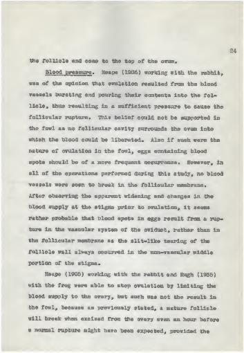
the follicle and come to the top of the ovum.
Blood pressure . Heape (1905) working with the rabbit,
was of the opinion that ovulation resulted from the blood
vessels bursting and pouring their contents into the fol-
licle, thus resulting in a sufficient pressure to cause the
follicular rupture. This belief could not be supported in
the fowl as no follicular cavity surrounds the ovum into
which the blood could be liberuted. Also if such were the
nature of ovulation in the fowl, eggs containing blood
spots should be of a more frequent occurrence. However, in
all of the operations performed during this study, no blood
vessels were seen to break in the follicular membrane.
After observing the apparent widening and changes in the
blood supply at the stigma prior to ovulation, it seems
rather probable that blood spots in eggs result from a rup-
ture in the vascular system of the oviduct, rather than in
the follicular membrane as the slit-like tearing of the
follicle wall always occurred in the non-vascular middle
portion of the stigma*
Heape (1905) working with the rabbit and Rugh (1935)
with the frog were able to stop ovulation by limiting ttie
blood supply to the ovary, but such was not tile result in
the fowl, because as previously stated, a mature follicle
will break when excised from the ovary even an hour before
a normal rupture might have been expected, provided the
muscle fibers in the stigma area were held under the ap-
proximate normal tension*
The one case where ovulation did not occur when the
follicle was clamped off. but not excised from the ovary,
might be explained by the muscle tonus of the follicle
being relaxed when the clamp was applied, and some control-
ling force not being able to stimulate the muscle fibers to
contract sufficiently to cause the follicle to tear at the
stigma.
The visible changes in the appearance of the mature
follicle of the fowl, resulting from the blurring or ob-
literation of the smaller blood vessels just previous to
ovulation, was caused by the muscle tension in the follicu-
lar wall rather than a change in blood supply, so apparently
this was hardly the responsible factor for the follicle
rupture.
Deposition of yolk . Pearl and Curtis (1914) support
the belief of Bartelmez (1912) that internal pressure due
to continued yolk formation was probably the most Important
factor causing ovulation. The results of this study would
not support this view, because although the deutoplasm
serves as a body over which the muscle fibers in the fol-
licle wall can contract and increase the tension, the
amount of yolk material would not cause ovulation in those
follicles where the muscle tonus was decreased due to the

26
death of the bird* Also the follicles clamped off an hour
before the ovulation was expected to occur ruptured in a
normal manner.
Smooth Muscle Theory of Ovulation
Muscular force of the Infundibulum . Pearl and Curtis
(1914) completely removed the entire oviduct from several
hens and ovulation continued to occur so this was definite
evidence that the force exerted by the funnel was not re-
sponsible for ttie follicular rupture. However, in this
study the infundibulum was usually observed exhibiting
great Motility in the ovarian pocket adjacent to the mature
follicle before ovulation.
Muscular force of the ovarian stroma and follicular
wall . Some investigators have thought that the muscle fib-
ers in the ovarian stroma are responsible for ovulation,
but Hugh (1935) reported that ovarian contractions occur in
both mature and immature frogs and does not believe this to
be a causative factor in ovulation, except by improving the
circulation to that region. In the case of the fowl, no
ovarian contractions were noted at any time, and since the
attachment of the ovary is so restricted, the musculature
of this cannot be considered a factor contributing to the
follicular rupture.
The compactness and abundance of the muscle fibers in

27
the follicular wall (Pigs. 3, 4, and 5) would indicate that
working as a unit these fibers would have a narked effect
on the control of the turgidity of the follicle. The ob-
literation of the finger-like blood vessels along the non-
vascular area of the follicle was partially if not en-
tirely due to these muscle cells. Figures 3 and 4 show the
difference in tension of these fibers prior to the time of
ovulation. Since these muscle fibers have the organization
which makes possible a three-way tension at the ends of the
stigma, and only a two-way pull in the center, it seems
reasonable to assume this is the point of greatest con-
traction and as a result, this was thought to be responsible
for the first point of the follicular rupture to occur at
one of the stigma ends.
Chemical Change Theory of Ovulation
Mnzyme action . Schochet (1916) reported the liquid
from the mature ovarian follicles of a hog was capable of
digesting the ovarian tissue, and as a result he concluded
that the digestion of the follicle wall was partially re-
sponsible for the follicular rupture. He further stated
that another factor causing ovulation was the compression
of the blood vessels, thus cutting off the nutrient supply
to the stigma, causing it to atrophy and break.
There was no indication of any digestive action in any

28
of the follicles of the fowl irrespective of size* Figure
3 shows the corona radiata is Intact in a follicle immedi-
ately before rupturing so this would indicate that there is
no digestive aetlon inside the follicle since it would have
acted first on this layer of cells* However, the more na-
ture the ovum the wider the stigma and the thinner the fol-
licular membranes at this region* The narrowness of the
follicular membrane at the stigma seems to result from an
inhibition of the growth of the tissue as a consequence of
the non-vascular condition in this region rather than from
any proteolytic digestion or atrophy*
Anterior pituitary . Many workers have investigated the
relationship between the pituitary gland and ovulation* The
rabbit seems to be the most favorable subject for this
study, since ovulation usually does not occur until approxi-
mately 10 hours after copulation* Bellerby (1929) was able
to Induce ovulation in the rabbit without copulation by
subcutaneous Injections of an extract from the anterior lobe
of the pituitary* The rabbit was kept under the Influence
of an anaesthetic to avoid any "mental effects'* and after
giving the extract, ovulation occurred in about 10 hours*
Thus. Bellerby concluded that copulation stimulates the
pituitary to secrete substances into the blood stream which
induce ovulation* Fee and Parkes (1929) found that the re-
moval of the pituitary within one hour after copulation

29
inhibited ovulation, but when the hypophysec tomy was per-
formed later, ovulation was not impeded. Smith and White
(1951) reported that ovulation would occur in the rabbit aa
long as the pituitary was not removed from the body until
1 1/4 hours after mating. Rugh (1935) reported that sub-
stances from the pituitary gland instigated the follicular
rupture in the frog.
No attempt was made to remove the pituitary gland from
the hens used in this study, but in two eases pltultrin in-
jections were made without performing the celiotomy. One
bird stopped laying for three days and then returned to the
normal intensity or rate of laying. The second case indi-
cated that the injections had diminished the number of eggs
in the clutch to a slight degree, although the bird laid
four consecutive days after the injection. This latter
bird was injected 10 minutes after ovipositlon had occurred.
The first bird was Injected before ovlposltion.
DISCUSSION
In some mammals, ovulation seems to be associated with
copulation in one way or another, but not always does ovu-
lation occur after coitus. However, it is well known that
in the fowl, ovulation is in no way dependent upon copula-
tion.
Since ovulation did not occur in the three cases where

30
the birds died after bulged areas had appeared on the stig-
ma for a considerable period, it is doubtful that any di-
gestive action is responsible for the release of the ovum.
Such digestive action should have continued sufficiently
long after death to rupture the follicle. Also digestive
action would not be expected to be so specific as to act
upon the tissues at each end of the stigma and not effect
the other portions of the same region. The histological
evidence presented indicates that the portion of the fol-
licle next to the vitelline membrane is intact, and this
would not be expected if digestive action had taken place.
Also there is no indication of pressure necrosis or vacu-
olization of the cells in the follicular membrane, so it
is doubtful that any of these theories aid in explaining
the ovulation phenomenon in the fowl.
Since many of the follicular ruptures observed in this
study took place very slowly as a result of the drying of
the follicular tissue, very accurate observations could be
made concerning the liquor folliculi in the follicle, and
in the 33 cases observed, no fluid was seen even when the
initial point of the rupture was very small. Also no blood
vessels were observed to break in the follicle and no blood
was seen to be expelled when the ovum was liberated, so it
is improbable that these are causative factors of ovulation
in the fowl.

31
One of the causes of ovulation In the fowl is apparent-
ly the prolonged tension of the muscle fibers in the fol-
licular membrane, as the muscle fibers are very abundant in
the inside layers of the follicular membranes where the
rupture is initiated. The initial point of the rupture is
at the ends of the stigma where the fibers have the great-
est power of contraction. The factor or factors respons-
ible for the contractions of these muscle fibers was not
determined.
Apparently the yolk merely serves as a body upon which
the muscle fibers can contract, because ovulation occurred
when the mature follicle was excised from the ovary, pro-
vided the original turgidity was maintained with clamps.
The addition of deutoplasm is not responsible for the in-
creased tension of the follicle as it becomes very flaccid
when the muscle tonus is decreased as a result of death or
excision.
SUMMARY
1. Thirty-three normally occurring ovulations were ob-
served in this study.
2. The mean period of time elapsing between ovi-
position and ovulation was 32.2 minutes, with a range of 7
to 74 minutes.
3. The mean length of interval between successive eggs

of the hens operated upon was 26.3 hours, with a range of
24.3 to 28.7 hours. No association was found between the
length of interval and the time intervening between ovl-
position and ovulation.
4. A last minute deposition of yolk was not respons-
ible for the follicular rupture.
5. Observations of follicles that failed to rupture
and the study of histological preparations indicates that
neither enzymatic action nor vacuolization of cells in the
follicular membrane is responsible for ovulation in the
fowl.
6. Mature follicles ruptured normally when excised
from the ovary so blood pressure was apparently not respons-
ible for ovulation.
7. No liquor folliculi was observed in any of the fol-
licles.
8. Pressure resulting from the prolonged tension of
the muscle fibers of the follicular membrane seems to be one
of the factors responsible for ovulation in the fowl, but
the factor or factors stimulating these fibers to contract
was not determined* No critical studies were made of the
Influence of hormones on ovulation.

ACKNOWLEDGMENT
The writer Is greatly Indebted to
Dr. D. C. Warren for suggesting this
problem, and for giving such a generous
amount of his time In aiding and criti-
cally supervising this study.

34
LITERATURE CITED
Bartelmez, George W.The bilateral!ty of the pigeon* s egg. I. A study inegg organisation from the first growth period of theoocyte to the beginning of cleavage. Jour. Morph.23: 269-329. June 1912.
Bellerby, C. W.The relation of the anterior lobe of the pituitary toovulation. Jour. Phys., Proc. 67: XXXIII-XXXIV.April 1929.
Craig 9 Wallace .
The stimulation and the inhibition of ovulation inbirds and mammals. Jour. Animal Behavior 3: 215-221.May 1913.
Pee, A. R. and Parkes, A. S.Studies on ovulation. I. The relation of the anteriorpituitary body to ovulation in the rabbit. Jour. Phys.67: 383-388. July 1929.
Hartman, Carl 0.Ovulation and the transport and viability of ova andsperm in the female genital tract. In Sex and inter-nal secretions by Edgar Allen. Baltimore. Williamsand Wilklns. p. 647-733. 1932.
Heape, Walter.Ovulation and degeneration of ova in the rabbit.Royal Soc. of London, Proc. Series B. 76: 260-268.June 1905.
KeNally, E. H. and Byerly, T. C.Variation in the early development of embryos in hens'eggs. Poultry Science 14: 296-297. September 1935.
Patterson, J. Thomas.Studies on the early development of the hen's egg.I. History of the early cleavage and of the accessorycleavage. Jour. Morph. 21: 101-134. March 1910.
35
Pearl, Raymond and Curtis, Maynie R,Studies on the physiology of reproduction in the do-mestic fowl, VIII. On some physiological effects ofligation, section, or removal of the oviduct. Jour,Exp, Zool. 17: 395-424, October 1914.
Rugh, Roberts
.
Ovulation in the frog, II. Follicular rupture to fer«
tilization. Jour. Exp. Zool. 71: 163-193. July 1935,
Schochet, S. S.A suggestion as to the process of ovulation andovarian cyst formation, Anat, Record 10: 447-457,April 1916,
Smith, Philip E, and White, W. E.The effect of hypophysectoray on ovulation and corpusluteum formation in the rabbit. Jour, Amer, Med,Assoc. 97: 1861-1863, December 1931,
Walton, A, and Hammond, J,Observations on ovulation in the rabbit* BritishJour, Exp, Biol, 6 i 190-204, December 1928,
Warren, D. G, and Scott, H. M.The time factor in egg formation.14: 195-207, July 1935.
Poultry Science