Long-term osseous morphologic outcome of surgically treated unilateral coronal craniosynostosis
7
PEDIATRIC/CRANIOFACIAL Long-Term Osseous Morphologic Outcome of Surgically Treated Unilateral Coronal Craniosynostosis Devra B. Becker, M.D. Christopher E. Fundakowski, B.A. Daniel P. Govier Valerie B. Deleon, Ph.D. Jeffrey L. Marsh, M.D. Alex A. Kane, M.D. St. Louis, Mo.; and Baltimore, Md. Background: Unilateral coronal craniosynostosis has characteristic osseous dys- morphology that persists into adulthood if untreated. Knowledge of the long- term in vivo osseous morphologic outcome of surgically treated unilateral coro- nal craniosynostosis patients is limited. The purpose of this study was to define the osseous morphology of adolescent patients who underwent surgery for unilateral coronal craniosynostosis in infancy, compared with both their 1-year postoperative morphology and the morphology of other individuals with un- treated unilateral coronal craniosynostosis. Methods: Three populations of unilateral coronal craniosynostosis were studied: group 1, patients with surgical treatment of unilateral coronal craniosynostosis in infancy who had reached dentoskeletal maturity, ranging in age from 13.5 to 32.7 years (n 9); group 2, individuals with untreated unilateral coronal craniosynos- tosis, ranging in age from 1.1 to 21 years (n 11); and group 3, a subset of group 1 patients 1 year after surgical correction of unilateral coronal craniosynostosis, ranging in age from 1.2 to 2.6 years (n 6). Data from high-resolution, thin-slice computed tomographic scans of the head were analyzed. Thirty-five reproducible osseous landmarks were recorded as three-dimensional coordinates using ETDIPS imaging software. Nonmidline landmarks were designated as either ipsilateral or contralateral to the synostosis. One researcher performed all landmarking with high intrarater reliability (average error, 2 mm). Data from the three groups were analyzed for asymmetry using Euclidean distance matrix analysis techniques. Results: Euclidean distance matrix analysis asymmetry analysis demonstrated more statistically significant ipsilateral-contralateral asymmetric pairs in group 1 (68 of 135) than in group 3 (25 of 135), but fewer statistically significant ipsilateral- contralateral asymmetric pairs than in group 2 (93 of 135). Conclusions: Surgical treatment of unilateral coronal craniosynostosis in infancy results in a less asymmetric craniofacial skeleton in adolescence than nontreatment. However, patients who have been followed to dentoskeletal maturity have a greater degree of asymmetry than those evaluated at 1 year postoperatively. These results support the conclusion that with time there is a partial reversion to the untreated phenotype. (Plast. Reconstr. Surg. 117: 929, 2006.) T he phenotype recognized as unilateral coronal craniosynostosis results from what has traditionally been thought of as in utero fusion of one of the coronal sutures. Normally, these sutures remain patent as part of the calvaria’s accommodation of the grow- ing brain. In unilateral coronal craniosynosto- sis, the constraint introduced by the synostosis prevents symmetric expansion of the craniofa- cial complex. This, in turn, leads to a charac- teristic and well-described asymmetric cranio- facial dysmorphology that persists into adulthood if untreated. Surgical release of craniosynostosis has been performed for over a century. The current ratio- nale for intervention is to minimize potential neurologic morbidity and to prevent psychoso- cial stigmatization secondary to dysmorphology. High-resolution computed tomography is rou- tinely performed for establishing diagnosis and From the Cleft Palate and Craniofacial Deformities Institute, St. Louis Children’s Hospital, Washington University Med- ical Center; the Department of Anthropology, Johns Hopkins University; and the Cleft Lip/Palate and Craniofacial De- formities Center, St. John’s Mercy Medical Center. Received for publication December 9, 2004; revised April 18, 2005. Presented at the 61st Annual Meeting of the American Cleft Palate Association, in Chicago, Illinois, March 15 through 20, 2004. Copyright ©2006 by the American Society of Plastic Surgeons DOI: 10.1097/01.prs.0000200613.06035.51 www.plasreconsurg.org 929
Transcript of Long-term osseous morphologic outcome of surgically treated unilateral coronal craniosynostosis

PEDIATRIC/CRANIOFACIAL
Long-Term Osseous Morphologic Outcomeof Surgically Treated UnilateralCoronal Craniosynostosis
Devra B. Becker, M.D.Christopher E.
Fundakowski, B.A.Daniel P. Govier
Valerie B. Deleon, Ph.D.Jeffrey L. Marsh, M.D.
Alex A. Kane, M.D.
St. Louis, Mo.; and Baltimore, Md.
Background: Unilateral coronal craniosynostosis has characteristic osseous dys-morphology that persists into adulthood if untreated. Knowledge of the long-term in vivo osseous morphologic outcome of surgically treated unilateral coro-nal craniosynostosis patients is limited. The purpose of this study was to definethe osseous morphology of adolescent patients who underwent surgery forunilateral coronal craniosynostosis in infancy, compared with both their 1-yearpostoperative morphology and the morphology of other individuals with un-treated unilateral coronal craniosynostosis.Methods: Three populations of unilateral coronal craniosynostosis were studied:group 1, patients with surgical treatment of unilateral coronal craniosynostosis ininfancy who had reached dentoskeletal maturity, ranging in age from 13.5 to 32.7years (n � 9); group 2, individuals with untreated unilateral coronal craniosynos-tosis, ranging in age from 1.1 to 21 years (n � 11); and group 3, a subset of group1 patients 1 year after surgical correction of unilateral coronal craniosynostosis,ranging in age from 1.2 to 2.6 years (n � 6). Data from high-resolution, thin-slicecomputed tomographic scans of the head were analyzed. Thirty-five reproducibleosseous landmarks were recorded as three-dimensional coordinates using ETDIPSimaging software. Nonmidline landmarks were designated as either ipsilateral orcontralateral to the synostosis. One researcher performed all landmarking with highintrarater reliability (average error, �2 mm). Data from the three groups wereanalyzed for asymmetry using Euclidean distance matrix analysis techniques.Results: Euclidean distance matrix analysis asymmetry analysis demonstrated morestatistically significant ipsilateral-contralateral asymmetric pairs in group 1 (68 of135) than in group 3 (25 of 135), but fewer statistically significant ipsilateral-contralateral asymmetric pairs than in group 2 (93 of 135).Conclusions: Surgical treatment of unilateral coronal craniosynostosis in infancyresults in a less asymmetric craniofacial skeleton in adolescence than nontreatment.However, patients who have been followed to dentoskeletal maturity have a greaterdegree of asymmetry than those evaluated at 1 year postoperatively. These resultssupport the conclusion that with time there is a partial reversion to the untreatedphenotype. (Plast. Reconstr. Surg. 117: 929, 2006.)
The phenotype recognized as unilateralcoronal craniosynostosis results fromwhat has traditionally been thought of as
in utero fusion of one of the coronal sutures.
Normally, these sutures remain patent as partof the calvaria’s accommodation of the grow-ing brain. In unilateral coronal craniosynosto-sis, the constraint introduced by the synostosisprevents symmetric expansion of the craniofa-cial complex. This, in turn, leads to a charac-teristic and well-described asymmetric cranio-facial dysmorphology that persists intoadulthood if untreated.
Surgical release of craniosynostosis has beenperformed for over a century. The current ratio-nale for intervention is to minimize potentialneurologic morbidity and to prevent psychoso-cial stigmatization secondary to dysmorphology.High-resolution computed tomography is rou-tinely performed for establishing diagnosis and
From the Cleft Palate and Craniofacial Deformities Institute,St. Louis Children’s Hospital, Washington University Med-ical Center; the Department of Anthropology, Johns HopkinsUniversity; and the Cleft Lip/Palate and Craniofacial De-formities Center, St. John’s Mercy Medical Center.Received for publication December 9, 2004; revised April 18,2005.Presented at the 61st Annual Meeting of the American CleftPalate Association, in Chicago, Illinois, March 15 through20, 2004.Copyright ©2006 by the American Society of Plastic Surgeons
DOI: 10.1097/01.prs.0000200613.06035.51
www.plasreconsurg.org 929

planning surgical reconstruction in individualswith craniosynostosis. Archived computed tomo-graphic data allow for cross-sectional and longi-tudinal quantitative assessment of surgical out-come.
Reports of the results of surgical release ofunilateral coronal craniosynostosis in infancyhave been published; however, these are gener-ally limited by short follow-up times or lack ofquantitative analysis.1,2 Therefore, the long-termmorphologic outcome of surgical interventionin infancy for unilateral coronal craniosynostosisis largely unknown.
The specific aim of this study was to define theosseous morphology of adolescent patients whounderwent surgery for unilateral coronal cranio-synostosis in infancy, compared with both their1-year postoperative morphology and the mor-phology of other individuals with untreated uni-lateral coronal craniosynostosis. We determinewhether individuals with unilateral coronal cra-niosynostosis who underwent surgery in infancydiffer at dentoskeletal maturity (defined in thisstudy as older than 12 years) in osseous cranio-facial symmetry from the same individuals 1 yearpostoperatively and from other individuals withuntreated unilateral coronal craniosynostosis.Thus, individuals are being evaluated both lon-gitudinally, for change over time from a startingpoint of 1 year postoperatively, and cross-sectionally, with a cohort of patients who havenot had surgical intervention. This addresses twoquestions: first, is there change over time? Sec-ond, if there is change over time, does the ulti-mate phenotype appear similar to, or differentfrom, individuals who have not had surgical in-tervention?
PATIENTS AND METHODS
Patient DataHuman studies committee approval was ob-
tained for this project. The long-term unilateralcoronal craniosynostosis patients underwent com-puted tomographic scanning for this study as partof a larger long-term follow-up study of unilateralcoronal craniosynostosis. Three populations ofunilateral coronal craniosynostosis were studied:group 1, patients with surgical treatment of uni-lateral coronal craniosynostosis in infancy whohad reached dentoskeletal maturity, ranging inage from 13.5 to 32.7 years (mean, 20.3 years;median, 19.2 years; n � 9); group 2, individualswith untreated unilateral coronal craniosynosto-sis, ranging in age from 1.1 to 21 years (mean, 6.6
years; median, 4.1 years; n � 11); and group 3, asubset of group 1 patients 1 year after surgicalcorrection of unilateral coronal craniosynostosis,ranging in age from 1.2 to 2.6 years (mean, 1.9years; median, 2.0 years; n � 6). Group 1 consistedof six patients who were operated on by the samesurgeon and three who were each operated on bya different surgeon. The surgical technique usedin all cases was a bifrontal coronal craniotomy withremodeling of the anterior calvaria, superolateralorbital advancement ipsilateral to the synostosis,and attempted equalization of orbital heights.3The age range at time of surgery was 10 to 44 weeks(mean, 20 weeks; median, 16 weeks). Group 1 wasrecruited by means of a letter sent to 56 patientslisted in our craniofacial clinic database who metthe inclusion criteria of a diagnosis of nonsyn-dromic unilateral coronal craniosynostosis andage older than 12 years. A protocol was used tomaximize response rates. All nonresponders werecalled at their telephone numbers of record. Of allthe patients to whom a letter was sent, we were notable to contact 23 by phone or letter. All patientswere reimbursed for their participation in thestudy. Group 2 was obtained from high-resolutioncomputed tomographic scans in the CraniofacialArchive located at the Craniofacial Imaging Lab ofWashington University School of Medicine.4 Thisgroup has been described previously and consistsof the computed tomographic scans of individualswith surgically untreated unilateral coronal cra-niosynostosis representing three institutions inthree separate countries (Denmark, The Nether-lands, and the United States).5 Group 3 was alsoobtained from the Craniofacial Archive.
Landmark Data CollectionNine midline landmarks and 10 paired land-
marks were digitally placed on surface- renderedcomputed tomographic scan data from each of thestudy groups using ETDIPS v2.0 (exploratory two/three-dimensional imaging processing system;http://www.cc.nih.gov/cip/software/etdips/). ET-DIPS is a multidimensional volume visualizationand analysis software that was co-developed by theNational Institutes of Health and the National Uni-versity of Singapore under the Cooperative Re-search and Development Agreement (CACR-645).See Table 1 and Figure 1 for a listing and sampleplacement of these landmarks. The x, y, and z co-ordinates for the landmarks were recorded. A singleinvestigator placed all landmarks. Intrarater reliabil-ity was determined by calculating average landmarkposition on five computed tomographic scans, each
Plastic and Reconstructive Surgery • March 2006
930

landmarked five separate times. Those landmarkswhose distance from the mean varied less than 2mm across all five ratings, indicating high intraraterreliability, were included.
Statistical AnalysisAfter landmark placement, data were pooled
within each group and analyzed using Euclideandistance matrix analysis asymmetry analysis on aPC platform. Euclidean distance matrix analysis isa landmark-based method of analyzing form6,7 thatcompares linear distances between landmarks, de-fined by x, y, and z coordinates, in two populations.In Euclidean distance matrix analysis asymmetryanalysis, the two populations consist of two sides(ipsilateral and contralateral) within a population.Thus, groups can be compared by the amount ofasymmetry seen within a sample group. The datawere sampled nonparametrically with 100 to 500resamples and an alpha level of 0.05.
RESULTSThe composition of groups 1, 2, and 3 is shown
in Table 2. The total possible landmark pairs were135. Of the 135 pairs, group 1 had 68 of 135statistically significant asymmetric pairs, group 2had 93 of 135 statistically significant asymmetricpairs, and group 3 had 25 of 135 statistically sig-nificant asymmetric pairs. Statistically significantasymmetric pairs were defined as those ipsilateraldistances that fell outside 95 percent confidenceintervals for contralateral distances. A graphic rep-resentation of the asymmetry can be seen in Figure2.
To address the possibility that three patients ingroup 1, who underwent surgical correction ofunilateral coronal craniosynostosis in infancy bydifferent surgeons, represented a bias that skewedthe results, we ran Euclidean distance matrix anal-ysis excluding these patients (group 1a, n � 6).Our results showed similar results (69 of 135 asym-
Fig. 1. Sample three-dimensional computed tomographic scan with landmarks inplace in anterior (above, left), lateral (above, right), external basilar (below, left), andinternal cranial (below, right) views.
Volume 117, Number 3 • Unilateral Coronal Craniosynostosis
931
metric pairs), suggesting that these patients do notrepresent a bias. It is possible that some patientsin group 1 do have a pronounced unilateral coro-nal craniosynostosis phenotype; however, Euclid-ean distance matrix analysis–computed confi-dence intervals address that issue. In addition, weran Euclidean distance matrix analysis excludingpatients who were not represented in group 3(group 1b, n � 6), which allows for a direct com-parison of individuals 1 year postoperatively and atdentoskeletal maturity. In that trial, 73 of 135 land-mark pairs were asymmetric. The difference be-tween the number of asymmetric landmark pairsin group 1 and the modified group 1 (which in-cluded only those patients represented in group3) was not statistically significant.
The asymmetry can be categorized on the basisof anatomical subdivision as well. The landmark-based findings are best understood when inter-preted within an anatomical context. Anteriorly,the nasal root has begun to revert to an ipsilateraldisplacement (as indicated by the decreased dis-tance from the nasion to the ipsilateral landmarksfrontozygomatic junction, zygomaxillare superius,external auditory meatus superior, and asterion).The lateral aspect of the orbit has similarly begunto revert to a superior and lateral displacement (asindicated by the increased distance from the ip-silateral frontozygomatic junction to the intraden-tale superius and ipsilateral zygomaxillare supe-rius and zygomaxillare inferius).
The lateral landmarks can be used as a proxyfor midface development. One year postopera-tively, the midface appears to be symmetric asmeasured by the distance from the external au-ditory meatus superior to the midline landmarksnasion, nasale, anterior nasal spine, intradentalesuperius, and the ipsilateral landmarks supraor-bital notch and zygomaxillare inferius. At long-term follow-up, the midface has reverted to ashortened phenotype as measured by the short-ened distances between the external auditory me-atus superior and the midline landmarks nasion,nasale, anterior nasal spine, and intradentale su-perius, and the ipsilateral landmarks supraorbitalnotch and zygomaxillare inferius.
In the cranial base analysis, the ipsilateral el-evation of the sphenoid wing, as measured bysphenoid landmarks, is corrected 1 year postop-eratively. This reverts over the long term, withdysmorphology similar to untreated unilateralcoronal craniosynostosis individuals. In untreatedunilateral coronal craniosynostosis individuals, itis closer ipsilaterally to midline landmarks and theexternal auditory meatus superior, indicating su-perior displacement. This is best shown in graphicform (Fig. 3).
DISCUSSIONIndividuals with untreated unilateral coronal
craniosynostosis have well-described morphologicabnormalities.8 These abnormalities can be de-scribed both qualitatively and quantitatively. Qual-itative descriptions of changes in the craniofacialskeleton attributable to unilateral coronal cranio-synostosis include the classic harlequin orbit andfrontal recession ipsilateral to the synostosis, withcompensatory deformation contralaterally. Qual-itative analysis has also been used to demonstratenormalization of the cranial base postoperatively.Third-party qualitative assessment of 20-monthpostsurgical endocranial bases has shown an im-provement in appearance to blinded observers.2Although qualitative description is useful, quan-titative analysis is the standard for documentingstatistically significant changes in skeletal form incraniofacial anomalies. Quantitative morphologicanalysis has been used to define the natural historyof untreated unilateral coronal craniosynostosis5
and has been extended to evaluate postoperativepatients. Anthropometric computed tomographicanalysis of the orbits and the globes9 of affectedindividuals shows a decrease in the orbital index(defined as orbital height/orbital width) 1 yearpostoperatively, suggesting a quantitative normal-ization of unilateral coronal craniosynostosis or-
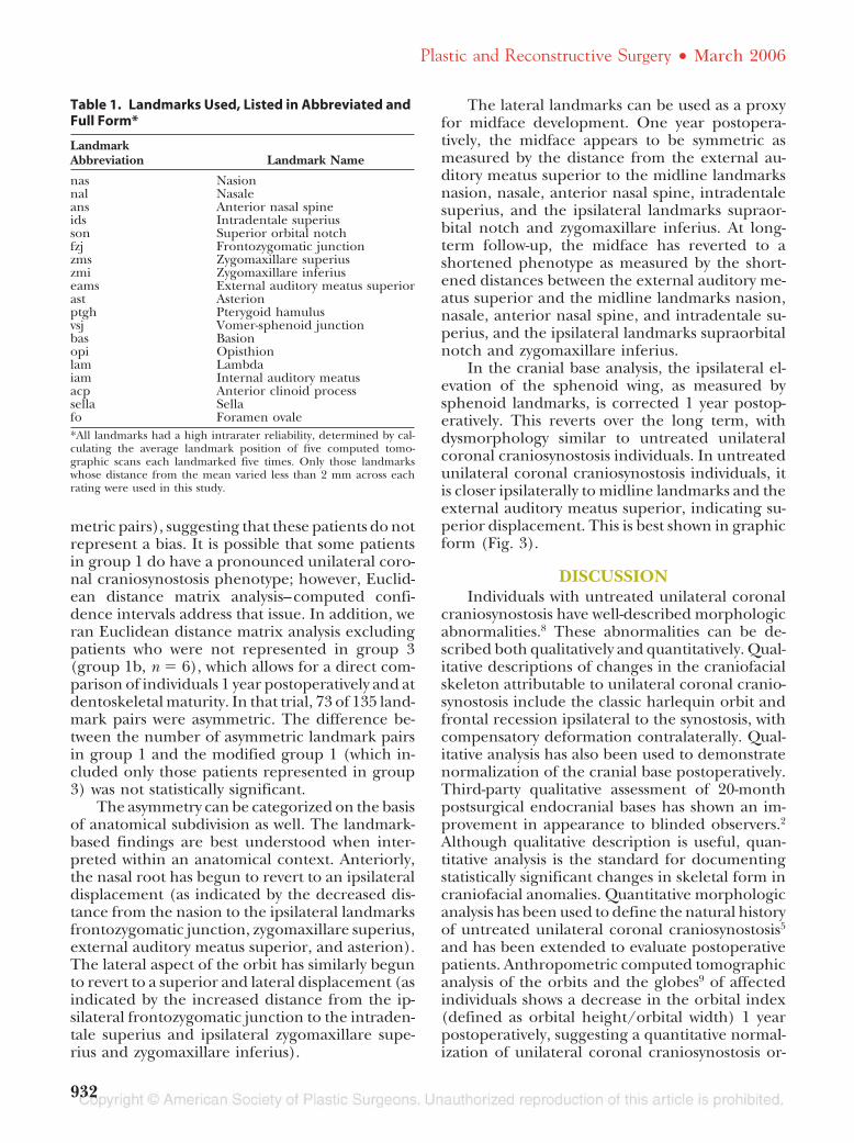
Table 1. Landmarks Used, Listed in Abbreviated andFull Form*
LandmarkAbbreviation Landmark Name
nas Nasionnal Nasaleans Anterior nasal spineids Intradentale superiusson Superior orbital notchfzj Frontozygomatic junctionzms Zygomaxillare superiuszmi Zygomaxillare inferiuseams External auditory meatus superiorast Asterionptgh Pterygoid hamulusvsj Vomer-sphenoid junctionbas Basionopi Opisthionlam Lambdaiam Internal auditory meatusacp Anterior clinoid processsella Sellafo Foramen ovale*All landmarks had a high intrarater reliability, determined by cal-culating the average landmark position of five computed tomo-graphic scans each landmarked five times. Only those landmarkswhose distance from the mean varied less than 2 mm across eachrating were used in this study.
Plastic and Reconstructive Surgery • March 2006
932

bital morphology in the short term. However,these postoperative morphologic studies—bothqualitative and quantitative—are limited by thefact that none of them include analysis for indi-viduals operated on in infancy more than 2 yearspostoperatively.
The natural history of untreated unilateralcoronal craniosynostosis is known primarilythrough quantitative morphologic analysis per-formed on cohorts of preoperative and untreatedunilateral coronal craniosynostosis. Lo et al.9 dem-onstrated, using preoperative computed tomo-graphic scans, that the ipsilateral bony orbit shapein unilateral coronal craniosynostosis is signifi-cantly different from the contralateral bony orbit.Kane et al.10 demonstrated in preoperative com-puted tomographic scans that ipsilateral mandi-bles in unilateral coronal craniosynostosis wereshorter and volumetrically smaller when com-
pared with the contralateral side. Patients withsurgically untreated unilateral coronal craniosyn-ostosis have been shown to have persistent dys-morphic features.5 The midface is narrower ipsi-lateral to the synostosis and shorter in theanteroposterior dimension.
Our data document several patterns. We haveconfirmed the observations made previously1,2
that the osseous craniofacial dysmorphology asso-ciated with unilateral coronal craniosynostosisdoes appear to normalize 1 year postoperatively.At dentoskeletal maturity, however, the craniofa-cial skeleton demonstrates a partial reversion to-ward the untreated phenotype. It is not simply thatthe long-term follow-up patients show an in-creased number of absolute asymmetric landmarkpairs when compared with 1-year postoperativecomputed tomographic scans; rather, the salientpoint is that the asymmetric pairs seen in the long-
Fig. 2. Plot depicting the axis of asymmetry for all the landmark pairs. A value of 1 on the xaxis indicates symmetry between the ipsilateral and contralateral sides, whereas a value ofless than 1 indicates a statistically significant asymmetry caused by a smaller distance on theipsilateral side and a value of greater than 1 indicates a statistically significant asymmetrycaused by a larger distance between landmark pairs on the ipsilateral side. Note that thegreatest symmetry is seen in the 1-year postoperative computed tomographic scans, as in-dicated by the high concentration of landmark pairs with an axis of asymmetry of 1. In con-trast, the least symmetry is seen in the untreated cohort.
Table 2. Characteristics of Each Comparison Group
Group Description No.
Total No.of MaleSubjects
Total No.of FemaleSubjects Right-Sided Left-Sided
1 Long-term surgically treated 9 1 8 7 22 Untreated 11 5 6 7 43 1 year postoperatively 6 1 5 4 1
Volume 117, Number 3 • Unilateral Coronal Craniosynostosis
933

Fig. 3. Patterns of asymmetry can be seen when evaluating landmark pairs in anatomical subgroups. Pictured are, from left to right,a representative computed tomographic scan of a 1-year postoperative individual, a representative long-term follow-up computedtomographic scan, and a representative computed tomographic scan of an untreated individual. Green lines indicate symmetrybetween ipsilateral and contralateral landmark pairs, red lines indicate a shorter ipsilateral distance between landmarks, and blue linesindicate a longer ipsilateral distance between landmarks. Note the partial reversion to the untreated phenotype in the anterior,lateral, and external basilar views.
Plastic and Reconstructive Surgery • March 2006
934
term follow-up cohort mirror the preoperativeasymmetric pairs in both quality and direction.
This study documents that some patients whounderwent craniofacial surgery for unilateralcoronal craniosynostosis in infancy will undergopartial reversion toward an untreated phenotypebetween 1 year postoperatively and dentoskeletalmaturity. We did not identify causative factors inthe reversion.
Limitations of the StudyThe major limitation of this study is that 16
percent of patients returned for follow-up specif-ically for this study. There may be a selection biasin the individuals who chose to participate, and itis impossible to know what bias this may haveintroduced. This subpopulation may be represen-tative of the whole population, of those with theleast long-term normalization, or of those with thebest long-term normalization. A second limitationis the range of age of the long-term assessment.Although some mandibular growth, especially inmale subjects, would be expected in the lowerrange, maxillary growth and upper midface, or-bital, and calvarial growth would have been com-pleted.
CONCLUSIONSWe draw the following conclusions from our
study. First, some individuals with unilateral coro-nal craniosynostosis whose dysmorphic featuresnormalize 1 year after surgery in infancy will un-dergo a partial reversion to an untreated unilat-eral coronal craniosynostosis phenotype by thetime they reach dentoskeletal maturity. Second,additional long-term unilateral coronal craniosyn-ostosis–treated individuals need to be studied todetermine the frequency of phenotypic reversion.Third, prospective studies must be undertaken to
identify factors involved in reversion. Finally, pa-tients’ families must be counseled about the pos-sibility of reversion between early childhood andadolescence.
Alex A. Kane, M.D.St. Louis Children’s Hospital
Suite 11 W 71 Children’s Place
St. Louis, Mo. [email protected]
REFERENCES1. Marsh, J. L., and Vannier, M. W. Cranial base changes fol-
lowing surgical treatment of craniosynostosis. Cleft Palate J. 23(Suppl.): 9, 1986.
2. Perlyn, C. A., Marsh, J. L., Pilgram, T. K., and Kane, A. A.Plasticity of the endocranial base in nonsyndromic cranio-synostosis. Plast. Reconstr. Surg. 108: 294, 2001.
3. Hardesty, R. A., Marsh, J. L., and Vannier, M. W. Unicoronalsynostosis: A surgical intervention. Neurosurg. Clin. North Am.2: 641, 1991.
4. Perlyn, C. A., Marsh, J. L., Vannier, M. W., et al. The cranio-facial anomalies archives at St. Louis Children’s Hospital: 20years of craniofacial imaging experience. Plast. Reconstr. Surg.108: 1862, 2001.
5. Kane, A. A., Yong-Oock, K., Eaton, A., et al . Quantificationof osseous facial dysmorphology in untreated unilateral coro-nal synostosis. Plast. Reconstr. Surg. 106: 251, 2000.
6. Lele, S., and Richtsmeier, J. T. Euclidean distance matrixanalysis: A coordinate-free approach for comparing biolog-ical shapes using landmark data. Am. J. Phys. Anthropol. 86:415, 1991.
7. Lele, S., and Richtsmeier, J. T. Euclidean distance matrixanalysis: Confidence intervals for form and growth differ-ences. Am. J. Phys. Anthropol. 98: 73, 1995.
8. Marsh, J. L., Gado, M. H., Vannier, M. W., and Stevens, W.G. Osseous anatomy of unilateral coronal synostosis. CleftPalate J. 23: 87, 1986.
9. Lo, L. J., Marsh, J. L., Kane, A. A., and Vannier, M. W. Orbitaldysmorphology in unilateral coronal synostosis. Cleft PalateCraniofac. J. 33: 190, 1996.
10. Kane, A. A., Lo, L. J., Vannier, M. W., and Marsh, J. L.Mandibular dysmorphology in unicoronal synostosis and pla-giocephaly without synostosis. Cleft Palate Craniofac. J. 33: 418,1996.
Volume 117, Number 3 • Unilateral Coronal Craniosynostosis
935