University of Groningen Osteoprotegerin in organ fibrosis: biomarker ...

Osteoprotegerin Overexpression by Breast Cancer Cells Enhances
Orthotopic and Osseous Tumor Growth and Contrasts with That
Delivered Therapeutically
Jane L. Fisher,1Rachel J. Thomas-Mudge,
1Jan Elliott,
1Daphne K. Hards,
1Natalie A. Sims,
2
John Slavin,3T. John Martin,
1,2and Matthew T. Gillespie
1,2
1St. Vincent’s Institute of Medical Research; 2Department of Medicine, The University of Melbourne; and 3Department of Pathology,St. Vincent’s Hospital, Fitzroy, Victoria, Australia
Abstract
Osteoprotegerin (OPG) acts as a decoy receptor for receptoractivator of NF-KB ligand (RANKL), which is a pivotalmolecule required for osteoclast formation. In vitro OPGinhibits osteoclast formation and in vivo (administered asFc-OPG) it reduces hypercalcemia and the establishment ofosteolytic lesions in mouse models of tumor cell growth inbone. Osteolysis can be induced by parathyroid hormone–related protein (PTHrP) produced by breast cancer cells thatresults in an increased osteoblastic RANKL/OPG ratio. Weexamined the effect of local tumor production of OPG on theability of breast cancer cells to establish and grow in bone andmammary fat pad. MCF-7 cells or MCF-7 cells overexpressingPTHrP were transfected with full-length OPG and inoculatedinto the proximal tibiae of athymic nude mice. Mice injectedwith cells overexpressing PTHrP and OPG showed enhancedtumor growth, increased osteolysis (2-fold compared withMCF-7 cells overexpressing PTHrP), and altered histologythat was reflective of a less differentiated (more aggressive)phenotype compared with MCF-7 cells. In contrast, adminis-tration of recombinant Fc-OPG reduced tumor growth andlimited osteolysis even in mice inoculated with OPG over-expressing cells. Similarly, OPG overexpression by breastcancer cells enhanced tumor growth following orthotopicinoculation. These results indicate that OPG overexpression bybreast cancer cells increases tumor growth in vivo and thatthere are strikingly different responses between therapeuti-cally administered Fc-OPG and full-length OPG produced bytumor cells. (Cancer Res 2006; 66(7): 3620-8)
Introduction
Breast cancer is the most common malignancy affecting womenand is a major cause of death in young middle-aged women inthe United States, with 1 in 14 affected in their lifetime. Thedevelopment of metastatic disease is invariably associated with afatal outcome. In all series reporting, the most common site offirst relapse is bone. When considered with other sites of firstmetastasis, >50% of patients will develop bone metastases at thetime of first recurrence (1). Bone metastases, usually osteolytic,may be expected to complicate the course of up to 80% of patients
with disseminated disease, with pathologic fracture and/or hyper-calcemia frequently observed.
A recently identified member of the tumor necrosis factor (TNF)receptor family, osteoprotegerin (OPG), has the capacity to blockosteoclast formation both in vitro and in vivo (2) and is considereda potential therapy to combat cancer-induced bone loss. OPGacts as a decoy receptor blocking the binding of the TNF ligand,receptor activator of NF-nB ligand (RANKL), to its cognatesignaling receptor RANK (present on hematopoietic cells), therebyinhibiting osteoclast formation as well as activity (2–5). Therequirement of RANKL and RANK for osteoclast formation hasbeen established because mice deficient in either RANKL or RANKare osteopetrotic due to failed osteoclast formation (6–8).Furthermore, the role for the decoy receptor OPG in osteoclastformation was shown by genetic experiments whereby OPG-nullmice were shown to be severely osteoporotic (9) whereastransgenic mice overexpressing OPG were osteopetrotic (2).
We have shown that human breast cancer cells do not expressRANKL, but do express OPG and RANK, and are unable to directlysupport osteoclast formation in vitro (10). Most importantly,however, we showed that breast cancer cells influence osteolysisby inducing the expression of RANKL by osteoblasts, an effectresulting from parathyroid hormone–related protein (PTHrP)production (10). We also noted that levels of OPG mRNA variedin primary cancers and in breast cancer cell lines (10). The currentstudy was aimed at examining whether overexpressing OPG inbreast cancer cells would inhibit tumor-associated osteolysis, usingthe model of intratibial inoculation of cancer cells in athymic mice,and compared this with recombinant OPG (Fc-OPG) treatment.Surprisingly, it was found that the overexpression of full-lengthOPG was associated with enhanced tumor growth in vivo , with anaccompanying increase in osteolysis, whereas Fc-OPG treatment ofmice inhibited osteolysis and, therefore, tumor advance in bone asexpected. Furthermore, consistent with this, the overexpression ofOPG by breast cancer cells also increased tumor growth followingmammary fat pad inoculation.
Materials and Methods
Stable transfection of cell lines. Human breast cancer cell lines
(MCF-7) transfected with PTHrP or vector control plasmids were as
previously described (10). The coding sequence of human OPG (86-1,310 bp;
GenBank accession number NM002546.1) was amplified from RNAextracted from MDA-MB-231 and MCF-7 cells by reverse transcription-
PCR (RT-PCR) using the primers OPG-16 [5V-CGCCTCGAGGGGACCA-
CAATGAACAAG-3V] and OPG-17 [5V-CGCGGATCCATTTCCAGTTATAAG-CAG-3V], which included an XhoI and BamHI restriction endonuclease site
in primer sequences, respectively. The PCR fragments were agarose gel
purified, digested with XhoI and BamHI, and cloned into the XhoI and
Note: J.L. Fisher and R.J. Thomas-Mudge contributed equally to this work.Requests for reprints: Matthew T. Gillespie, St. Vincent’s Institute of Medical
Research, 9 Princes Street, Fitzroy 3065, Victoria, Australia. Phone: 613-9288-2480; Fax:613-9416-2676; E-mail: [email protected].
I2006 American Association for Cancer Research.doi:10.1158/0008-5472.CAN-05-3119
Cancer Res 2006; 66: (7). April 1, 2006 3620 www.aacrjournals.org
Research Article
Research. on January 15, 2016. © 2006 American Association for Cancercancerres.aacrjournals.org Downloaded from

BamHI sites of pCEP4; resultant clones (e.g., pSMR969 from MCF-7 RNAand pSMR1141 from MDA-MB-231 RNA) were verified by nucleic acid
sequencing. The sequence of pSMR1141 was identical to that of the
published sequence of human OPG (GenBank entry NM002546.1) whereas
pSMR969 contained a T-to-C transition at position 557 of the human OPGsequence, which resulted in a single amino acid substitution of a serine to
a proline at amino acid 155. MCF-7 and MCF-7 + PTHrP cells were trans-
fected by calcium phosphate precipitation with DNA for either pSMR969,
pSMR1141, or pCEP4, and hygromycin-resistant clonal ( following limitingdilution) and nonclonal (14-day selection and are used for no more than
four passages from original derivation) lines were generated. MCF-7 cells
and derivatives were maintained in RPMI containing 10% fetal bovine
serum (FBS; ref. 10). All other reagents were of analytic grade obtained fromstandard suppliers. In all assays described herein, cell lines expressing either
OPG or Ser155Pro OPG were established and no difference was noted
between these cell lines in any of the in vitro or in vivo assays.RT-PCR. Total RNA extraction from cell lines, cDNA synthesis, and PCR
were done as described (11). Oligonucleotides used to amplify human
OPG were OPG-4 [5V-GGGGACCACAATGAACAAGTTG-3V] and OPG-5 [5V-AGCTTGCACCACTCCAAATCC-3V], and the resultant PCR product waselectrophoresed through a 1.2% wt/vol agarose gel and product verified and
quantified by detection with a 32P-labeled internal oligonucleotide [OPG-6
(5V-GCTGTCTGTGTAGTAGTGGTCAG-3V)] as previously described (10). As a
normalizing control, human glyceraldehyde-3-phosphate dehydrogenase(GAPDH) was amplified with oligonucleotides GAPDH-3 and GAPDH-4 and
verified with GAPDH-1 as previously described (10). Bound probe was
detected by PhosphorImager analysis (Molecular Dynamics, Inc., Sunnyvale,CA) and densitometry was done using the software program ImageQuant,
version 4.2.
OPG ELISA. To establish the level of OPG protein production from
the clones, an OPG ELISA of conditioned medium was done by Dr. S.Chandrasekhar (Lilly Research Laboratories, Indianapolis, IN) as previously
described (12, 13).
Animals and animal maintenance. Congenitally athymic female nude
mice (BALB/c, nu/nu , Animal Resource Centre, Perth, WA) were purchasedgerm-free at 2 to 3 weeks old and housed in the Orthopaedic Department
Sterile Facility, St. Vincent’s Hospital.
Before tumor inoculation, radiographic examination, and euthanasia,
mice were anesthetized with an intraperitoneal mixture of ketamine
(50 mg/kg), xylazine (5 mg/kg), and acepromazine (0.75 mg/kg). The
St. Vincent’s Hospital Animal Ethics Committee approved all experimental
procedures and the animal care was in accordance with the National Health
and Medical Research Council animal ethics guidelines.Osteoclast formation assays. Osteoblastic cells were prepared from the
calvaria of newborn mice by sequential digestion with 0.1% collagenase
(Worthington Biochemical Co., Freefold, Australia) and 0.2% dispase (Godo
Shusei, Tokyo, Japan). Bone marrow was obtained from the femur and tibia
of adult C57BL6J male mice. Osteoblastic cells and/or breast cancer cells
were cocultured with spleen or marrow cells as previously described
(10, 14).
Intratibial implantation. Cells in log-phase growth were harvested by
trypsinization (0.25% trypsin/0.02% EDTA) and medium containing 10%FBS was added; the cells were then washed thrice by centrifugation in
serum-free RPMI and subsequently resuspended in serum-free RPMI (0.5 �108 cells/mL). Cells were kept at 4jC until use (0-2 hours). The cell
concentration and viability (trypan blue exclusion, >90% viability) weredetermined with a hemocytometer.
Four-week-old mice received intratibial inoculation of parental MCF-7,
MCF-7 + PTHrP, MCF-7 + OPG, MCF-7 + PTHrP + OPG cells, or control cellsfor comparison, including MCF-7 + pCEP4, MCF-7 + pcDNAIneo, MCF-7 +
PTHrP + pCEP4, and MCF-7 + OPG + pcDNAIneo ( final concentration 0.5 �106 cells/10 AL) as previously described (15). The presence of empty vectors
in each of the cell lines did not affect their behavior relative to similargenetically altered cell lines without the empty vector (whether that was
pcDNAIneo or pCEP4). Right limbs were inoculated with media alone.
Mice were monitored daily for weight loss and at the completion of the
experiment for evidence of osteolysis by X-ray analysis. Mice were
anesthetized and radiograms were taken against X-Omat AR film andexposed with X-rays at 35KVP for 6 seconds using a cabinet X-ray system
(Faxitron Series, Hewlett-Packard, McMinnville, OR). Long bones were
analyzed for evidence of osteolysis, as calculated by the percent area of
osteolysis/total area of the tibia, using the M2 Image Analysis System(Imaging Research, Inc., St. Catharines, Ontario, Canada).
Mice were euthanized by anesthetic overdose at 42 days postinoculation.
The left and right limbs were harvested and immediately fixed in 4%
paraformaldehyde in PBS at 4jC for 24 hours, and then transferred into asterile decalcification solution (15% EDTA/0.5% paraformaldehyde in PBS,
pH 8.0) for 2 weeks.
Mammary fat pad inoculation. Cultured cells in log-phase growth were
harvested as described above. The cells were resuspended in serum-free a-MEM and with matrigel (5 mg/mL) in a ratio of 1:1 at a final concentration
of 0.33 � 108 cells/mL before inoculation.
Four-week-old mice received mammary fat pad inoculation of paren-tal MCF-7 cells, MCF-7 cells transfected with vector alone, MCF-7 + PTHrP,
MCF-7 + OPG, MCF-7 + PTHrP + OPG cells, MCF-7 + PTHrP + pCEP4, or
MCF-7 + OPG + pcDNAIneo ( final concentration, 0.5 � 106 cells/10 AL)as described (16). The length and width of the mammary tumors weremeasured daily with a pair of calipers. At the end of the experiment, the
animals were sacrificed, the mammary fat pads containing the tumors were
excised, and the tumor size measured and fixed in paraformaldehyde or
stored frozen for later work.Administration of recombinant OPG. Recombinant OPG was the TNF
receptor domain of OPG (amino acids 22-194) fused to the Fc domain of
human immunoglobulin G (IgG) as previously described (2, 17) and was
kindly provided by Dr. C.R. Dunstan (Amgen, Inc., Thousand Oaks, CA).
Recombinant Fc-OPG (2.5 mg/kg/d) or an equivalent volume of PBS
(control) was administered daily by s.c. injection to the flank of animals
inoculated (intratibial or mammary fat pad) with MCF-7, MCF-7 + PTHrP,
or MCF-7 + PTHrP + OPG based on previous work by Caparelli et al. (17).
Histochemical analysis. Following fixation, the tissues were paraffin
embedded for histologic examination. H&E staining assessed tumordevelopment in these limbs. Tibial sections were also stained for TRAP
activity (Sigma Chemical Co., St. Louis, MO) for osteoclasts. Osteoclast
number per unit tumor-bone interface surface was measured by standard
histomorphometeric procedures using the Osteomeasure system (Osteo-Metrics, Inc., Decatur, GA).
Immunohistochemical analysis. Immunohistochemistry was done on
representative tibial tumors (10-12 tumors per cell line). Serial sections wereimmunostained to identify proliferating cells by using an antibody to Ki67
(Santa Cruz Biotechnology, Santa Cruz, CA) and apoptotic cells were
assayed by the terminal deoxynucleotidytransferase-mediated dUTP-biotin
nick end-labeling (TUNEL) assay.Immunohistochemistry methods for Ki67 employed indirect avidin-
biotin–enhanced horseradish peroxidase. Antigen retrieval was done after
dewaxing and dehydration of the tissue sections (5 Am) by microwaving
for 10 minutes in citrate buffer (pH 6). Sections were cooled to roomtemperature, treated with 6% hydrogen peroxide in methanol for 30
minutes, and blocked with 10% nonimmune rabbit serum in 5% newborn
bovine serum/0.1% Tween 20/PBS (pH 7.4) for 30 minutes. Sections werethen incubated with the antibody to Ki67 in newborn bovine serum/
Tween 20/PBS overnight at 4jC. Sections were washed in PBS and
incubated with biotinylated rabbit antimouse IgG for 1 hour. After
washing, the antibodies were detected with streptavidin-horseradishperoxidase for 30 minutes at room temperature and color was developed
with 3,3V-diaminobenzidine tetrahydrochloride. The sections were counter-
stained with hematoxylin. Positive staining for Ki67 was measured using a
color image analyzer (MCID-M2 Image Analysis, Imaging Research) andresults expressed as the proportion of area with positive staining per field.
The measurements were done on four fields per tumor with a minimum
of six animals per group.
OPG-specific antiserumwas prepared by inoculating rabbitswith 1.0mL ofhemocyanin-conjugated peptide (0.5 mg) emulsified with Freund’s adjuvant
(Sigma-Aldrich Pty. Ltd., Milwaukee, WI). Complete Freund’s adjuvant was
used for the first challenge and incomplete Freund’s adjuvant was used for
OPG Enhances Cancer Growth
www.aacrjournals.org 3621 Cancer Res 2006; 66: (7). April 1, 2006
Research. on January 15, 2016. © 2006 American Association for Cancercancerres.aacrjournals.org Downloaded from

the subsequent challenges. ThemurineOPG peptide CPDHSYTDSWHTSDEC(residues 65-80) differs from human OPG in one amino acid (position 69; S,
murine and Y, human); however, both peptides and tissues from murine and
human sources were detected by the antiserum. Samples of preimmune
control serum were taken from the rabbits before inoculation. Antiseraobtained after the inoculation period were shown to contain OPG-specific
antibodies by ELISA and Western blotting.
Cell death detection ELISA. MCF-7 cells were plated (5 � 104 per well)
in 24-well plates and grown for a total of 8 days. On day 8, cells werechallenged with 100 ng/mL TRAIL (R&D Systems, Minneapolis, MN) either
in fresh medium or in medium in which they had been growing for up to 4
days. Apoptosis was analyzed 24 hours later using the Cell Death Detection
ELISA PLUS kit (Roche Diagnostics Corporation, Indianapolis, IN). Cellswere also treated with TRAIL in fresh medium supplemented with
recombinant OPG-Fc. Each data point is the average absorbance reading
of two wells and each cell line was tested at least thrice.Serum calcium assay. Blood was obtained for serum calcium
measurement by intracardiac puncture and terminal bleeding under
anesthesia at the end of each experiment. The serum calcium concentration
was measured with a calcium assay kit (Sigma Diagnostics, Deisenhofen,Germany), on a UV spectrophotometer (Beckman, Buckinghamshire, United
Kingdom) according to the instructions of the manufacturer and standard-
ized to serum protein levels.
Statistical analysis. Statistics were done using one-way ANOVA,followed by Fisher’s least significant difference test. Values are expressed
as mean F SE and differences were considered to be statistically significant
when P < 0.05.
Results
MCF-7 and MCF-7 + PTHrP cells were transfected with full-lengthhuman OPG to determine whether increased OPG production bybreast cancers could reverse osteolysis in an in vivo model of breastcancer cell growth in bone: PTHrP-overexpressing cells were usedbecause of their increased ability to induce osteolysis as a result of aPTHrP-induced increase in osteoblastic RANKL production and acorresponding decrease in osteoblastic OPG (10). MCF-7 cellsproduce very low levels of OPG compared with other cell lines andprimary tumor samples (10) and overexpression of OPG in this lineenabled us to examine the role of OPG when expressed at levelsmore comparable to those in tumors from patients.OPG overexpression. RT-PCR analysis showed a 5- to 11-fold
increase in OPG mRNA expression in each of the cell linestransfected with OPG compared with the MCF-7 or MCF-7 +PTHrP cells (Fig. 1A and B). Furthermore, ELISA analysis showedthat both MCF-7 parental cells and MCF-7 + PTHrP cells producelow levels of OPG, with only 0.1 ng/mL protein produced over a24-hour period in either cell line. Transfection with full-length OPGresulted in a substantial increase in OPG production, 5.7 ng/mL inMCF-7 + OPG cells (57-fold), and 4.4 ng/mL in MCF-7 + PTHrP +OPG cells (44-fold; Fig. 1C). These nonclonal populations of cellsrepresent cells selected on hygromycin for 14 days, and they werenot used for more than four passages from original derivation.Eight clonal lines of MCF-7 + PTHrP + OPG were also generated,which displayed a 1.4- to 3.8-fold increase in OPG mRNA and 12- to29-fold increase in protein production similar to the nonclonalpools: OPG expression was confirmed by growing the clonallyselected cells in selection-free media for 3 weeks and reassayingfor OPG expression. The nonclonal pools of cells were used inpreference to the clonally selected cell lines to circumvent potentialclonal cell line differences that may arise following cell selection bylimiting dilution, although the clonal lines phenotypically resem-bled their counterpart nonclonal pool in the in vitro and in vivobiological assays described below.
Figure 1. Overexpression of OPG in MCF-7 cells. A, semiquantitative RT-PCRamplification of OPG mRNA in MCF-7 (lane A), derivative cells MCF-7 + OPG(lane B ), MCF-7 + PTHrP (lane C ), and MCF-7 + PTHrP + OPG (lane D)compared with amplification of GAPDH mRNA. B, the single amplified PCRproducts for OPG and GAPDH from three independent experiments such as in(A ) were quantified using PhosphorImager analysis and ImageQuant software.Signals corresponding to OPG were normalized with respect to GAPDH mRNAlevels and plotted relative to the MCF-7 parental cells. C, levels of OPG inconditioned media produced by the cells in (A and B ) over 24 hours. D, effects ofOPG on osteoclast formation. Murine primary osteoblast and bone marrow cellcultures were done in the absence (open columns ) or presence (black columns)of vitamin D3 and PGE2 in the absence or presence MCF-7, MCF-7 + OPG, orMCF-7 + PTHrP + OPG. After culture for 10 days, TRAP+ multinucleated (>3)cells were counted. Each culture was repeated four times in quadruplicate wells;columns, mean; bars, SE. The MCF-7 + PTHrP cells produced significantly moreTRAP+ cells (*, P < 0.05) in both the presence and absence of vitamin D3 andPGE2 compared with the control (without MCF-7 cells) and MCF-7-containingcultures. Cultures with the MCF-7 + PTHrP + OPG cells resulted in significantlyless TRAP+ cells (*, P < 0.05) in the presence of D3 and PGE2 compared with theMCF-7 + PTHrP cells.
Cancer Research
Cancer Res 2006; 66: (7). April 1, 2006 3622 www.aacrjournals.org
Research. on January 15, 2016. © 2006 American Association for Cancercancerres.aacrjournals.org Downloaded from

An in vitro osteoclast formation assay was used to confirmthat the OPG produced by the transfected MCF-7 cells wasbiologically active. Osteoclasts are TRAP+ multinucleated cellsthat exhibit calcitonin receptors and have the ability to resorbbone, and OPG is potent inhibitor of this process (18). Theosteoclast formation cultures consisted of mouse bone marrowcells, mouse primary osteoblasts in the presence or absence ofvitamin D3 and prostaglandin E2 (PGE2), without and withMCF-7, MCF-7 + PTHrP, or MCF-7 + PTHrP + OPG cells.Osteoclasts (TRAP+ cells) formed in the coculture positivecontrol of murine osteoblasts with bone marrow only in thepresence of vitamin D3 and PGE2 (Fig. 1D), as we havepreviously described (10, 18). In cultures containing MCF-7 +PTHrP cells, osteoclasts were formed in both the presence andabsence of vitamin D3 and PGE2, and their numbers weresignificantly elevated relative to control or MCF-7-containingcultures (Fig. 1D). In cultures containing MCF-7 + PTHrP + OPGcells, osteoclast formation was significantly reduced in thepresence of vitamin D3 and PGE2 relative to the MCF-7 +PTHrP–containing cultures (Fig. 1D). These results indicatedthat the OPG expressed by MCF-7 + PTHrP + OPG cells wasbiologically active, inhibiting osteoclast formation.Tumorigenicity of the cells following intratibial inoculation.
The effect of endogenous OPG on the ability of MCF-7 cells to growin bone was determined following intratibial inoculation in nudemice. Mice injected with parental MCF-7 cells showed no osteolytic
damage compared with control limbs as observed by radiology(Fig. 2B and A , respectively). Furthermore, there was minimalosteolytic damage observed in mice inoculated with MCF-7 + OPG(Fig. 2C).
As expected, the MCF-7 + PTHrP cells promoted bone loss, andosteolysis was readily identified in the tibia of injected mice(Fig. 2D); such a finding is consistent with results reported whenthese cells were delivered by intracardiac injection, another modelthat permits tumor cells to establish in bone and induce osteolysis(10, 19). Most striking was the effect of OPG overexpression in theMCF-7 + PTHrP cells (Fig. 2E) where enhanced osteolysis andincreased tumor size were observed radiologically when comparedwith mice injected with MCF-7 + PTHrP cells (Fig. 2D).
OPG, administered as a recombinant protein (Fc-OPG), hasbeen shown to inhibit osteoclast formation in vitro and in vivoand prevents osteolysis in models of cancer-induced bone loss(17, 20–22). Fc-OPG was administered from the time of intratibialinoculation with MCF-7, MCF-7 + PTHrP, or MCF-7 + PTHrP +OPG cells. Because parental MCF-7 cells do not establish well inbone, no obvious effect on osteolysis was noted in mice treatedwith Fc-OPG, although it should be noted that the femora ofthese mice (Fig. 2G) as well as those of the sham-treated animals(Fig. 2F) appeared more radiologically dense than the femora ofanimals that had not received Fc-OPG (Fig. 2, F relative to A andG relative to B), indicating that Fc-OPG was active (17). However,Fc-OPG dramatically reduced osteolytic lesions in mice injected
Figure 2. Hind limb radiographs from mice inoculated by intratibial injection withMCF-7 cells. Radiographs were taken 42 days after intratibial inoculation into the lefthind limb of MCF-7 (B and G ), MCF-7 + OPG (C ), MCF-7 + PTHrP (D and H ), andMCF-7 + PTHrP + OPG (E and I ) cells compared with control limb inoculated withmedia (A and F ); the control limb is representative of inoculation of media into the tibiaof the right hind limb done on each mouse as a negative control. Osteolytic lesions aredarkened areas of the bone (arrows ). F to I , mice received s.c. injections of Fc-OPG(2.5 mg/kg/d). J, mean area of osteolysis (%) per tibia for tumors that promoteosteolysis (MCF-7 + PTHrP and MCF-7 + PTHrP + OPG); bars, SE. *, P < 0.01;**, P < 0.001.
OPG Enhances Cancer Growth
www.aacrjournals.org 3623 Cancer Res 2006; 66: (7). April 1, 2006
Research. on January 15, 2016. © 2006 American Association for Cancercancerres.aacrjournals.org Downloaded from
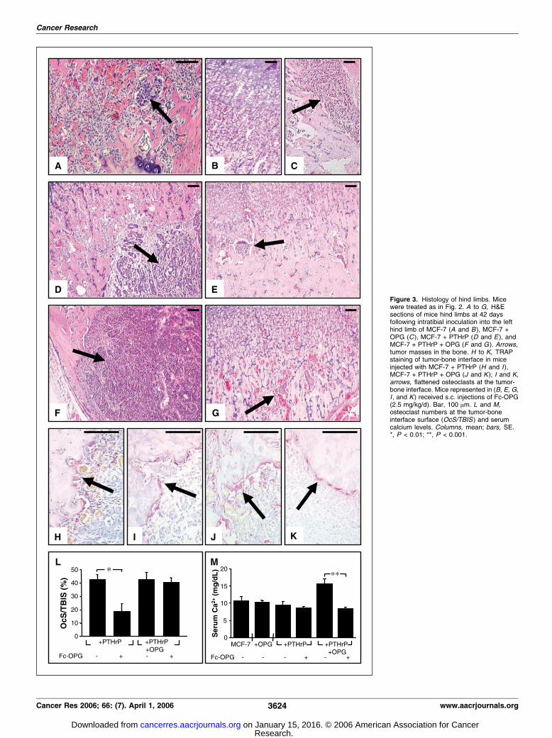
Figure 3. Histology of hind limbs. Micewere treated as in Fig. 2. A to G, H&Esections of mice hind limbs at 42 daysfollowing intratibial inoculation into the lefthind limb of MCF-7 (A and B ), MCF-7 +OPG (C ), MCF-7 + PTHrP (D and E ), andMCF-7 + PTHrP + OPG (F and G ). Arrows,tumor masses in the bone. H to K, TRAPstaining of tumor-bone interface in miceinjected with MCF-7 + PTHrP (H and I),MCF-7 + PTHrP + OPG (J and K); I and K,arrows, flattened osteoclasts at the tumor-bone interface. Mice represented in (B, E, G,I , and K ) received s.c. injections of Fc-OPG(2.5 mg/kg/d). Bar, 100 Am. L and M,osteoclast numbers at the tumor-boneinterface surface (OcS/TBIS ) and serumcalcium levels. Columns, mean; bars, SE.*, P < 0.01; **, P < 0.001.
Cancer Research
Cancer Res 2006; 66: (7). April 1, 2006 3624 www.aacrjournals.org
Research. on January 15, 2016. © 2006 American Association for Cancercancerres.aacrjournals.org Downloaded from

with MCF-7 + PTHrP or MCF-7 + PTHrP + OPG cells (Fig. 2, Hand I compared with D and E , respectively).
When the percentage area of osteolysis was quantified, OPGoverexpression resulted in a doubling of the area of osteolysis forthe MCF-7 + PTHrP cells (Fig. 2J). As mentioned earlier, noosteolysis was observed radiologically as a result of OPG over-expression in MCF-7 parental cells. Furthermore, quantificationshowed that Fc-OPG significantly reduced osteolysis in theMCF-7 + PTHrP and MCF-7 + PTHrP + OPG cells compared withuntreated animals (Fig. 2J). This implies that full-length OPG, whenexpressed by breast cancer cells, does not inhibit osteolysiswhereas OPG, when administered as a recombinant protein (Fc-OPG), is able to reduce cancer-induced osteolysis.
Histologic examination of the tibiae of these mice confirmed thedifferences in bone destruction evident from the Faxitron analyses(Fig. 2) along with accompanying cancer growth (Fig. 3). Smalltumor nests were evident in mice inoculated with MCF-7 cells,either without or with Fc-OPG administration (Fig. 3A and B),compared with control limbs where no tumor cells were evident(data not shown). Moderately larger tumors developed in miceinoculated with MCF-7 + OPG cells although these showed littlebone involvement (Fig. 3C). Inoculation of MCF-7 + PTHrP andMCF-7 + PTHrP + OPG cells resulted in the formation of largetumors that invaded the marrow cavity and the surrounding bone(Fig. 3D and F).
Similar levels of nuclear pleomorphism and mitoses in all of thetumors examined were revealed by histologic analysis. Interestingly,less tubule formation was apparent in the tumors overexpressingboth PTHrP and OPG compared with tumors overexpressingPTHrP alone, indicating that these tumors represent a highertumor grade (grade 9 versus grade 8, respectively) according to themodified Scarff-Bloom-Richardson grading system (23). Thissuggests that OPG expressed by cancer cells may affect tumorgrowth and differentiation, resulting in a less differentiated, moreaggressive tumor. Furthermore, these effects were not restricted toMCF-7 cells because similarly engineered MDA-MB-231 cells (e.g.,MDA-MB-231, MDA-MB-231 + OPG) showed similar enhancedgrowth in bone and osteolysis in a series of correspondingexperiments (data not shown).
Fc-OPG treatment of mice resulted in reduced tumor burden inbone in mice injected with MCF-7 + PTHrP or MCF-7 + PTHrP +OPG (Fig. 3, E and G compared with D and F, respectively).Compared with the MCF-7 and MCF-7 + OPG cells, a large numberof osteoclasts were present at the bone-tumor interface in thetumors produced by MCF-7 + PTHrP and the MCF-7 + PTHrP +OPG cells, as observed by H&E and TRAP staining (e.g., Fig. 3H andJ). Fc-OPG treatment dramatically reduced osteoclast numbers atthe tumor-bone interface in mice with MCF-7 + PTHrP tumors, butosteoclast numbers were unaltered by Fc-OPG treatment in micewith tumors produced by MCF-7 + PTHrP + OPG cells (Fig. 3L).Fc-OPG treatment of mice injected with either cell type resulted ina change of osteoclast morphology, with a flattened appearanceadjacent to bone and reminiscent of a quiescent osteoclast (Fig. 3Iand K).
Concomitant with the presence of large osteolytic lesions was arise in blood ionized calcium in mice inoculated with MCF-7 +PTHrP + OPG cells (Fig. 3M). Fc-OPG administration was able tocorrect this rise in blood ionized calcium evident in these mice(Fig. 3M). This finding, together with the appearance of theflattened osteoclasts following Fc-OPG treatment (Fig. 3K), sug-gests that these osteoclasts have reduced resorptive activity.
TRAIL-induced apoptosis. Because OPG has been reported toinfluence cellular apoptosis (24, 25), we examined the effect ofOPG overexpression to modulate TRAIL-induced apoptosis ofMCF-7 cells; cultures were treated with 100 ng/mL TRAIL in freshmedia and in conditioned media. MCF-7 parental and MCF-7 +PTHrP cells treated with TRAIL showed a 2.1- to 2.8-fold increasein apoptosis versus the control cultures (Fig. 4). However, therewas no significant difference in levels of apoptosis in MCF-7 +OPG or MCF-7 + PTHrP + OPG cell lines versus the MCF-7 orMCF-7 + PTHrP cells, respectively. Histologic sections of tumorswere also examined for the presence of apoptotic cells, whichwere dispersed uniformly through out the tumors, as observed byapoptotic bodies and by TUNEL staining. Similar to the in vitrofindings, when cells were challenged with TRAIL to induceapoptosis (Fig. 4), there was no difference in the number ofapoptotic cells in the intratibial tumors overexpressing OPG,compared with their counterparts, or following treatment withFc-OPG (Fig. 5A-C).
Cell proliferation within the intratibial tumors was examinedusing Ki67 staining. Whereas there was some nuclear staining forKi67 in the intratibial tumors produced by MCF-7 + PTHrP cells(Fig. 5D), significantly more cells stained positive for Ki67 in the
Figure 4. Overexpression of OPG in conditioned medium does not protectbreast cancer cells from TRAIL-induced apoptosis. MCF-7 (A) and MCF-7 +PTHrP (B) cells were exposed to 100 ng/mL TRAIL for 24 hours in the presenceof conditioned medium. A control sample was exposed to TRAIL in freshmedium. Columns, mean levels of apoptotic cell death, expressed as percentageof control (no TRAIL); bars, SE. Data from triplicate experiments.
OPG Enhances Cancer Growth
www.aacrjournals.org 3625 Cancer Res 2006; 66: (7). April 1, 2006
Research. on January 15, 2016. © 2006 American Association for Cancercancerres.aacrjournals.org Downloaded from
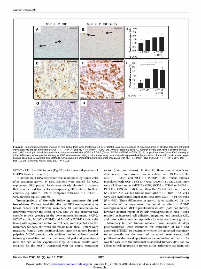
MCF-7 + PTHrP + OPG tumors (Fig. 5E), which was independent ofFc-OPG treatment (Fig. 5F).
To determine if OPG expression was maintained by tumor cellsafter sustained growth in vivo , sections were stained for OPGexpression. OPG protein levels were clearly elevated in tumorsthat were derived from cells overexpressing OPG relative to theircontrols (e.g., MCF-7 + PTHrP compared with MCF-7 + PTHrP +OPG tumors; Fig. 5G and H).Tumorigenicity of the cells following mammary fat pad
inoculation. We examined the effect of OPG overexpression inbreast cancer cells following mammary fat pad inoculation todetermine whether the effect of OPG that we had observed wasspecific to cells growing in the bone microenvironment. MCF-7,MCF-7 + OPG, MCF-7 + PTHrP, and MCF-7 + PTHrP + OPG cells(along with appropriate vector control cells) were injected into themammary fat pads of 4-week-old female nude mice. Tumors weremeasured from 21 days postinoculation once the tumors becamepalpable. MCF-7 parental cells exhibited an initial latent periodfollowing inoculation into the mammary fat pad and grew slowlyuntil the end of the experiment (Fig. 6): similar results wereobtained for the MCF-7 transfected with the empty expression
vector (data not shown). At day 21, there was a significantdifference in tumor size in mice inoculated with MCF-7 + OPG,MCF-7 + PTHrP, and MCF-7 + PTHrP + OPG versus animalsinoculated with MCF-7 cells (P = 0.01, ANOVA). By day 28, not onlywere all these tumors (MCF-7 + OPG, MCF-7 + PTHrP, or MCF-7 +PTHrP + OPG derived) larger than the MCF-7 cell line tumors(P = 0.007, ANOVA) but tumors from MCF-7 + PTHrP + OPG cellswere also significantly larger than those from MCF-7 + PTHrP cells(P = 0.03). These differences in growth rates continued for theremainder of the experiment. We found no effect of PTHrPoverexpression on MCF-7 proliferation in vitro (data not shown);however, another report of PTHrP overexpression in MCF-7 cellsresulted in increased cell adhesion, migration, and invasion (26),and these actions may be responsible for enhanced tumor growth.
Mammary fat pad tumors obtained from animals 49 dayspostinoculation were examined for expression of Ki67 andapoptosis (TUNEL) to determine whether the enhanced mammarytumor growth was the result of increased breast cancer cellproliferation, decreased apoptosis, or a combination of the two. Aswas the case with the intratibial-established tumors, OPG had noeffects on cell apoptosis in tumors at the orthotopic site (data not
Figure 5. Immunohistochemical analysis of hind limbs. Mice were treated as in Fig. 2. TUNEL staining of sections of mice hind limbs at 42 days following intratibialinoculation into the left hind limb of MCF-7 + PTHrP (A) and MCF-7 + PTHrP + OPG (B). Arrows, apoptotic cells. C, number of cells that have a positive TUNELstain. Ki67 staining in intratibial tumors from mice inoculated with MCF-7 + PTHrP (D ) and MCF-7 + PTHrP + OPG (E ). F, proportional area (%) of Ki67 staining inintratibial tumors, where positive staining for Ki67 was measured using a color image analyzer and results expressed as the proportion of area with positive staining perfield as described in Materials and Methods. OPG staining in intratibial tumors from mice inoculated with MCF-7 + PTHrP (G ) and MCF-7 + PTHrP + OPG (H ).Bar, 100 Am. Columns, mean; bars, SE. *, P < 0.05.
Cancer Research
Cancer Res 2006; 66: (7). April 1, 2006 3626 www.aacrjournals.org
Research. on January 15, 2016. © 2006 American Association for Cancercancerres.aacrjournals.org Downloaded from

shown). Proliferation rates, as determined by Ki67, were higher in themammary fat pad tumors than in intratibial tumors (16.1% versus6.4%). OPG overexpression resulted in a significant increase in Ki67staining in MCF-7 + PTHrP–derived mammary fat pad tumors(Fig. 6B). Although OPG overexpression inMCF-7 cells resulted in anincrease in tumor size (Fig. 6A), there was no significant difference inKi67 staining in these tumors. The differences in Ki67 stainingbetween mammary fat pad and intratibial tumors may be due to anumber of factors. The mammary fat pad tumors were harvested at49 days postinoculation versus 42 days for the intratibial tumors andthe area of Ki67 staining for tumors in mammary fat padwas substantially greater than those in bone. Alternatively, thephysical constraints of the bone environment or the differencesin the cytokine environments between these sites may affectproliferation.
Discussion
Many breast cancers possess properties that enable them toestablish and grow in bone. One of the primary mechanisms usedby breast cancers to achieve this is their ability to induce osteolysisas a result of enhanced osteoclast formation and activity. Animportant mediator of this process is PTHrP, which is produced bybreast cancer cells and promotes the expression of osteoblast-
derived RANKL, enabling osteoclast formation. We have previouslyreported that concomitant with the induction of RANKL in theosteoblast is a reduction in osteoblast-derived OPG production(10). The combination of these two outcomes was consideredfavorable for the establishment of the cancer in bone because ofthe enhanced osteolysis (10). However, in light of the resultspresented here, we must reconsider the pathophysiologic role ofOPG in tumor growth at the primary site as well as during cancer-induced osteolysis. Breast, lung, and prostate cancers, multiplemyeloma, and osteosarcomas express OPG (10, 20–22, 27), andincreased OPG expression has been associated with poor outcomesin gastrointestinal carcinoma and pancreatic cancer (28, 29).
Delivery of Fc-OPG as a recombinant protein has shown promiseas a potential therapy through experiments in animal models, inthat OPG limits hypercalcemia and osteolysis induced by myeloma,breast, lung, or prostate cancer and reduces tumor establishmentin bone (17, 20–22). Indeed, we reaffirm the ability of Fc-OPG tolimit breast cancer cell growth in bone and accompanyingosteolysis, even in cells that avidly promote osteolysis due to highlevel PTHrP production. In contrast to these actions, OPGexpression by breast cancers seems to be advantageous to thegrowth of MCF-7 cells in bone.
We assessed whether the OPG-mediated enhanced growth ofMCF-7 cells was a universal property, rather than restricted to thegrowth of these cells in bone, by examining whether this effect wasmimicked in soft tissue tumors. As was the case with the intratibialinoculation, OPG overexpression by MCF-7 cells was associatedwith an enhanced growth rate for both MCF-7 and MCF-7 + PTHrPcells following mammary fat pad inoculation. The enhancedgrowth seemed to result from increased proliferation becauseOPG expression did not affect apoptosis. Whereas OPG over-expression significantly increased tumor mass following mammaryfat pad inoculation, the increased Ki67 staining, indicating cellularproliferation provided by the OPG overexpressing tumor cells, wasnot as significant at this site as bone. The increased tumor massmay have resulted from altered matrix deposition in the breast oreffects on breast stromal cells: the Ki67 antibody only detectshuman cells and does not detect proliferating murine cells.
Whereas OPG expressed by breast cancer cells could inhibitosteoclast formation in vitro , it did not suppress osteoclastnumbers at the tumor-bone interface when tumor cells weredelivered directly into the tibiae. This finding might suggest eitherthat the OPG made by tumor cells was insufficient to inhibitosteoclast formation and activity or that the enhancement ofcancer cell growth conferred by OPG expression was thepredominant action and thus permitted tumor growth. Further-more, Fc-OPG administration did not limit osteoclast numbersin areas proximal to highly proliferative tumor cells althoughosteoclasts appeared flattened on the bone surface, suggesting thatFc-OPG treatment inhibited osteoclast activity, but not osteoclastgeneration, in these tumor-bearing mice. The enhanced prolifer-ation of tumor cells seems to favor increased bone loss, with Fc-OPG limited in its action in ablating RANKL. Comparison of thisresponse with an anti-RANKL antibody would be worthwhile topursue. Unfortunately AMG162, an anti-RANKL antibody, is humanspecific and does not target murine RANKL (30), which is essentialfor osteoclast formation in the nude mouse models that areroutinely used.
OPG is a receptor for the cytotoxic TNF-related apoptosisinducing ligand (TRAIL) that induces apoptosis, both in vitro andin vivo , in a wide range of tumor cells with little effect on normal
Figure 6. Mammary fat pad analysis. A, mice were inoculated with MCF-7parental cells (.), MCF-7 + PTHrP (x), MCF-7 + OPG (E), and MCF-7 +PTHrP + OPG (n). Tumor size was measured by calliper every 3 days, andtumors established in mice with MCF-7 + OPG and MCF-7 + PTHrP + OPG cellswere increased relative to their controls. B, proportional area (%) of Ki67 stainingin mammary fat pad tumors, where positive staining for Ki67 was measuredusing a color image analyzer and results expressed as the proportion of area withpositive staining per field as described in Materials and Methods. *, P < 0.05.
OPG Enhances Cancer Growth
www.aacrjournals.org 3627 Cancer Res 2006; 66: (7). April 1, 2006
Research. on January 15, 2016. © 2006 American Association for Cancercancerres.aacrjournals.org Downloaded from

tissues (24). TRAIL binds to its transmembrane receptors, DR4and DR5, which are expressed on many normal and malignant celltypes, including MCF-7 human breast cancer cells, inducingapoptosis through the caspase-8 cascade. Two decoy receptors,DcR1 and DcR2, which lack functional death domains and aretherefore unable to activate the caspase-8 cascade, also bind toTRAIL. DcR1 and DcR2 are expressed at high levels by normal cellsbut are either not expressed or only expressed at low levels bytumor cells, thereby restricting the cytotoxic activity of TRAIL tomalignant tissue. OPG represents the third decoy receptor forTRAIL (24) and Fc-OPG has been shown to inhibit TRAIL-inducedapoptosis in Jurkat cells and prostate cancer cells in vitro (24, 25).
The effect of native OPG produced by MCF-7 cells seems to beTRAIL independent because we observed (a) no difference inapoptotic cell number between tumors from OPG-overexpressingcells and those from their parental derivatives, and (b) nodifference in apoptosis induction by TRAIL between MCF-7 orMCF-7 + PTHrP cells with or without OPG overexpression. Theenhanced growth of MCF-7 cells in vivo in the absence of alteredapoptosis would argue that full-length OPG acts primarily toincrease cellular proliferation, and this has been proposed for othermembers of the TNF receptor superfamily (31). Consistent withthis, we noted increased Ki67 staining, suggesting increased cellproliferation in tumors that were overexpressing OPG relative tocell lines that were not. The addition of recombinant full-length
OPG to MCF-7 cells did not result in increased proliferation in vitro(data not shown), suggesting the actions that we observed may bea consequence of intracellular actions of OPG.
Whereas the TNF receptor domains of OPG fused to the IgG Fchas osteoclast inhibitory activity both in vitro and in vivo , wehave established that overexpression of native OPG results inenhanced growth of breast cancer cells in both soft tissue andbone. The actions of native OPG to promote cell growth relativeto Fc-OPG may result from the differences between thesemolecules because Fc-OPG lacks the potential death domains,the heparin binding region, and the dimerization sequences, and,unlike native OPG, cannot participate in intracellular actions.These differences, either alone or in combination, may contributeto the detrimental outcome of OPG overexpression by breastcancer cells.
Acknowledgments
Received 9/7/2005; revised 11/28/2005; accepted 1/28/2006.Grant support: Program grant 345401, Principal Research Fellowship no. ID345400
(M.T. Gillespie), and CJ Martin Fellowship no. ID247950 (R.J. Thomas-Mudge) from theNational Health and Medical Research Council (Australia); Lilly Centre for Women’sHealth (USA); the National Breast Cancer Foundation (Australia); and Department ofDefense U.S. Army grant DAMD17-03-1-0181.
The costs of publication of this article were defrayed in part by the payment of pagecharges. This article must therefore be hereby marked advertisement in accordancewith 18 U.S.C. Section 1734 solely to indicate this fact.
References1. Harris J, Hellman S, Henderson IC, Kinne DW. Bonediseases. Philadelphia: Lippincott JB; 1987.2. Simonet WS, Lacey DL, Dunstan CR, et al. Osteopro-tegerin: a novel secreted protein involved in theregulation of bone density. Cell 1997;89:309–19.3. Lacey DL, Timms E, Tan HL, et al. Osteoprotegerinligand is a cytokine that regulates osteoclast differen-tiation and activation. Cell 1998;93:165–76.4. Yasuda H, Shima N, Nakagawa N, et al. Osteoclastdifferentiation factor is a ligand for osteoprotegerin/osteoclastogenesis-inhibitory factor and is identical toTRANCE/RANKL. Proc Natl Acad Sci U S A 1998;95:3597–602.5. Suda T, Takahashi N, Udagawa N, Jimi E, Gillespie MT,Martin TJ. Modulation of osteoclast differentiation andfunction by the new members of the tumor necrosisfactor receptor and ligand families. Endocr Rev 1999;20:345–57.6. Kong YY, Boyle WJ, Penninger JM. Osteoprotegerinligand: a common link between osteoclastogenesis,lymph node formation and lymphocyte development.Immunol Cell Biol 1999;77:188–93.7. Dougall WC, Glaccum M, Charrier K, et al. RANK isessential for osteoclast and lymph node development.Genes Dev 1999;13:2412–24.8. Anderson DM, Maraskovsky E, Billingsley WL, et al. Ahomologue of the TNF receptor and its ligand enhanceT-cell growth and dendritic-cell function. Nature 1997;390:175–9.9. Bucay N, Sarosi I, Dunstan CR, et al. Osteoprotegerin-deficient mice develop early onset osteoporosis andarterial calcification. Genes Dev 1998;12:1260–8.10. Thomas RJ, Guise TA, Yin JJ, et al. Breast cancer cellsinteract with osteoblasts to support osteoclast forma-tion. Endocrinology 1999;140:4451–8.11. Southby J, Murphy LM, Martin TJ, Gillespie MT. Cell-specific and regulator-induced promoter usage andmessenger ribonucleic acid splicing for parathyroidhormone-related protein. Endocrinology 1996;137:1349–57.
12. Ma YL, Cain RL, Halladay DL, et al. Catabolic effectsof continuous human PTH (1–38) in vivo is associatedwith sustained stimulation of RANKL and inhibition ofosteoprotegerin and gene-associated bone formation.Endocrinology 2001;142:4047–54.13. Onyia JE, Galvin RJ, Ma YL, et al. Novel and selectivesmall molecule stimulators of osteoprotegerin expres-sion inhibit bone resorption. J Pharmacol Exp Ther2004;309:368–79.14. Udagawa N, Horwood NJ, Elliott J, et al. Interleukin-18(interferon-g-inducing factor) is produced by osteoblastsand acts via granulocyte/macrophage colony-stimulatingfactor and not via interferon-g to inhibit osteoclastformation. J Exp Med 1997;185:1005–12.15. Fisher JL, Schmitt JF, Howard ML, Mackie S, ChoongPFM, Risbridger GP. An in vivo model of prostatecarcinoma growth and invasion in bone. Cell Tissue Res2002;307:337–45.16. Price JE, Polyzos A, Zhang RD, Daniels LM.Tumorigenicity and metastasis of human breast car-cinoma cell lines in nude mice. Cancer Res 1990;50:717–21.17. Capparelli C, Kostenuik PJ, Morony S, et al.Osteoprotegerin prevents and reverses hypercalcemiain a murine model of humoral hypercalcemia of malig-nancy. Cancer Res 2000;60:783–7.18. Quinn JM, Elliott J, Gillespie MT, Martin TJ. Acombination of osteoclast differentiation factor andmacrophage-colony stimulating factor is sufficient forboth human and mouse osteoclast formation in vitro .Endocrinology 1998;139:4424–7.19. Guise TA, Yin JJ, Thomas RJ, Dallas M, Cui Y, GillespieMT. Parathyroid hormone-related protein (PTHrP)-(1-139) isoform is efficiently secreted in vitro and enhancesbreast cancer metastasis to bone in vivo . Bone 2002;30:670–6.20. Zhang J, Dai J, Qi Y, et al. Osteoprotegerin inhibitsprostate cancer-induced osteoclastogenesis and pre-vents prostate tumor growth in the bone. J Clin Invest2001;107:1235–44.21. Oyajobi BO, Anderson DM, Traianedes K, Williams
PJ, Yoneda T, Mundy GR. Therapeutic efficacy of asoluble receptor activator of nuclear factor nB-IgG Fcfusion protein in suppressing bone resorption andhypercalcemia in a model of humoral hypercalcemiaof malignancy. Cancer Res 2001;61:2572–8.22. Croucher PI, Shipman CM, Lippitt J, et al. Osteopro-tegerin inhibits the development of osteolytic bonedisease in multiple myeloma. Blood 2001;98:3534–40.23. Elston CW, Ellis IO. Pathological prognostic factors inbreast cancer. I. The value of histological grade in breastcancer: experience from a large study with long-termfollow-up. Histopathology 1991;19:403–10.24. Emery JG, McDonnell P, Burke MB, et al. Osteopro-tegerin is a receptor for the cytotoxic ligand TRAIL.J Biol Chem 1998;273:14363–7.25. Holen I, Croucher PI, Hamdy FC, Eaton CL. Osteo-protegerin (OPG) is a survival factor for human prostatecancer cells. Cancer Res 2002;62:1619–23.26. Shen X, Qian L, Falzon M. PTH-related proteinenhances MCF-7 breast cancer cell adhesion, migration,and invasion via an intracrine pathway. Exp Cell Res2004;294:420–33.27. Mackie PS, Fisher JL, Zhou H, Choong PFC.Bisphosphonates regulate cell growth and gene expres-sion in the UMR 106-01 clonal rat osteosarcoma cellline. Br J Cancer 2001;84:951–8.28. Ito R, Nakayama H, Yoshida K, et al. Expression ofosteoprotegerin correlates with aggressiveness and poorprognosis of gastric carcinoma. Virchows Arch 2003;443:146–51.29. Mizutani Y, Matsubara H, Yamamoto K, et al.Prognostic significance of serum osteoprotegerin levelsin patients with bladder carcinoma. Cancer 2004;101:1794–802.30. McClung MR, Lewiecki EM, Bolognese MA, et al.AMG162 increases bone mineral density (BMD) within 1month in postmenopausal women with low BMD.J Bone Miner Res 2004;19:S20.31. Weber CH, Vincenz C. The death domain superfam-ily: a tale of two interfaces? Trends Biochem Sci 2001;26:475–81.
Cancer Research
Cancer Res 2006; 66: (7). April 1, 2006 3628 www.aacrjournals.org
Research. on January 15, 2016. © 2006 American Association for Cancercancerres.aacrjournals.org Downloaded from

2006;66:3620-3628. Cancer Res Jane L. Fisher, Rachel J. Thomas-Mudge, Jan Elliott, et al. Contrasts with That Delivered TherapeuticallyEnhances Orthotopic and Osseous Tumor Growth and Osteoprotegerin Overexpression by Breast Cancer Cells
Updated version
http://cancerres.aacrjournals.org/content/66/7/3620
Access the most recent version of this article at:
Cited articles
http://cancerres.aacrjournals.org/content/66/7/3620.full.html#ref-list-1
This article cites 30 articles, 10 of which you can access for free at:
Citing articles
http://cancerres.aacrjournals.org/content/66/7/3620.full.html#related-urls
This article has been cited by 7 HighWire-hosted articles. Access the articles at:
E-mail alerts related to this article or journal.Sign up to receive free email-alerts
Subscriptions
Reprints and
To order reprints of this article or to subscribe to the journal, contact the AACR Publications
Permissions
To request permission to re-use all or part of this article, contact the AACR Publications
Research. on January 15, 2016. © 2006 American Association for Cancercancerres.aacrjournals.org Downloaded from
Copyright © 2022 FDOKUMEN