Health-promoting physical activity of adults with mental retardation
Upload
independentCategory
view
0download
0
Hum Genet (1981) 57:300-306
© Springer-Verlag 1981
Aniridia, Male Pseudohermaphroditism, Gonadoblastoma, Mental Retardation, and del llp13
Catherine Turleau l, J. de Grouchy ~, J. L. Duffer 2, L~ Hoang Phuc 2, P. H. Schmelck 3, R. Rappaport s, Claire NihouI-F~k6t64, and Nicole Diebold 5
U.173 I.N.S.E.R.M. and E.R. 149 C.N.R.S. 2 Service d'Ophthalmologie 3 Unit6 d'Endocrinologie P6diatrique 4Clinique Chirurgicale Infantile SGroupe de Pathologie P6diatrique (I.N.S.E.R.M.U.77), H6pital Necker-Enfants-Malades, 149, rue de S~vres, F-75730 Paris Cedex 15, France
Summary. A 20-month-old male patient was referred because of severe growth and mental retardation, bilateral glaucoma, hypospadias, and cryptorchidism. Karyotyping revealed a de novo complex three-chromosome rearrangement as well as deletion of band 1 lp13:46,XY,t(4;7;15)(q212;p14;q26),del(11) (p13p14). Trabeculectomia revealed bilateral aniridia. Surgery on the genitalia revealed male pseudohermaphrodit ism and bilateral gonadoblastoma. The kidneys were normal. A deficiency in catalase (CAT) activity allowed the regional assignment of the CAT gene to band 1 lp13.
Introduction
Aniridia is most often an autosomal dominant trait with a frequency of about 1.8 × 10 -5 and a mutation rate of about 4 x 10 -6 per gamete per generation (McKusick 1975). Yet this condition may occasionally be part of a syndrome affecting psychomotor, renal, and genital development. The association of aniridia and nephroblastoma or Wilms' tumor, which is also known to be of genetic origin in some instances, was reported for the first time by Brusa and Torricelli (1953). In 1964 Miller et al. studied 440 cases of Wilms' tumor and found six cases of aniridia, i.e., an incidence of 1/76. This first observation was confirmed by numerous reports. In 1979 Frangois et al. reviewed nearly 50 instances of the aniridia-Wilms' tumor association. A chromosomal aberration was first reported by Ladda et al. (1974) who identified a t(8p+;1 l q - ) in a 3-year-old patient with bilateral aniridia and Wilms' tumor diagnosed at 19 months. Computer analysis of optical microscope scanning profiles of chromosomes 8 and 11 revealed an interstitial deletion of 8p. Francke et al. (1977) and Riccardi et al. (1978) demonstrated a short interstitial deletion of l l p in another patient with aniridia and Wilms' tumor. Simultaneously Andersen et al. (1978a, b) reported a deletion of 1 lp in a patient with aniridia and gonadoblastoma, but without Wilms' tumor. Some 10 patients have now been reported with aniridia, Wilms' tumor, and /o r gonadoblastoma. They are reviewed by Francke et al. (1979) and the relevant clinical and cytogenetic data are summarized in Table 1. Two instances are now known of familial transmission due to a parental rearrangement (Hittner
Offprint requests to: J. de Grouchy
et al. 1979; Yunis and Ramsay 1980). The patient first studied by Ladda et al. (1974) was reexamined by Francke et al. (1979) and shown to have indeed an interstitial deletion of 1 lp. The shortest deletion common to all patients is band l lp13.
Several gene loci have been precisely localized on l l p : L D H A on p12.3~p12.8, HBB (the Hb cluster) on p1205~ p1208, and SA11-1 and 3 (cell surface antigens) on pl3-+pter , ACP2 on p l l ~ p l 2 (Human Gene Mapping 5 1979). More recently Wieacker et al. (1980) assigned the catalase (CAT) locus to 1 lp by cellular hybridization.
We here report a 20-month-old male patient with severe mental and growth retardation, aniridia and glaucoma, ambiguous external and internal genitalia, and gonado- blastoma, with no evidence of Wilms' tumor. Chromosome analysis showed a deletion of l lp13 and enzymatic activity determination allowed the regional assignment of the CAT gene
to 1 lp13.
Case Report
The patient, born in Algeria on June 28, 1978, was considered as a male. His parents are Algerians and consanguineous (probably at the fifth degree). The mother is healthy and was 26 years old. The father was 34 years old. He suffers from epilepsy. The propositus has two brothers and two sisters. One of the brothers has unilateral cryptor- chidism.
Birth weight was 2,500 g; length at birth and head circumference are unknown. Bilateral congenital glaucoma was diagnosed at two months. Delayed psychomotor development and growth retardation were noted in early infancy. The patient was transfered to Paris at 20 months of age for treatment of his glaucoma.
At this time growth retardation was evident: length: 69.5cm (-4SD); weight: 6,650 g (-4 SD); head circumference: 42 cm (-5 SD); bone age: eight months. Psychomotor development was much delayed and corresponded to that of a 6-month-old infant. Blindness was complete with clinically evident, bilateral glaucoma and opaque cornea. Hearing was normal, as well as tonus and reflexes. There was no striking dysmorphism (Fig. 1). Penile hypospadias was present. The penis was 30 mm long with a very moderate chordee, the scrotum was flat, normally striated, and empty. No gonad could be palpated. Karyo- type analysis was requested because of this sexual anomaly.
It was then decided to operate on the glaucoma because of the severe discomfort it seemed to cause to the patient even through it was beyond effective cure. When trabeculectomia was performed bilateral
0340-6717/81/0057/0300/$ 01.40

301
Table 1. Relevant data from ten patients reported in the literature and the present case
Patient Chromosomal Mental Height Eyes Sex rearrangement retard- (per- chromo-
ation centile) somes
Genitalia Tumor Other
1 (8;11)(p21.2;q14.4) + 50th Aniridia, glaucoma, XY del(11)(p1407p1304) cataracts, mega- parents normal cornea
2 del( l l ) (pl400pl l .3) + 10th Aniridia, glaucoma, XY parents not studied cataracts, ptosis
3 del(l l)(pl5.1pl208) + 25th Aniridia, glaucoma, XY parents normal cataracts, nystagmus,
strabismus, ptosis
4 del(ll)(p15.1p1305) + 25th Aniridia, ptosis XY parents not studied
5 del(11)(pl3pl I) + ? Aniridia, glaucoma, XX parents normal cataracts, nystagmus
6 del(11)(p1300p15.1) + ? Aniridia, glaucoma, parents normal cataracts
XX
7 Identical twin + ? Aniridia, glaucoma, XX of patient 6 cataracts, exotropia
8 del(11)(pl 1.3p14) + 3rd Aniridia, glaucoma, XX mother's karyotype: cataracts, corneal ins(l l)(q22pl 1.3p14) clouding, esotropia,
mild ptosis
9 del(11)(p13p14.1) + 5th Aniridia, cataracts, XY mother's karyotype: nystagmus, ptosis ins(2; 11) (q32;p13p14.1)
Slightly ventral orifice of urethra
Coronal hypo- spadias, no palpable testis, small scrotum
Microphallus, perineal hypo- spadias, unilateral cryptorchidia, anterior urethral diverticulum
Ambiguous genitalia, None no mullerian deriv- ative gonads present, hypospadias 3rd degree
Female external genitals, small gonadal streaks
Unilateral nephroblastoma at 19 months
None at 12 years
Nephroblastoma unilateral (left) at 42 months
at 8~/4 years
Benign gonado- blastoma autopsy: 21 months
Normal female
Bilateral un- descended testes
Bilateral nephro- blastoma at 36 and 49 months
None at 7 years
None at I0 months
Partial malrotation of right kidney
Obstructive uro- pathy
Female sex of rearing
Mild coloboma of the nares, micro- gnathia low-set ears, central hearing impairment, ab- normally rotated left kidney
Malrotation left kidney
Bilateralnephro- Microcephaly, half- blastoma at 15 brother and aunt and 36 months both with aniridia
Wilm's tumor had died
10 inv(7)(q21.2q3100) + 50th Aniridia, glaucoma, XY Ambiguous genitalia, None del(1 l)(p1305p1405) corneal clouding, micropenis, hypo- at 31°/12 months parents normal nystagmus spadias, normal
testicular biopsies, small scrotum
11 t(4;7;15) + 3rd Aniridia, glaucoma, XY Hypospadias, Bilateral (q21.2;p14;q26) corneal clouding severe gonadal gonadoblastoma, del(11)(p13p14) dysgenesis, presence surgery parents normal of Mullerian at 23 months
derivatives
Catalase deficiency
Patient 1. Ladda et al. 1974; Francke et al. 1979 Patient 2. Smith et al. 1977 (patient AC); Francke et al. 1977 (patient C); Riccardi et al. 1978 (patient 1) Patient 3. Smith et al. 1977 (patient LM); Francke et al. 1977 (patient B); Riccardi et al. 1978 (patient 2) Patient 4. Riccardi et al. 1978 (patient 3); Francke et al. 1977 (patient A) Patient 5. Andersen et al. 1978a,b; Warburg et al. 1980 Patients 6 and 7. Twins. Cotlier et al. 1978; Francke et al. 1978; Maurer et al. I979; addendum in Riccardi et al. 1978; Riccardi et al. to be published Patient 8. Hittner et al. 1980 Patient 9. Yunis and Ramsay 1980 Patient 10. Riccardi et al. to be published (case 1) Patient 11. Present report; Junien et al. 1980 (patient 1)

302
Fig. 1. Photography of the patient at 22 months
aniridia was discovered. The discovery of an interstitial deletion of 1 lp in association with aniridia prompted further investigations in order to detect a latent nephroblastoma (Wilms' tumor) or gonadoblastoma.
Ultrasound scan revealed normal sized kidneys. Intravenous pyelo- gram showed normal morphology and function of the kidneys and excretory cavities. Suprapubic cystography and mictional urethrography showed bilateral vesicoureteral reflux without urinary complication, and no urogenital sinus.
Stimulation tests of pituitary function gave normal results: under luteinizing hormone releasing hormone (LHRH), plasma follicle-stim- ulating hormone (FSH) levels rose from 0.8 to 7.4 m U / m l and luteinizing hormone (LH) levels f rom 0.5 to 0.8 mU/ml . Testicular function was also normal, as shown by human chorionic gonadotropin (HCG) stim- ulation: plasma testosterone rose from an undetectable level to 2.3 ng /ml (after three intra-muscular injections of 1,500 U every other day).
Operation for bilateral cryptorchidism was undertaken initially by a horizontal inguinal incision and was rapidly transformed in laparotomy because of the discovery of ambiguous internal genitalia. This revealed a vagina of normal size and situation and a uterus apparently complete and in continuation with two ducts having the consistancy of Fallopian tubes. On the left, this tube did not develop tubal folds. On the right, a pavillion and normal plicae were present. The gonads appeared as long, thick, yellow streaks located at the posterior side of the tubes. On the right side there was a vas deferens ending in the vaginal cul-de-sac and with no evident connection with the right gonad. Total resection of the internal genitalia was decided. The vagina was sutured as low as possible, very near the supposed urethrovaginal fistula.
Histologic examination confirmed the presence of uterus and vagina. The upper genital tract was ambiguous with Miillerian and Wolffian structures. Both gonads contained seminiferous tubules which occasionally appeared dysgenetic (Fig. 2a), and areas of typical gonadoblastoma (Fig. 2b).
Fig. 2. a Dysgenetic testicular tissue: seminiferous tubules, devoid of germinal elements, are embedded in abundant connective tissue, arranged in whorls, as in rudimentary gonadal streaks. Azan x 350. b Typical gonadoblastoma: nests of germ cells with large nuclei contain rounded hyaline bodies. Azan x 350. (Microphotographs: Dr. Nathalie Josso)

303
Fig. 3a and b. Karyotype of the patient showing the complex t(4;7;15) (q212;p14;q26)as well as the del(l 1)(p13p14), a R-banding; b G-banding
Laboratory Investigations
Hematologic investigations showed microcytosis which could not be related to abnormal hemoglobin synthesis (see below), and hypo- sideremia which could rapidly be cured by iron therapy. X-rays showed, as well as the delayed bone age, moderate osteoporosis. Phosphocalcic metabolism was normal. No endocrine or digestive disorder could be found to explain the severe growth retardation. Otherwise, all usual blood and urinary tests gave results within normal limits.
Cytogenetic Investigations
The patient's karyotype was established after applying RHG-, RBA-, GTG-banding to cultured lymphocytes and fibroblasts. Blood cells metaphases were also obtained after addition of mitomycin to the culture medium (final concentration 0.15 gg/ml) 20 h before harvesting, in order to obtain a higher degree of banding resolution.
A three-chromosome translocation was identified (Fig. 3): t(4;7;i5) (q212;p14;q26). The 4qter fragment was translocated onto the 15 and 7pl4pter onto the 4q remaining segment. This series of probably circulary translocations was considered balanced. In addition, an inter- stitial deletion was detected in l l p and limited to band l lp13. The breakpoints seemed to be at the proximal margins of p13 and p14 (Fig. 4). The complete karyotype formula was therefore: 46,XY, t(4;7;ll)(q212;p14;q26),del(ll)(p13p14). Both parents had normal karyotypes.
Sister chromatid exchanges were analysed in the patient's lympho- cytes and fibroblasts. Their frequency was normal. In 29 blood cells it varied from 0 to 13 with a mean of seven exchanges per cell. In 17 fibro-
blasts the variation was from 6 to 19, with a mean of l0 exchanges per cell.
Genetic Markers
The blood group phenotypes of the patient and his parents failed to show any abnormal transmission. Several genetic markers, known to be assigned to chromosome 11 and more specifically to 11p, were investigated. There was no evidence for a thalassemia-like syndrome. HbA was 93%, HbF: 4%, and HbA2: 2.9%. Although HbF was elevated, HbA2 and globular values were normal. Lactate dehy- &'ogenase A (LDHA) activity was normal. Catalase activity was found significantly decreased in the patient's red and white blood cells (Junien et al. 1980).
Discussion
Our pa t i en t was first re fe r red to us because o f b i la tera l g l aucoma with cornea l opac i ty , and it was only at surgery tha t anir idia was d i scovered . The assoc ia t ion o f anir idia , men ta l r e t a rda t ion , a m b i g u o u s geni ta l ia , a n d del(1 lp ) then in i t ia ted a search for n e p h r o b l a s t o m a a n d g o n a d o b l a s t o m a . Wi lms ' t u m o r was no t de tec ted bu t l a p a r o t o m y , which was p e r f o r m e d to f ind the gonads , revealed male p s e u d o h e r m a p h r o d i t i s m with Miil- lerian s t r u c t u r e s - - a vagina , a u terus , a n d Fa l lop ian t u b e s - - a s well as s t reak gonads . Total resec t ion o f the in ternal geni ta l ia

304
was decided and histopathology disclosed the presence of dysgenetic testes and bilateral gonadoblastoma.
Only one other patient with identified del(1 lp) is known to have had gonadoblastoma (patient 5 in Table 1). Her chromosomal sex was XX and her external genitalia were female but streak gonads were found at autopsy. It is therefore essential to evaluate the state of the gonads, in males as well as in females, the discovery of dysgenetic gonads commanding gonadectomy.
Wilms' tumor was found in four out of eleven patients. The tumor was uni- or bilateral. The age of first diagnosis varied from 15 to 42 months. The observation of Wilms' tumor developing in only one of identical twins (patients 6 and 7), demonstrates the postzygotic nature of the tumor and that the chromosomal deletion is only a predisposing factor for carcino- genesis. Exactly the same lesion of the genome may or may not lead to a tumoral process. In other words, the existence or not of Wilms' tumor does not imply different rearrangements within the visible l i p deletion.
Most patients had various facial dysmorphisms but it seems impossible to single out evocative features. Mental retardation and ambiguous genitalia in the male are constant but less specific and of various degrees. Aniridia remains the most important element of the syndrome and is always associated with other eye anomalies--glaucoma, cataracts, nystagmus,
ptosis, strabismus, corneal clouding--which result in partial or complete blindness.
When considering the chromosomal rearrangements, the size of the deleted segment varies, the breakpoints on 1 lp being located between pl l .3 and p15.1. As shown by Francke et al. (1979) the smallest region of overlapping is the distal half of band 1 lp13. In two cases out of eleven the deletion was due to a balanced parental rearrangement. Karyotyping of the parents is therefore required to prevent familial recurrence. It is also noteworthy that in three de novo cases other structural rearrangements were also present. Their significance is far from clear. They may indicate chromosome instability in the gametes.
Increased sister chromatid exchanges have been demon- strated in fibroblasts from a patient with del 13ql4-retino- blastoma (Turleau et al. 1980). Our patient had a normal rate of exchanges in fibroblasts as well as in leukocytes. This could reflect an important basic difference between both syndromes.
The llp13 band is neighbored by important gene loci. Among those that are more easily investigated are LDHA, ACP2, HBB, and CAT (Fig. 5). Patients 3, 4, 10, and 11 had normal values for LDHA activity. Patients 2 and 8 had a decreased activity. These results are consistent with the assign- ment of the LDHA gene to p1203~p1208 (Francke et al. 1977). The Hb cluster (HBB) is closely linked to LDHA in p1205~
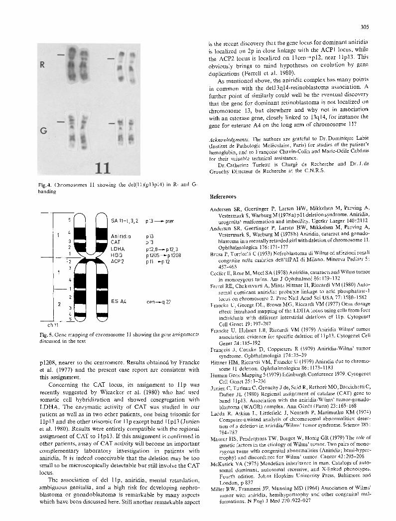
Fig.4. Chromosomes 11 showing the del(ll)(p13p14)in R-and G- banding
SA 11-1,3~2 p13 - pter
AnirJdia p13 CAT p13 LDHA p12,8~p 12,3 HBB p 1205-,--p 1208 ACP2 p 11--~-p 12
ES Az~ cen-~-q 22
ch11
Fig. 5. Oene mapping of chromosome 11 showing the gene assignments discussed in the text
p1208, nearer to the centromere. Results obtained by Francke et al. (1977) and the present case report are consistent with this assignment.
Concerning the CAT locus, its assignment to l i p was recently suggested by Wieacker et al. (1980) who had used somatic cell hybridization and showed cosegregation with L D H A . The enzymatic activity of CAT was studied in our patient as well as in two other patients, one being trisomic for 1 lp13 and the other trisomic for 1 lp except band 1 lp13 (Junien et al. 1980). Results were entirely compatible with the regional assignment of CAT to 1 lp13. If this assignment is confirmed in other patients, assay of CAT activity will become an important complementary laboratory inyestigation in patients with aniridia. It is indeed conceivable that the deletion may be too small to be microscopically detectable but still involve the CAT locus.
The association of del l lp, aniridia, mental retardation, ambiguous genitalia, and a high risk for developing nephro- blastoma or gonadoblas toma is remarkable by many aspects which have been discussed here. Still another remarkable aspect
305
is the recent discovery that the gene locus for dominant aniridia is localized on 2p in close linkage with the ACP1 locus, while the ACP2 locus is localized on l l c e n ~ p l 2 , near l lp13. This obviously brings to mind hypotheses on evolution by gene
duplications (Ferrell et al. 1980). As mentioned above, the aniridia complex has many points
in common with the del l3ql4-re t inoblas toma association. A further point of similarly could well be the eventual discovery that the gene for dominant retinoblastoma is not localized on chromosome 13, but elsewhere and why not in association with an esterase gene, closely linked to 13q14, for instance the gene for esterase A4 on the long arm of chromosome 11?
Acknowledgments. The authors are grateful to Dr. Dominique Labie (Institut de Pathologie Mol~culaire, Paris) for studies of the patient's hemoglobin, and to Franqoise Chavin-Colin and Marie-Odile Cabfinis for their valuable technical assistance.
Dr. Catherine Turleau is Charg6 de Recherche and Dr.J. de Grouchy Directeur de Recherche at the C.N.R.S.
References
Andersen SR, Geertinger P, Larsen HW, Mikkelsen M, Parving A, Vestermark S, Warburg M (1978 a)p 11 deletion syndrome. Aniridia, urogenita} malformation and imbecility. Ugeskr Laeger 140:2112
Andersen SR, Geertinger P, Larsen HW, Mikkelsen M, Parving A, Vestermark S, Warburg M (1978b) Aniridia, cataract and gonado- blastoma in a mentally retarded girl with deletion of chromosome 11. Ophthalmologica 176:171-177
Brusa P, Torricelli C (1953) Nefroblastoma di Wilms ed affezioni renali congenite nella casistica dell'IIPAI di Milano. Minerva Pediatr 5: 457-463
Cotlier E, Rose M, Moel SA (1978) Aniridia, cataracts and Wilms tumor in monozygous twins. Am J Ophthalmol 86:129-132
Ferrel RE, Chakravarti A, Mintz Hittner H, Riccardi VM (1980) Auto- somal dominant aniridia: probable linkage to acid phosphatase-1 locus on chromosome 2. Proc Natl Acad Sci USA 77:1580-1582
Francke U, George DL, Brown MG, Riccardi VM (1977) Gene dosage effect: intraband mapping of the LDHA locus using cells from four individuals with different interstitial deletions of lip. Cytogenet Cell Genet 19:197-207
Francke U, Holmes LB, Riccardi VM (1979) Aniridia-Wilms' tumor association: evidence for specific deletion of 1 lp13. Cytogenet Cell Genet 24:185-192
Francois J, Coucke D, Coppieters R (1979) Aniridia-Wilms' tumor syndrome. Ophthalmologia 174:35-39
Hitmer HM, Riccardi VM, Francke U (1979) Aniridia due to chromo- some 11 deletion. OphthalmoIogica 86:1173-1183
Human Genc Mapping 5 (1979) Edinburgh Conference 1979. Cytogenet Cell Genet 25 : 1-236
Junien C, Turleau C, Grouchy J de, Said R, Rethorb MO, Baccichetti C, Dufier JL (1980) Regional assignment of catalase (CAT) gene to band l lp13. Association with the aniridia-Wilms' tumor-gonado- blastoma (WAGR) complex. Ann G~n6t (Paris) 23:165-168
Ladda R, Atkins L, Littlefield J, Neurath P, Marimuthu KM (1974) Computer-assisted analysis of chromosomal abnormalities: detec- tion of a deletion in aniridia/Wilms' tumor syndrome. Science 185 : 784,787
Maurer HS, Pendergrass TW, Borges W, Honig GR (1979) The role of genetic factors in the etiology of Wilms' tumor. Two pairs of mono- zygous twins with congenital abnormalities (Aniridia; hemi-hyper- trophy) and discordance for Wilms' tumor. Cancer 43:295-208
McKusick VA (t975) Mendelian inheritance in man. Catalogs of auto- somal dominant, autosomal recessive, and X-linked phenotypes. Fourth edition. Johns Hopkins University Press, Baltimore and London, p 837
Miller RW, Fraumeni JF, Manning MD (1964) Association of Wilms' tumor with aniridia, hemihypertrophy and other congenital mal- formations. N Engl J Med 270:922-927

306
Riccardi VM, Sujansky E, Smith AC, Francke U (1978) Chromosomal imbalance in the aniridia-Wilms tumor association: 1 lp interstitial deletion. Pediatrics 61:604-610
Riccardi VM, Hittner HM, Francke U, Yunis J J, Ledbetter D, Borges W (to be published) The aniridia-Wilms' tumor associtation: the critical role of chromosome band 11p13. Cancer Genet Cytogenet
Smith ACM, Sujansky E, Riccardi VM (1977) Aniridia, mental retarda- tion and genital abnormality in two patients with 46,XY,1 lp - . New Syndromes, Birth Defects 13 : 257
Turleau C, Cabanis MO, Grouchy J de (1980) Augmentation des 6changes de chromatides dans les fibroblastes d'un enfant atteint de del(13) r6tinoblastome. Ann G~n~t (Paris) 23:169-170
Warburg M, Mikkelsen M, Andersen SR, Geertinger P, Larsen HW, Vestermark S, Parving A (1980) Aniridia and interstitial deletion of the short arm of chromosome 11. Metab Ped Ophthal 4:9%101
Wieacker P, Mueller CR, Mayerova A, Grzeschik KH, Ropers HH (1980) Assignment of the gene coding for human catalase to the short arm of chromosome 11. Ann G~n~t (Paris) 23 : 73-77
Yunis JJ, Ramsay NKC (1980) Familial occurrence of the aniridia- Wilms' tumor syndrome with deletion 1 lp13-14.1. J Pediatr 96: 1027-1030
Received November 24, 1980
Note Added in Proof
We have studied another patient with aniridia, nephroblastoma, mental and growth retardation, and found a deletion of approximately half of band 1 lp13. This patient had a very low catalase activity, thus confirming the assignment of the cat gene to 1 lp13.
Copyright © 2022 FDOKUMEN