Idiopathic" mental retardation and new chromosomal abnormalities
667
Idiopathic condylar resorption (ICR), alsoknown as idiopathic condylysis, condylar atrophy, andprogressive condylar resorption, is a well-documentedbut poorly understood disease. There are a number oflocal and systemic factors or diseases that can causemandibular condylar resorption. Local factors includesuch entities as ICR, osteoarthritis, reactive arthritis,avascular necrosis, infection, and traumatic injuries.Systemic connective tissue and autoimmune diseasesthat can create condylar resorption include: rheumatoidarthritis, psoriatic arthritis, scleroderma, systemic lupuserythematosis, Sjögren’s syndrome, ankylosingspondylitis, and others. ICR has a different cause andpathosis than other condylar resorption conditions andtherefore a specific method of treatment is indicated.
ICR can create occlusal and skeletal instability,dentofacial deformities, TMJ dysfunction, and pain.Several authors1-10 have described the occurrence ofICR and association with orthodontics and othognathic
surgery. Previously recommended treatment forICR5,6,11-14 include the following options: (1) splinttherapy to minimize joint loading; (2) “nonloading”orthodontic and orthognathic surgical procedures after6 to 12 months of remission; (3) deferment of treat-ment until ICR remission; (4) arthroscopic lysis andlavage; (5) condylar replacement with a costochondralgraft if the ICR cannot be controlled or recurs; and (6)maxillary surgery only to correct the occlusal defor-mity. No method of management has yet beendescribed for ICR that will provide predictable stableoutcomes for the TMJ, provide optimal functional andesthetic results, and eliminate pain. This article willaddress the diagnosis of ICR and the specific treatmentprotocol we have developed to achieve predictable andstable outcomes.
PATIENT PREDISPOSITION
People with the following specific facial morpho-logic characteristics appear to be most susceptible toICR: (1) females (approximately 9:1 female to maleratio); (2) age range from 10 to 40 years old with astrong predominance for teenagers in their pubertalgrowth phase; (3) high occlusal plane angle andmandibular plane angle; and (4) predominance of ClassII skeletal and occlusal relationship with or withoutopen bite. ICR rarely occurs in low occlusal andmandibular plane angle facial types or in Class IIIskeletal relationships.
CONTINUING EDUCATION ARTICLE
Idiopathic condylar resorption: Diagnosis, treatment protocol,and outcomes
Larry M. Wolford, DDS,a and Luis Cardenas, DMDb
Dallas, Tex, and Miami, Fla
Idiopathic condylar resorption is a poorly understood progressive disease that affects the TMJ and that canresult in malocclusion, facial disfigurement, TMJ dysfunction, and pain. This article presents the diagnosticcriteria for idiopathic condylar resorption and a new treatment protocol for management of this pathologiccondition. Idiopathic condylar resorption most often occurs in teenage girls but can occur at any age,although rarely over the age of 40 years. These patients have a common facial morphology including: (1)high occlusal and mandibular plane angles, (2) progressively retruding mandible, and (3) Class II occlusionwith or without open bite. Imaging usually demonstrates small resorbing condyles and TMJ articular diskdislocations. A specific treatment protocol has been developed to treat this condition that includes: (1)removal of hyperplastic synovial and bilaminar tissue; (2) disk repositioning and ligament repair; and (3)indicated orthognathic surgery to correct the functional and esthetic facial deformity. Patients with thiscondition respond well to the treatment protocol presented herein with elimination of the disease process.Two cases are presented to demonstrate this treatment protocol and outcomes that can be achieved.Idiopathic condylar resorption is a progressive disease that can be eliminated with the appropriate treatmentprotocol. (Am J Orthod Dentofacial Orthop 1999;116:667-77)
aClinical Professor of Oral and Maxillofacial Surgery at Baylor College of Den-tistry–Texas A & M University System; in private practice at Baylor UniversityMedical Center, Dallas.bFormer Fellow, Oral and Maxillofacial Surgery, Baylor College of Dentistry,and Baylor University Medical Center, Dallas; currently, Associate Professor,Department of Oral and Maxillofacial Surgery, Nova Southeastern UniversitySchool of Dental Medicine, Fort Lauderdale, Fla; in private practice, Miami.Reprint requests to: Larry M. Wolford, DDS, 3409 Worth St,Sammons Tower, Suite 400, Dallas, TX 75246Copyright © 1999 by the American Association of Orthodontists.0889-5406/99/$8.00 + 0 8/1/101648
668 Wolford and Cardenas American Journal of Orthodontics and Dentofacial OrthopedicsDecember 1999
PATHOGENESIS OF ICR
The specific cause of ICR has not been clearly iden-tified. Its strong predilection for teenage females givescredence to the theory of hormonal mediation. This isfurther supported by identification of estrogen recep-tors in female primates,15,16 human TMJ tissues thatappear to correlate to TMJ symptoms,17 and inosteoarthritic knee joints.18 Estrogen is known to medi-ate cartilage and bone metabolism in the female TMJ.An increase in receptors may predispose an exagger-ated response to joint loading from parafunctionalactivity, trauma, orthodontics, or orthognathic surgery.
In our ICR patient population, the majority of casesoccur in young teenage girls in their pubertal growthphase. Our hypothesis is this: the sex hormones medi-ate biochemical changes within the TMJ causinghyperplasia of the synovial tissues that stimulates theproduction of destructive substrates that initiates break-down of the ligamentous structures that normally sup-port and stabilize the articular disk in position. Thisallows the disk to become anteriorly displaced. Thehyperplastic synovial tissues then assume a positionaround the head of the condyle, further increasingexposure of the condyle to the substrates that create theresorptive phenomena. In this disease process, thecondyle shrinks in all 3 planes of space. The condylarresorption appears to occur in the subcondylar bonewithout clinically apparent destruction of the fibrocar-tilage on the condylar head and roof of the fossa, unlikethe arthritides where the fibrocartilage is destroyed bythe inflammatory disease processes. ICR may eventu-ally go into remission. However, if the condyle and thehyperplastic synovial tissues receive excessive loading
(ie, parafunctional habits, trauma, orthodontics,orthognathic surgery) the resorption process may bereinitiated.
CLINICAL EVALUATION
A diagnosis is usually based on patient history,clinical evaluation, and imaging. Patients may report aprogressive worsening of their occlusion and estheticswith or without TMJ symptoms and associated pain.Bilateral cases usually reflect a relatively symmetricposterior shifting of the mandible and development ofan anterior open bite (Fig 1A and B and Fig 2A and B).In unilateral cases, the mandibular dental midline andchin shifts toward the affected side with ipsilateralClass II occlusion, crossbite, and posterior occlusalprematurities, as well as an open bite on the contralat-eral side. Although TMJ symptoms can be present,often times they are very mild or relatively nonexistent.In fact, 25% of patients in our study19 had no TMJsymptoms. Clicking and popping may be absentbecause the hyperplastic synovial tissues increase thejoint space, which provides a smooth transition ontothe displaced disk during condylar translation. Rarelyare other joints involved in mandibular ICR. Labora-tory studies may be indicated, particularly if otherjoints are involved, to evaluate for connective tissueand autoimmune diseases, which can also cause condy-lar resorption. There are no laboratory tests specific forICR.
Reactive arthritis resulting in condylar resorptionis another common TMJ pathologic development infemales, however, it usually begins after the age of20 years. Factors that differentiate reactive arthritisfrom ICR include the following: a decreased joint
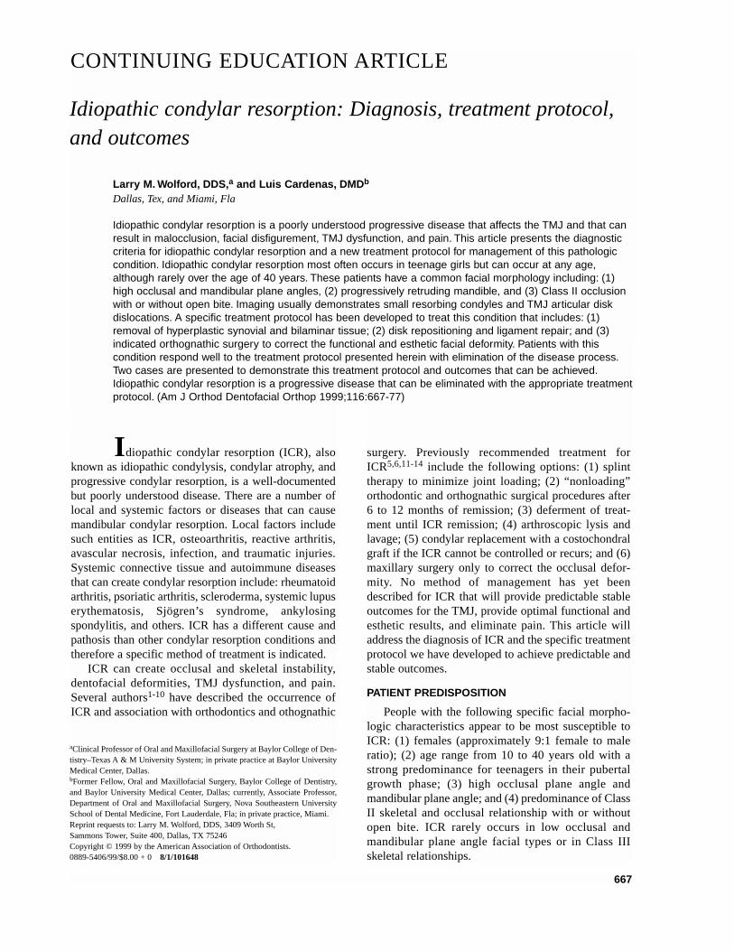
Fig 1. Case 1: A-B, Fifteen-year-old girl has active idiopathic condylar resorption. Note the signifi-cant retruded position of the mandible, high mandibular plane angle, and associated facial morphol-ogy. C-D, Patient is 3 years posttreatment of ICR with removal of hyperplastic synovial tissues, artic-ular disk repositioning, and simultaneous double-jaw orthognathic surgery.
B CA D

American Journal of Orthodontics and Dentofacial Orthopedics Wolford and Cardenas 669Volume 116, Number 6
space, erosion of condyle and fossa with loss offibrocartilage, osteophyte formation, anteriorbeaking, involvement of other joints, and other sys-temic problems (ie, female reproductive system,urinary tract, chronic upper and lower respiratoryinfections, gastrointestinal problems). Reactivearthritis may be related to bacterial infections (ie,chlamydia, mycoplasma)20,21 requiring differenttreatment considerations.
IMAGING FINDINGS
Common lateral cephalometric radiographic find-ings in bilateral TMJ ICR include the following: (1)skeletal and occlusal Class II deformity; (2) anterioropen bite; (3) high mandibular occlusal plane angle;(4) high mandibular plane angle; (5) decreased verti-cal height of the ramus; (6) the lower incisors mayappear overangulated (Fig 3A); and (7) a significantdecrease in the oropharyngeal airway can occur in themore severe cases. Serial lateral cephalograms willdemonstrate slow but progressive retrusion of themandible during the active resorption phase. Unilat-eral involvement includes (1) unilateral skeletal andocclusal Class II deformity; (2) vertical height differ-ence at the mandibular inferior border, ramus, andocclusal plane; and (3) an open bite on the contralat-
eral side. Serial P-A cephalograms may show wors-ening of the asymmetry.
Cephalometric tomographic evaluation of the TMJusually shows a relatively normal or excessive jointspace because of hyperplasia of the synovial tissues. Theinvolved condylar head will appear smaller in size, thedegree of which will be dependent on length of timesince onset of the pathosis and aggressiveness of the dis-ease. There may be some loss of integrity of the corticalbone on the head of the condyle. Superimposition of ser-ial cephalometric tomograms will help document thepresence of active condylar resorption. Bone scans maybe of no significant diagnostic benefit for ICR.
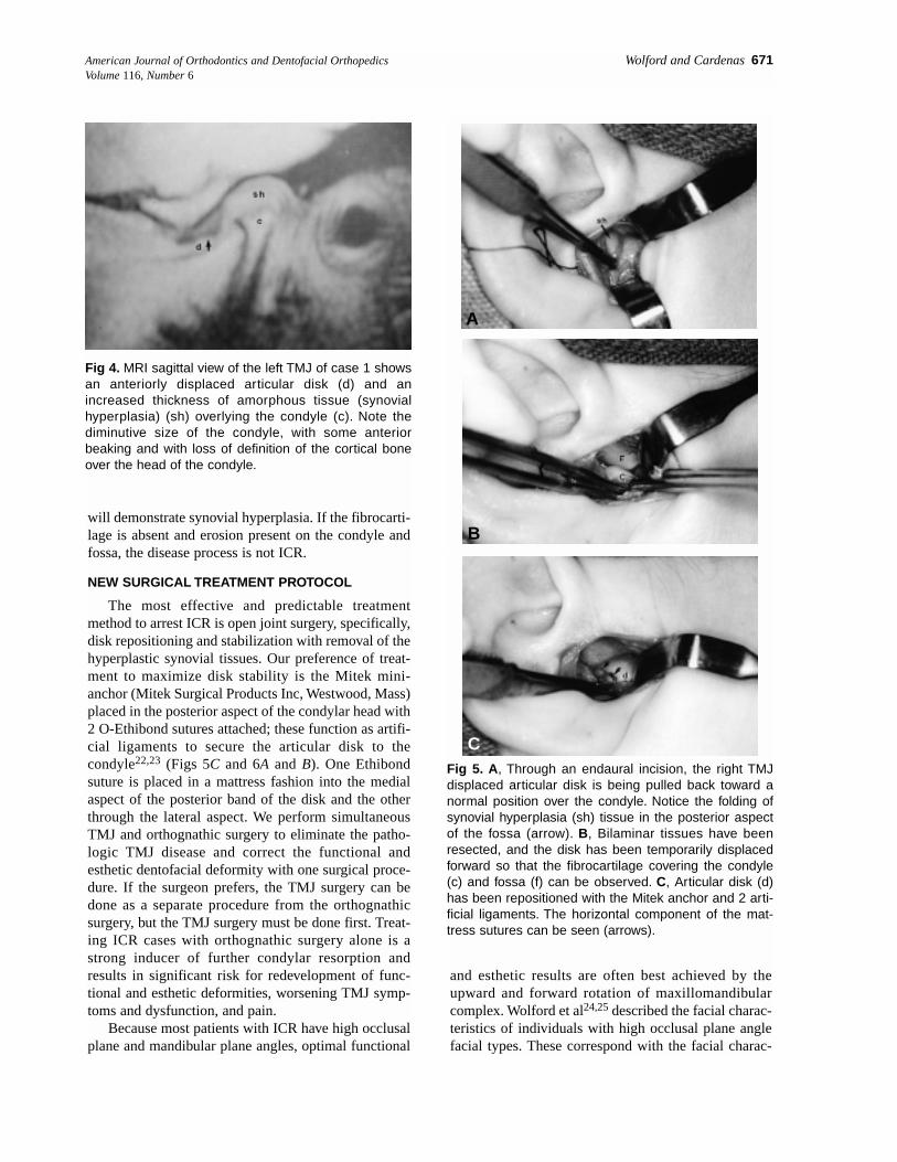
The MRI findings include the following: (1)decreased condylar volume; (2) anterior disk displace-ment with or without reduction on opening; (3)extreme thinness or loss of continuity of cortical boneon the head of the condyle; and (4) often thick amor-phous-appearing soft tissue occupying the spacebetween the condyle and fossa (Fig 4). The degree ofdeformation and degenerative changes of the disk willbe dependent on the length of time of the disk dis-placement. Determination must be made relative to sal-vageability of the disk and adequacy of the remainingcondyle to withstand normal functional loading andstress forces.
Fig 2. Case 1: A-B, Presurgical occlusion demonstrates anterior open bite and a Class II end-oncanine occlusal relationship. C-D, Occlusion remains stable 3 years postsurgical treatment.
A C
B D
670 Wolford and Cardenas American Journal of Orthodontics and Dentofacial OrthopedicsDecember 1999
SURGICAL FINDINGS
Observations at surgery reveal an anterior oranteromedial displaced disk. When the disk is pulledback into position, the hyperplastic synovial tissuesbunch posteriorly (Fig 5A). The tissue may appearamorphous in nature, with little vascular component,
and usually no inflammation. The glenoid fossa and thecondylar head usually have an intact fibrocartilage cov-ering (Fig 5B). The articular disk may or may notdemonstrate significant deformation and degenerativechanges depending on the length of time of the diskdisplacement. Histologically, the retrodiskal tissues
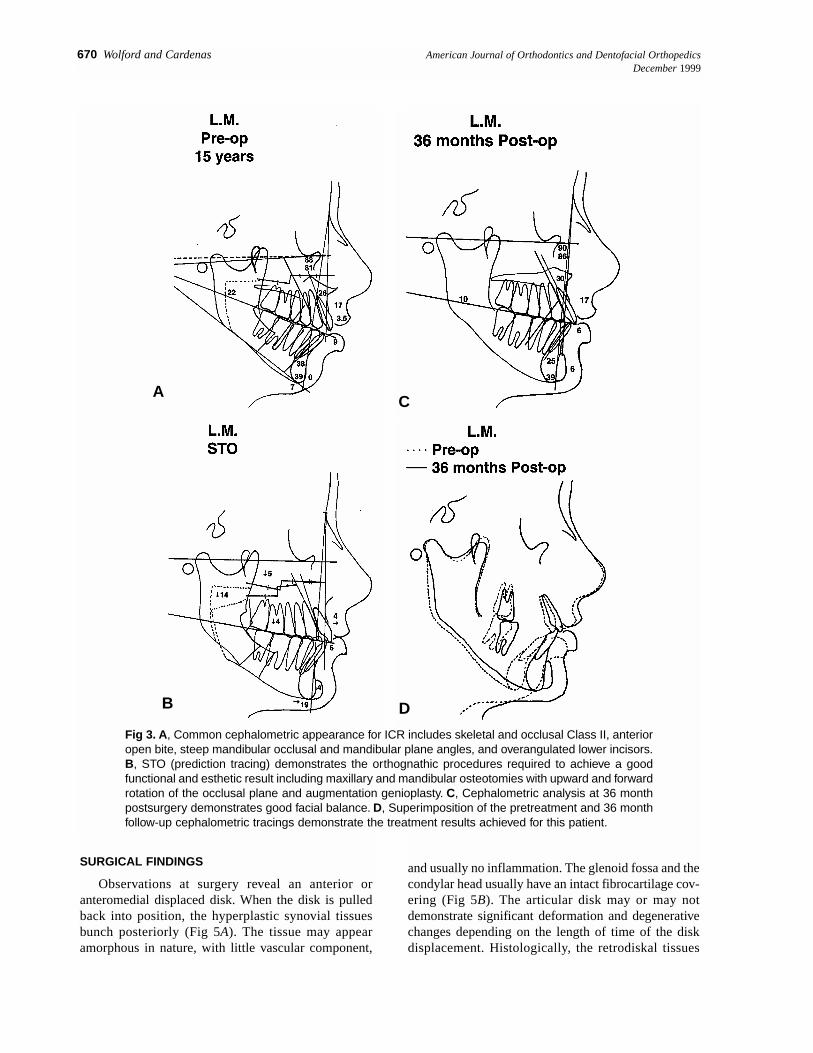
Fig 3. A, Common cephalometric appearance for ICR includes skeletal and occlusal Class II, anterioropen bite, steep mandibular occlusal and mandibular plane angles, and overangulated lower incisors.B, STO (prediction tracing) demonstrates the orthognathic procedures required to achieve a goodfunctional and esthetic result including maxillary and mandibular osteotomies with upward and forwardrotation of the occlusal plane and augmentation genioplasty. C, Cephalometric analysis at 36 monthpostsurgery demonstrates good facial balance. D, Superimposition of the pretreatment and 36 monthfollow-up cephalometric tracings demonstrate the treatment results achieved for this patient.
D
C
B
A
American Journal of Orthodontics and Dentofacial Orthopedics Wolford and Cardenas 671Volume 116, Number 6
will demonstrate synovial hyperplasia. If the fibrocarti-lage is absent and erosion present on the condyle andfossa, the disease process is not ICR.
NEW SURGICAL TREATMENT PROTOCOL
The most effective and predictable treatmentmethod to arrest ICR is open joint surgery, specifically,disk repositioning and stabilization with removal of thehyperplastic synovial tissues. Our preference of treat-ment to maximize disk stability is the Mitek mini-anchor (Mitek Surgical Products Inc, Westwood, Mass)placed in the posterior aspect of the condylar head with2 O-Ethibond sutures attached; these function as artifi-cial ligaments to secure the articular disk to thecondyle22,23 (Figs 5C and 6A and B). One Ethibondsuture is placed in a mattress fashion into the medialaspect of the posterior band of the disk and the otherthrough the lateral aspect. We perform simultaneousTMJ and orthognathic surgery to eliminate the patho-logic TMJ disease and correct the functional andesthetic dentofacial deformity with one surgical proce-dure. If the surgeon prefers, the TMJ surgery can bedone as a separate procedure from the orthognathicsurgery, but the TMJ surgery must be done first. Treat-ing ICR cases with orthognathic surgery alone is astrong inducer of further condylar resorption andresults in significant risk for redevelopment of func-tional and esthetic deformities, worsening TMJ symp-toms and dysfunction, and pain.
Because most patients with ICR have high occlusalplane and mandibular plane angles, optimal functional
and esthetic results are often best achieved by theupward and forward rotation of maxillomandibularcomplex. Wolford et al24,25 described the facial charac-teristics of individuals with high occlusal plane anglefacial types. These correspond with the facial charac-
Fig 4. MRI sagittal view of the left TMJ of case 1 showsan anteriorly displaced articular disk (d) and anincreased thickness of amorphous tissue (synovialhyperplasia) (sh) overlying the condyle (c). Note thediminutive size of the condyle, with some anteriorbeaking and with loss of definition of the cortical boneover the head of the condyle.
Fig 5. A, Through an endaural incision, the right TMJdisplaced articular disk is being pulled back toward anormal position over the condyle. Notice the folding ofsynovial hyperplasia (sh) tissue in the posterior aspectof the fossa (arrow). B, Bilaminar tissues have beenresected, and the disk has been temporarily displacedforward so that the fibrocartilage covering the condyle(c) and fossa (f) can be observed. C, Articular disk (d)has been repositioned with the Mitek anchor and 2 arti-ficial ligaments. The horizontal component of the mat-tress sutures can be seen (arrows).
A
B
C
672 Wolford and Cardenas American Journal of Orthodontics and Dentofacial OrthopedicsDecember 1999
teristics of many ICR patients. Chemello et al26
demonstrated that upward and forward rotation of themaxillomandibular complex is a very stable procedurein the presence of healthy joints. Cottrell et al27
reported minimal condylar change in healthy TMJs
after upward and forward rotation of the maxillo-mandibular complex. In our treatment protocol forICR, the TMJ disks are surgically repositioned and sta-bilized. With adequate remaining condyle, the TMJscan withstand upward and forward rotation of the max-illomandibular complex.
In the ICR patient, with salvageable disks andcondyles, our surgical sequencing is as follows: (1)TMJ surgery to remove hyperplastic synovial tissue,reposition the articular disk, and stabilize with Mitekanchor; (2) mandibular ramus sagittal split osteotomies,reposition the mandible, and stabilize with rigid fixa-tion28; (3) maxillary osteotomies, reposition the max-illa, and stabilize with rigid fixation and appropriategrafting; and (4) other procedures as necessary (ie,rhinoplasty, cheek augmentations, chin procedures,etc). This treatment protocol has proved to be very suc-cessful in ICR patients with salvageable disks and ade-quate remaining condyles.19
As arthroscopy and arthrocentesis do not removethe hyperplastic synovial tissue or reposition the artic-ular disk into a normal functional position, they arepredictably unsuccessful procedures in treating ICR,particularly if correction of the jaw deformity isattempted. In ICR cases where the disk is nonsalvage-able but adequate condyle remains, the hyperplasticsynovial tissue should be removed and the diskreplaced with autologous tissues. However, treatmentresults will be far less predictable. A meniscectomyalone is not recommended as this will introduce otherarthritic conditions. In more severe cases, where thecondyle is nonsalvageable, condylar replacement maybe necessary with either a sternoclavicular joint graft,costochondral graft, or FDA-approved total joint pros-thesis.
STABILITY OF RESULTS
Wolford et al19 reported on 12 patients with ICRwith documented active condylar resorption that weretreated by this specific protocol; their average age was21 years (range, 14 to 36 years). With an average 8-month presurgery evaluation, the average amount ofcondylar resorption was –1.1 mm (range, –2.5 to –0.5mm). Point B moved posteriorly an average of –1.8mm (range, –3 to –1 mm) and the mandibular occlusalplane increased an average of 1.5° (range, 0° to 3°).The average rate of condylar resorption was 0.12 mmper month or 1.5 mm per year, indicating that ICR is aslow but progressive disease process. Surgical changesincluded point B moving forward an average of 10.9mm (range, 2 to 18 mm) and the occlusal plane angledecreasing an average of –7.8° (range, -5° to -12°).
The long-term follow-up average of 33.2 months
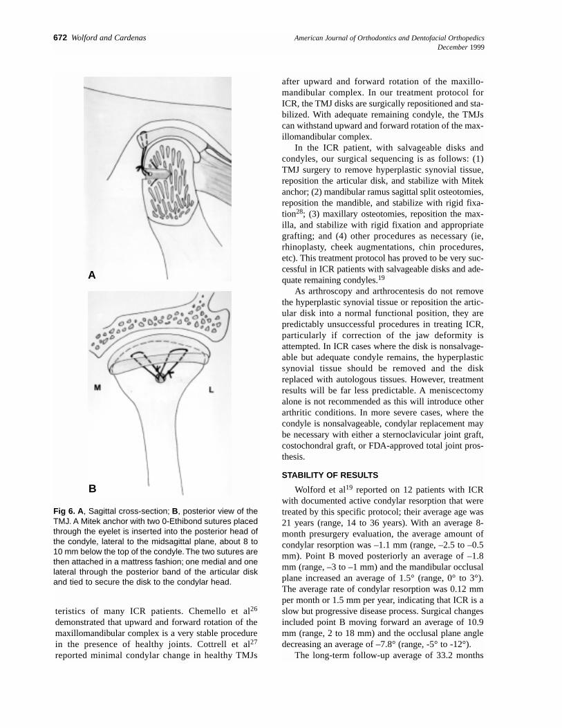
Fig 6. A, Sagittal cross-section; B, posterior view of theTMJ. A Mitek anchor with two 0-Ethibond sutures placedthrough the eyelet is inserted into the posterior head ofthe condyle, lateral to the midsagittal plane, about 8 to10 mm below the top of the condyle. The two sutures arethen attached in a mattress fashion; one medial and onelateral through the posterior band of the articular diskand tied to secure the disk to the condylar head.
A
B
American Journal of Orthodontics and Dentofacial Orthopedics Wolford and Cardenas 673Volume 116, Number 6
(range, 18 to 68 months) demonstrated an averagecondylar length change of +0.2 mm (range, –0.5 to+1.5 mm). Point B changed on average of + 0.3 mm(range, – 0.5 to 2 mm) and the average occlusal planechange was +0.04° (range, 0° to 0.5°). There was nostatistically significant change from immediate post-surgery to the longest follow-up period. Five patientswere under the age of 16 years and exhibited a modestamount of postsurgical condylar growth with an aver-age increase in condylar height of 0.43 mm (range,–0.1 to 1.5 mm). In all 12 patients, jaw functionremained unchanged with no statistically significantdifference in the presurgical and postsurgical incisalopening (presurgical and postsurgical 47 mm) andexcursion movements (preoperative and postoperative>7 mm). There was a significant decrease in pain on thevisual analog pain scale (0 = no pain to 10 = worstpain); the presurgery average pain was 3.5 (range, 0 to9) and the postsurgical average pain was 0.7 (range, 0to 4).
DISCUSSION
ICR frequently develops during pubertal growth, soconsequently these patients are often in orthodontictreatment at the onset of the disease. Because patientspredisposed to ICR are usually high occlusal planeangle facial types with skeletal and occlusal Class IIrelationships,22-24 they are often candidates for orthog-natic surgery before the onset of the disease. In mostcases, ICR will develop regardless of orthodontic ororthognathic procedures. However, if these proceduresincrease loading or stress to the TMJs, they may initi-ate or accelerate the rate of resorption. In some cases,ICR is self-limiting with only moderate amounts of
bone resorption, whereas in other cases, the entire headand neck of the condyle can be resorbed. It is importantto understand that if the disease does go into remission,it can be easily reactivated by orthodontics, orthog-nathic surgery, or other factors that load or stress thejoint.
Because of lack of integrity in the ligaments thatnormally stabilize the disk, use of the Mitekanchor25,26 has significantly improved the stability ofresults in ICR. The body of the anchor is a titaniumalloy cylinder with a bullet tip at one end and a sutureeyelet at the opposite end. A pair of superelasticnickel-titanium arcs project from the body. The arcsfold against the anchor body as the anchor is pushedthrough the interosseous hole in the cortex of theposterior head of the condyle, and they return to theiroriginal resting position once in the medullary bone,locking the anchor in position (Fig 6A). The Mitekanchor supports 2 artificial ligaments (O-Ethibondsuture) threaded through the eyelet of the anchor andsecured to the articular disk (Fig 6B). Fields et al29
determined the average pull-out force of the Mitekmini-anchor from human cadaver condyles was16.02 lbs. Fields et al30 histologically demonstratedosseous integration between the anchor and thecondylar bone.
Orthodontic and orthognathic surgery candidateswith high occlusal plane and mandibular plane angles,Class II occlusions with or without anterior open bites,radiographic evidence of resorption or diminutive sizeof the condyles, or the presence of TMJ signs andsymptoms should be scrutinized very carefully beforeand during treatment to determine if there is a presenceor predisposition for ICR or other TMJ pathoses. After
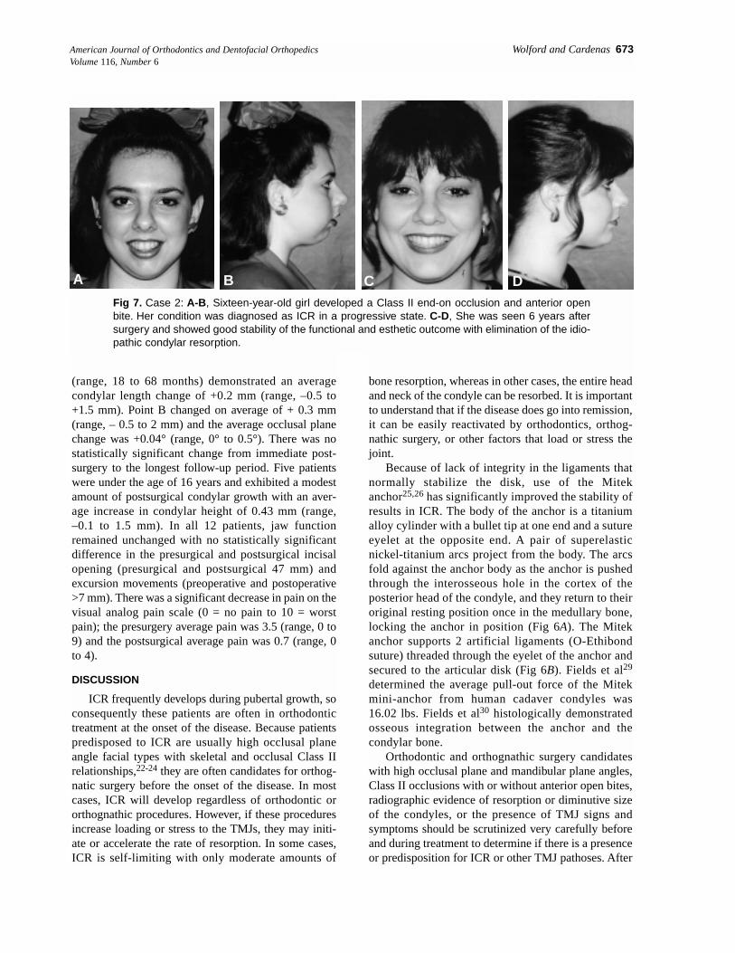
Fig 7. Case 2: A-B, Sixteen-year-old girl developed a Class II end-on occlusion and anterior openbite. Her condition was diagnosed as ICR in a progressive state. C-D, She was seen 6 years aftersurgery and showed good stability of the functional and esthetic outcome with elimination of the idio-pathic condylar resorption.
A B C D
674 Wolford and Cardenas American Journal of Orthodontics and Dentofacial OrthopedicsDecember 1999
appropriate diagnostic procedures, a proper treatmentplan can be developed to address the TMJ pathosis andjaw deformity.
A most interesting fact in our ICR study19 is that 5of the 6 patients who were 16 years or youngerdemonstrated some modest growth postsurgery. Thisindicates a reversal of the disease process from one ofresorption to subsequent return of growth. The bestresults in the management of ICR involves earlydetection of the disease process and early TMJsurgery. The earlier ICR is treated, the more likely thefollowing are to occur: (1) less resorption of thecondyle occurs, thus, maintaining a greater dimen-sional component of the condyle, (2) less distortionand degeneration of the articular disk, and (3) betterpostsurgical distribution of loading forces on the jointstructures. The high predictability of treatment out-comes with this protocol for ICR substantiates anearly diagnosis and initiation of treatment to providethe best success functionally, occlusally, esthetically,and with long-term stability.
Case 1
This 15-year-old girl, who was under active ortho-dontic treatment, was referred by her orthodontist
because she was developing a significant jaw deformity.Her mandible was becoming more retruded, and she haddifficulty chewing because her teeth did not fit together.She had no pain, headaches, TMJ noises, or dysfunction.Her diagnoses included: (1) bilateral ICR; (2) A-Pmandibular hypoplasia; (3) maxillary posterior verticalhypoplasia; (4) A-P microgenia; (5) high occlusal andmandibular plane angles; and (6) Class II open bite (Figs1A and B and 2A and B). The MRI shows diminution ofthe condylar size bilaterally and anteriorly displaceddisks (Fig 4). Serial cephalometric and tomographicanalysis confirmed the active condylar resorption.Presurgical incisal opening was 43 mm and lateralexcursive movements were 8 mm. Her treatmentincluded: (1) presurgical orthodontics to continue toalign and level the arches; (2) surgery (Fig 3B) includingthe following: (a) bilateral TMJ articular disk reposi-tioning and ligament repair with Mitek anchors andremoval of hyperplastic synovial tissue; (b) bilateralmandibular ramus osteotomies to advance the mandiblein a upward and forward direction; (c) multiple maxil-lary osteotomies to advance, expand, and decreaseocclusal plane angulation by downgrafting the posterioraspect of the maxilla; (d) augmentation genioplasty; and(3) postsurgical orthodontics to finish and retain.
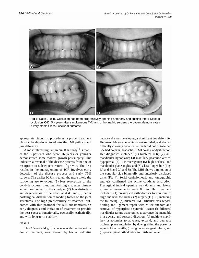
Fig 8. Case 2: A-B, Occlusion has been progressively opening anteriorly and shifting into a Class IIocclusion. C-D, Six years after simultaneous TMJ and orthognathic surgery, the patient demonstratesa very stable Class I occlusal outcome.
A
B
C
D
American Journal of Orthodontics and Dentofacial Orthopedics Wolford and Cardenas 675Volume 116, Number 6
The patient was seen 3 years after surgery and showedexcellent stability and significant improvement in herfunctional and esthetic facial balance (Figs 1C and D, 2Cand D, and 3C). Incisal opening is 54 mm and excursivemovements 7 mm bilaterally. She has no pain or headacheproblems. Postsurgical condylar change was 1 mm
growth on the right side and 0 mm change on the left.(Orthodontics by Dr Fred Spradley, Ft Worth, Tex)
Case 2
This 16-year-old girl was referred because herlower jaw began shifting to the left, and she was devel-
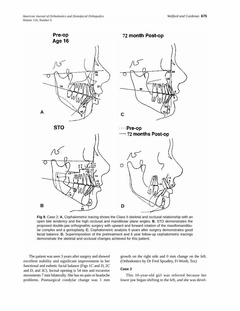
Fig 9. Case 2: A, Cephalometric tracing shows the Class II skeletal and occlusal relationship with anopen bite tendency and the high occlusal and mandibular plane angles. B, STO demonstrates theproposed double-jaw orthognathic surgery with upward and forward rotation of the maxillomandibu-lar complex and a genioplasty. C, Cephalometric analysis 6 years after surgery demonstrates goodfacial balance. D, Superimposition of the pretreatment and 6 year follow-up cephalometric tracingsdemonstrate the skeletal and occlusal changes achieved for this patient.
A
B
C
D
676 Wolford and Cardenas American Journal of Orthodontics and Dentofacial OrthopedicsDecember 1999
3. ICR appears to be hormonally mediated and can go intoremission, but it can be reactivated with loading or stressto the TMJ (ie, orthodontics, orthognathic surgery, para-functional habits, trauma).
4. The TMJ articular disk is displaced, and there may or maynot be TMJ symptoms (25% of patients have no TMJsymptoms).
5. TMJ articular disk repositioning and stabilization withremoval of the hyperplastic synovial tissues arrests thedisease process when the disk is salvageable and adequatecondylar head still remains. The Mitek mini-anchor hassignificantly improved the stability of the disk reposition-ing and treatment outcomes.
6. Early detection and treatment of the ICR will minimizethe amount of condylar bone resorption and degenerativechanges in the articular disk.
7. Simultaneous TMJ and orthognathic surgery can be doneto correct the existing dentoskeletal deformity, (includingupward and forward rotation of the maxillomandibularcomplex) with stable and predictable results.
8. In young patients, condylar growth may recur after surgi-cal intervention with the outlined protocol.
REFERENCES
1. Worms FW, Speidel TM, Bevis RR, et al. Posttreatment stability of esthetics oforthognathic surgery. Angle Orthod 1980;50:251-73.
2. Kerstens JCK, Tuinzing DB, Golding RP, van der Kwast WAM. Condylar atrophy andosteoarthrosis after bimaxillary surgery. Oral Surg Oral Med Oral Pathol 1990;69:274-80.
3. Moore KE, Gooris PJJ, Stoelinga PJW. The contributing role of condylar resorptionto skeletal relapse following mandibular advancement surgery. J Oral Maxillofac Surg1991;49:448-60.
4. De Clercq CA, Neyt LF, Mommaerts MY, et al. Condylar resorption in orthognathicsurgery: a retrospective study. Int J Adult Orthod Orthognath Surg 1994;9:233-40.
5. Arnett GW, Tamborello JA. Progressive Class II development: female idiopathiccondylar resorption. Oral Maxillofac Surg Clin North Am 1990;2:699-716.
6. Crawford JG, Stoelinga PJW, Blijdorp PA, Brouns JJA. Stability after reoperation ofprogressive condylar resorption after orthognathic surgery. J Oral Maxillofac Surg1994;52:460-6.
7. Schellhas KP, Wilkes CH, Fritts HM, et al. MR of osteochondritis dissecans and avas-cular necrosis of the mandibular condyle. Am J Neurorad 1989;10:3-12.
8. Copray JCVM, Jansen HWB, Duterloo HS. The role of biomechanical factors inmandibular condylar cartilage growth and remodeling in vitro. In: McNamara JA Jr,ed. Craniofacial Growth Series, Center for Human Growth and Development. AnnArbor, MI, University of Michigan, 1984.
9. Will LA, West RA. Factors influencing the stability of the sagittal split osteotomy formandibular advancement. J Oral Maxillofac Surg 1989;47:813-8.
10. Huang CS, Ross BR. Surgical advancement of the retrognathic mandible in growingchildren. Am J Orthod 1982;82:89-103.
11. Merkx MAW, Van Damme PA. Condylar resorption after orthognathic surgery. JCranio-Maxillo-Fac Surg 1994;22:53-8.
12. Huang YL, Pogrel MA, Kaban LB. Diagnosis and management of condylar resorp-tion. J Oral Maxillofac Surg 1997;55:114-9.
13. Arnett GW, Milam SB, Gottesman L. Progressive mandibular retrusion: part I—idio-pathic condylar resorption. Am J Orthod Dentofacial Orthop 1996;110:8-15.
14. Arnett GW, Milam SB, Gottesman L. Progressive mandibular retrusion: part II—idio-pathic condylar resorption. Am J Orthod Dentofacial Orthop 1996;110:117-27.
15. Aufdemorte TB, Van Sickels J, Dolwick FM, et al. Estrogen receptors in the tem-poromandibular joint of the baboon (Papio cynocephalus): an autoradiographic study.Oral Surg Oral Med Oral Pathol 1986;61:307-14.
16. Milam SB, Aufdemorte TB, Sheridan PJ, et al. Sexual dimorphism in the distributionof estrogen receptors in the temporomandibular joint complex of the baboon. OralSurg Oral Med Oral Pathol 1987;64:527-32.
17. Abubaker AO, Arslan W, Sotereanos GC. Estrogen and progesterone receptors in thetemporomandibular joint disc of symptomatic and asymptomatic patients. J Oral Max-illofac Surg 1993;51:1096-100.
18. Tsai CL, Liu TK, Chen TJ. Estrogen and osteoarthritis: a study of synovial estradiol
oping a Class II occlusion and an anterior open bitetendency (Fig 7A and B and 8A and B). She also com-plained of severe headaches, jaw and neck pain, anddifficulty chewing. Her diagnosis included: (1) bilat-eral ICR with the left side worse than the right side; (2)mandibular A-P deficiency; (3) maxillary anterior ver-tical hyperplasia; (4) A-P microgenia; and (5) myofas-cial pain, TMJ pain, and headaches. An MRI revealedbilaterally anteriorly displaced articular disks anddiminutive condylar size. Incisal opening was 53 mmand lateral excursions were 8 mm. Serial TMJ tomo-grams revealed progressive condylar resorption. VASpain scale recording was 9 (0 = no pain, 10 = mostsevere pain). Treatment included the following: (1)presurgical orthodontics to align and level the arches;(2) surgery (Fig 9B) including the following: (a) bilat-eral TMJ articular disk repositioning and ligamentrepair with Mitek anchor and removal of hyperplasticsynovial tissues; (b) bilateral mandibular ramusosteotomies to advance the mandible in a upward andforward direction; (c) multiple maxillary osteotomiesto move the anterior aspect superiorly, expand, andlevel the occlusal planes; (d) augmentation genio-plasty; and (3) postsurgical orthodontics to refineocclusion.
The patient was seen 6 years after surgery anddemonstrated excellent stability of results (Figs 7C andD, 8C and D and 9C) with an incisal opening of 52 mm,excursive movements of 8 mm in each direction, a sig-nificant decrease in her headache pain levels (VASchanged from 9 presurgery to 3 postsurgery), and noTMJ pain. The residual headaches appear to be vascu-lar or stress-related. At the long-term follow-up visit,she had a 1 mm increase on the right condyle heightand 1.5 mm increase on the left condyle height demon-strating that modest growth had occurred.
CONCLUSION
ICR is a poorly understood disease process, but itcan be treated effectively with the specific treatmentprotocol described herein provided that the articulardisks and condyles are still salvageable. Other patho-logic TMJ conditions must be ruled out because theymay not respond to this treatment protocol.
The following factors are important considerationswhen treating ICR:1. Common facial characteristics predisposing to ICR
include: (a) mandibular retrusion, (b) high mandibularocclusal plane angle, (c) high mandibular plane angle, and(d) Class II occlusion with or without anterior open bite.
2. ICR is a progressive TMJ disease process predominantlyfound in females that often begins during the pubertalgrowth phase and can occur unilaterally or bilaterally.
American Journal of Orthodontics and Dentofacial Orthopedics Wolford and Cardenas 677Volume 116, Number 6
and estradiol receptor binding in human osteoarthritic knees. Biochem Biophys ResComm 1992;83:1287-91.
19. Wolford LM, Cardenas LE, Evaluation of a new treatment protocol for idropathicconylor resorption [abstract]. J Oral Maxillofacial Surg Supplement 1995;53:124, Pre-sented at AAOMS Annual meeting, Toronto, Canada, 1995.
20. Henry CH, Hudson AP, Gerard HC, Franco PF, Wolford LM. Identification ofChlamydia Trachomatis in the human temporomandibular joint. J Oral MaxillofacSurg 1999;57:683-8.
21. Henry CH, Hughes CV, Babae R, Hudson AP, Wolford LM. Bacteria associated withreactive arthritis and the tempromandibular joint [abstract]. J Oral Maxillofac Surg Sup-plement 1999;1-57:51-2, Presented at AAOMS Annual Meeting, Boston, Mass, 1999.
22. Cottrell DA, Wolford LM. The Mitek mini-anchor in maxillofacial surgery [Abstract].J Oral Maxillofac Surg Supplement 1993;3-51:150, Presented at AAOMS AnnualMeeting, Denver, CO, 1993.
23. Wolford LM, Cottrell DA, Karras SC. Mitek mini-anchor in maxillofacial surgery. In:Pelton AR, Hodgson D, Duerig T, editors. Proceeding of the First International Con-ference on Shape Memory and Superelastic Technologies, Asilomar Conference Cen-ter, Pacific Grove, CA, MIAS, Monterey, CA. 1994. p. 477-82.
24. Wolford LM, Chemello PD, Hilliard FW. Occlusal plane alteration in orthognathicsurgery. J Oral Maxillofac Surg 1993;51:730-40.
25. Wolford LM, Chemello PD, Hilliard FW. Occlusal plane alteration in orthognathicsurgery: part I—effects on function and esthetics. Am J Orthod Dentofacial Orthop1994;106:304-16.
26. Chemello PD, Wolford LM, Buschang H. Occlusal plane alteration in orthognathicsurgery: part II—long-term stability of results. Am J Orthod Dentofacial Orthop1994;106:434-40.
27. Cottrell DA, Suguimoto RM, Wolford LM, Sachdeva R, Guo IY. Condylar changesafter upward and forward rotation of the maxillomandibular complex. Am J OrthodDentofacial Orthop 1997; 111:156-62.
28. Cottrell DA, Wolford LM. Altered orthognathic surgical sequencing and a modifiedapproach to model surgery. J Oral Maxillofac Surg 1994;52:1010-20.
29. Fields RT Jr, Cardenas LE, Wolford LM. The pull-out force of mini and micro sutureanchors systems in human mandibular condyles. J Oral Maxillofac Surg 1996;55:483-7.
30. Fields RT Jr, Franco PF, Wolford LM. The osseointegration of Mitek mini-sutureanchors in the mandibular condyle. AAOMS 78th Annual Meeting and Scientific Ses-sions. J Oral Maxillofac Surg 1997;55:92-3.
AVAILABILITY OF JOURNAL BACK ISSUES
As a service to our subscribers, copies of back issues of the American Journal of Ortho-dontics and Dentofacial Orthopedics for the preceding 5 years are maintained and are avail-able for purchase from the publisher, Mosby, Inc, at a cost of $13.00 per issue. The follow-ing quantity discounts are available: 25% off on quantities of 12 5o 23, and one third off onquantities of 24 or more. Please write to Mosby, Inc, Subscription Services, 11830 WestlineIndustrial Dr, St Louis, MO 63146-3318, or call (800)453-4351 or (314)453-4351 for infor-mation on availability of particular issues. If unavailable from the publisher, photocopies ofcomplete issues are available from Bell & Howell Information and Learning, 300 N. ZeebRd, Ann Arbor, MI 48106 (734)761-4700 or (800)521-0600.
Copyright © 2022 FDOKUMEN