Idiopathic condylar resorption: Diagnosis, treatment protocol, and outcomes
Upload
independentCategory
view
6download
0
Gene Expression Profiles of Acute Exacerbationsof Idiopathic Pulmonary Fibrosis
Kazuhisa Konishi1, Kevin F. Gibson1, Kathleen O. Lindell1, Thomas J. Richards1, Yingze Zhang1,Rajiv Dhir2, Michelle Bisceglia2, Sebastien Gilbert3, Samuel A. Yousem2, Jin Woo Song4, Dong Soon Kim4,and Naftali Kaminski1
1Division of Pulmonary, Allergy and Critical Care Medicine, Dorothy P. and Richard P. Simmons Center for Interstitial Lung Diseases;2Department of Pathology; and 3Department of Thoracic Surgery, University of Pittsburgh School of Medicine, Pittsburgh, Pennsylvania; and4Division of Pulmonary and Critical Care Medicine, Department of Medicine, Asan Medical Center, University of Ulsan, College of Medicine,
Seoul, South Korea
Rationale: The molecular mechanisms underlying acute exacerba-tions of idiopathic pulmonary fibrosis (IPF) are poorly understood.We studied the global gene expression signature of acute exacer-bations of IPF.Objectives: To understand the gene expression patterns of acuteexacerbations of IPF.Methods: RNA wasextracted from 23stable IPF lungs, 8 IPF lungswithacute exacerbation (IPF-AEx), and 15 control lungs and used forhybridization on Agilent gene expression microarrays. Functionalanalysis of genes was performed with Spotfire and Genomica. Genevalidations for MMP1, MMP7, AGER, DEFA1–3, COL1A2, and CCNA2were performed by real-time quantitative reverse transcription-polymerase chain reaction. Immunohistochemistry and in situ termi-nal deoxynucleotidyltransferase dUTP nick end-labeling assays wereperformed on the same tissues used for the microarray. ELISA for a-defensins was performed on plasma from control subjects, patientswith stable IPF, and patients with IPF-AEx.Measurements and Main Results: Gene expression patterns in IPF-AExand IPF samples were similar for the genes that distinguish IPF fromcontrol lungs. Five hundred and seventy-nine genes were differen-tially expressed (false discovery rate , 5%) between stable IPF andIPF-AEx. Functional analysis of these genes did not indicate anyevidence of an infectious or overwhelming inflammatory etiology.CCNA2 and a-defensins were among the most up-regulated genes.CCNA2 and a-defensin protein levels were also higher and localizedto the epithelium of IPF-AEx, where widespread apoptosis was alsodetected. a-Defensin protein levels were increased in the peripheralblood of patients with IPF-AEx.Conclusions: Our results indicate that IPF-AEx is characterized byenhanced epithelial injury and proliferation, as reflected by increasesin CCNA2 and a-defensins and apoptosis of epithelium. The concom-itant increase in a-defensins in the peripheral blood and lungs maysuggest their use as biomarkers for this disorder.
Keywords: CCNA2; a-defensins; microarray; apoptosis; viral infection
Idiopathic pulmonary fibrosis (IPF) is a progressive fibroticinterstitial lung disease with a median survival of 2.5–3 years(1), and is largely unaffected by currently available medicaltherapies (2). Although most patients experience a gradualdisease course characterized by steady worsening of symptoms,
lung function, and gas exchange, some experience rapid dete-riorations that are of unknown etiology. These deteriorationshave been defined as acute exacerbations of IPF (IPF-AEx) (3–10). The pathological hallmark of IPF-AEx is diffuse alveolardamage superimposed on the usual interstitial pneumoniapattern characteristic in IPF (7). IPF-AEx can occur at anytime during the disease course, and the risk of an exacerbationdoes not appear to be linked to the level of pulmonary functionderangement, age, or smoking history (11). Little is knownabout the pathogenesis of IPF-AEx. Along with histopathologyof diffuse alveolar damage, there is evidence of loss of alveolarepithelial cell integrity (12). It has been suggested that IPF-AExmay represent a response to a clinically occult infection (4, 13)but direct evidence of an association with infections is stillmissing (9).
Gene expression profiling was previously applied to stablesporadic (14–20) and familial IPF (21). To generate newhypotheses regarding the molecular events that underlie IPF-AEx and to identify new potential biomarkers for this syn-drome, we analyzed global gene expression patterns in the lungsof patients undergoing IPF-AEx and compared them withstable IPF and control lungs. Some of the results have beenpreviously reported in the form of an abstract (22).
METHODS
See the online supplement for details on methods.
Study Population
Lung tissue samples for microarray analysis were obtained through theUniversity of Pittsburgh Health Sciences Tissue Bank (Pittsburgh, PA)as previously described (15) (and see the online supplement). Those
AT A GLANCE COMMENTARY
Scientific Knowledge on the Subject
The mechanisms of acute exacerbation of idiopathic pul-monary fibrosis (IPF-AEx), a syndrome characterized bynew development of pulmonary infiltrates, deterioration oflung function and hypoxemia, are unknown.
What This Study Adds to the Field
This analysis of genome-scale gene expression patterns inlungs of patients with IPF-AEx identifies epithelial injuryand proliferation as the key molecular genetic events inIPF-AEx, and suggests plasma defensins as new biomark-ers. Therapeutic strategies that protect the epitheliumshould be evaluated in this syndrome.
(Received in original form October 14, 2008; accepted in final form April 9, 2009)
Supported by NIH grants HL073745, HL0894932, and HL095397 and by the
Dorothy P. and Richard P. Simmons Endowed Chair for Interstitial Lung Disease.
Correspondence and requests for reprints should be addressed to Naftali Kaminski,
M.D., University of Pittsburgh Medical Center, NW 628 MUH, 3459 5th Avenue,
Pittsburgh, PA 15261. E-mail: [email protected] (Naftali Kaminski, M.D.);
[email protected] (Dong Soon Kim, M.D.).
This article has an online supplement, which is accessible from this issue’s table of
contents at www.atsjournals.org
Am J Respir Crit Care Med Vol 180. pp 167–175, 2009
Originally Published in Press as DOI: 10.1164/rccm.200810-1596OC on April 16, 2009
Internet address: www.atsjournals.org

included 23 lungs from patients with IPF, 8 lungs from patients withacute exacerbation of IPF (IPF-AEx; obtained from explanted lungs orvia the warm autopsy protocol) (23), and normal lung histologysamples from control subjects. Plasma samples of patients with stableIPF (n 5 10), patients with IPF-AEx (n 5 16), and healthy controlsubjects (n 5 12) were obtained from Asan Medical Center (Seoul,South Korea). The diagnosis of IPF was based on the AmericanThoracic Society and European Respiratory Society definition (24).The definition of IPF-AEx was based on criteria provided by Collardand colleagues (3) or Akira and colleagues (25). All cases werereviewed by expert pulmonologists and pathologists. Detailed clinicalinformation about the subjects with IPF-AEx is provided (see TablesE1 and E2 in the online supplement).
The mean forced vital capacity (expressed as a percentage of thenormal expected value) (FVC%) and diffusing capacity for carbonmonoxide (expressed as a percentage of the normal expected value)(DLCO%) of patients with stable IPF and patients with IPF-AEx areprovided in Tables 1A and 1B. All studies were approved by theInstitutional Review Boards at the University of Pittsburgh and AsanMedical Center.
Oligonucleotide Microarray Experiments
Total RNA extracted from snap-frozen lung tissue was used astemplate for the generation of labeled cRNA that was hybridized toAgilent 4 3 4 4k whole human genome microarrays and scanned withan Agilent scanner (Agilent Technologies, Santa Clara, CA) asrecommended and previously described by us (16). The complete dataset is available in the Gene Expression Omnibus database (http://www.ncbi.nlm.nih.gov/geo/; accession number GSE10667).
Real-time Quantitative Reverse Transcription-Polymerase
Chain Reaction
The same RNA samples used for microarray experiments were used torun real-time quantitative reverse transcription-polymerase chain re-action (qRT-PCR) on TaqMan system (Applied Biosystems, FosterCity, CA). PCR was performed with TaqMan universal PCR mastermix (Applied Biosystems) for the following genes: CCNA2, DEFA1–3,AGER, COL1A2, MMP1, MMP7, and GUSB. The results wereanalyzed by the DDCt method and (GUSB, encoding b-glucuronidase)was used as a housekeeping gene. Fold change was calculated by takingthe average over all the control samples as the baseline.
Immunohistochemistry
OCT-embedded sections of normal and IPF-AEx samples were usedfor fluorescence immunohistochemistry. Rabbit polyclonal antibodyagainst cyclin A2 (CCNA2; Abcam, Cambridge, MA), prosurfactantprotein C (Abcam), and mouse monoclonal antibodies for cytoker-atin (Vector Laboratories, Burlingame, CA), vimentin (VectorLaboratories), Ki-67 (Abcam), and a-defensins (Hycult Biotechnol-ogy, Uden, The Netherlands) were used as described in the onlinesupplement. Each antigen–antibody complex was labeled with bio-tinylated antibody against mouse or rabbit IgG, and visualized withfluorescein green or Texas red (Vector Laboratories). Nuclei werecounterstained with 49,6-diamidino-2-phenylindole (Sigma-Aldrich,St. Louis, MO).
ELISA
Plasma concentrations of a-defensins were determined with an ELISAkit for a-defensins (DEFA1–3) (Hycult Biotechnology).
Western Blot
Total protein was denatured by adding Laemmli sample buffer (Bio-Rad, Hercules, CA) 2-mercaptoethanol and boiling. Fifteen micro-grams of total protein was used in the immunoblotting process.
Terminal Deoxynucleotidyltransferase dUTP Nick
End Labeling
Formalin-fixed, paraffin-embedded tissue samples were used forthe terminal deoxynucleotidyltransferase dUTP nick end labeling(TUNEL) assay, done with an in situ cell death detection kit (Roche
Applied Sciences, Indianapolis, IN). After proteinase digestion thesections were incubated in a mixture containing terminal deoxynucleo-tidyltransferase and fluorescein isothiocyanate–labeled dUTP. TheTUNEL conjugates were labeled with alkaline phosphatase, visualizedwith Vector red, and counterstained with hematoxylin. The sampleswere observed under a light microscope.
Statistical Analysis
Array images were processed according to the Agilent FeatureExtraction protocol (26). All arrays were cyclic-LOESS normalized,using the Bioconductor package as previously described (27). Forstatistical analysis we applied significance analysis of microarrays(SAM) (28). A q value of 5, which corresponds to a 5% false discoveryrate, was used as a cutoff of statistical significance in microarray data.Data visualization and clustering were performed with Genomica (28),Scoregene (29), and Spotfire DecisionSite 9 (TIBCO, Palo Alto, CA).For qRT-PCR, the Student t test was used and significance was definedas P , 0.05.
RESULTS
Compared with Control Samples, Global Gene Expression
Patterns of IPF-AEx and IPF Are Similar
When compared with control samples, the global gene expres-sion patterns of IPF-AEx are almost identical to those of stableIPF (Figure 1A). To better characterize this similarity welooked at genes that characterized IPF lungs in previous studies(Figure 1B) and compared their expression in IPF-AEx withthat in stable IPF (Figure 1C). Impressively, all the highlightedgenes were not significantly different between IPF and IPF-AEx(Figure 1C; see also Table 2). These results indicate thatcompared with control samples, IPF-AEx exhibits a fibrosissignature that is identical to that of stable IPF.
To confirm this observation we performed qRT-PCR forMMP1, MMP7, COL1A2, and AGER, which are among thegenes that consistently distinguish patients with IPF fromcontrol subjects (16, 20) (Figure 2). As expected, COL1A2,MMP1, and MMP7 were significantly higher and AGER wassignificantly lower in IPF and IPF-AEx compared with controlsamples, but there was no significant difference in their expres-sion between IPF and IPF-AEx.
Direct Comparison of IPF-AEx and Stable IPF
To identify the subtler gene expression changes that distinguishIPF-AEx from stable IPF, and to focus on clusters of genes that
TABLE 1. CHARACTERISTICS OF PATIENTS WITH STABLEIDIOPATHIC PULMONARY FIBROSIS (IPF) AND PATIENTSWITH ACUTE EXACERBATION OF IPF
Variable Stable IPF IPF-AEx
A. Patients from University of Pittsburgh Medical Center
Number of subjects 23 8
Average age, yr 61.71 (65.51) 68.25 (610.22)
Average FVC% 51.49 (611.29) 55.73 (615.85)*
Average DLCO% 40.26 (616.19) 36.61 (612.06)*
Male/female 19/4 6/2
B. Patients from Asan Medical Center
Number of subjects 10 16
Average age, yr 63.60 (69.94) 65.50 (610.30)
Average FVC% 81.1 (611.97) 55.0 (68.3)† (n 5 7)
Average DLCO% 66.40 (612.77) 38.9 (613.3)† (n 5 7)
Male/female 10/0 9/7
Definition of abbreviations: DLCO 5 diffusing capacity of carbon monoxide;
IPF 5 idiopathic pulmonary fibrosis; IPF-AEx 5 acute exacerbation of idiopathic
pulmonary fibrosis.
* Last before IPF-AEx.† At the time of IPF-AEx.
168 AMERICAN JOURNAL OF RESPIRATORY AND CRITICAL CARE MEDICINE VOL 180 2009

seemed differentially expressed in IPF and IPF-AEx (orangequadrangles; Figure 1A), we performed a direct comparison ofIPF-AEx and stable IPF. We identified 579 significantly differ-entially expressed genes (q , 5) (Figure 3A). Among themwere genes related to stress response such as heat shockproteins and a-defensins and mitosis-related genes includinghistones and CCNA2 (Figure 3B and Table 3).
Impressively, the gene expression signature of IPF-AExdid not exhibit an increase in inflammatory response comparedwith stable IPF. Genes known to be associated with thegeneral inflammatory response, adaptive or innate immunity,were not significantly enriched in genes that characterize IPF-
AEx. Similarly, only 2 (the a-defensins DEFA3 and DEFA4)of the 100 genes on the array that belong to gene ontology(GO) annotations associated with response to viral infectionwere significantly changed (see Figure E1 in the onlinesupplement).
CCNA2 Is Overexpressed in IPF-AEx
When compared with stable IPF, CCNA2 was one of the top 20up-regulated genes, with a q value of 0 and a 2.27-fold increase(Table 3). Considering that this gene is a regulator of the cellcycle we chose to validate and localize its expression. qRT-PCRconfirmed the microarray data (Figures 3B and 3C), and Western
Figure 1. (A) When
compared with control
samples,geneexpressionsignatures of acute exac-
erbation of idiopathic
pulmonary fibrosis (IPF-
AEx) and IPF are similar,as demonstrated in the
heatmap. Every row rep-
resents a gene and every
column a patient.Blue rec-tangles 5 genes express-
ing similar patterns in IPF
and IPF-AEx; orange rec-tangles 5 genes express-
ing different patterns in
IPF and IPF-AEx. (B) Di-
rect comparison of geneexpression between IPF
and control samples. x
axis 5 average gene ex-
pression in control sam-ples; y axis 5 average
gene expression in IPF.
Blue lines indicate a two-fold change between the
groups. Red circles indi-
cate fibrosis-related
genes (all significantlydifferent between IPF
and control samples).
(C) Direct comparison of
gene expression be-tween IPF and IPF-AEx. x
axis 5 average gene ex-
pression in IPF; y axis 5
average gene expression
in IPF-AEx. Blue lines in-
dicate a twofold change
difference between thegroups. Red circles indi-
cate fibrosis-related
genes (all significantly
different between IPFand control samples).
Note that they all fall be-
tween the blue lines and
are not differentiallyexpressed between IPF
and IPF-AEx.
Konishi, Gibson, Lindell, et al.: Profiles of Acute Exacerbations of IPF 169

blots indicated an increase in CCNA2 protein (Figure 3D). Tolocalize CCNA2 overexpression we performed double-fluores-cence labeling for CCNA2 with either cytokeratin or vimentin.
The double labeling demonstrated coexpression of CCNA2with cytokeratin (Figures 4A–4C), but not with vimentin(Figures 4D–4F), indicating that the increase in CCNA2 was
TABLE 2. GENES THAT DISTINGUISH STABLE IDIOPATHIC PULMONARY FIBROSIS (IPF) FROM CONTROLSAMPLES DO NOT SIGNIFICANTLY DISTINGUISH IPF FROM ACUTE EXACERBATION OF IPF
Probe ID Gene Symbol
q Value Stable
IPF/Control
q Value
IPF-AEx/Stable
Direction Stable
IPF/Control
Direction
IPF-AEx/Stable
A_23_P2492 C1S 0 50.81235685 Up Up
A_23_P119943 IGFBP2 0 65.54015605 Up Up
A_23_P100660 SERPINF1 0 51.91200838 Up Up
A_23_P142533 COL3A1 0 76.39075568 Up Down
A_23_P207520 COL1A1 0 63.33144534 Up Down
A_23_P52761 MMP7 0 22.96815416 Up Down
A_23_P27133 KRT15 0 42.91521282 Up Down
A_23_P201706 S100A2 0 68.1759329 Up Up
A_23_P213745 CXCL14 0 11.47868805 Up Down
A_23_P7313 SPP1 0 75.66331514 Up Up
A_23_P1691 MMP1 0 63.33144534 Up Down
A_23_P108062 LGALS7 0 70.79949555 Up Down
A_23_P218047 KRT5 0 74.65137647 Up Down
A_23_P85209 IL13RA2 0 4.219172327 Up Down
A_23_P13907 IGF1 0 61.75314131 Up Down
A_23_P501010 COL17A1 0 15.70941508 Up Down
A_23_P87653 KRT6A 0 61.75314131 Up Up
A_23_P13094 MMP10 0 55.98632176 Up Up
A_23_P153571 IGFL2 0 7.018853253 Up Down
A_23_P93360 AGER 0 42.91521282 Down Down
A_24_P12626 CAV1 0 24.69547444 Down Down
A_23_P70398 VEGFA 0 31.68574174 Down Down
Figure 2. Relative gene expression levels of representative genes known to distinguish idiopathic pulmonary fibrosis (IPF) from control samples, and
their behavior in acute exacerbation of idiopathic pulmonary fibrosis (IPF-AEx) is shown. Gene expression levels of (A) MMP1, (B) MMP7, (C) AGER,
and (D) COL1A2 were measured by microarray (green columns) and real-time quantitative reverse transcription-polymerase chain reaction (qRT-PCR) (pink columns) (*q , 5 by microarray and P , 0.05 by qRT-PCR).
170 AMERICAN JOURNAL OF RESPIRATORY AND CRITICAL CARE MEDICINE VOL 180 2009
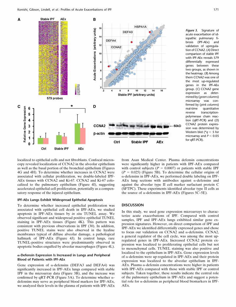
localized to epithelial cells and not fibroblasts. Confocal micros-copy revealed localization of CCNA2 in the alveolar epitheliumas well as the basal portion of the bronchial epithelium (Figures4G and 4H). To determine whether increases in CCNA2 wereassociated with cellular proliferation, we double-labeled IPF-AEx tissues with CCNA2 and Ki-67. CCNA2 and Ki-67 colo-calized to the pulmonary epithelium (Figure 4I), suggestingaccelerated epithelial cell proliferation, potentially as a compen-satory response of the injured epithelium.
IPF-AEx Lungs Exhibit Widespread Epithelial Apoptosis
To determine whether increased epithelial proliferation wasassociated with epithelial cell death in IPF-AEx, we studiedapoptosis in IPF-AEx tissues by in situ TUNEL assay. Weobserved significant and widespread positive epithelial TUNELstaining in IPF-AEx tissues (Figure 4K). This pattern wasconsistent with previous observations in IPF (30). In addition,positive TUNEL stains were also observed in the hyalinemembranes typical of diffuse alveolar damage, a pathologicalhallmark of IPF-AEx (Figure 4J). In control lungs, rareTUNEL-positive structures were predominantly observed inapoptotic bodies engulfed by alveolar macrophages (Figure 4L).
a-Defensin Expression Is Increased in Lungs and Peripheral
Blood of Patients with IPF-AEx
Gene expression of a-defensins (DEFA3 and DEFA4) wassignificantly increased in IPF-AEx lungs compared with stableIPF in the microarray data (Figure 3B), and the increase wasconfirmed by qRT-PCR (Figure 5A). To determine whether a-defensins may serve as peripheral blood markers for IPF-AEx,we analyzed their levels in the plasma of patients with IPF-AEx
from Asan Medical Center. Plasma defensin concentrationswere significantly higher in patients with IPF-AEx comparedwith control subjects (P 5 0.0007) or patients with stable IPF(P 5 0.025) (Figure 5B). To determine the cellular origins ofa-defensins in IPF-AEx, we performed double labeling on IPF-AEx lung sections with antibodies against a-defensins andagainst the alveolar type II cell marker surfactant protein C(SFTPC). These experiments identified alveolar type II cells asthe source of a-defensins in IPF-AEx (Figures 5C–5E).
DISCUSSION
In this study, we used gene expression microarrays to charac-terize acute exacerbations of IPF. Compared with controlsamples, IPF and IPF-AEx lungs exhibited similar gene ex-pression signatures. However, on direct comparison of IPF andIPF-AEx we identified differentially expressed genes and choseto focus our validation on CCNA2 and a-defensins. CCNA2,a general regulator of the cell cycle, was among the most up-regulated genes in IPF-AEx. Increased CCNA2 protein ex-pression was localized to proliferating epithelial cells but notto mesenchymal cells. TUNEL staining was also positive andlocalized to the epithelium in IPF-AEx. Gene expression levelsof a-defensins were up-regulated in IPF-AEx and their proteinexpression was localized to the alveolar epithelium in IPF-AEx. Plasma a-defensin concentrations were higher in patientswith IPF-AEx compared with those with stable IPF or controlsubjects. Taken together, these results indicate the central roleof the pulmonary epithelium in IPF-AEx and suggest a poten-tial role for a-defensins as peripheral blood biomarkers in IPF-AEx.
Figure 3. Signature of
acute exacerbation of id-iopathic pulmonary fi-
brosis (IPF-AEx) and
validation of upregula-
tion of CCNA2. (A) Directcomparison of stable IPF
with IPF-AEx reveals 579
differentially expressedgenes between these
two groups, as shown in
the heatmap. (B) Among
them CCNA2 was one ofthe most up-regulated
genes in the IPF-AEx
group. (C) CCNA2 gene
expression as deter-mined by (green columns)
microarray was con-
firmed by (pink columns)real-time quantitative
reverse transcription-
polymerase chain reac-
tion (qRT-PCR) and (D)CCNA2 protein expres-
sion was determined by
Western blot (*q , 5 for
microarray and P , 0.05for qRT-PCR).
Konishi, Gibson, Lindell, et al.: Profiles of Acute Exacerbations of IPF 171

One impressive feature of our results is the relative similarityof the gene expression patterns that distinguish IPF or IPF-AExfrom control lungs. We have previously reported the up-regulation of matrix metalloproteinase-7 (MMP7), matrix met-alloproteinase-1 (MMP1), collagens I and III, and osteopontin(14–21), as well as down-regulation of caveolin-1 (31) andadvanced glycosylation end products-specific receptor (AGER)
(32), in IPF. All these genes behaved similarly in stable IPF andIPF-AEx, as did the majority of all other genes that distin-guished IPF from control samples. We did not detect anydramatic shift in gene expression that would indicate a newprocess or a dramatic shift in lung cellular phenotype orcontent. Although we found increases in some genes associatedwith response to stress (HMOX1 and HSP1A1), we did not findany changes in known inflammation-related genes, such as IL-1,IL-6, tumor necrosis factor-a, or NF-kB target genes in thecomparison of IPF and IPF-AEx. Interestingly, other genesincreased in acute lung injury such as AGER, a known markerof generalized inflammation (33), were not increased in IPF-AEx lungs. In fact, AGER was significantly decreased in IPF-AEx compared with control samples, potentially reflecting lossof type I alveolar epithelial cells (16, 32). Taken together, theseresults do not support an overwhelming lung inflammatoryresponse as a potential mechanism for acute exacerbation. Wealso did not find any gene expression patterns indicative ofa response of the lung to viral or bacterial infections, a mech-anism observed in animals (34) but not yet confirmed in humanIPF-AEx (9, 35). Although our results do not rule out an occultviral infection or a previous viral infection as the triggeringmechanism for IPF-AEx, neither do they support a role for anactive infection during the last phase of the syndrome.
Naturally, our analysis is limited by our dependence on tissueharvested at explant or warm autopsy. It is entirely possible thatby the time the patients experienced the final deterioration allevidence of response to an infection or infected tissue wasdestroyed. In this context the finding of increased a-defensinlevels and the evidence of epithelial injury may be interpreted asremnants of an infectious process that triggered the acute lunginjury but was cleared by the time the lungs were harvested.
TABLE 3. TOP 20 UP-REGULATED PROBES THAT DISTINGUISHACUTE EXACERBATION OF IDIOPATHIC PULMONARY FIBROSIS(IPF-AEX) FROM STABLE IPF
Probe ID Gene Symbol
q Value
IPF-AEx/Stable
Fold Ratio
AEx/Stable
A_23_P31816 DEFA3 0 15.452
A_23_P326080 DEFA4 0 13.436
A_23_P219045 HIST1H3D 0 5.0072
A_32_P221799 HIST1H2AM 0 4.1309
A_23_P20022 HIG2 0 3.3581
A_23_P74059 NPPA 0 3.3542
A_24_P123616 HSPA1A 0 3.3534
A_23_P93258 HIST1H3B 0 3.0033
A_23_P329593 SEC24A 0 2.604
A_24_P933565 PGAP1 0 2.4868
A_23_P431734 SLC25A37 0 2.4823
A_23_P368740 HDAC10 0 2.4006
A_23_P381431 NPL 0 2.3955
A_23_P3643 DNASE1L2 0 2.3867
A_23_P48803 TMOD2 0 2.3068
A_23_P428184 HIST1H2AD 0 2.3003
A_23_P111132 HSPA1A 0 2.29
A_23_P58321 CCNA2 0 2.271
A_23_P333484 HIST1H3H 0 2.1118
A_23_P30799 HIST1H3F 0 2.0654
Figure 4. (A–C) Localization of CCNA2 in tissue obtainedfrom patients with acute exacerbation of idiopathic pul-
monary fibrosis (IPF-AEx) (original magnifications: A, 320;
B, 320; C, 320 merge): CCNA2 (green) is colocalized with
cytokeratin (red). (D–F) CCNA2 (green) does not colocalizewith vimentin (red) (original magnifications: D, 320; E,
320; F, 320 merge). (G and H) Confocal microscopy of
CCNA2 and cytokeratin. CCNA2 (green) and cytokeratinare colocalized in the basal portion of bronchial epithelium
(G; original magnification, 340). Coexpression of CCNA2
and cytokeratin is also observed in the alveolar epithelium
(H; original magnification, 360). Coexpression of CCNA2(green) and Ki-67 (red), a proliferation marker, is observed
in IPF-AEx tissue (I; original magnification, 340). In situ
terminal deoxynucleotidyltransferase dUTP nick end-label-
ing (TUNEL) reveals positive stains in hyaline membranes(arrows in J; original magnification, 340) and alveolar
epithelium (arrows in K; original magnification, 340) in
lung tissues with diffuse alveolar damage superimposedon the usual interstitial pneumonia pattern. In lungs with
normal morphology, TUNEL-positive structures are ob-
served predominantly in the alveolar macrophages (arrows
in L; original magnification, 340).
172 AMERICAN JOURNAL OF RESPIRATORY AND CRITICAL CARE MEDICINE VOL 180 2009

Although we cannot disprove this interpretation, we do think thatthe lack of expression of viral response genes reduces the likeli-hood of an active infection. A definite answer regarding the roleof infections will require sampling earlier at presentation andlongitudinal studies of the same patient, a task impossible withlung tissue but attainable with bronchial lavage or peripheralblood samples.
One of the remarkable features of our study is the localiza-tion of increased CCNA2 expression to the alveolar epithelium,rather than to fibroblasts or myofibroblastic foci. CCNA2 is themain A-type cyclin present in somatic cells (36) and a mediatorof the cell cycle. The overexpression and localization of CCNA2to epithelial cells but not to mesenchymal cells suggests thatIPF-AEx is probably an extension of the epithelial injury anddysregulation that characterizes IPF (37) and definitely is nota result of uncontrolled fibroblast proliferation. The fact thatthe majority of CCNA2-expressing cells were also positive forKi-67, a proliferation marker, suggests that CCNA2 expression
represents a proliferative response of the epithelium. In light ofthe positive TUNEL staining in the epithelium and hyalinemembranes, it is tempting to hypothesize that this enhancedproliferation represents a failed compensatory response to in-jury, localizing the pathogenesis of IPF-AEx to the epithelium.
One use of lung gene expression data is in the identificationof differentially expressed genes that encode secreted proteins.Such secreted proteins may be detected in the alveolar fluid orperipheral blood and thus be useful as potential surrogatemarkers for disease activity (16). Previous studies suggestedthat peripheral blood IL-8, KL-6, and most recently circulatingfibrocytes may be increased in IPF-AEx (38–40). In our studythe genes encoding a-defensins were significantly increased inIPF-AEx lungs compared with stable IPF or control samples,and their protein expression was increased in the plasmaof patients with IPF-AEx. a-Defensins are innate immunityantimicrobial peptides abundant in neutrophil granules andmucosal surfaces (41, 42). a-Defensins affect various immune
Figure 5. (A) Gene expression of a-defensins (DEFA1–3)
observed by microarray (green columns) or real-timequantitative reverse transcription-polymerase chain reac-
tion (pink columns). Gene expression levels of a-defensins
are significantly higher in patients with acute exacerbation
of idiopathic pulmonary fibrosis (IPF-AEx) compared withpatients with stable IPF or control subjects. (B) ELISA for a-
defensins in plasma obtained from control subjects,
patients with stable IPF, and patients with IPF-AEx. Plasmalevels of a-defensins in patients with IPF-AEx are signifi-
cantly higher (*q , 5 for microarray and P , 0.05 for
ELISA). (C–E) Histological localization of a-defensins in
tissues from patients with IPF-AEx (original magnification,360). (C, green) a-Defensins are present in cells express-
ing surfactant protein C (D, red and E, merge), suggesting
type II pneumocytes as one of the cellular sources for a-
defensins.
Konishi, Gibson, Lindell, et al.: Profiles of Acute Exacerbations of IPF 173

functions. a-Defensins are involved in activation of the classicalcomplement pathway (43, 44). In vitro a-defensins induce theproduction of heat shock proteins and type I collagens in humanlung fibroblasts (45), and stimulate cytokine production ofbronchial epithelial cells (46). Elevation of a-defensins has beendescribed in pulmonary alveolar proteinosis (47), a1-antitrypsindeficiency (48), acute respiratory distress (49), and chronic lungallograft rejection (50) and in patients with IPF but not in thecontext of acute exacerbation (51). In this context, it is importantto note that we observed a-defensin expression in surfactantprotein C–expressing cells in IPF-AEx lungs—a finding thatsuggests that the plasma increases in a-defensins may be in-dicative of the lung microenvironment in IPF-AEx and againhighlights the central role of the epithelium in IPF-AEx.
In summary, this is the first study of lung gene expressionpatterns in IPF-AEx lungs. Gene expression patterns indicatethat IPF-AEx represents an extension of the molecular processthat underlies IPF and not a new process. Although expressionpatterns that distinguish stable IPF and IPF-AEx lungs fromnormal lung are similar, we have identified genes that aredifferentially expressed in a direct comparison of IPF and IPF-AEx lungs. The increased expression of CCNA2 and a-defensinsis localized to the epithelium of IPF-AEx lungs, where wide-spread proliferation and apoptosis are detected, suggesting thatthe central molecular events in IPF-AEx are localized to thealveolar epithelium. Taken together, our results indicate thecentral role of alveolar epithelial injury in IPF-AEx and thussupport the study of agents that protect the epithelium astherapeutic measures in this devastating syndrome. The identifi-cation of increases in plasma concentrations of proteins originat-ing from the pulmonary epithelium in patients with IPF-AExsuggests their use as tools for evaluating patients with IPF duringthe course of the disease.
Conflict of Interest Statement: K.K. does not have a financial relationship witha commercial entity that has an interest in the subject of this manuscript. K.F.G. isan inventor on a patent application of the use of peripheral blood proteins asbiomarkers. K.O.L. does not have a financial relationship with a commercialentity that has an interest in the subject of this manuscript. T.J.R. is an inventor ona patent application of the use of peripheral blood proteins as biomarkers. Y.Z.does not have a financial relationship with a commercial entity that has aninterest in the subject of this manuscript. R.D. does not have a financialrelationship with a commercial entity that has an interest in the subject of thismanuscript. M.B. does not have a financial relationship with a commercial entitythat has an interest in the subject of this manuscript. S.G. does not havea financial relationship with a commercial entity that has an interest in the subjectof this manuscript. S.A.Y. does not have a financial relationship with a commercialentity that has an interest in the subject of this manuscript. J.W.S. does not havea financial relationship with a commercial entity that has an interest in the subjectof this manuscript. D.S.K. does not have a financial relationship with a commercialentity that has an interest in the subject of this manuscript. N.K. is a primaryinvestigator on two industry investigator initiated grants, one from Biogen Idecfor $674,000 and the other from Centocor for $250,000. N.K. is an inventor ona patent application for the use of peripheral blood proteins as biomarkers.
Acknowledgment: The authors thank Lara Chensny and Dr. Simon Watkins forsupport of the study, as well as the patient members of the Simmons Center ILDsupport group.
References
1. Flaherty KR, Travis WD, Colby TV, Toews GB, Kazerooni EA, GrossBH, Jain A, Strawderman RL, Flint A, Lynch JP, et al. Histopath-ologic variability in usual and nonspecific interstitial pneumonias. AmJ Respir Crit Care Med 2001;164:1722–1727.
2. Kim DS, Collard HR, King TE Jr. Classification and natural history ofthe idiopathic interstitial pneumonias. Proc Am Thorac Soc 2006;3:285–292.
3. Collard HR, Moore BB, Flaherty KR, Brown KK, Kaner RJ, King TEJr, Lasky JA, Loyd JE, Noth I, Olman MA, et al. Acute exacerbationsof idiopathic pulmonary fibrosis. Am J Respir Crit Care Med 2007;176:636–643.
4. Hyzy R, Huang S, Myers J, Flaherty K, Martinez F. Acuteexacerbation of idiopathic pulmonary fibrosis. Chest 2007;132:1652–1658.
5. Kubo H, Nakayama K, Yanai M, Suzuki T, Yamaya M, Watanabe M,
Sasaki H. Anticoagulant therapy for idiopathic pulmonary fibrosis.Chest 2005;128:1475–1482.
6. Noth I, Martinez FJ. Recent advances in idiopathic pulmonary fibrosis.
Chest 2007;132:637–650.7. Parambil JG, Myers JL, Ryu JH. Histopathologic features and out-
come of patients with acute exacerbation of idiopathic pulmonaryfibrosis undergoing surgical lung biopsy. Chest 2005;128:3310–3315.
8. Rice AJ, Wells AU, Bouros D, du Bois RM, Hansell DM, Polychrono-
poulos V, Vassilakis D, Kerr JR, Evans TW, Nicholson AG. Terminaldiffuse alveolar damage in relation to interstitial pneumonias: anautopsy study. Am J Clin Pathol 2003;119:709–714.
9. Tomioka H, Sakurai T, Hashimoto K, Iwasaki H. Acute exacerbation of
idiopathic pulmonary fibrosis: role of Chlamydophila pneumoniaeinfection. Respirology 2007;12:700–706.
10. Yasuda H, Yamaya M, Yanai M, Ohrui T, Sasaki H. Increased blood
carboxyhaemoglobin concentrations in inflammatory pulmonary dis-eases. Thorax 2002;57:779–783.
11. Kim DS, Park JH, Park BK, Lee JS, Nicholson AG, Colby T. Acute
exacerbation of idiopathic pulmonary fibrosis: frequency and clinicalfeatures. Eur Respir J 2006;27:143–150.
12. Ambrosini V, Cancellieri A, Chilosi M, Zompatori M, Trisolini R,
Saragoni L, Poletti V. Acute exacerbation of idiopathic pulmonaryfibrosis: report of a series. Eur Respir J 2003;22:821–826.
13. Vannella KM, Moore BB. Viruses as co-factors for the initiation or
exacerbation of lung fibrosis. Fibrogenesis Tissue Repair 2008;1:2.14. Cosgrove GP, Brown KK, Schiemann WP, Serls AE, Parr JE, Geraci
MW, Schwarz MI, Cool CD, Worthen GS. Pigment epithelium–derived factor in idiopathic pulmonary fibrosis: a role in aberrantangiogenesis. Am J Respir Crit Care Med 2004;170:242–251.
15. Pardo A, Gibson K, Cisneros J, Richards TJ, Yang Y, Becerril C,
Yousem S, Herrera I, Ruiz V, Selman M, et al. Up-regulation andprofibrotic role of osteopontin in human idiopathic pulmonaryfibrosis. PLoS Med 2005;2:e251.
16. Rosas IO, Richards TJ, Konishi K, Zhang Y, Gibson K, Lokshin AE,
Lindell KO, Cisneros J, Macdonald SD, Pardo A, et al. MMP1 andMMP7 as potential peripheral blood biomarkers in idiopathic pul-monary fibrosis. PLoS Med 2008;5:e93.
17. Selman M, Pardo A, Barrera L, Estrada A, Watson SR, Wilson K, Aziz
N, Kaminski N, Zlotnik A. Gene expression profiles distinguishidiopathic pulmonary fibrosis from hypersensitivity pneumonitis.Am J Respir Crit Care Med 2006;173:188–198.
18. Selman M, Carrillo G, Estrada A, Mejia M, Becerril C, Cisneros J,
Gaxiola M, Perez-Padilla R, Navarro C, Richards T, et al. Acceler-ated variant of idiopathic pulmonary fibrosis: clinical behavior andgene expression pattern. PLoS One 2007;2:e482.
19. Kaminski N, Allard JD, Pittet JF, Zuo F, Griffiths MJ, Morris D,
Huang X, Sheppard D, Heller RA. Global analysis of geneexpression in pulmonary fibrosis reveals distinct programs regulat-ing lung inflammation and fibrosis. Proc Natl Acad Sci USA 2000;97:1778–1783.
20. Zuo F, Kaminski N, Eugui E, Allard J, Yakhini Z, Ben-Dor A, Lollini L,
Morris D, Kim Y, DeLustro B, et al. Gene expression analysis revealsmatrilysin as a key regulator of pulmonary fibrosis in mice andhumans. Proc Natl Acad Sci USA 2002;99:6292–6297.
21. Yang IV, Burch LH, Steele MP, Savov JD, Hollingsworth JW, McElva-
nia-Tekippe E, Berman KG, Speer MC, Sporn TA, Brown KK, et al.Gene expression profiling of familial and sporadic interstitial pneu-monia. Am J Respir Crit Care Med 2007;175:45–54.
22. Konishi K, Richards TJ, Collard HR, Lindell KO, Chensny LJ, Yu G,
Herazo JD, Gibson KF, Kaminski N. Gene expression profileshighlight role of epithelial injury in acute exacerbations of idiopathicpulmonary fibrosis [abstract]. Presented at the 15th InternationalColloquium on Lung and Airway Fibrosis, September 28–October 1,2008, Sunset Beach, NC.
23. Lindell KO, Erlen JA, Kaminski N. Lessons from our patients:
development of a warm autopsy program. PLoS Med 2006;3:e234.
24. American Thoracic Society, European Respiratory Society. Idiopathic
pulmonary fibrosis: diagnosis and treatment [international consensusstatement]. Am J Respir Crit Care Med 2000;161:646–664.
25. Akira M, Hamada H, Sakatani M, Kobayashi C, Nishioka M, Yamamoto
S. Ct findings during phase of accelerated deterioration in patientswith idiopathic pulmonary fibrosis. AJR Am J Roentgenol 1997;168:79–83.
174 AMERICAN JOURNAL OF RESPIRATORY AND CRITICAL CARE MEDICINE VOL 180 2009

26. Zahurak M, Parmigiani G, Yu W, Scharpf RB, Berman D, Schaeffer E,Shabbeer S, Cope L. Pre-processing Agilent microarray data. BMCBioinformatics 2007;8:142.
27. Wu W, Dave N, Tseng GC, Richards T, Xing EP, Kaminski N.Comparison of normalization methods for codelink bioarray data.BMC Bioinformatics 2005;6:309.
28. Segal E, Friedman N, Kaminski N, Regev A, Koller D. From signaturesto models: understanding cancer using microarrays. Nat Genet 2005;37:S38–S45.
29. Dave NB, Kaminski N. Analysis of microarray experiments for pulmo-nary fibrosis. Methods Mol Med 2005;117:333–358.
30. Plataki M, Koutsopoulos AV, Darivianaki K, Delides G, Siafakas NM,Bouros D. Expression of apoptotic and antiapoptotic markers inepithelial cells in idiopathic pulmonary fibrosis. Chest 2005;127:266–274.
31. Wang XM, Zhang Y, Kim HP, Zhou Z, Feghali-Bostwick CA, Liu F,Ifedigbo E, Xu X, Oury TD, Kaminski N, et al. Caveolin-1: a criticalregulator of lung fibrosis in idiopathic pulmonary fibrosis. J Exp Med2006;203:2895–2906.
32. Englert JM, Hanford LE, Kaminski N, Tobolewski JM, Tan RJ, FattmanCL, Ramsgaard L, Richards TJ, Loutaev I, Nawroth PP, et al. A rolefor the receptor for advanced glycation end products in idiopathicpulmonary fibrosis. Am J Pathol 2008;172:583–591.
33. Uchida T, Shirasawa M, Ware LB, Kojima K, Hata Y, Makita K,Mednick G, Matthay ZA, Matthay MA. Receptor for advancedglycation end-products is a marker of type I cell injury in acute lunginjury. Am J Respir Crit Care Med 2006;173:1008–1015.
34. McMillan TR, Moore BB, Weinberg JB, Vannella KM, Fields WB,Christensen PJ, van Dyk LF, Toews GB. Exacerbation of establishedpulmonary fibrosis in a murine model by gammaherpesvirus. Am JRespir Crit Care Med 2008;177:771–780.
35. Saydain G, Islam A, Afessa B, Ryu JH, Scott JP, Peters SG. Outcome ofpatients with idiopathic pulmonary fibrosis admitted to the intensivecare unit. Am J Respir Crit Care Med 2002;166:839–842.
36. Gong D, Pomerening JR, Myers JW, Gustavsson C, Jones JT, Hahn AT,Meyer T, Ferrell JE Jr. Cyclin A2 regulates nuclear-envelopebreakdown and the nuclear accumulation of cyclin B1. Curr Biol2007;17:85–91.
37. Selman M, Pardo A. Role of epithelial cells in idiopathic pulmonaryfibrosis: from innocent targets to serial killers. Proc Am Thorac Soc2006;3:364–372.
38. Moeller A, Gilpin SE, Ask K, Cox G, Cook D, Gauldie J, Margetts PJ,Farkas L, Dobranowski J, Boylan C, et al. Circulating fibrocytes arean indicator for poor prognosis in idiopathic pulmonary fibrosis. Am JRespir Crit Care Med 2009;179:588–594.
39. Tajima S, Oshikawa K, Tominaga S, Sugiyama Y. The increase in serumsoluble ST2 protein upon acute exacerbation of idiopathic pulmonaryfibrosis. Chest 2003;124:1206–1214.
40. Ziegenhagen MW, Zabel P, Zissel G, Schlaak M, Muller-Quernheim J.Serum level of interleukin 8 is elevated in idiopathic pulmonaryfibrosis and indicates disease activity. Am J Respir Crit Care Med1998;157:762–768.
41. Boman HG. Peptide antibiotics and their role in innate immunity. AnnuRev Immunol 1995;13:61–92.
42. Zasloff M. Antimicrobial peptides of multicellular organisms. Nature2002;415:389–395.
43. van den Berg RH, Faber-Krol MC, van Wetering S, Hiemstra PS, DahaMR. Inhibition of activation of the classical pathway of complementby human neutrophil defensins. Blood 1998;92:3898–3903.
44. Prohaszka Z, Nemet K, Csermely P, Hudecz F, Mezo G, Fust G.Defensins purified from human granulocytes bind C1q and activatethe classical complement pathway like the transmembrane glycopro-tein gp41 of HIV-1. Mol Immunol 1997;34:809–816.
45. Yoshioka S, Mukae H, Ishii H, Kakugawa T, Ishimoto H, Sakamoto N,Fujii T, Urata Y, Kondo T, Kubota H, et al. a-Defensin enhancesexpression of hsp47 and collagen-1 in human lung fibroblasts. Life Sci2007;80:1839–1845.
46. Sakamoto N, Mukae H, Fujii T, Ishii H, Yoshioka S, Kakugawa T,Sugiyama K, Mizuta Y, Kadota J, Nakazato M, et al. Differentialeffects of a- and b-defensin on cytokine production by culturedhuman bronchial epithelial cells. Am J Physiol 2005;288:L508–L513.
47. Mukae H, Ishimoto H, Yanagi S, Ishii H, Nakayama S, Ashitani J,Nakazato M, Kohno S. Elevated BALF concentrations of a- and b-defensins in patients with pulmonary alveolar proteinosis. Respir Med2007;101:715–721.
48. Spencer LT, Paone G, Krein PM, Rouhani FN, Rivera-Nieves J, BrantlyML. Role of human neutrophil peptides in lung inflammationassociated with a1-antitrypsin deficiency. Am J Physiol 2004;286:L514–L520.
49. Ashitani J, Mukae H, Arimura Y, Sano A, Tokojima M, Nakazato M.High concentrations of a-defensins in plasma and bronchoalveolarlavage fluid of patients with acute respiratory distress syndrome. LifeSci 2004;75:1123–1134.
50. Nelsestuen GL, Martinez MB, Hertz MI, Savik K, Wendt CH. Proteo-mic identification of human neutrophil a-defensins in chronic lungallograft rejection. Proteomics 2005;5:1705–1713.
51. Mukae H, Iiboshi H, Nakazato M, Hiratsuka T, Tokojima M, Abe K,Ashitani J, Kadota J, Matsukura S, Kohno S. Raised plasma concen-trations of a-defensins in patients with idiopathic pulmonary fibrosis.Thorax 2002;57:623–628.
Konishi, Gibson, Lindell, et al.: Profiles of Acute Exacerbations of IPF 175
Copyright © 2022 FDOKUMEN