Methanol Extract of Euchelus asper Prevents Bone Resorption in Ovariectomised Mice Model
9
Research Article Methanol Extract of Euchelus asper Prevents Bone Resorption in Ovariectomised Mice Model Babita Balakrishnan, 1 Shubhada Vivek Chiplunkar, 2 and Madhavi Manohar Indap 1 1 e Department of Zoology, e D.G. Ruparel College, Senapati Bapat Marg, Mahim, Mumbai 400016, India 2 Advanced Centre for Treatment Research and Education in Cancer (ACTREC), Tata Memorial Centre, Kharghar, Navi Mumbai 410210, India Correspondence should be addressed to Madhavi Manohar Indap; [email protected] Received 10 January 2014; Revised 10 May 2014; Accepted 19 May 2014; Published 5 June 2014 Academic Editor: Klaus Engelke Copyright © 2014 Babita Balakrishnan et al. is is an open access article distributed under the Creative Commons Attribution License, which permits unrestricted use, distribution, and reproduction in any medium, provided the original work is properly cited. Marine molluscs are widely distributed throughout the world and many bioactive compounds exhibiting antiviral, antitumor, antileukemic, and antibacterial activity have been reported worldwide. e present study was designed to investigate the beneficial effect of methanol extract of Euchelus asper (EAME) on estrogen deficiency induced osteoporosis in ovariectomised mice model. Forty-two female Swiss albino mice were randomly assigned into Sham operated (Sham) group and six ovariectomised (OVX) subgroups such as OVX with vehicle (OVX); OVX with estradiol (2 mg/kg/day); OVX with EAME of graded doses (25, 50, 100, and 200 mg/kg/day). Bone turnover markers like serum alkaline phosphatase (ALP), serum acid phosphatase (ACP), serum calcium, and histological investigations of tibia and uterus were analysed. Metaphyseal DNA content of the femur bone was also studied. Antiosteoclastogenic activity of EAME was examined. Administration of EAME was able to reduce the increased bone turnover markers in the ovariectomised mice. Histomorphometric analysis revealed an increase in bone trabeculation and restoration of trabecular separation by EAME treatment. Metaphyseal DNA content of the femur of the OVX mice was increased by EAME administration. EAME also showed a potent antiosteoclastogenic behaviour. us, the present study reveals that EAME was able to successfully reduce the estrogen deficiency induced bone loss. 1. Introduction Osteoporosis is a condition of skeletal fragility characterised by decreased bone mass and microarchitectural deterioration of bone tissue, with a consequent increase in risk of fracture [1]. Osteoporosis is a worldwide health issue, with a high prevalence of disease not only in western countries but also in Asia and Latin America [2]. Women were found to have a higher risk of getting osteoporosis than men with the ratio of 1.6 : 1. Postmenopausal osteoporosis stems from the cessation of ovarian function at menopause which heightens and prolongs the rapid phase of bone loss characteristic of the early postmenopausal period [3]. It is associated with significant morbidity, mortality, reduction in quality of life, and increasing health care. About 1 in 3 women aged more than 50 years experienced an osteoporotic fracture in their lifetime. In women, osteoporosis and fractures mainly occur as a consequence of decrease in estrogen aſter menopause and result from an imbalance between bone resorption by osteoclasts and bone formation by osteoblasts, leading to a net bone loss with each remodelling cycle. Estrogen deficiency also augments erosion depth by extending the resorption phase of remodelling cycle through increased osteoclast life span due to its reduced apoptosis [4]. Estrogen replacement therapy (ERT) and combination hormone replacement therapy (CHRT) are two medication therapies oſten prescribed to supplement the decreased lev- els of hormones in postmenopausal women [5]. Research by Cauley et al. showed that hormone replacement ther- apy (HRT) can significantly improve bone mineral density (BMD) and reduce the risk for fractures of the vertebral spine, lower arm/waist and hip areas regardless of previous history, age, familial history, smoking, alcohol status, BMD, or fracture risk rates [6]. e Women’s Health Initiative (WHI) suggests, however, that the long-term risks of HRT outweigh the benefits. It is associated with coronary heart Hindawi Publishing Corporation Journal of Osteoporosis Volume 2014, Article ID 348189, 9 pages http://dx.doi.org/10.1155/2014/348189
-
Upload
independent -
Category
Documents
-
view
2 -
download
0
Transcript of Methanol Extract of Euchelus asper Prevents Bone Resorption in Ovariectomised Mice Model

Research ArticleMethanol Extract of Euchelus asper Prevents Bone Resorptionin Ovariectomised Mice Model
Babita Balakrishnan,1 Shubhada Vivek Chiplunkar,2 and Madhavi Manohar Indap1
1 The Department of Zoology, The D.G. Ruparel College, Senapati Bapat Marg, Mahim, Mumbai 400016, India2 Advanced Centre for Treatment Research and Education in Cancer (ACTREC), Tata Memorial Centre,Kharghar, Navi Mumbai 410210, India
Correspondence should be addressed to Madhavi Manohar Indap; [email protected]
Received 10 January 2014; Revised 10 May 2014; Accepted 19 May 2014; Published 5 June 2014
Academic Editor: Klaus Engelke
Copyright © 2014 Babita Balakrishnan et al. This is an open access article distributed under the Creative Commons AttributionLicense, which permits unrestricted use, distribution, and reproduction in any medium, provided the original work is properlycited.
Marine molluscs are widely distributed throughout the world and many bioactive compounds exhibiting antiviral, antitumor,antileukemic, and antibacterial activity have been reported worldwide.The present study was designed to investigate the beneficialeffect of methanol extract of Euchelus asper (EAME) on estrogen deficiency induced osteoporosis in ovariectomised mice model.Forty-two female Swiss albino mice were randomly assigned into Sham operated (Sham) group and six ovariectomised (OVX)subgroups such as OVXwith vehicle (OVX); OVXwith estradiol (2mg/kg/day); OVXwith EAME of graded doses (25, 50, 100, and200mg/kg/day). Bone turnover markers like serum alkaline phosphatase (ALP), serum acid phosphatase (ACP), serum calcium,and histological investigations of tibia and uterus were analysed. Metaphyseal DNA content of the femur bone was also studied.Antiosteoclastogenic activity of EAME was examined. Administration of EAME was able to reduce the increased bone turnovermarkers in the ovariectomised mice. Histomorphometric analysis revealed an increase in bone trabeculation and restoration oftrabecular separation by EAME treatment. Metaphyseal DNA content of the femur of the OVX mice was increased by EAMEadministration. EAME also showed a potent antiosteoclastogenic behaviour. Thus, the present study reveals that EAME was ableto successfully reduce the estrogen deficiency induced bone loss.
1. Introduction
Osteoporosis is a condition of skeletal fragility characterisedby decreased bonemass andmicroarchitectural deteriorationof bone tissue, with a consequent increase in risk of fracture[1]. Osteoporosis is a worldwide health issue, with a highprevalence of disease not only in western countries but alsoin Asia and Latin America [2]. Women were found to havea higher risk of getting osteoporosis than men with theratio of 1.6 : 1. Postmenopausal osteoporosis stems from thecessation of ovarian function at menopause which heightensand prolongs the rapid phase of bone loss characteristic ofthe early postmenopausal period [3]. It is associated withsignificant morbidity, mortality, reduction in quality of life,and increasing health care. About 1 in 3 women aged morethan 50 years experienced an osteoporotic fracture in theirlifetime. In women, osteoporosis and fractures mainly occuras a consequence of decrease in estrogen after menopause
and result from an imbalance between bone resorption byosteoclasts and bone formation by osteoblasts, leading to a netbone loss with each remodelling cycle. Estrogen deficiencyalso augments erosion depth by extending the resorptionphase of remodelling cycle through increased osteoclast lifespan due to its reduced apoptosis [4].
Estrogen replacement therapy (ERT) and combinationhormone replacement therapy (CHRT) are two medicationtherapies often prescribed to supplement the decreased lev-els of hormones in postmenopausal women [5]. Researchby Cauley et al. showed that hormone replacement ther-apy (HRT) can significantly improve bone mineral density(BMD) and reduce the risk for fractures of the vertebralspine, lower arm/waist and hip areas regardless of previoushistory, age, familial history, smoking, alcohol status, BMD,or fracture risk rates [6]. The Women’s Health Initiative(WHI) suggests, however, that the long-term risks of HRToutweigh the benefits. It is associated with coronary heart
Hindawi Publishing CorporationJournal of OsteoporosisVolume 2014, Article ID 348189, 9 pageshttp://dx.doi.org/10.1155/2014/348189
2 Journal of Osteoporosis
disease, breast cancer, and stroke. In most countries, HRTis only recommended for climacteric symptoms, at a dose assmall as possible and for a limited period of time.Thus, HRTis no longer recommended as a firstline treatment for theprevention and treatment of osteoporosis. Thus, the ultimategoal of treating women with postmenopausal osteoporosisis to identify compounds which not only reduce the risk offracture but also have minimal side effects.
Despite the myriad of potential health benefits of themarine bioactives, there are few data relating to their possiblepreventive effect on osteoporosis. Efforts are needed toexpand the marine based drug discovery to include osteo-porosis disorders, which need extensive and urgent attentionin terms of new therapies.
Euchelus asper, a marine mollusc that belongs to classGastropoda, is a shell-oriented animal. It is commonly seenalong the entire west coast and extends up to the south eastcoast in India [7]. It can be found on the rocky beaches nearlow tide marks. It is eaten by local people. Organic matrixfrom molluscan shells has the potential to regulate calciumcarbonate deposition and crystallization. Biomineralizationis one essential element in living organisms to protect them-selves against predators and to formmulticellular aggregates,for example, during bacterial biofilm formation, or in mul-ticellular organisms, for example, in animals and plants [8].Biomineralization is a highly orchestrated biological processin the mollusc. The organic matrix of the mollusc directscrystallization and the deposition and rate of crystals are alsocontrolled by hormones produced by the mollusc [9]. Earlierstudies by our group have shown that ether soluble fraction ofEuchelus asper (EAE) exhibited immunosuppressive activityin vivo by plaque forming assay [10]. In the present studywe have tried to investigate the antiosteoporotic effect ofmethanol extract of Euchelus asper (EAME).
2. Methods and Materials
2.1. Preparation of EAMEand Its Fractionation. Theorganismexposed during the low tide was collected from the rockyshore of Khar Danda, Mumbai, Maharashtra, India. Itwasdeshelled and the bodymasswas cold-percolated inmethanolfor 3-4 days. The resulting methanol extract was filteredand the residual animal mass was again immersed in freshmethanol. The process was repeated till the methanol extractbecame colorless. The filtered extracts were pooled andconcentrated under reduced pressure and crude gummymethanol extract was obtained with a yield of 1.25%. It wasrefrigerated at −20∘C till further use.
2.2. Animals andTreatments. Female Swiss albinomice of 18–22 g maintained at the Animal House of ACTREC were usedfor the study.This studywas approved by InstitutionalAnimalEthics Committee (IAEC) of ACTREC, Tata Memorial Cen-tre, Mumbai, India. They were fed with standard laboratorydiet and water ad libitum. The mice were randomly dividedinto 7 groups, consisting of 6 animals in each group.
Group A. Sham control mice treated with vehicle (Shamcontrol).
Group B. Bilaterally ovariectomized control treated withvehicle (OVX).
Group C. Bilaterally ovariectomized control treated withestradiol-2mg/kg body weight (positive control).
Group D. Bilaterally ovariectomized control treated withEAME 25mg/kg body weight.
Group E. Bilaterally ovariectomized control treated withEAME 50mg/kg body weight.
Group F. Bilaterally ovariectomized control treated withEAME 100mg/kg body weight.
Group G. Bilaterally ovariectomized control treated withEAME 200mg/kg body weight.
All mice were bilaterally ovariectomized after anesthetiz-ing with ketamine + xylazine. After 7 days of recovery fromsurgical convalescence, animals of groups D, E, F, and G wereadministered orally through stomach tube with EAME, 25,50, 100, and 200mg/kg body weight of EAME, respectively.The vehicle or extract was dissolved in sterilized water andwas administered daily for 4 weeks. Animals in the controlgroups A and B received water (vehicle control) and of groupC received estradiol (positive control).The bodyweight of theanimalswas recordedweekly during the experimental period.
2.3. BodyWeightMeasurements. On completion of the exper-imental period, the final body weights of animals of all the7 groups were recorded. The animals were sacrificed underdeep anaesthesia on the scheduled date and fresh weight ofuteri was recorded.
2.4. Biochemical Estimations
2.4.1. Estimation of Serum Calcium, Alkaline Phosphatase,and Acid Phosphatase. Blood was collected from retroorbitalregion and centrifuged at 1500×g for 20min. Serum wasseparated immediately and was stored at −80∘C. Serum alka-line phosphatase (ALP) and acid phosphatase activity (ACP)were estimated spectrophotometrically at 510 nm using SpanDiagnostic Kit, Span Diagnostic Ltd., India. Serum was alsoanalyzed for the presence of calcium. It was estimated by drychemistry biochemical analyzer Vitros DT60II.
2.5. Histological Examination
2.5.1. Tissue Collection and Processing. The tibia and uteruswere harvested rapidly and fixed with Bouin’s solution for2 h, which was followed by decalcification in 5% nitric acidfor tibia, dehydration in alcohol, clearing in xylene, andfinally embedding in paraffin for further tissue section andhistological staining [11].
2.6. Metaphyseal DNA Content. To measure bone DNAcontent, the metaphyseal tissues of the femur bone wereshaken with 4.0mL of ice-cold 0.1M NaOH solution for24 h after the homogenization of the bone tissues [12]. Afteralkaline extraction, the samples were centrifuged at 10000×g
Journal of Osteoporosis 3
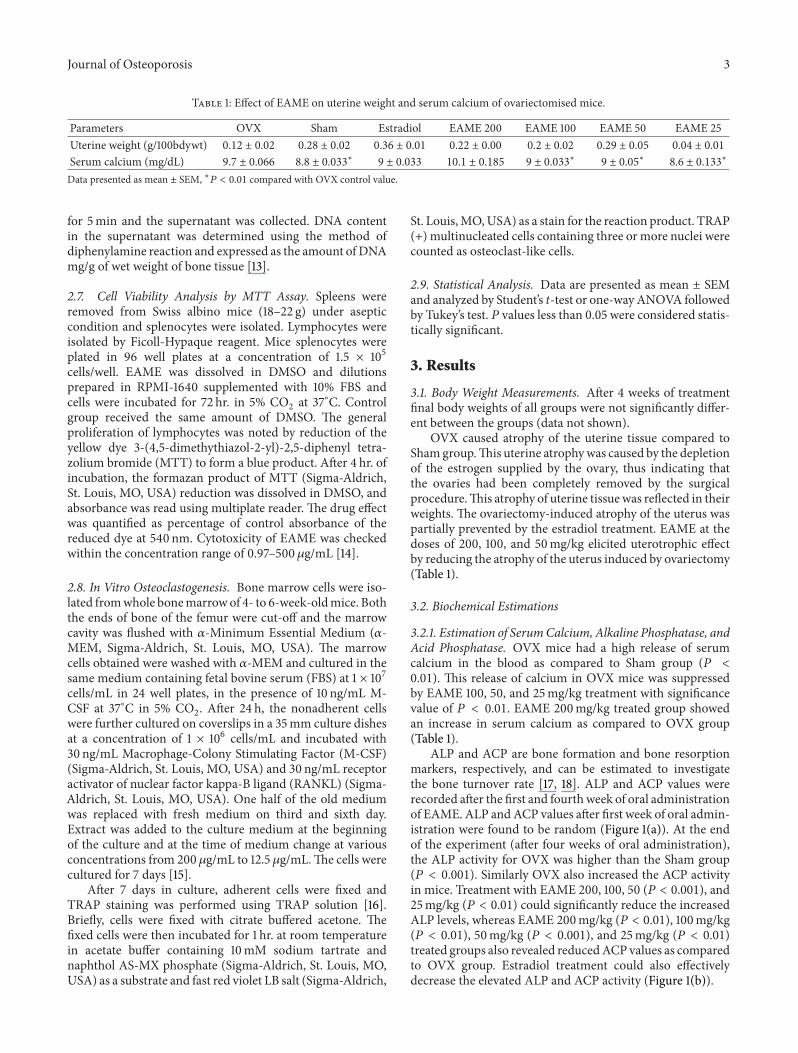
Table 1: Effect of EAME on uterine weight and serum calcium of ovariectomised mice.
Parameters OVX Sham Estradiol EAME 200 EAME 100 EAME 50 EAME 25Uterine weight (g/100bdywt) 0.12 ± 0.02 0.28 ± 0.02 0.36 ± 0.01 0.22 ± 0.00 0.2 ± 0.02 0.29 ± 0.05 0.04 ± 0.01
Serum calcium (mg/dL) 9.7 ± 0.066 8.8 ± 0.033
∗
9 ± 0.033 10.1 ± 0.185 9 ± 0.033
∗
9 ± 0.05
∗
8.6 ± 0.133
∗
Data presented as mean ± SEM, ∗𝑃 < 0.01 compared with OVX control value.
for 5min and the supernatant was collected. DNA contentin the supernatant was determined using the method ofdiphenylamine reaction and expressed as the amount of DNAmg/g of wet weight of bone tissue [13].
2.7. Cell Viability Analysis by MTT Assay. Spleens wereremoved from Swiss albino mice (18–22 g) under asepticcondition and splenocytes were isolated. Lymphocytes wereisolated by Ficoll-Hypaque reagent. Mice splenocytes wereplated in 96 well plates at a concentration of 1.5 × 10⁵cells/well. EAME was dissolved in DMSO and dilutionsprepared in RPMI-1640 supplemented with 10% FBS andcells were incubated for 72 hr. in 5% CO
2at 37∘C. Control
group received the same amount of DMSO. The generalproliferation of lymphocytes was noted by reduction of theyellow dye 3-(4,5-dimethythiazol-2-yl)-2,5-diphenyl tetra-zolium bromide (MTT) to form a blue product. After 4 hr. ofincubation, the formazan product of MTT (Sigma-Aldrich,St. Louis, MO, USA) reduction was dissolved in DMSO, andabsorbance was read using multiplate reader. The drug effectwas quantified as percentage of control absorbance of thereduced dye at 540 nm. Cytotoxicity of EAME was checkedwithin the concentration range of 0.97–500𝜇g/mL [14].
2.8. In Vitro Osteoclastogenesis. Bone marrow cells were iso-lated fromwhole bonemarrowof 4- to 6-week-oldmice. Boththe ends of bone of the femur were cut-off and the marrowcavity was flushed with 𝛼-Minimum Essential Medium (𝛼-MEM, Sigma-Aldrich, St. Louis, MO, USA). The marrowcells obtained were washed with 𝛼-MEM and cultured in thesame medium containing fetal bovine serum (FBS) at 1 × 10⁷cells/mL in 24 well plates, in the presence of 10 ng/mL M-CSF at 37∘C in 5% CO
2. After 24 h, the nonadherent cells
were further cultured on coverslips in a 35mm culture dishesat a concentration of 1 × 10⁶ cells/mL and incubated with30 ng/mL Macrophage-Colony Stimulating Factor (M-CSF)(Sigma-Aldrich, St. Louis, MO, USA) and 30 ng/mL receptoractivator of nuclear factor kappa-B ligand (RANKL) (Sigma-Aldrich, St. Louis, MO, USA). One half of the old mediumwas replaced with fresh medium on third and sixth day.Extract was added to the culture medium at the beginningof the culture and at the time of medium change at variousconcentrations from 200𝜇g/mL to 12.5 𝜇g/mL.The cells werecultured for 7 days [15].
After 7 days in culture, adherent cells were fixed andTRAP staining was performed using TRAP solution [16].Briefly, cells were fixed with citrate buffered acetone. Thefixed cells were then incubated for 1 hr. at room temperaturein acetate buffer containing 10mM sodium tartrate andnaphthol AS-MX phosphate (Sigma-Aldrich, St. Louis, MO,USA) as a substrate and fast red violet LB salt (Sigma-Aldrich,
St. Louis,MO,USA) as a stain for the reaction product. TRAP(+) multinucleated cells containing three or more nuclei werecounted as osteoclast-like cells.
2.9. Statistical Analysis. Data are presented as mean ± SEMand analyzed by Student’s t-test or one-way ANOVA followedby Tukey’s test. 𝑃 values less than 0.05 were considered statis-tically significant.
3. Results
3.1. Body Weight Measurements. After 4 weeks of treatmentfinal body weights of all groups were not significantly differ-ent between the groups (data not shown).
OVX caused atrophy of the uterine tissue compared toShamgroup.This uterine atrophywas caused by the depletionof the estrogen supplied by the ovary, thus indicating thatthe ovaries had been completely removed by the surgicalprocedure.This atrophy of uterine tissuewas reflected in theirweights. The ovariectomy-induced atrophy of the uterus waspartially prevented by the estradiol treatment. EAME at thedoses of 200, 100, and 50mg/kg elicited uterotrophic effectby reducing the atrophy of the uterus induced by ovariectomy(Table 1).
3.2. Biochemical Estimations
3.2.1. Estimation of SerumCalcium, Alkaline Phosphatase, andAcid Phosphatase. OVX mice had a high release of serumcalcium in the blood as compared to Sham group (𝑃 <0.01). This release of calcium in OVX mice was suppressedby EAME 100, 50, and 25mg/kg treatment with significancevalue of 𝑃 < 0.01. EAME 200mg/kg treated group showedan increase in serum calcium as compared to OVX group(Table 1).
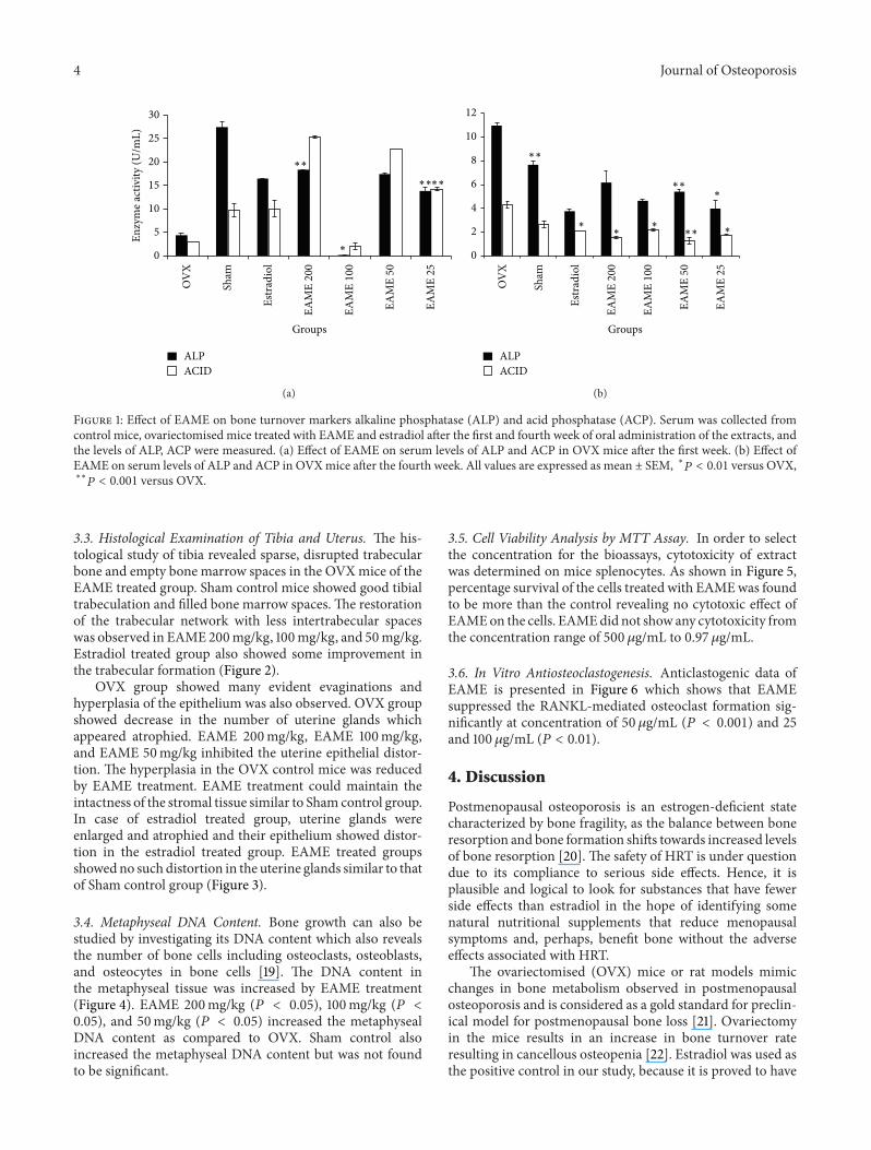
ALP and ACP are bone formation and bone resorptionmarkers, respectively, and can be estimated to investigatethe bone turnover rate [17, 18]. ALP and ACP values wererecorded after the first and fourthweek of oral administrationof EAME. ALP andACP values after first week of oral admin-istration were found to be random (Figure 1(a)). At the endof the experiment (after four weeks of oral administration),the ALP activity for OVX was higher than the Sham group(𝑃 < 0.001). Similarly OVX also increased the ACP activityin mice. Treatment with EAME 200, 100, 50 (𝑃 < 0.001), and25mg/kg (𝑃 < 0.01) could significantly reduce the increasedALP levels, whereas EAME 200mg/kg (𝑃 < 0.01), 100mg/kg(𝑃 < 0.01), 50mg/kg (𝑃 < 0.001), and 25mg/kg (𝑃 < 0.01)treated groups also revealed reducedACP values as comparedto OVX group. Estradiol treatment could also effectivelydecrease the elevated ALP and ACP activity (Figure 1(b)).
4 Journal of Osteoporosis
0
OV
X
Sham
Estr
adio
l
EAM
E 20
0
EAM
E 10
0
EAM
E 50
EAM
E 25
5
10
15
20
25
30
Enzy
me a
ctiv
ity (U
/mL)
Groups
ALPACID
∗
∗∗∗∗
∗∗
(a)
OV
X
Sham
Estr
adio
l
EAM
E 20
0
EAM
E 10
0
EAM
E 50
EAM
E 25
Groups
ALPACID
0
2
4
6
8
10
12
∗∗
∗∗
∗∗∗
∗∗
∗
∗
(b)
Figure 1: Effect of EAME on bone turnover markers alkaline phosphatase (ALP) and acid phosphatase (ACP). Serum was collected fromcontrol mice, ovariectomised mice treated with EAME and estradiol after the first and fourth week of oral administration of the extracts, andthe levels of ALP, ACP were measured. (a) Effect of EAME on serum levels of ALP and ACP in OVX mice after the first week. (b) Effect ofEAME on serum levels of ALP and ACP in OVXmice after the fourth week. All values are expressed as mean ± SEM, ∗
𝑃
< 0.01 versus OVX,∗∗
𝑃
< 0.001 versus OVX.
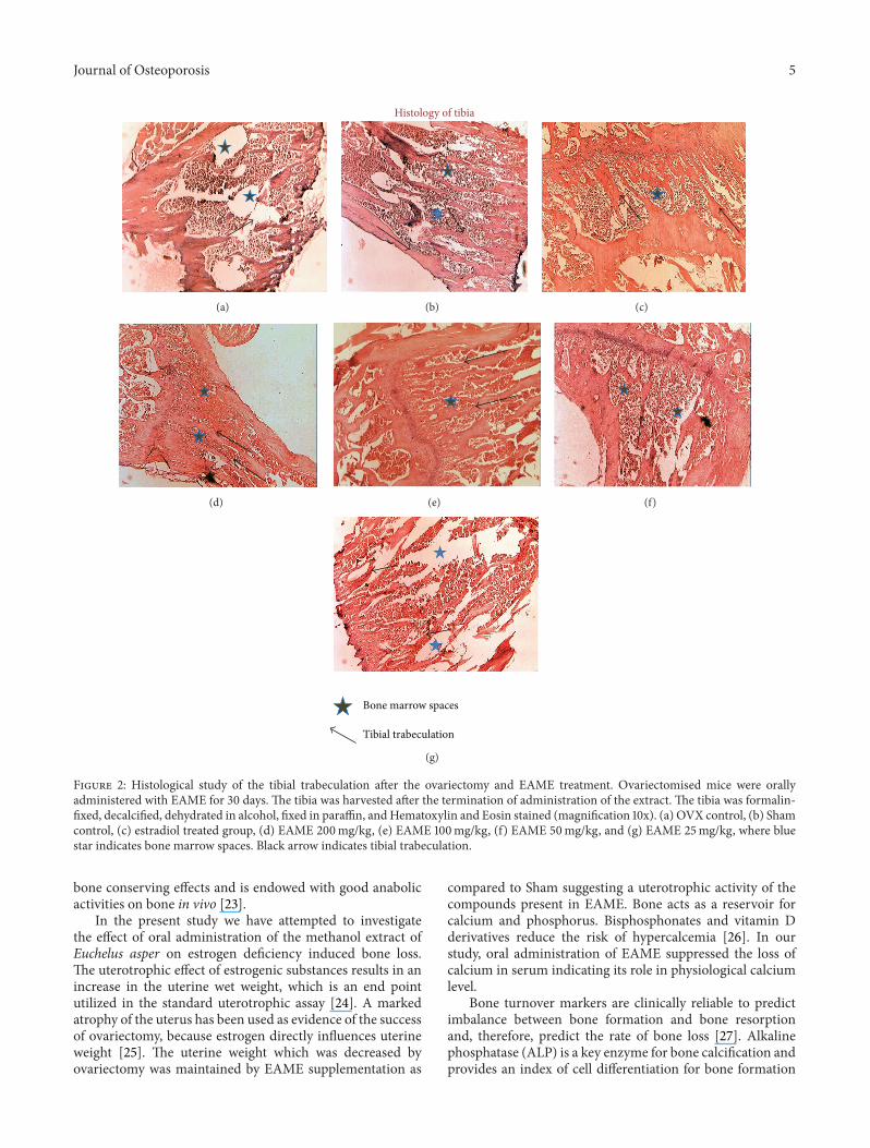
3.3. Histological Examination of Tibia and Uterus. The his-tological study of tibia revealed sparse, disrupted trabecularbone and empty bone marrow spaces in the OVXmice of theEAME treated group. Sham control mice showed good tibialtrabeculation and filled bone marrow spaces. The restorationof the trabecular network with less intertrabecular spaceswas observed in EAME 200mg/kg, 100mg/kg, and 50mg/kg.Estradiol treated group also showed some improvement inthe trabecular formation (Figure 2).
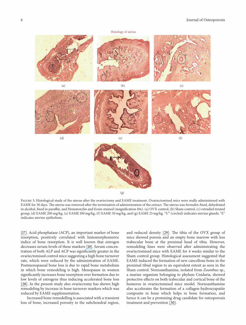
OVX group showed many evident evaginations andhyperplasia of the epithelium was also observed. OVX groupshowed decrease in the number of uterine glands whichappeared atrophied. EAME 200mg/kg, EAME 100mg/kg,and EAME 50mg/kg inhibited the uterine epithelial distor-tion. The hyperplasia in the OVX control mice was reducedby EAME treatment. EAME treatment could maintain theintactness of the stromal tissue similar to Sham control group.In case of estradiol treated group, uterine glands wereenlarged and atrophied and their epithelium showed distor-tion in the estradiol treated group. EAME treated groupsshowedno such distortion in the uterine glands similar to thatof Sham control group (Figure 3).
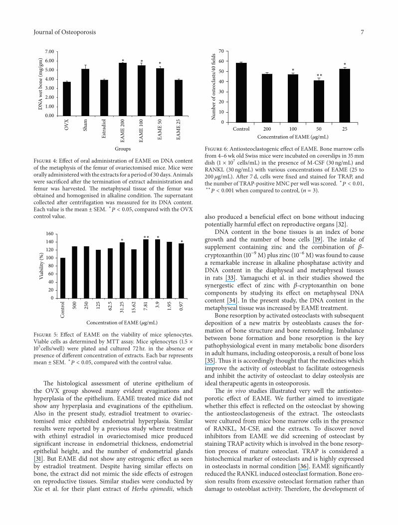
3.4. Metaphyseal DNA Content. Bone growth can also bestudied by investigating its DNA content which also revealsthe number of bone cells including osteoclasts, osteoblasts,and osteocytes in bone cells [19]. The DNA content inthe metaphyseal tissue was increased by EAME treatment(Figure 4). EAME 200mg/kg (𝑃 < 0.05), 100mg/kg (𝑃 <0.05), and 50mg/kg (𝑃 < 0.05) increased the metaphysealDNA content as compared to OVX. Sham control alsoincreased the metaphyseal DNA content but was not foundto be significant.
3.5. Cell Viability Analysis by MTT Assay. In order to selectthe concentration for the bioassays, cytotoxicity of extractwas determined on mice splenocytes. As shown in Figure 5,percentage survival of the cells treated with EAMEwas foundto be more than the control revealing no cytotoxic effect ofEAMEon the cells. EAMEdid not show any cytotoxicity fromthe concentration range of 500 𝜇g/mL to 0.97 𝜇g/mL.
3.6. In Vitro Antiosteoclastogenesis. Anticlastogenic data ofEAME is presented in Figure 6 which shows that EAMEsuppressed the RANKL-mediated osteoclast formation sig-nificantly at concentration of 50𝜇g/mL (𝑃 < 0.001) and 25and 100 𝜇g/mL (𝑃 < 0.01).
4. Discussion
Postmenopausal osteoporosis is an estrogen-deficient statecharacterized by bone fragility, as the balance between boneresorption and bone formation shifts towards increased levelsof bone resorption [20]. The safety of HRT is under questiondue to its compliance to serious side effects. Hence, it isplausible and logical to look for substances that have fewerside effects than estradiol in the hope of identifying somenatural nutritional supplements that reduce menopausalsymptoms and, perhaps, benefit bone without the adverseeffects associated with HRT.
The ovariectomised (OVX) mice or rat models mimicchanges in bone metabolism observed in postmenopausalosteoporosis and is considered as a gold standard for preclin-ical model for postmenopausal bone loss [21]. Ovariectomyin the mice results in an increase in bone turnover rateresulting in cancellous osteopenia [22]. Estradiol was used asthe positive control in our study, because it is proved to have
Journal of Osteoporosis 5
(a)
Histology of tibia
(b) (c)
(d) (e) (f)
Bone marrow spaces
Tibial trabeculation
(g)
Figure 2: Histological study of the tibial trabeculation after the ovariectomy and EAME treatment. Ovariectomised mice were orallyadministered with EAME for 30 days. The tibia was harvested after the termination of administration of the extract. The tibia was formalin-fixed, decalcified, dehydrated in alcohol, fixed in paraffin, andHematoxylin and Eosin stained (magnification 10x). (a) OVX control, (b) Shamcontrol, (c) estradiol treated group, (d) EAME 200mg/kg, (e) EAME 100mg/kg, (f) EAME 50mg/kg, and (g) EAME 25mg/kg, where bluestar indicates bone marrow spaces. Black arrow indicates tibial trabeculation.
bone conserving effects and is endowed with good anabolicactivities on bone in vivo [23].
In the present study we have attempted to investigatethe effect of oral administration of the methanol extract ofEuchelus asper on estrogen deficiency induced bone loss.The uterotrophic effect of estrogenic substances results in anincrease in the uterine wet weight, which is an end pointutilized in the standard uterotrophic assay [24]. A markedatrophy of the uterus has been used as evidence of the successof ovariectomy, because estrogen directly influences uterineweight [25]. The uterine weight which was decreased byovariectomy was maintained by EAME supplementation as
compared to Sham suggesting a uterotrophic activity of thecompounds present in EAME. Bone acts as a reservoir forcalcium and phosphorus. Bisphosphonates and vitamin Dderivatives reduce the risk of hypercalcemia [26]. In ourstudy, oral administration of EAME suppressed the loss ofcalcium in serum indicating its role in physiological calciumlevel.
Bone turnover markers are clinically reliable to predictimbalance between bone formation and bone resorptionand, therefore, predict the rate of bone loss [27]. Alkalinephosphatase (ALP) is a key enzyme for bone calcification andprovides an index of cell differentiation for bone formation
6 Journal of Osteoporosis
E
UE
(a)
U
U
E
Histology of uterus
(b)
EUU
(c)
E
U
U
(d)
EU
(e)
E
U
U
(f)
(g)
Figure 3: Histological study of the uterus after the ovariectomy and EAME treatment. Ovariectomised mice were orally administered withEAME for 30 days.The uterus was removed after the termination of administration of the extract.The uterus was formalin-fixed, dehydratedin alcohol, fixed in paraffin, and Hematoxylin and Eosin stained (magnification 10x). (a) OVX control, (b) Sham control, (c) estradiol treatedgroup, (d) EAME 200mg/kg, (e) EAME 100mg/kg, (f) EAME 50mg/kg, and (g) EAME 25mg/kg. “U” (circled) indicates uterine glands; “E”indicates uterine epithelium.
[17]. Acid phosphatase (ACP), an important marker of boneresorption, positively correlated with histomorphometricindice of bone resorption. It is well known that estrogendecreases serum levels of these markers [18]. Serum concen-tration of both ALP and ACP was significantly greater in theovariectomised control mice suggesting a high bone turnoverrate, which were reduced by the administration of EAME.Postmenopausal bone loss is due to rapid bone metabolismin which bone remodeling is high. Menopause in womensignificantly increases bone resorption over formation due tolow levels of estrogens thus inducing accelerated bone loss[28]. In the present study also ovariectomy has shown highremodeling by increase in bone turnover markers which wasreduced by EAME supplementation.
Increased bone remodelling is associated with a transientloss of bone, increased porosity in the subchondral region,
and reduced density [29]. The tibia of the OVX group ofmice showed porosis and an empty bone marrow with lesstrabecular bone at the proximal head of tibia. However,remodeling lines were observed after administrating theovariectomised mice with EAME for 4 weeks similar to theSham control group. Histological assessment suggested thatEAME induced the formation of new cancellous bone in theproximal tibial region to an equivalent extent as seen in theSham control. Norzoanthamine, isolated from Zoanthus sp.,a marine organism belonging to phylum Cnidaria, showedprotective effects on both trabecular and cortical bone of thehumerus in ovariectomised mice model. Norzoanthaminealso accelerates the formation of a collagen-hydroxyapatitecomposite in bone which helps in bone formation, andhence it can be a promising drug candidate for osteoporosistreatment and prevention [30].
Journal of Osteoporosis 7
0.00
1.00
2.00
3.00
4.00
5.00
6.00
7.00
DN
A w
et b
one (
mg/
gm)
OV
X
Sham
Estr
adio
l
EAM
E 20
0
EAM
E 10
0
EAM
E 50
EAM
E 25
Groups
∗∗∗
Figure 4: Effect of oral administration of EAME on DNA contentof the metaphysis of the femur of ovariectomised mice. Mice wereorally administeredwith the extracts for a period of 30 days. Animalswere sacrificed after the termination of extract administration andfemur was harvested. The metaphyseal tissue of the femur wasobtained and homogenised in alkaline condition. The supernatantcollected after centrifugation was measured for its DNA content.Each value is the mean ± SEM. ∗
𝑃
< 0.05, compared with the OVXcontrol value.
020406080
100120140160
Viab
ility
(%)
Concentration of EAME (𝜇g/mL)
∗∗
∗∗∗
Con
trol
500
250
125
62.5
31.25
15.62
7.81
3.9
1.95
0.97
Figure 5: Effect of EAME on the viability of mice splenocytes.Viable cells as determined by MTT assay. Mice splenocytes (1.5 ×10⁵cells/well) were plated and cultured 72 hr. in the absence orpresence of different concentration of extracts. Each bar representsmean ± SEM. ∗
𝑃
< 0.05, compared with the control value.
The histological assessment of uterine epithelium ofthe OVX group showed many evident evaginations andhyperplasia of the epithelium. EAME treated mice did notshow any hyperplasia and evaginations of the epithelium.Also in the present study, estradiol treatment to ovariec-tomised mice exhibited endometrial hyperplasia. Similarresults were reported by a previous study where treatmentwith ethinyl estradiol in ovariectomised mice producedsignificant increase in endometrial thickness, endometrialepithelial height, and the number of endometrial glands[31]. But EAME did not show any estrogenic effect as seenby estradiol treatment. Despite having similar effects onbone, the extract did not mimic the side effects of estrogenon reproductive tissues. Similar studies were conducted byXie et al. for their plant extract of Herba epimedii, which
0
10
20
30
40
50
60
70
Control 200 100 50 25
Num
ber o
f oste
ocla
sts/4
0 fie
lds
Concentration of EAME (𝜇g/mL)
∗∗
∗∗
Figure 6: Antiosteoclastogenic effect of EAME. Bone marrow cellsfrom 4–6wk old Swiss mice were incubated on coverslips in 35mmdish (1 × 10⁷ cells/mL) in the presence of M-CSF (30 ng/mL) andRANKL (30 ng/mL) with various concentrations of EAME (25 to200 𝜇g/mL). After 7 d, cells were fixed and stained for TRAP, andthe number of TRAP-positive MNC per well was scored. ∗
𝑃
< 0.01,∗∗
𝑃
< 0.001 when compared to control, (𝑛 = 3).
also produced a beneficial effect on bone without inducingpotentially harmful effect on reproductive organs [32].
DNA content in the bone tissues is an index of bonegrowth and the number of bone cells [19]. The intake ofsupplement containing zinc and the combination of 𝛽-cryptoxanthin (10−9M) plus zinc (10−6M)was found to causea remarkable increase in alkaline phosphatase activity andDNA content in the diaphyseal and metaphyseal tissuesin rats [33]. Yamaguchi et al. in their studies showed thesynergestic effect of zinc with 𝛽-cryptoxanthin on bonecomponents by studying its effect on metaphyseal DNAcontent [34]. In the present study, the DNA content in themetaphyseal tissue was increased by EAME treatment.
Bone resorption by activated osteoclasts with subsequentdeposition of a new matrix by osteoblasts causes the for-mation of bone structure and bone remodeling. Imbalancebetween bone formation and bone resorption is the keypathophysiological event in many metabolic bone disordersin adult humans, including osteoporosis, a result of bone loss[35]. Thus it is accordingly thought that the medicines whichimprove the activity of osteoblast to facilitate osteogenesisand inhibit the activity of osteoclast to delay osteolysis areideal therapeutic agents in osteoporosis.
The in vivo studies illustrated very well the antiosteo-porotic effect of EAME. We further aimed to investigatewhether this effect is reflected on the osteoclast by showingthe antiosteoclastogenesis of the extract. The osteoclastswere cultured from mice bone marrow cells in the presenceof RANKL, M-CSF, and the extracts. To discover novelinhibitors from EAME we did screening of osteoclast bystaining TRAP activity which is involved in the bone resorp-tion process of mature osteoclast. TRAP is considered ahistochemical marker of osteoclasts and is highly expressedin osteoclasts in normal condition [36]. EAME significantlyreduced the RANKL induced osteoclast formation. Bone ero-sion results from excessive osteoclast formation rather thandamage to osteoblast activity. Therefore, the development of
8 Journal of Osteoporosis
drugs that act to regulate osteoclast formation is importantto prevent bone disease. Consistent with these results, Ieji-malides obtained from marine tunicate Eudistoma cf. rigidawhich are unique 24 member-macrolides showed antiosteo-porotic activity by inhibiting osteoclasts [37]. Our earlierinvestigation has shown thatmolluscTurbo brunneus inhibitsthe bone resorption by acting on T cells to reduce the TNF𝛼secretion which is responsible for osteoclast maturation andalso revealed a potent antiosteoclastogenic activity [38].
In conclusion, the present study demonstrated thatEAME could reduce the rapid bone loss occurring in ovariec-tomised mice leading to an imbalanced bone metabolismusing bilaterally ovariectomised mice model. Our evidenceclearly demonstrates that Euchelus asper could be consideredas a natural alternative to hormone replacement therapy forthe prevention and treatment of bone loss in postmenopausalwomen. Further research is required to elucidate and identifythe bioactive compound present in EAME for its antiresorp-tive effect.
Conflict of Interests
The authors declare that there is no conflict of interestsregarding the publication of this paper.
Acknowledgment
The authors would like to thank the Department of Biotech-nology (DBT), BT/PR/10171/AAQ/03/378/2007 for providingfinancial support.
References
[1] D. A. Davey, “Osteoporosis, osteopenia and fracture risk: wid-ening the therapeutic horizons,” The South African MedicalJournal, vol. 102, no. 5, pp. 285–288, 2012.
[2] D. K. Dhanwal, E. M. Dennison, N. C. Harvey, and C. Cooper,“Epidemiology of hip fracture: worldwide geographic varia-tion,” Indian Journal of Orthopaedics, vol. 45, no. 1, pp. 15–22,2011.
[3] J. Gao, R. Tiwari-Pandey, R. Samadfam et al., “Altered ovarianfunction affects skeletal homeostasis independent of the actionof follicle-stimulating hormone,” Endocrinology, vol. 148, no. 6,pp. 2613–2621, 2007.
[4] R. Pacifici, “The immune system and bone,” Archives of Bio-chemistry and Biophysics, vol. 503, no. 1, pp. 41–53, 2010.
[5] R. Canderelli, L. A. Leccesse, and N. L. Miller, “Benefits ofhormone replacement therapy in postmenopausal women,”Journal of the American Academy of Nurse Practitioners, vol. 19,no. 12, pp. 635–641, 2007.
[6] J. A. Cauley, J. Robbins, Z. Chen et al., “Effects of estrogenplus progestin on risk of fracture and bone mineral density: thewomen's health initiative randomized trial,” The Journal of theAmerican Medical Association, vol. 290, no. 13, pp. 1729–1738,2003.
[7] D. Apte, Field Guide to the Marine Life of India, Stusa MudraPrivate Limited, Mumbai, India, 2012.
[8] W. E. G. Muller, X. Wang, K. P. Jochum, and H. C. Schroder,“Self healing, an intrinsic property of biomineralisation pro-cesses,” International Union of Biochemistry and MolecularBiology Life, vol. 65, no. 5, pp. 382–396, 2013.
[9] F. Marin and G. Luquet, “Molluscan shell proteins,” ComptesRendus—Palevol, vol. 3, no. 6-7, pp. 469–492, 2004.
[10] A. S. Akerkar, C. A. Ponkshe, and M. M. Indap, “Evaluation ofimmunomodulatory activity of extracts from marine animals,”Indian Journal of Marine Sciences, vol. 38, no. 1, pp. 22–27, 2009.
[11] S.-S. Gu, Y. Zhang, X.-L. Li et al., “Involvement of the skeletalrenin-angiotensin system in age-related osteoporosis of ageingmice,” Bioscience, Biotechnology and Biochemistry, vol. 76, no. 7,pp. 1367–1371, 2012.
[12] M. Yamaguchi, R. Hamamoto, S. Uchiyama, K. Ishiyama, andK. Hashimoto, “Anabolic effects of bee pollen Cistus ladaniferusextract on bone components in the femoral-diaphyseal and -metaphyseal tissues of rats in vitro and in vivo,” Journal of HealthScience, vol. 52, no. 1, pp. 43–49, 2006.
[13] S. Uchiyama, K. Ishiyama, K. Hashimoto, and M. Yamaguchi,“Synergistic effect of 𝛽-cryptoxanthin and zinc sulfate on thebone component in rat femoral tissues in Vitro: the uniqueanabolic effect with zinc,” Biological and Pharmaceutical Bul-letin, vol. 28, no. 11, pp. 2142–2145, 2005.
[14] T. Mosmann, “Rapid colorimetric assay for cellular growth andsurvival: application to proliferation and cytotoxicity assays,”Journal of Immunological Methods, vol. 65, no. 1-2, pp. 55–63,1983.
[15] H. B. Kwak, B. K. Lee, J. Oh et al., “Inhibition of osteoclast dif-ferentiation and bone resorption by rotenone, through down-regulation of RANKL-induced c-Fos and NFATc1 expression,”Bone, vol. 46, no. 3, pp. 724–731, 2010.
[16] G. Swarnkar, K. Sharan, J. A. Siddiqui et al., “A novel flavonoidisolated from the steam-bark of Ulmus Wallichiana Planchonstimulates osteoblast function and inhibits osteoclast andadipocyte differentiation,” European Journal of Pharmacology,vol. 658, no. 2-3, pp. 65–73, 2011.
[17] S.-H. Lim, T.-Y. Ha, S.-R. Kim, J. Ahn, H. J. Park, and S.Kim, “Ethanol extract of Psoralea corylifolia L. and its mainconstituent, bakuchiol, reduce bone loss in ovariectomisedSprague-Dawley rats,” British Journal of Nutrition, vol. 101, no.7, pp. 1031–1039, 2009.
[18] N. Li, L.-P. Qin, T. Han, Y.-B. Wu, Q.-Y. Zhang, and H. Zhang,“Inhibitory effects of morinda officinalis extract on bone lossin ovariectomized rats,”Molecules, vol. 14, no. 6, pp. 2049–2061,2009.
[19] T. Matsui and M. Yamaguchi, “Zinc modulation of insulin-likegrowth factor's effect in osteoblastic MC3T3-E1 cells,” Peptides,vol. 16, no. 6, pp. 1063–1068, 1995.
[20] T. Hertrampf, B. Schleipen, M. Velders, U. Laudenbach, K. H.Fritzemeier, and P. Diel, “Estrogen receptor subtype-specificeffects onmarkers of bone homeostasis,”Molecular and CellularEndocrinology, vol. 291, no. 1-2, pp. 104–108, 2008.
[21] M. A. Jackson, U. T. Iwaniec, R. T. Turner, T. J.Wronski, and S. P.Kalra, “Effects of increased hypothalamic leptin gene expressionon ovariectomy-induced bone loss in rats,” Peptides, vol. 32, no.8, pp. 1575–1580, 2011.
[22] Y. Zhang, W.-P. Lai, P.-C. Leung, C.-F. Wu, and M.-S. Wong,“Short- to mid-term effects of ovariectomy on bone turnover,bone mass and bone strength in rats,” Biological and Pharma-ceutical Bulletin, vol. 30, no. 5, pp. 898–903, 2007.
[23] M. Yamaguchi and M. N. Weitzmann, “The estrogen 17𝛽-estradiol and phytoestrogen genistein mediate differential
Journal of Osteoporosis 9
effects on osteoblastic NF-𝜅B activity,” International Journal ofMolecular Medicine, vol. 23, no. 2, pp. 297–301, 2009.
[24] A. D. Papaconstantinou, T. H. Umbreit, B. R. Fisher, P. L.Goering, N. T. Lappas, and K.M. Brown, “Bisphenol A-inducedincrease in uterine weight and alterations in uterine mor-phology in ovariectomized B6C3F1 mice: role of the estrogenreceptor,”Toxicological Sciences, vol. 56, no. 2, pp. 332–339, 2000.
[25] S. Hidaka, Y. Okamoto, S. Uchiyama et al., “Royal jelly preventsosteoporosis in rats: beneficial effects in ovariectomymodel andin bone tissue culture model,” Evidence-Based Complementaryand Alternative Medicine, vol. 3, no. 3, pp. 339–348, 2006.
[26] J.-J. Body, P. Bergmann, S. Boonen et al., “Extraskeletal benefitsand risks of calcium, vitamin D and anti-osteoporosis medica-tions,”Osteoporosis International, vol. 23, no. 1, pp. S1–S23, 2012.
[27] G. Lombardi, P. Lanteri, A. Colombini, and G. Banfi, “Bloodbiochemical markers of bone turnover: pre-analytical andtechnical aspects of sample collection and handling,” ClinicalChemistry and Laboratory Medicine, vol. 50, no. 5, pp. 771–789,2012.
[28] O. Demontiero, C. Vidal, and G. Duque, “Aging and boneloss: new insights for the clinician,” Therapeutic Advances inMusculoskeletal Disease, vol. 4, no. 2, pp. 61–76, 2012.
[29] D. B. Burr andM. A. Gallant, “Bone remodelling in osteoarthri-tis,” Nature Reviews Rheumatology, vol. 8, no. 11, pp. 665–672,2012.
[30] M. Kinugawa, S. Fukuzawa, and K. Tachibana, “Skeletal pro-tein protection: the mode of action of an anti-osteoporoticmarine alkaloid, norzoanthamine,” Journal of Bone andMineralMetabolism, vol. 27, no. 3, pp. 303–314, 2009.
[31] H. Z. Ke, H. K. Chen, H. A. Simmons et al., “Comparativeeffects of droloxifene, tamoxifen, and estrogen on bone, serumcholesterol, and uterine histology in the ovariectomized ratmodel,” Bone, vol. 20, no. 1, pp. 31–39, 1997.
[32] F. Xie, C.-F. Wu, W.-P. Lai et al., “The osteoprotective effect ofHerba epimedii (HEP) extract in vivo and in vitro,” Evidence-Based Complementary and Alternative Medicine, vol. 2, no. 3,pp. 353–361, 2005.
[33] M. Yamaguchi, “Role of carotenoid 𝛽-cryptoxanthin in bonehomeostasis,” Journal of Biomedical Science, vol. 19, no. 1, articleno. 36, 2012.
[34] M. Yamaguchi, S. Uchiyama, K. Ishiyama, and K. Hashimoto,“Oral administration in combination with zinc enhances 𝛽-cryptoxanthin- induced anabolic effects on bone components inthe femoral tissues of rats in vivo,”Biological and PharmaceuticalBulletin, vol. 29, no. 2, pp. 371–374, 2006.
[35] N. L. Fazzalari, “Bone remodeling: a review of the bonemicroenvironment perspective for fragility fracture (osteoporo-sis) of the hip,” Seminars in Cell and Developmental Biology, vol.19, no. 5, pp. 467–472, 2008.
[36] A. Hayman, “Tartrate-resistant acid phosphatase (TRAP) andthe osteoclast/immune cell dichotomy,” Autoimmunity, vol. 41,no. 3, pp. 218–223, 2008.
[37] S. Kazami, M. Muroi, M. Kawatani et al., “Iejimalides showanti-osteoclast activity via V-ATPase inhibition,” Bioscience,Biotechnology and Biochemistry, vol. 70, no. 6, pp. 1364–1370,2006.
[38] B. Balakrishnan, M. Indap, S. P. Singh, C. M. Krishna, and S. V.Chiplunkar, “Turbo methanol extract inhibits bone resorptionthrough regulation of T cell function,” Bone, vol. 58, pp. 114–125,2014.