Echistatin is a potent inhibitor of bone resorption in culture
11
Echistatin Is a Potent Inhibitor of Bone Resorption in Culture M. Sato, M. K. Sardana,* W. A. Grasser, V. M. Garsky,~ J. M. Murray,§ and R. J. Gould* Departments of Bone Biology,* Biological Chemistry, and *Medicinal Chemistry, Merck Sharp & Dohme Research Laboratories, West Point, Pennsylvania 19486; and §Department of Anatomy, University of Pennsylvania, Philadelphia, Pennsylvania 19104 Abstract. The venom protein, s-echistatin, originally derived from the saw-scaled viper Echis carinatus, was found to be a potent inhibitor of bone resorption by isolated osteoclasts: This Arg24-Gly2S-Asp26-(RGD)- containing protein inhibited the excavation of bone slices by rat osteoclasts (IC50 = 0.1 nM). It also in- hibited the release of [3H]proline from labeled bone particles by chicken osteoclasts (IC50 = 100 nM). By comparison, the tetrapeptide Arg-Gly-Asp-Ser (RGDS) inhibited resorption by rat or chicken osteoclasts with an IC~o of 0.1 mM while ala24-echistatin was inactive. Video microscopy showed that rat osteoclast attach- ment to substrate was more sensitive to s-echistatin than was the attachment of mononuclear cells or chicken osteoclasts. The difference in sensitivity of rat and chicken osteoclasts to s-echistatin may be due to differences between receptors on rat and chicken os- teoclasts for s-echistatin. Antibody localization of echistatin on these cells showed much greater echista- tin binding to rat osteoclasts than to chicken os- teoclasts. Laser scanning confocal microscopy after immunohistochemical staining showed that s-echistatin binds to osteoclasts, that s-echistatin receptors are most abundant at the osteoclast/glass interface, and that s-echistatin colocalizes with vinculin. Confocal interference reflection microscopy of osteoclasts in- cubated with s-echistatin, demonstrated colocalization of s-echistatin with the outer edges of clusters of grey contacts at the tips of some lamellipodia. Identification of the echistatin receptor as an integrin was confirmed by colocalization of echistatin fluorescence with stain- ing for an a-like subunit. Attachment of bone parti- cles labeled with [3H]proline to chicken osteoclasts confirmed that the mechanism of action of echistatin was to inhibit osteoclast binding to bone presumably by disrupting adhesion structures. These data demon- strate that osteoclasts bind to bone via an RGD- sequence as an obligatory step in bone resorption, that this RGD-binding integrin is at adhesion structures, and that it colocalizes with vinculin and has an o~-like subunit. O TEOCLASTS are multinucleated cells up to 400/zm in diameter that resorb mineralized tissue in ver- tebrates. Bone resorption appears to proceed by the intricate coordination of the processes of attachment to bone, polarized secretion of acid and proteases, and active motility of osteoclasts along the bone substrate (Kanehisa and Heersche, 1988; Baron et al., 1988; Blair et al., 1989; Zambonin-Zallone et al., 1989). Cells active in resorption are tightly apposed to the bone surface and form specialized structures at this interface consisting of a highly convoluted membrane called the "ruffled border" surrounded by an actin-rich region, called the "clear zone" (Vaes, 1988; Sato and Rodan, 1989). Vinculin is also localized here as shown by laser confocal microscopy (Taylor et al., 1989) and this region probably corresponds to the proposed cellular adhe- sive structures termed podosomes (Marchisio et al., 1984; Zambonin-Zallone et al., 1989). The molecular mechanisms by which osteoclasts attach to bone are not well understood. By analogy to other cells, members of the integrin superfamily of divalent cation- dependent adhesion molecules may mediate this interaction. Integrins are heterodimeric glycoproteins of a and/3 sub- units that participate in both cell-substrate and cell-cell in- teractions (Hynes, 1987). The superfamily is subdivided into several families defined by the highly disulfide-linked/3 subunit. These are the VLA/fibronectin (/30 receptors, the leukocyte Leu-CAM/ CD18 (/32) receptors, the ~3 recep- tors, and the epithelial cell/3, receptor (Kajiji et al., 1983). The/33 family, also called cytoadhesins, include the platelet GP IIblIIa complex that is essential for platelet aggregation and a vitronectin receptor (~v/33). Integrins immunologi- cally related to the C~v/33 vitronectin receptor are also found on osteoclastoma-derived osteoclasts, osteoblasts, and os- teosarcomas (Oldberg et al., 1988b; Horton, 1988; Dedhar et al., 1987; Zambonin-Zallone et al., 1989). Antibodies recognizing this integrin inhibit bone resorption and so it may function to anchor resorbing cells to bone (Chambers et al., 1986; Davies et al., 1989). Two additional/3 subunits, /3x and /3s associated with the vitronectin av subunit have also been described on adenocarcinoma and osteosarcoma cells, respectively (Cheresh et al., 1989; Freed et al., 1989). The relation of these two "vitronectin receptors" to the inte- © The Rockefeller University Press, 0021-9525/90/10/1713 / 11 $2.00 The Journal of Cell Biology, Volume 111, October 1990 1713-1723 1713 on September 30, 2014 jcb.rupress.org Downloaded from Published October 1, 1990
-
Upload
independent -
Category
Documents
-
view
1 -
download
0
Transcript of Echistatin is a potent inhibitor of bone resorption in culture

Echistatin Is a Potent Inhibitor of Bone Resorption in Culture M. Sato, M. K . Sa rdana ,* W. A. Grasser , V. M. Garsky,~ J. M. Murray,§ a n d R. J. Gou ld*
Departments of Bone Biology,* Biological Chemistry, and * Medicinal Chemistry, Merck Sharp & Dohme Research Laboratories, West Point, Pennsylvania 19486; and § Department of Anatomy, University of Pennsylvania, Philadelphia, Pennsylvania 19104
Abstract. The venom protein, s-echistatin, originally derived from the saw-scaled viper Echis carinatus, was found to be a potent inhibitor of bone resorption by isolated osteoclasts: This Arg24-Gly2S-Asp26-(RGD)- containing protein inhibited the excavation of bone slices by rat osteoclasts (IC50 = 0.1 nM). It also in- hibited the release of [3H]proline from labeled bone particles by chicken osteoclasts (IC50 = 100 nM). By comparison, the tetrapeptide Arg-Gly-Asp-Ser (RGDS) inhibited resorption by rat or chicken osteoclasts with an IC~o of 0.1 mM while ala24-echistatin was inactive. Video microscopy showed that rat osteoclast attach- ment to substrate was more sensitive to s-echistatin than was the attachment of mononuclear cells or chicken osteoclasts. The difference in sensitivity of rat and chicken osteoclasts to s-echistatin may be due to differences between receptors on rat and chicken os- teoclasts for s-echistatin. Antibody localization of echistatin on these cells showed much greater echista- tin binding to rat osteoclasts than to chicken os- teoclasts. Laser scanning confocal microscopy after
immunohistochemical staining showed that s-echistatin binds to osteoclasts, that s-echistatin receptors are most abundant at the osteoclast/glass interface, and that s-echistatin colocalizes with vinculin. Confocal interference reflection microscopy of osteoclasts in- cubated with s-echistatin, demonstrated colocalization of s-echistatin with the outer edges of clusters of grey contacts at the tips of some lamellipodia. Identification of the echistatin receptor as an integrin was confirmed by colocalization of echistatin fluorescence with stain- ing for an a-like subunit. Attachment of bone parti- cles labeled with [3H]proline to chicken osteoclasts confirmed that the mechanism of action of echistatin was to inhibit osteoclast binding to bone presumably by disrupting adhesion structures. These data demon- strate that osteoclasts bind to bone via an RGD- sequence as an obligatory step in bone resorption, that this RGD-binding integrin is at adhesion structures, and that it colocalizes with vinculin and has an o~-like subunit.
O TEOCLASTS are multinucleated cells up to 400/zm in diameter that resorb mineralized tissue in ver- tebrates. Bone resorption appears to proceed by the
intricate coordination of the processes of attachment to bone, polarized secretion of acid and proteases, and active motility of osteoclasts along the bone substrate (Kanehisa and Heersche, 1988; Baron et al., 1988; Blair et al., 1989; Zambonin-Zallone et al., 1989). Cells active in resorption are tightly apposed to the bone surface and form specialized structures at this interface consisting of a highly convoluted membrane called the "ruffled border" surrounded by an actin-rich region, called the "clear zone" (Vaes, 1988; Sato and Rodan, 1989). Vinculin is also localized here as shown by laser confocal microscopy (Taylor et al., 1989) and this region probably corresponds to the proposed cellular adhe- sive structures termed podosomes (Marchisio et al., 1984; Zambonin-Zallone et al., 1989).
The molecular mechanisms by which osteoclasts attach to bone are not well understood. By analogy to other cells, members of the integrin superfamily of divalent cation- dependent adhesion molecules may mediate this interaction.
Integrins are heterodimeric glycoproteins of a and/3 sub- units that participate in both cell-substrate and cell-cell in- teractions (Hynes, 1987). The superfamily is subdivided into several families defined by the highly disulfide-linked/3 subunit. These are the VLA/fibronectin (/30 receptors, the leukocyte Leu-CAM/ CD18 (/32) receptors, the ~3 recep- tors, and the epithelial cell/3, receptor (Kajiji et al., 1983). The/33 family, also called cytoadhesins, include the platelet GP IIblIIa complex that is essential for platelet aggregation and a vitronectin receptor (~v/33). Integrins immunologi- cally related to the C~v/33 vitronectin receptor are also found on osteoclastoma-derived osteoclasts, osteoblasts, and os- teosarcomas (Oldberg et al., 1988b; Horton, 1988; Dedhar et al., 1987; Zambonin-Zallone et al., 1989). Antibodies recognizing this integrin inhibit bone resorption and so it may function to anchor resorbing cells to bone (Chambers et al., 1986; Davies et al., 1989). Two additional/3 subunits, /3x and /3s associated with the vitronectin av subunit have also been described on adenocarcinoma and osteosarcoma cells, respectively (Cheresh et al., 1989; Freed et al., 1989). The relation of these two "vitronectin receptors" to the inte-
© The Rockefeller University Press, 0021-9525/90/10/1713 / 11 $2.00 The Journal of Cell Biology, Volume 111, October 1990 1713-1723 1713
on Septem
ber 30, 2014jcb.rupress.org
Dow
nloaded from
Published October 1, 1990

grin critical to binding to bone is unknown, but it is interest- ing to note that bone sialoprotein, an RGD-containing bone matrix protein, and vitronectin share some sequence homol- ogy around the RGD sequence (Oldberg et al., 1988a). Thus, several pieces of data suggest that a vitronectin receptor-like integrin may mediate osteoclast attachment to bone, and hence bone resorption.
Ligand binding to integrins of the ~1 and ~3 family is mediated via arginine-glycine-aspartic acid (RGD) ~ se- quences within the ligands (Hynes, 1987). The integrins recognize small peptides containing the RGD sequence, and affinity chromatography using RGD-containing peptides has been used to purify integrins from osteosarcomas (Freed et al., 1989; Oldberg et al., 1988b; Dedhar et al., 1987). How- ever, small RGD-containing peptides bind relatively weakly to integrins, and thus micromolar concentrations are needed to inhibit ligand binding and subsequent integrin-mediated functions (Ruoslahti and Pierschbacher, 1987).
We and others have recently isolated from viper venom a family of RGD-containing proteins that bind to platelet GP IIblIIa and inhibit integrin function at nanomolar concentra- tions (Gan et al., 1988; Huang et al., 1989; Shebuski et al., 1989; Knudsen et al., 1988). One of these proteins is echistatin which was purified from the venom of the saw- scaled viper Echis carinatus (Gan et al., 1988). This 49 resi- due, 5,400-D protein contains four disulfide bridges and in- hibits the fibrinogen-dependent platelet aggregation induced by ADP (IC50 = 30 nM). This is an integrin-mediated func- tion. Echistatin was chemically synthesized and synthetic echistatin (s-echistatin) was indistinguishable from native echistatin by reverse-phase HPLC, amino acid composition, CD spectra, electrophoretic mobility, or biological activity (Garsky et al., 1989). Chemical synthesis with ornithine or alanine substituting for arginine or tyrosine substitution for glycine verified the importance of the RGD in echistatin. We therefore sought to ascertain the role of the RGD sequence for cell adhesiveness in osteoclast function by using s-echi- statin and other RGD-containing peptides. The data presented here suggest that an RGD-mediated adhesion via an integrin is essential for bone resorption. A preliminary account of these experiments was presented at the American Society for Cell Biology meetings, 1989.
Materials and Methods
Osteoclast Primary Cultures Osteoclasts were isolated from the long bones of 1-3-d-old rats as described by Chambers et al. (1984). Femora, tibiae, and humera were dissected clean of soft tissue, split, and scraped with scalpels into 199 media, pH 7-7.2, 10% heat-inactivated FCS, penicillin G, streptomycin sulfate, and am- photericin B (Gibco Laboratories, Grand Island, NY). After gentle pipet- ting 60× with a wide bore pipette, the cell suspension was passed through a 110-#m nylon mesh (Spectrum Medical, Los Angeles, CA) and aliquoted onto plastic dishes (Costar Data Packaging Corp., Cambridge, MA), No. 1 coverslips (Coming Glass Works, New York, NY), glass cuvettes (Hellma Cells, Inc., Jamaica, NY), or bone slices (Arnett and Dempster, 1987). All experiments with rat osteoclast cultures were conducted within the first 24 h after isolation.
Osteoclasts from chickens were isolated by the methods of Zambonin- Zallone et al. (1982) and Blair et al. (1986). Medullary bone was harvested from split femora and tibiae of laying hens (Dekalb XL) maintained on a calcium-deficient diet (5070C- 9: Purifia Mills Inc., St. Louis, MO) for 3-6 wk. The bone suspension was washed in PBS at 4 °C, pressed through a 110- /~m nylon mesh, and incubated in 0.2% NaCI for 3 min at 37°C to lyse red
1. Abbreviation used in this paper: RGD, arginine-glycine-aspartic acid.
blood cells. Cells were collected by centrifugation at 350 g for 5 min at 4°C, and then sedimented through 70% serum for 90 min at 4°C in 50-ml tubes (Fisher Scientific Co., Pittsburgh, PA). The bottom 5 ml from the preceding step was layered over a discontinuous Nycodenz gradient (Accurate Chemi- cal & Scientific Corp., Westbury, NY) (1.073, 1.099, 1.143 g/cm 3) and cen- trifuged at 350 g for 20 min at 4°C. Cells from below the first band (mono- cytes) to above the pellet were pooled and resuspended into alpha-MEM, pH 7-7.2, 10% serum, antibiotics (Gibco Laboratories), and 5 #g/ml cytosine-l- B- D-arabinofuranoside (Sigma Chemical Co., St. Louis, MO), at 4°C. Viable cells (excluding erythrocytes) were aliquoted at 1-2 × 10⁶/cm 2 into 48-well plates (Costar Data Packaging Corp.), no. 1 cover- slips, or cuvettes (Fisher Scientific Co.). Osteoclasts typically comprised 10-50% of the total cell population although many were nonviable.
Resorption Assays Resorption of bone by osteoclasts was measured either by analysis of resorp- tion pits excavated by osteoclasts on slices of bovine femur or by the release of [3H]proline from radiolabeled bone particles in the presence of os- teoclasts (Sato and Grasser, 1990; Blair et al., 1986). In most experiments, the resorptive ability of rat osteoclasts was measured by the resorption pit assay while chicken osteoclasts were used in the proline release assay.
For the resorption pit assay, bone slices (4.4 x 4.4 ×0.2 mm) were cut from the diaphysis of bovine femur with a low-speed diamond saw (Isomet, Buehler Ltd., Lake Bluff, IL) and stored in 70% ethanol, at 4°C (Arnett and Dempster, 1987). Slices were rehydrated into 0.1 rnl complete 199 me- dia (rat cells) or complete alpha-MEM media (chicken cells) in a 96-well plate (Costar) before use. After 15-18 h incubation at 37°C, 5 % CO2, with osteoclasts and RGD-containing peptides, bone slices were devitalized, fixed, dehydrated, and stained with 1% toluidine blue (Arnett and Demp- ster, 1987). Resorption pits were quantitated by digital image processing of slices viewed under reflected light microscopy with crossed polarizers and rotatable lambda/4 plate (Sato and Grasser, 1990).
[3Hlproline (Amersham Corp., Arlington Heights, IL) release from ra- diolabeled bone particles was measured as described in Blair et al. (1986). Chicken osteoclasts in 48-well plates (Costar, Cambridge, MA) were washed 3× in complete alpha-MEM media and then incubated for 1-3 d with 100 /~g of 20-53 /~m particles of crushed rat bone radiolabeled in vivo with [3H]pmline (Blair et al., 1986). Standard curves to convert dpm to/Lg bone were generated by measuring radioactivity in samples hydrolyzed at 110°C in 6 N HCI for 1 d or samples ashed in a Packard 306 oxidizer (Packard Inst., Sterling, VA).
Examination of resorption as a function of time (see Fig. 1) showed maxi- mum osteoclastic activity of chicken cells between days 5-6 in culture. Therefore, for 1-d resorption experiments, unlabeled bone particles (50 p.g, <20 #m fraction) were first added to chicken cells at day 3 followed by 13H]proline-labeled bone (100 l~g, 20-53 /~m) and test compounds at day 5. Alternatively, [3H]proline-labeled bone (100 t~g) plus compounds were added at day 3. In both cases, radioactivity released to th e media was mea- sured at day 6 with a liquid scintillation counter (LKB Instruments, Inc., Gaithersburg, MD).
Attachment Assays Attachment of bone to chicken osteoclasts was determined after addition of 100 /~g/well of [3H]proline-labeled bone particles (20-53 ttm) to os- teoclasts cultured in 48-well plates. Unbound bone was defined as particles liberated by 3× swirling of cultures with media, while the bound pool was defined as particles liberated after 6 h incubation in 1 N NaOH. All cells were completely disrupted by the latter treatment. Samples were then ashed in a Packard 306 oxidizer (Camberra Inst.) and radioactivity measured (LKB Instruments, Inc.). Data are reported as fraction of bone particles bound.
Synthetic Peptides s-Echistatin and ala 24 echistatin were synthesized as previously described (Garsky et al., 1989). Reduced carboxamidomethylated echistatin was made by dissolving 2 mg s-echistatin in 0.4 ml of Gn buffer (6 M guanidine HC1, 1 mM EDTA, 50 mM Tris, pH 8.2) with 30 mg DTT and incubating at 40°C for 90 min under N2.85 mg iodoacetamide was added to the mix- ture and incubated for an additional 40 rain under N2. The reaction mix- ture was chromatographed by C-18 reversed phase HPLC (Waters Associ- ates, Millipore Corp., Milford, MA), and fractions were dried and analyzed on a protein sequencer (model 470A; Applied Biosystems, Foster City, CA), (Gan et al., 1988) to confirm carboxamidation of cysteines.
The Journal of Cell Biology, Volume 111, 1990 1714
on Septem
ber 30, 2014jcb.rupress.org
Dow
nloaded from
Published October 1, 1990

800
700
6OO
$ 500 E :~ 400
h-" 300
200 !
100
0 0
A 40 B
35
3O I
"~ 25 -
~ 2o
~. 15
.10
5
i 0 1 2 3 4 5 6 7 0 1 2 3 4 5 6
Days in Culture Days in Culture Figure 1. Resorption by chicken osteoclasts with time. The bone resorption activity of chicken cells on bone slices was compared with parallel cultures incubated with bone particles labeled with [3H]proline. Resorption of bone slices (A) was measured by quan- titating resorption lacunae stained with toluidine blue by reflected light microscopy with crossed polarizers (Sato and Grasser, 1990). Resorption with bone particles (B)was measured by quantitating the release of tritium into the media as described by Blair et al. (1986). The conditions for these experiments were 1,320 + 294 os- teoclasts/cm 2 with an average of 4.5 + 1.3 particles/osteoclast as counted on day 4. Maximal resorption by either assay was observed between days 3-6 in culture. For this reason, resorption assays with RGD-containing peptides were conducted between days 3-6 or 5-6 in culture.
the surfaces of the optics when collecting interference refection images. The quarter wave plate on the antiflex objective was rotated to 45 ° with re- spect to the laser polarization plane to allow reflections from the specimen to pass through the polarizer in front of the photomultiplier.
Results
R G D - c o n t a i n i n g pep t ides were o b s e r v e d to inh ib i t b o n e re- sorpt ion as shown in Fig. 2. Chicken osteoclasts and [3H]pro- l ine- labe led b o n e par t ic les ( 2 0 - 5 3 / z M ) or ra t os teoc las t s o n b o n e sl ices were i n c u b a t e d wi th di f ferent concen t r a t i ons of RGDS, s -ech i s ta t in , or p r o s t a g l a n d i n E2. C h i c k e n and ra t os teoc las t s were m o r e sens i t ive to s -ech is ta t in (ICs0 = 0.1
j /~M ch icken , 0.1 n M rat) than to the R G D S te t rapep t ide
120
100
E 0 0 8O
c 60 0
Q -
cc 20
P h o t o m i c r o g r a p h y
The effects of RGD-containing peptides on osteoclasts were recorded with a Newvicon camera (Image Technology Corp., New York) through a Nikon diaphot with a Sony VO-5800H video cassette recorder. Images were pho- 120 tographed off the monitor screen by spatial filtering with a ronchi ruling in front of the 35mm camera (Nikon) as described in InDue (1986). ~ 1 00
Osteoclasts on glass coverslips were prepared for immunofluorescence -~ by washing in buffer P (60 mM Pipes, 25mMHepespH6.9, 10mMEGTA, ~ 80 2 mM MgSO4) at 37°C for 2 min before fixing in 10% formaldehyde in 3 buffer P (pH 7.0) for 2 min and then rinsing in 0.9% NaC1, 0.01 M v Na2HPO4, pH 7.4 (PBS). Coverslips were incubated in primary antiserum ~ 60 followed by FITC-IgG (Cooper Biomedical, Inc., Malvern, PA) for 60 min ~_ at room temperature. Specimens were mounted in PBS-glycerol (1:1) with ~ 40 1% n-propylgallate (Sigma Chemical Co.) and observed through a Nikon Microphot. mAbs (VIN-11-5) to vinculin were purchased from ICN Im- cr 20 munobiologicals (Lisle, IL). The 23C6 mAb to the c~v subunit was a gener- ous girl from M. Horton (St. Bartholomew's Hospital, London, England).
L a s e r C o n f o c a l M i c r o s c o p y
Osteoclasts were imaged with a laser scanning confocal microscope (model MRC-600; BioRad, Cambridge, MA) utilizing epifluorescence, and inter- ference refection contrast. Illumination with the 488-nm line of an argon ion laser was used in collecting simultaneous confocal epifluorescence and confocal interference refection contrast images. Alternatively, illumination with both the 514- and 458/466-nm lines of the argon laser was used in col- lecting two simultaneous confocal interference refection contrast images, followed by simultaneous confocal epifluorescence and nonconfocal trans- mitted images using the 488-nm line.
Interference refection images were collected using an antiflex 63 ×/1.2 NA (Carl Zeiss, Inc., Thornwood, NY) objective (Bereiter-Hahn et al., 1979; Bailey and Gingell, 1988). Epifluorescence images were collected with the same objective or with a 60×/1.4 NA Planapo (Nikon) objective. Where necessary, images collected using the 60 x objective were magnified by a factor of 1.05 using digital interpolation to bring them to the same final magnification as those collected with the 63x lens.
A polarizer rotated 90 ° with respect to the plane of laser polarization was positioned in front of the photomultiplier to eliminate reflectance from
-10 -9 -8 -7 -6 -5 -4 -3 Dose (M)
A .
C h i c k e n
[ ] Echi • RGDS A PGE2
B.
x
t I I t [] I t 0 -12 -11 -10 -9 -8 . . . . 4 -3
Dose (M)
Figure 2. Effect of RGD-containing peptides on bone resorption. Bone resorption by chicken osteoclasts (A) and rat osteoclasts (B) was measured as a function of different concentrations of s-echi- statin ([2), RGDS tetrapeptide (o), or prostaglandin E2 (zx). The resorption activity of chicken ceils was measured with bone parti- cles labeled with [3H]proline between days 5 -6 in culture; and r a t
osteoclast activity was measured on bone slices after 1 d in culture. Both were sensitive to RGDS with IC50 = 10-100/zM. The activ- ity of both were more sensitive to s-echistatin with IC50 = 0.1 #M for chicken and IC50 = 0.1 nM for rat osteoclasts. For compari- son, prostaglandin E2 effects on resorption was examined for chicken osteoclasts as shown previously for mammalian osteoclasts (Chambers et al., 1985). Maximum resorption (100%) with chicken cells corresponded to 13-31 /~g bone resorbed (n = 6, mean + SD). Maximum resorption (100%) in rats corresponded to 21-153 pits/slice in which two to three slices were measured for each dose of a series (n = 4, mean + SEM).
Sato et al. Echistatin Inhibits Bone Resorption in Culture 1715
on Septem
ber 30, 2014jcb.rupress.org
Dow
nloaded from
Published October 1, 1990
120
-~ 100 E O o 80
c 60 0
0 co
m
4 0 -
2 0 -
0 - • -11
I I I I I -10 -9 -8 -7 -6
Dose (M)
A. Ala 24
O Chicken - • Rat
J _ -5
120
100 t - O o 80
c 60 0
~ 4O 0 cO
rr 20
0 I I i t I -12 -11 -10 -9 -8
Dose (M)
B .
R echi
t -7 -6 -5
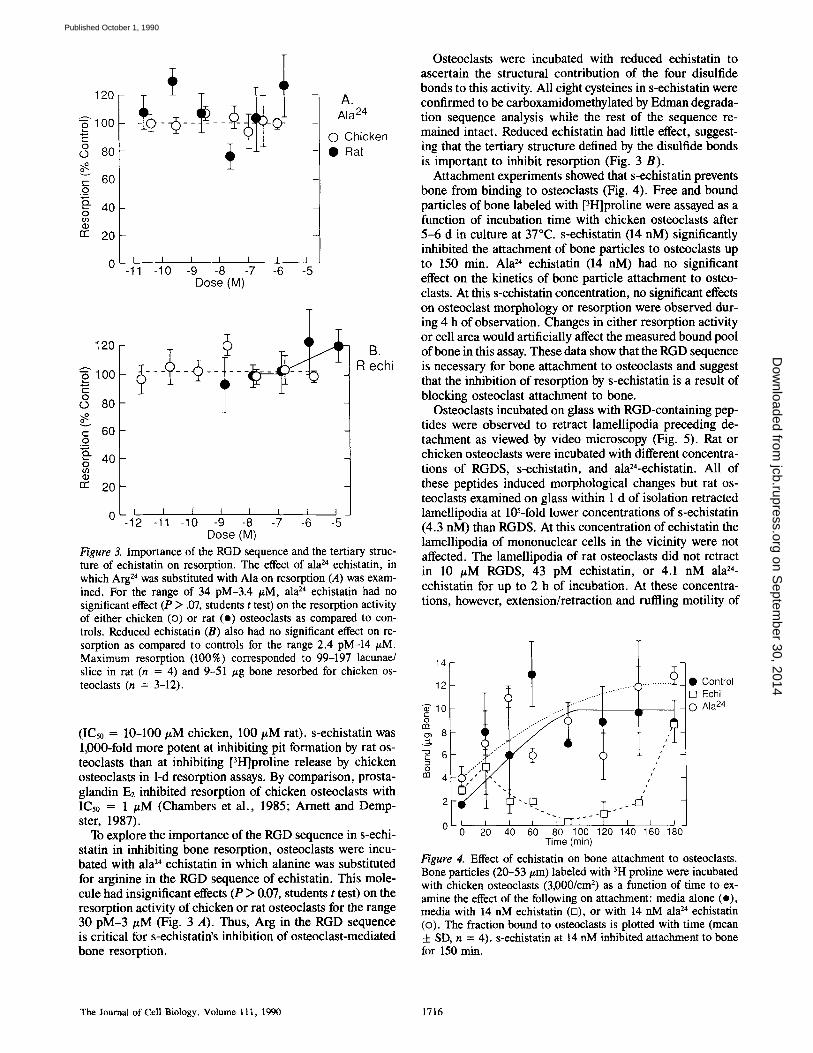
Figure 3. Importance of the RGD sequence and the tertiary struc- ture of echistatin on resorption. The effect of ala 24 echistatin, in which Arg 24 was substituted with Ala on resorption (A) was exam- ined. For the range of 34 pM-3.4 #M, ala 24 echistatin had no significant effect (P > .07, students t test) on the resorption activity of either chicken (<3) or rat (o) osteoclasts as compared to con- trois. Reduced echistatin (B) also had no significant effect on re- sorption as compared to controls for the range 2.4 pM-14 #M. Maximum resorption (100%) corresponded to 99-197 lacunae/ slice in rat (n = 4) and 9-51 /zg bone resorbed for chicken os- teoclasts (n = 3-12).
(IC50 = 10-100 #M chicken, 100 #M rat). s-echistatin was 1,000-fold more potent at inhibiting pit formation by rat os- teoclasts than at inhibiting [3H]proline release by chicken osteoclasts in 1-d resorption assays. By comparison, prosta- glandin E2 inhibited resorption of chicken osteoclasts with IC5o = 1 #M (Chambers et al., 1985; Arnett and Demp- ster, 1987).
To explore the importance of the RGD sequence in s-echi- statin in inhibiting bone resorption, osteoclasts were incu- bated with ala 24 echistatin in which alanine was substituted for arginine in the RGD sequence of echistatin. This mole- cule had insignificant effects (P > 0.07, students t test) on the resorption activity of chicken or rat osteoclasts for the range 30 pM-3 #M (Fig. 3 A). Thus, Arg in the RGD sequence is critical for s-echistatin's inhibition of osteoclast-mediated bone resorption.
Osteoclasts were incubated with reduced echistatin to ascertain the structural contribution of the four disulfide bonds to this activity. All eight cysteines in s-echistatin were confirmed to be carboxamidomethylated by Edman degrada- tion sequence analysis while the rest of the sequence re- mained intact. Reduced echistatin had little effect, suggest- ing that the tertiary structure defined by the disulfide bonds is important to inhibit resorption (Fig. 3 B).
Attachment experiments showed that s-echistatin prevents bone from binding to osteoclasts (Fig. 4). Free and bound particles of bone labeled with [3H]proline were assayed as a function of incubation time with chicken osteoclasts after 5 -6 d in culture at 37°C. s-echistatin (14 nM) significantly inhibited the attachment of bone particles to osteoclasts up to 150 min. Ala 24 echistatin (14 nM) had no significant effect on the kinetics of bone particle attachment to osteo- clasts. At this s-echistatin concentration, no significant effects on osteoclast morphology or resorption were observed dur- ing 4 h of observation. Changes in either resorption activity or cell area would artificially affect the measured bound pool of bone in this assay. These data show that the RGD sequence is necessary for bone attachment to osteoclasts and suggest that the inhibition of resorption by s-echistatin is a result of blocking osteoclast attachment to bone.
Osteoclasts incubated on glass with RGD-containing pep- tides were observed to retract lamellipodia preceding de- tachment as viewed by video microscopy (Fig. 5). Rat or chicken osteoclasts were incubated with different concentra- tions of RGDS, s-echistatin, and ala24-echistatin. All of these peptides induced morphological changes but rat os- teoclasts examined on glass within 1 d of isolation retracted lamellipodia at l@-fold lower concentrations of s-echistatin (4.3 nM) than RGDS. At this concentration of echistatin the lamellipodia of mononuclear cells in the vicinity were not affected. The lamellipodia of rat osteoclasts did not retract in 10 #M RGDS, 43 pM echistatin, or 4.1 nM ala 24- echistatin for up to 2 h of incubation. At these concentra- tions, however, extension/retraction and ruffling motility of
141
12
"~'10 o
8
6
rn 4
• Control [ ] Echi © Ala 24
/
0 i i i "I-FTS-- i p 0 20 40 60 80 100 120 140 160 180
Time (rain)
Figure 4. Effect of echistatin on bone attachment to osteoclasts. Bone particles (20-53/zm) labeled with 3H proline were incubated with chicken osteoclasts (3,000/cm 2) as a function of time to ex- amine the effect of the following on attachment: media alone (o), media with 14 nM echistatin (n), or with 14 nM ala 24 echistatin (<3). The fraction bound to osteoclasts is plotted with time (mean ± SD, n = 4). s-echistatin at 14 nM inhibited attachment to bone for 150 rain.
The Journal o f Cell Biology, Volume 111, 1990 1716
on Septem
ber 30, 2014jcb.rupress.org
Dow
nloaded from
Published October 1, 1990
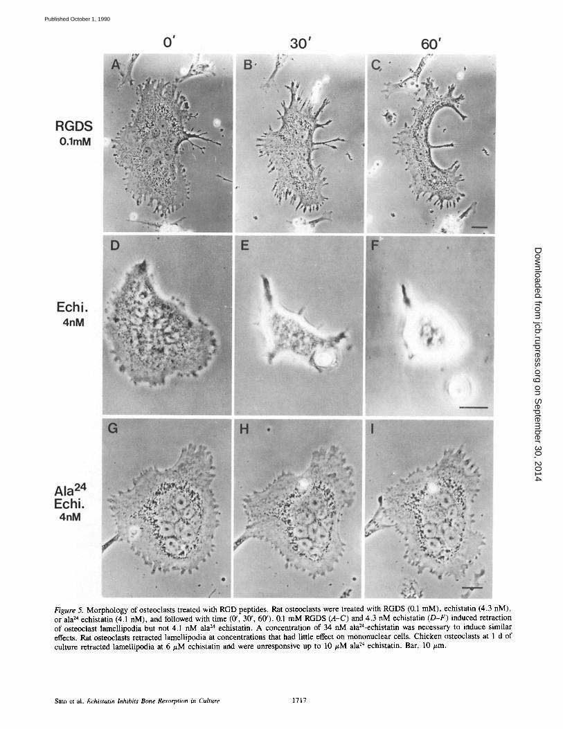
Figure 5. Morphology of osteoclasts treated with RGD peptides. Rat osteoclasts were treated with RGDS (0.1 mM), echistatin (4.3 nM), or ala 24 echistatin (4.1 nM), and followed with time (0', 30', 60'). 0.1 mM RGDS (A-C) and 4.3 nM echistatin (D-F) induced retraction of osteoclast lamellipodia but not 4.1 nM ala 24 echistatin. A concentration of 34 nM ala24-echistatin was necessary to induce similar effects. Rat osteoclasts retracted lamellipodia at concentrations that had little effect on mononuclear cells. Chicken osteoclasts at 1 d of culture retracted lamellipodia at 6/~M echistatin and were unresponsive up to 10 #M ala 24 echistatin. Bar, 10/~m.
Sato et al. Echistatin Inhibits Bone Resorption in Culture 1717
on Septem
ber 30, 2014jcb.rupress.org
Dow
nloaded from
Published October 1, 1990

on Septem
ber 30, 2014jcb.rupress.org
Dow
nloaded from
Published October 1, 1990

Figure 7. Echistatin colocalizes with vinculin at the substrate. Rat osteoclasts were incubated with 0.7 nM echistatin and stained with antisera followed by mAb to vinculin. Echistatin fluorescence of the cell (A) was most intense at the substrate focal plane but some staining of the dorsal membrane was observed. Vinculin staining (B) was also most intense at the substrate. As indicated by the arrowheads, echistatin colocalized with vinculin-containing structures. Note that only rat osteoclasts stained for echistatin and vinculin (A) as compared to mononuclear cells which are identified by vinculin staining (*, B). Bar, 10 #m.
the lamellipodia increased relative to controls. At 34 nM Ala 24 echistatin, lamellipodia retraction did occur. Chicken osteoclasts were observed 1 d after isolation retracted lamel- lipodia and eventually detached in response to similar con- centrations of RGDS peptide (0.1 mM) but required 103 x higher concentrations of echistatin (6/~M) compared to rat osteoclasts (Fig. 5).
Immunofluorescence with antisera to echistatin showed that more s-echistatin binds to rat osteoclasts than to nearby mononuclear cells or to chicken osteoclasts (Fig. 6). Parallel exposures of rat and chicken osteoclasts cultured for 1 d and incubated with s-echistatin (7 nM) showed that rat osteo- clasts with four nuclei were 6.4-fold brighter than similar chicken osteoclasts by the equation (fluorescence = 1/expo- sure time -1/background). At higher concentrations of up to 10 #M s-echistatin, the immunofluorescence of chicken os- teoclasts approached that of rat osteoclasts at 7 nM. These pharmacological and immunohistochemical data suggest that rat osteoclasts have higher affinity for echistatin than do mononuclear cells or chicken osteoclasts, but that the total number of s-echistatin binding sites may be similar.
Vinculin immunofluorescence colocalizes with the stain-
ing for s-echistatin at the osteoclast/glass interface. Rat os- teoclasts incubated with 0.7 nM echistatin plus antisera and mAb to vinculin showed similar, but not identical, distribu- tions of the antigens when focused at the substrate (Fig. 7, A and B). s-echistatin immunofluorescence was observed all over rat osteoclasts but was most intense at the cell/glass in- terface. The vinculin staining however was localized to spots and a few circles at the substrate, reminiscent of podosome structures shown previously for chicken and rabbit os- teoclasts (Marchisio et al., 1984; Turksen et al., 1988). As depicted with arrowheads, regions of echistatin were ob- served to colocalize with vinculin staining (Fig. 7, A and B). By contrast, rat osteoclasts incubated with 1 nM ala 24- echistatin for 10 min at 37°C showed no detectable staining with echistatin antisera. Fluorescence with excess echistatin (1/~M) added with or after antisera incubation was indistin- guishable from background fluorescence observed with sec- ondary antibody incubation alone. Background fluorescence is seen as a dull haze around the central cluster of nuclei in Fig. 7, A and B.
Optical sectioning by laser scanning confocal microscopy confirmed that echistatin preferentially localized to the
Figure 6. Echistatin binding to osteoclasts, s-echistatin binding to rat and chicken osteoclasts was shown by labeling pretreated cells with rabbit polyclonal antisera to echistatin. After pretreatment with 7 nM s-echistatin for 10 min at 37°C, extensive labeling of rat (A, C, E) but not chicken osteoclasts (B, D, F) occurred. All of these osteoclasts were labeled 1 d after isolation; however, similar images were obtained for chicken osteoclasts after 6 d of culture. Bars, 10 #m.
Sato et al. Echistatin Inhibits Bone Resorption in Culture 1719
on Septem
ber 30, 2014jcb.rupress.org
Dow
nloaded from
Published October 1, 1990

Figure 8. Laser scanning confocal microscopy and confocal interference reflection microscopy of osteoclasts. Chicken osteoclasts were incubated with 7 nM echistatin, stained with antisera (A), and examined (0.5 #m optical section) at the substrate. Optical sectioning of the entire cell confirmed that echistatin receptors were preferentially localized to the substrate as no fluorescence was detected 2.5 ttm into the cell. Comparison with mononuclear cells (B-D) near the osteoclast showed that chicken osteoclasts stain more intensely for echista- tin, even when corrected for path length. Interference patterns for the same cell are shown at 458 nm (B) and 514 nm (C). Numerous grey contact regions (spots and some circles) are seen throughout the optical section. Clusters of these grey contacts were seen at the periphery of some lamellipodia. The complex interference pattern suggests an undulating membrane surface beneath the osteoclast with much of the cell lifted off the substrate. The pattern of grey contacts was similar for the two wavelengths as seen by combining the 458- and 514-nm images (D) (see Bereiter-Hahn et al., 1979). This shows that reflection off of additional structures from within the osteoclast contributed little to this interference pattern. Bar, 10/zm.
chicken osteoclast/glass interface (Fig. 8 A). The confocal microscope focused at this interface showed echistatin stain- ing as spots scattered across the optical section and in clustered regions of the periphery of the lamellipodia.
The measured intensity for a fluorescent point object un- der the confocal imaging conditions for epifluorescence fell to half of its maximum value within 0.4 #m on either side of focus (Wilson, 1989). Thus, Fig. 8 A includes predomi- nantly the regions of the cell within •0.5 #m of the glass in- terface, with very weak contributions from material outside this region. When the focus was set to a depth of 2.5/~m away from the glass interface, very little echistatin staining is ob- served. Mononuclear cells in rat (Fig. 7 A) and chicken cul- tures (Fig. 8 A) showed comparatively little staining at any depth in the cell.
Confocal interference reflection microscopy showed that osteoclasts attach to glass via small regions scattered across the cell surface while most of the cell is lifted off the sub- strate (Fig. 8, B-D). Spots and circles of grey contacts were
observed across the cell/glass interface, with clusters of close contacts at the tips of some lamellipodia.
For interference reflectance confocal images, the mea- sured intensity declined to half maximum within 0.25 #m of focus. Thus, the dark interference bands recorded in our in- terference contrast images would be expected to be exclu- sively from zero order cancellations. To confirm this, we compared the interference reflectance image recorded using the wavelengths 458 nm (Fig. 8 B) and 514 nm (Fig. 8 C) as described by Bereiter-Hahn et al. (1979). As seen by com- bining the two images (Fig. 8 D), the differences between the two distributions of grey contacts were minor, which shows that reflections off of deeper structures from within the os- teoclast contributed little to this image.
The complex interference pattern suggested a convoluted membrane border beneath the osteoclasts. Both of these characteristics were previously described for highly motile cells (Bailey and Gingell, 1988; Turksen et al., 1988). Fig. 8, A and D were superpositioned to show that echistatin
The Journal of Cell Biology, Volume 111, 1990 1720
on Septem
ber 30, 2014jcb.rupress.org
Dow
nloaded from
Published October 1, 1990

Figure 9. Echistatin receptors colocalize to attachment complexes at the sub- strate. Superpositioning of Fig. 8 A (yellow) onto 8 D (purple) shows that echistatin receptors colocalize to the outer edges of clusters of grey contacts found toward the tips of some lamellipo- dia. Fainter additional spots of fluores- cence (yellow) are seen underneath the cell, some but not all of which colocal- ize with grey contacts. Bar, 10 t~m.
localized to the outer areas of clusters of grey contacts ob- served in the peripheral lamellipodia (Fig. 9). s-echistatin receptors, therefore, are concentrated in contacts formed by osteoclasts with the substrate.
Receptors for s-echistatin were identified as an integrin by double labeling chicken osteoclasts (Fig. 10). Chicken os- teoclasts showed abundant staining for 23C6 (Fig. 10 A), an mAb to the cry subunit, and lesser staining for echistatin (7 nM, Fig. 10 B). Superpositioning of these micrographs showed that 92.8 % of the s-echistatin fluorescence colocal- ized with 23C6 staining, suggesting that the s-echistatin receptor is an integrin with an otv-like subunit.
D i s c u s s i o n
Existing theories of how osteoclasts attach to bone range from integrin binding to matrix proteins in bone to the use of receptors for proteins with affinity for hydroxyapatite such as IgG (Pierce and Lindskog, 1986). Immunofluorescence and interference reflection microscopy showed that os- teoclasts organize podosome attachment complexes into a circular band on glass substrates (Marchisio et al., 1984; Turksen et al., 1988; Zambonin-Zallone et al., 1989). These are highly dynamic attachment complexes previously de- scribed for transformed cells (Burridge, 1987; Kanehisa and Heersche, 1988; Turksen et al., 1988). However, because osteoclasts do not appear to form ruffled borders on glass or plastic, the complexes that form on artificial substrates may not correlate with the attachment complexes formed at the clear zone during bone resorption.
Recently, a protein named echistatin was purified from the venom of the viper Echis carinatus and shown to inhibit the ADP-induced aggregation of platelets with an IC50 of 30 nM. This peptide contains the Arg-Gly-Asp (RGD) sequence but is 100 × more potent than the tetrapeptide Arg-Gly-Asp- Phe (IC50 = 4-10/~M; Gan et al., 1988). We therefore used s-echistatin to investigate whether the RGD sequence was
critical to osteoclast function, and thereby implicate a role for an RGD-binding protein in bone resorption.
Integrins that recognize the RGD sequence function in bone resorption based on the following evidence, s-echi- statin at 14 nM completely blocked the association of bone with chicken osteoclasts for 150 min and induced the detach- ment of rat osteoclasts from glass at 4 nM. Bone resorption was inhibited by s-echistatin at 0.1 nM for rat osteoclast- mediated resorption and 0.1/~M for chicken osteoclasts. The participation of the RGD sequence and especially Arg 24 in this effect was shown by noting that Ala 24 echistatin had no significant effect on resorption, osteoclast morphology, at- tachment to glass, or the binding of bone particles to os- teoclasts. However, since reduced echistatin had little effect on resorption, the tertiary structure contributes to the effects of echistatin perhaps by holding the RGD in a preferred con- formation.
The identification of the echistatin receptor as an integrin was achieved by observing the colocalization of s-echistatin fluorescence with staining for an otv-like subunit. The above data plus the colocalization of echistatin with vinculin and grey contacts shows that this integrin is associated with func- tional attachment complexes formed by osteoclasts to the substrate. The functional significance of this "echistatin inte- grin" is further supported by noting that 23C6 itself partially inhibited the resorption activity of human tumors of giant cells (osteoclastomas; Chambers et al., 1986).
The identity of the/3 subunit of the echistatin integrin is unknown, but previous data with osteoclastomas suggest a /33-like subunit (Horton, 1988; Zambonin-Zallone et al., 1989; Davies et al., 1989). Our data agrees with the hypoth- esis that a vitronectin-like receptor functions in bone resorp- tion; however, it remains to be shown if additional integrins participate in this process.
The lack of effect of Ala 24 echistatin on bone resorption as compared to inhibition of platelet aggregation (IC50 = 0.5/~M, Garsky et al., 1989) suggests that platelets and os-
Sato et al. Echistatin lnhibits Bone Resorption in Culture 1721
on Septem
ber 30, 2014jcb.rupress.org
Dow
nloaded from
Published October 1, 1990

Figure 10. The echistatin receptor is an integrin. Chicken os- teoclasts were extracted with 0.1% Triton x-100 (5 min, room tem- perature), fixed, and then incubated with antibodies to s-echistatin, followed by 23C6 mAb. This antibody was shown previously to be specific for the c~v integrin subunit (Horton, 1988; Davies et al., 1989). Osteoclasts showed an abundance of staining for 23C6 and lesser fluorescence for s-echistatin. When micrographs were su- perimposed, 92.8 % of the echistatin fluorescence colocalized to 23C6 staining, suggesting that the echistatin receptor is an integrin with an av-like subunit. Bar, 10/zm.
teoclasts have pharmacologically distinct integrins to which s-echistatin binds, s-echistatin appears to compete with fibrinogen for the GP IIb/IIIa complex on human platelets (Garsky et al., 1989). Biochemical characterization of the putative osteoclast integrin(s) to which s-echistatin binds is currently under investigation.
Two differences between the chicken and rat models may explain why rat osteoclasts are more sensitive to s-echistatin as compared to chickens. First, there are more multinucleate cells in the chicken than the rat system. That is, there were 1,000-5,000 multinucleate cells/cm 2 for chickens but 40-
200/cm 2 for rat. Because osteoclasts actively release pro- teases, the s-echistatin may have been degraded faster in the chicken assay. We have found in two of three experiments (not shown) that 1 /~M s-echistatin incubated with chicken osteoclasts for 3 d had insignificant effects on resorption, suggesting that biological activity is lost with time in culture. These data plus the abrupt increase in bone binding to os- teoclasts at 180 min (Fig. 4) both support the degradation hy- pothesis. Second, immunofluorescence intensity demon- strates a greater binding of s-echistatin to rat over chicken osteoclasts and mononuclear cells and suggests this is due to an increased affinity (Fig. 6). Additional experiments are in progress to define whether this enhanced binding is, in fact, a result of increased affinity or increased numbers of receptors.
The correlation between the microscopic observations of lamellipodia retraction and inhibition of bone resorption by s-echistatin demonstrate the importance of the integrity of osteoclast lamellipodia in bone resorption. The data reported here and previous data with cytochalasins (Chambers et al., 1984) show that at least two ways exist to induce retraction of lamellipodia. That is, cell attachment complexes may be destabilized by RGD-containing molecules as shown here or actin filament organization may be disrupted. Since similar concentrations of s-echistatin disrupt osteoclast attachment to glass and bone resorption, similar structures may be used by osteoclasts to adhere to synthetic substrates and bone, as implied previously (Turksen et al., 1988).
In conclusion, we have pharmacologically demonstrated the importance of the arginine-glycine-aspartic acid se- quence in osteoclast-mediated bone resorption and identify an integrin with an ~v-like subunit as critical to this process.
The authors gratefully thank P. Friedman (Merck Sharp & Dohme Re- search Laboratories), G. Rodan (Merck Sharp & Dohme Research Labora- tories), I. Singer (Merck Sharp & Dohme Research Laboratories), and A. Zambonin-Zallone (University of Bari, Italy) for helpful suggestions, and P. Lumma and P. Keller (Merck) for echistatin antisera.
Part of this effort was supported by grants from National Institutes of Health ($10-RR05008) and National Science Foundation (8801209) to J. M. Murray.
Received for publication 20 November 1989 and in revised form 24 May 1990.
References
Arnen, T. R., and D. W. Dempster. 1987. A comparative study of disag- gregated chick and rat osteoclasts in vitro: effects of calcitonin and prostaglandins. Endocrinology. 120:602-608.
Bailey, J., and D. Gingell. 1988. Contacts of chick fibroblasts on glass: results and limitations of quantitative interferometry. J. Cell Sci. 90:215-224.
Baron, R., L. Neff, W. Brown, P. J. Courtoy, D. Louvard, and M. G. Far- quhar. 1988. Polarized secretion of lysosomal enzymes: co-distribution of cation independent mannose- 6-phosphate receptors and lysosomal enzymes along the osteoclast exocytic pathway. J. Cell Biol. 106:1863-1872.
Bereiter-Hahn, J., C. H. Fox, and B. Thorell. 1979. Quantitative reflection con- trast microscopy of living cells. J. Cell Biol. 82:767-779.
Blair, H. C., A. J. Kahn, E. C. Crouch, J. J. Jeffrey, and S. L. Teitelbaum. 1986. Isolated osteoclasts resorb the organic and inorganic components of bone. J. Cell Biol. 102:1164-1172.
Blair, H. C., S. L. Teitelbaum, R. Ghiselli, and S. Gluck. 1989. Osteoclastic bone resorption by a polarized vacuolar proton pump. Science (Wash. DC). 245:855-857.
Burridge, K. 1987. Substrate adhesions in normal and transformed fibroblasts and organization and regulation of cytoskeletal, membrane and extracellular matrix components at focal contacts. Cancer Rev. 4:18-78.
Chambers, T. J., P. A. Revell, K. Fuller, and N. A. Athanasou. 1984. Resorp- tion of bone by isolated rabbit osteoclasts. J. Cell Sci. 66:383-399.
Chambers, T. J., P. M. J. McSheehy, B. M. Thomson, and K. Fuller. 1985.
The Journal of Cell Biology, Volume 111, 1990 1722
on Septem
ber 30, 2014jcb.rupress.org
Dow
nloaded from
Published October 1, 1990
The effect of Ca regulating hormones and prostaglandins on bone resorption by osteoclasts disaggregated from neonatal rabbit bones. Endocrinology. 116:234-239.
Chambers, T. J., K. Fuller, J. A. Darby, J. A. S. Pringle, and M. Horton. 1986. Monoclonal antibodies against osteoclasts inhibit bone resorption in vitro. Bone Miner. 1:127-135.
Cheresh, D. A., J. W. Smith, H. M. Cooper, and V. Quaranta. 1989. A novel vitronectin receptor integrin (~vBO is responsible for distinct adhesive properties of carcinoma cells. Cell. 57:59-69.
Davies, J., J. Warwick, N. Totty, R. Philp, M. Helfrich, and M. Horton. 1989. The osteoclast functional antigen, implicated in the regulation of bone resorption, is biochemically related to the vitronectin receptor. J. Cell Biol. 109:1817-1826.
Dedhar, S., W. S. Argraves, S. Suzuki, E. Ruoslahti, and M. C. Pierschbacher. 1987. Human osteosarcoma ceils resistant to detachment by an RGD contain- ing peptide overproduce the fibronectin receptor. J. Cell Biol. 105:1175- 1182.
Freed, E., J. Gailit, P. van der Geer, E. Ruoslahti, and T. Hunter. 1989. A novel integrin/3 subunit is associated with the vitronectin receptor u subunit (av) in a human osteosarcoma cell line and is a substrate for protein kinase C. EMBO (Eur. Mol. Biol. Organ.) J. 8:2955-2965.
Gan. Z., R. J. Gould, J. W. Jacobs, P. A. Friedman, and M. A. Polokoff. 1988. Echistatin: a potent platelet aggregation inhibitor from the venom of the vi- per, Echis carinatus. J. Biol. Chem. 263:19827-19832.
Garsky, V. M,, P. K. Lumma, R. M. Freidinger, S. M. Pitzenberger, W. C. Randall, D, F, Veber, R. J. Gould, and P. A. Friedman. 1989. Chemical synthesis of echistatin, a potent inhibitor of platelet aggregation from Echis carinatus: synthesis and biological activity of selected analogs. Proc. Natl. Acad. Sci. USA. 86:4022-4026.
Horton, M. A. 1988. Osteoclast-specific antigens. 1SIAtlas Sci. lmmunol. 1: 35-43.
Huang, T.-F., J. C. Holt, E. P. Kirby, and S. Niewiarowski. 1989. Trigramin: primary structure and its inhibition of von Willebrand factor binding to gly- coprotein llb/llIa complex on human platelets. Biochemistry. 28:661-666.
Hynes, R. O. 1987. lntegrins: a family of cell surface receptors. Cell. 48:549-554.
Inoue, S. 1986. Video Microsc. Plenum Press, New York. 423-429. Kajiji, S., R. N. Tamura, and V, Quaranta. 1989. A novel integrin (o~E/34)
from human epithelial cells suggests a fourth family of integrin adhesion receptors. EMBO (Eur. Mol. Biol. Organ.) J. 8:673-680.
Kanehisa, J., and J. N. M. Heersche. 1988. Osteoclastic bone resorption: in vitro analysis of the rate of resorption and migration of individual os- teoclasts. Bone. 9:73-79.
Knudsen, K. A., G. P. Tuzynski, T.-F. Huang, and S. Niewiarowski. 1988. Trigramin, an RGD-containing peptide from snake venom, inhibits cell-
substration adhesion of human melanoma cells. Exp. CellRes. 179:42-49. Marchisio, P. C., D. Cirillo, L. Naldini, M. V. Primavera, A. Teti, and A.
Zambonin-Zallone. 1984. Cell-substratum interactions of cultured avian osteoclasts is mediated by specific adhesion structures. J. Cell Biol. 99: 1696-1705.
Oldberg, A., A. Franzen, and D. Heinegard. 1988a. The primary structure of a cell-binding bone sialoprotein. J. Biol. Chem. 263:19430-19432.
Oldberg, A., A. Franzen, D. Heinegard, M. Pierschbacher, and E. Ruoslahti. 1988b. Identification of a bone sialoprotein receptor in osteosarcoma cells. J. Biol. Chem. 263:19433-19436.
Pierce, A. M., and S. Lindskog. 1986. Evidence for capping of Fc receptors on osteoclasts. Calcif Tissue Int. 39:109-116.
Ruoslahti, E., and M. Pierschbacher. 1987. New perspectives in cell adhesion: RGD and integrins. Science (Wash. DC). 238:491--497.
Sato, M., and G. A. Rodan. 1989. Bone cell shape and function. In Cell Shape: Determinants, Regulation and Regulatory Role. W. D. Stein and F. Bronner, editors. Academic Pres, Inc., Orlando, FL. 329-362.
Sato, M., and W. Grasser. 1990. Effects of bisphosphonates on isolated rat os- teoclasts as examined by reflected light microscopy. J. Bone Miner. Res. 5:31-40,
Shebuski, R. J., D. R. Ramjit, G. H. Bencen, and M. A. Polokoff. 1989. Char- acterization and platelet inhibitory activity of bitistatin, a potent arginine- glycine-aspartic acid-containing peptide from the venom of the viper Bitis arietans. J. Biol. Chem. 146:21550-21556.
Taylor, M. L., A. Boyde, and S. J. Jonco. 1989. The effect of fluoride on the patterns of adherence of osteoclasts cultured on and resorbing denture: a 3D assessment of vinculin-labeled cells using confocal optical microscopy. Anat. Embryol. 180:427-435.
Turksen, K., J. Kanehisa, M. Opas, J. N. M. Heersche, and J. E. Aubin. 1988. Adhesion patterns and cytoskeleton of rabbit osteoclasts on bone slices and glass. J. Bone Miner. Res. 3:389~-00.
Vaes, G. 1988. Cellular biology and biochemical mechanism of bone resorp- tion, Clin. Orthop. Relat. Res. 228:239-271.
Wilson, T. 1989. The role of the pinhole in confocal imagine systems. In The Confocal Microscopy Handbook. J. Pawley, editor. National Science Foun- dation. 99-113.
Zambonin-Zallone, A., A. Teti, and M. V. Primavera. 1982. Isolated os- teoclasts in primary culture: first observations on structure and survival in culture media. Anat. Embryol. 165:405-413.
Zambonin-Zallone, A., A. Teti, M. Grano, A. Rubinacci, M. Abbadini, M. Gaboli, and P. C. Marchisio. 1989. Immunocytochemical distribution ofex- tracellular matrix receptors in human osteoclasts: o~3 integrin colocalized with vinculin and talin in the podosome of osteosarcoma giant cells. Exp. Cell Res. 182:645-652.
Sato et al. Eehistatin Inhibits Bone Resorption in Culture 1723
on Septem
ber 30, 2014jcb.rupress.org
Dow
nloaded from
Published October 1, 1990


![Synthesis, Chiral High Performance Liquid Chromatographic Resolution and Enantiospecific Activity of a Potent New Geranylgeranyl Transferase Inhibitor, 2Hydroxy3-imidazo[1,2- a ]pyridin-3-yl-2-phosphonopropionic](https://static.fdokumen.com/doc/165x107/63139c8685333559270c5139/synthesis-chiral-high-performance-liquid-chromatographic-resolution-and-enantiospecific.jpg)