Echistatin is a potent inhibitor of bone resorption in culture
Upload
independentCategory
view
4download
0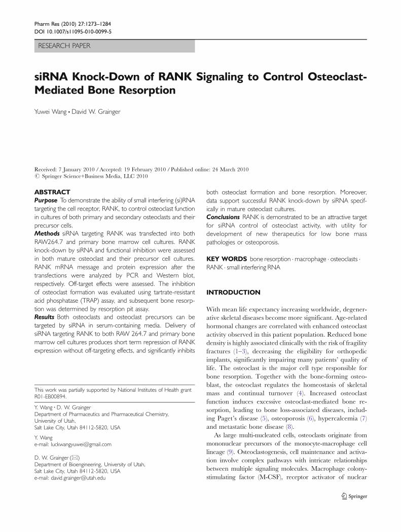
RESEARCH PAPER
siRNA Knock-Down of RANK Signaling to Control Osteoclast-Mediated Bone Resorption
Yuwei Wang & David W. Grainger
Received: 7 January 2010 /Accepted: 19 February 2010 /Published online: 24 March 2010# Springer Science+Business Media, LLC 2010
ABSTRACTPurpose To demonstrate the ability of small interfering (si)RNAtargeting the cell receptor, RANK, to control osteoclast functionin cultures of both primary and secondary osteoclasts and theirprecursor cells.Methods siRNA targeting RANK was transfected into bothRAW264.7 and primary bone marrow cell cultures. RANKknock-down by siRNA and functional inhibition were assessedin both mature osteoclast and their precursor cell cultures.RANK mRNA message and protein expression after thetransfections were analyzed by PCR and Western blot,respectively. Off-target effects were assessed. The inhibitionof osteoclast formation was evaluated using tartrate-resistantacid phosphatase (TRAP) assay, and subsequent bone resorp-tion was determined by resorption pit assay.Results Both osteoclasts and osteoclast precursors can betargeted by siRNA in serum-containing media. Delivery ofsiRNA targeting RANK to both RAW 264.7 and primary bonemarrow cell cultures produces short term repression of RANKexpression without off-targeting effects, and significantly inhibits
both osteoclast formation and bone resorption. Moreover,data support successful RANK knock-down by siRNA specif-ically in mature osteoclast cultures.Conclusions RANK is demonstrated to be an attractive targetfor siRNA control of osteoclast activity, with utility fordevelopment of new therapeutics for low bone masspathologies or osteoporosis.
KEY WORDS bone resorption . macrophage . osteoclasts .RANK . small interfering RNA
INTRODUCTION
With mean life expectancy increasing worldwide, degener-ative skeletal diseases become more significant. Age-relatedhormonal changes are correlated with enhanced osteoclastactivity observed in this patient population. Reduced bonedensity is highly associated clinically with the risk of fragilityfractures (1–3), decreasing the eligibility for orthopedicimplants, significantly impairing many patients’ quality oflife. The osteoclast is the major cell type responsible forbone resorption. Together with the bone-forming osteo-blast, the osteoclast regulates the homeostasis of skeletalmass and continual turnover (4). Increased osteoclastfunction induces excessive osteoclast-mediated bone re-sorption, leading to bone loss-associated diseases, includ-ing Paget’s disease (5), osteoporosis (6), hypercalcemia (7)and metastatic bone disease (8).
As large multi-nucleated cells, osteoclasts originate frommononuclear precursors of the monocyte-macrophage celllineage (9). Osteoclastogenesis, cell maintenance and activa-tion involve complex pathways with intricate relationshipsbetween multiple signaling molecules. Macrophage colony-stimulating factor (M-CSF), receptor activator of nuclear
This work was partially supported by National Institutes of Health grantR01-EB00894.
Y. Wang :D. W. GraingerDepartment of Pharmaceutics and Pharmaceutical Chemistry,University of Utah,Salt Lake City, Utah 84112-5820, USA
Y. Wange-mail: [email protected]
D. W. Grainger (*)Department of Bioengineering, University of Utah,Salt Lake City, Utah 84112-5820, USAe-mail: [email protected]
Pharm Res (2010) 27:1273–1284DOI 10.1007/s11095-010-0099-5

factor κB (RANK), and RANK ligand (RANKL) areknown to be key molecules initiating osteoclast formation.Interaction between M-CSF and its receptor, c-Fms,generates signals for osteoclast precursor cell survivaland proliferation (10). By contrast, osteoclastogenesis ismodulated by positive interactions between RANK andRANKL and negative interactions between RANKL andosteoprotegerin (OPG). RANK is a transmembranesignaling receptor expressed on haematopoietic precursorcells and osteoclasts (11). Interaction of RANKL isrequired for osteoclast formation, activation, and calciumhomeostasis (11,12). It has also been reported thatinteraction of RANK and RANKL increases survival ofthe mature osteoclast in vitro and in vivo (13). RANK signalsthrough the key adapter molecule, TNF receptor-associated factor (TRAF) 6, and RANK cytoplasmicdomains, to regulate formation and activation throughosteoclast-specific gene expression (11). Mice lackingRANK, TRAF 6, or RANKL are deficient in osteoclastsand lack osteoclastogenesis (12,14–16). OPG, a solubleprotein of the TNF receptor family, secreted by osteo-blasts, competitively binds RANKL (11,17), consequentlyacting as a decoy receptor to block osteoclastogenesis andsuppress osteoclast survival (18,19). Thus, positive regula-tor RANKL and negative regulator OPG are normallycoordinated to modulate bone degradation and formationhomeostasis by competitive interactions with RANK.RANK is therefore a central factor in this bone metabolicregulatory pathway.
RNA interference (RNAi) (20) is a relatively recentdevelopment with increasing utility as a sequence-specificpost-transcriptional gene silencing tool (21). Because systemicsiRNA targeting has proven very challenging, local or topicalsiRNA therapeutics have been most actively investigated.Successful delivery approaches include ocular, respiratory,CNS, skin and vaginal sites, where local siRNA deliveryaccesses desired cell target populations directly (22–26). Todate, siRNA targeting of RANK responsible for osteoclastformation and function has not been reported. The purposeof this study is to assess the utility of RNA interference (RNAi)methods to target RANK in regulating osteoclast formationand function in vitro. Reduction of RANK expression usingsiRNA specific to RANK is expected to suppress boneresorption by osteoclasts, ultimately increasing bone densityand potentially preventing bone mass loss. In order todetermine the efficacy of this RANK-targeting siRNA, bothRAW and primary cells were evaluated for RANK messageand protein expression after siRNA delivery. Functionalassessments included inhibition of osteoclast formation byTRAP assay, and functional suppression of osteoclasts bybone resorption pit assay. Results show that osteoclastformation and osteoclast-mediated bone resorption can besignificantly suppressed using siRNAs targeting RANK.
MATERIALS AND METHODS
Immortalized Murine Monocyte-Macrophage CellLine Culture
A subclonal line of murine monocytic pre-osteoclasticRAW264.7 cells, purchased from the American Type CultureCollection (ATCC), was cultured in Dulbecco’s modifiedEagle’s medium (DMEM, Gibco) supplemented with 10%heat-inactivated fetal bovine serum (FBS, Hyclone®, UT) and1% penicillin-streptomycin (Gibco), defined for all cellcultures as “complete media.” To induce osteoclast formationin vitro, RAW cells (passage number controlled to less than10) were cultured on 24-well plates in complete media at thedensity of 4×10⁴ cells per well supplemented with 100 ng/ml of RANKL. Complete media with RANKL was changedevery other day.
Primary Murine Cell Harvest and Differentiation
C57BL/6 male mice (6–8 weeks old, Jackson labs) weremaintained in a specific pathogen-free facility at the Univer-sity of Utah. All procedures were performed as approved bythe Institutional Animal Care and Use Committee ofUniversity of Utah. Bone marrow cells (BMC) were harvestedfrom murine tibias and femurs of C57BL/6 male mice anddifferentiated into osteoclast precursors using previouslydescribed methods (27–29). Briefly, BMC were cultured inα-MEM (Gibco) containing 10% FBS and 1% penicillin-streptomycin overnight at a density of 1×10⁶ cells/ml. Non-adherent cells were harvested the next day and immediatelyseeded into 24-well tissue culture plates in complete mediawith 30 ng/ml M-CSF (R&D Systems) at a density of 1×10⁶
cells per well. After 2 days of culture, attached cells were usedas osteoclast precursors. To generate osteoclasts, precursorcells were incubated in 200 ng/ml RANKL and 30 ng/ml M-CSF (R&D Systems) in complete media, refreshed every otherday. RANKL working concentrations to reliably generateosteoclasts from both RAW264.7 and primary cell cultures inserum media were experimentally determined.
sRANKL Expression
A glutathione-S-transferase (GST)-tagged sRANKL constructwas generated by cloning the murine sRANKL SalI/NotIfragment, coding 470–951 nucleotides, into the plasmidpGEX-4 T-1 (a generous gift of Dr. M. F. Manolson,University of Toronto). The expressed protein was harvestedas previously described (29–31). GST-tagged sRANKLwas purified by Glutathione Sepharose 4B affinity resin(Amersham Pharmacia Biotech), dialyzed against phosphate-
1274 Wang and Grainger

buffered saline (PBS, Gibco) and concentrated using AmiconUltra centrifuge tubes (Millipore). Protein concentration wasdetermined by BCA protein assay kit (Pierce).
siRNA Transfection of Cells
An siGENOME SMARTpool (Dharmacon) containingfour different siRNA sequences, all designed to targetmurine RANK, as well as four pure individual siRNAs (1,sense 5′–GAGCAGAACUGACUCUAUGUU- 3′, antisense5′-CAUAGAGUCAGUUCUGCUCUU-3′; 2, sense5′-GCGCAGACUUCACUCCAUAUU-3′, antisense 5′-UAUGGAGUGAAGUCUGCGCUU-3′; 3, sense 5′-CCAAGGAGGCCCAGGCUUAUU-3′, antisense 5′-UAAGCCUGGGCCUCCUUGGUU-3′; 4, sense 5′-CAAGAAGUGUGUGAAGGUAUU-3′, antisense 5′-UACCUUCACACACUUCUUGUU-3′), and a non-targeting control siRNA(sense: 5′-UAGCGACUAAACACAU CAAUU-3′, antisense:5′-UUAUCGCUGAUUUGUGUAGUU-3′), were pur-chased from Dharmacon. DharmaFECT 4 (DF4, Dharma-con) was used as the cationic lipid cell transfection reagent.RAW cells were seeded at a density of 4×10⁴ cells per well in24-well plates in DMEM at 37°C with 5% CO2 overnight.Transfection with siRNA/DF4 complexes was then carriedout in complete media. Primary BMC were seeded at 1×10⁶
cells per well in 24-well plates in complete media containing30 ng/ml M-CSF for 2 days. Subsequently, siRNA transfec-tion was immediately performed. Transfection reagent DF4and siRNA were prepared according to manufacturer’sinstructions (Dharmacon). Final dosing concentrations of allsiRNAs provided to each well were 125 nM in a total volume of1.0 microliter DF4. Cell uptake of siRNA complexes wasperformed by incubating cells with siRNA complexes incompletemedia at 37°Cwith 5%CO2. In osteoclast formationand pit formation assays, cells were transfected by siRNAcomplexes in complete media with 100 ng/ml RANKL(RAW cells) or 30 ng/ml M-CSF and 200 ng/ml RANKL(BMC). Non-specific knock-down of DF4 was assessed byusing non-targeting siRNA dosed under identical conditions.Multiple cell transfections were carried out identically eachday over the successive first 3 days, or on alternating days, asspecified in each figure. In the case of transfection of osteoclastcultures, RAW cells and primary BMC were seeded in 24-wellplates and treated as mentioned above to generate osteoclasts.Mature osteoclasts were purified essentially as describedelsewhere (28, 32) by gently washing with PBS withoutCa2+ and Mg2+ (Gibco). By tapping the plate, most mono-nuclear cells were detached, while multinucleated osteoclastsremained on the plate. Osteoclasts were transfected byincubating with siRNA complexes prepared as above incomplete media containing 100 ng/ml RANKL (RAW cells)or 200 ng/ml RANKL and 30 ng/ml M-CSF (BMC).
Reverse Transcriptase-Polymerase Chain Reaction(RT-PCR)
Total RNA was isolated 48 h after siRNA transfectionusing an RNeasy Mini Kit (Qiagen). Up to 4 microgramsof RNA were used to make cDNA with the SuperScript III1st strand RT kit for PCR (Invitrogen). PCR primers weredesigned for RANK (5′-AGATGTGGTCTGCAGCTCTTCCAT-3′, 5′-ACACACTTCTTGCTGACTGGAGGT-3′) and cyclophilin B (housekeeping control, 5′- AGCGCTTCCCAGATGAGAACTTCA-3′, 5′-GCAATGGCAAAGGGTTTCTCCACT-3′) using Primerquest soft-ware purchased from Integrated DNA Technologies(IDT). PCR was performed with iTaq DNA polymerase(Bio-Rad), 1.5 mM magnesium chloride, 200 μM each ofdNTPs, 500 nM of each primer, and 2 μL of the cDNA.Reactions were performed using the following protocol:95°C melt, 60°C anneal and 72°C extension in theiCycler Thermal cycler (Bio-Rad). PCR products wereanalyzed on ethidium bromide-stained TBE-based 2%agarose gels run at 100 V for 30 min and visualizedwith UV light.
Real-Time Quantitative PCR (qPCR) Analysis
cDNA was prepared as described above. Primers forRANK (5′-TAGGACGTCAGGCCAAAGGACAAA- 3′,5′-AGGGCCTACTGCCTAAGTGTGTTT-3′, Probe:56 -FAM/TGAAGGTGCCAGGGAAATTCAAGAAAGA/36-TAMSp) and cyclophilin B (5′-TCCGGCAAGATCGAAGTGGAGAAA-3′, 5′-AACCTT GTGACTGGCTACCTTCGT-3′, Probe: 56-FAM/TCATCCCTCTAAGCAGCTGTCTGTGT/36-TAMSp) weredesigned using Primerquest software and purchased fromIDT. Experiments were performed using 7900HT SequenceDetection System and data were analyzed using SDS RQmanager (Applied Biosystems).
RANK Western Immunoblot Assay
Cells were lysed in RIPA buffer (Sigma) supplied with 1×protease inhibitor cocktail (Pierce) and 1 mM phenyl-methylsulfonyl fluoride (Sigma) (32). Insoluble material wasremoved by centrifuging at 15,000 rpm at 4°C for 5 minafter 20 min on ice. Protein concentration was measuredwith a BCA protein assay kit (Pierce). Heat-denaturedsamples were separated on 4–12% SDS-polyacrylamidegels (Invitrogen) and blotted on PVDF filters (Bio-Rad).After blocking with 5% (w/v) dry milk in 0.5% Tween 20 inTBS (TBST), the filter was incubated overnight in primaryantibody against RANK (BAF692; R&D systems) in 5%
Inhibiting Osteoclast Bone Resorption Using siRNA 1275

BSA/TBST with constant shaking. After three washes withTBST, the membrane was incubated with streptravidin-HRP (RPN 1231; Amersham Pharmacia Biotech Ltd.). Thehousekeeping control was detected with antibody againstcyclophilin B (PA1-027; Affinity BioReagents) and HRP-conjugated donkey anti-rabbit antibody (SA1-200, AffinityBioReagents). Secondary antibodies were detected withchemiluminescence reagent (Santa Cruz Biotechnology),and band images were captured using a Molecular ImagerGel Doc XR System (Bio-Rad).
Tartrate-Resistant Acid Phosphatase (TRAP) Assay
Cells were stained for TRAP using a leukocyte acid phos-phatase kit (Sigma) according to the instructions. Osteoclastsstaining positive with at least three nuclei were counted asTRAP-positive cells.
Bone Resorption Pit Assay
Bovine bone was sawed into 0.2–0.3 mm thick slices (agift from Dr. S. Miller, University of Utah) and washedas described (29) before placing small pieces into 24-wellplates. A total of 4×10⁴ RAW cells were plated andcultured on bone slices in DMEM for 12 h, and then cellswere transfected with siRNA in complete media with100 ng/ml RANKL (Day 1). BMC were plated on boneslices at a density of 1×10⁶ per well in 30 ng/ml M-CSF complete media for 2 days. Thereafter, siRNAtransfection was performed in 200 ng/ml RANKL and30 ng/ml M-CSF-containing complete media (Day 1).The media was changed every other day. To observeosteoclast-generated bone resorption, slices were stainedusing a previously described method (29). Pit numbers perframe were counted from random fields under microscopicobservation.
Cell Imaging
Live adherent cells, TRAP-stained images and pit imageswere photographed using a Nikon Eclipse TE 2000-Umicroscope, with Photometrics Coolsnap ES camera (grayscale, Roper Scientific) or QImagine RETIGA EXi color12-bit camera (color, Canda), using Metamorph™ software(Molecular Devices) or QCapture™ software (QImaging).An average of 15 frames per well were taken randomly forTRAP assay analysis, and 10 frames were acquiredrandomly from each bone slice for pit formation analysis(except for triple siRNA transfection sequences in primaryBMC, where 5 frames were acquired).
Statistical Analysis
Analysis of variance (ANOVA) followed by two-tailedstudent’s t-test was used for statistical analysis. All experi-ments were repeated three times. Error bars represent thestandard error of the mean. Results were consideredstatistically significant if p<0.05.
RESULTS
Optimization of siRNA Transfection in Culturesof RAW Cells
DharmaFECT 4 (DF4) was chosen as a siRNA transfec-tion reagent since it was appropriate for mouse and ratcell lines based on the manufacturer’s instructions. Trans-fection conditions in serum-containing media were opti-mized using a commercial murine-targeted siRNA pool(SMARTpool). Each siRNA sequence was then testedindividually for their effectiveness to knock down RANKexpression in cells in complete media. While qPCR resultsfor sample siRNA-1 and -2 RANK knock-down werestatistically indistinguishable, siRNA-2 was selected forfurther study since it produced the greatest overall knock-down effect (Fig. 1a, p=0.009). Non-specific knock-down ofRANK by DF4 evaluated using non-targeting siRNA andDF4 demonstrates little effect by PCR analysis, shown inFig. 1b. Similarly, treatment of mature osteoclasts withRANK siRNA-2 also significantly suppressed RANKexpression in cultures compared with untreated osteoclasts(Fig. 1c). Additionally, in the presence of RANKL, smallosteoclasts continued to fuse into larger ones in controlcultures (Fig. 1d), while in RANK siRNA-treated cultures(Fig. 1e), the speed of cell fusion was suppressed aftertreatment, and the osteoclast size was smaller than those incontrols.
Effects of Multiple Serial siRNA Transfectionson RANK Expression in RAW Cells
RANK expression knock-down by multiple serial RANKand non-targeting siRNA transfections of RAW cells weretested using qPCR analysis (Fig. 1f). Serial transfectionswere performed every other day. mRNA was extracted onDay 3 (single siRNA transfection on Day 1), Day 5 (doublesiRNA transfections on Day 1 and 3), Day 7 (three siRNAtransfections on Day 1, 3, and 5) and Day 9 (four siRNAtransfections on Day 1, 3, 5, and 7), respectively. Comparedto control (no treatment) groups, RANK siRNA-2 signifi-cantly suppressed RANK expression in all four situations(p<0.001). There was no significant difference between thecontrol groups and the non-targeting siRNA groups on
1276 Wang and Grainger
RANK expression for one (p=0.2), two (p=0.49), three(p=0.23), or four (p=0.51) serial transfections, indicatingthat DF4 had no significant effect on expression of RANKin the absence of specific siRNA. In addition, comparedwith non-targeting siRNA transfections, the RANK siRNA-2 groups showed significant reduction of RANK expres-sion for all four dosing situations: one (p=0.00163), two(p=0.0007), three (p=0.0056), and four (p=0.0049) serialsiRNA transfections (p<0.01).
Effects of RANK siRNA on Protein Expressionin RAW Cells
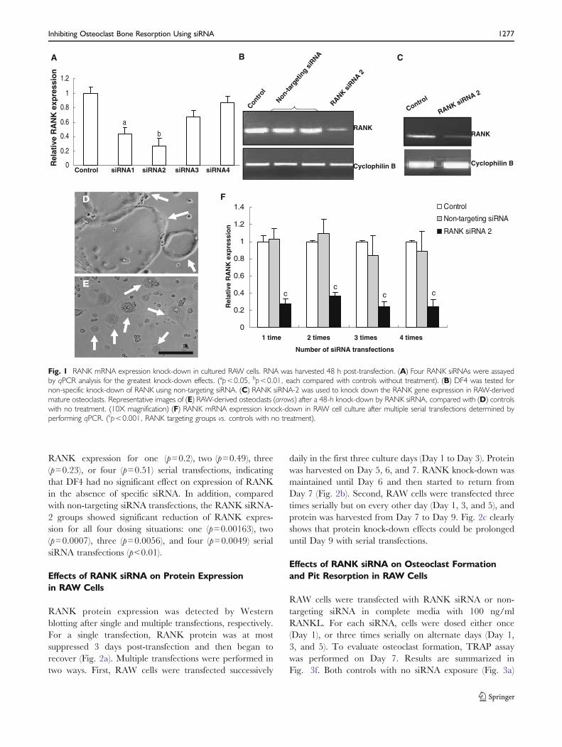
RANK protein expression was detected by Westernblotting after single and multiple transfections, respectively.For a single transfection, RANK protein was at mostsuppressed 3 days post-transfection and then began torecover (Fig. 2a). Multiple transfections were performed intwo ways. First, RAW cells were transfected successively
daily in the first three culture days (Day 1 to Day 3). Proteinwas harvested on Day 5, 6, and 7. RANK knock-down wasmaintained until Day 6 and then started to return fromDay 7 (Fig. 2b). Second, RAW cells were transfected threetimes serially but on every other day (Day 1, 3, and 5), andprotein was harvested from Day 7 to Day 9. Fig. 2c clearlyshows that protein knock-down effects could be prolongeduntil Day 9 with serial transfections.
Effects of RANK siRNA on Osteoclast Formationand Pit Resorption in RAW Cells
RAW cells were transfected with RANK siRNA or non-targeting siRNA in complete media with 100 ng/mlRANKL. For each siRNA, cells were dosed either once(Day 1), or three times serially on alternate days (Day 1,3, and 5). To evaluate osteoclast formation, TRAP assaywas performed on Day 7. Results are summarized inFig. 3f. Both controls with no siRNA exposure (Fig. 3a)
Non-ta
rget
ing
siRNA
Contro
l
RANKsi
RNA2
RANK
Cyclophilin B
DD
EE
A C
F
0
0.2
0.4
0.6
0.8
1
1.2
a
b
0
0.2
0.4
0.6
0.8
1
1.2
1.4
cc c c
Control
RANK
Cyclophilin B
RANK siRNA 2
Control siRNA1 siRNA2 siRNA3 siRNA4
Rel
ativ
e R
AN
K e
xpre
ssio
n
1 time 2 times 3 times 4 times
Number of siRNA transfections
Rel
ativ
e R
AN
K e
xpre
ssio
n
Control
Non-targeting siRNA
RANK siRNA 2
B
Fig. 1 RANK mRNA expression knock-down in cultured RAW cells. RNA was harvested 48 h post-transfection. (A) Four RANK siRNAs were assayedby qPCR analysis for the greatest knock-down effects. (ap<0.05, bp<0.01, each compared with controls without treatment). (B) DF4 was tested fornon-specific knock-down of RANK using non-targeting siRNA. (C) RANK siRNA-2 was used to knock down the RANK gene expression in RAW-derivedmature osteoclasts. Representative images of (E) RAW-derived osteoclasts (arrows) after a 48-h knock-down by RANK siRNA, compared with (D) controlswith no treatment. (10X magnification) (F) RANK mRNA expression knock-down in RAW cell culture after multiple serial transfections determined byperforming qPCR. (cp<0.001, RANK targeting groups vs. controls with no treatment).
Inhibiting Osteoclast Bone Resorption Using siRNA 1277

and cells treated with non-targeting siRNA (single transfec-tion, Fig. 3b; three transfections, Fig. 3c) showed stronglyTRAP-positive multi-nucleate giant cells. Compared tocontrols, both single (Fig. 3d, p=0.007) and three-dosetransfections with RANK siRNA (Fig. 3e, p=0.00015)showed significant reductions in numbers of TRAP-positive multi-nucleate cells in culture. There was nosignificant difference in the number of osteoclasts betweencontrols and the single non-targeting siRNA transfectiongroups (p=0.17). Although three serial non-targeting trans-fections exert some influence on osteoclast formation bythe transfection reagent (p=0.042), comparisons betweenthree serial transfections of non-targeting versus RANKsiRNA show that osteoclast numbers were significantlyreduced in the RANK siRNA-treated groups (p<0.001).The same trend exists for comparisons between single
transfections of non-targeting versus RANK targetingsiRNAs (p=0.017). Three-dose RANK siRNA transfectiongroups showed reduced osteoclast numbers compared withsingle-dose groups, but not significantly different (p=0.13).Resorption pit assay was performed 6 days post-singletransfection. Formation of resorption pits on bone slices wasmarkedly curtailed in the siRNA-treated groups (Fig. 3g,p=0.02) compared to controls (no treatment) that displayedmany pits formed by osteoclasts.
Effects of siRNA on RANK mRNA and ProteinExpression in Primary BMC Cultures
To ensure that RANK siRNA-2 retained the most powerfulknock-down effects, differences in siRNA knock-down forthe 4 different RANK siRNAs were tested on primaryBMC cultures in serum media. RT-PCR results clearlydemonstrated that RANK siRNA-2 was the most efficientat down-regulating BMC RANK mRNA expression(Fig. 4a). Therefore, RANK siRNA-2 was used forsubsequent transfections in primary BMC. Consistently,RANK protein production was reduced both by singlesiRNA transfection (Fig. 4b) and three serial transfectionson alternate days (Fig. 4c). Furthermore, RANK siRNAtransfection was also able to suppress RANK messageexpression in primary BMC-induced mature osteoclasts.PCR products showed significantly reduced RANK mRNAexpression in osteoclast cultures after single transfection(Fig. 4d), as well as reduced RANK protein expression byWestern blot assay (Fig. 4e). Similarly to RAW cell cultures,BMC-induced osteoclasts had reduced rates of cell fusionafter transfection (Fig. 4g) compared to large osteoclastsformed in control cultures (Fig. 4f).
Effects of RANK siRNA on Osteoclast Formationand Activity in Primary BMC Cultures
TRAP assays were performed on Day 5 cell cultures forsingle siRNA transfections. Compared with controls(Fig. 5b), significant reductions in numbers of TRAP-positive multi-nucleate giant cells post-transfection(Fig. 5c, p=0.001) were observed. Differences in numbersof TRAP-positive cells are summarized in Fig. 5a. Primaryosteoclast precursor cells were cultured on bovine boneslices to assess effects of siRNA on osteoclast function.Resorption pit assay was performed on Day 5 for singletransfections. Controls without siRNA treatment producedabundant resorption pits on bone slices as detected bymicroscopy (4X magnification). RANK siRNA treatmentinhibited osteoclastic bone resorption with significantreductions in resorption pit numbers (Fig. 5d, p=0.04).
A
B
C
treated treatedcontrol
3 days later
RANK
Cyclophilin B
control
4 days later
treatedcontrol
5 days later
control
treated control
treated treatedcontrol
Day 5 Day 6 Day 7
RANK
Cyclophilin B
control
Day 7
Day 8
Day 9
RANK
Cyclophilin B
Fig. 2 Western blot analysis of RANK protein knock-down in RAW cellcultures. Western blotting analyzed 30 μg of cell lysate per sample. (A)Protein was harvested at 3, 4, and 5 days after single RANK siRNA-2transfection. (B) RANK protein expression after three successive RANKsiRNA-2 transfections. Protein was harvested on Day 5, 6, and 7. (C)RANK protein expression after three serial RANK siRNA-2 transfectionson Day 1, 3, and 5. Protein was harvested on Day 7, 8, and 9.
1278 Wang and Grainger

DISCUSSION
We provide evidence that siRNA delivered to cells inserum-containing media can successfully and specificallyinhibit RANK expression both in osteoclast precursors andmature osteoclasts from RAW secondary and primaryBMC. This results in suppression of cell-based boneresorption mechanisms by reducing the number andactivity of osteoclasts in cultures.
RANK plays an essential role in regulating osteoclasto-genesis (33). Activation of RANK by its ligand, RANKL, isrequired for the formation and activation of osteoclasts
(12,15). Similar to RANK, OPG can also bind RANKL toact as a competitive inhibitor by blocking RANK interaction(17,34,35). Since the elucidation of the RANK/RANKL/OPG signaling pathways, RANKL and OPG have beenactively investigated as therapeutic targets. Several OPG-based approaches to regulate bone mass and reduce boneresorption have been reported in animal models (17,18,36)and in a human trial (37). However, reliable delivery of alarge protein therapeutic poses several challenges, includinglimited stability, relevant dosing and bioavailability (shorthalf-life), possible host immune responses to recombinantproducts, patient compliance (parenteral requirements) and
0
5
10
15
20
25
Control non-targetsiRNA singletransfection
non-targetsiRNA threetransfections
RANK siRNA-2 single
transfection
RANK siRNA-2 three
transfections
Nu
mb
er o
f T
RA
P-p
osi
tive
b
c
A C
D
F
E
F
0
5
10
15
20
control RANK siRNA 2single transfection
p
a
G
Pit
s n
um
ber
s p
er f
ram
e
B
cells
per
fra
me
Fig. 3 Effects of RANK siRNA and non-targeting siRNA on osteoclast formation determined by TRAP assay in RAW cell cultures. Cells were TRAP-stained and counted 7 days post-transfection. Cultures were performed in the presence of RANKL (A) with no siRNA treatment for positive controls, (B)transfected with non-targeting siRNA once on Day 1, (C) transfected with non-targeting siRNA three times serially every other day, (D) transfected withRANK siRNA once on Day 1, and (E) three times serially every other day. (F) Comparison of the number of TRAP-positive multi-nucleate cells formedafter siRNA dosing. (bp<0.01, cp<0.001, RANK siRNA-2 single and three transfection groups vs. controls). (scale bar=250 μm) Inhibition of osteoclast-mediate bone resorption by RANK siRNA was evaluated by pit formation assay. (G) Difference of resorption pit numbers per frame (10X magnification)between RANK siRNA untreated and treated RAW cells (ap<0.05).
Inhibiting Osteoclast Bone Resorption Using siRNA 1279

complex formulating issues for sustained protein release(38–40). A fully human monoclonal antibody targetingRANKL, denosumab, is currently investigated as a subcuta-neous injection in late-stage clinical development for bonemetabolic therapies (41–43). Possible safety issues of thisanti-RANKL antibody include cross-reactions with OPG orRANK-activated endogenous antibodies. Therefore, othertherapeutic options are necessary. RNAi is an alternativeapproach to RANK and RANKL control in this samecontext. In this approach, siRNA molecules silence geneexpression in a sequence-specific manner by causing degra-dation of corresponding endogenous mRNA (20,44,45).In fact, complete absence of either RANKL or RANK(as shown in RANKL and RANK knock-out mice) elimi-nates RANKL-RANK signaling between osteoblasts andimmature osteoclasts (12,15). This depletes functioning
mature osteoclasts, removes intrinsic bone deposition controlmechanisms and eventually causes osteopetrosis in theseknock-out mutants. Therefore, due to these concerns as wellas other side effects (i.e., targeting undesired cell populationsexpressing RANK and general non-specific targeting), site-and temporally selective, as well as reversible control overRANK-RANKL activity, rather than its complete, irrevers-ible abolition, is considered more appealing for developingnew clinical approaches to osteoporosis therapies. In thisstudy, siRNA targeting RANK was transfected into serum-based secondary macrophage cultures, primary monocyte-macrophage cultures, and culture-generated osteoclasts toobserve RANK mRNA knock-down and suppression ofprotein expression. While RNAi has noted issues withmammalian cell delivery efficiency and specific targeting(46), one advantage of siRNA in the context of RANK is
Con
trol
1# R
ANK siR
N2#
RAN
K s
iRNA
3# R
ANK
siR
NA4#
RA
NK
siR
NA
RANK
Cyclophilin B
FF GG
A
C E
control treated
RANK
Cyclophilin B
Control
Day 7
Day 8
Day 9
RANK
Cyclophilin B
Control
treat
ed
RANK
Cyclophilin B
RANK
Cyclophilin B
Control
treated
B
D
Fig. 4 RANK knock-down by RANK siRNA dosing to primary BMC and BMC-derived osteoclasts in serum-based culture. RNA was harvested 48 hourspost-transfection. Protein was harvested 3 days post-transfection, except from the mature osteoclasts, harvested at 48 h post-transfection. Westernblotting analyzed 30 μg of cell lysate per sample. (A) Four RANK siRNAs were analyzed by RT-PCR for the greatest knock-down effect. (B) Lysatesanalyzed by Western blot for RANK protein expression suppression by a single dose of RANK siRNA. (C) RANK protein expression after three serialsiRNA transfections on every other day. Protein was harvested on Day 7, 8, and 9. Inhibition of target RANK gene expression in primary cellinducedosteoclasts at the mRNA and protein levels as shown by (D) PCR, and (E) Western blot. Representative images of primary BMC-derived osteoclasts(arrows) after 48-h knock-down by (G) RANK siRNA compared with (F) controls. (10X magnification).
1280 Wang and Grainger

the transient gene knock-down experienced (5∼6 days,depending on the target, cell type, and frequency of targetprotein expression typical to that cell) with relatively smallamounts of siRNA dosed. This provides an alternativecontrol feature for RANK suppression in cells compared tomutants completely lacking RANK or protein-based sys-temic antagonism approaches. Additionally, clinical osteo-porosis bone augmentation approaches (47,48) permit localplacement of siRNA-releasing carriers directly into osteo-porotic sites by injection, permitting renewable, localsiRNA delivery to bone RANK at these sites, and avoidingsystemic delivery issues.
As osteoclasts central to both bone metabolic control andRANK presentation derive from fusions of haematopoieti-cally sourced circulating cells and tissue-resident differenti-ated macrophages, assessing and comparing the utility ofsiRNA knock-down effects in both secondary and primarymacrophage cultures was considered an important mile-stone. Immortalized commercial RAW264.7 monocyte-macrophages are commonly cultured as macrophagesurrogates, despite the general lack of reporting ofphenotypic indicators or fidelity to primary macrophagesand their relatively high rates of contamination fromcommercial sources (49). Some limitations with the accura-cy of RAW cell comparisons to primary macrophages inmodel assays have already been noted (27,50). Aggressiveendocytosis and proliferation rates in RAWs, characteristic
of tumor-derived cell lines, as well as higher frequency andintensity of both gene up-regulation and protein expression,may all alter the intensity and duration of siRNA effects(51). Nevertheless, as RAW264.7 cells have been frequentlyused for in vitro generation of osteoclasts (32,52–54), thisstudy compared RAW cells to primary BMC for effects ofsiRNA on osteoclast behavior.
RANK siRNA-2 provides the highest knock-down of theRANK mRNA for both RAW and primary BMC cultures.A dose of 125 nM RANK siRNA and the commercialcationic lipid transfection reagent, DF4, demonstratedsuccessful inhibition of the target RANK gene expressionat the mRNA level 48 h after transfection in serum-basedculture conditions. In addition, evidence indicates thatRANK message knock-down is not caused by the transfec-tion reagent, as there was no significant reduction ofRANK mRNA expression in cells transfected with non-targeting siRNA/DF4 complexes. Moreover, the accumu-lating effects of multiple serial transfections were evaluatedin RAW cells, showing sustained knock-down effects inserum-containing culture media. A likely explanation forthe similar RANK knock-down effects observed with thethree and four transfection cycles could be the increasingcell numbers by the third and fourth transfection cyclesdiluting effects of constant siRNA dosing.
Protein expression analysis of both cell cultures furtherconfirmed RANK knock-down using siRNA. In RAW cell
0
5
10
15
20
Pit
s fo
rmed
per
fra
me
a
0
5
10
15
20
25
control RANK siRNA 2 singletransfection
bNu
mb
er o
f T
RA
P-p
osi
tive
ce
lls p
er f
ram
e
A
D C
B
RANK siRNA 2 single transfection
control
Fig. 5 Inhibition of primary cell-induced osteoclast formation andactivity by RANK siRNA. (A)Inhibition of osteoclast formationby a single RANK siRNA dose(Day 1). TRAP assay was per-formed on Day 5 (bp<0.01).Resorption pit assay was per-formed on same day. Represen-tative image of TRAP-stained cellsin (B) control groups with notreatment, and (C) treated groupswith single siRNA dose. (D)Comparison of average pit num-ber for cell-treated bovine boneslices per frame (4X magnification)between controls (no treatment)and single dose of RANK siRNA-2(ap<0.05). (scale bar=250 μm).
Inhibiting Osteoclast Bone Resorption Using siRNA 1281

cultures, with single transfections, RANK protein levelswere suppressed after 3 days in culture. Subsequently,RANK protein production began to recover, due possiblyto rapid cell proliferation and dilution of siRNA withincells. Multiple transfections (i.e., 3 serial doses) wereperformed, successively in the first 3 days or serially everyother day throughout the culture period. In the latter case,RANK protein expression can be suppressed until Day 9.The same prolonged protein suppression was observed inBMC as well, suggesting that changing the frequency ofmultiple siRNA transfections can extend protein knock-down. Therefore, serial siRNA transfections performedevery other day was considered more efficective and henceused for multiple transfections in subsequent assays.
Since RANK also resides on mature osteoclasts andpositively regulates osteoclast activation and survival, it wasalso important to target RANK on mature osteoclasts. Wefound that mature osteoclasts induced from both RAW andprimary BMC cultures can be successfully transfected bysiRNA, producing significant reductions in both RANKmessage and protein compared with untreated controls. Insupport of this, we found that in both secondary andprimary cultures, after osteoclasts were transfected byRANK siRNA, further fusion was suppressed. This corre-sponds to a previous report showing that RANKLstimulation is essential for cell fusion of osteoclasts (55).Large osteoclasts have much greater bone resorbingcapability than small osteoclasts under same conditions(32,56,57); therefore, the bone resorbing activity of osteo-clasts is suppressed after RANK siRNA transfection at leastin part by controlling osteoclast fusion and size.
We further assessed RANK siRNA effects on osteoclastformation. In RAW cell cultures, osteoclast numbers werereduced significantly in both single and three serial RANK-siRNA-treated groups. No significant changes were ob-served in the number of osteoclasts in single non-targetingsiRNA-treated cultures compared with controls, consistentwith minimal apoptosis and cell viability influences fromtransfection. Three serial transfections of non-targetingsiRNA showed significant differences when compared tocontrols, though the average numbers were similar. A likelyexplanation may be a negative role played by thetransfection reagent in multiple dosing. However, compar-isons between three serial transfections of non-targetingsiRNA and RANK siRNA showed significant differences inosteoclast number, which confirms the inhibition effects onosteoclast differentiation by specific siRNA transfection.Though the average osteoclast number in three seriallyRANK siRNA-transfected cell cultures was lower than thatfor single-dose groups, there was no significant differencewithin the seven culture days, indicating that osteoclastformation is sensitive to RANK expression level and a
single transfection at culture initiation is sufficient to inhibitboth osteoclast formation and activity for a cycle ofosteoclast formation in RAW cells. Hence, single doses ofsiRNA were applied for evaluation of effects on osteoclastbone-resorption activity. Similar results were obtained inprimary BMC cultures; single dose of RANK siRNA led toa significant reduction in numbers of TRAP-positive cells.Furthermore, we have established that RANK siRNA is apotent inhibitor of osteoclast-mediated bone resorption,resulting in significant reduction of bone resorption pitnumber. Bone resorption activity was largely inhibited byRANK siRNA in both cultures. We attribute this in part tofewer osteoclasts formed, demonstrated by TRAP assayresults, consistent with RANK mRNA and protein knock-down specific to siRNA introduction in monocyte-macrophage precursor and mature osteoclast serum-basedcultures. All experiments with primary cells duplicated theresults and conclusions from RAW cell cultures, confirmingthe general knock-down effect of RANK siRNA in thesecell types in serum-based culture.
In summary, we conclude that siRNA can be successfullyused to specifically inhibit RANK expression both in RAWand primary macrophage cell cultures, and also inosteoclast precursors and mature osteoclasts in serum-based transfections. Since RANK is an essential receptorin the membranes of both osteoclast precursors andosteoclasts and plays a key role in osteoclast formationand function, control of RANK has important fundamentaland translational implications. The experimental resultsfollow theoretical predictions: formation of osteoclasts isstrongly suppressed after siRNA treatment, and the boneresorption pit numbers are largely reduced as well. Inaddition, our analysis demonstrates that a single dose ofRANK siRNA is sufficient to inhibit both osteoclast formationand activity for a cycle of osteoclast formation. These datasuggest that siRNA against RANK could be a powerful toolfor inhibiting osteoclast-mediated bone resorption and im-proving bone mass maintenance. Extensions of this concept totreatment of osteoporosis or low bone mass using siRNAadministration could be interesting if reasonable RANKcontrol at desired locations could be achieved.
ACKNOWLEDGMENTS
The authors appreciate the generous gift of the sRANKLplasmid expression construct from Professor M. F.Manolson, University of Toronto, Canada, and bovinebone slides from Professor S. Miller, University of Utah.A. Buffington is appreciated for technical assistance withRANKL expression and primary BMC harvest. Thiswork was partially supported by NIH grant EB00894.
1282 Wang and Grainger

REFERENCES
1. LaFleur J, McAdam-Marx C, Kirkness C, Brixner DI. Clinicalrisk factors for fracture in postmenopausal osteoporotic women: areview of the recent literature. Ann Pharmacother. 2008;42:375–86.
2. Lewiecki EM. Managing osteoporosis: challenges and strategies.Cleve Clin J Med. 2009;76:457–66.
3. Watts NB. Bone: bone density screening leads to reduced fracturerisk. Nat Rev Endocrinol. 2010;6:17–8.
4. Ducy P, Schinke T, Karsenty G. The osteoblast: a sophisticatedfibroblast under central surveillance. Science. 2000;289:1501–4.
5. Langston AL, Ralston SH. Management of Paget’s disease ofbone. Rheumatology (Oxford). 2004;43:955–9.
6. NIH Consensus Development Panel on Osteoporosis Prevention,and Therapy. Osteoporosis prevention, diagnosis, and therapy.JAMA. 2001;285:785–95.
7. Berenson JR. Treatment of hypercalcemia of malignancy withbisphosphonates. Semin Oncol. 2002;29:12–8.
8. Coleman RE. Bisphosphonates: clinical experience. Oncologist.2004;9 Suppl 4:14–27.
9. Raisz LG. Physiology and pathophysiology of bone remodeling.Clin Chem. 1999;45:1353–8.
10. Corral DA, Amling M, Priemel M, Loyer E, Fuchs S, Ducy P,et al. Dissociation between bone resorption and bone formationin osteopenic transgenic mice. Proc Natl Acad Sci USA. 1998;95:13835–40.
11. Dougall WC, Glaccum M, Charrier K, Rohrbach K, Brasel K,De Smedt T, et al. RANK is essential for osteoclast and lymphnode development. Genes Dev. 1999;13:2412–24.
12. Li J, Sarosi I, Yan XQ, Morony S, Capparelli C, Tan HL, et al.RANK is the intrinsic hematopoietic cell surface receptor thatcontrols osteoclastogenesis and regulation of bone mass andcalcium metabolism. Proc Natl Acad Sci USA. 2000;97:1566–71.
13. Lacey DL, Tan HL, Lu J, Kaufman S, Van G, Qiu W, et al.Osteoprotegerin ligand modulates murine osteoclast survival invitro and in vivo. Am J Pathol. 2000;157:435–48.
14. Jochum W, David JP, Elliott C, Wutz A, Plenk Jr H, Matsuo K,et al. Increased bone formation and osteosclerosis in mice over-expressing the transcription factor Fra-1. Nat Med. 2000;6:980–4.
15. Kong YY, Yoshida H, Sarosi I, Tan HL, Timms E, Capparelli C,et al. OPGL is a key regulator of osteoclastogenesis, lymphocytedevelopment and lymph-node organogenesis. Nature. 1999;397:315–23.
16. Lomaga MA, Yeh WC, Sarosi I, Duncan GS, Furlonger C, Ho A,et al. TRAF6 deficiency results in osteopetrosis and defectiveinterleukin-1, CD40, and LPS signaling. Genes Dev. 1999;13:1015–24.
17. Simonet WS, Lacey DL, Dunstan CR, Kelley M, Chang MS,Luthy R, et al. Osteoprotegerin: a novel secreted protein involvedin the regulation of bone density. Cell. 1997;89:309–19.
18. Akatsu T, Murakami T, Nishikawa M, Ono K, Shinomiya N,Tsuda E, et al. Osteoclastogenesis inhibitory factor suppressesosteoclast survival by interfering in the interaction of stromal cellswith osteoclast. Biochem Biophys Res Commun. 1998;250:229–34.
19. Emery JG, McDonnell P, Burke MB, Deen KC, Lyn S, SilvermanC, et al. Osteoprotegerin is a receptor for the cytotoxic ligandTRAIL. J Biol Chem. 1998;273:14363–7.
20. Fire A, Xu S, Montgomery MK, Kostas SA, Driver SE, MelloCC. Potent and specific genetic interference by double-strandedRNA in Caenorhabditis elegans. Nature. 1998;391:806–11.
21. Aagaard L, Rossi JJ. RNAi therapeutics: principles, prospectsand challenges. Adv Drug Deliv Rev. 2007;59:75–86.
22. Woodrow KA, Cu Y, Booth CJ, Saucier-Sawyer JK, Wood MJ,Saltzman WM. Intravaginal gene silencing using biodegradablepolymer nanoparticles densely loaded with small-interferingRNA. Nat Mater. 2009;8:526–33.
23. Reichert JC, Saifzadeh S, Wullschleger ME, Epari DR, SchutzMA, Duda GN, et al. The challenge of establishing preclinicalmodels for segmental bone defect research. Biomaterials. 2009;30: 2149–63.
24. Takanashi M, Oikawa K, Sudo K, Tanaka M, Fujita K, IshikawaA, et al. Therapeutic silencing of an endogenous gene by siRNAcream in an arthritis model mouse. Gene Ther. 2009;16:982–9.
25. Massaro D, Massaro GD, Clerch LB. Noninvasive delivery ofsmall inhibitory RNA and other reagents to pulmonary alveoliin mice. Am J Physiol Lung Cell Mol Physiol. 2004;287:L1066–70.
26. Thakker DR, Natt F, Husken D, Maier R, Muller M, van derPutten H, et al. Neurochemical and behavioral consequences ofwidespread gene knockdown in the adult mouse brain by usingnonviral RNA interference. Proc Natl Acad Sci USA. 2004;101:17270–5.
27. Chamberlain LM, Godek ML, Gonzalez-Juarrero M, GraingerDW. Phenotypic non-equivalence of murine (monocyte-) macro-phage cells in biomaterial and inflammatory models. J BiomedMater Res A. 2009;88:858–71.
28. Takami M, Kim N, Rho J, Choi Y. Stimulation by toll-likereceptors inhibits osteoclast differentiation. J Immunol. 2002;169:1516–23.
29. Wang Y, Lebowitz D, Sun C, Thang H, Grynpas MD, GlogauerM. Identifying the relative contributions of Rac1 and Rac2 toosteoclastogenesis. J Bone Miner Res. 2008;23:260–70.
30. Lambrinoudaki I, Christodoulakos G, Botsis D. Bisphosphonates.Ann NY Acad Sci. 2006;1092:397–402.
31. Hausler KD, Horwood NJ, Chuman Y, Fisher JL, Ellis J, MartinTJ, et al. Secreted frizzled-related protein-1 inhibits RANKL-dependent osteoclast formation. J Bone Miner Res. 2004;19:187381.
32. Trebec DP, Chandra D, Gramoun A, Li K, Heersche JN,Manolson MF. Increased expression of activating factors in largeosteoclasts could explain their excessive activity in osteolyticdiseases. J Cell Biochem. 2007;101:205–20.
33. Nakagawa N, Kinosaki M, Yamaguchi K, Shima N, Yasuda H,Yano K, et al. RANK is the essential signaling receptor forosteoclast differentiation factor in osteoclastogenesis. BiochemBiophys Res Commun. 1998;253:395–400.
34. Boyle WJ, Simonet WS, Lacey DL. Osteoclast differentiation andactivation. Nature. 2003;423:337–42.
35. Suda T, Takahashi N, Udagawa N, Jimi E, Gillespie MT, MartinTJ. Modulation of osteoclast differentiation and function by thenew members of the tumor necrosis factor receptor and ligandfamilies. Endocr Rev. 1999;20:345–57.
36. Kostenuik PJ, Capparelli C, Morony S, Adamu S, Shimamoto G,Shen V, et al. OPG and PTH-(1–34) have additive effects on bonedensity and mechanical strength in osteopenic ovariectomizedrats. Endocrinology. 2001;142:4295–304.
37. Bekker PJ, Holloway D, Nakanishi A, Arrighi M, LeesePT, Dunstan CR. The effect of a single dose of osteoprotegerinin postmenopausal women. J Bone Miner Res. 2001;16:348–60.
38. Breimer DD. Future challenges for drug delivery. J ControlRelease. 1999;62:3–6.
39. DeFelippis MR, Chance RE, Frank BH. Insulin self-associationand the relationship to pharmacokinetics and pharmacodynamics.Crit Rev Ther Drug Carrier Syst. 2001;18:201–64.
40. Akers MJ, DeFelippis MR. Peptides and proteins as parenteralsolutions. In: Frokjaer S, Hovgaard L, editors. Pharmaceutical
Inhibiting Osteoclast Bone Resorption Using siRNA 1283

formulation development of peptides and proteins. London:Taylor & Francis. 2000; p. 145–77.
41. Miller PD. Denosumab: anti-RANKL antibody. Curr OsteoporosRep. 2009;7:18–22.
42. Lewiecki EM. Is denosumab better than alendronate in thetreatment of osteoporosis? Nat Clin Pract Rheumatol. 2009;5:72–3.
43. Gerstenfeld LC, Sacks DJ, Pelis M, Mason ZD, Graves DT,Barrero M, Ominsky MS, Kostenuik PJ, Morgan EF, EinhornTA. Comparison of effects of the bisphosphonate alendronateversus the RANKL inhibitor denosumab on murine fracturehealing. J Bone Miner Res. 2009; 24:196–208.
44. Almeida R, Allshire RC. RNA silencing and genome regulation.Trends Cell Biol. 2005;15:251–8.
45. Myers JW, Jones JT, Meyer T, Ferrell Jr JE. Recombinant dicerefficiently converts large dsRNAs into siRNAs suitable for genesilencing. Nat Biotechnol. 2003;21:324–8.
46. Kumar LD, Clarke AR. Gene manipulation through the use ofsmall interfering RNA (siRNA): from in vitro to in vivo applications.Adv Drug Deliv Rev. 2007;59:87–100.
47. Bajammal SS, Zlowodzki M, Lelwica A, Tornetta 3rd P, EinhornTA, Buckley R, et al. The use of calcium phosphate bone cementin fracture treatment. A meta-analysis of randomized trials. JBone Jt Surg Am. 2008;90:1186–96.
48. An YH. Internal fixation in osteoporotic bone. New York: ThiemeMedical Publishers. 2002.
49. Hughes P, Marshall D, Reid Y, Parkes H, Gelber C. The costs ofusing unauthenticated, over-passaged cell lines: how much moredata do we need? Biotechniques 2007;43, 575, 577-8, 581-2 passim.
50. Godek ML, Sampson JA, Duchsherer NL, McElwee Q, GraingerDW. Rho GTPase protein expression and activation in murine
monocytes/macrophages is not modulated by model biomaterialsurfaces in serum-containing in vitro cultures. J Biomater Sci PolymEd. 2006;17:1141–58.
51. Bartlett DW, Davis ME. Insights into the kinetics of siRNA-mediated gene silencing from live-cell and live-animal biolumi-nescent imaging. Nucleic Acids Res. 2006;34:322–33.
52. Liu XH, Kirschenbaum A, Yao S, Levine AC. Interactive effect ofinterleukin-6 and prostaglandin E2 on osteoclastogenesis via theOPG/RANKL/RANK system. Ann NY Acad Sci. 2006;1068:225–33.
53. Hsu H, Lacey DL, Dunstan CR, Solovyev I, Colombero A,Timms E, et al. Tumor necrosis factor receptor family memberRANK mediates osteoclast differentiation and activation inducedby osteoprotegerin ligand. Proc Natl Acad Sci USA. 1999;96:3540–5.
54. Manolson MF, Yu H, Chen W, Yao Y, Li K, Lees RL, et al.The a3 isoform of the 100-kDa V-ATPase subunit is highlybut differentially expressed in large (>or=10 nuclei) andsmall (<or = nuclei) osteoclasts. J Biol Chem. 2003;278:49271–8.
55. Iwasaki R, Ninomiya K, Miyamoto K, Suzuki T, Sato Y,Kawana H, et al. Cell fusion in osteoclasts plays a critical rolein controlling bone mass and osteoblastic activity. BiochemBiophys Res Commun. 2008;377:899–904.
56. Lees RL, Sabharwal VK, Heersche JN. Resorptive state and cellsize influence intracellular pH regulation in rabbit osteoclastscultured on collagen-hydroxyapatite films. Bone. 2001;28:187–94.
57. Lees RL, Heersche JN. Differences in regulation of pH(i) in large(≥10 nuclei) and small (≤5 nuclei) osteoclasts. Am J Cell Physiol.2000;279:C751–61.
1284 Wang and Grainger
Copyright © 2022 FDOKUMEN