Imaging Cyclooxygenase2 ( Cox2 ) Gene Expression in Living Animals with a Luciferase Knock-in...
17
B Academy of Molecular Imaging 2006 Published Online: 24 March 2006 DOI: 10.1007/s11307-006-0034-7 Mol Imaging Biol (2006) 8:171Y187 RESEARCH ARTICLE Imaging Cyclooxygenase-2 (Cox-2) Gene Expression in Living Animals with a Luciferase Knock-in Reporter Gene Tomo-o Ishikawa, 1,2 Naveen K. Jain, 1 Makoto M. Taketo, 2 Harvey R. Herschman PhD 1 Department of Molecular and Medical Pharmacology, David Geffen School of Medicine, 341 Boyer Hall, 611 Charles E. Young Drive East, UCLA, Los Angeles, CA 90095, USA 2 Department of Pharmacology, Graduate School of Medicine, Kyoto University, Kyoto, Japan Abstract The cyclooxygenase-2 (Cox-2 ) gene plays a role in a variety of normal and pathophysiological conditions. Expression of the Cox-2 gene is induced in a broad range of cells, in response to many distinct stimuli. The ability to monitor and quantify Cox-2 expression noninvasively in vivo may facilitate a better understanding of the role of Cox-2, both in normal physiology and in different diseases. We generated a Bknock-in^ mouse in which the firefly luciferase reporter enzyme is expressed at the start site of translation of the endogenous Cox-2 gene. Correlation of luciferase and Cox-2 expression was confirmed in heterozygous Cox-2 luc/+ mouse embryonic fibroblasts isolated from the knock-in mouse. In an acute sepsis model, following injection of interferon gamma and endotoxin, ex vivo imaging and Western blotting demonstrated coordinate Cox-2 and luciferase induction in multiple organs. Using both paw and air pouch inflammation models, we can monitor repeatedly localized luciferase expression in the same living mouse. Cox-2 luc/+ knock-in mice should provide a valuable tool to analyze Cox-2 expression in many disease models. Key words: Cyclooxygenase, Luciferase, Imaging, Knock-in mouse, Gene expression Introduction P rostanoids play a major role in many normal physio- logical processes, including thermoregulation, platelet aggregation, wound healing, luteinization, ovulation, partu- rition, water balance, glomerular filtration, and hemostasis. The key enzyme of the prostaglandin biosynthesis pathway is cyclooxygenase (Cox), which catalyzes conversion of arachi- donic acid to PGH 2 , the common intermediate for the synthesis of the various prostanoids (prostaglandins, prosta- cyclins, and thromboxanes [1]). All the commonly used nonsteroidal anti-inflammatory drugs (NSAIDs) exert their major pharmacological effects by inhibiting cyclooxygenase activity and blocking prostanoid production. The effects of the NSAIDs suggest major roles for cyclooxygenase in pain perception, thermoregulation, chronic and acute inflamma- tion, heart and blood vessel biology, epithelial cell biology, a variety of cancers, and several neurodegenerative diseases. There are two cyclooxygenase genes, constitutively expressed cyclooxygenase-1 (Cox-1) and inducible cyclo- oxygenase-2 (Cox-2)[2Y4]. Cox-2 expression is induced in many kinds of cells in response to a wide range of physiological inducers [5, 6]. Pharmacological studies that used specific inhibitors and genetic studies using a Cox-2 knock-out mouse have been helpful in revealing the function(s) of Cox-2 in disease. Although a role for Cox-1 in inflammation, cancer, and various neurodegenerative diseases is also likely to exist, it is clear that exaggerated Cox-2 expression plays a major modulatory, and perhaps even causal, role in many cancers, in a variety of chronic and acute inflammation conditions, and in various neurodegen- erative diseases. 1 Correspondence to: Harvey R. Herschman PhD; e-mail: hherschman@ mednet.ucla.edu
-
Upload
independent -
Category
Documents
-
view
0 -
download
0
Transcript of Imaging Cyclooxygenase2 ( Cox2 ) Gene Expression in Living Animals with a Luciferase Knock-in...

B Academy of Molecular Imaging 2006
Published Online: 24 March 2006 DOI: 10.1007/s11307-006-0034-7
Mol Imaging Biol (2006) 8:171Y187
RESEARCH ARTICLE
Imaging Cyclooxygenase-2 (Cox-2) GeneExpression in Living Animals with a LuciferaseKnock-in Reporter GeneTomo-o Ishikawa,1,2 Naveen K. Jain,1 Makoto M. Taketo,2 Harvey R. Herschman PhD2
1Department of Molecular and Medical Pharmacology, David Geffen School of Medicine, 341 Boyer Hall, 611 Charles E. Young Drive
East, UCLA, Los Angeles, CA 90095, USA2Department of Pharmacology, Graduate School of Medicine, Kyoto University, Kyoto, Japan
Abstract
The cyclooxygenase-2 (Cox-2 ) gene plays a role in a variety of normal and pathophysiologicalconditions. Expression of the Cox-2 gene is induced in a broad range of cells, in response tomany distinct stimuli. The ability to monitor and quantify Cox-2 expression noninvasively in vivomay facilitate a better understanding of the role of Cox-2, both in normal physiology and indifferent diseases. We generated a Bknock-in^ mouse in which the firefly luciferase reporterenzyme is expressed at the start site of translation of the endogenous Cox-2 gene. Correlationof luciferase and Cox-2 expression was confirmed in heterozygous Cox-2luc/+ mouse embryonicfibroblasts isolated from the knock-in mouse. In an acute sepsis model, following injection ofinterferon gamma and endotoxin, ex vivo imaging and Western blotting demonstratedcoordinate Cox-2 and luciferase induction in multiple organs. Using both paw and air pouchinflammation models, we can monitor repeatedly localized luciferase expression in the sameliving mouse. Cox-2luc/+ knock-in mice should provide a valuable tool to analyze Cox-2expression in many disease models.
Key words: Cyclooxygenase, Luciferase, Imaging, Knock-in mouse, Gene expression
Introduction
Prostanoids play a major role in many normal physio-
logical processes, including thermoregulation, platelet
aggregation, wound healing, luteinization, ovulation, partu-
rition, water balance, glomerular filtration, and hemostasis.
The key enzyme of the prostaglandin biosynthesis pathway is
cyclooxygenase (Cox), which catalyzes conversion of arachi-
donic acid to PGH2, the common intermediate for the
synthesis of the various prostanoids (prostaglandins, prosta-
cyclins, and thromboxanes [1]). All the commonly used
nonsteroidal anti-inflammatory drugs (NSAIDs) exert their
major pharmacological effects by inhibiting cyclooxygenase
activity and blocking prostanoid production. The effects of
the NSAIDs suggest major roles for cyclooxygenase in pain
perception, thermoregulation, chronic and acute inflamma-
tion, heart and blood vessel biology, epithelial cell biology, a
variety of cancers, and several neurodegenerative diseases.
There are two cyclooxygenase genes, constitutively
expressed cyclooxygenase-1 (Cox-1) and inducible cyclo-
oxygenase-2 (Cox-2) [2Y4]. Cox-2 expression is induced in
many kinds of cells in response to a wide range of
physiological inducers [5, 6]. Pharmacological studies that
used specific inhibitors and genetic studies using a Cox-2
knock-out mouse have been helpful in revealing the
function(s) of Cox-2 in disease. Although a role for Cox-1
in inflammation, cancer, and various neurodegenerative
diseases is also likely to exist, it is clear that exaggerated
Cox-2 expression plays a major modulatory, and perhaps
even causal, role in many cancers, in a variety of chronic and
acute inflammation conditions, and in various neurodegen-
erative diseases.
1
Correspondence to: Harvey R. Herschman PhD; e-mail: [email protected]

Elevated Cox-2 enzyme levels in response to inflamma-
tory stimuli and cancer progression results from transcrip-
tional activation from the Cox-2 gene, posttranscriptional
stabilization of the Cox-2 message, and regulation of Cox-2
mRNA translation [5Y7]. A number of cis-acting regulatory
elements have been observed in the 50 regulatory region of
the Cox-2 gene, including a cAMP response binding ele-
ment (CRE), several nuclear factor for interleukin 6 (NFIL6)
elements, an NFkB element, a sterol response element [8], an
NFAT response element [9], a PU.1/ets binding site [10],
and even a binding/transcriptional transactivation site for a
nuclear form of the erb-B2/HER2 receptor tyrosine kinase
[11]. The cis-acting regions, signal transduction pathways,
and transcription factors of the Cox-2 gene have been
analyzed primarily in cell culture studies [1, 5].
Recently, molecular imaging has brought new paradigms
to small-animal research [12]. Luciferase from the firefly
Photinus pyralis has been developed as a bioluminescent
reporter gene whose activity can be monitored noninva-
sively in vivo by systemic injection of substrate (luciferin)
and subsequent imaging with a sensitive charged-coupled
device (CCD) camera [13]. Faithful expression and nonin-
vasive, repeated monitoring in living animals of a luciferase
reporter from the regulatory region of the Cox-2 gene would
be of great use in investigating the role of Cox-2 in normal
and pathological processes. We previously demonstrated
that it is possible to monitor, repeatedly and noninvasively
in living mice, regulated luciferase expression from a Cox-2
promoter/luciferase transfected cell, using tumor xenografts
[14]. In this study, we describe a knock-in mouse in which
the endogenous Cox-2 chromosomal promoter drives ex-
pression of the firefly luciferase reporter.
Materials and Methods
Targeting Vector Construction, HomologousRecombination, Chimera Construction,and Germline Transmission to CreateCox-2luc/+ Mice
Mouse genomic DNA fragments of the Cox-2 50 proximal region
were isolated by screening a 129/Sv mouse genomic library in a
bacteriophage l vector (Stratagene) as described previously [15].
Genomic fragments of the Cox-2 30 distal region were isolated by
screening an RPCI-22 mouse BAC library (CHORI BACPAC
resource center). To construct the pCox-2-lucneo targeting vector
(Fig. 1A), a silent mutation was introduced at the ATG
translational start site to create an NcoI site in both the Cox-2 50
genomic fragment and in the firefly luciferase coding sequence
(from pRL vector, Promega). These fragments were cut with NcoI
and ligated so that the Cox-2 coding region portion of the Cox-2 50
genomic fragment is replaced at the start codon by the firefly
luciferase coding sequence. To the resulting plasmid, we added the
following fragments: a neomycin-resistant cassette flanked by loxP
sites [16], a 1-kb AccIYSacI Cox-2 genomic fragment (containing
exon 6, exon 7, and a part of exon 8) for recombination in the 30
region, and a diphtheria toxin (PGK-DT) selection cassette [16] to
create the pCox-2-lucneo targeting vector.
The pCox-2-lucneo targeting vector was linearized with NotI
and electroporated into LW1 embryonic stem (ES) cells by the
UCLA Embryonic Stem Cell Shared Resource. Homologous
recombinant ES cell candidates were screened by PCR (Fig. 1A).
To identify ES cells with the targeted Cox-2lucneo allele, we used
the following primer set: PGKRa in the PGK promoter (50-CTA
AAG CGC ATG CTC CAG ACT-30) and COX2R2 in exon 8 (50-GGA GTT GTT GTA GAG AAA CTG-30). Homologous recom-
bination in ES cells was verified by Southern hybridization,
using the genomic Southern (GS) probe shown in Fig. 1A.
The pTURBO-Cre vector, which expresses Cre recombinase
from the CAG promoter, was electroporated into one of the ES
clones carrying the targeted allele. Cre-loxP recombinant clones
in which the neomycin-resistant cassette is deleted were
identified by PCR. To detect the Cox-2luc allele in which the
neomycin selection cassette has been deleted (Fig. 1A), the
following primer set was used: LucF2 in luciferase coding
sequence (50-TTC TGG AGA CAT AGC TTA CTG-30) and
LoxR2 in the loxP flanking sequence (50-AGT GAA CCT CTT
CGA GGG ACC-30). Cre-loxP recombination in ES cells was
verified by Southern hybridization, using the same GS probe.
Chimeras were generated by injecting the ES cells into C57BL/6
blastocysts, and mice with the Cox-2luc allele transmitted to the
germline were obtained. Animal experiments were carried out with
Animal Research Committee approval at UCLA.
Genotyping of Mice and Cells
The Cox-2luc allele was detected by PCR with LucF2 and LoxR2
primers (described above). The wild-type Cox-2 allele was
detected by PCR with the following primer set: COX2E1F1 in
exon 1 (50-TCA GTC AGG ACT CTG CTC AC-30) and
COX2E2R1 in exon 2 (50-GTG TAG TAC AGT TTT CAC C-30).Mice with the Cox-2luc allele were also genotyped by luciferase
assay because basal luciferase expression can be observed in tail
tissue. Tail tips from each mouse were lysed overnight at 4-C in
passive lysis buffer (Promega). Lysates were centrifuged, and the
supernatants were assayed with the Luciferase Assay System
(Promega).
In Vitro Differentiation of ES Cells
ES cells were seeded at a density of 1 � 105 cells/ml in 10-cm
bacterial Petri dishes, in Dulbecco’s modified Eagle’s medium
(DMEM) with 10% serum, without either Lif or 2-mercaptoetha-
nol. The cells were cultured for 48 hours to allow aggregation. The
small embryoid bodies were then plated in tissue culture plates with
0.5% dimethyl sulfoxide (DMSO) to promote fibroblast differenti-
ation [17]. After four days, differentiated cells were cultured over-
night in DMEM with 0.5% serum. Cells were stimulated for four
hours with either tetradecanoylphorbol acetate (TPA, 50 ng/ml) or
epidermal growth factor (EGF, 10 ng/ml), harvested with passive
lysis buffer and assayed using the Luciferase Assay System.
Relative light units (RLU) for each sample were normalized by
protein assays (Bio-Rad).
172 Ishikawa et al.: In Vivo Imaging of Cox-2

Fig. 1. Construction of the Cox-2luc/+ knock-in mouse. (A) Targeting strategy showing the structures of the wild-type Cox-2allele, the targeting vector pCox-2-lucneo, the targeted Cox-2lucneo allele, and the Bneo-deleted^ Cox-2luc allele, in which theneomycin-resistant cassette is deleted by transient Cre recombinase expression in Cox-2lucneo/+ ES cells. Cox-2 exons areshown as filled boxes; 50 genomic DNA, intronic sequences, and 30 genomic DNA are solid lines. The firefly luciferase codingregion with SV40 polyA (ffluc), the PGK-neo (neo) cassette, and the PGK-DT (DT ) cassette are shown as open boxes, withtheir transcriptional orientations indicated by arrows. The PCR primers are shown as pairs of short arrows. The GS probe usedfor genomic Southern blots is the solid box below the wild-type allele. The XbaI (X) genomic fragments hybridizable to theprobe are also shown (9 kb for the wild-type Cox-2 allele, 3 kb for the targeted Cox-2lucneo allele, and 2 kb for the neo-deletedCox-2luc allele, respectively). (B) Southern blot verification of homologous recombination to produce ES cells containing theCox-2lucneo allele and subsequent Cre-loxP elimination of the neo-resistance cassette to produce ES cells containing the Cox-2luc allele. Extracted DNA samples were digested with XbaI, subjected to electrophoresis, transferred to membranes, andhybridized with the GS probe shown in (A). (C) Luciferase induction in differentiated Cox-2lucneo/+ ES cells and differentiatedCox-2luc/+ ES cells. Cells were treated with TPA (50 ng/ml) or EGF (10 ng/ml) for four hours, harvested, and extracts wereassayed for luciferase activity. Activity was attenuated in ES cells containing the targeted Cox-2lucneo/+ allele, when comparedwith ES cells containing the neo-deleted Cox-2luc/+ allele. Data are averages T SD of three cultures.
Ishikawa et al.: In Vivo Imaging of Cox-2 173

Fig. 2. Characterization of Cox-2 and luciferase expression in Cox-2luc/+ and Cox-2luc/luc embryonic fibroblasts. (A) Westernblot analysis of Cox-2 protein in wild-type Cox-2+/+, heterozygous Cox-2luc/+, and homozygous Cox-2luc/luc MEFs after fourhours’ stimulation with TPA (50 ng/ml). Antiserum to 14-3-3 was used as a loading control. (B) Western blot analysis ofluciferase protein in wild-type, heterozygous Cox-2luc/+, and homozygous Cox-2luc/luc MEFs after four hours’ stimulation withTPA. (C) Time course of induction of luciferase activity by TPA (50 ng/ml) in Cox-2luc/+ and Cox-2luc/luc MEFs. Data areaverages T SD for three independent cultures at each time point. (D) Cox-2 protein induction by various ligands in Cox-2luc/+
MEF cultures. Cells were treated with TPA (50 ng/ml), serum (20%), PDGF (10 ng/ml), EGF (10 ng/ml), or IL-6 (20 ng/ml) forfour hours. Lysates were prepared and supernatants were analyzed for Cox-2 expression by Western blotting. Antiserum to14-3-3 was used as a loading control. (E) Coordinate expression of Cox-2 protein and luciferase activity in Cox-2luc/+ MEFs.Cells were treated with PBS, TPA (50 ng/ml), EGF (10 ng/ml), PDGF (10 ng/ml), or 20% serum for four hours. Lysates wereprepared, and supernatants were analyzed for Cox-2 expression by Western blotting and for luciferase activity. Western blotintensity and luciferase activity were plotted for each set of cultures. Luciferase activity data are shown as averages T SD. ForWestern blotting, sets of treated cells were pooled and analyzed. (F) Dexamethasone inhibits both luciferase activity and Cox-2 protein expression in Cox-2luc/+ MEFs. Cells were treated with vehicle, TPA (50 ng/ml), dexamethasone (1 2M), or [TPA +dexamethasone] for four hours. Lysates were prepared, and supernatants were analyzed both for Cox-2 expression byWestern blotting and for luciferase activity. Luciferase data are averages T SD for three independent cultures. (G) Induction ofCox-2 and luciferase proteins in Cox-2luc/+ MEFs. Cox-2luc/+ fibroblasts were plated in multiwell slide chambers. Cells weretreated with vehicle (left), TPA (50 ng/ml) and 20% serum (center), or [TPA and 20% serum + dexamethasone] (right) for fourhours. The cultures were then washed, fixed, and stained for Cox-2 (green) and luciferase (red) immunoreactivity.
174 Ishikawa et al.: In Vivo Imaging of Cox-2

Isolation and Analysis of Embryonic Fibroblastsand Peritoneal Macrophages
To isolate mouse embryonic fibroblasts (MEFs), embryos (12.5
days postcoitus) were collected from the uteri of pregnant mice.
The head and liver regions were removed, and the carcasses were
minced and trypsinized twice. Tissue debris was discarded, and
fibroblasts from each embryo were collected and plated in DMEM
with 10% serum [18]. Genomic DNAs isolated from the embryo
heads were used to genotype each embryo by PCR. For Cox-2 and
luciferase induction, fibroblasts were plated at 1.6 � 105 cells/well
in 12-well tissue culture plates in DMEM with 0.5% serum. After
overnight culture, cells were treated with stimulants as described
in the BResults^ section.
To isolate peritoneal macrophages, mice were injected intra-
peritoneally with 3 ml of 3% thioglycolate medium (Sigma). After
two days, peritoneal macrophages were obtained by lavage of the
peritoneal cavity with phosphate-buffered saline (PBS). Cells were
washed, resuspended, and plated in DMEM containing 10%
serum. For Cox-2 and luciferase induction, cells were treated with
lipopolysaccharide (LPS, 1 mg/ml) for six hours.
Western Immunoblotting
Cell or tissue samples were lysed and homogenized in passive lysis
buffer or radioimmunoprecipitation assay buffer [RIPA; 1% NP-
40, 0.5% sodium deoxycholate, and 0.1% sodium dodecyl sulfate
(SDS) in 1 � PBS] containing a proteinase inhibitor cocktail
(Roche Diagnostics). After centrifugation to remove the insoluble
fraction, samples (30 mg protein for cell and 100 mg for tissue)
were separated in 10% SDS polyacrylamide gels and transferred to
nitrocellulose membranes. The membranes were blocked with
10% skim milk and incubated with polyclonal antibodies against
Cox-2 (Cayman Chemicals) at 1000-fold dilution, luciferase
(Rockland) at 1000-fold dilution, or 14-3-3 (Santa Cruz) at 500-
fold dilution. The ECL detection system (Amersham Pharmacia)
was used to detect the signals on X-ray film. The film was scanned
and analyzed for quantification using NIH image software.
Standard curves to quantify relative Cox-2 protein levels were
prepared by mixing extracts of induced and uninduced cells (data
not shown).
In Vivo and Ex Vivo Imagingof Luciferase Activity
Mice were anesthetized by intraperitoneal administration of a
ketamine (80 mg/kg, Phoenix Pharmaceutical) and xylazine (4 mg/
kg, Phoenix Pharmaceutical) mixture. Anesthetized mice were
injected intraperitoneally with D-luciferin (125 mg/kg, Xenogen)
and placed in the light-tight box of the IVIS imaging system
(Xenogen). Whole-body one-minute images were acquired repeat-
edly until the maximum peak of photon number was confirmed
during the one-minute scans. Data at the time point that gave the
highest photon number during one minute of scanning time were
used for quantfication. Collected photon number and images were
analyzed using Living Image software (Xenogen).
For ex vivo imaging of isolated tissues, mice were anesthetized
and injected with D-luciferin. Repetitive images of one-minute
duration were taken for each animal. After the photon accumula-
tion at one-minute intervals, examined by in vivo imaging, reached
a maximum, sustained level (usually õ ten minutes), the mice
were sacrificed and the tissues were rapidly excised. Tissues were
placed on culture dishes and imaged with the IVIS system.
Fig. 3. Cox-2 protein expression does not modulate tran-scriptional activation of the Cox-2 gene in mouse embryonicfibroblasts. (A) Cox-2luc/+ and Cox-2luc/j MEF cultures weretreated with TPA (50 ng/ml) for the times shown andharvested; lysates were assayed for luciferase activity. Dataare the averages T SD for triplicate cultures. (B) Lysates fromthe TPA-treated Cox-2luc/+ and Cox-2luc/j MEF cultureswere subjected to Western blotting for Cox-2 protein andfor luciferase protein. (C) Cox-2luc/+ and Cox-2luc/j MEFcultures were treated with TPA (50 ng/ml), serum (20%),PDGF (10 ng/ml), EGF (10 ng/ml), or IL-6 (20 ng/ml) for fourhours and assayed for luciferase activity. Data are theaverages T SD for triplicate cultures.
Ishikawa et al.: In Vivo Imaging of Cox-2 175

Systemic Cox-2 Induction in Mice by InterferonGamma and Endotoxin
Systemic Cox-2 expression was induced by treatment with
interferon gamma (IFN-g) and bacterial endotoxin (LPS). Each
mouse was injected intraperitoneally with IFN-g (1 mg/mouse,
Chemicon International) in 100 ml PBS, followed two hours later by
a 100-ml intraperitoneal injection of LPS (3 mg/kg dissolved in PBS,
Sigma). Control mice were injected with PBS. Six hours after LPS
injection, ex vivo tissue images were taken as described above.
Immnohistochemical Staining
The spleens from mice treated with LPS/IFN-g or PBS were
collected after six hours and frozen in OCT compound (Tissue-
Tek, Sakura Finetechnical) in liquid nitrogen. Cryosections (4 mm)
were fixed with acetone and treated with 0.3% H202 and 0.3%
normal goat serum (Vector Laboratories) in PBS to quench
endogenous peroxidase activity. The sections were blocked with
5% normal goat serum in PBS and incubated with goat anti-
luciferase (1:50, overnight at 4-C; Promega) or rabbit anti-Cox-2
176 Ishikawa et al.: In Vivo Imaging of Cox-2

(1:100, overnight at 4-C; Cayman Chemicals) in PBS with 5%
normal goat serum. The sections were incubated with biotinylated
rabbit anti-goat IgG or goat anti-rabbit IgG (1:300, at room
temperature, Vector Laboratories). After washing in PBS, sections
were processed with reagents from an ABC kit (Vector Labora-
tories). Slides were examined with a BX60 microscope (Olympus).
Images were captured with a Macrofire 599831 camera and
processed with Pictureframe software (Optronics).
Cox-2 Induction by Zymosan in Mouse Paw
Paw inflammation was induced by subplantar injection of 30 ml of
2.0% (w/v) zymosan A (Sigma) in PBS into the mouse right
(ipsilateral) hind paw [19]. The contralateral left hind paw, used as
a control, was injected with 30 ml of PBS. This experiment was
initially performed with C57BL/6 mice to establish the Cox-2
induction pattern, prior to in vivo imaging in Cox-2luc/+mice. For
Western blotting, mice were sacrificed, and the ipsilateral and con-
tralateral paws were immediately amputated and processed for
protein isolation. Extract supernatants were used for Western blot
analysis.
To examine the effect of glucocorticoids on zymosan-induced
luciferase induction, mice were injected intraperitoneally with
dexamethasone (Sigma, 10 mg/kg in corn oil [20, 21]) 24 hours
and again one hour prior to the zymosan injection.
Air Pouch Inflammation Model
Air pouches were raised on the dorsum by subcutaneous injection
of 1 ml of air on both shoulders of mice. After three days, the air
pouch on the left shoulder was injected with 1 ml of 2.0% (w/v)
zymosan suspension in PBS [22]. The air pouch on the right
shoulder was injected with 1 ml of PBS as control. For Western
blotting, mice were sacrificed at indicated time points after the
zymosan challenge, and pouch tissues were excised and homog-
enized with RIPA buffer. For immunocytochemistry, cell suspen-
sions were obtained by lavage of the air pouch with PBS. Cells
were washed and attached on chamber glass slides coated with
poly-L-lysine.
Immunofluorescent Staining
For immunofluorescence of embryonic fibroblasts, cells were fixed
in 10% formalin/PBS. Fixed cells were incubated with mouse anti-
Cox-2 (1:200, BD Transduction Laboratories) and rabbit anti-
luciferase (1:100; Cortex Biochem) overnight. Then cells were
washed and incubated with goat anti-rabbit IgG with Alexa Fluor
568 (1:100; Molecular Probes) and goat anti-mouse IgG with Alexa
Fluor 488 (1:100; Molecular Probes). Slides were examined by
bright-field (not shown) and by epifluorescence microscopy.
For immunofluorescence of cells from air pouches and from
peritoneal macrophages, cells were fixed in 10% formalin/PBS.
Fixed cells were incubated with rabbit anti-Cox-2 (1:100, 2 h at
room temperature; Cayman Chemicals), washed, and incubated
with goat anti-rabbit IgG labeled with Alexa Fluor 568 (1:100
dilution, 1 h at room temperature; Molecular Probes). Cells were
again washed, blocked, and incubated with goat anti-luciferase
(1:100, overnight at 4-C; Promega). Cells were washed again and
incubated with FITC-labeled donkey anti-goat secondary antibody
(1:100, 1 h at room temperature; Jackson Immunoresearch).
Results
Construction and Characterization of Cox-2luc/+
Embryonic Stem Cells
To monitor noninvasively Cox-2 expression in living ani-
mals, we constructed a knock-in mouse strain in which the
coding region of the firefly luciferase reporter protein is
expressed at the start site of translation of the endogenous
Cox-2 gene. The gene targeting strategy is shown in Fig. 1A.
Fig. 4. Cox-2 and luciferase induction in Cox-2luc/+ mice by interferon gamma and endotoxin. (A) Ex vivo images of luciferaseexpression in tissues of Cox-2luc/+ mice. Six experimental mice (three males and three females) were injected intraperitoneallywith IFN-g followed two hours later with LPS. Control mice (three males and three females) received saline injections. After sixhours the mice were anesthetized, injected with luciferin, and imaged in the IVIS camera to determine the optimal time ofluciferase expression, as described in BMaterials and Methods^. The mice were sacrificed, tissues were rapidly removed andplaced in culture dishes, and ex vivo tissue images were obtained. Representative images for tissues from control (left) and[IFN-g + LPS]-treated (right) female mice are shown. The color overlay on the images illustrates the photons per secondemitted from the tissues. Vas deferens from control and experimental male mice are shown in the insets. Pseudocolor scalesare shown next to the images; note the difference in scale for the vas deferens images. (B) Quantification of luciferase activity,determined by bioluminescent imaging, from tissues of male and female mice (n = 3 for each). Bioluminescent data wereanalyzed with Living Image software. A region of interest (ROI) was manually selected. The area of the ROI was kept constantfor each tissue; signal intensity from the ROI was measured and reported as photons per second. Data are the averages T SDfor tissues from three male and three female mice each, for both the control (indicated by Bj^) and for the [IFN-g + LPS]-treated (indicated by B+^) groups. (C) Luciferase activity in individual tissues, measured by luciferase assay of tissue extracts.The same tissues analyzed in B were homogenized, and supernatants were assayed for luciferase activity. Data are theaverages T SD for three male and three female mice, for control and [IFN-g + LPS]-treated groups. (D) Comparison ofluciferase fold induction data obtained by ex vivo bioluminescent imaging (B) and by enzyme activity measurements (C) formouse tissues, following induction by [IFN-g + LPS]. Values are fold increases for tissues of treated vs. control Cox-2luc/+ mice.(E) Western blot analysis of Cox-2 protein expression in tissues of control and [IFN-g + LPS]-treated mice. Lysatesused for luciferase assays (C) were also used for Western blotting. 14-3-3 antibody was used as a loading control.(F) Immunohistochemistry for Cox-2 and luciferase proteins in the spleens of control and [IFN-g + LPS]-treated Cox-2luc/+
mice. Spleen was chosen for this analysis because of the large fold induction observed in this tissue. Tissue fixation andstaining are described in BMaterials and Methods^. Scale bars: 100 mm.
Ishikawa et al.: In Vivo Imaging of Cox-2 177

We employed a knock-in strategy to make the Cox-2 fluc
reporter mouse because this strategy eliminates chromosom-
al position effects and copy number concerns that can occur
in transgenic mice following random integration of reporter
genes. In addition, with a knock-in reporter, luciferase
transcription will also be responsive to regulatory elements
that might lie at substantial distances from the coding region
of the endogenous Cox-2 gene. Because insertion of the
PGK-neo selection cassette into the genome by homologous
recombination can alter or disrupt surrounding gene
expression, leading to a hypomorphic or null allele [16,
23], the PGK-neo selection cassette, which we flanked by
loxP sites, was excised by transient Cre expression after the
homologous recombination in ES cells (Fig. 1A,B).
178 Ishikawa et al.: In Vivo Imaging of Cox-2

Before proceeding with blastocyst injection, we exam-
ined luciferase reporter gene activity driven by the endo-
genous Cox-2 promoter in the homologous recombinant
Cox-2lucneo/+ ES cells and in the neo-deleted Cox-2luc/+ ES
cells (Fig. 1C). Because wild-type ES cells are reported to
not express Cox-2 until they undergo differentiation [17],
Cox-2lucneo/+ and Cox-2luc/+ ES cells were treated with 0.5%
DMSO and differentiated into fibroblast-like cells prior to
TPA or EGF treatment and assay for luciferase activity
(Fig. 1C). Luciferase activity is induced with TPA or EGF
in Bfibroblasts^ from both ES cell lines. However, the
luciferase activity in Cox-2luc/+ cells is two to three times
that observed for Cox-2lucneo/+ ES cells following TPA or
EGF treatment, suggesting that the presence of the PGK-
neo selection cassette may attenuate expression of the
luciferase reporter gene. For this reason, we used Cox-2luc/+
ES cells to generate mice with the firefly luciferase reporter
protein coding sequence knocked into the Cox-2 gene at the
start site of Cox-2 protein translation.
Luciferase and Cox-2 Expression in EmbryonicFibroblasts from Cox-2+/+, Cox-2luc/+,and Cox-2luc/luc Mice
Cox-2luc/+ ES cells were injected into blastocysts and mice
with germline transmission of the Cox-2luc allele were ob-
tained. Cox-2luc/+ mice are heterozygous for luciferase and
for Cox-2. To examine the correlation between gene dosage
and gene expression, wild-type Cox-2+/+, heterozygous
Cox-2luc/+, and homozygous knock-in Cox-2luc/luc MEF
cultures were established from embryos of a Cox-2luc/+
intercross.
The Cox-2 gene was originally identified in our labora-
tory as a gene induced by TPA in 3T3 mouse fibroblasts [2].
When treated with TPA, wild-type MEFs demonstrate sub-
stantial Cox-2 protein expression (Fig. 2A) and no lucifer-
ase protein (Fig. 2B). In contrast, homozygous Cox-2luc/luc
MEFs express no Cox-2, but have substantial amounts of
immunologically detectable luciferase. Heterozygous Cox-
2luc/+ MEFs express intermediate amounts of both Cox-2
and luciferase proteins, suggesting a gene dosage effect at
the Cox-2 locus.
As expected for a gene dosage effect, Cox-2luc/luc MEFs
have about a twofold higher baseline luciferase activity
relative to Cox-2luc/+ MEFs (Fig. 2C). This difference is
maintained throughout the time course of TPA induction.
These results indicate that both Cox-2 and luciferase
expression from the endogenous Cox-2 gene regulatory
region are gene-dosage-dependent in cultured MEFs.
Cox-2 expression is induced by a variety of inducers,
including TPA, serum, EGF, platelet-derived growth factor
(PDGF), and other ligands, in fibroblasts [2]. In Cox-2luc/+
MEFs, Cox-2 protein is strongly induced by 20% serum
treatment and by TPA treatment (Fig. 2D). PDGF and EGF
also induce Cox-2 protein; however, the induction is less
than that observed for TPA or serum. IL-6 does not induce
Cox-2 protein expression in Cox-2luc/+ MEFs. When
extracts used for Cox-2 quantitative Western blots are also
used to assay luciferase activity in Cox-2luc/+ MEFs induced
with these ligands, the correlation coefficient R for lucif-
erase activity and Cox-2 protein level is 0.99 (Fig. 2E),
demonstrating coordinate induction of the luciferase and
Cox-2 alleles in Cox-2luc/+ MEFs.
Cox-2 transcriptional induction in 3T3 cells and in many
other cell lines and tissues is inhibited by glucocorticoids
[24]. Luciferase expression from the Cox-2luc allele, like
Cox-2 protein expression from the Cox-2+ allele, is in-
hibited by dexamethasone in Cox-2luc/+ MEFs (Fig. 2F).
Transcriptional activation of the Cox-2luc allele appears to
be identical to transcriptional activation of the wild-type
Cox-2+ allele in Cox-2luc/+ MEFs. We also used immu-
nofluorescence to confirm the induction of luciferase and
Cox-2 proteins and the inhibition of their expression by
dexamethasone in Cox-2luc/+ fibroblasts (Fig. 2G).
Cox-2 Protein Expression Does Not ModulateTranscriptional Activation of the Cox-2Gene in MEFs
The construction of the Cox-2luc/+ mouse permits us to
isolate cells in which no Cox-2 protein is produced, but
in which transcriptional activation from the endogenous
Cox-2 gene regulatory region can be analyzed. Heterozy-
gous Cox-2luc/+ luciferase knock-in mice were crossed with
Fig. 5. Cox-2 and luciferase expression following intraplantar zymosan injection in Cox-2luc/+ mice. (A) Cox-2 expression inwild-type C57BL/6 mice following intraplantar zymosan (2%) injection. Paw samples were collected, homogenized, andanalyzed for Cox-2 protein by Western blotting. 14-3-3 antibody was used as a loading control. (B) Cox-2 protein expressionin paw of control (Vehicle) and dexamethasone-treated mice (Dex) at nine hours. Zymosan-injected (Z) and saline-injected (S)paw samples were analyzed by Western blotting. (C) In vivo imaging of Cox-2luc/+ mice following intraplantar zymosaninjection. Four mice were injected with zymosan in the right rear paw (left side in the pictures) and saline in the left paw (rightside in the pictures). The mice were injected with luciferin and imaged (ventral view) at 0, 3, 6, 9, 12, 24, and 48 hours followinginjection. (D) Quantification of luciferase activity of the paw regions, using Living Image software. Bioluminescentmeasurements of luciferase activity from the Cox-2luc allele are shown for both the zymosan-injected and the saline-injectedpaws for each mouse. (E) Dexamethasone inhibits luciferase expression from the Cox-2luc allele in response to intraplantarzymosan injection. One group of three Cox-2luc/+ mice was pretreated with dexamethasone (10 mg/kg) both at 24 hours and atone hour prior to the zymosan injection. The second group of three mice did not receive dexamethasone. At nine hours afterzymosan injection, the mice were injected with luciferin and imaged. Data are averages T SD. Data were analyzed by Student’st test; p values are shown.
Ishikawa et al.: In Vivo Imaging of Cox-2 179

heterozygous Cox-2+/j knockout mice (Ishikawa et al., un-
published data), and MEFs were established from Cox-2luc/+
and Cox-2luc/j embryos. Cox-2luc/+ and Cox-2luc/j MEFs
were treated with TPA and assayed for luciferase activity
(Fig. 3A). Protein samples used for luciferase assay were
also analyzed both for Cox-2 protein expression and for
luciferase protein expression by Western blotting (Fig. 3B).
Cox-2 protein is not detectable in Cox-2luc/j MEF samples;
the Cox-2luc knock-in allele, like the Cox-2j allele, disrupts
the Cox-2 coding region. In contrast, Cox-2 protein is in-
duced in response to TPA in Cox-2luc/+ MEFs. Despite the
absence of any Cox-2 protein expression in Cox-2luc/j
MEFs, the time course and magnitude of luciferase
expression in TPA-treated Cox-2luc/+ and Cox-2luc/j MEFs
180 Ishikawa et al.: In Vivo Imaging of Cox-2

is identical (Fig. 3A). When treated with serum, PDGF, or
EGF, the magnitude of induced luciferase expression in
Cox-2luc/+ and Cox-2luc/j MEFs is also identical (Fig. 3C).
Luciferase Activity and Cox-2 Expressionin the Organs of Cox-2luc/+ Mice Followinga Systemic Inflammatory Stimulus
This set of experiments is designed to address the question
of coordinate luciferase and Cox-2 expression in Cox-2luc/+
mice. Many murine tissues respond to IFN-g and LPS in-
duction by transcriptional activation of the Cox-2 gene. To
examine the relationship between luciferase and Cox-2
expression at times relatively early after a systemic inflam-
matory stimulus, Cox-2luc/+ mice were injected with IFN-g(1 mg/mouse i.p.), followed two hours later by LPS (3 mg/
kg i.p. [20]). Six hours after LPS injection, the mice were
anesthetized, injected with luciferin, and imaged in the IVIS
apparatus. When peak overall bioluminescence was ob-
served, the IFN-g- and LPS-treated mice and control Cox-
2luc/+ mice were killed. Organs were removed and imaged
with the IVIS apparatus (Fig 4A,B), then homogenized and
used for luciferase activity assay measurements (Fig. 4C)
and Cox-2 analysis by Western blotting (Fig. 4E).
Basal luciferase signal can be detected in many tissues
(Fig. 4A, left). The greatest signal in control, untreated
Cox-2luc/+ mice is observed in the male mouse vas deferens
(Fig. 4A, inset). This result is consistent with the high level
of constitutive Cox-2 expression in vas deferens reported by
Lazarus et al. [25]. Uninduced luciferase activity is inter-
mediate in brain, thymus, lung, stomach, pancreas, kidney,
intestine, uterus, and skin, and lowest in liver, spleen, and
heart. Following [IFN-g + LPS] injection, luciferase activity
is induced in a wide range of tissues (Fig. 4A, right). Using
Living Image software, we quantified the luciferase expres-
sion from each tissue of untreated and treated Cox-2luc/+
mice from the optical imaging data (Fig. 4B). Luciferase
enzymatic activity was then measured, by conventional
luciferase assay, in lysates from each tissue of these same
untreated and treated Cox-2luc/+ mice (Fig. 4C). Good cor-
relations are observed in the fold-induction values of brain,
lung, heart, and kidney when luciferase expression by tissue
image analysis and luciferase activity in tissue extract as-
says are compared (Fig. 4D). The spleen and liver have
particularly low basal Cox-2 expression levels (Fig. 4AYC).
The difference in the fold-induction values for luciferase
image analysis and luciferase biochemical assay compar-
isons of spleen and liver from untreated and [IFN-g + LPS]-
treated Cox-2luc/+ mice may be due to the accuracy with
which the very low levels of basal expression in these or-
gans can be measured.
The same tissue lysates used for conventional luciferase
assays (Fig. 4C) were also analyzed for Cox-2 protein
expression by Western blotting (Fig. 4E). Substantial Cox-2
protein expression is observed in brain, lung, spleen, kid-
ney, and vas deferens, the same tissues in which elevated
luciferase is observed (Fig. 4AYC).
Induction of luciferase activity by [IFN-g + LPS] is
observed in essentially all tissues of the Cox-2luc/+ mouse.
Differences in the relative levels of Cox-2 and luciferase
protein products, suggested for some of the tissues, may be
the result of posttranscriptional regulation of Cox-2 gene
expression. This possibility is considered more extensively
in the BDiscussion^.
The greatest difference in Cox-2 protein levels in tis-
sues from untreated vs. [IFN-g + LPS]-treated Cox-2luc/+
mice (i.e., the greatest fold induction) is in the spleen
(Fig. 4E). We used spleen tissue from these mice to
demonstrate the [IFN-g + LPS] induction of both luciferase
and Cox-2 protein in Cox-2luc/+ mice by immunocytochem-
istry (Fig. 4F).
Luciferase Induction from the Cox-2luc AlleleDuring Zymosan-Induced Paw Inflammationin Cox-2luc/+ Mice
Zymosan-induced paw edema in rodents is a classic local-
ized inflammatory model, albeit more commonly used in
rats than in mice. This model is very useful for initial
characterization of inflammatory response measurement
by optical imaging in the Cox-2luc/+ mouse; the inflam-
mation is localized, anatomically distinguishable, and in a
tissue where light absorption is minimal. We first op-
timized the zymosan dose to identify a concentration that
Fig. 6. Cox-2 and luciferase expression in the zymosan-injected air pouch in Cox-2luc/+ mice. (A) Cox-2 expression in thezymosan-induced air pouch in wild-type C57BL/6 mice. Zymosan (2%, 1 ml) or PBS (1 ml) was injected into 1-ml air poucheson wild-type C57BL/6 mice. Air pouch samples were collected, homogenized, and analyzed by Western blotting for Cox-2protein. (B) In vivo imaging of Cox-2luc/+ mice during zymosan-induced air pouch inflammation. Three mice were injected withzymosan (2%) in air pouches on their left shoulders; PBS was injected into similar air pouches on their right shoulders. Themice were injected with luciferin and imaged (dorsal view) at 0, 6, 12, and 24 hours following injection. (C) Quantification ofluciferase activity of the zymosan-injected and saline-injected air pouches, using Living Image software. Bioluminescentmeasurements of luciferase activity from the Cox-2luc allele are shown for both the zymosan-injected and the saline-injectedair pouch for each mouse. (D) Colocalization of Cox-2 and luciferase proteins in inflammatory cells present in the zymosan-induced air pouch of Cox-2luc/+ mice. Pouch exudates were removed at six hours and attached to glass chamber slidestreated with poly-L-lysine. The cultures were then washed, fixed, and stained sequentially for Cox-2 (red) and luciferase (green)immunoreactivity. Merged images demonstrate colocalization of Cox-2 and luciferase proteins.
Ishikawa et al.: In Vivo Imaging of Cox-2 181

gives a robust Cox-2 induction in wild-type C57BL/6 mice
(data not shown). Subplantar injection of 30 ml of a 2%
zymosan suspension in PBS results in detectable Cox-2
protein accumulation at two hours, with peak Cox-2 ac-
cumulation between 6 and 12 hours (Fig. 5A). We also
determined an appropriate schedule and dose for dexa-
methasone inhibition of zymosan-induced Cox-2 accumu-
lation (Fig. 5B).
After determining optimal zymosan concentrations and
times of Cox-2 accumulation, we injected zymosan into the
right rear paws of four Cox-2luc/+ mice. Saline was injected
into the left rear (contralateral) paws. Baseline luciferase
activity was measured noninvasively by whole-body optical
imaging before zymosan administration (Fig. 5C; 0 hours).
Luciferin was readministered, and whole-body images were
taken at 3, 6, 9, 12, 24, and 48 hours (Fig. 5C, D).
Significantly higher Cox-2 gene-directed luciferase activity
is observed in the ipsilateral, zymosan-injected paw,
compared with the control saline-injected contralateral
paw in all four mice three hours after injection. Cox-2
promoter-driven luciferase signal is further increased at six
hours and reaches a maximum level at nine hours for all
four mice.
The signal from two mice decreased to baseline values at
12 hours, but remained elevated in the other two mice. By
24 Y 48 hours, the luciferase signal returned to the baseline
level in all mice, despite substantial remaining edema in the
ipsilateral paw. Injection of the contralateral paw with saline
induces either a nondetectable or a very small elevation in
luciferase expression from the Cox-2luc allele. These data
illustrate the variability of individual animals with regard to
Cox-2 transcriptional responses to inflammatory stimuli and
the consequent value of repeated noninvasive imaging of
individual animals. Pretreatment of mice with dexametha-
sone caused significant inhibition (p = 0.002) of luciferase
signal at the nine-hour peak following zymosan injection
(Fig. 5E).
Luciferase Induction from the Cox-2luc AlleleDuring Zymosan-Induced Air PouchInflammation in Cox-2luc/+ Mice
The air pouch model, in which an inflammatory stimulus is
injected into an air pouch on the back of rodents, is an
additional well-established inflammation model. In this case,
the sequential recruitment of inflammatory cells in acute
immune reactions can be studied. Like the zymosan footpad
model, the zymosan-induced air pouch inflammation model
is a good one in which to study the Cox-2-gene-mediated
luciferase induction response from the Cox-2luc allele of
Cox-2luc/+ mice because the inflammation is localized and
the optical signal should not be substantially attenuated in
shaved mice. The air pouch inflammation model also makes
subsequent isolation and characterization of the participat-
ing inflammatory cells more accessible. We first established
two 1-ml air pouches on C57BL/6 mice to compare the
zymosan-stimulated Cox-2 and luciferase expression with
an unstimulated control. Seventy-two hours after air
injection to both shoulders, a 2% zymosan suspension in
PBS (1 ml) was injected into the left air pouches, and PBS
(1 ml) was injected into the right air pouches. Cox-2
protein was present at six hours, increased at 12 hours, and
remained elevated at 24 hours (Fig. 6A). No Cox-2 protein
expression was detected in the PBS-injected air pouches.
We next prepared two air pouches on three Cox-2luc/+
mice. Baseline luciferase activity was measured by nonin-
vasive optical imaging before zymosan injection (Fig. 6B; 0
hours). Images were taken at 6, 12, and 24 hours after
zymosan and PBS injection into the paired air pouches
(Fig. 6B, C). Zymosan injection elicited substantially higher
Fig. 7. LPS-induced Cox-2 and luciferase expression in peritoneal macrophages from Cox-2luc/+ mice. (A) Luciferaseinduction in peritoneal macrophages, measured by bioluminescent imaging. Peritoneal macrophages were cultured in four-well chamber slides. PBS was added to some cultures, LPS (1 mg/ml) to others. After six hours, luciferin was added to theculture wells, and luciferase activity was measured by bioluminescent imaging. (B) Colocalization of Cox-2 and luciferaseproteins in LPS-treated peritoneal macrophages. The LPS-stimulated cells were washed, fixed, and stained sequentially forCox-2 (red) and luciferase (green) immunoreactivity. Merged images demonstrate colocalization of Cox-2 and luciferaseproteins. Uninduced cultures showed little or no fluorescent staining for either anti-Cox-2 or anti-luciferase antibody (notshown).
182 Ishikawa et al.: In Vivo Imaging of Cox-2
induction of luciferase activity at six hours, compared with
the control air pouches, in all three mice. The bioluminescent
signals start to decline at 12 hours and have nearly returned
to baseline values at 24 hours. In the air pouch model, both
resident cells of the pouch lining tissue and cells that are
recruited into the pouch in response to zymosan injection
express Cox-2 [26, 27]. Pouch cells were collected by la-
vage six hours after zymosan injection and stained with
both antibody to Cox-2 (Fig. 6D, left) and antibody to lu-
ciferase (Fig. 6D, center). As shown in the merged pattern
(Fig. 6D, right), essentially identical expression patterns for
Cox-2 and luciferase proteins are observed at the cellular
level.
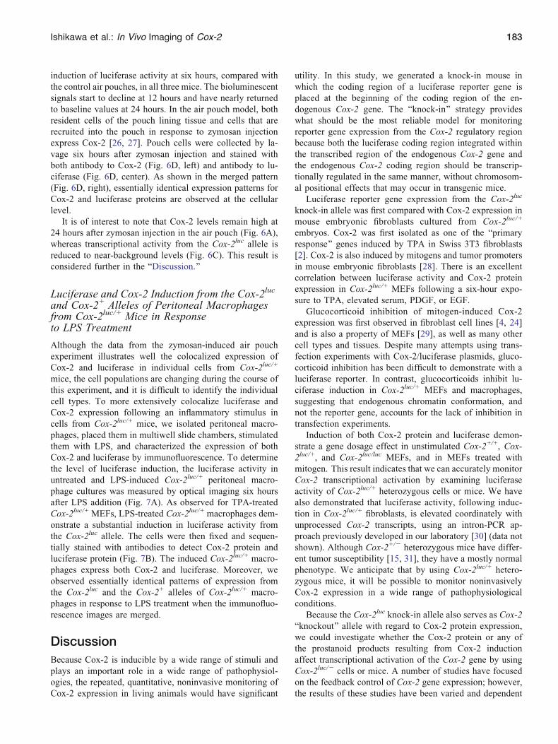
It is of interest to note that Cox-2 levels remain high at
24 hours after zymosan injection in the air pouch (Fig. 6A),
whereas transcriptional activity from the Cox-2luc allele is
reduced to near-background levels (Fig. 6C). This result is
considered further in the BDiscussion.^
Luciferase and Cox-2 Induction from the Cox-2luc
and Cox-2+ Alleles of Peritoneal Macrophagesfrom Cox-2luc/+ Mice in Responseto LPS Treatment
Although the data from the zymosan-induced air pouch
experiment illustrates well the colocalized expression of
Cox-2 and luciferase in individual cells from Cox-2luc/+
mice, the cell populations are changing during the course of
this experiment, and it is difficult to identify the individual
cell types. To more extensively colocalize luciferase and
Cox-2 expression following an inflammatory stimulus in
cells from Cox-2luc/+ mice, we isolated peritoneal macro-
phages, placed them in multiwell slide chambers, stimulated
them with LPS, and characterized the expression of both
Cox-2 and luciferase by immunofluorescence. To determine
the level of luciferase induction, the luciferase activity in
untreated and LPS-induced Cox-2luc/+ peritoneal macro-
phage cultures was measured by optical imaging six hours
after LPS addition (Fig. 7A). As observed for TPA-treated
Cox-2luc/+ MEFs, LPS-treated Cox-2luc/+ macrophages dem-
onstrate a substantial induction in luciferase activity from
the Cox-2luc allele. The cells were then fixed and sequen-
tially stained with antibodies to detect Cox-2 protein and
luciferase protein (Fig. 7B). The induced Cox-2luc/+ macro-
phages express both Cox-2 and luciferase. Moreover, we
observed essentially identical patterns of expression from
the Cox-2luc and the Cox-2+ alleles of Cox-2luc/+ macro-
phages in response to LPS treatment when the immunofluo-
rescence images are merged.
Discussion
Because Cox-2 is inducible by a wide range of stimuli and
plays an important role in a wide range of pathophysiol-
ogies, the repeated, quantitative, noninvasive monitoring of
Cox-2 expression in living animals would have significant
utility. In this study, we generated a knock-in mouse in
which the coding region of a luciferase reporter gene is
placed at the beginning of the coding region of the en-
dogenous Cox-2 gene. The Bknock-in^ strategy provides
what should be the most reliable model for monitoring
reporter gene expression from the Cox-2 regulatory region
because both the luciferase coding region integrated within
the transcribed region of the endogenous Cox-2 gene and
the endogenous Cox-2 coding region should be transcrip-
tionally regulated in the same manner, without chromosom-
al positional effects that may occur in transgenic mice.
Luciferase reporter gene expression from the Cox-2luc
knock-in allele was first compared with Cox-2 expression in
mouse embryonic fibroblasts cultured from Cox-2luc/+
embryos. Cox-2 was first isolated as one of the Bprimary
response^ genes induced by TPA in Swiss 3T3 fibroblasts
[2]. Cox-2 is also induced by mitogens and tumor promoters
in mouse embryonic fibroblasts [28]. There is an excellent
correlation between luciferase activity and Cox-2 protein
expression in Cox-2luc/+ MEFs following a six-hour expo-
sure to TPA, elevated serum, PDGF, or EGF.
Glucocorticoid inhibition of mitogen-induced Cox-2
expression was first observed in fibroblast cell lines [4, 24]
and is also a property of MEFs [29], as well as many other
cell types and tissues. Despite many attempts using trans-
fection experiments with Cox-2/luciferase plasmids, gluco-
corticoid inhibition has been difficult to demonstrate with a
luciferase reporter. In contrast, glucocorticoids inhibit lu-
ciferase induction in Cox-2luc/+ MEFs and macrophages,
suggesting that endogenous chromatin conformation, and
not the reporter gene, accounts for the lack of inhibition in
transfection experiments.
Induction of both Cox-2 protein and luciferase demon-
strate a gene dosage effect in unstimulated Cox-2+/+, Cox-
2luc/+, and Cox-2luc/luc MEFs, and in MEFs treated with
mitogen. This result indicates that we can accurately monitor
Cox-2 transcriptional activation by examining luciferase
activity of Cox-2luc/+ heterozygous cells or mice. We have
also demonstrated that luciferase activity, following induc-
tion in Cox-2luc/+ fibroblasts, is elevated coordinately with
unprocessed Cox-2 transcripts, using an intron-PCR ap-
proach previously developed in our laboratory [30] (data not
shown). Although Cox-2+/j heterozygous mice have differ-
ent tumor susceptibility [15, 31], they have a mostly normal
phenotype. We anticipate that by using Cox-2luc/+ hetero-
zygous mice, it will be possible to monitor noninvasively
Cox-2 expression in a wide range of pathophysiological
conditions.
Because the Cox-2luc knock-in allele also serves as Cox-2
Bknockout^ allele with regard to Cox-2 protein expression,
we could investigate whether the Cox-2 protein or any of
the prostanoid products resulting from Cox-2 induction
affect transcriptional activation of the Cox-2 gene by using
Cox-2luc/j cells or mice. A number of studies have focused
on the feedback control of Cox-2 gene expression; however,
the results of these studies have been varied and dependent
Ishikawa et al.: In Vivo Imaging of Cox-2 183

on the cell type. Studies using human synovial fibroblasts,
monocytes, and a prostate cancer cell line show positive
effects of PGE2 on Cox-2 expression [32Y34]. 15d-PGJ2, a
metabolite of PGD2, is reported to down-regulate Cox-2
expression in macrophage-like differentiated cell line
(U937) and rheumatoid synovial fibroblasts (RSFs) [35,
36], but not in the BAEC bovine endothelial cell line or in
human umbilical vein endothelial cells [35]. In mouse lung
fibroblast cells, PGE2, 15d-PGJ2, 6-keto PGF1�, and
PGF2� each enhanced Cox-2 protein expression [37]. In
our experiments, we do not see a difference in luciferase
induction when comparing Cox-2luc/j and Cox-2luc/+ het-
erozygous fibroblasts. Cox-2luc/j mice and other primary
cell cultures from these mice will be of great use in studying
the relationship between Cox-2-mediated prostanoid pro-
duction and transcription from the Cox-2 gene.
Ex vivo analysis shows that basal luciferase activity is
expressed in many tissues of the Cox-2luc/+ mouse. Highest
constitutive luciferase expression from the Cox-2luc allele is
observed in the vas deferens. Constitutive high Cox-2 ex-
pression in vas deferens was reported in rats [38], humans
[39], and mice [25]. In rat, castration severely depletes Cox-
2 and androgen replacement after castration restores Cox-2,
indicating that Cox-2 expression in the vas deferens is an-
drogen dependent [38]. Intermediate levels of luciferase
activity were observed in brain, thymus, lung, stomach,
pancreas, kidney, intestine, uterus, and skin in the Cox-2luc/+
mouse; lowest constitutive luciferase activity occurs in liver,
spleen, and heart. These results are consistent with a pre-
viously reported RTYPCR analysis of Cox-2 mRNA expres-
sion in mouse tissues under normal, uninduced conditions
[40].
Cox-2 induction is as an important early response to in-
fection. LPS stimulates Cox-2 expression by initiating
several signaling pathways, activating multiple transcription
factors to initiate Cox-2 mRNA expression [41, 42]. LPS
also induces other pro-inflammatory cytokines, including
IFN-g and interferon regulatory factors (IRFs), which are
important in the overall regulation of Cox-2 expression in
response to LPS [43]. Moreover, exogenously added IFN-ginduces Cox-2 expression in synergy with LPS [44]. To
obtain greater Cox-2 expression in systemic inflammation
experiments, we pretreated mice with IFN-g before LPS
injection. Cox-2 protein levels were higher in all tissues in
mice treated with [IFN-g + LPS] than in mice treated only
with LPS (data not shown). As demonstrated both by ex
vivo imaging of tissues and by luciferase assay of tissue
extracts, [LPS + IFN-g] treatment induces luciferase ex-
pression in all the tissues examined. There are no gender
differences in induction of luciferase activity.
In the acute inflammation study with LPS + IFN-g, we
see some differences in the relative levels of Cox-2 protein
and luciferase activity in tissues from Cox-2luc/+ mice. The
most likely explanation for this discrepancy in Cox-2 pro-
tein levels and luciferase activity is posttranscriptional regu-
lation of Cox-2 mRNA stability (reviewed in [7, 45]). The
Cox-2 30 untranslated region (UTR) contains 22 BARE^elements, which regulate the stability and translation of a
number of mRNAs whose expression is modulated by ex-
tracellular stimuli [46]. The Cox-2luc allele does not contain
the 30 UTR of the Cox-2 gene; the luciferase allele has an
SV40 30 UTR instead of Cox-2 30UTR. The activation of the
p38 MAPK pathway stabilizes Cox-2 message [47, 48]; in-
hibition of the p38 pathway leads to a decrease in Cox-2
message [49, 50]. Both stability of the Cox-2 message and
its translation are regulated by proteins that bind to the Cox-
2 AREs. HuR [51, 52], AUF1 [53], CPF-A [54], and TTP
[55] are reported to regulate message stability, and TIAR,
TIA-1 [56, 57], and CUGBP2 [58] are reported to regulate
Cox-2 message translation. Furthermore, activation of cells
with LPS results in modification of these RNAYprotein
complexes [59] and concomitant modulation of their
abilities to mediate Cox-2 mRNA stability and translation.
Although essentially all the studies of Cox-2 message sta-
bility and translational regulation have been performed in
cell culture, posttranscriptional mechanisms are certain to
play a role in regulation of Cox-2 expression in tissue-/cell-
type-specific manners in living animals. In Cox-2luc/+ mice,
luciferase expression reflects Cox-2 promoter activity. We
are currently developing murine strains to monitor, repeat-
edly and noninvasively in living animals, posttranscriptional
effects on Cox-2 mRNA stability and translation. In this
model, a transgenic mouse will be created in which the
coding sequence for the luciferase reporter gene will be
driven by a constitutive promoter and will be followed by the
30 UTR of the Cox-2 mRNA. To isolate the studies of Cox-2
mRNA stabilization to specific target tissues, a loxP flanked
STOP cassette will be used to restrict transgene expression to
tissues in which Cre recombinase is expressed.
Zymosan or carrageenan injections into the mouse paw
provoke inflammatory reactions [60, 61]. NSAIDs inhibit
edema in both zymosan- and carrageenan-induced inflam-
mation, and Cox-2 mRNA and protein expression has been
analyzed in the carrageenan-induced paw inflammation
model [62, 63]. We examined, by Western blotting, Cox-2
protein levels in both the zymosan- and carrageenan-
induced paw inflammation models in wild-type mice.
Because peak Cox-2 protein was higher in the zymosan-
induced model than in the carrageenan-induced model (data
not shown), we used the zymosan model for in vivo
imaging with Cox-2luc/+ mice. In wild-type mice, zymosan
induced substantial Cox-2 protein at both 6 and 12 hours
after injection. Local administration of zymosan into the
paw triggers both luciferase and Cox-2 protein induction
only in the zymosan-treated paw. However, the luciferase
signal is attenuated substantially before Cox-2 protein levels
are reduced (Fig. 5). The half-life of both firefly luciferase
and Cox-2 protein are estimated to be three to four hours
[64, 65]. Although these measurements were performed in
cell culture, it is likely that the two proteins have close to
the same half-lives in vivo as well. Consequently, we suggest
that the differences in Cox-2 protein and luciferase protein
184 Ishikawa et al.: In Vivo Imaging of Cox-2

accumulation in the paw of zymosan-injected Cox-2luc/+
mice reflects the additional posttranscriptional regulation
conferred by the 30 UTR region of the Cox-2 mRNA, as
discussed previously. Although cell culture experiments
show a strong correlation between luciferase activity and
transcriptional activation of the endogenous Cox-2 gene in
Cox-2luc/+ MEFs, similar experiments in Cox-2luc/+ mice
need to be done to confirm the extrapolation of this ex-
planation to in vivo inflammatory responses.
Dexamethasone, the most widely used anti-inflammatory
glucocorticoid in animal model experiments, inhibits Cox-2
expression in vitro and in vivo [24, 26]. Dexamethasone
inhibits both luciferase induction and Cox-2 protein accu-
mulation in the zymosan paw inflammation model. Howev-
er, luciferase inhibition by dexamethasone is not as
extensive as inhibition of Cox-2 protein accumulation. The
difference in dexamethasone sensitivity could also be
caused by the differences in 30 UTR regions for the Cox-
2+ and Cox-2luc alleles of Cox-2luc/+ mice; glucocorticoids
inhibit p38 MAPK function and destabilize Cox-2 mRNA
[66]. Noninvasive in vivo studies of the regulation of Cox-2
mRNA by inflammation and other stimuli await the de-
velopment of appropriate mouse models.
To demonstrate the potential utility of Cox-2luc/+ mice
in examining synovial inflammation, we examined Cox-2
and luciferase expression in the air pouch inflammation
model. Mechanical disruption of subcutaneous tissue by
air injection provides a structure closely resembling
synovial tissue [67]. Moreover, Cox-2 protein accumula-
tion is induced by injection of zymosan into the air pouch
[22]. Cox-2 expression is observed in both resident cells of
the pouch lining tissue and the recruited cells, predomi-
nantly polymorphonuclear neutrophils [27]. We modified
the original protocol, making two smaller air pouches on
individual mice, to compare Cox-2 and luciferase induc-
tion by zymosan and saline on same mouse. Cox-2 protein
accumulation and luciferase induction occur only in the
zymosan injected air pouch. We isolated cells from the air
pouch of zymosan treated Cox-2luc/+ mice, and demon-
strated Cox-2 and luciferase coexpression in essentially all
positive cells. Colocalization of Cox-2 and luciferase
expression was also confirmed in LPS-stimulated peritoneal
macrophages isolated from Cox-2luc/+ mice. These colocal-
ization studies, from both inflammatory challenges in the
whole animal and in isolated cells, demonstrate the validity
of surrogate luciferase expression as a measure of endog-
enous Cox-2 gene transcription in the Cox-2luc/+ heterozy-
gous mouse model.
In conclusion, we have established a murine strain with
a Cox-2luc knock-in allele and demonstrated the monitoring
of Cox-2 endogenous promoter-driven luciferase expres-
sion, noninvasively and repeatedly, in living animals. The
Cox-2luc/+ mouse should become a useful tool to analyze
noninvasively Cox-2 expression in a variety of pathophys-
iological conditions, including inflammation, neurodegener-
ative diseases, and cancer.
Acknowledgments. We thank Art Catapang and Raymond Bascontillo fortechnical assistance, Masanobu Oshima for genomic fragments, AlexGarcia and Hong Wu for assistance with immunohistochemistry andimmunofluorescence, and Shankkar Pattabhiraman and Hong Wu forhelpful discussions and comments. We also acknowledge the importantassistance of the UCLA Jonsson Comprehensive Cancer Center SharedResource for embryonic stem cell homologous recombination, and fortransgenic/knockout mouse production. These studies were supported bygrants R01 CA84572 and P50 CA86306 (H.R.H., principal investigator).
References1. Herschman HR, Talley JJ, Dubois R (2003) Cyclooxygenase 2 (COX-
2) as a target for therapy and noninvasive imaging. Mol Imaging Biol5:286Y303
2. Kujubu DA, Fletcher BS, Varnum BC, Lim RW, Herschman HR(1991) TIS10, a phorbol ester tumor promoter-inducible mRNA fromSwiss 3T3 cells, encodes a novel prostaglandin synthase/cyclooxyge-nase homologue. J Biol Chem 266:12866 Y12872
3. Xie W, Chipman JG, Robertson DL, Erikson RL, Simmons DL (1991)Expression of mitogen-responsive gene encoding prostaglandin syn-thase/cyclooxygenase homologue. Proc Natl Acad Sci U S A 88:2692Y2696
4. O’Banion MK, Sadowski HB, Winn V, Young DA (1991) A serum-and glucocorticoid-regulated 4-kilobase mRNA encodes a cyclo-oxygenase-related protein. J Biol Chem 266:12866 Y12872
5. Herschman HR (2003) Historical aspects of COX-2: Cloning andcharacterization of the cDNA, protein and gene. In: Harris RE (ed)COX-2 blockade in cancer prevention and therapy. Clifton, NJ:Humana Press, pp 13Y32
6. Herschman HR (2004) Regulation and function of prostaglandinsynthase 2/cyclooxygenase. In: Curtis-Prior P (ed) The eicosanoids.New York: John Wiley & Sons, Ltd
7. Espel E (2005) The role of the AU-rich elements of mRNAs incontrolling translation. Semin Cell Dev Biol 16:59Y67
8. Smith LH, Petrie MS, Morrow JD, Oates JA, Vaughan DE (2005) Thesterol response element binding protein regulates cyclooxygenase-2gene expression in endothelial cells. J Lipid Res 46:862Y871
9. Duque J, Fresno M, Iniguez A (2005) Expression and function of thenuclear factor of activated T cells in colon carcinoma cells. J BiolChem 280:8686Y8693
10. Joo M, Park GY, Wright JG, et al. (2004) Transcriptional regulation ofthe cyclooxygenase-2 gene in macrophages by PU.1. J Biol Chem279:6658Y6665
11. Wang S-C, Lien H-C, Xia W, et al. (2004) Binding at and trans-activation of the COX-2 promoter by nuclear tyrosine kinase receptorErbB-2. Cancer Cell 6:251Y261
12. Herschman HR (2003) Molecular imaging: Looking at problems,seeing solutions. Science 302:605Y608
13. Contag PR, Olum IN, Stevenson DK, Contag CH (1998) Biolumines-cent indicators in living mammals. Nat Med 4:245
14. Nguyen JT, Machado H, Herschman HR (2003) Repetitive, noninva-sive imaging of cyclooxytenase-2 gene expression in living mice. MolImaging Biol 5:248Y256
15. Oshima M, Dinchuck JE, Kargman SL, et al. (1996) Suppressionof intestinal polyposis in ApcD716 knockout mice by inhibition ofcyclooxygenase 2 (COX-2). Cell 87:803Y809
16. Ishikawa T, Tamai Y, Qin L, Oshima M, Taketo MM (2003)Requirement for tumor suppressor Apc in the morphogenesis ofanterior and ventral mouse embryo. Dev Biol 253:230Y246
17. Zhang X, Morham SG, Langenbach R, Bagg RB, Young DA (2000)Lack of cyclooxygenase-2 inhibits growth of teratocarcinomas in mice.Exp Cell Res 254:232Y240
18. Hogan B, Beddington R, Costantini F, Lacy E (1994) Manipulating themouse embryo. Cold Spring Harbor: Cold Spring Harbor LaboratoryPress
19. Tegeder I, Del Turco D, Schmidtko A, et al. (2004) Reducedinflammatory hyperalgesia with preservation of acute thermal noci-ception in mice lacking cGMP-dependent protein kinase I. Proc NatlAcad Sci U S A 101:3253Y3257
20. Zhang N, Weber A, Li B, et al. (2003) An inducible nitric oxidesynthaseYluciferase reporter system for in vivo testing of anti-inflam-matory compounds in trasgenic mice. J Immunol 170:6307Y6319
Ishikawa et al.: In Vivo Imaging of Cox-2 185

21. Zhang N, Ahsan MH, Zhu L, et al. (2005) NF-kB and not the MAPKsignaling pathway regulates GADD45b expression during acuteinflammation. J Biol Chem 280:21400 Y21408
22. Posadas I, Terencio MC, Guillen I, et al. (2000) Co-regulation betweencyclo-oxygenase-2 and inducible nitric oxide synthase expression inthe time-course of murine inflammation. Naunyn Schmiedebergs ArchPharmacol 361:98Y106
23. Lewandoski M (2001) Conditional control of gene expression in themouse. Nat Rev Genet 2:735Y755
24. Kujubu DA, Herschman HR (1992) Dexamethasone inhibits mitogeninduction of the TIS10 prostaglandin synthase/cyclooxygenase gene.J Biol Chem 267:7991Y7994
25. Lazarus M, Munday CJ, Eguchi N, et al. (2002) Immunohistochemicallocalization of microsomal PGE synthase-1 and cyclooxygenases inmale mouse reproductive organs. Endocrinology 143:2410Y2419
26. Masferrer JL, Zweifel BS, Manning PT, et al. (1994) Selectiveinhibition of inducible cyclooxygenase 2 in vivo is antiinflammatoryand nonulcerogenic. Proc Natl Acad Sci U S A 91:3228Y3232
27. Cadieux J-S, Leclerc P, St-Onge M, et al. (2005) Potentiation ofneutrophil cyclooxygenase-2 by adenosine: an early anti-inflammatorysignal. J Cell Sci 118:1437Y1447
28. Reddy ST, Herschman HR (1994) Ligand-induced prostaglandinsynthesis requires expression of the TIS10/PGS-2 prostaglandinsynthase gene in murine fibroblasts and macrophage. J Biol Chem269:15437Y15480
29. Gilbert RS, Reddy ST, Kujubu DA, et al. (1994) TransformingGrowth factor b1 augments mitogen-induced prostaglandin synthesisand expression of the TIS10/prostaglandin synthase 2 gene both inSwiss 3T3 cells and in murine embryo fibroblasts. J Cell Physiol159:67Y75
30. Reddy ST, Gilbert RS, Xie W, Luner S, Herschman HR (1994) TGF-beta inhibits both endotoxin-induced prostaglandin synthesis andexpression of the TIS10/prostaglandin synthase 2 gene in murinemacrophages. J Leuk Biol 55:192Y200
31. Tiano HF, Loftin CD, Akunda J, et al. (2002) Deficiency of eithercyclooxygenase (COX)-1 or COX-2 alters epidermal differentiationand reduces mouse skin tumorigenesis. Can Res 62:3395Y3401
32. Faour WH, He Y, He QW, et al. (2001) Prostaglandin E2 regulates thelevel and stability of cyclooxygenase-2 mRNA through activation ofp38 mitogen-activated protein kinase in interleukin-1 beta-treatedhuman synovial fibroblasts. J Biol Chem 276:31720 Y31731
33. Hinz B, Brune K, Pahl A (2000) Cyclooxygenase-2 expression inlipopolysaccharide-stimulated human monocytes is modulated bycyclic AMP, prostaglandin E2, and nonsteroidal anti-inflammatorydrugs. Biochem Biophys Res Commun 278:790Y796
34. Trandrawinata RR, Hughes-Fulford M (1997) Up-regulation of cyclo-oxygenase-2 by product-prostaglandin E2. Adv Exp Med Biol407:163Y170
35. Inoue H, Tanabe T, Umesono K (2000) Feedback control of cyclo-oxygenase-2 expression through PPARg. J Biol Chem 275:28028Y28032
36. Tsubouchi Y, Kawahito Y, Kohno M, et al. (2002) Feedback controlof the arachidonate cascade in rheumatoid synoviocytes by 15-deoxy-D12,14-prostaglandin J2. Biochem Biophys Res Commun283:750Y755
37. Vichai V, Suyarnsesthaforn C, Pittayakhajonwut D, Sriklung K,Kirtikara K (2005) Positive feedback regulation of COX-2 expressionby prostaglandin metabolites. Inflamm Res 54:163Y172
38. McKanna JA, Zhang M-Z, Wang J-L, Cheng H-F, Harris RC (1998)Constitutive expression of cyclooxygenase-2 in rat vas deferens. Am JPhysiol 275:R227YR233
39. Kirschenbaum A, Liotta DR, Yao S, et al. (2000) Immunohistochem-ical localization of cyclooxygenase-1 and cyclooxygenase-2 in thehuman fetal and adult male reproductive tracts. J Clin EndocrinolMetab 85:3436Y3441
40. Oshima M, Oshima H, Taketo MM (2005) Hypergravity inducesexpression of cyclooxygenase-2 in the heart vessels. Biochem BiophysRes Commun 330:928Y933
41. Smith WL, DeWitt DL, Garavito RM (2000) Cyclooxygenases:Structural, cellular, and molecular biology. Annu Rev Biochem 69:145Y182
42. Hwang D (2001) Modulation of the expression of cyclooxygenase-2 byfatty acids mediated through toll-like receptor 4-derived signalingpathways. FASEB J 15:2556Y2564
43. Zhang S, Thomas K, Blanco JCG, Salkowski CA, Vogel SN (2002)The role of the interferon regulatory factors, IRF-1 and IRF-2, in LPS-induced cyclooxygenase-2 (COX-2) expression in vivo and in vitro.J Endotoxin Res 8:381Y390
44. Blanco JCG, Contursi C, Salkowski CA, et al. (2000) Interferonregulatory factor (IRF)-1 and IRF-2 regulate interferon g-dependentcyclooxygenase 2 expression. J Exp Med 191:2131Y2144
45. Dean JLE, Sully G, Clark AR, Saklatvala J (2004) The involvement ofAU-rich element-binding proteins in p38 mitogen-activated proteinkinase pathway-mediated mRNA stabilisation. Cell Signal 16:1113Y1121
46. Saklatvala J, Dean J, Clark A (2003) Control of the expression ofinflammatory response genes. Biochem Soc Symp 70:95Y105
47. Srivastava SK, Tetsuka T, Daphna-Ikem D, Morrison AR (1994) IL-1beta stabilizes COX II mRNA in renal mesangial cells: Role of 30-untranslated region. Am J Physiol 267:F504YF508
48. Cok SJ, Morrison AR (2001) The 30-untranslated region of murinecyclooxygenase-2 contains multiple regulatory elements that altermessage stability and translational efficiency. J Biol Chem276:23179Y23185
49. Gou Q, Liu CH, Ben-Av P, Hla T (1998) Dissociation of basalturnover and cytokine-induced transcript stabilization of the humancyclooxygenase-2 mRNA by mutagenesis of the 30-untranslatedregion. Biochem Biophys Res Commun 242:508Y512
50. Dean JL, Brook M, Clark AR, Saklatvala J (1999) p38 mitogen-activated protein kinase regulates cyclooxygenase-2 mRNA stabilityand transcription in lipopolysaccharide-treated human monocytes.J Biol Chem 274:264Y269
51. Dixon DA, Tolley ND, King PH, et al. (2001) Altered expression ofthe mRNA stability factor HuR promotes cyclooxygenase-2 expressionin colon cancer cells. J Clin Invest 108:1657Y1665
52. Sengupta S, Jang B-C, Wu M-T, et al. (2003) The RNA-bindingprotein HuR regulates the expression of cyclooxygenase-2. J BiolChem 278:25227Y25233
53. Lasa M, Mahtani KR, Finch A, et al. (2000) Regulation of cyclo-oxygenase 2 mRNA stability by the mitogen-activated protein kinasep38 signaling cascade. Mol Cell Biol 20:4265Y4270
54. Dean JLE, Sully G, Wait R, et al. (2002) Identification of a novel AU-rich-element-binding protein which is related to AUF1. Biochem J366:709Y719
55. Sawaoka H, Dixon DA, Oates JA, Boutaud O (2003) Tristetraprolinbinds to the 30-untranslated region of cyclooxygenase-2 mRNA. J BiolChem 278:36157Y36162
56. Cok SJ, Acton SJ, Morrison AR (2003) The proximal region of the 30-untranslated region of cyclooxygenase-2 is recognized by a multimericprotein complex containing HuR, TIA-1, TIAR, and the heterogeneousnuclear ribonucleoprotein U. J Biol Chem 278: 36157Y36162
57. Dixon DA, Balch GC, Kedersha N, et al. (2003) Regulation ofcyclooxygenase-2 expression by translational silencer TIA-1. J ExpMed 198:475Y 481
58. Mukhopadhyay D, Houchen CW, Kennedy S, Dieckgraefe BK, AnantS (2003) Coupled mRNA stabilization and translational silencing ofcyclooxygenase-2 by a novel RNA binding protein, CUGBP2. MolCell 11:113Y126
59. Cok SJ, Acton SJ, Sexton AE, Morrison AR (2004) Identification ofRNA-binding protein in RAW 264.7 cells that recognize alipopolysaccharide-responsive element in the 3-untranslated regionof the murine cyclooxygenase-2 mRNA. J Biol Chem 279:8196 Y8205
60. Tarayre JP, Delhon A, Aliaga M, et al. (1989) Pharmacological studieson zymosan inflammation in rats and mice. 1: Zymosan-induced pawoedema in rats and mice. Pharmacol Res 21:375Y384
61. Calhoun W, Chang J, Carlson RP (1987) Effect of selectedantiinflammatory agents and other drugs on zymosan, arachidonicacid, PAF and carrageenan induced paw edema in the mouse. AgentsActions 21:306Y309
62. Fernandez P, Guillen MI, Ubeda A, et al. (2003) A novel indazolo-triazolo-benzotriazepine exerts anti-inflammatory effects by inhibitionof cycloozygenase-2 activity and nitric oxide synthase-2 expression.Naunyn Schmiedebergs Arch Pharmacol 368:26Y32
63. Posadas I, Bucci M, Roviezzo F, et al. (2004) Carrageenan-inducedmouse paw oedema is biphasic, ageYweight dependent and displaysdifferential nitric oxide cyclooxygenase-2 expression. Br J Pharmacol142:331Y338
186 Ishikawa et al.: In Vivo Imaging of Cox-2

64. Leclerc GM, Bookfor FR, Faught WJ, Frawley LS (2000) Develop-ment of a destabilized firefly luciferase enzyme for measurement ofgene expression. Biotechniques 29:190Y598
65. Kujubu DA, Reddy ST, Fletcher BS, Herschman HR (1993)Expression of the protein product of the prostaglandin synthase-2/TIS10 gene in mitogen-stimulated Swiss 3T3 cells. J Biol Chem268:5425Y5430
66. Lasa M, Mahtani KR, Finch A, et al. (2001) Dexamethasonedestabilizes cyclooxygenase 2 mRNA by inhibiting mitogen-activatedprotein kinase p38. Mol Cell Biol 21:771Y780
67. Edwards JC, Sedgwick AD, Willoughby DA (1981) The formationof a structure with the features of synovial lining by subcutaneousinjection of air: An in vivo tissue culture system. J Pathol 134:147Y156
Ishikawa et al.: In Vivo Imaging of Cox-2 187