Gene technology
30
389 19 Gene Technology Concept Outline 19.1 The ability to manipulate DNA has led to a new genetics. Restriction Endonucleases. Enzymes that cleave DNA at specific sites allow DNA segments from different sources to be spliced together. Using Restriction Endonucleases to Manipulate Genes. Fragments produced by cleaving DNA with restriction endonucleases can be spliced into plasmids, which can be used to insert the DNA into host cells. 19.2 Genetic engineering involves easily understood procedures. The Four Stages of a Genetic Engineering Experiment. Gene engineers cut DNA into fragments that they splice into vectors that carry the fragments into cells. Working with Gene Clones. Gene technology is used in a variety of procedures involving DNA manipulation. 19.3 Biotechnology is producing a scientific revolution. DNA Sequence Technology. The complete nucleotide sequence of the genomes of many organisms are now known. The unique DNA of every individual can be used to identify sperm, blood, or other tissues. Biochips. Biochips are squares of glass etched with DNA strands and can be used for genetic screening. Medical Applications. Many drugs and vaccines are now produced with gene technology. Agricultural Applications. Gene engineers have developed crops resistant to pesticides and pests, as well as commercially superior animals. Cloning. Recent experiments show it is possible to clone agricultural animals, a result with many implications for both agriculture and society. Stem Cells. Both embryonic stem cells and tissue- specific stem cells can potentially be used to repair or replace damaged tissue. Ethics and Regulation. Genetic engineering raises important questions about danger and privacy. O ver the past decades, the development of new and powerful techniques for studying and manipulating DNA has revolutionized genetics (figure 19.1). These tech- niques have allowed biologists to intervene directly in the genetic fate of organisms for the first time. In this chapter, we will explore these technologies and consider how they apply to specific problems of great practical importance. Few areas of biology will have as great an impact on our fu- ture lives. FIGURE 19.1 A famous plasmid. The circular molecule in this electron micrograph is pSC101, the first plasmid used successfully to clone a vertebrate gene. Its name comes from the fact that it was the one-hundred-and-first plasmid isolated by Stanley Cohen.
Transcript of Gene technology

389
19Gene Technology
Concept Outline
19.1 The ability to manipulate DNA has led to a newgenetics.
Restriction Endonucleases. Enzymes that cleave DNAat specific sites allow DNA segments from different sourcesto be spliced together.Using Restriction Endonucleases to Manipulate Genes.Fragments produced by cleaving DNA with restrictionendonucleases can be spliced into plasmids, which can beused to insert the DNA into host cells.
19.2 Genetic engineering involves easily understoodprocedures.
The Four Stages of a Genetic Engineering Experiment.Gene engineers cut DNA into fragments that they spliceinto vectors that carry the fragments into cells.Working with Gene Clones. Gene technology is used ina variety of procedures involving DNA manipulation.
19.3 Biotechnology is producing a scientificrevolution.
DNA Sequence Technology. The complete nucleotidesequence of the genomes of many organisms are nowknown. The unique DNA of every individual can be used toidentify sperm, blood, or other tissues.Biochips. Biochips are squares of glass etched with DNAstrands and can be used for genetic screening.Medical Applications. Many drugs and vaccines are nowproduced with gene technology.Agricultural Applications. Gene engineers havedeveloped crops resistant to pesticides and pests, as well ascommercially superior animals.Cloning. Recent experiments show it is possible to cloneagricultural animals, a result with many implications forboth agriculture and society.Stem Cells. Both embryonic stem cells and tissue-specific stem cells can potentially be used to repair orreplace damaged tissue.Ethics and Regulation. Genetic engineering raisesimportant questions about danger and privacy.
Over the past decades, the development of new andpowerful techniques for studying and manipulating
DNA has revolutionized genetics (figure 19.1). These tech-niques have allowed biologists to intervene directly in thegenetic fate of organisms for the first time. In this chapter,we will explore these technologies and consider how theyapply to specific problems of great practical importance.Few areas of biology will have as great an impact on our fu-ture lives.
FIGURE 19.1A famous plasmid. The circular molecule in this electronmicrograph is pSC101, the first plasmid used successfully to clonea vertebrate gene. Its name comes from the fact that it was theone-hundred-and-first plasmid isolated by Stanley Cohen.

quences of nucleotides in DNA. These enzymes are thebasic tools of genetic engineering.
Discovery of Restriction Endonucleases
Scientific discoveries often have their origins in seeminglyunimportant observations that receive little attention by re-searchers before their general significance is appreciated. Inthe case of genetic engineering, the original observationwas that bacteria use enzymes to defend themselves againstviruses.
Most organisms eventually evolve means of defendingthemselves from predators and parasites, and bacteria areno exception. Among the natural enemies of bacteria arebacteriophages, viruses that infect bacteria and multiplywithin them. At some point, they cause the bacterial cells toburst, releasing thousands more viruses. Through naturalselection, some types of bacteria have acquired powerfulweapons against these viruses: they contain enzymes calledrestriction endonucleases that fragment the viral DNA assoon as it enters the bacterial cell. Many restriction en-donucleases recognize specific nucleotide sequences in aDNA strand, bind to the DNA at those sequences, andcleave the DNA at a particular place within the recognitionsequence.
Why don’t restriction endonucleases cleave the bacter-ial cells’ own DNA as well as that of the viruses? The an-swer to this question is that bacteria modify their ownDNA, using other enzymes known as methylases to addmethyl (—CH3) groups to some of the nucleotides in thebacterial DNA. When nucleotides within a restriction en-donuclease’s recognition sequence have been methylated,the endonuclease cannot bind to that sequence. Conse-quently, the bacterial DNA is protected from being de-graded at that site. Viral DNA, on the other hand, has notbeen methylated and therefore is not protected from enzy-matic cleavage.
How Restriction Endonucleases Cut DNA
The sequences recognized by restriction endonucleases aretypically four to six nucleotides long, and they are oftenpalindromes. This means the nucleotides at one end of therecognition sequence are complementary to those at theother end, so that the two strands of the DNA duplex havethe same nucleotide sequence running in opposite direc-tions for the length of the recognition sequence. Two im-portant consequences arise from this arrangement ofnucleotides.
390 Part V Molecular Genetics
Restriction Endonucleases In 1980, geneticists used the relatively new technique ofgene splicing, which we will describe in this chapter, tointroduce the human gene that encodes interferon intoa bacterial cell’s genome. Interferon is a rare blood pro-tein that increases human resistance to viral infection,and medical scientists have been interested in its possibleusefulness in cancer therapy. This possibility was diffi-cult to investigate before 1980, however, because purifi-cation of the large amounts of interferon required forclinical testing would have been prohibitively expensive,given interferon’s scarcity in the blood. An inexpensiveway to produce interferon was needed, and introducingthe gene responsible for its production into a bacterialcell made that possible. The cell that had acquired thehuman interferon gene proceeded to produce interferonat a rapid rate, and to grow and divide. Soon there weremillions of interferon-producing bacteria in the culture,all of them descendants of the cell that had originally re-ceived the human interferon gene.
The Advent of Genetic Engineering
This procedure of producing a line of genetically identicalcells from a single altered cell, called cloning, made everycell in the culture a miniature factory for producing inter-feron. The human insulin gene has also been cloned in bac-teria, and now large amounts of insulin, a hormone essen-tial for treating some forms of diabetes, can bemanufactured at relatively little expense. Beyond these clin-ical applications, cloning and related molecular techniquesare used to obtain basic information about how genes areput together and regulated. The interferon experiment andothers like it marked the beginning of a new genetics, ge-netic engineering.
The essence of genetic engineering is the ability to cutDNA into recognizable pieces and rearrange those piecesin different ways. In the interferon experiment, a piece ofDNA carrying the interferon gene was inserted into a plas-mid, which then carried the gene into a bacterial cell. Mostother genetic engineering approaches have used the samegeneral strategy, bringing the gene of interest into the tar-get cell by first incorporating it into a plasmid or an infec-tive virus. To make these experiments work, one must beable to cut the source DNA (human DNA in the interferonexperiment, for example) and the plasmid DNA in such away that the desired fragment of source DNA can bespliced permanently into the plasmid. This cutting is per-formed by enzymes that recognize and cleave specific se-
19.1 The ability to manipulate DNA has led to a new genetics.

First, because the same recognitionsequence occurs on both strands of theDNA duplex, the restriction endonucle-ase can bind to and cleave both strands,effectively cutting the DNA in half.This ability to cut across both strands isalmost certainly the reason that restric-tion endonucleases have evolved to rec-ognize nucleotide sequences withtwofold rotational symmetry.
Second, because the bond cleaved bya restriction endonuclease is typicallynot positioned in the center of therecognition sequence to which it binds,and because the DNA strands are an-tiparallel, the cut sites for the twostrands of a duplex are offset from eachother (figure 19.2). After cleavage, eachDNA fragment has a single-strandedend a few nucleotides long. The single-stranded ends of the two fragments arecomplementary to each other.
Why Restriction EndonucleasesAre So Useful
There are hundreds of bacterial restric-tion endonucleases, and each one has aspecific recognition sequence. Bychance, a particular endonuclease’srecognition sequence is likely to occursomewhere in any given sample ofDNA; the shorter the sequence, themore often it will arise by chance withina sample. Therefore, a given restrictionendonuclease can probably cut DNAfrom any source into fragments. Eachfragment will have complementarysingle-stranded ends characteristic ofthat endonuclease. Because of theircomplementarity, these single-strandedends can pair with each other (conse-quently, they are sometimes called“sticky ends”). Once their ends havepaired, two fragments can then bejoined together with the aid of the en-zyme DNA ligase, which re-forms the phosphodiesterbonds of DNA. What makes restriction endonucleases sovaluable for genetic engineering is the fact that any two frag-ments produced by the same restriction endonuclease can bejoined together. Fragments of elephant and ostrich DNAcleaved by the same endonuclease can be joined to one an-other as readily as two bacterial DNA fragments.
Genetic engineering involves manipulating specific genesby cutting and rearranging DNA. A restrictionendonuclease cleaves DNA at a specific site, generating inmost cases two fragments with short single-stranded ends.Because these ends are complementary to each other, anypair of fragments produced by the same endonuclease,from any DNA source, can be joined together.
Chapter 19 Gene Technology 391
GAATTC
CTTAAG
GAATTC
AATTC
AATTC
AATTC
GAATTC
G
G
G
G
G AATTC
G
CTTAAG
CTTAAG CTTAA
CTTAA
CTTAAG
DNA ligasejoins the strands.
DNA from another sourcecut with the same restrictionendonuclease is added.
Restriction endonucleasecleaves the DNA.
DNAduplex
Sticky ends (complementarysingle-stranded DNA tails)
Restriction sites
Recombinant DNA molecule
FIGURE 19.2Many restriction endonucleases produce DNA fragments with “sticky ends.” Therestriction endonuclease EcoRI always cleaves the sequence GAATTC between G and A.Because the same sequence occurs on both strands, both are cut. However, the twosequences run in opposite directions on the two strands. As a result, single-stranded tailsare produced that are complementary to each other, or “sticky.”
Using RestrictionEndonucleases toManipulate GenesA chimera is a mythical creature with thehead of a lion, body of a goat, and tail ofa serpent. Although no such creatures ex-isted in nature, biologists have madechimeras of a more modest kind throughgenetic engineering.
Constructing pSC101
One of the first chimeras was manufac-tured from a bacterial plasmid called aresistance transfer factor by Americangeneticists Stanley Cohen and HerbertBoyer in 1973. Cohen and Boyer used arestriction endonuclease called EcoRI,which is obtained from Escherichia coli,to cut the plasmid into fragments. Onefragment, 9000 nucleotides in length,contained both the origin of replicationnecessary for replicating the plasmid anda gene that conferred resistance to theantibiotic tetracycline (tet r). Becauseboth ends of this fragment were cut bythe same restriction endonuclease, theycould be ligated to form a circle, asmaller plasmid Cohen dubbed pSC101(figure 19.3).
Using pSC101 to Make Recombinant DNA
Cohen and Boyer also used EcoRI to cleave DNA thatcoded for rRNA that they had isolated from an adult am-phibian, the African clawed frog, Xenopus laevis. Theythen mixed the fragments of Xenopus DNA with pSC101plasmids that had been “reopened” by EcoRI and allowedbacterial cells to take up DNA from the mixture. Some ofthe bacterial cells immediately became resistant to tetra-cycline, indicating that they had incorporated the pSC101plasmid with its antibiotic-resistance gene. Furthermore,some of these pSC101-containing bacteria also began toproduce frog ribosomal RNA! Cohen and Boyer con-cluded that the frog rRNA gene must have been insertedinto the pSC101 plasmids in those bacteria. In otherwords, the two ends of the pSC101 plasmid, produced bycleavage with EcoRI, had joined to the two ends of a frogDNA fragment that contained the rRNA gene, alsocleaved with EcoRI.
The pSC101 plasmid containing the frog rRNA gene isa true chimera, an entirely new genome that never existedin nature and never would have evolved by natural means.It is a form of recombinant DNA—that is, DNA created
in the laboratory by joining together pieces of differentgenomes to form a novel combination.
Other Vectors
The introduction of foreign DNA fragments into host cellshas become common in molecular genetics. The genomethat carries the foreign DNA into the host cell is called avector. Plasmids, with names like pUC18 can be inducedto make hundreds of copies of themselves and thus of theforeign genes they contain. Much larger pieces of DNA canbe introduced using YAKs (yeast artificial chromosomes) asa vector instead of a plasmid. Not all vectors have bacterialtargets. Animal viruses such as the human cold virus aden-ovirus, for example, are serving as vectors to carry genesinto monkey and human cells, and animal genes have evenbeen introduced into plant cells.
One of the first recombinant genomes produced bygenetic engineering was a bacterial plasmid into whichan amphibian ribosomal RNA gene was inserted.Viruses can also be used as vectors to insert foreignDNA into host cells and create recombinant genomes.
392 Part V Molecular Genetics
AmphibianDNA
Endonuclease EcoRI
rRNA gene
Recombinantplasmid
PlasmidpSC101
tet r geneCleaved plasmidis combined withamphibian fragment.
Cleave plasmidpSC101 withEcoRI.
Cleave amphibianDNA with restrictionendonuclease EcoRI.
FIGURE 19.3One of the first genetic engineering experiments. This diagram illustrates howCohen and Boyer inserted an amphibian gene encoding rRNA into pSC101. Theplasmid contains a single site cleaved by the restriction endonuclease EcoRI; it alsocontains tetr, a gene which confers resistance to the antibiotic tetracycline. The rRNA-encoding gene was inserted into pSC101 by cleaving the amphibian DNA and theplasmid with EcoRI and allowing the complementary sequences to pair.

Examples of GeneManipulation
SUPER SALMON!Canadian fisheries scientists have inserted recombinant growth hor-mone genes into developing salmon embryos, creating the first trans-genic salmon. Not only do these transgenic fish have shortened pro-duction cycles, they are, on an average, 11 times heavier thannontransgenic salmon! The implications for the fisheries industry andfor worldwide food production are obvious.
WILT-PROOF FLOWERSEthylene, the plant hormone that causes fruit to ripen, also causesflowers to wilt. Researchers at Purdue have found the gene thatmakes flower petals respond to ethylene by wilting and replaced itwith a gene insensitive to ethylene. The transgenic carnations theyproduced lasted for 3 weeks after cutting, while normal carnationslast only 3 days.
HERMAN THE WONDER BULLGenPharm, a California biotechnology company, engineered Herman,a bull that possesses the gene for human lactoferrin (HLF). HLF con-fers antibacterial and iron transport properties to humans. Many ofHerman’s female offspring now produce milk containing HLF, andGenPharm intends to build a herd of transgenic cows for the large-scale commercial production of HLF.
Chapter 19 Gene Technology 393
WEEVIL-PROOFPEASNot only has gene tech-nology afforded agricul-ture viral and pest con-trol in the field, it hasalso provided a pestcontrol technique for thestorage bin. A team ofU.S. and Australian sci-entists have engineereda gene that is expressedonly in the seed of thepea plant. The enzymeinhibitor encoded by thisgene inhibits feeding byweevils, one of the mostnotorious pests affectingstored crops. The world-wide ramifications aresignificant as up to 40%of stored grains are lostto pests.

394 Part V Molecular Genetics
The Four Stages of a GeneticEngineering ExperimentLike the experiment of Cohen and Boyer, most geneticengineering experiments consist of four stages: DNAcleavage, production of recombinant DNA, cloning, andscreening.
Stage 1: DNA Cleavage
A restriction endonuclease is used to cleave the sourceDNA into fragments. Because the endonuclease’s recog-nition sequence is likely to occur many times within thesource DNA, cleavage will produce a large number ofdifferent fragments. A different set of fragments will be
obtained by employing endonucleases that recognize dif-ferent sequences. The fragments can be separated fromone another according to their size by electrophoresis(figure 19.4).
Stage 2: Production of Recombinant DNA
The fragments of DNA are inserted into plasmids or viralvectors, which have been cleaved with the same restrictionendonuclease as the source DNA.
19.2 Genetic engineering involves easily understood procedures.
Longer fragments
Shorter fragments
Mixture of DNA fragments of different sizes in solution placed at the top of "lanes" in the gel
Electric current applied, fragments migrate down the gel by size—smaller ones move faster (and therefore go farther) than larger ones
Powersource
Completed gel
Gel
Glassplates
Anode+
Cathode
DNA andrestrictionendonuclease
–
FIGURE 19.4Gel electrophoresis. (a) After restriction endonucleases have cleaved the DNA, the fragments are loaded on a gel, and an electric currentis applied. The DNA fragments migrate through the gel, with bigger ones moving more slowly. The fragments can be visualized easily, asthe migrating bands fluoresce in UV light when stained with ethidium bromide. (b) In the photograph, one band of DNA has been excisedfrom the gel for further analysis and can be seen glowing in the tube the technician holds.
(a)
(b)

Stage 3: Cloning
The plasmids or viruses serve as vectors that can intro-duce the DNA fragments into cells—usually, but not al-ways, bacteria (figure 19.5). As each cell reproduces, it
forms a clone of cells that all contain the fragment-bearingvector. Each clone is maintained separately, and all ofthem together constitute a clone library of the originalsource DNA.
Chapter 19 Gene Technology 395
+
Animal cell
DNA
Gene ofinterest
Restrictionsite
lacZ �gene
NonfunctionallacZ�gene
ampr
gene
E. coli
Plasmid
Stage 1: DNA from two sourcesis isolated and cleaved with the same restriction endonuclease.
Stage 2: The two types ofDNA can pair at their sticky ends when mixed together; DNA ligase joins the segments.
Stage 3: Plasmids are inserted into bacterial cells by transformation;bacterial cells reproduce and formclones.
To stage 4: Clones are screened for gene of interest.
Stickyends
RecombinantDNA and plasmids
Restrictionendonuclease
cut sites
Clone 1 Clone 2 Clone 3
Part of a clone library
FIGURE 19.5Stages in a genetic engineering experiment. In stage 1, DNA containing the gene of interest (in this case, from an animal cell) andDNA from a plasmid are cleaved with the same restriction endonuclease. The genes ampr and lacZ' are contained within the plasmid andused for screening a clone (stage 4). In stage 2, the two cleaved sources of DNA are mixed together and pair at their sticky ends. In stage 3,the recombinant DNA is inserted into a bacterial cell, which reproduces and forms clones. In stage 4, the bacterial clones will be screenedfor the gene of interest.

Stage 4: Screening
The clones containing a specific DNA fragment of interest,often a fragment that includes a particular gene, are identi-fied from the clone library. Let’s examine this stage inmore detail, as it is generally the most challenging in anygenetic engineering experiment.
4–I: The Preliminary Screening of Clones. Investiga-tors initially try to eliminate from the library any clonesthat do not contain vectors, as well as clones whose vectorsdo not contain fragments of the source DNA. The first cat-egory of clones can be eliminated by employing a vectorwith a gene that confers resistance to a specific antibiotic,such as tetracycline, penicillin, or ampicillin. In figure19.6a, the gene ampr is incorporated into the plasmid andconfers resistance to the antibiotic ampicillin. When theclones are exposed to a medium containing that antibiotic,only clones that contain the vector will be resistant to theantibiotic and able to grow.
One way to eliminate clones with vectors that do nothave an inserted DNA fragment is to use a vector that, inaddition to containing antibiotic resistance genes, containsthe lacZ' gene which is required to produce β-galactosidase,an enzyme that enables the cells to metabolize the sugar,X-gal. Metabolism of X-gal results in the formation of ablue reaction product, so any cells whose vectors contain afunctional version of this gene will turn blue in the pres-ence of X-gal (figure 19.6b). However, if one uses a restric-tion endonuclease whose recognition sequence lies withinthe lacZ' gene, the gene will be interrupted when recombi-nants are formed, and the cell will be unable to metabolizeX-gal. Therefore, cells with vectors that contain a fragmentof source DNA should remain colorless in the presence ofX-gal.
Any cells that are able to grow in a medium containingthe antibiotic but don’t turn blue in the medium with X-galmust have incorporated a vector with a fragment of sourceDNA. Identifying cells that have a specific fragment of thesource DNA is the next step in screening clones.
396 Part V Molecular Genetics
Eliminate cellswithout plasmid
Colonies withplasmid
Ampicillin inmedia
Identify cellswithout
recombinant DNA
Colony withrecombinant
DNA
Cells that did not take up the plasmid arenot resistant to ampicillin and do not formcolonies on media containing this antibiotic.
(a) (b)
Bacterial cell that did not take up plasmid
ampr gene
Gene of interest
lacZ� gene (nonfunctional)
Bacterial cell withoutrecombinant DNA
lacZ � gene (functional)
Cells that did not take up DNA fragmentshave functional lacZ � genes, are able to metabolize X-gal, and turn blue on media that contain X-gal.
X-galin media
Fragment of DNA
FIGURE 19.6Stage 4-I: Using antibiotic resistance and X-gal as preliminary screens of restriction fragment clones. Bacteria are transformedwith recombinant plasmids that contain a gene (ampr) that confers resistance to the antibiotic ampicillin and a gene (lacZ') that is requiredto produce β-galactosidase, the enzyme which enables the cells to metabolize the sugar X-gal. (a) Only those bacteria that haveincorporated a plasmid will be resistant to ampicillin and will grow on a medium that contains the antibiotic. (b) Ampicillin-resistantbacteria will be able to metabolize X-gal if their plasmid does not contain a DNA fragment inserted in the lacZ' gene; such bacteria willturn blue when grown on a medium containing X-gal. Bacteria with a plasmid that has a DNA fragment inserted within the lacZ' gene willnot be able to metabolize X-gal and, therefore, will remain colorless in the presence of X-gal.

4–II: Finding the Gene of Interest. A clone library maycontain anywhere from a few dozen to many thousand indi-vidual fragments of source DNA. Many of those fragmentswill be identical, so to assemble a complete library of theentire source genome, several hundred thousand clonescould be required. A complete Drosophila (fruit fly) library,for example, contains more than 40,000 different clones; acomplete human library consisting of fragments 20 kilo-bases long would require close to a million clones. Tosearch such an immense library for a clone that contains afragment corresponding to a particular gene requires inge-nuity, but many different approaches have been successful.
The most general procedure for screening clone li-braries to find a particular gene is hybridization (figure19.7). In this method, the cloned genes form base-pairswith complementary sequences on another nucleic acid.The complementary nucleic acid is called a probe becauseit is used to probe for the presence of the gene of interest.At least part of the nucleotide sequence of the gene of in-terest must be known to be able to construct the probe.
In this method of screening, bacterial colonies contain-ing an inserted gene are grown on agar. Some cells are
transferred to a filter pressed onto the colonies, forming areplica of the plate. The filter is then treated with a solu-tion that denatures the bacterial DNA and that contains aradioactively labeled probe. The probe hybridizes withcomplementary single-stranded sequences on the bacterialDNA.
When the filter is laid over photographic film, areas thatcontain radioactivity will expose the film (autoradiography).Only colonies which contain the gene of interest hybridizewith the radioactive probe and emit radioactivity onto thefilm. The pattern on the film is then compared to the origi-nal master plate, and the gene-containing colonies may beidentified.
Genetic engineering generally involves four stages:cleaving the source DNA; making recombinants;cloning copies of the recombinants; and screening thecloned copies for the desired gene. Screening can beachieved by making the desired clones resistant tocertain antibiotics and giving them other properties thatmake them readily identifiable.
Chapter 19 Gene Technology 397
Film
Filter
1. Colonies of plasmid-containingbacteria, each from a clone fromthe clone library, are grown on agar.
5. A comparison with the originalplate identifies the colonycontaining the gene.
2. A replica of the plate is madeby pressing a filter against thecolonies. Some cells from each colony adhere to the filter.
3. The filter is washed with a solution that denaturesthe DNA and contains the radioactively labeledprobe. The probe contains nucleotide sequences complementary to the gene of interest and binds to cells containing the gene.
4. Only those colonies containing the genewill retain the probe and emit radioactivityon film placed over the filter.
FIGURE 19.7Stage 4-II: Using hybridization to identify the gene of interest. (1) Each of the colonies on these bacterial culture plates representsmillions of clones descended from a single cell. To test whether a certain gene is present in any particular clone, it is necessary to identifycolonies whose cells contain DNA that hybridizes with a probe containing DNA sequences complementary to the gene. (2) Pressing afilter against the master plate causes some cells from each colony to adhere to the filter. (3) The filter is then washed with a solution thatdenatures the DNA and contains the radioactively labeled probe. (4) Only those colonies that contain DNA that hybridizes with the probe,and thus contain the gene of interest, will expose film in autoradiography. (5) The film is then compared to the master plate to identify thegene-containing colony.

Working with Gene ClonesOnce a gene has been successfully cloned, a variety of pro-cedures are available to characterize it.
Getting Enough DNA to Work with: ThePolymerase Chain Reaction
Once a particular gene is identified within the library ofDNA fragments, the final requirement is to make multiplecopies of it. One way to do this is to insert the identifiedfragment into a bacterium; after repeated cell divisions,millions of cells will contain copies of the fragment. A farmore direct approach, however, is to use DNA polymeraseto copy the gene sequence of interest through the poly-merase chain reaction (PCR; figure 19.8). Kary Mullisdeveloped PCR in 1983 while he was a staff chemist at theCetus Corporation; in 1993, it won him the Nobel Prize inChemistry. PCR can amplify specific sequences or add se-quences (such as endonuclease recognition sequences) asprimers to cloned DNA. There are three steps in PCR:
Step 1: Denaturation. First, an excess of primer (typ-ically a synthetic sequence of 20 to 30 nucleotides) ismixed with the DNA fragment to be amplified. Thismixture of primer and fragment is heated to about98° C. At this temperature, the double-stranded DNAfragment dissociates into single strands.Step 2: Annealing of Primers. Next, the solution isallowed to cool to about 60°C. As it cools, the singlestrands of DNA reassociate into double strands. How-ever, because of the large excess of primer, each strandof the fragment base-pairs with a complementary primerflanking the region to be amplified, leaving the rest ofthe fragment single-stranded.Step 3: Primer Extension. Now a very heat-stabletype of DNA polymerase, called Taq polymerase (afterthe thermophilic bacterium Thermus aquaticus, fromwhich Taq is extracted) is added, along with a supply ofall four nucleotides. Using the primer, the polymerasecopies the rest of the fragment as if it were replicatingDNA. When it is done, the primer has been lengthenedinto a complementary copy of the entire single-strandedfragment. Because both DNA strands are replicated,there are now two copies of the original fragment.
Steps 1 to 3 are now repeated, and the two copies be-come four. It is not necessary to add any more polymerase,as the heating step does not harm this particular enzyme.Each heating and cooling cycle, which can be as short as 1or 2 minutes, doubles the number of DNA molecules. After20 cycles, a single fragment produces more than one mil-lion (220) copies! In a few hours, 100 billion copies of thefragment can be manufactured.
PCR, now fully automated, has revolutionized many as-pects of science and medicine because it allows the investi-gation of minute samples of DNA. In criminal investiga-
tions, “DNA fingerprints” are prepared from the cells in atiny speck of dried blood or at the base of a single humanhair. Physicians can detect genetic defects in very early em-bryos by collecting a few sloughed-off cells and amplifyingtheir DNA. PCR could also be used to examine the DNAof historical figures such as Abraham Lincoln and of now-extinct species, as long as even a minuscule amount of theirDNA remains intact.
398 Part V Molecular Genetics
Target sequence
Primers
DNA polymeraseFree nucleotides
2copies
4 copies
8 copiesCycle
3
Cycle2
Cycle1
P P
P
P
P
P
P
P
P
P
P
3 Primerextension
Annealingof primers
P P
P
PP
P
P
P
PP
P
Heat
Heat
Heat
Cool
Cool
Cool
Denaturation
FIGURE 19.8The polymerase chain reaction. (1) Denaturation. A solutioncontaining primers and the DNA fragment to be amplified isheated so that the DNA dissociates into single strands.(2) Annealing of primers. The solution is cooled, and the primersbind to complementary sequences on the DNA flanking the regionto be amplified. (3) Primer extension. DNA polymerase then copiesthe remainder of each strand, beginning at the primer. Steps 1–3are then repeated with the replicated strands. This process isrepeated many times, each time doubling the number of copies,until enough copies of the DNA fragment exist for analysis.

Identifying DNA: Southern Blotting
Once a gene has been cloned, it may be used as a probe toidentify the same or a similar gene in another sample (fig-ure 19.9). In this procedure, called a Southern blot, DNAfrom the sample is cleaved into restriction fragments with arestriction endonuclease, and the fragments are spreadapart by gel electrophoresis. The double-stranded helix ofeach DNA fragment is then denatured into single strandsby making the pH of the gel basic, and the gel is “blotted”
with a sheet of nitrocellulose, transferring some of theDNA strands to the sheet. Next, a probe consisting of puri-fied, single-stranded DNA corresponding to a specific gene(or mRNA transcribed from that gene) is poured over thesheet. Any fragment that has a nucleotide sequence com-plementary to the probe’s sequence will hybridize (base-pair) with the probe. If the probe has been labeled with 32P,it will be radioactive, and the sheet will show a band of ra-dioactivity where the probe hybridized with the comple-mentary fragment.
Chapter 19 Gene Technology 399
1. Electrophoresis is performed, using radioactively labeled markers as a size guide in the first lane.
3. Pattern on gel is copied faithfully, or "blotted", onto the nitrocellulose.
4. Blotted nitocellulose is incubated with radioactively labeled nucleic acids, and then rinsed.
5. Photographic film is laid over the paper and is exposed only in areas that contain radioactivity (autoradiography). Nitrocellulose is examined for radioactive bands, indicating hybridization of the original nucleic acids with the radioactively labeled ones.
2. The gel is covered with a sheet of nitrocellulose and placed in a tray of buffer on top of a sponge. Alkaline chemicals in the buffer denature the DNA into single strands. The buffer wicks its way up through the gel and nitrocellulose into a stack of paper towels placed on top of the nitrocellulose.
Test nucleicacids
Radioactivelylabeled markerswith specificsizes
Electrophoreticgel
Nitrocellulose paper nowcontains nucleic acid "print"
Sealed container
Sizemarkers
Hybridizednucleic acids
Film
Radioactivelylabeled nucleicacids
Gel
Buffer
Sponge
Stack of paper towels
Nitrocellulose paper
Gel
Electrophoresis
FIGURE 19.9The Southern blot procedure. E. M. Southern developed this procedure in 1975 to enable DNA fragments of interest to be visualized ina complex sample containing many other fragments of similar size. The DNA is separated on a gel, then transferred (“blotted”) onto asolid support medium such as nitrocellulose paper or a nylon membrane. It is then incubated with a radioactive single-strand copy of thegene of interest, which hybridizes to the blot at the location(s) where there is a fragment with a complementary sequence. The positions ofradioactive bands on the blot identify the fragments of interest.

Distinguishing Differences inDNA: RFLP Analysis
Often a researcher wishes not to finda specific gene, but rather to identifya particular individual using a specificgene as a marker. One powerful wayto do this is to analyze restrictionfragment length polymorphisms,or RFLPs (figure 19.10). Point muta-tions, sequence repetitions, andtransposons (see chapter 18) thatoccur within or between the restric-tion endonuclease recognition siteswill alter the length of the DNA frag-ments (restriction fragments) the re-striction endonucleases produce.DNA from different individualsrarely has exactly the same array ofrestriction sites and distances be-tween sites, so the population is saidto be polymorphic (having manyforms) for their restriction fragmentpatterns. By cutting a DNA samplewith a particular restriction endonu-clease, separating the fragments ac-cording to length on an elec-trophoretic gel, and then using aradioactive probe to identify thefragments on the gel, one can obtaina pattern of bands often unique for each region of DNAanalyzed. These “DNA fingerprints” are used in forensicanalysis during criminal investigations. RFLPs are alsouseful as markers to identify particular groups of peopleat risk for some genetic disorders.
Making an Intron-Free Copy of a EukaryoticGene
Recall from chapter 15 that eukaryotic genes are encodedin exons separated by numerous nontranslated introns.When the gene is transcribed to produce the primary tran-script, the introns are cut out during RNA processing toproduce the mature mRNA transcript. When transferringeukaryotic genes into bacteria, it is desirable to transferDNA already processed this way, instead of the raw eu-karyotic DNA, because bacteria lack the enzymes to carryout the processing. To do this, genetic engineers first iso-late from the cytoplasm the mature mRNA correspondingto a particular gene. They then use an enzyme called re-verse transcriptase to make a DNA version of the maturemRNA transcript (figure 19.11). The single strand ofDNA can then serve as a template for the synthesis of acomplementary strand. In this way, one can produce adouble-stranded molecule of DNA that contains a genelacking introns. This molecule is called complementaryDNA, or cDNA.
400 Part V Molecular Genetics
Restriction endonucleasecutting sites
Single base-pairchange
Sequence duplication
(a) Three different DNA duplexes
(b) Cut DNA
(c) Gel electrophoresis of restriction fragments
Largerfragments
Smallerfragments
– ++
– +
– ++
FIGURE 19.10Restriction fragment length polymorphism (RFLP) analysis. (a) Three samples of DNAdiffer in their restriction sites due to a single base-pair substitution in one case and asequence duplication in another case. (b) When the samples are cut with a restrictionendonuclease, different numbers and sizes of fragments are produced. (c) Gel electrophoresisseparates the fragments, and different banding patterns result.
Intron (noncoding region) Exon (coding region)
Eukaryotic DNA
Primary RNAtranscript
Transcription
Mature mRNA transcript
Introns are cut outand coding regions arespliced together
mRNA-cDNA hybrid
Isolation of mRNAAddition of reversetranscriptase
Addition of mRNA-degrading enzymes
DNA polymerase
Double-stranded cDNAgene without introns
FIGURE 19.11The formation of cDNA. A mature mRNA transcript is isolatedfrom the cytoplasm of a cell. The enzyme reverse transcriptase isthen used to make a DNA strand complementary to the processedmRNA. That newly made strand of DNA is the template for theenzyme DNA polymerase, which assembles a complementaryDNA strand along it, producing cDNA, a double-stranded DNAversion of the intron-free mRNA.

Sequencing DNA: The Sanger Method
Most DNA sequencing is currently done using the “chaintermination” technique developed initially by FrederickSanger, for which he earned his second Nobel Prize (figure19.12). (1) A short single-stranded primer is added to theend of a single-stranded DNA fragment of unknown se-quence. The primer provides a 3´ end for DNA poly-merase. (2) The primed fragment is added, along withDNA polymerase and a supply of all four deoxynucleotides(d-nucleotides), to four synthesis tubes. Each contains adifferent dideoxynucleotide (dd-nucleotide); such nu-cleotides lack both the 2´ and the 3´ —OH groups and arethus chain-terminating. The first tube, for example, con-tains ddATP and stops synthesis whenever ddA is incorpo-rated into DNA instead of dATP. Because of the relativelylow concentration of ddATP compared to dATP, ddA will
not necessarily be added to the first A site; this tube willcontain a series of fragments of different lengths, corre-sponding to the different distances the polymerase traveledfrom the primer before a ddA was incorporated. (3) Thesefragments can be separated according to size by elec-trophoresis. (4) A radioactive label (here dATP*) allowsthe fragments to be visualized on X-ray film, and thenewly made sequence can be read directly from the film.Try it. (5) The original fragment has the complementarysequence.
Techniques such as Southern blotting and PCR enableinvestigators to identify specific genes and producethem in large quantities, while RFLP analysis and theSanger method identify individuals and unknown genesequences.
Chapter 19 Gene Technology 401
1. A primer is added to oneend of a single-stranded
DNA of unknown sequence.
2. The primed DNA fragment is combined with DNA polymeraseand free nucleotides and thenis added to four tubes. Each tube contains a different, chain-terminating dideoxynucleotide.
3. DNA polymerase adds nucleotides to the single-stranded DNA. Fragments of different sizes are producedwhen a dideoxynucleotide is added and terminates synthesis. These fragments are separated by size ingel electrophoresis.
4. The radioactive label (dATP*) allows the gel pattern to be visualized on X-ray film. Each column on the gel corresponds to one of the four nucleotides, and each band in the gel corresponds to a DNA fragment that ends with the nucleotide of the column. The sequence of the newly synthesized DNA can be read from bottom to top.
5. The DNA sequence of interest iscomplementary to the DNA sequencefrom the gel.
3� 5�A ACA
A ACA
T
Primer
Single-stranded DNA ofunknown sequence
Reaction products
Template
TGTGC C CT T TTAG GA AAG
T TGT d d a
d d a
d d a
d d a
T TGT
T TGT
T TGT
dATP* (radioactively labeled)dGTP, dCTP, dTTP
DNA polymerase
ddATP ddCTP
Reactionmixtures
Gel electrophoresis
X-ray film
Sequenceof newstrandis read
Known primersequence
Sequenceof originalfragment
Smallfragments
Largefragments
ddGTP ddTTP
ACTAGTGACTCTAGC
TGATCACTGAGATCG
TGTT
ACAA
A C G T
FIGURE 19.12The Sanger dideoxynucleotide sequencing method.
DNA Sequence TechnologyThe 1980s saw an explosion of interest in biotechnology,the application of genetic engineering to practical humanproblems. Let us examine some of the major areas wherethese techniques have been put to use.
Genome Sequencing
Genetic engineering techniques are enabling us to learn agreat deal more about the human genome. Several clonallibraries of the human genome have been assembled,using large-size restriction fragments. Any cloned genecan now be localized to a specific chromosomal site byusing probes to detect in situ hybridization (that is, bind-ing between the probe and a complementary sequence onthe chromosome). Genes are now being mapped at an as-tonishing rate: genes that contribute to dyslexia, obesity,and cholesterol-proof blood are some of the importantones that were mapped in 1994 and 1995 alone! With anunderstanding of where specific genes are located in thehuman genome and how they work, it is not difficult toimagine a future in which virtually any genetic diseasecould be treated or perhaps even cured with gene ther-apy. As we mentioned in chapter 13, some success has al-ready been reported in treating patients who have cysticfibrosis with a genetically corrected version of the cysticfibrosis gene.
An exciting scientific by-product of the human genomeproject has been the complete genome sequencing of manymicroorganisms with smaller genomes, on the order of afew Mb (table 19.1). In general, about half of the genesprove to have a known function; what the other half of thegenes are doing is a complete mystery. The first eukaryoticgenome to be sequenced in its entirety was that of brewer’syeast Saccharomyces cerevisiae; many of its approximately6000 genes have a similar structure to some human genes.The complete sequences of many much larger genomeshave recently been completed, including the malarial Plas-modium parasite (30 Mb), the nematode (100 Mb), the plantArabidopsis (100 Mb) (figure 19.13), the fruit fly Drosophila(120 Mb), and the mouse (300 Mb).
The international scientific community has over the lastseveral years mounted a major effort to sequence the entirehuman genome. Because the human genome contains some3000 Mb (million nucleotide base-pairs), this task has pre-sented no small challenge. Rapid progress was made possi-ble by the use of so-called shotgun cloning techniques, inwhich the entire genome is first fragmented, then each ofthe fragments is sequenced by automated machines, and fi-nally computers use overlaps to order the fragments. Allbut a small portion of the sequence was completed by thebeginning of the year 2000.
402 Part V Molecular Genetics
19.3 Biotechnology is producing a scientific revolution.
FIGURE 19.13Part of the genome sequence of the plant Arabidopsis. Datafrom an automated DNA-sequencing run shows the nucleotidesequence for a small section of the Arabidopsis genome. AutomatedDNA sequencing has greatly increased the speed at whichgenomes can be sequenced.
Table 19.1 Genome Sequencing Projects
GenomeOrganism Size (Mb) Description
ARCHAEBACTERIA
Methanococcus jannaschi 1.7 Extreme thermophile
EUBACTERIA
Escherichia coli 4.6 Laboratory standard
FUNGI
Saccharomyces cerevisiae 13 Baker’s yeast
PROTIST
Plasmodium 30 Malarial parasite
PLANT
Arabidopsis thaliana 100 Relative of mustard plant
ANIMAL
Caenorhabditis elegans 100 NematodeDrosophila melanogaster 120 Fruit flyMus musculus 300 MouseHomo sapiens 3000 Human
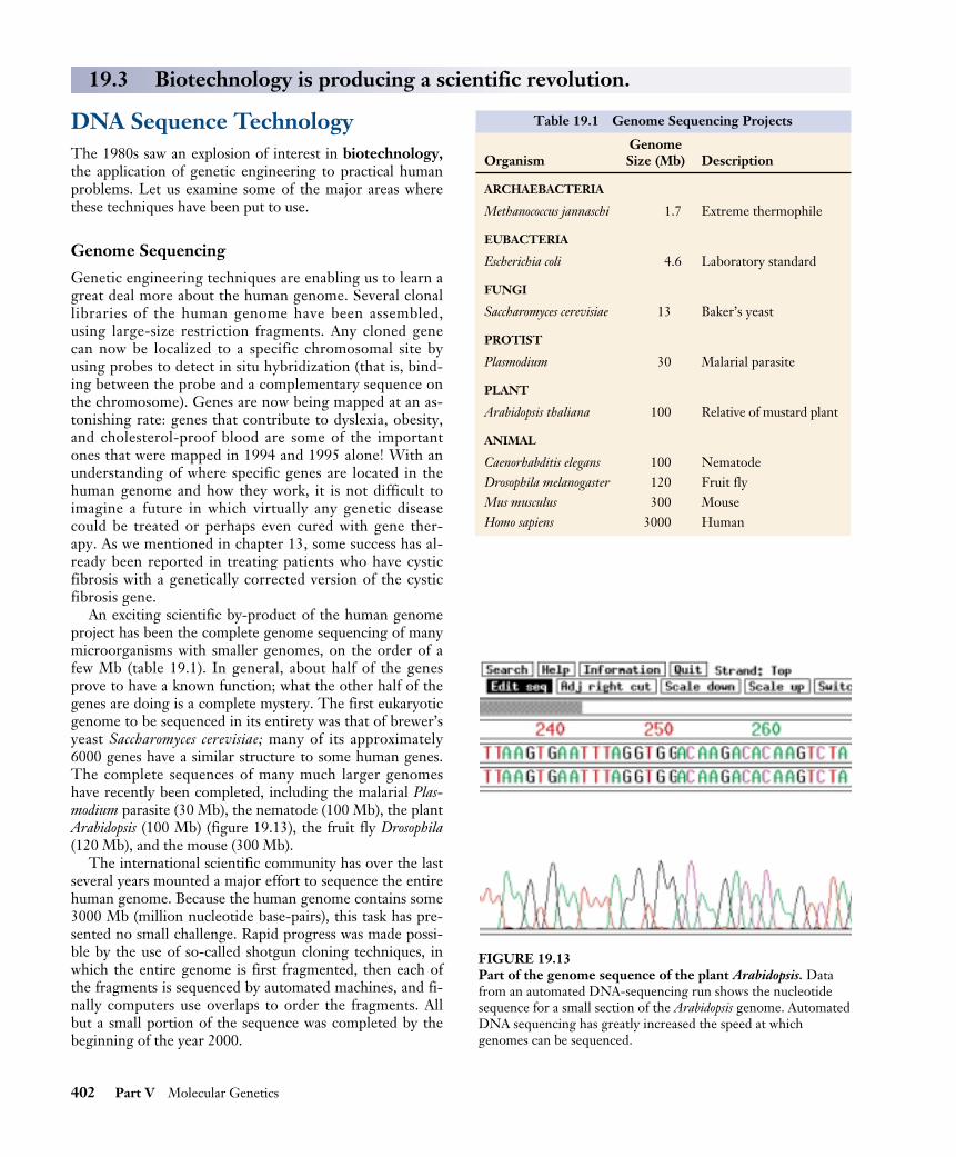
DNA Fingerprinting
Figure 19.14 shows the DNA fingerprints a prosecutingattorney presented in a rape trial in 1987. They consistedof autoradiographs, parallel bars on X-ray film resemblingthe line patterns of the universal price code found on gro-ceries. Each bar represents the position of a DNA restric-tion endonuclease fragment produced by techniques simi-lar to those described in figures 19.4 and 19.10. The lanewith many bars represents a standardized control. Twodifferent probes were used to identify the restriction frag-ments. A vaginal swab had been taken from the victimwithin hours of her attack; from it semen was collectedand the semen DNA analyzed for its restriction endonu-clease patterns.
Compare the restriction endonuclease patterns of thesemen to that of the suspect Andrews. You can see that thesuspect’s two patterns match that of the rapist (and are notat all like those of the victim). Clearly the semen collectedfrom the rape victim and the blood sample from the sus-pect came from the same person. The suspect was Tom-mie Lee Andrews, and on November 6, 1987, the jury re-turned a verdict of guilty. Andrews became the first personin the United States to be convicted of a crime based onDNA evidence.
Since the Andrews verdict, DNA fingerprinting hasbeen admitted as evidence in more than 2000 court cases(figure 19.15). While some probes highlightprofiles shared by many people, others arequite rare. Using several probes, identity canbe clearly established or ruled out.
Just as fingerprinting revolutionizedforensic evidence in the early 1900s, so DNAfingerprinting is revolutionizing it today. Ahair, a minute speck of blood, a drop ofsemen can all serve as sources of DNA todamn or clear a suspect. As the man who an-alyzed Andrews’ DNA says: “It’s like leavingyour name, address, and social security num-ber at the scene of the crime. It’s that pre-cise.” Of course, laboratory analyses of DNAsamples must be carried out properly—sloppy procedures could lead to a wrongfulconviction. After widely publicized instancesof questionable lab procedures, nationalstandards are being developed.
The genomes of several organisms havebeen completely sequenced. When DNAis digested with restrictionendonucleases, distinctive profiles onelectrophoresis gels can be used toidentify the individual that was the sourceof the tissue.
Chapter 19 Gene Technology 403
VictimRapist’s semen
Suspect’s blood
VictimRapist’s semen
Suspect’s blood
FIGURE 19.14Two of the DNA profiles that led to the conviction ofTommie Lee Andrews for rape in 1987. The two DNA probesseen here were used to characterize DNA isolated from thevictim, the semen left by the rapist, and the suspect. The darkchannels are multiband controls. There is a clear match betweenthe suspect’s DNA and the DNA of the rapist’s semen in these.
FIGURE 19.15The DNA profiles of O. J. Simpson and blood samples from the murderscene of his former wife from his highly publicized and controversialmurder trial in 1995.
BiochipsA biochip, also called a gene microarray, is a square of glasssmaller than a postage stamp, covered with millions ofstrands of DNA like blades of grass. Biochips were in-vented nine years ago by gene scientist Stephen Fodor. In aflash of insight, he saw that photolithography, the processused to etch semiconductor circuits into silicon, could alsobe used to assemble particular DNA molecules on a chip—a biochip.
Think of the chip surface as a field of assembly sites,much as a TV screen is a field of colored dots. Just as ascanning beam moves over each individual TV dot instruct-ing it to be red, green, or blue (the three components ofcolor), so a scanning beam moves over each biochip spot,commanding the addition there of a base to a growingstrand of DNA. A computer, by varying the wavelength ofthe scanning beam, determines which of four possible nu-cleotides is added to the growing DNA strand anchored toeach spot. When the entire chip has been scanned, eachDNA strand has been lengthened one nucleotide unit. Thecomputer repeats the process, layer by layer, until eachDNA strand is an entire gene or gene fragment. Onebiochip made in this way contains hundreds of thousands ofspecific gene sequences.
How could you use such a biochip to delve into a per-son’s genes? All you would have to do is to obtain a little ofthe person’s DNA, say from a blood sample or even a bit ofhair. Flush fluid containing the DNA over the biochip sur-face. Every place that the DNA has a gene matching one ofthe biochip strands, it will stick to it in a way the computercan detect.
Now here is where it gets interesting. The mad rushto sequence the human genome is over. The gene re-search firm Celera has recently announced it has essen-tially completed the sequence, with over 90% of genesdone. Already the researchers are busily comparing theirconsensus “reference sequence” to the DNA of individualpeople, and noting any differences they detect. Calledsingle nucleotide polymorphisms, or SNPs (pronounced“snips”), these spot differences in the identity of particu-lar nucleotides collectively record every way in which aparticular individual differs from the reference sequence.Some SNPs cause diseases like cystic fibrosis or sicklecell anemia. Others may give you red hair or elevatedcholesterol in your blood. As the human genome projectcharges toward completion, its researchers are excitedlyassembling a huge database of SNPs. Research indicatesthat SNPs can be expected to occur at a frequency ofabout one per thousand nucleotides, scattered about ran-domly over the chromosomes. Each of us thus differsfrom the standard "type sequence" in several thousandnucleotide SNPs. Everything genetic about you that isdiferent from a stranger you meet is caused by a fewthousand SNPs; otherwise you and that stranger areidentical.
How Biochips Can Be Used to Screen for Cancer
One of the biggest decisions facing an oncologist (cancerdoctor) treating a tumor is to select the proper treatment.Most cancer cells look alike, although the tumors may infact be caused by quite different forms of cancer. If the on-cologist could clearly identify the cancer, very targetedtherapies might be possible. Unable to tell the differencefor sure, however, oncologists take no chances. Tumors aretreated with therapy that attacks all cancers, usually withsevere side effects.
This year Boston researchers Todd Golub and EricLander took a vital step towards treating cancer, using newDNA technology to sniff out the differences between dif-ferent forms of a deadly cancer of the immune system.Golub and Lander worked with biochips.
The way to tell the difference between two kinds of can-cer is to compare the mutations that led to the cancer inthe first place. Biologists call such gene changes mutations.The mutations that cause many lung cancers are caused bya tobacco-induced alteration of a single DNA nucleotide inone gene. Such spot differences between the version of agene one person has and another person has, or a cancerpatient has, are examples of SNPs.
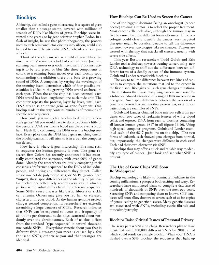
Golub and Lander obtained bone marrow cells from pa-tients with two types of leukemia (cancer of white bloodcells), and exposed DNA from each to biochips containingall known human genes, 6817 in all (figure 19.16). Usinghigh-speed computer programs, Golub and Lander exam-ined each of the 6817 positions on the chip. The twoforms of leukemia each showed gene changes from normal,but, importantly, the changes were different in each case!Each had their own characteristic SNP.
Biochips thus may offer a quick and reliable way to iden-tify any type of cancer. Just look and see what SNP ispresent.
The Use of Gene Chips Will Soon Be Widespread
Biochip technology is likely to dominate medicine in thecoming millennium, a prospect both exciting and scary. Re-searchers have announced plans to compile a database ofhundreds of thousands of SNPs over the next two years.Screening SNPs and comparing them to known SNP data-bases will soon allow doctors to screen each of us for copiesof genes leading to genetic diseases. Many genetic diseasesare associated with SNPs, including cystic fibrosis andmuscular dystrophy.
Biochips Raise Critical Issues of Personal Privacy
The scary part is SNPs on chips. Researchers plan to haveidentified some 300,000 different SNPs by 2001, all ofwhich could reside on a single biochip. When your DNA isflushed over a SNP biochip, the sequences that light up
404 Part V Molecular Genetics
will instantly reveal your SNP profile. The genetic charac-teristics that make you you, genes that might affect yourhealth, your behavior, your future potential—all are thereto be read by anyone clever enough to interpret the profile.
To what extent are you your genes? Scientists fight aboutthis question, and no one really knows the answer. It is clearthat much of what each of us is like is strongly affected byour genetic makeup. Researchers have proven beyond anyreal dispute that intelligence and major personality traitslike aggressiveness and inquisitiveness are about 80% herita-ble (that is, 80% of the variation in these traits reflects varia-tion in genes).
Your SNP profile will reflect all of this variation, a tableof contents of your chromosomes, a molecular window towho you are. When millions of such SNP profiles have been
gathered over the coming years, computers will be able toidentify other individuals with profiles like yours, and, byexamining health records, standard personality tests, and thelike, correlate parts of your profile with particular traits.Even behavioral characteristics involving many genes, whichuntil now have been thought too complex to ever analyze,cannot resist a determined assault by a computer comparingSNP profiles.
A biochip is a discrete collection of gene fragments on astamp-sized chip that can be used to screen for thepresence of particular gene variants. Biochips allowrapid screening of gene profiles, a tool that promises tohave a revolutionary impact on medicine and society.
Chapter 19 Gene Technology 405
1. DNA is obtained from thebone marrow cells of patientswith two types of leukemia.
3. High speed computer programsexamine the biochips and identifyany SNPs, or single nucleotidepolymorphisms.
4. The SNP profiles from eachtype of leukemia patient areexamined. Leukemia 1 exhibits adifferent SNP than leukemia 2.Thus, the two types of leukemiaare associated with two differentgene changes.
2. The DNA is exposed tobiochips containing all knownhuman genes.
Leukemia patient # 1
Leukemia patient # 2
Bone marrow
cells
Bone marrow
cells
DNA DNA
Biochip
Leukemia 1SNP profile
Leukemia 2SNP profile
FIGURE 19.16Biochips can help in identifying precise forms of cancer.

Medical ApplicationsPharmaceuticals
The first and perhaps most obvious commercial applica-tion of genetic engineering was the introduction of genesthat encode clinically important proteins into bacteria.Because bacterial cells can be grown cheaply in bulk (fer-mented in giant vats, like the yeasts that make beer), bac-teria that incorporate recombinant genes can synthesizelarge amounts of the proteins those genes specify. Thismethod has been used to produce several forms of humaninsulin and interferon, as well as other commerciallyvaluable proteins such as growth hormone (figure 19.17)and erythropoietin, which stimulates red blood cellproduction.
Among the medically important proteins now manufac-tured by these approaches are atrial peptides, small pro-teins that may provide a new way to treat high blood pres-sure and kidney failure. Another is tissue plasminogenactivator, a human protein synthesized in minute amountsthat causes blood clots to dissolve and may be effective inpreventing and treating heart attacks and strokes.
A problem with this general approach has been the diffi-culty of separating the desired protein from the others thebacteria make. The purification of proteins from such com-plex mixtures is both time-consuming and expensive, but itis still easier than isolating the proteins from the tissues ofanimals (for example, insulin from hog pancreases), whichis how such proteins used to be obtained. Recently, how-ever, researchers have succeeded in producing RNA tran-scripts of cloned genes; they can then use the transcripts toproduce only these proteins in a test tube containing thetranscribed RNA, ribosomes, cofactors, amino acids,tRNA, and ATP.
Gene Therapy
In 1990, researchers first attempted to combat genetic de-fects by the transfer of human genes. When a hereditarydisorder is the result of a single defective gene, an obviousway to cure the disorder is to add a working copy of thegene. This approach is being used in an attempt to combatcystic fibrosis, and it offers potential for treating musculardystrophy and a variety of other disorders (table 19.2). Oneof the first successful attempts was the transfer of a geneencoding the enzyme adenosine deaminase into the bonemarrow of two girls suffering from a rare blood disordercaused by the lack of this enzyme. However, while manyclinical trials are underway, no others have yet proven suc-cessful. This extremely promising approach will require alot of additional effort.
406 Part V Molecular Genetics
FIGURE 19.17Genetically engineered human growth hormone. These twomice are genetically identical, but the large one has one extragene: the gene encoding human growth hormone. The gene wasadded to the mouse’s genome by genetic engineers and is now astable part of the mouse’s genetic endowment.
Table 19.2 Diseases Being Treated in Clinical Trials of Gene Therapy
Disease
Cancer (melanoma, renal cell, ovarian, neuroblastoma, brain,head and neck, lung, liver, breast, colon, prostate,mesothelioma, leukemia, lymphoma, multiple myeloma)SCID (severe combined immunodeficiency)Cystic fibrosisGaucher’s diseaseFamilial hypercholesterolemiaHemophiliaPurine nucleoside phosphorylase deficiencyAlpha-1 antitrypsin deficiencyFanconi’s anemiaHunter’s syndromeChronic granulomatous diseaseRheumatoid arthritisPeripheral vascular diseaseAIDS

Piggyback Vaccines
Another area of potential significance involves the use ofgenetic engineering to produce subunit vaccines againstviruses such as those that cause herpes and hepatitis. Genesencoding part of the protein-polysaccharide coat of theherpes simplex virus or hepatitis B virus are spliced into afragment of the vaccinia (cowpox) virus genome (figure19.18). The vaccinia virus, which British physician EdwardJenner used almost 200 years ago in his pioneering vaccina-tions against smallpox, is now used as a vector to carry theherpes or hepatitis viral coat gene into cultured mammaliancells. These cells produce many copies of the recombinantvirus, which has the outside coat of a herpes or hepatitisvirus. When this recombinant virus is injected into a mouseor rabbit, the immune system of the infected animal pro-duces antibodies directed against the coat of the recombi-nant virus. It therefore develops an immunity to herpes orhepatitis virus. Vaccines produced in this way are harmlessbecause the vaccinia virus is benign and only a small frag-ment of the DNA from the disease-causing virus is intro-duced via the recombinant virus.
The great attraction of this approach is that it does not
depend upon the nature of the viral disease. In the future,similar recombinant viruses may be injected into humans toconfer resistance to a wide variety of viral diseases.
In 1995, the first clinical trials began of a novel new kindof DNA vaccine, one that depends not on antibodies butrather on the second arm of the body’s immune defense,the so-called cellular immune response, in which bloodcells known as killer T cells attack infected cells. The in-fected cells are attacked and destroyed when they stickfragments of foreign proteins onto their outer surfaces thatthe T cells detect (the discovery by Peter Doherty and RolfZinkernagel that infected cells do so led to their receivingthe Nobel Prize in Physiology or Medicine in 1996). Thefirst DNA vaccines spliced an influenza virus gene encod-ing an internal nucleoprotein into a plasmid, which wasthen injected into mice. The mice developed strong cellularimmune responses to influenza. New and controversial, theapproach offers great promise.
Genetic engineering has produced commerciallyvaluable proteins, gene therapies, and, possibly, newand powerful vaccines.
Chapter 19 Gene Technology 407
Human immuneresponse
Gene specifyingherpes simplexsurface protein
Harmless vaccinia(cowpox) virus
1. DNA is extracted. 2. Herpes simplexgene is isolated.
3. Vaccinia DNA isextracted and cleaved.
4. Fragment containingsurface gene combines with cleaved vaccinia DNA.
5. Harmless engineered virus(the vaccine) with surface likeherpes simplex is injected intothe human body.
6. Antibodies directedagainst herpes simplexviral coat are made.
Herpes simplex virus
FIGURE 19.18Strategy for constructing a subunit vaccine for herpes simplex.

Agricultural ApplicationsAnother major area of genetic engineering activity is ma-nipulation of the genes of key crop plants. In plants the pri-mary experimental difficulty has been identifying a suitablevector for introducing recombinant DNA. Plant cells donot possess the many plasmids that bacteria do, so thechoice of potential vectors is limited. The most successfulresults thus far have been obtained with the Ti (tumor-inducing) plasmid of the plant bacterium Agrobacteriumtumefaciens, which infects broadleaf plants such as tomato,tobacco, and soybean. Part of the Ti plasmid integratesinto the plant DNA, and researchers have succeeded in at-taching other genes to this portion of the plasmid (figure19.19). The characteristics of a number of plants have beenaltered using this technique, which should be valuable inimproving crops and forests. Among the features scientistswould like to affect are resistance to disease, frost, andother forms of stress; nutritional balance and protein con-tent; and herbicide resistance. Unfortunately, Agrobac-terium generally does not infect cereals such as corn, rice,and wheat, but alternative methods can be used to intro-duce new genes into them.
A recent advance in genetically manipulated fruit is Cal-gene’s “Flavr Savr” tomato, which has been approved for
sale by the USDA. The tomato has been engineered to in-hibit genes that cause cells to produce ethylene. In toma-toes and other plants, ethylene acts as a hormone to speedfruit ripening. In Flavr Savr tomatoes, inhibition of ethyl-ene production delays ripening. The result is a tomato thatcan stay on the vine longer and that resists overripeningand rotting during transport to market.
Herbicide Resistance
Recently, broadleaf plants have been genetically engineeredto be resistant to glyphosate, the active ingredient inRoundup, a powerful, biodegradable herbicide that killsmost actively growing plants (figure 19.20). Glyphosateworks by inhibiting an enzyme called EPSP synthetase,which plants require to produce aromatic amino acids. Hu-mans do not make aromatic amino acids; they get themfrom their diet, so they are unaffected by glyphosate. Tomake glyphosate-resistant plants, agricultural scientistsused a Ti plasmid to insert extra copies of the EPSP syn-thetase genes into plants. These engineered plants produce20 times the normal level of EPSP synthetase, enablingthem to synthesize proteins and grow despite glyphosate’ssuppression of the enzyme. In later experiments, a bacterialform of the EPSP synthetase gene that differs from the
408 Part V Molecular Genetics
Plant genetic engineering
Agrobacterium
Geneof interest
Plasmid
1. Plasmid is removed andcut open with restrictionendonuclease.
2. Gene is isolatedfrom the chromosomeof another organism.
3. New gene isinserted into plasmid.
4. Plasmid is putback intoAgrobacterium.
5. When mixed with plantcells, Agrobacterium duplicates the plasmid.
6. The bacterium transfersthe new gene into achromosome of theplant cell.
7. The plant cell divides,and each daughter cellreceives the new gene,giving the whole planta new trait.
FIGURE 19.19The Ti plasmid. This Agrobacterium tumefaciens plasmid is used in plant genetic engineering.

plant form by a single nucleotide was introduced intoplants via Ti plasmids; the bacterial enzyme in these plantsis not inhibited by glyphosate.
These advances are of great interest to farmers because acrop resistant to Roundup would never have to be weededif the field were simply treated with the herbicide. BecauseRoundup is a broad-spectrum herbicide, farmers would nolonger need to employ a variety of different herbicides,most of which kill only a few kinds of weeds. Furthermore,glyphosate breaks down readily in the environment, unlikemany other herbicides commonly used in agriculture. Aplasmid is actively being sought for the introduction of theEPSP synthetase gene into cereal plants, making them alsoglyphosate-resistant.
Nitrogen Fixation
A long-range goal of agricultural genetic engineering is tointroduce the genes that allow soybeans and other legumeplants to “fix” nitrogen into key crop plants. These so-callednif genes are found in certain symbiotic root-colonizingbacteria. Living in the root nodules of legumes, these bacte-ria break the powerful triple bond of atmospheric nitrogengas, converting N2 into NH3 (ammonia). The plants thenuse the ammonia to make amino acids and other nitrogen-containing molecules. Other plants lack these bacteria andcannot fix nitrogen, so they must obtain their nitrogen fromthe soil. Farmland where these crops are grown soon be-comes depleted of nitrogen, unless nitrogenous fertilizersare applied. Worldwide, farmers applied over 60 millionmetric tons of such fertilizers in 1987, an expensive under-taking. Farming costs would be much lower if major cropslike wheat and corn could be engineered to carry out bio-logical nitrogen fixation. However, introducing thenitrogen-fixing genes from bacteria into plants has proveddifficult because these genes do not seem to function prop-erly in eukaryotic cells. Researchers are actively experiment-ing with other species of nitrogen-fixing bacteria whosegenes might function better in plant cells.
Insect Resistance
Many commercially important plants are attacked by in-sects, and the traditional defense against such attacks is toapply insecticides. Over 40% of the chemical insecticidesused today are targeted against boll weevils, bollworms, andother insects that eat cotton plants. Genetic engineers arenow attempting to produce plants that are resistant to in-sect pests, removing the need to use many externally ap-plied insecticides.
The approach is to insert into crop plants genes encod-ing proteins that are harmful to the insects that feed on theplants but harmless to other organisms. One such insectici-dal protein has been identified in Bacillus thuringiensis, a soilbacterium. When the tomato hornworm caterpillar ingeststhis protein, enzymes in the caterpillar’s stomach convert it
into an insect-specific toxin, causing paralysis and death.Because these enzymes are not found in other animals, theprotein is harmless to them. Using the Ti plasmid, scien-tists have transferred the gene encoding this protein intotomato and tobacco plants. They have found that thesetransgenic plants are indeed protected from attack by theinsects that would normally feed on them. In 1995, theEPA approved altered forms of potato, cotton, and corn.The genetically altered potato can kill the Colorado potatobeetle, a common pest. The altered cotton is resistant tocotton bollworm, budworm, and pink bollworm. The cornhas been altered to resist the European corn borer andother mothlike insects.
Monsanto scientists screening natural compounds ex-tracted from plant and soil samples have recently isolated anew insect-killing compound from a fungus, the enzymecholesterol oxidase. Apparently, the enzyme disrupts mem-branes in the insect gut. The fungus gene, called the Boll-gard gene after its discoverer, has been successfully insertedinto a variety of crops. It kills a wide range of insects, in-cluding the cotton boll weevil and the Colorado potatobeetle, both serious agricultural pests. Field tests began in1996.
Some insect pests attack plant roots, and B. thuringiensisis being employed to counter that threat as well. This bac-terium does not normally colonize plant roots, so biologistshave introduced the B. thuringiensis insecticidal proteingene into root-colonizing bacteria, especially strains ofPseudomonas. Field testing of this promising procedure hasbeen approved by the Environmental Protection Agency.
Chapter 19 Gene Technology 409
FIGURE 19.20Genetically engineered herbicide resistance. All four of thesepetunia plants were exposed to equal doses of the herbicideRoundup. The two on top were genetically engineered to beresistant to glyphosate, the active ingredient of Roundup, whilethe two on the bottom were not.
The Real Promise of Plant Genetic Engineering
In the last decade the cultivation of genetically modifiedcrops of corn, cotton, and soybeans has become com-monplace in the United States—in 1999, over half of the72 million acres planted with soybeans in the UnitedStates were planted with seeds genetically modified to beherbicide resistant, with the result that less tillage hasbeen needed, and as a consequence soil erosion has beengreatly lessened. These benefits, while significant, havebeen largely confined to farmers, making their cultivationof crops cheaper and more efficient. The food that thepublic gets is the same, it just costs less to get it to thetable.
Like the first act of a play, these developments haveserved mainly to set the stage for the real action, which isonly now beginning to happen. The real promise of plantgenetic engineering is to produce genetically modifiedplants with desirable traits that directly benefit the con-sumer.
One recent advance, nutritionally improved rice, givesus a hint of what is to come. In developing countries largenumbers of people live on simple diets that are poorsources of vitamins and minerals (what botanists called"micronutrients"). Worldwide, the two major micronutri-ent deficiencies are iron, which affects 1.4 billion women,24% of the world population, and vitamin A, affecting 40million children, 7% of the world population. The defi-ciencies are especially severe in developing countries wherethe major staple food is rice. In recent research, Swiss bio-engineer Ingo Potrykus and his team at the Institute ofPlant Sciences, Zurich, have gone a long way towards solv-ing this problem. Supported by the Rockefeller Founda-tion and with results to be made free to developing coun-tries, the work is a model of what plant genetic engineeringcan achieve.
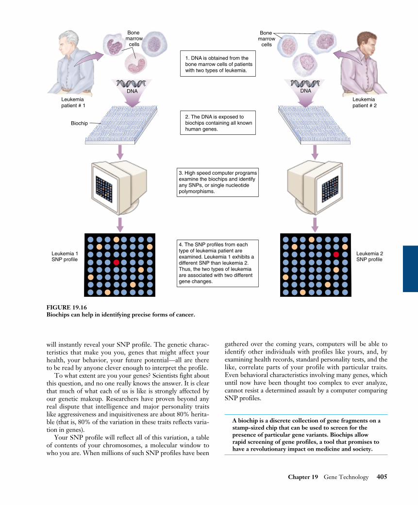
To solve the problem of dietary iron deficiency amongrice eaters, Potrykus first asked why rice is such a poorsource of dietary iron. The problem, and the answer,proved to have three parts:
1. Too little iron. The proteins of rice endosperm haveunusually low amounts of iron. To solve this prob-lem, a ferritin gene was transferred into rice frombeans (figure 19.21). Ferritin is a protein with an ex-traordinarily high iron content, and so greatly in-creased the iron content of the rice.
2. Inhibition of iron absorption by the intestine. Rice con-tains an unusually high concentration of a chemicalcalled phytate, which inhibits iron reabsorption in theintestine—it stops your body from taking up the ironin the rice. To solve this problem, a gene encoding anenzyme that destroys phytate was transferred into ricefrom a fungus.
3. Too little sulfur for efficient iron absorption. Sulfur isrequired for iron uptake, and rice has very little of it.To solve this problem, a gene encoding a particularlysulfur-rich metallothionin protein was transferredinto rice from wild rice.
To solve the problem of vitamin A deficiency, the sameapproach was taken. First, the problem was identified. Itturns out rice only goes part way toward making beta-carotene (provitamin A); there are no enzymes in rice tocatalyze the last four steps. To solve the problem, genes en-coding these four enzymes were added to rice from a famil-iar flower, the daffodil.
Potrykus's development of transgenic rice to combatdietary deficiencies involved no subtle tricks, juststraightforward bioengineering and the will to get the jobdone. The transgenic rice he has developed will directlyimprove the lives of millions of people. His work is rep-
410 Part V Molecular Genetics
Daffodil
Ferritin gene istransferred intorice from beans.
Phytase gene istransferred intorice from a fungus.
Metallothionin geneis transferred intorice from wild rice.
Enzymes for beta-carotenesynthesis are transferredinto rice from daffodils.
Fe Pt SRicechromosome A1 A2 A3 A4
Ferritin proteinincreases ironcontent of rice.
Phytate, which inhibits iron reabsorption,is destroyed by the phytase enzyme.
Metallothionin proteinsupplies extra sulfur to increase iron uptake.
Beta-carotene, a precursor to vitamin A,is synthesized.
Beans Aspergillus fungus Wild rice
FIGURE 19.21Transgenic rice. Developedby Swiss bioengineer IngoPotrykus, transgenic riceoffers the promise ofimproving the diets ofpeople in rice-consumingdeveloping countries, whereiron and vitamin Adeficiencies are a seriousproblem.

resentative of the very real promise of genetic engineer-ing to help meet the challenges of the coming newmillennium.
The list of gene modifications that directly aid con-sumers will only grow. In Holland, Dutch bioengineers an-nounced last month that they are genetically engineeringplants to act as vaccine-producing factories! To petuniasthey have added a gene for a vaccine against dog par-vovirus, hiding the gene within the petunia genes that di-rect nectar production. The drug is produced in the nec-tar, collected by bees, and extracted from the honey. It ishard to believe this isn't science fiction. Clearly, the realpromise of plant genetic engineering lies ahead, and notvery far.
Farm Animals
The gene encoding the growth hormone somatotropinwas one of the first to be cloned successfully. In 1994,Monsanto received federal approval to make its recombi-nant bovine somatotropin (BST) commercially available,and dairy farmers worldwide began to add the hormoneas a supplement to their cows’ diets, increasing the ani-mals’ milk production (figure 19.22). Genetically engi-
neered somatotropin is also being tested to see if it in-creases the muscle weight of cattle and pigs, and as atreatment for human disorders in which the pituitarygland fails to make adequate levels of somatotropin, pro-ducing dwarfism. BST ingested in milk or meat has noeffect on humans, because it is a protein and is digestedin the stomach. Nevertheless, BST has met with somepublic resistance, due primarily to generalized fears ofgene technology. Some people mistrust milk producedthrough genetic engineering, even though the milk itselfis identical to other milk. Problems concerning publicperception are not uncommon as gene technology makesan even greater impact on our lives.
Transgenic animals engineered to have specific desirablegenes are becoming increasingly available to breeders.Now, instead of selectively breeding for several generationsto produce a racehorse or a stud bull with desirable quali-ties, the process can be shortened by simply engineeringsuch an animal right at the start.
Gene technology is revolutionizing agriculture,increasing yields and resistance to pests, and producinganimals with desirable traits.
Chapter 19 Gene Technology 411
Bovine somatotropinproduction
Escherichia coli
Geneof interest
Cow DNA
Plasmid
1. Plasmid is removedand cut open with restriction endonuclease.
2. Cow somatotropingene is isolatedfrom cow cell.
3. Somatotropin gene isinserted into bacterialplasmid.
4. Plasmid is reintroducedinto bacterium.
5. Bacteria producingbovine somatotropinare grown infermentation tanks.
6. Somatotropin isremoved frombacteria and purified.
7. Bovine somatotropinis administered tocow to enhancemilk production.
FIGURE 19.22The production of bovine somatotropin (BST) through geneticengineering. Although BST is functional, harmless, and sanctioned by theFDA, much controversy exists over whether it is actually desirable.

CloningThe difficulty in using transgenic animals to improve live-stock is in getting enough of them. Breeding produces off-spring only slowly, and recombination acts to undo thepainstaking work of the genetic engineer. Ideally, onewould like to “Xerox” many exact genetic copies of thetransgenic strain—but until 1997 it was commonly ac-cepted that adult animals can’t be cloned. Now the holygrail of agricultural genetic engineers seems within reach.In 1997, scientists announced the first successful cloning ofdifferentiated vertebrate tissue, a lamb grown from a celltaken from an adult sheep. This startling result promises torevolutionize agricultural science.
Spemann’s “Fantastical Experiment”
The idea of cloning animals was first suggested in 1938 byGerman embryologist Hans Spemann (called the “father ofmodern embryology”), who proposed what he called a“fantastical experiment”: remove the nucleus from an eggcell, and put in its place a nucleus from another cell.
It was 14 years before technology advanced far enoughfor anyone to take up Spemann’s challenge. In 1952, twoAmerican scientists, Robert Briggs and T. J. King, usedvery fine pipettes to suck the nucleus from a frog egg (frogeggs are unusually large, making the experiment feasible)and transfer a nucleus sucked from a body cell of an adultfrog into its place. The experiment did not work whendone this way, but partial success was achieved 18 yearslater by the British developmental biologist John Gurdon,who in 1970 inserted nuclei from advanced frog embryosrather than adult tissue. The frog eggs developed into tad-poles, but died before becoming adults.
The Path to Success
For 14 years, nuclear transplant experiments were at-tempted without success. Technology continued to advancehowever, until finally in 1984, Steen Willadsen, a Danishembryologist working in Texas, succeeded in cloning asheep using a nucleus from a cell of an early embryo. Thisexciting result was soon replicated by others in a host ofother organisms, including cattle, pigs, and monkeys.
Only early embryo cells seemed to work, however. Re-searchers became convinced that animal embryo cells be-come irreversibly “committed” after the first few cell divi-sions. After that, nuclei from differentiated animal cellscannot be used to clone entire organisms.
We now know this conclusion to have been unwar-ranted. The key advance for unraveling this puzzle wasmade in Scotland by geneticist Keith Campbell, a specialistin studying the cell cycle of agricultural animals. By theearly 1990s, knowledge of how the cell cycle is controlled,advanced by cancer research, had led to an understandingthat cells don’t divide until conditions are appropriate. Justas a washing machine checks that the water has completelyemptied before initiating the spin cycle, so the cell checksthat everything needed is on hand before initiating cell di-vision. Campbell reasoned: “Maybe the egg and the do-nated nucleus need to be at the same stage in the cellcycle.”
This proved to be a key insight. In 1994 researcher NeilFirst, and in 1995 Campbell himself working with repro-ductive biologist Ian Wilmut, succeeded in cloning farmanimals from advanced embryos by first starving the cells,so that they paused at the beginning of the cell cycle at theG1 checkpoint. Two starved cells are thus synchronized atthe same point in the cell cycle.
412 Part V Molecular Genetics
Nucleus containingsource DNA
Mammary cell is extractedand grown in nutrient-deficient solution that arreststhe cell cycle.
Egg cell is extracted.Nucleus is removedfrom egg cell with amicropipette.
Mammary cell is insertedinside covering of egg cell.
Electric shock opens cellmembranes and triggerscell division.
Preparation Cell fusion Cell division
FIGURE 19.23Wilmut’s animal cloning experiment. Wilmut combined a nucleus from a mammary cell and an egg cell (with its nucleus removed) tosuccessfully clone a sheep.
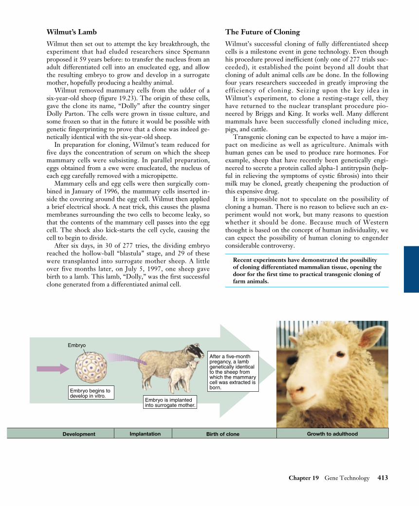
Wilmut’s Lamb
Wilmut then set out to attempt the key breakthrough, theexperiment that had eluded researchers since Spemannproposed it 59 years before: to transfer the nucleus from anadult differentiated cell into an enucleated egg, and allowthe resulting embryo to grow and develop in a surrogatemother, hopefully producing a healthy animal.
Wilmut removed mammary cells from the udder of asix-year-old sheep (figure 19.23). The origin of these cells,gave the clone its name, “Dolly” after the country singerDolly Parton. The cells were grown in tissue culture, andsome frozen so that in the future it would be possible withgenetic fingerprinting to prove that a clone was indeed ge-netically identical with the six-year-old sheep.
In preparation for cloning, Wilmut’s team reduced forfive days the concentration of serum on which the sheepmammary cells were subsisting. In parallel preparation,eggs obtained from a ewe were enucleated, the nucleus ofeach egg carefully removed with a micropipette.
Mammary cells and egg cells were then surgically com-bined in January of 1996, the mammary cells inserted in-side the covering around the egg cell. Wilmut then applieda brief electrical shock. A neat trick, this causes the plasmamembranes surrounding the two cells to become leaky, sothat the contents of the mammary cell passes into the eggcell. The shock also kick-starts the cell cycle, causing thecell to begin to divide.
After six days, in 30 of 277 tries, the dividing embryoreached the hollow-ball “blastula” stage, and 29 of thesewere transplanted into surrogate mother sheep. A littleover five months later, on July 5, 1997, one sheep gavebirth to a lamb. This lamb, “Dolly,” was the first successfulclone generated from a differentiated animal cell.
The Future of Cloning
Wilmut’s successful cloning of fully differentiated sheepcells is a milestone event in gene technology. Even thoughhis procedure proved inefficient (only one of 277 trials suc-ceeded), it established the point beyond all doubt thatcloning of adult animal cells can be done. In the followingfour years researchers succeeded in greatly improving theefficiency of cloning. Seizing upon the key idea inWilmut’s experiment, to clone a resting-stage cell, theyhave returned to the nuclear transplant procedure pio-neered by Briggs and King. It works well. Many differentmammals have been successfully cloned including mice,pigs, and cattle.
Transgenic cloning can be expected to have a major im-pact on medicine as well as agriculture. Animals withhuman genes can be used to produce rare hormones. Forexample, sheep that have recently been genetically engi-neered to secrete a protein called alpha-1 antitrypsin (help-ful in relieving the symptoms of cystic fibrosis) into theirmilk may be cloned, greatly cheapening the production ofthis expensive drug.
It is impossible not to speculate on the possibility ofcloning a human. There is no reason to believe such an ex-periment would not work, but many reasons to questionwhether it should be done. Because much of Westernthought is based on the concept of human individuality, wecan expect the possibility of human cloning to engenderconsiderable controversy.
Recent experiments have demonstrated the possibilityof cloning differentiated mammalian tissue, opening thedoor for the first time to practical transgenic cloning offarm animals.
Chapter 19 Gene Technology 413
Embryo
Embryo is implantedinto surrogate mother.
Embryo begins todevelop in vitro.
After a five-monthpregancy, a lambgenetically identicalto the sheep fromwhich the mammarycell was extracted isborn.
Development Implantation Birth of clone Growth to adulthood

Stem CellsSince the isolation of embryonic stem cells in 1998, labs allover the world have been exploring the possibility of usingstem cells to restore damaged or lost tissue. Exciting resultsare now starting to come in.
What is a stem cell? At the dawn of a human life, asperm fertilizes an egg to create a single cell destined to be-come a child. As development commences, that cell beginsto divide, producing a small ball of a few dozen cells. Atthis very early point, each of these cells is identical. We callthese cells embryonic stem cells. Each one of them is capableby itself of developing into a healthy individual. In cattlebreeding, for example, these cells are frequently separatedby the breeder and used to produce multiple clones of valu-able offspring.
The exciting promise of these embryonic stem cells isthat, because they can develop into any tissue, they maygive us the ability to restore damaged heart or spine tissue(figure 19.24). Experiments have already been tried suc-cessfully in mice. Heart muscle cells have been grown frommouse embryonic stem cells and successfully integratedwith the heart tissue of a living mouse. This suggests thatthe damaged heart muscle of heart attack victims might bereparable with stem cells, and that injured spinal cordsmight be repairable. These very promising experiments arebeing pursued aggressively. They are, however, quite con-troversial, as embryonic stem cells are typically isolatedfrom tissue of discarded or aborted embryos, raising seriousethical issues.
Tissue-Specific Stem Cells
New results promise a neat way around the ethical mazepresented by stem cells derived from embryos. Go back for
a moment to what we were saying about how a humanchild develops. What happens next to the embryonic stemcells? They start to take different developmental paths.Some become destined to form nerve tissue and, after thisdecision is taken, cannot ever produce any other kind ofcell. They are then called nerve stem cells. Others becomespecialized to produce blood, still others muscle. Eachmajor tissue is represented by its own kind of tissue-specificstem cell. Now here’s the key point: as development pro-ceeds, these tissue-specific stem cells persist. Even inadults. So why not use these adult cells, rather than embry-onic stem cells?
Transplanted Tissue-Specific Stem Cells CureMS in Mice
In pathfinding 1999 laboratory experiments by Dr. EvanSnyder of Harvard Medical School, tissue-specific stemcells were able to restore lost brain tissue. He and his co-workers injected neural stem cells (immediate descendantsof embryonic stem cells able to become any kind of neuralcell) into the brains of newborn mice with a disease resem-bling multiple sclerosis (MS). These mice lacked the cellsthat maintain the layers of myelin insulation around signal-conducting nerves. The injected stem cells migrated allover the brain, and were able to convert themselves intothe missing type of cell. The new cells then proceeded torepair the ravages of the disease by replacing the lost insu-lation of signal-conducting nerve cells. Many of the treatedmice fully recovered. In mice at least, tissue-specific stemcells offer a treatment for MS.
The approach seems very straightforward, and shouldapply to humans. Indeed, blood stem cells are already rou-tinely used in humans to replenish the bone marrow of can-cer patients after marrow-destroying therapy. The problem
414 Part V Molecular Genetics
Once sperm cell and eggcell have joined, cell cleavageproduces a blastocyst. Theinner cell mass of theblastocyst develops into the human embryo.
Biologists have culturedembryonic stem cells fromboth the inner cell mass andembryonic germ cells, whichescape early differentiation.
Egg
Sperm
Blastocyst
Embryo
Embryonic stem-cellcultureInner cell
mass
FIGURE 19.24Using embryonic stem cells to restoredamaged tissue. Embyronic stem cells candevelop into any body tissue. Methods forgrowing the tissue and using it to repairdamaged tissue in adults, such as the braincells of multiple sclerosis patients, heartmuscle, and spinal nerves, are beingdeveloped.

with extending the approach to other kinds of tissue-specific stem cells is that it has not always been easy to findthe kind of tissue-specific stem cell you want.
Transplanted Stem Cells Reverse JuvenileDiabetes in Mice
Very promising experiments carried out in 2000 by Dr.Ammon Peck and a team of researchers at the University ofFlorida concern a particularly vexing problem, that oftype 1 or juvenile diabetes. A person with juvenile diabeteslacks insulin-producing pancreas cells, because their im-mune system has mistakenly turned against them and de-stroyed them. They are no longer able to produce enoughinsulin to control their blood sugar levels and must take in-sulin daily. Adding back new insulin-producing cells calledislet cells has been tried many times, but doesn’t work well.Immune cells continue to destroy them.
Peck and his team reasoned, why not add instead thestem cells that produce islet cells? They would be able toproduce a continuous supply of new islet cells, replacingthose lost to immune attack. Because there would always becells to make insulin, the diabetes would be cured.
No one knew just what such a stem cell looked like, butthe researchers knew they come from the epithelial cellsthat line the pancreas ducts. Surely some must still lurkthere unseen. So the research team took a bunch of theseepithelial cells from mice and grew them in tissue cultureuntil they had lots of them.
Were the stem cells they sought present in the cell cul-ture they had prepared? Yes. In laboratory dishes the cellculture produced insulin in response to sugar, indicatingislet cells had developed in the growing culture, islet cellsthat must have been produced from stem cells.
Now on to juvenile diabetes. The scientists injectedtheir cell culture into the pancreas of mice specially bred todevelop juvenile diabetes. Unable to manufacture their owninsulin because they had no islet cells, these diabetic micecould not survive without daily insulin. What happened?The diabetes was reversed! The mice no longer requiredinsulin.
Impatient to see in more detail what had happened, theresearchers sacrificed the mice and examined the cells oftheir pancreas. The mice appeared to have perfectly normalislet cells.
One might have wished the researchers waited a littlelonger before terminating the experiment. It is not clearwhether the cure was transitory or long term. Still, there isno escaping the conclusion that injection of a culture ofadult stem cells cured their juvenile diabetes.
While certainly encouraging, a mouse is not a human,and there is no guarantee the approach will work in hu-mans. But there is every reason to believe it might. The ex-periment is being repeated now with humans. People suf-fering from juvenile diabetes are being treated with humanpancreatic duct cells obtained from people who have diedand donated their organs for research. No ethical issuesarise from using cells of adult organ donors, and initial re-sults look promising.
Transplanted stem cells may allow us to replacedamaged or lost tissue, offering cures for manydisorders that cannot now be treated. Current workfocuses on tissue-specific stem cells, which do notpresent the ethical problems that embryonic stemcells do.
Chapter 19 Gene Technology 415
For use in therapy, theembryonic stem cells aregenetically engineered to matchthe patient's immune system:the stem cells' self-recognitiongenes are replaced with thepatient's self-recognition genes.
The stem cells are grown toproduce whatever type of tissueis needed by the patient.
The tissue cells are injected intothe patient where needed. Oncein place, the tissue cellsrespond to local chemicalsignals, adding to or replacingdamaged cells.
Embryonicstem cell
Patient
Tissue cells
Patient's self-recognition genes

Ethics and RegulationThe advantages afforded by genetic engineering are revolu-tionizing our lives. But what are the disadvantages, the po-tential costs and dangers of genetic engineering? Manypeople, including influential activists and members of thescientific community, have expressed concern that geneticengineers are “playing God” by tampering with geneticmaterial. For instance, what would happen if one frag-mented the DNA of a cancer cell, and then incorporatedthe fragments at random into vectors that were propagatedwithin bacterial cells? Might there not be a danger thatsome of the resulting bacteria would transmit an infectiveform of cancer? Could genetically engineered products ad-ministered to plants or animals turn out to be dangerousfor consumers after several generations? What kind of un-foreseen impact on the ecosystem might “improved” cropshave? Is it ethical to create “genetically superior” organ-isms, including humans?
How Do We Measure the Potential Risks ofGenetically Modified Crops?
While the promise of genetic engineering is very much inevidence, this same genetic engineering has this summerbeen the cause of outright war between researchers andprotesters in England. In June 1999, British protesters at-tacked an experimental plot of genetically modified (GM)sugar beets; the following August they destroyed a test fieldof GM canola (used for cooking oil and animal feed). Thecontrast could not be more marked between American ac-ceptance of genetically modified crops on the one hand,and European distrust of genetically modified foods, on theother. The intense feelings generated by this dispute pointto the need to understand how we measure the risks asso-ciated with the genetic engineering of plants.
Two sets of risks need to be considered. The first stemsfrom eating genetically modified foods, the other concernspotential ecological effects.
Is Eating Genetically Modified Food Dangerous? Pro-testers worry that genetically modified food may have beenrendered somehow dangerous. To sort this out, it is usefulto bear in mind that bioengineers modify crops in two quitedifferent ways. One class of gene modification makes thecrop easier to grow; a second class of modification is in-tended to improve the food itself.
The introduction of Roundup-resistant soybeans to Eu-rope is an example of the first class of modification. Thismodification has been very popular with farmers in theUnited States, who planted half their crop with these soy-beans in 1999. They like GM soybeans because the beanscan be raised without intense cultivation (weeds are killedwith Roundup herbicide instead), which both saves moneyand lessens soil erosion. But is the soybean that results nu-tritionally different? No. The gene that confers Roundupresistance in soybeans does so by protecting the plant's
ability to manufacture so-called "aromatic" amino acids. Inunprotected weeds, by contrast, Roundup blocks this man-ufacturing process, killing the weed. Because humans don'tmake any aromatic amino acids anyway (we get them in ourdiets), Roundup doesn't hurt us. The GM soybean we eat isnutritionally the same as an "organic" one, just cheaper toproduce.
In the second class of modification, where a gene isadded to improve the nutritional character of some food,the food will be nutritionally different. In each of these in-stances, it is necessary to examine the possibility that con-sumers may prove allergic to the product of the intro-duced gene. In one instance, for example, addition of amethionine-enhancing gene from Brazil nut into soybeans(which are deficient in this amino acid) was discontinuedwhen six of eight individuals allergic to Brazil nuts pro-duced antibodies to the GM soybeans, suggesting the pos-sibility of a reverse reaction. Instead, methionine levels inGM crops are being increased with genes from sunflowers.Screening for allergy problems is now routine.
On both scores, then, the risk of bioengineering to thefood supply seems to be very slight. GM foods to dateseem completely safe.
Are GM Crops Harmful to the Environment? Whatare we to make of the much-publicized report thatMonarch butterflies might be killed by eating pollen blow-ing out of fields planted with GM corn? First, it shouldcome as no surprise. The GM corn (so-called Bt corn) wasengineered to contain an insect-killing toxin (harmless topeople) in order to combat corn borer pests. Of course itwill kill any butterflies or other insects in the immediatevicinity of the field. However, focus on the fact that theGM corn fields do not need to be sprayed with pesticide tocontrol the corn borer. An estimated $9 billion in damageis caused annually by the application of pesticides in theUnited States, and billions of insects and other animals, in-cluding an estimated 67 million birds, are killed each year.This pesticide-induced murder of wildlife is far more dam-aging ecologically than any possible effects of GM crops onbutterflies.
Will pests become resistant to the GM toxin? Notnearly as fast as they now become resistant to the far higherlevels of chemical pesticide we spray on crops.
How about the possibility that introduced genes willpass from GM crops to their wild or weedy relatives? Thissort of gene flow happens naturally all the time, and so thisis a legitimate question. But so what if genes for resistanceto Roundup herbicide spread from cultivated sugar beets towild populations of sugar beets in Europe? Why would thatbe a problem? Besides, there is almost never a potential rel-ative around to receive the modified gene from the GMcrop. There are no wild relatives of soybeans in Europe, forexample. Thus, there can be no gene escape from GM soy-beans in Europe, any more than genes can flow from you toother kinds of animals.
416 Part V Molecular Genetics

On either score, then, the risk of bioengineering to theenvironment seems to be very slight. Indeed, in some casesit lessens the serious environmental damage produced bycultivation and agricultural pesticides.
Should We Label Genetically Modified Foods?
While there seems little tangible risk in the genetic modifi-cation of crops, public assurance that these risks are beingcarefully assessed is important. Few issues manage to raisethe temperature of discussions about plant genetic engi-neering more than labeling of genetically modified (GM)crops. Agricultural producers have argued that there are nodemonstrable risks, so that a GM label can only have thefunction of scaring off wary consumers. Consumer advo-cates respond that consumers have every right to make thatdecision, and to the information necessary to make it.
In considering this matter, it is important to separatetwo quite different issues, the need for a label, and the rightof the public to have one. Every serious scientific investi-gation of the risks of GM foods has concluded that they aresafe—indeed, in the case of soybeans and many other cropsmodified to improve cultivation, the foods themselves arenot altered in any detectable way, and no nutritional testcould distinguish them from "organic" varieties. So thereseems to be little if any health need for a GM label for ge-netically engineered foods.
The right of the public to know what it is eating is a verydifferent issue. There is widespread fear of genetic manip-ulation in Europe, because it is unfamiliar. People theredon't trust their regulatory agencies as we do here, becausetheir agencies have a poor track record of protecting them.When they look at genetically modified foods, they arehaunted by past experiences of regulatory ineptitude. InEngland they remember British regulators' failure to pro-tect consumers from meat infected with mad cow disease.
It does no good whatsoever to tell a fearful Europeanthat there is no evidence to warrant fear, no trace of datasupporting danger from GM crops. A European consumerwill simply respond that the harm is not yet evident, that
we don't know enough to see the danger lurking aroundthe corner. "Slow down," the European consumers say."Give research a chance to look around all the corners.Lets be sure." No one can argue against caution, but it isdifficult to imagine what else researchers can look into—safety has been explored very thoroughly. The fear re-mains, though, for the simple reason that no amount of in-formation can remove it. Like a child scared of a monsterunder the bed, looking under the bed again doesn't help—the monster still might be there next time. And that meanswe are going to have to have GM labels, for people haveevery right to be informed about something they fear.
What should these labels be like? A label that only says"GM FOOD" simply acts as a brand—like a POISONlabel, it shouts a warning to the public of lurking danger.Why not instead have a GM label that provides informa-tion to the consumer, that tells the consumer what regula-tors know about that product?
(For Bt corn): The production of this food was mademore efficient by the addition of genes that made plantsresistant to pests so that less pesticides were required togrow the crop. (For Roundup-ready soybeans): Genes have been addedto this crop to render it resistant to herbicides—this re-duces soil erosion by lessening the need for weed-removing cultivation.(For high beta-carotene rice): Genes have been added tothis food to enhance its beta-carotene content and socombat vitamin A deficiency.
GM food labels that in each instance actually tell con-sumers what has been done to the gene-modified cropwould go a long way toward hastening public acceptance ofgene technology in the kitchen.
Genetic engineering affords great opportunities forprogress in medicine and food production, althoughmany are concerned about possible risks. On balance,the risks appear slight, and the potential benefitssubstantial.
Chapter 19 Gene Technology 417
CALVIN AND HOBBES©1995 Watterson. Dist. byUniversal Press Syndicate.Reprinted with permission.All rights reserved.

418 Part V Molecular Genetics
Chapter 19 Summary Questions Media Resources
19.1 The ability to manipulate DNA has led to a new genetics.
• Genetic engineering involves the isolation of specificgenes and their transfer to new genomes.
• An important component of genetic engineeringtechnology is a special class of enzymes calledrestriction endonucleases, which cleave DNAmolecules into fragments.
• The first such recombinant DNA was made byCohen and Boyer in 1973, when they inserted a frogribosomal RNA gene into a bacterial plasmid.
1. Why do the ends of the DNAfragments created by restrictionendonucleases enable fragmentsfrom different genomes to bespliced together?
• Genetic engineering experiments consist of fourstages: isolation of DNA, production of recombinantDNA, cloning, and screening for the gene(s) ofinterest.
• Preliminary screening can be accomplished bymaking the desired clones resistant to an antibiotic;hybridization can then be employed to identify thegene of interest.
• Gene technologies, including PCR, Southernblotting, RFLP analysis, and the Sanger method,enable researchers to isolate genes and produce themin large quantities.
2. Describe the procedure usedto eliminate clones that have notincorporated a vector in agenetic engineering experiment. 3. What is used as a probe in aSouthern blot? With what doesthe probe hybridize? How arethe regions of hybridizationvisualized?
19.2 Genetic engineering involves easily understood procedures.
• Extensive research on the human genome has yieldedimportant information about the location of genes,such as those that may be involved in dyslexia,obesity, and resistance to high blood cholesterollevels.
• Gene splicing holds great promise as a clinical tool,particularly in the prevention of disease withbioengineered vaccines.
• A major focus of genetic engineering activity has beenagriculture, where genes conferring resistance toherbicides or insect pests have been incorporated intocrop plants.
• Recent experiments open the way for cloning ofgenetically altered animals and suggest that humancloning is feasible.
• The impact of genetic engineering has skyrocketedover the past decade, providing many usefulinnovations for society; its moral and ethical aspectsstill provide a topic for heated debates.
4. What is the primary vectorused to introduce genes intoplant cells? What types of plantsare generally infected by thisvector? Describe three examplesof how this vector has been usedfor genetic engineering, andexplain the agriculturalsignificance of each example.5. How is the geneticengineering of bovinesomatotropin (BST) used toincrease milk production in thedairy industry? What effectwould BST in milk have onpersons who drink it?
19.3 Biotechnology is producing a scientific revolution.
www.mhhe.com www.biocourse.com
• Experiment:Cohen/Boyer/Berg-The first GeneticallyEngineered Organism
• Student Research:Homeobox Genes inthe Medicinal Leech
• Polymerase ChainReaction
• Recombinant
• On Science Article:How GeneticEngineering is Done
• Exploration: DNAfrom Real Court Cases
On Science Articles:• The Real Promise of
Plant GeneticEngineering
• Should We LabelGenetically ModifiedFoods?
• Measuring Risks ofGenetically ModifiedCrops
• The Search for theNatural Relatives ofCassava
• Renouncing theTerminator
• Frankenstein Grass isPoised to Invade myBack Yard
• The Road to Dolly• Should a Clone Have
Rights?• Who Should Own the
Secrets of Your Genes?