SQSTM1 gene analysis and gene-environment interaction in Paget's disease of bone
10
SQSTM1 Gene Analysis and Gene-Environment Interaction in Paget’s Disease of Bone Luigi Gennari, 1 Fernando Gianfrancesco, 2 Marco Di Stefano, 3 Domenico Rendina, 4 Daniela Merlotti, 1 Teresa Esposito , 2 Salvatore Gallone , 5 Pina Fusco , 2 Innocenzo Rainero , 5 Pierpaola Fenoglio , 5 Maria Mancini , 2 Giuseppe Martini , 1 Simona Bergui , 3 Gianpaolo De Filippo , 6 Giancarlo Isaia , 3 Pasquale Strazzullo , 4 Ranuccio Nuti , 1 and Giuseppe Mossetti 4 1 Department of Internal Medicine, Endocrine-Metabolic Sciences and Biochemistry, University of Siena, Siena, Italy 2 Institute of Genetics and Biophysics, CNR, Naples, Italy 3 Department of Internal Medicine, University of Turin, Turin, Italy 4 Department of Clinical and Experimental Medicine, Federico II University of Naples, Naples, Italy 5 Department of Neuroscience, University of Turin, Turin, Italy 6 Unit of Pediatric Endocrinology, A.O.R.N. Rummo, Benevento, Italy ABSTRACT Even though SQSTM1 gene mutations have been identified in a consistent number of patients, the etiology of Paget’s disease of bone (PDB) remains in part unknown. In this study we analyzed SQSTM1 mutations in 533 of 608 consecutive PDB patients from several regions, including the high-prevalence area of Campania (also characterized by increased severity of PDB, higher number of familial cases, and peculiar phenotypic characteristics as giant cell tumor). Eleven different mutations (Y383X, P387L, P392L, E396X, M401V, M404V, G411S, D423X, G425E, G425R, and A427D) were observed in 34 of 92 (37%) and 43 of 441 (10%) of familial and sporadic PDB patients, respectively. All five patients with giant cell tumor complicating familial PDB were negative for SQSTM1 mutations. An increased heterogeneity and a different distribution of mutations were observed in southern Italy (showing 9 of the 11 mutations) than in central and northern Italy. Genotype-phenotype analysis showed only a modest reduction in age at diagnosis in patients with truncating versus missense mutations, whereas the number of affected skeletal sites did not differ significantly. Patients from Campania had the highest prevalence of animal contacts (i.e., working or living on a farm or pet ownership) without any difference between patients with or without mutation. However, when familial cases from Campania were considered, animal contacts were observed in 90% of families without mutations. Interestingly, a progressive age-related decrease in the prevalence of animal contacts, as well as a parallel increase in the prevalence of SQSTM1 mutations, was observed in most regions except in the subgroup of patients from Campania. Moreover, patients reporting animal contacts showed an increased number of affected sites (2.54 2.0 versus 2.19 1.9, p < .05) over patients without animal contacts. This difference also was evidenced in the subgroup of patients with SQSTM1 mutations (3.84 2.5 versus 2.76 2.2, p < .05). Overall, these data suggest that animal-related factors may be important in the etiology of PDB and may interact with SQSTM1 mutations in influencing disease severity. ß 2010 American Society for Bone and Mineral Research. KEY WORDS: SQSTM1; PAGET’S DISEASE OF BONE; ENVIRONMENT; GENETICS; GIANT CELL TUMOR Introduction P aget’s disease of bone (PDB; OMIM 167250, 602080) is a chronic disease that typically results in enlarged and deformed bones in one or more regions of the skeleton. (1,2) Excessive bone breakdown and formation disrupt normal bone architecture and strength. As a result, bone pain, arthritis, noticeable deformities, and fractures can occur. The etiology of PDB has remained largely unknown for several decades. Both morphologic and immunocytologic studies demonstrated the presence of paramyxovirus material in pagetic osteoclasts, suggesting that a latent viral infection may be involved in the pathogenesis of this disorder. (2,3) However, PDB also has a clear hereditary component. Familial clustering has been recognized to occur in PDB in 10% to 40% of cases, and epidemiologic studies have indicated that the relative risk of PDB ORIGINAL ARTICLE J JBMR Received in original form June 6, 2009; revised form October 22, 2009; accepted January 6, 2010. Published online January 15, 2010. Address correspondence to: Luigi Gennari, MD, PhD, Department of Internal Medicine, Endocrine-Metabolic Sciences and Biochemistry, University of Siena, Viale Bracci 1, 53100 Siena, Italy. E-mail: [email protected] Journal of Bone and Mineral Research, Vol. 25, No. 6, June 2010, pp 1375–1384 DOI: 10.1002/jbmr.31 ß 2010 American Society for Bone and Mineral Research 1375
-
Upload
independent -
Category
Documents
-
view
4 -
download
0
Transcript of SQSTM1 gene analysis and gene-environment interaction in Paget's disease of bone

ORIGINAL ARTICLE JJBMR
SQSTM1 Gene Analysis and Gene-EnvironmentInteraction in Paget’s Disease of BoneLuigi Gennari,1 Fernando Gianfrancesco,2 Marco Di Stefano,3 Domenico Rendina,4 Daniela Merlotti,1
Teresa Esposito ,2 Salvatore Gallone ,5 Pina Fusco ,2 Innocenzo Rainero ,5 Pierpaola Fenoglio ,5
Maria Mancini ,2 Giuseppe Martini ,1 Simona Bergui ,3 Gianpaolo De Filippo ,6 Giancarlo Isaia ,3
Pasquale Strazzullo ,4 Ranuccio Nuti ,1 and Giuseppe Mossetti4
1Department of Internal Medicine, Endocrine-Metabolic Sciences and Biochemistry, University of Siena, Siena, Italy2Institute of Genetics and Biophysics, CNR, Naples, Italy3Department of Internal Medicine, University of Turin, Turin, Italy4Department of Clinical and Experimental Medicine, Federico II University of Naples, Naples, Italy5Department of Neuroscience, University of Turin, Turin, Italy6Unit of Pediatric Endocrinology, A.O.R.N. Rummo, Benevento, Italy
ABSTRACTEven though SQSTM1 gene mutations have been identified in a consistent number of patients, the etiology of Paget’s disease of bone
(PDB) remains in part unknown. In this study we analyzed SQSTM1mutations in 533 of 608 consecutive PDB patients from several regions,
including the high-prevalence area of Campania (also characterized by increased severity of PDB, higher number of familial cases, and
peculiar phenotypic characteristics as giant cell tumor). Eleven different mutations (Y383X, P387L, P392L, E396X, M401V, M404V, G411S,
D423X, G425E, G425R, and A427D) were observed in 34 of 92 (37%) and 43 of 441 (10%) of familial and sporadic PDB patients,
respectively. All five patients with giant cell tumor complicating familial PDB were negative for SQSTM1 mutations. An increased
heterogeneity and a different distribution of mutations were observed in southern Italy (showing 9 of the 11 mutations) than in central
and northern Italy. Genotype-phenotype analysis showed only a modest reduction in age at diagnosis in patients with truncating versus
missense mutations, whereas the number of affected skeletal sites did not differ significantly. Patients from Campania had the highest
prevalence of animal contacts (i.e., working or living on a farm or pet ownership) without any difference between patients with or
without mutation. However, when familial cases from Campania were considered, animal contacts were observed in 90% of families
without mutations. Interestingly, a progressive age-related decrease in the prevalence of animal contacts, as well as a parallel increase in
the prevalence of SQSTM1 mutations, was observed in most regions except in the subgroup of patients from Campania. Moreover,
patients reporting animal contacts showed an increased number of affected sites (2.54� 2.0 versus 2.19� 1.9, p< .05) over patients
without animal contacts. This difference also was evidenced in the subgroup of patients with SQSTM1 mutations (3.84� 2.5 versus
2.76� 2.2, p< .05). Overall, these data suggest that animal-related factors may be important in the etiology of PDB andmay interact with
SQSTM1 mutations in influencing disease severity. � 2010 American Society for Bone and Mineral Research.
KEY WORDS: SQSTM1; PAGET’S DISEASE OF BONE; ENVIRONMENT; GENETICS; GIANT CELL TUMOR
Introduction
Paget’s disease of bone (PDB; OMIM 167250, 602080) is
a chronic disease that typically results in enlarged and
deformed bones in one or more regions of the skeleton.(1,2)
Excessive bone breakdown and formation disrupt normal bone
architecture and strength. As a result, bone pain, arthritis,
noticeable deformities, and fractures can occur.
Received in original form June 6, 2009; revised form October 22, 2009; accepted J
Address correspondence to: Luigi Gennari, MD, PhD, Department of Internal Medici
Bracci 1, 53100 Siena, Italy. E-mail: [email protected]
Journal of Bone and Mineral Research, Vol. 25, No. 6, June 2010, pp 1375–1384
DOI: 10.1002/jbmr.31
� 2010 American Society for Bone and Mineral Research
The etiology of PDB has remained largely unknown for several
decades. Both morphologic and immunocytologic studies
demonstrated the presence of paramyxovirus material in pagetic
osteoclasts, suggesting that a latent viral infection may be
involved in the pathogenesis of this disorder.(2,3) However, PDB
also has a clear hereditary component. Familial clustering has
been recognized to occur in PDB in 10% to 40% of cases, and
epidemiologic studies have indicated that the relative risk of PDB
anuary 6, 2010. Published online January 15, 2010.
ne, Endocrine-Metabolic Sciences and Biochemistry, University of Siena, Viale
1375
in first-degree relatives of patients is about 7 to 10 times greater
than in the general population.(4–6) Genome-wide scan in
families with PDB identified at least 7 potential susceptibility loci
for the disease, even though some of these gene assignments
turned out to be false-positives.(7) In 2002, Laurin and colleagues
identified a recurrent C!T transition at position þ1215 leading
to a proline-to-leucine substitution at codon 392 (P392L) on the
SQSTM1 gene (within the 5q35 PDB3 locus) as a cause of PDB in
about 50% and 20% of familial and sporadic French-Canadian
patients, respectively.(8) This gene encodes the p62/sequesto-
some 1 protein, which acts as a scaffold protein in the NFkB
pathway as well as an intermediate protein in the proteosomal
degradation of polyubiquitinated proteins. The same P392L
mutation was identified subsequently in familial and sporadic
PDB subjects from different countries.(9–19) Currently, at least 20
further mutations in the SQSTM1 gene have been identified, all of
which are clustered within or near the ubiquitin-associated (UBA)
domain of the protein and lead to increased NFkB signaling and
enhanced bone resorption. In some but not all patient samples,
truncating mutations (where all or part of the UBA domain is
deleted) were associated with a more severe phenotype than
missense mutations.(11,16,17,19–21) Despite the fact that SQSTM1
mutations have been associated with a consistent number of
familial PDB cases, incomplete penetrance has been described, and
the prevalence of these mutations is low in sporadic PDB.(20–22)
Moreover, even in PDB families with SQSTM1 mutations, some
affected relatives without the mutation were described,
suggesting that additional factors (either genetic or exogenous)
may be associated with disease expression.(19) This is in keeping
with results from experimental animal models of PDB.(2,23,24)
Marked geographic differences in the distribution of PDB also
have been described, with a higher prevalence of the disease in
populations of British descent.(25) Moreover, increased-preva-
lence areas have been described in different countries. We
recently characterized an area of increased prevalence of PDB
in the region of Campania, in southern Italy. Patients from
this region also showed increased severity of disease often
associated with peculiar phenotypic characteristics (ie, giant cell
tumor) and an increased number of familial cases.(26–28) In this
study we compared the clinical characteristics and prevalence
and type of SQSTM1 mutations in a large sample of unrelated
PDB patients from several Italian regions, including patients from
the high-prevalence area of Campania. This sample also included
three families with PDB associated with giant cell tumor. The
large number of SQSTM1 mutations detected in our sample and
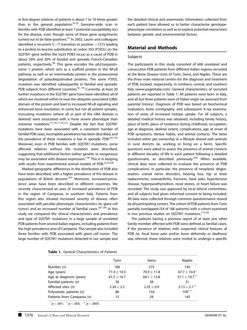
Table 1. General Characteristics of Patients
Turin
Number (n) 186
Age (years) 71.4� 10.3
Age at diagnosis (years) 61.5� 10.7
Familial patients (n) 30
Affected sites (n) 2.24� 2.2
Polyostotic patients (n) 86
Patients from Campania (n) 15
�p< .001; ��p< .005; ���p< .0001.
1376 Journal of Bone and Mineral Research
the detailed clinical and anamnestic information collected from
each patient have allowed us to better characterize genotype-
phenotype correlation as well as to explore potential interactions
between genetic and environmental factors.
Material and Methods
Subjects
The participants in this study consisted of 608 unrelated and
consecutive PDB patients from different Italian regions recruited
at the Bone Disease Units of Turin, Siena, and Naples. These are
the three main national centers for the diagnosis and treatment
of PDB, located, respectively, in northern, central, and southern
Italy (www.pagetitalia.com). General characteristics of recruited
patients are reported in Table 1. All patients were born in Italy,
and all but three patients were of Italian origin (as assessed from
parental history). Diagnosis of PDB was based on biochemical
evaluation, bone scintigraphy, and subsequent X-ray examina-
tion of areas of increased isotope uptake. For all subjects, a
detailed medical history was obtained, including family history,
place of birth, place of residence during childhood, occupation,
age at diagnosis, skeletal extent, complications, age at onset of
PDB symptoms, dietary habits, and animal contacts. The latter
included either pet ownership or a lifestyle shared with animals
in rural districts (ie, working or living on a farm). Specific
questions were asked to assess the presence of animal contacts
in different decades of life in each patient following a detailed
questionnaire, as described previously.(26) When available,
clinical data were collected to evaluate the presence of PDB
complications. In particular, the presence of neoplastic degen-
eration, cranial nerve disorders, hearing loss, hip or knee
replacements, osteoarthritis, fractures, back pain, hypertensive
disease, hyperparathyroidism, renal stones, or heart failure was
recorded. The study was approved by local ethical committees,
and all subjects had given informed consent to being included.
All data were collected through common questionnaires shared
by all participating centers. The cohort of PDB patients from Turin
partially overlapped (54 of 186 patients) with a cohort examined
in two previous studies on SQSTM1 mutations.(13,29)
The patients having a previous report of at least one other
family member affected with PDB were defined as familial cases.
If the presence of relatives with suspected clinical features of
PDB (ie, focal bone pain and/or bone deformity or deafness)
was referred, these relatives were invited to undergo a specific
Siena Naples
275 145
70.9� 11.8 67.7� 10.4�
60.1� 12.8 57.1� 10.7��
38 31
2.35� 0.9 3.13� 2.1��
154 109���
29 145
GENNARI ET AL.
diagnostic test for PDB. Patients with a negative history were
classified as sporadic PDB.
First-degree relatives of all recruited patients also were invited
to undergo biochemical evaluation of total alkaline phosphatase
to uncover new familial PDB cases, which were confirmed by
radiologic and bone-scan analyses. However, since we did not
perform a detailed evaluation of all the first-degree relatives,
we cannot exclude the possibility that sporadic patients may
have had relatives with asymptomatic disease.
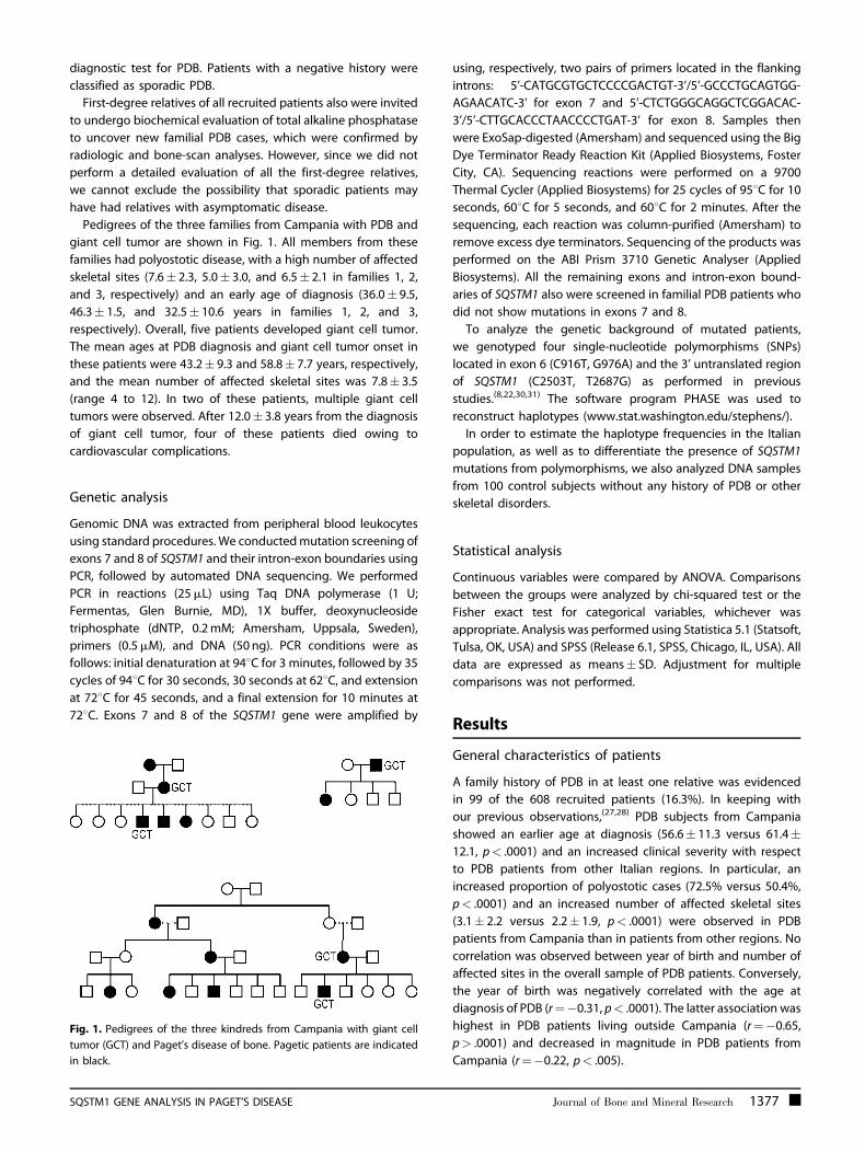
Pedigrees of the three families from Campania with PDB and
giant cell tumor are shown in Fig. 1. All members from these
families had polyostotic disease, with a high number of affected
skeletal sites (7.6� 2.3, 5.0� 3.0, and 6.5� 2.1 in families 1, 2,
and 3, respectively) and an early age of diagnosis (36.0� 9.5,
46.3� 1.5, and 32.5� 10.6 years in families 1, 2, and 3,
respectively). Overall, five patients developed giant cell tumor.
The mean ages at PDB diagnosis and giant cell tumor onset in
these patients were 43.2� 9.3 and 58.8� 7.7 years, respectively,
and the mean number of affected skeletal sites was 7.8� 3.5
(range 4 to 12). In two of these patients, multiple giant cell
tumors were observed. After 12.0� 3.8 years from the diagnosis
of giant cell tumor, four of these patients died owing to
cardiovascular complications.
Genetic analysis
Genomic DNA was extracted from peripheral blood leukocytes
using standard procedures. We conductedmutation screening of
exons 7 and 8 of SQSTM1 and their intron-exon boundaries using
PCR, followed by automated DNA sequencing. We performed
PCR in reactions (25mL) using Taq DNA polymerase (1 U;
Fermentas, Glen Burnie, MD), 1X buffer, deoxynucleoside
triphosphate (dNTP, 0.2mM; Amersham, Uppsala, Sweden),
primers (0.5mM), and DNA (50 ng). PCR conditions were as
follows: initial denaturation at 948C for 3 minutes, followed by 35
cycles of 948C for 30 seconds, 30 seconds at 628C, and extension
at 728C for 45 seconds, and a final extension for 10 minutes at
728C. Exons 7 and 8 of the SQSTM1 gene were amplified by
Fig. 1. Pedigrees of the three kindreds from Campania with giant cell
tumor (GCT) and Paget’s disease of bone. Pagetic patients are indicated
in black.
SQSTM1 GENE ANALYSIS IN PAGET’S DISEASE
using, respectively, two pairs of primers located in the flanking
introns: 5’-CATGCGTGCTCCCCGACTGT-3’/5’-GCCCTGCAGTGG-
AGAACATC-3’ for exon 7 and 5’-CTCTGGGCAGGCTCGGACAC-
3’/5’-CTTGCACCCTAACCCCTGAT-3’ for exon 8. Samples then
were ExoSap-digested (Amersham) and sequenced using the Big
Dye Terminator Ready Reaction Kit (Applied Biosystems, Foster
City, CA). Sequencing reactions were performed on a 9700
Thermal Cycler (Applied Biosystems) for 25 cycles of 958C for 10
seconds, 608C for 5 seconds, and 608C for 2 minutes. After the
sequencing, each reaction was column-purified (Amersham) to
remove excess dye terminators. Sequencing of the products was
performed on the ABI Prism 3710 Genetic Analyser (Applied
Biosystems). All the remaining exons and intron-exon bound-
aries of SQSTM1 also were screened in familial PDB patients who
did not show mutations in exons 7 and 8.
To analyze the genetic background of mutated patients,
we genotyped four single-nucleotide polymorphisms (SNPs)
located in exon 6 (C916T, G976A) and the 3’ untranslated region
of SQSTM1 (C2503T, T2687G) as performed in previous
studies.(8,22,30,31) The software program PHASE was used to
reconstruct haplotypes (www.stat.washington.edu/stephens/).
In order to estimate the haplotype frequencies in the Italian
population, as well as to differentiate the presence of SQSTM1
mutations from polymorphisms, we also analyzed DNA samples
from 100 control subjects without any history of PDB or other
skeletal disorders.
Statistical analysis
Continuous variables were compared by ANOVA. Comparisons
between the groups were analyzed by chi-squared test or the
Fisher exact test for categorical variables, whichever was
appropriate. Analysis was performed using Statistica 5.1 (Statsoft,
Tulsa, OK, USA) and SPSS (Release 6.1, SPSS, Chicago, IL, USA). All
data are expressed as means� SD. Adjustment for multiple
comparisons was not performed.
Results
General characteristics of patients
A family history of PDB in at least one relative was evidenced
in 99 of the 608 recruited patients (16.3%). In keeping with
our previous observations,(27,28) PDB subjects from Campania
showed an earlier age at diagnosis (56.6� 11.3 versus 61.4�12.1, p< .0001) and an increased clinical severity with respect
to PDB patients from other Italian regions. In particular, an
increased proportion of polyostotic cases (72.5% versus 50.4%,
p< .0001) and an increased number of affected skeletal sites
(3.1� 2.2 versus 2.2� 1.9, p< .0001) were observed in PDB
patients from Campania than in patients from other regions. No
correlation was observed between year of birth and number of
affected sites in the overall sample of PDB patients. Conversely,
the year of birth was negatively correlated with the age at
diagnosis of PDB (r¼�0.31, p< .0001). The latter association was
highest in PDB patients living outside Campania (r¼�0.65,
p> .0001) and decreased in magnitude in PDB patients from
Campania (r¼�0.22, p< .005).
Journal of Bone and Mineral Research 1377

Table 2. Prevalence of SQSTM1 Gene Mutations in Italy
Mutation Northern Italy (n¼ 132) Central Italy (n¼ 169) Southern Italy (n¼ 212) Islands (n¼ 15)
Y383X 0 0 7 (3.30%) 0
P387L 2 (1.51%) 0 1 (0.47%) 0
P392L 7 (5.30%) 14� (8.28%) 19 (8.96%) 0
E396X 1 (0.76%) 0 3 (1.41%) 0
M401V 0 0 1 (0.47%) 0
M404V 6 (4.55%) 5 (2.96%) 1 (0.47%) 1 (6.67%)
G411S 0 1� (0.59%) 0 0
D423X 1 (0.76%) 0 0 0
G425E 0 1 (0.59%) 3 (1.41%) 0
G425R 0 0 2 (0.94%) 0
A427D 0 0 2 (0.94%) 0
�With the inclusion of 1 patient with a G411S/P392L mutation, 5 patients without SQSTM1mutations were excluded from analysis owing to the inabilityto assign their region of origin.
Mutation screening of the SQSTM1 gene in theoverall sample
Genetic analysis was completed in 533 of the 608 patients.
Eleven different mutations in the SQSTM1 gene were observed in
34 of 92 (36.9%) and 43 of 441 (9.7%) of familial and sporadic PDB
patients, respectively (equivalent to 14.4% of the overall cohort)
(Table 2). DNA analysis from 100 control subjects failed to
detect the reported SQSTM1 mutations. A significantly higher
prevalence of mutations was observed in polyostotic than
monostotic patients (21.0% versus 5.7%, p< .0001).
Two of these mutations, M401V (A1241G) and A427D
(C1320A), were novel and have not been described previously.
Moreover, we disclosed for both amino acids a high degree
of conservation across species from fish to humans (data not
shown), which argues in favor of an important role of these
amino acids in the function of p62 protein. The other mutations
(Y383X, P387L, P392L, E396X, M404V, G411S, D423X, G425E, and
G425R) were described previously in other populations. One
subject carried a double P392L and G411S mutation. He had a
polyostotic form of disease (with three affected skeletal sites)
diagnosed at 55 years of age.
The overall prevalence of SQSTM1 mutations was higher in
younger than in older PDB patients. In fact, 64.9% of the
mutations were observed in subjects with a birth age above the
median (corresponding to 1937). The SQSTM1mutation rate was
9.5% versus 19.2% in PDB patients below or above the median
age (p< .01). A similar trend was observed when patients were
grouped according to the decades or quartiles in relation to
year of birth. Overall, 31 of 77 SQSTM1 mutations (40.26%) were
observed in subjects in the upper quartile of age (year of birth
after 1946).
Table 3. Clinical Characteristics of Patients With or Without SQSTM1
Subjects
(n)
Familial
patients (n)
Age
(years� SD)
WT 456 58 69.7� 21.5
SQSTM1 77 34 67.8� 12.2
p Level — <.0001 .74
1378 Journal of Bone and Mineral Research
After the analysis of exons 7 and 8, all the remaining exons and
intron-exon boundaries of SQSTM1 were screened in the 58
familial PDB patients who did not showmutations in exons 7 and
8. No furthermutations in SQSTM1 genewere found. In particular,
all the three families with giant cell tumor complicating PDB
were negative for SQSTM1 mutations.
Genotype-phenotype correlation
As shown in Table 3, PDB subjects with SQSTM1 mutation
showed an increased number of affected skeletal sites, an
increased prevalence of polyostotic disease, and earlier age of
onset than PDB patients without mutation. Conversely, no
differences were observed in the occurrence of major
complications of PDB between patients with or without
mutation. A similar pattern also was observed in familial versus
sporadic PDB patients. With respect to sporadic patients, familial
patients were younger (63.5� 12.0 versus 69.3� 11.6 years,
p< .005) and showed an earlier age at onset (54.6� 12.1 versus
60.7� 11.3 years, p< .0001), a higher number of affected skeletal
sites (3.56� 2.7 versus 2.33� 1.8, p< .0001), and an increased
proportion of polyostotic disease (73.8% versus 54.9%, p< .005).
When familial patients with and without SQSTM1 mutation were
compared, there were no differences in age at onset (53.9� 10.9
versus 54.9� 12.8 years, p¼ 0.7), and ther was a mild but not
significant variation in the number of affected skeletal sites
(4.07� 2.7 versus 3.23� 1.9, p¼ 0.08) or in the prevalence of
polyostotic disease (67.2% versus 85.1%, p¼ .06). Conversely,
PDB patients with SQSTM1 mutations considered to be sporadic
cases showed an earlier age at onset (55.8� 11.5 versus
61.4� 11.2 years, p< .005), an increased number of affected
skeletal sites (3.31� 2.3 versus 2.18� 1.8, p< .0005), and a
Gene Mutations
Age at diagnosis
(years� SD)
Polyostotic
patients (n)
Affected sites
(n� SD)
60.6� 11.6 238 2.31� 1.9
55.0� 11.2 64 3.60� 2.6
<.0005 <.0001 <.0001
GENNARI ET AL.

higher prevalence of polyostotic disease (82.2% versus
50.9%, p¼ .0001) than sporadic PDB patients without SQSTM1
mutations.
In order to better characterize genotype-phenotype corre-
lation, we analyzed 98 first-degree relatives of subjects with
SQSTM1 mutations, and we detected 20 additional mutations in
18 affected and 2 unaffected subjects. Interestingly, themutation
was not observed in 2 affected PDB relatives of two familial PDB
patients with SQSTM1mutations, indicating phenocopy. The first
kindred was composed of two affected brothers from southern
Italy, of whom only one had the M404V mutation. They had a
similar polyostotic phenotype with three or four affected skeletal
sites and a similar age at diagnosis (around 50 years). The second
kindred included three affected patients from central Italy, of
whom two had the P392L mutation. The two mutated patients,
an 87-year-old woman and her 85-year-old brother, had
polyostotic PDB with two and three affected sites, respectively.
Their affected brother (82 years old) without SQSTM1 mutation
had monostotic PDB of the left pelvis.
Phenotype characteristics of subjects according to the type of
SQSTM1mutation are shown in Table 4. Overall, slight differences
in the number of affected skeletal sites or age at disease onset
were observed among patients with different mutations. A
higher number of affected skeletal sites was observed in the two
patients with A427D mutations (7.00� 2.8, range 5 to 9) and in
the seven unrelated patients with Y383X mutations (4.84� 3.8,
range 1 to 12). The latter mutation also was associated with the
lowest age at diagnosis (mean 48.1� 9.3 years, range 40 to
60 years). Similar trends were observed when familial and
sporadic patients were considered separately or when affected
relatives with SQSTM1 mutations were included (bringing the
overall number of mutated patients to 95). In this latter case,
marked differences in clinical severity of disease also were
observed, even within each single family with P392L, M404V, or
E396X mutations. In fact, even in the case of a family with an
E396X mutation, causing the truncation of most of the UBA
domain, the number of affected skeletal sites in three PDB
patients varied from two to seven, with an estimated onset of
disease at between 38 and 64 years of age.
Table 4. Genotype-Phenotype Correlation in Patients with SQSTM1 G
Mutation
Subjects
(n)
Familial
patients (n)
Age
(years� SD)
Y383X 7 4 63.5� 4.6
P387L 3 0 80.0� 14.0
P392L 39 12 69.5� 11.0
E396X 4 3 55.0� 13.6
M401V 1 1 78
M404V 13 9 68.2� 16.5
G411S/P392L 1 0 68
D423X 1 0 61
G425E 4 1 59.2� 7.2
G425R 2 2 59.5� 2.1
A427D 2 2 78.0� 7.0
p Level — — .04
SQSTM1 GENE ANALYSIS IN PAGET’S DISEASE
To further explore possible genotype-phenotype correlations,
we grouped patients according to the type or site of SQSTM1
mutation: truncating versus missense and outside versus inside
the structured region of the UBA domain (amino acids 392 to
431). As shown in Table 5, an earlier age at diagnosis was
observed in patients with truncating mutations than in those
with missense mutations, whereas the number of affected
skeletal sites and the frequency of polyostotic disease did not
differ significantly. Mutations outside the UBA domain (Y383X
and P387L, n¼ 10) did not differ significantly from mutations
inside the UBA. Moreover, we observed a negative correlation
between year of birth and severity of disease, expressed as
number of affected sites, in patients with missense mutations
(r¼�0.22, p< .05) but not in patients with truncating mutations.
The correlation between birth year and age at PDB diagnosis
observed in the overall sample remained statistically significant
independent of type (truncating or missense) or site (inside or
outside UBA) of mutation.
Regional distribution of SQSTM1 mutations andgene-environment interactions
The prevalence of SQSTM1 mutations was higher in southern
(18.4%) than in central (11.8%) and northern (12.9%) Italy
(Table 2). Moreover, an increased heterogeneity and a different
distribution of mutations were observed in southern Italy
(showing 9 of the 11mutations) than in central and northern Italy
(where only 4 and 5 of the reported mutations were observed,
respectively). Interestingly, all seven patients with the Y383X
mutation were from southern Italy and specifically from
Campania. In contrast, the M404V mutation was more frequent
in northern (4.6%) and central (3.0%) Italy than in southern Italy
(0.5%).
Given the reported characteristics of PDB patients from
Campania, we performed a subanalysis of patients from this
region. Differences in terms of age at diagnosis and severity
between familial and sporadic patients or between patients with
or without SQSTM1 mutations were milder in PDB patients from
Campania than in patients from other regions. Moreover, despite
ene Mutations
Age at diagnosis
(years� SD)
Polyostotic
patients (n)
Affected sites
(n� SD)
49.0� 8.8 5 4.28� 3.8
64.3� 9.3 3 3.33� 2.3
56.7� 11.8 33 3.56� 2.6
50.5� 17.0 4 3.25� 2.5
48 1 10
53.5� 11.5 9 3.07� 2.1
55 1 3
57 1 2
49.7� 5.4 3 2.25� 0.9
52.0� 5.6 2 3.50� 0.7
62.0� 7.1 2 7.00� 2.8
.46 .75 .20
Journal of Bone and Mineral Research 1379

Table 5. Genotype-Phenotype Correlations According to the Type (Truncating versus Missense) or Site (Outside versus Inside the
Structured Region of the UBA Domain) of SQSTM1 Mutation
Truncating Missense Outside UBA Inside UBA
Number (n) 12 65 10 67
Age (years) 60.2� 9.8� 68.8� 12.1 69.0� 11.5 67.9� 12.2
Age at diagnosis (years) 50.5� 11.3�� 56.3� 10.7 52.8� 11.6 55.7� 11.1
Familial patients (n) 7/12 27/65 4/10 30/67
Affected sites (n) 3.77� 2.9 3.45� 2.4 4.22� 3.1 3.41� 2.0
Polyostotic patients (n) 10/12 54/65 8/10 56/67
�p< .05 and��p¼ .05, truncating versus missense mutation.
a higher prevalence of SQSTM1 mutations in the group of
patients from Campania, a consistent number of analyzed PDB
families from this region (26 of 35) did not have the mutation.
Genotype-phenotype analysis in PDB patients from Campania
did not evidence any significant difference in relation to the
type or site of mutation. Moreover, the prevalence of SQSTM1
mutations did not differ significantly based on decades or
quartiles of age in patients from Campania, whereas marked
age-related differences in the prevalence rates of SQSTM1
mutations were observed in patients from the other regions
(20.9% versus 8.4%, p< .001 in patients above or below the
median age, respectively).
Overall, 342 of 533 (64.2%) PDB patients indicated animal
contacts for at least 10 years before onset of the disease. These
included pet ownership (mainly cats and dogs) and previous or
current contact with animals such as pigs, rabbits, sheep, and
cattle in rural districts. The prevalence of subjects with these
contacts did not differ between patients with or without SQSTM1
mutations in the overall sample (58.4% versus 65.2%, p¼ 0.2) but
became significant in patients living outside Campania (48.9%
versus 64. 2%, p< .05). Conversely, patients from Campania had
a high prevalence of animal contact (69.3% versus 62.1% in the
other regions, p¼ .09) but without any difference between
patients with or without SQSTM1 mutations. When familial
patients from Campania were considered, animal contacts were
observed in 90.1% of families without SQSTM1 mutations.
Interestingly, a progressive age-related decrease in the pre-
valence of animal contact was observed based on the median
age as well as the decade of age in the overall sample of patients.
This decrease was not observed in the subgroup of patients from
Campania but increased in magnitude in patients from other
regions. In particular, in younger patients (with a birth year after
1950), 70.4% of patients from Campania reported animal
contacts with respect to 48.9% of patients from other regions
(p< .05).
Overall, PDB patients reporting animal contacts showed an
increased number of affected sites (2.19� 1.9 versus 2.54� 2.0,
p< .05) and a higher prevalence of familial disease (23.0% versus
14.1%, p< .05) than patients without animal contacts. A
significant difference in the number of affected skeletal sites
in relation to animal contacts also was seen in the subgroup of
patients with SQSTM1 mutations (3.84� 2.5 versus 2.76� 2.2,
p< .05). This difference also was seen when missense or
truncating mutations were considered separately. All the
1380 Journal of Bone and Mineral Research
preceding differences became milder and not significant
when only pet ownership was considered instead of animal
contacts.
SQSTM1 haplotypes in PDB patients and controls
We genotyped four SNPs in exon 6 and the 3’ untranslated
region of the SQSTM1 gene. The four selected SNPs were
analyzed in all mutation carriers as well as in 100 control
individuals and 100 PDB patients without SQSTM1 mutations.
Genotype distribution for these SNPs followed a Hardy-Weinberg
equilibrium. There was no significant difference in distribution
of the genotypes between patients and controls for any of the
SNPs studied.
Consistent with previous studies in different popula-
tions,(8,22,30,31) the H1 (916T-976A-2503C-2687T) and H2 (916C-
976G-2503T-2687G) haplotypes accounted for the largest
proportion of patients (94.3%) and controls (91.5%). The
remaining patients were accounted for by six rare haplotypes
with individual frequencies of between 0.2% and 3.6%. The
presence of H1 and H2 haplotypes was observed in 75.0%
(including two H1/H1 homozygous subjects) and 90.0% (includ-
ing seven H2/H2 homozygous subjects) of patients with the
P392L mutation, respectively, compared with 76% and 80%
observed in control individuals. Since we did not perform allele-
specific PCR, we could not unambiguously assign the mutation
to one of the two haplotypes in the 26 H1/H2 heterozygous
subjects, except than in three familial patients, in whom genetic
analysis of affected family members was able to assign the P392L
mutation to the H2 haplotype. An increased prevalence of the H2
haplotype also was observed in patients with M404V and Y383X
mutations. In particular, the Y383X mutation was associated
with the H2 haplotype in 100% of patients. In fact, only one
heterozygous H1/H2 subject with Y383X was observed, and
subsequent genetic analysis of affected family members (with
the identification of one H2/H2 mutation carrier) demonstrated
that the mutation is carried with the H2 allele. Conversely, 3 of 4
and 4 of 4 unrelated PDB patients with the G425E and E396X
mutations, respectively, were negative for the H2 haplotype,
suggesting that in this case the mutation is carried with a
different haplotype, probably the H1, which was present in 100%
of these patients (with a frequency of 87.5% and 75.0% in G425E
and E396X mutation carriers, respectively).
GENNARI ET AL.

Discussion
Despite the significant progress that has been made in recent
years, the etiology of PDB is not completely understood.(2,3,24)
Mutations in the SQSTM1 gene have been described worldwide
in consistent proportion of patients with PDB from different
ethnic groups, suggesting that functional differences in this
gene are a direct cause of the disease, particularly in familial
PDB. However, several clinical and experimental observations
raised the hypothesis that other genes and/or environmental
triggers are necessary to cause the disease, at least in some
cases.(2,20,21,23,32–34) In this study we report the results of the
largest SQSTM1 mutation screening performed to date in
consecutive familial and sporadic PDB patients.
In our sample, we identified 11 different SQSTM1mutations in
14.4% of patients. This percentage is comparable with the results
of mutational analysis studies performed in other countries,
such as Great Britain (13.9%), France (12.8%), and Canada
(19.8%).(8,9,11,17) Consistent with previous findings, the preva-
lence of mutations was highest (36.9%) in patients with a clear
family history. Since we were not able to clinically exclude the
presence of PDB in all first-degree relatives of recruited patients,
we cannot exclude that a proportion of patients reporting no
family history and thus classified as sporadic patients may have
familial PDB. In particular, in sporadic patients with SQSTM1
mutations, we observed an increased severity of disease that is
remarkably similar to that seen in familial patients. The latter
observation may suggest that a consistent proportion of
sporadic PDB patients with SQSTM1 mutations may indeed
represent familial patients. This is in keeping with a recent report
in a well-characterized sample of PDB patients from the United
States showing absence of SQSTM1 mutations in sporadic
patients.(18) Of interest, we also observed a higher heterogeneity
of SQSTM1mutations in our sample of patients of Italian ancestry
with respect to patients from other countries. In fact, we
detected 11 different mutations. Together with the results from a
recent additional study in an Italian population,(29) 15 different
SQSTM1mutations have been described in more than 800 Italian
patients analyzed to date. An additional mutation (P364S) has
been described in a single PDB family of Italian descent living
in Australia.(19) This heterogeneity is higher than observed in
populations of British descent or in other European populations
and might reflect the complex history of Italy as well as the
several foreign invasions and dominations that occurred
between sixth and nineteenth centuries. Moreover, in our study,
a different distribution and a higher heterogeneity of mutations
were particularly observed in southern Italy (showing 9 of the 11
mutations) than in central and northern Italy. While the M404V
mutation was more frequent in northern and central Italy than
in southern Italy, the Y383X mutation was observed in seven
unrelated patients from Campania and was absent in patients
from other regions. This mutation also was described in a
previous Italian study in a family from Campania and in two
sporadic patients of unreported origin(29) but was not observed
in more than 1000 PDB patients from other countries analyzed
in previous studies. The mutation is one of the two known
truncating SQSTM1 mutations located outside the UBA domain
and seems to be associated with a severe phenotype in most
SQSTM1 GENE ANALYSIS IN PAGET’S DISEASE
patients. A similar phenotype has been described in one familial
patient with the other mutation (K378X), and functional analysis
confirmed that this mutation, leading to the complete
elimination of the UBA domain, is associated with potentiated
osteoclast formation and bone resorption in human primary cell
cultures.(16)
Despite the severe phenotype and the earlier age at onset
of disease, we did not find SQSTM1 mutations in the three
kindreds with giant cell tumor. This complication represents
a quite unusual clinical feature of PDB (described in fewer than
100 patients worldwide) and occurs mainly in patients with
severe polyostotic disease, with a remarkably higher prevalence
in patients from Campania.(35–37) In fact, more than 50% of the
patients described originated in or descended from ancestors
who lived in this Italian region.(27) Thus it can be speculated
that a different gene is responsible for this particular variant of
familial PDB, alone or in combination with an environmental
trigger.(38)
Phenotype-genotype associations in PDB have not been
investigated extensively, and the results from available reports
are conflicting. This may reflect the limited number of patients
with SQSTM1mutations, except the P392L mutation, available in
previous studies. Consistent with recent observations in two
populations of British descent and in the French population, we
confirmed that PDB patients with SQSTM1 mutations have a
more extensive disease and an earlier age at diagnosis than
patients without SQSTM1mutations.(11,17,19) This observation is in
contrast to a previous report in a smaller sample of patients from
Italy that did not show significant phenotypic differences in
relation to the presence of SQSTM1 mutations, as well as
between familial and sporadic patients.(29) The prevalence of
SQSTM1mutations, however, was lower in that study (8.7%) than
in our or in previous studies, most likely reflecting the reduced
number of familial patients (12 of 357, equivalent to 3.4%). When
we compared clinical characteristics of patients with different
SQSTM1 mutations, we did not observe major differences in the
number of affected skeletal sites or in the age at diagnosis, even
though a trend for a more severe phenotype clearly was
observed in most patients with the Y383X and A427D mutations.
Since all patients with these mutations were from Campania, we
cannot exclude the possibility that this finding is related to the
overall increased clinical severity of disease observed in patients
from this region. Moreover, a variable disease severity was
observed among affected members of kindreds with the same
SQSTM1mutation for all mutations. These findings are in keeping
with two previous detailed analyses of large PDB kindreds with
SQSTM1 mutations of diverse racial or ethnic background
showing high variability in intrafamilial expressivity of disease
as well as incomplete penetrance.(20,21) Moreover, in one of
these studies, offspring who inherited an SQSTM1 mutation
from their parents were diagnosed with PDB later in life and
had less extensive disease than their parents.(21) Even though a
slight reduction in clinical severity and a higher variation in
the number of affected skeletal sites among family members
were observed in missense with respect to truncating mutations,
we can conclude that there are no major genotype-phenotype
differences in relation to the type or site of SQSTM1 mutation.
Together with the described cases of incomplete penetrance(20–22)
Journal of Bone and Mineral Research 1381
and the examples of phenocopy observed in this and other
previous studies,(19) these findings further reinforce the
hypothesis that additional factors may be required to cause
the disease (at least in a group of patients) as well as its skeletal
extension in subjects with or without SQSTM1 mutations. In this
context, the presence of somatically acquired SQSTM1mutations
in the pagetic bone cannot be excluded, even though this
hypothesis remains controversial and probably restricted to a
limited number of patients.(39,40)
We and others have previously evidenced an association
between PDB and animal-related factors, as well as a significantly
higher prevalence of the disease in rural than in urban
districts.(26,41–43) In this study, we also demonstrated that
patients reporting persistent animal contacts for at least 10
years before the onset of disease have an increased number of
affected skeletal sites and an increased prevalence of polyostotic
disease. Interestingly, this association also was evidenced in
patients with SQSTM1 mutations, suggesting an interaction
between genetic and environmental factors. The observed
differences, however, were small, and their clinical impact
remains to be addressed in future prospective studies with larger
numbers of patients with SQSTM1 mutations. In fact, even
though adjustment for multiple comparisons generally is not
required in this kind of study,(44,45) with a more conservative
approach (ie, with Bonferroni’s correction), some of these
differences will no longer be significant.
Nevertheless, together with different previous epidemiologic
reports, these data provide further evidence that infective agents
may be involved in PDB, not only promoting the occurrence of
the disease in some patients but also influencing its clinical
severity. In this context, the progressive age-related decrease in
the number of patients reporting animal contacts observed in
this study is consistent with secular trends showing a decrease in
both the prevalence and severity of PDB over time(25,46) and also
might explain, at least in part, the parallel age-related increase in
the prevalence of SQSTM1 mutations observed in our sample. In
fact, it cannot be excluded that in patients without SQSTM1
mutations, the disease also may originate from contacts with an
environmental factor (alone or in combination with additional
genetic causes). Thus a reduced exposure to these animal-
related environmental agents in recent years could have led to a
relative increase in the frequency of PDB patients owing to
SQSTM1 mutations. Consistently, this phenomenon was not
observed in PDB patients from Campania, where a higher
prevalence of animal contacts was observed even in recent years,
reaching 90% in familial PDB patients without SQSTM1
mutations. To date, the nature of the possible environmental
trigger remains unknown, but several reports suggested that
viral infections of the paramyxovirus family may infect the
osteoclast, inducing most of the cellular abnormalities of
PDB.(32,33,47–49) Indeed, in one of these studies, canine distemper
virus not only induced NFkB activation and bone resorption but
also markedly increased sequestosome 1/p62 gene expression in
human osteoclast cells.(33)
Finally, results from our analysis evidenced an increased
occurrence of the P392mutation with the H2 than H1 haplotypes.
This is consistent with the notion of a ‘‘founder effect’’ for this
mutation, even if it was not possible to unambiguously assign
1382 Journal of Bone and Mineral Research
the mutation to one of the two haplotypes in most of the
heterozygous subjects, because we did not perform allele-
specific screening. Moreover, we also observed two H1-H1
homozygous patients carrying the P392L mutation, suggesting
that the same mutation has occurred independently at least
twice, as observed previously in the French-Canadian popula-
tion.(8,22) An increased prevalence of the H2 haplotype also was
seen in patients with the M404V mutation, whereas the Y383X
mutation was carried with the H2 haplotype in all patients. This
also strongly supports the presence of a founder effect for this
mutation. Conversely, most patients with G425E and E396X
mutations were negative for H2 haplotype, suggesting that in
this case the mutation is carried with a different haplotype, most
likely H1.
In conclusion, while the clinical impact of most of the reported
differences remains to be addressed, our findings further
underline the complex etiology of PDB that cannot be explained
solely by the presence of SQSTM1mutations. It is likely that both
genetic and environmental factors may cause PDB or most likely
interact with each other to cause the disease and its variable
phenotypes. Moreover, our results also indicate that the
increased severity of PDB cases from Campania observed in
this and other previous studies seems to be related to
concomitant factors: (1) an increased heterogeneity of SQSTM1
mutations (with higher prevalence of truncating mutations such
as Y383X), (2) an increased persistence of the environmental
trigger (probably related to a lifestyle shared with animals), and
(3) the presence of additional predisposition genes (including a
gene causing familial PDB with giant cell tumor). Further genetic
studies in this population, particularly in familial patients
negative for SQSTM1 mutation, might be extremely useful for
a real understanding of the complex etiology of this disorder.
Disclosures
LG and FG contributed equally to this work. All the authors state
that they have no conflicts of interest.
Acknowledgments
The work was supported in part by grants from the Italian
Association of Patients with Paget’s Disease (AIP, www.pageti-
talia.com) and the University of Siena (PAR 2006). The authors
also thank Dr Vincenzo De Paola and Dr Annalisa Avanzati for
technical assistance.
References
1. Paget J. On a form of chronic inflammation of bones (osteitis
deformans). Med Chir Trans. 1877;60:37–63.
2. Roodman GD, Windle JJ. Paget disease of bone. J Clin Invest. 2005;
115:200–208.
3. Ralston SH, Langston AL, Reid IR. Pathogenesis and management of
Paget’s disease of bone. Lancet. 2008;72:155–163.
4. Montagu MFA. Paget’s disease (osteitis deformans) and hereditary.
Am J Hum Genet. 1949;1:94–95.
5. Siris ES, Ottman R, Flaster E, Kelsey JL. Familial aggregation of Paget’s
disease of bone. J Bone Miner Res. 1991;6:495–500.
GENNARI ET AL.

6. Siris ES. Epidemiological aspects of Paget’s disease: family history andrelationship to other medical conditions. Semin Arthritis Rheum.
1994;23:222–225.
7. Daroszewska A, Ralston SH. Genetics of Paget’s disease of bone. Clin
Sci (Lond). 2005;109:257–263.
8. Laurin N, Brown JP, Morissette J, Raymond V. Recurrent mutation of
the gene encoding sequestosome 1 (SQSTM1/p62) in Paget disease of
bone. Am J Hum Genet. 2002;70:1582–1588.
9. Hocking LJ, Lucas GJ, Daroszewska A, et al. Domain specific muta-
tions in Sequestosome 1 (SQSTM1) cause familial and sporadic Paget’s
disease. Hum Mol Genet. 2002;11:2735–2739.
10. Johnson-Pais TL, Wisdom JH, Weldon KS, et al. Three novel mutationsin SQSTM1 identified in familial Paget’s disease of bone. J Bone Miner
Res. 2003;18:1748–1753.
11. Hocking LJ, Lucas GJ, Daroszewska A, et al. Novel UBA domain
mutations of SQSTM1 in Paget’s disease of bone: genotype pheno-type correlation, functional analysis, and structural consequences.
J Bone Miner Res. 2004;19:1122–1127.
12. Eekhoff EW, Karperien M, Houtsma D, et al. Familial Paget’s disease in
The Netherlands: Occurrence, identification of new mutations in thesequestosome 1 gene, and their clinical associations. Arthritis Rheum.
2004;50:1650–1654.
13. Falchetti A, Di Stefano M, Marini F, et al. Two novel mutations at exon8 of Sequestosome 1 gene (SQSTM1) in an Italian series of patients
affected by Paget’s disease of bone (PDB). J Bone Miner Res. 2004;
19:1013–1017.
14. Good DA, Busfield F, Fletcher BH, et al. Identification of SQSTM1mutations in familial Paget’s disease in Australian pedigrees. Bone.
2004;35:277–282.
15. Beyens G, Van Hul E, Van Driessche K, et al. Evaluation of the role of
the SQSTM1 gene in sporadic Belgian patients with Paget’s disease.Calcif Tissue Int. 2004;75:144–152.
16. Rea SL, Walsh JP, Ward L, et al. A novel mutation (K378X) in the
sequestosome 1 gene associated with increased NF-kB signaling andPaget’s disease of bone with a severe phenotype. J Bone Miner Res.
2006;21:1136–1145.
17. Collet C, Michou L, Audran M, et al. Paget’s disease of bone in the
French population: novel SQSTM1 mutations, functional analysis,and genotype-phenotype correlations. J Bone Miner Res. 2007;22:
310–317.
18. Rhodes EC, Johnson-Pais TL, Singer FR, et al. Sequestosome 1
(SQSTM1) mutations in Paget’s disease of bone from the UnitedStates. Calcif Tissue Int. 2008;82:271–277.
19. Rea SL, Walsh JP, Ward L, et al. Sequestosome 1 Mutations in Paget’s
Disease of Bone in Australia: Prevalence, Genotype/Phenotype Cor-
relation and a Novel Non-UBA Domain Mutation (P364S) Associatedwith Increased NF-kB Signaling Without Loss of Ubiquitin-Binding.
J Bone Miner Res. 2009;24:1216–1223.
20. Leach RJ, Singer FR, Ench Y, Wisdom JH, Pina DS, Johnson-Pais TL.Clinical and cellular phenotypes associated with sequestosome 1
(SQSTM1) mutations. J Bone Miner Res. 2006;21 (Suppl 2): P45–
50.
21. Bolland MJ, Tong PC, Naot D, et al. Delayed development of Paget’sdisease in offspring inheriting SQSTM1 mutations. J Bone Miner Res.
2007;22:411–415.
22. Morissette J, Laurin N, Brown JP. Sequestosome 1: mutation frequen-
cies, haplotypes, and phenotypes in familial Paget’s disease of bone.J Bone Miner Res. 2006;21:P38–44.
23. Kurihara N, Hiruma Y, Zhou H, et al. Mutation of the sequestosome 1
(p62) gene increases osteoclastogenesis but does not induce Pagetdisease. J Clin Invest. 2007;117:133–142.
24. Reddy SV. Etiologic factors in Paget’s disease of bone. Cell Mol Life
Sci. 2006;63:391–398.
SQSTM1 GENE ANALYSIS IN PAGET’S DISEASE
25. Cooper C, Harvey NC, Dennison EM, van Staa TP. Update on theepidemiology of Paget’s disease of bone. J Bone Miner Res. 2006;21
(Suppl 2): P3–8.
26. Merlotti D, Gennari L, Galli B, et al. Characteristics and familial
aggregation of Paget’s disease of bone in Italy. J Bone Miner Res.2005;20:1356–1364.
27. Gennari L, Merlotti D, Martini G, Nuti R. Paget’s disease of bone in
Italy. J Bone Miner Res. 2006;21 (Suppl 2): P14–21.
28. Rendina D, Gennari L, De Filippo G, et al. Evidence for Increased
Clinical Severity of Familial and Sporadic Paget’s Disease of Bone
in Campania, Southern Italy. J Bone Miner Res. 2006;21:1828–
1835.
29. Falchetti A, Di Stefano M, Marini F, et al. GenePage Project. Genetic
epidemiology of Paget’s disease of bone in Italy: sequestosome1/p62
gene mutational test and haplotype analysis at 5q35 in a large
representative series of sporadic and familial Italian cases of Paget’sdisease of bone. Calcif Tissue Int. 2009;84:20–37.
30. Lucas GJ, Hocking LJ, Daroszewska A, et al. Ubiquitin-associated
domain mutations of SQSTM1 in Paget’s disease of bone: evidence
for a founder effect in patients of British descent. J Bone Miner Res.2005;20:227–231.
31. Chung PY, Beyens G, Guanabens N, et al. Founder effect in different
European countries for the recurrent P392L SQSTM1 mutation inPaget’s Disease of Bone. Calcif Tissue Int. 2008;83:34–42.
32. Kurihara N, Zhou H, Reddy SV, et al. Expression of measles virus
nucleocapsid protein in osteoclasts induces Paget’s disease-like bone
lesions in mice. J Bone Miner Res. 2006;21:446–455.
33. Selby PL, Davies M, Mee AP. Canine distemper virus induces human
osteoclastogenesis through NF-kB and sequestosome 1/P62 activa-
tion. J Bone Miner Res. 2006;21:1750–1756.
34. Lucas GJ, Riches PL, Hocking LJ, et al. Identification of a major locusfor Paget’s disease on chromosome 10p13 in families of British
descent. J Bone Miner Res. 2008;23:58–63.
35. Jacobs TP, Michelsen J, Polay JS, D’Adamo AC, Canfield RE. Giant celltumor in Paget’s disease of bone: Familial and geographic clustering.
Cancer. 1979;44:742–747.
36. Magitsky S, Lipton JF, Reidy J, Vigorita VJ, Bryk E. Ultrastructural
features of giant cell tumors in Paget’s disease. Clin Orthop Relat Res.2002;402:213–219.
37. Rendina D, Mossetti G, Soscia E, et al. Giant cell tumor and Paget’s
disease of bone in one family: geographic clustering. Clin Orthop
Relat Res. 2004;421:218–224.
38. De Chiara A, Apice G, Fazioli F, Silvestro P, Carone G, Manco A.
Multicentric giant cell tumor with viral-like inclusions associated with
Paget’s disease of bone: a case treated by steroid therapy. Oncol Rep.
1998;5:317–320.
39. Matthews BG, Naot D, Bava U, et al. Absence of somatic SQSTM1mutations in Paget’s disease of bone. J Clin Endocrinol Metab. 2009;
94:691–694.
40. Merchant A, Smielewska M, Patel N, et al. Somatic mutations
in SQSTM1 detected in affected tissues from patients withsporadic Paget’s disease of bone. J Bone Miner Res. 2009;24:484–
494.
41. O’Driscoll JB, Anderson DC. Past pets and Paget’s disease. Lancet.
1985;2:919–921.
42. Khan SA, Brennan P, Newman J, Gray RE, McCloskey EV, Kanis JA.
Paget’s disease of bone and unvaccinated dogs. Bone. 1996;19:47–
50.
43. Lopez-Abente G, Morales-Piga A, Elena-Ibanez A, Rey-Rey JS, Corres-
Gonzalez J. Cattle, pets, and Paget’s disease of bone. Epidemiology.
1997;8:247–251.
44. Rothman KJ. No adjustments are needed for multiple comparisons.Epidemiology. 1990;1:43–46.
Journal of Bone and Mineral Research 1383

45. Perneger TV. What’s wrong with Bonferroni adjustments. BMJ.1998;316:1236–1238.
46. Cundy HR, Gamble G, Wattie D, Rutland M, Cundy T. Paget’s disease
of bone in New Zealand: continued decline in disease severity. Calcif
Tissue Int. 2004;75:358–364.
47. Mee AP, May C, Bennett D, Sharpe PT. Generation of multinucleatedosteoclast-like cells from canine bone marrow: Effects of canine
distemper virus. Bone. 1995;17:47–55.
1384 Journal of Bone and Mineral Research
48. Kurihara N, Reddy SV, Menaa C, Anderson D, Roodman GD.Osteoclasts expressing the measles virus nucleocapsid gene
display a pagetic phenotype. J Clin Invest. 2000;105:607–
614.
49. Reddy SV, Kurihara N, Menaa C, et al. Osteoclasts formed by measlesvirus-infected osteoclast precursors from hCD46 transgenic mice
express characteristics of pagetic osteoclasts. Endocrinology. 2001;
142:2898–2905.
GENNARI ET AL.