Osteopenia and endothelin-1-mediated vasconstrictor tone in postmenopausal women
Review ArticleThe Relationship between Polymorphisms inthe Vitamin D Receptor Gene and Bone MineralDensity in Postmenopausal Women
Jose M. Moran,1 Francisco J. Rodriguez-Velasco,1 Raul Roncero-Martin,1
Purificación Rey-Sanchez,1 Mariana Martinez,1 and Juan D. Pedrera-Zamorano1,2
1 Metabolic Bone Diseases Research Group, University of Extremadura, 10071 Caceres, Spain2Departamento de Enfermerıa, Facultad de Enfermerıa y Terapia Ocupacional, Universidad de Extremadura,Avenida Universidad s/n, 10071 Caceres, Spain
Correspondence should be addressed to Juan D. Pedrera-Zamorano; [email protected]
Received 3 October 2013; Accepted 8 December 2013; Published 9 January 2014
Academic Editors: P. A. Fasching and M. Nacak
Copyright © 2014 Jose M. Moran et al. This is an open access article distributed under the Creative Commons Attribution License,which permits unrestricted use, distribution, and reproduction in any medium, provided the original work is properly cited.
The objective of this study was to identify, through a systematic review of the literature, Vitamin D receptor gene (VDR)polymorphisms related to osteoporosis and their effects on bone mineral density (BMD). The articles dated between January2000 and December 2011 in the Scielo and PubMed databases were reviewed. A total of 23 articles that studied the associationbetween the BsmI, ApaI, FokI, and TaqI polymorphisms and bone mineral density in postmenopausal women were selected. Wefound systematic studies/meta-analysis (level E-I) and case-control/cohort (level E-IV) studies. No definite conclusions can bemade regarding the association of BsmI, ApaI, FokI, and TaqI polymorphisms with BMD among postmenopausal women. Largerand more rigorous analytical studies with consideration of gene-gene/environment interactions are needed to further dissect themechanisms by which VDR alleles influence BMD.
1. Introduction
Osteoporosis is a systemic skeletal disease characterised bylow bonemineral density (BMD) and the deterioration of thebone microarchitecture that leads to increased bone frailtyand high-risk fractures [1]. It is the most common metabolicbone disease in the world, affecting one in three women andone in eight men over 50 years of age [2]. Postmenopausalosteoporosis is a major health problem due to its high preva-lence and the large social health costs it incurs because of itsclinical manifestations, specifically fractures. Approximately200 million people worldwide suffer from this disease. Thetreatment of this disease has a direct annual cost of 54 billiondollars in Europe, the United States, and Canada alone, notincluding the additional ten million dollars in indirect costs.In Europe, the direct cost of this disease reached 48 billionEuros in the year 2000; the costs incurred solely in thehospitals have increased by 33% over the last three years.
In Spain, osteoporosis is the most prevalent metabolic bonedisease.
Studies in twins [3, 4] and relatives [5, 6] estimate thatgenetic factors account for up to 50–90% of the total factorsthat determine bone mass [7–9] and contribute significantlyto the following processes: peak bone mass acquisition [4, 5,10], turnover [11, 12], and loss of bone mass throughout thelifetime of the patient [13]. Four polymorphisms (BsmI, TaqI,ApaI, and FokI) in the Vitamin D receptor (VDR) gene havebeen studied more frequently because of their associationswith BMD and osteoporosis. The VDR gene is located on thelong arm of chromosome 12 (12q12.11) and is a member of thegenetic receptor superfamily.The polymorphisms in theVDRgene were first postulated to predict the spinal and femoralBMD in Caucasian women in 1994 [14].
Although several studies on polymorphisms in the VDRgene have been published, the results have been contradic-tory, possibly because of variations in study design, small
Hindawi Publishing CorporationISRN GeneticsVolume 2014, Article ID 549457, 7 pageshttp://dx.doi.org/10.1155/2014/549457
2 ISRN Genetics
sample sizes, varying ethnic backgrounds, or environmentalfactors. The mechanism by which the VDR gene influencesbone mass has not been fully elucidated [15]. The discrepan-cies in the results on the importance of this gene suggest thatthe control of bone mass is polygenic [16, 17]. Based on theprevious considerations, osteoporosis is considered a “com-plex” genetic trait, as it is multifactorial andmultigenic. Gen-etic risk factors, certain alleles or genetic variants, are trans-mitted from one generation to the next. However, the pheno-typic expression will depend on the interactions with othergenetic variants and with environmental factors. It is necess-ary to investigate whether polymorphisms in the VDR genemay influence the pathogenesis of osteoporosis. It is essentialto find a marker that identifies the women that will developpoor bone prognosis to facilitate early treatment for the pre-vention of the loss of bone mass.The identification of geneticmarkers that predict the risk of developing osteoporosisenables the prevention and treatment of this disease, whichshould be integrated into the development of nursing careplans.
2. Methods
An integrative literature review is one of the researchmethodsused in evidence-based practice (EBP) that allows the incor-poration of evidence into clinical practice. This method isdesigned to collect and synthesise the results of research on aspecific, systematic, and methodical topic and deepens theknowledge on the research topic.
In the present study, the following classification for thelevels of evidence [18] was used: (I) evidence generated fromsystematic reviews or meta-analysis of controlled trials orclinical practice guidelines established from evidence basedon systematic reviews of randomised controlled trials, (II)evidence generated from at least one well-designed random-ised clinical trial, (III) evidence obtained from well-designedcontrolled trials without randomization, (IV) evidence fromwell-designed studies on cases-controls and cohorts, (V) evi-dence from systematic reviews of qualitative and descriptivestudies, (VI) evidence fromonly one qualitative or descriptivestudy, and (VII) evidence from the authorities or reports fromexpert committees. The development of this review followedpreviously described steps [19].ThePubMed (2000–2011) andScielo (2000–2011) databases weremined. All genetic associa-tion studies investigating the relationship between the BsmI[rs1544410], ApaI [rs7975232], FokI [rs10735810], and TaqI[rs731236] polymorphisms and theVDR gene and osteoporo-sis development were considered in the integrative review.Combinations of the following words were used to identifystudies: “VDR” or “Vitamin D Receptor” and “BMD” or“Bone Mineral Density.” The search was restricted to articlespublished in English and Spanish. A total of 81 articles wereinitially identified in the databases, and the final sample integ-rated 23 articles. The articles were adjusted to the followinginclusion criteria: (1) assessment the association betweenVDR and BMD in postmenopausal women; (2) inclusion thenumber of subjects assigned to the sample and mean andstandard deviation of the BMD for each VDR genotype; and
(3) subjects without chronic diseases affecting BMD and norecord of treatments affecting bone metabolism.
3. Results
The final sample contained 23 articles that studied the asso-ciation between the BsmI, ApaI, FokI, and TaqI (VDR) poly-morphisms and BMD; 2 articles focused on level E-I and 21articles focused on level E-IV (13 case-control studies and 8cohort studies). Based on the inclusion criteria outlinedabove, no articles were identified in the remaining levels. Thearticles included in this review were referenced with the fol-lowing numbers: 17 and 20 to 41.
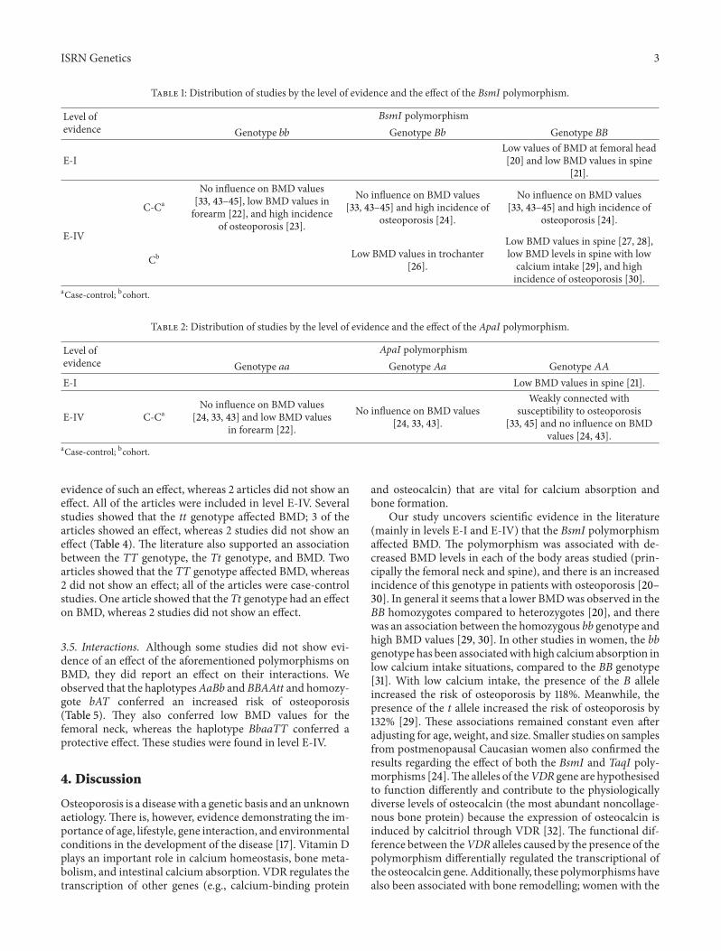
3.1. BsmI . A total of 11 articles (2 articles in level E-I and 9articles in level E-IV) were found that showed that the BsmIpolymorphism affected the loss of BMD. In contrast, 4 articlesreported that the BsmI polymorphism did not affect BMD; allof the articleswere level E-IV (Table 1).Therewere 8 studies (2articles in level E-I and 6 articles in level E-IV) supporting anassociation between the BB genotype and BMD; this was thelargest amount of data supporting an association between apolymorphism and BMD. However, 4 articles reported noevidence that this polymorphism affected BMD. Further-more, 2 studies on the genotype Bb showed an effect on BMD,as opposed to 4 studies that showed the same genotype had noeffect on BMD (both in level E-IV). Two studies on the bbgenotype showed an effect on BMD, whereas 4 studiesshowed that no evidence of this effect (both in level E-IV).
3.2. ApaI . Theeffect of theApaI polymorphismonBMDwasanalysed (Table 2). Four studies (1 article in level E-I and 3articles in level E-IV) reported evidence of the effect of thispolymorphism on BMD; in contrast, 2 studies showed no evi-dence of an effect (level E-IV). Likewise, 3 studies (1 article inlevel E-I and 2 articles in level E-IV) showed that theAA gen-otype had an effect on BMD, whereas 2 articles showed noevidence of this effect (both in level E-IV).There was less evi-dence indicating an association between the aa genotype andBMD in level E-IV; 1 article showed an effect, but 3 articlesshowed no evidence of an affect. Three articles (level E-IV)showed no evidence that the Aa genotype affected BMD.There were no cohort studies that examined the effect of theApaI polymorphism on BMD.
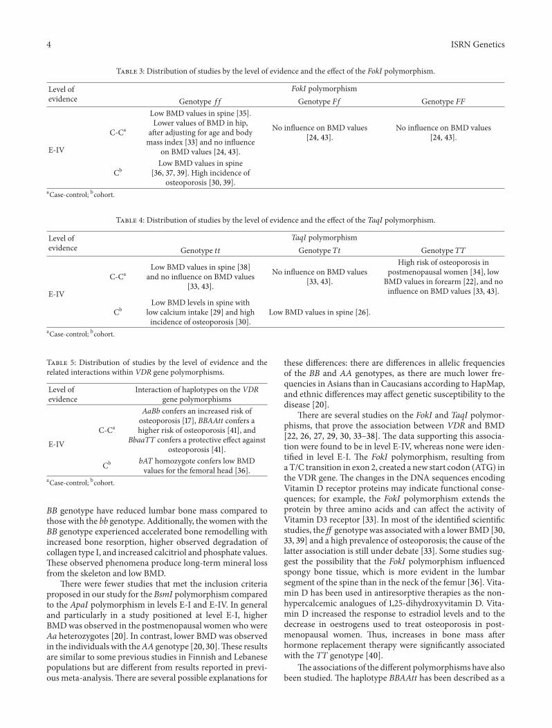
3.3. FokI . A total of 8 articles (level E-IV) studying the asso-ciation of the FokI genotype with BMD were found; of the 8articles, 6 articles reported evidence of an effect, but 2 articlesshowed no evidence of an effect on BMD (Table 3). Therewere several articles supporting the association between theffgenotype and BMD (6 articles showed an effect, whereas 2articles did not find a relationship).Wedid not find any articlethat showed that the Ff and FF polymorphisms affectedBMD. However, we found 2 articles rejecting such evidence(case-control studies).
3.4. TaqI . A total of 9 articles studying the effect of the TaqIpolymorphism on BMD were found; 7 articles reported
ISRN Genetics 3
Table 1: Distribution of studies by the level of evidence and the effect of the BsmI polymorphism.
Level ofevidence
𝐵𝑠𝑚𝐼 polymorphismGenotype 𝑏𝑏 Genotype 𝐵𝑏 Genotype 𝐵𝐵
E-ILow values of BMD at femoral head[20] and low BMD values in spine
[21].
E-IV
C-Ca
No influence on BMD values[33, 43–45], low BMD values inforearm [22], and high incidence
of osteoporosis [23].
No influence on BMD values[33, 43–45] and high incidence of
osteoporosis [24].
No influence on BMD values[33, 43–45] and high incidence of
osteoporosis [24].
Cb Low BMD values in trochanter[26].
Low BMD values in spine [27, 28],low BMD levels in spine with lowcalcium intake [29], and highincidence of osteoporosis [30].
aCase-control; bcohort.
Table 2: Distribution of studies by the level of evidence and the effect of the ApaI polymorphism.
Level ofevidence
ApaI polymorphismGenotype 𝑎𝑎 Genotype 𝐴𝑎 Genotype 𝐴𝐴
E-I Low BMD values in spine [21].
E-IV C-CaNo influence on BMD values
[24, 33, 43] and low BMD valuesin forearm [22].
No influence on BMD values[24, 33, 43].
Weakly connected withsusceptibility to osteoporosis
[33, 45] and no influence on BMDvalues [24, 43].
aCase-control; bcohort.
evidence of such an effect, whereas 2 articles did not show aneffect. All of the articles were included in level E-IV. Severalstudies showed that the tt genotype affected BMD; 3 of thearticles showed an effect, whereas 2 studies did not show aneffect (Table 4). The literature also supported an associationbetween the TT genotype, the Tt genotype, and BMD. Twoarticles showed that the TT genotype affected BMD, whereas2 did not show an effect; all of the articles were case-controlstudies. One article showed that theTt genotype had an effecton BMD, whereas 2 studies did not show an effect.
3.5. Interactions. Although some studies did not show evi-dence of an effect of the aforementioned polymorphisms onBMD, they did report an effect on their interactions. Weobserved that the haplotypesAaBb and BBAAtt and homozy-gote bAT conferred an increased risk of osteoporosis(Table 5). They also conferred low BMD values for thefemoral neck, whereas the haplotype BbaaTT conferred aprotective effect. These studies were found in level E-IV.
4. Discussion
Osteoporosis is a diseasewith a genetic basis and an unknownaetiology. There is, however, evidence demonstrating the im-portance of age, lifestyle, gene interaction, and environmentalconditions in the development of the disease [17]. Vitamin Dplays an important role in calcium homeostasis, bone meta-bolism, and intestinal calcium absorption. VDR regulates thetranscription of other genes (e.g., calcium-binding protein
and osteocalcin) that are vital for calcium absorption andbone formation.
Our study uncovers scientific evidence in the literature(mainly in levels E-I and E-IV) that the BsmI polymorphismaffected BMD. The polymorphism was associated with de-creased BMD levels in each of the body areas studied (prin-cipally the femoral neck and spine), and there is an increasedincidence of this genotype in patients with osteoporosis [20–30]. In general it seems that a lower BMDwas observed in theBB homozygotes compared to heterozygotes [20], and therewas an association between the homozygous bb genotype andhigh BMD values [29, 30]. In other studies in women, the bbgenotype has been associatedwith high calciumabsorption inlow calcium intake situations, compared to the BB genotype[31]. With low calcium intake, the presence of the B alleleincreased the risk of osteoporosis by 118%. Meanwhile, thepresence of the t allele increased the risk of osteoporosis by132% [29]. These associations remained constant even afteradjusting for age, weight, and size. Smaller studies on samplesfrom postmenopausal Caucasian women also confirmed theresults regarding the effect of both the BsmI and TaqI poly-morphisms [24].The alleles of theVDR gene are hypothesisedto function differently and contribute to the physiologicallydiverse levels of osteocalcin (the most abundant noncollage-nous bone protein) because the expression of osteocalcin isinduced by calcitriol through VDR [32]. The functional dif-ference between theVDR alleles caused by the presence of thepolymorphism differentially regulated the transcriptional ofthe osteocalcin gene.Additionally, these polymorphisms havealso been associated with bone remodelling; women with the
4 ISRN Genetics
Table 3: Distribution of studies by the level of evidence and the effect of the FokI polymorphism.
Level ofevidence
FokI polymorphismGenotype 𝑓𝑓 Genotype 𝐹𝑓 Genotype 𝐹𝐹
E-IV
C-Ca
Low BMD values in spine [35].Lower values of BMD in hip,
after adjusting for age and bodymass index [33] and no influence
on BMD values [24, 43].
No influence on BMD values[24, 43].
No influence on BMD values[24, 43].
CbLow BMD values in spine
[36, 37, 39]. High incidence ofosteoporosis [30, 39].
aCase-control; bcohort.
Table 4: Distribution of studies by the level of evidence and the effect of the TaqI polymorphism.
Level ofevidence
TaqI polymorphismGenotype 𝑡𝑡 Genotype 𝑇𝑡 Genotype 𝑇𝑇
E-IV
C-CaLow BMD values in spine [38]
and no influence on BMD values[33, 43].
No influence on BMD values[33, 43].
High risk of osteoporosis inpostmenopausal women [34], lowBMD values in forearm [22], and noinfluence on BMD values [33, 43].
CbLow BMD levels in spine with
low calcium intake [29] and highincidence of osteoporosis [30].
Low BMD values in spine [26].
aCase-control; bcohort.
Table 5: Distribution of studies by the level of evidence and therelated interactions within VDR gene polymorphisms.
Level ofevidence
Interaction of haplotypes on the VDRgene polymorphisms
E-IV
C-Ca
AaBb confers an increased risk ofosteoporosis [17], BBAAtt confers ahigher risk of osteoporosis [41], and
BbaaTT confers a protective effect againstosteoporosis [41].
Cb bAT homozygote confers low BMDvalues for the femoral head [36].
aCase-control; bcohort.
BB genotype have reduced lumbar bone mass compared tothose with the bb genotype. Additionally, the womenwith theBB genotype experienced accelerated bone remodelling withincreased bone resorption, higher observed degradation ofcollagen type I, and increased calcitriol and phosphate values.These observed phenomena produce long-term mineral lossfrom the skeleton and low BMD.
There were fewer studies that met the inclusion criteriaproposed in our study for the BsmI polymorphism comparedto the ApaI polymorphism in levels E-I and E-IV. In generaland particularly in a study positioned at level E-I, higherBMDwas observed in the postmenopausal women who wereAa heterozygotes [20]. In contrast, lower BMD was observedin the individuals with theAA genotype [20, 30].These resultsare similar to some previous studies in Finnish and Lebanesepopulations but are different from results reported in previ-ous meta-analysis.There are several possible explanations for
these differences: there are differences in allelic frequenciesof the BB and AA genotypes, as there are much lower fre-quencies in Asians than in Caucasians according to HapMap,and ethnic differences may affect genetic susceptibility to thedisease [20].
There are several studies on the FokI and TaqI polymor-phisms, that prove the association between VDR and BMD[22, 26, 27, 29, 30, 33–38]. The data supporting this associa-tion were found to be in level E-IV, whereas none were iden-tified in level E-I. The FokI polymorphism, resulting fromaT/C transition in exon 2, created a new start codon (ATG) inthe VDR gene. The changes in the DNA sequences encodingVitamin D receptor proteins may indicate functional conse-quences; for example, the FokI polymorphism extends theprotein by three amino acids and can affect the activity ofVitamin D3 receptor [33]. In most of the identified scientificstudies, the ff genotype was associated with a lower BMD [30,33, 39] and a high prevalence of osteoporosis; the cause of thelatter association is still under debate [33]. Some studies sug-gest the possibility that the FokI polymorphism influencedspongy bone tissue, which is more evident in the lumbarsegment of the spine than in the neck of the femur [36]. Vita-min D has been used in antiresorptive therapies as the non-hypercalcemic analogues of 1,25-dihydroxyvitamin D. Vita-min D increased the response to estradiol levels and to thedecrease in oestrogens used to treat osteoporosis in post-menopausal women. Thus, increases in bone mass afterhormone replacement therapy were significantly associatedwith the TT genotype [40].
The associations of the different polymorphisms have alsobeen studied. The haplotype BBAAtt has been described as a
ISRN Genetics 5
risk factor for osteoporosis; the haplotype BbaaTT has beendescribed as a protective factor in Spanish populations [41].The haplotype combinationAaBb conferred a fivefold greaterrisk when clinical parameters such as bodymass index (BMI)were adjusted. Accordingly, studies suggest that candidategenes affecting metabolic parameters as well as BMI can alterthe risk of osteoporosis [17]. An investigation of 75 analysedstudies on VDR gene polymorphisms and BMD concludedthat these polymorphisms were significantly associated withBMD. Additionally, the investigation concluded that thegenotypic effect was more prominent in premenopausalwomen than in postmenopausal women [42].
The VDR gene regulates the transcription of target genes(e.g., calcium-binding protein and osteocalcin) that are cru-cial for calcium absorption and bone formation. The mecha-nism by which theVDR gene affects BMD remains unknown.In addition, polymorphisms in theVDR gene and other genesand pathways, such as the oestrogen signalling pathway,Wnt/𝛽-catenin, transforming growth factor superfamily, and theactivating receptor of the nuclear factor 𝜅B (RANK) signall-ing pathway, can affect BMD and regulate the effect of theVDR gene polymorphisms on BMD. One of the current andmost probable reasons might be varying hormone levels,which can influence the effect of the VDR gene polymor-phisms on BMD.
In addition, environmental factors such as diet, physicalactivity, smoking, and alcohol consumption have beenproven to influence BMD and osteoporosis. Therefore, thesegene-environment factors may act as confusing elements thataffect the association between VDR polymorphisms andBMD [20]. However, different genotypes may also havediverse effects on different parts of the skeleton. This mightexplain why several studies did not find an association bet-ween VDR polymorphisms and BMD [26].
This study has several limitations because it was per-formed on a limited sample of studies on postmenopausalwomen. Hence, our findings cannot be generalised to specificpopulations, geographic locations, or different age subgroups.Parallel research on large-scale familial studies of postmeno-pausal women and younger individuals can help to clarify theexact role of geneticmarkers (includingVRD alleles) on bonemass loss.
5. Conclusion
From our comprehensive survey of the current available lit-erature it can be concluded that VDR polymorphisms do notappear to directly confer any significant BMDbenefit. Indeed,no definite conclusions can bemade regarding the associationof BsmI, ApaI, FokI, and TaqI polymorphism with BMDamong postmenopausal women.These findings show the im-portance of establishing suitable criteria, including geneticepidemiologic, phenotypic, and clinical criteria, to improvequality control of study design andmethods in genetic studiesrelated toVDR gene polymorphism andBMD.Based on thesecriteria, larger andmore rigorous analytical studies with con-sideration of gene-gene/environment interactions are neededto further dissect themechanisms bywhichVDR alleles influ-
ence BMD and interact with the other genetic and environ-mental factors.
Conflict of Interests
The authors declare that there is no conflict of interestsregarding the publication of this paper.
References
[1] W. A. Peck, P. Burckhardt, C. Christiansen et al., “Consensusdevelopment conference: diagnosis, prophylaxis, and treatmentof osteoporosis,” American Journal of Medicine, vol. 94, no. 6,pp. 646–650, 1993.
[2] W.-F. Li, S.-X. Hou, B. Yu, M.-M. Li, C. Ferec, and J.-M. Chen,“Genetics of osteoporosis: accelerating pace in gene identifica-tion and validation,” Human Genetics, vol. 127, no. 3, pp. 249–285, 2010.
[3] J. C. Christian, P.-L. Yu, C. W. Slemenda, and C. C. Johnston Jr.,“Heritability of bone mass: a longitudinal study in aging maletwins,” American Journal of Human Genetics, vol. 44, no. 3, pp.429–433, 1989.
[4] N. A. Pocock, J. A. Eisman, J. L. Hopper, M. G. Yeates, P. N.Sambrook, and S. Eberl, “Genetic determinants of bone mass inadults. A twin study,” Journal of Clinical Investigation, vol. 80, no.3, pp. 706–710, 1987.
[5] E. Seeman, J. L.Hopper, L. A. Bach et al., “Reduced bonemass indaughters of women with osteoporosis,”TheNew England Jour-nal of Medicine, vol. 320, no. 9, pp. 554–558, 1989.
[6] F. A. Tylavsky, A. D. Bortz, R. L. Hancock, and J. J. Anderson,“Familial resemblance of radial bone mass between premeno-pausal mothers and their college-age daughters,” CalcifiedTissue International, vol. 45, no. 5, pp. 265–272, 1989.
[7] J. A. Eisman, “VitaminD receptor gene alleles and osteoporosis:an affirmative view,” Journal of Bone and Mineral Research, vol.10, no. 9, pp. 1289–1293, 1995.
[8] J. A. Eisman, N. A. Morrison, P. J. Kelly et al., “Genetics of oste-oporosis and vitamin D receptor alleles,” Calcified Tissue Inter-national, vol. 56, supplement 1, pp. S48–S49, 1995.
[9] S. H. Ralston and B. de Crombrugghe, “Genetic regulation ofbone mass and susceptibility to osteoporosis,”Genes and Devel-opment, vol. 20, no. 18, pp. 2492–2506, 2006.
[10] R. A. Evans, G. M. Marel, E. K. Lancaster, S. Kos, M. Evans, andS. Y. Wong, “Bone mass is low in relatives of osteoporoticpatients,” Annals of Internal Medicine, vol. 109, no. 11, pp. 870–873, 1988.
[11] P. J. Kelly, J. L. Hopper, G. T. Macaskill, N. A. Pocock, P. N.Sambrook, and J. A. Eisman, “Genetic factors in bone turnover,”Journal of Clinical Endocrinology and Metabolism, vol. 72, no. 4,pp. 808–813, 1991.
[12] A. Tokita, P. J. Kelly, T. V. Nguyen et al., “Genetic influences ontype I collagen synthesis and degradation: further evidence forgenetic regulation of bone turnover,” Journal of Clinical Endo-crinology and Metabolism, vol. 78, no. 6, pp. 1461–1466, 1994.
[13] L. Flicker, J. L. Hopper, L. Rodgers, B. Kaymakci, R. M. Green,and J. D.Wark, “Bone density determinants in elderly women: atwin study,” Journal of Bone andMineral Research, vol. 10, no. 11,pp. 1607–1613, 1995.
[14] N. A. Morrison, J. C. Qi, A. Tokita et al., “Prediction of bonedensity from vitamin D receptor alleles,” Nature, vol. 367, no.6460, pp. 284–287, 1994.
6 ISRN Genetics
[15] J. P. Ioannidis, I. Stavrou, T. A. Trikalinos et al., “Association ofpolymorphisms of the estrogen receptor 𝛼 gene with bonemin-eral density and fracture risk in women: a meta-analysis,” Jour-nal of Bone and Mineral Research, vol. 17, no. 11, pp. 2048–2060,2002.
[16] A. G. Uitterlinden, Y. Fang, J. B. VanMeurs, H. A. Pols, and J. P.Van Leeuwen, “Genetics and biology of vitamin D receptorpolymorphisms,” Gene, vol. 338, no. 2, pp. 143–156, 2004.
[17] T. M. Durusu, T. G. Bora, T. D. Uluturk et al., “Evaluation of theeffects of vitamin D receptor and estrogen receptor 1 gene poly-morphisms on bone mineral density in postmenopausalwomen,” Clinical Rheumatology, vol. 29, no. 11, pp. 1285–1293,2010.
[18] B.M.Melnyk and E. Fineout-Overholt, Evidence-Based Practicein Nursing and Healthcare a Guide to Best Practice, LippincottWilliams &Wilkins, Philadelphia, Pa, USA, 2005.
[19] R. Whittemore and K. Knafl, “The integrative review: updatedmethodology,” Journal of Advanced Nursing, vol. 52, no. 5, pp.546–553, 2005.
[20] Y. Li, B. Xi, K. Li, and C.Wang, “Association between vitamin Dreceptor gene polymorphisms and bone mineral density inChinese women,” Molecular Biology Reports, vol. 39, no. 5, pp.5709–5717, 2012.
[21] A. Thakkinstian, C. D’Este, J. Eisman, T. Nguyen, and J. Attia,“Meta-analysis of molecular association studies: vitamin Dreceptor gene polymorphisms andBMDas a case study,” Journalof Bone and Mineral Research, vol. 19, no. 3, pp. 419–428, 2004.
[22] K.Douroudis, K. Tarassi, G. Ioannidis et al., “Association of vita-minD receptor gene polymorphismswith bonemineral densityin postmenopausal women of Hellenic origin,” Maturitas, vol.45, no. 3, pp. 191–197, 2003.
[23] R. Lisker, M. A. Lopez, S. Jasqui et al., “Association of vitamin Dreceptor polymorphisms with osteoporosis in Mexican post-menopausal women,” Human Biology, vol. 75, no. 3, pp. 399–403, 2003.
[24] B. L. Langdahl, C. H. Gravholt, K. Brixen, and E. F. Eriksen,“Polymorphisms in the vitaminD receptor gene and bonemass,bone turnover and osteoporotic fractures,” European Journal ofClinical Investigation, vol. 30, no. 7, pp. 608–617, 2000.
[25] L. Borjas-Fajardo, M. Zambrano, E. Fernandez et al., “Analysisof Bsm I polymorphism of the vitamin D receptor (VDR) genein Venezuelan female patients living in the state of Zulia withosteoporosis,” Investigacion Clinica, vol. 44, no. 4, pp. 275–282,2003 (Spanish).
[26] J. G. Kim, J. H. Kwon, S. H. Kim, Y.M. Choi, S. Y.Moon, and J. Y.Lee, “Association between vitamin D receptor gene haplotypesand bone mass in postmenopausal Korean women,” AmericanJournal of Obstetrics and Gynecology, vol. 189, no. 5, pp. 1234–1240, 2003.
[27] H. Y. Chen, W. C. Chen, C. D. Hsu, F. J. Tsai, C. H. Tsai, andC. W. Li, “Relation of BsmI vitamin D receptor gene polymor-phism to bone mineral density and occurrence of osteoporosisin postmenopausal Chinese women in Taiwan,” OsteoporosisInternational, vol. 12, no. 12, pp. 1036–1041, 2001.
[28] J. Marc, J. Prezelj, R. Komel, and A. Kocijancic, “Association ofvitaminD receptor gene polymorphismwith bonemineral den-sity in Slovenian postmenopausal women,”Gynecological Endo-crinology, vol. 14, no. 1, pp. 60–64, 2000.
[29] M. G. Stathopoulou, G. V. Z. Dedoussis, G. Trovas et al., “Therole of vitamin D receptor gene polymorphisms in the bonemineral density of Greek postmenopausal women with low cal-
cium intake,” Journal of Nutritional Biochemistry, vol. 22, no. 8,pp. 752–757, 2011.
[30] S. Mitra, M. Desai, and K.-H. M. Ikram, “Vitamin D receptorgene polymorphisms and bone mineral density in postmeno-pausal Indian women,”Maturitas, vol. 55, no. 1, pp. 27–35, 2006.
[31] B. Dawson-Hughes, S. S. Harris, and S. Finneran, “Calciumabsorption on high and low calcium intakes in relation to vita-minD receptor genotype,” Journal of Clinical Endocrinology andMetabolism, vol. 80, no. 12, pp. 3657–3661, 1995.
[32] N. A. Morrison, R. Yeoman, P. J. Kelly, and J. A. Eisman, “Con-tribution of trans-acting factor alleles to normal physiologicalvariability: vitamin D receptor gene polymorphisms and circu-lating osteocalcin,” Proceedings of the National Academy of Sci-ences of the United States of America, vol. 89, no. 15, pp. 6665–6669, 1992.
[33] K. Zajıckova, I. Zofkova, R. Bahbough, andA. Krepelova, “Vita-minD receptor gene polymorphisms, bonemineral density andbone turnover: FokI genotype is related to postmenopausalbone mass,” Physiological Research, vol. 51, no. 5, pp. 501–509,2002.
[34] A. Seremak-Mrozikiewicz, K. Drews, P. M. Mrozikiewicz et al.,“Correlation of vitamin D receptor gene (VDR) polymorphismwith osteoporotic changes in Polish postmenopausal women,”Neuroendocrinology Letters, vol. 30, no. 4, pp. 540–546, 2009.
[35] A. Falchetti, C. Sferrazza, C. Cepollaro et al., “FokI polymor-phism of the vitamin D receptor gene correlates with parame-ters of bone mass and turnover in a female population of theItalian island of Lampedusa,” Calcified Tissue International, vol.80, no. 1, pp. 15–20, 2007.
[36] W. Horst-Sikorska, R. Kalak, A. Wawrzyniak, M. Marcinkow-ska, L. Celczynska-Bajew, and R. Slomski, “Association analysisof the polymorphisms of theVDRgenewith bonemineral dens-ity and the occurrence of fractures,” Journal of Bone andMineralMetabolism, vol. 25, no. 5, pp. 310–319, 2007.
[37] E. Bandres, I. Pombo, M. Gonzalez-Huarriz, A. Rebollo, G.Lopez, and J. Garcıa-Foncillas, “Association between bonemin-eral density and polymorphisms of the VDR, ERalpha, COL1A1and CTR genes in Spanish postmenopausal women,” Journal ofEndocrinological Investigation, vol. 28, no. 4, pp. 312–321, 2005.
[38] B. S. Duman, R. Tanakol, N. Erensoy, M. Ozturk, and S. Yil-mazer, “Vitamin D receptor alleles, bone mineral density andturnover in postmenopausal osteoporotic and healthy women,”Medical Principles and Practice, vol. 13, no. 5, pp. 260–266, 2004.
[39] H.-Y. Chen, W.-C. Chen, C.-D. Hsu, F.-J. Tsai, and C.-H. Tsai,“Relation of vitaminD receptor FokI start codonpolymorphismto bonemineral density and occurrence of osteoporosis in post-menopausalwomen inTaiwan,”ActaObstetricia etGynecologicaScandinavica, vol. 81, no. 2, pp. 93–98, 2002.
[40] T. Kurabayashi, H. Matsushita, M. Tomita et al., “Association ofvitamin D and estrogen receptor gene polymorphism with theeffects of longterm hormone replacement therapy on bonemin-eral density,” Journal of Bone and Mineral Metabolism, vol. 22,no. 3, pp. 241–247, 2004.
[41] M. Zambrano-Morales, L. Borjas, E. Fernandez et al., “Associa-tion of the vitamin D receptor gene BBAAtt haplotype withosteoporosis in post-menopausic women,” Investigacion Clin-ica, vol. 49, no. 1, pp. 29–38, 2008 (Spanish).
[42] G.Gong,H. S. Stern, S.-C. Cheng et al., “The association of bonemineral density with vitamin D receptor gene polymorphisms,”Osteoporosis International, vol. 9, no. 1, pp. 55–64, 1999.
ISRN Genetics 7
[43] T. Yoldemir, D. G. Yavuz, G. Anik, N. Verimli, and M. Erenus,“Vitamin D receptor gene polymorphisms in a group of post-menopausal Turkish women: association with bone mineraldensity,” Climacteric, vol. 14, no. 3, pp. 384–391, 2011.
[44] S. Valimaki, R. Tahtela, K. Kainulainen et al., “Relation of colla-gen type I alpha 1 (COLIA 1) and vitamin D receptor genotypesto bone mass, turnover, and fractures in early postmenopausalwomen and to hip fractures in elderly people,” European Journalof Internal Medicine, vol. 12, no. 1, pp. 48–56, 2001.
[45] G. R. Fontova, F. C. Gutierrez, M. M. Broch et al., “Polymor-phism of vitamin D receptor gene, bone mass, and bone turn-over among women with postmenopausal osteoporosis,” Re-vista Clinica Espanola, vol. 200, no. 4, pp. 198–202, 2000(Spanish).

Submit your manuscripts athttp://www.hindawi.com
Hindawi Publishing Corporationhttp://www.hindawi.com Volume 2014
Anatomy Research International
PeptidesInternational Journal of
Hindawi Publishing Corporationhttp://www.hindawi.com Volume 2014
Hindawi Publishing Corporation http://www.hindawi.com
International Journal of
Volume 2014
Zoology
Hindawi Publishing Corporationhttp://www.hindawi.com Volume 2014
Molecular Biology International
GenomicsInternational Journal of
Hindawi Publishing Corporationhttp://www.hindawi.com Volume 2014
The Scientific World JournalHindawi Publishing Corporation http://www.hindawi.com Volume 2014
Hindawi Publishing Corporationhttp://www.hindawi.com Volume 2014
BioinformaticsAdvances in
Marine BiologyJournal of
Hindawi Publishing Corporationhttp://www.hindawi.com Volume 2014
Hindawi Publishing Corporationhttp://www.hindawi.com Volume 2014
Signal TransductionJournal of
Hindawi Publishing Corporationhttp://www.hindawi.com Volume 2014
BioMed Research International
Evolutionary BiologyInternational Journal of
Hindawi Publishing Corporationhttp://www.hindawi.com Volume 2014
Hindawi Publishing Corporationhttp://www.hindawi.com Volume 2014
Biochemistry Research International
ArchaeaHindawi Publishing Corporationhttp://www.hindawi.com Volume 2014
Hindawi Publishing Corporationhttp://www.hindawi.com Volume 2014
Genetics Research International
Hindawi Publishing Corporationhttp://www.hindawi.com Volume 2014
Advances in
Virolog y
Hindawi Publishing Corporationhttp://www.hindawi.com
Nucleic AcidsJournal of
Volume 2014
Stem CellsInternational
Hindawi Publishing Corporationhttp://www.hindawi.com Volume 2014
Hindawi Publishing Corporationhttp://www.hindawi.com Volume 2014
Enzyme Research
Hindawi Publishing Corporationhttp://www.hindawi.com Volume 2014
International Journal of
Microbiology
Copyright © 2022 FDOKUMEN