Possible Interaction between USH1B and USH3 Gene Products as Implied by Apparent Digenic Deafness...
23
247 Letters to the Editor Am. J. Hum. Genet. 65:247–249, 1999 Mutations of UFD1L Are Not Responsible for the Majority of Cases of DiGeorge Syndrome/ Velocardiofacial Syndrome without Deletions within Chromosome 22q11 To the Editor: Deletions of chromosome 22q11 are associated with a wide spectrum of congenital malformation, encom- passed by the acronym “CATCH22” (cardiac defects, abnormal facies, thymic hypoplasia, cleft palate, and hy- pocalcemia on chromosome 22), including velocardio- facial syndrome (VCFS; MIM 192430), DiGeorge syn- drome (DGS; MIM 188400), and conotruncal-anomaly face (Emanuel et al. 1998). The major anomalies include outflow-tract congenital heart defects, hypoplasia of the parathyroids and thymus, craniofacial dysmorphism, and learning/behavioral problems (Ryan et al. 1997). Many of these are thought to be due to a defective neural-crest contribution during development. The DiGeorge chromosomal region (DGCR) is entirely cloned (Carlson et al. 1997) and sequenced, and several genes have been reported mapping to the region. Mu- tation screens of genes mapping to the proximal end of this region, termed the “minimal DiGeorge chromoso- mal region” (MDGCR; Gong et al. 1996), have been negative (Wadey et al. 1995; Gong et al. 1997; Gottlieb et al. 1997; Lindsay et al. 1998). Attention therefore has turned to the regions adjacent and distal to the MDGCR. Recently, the gene UFD1L was proposed as the major gene haploinsufficient in this group of syndromes (Ya- magishi et al. 1999). UFD1L is downstream of dHAND, a gene known to be involved in control of the devel- opment of structures affected in DGS, and Ufd1l is ex- pressed in the branchial arches, frontonasal mass, and outflow tract. In addition, a single patient has been re- ported with a de novo deletion affecting UFD1L and the neighboring gene, CDC45L2 (Yamagishi et al. 1999). CDC45 is required for initiation of DNA repli- cation in yeast, and CDC45 mutants are nonviable. However, CDC45L2 expression is not altered in d- HAND 2/2 embryos. On the basis of these findings, Yamagishi and colleagues concluded that UFD1L hap- loinsufficiency (perhaps with some contribution from CDC45L2) causes DGS. We conducted mutation screens, in both UFD1L and CDC45L2, as a three-center collaboration. UFD1L was screened by direct sequencing of 12 patients in London, by direct sequencing of all exons and 900 bp of the 5 0 UTR in 20 patients in Rome, and by DGGE of 7 patients’ DNA in Rotterdam. Local ethical review and consenting procedures were followed. The majority of patients were chosen on the basis of the presence of two or more features of the 22q11 deletion syndromes, but with no detectable deletion of 22q11 or of the DGSII region of 10p13 (Daw et al. 1996). The Rome series contained six patients with an isolated (i.e., nonsyndromic) inter- rupted aortic arch, a congenital heart defect commonly associated with the deletion. These patients were in- cluded because point mutations may be associated with a narrower spectrum of malformation than deletion and—since UFD1L was specifically identified as a d- HAND target—because congenital heart defects might be especially significant. The previously described pa- tient with a balanced 2;22 translocation in association with DGS (patient ADU; Augusseau et al. 1986) was also screened. UFD1L primers and conditions are avail- able from the collaborating centers, and the genomic organization of UFD1L and the resources for exon PCR amplification have been described elsewhere by Novelli et al. (1998). In London, 24 patients were similarly screened for CDC45L2 mutations; primers and PCR conditions are available on request, and genomic or- ganization has been published previously (McKie et al. 1998). No mutations of either gene were detected. We did, however, detect a number of sequence variants. Within the 5 0 UTR of UFD1L we found a single poly- morphic sequence, initially detected by SSCP and sub- sequently shown to involve an ArG transition, located at the 2277 position (with respect to the first base of the initiation codon). Screening of 25 unrelated controls generated a heterozygosity value of .40. Within CDC45L2 we detected an ArG transition 22 bp up- stream of exon 17 (at intron 16, with heterozygosity of .3) and a GrT transversion 24 bp into intron 18 (het- erozygosity of .5). In addition, Southern analysis of 42 patients was conducted, with four different restriction- enzyme digests (HindIII, EcoRI, KpnI, and BamHI), in an attempt to ascertain rearrangements similar to the
-
Upload
independent -
Category
Documents
-
view
0 -
download
0
Transcript of Possible Interaction between USH1B and USH3 Gene Products as Implied by Apparent Digenic Deafness...

247
Letters to the Editor
Am. J. Hum. Genet. 65:247–249, 1999
Mutations of UFD1L Are Not Responsible for theMajority of Cases of DiGeorge Syndrome/Velocardiofacial Syndrome without Deletions withinChromosome 22q11
To the Editor:Deletions of chromosome 22q11 are associated witha wide spectrum of congenital malformation, encom-passed by the acronym “CATCH22” (cardiac defects,abnormal facies, thymic hypoplasia, cleft palate, and hy-pocalcemia on chromosome 22), including velocardio-facial syndrome (VCFS; MIM 192430), DiGeorge syn-drome (DGS; MIM 188400), and conotruncal-anomalyface (Emanuel et al. 1998). The major anomalies includeoutflow-tract congenital heart defects, hypoplasia of theparathyroids and thymus, craniofacial dysmorphism,and learning/behavioral problems (Ryan et al. 1997).Many of these are thought to be due to a defectiveneural-crest contribution during development. TheDiGeorge chromosomal region (DGCR) is entirelycloned (Carlson et al. 1997) and sequenced, and severalgenes have been reported mapping to the region. Mu-tation screens of genes mapping to the proximal end ofthis region, termed the “minimal DiGeorge chromoso-mal region” (MDGCR; Gong et al. 1996), have beennegative (Wadey et al. 1995; Gong et al. 1997; Gottliebet al. 1997; Lindsay et al. 1998). Attention therefore hasturned to the regions adjacent and distal to the MDGCR.Recently, the gene UFD1L was proposed as the majorgene haploinsufficient in this group of syndromes (Ya-magishi et al. 1999). UFD1L is downstream of dHAND,a gene known to be involved in control of the devel-opment of structures affected in DGS, and Ufd1l is ex-pressed in the branchial arches, frontonasal mass, andoutflow tract. In addition, a single patient has been re-ported with a de novo deletion affecting UFD1L andthe neighboring gene, CDC45L2 (Yamagishi et al.1999). CDC45 is required for initiation of DNA repli-cation in yeast, and CDC45 mutants are nonviable.However, CDC45L2 expression is not altered in d-HAND 2/2 embryos. On the basis of these findings,Yamagishi and colleagues concluded that UFD1L hap-
loinsufficiency (perhaps with some contribution fromCDC45L2) causes DGS.
We conducted mutation screens, in both UFD1L andCDC45L2, as a three-center collaboration. UFD1L wasscreened by direct sequencing of 12 patients in London,by direct sequencing of all exons and 900 bp of the 5′
UTR in 20 patients in Rome, and by DGGE of 7 patients’DNA in Rotterdam. Local ethical review and consentingprocedures were followed. The majority of patients werechosen on the basis of the presence of two or morefeatures of the 22q11 deletion syndromes, but with nodetectable deletion of 22q11 or of the DGSII region of10p13 (Daw et al. 1996). The Rome series containedsix patients with an isolated (i.e., nonsyndromic) inter-rupted aortic arch, a congenital heart defect commonlyassociated with the deletion. These patients were in-cluded because point mutations may be associated witha narrower spectrum of malformation than deletionand—since UFD1L was specifically identified as a d-HAND target—because congenital heart defects mightbe especially significant. The previously described pa-tient with a balanced 2;22 translocation in associationwith DGS (patient ADU; Augusseau et al. 1986) wasalso screened. UFD1L primers and conditions are avail-able from the collaborating centers, and the genomicorganization of UFD1L and the resources for exon PCRamplification have been described elsewhere by Novelliet al. (1998). In London, 24 patients were similarlyscreened for CDC45L2 mutations; primers and PCRconditions are available on request, and genomic or-ganization has been published previously (McKie et al.1998). No mutations of either gene were detected. Wedid, however, detect a number of sequence variants.Within the 5′UTR of UFD1L we found a single poly-morphic sequence, initially detected by SSCP and sub-sequently shown to involve an ArG transition, locatedat the 2277 position (with respect to the first base ofthe initiation codon). Screening of 25 unrelated controlsgenerated a heterozygosity value of .40. WithinCDC45L2 we detected an ArG transition 22 bp up-stream of exon 17 (at intron 16, with heterozygosity of.3) and a GrT transversion 24 bp into intron 18 (het-erozygosity of .5). In addition, Southern analysis of 42patients was conducted, with four different restriction-enzyme digests (HindIII, EcoRI, KpnI, and BamHI), inan attempt to ascertain rearrangements similar to the
248 Letters to the Editor
UFD1L/CDC45L2 deletion reported elsewhere. Thisanalysis included all of the London patients screened forpoint mutations, as well as an additional 18 patients.No rearrangements or deletions were detected, althoughfour RFLPs were observed. Finally, mice with hemizy-gous targeted mutations of Ufd1l were normal (A. Bal-dini, personal communication).
Where does this leave the molecular genetics of the22q11 deletion syndromes? Interpretation of currentdata must consider that, although >10% of deletionsare inherited (Ryan et al. 1997), there is no good evi-dence for inheritance of DGS/VCFS in nondeletion cases.Furthermore, there are a large number of potential phe-nocopies of the condition (Emanuel et al. 1998). It istherefore possible that only a fraction of nondeletedcases have an etiology related to chromosome 22q11.Therefore, UFD1L must still be regarded as a good can-didate for contributing to this complex multiple-mal-formation syndrome. However, it should be kept in mindthat a number of genes might be acting to produce acombined haploinsufficiency, especially since other geneswithin the DGCR are also expressed in affected tissues.In the case of HIRA, for instance, the protein is knownto interact with PAX3, a gene required for conotruncalseptation in the mouse (Magnaghi et al. 1998), and an-tisense attenuation of HIRA expression in chicks yieldsan increased incidence of persistent truncus arteriosus(Farrell et al. 1999). However, as with UFD1L, muta-tions within HIRA have not been detected. Another con-sideration is the presence of distinct (i.e., nonoverlap-ping) rearrangements of 22q11, associated with verysimilar DGS-like phenotypes (Dallapiccola et al. 1996;Kurahashi et al. 1996; Sutherland et al. 1996; Rauch etal. 1999). Perhaps haploinsufficiency of more than onegene can cause the syndrome, or long-range effects in-duced by the rearrangements can down-regulate the ex-pression of the relevant gene(s). The role of combina-tions of genes during development is being tested bychromosome engineering in the mouse (Lindsay and Bal-dini 1998), although it is conceivable that long-rangeeffects will confuse analysis in the murine system. Inagreement with other commentators (Baldini 1999; Hag-mann 1999), we think it is too early to call “ClosingTime” (Heller 1996) on “CATCH22” (Heller 1955).
Acknowledgments
We would like to thank the families and clinicians who madethe study possible. Support was from the Birth Defects Foun-dation and the British Heart Foundation (to P.J.S.), TelethonFoundation grant E. 723 (to B.D. and G.N.), and the DutchHeart Foundation and the Sophia Foundation for Medical Re-search (to C.M.). We would like to thank Drs. Antonio Baldiniand Elizabeth Lindsay for patient referrals, helpful discussion,and providing critical data prior to publication. Access to PCR
conditions can be obtained at the e-mail addresses that follow:[email protected] (for C.M.), [email protected](for R.W.), and [email protected] (for G.N.).
ROY WADEY,1 JUDITH MCKIE,1
CHARALAMBOS PAPAPETROU,1 HELEN SUTHERLAND,1
FRANS LOHMAN,2 JAN OSINGA,4 INGRID FROHN,3
ROBERT HOFSTRA,4 CAREL MEIJERS,2
FRANCESCA AMATI,5 EMANUELA CONTI,5
ANTONIO PIZZUTI,5 BRUNO DALLAPICCOLA,5
GIUSEPPE NOVELLI,5 AND PETER SCAMBLER1
1Molecular Medicine Unit, Institute of Child Health,London; 2Department of Cell Biology and Genetics/Pediatric Surgery, Erasmus University, and3Department of Pediatrics, Division of Cardiology,Sophia Children’s Hospital, Rotterdam, and4Department of Medical Genetics, State University ofGroningen, Groningen, the Netherlands; and5Department of Biopathology and DiagnosticImaging, Tor Vergata University and CSS-MendelInstitute, Rome
Electronic-Database Information
Accession numbers and URLs for data in this article are asfollows:
Online Mendelian Inheritance in Man (OMIM), http://www.ncbi.nlm.nih.gov/Omim/ (for VCFS [MIM 192430] andDGS [MIM 188400])
References
Augusseau S, Jouk S, Jalbert P, Priur M (1986) DiGeorge syn-drome and 22q11 rearrangements. Hum Genet 74:206–206
Baldini A (1999) DiGeorge syndrome: is the solution inHAND? Nat Genet 21:246–247
Carlson C, Sirotkin H, Pandita R, Goldberg R, McKie J,Wadey R, Patanjali S, et al (1997) Molecular definition of22q11 deletions in 151 VCFS patients. Am J Hum Genet61:620–629
Dallapiccola B, Pizzuti A, Novelli G (1996) How many breaksdo we need to CATCH on 22q11? Am J Hum Genet 59:7–11
Daw SCM, Taylor C, Kraman M, Call K, Mao J, MeitingerT, Lipson A, et al (1996) A common region of 10p deletedin DiGeorge and velo-cardio-facial syndrome. Nat Genet 13:458–460
Emanuel BS, Budarf BS, Scambler PJ (1998) The genetic basisof conotruncal heart defects: the chromosome 22q11.2 de-letion. In: Rosenthal N, Harvey R (eds) Heart development,pp. 463–478. Academic Press, San Diego
Farrell M, Stadt H, Wallis K, Scambler PJ, Hixon RL, WolfeR, Leatherbury L, et al (1999) Persistent truncus arteriosusis associated with decreased expression of HIRA by cardiacneural crest cells in chick embryos. Circ Res 84:127–135
Gong W, Emanuel BS, Collins J, Kim DH, Wang Z, Chen F,Zhang G, et al (1996) A transcription map of the DiGeorge

Letters to the Editor 249
and velo-cardio-facial syndrome critical region on 22q11.Hum Mol Genet 5:789–800
Gong W, Emanuel BS, Galili N, Kim DH, Roe B, Driscoll DA,Budarf ML (1997) Structural and mutational analysis of aconserved gene (DGSI) from the minimal DiGeorge syn-drome critical region. Hum Mol Genet 6:267–276
Gottlieb S, Emanuel BS, Driscoll DA, Sellinger B, Wang Z,Roe B, Budarf ML (1997) The DiGeorge syndrome minimalcritical region contains a Goosecoid-like (GSCL) homeoboxgene, which is expressed early in human development. AmJ Hum Genet 60:1194–1201
Hagmann M (1999) A gene that scrambles your heart. Science283:1091–1093
Heller JL (1955) Catch 22. Simon and Schuster, New York——— (1996) Closing time. Simon and Schuster, New YorkKurahashi H, Nakayama T, Osugi Y, Tsuda E, Masuno M,
Imaizumi K, Kamiya T, et al (1996) Deletion mapping of22q11 in catch22 syndrome: identification of a second crit-ical region. Am J Hum Genet 58:1377–1881
Lindsay EA, Baldini A (1998) Congenital heart defects and22q11 deletions: which genes count? Mol Med Today 4:350–357
Lindsay EA, Harvey EL, Scambler PJ, Baldini A (1998) ES2,a gene deleted in DiGeorge syndrome, encodes a nuclearprotein and is expressed during early mouse development,where it shares an expression domain with a Goosecoid-likegene. Hum Mol Genet 7:629–635
Magnaghi P, Roberts C, Lorain S, Lipinski M, Scambler PJ(1998) HIRA, a mammalian homologue of Saccharomycescerevisiae transcriptional co-repressors, interacts with pax3.Nat Genet 20:74–77
McKie JM, Wadey R, Sutherland H, Taylor K, Scambler PJ(1998) Direct selection of conserved cDNAs from theDiGeorge critical region: isolation of a novel CDC45-likegene. Genome Res 8:834–841
Novelli G, Mari A, Amati F, Colosimo A, Sangiuolo F, Beng-ala M, Conti E, et al (1998) Structure and expression ofthe human ubiquitin fusion-degradation gene (UFD1L).Biochem Biophys Acta 1396:158–162
Rauch A, Pfieffer RA, Leipold G, Singer H, Tigges M, HofbeckM (1999) A novel 22q11.2 microdeletion in DiGeorge syn-drome. Am J Hum Genet 64:658–666
Ryan AK, Goodship JA, Wilson DI, Philip N, Levy A, SiedelH, Schuffenhauer S, et al (1997) Spectrum of clinical featuresassociated with interstitial chromosome 22q11 deletions: aEuropean collaborative study. J Med Genet 34:798–804
Sutherland HF, Wadey R, McKie JM, Taylor C, Atif U, John-stone KA, Halford S, et al (1996) Identification of a noveltranscript disrupted by a balanced translocation associatedwith DiGeorge syndrome. Am J Hum Genet 59:23–31
Wadey R, Daw S, Taylor C, Atif U, Kamath S, Halford S,O’Donnell H, et al (1995) Isolation of a gene encoding anintegral membrane protein from the vicinity of a balancedtranslocation breakpoint associated with the DiGeorge syn-drome. Hum Mol Genet 4:1027–1034
Yamagishi H, Garg V, Matsuoka R, Thomas T, Srivastava D(1999) A molecular pathway revealing a genetic basis forhuman cardiac and craniofacial defects. Science 283:1158–1161
Address for correspondence and reprints: Prof. Peter Scambler, Room 211,Molecular Medicine Unit, Institute of Child Health, London, WC1N 1EH,United Kingdom. E-mail: [email protected]
q 1999 by The American Society of Human Genetics. All rights reserved.0002-9297/99/6501-0031$02.00
Am. J. Hum. Genet. 65:249–251, 1999
Haploinsufficiency of the HOXA Gene Cluster, in aPatient with Hand-Foot-Genital Syndrome,Velopharyngeal Insufficiency, and Persistent PatentDuctus Botalli
To the Editor:The homeobox-containing HOX genes constitute ahighly conserved gene family, with a role in specifyingthe body plan. In humans and in mice, four clusters(A–D) of HOX genes are located on different chromo-somes. The precise function of the individual HOXgenes, in humans, can be deduced from their expressionpattern during mouse development and from the phe-notype of mice with a targeted disruption or overex-pression of a specific HOX gene. In humans, mutationshave only been described in HOXD-13 and HOXA-13,causing synpolydactyly and the hand-foot-genital (HFG)syndrome, respectively (Muragaki et al. 1996; Mortlockand Innis 1997). The mechanisms by which mutationsin HOXA-13 lead to the phenotype—that is, whetherthrough haploinsufficiency or through a dominant neg-ative effect—are currently unknown. Here we report ona patient with HFG syndrome who carries a chromo-some 7p14 deletion involving the entire HOXA cluster,indicating that haploinsufficiency of HOXA-13 maycause the phenotype.
The patient is the second child of healthy, unrelatedparents. Pregnancy and delivery were uneventful. Facialdysmorphism was evident from birth, with retrognathia,low-set malformed ears, upturned nostrils, large mouth,and upslanted eyes. There were mild anomalies of thehands and feet, with shortened and laterally deviatedfirst toes and clinodactyly of the fifth fingers with shortterminal phalanges. Radiographs revealed hand and footanomalies characteristic of HFG syndrome (fig. 1 and2) (Stern et al. 1970; Halal 1988). There was left-sidedcryptorchidism and a ventral-bowed penis. An intrave-nous pyelogram was normal. In addition, he presentedwith severe feeding difficulties during infancy, caused byvelopharyngeal insufficiency with a shortened soft palateand very small uvula. On a barium swallow, massivenasal reflux was visible. A persistent patent ductus Bo-talli was surgically corrected at age 4 years. Growth wasnormal. Full-scale IQ at age 7 years was 85. Presently,

250 Letters to the Editor
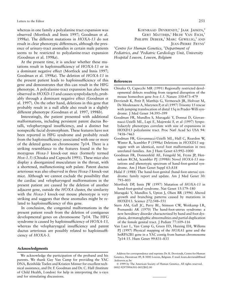
Figure 1 X-ray of the patient’s left foot, at age 2 years 10 mo.The first toe is laterally deviated, with a triangular distal phalanx andshortened proximal phalanx. There is absence of calcification of themiddle phalanges of toes II–V and distal phalanx of toe II.
Figure 2 X-ray of the patient’s left hand at age 7.5 years. Notethe thumb anomalies: shortened metacarpal, pointed distal phalanx,and pseudoepiphysis of the metacarpal. There is a brachymesophalanxV causing clinodactyly and associated with a pseudoepiphysis. Thereis shortening of the distal phalanx of finger II. Pseudoepiphyses arepresent at metacarpal II and V. Bone age was 4.1 years.
at age 21 years, he is healthy and functions at a bor-derline intelligence level.
Karyotype analysis of blood lymphocytes showed ade novo deletion in the short arm of chromosome 7,with karyotype 46,XY,del7(p14). FISH done with probeDO832 did not reveal a microdeletion in chromosome22q11. FISH and microsatellite analysis were performedfor the fine mapping of the deletion on 7p, as describedby Devriendt et al. (1997). Informed consent was ob-tained from the patient and his parents. The physical-map data were from Van Laer et al. (1997). With useof YACs Y915D12 and 920C6 (located telomeric fromthe HOXA cluster) and YAC 961E5 (containing theHOXA cluster), no signal was seen on the deleted chro-mosome 7p. Microsatellites D8S529 and D8S2496 maptelomeric and centromeric, respectively, from the HOXAcluster (Van Laer et al. 1997). Both markers were in-
formative in this family and their analysis revealed thatthe patient missed a maternal allele for both markers.These data demonstrated that the entire HOXA clusterwas deleted on this chromosome.
This patient with multiple congenital malformationscarries a de novo interstitial deletion of chromosome7p14, involving the entire HOXA gene cluster. Retro-spectively, the hand and foot anomalies present in thispatient are typical of HFG syndrome (Stern et al. 1970;Halal 1988). This autosomal dominant disorder iscaused by mutations in the HOXA-13 gene, which isthe most centromeric HOX gene of the HOXA clusteron chromosome 7p (Mortlock and Innis 1997). Muta-tions in HOXA-13 have so far been described in threefamilies with HFG syndrome. In two of the families themutations are predicted to lead to a truncated protein,
Letters to the Editor 251
whereas in one family a polyalanine tract expansion wasobserved (Mortlock and Innis 1997; Goodman et al.1998a). The different mutations in HOXA-13 do notresult in clear phenotypic differences, although the pres-ence of urinary-tract anomalies in certain male patientsseems to be restricted to polyalanine-tract expansion(Goodman et al. 1998a).
At the present time, it is unclear whether these mu-tations result in haploinsufficiency of HOXA-13 or ina dominant negative effect (Mortlock and Innis 1997;Goodman et al. 1998a). The deletion of HOXA-13 inthe present patient leads to haploinsufficiency of thisgene and demonstrates that this can result in the HFGphenotype. A polyalanine-tract expansion has also beenobserved in HOXD-13 and causes synpolydactyly, prob-ably through a dominant negative effect (Goodman etal. 1997). On the other hand, deletions in this gene thatprobably result in a null allele also result in a slightlydifferent phenotype (Goodman et al. 1997, 1998b).
Interestingly, the patient presented with additionalmalformations, including persistent patent ductus Bo-talli, velopharyngeal insufficiency, and a distinct butnonspecific facial dysmorphism. These features have notbeen reported in HFG syndrome and probably resultfrom the haploinsufficiency associated with one or moreof the deleted genes on chromosome 7p14. There is astriking resemblance to the features found in the ho-mozygous Hoxa-3 knock-out mice (formerly termedHox-1.5) (Chisaka and Capecchi 1991). These mice alsodisplay a disorganized musculature in the throat, witha shortened, malfunctioning soft palate. Patent ductusarteriosus was also observed in three Hoxa-3 knock-outmice. Although we cannot exclude the possibility thatthe cardiac and velopharyngeal malformations in thepresent patient are caused by the deletion of anotheradjacent gene, outside the HOXA cluster, the similaritywith the Hoxa-3 knock-out mouse phenotype is verystriking and suggests that these anomalies might be re-lated to haploinsufficiency of this gene.
In conclusion, the congenital malformations in thepresent patient result from the deletion of contiguousdevelopmental genes on chromosome 7p14. The HFGsyndrome is caused by haploinsufficiency of HOXA-13,whereas the velopharyngeal insufficiency and patentductus arteriosus are possibly related to haploinsuffi-ciency of HOXA-3.
Acknowledgments
We acknowledge the participation of the proband and hisparents. We thank Guy Van Camp for providing the YACDNA, Reinhilde Toelen and Kristien Minner for excellent tech-nical assistance, and Dr. F. Goodman and Dr. C. Hall (Instituteof Child Health, London) for help in interpreting the x-raysand for stimulating discussions.
KOENRAAD DEVRIENDT,1 JAAK JAEKEN,2
GERT MATTHIJS,1 HILDE VAN ESCH,1
PHILIPPE DEBEER,1 MARC GEWILLIG,3 AND
JEAN-PIERRE FRYNS1
1Centre for Human Genetics, 2Department ofPediatrics, and 3Pediatric Cardiology Unit, UniversityHospital Leuven, Leuven, Belgium
References
Chisaka O, Capecchi MR (1991) Regionally restricted devel-opmental defects resulting from targeted disruption of themouse homeobox gene hox-1.5. Nature 350:473–479
Devriendt K, Petit P, Matthijs G, Vermeesch JR, Holvoet M,De Meulenaere A, Marynen P, et al (1997) Trisomy 15 rescuewith jumping translocation of distal 15q in Prader-Willi syn-drome. J Med Genet 34:395–399
Goodman FR, Mundlos S, Muragaki Y, Donnai D, Giovan-nucci-Uzielli ML, Lapi E, Majewski F, et al (1997) Synpo-lydactyly phenotypes correlate with size of expansions inHOXD13 polyalanine tract. Proc Natl Acad Sci USA 94:7458–7463
Goodman FR, Giovannucci-Uzielli ML, Hall C, Reardon W,Winter R, Scambler P (1998a) Deletions in HOXD13 seg-regate with an identical, novel foot malformation in twounrelated families. Am J Hum Genet 63:992–1000
Goodman FR, Donnenfeld AE, Feingold M, Fryns JP, Hen-nekam RCM, Scambler PJ (1998b) Novel HOXA-13 mu-tations and phenotypic spectrum of hand-foot-genital syn-drome. Am J Hum Genet Suppl 63:A18
Halal F (1988) The hand-foot-genital (hand-foot-uterus) syn-drome: family report and update. Am J Med Genet 30:793–803
Mortlock DP, Innis JW (1997) Mutation of HOXA-13 inhand-foot-genital syndrome. Nat Genet 15:179–180
Muragaki Y, Mundlos S, Upton J, Olsen BR (1996) Alteredgrowth and branching patterns caused by mutations inHOXD13. Science 272:548–551
Stern AM, Gall JC, Perry BL, Stimson CW, Weitkamp LR,Poznanski AK (1970) The hand-foot-uterus syndrome: anew hereditary disorder characterized by hand and foot dys-plasia, dermatoglyphic abnormalities and partial duplicationof the female genital tract. J Pediatr 77:109–116
Van Laer L, Van Camp G, Green ED, Huizing EH, WillemsPJ (1997) Physical mapping of the HOXA1 gene and thehnRPA2B1 gene in a YAC contig from human chromosome7p14-15. Hum Genet 99:831–833
Address for correspondence and reprints: Dr. K. Devriendt, Centre for HumanGenetics, Herestraat 49, B-3000 Leuven, Belgium. E-mail: [email protected]
q 1999 by The American Society of Human Genetics. All rights reserved.0002-9297/99/6501-0032$02.00
252 Letters to the Editor
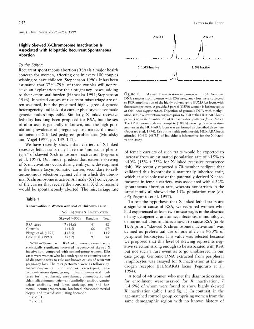
Figure 1 Skewed X inactivation in women with RSA. GenomicDNA samples from women with RSA pregnancy loss were subjectedto PCR amplification of the highly polymorphic HUMARA locus,withfluorescent primers. A gravida 5 para 0 (G5P0) woman is heterozygousat this locus (upper trace). Digestion of genomic DNA with methyl-ation-sensitive restriction enzymes prior to PCR at the HUMARA locuspermits accurate quantitation of X-inactivation patterns (lower trace).The G5P0 woman shows complete (100%) skewing. X-inactivationanalysis at the HUMARA locus was performed as described elsewhere(Pegoraro et al. 1994). Use of the highly polymorphic HUMARA locusafforded 90.6% (48/53) of individuals informative for the X-inacti-vation assay.
Table 1
X Inactivation in Women with RSA of Unknown Cause
NO. (%) WITH X INACTIVATION
Skewed >90% Random Total
RSA cases 7 (14.6) 41 48Controls 1 (1.5) 66 67a
Plenge et al. (1997) 4 (3.5) 111 115b
Gale et al. (1997) 3 (3.2) 91 94b
NOTE.—Women with RSA of unknown cause have astatistically significant increased frequency of skewed Xinactivation, compared with control-group women. RSAcases were women who had undergone an extensive seriesof diagnostic tests to rule out known causes of recurrentpregnancy loss. The tests performed were as follows: cy-togenetic—parental and abortus karyotyping; ana-tomic—hysterosalpingogram; infectious—cervical cul-tures for mycoplasma, ureaplasma, gonnococcus, andchlamydia; immunologic—anticardiolipin antibody, antin-uclear antibody, and lupus anticoagulant; and hor-monal—serum progesterone, late luteal-phase endometrialbiopsy, and thyroid-stimulating hormone.
a .P ! .01b .P ! .02
Am. J. Hum. Genet. 65:252–254, 1999
Highly Skewed X-Chromosome Inactivation IsAssociated with Idiopathic Recurrent SpontaneousAbortion
To the Editor:Recurrent spontaneous abortion (RSA) is a major healthconcern for women, affecting one in every 100 coupleswishing to have children (Stephenson 1996). It has beenestimated that 37%–79% of those couples will not re-ceive an explanation for their pregnancy losses, addingto their emotional burden (Hatasaka 1994; Stephenson1996). Inherited causes of recurrent miscarriage are of-ten assumed, but the presumed high degree of geneticheterogeneity and lack of a carrier phenotype have madegenetic studies impossible. Similarly, X-linked recessivelethality has long been proposed for RSA, but the sexof abortuses is generally unknown, and the high pop-ulation prevalence of pregnancy loss makes the ascer-tainment of X-linked pedigrees problematic (Motulskyand Vogel 1997, pp. 139–141).
We have recently shown that carriers of X-linkedrecessive lethal traits may have the “molecular pheno-type” of skewed X-chromosome inactivation (Pegoraroet al. 1997). Our model predicts that extreme skewingof X inactivation occurs during embryonic developmentin the female (asymptomatic) carrier, secondary to cell-autonomous selection against cells in which the abnor-mal X chromosome is active. All male (XY) conceptionsof the carrier that receive the abnormal X chromosomewould be spontaneously aborted. The miscarriage rate
of female carriers of such traits would be expected toincrease from an estimated population rate of ∼15% to∼40% ( for X-linked recessive recurrence15% 1 25%risk). We recently reported a 70-member pedigree thatvalidated this hypothesis: a maternally inherited trait,which caused sole use of the paternally derived X chro-mosome in female carriers, was associated with a 32%spontaneous abortion rate, whereas noncarriers in thesame family all showed the 15% population rate (P !
; Pegoraro et al. 1997)..05To test the hypothesis that X-linked lethal traits are
a significant cause of RSA, we recruited women whohad experienced at least two miscarriages in the absenceof any cytogenetic, anatomic, infectious, immunologic,or hormonal abnormalities known to cause RSA (table1). A priori, “skewed X chromosome inactivation” wasdefined as preferential use of one allele in >90% ofperipheral leukocytes. This value was selected becausewe proposed that this level of skewing represents neg-ative selection strong enough to be associated with RSAbut not such a rare event as to go unobserved in ourcase group. Genomic DNA extracted from peripherallymphocytes was assayed for X inactivation at the an-drogen receptor (HUMARA) locus (Pegoraro et al.1994).
A total of 48 women who met the diagnostic criteriafor enrollment were assayed for X inactivation, 7(14.6%) of whom were found to show highly skewedX inactivation (table 1 and fig. 1). In contrast, in theage-matched control group, comprising women from thesame demographic region with no known history of

Letters to the Editor 253
Figure 2 Frequency (vertical axis) of X inactivation (horizontal axis) in RSA cases ( [left histogram]) and controls ( [rightn 5 47 n 5 67histogram]). Women with RSA show a statistically significant abundance of highly skewed X-inactivation values, compared with control women.The X-inactivation values, which are reported as the percentage of activity of the more active allele; thus the data range is 50%–100%, inclusive.Although other groups have found the frequency of skewed X inactivation among controls to be closer to 10%, these studies use methodologicallydifferent assays, such as digestion with HhaI (Naumova et al. 1996). These specific methodological differences appear to yield distributionssignificantly different from those obtained in the present study and in studies published elsewhere (Busque et al. 1996; Gale et al. 1997; Pegoraroet al. 1997; Plenge et al. 1997).
pregnancy loss, only 1 (1.5%) of 67 exhibited similarX-inactivation skewing (>90%) with the same assay sys-tem (table 1). This finding is statistically significant( , one-tailed Fisher’s exact test). The distributionP ! .01of X-inactivation ratios for both cases and controls isshown in figure 2.
Although the frequency of skewed X inactivation inthe control women is lower than that observed by Nau-mova et al. (1996), this finding remains significant incomparison with the frequency observed in X-inacti-vation controls in other published reports (table 1).Plenge et al. (1997) found that in 115 unrelated control-group women, 4 (3.5%) showed skewed X inactivation>90%. When this result is compared with our case pop-ulation, the association remains statistically significant( , one-tailed Fisher’s exact test). In a study of theP ! .02effect of aging on patterns of X inactivation, Gale et al.(1997) found that 3 (3.2%) in 94 control-group womenin the cohort including the age range of our cases andcontrols (17–50 years) showed skewed X inactivation>90%. Again, our case group shows a statistically sig-nificant increase in the frequency of highly skewed Xinactivation when compared with this control group( , Fisher’s one-tailed exact test).P ! .02
The excess of women with idiopathic RSA observedin our study who showed highly skewed X inactivationsuggests that ∼15% of women with RSA may be carriersof X-linked cell-autonomous lethal traits. However,there are two potential confounding variables that meritfurther discussion: the mechanism of selection in pe-ripheral leukocytes and the effect of aging on X inac-tivation.
In Belmont’s (1996) review of X inactivation andmechanisms of skewing, it was hypothesized that someindividuals showing skewed X inactivation in bloodsamples (peripheral leukocytes) may exemplify somaticselection for a subset of hematopoietic cells. Such selec-tion may or may not be cell-autonomous lethal, since a
modest growth disadvantage may result in pronouncedskewing over extended time. However, because we showan association between a lethal phenotype (RSA) andhighly skewed X inactivation, we believe our hypothesisis also likely, that is, a subset of women with highlyskewed X inactivation are carriers of cell-autonomouslethal traits. Such lethal traits could be subcytogeneticdeletions, as reported by Pegoraro et al. (1997), or sin-gle-gene mutations, either of which would result in RSA.
Since reports of the effect of aging on X inactivationhave shown an increased frequency of skewed X inac-tivation in older women, we need to exclude the pos-sibility that our observed association was caused by anage effect (Busque et al. 1996; Gale et al. 1997). Thegroup of reproductive-age women (17–50 years) studiedby Gale et al. (1997) show a statistically significant lowerfrequency of highly skewed X inactivation, when com-pared with our case group. The distribution of agesamong the controls in the population studied by Galeet al. is not significantly different from the distributionof ages among our cases and controls. Furthermore, theseven women with idiopathic RSA and highly skewedX inactivation in our case group are distributed through-out this range (mean age in case group is 34.8 5 6.0years; the ages of case women with highly skewed Xinactivation are 28, 37, 39, 40, 41, 42, and 46 years).Thus, we feel it is unlikely that our observed associationis caused by an age effect.
Future efforts will be directed at expanding the patientpopulations studied, with the use of both positive (RSAof known cause) and negative (multiple live-born chil-dren in the absence of any spontaneous abortions) con-trol groups. This type of study will enable ascertainmentof larger pedigrees cosegregating skewed X inactivationand pregnancy loss, leading to the identification of spe-cific gene loci causing RSA. In such families, the molec-ular phenotype of skewed X inactivation should permitthe genetic mapping of these loci.

254 Letters to the Editor
Acknowledgments
This work is supported by the National Institutes of Health(grant no. R01 HD37148-01). We would like to thank Dr.Stephen Belle at the University of Pittsburgh and Dr. FrankD’Amico at Duquesne University for their assistance with sta-tistical analysis. Additionally, we would like to thank AnnetteNapaleon for her assistance with patient recruitment and sam-ple collection.
MARK C. LANASA,1 W. ALLEN HOGGE,2
CAROLYN KUBIK,3 JAN BLANCATO,4
AND ERIC P. HOFFMAN1
1Department of Molecular Genetics and Biochemistry,University of Pittsburgh, and Departments of2Obstetrics, Gynecology, Reproductive Science, andHuman Genetics and 3Reproductive Endocrinology,Magee Womens Hospital, Pittsburgh; and 4Institutefor Molecular and Human Genetics, GeorgetownUniversity Medical Center, Washington, DC
References
Belmont JW (1996) Genetic control of X inactivation and pro-cesses leading to X-inactivation skewing. Am J Hum Genet58:1101–1108
Busque L, Mio R, Mattioli J, Brais E, Blais N, Lalonde Y,Maragh M, et al (1996) Nonrandom X-inactivation patternsin normal females: lyonization ratios vary with age. Blood88:59–65
Gale RE, Fielding AK, Harrison CN, Linch DC (1997) Ac-quired skewing of X-chromosome inactivation patterns inmyeloid cells of the elderly suggests stochastic clonal losswith age. Br J Haematol 98:512–519
Hatasaka HH (1994) Recurrent miscarriage: epidemiologicfactors, definitions, and incidence. Clin Obstet Gynecol 37:625–634
Motulsky F, Vogel AG (1997) Human genetics: problems andapproaches. Springer-Verlag, Berlin
Naumova AK, Plenge RM, Bird LM, Leppert M, Morgan K,Willard HF, Sapienza C (1996) Heritability of X chromo-some–inactivation phenotype in a large family. Am J HumGenet 58:1111–1119
Pegoraro E, Schimke RN, Arahata K, Hayashi Y, Stern H,Marks H, Glasberg MR, et al (1994) Detection of new pa-ternal dystrophin gene mutations in isolated cases of dys-trophinopathy in females. Am J Hum Genet 54:989–1003
Pegoraro E, Whitaker J, Mowery-Rushton P, Surti U, LanasaM, Hoffman EP (1997) Familial skewed X inactivation: amolecular trait associated with high spontaneous-abortionrate maps to Xq28. Am J Hum Genet 61:160–170
Plenge RM, Hendrich BD, Schwartz C, Arena JF, NaumovaA, Sapienza C, Winter RM, et al (1997) A promoter mu-tation in the XIST gene in two unrelated families withskewed X-chromosome inactivation. Nat Genet 17:353–356
Stephenson MD (1996) Frequency of factors associated withhabitual abortion in 197 couples. Fertil Steril 66:24–29
Address for correspondence and reprints: Dr. E. P. Hoffman, Research Centerfor Genetic Medicine, Children’s National Medical Center, 111 Michigan AvenueNW, Washington, DC, 20010. E-mail: [email protected]
q 1999 by The American Society of Human Genetics. All rights reserved.0002-9297/99/6501-0033$02.00
Am. J. Hum. Genet. 65:254–256, 1999
No Evidence of Linkage for Chromosome 1q42.2-43in Prostate Cancer
To the Editor:On the basis of a genomewide search involving 47French and German families with multiple cases of pros-tate cancer, Berthon et al. (1998) reported linkage tochromosomal region 1q42.2-43 (multipoint nonpara-metric Z score of 3.1, at marker D1S2785).P 5 .001This finding is interesting because, although D1S2785is considerably distal to the region 1q24-25—identifiedby Smith et al. (1996) as containing the putative hered-itary prostate cancer locus HPC1—it is only 14 cM awayfrom the marker D1S235, which also produced an ele-vated Z score in the scan by Smith et al. In an attemptto confirm the finding by Berthon et al., we have eval-uated linkage to three markers in the 1q42.2-43 regionin 97 unrelated families containing three or more med-ically verified diagnoses of prostate cancer in first- orsecond-degree relatives. Eighty-two of these families ful-filled one or more of the proposed criteria for familieswhose prostate cancer is likely to be hereditary (i.e., threeor more affected individuals within one nuclear family,affected individuals in three successive generations, and/or two or more individuals affected at age !55 years).Seven families were African American, four were Japa-nese American, and three were Chinese American. Thefamilies were identified from several sources, describedby Hsieh et al. (1997). The mean number, per family, ofaffected and genotyped individuals was 2.6 (range 2–5),and the mean age at diagnosis of all affected individualswas 66.9 years (67.0 years in white families, 64.1 yearsin African American families, 69.2 years in Asian Amer-ican families). The overall number of genotyped affectedindividuals and the overall mean age at diagnosis aresimilar to those found for the families reported by Ber-thon et al. (1998). A total of 382 samples were geno-typed for the three markers. Genotyping was performedby the NHLBI (National Heart, Lung, and Blood Insti-tute) Mammalian Genotyping Service at the MarshfieldMedical Foundation (Yuan et al. 1997), by use of anABI 377 sequencer to read fluorescently labeled primersfor PCR products. We retyped individuals with ambig-uous or missing genotypes and also retyped one or more

Letters to the Editor 255
Table 1
Multipoint Z Values and NPL Z Values in 97 Families with Prostate Cancer, for Three Markers in ChromosomalRegion 1q42.2–43
MARKER
DISTANCEa
(cM)
MEAN AGE AT
ONSET !67 YEARS
(48 FAMILIES)
MEAN AGE AT
ONSET 167 YEARS
(49 FAMILIES) ALL 97 FAMILIES
Multipoint Z NPLZ (P) Multipoint Z NPL Z (P) Multipoint Z NPL Z (P)
D1S235 10.6 211.46 21.05 (.85) 28.82 .40 (.31) 210.18 .08 (.46)D1S2785 0 ) ) ) ) ) )D1S547 2.3 216.42 21.52 (.94) 212.83 21.01 (.84) 214.69 21.04 (.85)D1S1609 9.3 218.98 21.92 (.98) 210.78 2.36 (.63) 214.69 2.97 (.83)
a From D1S2785, the marker most strongly linked in the data of Berthon et al. (1998).
relatives of each such individual to insure interlabora-tory comparability. All samples were typed withoutknowledge of disease status.
Parametric LOD scores, nonparametric Z scores, andone-tailed P values were obtained with the softwareGENEHUNTER (Kruglyak et al. 1996). For the para-metric analyses, we assumed an autosomal dominantmode of inheritance of a disease-susceptibility allele withfrequency .003 and with penetrances as estimated in thesegregation analysis by Carter et al. (1992). For the mul-tipoint analyses, the three markers were assumed to bein the order shown in table 1. We estimated allele fre-quencies for the three markers in family founders, usingthe software FASTLINK (Cottingham et al. 1993; Schaf-fer et al. 1994).
Table 1 shows the three markers analyzed and theirestimated positions in relation to D1S2785, the markermost strongly linked in the data of Berthon et al. (1998).Table 1 also shows multipoint LOD scores and non-parametric Z scores among the 48 families with meanage at diagnoses !67 years, among the 49 remainingfamilies, and among all families. The negative values ofthe LOD scores and Z scores and the nonsignificant Pvalues provide no support for linkage. The three markerseach had negative two-point Z scores, and either neg-ative or very small positive heterogeneity LOD scores.Berthon et al. found stronger evidence for linkage whenanalysis was restricted to the nine families in their datafor which the age at diagnosis of all affected membersin the last generation was !60 years. In contrast, wefound negative scores similar to those in table 1 whenwe analyzed the 14 families in the present data whosatisfied this criterion.
Thus, the present data do not support the possibilityof a prostate cancer–susceptibility gene in the 1q42.2-43 region. Although the reasons for this lack of confir-mation are unclear, several possible explanations cometo mind. First, the spikes in this region seen by bothSmith et al. and Berthon et al. could be due to chance,since the evidence supporting linkage is somewhat weak.The P value of .001 for the Z score of 3.1 for marker
D1S2785, reported by Berthon et al., does not reflectthe multiple testing involved in their genome scan. Asnoted by Lander and Kruglyak (1995), a nominal Pvalue of .001, such as that reported by Berthon et al.,can be expected to occur by chance once in every genomescan. To keep the chance of encountering a false positive<5%, one must impose a threshold of nonparametric Zscore 14.1, LOD score 13.6, which corresponds to asignificance level of .25P 5 2 # 10
A second possible explanation for the lack of confir-mation is differences in ancestry and ethnicity in the twosets of families. Although most of the families in thepresent analysis were white and of European ancestry,their genetic heritage differs from that of the French andGerman families analyzed by Berthon et al.
Prostate cancer may be diagnosed at a more advancedstage in France and Germany than in the United States,because of international differences in the prevalence ofscreening with prostate-specific antigen (PSA). However,such differences are unlikely to explain the discrepantresults, because most of the prostate cancers in the pres-ent U.S. series were diagnosed before PSA screening be-came prevalent. Moreover, there is no evidence that PSAscreening is less likely to detect inherited cancer thansporadic cancer.
The lack of confirmation for this locus mirrors thedifficulties in confirmation of the HPC1 locus. Some datahave shown only weak confirmation (Hsieh et al. 1997;Cooney et al. 1997), whereas other data do not supportlinkage (McIndoe et al. 1997; Eeles 1998). This ambi-guity may reflect considerable heterogeneity in heredi-tary prostate cancer, with any one locus accounting foronly a small fraction of such disease. It also may reflectan inability to identify sporadics and to model themcorrectly.
Acknowledgments
This research was supported by National Institute of Healthgrant CA67044. The genotyping was conducted by the NHLBIMammalian Genotyping Service at the Marshfield Medical

256 Letters to the Editor
Foundation. The authors are grateful to Raymond R. Baliseand Anna Felberg for programming support and to Chong-ZeTeh, Ralph S. Paffenbarger, Jr., and Dee W. West for helpfuldiscussion.
ALICE S. WHITTEMORE,1 IPING G. LIN,2
INGRID OAKLEY-GIRVAN,1 RICHARD P. GALLAGHER,3
JERRY HALPERN,1 LAURENCE N. KOLONEL,4
ANNA H. WU,2 CHIH-LIN HSIEH2
1Stanford University School of Medicine, 2Universityof Southern California, 3British Columbia CancerAgency, 4University of Hawaii at Manoa
References
Berthon P, Valeri A, Cohen-Akenine A, Drelon E, Paiss T, WohrG, Latil A, et al (1998) Predisposing gene for early-onsetprostate cancer, localized on chromosome 1q42.2–43. AmJ Hum Genet 62:1416–1424
Carter BS, Beaty TH, Steinberg GD, Childs B, Walsh PC (1992)Mendelian inheritance of familial prostate cancer. Proc NatlAcad Sci USA 89:3367–3371
Cooney KA, McCarthy JD, Lange E, Huang L, Miesfeldt S,Montie JE, Oesterling JE, et al (1997) Prostate cancer sus-ceptibility locus on chromosome 1q: a confirmatory study.J Natl Cancer Inst 89:955–959
Cottingham RW Jr, Idury RM, Schaffer AA (1993) Faster se-quential genetic linkage computations. Am J Hum Genet 53:252–263
Eeles RA, Durocher F, Edwards S, Teare D, Badzioch M, Ham-oudi R, Gill S, et al (1998) Linkage analysis of chromosome1q markers in 136 prostate cancer families. Am J Hum Genet62:653–658
Hsieh C-L, Oakley-Girvan I, Gallagher RP, Wu AH, KolonelLN, Teh C-Z, Halpern J, et al (1997) Prostate cancer sus-ceptibility locus on chromosome 1q: a confirmatory study.J Natl Cancer Inst 89:1893–1894
Kruglyak L, Daly MJ, Reeve-Daly MP, Lander ES (1996) Par-ametric and nonparametric linkage analysis: a unified mul-tipoint approach. Am J Hum Genet 58:1347–1363
Lander E, Kruglyak L (1995) Genetic dissection of complextraits: guidelines for interpreting and reporting linkage re-sults. Nat Genet 11:241–247
McIndoe RA, Stanford JL, Gibbs M, Jarvik GP, Brandzel S,Neal CL, Li S, et al (1997) Linkage analysis of 49 high-riskfamilies does not support a common familial prostate can-cer–susceptibility gene at 1q24–25. Am J Hum Genet 61:347–353
Schaffer AA, Gupta SK, Shriram K, Cottingham RW Jr (1994)Avoiding recomputation in linkage analysis. Hum Hered 44:225–237
Smith JR, Freije D, Carpten JD, Gronberg H, Xu J, Isaacs SD,Brownstein MD, et al (1996) Major susceptibility locus forprostate cancer on chromosome 1 suggested by genome-wide search. Science 274:1371–1374
Yuan B, Vaske D, Weber JL, Beck J, Sheffield VC (1997) Im-proved set of short-tandem-repeat polymorphisms forscreening the human genome. Am J Hum Genet 60:459–460
Address for correspondence and reprints: Dr. Alice S. Whittemore, StanfordUniversity School of Medicine, Department of Health Research and Policy,Redwood Building, Room T204, Stanford, CA 94305-5405. E-mail:[email protected]
q 1999 by The American Society of Human Genetics. All rights reserved.0002-9297/99/6501-0034$02.00
Am. J. Hum. Genet. 65:256–261, 1999
A Third Locus Predisposing to Multiple Deletions ofmtDNA in Autosomal Dominant Progressive ExternalOphthalmoplegia
To the Editor:Autosomal dominant progressive external ophthalmo-plegia (adPEO) is a mitochondrial disorder characterizedclinically by ptosis and progressive muscle weak-ness—most severely affecting the external eye mus-cles—with disease onset in early adulthood. Ataxia, dys-phagia, sensorineural hypoacusia, neuropathy, tremor,cataract, and/or depression are present in some families(Zeviani et al. 1989, 1990; Servidei et al. 1991; Suo-malainen et al. 1992; Melberg et al. 1996). In a SwedishadPEO family, hypogonadism cosegregated with the dis-ease (Melberg et al. 1996). The typical morphologicalfindings are ragged red fibers in the modified Gomoritrichrome staining of muscle samples, and accumulation,enlargement, and abnormal shape of the mitochondria,on electron microscopy. Moderate reduction of the ac-tivities of respiratory-chain complexes I and IV is de-tected in biochemical analysis, and mtDNA analysisshows multiple mtDNA deletions in muscle samples(Zeviani et al. 1990; Servidei et al. 1991; Suomalainenet al. 1992, 1997).
We have shown previously that adPEO is a geneticallyheterogeneous disorder, by assigning two distinct ge-nomic loci; one, in a Finnish family, on 10q24 (MIM157640; Suomalainen et al. 1995) and the other, in threeItalian families, on 3p14-21 (MIM 601226; Kaukonenet al. 1996). However, several adPEO families studiedshowed exclusion of both of these loci, thus indicatingthe existence of one or more additional adPEO loci(MIM 601227; Suomalainen et al. 1995; Kaukonen etal. 1996). Here we report a genomewide search and theassignment of a third adPEO locus.
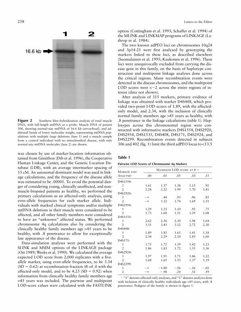
Figure 1 shows the adPEO pedigree used in the ge-nome scan, and figure 2 shows Southern blot–hy-bridization analyses of muscle mtDNA of patient 306and a healthy control. The affected status was deter-mined by observation of marked clinical symptoms inthe neurological examination and/or by detection ofmultiple mtDNA deletions in the analysis of the muscle-biopsy specimen. Muscle samples from patients 306 and

Letters to the Editor 257
Figure 1 adPEO family with linkage to the markers on chromosome 4q. The individuals with marked clinical symptoms and/or deletionsof mtDNA detected by PCR or Southern blot–hybridization analyses are indicated by blackened symbols. The unblackened symbols indicateclinically healthy individuals age 145 years. The markers used in the haplotype construction are shown in the upper-left corner of the figure.The boxes around the haplotypes indicate the shared regions of the affected chromosomes. The recombination events limit the adPEO region,between D4S2924 and D4S2920, to within a distance of 13.5 cM.
408 were examined. The clinical symptoms in this familywere milder than those in families with linkage to the10q and 3p loci (Suomalainen et al 1995; Kaukonen etal. 1996). All the patients had progressive externalophthalmoplegia and ptosis but had no generalized mus-cle weakness. Age at onset was ∼35 years. Several af-fected family members had sensorineural hypoacusia.Two subjects had goiter associated with hypo- or hy-perthyroidism (patients 305 and 306, respectively). Twoelderly subjects (patients 310 and 311) suffered fromdementia manifesting as impairment of the cognitivefunctions, with no affective component. An increasedserum-lactate level at rest was detected in one patient(patient 408). A typical example of a patient in thisfamily is patient 306, who at age 67 years had ptosisand ophthalmoplegia, bilateral hearing loss, and hyper-thyroidism with goiter. Her standard electromyogramwas myopathic. Nerve conduction–velocity studies werenormal. Multiple mtDNA deletions were detected in ananalysis of muscle specimen from the biceps brachialis.Histological analysis of her muscle sample revealed that3% of the fibers were ragged red and 5% showed partial
COX deficiency. No elevation of lactic acid was detectedat rest or after standard exercise, and her serum creatine-phosphokinase level was within the normal range. Res-piratory-chain analysis showed slightly reduced activitiesof complexes III and IV (65%–70% of controls’ mean),whereas activities of complexes I and II were within thenormal range. Informed consent was obtained from allfamily members, and total DNA was extracted fromlymphoblasts or from 10–150 mg of frozen muscle, asdescribed by Zeviani et al. (1988). Southern blot anal-ysis, with PvuII restriction digestion of total DNA, prep-aration of total human mtDNA as the hybridizationprobe, and PCR amplifications to detect mtDNA dele-tions, were conducted as described elsewhere (Zevianiet al 1988; Kaukonen et al. 1996). g[32P]-ATP–labeled,PCR-amplified microsatellite markers were separatedonto a 5% denaturing polyacrylamide gel and visualizedby autoradiography. Fluorescently labeled PCR-ampli-fied microsatellite markers were typed by use of a model377 Applied Biosystems automatic sequencer (Perkin-Elmer).
The marker set used for the genomewide gene search
258 Letters to the Editor
Figure 2 Southern blot–hybridization analysis of total muscleDNA, with full-length mtDNA as a probe. Muscle DNA of patient306, showing normal-size mtDNA of 16.6 kb (arrowhead), and ad-ditional bands of lower molecular weight, representing mtDNA pop-ulations with multiple large deletions (lane 1) and a muscle samplefrom a control individual with no mitochondrial disease, with onlynormal-size mtDNA molecules (lane 2) are shown.
Table 1
Pairwise LOD Scores of Chromosome 4q Markers
MARKER AND
ANALYSISa
MAXIMUM LOD SCORE AT v 5
.00 .01 .05 .10 .15
D4S1554:1 1.61 1.57 1.38 1.15 .922 2.28 2.22 1.99 1.70 1.41
D4S2920:1 2` .44 .93 .96 .852 2` 1.32 1.74 1.69 1.51
D4S2954:1 1.29 1.25 1.10 .92 .752 1.73 1.68 1.51 1.29 1.08
D4S1535:1 2.62 2.56 2.30 1.98 1.642 3.51 3.43 3.12 2.72 2.30
D4S408:1 1.89 1.85 1.65 1.41 1.182 2.34 2.29 2.10 1.85 1.60
D4S171:1 1.75 1.72 1.59 1.42 1.232 1.86 1.83 1.72 1.55 1.36
D4S2924:1 1.97 1.91 1.71 1.46 1.222 1.68 1.65 1.53 1.37 1.19
D4S2299:1 2` 21.80 2.56 2.16 2.022 2` 2.98 .20 .52 .59
a “1” denotes affected-only analyses, and “2” denotes analyses donewith inclusion of clinically healthy individuals age 145 years, with .8penetrance. Pedigree of the family is shown in figure 1.
was chosen by use of marker-location information ob-tained from Genethon (Dib et al. 1996), the CooperativeHuman Linkage Center, and the Genetic Location Da-tabase (LDB), with an average intermarker spacing of15 cM. An autosomal dominant model was used in link-age calculations, and the frequency of the disease allelewas estimated to be .00001. To avoid the potential dan-ger of considering young, clinically unaffected, and non-muscle-biopsied patients as healthy, we performed theprimary calculations as an affected-only analysis, usingeven-allele frequencies for each marker allele. Indi-viduals with marked clinical symptoms and/or multiplemtDNA deletions in their muscle were considered to beaffected, and all other family members were consideredto have an “unknown” affected status. We performedchromosome 4q calculations also by considering theclinically healthy family members age 145 years to behealthy, with .8 penetrance to allow for exceptionallylate appearance of the disease.
Data-simulation analyses were performed with theSLINK and MSIM options of the LINKAGE package(Ott 1989; Weeks et al. 1990). We calculated the averageexpected LOD score from 2,000 replicates with a five-allele marker, using even-allele frequencies, to be 3.34( ) at recombination fraction (v) of .0 with theSD 5 0.62affected-only model, and to be 4.23 ( ) whenSD 5 0.92information from clinically healthy family members age145 years was included. The pairwise and multipointLOD-score values were calculated with the FASTLINK
option (Cottingham et al. 1993; Schaffer et al. 1994) ofthe MLINK and LINKMAP programs of LINKAGE (La-throp et al. 1984).
The two known adPEO loci on chromosomes 10q24and 3p14-21 were first analyzed by genotyping themarkers linked to these loci, as described elsewhere(Suomalainen et al. 1995; Kaukonen et al. 1996). Theseloci were unequivocally excluded from carrying the dis-ease gene in this family, on the basis of haplotype con-struction and multipoint linkage analyses done acrossthe critical regions. Many recombination events weredetected in the disease chromosomes, and the multipointLOD scores were !22 across the entire regions of in-terest (data not shown).
After analysis of 315 markers, primary evidence oflinkage was obtained with marker D4S408, which pro-vided two-point LOD scores of 1.89, with the affected-only model, and 2.34, with the inclusion of clinicallynormal family members age 145 years as healthy, with.8 penetrance in the linkage calculations (table 1). Hap-lotypes across this chromosomal region were con-structed with informative markers D4S1554, D4S2920,D4S2954, D4S1535, D4S408, D4S171, D4S2924, andD4S2299. Recombination events detected in subjects306 and 402 (fig. 1) limit the third adPEO locus to !13.5

Letters to the Editor 259
Figure 3 Multipoint LOD-score calculation across the 4qadPEO region. The dotted line represents the analyses done with in-formation from the affected individuals, giving a maximum LOD scoreof 3.8. The solid line indicates the multipoint calculations done whendata from clinically healthy individuals age 145 years were added,giving a maximum LOD score of 4.7. The markers used in the cal-culations are shown above the X-axis, and the intermarker distancesare shown between the markers.
cM, between markers D4S2920 and D4S2924, on 4q34-35. The intermarker distances and cytogenetic locali-zation of this adPEO locus were established by use ofthe mapping information of the LDB.
The same set of 4q markers was used in pairwise andmultipoint linkage calculations. The best two-pointLOD scores obtained were 2.62, with marker D4S1535(affected-only model), and 3.51, when clinically healthyfamily members age 145 years were considered healthy,with .8 penetrance. The affected-only multipoint cal-culations across the critical region gave a maximumLOD score of 3.8; 4.7 was obtained when data on thehealthy family members were included in the analyses(fig. 3).
Our sample contained four informative Italian fami-lies with adPEO (each family alone was informativeenough to provide the maximum expected LOD scoreof 12 at , with 2,000 replicates) not previouslyv 5 .01assigned to known adPEO loci. To study the possiblelinkage to the 4q locus in these families, haplotypes wereconstructed across the entire 4q adPEO region. Manyrecombination events were observed across the regionin the disease chromosomes, and the multipoint calcu-lations across the region remained !22 (data notshown), thus clearly excluding the chromosome 4 locusas the cause of the disease in these families.
adPEO appears to be a genetically heterogeneous dis-order, with at least four different nuclear loci causingvery similar phenotypes. This heterogeneity could be ex-plained by causative genes that encode different com-ponents of related metabolic pathways or by differentsubunits of an enzyme complex. In databases, we havenot found evidence of functionally related proteins pre-viously mapped within the three chromosomal adPEOloci (GeneMap ’98). To date, two other autosomallyinherited diseases associated with mtDNA deletions havebeen mapped to distinct nuclear regions. Wolfram syn-drome is an autosomal recessive neurodegenerative dis-order sometimes associated with single or multiplemtDNA deletions, and it has been shown to be linkedto chromosome 4p (Polymeropoulos et al. 1994; Bar-rientos et al. 1996a, 1996b). The defective gene (WFS1)was recently identified and it appears to function in thesurvival of islet b-cells and neurons (Inoue et al. 1998).A recessively inherited mitochondrial neurogastrointes-tinal encephalomyopathy with multiple mtDNA dele-tions was recently shown to be caused by mutations inthe thymidine-phosphorylase gene on chromosome22q13.32-qter (Hirano et al. 1998; Nishino et al. 1999).
The clinical symptoms of the patients in the familywith linkage to 4q seem to be less severe than those infamilies with linkage to 10q and 3p. The muscular symp-toms are limited to facial muscles: all of the patientspresented with ophthalmoplegia and ptosis but with noexercise intolerance or generalized muscle weakness.
Most patients had sensorineural hypoacusia, and somehad goiter or dementia. It remains uncertain whether thelatter symptoms are a result of the adPEO-gene defect,because of the relatively high prevalence of these dis-orders in the general population.
To date, ∼65 expressed sequence tags representing dif-ferent genes have been localized to the 4q adPEO region,and eight of these represent known genes (GeneMap’98). The adenine nucleotide translocator is a key met-abolic enzyme of the mitochondria, transporting ADPand ATP across the inner mitochondrial membrane. Thegene for the heart- and muscle-specific isoform (ANT1)has been localized to 4q35 (Fan et al. 1992). The ANT1knockout mice showed ragged red muscle fibers and pro-liferation of mitochondria, lactic acidosis, severe exerciseintolerance, and cardiomyopathy (Graham et al. 1997).Our patients lacked the cardiac symptoms, but otherwisethe symptoms of the patients resembled those of ANT1knock out mice, making ANT1 a good candidate genefor adPEO. Whether ANT1 is involved in the patho-genesis of adPEO is being analyzed. In addition, the genefor dominantly inherited facioscapulohumeral musculardystrophy has been localized to the 4q adPEO region(Wijmenga et al. 1990). adPEO and this dystrophy sharesensorineural hearing loss as a symptom of the disease,but our patients had neither generalized muscle weak-ness nor retinal changes. The eventual characterizationof the first adPEO gene will not only reveal one of thepathogenic mechanisms causing the genetically hetero-geneous disease but will also enhance the search for theremaining adPEO genes and will improve our funda-

260 Letters to the Editor
mental understanding of mtDNA stability and mainte-nance in the cell.
Acknowledgments
The authors wish to thank Joseph Terwilliger for his valu-able comments on linkage analysis. We also thank Ritva Ti-monen, Anne Jokiaho, and Maikki Parkkonen for their skillfultechnical assistance. This study was supported by grants fromthe Finnish Medical Foundation, the Farmos Research andScience Foundation, and the Finnish Muscular Disease Foun-dation (to J.K.); from Telethon (grant 767 to M.Z.); from theAcademy of Finland (to L.P.); from the Emil Aaltonen Foun-dation (to A.S.); and from the Hjelt Fond of the PediatricResearch Foundation, Finland.
JYRKI KAUKONEN,1 MASSIMO ZEVIANI,2
GIACOMO PIETRO COMI,3 MARIA-GRAZIA PISCAGLIA,4
LEENA PELTONEN,1,* AND ANU SUOMALAINEN1
1National Public Health Institute, Department ofHuman Molecular Genetics, Helsinki; 2NationalNeurological Institute “Carlo Besta,” Division ofBiochemistry and Genetics, and 3Centro Dino Ferrari,Istituto di Clinica Neurologica, Universita degli Studidi Milano, Instituto di Ricovero e Cura a CarattereScientifico, Ospedale Maggiore Policlinico, Milan; and4Divisione di Neurologia, Ospedale degli Infermi,Rimini, Italy
Electronic-Database Information
Accession numbers and URLs for data in this article are asfollows:
Cooperative Human Linkage Center, http://www.chlc.org/ (formarkers)
GeneMap ’98, http://www.ncbi.nlm.nih.gov/genemap/ (formarkers)
Genetic Location Database (LDB), http://cedar.genetics.soton.ac.uk/public_html/index.html (for markers)
Online Mendelian Inheritance in Man (OMIM), http://www.ncbi.nlm.nih.gov/Omim/ (for adPEO loci in a Finnish family[MIM 157640], in three Italian families [MIM 601226], andfrom other sources [MIM 601227])
References
Barrientos A, Casademont J, Saiz A, Cardellach F, Volpini V,Solans A, Tolosa E, et al (1996a) Autosomal recessive Wolf-ram syndrome associated with an 8.5-kb mtDNA single de-letion. Am J Hum Genet 58:963–970
Barrientos A, Volpini V, Casademont J, Genis D, ManzanaresJ-M, Ferrer I, Corral J, et al (1996b) A nuclear defect in the4p16 region predisposes to multiple mitochondrial DNAdeletions in families with Wolfram syndrome. J Clin Invest97:1570–1576
Cottingham RW Jr, Idury RM, Schaffer AA (1993) Faster se-quential genetic linkage computations. Am J Hum Genet 53:252–263
Dib C, Faure S, Fizames C, Samson D, Drouot N, Vignal A,Millasseau P, et al (1996) A comprehensive genetic map ofthe human genome based on 5,264 microsatellites. Nature380:152–154
Fan YS, Yang HM, Lin CC (1992) Assignment of the humanmuscle adenine nucleotide translocator gene (ANT1) to4q35 by fluorescence in situ hybridization. Cytogenet CellGenet 60:29–30
Graham BH, Waymire KG, Cottrell B, Trounce IA, MacGregorGR, Wallace DC (1997) A mouse model for mitochondrialmyopathy and cardiomyopathy resulting from a deficiencyin the heart/muscle isoform of the adenine nucleotide trans-locator. Nat Genet 16:226–234
Hirano M, Carcia-de-Yebenes J, Jones AC, Nishino I, Di-Mauro S, Carlo JR, Bender AN, et al (1998) Mitochondrialneurogastrointestinal encephalomyopathy syndrome mapsto chromosome 22q13.32-qter. Am J Hum Genet 63:526–533
Inoue H, Tanizawa Y, Wasson J, Behn P, Kalidas K, Bernal-Mizrachi E, Mueckler M, et al (1998) A gene encoding atransmembrane protein is mutated in patients with diabetesmellitus and optic atrophy (Wolfram syndrome). Nat Genet20:143–148
Kaukonen JA, Amati P, Suomalainen A, Rotig A, Piscaglia M-G, Salvi F, Weissenbach J, et al (1996) An autosomal locuspredisposing to multiple deletions of mtDNA on chromo-some 3p. Am J Hum Genet 58:763–769
Lathrop GM, Lalouel JM, Julier C, Ott J (1984) Strategies formultilocus linkage analysis in humans. Proc Natl Acad SciUSA 81:3443–3446
Melberg A, Lundberg PO, Henriksson KG, Olsson Y, StalbergE (1996) Muscle-nerve involvement in autosomal dominantprogressive external ophthalmoplegia with hypogonadism.Muscle Nerve 19:751–757
Nishino I, Spinazzola A, Hirano M (1999) Thymidine phos-phorylase gene mutations in MNGIE, a human mitochon-drial disorder. Science 283:689–692
Ott J (1989) Computer-simulation methods in human linkageanalysis. Proc Natl Acad Sci USA 86:4175–4178
Polymeropoulos MH, Swift RG, Swift M (1994) Linkage ofthe gene for Wolfram syndrome to markers on the short armof chromosome 4. Nat Genet 8:95–97
Schaffer AA, Gupta SK, Shiriram K, Cottingham RW Jr (1994)Avoiding recomputation in linkage analysis. Hum Hered 44:225–237
Servidei S, Zeviani M, Manfredi G, Ricci E, Silvestri G, BertiniE, Gellera C, et al (1991) Dominantly inherited mitochon-drial myopathy with multiple deletions of mitochondrialDNA: clinical, morphologic and biochemical studies. Neu-rology 41:1053–1059
Suomalainen A, Kaukonen J, Amati P, Timonen R, Haltia M,Weissenbach J, Zeviani M, et al (1995) An autosomal locuspredisposing to deletions of mitochondrial DNA. Nat Genet9:146–151
Suomalainen A, Majander A, Haltia M, Somer H, LonnqvistJ, Savontaus ML, Peltonen L (1992) Multiple deletionsof mitochondrial DNA in several tissues of a patient withsevere retarded depression and familial progressive externalophthalmoplegia. J Clin Invest 90:61–66
Suomalainen A, Majander A, Wallin M, Setala K, Kontula K,
Letters to the Editor 261
Leinonen H, Salmi T, et al (1997) Autosomal dominant pro-gressive external ophthalmoplegia with multiple deletions ofmtDNA: clinical, biochemical, and molecular genetic fea-tures of the 10q-linked disease. Neurology 48:1244–1253
Weeks DE, Ott J, Lathrop GM (1990) SLINK: a general sim-ulation program for linkage analysis. Am J Hum GenetSuppl 47:A204
Wijmenga C, Frants RR, Brouwer OF, Moerer P, WeberJL, Padberg GW (1990) Location of facioscapulohumeralmuscular dystrophy gene on chromosome 4. Lancet 336:651–653
Zeviani M, Bresolin N, Gellera C, Bordoni A, Pannacci M,Amati P, Moggio M, et al (1990) Nucleus-driven multi-ple large-scale deletions of the human mitochondrial ge-nome: a new autosomal dominant disease. Am J Hum Genet47:904–914
Zeviani M, Moraes CT, DiMauro S, Nakase H, Bonilla E,Schon EA, Rowland LP (1988) Deletions of mitochondrialDNA in Keyrns-Sayre syndrome. Neurology 38:1339–1346
Zeviani M, Servidei S, Gellera C, Bertini E, DiMauro S,DiDonato S (1989) An autosomal dominant disorder withmultiple deletions of mitochondrial DNA starting at the D-loop region. Nature 339:309–311
Address for correspondence and reprints: Dr. Jyrki Kaukonen, National PublicHealth Institute, Department of Human Molecular Genetics, Mannerheimintie166, 00300 Helsinki, Finland. E-mail: [email protected]
∗ Present affiliation: Department of Human Genetics, University of CaliforniaSchool of Medicine, Los Angeles.
q 1999 by The American Society of Human Genetics. All rights reserved.0002-9297/99/6501-0035$02.00
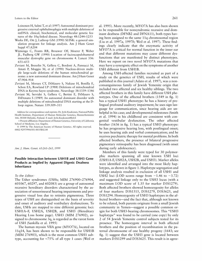
Am. J. Hum. Genet. 65:261–265, 1999
Possible Interaction between USH1B and USH3 GeneProducts as Implied by Apparent Digenic DeafnessInheritance
To the Editor:The Usher syndromes (USHs; MIM 276900–276904,601067, 60297, and 602083) are a group of autosomalrecessive hereditary disorders characterized by the as-sociation of sensorineural hearing impairments and pro-gressive visual loss due to retinitis pigmentosa. Threetypes of USH are distinguished on the basis of severityand onset of auditory and vestibulary dysfunctions. Todate, USHs are mapped to nine different genomic loci:USH1A–F, USH2A, USH2B, and USH3 (HereditaryHearing Loss home page). USH3 (MIM 276902), as-signed to chromosome 3q, is regarded as the rarest formof USH (Sankilla et al. 1995)
The human myosin VIIA gene (MYO7A), located on11q14, has been shown to be responsible for USH1B(MIM 276903), which is the most common USH1 sub-type, accounting for ∼75% of all type 1 cases (Weil et
al. 1995). More recently, MYO7A has also been shownto be responsible for nonsyndromic recessive and dom-inant deafness (DFNB2 and DFNA11), both types hav-ing been assigned to the same 11q chromosomal region(Liu et al. 1997a, 1997b; Weil et al. 1997). These find-ings clearly indicate that the enzymatic activity ofMYO7A is critical for normal function in the inner earand that different mutations may cause different dys-functions that are manifested by distinct phenotypes.Here we report on two novel MYO7A mutations thatmay have a synergistic effect on the symptoms of anotherUSH different from USH1B.
Among USH-affected families recruited as part of astudy on the genetics of USH, results of which werepublished in this journal (Adato et al. 1997), was a non-consanguineous family of Jewish Yemenite origin thatincluded two affected and six healthy siblings. The twoaffected brothers in this family have different USH phe-notypes. One of the affected brothers (1549 in fig. 1)has a typical USH1 phenotype: he has a history of pre-lingual profound auditory impairment; he uses sign lan-guage for communication, since hearing aids are un-helpful in his case; and developmental milestones (Smithet al. 1994) in his childhood are consistent with con-genital vestibular dysfunction. The other affectedbrother (1636 in fig. 1) has a typical USH3 phenotype:he has progressive hearing loss, with postlingual onset;he uses hearing aids and verbal communication; and hereceives psychiatric therapy for mental problems. In bothaffected brothers, the presence of bilateral progressivepigmentary retinopathy has been diagnosed (with onsetduring early adolescence).
Members of this family were typed for 30 polymor-phic markers spanning all nine known USH loci(USH1A-F, USH2A, USH2B, and USH3). Marker alleleswere identified and arranged into the most likely hap-lotypes, as shown in figure 1. Haplotype segregation andlinkage analysis resulted in exclusion of all USH1 andUSH2 loci (LOD scores range from 21.46 to 23.72)and suggested linkage only to the USH3 locus (with amaximum LOD score of 1.35 for marker D3S1279).Both affected brothers showed homozygosity for allelesof four markers: D3S1315, D3S1279, D3S3625, andD3S1294. Homozygosity of USH3 haplotypes in the af-fected brothers—and the fact that, although not knownto be related, both parents originate from a small Jewishcommunity in Yemen—suggest a possible common or-igin for both USH3-bearing chromosomes. This “USH3haplotype” was found to be carried (one copy) by only2 of 54 Jewish Yemenite control subjects tested for itspresence. The homozygote interval in both affectedbrothers and the position of recombination in the pa-ternal chromosome of one healthy progeny (1643; seefig. 1) suggest that the USH3 gene is located betweenmarkers D3S1299 and D3S3625. This result is in agree-

262 Letters to the Editor
Figure 1 Genomic DNA extracted from blood of family members was used as a template for PCR amplification, which was done with30 pairs of specific primers of markers spanning all nine USH loci. Marker alleles, identified according to their relative mobility on a denaturingformamide 4% acrylamide gel in all family members, were arranged into the most likely haplotypes. This haplotype arrangement results inexclusion of all USH1 and USH2 loci and suggests linkage only to the USH3 locus. Maternal chromosomes are gray and striped whereaspaternal chromosomes are white and dotted. Blackened squares on the gray USH1B maternal chromosomes indicate the presence of the mutatedMYO7A. The homozygote interval, of both affected brothers, in the USH3 locus is boxed.
ment with the location suggested by Sankilla et al. (1995)and by Joensuu et al. (1996). The order of markers span-ning the USH3 linkage region, as presented in figure 1,is cen, D3S2401, D3S1299, D3S1315, D3S1279,D3S3625, D3S1594, tel, in agreement with the orderpresented in the Whitehead contig (Whitehead Institutefor Biomedical Research). However, in this order, theposition of the markers D3S1315 and D3S1279, which
our findings indicated were the most closely linked toUSH3, differs from the one suggested by Joensuu et al.(1996).
Since one of the affected brothers had an USH1 phe-notype, family members were screened for mutations inthe human MYO7A gene, which has been shown to beresponsible for USH1B. Two new close nucleotidechanges were detected in exon 25 of the gene on one

Letters to the Editor 263
Figure 2 Comparative electrophoresis of normal (N) and mu-tated (M) exon 25 PCR cycle-sequencing reaction, with SequiThermEXEL DNA Sequencing Kit (FMC). Products were electrophoresedside by side through Long Ranger Gel (Epicentre Technologies):ddGTP-terminated products of normal exon 25 appear next to ddGTP-terminated products of mutated exon 25; and ddATP-terminated prod-ucts of normal exon 25 appear next to ddATP-terminated products ofmutated exon 25. Arrows indicate the TrC transition and the guaninedeletion (5 nt up stream of the transition).
maternal chromosome: a TrC transition and a guaninedeletion 5 nt upstream of this transition (fig. 2). Noneof these changes were found in 1200 control chromo-somes tested by allele-specific oligonucleotide analysis,as described by Whithney et al. (1993). This mutatedMYO7A is carried by the brother with the USH1 phe-notype (1549) but not by his affected brother with theUSH3 phenotype (1636). The mother (1637) and twounaffected siblings (1638 and 1639), who are all doubleheterozygotes for the mutated MYO7A and for a singleUSH3 haplotype, show no evidence of any USH symp-toms or nonsyndromic deafness. This suggests a digenicinheritance pattern, with a possible synergistic interac-tion between MYO7A and the USH3 gene product,where presence of a single defective MYO7A allele seemsto increase the severity of deafness as a part of the clinicalsymptoms associated with USH3.
Evidence for digenic inheritance of nonsyndromicdeafness was already presented in the case of a Swedishfamily (Balciuniene et al. 1998), whose affected memberswere carriers of DFNA2 and/or DFNA12. Increased se-verity of deafness was found in family members thatwere carriers of both alleles. This clear additive effectdiffers from the situation in our Yemenite family, wheremutated MYO7A appears to be phenotypically ex-pressed only on the background of two defective USH3alleles, suggesting an interaction between the MYO7Aand the USH3 gene products. Digenic inheritance wasalso suggested as one of the possible explanations in thecase of DFNB15 (Chen et al. 1997). This is an autosomalrecessive nonsyndromic deafness, found in a family ofIndian origin, linked to two loci on chromosomes 3qand 19p. Most interestingly in relation to our work, oneof these loci, 3q21.3-3q25.2, includes the USH3 locusand the other, 19p13.3p13.1, includes (among others)the MYO1F gene (Hasson et al. 1996), which is anothermember of the unconventional myosin group.
Human MYO7A is a member of the unconventionalmyosins group (Weil et al. 1996). All myosins have threedifferent functional domains defined within their heavychains: an N-terminal motor domain, a regulatory (light-chain–binding) domain, and a tail domain that variesdramatically in length and in sequence among myosins(Mooseker and Cheney 1995). The functions of myosin-tail domains are largely unknown. However, a commonassumption is that the tail directs the interaction of agiven myosin with its cargo (Mermall et al. 1998).MYO7A is predicted to dimerize, on the basis of thecoiled-coil sequence motif at the start of its tail region.
The sequence changes detected in exon 25 of MYO7Ain some members of the presented family are expectedto result, at the protein level, in a LeurPro substitutionat codon 1087 and in a frameshift of the reading framestarting at codon 1089. Both these AA codons are lo-cated after the coiled-coil domain of the protein. This
frameshift would result in the formation of a UGA stopcodon 18 amino acids downstream from the deletionsite and, therefore, in the translation of a truncated pro-tein that lacks150% of its normal AA sequence, whichcomprises most of the MYO7A tail domain. Segregationof the mutated MYO7A with healthy members of thisfamily and, on the other hand, with the more severeUSH phenotype, suggests a possible biological interac-tion between MYO7A and the USH3 gene products.This mutated MYO7A appears to be phenotypically ex-pressed only on the background of two USH3 alleles.
Many disease mutations that introduce stop codonswere found to lead to mRNA destabilization, as in themouse MYO6 sv allele (Avraham et al. 1995). In sucha case, segregation of the mutated MYO7A, as describedabove, could indicate that the normal USH3 gene prod-uct may affect the stabilization of the MYO7A mRNAor the protein.
If the mutated MYO7A in this Yemenite family wastranslated, then it would not lack its coiled-coil sequencemotif. Unlike the case of DFNB11, where a 9 bp deletionin the coiled-coil region of MYO7A was suggested to

264 Letters to the Editor
have a dominant negative effect (Liu et al. 1997b), seg-regation of the mutated MYO7A with healthy membersof our family clearly determines the recessive nature ofits mutation. There are several possible explanations forthis difference. The truncated MYO7A produced in ourcase may be incompetent for dimerization, since it isunknown whether the remainder of the tail is requiredfor self assembly or if the truncated molecule may bedestabilized. Another possible reason is the formationof a partially functional heterodimer. These possibilitieswould yield <50% of the normal protein amounts. Inany kind of direct or indirect USH3-MYO7A proteininteraction, a reduced-dosage effect of active MYO7Aprotein is likely to have a synergistic effect on the back-ground of the two impaired alleles of the USH3 geneproduct.
One possible explanation for the USH3-MYO7A in-teraction is that the USH3 protein might be involved intargeting or binding MYO7A to the plasma membrane.The tail of MYO7A consists of a direct repeat containingtwo elements. The distal element, the talinlike domain,shows significant homology to the N-terminus of talinand limited homology to the N-termini of other membersof the band-4.1 superfamily of actin-binding proteins(Weil et al. 1996; Chen et al. 1997). In talin and in band4.1, this region binds to acidic phospholipids and me-diates protein-protein interactions. Therefore, it isthought that talinlike motifs serve to bind and/or targetthe myosin to the plasma membrane (Cheney et al. 1993;Titus et al. 1997).
It is also possible that USH3 is a cytoskeletal com-ponent and as such interacts with the MYO7A protein:Actin cytoskeleton is essential for proper the function ofthe inner ear, and deafness-associated genes such asMYO7A and MYO15 as well as the human diaphanousprotein are assumed to be cytoskeletal components (Vas-iliki and Petit 1998).
The MYO7A protein may also serve an inner hair-cell–specific role distinct from its role in actin. This rolemight be complementary to the function of the USH3gene product. For example, in mice, MYO7A was foundto be involved in hair-cell vesicle trafficking of amino-glycosides, which are known to induce ototoxicity (Rich-ardson et al. 1997).
Acknowledgments
We would like to express our gratitude to Elias Kavakovand his staff members at the Center for Deaf-Blind Persons-Beth David Institute in Tel Aviv. We thank all family memberswho participated in this study. We also thank Tama Hassonfor critical comments on our manuscript. This work was sup-ported in part by an Israel Science Foundation research grant(1140041), a Ministry of Health grant (1140091), and an Ap-plebaum Foundation grant (1140111) (to B.B.T.); and a Eur-
opean Economic Community grant (PL951324; to C.P.). Thiswork was done as part of A. A.’s Ph.D. project.
AVITAL ADATO,1 HAGAR KALINSKI,1
DOMINIQUE WEIL,2 HASSAN CHAIB,2
MICHAEL KOROSTISHEVSKY,1 AND
BATSHEVA BONNE-TAMIR1
1Department of Human Genetics, Sackler School ofMedicine, Ramat Aviv, Israel; and 2Unite deGenetique des Deficits Sensoriels, Centre National dela Recherche Scientifique Unite de Recherche Associee1968, Institut Pasteur, Paris
Electronic-Database Information
Accession numbers and URLs for data in this article are asfollows:
Hereditary Hearing Loss home page, http://dnalab-www.uia.ac.be/dnalab/hhh/index.html
Online Mendelian Inheritance in Man (OMIM), http://www.ncbi.nlm.nih.gov/Omim (for Usher syndrome subtypes[MIM 276900, 276901, 276902, 276903, 276904, 601067,60297, and 602083])
Whitehead Institute for Biomedical Research/MIT Center forGenome Research, http://www.genome.wi.mit.edu/ (forWhitehead contig map)
References
Adato A, Weil D, Kalinski H, Pel-Or Y, Hammadi A, Petit C,Korostishevsky M, Bonne-Tamir B (1997) Mutation profileof all 49 exons of the human myosin VIIA gene and hap-lotype analysis in Usher 1B families from diverse origins.Am J Hum Genet 61:813–821
Avraham KB, Hasson T, Steel KP, Kingsley DM, Russell LB,Mooseker MS, Copeland NG, et al (1995) The mouse Snell’swaltzer deafness gene encodes an unconventional myosinrequired for structural integrity of inner hair cells. Nat Genet11:369–374
Balciuniene J, Dahl N, Borg E, Samuelsson E, Koisti MJ, Pet-tersson U, Jazin EE (1998) Evidence for digenic inheritanceof nonsyndromic hereditary hearing loss in a Swedish family.Am J Hum Genet 63:786–793
Chen A, Wayne S, Bell A, Ramesh A, Srisailapathy CR, SccottDA, Sheffield VC, et al (1997) New gene for autosomalrecessive non-syndromic hearing loss maps to either chro-mosome 3q or 19p. Am J Med Genet 71:467–471
Cheney R, Riley MA, Mooseker MS (1993) Phylogenetic anal-ysis of the myosin super family. Cell Motil Cytoskeleton 24:215–223
Hasson T, Skowron JF, Gilbert DJ, Avraham KB, Perry WL,Bement WM, Anderson BL, et al (1996) Mapping of un-conventional myosins in mouse and humans. Genomics 36:431–439
Joensuu T, Blanco G, Pakarinen L, Sistinen P, Kaariainen H,Brown S, de la Chapelle A, et al (1996) Refined mappingof Usher syndrome type III locus on chromosome 3, exclu-

Letters to the Editor 265
sion of candidate genes, and identification of the putativemouse homologous region. Genomics 38:255–263
Liu XZ, Walsh J, Mburu P, Kendrick-Jones J, Cope MJTV,Steel KP, Brown SDM (1997a) Mutations in the myosin VIIAgene cause non-syndromic recessive deafness. Nat Genet 16:188–190
Liu XZ, Walsh J, Tamagawa Y, Kitamura K, Nishizawa M,Steel KP, Brown SDM (1997b) Autosomal dominant non-syndromic deafness (DFNA11) caused by a mutation in themyosin VIIA gene. Nat Genet 17:268
Mermall V, Post PL, Mooseker MS (1998) Unconventionalmyosins in cell movement, membrane traffic, and signaltransduction. Science 279:527–533
Mooseker MS, Cheney RE (1995) Unconventional myosins.Annu Rev Cell Dev Biol 11:633–675
Richardson GP, Froge A, Kros CJ, Fleming J, Brown SD, SteelKP (1997) Myosin VIIA is required for aminoglycoside ac-cumulation in cochlear hair cells. J Neurosci 17:9506–9519
Sankilla E, Pakarinen L, Kaariainen H, Aitotomaki K, Kar-jalainen S, Sistonen P, de la Chapelle A (1995) Assignmentof an Usher syndrome type III (USH3) gene to chromosome3q. Hum Mol Genet 4:93–98
Sheffield VC, Beck JS, Kwitek AE, Sandstrom DW, Stone EM(1993) The sensitivity of single-strand conformation poly-morphism analysis for the detection of single base substi-tutions. Genomics 16:325–332
Smith RJH, Berlin CI, Hejtmancik JF, Keats BJB, KimberlingWJ, Lewis RA, Moller CG, et al (1994) Clinical diagnosisof the Usher syndromes. Am J Hum Mol 50:32–38
Titus MA (1997) Unconventional myosins: new frontiers inactin-based motors. Trends Cell Biol 7:119–123
Weil D, Blanchard S, Kaplan J, Guilford P, Gibson F, WalshJ, Mburu P, et al (1995) Defective myosin VIIA gene re-sponsible for Usher syndrome type 1B. Nature 374:60–61
Weil D, Kussel P, Blanchard S, Levy G, Levi-Acobas F, DriraM, Ayadi H, et al (1997) The autosomal recessive isolateddeafness, DFNB2, and the Usher 1B syndrome are allelicdefects of the myosin-VIIA gene. Nat Genet 16:191–193
Weil D, Levy G, Shahly I, Levi-Acobas F, Blanchard S, El-Amraou A, Crozet F, et al (1996) Human myosin VIIA re-sponsible for the Usher 1B syndrome: a predicted membrane-associated motor protein expressed in developing sensoryepithelia. Proc Natl Acad Sci USA 93:3232–3237
Whithney MA, Saito H, Jakobs PM, Gibson RA, Moss RE,Grompe M (1993) A common mutation in the FACC genecauses Fancini anemia in Ashkenazi Jews. Nat Genet 4:202–205
Vasiliki K, Petit C (1998) The fundamental and medical im-pacts of recent progress in research on hereditary hearingloss. Hum Mol Genet 7:1589–1597
Address for correspondence and reprints: Dr. Batsheva Bonne-Tamir, Depart-ment of Human Genetics, Sackler School of Medicine, Ramat Aviv 69978, Israel.E-mail: [email protected]
q 1999 by The American Society of Human Genetics. All rights reserved.0002-9297/99/6501-0036$02.00
Am. J. Hum. Genet. 65:265–269, 1999
Anticipation in Familial Chronic LymphocyticLeukemia
To the Editor:The term “anticipation” in genetic diseases refers to ear-lier age at onset and/or increased severity in successivegenerations. For some neurodegenerative diseases, an-ticipation results from expansion of unstable trinucle-otide repeats in successive generations (La Spada 1997).Epidemiological studies have demonstrated a significantfamilial effect for leukemia (Goldgar et al. 1994) andchronic lymphocytic leukemia (CLL) in particular (Cart-wright et al. 1987; Linet et al. 1989). Families with mul-tiple affected individuals are rare in population studiesbut may be more common in clinical samples (Cuttner1992). The mode of inheritance is unknown for leuke-mia, although it has been hypothesized that in pedigreeswith multiple affecteds the disease is due to a singleautosomal dominant gene (Horwitz 1997). Evidence foranticipation in familial leukemia has been reported byHorwitz et al. (1996), on the basis of a literature reviewof published pedigrees with acute myelogenous leukemia(AML) and CLL. The average difference, in age of onsetof CLL, between two generations in seven pedigrees (17individuals), was 15 years, although the mean parent-offspring difference was 21 years. Yuille et al. (1998)have recently confirmed this finding, in 10 families withtwo generations affected with CLL (mostly parent-off-spring pairs) systematically ascertained from a patientregistry. They found the age of onset difference betweengenerations to be 22 years. Given that there is a molec-ular basis for anticipation in some diseases, it is impor-tant to determine if there is anticipation in CLL. As withother diseases, anticipation in CLL could be due to anumber of well-known sampling biases (reviewed byMcInnis 1996), such as the tendency to select early-onsetprobands, parents with late onset, and families with si-multaneous onset of disease in parents and offspring, orother biases that cause a truncation of the sample offamilies (Hodge and Wickramartne 1995; Fraser 1997).Another bias particular to CLL may arise from the factthat individuals are often diagnosed on the basis of rou-tine blood tests when they are asymptomatic, and theymay remain asymptomatic for a number of years. It isconceivable that an anticipation phenomenon could beattributable to changes in medical practice over time,such that, because of the greater routine use of clinicaltests, individuals in the younger generations are beingdiagnosed earlier. Data collected by the National CancerInstitute Surveillance, Epidemiology, and End Results(SEER) program during the past 20 years have shownlittle secular change in the incidence of CLL (SEER). In

266 Letters to the Editor
Table 1
Age at Onset and Year of Diagnosis in 13 Families Investigated for Anticipation
Family and Relationshipto Proband Generation
Age atDiagnosis
(years)Symptomaticat Diagnosis?a
Year atDiagnosis
1:Father 1 69 yes 1954Proband (female) 2 47 yes 1972Brother 2 49 no 1971Brother 2 47 yes 1968Sister 2 57b no 1974
2:Father 1 75c yes 1967Uncle 1 63c yes 1955Proband (male) 2 56 ? 1978
3:Uncle 1 70 ? 1960Proband (male) 2 50 no 1977First cousin (female) 2 52 no 1978
4:Proband (male) 1 49 ? 1984Nephew 2 53 ? 1988
5:Mother 1 82 yes 1980Proband (female) 2 64 ? 1985
6:Father 0d 79 ? 1965Proband (female) 1 74 no 1990Son 2 55 yes 1991
7:Aunt 1 58 ? 1961Proband (male) 2 44 yes 1986
8:Mother 1 83 no 1978Proband (male) 2 55 yes 1983
9:Mother 1 32 yes 1961Proband (female) 2 37 yes 1993Brother 2 42b yes 1995
10:Mother 1 58 ? 1965Proband (female) 2 52 ? 1991
11:Father 1 76 no 1997Proband (male) 2 51 no 1997
12:Uncle 1 77 ? 1984Proband (male) 2 65 no 1996Brother 2 50 no 1994
13:Mother 1 ? ? ?Proband (female) 2 35 yes 1990
a A question mark (?) indicates that data were not available.b Individual sought diagnosis because of family history.c Age at diagnosis is age at death.d Not included in analysis.
fact, recent studies that have reported an increased in-cidence of CLL find the increase to be limited to olderindividuals (Call et al. 1994; Rozman et al. 1997). Wehave been studying familial CLL for a number of yearsand have analyzed age at onset in pedigrees with at least
two generations affected. We find evidence for antici-pation in these families, even when stage at diagnosisand other potential sampling biases are taken intoaccount.
Since 1974, we have ascertained and collected clinical

Letters to the Editor 267
Figure 1 Age at onset of CLL, in two generations
data on 27 families, each of which has two or moreconfirmed cases of CLL (see Caporaso et al. 1991). Thir-teen of these families have cases of CLL in two gener-ations; the remainder have affected siblings and/orcousins. We have complete age-at-onset data for 32individuals from the 13 two-generation families. Onefamily has missing age-at-onset data on an affected par-ent. In the majority of cases, age at onset was determinedon the basis of medical chart review, but in a few caseswe relied on personal reports. Insufficient informationwas available to determine the disease stage for eachindividual, by use of either the Rai or Binet staging meth-ods (reviewed in Dighiero and Binet 1996), but, when-ever possible, we determined whether the individual was“symptomatic” or “asymptomatic” when diagnosed.We classified as “symptomatic” individuals showingsigns or symptoms attributed to CLL, including lym-phadenopathy, splenomegaly, anemia, and thrombocy-topenia. Individuals were classified as “asymptomatic”if they presented only with a peripheral-blood absolutelymphocyte count (ALC) (Zwiebel and9> 5.0 # 10 /literCheson 1998). For those individuals diagnosed asymp-tomatically, we also noted the age at which they becamesymptomatic, when this information was available in themedical chart. Individuals were classified into two gen-erations, on the basis of their position in the pedigree.Generation 2 was defined as the youngest generationaffected; the generation preceding them was consideredto be generation 1. All affected individuals in each familywere included in the analysis. There was one family withthree generations affected; the individual in the oldestgeneration was not included in the analysis. The age-at-onset data for all individuals are displayed in table 1.In 11 of the 13 families, the proband was in the youngergeneration.
We found that the average age at onset in generation1 is 66.7 years (SD 14.6) and that that in generation 2
is 50.7 (SD 7.8). These onset ages are similar to thosereported by Horwitz et al. (1996). We used survival anal-ysis to plot the age-at-onset distribution for each gen-eration, using the Kaplan-Meier method as implementedin the SAS Lifetest procedure (Allison 1995). The resultsare displayed in figure 1. The difference between the twogenerations is highly significant, whether based on thelog-rank test ( ) or the Wilcoxon test (P 5 .0001 P 5
)..0009We examined the generational differences after taking
into account a number of possible biases. In this smallsample, there were no significant age-at-onset differencesbetween males and females, and each generation had anapproximately equal proportion of males and females.We scored the individuals for whether they were symp-tomatic, asymptomatic, or unknown at diagnosis. Theproportion of symptomatic individuals did not differ bygeneration (5/13 [.38] in generation 1 and 8/19 [.42] ingeneration 2; see table 1). In addition—after family 9,in which all members had unusually early ages at onset,was excluded—there was no difference, between symp-tomatic and asymptomatic individuals. There were twoindividuals in the sample who sought diagnosis becauseof their family history. These two individuals, indicatedin table 1, had ages at onset of 57 and 42 years, soeliminating them would not change the findings. In orderto analyze our data in the most conservative way pos-sible, we increased the age at onset for five individuals(generation 2) who were asymptomatic at diagnosis butwhose age at onset of clinical symptoms could be de-termined. In addition, there were two individuals fromgeneration 1 who were diagnosed on the basis of deathcertificates and who were assigned onset ages equal totheir ages at death. We lowered these individuals’ agesat onset by 7 years, since this is the median time betweenage at onset of symptoms and death (Zwiebel and Che-son 1998). Even under these conservative assumptions,the age at onset in the second generation was signifi-cantly lower than that in the first generation (log-ranktest ). It should also be noted that, evenP 5 .0002though we did not analyze all three generations in thethree-generation family, the proband’s father (generation0) had a later age at onset than did either the probandor her son. We also considered the possible bias due topreferential ascertainment of families with simultaneousonset in two generations. In 5 of 12 families for whichcomplete data were available, individuals in both gen-erations were diagnosed within a 5-year period (table1). In the seven other families, the average difference, incalendar year of diagnosis, between generations was 21years, and the average difference, in age at onset, be-tween generations was 13 years. Thus, our finding ofanticipation is not being driven by the presence of fam-ilies with simultaneous onset in two generations.
We find that, between the two generations in the fam-

268 Letters to the Editor
ilies we studied, the average decrease in age at onset ofCLL was 16 years. One could argue that we have pref-erentially ascertained early-onset probands. The averageage at onset in all 27 of the families that we studied is54.9 years for probands only and 59.6 years for allaffecteds. The average age at onset of CLL has beenreported to be ∼70 in some population-based samples(Travis et al. 1992; Hjalmar et al. 1996) but ∼62–65 inother studies (Radovanic et al. 1994; Rozman et al.1997). Thus, probands from the multiplex families thatwe studied have a somewhat decreased age at onset,compared with that in cases from the population. How-ever, for this analysis, we included all affected individualsin a family, which decreases any effect due to earlieronset in the index case. We cannot completely rule outthe possibility that anticipation in these families is dueto a cohort effect, since, in all of the families that westudied, birth cohort is confounded with generation. Asis to be expected from the way in which pedigrees areusually ascertained in genetic studies, the second gen-eration of individuals are from cohorts born more re-cently than those of the first generation. However, acohort effect seems unlikely, given the lack of seculartrends in CLL, as mentioned above. The age at onset ofCLL is also sufficiently late in life that it would not affectfertility, and we are not likely to have missed familieswith early onset in parents and later onset in offspring.Horwitz et al. (1996) suggested that the very large dif-ference (21 years) that they found between parents andoffspring in published families was also consistent withthe effect of some common environmental exposure.There is no well-established environmental risk factorfor CLL, although susceptibility to some environmentalrisk factor might exist in subjects with some unidentifiedgenetic susceptibility factor. Although we cannot rule outsuch an effect, we find a substantial but smaller age-at-onset decrease between generations. We also find thedecrease to be present in families that are not correlatedfor year of diagnosis, arguing against a purely environ-mental explanation.
In conclusion, we report significant evidence for an-ticipation in familial CLL and have ruled out variousbiases that could account for the finding. Therefore, weplan to look for expanded trinucleotide repeats in can-didate genes in families showing anticipation.
LYNN R. GOLDIN,1 MARIA SGAMBATI,1
GERALD E. MARTI,2 LAURA FONTAINE,3
NAOKO ISHIBE,1 AND NEIL CAPORASO1
1Genetic Epidemiology Branch, Division of CancerEpidemiology and Genetics, National CancerInstitute, Bethesda, MD; 2Center for BiologicsEvaluation and Research, Food and DrugAdministration; and 3Westat Research, Inc., Rockville,MD
Electronic-Database Information
The URL for data in this letter is as follows:
SEER, http://www-seer.ims.nci.nih.gov/Publications/CSR7395(for Cancer Statistics Review, 1973–1995)
References
Allison PD (1995) Survival analysis using the SAS system: apractical guide. Sas Institute, Cary, NC
Call TG, Phyliky RL, Noel P, Habermann TM, Beard CM,O’Fallon WM, Kurland LT (1994) Incidence of chronic lym-phocytic leukemia in Olmsted County, Minnesota, 1935through 1989, with emphasis on changes in initial stage atdiagnosis. Mayo Clin Proc 69:323–328
Caporaso NE, Whitehouse J, Bertin P, Amos C, PapadopolousN, Muller J, Whang-Peng J, et al (1991) A 20 year clinicaland laboratory study of familial B-chronic lymphocytic leu-kemia in a single kindred. Leuk Lymphoma 3:331–342
Cartwright RA, Bernard SM, Bird CC, Darwin CM, O’BrienC, Richards IDG, Roberts B, et al (1987) Chronic lympho-cytic leukaemia: case control epidemiological study in York-shire. Br J Cancer 56:79–82
Cuttner J (1992) Increased incidence of hematologic malig-nancies in first-degree relatives of patients with chronic lym-phocytic leukemia. Cancer Invest 10:103–109
Dighiero G, Binet JL (1996) Chronic lymphocytic leukemia.Hematol Cell Ther Suppl 38:S42–S61
Fraser FC (1997) Trinucleotide repeats not the only cause ofanticipation. Lancet 350:459–460
Goldgar DE, Easton DF, Cannon-Albright LA, Skolnick MH(1994) Systematic population-based assessment of cancerrisk in first-degree relatives of cancer probands. J Nat CancerInst 86:1600–1608
Hjalmar V, Carlsson M, Kimby E (1996) Chronic lymphocyticleukaemia at a county hospital in southern Sweden. MedOncol 13:95–101
Hodge SE, Wikramaratne P (1995) Statistical pitfalls in de-tecting age-of-onset anticipation: the role of correlation instudying anticipation and detecting ascertainment bias. Psy-chiatr Genet 5:43–47
Horwitz M (1997) The genetics of familial leukemia. Leukemia11:1347–1359
Horwitz M, Goode EL, Jarvik GP (1996) Anticipation in fa-milial leukemia. Am J Hum Genet 59:990–998
La Spada AR (1997) Trinucleotide repeat instability: geneticfeatures and molecular mechanisms. Brain Pathol 7:943–963
Linet MS, Van Natta ML, Brookmeyer R, Khoury MJ,McCaffrey LE, Humphrey RL, Szklo M (1989) Familial can-cer history and chronic lymphocytic leukemia: a case controlstudy. Am J Epidemiol 130:655–664
McInnis MG (1996) Anticipation: an old idea in new genes.Am J Hum Genet 59:973–979
Radovanovic Z, Markovic-Denic L, Jankovic S (1994) Cancermortality of family members of patients with chronic lym-phocytic leukemia. Eur J Epidemiol 10:211–213
Rozman C, Bosch F, Montserrat E (1997) Chronic lympho-

Letters to the Editor 269
cytic leukemia: a changing natural history? Leukemia 11:775–778
Travis LB, Curtis RE, Hankey BF, Fraumeni JF Jr (1992) Sec-ond cancers in patients with chronic lymphocytic leukemia.J Natl Cancer Inst 84:1422–1427
Yuille MR, Houlston RS, Catovsky D (1998) Anticipation infamilial chronic lymphocytic leukemia. Leukemia 12:1696–1698
Zwiebel JA, Cheson BD (1998) Chronic lymphocytic leukemia:staging and prognostic factors. Semin Oncol 25:42–59
Address for correspondence and reprints: Dr. Lynn R. Goldin, Genetic Epi-demiology Branch, DCEG, NCI 6120 Executive Boulevard, Room 7008, MSC7236, Bethesda, MD 20892-7236. E-mail: [email protected]
q 1999 by The American Society of Human Genetics. All rights reserved.0002-9297/99/6501-0037$02.00