Differences in osteoclast activity between rheumatoid arthritis ...
282
Universidade de Lisboa Faculdade de Medicina Differences in osteoclast activity between rheumatoid arthritis and ankylosing spondylitis Inês Pedro Perpétuo Orientadores: Professor Doutor João Eurico Fonseca Doutora Mari-Mia Ainola Tese especialmente elaborada para obtenção do grau de Doutor em Ciências Biomédicas Especialidade em Biologia Celular e Molecular 2016
-
Upload
khangminh22 -
Category
Documents
-
view
2 -
download
0
Transcript of Differences in osteoclast activity between rheumatoid arthritis ...

Universidade de Lisboa Faculdade de Medicina
DDiiffffeerreenncceess iinn oosstteeooccllaasstt aaccttiivviittyy bbeettwweeeenn rrhheeuummaattooiidd aarrtthhrriittiiss aanndd aannkkyylloossiinngg
ssppoonnddyylliittiiss
Inês Pedro Perpétuo
Orientadores: Professor Doutor João Eurico Fonseca Doutora Mari-Mia Ainola
Tese especialmente elaborada para obtenção do grau de Doutor em Ciências Biomédicas
Especialidade em Biologia Celular e Molecular
2016


Universidade de Lisboa Faculdade de Medicina
DDiiffffeerreenncceess iinn oosstteeooccllaasstt aaccttiivviittyy bbeettwweeeenn rrhheeuummaattooiidd aarrtthhrriittiiss aanndd aannkkyylloossiinngg ssppoonnddyylliittiiss
Inês Pedro Perpétuo
Orientadores: Professor Doutor João Eurico Fonseca
Doutora Mari-Mia Ainola
Tese especialmente elaborada para obtenção do grau de Doutor em Ciências Biomédicas
Especialidade em Biologia Celular e Molecular
Júri: Presidente: Doutor José Luís Bliebernich Ducla Soares, Professor Catedrático em regime de tenure e Vice-Presidente do Conselho Científico Vogais: Professor Georg Schett, Full Professor, Erlangen University, Alemanha Professor Mari-Mia Ainola, Adjunct Professor, University of Helsinki, Finlândia
(Co-orientadora) Doutor Fernando Manuel Pimentel Santos, Professor Auxiliar Convidado da
Faculdade de Ciências Médicas da Universidade Nova de Lisboa Doutor Luís Ricardo Simões da Silva Graça, professor Associado com Agregação
da Faculdade de Medicina da Universidade de Lisboa Doutor João Eurico Cortez Cabral da Fonseca, Professor Associado com Agregação
da Faculdade de Medicina da Universidade de Lisboa (Orientador) Doutora Maria José Parreira dos Santos, Professora Auxiliar Convidada da
Faculdade de Medicina da Universidade de Lisboa Bolsa de Doutoramento da Fundação para a Ciência e Tecnologia (referência SFRH/BD/70533/2010). Trabalho realizado no âmbito do projecto “Differences in Bone Cell Activity Between Rheumatoid Arthritis and Ankylosing Spondylitis” financiado pela Merck Sharp & Dohme Corp - Merck_P08574.
2016


As opiniões expressas nesta publicação
são da exclusiva responsabilidade
do seu autor, não cabendo qualquer
responsabilidade à Faculdade de
Medicina da Universidade de Lisboa pelos
conteúdos nele apresentados


A impressão desta tese foi
aprovada pelo Conselho
Cientifico da Faculdade de
Medicina de Lisboa em
reunião de 21 de Junho de
2016.


Dissertação apresentada à Faculdade de
Medicina da Universidade de Lisboa,
para obtenção do grau de Doutor em
Ciências Biomédicas.


À minha Mãe.


" When you open your eyes
When you gaze at the sky
When you look to the stars
As they shut down the night
You know... The story ain't over"
- 'The Story Ain't Over', Avantasia, Tobias Sammet


xv
Table of contents
Index of Figures ................................................................................................................. xvii
Index of Tables ................................................................................................................... xix
Acknowledgements ............................................................................................................ xxi
List of abbreviations ......................................................................................................... xxix
Sumário ............................................................................................................................ xxxv
Summary ......................................................................................................................... xxxix
Introduction ........................................................................................................................... 1
Aims .................................................................................................................................... 39
Results ................................................................................................................................. 43
I. Identification of a cytokine network sustaining neutrophil and Th17 activation in
untreated early rheumatoid arthritis. .................................................................................... 47
II. Methotrexate and low dose prednisolone downregulate osteoclast function by
decreasing receptor activator of nuclear factor κB expression in monocytes from early
rheumatoid arthritis patients ................................................................................................ 67
III. Effect of Tumor Necrosis Factor Inhibitor Therapy on Osteoclasts Precursors in
Rheumatoid Arthritis ........................................................................................................... 87
IV. Ankylosing spondylitis patients have impaired osteoclast gene expression in circulating
osteoclast precursors .......................................................................................................... 111
V. Effect of Tumor Necrosis Factor Inhibitor Therapy on Osteoclasts Precursors in
Ankylosing Spondylitis. .................................................................................................... 133
Discussion .......................................................................................................................... 161
Conclusions ....................................................................................................................... 175
References ......................................................................................................................... 179
Appendix ........................................................................................................................... 211

xvi

xvii
Index of Figures
Figure 1 - Osteoclastogenesis ................................................................................................ 6
Figure 2 - Mechanism of osteoclast bone resorption ........................................................... 11
Figure 3 - Cytokines activating bone resorption ................................................................. 24

xviii

xix
Index of Tables
Table 1 - The 2010 American College of Rheumatology/European League Against
Rheumatism classification criteria for rheumatoid arthritis ................................................ 26
Table 2 - The 1984 modified New York criteria for ankylosing spondylitis ...................... 32
Table 3 - Summary of the findings of RA and AS patients when compared to age and
gender matched healthy controls ....................................................................................... 171
Table 4 - Comparison between RA and AS patients of the monocyte subpopulation
characteristics and OC differentiation and activity ........................................................... 172

xx

xxi
Acknowledgements
A PhD is a journey that changes you, a life within your life that takes you to some very
dark and very bright places in a sequence that surprises you. A PhD in Science is the quest
for the answer to THAT question, to the riddle that puzzles you and in the end only more
questions arise - thank you Science for teaching me to expect the unexpected.
I would like to acknowledge both my supervisors, Professor Doutor João Eurico Fonseca
and Adjunct Professor Mari-Mia Ainola, for having accepted me as a PhD student, for the
guidance, support, discussions and for helping me to grow as a scientist. To João for
having the patience for my sometimes incontrollable temper and for embracing a very
challenging project like mine. To Mari for welcoming me in Helsinki and for very fruitful
discussions about osteoclasts, music and snow.
My thanks also go to my PhD thesis committee, Professor Doutor Jaime Branco, Doutora
Sandra Casimiro and Doutora Íris Caramalho, for the meetings, discussions and
suggestions in order improve the work and conclude it. To Íris for being there for me since
I started my research baby steps and for all the kind words and support.
I would like also to thank all the members from Rheumatology Department, Hospital de
Santa Maria, all the medical doctors and nurses, for the effort to collaborate in the
development of my work, especially in sample collection and in the organization of clinical
data. In particular, a thanking note to Enfª. Lurdes Narciso, Doutora Helena Canhão, Drª.
Ana Maria Rodrigues, Dr. Joaquim Polido Pereira, Drª. Elsa Sousa, Drª. Cristina Ponte,
Drª. Raquel Marques, all the nurses and administrative personnel in Hospital de Dia with
whom I have worked. We had a good network for ongoing sample collection and patient
follow-up.
I am also very grateful to all the patients and healthy volunteers for their willingness in
providing us with samples making this project possible.
This project would also not be possible without the valuable help of Ana Sofia Zhao and
Ângela Afonso from Biobanco-IMM. Ana's patience with all the patients and volunteers
even with the most difficult ones was priceless.
I would like to thank everyone in the Rheumatology Research Unit for their support. I
know working together is not always easy, but together we are indeed stronger!!!
Minha Mãe... como é que eu vou agradecer a toda a gente que me apoiou desde o início
desta grande viagem?

xxii
Helena Canhão... Lena, obrigada por tudo: por me aturares as birras de teimosia quando me
perguntavas qual era o meu "outcome", por me explicares cinquenta mil vezes o que é que
comparações múltiplas queriam dizer - obrigada por tudo o que me ensinaste e por todos os
momentos divertidos que passámos.
Ana Maria, obrigada por quereres alterar sempre a minha linha de raciocínio quando já
tinha os papers escritos! Agora fora de brincadeiras, foste uma das pessoas que mais me
ensinou na UIR e queria agradecer-te por isso. Obrigada pela companhia em tantos
congressos onde o teu nariz é que escolhia o restaurante como em Atenas :)
Joana Lopes, nem sei como te agradecer - acho que já teria desistido da ciência se a tua
teimosia não tivesse sido mais forte que a minha. Obrigada por não desistires de mim,
nunca estaria aqui, onde estou hoje se tu não estivesses ao meu lado em todos os momentos
dolorosos da minha vida de investigação e nos mais felizes também. Obrigada por trazeres
os nossos pequenos, mas valiosos osteoclastos, para a UIR e por tanto me teres dado na
cabeça - esta tese é também tua à sua maneira. Obrigada pela amizade que temos, que tem
um valor incalculável e que mesmo com um oceano de distância não deixa de estar a um
Skype de distância (e a algumas horas de diferença). Pelos momentos em congressos, pelas
tardes de cinema e pelas noites passadas no Colombo a discutir projectos mirabolantes,
ciência em Portugal e as nossas vidas.
Rita Moura, minha querida Rita - por mais que uma vez perdi a paciência contigo (como
aquela vez que íamos para o Congresso Português de Reumatologia em Albufeira e
combinei com toda a gente às 8h e contigo às 7h30 para apareceres às 8h) mas sem ti a
minha estadia da UIR teria sido bem mais aborrecida e a minha vinda para Londres bem
mais atribulada provavelmente. Obrigada por todos os momentos de citometria que
passámos, todas as tabelas de dados de doentes e discussões científicas que tivemos.
Obrigada por me lembrares que quem faz rápido não faz sempre bem (mas bolas, também
não é preciso andar à velocidade perfeccionista Rita Moura :) ).
Rita Cascão - D. Casca :) Partilhamos muitas opiniões mas também discordamos em
algumas coisas mas sem ti a UIR também não era a mesma. Obrigada pelas 18h15, pelas
gargalhadas que demos, pelos quartos de hotel partilhados, pelas histórias que construímos
juntas e pelo trabalho que desenvolvemos. Obrigada por me ensinares que "pés na terra"
também se aplica à Ciência e que ser pragmático e prático é a solução para a maioria dos
nossos problemas. Obrigada por me ensinares a objectividade e a ler papers!
Diana, passamos por muitos altos e baixos nestes anos todos mas nada nos tira os nossos
momentos. Obrigada pelas partilhas de amarguras e felicidades de doutoramento e não só,

xxiii
obrigada pelos nossos momentos musicais que continuam a ocorrer (mas agora pelo
telefone) e obrigada por me deixares partilhar contigo os cantos da vida. Obrigada pelo
companheirismo e lembra-te sempre - "Reach out for the light!".
Bruno, obrigada por me fazeres rir sempre que me vinhas perguntar qualquer coisa de
inglês ou sobre o osso. Obrigada por momentos inesquecíveis de parvoíce na UIR e não só
e por também partilhares as amarguras e felicidades de um doutoramento!
Ana Lopes, parece que vou ter de agradecer a mais alguém pelas horas de parvoíce na UIR
mas também pela incrível paciência e dedicação às nossas arcas, azoto líquido e
respectivos mapas... Sem ti o meu doutoramento teria demorado bem mais, por isso para
além da parvoíce, obrigada por me ensinares e mostrares que um mapa feito é menos uma
hora a procurar amostras.
Às minhas alunas: umas mais alunas que outras e outras mais minhas que outras. Mónica,
Rita, Ana Daniel, Soraia, Ana Dinis. Obrigada a todas por acreditarem em mim, por
aturarem o meu mau feitio às vezes, por ouvirem o que eu digo e por me questionarem.
Onde quer que o vosso caminho vos leve lembrem-se "Só não erra quem não trabalha".
Mónica e Rita - os vossos trabalhos de Mestrado deram uma outra dimensão a esta tese.
Obrigada por tudo meninas e obrigada por não se esquecerem de mim :) Soraia: Obrigada,
do fundo do coração, obrigada pelas horas infindáveis de conversa no gmail, pela ajuda na
análise de imagens e pelas discussões de Cinema/Séries/Livros. Obrigada por me mostrares
o Filipe Neto ("Não faz sentido") e um dia ainda vamos inaugurar esse grande V-log que é
a Chamuça Assassina!!!! Ana Pereira (uma das minhas wedding planners de serviço)
obrigada por não teres papas na língua - a minha admiração por ti é só igualada pela
gratidão que te tenho por me animares no gmail e por partilharmos várias e diversas
frustrações ;).
Os meus agradecimentos não podem ir só para os meus colegas mais directos na unidade
mas para toda a família do IMM desde os PhD até às fantásticas pessoas que lutam
diariamente para fazer do IMM um sítio melhor. João Freire, Rita Aroeira, Diana Fontinha,
Sofia Cerqueira, Aida Domingues, Patrícia Meireles, Inês Albuquerque, Francisco Aresta
Branco, Telma Lança, Guida Rodrigues, Alice Melão, Jorge Santos e Diogo Fonseca-
Pereira - vocês e tantos outros, trabalharam (e alguns ainda trabalham) incansavelmente
para fazer do IMM um sítio mais agradável para os estudantes de doutoramento e toda a
gente trabalhar - Obrigada pela viagem que fizemos juntos durante os anos da comissão de
estudantes de doutoramento e pelo apoio a TODOS os alunos de Doutoramento do IMM.

xxiv
À Marta Agostinho e à Inês Crisóstomo, porque apesar de já não trabalharem no IMM me
fizeram sempre acreditar a ir mais longe, a dizer claramente o que quero. À Marta que me
passou o gostinho pela comunicação em Ciência e à Inês que acredita que é sempre
possível melhorar e equilibrar as coisas (mas convém fazeres um pós-doc antes de quereres
trabalhar em comunicação para a Ciência ou gestão de Projectos).
À Andreia Machado, cujas capacidades de negociação excedem as minhas em milhares de
anos, pela amizade, carinho e apoio que deste a todos os eventos que quisemos organizar,
mesmo os que não deram em nada (...Mosteiro dos Jerónimos...). Obrigada por
dinamizares as festas e o Natal, obrigada por seres a nossa cara na Comunicação!
Porque antes do IMM houve o IGC quero agradecer do fundo do coração à Mª Inês Ramos,
minha companheira de laboratório e amiga desde o dia 1. Obrigada por noites
inesquecíveis em Amesterdão e surpresas mais ou menos (des)agradáveis no HSM.
Do IGC para a FCUL, para as professoras que nunca desistiram de mim e sempre
acreditaram que eu conseguia e que para a frente é que é o caminho: Margarida Telhada,
Rita Zilhão, Gabriela Rodrigues e Graça Vieira - vocês são o exemplo que nós temos na
faculdade por isso obrigada por tudo o que fizeram por mim e pelo apoio que me deram
mesmo depois do mestrado. Obrigada por não perderem a esperança!
Para as minhas Crubélias, Inês Pá, Cat, Gi, Marta. Vocês foram um dos meus pilares
durante estes anos todos, anos demais para serem escritos sem parecer que estamos a
envelhecer ao ritmo de 1 ano por ano (!!!). Inês - partilhamos tantas dúvidas nas nossas
vidas, tantos altos e baixos e sofro como ninguém por estar longe e por não podermos ir
aos nossos cafés. Obrigada por estares ao meu lado todos estes anos, merecemos uma
Bahama Mamma quando eu defender! (Podes trazer o Mário também, assim ele e o Nuno
podem falar do Benfica enquanto nós bebemos). Cat - embarcaste tu também numa
aventura de um doutoramento e eu não consigo estar mais orgulhosa de ti :) É a
experiência mais devastadora do mundo estar longe de quem amamos, e acredito que com
um oceano de distância seja ainda pior, mas sairás mais forte - Obrigada por seres a voz da
razão muitas vezes (se bem que às vezes falas sem filtro!). Germana - és das pessoas que
mais admiro na minha vida: passaste por tanto e continuas sempre mais forte e a remar
para tornar os teus sonhos realidade. Por favor nunca deixes de acreditar que consegues
porque tu fazes-me também acreditar que eu consigo. Obrigada por noites de jogatana de
Trivial incrível e festas de anos com álcool q.b. Não posso deixar de agradecer também ao
Daniel (apesar de ele não ser uma crubélia) por também me aturar nos nossos jantares e
nos nossos eternos devaneios de doutoramento. Marta - a ti, mais do que a ninguém quero

xxv
agradecer pela paciência e por me ensinares a ter paciência. Foi contigo que aprendi a não
dizer tudo o que penso (porque tu te lembras do que nós dissemos à letra!!!) e foi contigo
que aprendi que não é bom perder a paciência e gritar com outras pessoas (sorry...).
Contigo aprendi que às vezes desistir é mais fácil mas no fim é sempre pior para nós e por
isso mais vale engolirmos os sapos e andar com as coisas para a frente sem olhar para trás.
Quero deixar aqui também uma palavra ao Hugo (mas a ti também) por me mostrarem
outros jogos de tabuleiro de RPG (para quando uma sessão de Cutthroat Caverns ???).
Para as minhas outras meninas da FCUL: Laura e Susana. Laura, tu és das pessoas mais
corajosas que já conheci. Obrigada por HORAS de jogatana na FCUL e por me fazeres
acreditar que eu conseguia superar qualquer coisa. Tu e o Nuno são doidos, mas isso já tu
sabes, mas são também uma inspiração. Obrigada por me fazerem rir e estarem lá quando é
preciso chorar também. Eu sei que não sou a vossa madrinha oficial mas por favor vejam o
Van Helsing.
Susana - estes anos sempre estiveste lá com companhia para jantares e uma palavra de
apoio. Obrigada por estares lá para mim e por me aturares nos meus rantings.
Queria agradecer aos meus colegas da primária (sim, da primária - 28 anos juntos é obra!)
mas principalmente ao núcleo duro: Pedro, Hernâni, Bárbara e também à Margarida
(apesar de nós sabermos que não vais emigrar...). Porque quando emigramos fomos um
bocado ao desconhecido mas continuamos a juntar-nos quando podemos. Pedro, obrigada
por me aturares em Londres e não há mais visitas ao Elephant Head em Camden sem
jantar, OK????
For my new family in London (or should I say families?). In this past year I have grown as
a Scientist but also as an individual - I moved to embrace a Post-doc in basic science and I
am absolutely loving it. Thank you Michael, Andy, Isabel and John for believing in me and
accepting me in the project. To my colleagues thank you thank you thank you!!!
Alessandro, Richard, Lena, Jessal, Andrea, Freija, Rán, Stephanie, Marie - thank you for
the many moments of fun in the office and outside - you are simply AMAZING!!!! Thank
you for the support in this past year and for making me feel at home at the RVC :) To
Dilek I have to say the biggest thank you of all - for encouraging me, for helping me in the
first weeks at work and for wonderful food :) À minha família portuguesa estendida aqui
em Londres e Inglaterra: Ruivo, Teresa, Pedro, Olga, Nancy, Steven (I know you are not
Portuguese but it's almost like you are). Obrigada por me aturarem e por me fazerem
companhia:) Pedro, obrigada por me dares na cabeça, eu sei que também é preciso dar-te
na cabeça às vezes. Thank you to my other families here: my music families that have

xxvi
made me feel at home in the concerts in London thank you Graeme, Kyle, Steve, Adam,
Michalis, Ania, Lucy, Ruivo and Craig for being my heavy metal family away from home.
Thank you to my more broad and eccletic metal family Alex, Monika, Anastasia, Jack,
Adam, Gillian, Cairbre. Peter and Christian, thank you for many singing moments and
indescribable nights of hectic headbanging.
Uma tese não são só momentos de escrita e pesquisa mas muitos momentos de reflexão
sobre resultados e tentativas de explicação - Queria agradecer do fundo do coração à Ana
Luísa Miranda pela incansável companhia durante os meses de escrita, momentos online
de inspiração e motivação mútua para escrever a tese, por momentos de companhia durante
concertos a que fui sozinha aqui e por momentos de companhia presencial aqui e em
Portugal. Porque nós às vezes também precisamos de um bocadinho de farra :)
Aos meus amigos de Portugal, à minha família do Metal Portugal - a música é a nossa vida
mas a nossa vida é tão mais que a música!!! Obrigada pelo apoio continuado e pela
companhia virtual ou real que me vão fanzendo! Lara e F, Marujo, Carlos, Mafalda e
Pedro, Carlos e Joana (a minha outra wedding planner particular), Pedro Matos, Peixoto,
Diogo Eu, Undersave, Naz, Necro, Warfaust, Tormentinho, Márcia, Ricardo, Sandra, Inês -
vocês fazem-me falta aqui mas já sabem - a porta está sempre aberta para vos receber :)
São, porque nenhum agradecimento está completo sem ti minha Irmã do coração -
obrigada por todos os momentos que passamos, todas as provações nos trouxeram onde
estamos hoje. Obrigada por estares comigo ao longo deste doutoramento *.
Mafalda, não há palavras para te agradecer todo o apoio que me deste ao longo da minha
vida e não só deste doutoramento - Obrigada por estares lá sempre quando precisei. Espero
estar à altura dos novos desafios como tu também estás!!! Luísa, obrigada pela inspiração
para irmos mais longe, sempre atrás dos nossos sonhos!
Joana e Rita, vocês que mais me aturaram quando eu era adolescente também me aturaram
durante este doutoramento. Todas nós abraçamos um desafio diferente nestes últimos anos.
Eu saí do país para fazer um pós-doc e vocês abraçaram a maternidade :) Obrigada por
partilharem muitas das minhas angústias mas também das minhas alegrias. Obrigada por
estarem lá para mim, para os spoilers do Game of Thrones ou para as compras na Natura e
na Intimissimi.
Com a certeza de que há muita gente que falta agradecer, mas por último faltam as pessoas
mais importantes na minha vida: a minha família. Avós, Pais, Tios e Primos uns ainda
neste mundo e outros que já cá não estão. Obrigada por todo o apoio ao longo dos anos,
por acreditarem em mim e por me ajudarem a ser fiel a mim mesma. Ao meu Pai por

xxvii
continuar a dar-me dicas no Angry Birds e a dizer mal da burocracia inglesa e à minha mãe
por me inspirar a nunca desistir * sei que estás orgulhosa de mim * - esta tese é para ti.
Queria terminar por agradecer ao Nuno e à Inês. Inês, ensinaste-me que Mãe é aquela que
cuida, ensinaste-me que afinal a literatura juvenil até é boa, ensinaste-me paciência e a ser
um exemplo. O que uma criança de 9 anos que cresceu até aos 15 nos pode ensinar é quase
tanto como um doutoramento - obrigada por tudo Inês e nunca deixes de acreditar em mim
- eu também acredito em ti.
Nuno, o caminho que percorremos neste doutoramento foi cheio de altos e baixos e com
muita angústia e dor nos maus momentos mas muito MUITO feliz noutros :) Fizeste de
mim uma mulher melhor, nunca deixaste de acreditar que eu era capaz, apoiaste-me
quando tomei a decisão mais difícil da minha vida até agora e estiveste lá para me ouvir a
apresentar resultados mesmo quando não sabias o que era um "Osteosclasto" - a sério,
ainda me hás de explicar como é que isto se diz. Obrigada por estares sempre a meu lado
durante este doutoramento. Sou feliz contigo e nunca quero deixar de ser, OK?
Obrigada!

xxviii

xxix
List of abbreviations
ACPA - anti-citrullinated protein antibodies
ACR - American college of rheumatology
ANTXR2 - anthrax toxin receptor 2
ALP - alkaline phosphatase
AP-1 - activator protein-1
APRIL - a proliferation-inducing ligand
AS - ankylosing spondylitis
ASDAS - ankylosing spondylitis disease activity score
ATF - activating transcription factor
ATP - Adenosine triphosphate
ATP6V0D2 - ATPase H+ transporting, lysosomal 38kDa V0 subunit d2
BAFF - B cell activating factor
BGLAP - bone δ-carboxyglutamic acid-containing protein
BASDAI - Bath ankylosing spondylitis disease activity index
BASFI - Bath ankylosing spondylitis functional index
BMD - bone mineral density
BMP - Bone morphogenetic protein
BMU - basic multicellular unit
BRC - bone remodelling compartment
CA - carbonic anhydrase
Cbfa1 - core-binding factor subunit alpha-1
CCL - C-C chemokine ligand
CCR - C-C chemokine receptor
CD - cluster of differentiation

xxx
cDCs - conventional dendritic cells
CIA - collagen induced arthritis
CLCN7 - chloride/H+ antiporter channel 7
COX-2 - cyclooxygenase-2
CSF1R - colony-stimulating factor 1 receptor
CT-1 - cardiotrophin
CTLA4 - cytotoxic T-lymphocyte-associated protein 4
CTX-I - C-terminal cross-linked telopeptides of collagen type I
CXCL - C-X-C motif chemokine
DAP12 - DNAX activation protein of 12 kDa
DAS28 - disease activity score 28
DCs - dendritic cells
DC-STAMP - dendritic cell specific transmembrane protein
DKK1 - dickkopf-related protein 1
DMARDs - disease modifying anti-rheumatic drugs
DPD - deoxypyridinoline
Dsh - dishevelled
DXA - dual-energy X-ray absorptiometry
ERK - extracellular-signal-regulated kinases
EULAR - European league against rheumatism
Fab - fragment antigen-binding
Foxp3 - forkhead box P3
FRA - fos-related antigen
G-CSF - granulocyte colony stimulating factor
GM-CSF - granulocyte-macrophage colony stimulating factor

xxxi
GPI - glucose-6-phosphate isomerase
GSK-3β - glycogen synthase kinase-3β
HCl - hydrochloric acid
HLA - human leukocyte antigen
HSC - hematopoietic stem cell
hTNFtg - human tumor-necrosis-factor transgenic
ICAM - intercellular adhesion molecule
ICTP - cross-linked carboxyterminal telopeptide of type I collagen
IFN-γ - interferon-gamma
IGF-1 - insulin growth factor
iNKT - invariant natural killer T cells
JNK - c-Jun N-terminal kinase
KO - knockout
LPR - low-density lipoprotein receptor-related protein
LPS - Lipopolysaccharide
MAP2K6 - mitogen-activated protein kinase 6
M-CSF - macrophage colony stimulating factor
MCP-1 - monocyte chemotactic protein
MFR - macrophage fusion receptor
MGP – matrix gla protein
MHC - major histocompatibility complex
miR, miRNA - micro ribonulceic acid
MITF - microphthalmia-associated transcription factor
MMPs - matrix metalloproteinases
MSCs - mesenchymal stem cells

xxxii
MTX - methotrexate
NFATc1 - nuclear factor of activated T cell c1
NF-κB - nuclear factor-κB
NSAIDs - nonsteroidal anti-inflammatory drugs
NTX-I - type I collagen cross-linked N- telopeptide
OA - osteoarthritis
OBs - osteoblasts
OCN - osteocalcin
OC - osteoclast
OPG - osteoprotegerin
OPN - osteopontin
Osx - osterix
P1CP - C--terminal propeptide of type I procollagen
P1NP - N-terminal propeptide of type I procollagen
PDCD4 - programmed cell death protein 4
pDCs - plasmacytoid dendritic cells
PI3K - phosphatidylinositol-4,5-bisphosphate 3-kinase
PTH - parathyroid hormone
PTPN22 - protein tyrosine phosphatase non-receptor 22
PYD - pyridinoline
RA - rheumatoid arthritis
RANK - receptor activator of nuclear factor-κB
RANKL - receptor activator of nuclear factor-κB ligand
RF - rheumatoid factor
RGD - amino acid sequence - arginin-glycin-aspartic acid

xxxiii
ROS - reactive oxygen species
Runx2 - runt-related transcription factor
S1P - sphingosine-1-phosphate
SCF - stem cell factor
SDF-1 - stromal cell-derived factor
sFRPs - secreted Frizzled related proteins
SNP - single nucleotide polymorphisms
SOST - Sclerostin
SpA - spondyloarthritis
sRANKL - soluble receptor activator of nuclear factor-κB ligand
TACE - tumour necrosis factor-alpha converting enzyme
TCF/LEF - T-cell factor/lymphoid enhancer factor
TCRs - T-cell receptors
Teff - effector T cells
Tfe3 - transcription factor E3
TGF- β - transforming growth factor-β
Th17 - T helper 17 cells
TLR - Toll-like receptor
TNF - tumour necrosis factor
TNFi - tumour necrosis factor inhibitors
TRAF - tumour necrosis factor receptor-associated factor
TRAP - tartrate-resistant acid phosphatase
Treg - regulatory T cells
TREM2 - triggering receptor expressed on myeloid Cells 2
VCAM - vascular cell adhesion molecule

xxxiv
VEGF - vascular endothelial growth factor
WIF - Wnt inhibitory factor
Wnt - wingless-related integration site

xxxv
Sumário
Em muitas doenças inflamatórias, tais como a artrite reumatóide (AR) e a espondilite
anquilosante (EA), as células do sistema imunitário alteram a remodelação óssea. A AR
apresenta-se tipicamente como uma poliartrite simétrica, mais comum nas mulheres do que
nos homens e está associada à presença de auto-anticorpos no soro, tais como anticorpos
anti-proteínas citrulinadas (ACPA - anti-citrullinated protein antibodies) e factor
reumatóide. Os genes do complexo major de histocompatibilidade II (MHC-II - major
histocompatibility complex) human leukocyte antigen (HLA)-DR estão fortemente
associados à doença. A inflamação crónica na AR leva à destruição da cartilagem e do
osso, sendo que a AR é reconhecida como uma doença erosiva. Por outro lado, a EA é
caracterizada por ser uma doença com envolvimento inflamatório axial, com predomínio
pelas articulações sacroilíacas e coluna vertebral. Esta doença é mais prevalente no sexo
masculino e é fortemente associada ao HLA-B27. A longo da evolução da doença, os
doentes apresentam anquilose das articulações da coluna vertebral e sacro-ilíacas.
Enquanto a AR é uma doença caracterizada pela destruição do osso e cartilagem, na EA
assistimos a formação óssea aumentada. Neste trabalho, sugerimos que a incapacidade dos
osteoclastos (OC) ou dos seus precursores para responder aos estímulos osteoclastogénicos
em doentes com EA contribui para a formação óssea excessiva, característica desta doença.
O objetivo deste trabalho foi a caracterização dos precursores circulantes de OC
(monócitos) em doentes com AR e EA, bem como a sua capacidade de se diferenciar em
OC e a sua actividade de reabsorção quando diferenciados in vitro. Para além disso, foi
também objectivo desta tese compreender o efeito de terapêuticas, tais como o metotrexato
(MTX) e inibidores do factor de necrose tumoral (anti-TNF), nos precursores de OC em
doentes com AR e EA.
Em primeiro lugar foram investigados os níveis séricos de citocinas em doentes com AR
muito inicial, com menos de 6 semanas de duração da doença (very early rheumatoid
arthritis, VERA), antes e depois do tratamento com corticosteróides e MTX. As citocinas
pró-inflamatórias foram quantificados no soro de doentes VERA e doentes com AR
estabelecida e em comparação com outras artrites iniciais (very early arthritis, VEA) e
controlos saudáveis. Líquido sinovial de doentes com AR e osteoartrose (OA) foi também
analizado. Os doentes VERA apresentavam níveis séricos aumentados de citocinas que
promovem a polarização Th17 (IL-1β e IL-6), bem como IL-8 e citocinas derivadas de

xxxvi
células Th17 (IL-17A e IL-22) que promovem cronicidade da inflamação. Estas citocinas
também estão associadas com a promoção da osteoclastogénese. O tratamento inicial com
corticosteróides ou MTX levou a uma melhoria clínica, mas sem um impacto sobre o
padrão de citocinas. Este aumento de citocinas relacionadas com a polarização Th17 e a
osteoclastogénese numa fase tão inicial da doença, uma alteração também encontrados no
líquido sinovial das AR estabelecidas, sugere que um ambiente pro-inflamatório
favorecedor da polarização Th17 e da actividade dos OC é um evento precoce na
patogénese da AR.
De seguida avaliámos o efeito do MTX nos precursores circulantes de OC e na sua
diferenciação em OC em doentes com AR. Foram recrutados dadores saudáveis e doentes
com AR antes do tratamento e, pelo menos, 6 meses após a introdução de MTX. A
expressão do receptor activator of nf-kB (RANK) ligand (RANKL) em leucócitos e a
frequência e fenótipo de subpopulações de monócitos circulantes foram estudadas, bem
como foram quantificados os níveis séricos de marcadores de remodelação óssea. Os
percusores circulantes foram diferenciados in vitro e a sua actividade foi determinada. O
RANKL sérico, marcadores de activação clássicos de monócitos (C-C chemokine receptor
2 - CCR2, CD86, HLA-DR) e o RANK estavam aumentados em doentes com AR com
doença activa, em comparação com dadores saudáveis, e após a exposição ao MTX estes
parâmetros normalizaram. Não foram encontradas diferenças no número de OC entre os
três grupos, mas as células diferenciadas de doentes com AR apresentaram maior atividade
de reabsorção do que as de dadores saudáveis. Mais uma vez, após o tratamento com
MTX, a actividade dos OC, medida através de ensaio de reabsorção in vitro, foi
normalizada, sugerindo assim que o MTX desempenha um papel importante na regulação
negativa da função dos OC através da diminuição da expressão de RANK em monócitos.
De seguida o efeito dos anti-TNF na osteoclastogénese em doentes com AR foi estudado.
Doentes com AR tratados com anti-TNF foram analisados antes e após um período mínimo
de seguimento de 6 meses. Foi realizada uma análise de frequência e fenótipo de
subpopulações de monócitos, bem como da expressão de RANKL à superfície de
leucócitos. Foram quantificados os níveis séricos de marcadores de remodelação óssea e
efectuamos ensaios in vitro de diferenciação em OC e expressão génica das células em
cultura. Após o tratamento com anti-TNF, a expressão de RANKL em linfócitos B
encontrava-se diminuída assim como a frequência de precursores de OC em circulação. In
vitro, o número de OC após tratamento com anti-TNF estava diminuído e estas células
apresentavam também menor actividade de reabsorção. Nestas células observámos também

xxxvii
diminuição da expressão de genes importantes para a osteoclastogénese, como TNF
receptor associated protein 6 (TRAF6), Fos-2 related antigen (FRA-2) e para a
reabsorção, como a catepsina K. Estes resultados sugerem que na AR a terapêutica anti-
TNF diminui a reabsorção óssea através da redução directa do número de precursores em
circulação e da inibição das vias de sinalização intracelular que actuam através da TRAF6.
Depois de analisar o comportamento dos precursores circulantes de OC em doentes com
AR tratados e não tratados prosseguimos para a segunda parte deste trabalho que teve
como objetivo caracterizar a remodelação óssea, o ambiente pró-inflamatório e pro-
osteoclastogénico, o fenótipo e frequência de monócitos e a sua diferenciação em OC in
vitro em doentes com EA.
Doentes com EA activa sem qualquer terapêutica em curso e controlos saudáveis foram
recrutados. Os níveis de citocinas pró-inflamatórias estavam elevados em doentes com EA
em comparação com dadores saudáveis. Apesar de nao haver diferenças no número de
precursores circulantes, a expressão de CD51/CD61 (integrina αvβ3 ou receptor da
vitronectina, importante na adesão dos OC à matriz óssea) estava diminuída na
subpopulação clássica de monócitos. Não foram observadas diferenças no número de OC
formados in vitro nem na reabsorção óssea, no entanto, genes essenciais à diferenciação
em OC (colony stimulating factor 1 receptor, CSF1R; RANK e nuclear factor of activated
T cell c1, NFATc1), apresentavam menor expressão nos doentes com EA, o que
consequentemente levou a uma diminuição da expressão de genes de reabsorção, tais como
a catepsina K. Estes resultados mostraram que, apesar dos níveis elevados de citocinas pró-
inflamatórias presentes nos doentes com EA, os monócitos circulantes têm menor
expressão de genes específicos de OC, o que apoia a nossa hipótese de uma resposta
inadequada dos precursores OC a estímulos pró-osteoclastogénicos em doentes com EA.
Para compreender os efeitos do tratamento anti-TNF nos precursores circulantes de OC e
na sua capacidade de diferenciação em doentes com EA, seguimos uma coorte de doentes
antes e depois do início do tratamento com anti-TNF. Os níveis séricos de IL-23 e IL-17A
diminuíram após o tratamento com anti-TNF. O número de OC formados a partir de
células de doentes com EA antes do tratamento estava reduzido em comparação com os
controlos saudáveis. Não foram encontradas diferenças na frequência dos precursores de
OC ou no número de OC formados em cultura após o tratamento com anti-TNF. A
expressão de CSF1R e NFATc1 nos precursores circulantes em doentes após o tratamento
encontrava-se diminuída quando comparada com os dadores saudáveis. No entanto,
quando células de doentes sob anti-TNF foram diferenciadas in vitro apresentaram maior

xxxviii
actividade de reabsorção do que as células de doentes antes do tratamento e controlos
saudáveis. Estes resultados sugerem que em doentes com EA, o tratamento anti-TNF reduz
a resposta a estímulos pró-osteoclastogénicos sistémicos, mas quando os precursores de
OC de doentes tratados com anti-TNF são colocados em cultura a sua resposta aos
estímulos está aumentada.
Em conclusão, o trabalho discutido nesta tese elucida alguns dos mecanismos pelos quais o
MTX e os anti-TNF actuam sobre precursores circulantes de OC em doentes com AR e
EA.
Embora a AR e a EA sejam ambam doencas crónicas imunomediadas os seus efeitos no
metabolismo ósseo são diferentes. Nesta tese mostramos que os precursores de OC de
doentes com AR e EA se comportam de modo diferente in vitro, mesmo vindo de um
ambiente pró-inflamatório semelhante.
Os nossos resultados apoiam a hipótese de que os OC de doentes com EA têm uma falha
na sua actividade quando comparados com células de doentes com AR ou controlos
saudáveis. Esta diferença pode ser parcialmente explicada por uma incapacidade intrínseca
de resposta a estímulos osteoclastogénicos e pela diminuição da expressão de genes-chave
de diferenciação e actividade de OC em doentes com EA.
Palavras chave: Artrite reumatóide, espondilite anquilosante, osteoclasto, monócitos,
metotrexato, inibidores do TNF

xxxix
Summary
In many inflammatory diseases, such as rheumatoid arthritis (RA) and ankylosing
spondylitis (AS), bone is a target for immune cells unbalancing bone remodeling. RA
typically presents as a symmetric polyarthritis, affecting more women than men and is
linked with the presence of autoantibodies in the serum such as anti-citrullinated protein
antibodies (ACPA) and rheumatoid factor (RF). Human leukocyte antigen (HLA)-DR
genes are strongly associated with the disease. Chronic inflammation in RA leads to
cartilage and bone destruction, which is typically recognized as erosive disease on x-rays.
In contrast, AS is characterized by axial disease involving the sacroiliac joints and the
spine. AS affects more men than women and is strongly associated with HLA-B27
haplotypes. The long-term outcome is characterized by ankylosis of the spine and
sacroiliac joints.
While RA is a disease characterized by destruction of bone and cartilage, the predominant
finding in AS is bone formation rather than its destruction. In this work, we hypothesize
that the inability of osteoclasts (OC) or its precursors to respond to osteoclastogenic stimuli
in AS patients contributes to the excessive bone formation characteristic of this disease.
Therefore, the aim of this thesis was the characterization of OC circulating precursors
(monocytes) in RA and AS, as well as their ability to differentiate in resorbing OC when
cultured in vitro. Moreover, we also aimed to understand the effect of therapies, such as
methotrexate (MTX) and tumor necrosis factor inhibitors (TNFi), in the OC precursors in
RA and AS patients.
We first investigated whether cytokines were dysregulated in very early rheumatoid
arthritis patients with less than 6 weeks of disease duration (VERA) before and after
treatment with corticosteroids and MTX. Pro-inflammatory cytokines were quantified in
the serum of VERA and established RA patients and compared with other very early
arthritis (VEA) and healthy controls. Patients were also analyzed after therapy. Synovial
fluid (SF) from RA and osteoarthritis (OA) patients was also analyzed. VERA patients had
increased serum levels of cytokines promoting Th17 polarization (IL-1β and IL-6), as well
as IL-8 and Th17-derived cytokines (IL-17A and IL-22) known to promote the chronicity
of inflammation. These cytokines are also associated with the promotion of
osteoclastogenesis. In established RA this pattern was more evident within the SF. Early
treatment with MTX or corticosteroids led to clinical improvement but without an impact

xl
on the cytokine pattern. VERA patients already display increased levels of cytokines
related with Th17 polarization and osteoclastogenesis, a deregulation also found in SF of
established RA, suggesting that a cytokine-milieu favoring Th17 and OC activity is an
early event in RA pathogenesis.
We then aimed to assess the effect of MTX on circulating OC precursors and OC
differentiation in RA patients. RA patients were assessed before therapy and at least 6
months after the introduction of MTX therapy and results controlled with healthy donors.
We determined receptor activator of NF-κB (RANK) ligand (RANKL) surface expression
on circulating leukocytes and frequency and phenotype of monocyte subpopulations.
Quantification of serum levels of bone turnover markers and cytokines and in vitro OC
differentiation were also performed. We found that serum RANKL, classical activation
monocytes markers (C-C chemokine receptor 2 - CCR2, CD86, HLA-DR) and RANK
were increased in RA patients with active disease compared to healthy donors, and after
MTX exposure these parameters normalized to control levels. Although we found no
differences in OC number, cells differentiated from RA patients showed higher resorption
activity than from healthy donors. Again, after MTX treatment, osteoclasts resorption
activity was normalized. The results of this work suggested that MTX plays an important
role in downregulating OC function through the decrease in RANK surface expression in
monocytes.
We then proceeded to study the effect of TNFi in osteoclastogenesis in RA patients. RA
patients treated with TNFi were analyzed at baseline and after a minimum follow-up
period of 6 months. Results were controlled with healthy donors. After TNFi therapy,
RANKL surface expression was downregulated in B lymphocytes and the frequency of
circulating OC precursors was also decreased. Cells from TNFi treated patients had
decreased osteoclast numbers and resorption activity as well as decreased expression of
specific genes important for osteoclastogenesis, like tumour necrosis factor receptor-
associated factor (TRAF6), fos-related antigen 2 (FRA-2) and for bone resorption like
cathepsin K. Therefore, we suggest that in RA TNFi decreases bone resorption through the
direct reduction of the number of circulating precursors and the inhibition of intracellular
signalling pathways acting through TRAF6.
After exploring OC precursor behavior in untreated and treated RA patients we aimed to
characterize bone remodeling and pro-osteoclastogenesis inflammatory environment,
monocytes phenotype and in vitro OC differentiation in AS patients.

xli
Patients with active AS without any ongoing therapy and age and gender matched healthy
donors were recruited. We observed that pro-inflammatory cytokine levels were higher in a
cohort of untreated AS patients when compared to healthy donors, but CD51/CD61
expression (integrin αvβ3 or vitronectin receptor, important for osteoclast attachment to the
bone matrix) was downregulated in the classical OC precursors. No differences in the in
vitro osteoclastogenesis or bone resorption was observed when compared to healthy
donors, however we found low expression of colony stimulating factor 1 receptor
(CSF1R), RANK and nuclear factor of activated T cell c1 (NFATc1) in AS osteoclast
precursors that consequently led to a decreased expression of important resorption genes
such as cathepsin K. These findings showed us that despite the high levels of pro-
inflammatory cytokines present in AS patients, circulating monocytes have low OC
specific gene expression supporting our hypothesis of an impaired response of OC
precursors to pro-osteoclastogenic stimuli in AS patients.
In an effort to understand the effects of TNFi therapy in circulating OC precursors and
their differentiation ability from AS patients, we followed up a cohort of patients before
and after therapy. Results were controlled with healthy donors. We found that IL-17A and
IL-23 circulating levels decreased after TNFi treatment. OC number was decreased in AS
patients before treatment when compared to control. However, no differences in OC
precursor frequency or in the number OC in culture were found after treatment. RANK,
CSF1R and NFATc1 expression was downregulated in circulating OC precursors after
TNFi treatment. However, when cultured in OC differentiated from AS TNFi-treated
patients showed higher resorption activity than cells from patients before treatment. These
results showed us that in AS patients, TNFi treatment reduces systemic pro-
osteoclastogenic stimuli but when OC precursors are exposed to TNFi therapy they have
increased in vitro activity in response to osteoclastogenic stimuli.
From this work we were able to understand some of the mechanisms by which MTX and
TNFi therapies act on circulating OC precursors in RA and AS patients.
Although RA and AS are two chronic immune mediated diseases their effect on bone
metabolism is different. The work here discussed shows that RA and AS OC precursors
have a different behaviour in vitro, even coming from a similar pro-inflammatory milieu.
Our findings support the hypothesis that OC from AS patients have impairment in their
activity when compared both to RA patients or healthy controls. This difference can be
partially explained by an intrinsic inability to respond to osteoclastogenic stimuli and by
downregulation of key OC differentiation and activity genes in AS patients.

xlii
Keywords: Rheumatoid arthritis, ankylosing spondylitis, osteoclast, monocytes,
methotrexate, tumour necrosis factor inhibitors

1
Introduction

2

3
Bone
Bone is a mineralized connective tissue that is constantly remodelled to support calcium
homeostasis and structural needs. Apart from its mechanical role of sustaining the whole
body and allowing locomotion, bone has also many other important functions. It plays a
protective role shielding vital organs and structures like the bone marrow and spinal cord,
and a metabolic role, regulating the homeostasis of calcium and phosphate. Bone is also a
hematopoietic organ, since it is where the bone marrow is found, and an endocrine organ,
contributing to the global energy balance and serving as a reservoir of growth factors and
cytokines [1-3].
Organization
At the microscopic level, bone is composed of two portions, cortical and trabecular.
Cortical bone represents 80% of skeletal bone and is dense and compact with low turnover
ratio. This type of bone constitutes the outer part of all bones, providing mechanical
strength and protection [4]. The trabecular bone only composes 20% of the whole skeleton
and is found inside long bones and in vertebrae, surrounded by cortical bone [3, 5]. This
type of bone has a very porous structure and a higher turnover rate and elasticity than
cortical bone. Therefore, it is light, quickly adaptable to external biological stimulus,
flexible and capable of absorbing energy [4]. The cortical structure, on the other hand, has
the ability to tolerate the peak loads that the long bones are subjected to [6].
Microstructure
The bone matrix comprises an organic and an inorganic phase. The organic phase is
composed mainly by type I collagen, glycoproteins, proteoglycans and bone cells. On the
other hand, the inorganic part is formed by carbonated hydroxyapatite crystals
(Ca10(PO4)6(OH)2) that are distributed among the collagen fibres [7-9]. The composition
and organization of the bone matrix gives this tissue unique mechanical properties such as
stiffness, ductility, tensile strength and exceptional lightness [2, 9].
The organic component of bone is comprised mostly by type I collagen (90%) and non-
collagenous proteins. Type I collagen is synthesized by osteoblasts. It self-assembles in
fibrils and fibres that are deposited in parallel or concentric layers [1, 10]. There are
several non-collagenous proteins on bone, such as phosphoproteins, proteoglycans,
glycosylated proteins and γ-carboxylated proteins. These include osteocalcin (OCN),

4
matrix gla protein (MGP), osteopontin (OPN), bone sialoprotein (BSP), alkaline
phosphatase (ALP) and osteonectin [3]. These proteins are all involved in the ordered
deposition of hydroxyapatite by regulating the amount and size of the mineral crystals [1,
3, 11-13].
Phosphoproteins are proteins that undergo post-translational modifications by the
attachment of a phosphate group. They bind calcium and thereby act as mineral nucleators
[4]. They include BSP and proteoglycans as minor constituents of the bone matrix.
Proteoglycans inhibit calcification by masking the collagen fibrils or occupying critical
spaces within the fibril and thereby diminishing diffusion, chemical interaction and
sequestration of calcium ions or calcium phosphate complexes [13]. BSP is a significant
component of the bone extracellular matrix and has been suggested to constitute
approximately 8% of all non-collagenous proteins found in bone [14]. It is only present in
bone and it functions as another nucleator of hydroxyapatite as well as providing cell
attachment and possibly activation of osteoclasts like OPN [15-17].
OCN, also known as bone δ-carboxyglutamic acid-containing protein (BGLAP), is the
most abundant noncollagenous protein in bone comprising about 20% of the noncollagen
matrix proteins produced by osteoblasts [10, 15]. It contains three γ-carboxylglutamic acid
(Gla) residues that bind calcium, and is vitamin K-dependent. It has been postulated that
rather than facilitating calcification it could retard it, and also act as a chemoattractant for
osteoclasts [18]. ALP is an enzyme produced by osteoblasts and linked to the
mineralization process [19]. This protein may be involved in the degradation phosphate
esters to provide a local concentration of phosphate or it may remove pyrophosphate to
enable mineralization to proceed [4, 10, 20]. Like ALP, OCN is used clinically as a marker
of osteoblast activity and serum osteocalcin is measured by radioimmunoassay as a bone
turnover marker [18]. MGP, like OCN, is a member of the vitamin K-dependant Gla
proteins that acts as a regulator of extracellular matrix calcification. In fact, evidence
showed that MGP-deficient mice exhibit spontaneous calcification of cartilage and arteries
[15].
In addition to the above matrix proteins are a number of different cell attachment proteins
that have the common RGD amino acid sequence (arginin-glycin-aspartic acid), which
mediate attachment of these proteins to integrins (integral membrane proteins) on the cell
surface [17, 21]. These cell attachment proteins include fibronectin, OPN, osteonectin, and
BSP [14]. Fibronectin is a ubiquitous cell attachment protein synthesized locally but also
brought in by the vasculature [13, 22]. It is uncertain if the fibronectin has a special

5
function in bone other than to coordinate the migration, interaction and differentiation of
osteoblast precursors in vitro and in vivo [23-25]. OPN is produced by osteoblasts, binds to
calcium and has also a role in osteoclast attachment and bone resorption [14, 26]. OPN
expression is regulated by vitamin D, which increases its secretion. It binds to integrin
receptors on the osteoclast by its RGD sequence, increasing the osteoclast intracellular
calcium concentration [10, 27]. Osteonectin is an acidic glycoprotein involved in cell
attachment. It supports bone remodelling and maintenance of bone mass, as shown by the
poor bone quality of osteonectin-deficient mice [28]. Although it is synthesized by
osteoblasts, osteonectin is also synthesized by skin fibroblasts, tendon cells and
odontoblasts [4]. It binds to type I collagen and to hydroxyapatite, promoting crystal
growth in vitro [10, 29].
Bone cells
There are three main cell types present in bone: (i) osteoclasts (OCs), giant multinucleated
cells derived from macrophage-monocyte lineage that resorb bone by dissolving the
mineral phase and by enzymatically degrading extracellular matrix proteins; (ii) osteoblasts
(OBs), cells of mesenchymal origin responsible for bone formation that are able to produce
the organic bone matrix and aid in its mineralization; (iii) and osteocytes, that are mature
OBs that become entrapped in the bone matrix and act as mechanosensors, a crucial
function in the regulation of bone remodelling [8, 30].
Osteoclasts
Osteoclasts are bone resorbing cells, formed by cytoplasmic but not nuclear fusion of
mononuclear precursors of the monocyte/macrophage lineage of the bone marrow
(hematopoietic stem cells), peripheral circulation and tissue macrophage populations in a
process summarized in figure 1 [31-33]. OCs formation, differentiation, function and
survival are regulated by a network of signalling pathways and dependent on two major
factors: macrophage colony stimulating factor (M-CSF) and receptor activator of nuclear
factor-κB (RANK) ligand (RANKL) [32, 34].

6
Figure 1 - Osteoclastogenesis. Maturation of osteoclasts occurs in bone either from a bone marrow
precursor or from peripheral blood mononuclear cells from the monocytic lineage. M-CSF and RANKL are
essential factors for osteoclastogenesis. CSF1R and PU.1 regulate the differentiation of bone marrow cells
into osteoclast precursors, which then form multinucleated giant cells by cell-cell fusion. After fusion,
proteins specific for osteoclast resorbing activity are expressed. OPG can bind and neutralize RANKL and
can negatively regulate both osteoclastogenesis and activation of mature osteoclasts. Molecules are depicted
at the stages in which they are predominantly involved. ATP6V0D2 - ATPase H+ transporting, lysosomal
38kDa V0 subunit d2, CD - cluster of differentiation, CLCN7 - chloride/H+ antiporter channel 7, CSF1R -
colony-stimulating factor 1 receptor, DC-STAMP - dendritic cell-specific transmembrane protein, ERK -
extracellular-signal-regulated kinases, JNK - c-Jun N-terminal kinase, MAP2K6 - mitogen-activated protein
kinase 6, M-CSF - macrophage colony stimulating factor, MITF - microphthalmia-associated transcription
factor, NFATc1 - nuclear factor of activated T cell c1, NF-κB - nuclear factor-κB, OPG - osteoprotegerin,
RANK - receptor activator of nuclear factor-κB, RANKL - receptor activator of nuclear factor-κB ligand,
Tfe3 - transcription factor E3, TRAF - tumour necrosis factor receptor-associated factor, TRAP - tartrate-
resistant acid phosphatase
M-CSF is a hematopoietic growth factor expressed by OB lineage cells involved in the
proliferation, differentiation and survival of monocytes, macrophages and bone marrow
progenitor cells [35]. Commitment of cells to the myeloid lineage is determined through
expression of the transcription factor PU.1. In early stages of hematopoietic stem cell
commitment, PU.1 and a heterodimeric complex, formed by microphthalmia-associated
transcription factor (MITF) and transcription factor E3 (Tfe3), stimulate the expression of
colony-stimulating factor 1 receptor (CSF1R, also called M-CSF receptor), a tyrosine
kinase receptor that is the receptor for M-CSF [32, 34]. Mice deficient in M-CSF (op/op
mouse) are characterized by osteoclast-deficient osteopetrosis, a disease characterized by

7
increased bone mass, due to lack of bone resorption, and increased fracture risk, showing
the importance of this molecule to osteoclast differentiation [36]. M-CSF interacts with
CSF1R expressed on OC precursors, which is essential for their proliferation and survival
and stimulates surface expression of RANK [37, 38].
RANKL is a member of the tumour necrosis factor (TNF) superfamily. It is expressed by
bone marrow stromal cells, OBs, osteocytes, chondrocytes and immune system cells and
interacts with its receptor RANK at the cell surface of OC precursors to induce OC
differentiation [39, 40]. RANKL exists both as soluble and membrane-bound forms [3].
The soluble form (sRANKL) corresponds to the c-terminal part of membranous RANKL
that may be produced either directly by the stromal cells or OBs through alternative
splicing followed by secretion to the extra-cellular medium, or by proteolytic cleavage of
membranous RANKL by TNF-alpha converting enzyme (TACE) [41]. Membrane-bound
RANKL is expressed at the cell surface of OBs, bone marrow stromal cells, fibroblasts,
mammary epithelial cells and activated immune system cells [37]. RANKL binds to its
receptor RANK, a member of the TNF receptor superfamily present on OC precursors and
mature OCs and stimulates OC differentiation [41]. RANKL also binds to another TNF
receptor family member, osteoprotegerin (OPG), which is produced by OBs, bone marrow
stromal cells and immune system cells [37, 42]. When bound to RANKL, OPG prevents its
binding to RANK and thus inhibits the biological activity of RANKL, being the major
negative regulator of bone resorption [43, 44]. OPG-/-
mouse exhibit a decrease in total
bone density with severe trabecular and cortical bone porosity, thinning of the skull, and a
high incidence of fractures [45] demonstrating the importance of the RANKL/OPG ratio in
OC differentiation and its bone-resorptive function .
RANK is a type I transmembrane receptor protein expressed primarily on the cells of the
monocyte/macrophage lineage including OC precursors and dendritic cells but also on B
and T cells and neutrophils [46-48]. RANK-/-
mice lack osteoclasts and have a profound
defect in bone resorption and remodelling, resulting in osteopetrosis [49, 50]. Although
this model showed normal commitment, differentiation and function of dendritic cells and
macrophages it had a marked deficiency of B cells in the spleen and lacked all peripheral
non mucosal-associated lymph nodes [50]. Concordantly, OPGL-/-
mouse was shown to
have impaired osteoclast formation as well as defects of thymocytes and B-cell precursors,
suggesting the importance of the RANK/RANKL signaling in the immune system [48, 50].
RANK lacks intrinsic kinase activity [32, 34]. It transduces signals by recruiting adaptor
molecules such as the TNF receptor-associated factor (TRAF) family of proteins,

8
particularly TRAFs 1, 2, 3, 5 and 6, which function as adapter proteins to recruit protein
kinases [41]. TRAF6-/-
mice show impaired interleukin (IL)-1, cluster of differentiation
(CD)40 and lipopolysaccharide (LPS) signalling, as well as severe osteopetrosis and
deficiency in osteoclast formation indicating that TRAF6 plays an essential function in
OCs [51, 52]. TRAF6 activation leads to NF-κB, c-Jun N-terminal kinase (JNK) and the
transcription factors activator protein-1 (AP-1), c-myc and nuclear factor of activated T
cell c1 (NFATc1) signalling to induce OC formation, Src and mitogen-activated protein
kinase 6 (MAP2K6)/p38/MITF to mediate OC resorption and Src as well as extracellular-
signal-regulated kinases (ERK) to mediate OC survival [34, 41, 43, 53, 54]. The AP-1
heterodimeric transcription factor complexes are comprised of members of the Fos-related
AP1 transcription factor (FRA), Fos, Jun and activating transcription factor (ATF) families
and have important roles as regulators of bone development. FRA-2 is a member of the Fos
family of transcription factors that regulates the size and survival of OCs [55-57]. Both
AP-1 and NF-κB binding sites are present within the promoter region of the NFATc1 gene
and NFATc1-deficient mouse embryonic stem cells fail to differentiate to OC, suggesting
that NFATc1 is the master transcription factor in osteoclastogenesis [58].
OC precursors stimulation leads to expression of genes coding for molecules required for
the fusion of mononuclear OC precursors, such as dendritic cell specific transmembrane
protein (DC-STAMP) and the ATPase H+ transporting, lysosomal 38kDa V0 subunit d2,
(ATP6V0D2) [34, 59-62]. Fusion of OC precursors can be divided into 3 steps: firstly,
membrane lipid rafts recruit adhesion molecules and align them on the opposing
membranes of neighbour cells. Then, the membrane adhesion molecules interact with each
other and induce actin rearrangement and an intracellular signal transduction, which is
important to fusion and differentiation. The opposing membranes get closer during this
interaction. Finally, the force generated by actin polymerization pushes the phospholipid
bilayers on opposing membranes into direct contact and results in the formation of a fusion
pore. The force generated by actin polymerization is also essential for the expansion of
fusion pores and for total cytoplasmic fusion [63, 64].
The molecules participating in the fusion of OC precursors can be divided into RANKL-
dependent and independent molecules. CD47, CD9/CD81, ATP6V0D2, macrophage
fusion receptor (MFR) and DC-STAMP are regulated by RANK-RANKL pathway. On the
other hand, CD44 and triggering receptor expressed on myeloid Cells 2 (TREM2) are
important fusogenic molecules, which are not regulated by RANKL [59, 65-67].

9
A study showed that in DC-STAMP-deficient cells, the expression of osteoclast markers
and transcription factors including NFATc1 and RANK are induced under the stimulation
of M-CSF and RANKL. In DC-STAMP-deficient mice the number of OC precursors is the
same as in wild-type, however, multinucleation does not occur and the bone resorbing area
of DC-STAMP-deficient OC is smaller than that of wild-type OC, indicating that DC-
STAMP is crucial for OC fusion [68].
After fusion, the OC matures and polarizes with the resorption area towards the bone and
the opposite area towards the vascular stream [69, 70]. OCs have an increased number of
mitochondria organized near the bone surface and their nuclei are in the opposite region
surrounded by endoplasmic reticulum and Golgi's stacks [71, 72]. Mature OCs express
high levels of tartrate-resistant acid phosphatase (TRAP), which has been widely used as a
cytochemical marker of OCs and their precursors [73] and several other genes that regulate
their resorptive ability including those encoding the chloride/H+ antiporter channel CLC7
and cathepsin K [32, 34].
Bone resorption
Bone resorption is a complex and specific process that occurs in three stages: (i) OC
attachment to bone matrix and cell polarization, (ii) resorption, depicted in Figure 2, and
(iii) cessation of resorption.
OC attachment to bone surface is mainly dependent on integrins, which are a superfamily
of transmembrane proteins composed of an α and a β chain that can transduce signals
bidirectionally [74]. In the OC, the most important integrin is the vitronectin receptor
(αvβ3 integrin) [32] that recognizes the RGD motif of bone matrix proteins such as OPN
[53]. αvβ3 integrin mediates not only OC motility but also adhesion to the bone matrix,
which is required for polarization of the resorptive machinery and formation of an isolated,
acidified microenvironment [17]. Attachment to the bone surface by integrins results in
recruitment of Src tyrosine kinase and to the activation of Src-dependent signalling
pathways that allows the cell to spread and form actin rings. [34, 75]. After attachment,
and consequently polarization, OCs organize their actin cytoskeleton into an actin ring in
order to form the sealing zone [76]. At this moment, podosomes are formed. These are
highly dynamic structures formed by a core of densely packed actin filaments and F-actin-
associated proteins and are of major importance for bone resorption and OC motility [32,
76-79]. The membrane adjacent to the sealing zone becomes a highly convoluted ruffled
membrane, due to podosome formation and to the fusion of acidic vesicles of the

10
endocytic/lysosomal pathway that transport the resorption machinery such as cathepsin K,
TRAP, vacuolar H+ATPase and the Cl
-/H
+ antiporter channel CLCN7 to the plasma
membrane in direct contact with bone [32, 34, 75, 79].
The acidification process of the resorptive lacunae is initiated by carbonic anhydrase (CA),
which generates protons (H+) and HCO3
- ions. Protons are secreted into the resorption
lacuna by the vacuolar ATPase and the CLCN7 charge-coupled to the ATPase acts as a
chloride-proton antiporter (transporting Cl− ions into the resorptive lacuna) [77, 80].
Hence, OCs need both functional H+ATPase and CLCN7 in order to acidify the underlying
resorption lacuna [81]. Intracellular pH is maintained by a Cl−/HCO3
- exchanger on the
OC's antiresorptive surface [53]. H+ and Cl
- ions pass through the ruffled membrane and
form hydrochloric acid (HCl) to dissolve the mineral component of bone [77, 82].
Acidification of the resorption lacuna precedes collagen degradation by a few hours,
demineralizing the bone matrix and exposing the organic components of bone [83]. After
acidification, lysosomal enzymes are released into the lacuna [53, 72].
TRAP is a soluble acid resistant phosphatase secreted by the OC that plays a role in bone
resorption. TRAP knockout (KO) mouse have deformities in the long bones and axial
skeleton and increased mineralization reflecting a mild osteopetrosis caused by reduced
osteoclast activity. These mice also display impaired macrophage function, with abnormal
immunomodulatory cytokine responses and increased superoxide formation and nitrite
production in stimulated macrophages [84]. Although TRAP’s physiological substrates
have not been identified yet, the candidates are phosphorylated tyrosine, sugar-phosphates
as adenosine tri-phosphate (ATP), mannose-6-phosphates and bone matrix
phosphoproteins, such as phosphorylated OPN [72, 85, 86]. Another function of TRAP is
to generate reactive oxygen species (ROS) that are capable of destroying collagen and
other matrix proteins such as OPN, OCN and BSP [73, 79].
After removal of the inorganic bone phase and exposure of the matrix proteins, pro-
cathepsin K is activated. Cathepsin K is secreted by OCs to degrade several bone matrix
proteins, among which are included type I collagen and OPN among others.

11
Figure 2 - Mechanisms of osteoclastic bone resorption. Bone resorbing osteoclasts adhere to bone throught
αvβ3 vitronectin receptor binding to RGD residues and form sealing zones. The resorbing area under the
ruffled border is acidic and several transport systems including the H+ATPase proton pump, Cl
-/HCO3
-
exchanger and chloride channel are responsible for the acidification of this lacunae. ATP - adenosine
triphosphate, CLCN7 - chloride/H+ antiporter channel 7, RGD - amino acid sequence - arginin-glycin-
aspartic acid, TRAP - tartrate resistant acid phosphatase.
Cathepsin K-/-
mouse display an osteopetrotic phenotype with excessive trabeculation of
the bone-marrow space and abnormal joint morphology. Cathepsin K deficient OCs
showed altered morphology with poorly defined resorptive surfaces and a large
demineralized matrix portions with undigested collagen fibrils. Moreover, the resorptive
activity of cathepsin K-deficient OCs was severely impaired in vitro [87, 88].
It is not clear where activation of pro-cathepsin K into cathepsin K occurs. One study has
shown that it occurs intracellularly, before secretion into the resorption lacunae and the

12
onset of bone resorption [89]. However, the most accepted theory is that pro-cathepsin K
activation occurs in the acidic environment created in the resorption lacunae. In this
microenvironment, the low pH induces a conformational change of the pro-enzyme and
makes the pro-peptide more accessible to cleavage [90, 91]. Cathepsin K degrades type I
collagen in its non-collagenous termini (N- and C-telopeptide regions) and releases cross-
linked N- and C-telopeptides (NTX-I and CTX-I), which can be detected in urine and
serum by immunoassays and are currently used to assess bone turnover [92, 93]. Once
degraded, bone matrix products like CTX-I and calcium ions (Ca2+
), are internalized by
endocytosis in the central area of the ruffled membrane and are then transported to the
functional secretory domain, opposed to the resorbing area, to be secreted, in a process of
transcytosis [72, 94, 95]. This mechanism allows OCs to remove large amounts of matrix-
degradation products without losing their tight attachment to the underlying bone [32, 96].
The signal by which the OC stops its resorbing activity is unknown. Several factors have
been proposed to act as a stop signal, such as calcitonin [97], estrogen and transforming
growth factor-β (TGF-β) [98, 99] or extracellular calcium concentration [100]. Calcitonin
depresses serum calcium and this was associated with retraction of podosomes and ruffled
membrane in vitro [101, 102]. Estrogen promotes osteoclast apoptosis by stimulating TGF-
β release from OBs [98]. TGF-β has a direct and contradictory action on osteoclasts,
because on one hand it limits the proliferation and fusion of osteoclast precursors, and on
the other hand enhances RANKL-mediated differentiation [103, 104]. In addition, TGF-β
is a potent inducer of osteoclast apoptosis through the up-regulation of Bim, a member of
the pro-apoptotic Bcl2 family [98, 105]. Calcium is also a very important regulator of bone
resorption. When the concentration of calcium released from the bone surface rises, it leads
to disruption of cytoskeletal structures and, as a consequence, inhibits resorption [100].
Once the calcium concentration is diluted, the OC repolarizes and assembles a new sealing
zone. OCs are highly motile cells and are able to migrate from one site of resorption to a
new one. Their capacity to achieve this function is only possible by quickly disassembling
and reassembling their podosomes. Optimal bone resorption depends upon these cycles of
attachment, sealing zone formation and migration [32, 72].
OCs are more than resorptive cells, they not only regulate bone homeostasis through bone
remodelling, but also by regulating calcemia and phosphatemia by mobilization and
exchange of ions [106, 107]. OCs can interact with a multitude of other cell types
including immune system cells, hematopoietic cells and OBs [72, 108]. They also
contribute to the formation of the hematopoietic niche and to angiogenesis by releasing

13
growth factors from the bone matrix like C-X-C motif chemokine 12 (CXCL12) [109] and
by directly or indirectly (by the release of TGF-β) increase the expression of vascular
endothelial growth factor (VEGF) [110, 111].
Osteoblasts
Osteoblasts are mononuclear, non-terminally differentiated, specialized cells that derive
from the mesenchymal stem cell lineage and have the ability to secrete bone matrix where
hydroxyapatite crystals deposit [112-114]. The differentiation of a mesenchymal stem cell
into a functional OB requires the presence of three transcription factors, runt-related
transcription factor 2 (Runx2, also known as core-binding factor subunit alpha-1 - Cbfa1),
osterix (Osx) and β-catenin [115]. Runx2 is expressed at early phases of osteoblastogenesis
and is responsible for mesenchymal cell commitment [116]. In Runx2-/-
mouse both
intramembranous and endochondral ossification were completely blocked due to the
maturational arrest of osteoblasts [117, 118]. These mice also have no Osx expression
[119]. Osx is determinant in lineage commitment towards osteoblast differentiation
because it is responsible for the transcription of BSP, OCN and the increase in type I
collagen expression [119]. Osteoblast differentiation and function, and therefore bone
formation, is under the control of bone morphogenetic proteins (BMPs) and Wingless
(WNT) signalling pathways [30, 115].
BMPs belong to the TGF-β superfamily and are involved in the early steps of
osteoblastogenesis [8]. It has been described that BMP-2, BMP-4 and BMP-7 are able to
induce immature cells to differentiate to osteoblasts [120] and specifically that BMP-7
induces the expression of Runx2 indicating that this gene is a target of BMP signalling in
early osteoblast differentiation [115, 121].
The canonical Wnt/β-catenin pathway is essential for OB differentiation during skeletal
formation and development and continues to be important in mature OBs and cell death
[122, 123]. In the absence of Wnt proteins, glycogen synthase kinase-3β (GSK-3β)
phosphorylates β-catenin, which is degraded, and the OB signalling cascade is blocked, so
the mesenchymal stem cells (MSCs) become chondrocytes or adipocytes [123, 124]. When
Wnt proteins are present they bind to the frizzled receptor and to low-density lipoprotein
receptor-related protein 5/6 (LPR5/LRP6), activating the signalling cascade [125]. These
receptors transduce a signal to a complex formed by dishevelled (Dsh), GSK-3β, axin and
adenomatous polyposis coli (APC), which promotes the phosphorylation and inhibition of
GSK-3β [126, 127]. As a result, β-catenin accumulates in the cytoplasm and translocates to

14
the nucleus, where it interacts with the transcription factor T-cell factor/lymphoid enhancer
factor (TCF/LEF) and induces the expression of OB related genes and OPG [126].
Some molecules specifically block the Wnt pathway such as secreted Frizzled related
proteins (sFRPs), Wnt inhibitory factor (WIF), dickkopf-related protein 1 (DKK1) and
sclerostin (SOST) [125, 128-130].
DKK-1 is a soluble inhibitor of Wnt pathway produced by osteocytes and OBs [122]. It
interacts with LRP5/6 and transmembrane proteins kremen1 and 2, leading to the
internalization of this complex and degradation by the proteasome [131]. Its
overexpression leads to inhibition of OB proliferation and impaired mineralization [130]
and its blockade leads to increase in bone mass [132-134].
Another inhibitor of the Wnt pathway is sclerostin a secreted protein mainly expressed by
osteocytes [135]. This protein is secreted in response to mechanical stimuli, arresting bone
formation, as shown in both animal model and human disease [136-140]. Like DKK1,
SOST inhibits Wnt/β-catenin pathway by binding to LRP5/6 co-receptor; however, it binds
to a different region of LRP5/6 and it does not mediate receptor internalization [122, 141].
In vitro, sclerostin has been shown to inhibit OB proliferation, to impair mineralization by
OBs and to stimulate OB apoptosis by interfering with both Wnt and bone morphogenetic
protein (BMP) signalling [30, 115, 135, 142, 143].
Following lineage commitment and expression of type I collagen, OB progenitors undergo
a proliferative stage where they acquire ALP activity. Subsequently, they exit the
proliferative stage and begin to express genes encoding matrix proteins such as BSP, OCN
and more type I collagen as they start to produce and mature the extracellular matrix [115].
Finally, osteoprogenitors express genes of proteins involved in mineralization of the
extracellular matrix such as OCN and OPN [144, 145]. After carboxylation, OCN attracts
calcium ions and incorporates them into hydroxyapatite crystals, thus mineralizing the
collagen scaffold [146, 147].
OBs have many functions in the human bone: they are responsible for bone matrix
synthesis and mineralization and are important regulators of osteoclastogenesis as they
express several proteins that are crucial for OC differentiation like M-CSF, RANKL and
OPG. More recently, OBs have been described to have an important role in the endocrine
system by production of OCN that regulates adipose tissue insulin sensitivity [148],
promotes testosterone synthesis [149] and also crosses the blood-brain barrier, binding to
neurons and protecting them from apoptosis [115, 150]. In addition, it was recently shown
that OBs have a role in the hematopoietic niche by supporting in vivo Notch1 activation

15
and increasing the number of haematopoietic stem cells [151] and that β-catenin might be
associated with acute myeloid leukemia [115, 152].
At the end of the bone formation phase OBs have one of three possible fates: they can be
embedded in the matrix and differentiate to osteocytes, they can become bone lining cells
or they can undergo apoptosis [115, 153-156].
Osteocytes
When osteoblasts become entrapped in the bone matrix a dramatic shift in both shape and
function occurs. From a cuboidal cell specialized for active secretion of extracellular
matrix to a "dendritic" cell with a small cell body and numerous long, slender processes
that connect to their neighbours. Osteocytes are the most common cells in bone, as well as
the most long-lived, as their lifespan reaches 25 years [115, 157]. The dendrites are used to
communicate with each other and with other cells such as OBs and OC progenitors through
an extensive system of interconnecting canaliculi [158-161]. The communication is also
achieved by the interstitial fluid that flows through the canaliculi and is essential in
coordinating the response of bone to mechanical and biological signals [162].
Formation of osteocyte processes begins before the bone matrix mineralizes. The specific
mechanisms have yet to be fully elucidated in vivo. However, they appear to be dynamic
and dependant on the type of bone in which the OB/osteocyte is in [163-166]. There are
currently four different hypothesis by which osteoblasts could become entrapped in bone
matrix as osteocytes: 1) Osteoblasts secrete matrix in all directions. 2) Each osteoblast is
polarized in a different direction but still secretes matrix in one direction only. 3) One
generation of osteoblasts buries the next generation. 4) The osteoblast to be embedded
slows down its matrix production compared to neighbouring osteoblasts; each of these
mechanisms may occur in different bone types [161].
Osteocytes have diverse functions in bone homeostasis. They are mechanosensors that
translate mechanical stimuli into biochemical signals, which in turn regulates bone
turnover [167]. When responding to mechanical loading osteocytes can produce two
important inhibitors of OB function, SOST [135] and DKK1 [130, 168], but might also
reduce the expression of SOST through a negative feedback loop [169]. Osteocytes can
also regulate calcium release. In the early 70’s, Ramp and Neuman hypothesized that
osteocytic osteolysis accounted for more than 90% of total calcium release, through the
acidification of the osteocyte lacuna [170, 171]. It has also been shown that some

16
osteocytes are positive for TRAP, thus hypothesizing that osteocyte TRAP activity could
be stimulated by the same factors that stimulate osteoclast activity [115, 172, 173].
Osteocytes are able to sense bone micro fractures, thereby signalling the need for repair
[168, 172]. The death of osteocytes by apoptosis signals the presence of damage on its
location and is considered the initiation of targeted remodelling [115, 155]. When
apoptotic, osteocytes express higher levels of RANKL, TNF and other factors that travel
through the lacuno-canalicular system to the bone surface and are sensed by the progenitor
cells, thereby initiating a cycle of remodelling [115].
Bone remodelling
Bone remodelling is a dynamic process that renews and substitutes ischemic or micro
fractured bone; regulates calcium homeostasis and renews old bone. In this process bone
undergoes continuous destruction and formation at several sites (basic multicellular units,
BMUs) throughout the skeleton in response to mechanical and metabolic effects [174]. It
relies on the accurate balance between bone resorption by OCs and bone formation by
OBs, whose functions must be tightly coupled, not only quantitatively but also in time and
space [8]. In trabecular bone, remodelling occurs on the surface of trabeculae and lasts
about 200 days in normal bone [175-177].
Bone remodelling is initiated by the osteocyte when it recognizes a specific area to be
replaced. Signals like chemokines and growth factors arise to the bone surface through the
canaliculi and induce migration of OC and OB precursors. When resorption is needed at a
particular site, bone lining cells detach to form a raised canopy where the BMU can repair
the damaged area. This is called the bone remodelling compartment (BRC) [178]. Inside
the BRC, OC precursors enter through bone capillaries. These precursors will differentiate
to OCs due to the expression of M-CSF and RANKL by bone lining cells, apoptotic
osteocytes and early OB progenitors [179]. OCs are then formed and resorb bone in the
affected area. An essential requirement for balanced remodelling is that the amount
resorbed is replaced by an equal amount of new bone. This is achieved by several coupling
factors inside the BMU that coordinate the OC/OB activity. Inside the BMU, OCs and OBs
communicate not only with each other, but also with cells from the vascular and immune
systems [174]. After resorption, bone surface remains covered with undigested
demineralized collagen matrix and the cells responsible for the removal of matrix debris
act during this phase cleaning the lacunae and preparing for a cycle of bone formation
[180, 181].

17
It is now well established that during bone resorption growth factors including TGF-β and
insulin growth factor-1 (IGF-1) are released from bone matrix and stimulate OB
recruitment and activation. OCs can also secrete proteins that will lead to OB activation
like cardiotrophin-1 (CT-1), BMP-6, Wnt10b and sphingosine-1-phosphate (S1P) [174,
182]. Coupling is also achieved by direct communication between OB and OC, not only
through M-CSF and RANKL/OPG, but also through a class of surface bi-directional
signalling proteins, the ephrins, being the most studied the ephrinB2/EphB4 binding, which
is involved in OB differentiation and activity [183].
After bone resorption, OBs need to mature and deposit new bone matrix, completing the
remodelling process. Collagen type I is the primary organic component of bone and non-
collagenous proteins add the remaining organic material. Ultimately, hydroxyapatite
crystals are incorporated into this newly deposited osteon [9]. The signal that terminates
the remodelling process is still unknown, although it is believed that when OBs become
embedded in the mineralized matrix and differentiate to osteocytes, SOST expression
increases, bringing an end to the remodelling cycle [174, 180].
The correct balance between bone deposition and resorption is crucial for the proper
maintenance of bone mass and loss of this coupling mechanism is the starting point for
several skeletal pathologies, such as osteoporosis, characterized by low bone mineral
density (BMD) and increased resorption, or osteopetrosis characterized by excess bone
formation that leads to high BMD [184, 185].
Bone turnover markers
Biochemical markers are proteins usually measured in blood or urine used to assess bone
metabolism. These are very important in clinical practice as they allow the understanding
of bone remodeling status in a non-invasive way. They can be classified as markers of
bone resorption that may be associated with OC function or may reflect bone matrix
degradation and, on the other hand, markers of bone formation that are associated with OB
function and with type I collagen assembly [186, 187].
OCN, bone specific ALP and the C and N-propeptide of type I procollagen (P1CP and
P1NP, respectively) are examples of the most relevant bone formation markers [188]. OCN
is a small non-collagenous protein synthesized by osteoblasts and although it is a sensitive
marker of bone formation, the use of its measurement in clinical practice is limited by
assay variability, sample instability and high biological variability [188, 189]. Bone

18
specific ALP is produced by osteoblasts and its production is correlated positively with
bone formation rate [190-192]. Its detection in clinical samples relies on highly variable
detection methods that can be inaccurate, however this is still one of the major bone
turnover markers to be used in Paget’s disease, chronic kidney disease and in cancer
patients [193-195]. During bone formation, procollagen is cleaved at the N- and C-
terminal ends so P1CP and P1NP reflect the rate of new bone formation [196]. Both P1CP
and P1NP can be measured in serum, however the analysis of P1NP is more extensively
described in the literature than that of P1CP [188]. Circulating P1NP and P1CP
concentrations have been found to be directly proportional to the amount of new collagen
matrix being formed and subsequently, newly mineralized bone. Serum P1CP has been
shown to be useful indicator for predicting bone metastases and response to treatment in
solid prostate tumours and kidney disease [197-201]. Serum P1NP levels are frequently
used to monitor bone formation in humans [202].
Bone resorption markers include TRAP, collagen cross-link molecules (pyridinoline
(PYD) and deoxypyridinoline (DPD)), cross-linked telopeptides of collagen I,
hydroxyproline and hydroxylysine-glycosides, bone sialoprotein, cathepsin K and
RANKL/OPG [188].
During bone resorption, osteoclasts secrete TRACP5b (the osteoclast specific isoform of
TRAP), which produces reactive oxygen species to digest bone degradation products in the
microenvironment of the bone matrix and it is used as an index of osteoclast activity and
numbers [203]. PYD and DPD are molecules that mechanically stabilize the collagen
molecule, by crosslinking between individual collagen peptides. Because DPD is almost
solely found in bone and PYD is also expressed in cartilage, DPD is a more specific and
sensitive marker than PYD [204]. Increased production of collagen cross-links is seen in
metabolic bone diseases associated with increased bone turnover [205].
Telopeptides of type I collagen are the most extensively studied and used bone resorption
markers. There are two sites that originate the telopeptides: the N-terminal and the C-
terminal [188]. The N-terminal originates the N-terminal telopetide (NTX-I). On the C-
telopeptide end, two fragments have been characterized and include the cross-linked
carboxyterminal telopeptide of type I collagen (ICTP), a cross-link-containing collagen
peptide originally isolated by trypsin digestion of human bone collagen, and C-terminal
crosslinked telopeptide of type I collagen (CTX-I) [206]. CTX-I and ICTP respond
differently according to the clinical situations and treatments. Serum and urinary CTX-I
levels are increased in postmenopausal women compared with premenopausal controls.

19
These changes in CTX-I levels contrast with the only slight and non-significant changes of
serum ICTP in the same conditions [207, 208]. Serum ICTP has however shown to be a
valuable index of bone turnover in other pathological situations, including patients with
bone metastases of breast, prostate and lung cancer and multiple myeloma [209-211].
These two products are generated by different enzymes: while CTX-I is a product of
cathepsin K degradation of collagen, ICTP is a product of matrix metalloproteinases
(MMPs) MMP-2, -9, -13, or -14 collagen degradation [202, 206].
BSP, cathepsin K and the ratio RANKL/OPG are potentially useful markers of bone
resorption and have been used in both clinical and non-clinical research [212-215],
however further studies are required for commercial use, mainly due to inconsistent results
and a further need of improvement in assay methods [188].
Bone and inflammation
Bone and the immune system share several regulators and players. This connection is so
strong that, in 2009, the term 'osteoimmunology' was used for the first time to describe this
tight relationship [216].
Immune system cells
The hematopoietic stem cell (HSC) niche is a specific environment in the bone marrow.
Bone and bone marrow are now considered a single organ, with a hard bony cortex and a
medullary parenchymal core with bony trabeculae, in which hemato-lymphopoiesis
progresses under the control of bone cells, mainly OBs, as OCs importance is still under
investigation [72, 217]. However, communication between immune hematopoietic
progenitors and bone cells is not a one-way road and immune system cells also influence
how OBs and OCs function not only in the normal, but also in the disease state.
Neutrophils
Neutrophils are the predominant immune cell population in human blood. These cells are
derived from hematopoietic precursors on the bone marrow and are released into
circulation to patrol the blood stream and protect the human body from pathogens being
indispensable for fighting bacterial and fungal infections. Neutrophils express several pro-
inflammatory and pro-osteoclastogenic cytokines like IL-1β, IL-6, IL-17A,
immunoregulatory cytokines like TGF-β, interferon-gamma (IFNγ) and RANKL and

20
chemokines like CXCL12 [218, 219]. Neutrophils have been shown to express surface
RANKL when activated by toll like receptor (TLR)-2 and 4 to stimulate osteoclastogenesis
[220, 221]. In a 2012 report it was also shown that neutrophils express RANK and that
they are able to migrate through a RANKL gradient, however, no evidence of further
activation after RANKL binding was studied [222]. More recently it was shown that these
cells can transdifferentiate to monocytes, the OC precursors [223]. On the other hand,
neutrophils can prevent OB differentiation either by inhibiting stromal cell-derived factor 1
(SDF-1) expression on MSCs or by inducing OB progenitors’ apoptosis through reactive
oxygen species (ROS) production [224].
Dendritic cells, monocytes and macrophages
OCs share a common myeloid progenitor with monocytes, macrophages and dendritic cells
(DCs), directly linking bone to the innate immune system [225-227]. Both dendritic cells
and monocytes have at their surface the major histocompatibility complexes (MHC -
human leukocyte antigen (HLA)) that present antigens to T lymphocytes.
DCs consist in two main subtypes, plasmacytoid (pDCs) and conventional (cDCs). The
major function of pDCs is in response to virus and virus derived nucleic acids. On the
other hand, cDCs are professional antigen presenting cells (APCs), whose major function
is to process and present antigens to naive T lymphocytes, linking the innate and adaptive
immune system [228]. Both pDCs and cDCs are widely distributed in the mucosa and in
the spleen, but in the context of inflammatory diseases pDCs, cDCs and their monocytic
precursors are recruited into peripheral tissues and can act as OC precursors [77, 229]. It
has already been shown that DC-derived OCs are able to resorb bone [230, 231]. In
addition DCs indirectly increase the bone turnover by activating T-cells to express IFNγ,
RANKL and IL-17 [230]. Interestingly, activation of T lymphocytes diverts the monocyte
progenitor towards DC differentiation by down-regulation of RANK and c-Fos expression
through IL-3 secretion [232-234].
Monocytes are originated from hematopoietic stem cells in the bone marrow. They
constitute 5 to 10% of the circulating leukocytes and have a circulating half-life of 3 days
in humans [235-237]. Monocytes are a heterogeneous population in terms of surface
markers, phagocytic capacity and differentiation potential. Circulating monocytes
differentiate to a multitude of cells including macrophages, DCs, OCs, microglia in the
central nervous system, Kupfer cells in the liver, endothelial cells and to a lower extent
into cardio-myogenic and neuronal precursors [238-241]. It is currently accepted that there

21
are three sub-populations of monocytes in humans, based on their expression of CD14 and
CD16 surface markers [238, 240]. The classical subset, CD14bright
CD16-, accounts for
about 85% of monocytes. These cells are phagocytic and are considered to be OCs
precursors. The classical subset is characterized by CD62L, CXCR1, CXCR2 and C-C
chemokine receptor type 2 (CCR2) expression and secretion of IL-6, IL-8, IL-10,
granulocyte colony stimulating factor (G-CSF) and chemokine (C-C motif) ligand 5
(CCL5). The intermediate subset, CD14bright
CD16+, is the most recently described,
accounting for only 5% of monocytes [242]. This subset is considered to be the antigen
presenting subset due to the surface expression of HLA-DR and CD105 (endoglin, an
important co-receptor of TGF-β [243, 244]) and is responsible for ROS production. The
intermediate subset is characterized by secretion of both IL-6 and IL-8. The non-classical
subset, CD14dim
CD16+, accounts for 10% of monocytes and is involved in cytokine
production (TNF, IL-1β, IL-6 and IL-8) and T-cell activation due to the surface expression
of CD43, a transmembrane protein involved in the ligand-receptor T cell complex
activation [240, 245]. These three subpopulations have been showed to differentiate to OC
[246].
Macrophages are immune cells that are present in the tissues and exhibit tissue-specific
characteristics. In bone there are at least two types of tissue macrophages. Osteoclasts
(Calcitonin+TRAP
+) are considered a specialized type of macrophage that is responsible
for bone resorption. On the other hand, TRAP-CD169
+ macrophages, also called
'osteomacs' are involved in the removal of bone debris from the bone surface after bone
resorption, regulate osteoblast function and are also involved in the maintenance of the
erythropoietic niche in the bone marrow [179, 247-249].
The ability of the myeloid-lineage cells (DCs, monocytes and macrophages) to specifically
regulate OBs has also been shown. Osteomacs have been shown to form a canopy over
mature OBs at sites of bone formation, as well as being in close association with bone
lining cells [179]. Monocyte/macrophage lineage cells enhance osteogenic differentiation
and activity from MSCs through secretion of BMP-2 and oncostatin M (OSM) [228, 250-
252].
T lymphocytes
T lymphocytes play a central role in cell-mediated immunity. T lymphocytes mature in the
thymus and each subset of T lymphocytes has a distinct function.

22
Activated T lymphocytes can support osteoclastogenesis through the production of
cytokines such as IL-1, IL-6 and IL-17A since they act on OBs to induce RANKL
expression [253, 254]. There is also evidence that TNF contributes to gene expression of
specific OC proteins and that it directly activates OC differentiation from precursors
through cross activation of the NF-κB pathway or JNK signalling cascades [255]. On the
other hand, resting T lymphocytes have been shown to inhibit OC differentiation by
balancing TNF and IFNγ expression [48, 256, 257]. While TNF has been shown to up-
regulate RANKL expression on synovial fibroblasts, IFNγ has been shown to inhibit
osteoclastogenesis [258-260]. T cell derived IFNγ was found to potently inhibit RANKL
signalling through down-regulation of TRAF6 in OC precursors [259, 261-264]. RANKL-
deficient mice not only show a severe osteopetrotic phenotype but also defect in B and T
cell differentiation and have no lymph nodes [48]. Studies have shown that RANKL is not
only expressed by OBs and fibroblasts but also by activated T and natural killer (NK) cells
[48, 256, 265, 266]. In oestrogen deficient mice the formation of T helper 17 cells (Th17)
cells is associated with downregulation of Foxp3, a marker of regulatory T lymphocytes
(Tregs). The resulting increase in IL-17A expression induced bone loss by increasing pro-
osteoclastogenic cytokines like RANKL, IL-6 and TNF [267]. Moreover, the balance
between Tregs and T effector cells like Th17 and Th1 is regulated by TNF expression
[268]. Tregs have been shown to inhibit the formation of OCs through the production of
TGF-β and IL-4 [269, 270], which limits bone resorption by inducing OPG expression and
suppressing expression of RANKL, RANK, NF-κB, c-Fos, NFATc1 and calcium
signalling during OC formation [271].
Recent studies show that IL-17A in synergistic action with TNF can induce ALP activity
and increase Runx2 expression in MSC and stromal cells [258, 272, 273]. This effect is
dependent on the OB differentiation state since TNF can inhibit OB formation if it acts on
the pre-OB stage inhibiting either the Wnt or the BMP pathway [274, 275].T lymphocytes
have also been implicated in osteogenic differentiation during fracture repair by the
production of IL-17F, stimulating bone healing [276]. These cells also have a role in
postmenopausal osteoporosis bone formation induced by intermittent parathyroid hormone
(PTH) therapy. Bedi et al. showed that silencing PTH receptor specifically on T
lymphocytes reduced Wnt10b production by OBs and decreased bone turnover, bone
mineral density and trabecular bone volume [277]. However, further studies are needed to
address the effects of T lymphocytes on OBs [228]. The context of T cell presentation

23
determines whether they are pro- or anti-anabolic, but based on these studies the balance
appears to favour a role in anabolism [217].
B lymphocytes
B lymphocytes are key players in both innate and adaptive immunity. In addition, they
have functions that go beyond the immune system and are involved in tissue repair [278-
280]. B cell development occurs within the bone marrow and relies on factors produced
not only by stromal cells but also by OBs, such as a proliferation-inducing ligand (APRIL),
B cell activating factor (BAFF), CXCL12, stem cell factor (SCF), IL-7, RANKL and OPG
[281, 282]. Autologous RANKL expression in early B cell development stages is essential
for B cell development and correct bone homeostasis [48, 283]. Moreover, studies
associated specific B cell RANKL expression to OC-mediated bone loss after ovariectomy,
stressing the importance of these cells on osteoclastogenesis [283]. On the other hand,
plasma cells produce the highest concentrations of OPG per cell followed by mature B
lymphocytes. In earlier stages of B cell development where RANKL is expressed, OPG is
secreted in low amounts [284]. B lymphocytes also express RANK. In RANK-/-
mouse, in
which the RANK gene was excised at the very early pro-B cell stage, no B cell
abnormalities or altered lymphoid organ development were observed indicating that in the
early stages of differentiation of hematopoietic stem cells, RANKL engages in an
alternative cellular receptor to RANK in pro-B lymphocytes [285]. In 1999 studies with
Pax5-/-
mouse cells showed that if early B lymphocytes are treated with M-CSF and
RANKL they differentiate to TRAP positive multinucleated OC-like cells showing that
although from different lineages, there is a common immature hematopoietic precursor
shared by B lymphocytes and OCs [281, 286].
B lymphocytes can also indirectly influence bone mass due to their interaction with T
lymphocytes and by the production of cytokines. B lymphocytes secrete pro-inflammatory
cytokines like IL-1β, IL-6 and TNF, growth factors such as M-CSF, granulocyte-
macrophage (GM)-CSF and IL-7 and anti-inflammatory cytokines like TGF-β and IL-10
[287]. B lymphocytes have been shown to inhibit the formation of OCs by the secretion of
IFNγ, IL-10 [288] and TGF-β that induces mature OCs apoptosis [289]. However, TGF-β
has opposite effects on early OC progenitors, inducing RANK expression and working in
synergy with RANKL [290-293]. IL-10, an anti-inflammatory cytokine, which functions to
contain inflammation, limits OC formation by inhibiting the expression of c-Fos, c-Jun and
NFATc1 in OC precursors [271]. Moreover, there is strong evidence on the role of the OB

24
lineage on B cell development in the hematopoietic niche, mainly by IL-7 OB production
and by sclerostin production by osteocytes [294-297]. However, the role of B lymphocytes
in the OB lineage is less understood.
Clear evidences of the interplay between the immune system and osteoclasts are
summarized in figure 3. In chronic immune mediated diseases such as rheumatoid arthritis
and ankylosing spondylitis the immune system is hyper activated and this poses
consequences for bone.
Figure 3 - Cytokines activating bone resorption. TNF, IL-1β, IL-6 and IL-17 upregulate expression of
RANKL in osteoblasts, synovial fibroblasts and immune system cells. RANKL mediates differentiation,
survival and activation of osteoclasts. TNF and IL-1β promote differentiation and survival of osteoclasts. IL-
6 enhances expression of RANKL and contributes to the activation of immune system cells that secrete IL-17
and other cytokines. IL-17 induces the expression of RANKL in osteoblasts, synovial fibroblasts and
immune system cells and enhances the secretion of pro-inflammatory cytokines by macrophages.
β

25
Rheumatoid arthritis
Rheumatoid arthritis (RA) is a chronic, systemic, immune mediated inflammatory disease
that mainly affects the synovium of multiple joints, leading to the progressive destruction
of cartilage and bone. This disease is characterized by pain, swelling and destruction of the
synovial joints causing functional impairment and a significant increase of both morbidity
and mortality [298, 299].
The chronic nature of RA results in personal costs as it is associated with serious disability,
reduced quality of life, loss of work capacity and diminished life expectancy [300].
Epidemiological studies have shown that RA affects about 1% of the world population and
75% of RA patients are women [301, 302]. In Portugal it has been reported that the
prevalence of RA is 0.7% and it is also more frequent in women than in men [303, 304]. Its
incidence is highest among those aged between 30 and 50 years and, due to its chronic
nature, the prevalence increases with age, as well as co-morbidities [302, 305].
The predominant symptoms of RA are symmetrical pain, stiffness and swelling of
peripheral joints. The clinical course of the disease is variable, ranging from mild to severe
and highly destructive arthritis. The analysis of the clinical course and of laboratory and
radiologic parameters have defined prognostic factors related to progressive joint
destruction, such as the presence of autoantibodies like rheumatoid factor (RF) and anti-
citrullinated protein antibodies (ACPA) [306-308]. In some patients these markers emerge
years before the onset of the disease and probably contribute to its development and
chronicity [309-311]. For classification purposes, mainly related to research, patients are
classified as having RA based on the 2010 American College of Rheumatology/European
League Against Rheumatism (ACR/EULAR) rheumatoid arthritis classification criteria
(see table 1). This classification criteria is based in several variables including the number
and type of involved joints, presence of RF and/or ACPA, elevated acute-phase proteins
and symptoms duration [312]. The ultimate goals in managing RA are to decrease
symptoms as early as possible and to prevent joint damage and loss of function. A prompt
diagnosis and an accurate early therapeutic strategy are essential to prevent RA progression
and joint erosions, which can occur as early as 4 months after the onset of the disease [313,
314]. Tight disease control and treatment driven by a predefined disease activity target are
crucial for an effective long-term management of RA. Thus, it is important to diagnose RA
in the earliest possible phase of the disease course.

26
Table 1 - The 2010 American College of Rheumatology/European League Against Rheumatism
classification criteria for rheumatoid arthritis

27
Evaluation of disease activity must be performed periodically. The disease activity score
28 (DAS28) is a frequent outcome measure used in clinical trials and is also used in
therapeutic decisions.
DAS28 is based on count of tender and swollen joints (twenty-eight tender and swollen
joints scores including: shoulders, elbows, wrists, metacarpophalangeal joints, proximal
interphalangeal joints and knees), inflammatory activity (erythrocyte sedimentation rate,
ESR and C-reactive protein, CRP) and the subjective evaluation of the global impact of the
disease as measured by a visual analogue scale by the patient [315, 316]. By comparing a
patient’s DAS28 score over time it is possible to substantiate his/her improvement or
response to therapy [316, 317].
RA is a complex multifactorial disease with unknown ethiopathogenesis. The process
involves environmental triggers and genetic predisposition. Studies in twins suggest that
genetic factors account for 60% of the RA onset risk [301, 318]. The HLA system and the
protein tyrosine phosphatase non-receptor type 22 (PTPN22) are strongly associated with
RA [319-321]. MHC class II HLA-DRB1*0404 and DRB1*0401 alleles have been
reported to be strongly associated with this disease [322]. Specifically in the Portuguese
population it was also found an association with the DRB1*1001 allele [323]. As T cell
specificity is mediated by an interaction between the TCR and the peptide presented by
MHC molecules, it has been suggested that RA is caused by the presentation of an
unidentified arthritogenic antigen, either exogenous or endogenous [324-326].
The signs and symptoms of RA result from synovitis, the inflammation of the synovial
membrane within joints. A complex network of cells and cytokines are involved in the
pathogenesis of RA, particularly in the recruitment, activation and effector functions of
immune cells. The synovial layers become vascularized and infiltrated with macrophages
and fibroblasts in addition to an influx of B and T lymphocytes, plasma cells, mast cells,
DCs and neutrophils [302, 327]. The hypertrophic synovial layer, called pannus,
contributes to the destruction of bone and cartilage within the joint. IL-1β was the first
cytokine reported in the synovial fluid of patients with RA and was linked to cartilage
degradation in vitro [328, 329]. Subsequently, TNF was also correlated with cartilage
damage in vitro. It was by this time that TNF was shown to upregulate several other
cytokines including IL-1β, IL-6, IL-8 and GM-CSF [330] and of adhesion molecules, such
as intercellular adhesion molecule (ICAM) and vascular cell adhesion molecule (VCAM)
[331, 332]. Structural erosions in RA occur along the interface of pannus and cartilage in
an area densely populated with TNF producing cells [333]. Synovial fibroblasts, together

28
with monocyte lineage cells, also promote bone erosion by contributing to
osteoclastogenesis [334, 335]. The inflamed synovium invades adjacent cartilage and bone,
promoting joint destruction, which is mediated by the activity of OCs, chondrocytes and
synovial fibroblasts. In turn, joint damage is a source of antigens, thereby promoting
further autoimmune reactivity [336, 337].
In rheumatoid joints, it is well known that the imbalance between pro- and anti-
inflammatory cytokine activities contributes to autoimmunity, chronic inflammation and
therefore joint damage [253, 338-340]. Bone marrow serves as a site for priming naïve T
lymphocytes, recruiting the effector T lymphocytes and increasing their proliferation. T
lymphocytes and other immune system cells produce pro-inflammatory cytokines such as
TNF, IL-1β, IL-6 and IL-17A that enhance osteoclastogenesis and activate OCs [258].
Cytokines, such as TNF and IL-17, released from activated T lymphocytes, can target bone
lining cells and fibroblasts and increase RANKL production, promoting OC activity [253,
341]. Synovial T lymphocytes from RA patients produce low amounts of IFNγ but secrete
TNF that induces RANKL expression in synovial fibroblasts. Moreover RANKL is also
expressed by activated T lymphocytes, of which Th17 are a major source [258, 342, 343].
Effector T cells (Teff) action is under the tight control of regulatory T cells (Treg) and
these cells have been shown to directly inhibit osteoclastogenesis [269, 270]. Despite large
numbers of Tregs present in the inflamed synovium of RA patients, their ability to
suppress cytokine production or prevent cartilage and bone erosion is impaired due to
down regulation of forkhead box P3 (Foxp3) expression by TNF [268]. The increase in
TNF and IL-17 secretion associated with low levels of IFNγ and the absence of IL-4 in the
synovial fluid may account for the lack of regulation of OC activity [216, 344].
The development of osteoporosis secondary to inflammation in this disease has been
demonstrated by increased risk of vertebral and hip fractures, independently of
corticosteroid use [345, 346]. Experimental evidences support the view that the peripheral
monocyte population changes during inflammation towards an increase in the number of
OC precursors [347] and that inflammatory CD14+CD16
+ monocytes (both intermediate
and non-classical subsets) accumulate in the joint, promoting expansion of Th17 cells and
turning the balance in favour of OCs [348, 349]. OCs and the RANK/RANKL/OPG
pathway are the major players in RA associated bone loss [350-352]. On the other hand,
TNF produced by inflammatory cells induces the up regulation of sFRP and DKK proteins
inhibiting the Wnt signalling pathway and compromising OB function and matrix

29
mineralization [275, 353]. Thus, OB differentiation and function are also abnormal in this
disease, leading to the uncoupling of bone formation and bone resorption.
RA treatment
All RA patients are treated with disease-modifying anti-rheumatic drugs (DMARDs) like
hydroxychloroquine, chloroquine, sulfasalazine, methotrexate (MTX) and leflunomide
[354, 355]. If the conventional DMARDs fail, biotechnological drugs ('biologics') are
introduced [356]. Patients are also treated with concomitant nonsteroidal anti-inflammatory
drugs (NSAIDs) and low dose corticosteroids. NSAIDs reduce joint swelling and pain,
however there is limited evidence that these drugs alter the disease course [357]. Low dose
corticosteroids are potent suppressors of inflammation. However in high doses
glucocorticoids are deleterious for bone as they increase the lifespan of OCs and induce
osteoporosis [358, 359]. Previous studies have confirmed that low dose corticosteroids
contribute to decrease the progression of RA joint damage [358, 360]. Low dose
corticotherapy might be particularly useful in the early phase of the disease and as an add-
on therapy on a background of biologic or non-biologic DMARDs that are failing to
respond to these drugs [360, 361].
Among the different DMARDs, MTX is the most frequently used and the most likely to
induce a long-term response [362-364]. DMARDs have a relatively slow onset of action (1
up to 6 months) but they are effective on the long term and are considered safe [354, 362,
365, 366]. MTX has been shown to reduce the expression of RANKL in synovial fibroblast
cultures, which may indicate a specific effect on the OC [367] and more recently was
associated with changes in RANKL/OPG ratio and reduction of DKK1 levels [368].
Combination of DMARDs have also proven efficacy [369, 370]. However, around 30% of
the patients are either non-responsive to DMARDs therapy or develop adverse effects that
are incompatible with their use [365, 371, 372].
Biologics, also referred to as biologic DMARDs, are a class of drugs that has been used
and studied for almost 20 years. The first and most frequently used biologics are the group
of TNF inhibitors (TNFi): Infliximab, a chimeric monoclonal antibody that neutralizes
TNF, and etanercept, the soluble TNF receptor fused to an immunoglobulin region were
the first anti-TNF available, followed by adalimumab, a completely human monoclonal
antibody targeting TNF [356]. There are currently five TNF inhibitors approved in Europe
and in the USA for RA treatment. Of these, three are full-length monoclonal antibodies
(infliximab, adalimumab and golimumab), one is a humanized fragment antigen-binding

30
(Fab) conjugated to polyethylene glycol (certolizumab) and one is a soluble fusion protein
(etanercept) [327, 373-376]. TNFi bind with high affinity to soluble and membrane-bound
TNF and inhibit its effect by blocking TNF-receptor interactions both in the circulation and
in the synovium [375, 377-382]. All TNFi have similar efficacy and the combination
therapy with a TNFi plus MTX has been proven to be superior to TNFi alone [365, 383,
384]. When patients fail to respond to one TNFi they have a probability of 50% of
responding to a second one [385-387].
In RA, TNFi have shown efficacy in improving clinical, functional and radiographic
outcomes [388], especially if administrated early in the disease course [369]. TNF blocking
therapies have been shown to arrest radiographic damage in RA and are considered
excellent agents for achieving structural protection of joints [374, 389-391]. Indeed, the
activation of the destructive process of RA requires higher levels of pro-inflammatory
cytokines that even the incomplete inhibition of the signs and symptoms of inflammation
will have protective effects on cartilage and bone [392, 393]. TNF is found in high
concentration in rheumatoid joints and synovial biopsies and affects the trafficking of cells
that can serve as OC precursors, such as CD14+ monocytes. There is also an increase of
CD14+ monocytes efflux from the bone marrow and peripheral blood into secondary
lymphoid organs and sites of inflammation [334]. The increase in the number of OC
precursors in RA patients' PBMCs compared to normal controls appears to be reversible
with TNFi therapies and may be a predominant mechanism by which this treatment
inhibits erosions in these patients, however there is still no clear understanding of the
action of these therapies over the differentiation and activity of OC precursors and OCs
[394]. Overall, TNFi have been well tolerated by patients, despite the occurrence of
infections and injection-site reactions, which are among the most common adverse effects
[327]. However, patients receiving TNFi are more susceptible to bacterial infections
(including sepsis and pneumonia), mycobacterium (including tuberculosis) and invasive
fungal and opportunistic infections [327, 395, 396].
Despite the beneficial effects of TNFi in the disease progression, only around 60% of the
patients respond to this therapy [397]. Over the past decade, the repertoire of biological
drugs for RA has rapidly expanded with new inhibitors targeting other pathways of the
disease, including tocilizumab (humanized anti-IL-6 receptor monoclonal antibody),
abatacept (Fc region IgG1 fused to the extracellular domain of cytotoxic T-lymphocyte-
associated protein 4 - CTLA-4) and rituximab (anti-CD20 antibody) [371, 398]. In contrast
to the success of TNFi in RA, inhibitors of IL-1β (both IL-1β receptor antagonist anakinra

31
and the anti-IL-1β monoclonal antibody canakinumab) have been regarded as inferior to
TNF inhibitors in suppressing joint inflammation [399, 400]. Treatment of RA patients
with tocilizumab is highly effective, with reduced systemic inflammation, synovitis and
radiographic progression [401, 402]. Abatacept blocks the interaction of APCs and T
lymphocytes at the level of co-stimulatory signalling and shows good clinical efficacy in
RA [403]. This CTLA4-immunoglobulin molecule binds to CD80 and CD86 receptors on
the APC, selectively blocking the binding to CD28 on T lymphocytes preventing immune
responses [403, 404]. Rituximab depletes CD20-positive B lymphocytes by complement-
mediated toxicity or apoptosis induction [405-407]. Curiously adverse effects occur with a
similar frequency among the different biologics [408]. Moreover, the formation of
antibodies to biologic agents is a great concern since it can decrease the efficacy and
increase the adverse effects of the treatment [327]. Despite the success of these biological
DMARDs, up to 30% of RA patients still may not respond adequately. Many new targets
are being tested in early stage clinical trials and new inhibitors of the JAK-STAT
signalling pathway are expected to be available soon [356].
Ankylosing spondylitis
The term 'spondyloarthritis' (SpA) designates a diverse group of inter-related types of
arthritis that share multiple genetic and clinical features such as inflammatory back pain,
asymmetrical peripheral oligoarthritis, enthesitis and specific organ involvement, such as
anterior uveitis, psoriasis or chronic inflammatory bowel disease. SpAs are also
characterized by the presence of the HLA-B27 antigen [409-411]. Depending on the
predominant clinical manifestations SpA can be classified either as axial SpA
(characterized by predominant involvement of the spine and/or sacroiliac joints) or
peripheral SpA (peripheral arthritis, enthesitis and/or dactylitis) [412, 413]. Axial SpA is
characterized by chronic inflammatory back pain and based on clinical and radiological
features can be separated in two groups: ankylosing spondylitis (AS) which is defined by
the presence of radiographical changes in the sacroiliac joints; and non-radiographic axial
SpA which is defined by the presence of sacroiliac inflammation as detected by MRI or the
presence of HLA-B27 in combination with the presence of typical features of
spondyloarthritis [409, 414]. MHC class I molecule HLA-B27 is the strongest known
genetic factor for AS [415]. This antigen is critical for the presentation of antigen-derived
peptides to cytotoxic CD8 T lymphocytes [416]. 90-95% of AS patients are positive for

32
HLA-B27, however only about 10% of HLA-B27-positive subjects are at risk of
developing the disease, suggesting that other genes might be involved in disease
susceptibility [411, 417, 418].
AS has a prevalence of 0.1-1.4% in the world population [409, 419]. It affects mainly
young people at around 26 years of age and is more common in men than women, at a 2:1
ratio [409, 420]. In Portugal, the prevalence of SpA is around 1.6% and AS prevalence has
been estimated to be 0.6% [303, 304]. A patient is classified for research purposes to have
AS if his/her clinical manifestations match the 1984 Modified New York criteria, which
includes both radiological and clinical criteria (see table 2) [409, 421].
Two tools are commonly used to assess disease activity in AS patients: the Bath
ankylosing spondylitis disease activity index (BASDAI [422]) and the ankylosing
spondylitis disease activity score (ASDAS [423]). BASDAI is a questionnaire based on the
patients’ perception of fatigue and pain. This index has limitations since it is fully patient
oriented, while ASDAS combines patient-reported assessments (like back pain, duration of
morning stiffness, patient global assessment of disease activity, peripheral pain or
swelling) and also acute phase reactants such as CRP or ESR [424]. The Bath ankylosing
spondylitis functional index (BASFI) is used to evaluate the degree of functional limitation
and is a questionnaire based on the patients’ ability to perform several activities [425, 426].
Table 2 - The 1984 Modified New York criteria for ankylosing spondylitis

33
Apart from chronic inflammation, AS outcome is defined by new cartilage and bone
formation [427]. Osteoproliferation can lead to the formation of bone spurs known as
syndesmophytes. These are commonly seen in the ligaments of the intervertebral joints,
which may result in the progressive ankylosis of the sacroiliac joints and in the fusion of
the vertebrae, leading to the appearance of a bamboo like spine, a radiographic feature
typical of this disease [416]. Biologically, it is currently believed that AS comprises three
phases: i) axial inflammation driven by TNF and other cytokines [428], ii) erosion driven
by cathepsin K or MMPs and iii) abnormal bone remodelling driven by BMPs and Wnt
proteins [429].
A major question in the molecular pathophysiology of AS is how inflammation and
structural damage interact. Although AS is a disease characterized by focal excessive bone
formation, it is also a systemic inflammatory disease associated with skeletal bone loss. In
fact, low bone mass is a common complication of AS, even in the early stages of the
disease [430, 431]. The formation of syndesmophytes occurs at the same location as
resolved inflammatory lesions, however there is scarce evidence of correlation between
inflammation and new bone formation in AS [391]. Recently, it was shown preliminary
evidence of this link by the observation that MRI vertebral corner inflammation followed
by fat deposition is the strongest contributor to the development of syndesmophytes at the
same location [432].
Imaging and histological studies show that bone destruction is a prominent feature of both
axial and peripheral SpA [433, 434]. Accordingly, cellular and molecular pathways of
cartilage and bone destruction are activated at the sites of pathology in patients and are
largely dependent on TNF [435-438]. On the other hand, it was suggested that the
structural features of SpA relate to important pathways of endochondral bone formation. In
this process, a cartilage scaffold is formed and BMPs and Wnt signalling play an important
role in animal models [439, 440]. Several inhibitors of the Wnt pathway seem to be
dysfunctional in AS patients and are associated with new bone formation [441, 442]. There
is some debate regarding the need of initial bone erosion in this process but there is a clear
role for mechanical stress in the enthesis associated with inflammation and bone formation,
at least in animal models of SpA [443-445]. Bone formation in AS is thought to occur by
endochondral ossification and to some extent initiated by fatty deposits and direct
calcification of the fibrous tissues in both human and animal models [439, 446-450],
however the excess bone formation in AS associated with inflammation is not
compensated by equal bone resorption also suggesting a dissociation of both processes.

34
Inflammation in this disease is present both systemically and locally as levels of IL-6 have
been shown to be increased in the sacroiliac joints of AS patients [449].
Systemic inflammatory mediators, such as TNF, IL-1β, IL-6 and IL-17, play a role in
modulating bone turnover in AS. Serum levels of these cytokines are increased in patients
with AS and may be implicated in the development of secondary osteoporosis, since they
are known to induce OC differentiation [451, 452]. It has also been shown that OC activity
is increased in the bone of AS patients during acute stages of inflammation [453]. TNF has
also been identified in synovial and enthesial tissues of AS patients, and TGF-β is present
near sites of new bone formation in these samples [454]. In conjunction with IL-6, TGF-β
promotes the differentiation of Th17 cells from T cells, thus increasing IL-17 and IL-22
levels. The role of IL-6, TNF and IL-17 has been shown to be pro-osteoclastogenic and
anti-osteoblastogenic [455-459] suggesting that in AS there is local deregulated response
of OC and OB to these cytokines.
The presence of immune system cells and expression of pro-inflammatory cytokines at the
enthesis of AS patients was previously shown [434, 449, 450, 460, 461]. Due to the limited
accessibility of the axial skeleton samples, the mechanisms that lead to joint ankylosis and
syndesmophyte development in AS are poorly defined. The analysis of zygapophyseal and
sacroiliac joints has shown that there is synovial proliferation, fibrosis of synovial tissue,
and that cartilage destruction is common. It was also found that radiographic joint space
narrowing correlated with cartilage degeneration while the presence of radiographic
erosions correlated with the presence of fibrous tissue within the marrow space [462-466].
Interestingly, studies on both zygapophyseal and sacroiliac joints showed that AS patients
had increased number of OC and less OB than healthy donors [449, 461, 464, 465, 467].
These studies, however, are limited to sample availability, constraints on disease duration
and lack of appropriate controls.
It was also proposed that osteoproliferation in AS is, at least partly, uncoupled from
inflammation. Two major hypotheses support this suggestion. The first claims that
osteoproliferation can be explained by the intermittent nature of the inflammation [468]. In
an early disease phase, TNF would simultaneously drive destruction and up regulate
DKK1, thus inhibiting remodelling mediated by the Wnt pathway. By down-regulation of
TNF in a later phase, the brake on Wnt mediated remodelling would be released and the
early erosions would trigger reactive osteoproliferation. The relation between early
inflammation and subsequent new bone formation is, however, still highly debatable in
human ankylosing spondylitis because although inflammation is associated with an

35
increased likelihood of new bone formation, many syndesmophytes are located at sites
where no inflammation is detected [469, 470]. Moreover, this hypothesis does not explain
why new bone formation is independent of bone resorption in animal models [471, 472].
The second hypothesis, based in animal studies, proposes that direct activation of stromal
pathways, including BMPs, leads to new tissue formation independently of inflammation
or early erosive changes [471]. Mechanical stress at the synovio-entheseal complexes
might then induce distinct and unrelated pathways of inflammation and tissue remodelling
[473].
AS treatment
NSAIDs are highly effective in reducing symptoms as inflammation and acute and chronic
pain in AS and are thus the first-line therapy for AS patients [474, 475]. Studies suggest
that continuous dosing with NSAIDs rather than the usual on-demand prescription
decelerates radiographic progression of axial disease over 2 years [476-478]; other trials
have however cast doubts on this original observation [479, 480]. However, about half of
patients report insufficient control of their symptoms using NSAIDs alone [481-483].
The use of DMARDs for the treatment of axial disease in AS has been rather disappointing
[475]. Treatments that are effective in suppressing disease activity and slowing progression
in RA have notably failed in SpA, especially in patients with axial disease [482, 484].
Studies reviewed by Dougados et al. in 2011 have shown that treatment with DMARDs
has no proven efficacy for either the axial or enthesitis features of SpA and only some
efficacy for peripheral arthritis and other extra articular manifestations such as psoriasis,
uveitis, and inflammatory bowel disease [485-490].
Biologic therapies such as TNFi have been a great advance in the treatment of AS and have
become the standard of care for patients refractory to NSAIDs [491-495]. Disease activity
and spinal inflammation detected by MRI are substantially reduced by TNFi [475, 496-
498]. TNFi therapy is able to, at least partially, halt mechanisms driving cartilage and bone
destruction [499] but whether it is able to stop AS radiographic progression has not yet
been proven. TNF blockade is highly effective in targeting the different disease features,
not only axial disease, but also peripheral arthritis, enthesitis and extra-articular features,
such as psoriasis, uveitis and general symptoms, such as fatigue. It substantially improves
the overall function and quality of life [500-503]. The safety profile of TNFi in SpA is
similar to the one observed in RA patients [504]. The various TNFi seem to be equally
potent for the treatment of SpA and there is no recommendation at present to combine TNF

36
blockers with non-biological DMARDs. Despite its efficacy, around 30% of the patients do
not respond adequately to treatment and there are no reliable predictors of response [505,
506]. It seems that patients early in the course of the disease, with high CRP, positive MRI
findings and low structural damage are more likely to respond than patients with advanced
disease [505]. In case of failure of a first TNF blocker, trying a second drug is justified
since many patients still respond to a different TNFi [491].
TNF blockade seems to halt joint destruction [507], but fails to substantially slow new
bone formation in SpA [508-510]. It remains unclear whether this effect is related to the
fact that TNF blockade was started too late in the disease course in these studies or to the
fact that new bone formation is uncoupled from TNF-driven inflammation in SpA. Recent
studies and meta-analysis have shown that TNF inhibiting therapies in AS patients are
associated with increased lumbar and hip BMD [511]. Moreover, patients have decreased
CTX-I levels after TNF-blocking therapy, with increased BMD in lumbar spine [512].
Although inflammation can be treated successfully with TNFi, some follow-up studies
have shown a lack of effect on syndesmophyte formation, suggesting that additional
factors are governing this effect [391, 508-510, 513, 514]. In one observational study,
however, patients with AS taking TNF inhibitors were followed for up to 5 years, and TNF
inhibition appeared to reduce radiographic progression [515]. Daily or high use of NSAID
has been associated with less radiographic progression, but a truly effective treatment
against new bone formation in AS has yet to be found [476, 478, 516-518]. Parallel to the
osteoproliferation, patients with AS also have an increased loss of trabecular bone,
resulting in an elevated risk for vertebral fractures, even in early and mild AS, due to
reduced shock absorption and increased vertebral rigidity [519-525]. Prevalence of
osteoporosis of 19% to 62% and vertebral fractures of 9% to 42% have been reported from
different AS cohorts [431, 523, 526, 527]. Osteoproliferative changes cause an artefactual
increase in lumbar BMD and it has been suggested that the use of lateral projection or hip
dual-energy X-ray absorptiometry (DXA) should be used [519-521, 523, 525, 527-531].
These two enhanced but opposite bone processes create diagnostic and therapeutic
problems.
It was hypothesised that because TNF can stimulate DKK-1 production, blocking TNF
pharmacologically might stimulate new bone formation by decreasing this repressor [469].
However, recent studies showed evidence against this hypothesis by finding that TNFi use
was associated with a lower likelihood of radiographic progression and with lower number
of new syndesmophytes in 8 years of treatment [532, 533]. TNF blockade seems to halt

37
joint destruction in other spondyloarthritis, like psoriatic arthritis [507], but fails to
substantially slow new bone formation [508-510]. Observational studies of this topic are
difficult to design and given the slow rate of syndesmophyte development, clinical trials
are not practical using radiographic endpoints, and would require more sensitive measures
of syndesmophyte growth.
It remains unclear whether this effect is related to the fact that TNF blockade is started too
late in the disease course in these studies or to the fact that new bone formation is
uncoupled from TNF-driven inflammation at least in models of arthritis [472]. Follow-up
studies associated the use of TNFi to new syndesmophyte formation, which occurs when
systemic inflammation decreases (normalization of IL-6 and CRP levels) and ceases when
it persists [534, 535]. As such, inflammation and osteoproliferation are at least partially
uncoupled events in AS, because osteoproliferation occurs even when TNF is blocked
[436, 448, 472]. The precise effect of TNFi in the bone formation/resorption balance
remains elusive in AS.
Although RA and AS are two chronic immune mediated diseases their effect on bone
metabolism is different. While RA is essentially associated with erosive joint bone lesions,
AS induces osteoproliferation at specific sites of the skeleton, pointing towards the
existence of subtle differences between RA and AS in the regulation of osteoclastogenesis.
Due to excessive proliferation without being compensated by resorption, we hypothesize
that osteoclasts from AS patients have an impairment either in differentiation or activity.

38

39
Aims

40
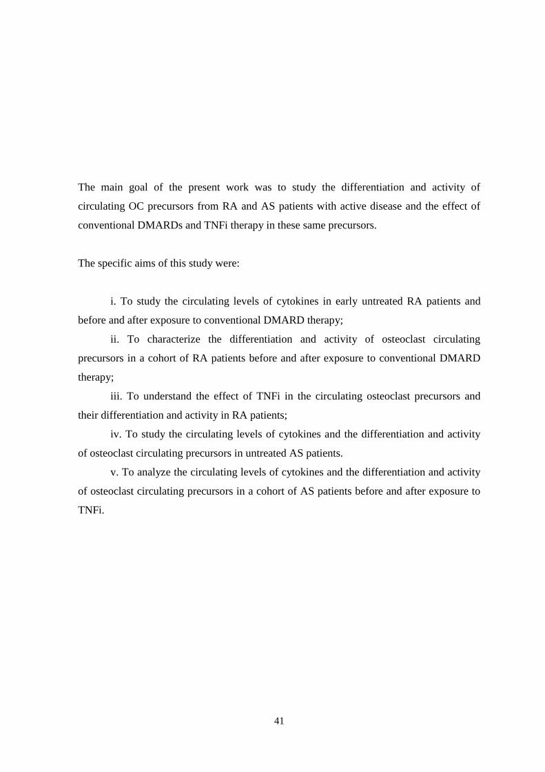
41
The main goal of the present work was to study the differentiation and activity of
circulating OC precursors from RA and AS patients with active disease and the effect of
conventional DMARDs and TNFi therapy in these same precursors.
The specific aims of this study were:
i. To study the circulating levels of cytokines in early untreated RA patients and
before and after exposure to conventional DMARD therapy;
ii. To characterize the differentiation and activity of osteoclast circulating
precursors in a cohort of RA patients before and after exposure to conventional DMARD
therapy;
iii. To understand the effect of TNFi in the circulating osteoclast precursors and
their differentiation and activity in RA patients;
iv. To study the circulating levels of cytokines and the differentiation and activity
of osteoclast circulating precursors in untreated AS patients.
v. To analyze the circulating levels of cytokines and the differentiation and activity
of osteoclast circulating precursors in a cohort of AS patients before and after exposure to
TNFi.

42

43
Results

44

45
In agreement with the Decreto-Lei 388/70, art. 8º, parágrafo 2, the results presented and
discussed in this thesis were published or submitted for publication in the following
scientific peer-reviewed journals:
I. Rita Cascão†, Rita A Moura†, Inês Perpétuo, Helena Canhão, Elsa Vieira-Sousa, Ana F
Mourão, Ana M Rodrigues, Joaquim Polido-Pereira, Mário V Queiroz, Henrique S
Rosário, Maria M Souto-Carneiro, Luis Graca†, João E Fonseca†; Identification of a
cytokine network sustaining neutrophil and Th17 activation in untreated early rheumatoid
arthritis. Arthritis Research & Therapy 2010 12:R196.
† contributed equally to this work.
II. Inês P. Perpétuo, Joana Caetano-Lopes, Ana Maria Rodrigues, Raquel Campanilho-
Marques, Cristina Ponte, Helena Canhão, Mari Ainola, João E. Fonseca; Methotrexate and
low dose prednisolone downregulate osteoclast function by decreasing receptor activator
of nuclear factor κB expression in monocytes from early rheumatoid arthritis patients
(submitted to RMD Open 2016)
III. Inês P. Perpétuo, Joana Caetano-Lopes, Ana Maria Rodrigues, Raquel Campanilho-
Marques, Cristina Ponte, Helena Canhão, Mari Ainola, João E. Fonseca; Effect of Tumor
Necrosis Factor Inhibitor Therapy on Osteoclasts Precursors in Rheumatoid Arthritis
(submitted to BioMed Research International 2016)
IV. Inês P. Perpétuo, Joana Caetano-Lopes, Elsa Vieira-Sousa, Raquel Campanilho-
Marques, Cristina Ponte, Helena Canhão, Mari Ainola, João E. Fonseca; Ankylosing
spondylitis patients have impaired osteoclast gene expression in circulating osteoclast
precursors (submitted to Frontiers in Medicine)
V. Inês P. Perpétuo*, Rita Raposeiro*, Joana Caetano-Lopes, Elsa Vieira-Sousa, Raquel
Campanilho-Marques, Cristina Ponte, Helena Canhão, Mari Ainola, João E. Fonseca;
(2015) Effect of Tumor Necrosis Factor Inhibitor Therapy on Osteoclasts Precursors in
Ankylosing Spondylitis. PLoS ONE 10(12): e0144655.doi:10.1371/journal.pone.0144655
* Inês P. Perpétuo and Rita Raposeiro contributed equally to this work.

46

47
I. Identification of a cytokine network
sustaining neutrophil and Th17 activation in untreated early
rheumatoid arthritis.
Rita Cascão†, Rita A Moura†, Inês Perpétuo, Helena Canhão, Elsa Vieira-Sousa, Ana F
Mourão, Ana M Rodrigues, Joaquim Polido-Pereira, Mário V Queiroz, Henrique S
Rosário, Maria M Souto-Carneiro, Luis Graca†, João E Fonseca†; Arthritis Research &
Therapy 2010 12:R196.
† contributed equally to this work.
Inês P. Perpétuo carried out the cytokine quantification in both serum and synovial fluid, performed
statistical analysis and wrote the manuscript draft in collaboration with Rita Cascão and Rita A.
Moura.

48

49
Identification of a cytokine network sustaining
neutrophil and Th17 activation in untreated early
Rheumatoid Arthritis
Cascão, Rita; Moura, Rita A.; Perpétuo, Inês; Canhão, Helena; Sousa, Elsa; Mourão, Ana F.;
Rodrigues, Ana M.; Polido-Pereira, Joaquim; Viana Queiroz, Mário; Rosário, Henrique S.;
Souto-Carneiro, Maria M.; Graca Luis†; Fonseca, João E.
†
Abstract
Introduction: Rheumatoid arthritis (RA) is a chronic inflammatory autoimmune disease
characterized by sustained synovitis. Recently, several studies have proposed neutrophils
and Th17 cells as key players in the onset and perpetuation of this disease. The main goal
of this work was to determine whether cytokines driving neutrophil and Th17 activation are
dysregulated in very early rheumatoid arthritis patients with less than 6 weeks of disease
duration and before treatment (VERA). Methods: Cytokines related to neutrophil and Th17
activation were quantified in the serum of VERA and established RA patients and
compared with other very early arthritis (VEA) and healthy controls. Synovial fluid (SF)
from RA and osteoarthritis (OA) patients was also analyzed. Results: VERA patients had
increased serum levels of cytokines promoting Th17 polarization (IL-1β and IL-6), as well
as IL-8 and Th17-derived cytokines (IL-17A and IL-22) known to induce neutrophil-
mediated inflammation. In established RA this pattern is more evident within the SF. Early
treatment with MTX or corticosteroids led to clinical improvement but without an impact
on the cytokine pattern. Conclusions: VERA patients already display increased levels of
cytokines related with Th17 polarization and neutrophil recruitment and activation, a

50
dysregulation also found in SF of established RA. Thus, our data suggest that a cytokine-
milieu favoring Th17 and neutrophil activity is an early event in RA pathogenesis.
Introduction
Rheumatoid arthritis (RA) is the most common chronic autoimmune disease, affecting
approximately 1% of the population worldwide. This disease comprises a syndrome of
pain, stiffness and symmetrical synovitis which leads to joint destruction, functional
disability and substantial comorbidity due to involvement of multiple organs and systems.
The migration of leukocytes towards the synovium is crucial for the establishment of a
chronic inflammatory process in RA [1-3]. This multi-regulated mechanism involves
interactions with endothelial cells through cell adhesion molecules and complex cytokine
and chemokine pathways.
Neutrophils specifically play an important role in the onset and perpetuation of RA, not
only as interleukin producing cells, but also for being responsible for the release of high
amounts of reactive oxygen species (ROS) and destructive enzymes, such as
metalloproteases, contributing to joint erosions [4]. Neutrophils are among the first
leukocytes to arrive at sites of inflammation. In fact, these cells are the most abundant in
the synovial fluid of patients with active RA and previous results from our group showed
that the synovial tissue is heavily infiltrated by neutrophils in the first weeks of RA onset
[5]. Interestingly, in animal models of arthritis, neutrophil depletion prevented joint
inflammation if neutrophil depleting antibodies were given before the induction of arthritis.
Moreover, when the depleting antibody was given very early after the induction of arthritis
complete abrogation of the inflammatory symptoms was also achieved [6].

51
T helper (Th)-17 cells have also been proposed to have a relevant role in the early phase of
RA through the production of interleukin (IL)-17 [7, 8]. This cytokine promotes the
recruitment and survival of neutrophils, induces the secretion of proinflammatory
cytokines, the upregulation of RANK ligand (RANKL) and stimulates the activity of matrix
metalloproteases, leading to cartilage catabolism and bone resorption [9, 10]. The
recruitment, activation and effector function of Th17 cells and neutrophils are driven by a
network of cytokines and chemokines, secreted by multiple cellular sources. In established
RA, it has been reported that IL-1β, IL-6, IL-8, IL-17, and tumor necrosis factor (TNF) are
elevated in the serum and this correlates with a higher disease activity [11-13].
Nevertheless, our knowledge on the influence of the cytokine network on RA onset remains
scarce. The characterization of the cytokine profile at this stage, where the transition from
an acute to a chronic inflammatory phase occurs, may lead to the identification of early key
players, with potential implications for early treatment strategies.
Thus, the main goal of our work was to determine whether cytokines driving neutrophil and
Th17 cell activation and pro-inflammatory function were already present in very early RA
(with less than 6 weeks of disease duration) and how this early cytokine environment
differs from established RA. We also evaluated whether the introduction of low dose
corticosteroids and methotrexate (MTX) therapy had any influence on the cytokine profile
observed at that early stage of the disease.
We found that cytokines related with Th17 polarization and neutrophil recruitment and
activation were elevated in early RA and that the conventional therapeutic options,
although able to control clinical manifestations of the disease, were ineffective in reversing
this underlying pro-inflammatory drive.

52
Material and methods
Patients
Blood samples were obtained from 38 consecutive untreated polyarthritis patients with less
than 6 weeks of disease duration. Some of these patients (19) after a minimum follow up of
3 months fulfilled the 1987 American College of Rheumatology (ACR) criteria for RA
[14]. These patients were classified as Very Early Rheumatoid Arthritis (VERA) patients
and further samples were collected 4-6 weeks after starting a low dose of oral
corticosteroids (5-10 mg of prednisone) (Time 1) and 4 months after reaching the minimum
effective dose of MTX (Time 2), up to a maximum of 20 mg/week, that was required to
reduce the 28 joints disease activity score (DAS 28) to less than 3.2 [15]. The remaining
early arthritis patients (19), who did not evolve into RA, were classified as very early
polyarthritis (VEA). Baseline blood samples from VERA and VEA patients were compared
with 27 healthy donors used as controls. Additionally, 12 blood and 15 SF samples were
obtained from established RA patients. SF samples were also collected from 10 OA
patients (Rheumatology Department, Hospital de Santa Maria, Lisbon) (Table 1). Due to
the clinical characteristics of the very early arthritis patients, SF in easily accessible joints
was not available in VERA and VEA patients and thus SF was not analyzed in these groups
of patients. The health assessment questionnaire (HAQ) [16] and DAS28 were applied to
all patients. The study was approved by the local ethics committee and all patients signed
an informed consent. Patient care was conducted in accordance with the standard clinical
practice and the study was performed in accordance with the Declaration of Helsinki as
amended in Edinburgh (2000).

53
Cytokine quantification
IL-1β, IL-2, IL-4, IL-6, IL-8, IL-10, IL-12(p70), IL-17A, IL-22, IL-23 and gamma
interferon (IFN-γ) levels were measured in the serum and SF by FlowCytomix assay kit
(FC) (Bender MedSystems, USA) according to the manufacturer instructions. Standard
curves for each cytokine were generated by using reference cytokine concentrations
supplied by the manufacturer. Samples were acquired with a FACS Calibur (BD). Raw data
of the FC bead assay were analyzed by FlowCytomixPro2.2 software.
Measurement of autoantibodies
Rheumatoid factor (RF)-IgM was determined in all patients by IMTEC Autoimmune
Diagnostics ELISA kit (Human GmbH, Germany) according to the manufacturer
instructions and samples were processed using a ChemWell® 2910 automated analyzer.
Serum levels of anti-cyclic citrullinated peptide (anti-CCP) were measured by ELIATM
CCP test system (Phadia GmbH, Germany) and samples were analyzed using an
ImmunoCAP® 100 instrument.
Statistical analysis
Statistical differences were determined with non-parametric Kruskal-Wallis, Mann-
Whitney and Wilcoxon Signed-Rank tests using GraphPad Prism (GraphPad, San Diego,
CA). Correlation analysis was performed using Spearman’s test. Differences were
considered statistically significant for p < 0.05.

54
Results
Characterization of patients and disease evaluation.
A total of 38 polyarthritis patients with less than 6 weeks of disease duration were
evaluated. Nineteen patients fulfilled the 1987 ACR criteria for RA after a minimum
follow-up of 3 months and were classified as VERA patients. The mean age of the VERA
patients was 59±17 years-old, 84% were female, 42% were RF positive and 32% anti-CCP
positive, the initial DAS 28 was of 6.1±1.8 and the initial HAQ was of 1.4±0.8. After
treatment with a low dose of prednisone and MTX there was a significant reduction of both
DAS28 and HAQ values (Table 1).
Table 1. Clinical information about healthy controls, VERA, VEA, RA and OA patients.
VEA – very early arthritis; VERA - very early rheumatoid arthritis; RA – rheumatoid arthritis; OA
– osteoarthritis; SF - synovial fluid; DAS 28 – Disease Activity Score; HAQ – Health Assessment
Questionnaire; RF – Rheumatoid Factor; Anti-CCP – anti-cyclic citrullinated peptide; NA – not
applicable; ND – not determined. *DAS28 and HAQ values were compared between VERA and
VEA patients with reference to VERA baseline values. Differences were considered statistically
significant for p values < 0.05.
The group of VEA patients included 19 patients, who evolved into several different
diagnosis: spondylarthritis (5 cases), systemic lupus erythematosus (4 cases), crystal
induced arthritis (2 cases), Sjögren’s syndrome (1 case), paraneoplastic polyarthritis related

55
to multiple myeloma (1 case), arthritis associated to HIV infection (1 case). Five patients
entered spontaneously into remission before 3 months of follow-up, remaining without a
specific diagnosis and were thus classified as presenting a self-limited form of polyarthritis.
The mean age of the VEA patients was 40±13 years-old, 79% were female, all patients
were RF and anti-CCP negative, the initial DAS 28 was of 4.5±1.6 and the initial HAQ was
of 0.8±0.6. Both DAS28 and HAQ values were significantly lower than those of VERA
patients at baseline (Table 1). These early polyarthritis patients represent a subset of a
larger cohort previously described by our group [17].
Furthermore, blood samples were collected from 12 established RA patients, with a mean
age of 60±10 years old, 92% were female, 67% were RF positive and 45% anti-CCP
positive (Table 1). Additionally, SF samples were also collected from 12 established RA
patients, with a mean age of 57±10 years old and 73% were female (Table 1). The
established RA group of patients had a DAS28 and a HAQ mean scores similar to VERA
baseline values.
IL-8 is increased in VERA patients and locally in the joints of established RA patients.
Given the proposed role of neutrophils in the pathogenesis of RA [18, 19], we quantified
the major neutrophil chemoattractant, IL-8, in the serum of VERA patients. At baseline,
VERA patients had significantly higher levels of IL-8 when compared to both VEA and
healthy controls (Figure 1A). After 2-4 weeks of low-dose corticosteroids and after 4
months of MTX therapy there were no significant changes in the levels of circulating IL-8
(data not shown). Interestingly, VERA patients also had significantly higher circulating
levels of IL-8 in comparison with serum from established RA (Figure 1A). Neutrophils
accumulate locally in the joints of RA patients [20]. Thus, we quantified the concentration

56
of IL-8 in the SF of RA patients, and compared to SF from OA patients. We found
significant higher levels of IL-8 in the SF of RA patients in comparison to OA SF (Figure
1B).
Figure 1. IL-8 is increased in the serum of VERA patients and in SF of established RA. (A) The
serum concentration of IL-8 was measured in VERA and VEA patients as well as healthy controls
and patients with established RA. Serum concentration of IL-8 was increased in VERA patients,
compared with any other group. Dotted line represents the limit of detection for the assay. (B) The
concentration of IL-8 was measured in the SF collected from patients with established RA and from
a control group with OA. We found a significant increase of IL-8 in RA-SF. Differences were
considered statistically significant for p values < 0.05 according to Mann-Whitney test.
IL-17 levels are dysregulated in both VERA and established RA patients.
Previous studies from our group showed that there is a delay in the apoptosis of circulating
neutrophils in VERA patients [21]. Therefore, we analyzed IL-17A levels in these patients,
since it has already been described that this cytokine is important for the survival of
neutrophils [22]. Moreover, IL-17A is a signature cytokine of Th17 cells, a subset proposed
to have a key role in RA pathogenesis [9, 23]. We found that VERA patients had
significantly higher levels of IL-17A when compared to healthy controls, but not with VEA
patients (Figure 2A). Furthermore, in our previous work we found no difference in the
57
frequency and absolute numbers of CD4+
and CD8+
T cell subpopulations in the peripheral
blood of these patients, when analyzed by flow cytometry [17].
Regarding the effects of early therapy, we found that neither corticosteroids nor MTX
affected the level of IL-17A (data not shown). Moreover, IL-17A was also significantly
increased locally within the joints of established RA patients in comparison with control SF
from OA patients (Figure 2B).
Figure 2. VERA patients and SF of established RA display increased levels of IL-17A. (A) The
serum concentration of IL-17A was measured in VERA and VEA patients as well as healthy
controls and patients with established RA. Serum concentration of IL-17A was increased in VERA
patients compared with healthy controls. (B) The concentration of IL-17A was measured in the SF
collected from patients with established RA and from a control group with OA. In the SF of RA
patients we observed a significant increase of IL-17A. Dotted lines represent the limit of detection
for the assay. Differences were considered statistically significant for p values < 0.05 according to
Mann-Whitney test.
RA has a Th17- cytokine pattern since the very first weeks of onset.
Having found that IL-17A was elevated in VERA patients, we decided to quantify a panel
of cytokines known to be associated with Th17 polarization. At baseline, VERA patients
had significantly higher levels of IL-1β and IL-22 in comparison with both VEA and
healthy controls. In addition, we also found that VERA patients have significantly higher
IL-6 levels than healthy controls (Figure 3). Furthermore, the significantly higher
circulating levels of IL-6 and IL-22 were maintained in established RA (Figure 3).

58
Figure 3. Cytokines related to Th17 polarization are increased in the serum of VERA patients and
SF of established RA. The serum concentration of IL-1β, IL-6 and IL-22 was measured in VERA
and VEA patients as well as healthy controls and patients with established RA. All three cytokines
were increased in VERA patients compared with healthy controls. IL-6 was equally elevated in all
groups of patients with an inflammatory disease, while the other two cytokines were increased only
in VERA (IL-1β), or in VERA and RA patients (IL-22). Dotted lines represent the limit of
detection for the assays. Differences were considered statistically significant for p values < 0.05
according to Mann-Whitney test.
Locally, within the joints of RA patients, the synovial fluid displayed elevated levels of IL-
1β and IL-6 in comparison with OA SF (Figure 4 and Table 2). Moreover, no significant
differences could be observed for IL-23 in circulation or locally in the joints (data not
shown).

59
Figure 4. Cytokines related to Th17 polarization are increased in the SF of established RA The
concentration of IL-1β and IL-6 was markedly increased in the SF collected from patients with
established RA, when compared with OA. Dotted lines represent the limit of detection for the
assays. Differences were considered statistically significant for p values < 0.05 according to Mann-
Whitney test.
Table 2. Cytokine levels in healthy controls, VERA, VEA, established RA and OA
patients.
Values are represented as median (range). VEA – very early arthritis; VERA - very early
rheumatoid arthritis; RA – rheumatoid arthritis; OA – osteoarthritis; SF - synovial fluid.
We have also studied cytokines associated with the function of Th2 (IL-4, IL-10) and Th1
(IL-2, IL-12 (p70) and INFγ) cells. However, no statistically significant differences could
be observed for any of these cytokines (data not shown).

60
Discussion
Several studies have previously demonstrated that neutrophils play an important role in the
onset of RA [21]. This hypothesis is supported by data from animal models [24]. In fact,
neutrophils are the most abundant leukocytes in the synovial fluid of patients with active
RA and, in early RA, these cells show significantly lower levels of apoptosis when
compared to patients with other persistent forms of arthritis or with arthritis that have a self
limited disease course [25]. Additionally, previous results from our group demonstrated
that there is also a delay in the apoptosis of circulating neutrophils in VERA patients [21]
and that these cells heavily infiltrate the synovial tissue during RA onset [5].
In the present study, we demonstrate that a neutrophil- and Th17-driving cytokine pattern is
present in untreated VERA patients with less than 6 weeks of disease duration. We consider
this observation of interest because the knowledge concerning the immune mechanisms
associated with the onset of RA is still elusive. In fact, the majority of early RA studies
include patients with 3 to 12 months of disease duration or even more. In accordance with
an early participation of neutrophils in RA, our results revealed that VERA patients have
increased levels of IL-8 when compared to both VEA and healthy controls, which could
explain the pre-activated state of circulating neutrophils [18] and their recruitment towards
the synovial fluid since the very first weeks of RA onset.
In addition, Th17 cells are known to be important for the promotion of neutrophil-mediated
inflammation by producing IL-17A, a cytokine known to indirectly activate neutrophil
chemotaxis and extend their survival [10, 22]. We found a high serum concentration of IL-
17A in VERA patients, as well as locally within the joints of patients with established RA.
This might indicate that there is an activation of Th17 cells from a very early phase of the

61
disease, that can promote neutrophil participation in RA pathogenesis [22]. However, we
found no evidence for changes in the frequency of T cell subsets in the peripheral blood of
VERA patients [17]. This observation is not unexpected, given the relatively small
representation of antigen-specific T cells in the circulating pool, being the activated T cells
driving the pathology more likely found within the tissues [26]. In a study performed by
Kokkonen and colleagues [27] the levels of several cytokines and chemokines were
analyzed in blood samples from a group of individuals 3.3 years before RA onset (“pre-
patients”) and compared with healthy donors and RA patients with 7.7±3.6 months of
disease duration. An interesting finding was that IL-17 was present at its highest
concentration in pre-patients and the level of this cytokine was lower in RA patients. This is
in accordance with our own results, since we observed an increased level of IL-17 in RA
patients with less than 6 weeks of disease duration, while in established RA patients the
levels were not significantly different to healthy controls. Remarkably, the IL-17 median
concentration observed in our established RA cohort (2.6 pg/ml) was even lower than that
of RA patients from Kokkonen and colleagues work (6.0 pg/ml). Thus, this observation
reinforces the role of IL-17 in the initial phase of RA and, as the pathogenesis progresses to
a chronic stage, other factors are subsequently brought into action in the peripheral blood.
Contrarily to what was observed in Kokkonen et al work we have not detected differences
in Th1 and Th2-related cytokines between both VERA and established RA patients in
comparison with controls. These discrepancies might be related with the different
methodologies used.
Additionally, the elevated levels of IL-1β observed in VERA patients can stimulate
endothelial cells, T and B cells and fibroblasts in the joints to produce IL-6 and IL-8. But
importantly, IL-1β together with IL-6, both found to be increased in VERA patients, are

62
known to promote the differentiation of Th17 cells, which in turn secrete IL-17A and IL-22
[28, 29], two cytokines that were elevated in VERA patients and have an essential function
in the pathogenesis of autoimmune diseases [29].
Currently, the treatment of choice for RA at the time of presentation is MTX. Interestingly,
in spite of clinical improvement (DAS28 reduced from 6.1±1.8 to 3.1±1.6), neither therapy
with low-dose corticosteroids nor combined therapy with low-dose corticosteroids and
MTX corrected the dysregulated cytokine pattern observed in VERA patients. In fact, low
dose corticosteroids and MTX have unclear effects on RA cytokine network. For instance,
corticosteroids fail to reduce serum levels of IL-1β and IL-8 [30] and MTX does not alter
serum IL-1β concentration when compared to pre-treatment levels [31, 32]. Our results
suggest that the conditions contributing to Th17 cells and neutrophil-mediated
inflammation, thus driving early pathogenesis, are not modified with early treatment with
low-dose corticosteroids and MTX.
The elevated IL-1β, IL-6, IL-8 and IL-17A levels observed in the SF of RA patients
confirm a local role for these cytokines in the maintenance of synovitis. Moreover, IL-6 can
support a continuous recruitment of autoreactive B cells towards the synovium [33, 34],
contributing to an exacerbation of the inflammatory process due to the production of
autoantibodies and immune complexes.
Conclusions
Taken together, our data reinforce the potential relevance of therapies targeting IL-1β [35,
36] and IL-6 [37, 38] in early RA. In addition the data also establish IL-8 and IL-17A as
other potential therapeutic targets at an early stage of the disease. Finally, we found that
63
MTX and corticosteroids, although effective in reducing disease activity in VERA patients,
do not appear to correct underlying cytokine dysregulation driving the Th17/neutrophil
mediated inflammation.
References
1. Kim, H.J., V. Krenn, G. Steinhauser and C. Berek. Plasma cell development in synovial
germinal centers in patients with rheumatoid and reactive arthritis. J Immunol, 1999. 162:
p. 3053-62.
2. Fonseca, J.E., N. Cortez-Dias, A. Francisco, M. Sobral, H. Canhao, C. Resende, et al.
Inflammatory cell infiltrate and rankl/opg expression in rheumatoid synovium: Comparison
with other inflammatory arthropathies and correlation with outcome. Clin Exp Rheumatol,
2005. 23: p. 185-92.
3. Fonseca, J.E., J.C. Edwards, S. Blades and N.J. Goulding. Macrophage subpopulations in
rheumatoid synovium: Reduced cd163 expression in cd4+ t lymphocyte-rich
microenvironments. Arthritis Rheum, 2002. 46: p. 1210-6.
4. Cascao, R., H.S. Rosario, M.M. Souto-Carneiro and J.E. Fonseca. Neutrophils in
rheumatoid arthritis: More than simple final effectors. Autoimmun Rev, 2010.
5. Mourao, A.F., H. Canhao, E. Sousa, R. Cascao, J.B. da Costa, L.S. de Almeida, et al. From
a neutrophilic synovial tissue infiltrate to a challenging case of rheumatoid arthritis. Acta
Reumatol Port, 2010. 35: p. 228-31.
6. Cascao, R., H.S. Rosario and J.E. Fonseca. Neutrophils: Warriors and commanders in
immune mediated inflammatory diseases. Acta Reumatol Port, 2009. 34: p. 313-26.
7. Chabaud, M., J.M. Durand, N. Buchs, F. Fossiez, G. Page, L. Frappart, et al. Human
interleukin-17: A t cell-derived proinflammatory cytokine produced by the rheumatoid
synovium. Arthritis Rheum, 1999. 42: p. 963-70.
8. Kotake, S., N. Udagawa, N. Takahashi, K. Matsuzaki, K. Itoh, S. Ishiyama, et al. Il-17 in
synovial fluids from patients with rheumatoid arthritis is a potent stimulator of
osteoclastogenesis. J Clin Invest, 1999. 103: p. 1345-52.
9. Stamp, L.K., M.J. James and L.G. Cleland. Interleukin-17: The missing link between t-cell
accumulation and effector cell actions in rheumatoid arthritis? Immunol Cell Biol, 2004.
82: p. 1-9.

64
10. Pelletier, M., L. Maggi, A. Micheletti, E. Lazzeri, N. Tamassia, C. Costantini, et al.
Evidence for a cross-talk between human neutrophils and th17 cells. Blood. 115: p. 335-43.
11. Nagatani, K., K. Itoh, K. Nakajima, H. Kuroki, Y. Katsuragawa, M. Mochizuki, et al.
Rheumatoid arthritis fibroblast-like synoviocytes express bcma and are stimulated by april.
Arthritis Rheum, 2007. 56: p. 3554-63.
12. Chen, D.Y., T.Y. Hsieh, Y.M. Chen, C.W. Hsieh, J.L. Lan and F.J. Lin. Proinflammatory
cytokine profiles of patients with elderly-onset rheumatoid arthritis: A comparison with
younger-onset disease. Gerontology, 2009. 55: p. 250-8.
13. Petrovic-Rackov, L. and N. Pejnovic. Clinical significance of il-18, il-15, il-12 and tnf-
alpha measurement in rheumatoid arthritis. Clin Rheumatol, 2006. 25: p. 448-52.
14. Arnett, F.C., S.M. Edworthy, D.A. Bloch, D.J. McShane, J.F. Fries, N.S. Cooper, et al. The
american rheumatism association 1987 revised criteria for the classification of rheumatoid
arthritis. Arthritis Rheum, 1988. 31: p. 315-24.
15. Prevoo, M.L., M.A. van 't Hof, H.H. Kuper, M.A. van Leeuwen, L.B. van de Putte and P.L.
van Riel. Modified disease activity scores that include twenty-eight-joint counts.
Development and validation in a prospective longitudinal study of patients with rheumatoid
arthritis. Arthritis Rheum, 1995. 38: p. 44-8.
16. Fries, J.F., P. Spitz, R.G. Kraines and H.R. Holman. Measurement of patient outcome in
arthritis. Arthritis Rheum, 1980. 23: p. 137-45.
17. Moura, R.A., P. Weinmann, P.A. Pereira, J. Caetano-Lopes, H. Canhao, E. Sousa, et al.
Alterations on peripheral blood b-cell subpopulations in very early arthritis patients.
Rheumatology (Oxford), 2010. 49: p. 1082-92.
18. Cedergren, J., T. Forslund, T. Sundqvist and T. Skogh. Intracellular oxidative activation in
synovial fluid neutrophils from patients with rheumatoid arthritis but not from other
arthritis patients. J Rheumatol, 2007. 34: p. 2162-70.
19. Poubelle, P.E., A. Chakravarti, M.J. Fernandes, K. Doiron and A.A. Marceau. Differential
expression of rank, rank-l, and osteoprotegerin by synovial fluid neutrophils from patients
with rheumatoid arthritis and by healthy human blood neutrophils. Arthritis Res Ther, 2007.
9: p. R25.
20. Assi, L.K., S.H. Wong, A. Ludwig, K. Raza, C. Gordon, M. Salmon, et al. Tumor necrosis
factor alpha activates release of b lymphocyte stimulator by neutrophils infiltrating the
rheumatoid joint. Arthritis Rheum, 2007. 56: p. 1776-86.

65
21. Weinmann, P., R.A. Moura, J.R. Caetano-Lopes, P.A. Pereira, H. Canhao, M.V. Queiroz, et
al. Delayed neutrophil apoptosis in very early rheumatoid arthritis patients is abrogated by
methotrexate therapy. Clin Exp Rheumatol, 2007. 25: p. 885-7.
22. Parsonage, G., A. Filer, M. Bik, D. Hardie, S. Lax, K. Howlett, et al. Prolonged,
granulocyte-macrophage colony-stimulating factor-dependent, neutrophil survival
following rheumatoid synovial fibroblast activation by il-17 and tnfalpha. Arthritis Res
Ther, 2008. 10: p. R47.
23. Kirkham, B.W., M.N. Lassere, J.P. Edmonds, K.M. Juhasz, P.A. Bird, C.S. Lee, et al.
Synovial membrane cytokine expression is predictive of joint damage progression in
rheumatoid arthritis: A two-year prospective study (the damage study cohort). Arthritis
Rheum, 2006. 54: p. 1122-31.
24. Vossenaar, E.R., S. Nijenhuis, M.M. Helsen, A. van der Heijden, T. Senshu, W.B. van den
Berg, et al. Citrullination of synovial proteins in murine models of rheumatoid arthritis.
Arthritis Rheum, 2003. 48: p. 2489-500.
25. Andersson, A.K., C. Li and F.M. Brennan. Recent developments in the immunobiology of
rheumatoid arthritis. Arthritis Res Ther, 2008. 10: p. 204.
26. Duarte, J., A. Agua-Doce, V.G. Oliveira, J.E. Fonseca and L. Graca. Modulation of il-17
and foxp3 expression in the prevention of autoimmune arthritis in mice. PLoS One. 5: p.
e10558.
27. Kokkonen, H., I. Soderstrom, J. Rocklov, G. Hallmans, K. Lejon and S. Rantapaa
Dahlqvist. Up-regulation of cytokines and chemokines predates the onset of rheumatoid
arthritis. Arthritis Rheum, 2010. 62: p. 383-91.
28. Liang, S.C., X.Y. Tan, D.P. Luxenberg, R. Karim, K. Dunussi-Joannopoulos, M. Collins, et
al. Interleukin (il)-22 and il-17 are coexpressed by th17 cells and cooperatively enhance
expression of antimicrobial peptides. J Exp Med, 2006. 203: p. 2271-9.
29. Pene, J., S. Chevalier, L. Preisser, E. Venereau, M.H. Guilleux, S. Ghannam, et al.
Chronically inflamed human tissues are infiltrated by highly differentiated th17
lymphocytes. J Immunol, 2008. 180: p. 7423-30.
30. Chikanza, I.C., D. Kozaci and Y. Chernajovsky. The molecular and cellular basis of
corticosteroid resistance. J Endocrinol, 2003. 179: p. 301-10.
31. Barrera, P., C.J. Haagsma, A.M. Boerbooms, P.L. Van Riel, G.F. Borm, L.B. Van de Putte,
et al. Effect of methotrexate alone or in combination with sulphasalazine on the production
and circulating concentrations of cytokines and their antagonists. Longitudinal evaluation in
patients with rheumatoid arthritis. Br J Rheumatol, 1995. 34: p. 747-55.

66
32. Segal, R., E. Mozes, M. Yaron and B. Tartakovsky. The effects of methotrexate on the
production and activity of interleukin-1. Arthritis Rheum, 1989. 32: p. 370-7.
33. Houssiau, F.A., J.P. Devogelaer, J. Van Damme, C.N. de Deuxchaisnes and J. Van Snick.
Interleukin-6 in synovial fluid and serum of patients with rheumatoid arthritis and other
inflammatory arthritides. Arthritis Rheum, 1988. 31: p. 784-8.
34. Gabay, C. Interleukin-6 and chronic inflammation. Arthritis Res Ther, 2006. 8 Suppl 2: p.
S3.
35. Smeets, T.J., E.C. Barg, M.C. Kraan, M.D. Smith, F.C. Breedveld and P.P. Tak. Analysis of
the cell infiltrate and expression of proinflammatory cytokines and matrix
metalloproteinases in arthroscopic synovial biopsies: Comparison with synovial samples
from patients with end stage, destructive rheumatoid arthritis. Ann Rheum Dis, 2003. 62: p.
635-8.
36. Joosten, L.A., S. Abdollahi-Roodsaz, M. Heuvelmans-Jacobs, M.M. Helsen, L.A. van den
Bersselaar, B. Oppers-Walgreen, et al. T cell dependence of chronic destructive murine
arthritis induced by repeated local activation of toll-like receptor-driven pathways: Crucial
role of both interleukin-1beta and interleukin-17. Arthritis Rheum, 2008. 58: p. 98-108.
37. al-Janadi, M., S. al-Balla, A. al-Dalaan and S. Raziuddin. Cytokine production by helper t
cell populations from the synovial fluid and blood in patients with rheumatoid arthritis. J
Rheumatol, 1993. 20: p. 1647-53.
38. Youinou, P. and C. Jamin. The weight of interleukin-6 in b cell-related autoimmune
disorders. J Autoimmun, 2009. 32: p. 206-10.

67
II. Methotrexate and low dose prednisolone downregulate osteoclast
function by decreasing receptor activator of nuclear factor κB
expression in monocytes from early rheumatoid arthritis patients
Inês P. Perpétuo, Joana Caetano-Lopes, Ana Maria Rodrigues, Raquel Campanilho-
Marques, Cristina Ponte, Helena Canhão, Mari Ainola, João E. Fonseca; (submitted to
RMD Open 2016)
Inês P. Perpétuo designed all the experiments and performed all the flow cytometry, cell isolation,
cell culture, functional assays, RANKL/OPG quantification, and image analysis, analyzed the data,
produced the figures and wrote the manuscript.

68

69
Methotrexate and low dose prednisolone
downregulate osteoclast function by decreasing
receptor activator of nuclear factor κB
expression in monocytes from early rheumatoid
arthritis patients
Inês P. Perpétuo, Joana Caetano-Lopes, Ana Maria Rodrigues, Raquel Campanilho-
Marques, Cristina Ponte, Helena Canhão, Mari Ainola, João E. Fonseca.
Abstract
Objective: Rheumatoid arthritis (RA) is a systemic, immune mediated inflammatory
disease that ultimately leads to bone erosions and joint destruction. Methotrexate
(MTX) slows bone damage but the mechanism by which it acts is still unknown. In this
study we aimed to assess the effect of MTX and low dose prednisolone (PDN) on
circulating osteoclast (OC) precursors and OC differentiation in RA patients. Methods:
RA patients before and at least 6 months after MTX therapy were analyzed and
compared with healthy donors. A blood sample was collected in order to assess receptor
activator of NF-κB (RANK) ligand (RANKL) surface expression on circulating
leukocytes and frequency and phenotype of monocyte subpopulations. Quantification of
serum levels of bone turnover markers and cytokines and in vitro OC differentiation
assays were performed. Results: Classical activation markers of monocytes and RANK
were increased in RA patients at baseline, comparing to control healthy donors, and
after MTX and low dose PDN (MTX+PDN) exposure they decreased to control levels.
Although the number of OC was not different between groups, the percentage of
resorbed area and the resorbed area/pit were reduced after treatment. Serum RANKL
levels were increased at baseline comparing to healthy donors and normalized after

70
therapy. Conclusion: Our results suggest that MTX+PDN play an important role in
downregulating OC function, which we believe occurs through the decrease in RANK
surface expression in monocytes.
Introduction
Rheumatoid arthritis (RA) is a chronic, immune-mediated, inflammatory disease that
mainly impacts joints. It affects around 1% of the world population and the majority of
the affected patients are women [1]. The main features of RA are synovial hyperplasia
with pannus formation and bone erosions. On the other hand, systemic inflammation
leads also to secondary osteoporosis and bone fragility [2]. In RA, there is an unbalance
between bone formation and bone resorption. In fact, the immune system contributes
directly to bone resorption by producing receptor activator of NF-κB ligand (RANKL)
and indirectly through the secretion of pro-inflammatory cytokines that act
synergistically with the RANK-RANKL system, further enhancing osteoclast (OC)
differentiation and bone resorption [3]. Monocytes are OC precursors; these cells have
phenotypical and functional heterogeneity and have a critical regulatory role in
inflammation and innate immune response [4, 5]. Monocytes can be subdivided into
three sub-populations based on their expression of CD14 and CD16 surface markers:
the classical (CD14bright
CD16-), intermediate (CD14
brightCD16
+) and non-classical
(CD14dim
CD16+) subsets [4]. All of these precursors are able to differentiate into OC
increasing bone resorption [6, 7]. Moreover, in RA, CD14+ monocytes continuously
migrate into the injured joint tissues where they are activated and act as local and
systemic amplifiers of the disease through a multitude of mediators [8-10].
Methotrexate (MTX) combined with low dose prednisolone (PDN) is the cornerstone of
early RA treatment, reducing inflammation and slowing down bone damage [11-14]. A

71
recent report depicted that OC formation and OC gene expression, such as NFATc1 and
DC-STAMP, which are induced by the cytokine RANKL, are significantly inhibited by
MTX [15]. In this study we aimed to assess the effect of MTX and low dose PDN
(MTX+PDN) on the circulating OC precursors and OC differentiation in RA patients.
Patients and Methods
Patients
Patients with early untreated RA (less than 12 months of disease duration) fulfilling the
American College of Rheumatology/European League Against Rheumatism criteria,
2010 [16] were recruited from the Rheumatology and Bone Metabolic Disease
Department, Hospital de Santa Maria, Lisbon Academic Medical Centre, Portugal. All
patients included were naïve to DMARDs and corticoids and were followed in the
outpatient clinic during a minimum of 6 months after starting MTX + PDN treatment.
Moderate to high disease activity was required for inclusion, defined as disease activity
score 28 (DAS28)>3.2 [17]. Patients previously exposed or actively under corticoids
and/or DMARD therapy were excluded from this study. The use of non-steroidal anti-
inflammatory drugs (NSAIDs) was not an exclusion criterion. Information regarding
patients’ demographics, duration of symptoms, erythrocyte sedimentation rate (ESR),
C-reactive protein (CRP), tender and swollen joints counts, presence of erosions,
presence of rheumatoid factor (RF) and presence of anti-citrullinated protein antibodies
(ACPA) was collected. Both DAS 28 and as the health assessment questionnaire (HAQ)
[18] were applied to patients. RA patients were managed by the standard of care
approach with no deviation induced by the participation in this study.
Heparinized blood and serum were collected from each participant at the starting of
MTX and prednisolone and after 6 months of reaching the minimum effective dose of

72
MTX. Blood was used for flow cytometry and peripheral blood mononuclear cell
(PBMC) isolation. Healthy Donors matched for age and gender were used as controls.
The local ethics committee approved this study and all participants signed an informed
consent. Patients were managed in accordance with the standard practice and the study
was conducted in accordance with the Declaration of Helsinki as amended in Brazil
(2013).
Antibodies and flow cytometry
Identification of B and T cells and neutrophils in peripheral blood and
immunophenotyping of monocytes in the PBMC samples were performed using
matched combinations of anti-human murine mAbs. For peripheral blood staining anti-
CD19 PerCP-Cy5.5 (eBioscience, San Diego, CA, USA), anti-CD3 PerCP (BD
Biosciences, Franklin Lakes, NJ, USA), anti-CD66b FITC (Immunotools, Friesoythe,
Germany) and anti-RANKL PE (Santa Cruz Biotechnology, Dallas, TX, USA) were
used. Monocyte subpopulations were identified with anti-CD14 FITC (BD Biosciences)
or PerCPCy5.5 (Immunotools) and anti-CD16 APC (Immunotools) and stained with
combinations of anti-CD11b PE-Cy7, CD105 PE, CD62L PE-Cy7, CD51/CD61 FITC
(eBioscience), CCR2 PE (R&D Systems, Minneapolis, MN, USA), HLA-DR PerCP
(BD Biosciences) and RANK PE (Santa Cruz Biotechnology). Cell death was assessed
by staining with Annexin V Apoptosis Detection Kit APC (eBioscience). Acquisition
was performed usig a FACSCalibur (BD Biosciences).
Heparinized whole blood was used for staining. Erythrocytes were lysed with red blood
cell lysis buffer and cells were incubated with IgG block solution 300ng/mL
(ChromPure Mouse IgG whole molecule, Jackson ImmunoResearch Laboratories, West
Grove, PA, USA) before staining. Absolute cell counts were calculated from differential

73
leukocyte count determined for all participants. PBMCs were isolated by density
gradient centrifugation with Histopaque®-1077 (Sigma-Aldrich, St. Louis, MO, USA).
Monocyte subpopulations were identified as described before based on their CD14 and
CD16 surface expression [4]. Data was analyzed using FlowJo software (TreeStar,
Stanford University, Stanford, CA, USA).
Osteoclast differentiation
PBMCs isolated by density gradient centrifugation were plated in 96-well culture plates
as described before [19]. PBMCs were left overnight for osteoclast precursors (OCPs)
to adhere on bone slices (Immunodiagnostic Systems, Boldon, UK) [20]. On the
following day (day 1 of culture) medium was changed to DMEM supplemented with
M-CSF 25 ng/mL (Peprotech, London, UK). Three days later, medium was again
changed to DMEM with M-CSF (25 ng/mL), sRANKL (50 ng/mL; Peprotech),
dexamethasone (10 nM; Sigma-Aldrich) and TGF-β (2.5 ng/mL; R&D Systems) in
order to differentiate the OC precursors into mature OC [21]. The culture medium was
then changed twice a week. Cells cultured on bone slices for 7, 14 and 21 days [20, 22]
were used for functional assays and gene expression.
Functional assays
TRAP staining of OC was performed using the Acid Phosphate, Leukocyte Kit (TRAP,
Sigma-Aldrich) according to the manufacturer's instructions. This assay allows the
identification of mature OC (TRAP positive cells containing three or more nuclei). In
the resorption assay, to measure the resorbed area, OC were removed from the bone
slices using sodium hypochlorite and stained with 0.1% toluidine blue.

74
Bone slices from both TRAP staining and resorption functional assays were
photographed in an area of 1.25 mm2 with a brightfield microscope (Leica DM2500,
Leica, Wetzlar, Germany) under a 10x objective. The number of TRAP stained OC was
counted for each time-point per condition and the resorption pits were traced using
ImageJ (NIH, Bethesda, MD, USA), a mean value of the resorbed area was calculated
and expressed in percentage of total area.
Bone turnover markers detection
Serum soluble RANKL and OPG were quantified by ELISA (Biomedica Grouppe,
Vienna, Austria) according to the manufacturer’s instructions.
Statistical analysis
Statistical analysis was performed with SPSS Statistics 17.0 (IBM, Portsmouth, UK).
Categorical variables were expressed as frequencies and comparisons were tested using
chi-square test. Continuous variables were expressed by median and interquartile range.
Before and after treatment values within each sample were compared using Wilcoxon's
matched-pairs signed-rank test. To compare RA patients with healthy age and sex-
matched donors Mann-Whitney test was used. P values lower than 0.05 were
considered significant.
Results
Population characteristics
This study was conducted in 14 untreated early RA patients, before and after initiation
of combined MTX and PDN treatment, and in 18 age and gender matched healthy
donors. All patients had less than 12 months disease duration and were corticosteroid

75
and synthetic DMARD naïve at baseline. The clinical and demographic features of
patients before and after treatment and of the healthy donors are described in table 1.
All patients were under MTX (mean dose of 15 mg/week) and prednisolone (less than
10 mg/day). Hydroxychloroquine (400 mg/day) was added later on 3 patients and 2
patients received concomitant sulfasalazine (2 g/day). Patients' median MTX therapy
duration was 13 months (minimum 6 and maximum 24 months).
Table 1 - Summary of patients and healthy controls' characteristics
RA patients
Healthy p-value Baseline Treated
Sample size 14 18
Age (years) 50 [32-60] 44 [36-52] 0.50853
Females % 71% 61% 0.7120
Symptoms duration (months) 9 [2-12] NA
RF(% positive) 57% NA
ACPA (% positive) 36% NA
Erosive (% yes) 23% NA
Treatment with NSAIDs (%) 57% NA
NSAIDs duration (months) 2.3 [0.3-5.5] NA
Treatment with MTX (%) 0% 100% a
ESR (mm/h) 22 [11-44] 13 [9-27] NA 0.1934
CRP (mg/dl) 0.3 [0.1-2.7] 0.2 [0.1-0.3] NA 0.1289
Tender joint count 4 [0-7] 0 [0-1] NA 0.3738
Swollen joint count 6 [2-10] 1 [0-1] NA 0.0380*
DAS28 4.7 [4.2-5.4] 2.4 [2.1-3.6] NA 0.0137*
HAQ 1.4 [0.6-2.2] 0.3 [0.0-0.7] NA 0.1250
Data is represented as median and interquartile range unless stated otherwise; aAll
patients were under MTX and prednisolone. To 3 patients hydroxychloroquine was
added latter on and 2 patients received concomitant sulfasalazine; *p<0.05 between
patients before and after treatment with MTX; RA – rheumatoid arthritis; NA – not
applicable; RF – rheumatoid factor; ACPA - Anti-citrullinated protein antibodies;
NSAIDs - non-steroidal anti-inflammatory drugs; MTX - methotrexate; ESR –
erythrocyte sedimentation rate; CRP – C-reactive protein; DAS – disease activity score;
HAQ – health assessment questionnaire.

76
RA patients had a higher number of neutrophils expressing RANKL than controls,
which regressed to control levels after treatment
The number of neutrophils was significantly higher in baseline RA patients when
compared to healthy donors (both frequency p=0.0110 and absolute number p=0.0032;
Fig.1). Moreover, the absolute number of neutrophils expressing RANKL on their
surface was also higher than in controls (p=0.0060). After treatment the absolute counts
of neutrophils decreased (p=0.0155). No difference was found in the total number of
circulating T or B cells before and after therapy or when compared to healthy donors
(data not shown). Moreover, no differences were found regarding RANKL surface
expression on neutrophils, T and B cells.
Figure 1. Frequency of neutrophils and RANKL+ neutrophils. Neutrophil
frequency is significantly higher in RA patients at baseline when compared to
healthy donors (both frequency p=0.0110 and absolute number p=0.0032).
RANKL+ neutrophil count is also higher at baseline when compared to healthy
donors (p=0.0060). Neutrophil number is decreased after MTX and low
prednisolone treatment in RA patients (absolute number only, p=0.0155). Each dot
represents a sample and the line in healthy represents median. RA - rheumatoid
arthritis; MTX - methotrexate; RANKL - receptor activator of nuclear factor κB
ligand.

77
Treatment reduced RANK expression in monocyte subpopulations in RA patients
Regarding the monocyte subpopulations (classical CD14bright
CD16-, intermediate
CD14bright
CD16+ and non-classical CD14
dimCD16
+) no differences in frequency or cell
death among the three subpopulations or between groups were found (data not shown).
In baseline RA patients, CCR2 expression was higher both in the classical and
intermediate subpopulations (p=0.0120 and p=0.0471 when compared to controls;
Fig.2) and decreased after therapy (p=0.0039 for classical and p=0.0237). HLA-DR and
CD86 expression were also higher in the classical subpopulation of RA patients at
baseline (p=0.0002 and p=0.0068) as compared to controls. However, while HLA-DR
expression remained elevated after treatment (p=0.0006 compared to controls), CD86
expression decreased (p=0.0237). RANK expression was higher than in controls, both
in the intermediate and non-classical subpopulations (p=0.0112 and p=0.0475,
respectively) while CD51/CD61 (αVβ3 integrin) was higher in the intermediate
subpopulation (p=0.0487) and CD11b in the non-classical subpopulation (p=0.0160) as
compared to controls. Treatment with MTX + PDN decreased RANK and CD11b in the
intermediate (p=0.0234 and p=0.0125) and non-classical subpopulations (p=0.0084 and
p=0.0132) and CD11b in the classical subpopulation (p=0.0091).

78
Figure 2. Phenotype of monocyte subpopulations. Before treatment there was higher expression
of CCR2 in classical and intermediate subpopulations, HLA-DR and CD86 in classical
subpopulation, CD51/CD61 in the intermediate subpopulation, CD11b in non-classical
subpopulation and RANK both in intermediate and non-classical subpopulations. HLA-DR
expression remained higher in patients after treatment when compared to controls. CCR2,
CD86, CD11b and RANK were decreased in the referred populations after treatment. Each dot
represents a sample and the line in healthy represents median. MFI - median fluorescence
intensity; CCR2 - C-C chemokine receptor type 2; HLA - human leukocyte antigen; RANK -
receptor activator of nuclear factor κB; MTX - methotrexate.
In vitro assays showed that OCs from RA patients at baseline are more active than
the ones from treated patients and controls
The number of OC increased in all groups throughout culture time. No significant
differences in OC number/mm2 or pit number were found (Fig.3). However, the

79
percentage of resorbed area was higher in baseline patients at culture days 14 and 21
when compared to controls (p=0.0333 and p=0.0436). Resorbed area/pit was higher at
culture day 21 before treatment when compared to controls (p=0.0249) and decreased
after therapy, reaching statistical significance at day 14 of culture (p<0.0001).
Figure 3. Functional assays in differentiated OC. A) No differences in OC number/mm2 or in
pit number were found. Percentage of resorbed area was higher in patients at baseline at culture
day 14 and 21 when compared to controls. Resorbed area/pit was lower at culture day 14 when
compared to patients at baseline. At culture day 21 RA patients at baseline had higher resorbed
area/pit than controls. Dots and lines represent median and interquartile range. B)
Representative images of resorption pit assay in all groups at culture day 21; scale bar
represents 100µm. d - day; OC - osteoclast; RA - rheumatoid arthritis.
Bone turnover markers were increased in RA patients at baseline and normalized
after treatment
Serum level of sRANKL was higher in RA patients at baseline than in controls
(p=0.0164; Fig.4) and normalized after treatment. OPG levels did not change with

80
treatment and was not different from controls. The sRANKL/OPG ratio was higher in
patients when compared to controls and it was not affected by treatment (p=0.0228 and
p=0.0283 respectively).
Figure 4. Serum sRANKL and OPG levels in healthy donors and RA patients at baseline and
after MTX and low prednisolone treatment. Both sRANKL and the sRANKL/OPG ratio were
increased in patients at baseline when compared to healthy donors. There was no differences in
circulating OPG level before and after treatment or when compared to controls. The
sRANKL/OPG ratio remained significantly higher after treatment. Each dot represents a sample
and the line in healthy represents median. sRANKL - soluble receptor activator of nuclear
factorr κB ligand; OPG - osteoprotegerin; RA – rheumatoid arthritis; MTX - methotrexate.
Discussion
In this study we have shown a decrease in the expression of surface markers on the
different monocyte subpopulations, as well as a decrease in OC activity, in early RA
patients treated with MTX + PDN.
Neutrophils are involved in RA pathology since the early phase of the disease and they
express surface RANKL when activated [23-26]. We showed that the number of
neutrophils and RANKL+ neutrophils were increased in untreated RA patients when
compared to controls and that, after treatment with MTX + PDN, the number of
neutrophils decreased, reaching control values. Despite also being a source of RANKL
[27-29] no differences were found in T or B lymphocyte RANKL surface expression
before and after therapy.

81
RA patients have increased expression of monocyte surface markers of adhesion,
activation and proteins involved in OC differentiation, like RANK and CD51/CD61
(αVβ3 integrin) [30-32]. In our study, after exposure to MTX + PDN, CCR2, CD86,
CD11b and RANK expression was reduced in several monocyte subpopulations,
reaching normal expression levels. Dendritic cells, also present in circulation, express
CD14, CD16 and CD86 [33, 34] and can also differentiate into OC [35, 36]. These cells
were also represented in the CD14/CD16 subpopulations of monocytes since we did not
exclude them. It was previously shown that CCR2 is up regulated in monocytes and T
cells in RA patients. In vivo studies have shown that this surface marker is involved in
recruitment and activation of monocytes in response to MCP-1 [37, 38]. CD86 is a co-
stimulatory molecule involved in the antigen presenting process and it has been shown
to have an important role in T cells activation in RA synovia [33]. CD11b expression is
increased in RA monocytes, promoting their binding and migrating properties and it has
been shown that corticosteroid therapy decreases CD11b expression [39-41]. These
surface molecules (CCR2, CD86 and CD11b) are important in migration, activation and
promotion of inflammation contributing to the continuous inflammatory infiltrate in the
synovia. After therapy with MTX and corticoids these surface markers were reduced in
line with a decrease of swollen and tender joints.
We verified that the serum sRANKL/OPG ratio was increased in RA, as previously
described [42]. After treatment with MTX and low dose prednisone the sRANKL/OPG
ratio decreased. MTX has been recently associated with both reduction of the
RANKL/OPG ratio and of dickkopf-1 (DKK-1) levels [43] and it was previously shown
that MTX directly inhibits osteoclastogenesis in vitro in synovial fibroblasts and PBMC
co-cultures by downregulating RANKL expression on synovial fibroblasts, but no effect
on RANK expression was found [44, 45].

82
Finally, RANK is the essential receptor for monocyte differentiation in OC, it is up-
regulated in RA patients and has been implicated in RA bone loss, both systemic and
locally [42]. We propose that the decrease in monocyte migration/adhesion molecules
and downregulation of RANK expression after therapy decrease the osteoclastogenic
potential of these cells. In fact, treated RA patients reached the maximum OC number
later than untreated patients and also than controls and the resorbed area per pit
decreased significantly after treatment. This suggests that MTX + PDN have an
important role in downregulating OC function, which we believe is at least partially
through the decrease in RANK surface expression by monocytes.
In conclusion, our results suggest that the EULAR recommended treatment approach
for early RA patients (MTX combined with low dose corticosteroids [14])
downregulates OC function, which we propose to occur through the decrease in RANK
surface expression by monocytes.
References
1. Smolen, J.S. and D. Aletaha Rheumatoid arthritis therapy reappraisal: strategies,
opportunities and challenges. Nat Rev Rheumatol, 2015;
2. van Vollenhoven, R.F. Rheumatoid arthritis in 2012: Progress in RA genetics,
pathology and therapy. Nat Rev Rheumatol, 2013;
3. Takayanagi, H. Immunology and bone. J Biochem, 2013;
4. Wong, K.L., W.H. Yeap, J.J. Tai, et al. The three human monocyte subsets:
implications for health and disease. Immunol Res, 2012;53:41-57.
5. Auffray, C., D. Fogg, M. Garfa, et al. Monitoring of blood vessels and tissues by a
population of monocytes with patrolling behavior. Science, 2007;317:666-70.
6. Costa-Rodrigues, J., A. Fernandes and M.H. Fernandes Spontaneous and induced
osteoclastogenic behaviour of human peripheral blood mononuclear cells and their CD14(+)
and CD14(-) cell fractions. Cell Prolif, 2011;44:410-9.
7. Sprangers, S., T. Schoenmaker, Y. Cao, et al. Different Blood-Borne Human Osteoclast
Precursors Respond in Distinct Ways to IL-17A. J Cell Physiol, 2015;

83
8. Thurlings, R.M., C.A. Wijbrandts, R.J. Bennink, et al. Monocyte scintigraphy in
rheumatoid arthritis: the dynamics of monocyte migration in immune-mediated inflammatory
disease. PLoS One, 2009;4:e7865.
9. Vandooren, B., T. Noordenbos, C. Ambarus, et al. Absence of a classically activated
macrophage cytokine signature in peripheral spondylarthritis, including psoriatic arthritis.
Arthritis Rheum, 2009;60:966-75.
10. Kinne, R.W., B. Stuhlmuller and G.R. Burmester Cells of the synovium in rheumatoid
arthritis. Macrophages. Arthritis Res Ther, 2007;9:224.
11. Cronstein, B.N. Low-dose methotrexate: a mainstay in the treatment of rheumatoid
arthritis. Pharmacol Rev, 2005;57:163-72.
12. de Jong, P.H., J.M. Hazes, H.K. Han, et al. Randomised comparison of initial triple
DMARD therapy with methotrexate monotherapy in combination with low-dose glucocorticoid
bridging therapy; 1-year data of the tREACH trial. Ann Rheum Dis, 2014;73:1331-9.
13. Ortendahl, M., T. Holmes, J.D. Schettler, et al. The methotrexate therapeutic response
in rheumatoid arthritis. J Rheumatol, 2002;29:2084-91.
14. Smolen, J.S., R. Landewe, F.C. Breedveld, et al. EULAR recommendations for the
management of rheumatoid arthritis with synthetic and biological disease-modifying
antirheumatic drugs: 2013 update. Ann Rheum Dis, 2014;73:492-509.
15. Kanagawa, H., R. Masuyama, M. Morita, et al. Methotrexate inhibits osteoclastogenesis
by decreasing RANKL-induced calcium influx into osteoclast progenitors. J Bone Miner Metab,
2015;
16. Aletaha, D., T. Neogi, A.J. Silman, et al. 2010 Rheumatoid arthritis classification
criteria: an American College of Rheumatology/European League Against Rheumatism
collaborative initiative. Arthritis Rheum, 2010;62:2569-81.
17. Aletaha, D., M.M. Ward, K.P. Machold, et al. Remission and active disease in
rheumatoid arthritis: defining criteria for disease activity states. Arthritis Rheum, 2005;52:2625-
36.
18. Hochberg, M.C., R.W. Chang, I. Dwosh, et al. The American College of Rheumatology
1991 revised criteria for the classification of global functional status in rheumatoid arthritis.
Arthritis Rheum, 1992;35:498-502.
19. Perpetuo, I.P., R. Raposeiro, J. Caetano-Lopes, et al. Effect of Tumor Necrosis Factor
Inhibitor Therapy on Osteoclasts Precursors in Ankylosing Spondylitis. PLoS One,
2015;10:e0144655.
20. Holloway, W.R., F.M. Collier, C.J. Aitken, et al. Leptin inhibits osteoclast generation. J
Bone Miner Res, 2002;17:200-9.

84
21. Takuma, A., T. Kaneda, T. Sato, et al. Dexamethasone enhances osteoclast formation
synergistically with transforming growth factor-beta by stimulating the priming of osteoclast
progenitors for differentiation into osteoclasts. J Biol Chem, 2003;278:44667-74.
22. Husheem, M., J.K. Nyman, J. Vaaraniemi, et al. Characterization of circulating human
osteoclast progenitors: development of in vitro resorption assay. Calcif Tissue Int, 2005;76:222-
30.
23. Cascao, R., R.A. Moura, I. Perpetuo, et al. Identification of a cytokine network
sustaining neutrophil and Th17 activation in untreated early rheumatoid arthritis. Arthritis Res
Ther, 2010;12:R196.
24. Cascao, R., H.S. Rosario, M.M. Souto-Carneiro, et al. Neutrophils in rheumatoid
arthritis: More than simple final effectors. Autoimmun Rev, 2010;9:531-5.
25. Chakravarti, A., M.A. Raquil, P. Tessier, et al. Surface RANKL of Toll-like receptor 4-
stimulated human neutrophils activates osteoclastic bone resorption. Blood, 2009;114:1633-44.
26. Poubelle, P.E., A. Chakravarti, M.J. Fernandes, et al. Differential expression of RANK,
RANK-L, and osteoprotegerin by synovial fluid neutrophils from patients with rheumatoid
arthritis and by healthy human blood neutrophils. Arthritis Res Ther, 2007;9:R25.
27. Horwood, N.J., V. Kartsogiannis, J.M. Quinn, et al. Activated T lymphocytes support
osteoclast formation in vitro. Biochem Biophys Res Commun, 1999;265:144-50.
28. Miranda-Carus, M.E., M. Benito-Miguel, A. Balsa, et al. Peripheral blood T
lymphocytes from patients with early rheumatoid arthritis express RANKL and interleukin-15
on the cell surface and promote osteoclastogenesis in autologous monocytes. Arthritis Rheum,
2006;54:1151-64.
29. Manilay, J.O. and M. Zouali Tight relationships between B lymphocytes and the
skeletal system. Trends Mol Med, 2014;20:405-12.
30. Davignon, J.L., M. Hayder, M. Baron, et al. Targeting monocytes/macrophages in the
treatment of rheumatoid arthritis. Rheumatology (Oxford), 2013;52:590-8.
31. Kawanaka, N., M. Yamamura, T. Aita, et al. CD14+,CD16+ blood monocytes and joint
inflammation in rheumatoid arthritis. Arthritis Rheum, 2002;46:2578-86.
32. Rossol, M., S. Kraus, M. Pierer, et al. The CD14(bright) CD16+ monocyte subset is
expanded in rheumatoid arthritis and promotes expansion of the Th17 cell population. Arthritis
Rheum, 2012;64:671-7.
33. Thomas, R. and C. Quinn Functional differentiation of dendritic cells in rheumatoid
arthritis: role of CD86 in the synovium. J Immunol, 1996;156:3074-86.
34. Collin, M., N. McGovern and M. Haniffa Human dendritic cell subsets. Immunology,
2013;140:22-30.
35. Maitra, R., A. Follenzi, A. Yaghoobian, et al. Dendritic cell-mediated in vivo bone
resorption. J Immunol, 2010;185:1485-91.

85
36. Rivollier, A., M. Mazzorana, J. Tebib, et al. Immature dendritic cell transdifferentiation
into osteoclasts: a novel pathway sustained by the rheumatoid arthritis microenvironment.
Blood, 2004;104:4029-37.
37. Kim, M.S., C.J. Day and N.A. Morrison MCP-1 is induced by receptor activator of
nuclear factor-{kappa}B ligand, promotes human osteoclast fusion, and rescues granulocyte
macrophage colony-stimulating factor suppression of osteoclast formation. J Biol Chem,
2005;280:16163-9.
38. Kim, M.S., C.L. Magno, C.J. Day, et al. Induction of chemokines and chemokine
receptors CCR2b and CCR4 in authentic human osteoclasts differentiated with RANKL and
osteoclast like cells differentiated by MCP-1 and RANTES. J Cell Biochem, 2006;97:512-8.
39. Higaki, M., N. Miyasaka and K. Sato Increased expression of CD11b (Mo1) on
peripheral blood monocytes of patients with rheumatoid arthritis. J Rheumatol, 1992;19:825-6.
40. Mazure, G., S.A. Jayawardene, J.D. Perry, et al. Abnormal binding properties of blood
monocytes in rheumatoid arthritis. Agents Actions, 1993;38 Spec No:C41-3.
41. Torsteinsdottir, I., N.G. Arvidson, R. Hallgren, et al. Monocyte activation in rheumatoid
arthritis (RA): increased integrin, Fc gamma and complement receptor expression and the effect
of glucocorticoids. Clin Exp Immunol, 1999;115:554-60.
42. Geusens, P. The role of RANK ligand/osteoprotegerin in rheumatoid arthritis. Ther Adv
Musculoskelet Dis, 2012;4:225-33.
43. Swierkot, J., K. Gruszecka, A. Matuszewska, et al. Assessment of the Effect of
Methotrexate Therapy on Bone Metabolism in Patients with Rheumatoid Arthritis. Arch
Immunol Ther Exp (Warsz), 2015;
44. Schett, G., C. Stach, J. Zwerina, et al. How antirheumatic drugs protect joints from
damage in rheumatoid arthritis. Arthritis Rheum, 2008;58:2936-48.
45. Lee, C.K., E.Y. Lee, S.M. Chung, et al. Effects of disease-modifying antirheumatic
drugs and antiinflammatory cytokines on human osteoclastogenesis through interaction with
receptor activator of nuclear factor kappaB, osteoprotegerin, and receptor activator of nuclear
factor kappaB ligand. Arthritis Rheum, 2004;50:3831-43.

86

87
III. Effect of Tumor Necrosis Factor Inhibitor Therapy on Osteoclasts
Precursors in Rheumatoid Arthritis
Inês P. Perpétuo, Joana Caetano-Lopes, Ana Maria Rodrigues, Raquel Campanilho-
Marques, Cristina Ponte, Helena Canhão, Mari Ainola, João E. Fonseca; (submitted to
BioMed Research International 2016)
Inês P. Perpétuo designed the experiments, and performed the flow cytometry, cell isolation, cell
culture, functional assays, bone turnover markers quantification, gene expression and image
analysis, analyzed the data, produced the figures and wrote the manuscript.

88

89
Effect of tumor necrosis factor inhibitor therapy on
osteoclasts precursors in rheumatoid arthritis
Inês P. Perpétuo, Joana Caetano-Lopes, Ana Maria Rodrigues, Raquel Campanilho-Marques,
Cristina Ponte, Helena Canhão, Mari Ainola, João E. Fonseca.
Abstract
Objective: Tumor necrosis factor (TNF) increases circulating osteoclast (OC) precursor
numbers by promoting their proliferation and differentiation. The aim of this study was to
assess the effect of TNF inhibitors (TNFi) in the differentiation and activity of OC in
rheumatoid arthritis (RA) patients. Methods: Seventeen RA patients treated with TNFi were
analyzed at baseline and after a minimum follow-up period of 6 months. Blood samples were
collected to assess receptor activator of nuclear factor kappa-B ligand (RANKL) surface
expression on circulating leukocytes and frequency and phenotype of monocyte
subpopulations. Quantification of serum levels of bone turnover markers, in vitro OC
differentiation assays and qRT-PCR for OC specific genes was performed. Results: After
TNFi therapy, patients had reduced RANKL surface expression in B lymphocytes and the
frequency of circulating classical CD14bright
CD16- monocytes was decreased. Serum levels of
sRANKL, sRANKL/OPG ratio and CTX-I were reduced in RA patients after TNFi treatment.
Moreover, after exposure to TNFi osteoclast differentiation and activity was decreased, as
well as the expression of TRAF6 and cathepsin K. Conclusion: We propose that TNFi arrests
bone loss and erosions, through three pathways: reduction of inflammatory cells numbers,
direct reduction of osteoclast precursor numbers, and inhibition of intracellular signaling
pathways acting through TRAF6.

90
Introduction
Rheumatoid arthritis (RA) is a chronic inflammatory disease characterized by systemic
inflammation, bone erosions and secondary osteoporosis [1].
The immune and skeletal systems have several regulatory factors in common and immune
system cells have a profound influence on bone metabolism, particularly in the context of
chronic inflammatory diseases. Receptor activator of nuclear factor κB ligand (RANKL) is
present on osteoblasts’ surface, but is also expressed by activated immune cells, both in its
membrane form and as a soluble molecule [2]. Tumor necrosis factor (TNF) increases the
trafficking of immune system cells that efflux from bone marrow and peripheral blood into
secondary lymphatic organs and sites of inflammation and is abundantly found in rheumatoid
joints [3]. TNF, together with other cytokines, acts synergistically with the RANK-RANKL
system [3, 4], further enhancing osteoclast (OC) differentiation from its circulatory precursors
(monocytes) and contributing to bone resorption [2, 5]. It also increases the number of
circulating OC precursors and the pro-inflammatory cytokine levels in RA patients. Of
interest, TNF inhibitors (TNFi) have a beneficial effect in delaying radiographic damage in
RA patients, even in the absence of clinical improvement, suggesting a specific effect of TNF
inhibition, independent of inflammation control [6]. Whether this specific effect of TNFi in
preventing bone damage in fact occurs independently from the overall inflammatory burden
and if it occurs because of reduced OC number and/or function is still unclear.
Our hypothesis was that TNFi decrease OC activity independently of clinical activity
response. Thus, the aim of this study was to assess the effect of TNFi in the differentiation
and activity of OC precursors in a cohort of RA patients, evaluating also the correlation
between clinical and laboratorial manifestations of inflammation and OC related parameters.

91
Patients and Methods
Patients
Patients with RA fulfilling the 2010 American College of Rheumatology/European League
Against Rheumatism criteria [7] were recruited from the Rheumatology Department, Hospital
de Santa Maria, Lisbon Academic Medical Centre, Portugal. All patients included were TNFi
naïve and were followed-up during a minimum of 6 months after starting TNFi therapy.
Information regarding patients’ demographics, duration of symptoms, erythrocyte
sedimentation rate (ESR), C-reactive protein (CRP), tender and swollen joints counts,
presence of erosions, presence of rheumatoid factor (RF) and presence of anti-citrullinated
protein antibodies (ACPA) was collected. Disease activity score (DAS28-CRP) was
evaluated, as well as the health assessment questionnaire (HAQ) [8].
Heparinized blood and serum were collected at the starting date of TNFi and after a minimum
period of 6 months of follow-up. Blood was used for flow cytometry and peripheral blood
mononuclear cell (PBMC) isolation. Samples were stored at the Biobanco-IMM, Lisbon
Academic Medical Center, Lisbon, Portugal. The local ethics committee approved this study
and all participants signed an informed consent. Patients were managed in accordance with
the standard practice and the study was conducted in accordance with the Declaration of
Helsinki as amended in Brazil (2013).
Flow cytometry
Identification of B and T cells and granulocytes in peripheral blood, RANKL surface
expression and immunophenotyping of monocytes in the PBMC samples were performed
using matched combinations of anti-human murine mAbs as previously described [9].
Heparinized whole blood was used for staining. Erythrocytes were lysed with red blood cell
lysis buffer and cells were incubated with IgG block solution 300ng/mL (ChromPure Mouse

92
IgG whole molecule, Jackson ImmunoResearch Laboratories) before staining. Absolute cell
counts were calculated from differential leukocyte count determined for all participants.
PBMCs were isolated by density gradient centrifugation with Histopaque®-1077 (Sigma-
Aldrich). Monocyte subpopulations were identified based on their CD14 and CD16 surface
expression [10]. Median fluorescence intensity (MFI) was calculated based only on positive
cells as determined by isotype control gating. Data was analyzed using FlowJo software
(TreeStar, Stanford University).
Bone turnover markers and bone metabolism proteins detection in the serum
Carboxy-terminal type I collagen crosslinks (CTX-I), human type I procollagen amino-
terminal-propeptide (P1NP, Sunred Biological technology), osteoprotegerin (OPG), sclerostin
(SOST), dickkopf-related protein (DKK)-1 and soluble RANKL (ampli-sRANKL, Biomedica
Grouppe) were quantified by enzyme-linked immunosorbent assay (ELISA) in serum samples
according to the manufacturer’s instructions.
PBMC isolation and cell culture
PBMCs were isolated by density gradient centrifugation and plated in 96-well culture plates
at a density of 7.0x10⁵ cells/well in Dulbecco's Modified Eagle Medium (DMEM; Invitrogen)
supplemented with 5000 U Penicilin/Streptomicin (Invitrogen), 2 mM L-Glutamine
(Invitrogen) and 10% Fetal Bovine Serum (FBS; Invitrogen) and incubated in a humidified
atmosphere at 37ºC, 5% CO2. PBMCs were left overnight for OC precursors to adhere on
bone slices and were further cultured for 21 days with M-CSF (25 ng/mL, Peprotech),
sRANKL (50 ng/mL, Peprotech), dexamethasone (10 nM, Sigma-Aldrich) and TGF-β (2.5
ng/mL, R&D Systems), as described previously [9]. Adherent cells at day 1 and cells cultured
on bone slices for 7, 14 and 21 days [11] were used for functional assays and gene expression.

93
Functional assays
Tartrate-resistant acid phosphatase (TRAP) staining of OCs was performed at days 7, 14 and
21 of culture using the Acid Phosphate Leukocyte Kit (TRAP, Sigma-Aldrich) according to
the manufacturer's instructions. OCs were counted as TRAP positive cells containing three or
more nuclei [12, 13]. For measurement of resorbed area in the resorption assay at days 7, 14
and 21 of culture, cells were removed from bone slices using sodium hypochlorite and stained
with 0.1% toluidine blue [14]. Bone slices from both TRAP staining and resorption functional
assays were photographed in an area of 1.25 mm2 with a brightfield microscope (Leica
DM2500, Leica). The number of TRAP stained OCs was assessed at each time-point and
resorption pits were traced using ImageJ software (NIH, Bethesda, MD). The resorbed area
was calculated and expressed in % of total area.
Gene expression
RNA was extracted from cells cultured over bone slices at days 1, 7, 14 and 21 of culture
using NZYol (NZYTech) according to the manufacturer's instructions. Following RNA
extraction, total RNA concentration and purity was quantified using Nanodrop 1000 (Thermo
Scientific). Complementary (c)DNA was synthesized at a concentration of 0.6 ng/µL using
the DyNAmo™ cDNA Synthesis Kit (Thermo Scientific) according to manufacturer's
instructions. Genes that encode for osteoclast proteins such as RANK, TRAF6, FRA-2, a
subunit of an H+-dependant ATPase (ATP6V0D2), TRAP and CTSK were studied by real-
time quantitative PCR (RT-qPCR). Ribossomal RNA 18s was chosen as the housekeeping
gene. Primers (see Suppl. Table 1) were designed using the primer-BLAST software [15] and
qPCR was performed using the DyNAmo™ Flash SYBR Green qPCR Kit (Thermo
Scientific). The efficiency of qPCR was analysed using the standard curve method [16] as

94
described previously [17]. The values obtained were normalized with the housekeeping gene
18s rRNA.
Statistical analysis
Statistical analysis was performed with SPSS Statistics 17.0 (IBM). Categorical variables
were expressed as frequencies and comparisons were tested using chi-square test. Continuous
variables were expressed by median and interquartile range. Spearman's correlations were
performed between the analyzed parameters and clinical variables (ESR, CRP, tender and
swollen joint count and DAS28). Baseline and post-treatment (follow-up) values within each
sample were compared using Wilcoxon's matched-pairs signed-rank test or paired t-test
according to distribution normality and p-values lower than 0.05 were considered significant.
Results
Patient background
Seventeen RA patients, evaluated before and after starting TNFi therapy, were included in
this study. All patients were receiving methotrexate (weekly 10-20 mg), 15 of them were also
under low dose prednisolone and 2 were additionally under bisphosphonates. These therapies
had been introduced more than 6 months before TNFi was started and were stable over the
study period. Patients were treated with one of four TNFi: one of the monoclonal antibodies
(adalimumab, golimumab or infliximab; 41%) or etanercept (59%). A blood sample was
obtained before the start of TNFi and after at least 6 months of treatment. Thirteen patients
(76%) were good responders to TNFi and 4 (24%) were moderate responders according to the
EULAR response criteria [18]. Joint counts, ESR, CRP, DAS28 and HAQ were significantly
decreased after TNFi therapy. The clinical and demographic characteristics of patients both at
baseline and follow-up are described in Table 1.

95
Table 1 - Baseline and follow-up characteristics of patients
RA patients (n=17) p-value
Baseline Follow-up
Age (years) 50 [38-63] -
% Females 71% -
Symptoms duration (years) 6 [3.5-9.5] -
Rheumatoid factor (% positive) 71 -
ACPA (% positive) 53 -
Erosive (% y) 59 -
Treatment with NSAIDs (% y) 47 -
Treatment with DMARD (% y) 100 -
DMARD duration (months) 15 [3-51] -
ESR (mm/h) 28 [18-48] 21 [13-26] 0.0257
CRP (mg/dl) 1.4 [0.7-2.0] 0.3 [0.04-0.8] 0.0018
Tender joint count 9 [4-14] 0 [0-2] 0.0005
Swollen joint count 7 [4-9] 0 [0-0] 0.0005
DAS28-CRP 5.6 [5.2-6.3] 2.9 [2.2-3.5] <0.0001
HAQ 1.7 [0.8-2.0] 0.1 [0.0-1.0] 0.0059
TNFi duration (months) - 6 [6-12] -
Data is represented as median [Interquartile range] unless stated otherwise; p-value<0.05 is considered
significant; ACPA - anti-citrullinated protein antibodies; CRP – C-reactive protein; DAS – disease
activity score; DMARDs – disease modifying antirheumatic drugs; ESR – erythrocyte sedimentation
rate; HAQ - Health assessment questionnaire; NSAIDs - non-steroidal anti-inflammatory drugs; RA –
rheumatoid arthritis; TNFi – tumor necrosis factor inhibitors; y – yes.
TNFi treatment in RA patients decreases the frequency of circulating osteoclast
precursors
After TNFi treatment, the frequency of the classical monocyte subpopulation
(CD14bright
CD16-) was decreased (p=0.0065; Table 2) and the non-classical subpopulation
(CD14dim
CD16+
[10]) was increased (p=0.0005). No differences were identified in either
CD51/CD61 (αvβ3 integrin) or RANK surface expression. After statistical correction for

96
multiple comparisons only the increase in the non-classical subpopulation remained
significant.
Table 2 - Monocyte subpopulation frequency and osteoclastogenic marker expression
Baseline Follow-up p-value
Classic (%) a 88 [82 - 89] 78 [70 - 83] 0.0065
**
Classic CD51/CD61 MFI 130 [119 - 148] 125 [111 - 137] 0.4258
Classic RANK MFI 133 [116 - 160] 122 [100 - 135] 0.1849
Intermediate (%) a 4.4 [2.4 - 5.6] 4.0 [2.1 - 7.1] 0.6013
Intermediate CD51/CD61 MFI 222 [139 - 400] 193 [146 - 240] 0.8203
Intermediate RANK MFI 197 [117 - 361] 188 [120 - 272] 0.9102
Non-classic (%)a 5.7 [4.1 - 11] 14 [11.5 - 18.1] 0.0005
***,†
Non-classic CD51/CD61 MFI 192 [80 - 290] 142 [127 - 167] 0.5703
Non-classic RANK MFI 139 [122 - 157] 138 [126 - 146] 1.0000
Flow cytometry results are shown as median and interquartile range; a gated on the monocyte
subpopulation from peripheral blood mononuclear cells. RANK - receptor activator of nuclear factor-
κB; MFI - median fluorescence intensity (arbitrary units); **p<0.01, ***p<0.001, † - remained
significant after correction for multiple comparisons.
RANKL surface staining was performed in CD66b+
neutrophils, CD3+ T cells and CD19
+ B
cells (Table 3). No difference was found in the total number of circulating neutrophils and T
or B cells after therapy. Although the frequency of RANKL+ neutrophils or T cells was not
significantly different after treatment, both frequency and absolute number of RANKL+ B
cells were higher post treatment (p=0.0088 and 0.0029, respectively). However, B cell
RANKL surface expression was significantly decreased after treatment (p=0.0401). When
statistically corrected for multiple comparisons the increase in RANKL+
B cells remained
significant.

97
Table 3 - Whole blood cell distribution and RANKL expression
Baseline Follow-up p-value
Neutrophils (%) a 82 [71 - 91] 90 [84 -091 ] 0.2662
Neutrophils (x108cells/L) 12.7 [8.0 - 15.6] 9.6 [8.4 - 12.9] 0.2642
RANKL+ Neutrophils (%)
b 22 [3 - 41] 53 [21 - 77] 0.0856
RANKL+ Neutrophils
(x108cells/L)
1.5 [0.3 - 4.3] 5.9 [1.8 - 7.1] 0.1475
Neutrophil RANKL MFI 33.2 [25.5 - 44.9] 24.1 [21.7 - 28] 0.0830
T cells (%) c 62 [58 - 74] 68 [52 - 72] 0.5265
T cells (x108cells/L) 4.2 [2.4 - 5.2] 3.4 [2.4 - 11.7] 0.4131
RANKL+ T cells (%)
b 6.2 [0.8 - 24] 6.7 [4.6 - 15.7] 0.8984
RANKL+ T cells (x10
8cells/L) 0.30 [0.03 - 1.03] 0.20 [0.16 - 0.69] 0.7646
T-cell RANKL MFI 49 [41 - 55] 32 [25 - 53] 0.2061
B cells (%) c 7.3 [4.8 - 14] 9.2 [4.9 - 15.0] 0.7364
B cells (x108cells/L) 0.40 [0.18 - 0.94] 0.44 [0.23 - 1.51] 0.9658
RANKL+ B cells (%) 4.7 [2.0 - 6.7] 14 [3 - 28] 0.0088
**
RANKL+ B cells (x10
8cells/L)
b 0.02 [0.01 - 0.06] 0.06 [0.02 - 1.22] 0.0029
**,†
B-cell RANKL MFI 48 [38 - 80] 30 [25 - 63] 0.0401*
Flow cytometry results are shown as median and interquartile range; a gated on granulocytes from
whole blood; b gated on the correspondent parent gate (neutrophil, T or B cell);
c gated on the non-
granulocyte cells from whole blood (also called the "monolymph" gate). RANKL - receptor activator
of nf-κβ ligand; MFI - median fluorescence intensity (arbitrary units); *p<0.05, **p<0.01, † -
remained significant after correction for multiple comparisons.
The sRANKL/OPG ratio and CTX-I circulating levels are reduced in RA patients after
TNFi treatment
Circulating levels of sRANKL were significantly decreased after TNFi (p=0.0085; Table 4),
leading to decreased sRANKL/OPG ratio (p=0.0031). We found no differences in the
circulating levels of DKK-1 or SOST. CTX-I and P1NP levels were lower in patients at 6
months of follow-up, when compared to patients at baseline (p=0.0005 and 0.0252,
respectively) and no difference was found in the CTX-I/P1NP ratio. After correcting for

98
multiple comparisons, the differences in sRANKL/OPG and CTX-I after treatment remained
significant.
Table 4 - Serum levels of bone turnover markers and bone metabolism proteins
Baseline Follow-up p-value
sRANKL (pmol/L) 0.32 [0.21 - 0.67] 0.18 [0.11 - 0.35] 0.0085**
OPG (pmol/L) 4.34 [2.60 - 5.82] 4.22 [3.05 - 5.08] 0.7990
sRANKL/OPG 0.08 [0.04 - 0.17] 0.05 [0.03 - 0.07] 0.0031**,†
DKK1 (pmol/L) 25.5 [18.1 - 43.3] 26.4 [21.9 - 31.7] 1.000
Sclerostin (pmol/L) 25.2 [16.94 - 33.8] 25.2 [19.2 - 29.3] 0.8577
CTX-I (ng/mL) 194.6 [176.6 - 430.7] 163.6 [152.1 - 173.9] 0.0005***,†
P1NP (ng/mL) 55.7 [46.3 - 61.3] 45.8 [39.6 - 48.9] 0.0252*
CTX/P1NP 3.36 [3.09 - 3.82] 3.71 [3.34 - 4.30] 0.5590
Enzyme-linked immunosorbent assay results are shown as median and interquartile range.
sRANKL - soluble receptor activator of nf-κβ ligand, OPG - osteoprotegerin, DKK1 –
dickkopf related protein 1, CTX - carboxy-terminal telopeptide of type I collagen, P1NP - total
procollagen type 1 N-terminal propeptide; *p<0.05, **p<0.01, ***p<0.001, † - remained
significant after correction for multiple comparisons.
Osteoclast differentiation and activity in RA patients is decreased after TNFi treatment
due to decreased TNF intracellular signaling and cathepsin K expression
Under stimulating conditions, cells from patients treated with TNFi formed less OC than cells
from patients at baseline (p=0.0094 at culture day 14, p=0.0203 at culture day 21; Fig.1).
Although the number of resorption pits was not significantly different before and after
treatment, the area resorbed per pit was significantly reduced in cells from patients at follow-
up at culture day 21 (p=0.0038), which resulted in significantly decreased total resorbed area

99
(p=0.0383). After statistical correction for multiple comparisons only the differences in OC
number at day 14 and the resorbed area per pit at day 21 remained significant.
Figure 1. Osteoclast differentiation from circulating precursors of RA patients before and after TNFi.
A) Representative images, at culture day 21, of cells stimulated with M-CSF, RANKL,
dexamethasone and TGF-β stained for TRAP and where the pit assay was performed. B) OC number
increased throughout time and at culture days 14 and 21, patients at follow-up have significantly less
osteoclasts than at baseline (p=0.0094 and 0.0203, respectively). No differences were found in the
number of resorption pits/mm2; patients at follow-up had significantly smaller pits at culture day 21
(resorbed area/pit, p=0.0038) and significantly less resorbed area at culture day 21, when compared to
their baseline (p=0.0383). Dots represent median counts for each group at each time-point and bars
represent interquartile range. d - day; OC - osteoclast; Scale bars 100µm, red arrows - osteoclasts,
black arrows - resorption pits. τ - remained significant after adjusting for multiple comparisons.

100
Gene expression by RT-qPCR was performed for OC genes that are known to be important
during these cells' differentiation and activity. At culture day 1, TRAF6 expression in patients
at follow-up was significantly lower than in patients at baseline (p=0.0229; Fig.2). At culture
day 7, expression of both FRA-2 and CTSK was significantly decreased after TNFi treatment
(p=0.0242 and 0.0350, respectively). No differences were found in any of the studied genes
at culture day 14, but at culture day 21 there was a significant decrease in CTSK expression in
cells from patients post treatment. This difference in CTSK expression remained significant
after multiple comparisons adjustment.
Figure 2. Gene expression profile of stimulated cells in culture for 21 days. At day 1 TRAF6
expression in patients at follow-up is significantly reduced (p=0.0229). At day 7, both FRA-2 and
CTSK expression is significantly decreased (p=0.0242 and 0.035 respectively). At day 21 patients at
follow-up have significantly reduced expression when compared to patients at baseline (p=0.008).
Relative gene expression is shown in arbitrary units. Dots in graphs represent median gene expression
for each group at each time-point and lines represent interquartile range [25-75]. d - day; TRAF6 -
gene encoding for tumor necrosis factor receptor-associated factor 6, FRA-2 - gene encoding for Fos-
related antigen 2, CTSK - gene encoding cathepsin K. τ - remained significant after adjusting for
multiple comparisons.

101
No differences were found in any of the studied parameters when comparing monoclonal
antibodies (adalimumab, infliximab or golimumab) with the fusion protein etanercept (data
not shown). No correlation was found between clinical or laboratorial inflammatory
parameters for any of the studied variables.
Discussion
With this study we aimed to test the effect of TNFi in the differentiation and activity of OC
precursors in RA patients.
We have shown that RA patients treated with TNFi have reduced frequency of classic
monocytes. We also found a decrease in the circulating levels of soluble RANKL and
consequently a reduction in the sRANKL/OPG ratio after TNFi treatment. Although no
differences in circulating levels of SOST or DKK-1 were detected, serum CTX-I and P1NP
levels were decreased after TNFi treatment, reflecting decreased bone turnover in these
patients. Accordingly, we found that the ex vivo differentiation and resorptive activity of OC
precursors from TNFi treated patients was reduced, mainly due to an early downregulation of
TNF signaling proteins, such as TRAF6 or FRA-2, and to a later reduction of CTSK
expression. Moreover, when comparing all studied parameters we found no differences
between the use of monoclonal antibodies (adalimumab, golimumab and infliximab) and the
fusion protein (etanercept), suggesting that they have similar effects on OC precursors.
Previous studies have compared the effects of different TNFi in disease activity,
sRANKL/OPG ratio and circulating leukocytes without finding significant differences [19,
20].
It has been previously reported that granulocyte numbers were reduced in circulation after 2
and 14 weeks of infliximab treatment [21]; however, this study identified granulocytes as
CD16+ cells instead of CD66b
+ cells. We found no significant differences in the frequency of

102
neutrophils and T lymphocytes or in RANKL surface expression in these cells, but we
observed a significant increase in RANKL+ B lymphocytes accompanied by a decrease in
RANKL surface expression. There have been a number of studies addressing the effect of
TNFi in RA patients’ peripheral lymphocytes, however there is no consensus among different
reports, mainly due to sampling differences. In 2005 Toubi and colleagues have shown that
infliximab decreased apoptosis in Tregs of RA patients [22]. Other studies showed that short
in vitro exposure of PBMCs to infliximab or etanercept had no effect in peripheral
lymphocyte apoptosis [23] or in synovial membrane biopsies [24]. It has previously been
shown that RA patients under TNFi have increased number of T regulatory cells and a
reduced number of T effector cells [25]. Other studies showed that in TNFi treated RA
patients there were no changes in T regulatory cells frequency [19] or in the frequency of total
T cells, monocytes or granulocytes and only a transient unspecified effect on B cells [26]. To
our knowledge a comparative study of RANKL expression in RA patients before and after
therapy with TNFi has never been published.
Three monocytes subpopulations, based on their expression of CD14 and CD16 surface
markers, have been described in humans [10]. In RA patients it has been shown that the
intermediate subpopulation is increased when compared to healthy donors [10] and apoptosis
of local and peripheral monocytes/macrophages was also increased after etanercept or
infliximab treatment [24, 27]. Another study has shown no differences in CD14dim
or
CD14bright
subpopulations after 4 months of infliximab therapy [21]. In our cohort, 6 months
after TNFi therapy, patients showed decreased classic (CD14bright
CD16-) and increased non-
classical subpopulations (CD14dim
CD16+). These changes in frequency were accompanied by
a non-significant decrease in CD51/CD61 (αvβ3 integrin) and RANK surface expression in all
subsets. In accordance with our results, a recent study showed a reduction in classical
monocyte subpopulation and an increase in the non-classical subpopulation following

103
infliximab therapy [28]. Moreover, Sprangers et al. observed that although non-classical
monocytes can also differentiate into OC, these cells have lower resorptive ability [29], which
might explain why we did not observe bone resorption increase.
Patients under TNFi had reduced levels of sRANKL, sRANKL/OPG, CTX-I and P1NP,
suggesting a decrease in OC activity and a return to a balanced coupling of bone resorption
and bone formation. No differences were found in the circulating levels of DKK1 and SOST
after TNFi treatment. Previous studies have shown discrepancies in the determination of these
bone remodeling-associated proteins. Studies have found no differences in sRANKL or OPG
serum levels after infliximab or etanercept [30, 31]. However contradictory results have
emerged regarding both OPG and sRANKL circulating levels after TNFi therapy [32, 33].
DKK1 and sclerostin have a direct effect in bone formation through interaction with the Wnt
signaling pathway [34] but they have not been extensively studied in RA patients under TNFi.
Previous reports have shown that etanercept has no effect in circulating levels of DKK1 but it
increases sclerostin in circulation after treatment [30]. However, infliximab has been shown to
decrease DKK1 levels in patients responding to therapy [35]. It has been previously shown
that TNFi have a beneficial effect, reducing radiographic damage in RA patients, even in the
absence of clinical improvement [6, 36]. Reports have described a decrease in CTX-I or
urinary markers of bone resorption after TNFi therapy [30, 37]. However, some discrepancies
have been found when studying bone formation markers. Studies with etanercept and
infliximab showed no alteration in circulating P1NP levels after treatment [30, 37], while
other study with etanercept showed reduced levels of urinary bone formation markers [38].
Although the classical monocyte subpopulation has been considered the OC precursor subset
all three subpopulations can differentiate in vitro into OC [39]. To understand the effect of
TNFi in OC differentiation and function we isolated PBMCs from RA patients before and
after TNFi treatment and cultured them in vitro over bone slices. After TNFi treatment we

104
found a decrease in OC number and both in the total resorbed area and in the average
resorbed area per pit. No differences in pit number and in the number of nuclei/OC, aspects
associated with OC activity, were identified [40]. These observations suggest that TNFi
reduces the number and mobility of OC.
Complex in vivo studies with animal models also showed that infliximab and etanercept
reduced the bone resorbed area [41, 42] and etanercept decreased αvβ3 integrin expression
[43]. In a study similar to ours, Gengenbacher and colleagues studied RA patients under
infliximab therapy for 6 months and observed decreased pit number after in vitro cell culture
in OC differentiating conditions [31]. There have been reports that infliximab inhibits directly
(in vitro) murine and human OC formation [44, 45]. Other authors show that although TNFi
reduce the number of murine pre-OCs in vitro, there is no effect in the total number of formed
OC [46]. Another study has shown that infliximab directly inhibits OC formation in high
density healthy PBMC cultures without any further stimuli [47]. Etanercept was also shown
to inhibited in vitro OC formation induced by M-CSF and IL-23 from healthy subjects [48].
Controversially, Takita and colleagues cultured PBMCs from RA patients exposing them to
M-CSF, RANKL and infliximab in vitro, and observed that infliximab increased bone
resorption when compared to M-CSF and RANKL alone [49].
There is evidence that TNF contributes to expression of specific OC proteins and that it
directly activates OC differentiation through cross activation of the NF-κB pathway or c-Jun
N-terminal kinase (JNK) signaling cascade [50]. We were interested in understanding the
underlying mechanisms of reduced OC formation and bone resorption after TNFi so we
conducted gene expression assays and observed that OC precursors from RA patients after
TNFi exposure had decreased expression of TRAF6 at culture day 1, followed by a reduction
of FRA-2 and CTSK at day 7 and finally decreased expression of CTSK at culture day 21,
when compared to patients before TNFi exposure. RANK/RANKL signaling cross-talks with

105
TNF signaling, as RANK is a TNF-superfamily member [51]. Upon activation, both RANK
and TNF activate cytoplasmic kinases and adaptor proteins, among them TRAF6, that further
activate FRA-2 [52]. Fra-2 is a protein that when associated with Fos and AP-1 promotes the
transcription of OC differentiating genes, including CTSK [53]. In TNFi-treated patients we
have observed, not only a decrease in serum CTX-I (cleaved by CTSK), but also a reduction
in CTSK expression after cell differentiation in vitro, as well as a decline in the resorbed area/
OC. This has previously been observed in a RA patient with concomitant pycnodysostosis, an
autosomal recessive mutation in the cathepsin K gene characterized by absence of this
enzyme. Osteoclasts from these patients form very small resorbing pits and do not release
CTX-I into the culture media [54].
The main limitations of this work are the reduced number of patients and the diversity of TNF
blockers studied, which we tried to overcome by studying the same patient before and after
therapy.
Taken together with the results found in the literature, these findings suggest that TNFi
decrease bone resorption, independently of the control of disease activity. We propose that
this is due to the direct reduction of OC classical precursors and inhibition of intracellular
signaling pathways involving TRAF6 resulting in a downregulation of CTSK expression and
consequent lack of OC motility.
References
1 Firestein GS. Evolving concepts of rheumatoid arthritis. Nature 2003;423(6937):356-61.
2 Schett G, Hayer S, Zwerina J, Redlich K, Smolen JS. Mechanisms of Disease: the link
between RANKL and arthritic bone disease. Nat Clin Pract Rheumatol 2005;1(1):47-54.
3 Schett G. Review: Immune cells and mediators of inflammatory arthritis. Autoimmunity
2008;41(3):224-9.
4 Cascao R, Moura RA, Perpetuo I, et al. Identification of a cytokine network sustaining
neutrophil and Th17 activation in untreated early rheumatoid arthritis. Arthritis Res Ther
2010;12(5):R196.

106
5 Appel H, Loddenkemper C, Miossec P. Rheumatoid arthritis and ankylosing spondylitis -
pathology of acute inflammation. Clin Exp Rheumatol 2009;27(4 Suppl 55):S15-9.
6 Hoff M, Kvien TK, Kalvesten J, Elden A, Kavanaugh A, Haugeberg G. Adalimumab reduces
hand bone loss in rheumatoid arthritis independent of clinical response: subanalysis of the PREMIER
study. BMC Musculoskelet Disord 2011;12:54.
7 Aletaha D, Neogi T, Silman AJ, et al. 2010 Rheumatoid arthritis classification criteria: an
American College of Rheumatology/European League Against Rheumatism collaborative initiative.
Arthritis Rheum 2010;62(9):2569-81.
8 Hochberg MC, Chang RW, Dwosh I, Lindsey S, Pincus T, Wolfe F. The American College of
Rheumatology 1991 revised criteria for the classification of global functional status in rheumatoid
arthritis. Arthritis Rheum 1992;35(5):498-502.
9 Perpetuo IP, Raposeiro R, Caetano-Lopes J, et al. Effect of Tumor Necrosis Factor Inhibitor
Therapy on Osteoclasts Precursors in Ankylosing Spondylitis. PLoS One 2015;10(12):e0144655.
10 Wong KL, Yeap WH, Tai JJ, Ong SM, Dang TM, Wong SC. The three human monocyte
subsets: implications for health and disease. Immunol Res 2012;53(1-3):41-57.
11 Husheem M, Nyman JK, Vaaraniemi J, Vaananen HK, Hentunen TA. Characterization of
circulating human osteoclast progenitors: development of in vitro resorption assay. Calcif Tissue Int
2005;76(3):222-30.
12 Osier LK, Popoff SN, Marks SC, Jr. Osteopetrosis in the toothless rat: failure of osteoclast
differentiation and function. Bone and mineral 1987;3(1):35-45.
13 Kurihara N, Suda T, Miura Y, et al. Generation of osteoclasts from isolated hematopoietic
progenitor cells. Blood 1989;74(4):1295-302.
14 Arnett TR, Dempster DW. A comparative study of disaggregated chick and rat osteoclasts in
vitro: effects of calcitonin and prostaglandins. Endocrinology 1987;120(2):602-8.
15 Ye J, Coulouris G, Zaretskaya I, Cutcutache I, Rozen S, Madden TL. Primer-BLAST: a tool to
design target-specific primers for polymerase chain reaction. BMC Bioinformatics 2012;13:134.
16 Wong ML, Medrano JF. Real-time PCR for mRNA quantitation. Biotechniques
2005;39(1):75-85.
17 Caetano-Lopes J, Rodrigues A, Lopes A, et al. Rheumatoid arthritis bone fragility is
associated with upregulation of IL17 and DKK1 gene expression. Clin Rev Allergy Immunol
2014;47(1):38-45.
18 Fransen J, van Riel PL. The Disease Activity Score and the EULAR response criteria. Clin
Exp Rheumatol 2005;23(5 Suppl 39):S93-9.
19 Blache C, Lequerre T, Roucheux A, et al. Number and phenotype of rheumatoid arthritis
patients' CD4+CD25hi regulatory T cells are not affected by adalimumab or etanercept.
Rheumatology (Oxford) 2011;50(10):1814-22.
20 Catrina AI, af Klint E, Ernestam S, et al. Anti-tumor necrosis factor therapy increases synovial
osteoprotegerin expression in rheumatoid arthritis. Arthritis Rheum 2006;54(1):76-81.
21 Coulthard LR, Geiler J, Mathews RJ, et al. Differential effects of infliximab on absolute
circulating blood leucocyte counts of innate immune cells in early and late rheumatoid arthritis
patients. Clin Exp Immunol 2012;170(1):36-46.
22 Toubi E, Kessel A, Mahmudov Z, Hallas K, Rozenbaum M, Rosner I. Increased spontaneous
apoptosis of CD4+CD25+ T cells in patients with active rheumatoid arthritis is reduced by infliximab.
Ann N Y Acad Sci 2005;1051:506-14.
23 Catrina AI, Trollmo C, af Klint E, et al. Evidence that anti-tumor necrosis factor therapy with
both etanercept and infliximab induces apoptosis in macrophages, but not lymphocytes, in rheumatoid
arthritis joints: extended report. Arthritis Rheum 2005;52(1):61-72.

107
24 Wijbrandts CA, Remans PH, Klarenbeek PL, et al. Analysis of apoptosis in peripheral blood
and synovial tissue very early after initiation of infliximab treatment in rheumatoid arthritis patients.
Arthritis Rheum 2008;58(11):3330-9.
25 Huang Z, Yang B, Shi Y, et al. Anti-TNF-alpha therapy improves Treg and suppresses Teff in
patients with rheumatoid arthritis. Cellular immunology 2012;279(1):25-9.
26 Moreland LW, Bucy RP, Weinblatt ME, Mohler KM, Spencer-Green GT, Chatham WW.
Immune function in patients with rheumatoid arthritis treated with etanercept. Clin Immunol
2002;103(1):13-21.
27 Smeets TJ, Kraan MC, van Loon ME, Tak PP. Tumor necrosis factor alpha blockade reduces
the synovial cell infiltrate early after initiation of treatment, but apparently not by induction of
apoptosis in synovial tissue. Arthritis Rheum 2003;48(8):2155-62.
28 Aeberli D, Kamgang R, Balani D, Hofstetter W, Villiger PM, Seitz M. Regulation of
peripheral classical and non-classical monocytes on infliximab treatment in patients with rheumatoid
arthritis and ankylosing spondylitis. RMD Open 2016;2(1):e000079.
29 Sprangers S, Schoenmaker T, Cao Y, Everts V, de Vries TJ. Different Blood-Borne Human
Osteoclast Precursors Respond in Distinct Ways to IL-17A. Journal of cellular physiology 2015.
30 Lim MJ, Kwon SR, Joo K, Son MJ, Park SG, Park W. Early effects of tumor necrosis factor
inhibition on bone homeostasis after soluble tumor necrosis factor receptor use. The Korean journal of
internal medicine 2014;29(6):807-13.
31 Gengenbacher M, Sebald HJ, Villiger PM, Hofstetter W, Seitz M. Infliximab inhibits bone
resorption by circulating osteoclast precursor cells in patients with rheumatoid arthritis and ankylosing
spondylitis. Ann Rheum Dis 2008;67(5):620-4.
32 Gonzalez-Alvaro I, Ortiz AM, Tomero EG, et al. Baseline serum RANKL levels may serve to
predict remission in rheumatoid arthritis patients treated with TNF antagonists. Ann Rheum Dis
2007;66(12):1675-8.
33 Ziolkowska M, Kurowska M, Radzikowska A, et al. High levels of osteoprotegerin and
soluble receptor activator of nuclear factor kappa B ligand in serum of rheumatoid arthritis patients
and their normalization after anti-tumor necrosis factor alpha treatment. Arthritis Rheum
2002;46(7):1744-53.
34 Miao CG, Yang YY, He X, et al. Wnt signaling pathway in rheumatoid arthritis, with special
emphasis on the different roles in synovial inflammation and bone remodeling. Cell Signal
2013;25(10):2069-78.
35 Wang SY, Liu YY, Ye H, et al. Circulating Dickkopf-1 Is Correlated with Bone Erosion and
Inflammation in Rheumatoid Arthritis. J Rheumatol 2011.
36 Seriolo B, Paolino S, Sulli A, Cutolo M. [Are there any positive effects of TNF-alpha blockers
on bone metabolism?]. Reumatismo 2006;58(3):199-205.
37 Chopin F, Garnero P, le Henanff A, et al. Long-term effects of infliximab on bone and
cartilage turnover markers in patients with rheumatoid arthritis. Ann Rheum Dis 2008;67(3):353-7.
38 Yasunori K, Masaaki T, Tetsuyuki N, Hayato K, Akira N. Reduction of urinary levels of
pyridinoline and deoxypyridinoline and serum levels of soluble receptor activator of NF-kappaB
ligand by etanercept in patients with rheumatoid arthritis. Clin Rheumatol 2008;27(9):1093-101.
39 Costa-Rodrigues J, Fernandes A, Fernandes MH. Spontaneous and induced osteoclastogenic
behaviour of human peripheral blood mononuclear cells and their CD14(+) and CD14(-) cell fractions.
Cell proliferation 2011;44(5):410-9.
40 Piper K, Boyde A, Jones SJ. The relationship between the number of nuclei of an osteoclast
and its resorptive capability in vitro. Anatomy and embryology 1992;186(4):291-9.

108
41 Matsuno H, Yoshida K, Ochiai A, Okamoto M. Requirement of methotrexate in combination
with anti-tumor necrosis factor-alpha therapy for adequate suppression of osteoclastogenesis in
rheumatoid arthritis. J Rheumatol 2007;34(12):2326-33.
42 Childs LM, Goater JJ, O'Keefe RJ, Schwarz EM. Efficacy of etanercept for wear debris-
induced osteolysis. J Bone Miner Res 2001;16(2):338-47.
43 Terry SY, Koenders M, Franssen G, et al. Monitoring therapy response of experimental
arthritis with radiolabeled tracers targeting fibroblasts, macrophages or integrin alphavbeta3. Journal
of nuclear medicine : official publication, Society of Nuclear Medicine 2015.
44 Yago T, Nanke Y, Ichikawa N, et al. IL-17 induces osteoclastogenesis from human monocytes
alone in the absence of osteoblasts, which is potently inhibited by anti-TNF-alpha antibody: a novel
mechanism of osteoclastogenesis by IL-17. J Cell Biochem 2009;108(4):947-55.
45 Lee CK, Lee EY, Chung SM, Mun SH, Yoo B, Moon HB. Effects of disease-modifying
antirheumatic drugs and antiinflammatory cytokines on human osteoclastogenesis through interaction
with receptor activator of nuclear factor kappaB, osteoprotegerin, and receptor activator of nuclear
factor kappaB ligand. Arthritis Rheum 2004;50(12):3831-43.
46 Binder NB, Puchner A, Niederreiter B, et al. Tumor necrosis factor-inhibiting therapy
preferentially targets bone destruction but not synovial inflammation in a tumor necrosis factor-driven
model of rheumatoid arthritis. Arthritis Rheum 2013;65(3):608-17.
47 de Vries TJ, Yousovich J, Schoenmaker T, Scheres N, Everts V. Tumor necrosis factor-alpha
antagonist infliximab inhibits osteoclast formation of peripheral blood mononuclear cells but does not
affect periodontal ligament fibroblast-mediated osteoclast formation. Journal of periodontal research
2016;51(2):186-95.
48 Yago T, Nanke Y, Kawamoto M, et al. IL-23 induces human osteoclastogenesis via IL-17 in
vitro, and anti-IL-23 antibody attenuates collagen-induced arthritis in rats. Arthritis Res Ther
2007;9(5):R96.
49 Takita C, Fujikawa Y, Itonaga I, Taira H, Kawashima M, Torisu T. Infliximab acts directly on
human osteoclast precursors and enhances osteoclast formation induced by receptor activator of
nuclear factor kappaB ligand in vitro. Modern rheumatology / the Japan Rheumatism Association
2005;15(2):97-103.
50 Kobayashi K, Takahashi N, Jimi E, et al. Tumor necrosis factor alpha stimulates osteoclast
differentiation by a mechanism independent of the ODF/RANKL-RANK interaction. J Exp Med
2000;191(2):275-86.
51 Osta B, Benedetti G, Miossec P. Classical and Paradoxical Effects of TNF-alpha on Bone
Homeostasis. Front Immunol 2014;5:48.
52 Teitelbaum SL, Ross FP. Genetic regulation of osteoclast development and function. Nat Rev
Genet 2003;4(8):638-49.
53 Bozec A, Bakiri L, Hoebertz A, et al. Osteoclast size is controlled by Fra-2 through LIF/LIF-
receptor signalling and hypoxia. Nature 2008;454(7201):221-5.
54 Ainola M, Valleala H, Nykanen P, Risteli J, Hanemaaijer R, Konttinen YT. Erosive arthritis in
a patient with pycnodysostosis: an experiment of nature. Arthritis Rheum 2008;58(11):3394-401.

109
Supplementary table 5 - Primers used for osteoclast gene expression
Gene Primer sequence Annealing
temperature
Transcript
size
RANK Fw 5' - GAACATCATGGGACAGAGAAATC - 3'
Rev 5' - GGCAAGTAAACATGGGGTTC - 3' 60ºC 89bp
TRAF6 Fw 5' - GCACTAGAACGAGCAAGTGAT - 3'
Rev 5' - GGCAGTTCCACCCACACTAT - 3' 60ºC 153bp
FRA-2 Fw 5' - CAGCAGAAATTCCGGGTAGA - 3'
Rev 5' - CATGGAGGTGATCACTGTGG - 3' 60ºC 120bp
ATP6V2D0 Fw 5' - CATTCTTGAGTTTGAGGCCG - 3'
Rev 5' - CCGTAATGATCCGCTACGTT - 3' 60ºC 186bp
TRAP Fw 5' - CGGCCACGATCACAATCT - 3'
Rev 5' - GCTTTGAGGGGTCCATGA - 3' 60ºC 92bp
CTSK Fw 5' - GCCAGACAACAGATTTCCATC - 3'
Rev 5' - CAGAGCAAAGCTCACCACAG - 3' 60ºC 75bp
18s rRNA Fw 5' - GGAGTATGGTTGCAAAGCTGA - 3'
Rev 5' - ATCTGTCAATCCTGTCCGTGT - 3' 60ºC 129bp

110

111
IV. Ankylosing spondylitis patients have impaired osteoclast gene
expression in circulating osteoclast precursors
Inês P. Perpétuo, Joana Caetano-Lopes, Elsa Vieira-Sousa, Raquel Campanilho-Marques,
Cristina Ponte, Helena Canhão, Mari Ainola, João E. Fonseca; (submitted to Frontiers in
Medicine 2016)
Inês P. Perpétuo designed the experiments and performed the flow cytometry, cell isolation, cell
culture, functional assays, cytokine and bone turnover markers quantification, gene expression,
image and statistical analysis, produced the figures and wrote the manuscript.

112

113
Ankylosing spondylitis patients have impaired
osteoclast gene expression in circulating
osteoclast precursors
IP Perpétuo, J Caetano-Lopes, E Vieira-Sousa, R Campanilho-Marques, C Ponte, H
Canhão, M Ainola, JE Fonseca
Abstract
Introduction: Ankylosing spondylitis (AS) is typically characterized by focal bone
overgrowth but also by systemic bone loss. We hypothesize that the increased
osteoproliferation found in AS might be partially due to reduced ability of osteoclast
precursors (OCPs) to differentiate into osteoclasts (OC). Therefore, our aim was to
characterize bone remodeling and pro-osteoclastogenesis inflammatory environment,
monocytes phenotype and in vitro osteoclast differentiation in AS patients. Methods:
Patients with active AS without any ongoing therapy and age and gender-matched
healthy donors were recruited. Receptor activator of nuclear factor-κβ (RANKL)
surface expression on circulating leukocytes and frequency and phenotype of monocyte
subpopulations were assessed. Quantification of serum levels of bone turnover markers
and cytokines, in vitro OC differentiation assay and quantitative reverse transcription
real-time PCR for OC specific genes were performed. Results: Pro and anti-
inflammatory cytokine serum levels were higher in AS patients than in controls.
RANKL neutrophil expression was higher in AS patients when compared to healthy
donors, but CD51/CD61 expression was lower in the classical monocyte subpopulation.
Concerning osteoclastogenesis, we found no differences in the in vitro osteoclast
differentiating potential of these cells when compared to healthy donors. However, we
observed low expression of CSF1R, RANK and NFATc1 in AS osteoclast precursors.
Conclusions: Despite the high levels of pro-inflammatory cytokines present in AS
patients, no differences in the number of OC or resorbed area were found between AS
patients and healthy donors. Moreover, we observed that OCPs have low OC specific
gene expression. These findings support our hypothesis of an impaired response of
OCPs to pro-osteoclastogenic stimuli in vivo in AS patients.

114
Introduction
Ankylosing spondylitis (AS) is an immune mediated chronic inflammatory disease,
affecting predominantly the axial skeleton and enthesis. Although its etiopathology is
still unknown, tumor necrosis factor (TNF) and interleukin (IL)-17A have been shown
to play a central role [1, 2]. In AS, chronic inflammation has a systemic impact on bone,
which is reflected by a higher incidence of osteoporosis and increased bone fragility,
although spine BMD assessment is difficult due to the presence of syndesmophytes [3].
In fact, in contrast with RA, trabecular bone loss in AS is accompanied by new bone
formation at the enthesis sites [4, 5].
The immune and skeletal systems have several regulatory factors in common and
immune system cells have a profound influence on bone metabolism, particularly in
chronic inflammatory diseases. Monocytes, the circulating osteoclast precursor (OCPs)
cells, are phenotypical and functionally heterogeneous and play a critical regulatory role
in inflammation and innate immune responses [6, 7]. Monocytes can be divided into
three subpopulations based on their expression of CD14 and CD16 surface markers. The
classical subset CD14bright
CD16- comprises the phagocytic monocytes, the non-classical
subset CD14dim
CD16+ is responsible for cytokine production and T cell activation and
the most recently described intermediate subset, CD14bright
CD16+, which comprises
specialized antigen presenting cells [7]. Although monocytes from the classical
subpopulation are considered OCPs, all of these subpopulations can differentiate into
OC [8, 9].
Receptor activator of nuclear factor κB (RANK) ligand (RANKL) is a key molecule for
OC differentiation and activity. RANKL is present on osteoblasts surface, but is also
expressed by activated immune cells. These cells contribute indirectly to OC
differentiation and function through the secretion of pro-inflammatory cytokines,
particularly TNF, IL-1β, IL-6 and IL-17A [2, 10, 11]. Taken together, this environment
contributes to increased OC differentiation and, consequently, bone resorption [10, 11].
It has been previously postulated that AS may have an early phase, featured by erosions
in sacroiliac joints and enthesis, and a later stage, when the disease course is dominated
by new bone formation (syndesmophytes and ankylosis) [4]. Therefore, AS is
characterized by exaggerated repair and bone overgrowth [12]. Evidence from animal
models suggests that dickkopf related protein (DKK)-1 and sclerostin (SOST), both
inhibitors of the Wnt/β-catenin pathway and thus inhibitors of osteoblast function, are

115
downregulated in AS, resulting in increased osteogenesis [13, 14]. On the other hand,
previous studies have found subtle histological differences in the synovial tissue
between AS and RA patients, namely the RANKL/osteoprotegerin (OPG) ratio, which
was shown to be significantly lower in AS [15]. However, not much attention has been
given to the bone resorption features of AS. We have previously shown in a cohort of
AS patients before TNF-blocking therapy a decreased in vitro differentiation rate of
monocytes into OC [16].
In this study we hypothesize that AS OCP have impaired capacity to respond to
osteoclastogenic stimuli, irrespectively of inflammation in AS. Therefore, the aim of
this study was to characterize bone remodeling and pro-osteoclastogenesis
inflammatory environment, monocytes phenotype and in vitro OC differentiation in AS
patients.
Patients and Methods
Patients
Patients who fulfilled the 1984 New York modified criteria for AS [17] were recruited
from the Rheumatology and Bone Metabolic Disease Department, Hospital de Santa
Maria, Lisbon Academic Medical Centre, Portugal. Inclusion criteria included active
disease, defined as an AS disease activity score (ASDAS-CRP)>1.3 [18] and
documented axial involvement by X-ray or magnetic resonance imaging (MRI). Patients
previously exposed or presently under disease modifying anti-rheumatic drugs
(DMARDs) or biological DMARDs were excluded. The previous use of non-steroidal
anti-inflammatory drugs (NSAIDs) was allowed. Information about gender, age, disease
duration, HLA-B27 status, peripheral manifestations and the presence of
syndesmophytes was collected. Erythrocyte sedimentation rate (ESR) and C-reactive
protein (CRP) were measured and the ASDAS, the Bath Ankylosing Spondylitis
Disease Activity Index (BASDAI [19]) and the Bath Ankylosing Spondylitis Functional
Index (BASFI [20]) were assessed. Twenty-one healthy donors matched for age and
gender were used as controls.
Heparinized blood from each participant was used for whole blood flow cytometry and
peripheral blood mononuclear cell (PBMC) isolation. The Hospital de Santa Maria
ethics committee approved this study and all participants signed an informed consent.
Patient’s management was performed in accordance with the standard practice and this

116
study was conducted in accordance with the Declaration of Helsinki, as amended in
Brazil (2013).
Cytokine detection in the serum
IL-1β, IL-2, IL-4, IL-6, IL-8, IL-10, IL-12p70, IL-17A, IL-18, IL-22, interferon gamma
(IFN-γ), macrophage inflammatory protein (MIP-1α), transforming growth factor-beta
(TGF-β) and TNF levels were measured in the serum by FlowCytomix custom assay
kits (Bender MedSystems), according to the manufacturer instructions. Samples were
acquired with a FACS Calibur flow cytometer (BD Biosciences). Raw data of the flow
cytometry bead assay were analyzed using the FlowCytomix Pro 3.0 software (Bender
MedSystems). Carboxy-terminal type I collagen crosslinks (CTX-I, Sunred Biological
technology), human type I procollagen amino-terminal-propeptide (P1NP, Sunred
Biological technology), OPG, SOST, DKK-1 and soluble RANKL (Biomedica
Grouppe) were quantified by ELISA according to the manufacturer’s instructions.
Antibodies and flow cytometry
Identification of B and T cells and neutrophils in peripheral blood, and
immunophenotyping of monocytes in the PBMC samples, were performed using
matched combinations of anti-human murine mAbs. For peripheral blood staining anti-
CD19 PerCP-Cy5.5 (eBioscience), anti-CD3 PerCP (BD Biosciences), anti-CD66b
FITC (Immunotools) and anti-RANKL PE (Santa Cruz Biotechnology) were used.
Monocyte subpopulations were identified with anti-CD14 FITC (BD Biosciences) or
PerCPCy5.5 (Immunotools) and anti-CD16 APC (Immunotools) and stained with
combinations of anti-CD11b PE-Cy7, CD105 PE, CD62L PE-Cy7, CD51/CD61 FITC
(eBioscience), CCR2 PE (R&D Systems), HLA-DR PerCP (BD Biosciences) and
RANK PE (Santa Cruz Biotechnology). Cell death was assessed by staining with
Annexin V Apoptosis Detection Kit APC (eBioscience). Acquisition was performed
using a FACSCalibur (BD Biosciences). Heparinized whole blood was used for
staining. Erythrocytes were lysed with red blood cell lysis buffer and cells were
incubated with IgG block solution 300ng/mL (ChromPure Mouse IgG whole molecule,
Jackson ImmunoResearch Laboratories) before staining. Absolute cell counts were
calculated from differential leukocyte count determined for all participants. PBMCs
were isolated by density gradient centrifugation with Histopaque®-1077 (Sigma-
Aldrich). Monocyte subpopulations were identified as described before based on their

117
CD14 and CD16 surface expression [7]. Data was analyzed using FlowJo software
(TreeStar, Stanford University).
Osteoclast differentiation
PBMCs isolated by density gradient centrifugation were plated in 96-well culture plates
at a density of 7.0x10⁵ cells/well. PBMCs were left overnight for OC precursors to
adhere on bone slices and were further cultured for 21 days with M-CSF (25 ng/mL),
sRANKL (50 ng/mL), dexamethasone (10 nM) and TGF-β (2.5 ng/mL), as described
previously [16, 21, 22]. The culture medium was then changed twice a week. Cells
cultured on bone slices for 7, 14 and 21 days [21, 23] were used for functional assays
and gene expression.
Functional assays
TRAP staining of OCs was performed using the Acid Phosphate, Leukocyte Kit (TRAP,
Sigma-Aldrich) according to the manufacturer's instructions for counting mature OCs
(TRAP positive cells containing three or more nuclei). In the resorption assay, to
measure the resorbed area, OCs were removed from the bone slices using sodium
hypochlorite and stained with 0.1% toluidine blue. Bone slices from both TRAP
staining and resorption functional assays were photographed in an area of 1.25 mm2
with a bright field microscope (Leica DM2500, Leica). The number of TRAP stained
OCs was counted for each time-point, per condition, and the resorption pits were traced
using ImageJ (NIH, Bethesda, MD). The resorbed area was calculated and expressed in
percentage of total area.
Gene expression
RNA was extracted from cells at days 1, 7, 14 and 21 of culture using NZYol
(NZYTech). Following RNA extraction, total RNA concentration and purity was
quantified using Nanodrop 1000 (Thermo Scientific). Complementary (c)DNA was
synthesized at a concentration of 0.6 ng/µL using the DyNAmo™ cDNA Synthesis Kit
(Thermo Scientific) according to the manufacturer's instructions. Genes that encode for
osteoclast proteins such as CSF1R, RANK, NFATc1, ATP6V0D2 and CTSK were
studied (see Supplementary table 1 for primers). Ribosomal RNA 18s was chosen as the
housekeeping gene. Primers were designed using the primer-BLAST [24] software and
qPCR was performed using the DyNAmo™ Flash SYBR Green qPCR Kit (Thermo

118
Scientific). The efficiency of qPCR was analysed using the standard curve method, as
described previously [25, 26]. The values obtained were normalized with the
housekeeping gene 18s rRNA.
Statistical analysis
Statistical analysis was performed with SPSS Statistics 17.0 (IBM, USA). Categorical
variables were expressed as frequencies and differences were tested using chi-square
test. Continuous variables were expressed by median and interquartile range. The
Mann-Whitney non-parametric test was used to compare medians and Spearman
correlations were performed. P-values were considered statistically significant when
lower than 0.05.
Results
Population characteristics
Thirty AS patients, 11 women and 19 men, with a median age of 40 [33-51] years and
ASDAS-ESR of 3.8 [2.4-4.0] were enrolled in this study as well as 21 healthy donors, 8
women and 13 men, with a median age of 45 [36-53] years. All patients presented axial
involvement documented by X-ray or MRI, however only 30% of the patients presented
syndesmophyte formation. The characteristics of each group are summarized in Table 1.
Pro-inflammatory cytokine levels were higher in AS patients than in controls
All the analyzed cytokines and chemokines had significantly higher circulating levels in
AS patients when compared to healthy donors (IL-1β p=0.0001; IL-2 p=0.0005; IL-4
p=0.0211; IL-6 p=0.0005; IL-8 p=0.0001; IL-10 p=0.0353; IL-12p70 p=0.00189; IL-
17A p=0.0010; IL-18 p=0.0002; IL-22 p=0.0332; IFN-γ p=0.0005; MIP-1α p=0.0428;
TGF-β p=0.0003; TNF p=0.0189; Supplementary table 2). After adjusting for multiple
comparisons only IL-1β, IL-2, IL-6, IL-8, IL-17A, IL-18, IFN-γ, TGF-β and TNF
remained significantly higher in AS patients as compared to controls. No correlation
was found between cytokine levels and disease activity scores (BASDAI or ASDAS).
Positive correlations were found between the functional activity index BASFI and the
circulating levels of IL-1β (r=0.414, p=0.004), IL-6 (r=0.485, p=0.014), IL-8 (r=0.407,
p=0.019), IL-10 (r=0.498, p=0.011), IL17A (r=0.489, p=0.013) and IL-18 (r=0.415,
p=0.039) in AS patients.

119
Table 1 - Characterization of the studied population
AS patients Healthy p-value
Age (years) 40 [33-51] 45 [36-53] 0.4081
Male (%) 63% 62% 1.0000
Symptoms duration (years) 11 [8-21] NA
ESR (mm/h) 23 [11-38] NA
CRP (mg/dl) 0.8 [0.2-1.7] NA
ASDAS-CRP 3.8 [2.4-4] NA
BASDAI 5.8 [3.7-7.3] NA
BASFI 5.7 [2.1-7.5] NA
HLA-B27 (% positive) 77% NA
Presence of syndesmophytes (%) 30% NA
Peripheral involvement (%) 37% NA
Treatment with NSAIDs (%) 60% NA
NSAIDs duration (months) 12 [6-52] NA
Data is represented as median [interquartile range] unless stated otherwise; NA – not applicable;
AS – ankylosing spondylitis; ESR – erythrocyte sedimentation rate; CRP – C-reactive protein;
ASDAS – ankylosing spondylitis disease activity score; BASDAI – Bath ankylosing spondylitis
disease activity index; BASFI – Bath ankylosing spondylitis functional index; HLA – human
leukocyte antigen; NSAIDs - non-steroidal anti-inflammatory drugs.
Systemic bone turnover markers were lower in AS patients as compared to healthy
controls
Regarding the bone remodeling proteins, no differences in circulating levels of
sRANKL, OPG and SOST were found, but DKK-1 was significantly higher in AS
patients when compared to healthy donors (p=0.0394; Supplementary table 2 and
Fig.1). The levels of CTX-I and P1NP were lower in AS patients when compared to

120
healthy donors (Fig.1), but no difference in the CTX-I/P1NP ratio was found. After
adjusting for multiple comparisons, P1NP and CTX-I remained significantly lower in
AS patients. No association was found between bone turnover markers and disease
activity scores.
Figure 1. Serum levels of bone turnover markers and DKK1. Each dot represents a sample. Line
represents median. *p<0.05 and **p<0.01. CTX - carboxy-terminal telopeptide of type I
collagen, P1NP - total procollagen type 1 N-terminal propeptide, DKK1 – dickkopf related
protein 1.
RANKL neutrophil expression was higher in AS patients when compared to
healthy controls
The frequency of neutrophils, B and T lymphocytes was analyzed, as well as RANKL
surface expression in these cells. The number of neutrophils was higher in AS patients
when compared to the control group (p=0.0059; Fig.2A). Regarding RANKL surface
expression a higher number and percentage of RANKL expressing neutrophils in AS
patients was found when compared to healthy donors (p=0.0022 and p=0.0071,
respectively); however, surface RANKL was lower on these cells (p=0.0134) when
compared to controls. No differences were observed in the total circulating T or B
lymphocytes (percentage and total number), in RANKL surface expression or in cell
death between any of the studied populations.
CD51/CD61 (αvβ3 integrin) surface expression was lower in osteoclast precursors
from AS patients
Monocyte subpopulations were analyzed after PBMC isolation and staining. The
frequency of non-classical CD14dim
CD16+ monocytes was lower in AS patients when

121
compared to control donors (p=0.0490; Fig.2B). No differences were found between
groups in the intermediate CD14bright
CD16+ or in the classical CD14
brigthCD16
- (data not
shown) subpopulations. Spearman correlation showed that, in AS patients, the
frequency of classical and non-classical subpopulations was inversely correlated (r=-
0.948, p<0.001).
Regarding the phenotype of circulating monocytes, in AS patients, the classical
subpopulation had lower surface expression of CD51/CD61 (αvβ3 integrin) (p=0.0074;
Fig.2C). The intermediate subpopulation had a higher expression of CD51/CD61 and
RANK (p=0.0061 and p=0.0011 respectively) and the non-classical subpopulation had
more CD62L surface expression (p=0.0457), when compared to healthy controls. No
differences in cell death between groups were found (data not shown).
Osteoclast differentiating genes were downregulated in osteoclast precursors from
AS patients
Osteoclast number increased from culture day 7 to 21. Although differences were not
found between groups, at culture day 21, AS patients had less OCs than healthy donors.
The number of nuclei per OC was similar between groups. Pit number and percentage
of resorption area markedly increased after day 14 in both groups; however, no
differences in the resorption activity of these cells or in the pit size were found between
groups (Fig.3A and B).
Regarding OC gene expression, CSF1R, RANK and NFATc1 were significantly
downregulated at day 1 in AS patients, when compared to healthy donors (Fig.3C). On
the last culture day CSF1R, NFATc1, ATP6V0D2 and CTSK expression was
significantly reduced in cells from AS patients. After correcting for multiple
comparisons ATP6V0D2 and CTSK expression at culture day 21 remained significantly
reduced in cells from AS patients.

122
Figure 2. Leukocyte analysis by flow cytometry. A) CD66b+ circulating cells are increased in
AS patients when compared to healthy donors (p=0.0059 for absolute number count).
CD66b+RANKL
+ cells are increased in the circulation of AS patients when compared to healthy
donors (both in absolute number and in percentage, p=0.0022 and p=0.0071 respectively).
However RANKL expression on neutrophils surface is significantly decreased in AS patients
when compared to healthy donors (p=0.0134). No differences were observed for T or B
lymphocytes in circulation. B) The frequency of the non-classical monocyte subpopulation
(CD14dim
CD16+) is decreased in AS patients when compared to healthy donors (p=0.0490). No
differences were found in the circulating classical or intermediate subpopulations. C) Phenotype
of circulating monocyte subpopulations; CD51/CD61 (αVβ3 integrin) in decreased in the
classical subpopulation in AS patients (p=0.0074), however it is increased in the intermediate
subpopulation of AS patients when compared to healthy donors (p=0.0061); CD62L (L-selectin)
is increased in the non-classical subpopulation of AS patients when compared to healthy donors
(p=0.0457) and RANK expression is increased in the intermediate subpopulation of AS patients
when compared to healthy donors (p=0.0011). Each dot represents a sample. Line represents
median. P<0.05 is considered significant. *p<0.05 and **p<0.01. MFI - Median fluorescence
intensity.

123
Figure 3. Functional assays and osteoclast gene expression. A) Representative images of
TRAP staining and resorption pits at culture day 21 are shown for healthy and AS patients. 10x
magnification. White arrows - osteoclasts; white arrowheads - resorption pit. B) Osteoclast
number, resorbed area, pit number and resorbed area/pit increased throughout time without
significant differences between groups. C) Gene expression profile of cells in culture for 21
days. CSF1R, RANK and NFATc1 are significantly decreased at day 1 in cells from AS
patients when compared to healthhy donors. We found no differences between groups
throughout the culture time except at culture day 21 where all genes are significantly higher in
cells differentiated from healthy donors except for RANK. Relative gene expression shown in
Log scale. Dots in graphs represent median gene expression for each group at each time-point
and bars represent interquartile range. d - day, OC - osteoclast, CSF1R - gene encoding
macrophage-colony stimulating factor (c-fms), RANK - gene encoding for receptor activator of
nuclear factor-κβ, NFATc1 - gene encoding for nuclear factor of activated T-cells, Atp6v0d2 -
gene encoding ATPase, H+ transporting, lysosomal V0 subunit D2, CTSK - gene encoding
cathepsin K. p<0.05 is considered significant.

124
Discussion
We hypothesized that AS OC precursors have impaired capacity to respond to
osteoclastogenic stimuli, regardless of inflammation in AS. In this study we
characterized the bone remodeling and pro-osteoclastogenic inflammatory environment,
monocyte phenotype and in vitro OC differentiation of OCP in AS patients without any
disease modifying therapy.
Serum levels of IL-1β, IL-2, IL-6, IL-8, IL-17A, IL-18, IFNγ, TGF-β and TNF were
significantly higher in AS patients when compared to controls, as described before [1,
27-30]. As previously described, the majority of these cytokines are pro-inflammatory
and pro-osteoclastogenic [1, 2, 8, 10, 28, 29, 31]. Of interest, TGF-β plays an anti-
inflammatory role and is also involved in wound healing and fibrosis [32] and it has
been described as an important cytokine for bone formation [33].
SOST and DKK1 are endogenous inhibitors of the Wnt/β-catenin pathway; these
proteins are mainly secreted by osteocytes and inhibit osteoblasts differentiation [34].
SOST serum levels were not significantly different between AS and controls, but DKK-
1 was significantly higher. Studies assessing levels of DKK-1 and SOST in AS are
scarce and have provided conflicting results [14, 31, 35]. Taylan et al described that
serum levels of DKK-1 and SOST were not different in AS patients with mild to active
disease when compared to healthy controls [27, 31]. In contrast, other studies reported
lower levels of circulating SOST and of DKK-1 in AS patients when compared to
healthy donors. Our results are consistent with a study from Miceli-Richard and
colleagues that found no differences between serum levels of SOST when comparing
AS patients with healthy donors [35].
When looking at the RANKL/OPG axis, no differences in sRANKL, OPG or
sRANKL/OPG ratio were found between groups. Previous studies have shown
discrepancies in sRANKL and OPG serum levels in AS patients. In some studies
sRANKL and OPG have been found to be increased in AS patients with active disease
[36]; however, in other studies AS patients with mild to active disease have lower
sRANKL/OPG [31, 37] than controls. Different results for sRANKL measurements
might be due to different patient’s characteristics, sample size and also to the use of
different detection methods.
CTX-I and P1NP levels were lower in AS patients when compared to healthy controls,
reflecting a low bone turnover. However, recent reports have shown increased levels of

125
urinary CTX-I levels in AS patients when compared to healthy donors [38]. Bone
turnover markers values remain highly controversial in the literature, since they are also
affected by time of collection and circadian rhythms [39, 40].
Circulating leukocyte numbers and RANKL expression was analyzed by flow
cytometry. We showed that there was an increase in the total number of neutrophils as
previously shown by several studies in AS patients [41-44]. We also observed higher
numbers of RANKL+ neutrophils, despite the lower surface expression of this protein,
as compared to controls. It was previously shown that activated neutrophils express
RANKL on their surface and are capable of promoting osteoclastogenesis in other
rheumatic diseases and healthy donors [45, 46].
Monocytes can be classified into three subpopulations that are phenotypically and
functionally heterogeneous. The classical subpopulation is the most abundant subset
and includes the OCP. Although the function of monocytes is well characterized in
inflammation, their role in AS axial disease is not clear [6, 7]. CD51/CD61 (αVβ3
integrin) is important for monocyte and OC adhesion to the bone matrix and for
providing survival signals and mobility traits. According to our results, CD51/CD61
expression was lower in the classical subpopulation of AS patients and in the
intermediate subpopulation both CD51/CD61 and RANK expression were higher, in
comparison to controls. To the best of our knowledge this is the first time that the
phenotype of the previously described monocyte subpopulations [7] has been addressed
in AS patients. The decreased CD51/CD61 expression in the classical monocyte
subpopulation, which has traditionally been regarded as the OC precursor, may be a
clue for the pattern of bone resorption seen in AS. This observation is consistent with
the study performed by Gengenbacher and colleagues that showed lower bone
resorption in vitro in AS when compared to RA patients, although the different
monocytes subpopulations were not assessed [47].
No differences were found in the number of OCs, the number of resorption pits or the
resorbed area. However, OC specific gene expression was significantly lower in cells
from AS patients when compared to healthy donors. This was observed not only at
culture day 1 for CSF1R, RANK and NFATc1, but also at day 21 for CSF1R, NFATc1,
ATP6V0D2 and CTSK. We believe that this decrease in OC specific genes expression
in the circulating precursors and the significant decrease of the expression of genes that
encode bone degrading proteins at culture day 21 are indicators of reduced
osteoclastogenesis in AS, probably due to poor response to osteoclastogenic stimuli.

126
High levels of pro-inflammatory cytokines and increased surface RANKL expression
have been previously associated with increased OC differentiation and activity.
According to our study, despite high levels of pro-inflammatory cytokines (IL-1β, IL-6,
IL-17A and TNF) and of high number of RANKL-expressing neutrophils, no
differences were found in OC number or activity between AS patients and healthy
controls. Moreover, osteoclast specific gene expression was significantly lower in cells
from AS patients when compared to healthy donors. We believe that this decrease in
osteoclast specific genes expression in the circulating precursors and the significant
decrease of the expression of genes that encode bone degrading proteins at culture day
21 are indicators of reduced osteoclastogenesis in AS.
We propose that the low RANK and CD51/CD61 expression in classical monocytes
contribute to decrease OC formation and adhesion to bone, resulting in reduced capacity
of these cells to resorb bone.
Although chronic inflammatory diseases like RA and psoriatic arthritis are classically
associated with increased bone resorption [48-52], and regardless of the pro-
inflammatory environment found in AS, our data shows decreased indicators of
osteoclast formation and bone resorption suggesting reduced capacity of OCPs to
differentiate into osteoclasts and resorb bone.
References
1. Gratacos, J., et al., Serum cytokines (IL-6, TNF-alpha, IL-1 beta and IFN-gamma) in
ankylosing spondylitis: a close correlation between serum IL-6 and disease activity and
severity. Br J Rheumatol, 1994. 33(10): p. 927-31.
2. Appel, H., C. Loddenkemper, and P. Miossec, Rheumatoid arthritis and ankylosing
spondylitis - pathology of acute inflammation. Clin Exp Rheumatol, 2009. 27(4 Suppl
55): p. S15-9.
3. Vosse, D. and K. de Vlam, Osteoporosis in rheumatoid arthritis and ankylosing
spondylitis. Clin Exp Rheumatol, 2009. 27(4 Suppl 55): p. S62-7.
4. Sieper, J., et al., Critical appraisal of assessment of structural damage in ankylosing
spondylitis: implications for treatment outcomes. Arthritis Rheum, 2008. 58(3): p. 649-
56.
5. Donnelly, S., et al., Bone mineral density and vertebral compression fracture rates in
ankylosing spondylitis. Ann Rheum Dis, 1994. 53(2): p. 117-21.
6. Auffray, C., et al., Monitoring of blood vessels and tissues by a population of
monocytes with patrolling behavior. Science, 2007. 317(5838): p. 666-70.
7. Wong, K.L., et al., The three human monocyte subsets: implications for health and
disease. Immunol Res, 2012. 53(1-3): p. 41-57.
8. Caetano-Lopes, J., H. Canhao, and J.E. Fonseca, Osteoimmunology--the hidden immune
regulation of bone. Autoimmun Rev, 2009. 8(3): p. 250-5.
9. Costa-Rodrigues, J., A. Fernandes, and M.H. Fernandes, Spontaneous and induced
osteoclastogenic behaviour of human peripheral blood mononuclear cells and their
CD14(+) and CD14(-) cell fractions. Cell Prolif, 2011. 44(5): p. 410-9.

127
10. Schett, G., et al., Mechanisms of Disease: the link between RANKL and arthritic bone
disease. Nat Clin Pract Rheumatol, 2005. 1(1): p. 47-54.
11. Wada, T., et al., RANKL-RANK signaling in osteoclastogenesis and bone disease.
Trends Mol Med, 2006. 12(1): p. 17-25.
12. McGonagle, D., et al., Distinct topography of erosion and new bone formation in
achilles tendon enthesitis: implications for understanding the link between
inflammation and bone formation in spondylarthritis. Arthritis Rheum, 2008. 58(9): p.
2694-9.
13. Appel, H., et al., Altered skeletal expression of sclerostin and its link to radiographic
progression in ankylosing spondylitis. Arthritis Rheum, 2009. 60(11): p. 3257-62.
14. Diarra, D., et al., Dickkopf-1 is a master regulator of joint remodeling. Nat Med, 2007.
13(2): p. 156-63.
15. Fonseca, J.E., et al., Inflammatory cell infiltrate and RANKL/OPG expression in
rheumatoid synovium: comparison with other inflammatory arthropathies and
correlation with outcome. Clin Exp Rheumatol, 2005. 23(2): p. 185-92.
16. Perpetuo, I.P., et al., Effect of Tumor Necrosis Factor Inhibitor Therapy on Osteoclasts
Precursors in Ankylosing Spondylitis. PLoS One, 2015. 10(12): p. e0144655.
17. van der Linden, S., H.A. Valkenburg, and A. Cats, Evaluation of diagnostic criteria for
ankylosing spondylitis. A proposal for modification of the New York criteria. Arthritis
Rheum, 1984. 27(4): p. 361-8.
18. Machado, P., et al., Ankylosing Spondylitis Disease Activity Score (ASDAS): defining
cut-off values for disease activity states and improvement scores. Ann Rheum Dis,
2011. 70(1): p. 47-53.
19. Garrett, S., et al., A new approach to defining disease status in ankylosing spondylitis:
the Bath Ankylosing Spondylitis Disease Activity Index. J Rheumatol, 1994. 21(12): p.
2286-91.
20. Calin, A., et al., A new approach to defining functional ability in ankylosing spondylitis:
the development of the Bath Ankylosing Spondylitis Functional Index. J Rheumatol,
1994. 21(12): p. 2281-5.
21. Holloway, W.R., et al., Leptin inhibits osteoclast generation. J Bone Miner Res, 2002.
17(2): p. 200-9.
22. Takuma, A., et al., Dexamethasone enhances osteoclast formation synergistically with
transforming growth factor-beta by stimulating the priming of osteoclast progenitors
for differentiation into osteoclasts. J Biol Chem, 2003. 278(45): p. 44667-74.
23. Husheem, M., et al., Characterization of circulating human osteoclast progenitors:
development of in vitro resorption assay. Calcif Tissue Int, 2005. 76(3): p. 222-30.
24. Ye, J., et al., Primer-BLAST: a tool to design target-specific primers for polymerase
chain reaction. BMC Bioinformatics, 2012. 13: p. 134.
25. Wong, M.L. and J.F. Medrano, Real-time PCR for mRNA quantitation. Biotechniques,
2005. 39(1): p. 75-85.
26. Caetano-Lopes, J., et al., Rheumatoid arthritis bone fragility is associated with
upregulation of IL17 and DKK1 gene expression. Clin Rev Allergy Immunol, 2014.
47(1): p. 38-45.
27. Taylan, A., et al., Evaluation of the T helper 17 axis in ankylosing spondylitis.
Rheumatol Int, 2012. 32(8): p. 2511-5.
28. Limon-Camacho, L., et al., In vivo peripheral blood proinflammatory T cells in patients
with ankylosing spondylitis. J Rheumatol, 2012. 39(4): p. 830-5.
29. Mei, Y., et al., Increased serum IL-17 and IL-23 in the patient with ankylosing
spondylitis. Clin Rheumatol, 2011. 30(2): p. 269-73.
30. Sveaas, S.H., et al., Circulating levels of inflammatory cytokines and cytokine receptors
in patients with ankylosing spondylitis: a cross-sectional comparative study. Scand J
Rheumatol, 2015. 44(2): p. 118-24.
31. Taylan, A., et al., Biomarkers and cytokines of bone turnover: extensive evaluation in a
cohort of patients with ankylosing spondylitis. BMC Musculoskelet Disord, 2012. 13: p.
191.

128
32. Wang, Q.W., et al., [Expression of transforming growth factor beta 1 and conection
tissue growth factor in ankylosing spondylitis]. Beijing Da Xue Xue Bao, 2012. 44(2):
p. 244-9.
33. Ai-Aql, Z.S., et al., Molecular mechanisms controlling bone formation during fracture
healing and distraction osteogenesis. J Dent Res, 2008. 87(2): p. 107-18.
34. Crockett, J.C., et al., Bone remodelling at a glance. J Cell Sci, 2011. 124(Pt 7): p. 991-
8.
35. Miceli-Richard, C.M., Stéphanie; Pavy, Stephan; Kolta, Sami; Briot, Karine; Sellam,
Jérémie; Mariette, Xavier, Prospective Serum Levels Assessment of DKK-1 and SOST
in Patients with Ankylosing Spondylitis and Treated with Anti-TNFa Treatments. .
Arthritis Rheum 2010. 62 (Suppl 10).
36. Chen, C.H., et al., Soluble receptor activator of nuclear factor-kappaB ligand (RANKL)
and osteoprotegerin in ankylosing spondylitis: OPG is associated with poor physical
mobility and reflects systemic inflammation. Clin Rheumatol, 2010. 29(10): p. 1155-61.
37. Klingberg, E., et al., Biomarkers of bone metabolism in ankylosing spondylitis in
relation to osteoproliferation and osteoporosis. J Rheumatol, 2014. 41(7): p. 1349-56.
38. Coiffier, G., et al., Common bone turnover markers in rheumatoid arthritis and
ankylosing spondylitis: a literature review. Joint Bone Spine, 2013. 80(3): p. 250-7.
39. Hannon, R. and R. Eastell, Preanalytical variability of biochemical markers of bone
turnover. Osteoporos Int, 2000. 11 Suppl 6: p. S30-44.
40. Hart, S.M. and R. Eastell, Biochemical markers of bone turnover. Curr Opin Nephrol
Hypertens, 1999. 8(4): p. 421-7.
41. Biasi, D., et al., Neutrophil functions, spondylarthropathies and HLA-B27: a study of 43
patients. Clin Exp Rheumatol, 1995. 13(5): p. 623-7.
42. Boyraz, I., et al., Ratio of neutrophil/lymphocyte and platelet/lymphocyte in patient with
ankylosing spondylitis that are treating with anti-TNF. Int J Clin Exp Med, 2014. 7(9):
p. 2912-5.
43. Gokmen, F., et al., Neutrophil-Lymphocyte Ratio Connected to Treatment Options and
Inflammation Markers of Ankylosing Spondylitis. J Clin Lab Anal, 2015. 29(4): p. 294-
8.
44. Mercan, R., et al., The Association Between Neutrophil/Lymphocyte Ratio and Disease
Activity in Rheumatoid Arthritis and Ankylosing Spondylitis. J Clin Lab Anal, 2015.
45. Chakravarti, A., et al., Surface RANKL of Toll-like receptor 4-stimulated human
neutrophils activates osteoclastic bone resorption. Blood, 2009. 114(8): p. 1633-44.
46. Poubelle, P.E., et al., Differential expression of RANK, RANK-L, and osteoprotegerin
by synovial fluid neutrophils from patients with rheumatoid arthritis and by healthy
human blood neutrophils. Arthritis Res Ther, 2007. 9(2): p. R25.
47. Gengenbacher, M., et al., Infliximab inhibits bone resorption by circulating osteoclast
precursor cells in patients with rheumatoid arthritis and ankylosing spondylitis. Ann
Rheum Dis, 2008. 67(5): p. 620-4.
48. Ritchlin, C.T., et al., Mechanisms of TNF-alpha- and RANKL-mediated
osteoclastogenesis and bone resorption in psoriatic arthritis. J Clin Invest, 2003.
111(6): p. 821-31.
49. Schett, G., Effects of inflammatory and anti-inflammatory cytokines on the bone. Eur J
Clin Invest, 2011. 41(12): p. 1361-6.
50. Schett, G., et al., Structural damage in rheumatoid arthritis, psoriatic arthritis, and
ankylosing spondylitis: traditional views, novel insights gained from TNF blockade, and
concepts for the future. Arthritis Res Ther, 2011. 13 Suppl 1: p. S4.
51. Yago, T., et al., IL-17 induces osteoclastogenesis from human monocytes alone in the
absence of osteoblasts, which is potently inhibited by anti-TNF-alpha antibody: a novel
mechanism of osteoclastogenesis by IL-17. J Cell Biochem, 2009. 108(4): p. 947-55.
52. Yao, Z., et al., Tumor necrosis factor-alpha increases circulating osteoclast precursor
numbers by promoting their proliferation and differentiation in the bone marrow
through up-regulation of c-Fms expression. J Biol Chem, 2006. 281(17): p. 11846-55.

129
Supplementary table 1 - Primers used for osteoclast gene expression
Gene Primer sequence Transcript
size
CFSR1 Fw 5' - GAACATCCACCTCGAGAAGAAA - 3'
88 bp Rev 5' - GACAGGCCTCATCTCCACAT - 3'
RANK Fw 5' - GAACATCATGGGACAGAGAAATC - 3'
89 bp Rev 5' - GGCAAGTAAACATGGGGTTC - 3'
NFATc1 Fw 5' - GCAAGCCGAATTCTCTGGTG - 3'
144 bp Rev 5' - TACCGTTGGCGGGAAGGTAG - 3'
ATP6V2D0 Fw 5' - CATTCTTGAGTTTGAGGCCG - 3'
186 bp Rev 5' - CCGTAATGATCCGCTACGTT - 3'
CTSK Fw 5' - GCCAGACAACAGATTTCCATC - 3'
75 bp Rev 5' - CAGAGCAAAGCTCACCACAG - 3'
18s rRNA Fw 5' - GGAGTATGGTTGCAAAGCTGA - 3'
129 bp Rev 5' - ATCTGTCAATCCTGTCCGTGT - 3'
All primers had an annealing temperature of 60ºC

130
Table 2 - Serum cytokine and bone turnover markers levels
AS patients Healthy p-value
IL-1β (pg/ml) 454 [4 - 4783] 4 [4 - 4] 0.0001 †
IL-2 (pg/ml) 3855 [16 - 5703] 16 [16 - 16] 0.0005 †
IL-4 (pg/ml) 10796 [21 - 19082] 21 [21 - 639] 0.0211
IL-6 (pg/ml) 15.8 [1.2 - 478.3] 1.2 [1.2 - 1.2] 0.0005 †
IL-8 (pg/ml) 161 [25 - 927] 0.5 [0.5 - 0.5] 0.0001 †
IL-10 (pg/ml) 12 [2 - 8547] 2 [2 - 1110] 0.0353
IL-12p70 (pg/ml) 1.5 [1.5 - 1163.9] 1.5 [1.5 - 132.3] 0.0189
IL-17A (pg/ml) 1412 [2.5 - 6469] 2.5 [2.5 - 2.5] 0.0010 †
IL-18 (pg/ml) 13199 [812 - 22608] 3.3 [3.3 - 3066] 0.0002 †
IL-22 (pg/ml) 4354 [1742 - 7323] 1624 [43 - 3586] 0.0332
IFN-γ (pg/ml) 4013 [2 - 16485] 2 [2 - 2] 0.0005 †
MIP-1α (pg/ml) 666 [1 - 5958] 1 [1 - 3904] 0.0428
TGF-β (pg/ml) 6333 [2866 - 28798] 2219 [1478 - 2753] 0.0003 †
TNF (pg/ml) 574 [3 - 8129] 3 [3 - 3] 0.0189 †
CTX-I (ng/ml) 228 [181 - 297] 500 [310 - 807] 0.0025 †
P1NP (ng/ml) 52 [50 - 62] 79 [71 - 107] 0.0012 †
CTX-I/P1NP 3.7 [3.4 - 5.5] 5.9 [3.0 - 7.3] 0.3724
sRANKL (pmol/L) 0.02 [0.02 - 0.18] 0.02 [0.02-0.25] 0.6273
OPG (pmol/L) 3.1 [2.6 - 4.2] 3.1 [2.4 - 3.6] 0.6084
sRANKL/OPG 0.009 [0.004 - 0.076] 0.009 [0.006 - 0.070] 0.9917
SOST (pmol/L) 27 [23 - 33] 29 [23 - 36] 0.6439
DKK1 (pmol/L) 24 [11 - 35] 12 [7 - 17] 0.0394
Data is represented as median [interquartile range]. p-values in bold are statistically
significant. † - values remain significant after adjusting for multiple comparisons. AS
- ankylosing spondylitis, IL - interleukin, IFN - interferon, MIP - macrophage
inflammatory protein, TGF - transforming growth factor, TNF - tumor necrosis factor,
CTX-I - carboxy-terminal collagen crosslinks, P1NP - procollagen type 1 amino-
terminal propeptide, sRANKL - soluble receptor activator of nuclear factor κB, OPG
- osteoprotegerin, SOST - sclerostin, DKK1 - dickkopf-related protein 1 .

131

132

133
V. Effect of Tumor Necrosis Factor Inhibitor Therapy on Osteoclasts
Precursors in Ankylosing Spondylitis.
Inês P. Perpétuo*, Rita Raposeiro*, Joana Caetano-Lopes, Elsa Vieira-Sousa, Raquel
Campanilho-Marques, Cristina Ponte, Helena Canhão, Mari Ainola, João E. Fonseca;
(2015) PLoS ONE 10(12): e0144655.doi:10.1371/journal.pone.0144655
*Inês P. Perpétuo and Rita Raposeiro equally contributed to this work
Inês P. Perpétuo designed all the experiments and performed the flow cytometry and cell isolation.
Cell culture, functional assays, cytokine and bone turnover marker quantification, gene expression,
image and statistical analysis, figure production were performed in collaboration with Rita
Raposeiro. Inês P. Perpétuo wrote the manuscript.

134

135
Effect of Tumor Necrosis Factor Inhibitor Therapy
on Osteoclasts Precursors in Ankylosing Spondylitis
Perpétuo, Inês P.1; Raposeiro, Rita
1; Caetano-Lopes, Joana; Vieira-Sousa, Elsa; Campanilho-
Marques, Raquel; Ponte, Cristina; Canhão, Helena; Ainola, Mari; Fonseca, João E.
1 The authors contributed equally to this work.
Abstract
Introduction: Ankylosing Spondylitis (AS) is characterized by excessive local bone
formation and concomitant systemic bone loss. Tumor necrosis factor (TNF) plays a central
role in the inflammation of axial skeleton and enthesis of AS patients. Despite reduction of
inflammation and systemic bone loss, AS patients treated with TNF inhibitors (TNFi) have
ongoing local bone formation. The aim of this study was to assess the effect of TNFi in the
differentiation and activity of osteoclasts (OC) in AS patients. Methods: 13 AS patients
treated with TNFi were analyzed at baseline and after a minimum follow-up period of 6
months. 25 healthy donors were recruited as controls. Blood samples were collected to assess
receptor activator of nuclear factor kappa-B ligand (RANKL) surface expression on
circulating leukocytes and frequency and phenotype of monocyte subpopulations.
Quantification of serum levels of bone turnover markers and cytokines, in vitro OC
differentiation assay and qRT-PCR for OC specific genes were performed. Results: RANKL+
circulating lymphocytes (B and T cells) and IL-17A, IL-23 and TGF-β levels were decreased
after TNFi treatment. We found no differences in the frequency of the different monocyte
subpopulations, however, we found decreased expression of CCR2 and increased expression
of CD62L after TNFi treatment. OC number was reduced in patients at baseline when
compared to controls. OC specific gene expression was reduced in circulating OC precursors

136
after TNFi treatment. However, when cultured in OC differentiating conditions, OC
precursors from AS TNFi-treated patients showed increased activity as compared to baseline.
Conclusion: In AS patients, TNFi treatment reduces systemic pro osteoclastogenic stimuli.
However, OC precursors from AS patients exposed to TNFi therapy have increased in vitro
activity in response to osteoclastogenic stimuli.
Introduction
Ankylosing spondylitis (AS) is a systemic, chronic, immune-mediated inflammatory disease
that affects the musculoskeletal system. The axial skeleton and enthesis are predominantly
involved in this disease and tumor necrosis factor (TNF) seems to play a central role [1]. AS
is characterized by local excessive bone formation, but it is also associated with systemic
bone loss, which is a common complication even in the early stages of the disease [2].
The immune and skeletal systems have several common regulatory factors and immune
system cells have a profound influence on bone metabolism, particularly in chronic
inflammatory diseases. Receptor activator of nuclear factor κB ligand (RANKL) is present on
osteoblasts surface, but is also expressed by activated immune cells, both in its membrane
form and as a soluble molecule [3]. Cytokines such as TNF, interleukin (IL)-1β, IL-6 and IL-
17 are secreted by activated immune cells and act synergistically with the RANK-RANKL
system [4, 5], further enhancing osteoclast (OC) differentiation from its circulatory precursors
(monocytes) and contributing to bone resorption [1, 3]. Monocytes are phenotypically and
functionally heterogeneous and have a critical regulatory role in inflammation and innate
immune responses [6]. Three sub-populations of monocytes have been described in humans,
based on their expression of CD14 and CD16 surface markers. The classical subset,
CD14bright
CD16- accounts for 85% of monocytes, includes phagocytic cells and OC
precursors; the non-classical subset CD14dim
CD16+ accounts for 10% of monocytes and is

137
involved in cytokine production and T-cell activation. The intermediate, the most recently
described subset, accounts for only 5% of monocytes and is CD14bright
CD16+. This latter
subset is considered to be the antigen presenting subset and is responsible for reactive oxygen
species production [6]. Monocytes are key players in immune-mediated inflammatory
diseases and their excessive and sustained activity is a hallmark of AS [7].
Serum levels of TNF, IL-6 and IL-17 are increased in AS patients, which may contribute to
the well documented secondary osteoporosis that occur in these patients [1, 8]. TNFi are very
effective in the mitigation of inflammation in AS patients and induce a reduction in CTX-I
levels, which may reflect a decrease in OC activity [8]. The aim of this study was to assess the
effect of TNFi in the differentiation and activity of OC precursors in AS patients.
Patients and Methods
Patients
The local ethics committee (Hospital de Santa Maria) approved this study and all participants
signed an informed consent. Patients were managed in accordance with the standard practice
and the study was conducted in accordance with the Declaration of Helsinki as amended in
Brazil (2013). Patients with AS fulfilling the New York modified criteria 1984 [9] were
recruited from the Rheumatology and Bone Metabolic Disease Department, Lisbon Academic
Medical Centre, Portugal. All patients were included before starting the first TNFi and were
followed-up during a minimum period of 6 months after initiating therapy. Other inclusion
criteria at baseline were: active disease, defined as AS disease activity score (ASDAS-
ESR)>1.3 [10] and documented axial involvement by X-ray or magnetic resonance imaging
(MRI). Patients previously exposed to biological TNFi were excluded. Information regarding
patients’ demographics, duration of symptoms, peripheral involvement, syndesmophyte
formation, HLA-B27 positivity, erythrocyte sedimentation rate (ESR) and C-reactive protein

138
(CRP) was collected. ASDAS was evaluated, as well as the Bath Ankylosing Spondylitis
Disease Activity Index (BASDAI [11]) and the Bath Ankylosing Spondylitis Functional
Index (BASFI [12]). Heparinized blood and serum were collected from each participant at the
starting date of TNFi and another collection was made after a minimum period of 6 months of
follow-up. Blood was used for flow cytometry and peripheral blood mononuclear cell
(PBMC) isolation. Donors matched for age and gender were used as controls. Samples were
stored and managed by the Biobanco-IMM, Lisbon Academic Medical Center, Lisbon,
Portugal. The local ethics committee approved this study and all participants signed an
informed consent.
Flow cytometry
Identification of B and T cells and granulocytes in peripheral blood and immunophenotyping
of monocytes in the PBMC samples were performed using matched combinations of anti-
human murine mAbs. For peripheral blood staining anti-CD19 PerCP-Cy5.5 (eBioscience),
anti-CD3 PerCP (BD Biosciences), anti-CD66b FITC (Immunotools) and anti-RANKL PE
(Santa Cruz Biotechnology) were used. Monocyte subpopulations were identified with anti-
CD14 FITC (BD Biosciences) or PerCPCy5.5 (Immunotools) and anti-CD16 APC
(Immunotools) and stained with combinations of anti-CD11b PE-Cy7, CD105 PE, CD62L
PE-Cy7, CD51/CD61 FITC (eBioscience), CCR2 PE (R&D Systems), HLA-DR PerCP (BD
Biosciences) and RANK PE (Santa Cruz Biotechnology). Cell death was assessed by staining
with Annexin V Apoptosis Detection Kit APC (eBioscience). Acquisition was performed
using a FACSCalibur (BD Biosciences).
Heparinized whole blood was used for staining. Erythrocytes were lysed with red blood cell
lysis buffer and cells were incubated with IgG block solution 300ng/mL (ChromPure Mouse
IgG whole molecule, Jackson ImmunoResearch Laboratories) before staining. Absolute cell
counts were calculated from differential leukocyte count determined for all participants.

139
PBMCs were isolated by density gradient centrifugation with Histopaque®-1077 (Sigma-
Aldrich). Monocyte subpopulations were identified as described before based on their CD14
and CD16 surface expression [6]. Data was analyzed using FlowJo software (TreeStar,
Stanford University).
Cytokine detection in the serum
IL-1β, IL-6, IL-12(p70), IL-17A, IL-23, monocyte chemotactic protein-1 (MCP-1),
transforming growth factor-beta (TGF-β) and TNF levels were measured in the serum by
FlowCytomix custom assay kits (Bender MedSystems) according to the manufacturer
instructions. Samples were acquired with a FACS Calibur flow cytometer (BD Biosciences).
Raw data of the flow cytometry bead assay were analyzed by FlowCytomix Pro 3.0 software
(Bender MedSystems). Carboxy-terminal type I collagen crosslinks (CTX-I), human type I
procollagen amino-terminal-propeptide (P1NP, Sunred Biological technology),
osteoprotegerin (OPG), sclerostin (SOST), dickkopf-related protein (DKK)-1 and soluble
RANKL (ampli-sRANKL, Biomedica Grouppe) were quantified by enzyme-linked
immunosorbent assay (ELISA) in serum samples according to the manufacturer’s
instructions.
PBMC isolation and cell culture
PBMCs were isolated by density gradient centrifugation and plated in 96-well culture plates
at a density of 7.0x10⁵ cells/well and in 24-well culture plates at a density of 1.5x10
6
cells/well in Dulbecco's Modified Eagle Medium (DMEM; Invitrogen) supplemented with
5000 U Penicilin/Streptomicin (Invitrogen), 2 mM L-Glutamine (Invitrogen) and 10% Fetal
Bovine Serum (FBS; Invitrogen) and incubated in a humidified atmosphere at 37ºC, 5% CO2.
PBMCs were left overnight for OC precursors (OCPs) to adhere on bone slices. On the
following day (day 1 of culture) medium was changed to DMEM supplemented with M-CSF

140
25 ng/mL (Peprotech) and three days later, medium was again changed to DMEM
supplemented with M-CSF (25 ng/mL), sRANKL (50 ng/mL; Peprotech), dexamethasone (10
nM; Sigma Aldrich) and TGF-β (2.5 ng/mL; R&D Systems) in order to differentiate the
osteoclast precursors into mature osteoclasts. The culture medium was then changed twice a
week. Adherent cells at day 1 and cells cultured on bone slices for 7, 14 and 21 days [13]
were used for functional assays and gene expression.
Functional assays
Tartrate-resistant acid phosphatase (TRAP) staining of OCs was performed at days 7, 14 and
21 of culture using the Acid Phosphate Leukocyte Kit (TRAP, Sigma-Aldrich) according to
the manufacturer's instructions. OCs were counted as TRAP positive cells containing three or
more nuclei [14, 15]. For measurement of resorbed area in the resorption assay at days 7, 14
and 21 of culture, cells were removed from the bone slices using sodium hypochlorite and
stained with 0.1% toluidine blue [16]. Bone slices from both TRAP staining and resorption
functional assays were photographed in an area of 1.25 mm2 with a brightfield microscope
(Leica DM2500, Leica) under a 10x objective. The number of TRAP stained OCs was
counted for each time-point per condition and the resorption pits were traced using ImageJ
software (NIH, Bethesda, MD). The resorbed area was calculated and expressed in % of total
area.
Gene expression
RNA was extracted from cells cultured over the bone slices at days 1, 7, 14 and 21 of culture
using NZYol (NZYTech) according to the manufacturer's instructions. Following RNA
extraction, total RNA concentration and purity was quantified using Nanodrop 1000 (Thermo
Scientific). Complementary (c)DNA was synthesized at a concentration of 0.6 ng/µL using
the DyNAmo™ cDNA Synthesis Kit (Thermo Scientific) according to the manufacturer's

141
instructions. Genes that encode for osteoclast proteins such as CSF1R, RANK, NFATc1,
ATP6V0D2 and CTSK were studied (see Table 1 for primers) by real-time quantitative PCR
(RT-qPCR). Ribossomal RNA 18s was chosen as the housekeeping gene. Primers were
designed using the primer-BLAST [17] software and qPCR was performed using the
DyNAmo™ Flash SYBR Green qPCR Kit (Thermo Scientific). The efficiency of qPCR was
analysed using the standard curve method [18] as described previously [19]. The values
obtained were normalized with the housekeeping gene 18s rRNA.
Table 1. Primers used for osteoclast gene expression
Gene Primer sequence Annealing
temperature
Transcript
size
CFSR1 Fw 5' - GAACATCCACCTCGAGAAGAAA - 3'
Rev 5' - GACAGGCCTCATCTCCACAT - 3' 60ºC 88bp
RANK Fw 5' - GAACATCATGGGACAGAGAAATC - 3'
Rev 5' - GGCAAGTAAACATGGGGTTC - 3' 60ºC 89bp
NFATc1 Fw 5' - GCAAGCCGAATTCTCTGGTG - 3'
Rev 5' - TACCGTTGGCGGGAAGGTAG - 3' 60ºC 144bp
ATP6V2D0 Fw 5' - CATTCTTGAGTTTGAGGCCG - 3'
Rev 5' - CCGTAATGATCCGCTACGTT - 3' 60ºC 186bp
CTSK Fw 5' - GCCAGACAACAGATTTCCATC - 3'
Rev 5' - CAGAGCAAAGCTCACCACAG - 3' 60ºC 75bp
18s rRNA Fw 5' - GGAGTATGGTTGCAAAGCTGA - 3'
Rev 5' - ATCTGTCAATCCTGTCCGTGT - 3' 60ºC 129bp

142
Statistical analysis
Statistical analysis was performed with SPSS Statistics 17.0 (IBM). Categorical variables
were expressed as frequencies and comparisons were tested using chi-square test. Continuous
variables were expressed by median and interquartile range. Baseline and post-treatment
(follow-up) values within each sample were compared using Wilcoxon's matched-pairs
signed-rank test. To compare AS patients with healthy age and sex-matched donors Mann-
Whitney test was used. Correlation analysis was performed using Spearman's correlation
coefficients. Values were corrected for multiple comparisons and p-values lower than 0.05
were considered significant.
Results
Patient background
Thirty-eight subjects were recruited, including 13 AS patients, evaluated before and after
TNFi therapy, and 25 age and gender matched healthy donors. Despite having an initial
cohort of 25 patients 3 were lost for follow-up and 9 switched biological therapy at 3 months
follow-up. Patients were treated with one of the four TNFi currently used in clinical practice:
Adalimumab (n=1, 8%), Golimumab (n=5, 38.5%), Infliximab (n=2, 15%) or Etanercept
(n=5, 38.5%). Treatment duration ranged from a minimum of 6 up to 12 months. The clinical
and demographic characteristics of the patients both at baseline and follow-up and healthy
donors are described in Table 2.

143
Table 2. Summary of the patients and healthy controls' characteristics
AS patients
Healthy p-value Baseline Follow-up
Sample size 13 25
Age (years) 37 [33-43] 39 [36-49] 0.8028
Females % 38% 48% 0.7342
Symptoms duration (years) 10 [7-21] NA
HLA-B27 (% positive) 54% NA
Presence of syndesmophytes (%) 40% NA
Peripheral involvement (%) 46% NA
Treatment with NSAIDs (%) 77% NA
NSAIDs duration (months) 24 [8-42] NA
Treatment with DMARDs (%) 46% NA
DMARDs duration (months) 24 [7-47] NA
Treatment with corticosteroids (% ) 15% NA
ESR (mm/h) 30 [14-54] 7 [4-17] NA 0.0010*
CRP (mg/dl) 1.4 [0.1-3.0] 0.1 [0.0-0.6] NA 0.0034*
ASDAS 3.8 [2.2-4.3] 1.7 [1.4-1.9] NA 0.0001*
BASDAI 4.7 [3.9-7.5] 2.5 [1.5-4.1] NA 0.0007*
BASFI 6.2 [5.1-7.4] 3.9 [1.2-5.4] NA 0.0032*
TNFi duration (months) NA 12 [6-12] NA
Data is represented as median [Interquartile range] unless stated otherwise; DMARDs include
methotrexate, hydroxychloroquine and sulfasalazine; AS – ankylosing spondylitis; HLA – human
leukocyte antigen; NA – not applicable; NSAIDs - non-steroidal anti-inflammatory drugs; DMARDs –
disease-modifying antirheumatic drugs; ESR – erythrocyte sedimentation rate; CRP – C-reactive
protein; ASDAS – ankylosing spondylitis disease activity score; BASDAI – Bath ankylosing
spondylitis disease activity index; BASFI – Bath ankylosing spondylitis functional index; TNFi –
tumor necrosis factor inhibitors. * p-value<0.05.

144
TNFi treatment in AS patients decreases the number of RANKL+ T and B cells in
circulation
RANKL surface staining was performed in whole blood leukocytes (neutrophils - CD66b+; T
cells CD3+; B cells CD19
+; Fig.1A). No difference was found in the total number of
circulating neutrophils, T or B cells before or after therapy or when compared to healthy
donors (data not shown). However, CD66b+RANKL
+ cells were higher in patients than in
healthy donors, both at baseline and follow-up. After TNFi therapy patients had lower number
of CD3+RANKL
+ cells in circulation when compared to healthy donors, both in percentage
and absolute number (p=0.0271 and p=0.0244, respectively; Fig.1A). Furthermore
CD19+RANKL
+ cell frequency and absolute number was decreased in patients after TNFi
treatment (percentage value significantly different, p=0.0122; Fig.1A).
Regarding monocyte subpopulations (classical CD14bright
CD16-, intermediate
CD14bright
CD16+ and non-classical CD14
dimCD16
+) no differences in frequency or cell death
among the three subpopulations and between groups were found. CD62L, a cell adhesion
molecule also known as L-selectin, was increased in the circulating classical subpopulation of
patients, both at baseline and follow-up, when compared to healthy donors (p=0.0436;
p=0.0158; Fig 1B). Moreover, comparing with healthy donors, CD62L expression was higher
in patients after 6 months of TNFi therapy both in the non-classical subpopulation (p=0.0398;
Fig 1B) and in the intermediate (p=0.001; Fig 1B) subpopulations. Conversely, CCR2
expression was lower in the intermediate subpopulation in patients after TNFi when
compared to healthy donors (p=0.0307; Fig 1B). No differences were identified in any of the
other studied surface markers.

145
Figure 1. RANKL surface expression in leukocytes and monocyte phenotype A) RANKL+ frequency
and absolute number in circulating leukocytes. CD66b+RANKL
+ cells are increased in the circulation
of patients at baseline and at follow-up when compared to healthy donors (frequency p=0.0092;
absolute number p=0.005). At follow-up, CD3+RANKL
+ are decreased in circulation when compared
to healthy donors (frequency p=0.0271; absolute number p=0.0244). CD19+RANKL
+ frequency is
decreased after treatment when compared to patients at baseline (p=0.0122) and with healthy donors
(p=0.0021). This difference is also observed at the absolute number level when compared to healthy
donors (p=0.0141). RANKL+ cells were analysed by flow cytometry and gated inside each
subpopulation. B) Phenotype of circulating monocyte subpopulations - Classical CD14bright
CD16-,
intermediate CD14bright
CD16+, non-classical CD14
dimCD16
+. CD62L is increased in the circulating

146
classical subpopulation of patients at baseline (p=0.0436) and at 6 months follow-up (p=0.0158) when
compared to healthy donors. CD62L expression is increased in patients after 6 months follow-up both
in the non-classical subpopulation (compared to healthy p=0.0398) and in the intermediate
subpopulation (compared to healthy p=0.001). CCR2 expression is reduced in the intermediate
subpopulation of patients at follow-up when compared to healthy donors (p=0.0307). Each dot
represents a sample. Line represents median. * vs Baseline, § vs Follow-up. * and § p<0.05, ** and §§
p<0.01. MFI - Median fluorescence intensity.
IL-17A, IL-23 and TGF- β circulating levels are reduced in AS patients after TNFi
treatment
DKK-1, IL-1β, IL-6, IL-17A, IL-12p70, IL-23, TNF, MCP-1 and TGF-β levels were
significantly higher in patients at baseline when compared to healthy donors (Fig.2). After
correcting for multiple comparisons only IL-1β, IL-23, MCP-1 and TNF remained
significantly higher in patients at baseline when compared to healthy donors
Circulating levels of IL-17A, IL-23 and TGF-β were decreased after TNFi treatment when
compared to baseline (p=0.0195, p=0.0098 and p=0.0115, respectively; Fig.2). CTX-I levels
were lower in patients at 6 months of follow-up when compared to healthy donors (p=0.0268;
Fig.2). After correcting for multiple comparisons, none of the markers were significantly
decreased after treatment.

147
Figure 2. Serum levels of bone turnover markers, bone metabolism proteins and cytokines. CTX-I
levels are decreased in patients at 6 months follow-up when compared to healthy donors (p=0.0268).
DKK-1, IL-1 β, IL-6, IL-17A, IL-12p70, IL-23, TNF, MCP-1 and TGF-β are increased in patients at
baseline when compared to healthy donors. After 6 months of therapy, follow-up patients had
decreased levels of IL-17A, IL-23 and TGF-β when compared to their baseline. We have also
observed that after therapy the levels of IL-1 β, IL-6, IL-12p70 and TNF were still significantly higher
than healthy donors levels’. Each dot represents a sample. Line represents median. * vs Baseline, § vs
Follow-up. DKK - dickkopf-related protein, CTX - carboxy-terminal collagen crosslinks, IL -
interleukin, TNF - tumor necrosis factor, MCP - monocyte chemmotractant protein, TGF -
transforming growth factor.

148
Osteoclast differentiation from circulating precursors in AS patients is lower than in
healthy controls and osteoclast-mediated bone resorption is increased after TNFi
treatment
Under stimulation, reproducing local bone inflammatory microenvironment, AS patients,
prior to TNFi, had less OC differentiation at culture day 21 than healthy donors (p=0.0038;
Fig.3A). However, the number of OC increased throughout the culture period in all the
studied groups. No differences were found in the number of nuclei per osteoclast between the
studied groups. Both resorption pit number and percentage of resorption area were markedly
increased after culture day 14 in cells from patients treated with TNFi as compared to
baseline, reaching statistical significance at culture day 21 (p=0.0469 for both resorption pit
number and percentage of resorbed area; Fig.3B).
Gene expression by RT-qPCR was performed for OC genes that are known to be important
during its differentiation and activity. All genes, except CTSK, were significantly lower at
culture day 1 in patients after TNFi treatment when compared to healthy donors (CSF1R
p=0.0186; RANK p=0.0095; NFATc1 p=0.0015; ATP6V0D2 p=0.0004; Fig.4). At culture
day 1 RANK expression in patients at baseline was significantly lower than in healthy donors
(p=0.0268; Fig.4). There were no differences between patients and controls or between
baseline and post TNFi treatment patients, except for ATP6V0D2 expression at culture day
21 in patients at baseline, which was significantly lower than in healthy donors (p=0.039;
Fig.4) and than in patients after TNFi (p=0.0234). After correcting for multiple comparisons,
expression of RANK, NFATc1 and ATP6V0D2, in culture day 1, from patients after TNFi
remained significantly lower than in healthy donors.
No differences were found in any of the studied parameters when comparing presence or
absence of HLA-B27, presence or absence of peripheral involvement or comparing
monoclonal antibodies (Adalimumab, Infliximab or Golimumab) to the fusion protein
Etanercept (data not shown).

149
Figure 3. Osteoclast number is reduced in baseline patients, but bone resorption activity is increased
after TNF-blocker exposure. A) Representative images of culture day 21 of cells stimulated with M-
CSF, RANKL, dexamethasone and TGF-β and stained for TRAP. OC number increased throughout
time and at culture day 21 baseline patients have significantly less osteoclasts than healthy donors
(p=0.0038). No differences were found in the number of nuclei per OC in any studied time of culture.
B) Representative images of pit assay at culture day 21. Patients at follow-up had significantly higher
number of pits and resorption area at culture day 21 when compared to their baseline (p=0.0469 for
both resorption pit number and percentage of resorbed area). Dots represent median counts for each
group at each time-point and bars represent interquartile range [10-90]. d - day; OC - osteoclast; Scale
bars 100µm, red arrows - osteoclasts, black arrows - resorption pits.

150
Figure 4. Gene expression profile of stimulated cells in culture for 21 days. All genes except CTSK
are significantly decreased at day 1 in patients at follow-up when compared to healthy donors. At day
1 RANK expression in patients at baseline is also significantly reduced when compared to healthy
donors (p=0.0268). We found no differences between groups throughout the culture time except for
Atp6v0d2 expression at day 21 in patients at baseline which is significantly reduced when compared
both to healthy donors (p=0.039) and patients at follow-up (p=0.0234). Relative gene expression
shown in Log scale. Dots in graphs represent median gene expression for each group at each time-
point. d - day; CSF1R - gene encoding macrophage-colony stimulating factor (c-fms), RANK - gene
encoding for receptor activator of nuclear factor-κβ, NFATc1 - gene encoding for nuclear factor of
activated T-cells, Atp6v0d2 - gene encoding ATPase, H+ transporting, lysosomal V0 subunit D2,
CTSK - gene encoding cathepsin K. p<0.05 is considered significant.
Discussion
We have shown that in TNFi treated AS patients, the pro-inflammatory and pro-
osteoclastogenic systemic stimuli were decreased due to reduced RANKL+ circulating
lymphocytes (B and T cells) and reduced levels of IL-17A and IL-23. Accordingly, OC
specific gene expression was reduced in circulating precursors after TNFi exposure. However,
when these precursors from TNFi treated AS patients were cultured in OC differentiating
conditions, reproducing the bone microenvironment, their response to osteoclastogenic
stimuli and activity was increased in comparison to baseline behavior.
One of the limitations of our study is the sample size and these results should be confirmed in
a larger population. Another limitation is the use of circulating precursors. Bone local samples
would have been preferred to use, however surgery to this bone areas of interest are rare and
in addition it would be most difficult to obtain healthy controls for these samples. Our

151
strategy was to address the question of circulating precursors being an important source of
osteoclasts in active disease and the fate of their osteoclast differentiation ability after
exposure to TNFi.
Our study found no correlation between monocyte phenotype, osteoclast activity or gene
expression and treatment duration. Although there have been suggestions that longer therapy
duration increases bone mineral density in AS patients [20] these studies have been performed
in longer time courses than our study. We also found no differences between the use of
monoclonal antibodies (Adalimumab, Golimumab and Infliximab) and the recombinant
protein Etanercept. Most of the literature does not compare different TNFi [21, 22] and thus
more studies are needed to address if differences might exist at the level of radiographic
progression.
Recent studies and meta-analysis have shown that TNFi treatment in AS patients is associated
with increased lumbar and hip BMD [23]. In addition, an increased likelihood of developing
new bone following resolution of inflammation after TNFi therapy has been suggested.
Accordingly, radiographic progression was associated with decreased systemic inflammation
and, on the contrary, radiographic nonprogression was associated with persistent
inflammation, as assessed by IL-6 and CRP levels and MRI, supporting a link between the
resolution of inflammation and new bone formation in AS patients during TNFi therapy [24,
25].
It has been previously reported that neutrophils are more active in AS patients [26] but no
differences in total B or T cell numbers were reported [27]. In our study no differences were
found in the frequency of granulocytes and T and B cells between any of the studied groups.
However, RANKL+ neutrophils count was increased in patients at baseline and TNFi
treatment reduced the number of RANKL+ T and B cells. Previous studies have shown that T
lymphocytes from AS patients have higher expression of RANKL than healthy donors [28],

152
but to our knowledge a comparative study of RANKL expression in AS patients before and
after TNFi was never been published.
In previous studies AS patients under NSAID therapy have been showed to have increased
circulating number of classical monocytes and decreased non-classical monocytes when
compared to healthy donors [29]. However, in our cohort, no differences were detected in any
of the circulating monocytes subpopulations. In the intermediate monocyte subpopulation,
patients exposed to TNFi had decreased CCR2 expression. CCR2 is a chemokine receptor
that binds MCP-1 and promotes osteoclast precursors fusion and maturation [30]; its
reduction after treatment is in accordance with the reduced gene expression of specific
osteoclast genes observed in cells from patients after TNFi treatment and with its previously
described role in osteoclastogenesis [30, 31]. On the other hand, CD62L (L-selectin) was
higher in AS patients after TNFi therapy in all three monocyte subpopulations when
compared to healthy donors suggesting that adhesion is increased in these cells after exposure
to TNFi. We speculate that our observation of high L-selectin (CD62L) expression in
circulating monocytes subpopulations after TNFi treatment may be related to increased
adhesion of OC precursors to bone slices and subsequent cell activation. It was previously
described in rats that the binding of L-selectin to some of its ligands (namely GlyCAM-1)
increases integrin binding (β2 and also α4) [32-34]. Therefore, binding of L-selectin to
ligands on the bone slice might increase αvβ3 integrin binding leading to increased OC
differentiation [35]. We suggest that when osteoclast precursors attach to bone slices through
integrins and L-selectin (CD62L), signaling pathways are activated and the expression of OC
differentiation genes is induced. It has been previously shown that attenuation of the integrin
αVβ3 pathway leads to inhibition of OC differentiation and that there is a crosstalk between
integrin β3 and M-CSF/c-fms pathways [36]. We further suggest that cell adhesion to bone by

153
integrins plays an important role in OC differentiation, but additional studies are required to
determinate how integrins are able, per se, of inducing OC differentiation.
As previously reported, serum levels of IL-1β, IL-6, IL-12p70, IL-17A, IL-23, MCP-1, TGF-
β and TNF were significantly higher in AS patients with active disease when compared to
healthy subjects [37, 38]. In addition, IL-17A, IL-23 and TGF-β were significantly decreased
in AS patients after TNFi therapy. IL-17A and IL-23 are well known for their role in the
pathogenesis of inflammatory disorders, such as AS [39], and TGF-β is an important cytokine
for bone formation [40]. In accordance with our study, Limón-Camacho et al found that
serum levels of IL-17A were significantly elevated in AS patients with active disease, when
compared to patients receiving TNFi; the same findings were observed for IL-6, IL-12, and
TNF [37]. Moreover, we observed that the levels of most cytokines present in the serum of
AS patients normalized to the healthy donors levels after 6 months of TNFi therapy,
indicating a reduction in the inflammatory environment induced by TNF blockade.
Concordantly, patients under TNFi had reduced levels of CTX-I, suggesting a decrease in
osteoclast activity. However, no differences in the levels of sRANKL, OPG and their ratio,
before and after TNFi were found. Previous studies have shown discrepancies in sRANKL
and OPG levels in AS patients. While sRANKL and OPG have been found increased in AS
patients with active disease [41], in patients with mild to active disease sRANKL/OPG was
lower than in healthy controls [38, 42]. These latter studies found that after TNFi,
sRANKL/OPG ratio was increased due to a decrease in OPG; however, none of the studies
used paired patients samples.
SOST and DKK-1 have been implicated in the pathophysiology of AS, either by their reduced
expression or by their functional de-regulation [43]. In our study, DKK-1 serum levels were
higher in patients at baseline when compared to healthy controls; however, no significant
differences in SOST or DKK-1 serum levels after treatment were found. Taylan et al reported

154
that patients under TNFi presented higher DKK-1 levels compared to patients under
conventional therapy (NSAIDs and/or DMARDs) [38]. More recently, Ustun et al found that
TNFi did not affect DKK-1 and SOST levels [44]. However, several other studies suggested
that DKK-1 levels decrease after TNFi and that there is no change in SOST circulating levels
[43].
To understand the effect of TNFi in osteoclast differentiation and function we isolated
PBMCs from AS patients before and after TNFi and cultured them in vitro over bone slices.
OC formation continuously increased up to day 21. Both controls and patients (before and
after TNFi treatment) exhibited the same pattern of increase in the number of resorption pits
and the percentage of resorbed area over time, although at day 14 there was a marked increase
in resorption in the TNFi treated patients that reached statistical significance at day 21. After
TNFi treatment no differences in pit size were found suggesting that OC mobility is not
affected by the disease or therapy and we found no differences in the number of
nuclei/osteoclast, which has been suggested to correlate with osteoclast activity [45-47].
There is evidence that TNF contributes to expression of specific OC proteins and that it
directly activates OC differentiation through cross activation of the NF-κB pathway or c-Jun
N-terminal kinase (JNK) signaling cascade [48]. In our study, all genes, with the exception of
cathepsin K, were downregulated after TNFi treatment. However, this reduced gene
expression did not impair OC differentiation when the PBMCs were cultured under OC
differentiating conditions. At later time points, culture days 14 and 21, there was a significant
increase in ATP6V0D2 expression in healthy and TNFi treated patients, which might be
related to our observation of increased bone resorption after TNFi.
In summary, in AS patients TNFi treatment reduces systemic pro osteoclastogenic stimuli.
However, TNFi effect on OC precursors from AS patients increases their response to
osteoclastogenic stimuli and their activity in bone pro inflammatory microenvironment. This

155
is in disagreement with the apparent increase in osteoproliferation in AS patients treated with
TNFi [24, 25]. However, patients treated early in the course of the disease appear to escape
this effect [49]. We hypothesize that early TNFi treated patients have an early normalization
of bone resorption by TNFi, thus preventing osteoproliferation.
References
1. Appel, H., C. Loddenkemper and P. Miossec. Rheumatoid arthritis and ankylosing spondylitis
- pathology of acute inflammation. Clin Exp Rheumatol, 2009. 27: p. S15-9.
2. El Maghraoui, A. Osteoporosis and ankylosing spondylitis. Joint Bone Spine, 2004. 71: p.
291-5.
3. Schett, G., S. Hayer, J. Zwerina, K. Redlich and J.S. Smolen. Mechanisms of disease: The link
between rankl and arthritic bone disease. Nat Clin Pract Rheumatol, 2005. 1: p. 47-54.
4. Carmona-Fernandes, D., M.J. Santos, I.P. Perpetuo, J.E. Fonseca and H. Canhao. Soluble
receptor activator of nuclear factor kappab ligand/osteoprotegerin ratio is increased in
systemic lupus erythematosus patients. Arthritis Res Ther, 2011. 13: p. R175.
5. Cascao, R., R.A. Moura, I. Perpetuo, H. Canhao, E. Vieira-Sousa, A.F. Mourao, et al.
Identification of a cytokine network sustaining neutrophil and th17 activation in untreated
early rheumatoid arthritis. Arthritis Res Ther, 2010. 12: p. R196.
6. Wong, K.L., W.H. Yeap, J.J. Tai, S.M. Ong, T.M. Dang and S.C. Wong. The three human
monocyte subsets: Implications for health and disease. Immunol Res, 2012. 53: p. 41-57.
7. Wright, C., M. Edelmann, K. diGleria, S. Kollnberger, H. Kramer, S. McGowan, et al.
Ankylosing spondylitis monocytes show upregulation of proteins involved in inflammation
and the ubiquitin proteasome pathway. Ann Rheum Dis, 2009. 68: p. 1626-32.
8. Singh, H.J., K. Nimarpreet, Ashima, S. Das, A. Kumar and S. Prakash. Study of bone mineral
density in patients with ankylosing spondylitis. J Clin Diagn Res, 2013. 7: p. 2832-5.
9. van der Linden, S., H.A. Valkenburg and A. Cats. Evaluation of diagnostic criteria for
ankylosing spondylitis. A proposal for modification of the new york criteria. Arthritis Rheum,
1984. 27: p. 361-8.
10. Machado, P., R. Landewe, E. Lie, T.K. Kvien, J. Braun, D. Baker, et al. Ankylosing
spondylitis disease activity score (asdas): Defining cut-off values for disease activity states
and improvement scores. Ann Rheum Dis, 2011. 70: p. 47-53.
11. Garrett, S., T. Jenkinson, L.G. Kennedy, H. Whitelock, P. Gaisford and A. Calin. A new
approach to defining disease status in ankylosing spondylitis: The bath ankylosing spondylitis
disease activity index. J Rheumatol, 1994. 21: p. 2286-91.

156
12. Calin, A., S. Garrett, H. Whitelock, L.G. Kennedy, J. O'Hea, P. Mallorie, et al. A new
approach to defining functional ability in ankylosing spondylitis: The development of the bath
ankylosing spondylitis functional index. J Rheumatol, 1994. 21: p. 2281-5.
13. Husheem, M., J.K. Nyman, J. Vaaraniemi, H.K. Vaananen and T.A. Hentunen.
Characterization of circulating human osteoclast progenitors: Development of in vitro
resorption assay. Calcif Tissue Int, 2005. 76: p. 222-30.
14. Osier, L.K., S.N. Popoff and S.C. Marks, Jr. Osteopetrosis in the toothless rat: Failure of
osteoclast differentiation and function. Bone Miner, 1987. 3: p. 35-45.
15. Kurihara, N., T. Suda, Y. Miura, H. Nakauchi, H. Kodama, K. Hiura, et al. Generation of
osteoclasts from isolated hematopoietic progenitor cells. Blood, 1989. 74: p. 1295-302.
16. Arnett, T.R. and D.W. Dempster. A comparative study of disaggregated chick and rat
osteoclasts in vitro: Effects of calcitonin and prostaglandins. Endocrinology, 1987. 120: p.
602-8.
17. Ye, J., G. Coulouris, I. Zaretskaya, I. Cutcutache, S. Rozen and T.L. Madden. Primer-blast: A
tool to design target-specific primers for polymerase chain reaction. BMC Bioinformatics,
2012. 13: p. 134.
18. Wong, M.L. and J.F. Medrano. Real-time pcr for mrna quantitation. Biotechniques, 2005. 39:
p. 75-85.
19. Caetano-Lopes, J., A. Rodrigues, A. Lopes, A.C. Vale, M.A. Pitts-Kiefer, B. Vidal, et al.
Rheumatoid arthritis bone fragility is associated with upregulation of il17 and dkk1 gene
expression. Clin Rev Allergy Immunol, 2014. 47: p. 38-45.
20. Haroon, N.N., J. Sriganthan, N. Al Ghanim, R.D. Inman and A.M. Cheung. Effect of tnf-alpha
inhibitor treatment on bone mineral density in patients with ankylosing spondylitis: A
systematic review and meta-analysis. Semin Arthritis Rheum, 2014. 44: p. 155-61.
21. Maxwell, L.J., J. Zochling, A. Boonen, J.A. Singh, M.M. Veras, E. Tanjong Ghogomu, et al.
Tnf-alpha inhibitors for ankylosing spondylitis. Cochrane Database Syst Rev, 2015. 4: p.
CD005468.
22. Kang, K.Y., J.H. Ju, S.H. Park and H.Y. Kim. The paradoxical effects of tnf inhibitors on
bone mineral density and radiographic progression in patients with ankylosing spondylitis.
Rheumatology (Oxford), 2013. 52: p. 718-26.
23. Siu, S., B. Haraoui, R. Bissonnette, L. Bessette, C. Roubille, V. Richer, et al. A meta-analysis
of tumor necrosis factor inhibitors and glucocorticoids on bone density in rheumatoid arthritis
and ankylosing spondylitis trials. Arthritis Care Res (Hoboken), 2014.
24. Pedersen, S.J., P. Chiowchanwisawakit, R.G. Lambert, M. Ostergaard and W.P.
Maksymowych. Resolution of inflammation following treatment of ankylosing spondylitis is
associated with new bone formation. J Rheumatol, 2011. 38: p. 1349-54.

157
25. Pedersen, S.J., I.J. Sorensen, R.G. Lambert, K.G. Hermann, P. Garnero, J.S. Johansen, et al.
Radiographic progression is associated with resolution of systemic inflammation in patients
with axial spondylarthritis treated with tumor necrosis factor alpha inhibitors: A study of
radiographic progression, inflammation on magnetic resonance imaging, and circulating
biomarkers of inflammation, angiogenesis, and cartilage and bone turnover. Arthritis Rheum,
2011. 63: p. 3789-800.
26. Biasi, D., A. Carletto, P. Caramaschi, P. Bellavite, G. Andrioli, M. Caraffi, et al. Neutrophil
functions, spondylarthropathies and hla-b27: A study of 43 patients. Clin Exp Rheumatol,
1995. 13: p. 623-7.
27. Barbieri, P., I. Olivieri, G. Benedettini, P. Marelli, M.L. Ciompi, G. Pasero, et al. Polyclonal b
cell activation in ankylosing spondylitis. Ann Rheum Dis, 1990. 49: p. 396-9.
28. Stupphann, D., M. Rauner, D. Krenbek, J. Patsch, T. Pirker, C. Muschitz, et al. Intracellular
and surface rankl are differentially regulated in patients with ankylosing spondylitis.
Rheumatol Int, 2008. 28: p. 987-93.
29. Surdacki, A., J. Sulicka, M. Korkosz, T. Mikolajczyk, D. Telesinska-Jasiowka, E. Klimek, et
al. Blood monocyte heterogeneity and markers of endothelial activation in ankylosing
spondylitis. J Rheumatol, 2014. 41: p. 481-9.
30. Mader, T.L., S.A. Novotny, A.S. Lin, R.E. Guldberg, D.A. Lowe and G.L. Warren. Ccr2
elimination in mice results in larger and stronger tibial bones but bone loss is not attenuated
following ovariectomy or muscle denervation. Calcif Tissue Int, 2014. 95: p. 457-66.
31. Kim, M.S., C.J. Day and N.A. Morrison. Mcp-1 is induced by receptor activator of nuclear
factor-{kappa}b ligand, promotes human osteoclast fusion, and rescues granulocyte
macrophage colony-stimulating factor suppression of osteoclast formation. J Biol Chem,
2005. 280: p. 16163-9.
32. Nicholson, M.W., A.N. Barclay, M.S. Singer, S.D. Rosen and P.A. van der Merwe. Affinity
and kinetic analysis of l-selectin (cd62l) binding to glycosylation-dependent cell-adhesion
molecule-1. J Biol Chem, 1998. 273: p. 763-70.
33. Hwang, S.T., M.S. Singer, P.A. Giblin, T.A. Yednock, K.B. Bacon, S.I. Simon, et al. Glycam-
1, a physiologic ligand for l-selectin, activates beta 2 integrins on naive peripheral
lymphocytes. J Exp Med, 1996. 184: p. 1343-8.
34. Simon, S.I., A.R. Burns, A.D. Taylor, P.K. Gopalan, E.B. Lynam, L.A. Sklar, et al. L-selectin
(cd62l) cross-linking signals neutrophil adhesive functions via the mac-1 (cd11b/cd18) beta 2-
integrin. J Immunol, 1995. 155: p. 1502-14.
35. Faccio, R., S. Takeshita, A. Zallone, F.P. Ross and S.L. Teitelbaum. C-fms and the
alphavbeta3 integrin collaborate during osteoclast differentiation. J Clin Invest, 2003. 111: p.
749-58.

158
36. Jung, Y.K., S.W. Han, G.W. Kim, J.H. Jeong, H.J. Kim and J.Y. Choi. Dicam inhibits
osteoclast differentiation through attenuation of the integrin alphavbeta3 pathway. J Bone
Miner Res, 2012. 27: p. 2024-34.
37. Limon-Camacho, L., M.I. Vargas-Rojas, J. Vazquez-Mellado, J. Casasola-Vargas, J.F.
Moctezuma, R. Burgos-Vargas, et al. In vivo peripheral blood proinflammatory t cells in
patients with ankylosing spondylitis. J Rheumatol, 2012. 39: p. 830-5.
38. Taylan, A., I. Sari, B. Akinci, S. Bilge, D. Kozaci, S. Akar, et al. Biomarkers and cytokines of
bone turnover: Extensive evaluation in a cohort of patients with ankylosing spondylitis. BMC
Musculoskelet Disord, 2012. 13: p. 191.
39. Mei, Y., F. Pan, J. Gao, R. Ge, Z. Duan, Z. Zeng, et al. Increased serum il-17 and il-23 in the
patient with ankylosing spondylitis. Clin Rheumatol, 2011. 30: p. 269-73.
40. Ai-Aql, Z.S., A.S. Alagl, D.T. Graves, L.C. Gerstenfeld and T.A. Einhorn. Molecular
mechanisms controlling bone formation during fracture healing and distraction osteogenesis. J
Dent Res, 2008. 87: p. 107-18.
41. Chen, C.H., H.A. Chen, H.T. Liao, C.H. Liu, C.Y. Tsai and C.T. Chou. Soluble receptor
activator of nuclear factor-kappab ligand (rankl) and osteoprotegerin in ankylosing
spondylitis: Opg is associated with poor physical mobility and reflects systemic inflammation.
Clin Rheumatol, 2010. 29: p. 1155-61.
42. Klingberg, E., M. Nurkkala, H. Carlsten and H. Forsblad-d'Elia. Biomarkers of bone
metabolism in ankylosing spondylitis in relation to osteoproliferation and osteoporosis. J
Rheumatol, 2014. 41: p. 1349-56.
43. Corr, M. Wnt signaling in ankylosing spondylitis. Clin Rheumatol, 2014. 33: p. 759-62.
44. Ustun, N., F. Tok, U. Kalyoncu, S. Motor, R. Yuksel, A. Yagiz, et al. Sclerostin and dkk-1 in
patients with ankylosing spondylitis. Acta Reumatol Port, 2014. 39: p. 146-151.
45. Boissy, P., F. Saltel, C. Bouniol, P. Jurdic and I. Machuca-Gayet. Transcriptional activity of
nuclei in multinucleated osteoclasts and its modulation by calcitonin. Endocrinology, 2002.
143: p. 1913-21.
46. Piper, K., A. Boyde and S.J. Jones. The relationship between the number of nuclei of an
osteoclast and its resorptive capability in vitro. Anat Embryol (Berl), 1992. 186: p. 291-9.
47. Lees, R.L. and J.N. Heersche. Macrophage colony stimulating factor increases bone resorption
in dispersed osteoclast cultures by increasing osteoclast size. J Bone Miner Res, 1999. 14: p.
937-45.
48. Kobayashi, K., N. Takahashi, E. Jimi, N. Udagawa, M. Takami, S. Kotake, et al. Tumor
necrosis factor alpha stimulates osteoclast differentiation by a mechanism independent of the
odf/rankl-rank interaction. J Exp Med, 2000. 191: p. 275-86.
49. Maksymowych, W.P., Y. Zheng, S. Wichuk, P. Chiowchanwisawakit and R.G. Lambert.
Op0144 the effect of tnf inhibition on radiographic progression in ankylosing spondylitis: An

159
observational cohort study of 384 patients. Annals of the Rheumatic Diseases, 2015. 74: p.
123.

160

161
Discussion

162

163
RA is a highly destructive disease characterized by inflammation of the joints,
hyperproliferation of the synovial membrane, with consequent cartilage and bone
destruction. Bone erosions and secondary osteoporosis are a downstream effect of
inflammation and of the partial dissociation between bone resorption and bone formation.
Local activation of OC precursors, together with the production of MMPs, leads to
cartilage and bone destruction and erosion. Bone formation is impaired by an increase in
Wnt pathway inhibitors like DKK1 and SOST, however it is not clear if these are the only
mechanisms blocking bone formation [275, 536, 537]. In contrast to RA, in AS joint and
spine, damage is characterized by new cartilage and bone formation leading to ankylosis of
the sacroiliac joints and spine. Although the affected sites are anatomically different,
systemic and local inflammation are present in both diseases. While RA is essentially
associated with erosive joint bone lesions, AS induces osteoproliferation at specific sites of
the skeleton, pointing towards the existence of subtle differences between RA and AS in
the regulation of osteoclastogenesis. Due to excessive proliferation without being
compensated by resorption, we have hypothesized that OC from AS patients have an
impairment either in differentiation or activity.
The main goal of the present work was to study the differentiation and activity of
circulating OC precursors from RA and AS patients with active disease and the effect of
conventional DMARDs and TNFi therapy in these cells.
We first addressed the cytokine and chemokine environment in early RA patients. To this
purpose, a cohort of untreated polyarthritis patients with less than six weeks of disease
duration was followed up, allowing the identification of a subset of patients that later on
evolved to RA, defined as very early RA (VERA) patients. The remaining patients either
had self-limited forms of arthritis or evolved into other chronic inflammatory joint
diseases, and were classified as very early arthritis (VEA) patients. These patients were
evaluated at baseline without therapy, after short-term treatment with low-dose
corticosteroids and after 4 months of MTX.
We found that VERA patients have a cytokine profile that supports neutrophil recruitment
(IL-8) and Th17 cells polarization (IL-1β and IL-6) and activation (IL-17A and IL-22)
when compared with VEA and established RA patients under MTX treatment. These
cytokines are also involved in the activation of monocytes and of the inflammatory
cascade, as well as in OC activation [253, 538]. The analysis of synovial fluid samples
from established RA patients revealed a similar cytokine pattern. Considering the cytokine
profile observed in the very early phase of the disease, we propose that after an initial

164
unknown starting event where autoantibody production is triggered, macrophages and
neutrophils migrate to the joints and are activated by MCP-1 and IL-8 chemotactic
gradients, respectively [539, 540]. In fact, previous results demonstrated that these cells
heavily infiltrate the synovial tissue during disease onset [541]. Hence, neutrophils and
macrophages are the most abundant cells infiltrated into the joints of RA patients and the
latter can be triggered to differentiate to OC [253]. In the synovial tissue, the production of
immune complexes that deposit in joints and activate Fcγ-receptors expressed by
macrophages [542-544], further increases the inflammatory process through the production
of pro-inflammatory cytokines, such as TNF and IL-1β by macrophages [455, 542, 545]. In
addition, IL-1β can stimulate endothelial cells, T and B cells, and fibroblasts to produce
RANKL, IL-6 and IL-8. Furthermore, IL-1β, together with IL-6, is able to drive Th17 cells
polarization [546, 547]. Activated Th17 cells produce high levels of IL-17A, which is able
to extend neutrophil survival [548, 549] and stimulate osteoclastogenesis leading to bone
destruction [341, 343, 550]. Moreover, increased IL-6 levels support a continuous
recruitment of autoreactive B cells toward the synovium during RA progression,
contributing to the chronic inflammatory process [551, 552].
It has been reported that in patients before disease onset, there are increased levels of
several circulating cytokines (including IL-1β, IL-6, IL-12, IL-17 and TNF) associated
with the development of inflammation and possibly predictive of the onset of RA. The
same report shows that IL-17 concentration was present at its highest level in prearthritis
patients and decreased afterwards in RA patients after more than 6 months of disease
duration [553]. This is in accordance with our observations that the concentration of IL-
17A was increased in VERA patients but similar to healthy controls in the established
phase of the disease. Interestingly, it was also demonstrated that in DMARD-naïve RA
patients with less than 2 years of disease duration there are increased circulating levels of
Th1 and monocyte/macrophage-derived cytokines, possibly counter-regulated by cytokines
produced by Th2 cells. In contrast, IL-17 was barely detectable in this cohort of patients,
which is in agreement with the idea that the early phase of RA is influenced by a Th17
profile, which is gradually diluted during the progression of the disease [553, 554]. IL-17
and Th17 were recently regarded as target therapies for RA. Indeed several anti-IL-17A
(ixekizumab and secukinumab) and IL-17RA (brodalumab) monoclonal antibodies have
been used in randomized controlled trials for RA patients. These trials showed less
promising results than expected [555]. Moreover, there are subsets of patients that do not
respond to the therapy and it is proposed that this is due to the change of Th17 to Th1

165
profile during disease progression or due to an heterogeneity of IL-17 (A and F) and its
receptors' (IL-17RA and IL-17RC) expression in the synovium of patients [556]. Our
results suggest that IL-1β, IL-6 and IL-17A are the cytokines most likely to be relevant in
the first few weeks of RA, where the transition to the chronic phase of the inflammatory
process occurs.
Currently, the gold standard treatment for RA at the time of presentation is MTX.
Interestingly, neither the initial therapy with low dose corticosteroids nor MTX affected
cytokine production in VERA patients. Thus, despite being able to reduce clinical
manifestations of the disease, these conventional therapies were ineffective in reversing the
proinflammatory cytokine production that underlies inflammation in VERA patients,
leaving an uncontrolled sustained stimulus on osteoclastogenesis in early RA patients.
We next aimed to investigate specifically the OC precursor behaviour in patients treated
with MTX and we recruited a cohort of early RA patients (less than one year of disease
evolution). These patients were evaluated at baseline and after 6 months of MTX therapy.
We observed that MTX treatment only decreased the number of circulating neutrophils
having no effect on the frequency of T or B lymphocytes. No effects were observed in the
surface expression of RANKL or in serum levels of sRANK and OPG in these patients.
Although MTX therapy did not affect the frequency of monocyte subpopulations it
decreased the expression of activation and adhesion markers, but importantly decreased the
surface RANK expression in the intermediate and non-classical subpopulations. According
to our aim, we then studied OC differentiation from these patients' circulating precursors.
In RA patients exposed to MTX we did not find any significant differences in OC numbers
after treatment and only a small decrease in the resorbed area per cell was noted.
It has been previously suggested that MTX and biologic drugs target distinct pathways of
the disease [557]. MTX stimulates adenosine release, suppressing the inflammatory
function of neutrophils, macrophage/monocytes, dendritic cells and lymphocytes through a
decrease of IL-12, TNF, MIP-1a and nitric oxide production, while increasing IL-10 [558-
560]. High dose MTX used in cancer therapy has been associated with bone loss through a
negative influence on osteoblasts; however, in low dose (around 10 mg/week) MTX use
has no deleterious effects in bone metabolism [561, 562]. It has been shown that MTX
reduces the expression of RANKL in synovial fibroblast cultures [367, 563] and more
recently it was associated with a decrease in RANKL/OPG ratio and reduction of DKK1
levels [368, 564].

166
We then studied the effect of TNFi in RA patients circulating OC precursors by recruiting
a cohort of patients and analyzing it before and after TNFi exposure.
TNFi decreased the surface RANKL expression on B lymphocytes and we also showed
that the sRANKL/OPG ratio was diminished in TNFi treated patients due to a reduction in
serum RANKL levels. TNFi declined the frequency of the classical subpopulation, defined
as the OC precursors [565], and increased the non-classical subpopulation. In the TNFi
treated patients RANK expression was lower after treatment but not to a significant extent.
In line with these observations we found that in RA patients under TNFi not only the
number of OC was decreased, but also the resorbed area was diminished. This effect was
associated with an initial downregulation of TRAF6 and Fra2 expression and a later
downregulation in cathepsin K expression.
TNFi have shown efficacy in improving clinical, functional and radiographic outcomes,
especially if administrated early in disease course [369, 388] and generally the effect is
optimized when in combination with MTX [384, 566-571]. TNFi downregulates pro-
inflammatory cytokines, rheumatoid factor and ACPA levels and reduce leukocyte
trafficking, angiogenesis and consequently joint destruction [572-576]. These drugs also
decline CD4+ T cell activation both in the blood and in the synovial fluid and upregulate
Treg suppressive function [577, 578]. TNF blocking therapies arrest radiographic damage
in RA [374, 389-391]. Consistent with that the increase in the number of OC precursors in
RA patients' PBMCs compared to normal controls appears to be reversed with TNFi
therapies [394]. TNFi improve RA outcome not only by blocking local and systemic
inflammatory cascades but also by providing beneficial effects against joint and bone
damage [566, 567]. Indeed, TNFi have a positive impact on the secondary systemic
osteoporosis associated with RA. For instance, both infliximab and etanercept were shown
to increase BMD in the spine and hip after 6 months and up to 1 year follow-up [579, 580]
[566] or at least were able to halt bone loss [390]. Focal bone loss, such as hand bone loss,
is more influenced by local disease activity and might be less influenced by TNFi if local
activity persists. In the BeSt study hand BMD decreased despite increased lumbar and hip
BMD after 1 year infliximab [581], indicating that if local joint inflammation remains
active it will lead to localized bone loss and structural joint damage, despite low global
disease activity [582].
Our next step was to study the circulating OC precursors in AS patients and for this
purpose we recruited a cohort of untreated AS patients where we observed an increase in

167
many of the pro-osteoclastogenic cytokines, such as IL-1β, IL-6 and TNF, but not of IL-17
or IL-23, when compared to controls. We also found less surface RANKL expression in
neutrophils, despite increased numbers of RANKL expressing neutrophils. Regarding OC
circulating precursors we found a decrease in the non-classical subpopulation in AS
patients when compared to controls and the major differences we observed between the
two groups were in the phenotype of monocytes. In AS only CD62L (the L-selectin) and
the OC specific markers (CD51/CD61 and RANK) were altered when compared to
controls. These markers were increased in the non-classical and intermediate
subpopulations of monocytes of AS patients when compared to controls. However the
vitronectin receptor (CD51/CD61, αvβ3 integrin) expression was reduced in the classical
subpopulation of AS patients, suggesting that some of the OC circulating precursors might
be unable to attach to bone matrix and differentiate to OC. In vitro culture showed no
differences between AS patients and healthy donors in the number of differentiated OC or
in their activity, despite originating from a highly pro-inflammatory environment. Since
AS patients have increased serum levels of pro-osteoclastogenic cytokines, but AS
circulating OC precursors and differentiated OC showed no differences when compared to
healthy donors, we performed gene expression in cells differentiated from AS OC
precursors and found that on the first day of culture, without any stimuli, the expression of
OC key genes like RANK, CSF1R (M-CSF receptor) and NFATc1 (an important OC
transcription factor) was downregulated. This was also true on the last culture day where
both CSF1R and NFATc1 were still decreased, as well as the genes that code for the
subunit of H+Atpase and cathepsin K, both important in OC resorption activity.
Several evidences point to disturbed monocytes subpopulations in AS patients but the
literature is not consistent with regards to which subpopulation is most affected. While
some studies support our findings of a decrease in the non-classical subpopulation in AS
[583], other studies have reported that axial spondyloarthritis patients (including AS
patients) had increased circulating number of classical monocytes when compared to
healthy donors, but found no differences in monocyte surface markers [584].
This result in the peripheral blood is also of particular interest when we speculate what is
happening in AS patient’s joints and enthesis since it was previously shown that immune
system cells are present locally and express pro-inflammatory cytokines [434, 449, 450,
460, 461]. Studies on both zygapophyseal and sacroiliac joints showed that AS patients
had increased number of OC and less OB than healthy donors [449, 461, 464, 465, 467].
These studies are limited to sample availability, constraints on disease duration and lack of

168
appropriate controls, so there is a need for further studies in AS patients in the areas of new
bone formation to understand if the decreased OC precursor response that we find in
circulating precursors is also present locally in bone.
We then analysed the effect of TNFi therapy in the circulating precursors of AS patients.
TNF blockade is highly effective in targeting the different disease features – not only axial
disease but also peripheral arthritis, enthesitis and extra-articular features, such as psoriasis
or uveitis, and on general symptoms such as fatigue and substantially improves the overall
function and quality of life [500-503]. It seems that patients early in the course of their
disease, with high CRP concentrations, positive MRI findings and less structural damage
are more likely to respond positively than patients with advanced disease, although all
subgroups benefit from this treatment [505]. Both RA and AS share common inflammatory
pathways that can be blocked by targeted therapies like TNF inhibitors, so we asked the
question of what would happen when circulating OC precursors from AS patients are
exposed to TNFi therapy. Would they be further inhibited?
To understand the behaviour of circulating osteoclast precursors in TNFi treated AS
patients we followed-up a cohort of AS patients before and after TNFi therapy. We found
that TNFi did not affect circulating leukocyte subpopulations besides a decrease in
RANKL+ B cells; however, TNFi treated patients had lower circulating levels of IL-17 and
IL-23 when compared to baseline, reaching normal levels. Despite the similar findings in
RA, TNFi in AS patients had no effect in the circulating sRANKL/OPG levels, nor in
monocytes' frequency or phenotype. We then characterized the circulating osteoclast
precursors’ ability to differentiate in vitro and observed that although the number of OC
remained the same, after TNFi they showed increased ability to resorb bone than baseline
patients or healthy donors. Moreover, cells from TNFi treated patients exhibited higher
expression of H+Atpase, which we speculate is responsible for increased bone resorption.
The direct effect of TNFi in cell differentiation has not been extensively studied and there
are contradictory results. An earlier study showed that infliximab directly enhanced
RANKL-dependent osteoclastogenesis and OC activity from PBMCs [585] and more
recently it was shown that direct treatment of PBMCs with infliximab decreased OC
forming ability and activity [586]. These results are not in line with the study of
Gegenbacher that found that OC from AS patients’ circulating precursors under TNF
blockers had decreased bone resorption activity than at baseline. In this study PBMC were
seeded in alpha-MEM media and supplemented with 1,25-dihydroxycholecalciferol,
dexamethasone, M-CSF and RANKL and differentiated to OC and OC activity was

169
measured solely by pit number [390]. Although not extensively studied, the use of 1,25-
dihydroxycholecalciferol or 1,25-dihydroxyvitamin D3 in OC culture is not consensual,
since it was shown that osteoclastogenesis was inhibited in the presence of this molecule
[587, 588]. These differences in the protocol can partially explain the differences between
our own study and the study by Gegenbacher.
We determined that cells from AS patients have impaired OC gene expression and we
propose that this happens as a result of disease specific mechanisms, such as epigenetic
regulation.
Previous studies found a specific profile of reduced activity of histone deacetylases and
histone acetyltransferase in AS patients and that specific sites of DNA methylation were
associated with disease activity and cytokine production [589, 590]. MicroRNAs
(miRNAs, miRs) are a group of endogenous, 20–25 nucleotides long non-coding RNAs
that bind to RNA and inhibit transcription of target genes [591, 592]. Overexpression of
microRNA (miR)-16, miR-221 and let-7i was also found in AS T lymphocytes and both
miR-221 and let-7i were positively correlated with radiographic changes in lumbar spine
[593, 594]. Recent studies have also tried to find alterations in the microRNA profile of AS
patients after TNFi therapy but no associations with clinical parameters were found [595].
OC differentiation is also subject to epigenetic regulation. RANKL induced changes in
histone modification stabilize NFATc1 expression but there are other mechanisms also
triggered by RANKL and also M-CSF that participate in the negative feedback loop of
NFATc1 expression. Interestingly several microRNAs have been also involved in the
regulation of osteoclastogenesis like miR-223, miR-21, miR-124 and miR-155 [292, 596-
600]. MiR-223, miR-21 and miR-155 have been implicated in RA pathogenesis [443, 601,
602] and miR-21 and miR-124 have been implicated in AS [603, 604]. MiR-21 increased
expression in whole blood of AS patients was associated with increase in c-fos expression,
OC formation and in serum levels of CTX-I [599, 604]. MiR-124 is downregulated by
RANKL and targets NFATc1 expression [605]. It was previously shown that miR-124
decreases osteoclastogenesis and inflammation in a mouse model of arthritis [606].
Moreover, in animal models it was also shown to target STAT3 and reduce IL-6 and ROS
production, and to target TACE impairing TNF release [606-608]. Recently, Xia and
colleagues showed that miR-124 expression was increased in whole blood of AS patients
and was associated with decreased anthrax toxin receptor 2 (ANTXR2) expression [603].
This led to increased JNK phosphorilation and the authors associated it with autophagy
related cell death in the gut of AS patients and increased expression of IL-23 [603, 609,

170
610]. ANTXR2 has functions in basement membrane matrix assembly, angiogenesis and
embryonic development and is associated with AS by genome-wide association studies,
however its role in AS pathogenesis is still unknown [611-614]. Further studies are needed
to understand the role of miR-124 in monocytes in AS and in their ability to differentiate to
OC and in the effects of TNFi therapy specifically on this miR in AS context.
RA and AS are two very distinct diseases in some aspects, such as the predominantly
affected gender, age of onset, anatomical involvement, disease activity assessment,
functional impact, structural damage and treatment strategy. This makes direct
comparisons between groups of RA and AS patients very difficult. The results of our
separate evaluations of RA and AS patients indeed suggested that OC from AS patients
have an impairment either in differentiation or activity. These very interesting results
motivated us, even taking into consideration the methodological limitations of this
exercise, to analyse and discuss, in an integrated approach, our observations in AS and RA
patients. In table 3 we have summarized our experimental findings from results chapters II
and IV, where we compared RA and AS with their respective control groups.
We found a lower number of non-classical monocytes in AS patients when compared to
controls, which was not observed in RA patients. Several evidences point to disturbed
monocytes subpopulations in RA and AS patients but the literature is not consistent
regarding which subpopulation is most affected. While some studies support our findings
of a decrease in the non-classical subpopulation in AS patients [583], other studies in RA
and AS have reported contradictory findings to ours, describing that RA patients had
increased numbers of circulating intermediate monocytes when compared to controls [349]
and that AS patients had increased circulating classical monocytes, when compared to
healthy donors [584]. It has been postulated that the classical monocyte subpopulation
would be the typical OC precursor subpopulation. However, it has already been shown that
all monocyte subpopulations can differentiate to OC [226, 246, 615].
Table 4 summarizes our findings when we compared RA and AS patients directly and after
statistical correction for symptom duration.
The major differences we observed between the two diseases were in the monocytes
phenotype. While in RA we found increased expression of classical activation markers,
adhesion molecules and OC specific receptors, consistent with previously published
studies [616]; in AS only CD62L (the L-selectin) and the OC specific markers
(CD51/CD61 and RANK) were altered when compared to controls. When we compared

171
the two groups of patients we found that in all monocyte subpopulations several surface
markers of activation were increased in RA, when compared to AS, but after correcting for
symptom duration only classical CCR2 and CD86 and non-classical CD51/CD61 remained
significantly increased in RA.
Table 3 - Summary of the findings of RA and AS patients when compared to age and gender matched healthy
controls.

172
We were particularly interested in the finding that the vitronectin receptor (CD51/CD61,
αvβ3 integrin, essential for OC adhesion to bone matrix) expression was reduced in the
classical subpopulation of AS patients, when compared to controls and in the non-classic
subpopulation, when compared to RA, indicating that these cells might have a deficit in
bone matrix attachment. There are only very few studies that analyze the phenotype of
monocytes in AS patients. In one study, authors showed that CD11b was increased in
patients with active AS when compared to AS patients with a BASDAI lower than 4;
however no differences were found when compared to healthy donors [583]. Another study
reported that monocyte derived dendritic cells from patients with active AS have lower
HLA-DR expression when compared to controls, but this was not a direct assessment of
monocyte phenotype [617].
Table 4 - Comparison between RA and AS patients of the monocytes subpopulation characteristics and
osteoclast differentiation and activity
Regarding circulating sRANKL and OPG levels we found that RA patients had increased
levels of sRANKL and consequently increased sRANKL/OPG ratio when compared to the
corresponding healthy donors, as previously described [618], but no differences were
detected between AS patients and controls or between the two disease groups. Previous
studies have shown discrepancies in sRANKL and OPG levels in AS patients [514, 619,
620]. Taylan et al. [514] included 55 AS Turkish patients treated only with NSAID and
Klingberg et al. included 204 Swedish patients that could be treated with any therapy -
30% of patients were receiving DMARDs and 21% were receiving TNFi [620]. In these

173
studies authors found lower OPG, with normal sRANKL levels in AS patients, but they did
not compare untreated patients with controls. However, in a study by Chen and colleagues
in 55 AS Chinese patients the authors found that both sRANKL and OPG levels were
higher in untreated patients [619]
OC precursor cells from RA patients formed the same number of OC as healthy donors but
had higher activity, translated by an increased total resorbed area and higher resorbed area
per lacunae. On the contrary, OC precursor cells from AS patients formed the same
number of OC, with same activity as those from healthy donors. When we compared the
two groups of patients, we observed that there was a transient increase in the number of
OC and OC activity at culture day 14 that was still significant after we corrected for
symptoms duration, confirming that cells from RA patients are more responsive to pro-
osteoclastogenic stimuli. This observation is consistent with the study performed by
Gengenbacher and colleagues that showed lower bone resorption in vitro in AS, when
compared to RA patients [390]. To further understand the apparent non-responsiveness of
AS circulating precursors we studied the expression of genes in the differentiated cells and
found that on the first day, after attachment, without any stimuli, the expression of OC key
genes as RANK, CSF1R (M-CSF receptor) and NFATc1 (an important osteoclast
transcription factor) was lower than in healthy controls. This was also true on the last
culture day, where we observed that both CSF1R and NFATc1 were still lower than in
healthy donors. The same observation was also valid for the genes that code for ATPase,
H+ transporting, lysosomal V0 subunit D2 and cathepsin K, all relevant for OC resorption
activity. These findings support our hypothesis that OC from AS patients have impairment
in their activity when compared both to RA patients or healthy controls. This difference
can be partially explained by an intrinsic inability to respond to osteoclastogenic stimuli
and by downregulation of key OC differentiation and activity genes in AS patients.

174

175
Conclusions

176

177
The study of circulating OC precursors in RA and AS has showed that: 1) The cytokine
pattern in early RA is promoting Th17 differentiation and osteoclastogenesis and it is not
affected by either corticosteroids or MTX therapy. 2) In RA patients, MTX therapy is not
efficient in decreasing OC formation and activity from circulating precursors. 3) TNFi
decreases osteoclastogenesis and OC activity from circulating precursors from RA patients
by downregulating key osteoclastogenic genes. 4) Monocytes from AS patients are not
primed to respond to osteoclastogenic stimulus and have decreased OC specific gene
expression despite high pro-inflammatory cytokine levels. 5) In AS patients, circulating
OC precursors have decreased OC gene expression that is further inhibited by TNFi
therapy; however, when stimulated to differentiate in OC, these precursors are very
responsive to the stimuli suggesting an epigenetic mechanism that impairs
osteoclastogenesis and is removed after TNFi therapy.
Although RA and AS are two chronic immune mediated diseases their effect on bone
metabolism is different. The results of our evaluations of RA and AS patients indeed
suggested that OC from AS patients have an impairment in activity despite the
inflammatory activity of the disease. We propose that the differences we found between
RA and AS OC circulating precursors are the result of a disease specific downregulation of
key OC differentiation and activity genes in AS patients leading to the impairment of
response to osteoclastogenic stimuli.

178

179
References

180

181
1. Boskey, A.L. and P.G. Robey, The Composition of Bone, in Primer on the Metabolic Bone
Diseases and Disorders of Mineral Metabolism. 2013, John Wiley & Sons, Inc. p. 49-58.
2. Burr, D.B. and O. Akkus, Chapter 1 - Bone Morphology and Organization, in Basic and
Applied Bone Biology, D.B.B.R. Allen, Editor. 2014, Academic Press: San Diego. p. 3-25.
3. Clarke, B., Normal bone anatomy and physiology. Clin J Am Soc Nephrol, 2008. 3 Suppl
3: p. S131-9.
4. Hadjidakis, D.J. and Androulakis, II, Bone remodeling. Ann N Y Acad Sci, 2006. 1092: p.
385-96.
5. Reznikov, N., R. Shahar, and S. Weiner, Bone hierarchical structure in three dimensions.
Acta Biomater, 2014. 10(9): p. 3815-26.
6. Seeman, E., Bone quality: the material and structural basis of bone strength. J Bone Miner
Metab, 2008. 26(1): p. 1-8.
7. Boskey, A.L., Bone composition: relationship to bone fragility and antiosteoporotic drug
effects. Bonekey Rep, 2013. 2: p. 447.
8. Del Fattore, A., A. Teti, and N. Rucci, Bone cells and the mechanisms of bone remodelling.
Front Biosci (Elite Ed), 2012. 4: p. 2302-21.
9. Kikuchi, M., Hydroxyapatite/collagen bone-like nanocomposite. Biol Pharm Bull, 2013.
36(11): p. 1666-9.
10. George, A. and A. Veis, Phosphorylated proteins and control over apatite nucleation,
crystal growth, and inhibition. Chem Rev, 2008. 108(11): p. 4670-93.
11. Roach, H.I., Why does bone matrix contain non-collagenous proteins? The possible roles
of osteocalcin, osteonectin, osteopontin and bone sialoprotein in bone mineralisation and
resorption. Cell Biol Int, 1994. 18(6): p. 617-28.
12. Young, M.F., et al., Structure, expression, and regulation of the major noncollagenous
matrix proteins of bone. Clin Orthop Relat Res, 1992(281): p. 275-94.
13. Nudelman, F., et al., In vitro models of collagen biomineralization. J Struct Biol, 2013.
183(2): p. 258-69.
14. Vincent, K. and M.C. Durrant, A structural and functional model for human bone
sialoprotein. J Mol Graph Model, 2013. 39: p. 108-17.
15. Karsenty, G. and E.F. Wagner, Reaching a genetic and molecular understanding of
skeletal development. Dev Cell, 2002. 2(4): p. 389-406.
16. Fisher, L.W., et al., Matrix sialoprotein of developing bone. J Biol Chem, 1983. 258(20): p.
12723-7.
17. Duong, L.T., et al., Integrins and signaling in osteoclast function. Matrix Biol, 2000. 19(2):
p. 97-105.
18. Price, P.A., et al., Excessive mineralization with growth plate closure in rats on chronic
warfarin treatment. Proc Natl Acad Sci U S A, 1982. 79(24): p. 7734-8.
19. Robinson, R.A., An electron-microscopic study of the crystalline inorganic component of
bone and its relationship to the organic matrix. J Bone Joint Surg Am, 1952. 34-A(2): p.
389-435; passim.
20. Balcerzak, M., et al., The roles of annexins and alkaline phosphatase in mineralization
process. Acta Biochim Pol, 2003. 50(4): p. 1019-38.
21. Roodman, G.D., Cell biology of the osteoclast. Exp Hematol, 1999. 27(8): p. 1229-41.
22. Weiss, R.E. and A.H. Reddi, Synthesis and localization of fibronectin during collagenous
matrix-mesenchymal cell interaction and differentiation of cartilage and bone in vivo. Proc
Natl Acad Sci U S A, 1980. 77(4): p. 2074-8.
23. Carter, D.H., P. Sloan, and J.E. Aaron, Immunolocalization of collagen types I and III,
tenascin, and fibronectin in intramembranous bone. J Histochem Cytochem, 1991. 39(5):
p. 599-606.
24. Weiss, R.E. and A.H. Reddi, Appearance of fibronectin during the differentiation of
cartilage, bone, and bone marrow. J Cell Biol, 1981. 88(3): p. 630-6.
25. Weiss, R.E., et al., The role of fibronectin and substrate size in attachment of rat bone
marrow cells to an osteoinductive matrix. Scan Electron Microsc, 1981. 4: p. 183-8.
26. Reinholt, F.P., et al., Osteopontin--a possible anchor of osteoclasts to bone. Proc Natl
Acad Sci U S A, 1990. 87(12): p. 4473-5.

182
27. Tanaka, H., et al., Disassociation of the macrophage-maturational effects of vitamin D
from respiratory burst priming. J Biol Chem, 1991. 266(17): p. 10888-92.
28. Delany, A.M., et al., Osteopenia and decreased bone formation in osteonectin-deficient
mice. J Clin Invest, 2000. 105(7): p. 915-23.
29. Termine, J.D., et al., Osteonectin, a bone-specific protein linking mineral to collagen. Cell,
1981. 26(1 Pt 1): p. 99-105.
30. Crockett, J.C., et al., Bone remodelling at a glance. J Cell Sci, 2011. 124(Pt 7): p. 991-8.
31. Boyle, W.J., W.S. Simonet, and D.L. Lacey, Osteoclast differentiation and activation.
Nature, 2003. 423(6937): p. 337-42.
32. Mellis, D.J., et al., The skeleton: a multi-functional complex organ: the role of key
signalling pathways in osteoclast differentiation and in bone resorption. J Endocrinol,
2011. 211(2): p. 131-43.
33. Hemingway, F., et al., In vitro generation of mature human osteoclasts. Calcif Tissue Int,
2011. 89(5): p. 389-95.
34. Boyce, B.F., Advances in the regulation of osteoclasts and osteoclast functions. J Dent
Res, 2013. 92(10): p. 860-7.
35. Felix, R., et al., Impairment of macrophage colony-stimulating factor production and lack
of resident bone marrow macrophages in the osteopetrotic op/op mouse. J Bone Miner
Res, 1990. 5(7): p. 781-9.
36. Yoshida, H., et al., The murine mutation osteopetrosis is in the coding region of the
macrophage colony stimulating factor gene. Nature, 1990. 345(6274): p. 442-4.
37. Wu, Y., M.B. Humphrey, and M.C. Nakamura, Osteoclasts - the innate immune cells of the
bone. Autoimmunity, 2008. 41(3): p. 183-94.
38. Kikuta, J. and M. Ishii, Osteoclast migration, differentiation and function: novel
therapeutic targets for rheumatic diseases. Rheumatology (Oxford), 2013. 52(2): p. 226-
34.
39. Lacey, D.L., et al., Osteoprotegerin ligand is a cytokine that regulates osteoclast
differentiation and activation. Cell, 1998. 93(2): p. 165-76.
40. Hsu, H., et al., Tumor necrosis factor receptor family member RANK mediates osteoclast
differentiation and activation induced by osteoprotegerin ligand. Proc Natl Acad Sci U S
A, 1999. 96(7): p. 3540-5.
41. Kwan Tat, S., et al., IL-6, RANKL, TNF-alpha/IL-1: interrelations in bone resorption
pathophysiology. Cytokine Growth Factor Rev, 2004. 15(1): p. 49-60.
42. Simonet, W.S., et al., Osteoprotegerin: a novel secreted protein involved in the regulation
of bone density. Cell, 1997. 89(2): p. 309-19.
43. Boyce, B.F. and L. Xing, Biology of RANK, RANKL, and osteoprotegerin. Arthritis Res
Ther, 2007. 9 Suppl 1: p. S1.
44. Walsh, N.C. and E.M. Gravallese, Bone remodeling in rheumatic disease: a question of
balance. Immunol Rev, 2010. 233(1): p. 301-12.
45. Bucay, N., et al., osteoprotegerin-deficient mice develop early onset osteoporosis and
arterial calcification. Genes Dev, 1998. 12(9): p. 1260-8.
46. Anderson, D.M., et al., A homologue of the TNF receptor and its ligand enhance T-cell
growth and dendritic-cell function. Nature, 1997. 390(6656): p. 175-9.
47. Khosla, S., Minireview: the OPG/RANKL/RANK system. Endocrinology, 2001. 142(12): p.
5050-5.
48. Kong, Y.Y., et al., OPGL is a key regulator of osteoclastogenesis, lymphocyte development
and lymph-node organogenesis. Nature, 1999. 397(6717): p. 315-23.
49. Li, J., et al., RANK is the intrinsic hematopoietic cell surface receptor that controls
osteoclastogenesis and regulation of bone mass and calcium metabolism. Proc Natl Acad
Sci U S A, 2000. 97(4): p. 1566-71.
50. Dougall, W.C., et al., RANK is essential for osteoclast and lymph node development. Genes
Dev, 1999. 13(18): p. 2412-24.
51. Lomaga, M.A., et al., TRAF6 deficiency results in osteopetrosis and defective interleukin-
1, CD40, and LPS signaling. Genes Dev, 1999. 13(8): p. 1015-24.

183
52. Naito, A., et al., Severe osteopetrosis, defective interleukin-1 signalling and lymph node
organogenesis in TRAF6-deficient mice. Genes Cells, 1999. 4(6): p. 353-62.
53. Novack, D.V. and S.L. Teitelbaum, The osteoclast: friend or foe? Annu Rev Pathol, 2008.
3: p. 457-84.
54. Wu, H. and J.R. Arron, TRAF6, a molecular bridge spanning adaptive immunity, innate
immunity and osteoimmunology. Bioessays, 2003. 25(11): p. 1096-105.
55. Bozec, A., et al., Osteoclast size is controlled by Fra-2 through LIF/LIF-receptor
signalling and hypoxia. Nature, 2008. 454(7201): p. 221-5.
56. Bozec, A., et al., Fra-2/AP-1 controls bone formation by regulating osteoblast
differentiation and collagen production. J Cell Biol, 2010. 190(6): p. 1093-106.
57. Wagner, E.F., Functions of AP1 (Fos/Jun) in bone development. Ann Rheum Dis, 2002. 61
Suppl 2: p. ii40-2.
58. Takayanagi, H., et al., Induction and activation of the transcription factor NFATc1
(NFAT2) integrate RANKL signaling in terminal differentiation of osteoclasts. Dev Cell,
2002. 3(6): p. 889-901.
59. Kukita, T., et al., RANKL-induced DC-STAMP is essential for osteoclastogenesis. J Exp
Med, 2004. 200(7): p. 941-6.
60. Lee, S.H., et al., v-ATPase V0 subunit d2-deficient mice exhibit impaired osteoclast fusion
and increased bone formation. Nat Med, 2006. 12(12): p. 1403-9.
61. Ishii, M., et al., RANKL-induced expression of tetraspanin CD9 in lipid raft membrane
microdomain is essential for cell fusion during osteoclastogenesis. J Bone Miner Res,
2006. 21(6): p. 965-76.
62. Kim, K., et al., NFATc1 induces osteoclast fusion via up-regulation of Atp6v0d2 and the
dendritic cell-specific transmembrane protein (DC-STAMP). Mol Endocrinol, 2008. 22(1):
p. 176-85.
63. Zhang, C., et al., DC-STAMP, the key fusion-mediating molecule in osteoclastogenesis. J
Cell Physiol, 2014. 229(10): p. 1330-5.
64. Zhou, X. and J.L. Platt, Molecular and cellular mechanisms of mammalian cell fusion. Adv
Exp Med Biol, 2011. 713: p. 33-64.
65. Xing, L., Y. Xiu, and B.F. Boyce, Osteoclast fusion and regulation by RANKL-dependent
and independent factors. World J Orthop, 2012. 3(12): p. 212-22.
66. Lundberg, P., et al., Osteoclast formation is strongly reduced both in vivo and in vitro in
the absence of CD47/SIRPalpha-interaction. Biochem Biophys Res Commun, 2007.
352(2): p. 444-8.
67. de Vries, T.J., et al., Effect of CD44 deficiency on in vitro and in vivo osteoclast formation.
J Cell Biochem, 2005. 94(5): p. 954-66.
68. Yagi, M., et al., DC-STAMP is essential for cell-cell fusion in osteoclasts and foreign body
giant cells. J Exp Med, 2005. 202(3): p. 345-51.
69. Hirvonen, M.J., et al., Novel perspectives on the transcytotic route in osteoclasts. Bonekey
Rep, 2013. 2: p. 306.
70. Peruzzi, B. and A. Teti, The Physiology and Pathophysiology of the Osteoclast. Clinical
Reviews in Bone and Mineral Metabolism, 2012. 10(2): p. 71-97.
71. Zallone, A.Z., et al., Osteoclasts and monocytes have similar cytoskeletal structures and
adhesion property in vitro. J Anat, 1983. 137 (Pt 1): p. 57-70.
72. Cappariello, A., et al., The Great Beauty of the osteoclast. Arch Biochem Biophys, 2014.
558: p. 70-8.
73. Igarashi, Y., M.Y. Lee, and S. Matsuzaki, Acid phosphatases as markers of bone
metabolism. J Chromatogr B Analyt Technol Biomed Life Sci, 2002. 781(1-2): p. 345-58.
74. Harburger, D.S. and D.A. Calderwood, Integrin signalling at a glance. J Cell Sci, 2009.
122(Pt 2): p. 159-63.
75. Teitelbaum, S.L., The osteoclast and its unique cytoskeleton. Ann N Y Acad Sci, 2011.
1240: p. 14-7.
76. Luxenburg, C., et al., The architecture of the adhesive apparatus of cultured osteoclasts:
from podosome formation to sealing zone assembly. PLoS One, 2007. 2(1): p. e179.
77. Teitelbaum, S.L., Bone resorption by osteoclasts. Science, 2000. 289(5484): p. 1504-8.

184
78. Takahashi, N., et al., Regulation of osteoclast polarization. Odontology, 2007. 95(1): p. 1-
9.
79. Ek-Rylander, B. and G. Andersson, Osteoclast migration on phosphorylated osteopontin is
regulated by endogenous tartrate-resistant acid phosphatase. Exp Cell Res, 2010. 316(3):
p. 443-51.
80. Qin, A., et al., V-ATPases in osteoclasts: structure, function and potential inhibitors of
bone resorption. Int J Biochem Cell Biol, 2012. 44(9): p. 1422-35.
81. Lee, N.K., Molecular Understanding of Osteoclast Differentiation and Physiology.
Endocrinol Metab, 2010. 25(4): p. 264-269.
82. Boyce, B.F., Advances in osteoclast biology reveal potential new drug targets and new
roles for osteoclasts. J Bone Miner Res, 2013. 28(4): p. 711-22.
83. Blair, H.C., et al., Isolated osteoclasts resorb the organic and inorganic components of
bone. J Cell Biol, 1986. 102(4): p. 1164-72.
84. Hayman, A.R. and T.M. Cox, Tartrate-resistant acid phosphatase knockout mice. J Bone
Miner Res, 2003. 18(10): p. 1905-7.
85. Mitic, N., et al., Human tartrate-resistant acid phosphatase becomes an effective ATPase
upon proteolytic activation. Arch Biochem Biophys, 2005. 439(2): p. 154-64.
86. Sheu, T.J., et al., A phage display technique identifies a novel regulator of cell
differentiation. J Biol Chem, 2003. 278(1): p. 438-43.
87. Gowen, M., et al., Cathepsin K knockout mice develop osteopetrosis due to a deficit in
matrix degradation but not demineralization. J Bone Miner Res, 1999. 14(10): p. 1654-63.
88. Saftig, P., et al., Impaired osteoclastic bone resorption leads to osteopetrosis in cathepsin-
K-deficient mice. Proc Natl Acad Sci U S A, 1998. 95(23): p. 13453-8.
89. Dodds, R.A., et al., Human osteoclast cathepsin K is processed intracellularly prior to
attachment and bone resorption. J Bone Miner Res, 2001. 16(3): p. 478-86.
90. Zhao, Q., Y. Jia, and Y. Xiao, Cathepsin K: a therapeutic target for bone diseases.
Biochem Biophys Res Commun, 2009. 380(4): p. 721-3.
91. McQueney, M.S., et al., Autocatalytic activation of human cathepsin K. J Biol Chem, 1997.
272(21): p. 13955-60.
92. Atley, L.M., et al., Proteolysis of human bone collagen by cathepsin K: characterization of
the cleavage sites generating by cross-linked N-telopeptide neoepitope. Bone, 2000. 26(3):
p. 241-7.
93. Sassi, M.L., et al., Immunochemical characterization of assay for carboxyterminal
telopeptide of human type I collagen: loss of antigenicity by treatment with cathepsin K.
Bone, 2000. 26(4): p. 367-73.
94. Stenbeck, G. and M.A. Horton, Endocytic trafficking in actively resorbing osteoclasts. J
Cell Sci, 2004. 117(Pt 6): p. 827-36.
95. Salo, J., et al., Removal of osteoclast bone resorption products by transcytosis. Science,
1997. 276(5310): p. 270-3.
96. Zhao, H., O. Ettala, and H.K. Vaananen, Intracellular membrane trafficking pathways in
bone-resorbing osteoclasts revealed by cloning and subcellular localization studies of
small GTP-binding rab proteins. Biochem Biophys Res Commun, 2002. 293(3): p. 1060-5.
97. Hattersley, G. and T.J. Chambers, Calcitonin receptors as markers for osteoclastic
differentiation: correlation between generation of bone-resorptive cells and cells that
express calcitonin receptors in mouse bone marrow cultures. Endocrinology, 1989. 125(3):
p. 1606-12.
98. Hughes, D.E., et al., Estrogen promotes apoptosis of murine osteoclasts mediated by TGF-
beta. Nat Med, 1996. 2(10): p. 1132-6.
99. Krum, S.A. and M. Brown, Unraveling estrogen action in osteoporosis. Cell Cycle, 2008.
7(10): p. 1348-52.
100. Miyauchi, A., et al., Osteoclast cytosolic calcium, regulated by voltage-gated calcium
channels and extracellular calcium, controls podosome assembly and bone resorption. J
Cell Biol, 1990. 111(6 Pt 1): p. 2543-52.
101. Chambers, T.J. and C.J. Magnus, Calcitonin alters behaviour of isolated osteoclasts. J
Pathol, 1982. 136(1): p. 27-39.

185
102. Austin, L.A. and H. Heath, 3rd, Calcitonin: physiology and pathophysiology. N Engl J
Med, 1981. 304(5): p. 269-78.
103. Fuller, K., et al., A role for TGFbeta(1) in osteoclast differentiation and survival. J Cell
Sci, 2000. 113 ( Pt 13): p. 2445-53.
104. Orcel, P., J. Bielakoff, and M.C. De Vernejoul, Effects of transforming growth factor-beta
on long-term human cord blood monocyte cultures. J Cell Physiol, 1990. 142(2): p. 293-8.
105. Roux, S., et al., Death receptors, Fas and TRAIL receptors, are involved in human
osteoclast apoptosis. Biochem Biophys Res Commun, 2005. 333(1): p. 42-50.
106. Teti, A. and A. Zallone, Do osteocytes contribute to bone mineral homeostasis? Osteocytic
osteolysis revisited. Bone, 2009. 44(1): p. 11-6.
107. Kovesdy, C.P. and L.D. Quarles, Fibroblast growth factor-23: what we know, what we
don't know, and what we need to know. Nephrol Dial Transplant, 2013. 28(9): p. 2228-36.
108. Charles, J.F. and A.O. Aliprantis, Osteoclasts: more than 'bone eaters'. Trends Mol Med,
2014. 20(8): p. 449-59.
109. Teti, A., Osteoclasts and hematopoiesis. Bonekey Rep, 2012. 1: p. 46.
110. Trebec-Reynolds, D.P., et al., VEGF-A expression in osteoclasts is regulated by NF-
kappaB induction of HIF-1alpha. J Cell Biochem, 2010. 110(2): p. 343-51.
111. Shao, E.S., et al., Expression of vascular endothelial growth factor is coordinately
regulated by the activin-like kinase receptors 1 and 5 in endothelial cells. Blood, 2009.
114(10): p. 2197-206.
112. Grigoriadis, A.E., J.N. Heersche, and J.E. Aubin, Differentiation of muscle, fat, cartilage,
and bone from progenitor cells present in a bone-derived clonal cell population: effect of
dexamethasone. J Cell Biol, 1988. 106(6): p. 2139-51.
113. Ducy, P., T. Schinke, and G. Karsenty, The osteoblast: a sophisticated fibroblast under
central surveillance. Science, 2000. 289(5484): p. 1501-4.
114. Komori, T., Regulation of osteoblast differentiation by transcription factors. J Cell
Biochem, 2006. 99(5): p. 1233-9.
115. Capulli, M., R. Paone, and N. Rucci, Osteoblast and osteocyte: games without frontiers.
Arch Biochem Biophys, 2014. 561: p. 3-12.
116. Lee, J.S., J.M. Lee, and G.I. Im, Electroporation-mediated transfer of Runx2 and Osterix
genes to enhance osteogenesis of adipose stem cells. Biomaterials, 2011. 32(3): p. 760-8.
117. Komori, T., et al., Targeted disruption of Cbfa1 results in a complete lack of bone
formation owing to maturational arrest of osteoblasts. Cell, 1997. 89(5): p. 755-64.
118. Otto, F., et al., Cbfa1, a candidate gene for cleidocranial dysplasia syndrome, is essential
for osteoblast differentiation and bone development. Cell, 1997. 89(5): p. 765-71.
119. Nakashima, K., et al., The novel zinc finger-containing transcription factor osterix is
required for osteoblast differentiation and bone formation. Cell, 2002. 108(1): p. 17-29.
120. Ahrens, M., et al., Expression of human bone morphogenetic proteins-2 or -4 in murine
mesenchymal progenitor C3H10T1/2 cells induces differentiation into distinct
mesenchymal cell lineages. DNA Cell Biol, 1993. 12(10): p. 871-80.
121. Ducy, P., et al., Osf2/Cbfa1: a transcriptional activator of osteoblast differentiation. Cell,
1997. 89(5): p. 747-54.
122. Milat, F. and K.W. Ng, Is Wnt signalling the final common pathway leading to bone
formation? Mol Cell Endocrinol, 2009. 310(1-2): p. 52-62.
123. Rossini, M., D. Gatti, and S. Adami, Involvement of WNT/beta-catenin signaling in the
treatment of osteoporosis. Calcif Tissue Int, 2013. 93(2): p. 121-32.
124. Fakhry, M., et al., Molecular mechanisms of mesenchymal stem cell differentiation towards
osteoblasts. World J Stem Cells, 2013. 5(4): p. 136-48.
125. Lerner, U.H. and C. Ohlsson, The WNT system: background and its role in bone. J Intern
Med, 2015.
126. Wang, Y., et al., Wnt and the Wnt signaling pathway in bone development and disease.
Front Biosci (Landmark Ed), 2014. 19: p. 379-407.
127. Zhong, Z., N.J. Ethen, and B.O. Williams, WNT signaling in bone development and
homeostasis. Wiley Interdiscip Rev Dev Biol, 2014. 3(6): p. 489-500.

186
128. Bafico, A., et al., Novel mechanism of Wnt signalling inhibition mediated by Dickkopf-1
interaction with LRP6/Arrow. Nat Cell Biol, 2001. 3(7): p. 683-6.
129. Kawano, Y. and R. Kypta, Secreted antagonists of the Wnt signalling pathway. J Cell Sci,
2003. 116(Pt 13): p. 2627-34.
130. Li, J., et al., Dkk1-mediated inhibition of Wnt signaling in bone results in osteopenia.
Bone, 2006. 39(4): p. 754-66.
131. Mao, B., et al., Kremen proteins are Dickkopf receptors that regulate Wnt/beta-catenin
signalling. Nature, 2002. 417(6889): p. 664-7.
132. MacDonald, B.T., et al., Bone mass is inversely proportional to Dkk1 levels in mice. Bone,
2007. 41(3): p. 331-9.
133. Wang, F.S., et al., Knocking down dickkopf-1 alleviates estrogen deficiency induction of
bone loss. A histomorphological study in ovariectomized rats. Bone, 2007. 40(2): p. 485-
92.
134. Morvan, F., et al., Deletion of a single allele of the Dkk1 gene leads to an increase in bone
formation and bone mass. J Bone Miner Res, 2006. 21(6): p. 934-45.
135. Winkler, D.G., et al., Osteocyte control of bone formation via sclerostin, a novel BMP
antagonist. EMBO J, 2003. 22(23): p. 6267-76.
136. Tu, X., et al., Sost downregulation and local Wnt signaling are required for the osteogenic
response to mechanical loading. Bone, 2012. 50(1): p. 209-17.
137. Li, X., et al., Targeted deletion of the sclerostin gene in mice results in increased bone
formation and bone strength. J Bone Miner Res, 2008. 23(6): p. 860-9.
138. Balemans, W., et al., Increased bone density in sclerosteosis is due to the deficiency of a
novel secreted protein (SOST). Hum Mol Genet, 2001. 10(5): p. 537-43.
139. Brunkow, M.E., et al., Bone dysplasia sclerosteosis results from loss of the SOST gene
product, a novel cystine knot-containing protein. Am J Hum Genet, 2001. 68(3): p. 577-89.
140. Lin, C., et al., Sclerostin mediates bone response to mechanical unloading through
antagonizing Wnt/beta-catenin signaling. J Bone Miner Res, 2009. 24(10): p. 1651-61.
141. Krause, C., et al., Distinct modes of inhibition by sclerostin on bone morphogenetic protein
and Wnt signaling pathways. J Biol Chem, 2010. 285(53): p. 41614-26.
142. van Bezooijen, R.L., et al., Sclerostin is an osteocyte-expressed negative regulator of bone
formation, but not a classical BMP antagonist. J Exp Med, 2004. 199(6): p. 805-14.
143. Sutherland, M.K., et al., Sclerostin promotes the apoptosis of human osteoblastic cells: a
novel regulation of bone formation. Bone, 2004. 35(4): p. 828-35.
144. Karsenty, G., H.M. Kronenberg, and C. Settembre, Genetic control of bone formation.
Annu Rev Cell Dev Biol, 2009. 25: p. 629-48.
145. Titorencu, I., et al., Osteoblast ontogeny and implications for bone pathology: an overview.
Cell Tissue Res, 2014. 355(1): p. 23-33.
146. Neve, A., A. Corrado, and F.P. Cantatore, Osteocalcin: skeletal and extra-skeletal effects. J
Cell Physiol, 2013. 228(6): p. 1149-53.
147. Rammelt, S., et al., Osteocalcin enhances bone remodeling around
hydroxyapatite/collagen composites. J Biomed Mater Res A, 2005. 73(3): p. 284-94.
148. Ferron, M., et al., Osteocalcin differentially regulates beta cell and adipocyte gene
expression and affects the development of metabolic diseases in wild-type mice. Proc Natl
Acad Sci U S A, 2008. 105(13): p. 5266-70.
149. Oury, F., et al., Endocrine regulation of male fertility by the skeleton. Cell, 2011. 144(5): p.
796-809.
150. Oury, F., et al., Maternal and offspring pools of osteocalcin influence brain development
and functions. Cell, 2013. 155(1): p. 228-41.
151. Calvi, L.M., et al., Osteoblastic cells regulate the haematopoietic stem cell niche. Nature,
2003. 425(6960): p. 841-6.
152. Kode, A., et al., Leukaemogenesis induced by an activating beta-catenin mutation in
osteoblasts. Nature, 2014. 506(7487): p. 240-4.
153. Sims, N.A. and J.H. Gooi, Bone remodeling: Multiple cellular interactions required for
coupling of bone formation and resorption. Semin Cell Dev Biol, 2008. 19(5): p. 444-51.
187
154. Manolagas, S.C., Birth and death of bone cells: basic regulatory mechanisms and
implications for the pathogenesis and treatment of osteoporosis. Endocr Rev, 2000. 21(2):
p. 115-37.
155. Noble, B.S., et al., Identification of apoptotic changes in osteocytes in normal and
pathological human bone. Bone, 1997. 20(3): p. 273-82.
156. Jilka, R.L., et al., Osteoblast programmed cell death (apoptosis): modulation by growth
factors and cytokines. J Bone Miner Res, 1998. 13(5): p. 793-802.
157. Marotti, G., et al., Quantitative evaluation on osteocyte canalicular density in human
secondary osteons. Bone, 1995. 16(1): p. 125-8.
158. Bonewald, L.F., Osteocytes as dynamic multifunctional cells. Ann N Y Acad Sci, 2007.
1116: p. 281-90.
159. Bonewald, L.F., The amazing osteocyte. J Bone Miner Res, 2011. 26(2): p. 229-38.
160. Chen, H., T. Senda, and K.Y. Kubo, The osteocyte plays multiple roles in bone remodeling
and mineral homeostasis. Med Mol Morphol, 2015.
161. Franz-Odendaal, T.A., B.K. Hall, and P.E. Witten, Buried alive: how osteoblasts become
osteocytes. Dev Dyn, 2006. 235(1): p. 176-90.
162. Civitelli, R., Cell-cell communication in the osteoblast/osteocyte lineage. Arch Biochem
Biophys, 2008. 473(2): p. 188-92.
163. Palumbo, C., A three-dimensional ultrastructural study of osteoid-osteocytes in the tibia of
chick embryos. Cell Tissue Res, 1986. 246(1): p. 125-31.
164. Palumbo, C., et al., Osteocyte differentiation in the tibia of newborn rabbit: an
ultrastructural study of the formation of cytoplasmic processes. Acta Anat (Basel), 1990.
137(4): p. 350-8.
165. Doty, S.B., et al., Cosmos 1887: morphology, histochemistry, and vasculature of the
growing rat tibia. FASEB J, 1990. 4(1): p. 16-23.
166. Dallas, S.L. and P.A. Veno, Live imaging of bone cell and organ cultures. Methods Mol
Biol, 2012. 816: p. 425-57.
167. Knothe Tate, M.L., et al., Mechanical modulation of osteochondroprogenitor cell fate. Int J
Biochem Cell Biol, 2008. 40(12): p. 2720-38.
168. Rochefort, G.Y., The osteocyte as a therapeutic target in the treatment of osteoporosis.
Ther Adv Musculoskelet Dis, 2014. 6(3): p. 79-91.
169. Robling, A.G., et al., Mechanical stimulation of bone in vivo reduces osteocyte expression
of Sost/sclerostin. J Biol Chem, 2008. 283(9): p. 5866-75.
170. Krempien, B., et al., The influence of immobilization on osteocyte morphology: osteocyte
differential count and electron microscopical studies. Virchows Arch A Pathol Anat
Histol, 1976. 370(1): p. 55-68.
171. Ramp, W.K. and W.F. Neuman, Some factors affecting mineralization of bone in tissue
culture. Am J Physiol, 1971. 220(1): p. 270-4.
172. Sims, N.A. and C. Vrahnas, Regulation of cortical and trabecular bone mass by
communication between osteoblasts, osteocytes and osteoclasts. Arch Biochem Biophys,
2014. 561: p. 22-8.
173. Bianco, P., P. Ballanti, and E. Bonucci, Tartrate-resistant acid phosphatase activity in rat
osteoblasts and osteocytes. Calcif Tissue Int, 1988. 43(3): p. 167-71.
174. Sims, N.A. and T.J. Martin, Coupling the activities of bone formation and resorption: a
multitude of signals within the basic multicellular unit. Bonekey Rep, 2014. 3: p. 481.
175. Eriksen, E.F., F. Melsen, and L. Mosekilde, Reconstruction of the resorptive site in iliac
trabecular bone: a kinetic model for bone resorption in 20 normal individuals. Metab Bone
Dis Relat Res, 1984. 5(5): p. 235-42.
176. Eriksen, E.F., et al., Reconstruction of the formative site in iliac trabecular bone in 20
normal individuals employing a kinetic model for matrix and mineral apposition. Metab
Bone Dis Relat Res, 1984. 5(5): p. 243-52.
177. Eriksen, E.F., Cellular mechanisms of bone remodeling. Rev Endocr Metab Disord, 2010.
11(4): p. 219-27.
178. Kristensen, H.B., et al., Increased presence of capillaries next to remodeling sites in adult
human cancellous bone. J Bone Miner Res, 2013. 28(3): p. 574-85.

188
179. Chang, M.K., et al., Osteal tissue macrophages are intercalated throughout human and
mouse bone lining tissues and regulate osteoblast function in vitro and in vivo. J Immunol,
2008. 181(2): p. 1232-44.
180. Raggatt, L.J. and N.C. Partridge, Cellular and molecular mechanisms of bone remodeling.
J Biol Chem, 2010. 285(33): p. 25103-8.
181. Pettit, A.R., et al., Osteal macrophages: a new twist on coupling during bone dynamics.
Bone, 2008. 43(6): p. 976-82.
182. Henriksen, K., M.A. Karsdal, and T.J. Martin, Osteoclast-derived coupling factors in bone
remodeling. Calcif Tissue Int, 2014. 94(1): p. 88-97.
183. Matsuo, K. and N. Otaki, Bone cell interactions through Eph/ephrin: bone modeling,
remodeling and associated diseases. Cell Adh Migr, 2012. 6(2): p. 148-56.
184. Nakashima, T. and H. Takayanagi, Osteoclasts and the immune system. J Bone Miner
Metab, 2009. 27(5): p. 519-29.
185. Teitelbaum, S.L. and F.P. Ross, Genetic regulation of osteoclast development and function.
Nat Rev Genet, 2003. 4(8): p. 638-49.
186. Coiffier, G., et al., Common bone turnover markers in rheumatoid arthritis and ankylosing
spondylitis: a literature review. Joint Bone Spine, 2013. 80(3): p. 250-7.
187. Vasikaran, S., et al., Markers of bone turnover for the prediction of fracture risk and
monitoring of osteoporosis treatment: a need for international reference standards.
Osteoporos Int, 2011. 22(2): p. 391-420.
188. Hlaing, T.T. and J.E. Compston, Biochemical markers of bone turnover - uses and
limitations. Ann Clin Biochem, 2014. 51(Pt 2): p. 189-202.
189. Power, M.J. and P.F. Fottrell, Osteocalcin: diagnostic methods and clinical applications.
Crit Rev Clin Lab Sci, 1991. 28(4): p. 287-335.
190. Parfitt, A.M., Bone histomorphometry: proposed system for standardization of
nomenclature, symbols, and units. Calcif Tissue Int, 1988. 42(5): p. 284-6.
191. Farley, J.R. and D.J. Baylink, Skeletal alkaline phosphatase activity as a bone formation
index in vitro. Metabolism, 1986. 35(6): p. 563-71.
192. van Straalen, J.P., et al., Bone-alkaline phosphatase as indicator of bone formation. Clin
Chim Acta, 1991. 201(1-2): p. 27-33.
193. Garnero, P. and P.D. Delmas, Assessment of the serum levels of bone alkaline phosphatase
with a new immunoradiometric assay in patients with metabolic bone disease. J Clin
Endocrinol Metab, 1993. 77(4): p. 1046-53.
194. Ahmed, F. and S.M. Gibbons, Bone-specific alkaline phosphatase by immunoassay or
electrophoresis: their use in clinical practice. J Clin Pathol, 2015. 68(3): p. 246-8.
195. Withold, W., U. Schulte, and H. Reinauer, Method for determination of bone alkaline
phosphatase activity: analytical performance and clinical usefulness in patients with
metabolic and malignant bone diseases. Clin Chem, 1996. 42(2): p. 210-7.
196. Hale, L.V., et al., PINP: a serum biomarker of bone formation in the rat. Bone, 2007.
40(4): p. 1103-9.
197. Kusec, V., et al., Interleukin-6, transforming growth factor-beta 1, and bone markers after
kidney transplantation. Calcif Tissue Int, 2004. 75(1): p. 1-6.
198. Bath, L.F., et al., Bone Turnover and Growth during and after Chemotherapy in Children
with Solid Tumors. Pediatr Res, 2004. 55(2): p. 224-30.
199. Clark, R.E., et al., Biochemical markers of bone turnover following high-dose
chemotherapy and autografting in multiple myeloma. Blood, 2000. 96(8): p. 2697-702.
200. Takeuchi, S. and H. Saitoh, Bone-turnover metabolites as clinical markers of bone
metastasis in patients with prostatic carcinoma. Int J Urol, 1997. 4(4): p. 368-73.
201. Nakashima, J., et al., The value of serum carboxyterminal propeptide of type 1 procollagen
in predicting bone metastases in prostate cancer. J Urol, 1997. 157(5): p. 1736-9.
202. Coates, P., Bone turnover markers. Aust Fam Physician, 2013. 42(5): p. 285-7.
203. Nenonen, A., et al., Serum TRACP 5b is a useful marker for monitoring alendronate
treatment: comparison with other markers of bone turnover. J Bone Miner Res, 2005.
20(10): p. 1804-12.

189
204. James, I.T., A.J. Walne, and D. Perrett, The measurement of pyridinium crosslinks: a
methodological overview. Ann Clin Biochem, 1996. 33 ( Pt 5): p. 397-420.
205. Sinigaglia, L., et al., Serum levels of pyridinium crosslinks in postmenopausal women and
in Paget's disease of bone. Calcif Tissue Int, 1997. 61(4): p. 279-84.
206. Garnero, P., et al., The type I collagen fragments ICTP and CTX reveal distinct enzymatic
pathways of bone collagen degradation. J Bone Miner Res, 2003. 18(5): p. 859-67.
207. Garnero, P., et al., Comparison of new biochemical markers of bone turnover in late
postmenopausal osteoporotic women in response to alendronate treatment. J Clin
Endocrinol Metab, 1994. 79(6): p. 1693-700.
208. Garnero, P., et al., Increased bone turnover in late postmenopausal women is a major
determinant of osteoporosis. J Bone Miner Res, 1996. 11(3): p. 337-49.
209. Kylmala, T., et al., Type I collagen degradation product (ICTP) gives information about
the nature of bone metastases and has prognostic value in prostate cancer. Br J Cancer,
1995. 71(5): p. 1061-4.
210. Elomaa, I., et al., Serum concentration of the cross-linked carboxyterminal telopeptide of
type I collagen (ICTP) is a useful prognostic indicator in multiple myeloma. Br J Cancer,
1992. 66(2): p. 337-41.
211. Garnero, P., Markers of bone turnover in prostate cancer. Cancer Treat Rev, 2001. 27(3):
p. 187-92; discussion 193-6.
212. Skoumal, M., et al., Serum cathepsin K levels of patients with longstanding rheumatoid
arthritis: correlation with radiological destruction. Arthritis Res Ther, 2005. 7(1): p. R65-
70.
213. Seibel, M.J., Biochemical markers of bone turnover: part I: biochemistry and variability.
Clin Biochem Rev, 2005. 26(4): p. 97-122.
214. Stork, S., et al., Bone sialoprotein is a specific biochemical marker of bone metabolism in
postmenopausal women: a randomized 1-year study. Osteoporos Int, 2000. 11(9): p. 790-6.
215. Perez-Castrillon, J.L., et al., Odanacatib, a new drug for the treatment of osteoporosis:
review of the results in postmenopausal women. J Osteoporos, 2010. 2010.
216. Takayanagi, H., Osteoimmunology and the effects of the immune system on bone. Nat Rev
Rheumatol, 2009. 5(12): p. 667-76.
217. Horwood, N.J., Immune cells and bone: coupling goes both ways. Immunol Invest, 2013.
42(7): p. 532-43.
218. Scapini, P. and M.A. Cassatella, Social networking of human neutrophils within the
immune system. Blood, 2014. 124(5): p. 710-9.
219. Kruger, P., et al., Neutrophils: Between Host Defence, Immune Modulation, and Tissue
Injury. PLoS Pathog, 2015. 11(3): p. e1004651.
220. Chakravarti, A., et al., Surface RANKL of Toll-like receptor 4-stimulated human
neutrophils activates osteoclastic bone resorption. Blood, 2009. 114(8): p. 1633-44.
221. Milanova, V., N. Ivanovska, and P. Dimitrova, TLR2 elicits IL-17-mediated RANKL
expression, IL-17, and OPG production in neutrophils from arthritic mice. Mediators
Inflamm, 2014. 2014: p. 643406.
222. Riegel, A., et al., Human polymorphonuclear neutrophils express RANK and are activated
by its ligand, RANKL. Eur J Immunol, 2012. 42(4): p. 975-81.
223. Koffel, R., et al., Monocytic cell differentiation from band-stage neutrophils under
inflammatory conditions via MKK6 activation. Blood, 2014. 124(17): p. 2713-24.
224. Singh, P., et al., Expansion of bone marrow neutrophils following G-CSF administration in
mice results in osteolineage cell apoptosis and mobilization of hematopoietic stem and
progenitor cells. Leukemia, 2012. 26(11): p. 2375-83.
225. Bar-Shavit, Z., The osteoclast: a multinucleated, hematopoietic-origin, bone-resorbing
osteoimmune cell. J Cell Biochem, 2007. 102(5): p. 1130-9.
226. Quinn, J.M., et al., Human osteoclast formation from blood monocytes, peritoneal
macrophages, and bone marrow cells. Calcif Tissue Int, 1998. 62(6): p. 527-31.
227. Nakashima, T. and H. Takayanagi, New regulation mechanisms of osteoclast
differentiation. Ann N Y Acad Sci, 2011. 1240: p. E13-8.

190
228. Wythe, S.E., V. Nicolaidou, and N.J. Horwood, Cells of the immune system orchestrate
changes in bone cell function. Calcif Tissue Int, 2014. 94(1): p. 98-111.
229. Rivollier, A., et al., Immature dendritic cell transdifferentiation into osteoclasts: a novel
pathway sustained by the rheumatoid arthritis microenvironment. Blood, 2004. 104(13): p.
4029-37.
230. Alnaeeli, M., J.M. Penninger, and Y.T. Teng, Immune interactions with CD4+ T cells
promote the development of functional osteoclasts from murine CD11c+ dendritic cells. J
Immunol, 2006. 177(5): p. 3314-26.
231. Maitra, R., et al., Dendritic cell-mediated in vivo bone resorption. J Immunol, 2010.
185(3): p. 1485-91.
232. Grcevic, D., et al., Activated T lymphocytes suppress osteoclastogenesis by diverting early
monocyte/macrophage progenitor lineage commitment towards dendritic cell
differentiation through down-regulation of receptor activator of nuclear factor-kappaB
and c-Fos. Clin Exp Immunol, 2006. 146(1): p. 146-58.
233. Khapli, S.M., et al., Irreversible inhibition of RANK expression as a possible mechanism
for IL-3 inhibition of RANKL-induced osteoclastogenesis. Biochem Biophys Res Commun,
2010. 399(4): p. 688-93.
234. Gupta, N., et al., IL-3 inhibits human osteoclastogenesis and bone resorption through
downregulation of c-Fms and diverts the cells to dendritic cell lineage. J Immunol, 2010.
185(4): p. 2261-72.
235. Ziegler-Heitbrock, H.W., Definition of human blood monocytes. J Leukoc Biol, 2000.
67(5): p. 603-6.
236. Fogg, D.K., et al., A clonogenic bone marrow progenitor specific for macrophages and
dendritic cells. Science, 2006. 311(5757): p. 83-7.
237. Whitelaw, D.M., Observations on human monocyte kinetics after pulse labeling. Cell
Tissue Kinet, 1972. 5(4): p. 311-7.
238. Tacke, F. and G.J. Randolph, Migratory fate and differentiation of blood monocyte subsets.
Immunobiology, 2006. 211(6-8): p. 609-18.
239. Auffray, C., et al., Monitoring of blood vessels and tissues by a population of monocytes
with patrolling behavior. Science, 2007. 317(5838): p. 666-70.
240. Wong, K.L., et al., The three human monocyte subsets: implications for health and disease.
Immunol Res, 2012. 53(1-3): p. 41-57.
241. Seta, N. and M. Kuwana, Human circulating monocytes as multipotential progenitors.
Keio J Med, 2007. 56(2): p. 41-7.
242. Grage-Griebenow, E., H.D. Flad, and M. Ernst, Heterogeneity of human peripheral blood
monocyte subsets. J Leukoc Biol, 2001. 69(1): p. 11-20.
243. Cheifetz, S., et al., Endoglin is a component of the transforming growth factor-beta
receptor system in human endothelial cells. J Biol Chem, 1992. 267(27): p. 19027-30.
244. Barbara, N.P., J.L. Wrana, and M. Letarte, Endoglin is an accessory protein that interacts
with the signaling receptor complex of multiple members of the transforming growth
factor-beta superfamily. J Biol Chem, 1999. 274(2): p. 584-94.
245. Serrador, J.M., et al., CD43 interacts with moesin and ezrin and regulates its redistribution
to the uropods of T lymphocytes at the cell-cell contacts. Blood, 1998. 91(12): p. 4632-44.
246. Costa-Rodrigues, J., A. Fernandes, and M.H. Fernandes, Spontaneous and induced
osteoclastogenic behaviour of human peripheral blood mononuclear cells and their
CD14(+) and CD14(-) cell fractions. Cell Prolif, 2011. 44(5): p. 410-9.
247. Dey, A., J. Allen, and P.A. Hankey-Giblin, Ontogeny and polarization of macrophages in
inflammation: blood monocytes versus tissue macrophages. Front Immunol, 2014. 5: p.
683.
248. Davies, L.C., et al., Tissue-resident macrophages. Nat Immunol, 2013. 14(10): p. 986-95.
249. Hartnell, A., et al., Characterization of human sialoadhesin, a sialic acid binding receptor
expressed by resident and inflammatory macrophage populations. Blood, 2001. 97(1): p.
288-96.
250. Nicolaidou, V., et al., Monocytes induce STAT3 activation in human mesenchymal stem
cells to promote osteoblast formation. PLoS One, 2012. 7(7): p. e39871.

191
251. Champagne, C.M., et al., Macrophage cell lines produce osteoinductive signals that
include bone morphogenetic protein-2. Bone, 2002. 30(1): p. 26-31.
252. Nakagawa, H., et al., Influence of monocyte-macrophage lineage cells on alkaline
phosphatase activity of developing osteoblasts derived from rat bone marrow stromal cells.
Nihon Seikeigeka Gakkai Zasshi, 1993. 67(5): p. 480-9.
253. Zupan, J., M. Jeras, and J. Marc, Osteoimmunology and the influence of pro-inflammatory
cytokines on osteoclasts. Biochem Med (Zagreb), 2013. 23(1): p. 43-63.
254. Schett, G., Review: Immune cells and mediators of inflammatory arthritis. Autoimmunity,
2008. 41(3): p. 224-9.
255. Kobayashi, K., et al., Tumor necrosis factor alpha stimulates osteoclast differentiation by a
mechanism independent of the ODF/RANKL-RANK interaction. J Exp Med, 2000. 191(2):
p. 275-86.
256. Horwood, N.J., et al., Activated T lymphocytes support osteoclast formation in vitro.
Biochem Biophys Res Commun, 1999. 265(1): p. 144-50.
257. Shinoda, K., et al., Resting T cells negatively regulate osteoclast generation from
peripheral blood monocytes. Bone, 2003. 33(4): p. 711-20.
258. Sato, K. and H. Takayanagi, Osteoclasts, rheumatoid arthritis, and osteoimmunology. Curr
Opin Rheumatol, 2006. 18(4): p. 419-26.
259. Takayanagi, H., et al., T-cell-mediated regulation of osteoclastogenesis by signalling
cross-talk between RANKL and IFN-gamma. Nature, 2000. 408(6812): p. 600-5.
260. Gao, Y., et al., IFN-gamma stimulates osteoclast formation and bone loss in vivo via
antigen-driven T cell activation. J Clin Invest, 2007. 117(1): p. 122-32.
261. Pappalardo, A. and K. Thompson, Activated gammadelta T cells inhibit osteoclast
differentiation and resorptive activity in vitro. Clin Exp Immunol, 2013. 174(2): p. 281-91.
262. Kitaura, H., et al., Immunological reaction in TNF-alpha-mediated osteoclast formation
and bone resorption in vitro and in vivo. Clin Dev Immunol, 2013. 2013: p. 181849.
263. Kohara, H., et al., Inhibitory effect of interferon-gamma on experimental tooth movement
in mice. J Interferon Cytokine Res, 2012. 32(9): p. 426-31.
264. Kitaura, H., et al., Effect of cytokines on osteoclast formation and bone resorption during
mechanical force loading of the periodontal membrane. ScientificWorldJournal, 2014.
2014: p. 617032.
265. Soderstrom, K., et al., Natural killer cells trigger osteoclastogenesis and bone destruction
in arthritis. Proc Natl Acad Sci U S A, 2010. 107(29): p. 13028-33.
266. Horwood, N.J., et al., Osteotropic agents regulate the expression of osteoclast
differentiation factor and osteoprotegerin in osteoblastic stromal cells. Endocrinology,
1998. 139(11): p. 4743-6.
267. Tyagi, A.M., et al., Estrogen deficiency induces the differentiation of IL-17 secreting Th17
cells: a new candidate in the pathogenesis of osteoporosis. PLoS One, 2012. 7(9): p.
e44552.
268. Nie, H., et al., Phosphorylation of FOXP3 controls regulatory T cell function and is
inhibited by TNF-alpha in rheumatoid arthritis. Nat Med, 2013. 19(3): p. 322-8.
269. Kim, Y.G., et al., Human CD4+CD25+ regulatory T cells inhibit the differentiation of
osteoclasts from peripheral blood mononuclear cells. Biochem Biophys Res Commun,
2007. 357(4): p. 1046-52.
270. Zaiss, M.M., et al., Regulatory T cells protect from local and systemic bone destruction in
arthritis. J Immunol, 2010. 184(12): p. 7238-46.
271. Zhao, B. and L.B. Ivashkiv, Negative regulation of osteoclastogenesis and bone resorption
by cytokines and transcriptional repressors. Arthritis Res Ther, 2011. 13(4): p. 234.
272. Osta, B., et al., Effects of Interleukin-17A on Osteogenic Differentiation of Isolated Human
Mesenchymal Stem Cells. Front Immunol, 2014. 5: p. 425.
273. Rifas, L., S. Arackal, and M.N. Weitzmann, Inflammatory T cells rapidly induce
differentiation of human bone marrow stromal cells into mature osteoblasts. J Cell
Biochem, 2003. 88(4): p. 650-9.
274. Kaneki, H., et al., Tumor necrosis factor promotes Runx2 degradation through up-
regulation of Smurf1 and Smurf2 in osteoblasts. J Biol Chem, 2006. 281(7): p. 4326-33.

192
275. Diarra, D., et al., Dickkopf-1 is a master regulator of joint remodeling. Nat Med, 2007.
13(2): p. 156-63.
276. Nam, D., et al., T-lymphocytes enable osteoblast maturation via IL-17F during the early
phase of fracture repair. PLoS One, 2012. 7(6): p. e40044.
277. Bedi, B., et al., Silencing of parathyroid hormone (PTH) receptor 1 in T cells blunts the
bone anabolic activity of PTH. Proc Natl Acad Sci U S A, 2012. 109(12): p. E725-33.
278. Viau, M. and M. Zouali, B-lymphocytes, innate immunity, and autoimmunity. Clin
Immunol, 2005. 114(1): p. 17-26.
279. Ankeny, D.P., Z. Guan, and P.G. Popovich, B cells produce pathogenic antibodies and
impair recovery after spinal cord injury in mice. J Clin Invest, 2009. 119(10): p. 2990-9.
280. Novobrantseva, T.I., et al., Attenuated liver fibrosis in the absence of B cells. J Clin Invest,
2005. 115(11): p. 3072-82.
281. Manilay, J.O. and M. Zouali, Tight relationships between B lymphocytes and the skeletal
system. Trends Mol Med, 2014. 20(7): p. 405-12.
282. Nagasawa, T., Microenvironmental niches in the bone marrow required for B-cell
development. Nat Rev Immunol, 2006. 6(2): p. 107-16.
283. Onal, M., et al., Receptor activator of nuclear factor kappaB ligand (RANKL) protein
expression by B lymphocytes contributes to ovariectomy-induced bone loss. J Biol Chem,
2012. 287(35): p. 29851-60.
284. Li, Y., et al., B cells and T cells are critical for the preservation of bone homeostasis and
attainment of peak bone mass in vivo. Blood, 2007. 109(9): p. 3839-48.
285. Perlot, T. and J.M. Penninger, Development and function of murine B cells lacking RANK.
J Immunol, 2012. 188(3): p. 1201-5.
286. Nutt, S.L., et al., Commitment to the B-lymphoid lineage depends on the transcription
factor Pax5. Nature, 1999. 401(6753): p. 556-62.
287. Harris, D.P., et al., Reciprocal regulation of polarized cytokine production by effector B
and T cells. Nat Immunol, 2000. 1(6): p. 475-82.
288. Choi, Y. and J.J. Kim, B cells activated in the presence of Th1 cytokines inhibit
osteoclastogenesis. Exp Mol Med, 2003. 35(5): p. 385-92.
289. Weitzmann, M.N., et al., B lymphocytes inhibit human osteoclastogenesis by secretion of
TGFbeta. J Cell Biochem, 2000. 78(2): p. 318-24.
290. Koseki, T., et al., Role of TGF-beta family in osteoclastogenesis induced by RANKL. Cell
Signal, 2002. 14(1): p. 31-6.
291. Yan, T., et al., Regulation of osteoclastogenesis and RANK expression by TGF-beta1. J
Cell Biochem, 2001. 83(2): p. 320-5.
292. Yasui, T., et al., Regulation of RANKL-induced osteoclastogenesis by TGF-beta through
molecular interaction between Smad3 and Traf6. J Bone Miner Res, 2011. 26(7): p. 1447-
56.
293. Cicek, M., et al., TGF-beta inducible early gene 1 regulates osteoclast differentiation and
survival by mediating the NFATc1, AKT, and MEK/ERK signaling pathways. PLoS One,
2011. 6(3): p. e17522.
294. Zhu, J., et al., Osteoblasts support B-lymphocyte commitment and differentiation from
hematopoietic stem cells. Blood, 2007. 109(9): p. 3706-12.
295. Visnjic, D., et al., Hematopoiesis is severely altered in mice with an induced osteoblast
deficiency. Blood, 2004. 103(9): p. 3258-64.
296. Wu, J.Y., et al., Osteoblastic regulation of B lymphopoiesis is mediated by Gs{alpha}-
dependent signaling pathways. Proc Natl Acad Sci U S A, 2008. 105(44): p. 16976-81.
297. Cain, C.J., et al., Absence of sclerostin adversely affects B-cell survival. J Bone Miner Res,
2012. 27(7): p. 1451-61.
298. Michaud, K. and F. Wolfe, Comorbidities in rheumatoid arthritis. Best Pract Res Clin
Rheumatol, 2007. 21(5): p. 885-906.
299. Meune, C., et al., Trends in cardiovascular mortality in patients with rheumatoid arthritis
over 50 years: a systematic review and meta-analysis of cohort studies. Rheumatology
(Oxford), 2009. 48(10): p. 1309-13.

193
300. Uhlig, T., R.H. Moe, and T.K. Kvien, The burden of disease in rheumatoid arthritis.
Pharmacoeconomics, 2014. 32(9): p. 841-51.
301. Firestein, G.S., Evolving concepts of rheumatoid arthritis. Nature, 2003. 423(6937): p.
356-61.
302. Scott, D.L., F. Wolfe, and T.W. Huizinga, Rheumatoid arthritis. Lancet, 2010. 376(9746):
p. 1094-108.
303. Monjardino, T., R. Lucas, and H. Barros, Frequency of rheumatic diseases in Portugal: a
systematic review. Acta Reumatol Port, 2011. 36(4): p. 336-63.
304. Branco, J.C., et al., Prevalence of rheumatic and musculoskeletal diseases and their impact
on health-related quality of life, physical function and mental health in Portugal: results
from EpiReumaPt- a national health survey. RMD Open, 2016. 2(1): p. e000166.
305. Alamanos, Y. and A.A. Drosos, Epidemiology of adult rheumatoid arthritis. Autoimmun
Rev, 2005. 4(3): p. 130-6.
306. Kocijan, R., U. Harre, and G. Schett, ACPA and bone loss in rheumatoid arthritis. Curr
Rheumatol Rep, 2013. 15(10): p. 366.
307. Gonzalez, S.M., et al., Alveolar bone loss is associated with circulating anti-citrullinated
protein antibody (ACPA) in patients with rheumatoid arthritis. J Periodontol, 2015. 86(2):
p. 222-31.
308. Tchetina, E.V., et al., Rheumatoid factor positivity is associated with increased joint
destruction and upregulation of matrix metalloproteinase 9 and cathepsin k gene
expression in the peripheral blood in rheumatoid arthritic patients treated with
methotrexate. Int J Rheumatol, 2013. 2013: p. 457876.
309. Kleyer, A., et al., Bone loss before the clinical onset of rheumatoid arthritis in subjects
with anticitrullinated protein antibodies. Ann Rheum Dis, 2014. 73(5): p. 854-60.
310. Bos, W.H., et al., Development of anti-citrullinated protein antibody and rheumatoid
factor isotypes prior to the onset of rheumatoid arthritis. Arthritis Res Ther, 2014. 16(2): p.
405.
311. Rantapaa-Dahlqvist, S., et al., Antibodies against cyclic citrullinated peptide and IgA
rheumatoid factor predict the development of rheumatoid arthritis. Arthritis Rheum, 2003.
48(10): p. 2741-9.
312. Aletaha, D., et al., 2010 Rheumatoid arthritis classification criteria: an American College
of Rheumatology/European League Against Rheumatism collaborative initiative. Arthritis
Rheum, 2010. 62(9): p. 2569-81.
313. McQueen, F.M., et al., Magnetic resonance imaging of the wrist in early rheumatoid
arthritis reveals a high prevalence of erosions at four months after symptom onset. Ann
Rheum Dis, 1998. 57(6): p. 350-6.
314. McGonagle, D., et al., The relationship between synovitis and bone changes in early
untreated rheumatoid arthritis: a controlled magnetic resonance imaging study. Arthritis
Rheum, 1999. 42(8): p. 1706-11.
315. Hobbs, K.F. and M.D. Cohen, Rheumatoid arthritis disease measurement: a new old idea.
Rheumatology (Oxford), 2012. 51 Suppl 6: p. vi21-7.
316. van Riel, P.L., The development of the disease activity score (DAS) and the disease activity
score using 28 joint counts (DAS28). Clin Exp Rheumatol, 2014. 32(5 Suppl 85): p. S-65-
74.
317. Fransen, J. and P.L. van Riel, The Disease Activity Score and the EULAR response criteria.
Clin Exp Rheumatol, 2005. 23(5 Suppl 39): p. S93-9.
318. MacGregor, A.J., et al., Characterizing the quantitative genetic contribution to rheumatoid
arthritis using data from twins. Arthritis Rheum, 2000. 43(1): p. 30-7.
319. Isomaki, H., et al., Letter: HL-A 27 and rheumatoid arthritis. Lancet, 1974. 2(7890): p.
1212-3.
320. Kueppers, F., D. Brackertz, and C. Mueller-Eckhardt, HL-A antigens in sarcoidosis and
rheumatoid arthritis. Lancet, 1972. 2(7792): p. 1425.
321. Begovich, A.B., et al., A missense single-nucleotide polymorphism in a gene encoding a
protein tyrosine phosphatase (PTPN22) is associated with rheumatoid arthritis. Am J Hum
Genet, 2004. 75(2): p. 330-7.

194
322. Lanchbury, J.S., The HLA association with rheumatoid arthritis. Clin Exp Rheumatol,
1992. 10(3): p. 301-4.
323. Ligeiro, D., et al., Influence of human leucocyte antigen-DRB1 on the susceptibility to
rheumatoid arthritis and on the production of anti-cyclic citrullinated peptide antibodies in
a Portuguese population. Ann Rheum Dis, 2007. 66(2): p. 246-8.
324. Gregersen, P.K., J. Silver, and R.J. Winchester, The shared epitope hypothesis. An
approach to understanding the molecular genetics of susceptibility to rheumatoid arthritis.
Arthritis Rheum, 1987. 30(11): p. 1205-13.
325. James, E.A., et al., HLA-DR1001 presents "altered-self" peptides derived from joint-
associated proteins by accepting citrulline in three of its binding pockets. Arthritis Rheum,
2010. 62(10): p. 2909-18.
326. Blass, S., J.M. Engel, and G.R. Burmester, The immunologic homunculus in rheumatoid
arthritis. Arthritis Rheum, 1999. 42(12): p. 2499-506.
327. Siebert, S., et al., Cytokines as therapeutic targets in rheumatoid arthritis and other
inflammatory diseases. Pharmacol Rev, 2015. 67(2): p. 280-309.
328. Fell, H.B. and R.W. Jubb, The effect of synovial tissue on the breakdown of articular
cartilage in organ culture. Arthritis Rheum, 1977. 20(7): p. 1359-71.
329. Saklatvala, J., S.J. Sarsfield, and L.M. Pilsworth, Characterization of proteins from human
synovium and mononuclear leucocytes that induce resorption of cartilage proteoglycan in
vitro. Biochem J, 1983. 209(2): p. 337-44.
330. Brennan, F.M. and M. Feldmann, Cytokines in autoimmunity. Curr Opin Immunol, 1992.
4(6): p. 754-9.
331. Pober, J.S., et al., Overlapping patterns of activation of human endothelial cells by
interleukin 1, tumor necrosis factor, and immune interferon. J Immunol, 1986. 137(6): p.
1893-6.
332. Osborn, L., et al., Direct expression cloning of vascular cell adhesion molecule 1, a
cytokine-induced endothelial protein that binds to lymphocytes. Cell, 1989. 59(6): p. 1203-
11.
333. Deleuran, B.W., et al., Localization of tumor necrosis factor receptors in the synovial
tissue and cartilage-pannus junction in patients with rheumatoid arthritis. Implications for
local actions of tumor necrosis factor alpha. Arthritis Rheum, 1992. 35(10): p. 1170-8.
334. Durand, M., et al., The increased in vitro osteoclastogenesis in patients with rheumatoid
arthritis is due to increased percentage of precursors and decreased apoptosis - the In
Vitro Osteoclast Differentiation in Arthritis (IODA) study. Bone, 2011. 48(3): p. 588-96.
335. Boissier, M.C., et al., Rheumatoid arthritis: from autoimmunity to synovitis and joint
destruction. J Autoimmun, 2012. 39(3): p. 222-8.
336. Goldring, S.R. and E.M. Gravallese, Mechanisms of bone loss in inflammatory arthritis:
diagnosis and therapeutic implications. Arthritis Res, 2000. 2(1): p. 33-7.
337. Bromley, M. and D.E. Woolley, Chondroclasts and osteoclasts at subchondral sites of
erosion in the rheumatoid joint. Arthritis Rheum, 1984. 27(9): p. 968-75.
338. McInnes, I.B. and G. Schett, Cytokines in the pathogenesis of rheumatoid arthritis. Nat
Rev Immunol, 2007. 7(6): p. 429-42.
339. Josef S. Smolen, K.R., Jochen Zwerina, Daniel Aletaha, Gunter Steiner, George Schett,
Pro-Inflammatory Cytokines in Rheumatoid Arthritis. Clinical Reviews in Allergy &
Immunology, 2005. 28(1080-0549): p. 239-248.
340. Smolen, J.S., et al., Pro-inflammatory cytokines in rheumatoid arthritis: pathogenetic and
therapeutic aspects. Clin Rev Allergy Immunol, 2005. 28(3): p. 239-48.
341. Kotake, S., et al., IL-17 in synovial fluids from patients with rheumatoid arthritis is a
potent stimulator of osteoclastogenesis. J Clin Invest, 1999. 103(9): p. 1345-52.
342. Takayanagi, H., Immunology and bone. J Biochem, 2013.
343. Pollinger, B., et al., Th17 cells, not IL-17+ gammadelta T cells, drive arthritic bone
destruction in mice and humans. J Immunol, 2011. 186(4): p. 2602-12.
344. Wythe, S.E., et al., Targeted delivery of cytokine therapy to rheumatoid tissue by a
synovial targeting peptide. Ann Rheum Dis, 2013. 72(1): p. 129-35.

195
345. Orstavik, R.E., et al., Vertebral deformities in rheumatoid arthritis: a comparison with
population-based controls. Arch Intern Med, 2004. 164(4): p. 420-5.
346. van Staa, T.P., et al., Clinical assessment of the long-term risk of fracture in patients with
rheumatoid arthritis. Arthritis Rheum, 2006. 54(10): p. 3104-12.
347. Ritchlin, C.T., et al., Mechanisms of TNF-alpha- and RANKL-mediated osteoclastogenesis
and bone resorption in psoriatic arthritis. J Clin Invest, 2003. 111(6): p. 821-31.
348. Kawanaka, N., et al., CD14+,CD16+ blood monocytes and joint inflammation in
rheumatoid arthritis. Arthritis Rheum, 2002. 46(10): p. 2578-86.
349. Rossol, M., et al., The CD14(bright) CD16+ monocyte subset is expanded in rheumatoid
arthritis and promotes expansion of the Th17 cell population. Arthritis Rheum, 2012.
64(3): p. 671-7.
350. Lubberts, E., et al., Increase in expression of receptor activator of nuclear factor kappaB
at sites of bone erosion correlates with progression of inflammation in evolving collagen-
induced arthritis. Arthritis Rheum, 2002. 46(11): p. 3055-64.
351. Crotti, T.N., et al., Receptor activator NF-kappaB ligand (RANKL) expression in synovial
tissue from patients with rheumatoid arthritis, spondyloarthropathy, osteoarthritis, and
from normal patients: semiquantitative and quantitative analysis. Ann Rheum Dis, 2002.
61(12): p. 1047-54.
352. Herman, S., G. Kronke, and G. Schett, Molecular mechanisms of inflammatory bone
damage: emerging targets for therapy. Trends Mol Med, 2008. 14(6): p. 245-53.
353. Walsh, N.C., et al., Osteoblast function is compromised at sites of focal bone erosion in
inflammatory arthritis. J Bone Miner Res, 2009. 24(9): p. 1572-85.
354. Gaffo, A., K.G. Saag, and J.R. Curtis, Treatment of rheumatoid arthritis. Am J Health Syst
Pharm, 2006. 63(24): p. 2451-65.
355. Sizova, L., Approaches to the treatment of early rheumatoid arthritis with disease-
modifying antirheumatic drugs. Br J Clin Pharmacol, 2008. 66(2): p. 173-8.
356. Koenders, M.I. and W.B. van den Berg, Novel therapeutic targets in rheumatoid arthritis.
Trends Pharmacol Sci, 2015. 36(4): p. 189-195.
357. Poddubnyy, D., I.H. Song, and J. Sieper, A systematic comparison of rheumatoid arthritis
and ankylosing spondylitis: non-steroidal anti-inflammatory drugs. Clin Exp Rheumatol,
2009. 27(4 Suppl 55): p. S148-51.
358. Bijlsma, J.W., et al., Glucocorticoids in rheumatoid arthritis: effects on erosions and bone.
Ann N Y Acad Sci, 2002. 966: p. 82-90.
359. Hirayama, T., A. Sabokbar, and N.A. Athanasou, Effect of corticosteroids on human
osteoclast formation and activity. J Endocrinol, 2002. 175(1): p. 155-63.
360. Jacobs, J.W., J.W. Bijlsma, and J.M. van Laar, Glucocorticoids in early rheumatoid
arthritis: are the benefits of joint-sparing effects offset by the adverse effect of
osteoporosis? the effects on bone in the utrecht study and the CAMERA-II study.
Neuroimmunomodulation, 2015. 22(1-2): p. 66-71.
361. Caporali, R., et al., Oral low-dose glucocorticoids should be included in any
recommendation for the use of non-biologic and biologic disease-modifying antirheumatic
drugs in the treatment of rheumatoid arthritis. Neuroimmunomodulation, 2015. 22(1-2): p.
104-11.
362. Cronstein, B.N., Low-dose methotrexate: a mainstay in the treatment of rheumatoid
arthritis. Pharmacol Rev, 2005. 57(2): p. 163-72.
363. Ortendahl, M., et al., The methotrexate therapeutic response in rheumatoid arthritis. J
Rheumatol, 2002. 29(10): p. 2084-91.
364. de Jong, P.H., et al., Randomised comparison of initial triple DMARD therapy with
methotrexate monotherapy in combination with low-dose glucocorticoid bridging therapy;
1-year data of the tREACH trial. Ann Rheum Dis, 2014. 73(7): p. 1331-9.
365. Emery, P., et al., Comparison of methotrexate monotherapy with a combination of
methotrexate and etanercept in active, early, moderate to severe rheumatoid arthritis
(COMET): a randomised, double-blind, parallel treatment trial. Lancet, 2008. 372(9636):
p. 375-82.

196
366. Goekoop-Ruiterman, Y.P., et al., Clinical and radiographic outcomes of four different
treatment strategies in patients with early rheumatoid arthritis (the BeSt study): a
randomized, controlled trial. Arthritis Rheum, 2005. 52(11): p. 3381-90.
367. Schett, G., et al., How antirheumatic drugs protect joints from damage in rheumatoid
arthritis. Arthritis Rheum, 2008. 58(10): p. 2936-48.
368. Swierkot, J., et al., Assessment of the Effect of Methotrexate Therapy on Bone Metabolism
in Patients with Rheumatoid Arthritis. Arch Immunol Ther Exp (Warsz), 2015.
369. Rantalaiho, V., et al., Targeted treatment with a combination of traditional DMARDs
produces excellent clinical and radiographic long-term outcomes in early rheumatoid
arthritis regardless of initial infliximab. The 5-year follow-up results of a randomised
clinical trial, the NEO-RACo trial. Ann Rheum Dis, 2014. 73(11): p. 1954-61.
370. O'Dell, J.R., et al., Treatment of rheumatoid arthritis with methotrexate and
hydroxychloroquine, methotrexate and sulfasalazine, or a combination of the three
medications: results of a two-year, randomized, double-blind, placebo-controlled trial.
Arthritis Rheum, 2002. 46(5): p. 1164-70.
371. Smolen, J.S., et al., EULAR recommendations for the management of rheumatoid arthritis
with synthetic and biological disease-modifying antirheumatic drugs: 2013 update. Ann
Rheum Dis, 2014. 73(3): p. 492-509.
372. Gaujoux-Viala, C., et al., Efficacy of conventional synthetic disease-modifying
antirheumatic drugs, glucocorticoids and tofacitinib: a systematic literature review
informing the 2013 update of the EULAR recommendations for management of rheumatoid
arthritis. Ann Rheum Dis, 2014. 73(3): p. 510-5.
373. Confavreux, C.B. and R.D. Chapurlat, Systemic bone effects of biologic therapies in
rheumatoid arthritis and ankylosing spondylitis. Osteoporos Int. 22(4): p. 1023-36.
374. Scott, D.L. and G.H. Kingsley, Tumor necrosis factor inhibitors for rheumatoid arthritis.
N Engl J Med, 2006. 355(7): p. 704-12.
375. van Schouwenburg, P.A., T. Rispens, and G.J. Wolbink, Immunogenicity of anti-TNF
biologic therapies for rheumatoid arthritis. Nat Rev Rheumatol. 9(3): p. 164-72.
376. Keystone, E., et al., Long-term safety and efficacy of certolizumab pegol in combination
with methotrexate in the treatment of rheumatoid arthritis: 5-year results from the RAPID
1 trial and open-label extension. Ann Rheum Dis, 2013.
377. Taylor, P.C. and M. Feldmann, Anti-TNF biologic agents: still the therapy of choice for
rheumatoid arthritis. Nat Rev Rheumatol, 2009. 5(10): p. 578-82.
378. Jansen, B. Infliximab medicine characteristics. 1999.
379. AbbVie, l. Adalimumab medicine characteristics. 2008.
380. Etanercept medicine characteristics. 1998.
381. UCB. Certolizumab Pegol medicine characteristics. 2009 [cited 2013; Available from:
http://www.ema.europa.eu/docs/pt_PT/document_library/EPAR_-
_Product_Information/human/001037/WC500069763.pdf.
382. Janssen, B.B., Golimumab medicine characteristics. 2009.
383. Patel, A.M. and L.W. Moreland, Methotrexate versus combination methotrexate and
etanercept for rheumatoid arthritis. Curr Rheumatol Rep, 2009. 11(5): p. 309-10.
384. Breedveld, F.C., et al., The PREMIER study: A multicenter, randomized, double-blind
clinical trial of combination therapy with adalimumab plus methotrexate versus
methotrexate alone or adalimumab alone in patients with early, aggressive rheumatoid
arthritis who had not had previous methotrexate treatment. Arthritis Rheum, 2006. 54(1):
p. 26-37.
385. Polido-Pereira, J., E. Vieira-Sousa, and J.E. Fonseca, Rheumatoid arthritis: what is
refractory disease and how to manage it? Autoimmun Rev, 2011. 10(11): p. 707-13.
386. Vander Cruyssen, B., et al., Four-year follow-up of infliximab therapy in rheumatoid
arthritis patients with long-standing refractory disease: attrition and long-term evolution
of disease activity. Arthritis Res Ther, 2006. 8(4): p. R112.
387. Chatzidionysiou, K., et al., Effectiveness of TNF inhibitor switch in RA: results from the
national Swedish register. Ann Rheum Dis, 2015. 74(5): p. 890-6.

197
388. Bizzi, E., et al., Radiological outcomes in randomized controlled trials on biologic
therapies for rheumatoid arthritis: a narrative review. Clin Rheumatol, 2014. 33(7): p.
877-84.
389. Croft, M., et al., TNF superfamily in inflammatory disease: translating basic insights.
Trends Immunol. 33(3): p. 144-52.
390. Gengenbacher, M., et al., Infliximab inhibits bone resorption by circulating osteoclast
precursor cells in patients with rheumatoid arthritis and ankylosing spondylitis. Ann
Rheum Dis, 2008. 67(5): p. 620-4.
391. Schett, G., et al., Structural damage in rheumatoid arthritis, psoriatic arthritis, and
ankylosing spondylitis: traditional views, novel insights gained from TNF blockade, and
concepts for the future. Arthritis Res Ther, 2011. 13 Suppl 1: p. S4.
392. Binder, N.B., et al., Tumor necrosis factor-inhibiting therapy preferentially targets bone
destruction but not synovial inflammation in a tumor necrosis factor-driven model of
rheumatoid arthritis. Arthritis Rheum, 2013. 65(3): p. 608-17.
393. Nam, J.L., et al., Remission induction comparing infliximab and high-dose intravenous
steroid, followed by treat-to-target: a double-blind, randomised, controlled trial in new-
onset, treatment-naive, rheumatoid arthritis (the IDEA study). Ann Rheum Dis, 2014.
73(1): p. 75-85.
394. Marotte, H. and P. Miossec, Prevention of bone mineral density loss in patients with
rheumatoid arthritis treated with anti-TNFalpha therapy. Biologics, 2008. 2(4): p. 663-9.
395. Keane, J., et al., Tuberculosis associated with infliximab, a tumor necrosis factor alpha-
neutralizing agent. N Engl J Med, 2001. 345(15): p. 1098-104.
396. Xie, X., et al., Risk of tuberculosis infection in anti-TNF-alpha biological therapy: From
bench to bedside. J Microbiol Immunol Infect.
397. Smolen, J.S., et al., New therapies for treatment of rheumatoid arthritis. Lancet, 2007.
370(9602): p. 1861-74.
398. Rossi, D., et al., Rheumatoid arthritis: Biological therapy other than anti-TNF. Int
Immunopharmacol, 2015.
399. Molto, A. and A. Olive, Anti-IL-1 molecules: new comers and new indications. Joint Bone
Spine, 2010. 77(2): p. 102-7.
400. Mertens, M. and J.A. Singh, Anakinra for rheumatoid arthritis: a systematic review. J
Rheumatol, 2009. 36(6): p. 1118-25.
401. Tanaka, T., et al., Successful treatment of reactive arthritis with a humanized anti-
interleukin-6 receptor antibody, tocilizumab. Arthritis Rheum, 2009. 61(12): p. 1762-4.
402. Feist, E. and G.R. Burmester, Is tocilizumab in combination with traditional DMARDs safe
and effective for patients with active RA? Nat Clin Pract Rheumatol, 2009. 5(3): p. 128-9.
403. Genovese, M.C., et al., Abatacept for rheumatoid arthritis refractory to tumor necrosis
factor alpha inhibition. N Engl J Med, 2005. 353(11): p. 1114-23.
404. Emery, P., et al., Evaluating drug-free remission with abatacept in early rheumatoid
arthritis: results from the phase 3b, multicentre, randomised, active-controlled AVERT
study of 24 months, with a 12-month, double-blind treatment period. Ann Rheum Dis,
2015. 74(1): p. 19-26.
405. Tak, P.P., et al., Sustained inhibition of progressive joint damage with rituximab plus
methotrexate in early active rheumatoid arthritis: 2-year results from the randomised
controlled trial IMAGE. Ann Rheum Dis, 2012. 71(3): p. 351-7.
406. Keystone, E., et al., Rituximab inhibits structural joint damage in patients with rheumatoid
arthritis with an inadequate response to tumour necrosis factor inhibitor therapies. Ann
Rheum Dis, 2009. 68(2): p. 216-21.
407. Edwards, J.C., et al., Efficacy of B-cell-targeted therapy with rituximab in patients with
rheumatoid arthritis. N Engl J Med, 2004. 350(25): p. 2572-81.
408. Ramiro, S., et al., Safety of synthetic and biological DMARDs: a systematic literature
review informing the 2013 update of the EULAR recommendations for management of
rheumatoid arthritis. Ann Rheum Dis, 2014. 73(3): p. 529-35.
409. Raychaudhuri, S.P. and A. Deodhar, The classification and diagnostic criteria of
ankylosing spondylitis. J Autoimmun, 2014. 48-49: p. 128-33.

198
410. Poddubnyy, D. and M. Rudwaleit, Early spondyloarthritis. Rheum Dis Clin North Am,
2012. 38(2): p. 387-403.
411. Khan, M.A., et al., The pathogenetic role of HLA-B27 and its subtypes. Autoimmun Rev,
2007. 6(3): p. 183-9.
412. Rudwaleit, M., et al., The development of Assessment of SpondyloArthritis international
Society classification criteria for axial spondyloarthritis (part I): classification of paper
patients by expert opinion including uncertainty appraisal. Ann Rheum Dis, 2009. 68(6):
p. 770-6.
413. Rudwaleit, M., et al., The development of Assessment of SpondyloArthritis international
Society classification criteria for axial spondyloarthritis (part II): validation and final
selection. Ann Rheum Dis, 2009. 68(6): p. 777-83.
414. van der Linden, S. and D. van der Heijde, Ankylosing spondylitis. Clinical features. Rheum
Dis Clin North Am, 1998. 24(4): p. 663-76, vii.
415. Sheehan, N.J., The ramifications of HLA-B27. J R Soc Med, 2004. 97(1): p. 10-4.
416. Braem, K. and R.J. Lories, Insights into the pathophysiology of ankylosing spondylitis:
contributions from animal models. Joint Bone Spine, 2012. 79(3): p. 243-8.
417. Zambrano-Zaragoza, J.F., et al., Ankylosing spondylitis: from cells to genes. Int J Inflam,
2013. 2013: p. 501653.
418. Reveille, J.D., E.J. Ball, and M.A. Khan, HLA-B27 and genetic predisposing factors in
spondyloarthropathies. Curr Opin Rheumatol, 2001. 13(4): p. 265-72.
419. Bakland, G. and H.C. Nossent, Epidemiology of spondyloarthritis: a review. Curr
Rheumatol Rep, 2013. 15(9): p. 351.
420. Feldtkeller, E., et al., Age at disease onset and diagnosis delay in HLA-B27 negative vs.
positive patients with ankylosing spondylitis. Rheumatol Int, 2003. 23(2): p. 61-6.
421. van der Linden, S., H.A. Valkenburg, and A. Cats, Evaluation of diagnostic criteria for
ankylosing spondylitis. A proposal for modification of the New York criteria. Arthritis
Rheum, 1984. 27(4): p. 361-8.
422. Garrett, S., et al., A new approach to defining disease status in ankylosing spondylitis: the
Bath Ankylosing Spondylitis Disease Activity Index. J Rheumatol, 1994. 21(12): p. 2286-
91.
423. Machado, P., et al., Ankylosing Spondylitis Disease Activity Score (ASDAS): defining cut-
off values for disease activity states and improvement scores. Ann Rheum Dis, 2011. 70(1):
p. 47-53.
424. Ramiro, S., Machado, Pedro, Roque, Raquel, Santos, Helena, Polido-Pereira, Joaquim,
Peixoto, Daniela, Duarte, Cátia, Performance Of The Ankylosing Spondylitis Disease
Activity Score (ASDAS) In Patients Under Biological Therapies In Daily Practice: Results
From The Portuguese Register Reuma.Pt. Arthritis & Rheumatism, 2011. Volume
63(November 2011 Abstract Supplement).
425. Irons, K., Jeffries, Claire, The Bath Indices - Outcome measures for use with ankylosing
spondylitis patients 2009, Surrey: The National Ankylosing Spondylitis Society (NASS).
426. Calin, A., et al., A new approach to defining functional ability in ankylosing spondylitis:
the development of the Bath Ankylosing Spondylitis Functional Index. J Rheumatol, 1994.
21(12): p. 2281-5.
427. Dougados, M. and D. Baeten, Spondyloarthritis. Lancet, 2011. 377(9783): p. 2127-37.
428. Tam, L.S., J. Gu, and D. Yu, Pathogenesis of ankylosing spondylitis. Nat Rev Rheumatol,
2010. 6(7): p. 399-405.
429. Chou, C.T., How to translate basic knowledge into clinical application of biologic therapy
in spondyloarthritis. Clin Dev Immunol, 2013. 2013: p. 369202.
430. Bronson, W.D., et al., Bone mineral density and biochemical markers of bone metabolism
in ankylosing spondylitis. J Rheumatol, 1998. 25(5): p. 929-35.
431. El Maghraoui, A., Osteoporosis and ankylosing spondylitis. Joint Bone Spine, 2004. 71(4):
p. 291-5.
432. Machado, P.M., et al., MRI vertebral corner inflammation followed by fat deposition is the
strongest contributor to the development of new bone at the same vertebral corner: a

199
multilevel longitudinal analysis in patients with ankylosing spondylitis. Ann Rheum Dis,
2015.
433. McGonagle, D., et al., Distinct topography of erosion and new bone formation in achilles
tendon enthesitis: implications for understanding the link between inflammation and bone
formation in spondylarthritis. Arthritis Rheum, 2008. 58(9): p. 2694-9.
434. Francois, R.J., et al., Histopathologic evidence that sacroiliitis in ankylosing spondylitis is
not merely enthesitis. Arthritis Rheum, 2000. 43(9): p. 2011-24.
435. Vandooren, B., et al., Involvement of matrix metalloproteinases and their inhibitors in
peripheral synovitis and down-regulation by tumor necrosis factor alpha blockade in
spondylarthropathy. Arthritis Rheum, 2004. 50(9): p. 2942-53.
436. Vandooren, B., et al., The abundant synovial expression of the
RANK/RANKL/Osteoprotegerin system in peripheral spondylarthritis is partially
disconnected from inflammation. Arthritis Rheum, 2008. 58(3): p. 718-29.
437. Vandooren, B., et al., Tumor necrosis factor alpha drives cadherin 11 expression in
rheumatoid inflammation. Arthritis Rheum, 2008. 58(10): p. 3051-62.
438. Vandooren, B., et al., Mediators of structural remodeling in peripheral spondylarthritis.
Arthritis Rheum, 2009. 60(12): p. 3534-45.
439. Lories, R.J., I. Derese, and F.P. Luyten, Modulation of bone morphogenetic protein
signaling inhibits the onset and progression of ankylosing enthesitis. J Clin Invest, 2005.
115(6): p. 1571-9.
440. Uderhardt, S., et al., Blockade of Dickkopf (DKK)-1 induces fusion of sacroiliac joints.
Ann Rheum Dis, 2010. 69(3): p. 592-7.
441. Appel, H., et al., Altered skeletal expression of sclerostin and its link to radiographic
progression in ankylosing spondylitis. Arthritis Rheum, 2009. 60(11): p. 3257-62.
442. Daoussis, D., et al., Evidence that Dkk-1 is dysfunctional in ankylosing spondylitis.
Arthritis Rheum, 2010. 62(1): p. 150-8.
443. Baum, R. and E.M. Gravallese, Bone as a Target Organ in Rheumatic Disease: Impact on
Osteoclasts and Osteoblasts. Clin Rev Allergy Immunol, 2015.
444. Jacques, P., et al., Proof of concept: enthesitis and new bone formation in spondyloarthritis
are driven by mechanical strain and stromal cells. Ann Rheum Dis, 2014. 73(2): p. 437-
45.
445. McGonagle, D., et al., An enthesitis based model for the pathogenesis of
spondyloarthropathy. additive effects of microbial adjuvant and biomechanical factors at
disease sites. J Rheumatol, 2001. 28(10): p. 2155-9.
446. Lories, R.J., F.P. Luyten, and K. de Vlam, Progress in spondylarthritis. Mechanisms of
new bone formation in spondyloarthritis. Arthritis Res Ther, 2009. 11(2): p. 221.
447. Lories, R.J. and G. Schett, Pathophysiology of new bone formation and ankylosis in
spondyloarthritis. Rheum Dis Clin North Am, 2012. 38(3): p. 555-67.
448. Tan, S., R. Wang, and M.M. Ward, Syndesmophyte growth in ankylosing spondylitis. Curr
Opin Rheumatol, 2015. 27(4): p. 326-32.
449. Francois, R.J., et al., Immunohistological examination of open sacroiliac biopsies of
patients with ankylosing spondylitis: detection of tumour necrosis factor alpha in two
patients with early disease and transforming growth factor beta in three more advanced
cases. Ann Rheum Dis, 2006. 65(6): p. 713-20.
450. Bollow, M., et al., Quantitative analyses of sacroiliac biopsies in spondyloarthropathies: T
cells and macrophages predominate in early and active sacroiliitis- cellularity correlates
with the degree of enhancement detected by magnetic resonance imaging. Ann Rheum Dis,
2000. 59(2): p. 135-40.
451. Gratacos, J., et al., Serum cytokines (IL-6, TNF-alpha, IL-1 beta and IFN-gamma) in
ankylosing spondylitis: a close correlation between serum IL-6 and disease activity and
severity. Br J Rheumatol, 1994. 33(10): p. 927-31.
452. Appel, H., C. Loddenkemper, and P. Miossec, Rheumatoid arthritis and ankylosing
spondylitis - pathology of acute inflammation. Clin Exp Rheumatol, 2009. 27(4 Suppl 55):
p. S15-9.

200
453. Appel, H., et al., Immunohistochemical analysis of hip arthritis in ankylosing spondylitis:
evaluation of the bone-cartilage interface and subchondral bone marrow. Arthritis Rheum,
2006. 54(6): p. 1805-13.
454. Braun, J., et al., Use of immunohistologic and in situ hybridization techniques in the
examination of sacroiliac joint biopsy specimens from patients with ankylosing spondylitis.
Arthritis Rheum, 1995. 38(4): p. 499-505.
455. Jovanovic, D.V., et al., IL-17 stimulates the production and expression of proinflammatory
cytokines, IL-beta and TNF-alpha, by human macrophages. J Immunol, 1998. 160(7): p.
3513-21.
456. Kotake, S. and Y. Nanke, Effect of TNFalpha on osteoblastogenesis from mesenchymal
stem cells. Biochim Biophys Acta, 2014. 1840(3): p. 1209-13.
457. Hashizume, M., N. Hayakawa, and M. Mihara, IL-6 trans-signalling directly induces
RANKL on fibroblast-like synovial cells and is involved in RANKL induction by TNF-alpha
and IL-17. Rheumatology (Oxford), 2008. 47(11): p. 1635-40.
458. Yao, Z., et al., Tumor necrosis factor-alpha increases circulating osteoclast precursor
numbers by promoting their proliferation and differentiation in the bone marrow through
up-regulation of c-Fms expression. J Biol Chem, 2006. 281(17): p. 11846-55.
459. Axmann, R., et al., Inhibition of interleukin-6 receptor directly blocks osteoclast formation
in vitro and in vivo. Arthritis Rheum, 2009. 60(9): p. 2747-56.
460. Bollow, M., et al., Very early spondyloarthritis: where the inflammation in the sacroiliac
joints starts. Ann Rheum Dis, 2005. 64(11): p. 1644-6.
461. Appel, H., et al., Immunohistologic analysis of zygapophyseal joints in patients with
ankylosing spondylitis. Arthritis Rheum, 2006. 54(9): p. 2845-51.
462. Cruickshank, B., Histopathology of diarthrodial joints in ankylosing spondylitis. Ann
Rheum Dis, 1951. 10(4): p. 393-404.
463. Cruickshank, B., Lesions of cartilaginous joints in ankylosing spondylitis. J Pathol
Bacteriol, 1956. 71(1): p. 73-84.
464. Appel, H., et al., Immunohistochemical analysis of osteoblasts in zygapophyseal joints of
patients with ankylosing spondylitis reveal repair mechanisms similar to osteoarthritis. J
Rheumatol, 2010. 37(4): p. 823-8.
465. Bleil, J., et al., Histomorphologic and histomorphometric characteristics of zygapophyseal
joint remodeling in ankylosing spondylitis. Arthritis Rheumatol, 2014. 66(7): p. 1745-54.
466. Ball, J., Enthesopathy of rheumatoid and ankylosing spondylitis. Ann Rheum Dis, 1971.
30(3): p. 213-23.
467. Neidhart, M., et al., Expression of cathepsin K and matrix metalloproteinase 1 indicate
persistent osteodestructive activity in long-standing ankylosing spondylitis. Ann Rheum
Dis, 2009. 68(8): p. 1334-9.
468. Sieper, J., et al., Critical appraisal of assessment of structural damage in ankylosing
spondylitis: implications for treatment outcomes. Arthritis Rheum, 2008. 58(3): p. 649-56.
469. Maksymowych, W.P., et al., Inflammatory lesions of the spine on magnetic resonance
imaging predict the development of new syndesmophytes in ankylosing spondylitis:
evidence of a relationship between inflammation and new bone formation. Arthritis
Rheum, 2009. 60(1): p. 93-102.
470. Baraliakos, X., et al., The relationship between inflammation and new bone formation in
patients with ankylosing spondylitis. Arthritis Res Ther, 2008. 10(5): p. R104.
471. Lories, R.J., I. Derese, and F.P. Luyten, Inhibition of osteoclasts does not prevent joint
ankylosis in a mouse model of spondyloarthritis. Rheumatology (Oxford), 2008. 47(5): p.
605-8.
472. Schett, G., et al., Tumor necrosis factor alpha and RANKL blockade cannot halt bony spur
formation in experimental inflammatory arthritis. Arthritis Rheum, 2009. 60(9): p. 2644-
54.
473. Masi, A.T., Might axial myofascial properties and biomechanical mechanisms be relevant
to ankylosing spondylitis and axial spondyloarthritis? Arthritis Res Ther, 2014. 16(2): p.
107.

201
474. Poddubnyy, D. and D. van der Heijde, Therapeutic controversies in spondyloarthritis:
nonsteroidal anti-inflammatory drugs. Rheum Dis Clin North Am, 2012. 38(3): p. 601-11.
475. Braun, J. and J. Sieper, Ankylosing spondylitis. Lancet, 2007. 369(9570): p. 1379-90.
476. Wanders, A., et al., Nonsteroidal antiinflammatory drugs reduce radiographic progression
in patients with ankylosing spondylitis: a randomized clinical trial. Arthritis Rheum, 2005.
52(6): p. 1756-65.
477. Guellec, D., et al., Should non-steroidal anti-inflammatory drugs be used continuously in
ankylosing spondylitis? Joint Bone Spine, 2014. 81(4): p. 308-12.
478. Kroon, F.P., et al., Nonsteroidal Antiinflammatory Drugs for Axial Spondyloarthritis: A
Cochrane Review. J Rheumatol, 2016.
479. Adams, K., C. Bombardier, and D. van der Heijde, Safety and efficacy of on-demand
versus continuous use of nonsteroidal antiinflammatory drugs in patients with
inflammatory arthritis: a systematic literature review. J Rheumatol Suppl, 2012. 90: p. 56-
8.
480. Wang, R., A. Dasgupta, and M.M. Ward, Comparative efficacy of non-steroidal anti-
inflammatory drugs in ankylosing spondylitis: a Bayesian network meta-analysis of clinical
trials. Ann Rheum Dis, 2015.
481. Ward, M.M. and S. Kuzis, Medication toxicity among patients with ankylosing spondylitis.
Arthritis Rheum, 2002. 47(3): p. 234-41.
482. Zochling, J., et al., ASAS/EULAR recommendations for the management of ankylosing
spondylitis. Ann Rheum Dis, 2006. 65(4): p. 442-52.
483. Braun, J., et al., 2010 update of the ASAS/EULAR recommendations for the management of
ankylosing spondylitis. Ann Rheum Dis, 2011. 70(6): p. 896-904.
484. Braun, J., et al., Major reduction in spinal inflammation in patients with ankylosing
spondylitis after treatment with infliximab: results of a multicenter, randomized, double-
blind, placebo-controlled magnetic resonance imaging study. Arthritis Rheum, 2006.
54(5): p. 1646-52.
485. Dougados, M., et al., Sulfasalazine in the treatment of spondylarthropathy. A randomized,
multicenter, double-blind, placebo-controlled study. Arthritis Rheum, 1995. 38(5): p. 618-
27.
486. Chen, J., S. Lin, and C. Liu, Sulfasalazine for ankylosing spondylitis. Cochrane Database
Syst Rev, 2014. 11: p. CD004800.
487. Gonzalez-Lopez, L., et al., Efficacy of methotrexate in ankylosing spondylitis: a
randomized, double blind, placebo controlled trial. J Rheumatol, 2004. 31(8): p. 1568-74.
488. Haibel, H., et al., No efficacy of subcutaneous methotrexate in active ankylosing
spondylitis: a 16-week open-label trial. Ann Rheum Dis, 2007. 66(3): p. 419-21.
489. Haibel, H., et al., Six months open label trial of leflunomide in active ankylosing
spondylitis. Ann Rheum Dis, 2005. 64(1): p. 124-6.
490. van Denderen, J.C., et al., Double blind, randomised, placebo controlled study of
leflunomide in the treatment of active ankylosing spondylitis. Ann Rheum Dis, 2005.
64(12): p. 1761-4.
491. Coates, L.C., et al., Anti-TNF Therapy in Ankylosing Spondylitis: Insights for the Clinician.
Ther Adv Musculoskelet Dis, 2010. 2(1): p. 37-43.
492. Brandt, J., et al., Successful treatment of active ankylosing spondylitis with the anti-tumor
necrosis factor alpha monoclonal antibody infliximab. Arthritis Rheum, 2000. 43(6): p.
1346-52.
493. Van den Bosch, F., et al., Effects of a loading dose regimen of three infusions of chimeric
monoclonal antibody to tumour necrosis factor alpha (infliximab) in spondyloarthropathy:
an open pilot study. Ann Rheum Dis, 2000. 59(6): p. 428-33.
494. Van Den Bosch, F., et al., Randomized double-blind comparison of chimeric monoclonal
antibody to tumor necrosis factor alpha (infliximab) versus placebo in active
spondylarthropathy. Arthritis Rheum, 2002. 46(3): p. 755-65.
495. Braun, J., et al., Treatment of active ankylosing spondylitis with infliximab: a randomised
controlled multicentre trial. Lancet, 2002. 359(9313): p. 1187-93.

202
496. Haibel, H., et al., Adalimumab reduces spinal symptoms in active ankylosing spondylitis:
clinical and magnetic resonance imaging results of a fifty-two-week open-label trial.
Arthritis Rheum, 2006. 54(2): p. 678-81.
497. Baraliakos, X., et al., Magnetic resonance imaging examinations of the spine in patients
with ankylosing spondylitis before and after therapy with the tumor necrosis factor alpha
receptor fusion protein etanercept. Arthritis Rheum, 2005. 52(4): p. 1216-23.
498. Sieper, J., et al., Persistent reduction of spinal inflammation as assessed by magnetic
resonance imaging in patients with ankylosing spondylitis after 2 yrs of treatment with the
anti-tumour necrosis factor agent infliximab. Rheumatology (Oxford), 2005. 44(12): p.
1525-30.
499. Hreggvidsdottir, H.S., T. Noordenbos, and D.L. Baeten, Inflammatory pathways in
spondyloarthritis. Mol Immunol, 2014. 57(1): p. 28-37.
500. Van den Bosch, F., et al., Crohn's disease associated with spondyloarthropathy: effect of
TNF-alpha blockade with infliximab on articular symptoms. Lancet, 2000. 356(9244): p.
1821-2.
501. Haibel, H., et al., Efficacy of adalimumab in the treatment of axial spondylarthritis without
radiographically defined sacroiliitis: results of a twelve-week randomized, double-blind,
placebo-controlled trial followed by an open-label extension up to week fifty-two. Arthritis
Rheum, 2008. 58(7): p. 1981-91.
502. Dougados, M., et al., A randomised, multicentre, double-blind, placebo-controlled trial of
etanercept in adults with refractory heel enthesitis in spondyloarthritis: the HEEL trial.
Ann Rheum Dis, 2010. 69(8): p. 1430-5.
503. Barkham, N., et al., Clinical and imaging efficacy of infliximab in HLA-B27-Positive
patients with magnetic resonance imaging-determined early sacroiliitis. Arthritis Rheum,
2009. 60(4): p. 946-54.
504. Baeten, D., et al., Systematic safety follow up in a cohort of 107 patients with
spondyloarthropathy treated with infliximab: a new perspective on the role of host defence
in the pathogenesis of the disease? Ann Rheum Dis, 2003. 62(9): p. 829-34.
505. Rudwaleit, M., et al., Prediction of a major clinical response (BASDAI 50) to tumour
necrosis factor alpha blockers in ankylosing spondylitis. Ann Rheum Dis, 2004. 63(6): p.
665-70.
506. Rudwaleit, M., et al., MRI in predicting a major clinical response to anti-tumour necrosis
factor treatment in ankylosing spondylitis. Ann Rheum Dis, 2008. 67(9): p. 1276-81.
507. van der Heijde, D., et al., Infliximab inhibits progression of radiographic damage in
patients with active psoriatic arthritis through one year of treatment: Results from the
induction and maintenance psoriatic arthritis clinical trial 2. Arthritis Rheum, 2007. 56(8):
p. 2698-707.
508. van der Heijde, D., et al., Radiographic progression of ankylosing spondylitis after up to
two years of treatment with etanercept. Arthritis Rheum, 2008. 58(5): p. 1324-31.
509. van der Heijde, D., et al., Radiographic findings following two years of infliximab therapy
in patients with ankylosing spondylitis. Arthritis Rheum, 2008. 58(10): p. 3063-70.
510. van der Heijde, D., et al., Assessment of radiographic progression in the spines of patients
with ankylosing spondylitis treated with adalimumab for up to 2 years. Arthritis Res Ther,
2009. 11(4): p. R127.
511. Siu, S., et al., A meta-analysis of tumor necrosis factor inhibitors and glucocorticoids on
bone density in rheumatoid arthritis and ankylosing spondylitis trials. Arthritis Care Res
(Hoboken), 2014.
512. Arends, S., et al., The effect of three years of TNFalpha blocking therapy on markers of
bone turnover and their predictive value for treatment discontinuation in patients with
ankylosing spondylitis: a prospective longitudinal observational cohort study. Arthritis Res
Ther, 2012. 14(2): p. R98.
513. Osta, B., G. Benedetti, and P. Miossec, Classical and Paradoxical Effects of TNF-alpha on
Bone Homeostasis. Front Immunol, 2014. 5: p. 48.

203
514. Taylan, A., et al., Biomarkers and cytokines of bone turnover: extensive evaluation in a
cohort of patients with ankylosing spondylitis. BMC Musculoskelet Disord, 2012. 13: p.
191.
515. Haroon, N., et al., The impact of tumor necrosis factor alpha inhibitors on radiographic
progression in ankylosing spondylitis. Arthritis Rheum, 2013. 65(10): p. 2645-54.
516. Poddubnyy, D., et al., Effect of non-steroidal anti-inflammatory drugs on radiographic
spinal progression in patients with axial spondyloarthritis: results from the German
Spondyloarthritis Inception Cohort. Ann Rheum Dis, 2012. 71(10): p. 1616-22.
517. Benhamou, M., L. Gossec, and M. Dougados, Clinical relevance of C-reactive protein in
ankylosing spondylitis and evaluation of the NSAIDs/coxibs' treatment effect on C-reactive
protein. Rheumatology (Oxford), 2010. 49(3): p. 536-41.
518. Poddubnyy, D. and M. Rudwaleit, Adalimumab for the treatment of ankylosing spondylitis
and nonradiographic axial spondyloarthritis - a five-year update. Expert Opin Biol Ther,
2013. 13(11): p. 1599-611.
519. Mitra, D., et al., The prevalence of vertebral fractures in mild ankylosing spondylitis and
their relationship to bone mineral density. Rheumatology (Oxford), 2000. 39(1): p. 85-9.
520. Will, R., et al., Osteoporosis in early ankylosing spondylitis: a primary pathological event?
Lancet, 1989. 2(8678-8679): p. 1483-5.
521. Mullaji, A.B., S.S. Upadhyay, and E.K. Ho, Bone mineral density in ankylosing
spondylitis. DEXA comparison of control subjects with mild and advanced cases. J Bone
Joint Surg Br, 1994. 76(4): p. 660-5.
522. Szejnfeld, V.L., et al., Systemic osteopenia and mineralization defect in patients with
ankylosing spondylitis. J Rheumatol, 1997. 24(4): p. 683-8.
523. Klingberg, E., et al., Vertebral fractures in ankylosing spondylitis are associated with
lower bone mineral density in both central and peripheral skeleton. J Rheumatol, 2012.
39(10): p. 1987-95.
524. Westerveld, L.A., J.J. Verlaan, and F.C. Oner, Spinal fractures in patients with ankylosing
spinal disorders: a systematic review of the literature on treatment, neurological status
and complications. Eur Spine J, 2009. 18(2): p. 145-56.
525. Bessant, R. and A. Keat, How should clinicians manage osteoporosis in ankylosing
spondylitis? J Rheumatol, 2002. 29(7): p. 1511-9.
526. Donnelly, S., et al., Bone mineral density and vertebral compression fracture rates in
ankylosing spondylitis. Ann Rheum Dis, 1994. 53(2): p. 117-21.
527. Geusens, P., et al., The prevalence of vertebral fractures in spondyloarthritis: relation to
disease characteristics, bone mineral density, syndesmophytes and history of back pain
and trauma. Arthritis Res Ther, 2015. 17: p. 294.
528. Reid, D.M., et al., Bone mass in ankylosing spondylitis. J Rheumatol, 1986. 13(5): p. 932-
5.
529. Emohare, O., et al., Opportunistic computed tomography screening shows a high incidence
of osteoporosis in ankylosing spondylitis patients with acute vertebral fractures. J Clin
Densitom, 2015. 18(1): p. 17-21.
530. Davey-Ranasinghe, N. and A. Deodhar, Osteoporosis and vertebral fractures in ankylosing
spondylitis. Curr Opin Rheumatol, 2013. 25(4): p. 509-16.
531. Ghozlani, I., et al., Prevalence and risk factors of osteoporosis and vertebral fractures in
patients with ankylosing spondylitis. Bone, 2009. 44(5): p. 772-6.
532. Baraliakos, X., et al., Continuous long-term anti-TNF therapy does not lead to an increase
in the rate of new bone formation over 8 years in patients with ankylosing spondylitis. Ann
Rheum Dis, 2014. 73(4): p. 710-5.
533. Haroon, N.N., et al., Effect of TNF-alpha inhibitor treatment on bone mineral density in
patients with ankylosing spondylitis: a systematic review and meta-analysis. Semin
Arthritis Rheum, 2014. 44(2): p. 155-61.
534. Pedersen, S.J., et al., Resolution of inflammation following treatment of ankylosing
spondylitis is associated with new bone formation. J Rheumatol, 2011. 38(7): p. 1349-54.
535. Pedersen, S.J., et al., Radiographic progression is associated with resolution of systemic
inflammation in patients with axial spondylarthritis treated with tumor necrosis factor

204
alpha inhibitors: a study of radiographic progression, inflammation on magnetic
resonance imaging, and circulating biomarkers of inflammation, angiogenesis, and
cartilage and bone turnover. Arthritis Rheum, 2011. 63(12): p. 3789-800.
536. Shaw, A.T. and E.M. Gravallese, Mediators of inflammation and bone remodeling in
rheumatic disease. Semin Cell Dev Biol, 2016. 49: p. 2-10.
537. Matzelle, M.M., et al., Resolution of inflammation induces osteoblast function and
regulates the Wnt signaling pathway. Arthritis Rheum, 2012. 64(5): p. 1540-50.
538. Kim, K.W., et al., Th17 cytokines regulate osteoclastogenesis in rheumatoid arthritis. Am
J Pathol, 2015. 185(11): p. 3011-24.
539. Dominical, V.M., et al., Neutrophils of rheumatoid arthritis patients on anti-TNF-alpha
therapy and in disease remission present reduced adhesive functions in association with
decreased circulating neutrophil-attractant chemokine levels. Scand J Immunol, 2011.
73(4): p. 309-18.
540. Koch, A.E., et al., Enhanced production of monocyte chemoattractant protein-1 in
rheumatoid arthritis. J Clin Invest, 1992. 90(3): p. 772-9.
541. Mourao, A.F., et al., From a neutrophilic synovial tissue infiltrate to a challenging case of
rheumatoid arthritis. Acta Reumatol Port, 2010. 35(2): p. 228-31.
542. Clavel, C., et al., Induction of macrophage secretion of tumor necrosis factor alpha
through Fcgamma receptor IIa engagement by rheumatoid arthritis-specific
autoantibodies to citrullinated proteins complexed with fibrinogen. Arthritis Rheum, 2008.
58(3): p. 678-88.
543. Blom, A.B., et al., Increased expression of Fcgamma receptors II and III on macrophages
of rheumatoid arthritis patients results in higher production of tumor necrosis factor alpha
and matrix metalloproteinase. Arthritis Rheum, 2003. 48(4): p. 1002-14.
544. Iwahashi, M., et al., Expression of Toll-like receptor 2 on CD16+ blood monocytes and
synovial tissue macrophages in rheumatoid arthritis. Arthritis Rheum, 2004. 50(5): p.
1457-67.
545. Sokolove, J., et al., Immune complexes containing citrullinated fibrinogen costimulate
macrophages via Toll-like receptor 4 and Fcgamma receptor. Arthritis Rheum, 2011.
63(1): p. 53-62.
546. Liang, S.C., et al., Interleukin (IL)-22 and IL-17 are coexpressed by Th17 cells and
cooperatively enhance expression of antimicrobial peptides. J Exp Med, 2006. 203(10): p.
2271-9.
547. Pene, J., et al., Chronically inflamed human tissues are infiltrated by highly differentiated
Th17 lymphocytes. J Immunol, 2008. 180(11): p. 7423-30.
548. Pelletier, M., et al., Evidence for a cross-talk between human neutrophils and Th17 cells.
Blood, 2010. 115(2): p. 335-43.
549. Parsonage, G., et al., Prolonged, granulocyte-macrophage colony-stimulating factor-
dependent, neutrophil survival following rheumatoid synovial fibroblast activation by IL-
17 and TNFalpha. Arthritis Res Ther, 2008. 10(2): p. R47.
550. Yago, T., et al., IL-17 induces osteoclastogenesis from human monocytes alone in the
absence of osteoblasts, which is potently inhibited by anti-TNF-alpha antibody: a novel
mechanism of osteoclastogenesis by IL-17. J Cell Biochem, 2009. 108(4): p. 947-55.
551. Fonseca, J.E., et al., Interleukin-6 as a key player in systemic inflammation and joint
destruction. Autoimmun Rev, 2009. 8(7): p. 538-42.
552. Houssiau, F.A., et al., Interleukin-6 in synovial fluid and serum of patients with rheumatoid
arthritis and other inflammatory arthritides. Arthritis Rheum, 1988. 31(6): p. 784-8.
553. Kokkonen, H., et al., Up-regulation of cytokines and chemokines predates the onset of
rheumatoid arthritis. Arthritis Rheum, 2010. 62(2): p. 383-91.
554. Nistala, K., et al., Th17 plasticity in human autoimmune arthritis is driven by the
inflammatory environment. Proc Natl Acad Sci U S A, 2010. 107(33): p. 14751-6.
555. Kunwar, S., K. Dahal, and S. Sharma, Anti-IL-17 therapy in treatment of rheumatoid
arthritis: a systematic literature review and meta-analysis of randomized controlled trials.
Rheumatol Int, 2016.

205
556. van Baarsen, L.G., et al., Heterogeneous expression pattern of interleukin 17A (IL-17A),
IL-17F and their receptors in synovium of rheumatoid arthritis, psoriatic arthritis and
osteoarthritis: possible explanation for nonresponse to anti-IL-17 therapy? Arthritis Res
Ther, 2014. 16(4): p. 426.
557. Smolen, J.S., et al., Evidence of radiographic benefit of treatment with infliximab plus
methotrexate in rheumatoid arthritis patients who had no clinical improvement: a detailed
subanalysis of data from the anti-tumor necrosis factor trial in rheumatoid arthritis with
concomitant therapy study. Arthritis Rheum, 2005. 52(4): p. 1020-30.
558. Cronstein, B., How does methotrexate suppress inflammation? Clin Exp Rheumatol, 2010.
28(5 Suppl 61): p. S21-3.
559. Hasko, G. and B.N. Cronstein, Adenosine: an endogenous regulator of innate immunity.
Trends Immunol, 2004. 25(1): p. 33-9.
560. Hasko, G., et al., Adenosine receptors: therapeutic aspects for inflammatory and immune
diseases. Nat Rev Drug Discov, 2008. 7(9): p. 759-70.
561. Vestergaard, P., Skeletal effects of drugs to treat cancer. Curr Drug Saf, 2008. 3(3): p. 173-
7.
562. Minaur, N.J., et al., Methotrexate in the treatment of rheumatoid arthritis. II. In vivo effects
on bone mineral density. Rheumatology (Oxford), 2002. 41(7): p. 741-9.
563. Kanagawa, H., et al., Methotrexate inhibits osteoclastogenesis by decreasing RANKL-
induced calcium influx into osteoclast progenitors. J Bone Miner Metab, 2015.
564. Hensvold, A.H., et al., Serum RANKL levels associate with anti- citrullinated protein
antibodies in early untreated rheumatoid arthritis and are modulated following
methotrexate. Arthritis Res Ther, 2015. 17: p. 239.
565. Nicholson, G.C., et al., Induction of osteoclasts from CD14-positive human peripheral
blood mononuclear cells by receptor activator of nuclear factor kappaB ligand (RANKL).
Clin Sci (Lond), 2000. 99(2): p. 133-40.
566. Dimitroulas, T., et al., Biologic therapies and systemic bone loss in rheumatoid arthritis.
Autoimmun Rev, 2013. 12(10): p. 958-66.
567. Keystone, E.C., et al., Longterm effect of delaying combination therapy with tumor
necrosis factor inhibitor in patients with aggressive early rheumatoid arthritis: 10-year
efficacy and safety of adalimumab from the randomized controlled PREMIER trial with
open-label extension. J Rheumatol, 2014. 41(1): p. 5-14.
568. Klareskog, L., et al., Therapeutic effect of the combination of etanercept and methotrexate
compared with each treatment alone in patients with rheumatoid arthritis: double-blind
randomised controlled trial. Lancet, 2004. 363(9410): p. 675-81.
569. van der Heijde, D., et al., Inhibition of radiographic progression with combination
etanercept and methotrexate in patients with moderately active rheumatoid arthritis
previously treated with monotherapy. Ann Rheum Dis, 2009. 68(7): p. 1113-8.
570. van der Heijde, D., et al., Comparison of etanercept and methotrexate, alone and
combined, in the treatment of rheumatoid arthritis: two-year clinical and radiographic
results from the TEMPO study, a double-blind, randomized trial. Arthritis Rheum, 2006.
54(4): p. 1063-74.
571. Yamanaka, H., et al., Recovery of clinical but not radiographic outcomes by the delayed
addition of adalimumab to methotrexate-treated Japanese patients with early rheumatoid
arthritis: 52-week results of the HOPEFUL-1 trial. Rheumatology (Oxford), 2014. 53(5):
p. 904-13.
572. Genovese, M.C., et al., Etanercept versus methotrexate in patients with early rheumatoid
arthritis: two-year radiographic and clinical outcomes. Arthritis Rheum, 2002. 46(6): p.
1443-50.
573. Keystone, E.C., et al., Golimumab in patients with active rheumatoid arthritis despite
methotrexate therapy: results through 2 years of the GO-FORWARD study extension. J
Rheumatol, 2013. 40(7): p. 1097-103.
574. Monaco, C., et al., Anti-TNF therapy: past, present and future. Int Immunol, 2015. 27(1):
p. 55-62.
206
575. Punzi, L., G. Lapadula, and A. Mathieu, Efficacy and safety of certolizumab pegol in
rheumatoid arthritis: meeting rheumatologists' requirements in routine clinical practice.
BioDrugs, 2014. 28 Suppl 1: p. S25-37.
576. Vivar, N. and R.F. Van Vollenhoven, Advances in the treatment of rheumatoid arthritis.
F1000Prime Rep, 2014. 6: p. 31.
577. Bryl, E., et al., Modulation of CD28 expression with anti-tumor necrosis factor alpha
therapy in rheumatoid arthritis. Arthritis Rheum, 2005. 52(10): p. 2996-3003.
578. Kosmaczewska, A., et al., Dysregulated expression of both the costimulatory CD28 and
inhibitory CTLA-4 molecules in PB T cells of advanced cervical cancer patients suggests
systemic immunosuppression related to disease progression. Pathol Oncol Res, 2012.
18(2): p. 479-89.
579. Seriolo, B., et al., Bone metabolism changes during anti-TNF-alpha therapy in patients
with active rheumatoid arthritis. Ann N Y Acad Sci, 2006. 1069: p. 420-7.
580. Vis, M., et al., Bone mineral density in patients with rheumatoid arthritis treated with
infliximab. Ann Rheum Dis, 2005. 64(2): p. 336-7.
581. Guler-Yuksel, M., et al., Changes in bone mineral density in patients with recent onset,
active rheumatoid arthritis. Ann Rheum Dis, 2008. 67(6): p. 823-8.
582. Brown, A.K., et al., Presence of significant synovitis in rheumatoid arthritis patients with
disease-modifying antirheumatic drug-induced clinical remission: evidence from an
imaging study may explain structural progression. Arthritis Rheum, 2006. 54(12): p. 3761-
73.
583. Surdacki, A., et al., Blood monocyte heterogeneity and markers of endothelial activation in
ankylosing spondylitis. J Rheumatol, 2014. 41(3): p. 481-9.
584. Conrad, K., et al., In vivo pre-activation of monocytes in patients with axial
spondyloarthritis. Arthritis Res Ther, 2015. 17: p. 179.
585. Takita, C., et al., Infliximab acts directly on human osteoclast precursors and enhances
osteoclast formation induced by receptor activator of nuclear factor kappaB ligand in
vitro. Mod Rheumatol, 2005. 15(2): p. 97-103.
586. de Vries, T.J., et al., Tumor necrosis factor-alpha antagonist infliximab inhibits osteoclast
formation of peripheral blood mononuclear cells but does not affect periodontal ligament
fibroblast-mediated osteoclast formation. J Periodontal Res, 2016. 51(2): p. 186-95.
587. Kim, T.H., et al., 1,25-Dihydroxyvitamin D3 inhibits directly human osteoclastogenesis by
down-regulation of the c-Fms and RANK expression. Joint Bone Spine, 2013. 80(3): p.
307-14.
588. Balani, D., et al., Interleukin-17A stimulates granulocyte-macrophage colony-stimulating
factor release by murine osteoblasts in the presence of 1,25-dihydroxyvitamin D(3) and
inhibits murine osteoclast development in vitro. Arthritis Rheum, 2013. 65(2): p. 436-46.
589. Toussirot, E., et al., Imbalance between HAT and HDAC activities in the PBMCs of
patients with ankylosing spondylitis or rheumatoid arthritis and influence of HDAC
inhibitors on TNF alpha production. PLoS One, 2013. 8(8): p. e70939.
590. Lai, N.S., et al., Association between cytokines and methylation of SOCS-1 in serum of
patients with ankylosing spondylitis. Mol Biol Rep, 2014. 41(6): p. 3773-80.
591. Kim, V.N., MicroRNA biogenesis: coordinated cropping and dicing. Nat Rev Mol Cell
Biol, 2005. 6(5): p. 376-85.
592. Bushati, N. and S.M. Cohen, microRNA functions. Annu Rev Cell Dev Biol, 2007. 23: p.
175-205.
593. Lai, N.S., et al., Aberrant expression of microRNAs in T cells from patients with ankylosing
spondylitis contributes to the immunopathogenesis. Clin Exp Immunol, 2013. 173(1): p.
47-57.
594. Roberts, S.B., et al., Epigenetics of osteoarticular diseases: recent developments.
Rheumatol Int, 2015. 35(8): p. 1293-305.
595. Lv, Q., et al., Disorders of MicroRNAs in Peripheral Blood Mononuclear Cells: As Novel
Biomarkers of Ankylosing Spondylitis and Provocative Therapeutic Targets. Biomed Res
Int, 2015. 2015: p. 504208.

207
596. Mann, M., et al., miRNA-based mechanism for the commitment of multipotent progenitors
to a single cellular fate. Proc Natl Acad Sci U S A, 2010. 107(36): p. 15804-9.
597. Sugatani, T. and K.A. Hruska, MicroRNA-223 is a key factor in osteoclast differentiation. J
Cell Biochem, 2007. 101(4): p. 996-9.
598. Sugatani, T. and K.A. Hruska, Impaired micro-RNA pathways diminish osteoclast
differentiation and function. J Biol Chem, 2009. 284(7): p. 4667-78.
599. Sugatani, T., J. Vacher, and K.A. Hruska, A microRNA expression signature of
osteoclastogenesis. Blood, 2011. 117(13): p. 3648-57.
600. Yasui, T., et al., Epigenetic regulation of osteoclast differentiation. Ann N Y Acad Sci,
2011. 1240: p. 7-13.
601. Zhu, S., et al., The microRNA miR-23b suppresses IL-17-associated autoimmune
inflammation by targeting TAB2, TAB3 and IKK-alpha. Nat Med, 2012. 18(7): p. 1077-86.
602. Zhu, S., et al., MicroRNA-21 targets tumor suppressor genes in invasion and metastasis.
Cell Res, 2008. 18(3): p. 350-9.
603. Xia, Y., et al., MicroRNA-124 involves in ankylosing spondylitis by targeting ANTXR2.
Mod Rheumatol, 2015: p. 1-24.
604. Huang, C.H., et al., Higher expression of whole blood microRNA-21 in patients with
ankylosing spondylitis associated with programmed cell death 4 mRNA expression and
collagen cross-linked C-telopeptide concentration. J Rheumatol, 2014. 41(6): p. 1104-11.
605. Kim, J.H. and N. Kim, Regulation of NFATc1 in Osteoclast Differentiation. J Bone Metab,
2014. 21(4): p. 233-41.
606. Nakamachi, Y., et al., MicroRNA-124 inhibits the progression of adjuvant-induced
arthritis in rats. Ann Rheum Dis, 2016. 75(3): p. 601-8.
607. Sun, Y., et al., MicroRNA-124 mediates the cholinergic anti-inflammatory action through
inhibiting the production of pro-inflammatory cytokines. Cell Res, 2013. 23(11): p. 1270-
83.
608. Louw, A.M., et al., Chitosan polyplex mediated delivery of miRNA-124 reduces activation
of microglial cells in vitro and in rat models of spinal cord injury. Nanomedicine, 2015.
609. Mizushima, N., et al., Autophagy fights disease through cellular self-digestion. Nature,
2008. 451(7182): p. 1069-75.
610. Ciccia, F., et al., Evidence that autophagy, but not the unfolded protein response, regulates
the expression of IL-23 in the gut of patients with ankylosing spondylitis and subclinical
gut inflammation. Ann Rheum Dis, 2014. 73(8): p. 1566-74.
611. Carmeliet, P. and R.K. Jain, Angiogenesis in cancer and other diseases. Nature, 2000.
407(6801): p. 249-57.
612. Karaderi, T., et al., Ankylosing spondylitis is associated with the anthrax toxin receptor 2
gene (ANTXR2). Ann Rheum Dis, 2014. 73(11): p. 2054-8.
613. Evans, D.M., et al., Interaction between ERAP1 and HLA-B27 in ankylosing spondylitis
implicates peptide handling in the mechanism for HLA-B27 in disease susceptibility. Nat
Genet, 2011. 43(8): p. 761-7.
614. Australo-Anglo-American Spondyloarthritis, C., et al., Genome-wide association study of
ankylosing spondylitis identifies non-MHC susceptibility loci. Nat Genet, 2010. 42(2): p.
123-7.
615. Komano, Y., et al., Identification of a human peripheral blood monocyte subset that
differentiates into osteoclasts. Arthritis Res Ther, 2006. 8(5): p. R152.
616. Davignon, J.L., et al., Targeting monocytes/macrophages in the treatment of rheumatoid
arthritis. Rheumatology (Oxford), 2013. 52(4): p. 590-8.
617. Slobodin, G., et al., Phenotype of resting and activated monocyte-derived dendritic cells
grown from peripheral blood of patients with ankylosing spondylitis. Inflammation, 2012.
35(2): p. 772-5.
618. Geusens, P., The role of RANK ligand/osteoprotegerin in rheumatoid arthritis. Ther Adv
Musculoskelet Dis, 2012. 4(4): p. 225-33.
619. Chen, C.H., et al., Soluble receptor activator of nuclear factor-kappaB ligand (RANKL)
and osteoprotegerin in ankylosing spondylitis: OPG is associated with poor physical
mobility and reflects systemic inflammation. Clin Rheumatol, 2010. 29(10): p. 1155-61.

208
620. Klingberg, E., et al., Biomarkers of bone metabolism in ankylosing spondylitis in relation
to osteoproliferation and osteoporosis. J Rheumatol, 2014. 41(7): p. 1349-56.

209
A presente dissertação foi realizada na Unidade de Investigação em Reumatologia do
Instituto de Medicina Molecular, Faculdade de Medicina da Universidade de Lisboa,
Lisboa, Portugal.
O trabalho aqui apresentado foi realizado no âmbito do projecto “Differences in Bone Cell
Activity Between Rheumatoid Arthritis and Ankylosing Spondylitis” - financiado
parcialmente por um estudo iniciado por investigadores pela Merck Sharp & Dohme Corp
- Merck_P08574. Mais ainda o trabalho recebeu ainda financiamento da 7.ª Bolsa de
Investigação Fundação AstraZeneca/FMUL “Differences in osteoclastic activity between
rheumatoid arthritis and ankylosing spondylitis" e da bolsa de investigação HSM-2007
"Doenças Inflamatórias Articulares Crónicas e Monócitos CD14+ CD16
+ - Implicações na
Fisiopatologia e Potenciais Alvos Terapêuticos Futuros."
As opiniões nesta tese e artigos nela mencionados são da autora e co-autores e não
representam as opiniões das agências financiadoras.
As entidades financiadoras não participaram no desenho do estudo, colheita de dados e
análise dos mesmos, nem na decisão de publicar os manuscritos.
Bolsa de Doutoramento conferida pela Fundação para a Ciência e Tecnologia (referência
SFRH/BD/70533/2010).

210

211
Appendix

212

RESEARCH ARTICLE Open Access
Identification of a cytokine network sustainingneutrophil and Th17 activation in untreatedearly rheumatoid arthritisRita Cascão1†, Rita A Moura1†, Inês Perpétuo1, Helena Canhão1,2, Elsa Vieira-Sousa1,2, Ana F Mourão1,3,Ana M Rodrigues1,2, Joaquim Polido-Pereira1,2, Mário V Queiroz2, Henrique S Rosário4, Maria M Souto-Carneiro5,Luis Graca6†, João E Fonseca1,2*†
Abstract
Introduction: Rheumatoid arthritis (RA) is a chronic inflammatory autoimmune disease characterized by sustainedsynovitis. Recently, several studies have proposed neutrophils and Th17 cells as key players in the onset andperpetuation of this disease. The main goal of this work was to determine whether cytokines driving neutrophiland Th17 activation are dysregulated in very early rheumatoid arthritis patients with less than 6 weeks of diseaseduration and before treatment (VERA).
Methods: Cytokines related to neutrophil and Th17 activation were quantified in the serum of VERA andestablished RA patients and compared with other very early arthritis (VEA) and healthy controls. Synovial fluid (SF)from RA and osteoarthritis (OA) patients was also analyzed.
Results: VERA patients had increased serum levels of cytokines promoting Th17 polarization (IL-1b and IL-6), aswell as IL-8 and Th17-derived cytokines (IL-17A and IL-22) known to induce neutrophil-mediated inflammation. Inestablished RA this pattern is more evident within the SF. Early treatment with methotrexate or corticosteroids ledto clinical improvement but without an impact on the cytokine pattern.
Conclusions: VERA patients already display increased levels of cytokines related with Th17 polarization andneutrophil recruitment and activation, a dysregulation also found in SF of established RA. 0 Thus, our data suggestthat a cytokine-milieu favoring Th17 and neutrophil activity is an early event in RA pathogenesis.
IntroductionRheumatoid arthritis (RA), the most common chronicautoimmune disease, affects approximately 1% of thepopulation worldwide. This disease comprises a syn-drome of pain, stiffness, and symmetrical synovitiswhich leads to joint destruction, functional disability,and substantial comorbidity due to the involvement ofmultiple organs and systems. The migration of leuko-cytes toward the synovium is crucial for the establish-ment of a chronic inflammatory process in RA [1-3].This multi-regulated mechanism involves interactions
with endothelial cells through cell adhesion moleculesand complex cytokine and chemokine pathways.Neutrophils specifically play an important role in the
onset and perpetuation of RA, not only as interleukin(IL)-producing cells but also as cells responsible for therelease of high amounts of reactive oxygen species anddestructive enzymes, such as metalloproteases, contri-buting to joint erosions [4]. Neutrophils are among thefirst leukocytes to arrive at sites of inflammation. Infact, these cells are the most abundant in the synovialfluid (SF) of patients with active RA, and previousresults from our group showed that the synovial tissueis heavily infiltrated by neutrophils in the first weeks ofRA onset [5]. Interestingly, in animal models of arthritis,neutrophil depletion prevented joint inflammation ifneutrophil-depleting antibodies were given before the
* Correspondence: [email protected]† Contributed equally1Rheumatology Research Unit, Instituto de Medicina Molecular, Edifício EgasMoniz, Faculdade de Medicina da Universidade de Lisboa, Av. Professor EgasMoniz, Lisboa, 1649-028, PortugalFull list of author information is available at the end of the article
Cascão et al. Arthritis Research & Therapy 2010, 12:R196http://arthritis-research.com/content/12/5/R196
© 2010 Cascão et al.; licensee BioMed Central Ltd. This is an open access article distributed under the terms of the Creative CommonsAttribution License (http://creativecommons.org/licenses/by/2.0), which permits unrestricted use, distribution, and reproduction inany medium, provided the original work is properly cited.

induction of arthritis. Moreover, when the depletingantibody was given very early after the induction ofarthritis, complete abrogation of the inflammatorysymptoms was achieved [6].T helper 17 (Th17) cells have also been proposed to
have a relevant role in the early phase of RA throughthe production of IL-17 [7,8]. This cytokine promotesthe recruitment and survival of neutrophils, induces thesecretion of proinflammatory cytokines and the upregu-lation of RANKL (receptor activator of nuclear factor-kappa B ligand), and stimulates the activity of matrixmetalloproteases, leading to cartilage catabolism andbone resorption [9,10]. The recruitment, activation, andeffector function of Th17 cells and neutrophils are dri-ven by a network of cytokines and chemokines secretedby multiple cellular sources. In established RA, it hasbeen reported that IL-1b, IL-6, IL-8, IL-17, and tumornecrosis factor are elevated in the serum and this corre-lates with a higher disease activity [11-13]. Nevertheless,our knowledge of the influence of the cytokine networkon RA onset remains limited. The characterization ofthe cytokine profile at this stage, where the transitionfrom an acute to a chronic inflammatory phase occurs,may lead to the identification of early key players, withpotential implications for early treatment strategies.Thus, the main goal of our work was to determine
whether cytokines driving neutrophil and Th17 cell acti-vation and proinflammatory function were already pre-sent in very early RA (with less than 6 weeks of diseaseduration) and how this early cytokine environment dif-fers from established RA. We also evaluated whether theintroduction of low-dose corticosteroids and methotrex-ate (MTX) therapy had any influence on the cytokineprofile observed at that early stage of the disease. Wefound that cytokines related to Th17 polarization andneutrophil recruitment and activation were elevated inearly RA and that the conventional therapeutic options,though able to control clinical manifestations of the dis-ease, were ineffective in reversing this underlying proin-flammatory drive.
Materials and methodsPatientsBlood samples were obtained from 38 consecutiveuntreated polyarthritis patients with less than 6 weeks ofdisease duration. Some of these patients (19), after aminimum follow-up of 3 months, fulfilled the 1987American College of Rheumatology (ACR) criteria forRA [14]. These patients were classified as very early rheu-matoid arthritis (VERA) patients, and further sampleswere collected 4 to 6 weeks after starting a low dose oforal corticosteroids (5 to 10 mg of prednisone) (time 1)and 4 months after reaching the minimum effective doseof MTX (time 2) (up to a maximum of 20 mg/week) that
was required to reduce the disease activity score using 28joint counts (DAS28) to less than 3.2 [15]. The remainingearly arthritis patients (19), who did not develop RA,were classified as very early arthritis (VEA). Baselineblood samples from VERA and VEA patients were com-pared with 27 healthy donors used as controls. Addition-ally, 12 blood and 15 SF samples were obtained frompatients with established RA. SF samples were also col-lected from 10 patients with osteoarthritis (OA) (Rheu-matology Department, Hospital de Santa Maria, Lisbon,Portugal) (Table 1). Owing to the clinical characteristicsof the VEA patients, SF in easily accessible joints was notavailable in VERA and VEA patients and thus SF was notanalyzed in these groups of patients. The health assess-ment questionnaire (HAQ) [16] and DAS28 were appliedto all patients. The study was approved by the local ethicscommittee, and all patients signed an informed consentform. Patient care was conducted in accordance withstandard clinical practice, and the study was performedin accordance with the Declaration of Helsinki asamended in Edinburgh (2000).
Cytokine quantificationIL-1b, IL-2, IL-4, IL-6, IL-8, IL-10, IL-12(p70), IL-17A,IL-22, IL-23, and interferon-gamma levels were mea-sured in the serum and SF by FlowCytomix assay kit(Bender MedSystems, Vienna, Austria) in accordancewith the instructions of the manufacturer. Standardcurves for each cytokine were generated by using refer-ence cytokine concentrations supplied by the manufac-turer. Samples were acquired with a FACS Calibur flowcytometer (BD Biosciences, San Jose, CA, USA). Rawdata of the flow cytometry bead assay were analyzed byFlowCytomix Pro 2.2 software (Bender MedSystems).
Measurement of autoantibodiesRheumatoid factor (RF)-IgM was determined in allpatients by means of an IMTEC Autoimmune Diagnos-tics ELISA [enzyme-linked immunosorbent assay] kit(Human GmbH, Wiesbaden, Germany) in accordancewith instructions of the manufacturer, and samples wereprocessed using a ChemWell 2910 automated analyzer(GMI, Ramsey, Minnesota, USA). Serum levels of anti-cyclic citrullinated peptide (anti-CCP) were measured byELIA™ CCP test system (Phadia GmbH, Freiburg, Ger-many), and samples were analyzed with an ImmunoCAP100 instrument (Phadia GmbH).
Statistical analysisStatistical differences were determined with non-parametric Kruskal-Wallis, Mann-Whitney, andWilcoxon signed-rank tests and GraphPad Prism (Graph-Pad Software, Inc., San Diego, CA, USA). Correlationanalysis was performed with the Spearman test.
Cascão et al. Arthritis Research & Therapy 2010, 12:R196http://arthritis-research.com/content/12/5/R196
Page 2 of 8

Differences were considered statistically significant forP values of less than 0.05.
ResultsCharacterization of patients and disease evaluationA total of 38 polyarthritis patients with less than 6weeks of disease duration were evaluated. Nineteenpatients fulfilled the 1987 ACR criteria for RA after aminimum follow-up of 3 months and were classified asVERA patients. The mean age of the VERA patients was59 ± 17 years, 84% were female, 42% were RF-positiveand 32% anti-CCP-positive, the initial DAS28 was 6.1 ±1.8, and the initial HAQ was 1.4 ± 0.8. After treatmentwith low doses of prednisone and MTX, there was a sig-nificant reduction of both DAS28 and HAQ values(Table 1). The group of VEA patients included 19patients, and 14 of them later had one of the followingdiagnoses: spondylarthritis (5 cases), systemic lupuserythematosus (4 cases), crystal induced arthritis (2cases), Sjögren syndrome (1 case), paraneoplastic polyar-thritis related to multiple myeloma (1 case), and arthritisassociated with HIV infection (1 case). Five patientsentered spontaneously into remission before 3 monthsof follow-up, remaining without a specific diagnosis andwere thus classified as presenting a self-limited form ofpolyarthritis. The mean age of the VEA patients was 40± 13 years, 79% were female, all patients were RF-nega-tive and anti-CCP-negative, the initial DAS28 was 4.5 ±1.6, and the initial HAQ was 0.8 ± 0.6. Both DAS28 andHAQ values were significantly lower than those ofVERA patients at baseline (Table 1). These early polyar-thritis patients represent a subset of a larger cohort pre-viously described by our group [17].Furthermore, blood samples were collected from 12
patients with established RA; mean age was 60 ± 10years, 92% were female, and 67% were RF-positive and45% anti-CCP-positive (Table 1). Additionally, SF sam-ples were collected from 12 patients with establishedRA; mean age was 57 ± 10 years, and 73% were female
(Table 1). The established RA group of patients had aDAS28 and a HAQ mean scores similar to VERA base-line values.
IL-8 is increased in VERA patients and locally in the jointsof patients with established RAGiven the proposed role of neutrophils in the pathogen-esis of RA [18,19], we quantified the major neutrophilchemoattractant, IL-8, in the serum of VERA patients.At baseline, VERA patients had significantly higherlevels of IL-8 when compared with both VEA andhealthy controls (Figure 1a). After 2 to 4 weeks of low-dose corticosteroids and after 4 months of MTX ther-apy, there were no significant changes in the levels ofcirculating IL-8 (data not shown). Interestingly, VERApatients also had significantly higher circulating levels ofIL-8 in comparison with serum from established RA(Figure 1a). Neutrophils accumulate locally in the jointsof patients with RA [20]. Thus, we quantified the con-centration of IL-8 in the SF of patients with RA andcompared the concentration with that of SF frompatients with OA. We found significantly higher levelsof IL-8 in the SF of patients with RA in comparisonwith OA SF (Figure 1b).
IL-17 levels are dysregulated in both VERA patients andpatients with established RAPrevious studies from our group showed that there is adelay in the apoptosis of circulating neutrophils inVERA patients [21]. Therefore, we analyzed IL-17Alevels in these patients since it has already beendescribed that this cytokine is important for the survivalof neutrophils [22]. Moreover, IL-17A is a signaturecytokine of Th17 cells, a subset proposed to have a keyrole in RA pathogenesis [9,23]. We found that VERApatients had significantly higher levels of IL-17A whencompared with healthy controls, but not with VEApatients (Figure 2a). Furthermore, in our previous work,we found no difference in the frequency and absolute
Table 1 Clinical information about healthy controls and patients with VERA, VEA, RA, or OA
Controls(n = 24)
VERA (n = 19) VEA(n = 19)
RA(n = 12)
RA SF(n = 15)
OA SF(n = 10)
Baseline Time 1 Time 2
Age in years, mean ± SD 40 ± 13 50 ± 17 40 ± 13 63 ± 10 57 ± 10 67 ± 13
Sex, female/male 17/7 16/3 15/4 11/1 11/4 5/5
DAS28, mean ± SD NA 6.1 ± 1.8 4.1 ± 1.6a 3.1 ± 1.6a 4.5 ± 1.6a 5.2 ± 1.0 4.6 ± 1.4 NA
HAQ, mean ± SD NA 1.4 ± 0.8 0.8 ± 0.7a 0.8 ± 0.7 0.8 ± 0.6a 1.5 ± 1.0 1.4 ± 0.8 NA
RF-positive, % ND 42 ND ND 0 67 ND ND
Anti-CCP-positive, % ND 32 ND ND 0 45 ND NDaDisease activity score using 28 joint counts (DAS28) and health assessment questionnaire (HAQ) values were compared between very early rheumatoid arthritis(VERA) and very early arthritis (VEA) patients with reference to VERA baseline values. Differences were considered statistically significant for P values of less than0.05. anti-CCP, anti-cyclic citrullinated peptide; IL, interleukin; MTX, methotrexate; NA, not applicable; ND, not determined; OA, osteoarthritis; RA, rheumatoidarthritis; RF, rheumatoid factor; SD, standard deviation; SF, synovial fluid.
Cascão et al. Arthritis Research & Therapy 2010, 12:R196http://arthritis-research.com/content/12/5/R196
Page 3 of 8

numbers of CD4+ and CD8+ T-cell subpopulations inthe peripheral blood of these patients when analyzed byflow cytometry [17].Regarding the effects of early therapy, we found that
neither corticosteroids nor MTX affected the level ofIL-17A (data not shown). Moreover, IL-17A was signifi-cantly increased locally within the joints of patients withestablished RA in comparison with control SF frompatients with OA (Figure 2b).
RA has a Th17-cytokine pattern since the very first weeksof onsetHaving found that IL-17A was elevated in VERApatients, we decided to quantify a panel of cytokinesknown to be associated with Th17 polarization. At base-line, VERA patients had significantly higher levels of IL-1b and IL-22 in comparison with both VEA and healthycontrols. In addition, we found that VERA patients have
significantly higher IL-6 levels than healthy controls(Figure 3). Furthermore, the significantly higher circulat-ing levels of IL-6 and IL-22 were maintained in estab-lished RA (Figure 3).Locally, within the joints of patients with RA, the SF
displayed elevated levels of IL-1b and IL-6 in compari-son with OA SF (Figure 4 and Table 2). Moreover, nosignificant differences could be observed for IL-23 incirculation or locally in the joints (data not shown). Wehave also studied cytokines associated with the functionof Th2 (IL-4 and IL-10) and Th1 (IL-2, IL-12 (p70), andINFg) cells. However, no statistically significant differ-ences could be observed for any of these cytokines (datanot shown).
DiscussionSeveral studies have previously demonstrated that neu-trophils play an important role in the onset of RA [21].
Figure 1 Interleukin-8 (IL-8) is increased in the serum of very early rheumatoid arthritis (VERA) patients and in synovial fluid (SF) ofestablished rheumatoid arthritis (RA). (a) The serum concentration of IL-8 was measured in VERA and very early arthritis (VEA) patients as wellas healthy controls and patients with established RA. The serum concentration of IL-8 was increased in VERA patients compared with any othergroup. Dotted line represents the limit of detection for the assay. (b) The concentration of IL-8 was measured in the SF collected from patientswith established RA and from a control group with osteoarthritis (OA). We found a significant increase of IL-8 in RA-SF. Differences wereconsidered statistically significant for P values of less than 0.05 according to the Mann-Whitney test.
Figure 2 Very early rheumatoid arthritis (VERA) patients and synovial fluid (SF) of established rheumatoid arthritis (RA) displayincreased levels of interleukin-17A (IL-17A). (a) The serum concentration of IL-17A was measured in VERA and very early arthritis (VEA)patients as well as healthy controls and patients with established RA. The serum concentration of IL-17A was increased in VERA patientscompared with healthy controls. (b) The concentration of IL-17A was measured in the SF collected from patients with established RA and froma control group with osteoarthritis (OA). In the SF of patients with RA, we observed a significant increase of IL-17A. Dotted lines represent thelimit of detection for the assay. Differences were considered statistically significant for P values of less than 0.05 according to the Mann-Whitneytest.
Cascão et al. Arthritis Research & Therapy 2010, 12:R196http://arthritis-research.com/content/12/5/R196
Page 4 of 8

This hypothesis is supported by data from animal mod-els [24]. In fact, neutrophils are the most abundant leu-kocytes in the SF of patients with active RA, and inearly RA, these cells show significantly lower levels ofapoptosis when compared with patients with other per-sistent forms of arthritis or with arthritis that has a self-limited disease course [25]. Additionally, previous results
from our group demonstrated that there is a delay inthe apoptosis of circulating neutrophils in VERApatients [21] and that these cells heavily infiltrate thesynovial tissue during RA onset [5].In the present study, we demonstrate that a neutro-
phil- and Th17-driving cytokine pattern is present inuntreated VERA patients with less than 6 weeks of
Figure 3 Cytokines related to T helper 17 (Th17) polarization are increased in the serum of very early rheumatoid arthritis (VERA)patients and synovial fluid of established rheumatoid arthritis (RA). The serum concentrations of interleukin (IL)-1b, IL-6, and IL-22 weremeasured in VERA and very early arthritis (VEA) patients as well as healthy controls and patients with established RA. All three cytokines wereincreased in VERA patients compared with healthy controls. IL-6 was equally elevated in all groups of patients with an inflammatory disease,whereas the other two cytokines were increased only in VERA (IL-1b) or in VERA and RA patients (IL-22). Dotted lines represent the limit ofdetection for the assays. Differences were considered statistically significant for P values of less than 0.05 according to the Mann-Whitney test.
Figure 4 Cytokines related to T helper 17 (Th17) polarization are increased in the synovial fluid (SF) of established rheumatoidarthritis (RA). The concentrations of interleukin (IL)-1b and IL-6 were markedly increased in the SF collected from patients with established RAwhen compared with osteoarthritis (OA). Dotted lines represent the limit of detection for the assays. Differences were considered statisticallysignificant for P values of less than 0.05 according to the Mann-Whitney test.
Cascão et al. Arthritis Research & Therapy 2010, 12:R196http://arthritis-research.com/content/12/5/R196
Page 5 of 8

disease duration. We consider this observation of inter-est because the knowledge concerning the immunemechanisms associated with the onset of RA is still elu-sive. In fact, the majority of early RA studies includepatients with 3 to 12 months of disease duration oreven more. In accordance with an early participation ofneutrophils in RA, our results revealed that VERApatients have increased levels of IL-8 when comparedwith both VEA and healthy controls, and this couldexplain the preactivated state of circulating neutrophils[18] and their recruitment toward the SF from the veryfirst weeks of RA onset.In addition, Th17 cells are known to be important for
the promotion of neutrophil-mediated inflammation byproducing IL-17A, a cytokine known to indirectly acti-vate neutrophil chemotaxis and extend their survival[10,22]. We found a high serum concentration of IL-17Ain VERA patients as well as locally within the joints ofpatients with established RA. This might indicate that anactivation of Th17 cells from a very early phase of thedisease can promote neutrophil participation in RApathogenesis [22]. However, we found no evidence forchanges in the frequency of T-cell subsets in the periph-eral blood of VERA patients [17]. This observation is notunexpected; the relatively small representation of anti-gen-specific T cells in the circulating pool are the acti-vated T cells that drive the pathology more likely foundwithin the tissues [26]. In a study performed by Kokko-nen and colleagues [27], the levels of several cytokinesand chemokines were analyzed in blood samples from agroup of individuals 3.3 years before RA onset (’pre-patients’) and compared with healthy donors and RApatients with 7.7 ± 3.6 months of disease duration. Aninteresting finding was that IL-17 was present at its high-est concentration in pre-patients and the level of thiscytokine was lower in patients with RA. This is in accor-dance with our own results; we observed an increasedlevel of IL-17 in RA patients with less than 6 weeks ofdisease duration, whereas in patients with establishedRA, the levels were not significantly different from those
of healthy controls. Remarkably, the IL-17 median con-centration observed in our established RA cohort (2.6 pg/mL) was even lower than that of RA patients from thework of Kokkonen and colleagues [27] (6.0 pg/mL).Thus, this observation reinforces the role of IL-17 in theinitial phase of RA, and as the pathogenesis progresses toa chronic stage, other factors are subsequently broughtinto action in the peripheral blood. Unlike Kokkonen andcolleagues, we have not detected differences in Th1- andTh2-related cytokines between both VERA and patientswith established RA in comparison with controls. Thesediscrepancies might be related to the different methodol-ogies used.Additionally, the elevated levels of IL-1b observed in
VERA patients can stimulate endothelial cells, T and Bcells, and fibroblasts in the joints to produce IL-6 andIL-8. But importantly, IL-1b and IL-6, both found to beincreased in VERA patients, are known to promote thedifferentiation of Th17 cells, which in turn secreteIL-17A and IL-22 [28,29], two cytokines that were ele-vated in VERA patients and have an essential functionin the pathogenesis of autoimmune diseases [29].Currently, the treatment of choice for RA at the time
of presentation is MTX. Interestingly, in spite of clinicalimprovement (DAS28 reduced from 6.1 ± 1.8 to 3.1 ±1.6), neither therapy with low-dose corticosteroids norcombined therapy with low-dose corticosteroids andMTX corrected the dysregulated cytokine patternobserved in VERA patients. In fact, low-dose corticos-teroids and MTX have unclear effects on the RA cyto-kine network. For instance, corticosteroids fail to reduceserum levels of IL-1b and IL-8 [30] and MTX does notalter serum IL-1b concentration when compared withpre-treatment levels [31,32]. Our results suggest that theconditions contributing to Th17 cells and neutrophil-mediated inflammation, thus driving early pathogenesis,are not modified with early treatment with low-dosecorticosteroids and MTX.The elevated IL-1b, IL-6, IL-8, and IL-17A levels
observed in the SF of patients with RA confirm a local
Table 2 Cytokine levels in healthy controls and patients with VERA, VEA, established RA, or OA
Cytokine, pg/mL Controls VERA VEA RA RA SF OA SF
IL-1b 4.2(4.2-116.8)
17.7(4.2-99.7)
4.2(4.2-272.7)
4.2(4.2-360.3)
6.5(4.2-322.1)
4.2(4.2-15.5)
IL-6 1.2(1.2-7.2)
5.2(1.2-153.2)
1.2(1.2-84.7)
8.2(1.2-19.7)
6361.0(272.7-24,135.0)
177.6(46.6-4,881.0)
IL-8 9.9(0.5-407.7)
57.6(12.5-1,546.0)
8.9(0.5-665.2)
15.2(0.8-67.5)
735.7(28.3-3,717.0)
48.2(22.0-680.0)
IL-17A 2.5(2.5-333.7)
62.0(2.5-1,714.0)
2.5(2.5-1,477.0)
2.6(2.5-375.6)
21.1(2.5-369.1)
2.5(2.5-9.5)
IL-22 43.3(43.3-153.4)
165.3(43.3-380.6)
43.3(43.3-270.5)
131.7(75.8-250.7)
153.4(75.8-336.0)
151.7(92.4-235.3)
Values are presented as median (range). IL, interleukin; OA, osteoarthritis; RA, rheumatoid arthritis; SF, synovial fluid; VEA, very early arthritis; VERA, very earlyrheumatoid arthritis.
Cascão et al. Arthritis Research & Therapy 2010, 12:R196http://arthritis-research.com/content/12/5/R196
Page 6 of 8

role for these cytokines in the maintenance of synovitis.Moreover, IL-6 can support a continuous recruitment ofautoreactive B cells toward the synovium [33,34], contri-buting to an exacerbation of the inflammatory processbecause of the production of autoantibodies andimmune complexes.
ConclusionsTaken together, our data reinforce the potential rele-vance of therapies targeting IL-1b [35,36] and IL-6[37,38] in early RA. In addition, the data establish IL-8and IL-17A as other potential therapeutic targets at anearly stage of the disease. Finally, we found that MTXand corticosteroids, though effective in reducing diseaseactivity in VERA patients, do not appear to correctunderlying cytokine dysregulation driving the Th17/neu-trophil-mediated inflammation.
AbbreviationsACR: American College of Rheumatology; anti-CCP: anti-cyclic citrullinatedpeptide; DAS28: disease activity score using 28 joint counts; HAQ: healthassessment questionnaire; IL: interleukin; MTX: methotrexate; OA:osteoarthritis; RA: rheumatoid arthritis; RF: rheumatoid factor; SF: synovialfluid; Th17: T helper 17; VEA: very early arthritis; VERA: very early rheumatoidarthritis.
AcknowledgementsThe authors would like to acknowledge the technical assistance of BrunoVidal. This work was supported by a grant from Sociedade Portuguesa deReumatologia/Schering-Plough 2005. RAM and RC were funded byFundação para a Ciência e a Tecnologia (FCT) SFRH/BD/30247/2006 andSFRH/BD/40513/2007, respectively. MMS-C was funded by Marie Curie Intra-European Fellowship PERG-2008-239422 and a EULAR Young InvestigatorAward.
Author details1Rheumatology Research Unit, Instituto de Medicina Molecular, Edifício EgasMoniz, Faculdade de Medicina da Universidade de Lisboa, Av. Professor EgasMoniz, Lisboa, 1649-028, Portugal. 2Rheumatology Department, CentroHospitalar de Lisboa Norte, EPE, Hospital de Santa Maria, Av. Professor EgasMoniz, Lisboa, 1649-028, Portugal. 3Rheumatology Department, CentroHospitalar de Lisboa Ocidental, EPE, Hospital Egas Moniz, Rua da Junqueira,126, Lisboa,1300, Portugal. 4Microvascular Biology and Inflammation Unit,Instituto de Medicina Molecular, Edifício Egas Moniz, Faculdade de Medicinada Universidade de Lisboa, Av. Professor Egas Moniz, Lisboa, 1649-028,Portugal. 5Chronic Inflammation Group, Center for Neurosciences and CellBiology, Rua Larga 6, Coimbra, 3030, Portugal. 6Cellular Immunology Unit,Instituto de Medicina Molecular, Edifício Egas Moniz, Faculdade de Medicinada Universidade de Lisboa, Av. Professor Egas Moniz, Lisboa, 1649-028,Portugal.
Authors’ contributionsRC and RAM equally performed all of the laboratorial work, data collection,and statistical analysis and wrote the paper. IP contributed to some of thelaboratory experiments. HC, ES, AFM, AMR, and JP-P were responsible for theselection, follow-up, and medical care of patients enrolled in this study andhelped review the paper. MVQ participated as the head of theRheumatology Department of Hospital de Santa Maria, which approved thestudy and patients’ management. HSR and MMS-C made a substantialintellectual contribution to the present work and revised it critically. LG andJEF, as senior authors, conceived of the study, participated in its design andcoordination, and contributed important intellectual input to the draft of themanuscript. All authors read and approved the final manuscript.
Competing interestsThe authors declare that they have no competing interests.
Received: 7 June 2010 Revised: 9 September 2010Accepted: 20 October 2010 Published: 20 October 2010
References1. Kim HJ, Krenn V, Steinhauser G, Berek C: Plasma cell development in
synovial germinal centers in patients with rheumatoid and reactivearthritis. J Immunol 1999, 162:3053-3062.
2. Fonseca JE, Cortez-Dias N, Francisco A, Sobral M, Canhao H, Resende C,Castelao W, Macieira C, Sequeira G, Saraiva F, da Silva JA, Carmo-Fonseca M,Viana Queiroz M: Inflammatory cell infiltrate and RANKL/OPG expressionin rheumatoid synovium: comparison with other inflammatoryarthropathies and correlation with outcome. Clin Exp Rheumatol 2005,23:185-192.
3. Fonseca JE, Edwards JC, Blades S, Goulding NJ: Macrophagesubpopulations in rheumatoid synovium: reduced CD163 expression inCD4+ T lymphocyte-rich microenvironments. Arthritis Rheum 2002,46:1210-1216.
4. Cascao R, Rosario HS, Souto-Carneiro MM, Fonseca JE: Neutrophils inrheumatoid arthritis: More than simple final effectors. Autoimmun Rev2010, 9:531-535.
5. Mourao AF, Canhao H, Sousa E, Cascao R, da Costa JB, de Almeida LS,Oliveira ME, Gomes MM, Queiroz MV, Fonseca JE: From a neutrophilicsynovial tissue infiltrate to a challenging case of rheumatoid arthritis.Acta Reumatol Port 2010, 35:228-231.
6. Cascao R, Rosario HS, Fonseca JE: Neutrophils: warriors and commandersin immune mediated inflammatory diseases. Acta Reumatol Port 2009,34:313-326.
7. Chabaud M, Durand JM, Buchs N, Fossiez F, Page G, Frappart L, Miossec P:Human interleukin-17: A T cell-derived proinflammatory cytokineproduced by the rheumatoid synovium. Arthritis Rheum 1999, 42:963-970.
8. Kotake S, Udagawa N, Takahashi N, Matsuzaki K, Itoh K, Ishiyama S, Saito S,Inoue K, Kamatani N, Gillespie MT, Martin TJ, Suda T: IL-17 in synovialfluids from patients with rheumatoid arthritis is a potent stimulator ofosteoclastogenesis. J Clin Invest 1999, 103:1345-1352.
9. Stamp LK, James MJ, Cleland LG: Interleukin-17: the missing link betweenT-cell accumulation and effector cell actions in rheumatoid arthritis?Immunol Cell Biol 2004, 82:1-9.
10. Pelletier M, Maggi L, Micheletti A, Lazzeri E, Tamassia N, Costantini C,Cosmi L, Lunardi C, Annunziato F, Romagnani S, Cassatella MA: Evidencefor a cross-talk between human neutrophils and Th17 cells. Blood 2010,115:335-343.
11. Nagatani K, Itoh K, Nakajima K, Kuroki H, Katsuragawa Y, Mochizuki M,Aotsuka S, Mimori A: Rheumatoid arthritis fibroblast-like synoviocytesexpress BCMA and are stimulated by APRIL. Arthritis Rheum 2007,56:3554-3563.
12. Chen DY, Hsieh TY, Chen YM, Hsieh CW, Lan JL, Lin FJ: Proinflammatorycytokine profiles of patients with elderly-onset rheumatoid arthritis: acomparison with younger-onset disease. Gerontology 2009, 55:250-258.
13. Petrovic-Rackov L, Pejnovic N: Clinical significance of IL-18, IL-15, IL-12and TNF-alpha measurement in rheumatoid arthritis. Clin Rheumatol2006, 25:448-452.
14. Arnett FC, Edworthy SM, Bloch DA, McShane DJ, Fries JF, Cooper NS,Healey LA, Kaplan SR, Liang MH, Luthra HS, et al: The AmericanRheumatism Association 1987 revised criteria for the classification ofrheumatoid arthritis. Arthritis Rheum 1988, 31:315-324.
15. Prevoo ML, van’t Hof MA, Kuper HH, van Leeuwen MA, van de Putte LB,van Riel PL: Modified disease activity scores that include twenty-eight-joint counts. Development and validation in a prospective longitudinalstudy of patients with rheumatoid arthritis. Arthritis Rheum 1995, 38:44-48.
16. Fries JF, Spitz P, Kraines RG, Holman HR: Measurement of patient outcomein arthritis. Arthritis Rheum 1980, 23:137-145.
17. Moura RA, Weinmann P, Pereira PA, Caetano-Lopes J, Canhao H, Sousa E,Mourao AF, Rodrigues AM, Queiroz MV, Souto-Carneiro MM, Graca L,Fonseca JE: Alterations on peripheral blood B-cell subpopulations in veryearly arthritis patients. Rheumatology (Oxford) 2010, 49:1082-1092.
18. Cedergren J, Forslund T, Sundqvist T, Skogh T: Intracellular oxidativeactivation in synovial fluid neutrophils from patients with rheumatoid
Cascão et al. Arthritis Research & Therapy 2010, 12:R196http://arthritis-research.com/content/12/5/R196
Page 7 of 8

arthritis but not from other arthritis patients. J Rheumatol 2007,34:2162-2170.
19. Poubelle PE, Chakravarti A, Fernandes MJ, Doiron K, Marceau AA:Differential expression of RANK, RANK-L, and osteoprotegerin bysynovial fluid neutrophils from patients with rheumatoid arthritis and byhealthy human blood neutrophils. Arthritis Res Ther 2007, 9:R25.
20. Assi LK, Wong SH, Ludwig A, Raza K, Gordon C, Salmon M, Lord JM, Scheel-Toellner D: Tumor necrosis factor alpha activates release of Blymphocyte stimulator by neutrophils infiltrating the rheumatoid joint.Arthritis Rheum 2007, 56:1776-1786.
21. Weinmann P, Moura RA, Caetano-Lopes JR, Pereira PA, Canhao H,Queiroz MV, Fonseca JE: Delayed neutrophil apoptosis in very earlyrheumatoid arthritis patients is abrogated by methotrexate therapy. ClinExp Rheumatol 2007, 25:885-887.
22. Parsonage G, Filer A, Bik M, Hardie D, Lax S, Howlett K, Church LD, Raza K,Wong SH, Trebilcock E, Scheel-Toellner D, Salmon M, Lord JM, Buckley CD:Prolonged, granulocyte-macrophage colony-stimulating factor-dependent, neutrophil survival following rheumatoid synovial fibroblastactivation by IL-17 and TNFalpha. Arthritis Res Ther 2008, 10:R47.
23. Kirkham BW, Lassere MN, Edmonds JP, Juhasz KM, Bird PA, Lee CS, Shnier R,Portek IJ: Synovial membrane cytokine expression is predictive of jointdamage progression in rheumatoid arthritis: a two-year prospectivestudy (the DAMAGE study cohort). Arthritis Rheum 2006, 54:1122-1131.
24. Vossenaar ER, Nijenhuis S, Helsen MM, van der Heijden A, Senshu T, vanden Berg WB, van Venrooij WJ, Joosten LA: Citrullination of synovialproteins in murine models of rheumatoid arthritis. Arthritis Rheum 2003,48:2489-2500.
25. Andersson AK, Li C, Brennan FM: Recent developments in theimmunobiology of rheumatoid arthritis. Arthritis Res Ther 2008, 10:204.
26. Duarte J, Agua-Doce A, Oliveira VG, Fonseca JE, Graca L: Modulation of IL-17 and Foxp3 expression in the prevention of autoimmune arthritis inmice. PLoS One 2010, 5:e10558.
27. Kokkonen H, Soderstrom I, Rocklov J, Hallmans G, Lejon K, RantapaaDahlqvist S: Up-regulation of cytokines and chemokines predates theonset of rheumatoid arthritis. Arthritis Rheum 2010, 62:383-391.
28. Liang SC, Tan XY, Luxenberg DP, Karim R, Dunussi-Joannopoulos K,Collins M, Fouser LA: Interleukin (IL)-22 and IL-17 are coexpressed byTh17 cells and cooperatively enhance expression of antimicrobialpeptides. J Exp Med 2006, 203:2271-2279.
29. Pene J, Chevalier S, Preisser L, Venereau E, Guilleux MH, Ghannam S,Moles JP, Danger Y, Ravon E, Lesaux S, Yssel H, Gascan H: Chronicallyinflamed human tissues are infiltrated by highly differentiated Th17lymphocytes. J Immunol 2008, 180:7423-7430.
30. Chikanza IC, Kozaci D, Chernajovsky Y: The molecular and cellular basis ofcorticosteroid resistance. J Endocrinol 2003, 179:301-310.
31. Barrera P, Haagsma CJ, Boerbooms AM, Van Riel PL, Borm GF, Van dePutte LB, Van der Meer JW: Effect of methotrexate alone or incombination with sulphasalazine on the production and circulatingconcentrations of cytokines and their antagonists. Longitudinalevaluation in patients with rheumatoid arthritis. Br J Rheumatol 1995,34:747-755.
32. Segal R, Mozes E, Yaron M, Tartakovsky B: The effects of methotrexate onthe production and activity of interleukin-1. Arthritis Rheum 1989,32:370-377.
33. Houssiau FA, Devogelaer JP, Van Damme J, de Deuxchaisnes CN, VanSnick J: Interleukin-6 in synovial fluid and serum of patients withrheumatoid arthritis and other inflammatory arthritides. Arthritis Rheum1988, 31:784-788.
34. Gabay C: Interleukin-6 and chronic inflammation. Arthritis Res Ther 2006,8(Suppl 2):S3.
35. Smeets TJ, Barg EC, Kraan MC, Smith MD, Breedveld FC, Tak PP: Analysis ofthe cell infiltrate and expression of proinflammatory cytokines andmatrix metalloproteinases in arthroscopic synovial biopsies: comparisonwith synovial samples from patients with end stage, destructiverheumatoid arthritis. Ann Rheum Dis 2003, 62:635-638.
36. Joosten LA, Abdollahi-Roodsaz S, Heuvelmans-Jacobs M, Helsen MM, vanden Bersselaar LA, Oppers-Walgreen B, Koenders MI, van den Berg WB: Tcell dependence of chronic destructive murine arthritis induced byrepeated local activation of Toll-like receptor-driven pathways: crucialrole of both interleukin-1beta and interleukin-17. Arthritis Rheum 2008,58:98-108.
37. al-Janadi M, al-Balla S, al-Dalaan A, Raziuddin S: Cytokine production byhelper T cell populations from the synovial fluid and blood in patientswith rheumatoid arthritis. J Rheumatol 1993, 20:1647-1653.
38. Youinou P, Jamin C: The weight of interleukin-6 in B cell-relatedautoimmune disorders. J Autoimmun 2009, 32:206-210.
doi:10.1186/ar3168Cite this article as: Cascão et al.: Identification of a cytokine networksustaining neutrophil and Th17 activation in untreatedearly rheumatoid arthritis. Arthritis Research & Therapy 2010 12:R196.
Submit your next manuscript to BioMed Centraland take full advantage of:
• Convenient online submission
• Thorough peer review
• No space constraints or color figure charges
• Immediate publication on acceptance
• Inclusion in PubMed, CAS, Scopus and Google Scholar
• Research which is freely available for redistribution
Submit your manuscript at www.biomedcentral.com/submit
Cascão et al. Arthritis Research & Therapy 2010, 12:R196http://arthritis-research.com/content/12/5/R196
Page 8 of 8

RESEARCH ARTICLE
Effect of Tumor Necrosis Factor InhibitorTherapy on Osteoclasts Precursors inAnkylosing SpondylitisInês P. Perpétuo1☯*, Rita Raposeiro1☯, Joana Caetano-Lopes1, Elsa Vieira-Sousa1,2,Raquel Campanilho-Marques1,2, Cristina Ponte1,2, Helena Canhão1,2, Mari Ainola3, JoãoE. Fonseca1,2
1 Rheumatology Research Unit, Instituto de Medicina Molecular, Faculdade de Medicina da Universidade deLisboa, Lisbon Academic Medical Centre, Lisboa, Portugal, 2 Rheumatology and bone metabolic diseasesdepartment, Hospital de Santa Maria, Centro Hospitalar Lisboa Norte, EPE, Lisbon Academic MedicalCentre, Lisboa, Portugal, 3 Musculoskeletal Diseases and Inflammation Research Group, BiomedicumHelsinki, Faculty of Medicine, Institute of Clinical Medicine, University of Helsinki, Helsinki, Finland
☯ These authors contributed equally to this work.* [email protected]
Abstract
Introduction
Ankylosing Spondylitis (AS) is characterized by excessive local bone formation and con-
comitant systemic bone loss. Tumor necrosis factor (TNF) plays a central role in the inflam-
mation of axial skeleton and enthesis of AS patients. Despite reduction of inflammation and
systemic bone loss, AS patients treated with TNF inhibitors (TNFi) have ongoing local bone
formation. The aim of this study was to assess the effect of TNFi in the differentiation and
activity of osteoclasts (OC) in AS patients.
Methods
13 AS patients treated with TNFi were analyzed at baseline and after a minimum follow-up
period of 6 months. 25 healthy donors were recruited as controls. Blood samples were col-
lected to assess receptor activator of nuclear factor kappa-B ligand (RANKL) surface
expression on circulating leukocytes and frequency and phenotype of monocyte subpopula-
tions. Quantification of serum levels of bone turnover markers and cytokines, in vitroOC dif-
ferentiation assay and qRT-PCR for OC specific genes were performed.
Results
RANKL+ circulating lymphocytes (B and T cells) and IL-17A, IL-23 and TGF-β levels were
decreased after TNFi treatment. We found no differences in the frequency of the different
monocyte subpopulations, however, we found decreased expression of CCR2 and
increased expression of CD62L after TNFi treatment. OC number was reduced in patients
at baseline when compared to controls. OC specific gene expression was reduced in circu-
lating OC precursors after TNFi treatment. However, when cultured in OC differentiating
PLOSONE | DOI:10.1371/journal.pone.0144655 December 16, 2015 1 / 16
OPEN ACCESS
Citation: Perpétuo IP, Raposeiro R, Caetano-LopesJ, Vieira-Sousa E, Campanilho-Marques R, Ponte C,et al. (2015) Effect of Tumor Necrosis Factor InhibitorTherapy on Osteoclasts Precursors in AnkylosingSpondylitis. PLoS ONE 10(12): e0144655.doi:10.1371/journal.pone.0144655
Editor: Valérie Geoffroy, INSERM - university Paris7, FRANCE
Received: June 26, 2015
Accepted: November 20, 2015
Published: December 16, 2015
Copyright: © 2015 Perpétuo et al. This is an openaccess article distributed under the terms of theCreative Commons Attribution License, which permitsunrestricted use, distribution, and reproduction in anymedium, provided the original author and source arecredited.
Data Availability Statement: All relevant data arewithin the paper.
Funding: This work was supported by Fundaçãopara a Ciência e Tecnologia (http://www.fct.pt/index.phtml.en) PhD grant (SFRH/BD/70533/2010) and inpart by a research grant from Investigator-InitiatedStudies Program of Merck Sharp & Dohme Corp(http://www.merck.com/index.html - Merck_P08574).The opinions expressed in this paper are those of theauthors and do not necessarily represent those ofMerck Sharp & Dohme Corp. The funding agencieshad no role in study design, data collection and

conditions, OC precursors from AS TNFi-treated patients showed increased activity as
compared to baseline.
Conclusion
In AS patients, TNFi treatment reduces systemic pro osteoclastogenic stimuli. However,
OC precursors from AS patients exposed to TNFi therapy have increased in vitro activity in
response to osteoclastogenic stimuli.
IntroductionAnkylosing spondylitis (AS) is a systemic, chronic, immune-mediated inflammatory diseasethat affects the musculoskeletal system. The axial skeleton and enthesis are predominantlyinvolved in this disease and tumor necrosis factor (TNF) seems to play a central role [1]. AS ischaracterized by local excessive bone formation, but it is also associated with systemic boneloss, which is a common complication even in the early stages of the disease [2].
The immune and skeletal systems have several common regulatory factors and immune sys-tem cells have a profound influence on bone metabolism, particularly in chronic inflammatorydiseases. Receptor activator of nuclear factor κB ligand (RANKL) is present on osteoblasts sur-face, but is also expressed by activated immune cells, both in its membrane form and as a solu-ble molecule [3]. Cytokines such as TNF, interleukin (IL)-1β, IL-6 and IL-17 are secreted byactivated immune cells and act synergistically with the RANK-RANKL system [4,5], furtherenhancing osteoclast (OC) differentiation from its circulatory precursors (monocytes) andcontributing to bone resorption [1,3]. Monocytes are phenotypically and functionally heteroge-neous and have a critical regulatory role in inflammation and innate immune responses [6].Three sub-populations of monocytes have been described in humans, based on their expressionof CD14 and CD16 surface markers. The classical subset, CD14brightCD16- accounts for 85% ofmonocytes, includes phagocytic cells and OC precursors; the non-classical subsetCD14dimCD16+ accounts for 10% of monocytes and is involved in cytokine production and T-cell activation. The intermediate, the most recently described subset, accounts for only 5% ofmonocytes and is CD14brightCD16+. This latter subset is considered to be the antigen present-ing subset and is responsible for reactive oxygen species production [6]. Monocytes are keyplayers in immune-mediated inflammatory diseases and their excessive and sustained activityis a hallmark of AS [7].
Serum levels of TNF, IL-6 and IL-17 are increased in AS patients, which may contribute tothe well documented secondary osteoporosis that occur in these patients [1,8]. TNFi are veryeffective in the mitigation of inflammation in AS patients and induce a reduction in CTX-I lev-els, which may reflect a decrease in OC activity [8]. The aim of this study was to assess the effectof TNFi in the differentiation and activity of OC precursors in AS patients.
Patients and Methods
PatientsThe local ethics committee (Hospital de Santa Maria) approved this study and all participantssigned an informed consent. Patients were managed in accordance with the standard practiceand the study was conducted in accordance with the Declaration of Helsinki as amended inBrazil (2013). Patients with AS fulfilling the New York modified criteria 1984 [9] were
Effect of TNFi on OC Precursors in AS
PLOSONE | DOI:10.1371/journal.pone.0144655 December 16, 2015 2 / 16
analysis, decision to publish, or preparation of themanuscript.
Competing Interests: This work was supported byFundação para a Ciência e Tecnologia PhD grant forthe first author's salary (SFRH/BD/70533/2010) andin part by a research grant from Investigator- InitiatedStudies Program of Merck Sharp & Dohme Corp(Merck_P08574). Funding from Merck Sharp &Dohme Corp was used to purchase reagents andlaboratory consumables only and was not used foremployees’ salary, consultancy, patents or anyproducts in development. This does not alter theauthors' adherence to PLOS ONE policies on sharingdata and materials. The opinions expressed in thispaper are those of the authors and do not necessarilyrepresent those of Merck Sharp & Dohme Corp. Thefunding agencies had no role in study design, datacollection and analysis, decision to publish, orpreparation of the manuscript. The authors declareno conflict of interests.

recruited from the Rheumatology and Bone Metabolic Disease Department, Lisbon AcademicMedical Centre, Portugal. All patients were included before starting the first TNFi and werefollowed-up during a minimum period of 6 months after initiating therapy. Other inclusioncriteria at baseline were: active disease, defined as AS disease activity score (ASDAS-ESR)>1.3[10] and documented axial involvement by X-ray or magnetic resonance imaging (MRI).Patients previously exposed to biological TNFi were excluded. Information regarding patients’demographics, duration of symptoms, peripheral involvement, syndesmophyte formation,HLA-B27 positivity, erythrocyte sedimentation rate (ESR) and C-reactive protein (CRP) wascollected. ASDAS was evaluated, as well as the Bath Ankylosing Spondylitis Disease ActivityIndex (BASDAI [11]) and the Bath Ankylosing Spondylitis Functional Index (BASFI [12]).Heparinized blood and serum were collected from each participant at the starting date of TNFiand another collection was made after a minimum period of 6 months of follow-up. Blood wasused for flow cytometry and peripheral blood mononuclear cell (PBMC) isolation. Donorsmatched for age and gender were used as controls. Samples were stored and managed by theBiobanco-IMM, Lisbon Academic Medical Center, Lisbon, Portugal. The local ethics commit-tee approved this study and all participants signed an informed consent.
Flow cytometryIdentification of B and T cells and granulocytes in peripheral blood and immunophenotypingof monocytes in the PBMC samples were performed using matched combinations of anti-human murine mAbs. For peripheral blood staining anti-CD19 PerCP-Cy5.5 (eBioscience),anti-CD3 PerCP (BD Biosciences), anti-CD66b FITC (Immunotools) and anti-RANKL PE(Santa Cruz Biotechnology) were used. Monocyte subpopulations were identified with anti-CD14 FITC (BD Biosciences) or PerCPCy5.5 (Immunotools) and anti-CD16 APC (Immuno-tools) and stained with combinations of anti-CD11b PE-Cy7, CD105 PE, CD62L PE-Cy7,CD51/CD61 FITC (eBioscience), CCR2 PE (R&D Systems), HLA-DR PerCP (BD Biosciences)and RANK PE (Santa Cruz Biotechnology). Cell death was assessed by staining with AnnexinV Apoptosis Detection Kit APC (eBioscience). Acquisition was performed using a FACSCali-bur (BD Biosciences).
Heparinized whole blood was used for staining. Erythrocytes were lysed with red blood celllysis buffer and cells were incubated with IgG block solution 300ng/mL (ChromPure MouseIgG whole molecule, Jackson ImmunoResearch Laboratories) before staining. Absolute cellcounts were calculated from differential leukocyte count determined for all participants.PBMCs were isolated by density gradient centrifugation with Histopaque1-1077 (Sigma-Aldrich). Monocyte subpopulations were identified as described before based on their CD14and CD16 surface expression [6]. Data was analyzed using FlowJo software (TreeStar, StanfordUniversity).
Cytokine detection in the serumIL-1β, IL-6, IL-12(p70), IL-17A, IL-23, monocyte chemotactic protein-1 (MCP-1), transform-ing growth factor-beta (TGF-β) and TNF levels were measured in the serum by FlowCytomixcustom assay kits (Bender MedSystems) according to the manufacturer instructions. Sampleswere acquired with a FACS Calibur flow cytometer (BD Biosciences). Raw data of the flowcytometry bead assay were analyzed by FlowCytomix Pro 3.0 software (Bender MedSystems).Carboxy-terminal type I collagen crosslinks (CTX-I), human type I procollagen amino-termi-nal-propeptide (P1NP, Sunred Biological technology), osteoprotegerin (OPG), sclerostin(SOST), dickkopf-related protein (DKK)-1 and soluble RANKL (ampli-sRANKL, Biomedica
Effect of TNFi on OC Precursors in AS
PLOSONE | DOI:10.1371/journal.pone.0144655 December 16, 2015 3 / 16

Grouppe) were quantified by enzyme-linked immunosorbent assay (ELISA) in serum samplesaccording to the manufacturer’s instructions.
PBMC isolation and cell culturePBMCs were isolated by density gradient centrifugation and plated in 96-well culture plates ata density of 7.0x10⁵ cells/well and in 24-well culture plates at a density of 1.5x10⁶ cells/well inDulbecco's Modified Eagle Medium (DMEM; Invitrogen) supplemented with 5000 U Penici-lin/Streptomicin (Invitrogen), 2 mM L-Glutamine (Invitrogen) and 10% Fetal Bovine Serum(FBS; Invitrogen) and incubated in a humidified atmosphere at 37°C, 5% CO2. PBMCs wereleft overnight for OC precursors (OCPs) to adhere on bone slices. On the following day (day 1of culture) medium was changed to DMEM supplemented with M-CSF 25 ng/mL (Peprotech)and three days later, medium was again changed to DMEM supplemented with M-CSF (25 ng/mL), sRANKL (50 ng/mL; Peprotech), dexamethasone (10 nM; Sigma Aldrich) and TGF-β(2.5 ng/mL; R&D Systems) in order to differentiate the osteoclast precursors into mature osteo-clasts. The culture medium was then changed twice a week. Adherent cells at day 1 and cellscultured on bone slices for 7, 14 and 21 days [13] were used for functional assays and geneexpression.
Functional assaysTartrate-resistant acid phosphatase (TRAP) staining of OCs was performed at days 7, 14 and21 of culture using the Acid Phosphate Leukocyte Kit (TRAP, Sigma-Aldrich) according to themanufacturer's instructions. OCs were counted as TRAP positive cells containing three ormore nuclei [14,15]. For measurement of resorbed area in the resorption assay at days 7, 14and 21 of culture, cells were removed from the bone slices using sodium hypochlorite andstained with 0.1% toluidine blue [16]. Bone slices from both TRAP staining and resorptionfunctional assays were photographed in an area of 1.25 mm2 with a brightfield microscope(Leica DM2500, Leica) under a 10x objective. The number of TRAP stained OCs was countedfor each time-point per condition and the resorption pits were traced using ImageJ software(NIH, Bethesda, MD). The resorbed area was calculated and expressed in % of total area.
Gene expressionRNA was extracted from cells cultured over the bone slices at days 1, 7, 14 and 21 of cultureusing NZYol (NZYTech) according to the manufacturer's instructions. Following RNA extrac-tion, total RNA concentration and purity was quantified using Nanodrop 1000 (Thermo Scien-tific). Complementary (c)DNA was synthesized at a concentration of 0.6 ng/μL using theDyNAmo™ cDNA Synthesis Kit (Thermo Scientific) according to the manufacturer's instruc-tions. Genes that encode for osteoclast proteins such as CSF1R, RANK, NFATc1, ATP6V0D2and CTSK were studied (see Table 1 for primers) by real-time quantitative PCR (RT-qPCR).Ribossomal RNA 18s was chosen as the housekeeping gene. Primers were designed using theprimer-BLAST [17] software and qPCR was performed using the DyNAmo™ Flash SYBRGreen qPCR Kit (Thermo Scientific). The efficiency of qPCR was analysed using the standardcurve method [18] as described previously [19]. The values obtained were normalized with thehousekeeping gene 18s rRNA.
Statistical analysisStatistical analysis was performed with SPSS Statistics 17.0 (IBM). Categorical variables wereexpressed as frequencies and comparisons were tested using chi-square test. Continuous
Effect of TNFi on OC Precursors in AS
PLOSONE | DOI:10.1371/journal.pone.0144655 December 16, 2015 4 / 16

variables were expressed by median and interquartile range. Baseline and post-treatment (fol-low-up) values within each sample were compared using Wilcoxon's matched-pairs signed-rank test. To compare AS patients with healthy age and sex-matched donors Mann-Whitneytest was used. Correlation analysis was performed using Spearman's correlation coefficients.Values were corrected for multiple comparisons and p-values lower than 0.05 were consideredsignificant.
Results
Patient backgroundThirty-eight subjects were recruited, including 13 AS patients, evaluated before and after TNFitherapy, and 25 age and gender matched healthy donors. Despite having an initial cohort of 25patients 3 were lost for follow-up and 9 switched biological therapy at 3 months follow-up.Patients were treated with one of the four TNFi currently used in clinical practice: Adalimumab(n = 1, 8%), Golimumab (n = 5, 38.5%), Infliximab (n = 2, 15%) or Etanercept (n = 5, 38.5%).Treatment duration ranged from a minimum of 6 up to 12 months. The clinical and demo-graphic characteristics of the patients both at baseline and follow-up and healthy donors aredescribed in Table 2.
TNFi treatment in AS patients decreases the number of RANKL+ T and Bcells in circulationRANKL surface staining was performed in whole blood leukocytes (neutrophils—CD66b+; Tcells CD3+; B cells CD19+; Fig 1A). No difference was found in the total number of circulatingneutrophils, T or B cells before or after therapy or when compared to healthy donors (data notshown). However, CD66b+RANKL+ cells were higher in patients than in healthy donors, bothat baseline and follow-up. After TNFi therapy patients had lower number of CD3+RANKL+
cells in circulation when compared to healthy donors, both in percentage and absolute number(p = 0.0271 and p = 0.0244, respectively; Fig 1A). Furthermore CD19+RANKL+ cell frequencyand absolute number was decreased in patients after TNFi treatment (percentage value signifi-cantly different, p = 0.0122; Fig 1A).
Table 1. Primers used for osteoclast gene expression.
Gene Primer sequence Transcript size
CFSR1 Fw 5'—GAACATCCACCTCGAGAAGAAA—3' 88bp
Rev 5'—GACAGGCCTCATCTCCACAT—3'
RANK Fw 5'—GAACATCATGGGACAGAGAAATC—3' 89bp
Rev 5'—GGCAAGTAAACATGGGGTTC—3'
NFATc1 Fw 5'—GCAAGCCGAATTCTCTGGTG—3' 144bp
Rev 5'—TACCGTTGGCGGGAAGGTAG—3'
ATP6V2D0 Fw 5'—CATTCTTGAGTTTGAGGCCG—3' 186bp
Rev 5'—CCGTAATGATCCGCTACGTT—3'
CTSK Fw 5'—GCCAGACAACAGATTTCCATC—3' 75bp
Rev 5'—CAGAGCAAAGCTCACCACAG—3'
18s rRNA Fw 5'—GGAGTATGGTTGCAAAGCTGA—3' 129bp
Rev 5'—ATCTGTCAATCCTGTCCGTGT—3'
Annealing temperature for all primers was 60°C
doi:10.1371/journal.pone.0144655.t001
Effect of TNFi on OC Precursors in AS
PLOSONE | DOI:10.1371/journal.pone.0144655 December 16, 2015 5 / 16

Regarding monocyte subpopulations (classical CD14brightCD16-, intermediateCD14brightCD16+ and non-classical CD14dimCD16+) no differences in frequency or celldeath among the three subpopulations and between groups were found. CD62L, a cell adhe-sion molecule also known as L-selectin, was increased in the circulating classical subpopula-tion of patients, both at baseline and follow-up, when compared to healthy donors(p = 0.0436; p = 0.0158; Fig 1B). Moreover, comparing with healthy donors, CD62L expres-sion was higher in patients after 6 months of TNFi therapy both in the non-classical subpop-ulation (p = 0.0398; Fig 1B) and in the intermediate (p = 0.001; Fig 1B) subpopulations.Conversely, CCR2 expression was lower in the intermediate subpopulation in patients afterTNFi when compared to healthy donors (p = 0.0307; Fig 1B). No differences were identifiedin any of the other studied surface markers.
IL-17A, IL-23 and TGF- β circulating levels are reduced in AS patientsafter TNFi treatmentDKK-1, IL-1β, IL-6, IL-17A, IL-12p70, IL-23, TNF, MCP-1 and TGF-β levels were significantlyhigher in patients at baseline when compared to healthy donors (Fig 2). After correcting formultiple comparisons only IL-1β, IL-23, MCP-1 and TNF remained significantly higher inpatients at baseline when compared to healthy donors.
Circulating levels of IL-17A, IL-23 and TGF-β were decreased after TNFi treatment whencompared to baseline (p = 0.0195, p = 0.0098 and p = 0.0115, respectively; Fig 2). CTX-I levels
Table 2. Summary of the patients and healthy controls' characteristics.
AS patients Healthy p-value
Baseline Follow-up
Sample size 13 25
Age (years) 37 [33–43] 39 [36–49] 0.8028
Females % 38% 48% 0.7342
Symptoms duration (years) 10 [7–21] NA
HLA-B27 (% positive) 54% NA
Presence of syndesmophytes (%) 40% NA
Peripheral involvement (%) 46% NA
Treatment with NSAIDs (%) 77% NA
NSAIDs duration (months) 24 [8–42] NA
Treatment with DMARDs (%) 46% NA
DMARDs duration (months) 24 [7–47] NA
Treatment with corticosteroids (%) 15% NA
ESR (mm/h) 30 [14–54] 7 [4–17] NA 0.0010*
CRP (mg/dl) 1.4 [0.1–3.0] 0.1 [0.0–0.6] NA 0.0034*
ASDAS 3.8 [2.2–4.3] 1.7 [1.4–1.9] NA 0.0001*
BASDAI 4.7 [3.9–7.5] 2.5 [1.5–4.1] NA 0.0007*
BASFI 6.2 [5.1–7.4] 3.9 [1.2–5.4] NA 0.0032*
TNFi duration (months) NA 12 [6–12] NA
Data is represented as median [Interquartile range] unless stated otherwise; DMARDs include methotrexate, hydroxychloroquine and sulfasalazine; AS—
ankylosing spondylitis; HLA—human leukocyte antigen; NA—not applicable; NSAIDs—non-steroidal anti-inflammatory drugs; DMARDs—disease-
modifying antirheumatic drugs; ESR—erythrocyte sedimentation rate; CRP—C-reactive protein; ASDAS—ankylosing spondylitis disease activity score;
BASDAI—Bath ankylosing spondylitis disease activity index; BASFI—Bath ankylosing spondylitis functional index; TNFi—tumor necrosis factor inhibitors.
* p-value<0.05.
doi:10.1371/journal.pone.0144655.t002
Effect of TNFi on OC Precursors in AS
PLOSONE | DOI:10.1371/journal.pone.0144655 December 16, 2015 6 / 16

Fig 1. RANKL surface expression in leukocytes andmonocyte phenotype. A) RANKL+ frequency andabsolute number in circulating leukocytes. CD66b+RANKL+ cells are increased in the circulation of patients atbaseline and at follow-up when compared to healthy donors (frequency p = 0.0092; absolute numberp = 0.005). At follow-up, CD3+RANKL+ are decreased in circulation when compared to healthy donors(frequency p = 0.0271; absolute number p = 0.0244). CD19+RANKL+ frequency is decreased after treatment
Effect of TNFi on OC Precursors in AS
PLOSONE | DOI:10.1371/journal.pone.0144655 December 16, 2015 7 / 16

were lower in patients at 6 months of follow-up when compared to healthy donors (p = 0.0268;Fig 2). After correcting for multiple comparisons, none of the markers were significantlydecreased after treatment.
Osteoclast differentiation from circulating precursors in AS patients islower than in healthy controls and osteoclast-mediated bone resorptionis increased after TNFi treatmentUnder stimulation, reproducing local bone inflammatory microenvironment, AS patients,prior to TNFi, had less OC differentiation at culture day 21 than healthy donors (p = 0.0038;Fig 3A). However, the number of OC increased throughout the culture period in all the studiedgroups. No differences were found in the number of nuclei per osteoclast between the studiedgroups. Both resorption pit number and percentage of resorption area were markedly increasedafter culture day 14 in cells from patients treated with TNFi as compared to baseline, reachingstatistical significance at culture day 21 (p = 0.0469 for both resorption pit number and per-centage of resorbed area; Fig 3B).
Gene expression by RT-qPCR was performed for OC genes that are known to be importantduring its differentiation and activity. All genes, except CTSK, were significantly lower at cul-ture day 1 in patients after TNFi treatment when compared to healthy donors (CSF1Rp = 0.0186; RANK p = 0.0095; NFATc1 p = 0.0015; ATP6V0D2 p = 0.0004; Fig 4). At cultureday 1 RANK expression in patients at baseline was significantly lower than in healthy donors(p = 0.0268; Fig 4). There were no differences between patients and controls or between base-line and post TNFi treatment patients, except for ATP6V0D2 expression at culture day 21 inpatients at baseline, which was significantly lower than in healthy donors (p = 0.039; Fig 4) andthan in patients after TNFi (p = 0.0234). After correcting for multiple comparisons, expressionof RANK, NFATc1 and ATP6V0D2, in culture day 1, from patients after TNFi remained sig-nificantly lower than in healthy donors.
No differences were found in any of the studied parameters when comparing presence orabsence of HLA-B27, presence or absence of peripheral involvement or comparing monoclonalantibodies (Adalimumab, Infliximab or Golimumab) to the fusion protein Etanercept (datanot shown).
DiscussionWe have shown that in TNFi treated AS patients, the pro-inflammatory and pro-osteoclasto-genic systemic stimuli were decreased due to reduced RANKL+ circulating lymphocytes (B andT cells) and reduced levels of IL-17A and IL-23. Accordingly, OC specific gene expression wasreduced in circulating precursors after TNFi exposure. However, when these precursors fromTNFi treated AS patients were cultured in OC differentiating conditions, reproducing the bone
when compared to patients at baseline (p = 0.0122) and with healthy donors (p = 0.0021). This difference isalso observed at the absolute number level when compared to healthy donors (p = 0.0141). RANKL+ cellswere analysed by flow cytometry and gated inside each subpopulation. B) Phenotype of circulating monocytesubpopulations—Classical CD14brightCD16-, intermediate CD14brightCD16+, non-classical CD14dimCD16+.CD62L is increased in the circulating classical subpopulation of patients at baseline (p = 0.0436) and at 6months follow-up (p = 0.0158) when compared to healthy donors. CD62L expression is increased in patientsafter 6 months follow-up both in the non-classical subpopulation (compared to healthy p = 0.0398) and in theintermediate subpopulation (compared to healthy p = 0.001). CCR2 expression is reduced in the intermediatesubpopulation of patients at follow-up when compared to healthy donors (p = 0.0307). Each dot represents asample. Line represents median. * vs Baseline, § vs Follow-up. * and § p<0.05, ** and §§ p<0.01. MFI—Median fluorescence intensity.
doi:10.1371/journal.pone.0144655.g001
Effect of TNFi on OC Precursors in AS
PLOSONE | DOI:10.1371/journal.pone.0144655 December 16, 2015 8 / 16

Fig 2. Serum levels of bone turnover markers, bonemetabolism proteins and cytokines. CTX-I levelsare decreased in patients at 6 months follow-up when compared to healthy donors (p = 0.0268). DKK-1, IL-1β, IL-6, IL-17A, IL-12p70, IL-23, TNF, MCP-1 and TGF-β are increased in patients at baseline whencompared to healthy donors. After 6 months of therapy, follow-up patients had decreased levels of IL-17A, IL-23 and TGF-β when compared to their baseline. We have also observed that after therapy the levels of IL-1 β,
Effect of TNFi on OC Precursors in AS
PLOSONE | DOI:10.1371/journal.pone.0144655 December 16, 2015 9 / 16

microenvironment, their response to osteoclastogenic stimuli and activity was increased incomparison to baseline behavior.
One of the limitations of our study is the sample size and these results should be confirmedin a larger population. Another limitation is the use of circulating precursors. Bone local sam-ples would have been preferred to use, however surgery to this bone areas of interest are rareand in addition it would be most difficult to obtain healthy controls for these samples. Ourstrategy was to address the question of circulating precursors being an important source ofosteoclasts in active disease and the fate of their osteoclast differentiation ability after exposureto TNFi.
Our study found no correlation between monocyte phenotype, osteoclast activity or geneexpression and treatment duration. Although there have been suggestions that longer therapyduration increases bone mineral density in AS patients [20] these studies have been performedin longer time courses than our study. We also found no differences between the use of mono-clonal antibodies (Adalimumab, Golimumab and Infliximab) and the recombinant proteinEtanercept. Most of the literature does not compare different TNFi [21,22] and thus more stud-ies are needed to address if differences might exist at the level of radiographic progression.
Recent studies and meta-analysis have shown that TNFi treatment in AS patients is associ-ated with increased lumbar and hip BMD [23]. In addition, an increased likelihood of develop-ing new bone following resolution of inflammation after TNFi therapy has been suggested.Accordingly, radiographic progression was associated with decreased systemic inflammationand, on the contrary, radiographic nonprogression was associated with persistent inflamma-tion, as assessed by IL-6 and CRP levels and MRI, supporting a link between the resolution ofinflammation and new bone formation in AS patients during TNFi therapy [24,25].
It has been previously reported that neutrophils are more active in AS patients [26] but nodifferences in total B or T cell numbers were reported [27]. In our study no differences werefound in the frequency of granulocytes and T and B cells between any of the studied groups.However, RANKL+ neutrophils count was increased in patients at baseline and TNFi treatmentreduced the number of RANKL+ T and B cells. Previous studies have shown that T lympho-cytes from AS patients have higher expression of RANKL than healthy donors [28], but to ourknowledge a comparative study of RANKL expression in AS patients before and after TNFiwas never been published.
In previous studies AS patients under NSAID therapy have been showed to have increasedcirculating number of classical monocytes and decreased non-classical monocytes when com-pared to healthy donors [29]. However, in our cohort, no differences were detected in any ofthe circulating monocytes subpopulations. In the intermediate monocyte subpopulation,patients exposed to TNFi had decreased CCR2 expression. CCR2 is a chemokine receptor thatbinds MCP-1 and promotes osteoclast precursors fusion and maturation [30]; its reductionafter treatment is in accordance with the reduced gene expression of specific osteoclast genesobserved in cells from patients after TNFi treatment and with its previously described role inosteoclastogenesis [30,31]. On the other hand, CD62L (L-selectin) was higher in AS patientsafter TNFi therapy in all three monocyte subpopulations when compared to healthy donorssuggesting that adhesion is increased in these cells after exposure to TNFi. We speculate thatour observation of high L-selectin (CD62L) expression in circulating monocytes
IL-6, IL-12p70 and TNF were still significantly higher than healthy donors levels’. Each dot represents asample. Line represents median. * vs Baseline, § vs Follow-up. DKK—dickkopf-related protein, CTX—carboxy-terminal collagen crosslinks, IL—interleukin, TNF—tumor necrosis factor, MCP—monocytechemmotractant protein, TGF—transforming growth factor.
doi:10.1371/journal.pone.0144655.g002
Effect of TNFi on OC Precursors in AS
PLOSONE | DOI:10.1371/journal.pone.0144655 December 16, 2015 10 / 16

Fig 3. Osteoclast number is reduced in baseline patients, but bone resorption activity is increasedafter TNF-blocker exposure. A) Representative images of culture day 21 of cells stimulated with M-CSF,RANKL, dexamethasone and TGF-β and stained for TRAP. OC number increased throughout time and atculture day 21 baseline patients have significantly less osteoclasts than healthy donors (p = 0.0038). Nodifferences were found in the number of nuclei per OC in any studied time of culture. B) Representative
Effect of TNFi on OC Precursors in AS
PLOSONE | DOI:10.1371/journal.pone.0144655 December 16, 2015 11 / 16

subpopulations after TNFi treatment may be related to increased adhesion of OC precursors tobone slices and subsequent cell activation. It was previously described in rats that the bindingof L-selectin to some of its ligands (namely GlyCAM-1) increases integrin binding (β2 and alsoα4) [32–34]. Therefore, binding of L-selectin to ligands on the bone slice might increase αvβ3integrin binding leading to increased OC differentiation [35]. We suggest that when osteoclastprecursors attach to bone slices through integrins and L-selectin (CD62L), signaling pathwaysare activated and the expression of OC differentiation genes is induced. It has been previouslyshown that attenuation of the integrin αVβ3 pathway leads to inhibition of OC differentiationand that there is a crosstalk between integrin β3 and M-CSF/c-fms pathways [36]. We furthersuggest that cell adhesion to bone by integrins plays an important role in OC differentiation,but additional studies are required to determinate how integrins are able, per se, of inducingOC differentiation.
As previously reported, serum levels of IL-1β, IL-6, IL-12p70, IL-17A, IL-23, MCP-1,TGF-β and TNF were significantly higher in AS patients with active disease when comparedto healthy subjects [37,38]. In addition, IL-17A, IL-23 and TGF-β were significantlydecreased in AS patients after TNFi therapy. IL-17A and IL-23 are well known for their rolein the pathogenesis of inflammatory disorders, such as AS [39], and TGF-β is an importantcytokine for bone formation [40]. In accordance with our study, Limón-Camacho et al foundthat serum levels of IL-17A were significantly elevated in AS patients with active disease,when compared to patients receiving TNFi; the same findings were observed for IL-6, IL-12,and TNF [37]. Moreover, we observed that the levels of most cytokines present in the serum
images of pit assay at culture day 21. Patients at follow-up had significantly higher number of pits andresorption area at culture day 21 when compared to their baseline (p = 0.0469 for both resorption pit numberand percentage of resorbed area). Dots represent median counts for each group at each time-point and barsrepresent interquartile range [10–90]. d—day; OC—osteoclast; Scale bars 100μm, red arrows—osteoclasts,black arrows—resorption pits.
doi:10.1371/journal.pone.0144655.g003
Fig 4. Gene expression profile of stimulated cells in culture for 21 days. All genes except CTSK are significantly decreased at day 1 in patients at follow-up when compared to healthy donors. At day 1 RANK expression in patients at baseline is also significantly reduced when compared to healthy donors(p = 0.0268). We found no differences between groups throughout the culture time except for Atp6v0d2 expression at day 21 in patients at baseline which issignificantly reduced when compared both to healthy donors (p = 0.039) and patients at follow-up (p = 0.0234). Relative gene expression shown in Log scale.Dots in graphs represent median gene expression for each group at each time-point. d—day; CSF1R—gene encoding macrophage-colony stimulating factor(c-fms), RANK—gene encoding for receptor activator of nuclear factor-κβ, NFATc1—gene encoding for nuclear factor of activated T-cells, Atp6v0d2—geneencoding ATPase, H+ transporting, lysosomal V0 subunit D2, CTSK—gene encoding cathepsin K. p<0.05 is considered significant.
doi:10.1371/journal.pone.0144655.g004
Effect of TNFi on OC Precursors in AS
PLOSONE | DOI:10.1371/journal.pone.0144655 December 16, 2015 12 / 16

of AS patients normalized to the healthy donors levels after 6 months of TNFi therapy, indi-cating a reduction in the inflammatory environment induced by TNF blockade. Concor-dantly, patients under TNFi had reduced levels of CTX-I, suggesting a decrease in osteoclastactivity. However, no differences in the levels of sRANKL, OPG and their ratio, before andafter TNFi were found. Previous studies have shown discrepancies in sRANKL and OPG lev-els in AS patients. While sRANKL and OPG have been found increased in AS patients withactive disease [41], in patients with mild to active disease sRANKL/OPG was lower than inhealthy controls [38,42]. These latter studies found that after TNFi, sRANKL/OPG ratio wasincreased due to a decrease in OPG; however, none of the studies used paired patientssamples.
SOST and DKK-1 have been implicated in the pathophysiology of AS, either by theirreduced expression or by their functional de-regulation [43]. In our study, DKK-1 serum lev-els were higher in patients at baseline when compared to healthy controls; however, no signif-icant differences in SOST or DKK-1 serum levels after treatment were found. Taylan et alreported that patients under TNFi presented higher DKK-1 levels compared to patientsunder conventional therapy (NSAIDs and/or DMARDs) [38]. More recently, Ustun et alfound that TNFi did not affect DKK-1 and SOST levels [44]. However, several other studiessuggested that DKK-1 levels decrease after TNFi and that there is no change in SOST circulat-ing levels [43].
To understand the effect of TNFi in osteoclast differentiation and function we isolatedPBMCs from AS patients before and after TNFi and cultured them in vitro over bone slices.OC formation continuously increased up to day 21. Both controls and patients (before andafter TNFi treatment) exhibited the same pattern of increase in the number of resorptionpits and the percentage of resorbed area over time, although at day 14 there was a markedincrease in resorption in the TNFi treated patients that reached statistical significance atday 21. After TNFi treatment no differences in pit size were found suggesting that OCmobility is not affected by the disease or therapy and we found no differences in thenumber of nuclei/osteoclast, which has been suggested to correlate with osteoclast activity[45–47].
There is evidence that TNF contributes to expression of specific OC proteins and that itdirectly activates OC differentiation through cross activation of the NF-κB pathway or c-JunN-terminal kinase (JNK) signaling cascade [48]. In our study, all genes, with the exception ofcathepsin K, were downregulated after TNFi treatment. However, this reduced gene expressiondid not impair OC differentiation when the PBMCs were cultured under OC differentiatingconditions. At later time points, culture days 14 and 21, there was a significant increase inATP6V0D2 expression in healthy and TNFi treated patients, which might be related to ourobservation of increased bone resorption after TNFi.
In summary, in AS patients TNFi treatment reduces systemic pro osteoclastogenic stimuli.However, TNFi effect on OC precursors from AS patients increases their response to osteoclas-togenic stimuli and their activity in bone pro inflammatory microenvironment. This is in dis-agreement with the apparent increase in osteoproliferation in AS patients treated with TNFi[24,25]. However, patients treated early in the course of the disease appear to escape this effect[49]. We hypothesize that early TNFi treated patients have an early normalization of boneresorption by TNFi, thus preventing osteoproliferation.
AcknowledgmentsThe authors would like to thank Biobanco-IMM, Lisbon Academic Medical Center, Lisbon,Portugal for samples collection and storage.
Effect of TNFi on OC Precursors in AS
PLOSONE | DOI:10.1371/journal.pone.0144655 December 16, 2015 13 / 16

Author ContributionsConceived and designed the experiments: IPP JC JEF HCMA. Performed the experiments: IPPRR JC. Analyzed the data: IPP RR JC RC CP ES. Contributed reagents/materials/analysis tools:IPP RR RC CP ES HC. Wrote the paper: IPP RR JC RC CP ES HCMA JEF.
References1. Appel H, Loddenkemper C, Miossec P (2009) Rheumatoid arthritis and ankylosing spondylitis—pathol-
ogy of acute inflammation. Clin Exp Rheumatol 27: S15–19. PMID: 19822040
2. El Maghraoui A (2004) Osteoporosis and ankylosing spondylitis. Joint Bone Spine 71: 291–295. PMID:15288853
3. Schett G, Hayer S, Zwerina J, Redlich K, Smolen JS (2005) Mechanisms of Disease: the link betweenRANKL and arthritic bone disease. Nat Clin Pract Rheumatol 1: 47–54. PMID: 16932627
4. Carmona-Fernandes D, Santos MJ, Perpetuo IP, Fonseca JE, Canhao H (2011) Soluble receptor acti-vator of nuclear factor kappaB ligand/osteoprotegerin ratio is increased in systemic lupus erythemato-sus patients. Arthritis Res Ther 13: R175. doi: 10.1186/ar3500 PMID: 22027240
5. Cascao R, Moura RA, Perpetuo I, Canhao H, Vieira-Sousa E, et al. (2010) Identification of a cytokinenetwork sustaining neutrophil and Th17 activation in untreated early rheumatoid arthritis. Arthritis ResTher 12: R196. doi: 10.1186/ar3168 PMID: 20961415
6. Wong KL, YeapWH, Tai JJ, Ong SM, Dang TM, et al. (2012) The three humanmonocyte subsets:implications for health and disease. Immunol Res 53: 41–57. doi: 10.1007/s12026-012-8297-3 PMID:22430559
7. Wright C, Edelmann M, diGleria K, Kollnberger S, Kramer H, et al. (2009) Ankylosing spondylitis mono-cytes show upregulation of proteins involved in inflammation and the ubiquitin proteasome pathway.Ann Rheum Dis 68: 1626–1632. doi: 10.1136/ard.2008.097204 PMID: 18952638
8. Singh HJ, Nimarpreet K, Ashima, Das S, Kumar A, et al. (2013) Study of bone mineral density inpatients with ankylosing spondylitis. J Clin Diagn Res 7: 2832–2835. doi: 10.7860/JCDR/2013/6779.3770 PMID: 24551650
9. van der Linden S, Valkenburg HA, Cats A (1984) Evaluation of diagnostic criteria for ankylosing spon-dylitis. A proposal for modification of the New York criteria. Arthritis Rheum 27: 361–368. PMID:6231933
10. Machado P, Landewe R, Lie E, Kvien TK, Braun J, et al. (2011) Ankylosing Spondylitis Disease ActivityScore (ASDAS): defining cut-off values for disease activity states and improvement scores. AnnRheum Dis 70: 47–53. doi: 10.1136/ard.2010.138594 PMID: 21068095
11. Garrett S, Jenkinson T, Kennedy LG, Whitelock H, Gaisford P, et al. (1994) A new approach to definingdisease status in ankylosing spondylitis: the Bath Ankylosing Spondylitis Disease Activity Index. JRheumatol 21: 2286–2291. PMID: 7699630
12. Calin A, Garrett S, Whitelock H, Kennedy LG, O'Hea J, et al. (1994) A new approach to defining func-tional ability in ankylosing spondylitis: the development of the Bath Ankylosing Spondylitis FunctionalIndex. J Rheumatol 21: 2281–2285. PMID: 7699629
13. HusheemM, Nyman JK, Vaaraniemi J, Vaananen HK, Hentunen TA (2005) Characterization of circu-lating human osteoclast progenitors: development of in vitro resorption assay. Calcif Tissue Int 76:222–230. PMID: 15692727
14. Osier LK, Popoff SN, Marks SC Jr. (1987) Osteopetrosis in the toothless rat: failure of osteoclast differ-entiation and function. Bone Miner 3: 35–45. PMID: 3505191
15. Kurihara N, Suda T, Miura Y, Nakauchi H, Kodama H, et al. (1989) Generation of osteoclasts from iso-lated hematopoietic progenitor cells. Blood 74: 1295–1302. PMID: 2669999
16. Arnett TR, Dempster DW (1987) A comparative study of disaggregated chick and rat osteoclasts invitro: effects of calcitonin and prostaglandins. Endocrinology 120: 602–608. PMID: 3467966
17. Ye J, Coulouris G, Zaretskaya I, Cutcutache I, Rozen S, et al. (2012) Primer-BLAST: a tool to designtarget-specific primers for polymerase chain reaction. BMC Bioinformatics 13: 134. doi: 10.1186/1471-2105-13-134 PMID: 22708584
18. WongML, Medrano JF (2005) Real-time PCR for mRNA quantitation. Biotechniques 39: 75–85. PMID:16060372
19. Caetano-Lopes J, Rodrigues A, Lopes A, Vale AC, Pitts-Kiefer MA, et al. (2014) Rheumatoid arthritisbone fragility is associated with upregulation of IL17 and DKK1 gene expression. Clin Rev AllergyImmunol 47: 38–45. doi: 10.1007/s12016-013-8366-y PMID: 23546988
Effect of TNFi on OC Precursors in AS
PLOSONE | DOI:10.1371/journal.pone.0144655 December 16, 2015 14 / 16

20. Haroon NN, Sriganthan J, Al Ghanim N, Inman RD, Cheung AM (2014) Effect of TNF-alpha inhibitortreatment on bone mineral density in patients with ankylosing spondylitis: a systematic review andmeta-analysis. Semin Arthritis Rheum 44: 155–161. doi: 10.1016/j.semarthrit.2014.05.008 PMID:24909809
21. Maxwell LJ, Zochling J, Boonen A, Singh JA, Veras MM, et al. (2015) TNF-alpha inhibitors for ankylos-ing spondylitis. Cochrane Database Syst Rev 4: CD005468. doi: 10.1002/14651858.CD005468.pub2PMID: 25887212
22. Kang KY, Ju JH, Park SH, Kim HY (2013) The paradoxical effects of TNF inhibitors on bone mineraldensity and radiographic progression in patients with ankylosing spondylitis. Rheumatology (Oxford)52: 718–726.
23. Siu S, Haraoui B, Bissonnette R, Bessette L, Roubille C, et al. (2014) A meta-analysis of tumor necrosisfactor inhibitors and glucocorticoids on bone density in rheumatoid arthritis and ankylosing spondylitistrials. Arthritis Care Res (Hoboken).
24. Pedersen SJ, Chiowchanwisawakit P, Lambert RG, Ostergaard M, MaksymowychWP (2011) Resolu-tion of inflammation following treatment of ankylosing spondylitis is associated with new bone forma-tion. J Rheumatol 38: 1349–1354. doi: 10.3899/jrheum.100925 PMID: 21459937
25. Pedersen SJ, Sorensen IJ, Lambert RG, Hermann KG, Garnero P, et al. (2011) Radiographic progres-sion is associated with resolution of systemic inflammation in patients with axial spondylarthritis treatedwith tumor necrosis factor alpha inhibitors: a study of radiographic progression, inflammation on mag-netic resonance imaging, and circulating biomarkers of inflammation, angiogenesis, and cartilage andbone turnover. Arthritis Rheum 63: 3789–3800. doi: 10.1002/art.30627 PMID: 22127697
26. Biasi D, Carletto A, Caramaschi P, Bellavite P, Andrioli G, et al. (1995) Neutrophil functions, spondylar-thropathies and HLA-B27: a study of 43 patients. Clin Exp Rheumatol 13: 623–627. PMID: 8575141
27. Barbieri P, Olivieri I, Benedettini G, Marelli P, Ciompi ML, et al. (1990) Polyclonal B cell activation inankylosing spondylitis. Ann Rheum Dis 49: 396–399. PMID: 2383063
28. Stupphann D, Rauner M, Krenbek D, Patsch J, Pirker T, et al. (2008) Intracellular and surface RANKLare differentially regulated in patients with ankylosing spondylitis. Rheumatol Int 28: 987–993. doi: 10.1007/s00296-008-0567-y PMID: 18369625
29. Surdacki A, Sulicka J, Korkosz M, Mikolajczyk T, Telesinska-Jasiowka D, et al. (2014) Blood monocyteheterogeneity and markers of endothelial activation in ankylosing spondylitis. J Rheumatol 41: 481–489. doi: 10.3899/jrheum.130803 PMID: 24488416
30. Mader TL, Novotny SA, Lin AS, Guldberg RE, Lowe DA, et al. (2014) CCR2 elimination in mice resultsin larger and stronger tibial bones but bone loss is not attenuated following ovariectomy or muscledenervation. Calcif Tissue Int 95: 457–466. doi: 10.1007/s00223-014-9914-z PMID: 25234653
31. Kim MS, Day CJ, Morrison NA (2005) MCP-1 is induced by receptor activator of nuclear factor-{kappa}B ligand, promotes human osteoclast fusion, and rescues granulocyte macrophage colony-stimulatingfactor suppression of osteoclast formation. J Biol Chem 280: 16163–16169. PMID: 15722361
32. Nicholson MW, Barclay AN, Singer MS, Rosen SD, van der Merwe PA (1998) Affinity and kinetic analy-sis of L-selectin (CD62L) binding to glycosylation-dependent cell-adhesion molecule-1. J Biol Chem273: 763–770. PMID: 9422729
33. Hwang ST, Singer MS, Giblin PA, Yednock TA, Bacon KB, et al. (1996) GlyCAM-1, a physiologic ligandfor L-selectin, activates beta 2 integrins on naive peripheral lymphocytes. J Exp Med 184: 1343–1348.PMID: 8879206
34. Simon SI, Burns AR, Taylor AD, Gopalan PK, Lynam EB, et al. (1995) L-selectin (CD62L) cross-linkingsignals neutrophil adhesive functions via the Mac-1 (CD11b/CD18) beta 2-integrin. J Immunol 155:1502–1514. PMID: 7543524
35. Faccio R, Takeshita S, Zallone A, Ross FP, Teitelbaum SL (2003) c-Fms and the alphavbeta3 integrincollaborate during osteoclast differentiation. J Clin Invest 111: 749–758. PMID: 12618529
36. Jung YK, Han SW, Kim GW, Jeong JH, Kim HJ, et al. (2012) DICAM inhibits osteoclast differentiationthrough attenuation of the integrin alphaVbeta3 pathway. J Bone Miner Res 27: 2024–2034. doi: 10.1002/jbmr.1632 PMID: 22492581
37. Limon-Camacho L, Vargas-Rojas MI, Vazquez-Mellado J, Casasola-Vargas J, Moctezuma JF, et al.(2012) In vivo peripheral blood proinflammatory T cells in patients with ankylosing spondylitis. J Rheu-matol 39: 830–835. doi: 10.3899/jrheum.110862 PMID: 22337239
38. Taylan A, Sari I, Akinci B, Bilge S, Kozaci D, et al. (2012) Biomarkers and cytokines of bone turnover:extensive evaluation in a cohort of patients with ankylosing spondylitis. BMCMusculoskelet Disord 13:191. doi: 10.1186/1471-2474-13-191 PMID: 23025387
Effect of TNFi on OC Precursors in AS
PLOSONE | DOI:10.1371/journal.pone.0144655 December 16, 2015 15 / 16
39. Mei Y, Pan F, Gao J, Ge R, Duan Z, et al. (2011) Increased serum IL-17 and IL-23 in the patient withankylosing spondylitis. Clin Rheumatol 30: 269–273. doi: 10.1007/s10067-010-1647-4 PMID:21161669
40. Ai-Aql ZS, Alagl AS, Graves DT, Gerstenfeld LC, Einhorn TA (2008) Molecular mechanisms controllingbone formation during fracture healing and distraction osteogenesis. J Dent Res 87: 107–118. PMID:18218835
41. Chen CH, Chen HA, Liao HT, Liu CH, Tsai CY, et al. (2010) Soluble receptor activator of nuclear factor-kappaB ligand (RANKL) and osteoprotegerin in ankylosing spondylitis: OPG is associated with poorphysical mobility and reflects systemic inflammation. Clin Rheumatol 29: 1155–1161. doi: 10.1007/s10067-010-1543-y PMID: 20690034
42. Klingberg E, Nurkkala M, Carlsten H, Forsblad-d'Elia H (2014) Biomarkers of bone metabolism in anky-losing spondylitis in relation to osteoproliferation and osteoporosis. J Rheumatol 41: 1349–1356. doi:10.3899/jrheum.131199 PMID: 24931960
43. Corr M (2014) Wnt signaling in ankylosing spondylitis. Clin Rheumatol 33: 759–762. doi: 10.1007/s10067-014-2663-6 PMID: 24820146
44. Ustun N, Tok F, Kalyoncu U, Motor S, Yuksel R, et al. (2014) Sclerostin and Dkk-1 in Patients withAnkylosing Spondylitis. Acta Reumatol Port 39: 146–151. PMID: 25111416
45. Boissy P, Saltel F, Bouniol C, Jurdic P, Machuca-Gayet I (2002) Transcriptional activity of nuclei in mul-tinucleated osteoclasts and its modulation by calcitonin. Endocrinology 143: 1913–1921. PMID:11956174
46. Piper K, Boyde A, Jones SJ (1992) The relationship between the number of nuclei of an osteoclast andits resorptive capability in vitro. Anat Embryol (Berl) 186: 291–299.
47. Lees RL, Heersche JN (1999) Macrophage colony stimulating factor increases bone resorption in dis-persed osteoclast cultures by increasing osteoclast size. J Bone Miner Res 14: 937–945. PMID:10352102
48. Kobayashi K, Takahashi N, Jimi E, Udagawa N, Takami M, et al. (2000) Tumor necrosis factor alphastimulates osteoclast differentiation by a mechanism independent of the ODF/RANKL-RANK interac-tion. J Exp Med 191: 275–286. PMID: 10637272
49. MaksymowychWP, Zheng Y, Wichuk S, Chiowchanwisawakit P, Lambert RG (2015) OP0144 TheEffect of TNF Inhibition on Radiographic Progression in Ankylosing Spondylitis: An ObservationalCohort Study of 384 Patients. Annals of the Rheumatic Diseases 74: 123.
Effect of TNFi on OC Precursors in AS
PLOSONE | DOI:10.1371/journal.pone.0144655 December 16, 2015 16 / 16