An ancestral HIV-2/simian immunodeficiency virus peptide with potent HIV-1 and HIV-2 fusion...
10
Copyright © Lippincott Williams & Wilkins. Unauthorized reproduction of this article is prohibited. An ancestral HIV-2/simian immunodeficiency virus peptide with potent HIV-1 and HIV-2 fusion inhibitor activity Pedro Borrego a,b , Rita Calado a,b , Jose ´ M. Marcelino c , Patrı ´cia Pereira b,d , Alexandre Quintas a,b , Helena Barroso a,b and Nuno Taveira a,b Objectives: To produce new fusion inhibitor peptides for HIV-1 and HIV-2 based on ancestral envelope sequences. Methods: HIV-2/simian immunodeficiency virus (SIV) ancestral transmembrane protein sequences were reconstructed and ancestral peptides were derived from the helical region 2 (HR2). The activity of one ancestral peptide (named P3) was examined against a panel of HIV-1 and HIV-2 primary isolates in TZM-bl cells and peripheral blood mononuclear cells and compared to T-20. Peptide secondary structure was analyzed by circular dichroism. Resistant viruses were selected and resistance mutations were identified by sequencing the env gene. Results: P3 has 34 residues and overlaps the N-terminal pocket-binding region and heptad repeat core of HR2. In contrast to T-20, P3 forms a typical a-helical structure in solution, binds strongly to the transmembrane protein, and potently inhibits both HIV-2 (mean IC 50 , 63.8 nmol/l) and HIV-1 (11 nmol/l) infection, including T-20-resistant isolates. The N43K mutation in the HR1 region of HIV-1 leads to 120-fold resistance to P3 indicating that the HR1 region in transmembrane glycoprotein is the target of P3. No HIV-2-resistant mutations could be selected by P3 suggesting that the genetic barrier to resistance is higher in HIV-2 than in HIV-1. HIV-1-infected patients presented significantly lower P3-specific antibody reactivity compared to T-20. Conclusion: P3 is an HIV-2/SIV ancestral peptide with low antigenicity, high stability, and potent activity against both HIV-1, including variants resistant to T-20, and HIV-2. Similar evolutionary biology strategies should be explored to enhance the production of antiviral peptide drugs, microbicides, and vaccines. ß 2013 Wolters Kluwer Health | Lippincott Williams & Wilkins AIDS 2013, 27:1081–1090 Keywords: ancestral P3 peptide, inhibition of HIV-1 and HIV-2 cell fusion and entry, P3 antigenic reactivity, P3 mechanism of action, resistance to P3 Introduction Over the last decade, the inhibition of viral entry has become one of the most attractive fields in the research for new anti-HIV-1 molecules. Currently, there are two entry inhibitors approved for antiretroviral therapy, the fusion inhibitor peptide enfuvirtide (or T-20; Fuzeon; Roche, USA) [1] and the CCR5 antagonist maraviroc a Unidade dos Retrovı ´rus e Infecc ¸o ˜ es Associadas, Centro de Patoge ´nese Molecular (URIA-CPM), Faculdade de Farma ´cia da Universidade de Lisboa, Avenida das Forc ¸as Armadas, Lisbon, b Centro de Investigac ¸a ˜o Interdisciplinar Egas Moniz (CiiEM), Instituto Superior de Cie ˆ ncias da Sau ´ de Egas Moniz, Quinta da Granja Monte de Caparica, Caparica, c Unidade de Microbiologia Me ´dica, Instituto de Higiene e Medicina Tropical, Universidade Nova de Lisboa (IHMT, UNL), and d Research Institute for Medicines and Pharmaceutical Sciences (iMed.UL), Faculdade de Farma ´cia da Universidade de Lisboa, Avenida Prof. Gama Pinto, Lisbon, Portugal. Correspondence to Nuno Taveira, PharmD, PhD, Unidade dos Retrovı ´rus e Infecc ¸o ˜ es Associadas, Centro de Patoge ´nese Molecular, Faculdade de Farma ´cia da Universidade de Lisboa, Avenida das Forc ¸as Armadas, 1649–019 Lisbon, Portugal. Tel: +351 21 793 4212; fax: +351 21 793 4212; e-mail: [email protected] Received: 30 January 2012; revised: 22 December 2012; accepted: 10 January 2013. DOI:10.1097/QAD.0b013e32835edc1d ISSN 0269-9370 Q 2013 Wolters Kluwer Health | Lippincott Williams & Wilkins 1081
-
Upload
independent -
Category
Documents
-
view
0 -
download
0
Transcript of An ancestral HIV-2/simian immunodeficiency virus peptide with potent HIV-1 and HIV-2 fusion...

An ancestral HIV-2/sim
ian immunodeficiencyvirus peptide with potent HIV-1 and HIV-2fusion inhibitor activity
Pedro Borregoa,b, Rita Caladoa,b, Jose M. Marcelinoc,
Patrıcia Pereirab,d, Alexandre Quintasa,b, Helena Barrosoa,b and
Nuno Taveiraa,b
Copyright © L
aUnidade dos RetUniversidade de LInstituto Superior dMedica, InstitutoMedicines and PhPinto, Lisbon, Por
Correspondence tMolecular, Faculd
Tel: +351 21 793Received: 30 Janu
DOI:10.1097/QAD
ISSN
Objectives: To produce new fusion inhibitor peptides for HIV-1 and HIV-2 based onancestral envelope sequences.
Methods: HIV-2/simian immunodeficiency virus (SIV) ancestral transmembraneprotein sequences were reconstructed and ancestral peptides were derived from thehelical region 2 (HR2). The activity of one ancestral peptide (named P3) was examinedagainst a panel of HIV-1 and HIV-2 primary isolates in TZM-bl cells and peripheralblood mononuclear cells and compared to T-20. Peptide secondary structurewas analyzed by circular dichroism. Resistant viruses were selected and resistancemutations were identified by sequencing the env gene.
Results: P3 has 34 residues and overlaps the N-terminal pocket-binding region andheptad repeat core of HR2. In contrast to T-20, P3 forms a typical a-helical structure insolution, binds strongly to the transmembrane protein, and potently inhibits both HIV-2(mean IC50, 63.8 nmol/l) and HIV-1 (11 nmol/l) infection, including T-20-resistantisolates. The N43K mutation in the HR1 region of HIV-1 leads to 120-fold resistanceto P3 indicating that the HR1 region in transmembrane glycoprotein is the target of P3.No HIV-2-resistant mutations could be selected by P3 suggesting that the genetic barrierto resistance is higher in HIV-2 than in HIV-1. HIV-1-infected patients presentedsignificantly lower P3-specific antibody reactivity compared to T-20.
Conclusion: P3 is an HIV-2/SIV ancestral peptide with low antigenicity, high stability,and potent activity against both HIV-1, including variants resistant to T-20, and HIV-2.Similar evolutionary biology strategies should be explored to enhance the production ofantiviral peptide drugs, microbicides, and vaccines.
� 2013 Wolters Kluwer Health | Lippincott Williams & Wilkins
AIDS 2013, 27:1081–1090
Keywords: ancestral P3 peptide, inhibition of HIV-1 and HIV-2 cell fusion andentry, P3 antigenic reactivity, P3 mechanism of action, resistance to P3
Introduction
Over the last decade, the inhibition of viral entry hasbecome one of the most attractive fields in the research for
ippincott Williams & Wilkins. Unaut
rovırus e Infeccoes Associadas, Centro de Patogisboa, Avenida das Forcas Armadas, Lisbon, bCe Ciencias da Saude Egas Moniz, Quinta da Gra
de Higiene e Medicina Tropical, Universidade Narmaceutical Sciences (iMed.UL), Faculdade detugal.
o Nuno Taveira, PharmD, PhD, Unidade dos Reade de Farmacia da Universidade de Lisboa, Av
4212; fax: +351 21 793 4212; e-mail: ntaveira@ary 2012; revised: 22 December 2012; accepte
.0b013e32835edc1d
0269-9370 Q 2013 Wolters Kluwer Hea
new anti-HIV-1 molecules. Currently, there are twoentry inhibitors approved for antiretroviral therapy,the fusion inhibitor peptide enfuvirtide (or T-20; Fuzeon;Roche, USA) [1] and the CCR5 antagonist maraviroc
horized reproduction of this article is prohibited.
enese Molecular (URIA-CPM), Faculdade de Farmacia daentro de Investigacao Interdisciplinar Egas Moniz (CiiEM),nja Monte de Caparica, Caparica, cUnidade de Microbiologiaova de Lisboa (IHMT, UNL), and dResearch Institute forFarmacia da Universidade de Lisboa, Avenida Prof. Gama
trovırus e Infeccoes Associadas, Centro de Patogeneseenida das Forcas Armadas, 1649–019 Lisbon, Portugal.
ff.ul.ptd: 10 January 2013.
lth | Lippincott Williams & Wilkins 1081
Co
1082 AIDS 2013, Vol 27 No 7
(Selzentry; Pfizer, USA) [2]. T-20 is a linear peptidecomposed of 36 amino acids that mimics the sequenceof gp41 helical region 2 (HR2) of the HIV-1 LAI isolate[3,4]. T-20 inhibits HIV-1 entry by competitive bindingto the complementary helical region 1 (HR1), therebyblocking the formation of the six-helix bundle structureand preventing viral fusion [5–7]. Despite strong anti-HIV-1 activity, the genetic barrier for T-20 resistanceis low [7,8] and resistance mutations are usually foundwithin the 36-45 positions of HR1 region [7,9]. T-20 haspoor bioavailability and has to be injected subcutaneouslytwice daily, complicating patient adherence to treatment.Currently, T-20 is only used as a salvage therapy in HIV-1infection [10–12]. Novel fusion inhibitor peptideshave been developed in an attempt to improve antiviralpotency, increase in-vivo stability, and overcome T-20resistance [7,13]. T-1249 is a representative secondgeneration 39-mer peptide derived from HR2 consensussequences [14,15]. It is a strong inhibitor of HIV-1 butthe elevated production costs and drug formulationdifficulties associated with its long size, have hamperedits clinical development beyond phase I/II trials [16].Sifuvirtide is a third generation fusion inhibitor peptidethat has shown promising results in phase II clinicalstudies being active against a broad range of HIV-1isolates, including T-20-resistant strains [7,13,17].
HIV type 2 (HIV-2), the second causative agent ofAIDS, is responsible for localized epidemics mainly inwest Africa and in few other countries (e.g., Portugal andFrance), affecting an estimated 1–2 million patientsworldwide [18]. HIV-2 is composed of eight groupstermed A–H of which group A is by far the mostdisseminated worldwide [19–21]. HIV-1 and HIV-2 havedifferent evolutionary histories [22] and share only 50% ofgenetic similarity [23]. Consequently, some anti-HIV-1drugs have limited or no activity on HIV-2, namelynonnucleoside reverse transcriptase inhibitors, someprotease inhibitors, and T-20 [4,24,25]. T-20 has limitedactivity on HIV-2 possibly because its sequencedivergence from HIV-2 prevents it from binding to theHR1 target in gp36 envelope glycoprotein [24,26,27].The aim of the current work was to produce newHR2-based peptides to inhibit HIV-2 fusion and entry.To enhance the likelihood of inhibiting replication ofall HIV-2 strains, the candidate peptides were derivedfrom ancestral HIV-2 and SIVmn gp36 sequences. Wefound that one selected peptide, named P3, potentlyinhibited the fusion and entry of both HIV-1 and HIV-2.
Materials and methods
Virus stocks and titrationA total of 26 primary isolates were included in thisstudy (seven HIV-1 and 19 HIV-2); all isolates werecharacterized for coreceptor usage as described previously
pyright © Lippincott Williams & Wilkins. Unautho
[27]. HIV reference strains were obtained by transfectionof HEK293T cells with pNL4-3 (HIV-1), pSG3.1(HIV-1), or pROD10 (HIV-2) plasmids using Fugene6 reagent (Roche, Switzerland) according to manu-facturer’s instructions. Pseudoviruses carrying the vesi-cular stomatitis virus (VSV) envelope were produced bycotransfection of HEK293T cells with pSG3.1Denvplasmid and a plasmid expressing VSV envelope (pHEF-VSVG). HIV-1 variants resistant to T-20 were propagatedin CEM-SS cells according to a protocol available atwww.aidsreagent.org. The 50% tissue culture infectiousdose (TCID50) of all viruses was determined in a single-round viral infectivity assay using a luciferase reportergene assay in TZM-bl cells [27].
Peptide designCustom peptides were derived from ancestral gp36HR2 sequences reconstructed from a phylogenetic treeof HIV-2 and simian immunodeficiency virus (SIV)reference sequences (see Support Digital Content 1,http://links.lww.com/QAD/A306 for a list of referencesequences used). Reconstruction of ancestral characterstates was performed by maximum likelihood in PAUPversion 4 software [28]. MODELTEST [29] estimatedbest-fit models of molecular evolution for maximumlikelihood analyses. The chosen model was GTRþGþI[30]. Tree searches were also performed in PAUP version4.0 using the nearest-neighbor interchange (NNI) andtree bisection and reconnection (TBR) heuristic searchstrategies and bootstrap resampling.
Peptides were produced commercially by GenemedSynthesis (San Antonio, Texas, USA). They weremodified with the N-terminus acetylated and the C-terminus as a carboxamide, the salt form being acetate.Reverse-phase high-pressure liquid chromatography(HPLC) was used for purification (>95%) and massspectrometry for confirmation analysis.
Circular dichroism spectroscopyCircular dichroism spectra were recorded for P3 and T-20in 10 mmol/l phosphate buffer and 100 mmol/l NaF(pH 7.4) using a Jasco 810 spectropolarimeter equippedwith a temperature control unit Julabo F25. Spectra wererecorded at 258C in the far ultraviolet (UV) region (185–240 nm), using a 0.1 cm pathlength cell (Helma UK Ltd),with a 50 nm/s scan speed, an 8-s response time, and 2 nmbandwidth. For each spectrum, four scans were averaged.P3 and T-20 concentration was previously determined byabsorbance at 280 nm using an extinction coefficientof e280¼ 13940 M�1 cm�1 and e280¼ 18350 M�1 cm�1,respectively, in a UV-Visible spectrophotometer Jasco V-530. The final concentration used for P3 and T-20 was of104 and 47 mmol/l, respectively. For protein secondarystructure estimation, circular dichroism spectra weredeconvoluted using the CDSSTR [31] deconvolutionalgorithm on Dichroweb [32,33]. Circular dichroism
rized reproduction of this article is prohibited.
Ancestral HIV fusion inhibitor peptide Borrego et al. 1083
spectra of the appropriate buffers were recorded andsubtracted from the protein spectra.
Peptide-binding assayAn ELISA assay was developed to study the bindingspecificity of peptide P3 to its predicted target in HIV-2env gp36. Polystyrene immune module microwells(Maxisorp; Nunc, Roskilde, Denmark) were coatedwith each peptide at a concentration of 50 mg/ml inphosphate-buffered saline (PBS) solution and incubatedovernight at 48C. After two washes with PBS, microwellswere blocked with 5% of bovine serum albumin (BSA;Sigma-Aldrich, USA) in PBS for 2 h at 378C and washedtwice with PBS. A recombinant gp36 protein with apolyhistidine tag (rgp36) previously produced in ourlaboratory [34] was diluted in PBS containing 0.05% ofTween-20 (Bio-Rad, USA) (PBS-T) and added (100 ml)at a concentration of 2.5 mg/ml and incubated for 1 h at378C. After five washes with PBS-T, a 1 : 2000 dilutionof mouse monoclonal antipolyhistidine antibodyconjugated to alkaline phosphatase (Sigma-Aldrich) inPBS-Twas added (100 ml) and incubated for 1 h at 378C.After another five washes with PBS-T, p-nitrophenylphosphate tablets (Sigma-Aldrich) were added as achromogenic substrate, and the optical density wasmeasured in a Tecan MP-500 plate reader (Tecan,Mannedorf, Switzerland) at 405 nm against a referencewavelength of 620 nm. The cut-off value of the assay,calculated as the mean optical density value of negativecontrols þ two times the standard deviation (SD), wasdetermined for each peptide using wells in whichthe peptide was incubated with PBS instead of rgp36.The results of the assay are expressed quantitatively asoptical densitypeptide/optical densitycut-off ratios (opticaldensity/cut-off ratio).
Drug susceptibility assaysThe antiviral activity of fusion inhibitors was evaluatedusing a single-round viral infectivity assay and a viralreplication inhibition assay. The former was performedusing the TZM-bl reporter cells as previously described[27]. Briefly, cells were infected with 200 TCID50 of eachvirus. Infections were performed in the presence of serial-fold dilutions of inhibitors in growth medium, supple-mented with DEAE-dextran. After 48 h of infection,luciferase expression was quantified with the One-Glow luciferase assay substrate reagent (Promega, USA)according to manufacturer’s instructions. The cyto-toxicity of the compounds was evaluated usingcontrol wells in the absence of the virus. Additional cellviability studies were also performed in peripheralblood mononuclear cells (PBMCs) incubated in thepresence of up to 20 mmol/l of peptide and evaluatedwith alamarBlue reagent (Invitrogen, USA).
For the viral replication inhibition assay, phytohemag-glutinin-stimulated PBMCs were infected at a multi-plicity of infection of 0.01 in the presence of serial-fold
Copyright © Lippincott Williams & Wilkins. Unaut
dilutions of inhibitors in growth medium, followed byincubation for 7–11 days. Viral replication was measuredby a p24 assay (INNOTEST HIV Antigen mAb;Innogenetics, Ghent, Belgium).
At least two independent experiments were performedfor each analysis and each assay was set up in duplicatewells. Dose–response curve parameters were estimatedusing the sigmoidal dose–response (variable slope)equation in Prism version 4.0c for Macintosh (GraphPadSoftware, San Diego, California USA, www.graphpad.com).
Time-of-addition experimentsTime-of-addition experiments were set in single-round viral infectivity assays to measure the antiviralactivity of P3 against HIV-1 (strain SG3.1) and HIV-2(strain ROD) at different time-points. T-20 andAMD3100 were used as controls for inhibiting differentstages of viral entry. TZM-bl cells were infected with200 TCID50 of HIV-1SG3.1 in the presence of P3(129 nmol/l), T-20 (1 nmol/l), or AMD3100 (58 nmol/l)or with 200 TCID50 of HIV-2ROD in the presenceof_ P3 (876 nmol/l) or AMD3100 (32 nmol/l).These concentrations correspond to two-fold the IC90
value of each drug [27]. The peptide was added at varioustimes either before or after infection. In the former, thepeptide was added both to cells and viruses separately.When added to cells only, an additional washingstep with PBS (twice with 200 ml) was performedbefore proceeding to infection. Luciferase expressionwas quantified after 48 h of infection as mentionedabove.
Antigenic reactivity assayA new ELISA assay was developed to measure antigenicreactivity of peptides in HIV-infected patients usingan ELISA protocol similar to the one described for thebinding assay. Briefly, microwells were independentlycoated with each peptide at a concentration of 10 ml/mlin PBS solution and incubated overnight at 48C. Afterblocking with BSA, 100 ml of a 1 : 300 dilution of plasmasamples collected from 29 HIV-2 and 30 HIV-1-infectedpatients (all naive to T-20) in PBS-T was addedand incubated for 1 h at 378C. Wells were then washedsix times with PBS containing 0.1% of Tween 20 and a1 : 2000 dilution of goat antihuman immunoglobulin G(Fc specific) conjugated to alkaline phosphatase (Sigma-Aldrich) in PBS-Twas added. Following incubation, thecolor was developed and optical densities were measuredas described above. The clinical cut-off value of theassay, calculated as the mean optical density value ofHIV-seronegative samples þ two times the SD, wasdetermined using samples from healthy HIV-seronegativeindividuals (n¼ 10). The results of the assay areexpressed quantitatively as optical densityclinical sample/optical densitycut-off ratios (optical density/cut-offratio).
horized reproduction of this article is prohibited.
Co
1084 AIDS 2013, Vol 27 No 7
Selection of P3 resistance mutationsPrimary HIV-1 and HIV-2 strains were used forselection of resistance mutations to P3 in PBMCs,using a standardized procedure as described else-where [35]. Phytohemagglutinin-stimulated PBMCswere infected at a multiplicity of infection of 0.01and selections started with concentrations below theIC50 level. Viral replication was monitored weekly byp24 antigen assay. Culture supernatants were harvestedfor subsequent genotypic analysis by sequencing.To this end, RNA was extracted using QIAmp viralRNA Mini Kit (Qiagen, Germany), according tomanufacturer’s instructions, and reverse transcribedusing Titan One Tube RT-PCR System (Roche,Switzerland). The env gene (positions 6203–8817 inHIV-1 HXB2 and positions 6673 - 9268 in HIV-2 BEN)was amplified by nested PCR, with the Expand LongTemplate PCR System kit (Roche) and sequenced.Primers used for amplification and sequencing aredescribed in Support Digital Content 2 and 3, http://links.lww.com/QAD/A306.
pyright © Lippincott Williams & Wilkins. Unautho
4
3
5
2
61
HIV-2 A
HR1
(a)
(b)
HIV-2 AB
HIV-2 B
HIV-2 GHIV-2 H
SIV
0.04
7
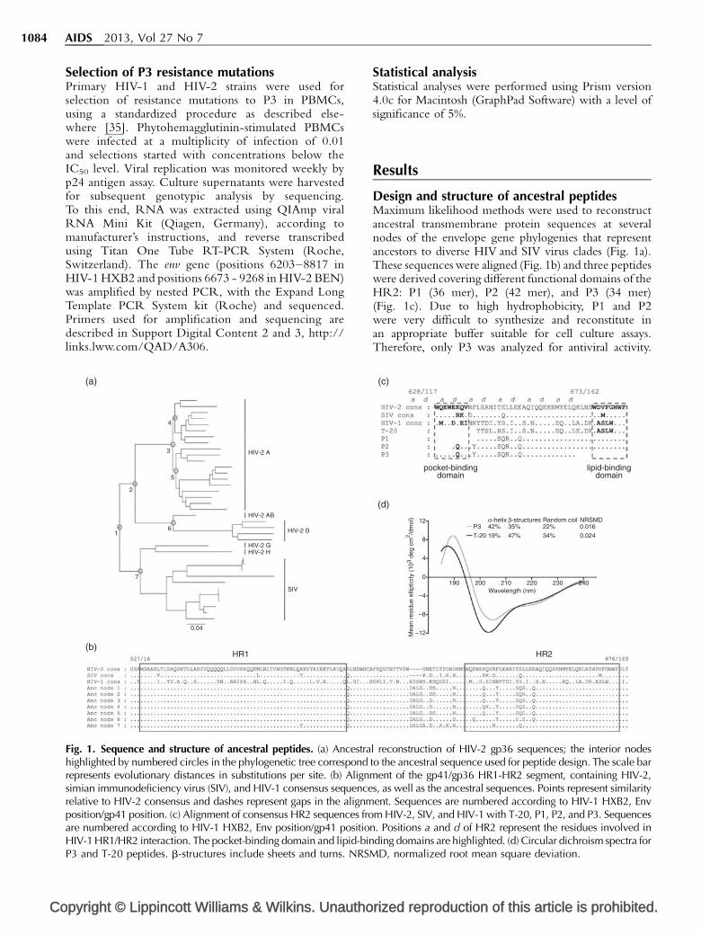
Fig. 1. Sequence and structure of ancestral peptides. (a) Ancestrahighlighted by numbered circles in the phylogenetic tree correspondrepresents evolutionary distances in substitutions per site. (b) Alignmsimian immunodeficiency virus (SIV), and HIV-1 consensus sequencerelative to HIV-2 consensus and dashes represent gaps in the alignmposition/gp41 position. (c) Alignment of consensus HR2 sequences froare numbered according to HIV-1 HXB2, Env position/gp41 positioHIV-1 HR1/HR2 interaction. The pocket-binding domain and lipid-binP3 and T-20 peptides. b-structures include sheets and turns. NRSM
Statistical analysisStatistical analyses were performed using Prism version4.0c for Macintosh (GraphPad Software) with a level ofsignificance of 5%.
Results
Design and structure of ancestral peptidesMaximum likelihood methods were used to reconstructancestral transmembrane protein sequences at severalnodes of the envelope gene phylogenies that representancestors to diverse HIV and SIV virus clades (Fig. 1a).These sequences were aligned (Fig. 1b) and three peptideswere derived covering different functional domains of theHR2: P1 (36 mer), P2 (42 mer), and P3 (34 mer)(Fig. 1c). Due to high hydrophobicity, P1 and P2were very difficult to synthesize and reconstitute inan appropriate buffer suitable for cell culture assays.Therefore, only P3 was analyzed for antiviral activity.
rized reproduction of this article is prohibited.
pocket-bindingdomain
(c)
(d)
HR2
lipid-bindingdomain
Mea
n re
sidu
e el
liptic
ity (
103
deg
cm2 /d
mol
)
–12
–8
–4
0190 200 210
α-helix β-structures Random coilP3T-20
42%19%
35%47%
22%34%
0.0160.024
NRSMD
Wavelength (nm)220 230 240
4
8
12
l reconstruction of HIV-2 gp36 sequences; the interior nodesto the ancestral sequence used for peptide design. The scale bar
ent of the gp41/gp36 HR1-HR2 segment, containing HIV-2,s, as well as the ancestral sequences. Points represent similarityent. Sequences are numbered according to HIV-1 HXB2, Envm HIV-2, SIV, and HIV-1 with T-20, P1, P2, and P3. Sequences
n. Positions a and d of HR2 represent the residues involved inding domains are highlighted. (d) Circular dichroism spectra forD, normalized root mean square deviation.
Ancestral HIV fusion inhibitor peptide Borrego et al. 1085
P3 overlaps the N-terminal pocket-binding region andheptad repeat core of the HR2 region (positions 628–661of HIV-1 HXB2 Env). It differs by 21 amino acids fromthe consensus HIV-1 sequence and by six amino acidsfrom consensus HIV-2. However, the positions a and d ofthe heptad repeat, considered critical for HIV-1 HR1/HR2 binding [36], were quite conserved. There wereonly four changes: I635V and Y638L, involving aminoacids with hydrophobic side chains, and L645R andS649A, involving amino acids from different chemicalgroups.
T-20 and P3 overlap in 24 amino acids of the heptadrepeat core and diverge in 14 residues (58%). P3 is morehydrophilic than T-20 (percentage of hydrophilic residuesis of 62% in P3 and 56% in T-20) [37] and its predominantsecondary structure in solution is an a-helix (42%). T-20has a lower content of regular secondary structureelements in solution with only 19% of helicity, aspreviously reported (Fig. 1d) [38].
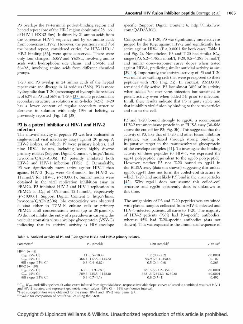
P3 is a potent inhibitor of HIV-1 and HIV-2infectionThe antiviral activity of peptide P3 was first evaluated insingle-round viral infectivity assays against 20 group AHIV-2 isolates, of which 19 were primary isolates, andnine HIV-1 isolates, including seven highly diverseprimary isolates (Support Digital Content 4, http://links.lww.com/QAD/A306). P3 potently inhibited bothHIV-2 and HIV-1 infection (Table 1). Remarkably,P3 was significantly more active against HIV-1 thanagainst HIV-2 (IC50 were 63.8 nmol/l for HIV-2 vs.11 nmol/l for HIV-1, P< 0.0001). Similar results wereobtained in the viral replication inhibition assay inPBMCs. P3 inhibited HIV-2 and HIV-1 replication inPBMCs at IC50 of 109.3 and 12.1 nmol/l, respectively(P< 0.0001; Support Digital Content 5, http://links.lww.com/QAD/A306). No cytotoxicity was observedin vitro either in TZM-bl culture cells or primaryPBMCs at all concentrations tested (up to 20 mmol/l).P3 did not inhibit the entry of a pseudovirus carrying thevesicular stomatitis virus envelope glycoprotein (VSV-G)indicating that its antiviral activity is HIV-envelope
Copyright © Lippincott Williams & Wilkins. Unaut
Table 1. Antiviral activity of P3 and T-20 against HIV-1 and HIV-2 prim
Parametera P3 (nmol/l)
HIV-1 (n¼9)IC50 (95% CI) 11 (6.5–18.4)IC90 (95% CI) 366.4 (117.5–1145.5)Hill slope (95% CI) 0.6 (0.4–0.82)
HIV-2 (n¼20)IC50 (95% CI) 63.8 (51.9–78.5)IC90 (95% CI) 709.6 (435.5–1158.8)Hill slope (95% CI) 0.9 (0.7–1.1)
aIC50, IC90, and Hill slope best-fit values were inferred from sigmoidal dose–and HIV-2 isolates, and represent geometric mean values; 95% CI – 95%bT-20 susceptibilities were obtained for the same HIV-1 and HIV-2 viral pcP value for comparison of best-fit values using the F-test.
specific (Support Digital Content 6, http://links.lww.com/QAD/A306).
Compared with T-20, P3 was significantly more active asjudged by the IC50 against HIV-2 and significantly lessactive against HIV-1 (P< 0.0001 for both cases; Table 1and Fig. 2). Nonetheless, P3 and T-20 had similar IC90
ranges (P3, 6.2–1785.5 nmol/l; T-20, 0.5–1285.3 nmol/l)and similar dose–response curve slopes when testedagainst HIV-1, predicting similar antiviral activity in vivo[39,40]. Importantly, the antiviral activity of P3 and T-20was null after washing cells that were preexposed to thesepeptides with PBS (Fig. 3a). In contrast, AMD3100remained fully active. P3 lost almost 30% of its activitywhen added 3 h after virus infection but sustained itspotent activity even when added 3 h prior to infection.In all, these results indicate that P3 is quite stable andthat it inhibits viral fusion by binding to the virus particlesand not to the cell.
P3 and T-20 bound strongly to rgp36, a recombinantHIV-2 transmembrane protein in an ELISA assay (30-foldabove the cut-off for P3; Fig. 3b). This suggested that theactivity of P3, like that of T-20 and other fusion inhibitorpeptides, was mediated through strong binding toits putative target in the transmembrane glycoproteinof the envelope complex [41]. To investigate the bindingactivity of these peptides to HIV-1, we expressed thergp41 polypeptide equivalent to the rgp36 polypeptide.However, neither P3 nor T-20 bound to rgp41 inthe ELISA assay (data not shown) suggesting that unlikergp36, rgp41 does not form the coiled-coil structure towhich T-20 (and most likely P3) bind in the virus particles[42]. Why rgp41 does not assume this coiled-coilstructure and rgp36 apparently does is unknown atthis time.
The antigenicity of P3 and T-20 peptides was examinedwith plasma samples collected from HIV-2-infected andHIV-1-infected patients, all naive to T-20. The majorityof HIV-2 patients (93%) had P3-specific antibodies,whereas 45% had T-20-specific antibodies (data notshown). This was expected as the amino acid sequence of
horized reproduction of this article is prohibited.
ary isolates.
T-20 (nmol/l)b P valuec
1.2 (0.7–2.2) <0.000195.9 (26.3–350.8) 0.1070.5 (0.4–0.6) 0.263
281.5 (223.2–354.9) <0.00013881.5 (2393.3–6280.6) <0.0001
0.8 (0.7–1) 0.492
response (variable slope) curves adjusted to combined results of HIV-1confidence interval.anel [27].
Co
1086 AIDS 2013, Vol 27 No 7
110
(a)
(b) (d)
(c)
100
90
80
70
60
50
40
30
20
10
0
–10 0
0
2500
5000
7500
10000
12500
1500032000
32250
32500
500
1000
1500
2000
P = 0.107
P < 0.0001
P3
P3
T-20
T-20
1x10⁻⁴ 1x10⁻³ 1x10⁻² 1x10⁻¹ 1x100 1x101
Peptide concentration (nmol/l)
P3
T-20
% in
hibi
tion
of H
IV-1
isol
ates
IC90
(nm
ol/l)
for
HIV
-1
1x10² 1x10³ 1x10⁴ 1x10⁵
110
100
90
80
70
60
50
40
30
20
10
0
–101x10–4 1x10⁻³ 1x10⁻² 1x10⁻¹ 1x100 1x101
Peptide concentration (nmol/l)
P3
T-20
% in
hibi
tion
of H
IV-1
isol
ates
IC90
(nm
ol/l)
for
HIV
-2
1x10² 1x10³ 1x10⁴ 1x10⁵
Fig. 2. Antiviral activity of P3. Representative dose–response curves for HIV-1 (a) and HIV-2 (b) with P3 and, for comparison, T-20[27]. Data points represent the average of results obtained for HIV-1 (N¼9) and HIV-2 (N¼20) primary isolates; bars representstandard error of the mean. Sigmoidal dose–response (variable slope) curves were adjusted to these data points; dashed linesrepresent the 95% confidence band of the best-fit curve. Mean IC90 values of P3 and T-20, for HIV-1 (c) and HIV-2 (d) primaryisolates. Bars represent median values. P values were obtained comparing best-fit values inferred from sigmoidal dose–response(variable slope) curves using the F-test.
P3 was derived from an ancestral HIV-2/SIV sequence.Likewise, the majority of HIV-1 patients (90%) hadT-20-specific antibodies, whereas 67% had P3-specificantibodies. Of note, the mean binding affinity of theT-20-specific antibodies from HIV-1 patients wassignificantly higher compared to P3-specific antibodies(Fig. 3c). These results demonstrate that P3 is weaklyantigenic in HIV-1 patients compared to T-20.
P3 inhibits the replication of most T-20-resistantHIV-1 variantsTo determine whether P3 is able to inhibit the infectionof HIV-1 strains resistant to T-20, we measuredthe susceptibility of HIV-1 variants carrying well definedT-20 resistance mutations to P3 [43,44]. Notably,P3 exhibited potent activity against all but one T-20-resistant strains (IC50 range, 0.15–11.8 nmol/l; Table 2).The mutation N43K conferred high-level resistance to
pyright © Lippincott Williams & Wilkins. Unautho
both P3 (2140-fold) and T-20 (2677-fold). Moreover, theV38A/N42D mutations conferred increased suscepti-bility to P3 (seven-fold lower IC50 compared to the wild-type). These results suggest that P3 could be used asan alternative for the treatment of patients infected withT-20-resistant HIV-1 strains.
Selection of P3-resistant variantsTo investigate the mechanism of action and the pathwaysof resistance to P3, resistance mutations were selectedin vitro by repeated passage of HIV-1 and HIV-2 primaryisolates in PBMCs in the presence of either constantor increasing concentrations of P3, according to theviral replication capability [35]. For HIV-1, the N43Ksubstitution in the HR1 region of gp41 was the onlymutation selected in env after 59 days in culture (eightpassages) in the presence of 212 nmol/l of P3. Inhibitionof replication of this mutant virus with P3 occurred at an
rized reproduction of this article is prohibited.

Copyright © Lippincott Williams & Wilkins. Unauthorized reproduction of this article is prohibited.
Ancestral HIV fusion inhibitor peptide Borrego et al. 1087
% in
hibi
tion
% in
hibi
tion
% in
hibi
tion
Time of P3 addition (h)
Time of T-20 addition (h)
Time of AMD3100 addition (h)
–50
–50
70–3 –2
Cells + P3
Cells + T-20
Cells +AMD3100
HIV-1 + AMD3100
HIV-2 + AMD3100
Cells + HIV-1 + AMD3100
Cells + HIV-2 + AMD3100
HIV-1 + P3
HIV-2 + P3
HIV-1 + T-20
Cells + HIV-1 + P3
0
5
10
P = 0.100
P < 0.0001
P3
OD
/Cut
t-of
f rat
io
T-20
P3 T-20
15
0
5
10
OD
/Cut
t-of
f rat
io
15
20
25
30
35
Cells + HIV-2 + P3
Cells + HIV-1 + T-20
–1 1 2 3
70–3 –2 –1 1 2 3
–3 –2 –1 1 2 3
80
90
100
(a) (b)
(c)
80
90
100
–50
70
80
90
100
Fig. 3. Mechanism of action and antibody reactivity of P3. (a) Time-of-addition assays for P3 with HIV-1 and HIV-2. T-20 andAMD3100 were used as control drugs. In these experiments, each drug was added at different time-points either before (cells andviruses, separately) or after HIV infection. (b) Binding activity of P3 and T-20 to HIV-2 rgp36 in an ELISA assay. (c) Antibodyreactivity of P3 and T-20 in HIV-1-infected patients. Bars represent mean values. Results from P3 are in open squares and from T-20are in closed circles. P values were obtained with the Mann–Whitney U-test.
Table 2. Comparison of antiviral activity of P3 and T-20 on T-20-resistant HIV-1 variants.
P3 T-20
HIV-1 variantSusceptibility
to T-20aIC50 nmol/l
(95% CI) Fold-increasebIC50 nmol/l
(95% CI)Fold-
increaseb P valuec
NL4-3 D36G (parental) S 0.4 (0.2–1.2) 1 0.03 (0.01–0.06) 1 0.0002NL4-3 (D36G) V38A R 1.5 (0.5–5.1) 3.8 43.8 (21.8–87.8) 1460 <0.0001NL4-3 (D36G) V38A/N42D R 0.06 (0.01–0.3) 0.15 118.2 (63.0–221.7) 3940 <0.0001NL4-3 (D36G) V38A/N42T R 4.7 (1.9–11.5) 11.8 482.0 (324.1–716.8) 16 066.7 <0.0001NL4-3 (D36G) N42T/N43K R 855.9 (628.0–1167.0) 2139.8 80.3 (61.1–105.6) 2676.7 <0.0001
CI, confidence interval.aSensitive (S) or resistant (R).bFold-increase of IC50 concentration relative to NL4-3 D36G (parental).cP value for comparison of best-fit values between P3 and T-20, using the F-test.
Co
1088 AIDS 2013, Vol 27 No 7
IC50 of 1.9 mmol/l and IC90 of 13.1 mmol/l, whichrepresent a 120-fold and 56.4-fold decrease insusceptibility, respectively. Under the same experimentalconditions and despite repeated attempts, we were notable to select P3-resistant HIV-2 isolates. Collectively,these results indicate that the HR1 region in thetransmembrane glycoprotein is the target of P3 andsuggest that the pathway of HIV-1 resistance to P3 differsfrom that of T-20 and the genetic barrier to P3 resistanceis significantly higher in HIV-2 than in HIV-1.
Discussion
We show here that an ancestral peptide (named P3)derived from the HR2 domains of HIV-2 and SIVtransmembrane glycoproteins potently inhibits bothHIV-1 and HIV-2 cell entry and replication. Therationale for using ancestral sequences of the trans-membrane envelope glycoprotein as a source for thenew antiviral peptide was to minimize HIV sequencedivergence by tracing the most likely evolutionary pathalong the phylogeny and capture more conservedstructural features of the HR2 sequences [45,46]. Thepotent activity of P3 against divergent HIV-1 and HIV-2primary isolates demonstrates that our strategy was highlysuccessful. The potent activity of P3 on HIV-1 was notexpected but it might be due to the conservation inP3 of the residues located in critical positions involved inthe HR1/HR2 interaction (a and d residues) [36] and/orto the presence of an alanine at position 22 (correspond-ing to position 138 in gp41). An alanine at this positionincreases the binding affinity of HR2 region to HR1 andenhances the activity of HR2-based HIV-1 fusioninhibitor peptides [47].
The N43K mutation in the HR1 region of HIV-1 wassufficient to confer high-level resistance to this peptide.These results suggest that, like T-20, P3 acts by selectivelybinding to the HR1 region in the transmembraneglycoprotein of HIV-1. However, P3 potently inhibitedthe replication of T-20-resistant viruses bearing the V38Aand/or the N42D resistance mutations indicating that thepathway of HIV-1 resistance to P3 differs from that of T-20. We were not able to generate HIV-2 isolates resistantto P3 even after 60 days in culture suggesting that thegenetic barrier for resistance to P3 might be significantlyhigher in HIV-2 than in HIV-1.
Peptides with high helical content like P3 tend to be morestable and less susceptible to proteolytic degradation inbiological fluids than unstructured peptides like T-20[7,13]. In addition, they usually display high anti-HIV-1potency due to an increased binding affinity for HR1[48,49]. Indeed, we showed that the potent anti-HIV-1activity of P3 was conserved for at least 3 h. Moreover, P3bound strongly to a recombinant HIV transmembrane
pyright © Lippincott Williams & Wilkins. Unautho
protein. Hence, the high helical content may alsocontribute to the potent antiviral activity of P3.
Antibodies present in the plasma of HIV-1-infectedpatients reacted poorly with P3 as compared to T-20.This was expected as HIV-1 and HIV-2 differ significantlyin the HR2 region and antibodies naturally generatedagainst this region in HIV-1 patients were unlikely to bindto a peptide derived from HIV-2/SIV. As drug-specificantibodies can compromise the clinical efficacy oftherapeutic proteins either by preventing their exposureto the active site or by decreasing their half-life [50], theweaker antigenicity of P3 in HIV-1-infected patientsmight translate into a better bioavailability profile anddurable clinical efficacy of P3 in HIV-1-infected patients.
ConclusionIn summary, we successfully derived an ancestral peptide(P3) with low antigenicity, high stability, and potentactivity against both HIV-1, including variants resistant toT-20, and HIV-2. Our findings provide proof of principlethat potent antiviral peptides can be constructed usingevolutionary biology strategies. Such strategies should beexplored to enhance the production of antiviral peptidedrugs, microbicides, and vaccines.
Acknowledgements
The following reagents were provided by the AIDSResearch and Reference Reagent Program, NationalInstitutes of Health: TZM-bl [51–54] and CEM-SS cells[55–57]; pNL4-3 [43], pSG3.1 [58], pHEF-VSVG [59]and pSG3Denv [52,60] plasmids; T-20-resistant pNL4-3gp41 (36G) variants [43,44]; T-20 (enfuvirtide) fusioninhibitor; bicyclam JM-2987 (hydrobromide salt ofAMD-3100) coreceptor antagonist [61–63]. pROD10plasmid was a gift from Keith Peden [64].
We thank Luıs Oliveira for help in the P3 circulardichroism spectra deconvolution.
P.B., A.Q., and N.T. conceived and designed theexperiments. P.B., R.C., J.M.M., H.B., P.P., and A.Q.performed the experiments. P.B., A.Q., and N.T.analyzed the data. P.B. and N.T. wrote the article.
This work was supported by grants PTDC/SAU-FCF/67673/2006 and PTDC/SAU-FAR/115290/2009 fromFundacao para a Ciencia e Tecnologia (FCT) (http://www.fct.pt), Portugal, and by Collaborative HIV andAnti-HIV Drug Resistance Network (CHAIN) from theEuropean Union. P.B. and R.C. were supported byPhD grants from Fundacao para a Ciencia e Tecnologia,Portugal. The funders had no role in study design, datacollection and analysis, decision to publish, or preparationof the article.
rized reproduction of this article is prohibited.
Ancestral HIV fusion inhibitor peptide Borrego et al. 1089
Conflicts of interestThe authors have no commercial or other association thatmight pose a conflict of interest.
References
1. Dando TM, Perry CM. Enfuvirtide. Drugs 2003; 63:2755–2766;discussion 2767–2758.
2. Carter NJ, Keating GM. Maraviroc. Drugs 2007; 67:2277–2288;discussion 2289–2290.
3. Wild C, Greenwell T, Matthews T. A synthetic peptide fromHIV-1 gp41 is a potent inhibitor of virus-mediated cell-cellfusion. AIDS Res Hum Retroviruses 1993; 9:1051–1053.
4. Wild CT, Shugars DC, Greenwell TK, McDanal CB, MatthewsTJ. Peptides corresponding to a predictive alpha-helical domainof human immunodeficiency virus type 1 gp41 are potentinhibitors of virus infection. Proc Natl Acad Sci U S A 1994;91:9770–9774.
5. Moore JP, Doms RW. The entry of entry inhibitors: a fusionof science and medicine. Proc Natl Acad Sci U S A 2003;100:10598–10602.
6. Liu S, Jing W, Cheung B, Lu H, Sun J, Yan X, et al. HIV gp41 C-terminal heptad repeat contains multifunctional domains.Relation to mechanisms of action of anti-HIV peptides. J BiolChem 2007; 282:9612–9620.
7. Eggink D, Berkhout B, Sanders RW. Inhibition of HIV-1 byfusion inhibitors. Curr Pharm Des 2010; 16:3716–3728.
8. Mink M, Mosier SM, Janumpalli S, Davison D, Jin L, Melby T,et al. Impact of human immunodeficiency virus type 1 gp41amino acid substitutions selected during enfuvirtide treatmenton gp41 binding and antiviral potency of enfuvirtide in vitro.J Virol 2005; 79:12447–12454.
9. Johnson VA, Brun-Vezinet F, Clotet B, Gunthard HF, KuritzkesDR, Pillay D, et al. Update of the drug resistance mutations inHIV-1: December 2009. Top HIV Med 2009; 17:138–145.
10. Lalezari JP, Henry K, O’Hearn M, Montaner JS, Piliero PJ,Trottier B, et al. Enfuvirtide, an HIV-1 fusion inhibitor,for drug-resistant HIV infection in North and South America.N Engl J Med 2003; 348:2175–2185.
11. Lazzarin A, Clotet B, Cooper D, Reynes J, Arasteh K, Nelson M,et al. Efficacy of enfuvirtide in patients infected with drug-resistant HIV-1 in Europe and Australia. N Engl J Med 2003;348:2186–2195.
12. De Clercq E. The history of antiretrovirals: key discoveries overthe past 25 years. Rev Med Virol 2009; 19:287–299.
13. Cai L, Jiang S. Development of peptide and small-moleculeHIV-1 fusion inhibitors that target gp41. ChemMedChem2010; 5:1813–1824.
14. Eron JJ, Gulick RM, Bartlett JA, Merigan T, Arduino R, Kilby JM,et al. Short-term safety and antiretroviral activity of T-1249, asecond-generation fusion inhibitor of HIV. J Infect Dis 2004;189:1075–1083.
15. Greenberg ML, Davison DB, Jin L, Mosier S, Melby T, Sista P,et al. In vitro antiviral activity of T-1249, a second generationfusion inhibitor. Antiviral Ther 2002; 7:S14.
16. Martin-Carbonero L. Discontinuation of the clinical develop-ment of fusion inhibitor T-1249. AIDS Rev 2004; 6:61.
17. He Y, Xiao Y, Song H, Liang Q, Ju D, Chen X, et al. Design andevaluation of sifuvirtide, a novel HIV-1 fusion inhibitor. J BiolChem 2008; 283:11126–11134.
18. Rowland-Jones SL, Whittle HC. Out of Africa: what canwe learn from HIV-2 about protective immunity to HIV-1?Nat Immunol 2007; 8:329–331.
19. Damond F, Worobey M, Campa P, Farfara I, Colin G, MatheronS, et al. Identification of a highly divergent HIV type 2 andproposal for a change in HIV type 2 classification. AIDS ResHum Retroviruses 2004; 20:666–672.
20. Gao F, Yue L, Robertson DL, Hill SC, Hui H, Biggar RJ, et al.Genetic diversity of human immunodeficiency virus type 2:evidence for distinct sequence subtypes with differences invirus biology. J Virol 1994; 68:7433–7447.
21. de Silva TI, Cotten M, Rowland-Jones SL. HIV-2: the forgottenAIDS virus. Trends Microbiol 2008; 16:588–595.
Copyright © Lippincott Williams & Wilkins. Unaut
22. Lemey P, Pybus OG, Wang B, Saksena NK, Salemi M,Vandamme AM. Tracing the origin and history of the HIV-2epidemic. Proc Natl Acad Sci U S A 2003; 100:6588–6592.
23. Hu DJ, Dondero TJ, Rayfield MA, George JR, Schochetman G,Jaffe HW, et al. The emerging genetic diversity of HIV. Theimportance of global surveillance for diagnostics, research,and prevention. JAMA 1996; 275:210–216.
24. Witvrouw M, Pannecouque C, Switzer WM, Folks TM, DeClercq E, Heneine W. Susceptibility of HIV-2, SIV and SHIVto various anti-HIV-1 compounds: implications for treatmentand postexposure prophylaxis. Antivir Ther 2004; 9:57–65.
25. Hizi A, Tal R, Shaharabany M, Currens MJ, Boyd MR, HughesSH, McMahon JB. Specific inhibition of the reverse transcrip-tase of human immunodeficiency virus type 1 and the chimericenzymes of human immunodeficiency virus type 1 and type 2by nonnucleoside inhibitors. Antimicrob Agents Chemother1993; 37:1037–1042.
26. Poveda E, Rodes B, Toro C, Soriano V. Are fusion inhibitorsactive against all HIV variants? AIDS Res Hum Retroviruses2004; 20:347–348.
27. Borrego P, Calado R, Marcelino JM, Bartolo I, Rocha C, Cavaco-Silva P, et al. Baseline susceptibility of primary human immuno-deficiency virus type 2 to entry inhibitors. Antivir Ther 2012;17:565–570.
28. Swofford DL. PAUP�. Phylogenetic analysis using parsimony(�and other Methods). Version 4. Sunderland, Massachusetts:Sinauer Associates; 1998.
29. Posada D, Crandall KA. MODELTEST: testing the model ofDNA substitution. Bioinformatics 1998; 14:817–818.
30. Rodriguez F, Oliver JL, Marin A, Medina JR. The generalstochastic model of nucleotide substitution. J Theor Biol1990; 142:485–501.
31. Johnson WC. Analyzing protein circular dichroism spectra foraccurate secondary structures. Proteins 1999; 35:307–312.
32. Whitmore L, Wallace BA. DICHROWEB, an online server forprotein secondary structure analyses from circular dichroismspectroscopic data. Nucleic Acids Res 2004; 32:W668–W673.
33. Lobley A, Whitmore L, Wallace BA. DICHROWEB: an inter-active website for the analysis of protein secondary structurefrom circular dichroism spectra. Bioinformatics 2002; 18:211–212.
34. Marcelino JM, Barroso H, Goncalves F, Silva SM, Novo C,Gomes P, et al. Use of a new dual-antigen enzyme-linkedimmunosorbent assay to detect and characterize the humanantibody response to the human immunodeficiency virustype 2 envelope gp125 and gp36 glycoproteins. J Clin Microbiol2006; 44:607–611.
35. Oliveira M, Brenner BG, Wainberg MA. Isolation of drug-resistant mutant HIV variants using tissue culture drugselection. Methods Mol Biol 2009; 485:427–433.
36. Chan DC, Fass D, Berger JM, Kim PS. Core structure of gp41from the HIV envelope glycoprotein. Cell 1997; 89:263–273.
37. Hopp TP, Woods KR. Prediction of protein antigenic deter-minants from amino acid sequences. Proc Natl Acad Sci U S A1981; 78:3824–3828.
38. Lawless MK, Barney S, Guthrie KI, Bucy TB, Petteway SR Jr,Merutka G. HIV-1 membrane fusion mechanism: structuralstudies of the interactions between biologically-active peptidesfrom gp41. Biochemistry 1996; 35:13697–13708.
39. Sampah ME, Shen L, Jilek BL, Siliciano RF. Dose-response curveslope is a missing dimension in the analysis of HIV-1 drugresistance. Proc Natl Acad Sci U S A 2011; 108:7613–7618.
40. Shen L, Peterson S, Sedaghat AR, McMahon MA, Callender M,Zhang H, et al. Dose-response curve slope sets class-specificlimits on inhibitory potential of anti-HIV drugs. Nat Med 2008;14:762–766.
41. Eggink D, Langedijk JP, Bonvin AM, Deng Y, Lu M, Berkhout B,Sanders RW. Detailed mechanistic insights into HIV-1sensitivity to three generations of fusion inhibitors. J Biol Chem2009; 284:26941–26950.
42. Champagne K, Shishido A, Root MJ. Interactions of HIV-1inhibitory peptide T20 with the gp41 N-HR coiled coil. J BiolChem 2009; 284:3619–3627.
43. Adachi A, Gendelman HE, Koenig S, Folks T, Willey R, RabsonA, Martin MA. Production of acquired immunodeficiencysyndrome-associated retrovirus in human and nonhuman cellstransfected with an infectious molecular clone. J Virol 1986;59:284–291.
horized reproduction of this article is prohibited.

Co
1090 AIDS 2013, Vol 27 No 7
44. Rimsky LT, Shugars DC, Matthews TJ. Determinants of humanimmunodeficiency virus type 1 resistance to gp41-derivedinhibitory peptides. J Virol 1998; 72:986–993.
45. Cai W, Pei J, Grishin NV. Reconstruction of ancestral proteinsequences and its applications. BMC Evol Biol 2004; 4:33.
46. Mullins JI, Nickle DC, Heath L, Rodrigo AG, Learn GH.Immunogen sequence: the fourth tier of AIDS vaccine design.Expert Rev Vaccines 2004; 3:S151–S159.
47. Izumi K, Kodama E, Shimura K, Sakagami Y, Watanabe K, Ito S,et al. Design of peptide-based inhibitors for human immuno-deficiency virus type 1 strains resistant to T-20. J Biol Chem2009; 284:4914–4920.
48. Naito T, Izumi K, Kodama E, Sakagami Y, Kajiwara K,Nishikawa H, et al. SC29EK, a peptide fusion inhibitor withenhanced alpha-helicity, inhibits replication of human immu-nodeficiency virus type 1 mutants resistant to enfuvirtide.Antimicrob Agents Chemother 2009; 53:1013–1018.
49. Otaka A, Nakamura M, Nameki D, Kodama E, Uchiyama S,Nakamura S, et al. Remodeling of gp41-C34 peptide leads tohighly effective inhibitors of the fusion of HIV-1 with targetcells. Angew Chem Int Ed Engl 2002; 41:2937–2940.
50. Schellekens H. Immunogenicity of therapeutic proteins:clinical implications and future prospects. Clin Ther 2002;24:1720–1740; discussion 1719.
51. Takeuchi Y, McClure MO, Pizzato M. Identification ofgammaretroviruses constitutively released from cell linesused for human immunodeficiency virus research. J Virol2008; 82:12585–12588.
52. Wei X, Decker JM, Liu H, Zhang Z, Arani RB, Kilby JM, et al.Emergence of resistant human immunodeficiency virus type 1in patients receiving fusion inhibitor (T-20) monotherapy.Antimicrob Agents Chemother 2002; 46:1896–1905.
53. Derdeyn CA, Decker JM, Sfakianos JN, Wu X, O’Brien WA,Ratner L, et al. Sensitivity of human immunodeficiency virustype 1 to the fusion inhibitor T-20 is modulated by coreceptorspecificity defined by the V3 loop of gp120. J Virol 2000;74:8358–8367.
54. Platt EJ, Wehrly K, Kuhmann SE, Chesebro B, Kabat D. Effects ofCCR5 and CD4 cell surface concentrations on infections bymacrophagetropic isolates of human immunodeficiency virustype 1. J Virol 1998; 72:2855–2864.
pyright © Lippincott Williams & Wilkins. Unautho
55. Foley GE, Lazarus H, Farber S, Uzman BG, Boone BA,McCarthy RE. Continuous culture of human lymphoblasts fromperipheral blood of a child with acute leukemia. Cancer 1965;18:522–529.
56. Nara PL, Hatch WC, Dunlop NM, Robey WG, Arthur LO,Gonda MA, Fischinger PJ. Simple, rapid, quantitative,syncytium-forming microassay for the detection of humanimmunodeficiency virus neutralizing antibody. AIDS ResHum Retroviruses 1987; 3:283–302.
57. Nara PL, Fischinger PJ. Quantitative infectivity assay for HIV-1and-2. Nature 1988; 332:469–470.
58. Ghosh SK, Fultz PN, Keddie E, Saag MS, Sharp PM, Hahn BH,Shaw GM. A molecular clone of HIV-1 tropic and cytopathicfor human and chimpanzee lymphocytes. Virology 1993;194:858–864.
59. Chang LJ, Urlacher V, Iwakuma T, Cui Y, Zucali J. Efficacy andsafety analyses of a recombinant human immunodeficiencyvirus type 1 derived vector system. Gene Ther 1999; 6:715–728.
60. Wei X, Decker JM, Wang S, Hui H, Kappes JC, Wu X, et al.Antibody neutralization and escape by HIV-1. Nature 2003;422:307–312.
61. De Clercq E, Yamamoto N, Pauwels R, Balzarini J, Witvrouw M,De Vreese K, et al. Highly potent and selective inhibition ofhuman immunodeficiency virus by the bicyclam derivativeJM3100. Antimicrob Agents Chemother 1994; 38:668–674.
62. Bridger GJ, Skerlj RT, Thornton D, Padmanabhan S, MartellucciSA, Henson GW, et al. Synthesis and structure-activityrelationships of phenylenebis(methylene)-linked bis-tetraaza-macrocycles that inhibit HIV replication. Effects of macrocyc-lic ring size and substituents on the aromatic linker. J MedChem 1995; 38:366–378.
63. Hendrix CW, Flexner C, MacFarland RT, Giandomenico C,Fuchs EJ, Redpath E, et al. Pharmacokinetics and safety ofAMD-3100: a novel antagonist of the CXCR-4 chemokinereceptor, in human volunteers. Antimicrob Agents Chemother2000; 44:1667–1673.
64. Ryan-Graham MA, Peden KW. Both virus and host componentsare important for the manifestation of a Nef- phenotype inHIV-1 and HIV-2. Virology 1995; 213:158–168.
rized reproduction of this article is prohibited.