Taxonomy, Phylogeny, Molecular Dating and Ancestral State ...
87
Page 1/87 Taxonomy, Phylogeny, Molecular Dating and Ancestral State Reconstruction of Xylariomycetidae ( Sordariomycetes ) Milan C. Samarakoon School of Life Science and Technology, Center for Informational Biology, University of Electronic Science and Technology of China, Chengdu 611731, P.R. China Kevin D Hyde ( [email protected] ) Center of Excellence in Fungal Research Sajeewa S. N. Maharachchikumbura School of Life Science and Technology, Center for Informational Biology, University of Electronic Science and Technology of China, Chengdu 611731, P.R. China Marc Stadler Institute of Microbiology, Technische Universität Braunschweig, Spielmannstraβe 7, 38106 Braunschweig, Germany E. B. Gareth Jones Department of Botany and Microbiology, College of Science, King Saud University, P.O Box 2455, Riyadh 11451, Kingdom of Saudi Arabia Itthayakorn Promputtha Department of Biology, Faculty of Science, Chiang Mai University, Chiang Mai 50200, Thailand Nakarin Suwannarach Department of Biology, Faculty of Science, Chiang Mai University, Chiang Mai 50200, Thailand Erio Camporesi A.M.B. Gruppo, Micologico Forlivese "Antonio Cicognani", Via Roma 18, Forlí, Italy Timur S. Bulgakov Department of Plant Protection, Federal Research Centre the Subtropical Scientic Centre of the Russian Academy of Sciences, Yana Fabritsiusa Street 2/28, Sochi 354002, Krasnodar Region, Russia Jian-Kui Liu School of Life Science and Technology, Center for Informational Biology, University of Electronic Science and Technology of China, Chengdu 611731, P.R. China Research Article Keywords: 33 new taxa, Amphisphaeriales, Appendicosporaceae, Evolution, Stromata, Xylariales Posted Date: October 1st, 2021 DOI: https://doi.org/10.21203/rs.3.rs-935829/v1 License: This work is licensed under a Creative Commons Attribution 4.0 International License. Read Full License
-
Upload
khangminh22 -
Category
Documents
-
view
1 -
download
0
Transcript of Taxonomy, Phylogeny, Molecular Dating and Ancestral State ...
Page 1/87
Taxonomy, Phylogeny, Molecular Dating and Ancestral State Reconstruction ofXylariomycetidae (Sordariomycetes)Milan C. Samarakoon
School of Life Science and Technology, Center for Informational Biology, University of Electronic Science and Technology of China, Chengdu 611731, P.R.ChinaKevin D Hyde ( [email protected] )
Center of Excellence in Fungal ResearchSajeewa S. N. Maharachchikumbura
School of Life Science and Technology, Center for Informational Biology, University of Electronic Science and Technology of China, Chengdu 611731, P.R.ChinaMarc Stadler
Institute of Microbiology, Technische Universität Braunschweig, Spielmannstraβe 7, 38106 Braunschweig, GermanyE. B. Gareth Jones
Department of Botany and Microbiology, College of Science, King Saud University, P.O Box 2455, Riyadh 11451, Kingdom of Saudi ArabiaItthayakorn Promputtha
Department of Biology, Faculty of Science, Chiang Mai University, Chiang Mai 50200, ThailandNakarin Suwannarach
Department of Biology, Faculty of Science, Chiang Mai University, Chiang Mai 50200, ThailandErio Camporesi
A.M.B. Gruppo, Micologico Forlivese "Antonio Cicognani", Via Roma 18, Forlí, ItalyTimur S. Bulgakov
Department of Plant Protection, Federal Research Centre the Subtropical Scienti�c Centre of the Russian Academy of Sciences, Yana Fabritsiusa Street 2/28,Sochi 354002, Krasnodar Region, RussiaJian-Kui Liu
School of Life Science and Technology, Center for Informational Biology, University of Electronic Science and Technology of China, Chengdu 611731, P.R.China
Research Article
Keywords: 33 new taxa, Amphisphaeriales, Appendicosporaceae, Evolution, Stromata, Xylariales
Posted Date: October 1st, 2021
DOI: https://doi.org/10.21203/rs.3.rs-935829/v1
License: This work is licensed under a Creative Commons Attribution 4.0 International License. Read Full License
Page 2/87
AbstractXylariomycetidae ( Ascomycota ) is a highly diversi�ed group with variable stromatic characters. Our research focused on inconspicuous stromatic xylarialeantaxa from China, Italy, Russia, Thailand and the United Kingdom. Detailed morphological descriptions, illustrations and combined ITS-LSU- rpb 2- tub 2- tef 1phylogenies revealed 38 taxa from our collections belonging to Amphisphaeriales and Xylariales . A new family ( Appendicosporaceae ), �ve new genera (Magnostiolata , Melanostictus , Neoamphisphaeria , Nigropunctata and Paravamsapriya ), 27 new species ( Acrocordiella photiniicola , Allocryptovalsasichuanensis , Amphisphaeria parvispora , Anthostomella lamiacearum , Apiospora guiyangensis , Ap. sichuanensis , Biscogniauxia magna , Eutypa camelliae, Helicogermslita clypeata , Hypocopra zeae , Magnostiolata mucida , Melanostictus longiostiolatus , Me. thailandicus , Nemania longipedicellata , Ne.delonicis , Ne. paraphysata , Ne. thailandensis , Neoamphisphaeria hyalinospora , Neoanthostomella bambusicola , Nigropunctata bambusicola , Ni.nigrocircularis , Ni. thailandica , Occultitheca rosae , Paravamsapriya ostiolata , Peroneutypa leucaenae , Seiridium italicum and Vamsapriya mucosa ) andseven new host/geographical records are introduced and reported. Divergence time estimates indicate that Delonicicolales diverged from Amphisphaeriales +Xylariales at 161 (123–197) MYA. Amphisphaeriales and Xylariales diverged 154 (117–190) MYA with a crown age of 127 (92–165) MYA and 147 (111–184)MYA, respectively. Appendicosporaceae ( Amphisphaeriales ) has a stem age of 89 (65–117) MYA. Ancestral character state reconstruction indicates thatastromatic, clypeate ascomata with aseptate, hyaline ascospores that lack germ slits may probably be ancestral Xylariomycetidae having plant-fungalendophytic associations. The Amphisphaeriales remained mostly astromatic with common septate, hyaline ascospores. Stromatic variations may havedeveloped mostly during the Cretaceous period. Brown ascospores are common in Xylariales , but they �rst appeared in Amphisphaeriaceae ,Melogrammataceae and Sporocadaceae during the early Cretaceous. The ascospore germ slits appeared only in Xylariales during the Cretaceous after thedivergence of Lopadostomataceae . Hyaline, �liform and apiospores may have appeared as separate lineages providing the basis to Xylariaceae , which mayhave diverged independently. The future classi�cation of polyphyletic xylarialean taxa will not be based on stromatic variations, but the type of ring, the colourof the ascospores, and the presence or absence of the type of germ slit.
IntroductionEriksson and Winka (1997) introduced Xylariomycetidae with a single order Xylariales, based on morphology and SSU phylogeny. Eriksson (1983) suggestedto treat Amphisphaeriales as accommodating Amphisphaeriaceae, Cainiaceae, Clypeosphaeriaceae and Hyponectriaceae based on ascospore morphology.Barr (1990), however, arranged Amphisphaeriales, Diatrypales, Phyllachorales and Trichosphaeriales in Xylariales. Kang et al. (1998) revived Amphisphaerialesbased on 5.8S-ITS2 molecular phylogeny and coelomycetous asexual morphs and accepted Clypeosphaeriaceae and Cainiaceae in the order. Subsequently,Kang et al. (2002) revised their previous conclusion, and Amphisphaeriaceae and Xylariaceae were accepted in Xylariales based on a combined SSU-ITSmolecular phylogeny. Kirk et al. (2008) documented Xylariales as the only order in Xylariomycetidae with nine families (Amphisphaeriaceae, Cainiaceae,Clypeosphaeriaceae, Diatrypaceae, Graphostromataceae, Hyponectriaceae, Iodosphaeriaceae, Myelospermataceae and Xylariaceae). A revision with additionalcollections and the morpho-phylogenetic study revealed that Xylariomycetidae consisted of two orders; Amphisphaeriales (Amphisphaeriaceae, Bartaliniaceae,Clypeosphaeriaceae, Discosiaceae, Pestalotiopsidaceae and Phlogicylindriaceae) and Xylariales (Apiosporaceae, Cainiaceae, Coniocessiaceae, Diatrypaceae,Graphostromataceae, Hyponectriaceae, Iodosphaeriaceae, Lopadostomataceae, Melogrammataceae, Pseudomassariaceae, Vialaeaceae and Xylariaceae)(Senanayake et al. 2015). In an outline of Sordariomycetes, Maharachchikumbura et al. (2016) treated Amphisphaeriales as a synonym of Xylariales due toinadequate statistical support in the phylogenetic analyses by Senanayake et al. (2015), and accepted 22 families in Xylariales based on morphology andcombined LSU-SSU-tef1-rpb2 phylogeny. With divergence time estimates as additional information for the standardizing of higher ranks, Samarakoon et al.(2016) and Hongsanan et al. (2017) accepted Xylariales and Amphisphaeriales in Xylariomycetidae, which may have diverged around 152–187 MYA.
Perera et al. (2017) introduced Delonicicolales as the third order in Xylariomycetidae, which diverged from the Amphisphaeriales+Xylariales clade at 181 (133–234) MYA. Following several consecutive morpho-molecular studies, Hyde et al. (2020b) provided an outline for the Sordariomycetes, includingXylariomycetidae with three orders as Amphisphaeriales (17 families), Delonicicolales (2 families) and Xylariales (15 families). In addition,Myelospermataceae is treated as a Xylariomycetidae families incertae sedis due to lack of molecular data.
Species in Xylariomycetidae are distributed worldwide with dynamic nutritional relationships as endophytes (U’Ren et al. 2016; Rashmi et al. 2019), pathogensand saprobes (Zhang et al. 2006; Daranagama et al. 2018; Hyde et al. 2020b). Xylariomycetidae comprises species with conspicuous and inconspicuous,super�cial or immersed stromata, usually black and thick-walled ascomata with periphysate, papillate ostioles; unitunicate or rarely bitunicate-like asci with orwithout apical ring bluing in Melzer’s reagent, and mostly pigmented ascospores in their sexual state and hyphomycetous or coelomycetous asexual morphs(Smith et al. 2003; Wang et al. 2004; Zhang et al. 2006; Jaklitsch and Voglmayr 2012; Senanayake et al. 2015; Hyde et al. 2020b). Conspicuous massive,stalked or sessile stromata are commonly found among xylarialean taxa (Daranagama et al. 2016b). Daranagama et al. (2016b) reviewed the stromaticdiversity of xylarialean taxa and considered the collection of taxa with inconspicuous form to be sparse.
As a result, the taxonomic placement of many taxa lacking distinct stromata are uncertain (Daranagama et al. 2018; Wendt et al. 2018). Several recent studieshave focused on the morphology and phylogeny of inconspicuous xylarialean taxa, including the re-examination of herbarium specimens (Daranagama et al.2018). Those studies not only focused on providing morpho-molecular information but also placed them in higher ranks (e.g. Barrmaeliaceae, Induratiaceae,Fasciatisporaceae and Oxydothidaceae) (Konta et al. 2016; Voglmayr et al. 2018; Hyde et al. 2020a; Samarakoon et al. 2020c). The genera, which have beenintroduced in new families were previously accepted with uncertain morphologies and phylogenies. They are morphologically unique in having inconspicuous,immersed ascomata, that do not have key characters for delimiting higher ranks as compared to conspicuous stromatic xylarialean taxa. However, the asciand ascospore morphologies were cardinal characters coupled with molecular phylogenies towards establishing new higher ranks. There are many taxonomicuncertainties of xylarialean taxa that are not yet resolved.
We are researching xylarialean taxa towards resolving taxonomic uncertainties. Here we provided new collections with their morphology, analysed their DNAsequences and investigated their phylogenetic relationships to better identify and classify them. We evaluated different stromatic characters of selected taxa
Page 3/87
in Xylariomycetidae to reconstruct the ancestral state. In addition, ascospore characters i.e. colour, septation and the presence or absence of a germ slit wereevaluated to understand the ancestral state of the xylarialean taxa.
Materials And MethodsCollection, isolation and morphological studies
Fresh specimens were collected and received from China, Italy, Russia, Thailand and the United Kingdom during 2016–2020. External examinations weremade as described in Samarakoon et al. (2020b). Indian ink, Congo red and Melzer’s reagent were used where necessary. The Tarosoft (R) Image Frame Work(v 0.9.7) program and Adobe Photoshop CS6 software (Adobe Systems, USA) were used for measuring and processing images.
Axenic cultures were obtained from single spores or tissues by the method described in Senanayake et al. (2020). Germinating spores were observed with aMotic SMZ 168 Stereo Zoom microscope and transferred to potato dextrose agar (PDA; 39 g/l distilled water, Difco potato dextrose). The cultures wereincubated at 25–30°C for 4–6 weeks, with frequent observations. The herbarium specimens were deposited in the Mae Fah Luang University Herbarium(MFLU), Chiang Rai, Thailand and the Cryptogamic Herbarium of Kunming Institute of Botany Academia Sinica (HKAS), Chinese Academy of Sciences,Kunming, China. Ex-type cultures were deposited in the Mae Fah Luang University Culture Collection (MFLUCC), the Guizhou Culture Collection (GZCC),Guizhou, and China General Microbiological Culture Collection Center (CGMCC), Institute of Microbiology Chinese Academy of Sciences, Beijing, China. Newtaxa were linked with Facesoffungi and Index Fungorum databases as explained in Jayasiri et al. (2015) and Index Fungorum (http://www.indexfungorum.org;accessed at 9 September 2021).
In addition, selected cultures deposited in MFLUCC were loaned for regenerating missing sequences. Subcultures were obtained and incubated them at 25–30°C for 4–6 weeks with frequent observations. Those cultures were used for total DNA extraction and PCR ampli�cation.
DNA extraction, PCR ampli�cation and sequencing
Fresh mycelium was scraped from the margins of colonies on PDA plates, incubated at 25–30°C for four weeks. When fungi failed to grow in culture, DNAwas extracted directly from the fruiting bodies. Total DNA extraction kits were used according to the manufacturer’s instructions [Sangon Biotech (Shanghai)Co. Ltd. China]. The primers and PCR protocols are summarised in Table 1. The total volume of 25 μl containing 12.5 μl of 2× PCR Master Mix with dye [0.1 UTaq Polymerase/μl, 500 μM dNTP each, 20 mM Tris-HCl (pH 8.3), 100 mM KCl, 3 mM MgCl2], 1 μl of each primer, 9.5 μl of double-distilled water and 1 μl (100–500 ng) of DNA template. All the PCR products were immediately subjected to 4°C and were visualised on 1% agarose electrophoresis gels stained withGoldView I nuclear staining dye (1 µL/10 mL of agarose) with D2000 DNA ladder (Realtimes Biotech, Beijing, China). DNA sequencing was performed atSangon Biotech (Shanghai) Co. Ltd., China.
Phylogenetic analyses
All the assembled sequences were used for BLAST search (https://www.ncbi.nlm.nih.gov) (Altschul et al. 1990). Related sequences for newly obtainedsequences were downloaded from the GenBank (Supplementary Table 1). Individual loci were aligned using FFT-NS-2 Tree-based progressive method, 20PAM/ k = 2 Scoring matrix for nucleotide sequences and 1.0 Gap opening penalty settings of MAFFT V.7.036 (http://mafft.cbrc.jp/alignment/server/) (Katohet al. 2019) and improved manually when necessary, using BioEdit v. 7.0 (Hall 1999). ITS and LSU sequences were trimmed with TrimAl [(v.1.0) Gappyoutoption] (Capella-Gutierrez et al. 2009). Exon regions of rpb2, tub2 and tef1 were extracted with reference to Amphirosellinia nigrospora (HAST 91092308) andGraphostroma platystomum (CBS 270.87).
Characters were assessed to be unordered and equally weighted. MrModeltest 2.3 was performed for each locus to estimate the best-�t evolutionary modelunder the Akaike Information Criterion (AIC) (Nylander 2004). Phylogenies were generated using maximum-likelihood (ML) and Bayesian Inference (BI)analyses using single and ITS-LSU-rpb2-tub2 and ITS-LSU-rpb2-tub2-tef1 combined alignments. For future studies, all the newly generated sequences weredeposited in GenBank (Dissanayake et al. 2020) (Table 2).
The ML analyses were performed with IQ-TREE (Nguyen et al. 2015, Tri�nopoulos et al. 2016) using the ML+rapid bootstrap setting with 1,000 replicates. TheBayesian tree was generated using MCMC sampling in MrBayes v3.1.2 (Huelsenbeck and Ronquist 2001; Zhaxybayeva and Gogarten 2002) for 10,000,000MCMC generations using four chains and partition analysis with 100 sample frequencies. The �rst 25,000 (25% from total) trees were in the burn-in phase andwere discarded. The remaining 75,000 trees were used to calculate the posterior probability (PP). The resulting trees were viewed with FigTree v.1.4.0(Rambaut 2012), and the �nal layout was done with Adobe Illustrator® CS5 (Version 15.0.0, Adobe®, San Jose, CA). The �nal alignment and tree wereregistered in TreeBASE (http://www.treebase.org/) under the submission ID: XXXX.
Divergence time estimation
Divergence time estimation among the families in Xylariomycetidae was performed using the BEAST.v1.10.4 program. Combined LSU, ITS, rpb2, tub2 and tef1DNA loci were used for the analysis, representing 240 taxa (Supplementary Table 2). The XML �le was obtained, including the partitioned alignment, using theBEAUti (BEAST package). The crown age of Xylariomycetidae was used as the secondary calibration node (mean = 168 MYA, SD = 16, Normal distribution)(Samarakoon et al. 2016; Hongsanan et al. 2017). The analysis was performed for 80,000,000 generations using BEAST.v1.10.4 (Suchard et al. 2018),obtaining logging parameters and trees for every 5000 generations. Effective sample sizes (ESS) of parameters were checked using Tracer v.1.6 (Rambaut etal. 2013) (ESS > 200). The �rst 20% trees were discarded based on the ESS values, and the remaining trees were used to generate a maximum clade credibilitytree by using TreeAnnotator v1.10.4. The resulted tree was viewed with FigTree v.1.4.0 (Rambaut 2012), and the �nal layout was done with Adobe Illustrator®CS5 (Version 15.0.0, Adobe®, San Jose, CA). The geographical timescale was followed as in Walker (2019).
Page 4/87
Ancestral character state analyses
Bayesian Binary MCMC was performed in RASP 3.2.1 (Reconstruct Ancestral State in Phylogenies) to construct ancestral character state (Yu et al. 2015).Time-calibrated maximum clade credibility tree reconstructed in BEAST was used for the analysis and exported to RASP 3.2.1. Each terminal in the tree wascoded for seven stromatic characters (Table 3), and undetermined sexual morphs were treated as separate undetermined characters for the family level. Inaddition, characters of ascospore septation (aseptate/septate/apiosporous/undetermined), ascospore colour (hyaline/brown/undetermined) and ascosporegerm slit (presence/absence/undetermined) were evaluated (Supplementary Table 2). Bayesian Binary MCMC trees were performed and visualised in RASP3.2.1 using default settings as follows: 1,010,000 iterations for BayesTraits with a burnin of 10,000, sampling 1000 trees and with 10 ML trees; 50,000generations for Bayesian Binary MCMC, with 10 chains, a sampling frequency of 100, a temperature of 0.1, state frequencies �xed (JC), and among-site ratevariation equal.
ResultsPhylogenetic analyses
All gene regions resulted in GTR+I+G model. Maximum likelihood tree topologies for each gene dataset and combined datasets were compared, and theoverall tree topology was congruent to those obtained from the combined dataset. The RAxML analysis of the combined ITS-LSU-rpb2-tub2-tef1 datasetyielded the best-scoring tree (Fig. 1). Bayesian posterior probabilities from MCMC were evaluated with a �nal average standard deviation of split frequenciesless than 0.01.
Delonicicolales clusters as basal to the Amphisphaeriales and Xylariales clades, with 100%/1.00 PP statistical support. Amphisphaeriales and Xylariales formdistinct clades with 99%/0.92 PP statistical support, similar to a previous study in Hyde et al. (2020b). Amphisphaeriales comprised 27 clades (Clade Am)including 21 families, while Xylariales (Clade Xy) comprised 31 clades including 16 families. Uncertain clades with a single or few taxa are identi�ed as six inAmphisphaeriales and 15 in Xylariales. Forty-nine of newly generated sequences from our study group with Xylariales and nine with Amphisphaeriales.
One of our collections (HKAS 107015) is similar to Appendicospora hongkongensis and is introduced here as a reference specimen. Two isolates (MFLU 19-2131, HKAS 106988), Neoamphisphaeria hyalinospora gen. et sp. nov. form a sister clade to Appendicospora, and here we introduce Appendicosporaceaefam. nov. in Amphisphaeriales to accommodate Appendicospora and Neoamphisphaeria. New species for the families Amphisphaeriaceae (Amphisphaeriaparvispora sp. nov. MFLU 18-0767), Apiosporaceae (Apiospora guiyangesnsis sp. nov. HKAS 102403, Ap. sichuanensis sp. nov. HKAS 107008) andSporocadaceae (Seiridium italicum sp. nov. MFLU 16-1315) are introduced with high statistical support.
Seven newly generated sequences group in Diatrypaceae and identify them as, Allocryptovalsa sichuanensis sp. nov. (HKAS 107017), Diatrype disciformis(MFLU 17-1549), Eutypa camelliae sp. nov. (HKAS 107022, MFLU 20-0182HT), Melanostictus longiostiolatus gen. et sp. nov. (MFLU 19-2146), Me.thailandicus sp. nov. (MFLU 19-2123) and Peroneutypa leucaenae sp. nov. (MFLU 18-0816). Twelve taxa cluster in Xylariaceae sensu stricto. Helicogermslitaclypeata sp. nov. (MFLU 18-0852, HKAS 102321) clusters in Astrocystis+Collodiscula clade. Four Nemania species, Ne. longipedicellata sp. nov. (MFLU 18-0819), Ne. delonicis sp. nov. (MFLU 19-2124), Ne. paraphysata sp. nov. (MFLU 19-2121) and Ne. thailandensis sp. nov. (MFLU 19-2122, MFLU 19-2117) aresupported with high statistical support. A single taxon, MFLU 18-0809, clusters with Stromatoneurospora phoenix (BCC 82040) sister to the Hypocopra clade.Three newly generated sequences cluster in Vamsapriyaceae. Paravamsapriya ostiolata gen. et sp. nov. (MFLU 18-0761, MFLU 18-0813) and Vamsapriyamucosa sp. nov. (MFLU 18-0103) are described here.
Anthostomella-like taxa collected in this study group in seven clades (Xy3, Xy4, Xy22, Xy23, Xy24, Xy25 and Xy26) in Xylariales. Magnostiolata mucida gen. etsp. nov. (MFLU 19-2133) and Occultitheca rosae sp. nov. (HKAS 102393) cluster between Clypeosphaeriaceae and Induratiaceae as distinct clades.Neoanthostomella bambusicola sp. nov. (MFLU 18-0796) is accommodated in Clade Xy22 with Neo. pseudostromatica, the generic type with high statisticalsupport (100%/1.00 PP). Clade Xy23 comprises Calceomyces, Ceratocladium, Circinotrichum, Gyrothrix and Xenoanthostomella. Our new collection, MFLU 18-0840, clusters as a sister to Xe. chromolaenae (MFLUCC 17-1484) with 100%/1.00 PP statistical support. Anthostomella lamiacearum, “Neoathostomella �ci”and “Neo. viticola” cluster group in Clade Xy24 (Anthostomella helico�ssa clade), which is distinct from Neo. pseudostromatica. Our new collections,Anthostomella lamiacearum sp. nov. (MFLU 18-0101, HKAS 102325) clustered in Clade Xy24.
Several Anthostomella, Alloanthostomella and Pseudoanthostomella taxa cluster in Clade Xy25. Three newly generated sequences cluster inPseudoanthostomella. Based on morphological similarities and phylogeny, we accepted those collections as Ps. pini-nigrae. Clade Xy26 accommodates �vetaxa (three species): Nigropunctata bambusicola gen. et sp. nov. (MFLU 19-2134, MFLU 19-2145), Ni. nigrocircularis sp. nov. (MFLU 19-2130) and Ni.thailandica sp. nov. (MFLU 19-2118, HKAS 106975). Thirteen sequences of our collections clustered in other families’; viz. Coniocessiaceae (Paraxylariaxylostei MFLU 17-1645, MFLU 17-1636, HKAS 102313), Fasciatisporaceae (Fasciatispora cocoes MFLU 19-2143, HKAS 107000), Graphostromataceae(Biscogniauxia magna sp. nov. MFLU 18-0850, Bi. Petrensis HKAS 102388, Camillea tinctor MFLU 18-0786), Lopadostomataceae (Lopadostoma quercicolaMFLU 17-0843, MFLU 17-0731, MFLU 17-0940) and Requienellaceae (Acrocordiella photiniicola sp. nov. MFLU 17-1552, HKAS 102287).
Divergence time estimation
Three clades were obtained in Xylariomycetidae, including 39 families representing the orders Amphisphaeriales, Delonicicolales and Xylariales (Fig. 2).According to the estimates, Delonicicolales diverged from Amphisphaeriales+Xylariales 161 (123–197) MYA. Amphisphaeriales and Xylariales diverged 154(117–190) MYA with a crown age of 127 (92–165) MYA and 147 (111–184) MYA, respectively, with similar results to Hyde et al. (2020b) and Samarakoon etal. (2020c). The new family Appendicosporaceae diverged from Hyponectriaceae and Nothodactylariaceae 89 (65–117) MYA.
Character analysis
Page 5/87
Ancestral character state analyses resulting from Bayesian Binary MCMC (BBM) are shown in Figs. 2 and 3. Xylariomycetidae was reconstructed as derivedfrom inconspicuous, immersed or semi-immersed ascomata with a prominent or rudimentary carbonaceous clypeus (Character 6) and shared a highpercentage among Amphisphaeriales, Delonicicolales and Xylariales. Xylariaceae includes highly variable stromatic characters, and is a diversi�ed group ascompared to all the other families in Xylariomycetidae. Hypoxylaceae was reconstructed as having a conspicuous, unipartite, carbonaceous stroma (Character3) and diversi�ed into a conspicuous, stalked or sessile, carbonaceous stroma (Character 1). The conspicuous, erumpent, bipartite, carbonaceous stromaticdevelopment (Character 2) and semi-immersed, erumpent or super�cial, pseudostromatic development (Character 4) were mostly distributed inGraphostromataceae and Diatrypaceae, respectively. Even though the sexual morphs of Beltraniaceae and Castanediellaceae are undetermined, there is a highpossibility that they will have inconspicuous, immersed or semi-immersed ascomata with a prominent or rudimentary carbonaceous clypeus stromata(Character 6), based on evidence from recent ancestors of the clade. It is therefore possible to predict characters of the sexual morphs in some families thatlack known sexual morphs through their ancestral characters. In addition, septate, hyaline ascospores and the absence of a germ slit are ancestral charactersof Xylariomycetidae. Apiospores have evolved independently in several clades. Brown ascospores are often found in Xylariales, while Induratiaceae andVamsapriyaceae have hyaline apiospores. Several xylarialean taxa have ascospores with germ slits, but these are not found in Amphisphaeriales andDelonicicolales. The Amphisphaeriales clade comprises a variety of characters and several groups with undetermined sexual morphs.
Taxonomy
In this paper, we follow the classi�cations in the studies of Hyde et al. (2020b) and Wijayawardene et al. (2020), and are updated according to recent relevantliterature.
Ascomycota R.H. Whittaker, Quarterly Review of Biology 34: 220 (1959)
Sordariomycetes O.E. Erikss. & Winka, Myconet 1: 10 (1997)
XylariomycetidaeO.E. Erikss & Winka
Notes: For the latest treatments of this subclass, we follow Hyde et al. (2020b) and Wijayawardene et al. (2020). Myelospermataceae is accepted inXylariomycetidae families incertae sedis due to lack of molecular data.
AmphisphaerialesD. Hawksw. & O.E. Erikss.
Amphisphaeriaceae G. Winter [as 'Amphisphaerieae'], Rabenh. Krypt.-Fl., Edn 2 (Leipzig) 1.2: 259 (1885)
Amphisphaeriaceae was introduced by Winter (1887), which mainly consists of saprobes in terrestrial, aquatic and marine habitats and occasionallyhemibiotrophic or necrotrophic species (Wang et al. 2004; Senanayake et al. 2015; Jaklitsch et al. 2016). Hyde et al. (2020b) and Wijayawardene et al. (2020)accepted Amphisphaeriaceae in Amphisphaeriales with three genera as Amphisphaeria, Griphosphaerioma, and Lepteutypa. Samarakoon et al. (2020b)revised the morphology and phylogeny of Amphisphaeria and Lepteutypa, and synonymised Lepteutypa under Amphisphaeria. In addition, the monospeci�cgenus Trochilispora, which had been accepted in Amphisphaeriaceae, is revised and synonymised under Hymenopleella (Sporocadaceae) (Samarakoon et al.2020b). As a result of these studies, only Amphisphaeria and Griphosphaerioma are accepted in Amphisphaeriaceae.
Amphisphaeria Ces. & De Not., Comm. Soc. crittog. Ital. 1(4): 223 (1863)
Notes: Amphisphaeria is the type genus of Amphisphaeriaceae, with A. umbrina as the type species (Cesati and de Notaris 1863). Amphisphaeria species aresaprobes on woody branches and some monocotyledons, including grasses (Wang et al. 2004). Amphisphaeria accommodates 27 species (Samarakoon etal. 2020b), which are characterised by solitary or aggregated ascomata under a poorly-developed clypeus or clypeus lacking; unitunicate asci with J+ or J-,apical rings and light brown to dark brown, ellipsoid to fusiform, 1–3-septate ascospores and coelomycetous asexual morphs.
Amphisphaeria parvispora Samarak. & K.D. Hyde, sp. nov.
Index Fungorum number: IF558710; Facesoffungi number: FoF 10186; Fig. 4
Etymology: The speci�c epithet re�ects the small ascospores.
Holotype: MFLU 18-0767
Saprobic on a dead branch. Sexual morph: Ascomata 230–260 × 300–400 μm (x̄ = 245 × 360 μm, n = 10), immersed, visible as raised, black dots, solitary, incross-section, conical with mostly �attened base. Ostioles centric, prominent, conical, wide, ostiolar canal periphysate. Peridium 8.5–23 μm (x̄ = 16.5 μm, n =10) wide, wider at the apex, multi-layered, outer layer comprising reddish brown, thick-walled cells of textura angularis, inner layer composed of hyaline, thin-walled cells of textura angularis. Paraphyses 2.5–5 μm (x̄ = 3.6 μm, n = 15) wide, longer than asci, cellular, septate, constricted at septa, guttulate, embeddedin a gelatinous matrix. Asci 62–105 × 5–6.5 μm (x̄ = 83.5 × 5.7 μm, n = 20), 8-spored, unitunicate, cylindrical, with a bifurcate pedicel, with a 0.7–0.9 × 1.8–2.2μm (x̄ = 0.8 ×2 μm, n = 5), discoid, apical ring, J+ in Melzer’s reagent, apically rounded. Ascospores 9.5–11.5 × 3–4 μm (x̄ = 10.5 × 3.5 μm, n = 30), L/W 3,uniseriate, hyaline when young, light brown to grayish when mature, ellipsoid, 1-septate, constricted at septa, bi-guttulate, smooth-walled, lack of mucilaginoussheath. Asexual morph: Undetermined.
Material examined: Thailand, Phayao Province, Phu Sang, on dead branch, 20 July 2017, M.C. Samarakoon, SAMC060 (MFLU 18-0767, holotype), (HKAS102328, isotype).
Page 6/87
Notes: Amphisphaeria parvispora shares similar morphologies to other species in the genus in having solitary, immersed ascomata with two-layered peridium,unitunicate asci with J+, discoid, apical ring, and brown ascospores. Our novel taxon is similar to Am. curvaticonidia, Am. thailandica (Thailand) and Am. sorbi(Italy) in having conical to subglobose, solitary ascomata with a short, periphysate and a narrow ostiolar canal. Amphisphaeria curvaticonidia possesses 2-distoseptate ascospores with a median, slightly constricted euseptum and thin mucilaginous sheath, while Am. parvispora possesses 1-septate ascosporeslacking a mucilaginous sheath. Amphisphaeria thailandica and Am. sorbi have J-, apical rings, while Am. parvispora has a J+, apical ring. Compared to allAmphishaeria species, our new collection has the smallest asci (83.5 × 5.7 μm) and ascospores (10.5 × 3.5 μm) among J+, apical ring bearing species. TheLSU sequence of Am. parvispora is similar to Am. curvaticonidia MFLU 18-0789 (98.5%, 4/732 gaps), Am. fuckelii CBS 140409 (98%, 1/873 gaps) and Am.thailandica MFLU 18-0794 (98%, 0/869 gaps), while rpb2 is similar to Am. fuckelii CBS 140409 (87%, 0/1067 gaps), Am. qujingensis KUMCC 19-0187 (86%,0/1067 gaps) and Am. curvaticonidia MFLU 18-0789 (85%, 2/880 gaps). In combined gene phylogeny, Am. parvispora clusters with Am. sorbi (MFLUCC 13-0721) and Am. thailandica (MFLU 18-0767), as a basal clade with 84% statistical support. Based on distinct morphology and phylogeny, Am. parvispora isintroduced as a new species.
Apiosporaceae K.D. Hyde, J. Fröhl., Joanne E. Taylor & M.E. Barr, in Hyde, Fröhlich & Taylor, Sydowia 50(1): 23 (1998)
Hyde et al. (1998) established Apiosporaceae with �ve genera as Appendicospora, Arthrinium (= Apiospora), Dictyoarthrinium, Endocalyx and Spegazziniabased only on morphology. Species accommodated in Apiosporaceae are saprobic, pathogenic or endophytic on plant tissues, lichens, and marine algae,occasionally infecting humans or isolated from soil (Hyde et al. 2020b). Tanaka et al. (2015) provided the phylogenetic a�nity of Spegazzinia inDidymosphaeriaceae (Pleosporales). A taxonomic and phylogenetic revision of Nigrospora showed that the genus has a close a�nity to Apiosporaceae(Wang et al. 2017). Samarakoon et al. (2020a) revised the morphology and phylogeny of Dictyoarthrinium (D. musae and D. sacchari), and transferred it intoDidymosphaeriaceae (Pleosporales) sister to Spegazzinia. Moreover, Konta et al. (2021) introduced Endocalyx metroxyli and transferred Endocalyx toCainiaceae based on morphology and multigene phylogeny. Pintos and Alvarado (2021) re-evaluated the multigene phylogeny and the morphology ofArthrinium and suggested accepting Arthrinium sensu stricto and Apiospora as independent lineages within Apiosporaceae. Appendicosporaceae isintroduced as a new family to accommodate Appendicospora in this study. At present, only the genera Apiospora, Arthrinium and Nigrospora remain inApiosporaceae (Hyde et al. 2020b; Samarakoon et al. 2020a; Konta et al. 2021).
Apiospora Sacc., Atti Soc. Veneto-Trent. Sci. Nat., Padova, Sér. 4 4: 85 (1875)
Notes: Crous and Groenewald (2013) re-evaluated the morphology and phylogeny of Arthrinium (= Apiospora). Arthrinium species have densely arrangedperithecial ascomata in a longitudinal stroma; clavate to broadly cylindrical asci and apiospores in the sexual and coelomycetous or hyphomycetous asexualmorphs. The genus is widely distributed as endophytes, epiphytes, saprobes and plant pathogens on commercial crops and ornamentals (Crous andGroenewald 2013; Hyde et al. 2020b). Arthrinium was expanded with abundant sampling and isolation with morpho-phylo studies while accepting > 70species in recent years (Hyde et al. 2020b). Pintos and Alvarado (2021) provided molecular data for the type species Ar. cariciola and accepted two genera asApiospora and Arthrinium. Arthrinium species have variously shaped conidia and inhabit Cyperaceae or Juncaceae in temperate, cold or alpine habitats.Apiospora species have rounded/lenticular conidia and inhabit mainly on Poaceae (and many other plant host families) in a wide range of habitats, includingtropical and subtropical regions. Nearly 20 Apiospora/Arthrinium species have been recorded from China (Senanayake et al. 2020; Farr and Rossman 2021;Feng et al. 2021).
Apiospora guiyangensisSamarak., Jian K. Liu & K.D. Hyde, sp. nov.
Index Fungorum number: IF558711; Facesoffungi number: FoF 10187; Fig. 5
Etymology: The speci�c epithet re�ects the location, Guiyang, from where the species was �rst collected.
Holotype: HKAS 102403
Saprobic on dead culm of grass. Sexual morph: Stromata 3.6–6 × 0.9–4.6 × 0.16–1.2 mm (x̄ = 4.4 × 2.2 × 0.4 mm), scattered to gregarious, partiallyimmersed, becoming erumpent to super�cial, raised, dark brown, in linear rows, with a slit-like opening, multi-loculate. Ascomata 150–210 × 100–230 μm (x̄ =170 × 180 μm, n = 10), with 2–12 ascomata forming groups immersed in stromata, arranged in rows, clustered, gregarious, to erumpent through host surface,dark brown, in cross-section ellipsoidal to subglobose. Ostioles centric, ostiolar canal periphysate. Peridium 20–30 μm (x̄ = 23.7 μm, n = 15) wide, multi-layered, outer layer comprising dark brown or reddish brown to lightly pigmented cells of textura angularis, inner layer very thin, composed of hyaline cells oftextura angularis. Paraphyses 3.5–6 μm (x̄ = 4.5 μm, n = 20) wide, septate, branched, smooth-walled, constricted at septa, embedded in a gelatinousmatrix. Asci 80–110 × 12–15 μm (x̄ = 94 × 13.5 μm, n = 20), 8-spored, unitunicate, clavate, with short basal pedicel, thin-walled, lacking an apical ring, withobtusely rounded apex. Ascospores 26–29 × 5.5–7 μm (x̄ = 28 × 6.5 μm, n = 25), L/W 4.3, 2–3-seriate, hyaline, ellipsoid to reniform, straight to curved,apiosporous, not constricted at septa, large cell 21–24 μm (x̄ = 22.7 μm) long, small cell 5–5.6 μm (x̄ = 5.2 μm) long, covered with a 4–8 μm (x̄ = 6 μm, n = 10)wide mucilaginous sheath. Asexual morph: On PDA, Hyphae 1.5–3.5 μm (x̄ = 2.4 μm, n = 10) wide, branched, septate, hyaline. Conidiophores reduced toconidiogenous cells. Conidiogenous cells 3.5–7.5 × 3–6 (x̄ = 5.3 × 4.5 μm, n = 8), solitary on hyphae, integrated, branched, ampuliform, cylindrical, hyaline tobrown. Conidia 10–13 × 7–10.5 (x̄ = 11.3 × 8.9 μm, n = 15), brown, smooth, guttulate, globose to ellipsoid in surface view, lenticular with a paler equatorial slitin side view. Sterile cells 13–20 × 6–11 (x̄ = 16.7 × 8.7 μm, n = 10), elongated, mixed among conidia.
Culture characteristics: Colonies on PDA reaching 55 mm diam. after two weeks at 25°C, cottony, �at, spreading, with moderate aerial mycelium, circular,dense, entire margin, and light brown; reverse brown at center and dirty white.
Material examined: China, Guizhou, Guiyang, Guizhou Academy of Agricultural Sciences (GZAAS) premises, on dead culm of grass (Poaceae), 7 July 2018,M.C. Samarakoon, SAMC173 (HKAS 102403, holotype), (MFLU 19-2113, isotype); ex-type living cultures GZCC 21-0041 = CGMCC3.20365.
Page 7/87
Notes: Apiospora guiyangensis differs from Ap. cyclobalanopsidis in its small conidiogenous cells (3.5–7.5 × 3–6 µm vs 6–19 × 2.5–7 µm). Apiosporaguiyangensis clustered in a distinct well-supported clade closely related to Ap. camelliae-sinensis CGMCC 3.18333 (98% sequence similarity in ITS, 2/584gaps; 92% in tub2, 11/760 gaps), Ap. cyclobalanopsidis (99% sequence similarity in ITS, 1/572 gaps; 93% in tub2, 11/786 gaps) and Ap. jiangxiense (97%sequence similarity in ITS; 3/541 gaps, 93% in tub2; 6/736 gaps).
Apiospora sichuanensis Samarak., Jian K. Liu & K.D. Hyde, sp. nov.
Index Fungorum number: IF558712; Facesoffungi number: FoF 10188; Fig. 6
Etymology: The speci�c epithet re�ects the location, Sichuan, from where the species was �rst collected.
Holotype: HKAS 107008
Saprobic on dead culm of grass. Sexual morph: Stromata 1.1–5.1 × 0.3–0.7 × 0.2–0.4 mm (x̄ = 2.2 × 0.47 × 0.28 mm, n=10), scattered to gregarious, partiallyimmersed, becoming erumpent to super�cial, raised, dark brown, in linear rows, with a slit-like opening, multi-loculate. Ascomata 160–205 × 205–270 μm (x̄ =182.5 × 241.6 μm, n = 10), with 5–15 ascomata forming in groups immersed in stromata, arranged in rows, clustered, gregarious, to erumpent through hostsurface, dark brown, in cross-section ellipsoidal to subglobose. Ostioles centric, ostiolar canal periphysate. Peridium 11–20 μm (x̄ = 16.7 μm, n = 15) wide,thinner at the base, multi-layered, outer layer comprising dark brown or reddish brown to lightly pigmented cells of textura angularis, inner layer very thin,composed of hyaline cells of textura angularis. Paraphyses 3.6–6.5 μm (x̄ = 5.1 μm, n = 20) wide, septate, branched, smooth-walled, constricted at septa,embedded in a gelatinous matrix. Asci 72–125 × 18–30 μm (x̄ = 100.2 × 23.7 μm, n = 20), 8-spored, unitunicate, clavate, with short basal pedicel, thin-walled,lacking an apical ring, with obtusely rounded apex. Ascospores 29–48 × 7–10.5 μm (x̄ = 39.7 × 9 μm, n = 25), L/W 4.4, 2–3-seriate, hyaline, ellipsoid toreniform, straight to curved, apiosporous, not constricted at septa, large cell 34–43 μm (x̄ = 38 μm) long, small cell 3.5–7.5 μm (x̄ = 5.3 μm) long, covered witha 13–24 μm (x̄ = 19 μm, n = 10) wide, up to 30 μm, mucilaginous sheath. Asexual morph: Undetermined.
Material examined: China, Sichuan, Chengdu, Flowing Water Park, Huaxing Road 5, Jinjiang, on dead culm of grass (Poaceae), 1 October 2019, M.C.Samarakoon, SAMC241 (HKAS 107008, holotype), (MFLU HT20-0168, isotype).
Notes: Apiospora sichuanensis clustered with Ap. pseudoparenchymatica in the combined gene phylogeny. Morphological comparison is not possible due tothe lack of a similar morph for both species (Wang et al. 2018). The ITS sequence of Ap. sichuanensis is similar to Ap. pseudoparenchymatica CGMCC3.18336 (97.5%, 3/559 gaps) and Ap. hyphopodii MFLUCC 15-0003 (93%, 13/585 gaps), and tub2 to Ap. pseudoparenchymatica CGMCC 3.18336 (94.5%,17/705 gaps) and Ap. marii (86%, 30/959 gaps).
Appendicosporaceae Samarak. & K.D. Hyde, fam. nov.
Index Fungorum number: IF558713; Facesoffungi number: FoF 06297
Etymology: Named after the type genus, Appendicospora.
Saprobic on dead rachis/fronds of palms and dicotyledonous twigs. Sexual morph: Ascomata immersed, under slightly raised areas, visible as brown or blackdots, solitary or aggregated in clusters, in cross-section, conical to subglobose with mostly �attened base. Ostioles centric, ostiolar canal periphysate or �lledwith white amorphous tissues. Peridium multi-layered, outer layer comprising brown, thick-walled, �attened cells of textura angularis, inner layer composed ofhyaline, thin-walled cells of textura angularis. Paraphyses wider at the base, septate, embedded in a gelatinous matrix. Asci 8-spored, unitunicate, clavate tocylindrical, short pedicellate or sessile, lacking an apical ring, apically rounded. Ascospores uniseriate or 2–3-seriate, hyaline, clavate to broadly ellipsoidal, 1-septate, not constricted at septa, with or without appendages at one end. Asexual morph: Undetermined.
Type genus: Appendicospora K.D. Hyde
Notes: Appendicospora shares similar morphologies with Apiospora and Pseudomassaria with an uncertain taxonomic placement (Hyde 1995; Bahl 2006).Several morpho-phylo studies suggested that Appendicospora consistently grouped with Hyponectria and was best placed within the Hyponectriaceae,although further work to con�rm the taxonomic placement was suggested (Wang and Hyde 1999; Smith et al. 2003; Bahl 2006). The only available LSUsequence of Appendicospora sp. (HKUCC 1120) links the morphology and phylogeny of this group. Combined gene phylogeny in our study shows thatAppendicospora forms a distinct clade to Apiosporaceae in Amphisphaeriales. In addition, an inconspicuous taxon introduced as Neoamphisphaeria in thisstudy clustered with Appendicospora with high statistical support (100%/1.00 PP). Appendicosporaceae clustered in the clade comprisingAnungitiomycetaceae, Iodosphaeriaceae and Pseudosporidesmiaceae with strong statistical support (95%). In addition, the divergence time estimates showthat Appendicosporaceae has diverged at 89 (65–117) MYA (Amphisphaeriales), which is comparable with the common divergence trend in family level (50–150 MYA) as described in Hyde et al. (2017). Based on distinct morphologies, phylogeny and divergence time estimates, we introduce Appendicosporaceaewith the type genus Appendicospora and tentatively accommodate Neoamphisphaeria.
Appendicospora K.D. Hyde, Sydowia 47(1): 31 (1995)
Notes: Appendicospora was introduced by Hyde (1995), which is distinguished from Apiospora by ascospores with basal bifurcate appendages.Appendicospora coryphae (≡ Apiosporella coryphae), the generic type, was described on dead rachides of Corypha elata from the Philippines. Hyde andFröhlich (1997) introduced the second species as App. hongkongensis, occurring on fronds of Livistona chinensis in Hong Kong.
Appendicospora hongkongensis Yanna, K.D. Hyde & J. Fröhl., Mycoscience 38(4): 395 (1997)
Page 8/87
Index Fungorum number: IF442936; Facesoffungi number: FoF 10189; Fig. 7
Reference specimen: HKAS 107015 designated here
Saprobic on dead frond of Livistona chinensis. Sexual morph: Ascomata 70–125 × 105–145 μm (x̄ = 95 × 125 μm, n = 10), immersed in the host tissue(subepidermal) under slightly raised areas, irregular in outline, individually light brown in the middle and dark at the periphery, solitary or aggregated inclusters, or evenly distributed, in cross-section conical to subglobose with mostly �attened base. Ostioles centric, ostiolar canal periphysate, �lled with brightyellow pigmented drops when immature, blackish when mature, deteriorating when overmature. Peridium 5.5–13 μm (x̄ = 9.2 μm, n = 10) wide, multi-layered,outer layer comprising light brown, �attened cells of textura angularis, inner layer composed of hyaline cells of textura angularis. Paraphyses 5–9 μm (x̄ = 6.5μm, n = 20) wide, septate, smooth-walled, constricted at septa, di�cult to distinguish, embedded in a gelatinous matrix. Asci 55–70 × 15–19.5 μm (x̄ = 60 ×17.5 μm, n = 15), few, 8-spored, unitunicate, clavate, short pedicellate, or pedicel lacking, thin-walled, lacking an apical ring, deliquescing early and releasingspores, developing from the base and lower sides of the ascomata, apically rounded. Ascospores 18–24.5 × 6–7 μm (x̄ = 22 × 6.5 μm, n = 25), L/W 3.4, 2–3-seriate, hyaline, clavate, unequally 2-celled, not constricted at septa, large cell 10.5–18 μm (x̄ = 14 μm) long, small cell 10.5–18 μm (x̄ = 14 μm) long with abifurcated (moustache-shaped) appendage, lacking a mucilaginous sheath. Asexual morph: Undetermined.
Culture characteristics: Colonies on PDA reaching 45 mm diam. after three weeks at 25°C, �at, powdery, with an outer radiating margin, hyphae embedded inthe media, greenish white; reverse yellowish brown in the center, greenish brown marginal area, media becoming light brown.
Material examined: China, Sichuan Province, Chengdu, University of Electronic Science and Technology of China (Qingshuihe Campus), on dead frond ofLivistona chinensis (Arecaceae), 30 September 2019, M.C. Samarakoon, SAMC247 (HKAS 107015, reference specimen designated here), (MFLU HT20-0175);living cultures GZCC 21-0044 = CGMCC3.20364.
Notes: The type of Appendicospora hongkongensis (HKU(M) 5301) was collected on fronds of Livistona chinensis in Hong Kong. Our specimen is similar toApp. hongkongensis with overlapping size of height of ascomata (70–125 μmvs 108–128 μm), asci (55–70 × 15–19.5 μmvs 70–80 × 16–24 μm),ascospores (18–24.5 × 6–7 μmvs 70–80 × 17–24 × 5–8 μm) and brown peridium. Appendicospora coryphae possesses a hyaline peridium, which allows fordiscrimination of the species from App. hongkongensis (Hyde 1995; Hyde and Fröhlich 1997). Apart from similar morphology, both (HKU(M) 5301) and ourspecimen were collected from the same host and similar geography. In the multigene phylogeny, our strain clusters with an Appendicospora sp. (HKUCC1120), which has only LSU sequence data (Smith et al. 2003). However, HKU(M) 5301 does not have molecular data. Based on similar morphology, host andgeographical distribution, here we propose a reference specimen for App. hongkongensis on the dead frond of Livistona chinensis from Sichuan, China.
Neoamphisphaeria Samarak. & K.D. Hyde, gen. nov.
Index Fungorum number: IF558714; Facesoffungi number: FoF 10190
Etymology: After its morphological similarities to Amphisphaeria.
Saprobic on dead twigs. Sexual morph: Ascomata immersed, slightly raised, visible as black dots, solitary, in cross-section conical with mostly �attened baseor less globose. Ostioles centric, �lled with white amorphous tissue. Peridium multi-layered, outer layer comprising reddish brown, thick-walled cells of texturaangularis, inner layer composed of hyaline, thin-walled cells of textura angularis. Paraphyses wider at the base, long, septate, branched. Asci 8-spored,unitunicate, cylindrical, short pedicel, with a bilobed or dome-shaped apical ring, J- in Melzer’s reagent, apically rounded. Ascospores uniseriate, hyaline,broadly ellipsoidal, aseptate when immature, 1-septate when mature, guttulate, lacking a mucilaginous sheath. Asexual morph: Undetermined.
Type: Neoamphisphaeria hyalinospora Samarak. & K.D. Hyde
Notes: Neoamphisphaeria is similar to Amphisphaeria in having immersed ascomata with a brown peridium, long hyaline paraphyses, cylindrical asci andellipsoid, 1-septate, mature ascospores. The distinct characters of Neoamphisphaeria are the ostiolar canal �lled with amorphous hyaline cells, asci with abilobed or dome-shaped apical ring and hyaline ascospores. In the phylogeny, Neoamphisphaeria clusters with Appendicospora with 100%/1.00 PP statisticalsupport, which has a periphysate ostiolar canal, clavate asci and 2–3-seriate, hyaline, clavate ascospores with a bifurcated (moustache-shaped) appendage.With inconspicuous, immersed ascomata, asci with short or lacking pedicels with J-, apical ring and 2-celled hyaline ascospores and strong phylogeneticevidence, we accept Neoamphisphaeria as a new genus in Appendicosporaceae.
Neoamphisphaeria hyalinosporaSamarak. & K.D. Hyde, sp. nov.
Index Fungorum number: IF558715; Facesoffungi number: FoF 10191; Fig. 8
Etymology: The speci�c epithet re�ects the hyaline ascospores.
Holotype: MFLU 19-2131
Saprobic on dead twigs. Sexual morph: Ascomata 220–280 × 335–365 μm (x̄ = 250 × 350 μm, n = 8), immersed, under slightly raised areas, visible as blackdots, solitary, in cross-section conical with mostly �attened base or subglobose. Ostioles prominent, centric, �lled with white amorphous tissues. Peridium 25–34 μm (x̄ = 30 μm, n = 10) wide, wider in upper regions, multi-layered, outer layer comprising reddish brown, thick-walled cells of textura angularis, inner layercomposed of hyaline, thin-walled cells of textura angularis. Paraphyses 5–9 μm (x̄ = 6.5 μm, n = 15) wide, wider at the base, long, cellular, septate, branched,guttulate, constricted at septa, embedded in a gelatinous matrix. Asci 105–130 × 7.5–9.5 μm (x̄ = 118 × 8.5 μm, n = 20), 8-spored, unitunicate, cylindrical, witha short pedicel, with a bilobed or dome-shaped apical ring, J- in Melzer’s reagent, apically rounded. Ascospores 14.5–17.5 × 6–8 μm (x̄ = 16 × 6.5 μm, n = 30),
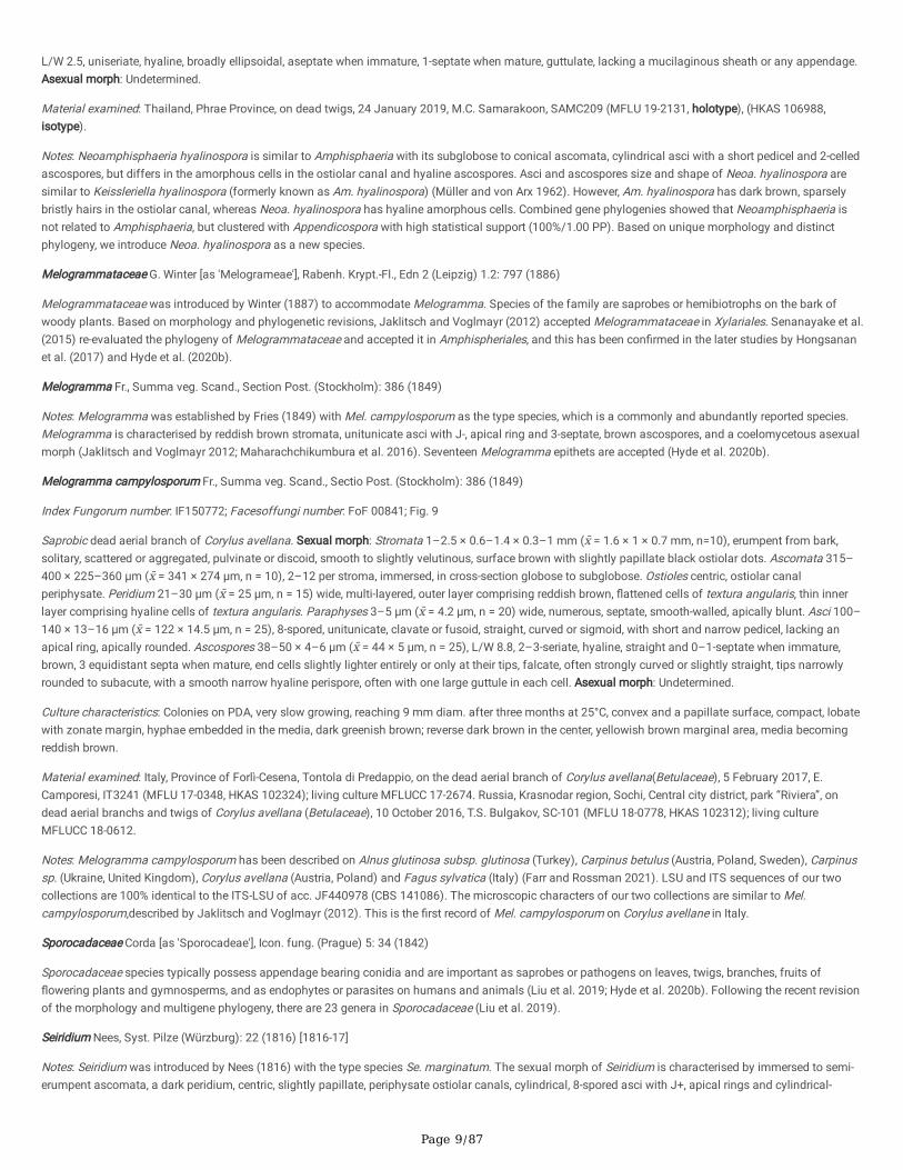
Page 9/87
L/W 2.5, uniseriate, hyaline, broadly ellipsoidal, aseptate when immature, 1-septate when mature, guttulate, lacking a mucilaginous sheath or any appendage.Asexual morph: Undetermined.
Material examined: Thailand, Phrae Province, on dead twigs, 24 January 2019, M.C. Samarakoon, SAMC209 (MFLU 19-2131, holotype), (HKAS 106988,isotype).
Notes: Neoamphisphaeria hyalinospora is similar to Amphisphaeria with its subglobose to conical ascomata, cylindrical asci with a short pedicel and 2-celledascospores, but differs in the amorphous cells in the ostiolar canal and hyaline ascospores. Asci and ascospores size and shape of Neoa. hyalinospora aresimilar to Keissleriella hyalinospora (formerly known as Am. hyalinospora) (Müller and von Arx 1962). However, Am. hyalinospora has dark brown, sparselybristly hairs in the ostiolar canal, whereas Neoa. hyalinospora has hyaline amorphous cells. Combined gene phylogenies showed that Neoamphisphaeria isnot related to Amphisphaeria, but clustered with Appendicospora with high statistical support (100%/1.00 PP). Based on unique morphology and distinctphylogeny, we introduce Neoa. hyalinospora as a new species.
Melogrammataceae G. Winter [as 'Melogrameae'], Rabenh. Krypt.-Fl., Edn 2 (Leipzig) 1.2: 797 (1886)
Melogrammataceae was introduced by Winter (1887) to accommodate Melogramma. Species of the family are saprobes or hemibiotrophs on the bark ofwoody plants. Based on morphology and phylogenetic revisions, Jaklitsch and Voglmayr (2012) accepted Melogrammataceae in Xylariales. Senanayake et al.(2015) re-evaluated the phylogeny of Melogrammataceae and accepted it in Amphispheriales, and this has been con�rmed in the later studies by Hongsananet al. (2017) and Hyde et al. (2020b).
Melogramma Fr., Summa veg. Scand., Section Post. (Stockholm): 386 (1849)
Notes: Melogramma was established by Fries (1849) with Mel. campylosporum as the type species, which is a commonly and abundantly reported species.Melogramma is characterised by reddish brown stromata, unitunicate asci with J-, apical ring and 3-septate, brown ascospores, and a coelomycetous asexualmorph (Jaklitsch and Voglmayr 2012; Maharachchikumbura et al. 2016). Seventeen Melogramma epithets are accepted (Hyde et al. 2020b).
Melogramma campylosporum Fr., Summa veg. Scand., Sectio Post. (Stockholm): 386 (1849)
Index Fungorum number: IF150772; Facesoffungi number: FoF 00841; Fig. 9
Saprobic dead aerial branch of Corylus avellana. Sexual morph: Stromata 1–2.5 × 0.6–1.4 × 0.3–1 mm (x̄ = 1.6 × 1 × 0.7 mm, n=10), erumpent from bark,solitary, scattered or aggregated, pulvinate or discoid, smooth to slightly velutinous, surface brown with slightly papillate black ostiolar dots. Ascomata 315–400 × 225–360 μm (x̄ = 341 × 274 μm, n = 10), 2–12 per stroma, immersed, in cross-section globose to subglobose. Ostioles centric, ostiolar canalperiphysate. Peridium 21–30 μm (x̄ = 25 μm, n = 15) wide, multi-layered, outer layer comprising reddish brown, �attened cells of textura angularis, thin innerlayer comprising hyaline cells of textura angularis. Paraphyses 3–5 μm (x̄ = 4.2 μm, n = 20) wide, numerous, septate, smooth-walled, apically blunt. Asci 100–140 × 13–16 μm (x̄ = 122 × 14.5 μm, n = 25), 8-spored, unitunicate, clavate or fusoid, straight, curved or sigmoid, with short and narrow pedicel, lacking anapical ring, apically rounded. Ascospores 38–50 × 4–6 μm (x̄ = 44 × 5 μm, n = 25), L/W 8.8, 2–3-seriate, hyaline, straight and 0–1-septate when immature,brown, 3 equidistant septa when mature, end cells slightly lighter entirely or only at their tips, falcate, often strongly curved or slightly straight, tips narrowlyrounded to subacute, with a smooth narrow hyaline perispore, often with one large guttule in each cell. Asexual morph: Undetermined.
Culture characteristics: Colonies on PDA, very slow growing, reaching 9 mm diam. after three months at 25°C, convex and a papillate surface, compact, lobatewith zonate margin, hyphae embedded in the media, dark greenish brown; reverse dark brown in the center, yellowish brown marginal area, media becomingreddish brown.
Material examined: Italy, Province of Forlì-Cesena, Tontola di Predappio, on the dead aerial branch of Corylus avellana(Betulaceae), 5 February 2017, E.Camporesi, IT3241 (MFLU 17-0348, HKAS 102324); living culture MFLUCC 17-2674. Russia, Krasnodar region, Sochi, Central city district, park “Riviera”, ondead aerial branchs and twigs of Corylus avellana (Betulaceae), 10 October 2016, T.S. Bulgakov, SC-101 (MFLU 18-0778, HKAS 102312); living cultureMFLUCC 18-0612.
Notes: Melogramma campylosporum has been described on Alnus glutinosa subsp. glutinosa (Turkey), Carpinus betulus (Austria, Poland, Sweden), Carpinussp. (Ukraine, United Kingdom), Corylus avellana (Austria, Poland) and Fagus sylvatica (Italy) (Farr and Rossman 2021). LSU and ITS sequences of our twocollections are 100% identical to the ITS-LSU of acc. JF440978 (CBS 141086). The microscopic characters of our two collections are similar to Mel.campylosporum,described by Jaklitsch and Voglmayr (2012). This is the �rst record of Mel. campylosporum on Corylus avellane in Italy.
Sporocadaceae Corda [as 'Sporocadeae'], Icon. fung. (Prague) 5: 34 (1842)
Sporocadaceae species typically possess appendage bearing conidia and are important as saprobes or pathogens on leaves, twigs, branches, fruits of�owering plants and gymnosperms, and as endophytes or parasites on humans and animals (Liu et al. 2019; Hyde et al. 2020b). Following the recent revisionof the morphology and multigene phylogeny, there are 23 genera in Sporocadaceae (Liu et al. 2019).
Seiridium Nees, Syst. Pilze (Würzburg): 22 (1816) [1816-17]
Notes: Seiridium was introduced by Nees (1816) with the type species Se. marginatum. The sexual morph of Seiridium is characterised by immersed to semi-erumpent ascomata, a dark peridium, centric, slightly papillate, periphysate ostiolar canals, cylindrical, 8-spored asci with J+, apical rings and cylindrical-
Page 10/87
oblong, euseptate, yellow to dark brown ascospores (Bonthond et al. 2018). The coelomycetous asexual morph of Seiridium differs from closely relatedNothoseiridium and Nonappendiculata in having versicolorous, 5-septate conidia with appendages. There are 44 Seiridium species (Bonthond et al. 2018).
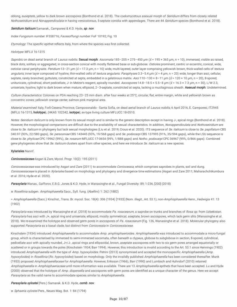
Seiridium italicum Samarak., Camporesi & K.D. Hyde, sp. nov.
Index Fungorum number: IF558716; Facesoffungi number: FoF 10192; Fig. 10
Etymology: The speci�c epithet re�ects Italy, from where the species was �rst collected.
Holotype: MFLU 16-1315
Saprobic on dead aerial branch of Laurus nobilis. Sexual morph: Ascomata 185–205 × 275–450 μm (x̄ = 195 × 365 μm, n = 10), immersed, visible as raised,black dots, solitary or aggregated, in cross-section conical with mostly �attened base or sub-globose. Ostioles prominent, centric or eccentric, conical, wide,ostiolar canal periphysate. Peridium 13–21 μm (x̄ = 17.3 μm, n = 10) wide, multi-layered, outer layer comprising yellowish brown, thick-walled cells of texturaangularis, inner layer composed of hyaline, thin-walled cells of textura angularis. Paraphyses 3.3–5.4 μm (x̄ = 4 μm, n = 20) wide, longer than asci, cellular,septate, rarely branched, guttulate, constricted at septa, embedded in a gelatinous matrix. Asci 110–130 × 8–11 μm (x̄ = 120 × 10 μm, n = 20), 8-spored,unitunicate, cylindrical, short pedicellate, J- in Melzer’s reagent, apically rounded. Ascospores 14.8–18.5 × 5.5–8 μm (x̄ = 16.3 × 7.3 μm, n = 30), L/W 2.3,uniseriate, hyaline, light to dark brown when mature, ellipsoid, 2–3-septate, constricted at septa, lacking a mucilaginous sheath. Asexual morph: Undetermined.
Culture characteristics: Colonies on PDA reaching 23–25 mm diam. after four weeks at 25°C, circular, �at, entire margin, white and yellowish brown asconcentric zones; yellowish orange center, salmon pink marginal area.
Material examined: Italy, Forlì-Cesena Province, Camposonaldo - Santa So�a, on dead aerial branch of Laurus nobilis, 6 April 2016, E. Camporesi, IT2945(MFLU 16-1315, holotype), (HKAS 102342, isotype); ex-type living culture MFLUCC 18-0510.
Notes: Seiridium italicum is only known from its sexual morph and is similar to the generic description except in having J-, apical rings (Bonthond et al. 2018).However, the morphological comparisons are di�cult due to the availability of sexual characteristics. In addition, Nonappendiculata and Nothoseiridium areclose to Se. italicum in phylogeny but lack sexual morphologies (Liu et al. 2019; Crous et al. 2020). ITS sequence of Se. italicum is close to Se. papillatum CBS340.97 (93%, 22/580 gaps), Se. persooniae CBS 143445 (93%, 19/568 gaps) and Se. podocarpi CBS 137995 (91%, 35/594 gaps), while the LSU sequence isclose to Se. phylicae CPC 19962 (99%), Se. rosarum MFLUCC 17-0654 (99%, 1/888 gaps) and Notho. podocarpi CPC 36967 (99%, 0/866 gaps). Combinedgene phylogenies show that Se. italicum clusters apart from other species, and here we introduce Se. italicum as a new species.
Xylariales Nannf.
Coniocessiaceae Asgari & Zare, Mycol. Progr. 10(2): 195 (2011)
Coniocessiaceae was introduced by Asgari and Zare (2011) to accommodate Coniocessia, which comprises saprobes in plants, soil and dung.Coniocessiaceae is placed in Xylariales based on morphology and phylogeny and divergence time estimations (Asgari and Zare 2011; Maharachchikumburaet al. 2016; Hyde et al. 2020).
Paraxylaria Wanas., Gafforov, E.B.G. Jones & K.D. Hyde, in Wanasinghe et al., Fungal Diversity: 89; 1-236, [200] (2018)
≡ Rosellinia subgen. Amphisphaerella Sacc., Syll. fung. (Abellini) 1: 262 (1882)
= Amphisphaerella (Sacc.) Kirschst., Trans. Br. mycol. Soc. 18(4): 306 (1934) [1933] (Nom. illegit., Art. 53.1), non-Amphisphaerella Henn., Hedwigia 41: 13(1902)
Paraxylaria was introduced by Wanasinghe et al. (2018) to accommodate Pa. rosacearum, a saprobe on trunks and branches of Rosa sp. from Uzbekistan.Paraxylaria has asci with J+, apical ring and uniseriate, ellipsoid, mostly symmetrical, aseptate, brown ascospores, which lack germ slits (Wanasinghe et al.2018). We re-examined the holotype and observed germ pores in ascospores of Pa. rosacearum (Fig. 136, Wanasinghe et al. 2018). Molecular phylogeny alsosupported Paraxylaria as a basal clade, but distinct from Coniocessia in Coniocessiaceae.
Kirschstein (1934) introduced Amphisphaerella to accommodate Amp. amphisphaerioides. Amphisphaerella was introduced to accommodate a micro-fungalgroup, which is characterised by immersed to semi-immersed ascomata, often beneath a clypeus, globose to subglobose in section, 8-spored, cylindrical,pedicellate asci with apically rounded, J+/J-, apical rings and ellipsoidal, brown, aseptate ascospores with two to six germ pores arranged equatorially orscattered or in groups towards the poles (Kirschstein 1934; Barr 1994). However, this introduction is invalid according to the Art. 53.1 since Hennings (1902)introduced Amphisphaerella with the type of Amp. hypoxyloides. Petrini (2013) synonymised and accepted the monospeci�c Amphisphaerella (Amp.hypoxyloides) in Rosellinia (Ro. hypoxyloides) based on morphology. Only the invalidly published Amphisphaerella has been considered thereafter. Munk(1953) proposed Amphisphaerellaceae for Amphisphaerella. However, Eriksson (1966), Barr (1990) and Lumbsch and Huhndorf (2010) retainedAmphisphaerella in Amphisphaeriaceae until more information was available. There are 13 Amphisphaerella epithets that have been accepted. Lu and Hyde(2000) observed that the holotype of Amp. dispersella and ascospores with germ pores are identi�ed as a unique character of the genus. Here we acceptParaxylaria as the valid name to accommodate species similar to Amphisphaerella.
Paraxylaria xylostei (Pers.) Samarak. & K.D. Hyde, comb. nov.
≡ Sphaeria xylostei Pers., Neues Mag. Bot. 1: 84 (1794)
Page 11/87
= Amphisphaerella xylostei (Pers.) Munk, 1953 (this Index 5: 278), nom. inval., Art. 41.5
= Amphisphaerella xylostei (Pers.) Rulamort, Bull. Soc. bot. Centre-Ouest, Nouv. sér. 17(2): 192 (1986)
Index Fungorum number: IF558404; Facesoffungi number: FoF 10193; Fig. 11
Saprobic on dead aerial branch of Lonicera sp. Sexual morph: Ascomata 200–250 × 250–310 μm (x̄ = 225 × 275 μm, n = 10), immersed beneath the clypeus,visible as black patches, solitary or aggregated in small numbers, in cross-section conical with rounded base or subglobose. Ostioles prominent, centric oreccentric, conical, more or less acute, shiny black, periphasate ostiolar canal. Peridium 17–26 μm (x̄ = 22 μm, n = 10) wide at base, thickened to 27–44 μm(x̄ = 36 μm, n = 10) wide near to ostiole, hard in upper regions, multi-layered, outer layer comprising brown, thick-walled cells of textura angularis, inner layercomposed of hyaline, thin-walled cells of textura angularis. Paraphyses 2–3.5 μm (x̄ = 2.6 μm, n = 15) wide, long, numerous, �lamentous, �exuous, septate,rarely branched, guttulate. Asci 75–140 × 9.5–13.5 μm (x̄ = 105 × 11.3 μm, n = 20), 8-spored, unitunicate, cylindrical, pedicellate up to 20 μm long, with a 1.5–2 × 3.5–5 μm (x̄ = 1.7 × 4.3 μm, n = 10), wedge-shaped apical ring, J+ in Melzer’s reagent. Ascospores 15–19 × 7–10 μm (x̄ = 17.8 × 8.7 μm, n = 25), L/W 2.1,uniseriate, initially hyaline to yellowish brown, becoming blackish brown at maturity, ellipsoid, mostly symmetrical, aseptate, with rounded ends, guttulate, with3–5 equatorial germ pores surrounded by thick, dark brown margin, lacking a mucilaginous sheath, lacking a germ slit. Asexual morph: Undetermined.
Material examined: Italy, Province of Forlì-Cesena, Campigna - Santa So�a, on dead aerial branch of Lonicera sp. (Caprifoliaceae), 5 February 2017, E.Camporesi, IT3479 (MFLU 17-1636, HKAS 102371). ibid. IT3479A (MFLU 17-1645, HKAS 102313).
Notes: Paraxylaria xylostei is common in temperate regions and is variable in both ascus and ascospore characters, amyloid/nonamyloid and size, probablydue to geographical variation and the different hosts (Mathiassen 1993). The morphological variations are higher in the Lonicera materials compared to Salix(Mathiassen 1993). Paraxylaria xylostei is known from Lonicera microphylla, L. tatarica, L. xylosteum, Salix glauca ssp. glauca, S. lanata ssp. lanata and S.phylicifolia (Mathiassen 1993; Gafforov 2017). Paraxylaria xylostei possesses J+ asci and 14–18 × 6–9 μm ascospores with (3)5–6 equatorial germ pores,while Pa. rosacea has 16–20 × 9–11 μm ascospores with 7–11 scattered germ pores. Here we provide another collection of Pa. xylostei from Italy as a newgeographical record and update the phylogenetic a�nity of Paraxylaria in Coniocessiaceae.
Diatrypaceae Nitschke [as 'Diatrypeae'], Verh. naturh. Ver. preuss. Rheinl. 26: 73 (1869)
Diatrypaceae was introduced by Nitschke (1869), which comprises saprobes, pathogens and endophytes on economic crops and forest trees worldwideoccurs in aquatic and terrestrial habitats (Hyde et al. 2020b; Konta et al. 2020a). Based on phylogeny and divergence time estimations, Diatrypaceae isaccepted as a well-supported family in Xylariales (Hongsanan et al. 2017). With the addition of several new genera, there are 25 genera accommodated inDiatrypaceae as; Allocryptovalsa, Allodiatrype, Anthostoma, Cryptosphaeria, Cryptovalsa, Diatrypasimilis, Diatrype, Diatrypella, Dothideovalsa, Echinomyces,Endoxylina, Eutypa, Eutypella, Halocryptosphaeria, Halocryptovalsa, Halodiatrype, Leptoperidia, Libertella, Monosporascus, Neoeutypella, Paraeutypella,Pedumispora, Peroneutypa, Quaternaria and Rostronitschkia (Hyde et al. 2020b; Konta et al. 2020a; Dissanayake et al. 2021a).
Allocryptovalsa Senwanna, Phookamsak & K.D. Hyde, in Senwanna et al., Mycosphere 8(10): 1839 (2017)
Notes: Allocryptovalsa was introduced by Senwanna et al. (2017) with the type species Al. polyspora on a dead twig of Hevea brasiliensis from Thailand.Allocryptovalsa has immersed ascomata, polysporous asci, and allantoid ascospores and forms a distinct phylogenetic a�nity in Diatrypaceae with ITS-tub2phylogeny. There are �ve Allocryptovalsa species.
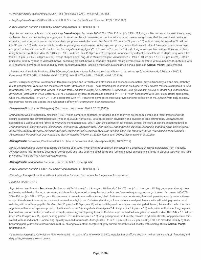
Allocryptovalsa sichuanensis Samarak., Jian K. Liu & K.D. Hyde, sp. nov.
Index Fungorum number: IF558717; Facesoffungi number: FoF 10194; Fig. 12
Etymology: The speci�c epithet re�ects the location, Sichuan, from where the fungus was �rst collected.
Holotype: HKAS 107017
Saprobic on dead branch. Sexual morph: Stromata 0.7–4.1 mm (x̄ = 1.9 mm, n = 10) length, 0.8–1.75 mm (x̄ = 1.1 mm, n = 10) high, erumpent through hostepidermis, with bark adhering to stromata, visible as black, rounded to irregular dots on host surface, solitary to aggregated, scattered. Ascomata 460–755 ×300–420 μm (x̄ = 579 × 367 μm, n = 10), immersed to semi-immersed in stroma, black, 2–9 ascomata per stroma, thin black pseudoparenchymatous tissuearound the white entostroma, in cross-section ovoid to subglobose. Ostioles cylindrical, sulcate, ostiolar canal periphysate, with yellowish pigment aroundostioles, with or without papilla. Peridium 34–56 μm (x̄ = 43.5 μm, n = 10) wide, multi-layered, outer layer comprising dark brown, thick-walled cells of texturaangularis, a thin inner layer composed of hyaline cells of textura angularis. Paraphyses 2.4–4.4 μm (x̄ = 3.4 μm, n = 20) wide, wider at the base, long, septate,�lamentous, smooth-walled, constricted at septa, narrowing and tapering towards the blunt apex, embedded in a gelatinous matrix. Asci 100–142 × 16–24 μm(x̄ = 122 × 19.4 μm, n = 15), spore bearing part 60–75 μm (x̄ = 68 μm, n = 10) long, polysporous, unitunicate, clavate to cylindric-clavate, long pedicellate, thin-walled, with an indistinct, J-, apical ring, apically rounded to truncate. Ascospores 6–11 × 2–3 µm (= 8.5 × 2.7 μm, n = 35), L/W 3.2, crowded, initially hyaline,becoming pale yellowish to brown when mature, oblong to allantoid, aseptate, slightly curved, smooth-walled, mostly with small guttules. Asexual morph:Undetermined.
Culture characteristics: Colonies on PDA reaching 55 mm diam. after one week at 25°C, irregular, �at or effuse, cottony, medium dense, margin �mbriate, anddirty white; reverse yellowish brown.
Page 12/87
Material examined: China, Sichuan Province, Chengdu, University of Electronic Science and Technology of China (Qingshuihe Campus), on the dead branch,30 September 2019, M.C. Samarakoon, SAMC249 (HKAS 107017, holotype), (MFLU HT20-0177, isotype); ex-type living cultures MFLUCC T20-0653 = GZCC21-0043 = CGMCC3.20363.
Notes: Allocryptovalsa sichuanensis has stromata with 2–9 ascomata, which distinguishes it from Al. elaeidis (1–2), Al. polyspora (1–3) and Al. rabenhorstii(5–25) (Mehrabi et al. 2016; Senwanna et al. 2017; Konta et al. 2020a). The LSU, ITS and tub2 sequences of Al. sichuanensis are similar to Al. elaeidisMFLUCC 15-0707 (LSU 99%, 0/892 gaps; ITS 99%, 0/532 gaps; tub2 99%, 1/1064 gaps) and Al. polyspora MFLU 17-1218 (LSU 100%, 0/823 gaps; ITS 99%,1/486 gaps). Combined ITS-tub2 (data not shown) and ITS-LSU-rpb2-tub2-tef1 (Fig. 1) phylogenies showed our taxon is basal to Allocryptovalsa with97%/1.00 PP statistical support. Here we introduce Al. sichuanensis as a new species.
Diatrype Fr., Summa veg. Scand., Section Post. (Stockholm) 384 (1849)
Notes: Diatrype was introduced with the type D. disciformis by Fries (1849). Species in the genus are characterised by discoid or widely effuse, erumpentstromata with embedded ascomata in their sexual morph and libertella-like asexual morph (Vasilyeva and Stephenson 2009; Senanayake et al. 2015).
Diatrype disciformis (Hoffm.) Fr., Summa veg. Scand., Sectio Post. (Stockholm): 385 (1849)
Index Fungorum number: IF233766; Facesoffungi number: FoF 00691; Fig. 13
Saprobic on the dead branch. Sexual morph: Stromata 3.3–5.6 mm (x̄ = 4.1 mm, n = 10) length, 3–4.8 mm (x̄ = 3.7 mm, n = 10) high, scattered, erumpent tosuper�cial, orbicular, somewhat convex, edges of cracks remaining as pointed, angular parts, margin thick, black, composed of an outer, dark brown, small,tightly packed, thin parenchymatous cell layer and inner, yellowish white, large, loosely packed, parenchymatous cell layer. Ascomata 590–700 × 280–430 μm(x̄ = 636 × 357 μm, n = 15), immersed in stromatic tissues, aggregated, in cross-section globose to subglobose, narrowing towards the apex and very narrow atthe base of ostiolar canal, pale brown, thin-walled, ostiolate. Ostioles short, centric, compressed, apex wider than base, ostiolar canal periphysate, ostiolaropening covered with carbonaceous, black cells. Peridium 17–20 μm (x̄ = 18.8 μm, n = 8) wide, multi-layered, outer layer comprising dark brown cells oftextura angularis, a thin inner layer composed of hyaline cells of textura angularis. Paraphyses 2–5 μm (x̄ = 3.2 μm, n = 20) wide, wider at the base, long,septate, smooth-walled, constricted at septa, tapering towards the blunt apex. Asci 55–80 × 3.5–5 μm (x̄ = 69 × 4.5 μm, n = 25), 8-spored, unitunicate, clavate,thin-walled, pedicel 37–53 μm (x̄ = 44 μm, n = 15) long, apex �at, J- in Melzer’s reagent. Ascospores 4.5–7 × 0.8–1.3 μm (x̄ = 5.7 × 1.1 μm, n = 30), L/W 5.2,overlapping, cylindrical or elongate-allantoid, hyaline, aseptate, guttulate, smooth-walled. Asexual morph: Undetermined.
Material examined: UK, on dead branch, 2015, E.B.G. Jones, GJ384 (MFLU 17-1549; HKAS 107036).
Notes: Senanayake et al. (2015) designated a reference specimen for Diatrype disciformis (MFLU 15–0722) on a branch of Ostrya carpinifolia from Italy. Inthis study, we provide another specimen collected from the UK, which is morphologically similar with overlapping measurements of ascomata, asci andascospores. However, our collection has larger stromata (3.3–5.6 mm vs 1.5–2 mm length) and a thinner peridium (17–20 μm vs 20–30 μm) as compared toMFLU 15–0722. Mehrabi et al. (2016) also described D. disciformis, which has up to 8 mm diam. of stromata. The known distribution of the fungus is onAlnus and Fagus from Europe and lead-contaminated soils from abandoned �ring range USA (Acero et al. 2004; Vasilyeva and Stephenson 2009; 2014;Sullivan et al. 2012; Vasilyeva and Ma 2014; Mehrabi et al. 2016).
Eutypa Tul. & C. Tul., Select. fung. carpol. (Paris) 2: 52 (1863)
Notes: Eutypa is an important plant pathogenic group on fruit crops that causes eutypa dieback (Hyde et al. 2020b). Eutypa species have super�cial, irregularstromata with scattered, roundish to prominent ostioles, clavate asci with a round to truncate apex and allantoid to ellipsoidal, aseptate ascospores. Hyde etal. (2020b) and Wijayawardene et al. (2020) estimated the genus contains 66 species, with 17 with sequence data.
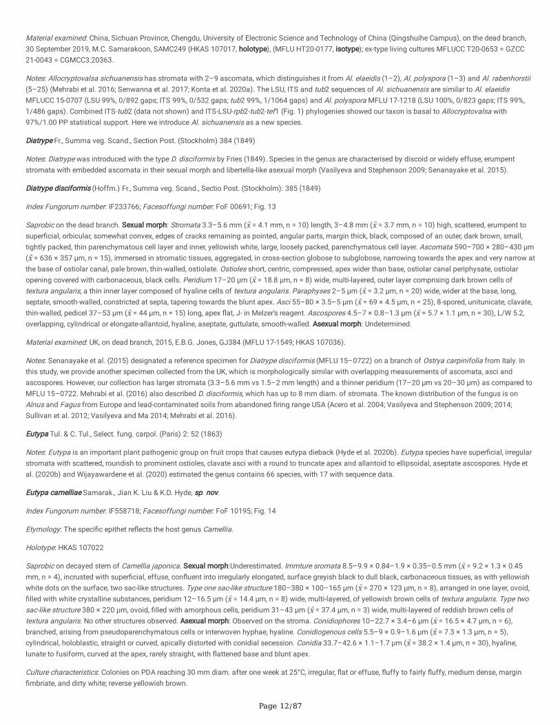
Eutypa camelliae Samarak., Jian K. Liu & K.D. Hyde, sp. nov.
Index Fungorum number: IF558718; Facesoffungi number: FoF 10195; Fig. 14
Etymology: The speci�c epithet re�ects the host genus Camellia.
Holotype: HKAS 107022
Saprobic on decayed stem of Camellia japonica. Sexual morph:Underestimated. Immture sromata 8.5–9.9 × 0.84–1.9 × 0.35–0.5 mm (x̄ = 9.2 × 1.3 × 0.45mm, n = 4), incrusted with super�cial, effuse, con�uent into irregularly elongated, surface greyish black to dull black, carbonaceous tissues, as with yellowishwhite dots on the surface, two sac-like structures. Type one sac-like structure 180–380 × 100–165 μm (x̄ = 270 × 123 μm, n = 8), arranged in one layer, ovoid,�lled with white crystalline substances, peridium 12–16.5 μm (x̄ = 14.4 μm, n = 8) wide, multi-layered, of yellowish brown cells of textura angularis. Type twosac-like structure 380 × 220 μm, ovoid, �lled with amorphous cells, peridium 31–43 μm (x̄ = 37.4 μm, n = 3) wide, multi-layered of reddish brown cells oftextura angularis. No other structures observed. Asexual morph: Observed on the stroma. Conidiophores 10–22.7 × 3.4–6 μm (x̄ = 16.5 × 4.7 μm, n = 6),branched, arising from pseudoparenchymatous cells or interwoven hyphae, hyaline. Conidiogenous cells 5.5–9 × 0.9–1.6 μm (x̄ = 7.3 × 1.3 μm, n = 5),cylindrical, holoblastic, straight or curved, apically distorted with conidial secession. Conidia 33.7–42.6 × 1.1–1.7 μm (x̄ = 38.2 × 1.4 μm, n = 30), hyaline,lunate to fusiform, curved at the apex, rarely straight, with �attened base and blunt apex.
Culture characteristics: Colonies on PDA reaching 30 mm diam. after one week at 25°C, irregular, �at or effuse, �uffy to fairly �uffy, medium dense, margin�mbriate, and dirty white; reverse yellowish brown.
Page 13/87
Material examined: China, Sichuan Province, Chengdu, University of Electronic Science and Technology of China (Qingshuihe Campus), on a dead stem ofCamellia japonica (Theaceae), 30 September 2019, M.C. Samarakoon, SAMC254X (HKAS 107022, holotype), (MFLU HT20-0182, isotype); ex-type livingcultures MFLUCC T20-0643 = GZCC 21-0042.
Notes: Our new isolates were obtained through the internal tissue isolation of the stromata. We did not observe any spores in those stromata, but observed anasexual morph, which is similar to diatrypaceous asexual morphs on the stromata. Two areas on the stroma produced ascoma-like structures with a brownperidium �lled with amorphous cells, probably immature ascomata. However, these observations did not provide enough morphology for the completeidenti�cation of the strain. Our two isolates are phylogenetically similar to each other and form a distinct single clade in Eutypa. The ITS and LSU sequencesof our strains are 98% (4/559 gaps) and 99% (3/887 gaps) similar to E. lata respectively. Combined ITS-tub2 (data not shown) and ITS-LSU-rpb2-tub2-tef1(Fig. 1) phylogenies showed that our new isolates clustered in Eutypa sensu stricto and is sister to E. armeniacae (ATCC 28120) with poor statistical support(58%/0.99 PP). Here we propose E. camelliae as a new species.
Melanostictus Samarak. & K.D. Hyde, gen. nov.
Index Fungorum number: IF558719; Facesoffungi number: FoF 10196
Etymology: The generic epithet refers to the Greek: melano- “black” + stictus “spot”.
Saprobic on dead branch. Sexual morph: Ascomata immersed, visible as black, raised dots, solitary or aggregated, in cross-section globose. Ostioles centric,ostiolar canal periphysate, sulcate on top. Ectostroma yellow to white. Peridium multi-layered, outer layer comprising dark brown, �attened cells of texturaangularis, inner layer comprising hyaline cells of textura angularis. Paraphyses septate. Asci 8-spored, unitunicate, clavate, with a long pedicel, apical ringminute, apically �attened. Ascospores overlapping, hyaline, cylindrical or elongate-allantoid, aseptate, smooth-walled. Asexual morph: Undetermined.
Type: Melanostictus longiostiolatus Samarak. & K.D. Hyde
Notes: Melanostictus is characterised by immersed ascomata appearing as black dots on the host surface, long papillate ostioles with a sulcate top, yellow towhite ectostroma, 8-spored asci with cylindrical or elongate-allantoid ascospores. Several genera are phylogenetically close to Melanostictus bear 8-sporedasci with diversi�ed stromatic characters. Cryptosphaeria, Eutypa and Neoeutypella have effused stromata and aggregated ascomata on the host surface,while Allodiatrype has well-developed, erumpent stromata (Trouillas et al. 2015; de Almeida et al. 2016; Phookamsak et al. 2019; Konta et al. 2020a).Halodiatrype and Pedumispora are the closest genera to our new genus. Halodiatrype has immersed ascomata, papillate ostioles with a brown outeramorphous layer and inner yellow cells of textura porrecta, 8-spored, unitunicate asci with oblong to allantoid or sub-inaequilateral, aseptate to septate, lightbrown ascospores (Dayarathne et al. 2016), while Pedumispora has 1–4 immersed ascomata per stroma, papillate ostioles, 8-spored, fusiform asci with�liform, multi-septate, curved, longitudinally striate ascospores (Hyde and Jones 1992). Melanostictus clustered sister to Halodiatrype and Pedumispora as adistinct group in morphology and phylogeny; hence we introduce our two new collections in the new genus.
Melanostictus longiostiolatusSamarak. & K.D. Hyde, sp. nov.
Index Fungorum number: IF558720; Facesoffungi number: FoF 10197; Fig. 15
Etymology: The speci�c epithet re�ects the papillate ostioles.
Holotype: MFLU 19-2146
Saprobic on dead branch. Sexual morph: Ascomata 550–630 × 300–370 μm (x̄ = 585 × 345 μm, n = 10), immersed in the host tissue under slightly raisedareas, visible as black dots, surrounded by black periphery, solitary or aggregated, mostly in pairs, with clusters or evenly distributed, in cross-section globose.Ostioles 300–390 × 110–180 μm (x̄ = 340 × 145 μm, n = 5), centric, ostiolar canal periphysate, sulcate on top. Ectostromayellow. Peridium 30–38 μm (x̄ = 35μm, n = 10) wide, multi-layered, outer layer comprising dark brown, �attened cells of textura angularis, inner layer comprising hyaline cells of texturaangularis. Paraphyses 3–7.5 μm (x̄ = 5.4 μm, n = 20) wide, wider at the base, septate, rarely branched, constricted at septa, smooth-walled, with a bluntend. Asci 50–65 × 5.5–8 μm (x̄ = 56 × 6.5 μm, n = 25), 8-spored, unitunicate, clavate, thin-walled, pedicel 19–35 μm (x̄ = 26 μm, n = 25) long, apical ringminute, deliquescing early and releasing spores, developing from the base and lower sides of the ascomata, apically �attened. Ascospores 3.5–5.5 × 1–1.5μm (x̄ = 4.5 × 1.2 μm, n = 30), L/W 3.75, overlapping, hyaline, cylindrical or elongate-allantoid, aseptate, smooth-walled. Asexual morph: Undetermined.
Material examined: Thailand, Chiang Rai Province, Amphoe Mueang Chiang Rai, on dead branch, 15 June 2019, M.C. Samarakoon, SAMC229 (MFLU 19-2146,holotype), (HKAS 107003, isotype).
Notes: Please see the notes of Melanostictus thailandicus.
Melanostictus thailandicusSamarak. & K.D. Hyde, sp. nov.
Index Fungorum number: IF558721; Facesoffungi number: FoF 10198; Fig. 16
Etymology: The speci�c epithet re�ects Thailand, where the species was �rst collected.
Holotype: MFLU 19-2123
Page 14/87
Saprobic on dead branch. Sexual morph: Ascomata 415–580 × 300–410 μm (x̄ = 512 × 362 μm, n = 10), immersed in the host tissue under slightly raisedareas, visible as black dots, solitary or aggregated, with clusters or evenly distributed, in cross-section globose or subglobose, base rarely �attened. Ostioles185–280 × 85–145 μm (x̄ = 235 × 115 μm, n = 5), centric, ostiolar canal periphysate, sulcate on top. Ectostroma yellowish white. Peridium 27–40 μm (x̄ = 33μm, n = 10) wide, multi-layered, outer layer comprising dark brown, �attened cells of textura angularis, inner layer composed of hyaline cells of texturaangularis, Paraphyses 2.4–4 μm (x̄ = 3.1 μm, n = 20) wide, long, septate, constricted at septa, smooth-walled, ends blunt. Asci 50–64 × 3.8–5 μm (x̄ = 56 × 4.2μm, n = 25), 8-spored, unitunicate, clavate, thin-walled, pedicel 24–30 μm (x̄ = 26.5 μm, n = 15) long, apical ring minute, deliquescing early and releasingspores. Ascospores 5–6.2 × 1–1.6 μm (x̄ = 5.5 × 1.4 μm, n = 30), L/W 3.9, overlapping, cylindrical or elongate-allantoid, hyaline, aseptate, guttulate, smooth-walled. Asexual morph: Undetermined.
Material examined: Thailand, Lampang Province, Wang Nuea, on the dead branch, 7 December 2018, M.C. Samarakoon, SAMC190 (MFLU 19-2123, holotype),(HKAS 106980, isotype).
Notes: Two Melanostictus species are introduced, namely Me. longiostiolatus and Me. thailandicus. Melanostictus longiostiolatus has longer and widerpapillate ostioles (300–390 × 110–180 μm), and yellow ectostroma as compared to Me. thailandicus (185–280 × 85–145 μm), which has a white ectostroma.The LSU and ITS bp (base pair) comparisons show only 3 and 2 bp differences respectively, among Me. longiostiolatus and Me. thailandicus, while 35 bpdifferences (including gaps) among tub2 sequences (4.6%). Combined gene phylogeny (Fig. 1) showed that Me. longiostiolatus is sister to Me. thailandicuswith 100%/1.00 PP statistical support. In addition, we observed few asci of both Me. longiostiolatus and Me. thailandicus collections with J+, apical ring,mildly bluing in Melzer’s reagent. However, it is not constant throughout the study, and further observations are needed in future studies.
Peroneutypa Berl., Icon. fung. (Abellini) 3(3-4): 80 (1902)
Notes: Peroneutypa is typi�ed by Pe. bellula and characterised by valsoid stromata with long-necked ascomata; 8-spored, clavate asci and allantoid, hyaline oryellowish ascospores (Carmarán et al. 2006; Senwanna et al. 2017; Shang et al. 2017). Hyde et al. (2020b) listed 28 species, including 11 species withsequence data.
Peroneutypa leucaenae Samarak. & K.D. Hyde, sp. nov.
Index Fungorum number: IF558722; Facesoffungi number: FoF 10199; Fig. 17
Etymology: The speci�c epithet re�ects the host genus Leucaena leucocephala.
Holotype: MFLU 18-0816
Saprobic on dead branch of Leucaena leucocephala. Sexual morph: Ascomata (including neck) 635–680 × 480–580 μm (x̄ = 655 × 525 μm, n = 10),immersed in the host tissue under slightly raised areas, black, mostly solitary or sometimes aggregated, with clusters or evenly distributed, in cross-sectionglobose or subglobose, delimited by a black zone in host tissues, glabrous. Ostioles with 275–350 μm (x̄ = 310 μm, n = 5) long neck, centric, ostiolar canalperiphysate. Peridium 22–43 μm (x̄ = 34 μm, n = 10) wide, multi-layered, outer layer comprising thick, dark brown cells of textura angularis, inner layercomposed of hyaline, thin-walled cells of textura angularis. Paraphyses 3.2–7 μm (x̄ = 5.2 μm, n = 20) wide, wider at the base, long, septate, smooth-walled,constricted at septa. Asci 30–37 × 3.8–4.5 μm (x̄ = 33 × 4.2 μm, n = 25), 8-spored, unitunicate, clavate, pedicel 17–27 μm (x̄ = 22 μm, n = 10) long, thin-walled,with wedge-shaped, apical ring, J+ in Melzer’s reagent. Ascospores 2.9–3.7 × 0.9–1.3 μm (x̄ = 3.2 × 1.1 μm, n = 30), L/W 2.9, overlapping, yellowish brown,ellipsoidal to cylindrical or elongate-allantoid, aseptate, smooth-walled. Asexual morph: Undetermined.
Culture characteristics: Colonies on PDA reaching 50–52 mm diam. after one week at 25°C, fast growing, irregular, �at or effuse, slightly raised, margin�mbriate, with aerial mycelium, and dirty white; reverse yellow to pale brown at the center, white at the margin.
Material examined: Thailand, Lampang Province, Wang Nuea, on dead branch of Leucaena leucocephala (Fabaceae), 18 August 2017, M.C. Samarakoon,SAMC048 (MFLU 18-0816, holotype), (HKAS 102352, isotype); ex-type living culture MFLUCC 18-0522.
Notes: Peroneutypa leucaenae has stromata with stromatic tissues developing only between perithecial necks, urn-shaped asci with J+, apical rings andallantoid, slightly to moderately curved ascospores similar to Pe. diminutispora. However, Pe. diminutispora has protruded necks and up to seven ascomataper stroma and lacks paraphyses, which differs from Pe. leucaenae in having one ascoma per stroma and raised neck on the surface with free hyphae andthick paraphyses (de Almeida et al. 2016). Peroneutypa leucaenae has asci with a J+, apical ring similar to Pe. cosmosa, but differs in having larger asci (30–37 vs 18–25 μm) and smaller ascospores (2.9–3.7 vs 6–8 μm) (Carmarán et al. 2006). The LSU sequence of Pe. leucaenae is similar to Pe. mangroveiPUFD526 (99%, 0/862 gaps) and Pe. scoparia MFLUCC 11-0615 (97%, 0/886 gaps), while the ITS sequence is similar to Pe. polysporae NFCCI 4392 (93%,10/394 gaps) and E. microasca BAFC51550 (89%, 14/444 gaps). Combined ITS-tub2 and ITS-LSU-rpb2-tub2-tef1 gene phylogeny also supports Pe.leucaenae as distinct, forming an independent lineage (Fig. 1).
Fasciatisporaceae S.N. Zhang, J.K. Liu & K.D. Hyde, in Hyde et al., Fungal Diversity 100: 227 (2020)
Hyde et al. (2020a) introduced Fasciatisporaceae to accommodate Fasciatispora with distinctive morphology of aseptate ascospores with a central pallidband, and on a multigene phylogeny, thus distinguishing it from other members of Xylariales. Fasciatisporaceous species are saprobes on palms and othermonocotyledons (from Arecaceae and Pandanaceae) distributed in terrestrial and coastal habitats.
Fasciatispora K.D. Hyde, Trans. Mycol. Soc. Japan 32(2): 265 (1991)
Page 15/87
Notes: Fasciatispora was introduced by Hyde (1991) with F. nypae as the type species with clypeate ascomata; asci with J +/J-, wedge-shaped apical ring, andpale brown to brown, ellipsoidal, ovoid or rhomboid, aseptate ascospores with a central pallid band (Hyde et al. 2020a). Liu et al. (2015) proposed a referencespecimen for F. nypae and accepted it in Xylariales based on molecular data. Thus, following several consecutive studies, 11 Fasciatispora species have beenaccepted, �ve with molecular data.
Fasciatispora cocoesS.N. Zhang, K.D. Hyde & J.K. Liu, in Hyde et al., Fungal Diversity 100: 231 (2020)
Index Fungorum number: IF557000; Facesoffungi number: FoF 06503; Fig. 18
Saprobic on dead lea�ets of Cocos nucifera. Sexual morph: Ascomata 200–230 × 160–210 μm (x̄ = 220 × 185 μm, n = 7), immersed beneath the clypeus,visible as black, circular dots, ostioles slightly protruding outside, indistinct, mostly solitary, sometimes aggregated into small groups, in cross-sectionsubglobose or globose, with a rounded or �attened base. Ostioles centric, ostiolar canal periphysate. Peridium 7–14.5 μm (x̄ = 10.3 μm, n = 10) wide, multi-layered, outer layer comprising brown, thick-walled cells of textura angularis, inner layer composed of hyaline, thin-walled cells of texturaangularis. Paraphyses 2–3.5 μm (x̄ = 2.7 μm, n = 15) wide, long, numerous, �lamentous, �exuous, septate, rarely branched, tapering. Asci 70–100 × 8–11 μm(x̄ = 82.5 × 9.5 μm, n = 20), 8-spored, unitunicate, cylindrical, short pedicellate, with a 0.5–0.8 × 2.2–3.1 μm (x̄ = 0.7 × 2.6 μm, n = 10), wedge-shaped apicalring, J+ in Melzer’s reagent, apex rounded. Ascospores 9–12 × 4.5–6.5 μm (x̄ = 10.8 × 5.5 μm, n = 25), L/W 2, uniseriate, hyaline when immature, yellowishbrown, with wide equatorial pallid band when mature, ellipsoidal to obovoid, aseptate, 1–2-guttulate, surrounded by a 3.5–6 μm (x̄ = 4.5 μm, n = 10) widemucilaginous sheath, lacking a germ slit.Asexual morph: Undetermined.
Material examined: Thailand, Chiang Rai Province, Amphoe Mueang Chiang Rai, on the dead lea�ets of Cocos nucifera (Arecaceae), 6 June 2019, M.C.Samarakoon, SAMC226 (MFLU 19-2143, HKAS 107000).
Notes: Our collection is similar to Fasciatispora cocoes (MFLU 18-1589) in asci (70–100 × 8–11 μm vs 78–110 × 6–12 µm) and ascospore (9–12 × 4.5–6.5µmvs 10–15 × 4.5–6 μm) and has the same host Cocos nucifera. Combined gene phylogenetic analyses show that our collection clusters with the type of F.cocoes (MFLUCC 18-1445) with 100%/1.00 PP statistical support.
Graphostromataceae M.E. Barr, J.D. Rogers & Y.M. Ju, Mycotaxon 48: 533 (1993)
Graphostromataceae was introduced by Barr et al. (1993), which comprises saprobes, endophytes and pathogens on trunks, branches, and twigs ofangiosperms (Hyde et al. 2020b). Biscogniauxia, Camillea, Graphostroma, Obolarina and Vivantia have been accepted in this family (Hyde et al. 2020b;Wijayawardene et al. 2020).
Biscogniauxia Kuntze, Revis. Gen. Pl. (Leipzig) 2: 398 (1891)
Notes: Biscogniauxia (type species Bi. nummularia) species are characterised by bipartite stromata lacking KOH-extractable pigments, and short pedicellatecylindrical asci and ellipsoid to short fusoid ascospores in their sexual and nodulisporium-like or periconiella-like asexual morphs (Daranagama et al. 2018).Biscogniauxia appears paraphyletic with Camillea, Obolarina and Graphostoma (Daranagama et al. 2018). There are 76 Biscogniauxia species and 23 havemolecular data (Hyde et al. 2020b).
Biscogniauxia magna Samarak. & K.D. Hyde, sp. nov.
Index Fungorum number: IF558723; Facesoffungi number: FoF 10200; Fig. 19
Etymology: The speci�c epithet refers to the Latin word that means ‘‘large’’ because of the large ascomata.
Holotype: MFLU 18-0850
Saprobic on dead branch. Sexual morph: Stromata 3–18 × 2–7 mm, applanate, dull brown to black, carbonaceous. Ascomata 475–700 × 265–375 μm (x̄ =595 × 320 μm, n = 15), ovoid to obpyriform, laterally compressed. Ostioles pointed, raised. Paraphyses 2.5–6.5 μm (x̄ = 4 μm, n = 15) wide, long, numerous,�lamentous, �exuous, septate, apex rounded. Asci 72–100 × 8–10 μm (x̄ = 85.5 × 9.3 μm, n = 20), 8-spored, unitunicate, cylindrical, short pedicellate, with a1.9–3.3 × 3.3–4.5 μm (x̄ = 2.8 × 3.9 μm, n = 10), wedge-shaped apical ring, J+ in Melzer’s reagent, apex rounded. Ascospores 11.5–14 × 6.5–8.5 μm (x̄ = 12.5× 7.5 μm, n = 35), L/W 1.6, uniseriate, hyaline when immature, dark brown to black when mature, ellipsoid, ends rounded, 1–2-guttulate, aseptate, with straightgerm slit along the entire spore length.Asexual morph: Hyphae 1.5–2.5 μm (x̄ = 1.9 μm, n = 15) wide, hyaline to brown, septate, branched, thin-walled aerialmycelia abundant. Conidiophores 2.5–5.5 μm (x̄ = 3.8 μm, n = 15) wide, composed of main axis, rough-walled, light brown to dark brown, sometimes one ormore major branches, with conidiogenous cells arising terminally or laterally. Conidiogenous cells 7–11 × 2.5–4.5 μm (x̄ = 8.5 × 3.5 μm, n = 20), swollen at theapex and with conidial secession scars, thin- and rough-walled, cylindrical to oblong, hyaline. Conidia 5.5–7.5 × 2–3 μm (x̄ = 6.5 × 2.6 μm, n = 20), holoblastic,ovoid to clavate, obtuse tip and acute truncated base, hyaline, smooth.
Culture characteristics: Colonies on PDA reaching 55 mm diam. after one week at 25°C, growing fast, �at, circular, dense, margin entire, aerial mycelia, lightbrown; reverse brown at center with dirty white edges, becoming yellowish brown when mature.
Material examined: Thailand, Chiang Rai Province, Mae Suai, on dead wood, 18 August 2017, M.C. Samarakoon, SAMC011 (MFLU 18-0850, holotype), (HKAS102298, isotype); ex-type living culture MFLUCC 17-2665.
Notes: Biscogniauxia lithocarpi (on Lithocarpus sp.), Bi. magna, Bi. mangiferae (Mangifera indica) and Bi. reticulospora have been introduced from Thailand.Biscogniauxia magna has larger ascomata (475–700 × 265–375 μm) compared to Bi. lithocarpi (300–400 × 150–200 μm) and Bi. mangiferae (310–430 ×
Page 16/87
185–225 μm) (Ju et al. 1998; Vasilyeva et al. 2012), but are similar in size when compare to Bi. schweinitzii (400–700 × 300–500 μm). Biscogniauxiamangiferae and Bi. schweinitzii have inconspicuous carbonaceous tissues beneath perithecia, while Bi. magna has a thick layer. Biscogniauxia reticulosporadiffers from our strain by its comparatively larger ascospores (27–34 × 14–15.5 μm) with surface ornamentations and reticulations (Ju and Rogers 2001).Combined gene phylogenies showed that Bi. magna clusters with Bi. mangiferae with strong statistical support (100%). The ITS and LSU bp comparisons ofBi. magna and Bi. mangiferae (MFLU 18-0827) show 94% (13/578 gaps) and 99% (0/863 gaps) similarities, respectively. Here, we introduce Bi. magna as anew species based on distinct morphology and phylogeny.
Biscogniauxia petrensis Z.F. Zhang, F. Liu & L. Cai, in Zhang, Liu, Zhou, Liu, Liu & Cai, Persoonia 39: 11 (2017)
Index Fungorum number: IF818247; Facesoffungi number: FoF 10201; Fig. 20
Saprobic on dead branch of Osmanthus fragrans. Sexual morph: Stromata 8–20 × 5.5–10 mm, applanate, dull brown to black, carbonaceous. Ascomata260–430 × 80–145 μm (x̄ = 336 × 104 μm, n = 15), in cross-section ovoid to obpyriform, laterally compressed. Ostioles pointed, raised. Paraphyses 2.5–5 μm(x̄ = 3.8 μm, n = 15) wide, long, numerous, �lamentous, �exuous, septate, apex rounded. Asci 75–105 × 7.5–9.5 μm (x̄ = 91.5 × 8.5 μm, n = 20), 8-spored,unitunicate, cylindrical, short pedicellate, with a 1.7–3.2 × 3–3.7 μm (x̄ = 2.4 × 3.4 μm, n = 10), wedge-shaped, apical ring, J+ in Melzer’s reagent, apexrounded. Ascospores 11–15 × 5.7–7 μm (x̄ = 12.7 × 6.3 μm, n = 35), L/W 2.02, uniseriate, hyaline when immature, dark brown to black when mature, ellipsoid,ends rounded, aseptate, 1–2-guttulate, with straight germ slit along the entire spore length. Asexual morph: Hyphae 2.3–3.7 μm (x̄ = 3 μm, n = 25) wide,hyaline to brown, septate, branched, thin-walled aerial mycelia abundant. Conidiophores 3–4.5 μm (x̄ = 3.5 μm, n = 15) wide, composed of main axis, rough-walled, hyaline to slightly yellowish, sometimes one or more major branches, with conidiogenous cells arising terminally or laterally. Conidiogenous cells 5–12× 2–3.5 μm (x̄ = 8 × 2.7 μm, n = 25), swollen at the apex and with conidial secession scars, thin- and rough-walled, cylindrical to oblong, hyaline. Conidia 4.3–6.5 × 2–3.5 μm (x̄ = 5.3 × 2.6 μm, n = 20), holoblastic, ovoid to clavate, obtuse tip and acute truncated base, hyaline, smooth.
Culture characteristics: Colonies on PDA reaching 50 mm diam. after one week at 25°C, growing fast, �at, circular, dense, margin entire, aerial mycelia, lightbrown; reverse brown at center with dirty white edges.
Material examined: China, Guizhou Province, Guiyang, Guizhou Academy of Agricultural Sciences (GZAAS), on dead branch of Osmanthus fragrans(Oleaceae), 12 June 2018, M.C. Samarakoon, SAMC157 (HKAS 102388; MFLU 19-2100); living cultures GZCC 21-0040 = MFLUCC T20-0650.
Notes: Zhang et al. (2017) introduced Bi. petrensis from a karst cave based on phylogeny and asexual morphology. Ma et al. (2020) reported Bi. petrensis asan endophyte in the root of Dendrobium harveyanum in Thailand, and Das et al. (2020) found it in adult mosquitoes from Korea. Conidia of our isolate (4.3–6.5 × 2–3.5 μm) overlap with the type CGMCC 3.17912 (4.5–7.5 × 2.5–4.5 μm) and MFLUCC 14-0151 (3.5 – 5 × 2.5 – 3 μm). The BLAST searches alsocon�rmed that our isolate is similar to Bi. petrensis, and here we provided a new host record from Osmanthus fragrans in China.
Camillea Fr., Summa veg. Scand., Section Post. (Stockholm): 382 (1849)
Notes: Camillea was described to accommodate species with long, erect, hard, carbonaceous, cylindrical, black stromata with light coloured ascospores, whichare saprobes on wood with the type species C. leprieurii (Laessøe et al. 1989; Daranagama et al. 2018; Hyde et al. 2020b). There are 44 species and four withmolecular data under Camillea (Hyde et al. 2020b).
Camillea tinctor(Berk.) Læssøe, J. D. Rogers & Whalley, Mycol. Res. 93(2): 145 (1989)
Index Fungorum number: IF135971; Facesoffungi number: FoF 02973; Fig. 21
Saprobic on dead bark of Bauhinia racemosa. Sexual morph: Stromata 10–120 × 16–30 × 1.2–2.5 mm, erumpent through bark or wood, solitary oraggregated, elongate to irregular, applanate with slightly convex, often with grooves on the surface, shiny black, carbonaceous, margin raised,rounded. Ascomata 1010–1200 × 205–270 μm (x̄ = 1155 × 234 μm, n = 15), in cross-section tubular, laterally compressed. Ostioles pointed, raised.Paraphyses 3.5–8.6 μm (x̄ = 5.2 μm, n = 25) wide, long, numerous, �lamentous, �exuous, septate, apex rounded. Asci 112–146 × 9.5–11.5 μm (x̄ = 128.5 ×10.5 μm, n = 20), 8-spored, unitunicate, cylindrical, short pedicellate, with a 4–5.3 × 3.4–3.6 μm (x̄ = 4.7 × 3.5 μm, n = 10), wedge-shaped, apical ring, J+ inMelzer’s reagent, apex rounded. Ascospores 15.5–21 × 6.5–8.3 μm (x̄ = 17.6 × 7.5 μm, n = 35), L/W 2.35, uniseriate, hyaline when immature, yellowish brownwhen mature, inequilaterally fusiform, ends rounded, aseptate. Asexual morph: Conidiogenous structure xylocladium-like. Conidiophores long, with ampulla inthe upper part, 27–40 × 7.5–10 μm (x̄ = 33.4 × 8.9 μm, n = 5), pale brown, clavate, smooth. Conidiogenous cells 8.5–11 × 2.5–4 μm (x̄ = 9.8 × 3.2 μm, n = 8),polyblastic, cylindrical, oblong-elliptical to irregular in shape, compact and continuous on the ampulla, simple or branched, hyaline to pale brown. Conidia 4–8× 1.5–3.1 μm (x̄ = 6 × 2.5 μm, n = 15), hyaline, ellipsoidal, aseptate.
Culture characteristics: Colonies on PDA reaching 55 mm diam. after two weeks at 25 ºC, �at, circular, dense, with a smooth surface, margin entire, and whiteto light brown.
Material examined: Thailand, Nan Province, Doi Phu Kha roadside, on dead bark of Bauhinia racemosa (Fabaceae), 4 August 2017, M.C. Samarakoon,SAMC043 (MFLU 18-0786; HKAS 102292); living culture MFLUCC 18-0508.
Notes: Camillea tinctor exhibits a wide distribution in Thailand, and has been described on various timbers (Thienhirun 1997; Whalley et al. 1999; Suwannasai2005). There is a slight difference in the size of stromata from different locations with overlapping size ranges of ascomata, asci and ascospores describedfrom different studies (Whalley et al. 1999; Suwannasai 2005; Daranagama et al. 2018). Conspicuous orange staining of the host substrate is often reported,but we did not observe such staining in our specimen, as did San Martín González and Rogers (1993). Our collection is provided as a new host record fromdead Bauhinia racemosa wood, and there is a need to determine if the fungus affects plants as a pathogen.
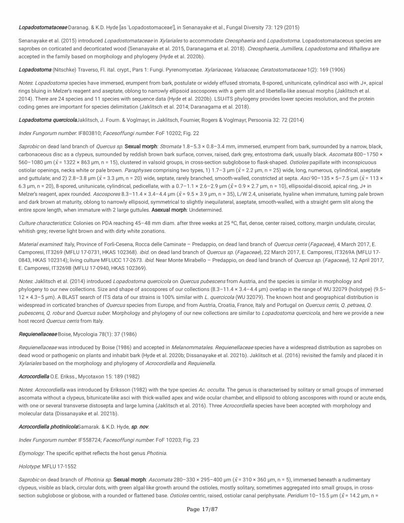
Page 17/87
Lopadostomataceae Daranag. & K.D. Hyde [as 'Lopadostomaceae'], in Senanayake et al., Fungal Diversity 73: 129 (2015)
Senanayake et al. (2015) introduced Lopadostomataceae in Xylariales to accommodate Creosphaeria and Lopadostoma. Lopadostomataceous species aresaprobes on corticated and decorticated wood (Senanayake et al. 2015, Daranagama et al. 2018). Creosphaeria, Jumillera, Lopadostoma and Whalleya areaccepted in the family based on morphology and phylogeny (Hyde et al. 2020b).
Lopadostoma (Nitschke) Traverso, Fl. ital. crypt., Pars 1: Fungi. Pyrenomycetae. Xylariaceae, Valsaceae, Ceratostomataceae 1(2): 169 (1906)
Notes: Lopadostoma species have immersed, erumpent from bark, postulate or widely effused stromata, 8-spored, unitunicate, cylindrical asci with J+, apicalrings bluing in Melzer’s reagent and aseptate, oblong to narrowly ellipsoid ascospores with a germ slit and libertella-like asexual morphs (Jaklitsch et al.2014). There are 24 species and 11 species with sequence data (Hyde et al. 2020b). LSU-ITS phylogeny provides lower species resolution, and the proteincoding genes are important for species delimitation (Jaklitsch et al. 2014; Daranagama et al. 2018).
Lopadostoma quercicolaJaklitsch, J. Fourn. & Voglmayr, in Jaklitsch, Fournier, Rogers & Voglmayr, Persoonia 32: 72 (2014)
Index Fungorum number: IF803810; Facesoffungi number: FoF 10202; Fig. 22
Saprobic on dead land branch of Quercus sp. Sexual morph: Stromata 1.8–5.3 × 0.8–3.4 mm, immersed, erumpent from bark, surrounded by a narrow, black,carbonaceous disc as a clypeus, surrounded by reddish brown bark surface, convex, raised, dark grey, entostroma dark, usually black. Ascomata 800–1750 ×560–1080 μm (x̄ = 1322 × 863 μm, n = 15), clustered in valsoid groups, in cross-section subglobose to �ask-shaped. Ostioles papillate with inconspicuousostiolar openings, necks white or pale brown. Paraphyses comprising two types, 1) 1.7–3 μm (x̄ = 2.2 μm, n = 25) wide, long, numerous, cylindrical, aseptateand guttulate; and 2) 2.8–3.8 μm (x̄ = 3.3 μm, n = 20) wide, septate, rarely branched, smooth-walled, constricted at septa. Asci 90–135 × 5–7.5 μm (x̄ = 113 ×6.3 μm, n = 20), 8-spored, unitunicate, cylindrical, pedicellate, with a 0.7–1.1 × 2.6–2.9 μm (x̄ = 0.9 × 2.7 μm, n = 10), ellipsoidal-discoid, apical ring, J+ inMelzer’s reagent, apex rounded. Ascospores 8.3–11.4 × 3.4–4.4 μm (x̄ = 9.5 × 3.9 μm, n = 35), L/W 2.4, uniseriate, hyaline when immature, turning pale brownand dark brown at maturity, oblong to narrowly ellipsoid, symmetrical to slightly inequilateral, aseptate, smooth-walled, with a straight germ slit along theentire spore length, when immature with 2 large guttules. Asexual morph: Undetermined.
Culture characteristics: Colonies on PDA reaching 45–48 mm diam. after three weeks at 25 ºC, �at, dense, center raised, cottony, margin undulate, circular,whitish grey; reverse light brown and with dirty white zonations.
Material examined: Italy, Province of Forlì-Cesena, Rocca delle Caminate – Predappio, on dead land branch of Quercus cerris (Fagaceae), 4 March 2017, E.Camporesi, IT3269 (MFLU 17-0731, HKAS 102368). ibid. on dead land branch of Quercus sp. (Fagaceae), 22 March 2017, E. Camporesi, IT3269A (MFLU 17-0843, HKAS 102314); living culture MFLUCC 17-2673. ibid. Near Monte Mirabello – Predappio, on dead land branch of Quercus sp. (Fagaceae), 12 April 2017,E. Camporesi, IT3269B (MFLU 17-0940, HKAS 102369).
Notes: Jaklitsch et al. (2014) introduced Lopadostoma quercicola on Quercus pubescens from Austria, and the species is similar in morphology andphylogeny to our new collections. Size and shape of ascospores of our collections (8.3–11.4 × 3.4–4.4 μm) overlap in the range of WU 32079 (holotype) (9.5–12 × 4.3–5 μm). A BLAST search of ITS data of our strains is 100% similar with L. quercicola (WU 32079). The known host and geographical distribution iswidespread in corticated branches of Quercus species from Europe, and from Austria, Croatia, France, Italy and Portugal on Quercus cerris, Q. petraea, Q.pubescens, Q. robur and Quercus suber. Morphology and phylogeny of our new collections are similar to Lopadostoma quercicola, and here we provide a newhost record Quercus cerris from Italy.
Requienellaceae Boise, Mycologia 78(1): 37 (1986)
Requienellaceae was introduced by Boise (1986) and accepted in Melanommatales. Requienellaceae species have a widespread distribution as saprobes ondead wood or pathogenic on plants and inhabit bark (Hyde et al. 2020b; Dissanayake et al. 2021b). Jaklitsch et al. (2016) revisited the family and placed it inXylariales based on the morphology and phylogeny of Acrocordiella and Requienella.
Acrocordiella O.E. Erikss., Mycotaxon 15: 189 (1982)
Notes: Acrocordiella was introduced by Eriksson (1982) with the type species Ac. occulta. The genus is characterised by solitary or small groups of immersedascomata without a clypeus, bitunicate-like asci with thick-walled apex and wide ocular chamber, and ellipsoid to oblong ascospores with round or acute ends,with one or several transverse distosepta and large lumina (Jaklitsch et al. 2016). Three Acrocordiella species have been accepted with morphology andmolecular data (Dissanayake et al. 2021b).
Acrocordiella photiniicolaSamarak. & K.D. Hyde, sp. nov.
Index Fungorum number: IF558724; Facesoffungi number: FoF 10203; Fig. 23
Etymology: The speci�c epithet re�ects the host genus Photinia.
Holotype: MFLU 17-1552
Saprobic on dead branch of Photinia sp. Sexual morph: Ascomata 280–330 × 295–400 μm (x̄ = 310 × 360 μm, n = 5), immersed beneath a rudimentaryclypeus, visible as black, circular dots, with green algal-like growth around the ostioles, mostly solitary, sometimes aggregated into small groups, in cross-section subglobose or globose, with a rounded or �attened base. Ostioles centric, raised, ostiolar canal periphysate. Peridium 10–15.5 μm (x̄ = 14.2 μm, n =
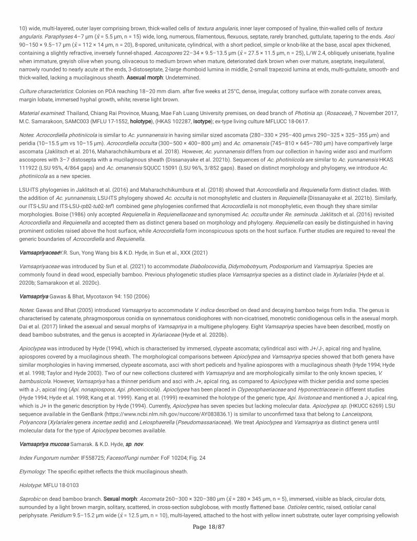
Page 18/87
10) wide, multi-layered, outer layer comprising brown, thick-walled cells of textura angularis, inner layer composed of hyaline, thin-walled cells of texturaangularis. Paraphyses 4–7 μm (x̄ = 5.5 μm, n = 15) wide, long, numerous, �lamentous, �exuous, septate, rarely branched, guttulate, tapering to the ends. Asci90–150 × 9.5–17 μm (x̄ = 112 × 14 μm, n = 20), 8-spored, unitunicate, cylindrical, with a short pedicel, simple or knob-like at the base, ascal apex thickened,containing a slightly refractive, inversely funnel-shaped. Ascospores 22–34 × 9.5–13.5 μm (x̄ = 27.5 × 11.5 μm, n = 25), L/W 2.4, obliquely uniseriate, hyalinewhen immature, greyish olive when young, olivaceous to medium brown when mature, deteriorated dark brown when over mature, aseptate, inequilateral,narrowly rounded to nearly acute at the ends, 3-distoseptate, 2-large rhomboid lumina in middle, 2-small trapezoid lumina at ends, multi-guttulate, smooth- andthick-walled, lacking a mucilaginous sheath. Asexual morph: Undetermined.
Culture characteristics: Colonies on PDA reaching 18–20 mm diam. after �ve weeks at 25°C, dense, irregular, cottony surface with zonate convex areas,margin lobate, immersed hyphal growth, white; reverse light brown.
Material examined: Thailand, Chiang Rai Province, Muang, Mae Fah Luang University premises, on dead branch of Photinia sp. (Rosaceae), 7 November 2017,M.C. Samarakoon, SAMC003 (MFLU 17-1552, holotype), (HKAS 102287, isotype); ex-type living culture MFLUCC 18-0617.
Notes: Acrocordiella photiniicola is similar to Ac. yunnanensis in having similar sized ascomata (280–330 × 295–400 μmvs 290–325 × 325–355 μm) andperidia (10–15.5 μm vs 10–15 μm). Acrocordiella occulta (300–500 × 400–800 μm) and Ac. omanensis (745–810 × 645–780 μm) have compartively largeascomata (Jaklitsch et al. 2016, Maharachchikumbura et al. 2018). However, Ac. yunnanensis differs from our collection in having wider asci and muriformascospores with 3–7 distosepta with a mucilaginous sheath (Dissanayake et al. 2021b). Sequences of Ac. photiniicola are similar to Ac. yunnanensis HKAS111922 (LSU 95%, 4/864 gaps) and Ac. omanensis SQUCC 15091 (LSU 96%, 3/852 gaps). Based on distinct morphology and phylogeny, we introduce Ac.photiniicola as a new species.
LSU-ITS phylogenies in Jaklitsch et al. (2016) and Maharachchikumbura et al. (2018) showed that Acrocordiella and Requienella form distinct clades. Withthe addition of Ac. yunnanensis, LSU-ITS phylogeny showed Ac. occulta is not monophyletic and clusters in Requienella (Dissanayake et al. 2021b). Similarly,our ITS-LSU and ITS-LSU-rpb2-tub2-tef1 combined gene phylogenies con�rmed that Acrocordiella is not monophyletic, even though they share similarmorphologies. Boise (1986) only accepted Requienella in Requienellaceae and synonymised Ac. occulta under Re. seminuda. Jaklitsch et al. (2016) revisitedAcrocordiella and Requienella and accepted them as distinct genera based on morphology and phylogeny. Requienella can easily be distinguished in havingprominent ostioles raised above the host surface, while Acrocordiella form inconspicuous spots on the host surface. Further studies are required to reveal thegeneric boundaries of Acrocordiella and Requienella.
VamsapriyaceaeY.R. Sun, Yong Wang bis & K.D. Hyde, in Sun et al., XXX (2021)
Vamsapriyaceae was introduced by Sun et al. (2021) to accommodate Diabolocovidia, Didymobotryum, Podosporium and Vamsapriya. Species arecommonly found in dead wood, especially bamboo. Previous phylogenetic studies place Vamsapriya species as a distinct clade in Xylariales (Hyde et al.2020b; Samarakoon et al. 2020c).
Vamsapriya Gawas & Bhat, Mycotaxon 94: 150 (2006)
Notes: Gawas and Bhat (2005) introduced Vamsapriya to accommodate V. indica described on dead and decaying bamboo twigs from India. The genus ischaracterised by catenate, phragmosporous conidia on synnematous conidiophores with non-cicatrised, monotretic conidiogenous cells in the asexual morph.Dai et al. (2017) linked the asexual and sexual morphs of Vamsapriya in a multigene phylogeny. Eight Vamsapriya species have been described, mostly ondead bamboo substrates, and the genus is accepted in Xylariaceae (Hyde et al. 2020b).
Apioclypea was introduced by Hyde (1994), which is characterised by immersed, clypeate ascomata; cylindrical asci with J+/J-, apical ring and hyaline,apiospores covered by a mucilaginous sheath. The morphological comparisons between Apioclypea and Vamsapriya species showed that both genera havesimilar morphologies in having immersed, clypeate ascomata, asci with short pedicels and hyaline apiospores with a mucilaginous sheath (Hyde 1994; Hydeet al. 1998; Taylor and Hyde 2003). Two of our new collections clustered with Vamsapriya and are morphologically similar to the only known species, V.bambusicola. However, Vamsapriya has a thinner peridium and asci with J+, apical ring, as compared to Apioclypea with thicker peridia and some specieswith a J-, apical ring (Api. nonapiospora, Api. phoenicicola). Apioclypea has been placed in Clypeosphaeriaceae and Hyponectriaceae in different studies(Hyde 1994; Hyde et al. 1998; Kang et al. 1999). Kang et al. (1999) re-examined the holotype of the generic type, Api. livistonae and mentioned a J-, apical ring,which is J+ in the generic description by Hyde (1994). Currently, Apioclypea has seven species but lacking molecular data. Apioclypea sp. (HKUCC 6269) LSUsequence available in the GenBank (https://www.ncbi.nlm.nih.gov/nuccore/AY083836.1) is similar to uncon�rmed taxa that belong to Lanceispora,Polyancora (Xylariales genera incertae sedis) and Leiosphaerella (Pseudomassariaceae). We treat Apioclypea and Vamsapriya as distinct genera untilmolecular data for the type of Apioclypea becomes available.
Vamsapriya mucosa Samarak. & K.D. Hyde, sp. nov.
Index Fungorum number: IF558725; Facesoffungi number: FoF 10204; Fig. 24
Etymology: The speci�c epithet re�ects the thick mucilaginous sheath.
Holotype: MFLU 18-0103
Saprobic on dead bamboo branch. Sexual morph: Ascomata 260–300 × 320–380 μm (x̄ = 280 × 345 μm, n = 5), immersed, visible as black, circular dots,surrounded by a light brown margin, solitary, scattered, in cross-section subglobose, with mostly �attened base. Ostioles centric, raised, ostiolar canalperiphysate. Peridium 9.5–15.2 μm wide (x̄ = 12.5 μm, n = 10), multi-layered, attached to the host with yellow innert substrate, outer layer comprising yellowish
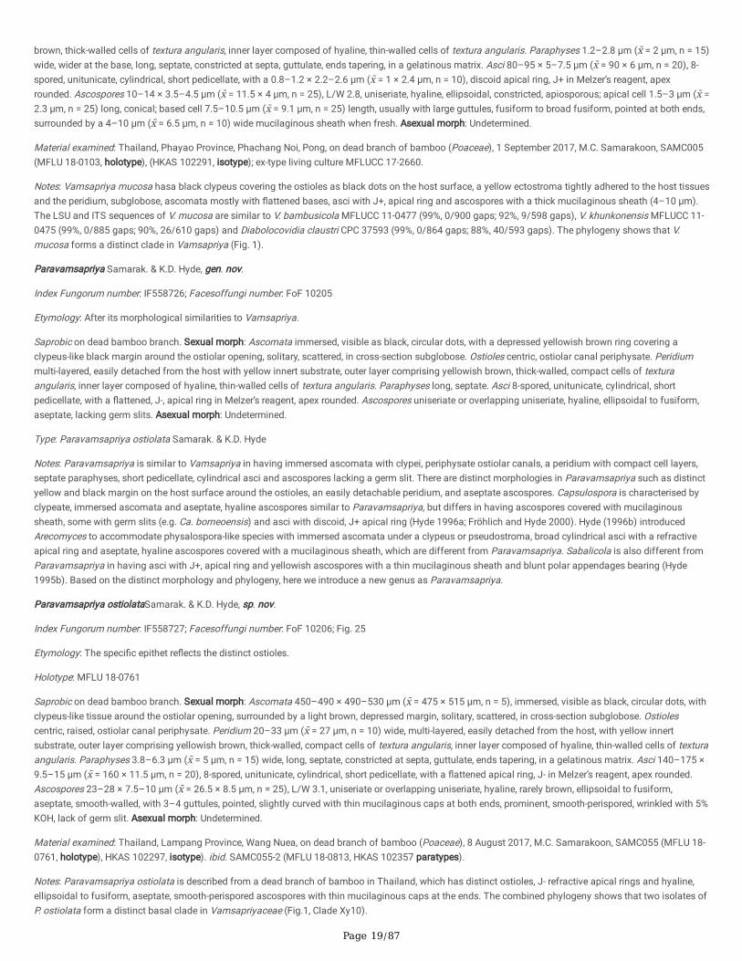
Page 19/87
brown, thick-walled cells of textura angularis, inner layer composed of hyaline, thin-walled cells of textura angularis. Paraphyses 1.2–2.8 μm (x̄ = 2 μm, n = 15)wide, wider at the base, long, septate, constricted at septa, guttulate, ends tapering, in a gelatinous matrix. Asci 80–95 × 5–7.5 μm (x̄ = 90 × 6 μm, n = 20), 8-spored, unitunicate, cylindrical, short pedicellate, with a 0.8–1.2 × 2.2–2.6 μm (x̄ = 1 × 2.4 μm, n = 10), discoid apical ring, J+ in Melzer’s reagent, apexrounded. Ascospores 10–14 × 3.5–4.5 μm (x̄ = 11.5 × 4 μm, n = 25), L/W 2.8, uniseriate, hyaline, ellipsoidal, constricted, apiosporous; apical cell 1.5–3 μm (x̄ =2.3 μm, n = 25) long, conical; based cell 7.5–10.5 μm (x̄ = 9.1 μm, n = 25) length, usually with large guttules, fusiform to broad fusiform, pointed at both ends,surrounded by a 4–10 μm (x̄ = 6.5 μm, n = 10) wide mucilaginous sheath when fresh. Asexual morph: Undetermined.
Material examined: Thailand, Phayao Province, Phachang Noi, Pong, on dead branch of bamboo (Poaceae), 1 September 2017, M.C. Samarakoon, SAMC005(MFLU 18-0103, holotype), (HKAS 102291, isotype); ex-type living culture MFLUCC 17-2660.
Notes: Vamsapriya mucosa hasa black clypeus covering the ostioles as black dots on the host surface, a yellow ectostroma tightly adhered to the host tissuesand the peridium, subglobose, ascomata mostly with �attened bases, asci with J+, apical ring and ascospores with a thick mucilaginous sheath (4–10 μm).The LSU and ITS sequences of V. mucosa are similar to V. bambusicola MFLUCC 11-0477 (99%, 0/900 gaps; 92%, 9/598 gaps), V. khunkonensis MFLUCC 11-0475 (99%, 0/885 gaps; 90%, 26/610 gaps) and Diabolocovidia claustri CPC 37593 (99%, 0/864 gaps; 88%, 40/593 gaps). The phylogeny shows that V.mucosa forms a distinct clade in Vamsapriya (Fig. 1).
Paravamsapriya Samarak. & K.D. Hyde, gen. nov.
Index Fungorum number: IF558726; Facesoffungi number: FoF 10205
Etymology: After its morphological similarities to Vamsapriya.
Saprobic on dead bamboo branch. Sexual morph: Ascomata immersed, visible as black, circular dots, with a depressed yellowish brown ring covering aclypeus-like black margin around the ostiolar opening, solitary, scattered, in cross-section subglobose. Ostioles centric, ostiolar canal periphysate. Peridiummulti-layered, easily detached from the host with yellow innert substrate, outer layer comprising yellowish brown, thick-walled, compact cells of texturaangularis, inner layer composed of hyaline, thin-walled cells of textura angularis. Paraphyses long, septate. Asci 8-spored, unitunicate, cylindrical, shortpedicellate, with a �attened, J-, apical ring in Melzer’s reagent, apex rounded. Ascospores uniseriate or overlapping uniseriate, hyaline, ellipsoidal to fusiform,aseptate, lacking germ slits. Asexual morph: Undetermined.
Type: Paravamsapriya ostiolata Samarak. & K.D. Hyde
Notes: Paravamsapriya is similar to Vamsapriya in having immersed ascomata with clypei, periphysate ostiolar canals, a peridium with compact cell layers,septate paraphyses, short pedicellate, cylindrical asci and ascospores lacking a germ slit. There are distinct morphologies in Paravamsapriya such as distinctyellow and black margin on the host surface around the ostioles, an easily detachable peridium, and aseptate ascospores. Capsulospora is characterised byclypeate, immersed ascomata and aseptate, hyaline ascospores similar to Paravamsapriya, but differs in having ascospores covered with mucilaginoussheath, some with germ slits (e.g. Ca. borneoensis) and asci with discoid, J+ apical ring (Hyde 1996a; Fröhlich and Hyde 2000). Hyde (1996b) introducedArecomyces to accommodate physalospora-like species with immersed ascomata under a clypeus or pseudostroma, broad cylindrical asci with a refractiveapical ring and aseptate, hyaline ascospores covered with a mucilaginous sheath, which are different from Paravamsapriya. Sabalicola is also different fromParavamsapriya in having asci with J+, apical ring and yellowish ascospores with a thin mucilaginous sheath and blunt polar appendages bearing (Hyde1995b). Based on the distinct morphology and phylogeny, here we introduce a new genus as Paravamsapriya.
Paravamsapriya ostiolataSamarak. & K.D. Hyde, sp. nov.
Index Fungorum number: IF558727; Facesoffungi number: FoF 10206; Fig. 25
Etymology: The speci�c epithet re�ects the distinct ostioles.
Holotype: MFLU 18-0761
Saprobic on dead bamboo branch. Sexual morph: Ascomata 450–490 × 490–530 μm (x̄ = 475 × 515 μm, n = 5), immersed, visible as black, circular dots, withclypeus-like tissue around the ostiolar opening, surrounded by a light brown, depressed margin, solitary, scattered, in cross-section subglobose. Ostiolescentric, raised, ostiolar canal periphysate. Peridium 20–33 μm (x̄ = 27 μm, n = 10) wide, multi-layered, easily detached from the host, with yellow innertsubstrate, outer layer comprising yellowish brown, thick-walled, compact cells of textura angularis, inner layer composed of hyaline, thin-walled cells of texturaangularis. Paraphyses 3.8–6.3 μm (x̄ = 5 μm, n = 15) wide, long, septate, constricted at septa, guttulate, ends tapering, in a gelatinous matrix. Asci 140–175 ×9.5–15 μm (x̄ = 160 × 11.5 μm, n = 20), 8-spored, unitunicate, cylindrical, short pedicellate, with a �attened apical ring, J- in Melzer’s reagent, apex rounded.Ascospores 23–28 × 7.5–10 μm (x̄ = 26.5 × 8.5 μm, n = 25), L/W 3.1, uniseriate or overlapping uniseriate, hyaline, rarely brown, ellipsoidal to fusiform,aseptate, smooth-walled, with 3–4 guttules, pointed, slightly curved with thin mucilaginous caps at both ends, prominent, smooth-perispored, wrinkled with 5%KOH, lack of germ slit. Asexual morph: Undetermined.
Material examined: Thailand, Lampang Province, Wang Nuea, on dead branch of bamboo (Poaceae), 8 August 2017, M.C. Samarakoon, SAMC055 (MFLU 18-0761, holotype), HKAS 102297, isotype). ibid. SAMC055-2 (MFLU 18-0813, HKAS 102357 paratypes).
Notes: Paravamsapriya ostiolata is described from a dead branch of bamboo in Thailand, which has distinct ostioles, J- refractive apical rings and hyaline,ellipsoidal to fusiform, aseptate, smooth-perispored ascospores with thin mucilaginous caps at the ends. The combined phylogeny shows that two isolates ofP. ostiolata form a distinct basal clade in Vamsapriyaceae (Fig.1, Clade Xy10).
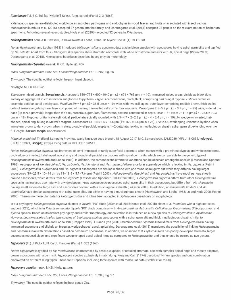
Page 20/87
Xylariaceae Tul. & C. Tul. [as 'Xylariei'], Select. fung. carpol. (Paris) 2: 3 (1863)
Xylariaceous species are distributed worldwide as saprobes, pathogens and endophytes in wood, leaves and fruits or associated with insect vectors.Maharachchikumbura et al. (2016) accepted 87 genera into the family, and Daranagama et al. (2018) accepted 37 genera on the re-examination of herbariumspecimens. Following several recent studies, Hyde et al. (2020b) accepted 32 genera in Xylariaceae.
Helicogermslita Lodha & D. Hawksw., in Hawksworth & Lodha, Trans. Br. Mycol. Soc. 81(1): 91 (1983)
Notes: Hawksworth and Lodha (1983) introduced Helicogermslita to accommodate a xylarialean species with ascospores having spiral germ slits and typi�edby He. celastri. Apart from this, Helicogermslita species share stromatic ascomata with white ectostroma and asci with J+, apical rings (Petrini 2003;Daranagama et al. 2018). Nine species have been described based only on morphology.
Helicogermslita clypeataSamarak. & K.D. Hyde, sp. nov.
Index Fungorum number: IF558728; Facesoffungi number: FoF 10207; Fig. 26
Etymology: The speci�c epithet re�ects the prominent clypeus.
Holotype: MFLU 18-0852
Saprobic on dead branch. Sexual morph: Ascomata 550–775 × 600–1040 μm (x̄ = 671 × 763 μm, n = 10), immersed, raised areas, visible as black dots,solitary or aggregated, in cross-section subglobose to pyriform. Clypeus carbonaceous, black, thick, comprising dark fungal hyphae. Ostioles centric oreccentric, ostiolar canal periphysate. Peridium 29–45 μm (x̄ = 36.5 μm, n = 10) wide, with two cell layers, outer layer comprising reddish brown, thick-walledcells of textura angularis, inner layer composed of hyaline, thin-walled cells of textura angularis. Paraphyses 2.5–5.2 μm (x̄ = 3.7 μm, n = 25) wide, wider at thebase (up to 9.5 μm wide), longer than the asci, numerous, guttulate, �lamentous, septate, constricted at septa. Asci 115–145 × 9–11.5 μm (x̄ = 128.5 × 10.3μm, n = 18), 8-spored, unitunicate, cylindrical, pedicellate, apically rounded, with 3.5–4.7 × 2–2.8 μm (x̄ = 4 × 2.4 μm, n = 10), J+, wedge- or inverted, hat-shaped, apical ring, bluing in Melzer’s reagent. Ascospores 13–18.5 × 5.7–7.6 μm (x̄ = 16.2 × 6.6 μm, n = 25), L/W 2.45, overlapping uniseriate, hyaline whenimmature, brown to dark brown when mature, broadly ellipsoidal, aseptate, 1–2-guttulate, lacking a mucilaginous sheath, spiral germ slit extending over thefull length. Asexual morph: Undetermined.
Material examined: Thailand, Lampang Province, Wang Nuea, on dead branch, 18 August 2017, M.C. Samarakoon, SAMC080 (MFLU 18-0852, holotype),(HKAS 102321, isotype); ex-type living culture MFLUCC 18-0517.
Notes: Helicogermslita clypeata has immersed or semi immersed or rarely super�cial ascomata when mature with a prominent clypeus and white ectostroma,J+, wedge- or inverted, hat-shaped, apical ring and broadly ellipsoidal ascospores with spiral germ slits, which are comparable to the generic type ofHelicogermslita (Hawksworth and Lodha 1983). In addition, the carbonaceous stromatic variations can be observed among the species (Læssøe and Spooner1993). Ascospores of He. �eischhakii, He. gisbornia, He. johnstonii and He. mackenziei bear a cellular appendage, which is lacking in He. clypeata (Petrini2003). Helicogermslita aucklandica and He. clypeata ascospores are similar in shape with one round spiral germ slit, while they differ in having largerascospores (19–23.5 × 10⁻¹⁴ μm vs 13–18.5 × 5.7–7.6 μm) (Petrini 2003). Helicogermslita �eischhakii and He. gaudefroyi have mucilaginous sheatharound ascospores, which differs from He. clypeata (Læssøe and Spooner 1993; Petrini 2003). Helicogermslita clypeata differs from other Helicogermslitaspecies in having large asomata with a wide clypeus. Yuea chusqueicola possesses spiral germ slits in their ascospores, but differs from He. clypeata inhaving small ascomata, large asci and ascospores covered with a mucilaginous sheath (Eriksson 2003). In addition, Anthostomella limitata and An.umbrinella have similar ascospores with spiral germ slits, but differ in having a mucilaginous sheath (Hawksworth and Lodha 1983; Lu and Hyde 2000; Petrini2003). There is no molecular data for Helicogermslita, and it has been accepted in Xylariaceae based only on morphology.
In our phylogeny, Helicogermslita clypeata clusters in Xylaria “PO” clade (U’Ren et al. 2016; Konta et al. 2021b) sister to X. frustulosa with a high statisticalsupport (92%), which is in Xylaria sensu lato. Xylaria “PO” clade comprises with Amphirosellinia, Astrocystis, Collodiscula, Kretzmariella, Stilbohypoxylon andXylaria species. Based on its distinct phylogeny and similar morphology, our collection is introduced as a new species of Helicogermslita in Xylariaceae.However, Leptomassaria simplex, type species of Leptomassaria has ascospores with a spiral germ slit and thick mucilaginous sheath similar toHelicogermslita (Hawksworth and Lodha 1983; Rappaz 1995). Lu and Hyde (2000) mentioned that Leptomassaria differs from Helicogermslita in havingimmersed ascomata and slightly an irregular, wedge-shaped, ascal, apical ring. Daranagama et al. (2018) mentioned the possibility of linking Helicogermslitaand Leptomassaria with observations based on herbarium specimens. In addition, we observed that Leptomassaria has poorly developed stromata, largerascomata, reduced clypei and signi�cant wedge-shaped ascal apical rings as compared to Helicogermslita, and thus should be treated as two genera.
Hypocopra (Fr.) J. Kickx f., Fl. Crypt. Flandres (Paris) 1: 362 (1867)
Notes: Hypocopra is typi�ed by Hy. merdaria and characterised by sessile, clypeoid, or reduced stromata; asci with complex apical rings and mostly aseptate,brown ascospores with a germ slit. Hypocopra species exclusively inhabit dung. Krug and Cain (1974) described 14 new species and one combinationdiscovered on different dung types. There are 31 species, including three species with molecular data (Becker et al. 2020).
Hypocopra zeaeSamarak. & K.D. Hyde, sp. nov.
Index Fungorum number: IF558729; Facesoffungi number: FoF 10208; Fig. 27
Etymology: The speci�c epithet re�ects the host genus Zea.

Page 21/87
Holotype: MFLU 18-0809
Saprobic on dead culm of Zea mays. Sexual morph: Ascomata 255–305 × 185–290 μm (x̄ = 280 × 250 μm, n = 10), immersed, visible as greyish black, raisedareas, solitary or aggregated, in cross-section globose to subglobose, sometimes with a �attened base. Clypeus carbonaceous, greyish black, thick-walled,short, comprising dark fungal hyphae and host epidermal cells. Ostioles centric, ostiolar canal periphysate. Peridium 35–50 μm (x̄ = 43 μm, n = 10) wide,tightly attached to the host, with two cell layers, outer layer thick, yellowish brown, thick-walled cells of textura angularis, inner layer thin, composed of hyaline,thin-walled cells of textura angularis. Paraphyses 2–5.5 μm (x̄ = 3.4 μm, n = 25) wide, wider at the base, longer than the asci, numerous, �lamentous, septate,rarely branched, constricted at septa, guttulate, apically blunt. Asci 65–85 × 5–7.5 μm (x̄ = 73 × 6.5 μm, n = 25), 8-spored, unitunicate, cylindrical, shortpedicellate, apically rounded with a 1.5–2 × 2–2.6 μm (x̄ = 1.8 × 2.3 μm, n = 15), rectangular to slightly obconic apical ring, J+ in Melzer’s reagent. Ascospores8–10 × 3.5–5 μm (x̄ = 9 × 4.4 μm, n = 35), L/W 2.04, uniseriate, brown, inequilaterally ellipsoidal, aseptate, 1–2-guttulate, germ slit on ventral side of theascospore along part of the spore. Asexual morph: Undetermined.
Material examined: Thailand, Phayao Province, Phachang Noi, Pong, on dead culm of Zea mays (Poaceae), 11 September 2017, M.C. Samarakoon, SAMC030(MFLU 18-0809, holotype), (HKAS 102327, isotype).
Notes: Hypocopra zeae is similar to xylariaceous taxa in having immersed ascomata under a clypeus, septate, hyaline paraphyses, 8-spored, unitunicate,cylindrical asci with a J+, apical ring and uniseriate, brown ascospores with a short germ slit. Anthostomelloid taxa such as An. eructans, An. delitesnems, andAn. pedemontana have similar ascospores with a short germ slit, but differs in the J-, apical ring and a mucilaginous sheath, while An. aquatica from aquatichabitats has larger ascomata, asci and ascospores (Lu and Hyde 2000). In having immersed ascomata under a clypeus, J+ apical ring and ascospores with ashort germ slit, our new collection is similar to Hypocopra than Stromatoneurospora. The LSU, ITS and rpb2 sequences of Hy. zeae are similar to Hy. rostrataNRRL 66178 (99%, 1/911 gaps), Podosordaria muli WSP 167 (89%, 19/585 gaps) and S. phoenix BCC82040 (92%, 0/705 gaps) respectively. Single andcombined gene phylogenies show that our new collection clusters with Stromatoneurospora phoenix. There are few Hypocopra species and onlyStromatoneurospora phoenix with molecular data (Becker et al. 2020). Therefore, the phylogenetic placement could be changed with the addition of newsequence data. Giving priority to morphology, we tentatively place our collection as a Hypocopra species. In addition, there are no Hypocopra species that havebeen described from plant substrates. Further collections are required in view of the uncertain taxonomic placement of the Hy. zeae.
Nemania Gray, Nat. Arr. Brit. Pl. (London) 1: 516 (1821)
Notes: Nemania is a species-rich genus (typi�ed by Ne. serpens) including endophytes, saprobes and pathogens (U’Ren et al. 2016; Daranagama et al. 2018;Hyde et al. 2020b). Nemania species are characterised by stromata not associated with bark rupturing structures, lacking KOH-extractable pigments and �nelypapillate ostioles, unitunicate, cylindrical asci, and ascospores with a germ slit in their sexual morph, and geniculosporium-like asexual morph (Daranagama etal. 2018). Currently, there are 55 species and 16 with molecular data (Hyde et al. 2020b; Tibpromma et al. 2021).
Nemania longipedicellata Samarak. & K.D. Hyde, sp. nov.
Index Fungorum number: IF558730; Facesoffungi number: FoF 10209; Fig. 28
Etymology: The speci�c epithet re�ects the long pedicellate asci.
Holotype: MFLU 18-0819
Saprobic on dead branch of Camellia sinensis. Sexual morph: Stromata 480–720 × 620–870 μm (x̄ = 592 × 768 μm, n = 6), super�cial or semi-immersed,single or rarely aggregated, mammiformis, dark brown to black, with conspicuous ascomatal mounds, carbonaceous. Ascomata 590–670 × 550–660 μm (x̄ =628 × 608 μm, n = 5), in cross-section globose to sphaerical or sometimes ovoid. Ostioles surrounding area slightly �attened, shiny black,conspicuous. Paraphyses 1.9–2.8 μm (x̄ = 2.3 μm, n = 15) wide, long, cylindrical, septate, constricted at septum, smooth-walled. Asci 72–125 × 5.2–8.2 μm (x̄= 93.6 × 6.3 μm, n = 20), spore-bearing part 50–57.5 μm (x̄ = 53.8 μm, n = 15), 8-spored, unitunicate, cylindrical, pedicellate, with a 1.5–2.3 × 1.8–2.4 μm (x̄ = 2× 2.2 μm, n = 10), inverted, hat-shaped, apical ring, J+ in Melzer’s reagent, apex rounded. Ascospores 6.8–9.3 × 3.6–4.7 μm (x̄ = 7.8 × 4.1 μm, n = 30), L/W 1.9,uniseriate, hyaline, guttulate when immature, turning pale brown and dark brown when mature, oblong to narrowly ellipsoid, symmetrical to slightlyinequilateral, aseptate, smooth-walled, with a straight germ slit along the entire spore length. Asexual morph: Undetermined.
Material examined: Thailand, Chiang Mai Province, Tambon Pa Pae, Amphoe Mae Taeng, on dead branch of Camellia sinensis (Theaceae), 1 September 2017,M.C. Samarakoon, SAMC045 (MFLU 18-0819, holotype), (HKAS 102329, isotype).
Notes: Nemania longipedicellata differs from closely related species with mammiform stromata such as Ne. kauaiensis, Ne. parapauzarii, and Ne.thailandensis in having long pedicellate asci and smaller ascospores (Rogers and Ju 2002; Rogers et al. 2008). Nemania carbonacea has long, sterile, hyphoidbased asci (ca. 80 vs 45–60μm), and larger ascospores (11.5–15.5 × 5–6.5 vs 6.8–9.3 × 3.6–4.7 μm) compared to Ne. longipedicellata (Pouzar 1985).Nemania pouzarii ascospores have hyaline, cellular remnants, whereas Ne. longipedicellata lacks cellular remnants. Nemania longipedicellata sequences aresimilar to Ne. macrocarpa WSP 265 (ITS 91%, 5/378 gaps; tub2 80%, 24/488 gaps), Ne. viridis MFLU 17-2600 (ITS 90%, 10/393 gaps), Ne. maritimaHAST 89120401 (rpb2 84%, 0/926 gaps) and Ne. abortiva BISH 467 (tub2 80%, 19/472 gaps). The combined gene phylogeny resulted in Ne. longipedicellataas a distinct clade with 99% statistical support. We introduce our collection as a new species among other mammiform stromata forming Nemania speciesbased on morphology and phylogeny.
Nemania delonicis Samarak. & K.D. Hyde, sp. nov.
Index Fungorum number: IF558731; Facesoffungi number: FoF 10210; Fig. 29
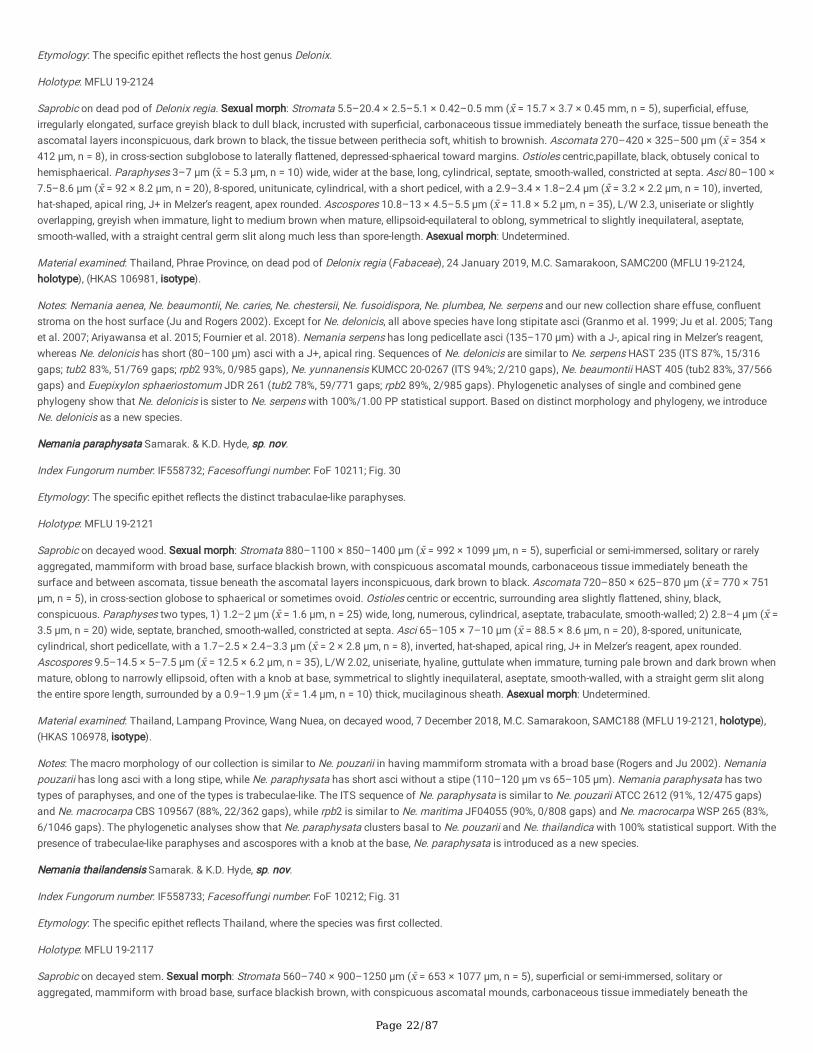
Page 22/87
Etymology: The speci�c epithet re�ects the host genus Delonix.
Holotype: MFLU 19-2124
Saprobic on dead pod of Delonix regia. Sexual morph: Stromata 5.5–20.4 × 2.5–5.1 × 0.42–0.5 mm (x̄ = 15.7 × 3.7 × 0.45 mm, n = 5), super�cial, effuse,irregularly elongated, surface greyish black to dull black, incrusted with super�cial, carbonaceous tissue immediately beneath the surface, tissue beneath theascomatal layers inconspicuous, dark brown to black, the tissue between perithecia soft, whitish to brownish. Ascomata 270–420 × 325–500 μm (x̄ = 354 ×412 μm, n = 8), in cross-section subglobose to laterally �attened, depressed-sphaerical toward margins. Ostioles centric,papillate, black, obtusely conical tohemisphaerical. Paraphyses 3–7 μm (x̄ = 5.3 μm, n = 10) wide, wider at the base, long, cylindrical, septate, smooth-walled, constricted at septa. Asci 80–100 ×7.5–8.6 μm (x̄ = 92 × 8.2 μm, n = 20), 8-spored, unitunicate, cylindrical, with a short pedicel, with a 2.9–3.4 × 1.8–2.4 μm (x̄ = 3.2 × 2.2 μm, n = 10), inverted,hat-shaped, apical ring, J+ in Melzer’s reagent, apex rounded. Ascospores 10.8–13 × 4.5–5.5 μm (x̄ = 11.8 × 5.2 μm, n = 35), L/W 2.3, uniseriate or slightlyoverlapping, greyish when immature, light to medium brown when mature, ellipsoid-equilateral to oblong, symmetrical to slightly inequilateral, aseptate,smooth-walled, with a straight central germ slit along much less than spore-length. Asexual morph: Undetermined.
Material examined: Thailand, Phrae Province, on dead pod of Delonix regia (Fabaceae), 24 January 2019, M.C. Samarakoon, SAMC200 (MFLU 19-2124,holotype), (HKAS 106981, isotype).
Notes: Nemania aenea, Ne. beaumontii, Ne. caries, Ne. chestersii, Ne. fusoidispora, Ne. plumbea, Ne. serpens and our new collection share effuse, con�uentstroma on the host surface (Ju and Rogers 2002). Except for Ne. delonicis, all above species have long stipitate asci (Granmo et al. 1999; Ju et al. 2005; Tanget al. 2007; Ariyawansa et al. 2015; Fournier et al. 2018). Nemania serpens has long pedicellate asci (135–170 μm) with a J-, apical ring in Melzer’s reagent,whereas Ne. delonicis has short (80–100 μm) asci with a J+, apical ring. Sequences of Ne. delonicis are similar to Ne. serpens HAST 235 (ITS 87%, 15/316gaps; tub2 83%, 51/769 gaps; rpb2 93%, 0/985 gaps), Ne. yunnanensis KUMCC 20-0267 (ITS 94%; 2/210 gaps), Ne. beaumontii HAST 405 (tub2 83%, 37/566gaps) and Euepixylon sphaeriostomum JDR 261 (tub2 78%, 59/771 gaps; rpb2 89%, 2/985 gaps). Phylogenetic analyses of single and combined genephylogeny show that Ne. delonicis is sister to Ne. serpens with 100%/1.00 PP statistical support. Based on distinct morphology and phylogeny, we introduceNe. delonicis as a new species.
Nemania paraphysata Samarak. & K.D. Hyde, sp. nov.
Index Fungorum number: IF558732; Facesoffungi number: FoF 10211; Fig. 30
Etymology: The speci�c epithet re�ects the distinct trabaculae-like paraphyses.
Holotype: MFLU 19-2121
Saprobic on decayed wood. Sexual morph: Stromata 880–1100 × 850–1400 μm (x̄ = 992 × 1099 μm, n = 5), super�cial or semi-immersed, solitary or rarelyaggregated, mammiform with broad base, surface blackish brown, with conspicuous ascomatal mounds, carbonaceous tissue immediately beneath thesurface and between ascomata, tissue beneath the ascomatal layers inconspicuous, dark brown to black. Ascomata 720–850 × 625–870 μm (x̄ = 770 × 751μm, n = 5), in cross-section globose to sphaerical or sometimes ovoid. Ostioles centric or eccentric, surrounding area slightly �attened, shiny, black,conspicuous. Paraphyses two types, 1) 1.2–2 μm (x̄ = 1.6 μm, n = 25) wide, long, numerous, cylindrical, aseptate, trabaculate, smooth-walled; 2) 2.8–4 μm (x̄ =3.5 μm, n = 20) wide, septate, branched, smooth-walled, constricted at septa. Asci 65–105 × 7–10 μm (x̄ = 88.5 × 8.6 μm, n = 20), 8-spored, unitunicate,cylindrical, short pedicellate, with a 1.7–2.5 × 2.4–3.3 μm (x̄ = 2 × 2.8 μm, n = 8), inverted, hat-shaped, apical ring, J+ in Melzer’s reagent, apex rounded.Ascospores 9.5–14.5 × 5–7.5 μm (x̄ = 12.5 × 6.2 μm, n = 35), L/W 2.02, uniseriate, hyaline, guttulate when immature, turning pale brown and dark brown whenmature, oblong to narrowly ellipsoid, often with a knob at base, symmetrical to slightly inequilateral, aseptate, smooth-walled, with a straight germ slit alongthe entire spore length, surrounded by a 0.9–1.9 μm (x̄ = 1.4 μm, n = 10) thick, mucilaginous sheath. Asexual morph: Undetermined.
Material examined: Thailand, Lampang Province, Wang Nuea, on decayed wood, 7 December 2018, M.C. Samarakoon, SAMC188 (MFLU 19-2121, holotype),(HKAS 106978, isotype).
Notes: The macro morphology of our collection is similar to Ne. pouzarii in having mammiform stromata with a broad base (Rogers and Ju 2002). Nemaniapouzarii has long asci with a long stipe, while Ne. paraphysata has short asci without a stipe (110–120 μm vs 65–105 μm). Nemania paraphysata has twotypes of paraphyses, and one of the types is trabeculae-like. The ITS sequence of Ne. paraphysata is similar to Ne. pouzarii ATCC 2612 (91%, 12/475 gaps)and Ne. macrocarpa CBS 109567 (88%, 22/362 gaps), while rpb2 is similar to Ne. maritima JF04055 (90%, 0/808 gaps) and Ne. macrocarpa WSP 265 (83%,6/1046 gaps). The phylogenetic analyses show that Ne. paraphysata clusters basal to Ne. pouzarii and Ne. thailandica with 100% statistical support. With thepresence of trabeculae-like paraphyses and ascospores with a knob at the base, Ne. paraphysata is introduced as a new species.
Nemania thailandensis Samarak. & K.D. Hyde, sp. nov.
Index Fungorum number: IF558733; Facesoffungi number: FoF 10212; Fig. 31
Etymology: The speci�c epithet re�ects Thailand, where the species was �rst collected.
Holotype: MFLU 19-2117
Saprobic on decayed stem. Sexual morph: Stromata 560–740 × 900–1250 μm (x̄ = 653 × 1077 μm, n = 5), super�cial or semi-immersed, solitary oraggregated, mammiform with broad base, surface blackish brown, with conspicuous ascomatal mounds, carbonaceous tissue immediately beneath the
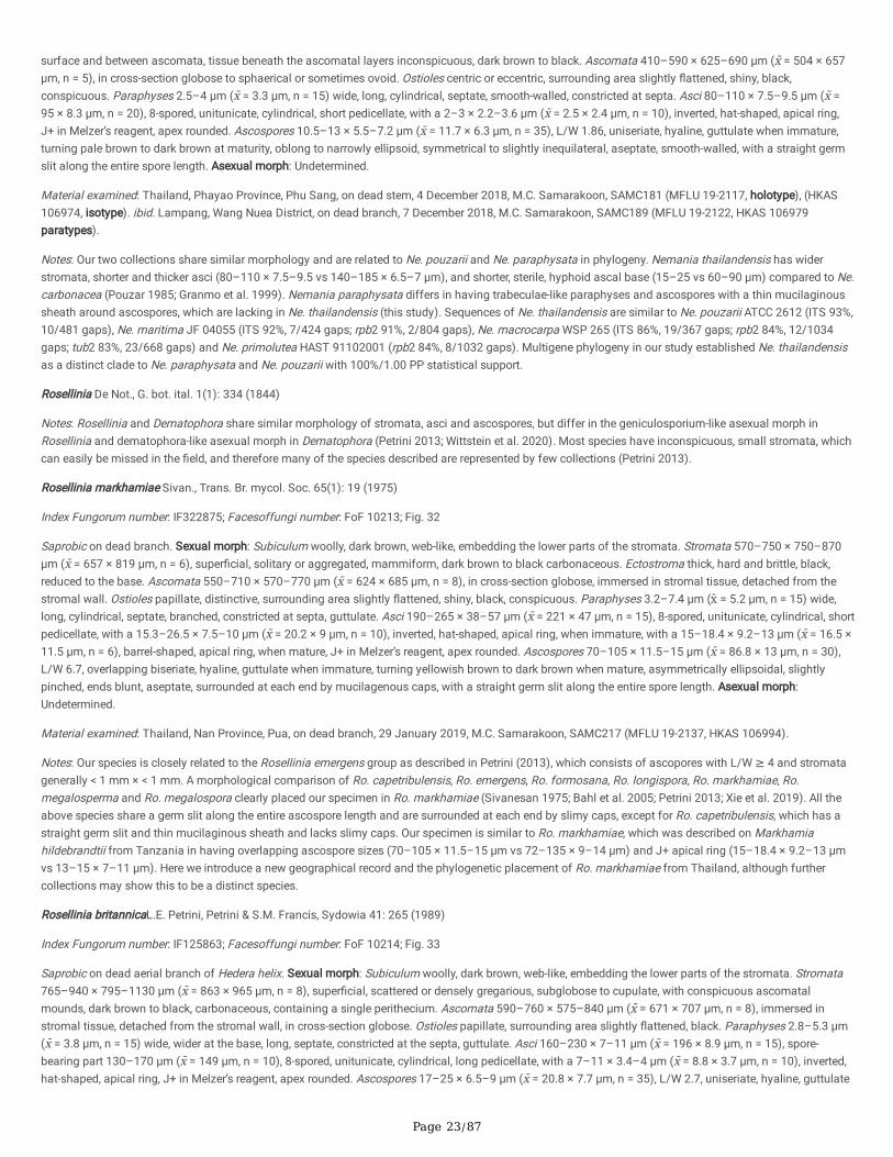
Page 23/87
surface and between ascomata, tissue beneath the ascomatal layers inconspicuous, dark brown to black. Ascomata 410–590 × 625–690 μm (x̄ = 504 × 657μm, n = 5), in cross-section globose to sphaerical or sometimes ovoid. Ostioles centric or eccentric, surrounding area slightly �attened, shiny, black,conspicuous. Paraphyses 2.5–4 μm (x̄ = 3.3 μm, n = 15) wide, long, cylindrical, septate, smooth-walled, constricted at septa. Asci 80–110 × 7.5–9.5 μm (x̄ =95 × 8.3 μm, n = 20), 8-spored, unitunicate, cylindrical, short pedicellate, with a 2–3 × 2.2–3.6 μm (x̄ = 2.5 × 2.4 μm, n = 10), inverted, hat-shaped, apical ring,J+ in Melzer’s reagent, apex rounded. Ascospores 10.5–13 × 5.5–7.2 μm (x̄ = 11.7 × 6.3 μm, n = 35), L/W 1.86, uniseriate, hyaline, guttulate when immature,turning pale brown to dark brown at maturity, oblong to narrowly ellipsoid, symmetrical to slightly inequilateral, aseptate, smooth-walled, with a straight germslit along the entire spore length. Asexual morph: Undetermined.
Material examined: Thailand, Phayao Province, Phu Sang, on dead stem, 4 December 2018, M.C. Samarakoon, SAMC181 (MFLU 19-2117, holotype), (HKAS106974, isotype). ibid. Lampang, Wang Nuea District, on dead branch, 7 December 2018, M.C. Samarakoon, SAMC189 (MFLU 19-2122, HKAS 106979paratypes).
Notes: Our two collections share similar morphology and are related to Ne. pouzarii and Ne. paraphysata in phylogeny. Nemania thailandensis has widerstromata, shorter and thicker asci (80–110 × 7.5–9.5 vs 140–185 × 6.5–7 μm), and shorter, sterile, hyphoid ascal base (15–25 vs 60–90 μm) compared to Ne.carbonacea (Pouzar 1985; Granmo et al. 1999). Nemania paraphysata differs in having trabeculae-like paraphyses and ascospores with a thin mucilaginoussheath around ascospores, which are lacking in Ne. thailandensis (this study). Sequences of Ne. thailandensis are similar to Ne. pouzarii ATCC 2612 (ITS 93%,10/481 gaps), Ne. maritima JF 04055 (ITS 92%, 7/424 gaps; rpb2 91%, 2/804 gaps), Ne. macrocarpa WSP 265 (ITS 86%, 19/367 gaps; rpb2 84%, 12/1034gaps; tub2 83%, 23/668 gaps) and Ne. primolutea HAST 91102001 (rpb2 84%, 8/1032 gaps). Multigene phylogeny in our study established Ne. thailandensisas a distinct clade to Ne. paraphysata and Ne. pouzarii with 100%/1.00 PP statistical support.
Rosellinia De Not., G. bot. ital. 1(1): 334 (1844)
Notes: Rosellinia and Dematophora share similar morphology of stromata, asci and ascospores, but differ in the geniculosporium-like asexual morph inRosellinia and dematophora-like asexual morph in Dematophora (Petrini 2013; Wittstein et al. 2020). Most species have inconspicuous, small stromata, whichcan easily be missed in the �eld, and therefore many of the species described are represented by few collections (Petrini 2013).
Rosellinia markhamiae Sivan., Trans. Br. mycol. Soc. 65(1): 19 (1975)
Index Fungorum number: IF322875; Facesoffungi number: FoF 10213; Fig. 32
Saprobic on dead branch. Sexual morph: Subiculum woolly, dark brown, web-like, embedding the lower parts of the stromata. Stromata 570–750 × 750–870μm (x̄ = 657 × 819 μm, n = 6), super�cial, solitary or aggregated, mammiform, dark brown to black carbonaceous. Ectostroma thick, hard and brittle, black,reduced to the base. Ascomata 550–710 × 570–770 μm (x̄ = 624 × 685 μm, n = 8), in cross-section globose, immersed in stromal tissue, detached from thestromal wall. Ostioles papillate, distinctive, surrounding area slightly �attened, shiny, black, conspicuous. Paraphyses 3.2–7.4 μm (x̄ = 5.2 μm, n = 15) wide,long, cylindrical, septate, branched, constricted at septa, guttulate. Asci 190–265 × 38–57 μm (x̄ = 221 × 47 μm, n = 15), 8-spored, unitunicate, cylindrical, shortpedicellate, with a 15.3–26.5 × 7.5–10 μm (x̄ = 20.2 × 9 μm, n = 10), inverted, hat-shaped, apical ring, when immature, with a 15–18.4 × 9.2–13 μm (x̄ = 16.5 ×11.5 μm, n = 6), barrel-shaped, apical ring, when mature, J+ in Melzer’s reagent, apex rounded. Ascospores 70–105 × 11.5–15 μm (x̄ = 86.8 × 13 μm, n = 30),L/W 6.7, overlapping biseriate, hyaline, guttulate when immature, turning yellowish brown to dark brown when mature, asymmetrically ellipsoidal, slightlypinched, ends blunt, aseptate, surrounded at each end by mucilagenous caps, with a straight germ slit along the entire spore length. Asexual morph:Undetermined.
Material examined: Thailand, Nan Province, Pua, on dead branch, 29 January 2019, M.C. Samarakoon, SAMC217 (MFLU 19-2137, HKAS 106994).
Notes: Our species is closely related to the Rosellinia emergens group as described in Petrini (2013), which consists of ascopores with L/W ≥ 4 and stromatagenerally < 1 mm × < 1 mm. A morphological comparison of Ro. capetribulensis, Ro. emergens, Ro. formosana, Ro. longispora, Ro. markhamiae, Ro.megalosperma and Ro. megalospora clearly placed our specimen in Ro. markhamiae (Sivanesan 1975; Bahl et al. 2005; Petrini 2013; Xie et al. 2019). All theabove species share a germ slit along the entire ascospore length and are surrounded at each end by slimy caps, except for Ro. capetribulensis, which has astraight germ slit and thin mucilaginous sheath and lacks slimy caps. Our specimen is similar to Ro. markhamiae, which was described on Markhamiahildebrandtii from Tanzania in having overlapping ascospore sizes (70–105 × 11.5–15 μm vs 72–135 × 9–14 μm) and J+ apical ring (15–18.4 × 9.2–13 μmvs 13–15 × 7–11 μm). Here we introduce a new geographical record and the phylogenetic placement of Ro. markhamiae from Thailand, although furthercollections may show this to be a distinct species.
Rosellinia britannicaL.E. Petrini, Petrini & S.M. Francis, Sydowia 41: 265 (1989)
Index Fungorum number: IF125863; Facesoffungi number: FoF 10214; Fig. 33
Saprobic on dead aerial branch of Hedera helix. Sexual morph: Subiculum woolly, dark brown, web-like, embedding the lower parts of the stromata. Stromata765–940 × 795–1130 μm (x̄ = 863 × 965 μm, n = 8), super�cial, scattered or densely gregarious, subglobose to cupulate, with conspicuous ascomatalmounds, dark brown to black, carbonaceous, containing a single perithecium. Ascomata 590–760 × 575–840 μm (x̄ = 671 × 707 μm, n = 8), immersed instromal tissue, detached from the stromal wall, in cross-section globose. Ostioles papillate, surrounding area slightly �attened, black. Paraphyses 2.8–5.3 μm(x̄ = 3.8 μm, n = 15) wide, wider at the base, long, septate, constricted at the septa, guttulate. Asci 160–230 × 7–11 μm (x̄ = 196 × 8.9 μm, n = 15), spore-bearing part 130–170 μm (x̄ = 149 μm, n = 10), 8-spored, unitunicate, cylindrical, long pedicellate, with a 7–11 × 3.4–4 μm (x̄ = 8.8 × 3.7 μm, n = 10), inverted,hat-shaped, apical ring, J+ in Melzer’s reagent, apex rounded. Ascospores 17–25 × 6.5–9 μm (x̄ = 20.8 × 7.7 μm, n = 35), L/W 2.7, uniseriate, hyaline, guttulate
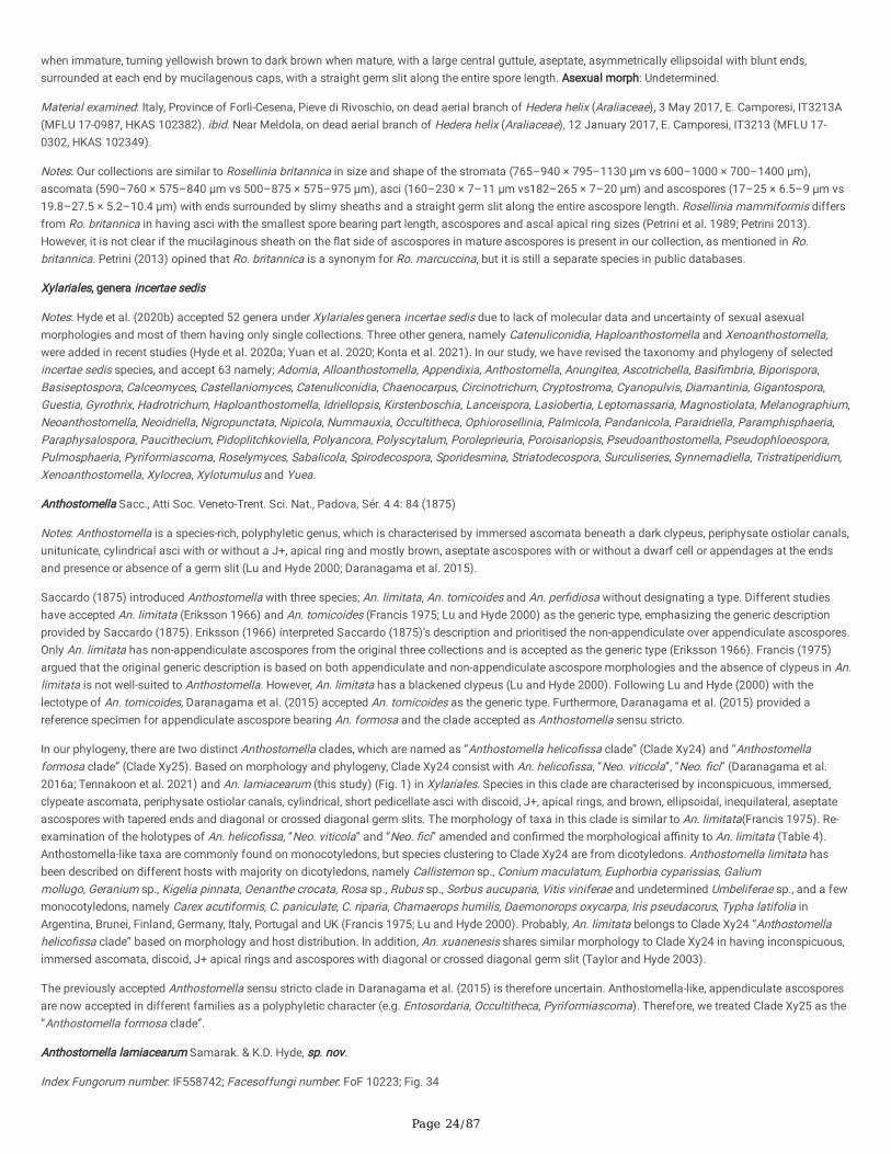
Page 24/87
when immature, turning yellowish brown to dark brown when mature, with a large central guttule, aseptate, asymmetrically ellipsoidal with blunt ends,surrounded at each end by mucilagenous caps, with a straight germ slit along the entire spore length. Asexual morph: Undetermined.
Material examined: Italy, Province of Forlì-Cesena, Pieve di Rivoschio, on dead aerial branch of Hedera helix (Araliaceae), 3 May 2017, E. Camporesi, IT3213A(MFLU 17-0987, HKAS 102382). ibid. Near Meldola, on dead aerial branch of Hedera helix (Araliaceae), 12 January 2017, E. Camporesi, IT3213 (MFLU 17-0302, HKAS 102349).
Notes: Our collections are similar to Rosellinia britannica in size and shape of the stromata (765–940 × 795–1130 μm vs 600–1000 × 700–1400 μm),ascomata (590–760 × 575–840 μm vs 500–875 × 575–975 μm), asci (160–230 × 7–11 μm vs182–265 × 7–20 μm) and ascospores (17–25 × 6.5–9 μm vs19.8–27.5 × 5.2–10.4 μm) with ends surrounded by slimy sheaths and a straight germ slit along the entire ascospore length. Rosellinia mammiformis differsfrom Ro. britannica in having asci with the smallest spore bearing part length, ascospores and ascal apical ring sizes (Petrini et al. 1989; Petrini 2013).However, it is not clear if the mucilaginous sheath on the �at side of ascospores in mature ascospores is present in our collection, as mentioned in Ro.britannica. Petrini (2013) opined that Ro. britannica is a synonym for Ro. marcuccina, but it is still a separate species in public databases.
Xylariales, genera incertae sedis
Notes: Hyde et al. (2020b) accepted 52 genera under Xylariales genera incertae sedis due to lack of molecular data and uncertainty of sexual asexualmorphologies and most of them having only single collections. Three other genera, namely Catenuliconidia, Haploanthostomella and Xenoanthostomella,were added in recent studies (Hyde et al. 2020a; Yuan et al. 2020; Konta et al. 2021). In our study, we have revised the taxonomy and phylogeny of selectedincertae sedis species, and accept 63 namely; Adomia, Alloanthostomella, Appendixia, Anthostomella, Anungitea, Ascotrichella, Basi�mbria, Biporispora,Basiseptospora, Calceomyces, Castellaniomyces, Catenuliconidia, Chaenocarpus, Circinotrichum, Cryptostroma, Cyanopulvis, Diamantinia, Gigantospora,Guestia, Gyrothrix, Hadrotrichum, Haploanthostomella, Idriellopsis, Kirstenboschia, Lanceispora, Lasiobertia, Leptomassaria, Magnostiolata, Melanographium,Neoanthostomella, Neoidriella, Nigropunctata, Nipicola, Nummauxia, Occultitheca, Ophiorosellinia, Palmicola, Pandanicola, Paraidriella, Paramphisphaeria,Paraphysalospora, Paucithecium, Pidoplitchkoviella, Polyancora, Polyscytalum, Poroleprieuria, Poroisariopsis, Pseudoanthostomella, Pseudophloeospora,Pulmosphaeria, Pyriformiascoma, Roselymyces, Sabalicola, Spirodecospora, Sporidesmina, Striatodecospora, Surculiseries, Synnemadiella, Tristratiperidium,Xenoanthostomella, Xylocrea, Xylotumulus and Yuea.
Anthostomella Sacc., Atti Soc. Veneto-Trent. Sci. Nat., Padova, Sér. 4 4: 84 (1875)
Notes: Anthostomella is a species-rich, polyphyletic genus, which is characterised by immersed ascomata beneath a dark clypeus, periphysate ostiolar canals,unitunicate, cylindrical asci with or without a J+, apical ring and mostly brown, aseptate ascospores with or without a dwarf cell or appendages at the endsand presence or absence of a germ slit (Lu and Hyde 2000; Daranagama et al. 2015).
Saccardo (1875) introduced Anthostomella with three species; An. limitata, An. tomicoides and An. per�diosa without designating a type. Different studieshave accepted An. limitata (Eriksson 1966) and An. tomicoides (Francis 1975; Lu and Hyde 2000) as the generic type, emphasizing the generic descriptionprovided by Saccardo (1875). Eriksson (1966) interpreted Saccardo (1875)’s description and prioritised the non-appendiculate over appendiculate ascospores.Only An. limitata has non-appendiculate ascospores from the original three collections and is accepted as the generic type (Eriksson 1966). Francis (1975)argued that the original generic description is based on both appendiculate and non-appendiculate ascospore morphologies and the absence of clypeus in An.limitata is not well-suited to Anthostomella. However, An. limitata has a blackened clypeus (Lu and Hyde 2000). Following Lu and Hyde (2000) with thelectotype of An. tomicoides, Daranagama et al. (2015) accepted An. tomicoides as the generic type. Furthermore, Daranagama et al. (2015) provided areference specimen for appendiculate ascospore bearing An. formosa and the clade accepted as Anthostomella sensu stricto.
In our phylogeny, there are two distinct Anthostomella clades, which are named as “Anthostomella helico�ssa clade” (Clade Xy24) and “Anthostomellaformosa clade” (Clade Xy25). Based on morphology and phylogeny, Clade Xy24 consist with An. helico�ssa, “Neo. viticola”, “Neo. �ci” (Daranagama et al.2016a; Tennakoon et al. 2021) and An. lamiacearum (this study) (Fig. 1) in Xylariales. Species in this clade are characterised by inconspicuous, immersed,clypeate ascomata, periphysate ostiolar canals, cylindrical, short pedicellate asci with discoid, J+, apical rings, and brown, ellipsoidal, inequilateral, aseptateascospores with tapered ends and diagonal or crossed diagonal germ slits. The morphology of taxa in this clade is similar to An. limitata(Francis 1975). Re-examination of the holotypes of An. helico�ssa, “Neo. viticola” and “Neo. �ci” amended and con�rmed the morphological a�nity to An. limitata (Table 4).Anthostomella-like taxa are commonly found on monocotyledons, but species clustering to Clade Xy24 are from dicotyledons. Anthostomella limitata hasbeen described on different hosts with majority on dicotyledons, namely Callistemon sp., Conium maculatum, Euphorbia cyparissias, Galiummollugo, Geranium sp., Kigelia pinnata, Oenanthe crocata, Rosa sp., Rubus sp., Sorbus aucuparia, Vitis viniferae and undetermined Umbeliferae sp., and a fewmonocotyledons, namely Carex acutiformis, C. paniculate, C. riparia, Chamaerops humilis, Daemonorops oxycarpa, Iris pseudacorus, Typha latifolia inArgentina, Brunei, Finland, Germany, Italy, Portugal and UK (Francis 1975; Lu and Hyde 2000). Probably, An. limitata belongs to Clade Xy24 “Anthostomellahelico�ssa clade” based on morphology and host distribution. In addition, An. xuanenesis shares similar morphology to Clade Xy24 in having inconspicuous,immersed ascomata, discoid, J+ apical rings and ascospores with diagonal or crossed diagonal germ slit (Taylor and Hyde 2003).
The previously accepted Anthostomella sensu stricto clade in Daranagama et al. (2015) is therefore uncertain. Anthostomella-like, appendiculate ascosporesare now accepted in different families as a polyphyletic character (e.g. Entosordaria, Occultitheca, Pyriformiascoma). Therefore, we treated Clade Xy25 as the“Anthostomella formosa clade”.
Anthostomella lamiacearum Samarak. & K.D. Hyde, sp. nov.
Index Fungorum number: IF558742; Facesoffungi number: FoF 10223; Fig. 34
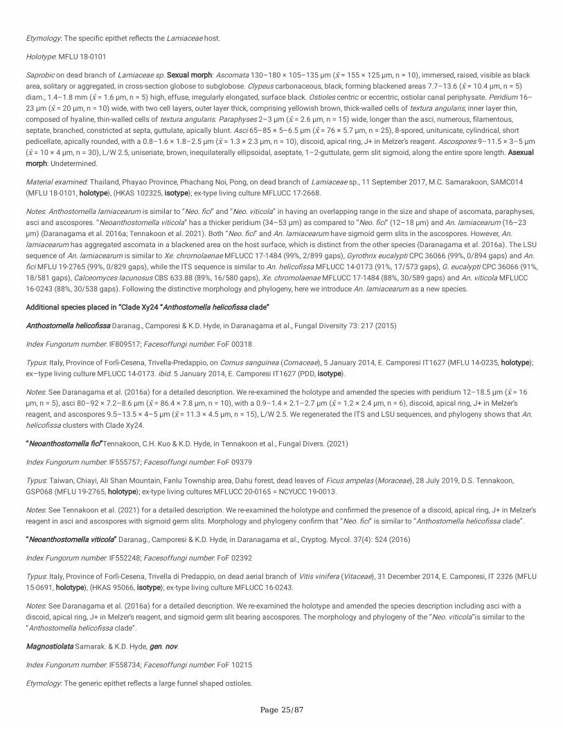
Page 25/87
Etymology: The speci�c epithet re�ects the Lamiaceae host.
Holotype: MFLU 18-0101
Saprobic on dead branch of Lamiaceae sp. Sexual morph: Ascomata 130–180 × 105–135 μm (x̄ = 155 × 125 μm, n = 10), immersed, raised, visible as blackarea, solitary or aggregated, in cross-section globose to subglobose. Clypeus carbonaceous, black, forming blackened areas 7.7–13.6 (x̄ = 10.4 μm, n = 5)diam., 1.4–1.8 mm (x̄ = 1.6 μm, n = 5) high, effuse, irregularly elongated, surface black. Ostioles centric or eccentric, ostiolar canal periphysate. Peridium 16–23 μm (x̄ = 20 μm, n = 10) wide, with two cell layers, outer layer thick, comprising yellowish brown, thick-walled cells of textura angularis, inner layer thin,composed of hyaline, thin-walled cells of textura angularis. Paraphyses 2–3 μm (x̄ = 2.6 μm, n = 15) wide, longer than the asci, numerous, �lamentous,septate, branched, constricted at septa, guttulate, apically blunt. Asci 65–85 × 5–6.5 μm (x̄ = 76 × 5.7 μm, n = 25), 8-spored, unitunicate, cylindrical, shortpedicellate, apically rounded, with a 0.8–1.6 × 1.8–2.5 μm (x̄ = 1.3 × 2.3 μm, n = 10), discoid, apical ring, J+ in Melzer’s reagent. Ascospores 9–11.5 × 3–5 μm(x̄ = 10 × 4 μm, n = 30), L/W 2.5, uniseriate, brown, inequilaterally ellipsoidal, aseptate, 1–2-guttulate, germ slit sigmoid, along the entire spore length. Asexualmorph: Undetermined.
Material examined: Thailand, Phayao Province, Phachang Noi, Pong, on dead branch of Lamiaceae sp., 11 September 2017, M.C. Samarakoon, SAMC014(MFLU 18-0101, holotype), (HKAS 102325, isotype); ex-type living culture MFLUCC 17-2668.
Notes: Anthostomella lamiacearum is similar to “Neo. �ci” and “Neo. viticola” in having an overlapping range in the size and shape of ascomata, paraphyses,asci and ascospores. “Neoanthostomella viticola” has a thicker peridium (34–53 μm) as compared to “Neo. �ci” (12–18 μm) and An. lamiacearum (16–23μm) (Daranagama et al. 2016a; Tennakoon et al. 2021). Both “Neo. �ci” and An. lamiacearum have sigmoid germ slits in the ascospores. However, An.lamiacearum has aggregated ascomata in a blackened area on the host surface, which is distinct from the other species (Daranagama et al. 2016a). The LSUsequence of An. lamiacearum is similar to Xe. chromolaenae MFLUCC 17-1484 (99%, 2/899 gaps), Gyrothrix eucalypti CPC 36066 (99%, 0/894 gaps) and An.�ci MFLU 19-2765 (99%, 0/829 gaps), while the ITS sequence is similar to An. helico�ssa MFLUCC 14-0173 (91%, 17/573 gaps), G. eucalypti CPC 36066 (91%,18/581 gaps), Calceomyces lacunosus CBS 633.88 (89%, 16/580 gaps), Xe. chromolaenae MFLUCC 17-1484 (88%, 30/589 gaps) and An. viticola MFLUCC16-0243 (88%, 30/538 gaps). Following the distinctive morphology and phylogeny, here we introduce An. lamiacearum as a new species.
Additional species placed in “Clade Xy24 “Anthostomella helico�ssa clade”
Anthostomella helico�ssa Daranag., Camporesi & K.D. Hyde, in Daranagama et al., Fungal Diversity 73: 217 (2015)
Index Fungorum number: IF809517; Facesoffungi number: FoF 00318
Typus: Italy, Province of Forlì-Cesena, Trivella-Predappio, on Cornus sanguinea (Cornaceae), 5 January 2014, E. Camporesi IT1627 (MFLU 14-0235, holotype);ex–type living culture MFLUCC 14-0173. ibid. 5 January 2014, E. Camporesi IT1627 (PDD, isotype).
Notes: See Daranagama et al. (2016a) for a detailed description. We re-examined the holotype and amended the species with peridium 12–18.5 μm (x̄ = 16μm, n = 5), asci 80–92 × 7.2–8.6 μm (x̄ = 86.4 × 7.8 μm, n = 10), with a 0.9–1.4 × 2.1–2.7 μm (x̄ = 1.2 × 2.4 μm, n = 6), discoid, apical ring, J+ in Melzer’sreagent, and ascospores 9.5–13.5 × 4–5 μm (x̄ = 11.3 × 4.5 μm, n = 15), L/W 2.5. We regenerated the ITS and LSU sequences, and phylogeny shows that An.helico�ssa clusters with Clade Xy24.
“Neoanthostomella �ci”Tennakoon, C.H. Kuo & K.D. Hyde, in Tennakoon et al., Fungal Divers. (2021)
Index Fungorum number: IF555757; Facesoffungi number: FoF 09379
Typus: Taiwan, Chiayi, Ali Shan Mountain, Fanlu Township area, Dahu forest, dead leaves of Ficus ampelas (Moraceae), 28 July 2019, D.S. Tennakoon,GSP068 (MFLU 19-2765, holotype); ex-type living cultures MFLUCC 20-0165 = NCYUCC 19-0013.
Notes: See Tennakoon et al. (2021) for a detailed description. We re-examined the holotype and con�rmed the presence of a discoid, apical ring, J+ in Melzer’sreagent in asci and ascospores with sigmoid germ slits. Morphology and phylogeny con�rm that “Neo. �ci” is similar to “Anthostomella helico�ssa clade”.
“Neoanthostomella viticola” Daranag., Camporesi & K.D. Hyde, in Daranagama et al., Cryptog. Mycol. 37(4): 524 (2016)
Index Fungorum number: IF552248; Facesoffungi number: FoF 02392
Typus: Italy, Province of Forlì-Cesena, Trivella di Predappio, on dead aerial branch of Vitis vinifera (Vitaceae), 31 December 2014, E. Camporesi, IT 2326 (MFLU15-0691, holotype), (HKAS 95066, isotype); ex-type living culture MFLUCC 16-0243.
Notes: See Daranagama et al. (2016a) for a detailed description. We re-examined the holotype and amended the species description including asci with adiscoid, apical ring, J+ in Melzer’s reagent, and sigmoid germ slit bearing ascospores. The morphology and phylogeny of the “Neo. viticola”is similar to the“Anthostomella helico�ssa clade”.
Magnostiolata Samarak. & K.D. Hyde, gen. nov.
Index Fungorum number: IF558734; Facesoffungi number: FoF 10215
Etymology: The generic epithet re�ects a large funnel shaped ostioles.
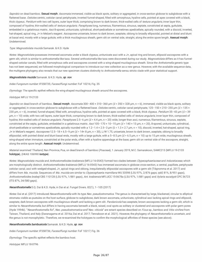
Page 26/87
Saprobic on dead bamboo. Sexual morph: Ascomata immersed, visible as black spots, solitary or aggregated, in cross-section globose to subglobose with a�attened base. Ostioles centric, ostiolar canal periphysate, inverted funnel-shaped, �lled with amorphous, hyaline cells, pointed at apex covered with a black,thick clypeus. Peridium with two cell layers, outer layer thick, comprising brown to dark brown, thick-walled cells of textura angularis, inner layer thin,composed of hyaline, thin-walled cells of textura angularis. Paraphyses long, numerous, �lamentous, sinuous, septate, constricted at septa, guttulate,embedded in a gelatinous matrix. Asci 8-spored, unitunicate, cylindrical, short-pedicellate or sometimes apedicellate, apically rounded, with a discoid, inverted,hat-shaped, apical ring, J+ in Melzer’s reagent. Ascospores uniseriate, brown to dark brown, aseptate, oblong to broadly ellipsoidal, pointed at distal and bluntat basal end, mostly with a large guttule, with a thick mucilaginous sheath, germ slit on ventral side, straight, along the entire spore length. Asexual morph:Undetermined.
Type: Magnostiolata mucida Samarak. & K.D. Hyde
Notes: Magnostiolata possesses immersed ascomata under a black clypeus, unitunicate asci with a J+, apical ring and brown, ellipsoid ascospores with agerm slit, which is similar to anthostomella-like taxa. Several anthostomella-like taxa were discovered during our study. Magnostiolata differs as it has funnel-shaped ostiolar canals, �lled with amorphous cells and ascospores covered with a wing-shaped mucilaginous sheath. Since the Anthstomella generic typehas not been sequenced, we followed morphological comparison to introduce our new collection as a new genus. However, with the available sequence data,the multigene phylogeny also reveals that our new specimen clusters distinctly to Anthostomella sensu stricto clade with poor statistical support.
Magnostiolata mucida Samarak. & K.D. Hyde, sp. nov.
Index Fungorum number: IF558735; Facesoffungi number: FoF 10216; Fig. 35
Etymology: The speci�c epithet re�ects the wing-shaped mucilaginous sheath around the ascospores.
Holotype: MFLU 19-2133
Saprobic on dead branch of bamboo. Sexual morph: Ascomata 300–400 × 310–360 μm (x̄ = 350 × 328 μm, n = 6), immersed, visible as black spots, solitaryor aggregated, in cross-section globose to subglobose with a �attened base. Ostioles centric, ostiolar canal periphysate, 125–150 × 210–255 μm (x̄ = 135 ×230 μm, n = 6), inverted, funnel-shaped, �lled with, amorphous, hyaline cells, pointed at apex covered with a black, thick clypeus. Peridium 35–43 μm (x̄ = 39μm, n = 10) wide, with two cell layers, outer layer thick, comprising brown to dark brown, thick-walled cells of textura angularis, inner layer thin, composed ofhyaline, thin-walled cells of textura angularis. Paraphyses 2.5–6 μm (x̄ = 4.4 μm, n = 20) wide, longer than asci, numerous, �lamentous, sinuous, septate,constricted at the septa, guttulate, embedded in a gelatinous matrix. Asci 105–170 × 10–15 μm (x̄ = 140 × 13 μm, n = 20), 8-spored, unitunicate, cylindrical,short-pedicellate or sometimes apedicellate, apically rounded with a 1.2–1.4 × 2.5–3 μm (x̄ = 1.3 × 2.7 μm, n = 10), discoid, inverted, hat-shaped, apical ring,J+ in Melzer’s reagent. Ascospores 12.5–18 × 6.5–9 μm (x̄ = 14× 8 μm, n = 30), L/W 1.75, uniseriate, brown to dark brown, aseptate, oblong to broadlyellipsoidal, with pointed distal and blunt basal ends, mostly with a large guttule, with a 5–8.5 μm (x̄ = 6.5 μm, n = 10) up to 15 μm wide, mucilaginous sheath,wing-shaped when immature, constricted at the polar ends, often with a hyaline appendage at the base, germ slit on ventral side of the ascospore, straight,along the entire spore length. Asexual morph: Undetermined.
Material examined: Thailand, Nan Province, Pua, on dead branch of bamboo (Poaceae), 1 January 2019, M.C. Samarakoon, SAMC213 (MFLU 19-2133holotype), (HKAS 106990 isotype).
Notes: Magnostiolata mucida and Anthostomelloides krabiensis (MFLU 16-0543) formed two clades between Clypeosphaeriaceae and Induratiaceae, whichare morphologically distinct. Anthostomelloides krabiensis (MFLU 16-0543) has immersed ascomata in globose cross-section, a central, papillate, periphysateostiolar canal, asci with wedged-shaped, J+, apical rings and oblong, inequilaterally ellipsoidal ascospores with a germ slit (Tibpromma et al. 2017) anddiffers from Ma. mucida. Sequences of Ma. mucida are similar to Clypeosphaeria mamillana WU 33598 (LSU 97%, 2/876 gaps; rpb2 81%, 8/951 gaps),Anthostomelloides brabeji CBS 110128 (LSU 97%, 1/881 gaps), Ant. krabiensis MFLUCC 15-0678a (LSU 97%, 1/881 gaps) and Xylaria eucalypti CPC 36723(ITS 87%, 34/580 gaps).
Neoanthostomella D.Q. Dai & K.D. Hyde, in Dai et al. Fungal Divers. 82(1), 1–105 (2017)
Notes: Dai et al. (2017) introduced Neoanthostomella with its type Neo. pseudostromatica. The genus is characterised by large, blackened, circular to ellipticalstromata visible as pustules on the host surface, globose to subglobose, dark brown ascomata, unitunicate, cylindrical asci lacking apical rings and ellipsoid,aseptate, dark brown ascospores with mucilaginous sheath and lacking a germ slit. Pandanicola has aseptate, brown ascospores lacking a germ slit, which issimilar to Neoanthstomella, but differs in having ascomata beneath a black, raised, oval spots as solitary or clustered and ascospores with polar germ pores(Hyde 1994b). “Neoanthostomella �ci”, Neo. pseudostromatica and“Neo. viticola” are extant species described on Ficus sp., bamboo and Vitis vinifera fromTaiwan, Thailand, and Italy (Daranagama et al. 2016a; Dai et al. 2017; Tennakoon et al. 2021). However, the phylogeny of Neoanthstomella is uncertain, andthe genus is not monophyletic. Therefore, we re-examined the holotypes to con�rm the morphological a�nities of these species (see above).
Neoanthostomella bambusicola Samarak. & K.D. Hyde, sp. nov.
Index Fungorum number: IF558736; Facesoffungi number: FoF 10217; Fig. 36
Etymology: The speci�c epithet re�ects the bamboo host.
Holotype: MFLU 18-0796

Page 27/87
Saprobic on dead branch of bamboo. Sexual morph: Stromata 1.1–4.3 × 0.84–1.5 mm (x̄ = 2.3 × 1.1 μm, n = 6), super�cial, solitary or aggregated, effuse,subglobose to ellipsoid, surface black to dull black, incrusted with super�cial, thick carbonaceous tissue, with yellowish brown margin. Ascomata 220–295 ×220–470 μm (x̄ = 254 × 365 μm, n = 10), immersed in the stroma, 3–13 per stroma, randomly arranged, in cross-section subglobose to rounded rectangularwith a �attened base. Ostioles centric or eccentric, ostiolar canal periphysate. Peridium 14–18 μm (x̄ = 16.3 μm, n = 10) wide, easily detachable, with two celllayers, outer layer thick, comprising yellowish brown, thick-walled cells of textura angularis, inner layer thin, composed of hyaline, thin-walled cells of texturaangularis. Paraphyses 4.5–8 μm (x̄ = 6.5 μm, n = 15) wide, long, sparse, �lamentous, septate, constricted at septa, guttulate. Asci 95–130 × 11.5–16 μm (x̄ =116 × 13.6 μm, n = 20), 8-spored, unitunicate, cylindrical, short pedicellate, with a J-, apical ring, rounded at the apex. Ascospores 15–19 × 5.5–7.5 μm (x̄ =16.9 × 6.4 μm, n = 30), L/W 2.6, overlapping uniseriate, hyaline when immature, brown when mature, inequilaterally ellipsoidal, slightly curved, aseptate, 1–2-guttulate, covered with a 2.5–3.3 μm (x̄ = 2.8 μm, n = 5) thick mucilaginous sheath, lacking a germ slit. Asexual morph: Undetermined.
Material examined: Thailand, Chiang Rai Province, Mae Suai, on dead branch of bamboo (Poaceae), 18 August 2017, M.C. Samarakoon, SAMC098 (MFLU 18-0796, holotype), (HKAS 102348, isotype).
Notes: Neoanthostomella bambusicola shares similar morphology to the type N. pseudostromatica in having pustule-like stromata, asci with J-, apical ring inMelzer’s reagent, and brown, aseptate ascospores with a thick mucilaginous sheath and lacking a germ slit (Dai et al. 2017). Our collection differs from Neo.pseudostromatica in having more ascomata per stroma (3–13 vs 2–5), a thick peridium (14–18 μm vs 5–10 μm), wider paraphyses (4.5–8 μm vs 2.5–4 μm)and larger ascospores (15–19 × 5.5–7.5 μm vs 11.5–15× 4–5.5 μm). Anthostomella smilacis has �at-based ascomata, similar to Neo. bambusicola, butdiffers in being immersed in the host, and lacking a stroma (Lu and Hyde 2000). “Neoanthostomella �ci” and “Neo. viticola” have immersed ascomata with aclypeus and differ from Neo. bambusicola and Neo. pseudostromatica. The LSU sequence of Neo. bambusicola is similar to Neo. pseudostromatica MFLUCC11-0610 (99%, 1/869 gaps), Melanographium phoenicis MFLUCC 18-1481 (95%, 5/869 gaps) and Xe. chromolaenae MFLUCC 17-1484 (94%, 9/870 gaps),while the ITS sequence is similar to Neo. pseudostromatica MFLU 15-1190 (99%, 0/542 gaps) and Gyrothrix oleae CPC 37069 (82%, 43/570 gaps). In ourphylogeny, Neo. bambusicola clusters with Neo. pseudostromatica with a 100%/1.00 PP statistical support. Based on distinct morphology and phylogeny, weintroduce Neo. bambusicola as a new species.
Nigropunctata Samarak. & K.D. Hyde, gen. nov.
Index Fungorum number: IF558737; Facesoffungi number: FoF 10218
Etymology: The epithet re�ects the ostioles as black dots on the host.
Saprobic on dead bamboo. Sexual morph: Ascomata immersed, raised, visible as black dots, solitary or aggregated, in cross-section globose tosubglobose. Ostioles centric or eccentric, ostiolar canal periphysate, �attened at top, covered with a black, thick clypeus. Ectostroma yellow to brown. Peridiumeasily detached from host tissues, with two cell layers, outer layer thick, comprising brown, thick-walled cells of textura angularis, inner layer thin, composed ofhyaline, thin-walled cells of textura angularis. Paraphyses long, numerous, �lamentous, septate, rarely branched, constricted at septa, guttulate, apically blunt,embedded in a gelatinous matrix. Asci 8-spored, unitunicate, cylindrical, short pedicellate, apically rounded, with a discoid, or inverted, hat-shaped, apical ring,J+ in Melzer’s reagent. Ascospores uniseriate or overlapping uniseriate, brown to dark brown, cylindrical to broadly ellipsoidal, aseptate, 1–2-guttulate, coveredwith a thick mucilaginous sheath or sheath lacking, germ slit on ventral side, straight, along the entire spore length. Asexual morph: Undetermined.
Type: Nigropunctata bambusicola Samarak. & K.D. Hyde
Notes: Anthostomella-like taxa are polyphyletic in Xylariales with diversi�ed morphologies (Lu and Hyde 2000). Several morpho-molecular studies haveintroduced anthostomella-like taxa as new genera in an attempt to resolve the polyphyletic nature of Anthostomella (Daranagama et al. 2015; 2016a; Konta etal. 2021). Lu and Hyde (2000) emphasised that the position and morphology of ascomata, presence or absence, and shape of the J+/J-, apical ring andascospore morphology, play a vital role in identifying Anthostomella species. However, the type species has not yet been designated formally (proposed An.limitata and An. tomicoides) and sequenced to provide a phylogenetic a�nity (Voglmayr et al. 2018). However, based on morphology, Nigropunctata does not�t with either An. limitata and An. tomicoides. Nigropunctata species have immersed ascomata with a thick clypeus; white or yellow ectostroma, cylindrical,short pedicel, apically rounded asci with J+, discoid or inverted, hat-shaped, apical ring and cylindrical to broadly ellipsoidal, aseptate, ascospores with a germslit, which excludes it from Anthostomella. In addition, all Nigropunctata species introduced in this study occur on dead bamboo. The multigene phylogenyalso reveals that Nigropunctata clusters independently in Xylariales (Fig. 1; Clade Xy26).
Nigropunctata bambusicolaSamarak. & K.D. Hyde, sp. nov.
Index Fungorum number: IF558738; Facesoffungi number: FoF 10219; Fig. 37
Etymology: The speci�c epithet re�ects the bamboo host.
Holotype: MFLU 19-2145
Saprobic on dead branch of bamboo. Sexual morph: Ascomata 285–315 × 260–340 μm (x̄ = 300 × 295 μm, n = 6), immersed, visible as black dots,surrounded by white periphery, solitary or aggregated, in cross-section globose to subglobose with a �attened base. Ostioles centric, ostiolar canal periphysate,�attened at top covered with black, thick clypeus. Ectostroma yellow. Peridium 10–18 μm (x̄ = 15 μm, n = 10) wide, with two cell layers, outer layer thick,comprising brown to dark brown, thick-walled cells of textura angularis, inner layer thin, composed of hyaline, thin-walled cells of textura angularis.Paraphyses 3–5 μm (x̄ = 3.8 μm, n = 20) wide, longer than the asci, numerous, �lamentous, sinuous, septate, constricted at septa, guttulate, embedded in agelatinous matrix. Asci 95–140 × 9.5–12.5 μm (x̄ = 120 × 11 μm, n = 20), 8-spored, unitunicate, cylindrical, short-pedicellate or sometimes apedicellate,
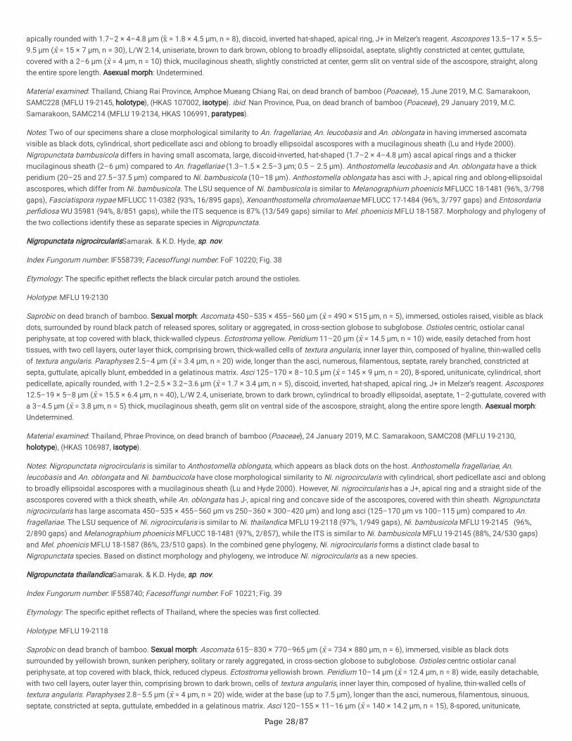
Page 28/87
apically rounded with 1.7–2 × 4–4.8 μm (x̄ = 1.8 × 4.5 μm, n = 8), discoid, inverted hat-shaped, apical ring, J+ in Melzer’s reagent. Ascospores 13.5–17 × 5.5–9.5 μm (x̄ = 15 × 7 μm, n = 30), L/W 2.14, uniseriate, brown to dark brown, oblong to broadly ellipsoidal, aseptate, slightly constricted at center, guttulate,covered with a 2–6 μm (x̄ = 4 μm, n = 10) thick, mucilaginous sheath, slightly constricted at center, germ slit on ventral side of the ascospore, straight, alongthe entire spore length. Asexual morph: Undetermined.
Material examined: Thailand, Chiang Rai Province, Amphoe Mueang Chiang Rai, on dead branch of bamboo (Poaceae), 15 June 2019, M.C. Samarakoon,SAMC228 (MFLU 19-2145, holotype), (HKAS 107002, isotype). ibid. Nan Province, Pua, on dead branch of bamboo (Poaceae), 29 January 2019, M.C.Samarakoon, SAMC214 (MFLU 19-2134, HKAS 106991, paratypes).
Notes: Two of our specimens share a close morphological similarity to An. fragellariae, An. leucobasis and An. oblongata in having immersed ascomatavisible as black dots, cylindrical, short pedicellate asci and oblong to broadly ellipsoidal ascospores with a mucilaginous sheath (Lu and Hyde 2000).Nigropunctata bambusicola differs in having small ascomata, large, discoid-inverted, hat-shaped (1.7–2 × 4–4.8 μm) ascal apical rings and a thickermucilaginous sheath (2–6 μm) compared to An. fragellariae (1.3–1.5 × 2.5–3 μm; 0.5 – 2.5 μm). Anthostomella leucobasis and An. oblongata have a thickperidium (20–25 and 27.5–37.5 μm) compared to Ni. bambusicola (10–18 μm). Anthostomella oblongata has asci with J-, apical ring and oblong-ellipsoidalascospores, which differ from Ni. bambusicola. The LSU sequence of Ni. bambusicola is similar to Melanographium phoenicis MFLUCC 18-1481 (96%, 3/798gaps), Fasciatispora nypae MFLUCC 11-0382 (93%, 16/895 gaps), Xenoanthostomella chromolaenae MFLUCC 17-1484 (96%, 3/797 gaps) and Entosordariaper�diosa WU 35981 (94%, 8/851 gaps), while the ITS sequence is 87% (13/549 gaps) similar to Mel. phoenicis MFLU 18-1587. Morphology and phylogeny ofthe two collections identify these as separate species in Nigropunctata.
Nigropunctata nigrocircularisSamarak. & K.D. Hyde, sp. nov.
Index Fungorum number: IF558739; Facesoffungi number: FoF 10220; Fig. 38
Etymology: The speci�c epithet re�ects the black circular patch around the ostioles.
Holotype: MFLU 19-2130
Saprobic on dead branch of bamboo. Sexual morph: Ascomata 450–535 × 455–560 μm (x̄ = 490 × 515 μm, n = 5), immersed, ostioles raised, visible as blackdots, surrounded by round black patch of released spores, solitary or aggregated, in cross-section globose to subglobose. Ostioles centric, ostiolar canalperiphysate, at top covered with black, thick-walled clypeus. Ectostroma yellow. Peridium 11–20 μm (x̄ = 14.5 μm, n = 10) wide, easily detached from hosttissues, with two cell layers, outer layer thick, comprising brown, thick-walled cells of textura angularis, inner layer thin, composed of hyaline, thin-walled cellsof textura angularis. Paraphyses 2.5–4 μm (x̄ = 3.4 μm, n = 20) wide, longer than the asci, numerous, �lamentous, septate, rarely branched, constricted atsepta, guttulate, apically blunt, embedded in a gelatinous matrix. Asci 125–170 × 8–10.5 μm (x̄ = 145 × 9 μm, n = 20), 8-spored, unitunicate, cylindrical, shortpedicellate, apically rounded, with 1.2–2.5 × 3.2–3.6 μm (x̄ = 1.7 × 3.4 μm, n = 5), discoid, inverted, hat-shaped, apical ring, J+ in Melzer’s reagent. Ascospores12.5–19 × 5–8 μm (x̄ = 15.5 × 6.4 μm, n = 40), L/W 2.4, uniseriate, brown to dark brown, cylindrical to broadly ellipsoidal, aseptate, 1–2-guttulate, covered witha 3–4.5 μm (x̄ = 3.8 μm, n = 5) thick, mucilaginous sheath, germ slit on ventral side of the ascospore, straight, along the entire spore length. Asexual morph:Undetermined.
Material examined: Thailand, Phrae Province, on dead branch of bamboo (Poaceae), 24 January 2019, M.C. Samarakoon, SAMC208 (MFLU 19-2130,holotype), (HKAS 106987, isotype).
Notes: Nigropunctata nigrocircularis is similar to Anthostomella oblongata, which appears as black dots on the host. Anthostomella fragellariae, An.leucobasis and An. oblongata and Ni. bambucicola have close morphological similarity to Ni. nigrocircularis with cylindrical, short pedicellate asci and oblongto broadly ellipsoidal ascospores with a mucilaginous sheath (Lu and Hyde 2000). However, Ni. nigrocircularis has a J+, apical ring and a straight side of theascospores covered with a thick sheath, while An. oblongata has J-, apical ring and concave side of the ascospores, covered with thin sheath. Nigropunctatanigrocircularis has large ascomata 450–535 × 455–560 μm vs 250–360 × 300–420 μm) and long asci (125–170 μm vs 100–115 μm) compared to An.fragellariae. The LSU sequence of Ni. nigrocircularis is similar to Ni. thailandica MFLU 19-2118 (97%, 1/949 gaps), Ni. bambusicola MFLU 19-2145 (96%,2/890 gaps) and Melanographium phoenicis MFLUCC 18-1481 (97%, 2/857), while the ITS is similar to Ni. bambusicola MFLU 19-2145 (88%, 24/530 gaps)and Mel. phoenicis MFLU 18-1587 (86%, 23/510 gaps). In the combined gene phylogeny, Ni. nigrocircularis forms a distinct clade basal toNigropunctata species. Based on distinct morphology and phylogeny, we introduce Ni. nigrocircularis as a new species.
Nigropunctata thailandicaSamarak. & K.D. Hyde, sp. nov.
Index Fungorum number: IF558740; Facesoffungi number: FoF 10221; Fig. 39
Etymology: The speci�c epithet re�ects of Thailand, where the species was �rst collected.
Holotype: MFLU 19-2118
Saprobic on dead branch of bamboo. Sexual morph: Ascomata 615–830 × 770–965 μm (x̄ = 734 × 880 μm, n = 6), immersed, visible as black dotssurrounded by yellowish brown, sunken periphery, solitary or rarely aggregated, in cross-section globose to subglobose. Ostioles centric ostiolar canalperiphysate, at top covered with black, thick, reduced clypeus. Ectostroma yellowish brown. Peridium 10–14 μm (x̄ = 12.4 μm, n = 8) wide, easily detachable,with two cell layers, outer layer thin, comprising brown to dark brown, cells of textura angularis, inner layer thin, composed of hyaline, thin-walled cells oftextura angularis. Paraphyses 2.8–5.5 μm (x̄ = 4 μm, n = 20) wide, wider at the base (up to 7.5 μm), longer than the asci, numerous, �lamentous, sinuous,septate, constricted at septa, guttulate, embedded in a gelatinous matrix. Asci 120–155 × 11–16 μm (x̄ = 140 × 14.2 μm, n = 15), 8-spored, unitunicate,
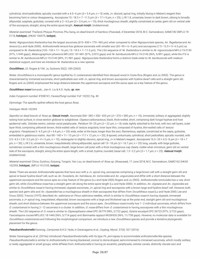
Page 29/87
cylindrical, short-pedicellate, apically rounded with a 4.5–6 μm (x̄ = 5.4 μm, n = 5) wide, J+, discoid, apical ring, initially bluing in Melzer’s reagent thenbecoming faint or colour disappearing. Ascospores 15–18.5 × 7–11.5 μm (x̄ = 17 × 9 μm, n = 25), L/W 1.8, uniseriate, brown to dark brown, oblong to broadlyellipsoidal, aseptate, guttulate, covered with a 2–4.5 μm (x̄ = 3.4 μm, n = 10), thick mucilaginous sheath, slightly constricted at center, germ slit on ventral sideof the ascospore, straight, along the entire spore length. Asexual morph: Undetermined.
Material examined: Thailand, Phayao Province, Phu Sang, on dead branch of bamboo (Poaceae), 4 December 2018, M.C. Samarakoon, SAMC182 (MFLU 19-2118, holotype), (HKAS 106975, isotype).
Notes: Nigropunctata thailandica has the largest ascomata (615–830 × 770–965 μm) when compared to other Nigropunctata species, An. �agellariae and An.tenacis (Lu and Hyde 2000). Anthostomella tenacis has globose ascomata with smaller asci (65–90 × 6–9 μm) and ascospores (7.5–12.5 × 5–6.5 μm) ascompared to Ni. thailandica (120–155 × 11–16 μm; 15–18.5 × 7–11.5 μm). The LSU sequence of Ni. thailandica is similar to Ni. nigrocircularis MFLU 19-2130(97%, 1/949 gaps), Melanographium phoenicis MFLUCC 18-1481 (97%, 2/872 gaps) and Ni. bambusicola MFLU 19-2145 (96%, 5/891 gaps), while the ITS issimilar to Ni. bambusicola MFLU 19-2145 (89%, 21/581 gaps). Nigropunctata thailandica forms a distinct clade sister to Ni. bambusicola with mediumstatistical support, and here we introduce Ni. thailandica as a new species.
Occultitheca J.D. Rogers & Y.M. Ju, Sydowia 55(2): 359 (2003)
Notes: Occultitheca is a monospeci�c genus typi�ed by O. costaricensis identi�ed from decayed wood in Costa Rica (Rogers and Ju 2003). The genus ischaracterised by immersed ascomata, short pedicellate asci with J+, apical ring, and brown ascospores with hyaline dwarf cells and a straight germ slit.Rogers and Ju (2003) emphasised the large distance between the uppermost ascospore and the ascus apex as a key feature of the genus.
Occultitheca rosae Samarak., Jian K. Liu & K.D. Hyde, sp. nov.
Index Fungorum number: IF558741; Facesoffungi number: FoF 10222; Fig. 40
Etymology: The speci�c epithet re�ects the host genus Rosa.
Holotype: HKAS 102393
Saprobic on dead branch of Rosa sp. Sexual morph: Ascomata 360–385 × 350–420 μm (x̄ = 370 × 385 μm, n = 10), immersed, solitary or aggregated, slightlyraising host surface, in cross-section globose to subglobose. Clypeus carbonaceous, black, thick-walled, short, comprising dark fungal hyphae and hostepidermal cells. Ostioles centric, ostiolar canal periphysate. Peridium 18–25 μm (x̄ = 22 μm, n = 8) wide, tightly attached to the host, with two cell layers, outerlayer thick, comprising yellowish brown, thick-walled cells of textura angularis, inner layer thin, composed of hyaline, thin-walled cells of texturaangularis. Paraphyses 3–6.5 μm (x̄ = 4.4 μm, n = 25) wide, wider at the base, longer than the asci, �lamentous, septate, constricted at the septa, guttulate,embedded in gelatinous matrix. Asci 90–140 × 11–13 μm (x̄ = 117 × 12 μm, n = 20), 8-spored, unitunicate, cylindrical, short pedicellate, apically rounded, with3.5–4.5 × 2.8–3.2 μm (x̄ = 4 × 3 μm, n = 10), rectangular to slightly obconic, apical ring, J+ in Melzer’s reagent. Ascospores 16.5–20 × 6.5–8 μm (x̄ = 18 × 7μm, n = 30), L/W 2.6, uniseriate, brown, inequilaterally oblong-ellipsoidal, apical cell 15–18 μm (x̄ = 16.7 μm, n = 25) long, usually with large guttules,sometimes covered with a thin mucilaginous sheath, large brown cell polar with a thick mucilaginous cap clearly visible when immature, germ slit on ventralside of the ascospore, straight, along the entire spore length, with a small, hyaline, rounded, basal cell, 1.5–2.2 μm (x̄ = 1.7 μm, n = 25). Asexual morph:Undetermined.
Material examined: China, Guizhou, Guiyang, Tongxin, Yan Lou, on dead branch of Rosa sp. (Rosaceae), 17 June 2018, M.C. Samarakoon, SAMC162 (HKAS102393, holotype), (MFLU 19-2105, isotype).
Notes: There are several Anthostomella species that have asci with a J+, apical ring, ascospores comprising a large brown cell with a straight germ slit andapical or basal hyaline dwarf cell, such as An. foveolaris, An. hemileuca, An. tomicoides and An. unguiculata and differ with a short distance between theuppermost ascospore and the ascus apex as a key feature of the genus (Lu and Hyde 2000; Rogers and Ju 2003). Anthostomella tomicoides has a very shortgerm slit, while Occultitheca rosae has a straight germ slit along the entire spore length (Lu and Hyde 2000). In addition, An. clypeata and An. clypeoides aresimilar to Occultitheca rosae in having immersed, clypeate ascomata, J+, apical ring and ascospores with a brown large and hyaline dwarf cell. However, bothspecies lack germ slits and An. clypeoides has a mucilaginous sheath in their ascospores that differs from Occultitheca rosae (Lu and Hyde 2000; Lee andCrous 2003). Francis (1975) described An. sabiniana on Pinus sabiniana needles, which is similar to Occultitheca rosae in having clypeate, immersedascomata, a J+ apical ring, inequilateral, ellipsoidal, brown ascospores with a large and thickened cap at the polar end, staright germ slit and mucilaginoussheath, and short distance between the uppermost ascospore and the ascus apex. Occultitheca rosae mostly has 1–2 individual ascomata, which differs fromO. costaricensis in having 2–12 ascomata in a cluster. In addition, O. rosae differs from O. costaricensis in having ascospores with a thin mucilaginoussheath. The LSU sequence of O. rosae is similar to Clypeosphaeria oleae CPC 36779 (98%, 2/722 gaps), Xylaria eucalypti CPC 36723 (97%, 2/723 gaps),Fasciatispora cocoes MFLUCC 18-1445 (96%, 5/714 gaps) and Barrmaelia rappazii WU36926 (96%, 11/738 gaps). However, no molecular data is available forOccultitheca costaricensis and following the morphological comparison, we introduce a new Occultitheca species and provide a tentative phylogeneticplacement for the genus.
Pseudoanthostomella Daranag., Camporesi & K.D. Hyde, in Daranagama et al., Cryptog. Mycol. 37(4): 527 (2016)
Notes: Daranagama et al. (2016a) introduced Pseudoanthostomella, with its type Ps. pini-nigrae, to accommodate anthostomella-like species.Pseudoanthostomella is similar to Anthostomella in having blackened, conical to dome-shaped, semi-immersed to immersed ascomata, which mostly solitaryor rarely aggregated in small groups, while differes from Anthostomella in having an eccentric, periphysate, ostiolar canals, distinctly clavate asci and
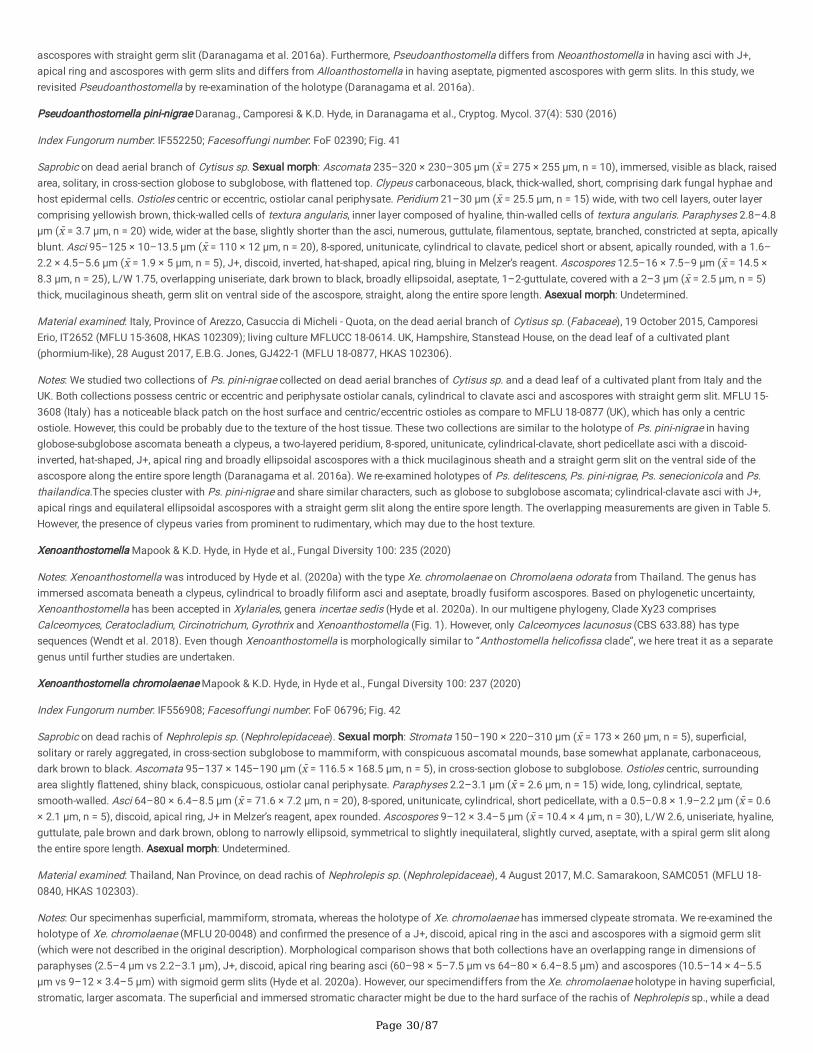
Page 30/87
ascospores with straight germ slit (Daranagama et al. 2016a). Furthermore, Pseudoanthostomella differs from Neoanthostomella in having asci with J+,apical ring and ascospores with germ slits and differs from Alloanthostomella in having aseptate, pigmented ascospores with germ slits. In this study, werevisited Pseudoanthostomella by re-examination of the holotype (Daranagama et al. 2016a).
Pseudoanthostomella pini-nigrae Daranag., Camporesi & K.D. Hyde, in Daranagama et al., Cryptog. Mycol. 37(4): 530 (2016)
Index Fungorum number: IF552250; Facesoffungi number: FoF 02390; Fig. 41
Saprobic on dead aerial branch of Cytisus sp. Sexual morph: Ascomata 235–320 × 230–305 μm (x̄ = 275 × 255 μm, n = 10), immersed, visible as black, raisedarea, solitary, in cross-section globose to subglobose, with �attened top. Clypeus carbonaceous, black, thick-walled, short, comprising dark fungal hyphae andhost epidermal cells. Ostioles centric or eccentric, ostiolar canal periphysate. Peridium 21–30 μm (x̄ = 25.5 μm, n = 15) wide, with two cell layers, outer layercomprising yellowish brown, thick-walled cells of textura angularis, inner layer composed of hyaline, thin-walled cells of textura angularis. Paraphyses 2.8–4.8μm (x̄ = 3.7 μm, n = 20) wide, wider at the base, slightly shorter than the asci, numerous, guttulate, �lamentous, septate, branched, constricted at septa, apicallyblunt. Asci 95–125 × 10–13.5 μm (x̄ = 110 × 12 μm, n = 20), 8-spored, unitunicate, cylindrical to clavate, pedicel short or absent, apically rounded, with a 1.6–2.2 × 4.5–5.6 μm (x̄ = 1.9 × 5 μm, n = 5), J+, discoid, inverted, hat-shaped, apical ring, bluing in Melzer’s reagent. Ascospores 12.5–16 × 7.5–9 μm (x̄ = 14.5 ×8.3 μm, n = 25), L/W 1.75, overlapping uniseriate, dark brown to black, broadly ellipsoidal, aseptate, 1–2-guttulate, covered with a 2–3 μm (x̄ = 2.5 μm, n = 5)thick, mucilaginous sheath, germ slit on ventral side of the ascospore, straight, along the entire spore length. Asexual morph: Undetermined.
Material examined: Italy, Province of Arezzo, Casuccia di Micheli - Quota, on the dead aerial branch of Cytisus sp. (Fabaceae), 19 October 2015, CamporesiErio, IT2652 (MFLU 15-3608, HKAS 102309); living culture MFLUCC 18-0614. UK, Hampshire, Stanstead House, on the dead leaf of a cultivated plant(phormium-like), 28 August 2017, E.B.G. Jones, GJ422-1 (MFLU 18-0877, HKAS 102306).
Notes: We studied two collections of Ps. pini-nigrae collected on dead aerial branches of Cytisus sp. and a dead leaf of a cultivated plant from Italy and theUK. Both collections possess centric or eccentric and periphysate ostiolar canals, cylindrical to clavate asci and ascospores with straight germ slit. MFLU 15-3608 (Italy) has a noticeable black patch on the host surface and centric/eccentric ostioles as compare to MFLU 18-0877 (UK), which has only a centricostiole. However, this could be probably due to the texture of the host tissue. These two collections are similar to the holotype of Ps. pini-nigrae in havingglobose-subglobose ascomata beneath a clypeus, a two-layered peridium, 8-spored, unitunicate, cylindrical-clavate, short pedicellate asci with a discoid-inverted, hat-shaped, J+, apical ring and broadly ellipsoidal ascospores with a thick mucilaginous sheath and a straight germ slit on the ventral side of theascospore along the entire spore length (Daranagama et al. 2016a). We re-examined holotypes of Ps. delitescens, Ps. pini-nigrae, Ps. senecionicola and Ps.thailandica.The species cluster with Ps. pini-nigrae and share similar characters, such as globose to subglobose ascomata; cylindrical-clavate asci with J+,apical rings and equilateral ellipsoidal ascospores with a straight germ slit along the entire spore length. The overlapping measurements are given in Table 5.However, the presence of clypeus varies from prominent to rudimentary, which may due to the host texture.
Xenoanthostomella Mapook & K.D. Hyde, in Hyde et al., Fungal Diversity 100: 235 (2020)
Notes: Xenoanthostomella was introduced by Hyde et al. (2020a) with the type Xe. chromolaenae on Chromolaena odorata from Thailand. The genus hasimmersed ascomata beneath a clypeus, cylindrical to broadly �liform asci and aseptate, broadly fusiform ascospores. Based on phylogenetic uncertainty,Xenoanthostomella has been accepted in Xylariales, genera incertae sedis (Hyde et al. 2020a). In our multigene phylogeny, Clade Xy23 comprisesCalceomyces, Ceratocladium, Circinotrichum, Gyrothrix and Xenoanthostomella (Fig. 1). However, only Calceomyces lacunosus (CBS 633.88) has typesequences (Wendt et al. 2018). Even though Xenoanthostomella is morphologically similar to “Anthostomella helico�ssa clade”, we here treat it as a separategenus until further studies are undertaken.
Xenoanthostomella chromolaenae Mapook & K.D. Hyde, in Hyde et al., Fungal Diversity 100: 237 (2020)
Index Fungorum number: IF556908; Facesoffungi number: FoF 06796; Fig. 42
Saprobic on dead rachis of Nephrolepis sp. (Nephrolepidaceae). Sexual morph: Stromata 150–190 × 220–310 μm (x̄ = 173 × 260 μm, n = 5), super�cial,solitary or rarely aggregated, in cross-section subglobose to mammiform, with conspicuous ascomatal mounds, base somewhat applanate, carbonaceous,dark brown to black. Ascomata 95–137 × 145–190 μm (x̄ = 116.5 × 168.5 μm, n = 5), in cross-section globose to subglobose. Ostioles centric, surroundingarea slightly �attened, shiny black, conspicuous, ostiolar canal periphysate. Paraphyses 2.2–3.1 μm (x̄ = 2.6 μm, n = 15) wide, long, cylindrical, septate,smooth-walled. Asci 64–80 × 6.4–8.5 μm (x̄ = 71.6 × 7.2 μm, n = 20), 8-spored, unitunicate, cylindrical, short pedicellate, with a 0.5–0.8 × 1.9–2.2 μm (x̄ = 0.6× 2.1 μm, n = 5), discoid, apical ring, J+ in Melzer’s reagent, apex rounded. Ascospores 9–12 × 3.4–5 μm (x̄ = 10.4 × 4 μm, n = 30), L/W 2.6, uniseriate, hyaline,guttulate, pale brown and dark brown, oblong to narrowly ellipsoid, symmetrical to slightly inequilateral, slightly curved, aseptate, with a spiral germ slit alongthe entire spore length. Asexual morph: Undetermined.
Material examined: Thailand, Nan Province, on dead rachis of Nephrolepis sp. (Nephrolepidaceae), 4 August 2017, M.C. Samarakoon, SAMC051 (MFLU 18-0840, HKAS 102303).
Notes: Our specimenhas super�cial, mammiform, stromata, whereas the holotype of Xe. chromolaenae has immersed clypeate stromata. We re-examined theholotype of Xe. chromolaenae (MFLU 20-0048) and con�rmed the presence of a J+, discoid, apical ring in the asci and ascospores with a sigmoid germ slit(which were not described in the original description). Morphological comparison shows that both collections have an overlapping range in dimensions ofparaphyses (2.5–4 μm vs 2.2–3.1 μm), J+, discoid, apical ring bearing asci (60–98 × 5–7.5 μm vs 64–80 × 6.4–8.5 μm) and ascospores (10.5–14 × 4–5.5μm vs 9–12 × 3.4–5 μm) with sigmoid germ slits (Hyde et al. 2020a). However, our specimendiffers from the Xe. chromolaenae holotype in having super�cial,stromatic, larger ascomata. The super�cial and immersed stromatic character might be due to the hard surface of the rachis of Nephrolepis sp., while a dead
Page 31/87
stem of Chromolaena odorata has a soft surface. Anthostomella limitata, An. spiralis and An. xuanenesis have similar cylindrical asci with J+, discoid, apicalrings and ascospores with sigmoid/diagonal germ slits, but differ in having immersed ascomata under a clypeus as compare to a super�cial stroma in Xe.chromolaenae (Francis 1975; Lu and Hyde 2000; Taylor and Hyde 2003). The LSU and ITS sequences of our specimen is identical similar to Xe. chromolaenae(MFLUCC 17-1484). Based on similar morphology and phylogeny, here we provide a new host record, Nephrolepis sp. for Xe. chromolaenae. If these are thesame species, it would be that Xe. chromolaenae has jumped to the introduced weed Chromolaena odorata from Nephrolepis sp. (a fern), the latter may be itsoriginal host.
DiscussionStromatic variation in xylarialean taxa
A stroma is an aggregation of vegetative mycelium with more or less differentiation, mainly based on the properties of the substrate or on the factorsaffecting growth, which cause variations in the type or amount of mycelium produced (Wehmeyer 1926; Læssøe and Spooner 1993). The formation of astroma begins with the development and differentiation of immersed mycelium, appearing as a blackening of the substrate surface and followed byproliferation of mycelia (Wehmeyer 1926). The marginal zone comprises blackened tissues that protect the stromatic areas and provide nutrition for thedeveloping ascomata (Wehmeyer 1926). A pseudostroma is an altered form of stromata consisting of host and fungus tissues (Læssøe and Spooner 1993)and probably is an intermediate evolutionary form between a clypeus and true stromata. Early mycologists used the position, structure, and arrangement ofperithecia and stromata to distinguish the families of ascomycetous taxa (Barr 1987). It is important to discuss the varying characters of the stromata than�xed types of stroma i.e. valsoid, eutypoid (Wehmeyer 1926).
Several conspicuous stromatic genera are accommodated in the xylarialean taxa, ranging from �rm, compact, external regions and an inner region ofstromata composed of loosely interwoven hyphae or pseudoparenchymatous cells (Barr 1990). The texture of stromata varies from �eshy, brittle andcarbonaceous to tough and woody with different sterile and fertile regions. The diverse shapes and size of the stromata can range from sessile to stalked,subglobose or globose and erumpent from the substrate or super�cial. The forms are crustose, applanate or pulvinate, and sometimes effuse. Thepigmentation and shape of the �nal stromata structure can be recognizable as the ectostroma and entostroma, or in the incorporation of substrate cellsknown as a pseudostroma (Barr 1990). Such stromatic features are essential for macroscopic identi�cation of xylarialean fungi (Rogers 1979; Barr 1987).
In the family Diatrypaceae, extensive use is made of stromatic characters for generic segregation (Glawe and Jacobs 1987). Acero et al. (2004) groupedstromata of diatrypaceous taxa into diatrypoid, eutypoid and valsoid. However, it is emphasised that the evolutionary relationship among those stromaticcharacters among genera are not well-de�ned. The factors affecting the development of stromata, such as host species, substrate structure andenvironmental humidity, might be a reason for intra and interspeci�c stromatic variations (Rappaz 1987). However, in some xylarialean classi�cations, thecolour of stromatic tissue between ostiolar necks or the presence or absence of a black stromatic line around individual ascomatal clusters are vitalcharacters for species delimitation, rather than the diameter of stromata, the number of ascomata and their size (Barr 1987; Jaklitsch et al. 2014).
A clypeus is a form of modi�ed stromatic features with stromatic tissue or melanised hyphae developing above the immersed or semi-immersed ascomata,which is shield-shaped and variable in development (Læssøe and Spooner 1993). Sometimes, several stromata are contained within a region of the substratedelimited from noninfected tissues by a blackened ventral and sometimes a dorsal zone or line (Barr 1990). Since stromatic tissues are formed fromvegetative hyphae, they are considered a stable character (Barr 1990). In past studies that were published before molecular evidence, many taxa were lumpedin Anthostomella or Xylaria in Xylariales, which were classi�ed, based on stromatic characters, either being conspicuous (Xylaria) or inconspicuous(Anthostomella) stromata (Lu and Hyde 2000; Peršoh et al. 2009; Hsieh et al. 2010; Daranagama et al. 2018).
Even though the stromatic characters are easy to observe, they have sometimes been ignored, misinterpreted or overemphasised (Rogers 1979). The stromaticcharacters, such as size, shape, colour and external roughness, are sometimes highly variable and di�cult to use for taxon identi�cation (Rogers 1979). Thus,it is important to correlate other microscopic characters, such as conidial features, asci, and ascospores (Rogers 1979). Furthermore, Rogers (1979) suggestedthat the super�cial stromatic nature of Xylariaceae and Sordariaceae probably originated from an astromatic common ancestor through similar environmentalconditions. Even though there is a need to use stromatic variations for generic delimitation of some xylarialean taxa, the family level stromatic variations aremore important for general identi�cations (Daranagama et al. 2018).
Signi�cant aspects of micro characters
Besides the stromatic characteristics, amyloid reactions and the shape and size of the ascal apical ring are used for xylarialean taxa identi�cation. The ascalapical ring evolved for the ejaculation of ascospores and is a character of most xylarialean taxa. Exceptions have formed in xylarialean taxa and have mostlikely been lost due to lifestyle adaptions. Suwannasai et al. (2012) proposed six types of apical ring, stacks of small rings (e.g. Hypocopra, Poronia), discoidor triangular (e.g. Hypoxylon, Daldinia), broad to discoid (e.g. Biscogniauxia), rhomboid to diamond-shaped (e.g. Camillea), inverted hat or urn-shaped (e.g.Xylaria, Rosellinia, Kretzschmaria and Nemania), and no visible apical ring under the light microscope (Rhopalostroma and Ascotricha). According to thereaction of the apical ring in Melzer’s reagent, they are consistently iodine positive, consistently iodine-negative and vary in reaction (Suwannasai et al. 2012).The different ring types are likely to have evolved across xylarialean taxa and should re�ect distinct genera when rings are distinct. We observed that theapical ring in some specimens stained blue with Melzer’s reagent for only a short time. This may lead to misidenti�cation in those taxa having a J-, apical ring(e.g. Nigropunctata bambusicola, Pseudoanthostomella senecionicola) when the species delimitation is based on the amyloid reaction. The amyloid reactionis constant within most taxa, but the interpretation can be problematic. However, positive reactions will continue to be a cardinal character for xylarialean taxa

Page 32/87
in delimiting species (Rogers 1979). In addition, the ascus stipe length is also important in xylarialean taxonomy (Hsieh et al. 2010). This study introducesNemania longipedicellata with a characteristically long pedicel among closely related mammiform stromata forming Nemania species.
The number of ascospores per ascus, colour, septation and the germ slit play a vital role in the identi�cation of xylarialean taxa. The number of ascospores isusually eight but may vary from four to polysporous (Barr 1990). Diatrypaceous generic delimitation is determined by the number of ascospores per ascus asa key character (Barr 1990; Carmarán et al. 2006; Senwanna et al. 2017; Konta et al. 2020a).
Another important character is the germ slit, through which the germ tube emerges. It is a typical structure in Xylariomycetidae, but is also found in otherSordariomycetes families (e.g. Auratiopycnidiellaceae, Ceratostomataceae, Coniochaetaceae, Lasiosphaeriaceae) (Hyde et al. 2020b) and even someDothideomycetes (e.g. Botryosphaeriaceae, Venturiaceae) (Hongsanan et al. 2021). The germ slit or germ pore of the ascospores is a constant and diagnosticfeature for certain xylarialean species (Beckett 1979). Generally, if the ascospore lacks a germ slit, it appears as light (less melanised) than those having it(Suwannasai 2005). This is due to the germ slit development process linked with the pigment deposition described in Beckett (1979). The germ slits arecharacterised by their shape, position on the spore, orientation along the long axis of the spore, and length. We observed straight to sigmoid, full ascosporelength or less, concave or convex sided germ slits from our collections during our study. Interestingly, we observed an equatorial and scattered distribution ofgerm pores in Paraxylaria as described above. The nature of the germ slit is distinct and likely to be a good character to differentiate between genera. Thecolour of the ascospores is generally a re�ection of their habitat, as they need to be protected from UV light (Wong et al. 2019).
Generally, ascospores are brown to dark brown or hyaline, aseptate, septate or apiosporous and infrequently have a dwarf cell (Barr 1990). Ascosporeappendages are important in spore dispersal (Jones and Moss 1978) and ascospores with distinct characters are likely to re�ect distinct genera. Morefrequently, immature ascospores have cellular appendages, which can contribute to the characterization of species (Rogers 1979). Appendages inXylariomycetidae, however, often occur and may not re�ect generic status.
Paleomycological evidence of xylarialean taxa
Fossil remains of fungi and the taxonomic placement with extant taxa are useful for the reconstruction of past ecological conditions (van Geel and Aptroot2006; Samarakoon et al. 2019; Saxena et al. 2021). This is one of the challenges in the divergence time estimations and the ancestral reconstructions inxylarialean taxa, as there are very few taxonomically well-placed fossil specimens. Poinar (2014) described Xylaria antiqua from Tertiary Dominican amber(15–20 MYA), which has close a�nity to extant X. allantoidea and X. grandis in having similar shaped ascospores and longitudinal germ slits. In addition,several other fossil ascospores have been identi�ed, which are similar to extant xylarialean taxa. Saxena et al. (2021) reviewed fossil ascospores andaccepted 55 Hypoxylonites species, which are similar to extant Hypoxylon (Hypoxylaceae) and one Nigrospora (Apiosporaceae). van Geel and Aptroot (2006)described Amphisphaerella dispersella (Coniocessiaceae) fossil spores in the early Holocene deposits in the Netherlands with the characteristic equatoriallyarranged germ pores. Similarly, Palaeoamphisphaerella pirozynskii has been discovered from India aged to the Miocene, which is characterised by 8–10equatorial germ pores similar to extant Paraxylaria. In addition, Saxena et al. (2021) accepted another two Palaeoamphisphaerella, namely P. keralensis and P.tankensis. However, it is important to research the integrated approach to identify taxonomically well-placed fossil xylarialean fungi for future studies(Samarakoon et al. 2019).
Evolution and ancestral character reconstruction of Xylariomycetidae
Based on selected taxon sampling, Amphisphaeriales and Xylariales diverged around 150.5 (115–180) MYA within well-supported clades during the period ofrapid divergence in the Early Mesozoic. The ancestral character analysis show the earlier Xylariomycetidae to have evolved around 159 (124–193) MYA withancestral ancestors with astromatic forms and hyaline, aseptate ascospores, which lacked germ slits. Amphisphaeriales remained mostly as astromatic formswith aseptate and hyaline ascospores and are mostly known only as asexual morphs. Therefore, the unidenti�ed morphologies are separately denoted in theanalysis. In our ancestral character reconstruction based on seven stromatic characters, the immersed or semi-immersed inconspicuous ascomata with aprominent or rudimentary carbonaceous clypeus were an ancestral character of Xylariomycetidae. The divergence of ascomata types into different stromaticforms and ascospore modi�cations mainly occurred during the Cretaceous (66–145 MYA). The hypoxyloid (100–164 MYA), pseudostroma (95–156 MYA),bipartite and applanate (72–128 MYA), xylarioid (69–119 MYA) and rosellinoid (52.5–88.5 MYA) stromata types appear to have developed mostly during theCretaceous period. The pseudostromatic character found in some diatrypaceous evolved may have an intermediate development of the stromatic andastromatic development, which needs further characterizations to investigate unidenti�ed sexual morphs. Brown ascospores are common in Xylariales, but�rst appeared in Amphisphaeriaceae, Melogrammataceae and Sporocadaceae during the early Cretaceous (94–155 MYA) (Fig. 2). The hyaline apiospores and�liform ascospores may have appeared as separate lineages basal to Xylariaceae. The ascospore germ slit appeared only in Xylariales during the Cretaceous(95–156 MYA) after the divergence of Lopadostomataceae.
The ancestral character state reconstruction plays an important role in a better understanding of morphological character evolution (Schmitt et al. 2009). Themethodology has been used in various studies on fungi to show, lifestyles and nutritional modes (Thiyagaraja et al. 2020; 2021), morphologicalcharacterization of ascospores (Rathnayaka et al. 2021) and appressoria (Chethana et al. 2021b), and geographical and host distributions (Píchová et al.2018; Zhu et al. 2019). Even though the xylarialean taxa are highly diversi�ed, no study of the ancestral character state reconstruction through molecular datahas been performed.
Rogers (2000) hypothesised that the aseptate ascospores with a germ slit, “truly xylariaceous” evolved from dark coloured one septate ascospores that lackeda germ slit, while passing several complex evolutionary stages. Considering the stromatic nature of Xylariaceae and Sordariaceae taxa, Rogers (1979)
Page 33/87
suggested that these two lineages may have evolved from astromatic ancestors, probably with bicellular ascospores. This is an important initiative to developa hypothesis of the ancestor of Xylariomycetidae.
It has been theoreticised that fungi may have moved from aquatic to land environments in ancient times as symbionts of plants (Krings et al. 2012; Lutzoni etal. 2018). If this was the case, the early Ascomycota are likely to have been endophytes in symbiosis with plants and have subsequently evolved as saprobesand pathogens or maintained two or three of these lifestyles. The wide taxonomic distribution of viaphytes, i.e. fungi that undergo an interim stage as leafendophytes and after leaf senescence, colonize other woody substrates via hyphal growth, suggests that viaphytic dispersal may be a deeply ancestral trait(U’Ren et al. 2016; Nelson et al. 2020). There is a broader host range for endophytic lifestyle compared to saprotrophs (Whalley 1996; U’Ren et al. 2016; Nelsonet al. 2020). Xylarialean taxa can be found in living, asymptomatic leaves of angiosperms, gymnosperms, and bryophytes with close phylogeneticrelationships (Davis et al. 2003; U’Ren et al. 2016). Interestingly, some of the taxa that appeared on freshly fallen branches or even on branches still attachedto the parent tree (Whalley 1996), may have undergone rapid lifestyle changes. This shows that xylarialean taxa evolved as astromatic forms fromendophytes and diversi�ed into stromatic forms as adaptations to different conditions. The question then arises how the endophytic species appear assaprobes. This might be a result of the endophytes emerging from spores from saprobes. Rodrigues et al. (1993) tested a high degree of genetic diversity ofendophytic X. cubensis, isolated from leaves of the Brazilian rainforest palm Euterpe oleracea. This is possibly due to the spore origin of endophytes. Ju et al.(2018) described the possibility of wind-borne xylariaceous fungal spores to establish endophytic associations. Furthermore, Ju et al. (2018) mentioned thee�ciency of the modern molecular techniques in tracing propagules and infections rather than using tedious cultural procedures. However, the studies onbiochemical mechanisms and transition of the saprobe to endophyte are poorly researched (Promputtha et al. 2007, Zhou et al. 2018).
Rodriguez and Redman (2008) pointed out that both symbiosis and pathogens invade the host and remain dormant until favourable environmental factors.This is a vital strategy to exploit plant nutrients effectively (Davis et al. 2003). Some saprobic Ascomycota produce appressoria and that is evidence towardsendophytic lifestyles and would also indicate that many saprobes may be host-speci�c (Chethana et al. 2021a,b). Oxydothis the early colonizers of palms, andLinocarpon, which are very common on degrading palm fronds, have been shown to produce appressoria (Konta et al. 2016; 2017). More endophytic taxa canbe recovered from senescent leaves or decomposing leaves, wood, bark, fruits or �owers, and endophytism could be a stage to a complex lifecycle that caninvolve interactions with diverse host lineages (U’Ren et al. 2016). This provides evidence that endophytic fungi are the ancestral forms of saprobic nutritionalmodes. Xylarialean taxa however, show a wide range of host-substrate invasions of 1) living leaves and stems, often fruiting on living host material (e.g.Anthostomella), of 2) living stems as opportunists and rapidly and widely colonizing the host and fruiting (e.g. Daldinia, Biscogniauxia, Camillea, Hypoxylon),of 3) primarily as saprobes and then facultative parasites causing serious diseases (e.g. Dematophora necatrix, Kretzschmaria clavus, Xylaria), of 4) living asendophytes and often fruit on decayed material (e.g. Nemania, Xylaria), of 5) fruit on seeds and fruits with speci�c and discrete host-fungal relationships (e.g.Xylaria), of 6) invading dung (e.g. Hypocopra, Podosordaria, Poronia), of 7) associated with ant and termite nests, of 8) inhabiting litter and organic soils, and,of 9) damaging pathogens (e.g. Entoleuca, Rosellinia) (Rogers 2000). Voglmayr et al. (2019) emphasised the endophytic lifestyle as the primary stage ofLeptosillia lined with ascomatal development as saprobes, while pathogenicity as the secondary evolved character.
Even though the exact ancestral state of xylarialean taxa is unclear, the stromatic nature of the taxa might have evolved for successful parasitism andsaprotrophism in dry sites (Rogers 1979). Thus, the early xylarialeans were probably endophytes that formed simple anthostomella-like ascomata with smallclypei on the host surface and later evolved into other stromatic forms. The rapid morphological diversi�cations resulted in the Cretaceous radiation ofangiosperms (Rogers 2000; Phillips et al. 2019) and probably with multiple independent diversi�cations. Rogers (1979) suggested that Collodiscula was as aprimitive xylarialean taxon due to its two-celled ascospores, and in having Astrocystis as the nearest relative (Ju and Rogers 1990). Septate to aseptate,ascospores may have evolved, which is evident from the cellular appendages in some immature ascospores in xylarialean taxa. However, an independentevolution from aseptate to septate ascospores may have occured due to rapid diversi�cation. Another example is long, �liform, aseptate ascospores ofLinosporopsis. Voglmayr and Beenken (2020) mentioned that Linosporopsis are saprobes and endophytes, but not pathogens, and have unusual ascosporemorphologies borne in asci in astromatic, clypeate ascomata. Furthermore, Voglmayr and Beenken (2020) proposed that it is not surprising that independentlyevolved leaf-inhabiting species of various ascomycete lineages occurred.
Much work needs to be carried out on Xylariomycetidae as the classi�cation is only now being resolved (Senanayake et al. 2015; Daranagama et al. 2018;Wendt et al. 2018; Konta et al. 2021). If as theoreticised above, the stromatic Xylariomycetidae evolved from astromatic ancestors, it would be interesting toresearch chemical pro�les of a range of genera. The stromatic form is known to produce an array of chemical compounds that are probably useful in deterringinsect predation (Becker and Stadler 2021), but what about the ancestral clypeate forms? Was there an evolution of chemicals produced in the astromaticforms as composed with those with stroma? The former would have been protected by the host plant and developed simple clypei for protection from UVlight. Even though, as early predictions, the stromata development may have been related to moisture conservation, however, the stromatic forms would haveneeded insecticidal chemicals to prevent insect and other pest predations.
In summary, astromatic, clypeate ascomata with aseptate, hyaline ascospores lacking a germ slit may probably be the ancestral Xylariomycetidae, whichevolved as the result of plant fungal endophytic associations. The Cretaceous period with rapid diversi�cation of angiosperms may have affected thediversi�cation of xylarialean fungi with several independent lineages.
ConclusionBefore molecular data, most of the xylarialean taxa were classi�ed based on their stromatic nature e.g. anthostomelloid (with indistinct stromata), hypoxyloid(with pulvinate atromata), rosellinioid (stroma with a single ascomata) and xylarioid (with well-developed stromata). Other forms had distinct characters, e.g.Astrocystis (star-shaped, split ectostroma) and diatrypoid taxa were placed in Diatrypaceae. With the molecular data, the criteria for taxonomic classi�cationchanged from previous schemes. It revealed that we could not use a stalk-like or well-developed stromata to place all taxa in Xylaria. Xylaria is polyphyleticand contains many distinct genera, and the well-developed stromata has evolved on multiple occasions. Therefore, we should not be conservative and should
Page 34/87
introduce higher ranks for xylarioid taxa (see Konta et al. 2020b; Maharachchikumbura et al. 2021). Similarly, taxa with poorly developed stromata with clypeior even lacking external structures should not be lumped in Anthotomella. Recent studies using molecular data have shown poorly develop stromata to bepolyphyletic and found across Xylariales. The clypeus or immersed ascomata, lacking a well-developed stroma, is a primitive character and cannot be used todelimit genera without a combination of other characters, including molecular data. Furthermore, stromatic variations will not be a key character to introducehigher ranks, but the type of ring, the colour of the ascospores, and the presence or absence of the type of germ slit will be important in xylarialean taxa.
DeclarationsAcknowledgements
This study was supported by the Joint Fund of the National Natural Science Foundation of China and the Karst Science Research Center of Guizhou province(Grant No. U1812401). Kevin D. Hyde thanks the Thailand Research grants entitled “Impact of climate change on fungal diversity and biogeography in theGreater Mekong Subregion” (grant no: RDG6130001), Thailand Science Research and Innovation (TSRI) grant entitled "Macrofungi diversity research from theLancang-Mekong watershed and surrounding areas (grant no: DBG6280009), and Chaing Mai University for the award of Visiting Professor. This researchwork was supported by the Mushroom Research Foundation (MRF), Chiang Mai University. Timur Bulgakov was supported by the State Research Task of theSubtropical Scienti�c Centre of the Russian Academy of Sciences (Theme No. 0492-2021-0007). Gareth Jones is supported under the Distinguished ScientistFellowship Program (DSFP), King Saud University, Kingdom of Saudi Arabia. The authors extend their gratitude to Hiran Ariyawansa, Eleni Gentekaki,Samantha C. Karunarathna, Thilini Chethana, Shaun Pennycook, Saranyaphat Boonmee and Dinushani A. Daranagama for their valuable suggestions. Wethank the technical staff of the Center of Excellence in Fungal Research, Sornram Sukpisit, Wilawan Punyaboon and Witchuda Taliam for their invaluableassistance. Feng Yao, Asha J. Dissanayake and Ning-Guo Liu are thanked for their valuable assistance.
Con�icts of Interest: The authors declare no con�ict of interest.
References1. Acero FJ, González V, Sánchez-Ballesteros J, Rubio V, Checa J, Bills GF, Salazar O, Platas G, Peláez F (2004) Molecular phylogenetic studies on the
Diatrypaceae based on rDNA-ITS sequences. Mycologia 96:249–259
2. Altschul SF, Gish W, Miller W, Myers EW, Lipman DJ (1990) Basic Local Alignment Search Tool. J Mol Biol 215:403–410
3. Ariyawansa HA, Hyde KD, Jayasiri SC, Buyck B, Chethana KWT, Dai DQ, Dai YC, Daranagama DA, Jayawardena RS, Lücking R, Ghobad-Nejhad M,Niskanen T, Thambugala KM, Voigt K, Zhao RL, Li GJ, Doilom M, Boonmee S, Yang ZL, Cai Q, Cui YY, Bahkali AH, Chen J, Cui BK, Chen JJ, Dayarathne MC,Dissanayake AJ, Ekanayaka AH, Hashimoto A, Hongsanan S, Jones EBG, Larsson E, Li WJ, Li QR, Liu JK, Luo ZL, Maharachchikumbura SSN, Mapook A,McKenzie EHC, Norphanphoun C, Konta S, Pang KL, Perera RH, Phookamsak R, Phukhamsakda C, Pinruan U, Randrianjohany E, Singtripop C, Tanaka K,Tian CM, Tibpromma S, Abdel-Wahab MA, Wanasinghe DN, Wijayawardene NN, Zhang JF, Zhang H, Abdel-Aziz FA, Wedin M, Westberg M, Ammirati JF,Bulgakov TS, Lima DX, Callaghan TM, Callac P, Chang CH, Coca LF, Dal Forno M, Dollhofer V, Fliegerová K, Greiner K, Gri�th GW, Ho HM, Hofstetter V,Jeewon R, Kang JC, Wen TC, Kirk PM, Kytövuori I, Lawrey JD, Xing J, Li H, Liu ZY, Liu XZ, Liimatainen K, Lumbsch HT, Matsumura M, Moncada B,Nuankaew S, Parnmen S, de Azevedo Santiago ALCM, Sommai S, Song Y, de Souza CAF, de Souza-Motta CM, Su HY, Suetrong S, Wang Y, Wei SF, Wen TC,Yuan HS, Zhou LW, Réblová M, Fournier J, Camporesi E, Luangsa-ard JJ, Tasanathai K, Khonsanit A, Thanakitpipattana D, Somrithipol S, Diederich P,Millanes AM, Common RS, Stadler M, Yan JY, Li XH, Lee HW, Nguyen TTT, Lee HB, Battistin E, Marsico O, Vizzini A, Vila J, Ercole E, Eberhardt U, Simonini G,Wen HA, Chen XH (2015) Fungal diversity notes 111–252 taxonomic and phylogenetic contributions to fungal taxa. Fungal Divers 75:1–248
4. Asgari B, Zare R (2011) Contribution to the taxonomy of the genus Coniocessia (Xylariales). Mycol Prog 10:189–206
5. Bahl J (2006) Molecular evolution of three morphologically similar families in the Xylariomycetidae (Apiosporaceae, Clypeosphaeriaceae,Hyponectriaceae). The University of Hong Kong. Pokfulam Road, Hong Kong SAR
�. Bahl J, Jeewon R, Hyde KD (2005) Phylogeny of Rosellinia capetribulensis sp. nov. and its allies (Xylariaceae). Mycologia 97:1102–1110
7. Barr ME (1987) Prodromus to class Loculoascomycetes. United States of America Hamilton I. Newell, Inc, Amherst, Massachusetts
�. Barr ME (1990) Prodromus to nonlichenized, pyrenomycetous members of class Hymenoascomycetes. Mycotaxon XXXIX:43–184
9. Barr ME (1994) Notes on the Amphisphaeriaceae and related families. Mycotaxon 51:191–224
10. Barr ME, Rogers JD, Ju YM (1993) Revisionary studies in the Calosphaeriaceae. Mycotaxon 48:529–535
11. Becker K, Stadler M (2021) Recent progress in biodiversity research on the Xylariales and their secondary metabolism. Jpn J Antibiot 74:1–23
12. Becker K, Wongkanoun S, Wessel A, Bills GF, Stadler M, Luangsa-ard JJ (2020) Phylogenetic and chemotaxonomic studies con�rm the a�nities ofStromatoneurospora phoenix to the coprophilous Xylariaceae. J Fungi 6:144
13. Beckett A (1979) Ultrastructure and development of the ascospore germ slit in Xylaria longipes. Trans Brit Mycol Soc 72:269–276
14. Boise J (1986) Requienellaceae, a new family of Loculoascomycetes. Mycologia 78:37–41
15. Bonthond G, Sandoval-Denis M, Groenewald JZ, Crous PW (2018) Seiridium (Sporocadaceae): an important genus of plant pathogenic fungi. Persoonia40:96–118
1�. Capella-Gutierrez S, Silla-Martinez JM, Gabaldon T (2009) trimAl: a tool for automated alignment trimming in large-scale phylogenetic analyses.Bioinformatics 25:1972–1973
17. Carmarán CC, Romero AI, Giussani LM (2006) An approach towards a new phylogenetic classi�cation in Diatrypaceae. Fungal Divers 23:67–87
Page 35/87
1�. Cesati V, de Notaris G (1863) Schema di classi�cazione degli sferiacei italici aschigeri più o meno appartenenti al genere Sphaeria nell’antico signi�catoattribuitoglda Persoon. Comment Soc Crittogamia Ital 1:177–240
19. Chethana KW, Jayawardena RS, Chen YJ, Konta S, Tibpromma S, Phukhamsakda C, Abeywickrama PD, Samarakoon MC, Senwanna C, Mapook A, TangX, Gomdola D, Marasinghe DS, Padaruth OD, Balasuriya A, Xu J, Lumyong S, Hyde KD (2021a) Appressorial interactions with host and their evolution.Fungal Divers https://doi.org/10.1007/s13225-021-00487-5
20. Chethana KWT, Jayawardena RS, Chen Y.-J, Konta S, Tibpromma S, Abeywickrama PD, Gomdola D, Balasuriya A, Xu J, Lumyong S, Hyde KD (2021b)Diversity and function of appressoria. Pathogens 10:746
21. Crous PW, Groenewald JZ (2013) A phylogenetic re-evaluation of Arthrinium. IMA Fungus 4:133–154
22. Crous PW, Wing�eld MJ, Chooi YH, Gilchrist CLM, Lacey E, Pitt JI, Roets F, Swart WJ, Cano-Lira JF, Valenzuela-Lopez N, Hubka V, Shivas RG, Stchigel AM,Holdom DG, Jurjević Ž, Kachalkin AV, Lebel T, Lock C, Martín MP, Tan YP, Tomashevskaya MA, Vitelli1 JS, Baseia IG, Bhatt VK, Brandrud TE, De Souza JT,Dima B, Lacey HJ, Lombard L, Johnston PR, Morte A, Papp V, Rodríguez A, Rodríguez-Andrade E, Semwal KC, Tegart L, Abad ZG, Akulov A, Alvarado P,Alves A, Andrade JP, Arenas F, Asenjo C, Ballarà J, Barrett MD, Berná LM, Berraf-Tebbal A, Bianchinotti MV, Bransgrove K, Burgess TI, Carmo FS, Chávez R,Čmoková A, Dearnaley JDW, de A. Santiago ALCM, Freitas-Neto JF, Denman S, Douglas B, Dovana F, Eichmeier A, Esteve-Raventós F, Farid A, FedosovaAG, Ferisin G, Ferreira RJ, Ferrer A, Figueiredo CN, Figueiredo YF, Reinoso-Fuentealba CG, Garrido-Benavent I, Cañete-Gibas CF, Gil-Durán C, GlushakovaAM, Gonçalves MFM, González M, Gorczak M, Gorton C, Guard FE, Guarnizo AL, Guarro J, Gutiérrez M, Hamal P, Hien LT, Hocking AD, Houbraken J, HunterGC, Inácio CA, Jourdan M, Kapitonov V, Kelly L, Khanh TN, Kisło K, Kiss L, Kiyashko A, Kolařík M, Kruse J, Kubátová A, Kučera V, Kučerová I, Kušan I, LeeHB, Levicán G, Lewis A, Liem NV, Liimatainen K, Lim HJ, Lyons MN, Maciá-Vicente JG, Magaña-Dueñas V, Mahiques R, Malysheva EF, Marbach PAS,Marinho P, Matočec N, McTaggart AR, Mešić A, Morin L, Muñoz-Mohedano JM, Navarro-Ródenas A, Nicolli CP, Oliveira RL, Otsing E, Ovrebo CL, PankratovTA, Paños A, Paz-Conde A, Pérez-Sierra A, Phosri C, Pintos Á, Pošta A, Prencipe S, Rubio E, Saitta A, Sales LS, Sanhueza L, Shuttleworth LA, Smith J, SmithME, Spadaro D, Spetik M, Sochor M, Sochorová Z, Sousa JO, Suwannasai N, Tedersoo L, Thanh HM, Thao LD, Tkalčec Z, Vaghe� N, Venzhik AS, VerbekenA, Vizzini A, Voyron S, Wainhouse M, Whalley AJS, Wrzosek M, Zapata M, Zeil-Rolfe I, Groenewald JZ (2020) Fungal Planet description sheets: 1042–1111. Persoonia 44:301–459
23. Dai DQ, Phookamsak R, Wijayawardene NN, Li WJ, Bhat DJ, Xu JC, Taylor JE, Hyde KD, Chukeatirote E (2017) Bambusicolous fungi. Fungal Divers 82:1–105
24. Daranagama DA, Camporesi E, Liu XZ, Jeewon R, Liu X, Stadler M, Lumyong S, Hyde KD (2016b) Taxonomic rearrangement of Anthostomella(Xylariaceae) based on multigene phylogenies and morphology. Cryptogam Mycol 37:509–538
25. Daranagama DA, Camporesi E, Tian Q, Liu X, Chamyuang S, Stadler M, Hyde KD (2015) Anthostomella is polyphyletic comprising several genera inXylariaceae. Fungal Divers 73:203–238
2�. Daranagama DA, Hyde KD, Sir EB, Thambugala KM, Tian Q, Samarakoon MC, Mckenzie EHC, Jayasiri SC, Tibpromma S, Bhat JD, Liu XZ, Stadler M (2018)Towards a natural classi�cation and backbone tree for Graphostromataceae, Hypoxylaceae, Lopadostomataceae and Xylariaceae. Fungal Divers 88:1–165
27. Daranagama DA, Jones EBG, Liu XZ, To-anun C, Stadler M, Hyde KD (2016b) Mycosphere Essays 13 – Do xylariaceous macromycetes make up most ofthe Xylariomycetidae? Mycosphere 7:582–601
2�. Das K, Kim JH, Choi KS, Seung-Yeol L, Hee-Young J (2020) A new report of Biscogniauxia petrensis isolated from mosquitoes in Korea. Korean J MedMycol 48:87–93
29. Davis EC, Franklin JB, Shaw AJ, Vilgalys R (2003) Endophytic Xylaria (Xylariaceae) among liverworts and angiosperms: phylogenetics, distribution, andsymbiosis. Am J Bot 90:1661–1667
30. Dayarathne MC, Phookamsak R, Hyde KD, Manawasinghe IS, To-anun C, Jones EBG (2016) Halodiatrype, a novel diatrypaceous genus from mangroveswith H. salinicola and H. avicenniae spp. nov. Mycosphere 7:612–627
31. de Almeida DAC, Gusmão LFP, Miller AN (2016) Taxonomy and molecular phylogeny of Diatrypaceae (Ascomycota, Xylariales) species from the Braziliansemi-arid region, including four new species. Mycol Prog 15:53
32. Dissanayake AJ, Bhunjun CS, Maharachchikumbura SSN, Liu JK (2020) Applied aspects of methods to infer phylogenetic relationships amongst fungi.Mycosphere 11:2652–2676
33. Dissanayake LS, Maharachchikumbura SSN, Mortimer PE, Hyde KD, Kang JC (2021b) Acrocordiella yunnanensis sp. nov. (Requienellaceae, Xylariales)from Yunnan, China. Phytotaxa 487:103–113
34. Dissanayake LS, Wijayawardene NN, Dayarathne MC, Samarakoon MC, Dai DQ, Hyde KD, Kang JC (2021a) Paraeutypella guizhouensis gen. et sp. nov.and Diatrypella longiasca sp. nov. (Diatrypaceae) from China. Biodivers Data J 9:e63864
35. Eriksson OE (1966) On Anthostomella Sacc., Entosordaria (Sacc.) Hohn. and some related genera (Pyrenomycetes). Sven Bot Tidskr 60:315–324
3�. Eriksson OE (1982) Notes on ascomycetes and coelomycetes from NW Europe. Mycotaxon 15:189–202
37. Eriksson OE (1983) Outline of the ascomycetes - 1983. Systema Ascomycetum 2:1–37
3�. Eriksson OE (2003) Yuea, a new genus in Xylariales. Mycotaxon 85:313–317
39. Eriksson OE, Baral HO, Currah RS, Hansen K, Kurtzman CP, Rambold G, Laessøe T (2003) Outline of Ascomycota - 2003. Myconet 9:1–89
40. Eriksson OE, Winka K (1997) Supraordinal taxa of Ascomycota. Myconet 1:1–16
41. Farr DF, Rossman AY (2021) Fungal Databases, U.S. National Fungus Collections, ARS, USDA. https://nt.ars-grin.gov/fungaldatabases (Accessed on 10March 2021)
Page 36/87
42. Feng Y, Liu JK, Lin CG, Chen YY, Xiang MM, Liu ZY (2021) Additions to the genus Arthrinium (Apiosporaceae) from bamboos in China. Front. Microbiol.12:661281.
43. Fournier J, Lechat C, Courtecuisse R (2018) The genera Kretzschmariella and Nemania (Xylariaceae) in Guadeloupe and Martinique (French West Indies).Ascomycete.org 10:1–47
44. Francis SM (1975) Anthostomella Sacc. (Part 1). Mycol Pap 139:1–97
45. Fries EM (1849) Summa Vegetabilium Scandinaviae (in Latin) Sectio posterior. Uppsala, Sweden: Typographia Academica. 259–572
4�. Fröhlich J, Hyde KD (2000) Palm microfungi, Fungal Diversity Research Series. Fungal Diversity Press, Hong Kong
47. Gafforov Y (2017) A preliminary checklist of Ascomycetous microfungi from Southern Uzbekistan. Mycosphere 8:660–696
4�. Gawas P, Bhat DJ (2005) Vamsapriya indica gen. et sp. nov., a bambusicolous, synnematous fungus from India. Mycotaxon 94:149–154
49. Glawe A, Jacobs KA (1987) Taxonomic notes on Eutypella vitis, Cryptosphaeria populina, and Diatrype stigma. Mycologia 79:135–139
50. Granmo A, Læssøe T, Schumacher T (1999) The genus Nemania s.l. (Xylariaceae) in Norden. Sommerfeltia 27:1–96
51. Hall TA (1999) BioEdit: a user-friendly biological sequence alignment editor and analysis program for Windows 95/98/NT. Nucleic Acids Symp Ser 41:95–98
52. Hawksworth DL, Eriksson OE (1986) The names of accepted orders of Ascomycetes. Systema Ascomycetum 5:175–184
53. Hawksworth DL, Lodha BC (1983) Helicogermslita, a new stromatic Xylariaceous genus with a spiral germ slit from India. Trans Brit Mycol Soc 81:91–96
54. Hennings P (1902) Fungi blumenavienses II. Hedwigia 41:1–33
55. Hongsanan S, Hyde KD, Phookamsak R, Wanasinghe DN, McKenzie HCE, Sarma VV, Boonmee S, Lücking R, Pem D, Bhat JD, Liu N, Tennakoon DS,Karunarathna A, Jiang SH, Jones EBG, Phillips AJL, Manawasinghe I, Tibpromma S, Jayasiri SC, Sandamali D, Jayawardena RS, Wijayawardene NN,Ekanayaka AH, Jeewon R, Lu YZ, Dissanayake AJ, Zeng XY, Luo Z, Tian Q, Phukhamsakda C, Thambugala KM, Dai D, Chethana TKW, Ertz D, Doilom M,Liu JK, Pérez-Ortega S, Suija A, Senwanna C, Wijesinghe SN, Konta S, Niranjan M, Zhang SN, Ariyawansa HA, Jiang HB, Zhang JF, de Silva NI, ThiyagarajaV, Zhang H, Bezerra JDP, Miranda-Gonzáles R, Aptroot A, Kashiwadani H, Harishchandra D, Aluthmuhandiram JVS, Abeywickrama PD, Bao DF, DevadathaB, Wu HX, Moon KH, Gueidan C, Schumm F, Bundhun D, Mapook A, Monkai J, Chomnunti P, Samarakoon MC, Suetrong S, Chaiwan N, Dayarathne MC,Jing Y, Rathnayaka AR, Bhunjun CS, Xu J, Zheng J, Liu G, Feng Y, Xie N (2020) Re�ned families of Dothideomycetes: Dothideomycetidae andPleosporomycetidae. Mycosphere 11:1553–2107
5�. Hongsanan S, Maharachchikumbura SSN, Hyde KD, Samarakoon MC, Jeewon R, Zhao Q, Al-Sadi AM, Bahkali AH (2017) An updated phylogeny ofSordariomycetes based on phylogenetic and molecular clock evidence. Fungal Divers 84:25–41
57. Hsieh HM, Lin CR, Fang MJ, Rogers JD, Fournier J, Lechat C, Ju YM (2010) Phylogenetic status of Xylaria subgenus Pseudoxylaria among taxa of thesubfamily Xylarioideae (Xylariaceae) and phylogeny of the taxa involved in the subfamily. Mol Phylogenet Evol 54:957–969
5�. Huelsenbeck JP, Ronquist F (2001) MRBAYES: Bayesian inference of phylogenetic trees. Bioinformatics 17:754–755
59. Hyde KD (1991) A new amphisphaeriaceous fungus from intertidal fronds of Nypa fruticans. Trans Mycol Soc Japan 32:265–271
�0. Hyde KD (1994a) Fungi from rachides of Livistona in the Western Province of Papua New Guinea. Bot J Linn Soc 116:315–324
�1. Hyde KD (1994b) Fungi from Pandanus. I. Pandanicola gen. nov. from Australia and the Philippine Islands. Sydowia 46:35–40
�2. Hyde KD (1995a) Fungi from palms. XIX. Appendicospora coryphae, a new name for Apiosporella coryphae. Sydowia 47:31–37
�3. Hyde KD (1995b) Fungi from palms. XV. Sabalicola gen. nov., and a new combination for Anthostomella sabalensioides. Nova Hedwig 60:595–598
�4. Hyde KD (1996a) Fungi from palms. XXVII. Capsulospora gen. nov., with three new species. Sydowia 48:111–121
�5. Hyde KD (1996b) Fungi from palms. XXXIII. Arecomyces gen. nov., with seven new species. Sydowia 48:224–240
��. Hyde KD, Dong Y, Phookamsak R, Jeewon R, Bhat DJ, Jones EBG, Liu NG, Abeywickrama PD, Mapook A, Wei D, Perera RH, Manawasinghe IS, Pem D,Bundhun D, Karunarathna A, Ekanayaka AH, Bao DF, Li J, Samarakoon MC, Chaiwan N, Lin CG, Phutthacharoen K, Zhang SN, Senanayake IC,Goonasekara ID, Thambugala KM, Phukhamsakda C, Tennakoon DS, Jiang HB, Yang J, Zeng M, Huanraluek N, Liu JK, Wijesinghe SN, Tian Q, TibprommaS, Brahmanage RS, Boonmee S, Huang SK, Thiyagaraja V, Lu YZ, Jayawardena RS, Dong W, Yang EF, Singh SK, Singh SM, Rana S, Lad SS, Anand G,Devadatha B, Niranjan M, Sarma VV, Liimatainen K, Aguirre-Hudson B, Niskanen T, Overall A, Alvarenga RLM, Gibertoni TB, P�iegler WP, Horváth E, Imre A,Alves AL, Santos ACdaS, Tiago PV, Bulgakov TS, Wanasinghe DN, Bahkali AH, Doilom M, Elgorban AM, Maharachchikumbura SSN, Rajeshkumar KC,Haelewaters D, Mortimer PE, Zhao Q, Lumyong S, Xu J, Jun Sheng J (2020a) Fungal diversity notes 1151–1276: taxonomic and phylogeneticcontributions on genera and species of fungal taxa. Fungal Divers 100:5–277
�7. Hyde KD, Fröhlich J (1997) A new species of Appendicospora from Hong Kong. Mycoscience 38:395–397
��. Hyde KD, Fröhlich J, Taylor JE (1998) Fungi from palms XXXVI - Re�ections on unitunicate ascomycetes with apiospores. Sydowia 50:21–80
�9. Hyde KD, Jones EBG (1992) Intertidal mangrove fungi: Pedumispora gen. nov. (Diaporthales). Mycol Res 96:78–80
70. Hyde KD, Maharachchikumbura SS, Hongsanan S, Samarakoon MC, Lücking R, Pem D, Harishchandra D, Jeewon R, Zhao RL, Xu JC, Liu JK (2017) Theranking of fungi: a tribute to David L. Hawksworth on his 70th birthday. Fungal Divers 84:1–23
71. Hyde KD, Norphanphoun C, Maharachchikumbura SSN, Bhat DJ, Jones EBG, Bundhun D, Chen YJ, Bao DF, Boonmee S, Calabon MS, Chaiwan N, ChethanaKWT, Dai DQ, Dayarathne MC, Devadatha B, Dissanayake AJ, Dissanayake LS, Doilom M, Dong W, Fan XL, Goonasekara ID, Hongsanan S, Huang SK,Jayawardena RS, Jeewon R, Karunarathna A, Konta S, Kumar V, Lin CG, Liu JK, Liu NG, Luangsa-ard J, Lumyong S, Luo ZL, Marasinghe DS, McKenzieEHC, Niego AGT, Niranjan M, Perera RH, Phukhamsakda C, Rathnayaka AR, Samarakoon MC, Samarakoon SMBC, Sarma VV, Senanayake IC, Shang QJ,Stadler M, Tibpromma S, Wanasinghe DN, Wei DP, Wijayawardene NN, Xiao YP, Yang J, Zeng XY, Zhang SN, Xiang MM (2020b) Re�ned families ofSordariomycetes. Mycosphere 11:305–1059
Page 37/87
72. Jaklitsch WM, Fournier J, Rogers JD, Voglmayr H (2014) Phylogenetic and taxonomic revision of Lopadostoma. Persoonia 32:52–82
73. Jaklitsch WM, Gardiennet A, Voglmayr H (2016) Resolution of morphology-based taxonomic delusions: Acrocordiella, Basiseptospora, Blogiascospora,Clypeosphaeria, Hymenopleella, Lepteutypa, Pseudapiospora, Requienella, Seiridium and Strickeria. Persoonia 37:82–105
74. Jaklitsch WM, Voglmayr H (2012) Phylogenetic relationships of �ve genera of Xylariales and Rosasphaeria gen. nov. (Hypocreales). Fungal Divers 52:75–98
75. Jayasiri SC, Hyde KD, Abd-Elsalam KA, Abdel-Wahab MA, Ariyawansa HA, Bhat J, Buyck B, Dai YC, Ertz D, Hidayat I, Jeewon R, Jones EBG, KarunarathnaSC, Kirk P, Lei C, Liu JK, Maharachchikumbura SSN, McKenzie E, Ghobad-Nejhad M, Nilsson H, Pang KL, Phookamsak R, Rollins AW, Romero AI,Stephenson S, Suetrong S, Tsui CKM, Vizzini A, Wen TC, De Silva NI, Promputtha I, Kang JC (2015) The Faces of Fungi database: fungal names linkedwith morphology, phylogeny and human impacts. Fungal Divers 74:3–18
7�. Jones EBG, Moss ST (1978) Ascospore appendages of marine ascomycetes: an evaluation of appendages as taxonomic criteria. Mar Biol 49:11–26
77. Ju YM, Rogers JD (1990) Astrocystis reconsidered. Mycologia 82:342–349
7�. Ju YM, Rogers JD (2001) New and interesting Biscogniauxia taxa, with a key to the world species. Mycol Res 105:1123–1133
79. Ju YM, Rogers JD (2002) The genus Nemania (Xylariaceae). Nova Hedwig 74:75–120
�0. Ju YM, Rogers JD, Hsieh HM (2005) New Hypoxylon and Nemania species from Costa Rica and Taiwan. Mycologia 97:562–567
�1. Ju YM, Rogers JD, Hsieh HM (2018) Xylaria species associated with fallen fruits and seeds. Mycologia 110:726–749
�2. Ju YM, Rogers JD, San Martín F, Granmo A (1998) The genus Biscogniauxia. Mycotaxon 66:1–98
�3. Kang JC, Hyde KD, Kong RYC (1999) Studies on the Amphisphaeriales I. The Clypeosphaeriaceae. Mycoscience 40:151–164
�4. Kang JC, Kong RYC, Hyde KD (1998) Studies on the Amphisphaeriales l. Amphisphaeriaceae (sensu stricto) and its phylogenetic relationships inferredfrom 5.8S rDNA and ITS2 sequences. Fungal Divers 1:147–157
�5. Kang JC, Kong RYC, Hyde KD (2002) Phylogeny of Amphisphaeriaceae (sensu stricto) and related taxa revisited based on nrDNA sequences. MycotaxonLXXXXI:321–330
��. Katoh K, Rozewicki J, Yamada KD (2019) MAFFT online service: multiple sequence alignment, interactive sequence choice and visualization. BriefBioinform 20:1160–1166
�7. Kirk PM, Cannon PF, Minter DW, Stalpers JA (2008) Ainsworth & Bisby’s Dictionary of the Fungi, 10th Edition. CABI Publishing, Wallingford, UK
��. Kirschstein W (1934) Remarks on a collection of British species of Rosellinia and a redistribution of the species of that genus. Trans Brit Mycol Soc18:302–307
�9. Konta S, Hongsanan S, Eungwanichayapant PD, Liu JK, Jeewon R, Hyde KD, Maharachchikumbura SSN, Boonmee S (2017) Leptosporella(Leptosporellaceae fam. nov.) and Linocarpon and Neolinocarpon (Linocarpaceae fam. nov.) are accommodated in Chaetosphaeriales. Mycosphere8:1943–1974
90. Konta S, Hongsanan S, Tibpromma S, Thongbai B, Maharachchikumbura SSN, Bahkali AH, Hyde KD, Boonmee S (2016) An advance in the endophytestory: Oxydothidaceae fam. nov. with six new species of Oxydothis. Mycosphere 7:1425–1446
91. Konta S, Hyde KD, Eungwanichayapant PD, Karunarathna SC, Samarakoon MC, Xu J, Dauner LAP, Aluthwattha ST, Lumyong S, Tibpromma S (2021)Multigene phylogeny reveals Haploanthostomella elaeidis gen. et sp. nov. and familial replacement of Endocalyx (Xylariales, Sordariomycetes,Ascomycota). Life 11:486
92. Konta S, Hyde KD, Phookamsak R, Xu JC, Maharachchikumbura SSN, Daranagama DA, McKenzie EHC, Boonmee S, Tibpromma S, EungwanichayapantPD, Samarakoon MC, Lu YZ (2020b) Polyphyletic genera in Xylariaceae (Xylariales): Neoxylaria gen. nov. and Stilbohypoxylon. Mycosphere 11:2629–2651
93. Konta S, Maharachchikumbura SSN, Senanayake IC, McKenzie EHC, Stadler M, Boonmee S, Phookamsak R, Jayawardena RS, Senwanna C, Hyde KD,Elgorban AM, Eungwanichayapant PD (2020a) A new genus Allodiatrype, �ve new species and a new host record of diatrypaceous fungi from palms(Arecaceae). Mycosphere 11:239–268
94. Krings M, Taylor TT, Dotzler N (2012) Fungal endophytes as a driving force in land plant evolution: Evidence from the fossil record. In: Southworth D. (ed)Biocomplexity of Plant–Fungal Interactions. John Wiley & Sons, Inc, pp 5–27
95. Krug JC, Cain RF (1974) New species of Hypocopra (Xylariaceae). Can J Bot 52:809–843
9�. Laessøe T, Rogers JD, Whalley AJS (1989) Camillea, Jongiella and light-spored species of Hypoxylon. Mycol Res 93:121–155
97. Læssøe T, Spooner BM (1993) Rosellinia & Astrocystis (Xylariaceae): New species and generic concepts. Kew Bull 49:1–70
9�. Lee S, Crous PW (2003) New species of Anthostomella on fynbos, with a key to the genus in South Africa. Mycol Res 107:360–370
99. Liu F, Bonthond G, Groenewald JZ, Cai L, Crous PW (2019) Sporocadaceae, a family of coelomycetous fungi with appendage-bearing conidia. Stud Mycol92:287–415
100. Liu JK, Hyde KD, Jones EBG, Ariyawansa HA, Bhat DJ, Boonmee S, Maharachchikumbura S, McKenzie EHC, Phookamsak R, Phukhamsakda C, ShenoyBD, Abdel-Wahab MA, Buyck B, Chen J, Chethana KWT, Singtripop C, Dai DQ, Dai YC, Daranagama DA, Dissanayake AJ, Doliom M, D’souza MJ, Fan XL,Goonasekara ID, Hirayama K, Hongsanan S, Jayasiri SC, Jayawardena RS, Karunarathna SC, Li WJ, Mapook A, Norphanphoun C, Pang KL, Perera RH,Peršoh D, Pinruan U, Senanayake IC, Somrithipol S, Suetrong S, Tanaka K, Thambugala KM, Tian Q, Tibpromma S, Udayanga D, Wijayawardena NN,Wanasinghe D, Wisitrassameewong K, Abdel-Aziz FA, Adamčík S, Bahkali AH, Boonyuen N, Bulgakov T, Callac P, Chomnunti P, Greiner K, Hashimoto A,Hofstetter V, Kang JC, Lewis D, Li XH, Liu XX, Liu ZY, Matumura M, Mortimer PE, Rambold G, Randrianjohany E, Sato G, Sri-Indrasutdhi V, Tian CM,
Page 38/87
Verbeken A, von Brackel W, Wang Y, Wen TC, Xu JC, Yan JY, Zhao RL, Camporesi E (2015) Fungal diversity notes 1–110: taxonomic and phylogeneticcontributions to fungal species. Fungal Divers 72:1–197
101. Liu YJ, Whelen S, Hall BD (1999) Phylogenetic relationships among ascomycetes: evidence from an RNA polymerse II subunit. Mol Biol Evol 16:1799–1808
102. Lu BS, Hyde KD (2000) A world monograph of Anthostomella, Fungal Diversity Research Series. Fungal Diversity Press
103. Lu BS, Hyde KD, Liew ECY (2000) Eight new species of Anthostomella from South Africa. Mycol Res 104:742–754
104. Lumbsch HT, Huhndorf SM (2010) Myconet Volume 14. Part One. Outline of Ascomycota—2009. Part Two. Notes on Ascomycete Systematics. Nos.4751–5113. Fieldiana Life and Earth Sci 1:1–64
105. Lutzoni F, Nowak MD, Alfaro ME, Reeb V, Miadlikowska J, Krug M, Arnold AE, Lewis LA, Swofford DL, Hibbett D, Hilu K, James TY, Quandt D, Magallón S(2018) Contemporaneous radiations of fungi and plants linked to symbiosis. Nat Commun 9:5451
10�. Ma XY, Nontachaiyapoom S, Hyde KD, Jeewon R, Doilom M, Chomnunti P, Kang JC (2020) Biscogniauxia dendrobii sp. nov. and B. petrensis fromDendrobium orchids and the �rst report of cytotoxicity (towards A549 and K562) of B. petrensis (MFLUCC 14-0151) in vitro. South African J Bot 134:382–393
107. Maharachchikumbura SSN, Chen Y, Ariyawansa HA, Hyde KD, Haelewaters D, Perera RH, Samarakoon MC, Wanasinghe DN, Bustamante DE, Liu JK,Lawrence DP, Cheewangkoon R, Stadler M (2021) Integrative approaches for species delimitation in Ascomycota. Fungal Diversity,https://doi.org/10.1007/s13225-021-00486-6.
10�. Maharachchikumbura SSN, Hyde KD, Jones EBG, McKenzie EHC, Bhat DJ, Dayarathne MC, Huang SK, Norphanphoun C, Senanayake IC, Perera RH, ShangQJ, Xiao YP, D’souza MJ, Hongsanan S, Jayawardena RS, Daranagama DA, Konta S, Goonasekara ID, Zhuang WY, Jeewon R, Phillips AJL, AbdelWahabMA, Al-Sadi AM, Bahkali AH, Boonmee S, Boonyuen N, Cheewangkoon R, Dissanayake AJ, Kang JC, Li QR, Liu JK, Liu XZ, Liu ZY, Luangsa-ard JJ, PangKL, Phookamsak R, Promputtha I, Suetrong S, Stadler M, Wen TC, Wijayawardene NN (2016) Families of Sordariomycetes. Fungal Divers 79:1–317
109. Maharachchikumbura SSN, Hyde KD, Perera RH, Al-Sadi AM (2018) Acrocordiella omanensis sp. nov. (Requienellaceae, Xylariales) from the Sultanate ofOman. Phytotaxa 338:294–300
110. Mathiassen G (1993) Corticolous and lignicolous Pyrenomycetes s.lat. (Ascomycetes) on Salix along a mid-Scandinavian transect. Oslo
111. Mehrabi M, Hemmti R, Vasilyeva LN, Trouillas FP (2016) Diatrypella macrospora sp. nov. and new records of diatrypaceous fungi from Iran. Phytotaxa252:43–55
112. Müller E, von Arx JA (1962) Die Gattungen der didymosphoren pyrenomyceten. Beiträge zur Krytogamen�ora der Schweiz 11:1–922
113. Munk A (1953) The system of the Pyrenomycetes: a contribution to a natural classi�cation of the group Sphacriales sensu Lindau., Danskbota. Copenhagen
114. Nees von Esenbeck CG (1816) Das system der pilze und schwämme. Wurzburg, Germany
115. Nelson A, Vandegrift R, Carroll GC, Roy BA (2020) Double lives: transfer of fungal endophytes from leaves to woody substrates. PeerJ 8:e9341
11�. Nguyen LT, Schmidt HA, von Haeseler A, Minh BQ (2015) IQ-TREE: a fast and effective stochastic algorithm for estimating maximum-likelihoodphylogenies. Mol Biol Evol 32:268–274
117. Nitschke TRJ (1869) Grundlage eines Systems der Pyrenomyceten. Verhandlungen des Naturhistorischen Vereins der Preuss Rheinlande, Westfalens unddes Regierungsbezirks Osnabrück 262:70–77
11�. Nylander JAA (2004) MrModeltest v2. Program distributed by the author. Evolutionary Biology Centre, Uppsala University
119. O’Donnell K, Cigelnik E (1997) Two divergent intragenomic rDNA ITS2 Types within a monophyletic lineage of the fungus Fusarium are nonorthologous.Mol Biol Evol 7:103–116
120. Perera RH, Maharachchikumbura SSN, Jones EBG, Bahkali AH, Elgorban AM, Liu JK, Liu ZY, Hyde KD (2017) Delonicicola siamense gen. et sp. nov.(Delonicicolaceae fam. nov., Delonicicolales ord. nov.), a saprobic species from Delonix regia seed pods. Cryptogam Mycol 38:321–340
121. Peršoh D, Melcher M, Graf K, Fournier J, Stadler M, Rambold G (2009) Molecular and morphological evidence for the delimitation of Xylaria hypoxylon.Mycologia 101:256–268
122. Petrini LE (2003) Rosellinia and related genera in New Zealand. New Zeal J Bot 41:71–138
123. Petrini LE (2013) Rosellinia – a world monograph. J. Cramer in der Gebr. Borntraeger Verlagsbuchhandlung, Berlin
124. Petrini LE, Petrini O, Francis SM (1989) On Rosellinia mammaeformis and other related species. Sydowia 41:257–276
125. Phillips AJ, Hyde KD, Alves A, Liu JK (2019) Families in Botryosphaeriales: a phylogenetic, morphological and evolutionary perspective. Fungal Divers94:1–22
12�. Phookamsak R, Hyde KD, Jeewon R, Bhat DJ, Jones EBG, Maharachchikumbura SSN, Raspé O, Karunarathna SC, Wanasinghe DN, Hongsanan S, DoilomM, Tennakoon DS, Machado AR, Firmino AL, Ghosh A, Karunarathna A, Mešić A, Dutta AK, Thongbai B, Devadatha B, Norphanphoun C, Senwanna C, WeiD, Pem D, Ackah FK, Wang GN, Jiang HB, Madrid H, Lee HB, Goonasekara ID, Manawasinghe IS, Kušan I, Cano J, Gené J, Li J, Das K, Acharya K, Raj KNA,Latha KPD, Chethana KWT, He MQ, Dueñas M, Jadan M, Martín MP, Samarakoon MC, Dayarathne MC, Raza M, Park MS, Telleria MT, Chaiwan N, MatočecN, de Silva NI, Pereira OL, Singh PN, Manimohan P, Uniyal P, Shang QJ, Bhatt RP, Perera RH, Alvarenga RLM, Nogal-Prata S, Singh SK, Vadthanarat S, OhSY, Huang SK, Rana S, Konta S, Paloi S, Jayasiri SC, Jeon SJ, Mehmood T, Gibertoni TB, Nguyen TTT, Singh U, Thiyagaraja V, Sarma VV, Dong W, Yu XD,Lu YZ, Lim YW, Chen Y, Tkalčec Z, Zhang ZF, Luo ZL, Daranagama DA, Thambugala KM, Tibpromma S, Camporesi E, Bulgakov TS, Dissanayake AJ,Senanayake IC, Dai DQ, Tang LZ, Khan S, Zhang H, Promputtha I, Cai L, Chomnunti P, Zhao RL, Lumyong S, Boonmee S, Wen TC, Mortimer PE, Xu J (2019)Fungal diversity notes 929–1035: taxonomic and phylogenetic contributions on genera and species of fungi. Fungal Divers 95:1–273
Page 39/87
127. Píchová K, Pažoutová S, Kostovčík M, Chudíčková M, Stodůlková E, Novák P, Flieger M, van der Linde E, Kolařík M (2018) Evolutionary history of ergotwith a new infrageneric classi�cation (Hypocreales: Clavicipitaceae: Claviceps). Mol Biol Evol 123:73–87
12�. Pintos Á, Alvarado P (2021) Phylogenetic delimitation of Apiospora and Arthrinium. Fungal Syst Evol 7:197–221
129. Poinar GO (2014) Xylaria antiqua sp. nov. (Ascomycota: Xylariaceae) in Dominican Amber. J Bot Res Inst Texas 8:145–149
130. Pouzar Z (1985) Reassessment of Hypoxylon serpens-complex I. Česká Mykol 39:15–25
131. Promputtha I, Lumyong S, Dhanasekaran V, McKenzie EHC, Hyde KD, Jeewon R (2007) A phylogenetic evaluation of whether endophytes becomesaprotrophs at host senescence. Microb Ecol 53:579–590
132. Rambaut A (2012) FigTree: Tree �gure drawing tool 2006–2012, version 1.4.0. Institute of Evolutionary Biology, University of Edinburgh, Edinburgh
133. Rambaut A, Suchard MA, Xie D, Drummond AJ (2013) Tracer version 1.6. University of Edinburgh. [Online]. available athttp://tree.bio.ed.ac.uk/software/tracer. (Accessed on 01 June 2021)
134. Rappaz F (1987) Taxonomie et momenclature des Diatrypacees à asques octospores. Mycol Helv 2:285–648
135. Rappaz F (1995) Anthostomella and related xylariaceous fungi on hardwoord from Europe and North America. Mycol Helv 7:99–168
13�. Rashmi M, Kushveer JS, Sarma VV (2019) A worldwide list of endophytic fungi with notes on ecology and diversity. Mycosphere 10:798–1079
137. Rathnayaka AR, Chethana KWT, Phillips AJL, Liu JK, Samarakoon MC, Hyde KD (2021) Re-evaluating of Botryosphaeriales: Ancestral character analysesof selected characters and evolution of nutritional modes (prep)
13�. Rehner SA, Samuels GJ (1994) Taxonomy and phylogeny of Gliocladium analysed from nuclear large subunit ribosomal DNA sequences. Mycol Res98:625–634
139. Rodrigues KF, Leuchtmann L, Petrini O (1993) Endophytic species of Xylaria: cultural and isozymic studies. Sydowia 45:116–138
140. Rodriguez R, Redman R (2008) More than 400 million years of evolution and some plants still can’t make it on their own: plant stress tolerance via fungalsymbiosis. J Exp Bot 59:1109–1114
141. Rogers JD (1979) The Xylariaceae: Systematic, biological and evolutionary aspects. Mycologia 71:1–42
142. Rogers JD (2000) Thoughts and musings on tropical Xylariaceae. Mycol Res 104:1412–1420
143. Rogers JD, Hidalgo A, Fernández FA, Huhndorf SM (2004) Ophiorosellinia costaricensis gen. et sp. nov., a xylariaceous fungus with scolecosporousascospores. Mycologia 96:172–174
144. Rogers JD, Ju YM (2002) Nemania pouzarii, a new species from Oahu Island, Hawaii. Czech Mycol 54:79–81
145. Rogers JD, Ju YM (2003) Occultitheca costaricensis gen. et sp. nov. and Apiocamarops pulvinata sp. nov. from Costa Rica. Sydowia 55:359–364
14�. Rogers JD, Vasilyeva L, Hay FO (2008) New Xylariaceae from Hawaii and Texas (USA). Sydowia 60:277–286
147. Saccardo PA (1875) Conspectus generum Pyrenomycetum Italicorum. Atti della Accademia Scienti�ca Veneto-Trentino-Istriana Già 4:77–100
14�. Samarakoon BC, Wanasinghe DN, Samarakoon MC, Phookamsak R, McKenzie EHC, Chomnunti P, Hyde KD, Lumyong S, Karunarathna SC (2020a) Multi-gene phylogenetic evidence suggests Dictyoarthrinium belongs in Didymosphaeriaceae (Pleosporales, Dothideomycetes) and Dictyoarthrinium musae sp.nov. on Musa from Thailand. MycoKeys 71:101–118
149. Samarakoon MC, Hyde KD, Hongsanan S, McKenzie EHC, Ariyawansa HA, Promputtha I, Zeng XY, Tian Q, Liu JK (2019a) Divergence time calibrations forancient lineages of Ascomycota classi�cation based on a modern review of estimations. Fungal Divers 96:285–346
150. Samarakoon MC, Hyde KD, Promputtha I, Hongsanan S, Ariyawansa HA, Maharachchikumbura SSN, Daranagama DA, Stadler M, Mapook A (2016)Evolution of Xylariomycetidae (Ascomycota: Sordariomycetes). Mycosphere 7:1746–1761
151. Samarakoon MC, Liu JK, Hyde KD, Promputtha I (2019b) Two new species of Amphisphaeria (Amphisphaeriaceae) from northern Thailand. Phytotaxa391:207–217
152. Samarakoon MC, Maharachchikumbura SSN, Liu JK, Hyde KD, Promputtha I, Stadler M (2020b) Molecular phylogeny and morphology of Amphisphaeria(= Lepteutypa) (Amphisphaeriaceae). J Fungi 6:174
153. Samarakoon MC, Thongbai B, Hyde KD, Brönstrup M, Beutling U, Lambert C, Miller AN, Liu JK, Promputtha I, Stadler M (2020c) Elucidation of the life cycleof the endophytic genus Muscodor and its transfer to Induratia in Induratiaceae fam. nov., based on a polyphasic taxonomic approach. Fungal Divers101:177–210
154. San Martín González F, Rogers JD (1993) Biscogniauxia and Camillea in Mexico. Mycotaxon 47:229–258
155. Saxena RK, Wijayawardene NN, Dai DQ, Hyde KD, Kirk PM (2021) Diversity in fossil fungal spores. Mycosphere 12:670–874
15�. Schmitt I, Prado R del, Grube M, Lumbsch HT (2009) Repeated evolution of closed fruiting bodies is linked to ascoma development in the largest group oflichenized fungi (Lecanoromycetes, Ascomycota). Mol Phylogenetics Evol 52:34–44
157. Senanayake IC, Bhat JD, Cheewangkoon R and Xie N (2020) Bambusicolous Arthrinium species in Guangdong Province, China. Front. Microbiol.11:602773.
15�. Senanayake IC, Maharachchikumbura SSN, Hyde KD, Bhat JD, Jones EBG, McKenzie EHC, Dai DQ, Daranagama DA, Dayarathne MC, Goonasekara ID,Konta S, Li WJ, Shang Q, Stadler M, Wijayawardene NN, Xiao YP, C. Norphanphoun C, Li Q, Liu XL, Bahkali A, Kang J, Wang Y, Wen T, Wendt L, Xu J,Camporesi E (2015) Towards unraveling relationships in Xylariomycetidae (Sordariomycetes). Fungal Divers 73:73–144
159. Senanayake IC, Rathnayaka AR, Marasinghe DS, Calabon MS, Gentekaki E, Lee HB, Hurdeal VG, Pem D, Dissanayake LS, Wijesinghe SN, Bundhun D,Nguyen TT, Goonasekara ID, Abeywickrama PD, Bhunjun CS, Jayawardena RS, Wanasinghe DN, Jeewon R, Bhat DJ, Xiang MM (2020) Morphologicalapproaches in studying fungi: collection, examination, isolation, sporulation and preservation. Mycosphere 11:2678–2754
Page 40/87
1�0. Senwanna C, Phookamsak R, Doilom M, Hyde KD, Cheewangkoon R (2017) Novel taxa of Diatrypaceae from Para rubber (Hevea brasiliensis) in northernThailand; introducing a novel genus Allocryptovalsa. Mycosphere 8:1835–1855
1�1. Shang QJ, Hyde KD, Phookamsak R, Doilom M, Bhat DJ, Maharachchikumbura SSN, Promputtha I (2017) Diatrypella tectonae and Peroneutypamackenziei spp. nov. (Diatrypaceae) from northern Thailand. Mycol Prog 16:463–476
1�2. Sivanesan A (1975) New ascomycetes and some revisions. Trans Brit Mycol Soc 65:19–27
1�3. Smith GJD, Liew ECY, Hyde KD (2003) The Xylariales: a monophyletic order containing 7 families. Fungal Divers 13:185–218
1�4. Suchard MA, Lemey P, Baele G, Ayres DL, Drummond AJ, Rambaut A (2018) Bayesian phylogenetic and phylodynamic data integration using BEAST 1.10.Virus Evol 4:vey016
1�5. Sullivan TS, Gottel NR, Basta N, Jardine PM, Schadt CW (2012) Firing range soils yield a diverse array of fungal isolates capable of organic acidproduction and Pb mineral solubilization. Appl Environ Microbiol 78:6078–6086
1��. Suwannasai N (2005) Molecular taxonomic studies of selected members of the Xylariaceae (Fungi). Suranaree University of Technology, Thailand
1�7. Suwannasai N, Whalley MA, Whalley AJS, Thienhirun S, Sihanonth P (2012) Ascus apical apparatus and ascospore characters in Xylariaceae. IMAFungus 3:125–133
1��. Tanaka K, Hirayama K, Yonezawa H, Sato G, Toriyabe A, Kudo H, Hashimoto A, Matsumura M, Harada Y, Kurihara Y, Shirouzu T, Hosoya T (2015) Revisionof the Massarineae (Pleosporales, Dothideomycetes). Stud Mycol 82:75–136
1�9. Tang AMC, Jeewon R, Hyde KD (2007) Phylogenetic relationships of Nemania plumbea sp. nov. and related taxa based on ribosomal ITS and RPB2sequences. Mycol Res 111:392–402
170. Tang AMC, Jeewon R, Hyde KD (2009) A re-evaluation of the evolutionary relationships within the Xylariaceae based on ribosomal and protein-codinggene sequences. Fungal Divers 34:127–155
171. Taylor JE, Hyde KD (2003) Microfungi of tropical and temperate palms, Fungal Diversity Research Series. Fungal Diversity Press
172. Tennakoon DS, Kuo CH, Maharachchikumbura SSN, Thambugala KM, Gentekaki E, Phillips AJL, Bhat DJ, N. Wanasinghe DN, de Silva NI, Promputtha I,Hyde KD (2021) Taxonomic and phylogenetic contributions to Celtis formosana, Ficus ampelas, F. septica, Macaranga tanarius and Morus australis leaflitter inhabiting microfungi. Fungal Divers 108:1–215
173. Thienhirun S (1997) A Preliminary Account of the Xylariaceae of Thailand. Liverpool John Moores University. UK
174. Thiyagaraja V, Lücking R, Ertz D, Karunarathna SC, Wanasinghe DN, Lumyong S, Hyde KD (2021) The evolution of life modes in Stictidaceae, with threenovel taxa. J Fungi 7:105
175. Thiyagaraja V, Lücking R, Ertz D, Wanasinghe DN, Karunarathna SC, Camporesi E, Hyde KD (2020) Evolution of non-lichenized, saprotrophic species ofArthonia (Ascomycota, Arthoniales) and resurrection of Naevia, with notes on Mycoporum. Fungal Divers 102:205–224
17�. Tibpromma S, Daranagama DA, Boonmee S, Promputtha I, Nontachaiyapoom S, Hyde KD (2017) Anthostomelloides krabiensis gen. et sp. nov.(Xylariaceae) from Pandanus odorifer (Pandanaceae). Turk J Botany 41:107–116
177. Tibpromma S, McKenzie EHC, Karunarathna SC, Mortimer PE, Xu J, Hyde KD, Hu DM (2021) Volatile constituents of endophytic fungi isolated fromAquilaria sinensis with descriptions of two new species of Nemania. Life 11:363
17�. Tri�nopoulos J, Nguyen LT, von Haeseler A, Minh BQ (2016) W-IQ-TREE: a fast-online phylogenetic tool for maximum likelihood analysis. Nucleic AcidsRes 44:W232–W235
179. Trouillas FP, Hand FP, Inderbitzin P, Gubler WD (2015) The genus Cryptosphaeria in the western United States: taxonomy, multilocus phylogeny and a newspecies, C. multicontinentalis. Mycologia 107:1304–1313
1�0. U’Ren JM, Miadlikowska J, Zimmerman NB, Lutzoni F, Stajich JE, Arnold AE (2016) Contributions of North American endophytes to the phylogeny,ecology, and taxonomy of Xylariaceae (Sordariomycetes, Ascomycota). Mol Phylogenet Evol 98:210–232
1�1. van Geel B, Aptroot A (2006) Fossil ascomycetes in Quaternary deposits. Nova Hedwig 82:313–329
1�2. Vasilyeva LN, Ma HX (2014) Diatrypaceous fungi in north-eastern China. 1. Cryptosphaeria and Diatrype. Phytotaxa 186:261–270
1�3. Vasilyeva LN, Stephenson SL (2009) The genus Diatrype (Ascomycota, Diatrypaceae) in Arkansas and Texas (USA). Mycotaxon 107:307–313
1�4. Vasilyeva LN, Stephenson SL (2014) Notes on pyrenomycetous fungi in the Mountain Lake area of southwestern Virginia. Mycosphere 5:218–227
1�5. Vasilyeva LN, Stephenson SL, Hyde KD, Bahkali AH (2012) Some stromatic pyrenomycetous fungi from northern Thailand - 1. Biscogniuxia, Camillea andHypoxylon (Xylariaceae). Fungal Divers 55:65–76
1��. Vilgalys R, Hester M (1990) Rapid genetic identi�cation and mapping of enzymatically ampli�ed ribosomal DNA from several Cryptococcus species. JBacteriol 172:4238–4246
1�7. Voglmayr H, Aguirre-Hudson MB, Wagner HG, Tello S, Jaklitsch WM (2019) Lichens or endophytes? The enigmatic genus Leptosillia in the Leptosilliaceaefam. nov. (Xylariales), and Furfurella gen. nov. (Delonicicolaceae). Persoonia 42:228–260
1��. Voglmayr H, Beenken L (2020) Linosporopsis, a new leaf-inhabiting scolecosporous genus in Xylariaceae. Mycol Prog 19:205–222
1�9. Voglmayr H, Friebes G, Gardiennet A, Jaklitsch WM (2018) Barrmaelia and Entosordaria in Barrmaeliaceae (fam. nov., Xylariales) and critical notes onAnthostomella-like genera based on multigene phylogenies. Mycol Prog 17:155–177
190. Walker JD (2019) GSA Geologic Time Scale v. 5.0. Elsevier
191. Wanasinghe DN, Phukhamsakda C, Hyde KD, Jeewon R, Lee HB, Jones EBG, Tibpromma S, Tennakoon DS, Dissanayake AJ, Jayasiri SC, Gafforov Y,Camporesi E, Bulgakov TS, Ekanayake AH, Perera RH, Samarakoon MC, Goonasekara ID, Mapook A, Li WJ, Senanayake IC, Li J, Norphanphoun C, Doilom
Page 41/87
D, Bahkali AH, Xu J, Mortimer PE, Tibell L, Tibell S, Karunarathna SC (2018) Fungal diversity notes 709–839: taxonomic and phylogenetic contributions tofungal taxa with an emphasis on fungi on Rosaceae. Fungal Divers 89:1–236
192. Wang M, Liu F, Crous PW, Cai L (2017) Phylogenetic reassessment of Nigrospora: ubiquitous endophytes, plant and human pathogens. Persoonia39:118–142
193. Wang M, Tan XM, Liu F, Cai L (2018) Eight new Arthrinium species from China. MycoKeys 34:1–24
194. Wang YZ, Aptroot A, Hyde KD (2004) Revision of the Ascomycete genus Amphisphaeria, The Fungal Diversity Research Series. Hong Kong SAR, China
195. Wang YZ, Hyde KD (1999) Hyponectria buxi with notes on the Hyponectriaceae. Fungal Divers 3:159–172
19�. Wehmeyer LE (1926) A biologic and phylogenetic study of the stromatic Sphaeriales. Am J Bot 13:575–645
197. Wendt L, Sir EB, Kuhnert E, Heitkämper S, Lambert C, Hladki AI, Romero AI, Luangsa-ard JJ, Srikitikulchai P, Peršoh D, Stadler M (2018) Resurrection andemendation of the Hypoxylaceae, recognised from a multigene phylogeny of the Xylariales. Mycol Prog 17:115–154
19�. Whalley AJS (1996) The xylariaceous way of life. Mycol Res 100:897–922
199. Whalley MA, Whalley AJS, Thienhirun S, Sihanonth P (1999) Camillea malaysianensis sp. nov. and the distribution of Camillea in Southeast Asia. KewBull 54:715
200. White TJ, Bruns T, Lee S, Taylor J (1990) Ampli�cation and direct sequencing of fungal ribosomal rna genes for phylogenetics. In: PCR Protocols. Elsevier,pp 315–322
201. Wijayawardene NN, Hyde KD, Al-Ani LKT, Tedersoo L, Haelewaters D, Rajeshkumar KC, Zhao RL, Aptroot A, Leontyev DV, Saxena RK, Tokarev YS, Dai DQ,Letcher PM, Stephenson SL, Ertz D, Lumbsch HT, Kukwa M, Issi IV, Madrid H, Phillips AJL, Selbmann L, P�iegler WP, Horváth E, Bensch K, Kirk PM,Kolaříková K, Raja HA, Radek R, Papp V, Dima V, Ma J, Malosso E, Takamatsu S, Rambold G, Gannibal PB, Triebel D, Gautam AK, Avasthi S, Suetrong S,Timdal E, Fryar SC, Delgado G, Réblová M, Doilom M, Dolatabadi S, Pawłowska J, Humber RA, Kodsueb R, Sánchez-Castro I, Goto BT, Silva DKA, de SouzaFA, Oehl F, da Silva GA, Silva IR, Błaszkowski J, Jobim K, Maia LC, Barbosa FR, Fiuza PO, Divakar PK, Shenoy BD, Castañeda-Ruiz RF, Somrithipol S, LateefAA, Karunarathna SC, Tibpromma S, Mortimer PE, Wanasinghe DN, Phookamsak R, Xu J, Wang Y, Tian F, Alvarado P, Li DW, Kušan I, Matočec N,Maharachchikumbura SSN, Papizadeh M, Heredia G, Wartchow F, Bakhshi M, Boehm E, Youssef N, Hustad VP, Lawrey JD, Santiago ALCMA, Bezerra JDP,Souza-Motta CM, Firmino AL, Tian Q, Houbraken J, Hongsanan S, Tanaka K, Dissanayake AJ, Monteiro JS, Grossart HP, Suija A, Weerakoon G, Etayo J,Tsurykau A, Vázquez V, Mungai P, Damm U, Li QR, Zhang H, Boonmee S, Lu YZ, Becerra AG, Kendrick B, Brearley FQ, Motiejūnaitė J, Sharma B, Khare R,Gaikwad S, Wijesundara DSA, Tang LZ, He MQ, Flakus A, Rodriguez-Flakus P, Zhurbenko MP, McKenzie EHC, Stadler M, Bhat DJ, Liu JK, Raza M, JeewonR, Nassonova ES, Prieto M, Jayalal RGU, Erdoğdu M, Yurkov A, Schnittler M, Shchepin ON, Novozhilov YK, Silva-Filho AGS, Liu P, Cavender JC, Kang Y,Mohammad S, Zhang LF, Xu RF, Li YM, Dayarathne MC, Ekanayaka AH, Wen TC, Deng CY, Pereira OL, Navathe S, Hawksworth DL, Fan XL, DissanayakeLS, Kuhnert E, Grossart HP, Thines M (2020) Outline of Fungi and fungus-like taxa. Mycosphere 11:1060–1456
202. Winter G (1887) Die Pilze Deutschlands, Oesterreichs und der Schweiz. 2 Abt., Ascomyceten: Gymnoasceen und Pyrenomyceten. Rabenhorst’sKryptogamen-Flora von Deutschland, Oesterreich und der Schweiz 2:1–928
203. Wittstein K, Cordsmeier A, Lambert C, Wendt L, Sir EB, Weber J, Wurzler N, Petrini LE, Stadler M (2020) Identi�cation of Rosellinia species as producers ofcyclodepsipeptide PF1022 A and resurrection of the genus Dematophora as inferred from polythetic taxonomy. Stud Mycol 96:1–16
204. Wong HJ, Mohamad-Fauzi N, Rizman-Idid M, Convey P, Alias SA (2019) Protective mechanisms and responses of micro-fungi towards ultraviolet-inducedcellular damage. Polar Sci 20:19–34
205. Xie X, Liu L, Zhang X, Long Q, Shen X, Boonmee S, Kang J, Li Q (2019) Contributions to species of Xylariales in China—2. Rosellinia pervariabilis and R.tetrastigmae spp. nov., and a new record of R. caudata. Mycotaxon 134:183–196
20�. Yu Y, Harris AJ, Blair C, He X (2015) RASP (Reconstruct Ancestral State in Phylogenies): A tool for historical biogeography. Mol Phylogenet Evol 87:46–49
207. Yuan HS, Lu X, Dai YC, Hyde KD, Kan YH, Kušan I, He SH, Liu NG, Sarma VV, Zhao CL, Cui BK, Yousaf N, Sun G, Liu SY, Wu F, Lin CG, Dayarathne MC,Gibertoni TB, Conceição LB, Garibay-Orijel R, Villegas-Ríos M, Salas-Lizana R, Wei TZ, Qiu JZ, Yu ZF, Phookamsak R, Zeng M, Paloi S, Bao DF,Abeywickrama PD, Wei DP, Yang J, Manawasinghe IS, Harishchandra D, Brahmanage RS, de Silva NI, Tennakoon DS, Karunarathna A, Gafforov Y, Pem D,Zhang SN, de Azevedo Santiago ALCM, Bezerra JDP, Dima B, Acharya K, Alvarez-Manjarrez J, Bahkali AH, Bhatt VK, Brandrud TE, Bulgakov TS, CamporesiE, Cao T, Chen YX, Chen YY, Devadatha B, Elgorban AM, Fan LF, Du X, Gao L, Gonçalves CM, Gusmão LFP, Huanraluek N, Jadan M, Jayawardena KhalidAN, Langer E, Lima DX, de Lima-Júnior NC, de Lira CRS, Liu JK, Liu S, Lumyong S, Luo ZL, Matočec N, Niranjan M, Oliveira-Filho JRC, Papp V, Pérez-PazosE, Phillips AJP, Qiu PL, Ren Y, Ruiz RFC, Semwal KC, Soop K, de Souza CAF, Souza-Motta CM, Sun LH, Xie ML, Yao YJ, Zhao Q, Zhou LW (2020) Fungaldiversity notes 1277–1386: taxonomic and phylogenetic contributions to fungal taxa. Fungal Divers 104:1–266
20�. Yuan ZQ, Zhao ZY (1992) Anthostomella on Lonicera in China. Sydowia 44:85–89
209. Zhang N, Castlebury LA, Miller AN, Huhndorf SM, Schoch CL, Seifert KA, Rossman AY, Rogers JD, Kohlmeyer J, Volkmann-Kohlmeyer B, Volkmann-Kohlmeyer B, Sung GH (2006) An overview of the systematics of the Sordariomycetes based on a four-gene phylogeny. Mycologia 98:1076–1087
210. Zhang ZF, Liu F, Zhou X, Liu XZ, Liu SJ, Cai L (2017) Culturable mycobiota from Karst caves in China, with descriptions of 20 new species. Persoonia39:1–31
211. Zhaxybayeva O, Gogarten JP (2002) Bootstrap, Bayesian probability and maximum likelihood mapping: exploring new tools for comparative genomeanalyses. BMC Genomics 3:4
212. Zhou J, Li X, Huang PW, Dai CC (2018) Endophytism or saprophytism: Decoding the lifestyle transition of the generalist fungus Phomopsis liquidambari.Microbiol Res 206:99–112
213. Zhu L, Song J, Zhou JL, Si J, Cui BK (2019) Species diversity, phylogeny, divergence time, and biogeography of the genus Sanghuangporus(Basidiomycota). Front Microbiol 10:812

Page 42/87
TablesTable 1 Genes/DNA loci ampli�ed in this study with respective PCR primers and protocols
Gene/loci PCR primers (forward/reverse) PCR protocol References
ITS ITS5/ITS4 94°C/30 s, 56°C/50 s, 72°C/60 s White et al. (1990)
LSU LR0R/LR5 94°C/30 s, 55°C/50 s, 72°C/60 s Vilgalys and Hester (1990)
SSU NS1/NS4 94°C/30 s, 54°C/50 s, 72°C/60 s White et al. (1990)
rpb2 fRPB2-5f/fRPB2-7cR 95°C/45 s, 57°C/50 s, 72°C/90 s Liu et al. (1999)
tub2 T1/T22 95°C/60 s, 54°C/110 s, 72°C/120 s O’Donnell and Cigelnik (1997)
tef1 EF1-983F/EF1-2218R 94°C/30 s, 55°C/50 s, 72°C/60 s Rehner and Samuels (1994)
Initial denaturation at 95°C for 5 min and �nal extension at 72°C for 10 min with 35 cycles for all gene regions
Table 2 GenBank accession numbers obtained in this study
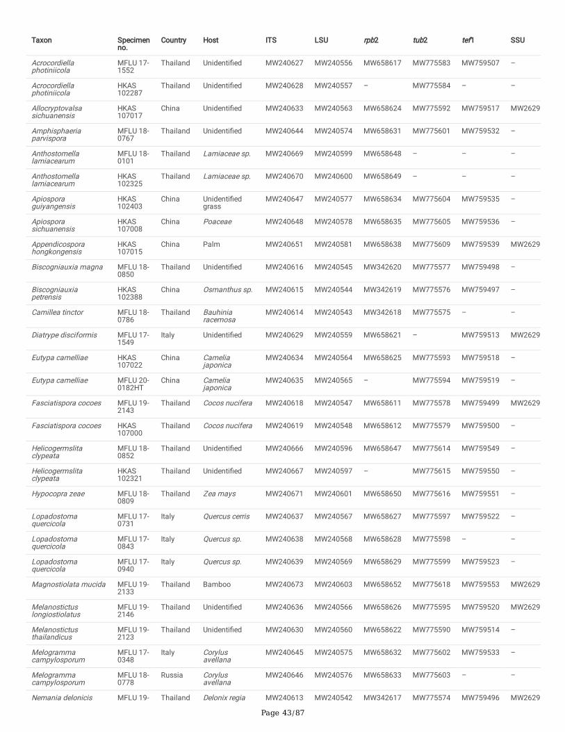
Page 43/87
Taxon Specimenno.
Country Host ITS LSU rpb2 tub2 tef1 SSU
Acrocordiellaphotiniicola
MFLU 17-1552
Thailand Unidenti�ed MW240627 MW240556 MW658617 MW775583 MW759507 –
Acrocordiellaphotiniicola
HKAS102287
Thailand Unidenti�ed MW240628 MW240557 – MW775584 – –
Allocryptovalsasichuanensis
HKAS107017
China Unidenti�ed MW240633 MW240563 MW658624 MW775592 MW759517 MW26299
Amphisphaeriaparvispora
MFLU 18-0767
Thailand Unidenti�ed MW240644 MW240574 MW658631 MW775601 MW759532 –
Anthostomellalamiacearum
MFLU 18-0101
Thailand Lamiaceae sp. MW240669 MW240599 MW658648 – – –
Anthostomellalamiacearum
HKAS102325
Thailand Lamiaceae sp. MW240670 MW240600 MW658649 – – –
Apiosporaguiyangensis
HKAS102403
China Unidenti�edgrass
MW240647 MW240577 MW658634 MW775604 MW759535 –
Apiosporasichuanensis
HKAS107008
China Poaceae MW240648 MW240578 MW658635 MW775605 MW759536 –
Appendicosporahongkongensis
HKAS107015
China Palm MW240651 MW240581 MW658638 MW775609 MW759539 MW26299
Biscogniauxia magna MFLU 18-0850
Thailand Unidenti�ed MW240616 MW240545 MW342620 MW775577 MW759498 –
Biscogniauxiapetrensis
HKAS102388
China Osmanthus sp. MW240615 MW240544 MW342619 MW775576 MW759497 –
Camillea tinctor MFLU 18-0786
Thailand Bauhiniaracemosa
MW240614 MW240543 MW342618 MW775575 – –
Diatrype disciformis MFLU 17-1549
Italy Unidenti�ed MW240629 MW240559 MW658621 – MW759513 MW26299
Eutypa camelliae HKAS107022
China Cameliajaponica
MW240634 MW240564 MW658625 MW775593 MW759518 –
Eutypa camelliae MFLU 20-0182HT
China Cameliajaponica
MW240635 MW240565 – MW775594 MW759519 –
Fasciatispora cocoes MFLU 19-2143
Thailand Cocos nucifera MW240618 MW240547 MW658611 MW775578 MW759499 MW26299
Fasciatispora cocoes HKAS107000
Thailand Cocos nucifera MW240619 MW240548 MW658612 MW775579 MW759500 –
Helicogermslitaclypeata
MFLU 18-0852
Thailand Unidenti�ed MW240666 MW240596 MW658647 MW775614 MW759549 –
Helicogermslitaclypeata
HKAS102321
Thailand Unidenti�ed MW240667 MW240597 – MW775615 MW759550 –
Hypocopra zeae MFLU 18-0809
Thailand Zea mays MW240671 MW240601 MW658650 MW775616 MW759551 –
Lopadostomaquercicola
MFLU 17-0731
Italy Quercus cerris MW240637 MW240567 MW658627 MW775597 MW759522 –
Lopadostomaquercicola
MFLU 17-0843
Italy Quercus sp. MW240638 MW240568 MW658628 MW775598 – –
Lopadostomaquercicola
MFLU 17-0940
Italy Quercus sp. MW240639 MW240569 MW658629 MW775599 MW759523 –
Magnostiolata mucida MFLU 19-2133
Thailand Bamboo MW240673 MW240603 MW658652 MW775618 MW759553 MW26299
Melanostictuslongiostiolatus
MFLU 19-2146
Thailand Unidenti�ed MW240636 MW240566 MW658626 MW775595 MW759520 MW26299
Melanostictusthailandicus
MFLU 19-2123
Thailand Unidenti�ed MW240630 MW240560 MW658622 MW775590 MW759514 –
Melogrammacampylosporum
MFLU 17-0348
Italy Corylusavellana
MW240645 MW240575 MW658632 MW775602 MW759533 –
Melogrammacampylosporum
MFLU 18-0778
Russia Corylusavellana
MW240646 MW240576 MW658633 MW775603 – –
Nemania delonicis MFLU 19- Thailand Delonix regia MW240613 MW240542 MW342617 MW775574 MW759496 MW26299
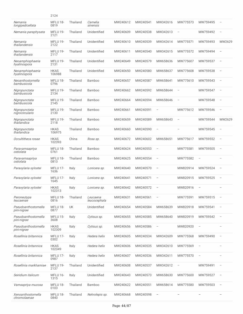
Page 44/87
2124
Nemanialongipedicellata
MFLU 18-0819
Thailand Cameliasinensis
MW240612 MW240541 MW342616 MW775573 MW759495 –
Nemania paraphysata MFLU 19-2121
Thailand Unidenti�ed MW240609 MW240538 MW342613 – MW759492 –
Nemaniathailandensis
MFLU 19-2122
Thailand Unidenti�ed MW240610 MW240539 MW342614 MW775571 MW759493 MW26298
Nemaniathailandensis
MFLU 19-2117
Thailand Unidenti�ed MW240611 MW240540 MW342615 MW775572 MW759494 –
Neoamphisphaeriahyalinospora
MFLU 19-2131
Thailand Unidenti�ed MW240649 MW240579 MW658636 MW775607 MW759537 –
Neoamphisphaeriahyalinospora
HKAS106988
Thailand Unidenti�ed MW240650 MW240580 MW658637 MW775608 MW759538 –
Neoanthostomellabambusicola
MFLU 18-0796
Thailand Bamboo MW240657 MW240587 MW658641 MW775610 MW759543 –
Nigropunctatabambusicola
MFLU 19-2134
Thailand Bamboo MW240662 MW240592 MW658644 – MW759547 –
Nigropunctatabambusicola
MFLU 19-2145
Thailand Bamboo MW240664 MW240594 MW658646 – MW759548 –
Nigropunctatanigrocircularis
MFLU 19-2130
Thailand Bamboo MW240661 MW240591 – MW775612 MW759546 –
Nigropunctatathailandica
MFLU 19-2118
Thailand Bamboo MW240659 MW240589 MW658643 – MW759544 MW26299
Nigropunctatathailandica
HKAS106975
Thailand Bamboo MW240660 MW240590 – – MW759545 –
Occultitheca rosae HKAS102393
China Rosa sp. MW240672 MW240602 MW658651 MW775617 MW759552 –
Paravamsapriyaostiolata
MFLU 18-0761
Thailand Bamboo MW240624 MW240553 – MW775581 MW759505 –
Paravamsapriyaostiolata
MFLU 18-0813
Thailand Bamboo MW240625 MW240554 – MW775582 – –
Paraxylaria xylostei MFLU 17-1636
Italy Lonicera sp. MW240640 MW240570 – MW820914 MW759524 –
Paraxylaria xylostei MFLU 17-1645
Italy Lonicera sp. MW240641 MW240571 – MW820915 MW759525 –
Paraxylaria xylostei HKAS102313
Italy Lonicera sp. MW240642 MW240572 – MW820916 – –
Peroneutypaleucaenae
MFLU 18-0816
Thailand Leucaenaleucocephala
MW240631 MW240561 – MW775591 MW759515 –
Pseudoanthostomellapini-nigrae
MFLU 18-0877
UK Unidenti�ed MW240654 MW240584 MW658639 MW820918 MW759541 –
Pseudoanthostomellapini-nigrae
MFLU 15-3608
Italy Cytisus sp. MW240655 MW240585 MW658640 MW820919 MW759542 –
Pseudoanthostomellapini-nigrae
HKAS102309
Italy Cytisus sp. MW240656 MW240586 – MW820920 – –
Rosellinia britannica MFLU 17-0302
Italy Hedera helix MW240605 MW240534 MW342609 MW775568 MW759490 –
Rosellinia britannica HKAS102349
Italy Hedera helix MW240606 MW240535 MW342610 MW775569 – –
Rosellinia britannica MFLU 17-0987
Italy Hedera helix MW240607 MW240536 MW342611 MW775570 – –
Rosellinia markhamiae MFLU 19-2137
Thailand Unidenti�ed MW240608 MW240537 MW342612 – MW759491 –
Seiridium italicum MFLU 16-1315
Italy Unidenti�ed MW240643 MW240573 MW658630 MW775600 MW759527 –
Vamsapriya mucosa MFLU 18-0103
Thailand Bamboo MW240622 MW240551 MW658614 MW775580 MW759503 –
Xenoanthostomellachromolaenae
MFLU 18-0840
Thailand Nehrolepis sp. MW240668 MW240598 – – – –
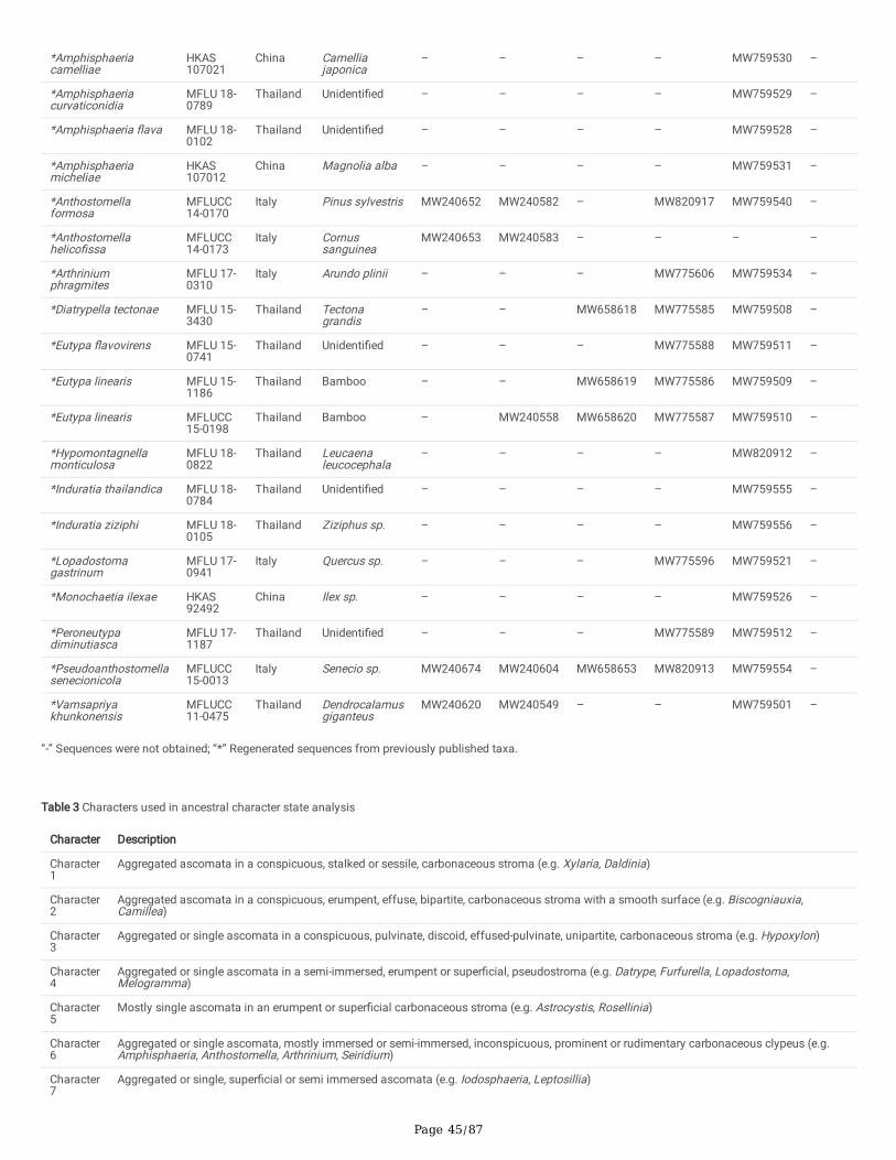
Page 45/87
*Amphisphaeriacamelliae
HKAS107021
China Camelliajaponica
– – – – MW759530 –
*Amphisphaeriacurvaticonidia
MFLU 18-0789
Thailand Unidenti�ed – – – – MW759529 –
*Amphisphaeria �ava MFLU 18-0102
Thailand Unidenti�ed – – – – MW759528 –
*Amphisphaeriamicheliae
HKAS107012
China Magnolia alba – – – – MW759531 –
*Anthostomellaformosa
MFLUCC14-0170
Italy Pinus sylvestris MW240652 MW240582 – MW820917 MW759540 –
*Anthostomellahelico�ssa
MFLUCC14-0173
Italy Cornussanguinea
MW240653 MW240583 – – – –
*Arthriniumphragmites
MFLU 17-0310
Italy Arundo plinii – – – MW775606 MW759534 –
*Diatrypella tectonae MFLU 15-3430
Thailand Tectonagrandis
– – MW658618 MW775585 MW759508 –
*Eutypa �avovirens MFLU 15-0741
Thailand Unidenti�ed – – – MW775588 MW759511 –
*Eutypa linearis MFLU 15-1186
Thailand Bamboo – – MW658619 MW775586 MW759509 –
*Eutypa linearis MFLUCC15-0198
Thailand Bamboo – MW240558 MW658620 MW775587 MW759510 –
*Hypomontagnellamonticulosa
MFLU 18-0822
Thailand Leucaenaleucocephala
– – – – MW820912 –
*Induratia thailandica MFLU 18-0784
Thailand Unidenti�ed – – – – MW759555 –
*Induratia ziziphi MFLU 18-0105
Thailand Ziziphus sp. – – – – MW759556 –
*Lopadostomagastrinum
MFLU 17-0941
Italy Quercus sp. – – – MW775596 MW759521 –
*Monochaetia ilexae HKAS92492
China Ilex sp. – – – – MW759526 –
*Peroneutypadiminutiasca
MFLU 17-1187
Thailand Unidenti�ed – – – MW775589 MW759512 –
*Pseudoanthostomellasenecionicola
MFLUCC15-0013
Italy Senecio sp. MW240674 MW240604 MW658653 MW820913 MW759554 –
*Vamsapriyakhunkonensis
MFLUCC11-0475
Thailand Dendrocalamusgiganteus
MW240620 MW240549 – – MW759501 –
“-” Sequences were not obtained; “*” Regenerated sequences from previously published taxa.
Table 3 Characters used in ancestral character state analysis
Character Description
Character1
Aggregated ascomata in a conspicuous, stalked or sessile, carbonaceous stroma (e.g. Xylaria, Daldinia)
Character2
Aggregated ascomata in a conspicuous, erumpent, effuse, bipartite, carbonaceous stroma with a smooth surface (e.g. Biscogniauxia,Camillea)
Character3
Aggregated or single ascomata in a conspicuous, pulvinate, discoid, effused-pulvinate, unipartite, carbonaceous stroma (e.g. Hypoxylon)
Character4
Aggregated or single ascomata in a semi-immersed, erumpent or super�cial, pseudostroma (e.g. Datrype, Furfurella, Lopadostoma,Melogramma)
Character5
Mostly single ascomata in an erumpent or super�cial carbonaceous stroma (e.g. Astrocystis, Rosellinia)
Character6
Aggregated or single ascomata, mostly immersed or semi-immersed, inconspicuous, prominent or rudimentary carbonaceous clypeus (e.g.Amphisphaeria, Anthostomella, Arthrinium, Seiridium)
Character7
Aggregated or single, super�cial or semi immersed ascomata (e.g. Iodosphaeria, Leptosillia)
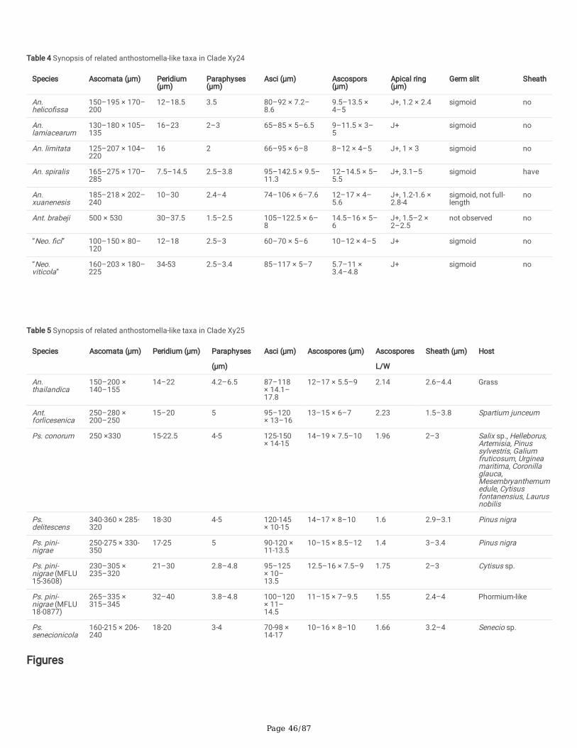
Page 46/87
Table 4 Synopsis of related anthostomella-like taxa in Clade Xy24
Species Ascomata (μm) Peridium(μm)
Paraphyses(μm)
Asci (μm) Ascospors(μm)
Apical ring(μm)
Germ slit Sheath
An.helico�ssa
150–195 × 170–200
12–18.5 3.5 80–92 × 7.2–8.6
9.5–13.5 ×4–5
J+, 1.2 × 2.4 sigmoid no
An.lamiacearum
130–180 × 105–135
16–23 2–3 65–85 × 5–6.5 9–11.5 × 3–5
J+ sigmoid no
An. limitata 125–207 × 104–220
16 2 66–95 × 6–8 8–12 × 4–5 J+, 1 × 3 sigmoid no
An. spiralis 165–275 × 170–285
7.5–14.5 2.5–3.8 95–142.5 × 9.5–11.3
12–14.5 × 5–5.5
J+, 3.1–5 sigmoid have
An.xuanenesis
185–218 × 202–240
10–30 2.4–4 74–106 × 6–7.6 12–17 × 4–5.6
J+, 1.2-1.6 ×2.8-4
sigmoid, not full-length
no
Ant. brabeji 500 × 530 30–37.5 1.5–2.5 105–122.5 × 6–8
14.5–16 × 5–6
J+, 1.5–2 ×2–2.5
not observed no
“Neo. �ci” 100–150 × 80–120
12–18 2.5–3 60–70 × 5–6 10–12 × 4–5 J+ sigmoid no
“Neo.viticola”
160–203 × 180–225
34-53 2.5–3.4 85–117 × 5–7 5.7–11 ×3.4–4.8
J+ sigmoid no
Table 5 Synopsis of related anthostomella-like taxa in Clade Xy25
Species Ascomata (μm) Peridium (μm) Paraphyses
(μm)
Asci (μm) Ascospores (μm) Ascospores
L/W
Sheath (μm) Host
An.thailandica
150–200 ×140–155
14–22 4.2–6.5 87–118× 14.1–17.8
12–17 × 5.5–9 2.14 2.6–4.4 Grass
Ant.forlìcesenica
250–280 ×200–250
15–20 5 95–120× 13–16
13–15 × 6–7 2.23 1.5–3.8 Spartium junceum
Ps. conorum 250 ×330 15-22.5 4-5 125-150× 14-15
14–19 × 7.5–10 1.96 2–3 Salix sp., Helleborus,Artemisia, Pinussylvestris, Galiumfruticosum, Urgineamaritima, Coronillaglauca,Mesembryanthemumedule, Cytisusfontanensius, Laurusnobilis
Ps.delitescens
340-360 × 285-320
18-30 4-5 120-145× 10-15
14–17 × 8–10 1.6 2.9–3.1 Pinus nigra
Ps. pini-nigrae
250-275 × 330-350
17-25 5 90-120 ×11-13.5
10–15 × 8.5–12 1.4 3–3.4 Pinus nigra
Ps. pini-nigrae (MFLU15-3608)
230–305 ×235–320
21–30 2.8–4.8 95–125× 10–13.5
12.5–16 × 7.5–9 1.75 2–3 Cytisus sp.
Ps. pini-nigrae (MFLU18-0877)
265–335 ×315–345
32–40 3.8–4.8 100–120× 11–14.5
11–15 × 7–9.5 1.55 2.4–4 Phormium-like
Ps.senecionicola
160-215 × 206-240
18-20 3-4 70-98 ×14-17
10–16 × 8–10 1.66 3.2–4 Senecio sp.
Figures
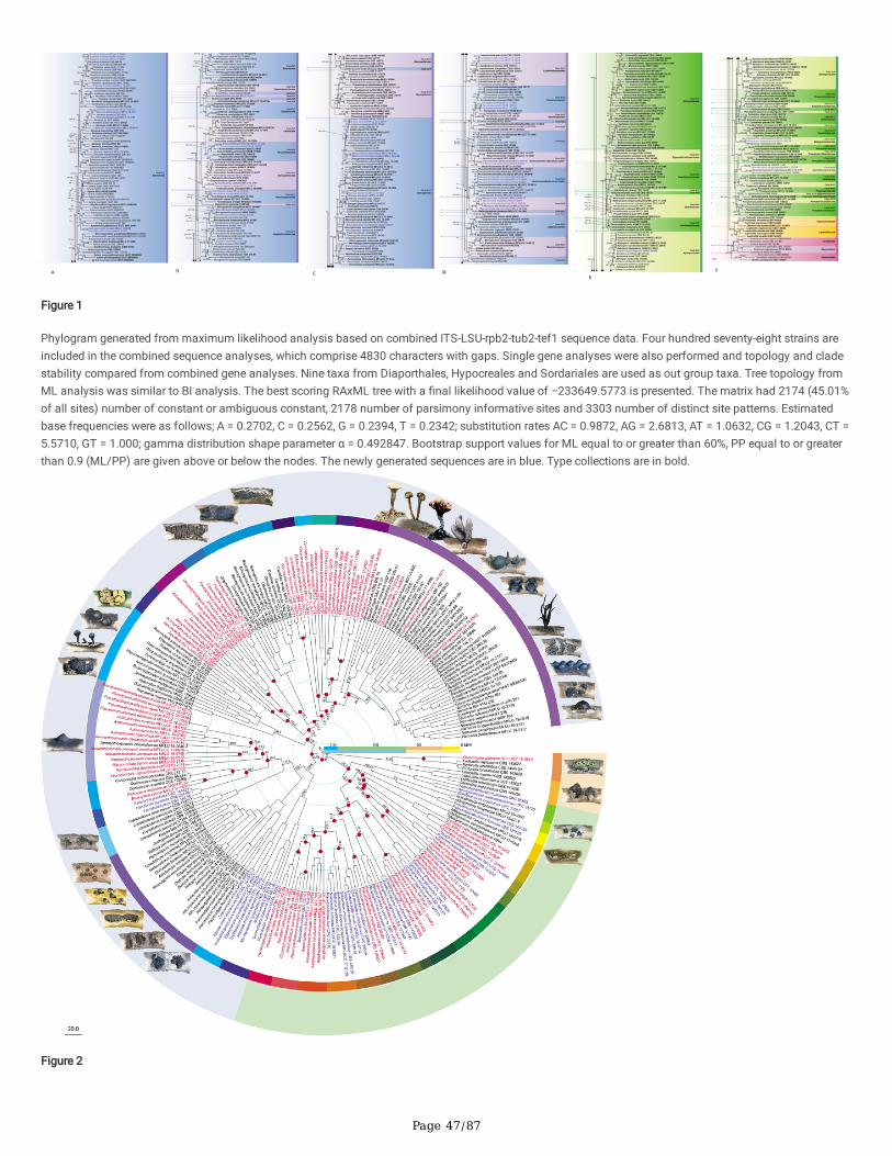
Page 47/87
Figure 1
Phylogram generated from maximum likelihood analysis based on combined ITS-LSU-rpb2-tub2-tef1 sequence data. Four hundred seventy-eight strains areincluded in the combined sequence analyses, which comprise 4830 characters with gaps. Single gene analyses were also performed and topology and cladestability compared from combined gene analyses. Nine taxa from Diaporthales, Hypocreales and Sordariales are used as out group taxa. Tree topology fromML analysis was similar to BI analysis. The best scoring RAxML tree with a �nal likelihood value of −233649.5773 is presented. The matrix had 2174 (45.01%of all sites) number of constant or ambiguous constant, 2178 number of parsimony informative sites and 3303 number of distinct site patterns. Estimatedbase frequencies were as follows; A = 0.2702, C = 0.2562, G = 0.2394, T = 0.2342; substitution rates AC = 0.9872, AG = 2.6813, AT = 1.0632, CG = 1.2043, CT =5.5710, GT = 1.000; gamma distribution shape parameter α = 0.492847. Bootstrap support values for ML equal to or greater than 60%, PP equal to or greaterthan 0.9 (ML/PP) are given above or below the nodes. The newly generated sequences are in blue. Type collections are in bold.
Figure 2
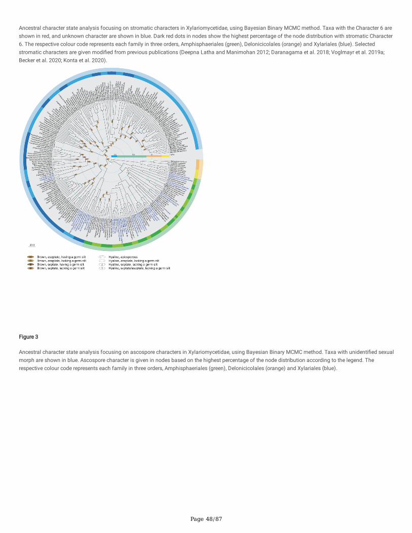
Page 48/87
Ancestral character state analysis focusing on stromatic characters in Xylariomycetidae, using Bayesian Binary MCMC method. Taxa with the Character 6 areshown in red, and unknown character are shown in blue. Dark red dots in nodes show the highest percentage of the node distribution with stromatic Character6. The respective colour code represents each family in three orders, Amphisphaeriales (green), Delonicicolales (orange) and Xylariales (blue). Selectedstromatic characters are given modi�ed from previous publications (Deepna Latha and Manimohan 2012; Daranagama et al. 2018; Voglmayr et al. 2019a;Becker et al. 2020; Konta et al. 2020).
Figure 3
Ancestral character state analysis focusing on ascospore characters in Xylariomycetidae, using Bayesian Binary MCMC method. Taxa with unidenti�ed sexualmorph are shown in blue. Ascospore character is given in nodes based on the highest percentage of the node distribution according to the legend. Therespective colour code represents each family in three orders, Amphisphaeriales (green), Delonicicolales (orange) and Xylariales (blue).

Page 49/87
Figure 4
Amphisphaeria parvispora (MFLU 18-0767, holotype). a Substrate. b,c Ascomata in dead substrate. d Section through ascoma. e Peridium. f Section throughostiole. g Paraphyses. h–l Asci. m Apical ring blueing in Melzer’s reagent. n–u Ascospores. Scale bars: a = 1 cm, b,c = 1000 μm, d = 100 μm, e,f,h–l = 20 μm,m–u = 10 μm, g = 5 μm.
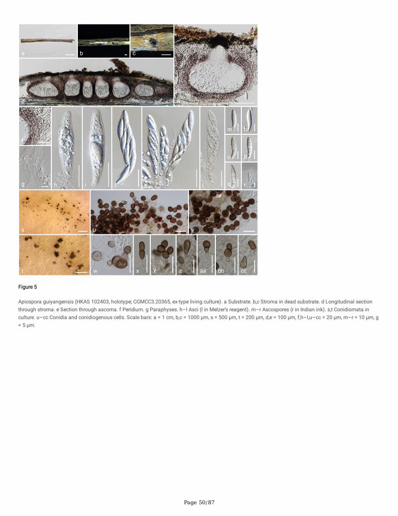
Page 50/87
Figure 5
Apiospora guiyangensis (HKAS 102403, holotype; CGMCC3.20365, ex-type living culture). a Substrate. b,c Stroma in dead substrate. d Longitudinal sectionthrough stroma. e Section through ascoma. f Peridium. g Paraphyses. h–l Asci (l in Melzer’s reagent). m–r Ascospores (r in Indian ink). s,t Conidiomata inculture. u–cc Conidia and conidiogenous cells. Scale bars: a = 1 cm, b,c = 1000 μm, s = 500 μm, t = 200 μm, d,e = 100 μm, f,h–l,u–cc = 20 μm, m–r = 10 μm, g= 5 μm.
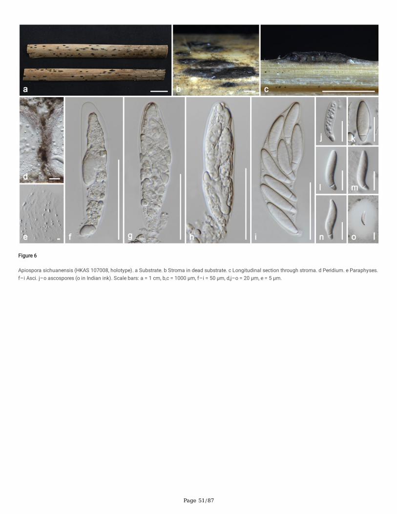
Page 51/87
Figure 6
Apiospora sichuanensis (HKAS 107008, holotype). a Substrate. b Stroma in dead substrate. c Longitudinal section through stroma. d Peridium. e Paraphyses.f–i Asci. j–o ascospores (o in Indian ink). Scale bars: a = 1 cm, b,c = 1000 μm, f–i = 50 μm, d,j–o = 20 μm, e = 5 μm.
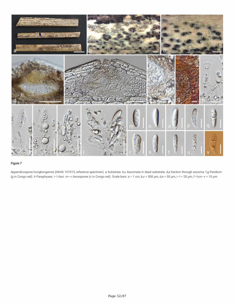
Page 52/87
Figure 7
Appendicospora hongkongensis (HKAS 107015, reference specimen). a Substrate. b,c Ascomata in dead substrate. d,e Section through ascoma. f,g Peridium(g in Congo red). h Paraphyses. i–l Asci. m–v Ascospores (v in Congo red). Scale bars: a = 1 cm, b,c = 500 μm, d,e = 50 μm, i–l = 20 μm, f–h,m–v = 10 μm.
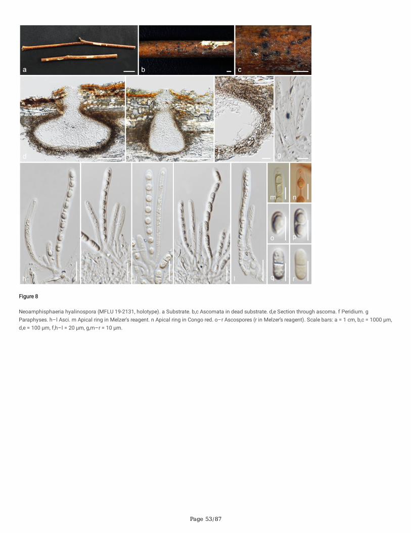
Page 53/87
Figure 8
Neoamphisphaeria hyalinospora (MFLU 19-2131, holotype). a Substrate. b,c Ascomata in dead substrate. d,e Section through ascoma. f Peridium. gParaphyses. h–l Asci. m Apical ring in Melzer’s reagent. n Apical ring in Congo red. o–r Ascospores (r in Melzer’s reagent). Scale bars: a = 1 cm, b,c = 1000 μm,d,e = 100 μm, f,h–l = 20 μm, g,m–r = 10 μm.
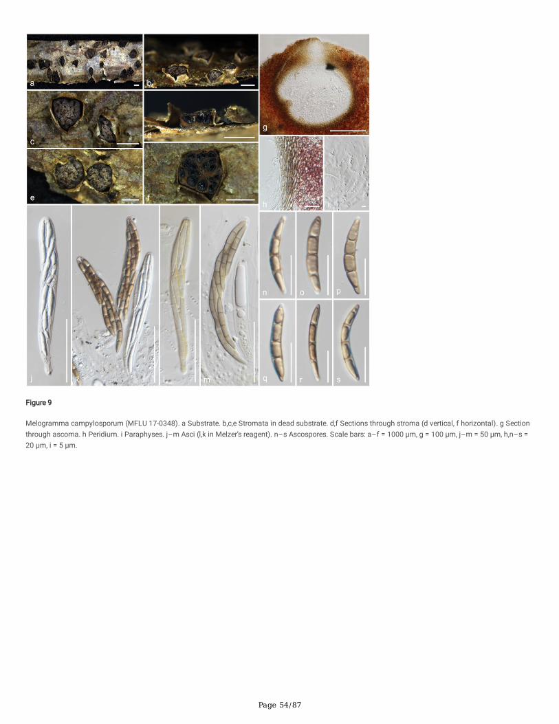
Page 54/87
Figure 9
Melogramma campylosporum (MFLU 17-0348). a Substrate. b,c,e Stromata in dead substrate. d,f Sections through stroma (d vertical, f horizontal). g Sectionthrough ascoma. h Peridium. i Paraphyses. j–m Asci (l,k in Melzer’s reagent). n–s Ascospores. Scale bars: a–f = 1000 μm, g = 100 μm, j–m = 50 μm, h,n–s =20 μm, i = 5 μm.
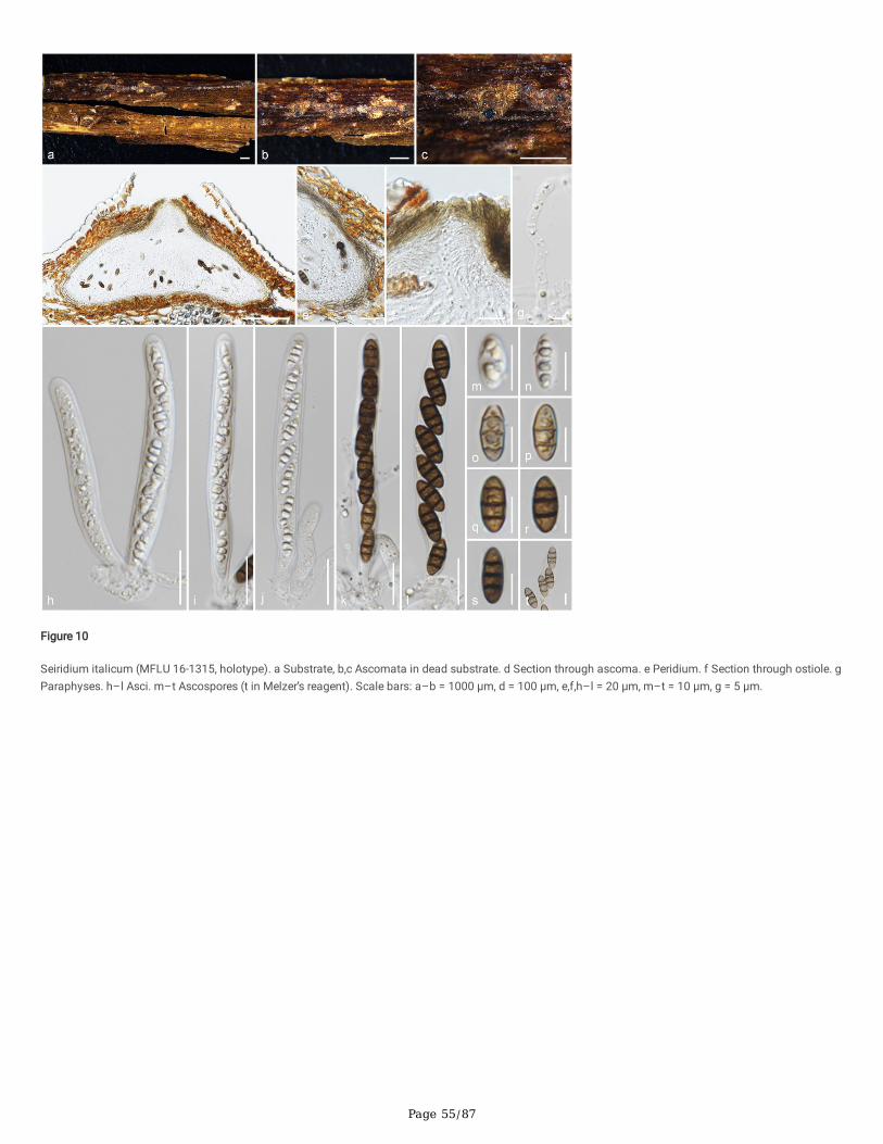
Page 55/87
Figure 10
Seiridium italicum (MFLU 16-1315, holotype). a Substrate, b,c Ascomata in dead substrate. d Section through ascoma. e Peridium. f Section through ostiole. gParaphyses. h–l Asci. m–t Ascospores (t in Melzer’s reagent). Scale bars: a–b = 1000 μm, d = 100 μm, e,f,h–l = 20 μm, m–t = 10 μm, g = 5 μm.
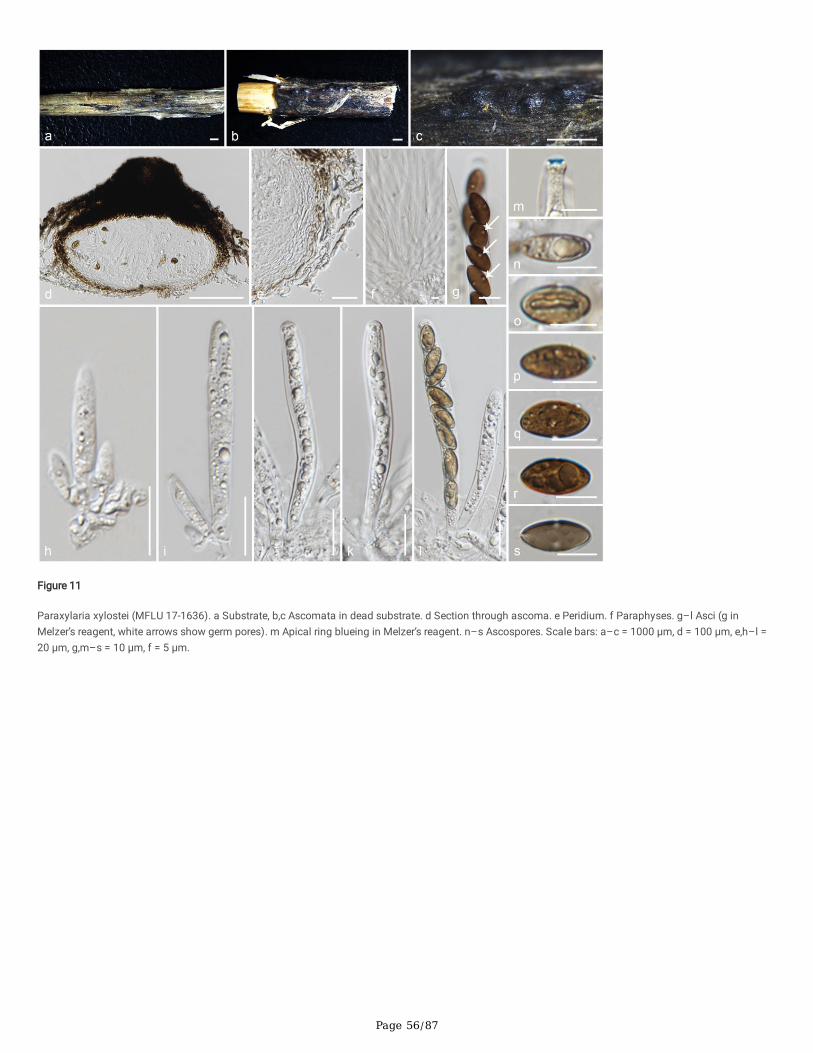
Page 56/87
Figure 11
Paraxylaria xylostei (MFLU 17-1636). a Substrate, b,c Ascomata in dead substrate. d Section through ascoma. e Peridium. f Paraphyses. g–l Asci (g inMelzer’s reagent, white arrows show germ pores). m Apical ring blueing in Melzer’s reagent. n–s Ascospores. Scale bars: a–c = 1000 μm, d = 100 μm, e,h–l =20 μm, g,m–s = 10 μm, f = 5 μm.
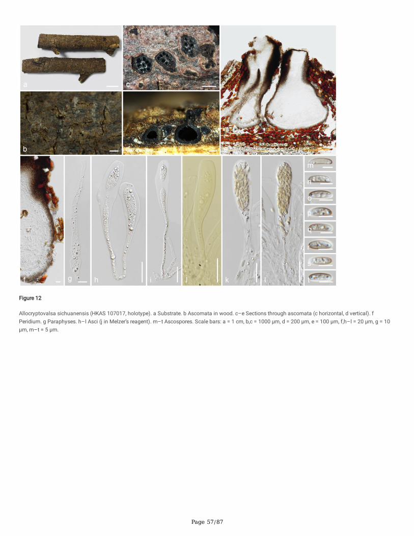
Page 57/87
Figure 12
Allocryptovalsa sichuanensis (HKAS 107017, holotype). a Substrate. b Ascomata in wood. c–e Sections through ascomata (c horizontal, d vertical). fPeridium. g Paraphyses. h–l Asci (j in Melzer’s reagent). m–t Ascospores. Scale bars: a = 1 cm, b,c = 1000 μm, d = 200 μm, e = 100 μm, f,h–l = 20 μm, g = 10μm, m–t = 5 μm.
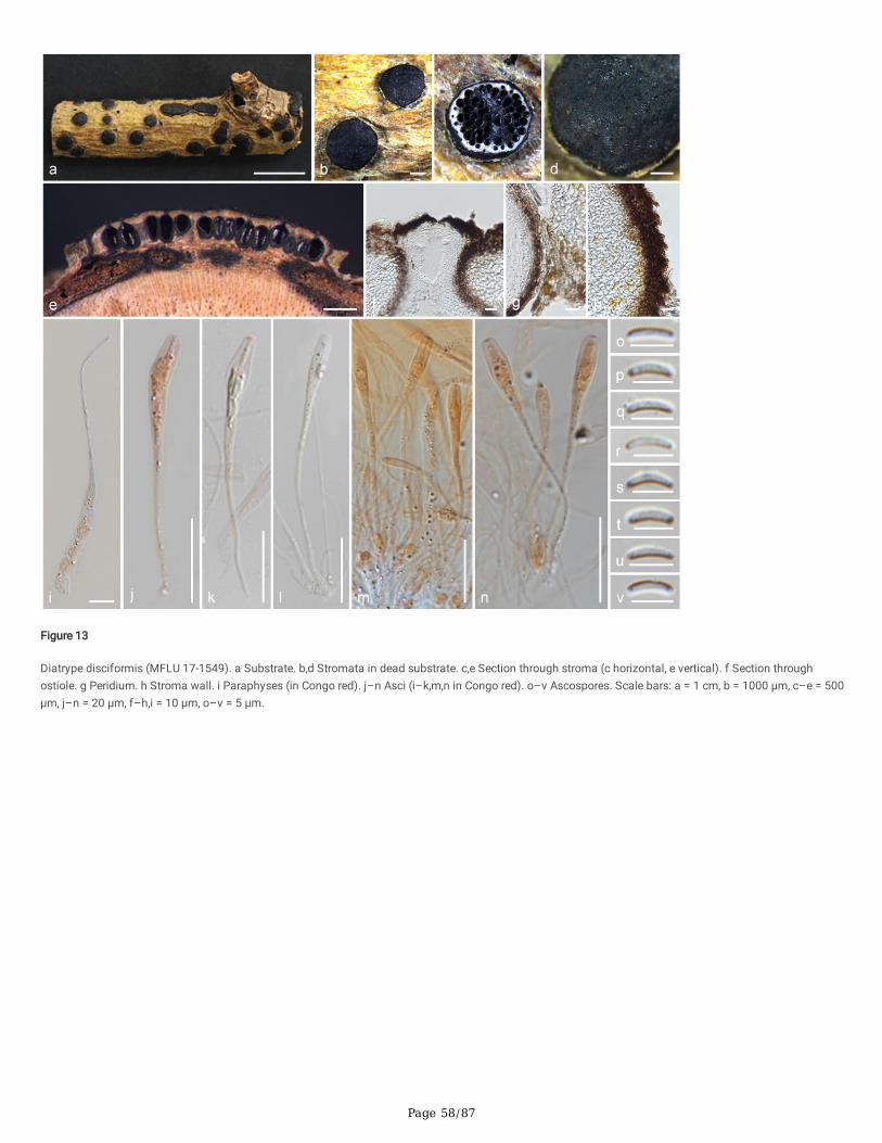
Page 58/87
Figure 13
Diatrype disciformis (MFLU 17-1549). a Substrate. b,d Stromata in dead substrate. c,e Section through stroma (c horizontal, e vertical). f Section throughostiole. g Peridium. h Stroma wall. i Paraphyses (in Congo red). j–n Asci (i–k,m,n in Congo red). o–v Ascospores. Scale bars: a = 1 cm, b = 1000 μm, c–e = 500μm, j–n = 20 μm, f–h,i = 10 μm, o–v = 5 μm.
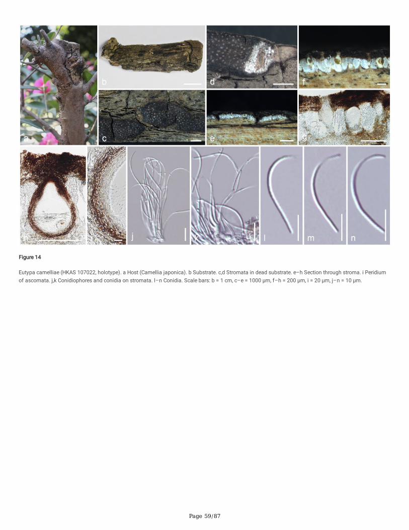
Page 59/87
Figure 14
Eutypa camelliae (HKAS 107022, holotype). a Host (Camellia japonica). b Substrate. c,d Stromata in dead substrate. e–h Section through stroma. i Peridiumof ascomata. j,k Conidiophores and conidia on stromata. l–n Conidia. Scale bars: b = 1 cm, c–e = 1000 μm, f–h = 200 μm, i = 20 μm, j–n = 10 μm.
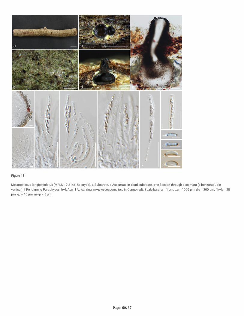
Page 60/87
Figure 15
Melanostictus longiostiolatus (MFLU 19-2146, holotype). a Substrate. b Ascomata in dead substrate. c–e Section through ascomata (c horizontal, d,evertical). f Peridium. g Paraphyses. h–k Asci. l Apical ring. m–p Ascospores (o,p in Congo red). Scale bars: a = 1 cm, b,c = 1000 μm, d,e = 200 μm, f,h–k = 20μm, g,l = 10 μm, m–p = 5 μm.
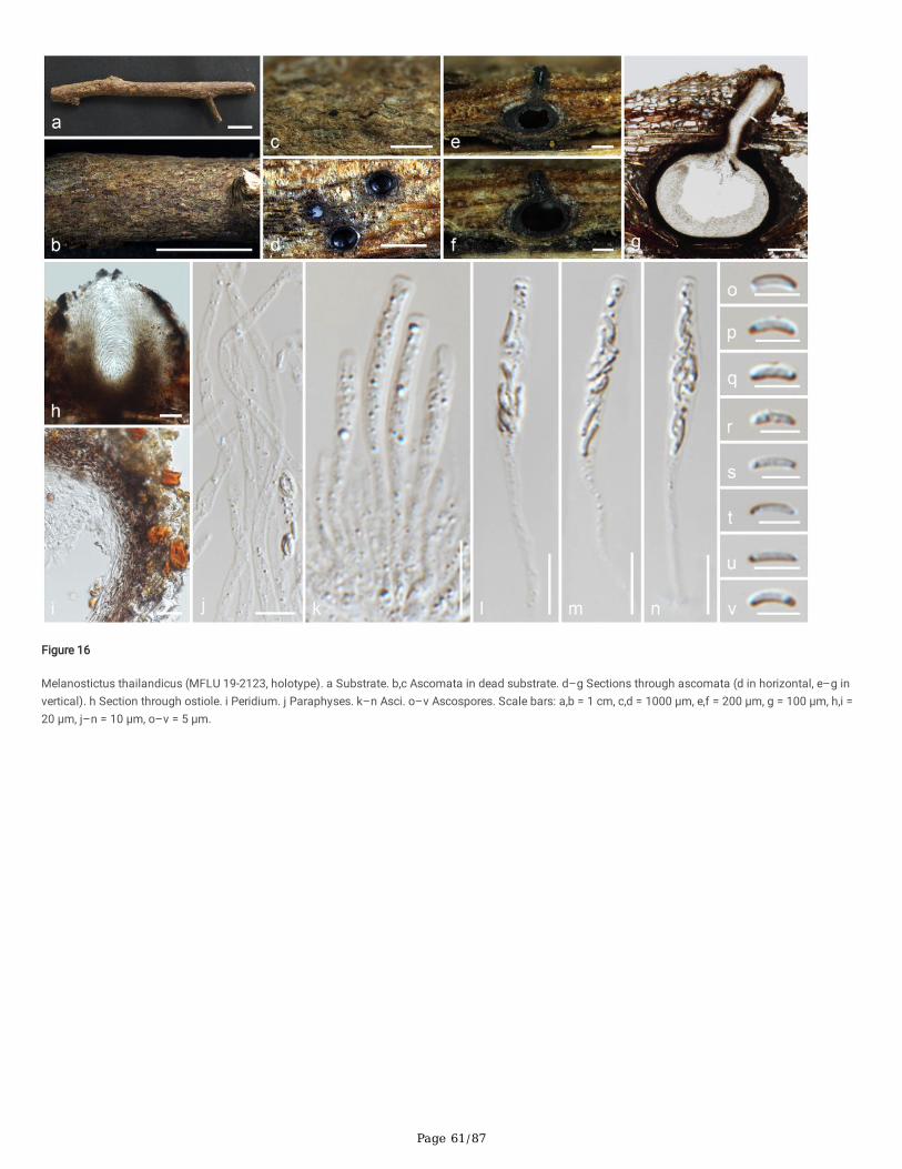
Page 61/87
Figure 16
Melanostictus thailandicus (MFLU 19-2123, holotype). a Substrate. b,c Ascomata in dead substrate. d–g Sections through ascomata (d in horizontal, e–g invertical). h Section through ostiole. i Peridium. j Paraphyses. k–n Asci. o–v Ascospores. Scale bars: a,b = 1 cm, c,d = 1000 μm, e,f = 200 μm, g = 100 μm, h,i =20 μm, j–n = 10 μm, o–v = 5 μm.
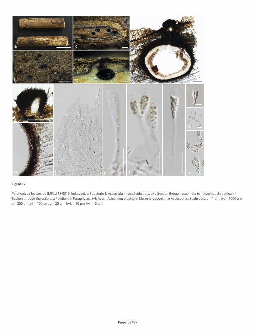
Page 62/87
Figure 17
Peroneutypa leucaenae (MFLU 18-0816, holotype). a Substrate, b Ascomata in dead substrate, c–e Section through ascomata (c horizontal, d,e vertical), fSection through the ostiole. g Peridium. h Paraphyses. i–k Asci. l Apical ring blueing in Melzer’s reagent. m,n Ascospores. Scale bars: a = 1 cm, b,c = 1000 μm,d = 200 μm, e,f = 100 μm, g = 20 μm, h–k = 10 μm, l–n = 5 μm.
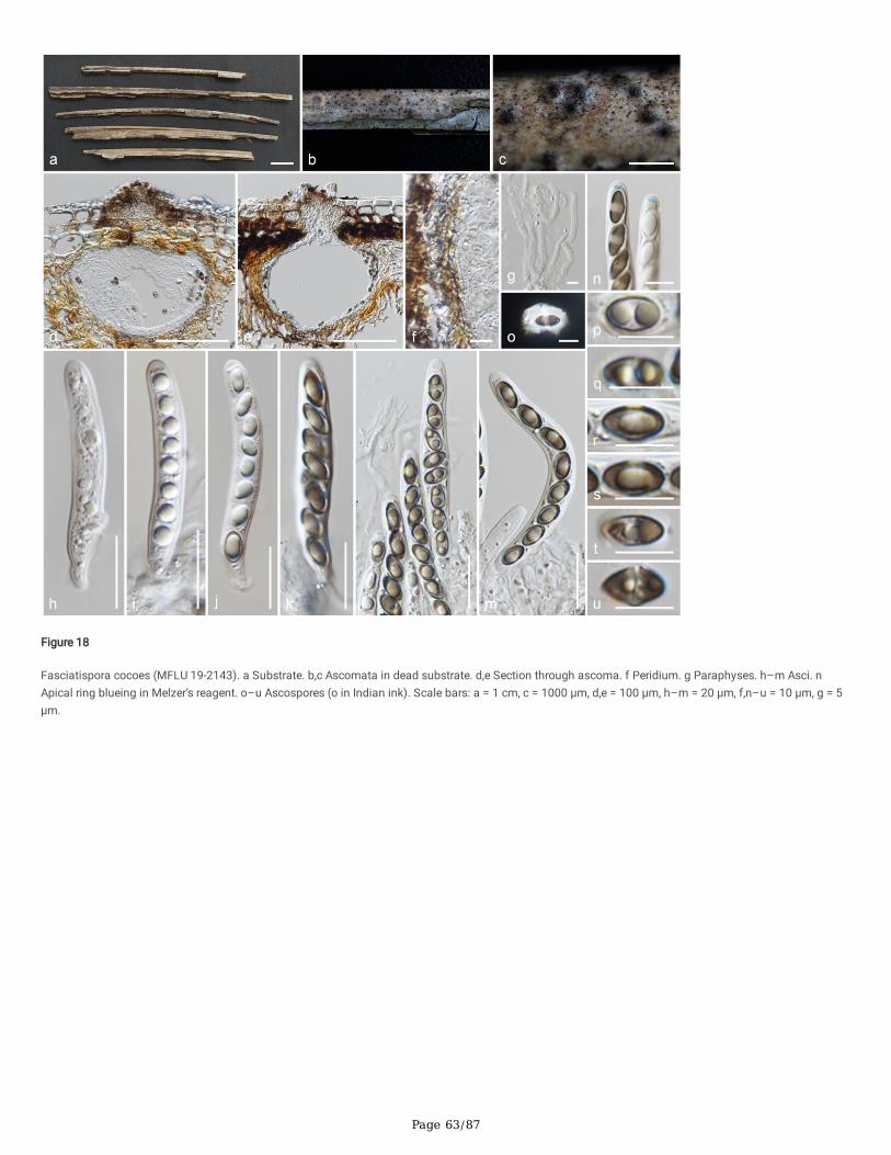
Page 63/87
Figure 18
Fasciatispora cocoes (MFLU 19-2143). a Substrate. b,c Ascomata in dead substrate. d,e Section through ascoma. f Peridium. g Paraphyses. h–m Asci. nApical ring blueing in Melzer’s reagent. o–u Ascospores (o in Indian ink). Scale bars: a = 1 cm, c = 1000 μm, d,e = 100 μm, h–m = 20 μm, f,n–u = 10 μm, g = 5μm.
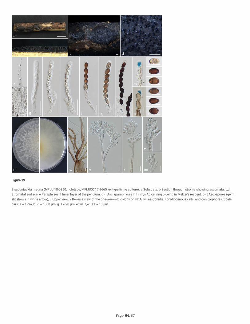
Page 64/87
Figure 19
Biscogniauxia magna (MFLU 18-0850, holotype; MFLUCC 17-2665, ex-type living culture). a Substrate. b Section through stroma showing ascomata. c,dStromatal surface. e Paraphyses. f Inner layer of the peridium. g–l Asci (paraphyses in f). m,n Apical ring blueing in Melzer’s reagent. o–t Ascospores (germslit shows in white arrow), u Upper view. v Reverse view of the one-week-old colony on PDA. w–aa Conidia, conidiogenous cells, and conidiophores. Scalebars: a = 1 cm, b–d = 1000 μm, g–l = 20 μm, e,f,m–t,w–aa = 10 μm.
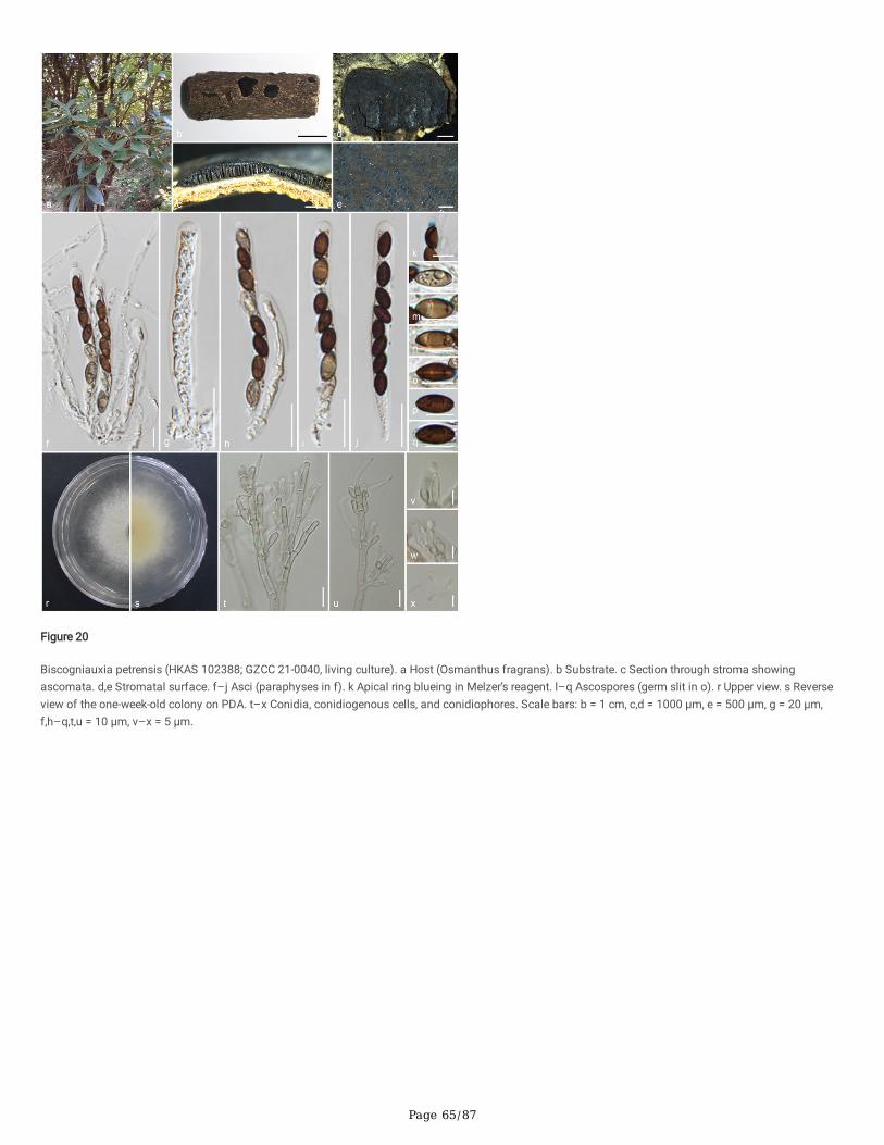
Page 65/87
Figure 20
Biscogniauxia petrensis (HKAS 102388; GZCC 21-0040, living culture). a Host (Osmanthus fragrans). b Substrate. c Section through stroma showingascomata. d,e Stromatal surface. f–j Asci (paraphyses in f). k Apical ring blueing in Melzer’s reagent. l–q Ascospores (germ slit in o). r Upper view. s Reverseview of the one-week-old colony on PDA. t–x Conidia, conidiogenous cells, and conidiophores. Scale bars: b = 1 cm, c,d = 1000 μm, e = 500 μm, g = 20 μm,f,h–q,t,u = 10 μm, v–x = 5 μm.
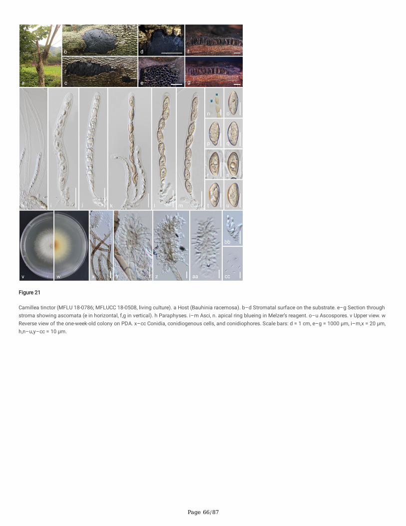
Page 66/87
Figure 21
Camillea tinctor (MFLU 18-0786; MFLUCC 18-0508, living culture). a Host (Bauhinia racemosa). b–d Stromatal surface on the substrate. e–g Section throughstroma showing ascomata (e in horizontal, f,g in vertical). h Paraphyses. i–m Asci, n. apical ring blueing in Melzer’s reagent. o–u Ascospores. v Upper view. wReverse view of the one-week-old colony on PDA. x–cc Conidia, conidiogenous cells, and conidiophores. Scale bars: d = 1 cm, e–g = 1000 μm, i–m,x = 20 μm,h,n–u,y–cc = 10 μm.
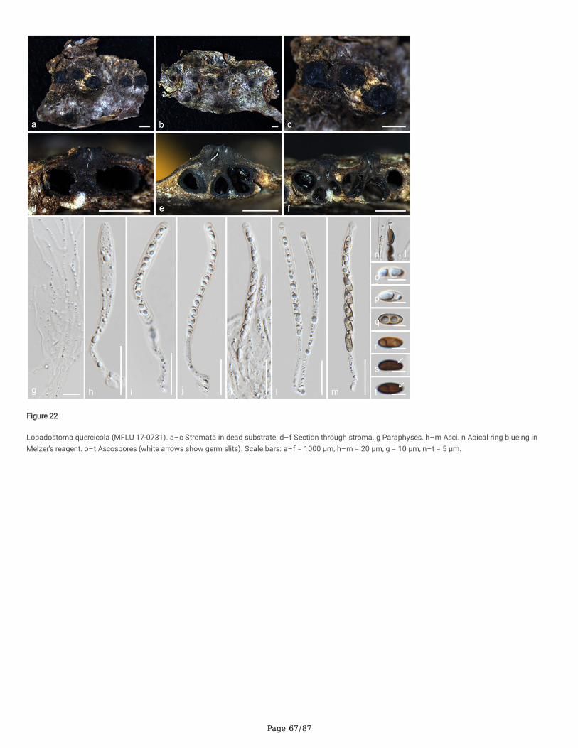
Page 67/87
Figure 22
Lopadostoma quercicola (MFLU 17-0731). a–c Stromata in dead substrate. d–f Section through stroma. g Paraphyses. h–m Asci. n Apical ring blueing inMelzer’s reagent. o–t Ascospores (white arrows show germ slits). Scale bars: a–f = 1000 μm, h–m = 20 μm, g = 10 μm, n–t = 5 μm.

Page 68/87
Figure 23
Acrocordiella photiniicola (MFLU 17-1552, holotype). a Substrate. b–d Ascomata in branches. e,f Section through ascoma. g Peridium. h Paraphyses. i–l Asci.m Ascospores in 5% KOH. n Apical ring in Melzer’s reagent. o–v Ascospores. Scale bars: a,b = 1000 μm, c = 200 μm, d–f = 100 μm, i–l = 20 μm, g,h,m–v = 10μm.
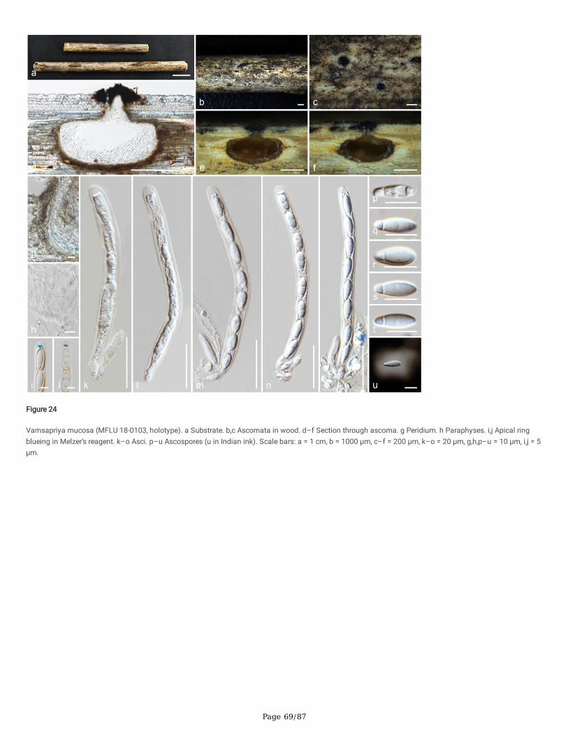
Page 69/87
Figure 24
Vamsapriya mucosa (MFLU 18-0103, holotype). a Substrate. b,c Ascomata in wood. d–f Section through ascoma. g Peridium. h Paraphyses. i,j Apical ringblueing in Melzer’s reagent. k–o Asci. p–u Ascospores (u in Indian ink). Scale bars: a = 1 cm, b = 1000 μm, c–f = 200 μm, k–o = 20 μm, g,h,p–u = 10 μm, i,j = 5μm.
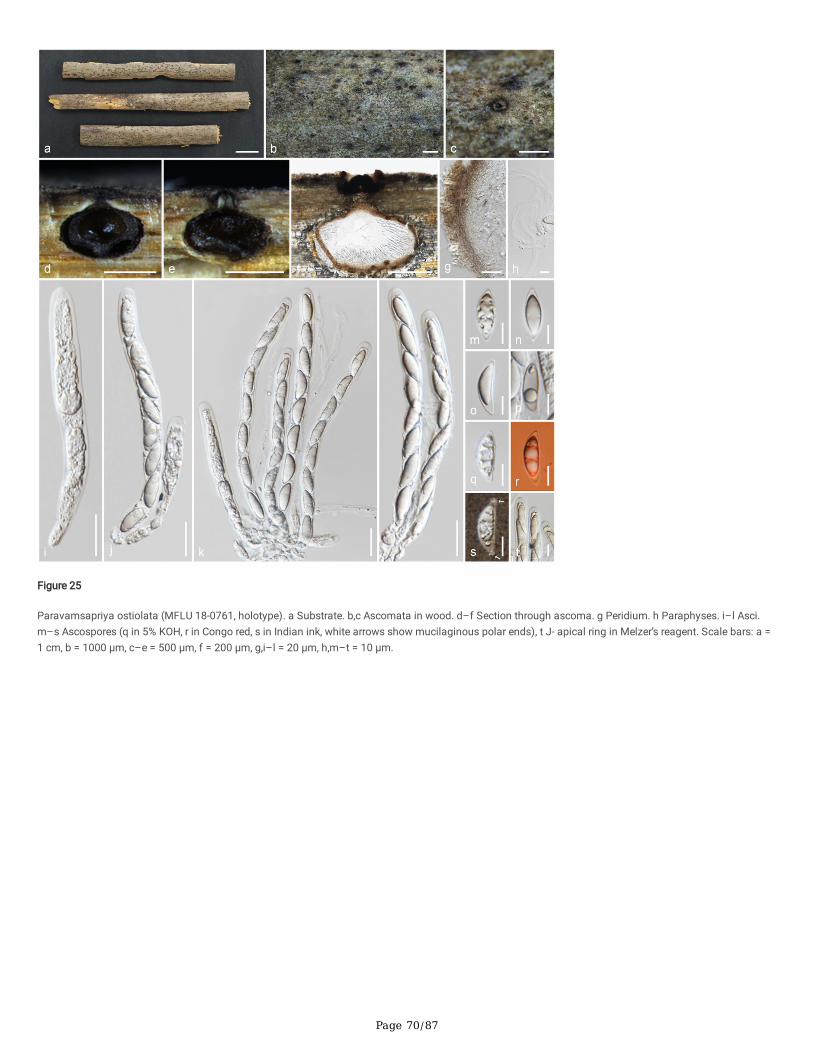
Page 70/87
Figure 25
Paravamsapriya ostiolata (MFLU 18-0761, holotype). a Substrate. b,c Ascomata in wood. d–f Section through ascoma. g Peridium. h Paraphyses. i–l Asci.m–s Ascospores (q in 5% KOH, r in Congo red, s in Indian ink, white arrows show mucilaginous polar ends), t J- apical ring in Melzer’s reagent. Scale bars: a =1 cm, b = 1000 μm, c–e = 500 μm, f = 200 μm, g,i–l = 20 μm, h,m–t = 10 μm.
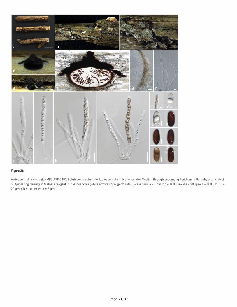
Page 71/87
Figure 26
Helicogermslita clypeata (MFLU 18-0852, holotype). a substrate. b,c Ascomata in branches. d–f Section through ascoma. g Peridium. h Paraphyses. i–l Asci.m Apical ring blueing in Melzer’s reagent. n–t Ascospores (white arrows show germ slits). Scale bars: a = 1 cm, b,c = 1000 μm, d,e = 200 μm, f = 100 μm, i–l =20 μm, g,h = 10 μm, m–t = 5 μm.
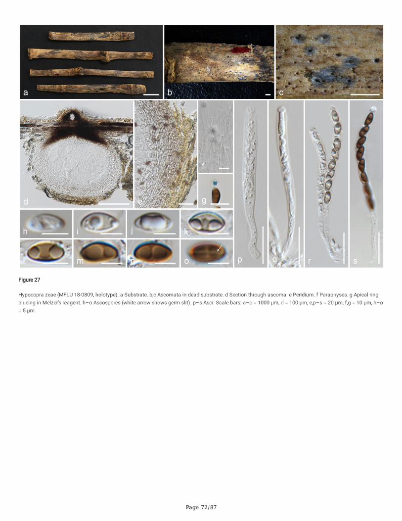
Page 72/87
Figure 27
Hypocopra zeae (MFLU 18-0809, holotype). a Substrate. b,c Ascomata in dead substrate. d Section through ascoma. e Peridium. f Paraphyses. g Apical ringblueing in Melzer’s reagent. h–o Ascospores (white arrow shows germ slit). p–s Asci. Scale bars: a–c = 1000 μm, d = 100 μm, e,p–s = 20 μm, f,g = 10 μm, h–o= 5 μm.
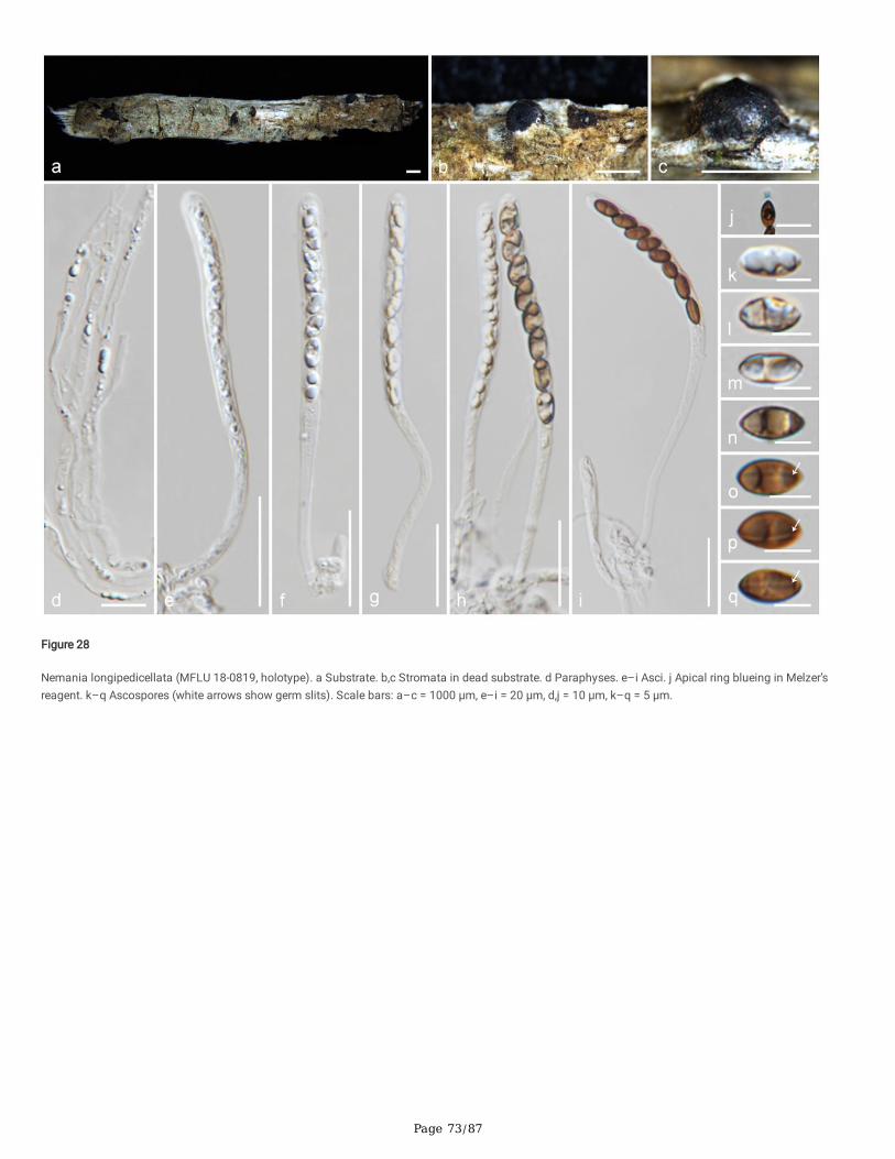
Page 73/87
Figure 28
Nemania longipedicellata (MFLU 18-0819, holotype). a Substrate. b,c Stromata in dead substrate. d Paraphyses. e–i Asci. j Apical ring blueing in Melzer’sreagent. k–q Ascospores (white arrows show germ slits). Scale bars: a–c = 1000 μm, e–i = 20 μm, d,j = 10 μm, k–q = 5 μm.

Page 74/87
Figure 29
Nemania delonicis (MFLU 19-2124, holotype). a,b Stromata in dead substrate. c,d Section through stromata (c vertical, d horizontal), e Paraphyses. f–j Asci. kApical ring blueing in Melzer’s reagent. l–r Ascospores (white arrows show germ slits). Scale bars: a = 1 cm, b–d = 1000 μm, f–j = 20 μm, e,k–r = 10 μm.
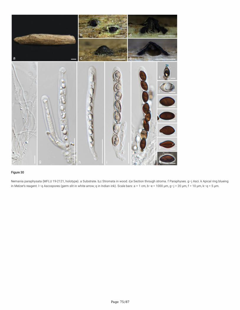
Page 75/87
Figure 30
Nemania paraphysata (MFLU 19-2121, holotype). a Substrate. b,c Stromata in wood. d,e Section through stroma. f Paraphyses. g–j Asci. k Apical ring blueingin Melzer’s reagent. l–q Ascospores (germ slit in white arrow; q in Indian ink). Scale bars: a = 1 cm, b–e = 1000 μm, g–j = 20 μm, f = 10 μm, k–q = 5 μm.
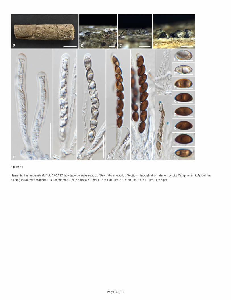
Page 76/87
Figure 31
Nemania thailandensis (MFLU 19-2117, holotype). a substrate. b,c Stromata in wood. d Sections through stromata. e–i Asci. j Paraphyses. k Apical ringblueing in Melzer’s reagent. l–s Ascospores. Scale bars: a = 1 cm, b–d = 1000 μm, e–i = 20 μm, l–s = 10 μm, j,k = 5 μm.
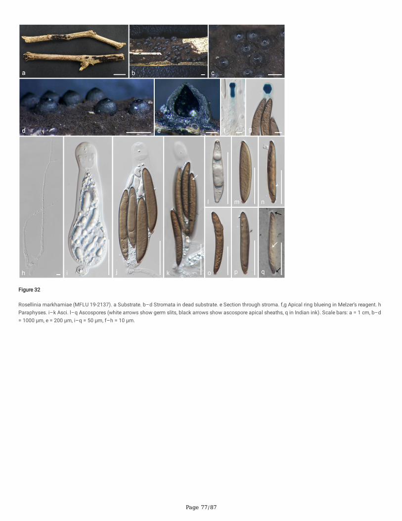
Page 77/87
Figure 32
Rosellinia markhamiae (MFLU 19-2137). a Substrate. b–d Stromata in dead substrate. e Section through stroma. f,g Apical ring blueing in Melzer’s reagent. hParaphyses. i–k Asci. l–q Ascospores (white arrows show germ slits, black arrows show ascospore apical sheaths, q in Indian ink). Scale bars: a = 1 cm, b–d= 1000 μm, e = 200 μm, i–q = 50 μm, f–h = 10 μm.
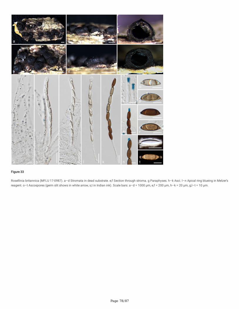
Page 78/87
Figure 33
Rosellinia britannica (MFLU 17-0987). a–d Stromata in dead substrate. e,f Section through stroma. g Paraphyses. h–k Asci. l–n Apical ring blueing in Melzer’sreagent. o–t Ascospores (germ slit shows in white arrow, s,t in Indian ink). Scale bars: a–d = 1000 μm, e,f = 200 μm, h–k = 20 μm, g,l–t = 10 μm.
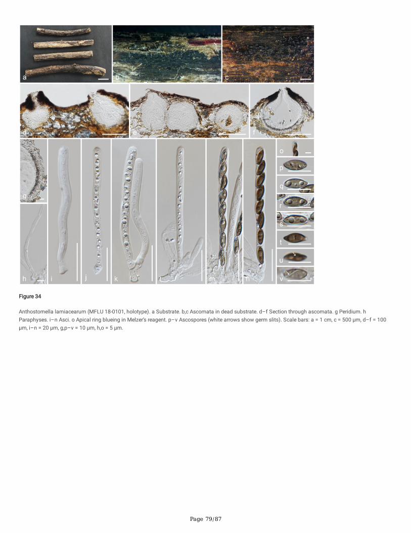
Page 79/87
Figure 34
Anthostomella lamiacearum (MFLU 18-0101, holotype). a Substrate. b,c Ascomata in dead substrate. d–f Section through ascomata. g Peridium. hParaphyses. i–n Asci. o Apical ring blueing in Melzer’s reagent. p–v Ascospores (white arrows show germ slits). Scale bars: a = 1 cm, c = 500 μm, d–f = 100μm, i–n = 20 μm, g,p–v = 10 μm, h,o = 5 μm.
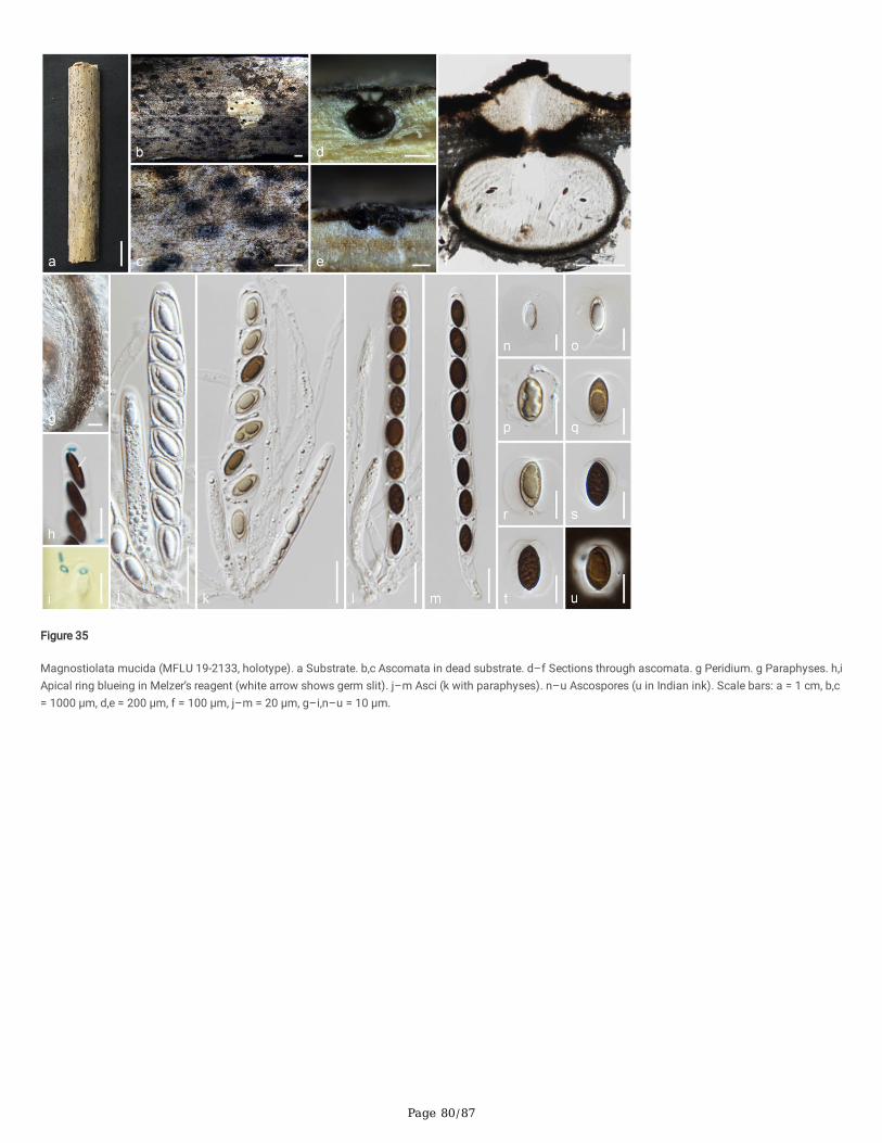
Page 80/87
Figure 35
Magnostiolata mucida (MFLU 19-2133, holotype). a Substrate. b,c Ascomata in dead substrate. d–f Sections through ascomata. g Peridium. g Paraphyses. h,iApical ring blueing in Melzer’s reagent (white arrow shows germ slit). j–m Asci (k with paraphyses). n–u Ascospores (u in Indian ink). Scale bars: a = 1 cm, b,c= 1000 μm, d,e = 200 μm, f = 100 μm, j–m = 20 μm, g–i,n–u = 10 μm.
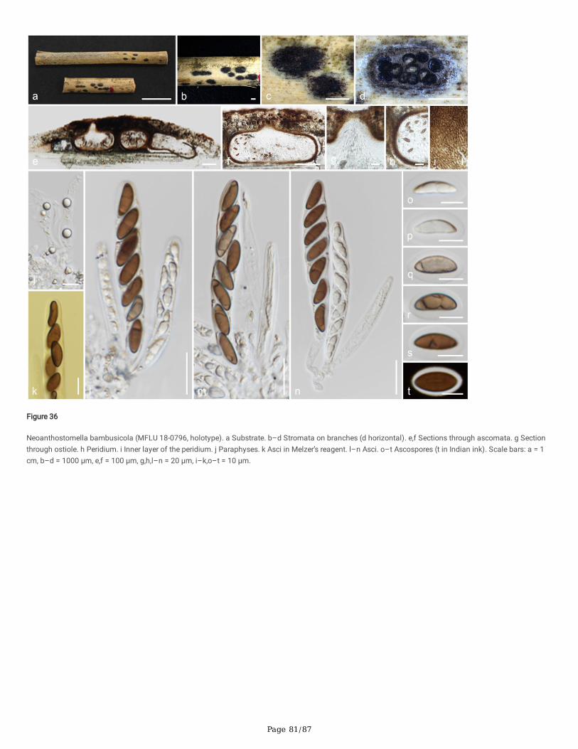
Page 81/87
Figure 36
Neoanthostomella bambusicola (MFLU 18-0796, holotype). a Substrate. b–d Stromata on branches (d horizontal). e,f Sections through ascomata. g Sectionthrough ostiole. h Peridium. i Inner layer of the peridium. j Paraphyses. k Asci in Melzer’s reagent. l–n Asci. o–t Ascospores (t in Indian ink). Scale bars: a = 1cm, b–d = 1000 μm, e,f = 100 μm, g,h,l–n = 20 μm, i–k,o–t = 10 μm.

Page 82/87
Figure 37
Nigropunctata bambusicola (MFLU 19-2145, holotype). a Substrate. b,c Ascomata in dead substrate. d,e Sections through ascomata. f Peridium. gParaphyses. h–k Asci. l Apical ring blueing in Melzer’s reagent. m–r Ascospores (germ slit shows in white arrow, q,r in Indian ink). Scale bars: a,b = 1 cm, c =1000 μm, d = 500 μm, e = 100 μm, f = 20 μm, g–r = 10 μm.
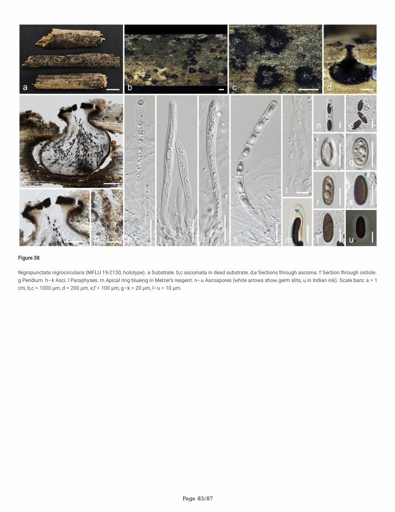
Page 83/87
Figure 38
Nigropunctata nigrocircularis (MFLU 19-2130, holotype). a Substrate. b,c ascomata in dead substrate. d,e Sections through ascoma. f Section through ostiole.g Peridium. h–k Asci. l Paraphyses. m Apical ring blueing in Melzer’s reagent. n–u Ascospores (white arrows show germ slits, u in Indian ink). Scale bars: a = 1cm, b,c = 1000 μm, d = 200 μm, e,f = 100 μm, g–k = 20 μm, l–u = 10 μm.

Page 84/87
Figure 39
Nigropunctata thailandica (MFLU 19-2118, holotype). a Substrate. b,c Ascomata in dead substrate. d,e Sections through ascomata. f Peridium. g Paraphyses.h–l Asci. m Apical ring blueing in Melzer’s reagent. n–t Ascospores. Scale bars: a = 1 cm, b,c = 1000 μm, d = 200 μm, e = 100 μm, f,h–l = 20 μm, g,m–t = 10μm.
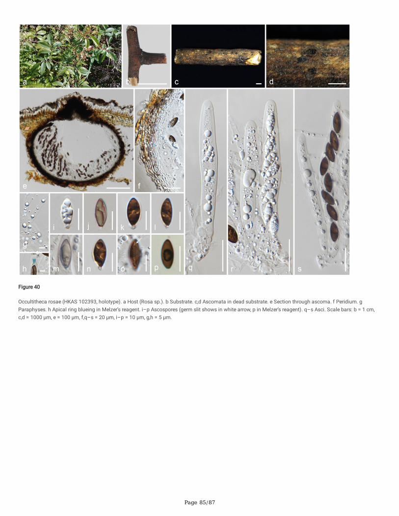
Page 85/87
Figure 40
Occultitheca rosae (HKAS 102393, holotype). a Host (Rosa sp.). b Substrate. c,d Ascomata in dead substrate. e Section through ascoma. f Peridium. gParaphyses. h Apical ring blueing in Melzer’s reagent. i–p Ascospores (germ slit shows in white arrow, p in Melzer’s reagent). q–s Asci. Scale bars: b = 1 cm,c,d = 1000 μm, e = 100 μm, f,q–s = 20 μm, i–p = 10 μm, g,h = 5 μm.
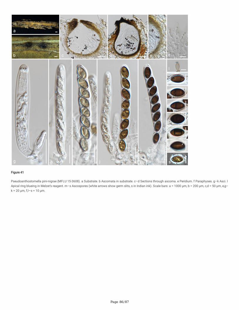
Page 86/87
Figure 41
Pseudoanthostomella pini-nigrae (MFLU 15-3608). a Substrate. b Ascomata in substrate. c–d Sections through ascoma. e Peridium. f Paraphyses. g–k Asci. lApical ring blueing in Melzer’s reagent. m–s Ascospores (white arrows show germ slits, s in Indian ink). Scale bars: a = 1000 μm, b = 200 μm, c,d = 50 μm, e,g–k = 20 μm, f,l–s = 10 μm.
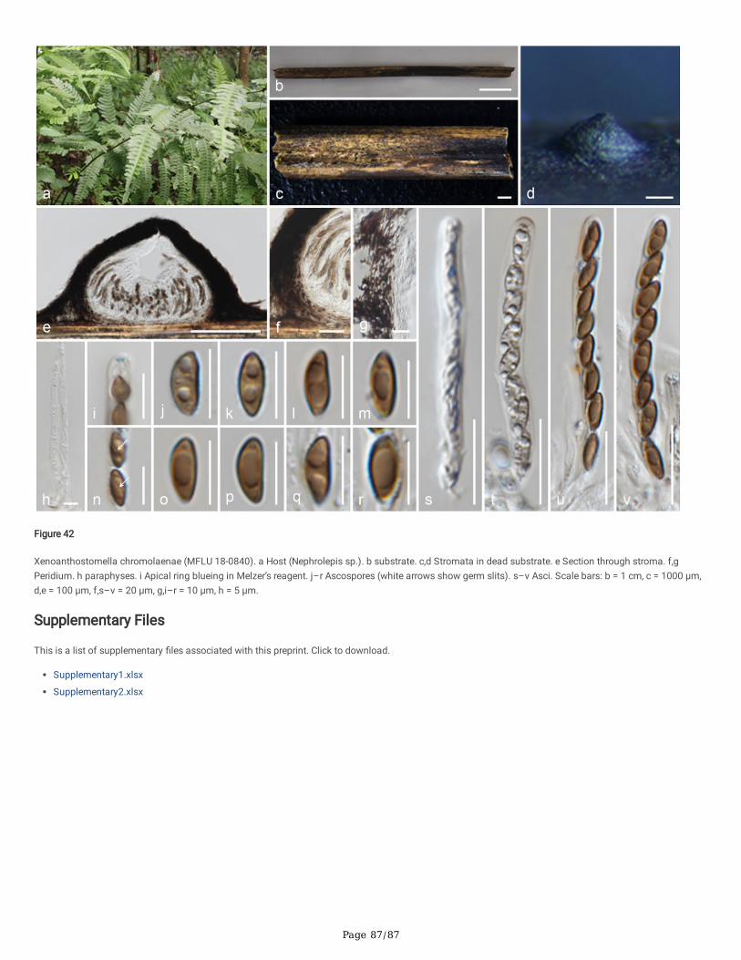
Page 87/87
Figure 42
Xenoanthostomella chromolaenae (MFLU 18-0840). a Host (Nephrolepis sp.). b substrate. c,d Stromata in dead substrate. e Section through stroma. f,gPeridium. h paraphyses. i Apical ring blueing in Melzer’s reagent. j–r Ascospores (white arrows show germ slits). s–v Asci. Scale bars: b = 1 cm, c = 1000 μm,d,e = 100 μm, f,s–v = 20 μm, g,i–r = 10 μm, h = 5 μm.
Supplementary Files
This is a list of supplementary �les associated with this preprint. Click to download.
Supplementary1.xlsx
Supplementary2.xlsx