A self-validating numerical method for the matrix exponential
Upload
khangminh22Category
view
1download
0
RESEARCH ARTICLE Open Access
Validating potent anti-inflammatory andanti-rheumatoid properties of Drynariaquercifolia rhizome methanolic extractthrough in vitro, in vivo, in silico and GC-MS-based profilingDebabrata Modak1, Subhashis Paul1, Sourav Sarkar1, Subarna Thakur2 and Soumen Bhattacharjee1*
Abstract
Background: The fronds of Drynaria quercifolia have traditionally been used in rheumatic pain management. Thegoal of the present study was to validate the potent anti-inflammatory and anti-rheumatoid properties of themethanolic-extract of its rhizome using in vitro, in vivo and in silico strategies.
Methods: The plant was collected and the methanolic extract was prepared from its rhizome. Protein denaturationtest, hypotonicity and heat-induced haemolysis assays were performed in vitro. The in vivo anti-rheumatoidpotential was assessed in Freund’s complete adjuvant (FCA)-induced Wistar rat model through inflammatory paw-edema, haematological, biochemical, radiological and histopathological measurements. Moreover, metabolites ofmethanolic extract were screened by gas chromatography-mass spectrometry (GC-MS) and 3D molecular structuresof active components were utilized for in silico docking study using AutoDock.
Results: In vitro results evinced a significant (p < 0.05) anti-inflammatory activity of the rhizome methanolic extractin a dose-linear response. Further, Drynaria quercifolia rhizome methanolic extract (DME) significantly amelioratedrheumatoid arthritis as indicated by the inhibition of arthritic paw-edema (in millimeter) in the rat rheumatoidarthritis models in both the low (57.71 ± 0.99, p < 0.01) and high dose groups (54.45 ± 1.30, p < 0.001) whencompared to arthritic control. Treatment with DME also normalized the haematological (RBC, WBC, platelet countsand hemoglobin contents) and biochemical parameters (total protein, albumin, creatinine and ceruloplasmin)significantly (p < 0.05), which were further supported by histopathological and radiological analyses. Furthermore,GC-MS analysis of DME demonstrated the presence of 47 phytochemical compounds. Compounds like Squalene,Gamma Tocopherol, n-Hexadecanoic acid showed potent inhibition of cyclooxygenase-2 (COX-2), tumor necrosisfactor (TNF-α), and interleukin (IL-6) in the docking analysis.
(Continued on next page)
© The Author(s). 2021 Open Access This article is licensed under a Creative Commons Attribution 4.0 International License,which permits use, sharing, adaptation, distribution and reproduction in any medium or format, as long as you giveappropriate credit to the original author(s) and the source, provide a link to the Creative Commons licence, and indicate ifchanges were made. The images or other third party material in this article are included in the article's Creative Commonslicence, unless indicated otherwise in a credit line to the material. If material is not included in the article's Creative Commonslicence and your intended use is not permitted by statutory regulation or exceeds the permitted use, you will need to obtainpermission directly from the copyright holder. To view a copy of this licence, visit http://creativecommons.org/licenses/by/4.0/.The Creative Commons Public Domain Dedication waiver (http://creativecommons.org/publicdomain/zero/1.0/) applies to thedata made available in this article, unless otherwise stated in a credit line to the data.
* Correspondence: [email protected]; [email protected] and Molecular Biology Laboratory, Department of Zoology, University ofNorth Bengal, Raja Rammohunpur, Darjeeling, West Bengal 734013, IndiaFull list of author information is available at the end of the article
BMC ComplementaryMedicine and Therapies
Modak et al. BMC Complementary Medicine and Therapies (2021) 21:89 https://doi.org/10.1186/s12906-021-03265-7
(Continued from previous page)
Conclusion: Results from in vivo and in vitro studies indicated that DME possesses a potent anti-inflammatory andanti-arthritic activity. In silico studies delineated the emergent potent inhibitory effects of several bio-activecomponents on the target inflammatory markers (COX-2, TNF-α and IL-6).
Keywords: Drynaria quercifolia, Rheumatoid arthritis, Inflammation, Paw edema, GC-MS, Molecular docking
BackgroundInflammation, the body’s defense mechanism, is a com-mon biological cascade in response to foreign pathogens,tissue injury, or chemical irritation that acts by removingthe injurious stimuli and begins the healing process whichis rapid and self-limiting [1]. Both infectious and non-infectious stimuli can trigger an inflammatory pathway oracute inflammatory response that contributes to restoringphysiological homeostasis. However, when prolonged, itmay become chronic, enhance tissue inflammation andcontribute to a variety of chronic inflammatory diseaseslike rheumatoid arthritis (RA) [2, 3]. RA is a chronic sys-temic autoimmune inflammatory disorder that has showna worldwide prevalence of approximately 0.5 to 1% amongadult individuals [4].The disease RA is characterized by synovial hyperpla-
sia, inflammatory cell infiltration in the synovial tissuesand progression of this disease leads to the destructionof cartilages and bones and chronic disability of the pa-tient [5]. Bone joint destruction is associated withchronic inflammation combined with the destruction ofthe surface and extracellular matrix of articular cartilage,and erosion of bone and if undiagnosed, it hampers thequality of lifestyle [6, 7]. The inflammation during RAresults in the redness and swelling of the joint regionswhich is associated with the elevation of the temperaturein the inflamed region and evokes severe pain [8]. Thedestruction of bones is mediated by the osteoclasts andthe cartilage is mainly degraded by the matrix metallo-proteinases in the inflammatory arthritic condition [9].The cytokine including TNF-α, IL-6 and the COX-2 playvital role in the process. TNF-α and IL-6 are two apexcytokines contributing to the progression of inflamma-tion whereas, COX-2 elevates the inflammatory painthrough prostaglandin production [10]. Presently, theavailable treatment protocol involves the use of conven-tional corticosteroids, disease-modifying anti-rheumaticdrugs (DMARDs), such as methotrexate, hydroxychloro-quine; non-steroidal anti-rheumatic drugs (NSAIDs),such as indomethacin, ibuprofen, aspirin, etc. and im-munosuppressive agents [11–13]. However, profoundadverse effects are associated with the use of theseagents, often leading to nephrotoxicity, gastrointestinaltract irritation, and hematological abnormalities and in-creased risk for cardiovascular diseases [14, 15]. Inflixi-mab, a potent TNF-α inhibitor [16] and tocilizumab, a
IL-6 inhibitor [17], have been used in the therapy againstRA which are either less explored or have shown less di-verse side-effects in the patients. Due to the persistentsymptoms, adverse side-effects and the cost involved inthe current treatment protocol, alternative strategies aredrawing more attention nowadays.In the last few decades, the search for a safe alternative
remedy from herbal resources has been an area of inter-est as herbs play a key role in the modern medicationsystem and constitute a major reservoir for potentiallyactive medicinal compounds all over the World. Medi-cinal plants have been proven to be potent and effica-cious in ameliorating RA [9]. The field of RA researchhas progressed rapidly towards herbal treatment, as theyare effective in ameliorating the pain and inflammationassociated with the disease, have lesser side effects andalso at a low cost. Drynaria quercifolia (commonly called‘Pankhiraj’ in Bengali/India) has been one of the pioneermedicinal plants which have a great medicinal valueagainst various diseases. It can be either epiphytic (whengrowing on trees) or epipetric (when growing on rocks)in nature. Drynaria quercifolia belongs to the familyPolypodiaceae of Pteridophyta and is widely distributedin tropical and subtropical countries like India,Bangladesh, Pakistan, North America, and Africa [18,19]. Traditionally, the fronds of this plant are reportedto be used by different tribal communities of India inthe treatment of various diseases like typhoid fever,chronic jaundice, headache, cough, cholera and skin dis-ease [19–21]. The soup prepared from the rhizomes ofDrynaria quercifolia is popularly used by some tribes ofEastern Ghats, Tamil Nadu to get relief from rheumaticcomplaints [22]. Consumption of Drynaria quercifoliacan help to heal and strengthen broken bones and alsobe used to promote the treatment of bone fracture bythe sub-Himalayan tribal communities like Mech, Toto,Rabha and also Koch tribe from the Cooch Behar regionof West Bengal, India [23, 24]. The fronds have a strin-gent property and are found to strengthen and promotethe repair of sinews, muscles, and bones [25]. Phyto-chemical screening of Drynaria quercifolia has con-firmed the presence of flavonoids, alkaloids, saponin,tannins, and other chemical substances [26]. The efficacyof the Drynaria quercifolia rhizome has already beenshown in different in vitro and in vivo models with re-spect to antimicrobial [27], analgesic [19], anti-
Modak et al. BMC Complementary Medicine and Therapies (2021) 21:89 Page 2 of 20
inflammatory [28] and hepatoprotective activities [29].Membrane-stabilization and thrombolytic potentials ofthe plant has also been reported [30]. The rhizome ofthis plant has also shown its efficacy by decreasing lyso-somal enzymes levels, protein bound carbohydrate levels,urinary degradative collagen levels and serum cytokineslevel in inflammatory arthritis model [31]. All the initialstudies have drawn significant evidences regarding themedicinal properties of the plant rhizome extract.The current study was aimed at validating the trad-
itional uses of Drynaria quercifolia rhizome extracts in in-flammatory conditions through in vitro and in vivostudies. The present study also intended to identify theactive constituents of methanolic extract using GC-MSanalysis and to identify potential active anti-inflammatorycomponents against COX-2, TNF-α and IL-6, key playersin the inflammatory pathway, through in silico moleculardocking studies.
Materials and methodCollection of rhizome of D. quercifoliaRhizomes of naturally growing Drynaria quercifolia (L.) J.Sm. (Class Polypodiopsida, Order Polypodiales, and Fam-ily Polypodiaceae) were collected from the North BengalUniversity Campus during September 2018. Collectedplant specimens were authenticated by Taxonomy ofAngiosperm and Biosystematics Laboratory, Departmentof Botany, University of North Bengal. A voucher speci-men was deposited with the accession number: 09746 atthe herbarium of the Department of Botany, University ofNorth Bengal. A flow sheet of methodology has been pro-vided as an additional file (see Additional file 1).
Preparation of methanolic extract of D. quercifoliaFresh rhizomes (~ 10 kg) of Drynaria quercifolia (L.) J.Sm. were properly cleaned and washed with tap water toremove the dust particles. Then the wooly brown scalesof the rhizomes were removed and the clean rhizomeswere shade dried at room temperature for 2 weeks to ob-tain about 760 g dry sample. The dried rhizomes werethen crushed into powder using an electrical grinder.The coarsely powdered rhizomes (30 g) were successfullyextracted with 100 ml of HPLC grade methanol (Merck,India) using Soxhlet apparatus for 6–7 h. The obtainedextract was then concentrated using a Buchi type rotaryevaporator (Cole Parmer RV1010D596, India) under re-duced pressure and temperature (45 °C) and the percent-age yield of the extract was 7% (w/w). The resultantDrynaria quercifolia rhizome methanolic extract (DME)was stored in an air tight container at 4 °C for furtheruse. For animal feeding, DME was reconstituted in 0.5%dimethyl sulfoxide (DMSO) (Sigma-Aldrich, USA).
In vitro anti-inflammatory assaysProtein denaturation testThe test was performed as described by previous proto-cols [32, 33]. Chicken egg albumin was used as a proteinsource and the experiment was carried out in triplicatesets (per dose). Briefly, the reaction mixture (5 ml) con-sisted of 0.2 ml of egg albumin, 2.8 ml of phosphate-buffered saline (PBS, pH 6.4) and 2ml of varying con-centrations of DME resulting in the final concentrationsof 600, 800, and 1000 μg/ml. The negative control grouphad 2ml of distilled water instead of DME. Diclofenacsodium (Novartis India Ltd., India) at the final concen-tration (100 μg/ml) was used as a standard drug. The re-action mixture was then incubated at 37 °C for 15 minand then the mixture was heated at 70 °C for 15 min in aregulated water bath. After cooling, the turbidity of thereaction mixture was measured at 660 nm. The percent-age inhibition (PI) of protein denaturation was calcu-lated using the following formula [32, 33]:
PI ¼ OD2 −OD1ð ÞOD2
� 100
Where, OD1 = Absorbance of heated test sample;OD2 = Absorbance of heated negative control sample
Membrane stabilization activityPreparation of erythrocyte suspensionErythrocyte suspension was prepared according to previ-ously described protocol [33, 34]. Briefly, 3 ml of wholeblood was collected from a healthy volunteer in a tubecontaining ethylene-diamine-tetraacetic acid (EDTA),centrifuged at 3000 rpm for 10 min, and washed threetimes with an equal volume of normal saline (0.9%).Then the volume of the dissolved red blood pellets wasmeasured and reconstituted as a 40% (v/v) suspensionwith an isotonic buffer solution (10 mM sodium phos-phate buffer, pH 7.4).
Hypotonic solution induced haemolysisFor this test, the hypotonic solution (distilled water) (5ml) containing 600, 800, and 1000 μg/ml of DME wasput in 3 pairs (per dose) on centrifuge tubes. In anotherset of centrifuge tubes, isotonic buffer solution (5 ml)containing the similar graded doses of DME were takenin 3 pairs (per dose). Negative control tubes contained 5ml of hypotonic solution (distilled water) and indometh-acin (200 μg/ml) (Cipla, India) served as the standarddrug. Stock erythrocyte suspension (100 μl) was added toeach tube, and after gentle mixing, the tubes were incu-bated at 37°C for 1 h. After incubation, the reaction mix-ture was centrifuged for 3 min at 1300 g at roomtemperature and the absorbance (OD) of the super-natant was measured at 540 nm. The percentage
Modak et al. BMC Complementary Medicine and Therapies (2021) 21:89 Page 3 of 20
inhibition (PI) of haemolysis was calculated using thefollowing equation [33, 34]:
PI ¼ 1 −OD2 −OD1OD3 −OD1
� �� �� 100
Where,OD1 = Absorbance of test sample in isotonic solutionOD2 = Absorbance of test sample in hypotonic solu-
tion (distilled water)OD3 = Absorbance of negative control sample in
hypotonic solution (distilled water)
Heat induced haemolysisFor this test, the isotonic buffer solution (5 ml) contain-ing 600, 800, and 1000 μg/ml of DME were assorted in3 sets (per dose) of centrifuge tubes. Control tubes con-tained 5 ml of the vehicle (distilled water) and the stand-ard dose group contained 5 ml isotonic buffer solutioncontaining indomethacin (200 μg/ml). Erythrocyte sus-pension (100 μl) was added to each tube and gentlymixed. Then the tubes were incubated at 54 °C for 20min in a regulated water bath. At the end of the incuba-tion, the reaction mixtures were centrifuged at 1300 gfor 3 min at room temperature and the absorbance (OD)of the supernatant was measured at 540 nm. The percentinhibition (PI) of haemolysis was calculated using thefollowing equation [33]:
PI ¼ OD2 −OD1ð ÞOD2
� 100
Where,OD1 = Absorbance of heated test sample (isotonic
buffer).OD2 = Absorbance of heated negative control sample
(distilled water).
In vivo anti-inflammatory studyAnimal maintenanceWistar albino rats of both sexes (8–12 weeks old, 120 ±10 g) were purchased from authorized animal dealers(Chakraborty Enterprise, Kolkata, India; Regd. No. 1443/PO/Bt/s/11/CPCSEA). All animals were housed underwell-ventilated polypropylene cages (Tarsons, India) withpaddy husk as bedding material. All the experimentalrats were maintained in the animal house of the Depart-ment of Zoology, University of North Bengal with a con-trolled environmental condition maintained at aconstant room temperature (25 ± 3 °C) and under a con-stant 12-h dark/light cycle. Rats were provided with suf-ficient food and water ad libitum. The study has beenapproved by the Institutional Animal Ethical Committee(IAEC) of CPCSEA (Committee for the Purpose of Con-trol and Supervision of Experiments on Animals) of the
University of North Bengal, West Bengal, India (IAEC/NBU/2018/03). Animals were acclimatized for 2 weeksbefore the study.
Acute oral toxicity testAcute toxicity test was performed according to theOrganization for Economic Cooperation and Develop-ment guidelines (OECD) 423 [35]. In the acute toxicitytests, Wistar albino rats of both sexes were clusteredinto five groups; each contained 6 rats; 3 males and 3 fe-males. The first group contained normal animals; theother four were considered as experimental groupswhere DME was administrated orally by using gavage insingle doses of 250 mg/kg b.w., 500 mg/kg b.w., 1000mg/kg b.w. and 2000mg/kg b.w. The general behaviors,such as aggressiveness, the consumption rate of foodand water intake, sedation, diarrhea, rising of fur, leth-argy were continuously observed individually during thefirst 30 mins and periodically for 24 h after the treat-ments were administered. All animals were observed in-dividually and special attention was given during thefirst 4 h daily and thereafter, for a period of 14 days forany late sign of toxicological effects.
Experimental design for anti-arthritic property of DME inFCA-induced inflammatory arthritis modelExperimental groupsA total number of twenty-four (24) Wistar albino malerats (120 ± 10 g) randomly divided into four groups ofsix rats each as under:
Group 1 (Arthritic control): Arthritic rats receivedorally normal saline (p.o.) only, from day 0 to day 28.Group 2 (Low dose group of DME): Rats were treatedwith DME (250 mg/kg b.w.). The therapy wascommenced on day 0 and continued till the 28th day.Group 3 (High dose group of DME): Rats were treatedwith DME (500 mg/kg b.w.). The therapy wascommenced on day 0 and continued till the 28th day.Group 4 (Vehicle control): 0.5% of DMSO wasadministered (p.o.) to each healthy rat of the vehiclegroup. The duration of treatment was similar to otherexperimental groups.
Induction of arthritisThe adjuvant-induced RA model performed in this studywas similar to that described by previously publishedprotocol [36]. Arthritis was induced by intradermal in-jection of 0.1 ml of FCA into the sub-plantar region ofthe right hind paws of the rats. On day 0, FCA (Sigma-Aldrich, India) was injected into all the animals ingroups, except to the vehicle control group animals. Theanimals received a booster dose of 0.1 ml of FCA on the14th day. The treatment commenced on day 0 of
Modak et al. BMC Complementary Medicine and Therapies (2021) 21:89 Page 4 of 20
arthritic induction and continued till the 28th day. DMEdoses were given once each day according to experimen-tal groups and all animals were sacrificed on day 28.
Assessment of paw edemaThe severity of arthritis was blindly measured by meas-uring the paw edema with the help of a vernier caliper.Paw circumferences were measured at regular intervalsof 2 to 3 days from the day of arthritis induction, usingthe formula 2π√[(A2 + B2)/2], where A and B were themeasures of paw circumferences at two different axislengths each placed at right angle to the other; one atdorsal-plantar side and the other at right-left side [36].To minimize the error rate, the mean values of threemeasurements were taken for each animal.
Haematological parametersAfter the treatment schedule, all animals were anesthe-tized with sodium pentobarbital (60mg/kg; i.p.) and eu-thanized by cervical decapitation. Thereafter, bloodsamples were obtained through the cardiac puncture intoEDTA coated vials and different hematological parameters(RBC count, WBC count, platelet count, and Hb content)were determined using an automated haematologyanalyzer (Sysmex XN-1000, Mumbai, India).
Biochemical parametersFor serum analyses, blood samples were centrifuged(5000 rpm for 10min) and serums were collected. Totalprotein, creatinine and albumin were estimated by CoralKits (Coral clinical systems, India) following the manu-factures’ protocol. Serum ceruloplasmin was evaluatedby p-phenylenediamine oxidase activity [37].
Histopathological parametersThe ankle joints were surgically removed and placed in10% formalin for fixation. A solution of 3% HCl wasused for decalcification [36]. The old solution was re-placed with a fresh solution every 5 days till optimal de-calcification. Liver and kidney samples were alsocollected and fixed in 4% formalin. In the next step, thetissues were dehydrated using serial dilutions of ethanoland subsequently embedded in paraffin wax. Tissueswere cut into longitudinal sections (for ankle joint) andtransverse sections (for liver and kidney) at 5 μm thick-ness and stained with haematoxylin and eosin. There-after, microscopic examinations were done under a lightmicroscope (Nikon Eclipse E200, Nikon, Tokyo, Japan)with 10X and 40X objectives [36] and respectively scalebar has been attached (100 μm and 25 μm).
Radiological parametersHigh-resolution digital X-ray imaging of right hind-limbs was performed blind-folded to assess the severity
of FCA induced arthritis. X-ray imaging was performedin a fixed X-ray machine (Allengers 325/625, Mumbai,India) at 50 kV peak, 50 mA and the exposure time was3 seconds. The limb region images were manuallycropped from the digitized image and were analyzed[38].
GC-MS studyDME extract was subjected to GC-MS analysis usingGCMS-QP2010 Ultra (Shimadzu, Japan) attached with afused silica capillary column Rtx-5MS (column length30.0m and column diameter 0.25mm) at AIRF center,JNU, New Delhi following standard protocols [39, 40].The analysis was performed by manually injecting 1 μl ofthe sample with a split ratio of 10.0. Pure helium gas(99.9%) was used as the carrier gas at a constant flow rateof 1.21ml/min. The injector was operated at 260 °C andthe ion source temperature was set at 230 °C. The separ-ation of the nonpolar components was achieved using atemperature program of 60 °C for 3 min, then ramped at10 °C/min to 280 °C and held for 25min. The mass spectrawere taken at a scan-interval of 0.2 seconds and fragmentsrange was scanned from 40 to 650m/z. The total run timeof the program was 50min. The relative quantity of thechemical components present in DME was evaluatedusing total ion count (TIC) and expressed as a percentagebased on peak area produced in the chromatogram. Thespectrums of the component were identified based on thecomparison of their mass spectra with those available inthe computer library (NIST11 and Willey 8) attached tothe GC-MS instrument [39, 40].
Molecular docking studiesPhytochemicals identified in GC-MS analysis were se-lected for molecular docking studies. The structures ofselected phytochemicals (in. SDF format) were obtainedfrom the NCBI PubChem database [41]. SMILES serverwas utilized for converting the. SDF to PDB file format[42] and these structures was then served as the ligand.The crystal structure of COX-2 (PDB id- 4COX), TNF-α(PDB id- 2AZ5), and IL-6 (PDB id- 1ALU) were ob-tained from protein data bank [43] which were com-plexed with an inhibitor [44–46]. The resolution ofprotein structures were 2.9 Å, 2.10 Å, and 1.90 Å forCOX-2, TNF-α and IL-6 respectively, which are suffi-cient for the docking study. The original ligand was re-moved before the docking study using Discovery StudioVisualizer. Then the target protein was fed into theCASTp server for predicting the active sites [47]. Watermolecules in the protein structures were replaced bypolar hydrogen and Kollman charges were added it. TheAutoDock tool 1.5.6 was used for grid-based in silicomolecular docking studies [48]. The grid-size of the re-ceptor molecule was set at 60 × 60 × 60 and the rest
Modak et al. BMC Complementary Medicine and Therapies (2021) 21:89 Page 5 of 20

parameters were left to default. The Lamarckian geneticalgorithm (GA) method was used for docking analysis[49] and 100 GA runs were implemented for each ligandmolecule. Both Autogrid 4 and Autodock 4 computa-tions were performed on the Cygwin platform. Thedocking results were analyzed using Discovery StudioVisualizer software.
Statistical analysisQuantitative data concerning the paw circumferencemeasurement was expressed as mean ± standard ErrorMean (S.E.M). For the remaining biochemical assays, thedata are expressed as mean ± standard deviation (S.D).Comparison between more than two groups was doneusing one-way analysis of variance (ANOVA) or by two-way ANOVA following the post hoc analysis with aDunnett’s multiple comparisons test. Inter-group varia-tions between more than two groups were measuredusing ANOVA followed by Tukey’s post hoc test. Valuesof p ≤ 0.05 were taken to indicate a statistical difference.All the statistical analyses were performed using GraphPad Prism Version 7.00 for Windows (GraphPad Soft-ware Inc., San Diego, USA).
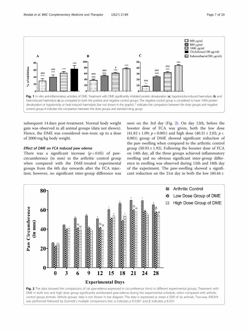
ResultsResults of in vitro anti-inflammatory assaysEffect of DME on protein denaturation testIn the present study, DME showed good anti-inflammatory activity with a dose linear response when wecompared with the negative control group (F = 58.5).DME showed mean inhibition of protein denaturation of29 ± 6.34%, 39.6 ± 6.46% and 51.2 ± 4.41% for the doses of600 μg/ml, 800 μg/ml and 1000 μg/ml respectively(Table 1). The ability of extract of DME to inhibit de-naturation of protein was found to be statistically signifi-cant (p ≤ 0.05) when compared to the negative controldose group (distilled water). Diclofenac, the standardNSAID drug, showed the maximum inhibition, 62.8 ±9.79% at a concentration of 100 μg/ml (Fig. 1a). The1000 μg/ml dose group showed no difference in proteindenaturation inhibitory activity when compared with the
standard Diclofenac (F = 58.5). However, all the 600 μg/ml, 800 μg/ml and 1000 μg/ml dose groups showed dosedependent relationship for the protein denaturation inhib-ition, when compared with standard drug group (Fig. 1a).
Effect of DME on hypotonicity induced haemolysisThe DME protected the erythrocyte in hypotonicity in-duced haemolysis in a concentration-related manner.Within experimental groups, the maximum protective ef-fect was seen in 1000 μg/ml DME dose group (61.1 ±4.81%). Results showed that the DME dose groups(600 μg/ml, 800 μg/ml and 1000 μg/ml) significantly (F =58.5; p ≤ 0.05) inhibited the haemolysis in hypotonic con-ditions (Fig. 1b). However, the percentage of inhibitions ofhaemolysis shown by the DME doses were lower than thatobtained for standard NSAID drug, indomethacin (76.3 ±5.82%) (Table 1). When we compared all the dose groupsof DME with the standard drug group, a significant dose-dependent haemolysis was observed (Fig. 1b).
Effect of DME on heat induced haemolysisAll the doses of DME (600 μg/ml, 800 μg/ml and1000 μg/ml) effectively inhibited the heat induced haem-olysis (Table 1). Within the experimental groups, max-imum inhibition (31.9 ± 3.35%) was observed in the1000 μg/ml dose group. The percentage of inhibition ofhaemolysis caused by DME in all experimental groupswas found to be dose-dependent and statistically signifi-cant (F = 193; p ≤ 0.05). However, the standard drug,indomethacin showed the highest protection (43.9 ±3.03%) at a concentration of 200 μg/ml (Fig. 1c). Whenwe compared with standard-drug group, all doses ofDME (600 μg/ml, 800 μg/ml and 1000 μg/ml) signifi-cantly (p ≤ 0.05) inhibited the denatured protein.
Result of DME on in vivo anti-inflammatory studyAcute oral toxicity test of DMEAll rats treated with different concentrations of DMEwere alive for all 14 days of observation. The DME didnot show any mortality or behavioral abnormalities inthe immediate 24 h after DME feeding as well as in the
Table 1 Inhibition properties of DME in protein denaturation and membrane stabilization (both hypotonicity and heat induced) ascompared to negative control group
Test samples Concentration(μg/ml)
Mean% of inhibition of haemolysis ± S.D
Protein denaturation test Hypotonicity induced Heat induced
DME 600 29 ± 6.34 *** 36.5 ± 7.3*** 8.37 ± 1.24**
800 39.6 ± 6.46 *** 52.1 ± 4.22*** 24.6 ± 3.21***
1000 51.2 ± 4.41*** 61.1 ± 4.81*** 31.9 ± 3.35***a Diclofenac 100 62.8 ± 9.79*** – –b Indomethacin 200 – 76.3 ± 5.82*** 43.9 ± 3.03***
***indicates p ≤ 0.001, **indicates p ≤ 0.01, and *indicates p ≤ 0.05a Diclofenac served as a standard drug for anti-protein denaturation agentsb Indomethacin served standard drug for a membrane stabilizing activity
Modak et al. BMC Complementary Medicine and Therapies (2021) 21:89 Page 6 of 20
subsequent 14 days post-treatment. Normal body weightgain was observed in all animal groups (data not shown).Hence, the DME was considered non-toxic up to a doseof 2000mg/kg body weight.
Effect of DME on FCA induced paw edemaThere was a significant increase (p < 0.05) of paw-circumference (in mm) in the arthritic control groupwhen compared with the DME-treated experimentalgroups from the 6th day onwards after the FCA injec-tion; however, no significant inter-group difference was
seen on the 3rd day (Fig. 2). On day 12th, before thebooster dose of FCA was given, both the low dose(41.82 ± 1.09; p < 0.001) and high dose (40.31 ± 2.02; p <0.001) group of DME showed significant reduction ofthe paw swelling when compared to the arthritic controlgroup (50.93 ± 1.92). Following the booster dose of FCAon 14th day, all the three groups achieved inflammatoryswelling and no obvious significant inter-group differ-ence in swelling was observed during 15th and 18th dayof the experiment. The paw-swelling showed a signifi-cant reduction on the 21st day in both the low (60.44 ±
Fig. 1 In vitro anti-inflammatory activities of DME. Treatment with DME significantly inhibited protein denaturation (a), hypotonicity-induced haemolysis (b) andheat-induced haemolysis (c) as compared to both the positive and negative control groups. The negative control group is considered to have 100% proteindenaturation or hypotonicity or heat induced haemolysis (bar not shown in the graphs). * indicates the comparison between the dose groups and negativecontrol group; # indicates the comparison between the dose groups and standard drug group
Fig. 2 The data showed the comparisons of rat paw-edema expressed in circumference (mm) in different experimental groups. Treatment withDME in both low and high dose group significantly ameliorated paw-edema during the experimental schedule, when compared with arthriticcontrol group animals. Vehicle groups’ data is not shown in bar diagram. The data is expressed as mean ± SEM of six animals. Two-way ANOVAwas performed followed by Dunnett’s multiple comparisons test. α indicates p≤ 0.001 and β indicates p≤ 0.01
Modak et al. BMC Complementary Medicine and Therapies (2021) 21:89 Page 7 of 20
1.44; p < 0.001) and high dose groups (58.39 ± 1.72; p <0.001) of DME when compared to the arthritic controlgroup (70.13 ± 2.59). On the last day of anti-arthritictreatment, we found a significant (p < 0.001) inhibitionof paw-swelling in high dose group of DME (54.45 ±1.30) and a significant reduction (p < 0.01) of paw swell-ing in low dose group of DME (57.71 ± 0.99) when com-pared to control arthritic group (65.43 ± 2.31). However,the two experimental dose-groups showed no significantdifference in their paw circumference measurementsduring the experimental tenure when compared witheach other following Tukey’s post hoc test.
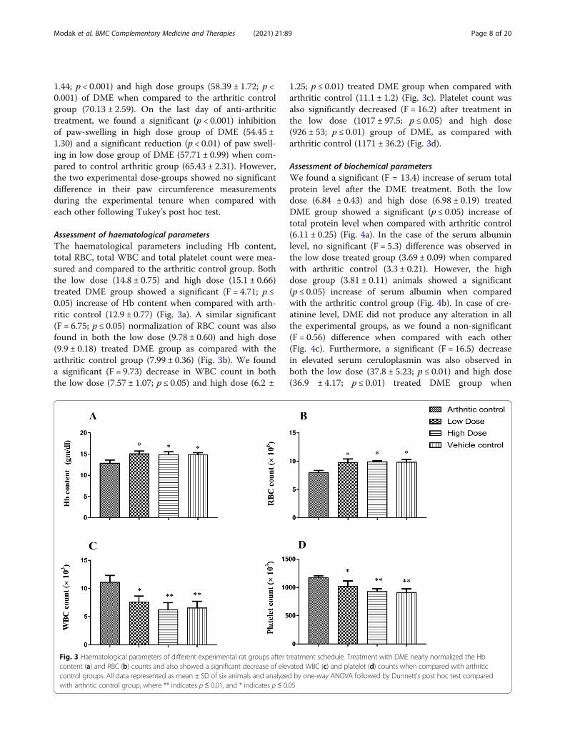
Assessment of haematological parametersThe haematological parameters including Hb content,total RBC, total WBC and total platelet count were mea-sured and compared to the arthritic control group. Boththe low dose (14.8 ± 0.75) and high dose (15.1 ± 0.66)treated DME group showed a significant (F = 4.71; p ≤0.05) increase of Hb content when compared with arth-ritic control (12.9 ± 0.77) (Fig. 3a). A similar significant(F = 6.75; p ≤ 0.05) normalization of RBC count was alsofound in both the low dose (9.78 ± 0.60) and high dose(9.9 ± 0.18) treated DME group as compared with thearthritic control group (7.99 ± 0.36) (Fig. 3b). We founda significant (F = 9.73) decrease in WBC count in boththe low dose (7.57 ± 1.07; p ≤ 0.05) and high dose (6.2 ±
1.25; p ≤ 0.01) treated DME group when compared witharthritic control (11.1 ± 1.2) (Fig. 3c). Platelet count wasalso significantly decreased (F = 16.2) after treatment inthe low dose (1017 ± 97.5; p ≤ 0.05) and high dose(926 ± 53; p ≤ 0.01) group of DME, as compared witharthritic control (1171 ± 36.2) (Fig. 3d).
Assessment of biochemical parametersWe found a significant (F = 13.4) increase of serum totalprotein level after the DME treatment. Both the lowdose (6.84 ± 0.43) and high dose (6.98 ± 0.19) treatedDME group showed a significant (p ≤ 0.05) increase oftotal protein level when compared with arthritic control(6.11 ± 0.25) (Fig. 4a). In the case of the serum albuminlevel, no significant (F = 5.3) difference was observed inthe low dose treated group (3.69 ± 0.09) when comparedwith arthritic control (3.3 ± 0.21). However, the highdose group (3.81 ± 0.11) animals showed a significant(p ≤ 0.05) increase of serum albumin when comparedwith the arthritic control group (Fig. 4b). In case of cre-atinine level, DME did not produce any alteration in allthe experimental groups, as we found a non-significant(F = 0.56) difference when compared with each other(Fig. 4c). Furthermore, a significant (F = 16.5) decreasein elevated serum ceruloplasmin was also observed inboth the low dose (37.8 ± 5.23; p ≤ 0.01) and high dose(36.9 ± 4.17; p ≤ 0.01) treated DME group when
Fig. 3 Haematological parameters of different experimental rat groups after treatment schedule. Treatment with DME nearly normalized the Hbcontent (a) and RBC (b) counts and also showed a significant decrease of elevated WBC (c) and platelet (d) counts when compared with arthriticcontrol groups. All data represented as mean ± SD of six animals and analyzed by one-way ANOVA followed by Dunnett’s post hoc test comparedwith arthritic control group, where ** indicates p≤ 0.01, and * indicates p≤ 0.05
Modak et al. BMC Complementary Medicine and Therapies (2021) 21:89 Page 8 of 20
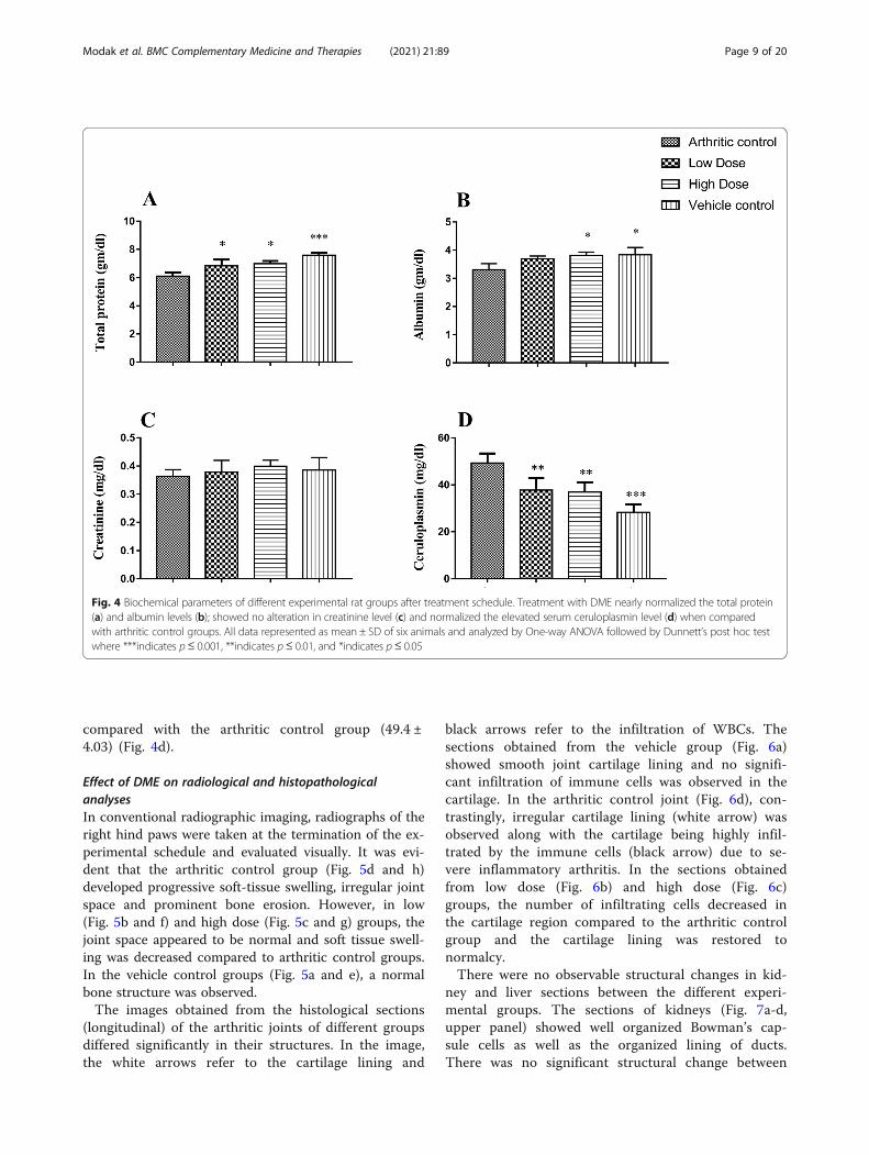
compared with the arthritic control group (49.4 ±4.03) (Fig. 4d).
Effect of DME on radiological and histopathologicalanalysesIn conventional radiographic imaging, radiographs of theright hind paws were taken at the termination of the ex-perimental schedule and evaluated visually. It was evi-dent that the arthritic control group (Fig. 5d and h)developed progressive soft-tissue swelling, irregular jointspace and prominent bone erosion. However, in low(Fig. 5b and f) and high dose (Fig. 5c and g) groups, thejoint space appeared to be normal and soft tissue swell-ing was decreased compared to arthritic control groups.In the vehicle control groups (Fig. 5a and e), a normalbone structure was observed.The images obtained from the histological sections
(longitudinal) of the arthritic joints of different groupsdiffered significantly in their structures. In the image,the white arrows refer to the cartilage lining and
black arrows refer to the infiltration of WBCs. Thesections obtained from the vehicle group (Fig. 6a)showed smooth joint cartilage lining and no signifi-cant infiltration of immune cells was observed in thecartilage. In the arthritic control joint (Fig. 6d), con-trastingly, irregular cartilage lining (white arrow) wasobserved along with the cartilage being highly infil-trated by the immune cells (black arrow) due to se-vere inflammatory arthritis. In the sections obtainedfrom low dose (Fig. 6b) and high dose (Fig. 6c)groups, the number of infiltrating cells decreased inthe cartilage region compared to the arthritic controlgroup and the cartilage lining was restored tonormalcy.There were no observable structural changes in kid-
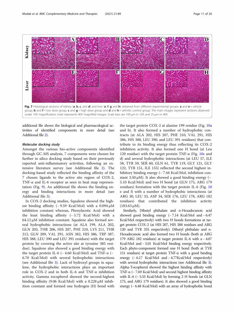
ney and liver sections between the different experi-mental groups. The sections of kidneys (Fig. 7a-d,upper panel) showed well organized Bowman’s cap-sule cells as well as the organized lining of ducts.There was no significant structural change between
Fig. 4 Biochemical parameters of different experimental rat groups after treatment schedule. Treatment with DME nearly normalized the total protein(a) and albumin levels (b); showed no alteration in creatinine level (c) and normalized the elevated serum ceruloplasmin level (d) when comparedwith arthritic control groups. All data represented as mean ± SD of six animals and analyzed by One-way ANOVA followed by Dunnett’s post hoc testwhere ***indicates p≤ 0.001, **indicates p≤ 0.01, and *indicates p≤ 0.05
Modak et al. BMC Complementary Medicine and Therapies (2021) 21:89 Page 9 of 20

groups. The liver sections (Fig. 7e-h, lower panel) ofall the groups also showed well-organized cellularstructure and no abnormalities were seen in anygroups. A, E represent vehicle group; B, F representlow dose group; C, G represents high dose group andD, H represents arthritic control group.
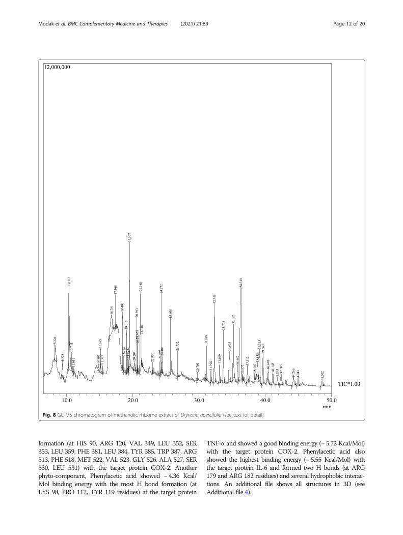
GC-MS analysis of DMEThe DME was subjected to phytochemical analysis usingoptimized GC-MS parameters and the resultant gas-chromatogram is presented in Fig. 8.The compositions of the phytochemical compounds
present in methanolic extracts of DME identified by GC-MSanalysis with their retention time (RT), molecular formula,molecular weight and area (%) are presented in (Table 2). An
Fig. 5 Radiological analyses of right hind paw ankle joints of experimental rats. Arrow marks indicate the swelling and degradation of cartilage inexperimental animals. Upper panel (a, b, c, d) shows the horizontal view and lower panel (e, f, g, h) shows the angular view of the same bonejoints; where, a and e = vehicle group, b and f = low dose group, c and g = high dose group and d and h = arthritic control group
Fig. 6 Histological sections of rat paw (ankle) joints obtained from different experimental groups. White arrows represent the destruction incartilage lining and the black arrows represent the immune cell infiltration in all experimental groups. a = vehicle, b = low dose, c = high doseand d = arthritic control group. The main images represent sections observed under 10X magnification; inset represents 40X magnified images.Scale bars are 100 μm in 10X and 25 μm in 40X
Modak et al. BMC Complementary Medicine and Therapies (2021) 21:89 Page 10 of 20
additional file shows the biological and pharmacological ac-tivities of identified components in more detail (seeAdditional file 2).
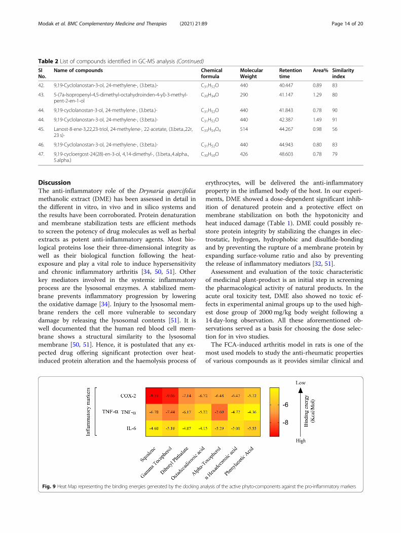
Molecular docking studyAmongst the various bio-active components identifiedthrough GC-MS analysis, 7 components were chosen forfurther in silico docking study based on their previouslyreported anti-inflammatory activities, following an ex-tensive literature survey (see Additional file 2). Thedocking-based study reflected the binding affinity of the7 chosen ligands to the active site region of COX-2,TNF-α and IL-6 receptor as seen in heat map represen-tation (Fig. 9). An additional file shows the binding en-ergy and binding interactions in more detail (seeAdditional file 3).In COX-2 docking studies, Squalene showed the high-
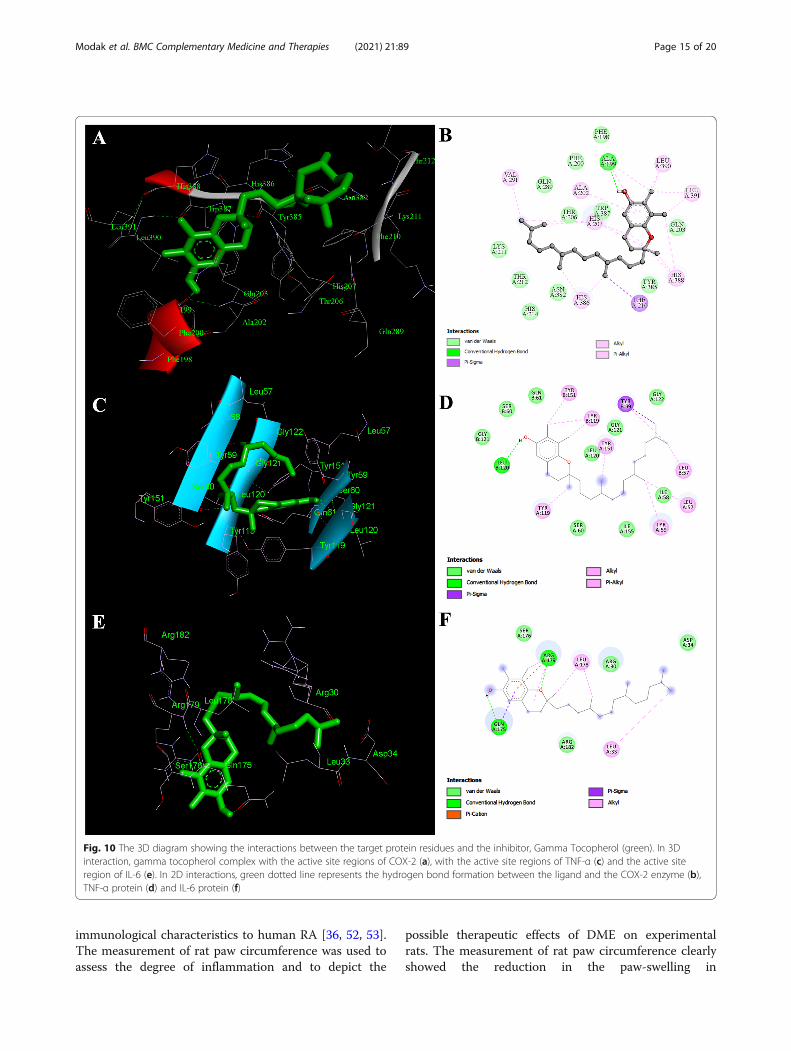
est binding affinity (− 9.59 Kcal/Mol) with a 0.094 μMinhibition constant whereas, Phenylacetic Acid showedthe least binding affinity (− 5.72 Kcal/Mol) with a64.12 μM inhibition constant. Squalene also formed sev-eral hydrophobic interactions (at ALA 199, ALA 202,GLN 203, THR 206, HIS 207, PHE 210, LYS 211, THR212, GLN 289, VAL 291, ASN 382, HIS 386, TRP 387,HIS 388, LEU 390 and LEU 391 residues) with the targetprotein by covering the active site at tyrosine 385 resi-due). Squalene also showed a good binding energy withthe target protein IL-6 (− 4.60 Kcal/Mol) and TNF-α (−6.70 Kcal/Mol) with several hydrophobic interactions(see Additional file 3). Lack of hydroxyl groups in squa-lene, the hydrophobic interactions plays an importantrole in COX-2 and in both IL-6 and TNF-α inhibitionactivity. Gamma tocopherol showed the second-highestbinding affinity (9.06 Kcal/Mol) with a 0.228 μM inhib-ition constant and formed one hydrogen (H) bond with
the target protein COX-2 at alanine 199 residue (Fig. 10aand b). It also formed a number of hydrophobic con-tracts (at ALA 202, HIS 207, PHE 210, VAL 291, HIS386, HIS 388, LEU 390, and LEU 391 residues) that con-tribute to its binding energy thus reflecting its COX-2inhibition activity. It also formed one H bond (at Leu120 residue) with the target protein TNF-α (Fig. 10c andd) and several hydrophobic interactions (at LEU 57, ILE58, TYR 59, SER 60, GLN 61, TYR 119, GLY 121, GLY122, TYR 151, ILE 155) reflected the second highest in-hibitory binding energy (− 7.44 Kcal/Mol, inhibition con-stant 3.50 μM). It also showed a good binding energy (−5.10 Kcal/Mol) and two H bond (at GLN 175, ARG 179residues) formation with the target protein IL-6 (Fig. 10e and f) with a number of hydrophobic interactions (atARG 30, LEU 33, ASP 34, SER 176, LEU 178, ARG 182residues) that contributed the inhibition activity(183.63 μM).Similarly, Dibutyl phthalate and n-Hexadecanoic acid
showed good binding energy (− 7.14 Kcal/Mol and− 6.47Kcal/Mol respectively) with two H bonds formations at tar-get protein COX-2 (at HIS 207, HIS 388 residues and ARG120 and TYR 355 respectively). Dibutyl phthalate and n-Hexadecanoic acid also formed two H bonds (both at ARG179 ARG 182 residues) at target protein IL-6 with a− 4.07Kcal/Mol and− 5.01 Kcal/Mol binding energy respectively.Each phyto-component formed one H bond (both at TYR151 residues) at target protein TNF-α with a good bindingenergy (− 6.17 Kcal/Mol and− 4.77Kcal/Mol respectively)with several hydrophobic interactions (see Additional file 3).Alpha-Tocopherol showed the highest binding affinity withTNF-α (− 7.69 Kcal/Mol) and second highest binding affinitywith IL-6 (− 5.55 Kcal/Mol) by forming 2 H bonds (at GLN175, and ARG 179 residues). It also showed a good bindingenergy (− 6.48 Kcal/Mol) with an array of hydrophobic bond
Fig. 7 Histological sections of kidney (a, b, c, and d) and liver (e, f, g and h) obtained from different experimental groups. a and e = vehiclegroup, b and f = low dose group, c and g = high dose group and d and h = arthritic control group. The main images represent sections observedunder 10X magnification; inset represents 40X magnified images. Scale bars are 100 μm in 10X and 25 μm in 40X
Modak et al. BMC Complementary Medicine and Therapies (2021) 21:89 Page 11 of 20
formation (at HIS 90, ARG 120, VAL 349, LEU 352, SER353, LEU 359, PHE 381, LEU 384, TYR 385, TRP 387, ARG513, PHE 518, MET 522, VAL 523, GLY 526, ALA 527, SER530, LEU 531) with the target protein COX-2. Anotherphyto-component, Phenylacetic acid showed − 4.36 Kcal/Mol binding energy with the most H bond formation (atLYS 98, PRO 117, TYR 119 residues) at the target protein
TNF-α and showed a good binding energy (− 5.72 Kcal/Mol)with the target protein COX-2. Phenylacetic acid alsoshowed the highest binding energy (− 5.55 Kcal/Mol) withthe target protein IL-6 and formed two H bonds (at ARG179 and ARG 182 residues) and several hydrophobic interac-tions. An additional file shows all structures in 3D (seeAdditional file 4).
Fig. 8 GC-MS chromatogram of methanolic rhizome extract of Drynaria quercifolia (see text for detail)
Modak et al. BMC Complementary Medicine and Therapies (2021) 21:89 Page 12 of 20
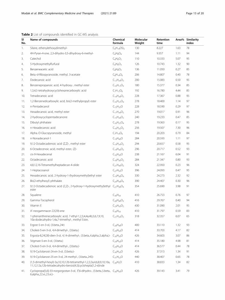
Table 2 List of compounds identified in GC-MS analysis
SlNo.
Name of compounds Chemicalformula
MolecularWeight
Retentiontime
Area% Similarityindex
1. Silane, ethenylethoxydimethyl- C6H14OSi2 130 8.227 1.63 78
2. 4H-Pyran-4-one, 2,3-dihydro-3,5-dihydroxy-6-methyl- C6H8O4 144 9.357 1.11 94
3. Catechol C6H6O2 110 10.333 5.07 95
4. 5-Hydroxymethylfurfural C6H6O3 126 10.743 1.32 90
5. Benzeneacetic acid C8H8O2 136 11.050 0.27 85
6. Beta.-d-Ribopyranoside, methyl, 3-acetate C8H14O6 206 14.807 0.40 78
7. Dodecanoic acid C12H24O2 200 15.083 0.50 95
8. Benzenepropanoic acid, 4-hydroxy-, methyl ester C10H12O3 180 15.377 0.34 85
9. 1,3,4,5-tetrahydroxycyclohexanecarboxylic acid C7H12O6 192 16.780 4.44 85
10. Tetradecanoic acid C14H28O2 228 17.367 0.88 95
11. 1,2-Benzenedicarboxylic acid, bis(2-methylpropyl) ester C16H22O4 278 18.400 1.14 97
12. n-Pentadecanol C15H32O 228 18.590 0.29 97
13. Hexadecanoic acid, methyl ester C17H34O2 270 19.017 0.91 96
14. 2-hydroxycyclopentadecanone C15H28O2 240 19.233 0.47 85
15. Dibutyl phthalate C16H22O4 278 19.363 0.17 95
16. n-Hexadecanoic acid C16H32O2 256 19.507 7.30 96
17. Alpha.-D-Glucopyranoside, methyl C7H14O6 194 20.203 0.79 84
18. n-Nonadecanol-1 C19H40O 284 20.593 1.11 97
19. 9,12-Octadecadienoic acid (Z,Z)-, methyl ester C19H34O2 294 20.657 0.58 95
20. 6-Octadecenoic acid, methyl ester, (Z)- C19H36O2 296 20.717 0.52 93
21. cis-9-Hexadecenal C16H30O 238 21.167 6.04 91
22. Octadecanoic acid C18H36O2 284 21.347 0.80 93
23. 4,8,12,16-Tetramethylheptadecan-4-olide C21H40O2 324 22.950 0.23 96
24 1-Heptacosanol C27H56O 396 24.093 0.47 95
25. Hexadecanoic acid, 2-hydroxy-1-(hydroxymethyl)ethyl ester C19H38O4 330 24.273 2.32 92
26. Bis(2-ethylhexyl) phthalate C24H38O4 390 24.407 0.30 96
27. 9,12-Octadecadienoic acid (Z,Z)-, 2-hydroxy-1-hydroxymethyl)ethylester
C21H38O4 354 25.690 3.98 91
28. Squalene C30H50 410 26.753 0.76 97
29. Gamma-Tocopherol C28H48O2 416 29.767 0.40 94
30. Vitamin E C29H50O2 430 31.090 2.01 95
31. A’-neogammacer-22(29)-ene C30H50 410 31.797 0.59 83
32. 1-phenanthrenecarboxylic acid, 7-ethyl-1,2,3,4,4a,4b,5,6,7,9,10,10a-dodecahydro-1,4a,7-trimethyl-, methyl Ester,
C21H34O2 318 32.357 6.07 65
33. Ergost-5-en-3-ol, (3.beta.,24r) C28H48O 400 33.110 1.32 93
34. Cholest-5-en-3-ol, 4,4-dimethyl-, (3.beta.) C29H50O 414 33.703 4.17 82
35. Ergosta-8,24(28)-dien-3-ol, 4,14-dimethyl-, (3.beta.,4.alpha.,5.alpha.)- C30H50O 426 34.603 3.07 86
36. Stigmast-5-en-3-ol, (3.beta.) C29H50O 414 35.180 4.98 81
37. Cholest-5-en-3-ol, 4,4-dimethyl-, (3.beta.)- C29H50O 414 36.577 0.44 78
38. 9,19-Cyclolanost-24-en-3-ol, (3.beta.)- C30H50O 426 37.313 1.34 91
39. 9,19-Cyclolanost-25-en-3-ol, 24-methyl-, (3.beta.,24S)- C31H52O 440 38.407 0.65 78
40. (1,5-dimethyl-hexyl)-3a,10,10,12b-tetramethyl-1,2,3,3a,4,6,8,9,10,10a,11,12,12a,12b-tetradecahydro-benzo[4,5]cyclohepta[1,2-e]inde
C30H5O 410 38.833 1.34 82
41. Cyclopropa[5,6]-33-norgorgostan-3-ol, 3′,6-dihydro-, (3.beta.,5.beta.,6.alpha.,22.xi.,23.xi.)-
C30H50O 426 39.143 3.41 79
Modak et al. BMC Complementary Medicine and Therapies (2021) 21:89 Page 13 of 20
DiscussionThe anti-inflammatory role of the Drynaria quercifoliamethanolic extract (DME) has been assessed in detail inthe different in vitro, in vivo and in silico systems andthe results have been corroborated. Protein denaturationand membrane stabilization tests are efficient methodsto screen the potency of drug molecules as well as herbalextracts as potent anti-inflammatory agents. Most bio-logical proteins lose their three-dimensional integrity aswell as their biological function following the heat-exposure and play a vital role to induce hypersensitivityand chronic inflammatory arthritis [34, 50, 51]. Otherkey mediators involved in the systemic inflammatoryprocess are the lysosomal enzymes. A stabilized mem-brane prevents inflammatory progression by loweringthe oxidative damage [34]. Injury to the lysosomal mem-brane renders the cell more vulnerable to secondarydamage by releasing the lysosomal contents [51]. It iswell documented that the human red blood cell mem-brane shows a structural similarity to the lysosomalmembrane [50, 51]. Hence, it is postulated that any ex-pected drug offering significant protection over heat-induced protein alteration and the haemolysis process of
erythrocytes, will be delivered the anti-inflammatoryproperty in the inflamed body of the host. In our experi-ments, DME showed a dose-dependent significant inhib-ition of denatured protein and a protective effect onmembrane stabilization on both the hypotonicity andheat induced damage (Table 1). DME could possibly re-store protein integrity by stabilizing the changes in elec-trostatic, hydrogen, hydrophobic and disulfide-bondingand by preventing the rupture of a membrane protein byexpanding surface-volume ratio and also by preventingthe release of inflammatory mediators [32, 51].Assessment and evaluation of the toxic characteristic
of medicinal plant-product is an initial step in screeningthe pharmacological activity of natural products. In theacute oral toxicity test, DME also showed no toxic ef-fects in experimental animal groups up to the used high-est dose group of 2000 mg/kg body weight following a14 day-long observation. All these aforementioned ob-servations served as a basis for choosing the dose selec-tion for in vivo studies.The FCA-induced arthritis model in rats is one of the
most used models to study the anti-rheumatic propertiesof various compounds as it provides similar clinical and
Table 2 List of compounds identified in GC-MS analysis (Continued)
SlNo.
Name of compounds Chemicalformula
MolecularWeight
Retentiontime
Area% Similarityindex
42. 9,19-Cyclolanostan-3-ol, 24-methylene-, (3.beta.)- C31H52O 440 40.447 0.89 83
43. 5-(7a-Isopropenyl-4,5-dimethyl-octahydroinden-4-yl)-3-methyl-pent-2-en-1-ol
C20H34O 290 41.147 1.29 80
44. 9,19-cyclolanostan-3-ol, 24-methylene-, (3.beta.)- C31H52O 440 41.843 0.78 90
44. 9,19-Cyclolanostan-3-ol, 24-methylene-, (3.beta.)- C31H52O 440 42.387 1.49 91
45. Lanost-8-ene-3,22,23-triol, 24-methylene-, 22-acetate, (3.beta.,22r,23 s)-
C33H54O4 514 44.267 0.98 56
46. 9,19-Cyclolanostan-3-ol, 24-methylene-, (3.beta.)- C31H52O 440 44.943 0.80 83
47. 9,19-cycloergost-24(28)-en-3-ol, 4,14-dimethyl-, (3.beta.,4.alpha.,5.alpha.)
C30H50O 426 48.603 0.78 79
Fig. 9 Heat Map representing the binding energies generated by the docking analysis of the active phyto-components against the pro-inflammatory markers
Modak et al. BMC Complementary Medicine and Therapies (2021) 21:89 Page 14 of 20
immunological characteristics to human RA [36, 52, 53].The measurement of rat paw circumference was used toassess the degree of inflammation and to depict the
possible therapeutic effects of DME on experimentalrats. The measurement of rat paw circumference clearlyshowed the reduction in the paw-swelling in
Fig. 10 The 3D diagram showing the interactions between the target protein residues and the inhibitor, Gamma Tocopherol (green). In 3Dinteraction, gamma tocopherol complex with the active site regions of COX-2 (a), with the active site regions of TNF-α (c) and the active siteregion of IL-6 (e). In 2D interactions, green dotted line represents the hydrogen bond formation between the ligand and the COX-2 enzyme (b),TNF-α protein (d) and IL-6 protein (f)
Modak et al. BMC Complementary Medicine and Therapies (2021) 21:89 Page 15 of 20
experimental groups at the end of the experimentalschedule (Fig. 2). In arthritic conditions, RBC counts de-creases because bone marrow is depressed and fails torespond to anemic condition. Our results also show adecreased level of RBC count in arthritic control groups.This may be due to the abnormal storage of iron or theinability of bone marrow to produce cells [53]. However,the RBC count increases significantly towards their nor-mal ranges in both the low and high dose groups whentreated with DME (Fig. 3b). The hemoglobin count alsodecreases in arthritic condition as low RBC level resultsin low hemoglobin levels. The significant increases ofWBC count in arthritic condition account for the boost-ing of the immune system with the invading antigens[54]. A significant decrement of platelet counts was alsoobserved in the experimental groups when compared toarthritic control groups (Fig. 3d). Thus, treatments withDME normalized the haematological parameters in theexperimental groups which reflect its immunomodula-tory effects.In the present study, we have observed a decrease in
serum total protein level (Fig. 4a) and albumin level (Fig.4b) in the arthritic control rats. This due to the changesin plasma protein concentration and involvement of sev-eral inflammatory mediators like prostaglandins, brady-kinin that increases the vascular permeability leads tothe reduction of total protein and albumin levels [54].Treatment of DME significantly increases the total pro-tein level in both experimental groups when comparedto the arthritic control group (Fig. 4a). Furthermore, thehigh dose group rats showed a significant increase in thealbumin level when compared with the rats of the arth-ritic control group suggesting the role of DME in therestoration of serum albumin level (Fig. 4b), which mayindicate the suppressive activity of DME on the inflam-matory mediators. However, in our study, no significantalteration was observed in the serum creatinine level inall the experimental groups (Fig. 4c), which indicatesnormal kidney function. Ceruloplasmin is another majoracute-phase serum protein that increases during RA[36]. This copper-containing protein is mainly producedin the liver and released in the blood circulation upontissue injury [54]. We have observed an elevated level ofthis protein in the arthritic control group which indi-cates the tissue injury during the chronic arthritis pro-gression (Fig. 4d). However, a significant decrease wasobserved in both the low and high dose groups after theDME treatment (Fig. 4d), which shows its effects on tis-sue repair.In clinical practice, RA diagnosis and follow-up are
mainly based on conventional radiographs, which areuseful diagnostic measures in RA severity. The mostcommon earliest sign of RA is the soft tissue swelling,whereas in more chronic stages, is the narrowing of joint
spaces and erosions of bones occurs [55]. In our study,we have observed a soft tissue swelling along with ir-regular joint space in arthritic control groups, that mayindicate the severity of the disease progression (Fig. 5dand h). However, DME treatment for 28 days has shownthe prevention of bone destruction in both the low (Fig.5b and f) and high dose groups (Fig. 5c and g), whencompared to arthritic control groups. These radio-graphic changes are further supported by histopatho-logical analysis of ankle-joints of the experimental rats.In the arthritic control groups, the arthritic rat jointshowed extensive infiltration of inflammatory cells in thearticular cartilage regions (Fig. 6d). However, treatmentwith DME for 28 days showed less cellular infiltrationsin both the low and high dose groups, when comparedto the arthritic control group animals.GC-MS technique is routinely employed to identify
the array of volatile bioactive compounds present in aplant extract [56, 57]. The GC-MS analysis of the DMEdetected several promising compounds (Fig. 8). The de-tailed analysis of chromatogram has revealed the pres-ence of 47 potent bio-active compounds in themethanolic rhizome extract of Drynaria quercifolia(Table 2). Compounds like squalene can attenuate theoverexpressed COX-2 activity in Lipopolysaccharides(LPS) treated human monocytes and neutrophils andalso down-regulate Matrix metalloproteinase (MMP)-1and MMP-9 gene expression in LPS treated humanmonocytes, and of MMP-1 and MMP-3 gene expressionin LPS-treated human neutrophils [58] and it also has apotent antioxidant and anticancer activity [59]. Similarly,it has been previously reported that compounds like n-Hexadecanoic acid and Gamma-tocopherol also possessa potent anti-inflammatory activity [60–62].Our in vitro and in vivo analysis of anti-inflammatory
and anti-rheumatoid activity of DME showed promisingresults. To further substantiate the results in silico, mo-lecular docking technique was utilized to verify the abilityof phyto-constituents to inhibit principal inflammatorymediators such as COX-2, TNF-α and IL-6. COX-2 is themain enzyme that mediates the bioconversion of arachi-donic acid to inflammatory prostaglandins [44]. Thearachidonate binding site is created by a long hydrophobicchannel, from Arg 120 to near Tyr 385 and Glu 524, andthis binding channel provides the binding pockets of trad-itional NSAIDs for selective inhibition [63].Molecular docking is one of the most powerful tech-
niques to discover novel ligands for proteins of knownstructure and thus plays a key role in structure-baseddrug design. It is based on the “lock-key” principle,which looks into the ligand-receptor interaction throughthe formation of electrostatic interaction, hydrogenbonding, hydrophobic interaction, van der Waals inter-action, etc. In this study, seven phytochemical
Modak et al. BMC Complementary Medicine and Therapies (2021) 21:89 Page 16 of 20

compounds identified from DME were docked withCOX-2, TNF-α, IL-6 proteins to see whether these com-pounds can bind to the active sites of the enzymes andinhibit their activities which may indicate the anti-inflammatory activity of DME.In our docking assays, the binding energy and inhib-
ition constant obtained from the calculated results byAutodock 4.2 using a Lamarckian genetic algorithm [49],was the reflection of the binding affinity of the bioactiveligands with the target proteins. The smaller the inhib-ition constant, the greater the binding affinity and thesmaller amount of medication needed to inhibit the ac-tivity of that enzyme. Compounds like Squalene, Pheny-lacetic acid, α-Tocopherol, n-Hexadecanoic acid, γ-Tocopherol, Dibutyl phthalate showed efficient bindingaffinity with the target inflammatory markers which maysubsequently down-regulate inflammation. COX-2 in-hibition leads to a decrease in the production of prosta-glandin E2 which is a limiting factor in inflammation[44, 64]. Docking of the phyto-compounds with the tar-get proteins are explored in detail to reveal the numberof hydrogen bonds in the interaction and the interactingamino acids. Recent studies on plant extract and herbalproducts have revealed that the plant-based herbal rem-edies can directly act on the transcription of specificgenes responsible for the production of cytokines,cyclooxygenases and other inflammatory biomarkers [10,65, 66]. In a recent study, the present author group hasdemonstrated that Aloe vera gel in its crude form, maydownregulate TNF-α and COX-2 expressions in Wistarrat inflammatory arthritis models [10]. The in silicostudy indicates that the target inflammatory markershave been inhibited significantly by the phyto-compounds of the Drynaria quercifolia as indentified inGC-MS study. The authors assume that similar inhib-ition of COX-2 may be possible in vivo at protein and/or gene expression levels.In inflammatory arthritis, the infiltrating immune cells
contribute to the degree of inflammation [5, 8]. The initial in-filtrating cells stimulate both Th1 and Th2 cell types; Th1cells provide delayed type of hypersensitivity and Th2 in-volves B-cells in the inflammatory process [67]. A vast arrayof cytokines including TNF-α, interleukins and other inflam-mation mediators like COX-2 are secreted from the infiltrat-ing immune cells and synoviocytes during the disease course[8]. The blocking of these key molecules may provide an ex-cellent interruption in the progress of inflammation. As seenin our studies, significant synergistic interactions betweenDrynaria phyto-compounds with cytokines like TNF-α, IL-6and with COX-2-like immune-modulators may play signifi-cant roles in down-regulating inflammation. Some phyto-components have been shown to interact with cytokines andsome others with COX-2 in controlling progression of thedisease. In this regard, the known phyto-compounds of
Withania somnifera has shown interaction with differ-ent cytokines in in silico docking and in vivo animalmodels and downregulated the expressions of iNOS,TNF-α, NF-κB etc. [68, 69]. Compounds from Cana-bis sative and Prunella vulgaris also showed interac-tions with different cytokines in silico [68]. Moringarivae leaf extract is known to down-regulate theTNF- α and COX-2 expressions in the animal modelsystems [65]; Fenugreek (Trigonella foenum graecum),extracted in ethanol, ameliorates arthritis in experi-mental animals and decreased TNF-α expression [70].Poly-herbal formulations like Kashayams have beenshown to down-regulate elevated levels of Cox-2,TNF-α, iNOS mRNAs in vivo when treated to arth-ritic rat models [71].Therefore, it can be inferred that the anti-
inflammatory and anti-rheumatoid effect of methanolicrhizome extract of Drynaria quercifolia may be due tothe presence of potent bio-active components whichmay synergistically show the inhibitory effects on inflam-matory markers (COX-2, TNF-α and IL-6) leading to in-hibition of inflammation.
Concluding remarksOur study experimentally presents the anti-inflammatory and anti-arthritic activities of Drynariaquercifolia rhizome methanolic extract in both in vitroand in vivo scenarios. Both the membrane stabilizationand protein denaturation test confirmed the anti-inflammatory activity of extracts. Our results further val-idated the ameliorative property of D. quercifolia inFCA-induced chronic arthritis model. Treatment withthe plant extracts improved the inflammatory pawedema, hematological and biochemical parameters with-out exhibiting any signs of hepatotoxicity and nephro-toxicity. Moreover, GC–MS analysis indicated that thebio-active phyto-components of D. quercifolia possesspotent anti-inflammatory activity against molecular tar-gets. The results of the docking study provide validationof anti-inflammatory activity. However, an extensivescreening and identification of the major anti-inflammatory phyto-components is also required involv-ing their isolation, purification, bio-availability assays.Further studies regarding the role of the plant-extractand its phyto-compounds on the gene and protein-levelexpression of a broad spectrum of inflammatory bio-markers would provide a clearer view on the anti-inflammatory aspects of the plant. Furthermore, thetoxicological properties of the plant extract and its com-ponents also need a detailed exploration. The studyoutcome denotes that the D. quercifolia rhizome metha-nolic extract can provide a new opportunity for finding acomplementary and alternative remedy in inflammatoryand arthritic disease conditions.
Modak et al. BMC Complementary Medicine and Therapies (2021) 21:89 Page 17 of 20
AbbreviationsANOVA: Analysis of variance; b.w.: Body weight; CPCSEA: Committee for thePurpose of Control and Supervision of Experiments on Animals; COX-2: Cyclooxygenase-2; DMARDs: Disease-Modifying Anti-Rheumatic Drugs;DME: Drynaria quercifolia rhizome Methanolic Extract; DMSO: Dimethylsulfoxide; EDTA: Ethylene-diamine-tetraacetic acid; FCA: Freund’s CompleteAdjuvant; GA: Genetic algorithm; GC-MS: Gas Chromatography-Mass Spec-trometry; HCl: Hydrogen chloride; IAEC: Institutional Animal EthicalCommittee; i.p.: Intraperitoneal; LPS: Lipopolysaccharides; MMP: Matrixmetalloproteinase; NCBI: National Center for Biotechnology Information; NSAIDs: Non-Steroidal Anti-Rheumatic Drugs; OECD: Organization for EconomicCooperation and Development; PBS: Phosphate-buffered saline; p.o.: Per os;RA: Rheumatoid Arthritis; RBC: Red Blood Corpuscles; S.D.: Standard deviation;S.E.M: Standard Error Mean; TIC: Total ion count; WBC: White BloodCorpuscles
Supplementary InformationThe online version contains supplementary material available at https://doi.org/10.1186/s12906-021-03265-7.
Additional file 1. Flow sheet of methodology.
Additional file 2. List of compounds identified in GC-MS analysis withtheir chemical class and biological properties.
Additional file 3: Table S1. Molecular docking scores of bioactivecompounds identified in Drynaria quercifolia methanolic extracts withCOX-2, IL-6, and TNF-α.
Additional file 4 Figure S1. Represents the interaction between theCOX-2 and the inhibitor, Squalene. Figure 2. Represents the interactionbetween the COX-2 and the inhibitor, Dibutyl phthalate. Figure 3. Repre-sents the interaction between the COX-2 and the inhibitor, 9,12-Octade-cadienoic acid (Z,Z)-, methyl ester_Methyl Linoleate. Figure 4.Represents the interaction between the COX-2 and the inhibitor, VitaminE, Alpha –Tocopherol. Figure 5. Represents the interaction between theCOX-2 and the inhibitor, n Hexadecanoic acid. Figure 6. represents theinteraction between the COX-2 and the inhibitor, Phenylacetic Acid. Fig-ure 7. represents the interaction between the TNF-α and the inhibitor,Squalene. Figure 8. Represents the interaction between the TNF-α andthe inhibitor, Dibutyl phthalate. Figure 9. Represents the interaction be-tween the TNF-α and the inhibitor, 9,12-Octadecadienoic acid (Z,Z)-, me-thyl ester_Methyl Linoleate. Figure 10. Represents the interactionbetween the TNF-α and the inhibitor, Vitamin E, Alpha –Tocopherol. Fig-ure 11. Represents the interaction between the TNF-α and the inhibitor,n Hexadecanoic acid. Figure 12. Represents the interaction between theTNF-α and the inhibitor, n Phenylacetic Acid. Figure 13. Represents theinteraction between the IL-6 and the inhibitor, Squalene. Figure 14. Rep-resents the interaction between the IL-6 and the inhibitor, Dibutyl Phthal-ate. Figure 15. Represents the interaction between the IL-6 and theinhibitor, 9,12-Octadecadienoic acid (Z,Z)-, methyl ester_Methyl Linoleate.Figure 16. Represents the interaction between the IL-6 and the inhibitor,Vitamin E, Alpha –Tocopherol. Figure 17. Represents the interaction be-tween the IL-6 and the inhibitor, n Hexadecanoic acid. Figure 18. Repre-sents the interaction between the IL-6 and the inhibitor, PhenylaceticAcid.
AcknowledgementsAuthors acknowledge the expertise of Taxonomy of Angiosperm andBiosystematics Laboratory, Department of Botany, University of North Bengalfor the identification of the plant sample and for providing the herbariumnumber. The authors also thank the Department of Zoology for providingthe animal house and instrument facilities for the research purpose.Advanced Instrumentation Research Facility, Jawaharlal Nehru University,New Delhi is acknowledged for providing the facility to conduct GC-MS ana-lysis of the plant sample. Moreover, authors also acknowledge Chiranjib Sar-kar, Assistant Professor, Department of Bioinformatics, for his valuablecomments for the execution of the in silico model-related studies reportedin the article.
Authors’ contributionsSB contributed to concept and design of in vitro and in vivo experimentsand ST contributed to the concept and design of the in silico studies. DM,SP and SS performed the in vitro and in vivo experiments and analyzed thedata; DM performed the in silico experiments and interpreted the data; DMprepared and revised the manuscript; Interpretation of the experimentaloutcome, critical revision of the manuscript and approval of the manuscriptfor publication were done ST and SB. All authors read and approved the finalmanuscript.
FundingDebabrata Modak is supported by the University of North Bengalinstitutional fellowship [University fellowship No- 259/R-2018, dated- 5th July,2018]. Sourav Sarkar is funded by the Council of Scientific and IndustrialResearch (CSIR), Human Resource Development group under the JuniorResearch Fellowship Scheme in Science [CSIR-JRF sanction no. 09/285(0094)/2019-EMR-I, dated-3rd March, 2020].
Availability of data and materialsAll the materials will be available for research purposes if requested to thecorresponding author.
Declarations
Ethics approval and consent to participateThe experimental procedures were carried out from 2018 to 2020 in strictcompliance with the ethical guidelines approved by the Institutional AnimalEthical Committee [Approval number: IAEC/NBU/2018/03](IAEC) of CPCSEA(Committee for the Purpose of Control and Supervision of Experiments onAnimals)] of the University of North Bengal, West Bengal, India. The ARRIVEguidelines were followed in reporting the study.
Consent for publicationNot applicable.
Competing interestsThe authors declare that they have no competing interests.
Author details1Cell and Molecular Biology Laboratory, Department of Zoology, University ofNorth Bengal, Raja Rammohunpur, Darjeeling, West Bengal 734013, India.2Department of Bioinformatics, University of North Bengal, Darjeeling, WestBengal 734013, India.
Received: 9 January 2021 Accepted: 25 February 2021
References1. Medzhitov R. Inflammation 2010: new adventures of an old flame. Cell.
2010;140:771–6.2. Libby P. Inflammatory mechanisms: the molecular basis of inflammation
and disease. Nutr Rev. 2007;65(Suppl 3):140–6.3. Chen L, Deng H, Cui H, Fang J, Zuo Z, Deng J, et al. Inflammatory responses
and inflammation-associated diseases in organs. Oncotarget. 2018;9:7204–18.4. Scott DL, Wolfe F, Huizinga TW. Rheumatoid arthritis. Lancet. 2010;376:
1094–108.5. McInnes IB, Schett G. The pathogenesis of rheumatoid arthritis. N Engl J
Med. 2011;365:2205–19.6. England BR, Thiele GM, Anderson DR, Mikuls TR. Increased cardiovascular
risk in rheumatoid arthritis: mechanisms and implications. BMJ. 2018;36:k1036.
7. McFarlane IM, Leon SY, Bhamra MS, Burza A, Waite SA, Rodriguez Alvarez M,et al. Assessment of cardiovascular disease risk and therapeutic patternsamong urban black rheumatoid arthritis patients. Med Sci. 2019;7:31.
8. Firestein GS. Evolving concepts of rheumatoid arthritis. Nature. 2003;423:256–61.
9. Paul S, Das AP, Bhattacharjee S. Rheumatoid arthritis: molecular basis andcures from nature. Int J Pharm Pharm Sci. 2015;7:30–9.
10. Paul S, Modak D, Chattaraj S, Nandi D, Sarkar A, Roy J, et al. Aloe vera gelhomogenate shows anti-inflammatory activity through lysosomalmembrane stabilization and down-regulation of TNF-α and cox-2 gene
Modak et al. BMC Complementary Medicine and Therapies (2021) 21:89 Page 18 of 20
expressions in inflammatory arthritic model. Future J Pharm Sci. 2021;7:1–8https://doi.org/10.1186/s43094-020-00163-6.
11. Salt E, Frazier S. Adherence to disease modifying anti-rheumatic drugs inrheumatoid arthritis patients: a narrative review of the literature. OrthopNurs. 2010;29:260–75.
12. Vivar N, Van Vollenhoven RF. Advances in the treatment of rheumatoidarthritis. F1000Prime Rep. 2014;6:31.
13. Lopez-Olivo MA, Colmegna I, Karpes Matusevich AR, Qi SR, Zamora NV,Sharma R, et al. Systematic review of recommendations on the use ofdisease-modifying Antirheumatic drugs in patients with rheumatoid arthritisand Cancer. Arthritis Care Res (Hoboken). 2020;72:309–18.
14. Harirforoosh S, Asghar W, Jamali F. Adverse effects of nonsteroidalantiinflammatory drugs: an update of gastrointestinal, cardiovascular andrenal complications. J Pharm Pharm Sci. 2013;16:821–47.
15. Roubille C, Richer V, Starnino T, McCourt C, McFarlane A, Fleming P, et al.The effects of tumour necrosis factor inhibitors, methotrexate, non-steroidalanti-inflammatory drugs and corticosteroids on cardiovascular events inrheumatoid arthritis, psoriasis and psoriatic arthritis: a systematic review andmeta-analysis. Ann Rheum Dis. 2015;74:480–9.
16. Markham A, Lamb HM. Infliximab: a review of its use in the management ofrheumatoid arthritis. Drugs. 2000;59:1341–59.
17. Sato K, Tsuchiya M, Saldanha J, Koishihara Y, Ohsugi Y, Kishimoto T, et al.Reshaping a human antibody to inhibit the interleukin 6-dependent tumorcell growth. Cancer Res. 1993;53:851–6.
18. Khan A, Haque E, Rahman M, Nessa F. Antibacterial activity and cytotoxicityof rhizomes of Dryneria quercifolia. J Med Plant Res. 2012;6:2576–80.
19. Das B, Choudhury MD, Dey A, Talukdar AD, Nongalleima KH, Deb L.Antioxidant and anti-inflammatory activity of aqueous and methanolicextracts of rhizome part of Drynaria quercifolia (L.) J. Smith. Int J PharmPharm Sci. 2014;6:43–9.
20. Mohanta MC, Dey A, Abdur Rahman SM, Chowdhury RN. Evaluation of anti-oxidant, cytotoxic and anti-microbial properties of Drynaria quercifolia. IntRes J Pharm. 2013;4:46–8.
21. Prasanna G, Chitra M. In vitro antioxidant activity of Drynaria quercifolia L.rhizome. Int J Pharm Sci Res. 2015;6:3061–6.
22. Ahmed MN, Gowan M, Azam MN, Mannan MA, Rahman MM. Clinicalappraisals and phytochemical potential of Ethnomedicinal Pteridophyte:Drynaria quercifolia (L.) J. Smith (Polypodiaceae). Pharmacologyonline. 2015;1:4–17.
23. Rahmatullah M, Haque ME, Mondol MRK, Hasan M, Aziz T, Jahan R, et al.Medicinal formulations of the Kuch tribe of Bangladesh. J AlternComplement Med. 2014;20:428–40.
24. Bose D, Roy JG, Mahapatra SD, Datta T, Mahapatra SD, Biswas H. Medicinalplants used by tribals in Jalpaiguri district, West Bengal, India. J Med PlantsStud. 2015;3:15–21.
25. Prasanna G, Anuradha R. A comprehensive review on Phytopharmacologicalactivities of Drynaria quercifolia L. Int J Pharmacogn Phytochem Res. 2016;8:1304–13.
26. Selvi BP, Prasanna G, Anuradha R. Physicochemical and phytochemicalanalysis of the rhizome of Drynaria quercifolia L. Int J Phytopharm. 2016;7:18–22.
27. Ramesh N, Viswanathan MB, Saraswathy A, Balakrishna K, Brindha P,Lakshmanaperumalsamy P. Phytochemical and antimicrobial studies onDrynaria quercifolia. Fitoterapia. 2001;72:934–6.
28. Anuja GI, Latha PG, Suja SR, Shyamal S, Shine VJ, Sini S, et al. Anti-inflammatory and analgesic properties of Drynaria quercifolia (L.) J. Smith. JEthnopharmacol. 2010;132:456–60.
29. Devika V, Prasanna G. In vitro hepatoprotective activity of Drynaria quercifiliaL. rhizome. World J Pharm Pharm Sci. 2016;5:512–21.
30. Chaity FR, Khatun M, Rahman MS. In vitro membrane stabilizing,thrombolytic and antioxidant potentials of Drynaria quercifolia L., a remedialplant of the Garo tribal people of Bangladesh. BMC Compl Alternative Med.2016;16:184.
31. Saravanan S, Mutheeswaran S, Saravanan M, Chellappandian M, Paulraj MG, RajMK, et al. Ameliorative effect of Drynaria quercifolia (L.) J. Sm., anethnomedicinal plant, in arthritic animals. Food Chem Toxicol. 2013;51:356–63.
32. Gupta AK, Parasar D, Sagar A, Choudhary V, Chopra BS, Garg R, et al.Analgesic and anti-inflammatory properties of gelsolin in acetic acidinduced writhing, tail immersion and carrageenan induced paw edema inmice. PLoS One. 2015;10:e0135558.
33. Das K, Paul S, Modak D, Mahato B, Bhattacharjee S, Roy MN. Synthesis,characterization, and comparison of host–guest complexes of β-CD withvitamins explored through their biological activities. ACS Omega. 2019;4:7151–75.
34. Shinde UA, Phadke AS, Nair AM, Mungantiwar AA, Dikshit VJ, Saraf MN.Membrane stabilizing activity—a possible mechanism of action for the anti-inflammatory activity of Cedrus deodara wood oil. Fitoterapia. 1999;70:251–7.
35. OECD. OECD Guideline for testing of chemicals. Acute Oral toxicity - acutetoxic class method, guideline no. 423. Adopted 23 July 2001: OECDPublishing, Paris; 2001.
36. Paul S, Dutta T, Chaudhuri TK, Bhattacharjee S. Curative and protectiveproperties of crude gel of Aloe vera from sub-Himalayan West Bengal inchronic and acute inflammatory rat models. Indian J Tradit Knowl. 2017;16:121–7.
37. Sunderman FW Jr, Nomoto S. Measurement of human serum ceruloplasminby its p-phenylenediamine oxidase activity. Clin Chem. 1970;16:903–10.
38. Ahmed KS, Ahmed SS, Thangakumar A, Krishnaveni R. Therapeutic effect ofParmotrema tinctorum against complete Freund’s adjuvant-induced arthritisin rats and identification of novel Isophthalic ester derivative. BiomedPharmacother. 2019;112:108646.
39. Kumari R, Mishra RC, Yadav A, Yadav JP. Screening of traditionally usedmedicinal plants for their antimicrobial efficacy against oral pathogens and GC-MS analysis of Acacia nilotica extract. Indian J Tradit Knowl. 2019;18:162–8.
40. Berwal R, Vasudeva N, Sharma S, Das S. Investigation on biomolecules inethanol extract of fruits of Prosopis Juliflora (Sw.) DC. Using GC-MS. Int JGeogr Inf Syst. 2019;25:172–80.
41. Kim S, Thiessen PA, Bolton EE, Chen J, Fu G, Gindulyte A, et al. PubChemsubstance and compound databases. Nucleic Acids Res. 2016;44:D1202–13.
42. Weininger D. SMILES, a chemical language and information system. 1.Introduction to methodology and encoding rules. J Chem Inf Comput Sci.1988;28:31–6.
43. Burley SK, Berman HM, Bhikadiya C, Bi C, Chen L, Costanzo LD, et al. RCSBprotein data Bank: biological macromolecular structures enabling researchand education in fundamental biology, biomedicine, biotechnology andenergy. Nucleic Acids Res. 2019;47:D464–74.
44. Kurumbail RG, Stevens AM, Gierse JK, McDonald JJ, Stegeman RA, Pak JY,et al. Structural basis for selective inhibition of cyclooxygenase-2 by anti-inflammatory agents. Nature. 1996;384:644–8.
45. He MM, Smith AS, Oslob JD, Flanagan WM, Braisted AC, Whitty A, et al.Small-molecule inhibition of TNF-α. Science. 2005;310:1022–5.
46. Somers W, Stahl M, Seehra JS. 1.9 Å crystal structure of interleukin 6:implications for a novel mode of receptor dimerization and signaling. EMBOJ. 1997;16:989–97.
47. Tian W, Chen C, Lei X, Zhao J, Liang J. CASTp 3.0: computed atlas of surfacetopography of proteins. Nucleic Acids Res. 2018;46:W363–7.
48. Forli S, Huey R, Pique ME, Sanner MF, Goodsell DS, Olson AJ. Computationalprotein–ligand docking and virtual drug screening with the AutoDock suite.Nat Protoc. 2016;11:905–19.
49. Morris GM, Goodsell DS, Halliday RS, Huey R, Hart WE, Belew RK, et al.Automated docking using a Lamarckian genetic algorithm and an empiricalbinding free energy function. J Comput Chem. 1998;19:1639–62.
50. Anosike CA, Obidoa O, Ezeanyika LU. Membrane stabilization as amechanism of the anti-inflammatory activity of methanol extract of gardenegg (Solanum aethiopicum). Daru J Pharm Sci. 2012;20:76.
51. Lala M, Modak D, Paul S, Sarkar I, Dutta A, Kumar A, et al. Potent bioactivemethanolic extract of wild orange (Citrus macroptera Mont.) showsantioxidative, anti-inflammatory, and antimicrobial properties in in vitro,in vivo, and in silico studies. Bull Natl Res Cent. 2020;44:1–15.
52. Shabbir A, Batool SA, Basheer MI, Shahzad M, Sultana K, Tareen RB, et al.Ziziphora clinopodioides ameliorated rheumatoid arthritis and inflammatorypaw edema in different models of acute and chronic inflammation. BiomedPharmacother. 2018;97:1710–21.
53. Akhtar G, Shabbir A. Urginea indica attenuated rheumatoid arthritis andinflammatory paw edema in diverse animal models of acute and chronicinflammation. J Ethnopharmacol. 2019;238:111864.
54. Ekambaram S, Perumal SS, Subramanian V. Evaluation of antiarthritic activityof Strychnos potatorum Linn seeds in Freund's adjuvant induced arthritic ratmodel. BMC Complement Altern Med. 2010;10:56.
55. Llopis E, Kroon HM, Acosta J, Bloem JL. Conventional radiology inrheumatoid arthritis. Radiol Clin N Am. 2017;55:917–41.
Modak et al. BMC Complementary Medicine and Therapies (2021) 21:89 Page 19 of 20
56. Khan W, Parveen R, Chester K, Parveen S, Ahmad S. Hypoglycemic potentialof aqueous extract of Moringa oleifera leaf and in vivo GC-MSmetabolomics. Front Pharmacol. 2017;8:577.
57. Murugesu S, Ibrahim Z, Ahmed QU, Nik Yusoff NI, Uzir BF, Perumal V, et al.Characterization of α-glucosidase inhibitors from Clinacanthus nutansLindau leaves by gas chromatography-mass spectrometry-basedmetabolomics and molecular docking simulation. Molecules. 2018;23:2402.
58. Cárdeno A, Aparicio-Soto M, Montserrat-de la Paz S, Bermudez B, MurianaFJ, Alarcón-de-la-Lastra C. Squalene targets pro-and anti-inflammatorymediators and pathways to modulate over-activation of neutrophils,monocytes and macrophages. J Funct Foods. 2015;14:779–90.
59. Garcia-Bermudez J, Baudrier L, Bayraktar EC, Shen Y, La K, Guarecuco R, et al.Squalene accumulation in cholesterol auxotrophic lymphomas preventsoxidative cell death. Nature. 2019;567:118.
60. Jiang Q, Elson-Schwab I, Courtemanche C, Ames BN. γ-Tocopherol and itsmajor metabolite, in contrast to α-tocopherol, inhibit cyclooxygenaseactivity in macrophages and epithelial cells. Proc Natl Acad Sci U S A. 2000;97:11494–9.
61. Aparna V, Dileep KV, Mandal PK, Karthe P, Sadasivan C, Haridas M. Anti-inflammatory property of n-hexadecanoic acid: structural evidence andkinetic assessment. Chem Biol Drug Des. 2012;80:434–9.
62. Shin J, Yang SJ, Lim Y. Gamma-tocopherol supplementation amelioratedhyper-inflammatory response during the early cutaneous wound healing inalloxan-induced diabetic mice. Exp Biol Med (Maywood). 2017;242:505–15.
63. Rowlinson SW, Kiefer JR, Prusakiewicz JJ, Pawlitz JL, Kozak KR, Kalgutkar AS,et al. A novel mechanism of cyclooxygenase-2 inhibition involvinginteractions with Ser-530 and Tyr-385. J Biol Chem. 2003;278:45763–9.
64. Li SM, Tsai SE, Chiang CY, Chung CY, Chuang TJ, Tseng CC, et al. Newmethyl 5-(halomethyl)-1-aryl-1H-1, 2, 4-triazole-3-carboxylates as selectiveCOX-2 inhibitors and anti-inflammatory agents: design, synthesis, biologicalevaluation, and docking study. Bioorg Chem. 2020;104:104333.
65. Saleem A, Saleem M, Akhtar MF, Shahzad M, Jahan S. Moringa rivae leafextracts attenuate complete Freund’s adjuvant-induced arthritis in Wistarrats via modulation of inflammatory and oxidative stress biomarkers.Inflammopharmacology. 2020;28:139–51.
66. Xu S, Peng H, Wang N, Zhao M. Inhibition of TNF-α and IL-1 by compoundsfrom selected plants for rheumatoid arthritis therapy: In vivo and in silicostudies. Trop J Pharm Res. 2018;17:277–85.
67. Abbas AK, Murphy KM, Sher A. Functional diversity of helper T lymphocytes.Nature. 1996;383:787–93.
68. Zaka M, Sehgal SA, Shafique S, Abbasi BH. Comparative in silico analyses ofCannabis sativa, Prunella vulgaris and Withania somnifera compoundselucidating the medicinal properties against rheumatoid arthritis. J MolGraph Model. 2017;74:296–304.
69. Khan MA, Ahmed RS, Chandra N, Arora VK, Ali A. In vivo, extract fromWithania somnifera root ameliorates arthritis via regulation of key immunemediators of inflammation in experimental model of arthritis. AntiinflammAntiallergy Agents Med Chem. 2019;18:55–70.
70. Suresh P, Kavitha CN, Babu SM, Reddy VP, Latha AK. Effect of ethanol extractof Trigonella foenum graecum (Fenugreek) seeds on Freund's adjuvant-induced arthritis in albino rats. Inflammation. 2012;35:1314–21.
71. Aswathy IS, Krishnan S, Peter J, Sabu V, Helen A. Scientific validation of anti-arthritic effect of Kashayams- a polyherbal formulation in collagen inducedarthritic rats. J Ayurveda Integr Med. 2018; https://doi.org/10.1016/j.jaim.2018.02.139.
Publisher’s NoteSpringer Nature remains neutral with regard to jurisdictional claims inpublished maps and institutional affiliations.
Modak et al. BMC Complementary Medicine and Therapies (2021) 21:89 Page 20 of 20
Copyright © 2022 FDOKUMEN