Effect of Misoprostol on Ultrastructural Changes of Renal Tissues in rats

Ultrastructural Abnormalities in CA1 HippocampusCaused by Deletion of the Actin Regulator WAVE-1Diána Hazai1☯, Róbert Szudoczki1☯, Jindong Ding2, Scott H. Soderling3, Richard J. Weinberg4, PéterSótonyi1, Bence Rácz1*
1 Department of Anatomy and Histology, Faculty of Veterinary Science, Szent István University, Budapest, Hungary, 2 Department of Ophthalmology, Duke EyeCenter, Duke University, Durham, North Carolina, United States of America, 3 Departments of Cell Biology and Neurobiology, Duke University, Durham, NorthCarolina, United States of America, 4 Department of Cell Biology & Physiology and Neuroscience Center, University of North Carolina, Chapel Hill, NorthCarolina, United States of America
Abstract
By conveying signals from the small GTPase family of proteins to the Arp2/3 complex, proteins of the WAVE familyfacilitate actin remodeling. The WAVE-1 isoform is expressed at high levels in brain, where it plays a role in normalsynaptic processing, and is implicated in hippocampus-dependent memory retention. We used electron microscopyto determine whether synaptic structure is modified in the hippocampus of WAVE-1 knockout mice, focusing on theneuropil of CA1 stratum radiatum. Mice lacking WAVE-1 exhibited alterations in the morphology of both axonterminals and dendritic spines; the relationship between the synaptic partners was also modified. The abnormalsynaptic morphology we observed suggests that signaling through WAVE-1 plays a critical role in establishing normalsynaptic architecture in the rodent hippocampus.
Citation: Hazai D, Szudoczki R, Ding J, Soderling SH, Weinberg RJ, et al. (2013) Ultrastructural Abnormalities in CA1 Hippocampus Caused by Deletionof the Actin Regulator WAVE-1. PLoS ONE 8(9): e75248. doi:10.1371/journal.pone.0075248
Editor: Laurent Groc, Institute for Interdisciplinary Neuroscience, France
Received April 4, 2013; Accepted August 12, 2013; Published September 25, 2013
Copyright: © 2013 Hazai et al. This is an open-access article distributed under the terms of the Creative Commons Attribution License, which permitsunrestricted use, distribution, and reproduction in any medium, provided the original author and source are credited.
Funding: The authors acknowledge grants from the Hungarian Scientific Research Fund (OTKA, grant number K83830 to BR) and from the NationalInstitutes of Health (NS-39444 and NS-35527 to RJW, and 2R56-NS059957 to SHS). The study was also sponsored by the TÁMOP-4.2.2.B-10/1 andTÁMOP-4.2.1.B-11/2/KMR-2011-0003 projects. BR is supported by the János Bólyai Research Fellowship from the Hungarian Academy of Sciences. Thefunders had no role in study design, data collection and analysis, decision to publish, or preparation of the manuscript.
Competing interests: The authors have declared that no competing interests exist.
* E-mail: [email protected]
☯ These authors contributed equally to this work.
Introduction
Most excitatory neurons in the mammalian forebrain have along axon, and several shorter dendrites covered with spines.These dendritic spines are the primary target of glutamatergicaxon terminals; modifications in spine shape and size areassociated with multiple forms of long-term synaptic plasticity[1]. Spines are rich in actin, their principal cytoskeletal element[2,3,4,5]. Actin is also found in presynaptic axon terminals,where it can modulate the organization of the different pools ofsynaptic vesicles [6,7]. For example, by creating a barrierbetween the reserve pool and the presynaptic active zone, F-actin may lower release probability [8]; conversely, throughinteraction with synapsins, actin can facilitate transfer ofvesicles from the reserve pool into the readily-releasable pool[9]. Thus, the actin cytoskeleton is important for both pre- andpostsynaptic function.
Extensive research in model systems has shown that theactin cytoskeleton is dynamically controlled via an elaboratenetwork of biochemical cascades [10,11]. A key upstream
component of this cascade is the Rho/Rac family of smallGTPases [12,13,14,15,16,17,18], which are also involved inneuronal proliferation and migration during development, andhelp to regulate synaptic plasticity in the mature brain [19].Signaling through these GTPases is relayed to the actincytoskeleton by the WAVE (Wiskott-Aldrich syndrome verprolinhomology) family of scaffolding proteins [20,21,22,23]. WAVEcontains multiple protein-interaction domains: the N-terminalSCAR-homology domain regulates Rac signaling [24], and acentral proline-rich region can interact with SH3 domain-containing proteins, whereas the C-terminal Verprolin-Cofilin-Acidic domain plays a key role in activation of the Actin-RelatedProtein 2/3 (Arp2/3) complex, which mediates nucleation andbranching of F-actin filaments [20]. Thus, WAVE provides aplatform to assemble multiple molecules that can interact tomodulate remodeling of the actin cytoskeleton.
While our knowledge of WAVE signaling derives mainly fromstudies in model systems, accumulating evidence points to animportant role for WAVE-mediated signaling also in neurons[21,25,26,27]. In vitro evidence suggests that WAVE-1 (the
PLOS ONE | www.plosone.org 1 September 2013 | Volume 8 | Issue 9 | e75248
major isoform in brain [28]) is required for the formation,maintenance, and activity-dependent reorganization ofdendritic spines; moreover, loss of WAVE-1 function reducesspine number and alters spine shape in cultured hippocampalneurons [29]. WAVE-1 is also found in axon terminals, where invitro experiments suggest an important role in neurite growthand formation of axonal filopodia [21,29,30,31,32,33,34].Behavioral and electrophysiological studies in mutant miceshow that WAVE-1 deletion leads to disrupted synapticplasticity in the hippocampus, and impairs hippocampus-dependent learning and memory [21,27]. However, it remainsunclear whether genetic deletion of WAVE-1 affects thearchitecture of synapses in the intact hippocampus. We hereuse quantitative electron microscopy to investigate alterationsin the CA1 neuropil caused by genetic ablation, finding thatloss of WAVE-1 protein disrupts the architecture of both axonterminals and dendritic spines.
Materials and Methods
The WAVE-1 knockout (KO) mice used in this study havebeen described previously [21,27]. Experimental animals werelittermates from heterozygous pairings. All mice were housed inDuke University’s Division of Laboratory Animal Resourcesfacilities. All procedures were conducted with protocolsapproved by the Szent István University (permit numbers:22.1/2060/3/2011, MÁB 18/2011) and Duke UniversityInstitutional Animal Care and Use Committees (permit number:A288-11-11) in accordance with Hungarian Animal Health andWelfare Committee and U.S. National Institutes of Healthguidelines. All efforts were made to minimize the number ofanimals, and to minimize animal stress, suffering, anddiscomfort.
Preparation of tissueExperiments were carried out on 60-65 day old C57BL/6
WAVE-1 KO mice and wild-type (wt) littermate controls of bothsexes. Animals were deeply anesthetized with pentobarbital(60 mg/kg i.p.), then perfused intracardially with saline,followed by a mixture of depolymerized paraformaldehyde (4%)and glutaraldehyde (0.2%) in 0.1 M phosphate buffer pH 7.4(PB). Sections were postfixed in 0.5-1% osmium tetroxide in0.1 M PB for 35-45 min and stained en bloc with 1% uranylacetate for 1 h. After dehydration in ascending ethanol seriesand propylene oxide, sections were infiltrated with Epon/Spurrresin (EMS) and flat-mounted between sheets of Aclar withinglass slides. For single section analysis seventy nm sectionswere cut and mounted on 300 mesh copper grids; for threedimensional reconstructions, fifty nm serial sections weremounted on Formvar-coated single slot grids (EMS) andcontrasted with uranyl acetate and Sato’s lead. We used thefreely available Reconstruct software (see http:// http://synapses.clm.utexas.edu/tools/reconstruct/reconstruct.stm) toreconstruct spines from serial sections [35].
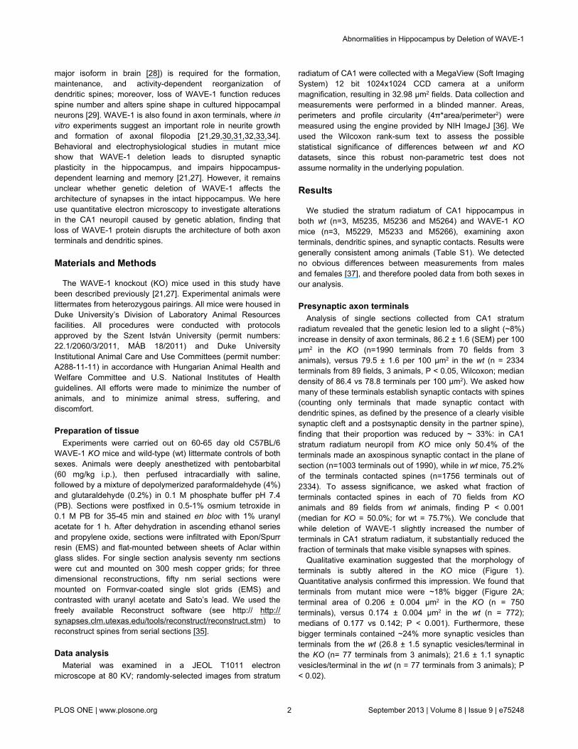
Data analysisMaterial was examined in a JEOL T1011 electron
microscope at 80 KV; randomly-selected images from stratum
radiatum of CA1 were collected with a MegaView (Soft ImagingSystem) 12 bit 1024x1024 CCD camera at a uniformmagnification, resulting in 32.98 µm2 fields. Data collection andmeasurements were performed in a blinded manner. Areas,perimeters and profile circularity (4π*area/perimeter2) weremeasured using the engine provided by NIH ImageJ [36]. Weused the Wilcoxon rank-sum text to assess the possiblestatistical significance of differences between wt and KOdatasets, since this robust non-parametric test does notassume normality in the underlying population.
Results
We studied the stratum radiatum of CA1 hippocampus inboth wt (n=3, M5235, M5236 and M5264) and WAVE-1 KOmice (n=3, M5229, M5233 and M5266), examining axonterminals, dendritic spines, and synaptic contacts. Results weregenerally consistent among animals (Table S1). We detectedno obvious differences between measurements from malesand females [37], and therefore pooled data from both sexes inour analysis.
Presynaptic axon terminalsAnalysis of single sections collected from CA1 stratum
radiatum revealed that the genetic lesion led to a slight (~8%)increase in density of axon terminals, 86.2 ± 1.6 (SEM) per 100µm2 in the KO (n=1990 terminals from 70 fields from 3animals), versus 79.5 ± 1.6 per 100 µm2 in the wt (n = 2334terminals from 89 fields, 3 animals, P < 0.05, Wilcoxon; mediandensity of 86.4 vs 78.8 terminals per 100 µm2). We asked howmany of these terminals establish synaptic contacts with spines(counting only terminals that made synaptic contact withdendritic spines, as defined by the presence of a clearly visiblesynaptic cleft and a postsynaptic density in the partner spine),finding that their proportion was reduced by ~ 33%: in CA1stratum radiatum neuropil from KO mice only 50.4% of theterminals made an axospinous synaptic contact in the plane ofsection (n=1003 terminals out of 1990), while in wt mice, 75.2%of the terminals contacted spines (n=1756 terminals out of2334). To assess significance, we asked what fraction ofterminals contacted spines in each of 70 fields from KOanimals and 89 fields from wt animals, finding P < 0.001(median for KO = 50.0%; for wt = 75.7%). We conclude thatwhile deletion of WAVE-1 slightly increased the number ofterminals in CA1 stratum radiatum, it substantially reduced thefraction of terminals that make visible synapses with spines.
Qualitative examination suggested that the morphology ofterminals is subtly altered in the KO mice (Figure 1).Quantitative analysis confirmed this impression. We found thatterminals from mutant mice were ~18% bigger (Figure 2A;terminal area of 0.206 ± 0.004 µm2 in the KO (n = 750terminals), versus 0.174 ± 0.004 µm2 in the wt (n = 772);medians of 0.177 vs 0.142; P < 0.001). Furthermore, thesebigger terminals contained ~24% more synaptic vesicles thanterminals from the wt (26.8 ± 1.5 synaptic vesicles/terminal inthe KO (n= 77 terminals from 3 animals); 21.6 ± 1.1 synapticvesicles/terminal in the wt (n = 77 terminals from 3 animals); P< 0.02).
Abnormalities in Hippocampus by Deletion of WAVE-1
PLOS ONE | www.plosone.org 2 September 2013 | Volume 8 | Issue 9 | e75248

Presynaptic vesicles in the KO seemed to be less tightlyassociated with the active zone than in the wt, whose vesiclestypically concentrated at the active zone, becoming sparse atthe periphery of the axon terminal (Figure 2B). To analyze theirrelative position, we measured the distance of vesicles from thesynaptic membrane of the active zone, confirming that vesicleslay considerably further from the active zone in KO animals(180 ± 6 nm; n = 596 vesicles) than in the wt (136 ± 4 nm; n =574); medians of 147 vs 111; P < 0.001. However, KOterminals were larger than those from wt mice, potentiallyconfusing the issue. We controlled for this difference by
computing a normalized distribution of synaptic vesicles, suchthat a vesicle directly touching the presynaptic membrane atthe active zone would have a normalized radial distance of 0,and a vesicle at the opposite side of the plasma membranewould have a normalized distance of 1.0 (see inset in Figure2C). This analysis confirmed our subjective impression,showing that vesicles in the KO lay at a mean distance of 0.40normalized units, while synaptic vesicles in the wt lay closer tothe synapse, at a mean normalized distance of 0.29 (Figure2C; medians of 0.37 vs 0.29; P < 0.001). Thus, bigger axon
Figure 1. Overview of ultrastructural changes associated with deletion of WAVE-1. Representative low-magnification electronmicrographs of synaptic neuropil in stratum radiatum of CA1 hippocampus, showing postsynaptic spines (orange) and presynapticboutons (blue) in material from KO (A) and wt mice (B). Scale bar: 5 µm.doi: 10.1371/journal.pone.0075248.g001
Abnormalities in Hippocampus by Deletion of WAVE-1
PLOS ONE | www.plosone.org 3 September 2013 | Volume 8 | Issue 9 | e75248

Figure 2. WAVE-1 affects size of presynaptic terminalsand organization of synaptic vesicles. A. Cumulative plotshows the distribution of the mean diameter (defined assqrt(area*4/π)) of terminals in KO (grey circles) and wt (blackdiamonds) CA1 KO terminals are generally larger than wtterminals. B. Electron micrographs (upper panel) andcorresponding line drawings (lower panel) illustrateorganization of synaptic vesicles within an axon terminal from aKO mouse (left), compared to wt control (right). Micrographsare from stratum radiatum of CA1 hippocampus. Synapticvesicles are more numerous and lie farther from the activezone in KO mice, compared to wt. Scale bar: 200 nm. C.Quantitative analysis of the organization of vesicles in KOmice, versus wt controls. To combine data from terminals ofdifferent sizes, the distribution of vesicles was normalized (seeinset): 0 corresponds to a vesicle lying directly at thepresynaptic membrane, and 1 to a vesicle lying at the oppositenon-synaptic membrane along an axis perpendicular to thesynapse. Black circles (representing positions of KO vesicles interminals) tend to lie farther from the active zone than whitecircles (representing positions of the wt vesicles). Vertical barsare standard errors (N = 3 animals for each genotype).doi: 10.1371/journal.pone.0075248.g002
terminals in the WAVE-1 KO animals contained significantlymore vesicles, which distributed abnormally within the terminal.
Postsynaptic dendritic spinesConsistent with previously-published evidence from light
microscopy of in vitro material that suggested a reduction inspine number as a result of WAVE-1 loss, we found a marked(~30%) reduction in the number of postsynaptic spines in CA1stratum radiatum from KO mice (KO, 42.8 ± 1.2 spines/100µm2, n = 70 fields; wt, 60.7 ± 1.5 spines/100 µm2, n = 80 fields;P < 0.001; Figure 1). We compared the size of spines from KOanimals with those from wt (n = 384 spines in KO and 395 inwt), finding no significant differences in area (KO, 0.077 ±0.002 µm2; wt, 0.079 ± 0.002 µm2; P > 0.4). In contrast, thespine perimeter was significantly increased in KO mice (meanperimeter for KO, 1221 ± 22 nm; wt, 1133 ± 21 nm; P < 0.002).Likewise, the length of postsynaptic densities (PSDs) as seenin single sections was significantly longer in KO spines (260 ± 6nm) compared to wt spines (216 ± 5 nm; P < 0.001). Previouswork using serial-section microscopy shows that larger spinestend to have larger PSDs [38]. Accordingly, we analyzed therelationship between spine head area and PSD length in ourmaterial. Randomly-selected single sections of spines fromCA1 stratum radiatum of the KO mice exhibited a positivecorrelation between spine size and PSD length (r2 = 0.36), butthere was a considerably stronger correlation for spines fromwt mice (r2 = 0.54; Figure 3).
That spine perimeter increased while spine area wasunchanged in the KO mice suggests spine profiles from KOmice were less round. To test this, we computed the"circularity" of randomly selected spine head profiles fromsingle sections (a value of 1.0 indicates a perfect circle, and 0indicates a completely flattened shape; see methods fordetails). We found that spines in the KO mice were significantlyless circular (0.65 ± 0.01) than spines from the wt stratumradiatum (0.74 ± 0.01; P < 0.001), implying that KO animalshave flattened or elongated spine heads.
We also noticed abnormalities in the internal structure ofspines. We found no spine apparatus in our KO sample (0 of989 spines); in contrast, we found that ~2% of spines (35 of1602) in the wt animals had a clearly-defined spine apparatus(typically in large mushroom-shaped spines). On the otherhand, spines from the KO material contained almost threetimes more endosomes (46.4 per 100 spines) than spines fromthe wt (16.6); P < 0.001 (Figure 4, arrowheads).
Synaptic relationshipThe above results suggest that loss of WAVE-1 affects
postsynaptic spines more dramatically than axon terminals.Interestingly, we also noted that some of the characteristicallyflattened spines had two spatially-separated PSDs. These werenot typical perforated synapses; the PSDs instead lay onopposite sides of the spine head, apparently contacting twodifferent axon terminals (see arrows, Figure 5A). Thesefeatures are rarely observed in the normal CA1 stratumradiatum. To see whether these synaptic contacts areestablished by two independent axon terminals, we performed3D serial reconstruction of a representative sample of these
Abnormalities in Hippocampus by Deletion of WAVE-1
PLOS ONE | www.plosone.org 4 September 2013 | Volume 8 | Issue 9 | e75248

abnormal synaptic contacts (n= 21), finding that in all cases, asingle axon terminal gave rise to both synapses (Figure 5B).These anomalous synaptic contacts might account—at least inpart—for the observed numerical asymmetry between thesynaptic partners.
In summary, we found that the density of terminals in KOanimals changed little, but these terminals were bigger, withmore but less organized synaptic vesicles. On the postsynapticside, the density of spines in KO animals was significantlyreduced, and these spines made abnormal synaptic contacts;furthermore, the spine head was flattened, with an abnormalcontent of internal membrane-bound structures.
Discussion
Changes in synaptic efficacy are typically associated withmorphological changes, in part because the biochemicalcascades implicated in synaptic plasticity share commonsignaling pathways with the machinery controlling actindynamics and reorganization [39]. Molecules that serve as‘hubs’ for these shared pathways are thus essential both fornormal neuronal morphology and for activity-dependentsynaptic plasticity. Accumulating evidence suggests thatWAVE-1 is one such hub: WAVE-1 is required for lamellipodialextension in neuronal growth cones [40], and disruption of theWAVE gene causes deficits at the (glutamatergic)neuromuscular junction in Drosophila [41]. Moreover,decreased expression of WAVE-1 (resulting from RNAi)reduces the number of mature dendritic spines in cultured
primary hippocampal neurons [29]. Likewise, disruption ofupstream signaling to WAVE-1 also causes spine reduction,altered synaptic plasticity, and deficits in memory retention[21,27], as does downstream disruption of the WAVE-1 ligandAbi-2 [42,43] or the Arp2/3 complex [44]. Thus, the WAVE-1signaling hub appears to play a key role in mediating themorphological changes associated with synaptic plasticity.
The present ultrastructural data from KO mice provides cluesas to how WAVE-1 may regulate synaptic function in CA1hippocampus. Presynaptically, we found that loss of WAVE-1affects the number and distribution of synaptic vesicles inSchaffer-collateral axon terminals in CA1 hippocampus. Thebiochemical pathway underlying this effect is unclear.Electrophysiological evidence from WAVE-1 KO mice revealedthat paired-pulse facilitation is normal in the hippocampus,suggesting normal presynaptic release probability [21].However, phosphorylation of WAVE-1 by cyclin-dependentkinase 5 (Cdk5) inhibits its ability to regulate Arp2/3-dependentactin polymerization, and the functionally recycling vesiclefraction in hippocampal synapses is regulated by Cdk5 activity[45]. Accordingly, we speculate that WAVE-1 in axon terminalsmay regulate synaptic vesicle distribution via Cdk5.
The abnormal flattening of spine heads in KO micepresumably reflects dysregulation of the actin spinoskeleton[10,46,47]. We also found that postsynaptic spines in the KOhave longer PSDs. Since the length of the PSD correlates withthe number of glutamate receptors at the synapse, and themagnitude of EPSCs [48,49,50,51,52], the longer PSDs wedetected in the mutants are likely to contain more glutamate
Figure 3. WAVE-1 affects relationship between PSD size and spine size. Scatterplots show the area of spine profiles as afunction of PSD length, in CA1 stratum radiatum of KO (left) and wt hippocampus (right). Linear regression analysis demonstrates aweaker correlation between spine area and PSD length in the KO (R = 0.60) than the wt material (R = 0.73).doi: 10.1371/journal.pone.0075248.g003
Abnormalities in Hippocampus by Deletion of WAVE-1
PLOS ONE | www.plosone.org 5 September 2013 | Volume 8 | Issue 9 | e75248

receptors, consistent with the enhanced LTP and impaired LTDpreviously reported for WAVE-1 KOs [21]. Current evidence
Figure 4. Loss of WAVE-1 causes abnormalities in theinternal structure of spines. Representative electronmicrographs of CA1 synaptic neuropil; coloring as in Figure 1.Spines from the KO material (A) were far more likely to containendosomes (black arrowheads) than spines from the wt (B).Scale bar: 1 µm.doi: 10.1371/journal.pone.0075248.g004
suggests that AMPA receptors are added to the synapse fromrecycling endosomes in the spine [53,54]. Interestingly wefound a marked increase of endosomes within KO spines.Whether these endosomes are trapped within the spinoplasmdue to defective actin polymerization, or are more numerousbecause more receptor is being transported to the spinesurface (as suggested by the enhanced LTP) will requirefurther investigation.
Figure 5. Abnormal spines in stratum radiatum of theWAVE-1 KO mouse. A. Arrows point to spines with twoPSDs. Spines are colored in orange, axon terminals in blue. B.3D reconstruction of an axospinous synaptic contact frommutant hippocampus. The reconstructed CA1 apical dendriticsegment (D) has a spine (Sp) oriented to show the synapticsurface. This spine has two distinct postsynaptic densities (red,arrows). The same axon (yellow) establishes separate synapticcontacts with both PSDs. Axon is yellow, spine is grey, PSD isred. Arrows point to synaptic surface between presynapticactive zone and postsynaptic density of the spine (Sp). Scalebars: 200 nm.doi: 10.1371/journal.pone.0075248.g005
Abnormalities in Hippocampus by Deletion of WAVE-1
PLOS ONE | www.plosone.org 6 September 2013 | Volume 8 | Issue 9 | e75248

In conclusion, the combined pre- and postsynaptic changesin synaptic architecture reported here provide a structuralsubstrate for the cognitive deficits previously reported, andsupport a role for WAVE-1 as an important modulator ofsynaptic plasticity.
Supporting Information
Table S1. Synapse-related parameters for each of the KOand wt animals studied. No clear relationship between any ofthese parameters and sex of mouse was apparent.
(PDF)
Acknowledgements
We thank Susan Grand, Tünde Magyar and Renáta Pop fortheir excellent technical assistance.
Author Contributions
Conceived and designed the experiments: BR RJW SHS.Performed the experiments: BR DH JD RS. Analyzed the data:BR PS RJW. Contributed reagents/materials/analysis tools:SHS JD. Wrote the manuscript: BR RJW.
References
1. Fortin DA, Srivastava T, Soderling TR (2012) Structural modulation ofdendritic spines during synaptic plasticity. Neuroscientist 18: 326-341.doi:10.1177/1073858411407206. PubMed: 21670426.
2. Lippman J, Dunaevsky A (2005) Dendritic spine morphogenesis andplasticity. J Neurobiol 64: 47-57. doi:10.1002/neu.20149. PubMed:15884005.
3. Nakagawa T, Engler JA, Sheng M (2004) The dynamic turnover andfunctional roles of alpha-actinin in dendritic spines. Neuropharmacology47: 734-745. doi:10.1016/j.neuropharm.2004.07.022. PubMed:15458845.
4. Matus A (2000) Actin-based plasticity in dendritic spines. Science 290:754-758. doi:10.1126/science.290.5492.754. PubMed: 11052932.
5. Fifková E, Delay RJ (1982) Cytoplasmic actin in neuronal processes asa possible mediator of synaptic plasticity. J Cell Biol 95: 345-350. doi:10.1083/jcb.95.1.345. PubMed: 6890558.
6. Gotow T, Miyaguchi K, Hashimoto PH (1991) Cytoplasmic architectureof the axon terminal: filamentous strands specifically associated withsynaptic vesicles. Neuroscience 40: 587-598. doi:10.1016/0306-4522(91)90143-C. PubMed: 2027472.
7. Morales M, Colicos MA, Goda Y (2000) Actin-dependent regulation ofneurotransmitter release at central synapses. Neuron 27: 539-550. doi:10.1016/S0896-6273(00)00064-7. PubMed: 11055436.
8. Cingolani LA, Goda Y (2008) Actin in action: the interplay between theactin cytoskeleton and synaptic efficacy. Nat Rev Neurosci 9: 344-356.doi:10.1038/nrn2373. PubMed: 18425089.
9. Jensen V, Walaas SI, Hilfiker S, Ruiz A, Hvalby O (2007) A delayedresponse enhancement during hippocampal presynaptic plasticity inmice. J Physiol 583: 129-143. doi:10.1113/jphysiol.2007.131300.PubMed: 17569738.
10. Murakoshi H, Yasuda R (2012) Postsynaptic signaling during plasticityof dendritic spines. Trends Neurosci 35: 135-143. doi:10.1016/j.tins.2011.12.002. PubMed: 22222350.
11. Haditsch U, Leone DP, Farinelli M, Chrostek-Grashoff A, Brakebusch Cet al. (2009) A central role for the small GTPase Rac1 in hippocampalplasticity and spatial learning and memory. Mol Cell Neurosci 41:409-419. doi:10.1016/j.mcn.2009.04.005. PubMed: 19394428.
12. Tolias KF, Duman JG, Um K (2011) Control of synapse developmentand plasticity by Rho GTPase regulatory proteins. Prog Neurobiol 94:133-148. doi:10.1016/j.pneurobio.2011.04.011. PubMed: 21530608.
13. Brünig I, Kaech S, Brinkhaus H, Oertner TG, Matus A (2004) Influx ofextracellular calcium regulates actin-dependent morphological plasticityin dendritic spines. Neuropharmacology 47: 669-676. doi:10.1016/j.neuropharm.2004.07.038. PubMed: 15458838.
14. Sanhueza M, Fernandez-Villalobos G, Stein IS, Kasumova G, Zhang Pet al. (2011) Role of the CaMKII/NMDA receptor complex in themaintenance of synaptic strength. J Neurosci 31: 9170-9178. doi:10.1523/JNEUROSCI.1250-11.2011. PubMed: 21697368.
15. Ultanir SK, Kim JE, Hall BJ, Deerinck T, Ellisman M et al. (2007)Regulation of spine morphology and spine density by NMDA receptorsignaling in vivo. Proc Natl Acad Sci U S A 104: 19553-19558. doi:10.1073/pnas.0704031104. PubMed: 18048342.
16. Heasman SJ, Ridley AJ (2008) Mammalian Rho GTPases: newinsights into their functions from in vivo studies. Nat Rev Mol Cell Biol9: 690-701. doi:10.1038/nrm2476. PubMed: 18719708.
17. Choi J, Ko J, Racz B, Burette A, Lee JR et al. (2005) Regulation ofdendritic spine morphogenesis by insulin receptor substrate 53, adownstream effector of Rac1 and Cdc42 small GTPases. J Neurosci
25: 869-879. doi:10.1523/JNEUROSCI.3212-04.2005. PubMed:15673667.
18. Etienne-Manneville S, Hall A (2002) Rho GTPases in cell biology.Nature 420: 629-635. doi:10.1038/nature01148. PubMed: 12478284.
19. Gonzalez-Billault C, Muñoz-Llancao P, Henriquez DR, Wojnacki J,Conde C et al. (2012) The role of small GTPases in neuronalmorphogenesis and polarity. Cytoskeleton (Hoboken) 69: 464-485. doi:10.1002/cm.21034. PubMed: 22605667.
20. Machesky LM, Insall RH (1998) Scar1 and the related Wiskott-Aldrichsyndrome protein, WASP, regulate the actin cytoskeleton through theArp2/3 complex. Curr Biol 8: 1347-1356. doi:10.1016/S0960-9822(98)00015-3. PubMed: 9889097.
21. Soderling SH, Guire ES, Kaech S, White J, Zhang F et al. (2007) AWAVE-1 and WRP signaling complex regulates spine density, synapticplasticity, and memory. J Neurosci 27: 355-365. doi:10.1523/JNEUROSCI.3209-06.2006. PubMed: 17215396.
22. Tahirovic S, Hellal F, Neukirchen D, Hindges R, Garvalov BK et al.(2010) Rac1 regulates neuronal polarization through the WAVEcomplex. J Neurosci 30: 6930-6943. doi:10.1523/JNEUROSCI.5395-09.2010. PubMed: 20484635.
23. Takenawa T, Suetsugu S (2007) The WASP-WAVE protein network:connecting the membrane to the cytoskeleton. Nat Rev Mol Cell Biol 8:37-48. doi:10.1038/nrm2069. PubMed: 17183359.
24. Eden S, Rohatgi R, Podtelejnikov AV, Mann M, Kirschner MW (2002)Mechanism of regulation of WAVE1-induced actin nucleation by Rac1and Nck. Nature 418: 790-793. doi:10.1038/nature00859. PubMed:12181570.
25. Ceglia I, Kim Y, Nairn AC, Greengard P (2010) Signaling pathwayscontrolling the phosphorylation state of WAVE1, a regulator of actinpolymerization. J Neurochem 114: 182-190. PubMed: 20403076.
26. Miki H, Takenawa T (2003) Regulation of actin dynamics by WASPfamily proteins. J Biochem 134: 309-313. doi:10.1093/jb/mvg146.PubMed: 14561714.
27. Soderling SH, Langeberg LK, Soderling JA, Davee SM, Simerly R et al.(2003) Loss of WAVE-1 causes sensorimotor retardation and reducedlearning and memory in mice. Proc Natl Acad Sci U S A 100:1723-1728. doi:10.1073/pnas.0438033100. PubMed: 12578964.
28. Soderling SH, Scott JD (2006) WAVE signalling: from biochemistry tobiology. Biochem Soc Trans 34: 73-76. doi:10.1042/BST0340073.PubMed: 16417486.
29. Kim Y, Sung JY, Ceglia I, Lee KW, Ahn JH et al. (2006)Phosphorylation of WAVE1 regulates actin polymerization and dendriticspine morphology. Nature 442: 814-817. doi:10.1038/nature04976.PubMed: 16862120.
30. Nakamura Y, Wood CL, Patton AP, Jaafari N, Henley JM et al. (2011)PICK1 inhibition of the Arp2/3 complex controls dendritic spine size andsynaptic plasticity. EMBO J 30: 719-730. doi:10.1038/emboj.2010.357.PubMed: 21252856.
31. Korobova F, Svitkina T (2010) Molecular architecture of synaptic actincytoskeleton in hippocampal neurons reveals a mechanism of dendriticspine morphogenesis. Mol Biol Cell 21: 165-176. doi:10.1091/mbc.E09-07-0596. PubMed: 19889835.
32. Wegner AM, Nebhan CA, Hu L, Majumdar D, Meier KM et al. (2008) N-wasp and the arp2/3 complex are critical regulators of actin in thedevelopment of dendritic spines and synapses. J Biol Chem 283:15912-15920. doi:10.1074/jbc.M801555200. PubMed: 18430734.
33. Spillane M, Ketschek A, Jones SL, Korobova F, Marsick B et al. (2011)The actin nucleating Arp2/3 complex contributes to the formation of
Abnormalities in Hippocampus by Deletion of WAVE-1
PLOS ONE | www.plosone.org 7 September 2013 | Volume 8 | Issue 9 | e75248
axonal filopodia and branches through the regulation of actin patchprecursors to filopodia. Dev Neurobiol 71: 747-758. doi:10.1002/dneu.20907. PubMed: 21557512.
34. Stavoe AK, Nelson JC, Martínez-Velázquez LA, Klein M, Samuel AD etal. (2012) Synaptic vesicle clustering requires a distinct MIG-10/Lamellipodin isoform and ABI-1 downstream from Netrin. Genes Dev26: 2206-2221. doi:10.1101/gad.193409.112. PubMed: 23028145.
35. Fiala JC (2005) Reconstruct: a free editor for serial section microscopy.J Microsc 218: 52-61. doi:10.1111/j.1365-2818.2005.01466.x. PubMed:15817063.
36. Schneider CA, Rasband WS, Eliceiri KW (2012) NIH Image to ImageJ:25 years of image analysis. Nat Methods 9: 671-675. doi:10.1038/nmeth.2089. PubMed: 22930834.
37. Sanchez AM, Flamini MI, Genazzani AR, Simoncini T (2013) Effects ofprogesterone and medroxyprogesterone on actin remodeling andneuronal spine formation. Mol Endocrinol 27: 693-702. doi:10.1210/me.2012-1278. PubMed: 23487486.
38. Harris KM, Stevens JK (1989) Dendritic spines of CA 1 pyramidal cellsin the rat hippocampus: serial electron microscopy with reference totheir biophysical characteristics. J Neurosci 9: 2982-2997. PubMed:2769375.
39. Matsuzaki M, Honkura N, Ellis-Davies GC, Kasai H (2004) Structuralbasis of long-term potentiation in single dendritic spines. Nature 429:761-766. doi:10.1038/nature02617. PubMed: 15190253.
40. Nozumi M, Nakagawa H, Miki H, Takenawa T, Miyamoto S (2003)Differential localization of WAVE isoforms in filopodia and lamellipodiaof the neuronal growth cone. J Cell Sci 116: 239-246. doi:10.1242/jcs.00233. PubMed: 12482910.
41. Schenck A, Qurashi A, Carrera P, Bardoni B, Diebold C et al. (2004)WAVE/SCAR, a multifunctional complex coordinating different aspectsof neuronal connectivity. Dev Biol 274: 260-270. doi:10.1016/j.ydbio.2004.07.009. PubMed: 15385157.
42. Grove M, Demyanenko G, Echarri A, Zipfel PA, Quiroz ME et al. (2004)ABI2-deficient mice exhibit defective cell migration, aberrant dendriticspine morphogenesis, and deficits in learning and memory. Mol CellBiol 24: 10905-10922. doi:10.1128/MCB.24.24.10905-10922.2004.PubMed: 15572692.
43. Proepper C, Steinestel K, Schmeisser MJ, Heinrich J, Steinestel J et al.(2011) Heterogeneous nuclear ribonucleoprotein k interacts with Abi-1at postsynaptic sites and modulates dendritic spine morphology. PLOS
ONE 6: e27045. doi:10.1371/journal.pone.0027045. PubMed:22102872.
44. Kim IH, Racz B, Wang H, Burianek L, Weinberg R et al. (2013)Disruption of Arp2/3 results in asymmetric structural plasticity ofdendritic spines and progressive synaptic and behavioral abnormalities.J Neurosci 33: 6081-6092. doi:10.1523/JNEUROSCI.0035-13.2013.PubMed: 23554489.
45. Marra V, Burden JJ, Thorpe JR, Smith IT, Smith SL et al. (2012) Apreferentially segregated recycling vesicle pool of limited size supportsneurotransmission in native central synapses. Neuron 76: 579-589. doi:10.1016/j.neuron.2012.08.042. PubMed: 23141069.
46. Penzes P, Rafalovich I (2012) Regulation of the actin cytoskeleton indendritic spines. Adv Exp Med Biol 970: 81-95. doi:10.1007/978-3-7091-0932-8_4. PubMed: 22351052.
47. Penzes P, Cahill ME, Jones KA, VanLeeuwen JE, Woolfrey KM (2011)Dendritic spine pathology in neuropsychiatric disorders. Nat Neurosci14: 285-293. doi:10.1038/nn.2741. PubMed: 21346746.
48. Bourne JN, Harris KM (2008) Balancing structure and function athippocampal dendritic spines. Annu Rev Neurosci 31: 47-67. PubMed:18284372.
49. Harris KM, Weinberg RJ (2012) Ultrastructure of synapses in themammalian brain. Cold Spring Harb Perspect Biol.
50. Kasai H, Fukuda M, Watanabe S, Hayashi-Takagi A, Noguchi J (2010)Structural dynamics of dendritic spines in memory and cognition.Trends Neurosci 33: 121-129. doi:10.1016/j.tins.2010.01.001. PubMed:20138375.
51. Matsuzaki M, Ellis-Davies GC, Nemoto T, Miyashita Y, Iino M et al.(2001) Dendritic spine geometry is critical for AMPA receptorexpression in hippocampal CA1 pyramidal neurons. Nat Neurosci 4:1086-1092. doi:10.1038/nn736. PubMed: 11687814.
52. Nusser Z, Lujan R, Laube G, Roberts JDB, Molnar E et al. (1998) Celltype and pathway dependence of synaptic AMPA receptor number andvariability in the hippocampus. Neuron 21: 545-559. doi:10.1016/S0896-6273(00)80565-6. PubMed: 9768841.
53. Ehlers MD (2000) Reinsertion or degradation of AMPA receptorsdetermined by activity-dependent endocytic sorting. Neuron 28:511-525. doi:10.1016/S0896-6273(00)00129-X. PubMed: 11144360.
54. Park M, Penick EC, Edwards JG, Kauer JA, Ehlers MD (2004)Recycling endosomes supply AMPA receptors for LTP. Science 305:1972-1975. doi:10.1126/science.1102026. PubMed: 15448273.
Abnormalities in Hippocampus by Deletion of WAVE-1
PLOS ONE | www.plosone.org 8 September 2013 | Volume 8 | Issue 9 | e75248
Copyright © 2022 FDOKUMEN