Punching, Cutting, Forming – Standardized, optimal Drive ...
Upload
independentCategory
view
0download
0
www.elsevier.com/locate/brainres
Brain Research 993 (2003) 18–29
Research report
Neurosphere and neurosphere-forming cells:
morphological and ultrastructural characterization
Alessandra Beza, Elena Corsinia, Daniela Curtib, Marco Biggiogerac, Augusto Colombod,Roberto Francesco Nicosiae,f, Stefano Filippo Paganoa, Eugenio Agostino Paratia,*
aLaboratory of Neurobiology, Department of Neurobiology and Neurorestorative Therapies, National Neurological Institute ‘‘C. Besta’’,
Via Celoria 11, 20133 Milan, ItalybDepartment of Physiological Science and Cellular and Molecular Pharmacology, University of Pavia, Pavia, Italy
cDepartment of Animal Biology, University of Pavia, Pavia, Italyd Institute for Obstetric and Gynaecology ‘‘L. Mangiagalli’’, ICP, Milan, Italy
eVeterans Affairs Puget Sound Health Care System, Seattle, WA, USAfDepartment of Pathology, University of Washington, Seattle, WA, USA
Accepted 14 August 2003
Abstract
Despite recent advances in our understanding of neural stem cell (NSC) biology, the free-floating structures generated by these cells
in vitro, the ‘‘neurospheres’’, have not been fully characterized. To fill this gap, we examined neurospheres and neurosphere-derived
NSCs by confocal microscopy, electron microscopy (EM) and cytofluorimetry. Here, we show that neurospheres and neurosphere-
forming cells are morphologically and functionally heterogeneous. Confocal microscopy reveals differences in cell size, viability,
cytoplasmic content and in the presence and distribution of active mitochondria. By electron microscopy, neurospheres appear as
complex structures in which biological events such as mitosis, apoptosis and even phagocytosis are influenced by NSCs localization
within the architecture of the neurosphere. NSCs derived from neurospheres are not synchronized and are represented in all phases of
the cell cycle. Cytofluorimetric studies demonstrate NSCs’ heterogeneity in cell size by forward scatter (FSC) analysis, and in
cytoplasmic granularity by side scatter (SSC) profiling. These findings may contribute to our understanding of the morphogenesis of the
neurospheres, particularly as this process relates to the high environmental adaptability of the NSCs and the reported existence of
different subpopulations of neural stem cells.
D 2003 Elsevier B.V. All rights reserved.
Keywords: Neurosphere; Neurosphere-forming cells; Characterization
1. Introduction brains were later found to contain neural stem cells
Since they were first described in the mouse brain,
neural stem cells (NSCs) have been the subject of inten-
sive investigation because of their potential therapeutic use
in neurodegenerative disorders. Mouse NSCs were initially
thought to derive from the striatum [27]; additional studies
demonstrated that the site of origin of these NSCs was
actually located in the adjacent subventricular zone (SVZ).
Different areas of embryonic and adult mouse and human
0006-8993/$ - see front matter D 2003 Elsevier B.V. All rights reserved.
doi:10.1016/j.brainres.2003.08.061
* Corresponding author. Tel.: +39-2-2394387; fax: +39-2-270638217.
E-mail address: [email protected] (E.A. Parati).
[2,3,8,12,24,30,31].
Various methods have been developed to isolate NSCs
and characterize their capacity to proliferate and differen-
tiate [2,6,11,28,29,37,40–43]. NSCs can be isolated by
cell sorting based on expression of individual surface
antigens such as CD24 [5,9,13,33,38] and CD 133 [40]
or physical properties such as forward scattering (cell size)
and side scattering (granularity) [23].
NSCs are highly plastic cells capable of fate conver-
sion/transdifferentiation events [4,14,20,22,26,34,44],
which are essential for their engraftment and migration.
This plasticity allows NSCs to home to and differentiate
into neural tissues of interest, with resulting amelioration
of signs and symptoms of neurodegenerative or cerebro-

A. Bez et al. / Brain Research 993 (2003) 18–29 19
vascular diseases [1,10,18,21,35,39]. Recently, NSCs have
even been shown to fuse with embryonic stem cells (ES)
[45].
Though much is known about NSCs’ biology and
behaviour, investigative efforts have only recently focused
on the characterization of NSCs morpho-functional fea-
tures and metabolic properties, which are largely un-
known, particularly in humans. For a long time, human
NSCs have been considered a quiescent population of
undifferentiated and homogeneous cells, which could be
activated by environmental cues in vivo or epigenetic
stimuli in vitro. Recent data, however, suggest that NSCs
are heterogeneous and may express not only nestin,
which is a marker of precursor neural cells [16], but
also CD34, CD31 and Tie2 [15,25], which were previ-
ously described only in extraneural tissues. In addition,
studies demonstrating that neurosphere forming cells are
ultrastructurally heterogeneous [17] and express different
neural lineage-specific markers indicate the existence
within these in vitro formed structures of distinct cellular
phenotypes, which implies variable developmental com-
mitments of parental clone-forming cells [36]. A gap,
however, exists in our understanding of the cytoarchitec-
ture of human neurospheres and of the morpho-functional
characteristics of human neurosphere-forming NSCs.
To fill this gap, we have evaluated human embryonal
brain-derived neurospheres and neurosphere-derived NSCs
by electron microscopy (EM), confocal microscopy and
flow cytometry. Our results indicate that human neuro-
spheres are highly dynamic structures with distinct radial
gradients of cell proliferation, survival, apoptosis and
phagocytosis. NSCs apoptotic and necrotic events increase
as neurospheres enlarge and develop insufficiently nour-
ished inner cores. In addition, NSCs derived from these
neurospheres are morphologically heterogeneous, exhibit
different sizes and cytoplasmic granularity and coexist in
different phases of the cell cycle.
2. Material and methods
2.1. Establishment of human embryonic stem cell lines
The use of human central nervous system (CNS) tissues
was authorized by the Ethics Committee of ‘‘C. Besta’’
Neurological Institute and ‘‘L. Mangiagalli’’ Obstetric–
Gynecological Clinic. Human NSCs were derived from
the brains of 12-week-old human embryos obtained fol-
lowing the ethical guidelines of the European Network
for Transplantation (NECTAR), as previously reported
[25,42]. Briefly, the tissue was mechanically minced and
the resulting cell suspension was plated in the presence of
20 ng/ml EGF and 10 ng/ml bFGF (both human recombi-
nant) in an NS-A basal serum-free medium (Euroclone,
Irvine, UK), optimised for neural stem cell growth and
referred to as control medium. These experimental condi-
tions promote the formation of spherical clusters called
‘‘neurospheres’’ from floating cultures of single cells. The
resulting cell strains can be amplified in vitro and cryopre-
served. They maintain multipotentiality even after several
passages in vitro, as demonstrated by clonal analysis, and
can differentiate into neurons, astrocytes and oligodendro-
cytes [24,42].
2.2. Laser scanning confocal microscopy (LSCM)
Neurospheres were dissociated to cell suspension and
centrifuged at 1000 rpm for 10 min (Hermle Z300K). The
pellet was resuspended in a buffer of the following
composition (in mM): 142 NaCl, 2 KCl, 1.2 K2HPO4, 1
MgSO4, 10 HEPES, 1% glutamine, 1.3 CaCl2, 10 glu-
cose, pH 7.4. Aliquots of cell suspensions were labeled
with intravital fluorescent probes for 20 min at 37 jC.Mitochondrial transmembrane potential (/) was measured
with 5,5V,6,6V tetracloro-1,1V,3,3V-tetraetilbenzimidazol car-
bocianine (JC-1, 1.5 AM) at ex 488 nm with an Argon
laser and at em 527 nm (green fluorescence, monomeric
form) and 590 nm (red fluorescence, J-aggregates). A
‘‘shift’’ from red to green fluorescence is observed upon
depolarisation of mitochondria [7]. Cytosolic and mito-
chondrial calcium were investigated with Rhod2 (0.5 AM),
at ex 543 and em 580 and Indo1 (5 AM) at ex 350 and
em 400/500 with an UV laser. In some experiments,
NSCs were stained also with the nuclear dye Hoechst
33342. The neurospheres, either intact or dissociated into
individual cells, were double stained with the SYTO 59
vital nuclear dye (0.5 AM) and propidium iodide (0.15
AM) to estimate cell viability and position in the neuro-
sphere. After labeling, the cells were washed with fresh
buffer, left for at least 30 min in the dark, and then
incubated for 5 min on polylysin-coated coverslips (19
mm diameter), washed again with fresh medium and
perfused (0.5 ml/min) in a 300-Al chamber whose bottom
is the coverslip. Image galleries were acquired at 0.8–1
Am interval on the z axis. Experiments were performed
with a DMS IRBE SP2 (Leica) confocal microscope,
equipped with an inverted microscope and a 63� oil
objective (NA 1.3). Data analysis was performed with
Leica software.
2.3. Electron microscopy
For ultrastructural analysis, the neurospheres were fixed
by adding glutaraldehyde directly to the growth medium to a
final concentration of 2%, and left in fixative for 2 h.
Neurospheres were then centrifuged, rinsed in Sorensen
buffer (pH 7.2) and postfixed in 1% OsO4 for 2 h at room
temperature. The pellet was then incorporated into a 2%
agarose gel, dehydrated in ethanol and embedded in LR
White resin. Semithin sections were routinely stained with
toluidine blue. Ultrathin sections were stained with uranyl
acetate and lead citrate. For immunolabeling, 100 AM

A. Bez et al. / Brain Research 993 (2003) 18–2920
bromouridine (BrU) was added to the growth medium prior
to fixation and left for 10 min. The cells were then fixed
with 4% paraformaldehyde for 2 h and processed as above.
Thin sections were immunolabeled by the colloidal gold
method for BrU for evaluation of RNPs. All the specimens
were examined with a Zeiss EM 900 electron microscope
operating at 80 kV.
2.4. Cytofluorimetric analysis
Neurospheres from six different cell strains were ana-
lysed by flow cytometry. The neurospheres were collected
at 24 h, 5 days and 10 days after plating in fresh medium
and dissociated into single cells. The percentage of cells
in G0/G1, S and G2/M phases, and the number of cells
present in each phase were evaluated. Cell cycle was
evaluated by using a staining kit for DNA analysis in
flow cytometry (DAKO). Approximately 500,000 cells
Fig. 1. Phase contrast images of neurospheres (A–C) and confocal image of neu
conditions in presence of EGF and bFGF. (A) Neurosphere with irregular rim
Neurosphere with regular rim and well defined spherical shape. (C) Neurosphere a
even though it is cultured in serum-free medium supplemented with GFs (not differ
again a few days later. (D) Neurosphere-derived cells stained intravitally with JC
mitochondrial membrane depolarisation. Cytoplasmic staining obtained with this
kinds of cells (b and c) can be identified based on size, amount of cytoplasm and m
and less frequently observed. Bars: 50 Am (A–C) or 12 Am (D).
were incubated for 1 h at 4 jC with propidium iodide
(50 Ag/ml), resuspended in a lysis-containing buffer con-
taining a lysis agent, a Rnase and a chromatin stabilizer,
according to manufacturer’s instruction. Then cells were
resuspended again, filtered through a 50-Am pore filter and
measured on a DAKO Galaxy (DAKO) using FloMax
software. Cytofluorimetric analysis was then performed to
establish NSCs size. Microspheres of pre-defined sizes
(NileredR Fluorescent beads, BD Biosciences; 1.7–2.2
Am; 2.5–4.5 Am; 10–14 Am; 15–19 Am) were resus-
pended in PBS and used as standard to establish NSCs
size by cytofluorimetric analysis. Both cells and beads
were analysed with the same setting of instrumental
physical parameters (forward scatter [FSC], representing
cell and bead size, and side scatter [SSC], representing
cellular granularity). Cell size was calculated on a curve
employing bead size on x axis and FSC values on the
y axis.
rosphere-derived NSCs (D). Neurospheres were cultured under serum-free
and cilia-like cytoplasmic processes protruding from its surface (a). (B)
dherent to an uncoated well exhibits pseudopod-like cytoplasmic processes
entiating culture conditions); this neurosphere detached and became floating
-1, a cationic dye that shifts from red to green fluorescence as a result of
dye demonstrates morphological heterogeneity of NSCs. In particular, two
itochondrial distribution. Other cellular phenotypes are more heterogeneous

A. Bez et al. / Brain Research 993 (2003) 18–29 21
To quantify the number of apoptotic and necrotic cells,
we used human Annexin V-FITC Kit (Bender MedSystem).
Cells at two different DIV, 12 and 19, and dimensions,
between about 150 and 220 Am, respectively, were treated
according to the manufacturer’s instructions. Neurospheres
were dissociated to single cells, resuspended in the binding
buffer of the kit and adjusted to a cell density of 2–5� 105.
Annexin V-FITC (5 Al) was added to 195 Al of the cell
suspension. Cells were then mixed and incubated for 10 min
at room temperature. Finally, cells were washed, resus-
pended in 190 Al of binding buffer, mixed with 10 Al ofpropidium iodide (final concentration 1 Ag/ml) and analysed
by FACS.
2.5. Immunocytochemistry
For the analysis of the distribution of cells in mitosis, the
neurospheres were plated on Matrigel and kept in growth
medium containing BrdU (20 AM, Sigma) for 24 h. During
this period, they spread so that it was possible identify
peripheral and internal cells. The neurospheres were then
fixed for 20 min in 4% paraformaldehyde in PBS (pH 7.4),
washed and incubated with PBS 1� , 0.3% Triton contain-
ing 10% normal goat serum and a monoclonal anti-BrdU
antibody (1:500, Chemicon) for 90 min at 37 jC. The
neurospheres were processed using the standard avidin–
biotin peroxidase procedure. Antigen unmasking was per-
formed by treating the cultures with 2 M HCl for 1 h. After
Fig. 2. Confocal micrograph of neurosphere double stained with the SYTO 59 vi
SYTO 59 is a lipophilic dye capable of labeling nuclei of viable cells. Propidium io
and no longer viable. Blue cells are therefore healthy whereas red cells are suffering
Bar: 50 Am.
being rinsed in Tris buffer, the neurospheres were treated
with biotinylated secondary antibody and incubated with the
avidin–peroxidase complex ABC kit (Vector), according to
the manufacturer’s instructions.
3. Results
A first analysis was performed by confocal and electron
microscopy on whole neurospheres. This was followed by
confocal microscopic and cytofluorometric evaluation of
viable cell suspensions obtained after mechanical dissocia-
tion of the neurospheres. The cells used in all the experi-
ments were fresh, never frozen and thawed, and the number
of neurosphere dissociations (passages) varied from 7 to 16
(100–180 DIV). We avoided passages that were too low or
too high to elude the possibility of non-pure NSCs culture or
an excessively long cell manipulation, respectively. Similar
results were obtained using cell lines at different passages.
3.1. Neurospheres
A cross comparison of neurospheres derived from the
same brain, which were the starting materials of our experi-
ments, revealed significant heterogeneity (Fig. 1A–C).
Some neurospheres had a well-defined spherical shape
(Fig. 1B), whereas others appeared as irregular cell clusters
with uneven external rims (Fig. 1A). Ciliated-like cells (Fig.
tal nuclear dye (blue) and propidium iodide (red) to estimate cell viability.
dide penetrates the cell membrane and labels nuclei only if cells are injured
or dead. Viable cells are mostly located at the periphery of the neurosphere.
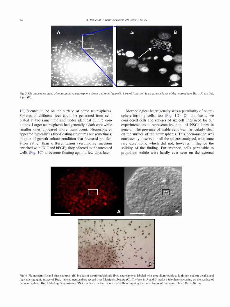
Fig. 3. Chromosome spread of representative neurosphere shows a mitotic figure (B, inset of A, arrow) in an external layer of the neurosphere. Bars: 50 Am (A),
8 Am (B).
A. Bez et al. / Brain Research 993 (2003) 18–2922
1C) seemed to be on the surface of some neurospheres.
Spheres of different sizes could be generated from cells
plated at the same time and under identical culture con-
ditions. Larger neurospheres had generally a dark core while
smaller ones appeared more translucent. Neurospheres
appeared typically as free-floating structures but sometimes,
in spite of growth culture condition that favoured prolifer-
ation rather than differentiation (serum-free medium
enriched with EGF and bFGF), they adhered to the uncoated
wells (Fig. 1C) to become floating again a few days later.
Fig. 4. Fluorescent (A) and phase contrast (B) images of paraformaldehyde-fixed n
light micrographic image of BrdU-labeled neurosphere spread over Matrigel subst
the neurosphere. BrdU labeling demonstrates DNA synthesis in the majority of c
Morphological heterogeneity was a peculiarity of neuro-
sphere-forming cells, too (Fig. 1D). On this basis, we
considered cells and spheres of six cell lines used for our
experiments as a representative pool of NSCs lines in
general. The presence of viable cells was particularly clear
on the surface of the neurospheres. This phenomenon was
consistently observed in all the spheres analysed, with some
rare exceptions, which did not, however, influence the
solidity of the finding. For instance, cells permeable to
propidium iodide were hardly ever seen on the external
eurospheres labeled with propidium iodide to highlight nuclear details, and
rate (C). The box in A and B marks a telophase occurring on the surface of
ells occupying the outer layers of the neurosphere. Bars: 50 Am.

Fig. 5. Toluidine blue-stained semithin sections (A, B) and electron micrographs (C, D) of neurosphere. Apoptotic phenomena (highlighted in boxes in A and
B) are rarely seen at the periphery of the neurosphere (A) and are identified mainly in its inner layers (B). Ultrastructural studies demonstrate phagocytosed
apoptotic bodies (arrows) mostly in the internal layers of the neurosphere (C) and only rarely in peripheral cells (D). Note degradation of apoptotic body in C.
Bars: 50 Am (A, B), 1 Am (C, D).
A. Bez et al. / Brain Research 993 (2003) 18–29 23
layers of the neurospheres, whereas cells positive for SYTO
59, a lipophilic dye capable of labeling nuclei of viable
cells, were demonstrated on the neurosphere surface (Fig.
Fig. 6. Electron micrographs of neurosphere cells. Adjacent cells of the neur
demonstrated in the neurosphere cell cytoplasm (B). Bars: 5 Am.
2). In addition, mitotic figures were found peripherally and
were never noticed in the inner part of the spheres (Figs. 3
and 4). We verified these data by immunocytochemistry:
osphere are joined by a gap-junction (A, inset). Lysosomes (arrow) are

Fig. 7. Electron micrographs of neurosphere cells immunostained by the colloidal gold method for BrU, a precursor of RNA. RNA synthesis is more prominent
in a representative cell at the periphery of the neurosphere (A) than in a cell from the inner layers (B). Colloidal gold particles are present on both perichromatin
fibrils and nucleolus, evidence of high trascriptional activity. Bars: 1.5 Am (A), 5 Am (B).
A. Bez et al. / Brain Research 993 (2003) 18–2924
When a neurosphere was plated on a matrix, it spread and
flattened losing its spherical shape and becoming a mono-
layer of cells except for its center that, even if flat, was
always composed of several cell layers. Only peripheral
Fig. 8. Confocal images of NSCs stained with the intravital dye JC-1 to highlig
differentiate into oligodendrocytic (A), astrocytic (B) and neuronal (C) phenotype
cells, which as a consequence of the contact with an
adhesive matrix spread forming pseudopodia-like cytoplas-
mic processes, stained for BrdU, that is, were actively
synthesizing DNA after 24 h of incubation with BrdU
ht morphologic features. NSCs, under appropriate culture conditions, may
s.

Fig. 10. Flow cytometric analysis of representative NSCs suspension
demonstrates heterogeneity of physical parameters as no subpopulation
with defined FSC/SSC values can be identified.
Fig. 9. Confocal micrograph of neurosphere-derived NSCs after mechanical dissociation and double staining with the fluorescent dyes JC-1 (green/red,
cytoplasm/mitochondria) and Hoechst 33342 (blue, nuclei). Representative NSCs, including a three-cell cluster, exhibit similar characteristics: large size,
cytoplasmic and mitochondria content, and nuclear/cytoplasmic ratio. These cells correspond to the cellular phenotype shown in Fig. 1D/c. Bar: 15 Am.
A. Bez et al. / Brain Research 993 (2003) 18–29 25
(Fig. 4). We have also documented, for the first time in the
literature of human neurospheres, the presence of phago-
cytic events and, in particular, phagocytosis of apoptotic
bodies; apoptosis and phagocytosis of apoptotic bodies
never occurred at the periphery of the neurospheres but
were typically seen in their inner regions, between the
external 2nd to 3rd layers and their core (Fig. 5). The
presence of lysosomes further supported the evidence of
phagocytosis (Fig. 6). EM revealed that necrotic cells, like
apoptotic cells, never localized in the external layers. The
percentage of viable, apoptotic and necrotic cells in a neuro-
sphere was quantified by cytofluorimetric analysis using a
human Annexin V-FITC Kit. Neurospheres maintained in
culture for 12 and 19 DIV, which corresponded to neuro-
spheres of about 150 and 220 Am in diameter, contained
11.2% and 21% of apoptotic cells and 9 and 19.2% of
necrotic cells, respectively. BrU incorporation experiments,
which were performed to evaluate RNA synthesis, demon-
strated a high trascriptional activity in peripheral cells and a
definitely reduced one in the inner ones (Fig. 7). Neuro-
sphere-forming cells were connected by weak junctional
complexes (Fig. 6). Desmosoms were never observed and
there was no evidence of fusion of plasmalemmas.
3.2. Cells dissociated from the neurospheres
The second part of our study was carried out on NSCs
obtained by mechanical dissociation of neurospheres.
NSCs appeared as spherical and floating cells after
dissociation but, under appropriate culture conditions, they
differentiated into oligodendrocytic, astrocytic or neuronal

Fig. 12. Percentage of NSCs in different phases of the cell-cycle evaluated
after being plated and maintained in the same medium for 1, 5 or 10 days.
The absence of significant fluctuations over time suggests that the freshness
of the medium and the depletion of GFs occurring after 10 days have no
effects on the cell-cycle trend.
Fig. 11. Flow cytometric analysis of representative NSCs suspension labeled with propidium iodide and evaluated for cell cycle. (A) RN1, RN2 and RN3 gates
identify cells in G0/G1, S, and G2/M phases, respectively. (B, C, D) Dot-plots showing cell size and organelle complexity of cells in G0/G1 phases (B), S phase
(C) and G2/M phases (D) demonstrate that neurosphere-derived cells with different physical properties (SSC/FSC parameters) can be identified in all phases of
the cell cycle.
A. Bez et al. / Brain Research 993 (2003) 18–2926
phenotypes (Fig. 8). Cytofluorimetric analysis of NSCs
revealed heterogeneity of NSCs size and granularity.
Using NileredR Fluorescent beads of predefined sizes as
a reference, the NSCs range in size by flow cytometry
between 9.28 and 19.27 Am in diameter. After neuro-
sphere dissociation, more than 80% of the recovered cells
were viable. Some cells had many energized mitochon-
dria, whereas others showed few mitochondria and/or low
aerobic energy metabolism (Figs. 1D and 9). Analyses of
calcium distribution allowed the observation of cells
enriched in mitochondrial calcium depots. The mitochon-
drial calcium signal was higher in cells with very low
cytoplasmic calcium (data not shown). The FSC/SSC
profile of NSCs confirmed the heterogeneity of neuro-
sphere-forming cells from the physical parameters point
of view (Fig. 10). The neurosphere-derived cells were not
synchronized, were represented in all the phases of the
cell cycle and their dimensions varied with the cycle
phase. The means of the values obtained from the six cell
lines utilised were: 75.24% of cells in G0/G1 phase,
8.36% in S and 15.82% in G2/M. A typical cell cycle
trend and a three dot-plots representing cell size and
organelle complexity/cellular granularity of each cell-cycle
phase are shown in Fig. 11. Slight and nonsignificant
fluctuations in the percentage of cells present in the

A. Bez et al. / Brain Research 993 (2003) 18–29 27
different cell cycle phases could be observed from 1 to 10
DIV (Fig. 12).
4. Discussion
Our results demonstrate that neurospheres derived from
NSCs plated at the same time and under the same culture
conditions differ in size and morphology. Neurospheres
can grow considerably becoming darker as they enlarge.
Their dark cores become the site of necrotic events
probably because of reduced nourishment from the exter-
nal medium. Smaller spheres, on the other hand, are
translucent, have no dark cores and appear much health-
ier. Neurospheres may have regular or irregular shapes,
and cells on their external layers may occasionally show
cytoplasmic processes resembling cilia or pseudopodia.
Neurospheres may temporarily adhere to the bottom of
the culture dish, even under growing culture conditions,
to spontaneously become floating again a few days later.
Heterogenity characterizes not only the neurospheres but
the neurosphere-forming cells as well. These cells have
different sizes, granularity, metabolism, cytoplasmic con-
tent and are in different phases of the cell cycle. More-
over, they exhibit different features according to the
layers of the spheres they occupy. The distribution of
biological phenomena such as mitosis, transcription pro-
cesses, apoptosis, phagocytosis and necrosis was distinctly
influenced by the position cells took in the structure of
the neurosphere. The cytoarchitecture of the neurosphere
and the degree of cell viability and activity within it may
depend on the access neurosphere-forming cells have to
the culture medium and therefore on the availability of
oxygen and nutrients and on the possibility to readily
eliminate catabolites in the environment (culture medium).
This interpretation would justify the higher cell activity
(i.e., mitosis, protein syntesis) we observed at the periph-
ery of the neurospheres where exchange of nutrients,
oxygen and catabolites is facilitated. In contrast, apopto-
sis, phagocytosis, necrosis and low or absent mitotic and
transcriptional activity are typical of the inner layers
where nutritional exchanges are more difficult. This is
confirmed by the higher percentage of apoptotic and
necrotic cells found in neurospheres that are maintained
in culture without being dissociated for 19 DIV and are
larger than neurospheres dissociated after 12 DIV.
The neurosphere properties described above can be
interpreted as an adaptation of NSCs to in vitro culture
conditions. The NSCs may organize in such clusters to
survive the nonphysiological conditions of the in vitro
environment. Cells probably optimise their interactions
acquiring the most advantageous shape from a thermody-
namycal point of view: the sphere. The neurosphere, in turn,
develops a high degree of biological organization. The
observed phagocytosis is probably the expression of a
dynamically maintained equilibrium between the generation
of new cells and the apoptosis or necrosis of older cells. A
neurosphere may be considered a microsystem able to grow
and survive until a threshold crucial point is reached when
the self-restoration mechanisms fail; we can therefore con-
sider the neurospheres an example of ‘‘environmental
adaptability’’. This environmental adaptability may enable
the NSCs to better express the plasticity they need for in
vivo engraftment and fate conversion [4,14,20,22,26,34,44].
In light of the evidence regarding the neurosphere
structure and its possible morphogenetic mechanisms, we
can hypothesize that neurosphere-derived cell heterogeneity
is due to their topographic distribution within the cytoarch-
itecture of the sphere. This morphologic heterogeneity may
also be due, at least in part, to the presence of subpopula-
tions of neurosphere-forming cells with distinct survival and
proliferative behaviour. Previous studies have in fact dem-
onstrated that NSCs are endowed with different degrees of
stemness properties, as they are able to give origin to
neurospheres with variable frequencies, and exhibit different
EGF and bFGF responsiveness. We know, for example, that
in the presence of EGF, cloned murine neurospheres are
larger in diameter, whereas in the presence of bFGF, they are
small and that exposure to both GFs produces both large and
small spheres [46]; moreover, the EGF-responsive mouse
population increases in size by asymmetric division of
bFGF-responsive cells and by symmetric division of EGF
responsive cells [19] EGF-, bFGF- and both GFs-responsive
NSCs with different capacity to generate neurospheres have
been also demonstrated in human fetal brain [34]. These
cells can be also selectively isolated based on their size and
PNA-binding activity or on the expression of cell surface
markers such as CD133+ (CD45� , CD34� ) and CD24
(low levels) [31].
In summary, our study provides new insights into the
behaviour of NSCs in vitro and the morphogenesis of
neurospheres. This knowledge may help optimise methods
for the isolation and in vitro expansion of NSCs toward the
utilisation of these cells for in vivo studies and therapeutic
applications.
Acknowledgements
We thank Dr. M. Di Segni and Dr. G. Terzoli for kindly
providing Fig. 3, Dr. P. Veglianese for Fig. 4A,B and Dr. S.
Pozzi for Fig. 4C.
The Laboratory of Neurobiology dedicates this work to
its precious research scientist, Dr. Stefano Pagano, who
passed away in his youth.
References
[1] M.L. Alexandrova, P.G. Bochev, V.I. Markova, B.G. Bechev, M.A.
Popola, M.P. Danovska, V.K. Simeonova, Changes in phagocyte
activity in patients with ischaemic stroke, Luminescence 16 (2001)
357–365.

A. Bez et al. / Brain Research 993 (2003) 18–2928
[2] A. Alvarez-Buylla, C. Lois, Neuronal stem cells in the brain of adult
vertebrates, Stem Cells 13 (1995) 263–272.
[3] Y. Arsenijevic, J.G. Villemure, J.F. Brunet, J.J. Bloch, N. Deglon, C.
Kostic, A. Zurn, P. Aebischer, Isolation of multipotent neural precur-
sors residing in the cortex of the adult human brain, Exp. Neurol. 170
(2001) 48–62.
[4] C.R. Bjornson, R.L. Rietze, B.A. Reynolds, M.C. Magli, A.L. Ves-
covi, Turning brain into blood: a hematopoietic fate adopted by adult
stem cells in vivo, Science 283 (1999) 534–537.
[5] V. Calaora, G. Chazal, P.J. Nielsen, G. Rougon, H. Moreau, mCD24
expression in the developing mouse brain and zones of secondary
neurogenesis in the adult, Neuroscience 73 (1996) 581–594.
[6] E. Cattaneo, R. McKay, Proliferation and differentiation of neuro-
nal stem cells regulated by nerve growth factor, Nature 347 (1990)
762–765.
[7] O. Cazzalini, M.C. Lazze, L. Iamele, L.A. Stivala, L. Bianchi, P.
Vaghi, A. Cornaglia, A. Calligaro, D. Curti, A. Alessandrini, E. Pros-
peri, V. Tannini, Early effects of AZTon mitochondrial functions in the
absence of DNA depletion in the rat myotubes, Biochem. Pharmacol.
62 (2001) 893–902.
[8] A.A. Davis, S. Temple, A self-renewing multipotential stem cell in
embryonic rat cerebral cortex, Nature 372 (1994) 263–266.
[9] F.K. Doetsch, I. Caille, J.M. Garcia-Verdugo, A. Alvarez-Buylla, EGF
induces conversion of transit amplifying neurogenic precursors into
multipotential invasive cells in the adult brain, Soc. Neurosci. Abstr.
894.
[10] J. Fricker, Human neural stem cells on trial for Parkinson’s disease,
Mol. Med. Today 5 (1999) 144.
[11] F.H. Gage, J. Ray, L.J. Fisher, Isolation, characterization, and use of
stem cells from the CNS, Annu. Rev. Neurosci. 18 (1995) 159–192.
[12] A. Gritti, L. Bonfanti, F. Doetsch, I. Caille, A. Alvarez-Buylla, D.A.
Lim, R. Galli, J.M. Verdugo, D.G. Herrera, A.L. Vescovi, Multipotent
neural stem cells reside into the rostral extension and olfactory bulb of
adult rodents, J. Neurosci. 22 (1999) 437–445.
[13] C.B. Johansson, S. Momma, D.L. Clarke, M. Risling, U. Lendhal, J.
Frisen, Identification of a neural stem cell in the adult mammalian
central nervous system, Cell 96 (1999) 25–34.
[14] G.C. Kopen, D.J. Prockop, D.G. Phinney, Marrow stromal cells mi-
grate throughout forebrain and cerebellum and they differentiate into
astrocytes after injection into neonatal mouse brains, Proc. Natl.
Acad. Sci. 96 (1999) 10711–10716.
[15] V.G. Kukekov, E.D. Laywell, L.B. Thomas, D.A. Steindler, A nestin-
negative precursor cell from the adult mouse brain gives rise to neu-
rons and glia, Glia 21 (1999) 399–407.
[16] U. Lendahl, L.B. Zimmerman, R.D. McKay, CNS stem cells express a
new class of intermediate filament protein, Cell 160 (1990) 585–595.
[17] M.V.T. Lobo, F.J.M. Alonso, C. Redondo, M.A. Lopez-Toledano, E.
Caso, A.S. Herranz, C.L. Paino, D. Reimers, E. Bazan, Cellular char-
acterization of epidermal growth factor-expanded free-floating Neuro-
spheres, J. Histochem. Cytochem. 51 (2003) 89–103.
[18] C. Lois, A. Alvarez-Buylla, Long-distance neuronal migration in the
adult mammalian brain, Science 264 (1994) 1145–1148.
[19] D.J. Martens, V. Tropepe, D. van Der Kooy, Separate proliferation
kinetics of fibroblast growth factor-responsive and epidermal growth
factor-responsive neural stem cells within the embryonic forebrain
germinal zone, J. Neurosci. 20 (2000) 1085–1095.
[20] E. Mezey, K.J. Chandross, G. Harta, R.A. Maki, S.R. McKercher,
Turning blood into brain: cells bearing neuronal antigens generated
in vivo from bone marrow, Science 290 (2000) 1779–1782.
[21] S.J. Morrison, P.M. White, C. Zock, D.J. Anderson, Prospective iden-
tification, isolation by flow cytometry and in vivo self renewal of
multipotent mammalian neural crest stem cells, Cell 96 (1999)
737–749.
[22] C.M. Morshead, P. Benveniste, N.N. Iscove, D. van der Kooy, Hem-
atopoietic competence is a rare property of neural stem cells that may
depend on genetic and epigenetic alterations, Nat. Med. 8 (2002)
268–273.
[23] A. Murayama, Y. Matsuzaki, A. Kawaguchi, T. Shimazaki, O.
Hideyuki, Flow cytometric analysis of neural stem cells in the
developing and adult mouse brain, J. Neurosci. Res. 69 (2002)
837–847.
[24] S.F. Pagano, F. Impagnatiello, M. Girelli, L. Cova, E. Grioni, M.
Onofrj, M. Cavallaro, S. Etteri, F. Vitello, S. Giombini, C.L. Solero,
E.A. Parati, Isolation and characterization of neural stem cells from the
adult human olfactory bulb, Stem Cells 8 (2000) 295–300.
[25] E.A. Parati, A. Bez, D. Ponti, U. de Grazia, E. Corsini, L. Cova, S.
Sala, A. Colombo, G. Alessandri, S.F. Pagano, Human neural stem
cells express extra-neural markers, Brain Res. 925 (2002) 213–221.
[26] M. Reyes, C.M. Verfaillie, Turning marrow into brain: generation of
glial and neuronal cells from adult bone marrow mesenchymal stem
cells, Blood 94 (1999) (10 (S1): 377a).
[27] B.A. Reynolds, S. Weiss, Generation of neurons and astrocytes from
isolated cells of the adult mammalian central nervous system, Science
255 (1992) 1707–1710.
[28] B.A. Reynolds, S. Weiss, Clonal and population analyses demonstrate
that an EGF-responsive mammalian embryonic CNS precursor is a
stem cell, Dev. Biol. 175 (1996) 1–13.
[29] B.A. Reynolds, W. Tetzlaff, S. Weiss, A multipotent EGF responsive
striatal embryonic progenitor cell produces neurons and astrocytes,
J. Neurosci. 12 (1992) 4565–4574.
[30] R.L. Rietze, P. Poulin, S. Weiss, Mitotically active cells that generate
neurons and astrocytes are present in multiple regions of the adult
mouse hippocampus, J. Comp. Neurol. 424 (2000) 397–408.
[31] R.L. Rietze, H. Valcanis, G.F. Brooker, T. Thomas, A.K. Voss, P.F.
Bartlett, Purification of a pluripotent neural stem cell from the adult
mouse brain, Nature 412 (2001) 736–739.
[33] D. Shewan, V. Calaora, P. Nielsen, J. Cohen, G. Rougon, H. Moreau,
mCD24, a glycoprotein transiently expressed by neurons, is an inhib-
itor of neurite outgrowth, J. Neurosci. 16 (1996) 2624–2634.
[34] C.C. Shih, Y. Weng, A. Mamelak, T. LeBon, M.C. Hu, S.J. Forman,
Identification of a candidate human neurohematopoietic stem-cell
population, Blood 98 (2001) 2412–2422.
[35] J.O. Suhonen, D.A. Peterson, J. Ray, F.H. Gage, Differentiation of
adult hippocampus-derived progenitors into olfactory neurons in vivo,
Nature 383 (1996) 624–627.
[36] O.N. Suslov, V.G. Kukekov, T.N. Ignatova, D.A. Steindler, Neural stem
cell heterogeneity demonstrated by molecular phenotyping of clonal
neurospheres, Proc. Natl. Acad. Sci. 99 (2002) 14506–14511.
[37] C.N. Svendsen, A.E. Rosser, Neurones from stem cells? Trends Neu-
rosci. 18 (1995) 465–467.
[38] S. Tamaki, K. Eckert, D. He, R. Sutton, M. Doshe, G. Jain, R. Tush-
inski, M. Reitsma, B. Harris, A. Tsukamoto, F. Gage, I. Weissman, N.
Uchida, Engraftment of sorted/expanded human central nervous sys-
tem stem cells from fetal brain, J. Neurosci. Res. 69 (2002) 976–986.
[39] Y.D. Teng, E.B. Lavik, X. Qu, K.I. Park, J. Ourednik, D. Zurakowski,
R. Langer, E.Y. Snyder, Functional recovery following traumatic spi-
nal cord injury mediated by a unique polymer scaffold seeded with
neural stem cells, Proc. Natl. Acad. Sci. 99 (2002) 3024–3029.
[40] N. Uchida, D.W. Buck, D. He, M.J. Reitsma, M. Masek, T.V. Phan,
A.S. Tsukamoto, F.H. Gage, I.L. Weissman, Direct isolation of human
central nervous system stem cells, Proc. Natl. Acad. Sci. 97 (2000)
14720–14725.
[41] A.L. Vescovi, B.A. Reynolds, D.D. Fraser, S. Weiss, bFGF regulates
the proliferative fate of unipotent (neuronal) and bipotent (neuronal/
astroglial) EGF-generated CNS progenitor cells, Neuron 11 (1993)
951–966.
[42] A.L. Vescovi, E.A. Parati, A. Gritti, P. Poulin, M. Ferrario, E. Wanke,
P. Frolichsthal-Schoeller, L. Cova, M. Arcellana-Panlilio, A. Colom-
bo, R. Galli, Isolation and cloning of multipotential stem cells from
the embryonic human CNS and establishment of transplantable hu-
man neural stem cell lines by epigenetic stimulation, Exp. Neurol. 156
(1999) 71–83.
[43] S. Weiss, C. Dunne, J. Hewson, C. Wohl, M. Wheatley, A.C. Peter-
son, B.A. Reynolds, Multipotent CNS stem cells are present in the

A. Bez et al. / Brain Research 993 (2003) 18–29 29
adult mammalian spinal cord and ventricular neuroaxis, J. Neurosci.
16 (1996) 7599–7609.
[44] D. Woodbury, E.J. Schwarz, D.J. Prockop, I.B. Black, Adult rat and
human bone marrow stromal cells differentiate into neurons, J. Neuro-
sci. Res. 61 (2000) 364–370.
[45] Q.L. Ying, J. Nichols, E.P. Evans, A.G. Smith, Changing potency by
spontaneous fusion, Nature 416 (2002) 545–548.
[46] S.C. Zhang, D. Lipsitz, Self-renewing canine oligodendroglial pro-
genitor expanded as oligospheres, J. Neurosci. Res. 54 (1998)
181–190.
Copyright © 2022 FDOKUMEN