Ultrastructural Morphology and Cytochemistry of Iron-Deficient ...
10
EXPERIMENTAL AND MOLECULAR PATHOLOGY 4, 57-66 (1986) Ultrastructural Morphology and Cytochemistry of Iron-Deficient Polymorphonuclear Leu kocytesl R. T. PARMLEY,*,~ J. C. BARTON,~~. FITTSCHEN,~AND L. A. BOXER~ 2Department of Pediatrics and Pathology, The University of Texas Health Science Center at San Antonio; 4Department of Medicine and JComparative Medicine, University of Alabama at Birmingham; the 4Weterans Administration Hospital, Birmingham, Alabama; and 6Department of Pediatrics, University of Michigan, Ann Arbor, Michigan Received October 8, 1985, and in revised form October 28, 1985 Previous studies have documented decreased activities of certain enzymes and altered function in polymorphonuclear leukocytes (PMN) during iron deficiency. The present study was undertaken to determine if the enzymatic abnormalities could be correlated with mor- phologic or quantitative change in PMN granules. Ultrastructural examination of primary and secondary granules and assessment of the secondary granule components alkaline phosphatase and vicinal glycol-containing glycoconjugates was performed in rabbit bone marrow, peripheral blood, and peritoneal heterophils. In addition, biochemical quantifica- tions of the secondary granule component alkaline phosphatase and the primary granule marker P-glucuronidase were performed. The results confirmed that a marked, significant decrease in alkaline phosphatase occurs in ,iron-deficient animals; however, no biochemical decrease in P-glucuronidase activity was observed. Ultrastructurally, PMN secondary granules of iron-deficient rabbits tended to be more numerous than in controls when exam- ined with morphometric and glycoconjugate staining methods, but lacked staining in alka- line phosphatase preparations. These results demonstrate that iron-deficient rabbits pro- duce normal to increased quantities of primary and secondary granules, despite a uniform deficiency of alkaline phosphatase, a secondary granule marker. Q 1986 Academic PESS. h. INTRODUCTION The bactericidal capacity of PMN is decreased in iron deficiency (Chandra, 1973; Yetgin et al., 1979). Hexose monophosphate shunt activation and reduction of nitroblue tetrazolium are decreased in iron-deficient rabbits and humans (Ce- lada et al., 1979; Yetgin et al., 1979). Similarly, myeloperoxidase-mediated iodin- ation is decreased in PMN from iron-deficient humans (Prasad, 1979). Myeloper- oxidase activity has been reported to be normal (Yetgin et al., 1979) or decreased (Mackler et al., 1984; Sinha and Swarup-Mitra, 1981) in iron deficiency. A de- crease in leukocyte alkaline phosphatase (LAP) has been demonstrated in iron- deficient rabbits (Celada ef al., 1979). In some humans with iron deficiency, a decrease in PMN and serum lactoferrin has been reported (deVet and Ten Hoopen, 1978). The present study was undertaken to determine if morphological or quantitative decreases in PMN granules could be correlated with a decrease in granule constituents during iron deficiency. MATERIALS AND METHODS Preparation of animals. The principles of laboratory animal care as promul- gated by the National Research Council were observed. New Zealand White rabbits (Myrtle’s Rabbitry, Thompson Station, Tenn.) were designated randomly as either control animals (n = 4) or those to be made iron-deficient (FeD, n = 5). r Supported in part by grants from the National Institutes of Health IK AM 000752, A1220065, HL31%3, and The Veterans Administration. 3 To whom requests for reprints should be addressed at Department of Pediatrics and Pathology, The University of Texas Health Science Center at San Antonio, 7703 Floyd Curl Drive, San Antonio, Texas 78284. 57 0014-4800/86 $3.00 Copyright 0 1986 by Academic Press. Inc. All rights of reproduction in any form reserved.
-
Upload
khangminh22 -
Category
Documents
-
view
0 -
download
0
Transcript of Ultrastructural Morphology and Cytochemistry of Iron-Deficient ...
EXPERIMENTAL AND MOLECULAR PATHOLOGY 4, 57-66 (1986)
Ultrastructural Morphology and Cytochemistry of Iron-Deficient Polymorphonuclear Leu kocytesl
R. T. PARMLEY,*,~ J. C. BARTON,~~. FITTSCHEN,~AND L. A. BOXER~
2Department of Pediatrics and Pathology, The University of Texas Health Science Center at San Antonio; 4Department of Medicine and JComparative Medicine, University of Alabama at
Birmingham; the 4Weterans Administration Hospital, Birmingham, Alabama; and 6Department of Pediatrics, University of Michigan, Ann Arbor, Michigan
Received October 8, 1985, and in revised form October 28, 1985
Previous studies have documented decreased activities of certain enzymes and altered function in polymorphonuclear leukocytes (PMN) during iron deficiency. The present study was undertaken to determine if the enzymatic abnormalities could be correlated with mor- phologic or quantitative change in PMN granules. Ultrastructural examination of primary and secondary granules and assessment of the secondary granule components alkaline phosphatase and vicinal glycol-containing glycoconjugates was performed in rabbit bone marrow, peripheral blood, and peritoneal heterophils. In addition, biochemical quantifica- tions of the secondary granule component alkaline phosphatase and the primary granule marker P-glucuronidase were performed. The results confirmed that a marked, significant decrease in alkaline phosphatase occurs in ,iron-deficient animals; however, no biochemical decrease in P-glucuronidase activity was observed. Ultrastructurally, PMN secondary granules of iron-deficient rabbits tended to be more numerous than in controls when exam- ined with morphometric and glycoconjugate staining methods, but lacked staining in alka- line phosphatase preparations. These results demonstrate that iron-deficient rabbits pro- duce normal to increased quantities of primary and secondary granules, despite a uniform deficiency of alkaline phosphatase, a secondary granule marker. Q 1986 Academic PESS. h.
INTRODUCTION
The bactericidal capacity of PMN is decreased in iron deficiency (Chandra, 1973; Yetgin et al., 1979). Hexose monophosphate shunt activation and reduction of nitroblue tetrazolium are decreased in iron-deficient rabbits and humans (Ce- lada et al., 1979; Yetgin et al., 1979). Similarly, myeloperoxidase-mediated iodin- ation is decreased in PMN from iron-deficient humans (Prasad, 1979). Myeloper- oxidase activity has been reported to be normal (Yetgin et al., 1979) or decreased (Mackler et al., 1984; Sinha and Swarup-Mitra, 1981) in iron deficiency. A de- crease in leukocyte alkaline phosphatase (LAP) has been demonstrated in iron- deficient rabbits (Celada ef al., 1979). In some humans with iron deficiency, a decrease in PMN and serum lactoferrin has been reported (deVet and Ten Hoopen, 1978). The present study was undertaken to determine if morphological or quantitative decreases in PMN granules could be correlated with a decrease in granule constituents during iron deficiency.
MATERIALS AND METHODS
Preparation of animals. The principles of laboratory animal care as promul- gated by the National Research Council were observed. New Zealand White rabbits (Myrtle’s Rabbitry, Thompson Station, Tenn.) were designated randomly as either control animals (n = 4) or those to be made iron-deficient (FeD, n = 5).
r Supported in part by grants from the National Institutes of Health IK AM 000752, A1220065, HL31%3, and The Veterans Administration.
3 To whom requests for reprints should be addressed at Department of Pediatrics and Pathology, The University of Texas Health Science Center at San Antonio, 7703 Floyd Curl Drive, San Antonio, Texas 78284.
57 0014-4800/86 $3.00 Copyright 0 1986 by Academic Press. Inc. All rights of reproduction in any form reserved.
58 PARMLEY ET AL.
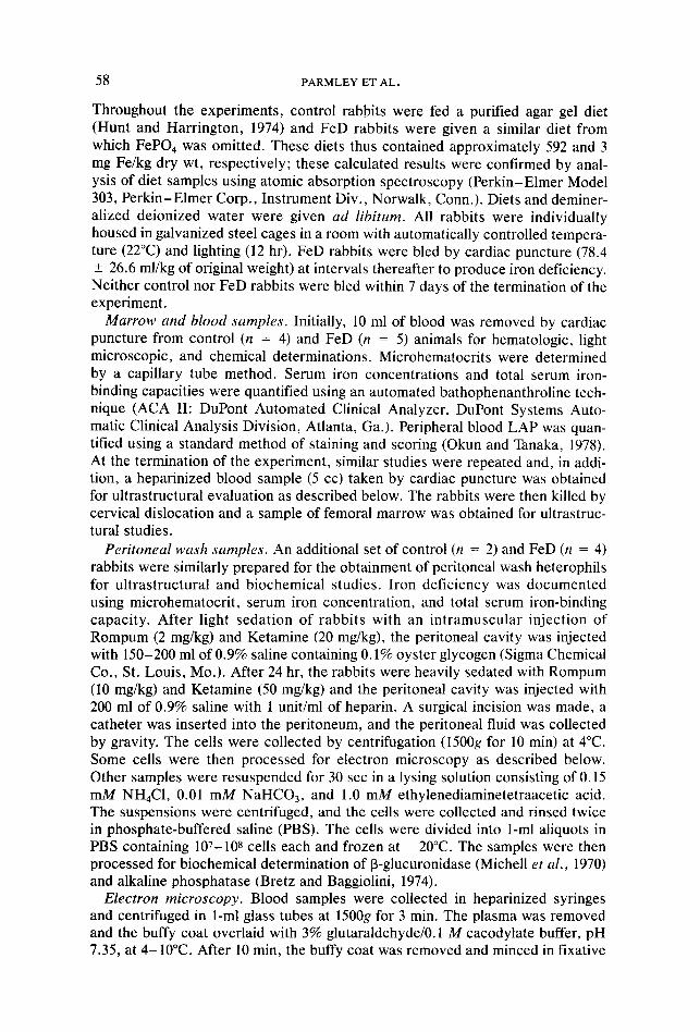
Throughout the experiments, control rabbits were fed a puritied agar gel diet (Hunt and Harrington, 1974) and FeD rabbits were given a similar diet from which FeP04 was omitted. These diets thus contained approximately 592 and 3 mg Fe/kg dry wt, respectively; these calculated results were confirmed by anal- ysis of diet samples using atomic absorption spectroscopy (Perkin-Elmer Model 303, Perkin-Elmer Corp., Instrument Div., Norwalk, Conn.). Diets and deminer- alized deionized water were given ad libitum. All rabbits were individually housed in galvanized steel cages in a room with automatically controlled tempera- ture (22°C) and lighting (12 hr). FeD rabbits were bled by cardiac puncture (78.4 ? 26.6 ml/kg of original weight) at intervals thereafter to produce iron deficiency. Neither control nor FeD rabbits were bled within 7 days of the termination of the experiment.
Marrow and blood samples. Initially, 10 ml of blood was removed by cardiac puncture from control (n = 4) and FeD (n = 5) animals for hematologic, light microscopic, and chemical determinations. Microhematocrits were determined by a capillary tube method. Serum iron concentrations and total serum iron- binding capacities were quantified using an automated bathophenanthroline tech- nique (ACA II: DuPont Automated Clinical Analyzer, DuPont Systems Auto- matic Clinical Analysis Division, Atlanta, Ga.). Peripheral blood LAP was quan- tified using a standard method of staining and scoring (Okun and Tanaka, 1978). At the termination of the experiment, similar studies were repeated and, in addi- tion, a heparinized blood sample (5 cc) taken by cardiac puncture was obtained for ultrastructural evaluation as described below. The rabbits were then killed by cervical dislocation and a sample of femoral marrow was obtained for ultrastruc- tural studies.
Peritoneal wash samples. An additional set of control (n = 2) and FeD (n = 4) rabbits were similarly prepared for the obtainment of peritoneal wash heterophils for ultrastructural and biochemical studies. Iron deficiency was documented using microhematocrit, serum iron concentration, and total serum iron-binding capacity. After light sedation of rabbits with an intramuscular injection of Rompum (2 mg/kg) and Ketamine (20 mg/kg), the peritoneal cavity was injected with 150-200 ml of 0.9% saline containing 0.1% oyster glycogen (Sigma Chemical Co., St. Louis, MO.). After 24 hr, the rabbits were heavily sedated with Rompum (10 mg/kg) and Ketamine (50 mg/kg) and the peritoneal cavity was injected with 200 ml of 0.9% saline with 1 unit/ml of heparin. A surgical incision was made, a catheter was inserted into the peritoneum, and the peritoneal fluid was collected by gravity. The cells were collected by centrifugation (15OOg for 10 min) at 4°C. Some cells were then processed for electron microscopy as described below. Other samples were resuspended for 30 set in a lysing solution consisting of 0.15 mM NH&l, 0.01 mM NaHCO,, and 1.0 mM ethylenediaminetetraacetic acid. The suspensions were centrifuged, and the cells were collected and rinsed twice in phosphate-buffered saline (PBS). The cells were divided into l-ml aliquots in PBS containing 107-108 cells each and frozen at -20°C. The samples were then processed for biochemical determination of B-glucuronidase (Michell et al., 1970) and alkaline phosphatase (Bretz and Baggiolini, 1974).
Electron microscopy. Blood samples were collected in heparinized syringes and centrifuged in l-ml glass tubes at 1500g for 3 min. The plasma was removed and the buffy coat overlaid with 3% glutaraldehyde/O. 1 M cacodylate buffer, pH 7.35, at 4-10°C. After 10 min, the buffy coat was removed and minced in fixative

IRON DEFICIENT PMN 59
with a razor blade and allowed to fix an additional 50 min. Femoral marrow tissue was either directly minced in fixative and allowed to fix for 1 hr, or resuspended in PBS, centrifuged, and resuspended in fixative for 1 hr. Peritoneal wash cell pellets were fixed as a tissue pellet and minced in the fixative or resuspended in the fixative. All samples were then rinsed in 0.1 M cacodylate, 7 g/d1 sucrose, pH 7.35.
In addition to routine morphologic studies, specimens were processed for alka- line phosphatase (Mayahara ef al., 1967); a secondary granule constituent (Wetzel er al., 1967b); peroxidase (Graham and Karnovsky, 1966); a primary granule marker (Dunn et al., 1968); acid phosphatase (Barka and Anderson, 1967); a pri- mary granule constituent (Wetzel et al., 1967b); and vicinal glycol staining after amylase digestion for localization of glycoprotein-rich secondary granules (Fitt- schen et al., 1983). All specimens except those processed for vicinal glycol staining were postfixed in 1% Os04, 0.1 M cacodylate. The specimens were then routinely dehydrated in graded alcohols and propylene oxide and embedded in Spurr low-viscosity medium.
Thin sections (50-70 nm) were obtained with a diamond knife and collected on copper grids for morphologic and cytochemical evaluation of content. Specimens processed for vicinal glycol staining were collected on stainless-steel grids for sequential incubation in periodic acid-thiocarbohydrazide-silver proteinate (PA-TCH-SP) as described previously (Fittschen et al., 1983). Thin sections were counterstained with uranyl acetate and lead citrate, and examined with a Philips 300 electron microscope at an accelerating voltage of 60 kV.
Methanolic uranyl acetate and lead citrate stained marrow late heterophils (containing two or more nuclear lobes with condensed nuclear chromatin) were sequentially photographed (n = 10 micrographs) from each of the first three rabbits studied in FeD and control groups. For morphometric studies, the micro- graphs (7 x 9 in format) were evaluated with a graf/Pen sonic digitizer connected to a Hewlett-Packard 9825 A calculator. The outline of each cell and its nucleus were traced with a sonic pen and the x and y coordinates of successive points were transmitted to a calculator, thus allowing calculation of cytoplasmic and nuclear area (Bishop and Drummond, 1979). In the point counting mode, the number of primary and secondary granules were enumerated. Primary granules were identified by their content of variably extracted osmiophilic material and large size (0.2-0.8 km) and secondary granules by their homogeneous moder- ately dense content and generally smaller diameter (0.1-0.5 km) (Bainton and Farquhar, 1966; Wetzel et al., 1967a). The granule counts were expressed as numbers of granules per square micrometer of cytoplasm. Although only the first three rabbits in each group were evaluated statistically for granule number, quali- tative assessment of heterophil granules using cytochemical and morphologic techniques were performed on samples from all rabbits.
RESULTS
Hematologic Parameters
At the initiation of experiments, control rabbits (n = 4) weighed 2.49 + 0.22 kg and those to be made iron-deficient (n = 5), 2.75 _t 0.36 kg (mean ? SD). Con- trol and FeD rabbits were fed their designated diets for 30.0 + 15.0 and 33.8 2 12.1 days, respectively. Expressed as a percentage of initial weight, control
60 PARMLEY ET AL.
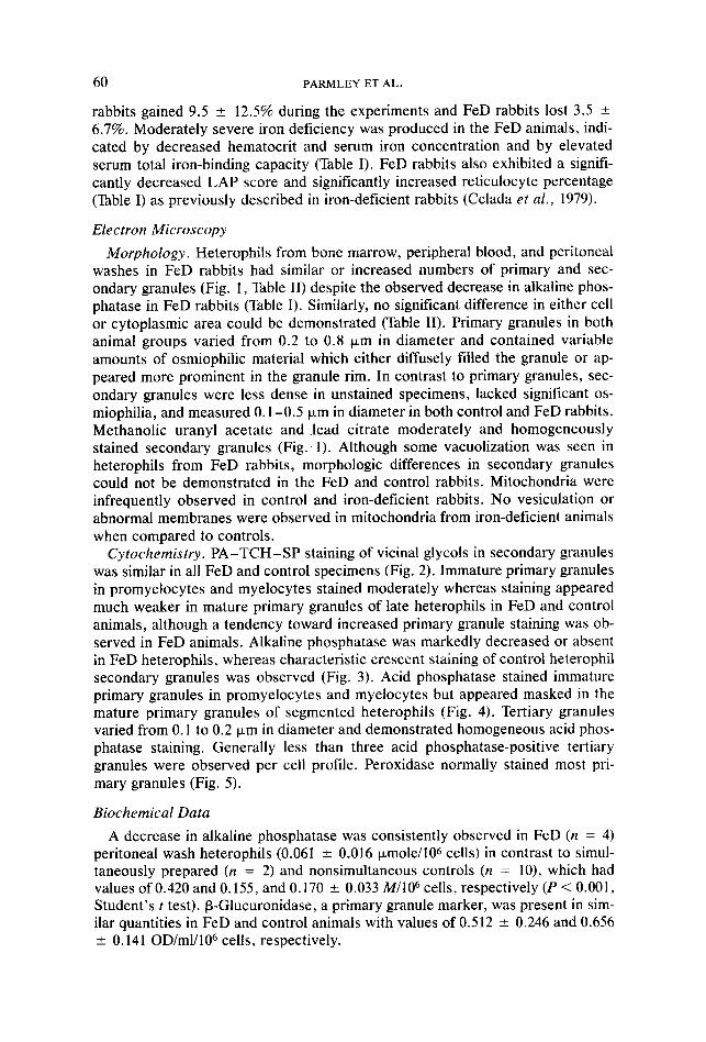
rabbits gained 9.5 + 12.5% during the experiments and FeD rabbits lost 3.5 + 6.7%. Moderately severe iron deficiency was produced in the FeD animals, indi- cated by decreased hematocrit and serum iron concentration and by elevated serum total iron-binding capacity (Table I). FeD rabbits also exhibited a signifi- cantly decreased LAP score and significantly increased reticulocyte percentage (Table I) as previously described in iron-deficient rabbits (Celada et al., 1979).
Electron Microscopy
Morphology. Heterophils from bone marrow, peripheral blood, and peritoneal washes in FeD rabbits had similar or increased numbers of primary and sec- ondary granules (Fig. 1, Table II) despite the observed decrease in alkaline phos- phatase in FeD rabbits (Table I). Similarly, no significant difference in either cell or cytoplasmic area could be demonstrated (Table II). Primary granules in both animal groups varied from 0.2 to 0.8 pm in diameter and contained variable amounts of osmiophilic material which either diffusely filled the granuie or ap- peared more prominent in the granule rim. In contrast to primary granules, sec- ondary granules were less dense in unstained specimens, lacked significant os- miophilia, and measured 0.1-0.5 Frn in diameter in both control and FeD rabbits. Methanolic uranyl acetate and lead citrate moderately and homogeneously stained secondary granules (Fig.. 1). Although some vacuolization was seen in heterophils from FeD rabbits, morphologic differences in secondary granules could not be demonstrated in the FeD and control rabbits. Mitochondria were infrequently observed in control and iron-deficient rabbits. No vesiculation or abnormal membranes were observed in mitochondria from iron-deficient animals when compared to controls.
Cytochemistry. PA-TCH-SP staining of vicinal glycols in secondary granules was similar in all FeD and control specimens (Fig. 2). Immature primary granules in promyelocytes and myelocytes stained moderately whereas staining appeared much weaker in mature primary granules of late heterophils in FeD and control animals, although a tendency toward increased primary granule staining was ob- served in FeD animals. Alkaline phosphatase was markedly decreased or absent in FeD heterophils, whereas characteristic crescent staining of control heterophil secondary granules was observed (Fig. 3). Acid phosphatase stained immature primary granules in promyelocytes and myelocytes but appeared masked in the mature primary granules of segmented heterophils (Fig. 4). Tertiary granules varied from 0.1 to 0.2 Km in diameter and demonstrated homogeneous acid phos- phatase staining. Generally less than three acid phosphatase-positive tertiary granules were observed per cell profile. Peroxidase normally stained most pri- mary granules (Fig. 5).
Biochemical Data
A decrease in alkaline phosphatase was consistently observed in FeD (n = 4) peritoneal wash heterophils (0.061 + 0.016 pmole/l06 cells) in contrast to simul- taneously prepared (n = 2) and nonsimultaneous controls (n = lo), which had values of 0.420 and 0.155, and 0.170 + 0.033 M/lo6 cells, respectively (P < 0.001, Student’s t test). P-Glucuronidase, a primary granule marker, was present in sim- ilar quantities in FeD and control animals with values of 0.512 L 0.246 and 0.656 ? 0.141 OD/m1/106 cells, respectively.

TABL
E I
Hem
atol
ogic
Para
met
ers
of C
ontro
l an
d Iro
n-D
efic
ient
R
abbi
ts
Initi
al
Fina
l
Micr
ohem
atoc
rit
(%)
Seru
m i
ron
conc
entra
tion
@g/
l00
ml)
Seru
m t
otal
iro
n-bi
ndin
g ca
paci
ty
(pg/
lOO
ml)
Satu
ratio
n,
seru
m t
otal
iro
n-bi
ndin
g ca
paci
ty
(%)
Leuk
ocyt
e al
kalin
e ph
osph
atas
e (L
AP)
scor
e R
etic
uloc
ytes
(%
)
Con
trol
(n =
4)
35.5
2
2.1
189.
8 *
68.6
33
0.0
2 20
.5
57.5
T
20.3
15
6.5
2 10
.9
-0
Iron-
defic
ient
(n
=
5)
40.2
2
4.1
193.
8 2
37.3
30
3.8
f 43
.1
65.8
k
20.7
17
5.8
2 29
.4
-
Con
trol
(n =
4)
38.3
k
3.4
202.
8 *
75.4
394.
0 5
149.
6 52
.8
f 17
.0
182.
5 2
20.5
1.
9 *
0.1
Iron-
defic
ient
j;i
(n
=
5)
2
17.6
*
3.2*
E
51.2
”
19.6
**
J 0 75
9.0
-t 81
.9**
E
6.7
2 2.
4*
5 41
.4 ”
14
.3*
17.2
2
4.0*
a N
ot p
erfo
rmed
. *
Valu
e of
P <
0.
0005
by
com
paris
on
to c
ontro
ls
at te
rmin
atio
n of
exp
erim
ent
(Stu
dent
’s t
test
). **
Val
ue
of P
<
0.00
25 b
y co
mpa
rison
to
con
trols
at
term
inat
ion
of e
xper
imen
t (S
tude
nt’s
t te
st).

62 PARMLEY ET AL.
FIG. 1. Primary and secondary granules are abundant in this heterophil from an iron-deficient rabbit. Primary granules (P) are larger and demonstrate increased peripheral staining, whereas sec- ondary granules (S) are smaller and stain homogeneously (enlarged in inset). Some primary granules appear extracted (arrows). Nuclei (N). Bone marrow specimen. Thin section stained with methanolic uranyl acetate and lead citrate. x 20,000: Inset x 50,000.
IRON DEFICIENT PMN
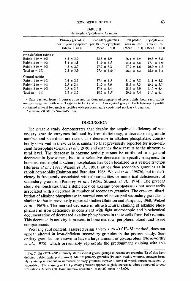
TABLE II Heterophil Cytoplasmic Granules
63
Primary granules Secondary granules Cell profile Cytoplasmic per 10 pm2 cytoplasm per 10 km2 cytoplasm area in km2 area in km2
(Mean f SD) (Mean ? SD) (Mean ? SD) (Mean f SD)
Iron-deficient rabbitsa Rabbit 1 (n = 10) Rabbit 2 (n = 10) Rabbit 3 (n = 10) Total (n = 30)
Control rabbits Rabbit 1 (n = 10) Rabbit 2 (n = 10) Rabbit 3 (n = 10) Total (n = 30)
8.2 rt 3.0 8.4 2 1.8 4.8 5 2.7 7.2 ” 3.0
6.4 +- 2.7 5.5 2 2.4 5.5 2 2.5 5.8 2 2.5
22.8 + 4.9 26.1 f 6.9 19.5 f 5.8 31.9 +- 6.5 25.1 k 3.8 17.1 ” 4.0 27.7 2 6.2 27.9 2 4.6 20.0 2 4.5 27.4 f 6.84* 26.4 k 5.2 18.6 + 5.2
17.4 * 4.5 31.0 f 7.8 21.1 -e 6.8 21.0 f 7.0 28.9 f 9.3 20.2 k 5.7 17.8 +- 4.4 28.6 f 5.0 21.7 k 6.4 18.7 k 5.5* 29.5 2 7.4 21.0 k 6.1
a Data derived from 10 consecutive and random micrographs of heterophils from each rabbit marrow specimen with n = 3 rabbits in FeD and n = 3 in control groups. Each heterophil profile contained at least two nuclear profiles with predominantly condensed nuclear chromation.
* P value <O.OOl by Student’s r test.
DISCUSSION
The present study demonstrates that despite the acquired deficiency of sec- ondary granule enzymes induced by iron deficiency, a decrease in granule number and size does not occur. The decrease in alkaline phosphatase consis- tently observed in these cells is similar to that previously reported for iron-defi- cient heterophils (Celada et al., 1979) and extends those results to the ultrastruc- tural level. The decrease in enzyme activity cannot be attributed to a general decrease in lysosomes, but to a selective decrease in specific enzymes. In humans, neutrophil alkaline phosphatase has been localized in a vesicle fraction (Borgers ef al., 1978; Wilson et al., 1981), rather than secondary granules as in rabbit heterophils (Bainton and Farquhar, 1968; Wetzel et al., 1967b), but its defi- ciency is frequently associated with abnormalities or numerical deficiencies of secondary granules (Parmley et al., 1980a; Strauss et al., 1974). The present study demonstrates that a deficiency of alkaline phosphatase is not necessarily associated with a decrease in number of secondary granules. The crescent distri- bution of alkaline phosphatase in normal control heterophil secondary granules is similar to that in previously reported studies (Bainton and Farquhar, 1968; Wetzel et al., 1967b). The marked decrease in ultrastructural staining of alkaline phos- phatase in iron deficiency is consistent with light microscopic and biochemical documentation of decreased akaline phosphatase in these cells from FeD rabbits. This decrease in activity is present in bone marrow, peripheral blood, and tissue compartments.
Vicinal glycol content, assessed using Thiery’s PA-TCH-SP method, does not appear altered in iron-deficient secondary granules in the present study. Sec- ondary granules are known to have a large amount of glycoprotein (Noseworthy et al., 1973, which presumably represents the predominant staining with this
FIG. 2. PA-TCH-SP intensely stains vicinal glycol groups in secondary granules (S) of this iron- deficient rabbit (enlarged in inset). Mature primary granules (P) stain weakly whereas stronger irreg- ular staining is evident in immature primary granules (arrows), some of which appear extracted or vacuolated. The staining of FeD primary granules appears slightly increased when compared to con- trol rabbits. Nuclei (N). Bone marrow specimen. x 18,000; inset ~45,000.

64 PARMLEY ET AL.
FIG. 3. This metamyelocyte from a control rabbit demonstrates typical, crescent distribution of alkaline phosphatase staining in several secondary granules (S, enlarged in inset). Primary granules (P) lack staining. Staining was markedly decreased or absent in iron-deficient rabbits. Bone marrow spec- imen, not counterstained. x 20,000; inset x 45,000
FIG. 4. Acid phosphatase reactivity is present in a diffuse or rim pattern in primary granules (P) of a myelocyte from this iron-deficient rabbit (similar to control rabbits). Secondary granules (S) lack staining. Bone marrow specimen. Thin section not counterstained. x 15,000.
FIG. 5. Primary granules (P) from this iron-deficient rabbit demonstrate myeloperoxidase reactivity. Although some granules appear to be less reactive, similar variability was seen in control rabbits. Secondary granules (S) lack staining. Nucleus (N). Bone marrow specimen. Thin section not counter- stained. x 13.000.

IRON DEFICIENT PMN 65
method. Vicinal glycol staining does appear to be relatively specific for human neutrophil and rabbit heterophil secondary granules (Fittschen et al., 1983; Parmley et al., 1980b). The normal-to-increased quantities of positively stained granules further confirms morphologic observations in iron deficiency.
Primary granules of iron-deficient heterophils also do not appear to be de- creased in number in comparison to control cells. This is consistent with the relatively normal amount of P-glucuronidase observed in these cells in this study and the presence of acid phosphatase and peroxidase observed cytochemically. Previous studies have suggested that some deficiency in heme-containing myelo- peroxidase may be present (Mackler et al., 1984; Sinha and Swarup-Mitra, 1981). Our failure to document this with ultrastructural benzidine staining may be re- lated to the increased sensitivity of the technique, which results in intense staining even when small amounts of heme are present. The present study dem- onstrates that if enzymatic deficiency is present in these primary granules, the deficiency is not expressed by a decrease in granule number.
The tendency of increased granules per unit area observed in iron-deficient animals may be comparable to an increase in the hepatocyte mitochondrial com- partment (although largely due to an increase in organelle size) in iron-deficient animals despite a decrease in their cytochrome enzymes (Dallman and Goodman, 1971). The finding in heterophils conceivably could be a secondary phenomenon related to either marrow environment or hypercellular marrow activity. Alterna- tively, an increase in granules may represent a compensatory mechanism to in- crease granule number when granules are produced with deficient constituents.
Although decreases in granule number and size were not observed, some cyto- plasmic vacuoles were observed. However, even this appeared relatively subtle. Previous ultrastructural and biochemical studies of iron-deficient hepatocytes have shown an increase in mitochondrial vesiculation and a decrease of cy- tochromes A and C (Dallman and Goodman, 1971). The paucity of mitochondria in control and iron-deficient heterophils and the relatively short life span of heter- ophils may account for the lack of a noticeable involvement of this organelle in heterophils.
REFERENCES BAINTON, D. F., and FARQUHAR, M. G. (1966). Origin of granules in polymorphonuclear leukocytes.
Two types derived from opposite faces of the Golgi complex in developing granulocytes. J. Cell.
Biol. 28, 277. BAINTON, D. F., and FARQUHAR, M. G. (1968). Differences in enzyme content of azurophil and specific
granules of polymorphonuclear leukocytes. II. Cytochemistry and electron microscopy of bone
marrow cells. J. Ceil. Bioi. 39, 299. BARKA, T., and ANDERSON, P. T. (1967). “Histochemistry: Theory, Practice, and Bibliography,” p. 238.
Harper & Row, New York. BISHOP, S. P., and DRUMMOND, J. L. (1979). Surface morphology and cell size measurement of isolated
rat cardiac myocytes. J. Mol. Cell. Cnrdiol 11, 423. BORGERS, M., THONE, F., DE CREE, J., and DE COCK, W. (1978). Alkaline phosphatase activity in human
polymorphonuclear leukocytes. Hisrochem. 3. 10, 31. BRETZ, U., and BAGGIOLINI, M. (1974). Biochemical and morphological characterization of azurophil
and specific granules of human neutrophilic polymorphonuclear leukocyte3. J. Cell. Biol. 63, 251. 1 CELADA, A., HERREROS, V., PUGIN, P., ~~~RUDOLF, H. (1979). Reduced leukocyte alkaline phosphatase
activity and decreased NBT reduction test in induced iron deficiency anaemia in rabbits. Brit. J. Haematol. 43, 457.
CHANDRA, R. K. (1973). Reduced bactericidal capacity of polymorphs in iron deficiency. Arch. Dis. Child. 48, 864.
DALLMAN, P. R., and GOODMAN, J. R. (1971). The effects of iron deficiency on the hepatocyte: A bio- chemical and ultrastructural study. J. Cell. Biol. 48,. 79.

66 PARMLEY ET AL.
DE VET, B. J., and TEN HOOPEN, C. H. (1978). Lactoferrin in human neutrophilic polymorphonuclear leukocytes in relation to iron metabolism. A’cfa Med. &and. 203, 197.
DUNN, W. B., HARDIN, J. H., and SPICER, S. S. (1968). Ultrastructural localization of myeloperoxidase in human neutrophil and rabbit heterophil and esoinophil leukocytes. BIood 32, 935.
FITTSCHEN, C., PARMLEY, R. T.. AUSTIN, R. L.. and GRIST. W. M. (1983). Vicinal glycol-staining iden- tifies secondary granules in human normal and Chediak-Higashi neutrophils. Anal. Rec. 205, 301.
GRAHAM, R. C., JR., and KARNOVSKY, M. J. (1966). The early stages of absorption of injected horse- radish peroxidase in the proximal tubules of mouse kidney: Ultrastructural cytochemistry by a new technique. .I. Histochem. Cytochem. 14, 291.
HUNT, C. E. and HARRINGTON, D. D. (1974). Nutrition and nutritional diseases of the rabbit. It1 “The Biology of the Laboratory Rabbit” (S. H. Weisbroth, R. E. Flatt, and A. L. Kraus, eds), Chapt. 16, p. 403. Academic Press, New York.
MACKLER, B., PERSON, R., OCHS, H., and FINCH, C. A. (1984). Iron deficiency in the rat: Effects on neutrophil activation and metabolism. Pediatr. Res. 18, 549.
MAYAHARA, H., HIRANO. H. M., SAITO, T., and OGAWA, K. (1967). The new lead citrate method for the ultracytochemical demonstration of activity of nonspecific alkaline phosphatase (orthophosphoric monoester phosphohydrolase). Histochemie 11, 88.
MICHELL, R. H., KARNOVSKY, M. J., and KARNOVSKY. M. L. (1970). The distributions of some granule- associate enzymes in guinea-pig polymorphonuclear leukocytes. Biochem. J. 116, 207.
NOSEWORTHY, J., SMITH. G. H., HIMMELHOCK, S. R., and EVANS, W. H. (1975). Protein andglycoprotein electrophoretic patterns of enriched fractions of primary and secondary granules from guinea pig polymorphonuclear leukocytes. J. Cell. Biol. 65, 577.
OKUN, D. B., and TANAKA, K. R. (1978). Leukocyte alkaline phosphatase. Amer. .I. Hematol. 4, 293. PARMLEY, R. T.. CRIST, W. M., RAGAB, A. H.. BOXER, L. A., MALLUH, A., LUI, V. K. and DARBY, C. P.
(1980a). Congenital dysgranulopoietic neutropenia: Clinical, serologic, ultrastructural, and in vitro proliferative characteristics. Blood 56, 465.
PARMLEY, R. T., EGUCHI, M., SPICER, S. S., ALVAREZ, C. J., and AUSTIN, R. L. (1980b). Ultrastructural cytochemistry and radioautography of complex carbohydrates in heterophil granulocytes from rabbit bone marrow. J. Histochem. Cytochem. 28, 1067.
PRASAD, J. S. (1979). Leukocyte function in iron-deficiency anemia. Amer. J. Clin. Nutr. 32, 550. SINHA, A. K., and SWARUP-MITRA, S. (1981). Myeloperoxidase activity in polymorphonuclear neutro-
phils in nutritional anaemia. Trans. R. Sot. Trap. Med. Hyg. 75, 758 (Letter). STRAUSS, R. G., BOVE, K. E., JONES, J. F., MAUER, A. M.. and FULGINITI, V. A. (1974). An anomaly of
neutrophil morphology with impaired function. N. Eng(. J. Med. 290, 478. VAN HEERDEN. C., OOSTHUIZEN, R., VAN WYK, H., PRINSLOO, I?, and ANDERSON, R. (1981). Evaluation
of neutrophil and lymphocyte function in subjects with iron deficiency. S. Afr. Med. J. 59, 111. WETZEL, B. K., HORN, R. G., and SPICER, S. S. (1967a). Fine structural studies on the development of
heterophil, eosinophil. and basophil granulocytes in rabbits. Lab. Invest. 16, 349. WETZEL, B. K., SPICER, S. S., and HORN, R. G. (1967b). Fine structural localization of acid and alka-
line phosphatases in cells of rabbit blood and bone marrow. J. Histochem. Cytochem. 15, 311. WILSON, P D., RUSTIN, G. J., and PETERS, T. J. (1981). The ultrastructural localization of human neu-
trophil alkaline phosphatase in normal individuals during pregnancy and in patients with chronic granulocytic leukaemia. Histochem. J. 13, 31.
YETGIN, S., ALTAY, C., CILVIC, G., and LALELI, Y. (1979). Myeloperoxidase activity and bactericidal function of PMN in iron deficiency. Acfa Haematol. 61, 10.