Ultrastructural morphology of leg cuticle derivatives useful for phylogenetic study of Neuropterida...
14
_____________________________________________________ Proceedings of the Tenth International Symposium on Neuropterology. Piran, Slovenia, 2008. Devetak, D., Lipovšek, S. & Arnett, A.E. (eds). Maribor, Slovenia, 2010. Pp. 287–300. ___________________________________________________________________________ Ultrastructural morphology of leg cuticle derivatives useful for phylogenetic study of Neuropterida (Insecta: Megaloptera, Neuroptera): preliminary report Tatyana S. Vshivkova 1 & Vladimir N. Makarkin 1,2 1 Institute of Biology and Soil Sciences, Far Eastern Branch of Russian Academy of Sciences, Vladivostok 690022, Russia; E-mail: [email protected] 2 E-mail: [email protected] Abstract. Ultrasculpture of leg surface and cuticle derivatives such as spinules, sensilla, and spurs were investigated with SEM in the Neuropterida families Corydalidae and Sialidae (Megaloptera); Ascalaphidae, Chrysopidae, Dilaridae, Hemerobiidae, Ithonidae, Mantispidae, Myrmeleontidae, Nemopteridae, Nevrorthidae, Osmylidae, Polystoechotidae, Psycho- psidae, and Sisyridae (Neuroptera). These characters may provide useful phylogenetic information for Neuropterida. The polarity of twenty-five character states of the tibio-tarsal area of the hind leg is preliminary determined. True spurs were not detected in Sisyridae examined. Key words: Neuropterida, Megaloptera, Neuroptera, SEM, cuticle derivatives, microsculpture (texture) of leg surface, external morphology, spinules, sensilla, spines, spurs Introduction Characters of leg cuticle derivatives have recently been found to be useful in phylogenetic analyses of some Holometabolian orders (Basibuyuk & Quicke, 1995; Basibuyuk et al., 2000; Baszio & Richter, 2002; Vshivkova et al. 2007, 2009). The possible value of the ultrastructure morphology of sensilla, spines, and spurs is also obvious for palaeontology, palaeoecology, and taxonomy, which often use insect body fragments. However, neuropterologists’ attention has been almost exclusively concentrated to the presence, number, shape and size of spurs. These characters have been discussed at least passing mention in many publications on Neuroptera (e.g., Tillyard, 1919; Tjeder, 1959, 1960, 1986; Hölzel, 1975, 1999; New, 1981, 1983, 1984a, 1985a,b; Penny, 1983, 1996; Mansell, 1985, 1990, 1992; Stange, 1989, 2008; Brooks & Barnard, 1990; Willmann, 1990; Oswald, 1993, 1998; Aspöck & Aspöck, 1997; Engel, 2002; Archibald & Makarkin, 2004; Penny & Winterton, 2007; Miller, 2008). However, apart from these brief instances, leg cuticle vestiture and the external morphology of spurs, spines, and sensilla have received very little attention in Neuropterida. We know only few references where they have been treated in any detail. For example, Killington (1936) mentioned that tibial spurs of Osmylidae “are peculiar in being densely clothed with stiff hairs.” Poivre (1978, 1981) described and schematically illustrated the tibial spur vestiture in some species of Mantispidae. Further, the ultrastructure of leg sensilla with regards to vibration communication in Neuropterida (particularly Chrysopidae) has received some attention (e.g., Devetak, 1998; Devetak et al., 1996, 2004). Here, we report preliminary results of a study of the external morphology of leg cuticle derivatives in the majority of the neuropterid families, mainly to determine any phylogenetic significance of variation. We have paid particular attention to the tibio-tarsal area of the hind legs. ISBN 978-961-6657-16-7 © 2010 Faculty of Natural Sciences and Mathematics UM
-
Upload
independent -
Category
Documents
-
view
1 -
download
0
Transcript of Ultrastructural morphology of leg cuticle derivatives useful for phylogenetic study of Neuropterida...
_____________________________________________________
Proceedings of the Tenth International Symposium on Neuropterology.
Piran, Slovenia, 2008. Devetak, D., Lipovšek, S. & Arnett, A.E. (eds).
Maribor, Slovenia, 2010. Pp. 287–300.
___________________________________________________________________________
Ultrastructural morphology of leg cuticle derivatives useful for phylogenetic study of Neuropterida (Insecta: Megaloptera,
Neuroptera): preliminary report
Tatyana S. Vshivkova1 & Vladimir N. Makarkin1,2
1Institute of Biology and Soil Sciences, Far Eastern Branch of Russian Academy of Sciences, Vladivostok 690022, Russia;
E-mail: [email protected] 2E-mail: [email protected]
Abstract. Ultrasculpture of leg surface and cuticle derivatives such as spinules, sensilla, and spurs were investigated with
SEM in the Neuropterida families Corydalidae and Sialidae (Megaloptera); Ascalaphidae, Chrysopidae, Dilaridae,
Hemerobiidae, Ithonidae, Mantispidae, Myrmeleontidae, Nemopteridae, Nevrorthidae, Osmylidae, Polystoechotidae, Psycho-
psidae, and Sisyridae (Neuroptera). These characters may provide useful phylogenetic information for Neuropterida. The
polarity of twenty-five character states of the tibio-tarsal area of the hind leg is preliminary determined. True spurs were not
detected in Sisyridae examined.
Key words: Neuropterida, Megaloptera, Neuroptera, SEM, cuticle derivatives, microsculpture (texture) of leg surface,
external morphology, spinules, sensilla, spines, spurs
Introduction
Characters of leg cuticle derivatives have recently been found to be useful in phylogenetic analyses of some
Holometabolian orders (Basibuyuk & Quicke, 1995; Basibuyuk et al., 2000; Baszio & Richter, 2002; Vshivkova
et al. 2007, 2009). The possible value of the ultrastructure morphology of sensilla, spines, and spurs is also
obvious for palaeontology, palaeoecology, and taxonomy, which often use insect body fragments. However,
neuropterologists’ attention has been almost exclusively concentrated to the presence, number, shape and size of
spurs. These characters have been discussed at least passing mention in many publications on Neuroptera (e.g.,
Tillyard, 1919; Tjeder, 1959, 1960, 1986; Hölzel, 1975, 1999; New, 1981, 1983, 1984a, 1985a,b; Penny, 1983,
1996; Mansell, 1985, 1990, 1992; Stange, 1989, 2008; Brooks & Barnard, 1990; Willmann, 1990; Oswald, 1993,
1998; Aspöck & Aspöck, 1997; Engel, 2002; Archibald & Makarkin, 2004; Penny & Winterton, 2007; Miller,
2008).
However, apart from these brief instances, leg cuticle vestiture and the external morphology of spurs, spines, and
sensilla have received very little attention in Neuropterida. We know only few references where they have been
treated in any detail. For example, Killington (1936) mentioned that tibial spurs of Osmylidae “are peculiar in
being densely clothed with stiff hairs.” Poivre (1978, 1981) described and schematically illustrated the tibial spur
vestiture in some species of Mantispidae. Further, the ultrastructure of leg sensilla with regards to vibration
communication in Neuropterida (particularly Chrysopidae) has received some attention (e.g., Devetak, 1998;
Devetak et al., 1996, 2004).
Here, we report preliminary results of a study of the external morphology of leg cuticle derivatives in the
majority of the neuropterid families, mainly to determine any phylogenetic significance of variation. We have
paid particular attention to the tibio-tarsal area of the hind legs.
ISBN 978-961-6657-16-7 © 2010 Faculty of Natural Sciences and Mathematics UM

Material and methods
Three species of Megaloptera (two families) and 15 species of Neuroptera (13 families) from Entomological
Collections of the Institute of Biology and Soil Sciences, Russia (IBSS) were studied using scanning electron
microscopy: MEGALOPTERA: Corydalidae: Chauliodinae gen. sp. (Thailand), Protohermes martynovae
Vshivkova, 1995 (Russian Far East: Primorye); Sialidae: Sialis sp. (Primorye); NEUROPTERA: Ascalaphidae:
Ascalohybris subjacens (Walker, 1853) (Japan: Kyusyu), Libelloides sibiricus (Eversmann, 1850) (Primorye);
Chrysopidae: Chrysopa intima McLachlan, 1893 (Russian Far East: Khabarovski Krai), Nineta alpicola
Kuwayma, 1936 (Primorye); Dilaridae: Dilar septentrionalis Navás, 1912 (Primorye); Hemerobiidae: Micromus
paganus (Linnaeus, 1767) (Russia: Transbaikalia); Ithonidae: Ithone fusca Newman, 1838 (Australia);
Mantispidae: Mantispa floridana Banks, 1897 (USA: Florida); Myrmeleontidae: Dendroleon jezoensis Okamoto,
1910 (Primorye); Nemopteridae: Nemoptera sinuata Olivier, 1811 (Armenia); Nevrorthidae: Nipponeurorthus
pallidinervis Nakahara, 1958 (Japan: Kyusyu); Osmylidae: Lysmus harmandinus (Navas, 1910) (Primorye);
Polystoechotidae: Polystoechoetes punctata (Fabricius, 1793) (USA: California); Psychopsidae: Balmes sp.
(China); Sisyridae: Climacia areolaris (Hagen, 1861) (USA: Florida).
Both dry pinned specimens and ethanol preserved material were employed. The hind legs were removed (the
right hind legs only used for microphotographs) and transferred to 98% ethanol, rinsed once, then air dried and
mounted on a microscope stub prior to sputter coated with gold/platinum. Specimens were examined using a
ZEISS EVO40 Scanning Electron Microscope.
The terminology used here for the general leg structures and derivatives follows that of Snodgrass (1993); and
for the details of sensilla, spines, and spurs follows Altner (1977), Hallberg and Hansson (1999), Baszio and
Richter (2002), and Vshivkova et al. (2007). We propose the following new terms describing leg cuticle:
peritheca, the circular adjoining cuticle around the theca (the opening in the cuticle from which the sensillum is
raised), which is either elevated above the surrounded cuticle (mound-like peritheca) or flat with it; and
perithecal process, for the thorn-like outgrowth of the peritheca. For the purposes of this paper, the terms
ventral, dorsal, and lateral are used with respect to the leg, i.e., ventral and dorsal indicate the lower and upper
side of the leg derivatives relative to the surface of tibia and tarsus, lateral refers to the sides of the derivatives.
SEM was used to examine the microstructure of hind leg tibio-tarsal regions (Fig. 1), in particular, the apical
(distal) tibial area, and the basal basitarsal area (Figs. 2, 3). The cuticle surface of some of the basitarsal area
near the tibial spur attachment is often very different from the surrounded cuticle in having a denser, ribbed,
spinulate, or wart-like vestiture, and it is termed here the basitarsal rough area or basitarsal pad (Figs. 5, 6).
Results and discussion
I. Ultramorphology of the cuticle surface (surface texture)
The outer surface of the cuticle, the epicuticula, is the thin non-cellular layer. Its texture throughout the tibio-
tarsal area including spines and spurs is seldom plain, smooth or bare (Figs. 3, 5); it often has a wavy
(corrugated) (Fig. 4) or wrinkled structure (Figs. 32, 33, 37, 45). It bears various irregular or regular ribs or
ridges (Fig. 11), scale- or tile-like (polygonal) structures (Figs. 39, 42), wart- or pimple-like protuberances (Fig.
6), or different types of non-cellular (nodules, spinules, small spines) or cellular projections such as multicellular
(spines, or spine-like structures) or unicellular (different sensilla) processes.
The cuticle of the basitarsal rough area (basal basitarsus) strongly differs from that of other portion of the tibio-
tarsal area, particularly characterized by a denser cover of non-cellular formations, which consists of ordinary
(Fig. 32) or modified spinules (Figs. 3, 7–9, 36, 44), ribs (Fig. 10), or warts (which tend to be scales distally)
(Figs. 5, 6).
II. Spinules
Spinules (or "microtrichia") are non-cellular, non-innervated, non-socketed, and non-articulated minute cuticle
processes. Three types of spinules are distinguished according their location in tibio-tarsal area: (1) ordinary
spinules, (2) spinules of the basitarsal rough area, and (3) spur spinules.
Ordinary spinules. The leg surface is covered with ordinary spinules, which usually spread uniformly throughout
the surface (Figs. 4, 21). The ordinary spinules may completely cover the entire tibio-tarsal area (e.g., Mega-
288
loptera, Dilaridae, Mantispidae, Nevrorthidae, Psychopsidae, Sisyridae; Fig. 2), or partially disappear in the
apical (distal) portion of the tibia (e.g., Chrysopidae, Hemerobiidae; Fig. 7), or are absent in most portion of tibia
and tarsus (e.g., Ascalaphidae, Ithonidae, Myrmeleontidae, Nemopteridae, Osmylidae, Polystoechotidae; Figs. 5,
10, 31). They are trichoid (many neuropterids; Figs. 3, 4, 24, 25) or thorn-like structures (e.g., Dilaridae; Figs.
21–23). The external morphology of ordinary spinules is usually the same throughout entire tarso-tibial area.
Basitarsal rough area spinules. These spinules may be trichoid (Figs. 3, 43), spine-like or pyramidal-like
projections with a short or long tapering apex (Figs. 7, 8) or flattened, triangle-like with a weakly or strongly
protruding apex (Fig. 9). Spinules of the basitarsal rough area are the same as the ordinary spinules only in
Megaloptera and Nevrorthidae (Nipponeurorthus pallidinervis) (Fig. 3); these spinules differ in other
neuropterids examined. They are usually weaker developed at the base of the basitarsal rough area and become
longer in the central and apical portions.
Spur spinules. Spinules on the spur surface vary from simple needle-like projections (e.g., Sialidae; Figs. 34, 35)
to the very developed scale-like outgrowths (e.g., Nevrorthidae, Osmylidae, Psychopsidae; Figs. 47–50). They
cover the entire spur body except the very basal and apical portions. Scale-like spinules may lie tightly against
the spur body (Fig. 47) or at an angle to it, in this case the spurs have a pine cone-like appearance (Figs. 49, 50).
III. Sensilla
All sensilla detected in the tibio-tarsal area were setiform; campaniform, coeloconicum or other non-trichoid
sensilla were not found.
Many different names have been applied to setiform sensilla despite similarity in form and external morphology.
Altner (1977) proposed a classification based on the presence/absence and structure of the pores. According to
this classification, all sensilla may be grouped into three major groups: poreless (NP), wall-pore single-walled
(subtype SW) and wall-pore double-walled (subtype DW), and terminal pore sensilla (TP). The most common
types of setiform sensilla can be defined as (I) trichoid (SWtrh) sensilla with a long shaft, a low number of
sensory cells (1-3), usually with unbranched dendrites, and comparatively thick cuticle with a low pore density;
(II) basiconic (SWbsc) sensilla usually with a short shaft, a low number of sensory cells (1-3), branched
dendrites, and a thin cuticle with high pore density; (III) chaetic (TP) sensilla with a long shaft, an apical pore, 4-
5 sensory cells one of which is mechanosensory and four are gustatory; (IV) poreless chaetic sensilla. Some non-
setiform sensilla are also recognized (Hallberg & Hansson, 1999). The majority of setiform sensilla (single-
walled and poreless) appear to be mechanosensory. Setiform sensilla with a long shaft and an apical pore are
functioning collectively as mechanosensory and gustatory organs. Wall-pore sensilla with numerous pores (e.g.,
sensilla basiconica) are known as chemoreceptors (olfaction, gustation). In spite of such detailed classification,
problem distinguishing types of setiform sensilla still exist because the pores are often not visible even with
SCAN or TEM examination.
Based on size, shape, and external ultramorphology, five types and subtypes of sensilla were identified in the
tibio-tarsal area:
Type A. Sensilla chaetica (perhaps, poreless). These are basic, ordinary sensilla of the tibio-tarsal area in almost
all studied neuropterids except some Ascalaphidae (Fig. 5) and Nemopteridae.
1. Subtype t-ss I: long sensilla, arising from flexible sockets, with the seta trunk gradually tapering and a sharp
apex. They are straight or slightly curved and sit in openings (theca) surrounded by a more or less developed
bulb-like (Figs. 14, 17) or pitcher-like base (peritheca) (Figs. 21, 22) formed by a joint membrane. These sensilla
are distinguished from sensilla t-ss II subtype by larger shaft size (as much as 2-3 times) and larger socket. They
are longitudinally grooved (Figs. 14, 18, 24, 25) with weakly (e.g., Chauliodinae; Fig. 17) or well-developed
ridges; the ridges are usually non-serrate (Figs. 14, 16, 25), or may be slightly notched (Fig. 22), especially
distally. The number of sensillum ridges varies from 6 (Balmes sp.), 10 (Dilar septentrionalis) to 12–16 (e.g.,
Megaloptera; Mantispa floridana, Micromus paganus). Each sensillum ridge is directed from the base to the
apical portion (Figs. 18, 22), sometimes merged with each other (Fig. 23).
The peritheca of sensilla chaetica in some neuropterid families has a spine-like perithecal process, which may be
short (less than the sensillum diameter at its base) (Lysmus harmandinus, Balmes sp., Chrysopa intima) (Figs.
14, 23, 25) or long (longer than the sensillum diameter at its base) (Micromus paganus, Climacia areolaris)
(Figs. 28–31). This perithecal process probably restricts the backward mobility of the sensilla. A similar struc-
289

ture was observed in some Hymenoptera sensilla ("spur of STB socket") (Basibuyuk et al., 2000); however, the
homology of those "spurs" with spine-like perithecal process described here is uncertain.
2. Subtype t-ss II: sharp-tipped, setiform sensilla, which differ from the t-ss I subtype by being shorter and more
slender, and in having a less developed socket.
3. Subtype t-ss III: spine-like, thick, robust, and blunt-tipped sensilla. This type of sensilla appears to be
numerous in Ascalaphidae and Nemopteridae examined, especially on the tarsal surface of Libelloides sibiricus
(Fig. 5). The long, medium and short spine-like sensilla are distinguished by their size.
4. Type B. Sensilla trichodea: stright, pointed, longitudinally grooved (Figs. 10, 14, 15), smaller than sensilla
chaetica distinguishing from the latter by a weakly developed peritheca; these occur rarely, and then in the apical
tibial area. This type is characterized by the absence of a perithecal process, even in those taxa in which the
ordinary chaetic sensilla posses such projection (Fig. 14); they are usually lighter at SEM observation in
comparison with ordinary chaetic sensilla.
5. Type C. Sensilla basiconica: markedly shorter than sensilla chaetica, slightly curved (Figs. 17, 19, 26), peg-
like, blunt-tipped sensilla with an obvious pimple at the apex (Fig. 20). The sensilla wall is longitudinally
grooved and slightly transversally wrinkled; the surface appears to have pores. Such sensilla are rarely found in
the apical tibial area, but more often on tarsal segments.
IV. Spines
Spines are multicellular cuticle processes. Some of them are solidly fixed to the surrounded cuticle (immovable
spines) and some are set in openings surrounded by the perithecal ring and, probably, partially movable
(socketed spines). Immovable spines may independently arise from the epicuticula surface or be a projection of
other structures such a perithecal process of some neuropterid sensilla chaetica (Figs. 29, 30). The tibio-tarsal
socketed spines remind ordinary sensilla chaetica in general structure and external morphology being, however,
thicker and having somewhat different perithecal morphology (Figs. 10, 13, 18). Socketed spines differ from
tibial spurs in having longitudinal ridges and in the absence any scattered spinules or setiform sensilla.
The spines located at the base of apical tibial spurs (usually one or two in number) are termed
falsicalcar/falsicalcarae (or false spurs) (Vshivkova et al., 2007). In Climacia areolaris (Sisyridae) two
falsicalcarae are found to present sitting in well-formed sockets looking like spurs (Fig. 43), while true hind
tibial spurs in this species were not detected.
V. Spurs
Spurs are spine-like, multicellular processes, which are only present on the tibia, therefore they are often named
"tibial spurs". In Neuropterida there are only apical (distal) spurs, while some other holometabolian orders also
possess subapical spur/ spurs. They are located ventrally at the apical edge of the tibia, which is separated from
the first tarsal segment (basitarsus) by a more or less visible joint membrane; the spurs are inserted in the
membrane and often appear as structures roofed by the apical tibial edge. The number of spurs varies from 0 to
2. The spur formula is 2.2.2 if maximal number of spurs is present in each leg.
Tibial spurs are present in almost all families of Neuropterida. We could not detect them in Dilar septentrionalis
(Dilaridae), Nemoptera sinuata (Nemopteridae), and Climacia areolaris (Sisyridae). Any data on the presence/
absence of spurs in Dilaridae are absent in the literature. Small spurs are sometimes present in Nemopteridae
(Hölzel, 1975, 1999). As to Sisyridae, it was widely accepted that spurs are present in this family, i.e., one spur
on the distal end of the prothoracic tibia and two on the distal ends of the meso- and metathorasic tibiae
(Killington, 1936; Parfin and Gurney, 1956; Riek, 1975). However, we found that at least the metathorasic tibiae
of Climacia areolaris have socketed spines (falsicalcarae, see above), not true spurs (Fig. 43). In many
subfamilies, tribes, or genera, spurs are also absent (e.g., Berothidae: Nosybus Navás, Stenobiella Tillyard,
Spermophorella Tillyard, Trichoma Tillyard; Coniopterygidae: Brucheiserinae; Hemerobiidae: Zachobiella
Banks; Ithonidae: Rapisma McLachlan; Myrmeleontidae: Fenestroleon New, tribe Dendroleontini; Nymphidae:
Myiodactylus-group) (Tillyard, 1916; Rick, 1975; Barnard, 1981; New, 1984b).
If two spurs are present, one is anterior (inner) and the other posterior (outer). In many neuropterids, both spurs
are usually of the same size and length (Figs. 3, 34, 44, 48). Sometimes the anterior spur may be larger than the
290
posterior (Fig. 36). Anterior and posterior spurs may be similar in shape or different (e.g., Ascalohybris
subjacens, Libelloides sibiricus). Spurs of most neuropterids are usually straight and conical (Figs. 5, 32, 34, 46),
but, some are slightly curved. Some neuropterids are characterized by very strongly arched spurs with a sub-
basal prominence, i.e., "hooked spurs" (e.g., Myrmeleontidae: Acanthaclisinae) (New, 1985a).
Spur texture may be almost smooth (Chrysopa intima; Fig. 46), slightly (e.g., Micromus paganus; Figs. 44, 45)
or strongly wrinkled in the central (e.g., Corydalidae; Figs. 32, 33) or distal portion (e.g., Polystoechoetes
punctata; Fig. 37). Spur surface may be polygonal (e.g., Ascalaphidae, Fig. 42) or scaled in texture (e.g.,
Ithonidae; Fig. 39). Different projections are developed in other neuropterids: needle-like spinules (Sialidae; Fig.
34); more or less developed scale-like projections tightly adjoining the spur body (Nipponeurorthus
pallidinervis, Fig. 48) or at an angle to the spur body (Balmes sp.; Figs. 49, 50). Spur surface in different
neuropterids usually possesses similar ultramorphology (Figs. 12, 37, 42), however the dorsal surface may have
more developed microsculpture or denser spinulate cover; the apices of spurs are usually devoid of
microprojections. The ventral portion of both spurs in Dendroleon jezoensis (Myrmeleontidae) bear a thin, sharp
longitudinal blade from base to apex (Figs. 40, 41).
Characters potentially useful for phylogenetic purpose
All leg cuticle derivatives (non-cellular, unicellular and multicellular) are directed apicad as if brushed in this
way. The tibio-tarsal areas of Megaloptera and Neuroptera examined have some shared principal features: (1)
development of non-cellular vestiture on the leg cuticle surface; (2) presence of the basitarsal rough area; (3)
presence of numerous longitudinally grooved sensilla chaetica with non-serrate or slightly serrate ridges and
fewer setiform sensilla of other types (trichodea, basiconica); (4) spines possess non-serrate or slightly serrate
longitudinal ridges; and (5) non-sensillate spurs, which lack toothed rim/rims.
The differences in leg derivatives are probably characteristic of families and genera, and therefore carry a
phylogenetic signal. Below, the polarities determined for the twenty-five identified characters found in the tibio-
tarsal area of the hind leg are listed, coded “(0)” for putative primitive (plesiomorphic) state, and “(1)” to “(3)”
for putative advanced (apomorphic) states. This evaluation should be considered preliminary and requires further
investigation of other neuropterid taxa, and other holometabolous orders to confirm its accuracy. Some of
characters are multi-state; however, we do not presently propose any transformation series because of incomplete
data. Nevertheless, these characters may be used as an additional source of phylogenetic information together
with characters of the larvae, genitalia of imagoes (e.g., Aspöck et al., 2001), and molecular data (e.g., Haring &
Aspöck, 2004).
Cuticle surface texture and non-cellular vestiture
Character 1: Surface texture of tibio-tarsal area.
(0) slightly wrinkled (e.g., Megaloptera; Osmylidae; Figs. 2, 32);
(1) strongly wrinkled, or wavy (corrugated) (e.g., Mantispidae; Fig. 4);
(2) more or less plain, smooth (often non-spinulate) (e.g., Ascalaphidae, Chrysopidae, Dilaridae,
Myrmeleontidae, Polystoechotidae; Figs. 5, 10).
Character 2: Non-cellular vestiture of tibio-tarsal area.
(0) uniformly covered by spinules (e.g., Megaloptera, Dilaridae, Mantispidae, Nevrorthidae, Psychopsidae,
Sisyridae; Figs. 3, 4, 14, 50);
(1) spinules absent at apical (distal) portion of tibia and basitarsal area (e.g., Chrysopidae, Hemerobiidae; Figs. 7,
44);
(2) spinules absent over most of tibio-tarsal area (e.g., Ascalaphidae, Ithonidae, Myrmeleontidae, Nemopteridae,
Osmylidae, Polystoechotidae; Figs. 5, 10).
Character 3: Non-cellular vestiture of basitarsal rough area.
(0) covered with spinules similar to ordinary spinules (e.g., Megaloptera, Nevrorthidae; Figs. 2–3);
(1) covered with spinules different from ordinary spinules (e.g., Chrysopidae, Ithonidae, Hemerobiidae; Figs. 7–
9, 44);
(2) covered by non-spinule formations (ribs, or pimple-like, or scale-like protuberances) (e.g., Osmylidae,
Ascalaphidae; Figs. 6, 11).
Character 4: Spinulate vestiture of basitarsal rough area.
(0) covered with trichoid spinules having similar size with ordinary spinules (e.g., Megaloptera, Nevrorthidae;
Figs. 3, 32);
291

Figs. 1–9. Neuropterida leg surface and cuticle derivatives: spinules, spines, spurs. 1, 2, Chauliodinae gen. sp.; 3, Nipponeurorthus
pallidinervis; 4, Mantispa floridana; 5, 6, Libelloides sibiricus; 7, Nineta alpicola; 8, Ithone fusca; 9, Micromus paganus. 1, tarsus and apex
of tibia, 2, 3, 5, 7, tibio-tarsal area; 4, apical (distal) portion of tibia; 6, 8, 9, proximal portion of basitarsus (basitarsal rough area). atb.a, apical tibial area; btr.ra, basitarsal rough area; crg.s, corrugated surface; ll-spl, spinules of basitarsal area; pl-spl, long trichoid ordinary
spinules; pmp.s, pimpled surface; tb-tr.a, tibio-tarsal area; sp-ss, spine-like sensilla; sm.s, smooth surface; spr, spur; t-spl - short trichoid
ordinary spinules; t-ss, sensilla chaetica.
292
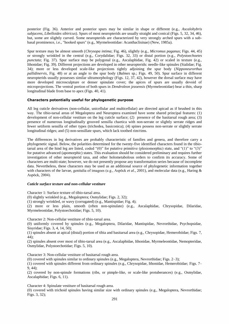
Figs. 10–20. Leg surface, sensilla, and spines. 10–15, Lysmus harmandinus; 16, 17, Chauliodinae gen. sp.; 18–20, Sialis sp.; 10, tibio-tarsal
area; 11, basitarsal rough area; 12, tibial spur; 13, tibial spine; 14, sensilla chaetica and sensillum trichodea; 15, sensillum trichodea; 16,
sensilla chaetica and ordinary spinules; 17, sensillum basiconica and basal portions of sensilla chaetica; 18 – sensilla chaetica and spine; 19, 20, sensillum basiconica and its apical portion. bc-ss, sensillum basiconica; ppr, perithecal process; spl, ordinary spinule; spn, spine; spr,
spur; st-ss, sensillum trichodea; t-ss, sensillum chaetica.
293

Figs. 21–31. Spines and sensilla. 21, 22, Dilar septentrionalis; 23, 27, Balmes sp.; 24, Chrysopa intima; 25, Nipponeurorthus pallidinervis; 26, Libelloides sibiricus; 28, 29, Micromus paganus; 30, 31, Climacia areolaris. 21–25, 28–31, fragments of apical tibial area with spinules,
sensilla; 26, basitarsus with spine-like sensilla and sensilla basiconica; 27, spine. ppr, perithecal process; t-ss, sensillum chaetica.
294
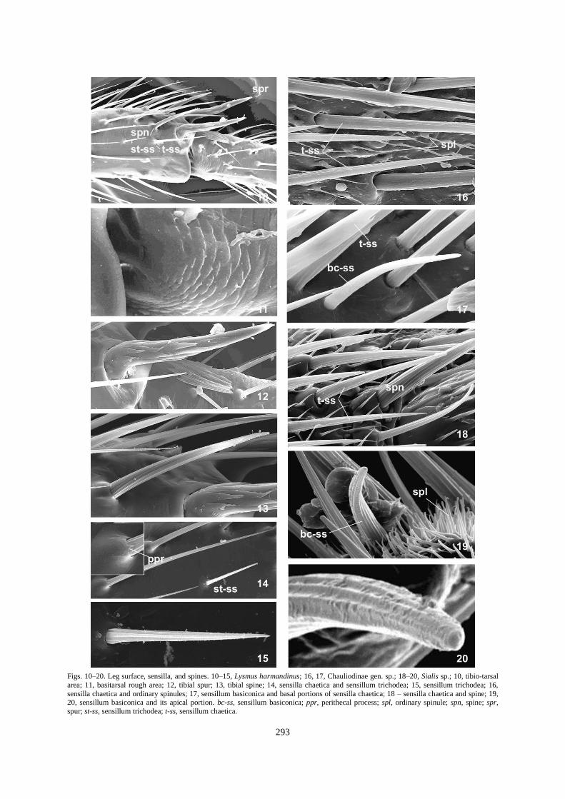
Figs. 32–41. Spurs. 32, 33, Chauliodinae gen. sp.; 34, 35, Sialis sp.; 36, 37, Polystoechotes punctata; 38, 39, Ithone fusca; 40, 41,
Dendroleon jezoensis. 32, 34, 36–38, apical (distal) tibial spurs; 33, 35, medial portion of spurs; 37, 39, apical portion of spurs; 41, blade of spur. rgs.s, wrinkled (rugose) surface of spur.
295
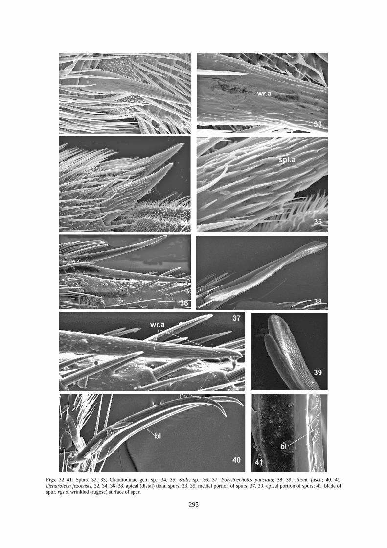
Figs. 42–50. Spurs. 42, Libelloides sibiricus; 43, Climacia areolaris; 44, 45, Micromus paganus; 46, Chrysopa intima; 47, Lysmus harmandinus; 48, Nipponeurorthus pallidinervis; 49, 50, Balmes sp.; 42, 45, medial portion of spur; 43, falsicalcarae; 44, 46–48, 50, tibial
spurs; 49, apical portion of spur.
296

(1) covered with trichoid spinules markedly longer than ordinary spinules (e.g., Sisyridae; Fig. 43);
(2) covered with pyramidal outgrowths with tapering apical projection (e.g., Chrysopidae, Ithonidae; Figs. 7–8);
(3) covered with flattened, triangle-like projections with tapering apex (e.g., Hemerobiidae; Fig. 9).
Character 5: Shape of ordinary spinules.
(0) simple trichoid projection (i.e., thin, uniformly tapering to the apex; e.g., Chrysopidae, Nevrorthidae; Figs.
24, 25);
(1) thorn-like (e.g., Dilaridae; Fig. 21).
Sensilla
Character 6: Sensilla chaetica vestiture of tibio-tarsal area.
(0) distributed irregularly (Figs. 2, 10);
(1) distributed regularly (this character state is present in some mecopterids, not discovered yet in neuropterids
examined).
Character 7: Spine-like sensilla vestiture of tibio-tarsal area.
(0) absent (e.g., Megaloptera; Chrysopidae, Mantispidae);
(1) present, rare (e.g., Polystoechotidae);
(2) present, numerous (may be more numerous than sensilla chaetica) (e.g., Ascalaphidae; Fig. 26).
Character 8: Number of chaetic sensilla ridges.
(0) about 14–16, or more (e.g., Megaloptera; Mantispidae; Fig. 18);
(1) about 10–12 ridges (e.g., Ascalaphidae, Chrysopidae, Dilaridae, Hemerobiidae, Myrmeleontidae,
Nevrorthidae, Osmylidae, Polystoechotidae, Sisyridae; Fig. 9);
(2) less than 10 (e.g., Psychopsidae; Fig. 27).
Character 9: Shape of chaetic sensilla longitudinal ridges.
(0) non-serrate ridges (most neuropterids; Figs. 16, 24);
(1) weakly serrate, especially distally (e.g., Dilaridae; Fig. 22);
(2) strongly serrate (some Trichoptera) (Vshivkova et al., 2007).
Character 10: Shape of chaetic sensilla peritheca.
(0) weakly developed, with slightly prominent wall (e.g., Corydalidae, Fig. 17);
(1) markedly prominent, bulb-like (e.g., Sialidae, most Neuroptera; Figs. 3, 14, 18, 23–25, 28-30);
(2) strongly prominent, pitcher-like (e.g., Dilaridae; Fig. 21).
Character 11: Perithecal process.
(0) absent (e.g., Megaloptera; many Neuroptera; Fig. 17);
(1) present (Psychopsidae, Chrysopidae, Osmylidae, Nevrorthidae, Hemerobiidae, Sisyridae; Figs. 14, 23, 24,
25, 29, 30).
Character 12: Size of perithecal process.
(0) short process (shorter than the sensilla diameter at the base) (e.g., Osmylidae, Chrysopidae; Figs. 14, 24);
(1) long process (longer than the sensilla diameter at the base) (e.g., Hemerobiidae, Sisyridae; Figs. 29–31).
Character 13: Sensilla basiconica.
(0) rare in the tibio-tarsal area (e.g., Corydalidae, most Neuroptera; Fig. 17);
(1) relatively numerous (e.g., Ascalaphidae, Fig. 26).
Socketed spines
Character 14: Longitudinal ridges of socketed spines.
(0) non-serrate (e.g., Megaloptera, most Neuroptera; Fig. 10);
(1) slightly serrate (e.g., Psychopsidae; Fig. 27);
(2) strongly serrate (some Trichoptera) (Vshivkova et al., 2007).
Character 15: Falsicalcarae.
(0) absent;
(1) present (Sisyridae; Fig. 43).
297

Spurs
Character 16: Hind tibial spur.
(0) two spurs present (Megaloptera, most Neuroptera examined);
(1) one spur present (Chrysopidae; Fig. 46);
(2) spurs absent (e.g., Sisyridae, Dilaridae, Nemopteridae examined; Fig. 43).
Character 17: Spur vestiture.
(0) non-spinulate (e.g., Corydalidae; Chrysopidae, Hemerobiidae, Ascalaphidae, Ithonidae; Figs. 32, 38–39, 42,
44–46);
(1) spinulate (e.g., Sialidae; Figs.12, 34, 47–50);
Character 18. Texture of spur surface.
(0) markedly wrinkled, at least in apical portion (e.g., Corydalidae; Polystoechotidae; Figs. 32, 37);
(1) smooth or slightly wrinkled (e.g., Hemerobiidae, Chrysopidae; Figs. 44–45, 46);
(2) polygonal (e.g., Ascalaphidae, Myrmeleontidae; Figs. 5, 40);
(3) tight scale-like (e.g., Ithonidae; Fig. 39).
Character 19. Shape of spur spinules.
(0) needle-like (e.g., Sialidae; Fig. 34);
(3) scale-like (e.g., Osmylidae, Nevrorthidae, Psychopsidae; Figs. 47-50).
Character 20: Spur relative size.
(0) both spurs almost equal size (e.g., Megaloptera; Hemerobiidae, Neurorthidae, Osmylidae, Psychopsidae;
Figs. 3, 44, 47, 48, 50);
(1) anterior spur markedly longer than posterior (e.g., Polystoechotidae; Fig. 36).
Character 21: Texture of the basitarsal rough area (near the tibial spur attachment).
(0) spinulate; shape and size of spinules not strongly differ from ordinary spinules on tarsotibial surface (e.g.,
Megaloptera, Nevrorthidae (Nipponeurorthus pallidinervis) (Fig. 2);
(1) spinulate; spinules differ from ordinary spinules - larger, or with wider basement (Fig. 7);
(2) ribbed (Fig. 11);
(3) covered with pyramid, scale- or wart-like structures (Figs. 5, 6).
Character 22: Spur shape.
(0) straight, conical, (e.g., Hemerobiidae, Chrysopidae; Figs. 44, 46);
(1) at least one spur curved (e.g., Myrmeleontidae; Fig. 40);
(2) both spurs curved (arched) (e.g., Myrmeleontidae: Acanthaclisinae) (New, 1985a).
Character 23: Differentiation of spur dorsal and ventral portions.
(0) uniformly conical spurs with uniform vestiture (e.g., Megaloptera; most Neuroptera; Fig. 50);
(1) dorsal and ventral parts of spur are different in vestiture and shape (e.g., Myrmeleontidae; Fig. 41).
Character 24. Subapical prominence (tooth) of spur.
(0) no subapical tooth (Megaloptera, most Neuroptera);
(1) subapical tooth present (e.g., Myrmeleontidae: Acanthaclisinae) (New, 1985a).
Character 25: Spur blade formation.
(0) any blade or rim/rims absent;
(1) spur with developed blade (e.g., Myrmeleontidae; Figs. 40, 41).
Acknowledgements. We thank Natalia Naryshkina (IBSS) for assistance in the study with SEM, and Bruce Archibald
(Simon Fraser University, Burnaby, Canada) for correction of English.
References Altner, H. 1977. Insektensensillen: Bau- und Funktionsprinzipien. Verh. Dtsch. Zool. Ges., 70: 139–153.
Altner, H. & Prillinger, L. 1980. Ultrastructure of invertebrate chemo-, thermo-, and hygroreceptors and its functional
significance. Int. Rev. Cytol., 67: 69–139.
298

Archibald, S.B. & Makarkin, V.N. 2004. A new genus of minute Berothidae (Neuroptera) from Early Eocene amber of
British Columbia, Canada. Can. Entomol., 136: 59–74.
Aspöck, U., & Aspöck, H. 1997. Studies on new and poorly-known Rhachiberothidae (Insecta: Neuroptera) from subsaharan
Africa. Ann. Naturhistorischen Mus. Wien, 99B: 1–20.
Aspöck, U., Plant, J.D. & Nemeschkal, H.L. 2001. Cladistic analysis of Neuroptera and their systematic position within the
Neuropterida (Insecta: Holometabola: Neuropterida: Neuroptera). Syst. Entomol., 26: 73–86.
Barnard, P. C. 1981. The Rapismatidae (Neuroptera): montane lacewings of the oriental region. Syst. Entomol., 6: 121–136.
Basibuyuk, H.H. & Quicke, D.L.J. 1995. Morphology of the antenna cleaner in the Hymenoptera with particular reference to
non-aculeate families (Insecta). Zool. Scripta, 24 (2): 157–177.
Basibuyuk, H.H., Quicke, D.L.J., Rasnitsyn, A.P. & Fitton, M.G. 2000. Morphology and sensilla of the orbicula, a sclerite
between the tarsal claws, in the Hymenoptera. Ann. Entomol. Soc. Am., 93: 625–636.
Baszio, S. & Richter, G. 2002. Ultrastructural characters of adult leg armature in Trichoptera (Insecta) and their potential for
use in phylogenetic analysis. Aquat. Insects, 24: 1–20.
Brooks, S.J. & Barnard, P.C. 1990. The green lacewings of the world: a generic review (Neuroptera: Chrysopidae). Bull. Brit.
Mus. (Nat. Hist.) Entomol., 59: 117–286.
Devetak, D. 1998. Detection of substrate vibration in Neuropteroidea: a review. Acta Zool. Fenn., 209: 87–94.
Devetak, D., Drašlar, K. & Fišer, I. 1996. Staining of two scolopidial organs in the legs of the green lacewing, Chrysoperla
carnea (Stephens). Znanstvena Revija, 8: 121–128.
Devetak, D., Pabst, M.A. & Lipovšek Delakorda, S. 2004. Leg chordotonal organs and campaniform sensilla in Chrysoperla
Steinmann 1994 (Neuroptera): structure and function. Denisia, 13: 163–171.
Engel, M.S. 2002. A new dustywing (Neuroptera: Coniopterygidae) in Turonian amber from New Jersey, with a
reassessment of Glaesoconis in Neoconian amber from Lebanon. J. Kansas Entomol. Soc., 75: 38–42.
Hallberg, E. & Hansson, B.S. 1999. Arthropod sensilla: Morphology and phylogenetic considerations. Microsc. Res. Techn.,
47: 428–439.
Haring, E. & Aspöck, U. 2004. Phylogeny of the Neuropterida: a first molecular approach. Syst. Entomol., 29: 415–430.
Hölzel, H. 1975. Revision der Netzflügler-Unterfamilie Crocinae (Neuroptera: Nemopteridae). Entomol. Ger., 2: 44–97.
Hölzel, H. 1999. Die nemopteriden (Fadenhafte) Arabiens: ein Beitrag zur Kenntnis der Neuropterida der Arabischen
Halbinsel (Neuropterida: Neuroptera: Nemopteridae). Stapfia, 60: 129–146.
Killington, F.J. 1936. A monograph of the British Neuroptera. Vol. 1. Ray Society, London. 269 pp.
Mansell, M.W. 1985. The ant-lions of southern Africa (Neuroptera: Myrmeleontidae). Introduction and genus Bankisus
Navás. J. Entomol. Soc. S. Afr., 48: 189–212.
Mansell, M.W. 1990. The Myrmeleontidae of southern Africa: tribe Palparini. Introduction and description of Pamares gen.
nov., with four new species (Insecta: Neuroptera). J. Entomol. Soc. S. Afr., 53: 165–189.
Mansell, M.W. 1992. The ant-lions of southern Africa: genus Pamexis Hagen (Neuroptera: Myrmeleontidae: Palparinae:
Palparini). Syst. Entomol., 17: 65–78.
Miller, R.B. 2008. A new genus and species of Brachynemurini from Venezuela (Neuroptera: Myrmeleontidae). Insecta
Mundi, 59: 1–5.
New, T.R. 1981 [1982]. A revision of the Australian Nymphidae (Insecta: Neuroptera). Aust. J. Zool., 29: 707–750.
New, T.R. 1983. Revision of the osmylid subfamilies Porisminae and Eidoporisminae (Insecta: Neuroptera). Aust. J. Zool.,
31: 763–770.
New, T.R. 1984a. Revision of the Australian Ascalaphidae. Aust. J. Zool. Sup., 100: 1–86.
New, T.R. 1984b. Intergeneric relationships in recent Nymphidae. In: Gepp, J., Aspöck, H. & Hölzel, H. (eds.), Progress in
World's Neuropterology. Proceedings of the 1st International Symposium on Neuropterology. Privately printed, Graz,
Austria, pp. 125–131.
299
New, T.R. 1985a. A revision of the Australian Myrmeleontidae (Insecta: Neuroptera). I. Introduction, Myrmeleontini,
Protoplectrini. Aust. J. Zool. Sup., 104: 1–90.
New, T.R. 1985b. A revision of the Australian Myrmeleontidae (Insecta: Neuroptera). II. Dendroleontini. Aust. J. Zool. Sup.,
105: 1–170.
Oswald, J.D. 1993. Phylogeny, taxonomy, and biogeography of extant silky lacewings (Insecta: Neuroptera: Psychopsidae).
Mem. Am. Entomol. Soc., 40: 1–65.
Oswald, J.D. 1998. Rediscovery of Polystoechotes gazullai Navás (Neuroptera: Polystoechotidae). P. Entomol. Soc. Wash.,
100: 389–394.
Parfin, S.I. & Gurney, A.B. 1956. The spongilla-flies, with special reference to those of the western hemisphere (Sisyridae,
Neuroptera). P. U. S. Natl. Mus., 105: 421–529.
Penny, N.D. 1983 [1985]. Neuroptera of the Amazon Basin. Part 9. Albardiinae. Acta Amazonica, 13: 697–699.
Penny, N.D. 1996. A remarkable new genus and species of Ithonidae from Honduras (Neuroptera). J. Kansas Entomol. Soc.,
69: 81–86.
Penny, N.D. & Winterton, S. L. 2007. Rediscovery of the unusual genus Ormiscocerus (Neuroptera: Berothidae: Cyreno-
berothinae). P. Calif. Acad. Sci., (4), 58: 1–6.
Poivre, C. 1978. Morphologie externe comparée de Gerstaeckerella gigantea Enderlein [Planipennia, Mantispidae]. Ann.
Soc. Entomol. Fr. (N.S.), 14: 191–206.
Poivre, C. 1981. Morphologie comparative et systématique de mantispides d'Afrique et d'Europe (Neuroptera, Planipennia).
Thése de docteur d'Université, Université de Nancy I, 256 pp.
Riek, E.F. 1975. On the phylogenetic position of Brucheiser argentinus Navás 1927 and description of a second species from
Chile (Insecta: Neuroptera). Stud. Neotrop. Fauna, 10: 117–126.
Snodgrass, R.E. 1993. Principles of the insect morphology. 2nd ed. Cornell Univ. Press, 667 pp.
Stange, L.A. 1989. Review of the New World Dimarini with the description of a new genus from Peru (Neuroptera:
Myrmeleontidae). Fl. Entomol., 72: 450–461.
Stange, L.A. 2008. A new species of the genus Dendroleon Brauer from Mexico (Neuroptera: Myrmeleontidae). Insecta
Mundi, 54: 1–9.
Tillyard, R.J. 1916. Studies in Australian Neuroptera. No. iv. The families Ithonidae, Hemerobiidae, Sisyridae, Berothidae,
and the new family Trichomatidae; with a discussion of their characters and relationships, and descriptions of new and little-
known genera and species. P. Linn. Soc. N. S. W., 41: 269–332.
Tillyard, R.J. 1919. Studies in Australian Neuroptera. No. 8. Revision of the family Ithonidae, with descriptions of a new
genus and two new species. P. Linn. Soc. N. S. W., 44: 414–437.
Tjeder, B. 1959. Neuroptera-Planipennia. The Lace-wings of Southern Africa. 2. Family Berothidae. In: Hanström, B.,
Brinck, P. & Rudebec, G. (eds), South African Animal Life, Vol. 6. Swedish Natural Science Research Council, Stockholm,
pp. 125–131.
Tjeder, B. 1960. Neuroptera-Planipennia. The Lace-wings of Southern Africa. 3. Family Psychopsidae. In: Hanström, B.,
Brinck, P. & Rudebec, G. (eds), South African Animal Life, Vol. 7. Swedish Natural Science Research Council, Stockholm,
164–209.
Tjeder, B. 1986. A new species of Ascalaphus (Neuroptera, Ascalaphidae) from SW Tunisia. Neuroptera Int., 4: 117–121.
Vshivkova, T.S., Stocks, I.C., Morse, J.C. & Naryshkina, N.N. 2007. Some morphological characters useful for phylogenetic
study of caddisflies: Leg spinules, sensilla, spurs, and spines. In: Bueno-Soria, J., Barba-Alvarez, R. & Armitage, B. (eds.),
Proceedings of the XIIth International Symposium on Trichoptera. The Caddis Press, Columbus, Ohio, pp. 321–334.
Vshivkova, T.S., Loktionov, V.M. & Lelej, A.S. 2010. Ultrastructure of the leg cuticle derivatives and appendages in
Cimbicidae and Pompilidae (Hymenoptera). Far-Eastern Entomologist (in press).
Willmann, R. 1990. The phylogenetic position of the Rhachiberothinae and the basal sister-group relationships within the
Mantispidae (Neuroptera). Syst. Entomol., 15: 253–265.
300