FRONT OF LEG & DORSUM OF FOOT Dr. Gitanjali Khorwal
39
FRONT OF LEG & DORSUM OF FOOT Dr. Gitanjali Khorwal
-
Upload
khangminh22 -
Category
Documents
-
view
2 -
download
0
Transcript of FRONT OF LEG & DORSUM OF FOOT Dr. Gitanjali Khorwal

FRONT OF LEG &
DORSUM OF FOOT
Dr. Gitanjali Khorwal
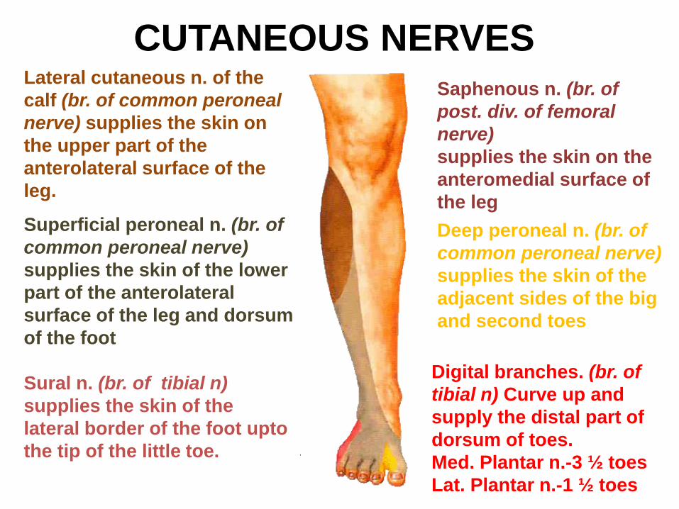
CUTANEOUS NERVESLateral cutaneous n. of the
calf (br. of common peroneal
nerve) supplies the skin on
the upper part of the
anterolateral surface of the
leg.
Superficial peroneal n. (br. of
common peroneal nerve)
supplies the skin of the lower
part of the anterolateral
surface of the leg and dorsum
of the foot
Saphenous n. (br. of
post. div. of femoral
nerve)
supplies the skin on the
anteromedial surface of
the leg
Deep peroneal n. (br. of
common peroneal nerve)
supplies the skin of the
adjacent sides of the big
and second toes
Sural n. (br. of tibial n)
supplies the skin of the
lateral border of the foot upto
the tip of the little toe.
Digital branches. (br. of
tibial n) Curve up and
supply the distal part of
dorsum of toes.
Med. Plantar n.-3 ½ toes
Lat. Plantar n.-1 ½ toes
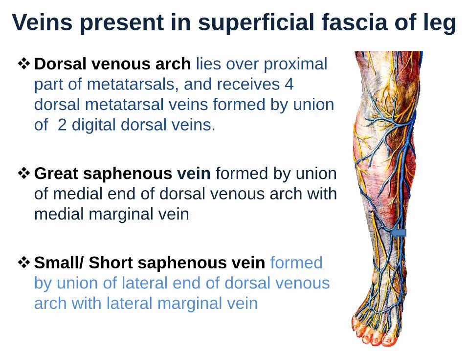
Veins present in superficial fascia of leg
Dorsal venous arch lies over proximal
part of metatarsals, and receives 4
dorsal metatarsal veins formed by union
of 2 digital dorsal veins.
Great saphenous vein formed by union
of medial end of dorsal venous arch with
medial marginal vein
Small/ Short saphenous vein formed
by union of lateral end of dorsal venous
arch with lateral marginal vein
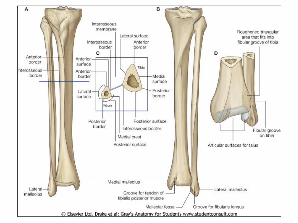

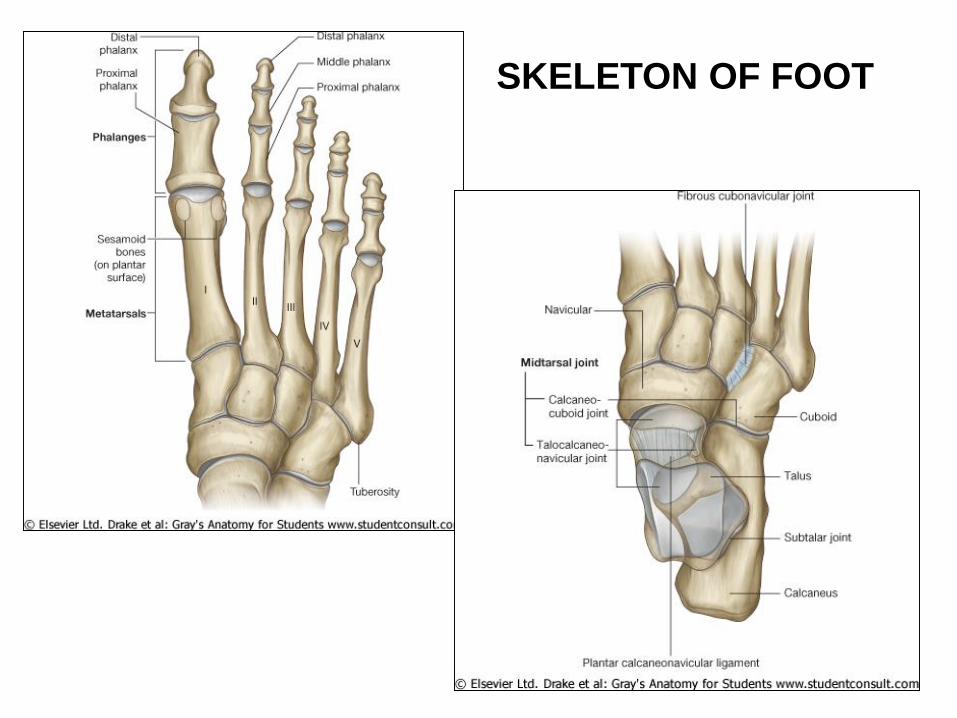
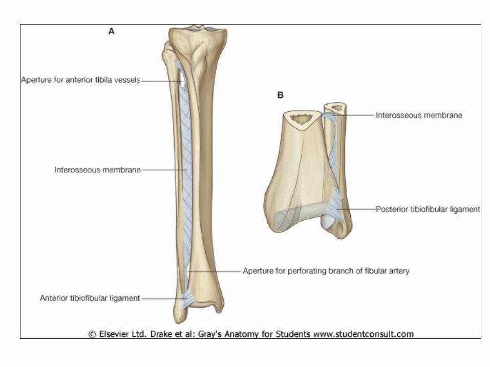
SKELETON OF FOOT

Deep fascia of leg
Anterior
compartment
Lateral
compartment
Posterior
compartment
Anterior
intermuscular
septum
Posterior
intermuscular
septum
Interosseous
membrane
Replaced by periosteum at subcutaneous areas.
Forms intermuscular septae to form three
compartments

RetinaculaDeep fascia is thickened to
form bands.
Retain tendons in place.
•Superior Extensor
Retinaculum
• Inferior Extensor
Retinaculum
•Superior Peroneal
Retinaculum
• Inferior Peroneal
Retinaculum
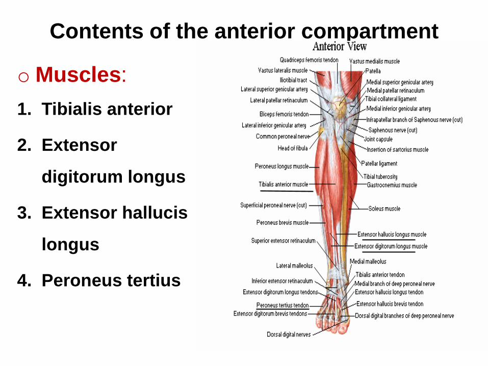
Contents of the anterior compartment
o Muscles:
1. Tibialis anterior
2. Extensor
digitorum longus
3. Extensor hallucis
longus
4. Peroneus tertius
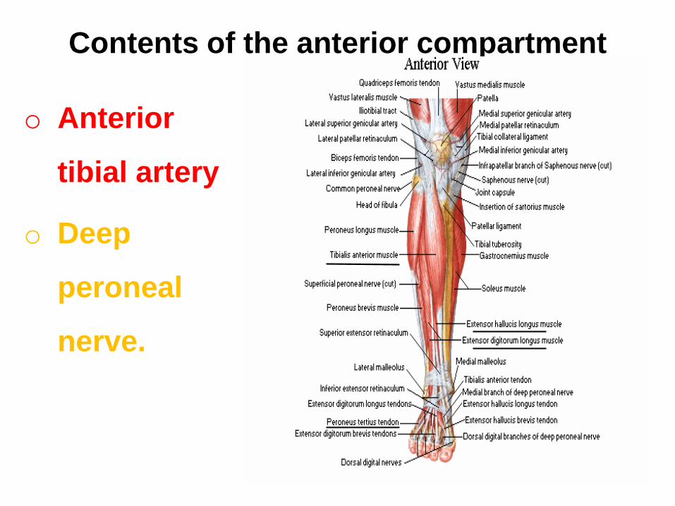
Contents of the anterior compartment
o Anterior
tibial artery
o Deep
peroneal
nerve.
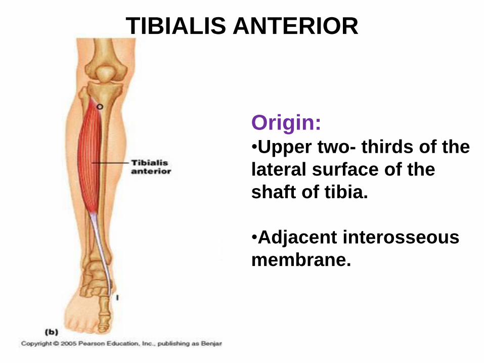
Origin: •Upper two- thirds of the
lateral surface of the
shaft of tibia.
•Adjacent interosseous
membrane.
TIBIALIS ANTERIOR
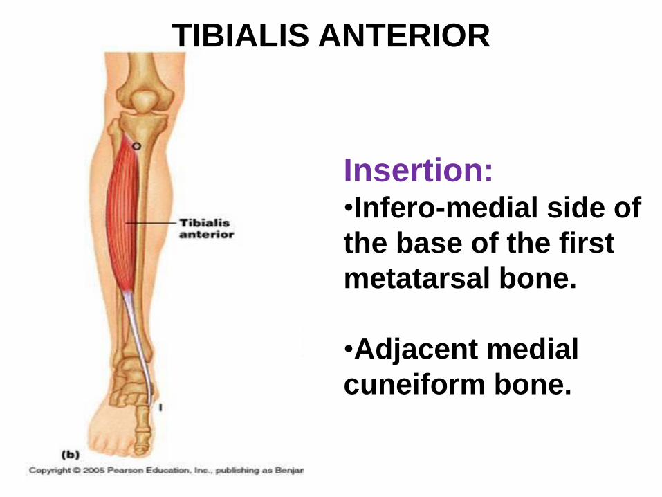
Insertion: •Infero-medial side of
the base of the first
metatarsal bone.
•Adjacent medial
cuneiform bone.
TIBIALIS ANTERIOR
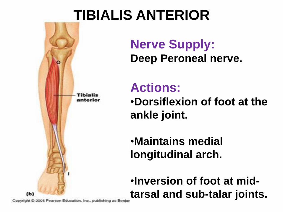
Nerve Supply:Deep Peroneal nerve.
Actions: •Dorsiflexion of foot at the
ankle joint.
•Maintains medial
longitudinal arch.
•Inversion of foot at mid-
tarsal and sub-talar joints.
TIBIALIS ANTERIOR
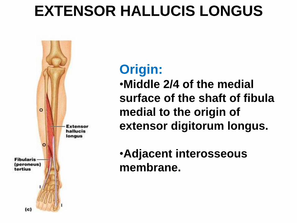
Origin: •Middle 2/4 of the medial
surface of the shaft of fibula
medial to the origin of
extensor digitorum longus.
•Adjacent interosseous
membrane.
EXTENSOR HALLUCIS LONGUS
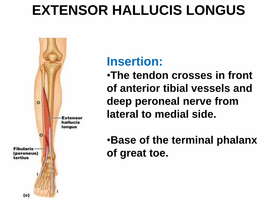
Insertion:•The tendon crosses in front
of anterior tibial vessels and
deep peroneal nerve from
lateral to medial side.
•Base of the terminal phalanx
of great toe.
EXTENSOR HALLUCIS LONGUS
EXTENSOR HALLUCIS LONGUS
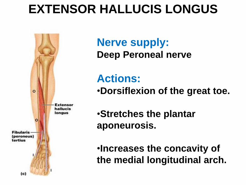
Nerve supply:Deep Peroneal nerve
Actions: •Dorsiflexion of the great toe.
•Stretches the plantar
aponeurosis.
•Increases the concavity of
the medial longitudinal arch.
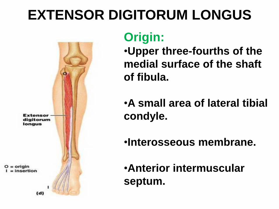
Origin: •Upper three-fourths of the
medial surface of the shaft
of fibula.
•A small area of lateral tibial
condyle.
•Interosseous membrane.
•Anterior intermuscular
septum.
EXTENSOR DIGITORUM LONGUS

Insertion: •Tendon divides into four
digital slips for insertion
into lateral four toes.
•The slips join laterally with
the three digital slips of
extensor digitorum brevis
to form the dorsal digital
expansion.
EXTENSOR DIGITORUM LONGUS
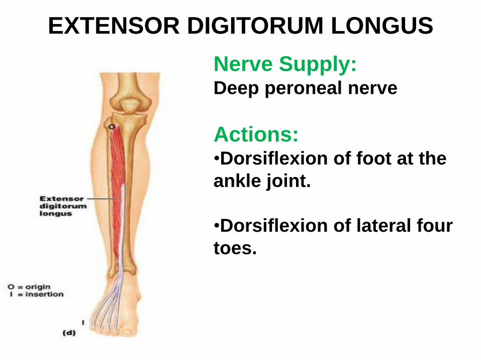
Nerve Supply: Deep peroneal nerve
Actions: •Dorsiflexion of foot at the
ankle joint.
•Dorsiflexion of lateral four
toes.
EXTENSOR DIGITORUM LONGUS
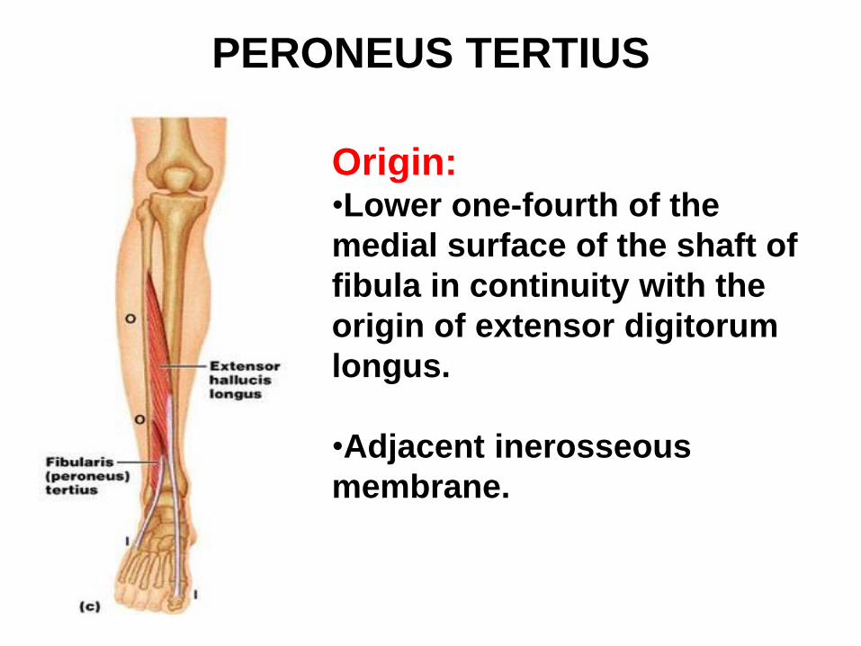
PERONEUS TERTIUS
Origin: •Lower one-fourth of the
medial surface of the shaft of
fibula in continuity with the
origin of extensor digitorum
longus.
•Adjacent inerosseous
membrane.
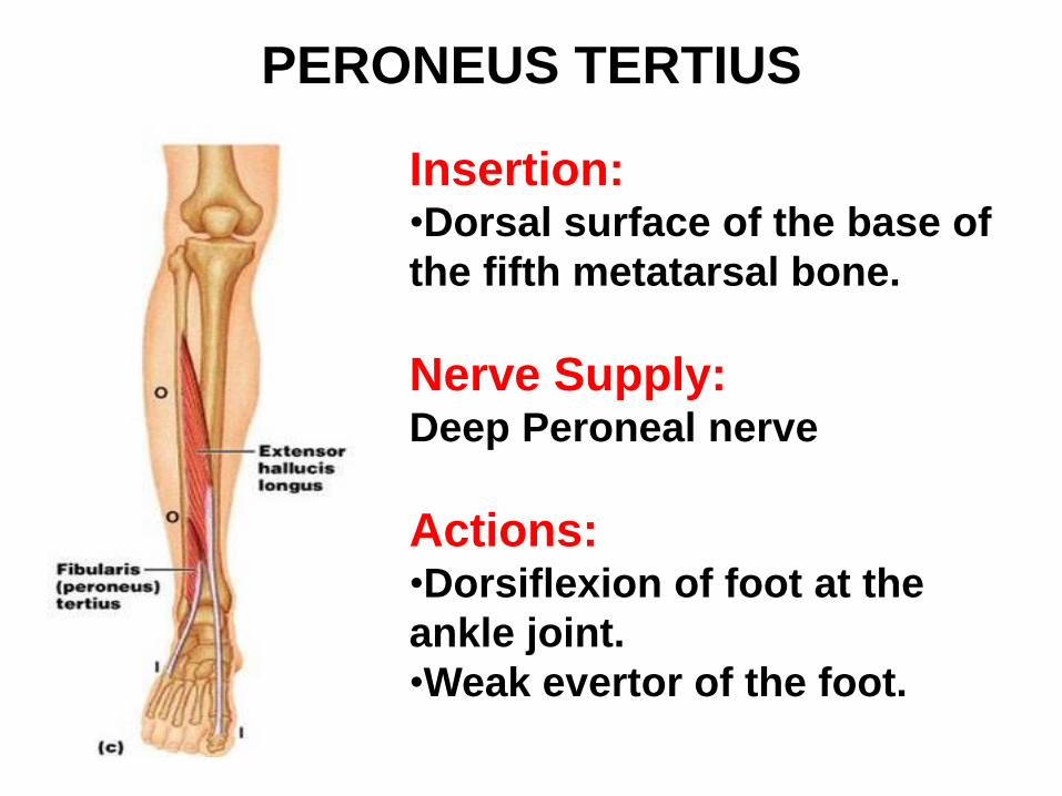
PERONEUS TERTIUS
Insertion: •Dorsal surface of the base of
the fifth metatarsal bone.
Nerve Supply:Deep Peroneal nerve
Actions: •Dorsiflexion of foot at the
ankle joint.
•Weak evertor of the foot.
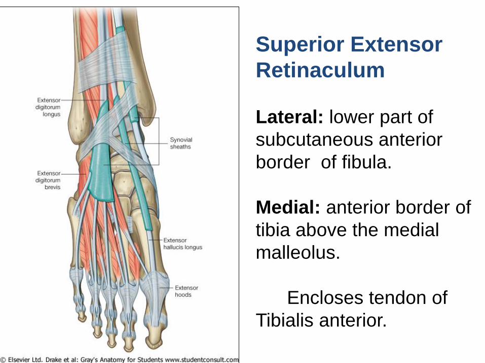
Superior Extensor
Retinaculum
Lateral: lower part of
subcutaneous anterior
border of fibula.
Medial: anterior border of
tibia above the medial
malleolus.
Encloses tendon of
Tibialis anterior.
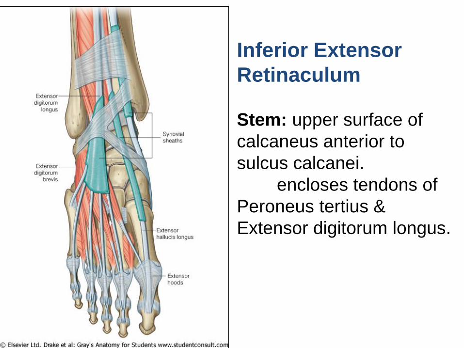
Inferior Extensor
Retinaculum
Stem: upper surface of
calcaneus anterior to
sulcus calcanei.
encloses tendons of
Peroneus tertius &
Extensor digitorum longus.
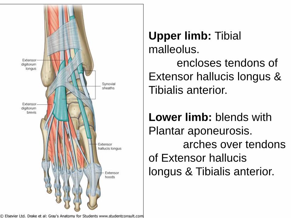
Upper limb: Tibial
malleolus.
encloses tendons of
Extensor hallucis longus &
Tibialis anterior.
Lower limb: blends with
Plantar aponeurosis.
arches over tendons
of Extensor hallucis
longus & Tibialis anterior.
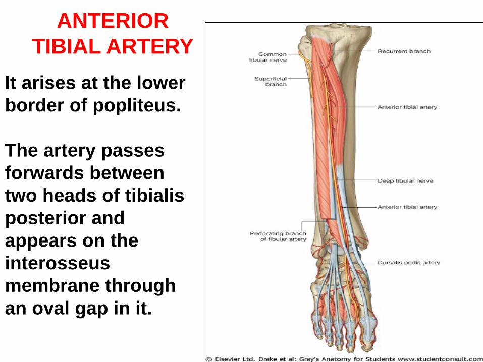
ANTERIOR
TIBIAL ARTERY
It arises at the lower
border of popliteus.
The artery passes
forwards between
two heads of tibialis
posterior and
appears on the
interosseus
membrane through
an oval gap in it.
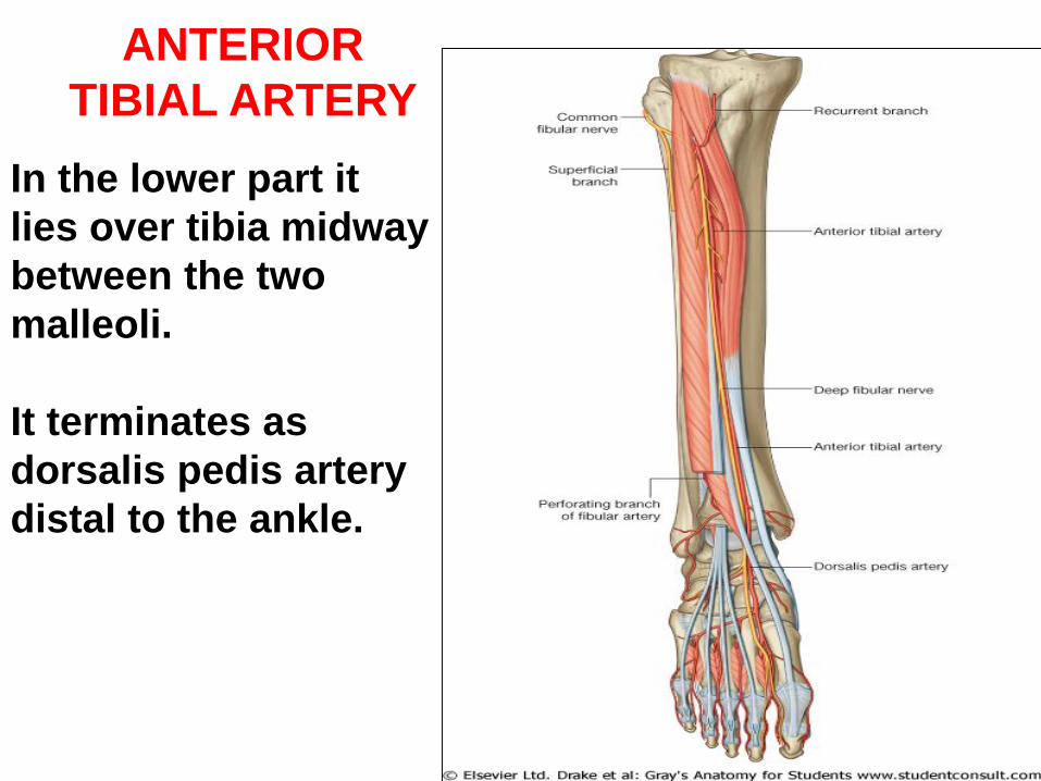
ANTERIOR
TIBIAL ARTERY
In the lower part it
lies over tibia midway
between the two
malleoli.
It terminates as
dorsalis pedis artery
distal to the ankle.
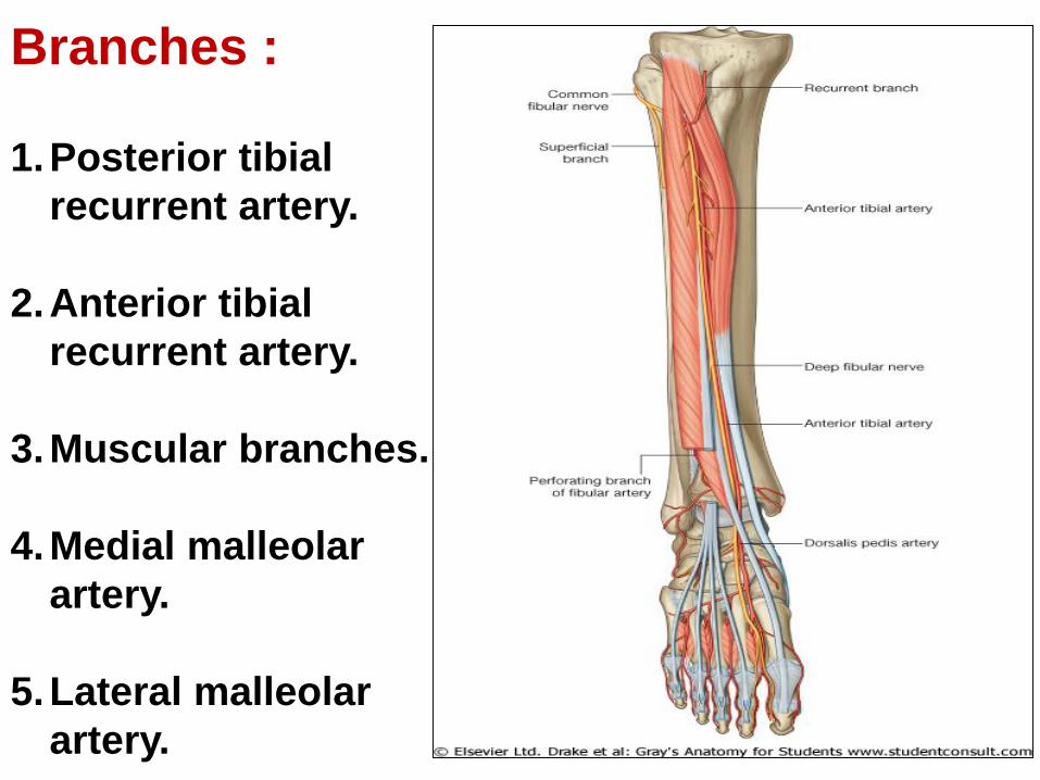
Branches :
1.Posterior tibial
recurrent artery.
2.Anterior tibial
recurrent artery.
3.Muscular branches.
4.Medial malleolar
artery.
5.Lateral malleolar
artery.
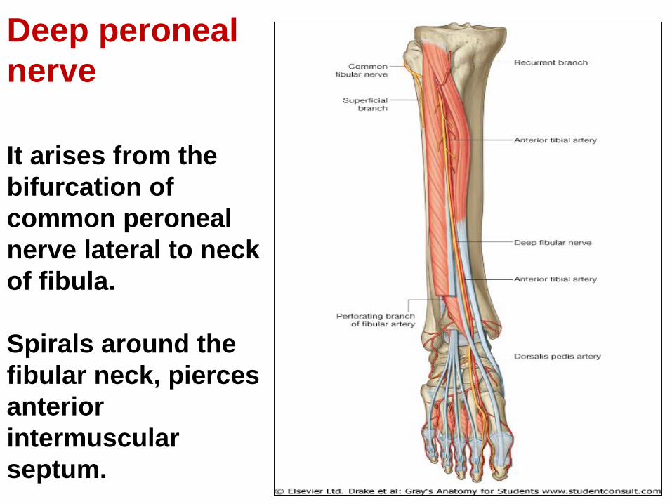
Deep peroneal
nerve
It arises from the
bifurcation of
common peroneal
nerve lateral to neck
of fibula.
Spirals around the
fibular neck, pierces
anterior
intermuscular
septum.
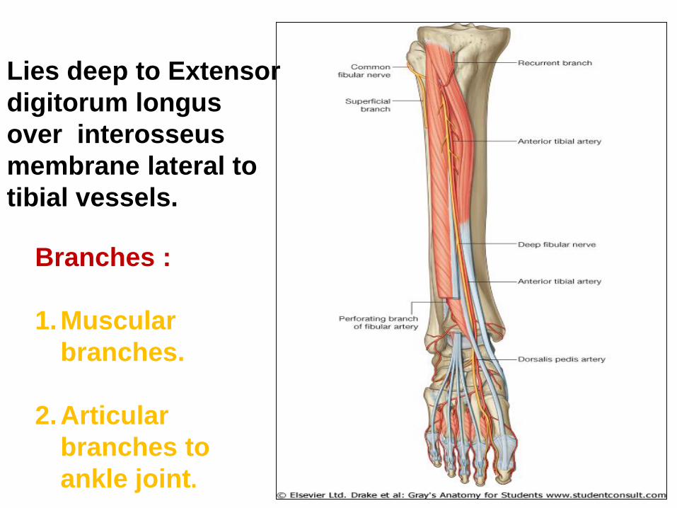
Lies deep to Extensor
digitorum longus
over interosseus
membrane lateral to
tibial vessels.
Branches :
1.Muscular
branches.
2.Articular
branches to
ankle joint.
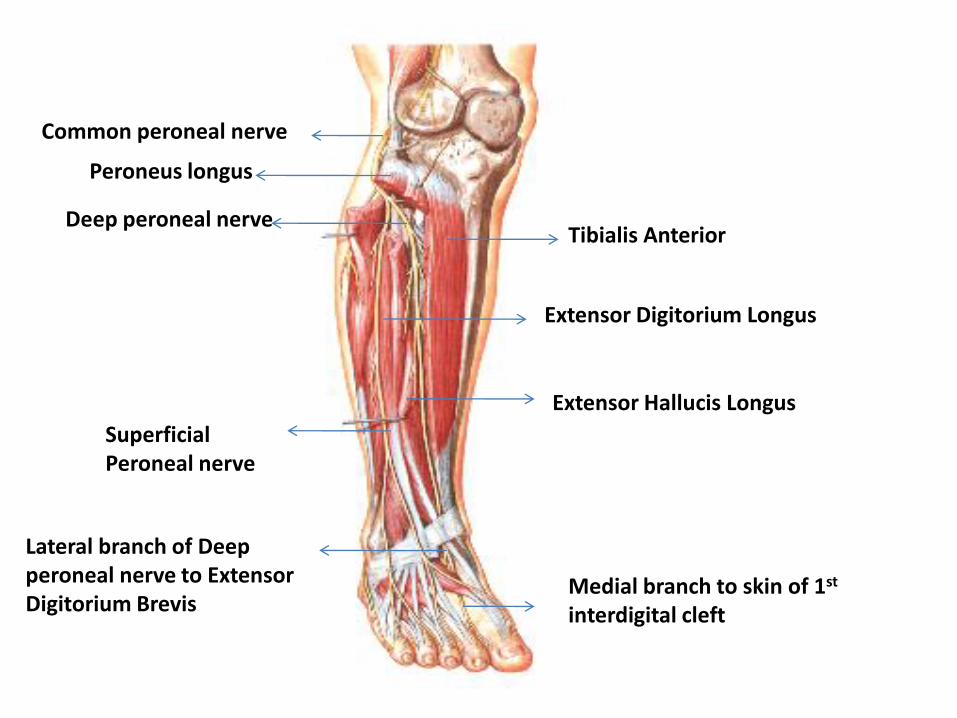
Common peroneal nerve
Peroneus longus
Deep peroneal nerve
Superficial Peroneal nerve
Tibialis Anterior
Extensor Digitorium Longus
Extensor Hallucis Longus
Lateral branch of Deep peroneal nerve to Extensor Digitorium Brevis
Medial branch to skin of 1st
interdigital cleft
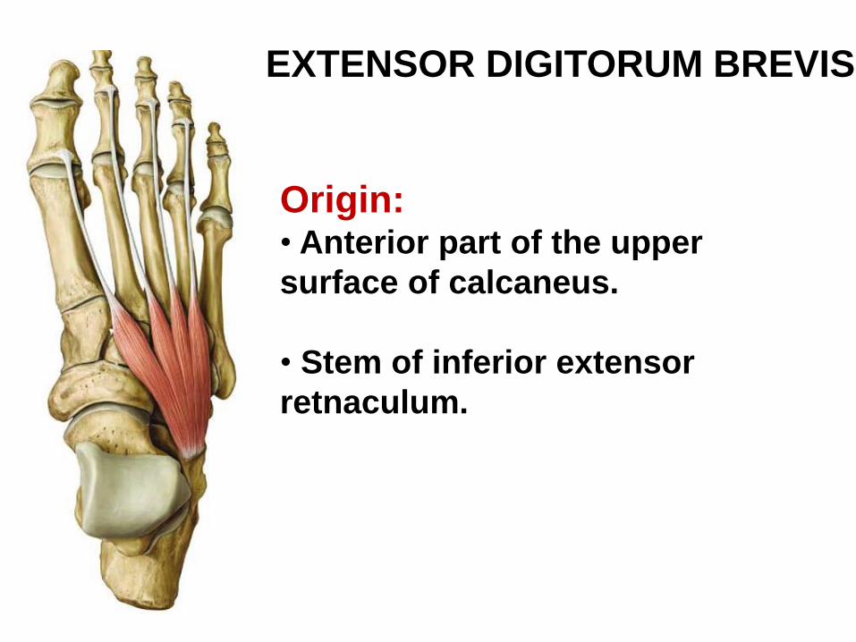
EXTENSOR DIGITORUM BREVIS
Origin:• Anterior part of the upper
surface of calcaneus.
• Stem of inferior extensor
retnaculum.
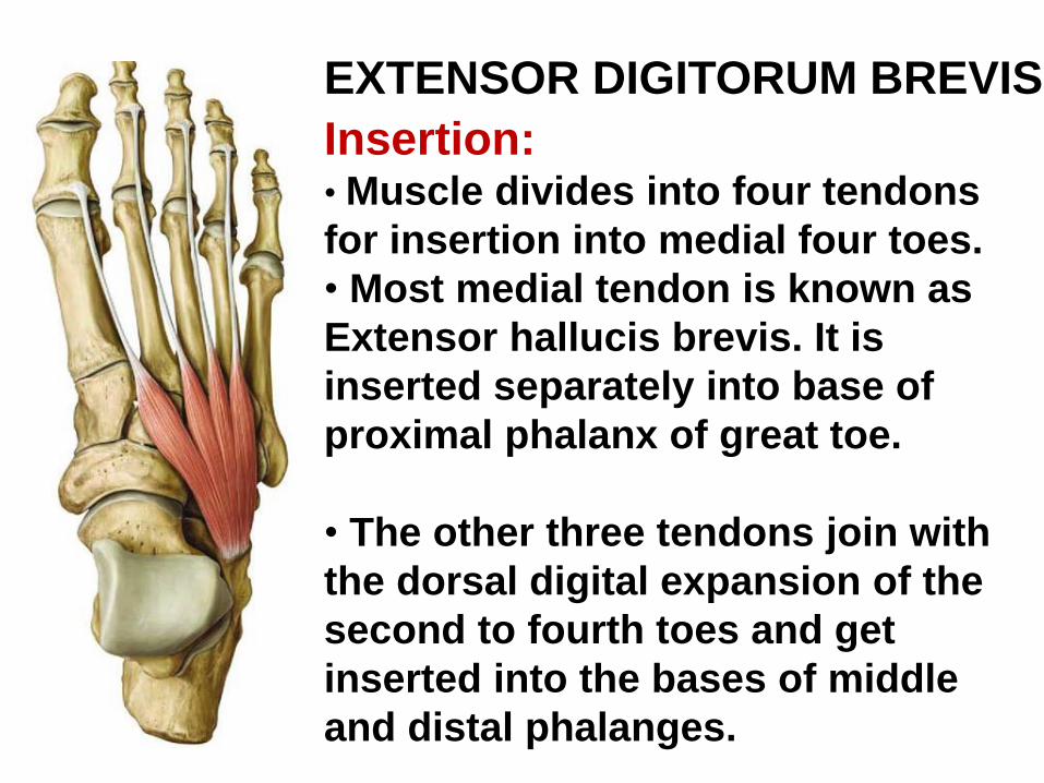
EXTENSOR DIGITORUM BREVIS
Insertion:• Muscle divides into four tendons
for insertion into medial four toes.
• Most medial tendon is known as
Extensor hallucis brevis. It is
inserted separately into base of
proximal phalanx of great toe.
• The other three tendons join with
the dorsal digital expansion of the
second to fourth toes and get
inserted into the bases of middle
and distal phalanges.
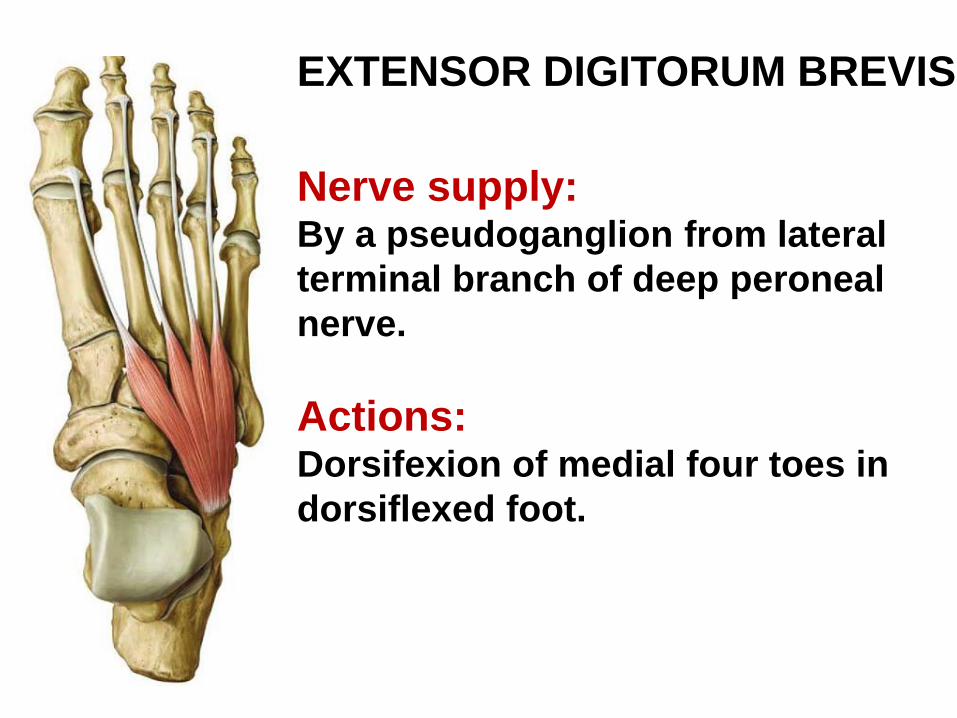
EXTENSOR DIGITORUM BREVIS
Nerve supply:By a pseudoganglion from lateral
terminal branch of deep peroneal
nerve.
Actions:Dorsifexion of medial four toes in
dorsiflexed foot.
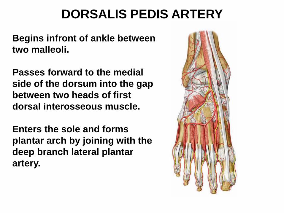
DORSALIS PEDIS ARTERY
Begins infront of ankle between
two malleoli.
Passes forward to the medial
side of the dorsum into the gap
between two heads of first
dorsal interosseous muscle.
Enters the sole and forms
plantar arch by joining with the
deep branch lateral plantar
artery.
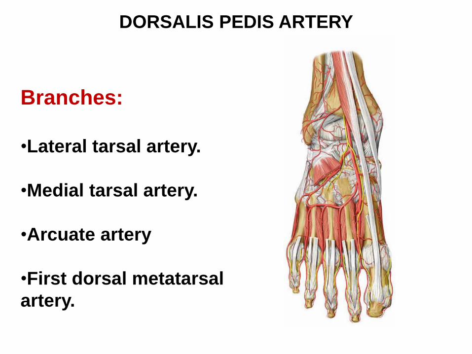
DORSALIS PEDIS ARTERY
Branches:
•Lateral tarsal artery.
•Medial tarsal artery.
•Arcuate artery
•First dorsal metatarsal
artery.
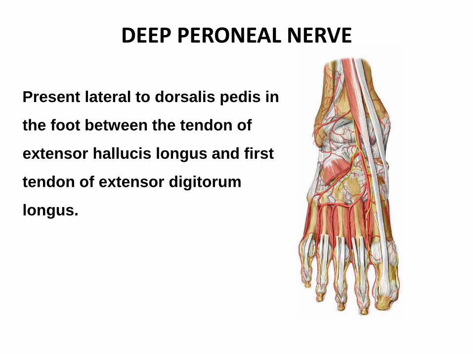
DEEP PERONEAL NERVE
Present lateral to dorsalis pedis in
the foot between the tendon of
extensor hallucis longus and first
tendon of extensor digitorum
longus.
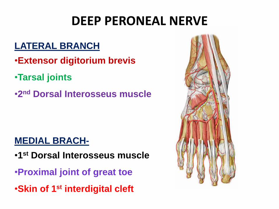
DEEP PERONEAL NERVE
LATERAL BRANCH
•Extensor digitorium brevis
•Tarsal joints
•2nd Dorsal Interosseus muscle
MEDIAL BRACH-
•1st Dorsal Interosseus muscle
•Proximal joint of great toe
•Skin of 1st interdigital cleft
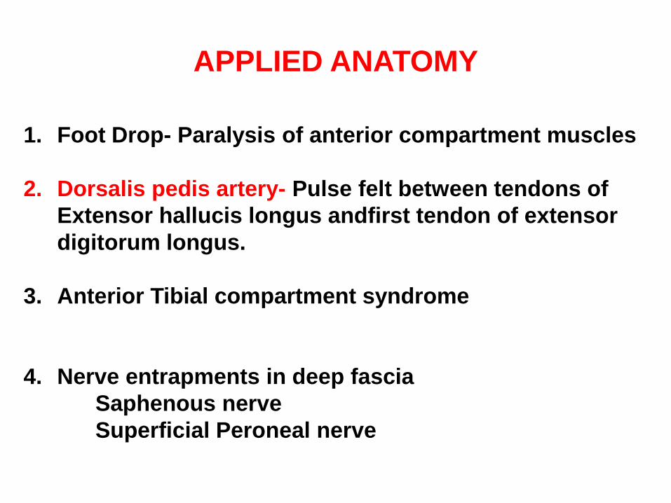
APPLIED ANATOMY
1. Foot Drop- Paralysis of anterior compartment muscles
2. Dorsalis pedis artery- Pulse felt between tendons of
Extensor hallucis longus andfirst tendon of extensor
digitorum longus.
3. Anterior Tibial compartment syndrome
4. Nerve entrapments in deep fascia
Saphenous nerve
Superficial Peroneal nerve