Ultrastructural Study of Yam Tuber as Related to ... - CiteSeerX
10
Food Structure Volume 4 | Number 1 Article 19 1985 Ultrastructural Study of Yam Tuber as Related to Postharvest Hardness L. Sealy S. Renaudin D. J. Gallant B. Bouchet J. M. Brillouet Follow this and additional works at: hp://digitalcommons.usu.edu/foodmicrostructure Part of the Food Science Commons is Article is brought to you for free and open access by the Western Dairy Center at DigitalCommons@USU. It has been accepted for inclusion in Food Structure by an authorized administrator of DigitalCommons@USU. For more information, please contact [email protected]. Recommended Citation Sealy, L.; Renaudin, S.; Gallant, D. J.; Bouchet, B.; and Brillouet, J. M. (1985) "Ultrastructural Study of Yam Tuber as Related to Postharvest Hardness," Food Structure: Vol. 4: No. 1, Article 19. Available at: hp://digitalcommons.usu.edu/foodmicrostructure/vol4/iss1/19
-
Upload
khangminh22 -
Category
Documents
-
view
1 -
download
0
Transcript of Ultrastructural Study of Yam Tuber as Related to ... - CiteSeerX
Food Structure
Volume 4 | Number 1 Article 19
1985
Ultrastructural Study of Yam Tuber as Related toPostharvest HardnessL. Sealy
S. Renaudin
D. J. Gallant
B. Bouchet
J. M. Brillouet
Follow this and additional works at: http://digitalcommons.usu.edu/foodmicrostructure
Part of the Food Science Commons
This Article is brought to you for free and open access by the Western DairyCenter at DigitalCommons@USU. It has been accepted for inclusion inFood Structure by an authorized administrator of [email protected] more information, please contact [email protected].
Recommended CitationSealy, L.; Renaudin, S.; Gallant, D. J.; Bouchet, B.; and Brillouet, J. M. (1985) "Ultrastructural Study of Yam Tuber as Related toPostharvest Hardness," Food Structure: Vol. 4: No. 1, Article 19.Available at: http://digitalcommons.usu.edu/foodmicrostructure/vol4/iss1/19
FOOD M ICROSTRUCTURE, Vol. 4 (1985) , pp. 173- 181 SEM Inc., AMF O'Hare (Chicago) , IL 60666-05(]7 U.S .A.
0730-5419/85$1.00+.05
ULTRASTRUCTURAL STUDY OF YAM TUBER AS RELATED TO POSTHARVEST HARDNESS
L S 1 (l,Z) S R d . (2) D J G 11 t(l) B B h t(l) J M B "11 t(l) . ea y , . en au 1 n , . . a an , . . ouc e , . . n oue
(l) INSTITUT NATIONAL DE LA RECHERCHE AGRONOMIQUE Centre de Recherches Agro-alimentaires
rue de la Geraudiere - 44072 Nantes Cedex, France
(2) INSTITUT DES SCIENCES DE LA NATURE Laboratoire de Cytopathologie Vegetale
2, rue de la Houssiniere - 44072 Nantes Cedex, France.
Abstract
Usua ll y, parenchyma cell walls of mo nocotyl edons do not develop secondary walls; howeveG a few days after harvesting, the yam tuber of Dioscorea dumetorum starts to harden. Two or three weeks I ater, hardness is so pronounced that the tubers cannot be eaten, even after a long cooking time.
Cytochem i ca l st udies us in g autof luorescence or some f l uorescent dyes, such as ph l orogluc inol hydrochloride showed that the thin, and f l exib l e cel l wall s of parenchyma tubers very quickly became fully lignified after harvest ing. Ultrastructura 1 stud ie s of the hardened ce 11 wa 11 s showed very thick secondary wa 11 s and very deep pit apertures. These secondary walls reacted strong ly with li gn in reactants such as potassium permanganate. The use of a radioactive (l '• C) ce llulose precursor, uri dine- 5'-d ipho sphateglucose, conf irmed the formation of such secondary walls. The lignification started from the corners of the cel l s around intercel l ul ar spaces and proceeded al ong the walls .
Initial paper received February 13, 1985. Final man uscr ipt received May 23, 1985 . Direct inquiries to D.J . Gal l ant. Telephone number: (40) 76.23.64.
Key Words : Yam tuber parenchyma, postharvest, hardening, cell walls, scanning electron microscopy, transmission e 1 ectron microscopy, autoradiography, cytochemistry.
173
Introduction
Tubers ori gi nat i ng from tropi ca 1 countries constitute the basic foods of mo st people in Africa, South America, India and Asia . Cassavas (Manihot utilissima Pohl ), sweet potatoes (Ipomea batatas L.), taros or arums (Colocasia escUTenfd L.) and yams ( Di oscorea sp . ), respect 1 ve ly. are the principal cult1vated crops (Ga ll ant et al., 1982). Amongst the yam species, Dioscorea dumetorum ( Kunth), a Cameroon and Ni gena crop, seems very interesting due to its cro p yi e ld s (40 tons/hectare) as noted by Treche and Guion ( 1979). Stakers are not necessary, the s ize and morphology of the tubers are genera ll y homogeneous, and mechanization can be used; the protein content is relatively high (c .a. 10%/ dry mass) and consumer acceptabi 1 i ty i s high after cooking ( Onwu eme, 1978). Even though these advantages ex i st, tubers of Oioscorea dumetorum present the disadvantage that they harden very quickly a few weeks after harvest, and cannot be eaten even after a l ong cooking time.
Only few stud i es have been made to explain such harden ing. Del peuch and Treche ( 1978) descr ibed the presence of cellulose-like thickened areas in the parenchymatic ce ll walls. The same authors, and Brill ouet et a 1. ( 1981) determined the biochemical changes that occurred during hardening. However, these stud i es, carried out on soft and hardened samples, did not show the right course of deve 1 opment of the phenomena. In the present study, mi croscopy, cy tochemistry and autoradiography techniques were used to follow the ul trastructura l changes which l ead to hardening during si x weeks postharvest.
Materials and Method s
Di oscorea dumetorum were cultivated at the exper1menta I Stat 10n of Bambui (Cameroon West) in 1979-80 and 1980 -81. After a growth of 8 to 9 months, tubers were harvested, and immediat e ly covered with paraffin wax and then airmailed to Nantes (France). Paraffinized tubers could be stored severa l weeks at room temperature without any changes concern ing si ze, ultrastructure, cooki ng behaviour and taste.
L. Sealy, S. Renaudin, D.J. Gallant, B. Bouc het and J.M. Brillouet
Sampling Six tubers of the same size and weight were
cho se n for TEM and autoradiography. The hardening time started when paraffin was removed from the tubers. Tubers were stored in the dark ( 20 °C I during 0, 3 and 6 weeks. Each tuber was punched in its midd l e part and perpendicular to the principa l axis . Cylinders (6 mm diameter) obtained in this way were cut into two parts, each of them being equally divided into three equal parts: A, per ipheral; B, medium and C, central parts of each tuber. Microscopy
L1ght mi croscopy. Demonstration of the li-gnin has been done on hand sectioned samples: a) using the phloroglucinol-HCl stain, lignin components appearing red; b) or directly under UV light, l ign in components as well as fluore scent phenolic compounds giving inten se blue-white emission .
SEM. Samples from parts A, i3 and C were frozen-Tn isopentane cooled to -l60 °C with liquid nitrogen, then fractured along the axis of the cy linder s .
In order to see cel l wall pit apertures, samples of outer cell walls were usually c leaned (24h, 40 °C) with a solution of 20 ~ g/ml of alpha-amylase from Bacillus subtilis to hydrolyse the starch granules . fhen, the sampl es were dehydrated in a graded acetone series an d crit i ca 1-point dried. Te n to 20 nm go ld were deposited on dried samp l es us ing the JEOL i on s putt er JFC ll 00 . Samp l es were ex am i ned in a JEOL 50A at 20 keV .
TEM. Samp le s ( l mm 3 ) were fixed in 3% glutaraTdehyde (O.lM phosphate buffer, pH 7.2) for 2h at room temperature and postfixed for lh in 1% Os0 4 (same buffer) . Subsequently, they were dehydrated in a graded ethanol ser ie s and embedded in Epon 812 . Silver sect ion s were stained for 30 min in 2.8% uranyl acetate (50% methanol ) at 48°C and for 5 min in lead cit rate (pH 13). Sect i ons were observed i n a JEOL lOOS at 80 keV .
Oemonstration of the polysacchar ides (TEM by the method of lh1ery, 1967 ): lh1n sect1ons were oxidized on gold grids for 30 min in 1% periodic acid, treated for 48h in a sat urated thiosemicarb azide solution, treated for 30 min in 1% si lver proteinate and then rinsed several t 1mes in distilled water. Th i s i s the PATAg method.
Demonstration of the pectic substances (TEM by the method of Luft, 1971 ): Dur1ng samp le fixat i ons 0.6% ruthen ium red was added to the glutaraldehyde and l % to t he osmium tetroxide.
Demonstration of the li gn in (TE M by the method of Hep ler et a l . , 1970): Samples were fixed 90 min in 2% KM n0 4 , then i mme di ately dehydrated in a qraded etha nol series and embedded on Epon 812.
- Autora di o~ r a phy ( light microscopy): Sma ll s amples (8 mm y--were immersed i n a so luti on of the ammon ium salt of urid ine- 5' -dipho sphate -g l ucose ( UDPG) . UDPG rad i oact i vi ty was 25 ~ Ci/ml. After 6h i ncubati on, the sampl es were fiXfJ for 2h in 3% glutaraldehyde (O .l M phosphate buffer, pH 7.2), washed for 2h with non radioactive sa lted UDPG and for l 2h in run ning water. The samp l es were embedded in Epon 812. Sec ti ons (2 ~ m ) were deposited on glass s lides and covered
174
with the Ilford G5 emulsion. After 4 days, the emulsion was deve loped and the sections were sta ined with a 2% so lution of methy l ene blue.
Contro l sample s were trea t ed wi th a radioac tive UDPG so luti on to which HgC l 2 was added to inhibit the metabo li c act i vity of the cells.
Result s
SEM study of cell walls during hardening When the paraff 1 n was removed from a tuber,
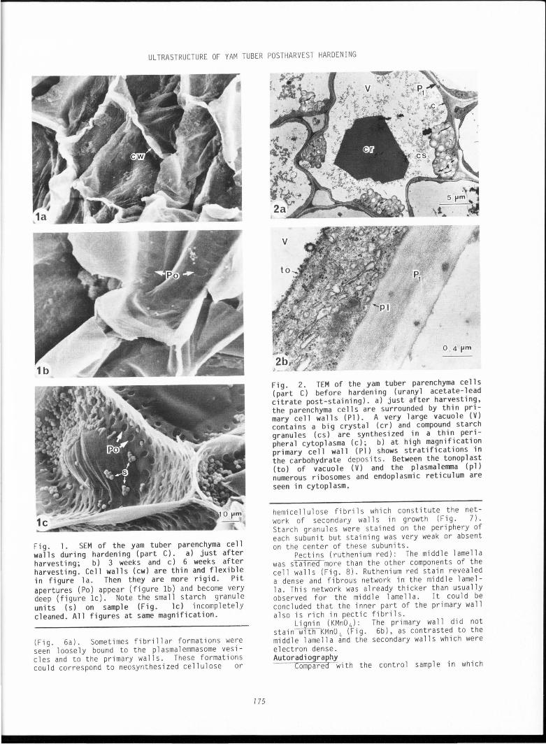
the parenchyma cell walls were very thin and presented a fold-like struct ure (Fig. la). After 3 weeks (Fig . lb), cell walls in part A were already thin and flexible, but in part s B and C many pit apertures were see n, many more being visib l e near the corner of the ce ll s . These pit apertures were regu l ar (7 to 8 ~ m diameter ) . After 6 weeks (Fig. lc), all the cell walls in parts A, B and C were pitted with apertures although they were more numerou s in the corners around the intercellular spaces. TEM s tudy of cell walls during hardening
lmmed1ately after paraff1n remova l , at low magnification (Fig. 2a) the cells of medullary region (part C) showed very thin and regular.cell walls, a thin l ayer of cytop l asm conta1n1ng severa l compound starch granu l es in plastid s, and a large central vacuo l e, somet imes with a bi g hexago na l crysta l. At higher magnification (F ig . 2b), it cou ld be easi ly seen that the cell wall (800 nm thickness) consisted on ly of the middle lame ll a and the primary wall. The cytoplasm appeared dense and contained numerous ribosomes, mitochondria, rough endoplasmic reticulum, some dictyosomes (showing 3 to 4 cisterna but only few Golgi vesicles), and compound starch granules with sma ll polyhedric faced units .
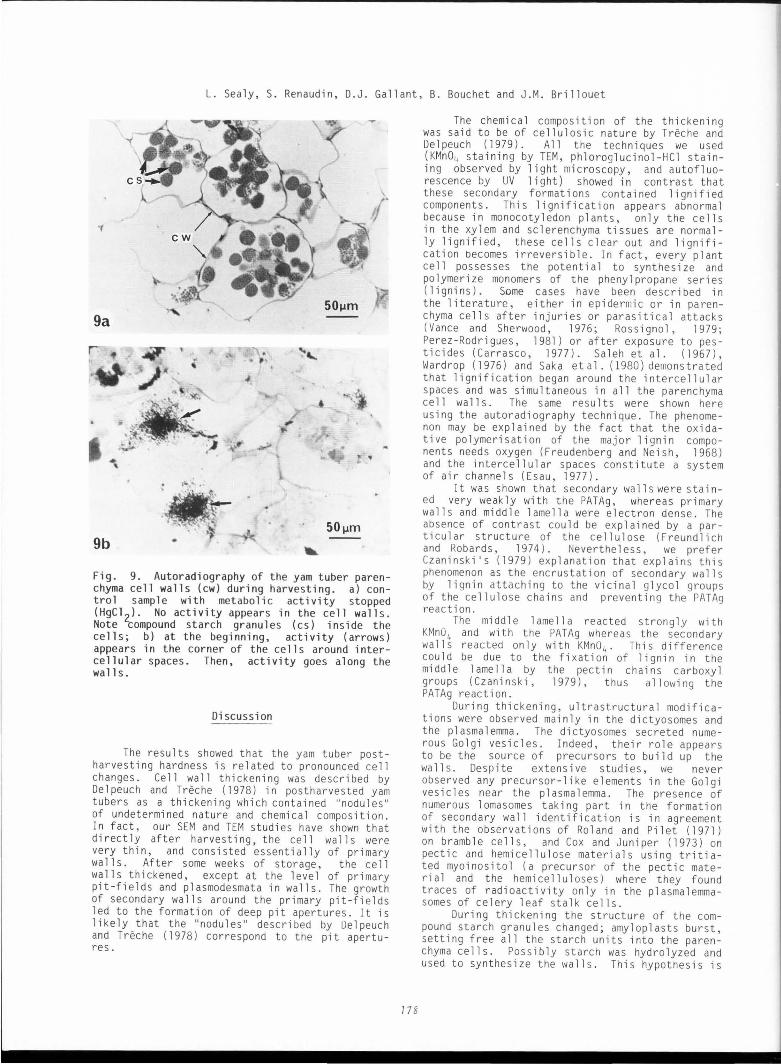
After 3 weeks, the cells showed important cha nge s: a thickening of the cell wall s that did not stai n very well with uranyl acetate and l ead citrate. Close to the seco ndary walls the dictyosomes secreted numerou s Golg i vesicles, some of which (lCJ nm in diameter) were electron dense while others were l arger and lighter in appearance. Between the cell wall and cytop l asm, the invaginations of pl asma l emma formed pockets and the plasmalemmasomes (Fig . 3a, b) contained numerous s pherical ves i cles and e l ongated tubules . Ma ny amy l op l asts had been broken and the polyhedric units of the compound starch granules were dispersed i nto the ce ll s (F ig. 4).
After 6 weeks, the ce ll walls had become much thicker and showed dee p pit apertures tra versed through by thin ca nal s of pla smodesmata (F i g. 5a) . In addition, t he plasmalemma showed some invaginati ons of paramural bodies (Es au , 1977) . In some cases, near to these fo rmati ons, an ir regular network of fib r il s could be seen (Fig . 5b), pres umabl y amorphous parapla sm i c materia l assoc iated to the ce ll walls. Most of the starch granules had burst and were di spersed into the ce ll s . Cytochemistry
Polysaccharides (PATAg) : After 6 weeks onl y the m1ddle lamella and primary wa ll s were electron dense; the second ary walls were not stai ned
ULTRASTRUCTURE OF YAM TUBER POSTHARVEST HARDENING
Fig. 1 • SEM of the yam tuber parenchyma ce 11 wa 11 s during hardening (part C). a) just after harvesting; b) 3 weeks and c) 6 weeks after harvesting. Cell walls (cw) are thin and flexible in figure la. Then they are more rigid . Pit apertures (Po) appear (figure lb) and become very deep (figure lc). Note the sma ll sta rch granule units (s) on sample (Fig. lc) incompletely cleaned. All figures at same magnification.
(Fig. 6a). Somet ime s fibrillar formations were seen 1 oose 1 y bound to the p 1 asma 1 emma some ves icles and to the primary walls. These formations could correspond to neosynthesized cellulose or
775
0 , 4 pm
Fig. 2. TEM of the yam tuber parenchyma ce 11 s (part C) before hardening (uranyl acetate-~ead citrate post-staining). a) just after harvest1ng, the parenchyma cells are surrounded by thin pri mary cell walls (Pl) . A very large vacuole (V) contains a big crysta 1 ( cr) and compound starch granules (cs) are synthesized in a thin peripheral cytoplasma (c); b) at high magnification primary cell wall (Pl) shows stratifications in the carbohydrate deposits. Between the tonop 1 ast (to) of vacuole (V) and the plasmalerrma (pl) numerous ribosomes and endoplasmic reticulum are seen in cytoplasm.
hemicellulose fibrils which const itute the network of secondary walls in growth (F ig. 7). Starch granules were stained on the periphery of each subunit but staining was very weak or absent on the center of these subunits.
Pectins (ruthenium red): The middle lamella was sta1ned more than the other components of the cell walls (Fig. 8) . Ruthenium red stai n revealed a dense and fibrous network in the middle lamella. Th i s network was already thicker than usually observed for the middle lamella. It could be concluded that the inner part of the primary wall also is rich in pectic fibrils.
Lignin (KMn0 4 ): The primary wall did not stain~KMn0 4 (Fig . 6b), as contrasted to the middle lamella and the secondary walls which were electron dense. Autoradiography
Compared with the contro l samp le in which
L. Sea ly, S. Rena udin, D. J. Ga ll ant, B. Bouchet and J.M. Bri ll ouet
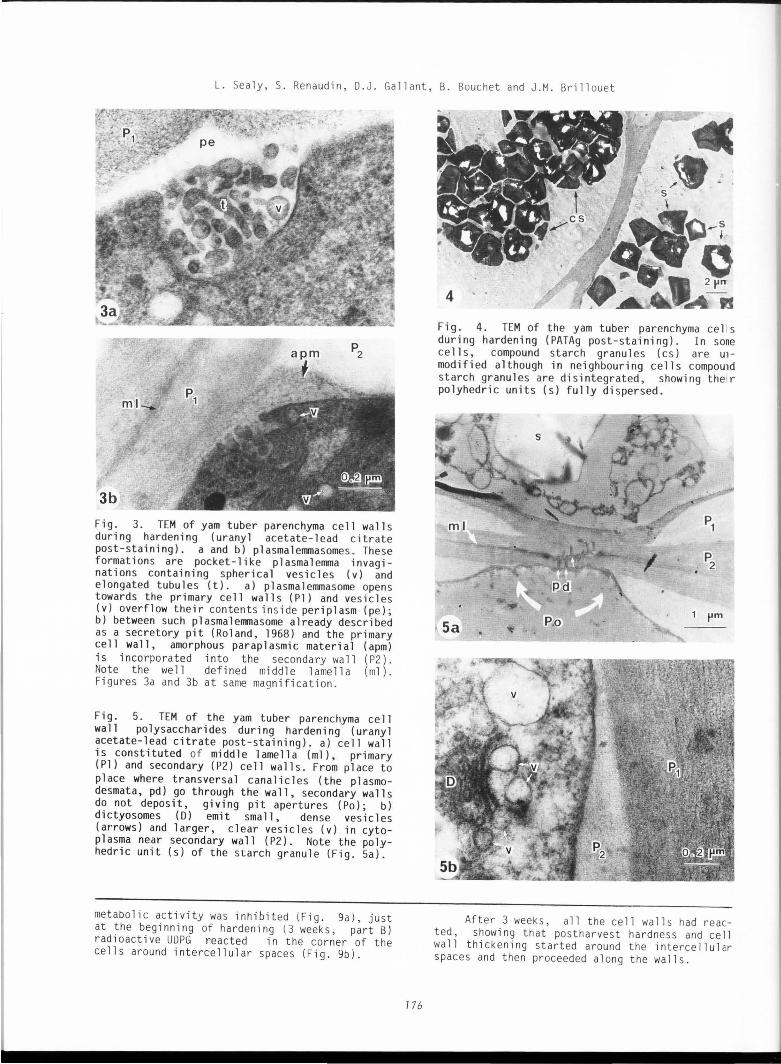
Fig . 3. TEM of yam tuber parenchyma cell walls during hardening (uranyl acetate-lead citrate post-staining). a and b) plasmalemmasomes. These formations are pocket-like plasmalemma invaginations containing spherical vesicles (v) and elongated tubules (t). a) plasmalemmasome opens towards the primary cell walls (Pl) and vesicles (v) overflow their contents in side periplasm (pe); b) between such plasmalemmasome already described as a secretory pit (Roland, 1968) and the primary cell wall, amorphous paraplasmic material (apm) is incorporated into the secondary wall (P2) . Note the well defined middle l amel la (ml ). Figures 3a and 3b at same magn ifi cation.
Fig. 5. TEM of the yam tuber parenchyma cell wall polysaccharides during hardening (uranyl acetate-lead citrate post-staining) . a) cell wall is constituted of middle lamella (ml), primary (Pl) and secondary (P2) cell walls . From place to place where transversal canalicles (the plasmodesmata, pd) go through the wall, secondary walls do not deposit, giving pit apertures (Po); b) di ctyosomes (D) emit small, dense vesicles (arrows) and larger, clear vesicles (v) in cytop 1 asma near secondary wall ( P2). Note the polyhedric unit (s) of the starch granule (Fig. 5a).
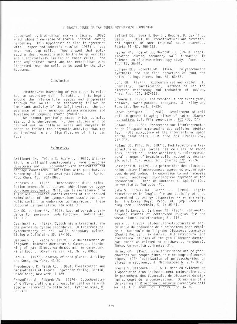
metabolic activity wa s inhibited (Fig. 9a), just at the beginning of hardening (3 weeks, part B) radioactive UDPG reacted in the corner of the cells around intercellular spaces (Fig. 9b).
176
Fig. 4. TEM of the yam tuber parenchyma ce 1 s during hardening ( PATAg post-staining). In sane cells, compound starch granules (cs) are Ul
modified although in neighbouring cells compou1d starch granules are disintegrated, showing the1r polyhedric units (s) fully dispersed.
After 3 wee ks , all the cell walls had reacted, showing that po stharve st hardness and ce 11 wall thickening started around the intercellul ar spaces and then proceeded al ong the walls.

ULTRASTRUCTURE OF YAM TUBER POSTHARVF.ST HARDENING
P2
p~
Fig. 6. TEM of the lignified yam tuber parenchyma cell walls. a) after the PATAg post-staining, primary wall s (Pl) and middl e lamella (ml) are e lect ron dense, wh ereas secondary walls (P2) remain clear. Note very deep pit apertures (Po) with plasmodesmata (pd); b) after KMn04 treatment, secondary wall s (P2) and middle lamella (ml) are more el ectron dense than primary walls (Pl). Figures 6a and 6b at same magnifi -cation.
Fig. 7. TEM of the yam tuber parenchyma cell wall s polysaccharides (PATAg post- staining). Neosynthesized cellulosic and hemicellulosic fibrils (arrows) constitute a network of the secondary walls in growth between primary wall (Pl) and plasmalemma (pl) and vesicles (v) into the periplasm (pel.
177
Fig. 8. TEM of the yam tuber parenchyma cell wall pectins (ruthenium red stain). Middle lamella (ml) shows fibrillar, disordered structure (arrows), nevertheless more ordered near the primary wall ( Pl ) . Secondary walls ( P2) do not show such structure.
L. Sea ly, S. Renaudin, D.J. Gallant, B. Bouchet and J.M. Brillouet
)
9a
50~m
9b
Fi g. 9. Autor adiog r aphy of t he yam t uber parenchyma ce ll wall s (cw) during harvesti ng . a) cont ra l sampl e with metabolic acti vity stopped (HgCl 2) . No activi ty appears i n t he cell wall s. Note compound st arch granul es (cs ) i nside t he ce ll s; b) at the beginning , act ivity (ar rows ) appears in the corner of t he cel l s around i nter cellu l ar spaces . Then, act i vity goes along the walls .
Di scussi on
The results showed that the yam tuber postharvesting hardness is related to pronounced cell changes. Ce l l wall thickening was described by De l peuch and Treche (1978) in postharvested yam tubers as a th ickening which contained "nodules" of undetermi ned nature and chemica l composition. In fact, our SEM and TEM stud i es have shown that di rect ly after harvesting, the ce l l walls were very thin, and consisted essentia l ly of primary walls. After some weeks of storage, the cell walls thickened, except at the level of primary pit-fields and pl asmodesmata in walls. The growth of secondary walls around the primary pit-fields led to the formation of deep pit apertures. It is likely that the "nodules" described by Del peuch and Treche ( 1978) correspond to the pit apertures.
17 8
The chemical composition of the thickening was said to be of cellulosic nature by Treche and lJelpeuch (1979). All the techniques we used (KMn04 staining by TEM, ph l oroglucinol-HCl staining observed by light microscopy, and autofluorescence by UV light) showed in contrast that these secondary formations contained lignified components. This lignification appears abnormal because in monocotyledon plants, only the cells in the xylem and sclerenchyma tissues are normally lignified, these cells clear out and lignification becomes irreversible. In fact, every plant cell possesses the potential to synthesize and polymerize monomers of the phenyl propane series (lignins). Some cases have been described in the literature, either in epidermic or in parenchyma cells after injuries or parasitical attacks (Vance and Sherwood, 1976; Rossignol , 1979; Perez-Rodrigues, 1981) or after exposure to pesticides (Carrasco, 1977). Saleh et al. (1967), Wardrop ( 1976) and Saka et al. ( 1980) demonstrated that lignification began around the intercellular spaces and was simultaneous in all the parenchyma cell walls. The same results were shown here using the autoradiography technique. The phenomenon may be explained by the fact that the oxidative polymerisation of the major lignin components needs oxygen (Freudenberg and Neish, 1968) and the intercellular spaces constitute a system of air channe l s (Esau, 1977).
It was shown that secondary walls were stained very weak ly v1 i th the PATAg, whereas primary walls and mi ddle lamella were el ectron dense. The absence of contrast could be explained by a particular structure of the cellulose (Freundlich and Robards, 1974). Nevertheless, we prefer Czaninski 's (1979) explanation that explains this phenomenon as the encrustation of secondary wall s by lignin attaching to the vicinal glycol groups of the cellulose chains and preventing the PATAg reaction.
The middle lamella reacted strongly with KMn0 4 and with the PATAg whereas the secondary walls reacted only with KMn0 4 • This difference could be due to the fixation of lignin in the middle lamella by the pectin chains carboxyl groups (Czaninski, 1979), thus allowing the PATAg reaction.
During thickening, ultrastructural modifications were observed mainly in the dictyosomes and the plasmal emma. The di ctyosomes secreted numerous Go l gi vesicles. Indeed, their role appears to be the source of precursors to build up the walls. Despite extensive studies, we never observed any precursor- l ike elements in the Golgi vesicles near the plasmalemma. The presence of numerous lomasomes taking part in the formation of secondary wall i dent ifi cation is i n agreement with the observations of Ro l and and Pilet (1971) on bramble cells, and Cox and Juniper (1973) on pectic and hemicellu l ose materials using tritiated myoinositol (a precursor of the pectic mate rial and the hemicelluloses) where they found traces of radioactivity only in the plasmalemmasomes of celery leaf stalk cells.
During thickening the structure of the compound starch granules changed; amyloplasts burst, setting free all the starch units into the parenchyma cells. Possibly starch was hydrolyzed and used to synthesize the walls. This hypothesis is
ULTRASTRUCTURE OF YAM TUBER POSTHARVEST HARDENING
supported by biochemical analysis (Sealy, 1982) which shows a decrease of starch content during hardening. This hypothesis is also in agreement with Juniper and Robert's results ( 1966) on zea mays root cap cel l s. They showed that polysaccharides precursors used by the Golgi vesicles are quantitatively limited in those cells, and that amyloplasts burst and the metabolites were liberated into the cells to be used by the dictyosomes.
Conclusion
Postharvest hardening of yam tuber is related to secondary wall formation . This begins around the intercellular spaces and progresses through the wal l s . The thickening follows an important activity of the Go l gi system, the appearance of very numerous plasmalemmasomes and bursting of compound starch granules .
We cannot precisely state which stimulus starts this phenomenon. Further studies will be carried out on cellular areas and enzymes in order to inhibit the enzymatic activity that may be involved in the lignification of this yam tuber.
References
Brillouet JM, Treche S, Sealy L. (1981). Alterations in cell wall constituents of yams Dioscorea dumetorum and D. rotundata with maturat 10n and storage conditions. Relat1ons with post-harvest hardening of D. dumetorum yam tubers. J. Agric . Food Chern. 46, 1964-1967. Carrasco A. (1977). Repercussion d'une stimu-1 at ion provoquee du contenu phenol i que de Lycopersicon esculentum Mill. sur la resistance-a-Ta fusar1ose. (Consequential effects of the induced stimulation of the Lycopersicon esculentum phenolic content on endurance to fusan ose). These Doctorat de Specialite, Toulouse (F). Cox GC, Juniper BE. (1973). Autoradiographic evidence for paramura l body function. Nature 243, 116-117. Czaninski Y. (1979). Cytochimie ultrastructurale des parois du xyleme secondaire. (Ultrastructural cytochemistry of cell walls secondary xylem). Biologie Cellulaire 35, 97-102.
Delpeuch F, Treche S. (1978). Le durcissement de l 'igname Dioscorea dumetorum au Cameroun. (Hardening of yam ( D10scorea dumetorum) in Cameroon). Final Report. DGRST (Pans), XI, 76, 7, 0066.
Esau K. (1977). Anatomy of seed plants. J . Wiley and Sons, New York, 43 -60.
Freudenberg K, Neish AC. (1968) . Constitution and biosynthesis of lignin. Springer Verlag, Berlin, Heidelberg, New York, l-129.
Freundlich A, Robards AW. (1974). Cytochemistry of differentiating plant vascular cel l walls with special reference to cellu lose. Cytobiolog i e, 8, 355-370.
179
Gallant OJ, Bewa H, Buy QH, Bouchet B, Szylit 0, Sealy L. (1982). On ultrastructural and nutritional aspects of some tropical tuber starches. Starke 34 (8), 255 -262.
Hepler PK, Fosket DE, Newcomb EH . (1970). Lignification during secondary wall formation in Coleus: an electron microscopy study. Amer. J. ~7, 85-96.
Juniper BE, Roberts RM. (1966). Po lysaccharide synthesis and the fine structure of root cap cells. J. Roy. Micro . Soc. 85, 63-72.
Luft JH. (1971). Ruthenium red and violet. I. Chemistry, purification, methods of use for electron microscopy and mechanism of act i on. Anat. Rec. 171, 347-368. Onwueme I. ( 1978) . The tropical tuber crops yams, cassava, sweet potato, cocoyams . J. Wi l ey and Sons Ltd, New York, l-234. Perez-Rodrigues D. (1981). Deve lopment of cell wall in growth in aging slices of radish (Raphanus sativus L.). Pflanzenphysiol. 2_Qi (3), ~
Roland JC. (1968) . Recherches sur l 'infrastructure de l'espace membranaire des cellules vegetales. (Ultrastructure of the intercellular space in the plant cells). C.R . Acad. Sci. (Paris) 267, 712-715 . -
Roland JC, Pi l et PE. (1971 ). Modifications ultrastructurales des parois des cel lul es de ranee sous l'effet de l'acide abscissique. (Ultrastructural changes of bramble cells induced by abscissic acid). C.R. Acad. Sci. (Paris) 272, 72-76.
Rossignol M. (1979). La premonition des plants de Melon contre l 'anthracnose: approches physiologiques du phenomene. (Premonition to anthracnosis of melon seedlings: physiological approach of the phenomenon). These de Doctorat de Special i tes. Universite de Toulouse (F). Saka S, Thomas RJ, Gratzl JS. (1980). Lignin distribution in Douglas-fir and Loblolly pine as determined by energy dispersive X-ray analysis. In: The Eckman Days. Proc. Int . Symp . Wood Pulping Chern., Stockholm,~, I : 35-41. Saleh T, Leney L, Sarkanen KV. (1967). Radioautographic studies of cottonwood Douglas fir and wheat plants. Holzforschung ~, 116. Sealy L. (1982). Etudes ultrastructurale et biochimique du phenomene de durcissement post recolte du tubercu l e de l 'i gname Di oscorea dumetorum (Kunth) Pax var. ex Jakiri. (Ultrastructural and biochemical studies of the yam (Diocorea dumetorum) tuber as related to postharvest hardness). These, Universite de Nantes (F) .
Thiery JP . (1967). Mise en evidence des polysaccharides sur coupes fines en microscopie electronique. (TEM localizati on of polysaccharides on ultrathin sections) . J. Microscopie ~' 987-1019.
Treche S, Delpeuch F. (1979 ). Mise en evidence de l 'apparition d'un epaississement membranaire dans le parenchyme des tubercules de Dioscorea dumetorum au cours de la conservat i on. (C learness of a thlckening in Dioscorea dumetorum parenchyma cell walls). C.R. Acad. Sc1. (Parls) 288, 67 -70.
L. Sea ly, S. Renaudin, D.J . Gal l ant, B. Bouchet and J .M. Brillouet
Treche S, Guion P. (1979). Etude de s potenti alites nutriti onnell es de quelques tubercules tropicaux au Camer oun . (Study of the nutriti onal potentialities of some tropical tubers in Cameroon). Agronomie Tropicale 34 (2), 127 -1 56.
Va nce CP, Sherwood RT. ( 1976). Regu 1 at i on of lignin formation in reed canary grass in relation to di sease resistance. Plant Physio l. 57, 915-919. -
Wardrop AB. (1976). Lignifi cation of the plant cel l wall. In: 8th Ce llul ose Conference Syracuse University, New York . Applied Polymer Sympo si um no 28 . TE Timell (ed), J. Wiley and Sons Inc, New York, 104 1-1 063 .
Acknowledgements
The authors thank S. Treche, ORSTOM , Yao un de (Cameroon ) for the yam tuber samp l es and gratefully acknowledge Mrs. Martin Chapeau for typing this paper.
Discussion with Reviewers
J.F. Chabot: In figure 2a, the cell on the right has a 1 1 gn1 f 1 ed wa 11, with pits. How frequently was this seen in contro l t i ss ues? Although only "parenchyma" ce ll s were di sc ussed, it seems li ke ly that there were other cell types, including some vascu l ar tissue. Are all ce ll types alt ered during ag ing ? Authors: That is true. In the central part of tuber parenchyma (part C) oth er ce 11 type, vascular-like tissues (vascu l ar bundl es) are li gn ifi ed. Lignification of parenchyma cel l s genera ll y begins in the corner of the ce ll s arou nd vascular bundle ce ll s .
J.F. Chabot : What is the compos iti on of the crystal seen 1n figure 2a? Authors: We had not the opport unity t o study the crys tal composition using mi cro analys i s in TEM. However, numerous bundles of needle-like crys tal s in pa renchyma cells have been st udied using microanal ysis in SEM. Ca l cium rich, it may be poss ible that they were calcium oxalate crystals.
J.F. Chabot: How much of the aging phenomenon is related to a loss of water? Authors: Loss of water has been studied on our samp I es. Before hardening, tuber water content was 82%. After 6 weeks storage , water content decreased to 75%. According to Treche and Guion ( 1979) 1 ass of water i s about 60% after 20 weeks st orage.
J .F. Chabot : Most of the ce lls in the aged t 1 ssues w1 th 1 i gni fi ed walls a 1 so have destruc-
180
tion of cytoplasmic content s. Is thi s the end result of a pathologi ca l process? Under natural condition s, do these yams resume growth when proper ly treated? (How much of the lignification i s due to natural bi o l ogi ca l processes ver sus pathological change s?). Why i s there no cyto la sm in figure 6? Authors: uestruction of cytoplasmic contents was essent 1 ally the result of very bad pre servation of the cell struct ure and not the end result of a pathological process. Indeed, as reported by Treche and Guion (1979), in Afr ica and ten month s after planting (that corresponds t o the beginning of the dry season) tubers are harvested and stored for 5 months (s torage temperature varying from 18 to 3l°C and humidity varying from 100 to 62% during night and day, respectively) tubers being under dormancy . During this period, about 40 to 50% of the yam tubers are generally l ost ow ing to rottenness deve l opment. However, l oss i s about 6 to l 0% on ly in case of Di oscorea dumetorum accord ing to the hardening phenomenon. In 7\Trlca, dormancy end s pr act ically after 8 weeks storage. In France, we observed during 3 successive years that germination of paraffined tubers (that were cult ivated in Africa) always occurred about 5 months after they were harvested there. So, tubers of the yam specie s Di oscorea dumetorum resume growth whenever they have been stored.
Figure 6 i s a picture mounting excluding cyto plasma in order to show exclusively the ce ll wall s component s.
J .F. Chabot: In f igu re 9 there was very littl e 1ncorporat1on of l abel. were on ly a few ce ll s active? Was the cytop l asm normally so poor ly preserved and retracted from the wa 11, or was this seen in the preparations for autoradiography? Authors: As seen in figure 2a, cytoplasm was thinly l ayered al ong t he walls and wa s not cl early visible under li ght mic ro scopy. Concerning the ce ll s ' act i vity shown in figure 9b, incubat ion of samp l e with UDPG was short (6 h) and contact of labelled sample sect i on with nuclear emulsion took onl y 4 days.
H.G. Fromme and M. Grote: What i s known about the spec 1f1 c1ty of potass 1um permanganate for lignin considering the fact that pota ssi um permanganate has long been used as a general fi xative for plant tissues with a speci al affinity for the cytop la smic membrane s? Authors: Potassium permanganate used f or general fl xabon can al so sta in lignin as well as cytopla smi c membrane s. Th us, it cannot be cons idered as a spec ifi c reagent. In our case, observation unde r light-mi croscope of samples stained with ph 1 orog 1 uci ne-HC l showed the presence of 1 i gnified wal l s at the same level as with pota ss ium permanganate stai n.
Absence of specif icity on fern stele unlign if ied walls which were stained with potass ium permanganate used as a fixative has been shown by M. Liberman-Maxe (E tude ultrastructurale et cy toch imique de la differenciation de s tissus de la ste le d'une foug ere, Polypodium vulgare L.; ultrastructural and cytochem1ca l stud1es of the fern ste le tissular differentiation . These de
ULTRASTRUCTURE OF YAM TUBER POSTHARVEST HARDENING
Doctoratd'Etat. (1984). Paris VI University) who explained the stain she obtained on the fern stele by the presence of pectic acids.
Liberman-Maxe (1984) gave references of some authors who were in favour of the stain specificity for lignin: a) Hep l er PK, Fosket DE, Newcomb EH. (1970) as cited in references; b) Kerr AJ, Goring DAI. (1975). The ultrastructural arrangement of the wood cell wall . Cellulose Chern. Technol. 9, 563-573; c) Ruel K, Barnoud F. (1981) . SupramOlecular aspects of wood constituents as seen by electron microscopic investigations. Int. Symposium on wood and pulping chemistry, Stockho l m, l, ll - 15; d) Wardrop AB. ( 1965). Cellular differentiation in xylem. In: "Ce l lular ultrastructure of woody plants". W.A . Cote, Jr (ed), Syracuse University Press, 61 - 97; e) Wardrop AB . ( 1981). Anatomical aspects of l ignin format i on i n pl ants. Int. Sympos i um on wood and pu l ping chemistry, Stockho l m l, 44-51.
References of authors who were not in favour of the stain specif i city for lignin are: a) Czaninski Y. (1979). Cytochimie ultrastructurale des parois du xyleme secondaire (ultrastructural cytochemistry of secondary xylem walls). Biol. Cellulaire 35, 97 - 102; b) Hoffman P, Parameswaran N. (1976). On the ultrastructural loca l ization of hemicelluloses within delignified tracheids of spruce. Holzforschung 30, 62-70; c) O'Brien TP. (1974). Primary vascu lar tissues. In: "Dynamic a::pects of plant ultrastructure". A.W. Robarts (ed). McGraw Hi ll Book Company, Maidenhead-Berkshire (UK), 414-440.
H.G. Fr omme and M. Grot e: Why were samples for SEM 1nvest1gat1ons not f1xed before or after cryo-fractioning, in any case, before dehydration? Aut hors: Preservation of cytoplasmic structures was not necessary because cell walls were generally cleaned and washed in order to see the pit apertures.
H. G. Fr omme and M. Grote : Does the paraffin coven ng of the tubers aiTect the metabolism of the cells? (e.g., by interference with the gas exchange process)? Provided that this is the fact, might this explain the bad morphological preservation of the cells visible e.g., in Fig . 5a, 5b and 8? Aut hor s: Paraffin covering of the tubers does affect the metabo l ism of the cells (e.g ., respiratory system and gas exchanges) but it cannot explain their bad preservation . After 6 weeks hardening, mater i al was extremely hard and it was very difficult to obtain a good preservation of the cel l structures according to very low fixa tive penetration.
H.G . Fromme and M. Grote: Do the authors anti Cl pate pracflca l h1nts from such investigat i ons wi th regard to economic utilization of th i s food? Authors: This study started on agronomical obser vatlons and showed that hardening was c l osely bound to parenchyma cell wal l s lignification. No practica l hi ntswere anticipated by authors regarding t he economic ut ili zation of this yam tuber . Further stud i es must be fun damental only .
181