Morphometric and histological analysis of the lungs of - NCBI
Upload
khangminh22Category
view
0download
0
S. Afr. J. Zoo!. 1990,25(1) 1
Histological and ultrastructural study of the gastric wall of the freshwater bream, Oreochromis mossambicus (Peters) with reference to 'parietal-like' cells
Heleen L. Coetzee*, Maria M. Nel and J.H. Swanepoel Department of Zoology, Rand Afrikaans University, P.O. Box 524, Johannesburg, 2000 Republic of South Africa
Received 5 January 1989; accepted 29 June 1989
The stomach wall of the freshwater bream 0. mossambicus is described and compared with that of other bony fishes and vertebrates. The histology of the stomach layers and fine structure of the various cell types of 0. mossambicus are basically similar to the correspondlngcells of other vertebrates although some differences do occur. The mucosa consists of the following. (a) Surface epithelium distinguished by its luminal location and secretory granules. (b) Gastric pit mucous cells identified by their different location, appearance and secretory granules. (c) Gastric gland cells comprising two cell types designated Type I and II. Type I cells, the chief component of the glands, are large cells characterized by tubulovesicles in the apical cytoplasm. Type II cells are identified by the characler of their small dense granules in the cytoplasm. (d) Basally granula- . ted cells were identified. (e) A lamina propria and a muscularis mucosae are also present in the mucosa. A submucosal, muscular and serous coat were distinguished and described. Additionally in the submucosa a prominent stratum compactum and stratum granulosum are present.
Die maagwand van die bloukurper, 0. mossambicus word beskryf en vergelyk met die van ander beenvisse en werweldiere. Die histologie van die lae van die maag en die ultrastruktuur van die verskillende seltipes van O. mossambicus is basies dieselfde as die ooreenstemmende selle van ander vertebrate'alhoewel verskille wei voorkom. Die mukosa bestaan uit die volgende. (a) Oppervlakepiteel wat deur middel van die luminale ligging en sekretoriese granules uitgeken kan word. (b) Slymselle van die gastriese putte wat op grond van ligging, voorkoms en sekretoriese granules onderskeibaar is. (c) Die gastriese klierselle bestaande uit twee seltipes wat as tipe I en II benoem is. Tipe I is <;lie hoofkomponent van die kliere en is groot selle met karakteristieke tubulovesikels wat in die apikale. sitoplasma voorkom. Tipe II-selle word deur die kenmerkende klein digte granules in die sitoplasma identifiseer. (d) Basaal gegranuleerde selle is identifiseer, (e) 'n Lamina propria en muscularis mucosae is ook teenwoordig in die mukosa. 'n Submukosale, muskulere en sereuse I~g word onderskei en beskryf. In die submukosa kom 'n prominente stratum kompaktum en stratum granulosum ook voor.
• To whom correspondence should be addressed at: Department of Anatomy, University of Pretoria, P.O. Box 2034, Pretoria, 0001 Republic of South Africa
One of the earliest studies on the morphology and histology of the digestive system of bony fish was undertaken by Edinger (1877) who observed that the gastric glands of fish differ histologically from those of mammals. Subsequent work has confirmed his observations (Dawes 1929; Burnstock 1959; Hale 1965; Weisel 1973; Huebner & Chee 1978; Sis, Ives, Jones, Lewis & Haensly 1979).
Very few ultrastructural investigations have been carried out on the stomach wall of teleosts. Ling & Tan (1975) studied the fine structure of the gastric epithelium of the coral fish Chelmon rostratus and Noaillac-Depeyre & Gas (1978) described the ultrastructure of the gastric epithelium of the perch Perea f/uviatilis. The ultrastructural specialization of the intestinal tract of the intestinal air breather Hoplosternum thoraeatum was investigated by Huebner & Chee (1978).
The present study was undertaken to determine the histology and ultrastructural characteristics of the various cell types and components constituting the stomach wall of O. mossambieus and also to provide a morphological basis for future histochemical work on specific cells present in the stomach wall of fishes.
Materials and Methods
Freshwater bream O. mossambieus were netted during
late summer/early autumn and late ~inter/early spring in the Roodeplaat dam, Transvaal. Stomachs were dissected from freshly killed O. mossambieus. Tissue was cut from the stomach wall immediately after dissection and prepared for light and electron microscopy.
Cross sections of the stomach were fixed for light microscopy in Bouin's fixative for 12 h, processed, embedded in paraffin wax and sectioned at 8 ~m. Sections were routinely stained with haematoxylin (Romeis 1948) and eosin (Humason 1979) and by the combined Alcian blue-PAS technique (Bancroft & Stevens 1982) for collagen.
Tissue sections of the stomach wall (2 mm by 2 mm) for electron microscopy were fixed in cold 0; 1 mol dm-3
phosphate buffered 4,5% glutaraldehyde for 24 hand subsequently rinsed in 0,1 mol dm-3 phosphate buffered 0,2 mol dm-3 sucrose (Sabatini, Bensch & Barrnett 1963). Tissues were then transferred to 1% phosphate buffered osmium tetroxide (Millonig 1961) for 4 h.
After fixation the tissues were rinsed in 0,1 mol dm-3
phosphate buffered 0,2 mol dm-3 sucrose and dehydrated in ascending concentrations of ethanol (Pease 1964). Thereafter the tissues were rinsed three times in propylene oxide for 10 min each and left for 12 h in a 1 : 1 mixture of propylene oxide and Araldite. This was followed by immersion into a mixture of 1 : 3 propylene oxide and Araldite for 2 h. Finally the tissues were
Rep
rodu
ced
by S
abin
et G
atew
ay u
nder
lice
nce
gran
ted
by th
e Pu
blis
her (
date
d 20
10).

2
embedded In Araldite (Luft 1961) and polirnerized at 650C for 48 h.
Thin sections were obtained using a Reichert Ultracut ultramicrotome. For orientation of the tissue 1 $.Lm thick section·s were stained with toluidine blue and observed u·nder the ligbt microscope. For electron microscopy gold interference coloured sections were collected and stained with uranyl acetate (Gibbons & Grimstone 1960)
S.-Afr. Tydskr. Diede:. 1990,25(1)
for 40 min and lead citrate (Venable & Coggeshall 1965) for 4 min. The sections were studied under a Philips 301$ electron microscope.
Rssult8
Mucosa
The mucosa consists of the surface epithelial layer, gastric glands, lamina propria and muscularis mucosae
Figure oI l A: Histologicallransverse section through the stomach wall. H & E, 8 ~m. 1 = gastric gland; 2 = serosa; 3 = longitudinal
muscle layer; 4 = circular muscle layer; 5 == tunica muscularis; 6 '" submucosa; 7 == muscularis mucosae; 8 = mucosa; 9 = gastric
pit~ 10 = lumen x536. B: Light microscopic transverse section through the surface epithelium and gastric glands. H & E, B ~m. 1
= gastric pit; 2 = surface epithelium; 3 = lamina propria; 4 = gastric gland cell nucleus; 5 = gastric gland lumen; 6 = gastric pit
mucous cell nucleus X \340. C: Micrograph of a transverse section of the basal portion of the mucosa and the submucosa.
Toluidine blue, 0,5 ).Lm. 1 = gastric gland base; 2 = lamina propria; 3 = submucosa; 4 = stratum granulosum; 5 = muscularis
mucosae; 6 = smooth muscle fibre nucleus X 1340. D: Micrograph of the surface epithelial cells. I 0= muCous granule; 2 = m
O
icroviJlus; 3 = mitochondrion; 4 = nucleus; 5 = intercellular space x 2980.
Rep
rodu
ced
by S
abin
et G
atew
ay u
nder
lice
nce
gran
ted
by th
e Pu
blis
her (
date
d 20
10).

......... J ~,_"" >
1"-... 'A). _ .... '" PI> -...... ,.... _ .. " ( .... ' B).-...."'_..,..nc.- .....
_ .. _01 .... ....,.,1""- _ ............. _ ...... V __ .. ~"' ... ... "',.. _ ......... ,... ' ,a ...
I" .'B,. '(;),
~ "1" - __ 4 __ ,, - '1" .......... ..,.,'" gl" ' .... <d1o. _ ,_ ;. ..... ..... _"" ioo ,II<
, _ 01 ....... (FIp" ,a), Doc'KIO ~ .. ~_ ...... ,_ .db ........ ..,. ... lI<il'" 01 I I _ .• _001 _0., .... ,_ j .... ..;,~ . ... '"
.,.. ..... , '" I _ " ... _ 01,_ ",u., Tho _I _ ......... ..,... beI __ U ~ .. . O<I ~,~ .... """ _ '.1 ........... I.' _ """ 1..1 .... (F\pIro ID). _ "" ,I ___ '" 110 II _', """"
•• ;a: __ ............. O,oJl _ """ ....... _"'MI' _If"_ 'DoIU), Ad_ "' .... ;0;0«1 ..... _.., ....,.. 01 (j;11<~
.":F:,:':'~'~' ~ <i ", I" 01 .... _ ... _. , • """.,0' .1. _ V-*' I· " J \'. : :1" "'v_ ... ..... ,'" ' ..... _,1._ .. _ ... __ ,"_ ' ... • ,. .... -.,_.", ...... -.-- ...... _ , , _ ... «< ...... _ '1 __ .1_ ''': ' _ " 1_'" __ , J ___ •• __ ""' .... '.'"" _mo. <; . .... _ ..... _ , , __ ...' _,I · _, ... ,J· ....... _ , . · __ · nIMI.p, " 1 ....... -'<,. .... __ ' . ..... ,. ... , .. ,' .,. __ .. _,._ .... _.,.'0' .. :1 ____ _
Rep
rodu
ced
by S
abin
et G
atew
ay u
nder
lice
nce
gran
ted
by th
e Pu
blis
her (
date
d 20
10).

I • _, ;"",' ____ , .. _l>' ' ...... ,-"'
• • ' ot>d ........ (1'"_ 211). no. _ ... ---_ .................. -___ ..... Ik_ ,,,. ........... 00bt_ _ f .. n 1M ..... ...,. .. __ ( ...... "1. __ ... _ ' _· .... ""· __ IFIpt< a). .................... ___ --. ... _ ...... _ ..... 50_(F"_2C).
n&,.aIq.c ' = . 7 ' ,I<, d ..... , _
-- .. p_ ... , .... - ..... ..
I
O,oIJ ... '" O,S< oM. The>< m""",. " .. ..,. • • r< "'-'It_, ."""roo 0. ... M;l<><hoodn, ~I 10 ""'" _0.1 .... "'0.1_ ..... ,..;1b ....... ,.;,w,. oj o!)O! ~ ......... ,.t«I """"'I" ' ... ,,""'" .,. ...... ( ..... tAl
"""P <....".. ... ~ ..-oom, ....... "'" '''''' nbu--..,.... ___ "do .. , '.-, ..... GoI£i <001'
,..... au _ P<_anl" _n •• ;. lh< ...... ,
<,oe, '!!" , Tot Gooifj """ ...... _, .. ,hne '" ~ .. .. ... «IeW ...... .
Rep
rodu
ced
by S
abin
et G
atew
ay u
nder
lice
nce
gran
ted
by th
e Pu
blis
her (
date
d 20
10).

c-..,., _ ..... ";:::;:'~~:~1100_,"" C"-"~":'~ID~.;'.:, '~':' " .~- " . ,'.1_ " •. _ ;;7' .. '._-_._,. ...... """ ..... _-
.. _ ...... "' ........ { F" ..... 'B) S' ..
:::~~ .. :':'·~I - '''' .. - ...... ... (I'\JoN 1A,~ ..,., ... ,. """"" .... __ ... u, ........ "-IJ .. _ ...... __ .......... . ......... .,;1 __ ......... _'" 1./0_. ""
"' _ ......... 0 •• ' 7 f 's"'_O.I:il_
, ..O.lfl_Io ......... 0.11,... .. 0.11,..10 __
... .. joioood 7 ." .,
, '----' ,
, , _ .. _~l:D). _"""'''''
" .".~
7 oft4j' _ ' ....., __
_ ,F , , T ,d , ' __ 009
"
-ofO.sJ .... .. '
""- "'" ,'--
.J. ~.,..'~'. _" ::';:':::: P .. ' -_ • ____ '- " ...... __ ., . ___ J_-. P ", _ _.'-___ :J.' ; i
Rep
rodu
ced
by S
abin
et G
atew
ay u
nder
lice
nce
gran
ted
by th
e Pu
blis
her (
date
d 20
10).
• ---Cd 7)110 I . ~ <do ....... _no: ..-. TOt . ' .) . j ......, .... doo,_.....,. .... 'I .......... 00' ...... " ,W (F\ptt ' 8 )
EIotttooo .. , i '..., .... TrPI ' orIIo_~ ... -....... ..., ......... - ._..,.-_ (~18). TOt ............... ,II< .... ~
__ -,-10 ~ _ 0,2 _ .. 0.' _ . TOt
$. .... ...,.... fIir;>.'O!oUJI'j
......... ' "odj .... ",.- .. -_ ..,. _ ........ )U4K - .... " "<-_"d ' _' .... ... . 1"... JC) TOt ........ «I __ ....... . .,-, """_ ~ . .• " .... _ • .do ....... IF.,.... JC).
TOt ....,..oit, .. .., _ ..... __ • po "' _ ........ _ ...... +... .., .. 1,2 _ ." __ D·' i __ po ••. , • i._
.... <J1op1 !L. ~ ... '" , .. ..., _IJ jo ....,. _..... ''r" ... _ "". _
_ ""-__ .... _-,._-.,._-_... - :,._, ...... -_~.l1II •. A-,.. ,,,,. . -, _ __,,-.1- • ,= .ZI".C! w, , • • ". __ .. -• .-, II :1-__ . ' , _,,-:,,_ s ___ ., . _, _ 'F oR 00;' - 'e' , .....
.~ 0-_ ......... - " .. s _ !: •• 1_.) " .,· _._ .... I1 ... . h
•
,
•
•
,
Rep
rodu
ced
by S
abin
et G
atew
ay u
nder
lice
nce
gran
ted
by th
e Pu
blis
her (
date
d 20
10).
. . ..... J . ..... , .... 2S('. , ,. _ .. , .,. l.l __ .. lien .... _ _ ;,h
a LL+ _.,.9J _by ,.1_ ... OIIotlie" _ ..,. ... aooI __ ... :/11.1 ... by L,I ...
~ 38) ........... AsooriaL<d ..... :h< ";1<><1'0 ... __ ,. b',,_ .. _ ....... '" ... • ..... _ ...... (f1rmL LA. B. c'" OJ. n- t ' . " _ <aD l _ "'" 10: .. ..:d
• ' ' " '" :h<~. L,.. boo .. ". , ..... .,;ooc,_ __ ~ _,. __ .. ·,GoIsi"""'S'l<· ....... - 1 ' , .... 'I ...
no. .... ' fI'" ..,.,.. ........ """",", '"
, _""->Lbo_ ,_ , ' •• (F"~ )0). 11:< boW ...... _ • laWly ,."..., _ ...., .. a _ 1 ..... .,. __ ,lie ....... , _ _ 6.1 _ ....... '
<1m .. n .. in ~ ("-< u).
0/1 T_ /I . no- <dlo .. ""d: r.-:I:o baoo:oI ..... "'Lit r ,. ",. po,ric 11_10 ,lie -.. llooy .... """ __ "'. 1)p< I """ .... Irw .. I • . E><ep: .., ,.., ",."ally 1oxoL<d _k .. '"'" • 1_ mi,,, .. I ..... _ 'Y"" _ roo£ ' '" ..... n ... _ .11:n< ........ "'" !. __ .....
Rep
rodu
ced
by S
abin
et G
atew
ay u
nder
lice
nce
gran
ted
by th
e Pu
blis
her (
date
d 20
10).

8
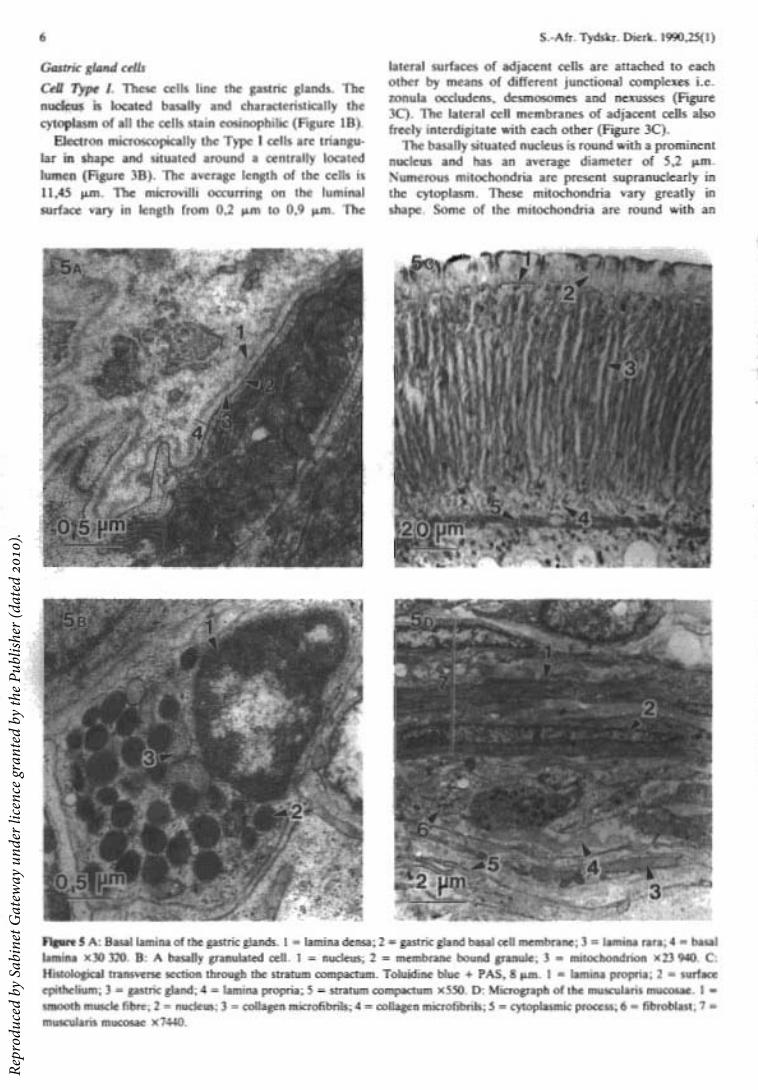
have a homogeneously electron dense core. The diameters of the granules vary from 114 nm to 164 nm (Figure 3C).
Basally granulated cells
These cells are located in the mucosa with a number accumulating beneath the surface epithelium. Numerous granules are situated in the basal portion of the cells. The shape of the cells varies, some being triangular in shape and others flask-shaped making contact with the gland lumen. The granules are membrane bound with an electron dense core. The diameter of the granules of the different cells varies from approximately 160 nm to 700 nm (Figure 5B). All the cells are in contact with the gland lumen.
Lamina propria
Light microscopically the lamina propria forms two fairly distinct layers, one layer adjacent to the surface epithelium (Figures 1B & 5C) and the other between the basal ends of the gastric glands and the muscularis mucosae (Figures Ie & 5C). Between the gastric glands the connective tissue of the lamina propria is sparse. Electron microscopically the fibre components of the lamina propria consist mainly of collagen microfibrils with diameters of 20 nm to 26 nm. The cellular components of the lamina propria contain cells such as fibroblasts, plasma cells and macrophages.
Muscularis mucosae
The muscularis mucosae consists of a few smooth muscle fibres that form a thin circular muscle layer (Figure lA). Electron microscopically the smooth muscle fibres are arranged in two or three layers (Figure 5D). The smooth muscle fibres are thin and spindle shaped with a centrally located nucleus. The cytoplasm contains myofilaments, dense bands, dense bodies and rough endoplasmic reticulum. The sarcolemma contains numerous caveolae.
The smooth muscle fibres are separated from each other by conspicuous amounts of collagen microfibrils between the individual muscle fibres (Figure 5D). The muscle fibres are surrounded by a basal lamina.
Stratum compactum. A distinct connective tissue layer situated beneath the gastric glands and lamina propria is observed with Alcian blue-PAS staining (Figure 5C).
Electron microscopically the stratum compactum seems to be closely packed collagen microfibrils between the smooth muscle fibres of the muscularis mucosae (Figure 5D).
Stratum granulosum. A conspicuous accumulation of granulated cells adjacent to the stratum compactum in the lamina propria and submucosa is observed in O. mossambicus (Figure 1 C).
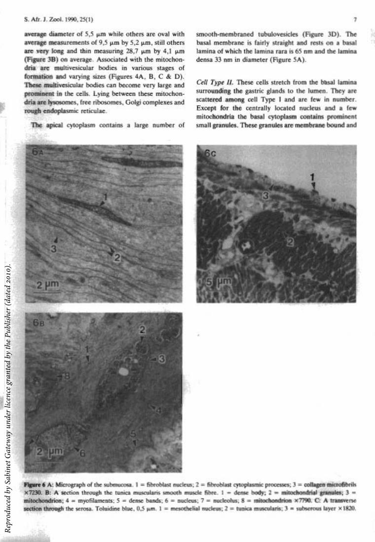
Submucosa
The connective tissue forming the submucosa is of a loose type (Figure 6A) with the collagen showing cross striations. These collagen microfibrils vary in diameter
S.-Afr. Tydskr. Dierk. 1990,25(1)
from 24,8 nm to 31,0 nm and form a loose network of microfibrils throughout the submucosa. The cellular component consists of different cells such as fibroblasts, plasma cells, macrophages and mast cells.
Tunica muscularis
This layer can be subdivided into an inner circular layer and an outer longitudinal layer according to the orientation of the smooth muscle fibres (Figure lA).
The smooth muscle fibres of the tunica muscularis are more robust compared to the fibres of the muscularis mucosae. The smooth muscle fibres are spindle-shaped with a central oblong nucleus. In the cytoplasm organelles such as myofilaments, dense bodies, mitochondria, Goigi complexes, rough endoplasmic reticulum and glycogen are present (Figure 6B). A large amount of conspicuous dark granules are present between the cristae of the mitochondria (Figure 6B). These granules can become so numerous that they displace the cristae. The caveolae of the sarcolemma are grouped in rows, two caveolae wide and parallel to the long axis of the fibre. Intervening between the groups of caveolae are dense bands (Figure 6B). Adjacent muscle fibres make contact with each other by means of desmosome-like junctional complexes. A basal lamina surrounds the smooth muscle fibres and collagen microfibrils are present between the fibres.
Serosa
A serosa is present with a single layer of squamous cells resting on a prominent sereus connective tissue layer (Figure 6C).
Discussion
The fine structure of the surface mucous cells in Oreochromis mossambicus differs very little from that described for the perch Perca fluviatilis (NoaillacDepeyre & Gas 1978), coral fish Chelmon rostratus (Ling & Tan 1975) and other vertebrates (Ito & Winchester 1963; Ito 1967; Stephens & Pheiffer 1968; Rubin, Ross, Sleisenger & Jeffries 1968; Geuze 1971). The surface mucous cells of O. mossambicus do not, however, cover the gastric pits as is generally found in other vertebrates. Bouhours, Bouhours & Bryon (1981) distinguish two types of mucus-secreting cells in the epithelial cells of the guinea pig stomach. The one type of mucus-secreting cell has smaller secretory granules homogeneously electron dense and entirely glycoproteic in nature. The mucous granules present in the apical portion of the surface mucous cells of the bream appear to be similar to the glycoproteic smaller granule as described by Bouhours et al. (1981). The other type of mucus-secreting cells defined by Bouhours et al. (1981) contains a larger heterogeneous secretory granule. These granules contain a proteinaceous core containing pepsinogen surrounded by carbohydrates. Similar heterogeneous granules have been described by Ito & Winchester (1963) in the bat Myotis lucifugus lucifugus, Ito (1967) in man and Noaillac-Depeyre & Gas (1978) in
Rep
rodu
ced
by S
abin
et G
atew
ay u
nder
lice
nce
gran
ted
by th
e Pu
blis
her (
date
d 20
10).

S. Afr. J. Zool. 1990,25(1)
the perch P. fluviatilis. The gastric pit mucous cells of the bream O. mossambieus contain similar heterogeneous mucous granules. These granules may be the source of pepsinogen in the stomach of the bream O. mossambieus as no pepsinogenic cells appear to be present. The gastric pit mucous cells of O. mossambieus are similar to the mucous neck cells of the perch P. fluviatilis (Noaillac-Depeyre & Gas 1978), the bat M. lucifugus lucifugus (Ito & Winchester 1963), man (Ito 1967; Rubin et al. 1968), the rat (Wattel, Geuze & de Rooij 1977) and the guinea pig (Bouhours et al. 1981), although these cells are located in the gastric pits in contrast to other vertebrates where they are found in the neck region of the gastric gland.
The gastric glands of O. mossambieus differ from the gastric glands that were originally described by Steven & Leblond (1953) for the rat and since then generally used as the model for gastric glands (Ito & Winchester 1963; Ito 1967; Wattel, Geuze & de Rooij, 1977; Fawcett 1986). This model depicts the gland being divided into an isthmus, neck and base opening into the bottom of a gastric pit. The uppermost parietal cell marks the boundary between foveola and isthmus, the uppermost mucous neck cell demarcates the boundary between the isthmus and neck and the lowest mucous neck cell the boundary between the neck and base. In contrast the gastric gland of the bream O. mossambieus seems to be far simpler. The gland consists of two cell types, cell Type I being the main component of the gland. The possible equivalent to mucous neck cells in the bream are located in the gastric pits. No cells similar to chief cells have been observed in the gastric glands of O. mossambieus .
It is generally accepted that in the gastric glands of bony fish, amphibians, reptiles and birds both hydrochloric acid and pepsinogen are secreted by one cell type namely the oxynticopeptic cell while the gastric glands of mammals have separate cells producing hydrochloric acid and zymogen (Ito 1967). In O. mossambicus, however, cell Type I of the gastric glands was found to be similar in ultrastructure to the mammalian parietal cell. No zymogen granules appear to be present in the cytoplasm of these cells. Cell Type I of the gastric gland of the bream, O. mossambieus in contrast to that of other bony fish (Noaillac-Depeyre & Gas 1978; Ling & Tan 1975), seems to have only a single function i.e. that of possible hydrochloric acid production. Although cell Type I of O. mossambieus appear to be similar to the parietal cell of mammals there are a few differences. Neither secretory canaliculi or intracellular canaliculi nor intercellular canaliculi between the lateral plasmamembranes are present in cell Type I of O. mossambicus.
A wide variety of studies have been done on the formation, increase and decrease of tubulovesicles and microvilli of parietal cells in a number of different animals (Lillibridge 1964; Sedar 1969; Helander & Hirschowitz 1972; Leeson 1973; Ito & Scofield 1974). In studies done on the changes that take place in the tubulovesicular compartment of mouse parietal cells during gastric acid secretion, Ito & Schofield (1974)
noted that multivesicular bodies were particularly abundant. Although the significance of this is still obscure the multi vesicular bodies may be involved in the activation and deactivation of parietal cell secretions. Winborn & Seelig (1974), however, have attributed the role of degradation of mitochondria to the multivesicular bodies. The remarkably high number and variety of forms of mitochondria in parietal cells are suggestive of a high oxidative metabolism (Helander 1981), and the changes in the tubulovesicular compartment could suggest a role for the multivesicular bodies of membrane redistribution or reconstitution. Both these theories seem to be borne out in cell Type I of the gastric gland of O. mossambicus where multivesicular bodies are found in different stages of formation as noted ultrastructurally.
Cells with basally situated granules in the cytoplasm were noted in the mucosa of O. mossambieus. These cells are possibly endocrine cells and morphologically comparable to the argentaffin cells as described by Ito & Winchester (1963) in the bat M. lucifugus lucifugus, Ito (1967) in man, Stephens & Pheiffer (1968) in the ferret, Ling & Tan (1975) in the coral fish Chelmon rostratus and Noaillac-Depeyre & Gas (1978) in the perch Perea fluviatilis. Further study is currently underway to histochemically determine the nature of these cells.
References
BANCROFT, J.D. & STEVENS, A. 1982. Theory and practice of histological techniques. Churchill Livingstone, London.
BOUHOURS, D., BOUHOURS, J-F. & BRYON, P-A. 1981. Association of glycoproteins and pepsinogen in the secretory granules of fundic epithelial cells isolated from guinea pig stomach. Biochem. Biophys. Acta 672: 288-296.
BURNSTOCK, G. 1959. The morphology of the gut of the brown trout Salmo trutta. Quart. J. micro Sci. 100: 183-198.
DA WES, B. 1929. The histology of the alimentary tract of the plaice Pleuronectes platessa. Quart. J. micro Sci. 73: 243-274.
EDINGER, L. 1877. Ueber die Schleimhaut des Fischdarmes, nebst Bemerkungen zur Phylogenese der Drusen des Darmrohres. Arch. f. mikros. Anat. 13: 651-692.
FAWCETT, D.W. 1986. A text book of histology. W.B. Saunders Co.
GEUZE, J.J. 1971. Light and electron microscope observations on the gastric mucosa of the frog Rana esculenta. Z. Zellfrosch. 117: 87-102.
GIBBONS, I.R. & GRIMSTONE, A.V. 1960. On the flagellar structure in certain flagellates. J. biophys. biochem. Cytol. 7: 679-716.
HALE, P.A. 1965. The morphology and histology of the digestive systems of two freshwater teIeosts, Poecilia reticulata and Gasterosteus aculeatus. J. Zool. 146: 132-149.
HELANDER, H.F. 1981. The cells of the gastric mucosa. Int. Rev. Cyt. 70: 217-289.
Rep
rodu
ced
by S
abin
et G
atew
ay u
nder
lice
nce
gran
ted
by th
e Pu
blis
her (
date
d 20
10).

10
HELANDER, H.F. & HIRSCHOWITZ, B.1. 1972. Quantitative ultrastructural studies on gastric parietal cells. Gastroenterol. 63: 951-961.
HUEBNER, E. & CHEE, G. 1978. Histological and ultrastructural specialization of the digestive tract of the intestinal air breather Hoplosternum thoraeatum (Teleost). J. Morph. 157: 301-328.
HUMASON, G.L. 1979. Animal tissue techniques. W.H. Freeman and Company, San Francisco.
ITO, S. 1967. Anatomic structure of the gastric mucosa. In: Handbook of physiology. (Ed.) Code, C.F., Vol. II, pp. 705--741. American Physiology Society, Washington, D.C.
ITO, S. & SCOFIELD, G.C. 1974. Studies on the depletion and accumulation of microvilli and changes in the tubulovesicular compartment of mouse parietal cells in relation to gastric acid secretion. J. Cell Bioi. 63: 364-382.
ITO, S. & WINCHESTER, R.J. 1963. The fine structure of the gastric mucosa in the bat. J. Cell Bioi. 16: 541-577.
LEESON, T.S. 1973. Canaliculi and tubulovesicles of rat parietal cells. Am. J. Anat. 136: 541-547.
LILLmRIDGE, C.B. 1964. The fine structure of normal human gastric mucosa. Gastroenterol. 47: 269-296.
LING, E.A. & TAN, C.K. 1975. Fine structure of the gastric epithelium of the coral fish, Chelmon rostratus Cuvier. Okajimas Fol. anat. jap. 51: 285--310.
LUFf, J.H. 1961. Improvements in epoxy resin embedding methods. J. biophys. bioehem. Cytol., 9: 409-414.
MILLONIG, G. 1961. Advantages of a phosphate buffer for osmium tetroxide solution in fixation. J. appl. Physiol. 32: 1637-1638.
NOAILLAC-DEPEYRE, J. & GAS, N. 1978. Ultrastructural and cytochemical study of the gastric epithelium in a freshwater teleostean fish Perea fluviatilis. Tiss. Cell 10: 23-37.
S.-Afr. Tydskr. Dierk. 1990,25(1)
PEASE, D.C. 1964. Histological techniques for electron microscopy. Academic Press, New York.
ROMEIS, R. 1948. Mikroskopische Technik. Leibniz Verlag, Miinchen.
RUBIN, W., ROSS, L.L., SLEISENGER, M.H. & JEFFRIES, G.H. 1968. The normal human gastric epithelia. A fine structural study. Lab. [nv. 19: 598-626.
SABATINI, D.D., BENSCH, K. & BARRNETT, R.L. 1963. Cytochemistry and electron microscopy. The preservation of cellular ultrastructure and enzymatic activity by aldehyde fixation. J. Cell Bioi. 17: 19-23.
SEDAR, A.W. 1969. Uptake of peroxidase into the smoothsurfaced tubular system of the gastric acid-secreting cell. J. Cell Bioi. 43: 179-184.
SIS, R.F., IVES, P.J., JONES, D.M., LEWIS, D.H. & HAENSL Y, W.E. 1979. The microscopic anatomy of the oesophagus, stomach and intestine of the channel catfish, Ictalurus punetatus. J. Fish Bioi. 14: 179-186.
STEPHENS, R.J. & PHEIFFER, C.J. 1968. Ultrastructure of the gastric mucosa of normal laboratory ferrets. J. Ultrastruet. Res. 22: 45--62.
STEVENS, C.E. & LEBLOND, C.P. 1953. Renewal of the mucosa cells in the gastric mucosa of the rat. Anat. Ree. 115: 231-239.
VENABLE, J.H. & COGGESHALL, R. 1965. A simplified lead citrate stain for use in electron microscopy. J. Cell Bioi. 25: 407-408.
WATTEL, W., GEUZE, J.J. & DE ROOU, D.G. 1977. Ultrastructural and carbohydrate histochemical studies on the differentiation and renewal of mucous cells in the rat gastric fundus. Cell Tiss. Res. 176: 445-462.
WEISEL, G.F. 1973. Anatomy and histology of the digestive system of the paddlefish Polyodon spathula. J. Morph. 140: 243-256.
WINBORN, W.B. & SEELIG, L.L. 1974. Pattern of osmium' deposition in parietal cells of the stomach. J. Cell Bioi. 63: 99-119.
Rep
rodu
ced
by S
abin
et G
atew
ay u
nder
lice
nce
gran
ted
by th
e Pu
blis
her (
date
d 20
10).
Copyright © 2022 FDOKUMEN