Ultrastructural Modelling of the Matrix-Cilium-Golgi Continuum ...
397
Ultrastructural Modelling of the Matrix-Cilium-Golgi Continuum in Hyaline Chondrocytes Michael John Jennings A thesis submitted for the degree of Doctor of Philosophy of the University of Otago, Dunedin, New Zealand 21 st July, 2014
-
Upload
khangminh22 -
Category
Documents
-
view
0 -
download
0
Transcript of Ultrastructural Modelling of the Matrix-Cilium-Golgi Continuum ...

Ultrastructural Modelling of the
Matrix-Cilium-Golgi Continuum in Hyaline Chondrocytes
Michael John Jennings
A thesis submitted for the degree of
Doctor of Philosophy
of the University of Otago, Dunedin,
New Zealand
21st July, 2014

i
Abstract
Overlooked for decades as an evolutionary vestigial organelle, the primary cilium has
recently become the focus of intensive investigations into understanding of the physical
structure and processes of eukaryote cell function. The cilium is central to various signalling
pathways and modalities for signalling, allowing centrosomal processing and regulation of
cellular and organelle function. The enigmatic process of their nanoscale ciliogenesis and
sensory function at present remain poorly understood. Dysfunction of their normal function or
component proteins correlates with a wide spectrum of diseases, or ciliopathies, which
express as developmental disorders and pathologies [1].
Normal connective tissue function requires their cells to sense and respond to
mechanical and physiochemical changes in the extracellular environment, and matrix, in order
to maintain their form and function. In hyaline cartilage, chondrocyte primary cilia are located
at an intermediate position between the mechanically functional extracellular matrix (ECM)
surrounding the cell, and the intracellular organelles responsible for producing, modifying,
transporting and secreting extracellular matrix materials [26, 96]. While many cellular and
physiological processes involving primary cilia are at present known, accurate in situ
knowledge of their form and fine structure in connective tissue is lacking.
A three dimensional model has been created of an in situ chondrocyte primary cilium.
This details the anatomical structure of the cilium and its relationship to other cellular
organelles. Components were mapped and divided into groups containing: the Matrix, the
Cilium, the Centrosome, the Golgi apparatus and the Nucleus. The extracellular matrix was
found to consist of interconnected tethered proteoglycans and matrix granules, with the
granules composed of aggregates of finer components. These are tethered to the ciliary
membrane at localised binding points. The membrane itself was observed as a dynamic
extension of the cell membrane, fitting neatly over the axoneme microtubule doublets and
their transport cargoes, while responding to the bending and torsional forces induced by the
ECM. The ciliary axoneme comprises a ‘9+0 structure’ of microtubule doublets of various
lengths and inclinations, associated linkage proteins, ciliary necklace proteins, and
intraflagellar-transport particle ‘rafts’ of materials, contained within the ciliary membrane.
The basal body is composed of nine microtubule triplets, and is decorated with
appendages of basal feet, for the attachment of radiant cytoplasmic microtubules (as the foci
of the microtubule organising centre, MTOC), as well as alar sheets and transition fibres,
which function to anchor the basal body to the cell and ciliary membrane. The basal body
microtubule polarisation, curl and inclination have been determined, along with those of the
subtending proximal centriole, together the constituent components of the centrosome. The
nuclear double membrane, and nuclear pores, and their close co-relation with the centrosome
are shown.

ii
Golgi cis-, medial- and trans-compartments, with associated transport vesicles are
described along with their polarization. Clathrin coated pits were found, indicating receptor-
mediated endocytosis. The outcome of this study is the first high-resolution reconstruction of
a primary cilium. The model will enable interpretation of the interactions and involvement of
the many biochemical and biophysical pathways now known to be associated with the cilium,
and will lead to new understandings of processes fundamental to the workings of the
eukaryotic cell.
The model raises new questions about the visualisation of the structure and form of
the primary cilium. A vast volume of literature exists upon many aspects of cell biology,
however few studies have undertaken investigations of primary cilia to understand their
structure, and attempt to translate it to function. This is the first study to attempt to probe the
complex relationship between the extracellular matrix, the mechanosensitive primary cilium,
the centrosome and the Golgi apparatus, which as a continuum are responsible for
maintaining the cells microenvironment.

iii
Acknowledgements
I am indebted to Associate Professors John and Jenny Leader for their kind support
and guidance in making the completion of this manuscript possible. I would also like to thank
Professor Rob Walker, and Dr Duane Harland and Mr Richard Walls of AgResearch Lincoln
for their encouragement and invaluable assistance in gathering data during electron
tomography visits to Ag-Research Lincoln, and with KAREN remote access sessions.
Likewise, Associate Professor Tony Poole for many interactions, Mr Allan Mitchell, Mr
Richard Easingwood and Ms Gillian Grayston for electron microscopy support, from
specimen fixation, and electron microscopy techniques, to discussions and many coffees.
I would also like to thank Dr David Mastronarde of Boulder Colorado for his support
with IMOD software packages, and many interesting discussions on removing bugs and
glitches from the software during the many hours of reconstructions and final modelling
processes. I am also indebted to Ms Megan Bailey, Ms Katrin Geist and Mr Chaz Forsyth for
their patience in proof reading and kind support, as well to many others
And lastly, Mr Jean-Pierre Houdin, for sharing many interesting parallel discussions
about the intricacies of designing and building specialist architectural structures from the
inside that do not succumb to the rigors of time.

iv
Table of Contents
Preliminaries Page
Abstract i
Acknowledgements iii
Table of Contents iv
List of Tables ix
List of Figures x
List of Abbreviations xiii
1.0 Chapter One: The Matrix-Cilium-Golgi-Continuum
1.01 Preamble 1
Section I – Previous Knowledge of the Structure of the Primary Cilium
1.02 A Brief History of the Primary Cilium 3
1.03 Early Work: Optical and Electron Microscopy of Cilia 4
1.04 The Discovery of Chondrocyte Primary Cilia 5
1.05 The Ciliary Axoneme 5
1.06 Ciliary Ultrastructure 6
1.07 The Transition Zone ‘Compartment’ 7
1.08 Basal Body Appendages - Proximal to Distal 8
1.09 The Basal Feet (Basal Appendages) 8
1.10 The Striated Rootlets 9
1.11 The Centriole: A Template for the Basal Body and Axoneme 10
1.20 The Matrix-Cilium-Golgi Continuum 11
1.21 The Microtubule Organising Centre (MTOC) 13
1.22 The Peri-Centriolar Matrix (PCM) 14
1.23 The Cytoskeleton 15
1.24 Microtubules 16
1.241 Tubulins 16
1.242 Microtubule Nucleation Complex 16
1.243 Polymerisation 16
1.244 Tubulin Post-Translational Modification 17
Section II – Functional Relations of the Cilium during the Cell Cycle
1.30 The Cell-Centrosome-Cilium Cycle 19
1.31 Intra-Flagellar Transport (IFT) 20
1.311 Ciliary Transport 21
1.312 IFT co-ordination 21
1.32 Primary Cilium Biogenesis 21

v
1.33 Entry of Material to the Cilium 23
1.34 Ciliary Localisation Sequences 24
1.35 Dynamic Control of Ciliary Length 25
1.40 The Ciliary Membrane 25
1.41 The Cilium: Receptors and Luminal Components 27
1.42 The Cilium and the Extracellular Matrix 29
1.43 Signalling and Mechanotransduction 29
1.44 Purinergic Mechanotransduction 30
1.45 Ciliary Polymodal Sensory Function 31
1.50 The Centrosome: The Central Processing Unit 32
1.501 The Centrosome, the Cytoskeleton and Transport 32
1.502 The Kinesin Family 33
1.503 The Dynein Family 34
1.504 The Dynein Activator - Dynactin 34
1.51 The GTPase Family Members 36
1.511 Function of Rab Family Members 37
1.512 The Cilium, Rabs and GTPases 37
1.513 Rabs, Vesicular Processes and the Golgi 38
1.514 Golgi Derived Transport: Targeting of Materials from the Golgi 39
1.515 Transport and the Golgi Apparatus 41
1.60 Microtubule Roles During the Interphase 41
1.61 The Cilium, Centrosome and the Golgi 42
1.62 Centrosome Positioning 44
1.63 Intracellular Transport 44
1.64 Transport of Vesicles 44
1.65 Rabs and Microtubule Motors 44
1.66 Myosin Motors and Rabs 45
1.67 The Golgi Apparatus: Transport Motors 45
1.70 A Brief History of the Golgi Apparatus 47
1.71 Models of Transport Through The Golgi 48
1.72 The Golgi: Regulated Processing and Secretion 49
1.73 Planar Cell Polarity: The Centrosome and the Golgi 50
1.74 Planar Cell Polarity, Wnts and Cartilage 51
1.75 The Cilium to Golgi Continuum: Summary 51
Section III – A Biomechanical Connective Tissue: Cartilage
1.80 Introduction: Biomechanical Function of Connective Tissues 52

vi
1.81 Overview: Connective Tissues 52
1.82 A Compressive Tissue Matrix: Hyaline Cartilage 53
1.83 The Chondron 54
1.84 Articular Cartilage Function 55
1.85 Cartilage Collagens 56
1.86 Cartilage Glycosaminoglycans (GAGs) 57
1.87 Cartilage Proteoglycan Core Proteins and Glycoproteins 57
1.88 Aggrecan 58
1.89 Synthesis of Extracellular Matrix: Proteoglycans, GAGs and Collagen 59
1.90 Binding the Matrix to the Cell Membrane 60
1.91 Chondrocyte Matrix-Coupled Receptors 61
1.92 Cartilage Biomechanics: The Chondrocyte Microenvironment 62
2.0 Chapter Two: Methods
2.0 Overview 63
2.10 Introduction: Electron Tomography and Primary Cilia 63
2.11 Connective Tissue Models 63
2.12 Embryonic Chick Sternal Cartilage: A Model Tissue 64
2.13 Chick Embryo Cartilage Fixation: in vivo 64
2.14 Chondrocyte Cell Culture: in vitro 65
2.15 Fixation of Cultured Cells 65
2.20 Fixation, Osmification, Ruthenium Red (RHT) and Artefacts 66
2.21 Fixation with Ruthenium Complexes 66
2.22 Fixation of Extracellular Matrix Proteoglycans 67
2.30 Serial Sectioning and Detection of Primary Cilia 68
2.31 Semithick Sectioning of Sternal Cartilage 68
2.32 Tomography of Monolayer Cell Culture: Aligning Cilia With The Substrate 69
2.40 Investigative Electron Microscopy 70
2.41 Fiducial Marker Application 70
2.42 Imaging Microscope 71
2.43 Remote Tomography 71
2.44 Specimen Attrition 72
2.45 Collection and Analysis 72
2.46 Data Processing 73
2.47 Fiducial Tracking 73
2.50 Analysis and Modelling of the Tomogram 74
2.51 Model Display 75

vii
2.6 A Virtual Cilium: SolidWorks 75
Chapter Three: Results
3.0 Introduction 77
3.1 Mechanical Connective Tissues: Tendon 77
3.2-3.3 Mechanical Connective Tissues: Chondrocyte Cilia Morphology and
Staining 77
3.4 The Chondrocyte Primary Cilium: In Vitro 78
3.5 The Chondrocyte Primary Cilium: In Situ 78
3.6 Tomography of Native Sternal Chondrocytes 79
3.7-3.9 A Single Axis Model 80
3.10 Tomographical Modelling of the Extracellular Matrix and Matrix Granules 80
3.11 Matrix Ciliary Membrane Interactions 80
3.12 Matrix to Ciliary Membrane to Microtubule Interactions 81
3.13-3.15 The Axoneme Microtubules 81
3.151 Localisation of Intra Ciliary Transport Materials 82
3.16 The Distal Axoneme 82
3.17 The Middle Axoneme 82
3.18 The Transition Zone – One 83
3.19 The Transition Zone – Two 83
3.20 The Basal Body Microtubule Triplets 84
3.21-3.22 Internal Structures, Luminal Disc, Fibres and Vesicle 84
3.23-34 Basal Appendage One 84
3.26-3.27 Basal Appendage Two (BA2) 85
3.28 A Review of Basal Appendages One and Two 86
3.29 The Cilium, Basal Body Materials and Orientation 86
3.30 The Proximal Centriole 87
3.31 Proximal Centriole Radial Components 87
3.32 The Centrosome (1) Ultrastucture and Modelling 88
3.33 The Centrosome (2) The Centrosomal Torus 88
3.34 The Centrosome (3) The Pericentriolar Environment 89
3.35 The Centrosome (4) Basal Body, Proximal Centriole within the MTOC 89
3.36-3.40 The Cytosolic Microtubules; Centrosome to Golgi Connectivity;
The Cis-, Medial- and Trans-Golgi Compartments;
Transport of Matrix from the Golgi 90
3.41-3.42 The Complete Model: The Matrix-Cilium-Golgi Continuum 90
3.43 The Continuum: The Cilium and Centrosome 91

viii
Chapter Four: Discussion
4.0 Introduction and Overview 133
4.10 Limitations of Study 134
4.11 Ruthenium Interactions with Cells and Membrane Bound Structures 135
4.12 Occurrence and Morphology of Chondrocyte Primary Cilia 135
4.13 Chick Sternal Cartilage Tomogram Selection 136
4.14 Defining the Cilium and its Relation to the Golgi 136
4.20 Interpretation of the Model Structure 137
4.21 The Matrix of the Ciliary Pocket 137
4.22 The Periciliary Membrane and the Ciliary Pocket 139
4.23 The Ciliary Membrane 139
4.231 Linking Matrix to the Ciliary Membrane and Transduction 140
4.232 Cilia, ENac and Mechanotransduction 141
4.233 The Axoneme Membrane 142
4.234 Ciliary Membrane Forces 142
4.24 The 9+0 Microtubule Axoneme 143
4.241 The Flexural Rigidity of Primary Cilia 143
4.242 Distribution of Materials upon the Axoneme Doublets 144
4.243 Distribution of IFT-like Particles 145
4.25 The Transition Zone 147
4.251 Y-shaped Linker Structure 147
4.252 Sub-Distal Fibres: Filamentous Structures 148
4.253 The Alar-Sheets Structure 148
4.30 The Centrosome 150
4.31 The Basal Body 151
4.32 The Microtubule Triplet Structure 151
4.33 Luminal Vesicles 152
4.34 Basal Luminal Discs and Internal Structures 152
4.35 The Basal Appendages 153
4.36 The Proximal Centriole 154
4.40 Microtubule Populations 155
4.41 The Intermediate Filament Organisation Centre (IFOC) 156
4.42 Order in the Pericentriolar Matrix 157
4.43 The Centrosomal ‘Torus’ 157
4.44 Nuclear Pores 158
4.50 The Golgi Apparatus 159
4.51 The trans-Golgi Network 159

ix
4.52 Coated Pits and Caveoli 161
4.53 Are Primary Cilia Both Displacement Detectors and Pressure Sensors? 161
4.54 Matrix-Cilium-Golgi Continuum in Chondrocytes 162
4.60 Modelling the Primary Cilium 162
4.7 Future Work 163
References 165
Appendices Supplemental Information to Thesis 239
Appendix I: Receptors and Signalling in the Cilium and Centrosome 240
1.1 Membrane Receptors, Luminal Components and Signalling Pathways 240
1.2 Axoneme Associated Proteins 272
1.3 Centrosome Associated Proteins 283
1.4 Cell-Cycle Regulatory Kinases 299
1.5 Regulatory Components 304
Appendix II: Rab-GTPases - Ciliary and Golgi Function 309
Appendix III: Kinesin and Dynein Microtubule Motors 321
3.1 Kinesin Motor Proteins 322
3.2 Axonemal Dynein Proteins 333
3.3 Cytoplasmic Dynein Proteins 333
Appendix IV: Intra-Flagellar Transport Complexes 342
Appendix V: Kinesin and Dynein Motors Responsible for Organelle Processes
and Transport 347
Appendix VI: A Select Review of Tomography and Modelling 360
Appendix VII: Animation List 370
Appendix VIII: Poster Presentations 372
List of Tables
Table 3.1 Measurements of Chondrocyte Primary Cilia 79

x
List of Figures
Chapter One: The Matrix-Cilium-Golgi-Continuum
Figure 1.1 The ‘Central Flagellum’ 3
Figure 1.2 The Primary Cilium of a Chondrocyte 5
Figure 1.3 Diagrammatic Cross Sections of Axonemes 6
Figure 1.4 The Membrane, Axoneme and Basal Body of the Motile Cilium 7
Figure 1.5 The Basal Body and Transition Fibres (Alar Sheets) 8
Figure 1.6 Observations of the Range and Distribution of Basal Appendages 9
Figure 1.7 Striated Rootlets of the Diplosome 9
Figure 1.8 The Nine-Fold Symmetry of the Centriole 10
Figure 1.9 Illustration of the Matrix-Cilium-Golgi-Continuum 12
Figure 1.10 The Interphase Centrosome 13
Figure 1.11 The Centrosome 15
Figure 1.12 Microtubule Nucleation and Doublet Structure 17
Figure 1.13 The Cell Centrosome Cilium Cycle 20
Figure 1.14 Biogenesis of the Primary Cilium 23
Figure 1.15 Golgi Derived Materials for Ciliogenesis 24
Figure 1.16 The Primary Cilium 28
Figure 1.17 The PC1/PC2 Mechanotransduction Complex 30
Figure 1.18 Proposed Mechanism of Flow Induced Ciliary Ca2+
and cAMP
Signalling Modalities 31
Figure 1.19 The Kinesin Motor 33
Figure 1.20 The Dynein Motor and Dynactin Complex 35
Figure 1.21: Regulated Golgi Transport and Signalling 40
Figure 1.22 Microtubules and Vesicle Transport 42
Figure 1.23 Polarisation of the Centrosome and Golgi in Cell Migration 43
Figure 1.24 The Golgi Intracellular Transport Pathways 48
Figure 1.25 Hyaline Articular Cartilage 54
Figure 1.26 The Chondron 55
Figure 1.27 Disaccharide components of hyaluronic acid, chondroitin-4, -6,
and keratan sulphates. 57
Figure 1.28 Cartilage Proteoglycans 58
Figure 1.29 The Proteoglycan Aggrecan 59
Figure 1.30 The Golgi Apparatus 59
Figure 1.31 Synthesis and Formation of Collagen: 60
Figure 1.32 Chondrocyte Matrix Membrane Adhesion Receptors 61

xi
Chapter Two: Methods
Figure 2.1 Imaging Resolution of Electron Tomography compared with Confocal
Microscopy 63
Figure 2.2 Chick Embryo Cartilage 64
Figure 2.3 Cell Culture 65
Figure 2.4 Ruthenium Hexa-amine-Trichloride 67
Figure 2.5 Tomography of a Primary Cilium Aligned with the Substrate 70
Figure 2.6 Fiducial Markers 70
Figure 2.7 Remote Electron Tomography 71
Figure 2.8 The ‘Missing Wedge’ Problem 72
Figure 2.9 Data Acquisition 73
Figure 2.10 Fiducial Tracking in IMOD 74
Figure 2.11 The Modelling Process 75
Figure 2.12 The Virtual Cilium 76
Chapter Three: Results
Figure 3.1 Connective Tissue Primary Cilia Sectioning of Tendon 91
Figure 3.2 Serial Sectioning of Chick Sternal Cartilages 92
Figure 3.3 Projection Types of Chondrocyte Primary Cilia 93
Figure 3.4 Cilium-Centrosome Serial Sectioning of Cultured Chondrocytes 94
Figure 3.5 Selection of Tomograms and their Z-axis Optical Sections 95
Figure 3.6 A Semithick Section Containing a Primary Cilium 96
Figure 3.7 Alignment of Tomogram Dataset 96
Figure 3.8 Tomogram Z-Axis Stack 97
Figure 3.9 An Optical Section of the Cilium Axoneme 98
Figure 3.10 Tomographical Modelling of Extracellular Matrix and
Matrix Granules 99
Figure 3.11 Matrix Ciliary Membrane Interactions 100
Figure 3.12 Matrix Ciliary Membrane Microtubule Interactions 101
Figure 3.13 Microtubule Doublet Nine-Fold Architecture 102
Figure 3.14 Localisation of Materials within the Axoneme 103
Figure 3.15 The Luminal Axoneme: Intra-Ciliary ‘Rafts’ and
Microtubule-Microtubule Interactions 104
Figure 3.16 The Distal Axoneme (1) 105
Figure 3.17 The Middle Axoneme (2) 106
Figure 3.18 The Transition Zone (1) – Tapered Zone 107
Figure 3.19 The Transition Zone (2) – Alar Sheets / Transition Fibres 108
Figure 3.20 The Basal Body (1) Microtubule Triplets 109

xii
Figure 3.21 The Basal Body (2) Luminal Discs, Deposits and Fibers 110
Figure 3.22 Basal Body (3) Internal Structures and Vesicle 111
Figure 3.23 Basal Appendage One: Optical Sectioning and Reconstruction 112
Figure 3.24 Basal Appendage One (1) Alignment of Basal Appendage
Basement Structures 113
Figure 3.25 Basal Appendage One (2) Ultrastructure of Arm, Docking Station and
Substrate 114
Figure 3.26 Basal Appendage Two (1) Ultrastructure and Modelling 115
Figure 3.27 Basal Appendage Two (2) Docking Complexes 116
Figure 3.28 Basal Body: Appendages Summary; Basement Structures,
Basal Arm and Docking Complex 117
Figure 3.29 Basal Body And Cilium Radial Components 118
Figure 3.30 Proximal Centriole Ultrastucture, Deposits and Model 119
Figure 3.31 Proximal Centriole Radial Components and Proximity
to Nuclear Pores 120
Figure 3.32 The Centrosome (1) Ultrastucture and Modelling 121
Figure 3.33 The Centrosome (2) The Centrosomal Torus 122
Figure 3.34 The Centrosome (3) The Pericentriolar Environment 123
Figure 3.35 The Centrosome (4) Basal Body, Proximal Centriole within
the MTOC 124
Figure 3.36 The Cytosolic Microtubules 125
Figure 3.37 Centrosome to Golgi Connectivity 126
Figure 3.38 The Cis, Medial and Trans Golgi Compartments 127
Figure 3.49 Transport of Matrix from the Golgi 128
Figure 3.40 The Cilium, Coated Pits and Caveolae 129
Figure 3.41 The Matrix Cilium Golgi Continuum 130
Figure 3.42 Plan of Tomogram Features 131
Figure 3.43 The Cilium Centrosome Continuum 132
Chapter Four: Discussion
Figure 4.1 The Transition Zone ‘Compartment’ 149

xiii
List of Abbreviations
α Alpha
β Beta
γ Gamma
δ Delta
δ Zeta
ε Epsilon
µ micron 1 × 10⁻⁶
Å Angstrom 1 × 10⁻¹⁰
aDMEM advanced Dulbecco’s Modified Eagle Medium
AC Adenylyl Cyclase
AKAP450 A-Kinase Anchoring Protein 450
ARF ADP-Ribosylation Factor
ARHGAP21 Rho GTPase activating protein 21
Arp1 Actin Related Protein 1
ATP-2 ATP synthase
AP2 Cytosolic Adaptor Protein
ATP Adenosine Tri-Phosphate
BA1 Basal Appendage 1
BA2 Basal Appendage 2
BBS Bardet–Biedl Syndrome
BCL B-cell lymphoma
BMP Bone Metamorphic Protein
BP1 Binding Protein 1
BSA Bovine Serum Albumen
CAM Cell Adhesion Molecule
cAMP cyclic-Adenosine Mono Phosphate
CDC42 Cell Division Control Protein 42
CC2D2A Coil and C2 Domain Containing 2A
CDK Cyclin Dependent Kinase
CEP Centrosomal Protein
CFTR Cystic Fibrosis Trans-membrane conductance Regulator
CLS / CTS Ciliary Targeting Sequence / Ciliary Localisation Sequence
CLSM Confocal Laser Scanning Microscopy
CNK2 Connector Enhancer Kinase 2
cnRNA Centrosomal RNA
CLASP Cytoplasmic Linker Associate Protein

xiv
COP1 Coat Protein One
CS Chondroitin Sulphate
Da Dalton
DAAM1 Dishevelled Associated Activator of Morphogenesis
DAG Diacyl-Glycerol
DCTN1 Dynactin Subunit 1
DNA Deoxyribo Nucleic Acid
DLIC-1 Dynein light intermediate chain-1
DYN Dynein
EB1 End-Binding Protein 1
ECM Extra Cellular Matrix
EGR Epithelial Growth Factor
EGFR Epidermal Growth Factor Receptor
ER Endoplasmic Reticulum
ERGIC Endoplasmic Reticulum Golgi Intermediate Compartment
ENaC Epithelial Na+
(Sodium) Transporter Channel
ET Electron Tomography
FCS Foetal Calf Serum
FGF Fibroblast Growth Factor
γ-TuRC γ-Tubulin Ring Complex
γ-TuSC γ-Tubulin Small Complex
GAG Glycoaminosglycan
Gβγ G beta gamma
GDP Guanosine Di-Phosphatase
GCP Gamma Complex Associated Protein
GERL Golgi-Endoplasmic Reticulum-Lysosome
GM130 Golgi Matrix protein 130
GMAP210 Golgi-microtubule-associated protein of 210 kDa
GPC Glypican
GPCR G-Protein Coupled Receptor
GTP Guanosine Tri-Phosphatase
GM130 Golgi Matrix Protein 130
GPC Glypican
HA Hyaluronic Acid
HF Hydrofluoric Acid
HEK Human Epithelial Kidney
HYLS1 Hydrolethalus syndrome protein 1

xv
IFT Intra-Flagellar Transport
IGD Interglobular Domain
IGF Insulin Like Growth Factor
IGFR-1R Insulin-Like Growth Factor-1 Receptor
ImageJ Freeware image analysis programme
IMOD An open-source suite for modelling electron microscopy data
INPP5E Inositol 1,4,5-trisphosphate (InsP3) 5-phosphatase
IP3 Inositol Trisphosphate
JBTS Joubert Syndrome
KAREN Kiwi Advanced Research Network
KIF Kinesin Motor Protein
KS Keratan Sulphate
l litre
Lis Lissencephaly sequence
LKB1 Liver Kinase B1
LZTFL1 Leucine Zipper Transcription Factor-Like 1
m meter
M Mega 1 × 10⁶
milli milli 1 × 10⁻³
ml milli-litre
µ micro 1 × 10⁻⁶
Osm Osmolarity
MARK Microtubule Affinity Regulating Kinase
MAP Microtubule Associated Protein
MAPK Mitogen Activated Kinase
MCH Melanin Concentrating Hormone
MDCK Madin-Darby Canine Kidney
MT Microtubule
MTOC Microtubule Organising Centre
n Nano 1 × 10⁻⁹
N Newton
NEDD1 Neural precursor cell Expressed, Developmentally Down-regulated 1
NIH3T3 National Institute of Health 3T3
NG2 Neural/Glial Antigen 2
NKF-kB Nuclear Factor Kappa Beta
NPHP Nephronophthisis (protein designator)
NudE Nuclear Distribution E

xvi
Nup133 Nucleoporin 133
ODF Outer Dense Fibre
ORP1L Oxysterol-binding Protein-Related protein 1L
Pa Pascal
Patched HedgeHog Signalling Component
PC Polycystin
PCM Peri-Centriolar Matrix
PCM-1 Pericentriolar Material 1
PDB Protein Data Bank
PDGF Platelet-Derived Growth Factor
PG Proteoglycan
pH H+ ion concentration
pico Pico 1 × 10-12
PIP3 Phosphoinositol-3
PI3K Phosphoinositide 3-kinase
PKC Protein Kinase C
PKD Polycystic Kidney Disease
PSG Penicillin Streptomycin Glutamine
p150Glued
Dynactin Component
P2X Purinergic ATP-gated ion channel
P2Y G-coupled receptor
Rab G Protein, member of the Ras family GTPases
RAC Rho-GTPase subfamily member
RHT Ruthenium Hexaamine Tri-Choride
ROCK Rho-GTPase effector
RP Retinitis Pigmentosa
RPGRIP Retinitis Pigmentosa GTPase Regulator Interacting Protein
RILP Rab-Interacting Lysosomal Protein
RNA Ribo Nucleic Acid
Shh Sonic Hedgehog
Solidworks Proprietary engineering software package
Smo Smoothened
SuFu Suppressor of Fused
SSTR Somatostatin Receptor
STAT6 Signal Transducer and Activator of Transcription
SD/ζ Standard Deviation
TMEM Trans Membrane Protein

xvii
TRAP Transport Protein Particle
TRP Transient Receptor Protein
TGF-β Transforming Growth Factor β
TRPV4 Transient Receptor Potential Vanillinoid 4
TEM Transmission Electron Microscopy
TGF-β Tumour Growth Factor-β
Vangl2 Vang-like protein 2
VEGF Vascular Endothelial Growth Factor
VxPx Ciliary Targeting Motif
XMAP215 Xenopus Microtubule Associated Protein 215
Wnt Wingless
ZW10 Zeste White 10
2D Two dimensions
3D Three dimensions
Pathways
Par3/Par6/aPKC Polarity Pathway
PI3K/Akt/mTOR Apoptosis Pathway
MEK/ERK Mitogen Activated / Extracellular signal Regulated Kinases
NB: For proteins not listed here, see the Appendices.

1
Chapter One: The Matrix-Cilium-Golgi Continuum
1.01 Preamble
There has been an explosive growth of interest in the primary cilium since the realisation that
its occurrence is almost universal in eukaryotic cells and its principal role is presumably to be sense
features of the cellular environment significant to a particular cell type, allowing an appropriate
response. Hence, the primary cilia of the renal epithelial cells detect flow, while the cilia of the rods of
the retina are light detectors, the cells of the cochlea detect vibrations, and the cilium of the
cartilaginous chondrocyte detects stress-induced deformation. The importance of this structure has
been underlined by the discovery that about 2000 proteins1 are involved in its construction and
operation, with mutations in these being often lethal or imposing severe functional deficiencies, which
have become known as ciliopathies [1]. This has focused attention on the relation between structure
and function of this organelle. However, most of the primary cilium and its associated structures is at
the limits of resolution of available imaging techniques.
The primary cilium is a singular centrosomal organelle present in almost all eukaryotic cells
and is distinct from other kinds of motile cilia and flagella in its unique (9+0) microtubule structure.
Few conventional electron microscopy based investigations have attempted to model the primary
cilium. The main experimental objective of this study was to produce an anatomically accurate,
ultrastructure based three-dimensional model of a connective tissue primary cilium using electron
tomography. Previous connective tissue investigations of primary cilia used serial sectioning, and
these were limited in their resolution by the ultrathin section thickness (90-120 nm). The technique of
electron tomography is ideally suited for investigation of ultrastructure contained within thicker
semithick (350 nm) sections that could contain a longitudinally aligned entire cilium. The application
of tomography permits the construction of an interrogative model allowing investigation of the
structural continuum between the biomechanically functional matrix, the mechanically sensitive
primary cilium and the cytoplasmic organelles responsible for the secretion of the functionally
effective extracellular matrix.
The principal aim of all biological investigation is to achieve a marriage between structure
and function. Modern technological advances, including high voltage electron tomography, offer the
potential to achieve a physical resolution near to atomic levels, and in three dimensions, albeit in a
static form. Advances in the incorporation of function, on the other hand, have been less clear. There
is an abundance of information, drawn from diverse sources, inferring presence of macromolecules,
and intermolecular relationships, but at the present time there is little in the way of synthesis into a
coherent picture. This work attempts to progress towards a unifying picture of the relation between
structure and function of the primary cilium by introducing a valid, three dimensional representation
of the primary cilium at high resolution upon which functional activity can be imposed as biochemical
1 http://www.ciliaproteome.org/

2
knowledge advances. This introduction is intended to provide not only a summary of present
knowledge of the ultrastructure of the primary cilium and its associated structures, but also a review
of the proteins known or hypothesised to be involved with its regulation and functional role.
This study presents novel new research using the recently refined ultrastructural imaging
technique of electron tomography. This advanced technology not only allows detailed investigation of
structural relationships at extreme magnifications not previously possible, but also enables translation
of this acquired data into an anatomically accurate, interactive, three-dimensional model of the
ultrastructural continuum that exists between the matrix, the cilium and the Golgi apparatus in
connective tissue cells. Hyaline cartilage chondrocytes from chick embryo sterna have previously
used as a model of connective tissue to study primary cilia, and further study of this tissue forms the
basis of this thesis.

3
Section I – Previous Knowledge of the Structure of the Primary Cilium
1.02 A Brief History of the Primary Cilium
The history of the investigation of primary cilia has been extensively reviewed by Bloodgood
(2009) [2] and Wheatley (1982) [3]. Cilia are amongst the earliest known eukaryotic organelles, with
the first observations of primary cilia likely unknowingly made around 1763 by Leeuwenhoek [2, 4]
during his detailed observations of cells and other micro-organisms resulting from his invention of the
optical microscope [5]. The descriptive term ‘cilium’ was first introduced by Muller in 1786 [6]
meaning ‘eyelash’, while Dujardin [2] used the term ‘flagellum’ in 1841 to describe motile cilia [2].
In 1876 Langerhans [7] published sketches of epithelia, showing a singular cilium projecting from
cells, with an intracellular component at its base. It is clear that many have observed both types of
cilia, yet Zimmerman is credited with discovering the primary cilium in 1894 [8], which he termed the
‘central flagellum’ [9]. In 1898 Zimmerman [9] described motile cilia as being distinct from a
singular ‘primary cilium’, which originated from a pair of centrioles (the diplosome), and he proposed
a role for it as a sensory organelle (see Figure 1.1) [2]. Bernhard and deHarven, however, introduced
the term ‘primary cilium’ in 1956 [3]. Cilia and flagella are unique microtubule based membrane
bound organelles, where description in the common lexis, flagella pertain to the uni-cellular motile
apparatus, cilia being used to describe both motile, and non-motile primary cilia alike [2, 10].
Importantly, a functional distinction should be clearly drawn between motile cilia, which act on the
surrounding medium to generate motion, and the primary cilium, which not only has a unique and
characteristic structure, but also is non-motile and functions as a sensory detector.
Figure 1.1 The ‘Central Flagellum’: A reproduction of images from Zimmerman’s original publication ‘Beitrage zur
Kenntniss einiger Drusen und Epithelien’ [9] published in 1898, detailing observations of kidney tubule epithelium expressing a primary cilium from their centrosome (arrows). The centrosome consists of a ‘diplosome’ of two centrioles
observed near the resolving limit of light microscopy. Reproduced from original scans publish in Bloodgood (2009) [2],
courtesy of Dr Bloodgood.
The discovery of the centrosome is attributed to Flemming, around 1875 [11, 12], while the
common terms ‘centrosome’ and ‘centriole’ were introduced by Boveri in 1888 and 1895 respectively
[13]. The Henneguy-Lenhossek hypothesis originated in 1898, defining the role of the centrosome in
the formation of the spindle poles during cell division, the motile tail of spermatids in
spermatogenesis, and generating the base of a primary cilium of a cell during interphase [3, 14].
Jennings (1899) [15] studied mechano-sensory reactions in unicellular organisms and described

4
behavioural responses to stimuli in the ciliate Paramecium, hinting at a sensory role for motile cilia
[16], although a sensory function is now attributed to all cilia [17].
1.03 Early Work: Optical and Electron Microscopy of Cilia
A lot of the early 20th century work on cilia focussed on cilia of unicellular organisms or
epithelial sheets contrasted against an optically transparent lumen. Unlike epithelial cells and
unicellular organisms which often bear many cilia, most eukaryotic cells typically possess only a
single primary cilium, whose small width of 200 nm and nominal length of 1-4 µm, places it near the
limit of resolution of optical microscopes (about 200 nm) determined by Abbe (1873) [18, 19].
The invention of the transmission electron microscope (TEM) in 1931 by Max Knoll and
Ernst Ruska [20] allowed far greater magnifications to be achieved. The use of osmium tetroxide [21]
for fixation, combined with the embedding of biological materials into epoxy resins in the 1950’s
enabled the stabilisation of chemically fixed structures, and the use of heavy metal stains, such as lead
or uranium, provided enhanced contrast [22]. With the development of diamond knives, reliable
methods of ultra-thin sectioning emerged in the 1950’s [2]. The main drawback with many of these
early observations in electron microscopy was that inadequate fixation and processing techniques
produced uncertain artefacts. During this period, different anatomical expressions of primary cilia
were discovered to exist in many cell types and organisms ranging from protozoa to mammals [23].
For a review of historic publications on the primary cilium prior to 2005, see Wheatley (2005) [24].
Extensive ultrastructural analysis of primary cilia both in vivo [25-29] and in vitro [30, 31, 32] largely
ignored their functional role within the eukaryotic cell until recently [33].
Studies by Praetorius et al., (2001, 2003) [34, 35] established that epithelial primary cilia
could act as flow sensors in the renal collecting duct, and their bending resulted in the activation of
intracellular calcium signalling. It has subsequently become apparent that primary cilia in vertebrates
encompass a wide range of sensory modalities from the ocular rod and cone photoreceptors [36],
including olfaction and audition, to tonicity and flow detection in renal primary cilia, which all play
specialist roles in signal transduction. They also occur in numerous tissues and cell types (with a
current list maintained at the Bowser Lab2), although red blood cells, oocytes and some lymphatic
cells lack this structure. Signalling components and structural proteins involved with the cilium
involve at least 2,000 genes, whose proteins are specific to, or associated with ciliary function or the
Ciliome3 [37-41].
2http://www.bowserlab.org/primarycilia/cilialist.html
3 www.ciliome.com

5
1.04 The Discovery of Chondrocyte Primary Cilia
There are few anatomical examples of the basic structure of the
primary cilium. One of these is from chondrocytes (Figure 1.2). Initially,
these were thought to be non-ciliated. The first known recorded
observations of primary cilia in chondrocytes were made by Scherft et
al., (1967) [42] reviewing their occurrence in cartilage. Hart (1968) [43]
undertook ultrastructural studies of the cilium, and this was followed by
Federman et al., (1974) [44], and Wilsman [27, 28, 29]. Wilsman (1978)
[27, 28] performed the first detailed studies of in situ chondrocyte
primary cilia where their incidence and morphology were investigated by
exhaustive serial sectioning. This allowed for the first artistic perspective
of the three-dimensional anatomy of a chondrocyte primary cilium,
which shares a strong similarity to the basal body described by Anderson
in (1972) [45]. The axoneme is shown deflected, laying within a
invagination. Not all microtubule doublets extend the full length of the
axoneme, with many falling short, inclining luminally inwards in their
projection.
Figure 1.2 The Primary Cilium of a Chondrocyte: An artistic interpretation of a
primary cilium invaginated within a ‘ciliary pocket’ of the cell membrane,
generated by serial sectioning. The cilium consists of a distal tip, a ciliary
axoneme and a basal body, with which the proximal centriole forms the
centrosome. Detailed are the plasma membrane (pm), the alar sheets (as) /
transition fibres, the basal foot (bf), basal body microtubule triplet linkers (a-c),
the (a-a) connector and striated rootlets (r) linking to the proximal centriole.
Modified and reproduced with permission from Wilsman (1978) [27].
1.05 The Ciliary Axoneme
The axoneme of all cilia shows distinct common zones: the distal tip, the middle axoneme and
the transition zone adjacent to the basal body (see Figure 1.2). The distal zone contains the tapered
end of the cilium, while the middle axoneme contains the bulk of the microtubule luminal
components, and the transition zone marks the merging of the axoneme with the cell membrane and
basal body [25]. The distal tip of the axoneme represents the terminus zone for bi-directional
microtubule-dependent internal transport complexes (commonly referred to as intra-flagellar
transport, IFT) [46] and represents a unique area for the assembly and maintenance of ciliary
components [47-49]. It also contains a reduced number of microtubule doublets whose distal tips
contain finer structures attached to the end caps of the microtubules [49, 50].

6
Investigation of ciliary membranes by freeze fracture techniques shows the presence of
membrane embedded structures along the length of the axoneme, consisting of populations of
longitudinal rows, rosettes, plaques and the ciliary necklace [51, 52]. These act as tethering points for
fine filaments bridging between the ciliary membrane and the microtubule doublets of the axoneme
[49, 51-53] (Figure 1.3).
1.06 Ciliary Ultrastructure
Electron microscopy studies of the more numerous motile cilia revealed much about the
components of their mechanisms of motion and the axoneme ultrastructure, which they have common
with primary cilia [28, 45, 49, 54-56]. Axonemes comprise a plasma membrane enclosing unique
linear microtubule doublets extended from a centriole base, which when expressing a cilium is known
as a ‘basal body’. Centrioles possess a unique ‘nine-fold’ microtubule symmetry, which are shared
with the microtubules of the axoneme projected from the basal body [3, 57]. While many authors have
reviewed the ultrastructure of motile cilia [54], that of primary cilia is less well known due to their
relative rarity, being first described ultrastructurally by Sotelo in 1958 [58]. Motile cilia and flagella
usually contain a central doublet (9+2) architecture [37, 59] which was first described by Manton et
al., (1952) [60] in the sperm flagellum, in which the axoneme contains a central pair of microtubules
[54, 55] (Figure 1.3 A). The microtubule doublets extend most of the full length of the cilium, where
the central microtubule pair defines the axis of flexion emanating from a base plate. In marked
contrast, the (9+0) primary cilium lacks the central pair and base plate, as well as the microtubule
doublet associated components of the dynein arms responsible for motion [3, 55, 49, 61, 62] (Figure
1.3 B).
Figure 1.3 Diagrammatic Cross Sections of Axonemes: of [A] motile (9+2) and [B] primary (9+0) cilia viewed in the
positive microtubule doublet direction from their respective basal bodies (into the page). Primary cilia are immotile, and lack
the inner microtubule pair, the motive machinery of the outer dynein ciliary arms and radial spoke components of motile (9+2) cilia (see Lin et al., (2012) [56]). The (9+2) axoneme contains an axis of symmetry defined by the inner microtubule
pair (see Gibbons (1961) [54]. Modified and reproduced courtesy of C.A. Poole.
6 5
4
8
7
9
1
2
3
A B

7
In addition, primary cilia microtubule doublets commonly fall short of the tip of the axoneme
and are commonly displaced inward. The primary cilium is uniquely defined by the presence of a
subtending proximal centriole [28, 26, 63, 64]. All cilia contain extensive microtubule doublet-to-
membrane linkages of filaments and y-shaped linkers (see Figure 1.3).
1.07 The Transition Zone ‘Compartment’
The transition zone separates the axoneme from the cytosol of the cell, demarcating the
boundary between the ciliary membrane and its attachment to the cell membrane [49, 65]. Observed
historically in both motile and primary cilia, the transition zone is a specialist membrane-bound area
encompassing the proximal region of the axoneme, where it merges with the cell membrane. It
comprises structures connecting microtubules to the membrane, made up of y-shaped linkers,
filaments and alar sheets (see Figs 1.3-1.5) [54, 55]. These function as membrane-microtubule
anchoring components and are highly conserved [65, 66] although their ultrastructure varies between
species and cell types [49]). The transition-zone in primary cilia is proposed to be a regulatory zone,
or ciliary gate analogous to a nuclear pore, organised for the orderly bi-directional transport of
materials [66-70]. Y-shaped linkers commonly feature in the distal transition zone, where they
comprise the principle components of the ‘ciliary necklace’, which encircles the shaft as a membrane
microtubule complex [51, 55] (Figure 1.4). Investigation of ciliary structure by freeze fracture has
relied heavily upon the study of motile cilia.
Figure 1.4 The Membrane, Axoneme and Basal
Body of the Motile Cilium: [A] An artistic
interpretation of the basal body of the motile
cilium. Anatomical features detailed include the inclination of the basal body microtubule triplets
(their connectivity to each other by linkages),
their transition into the doublets of the axoneme,
and where they are tethered to the plasma membrane by transition fibres (alar sheets). A
single basal appendage (the ‘basal foot’)
attaches to the microtubule triplet faces, while a
striated rootlet binds to the distal end of the basal body. Reproduced and modified from
Anderson [45]. [B] The axoneme membrane is
decorated with distinct patterns of intra-
membrane structures which make up such features as the ciliary necklace, plaques,
rosettes, and longitudinal rows. These link the
membrane to the microtubule doublets of the
axoneme. Modified and reproduced from Bardele (1981) [52]. [C] A freeze fracture
image of the ciliary membrane showing the
protein linkages that make up the ciliary decorations described in [B]. Note presence of
zones of ‘longitudinal rows’ of membrane
bound materials within the axoneme membrane.
Reproduced from Gilula et al., (1972) [51].
A B
C

8
1.08 Basal Body Appendages - Proximal to Distal
There remains confusion in the literature about the naming convention for ciliary structures,
especially of appendages observed upon basal bodies [71]. Anderson (1972) [45], using serial
sectioning, investigated the structure of the basal body in motile cilia, and reconstructed a three
dimensional model likeness defining the structural form of the microtubule triplet architecture, alar
sheets and basal foot (Figure 1.5). Understanding of these structures is based largely upon studies of
motile cilia. The basal body is delimited at the distal end by the presence of alar sheets or ‘transition
fibres’, while conical shaped basal feet project from the mid-body (Figure 1.5). Subdistal appendages,
as their name implies, are located in the ‘sub-distal zone’ between the distal alar sheets, and the basal
feet.
Figure 1.5 The Basal Body and Transition Fibres (Alar Sheets): [A] A scale model produced by Anderson (1972) [45] of a
motile cilium from a cell in the ovary of a rhesus monkey, detailing the microtubule triplet architecture of the basal body, the single basal (foot) appendage and the alar sheets. Note the change of pitch angles and curl in the microtubule triplets in their
distal projection along the axis of the basal body. The ‘alar sheets’ show as ‘cusps’ originating from the distal end of the
triplets. [B] Comparison of a 100 nm thick ultrathin cross section of an ovine chondrocyte showing the transition fibres
originating from the triplet faces, and tapering to a point of contact with the periciliary membrane in the adjacent section. Reproduced from Anderson [45].
1.09 The Basal Feet (Basal Appendages)
Primary cilia have been observed expressing up to five basal appendages [26, 25, 72] (Figure
1.6), yet regulation of their structure, packing density and role in centriole maturation remain
unknown [73]. The basal appendages each consist of a conical structure, 174 nm long [45], which at
its base spans a domain of at least three microtubule triplets of the basal body [74], and at its apex acts
as a microtubule anchoring complex [26, 45]. In contrast, the ultrastructure of the motile cilium is
defined by the presence of a single lateral basal foot, positioned perpendicular to the axis of the
central microtubule pair, indicating the plane of motility and fluid flow [45, 54, 75]. Thus, the basal
body of motile cilia exhibits a structural polarity [76], linking structure to the mechanism of planar
cell polarity [77]. The possibility of a similar radial structural relationship with respect to polarisation
of primary cilia remains unknown.
A B

9
Figure 1.6 Observations of the Range and Distribution of Basal Appendages: The radial projections of basal feet upon basal bodies range from 1 to 5: [A] Wilsman (equine chondrocyte, in situ) [29], [B] from Alieva et al., (porcine kidney cells, in
vivo) [72], [C] ovine, chondrocyte in vitro, [D] Poole et al., (1985) [26] unpublished image of chick embryo sternal cartilage
and [E] Alieva (porcine kidney cells) [72]. [F] Centriole triplet nine-fold template reproduced and modified from Wheatley
[3] reflecting images [A-E] where each basal appendage substrate covers at least three microtubule triplets evenly (blue), while occasionally an appendage ‘shares’ an adjacent triplet (red). Preferential order, orientation and assembly of basal
appendages remain undetermined. Images reproduced with permission.
1.10 The Striated Rootlets
Striated rootlets commonly occur with motile cilia, where they form conical shaped,
longitudinally aligned, filamentous periodic structures that frequently radiate from the proximal end
of the basal body into the cytosol [45, 54, 78] (Figure 1.4). However, they are occasionally observed
originating from the basal bodies of primary cilia (Figure 1.7), where they are proposed to be involved
in ciliary stability [79] and may be crucial for basal body anchoring. Hagiwara et al., [78] revealed a
conserved periodicity of striations in human oviduct epithelium of 68.5 ± 2.95 nm in the fibrillar part,
and of shorter 63.9 ± 2.25 nm spacing in the conical zone. Variances in spacing exist between
eukaryotes [80], indicating a possible link of their periodicity to specific functions [81]. The proximal
ends of the basal body and the subtending proximal centriole are connected via inter-centriolar linker
fibres, which are sometimes striated [82], and which
display a still shorter periodicity of about 55 nm [64,
83].
Figure 1.7 Striated Rootlets of the Diplosome: Inter-
centriolar striated rootlets (small arrowheads) connect the
basal body (right) with the proximal centriole (left), while a
striated rootlet is seen radiating from the basal body (large
arrowheads). Unpublished image, courtesy of C.A. Poole
(circa 1985).
A B C
D E F

10
1.11 The Centriole: A Template for the Basal Body and Axoneme
As an ancient ‘inherited organelle’ from an ancestral eukaryote, the centriole is essential for
the formation of microtubule based cilia and flagella [3, 59, 63, 84-87]. It has been widely speculated
that its function is related to the unique nine-fold symmetry, whose structure is based upon
microtubule triplet ‘vanes’ [3, 28, 63, 88]. These are inclined into a ‘barrel’ shaped structure with an
inner 130 nm and outer 250 nm diameter of 400 nm length [28, 89, 90]. The microtubule triplet
subfibres have a an inclination in the longtidunal axis forming an arc, between 10 to 15°, with each
triplet having an average 40° ‘triplet angle’ with respect to the basal body [45, 63]. The inclination
angle decreases distally along the axis of projection of the basal body, where the triplets are twisted
with a left-handed ‘curl’. The first model of a basal body of a motile cilium interpreted by Anderson
(1972) [45] (Figure 1.4 A), indicated that triplet angles and basal body diameter gradually decreased
towards the transition zone from 40° to 15°. The first model of a primary cilium by Wilsman (1978)
[27] (Figure 1.2) described an inclined taper in the microtubule projection towards the distal end from
51° to 28°, while Vorobjev et al., (1982) [63] observed their inclination varied from radially from
proximal to distal by 55° to 85°. More recently Li et al., (2011) [89] tomographically confirmed this
distally decreasing angle along the axis of the basal body.
Each microtubule triplet consists of a complete tubulin based A-subfibre (of 13
protofilaments), with the partial B and C-subfibres (each of 10 protofilaments) [88, 91, 92], shown in
Figure 1.8 (for numbering conventions see Linck et al., (2007) [93]). The distal portion of the C-
subfibre protofilaments progressively uncouple distally, forming a ‘hook’ [88, 94] (Figure 1.8) which
terminates near the transition zone. The A and B-subfibres of the basal body extend to form the nine-
microtubule doublets of the axoneme.
Figure 1.8 The Nine-Fold Symmetry of the Centriole: Detail of
the tubulin based microtubule architecture of a centriole
detailing symmetries, linkages and luminal spaces, from the aquatic fungus Phlyctochytrium irregulare (modified and
reproduced from McNitt (1974) [91]). Each triplet consists of a
central microtubule core (A) with subtending subfibres (B) and
(C) composed of tubulin dimers. Protein linkages interconnect neighbouring triplets via A-C inter-triplet bridges providing
structural support (white arrow, note uneven symmetry of A-C
triplet linkers). A-subfibre ‘feet’ (black arrow) project luminally
and are associated with the cartwheel complex during centriole biogenesis [57]. Luminal zones I, II and III encompass the faces
of the A, B and C-subfibres, where fine filamentous materials
have been observed. Studies by Li et al., (2010) [89] have
confirmed the triplet tubulin arrangement of the A-subfibre
having 13 protofilaments members (A1-A13), the B-subfibre
(B1-B9) and the C-subfibre (C1-C9) following the Tilney-Linck
convention [93], although no common standard yet exists. The nine-fold symmetry of the cartwheel (and basal body) arises
from assembly of the SAS-6 homodimer [57]. Viewed in the
positive microtubule direction, into the page, in the direction of
the aligned thumb of the ‘left hand’, the fingers ‘curl’ in the orientation of the inclined triplets.

11
Linkers between adjacent triplet A and C-fibres occur along the proximal half of the basal
body [28] where they are involved in inter-triplet stabilisation and structural integrity [45]. Often
observed are A-subfibre ‘feet’ which link with the fine components of the cartwheel complex which
are remnants of centriole assembly [3, 28, 91]. In comparison, ultrastructural details of proximal
centrioles are less well known. Their triplets are reported to be inclined at 60° with respect to the
longitudinal axis over the length of the centriole, and are devoid of any decorating appendages or
association with the cell membrane [63]. Many studies show an absence of cartwheel-like structures
in mature centrioles, and report the presence of luminal accumulations of proteinaceous materials and
vesicles [25, 45, 63]. Due to their inherent difficulty for study, confusion has existed within the
literature over the correct ‘handedness’ of centrosomal centrioles, particularly with regard to their
polarisation and curl [63, 94].
1.20 The Matrix-Cilium-Golgi Continuum
The morphological relationship between the extracellular matrix, the primary cilium and the
Golgi apparatus has been previously described in chondrocytes [25, 26, 28, 95-98] but structural and
biochemical interactions between the matrix, the cilium and the Golgi have been neglected. Poole et
al., (1985) [26] first proposed that the primary cilium was in fact a ‘cybernetic probe’ [99] capable of
detecting extracellular information, transducing this information to the centrosome and microtubule
network, which responds by polarising the Golgi secretory apparatus, leading to the regulated spatial
and temporal secretion of connective tissue macromolecules. Furthermore, McGlashan et al., [100]
identified the occurrence of α2, α3, α5, β1-integrins and NG2 receptors upon the primary cilium
membrane, while the adhesion receptors of CD44 and Annexin-V were undetected, suggesting the
chondrocyte ciliary membrane is only selective for certain matrix connections. Since these receptor
proteins are known to physically link extracellular matrix components to the cellular membrane, it is
hypothesised that the biomechanical signals from the matrix probably influence ciliary
mechanotransduction.
Figure 1.9 shows an unpublished drawing of the ‘matrix-cilium-Golgi continuum’ derived
from ultrastructural information (by Poole et al., [26]), hypothesising a putative signalling continuum
between the mechanically functional extracellular matrix, the mechanically responsive primary
cilium, the centrosome, and the secretory Golgi apparatus and the nucleus. Extracellular matrix is
shown attached to the ciliary membrane via specific receptors, so that a pressure induced distortion
can exert a bending moment on the axoneme. This distortion may be conveyed to the basal body, from
which the cilium projects, and the centrosome could direct the microtubule network, which then co-
ordinates Golgi assembly and polarisation for secretion. Thus, like an antenna, the primary cilium
provides hypothetically the ability to detect extracellular signals and forces, and transduce these to the
centrosome, which can then orchestrate and engender the appropriate cellular response. In addition,
the primary cilium can potentially detect chemical changes in its environment. This is thought to be

12
achieved through bi-directional microtubule-dependent intra-flagellar transport (IFT) [101]. The
lumen of the axoneme contains electron dense deposits of intra-ciliary transport ‘trains’, likely
composed of cargo materials, their IFT-complexes powered by dynein [102] and kinesin family
member (KIF) motors in transit within the axoneme [103].
Specific chemoreceptors and associated materials assemble in a regulated way within the
centrosome and are conveyed into the ciliary membrane. The centrosome consists of two centrioles
commonly called a ‘diplosome’, but when associated with a cilium, are referred to respectively as a
basal body and the subtending proximal centriole. Centrosomal microtubules participate in the
organisation, regulation and transport of cellular organelles, including the Golgi components. Primary
cilia are frequently observed surrounded by a ciliary pocket generated by retraction of the centrosome
within the cell. The presence of receptor-mediated endocytosis in proximity to the ciliary ‘pocket’
suggests materials are recycled to the Golgi cisternae through the endosomal-lysosomal system.
Figure 1.9 Illustration of the Matrix-Cilium-Golgi Continuum: The extracellular matrix consists of proteoglycans (Pg),
fibronectin (Fn) and collagen fibres (Cl) which experience mechanical forces and are physically tethered to the membrane of
the primary cilium by integrins. The basal body (BB) extends a microtubule based axoneme (Ax) into the extracellular
domain. The axoneme contains components of intra-flagellar transport (IFT) particles, vesicles (Vs), fine internal linkages,
and is partially invaginated into the cell within a ‘ciliary pocket’ (Cp). Orthogonally aligned to the basal body lies the
proximal centriole (Pc), and these together form the structural core of the centrosome (C), which acts as the microtubule
(Mt) organising centre of the cell. Located in a juxta-centrosome-nuclear position the Golgi apparatus occupies an intermediate position between the nucleus (Nu), the endoplasmic reticulum (ER), and the cell membrane. The Golgi consists
of polarised stacks of cis (cis), medial (med) and trans (trans) cisternae, which modifies and secretes extracellular matrix
(including pro-collagen ProCl) and forms the Golgi-endosomal-lysosome system. Receptor mediated endocytosis (RME)
processes sequester materials within the lysosome system (primary 1º Ly and secondary 2ºLy). Note the attachment of collagen to the ciliary tip, and its capacity for deflection by mechanical force (Mech Force). Reproduced and modified
courtesy of C.A. Poole (2003).

13
1.21 The Microtubule Organising Centre (MTOC)
The diplosome based centrosome (Figure 1.10) is a semi-conserved organelle which acts as
the microtubule organising centre of the cell, regulating microtubule nucleation, organelle positioning,
cellular transport, cell migration, cytokinesis as well as being intricately involved in aspects of the cell
cycle [12, 104, 105]. The centrosome is also responsible for organising the bi-directional transport of
vesicles, other materials and for positioning intracellular organelles of the endoplasmic reticulum
(ER), the Golgi apparatus, the lysosomal system, mitochondria and the nucleus [106-108]. During
interphase, the centrosome initiates ciliogenesis, and is responsible for cell polarisation, microtubule
nucleation and maintaining the primary cilium [109]. The structural stability of the centrosome is
dependent upon the centrioles [110], which are of greater resilience than cytoplasmic microtubules
[111], and also participate in regulating centrosome size [112].
Conventional ultra-thin electron microscopy imaging, cut without reference to the plane of the
interphase centrosome, usually reveals a cross-section of electron dense metal stain accumulation
upon the microtubule structures of the axoneme, basal body, proximal centriole, the cytosolic
microtubule network and amorphous surrounding materials (Figure 1.10).
Figure 1.10 The Interphase Centrosome: An electron micrograph of a cross section of the centrosome of an ovine chondrocyte in vitro, section thickness 100 nm. The MTOC consists of a pair of ‘barrel shaped’ centrioles. The basal body
extends the ciliary axoneme, and attaches to the periciliary membrane by alar sheets and sub-distal fibres, while its proximal
end radiates a striated rootlet, and is also connected by inter-centriolar linkers to the subtending proximal centriole. Basal
feet tether a number of cytosolic microtubules, which also radiate from the surrounding amorphous pericentriolar material. The obliquely sectioned subtending proximal centriole lacks appendages, and appears not to directly nucleate cytosolic
microtubules. Scale bar 200 nm.
Alar sheets and finer sub-distal fibres link the distal end of the basal body to the periciliary
cell membrane, while basal feet act as attachment points for arrays of cytosolic microtubules. Striated
rootlets originate from the proximal end. Centrosomes may also contain varying numbers of densely

14
stained granular ‘satellite’ materials [113] that are involved in ciliogensis [114] while the surrounding
pericentriolar matrix acts as an amorphous MTOC.
The number of microtubules radiating from the centrosome has been estimated to vary
between 20 and 100 [115], being divided between the pericentriolar matrix (PCM) ‘cloud’ and the
globular heads of the basal appendages [116]. Aspects of their dynamics [117] and anchoring points
[118] (Figure 1.11) and are believed to be cell type specific [119]. The PCM is a favourable site for
microtubule nucleation and anchoring of microtubules [118]. Different anchors are believed to tether
microtubules to the centrosome [120, 121], where some may be released into the cytosol [106, 118].
The ability of the proximal centriole to nucleate and anchor microtubules remains contentious [115,
118].
1.22 The Peri-Centriolar Matrix (PCM)
The pericentriolar matrix material is a dynamic, nebulous ‘cloud’ (1-2 µm3) made up of a
collection of fine fibres surrounding the ‘mother and daughter’ centrioles. Composed of both
structural and regulatory proteins, these are thought to support higher order functions [3, 59, 63, 118,
120-125] and contribute to the complex processes of centrosome cohesion [126], centriole replication
[127], cell regulation and ciliary control [62, 120]. Proteomic studies have revealed the presence of
over 100 different proteins contained within the matrix [129-132] and its nebulous structure is
observed to vary between cell types and during the cell cycle [127, 128, 133, 134]. Despite extensive
studies of the centriole ultrastructure, investigation of the fine filamentous and nebulous nature of the
pericentriolar matrix has remained elusive, and its functional makeup and regulation remain largely
unknown [109, 124, 127, 128, 135, 136].
Recent confocal immuno-histochemistry visualisation of key pericentriolar matrix
centrosomal proteins during interphase has revealed the detailed organisation of many protein species
required for centrosomal function, which are confined in spatial layers surrounding the basal body
[104, 123, 129]. The role of the matrix in support of basal body maturation, and of the protein
machinery for ciliogenesis and maintaining the primary cilium is presently poorly known [62, 73,
128]. The centrosome is a dynamic structure, which occasionally ejects PCM flares [137].
Tomographic modelling of the centrosome has been undertaken by O’Toole et al., (2012) [138] and
Moritz et al., (1995) [135] revealing that microtubule nucleation sites are distributed at a mean radial
distance ~740 nm from the centre of the centrosome, and are excluded from near the centrioles [138].
Ultrastructurally, the matrix appears to support centriole and microtubule function by tethering
microtubule anchoring complexes, and surrounding the basal appendages, whose anchoring head
complexes are located some distance from the basal body surface (see Figure 1.11). The molecular
composition and functional regulation of the pericentriolar components of the centrosome are yet to
be determined [109], along with their intricate involvement in organelle positioning, especially with
respect to the endoplasmic reticulum and the Golgi apparatus.

15
Figure 1.11 The Centrosome: An artistic interpretation of the
microtubule based structure and organisation of the centrosome.
Detailed are the microtubule structures of the orthogonal
centrioles, and their surrounding pericentriolar matrix.
Polyglutamylation of the microtubule triplet walls may be
involved with binding the pericentriolar matrix and stabilising
proteins. Microtubule binding proteins attach the γ-tubulin
nucleation complexes to the pericentriolar matrix or to anchoring
heads upon the basal feet, where their regulation is likely governed
by components of the centrosome. Capped microtubules may be
released, migrating into the cytoplasm. Many regulatory
mechanisms remain poorly understood. Reproduced and modified
with permission from Bornens (2002) [118] and Azimzadeh et al.,
(2007) [124].
1.23 The Cytoskeleton
The cytoskeleton is a three-dimensional protein network made up of actin filaments,
microtubules (tubulin) and intermediate filaments (comprising the proteins desmin, cytokeratin and
vimentin) [115, 139, 140, 141]. Beneath the cell membrane, a cortical layer of actin microfilament
networks provides resistance to buckling from applied compressive forces [142, 143]. In interphase
cells the centrosome acts as an MTOC extending a network of rigid microtubules throughout the
cytoplasm [144, 145], which also assists in maintaining cellular shape under compressive loads [146].
Microtubules not only maintain cell shape, but also play a role in organelle positioning, directed cell
migration and movement [147]. Intermediate filaments function as viscoelastic elements, with roles in
maintaining cell architecture, plasticity control, stress absorption, and in signalling [115, 141, 148,
149] (for a review see Herrmann et al., (2009) [150]).
The centrosome functions as the primary organising and nucleating centre for both
microtubules and intermediate filaments [115], which, in combination with actin, form a cytoskeletal
tensegrity network of tensile and compressive elements that resist extracellular biomechanical forces
[143, 151-155]. During interphase, the primary cilium is located at the structural focus of this
network. Normal movements of the cell and the centrosome in positioning also likely contribute to a
level of signal transduction upon the primary cilium [144]. The interphase microtubule ‘interactome’
is vital for the generation and maintenance of cell shape, polarity, vesicle and organelle transport and
anchoring [156, 157, 158] that utilises microtubule’s different stability and physical properties [118,
121, 159, 160].
Satellite Granule
Basal Foot

16
As the focus of the cytoplasm, the nucleus experiences forces transmitted through the
cytoskeletal network. The viscoelastic properties of cell nuclei exceed those of the cytoplasm by three
to four times [161, 162]. Application of an external force may therefore lead to alteration of nuclear
shape. It has been proposed that such nuclear compression may alter gene expression, as cells such as
chondrocytes change shape and volume under compression [163], resulting in increased matrix
production [164].
1.24 Microtubules
Microtubules consist of 13 elements of tubulin α-ß-dimers that polymerise into longitudinal
protofilaments, which self-organise into polarised hollow cylinders [165-168] (Figure 1.12). With an
inner and outer diameter of 15 nm and 25 nm respectively, microtubules may reversibly and
dynamically polymerise into lengths of many microns, forming the more permanent complex
structures of centrioles and the axoneme [166, 169].
1.241 Tubulins
The tubulin superfamily of globular proteins consists of six distinct known groups: α, β, γ, δ,
ε, and δ-tubulins with α, β and γ-tubulins being highly conserved in eukaryotes [92, 120, 170, 171].
α and β-tubulins, of which several isoforms of each member are known [172, 173], make up the
majority of the content of microtubules [174] and these have been reported to be vital for ciliary
function [175]. However, it is yet to be shown how these different isoforms affect microtubule
structure [176], post-translational modification or influence interactions with microtubule associated
proteins [177-179].
1.242 Microtubule Nucleation Complex
Tubulin nucleation has been reviewed by Wiese et al., (2006) [180], Kollman et al., (2011)
[121], Teix et al., (2012) [181] and Prokop (2013) [182]. Microtubule formation typically occurs
within the MTOC from a γ-tubulin template comprised of either small or larger ring complexes (γ-
TuSC and γ-TuRC), which facilitate tubulin polymerisation, as well as forming an attachment site for
tethering the nucleation complex [183]. The small γ-TuSC is comprised of γ-tubulin and the complex
proteins GCP2 and GCP3, while the full γ-TuRC includes support proteins GCP4-6 [121, 184]
(Figure 1.12). Each complex possesses separate attachment factor proteins, which may be substrate
specific [120, 121] (see Figure 1.11).
1.243 Polymerisation
The ability of microtubules to direct translation of materials within the cell is depends upon
their ability to be assemble and dissemble as required. For a review of microtubules, their assembly
and regulation see Van Buren et al., (2005) [185] and Conde et al., (2009) [186]. Guanosine-
17
triphosphate (GTP) allows polymerisation of tubulins from their exposed β-ends into protofilaments
[186], where hydrolysis to GDP allows inter-dimer linkage formation and microtubule assembly
[187]. Dynamic instability arises during the assembly process at the β-end, where microtubules
containing a non-hydrolysable GTP analogue are relatively stable [117, 182]. Discontinuities are
observed in the microtubule structure arising from geometric constraints giving rise to a disjointed
pattern known as ‘lattice seam’ [5, 188-191] (Figure 1.12), which may influence kinesin motor
binding [192]. Limited information exists surrounding the process of initiation and assembly of the
microtubule doublets of the axoneme through the extension of the A and B-subfibres of the centriole
[93], where the B-subfibres do not always extend completely to the tip [193, 194]. Nicastro et al.
(2011) [195] reported conserved features of the doublet architecture of Chlamydomonas flagellum,
however, understanding of microtubule assembly, support molecules and their modification remains
limited [196, 197].
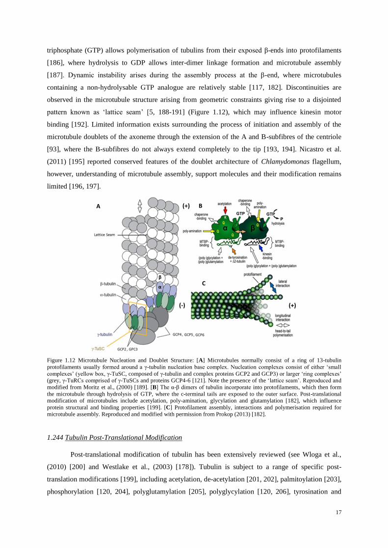
Figure 1.12 Microtubule Nucleation and Doublet Structure: [A] Microtubules normally consist of a ring of 13-tubulin
protofilaments usually formed around a γ-tubulin nucleation base complex. Nucleation complexes consist of either ‘small
complexes’ (yellow box, γ-TuSC, composed of γ-tubulin and complex proteins GCP2 and GCP3) or larger ‘ring complexes’ (grey, γ-TuRCs comprised of γ-TuSCs and proteins GCP4-6 [121]. Note the presence of the ‘lattice seam’. Reproduced and
modified from Moritz et al., (2000) [189]. [B] The α-β dimers of tubulin incorporate into protofilaments, which then form
the microtubule through hydrolysis of GTP, where the c-terminal tails are exposed to the outer surface. Post-translational modification of microtubules include acetylation, poly-amination, glycylation and glutamylation [182], which influence
protein structural and binding properties [199]. [C] Protofilament assembly, interactions and polymerisation required for
microtubule assembly. Reproduced and modified with permission from Prokop (2013) [182].
1.244 Tubulin Post-Translational Modification
Post-translational modification of tubulin has been extensively reviewed (see Wloga et al.,
(2010) [200] and Westlake et al., (2003) [178]). Tubulin is subject to a range of specific post-
translation modifications [199], including acetylation, de-acetylation [201, 202], palmitoylation [203],
phosphorylation [120, 204], polyglutamylation [205], polyglycylation [120, 206], tyrosination and
(+) A B
(-)
α
β C
(+)

18
detyrosination [186, 199]). These result in specific microtubule properties, influencing stability,
specificity, axonemal rigidity, signal transduction [180, 207], and the binding of proteins [208],
chemicals and transient ionic species [209]. Post-translational modifying enzymes act upon
microtubules, although tyrosine ligase is reported to act upon non-assembled tubulin [210]. Post-
translational modifications have also been also correlated with microtubule distribution and lifetime
[186, 211], where it has been suggested they initiate important functions and contribute to the intrinsic
properties of the cytoskeleton and axoneme [120, 194, 199, 212].
Variable levels of polyglutamylation exist on both axonemal microtubules and basal bodies
[213], and a low level of glycation also occurs [214, 215] (although present upon longer length cilia
[216]), implying that specific glycase and glutamylase enzymatic modifications are important to
function and stability [216]. Most cytosolic microtubules are polyglutamylated to some extent [217],
however, the role of such modifications, and control of their enzymes during the cell cycle and
ciliogenesis remain presently unknown. Microtubule associated proteins are associated with
tyrosination [218] and acetylation, where these modifications concur with increased flexural rigidity
[219] and kinesin-1 motor binding [220] (see Figure 1.12). For example in Chlamydomonas, tubulin
of both A and B-subfibres was found to be tyrosinated, while detyrosinated tubulin occurred mostly
upon the B-subfibre [221]. Furthermore, many studies [222] have revealed that the microtubules of
the centrioles and primary cilia are resistant to microtubule depolymerising drugs, suggesting a more
stable structure than the cytoplasmic microtubules.
Poole et al., (2001) [97] utilised antibodies to locate acetylated α-tubulin and detyrosinated α-
tubulin, demonstrating that the cilium and centrioles in chondrocytes, as well as parts of the
microtubular cytoskeleton of the cell, consisted of both acetylated and detyrosinated α-tubulin, with a
subset of acetylated α-tubulin microtubules being associated with the Golgi apparatus [96, 97]. Post-
translational modifications of tubulin components determine microtubule stability and the ability to
interact with associated proteins.
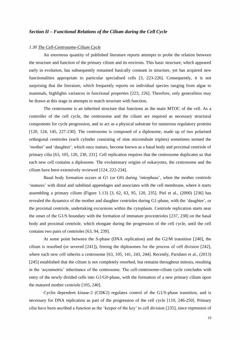
19
Section II – Functional Relations of the Cilium during the Cell Cycle
1.30 The Cell-Centrosome-Cilium Cycle
An enormous quantity of published literature reports attempts to probe the relation between
the structure and function of the primary cilium and its environs. This basic structure, which appeared
early in evolution, has subsequently remained basically constant in structure, yet has acquired new
functionalities appropriate to particular specialised cells [3, 223-226]. Consequently, it is not
surprising that the literature, which frequently reports on individual species ranging from algae to
mammals, highlights variances in functional properties [223, 226]. Therefore, only generalities may
be drawn at this stage in attempts to match structure with function.
The centrosome is an inherited structure that functions as the main MTOC of the cell. As a
controller of the cell cycle, the centrosome and the cilium are required as necessary structural
components for cycle progression, and to act as a physical substrate for numerous regulatory proteins
[120, 124, 145, 227-230]. The centrosome is composed of a diplosome, made up of two polarised
orthogonal centrioles (each cylinder consisting of nine microtubule triplets) sometimes termed the
‘mother’ and ‘daughter’, which once mature, become known as a basal body and proximal centriole of
primary cilia [63, 105, 120, 230, 231]. Cell replication requires that the centrosome duplicates so that
each new cell contains a diplosome. The evolutionary origins of eukaryotes, the centrosome and the
cilium have been extensively reviewed [124, 222-234].
Basal body formation occurs at G1 (or G0) during ‘interphase’, when the mother centriole
‘matures’ with distal and subdistal appendages and associates with the cell membrane, where it starts
assembling a primary cilium (Figure 1.13) [3, 62, 63, 95, 120, 235]. Piel et al., (2000) [236] has
revealed the dynamics of the mother and daughter centrioles during G1-phase, with the ‘daughter’, or
the proximal centriole, undertaking excursions within the cytoplasm. Centriole replication starts near
the onset of the G1/S boundary with the formation of immature procentrioles [237, 238] on the basal
body and proximal centriole, which elongate during the progression of the cell cycle, until the cell
contains two pairs of centrioles [63, 94, 239].
At some point between the S-phase (DNA replication) and the G2/M transition [240], the
cilium is resorbed (or severed [241]), freeing the diplosomes for the process of cell division [242],
where each new cell inherits a centrosome [63, 105, 141, 243, 244]. Recently, Paridaen et al., (2013)
[245] established that the cilium is not completely resorbed, but remains throughout mitosis, resulting
in the ‘asymmetric’ inheritance of the centrosome. The cell-centrosome-cilium cycle concludes with
entry of the newly divided cells into G1/G0-phase, with the formation of a new primary cilium upon
the matured mother centriole [105, 240].
Cyclin dependent kinase-2 (CDK2) regulates control of the G1/S-phase transition, and is
necessary for DNA replication as part of the progression of the cell cycle [110, 246-250]. Primary
cilia have been ascribed a function as the ‘keeper of the key’ to cell division [235], since repression of

20
primary cilium formation leads to progression of the cell cycle [251]. The role of gene expression
during the chondrocyte cell cycle has been reviewed by Schibler et al., (2009) [252], while the
structure and duplication of the centrosome has been reviewed by Azimzadeh et al., (2010) [253] and
Hinchcliffe et al., (2001) [254]. Cyclins and cyclin-dependent kinases, their interaction, localisation
and cell cycle implications are tabulated within Appendix I.
Figure 1.13 The Cell Centrosome
Cilium Cycle: A composite
demonstrating the cell and
centrosome cycle through G1, S, G2,
M and G0-phases [63]. Key events in
the cell cycle include the formation of
the mitotic spindle for mitosis (1), the
presence of interphase microtubules
(2), initiation of centriole replication
through procentriole formation (3)
and the formation and expression of a
primary cilium (4). The exact details
of specific aspects of the centrosome
and the cilium cycle are still subject
to debate [255, 256]. Modified from
Nigg et al., (2002) [248], Vorobjev et
al., (1982) [63] and Schibler et al.,
(2009) [252].
1.31 Intra-Flagellar Transport (IFT)
Intra-flagellar transport complex proteins are highly conserved across eukaryotes [257], and
orchestrate the bi-directional microtubule transport of materials and macromolecules from the basal
body to their required destinations within the cilium [23, 62, 101, 102]. IFT transport components
[258] accumulate upon the basal body [259, 260] where they interact with the Bardet-Biedl Syndrome
(BBS or BBsome) [46] family of proteins for the regulation of IFT assembly, ciliary protein targeting,
re-cycling turnaround [261-264] and signalling [265], as well as for Golgi trafficking [66].
Comprising A and B-assemblies [62, 257] of at least 8 and 14 subunits respectively, the A-assembly
is involved in retrograde transport, while the B-assembly participates in anterograde ciliary transport.
Anterograde transport is known to utilise kinesin motor members KIF3A, KIF3B and KIF17, while
cytoplasmic dyneins DYNC2H1 and DYNC2LI1 are utilised for retrograde transport [62, 257, 266].
IFT172 is reported to enable the dis-engagement of kinesin, and preferential activation of dynein
motors [267, 268] (for a review of transport motors and IFT-components, see Ishikawa et al., (2011)
[62], and Appendices III and IV. Electron microscopy of IFT particles reveals them as fine electron-
dense granules located between the membrane and the microtubule doublets of the axoneme, which
are commonly found concatenated into transport trains or ‘rafts’ [269].

21
1.311 Ciliary Transport
The mechanisms of IFT in ciliary function, regulation and signalling remain poorly
understood [102, 258]. The exact roles of the kinesin and dynein motors involved within the cilium
remain obscure, with KIF3C, KIF7, KIF19A and KIF24 members also having roles within the cilium
(Appendices IV and V). KIF3 and KIF17 both serve as IFT-complex motors (joined by BBS7 and
BBS8) [262, 270], with only KIF17 reportedly operating in the distal axoneme region [47, 270],
where, in photoreceptors, it supports outer segment development [271]. KIF7 localises to the ciliary
tips where it regulates Sonic Hedgehog (Shh) signalling, acting downstream of Smo and upstream of
Gli [272, 273]. However, without its ligand, KIF7 is localised to the base of the cilium and only
translocates to the distal tip upon activation [274], where it is suspected to play a role in Gli transport
[275] and as a regulator of Gli transcription [272, 276]. KIF7 acts as a critical regulator in the Shh
signalling cascade [272], restricting ‘suppressor of fused’ (SuFu), and promoting chondrocyte
signalling in the growth plate [277].
The IFT-A complex with Tubby-Like protein TULP3 [278] promotes the trafficking of G-
coupled protein receptors into the cilium [279], additionally requiring the BBSome [280], as well as
components of the Shh signalling pathway [281]. IFT-B may be required for the transport of Gli2,
SuFu and KIF7, while the absence of IFT-A results in the failure of the ADP-ribosylation factor
(ARF) like GTPase ARL13B, Adenylate Cyclase-3 (AC3) and Smoothened (Smo) to localise to the
ciliary tip [281] (see Appendix I). Protein LZTFL1 regulates the trafficking of Hedgehog signalling
pathway member Smo and the BBSome [282, 283].
1.312 IFT co-ordination
Little is known about docking and arrangement of IFT particles upon the basal body, or their
transport preference for certain axoneme microtubules. Buisson et al., (2012) [261] and Absalon et al.,
(2008) [284] established in Trypanosoma brucei that IFT particles travel upon distinct, or ‘restricted’,
subsets of axonemal microtubules. Retrograde trafficking occurs at a higher velocity than anterograde
transport, although IFT trains have been observed to travel at different speeds, and their recycling is
dependent upon ciliary length and the number of IFT particles in transit [261]. However, not all IFT
particles present at the basal body participate in transport [261, 285].
1.32 Primary Cilium Biogenesis
Sorokin (1962) [95] first described the process of genesis of the primary cilium from the
diplosome, where he documented initiation through a Golgi derived vesicle docking at the distal tip of
the mature basal body (Figure 1.14). Cilium formation requires the intricate combination of many
proteins and pathways. At present, incomplete information exists about their role. Sonic Hedgehog
signalling pathways are certainly involved in ciliogenesis [286] and interpretation of their mode of

22
action is still on-going [287]. It has been found that Patch1 regulates Smo activity in a late endosomal
sorting pathway [288], which is important for placement of proteins.
Planar cell polarity effector Inturned, together with Dishevelled and Rho-GTPase act in the
process of apical membrane docking [289], where Talpid3 is reported to influence actin localisation
during docking of the basal body [73]. Wnt signalling also plays a role in ciliogenesis [289], involving
shared components of the planar cell polarity pathway [290], since reduction of Wnt/β-catenin
signalling has been found to disrupt ciliogenesis. Proteins of the Par3/Par6/aPKC cassette are also
involved with ciliogenesis through control of kinesin motors [291], although many of these intricate
processes are not yet fully understood [292].
CEP164 is required for vesicle docking to the basal body [293, 294], and ciliary assembly is
initiated by Rab11-GTPase, the transport protein particle two (TRAPII) and Rabin-8 in the
centrosome [295]. Rab8 is required for ciliary formation through vesicle docking and fusion at the
base of the cilium and at the ciliary pocket [296-298] and facilitates membrane ciliogenesis with the
clathrin adaptor protein (AP-1) [295, 296, 299, 300]. Components of the Exocyst complex (sec10
[301]) and SNAREs are additionally required for ciliogenesis [302].
Centrosomal proteins CP110, CEP97 and CEP290 suppress primary cilium formation upon
the mother centriole [303, 304]. CEP97 and CP110 are located at the distal end of the basal body and
act to prevent maturation [305] where CP110 coordinates ciliary assembly [306]. For a review of
regulation of vertebrate ciliogenesis see Santos et al., (2008) [223].
The transition fibres are required for physical adherence to the plasma membrane, but also act
as docking sites for IFT particles involved in assembly and trafficking [307, 308]. Small GTPase
ARL6/BBS3 regulates membrane protein transport into the cilium [283], functioning near the ciliary
‘gate’, where it also incidentally plays a role in modulating Wnt signalling [309]. Components of the
Exocyst complex are required for ciliary function [301, 310], and are downstream effectors of Rab8
and Rab11 [311].
Extension of the axoneme requires co-ordinated active transport of material into the ciliary
‘bud’ using components derived from the cell membrane, and from transport vesicles. It is believed
that materials are highly concentrated through intra-ciliary transport mechanisms, allowing for the
elongation of the axoneme microtubules. Axoneme extension during initial ciliogenesis utilises
MARK4 kinase [312] and requires microtubule associated protein EB1 [313]. Assemblage leads to
formation of a ‘transition zone’ compartment, followed by the axoneme, and the distal tip, although
the exact mechanisms are yet to be elucidated [62, 65, 244, 298, 302, 312].
Ciliary formation and assembly also requires various ancillary complexes to accumulate
within the centrosome [55, 314]. These include many obscure proteins which form, regulate and
maintain ciliary function [315] through regulation of IFT, microtubule processes of tubulin transport,
specific poly-modifications [212, 316, 317], katanin mediated severing [318], and kinesin and dynein
transport motors [319, 320]. Tubulin transport into the cilium has recently been reported to occur
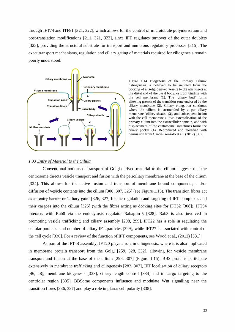
23
through IFT74 and ITF81 [321, 322], which allows for the control of microtubule polymerisation and
post-translation modifications [211, 321, 323], since IFT regulates turnover of the outer doublets
[323], providing the structural substrate for transport and numerous regulatory processes [315]. The
exact transport mechanisms, regulation and ciliary gating of materials required for ciliogenesis remain
poorly understood.
Figure 1.14 Biogenesis of the Primary Cilium:
Ciliogenesis is believed to be initiated from the docking of a Golgi derived vesicle to the alar sheets at
the distal end of the basal body, or from binding with
the cell membrane (1). The ‘ciliary bud’ forms
allowing growth of the transition zone enclosed by the ciliary membrane (2). Ciliary elongation continues
where the cilium is surrounded by a peri-ciliary
membrane ‘ciliary sheath’ (3), and subsequent fusion
with the cell membrane allows externalisation of the primary cilium into the extracellular domain, and with
displacement of the centrosome, sometimes forms the
ciliary pocket (4). Reproduced and modified with
permission from Garcia-Gonzalo et al., (2012) [302].
1.33 Entry of Material to the Cilium
Conventional notions of transport of Golgi-derived material to the cilium suggests that the
centrosome directs vesicle transport and fusion with the periciliary membrane at the base of the cilium
[324]. This allows for the active fusion and transport of membrane bound components, and/or
diffusion of vesicle contents into the cilium [300, 307, 325] (see Figure 1.15). The transition fibres act
as an entry barrier or ‘ciliary gate’ [326, 327] for the regulation and targeting of IFT-complexes and
their cargoes into the cilium [325] (with the fibres acting as docking sites for IFT52 [308]). IFT54
interacts with Rab8 via the endocytosis regulator Rabaptin-5 [328]. Rab8 is also involved in
promoting vesicle trafficking and ciliary assembly [298, 299]. IFT22 has a role in regulating the
cellular pool size and number of ciliary IFT-particles [329], while IFT27 is associated with control of
the cell cycle [330]. For a review of the function of IFT components, see Wood et al., (2012) [331].
As part of the IFT-B assembly, IFT20 plays a role in ciliogenesis, where it is also implicated
in membrane protein transport from the Golgi [259, 328, 332], allowing for vesicle membrane
transport and fusion at the base of the cilium [298, 307] (Figure 1.15). BBS proteins participate
extensively in membrane trafficking and ciliogenesis [283, 307], IFT localisation of ciliary receptors
[46, 48], membrane biogenesis [333], ciliary length control [334] and in cargo targeting to the
centriolar region [335]. BBSome components influence and modulate Wnt signalling near the
transition fibres [336, 337] and play a role in planar cell polarity [338].
1 2
3 4

24
Figure 1.15 Golgi Derived Materials for Ciliogenesis:
Ciliogenesis and ciliary maintenance require the regulated
transport of specific cargoes and receptors to be delivered to
the periciliary membrane. Components are sequestered from the Golgi apparatus through involvement of IFT20 and the
BBSome in vesicular events, from where they are transported
to the centrosome and made available for transport into the
cilium by IFT-B (anterograde kinesin transport) and recycled via IFT-A (retrograde dynein transport). BBSome proteins
BBS4 and BBS8 interact with the pericentriolar matrix
protein PCM-1 surrounding the basal body. BBS1 cooperates
with Rabin8 for Rab8-GTP bound mediated docking and fusion of vesicles at the membrane and ciliary pocket. Rab8
regulates ciliary protein trafficking, and is controlled by
Rabin8, whose activity and localisation is controlled by the
BBsome and Rab11 [300]. Reproduced with permission from
LeCroux (2007) [307].
Receptors such as retinitis pigmentosa-2 (RP2) require importin-β2 for ciliary entry [67],
while KIF17 motor entry is governed by Ran-GTPase and importin-β2 [339, 340]. Curiously, the
KIF17 c-terminal tail domain possesses both ciliary and nuclear localisation sequences [67, 339], with
inhibition of Ran-BP1 disrupting distal localisation of KIF17 [339, 341], since importin-β2 controls
ciliary entry [339]. Ran members and initiation processes are important for a host of cytosolic and
nuclear functions [67, 342], where control of centrosome activity and ciliary entry is known to utilise
β-arrestin [343].
1.34 Ciliary Localisation Sequences
Many key receptors and their adaptor molecules share complex interactions in which their
localisation requires specialised signalling for their proper inclusion into ciliary processes [326, 344,
345]. This is believed to be achieved through ciliary targeting motifs that are required for the orderly
transport of proteins and macromolecules from the Golgi to the cilium [297, 300, 325 307, 346, 347].
Many details of the mechanisms of ciliary receptor transport, localisation, turnover, cell specificity,
downstream signalling effects and their partners presently remain little known [348]. Ciliary receptors
which are known to have localisation sequences include smoothened (Smo) [337], polycystin-1
(PC1/TRPP1) [349], polycystin-2 (PC2/TRPP2) [350], retinitis pigmentosa protein (RP2) [349], opsin
[325], rhodopsin [351], fibrocystin [352], somatostatin (SSTR3) [325], serotonin (5HTR6) [353] and
melanin concentrating hormone (MCH) [354].
Some ciliary proteins also reportedly contain nuclear localisation sequences, including
transient receptor potential (TRP) vanilloid member OCR-2 [355], the KIF17 c-tail [67], the PC1 c-
tail [356], Sonic Hedgehog Gli family members [357] and G-coupled receptor kinase GRK5 (which
includes a DNA binding motif [358]).

25
Localisation of ciliary messengers to the nucleus is requirement for Notch signalling [359],
for Dishevelled function in Wnt/beta-catenin signalling [360], in components of Hedgehog signalling,
and for Gli transcriptional activity, regulated by cyclic-Adenosine Mono Phosphate (cAMP) [361].
Protein c-terminal residues are likely critical for recognition and localisation for ciliary targeting,
where trafficking takes place by several molecular processes, which can vary between proteins of the
same family [354]. The ‘VxPx’ targeting motif [354, 362] utilises GTPases, ARF4 and Rab11, the
Rab11/ARF effector FIP3, and the ARF GTPase-activating protein ASAP1 [362]. At present, an
incomplete picture exists of the mechanisms of targeting of many ciliary proteins.
1.35 Dynamic Control of Ciliary Length
The length of the primary cilium is influenced by external factors such as mechanical load,
fluid shear, osmotic challenge and heat induced shortening [255, 363-365]. Cilia may lengthen
following injury [366], or in response to certain soluble cations [363]. Wilsman [27-29] observed
variation in microtubule doublet lengths within the axoneme, which may indicate that length
regulation is a dynamic process, with each doublet being subject to individual control mechanisms.
Ciliary length is under the control of IFT, which balances the continuous turnover of the
microtubule doublet tips [323, 363, 367]. It is not yet known how differences in microtubule doublet
lengths occur, as the regulatory mechanisms of ciliary dynamics and cell cycle remain as yet unknown
[284]. Kinases LF4 and CNK2 are reported to determine the length of cilia in Chlamadymonas
reinhardtii by controlling the rate of assembly and disassembly [368] through increasing IFT traffic
and halting anterograde cargo loading [369].
Other ciliary components involved in length regulation include Septins-2, 7 and 9 (with
MAP4) [370], galectin Gal-7 [371] and ciliary associated kinesins KIF19A and KIF24. KIF19A is
observed to reduce microtubule length through depolymerisation at the tip in mammals [372], whilst
KIF24 contributes to microtubule remodelling and depolymerisation. It also interacts with
centrosomal proteins CP110 and CEP97 in HEK cells in culture [305]. Interleukin-1 has been found
to increase ciliary length in chondrocytes through a protein kinase pathway [366]. The Rer1p protein
regulates ciliary length through recruiting γ-secretase and Foxj1 [373], and Rer1p is also reported to
be necessary for endoplasmic reticulum protein retrieval, where it cycles between the Golgi [374].
1.40 The Ciliary Membrane
The ciliary membrane is a lipid bilayer structure of unique composition, enclosing the
axoneme, enriched with lipid rafts of specific components, receptors and their luminal mediated
processes, which vary in their abundance along the ciliary membrane [345, 375]. Differing from both
the periciliary and plasma membranes, it is greatly enriched in sterols, glycolipids and sphingolipids
relative to the plasma membrane [376, 377]. External to the membrane is a glycocalyx of
glycoproteins, which are involved in adhesion and signalling, as well as interacting with fluids,

26
solutes, osmolytes, and signalling molecules [345, 378, 379]. They may also be influenced by
compression induced electric fields in cartilage [380]. Regions of ciliary membranes may vary in their
charge density [381], as ferritin labelling has revealed negatively charged sites located in association
with fine filaments [49] and at ciliary necklace sites [49, 382]. The presence of ciliary necklace,
plaques, rosettes and longitudinal rows indicates a higher order of complexity in the membrane, which
is supported by the presence of membrane linkages aligned with the microtubules [52] (Figure 1.4).
As yet, the spatial and functional interaction between ciliary receptors and the linkages remains
obscure [383].
Receptors become localised to the membrane after ciliogenesis, and their proper function is
dependent on correct sorting, trafficking and placement within the ciliary membrane [345, 383-385].
Localisation of receptors and their components within the cilium usually requires IFT [281], while
Rab-effector related proteins Rilpl1 and Rilpl2 [386] regulate ciliary membrane content. Much
remains unknown with regard to receptor entry, regulation, and sorting within the cilium [297].
In flagella, membrane localisation of specific proteins may occur on lipid ‘rafts’ [377], where
some receptor clusters (such as epidermal growth factor receptor EGFR) are known to vary in size,
and to be associated with cholesterol and sphingolipids [387]. Many membrane receptors occur in
close proximity to the axoneme microtubules, and their organisation appears to regulate polycystin-2
(PC2) signalling [388], while the transient receptor potential vanilloid-4 (TRPV4) [389] channel is
known to form complexes with regulatory kinases enabling roles in cytoskeletal interactions and
microtubule depolymerisation [390].
Proteins within the cilium and its membrane undergo preferential turnover, influencing
function [391, 392]. An extreme example is the photoreceptor cilium, which is estimated to contain
109 rhodopsin (GPCR) molecules in 10
3 photo-sensitive stacks [297]. In the murine photoreceptor,
opsin is delivered to the base of the cilium, and transported along the ‘connecting cilium’ for forming
new photoreceptor membranes at 2000 opsin units per minute [393], indicating that this cilium is a
highly ordered and regulated structure capable of rapid molecular transport [394]. To maintain ciliary
function, proteins must be actively transported from the Golgi to the centrosome, from where they are
actively vectored into the cilium. The regulated transport of Golgi-derived receptors and structural
proteins remains largely unknown, however many proteins possess ‘ciliary targeting sequences’
enabling active directed transport [297]. The Taurine transporter, for example, has a high expression
in the primary cilia of NIH3T3 fibroblasts (Appendix I).

27
1.41 The Cilium: Receptors and Luminal Components
Primary cilia are ancient specialist compartments for the detection, amplification and
transmission of a wide range of signals. For example, the murine photoreceptor sensory complex
[132] is reported to contain over 2000 proteins4 specific to or involved with, ciliary function [38, 40].
Primary cilia utilise various membrane receptors, channels and signalling components whose
transport and function is tightly controlled [17, 48, 297]. Figure 1.16 illustrates an abbreviated
summary of known ciliary membrane receptors, and associated luminal and structural components,
that are listed in further detail in Appendix I.
Depending upon its sensory requirements, the ciliary membrane and lumen may contain
specialist receptors and downstream components for Notch [395], Wnt (Frizzled, Dishevelled, Chibby
and Inversin) [396], Hedgehog (Smo, SuFu, Patched (Ptch) and Gli1-3) [69, 396, 398, 399, 400, 401]
and platelet derived growth factor receptor (PDGFRα) [384] signalling pathways. Many other
receptors allow for a wide range of physiological signals to be transduced [402], while structural and
luminal associated proteins like the Nephrocystins (NPHP) and IFT-complexes influence the dynamic
support of many receptors whose full function in the cilium remain to be determined. Appendix I
details known receptors, interactions, signalling cascades, structural proteins and some of the diseases
resulting from loss or mutation of known genes [1]. These represent only a small number of the
proteins believed at present to be involved within the cilium, however not all of these receptor
proteins have been shown to be present upon chondrocyte primary cilia.
4 http://www.ciliaproteome.org/

28
Figure 1.16 The Primary Cilium: The distal, middle and transition zones projected from the basal body. The ciliary membrane has been shown to contain numerous receptors and their associated components of several signalling pathways,
although not all are present within every cilium. These are activated by a variety of extracellular signals and ligands, as well
as from mechanotransductive forces acting upon the cilium. Small forces may result in the localised distortion of the ciliary
membrane, while larger forces may translate into bending of the microtubule doublet based axoneme. Ciliary functionality is maintained by microtubule based anterograde and retrograde bi-directional intra-flagellar transport components, which are
organised from the basal body. These transfer receptors, structural proteins and regulatory complexes are vital for
maintaining ciliary homeostasis. Details of these components are tabulated within the Appendices.

29
1.42 The Cilium and the Extracellular Matrix
The primary cilium is known to interact with the extracellular environment, and convey
information to the cell, for example, the membrane of the chondrocyte cilium has been observed to
interact with collagen fibres and proteoglycans of the extracellular matrix [25, 26, 97]. Cartilage
extracellular matrix has been found to contain matrix vesicles [403] and a variety of signalling
molecules intrinsic to the roles of development and homeostasis of the chondron [404, 405]. The
chondrocyte ciliary membrane is known to contain matrix receptors of α2, α3, β1-integrins and NG2,
but not for of Annexin-V and CD44 (see Section 1.90), indicating that the ciliary membrane is only
selective for certain matrix connections [25, 26, 97, 100]. β1-integrins on MDCK epithelial cell
primary cilia are reported to detect fluid flow following deflection via fibronectin induced Ca2+
signalling in association with PC2 [34, 406, 407].
It is widely known that proteoglycans are important signalling components in many cell
pathways [209, 408-410] with their role in the chondrocyte matrix being particularly important for
differentiation [411]. The heparan sulphate proteoglycans [412] (syndecan, glypican and perlecan
[410], see Section 1.86 - 1.89) are signalling components of the pericellular microenvironment [413].
Glypican-5 (GPC5) core proteins containing heparin sulphate and 2-O-sulpho-iduronic acid are
reported to be involved with Sonic Hedgehog as co-receptors [414], assisting in binding of Hh to
Patched1. GPC5 increases binding of Shh to Patched1, conversely GPC3 inhibits this interaction
through competition for Shh [415, 416]. Li et al., (2011) [415] argue that Glypican-5 binds to primary
cilia, and stabilises interactions between Shh and Patched1, facilitating signalling. The solubility and
diffusion of Hedgehog ligands within the extracellular matrix may depend not only upon their
modification, but also on their interactions with the matrix [417, 418], while Wnt protein
concentration may be influenced by the concentration of matrix at the cell membrane [419]. Recently,
TGF-β signalling is reported to occur through clathrin-dependent endocytosis at the ciliary pocket,
where receptors localise to the ciliary tip and clathrin vesicles at the base of the cilium [420]. For a
review of signalling in cartilage development, see Brochhausen et al., (2010) [404] and Kirn-Safran et
al., (2004) [410].
1.43 Signalling and Mechanotransduction
The ciliary membrane provides a large surface area to volume ratio for detecting and
amplifying external chemical stimuli. Electrical signals from patch clamped primary cilia have
recently been measured, indicating the presence of conducting channels and secondary messengers
[421], however the majority of studies on primary cilia have examined their role as flow sensors in
epithelia, with few studies being concerned with mechanical loads experienced by the cilium within a
cartilaginous matrix [422]. Many receptors have been identified localised to primary cilia, but in
connective tissues, their full role in, for example, chondrocytes remains unknown [423]. Channels of
the transient receptor potential (TRP) family members participate in a wide range of sensory

30
modalities [424], and each is specific to one of a number of stimuli; mechanical [407, 425], light
detection [132], oxidative stress, tonicity changes [426, 427], pH, temperature and sensitivity to
chemical species [428].
Praetorius and Spring (2001) [34] established that primary cilia acted as flow sensors in
MDCK cells, for mechanical bending induced an increase in intracellular Ca2+
concentration, and this
was communicated to adjacent cells. Nauli et al., (2003) [425] later found that Polycystin-1 (PC1) and
Polycystin-2 (PC2) ion channels were responsible for mediation of mechanotransduction in renal
primary cilia (Figure 1.17). PC1 and PC2 are well known to form a membrane bound mechano-
molecular complex localised to the primary cilium [406, 429, 430] (Figure 1.17). PC1 contains a
lengthy extracellular component responsible for mediating kidney cell mechanotransduction [425,
431]. PC1 is known to interact with PIP9 [432] and influences transcription factor Jade1 [433], for
cleavage of the PC1 c-tail domain may control β-catenin and canonical Wnt signalling [429]. PC2 is
responsible for angiotensin II-induced Ca2+
signalling [407, 434], and may include other binding
partners EGFR, TRPC1, TRPC4 [434], and TRPV4 (which is mechano- and thermosensitive) [428,
429, 435], although many aspects of their ciliary roles remain unknown. In chondrocytes TRPV4 acts
as an osmotically sensitive Ca2+
channel, and its loss results in osteoarthritic matrix changes [436]. It
also plays a role in regulation of chondrogenic differentiation [437].
Figure 1.17 The PC1/PC2
Mechanotransduction Complex: The
PC1/PC2 complex contains an extra-cellular
PC1 (polycystin-1 or PKD1) domain, and
the PC2 (polycystin-2, PKD2 or TRPP1)
Ca2+ cation channel which are both defined
by a shared complex trans-membrane loop,
accompanied by extracellular and intracellular components which interact at
their c-terminal domains. Extracellular
stimulation of the PC1 protein is responsible
for activating the PC2 channel, where PC1 c-terminal cleavage is controlled by PC2
expression. Reproduced and modified from
Zhou et al., (2009) [429].
1.44 Purinergic Mechanotransduction
Purinergic family members are composed of two groups; P2X ATP-gated ion channels, and
P2Y G-coupled receptors. P2Y1, P2Y2, P2X2, P2X4 and P2X7 have been identified upon
chondrocyte membranes [438] (Appendix I). Purinergic mechanotransduction in chondrocyte cilia had
been speculated upon [438, 439], since the cilia of cholangiocytes are known to utilise P2Y12
purinergic receptors as chemosensors [440]. It has been demonstrated that cilia of compressed
chondrocytes mediate mechano-transduction through ATP-induced Ca2+
signalling [422, 441], but
31
they are not necessary for the initial stage of induced ATP release. They are however required for
exogenous detection of ATP [422]. Connexin-43 hemichannels have also been located upon the
ciliary membrane [438] and are believed to provide an avenue for release of ATP in response to
mechanical loading in chondrocytes [422]. Mechano-transductive purinergic Ca2+
signalling may be
regulated by PC1 in the chondrocyte cilium, which could also mediate control of ATP reception
[422], since the proteolytically cleaved PC1 tail is known to translocate to the nucleus for STAT6
gene activation [442]. Activation of the G-coupled receptor Smo [443] results in the targeting of Gli,
altering transcription and gene expression [398, 444] while Gpr161 negatively regulates the Shh
pathway through cAMP signalling [445]. Such cAMP dependent processes are central to ciliary
signalling.
1.45 Ciliary Polymodal Sensory Function
Ciliary mechanosensitive molecular pathways are believed to be shared, but also vary
between cell types [446, 447], where some utilise the cAMP pathway instead of Ca2+
signalling.
Adenylyl cyclases AC3, AC4, AC6 and AC8 have been identified within the cilium, and are required
for the production of cAMP (Appendix I). Hoey et al., (2012) [448] indicated that ciliary
biomechanical transduction in bone cells is distinct from that in the kidney distal epithelium. It is
believed that osteocyte cilia transduce flow via AC6 and cAMP, independent of extracellular Ca2+
,
resulting in protein kinase-A cascade signalling [447,
449]. Renal cilia are believed to transduce flow as a
result of Ca2+
entry through the TRPV4/PC2 membrane
domain as well as utilising PC1 cleavage for signalling
[425, 428 447, 448] (Figure 1.18). It is reported that
AC6/cAMP [449] signalling is preserved through the
osteocyte linage in mesenchymal cells [448, 450]. It is
probable that similar signalling modalities may occur in
chondrocyte primary cilia.
Figure 1.18 Proposed Mechanism of Flow Induced Ciliary
Ca2+
and cAMP Signalling Modalities: Extracellular Ca2+
may enter the cilium through either the PC1/PC2 or from
TRPV4 channels form a polymodal sensory channel complex
[428]. Cell phenotype is believed to determine the signalling
modality utilising PC1/PC2 and / or TRPV4 Ca2+
entry,
adenylate cyclase-6 initiating intracellular Ca2+
release.
Cleavage of the luminal PC1 tail results in nuclear
localisation via STAT6 and P100. Reproduced with
permission from Kwon et al., (2011) [447].
PC2 ciliary Ca
2+ entry modality occurs in nodal, osteocyte, cholangiocyte, vascular
endothelial and kidney epithelial cells, while kidney and vascular cells also utilise PC1 c-tail cleavage

32
in signalling and nuclear localisation [356, 447], including transcription activation in T-cells [451].
Loss or altered functionality of ciliary polycystin is implicated in polycystic kidney disease [452] and
the growth of renal cysts [453]. It has been shown that while fluid induced movement of the primary
cilium results in PC2 activation, the transductive cellular response also requires feedback from
combined components of the cell cytoskeleton and the extracellular matrix [454].
Fibrocystin/polyductin (PKHD1) serves as a bi-directional signalling molecule, which may undergo
Notch-like cleavage of its extracellular domain [48, 455] (Appendix I).
In connective tissues, a network of mechanically interconnected collagen fibres and
proteoglycans surrounds chondrocyte cilia, which if bound through integrin and NG2 matrix receptors
may provide a physical link to the many possible receptors within the ciliary membrane [25, 100, 456,
457]. Integrins are also reported to potentiate fibronectin induced Ca2+
signalling in renal cilia [458],
although it remains to be seen which other channels may play a role in mechanotransduction within
connective tissues. It is not possible to discuss all known primary ciliary receptors in detail, or
receptors that could be specific to the chondrocyte.
1.50 The Centrosome: The Central Processing Unit
The centrosome contains functional groups of proteins involved in microtubule nucleation,
anchoring, duplication, cell cycle, and primary cilia regulation (reviewed by Alieva et al., (2008)
[120]). The centrosome is an inherited structure [229, 459-461] and functions as the main microtubule
organising centre of the cell [138, 145] as well as the controller of the cell cycle [124, 459, 461]. It is
required for cell cycle transitions and as a substrate for numerous regulatory proteins [109, 461, 462].
Microtubules are polarised dynamic structures [463] which extend with a defined orientation
originating from γ-tubulin ring complexes (γ-TuRC) [118, 135, 189, 198, 461], some of which attach
to the basal appendage docking complexes, while others are anchored within the PCM lattice ‘cloud’
surrounding the diplosome [105, 464-466]. Numerous molecules are involved in microtubule tubulin
polymerisation and depolymerisation, as well as severing.
1.501 The Centrosome, the Cytoskeleton and Transport
The centrosome is the main director of the primary cilium, and also contains functional
groups of proteins involved in a wide range of cellular functions. Control of cell cycle, planar cell
polarity, organelle transport and motility requires coordination of the cytoskeleton. The cytoskeleton
consists of a dynamic interplay of actin microfilaments, intermediate filaments and microtubules.
Microtubules are dynamic directional structural conduits involved in cell movement, polarisation, cell
cycle, cytokinesis and pathogenesis. The physical ability of actin microfilaments and tubulin
microtubules to dynamically assemble and disassemble as required permits their use in the physical
transport of vesicles, organelles and for signalling. During interphase, intermediate filaments and

33
microtubules radiate from the organising centre of the centrosome, while actin microfilaments
associate with the cell membrane [120, 141].
Cytoskeletal molecular transport motors are ATP-dependent nano-actuators consisting of
three classes; kinesins, dyneins and myosins, which are responsible for driving directional movement
for the transport of materials, vesicles, organelles and cytoskeletal dynamics within the cell [120, 188,
270, 467-472]. Myosins are microfilament transport motors, while dynein and kinesin family
members facilitate the directed transport along microtubules. The Kinesin family of proteins are
anterograde transport motors (except family 14B members [270]), while dynein family members
facilitate retrograde transport [270, 473]. The majority of structural information concerning these
motors comes from studies using crystallography and electron tomography [474]. X-ray
crystallographic structures of various conformation states are known for members of kinesin [475],
dynein [476] and myosin motors [474]. Motor complex components bind to different receptors [477,
478] along with the dynein interaction complex dynactin, which is a multi-unit protein that normally
directs retrograde traffic [473].
The spatial regulation and ‘interactome’ of microtubules, their motor cargo interactions,
transport specificity and signalling remain poorly understood [120, 146, 147, 158, 186, 477, 478].
These aspects include specific movement of cellular components of the cytosol, the nucleus,
mitochondria, endoplasmic reticulum, centrosome, and the Golgi [479, 480]. Functional interactions
of kinesin and dynein member motors are central to both ciliary and non-ciliary transport alike, where
they are utilised in a host of cytoplasmic processes for signalling, regulation and organelle trafficking
(tabulated in Appendices II, III and V). Ciliary processes are known to selectively utilise only
cytoplasmic dynein DYNC2LI1, DYNC2H1 [492] and kinesin family members KIFs 3A, 3B, 3C, 7,
17, 19A and 24 (where IFT-complexes utilises KIFs 3A, 3B, and 17).
1.502 The Kinesin Family
The crystal structure of kinesin domains consists of two synchronous motor domains existing
as a tetramer of two homodimers roughly 8 × 5 × 5 nm in size [474, 481-483] (Figure 1.19). Each
motor can generate a force up to 7 picoNewtons [484] (although ATP and Mg2+
concentration may
influence kinesin performance [485]), involving steps of 8 nm, the corresponding length of the tubulin
α-β dimer [474]. Kinesin contains many subfamily members that exhibit a variety of properties,
sharing similar evolutionary structural properties to the myosin
family [471, 472, 474]. Eukaryotic kinesins consist of a conserved
ATPase core and many isoforms, alternative splicing and variable
sub-assemblies allowing for a variety of cargoes to be transported
[270, 471, 472].
Figure 1.19 The Kinesin Motor: A molecular model of the crystal structure of
kinesin member kinesin NCD (1N6M) [475] obtained from the PDB protein
databank [486] viewed in the Swiss-PDB viewer [487].

34
1.503 The Dynein Family
Dynein consists of a diverse family of proteins whose components participate in a range of
cellular functions [488, 489]. There are two classes of dyneins; axonemal and cytoplasmic [489, 490].
Cytoplasmic dynein complex naming nomenclature varies [491], since dynein is built around
respective DYNC1H1 and DYNC2H1 units, and these have been reviewed [492] (Appendix III).
Dynein generates forces of around 1.1 pN [493], and although the output is similar to kinesin motors,
it is speculated that dynein output may be tuned for transport needs in response to load [473, 493].
Crystal structure studies of dynein motors [476, 494] have revealed them as high molecular weight
homodimers of 1-2 MDa, consisting of a central head complex and a long tail [495], which contains
significant residues involved in cargo binding [496]. Regulation of dynein properties is known to
occur through three cofactors, the dynactin complex, lissecephaly-1 (Lis1), and nuclear distribution-E
(NudE) proteins [496, 497], and these determine its motile properties [471]. Lis1, NudE, and Rab6A
are involved with the centrosomal and nuclear localisation of dynein [473], in which the Lis1 enzyme
is an initiation factor for dynein transport [498, 499], and Rab6A removes Lis1 from the idle dynein
complex and promotes retrograde movement [500]. Lis1 is recruited to dynein by NudE, to regulate
motor force production [501].
Cytoplasmic dynein isoforms-1 and 2 mediate vesicular transport, while dynein-2 has in
addition ciliary and Golgi functions [502]. Dynein binds to centrosomal protein pericentrin (via
DLIC-1) [471] and transport regulatory components such as the glucocorticoid receptor [503].
Dyneins may also have specific membrane functions within the cell for the transport of organelles and
signalling [270, 504], as well as in maintaining the Golgi apparatus [492, 505], since dynein
dysfunction disrupts intracellular vesicle trafficking [506]. Appendix III details the known roles of
dynein family members.
1.504 The Dynein Activator - Dynactin
The dynactin transport complex [507] consists of a filament 37 nm in length and 10 nm wide
(resembling F-actin) comprised of globular protein heads [508], and a molecular mass 1 ×10⁶ Da
[473]. It is responsible for bi-directional microtubule transport [509] (acting through dynein and
kinesin motors), as well as a wide range of regulatory interactions [120, 473, 507, 510], and is also
required for microtubule anchoring within the centrosome [511]. As a regulator of dynein and kinesin
function [507], it is involved in vesicle transport [513], cell cycle and cell division [514, 515, 516].
Dynactin is a multi-protein complex involved in linking cargo [517, 518] and it consists of many
subunits [507] (Figure 1.20). Dynein microtubule transport depends upon complex interactions
between dynein, the dynactin regulatory complex, linkage proteins spectrin-βIII, ZW10 [519] and
syntaxin [520]. Arp1 vesicle binding occurs through spectrin-β, allowing binding to Golgi spectrin, as
well as containing two microtubule-binding domains [5, 507, 521].

35
Figure 1.20 The Dynein Motor and Dynactin Complex: [A] The dynein motor complex consists of two hexameric ATPase modules and intermediate chains. [B] The dynactin complex consists of a p150 arm that binds with the dynein intermediate
chain (also containing a microtubule binding domain) which is connected to Arp1 (and other components), allowing binding
to spectrin upon membranes. Reproduced and modified with permission from Yadav et al., (2011) [518] and Kardon et al., (2009) [473].
During interphase, dynactin accumulates in the centrosome for a cell cycle specific role in
which dynactin influences microtubule function and components of the PCM [511, 514]. It is found
localised to endocytic organelles and vesicles in the Golgi region [522], while spectrin-βIII regulates
secretory protein and Golgi integrity [521]. Dynactin subunit p150Glued
is found localised to the
maternal centriole [514], where it and other pericentrally located subunits provide as yet unknown
regulatory roles related to the recruitment of materials, signalling, transport of organelles and the cell-
cycle [120, 507, 515]. KIF3A interacts with p150Glued
for the organisation of basal feet [523]. Loss of
p150Glued
from Arp1 results in microtubule disorganisation [507, 514]. The dynactin p50 sub-unit
interacts with CEP135 in the centrosome, playing a role in assembly and maintenance of the MTOC
[524].
Dynactin subunit-1 (DCTN1) interacts with BBS4, where it is involved in microtubule and
cargo anchoring within the centrosome [335] (Appendix I). p50 and Golgi-Rab6 bind to biclaudin-D
[473], while Rab6A [525] regulates the recruitment of the dynactin complex to membranes of the
Golgi [526], where it is involved with intra-Golgi and Golgi-endoplasmic reticulum trafficking [525].
Rab7 is involved with p150 in the late endosome mediating fusion [473]. Interactions with spectrin-
βIII attachment via Arp1 are required for long-range vesicle transport [473, 510].
Dynactin regulates bi-directional transport of both dynein and kinesin [471], and dynein,
kinesin-2 and kinesin-5 are known to interact with p150Glued
[473]. The dynactin adaptor complex
links to dynein between the intermediate chains and p150Glued
, where it facilitates a diverse transport
array, including vesicle transport of material from the endoplasmic reticulum to the Golgi [473, 517].
In this process, Rab6 acts as a membrane receptor, and also binds p50 to dynactin and biclaudin-D
[473]. Rab6-biclaudin-D binding is important for Golgi function, where it is reported to inhibit Golgi
fragmentation [527].
Kinesin-2 and Kinesin-5 binding domain
Sidearm
Binds EB1 and CLIP170
A B

36
Most cellular dynein activity requires the dynactin complex [507, 509], disruption of which
inhibits endoplasmic reticulum to Golgi transport and leads to re-arrangement of the Golgi and the
endosomal/lysosome system [511, 528]. Independent vesicle transport from the Golgi to the ER-
COP1 requires biclaudin-D for recruitment of the dynein-dynactin complex [525]. Cell division
control protein CDC42, dynein and dynactin are reported to be involved with orientation of the
MTOC and Golgi in migrating cells [529] (see Section 1.61). It is believed that dynactin regulates the
presence of dynein at specific cytosolic locations, providing linkage to cargoes and adaptor molecules,
although complete understanding of these processes is presently lacking. GTPase family members
function at specific subcellular compartments [473], where they may provide a link between dynein
and regulatory protein function [530].
1.51 The GTPase Family Members
The GTPase superfamily contain four well known subfamily members Arf, Rab, Ran and Ras
and with their effectors and regulators are involved with most aspects of signal transduction, cell
cycle, cell migration and intracellular function [531-536]. GTPases are involved in signal transduction
at the intracellular domain of many trans-membrane receptor proteins, in cellular control, cell
polarity, protein synthesis, cytoskeletal dynamics, transport, and vesicle targeting as well as
membrane organisation [342, 537-539]. Switching is achieved through the hydrolysis of the GTP
form to the inactive GDP state [534].
Members of the Ras family of GTPases are involved in cell division, differentiation, and
cytoskeletal dynamics [540-543] as well as in apoptosis and cancer progression [543]. For example,
B-cell lymphoma protein (BCL-2) and Ras-GTPase may determine regulation of cell fate [543]. Rho
is a subset of the Ras family [532] whose members RAC1, CDC42 and RhoA are involved in gene
expression and cell proliferation [544], as well as in regulation of cytoskeletal and cell division.
RhoA-mediates cytoskeletal actin enrichment for ciliogenesis [545], with Meckelin and Nesprin
proteins being required components for primary cilium formation [546, 547]. RhoA plays a role in
cytoskeletal dependent functions in hematopoietic stem cells [548], with its centrosome associated
kinase p160ROCK regulates centrosome position [549]. Rho-family members are involved with
pathways of adhesion signalling between matrix and integrins [550], as well as cell shape and motility
[551] in cell migration, requiring alteration of the centrosome, cytoskeleton and microtubules [543,
552]. Rho, Rac and CDC42-GTPases also regulate the formation of cell-matrix focal adhesion
complexes, control polarity, adhesion during cell migration, as well as assisting with the
reorganisation of the actin cytoskeleton in response to mechanical forces [550, 553, 554]
Ran is involved with ciliary entry [555], ciliogenesis [341], centrosome division [556], in
nuclear entry [547] and is required for centrosome microtubule organising activity [558]. The ARL-
GTPases regulate several different aspects of centrosome microtubule function and polymerisation
[559, 560]. Zhang et al., (2013) [561] reviewed the role of the Arf-like GTPases ARL3, ARL6 and

37
ARL13 and their vital functional roles within the cilium. ARL3 is reported to be a negative regulator
of ciliogenesis, but it is required for functionality of the IFT-B complex and interacts with NPHP3.
ARL6 is required for signalling (where it functions within the BBSome) while ARL13 is required for
both ciliogenesis and functional IFT-targeting of receptors (such as PC2 [562]). Furthermore,
ARL13B, with CEP164 is reported to be involved with INPP5E for targeting to the cilium [347].
A full review of the GTPase members and their intrinsic cellular and ciliary roles is beyond
this thesis; however, their importance in driving transport, vesicular processes and signalling is
complex and fundamental to ciliary function. Cross-talk occuring between GTPase families has
recently been reviewed by Derectic et al., (2013) [563] (for example Rabs and ARF-members for
ciliary transport), where family members coordinate to control cell shape, motility and functionality of
the Golgi [539].
1.511 Function of Rab Family Members
Rab-GTPases represent the largest branch of the Ras superfamily [564]. Intracellular vesicle
trafficking is partially regulated by Rab-GTPases and their effector proteins, which determine
temporal and spatial regulation of vesicle trafficking [565]. The role of Rabs as master regulators,
their effectors, interactions and organelle specific roles has been extensively reviewed [535, 538, 566-
568]. Rab family members participate in cytoskeletal transport motor processes [565, 569], as well as
membrane based vesicle formation and fusion [538, 567]. These involve enzymes, elements of the
cytoskeleton, and soluble N-ethylmaleimide-sensitive factor attachment protein receptors (SNAREs)
[570] which control the specificity of docking and fusion processes [571]. Together with vesicle coat
proteins they enable a wide range of cellular functions [572] which mediate the intracellular
destination of vesicles [573], vesicle fusion and motility [535]. The Rab family of proteins and their
effectors allow precise control over endo- and exocytotic processes [574], membrane bound vesicles
(and their contents), providing a wide variety of cellular functions, including those within the cilium
[566]. Aspects of Rab expression may vary between cell types [575] and within the cell cycle, where
it is reported that differential regulation of Rab expression modulates cell maturation [576]. Rabs and
their effectors are also essential for viral infections [577], such as hepatitis-c replication [578], and are
associated with pathologies ranging from protein trafficking disorders [579] to cancer [580]. Various
mechanisms are involved in prevention of Rab overlap and dysfunction [567]. Rabs are function as
master regulators of cellular membrane bound trafficking, sorting, and targeted fusion, however their
temporal expression is still unknown [567, 576]. A select list of Rab members involved in ciliary and
organelle function is contained in Appendices II and III.
1.512 The Cilium, Rabs and GTPases
As a specialist organelle the cilium represents a novel ‘compartment’ in which Rab5, Rab6,
Rab8, Rab10, Rab11 Rab17 and Rab23 are selectively involved with ciliary genesis and function

38
[300, 538, 563, 581] (Appendix II). Of those Rabs involved in the cilium [300], some are involved in
vesicle and membrane fusion, as well as serving secondary functions. While Rab-GTPase membrane
trafficking regulators Rab8a, Rab17 and Rab23 are known to be required for ciliogenesis, only Rab8
is reported to be present within the cilium [299]. Rab23 regulates Smo levels [582], while Rab8
localisation to the cilium requires CEP290 and PCM-1 [583] at the basal body. These interact with the
BBSome (a regulator of Rab8 in ciliogenesis [333]) which also localises Rabin-8 to Rab11-positive
vesicles in the centrosome [295]. These later promote membrane biogenesis, and axoneme extension,
including the docking and fusion of vesicles at the base of the cilium [307]. Rab8A interacts with
Cenexin/ODF2 of the basal body, indicating that the microtubule sub-structure of the basal body is
vital for interactions of many downstream effectors.
Intra-flagellar component IFT22 is a RabL5-like protein regulating cellular pool size and
number of IFT particles within the ciliary compartment in Chlamydomonas reinhardtii [359],
indicating IFT components may also have ancillary active functions. The basal body appendages have
been identified as sites where centriolin and cenexin regulate GTP-bound Rab11, its activator Evi5
and Exocyst component sec15, thereby influencing the localisation of the endosomal recycling
compartment to the centrosome [584].
1.513 Rabs, Vesicular Processes and the Golgi
At present, the role of GTPases in chondrocyte function, signalling, differentiation and
disease remains largely unknown [585, 586]. A number of Rab-GTPases share or have overlapping
functions forming a complex network of regulatory pathways involving the cilium, cytoplasmic
organelles and Golgi function within the cytosol [587]. Specific Rabs and their known interactions are
summarised in Appendix II, while their involvement with organelles are tabulated in greater depth in
Appendix V.
Secretion of materials derived from the endoplasmic reticulum is controlled by a set of Rab-
GTPases. Reticulum to Golgi transport utilises Rab1 and Rab2 [588, 589]. Rab6 is involved in trans-
Golgi and anterograde Golgi-ER microtubule transport [590], while Rab-Sec4p is active in the trans-
Golgi derived vesicles (and is associated with the exocyst) [311]. Rab8 and Rab11 are involved in
vesicular secretion, as well as ciliogenesis [591]. Rab6 is required to maintain cisternae number, and
for trafficking of trans-Golgi ‘coated protein’ COPI-coated vesicles and Clathrin [592].
Rab1 is involved with endoplasmic reticulum exit sites while Rab2 is involved in pre-Golgi
intermediates and Golgi to ER trafficking [581, 589]. Rab6 (liquid droplet formation) Rab18, and
Rab24 [581, 593] function to expedite transport from the endoplasmic reticulum ‘intermediate
compartment’ to the Golgi, which is known to utilise Rab6, Rab10, Rab22, Rab30, Rab33, and Rab40
to facilitate control of morphology and intra-Golgi trafficking [581] (Appendix II). Specific Rabs are
also participate in compartments and periphery processes; Rab6, Rab24, Rab33 and Rab40 (cis edges
[581]), Rab6, Rab33B (medial), Rab3D, Rab6, Rab8, Rab9, Rab14, Rab22, Rab31 (trans), Rab3D,

39
Rab11, Rab14, and Rab43 with the trans-Golgi-network (tabulated in Appendix II). Rabs and Golgins
[594] coordinate the trans-Golgi network structure and interact with the cytoskeleton to create a
functional Golgi [512].
Rab3, Rab8 and Rab11 [595] are involved with docking of secretory vesicles [596], while
Rab18, Rab27 and Rab37 are concerned with secretory granules, although there appears to be some
overlap in functionality [566]. Rab5A is occurs with coated pits (Rab5 controls endocytosis [597]),
while Rab10, Rab18 and Rab35 play roles in the endosome, Rab4, Rab5C, Rab11, Rab15, Rab21
(Rab21 is involved in recycling integrin receptors [581]), Rab22 and Rab35 (early), Rab7, Rab9,
Rab22, Rab24 and Rab25 (late) with Rab11, Rab15 and Rab35 in the recycling endosome (tabulated
in Appendix II).
Rab-GTPases bridge membrane and cytoskeletal dynamics in both secretory and endocytotic
pathways [598] as well as regulating the endoplasmic reticulum-Golgi intermediate compartment
mobility through COPI coated pits, motor proteins and microtubules [599]. The role of small GTPase
components and the Golgi has been recently reviewed by Baschieri et al., (2012) [539], pointing out
spatial and temporal signalling controls the entry and egress of membrane bound components of the
Golgi apparatus. Rabs are involved with organelle interactions in cellular processes, including the
endosomal, lysosomal and phagocytic membrane processes tabulated in Appendices II and III.
Rab6 is critical to the function of the Golgi apparatus, where it not only interacts with
numerous effectors for maintaining Golgi function and secretory processes, but also enables Golgi
homeostasis, facilitation of vesicle transport, intra-Golgi cisternal fission, docking and tethering [527].
Golgins have several Rab binding partners within the Golgi and trans-Golgi network, where
they are proposed to act as scaffolding molecules for regulating membrane recruitment and trafficking
pathways by promoting cytoskeletal association with the Golgi membrane [512]. Golgins,
predominant on the cis and trans-faces of the Golgi, are required for organising the network for
secretion [600, 601].
1.514 Golgi Derived Transport: Targeting of Materials from the Golgi
The targeting of materials from the Golgi apparatus enables regulated secretion, exocytosis
and transport to the endosomal and lysosomal systems. These depend on a host of regulatory
processes from the trans-Golgi network [602]. Cross talk between GTPase family members for cargo
selection and processing remains largely unknown; however, their intricate signalling, switching and
cross-talk between components certainly influence a variety of cellular functions [536, 543 563, 603].
GTPases and their effectors are required for the ciliary targeting of Golgi derived vesicles for both
ciliogenesis and normal ciliary function [563].
Protein trafficking from the Golgi to the primary cilium occurs through ‘ciliary localisation
sequences’ utilising the terminal protein residue sequence V-x-P-x [325], commonly known as the
‘VxPx’ targeting motif [362]. These are recognised by ARF4 [563] in a transport ‘ciliogenesis

40
cascade’ involving ARF4, Rab11 and effector FIP3 [563] involving components within the Golgi and
trans-Golgi network. For example, in photoreceptor cells, Rab11 and effector FIP3 regulate the
budding and sorting of vesicles in the trans-Golgi-network containing Rhodopsin [604] (Figure
1.21A). RP2 regulation of ARL3 is important for Golgi cohesion (loss of KIF3A, ARL3 or RP2
causes Golgi fragmentation) [605]. The retinitis pigmentosa protein RP2 may link centrosomal vesicle
trafficking and the primary cilium, where it is found upon the periciliary ridge, and also co-localises
with γ-tubulin and pericentrin [605]. Mutation of any ciliary protein may affect the targetting and
action of other ciliary proteins, such as the retinitis pigmentosa GTPase regulator interacting protein 1
(RPGRIP1) ‘interactome’ complex, malfunction of which leads to specific retinal ciliopathies [606].
Understanding of the functional relationships of ciliary targeting and subsequent pathogenesis is not
well established.
Figure 1.21: Regulated Golgi Transport and Signalling: [A] Materials are regulated in their passage from the endoplasmic
reticulum to the intermediate compartment (ERGIC), before processing through the cis, medial and trans compartments,
where the trans-Golgi network (TGN) directs them to specific locations via membrane complexes, targeting proteins and
GTPases. Shown are the ARF-GTPase and its adaptor molecules, yet, the full mechanisms for Golgi regulatory control remain presently unknown. Reproduced with permission from Wang et al., (2013) [602]. GTPase ‘Cross-Talk’: [B] Cargo
sorting and directed transport occurring from the trans-Golgi face requires the co-ordinated communication between effector
molecules and the ARF and Rab-GTPases for the targeted delivery of the Rhodopsin (Rh) cargo to the primary cilium.
Reproduced with permission from Deretic (2013) [563].
Ciliary components opsin, RP2, PC1 and PC2 also utilise the ‘VxPx’ localisation motif, and
their trafficking utilises ARF4 [349, 350, 362, 563, 607-609], from the trans-Golgi ciliary trafficking
(with the exception of PC2). PC2 however is sequestered directly to the cilium from the cis-Golgi side
(GM130 co-localised), while PC2 not destined for the ciliary membrane passes directly through the
Golgi [610]. PC1 ciliary directed trafficking is likely to be similar in both photoreceptors and renal
epithelial cells, using a Golgi bound complex consisting of Rab6, Rab11 and ASAP1, while ARF4
and Rab8 are also utilised for ciliary trafficking [349].
IFT20 is functionally linked to the Golgi, where it is anchored by GMAP210 [259], and is
known to localise with PC2, where it is necessary for ciliary assembly [332]. Rab3D reportedly
A B
Arf4
Rab11
ASAP1
Rh
Sec15
Rab8

41
regulates a subset of the trans-Golgi-network [611], while Rab7 activates endosomal specific dynein
motors through a Rab7-RILP-p150Glued
-ORP1L-spectrin complex [612]. ARF components are
involved with the trans-Golgi, where ARF4, ARL3, ARL6, and ARL13B facilitate ciliary protein
targeting [604] (Figure 1.21B).
1.515 Transport and the Golgi Apparatus
Golgi localisation requires the temporal and spatial coordination of the cytoskeleton, and a
host of scaffold and adaptor protein interactions including GTPases and their effectors [563, 609].
Transport motors are vital for cellular processes, and loss of dynein or the microtubule cytoskeleton
results in loss of Golgi function [519], since both dynein and kinesin are required for dispersion of the
Golgi products. Rab6 acts as a recruiter for biclaudin-D, dynactin and dynein [525]. Dynactin can also
bind microtubules to the COPII vesicle coat [520]. Bicaudal-D regulates aspects of COPI independent
Golgi-to-ER traffic through recruitment of dynactin [525], where CDC42 may participate in dynein
recruitment to COP1 vesicles [613]. Huntingtin plays a role in myosin and dynactin-dynein vesicle
transport [614], as well as co-ordinating dynein mediated positioning of endosome and lysosomes
[615]. Specific KIF-member motors facilitate interaction between cargo and a range of adapter
proteins [270], are involved in divergent trafficking from the trans-Golgi [519] and probably share a
level of redundancy and overlap in function [615].
1.60 Microtubule Roles During the Interphase
During interphase, microtubules mediate centrosome positioning, cell shape, organelle
trafficking, planar cell polarity, endocytosis, secretion and extracellular matrix assembly [120, 616,
617] (Appendix V). Centrosomal derived microtubules appear to play a complex dynamic role, in
which they are spatially and temporally regulated [618, 619] and simultaneously provide cytoskeletal
resistance to compressive forces [142, 146, 152, 153].
Microtubules can be divided into distinct populations; those derived from the centrosome,
those originating from the Golgi apparatus and free in the cytoplasm [518, 620, 621]. These are
believed to have functionally different roles [622]. The centrosome provides a central focus for
microtubules which form a transport conduit for vesicle transport between the endoplasmic reticulum
and the Golgi, as well as other organelles. Appendix V gives Tables of kinesin and dynein motors,
their effectors and roles in transport and regulation of the primary cilium, centrosome tensegrity and
pericentrosomal vesicle recycling. Included are organelles of the endoplasmic reticulum, the Golgi, as
well as the endosome, lysosome, melanosome, podosome, desmosome, nucleus and mitochondria.
Transport motors are implicated in the regulation of cytosolic components, maintaining cytoskeletal
shape, focal adhesions and matrix interactions. Re-arrangement of the cytoskeleton is required for the
orderly movement of organelles, such as the Golgi complex [623] and nucleus [624]. Microtubule
processes may interact with the cell membrane [625], nuclear membrane [624], endoplasmic

42
reticulum [626], mitochondria [627] and Golgi apparatus components and processes [622], although
little is known about their regulation (Figure 1.22).
Microtubule dynamics and guidance [628] are controlled by protein regulators which include
the promoter and stabiliser protein XMAP215 [629], the destabiliser XKCM1 and microtubule
associated proteins EB1, APC, and CLIP170 [630, 631]. In Drosophila, kinesin-13 family members
regulate microtubule dynamics and depolymerisation at the distal end [632]. Microtubule distal ends
are involved in cytoskeletal support, and act as anchoring platforms through CLASPs allowing force
transmission, a process where integrin receptors act as force couplers linking the extracellular matrix
impinging upon the cell membrane to the cytoskeleton [633], and they also act as regulators of
microtubule nucleation through MEK/ERK signalling [634]. Ran-GTP is known to coordinate
regulation of microtubule nucleation [341, 634], since the attachment of microtubules to target
binding sites may increase their stability. For a review of microtubule dynamics and their interaction
partners in signalling see Tamura et al., (2012) [633]. Microtubule nucleation requires NEDD1 (GCP-
WD) by way of involvement with γ-TuRC [533, 634], where it is believed discrete protein interaction
networks within the centrosome govern interphase microtubules. This becomes evident upon
transition to metaphase with the CDK1 kinase induced reorganisation, whose coordinated changes are
not yet understood [631]. Organelle specific microtubule transport motors, their interactions and
associated Rabs are tabulated within Appendix V.
Figure 1.22 Microtubules and Vesicle Transport:
Subsets of the centrosome microtubule network
are believed to participate with transport
processes and Golgi associated organelles. Microtubules are responsible for organelle
homeostasis and function through bi-directional
transport complexes allowing connectivity with
the centrosome. Reproduced with permission from Klann et al., (2012) [635].
1.61 The Cilium, Centrosome and the Golgi
The intimate relationship between Golgi apparatus to the centrosome and the primary cilium
during interphase is well documented [3, 26, 95-97]. For example, primary cilia and centrosome re-
orientation are closely involved with the wound healing response [636, 637]. This involves cell
migration, repositioning of the Golgi [638], the centrosome and nucleating microtubules [639].
During cell migration the primary cilium and centrosome are located close to the nucleus, with the
Golgi apparatus aligned with the leading edge of the cell, in the direction of migration [518, 636]
(Figure 1.23). This requires cytoskeletal re-organisation and polarisation of the Golgi that is initiated
through CDC42 and the centrosome [529].

43
ARF1 and Rho protein CDC42 control dynein recruitment to the Golgi, and activating protein
ARHGAP21 regulates ARF1 and CDC42 activity for microtubule and dynein dependent Golgi
positioning [640]. For example, CDC42 activation is reported to control polarisation of the Golgi in
migrating astrocytes [641].
Microtubule dynamics directing the bi-directional transport of vesicles and organelles remain
poorly understood at present. Dynein, dynactin and kinesin family members are responsible for the
polarised transport of various cargoes of materials and organelles within the cell, although the details
of such processes remain elusive [145, 514], particularly their role in anchoring to and positioning the
centrosome [121, 144].
Kinesin motors show preference for particular subsets of microtubules [642], although their
selection, regulation, recycling and role in transport of organelles and polarisation of the cytoskeleton
remains poorly understood. Microtubule disruption influences centrosome and Golgi localisation
[643] as well as resulting in dispersal of the Golgi ribbon [644]. CAP350 centrosomal protein
stabilises Golgi associated microtubules, and functions to maintain Golgi peri-centrosomal
localisation [645], while CDC42 regulates microtubule Golgi positioning [640]. Klann et al., (2012)
[635] has reviewed the modelling of vesicle transport processes and the cytoskeleton (see Figure
1.22).
Figure 1.23 Polarisation of the Centrosome and Golgi in Cell Migration: [A] Migrating cells
align the Golgi and centrosome with the
cytoskeleton and leading edge aligned in the
direction of migration. [B] Organisation of
microtubule polarisation and secretion involves
the centrosome and numerous pathways to
achieve the required polarity. Reproduced and
modified from Yadav et al., (2011) [518]. Different microtubule ends may be involved in
planar cell polarity pathways and endocytosis.
Within migrating cells, the Golgi and
centrosome are constrained by the substrate [651].
Recent studies have shown that the Golgi apparatus also acts as a microtubule nucleating and
organising centre [646,647], and that those microtubules are essential to the proper operation and
behaviour of the Golgi [156, 622, 648]. Conversely, Golgi derived CLASP-dependent microtubules
reciprocally influence Golgi polarisation and organisation [648]. Microtubule nucleation onto the cis-
side of the Golgi requires proteins GM130 and AKAP450 [647], with cis-Golgi-network-associated-
protein GMAP210 binding the (-) microtubule ends [649], while Golgi-derived CLASP-dependent
microtubules control Golgi organisation and polarised trafficking [625, 648]. Components of
microtubules and actin also regulate the trans-Golgi architecture: the trans-Golgi network

44
microtubules [512, 650] deliver vesicles containing precursor materials from the Golgi for directed
secretion [636]. Many features of centrosome and Golgi positioning, regulation, secretory cargo
transport, and exocytosis remain unclear.
1.62 Centrosome Positioning
Centrosome positioning requires a combination of pulling and pushing forces generated by
microtubule motors applied to the cytoskeleton [144, 652]. Movement of the centrosome to adjust its
position requires the timely balancing of forces generated upon the microtubule network by dynein
and myosin [653, 654]. The close association of the centrosome with the nucleus is balanced by KIF9
and dynein [493, 653, 655, 656], where dynein is involved in rotation of the nucleus [504]. Kinesins
KIFC3, KIF2A and KIF2C are thought to be involved in targeting CEP170 to the centrosome [479].
1.63 Intracellular Transport
Intracellular transport of organelles and materials uses specific proteins, GTPases, kinesin and
dynein motors that are involved in the processes of transport, signalling and cytoskeletal
rearrangement tabulated in Appendices II, III and V. The Golgi apparatus requires the bi-directional
trafficking of materials derived from endoplasmic reticulum exit sites to the intermediate ER-Golgi
compartments. This then requires orderly transport through the cis, medial and trans-compartments, to
the trans-Golgi network, making the product available for either direct secretion, recycling or storage.
1.64 Transport of Vesicles
The steps of vesicle budding, tethering, transport and targeted delivery of vesicles require
GTPases, with Rab proteins representing the most commonly known group. These also control
transport motors allowing for the temporal and spatial controlled transport [502]. The transport of
cytoplasmic vesicles, materials and organelles requires three classes of molecular transport motors;
kinesins, dyneins and myosins [477, 657]. KIF3 members and dynein have vital roles in both ciliary
and organelle transport, while dynein contributes to receptor transportation [503] (Appendices III and
V).
1.65 Rabs and Microtubule Motors
Rab-GTPases have been identified as key regulators of microtubule transport of cargo and
organelles [448] - however, their specific interactions remain poorly understood. Jordens et al., (2005)
[502], reviewed Rab-GTPases and their interactions with protein motors. The early endosome utilises
Rab4 (with transport motors KIF3B and dynein), and Rab5 (with KIF16B and dynein), while the
Golgi exclusively uses Rab6 (p50, p150 and Rab6-kinesin). Rab7 is involved with the late endosome
and lysosome with protein sorting [658], and in recruiting dynein/dynactin to the lysosome [502, 659].
Rab5 modulates endocytotic membrane docking and fusion, as well as microtubule interactions within

45
the microtubule network [660, 661] where the positioning and translocation of endosomal and
lysosomal clusters depends upon microtubules [662]. Rab6-kinesin is involved with the Golgi and
endosomal processing [502, 663], while Rab6A activates the idle dynein complex, dislocating LIS1 in
the process [500]. It addition, Rab6 is involved with actin and microtubule motors in regulating
trafficking from the endosomes (reviewed by Goud et al., (2012) [527]).
Rab6C (specific to Hominidae) is involved in a subset of tissues and has been identified in
centrioles in the centrosome, where its depletion gives rise to tetraploid cells and over-expression in
G1-phase arrest, indicating a significant role in centrosomal function [664]. Rab6 centrosome
associated activating protein GAPCenA complexes with γ-tubulin, where it may regulate Golgi and
microtubule dynamics [563]. Rab6 also reportedly functions as a modulator of the Golgi redistribution
response to hypotonic stress [665], and as part of the secretory apparatus, linking endosomal function
to secretion.
1.66 Myosin Motors and Rabs
Myosin motor protein structure and function has been reviewed by Sweeny et al., (2010)
[666]. The myosin protein consists of a motor head, attached to a variable calmodulin tail that allows
for specific interactions with cargo and demonstrates actin-dependent ATPase activity [502, 667,
668]. Myosin family members I, V, VI and VII are involved in transport, with myosin-V members
regarded as the most prominent [502, 657]. Myosin-V member tails exhibit tissue specific splice
variations [669] and contain three distinct Rab-binding domains, which are reported to interact with
specific members [670]. Myosin-Va also interacts with a subset of the Rab-GTPases that are
associated with the Golgi secretory and endocytic recycling systems. Rab10 and Rab11 play a role in
membrane recruitment, while endocytic recycling and post-Golgi transport processes use Rab11 and
Rab14 [668]. Rab-GTPases are also reported to be involved with subsets of myosin motors; the Golgi
utilises Rab8 (with myosin-VI), the recycling compartment Rab11 (with myosin-Vb), while the
melanosome uses Rab27 (with myosin-Va) and Rab27A (with myosin-VIIa) [502]. The transport of
secretory vesicles from the trans-Golgi by Myosin-V also uses components of the Exocyst [671].
During interphase, myosin-V is reported to be localised to the pericentriolar matrix and centrioles
through the myosin tail, enabling targeting of the centrosome [668]. The myosin regulatory chain
reportedly phosphorylates Rho-kinase [672], with myosin activity being regulated by CP190 within
the centrosome [673].
1.67 The Golgi Apparatus: Transport Motors
Kinesins, dyneins, the dynactin complex, myosin motors, Rabs and their effectors govern
many aspects of vesicular transport and organelle movements. Distinct subsets of motors, Rabs and
effectors are involved in Golgi apparatus positioning, and bi-directional transport from the
endoplasmic reticulum compartments to the Golgi intermediate compartment for processing into the
46
apparatus. Further components are involved with discrete vesicle transport from the trans-Golgi to a
network including the endosome, the lysosome, as well as the melanosome and podosome
compartments.
The positioning of the Golgi apparatus involves complex motor coordination of myosins (II
and VI) [502], kinesins (KIFC3) [674] and the dynein-dynactin complex. Dynein (which is recruited
to the Golgi by Golgin160, mediated through ARF1 [675]) participates in the centrosomal localisation
of the Golgi complex [676], with Kinesin-13 reported to be associated with the stacks of the Golgi
[677]. The transport of materials synthesised in the endoplasmic reticulum to the Golgi requires the
coordination of transport motors. Materials which bud from the endoplasmic reticulum require
dynein/dynactin for transport, with kinesin-1 serving a role in reticulum elongation and positioning.
Hiro et al., (2009) [270] extensively reviewed the kinesin superfamily and concluded that ER-to-Golgi
transport requires cytoplasmic dynein and kinesins KIF3, KIF5 and KIF1C. Transport complexes
KIF3 and KIF5 are believed to operate bi-directionally and synergistically between the endoplasmic
reticulum and the Golgi [678, 679]. Endoplasmic reticulum to Golgi transport (COPII) is known to
utilise KIF5B and dynein/dynactin, while Golgi-ER (COPI) involves KIF5B, Kinesin-2, KIF1C [598],
with Bicaudal-D believed to regulate transport through recruiting the dynein-dynactin motor complex
[525] (Appendix V).
Golgi vesicular processing utilises KIF5 and KIF20 (with Rab6 [468]) although the exact role
of kinesins and dyneins with respect to Rabs is still elusive. Vesicles and their materials are recruited
to the pre-Golgi intermediate compartment (endoplasmic-reticulum-Golgi complex ERGIC) via
involvement of kinesin-2 members and the dynein heavy chain. Golgi derived materials for export to
the cell membrane entering the trans-Golgi-network rely upon components of myosin, KIF1A,
KIF1B, KIF5B, KIF5C, KIF13A, KIF17, KIFC3 and dynein-dynactin for their delivery (tabulated in
Appendices III and V). Many vesicle transport-dependent processes utilise different subsets of motors
and GTPases for the transport and positioning of materials to, and secretion from, the Golgi apparatus
[598]. For example, the direct polarised apical transport of the neurotrophin receptor uses KIF1A,
KIF1Bβ and KIF5B, for selected transport in MDCK cells [680]. Endosomal positioning uses KIF16B
(which anchors the endosome [681]), while the secretory endosome entails KIF5B, KIF13A and
Myosin-1b, and the recycling endosome involves KIF5B and dynein/dynactin [270, 598]. These many
processes remain largely unexplored, but known components are selectively tabulated in Appendices
III and V.

47
1.70 A Brief History of the Golgi Apparatus
Camillo Golgi first described the organelle that now bears his name in 1897 [682, 683], which
he termed the ‘internal reticular apparatus’ and in 1909 [684] described structural changes of the
apparatus in mucosal gastric cells related to the cell cycle [685]. It was not until electron microscopy
studies that the Golgi became more structurally defined, from two dimensional images by Bahr et al.,
(1981) [686] and Olins et al. (1983) [687]. Many unknowns remain today regarding its function.
Golgi occur in both plant and animal cells and are composed of a series of individual stacks (or
dictyosomes) with singular membranes known as cisternae (singular cisterna) [141]. Usually from
four to eight cisternae are present in any given stack, which is comprised of enclosed membranous
discs that transport, modify and export cellular materials [141]. These are divided into a basic Golgi
network comprising cis, medial and trans compartments, each containing enzymes that selectively
modify the contents [688], depending on where they reside (Krieger et al. 2004) [689]. These
compartments can vary in size and when adjoined appear as continuous ‘ribbons’, and undergo cell-
cycle dependent changes [690]. During interphase, the Golgi stacks are observed arranged in an
interconnected network in close proximity to the centrosome [691, 692].
The Golgi is involved in the orderly post-translational modification of endoplasmic-
reticulum-derived proteins (and materials) through acylation, glycosylation [693], phosphorylation
[694], sulphation [695] and proteolytic cleavage [141, 696, 697]. Golgi associated transport vesicles
have been identified; COP1 [698] and COPII [699] vesicles are involved with endoplasmic reticulum
and Golgi transport, while clathrin coated vesicles are also associated with the Golgi and trans-Golgi
network [700].
Processed materials are delivered to their final destinations through the trans-Golgi network,
which is associated with the endosome, lysosome and storage granule systems [141, 701, 702]. The
early and late endosome functions to sort and reprocess internalised materials [703, 704], before
degradation in the lysosome [705] (Figure 1.24). Multi-vesicular bodies internalise ligands, receptors
and nutrients through the endosomal system [705], with macropinosome and phagosome processes
involved in aspects of materials processing [141, 706]. A number of diseases are associated with
disruption or dys-regulation of Golgi processes from achondrogenesis to types of congenital muscular
dystrophy [707].

48
Figure 1.24 The Golgi Intracellular Transport Pathways: Details of the intracellular transport pathway for secretion.
Materials derived from the endoplasmic reticulum (ER) pass to the Golgi intermediate compartment (ERGIC) [520] as
vesicles decorated with ‘coat proteins’ (COP) [598] enabling directed transport (arrows). Adaptor proteins are colour coded
as COPI (red), COPII (blue) and Clathrin (orange). Materials are transported through the cis, medial and trans-Golgi compartments, whence they are available, for direct secretion, the formation of secretory granules, or to components of the
endosomal and lysosomal systems. Reproduced with permission from Bonifacino et al., (2004) [702].
1.71 Models of Transport Through The Golgi
Two intra-Golgi transport mechanisms have been proposed for the way in which endoplasmic
reticulum derived vesicles are transported through the Golgi stacks for post-translational modification
and processing [141, 700, 708]. Current evidence suggests this occurs via both mechanisms [709],
with ample evidence supporting both models [700].
The Vesicular Transport Model is built around the concept of the Golgi as a distinct
organelle divided into cisternal compartments in which smaller vesicles shuttle materials through the
stack [708]. Vesicles budding from between the stacks are restricted in their movements due to fine
filamentous proteins tethering the budded vesicles [141, 702].
The Cisternal Transport Model is based upon the premise that vesicles are continually
transported to the cis-face where they coalesce and fuse to form cisternae along the pre cis-face. This
model is supported by observations of pre-processed materials moving into the stacks from vesicles
(ie collagen fibrils [692]), along with evidence of coat protein (COP) vesicles (Figure 1.24) [141, 710,
711].
Different appearances of the Golgi between species result from evolutionary divergence
(extending to matrix proteins [712, 713]) so that a complete unified model of Golgi organelle function
remains unclear [712, 714]. The architecture of the Golgi is also known to differ between cell types
revealing that the Golgi is a dynamic specialised organelle with complex membranes, recycling and
key signalling properties [715]. It has been suggested that the Golgi has evolved to become co-
dependent with other cellular signalling and regulating processes [716-718].

49
In most cells, the Golgi apparatus fragments and dissolves upon the onset of mitosis (G2)
(reviewed by Colanzi et al., (2003) [692]) [719], only to re-appear during telophase [720]. The re-
appearance of the Golgi usually takes place during the onset of interphase, where its morphology and
function are dependent on reassembly of the centrosomal network of microtubules [108]. Assembly of
the Golgi is achieved by centrosome driven microtubule transport (or by self-organisation) where
microtubules complement formation, with two populations being involved in its polarisation [156,
518].
1.72 The Golgi: Regulated Processing and Secretion
The basic model of vesicular transport through the cell begins with the synthesis of membrane
bound materials transported from the endoplasmic reticulum to the Golgi intermediate compartment
(ERGIC), which consists of vesicles and tubular structures coalescing with the cis-Golgi cisternae
face, which is commonly highly fenestrated [520, 721]. Bi-directional Golgi transport is believed to
be mediated by coated protein (COP) vesicles [722], where cisternae are thought to be in continuous
turnover [723, 724]. COPI vesicles are involved in retrograde membrane trafficking from the cis-
Golgi to the ER, while COPII vesicles are involved in anterograde trafficking from the ER to the
Golgi. The progression of materials through the cisternae appears to be dependent upon the peripheral
generation of COPI vesicles [702, 722, 725].
The trans-Golgi also interfaces with the endosomal and lysosomal systems through bi-
directional transport [726] although the budding, tethering, cargo selection and fusion of vesicles is
not well understood [702] (Figure 1.24). Retrograde and anterograde transport of materials between
the cis and the trans-compartments, is probably facilitated by inter-connecting tubules and vesicles
[716, 727]. Studies indicate compact cisternae are perforated, and interconnected by vesicular tubular
linkage formations [728], which are observed to form between cisternae as a result of Golgi secretory
trafficking [729]. Vesicle buds are evident upon the medial and the trans-compartments where
enzymes are enriched within the perforated zone, but found to be depleted in COPI vesicles [728].
Materials are processed through the medial, and then trans-compartments before being delivered to its
required destination (see Figs 1.21 A and 1.24).
The trans-Golgi network partakes in dynamic cargo-dependent packaging and sorting
contents to their final destinations where the temporal morphology is dependent upon trafficking of
materials [730]. One function of the Golgi is the sequential glycosylation of proteins and lipids of
which there are at least 200 glyco-transferases [731] and several enzymes may occupy the same sub-
compartment membrane [693]. These are recycled in the trans to cis-direction [732] in association
with COPI vesicles [698], However recent studies have identified structural evidence that transport
mechanisms throughout the Golgi are more complicated, with evidence for multiple transport
mechanisms (Marsh et al. 2001) [715] including direct ER-trans-Golgi transport by-passing most of

50
the apparatus [724]. Curiously, some proteins bypass the Golgi altogether such as the cystic fibrosis
trans-membrane conductance regulator (CFTR), CD45, FGF2, and Galectins 1 and 3 [733].
Microtubule disruption influences both centrosome and Golgi localisation, resulting in Golgi
dispersal [643]. Golgi function and organisation is interdependent with other organelles on the actin
and microtubule cytoskeleton [700], where the membrane shape is maintained through the roles of
Grasp and Golgin proteins [734]. Golgi assembly is held together with oligomers of Grasps55/65,
while glycosylation and sorting is also believed to be regulated by GRASP55/65 proteins [725, 735].
Clathrin proteins are involved in the endocytosis and internalisation of materials through the
formation of ‘coated pits’ [141, 702], where their activity also enhances microtubule α-tubulin
acetylation at pit sites [736]. Many aspects of the Golgi apparatus, its transport models and their
networked structures are actively debated.
1.73 Planar Cell Polarity: The Centrosome and the Golgi
Planar cell polarity is required for development, cell migration, polarisation and symmetrical
cell division where unique pathways regulate cytoskeletal rearrangement, polarisation and
modification of cell behaviour [292, 462, 737-740]. Mutations in these pathways lead to a number of
genetic abnormalities [289, 739, 652]. Extracellular Wnt ligands bind to Frizzled family membrane
receptors [739], allowing transduction through the protein Dishevelled [741]. Canonical Wnt
signalling involves gene regulation (β-catenin pathway) [742], while non-canonical (planar cell
polarity) pathways control cytoskeletal members and are involved in a host of functional cellular,
organ development and pathological processes [289, 292, 612, 739, 743-745].
Inversin is required for recruiting Dishevelled in response to activated Frizzled, acting as a
switch between the planar cell polarity and β-catenin signalling pathways where it inhibits canonical
Wnt signalling by degrading translocated Dishevelled [746, 747].
Wnt sensory protein Vangl2 localises to the cilium [744] and is responsible for ciliary tilting
and positioning [748], but it also interacts with BBS8 [749] in determining left-right asymmetry in
development. Wnt partner Chibby binds Cenexin at the distal end of the basal body, linking Wnt
function to the basal body, where it plays a role in microtubule regulation [744] (Appendix I).
Planar cell polarity has a role in ciliogenesis [290]. The Dishevelled protein links basal body
docking and orientation in ciliated epithelial cells [750]. Primary cilia are required for polarisation of
sensory kinocilia [751], for which Frizzled and Van Gogh provide polarity information [751-753].
Downstream of Wnt signalling, DAAM1 is required for centrosome and Golgi re-orientation during
cell migration [754]. Curiously, polarisation of the Golgi has been observed to be influenced by an
external electric field [755], which in turn influences the cytoskeleton to align the cell in the direction
of the field [756, 757].
The Rho family of GTPases [758] participate in reorganising the cytoskeleton and cell
adhesion [543, 759]. CDC42 is involved in polarity and migration mediated by integrin activation

51
[760]. Dishevelled and Inturned mediate Rho-GTPase activity at the basal body [289], while
Dishevelled is also associated with Clathrin for endocytosis [752]. CDC42 regulates pathways for
controlling cytoskeletal structures [744] (Figure 1.23). Polarisation of the Golgi is also believed to be
influenced by intracellular pathways involving the Ras/Raf/MEK/ERK and the PI3K/Akt/mTOR
pathways, with MEK/ERK components required for two-dimensional orientation, while PI3K
contributes to three-dimensional polarisation [761].
1.74 Planar Cell Polarity, Wnts and Cartilage
For a review of proteins within the canonical and non-canonical Wnt planar cell polarity
signalling pathways and the cilium, see reviews by May-Simera et al., (2012) [744], Schlessinger et
al. (2009) [756] and Church et al., (2002) [762]. In cartilage, Wnt is involved with planar cell polarity
signalling for control of growth and chondrocyte column formation [763, 764] as well as induction of
hypertrophy [765]. Non-canonical Frizzled signalling regulates the plane of cell division in
chondrocytes [747], whereby daughter cells divide horizontally and then arrange into vertical
columns. Wnt family members also have roles in cartilage function and chondrocyte differentiation
[766]. Church et al., (2002) [762] reported Wnt expression to be developmentally important in
cartilage; with Wnt-5b and Wnt-11 being expressed in pre-hypertrophic chondrocytes, Wnt-5a in the
joints and perichondrium, with Wnt-4 blocking chondrogenesis. In contrast, Wnt-5a and Wnt-5b
promote chondrogenesis, while Wnt-5b and Wnt-11 have Indian Hedgehog functionality. Wnt-5b also
acts to inhibit chondrocyte hypertrophy and regulates mesenchymal cell aggregation and migration
[767].
1.75 The Cilium to Golgi Continuum: Summary
Proper functioning of the Matrix-Cilium-Golgi Continuum in chondrocytes necessitates that
the centrosome senses the extracellular environment, and regulates the necessary cellular processes
required for the production, transport and post-translational modification and exocytosis of matrix
materials. The concept of the centrosome as a highly organised cytoplasmic ‘central processing unit’
for controlling microtubule dynamics, sorting vesicles and organelles through coordinating the
networks of motors and signalling effectors has not yet been fully developed. To this end, the roles of
GTPases, kinesins, dyneins, and their known organelle interaction pathways have been tabulated
within the Appendices. Central to this premise, is the primary cilium, which is a functional and
structural microtubule based extension of the basal body, and the centrosome. It may express specific
membrane bound receptors, and is involved in processing a variety of extracellular communication
and sensing modalities in a variety of cell types, including connective tissues. The exact dynamics of
the control mechanisms responsible for centrosomal control of the Golgi in synergy with the
cytoplasm and respective organelles remain to be identified.

52
Section III – A Biomechanical Connective Tissue: Cartilage
1.80 Introduction: Biomechanical Function of Connective Tissues
Connective tissues comprise a broad range of tissue types, from tension transmitters like
tendons, ligaments and skin, through to load bearers like cartilage and bone, including those
interstitial, reticular, elastic and basement membrane networks which support specialised cells found
in most organ systems. All vertebrates share a common integrated connective tissue network which
spans the molecular, cellular and whole organ levels.
Connective tissues consist of two basic parts, a complex extracellular matrix (ECM) which
forms the vast bulk of the tissue, and a smaller enclosed volume of cells, which are solely responsible
for its formation, maintenance and repair. The extracellular matrix performs biomechanical and
physicochemical functions that are appropriately sensed by the cells, which respond by producing the
correct cocktail of extracellular matrix macromolecules necessary to maintain its structure and
mechanical function. The composition of the extracellular matrix of tendon under tension is distinctly
different to that of the matrix composition of cartilage under load, and the mechanisms mediating
these mechano-biological interactions remain unclear. Previous studies suggest that the primary
cilium could play a central role in this feedback loop, since it is located in an intermediate position
between the extracellular matrix and the Golgi apparatus (responsible for matrix secretion), and is
inferred to be capable of sensing mechanical signals, transducing information for transcription, and
polarising the cellular secretory response [26, 97].
To date, knowledge of this relationship between the extracellular matrix, the primary cilium
and the Golgi apparatus derives from conventional ultrastructural methods which provide a two
dimensional perspective of the structural anatomical relationship between these strikingly different
biological components. Few studies have attempted an ultrastructural serial reconstruction of a
primary cilium, and none have attempted to include the three dimensional extracellular matrix which
interacts with the ciliary membrane. To fully understand the dynamic relationship between connective
tissue cells and their mechanically functional extracellular matrix, an essential preliminary is to define
the three dimensional structural relationships which link the matrix macromolecules, the ciliary
components and the organelles responsible for matrix secretion.
1.81 Overview: Connective Tissues
Connective tissues respond to mechanical loads, but how they do this depends upon the
complex nature and unique constituent properties of collagens, proteoglycans and glycoproteins [141,
768-777]. These differ greatly in proportion between connective tissues, from the extreme toughness
of bone and tendon to the softer basement membrane of epithelia, and they vary within the matrix
environment, depending upon age and pathogenesis [775-779].

53
Matrix is composed of varying amounts of proteins and polysaccharides, which serve not only
as a biomechanical scaffold, but also have a complex role as a biochemical microenvironment [141].
The composition and architecture of the extracellular matrix dictates cell behaviour, influencing cell
proliferation, morphology, migration, development, function and survival [780, 781]. The unique
properties of bone, the optical transparency of the cornea, the tensile strength of tendon, and load
bearing properties of cartilage are all due to the different combinations of the same components
comprising the extracellular matrix [782-784]. Changes to matrix composition result from a host of
host of processes, ranging from developmental, homeostasis, aging and disease [784, 785]. In this
study electron tomography was used to investigate cartilage as a compressive load bearing connective
tissue, using avian sternal cartilage as a model.
1.82 A Compressive Tissue Matrix: Hyaline Cartilage
Hyaline (derived from the Greek word for glass) cartilage is one of the most common forms
of cartilage, the others being the elastic and fibro- cartilages. It is a hypoxic, avascular, aneural, low
friction load bearing tissue, which transmits and dissipates applied mechanical forces. Hyaline
cartilage comprises the surfaces of articulated diathrodial joints, elastic cartilage occurs in the nose,
ears, the trachea and growth plates, while fibrocartilage makes up the intervertebral discs and the
meniscus of the knee [141, 786].
Articular cartilage consists of four distinct morphological and biochemical zones which are
bound between the synovial cavity and the subchondral bone: the superficial (tangential), the
intermediate (transitional), the deep (radial) and the calcified zones [768] (see Figure 1.25). The
superficial layer of articular cartilage makes up 1-5% of the cartilage volume, consisting of the lamina
splendens [787], containing small flattened chondrocytes, with a low proteoglycan content, and
incorporating a dense collagen fibre mesh which results in the characteristic hyaline translucency
[768]. The intermediate layer comprises 40-45% of the matrix volume, and contains rounded
encapsulated cells within a collagen matrix [768]. The deep layer makes up almost 45% of the total
matrix volume and is characterised by increased proteoglycan content, presence of a radially
orientated collagen network aligned perpendicular to the articular surface, and chondrocytes aligned
into vertical columns in the direction of the load [768, 788, 789]. Each chondrocyte is suspended
within a gelatinous pericellular matrix, which is encompassed by a capsule forming a chondron. Each
chondron is surrounded by a local territorial matrix, while clusters of chondrons are separated by
interterritorial matrices [788, 790] (see Figure 1.25). The calcified cartilage zone makes up
approximately 10% of the matrix and is characterised by the presence of a ‘tidemark’, marking the
transition from the middle layer to the calcified matrix zone [791]. This zone contains spherical
encapsulated chondrocytes, calcium and radial collagen fibres, but is noticeably deficient in
proteoglycans [768, 791].

54
Adult articular cartilage is distinguished by an extensive extracellular matrix which comprises
in excess of 95% of the tissue volume, consisting primarily of collagen type II and sulphated
proteoglycans [141, 768]. Cartilage water content contributes between 68% and 85% of the net wet
weight of the tissue (remainder being 10-20% collagen and 5-10% proteoglycan) [792-794]. A
mechanical balance against the hydration swelling pressure of proteoglycans is provided by the tensile
resistance of the collagen fibres, which also allows the cartilage network to bear loads
hydrodynamically [795]. An irregular interface between the calcified zone and the subchondral bone
provides increased surface area for adhesion of the two matrices [768, 788].
Figure 1.25 Hyaline Articular Cartilage: [A] Hyaline cartilage matrix morphology, showing the superficial zone (containing
flattened cells), the deep columnar zone, the calcified zone (with tidemark) and the anchoring subchondral bone. Reproduced
with from Gray’s Anatomy. [B] Alignment of chondrocytes within the deep layer zone, detailing the pericellular matrix
(Pm), the territorial matrix (Tm) and the interterritorial matrix (Im). Scale bar 10 µm. Reproduced from Poole (1997) [789].
1.83 The Chondron
Benninghoff first described the chondron concept [796], which was later confirmed by Poole
et al., (1987) [790], comprising a chondrocyte in hyaline cartilage surrounded by a zone of specialist
matrix, the whole functioning as an essential structural component of mature cartilage. Here the
chondron acts as a hydro-dynamically protective capsule surrounding the chondrocyte. The capsule
consists mainly of tightly interwoven collagens of types II, VI, IX, XI [768, 797-800] and laminin
[801]. The capsule is connected to the chondrocyte by type VI collagen while the pericellular
glycocalyx contains aggrecan [802, 803], fibronectin [804], and laminin [801] as well as numerous
minor components such as perlecan [805], biglycan, decorin [806], glypican, syndecan and link
protein [807-809]. The capsule forms a boundary retaining the expansion of pericellular glycalyx
[800]. Chondron morphology varies between the superficial layer, the middle and deep layers,
reflecting the local interterritorial matrix architecture, suggesting that zones experience different loads
[789, 808, 809]. Fine channels of proteoglycan varying in diameter between 0.2 and 1.8 µm extend
A B
Tidemark
Columnar
Zone
Superficial
Zone
Calcified matrix
Bone
Vertical rows of cells
Superficial flattened cells

55
from the capsule into the adjacent territorial matrix, indicating intricate matrix specialisation around
each chondron [768, 770, 771] (Figure 1.26).
It is widely accepted that chondrocytes actively modify their pericellular matrix in response to
environmental influences [800, 809, 810]. In turn, the sequestered matrix affords protection to the
chondrocyte from applied forces and associated osmotic challenges, limiting cell deformation under
normal physiological conditions [771, 800, 811]. How a chondrocyte measures and respond to
mechanical load has been proposed by Poole et al., (1985) [26] to involve the primary cilium as a
‘cybernetic probe’, where the cilium senses the extracellular environment, and enables the cell to
respond to physiological signals and biomechanical forces, thereby producing a functional territorial
and interterritorial matrix.
Figure 1.26: The Chondron: A conceptual diagram of the dynamic chondron microenvironment. The chondrocyte (C) and its
primary cilium (Cl) is seen suspended within the pericellular matrix (Pm), encapsulated by the pericellular capsule (Pc),
which has a pericellular channel (Pch) and matrix vesicles (Mv). Arrowhead indicates the direction of a matrix applied compressive force, which deforms the chondron, forcing mobile components out of the capsule into the surrounding
territorial matrix. Note deflection of the primary cilium. Reproduced from Poole et al., (1988) [808].
1.84 Articular Cartilage Function
Cartilage functions as a viscoelastic matrix which provides a low friction surface allowing for
movement, and also protects the underlying bone from mechanical impulses experienced from
compressive shear, tensile forces and shock forces [456, 795, 814]. This is achieved through the high
swelling tendency and low permeability of matrix proteoglycans, which are constrained by the
collagen network [815]. The ability to deform is governed by the permeability of the hydrogel, which
allows for the transmission and dissipation of load across the joint surface and the transmission of
forces to the subchondral bone [788].
Compression of cartilage also results in shear stress, generating streaming potentials [817] by
separating mobile cations (mainly Na+, Ca
2+ and K
+) from the largely negatively fixed charge (SO4
2-)
of the matrix [818]. This results in a local osmolarity increase and decrease in pH [819]. The
osmlarity of normal cartilage ranges between 350-450 milliOsm kg-1
, and with a pH between 6.6-6.9
(reviewed in Wilkins et al., (2000) [818]).

56
Under applied load, the deeper layers of the cartilage matrix deform, forcing water and low
molecular weight solutes from around the proteoglycan components. Cessation of the applied force
enables the displaced fluid to return to osmotic equilibrium [812]. The ability to successfully achieve
this depends upon the unique interaction of the matrix components of collagen, proteoglycan and
glycoproteins [769, 816, 820].
1.85 Cartilage Collagens
Collagen proteins represent the most abundant proteins in mammalian connective tissues
[141, 769] and their fibres allow a plethora of connective tissues properties, spanning from the hard
properties of bone to the flexibility of skin [821, 822]. Collagens belong to a ‘super-family’ of
proteins whose genes are regulated to provide tissue specific expressions of fibrillar collagen, and
comprise several subclasses [769]. Although the exact mechanism of genetic regulation of collagen
composition remains unclear [823], a great deal is known about their chemical and physical properties
[824-826]. For reviews of collagens, their synthesis and post-translational processing see Gelse et al.,
(2003) [827], Richard-Blum et al., (2011) [769] and Jalan et al., (2013) [825].
Mature cartilage principally consists of collagens of type II [828] (75% foetal, 90-95% adult
[829]), together with and minor amounts of types III [830] (>10% adult) [829], VI [828, 829, 831,
832] (<1%) [829], IX [833, 834] (10% foetal, 1% adult [829, 835]), X [824] (hypertrophic cartilage
only [828, 829]), XI (10% foetal, 3% adult) [829]. Lesser amounts of types XII, XIII (trans-
membrane) and XIV [829] also contribute to the matrix.
Type II collagen is the most abundant collagen in cartilage and thus contributes most to the
mechanical properties of the matrix [836]. Type II collagen fibres form around an inner core of type
XI collagen, and sometimes occur externally decorated with type IX collagen, which auto-regulates
type II fibre diameter [837], and is able to interact with the proteoglycan glycosaminoglycan (GAG)
side chains [823, 838, 839 ]. Chondroitin and dermatan sulphate glycosaminoglycan side chains cross
link to type II collagen, stabilising its structure [826]. Type III collagen associates with type II in the
cartilage matrix [839], where it also binds von Willebrand factor [840].
Type VI collagen (1-3% [835]) mediates attachment of the chondrocyte to the pericellular
matrix via integrin receptors, as well as hyaluronan [831, 841, 842], fibronectin [843], decorin [844],
hyaluronan [842] and type II collagen [768, 838, 845]. Type VI is used as a marker to define the
chondron microenvironment and identifies the chondron and bulk matrix [768, 799, 845].
Both type XI [846] and IX [847] interact and bind with glycosaminoglycan side chains. Type
IX has a role in matrix stabilisation in association with fibronectin [848]. Type XI regulates collagen
fibre and proteoglycan interactions making up 1-2% of the total collagen present in articular cartilage
[849] where it is predominant in the chondron capsule and matrix [831]. Type X collagen is involved
in the calcified cartilage zone [824].

57
1.86 Cartilage Glycosaminoglycans (GAGs)
The most significant glycosaminoglycans in hyaline cartilage are chondroitin sulphate (CS)
[850], keratan sulphate (KS) [782, 851] and hyaluronic acid (HA) [852], all formed from unbranched
long chain polysaccharides [770] (Figure 1.27). Chondroitin sulphate consists of repeating
polymerised disaccharides composed of a hexose sugar, D-glucuronic acid and N-acetyl-D-
galactosamine sulphation sites [850], while keratan sulphate consists of galactose and N-
acetylglucosamine [782, 850]. Chondroitin and keratan sulphates are synthesised in the Golgi where
they are covalently bound to a proteoglycan core protein and then sulphated prior to secretion. Post
translational modification through the Golgi apparatus renders these as highly anionic polymers [853,
854] (see Figure 1.28). Hyaluronic acid is uniquely synthesised at the cell membrane, consisting of
non-sulphated disaccharides of glucuronic acid and N-acetylglucosamine [772, 853, 855].
Figure 1.27 Disaccharide components of hyaluronic acid, chondroitin-4, -6, and keratan sulphates. Modified from Silbert et al., (1995) [772] and Oelker et al., (2008)[856].
1.87 Cartilage Proteoglycan Core Proteins and Glycoproteins
Proteoglycans are classified according to the leucine repeats present in their core proteins
[787, 790] and exhibit a wide variety of molecular shapes and weights, resulting in their diverse
structural and adhesive properties [773, 857-859]. The primary proteoglycan in cartilage, aggrecan
consists of a protein core with highly charged modifiable glycosaminoglycan side-chains of
chondroitin sulphate and keratin sulphate [802, 860] (Figs 1.27-1.29).

58
Figure 1.28 Cartilage Proteoglycans: Aggrecan represents the largest proteoglycan contributor in cartilage, binding to hyaluronan on the cell surface and within the matrix. Hyaluronan is produced at the cell membrane by hyaluronan synthase
(HAS) where it acts as a binding site for aggrecan and can also interact with the CD44 receptor. Syndecans and glypican
associate with the cell membrane, while fibromodulin, decorin, and type IX collagen interact with, and bind to collagen
fibrils (of types II, IX and XI). Perlecan and biglycan are also present but their role is less well defined. Reproduced and modified with permission from Knudson et al., (2001) [862].
1.88 Aggrecan
Aggrecan is a high molecular weight protein which forms a multi-domain structure
contributing 80-90% of the proteoglycan content of cartilage [835, 863, 864]. Alternative splicing
sites add to aggrecan core protein variability [863]. Up to 150 chondroitin and keratan sulphate GAG
chains can be attached [860, 865] to the protein core, each consisting of lengths up to 1000
disaccharide units [819]. Aggrecan assembles around a central fibre of hyaluronic acid, with up to 800
aggrecans [866] tethered via Link protein [867], contributing to the high GAG mass (87% CS, 6% KS
and 7% protein [838]) [860] (Figure 1.29). The aggrecan domain possesses a hyaluronan binding site
(the G1 domain interacts with hyaluronan acid and the Link protein [860, 865], see Figure 1.29). Once
secreted by the Golgi, aggrecan undergoes post secretion maturation, altering its binding and physical
properties. It can then diffuse from the chondron into the territorial matrix, where it may be
subsequently modified [860, 868], reprocessed or lost through diffusion [860, 869, 870].
Proteoglycans are strongly hydrophilic, and their primary function in cartilage is thought to be
to bind and retain water, providing the unique shock absorbing and lubricating properties of
connective tissue. They are also vital for the formation, maintenance and functional support of
chondrocytes within the pericellular microenvironment [835, 860]. Aggrecan is the predominant
aggregating proteoglycan in cartilage, with minor amounts of versican, perlecan, aggrecan, syndecan,
decorin, fibromodulin, biglycan and lumican. Cartilage glycoproteins include fibronectin, laminin,
glypican, and tenascin [835, 861, 862, 838, 871, 872]. Cartilage contains only minor amounts of
glycoproteins, where it is proposed that they function in matrix regulation [836, 872]. Proteoglycans
decrease in size with age, and the glycosaminoglycan keratin sulphate content increases, while
chondroitin sulphate respectively decreases [873, 874].
Collagen type II fibril
Type IX
Hyaluronan Syndecans
Glypican
Aggrecan
CD44
Perlecan
Hyaluronan
Biglycan

59
Figure 1.29 The Proteoglycan Aggrecan: [A] The aggrecan core protein consists of three globular domains (G1-G3), an
interglobular domain (IGD), and spanning domains G1 and G2. Globular domain G1 may interact with link protein (A) or
bind to hyaluronan (B1-B2), while the IGD region contains two main targets of cleavage enzymes. Beyond the G2 domain there are attachment sites for keratan sulphate (KS), chondroitin (CS1-2), while the G3 region contains an epidermal growth
factor (EGF) like-domain, and a carbohydrate recognition domain [860]. Reproduced with permission from Roughley (2006)
[875]. [B] Aggrecan core proteins (with their many variable glycosaminoglycan chains of chondroitin and keratan sulphate)
are connected via Link protein to a central fibre of hyaluronan [860]. [C] An electron micrograph (scale bar 0.5 µm) detailing the aggregate structure of aggrecan. Modified from Lu et al., (2008) [456].
1.89 Synthesis of Extracellular Matrix: Proteoglycans, GAGs and Collagen
The Golgi apparatus acts as a shuttling centre in the secretory pathway, where proteins and
lipids synthesised in the endoplasmic reticulum are sorted, post-translationally modified by families of
specialist enzymes [141] and exported to their targeted destinations. Proteoglycan core proteins are
sequentially assembled by specific transferases for the covalent addition of GAG components
(reviewed in Prydz et al., (2000) [854]) and then undergo sulphation and increased charge density
prior to export [141, 695, 772, 876-878]. Chondroitin sulphate attachment and synthesis upon
proteoglycans occurs in the medial and trans-compartments [879] (see Figure 1.30) and involves at
least 200 distinct glycotransferases and other enzymes [141, 693]. For a review of Golgi proteins
involved in transport, signalling and tethering see Kümmel et al., (2011) [880], and on the sub-
compartment organisation for N-glycan enzyme processes Schoberer et al., (2011) [881].
Figure 1.30 The Golgi Apparatus:
The biosynthesis of GAGs that
occurs in the Golgi apparatus allows for post-translational modification of
endoplasmic reticulum (ER) derived
matrix proteins through transferases,
polymerases and finally sulphation, before packaging and exocytosis to
the extracellular domain.
Reproduced with permission from
Silbert et al., (2002) [882]. Compartmentalisation of the Golgi
into the cis, medial and trans-
cisternal compartments allows for
contained post-translational sequential enzymatic processes and
sorting [141].
A B C

60
Collagen fibres consist of micro-fibrils composed of repeating specific amino acids of
glycine, proline, hydroxyproline and arginine with a sequence pattern of Gly-Pro-X or Gly-X-Hyp
(where X represents other amino acid residues). Peptide chains are synthesised upon the endoplasmic
reticulum, where they undergo hydroxylation of lysine and proline amino acids, and glycosylation of
specific hydroxylysine residues prior to formation of pro-collagens from pro-α chains (defined by
three parallel left-handed helical coils) which are then transferred to the Golgi (see Figure 1.31) [141,
827]. The Golgi post-translationally modifies pro-collagens, arranging them into cylindrical
membrane bound distensions, which are released from the trans-saccules to form secretory granules
for export [141, 691]. Upon secretion into the extracellular microenvironment, pro-collagen is cleaved
by peptidase to form tropocollagen, which self-assembles into collagen fibrils, these in turn
aggregating to form larger collagen fibres [141, 883].
Figure 1.31 Synthesis and Formation of Collagen: (1) Synthesis of the pro-α chain in the endoplasmic reticulum (ER) results in its transport to the Golgi apparatus for the sequential hydroxylation (2) and glycosylation (3) for the formation of triple
helix based pro-collagen (4, 5), which is then packaged for secretion (6), where upon peptide cleavage (7) it self-assembles
into fibrils (8) which aggregate to form large collagen fibres (9). Reproduced with permission from Alberts et al., (1983)
[141].
1.90 Binding the Matrix to the Cell Membrane
Cartilage is a tensegritous matrix that allows for the transmission of mechanical forces of
applied load, to the cell and cytoskeleton, where the resultant stress is detected by matrix receptors
[143, 153, 154, 457, 884, 886]. Each chondrocyte is enveloped in a specialised microenvironment of
structured matrix macro-molecules which adhere to the cell membrane through families of specialist
receptors [769, 862]. Some of these allow for the coupling of mechanical forces to the cell (Figure
1.32), while other receptor complexes are involved in the transduction of bio-chemical signals [153,
771, 886, 895].
Families of cell specific membrane receptors are responsible for the transduction of a wide
spectrum of extracellular signals originating from the extracellular environment. These also have roles
in adhesion to extracellular matrix for homeostasis and development [885-888]. They include

61
integrins [889], G-coupled proteins [353], CD44 [890], purinergic receptors [891], tyrosine and
histidine kinases [348, 892], toll-like receptors [893], and intracellular receptors [894] that are closely
associated with secondary messengers. Activators of cellular response pathways include such
secondary messengers as cyclic adenosine monophosphate (cAMP) [445], Ca2+
[895] and the inositol
triphosphate/diacylglycerol (IP3/DAG) pathway [782, 896], while extracellular signalling by receptor
tyrosine kinases, integrins and ion channels results in activation of intracellular kinase/mitogen-
activated protein kinase (ERK/MAPK) pathway [782, 897-900]. The chondrocyte cell membrane
contains a rich ‘channel-ome’ of receptors and ion channels reviewed by Barrett-Jolley et al., (2010)
[901].
Figure 1.32 Chondrocyte Matrix Membrane Adhesion Receptors: Artistic visualisation of interactions between pericellular
matrix components and the chondrocyte cell membranes. Matrix adhesion occurs through the hyaluronan receptor CD44,
integrin, anchorin and NG-2. CD44 involves two chondroitin/heparin sulphate side chains which mediate hyaluronan binding of collagen type VI, which can in turn anchor collagens, proteoglycans and glycoproteins (see Figure 1.28). Integrins
may bind directly to collagen type VI or through matrix molecules. Anchorin-CII binds collagen type II (which binds to type
VI by its globular domain), while the NG2 receptor binds collagen type VI. Type XI collagen forms the core of type II
collagen fibres, whose surface exhibits decorations of type IX collagen. Modified from Poole et al., (1992) [831].
1.91 Chondrocyte Matrix-Coupled Receptors
Integrin structure, interaction and activation has been reviewed by Campbell et al., (2011)
[902]. Integrins interact with cartilage matrix proteins of collagens of types II, III, VI and XI, as well
as fibronectin (which binds to α5β1, α1β1 and αvβ3 [903, 904]) and laminin (α3β1, α6β1, α7β1 and
α6β1 [905]), allowing for the physical coupling and the transmission of biomechanical impulses [769,
819, 904]. Type II collagen is known to bind to the cell via integrins α2β1 [904] and α10β1 [906],
type III by α1β1 and α2β1 [906], type VI by α1β1, α10β1 [897] and α3β1 [907] integrins. Type VI
collagen also interacts with the neuron-glial-antigen-2 (NG2) proteoglycan receptor [907, 908], CD44
CD44
CELL MEMBRANE

62
[909] and anchorin-CII (annexin-V via type II) [831]. NG2, α3β1 integrin and Galectin-3 are reported
to form a complex on the cell surface [910], with Galectin-3 reported to regulate α2β1-mediated
adhesion to collagen IV [911]. Anchorin-CII also binds selectively to collagens types II and X [912].
CD44 interacts with collagen type VI (binding through hyaluronic acid) [831, 862] and it also binds
aggrecan [862].
Versican is known to bind to hyaluronan and tenascin (which binds to decorin [913]), is
involved with receptors CD44, β1-integrin and also contains an epidermal growth factor domain
[914]. Syndecan binds to collagen type III, fibronectin and tenascin [807]. Decorin binds to type VI
collagen [844], and is an antagonist of the epidermal growth factor receptor (EGFR) [915]. Biglycan
interacts with type II collagen and BMP growth factors [916, 917], while glypican is involved with
the Sonic Hedgehog (Shh) signalling pathway [416].
1.92 Cartilage Biomechanics: The Chondrocyte Microenvironment
Physiological compressive forces applied to cartilage are normally uni-directional, causing
the normally spherical chondrons to deform laterally into oblate spheroid shapes. Proteoglycans are
limited in their hydration to about 20% of their swelling potential by the collagen network. When load
is applied, elastic deformation results in water loss from proteoglycans within the zone of
deformation, which equilibrates when the change in osmotic pressure balances the applied load. Upon
the cessation of load, this osmotic differential results in fluid restoration and recovery from the
cartilage deformation [768]. Normal load bearing activities within cartilage may result in hydrostatic
pressures ranging from 100-200 atmospheres (10-20 MPa) [810, 813] over millisecond time periods
819] during cartilage matrix deformation. Extreme sustained loads of between 15-20 MPa in bovine
cartilage have been shown to disrupt collagen fibres and result in death of the enclosed chondrocytes
[918]. However, apoptosis may be induced following pressures as low as 4.5 MPa [919], and collagen
degradation may occur between 7-12 MPa [774, 919]. Degradation of matrix plays a key role in the
progressive wearing of articular surfaces, resulting in the degenerative changes associated with
osteoarthritis [365, 585, 774, 819, 919-920].

63
Chapter Two: Methods
2.0 Overview
The premise of electron microscopy is to adequately fix biological materials, to provide heavy
metal contrast, process into resin, cure and section so as to examine thin slices of the preserved
structure in vacuum with the use of transmitted electrons to generate images.
The ability to resolve structures is dependent upon the uptake of stain, and the resilience and
preservation of delicate structures through a number of extreme chemical processes. As such, artefacts
may be introduced at any stage of the process, raising the question as to how much of any structure
retains its true form after processing, dehydration and embedding [921]. However, with careful
attention to details and implementing appropriate strategies, one may expect to achieve a compromise,
especially in cartilage. This has been achieved with the use of ‘Ruthenium Red’, which provides
superior staining, and is also believed to reduce artefact formation through stabilisation of membrane
structures.
2.10 Introduction: Electron Tomography and Primary Cilia
Electron Tomography (ET) is a technique for visualising cellular organelles and supra-
molecular complexes at high resolution [922-928]. It entails the collection of a series of digital images
at different known angular tilts (±70°) from a three dimensional specimen (200-450 nm thick) in a
transmission electron microscope (TEM). Image projections are then processed to produce a
tomogram which represents a three dimensional reconstruction of the object [929-933]. This method
is analogous to modern medical tomography imaging technologies and is ideally suited for
reproducing high resolution images otherwise unobtainable with conventional ultrathin TEM. In this
study, a model connective tissue preparation was used to image chondrocyte primary cilia by ET,
which generated tomograms with an average resolution of 1.2 nm (Figure 2.1).
Figure 2.1 Imaging Resolution of Electron Tomography compared with Confocal Microscopy: Comparison of available ‘slice’ resolutions between light microscopy and electron tomography. Ultrathin (100 nm) and semithick (350 nm) sections
are utilised for electron microscopy and tomography respectively. Reproduced and modified with permission from Dr
Harland [934].
2.11 Connective Tissue Models
Chick embryo sternal cartilage and flexor tendon have previously served as connective tissue
models to study primary cilia structure and function under both compressive and tensile loads [25,

64
26]. In this study it was found that tendon was extremely difficult to section reliably and cilia, when
identified, were almost always acutely deflected and / or projecting out of the plane of section. Sternal
cartilage was therefore chosen as the ‘model connective tissue’ with the chondrocyte as the model
cell, both in vivo and in vitro.
2.12 Embryonic Chick Sternal Cartilage: A Model Tissue
Embryonic sternal cartilage was chosen as a model tissue for a number of reasons. The three
cartilage vanes of the chick embryo sterna were easy to dissect, prepare, fix and embed for electron
microscopy. The presence of low cell numbers and a developing matrix allowed for smooth
sectioning, providing excellent contrast and little obstruction for viewing between the extracellular
matrix and the chondrocyte. Chondrocyte cilia appeared to be shorter and less deflected in their
projections than those of other connective tissues, making chick embryonic sternal cartilage an ideal
tissue model to investigate the relationship between the extracellular matrix, the primary cilium,
cytoskeleton and the Golgi apparatus [97].
2.13 Chick Embryo Cartilage Fixation: in vivo
Chick embryo sternal cartilage was harvested after 15 days incubation at 39°C, and cut into 1
mm3 pieces for embedding (producing tri-lateral segments). They were fixed in 2.5% glutaraldehyde
with 1600 ppm Ruthenium hexamine tri-chloride (RHT) for 16 hours at 37°C and post-fixed in 1%
osmium tetroxide (OsO4) with 1600 ppm RHT at 4 ºC for 4 hours, followed by en bloc 4% uranyl
acetate for 2 hours in 50% ethanol. RHT, commonly known as ‘Ruthenium red’, was used to
complement the fixation of cartilage and to stabilise proteoglycan and glycoprotein in the extracellular
matrix. Samples were stained with RHT, as previously described [26, 935-937], and semithick
sections examined for staining quality (Figure 2.2).
Applications of cryogenic preservation techniques have demonstrated chondrocytes in their
near ‘native state’ [938-943]. Hunziker et al., (1987) [939] indicated that this resulted in retention of
extracellular matrix proteoglycans, although there was some migration of proteoglycan. Thus, the
preservation and preparation of chondrocyte extracellular matrix for tomography through chemical or
cryogenic techniques represents a trade off in
performance of available fixation strategies. With
the need to find chondrocyte primary cilia by
large scale serial sectioning, conventional resin
processing was favoured over more complex and
intricate cryogenic strategies.
Figure 2.2 Chick Embryo Cartilage: A semithick section of
chick embryo sternal cartilage detailing the extracellular
matrix, chondrocytes and their primary cilia. Axis of tissue
indicated by dashed line, and the presence of primary cilia by arrowheads.

65
2.14 Chondrocyte Cell Culture: in vitro
Wild-type postnatal ovine chondrocytes were obtained from an ethically approved (AEC
88/07) cryogenic Bio-bank. Cells had been cryogenically preserved at a concentration of 1x10⁶
cells/ml, with Advanced Dulbeccos’ Modified Eagle Medium (aDMEM)/20% Foetal Calf Serum
(FCS) (Invitrogen, New Zealand)/20% Di-Methyl-Sulphoxide (DMSO) for cryo-protection and stored
in liquid nitrogen (-195 C) [941-943]. Reanimated cryotubes were thawed in a 37 C water-bath,
wiped with ethanol, and washed in a 15 ml Fisher Falcon tube with aDMEM/Penicillin-Streptomycin-
Glutamine (PSG). After centrifuging at 600g for 5 minutes, cells were resuspensed in 100 µl of
aDMEM and 2 µl extracted for cell counting and determination of cell viability using trypan blue
exclusion. Primary cell lines were cultured in aDMEM with 1% PSG and 5% FCS. Cells were
transferred to T75 flasks for culture (seeded with 200,000 viable cells) to expand cell numbers and
plated upon 13 mm diameter ThermanoxTM
or glass cover-slips (Thermo Scientific, USA), which
were placed in 24 well plates and incubated at 37 C/5% CO2. Once at 80% confluence, they were
‘serum starved’ (aDMEM/PSG without FCS) for 3 days prior to fixation. This increases expression of
primary cilia, which project down onto, and become aligned in parallel with, the substrate [31, 944]
(Figure 2.3).
Figure 2.3 Cell Culture: Confluent serum starved chondrocytes prior to fixation, 40× magnification.
2.15 Fixation of Cultured Cells
Chondrocytes were grown upon Thermanox or glass cover-slips and subsequently processed
through a variety of methods in line with several established protocols [26, 945-947]. A number of
experimental variations were explored for their suitability for optimising the detection of primary
cilia. After several trials the following protocol was adopted. Cells on cover-slips were fixed in 2%
glutaraldehyde at 37°C, post-fixed in 1% OsO4 with 1600 ppm RHT or 1% uranyl acetate en bloc,
followed by dehydration through a progressive series of graded ethanol concentrations. After transfer
to propylene oxide, samples were embedded in EMBed-812 resin and left to polymerise on a small
resin chip at 60°C for 24 hours. Cover-slips were placed in specially designed Teflon holding
canisters for processing. Thermanox was removed by heating on a hot plate or cooling in dry ice or
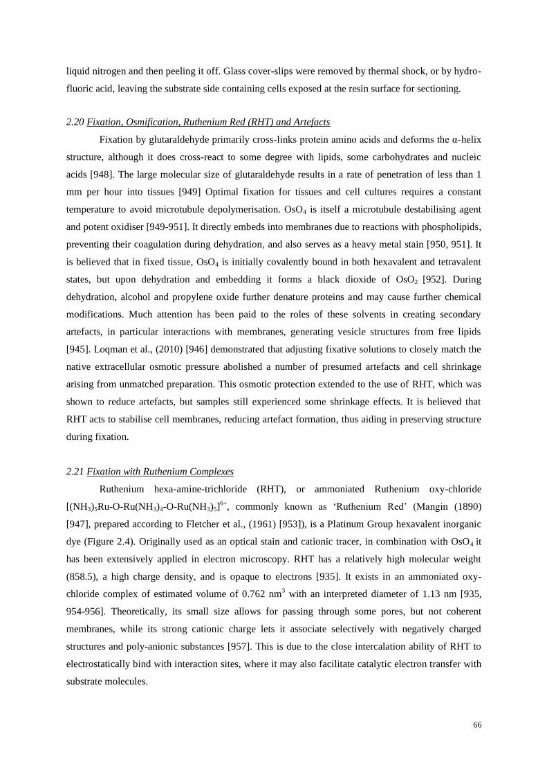
66
liquid nitrogen and then peeling it off. Glass cover-slips were removed by thermal shock, or by hydro-
fluoric acid, leaving the substrate side containing cells exposed at the resin surface for sectioning.
2.20 Fixation, Osmification, Ruthenium Red (RHT) and Artefacts
Fixation by glutaraldehyde primarily cross-links protein amino acids and deforms the α-helix
structure, although it does cross-react to some degree with lipids, some carbohydrates and nucleic
acids [948]. The large molecular size of glutaraldehyde results in a rate of penetration of less than 1
mm per hour into tissues [949] Optimal fixation for tissues and cell cultures requires a constant
temperature to avoid microtubule depolymerisation. OsO4 is itself a microtubule destabilising agent
and potent oxidiser [949-951]. It directly embeds into membranes due to reactions with phospholipids,
preventing their coagulation during dehydration, and also serves as a heavy metal stain [950, 951]. It
is believed that in fixed tissue, OsO4 is initially covalently bound in both hexavalent and tetravalent
states, but upon dehydration and embedding it forms a black dioxide of OsO2 [952]. During
dehydration, alcohol and propylene oxide further denature proteins and may cause further chemical
modifications. Much attention has been paid to the roles of these solvents in creating secondary
artefacts, in particular interactions with membranes, generating vesicle structures from free lipids
[945]. Loqman et al., (2010) [946] demonstrated that adjusting fixative solutions to closely match the
native extracellular osmotic pressure abolished a number of presumed artefacts and cell shrinkage
arising from unmatched preparation. This osmotic protection extended to the use of RHT, which was
shown to reduce artefacts, but samples still experienced some shrinkage effects. It is believed that
RHT acts to stabilise cell membranes, reducing artefact formation, thus aiding in preserving structure
during fixation.
2.21 Fixation with Ruthenium Complexes
Ruthenium hexa-amine-trichloride (RHT), or ammoniated Ruthenium oxy-chloride
[(NH3)5Ru-O-Ru(NH3)4-O-Ru(NH3)5]6+
, commonly known as ‘Ruthenium Red’ (Mangin (1890)
[947], prepared according to Fletcher et al., (1961) [953]), is a Platinum Group hexavalent inorganic
dye (Figure 2.4). Originally used as an optical stain and cationic tracer, in combination with OsO4 it
has been extensively applied in electron microscopy. RHT has a relatively high molecular weight
(858.5), a high charge density, and is opaque to electrons [935]. It exists in an ammoniated oxy-
chloride complex of estimated volume of 0.762 nm3 with an interpreted diameter of 1.13 nm [935,
954-956]. Theoretically, its small size allows for passing through some pores, but not coherent
membranes, while its strong cationic charge lets it associate selectively with negatively charged
structures and poly-anionic substances [957]. This is due to the close intercalation ability of RHT to
electrostatically bind with interaction sites, where it may also facilitate catalytic electron transfer with
substrate molecules.

67
Figure 2.4 Ruthenium Hexa-amine-Trichloride: A proposed structure of the Ruthenium cation complex [958], [(NH3)5Ru-O-Ru-(NH3)4-O-Ru(NH3)5]Cl6.
Ruthenium red has been used extensively as an electron-dense stain for muco-
polysaccharides, glycoproteins and extracellular matrix components [935, 936, 954, 959-961].
Optimal results have been obtained by the inclusion of RHT with glutaraldehyde fixative, followed by
a combination of RHT with OsO4 prior to tissue osmication [935, 959-961]. RHT exists in
equilibrium as a number of complex species (Ruthenium brown, red and violet cations), which also
bind to anionic glycosaminoglycans and polysaccharide structures within tissues [935].
It is believed that ‘Ruthenium red’ is oxidised to ‘Ruthenium brown’ by OsO4, which is then
available to oxidise polysaccharides (to which it is bound), reverting in the process back into
‘Ruthenium red’. This allows the process to be repeated at the reaction site, and also forms the
insoluble electron opaque osmium dioxide (OsO2) by-product. This mode of action allows for the
deposition of multiple molecules of osmium oxide per saccharide interaction, but it only occurs in the
presence of RHT [935, 936].
Many authors have reported variable results with RHT, which could reflect subtle variances
in preparation techniques [936, 963]. Luft (1971) [936] indicated that the intracellular space was
inaccessible to RHT penetration, although results could be indeterminate and controversial in this
regard [960]. He also mentioned a number of exceptions, namely the nuclei of chondrocytes and oral
epithelial cells, as well as mast cell granules, which have readily taken up the dye [936]. This effect
appears to be due to the settling time of the RHT-OsO4 mix prior to use, where it is believed OsO4
generates Ruthenium coupled reactions resulting in enhanced cellular penetration. The effectiveness
of the staining, as noted by Luft (1971) [936] and Hagiwara et al., (1992) [961], is of such density that
further post-staining is unnecessary, as the effect is even optically visible in samples. In the present
work, RHT together with OsO4, gave superior fine resolution imaging.
2.22 Fixation of Extracellular Matrix Proteoglycans
Glycosaminoglycans and muco-polyaccharide proteoglycans are not adequately fixed by
treatments of glutaraldehyde or osmium tetroxide, resulting in loss of much of their respective vital
structures during the process of dehydration and embedding. Hence, to accurately study extracellular
matrices, one is faced with the problems of preventing not only artefact formation, but also fixing,
retaining and staining members of extracellular matrix components. RHT is one of the most widely
used dyes for localising proteoglycans [962], and, when used in combination with glutaraldehyde and
OsO4, provides excellent stabilisation, staining and contrast of matrix components in cartilage [959,

68
961, 964-970]. This stabilising effect prevents degradation and loss of proteoglycans and
carbohydrates during chemical fixation and processing, allowing preservation of their spatial structure
for resin embedding. It is believed that chemically fixed ‘matrix granules’ (which are observed in the
electron microscope) are precipitates of stained proteoglycans which have become condensed through
the combined effects of the cationic dye and the dehydration process [966, 967]. Hunziker et al.
(1981) [959] noted the presence of matrix granules after OsO4/RHT treatment which produced
repeatable superior results for preservation of cell membrane components and pericellular matrix
proteoglycans. Other authors have noted improved ultra-structural cartilage fixation and prevention of
proteoglycan loss during fixation using an RHT/OsO4 combination [960, 966, 968-970]. Loqman et
al., (2010) [946] and Swan et al., (1999) [971] have shown that careful attention to fixation strategies
and osmolarity protection greatly reduced shrinkage issues and artefacts, resulting in improved overall
preservation. Thus, although some matrix granule artefacts may result, RHT treatment preserves
structures with which it interacts and represents the stain of choice for highlighting interactions
between the matrix and cilia in chondrocytes [25, 26].
2.30 Serial Sectioning and Detection of Primary Cilia
Resin blocks were sectioned on a Reichert Ultra-Microtome E (Reichert, Austria) yielding a
selection of candidate cilia which were completely contained within the semithick sections (350-550
nm) and therefore suitable for electron tomography. Sections were held together with improvised glue
(formvar solution with dissolved Sellotape glue spray) before placement upon formvar-coated parallel
slot copper electron microscope grids. Cell culture samples were cut into pieces to be mounted upon
resin stubs for en face serial sectioning as above. Ultrathin sections were post-stained with 2 % uranyl
acetate and 1 % lead citrate using the automated LKB 2168 Ultrostain grid stainer (LKB-Produkter
AB, Bromma, Sweden, 1985).
Sternal chondrocytes were easier to serially section than tendon, and repeatedly produced
excellent uniform ribbons (Figure 3.2A). During embedding sternal tissue sections were arranged in a
known orientation with respect to the tri-lateral orientation of the cartilage, with blocks then being
sectioned perpendicular to this. Mounting of serial ribbons onto parallel slot grids allowed for
inspection in the direction of sectioning, for the identification of candidate cilia (Figure 3.2 A and B,
arrows).
2.31 Semithick Sectioning of Sternal Cartilage
As a model connective tissue, sternal cartilage exhibits a low cell density (2-5 × 10⁵ cells/mm
3
[28]), so that the probability of encountering a complete cilium within the plane of a semithick (350
nm) section is low. This is partially compensated for by chondrocytes usually having shorter cilia in
comparison to other cell types [365, 972], thus increasing the odds of finding complete aligned cilia.
The chance of obtaining a primary cilium fully aligned in the plane of an ultrathin (100 nm) section is

69
low [973], but increases proportionally with thickness. However, TEM imaging quality decreases with
increasing specimen thickness, which is exaggerated at high tilt angles during tomography,
influencing imaging data quality. Thus, the choice of section thickness represents a trade-off between
cutting a section thick enough to contain a cilium, but thin enough to retain imaging quality,
especially at high tilt angles [975]. In reality cilia are always to some degree bent in their projection
within the matrix, complicating the search for candidate samples of aligned cilia suitable for
tomography [25, 26, 100]. Since serial sectioning of chondrocyte connective tissue resulted in a low
incidence of cilia in their longitudinal axis [973] due to their pseudo random projection within the
matrix [31, 100, 973, 974], it became necessary to investigate cell culture techniques to increase the
incidence of candidate cilia.
2.32 Tomography of Monolayer Cell Culture: Aligning Cilia With The Substrate
Analysis of primary cilia in cell culture has been aided by the observation that in many cells
the cilia are aligned with the substrate [31, 63, 976] (Figure 2.5). It has been observed during cell
culture that cell cycle arrest, through ‘serum starvation’ [986] increases the interphase expression of
primary cilia [120, 977, 978, 985]. Albrecht-Buehler (1977) [979] examined large numbers of cells in
culture and found that of their primary cilia, 73% ran parallel with the basement substrate whilst the
other 27% were oriented apically upwards. Vorobjev et al., (1980) [980] examined 11 interphase
cells, in which cilia were aligned not less than 75 degrees to the substrate (P<0.00125), however this
was not observed during G2-phase or mitosis [63].
An in vitro preparation represents a significant improvement in the incidence of usable cilia
per section over in situ sectioning. The precise mechanisms by which chondrocytes cilia become
aligned with the substrate presently remains unknown, however it is most likely due to a combination
of a number of effects ranging from loss of micro-environment to culture media, and involving planar
cell polarity and cytoskeletal rearrangement [981, 982]. Wheatley et al., (2000) [983] reported the
presence of 5% bi-ciliated (and some multi-) ciliated centrosomes of cells in culture. It has been
shown de-differentiation occurs during monolayer cell cultivation of chondrocytes [984], with Alieva
et al., (2004) [985] indicating that unreliable data could result, as primary cilia in vitro may not be
functioning correctly, since the cell is not in its native environment [986].
The application of cell culture allowed for the easy and accurate investigation of the proximal
centriole with respect to the basal body, which was previously difficult to establish in situ. The correct
polarisation of the orthogonal arrangement of the diplosomal centrioles has resulted in confusion
within the literature, as many illustrations of them have been either vague or inaccurate [94].

70
Figure 2.5 Tomography of a Primary Cilium
Aligned with the Substrate: A tomogram optical section of an ovine chondrocyte in
culture. The cilium is located beneath the cell,
in a juxta-nuclear position, and is surrounded
by a unique membrane encapsulate bound by the substrate. Centrosomal microtubules are
aligned in a lateral radiating ‘fan out’ pattern,
notably from the basal body, while the
proximal centriole is seen tethered closely
nearby.
2.40 Investigative Electron Microscopy
Investigative microscopy of semithick serial sections for candidate cilia was carried out using
low voltage microscopy (100 kV on a Philips 410 with a LaB6 filament) at low beam currents to
prevent aging and damage to the resin. Detailed, image based maps of each electron microscope grid
were constructed to aid in the re-identification of suitable cilia for electron tomography. These maps
noted the position of cilia with respect to the cells in the section, and their relative positions on the
grid. Captured images of primary cilia were measured using ImageJ [987].
2.41 Fiducial Marker Application
For accurate tomography reconstruction, electron-dense fiducial markers were added to the
section for tracking purposes (Figure 2.6). Grids containing suitable candidate sections for
tomography were labelled with 15 nm gold fiducial markers (British Bio-Cell, UK). Adequate
application upon both sides of the electron microscope grid is required for use as electro-optical
markers. Due to differences in material properties between the formvar and the resin of the section
surfaces, gold fiducial applications require longer settling times upon formvar (by a factor of 30) in
order to acquire suitable coverage densities for tomography.
Figure 2.6 Fiducial Markers: Application of 15 nm gold markers upon a section containing a cultured chondrocyte primary cilium aligned in the direction of the substrate. Fiducial coverage aids in alignment and reconstruction of tilt series images.
Scale bars 500 nm.

71
Fiducials were applied undiluted to the top (resin section) and bottom (formvar) sides of the electron
microscope grid, for between 2-4 seconds, and 60 seconds respectively (rinsed off in distilled water)
achieving a uniform distribution of 10-15 fiducials per square micron per side. Grids were inspected
in a Philips 410 TEM to monitor application. Excess application of gold fiducials was found to make
tomography impossible, as it ‘drowned out’ the signal to noise ratio of the images.
2.42 Imaging Microscope
Electron tomography was carried out at AgResearch Limited, Lincoln Research Centre, using
a FEI Tecnai G2 T30 300 kV transmission electron microscope (FEI Company, Oregon, USA) with a
LaB6 filament electron source. Single and dual axis tomography was undertaken at 300 kV, in a single
axis tilted through ±64.5° at 1.5° increments. Images were collected with a Tietz camera (2048 by
2048 pixels, 12 bit). Not all specimens were suitable for dual axis tomography, as they were located in
proximity to grid bars or close to the edge of sections Figure 3.2 (D- F). This sometimes resulted in
occlusion of the image along one axis at higher tilt angles (Figure 3.2 E) or precluded imaging from
contrast effects (Figure 3.2 F). During long data acquisition periods under the beam, electron heating
aged the samples, leading to structural changes affecting the ability to reconstruct images into a
tomogram.
2.43 Remote Tomography
Remote tomography was undertaken using the Kiwi Advanced Research Network (KAREN)
between AgResearch Ltd in Lincoln and the Otago Centre for Electron Microscopy in Dunedin. This
allowed the remote user to access the microscope via video-audio-link, to interact with technical
personnel supporting the acquisition of the tomograms and to securely transfer images and data files
offsite. This allowed for rapid feedback, as the user was able to quickly ascertain the quality of the
data through remote viewing and reconstruction without being physically present at the AgResearch
Lincoln Research Centre (Figure 2.7).
Figure 2.7 Remote Electron Tomography: Electron tomography undertaken over the KAREN network. Electron tomography data is acquired remotely, where monitoring of the process and tilt series data are transferred over the KAREN network for
remote parties.

72
2.44 Specimen Attrition
Many otherwise excellent cilia were found to be unsuitable for tomography. For example,
some were either too close to the grid bars, too close to the edge of the section or located at extremes
towards the edges of the electron microscope grid which resulted in difficulty tracking during tilting
or loss of imaging. Of those suitable cilia sampled at 300 kV, there was a high (50%) attrition rate
during tomography, where sections physically failed under the microscope beam, or significant
section aging resulted in failure to recombine the image alignments into a tomogram. Final inspection
resulted in the discarding of many apparently suitable tomograms, as their quality proved inadequate
for analysis. Handling and processing errors also destroyed specimens.
2.45 Collection and Analysis
Conventional electron microscopy provides projected spatial resolution in one dimension and
is limited to ultrathin sections, while tomography provides a better spatial resolution. Single axis
reconstructions result in missing geometric information known as the ‘missing wedge’ problem [988,
989] which is also present to a lesser extent in dual axis recombination [990-992] (Figure 2.8). Dual
axis tomography provides improved resolution by the incorporation of two tomograms taken at
orthogonal angles to each other and combining them into one, a more challenging task in terms of
sample aging and distortion during acquisition.
Figure 2.8 The ‘Missing Wedge’ Problem: A series of images of a sample section is gathered by the transmission of
electrons onto a detector over a series of tilt angles (± 65°) [A]. These are reconstructed so as to represent the volume imaged. However, it is not possible to tilt the specimen through all angles to reconstruct a full theoretical volume [B]. For a
single axis acquisition, incomplete geometric information is represented as a ‘missing wedge’ (red) [C], which is reduced by
incorporating dual axis images [D], taken at 90 degrees to each other. Reproduced and modified with permission from Dr
Harland, AgResearch Ltd, Lincoln, personal correspondence.
Many parameters during tilt series image acquisition are under the control of the operator
while others are automated processes within the microscope. Successful reconstruction of gathered
data depends upon tracking each successive tilt series image with the aid of surface localised
specialised markers on both sides of the section for determining the image axis and location through
the full tilt range (Figure 2.9). Image series at known tilt angles are first processed, filtered, aligned
and selected fiducial markers are then used to calculate an exact alignment solution to produce a
C D A B

73
tomogram using digital software. The process of tilt series acquisitions are visualised as animations
listed in Appendix VII, Section 7.1.
Figure 2.9 Data Acquisition: Tilt series images of a 300 nm semithick section containing a chick sternal chondrocyte primary
cilium, taken in the Tecnai G2 T30 electron microscope imaged at 300 kV. This tilt series is visualised as an animation
<TiltSeries K10.avi>, listed in Appendix VII, Section 7.1.
Various software packages are available for aligning, tracking and reconstruction of a
tomogram (such as IMOD [993], TomoJ [994, 995] and microscope specific packages) as well as for
alignments without using fiducials [931]. IMOD, developed by Mastronarde and Gaudette and
supported by the US National Centre for Research Resources [996], was chosen as a reconstruction
and modelling package as it is recognised as the present standard freeware package. Tomograms are
viewed as images conventionally in two dimensions as ‘optical slices’ (as seen in Figure 2.5) in the
form of layered z-axis images. These differ from conventional ultrathin images in that their thickness
is in the nanometer range (Figure 2.1). A series of optical slice images maintains structural
information between consecutive images, allowing for precise determination of intricate features.
Thus, ET provides detail of macromolecular assemblies such as membranes, microtubules, fibres and
other complexes [924] at a resolution much greater than conventional TEM [932].
2.46 Data Processing
Data processing was carried out in the IMOD package (several upgrade versions) using an
Intel® Core (TM2) Quad CPU Q8200 operating at 2.23 GHz with 4 GB of RAM. Single and dual
axis tomograms were aligned and reconstructed using back projection and combined using IMOD
[992]. Models were constructed using the ‘eTomo’ interface, allowing analysis and viewing of the
biological volume in three dimensions [997].
2.47 Fiducial Tracking
To obtain the best tracking solutions, fiducials were chosen specifically for their ability to
provide good contrast and visibility over the full range of imaging angles between consecutive frames,
ensuring complete tracking solutions for all marker beads. This is necessary as many fiducials are not
visible within the imaging field over all tilt angles, whilst others become occluded or obscured during
tracking at higher tilt angles where the semithick section thickness increases, resulting in local

74
tracking errors (Figure 2.10). Fiducial tracking errors are visualised in series of animations listed in
Appendix VII, Section 7.2, with animations of tomogram stacks in Section 7.3.
Figure 2.10 Fiducial Tracking in IMOD: [A-C] details tracking of gold markers across aligned tilt series prior to final
tomogram formation. Enlarged views in [A’, B’ and C’] show the process of the induction of tracking errors (direction of
motion, arrows) where fiducials come into occultation. Errors can also occur from background signal levels during tracking
at higher tilt angles due to the increased effective imaging thickness. Animations are listed in Appendix VII, Section 7.2:
<Fidtrackone.avi> and close up view in <Fidtracktwo.avi>.
2.50 Analysis and Modelling of the Tomogram
Visualisation of structures is achieved in IMOD (3DMod) through tracing components of
interest, forming a three dimensional representation based on contour mapping and volume rendering.
Vesicles were hand traced with drawing tools in the ‘zap window’ in the z-axis, with each new vesicle
assigned a new unique model object number with selectable colour. Vesicle components of the Golgi
apparatus were broken into their respective cis, medial and trans compartments, and were each
grouped as distinct objects, while the nuclear and cell membranes and vesicles were each treated as
individual objects. The ciliary pocket invagination was treated as a distinct structure, which also
encompassed part of the ciliary membrane (due to the axis of the cilium within the section). The other
half of the ciliary membrane was modelled as an extension of this structure, giving rise to the two
element membrane model (Figure 2.11). The ‘image slicer window’ allows for visualisation of the
tomogram ultrastructure in any plane, aiding the tracking and tracing of finer components, such as
filaments and microtubules. Microtubule tracing within the axoneme and basal body was problematic
due to the close spacing of microtubules and the twisting inward taper in their projections.
The 3DMod ‘iso-surface’ volume rendering function allowed complex electron-dense deposits and
surfaces to be averaged and modelled as discrete volumes. Matrix granules, luminal deposits,
microtubule deposits, y-shaped linkers, alar sheets and basal feet were modelled in this way.
Model construction was an intensive iterative process. Once structures had been meticulously
traced, error checked and confirmed as accurate, they were saved as distinct objects. Model elements
could be measured, spatial inter-relationships mapped and volumes ascertained. These processes are

75
time consuming, as manual tracing, control and input is required to generate model components and
then display them. Automated image processing, data extraction and modelling for tomography is not
yet possible. Animations of tomogram stacks are listed in Appendix VII, sections 1.3, while model
constructions are listed in Section 7.5.
Figure 2.11 The Modelling Process: [A] Manual tracing of vesicles and microtubules produces a ‘raw’ layered view in
3DMod prior to meshing. Meticulous attention to detail ensured the accurate translation of information between successive
optical slices of the tomogram for forming each object [B]. Details of the ciliary membrane surrounded by the extracellular matrix are seen in [A], with [B] detailing the ciliary pocket and cytosolic vesicles. Individual objects are assigned distinct
distinguishing colours for clarity and identification. Animations of the coarse tracing process of model generation are listed
within Appendix VII, Section 7.5.
2.51 Model Display
Objects were organized into groups defined by their ultra-structure. Tubes were used to form
microtubules, intermediate filaments, actin and subdistal fibres, while vesicles and membranes were
modelled as open or closed surfaces. Smaller electron dense deposits were displayed as solid
volumetric renderings. Each object may be individually or selectively visualised by the user, with
optional control over colour shading and transparency being possible. Object selection allows for
individual or groups of objects to be examined, their spatial geometry and inter-relationships to be
investigated. Screenshot capture sequences allowed for the generation of animations providing a three
dimensional perspective.
2.6 A Virtual Cilium: SolidWorks
A classical reference image of an ultra-structural cross-section of a centriole from the
frontispiece of Wheatley’s book The Centriole: A Central Enigma of Cell Biology [63] was imported
into SolidWorks (Dassault Systèmes, SolidWorks Corp1) for the purpose of forming an electronic
template for the generation of a virtual cilium (Figure 2.12 A, B). Components could be extruded
forming the triplets and inter-triplet linkages of a basal body (Figure 2.12 C and D) along with the
axoneme sub-fibre doublets (Figure 2.12 E and F). The nine-fold (40°) symmetry of the model triplets
should be noted, and also a slight distortion of the original image (which may be due to the technique
used to generate the original image (Figure 2.10 A, B). Microtubule triplet and linkage lengths were
1 http://www.solidworks.com/

76
extruded to match ultrastructural results (Figure 3.4), while doublet lengths were derived from the
tomographic model data, providing an anatomical comparison in Figure 3.4 F.
Figure 2.12 The Virtual Cilium: Extruded from a pictorial template into a structural likeness based upon tomographic data
obtained from [A] and Figure 3.4. This model is visualised in animation <SolidWorksModel.avi> listed in Appendix VII, Section 7.7.
B
D
A C
E
F

77
Chapter Three: Results
3.0 Introduction
All cells communicate with their extracellular microenvironment, yet up to the present the
significant role of the primary cilium has been overlooked. Many researchers have studied primary
cilia in the past, including using various biomechanically responsive connective tissues. In this work
two connective tissue types have been investigated as a source of primary cilia: chick sternal cartilage
and the flexor tendon of the chick Gallus domesticus. Flexor tendon was found to be too difficult to
section reliably, hence sternal cartilage was utilised for tomographical investigation. Ovine
chondrocyte cell culture served as a supplemental approach for investigating primary cilia.
3.1 Mechanical Connective Tissues: Tendon
Flexor tendon was sectioned longitudinally (with the grain of the collagen fibres) revealing
highly aligned collagen bundles surrounded by tenocyte support cells (see Figure 3.1 A). Primary cilia
were usually found to be deflected, and no cilia were found completely contained within a thick
section. Some cilia were found seated within invaginations of the ciliary pocket, or protruding
between bundles of collagen bundles. A high percentage of those observed were found to be highly
deflected in their projection from the cell, so as to run parallel with the collagen fibres (Figure 3.1 B).
This orientation made it virtually impossible to obtain a complete primary cilium within the plane of
the section. A well-developed endoplasmic reticulum and sparse Golgi structures were observed
within many cells. Transverse sectioning revealed tight bundles of highly polarised collagen bundles
in the extracellular space, each consisting of hundreds of uniform arrays of collagen fibres, and with
tenocyte cilia protruding between bundles (Figure 3.1 D-F).
3.2- 3.3 Mechanical Connective Tissues: Chondrocyte Cilia Morphology and Staining
The choice of sternal cartilage was fortuitous, for investigation of chondrocyte primary cilia
revealed they were generally undisturbed in their linear projections, increasing the odds of being
completely contained within a semithick section (Figure 3.2). Two electron microscopy staining
techniques were utilised for electron microscopy, conventional fixation with en bloc uranyl acetate,
and ‘Ruthenium Red’, a platinum group transition metal utilised specifically for the fixation, retention
and staining of extracellular proteoglycans and saccharides. Conventional electron microscopy
staining revealed an interspersed network of collagens and some weakly stained structures, while the
Ruthenium treated cartilage reveals not only this network, but the finer details of proteoglycans and
matrix granules, which are not fixed or retained by conventional treatment techniques. Cilia were
found in various projections, including being retracted (or invaginated) within the cell and being
closely ‘pocketed’ by the membrane (see Figs 3.2 F, 3.3, and 3.5).

78
3.4 The Chondrocyte Primary Cilium: In vitro
The basal body form was similar to that described in other centriole studies [28, 29, 45, 998],
as a longitudinal cylindrical barrel-shaped body comprised of nine semi-equidistant microtubule
triplets, nominally 200 nm in diameter and inclined to the main ciliary axis with a left-hand ‘curl’ or
twist in the positive microtubule direction. This allows for the determination of cilium microtubule
direction in conventional ultra-thin sections, as viewed in the series from Figure 3.4 L-Q. The inner A
and B-subfibres form the axoneme microtubule doublets, while the C-subfibre demarcates the basal
body. Inter-triplet linkers are seen between the A and C-subfibres of adjoining triplets (Figure 3.4 L)
as well as an inner A-subfibre ‘hook’ subtending into the lumen at the proximal end of the basal body
(Figure 3.4 L and M) but do not appear to continue past the mid-body (Figure 3.4 N). The distal end
the C-subfibre appears to become partially unattached from the B-subfibre (Figure 3.4 N) where the
alar sheets appear to be extended from an amorphous ‘basement’ zone across the triplets, focused
upon the B-subfibre. These sheets run to fine points of contact upon the axoneme membrane (Figure
3.4 N to O) anchoring the basal body to the cell membrane, and also demarcates the end of the C-
subfibre and the start of the axoneme. Throughout this region the inclination of the microtubules
transition in their pitch from the triplet structure of the basal body to axonemal configuration as seen
in (Figure 3.4 P, Q). In this transition zone, y-shaped linkers were identified spanning between the
doublets and the ciliary membrane, becoming infrequent further along the axoneme.
3.5 The Chondrocyte Primary Cilium: In Situ
The general morphology of all chondrocyte primary cilia examined conformed to work by
earlier investigators [25-29] and where visible, the Golgi network was found to be located in
proximity to the centrosome. The cilia themselves varied in their degree of projection and deflection
into the matrix, with respect to the basal body (Figure 3.3), ranging from a full interactive extension,
to a partially retracted invagination, and even to fully reclined extension (with less matrix present)
against the cell membrane. Of hundreds of semithick serial ribbons inspected, 167 primary cilia were
found to be completely contained within the plane of their section. Of these, 19 were prepared for
electron tomography at 300 kV. Specimen attrition resulted in a total of 9 single axis and 5 dual axis
tilt series of cilia being reconstructed, with only the most suitable (contained within section, location
on grid, and with good contrast) chosen for modelling. Measurements were taken of the candidate
cilia membrane length, axoneme, basal body length and width of transition zone, where it was
possible to do so accurately (see Table 3.1). Ciliary membranes varied in length between 0.7-2.41 µm
with an average length of 1.38 µm. Basal body lengths were found to average 0.365 µm, with the
average transition zone width of 0.26 µm. Dual ciliary length measurements were taken due to
discrepancies arising from the transition zone, in measuring between the cilium membrane and the full
microtubule lengths of the cilium.

79
Table 3.1 Measurements of Chondrocyte Primary Cilia
3.6 Tomography of Native Sternal Chondrocytes
The majority of tomograms undertaken were not fully successful primarily due to the
combined effects of aging upon the semithick section during tomography, such as failure of the
section under beam, or alignments were rendered unsuitable for tomogram reconstruction. Others
failed to deliver adequate contrast upon reconstruction, or occasionally were lost through attrition of
the electron microscope grids or handling errors. Initially, the process of post-staining in some cases
caused loss of stain on section, and precipitate fouling of the grids. This step was subsequently
dropped, in favour of the use of en bloc staining alone, which proved more than sufficient with the
RHT treatment. Fiducial application times were also reduced to minimise aqueous interactions and
‘wash out’ of stain. Once generated, tomograms provided high resolution optical slices of the imaged
volume, however reconstruction results were found to vary somewhat, compared with conventionally
stained samples (Figure 3.5). With enhanced staining from ‘Ruthenium Red’ treated samples (Figure
3.5 C’), the extracellular matrix was preserved revealing the abundant presence of matrix granules
(Figure 3.5 C). The presence of collagen fibres was noted within the matrix, however these were
largely obscured by granules within the Ruthenium treated samples (Figure 3.5 C, C’). The presence
of luminal vesicles within the basal body of cilia was noted on numerous occasions and is in line with
observations of previous researchers (see Section 1.11).
In this study a total of 9 single and dual axis tomograms of chondrocyte primary cilia were
successfully reconstructed. A single axis tomogram (±65° in 0.5° steps, 350 nm thick) was chosen to
model as it showed excellent contrast of a near complete cilium located within an indentation in the
cell membrane (see Figs 3.6 and 3.7). As an aid to understanding, a series of animations have been
constructed and are included in Appendix VII.

80
3.7-3.9 A Single Axis Model
The model in this study was created based upon a single axis tomogram. Inspection revealed
that not only was the cilium almost completely within the plane of the section, but suitable
components, the extracellular matrix interacting with the cilium, along with other organelles; the
Centrosome, Golgi Apparatus, Endoplasmic Reticulum and Nuclear Membrane were also within the
tomogram (Figures 3.6, 3.7 and 3.8). The ultrastructural anatomy of the axoneme and basal body of
the cilium is detailed in Figure 3.9. It was found however that a part of the distal tip of the cilium,
along with a portion of the basal body and proximal centriole were missing, being out of the plane of
section. During modelling, each structure within the tomogram was carefully considered, error
checked and compared with ultrathin sections before being accepted as an element. The resulting
model consists of roughly 2200 objects totalling 240 megabytes in size, taking 3000 hours to
construct. The model is also in broad agreement with previous studies of primary ciliary ultrastructure
[25-29], and chondrocyte ultrastructural observations (Figure 3.4).
3.10 Tomographical Modelling of the Extracellular Matrix and Matrix Granules
Cartilage connective tissue consists of chondrocytes surrounded by a pericellular capsule
containing a complex mix of proteoglycans, collagens, osmolytes and signalling molecules.
Specimens treated with RHT prior to and / or during osmication demonstrated greater contrast of
proteoglycans compared to conventional treatment with en bloc uranyl-acetate alone, revealing fine
structure content of the matrix granules and their fine inter-linkages (Figure 3.5 C’). Stained granules
in the extracellular matrix were composed of condensed collections of interconnected minor
aggregates, forming a complex three dimensional web of tensile proteoglycans which were modelled
as ‘condensates’. These are believed to be composed of Aggrecan and Link protein. Dual-axis
tomography of these granules (Figure 3.10 D-G) revealed their fine structure and inter-connectivity
but when seen in three dimensions they appear as ‘coarse nuggety’ structures (Figure 3.10 H-L).
These were found to be usually distorted in the direction of their attached filaments. The proteoglycan
matrix exists inside a ‘vacuole like’ indentation of the cell membrane located in the pericellular space.
This surrounds the protruding cilium uniformly and is very similar in appearance to an enlarged
ciliary pocket [325], however the matrix differs as it is less dense than the pericellular environment
(Figs 3.6 and 3.7).
3.11 Matrix Ciliary Membrane Interactions
The ciliary membrane was found to be decorated on both sides with stained components
which may represent receptors, trans-membrane proteins, channels and attachment complexes for
unknown tethered structures (Figure 3.10). Extracellular matrix granules were found to be physically
tethered via filaments to the membrane by unknown receptors along the full length of the axoneme, in
some cases being the foci of localised distortions in the membrane created by matrix forces (Figs 3.11

81
and 3.12). Many densely stained structures were identified on both sides of the lipid membrane
(Figure 3.11 F-M) which were associated with extracellular connecting filaments (Figure 3.11 G-J)
and membrane-spanning particles (Figure 3.11 K-M).
3.12 Matrix to Ciliary Membrane to Microtubule Interactions
Matrix-membrane interaction sites were sometimes observed as local ‘dimples’ in the local
membrane (Figs 3.11 and 3.12). These were connected via fine strands to matrix granules apparently
under load from the external environment. These unknown receptor mediated ciliary membrane
connections were matched nearby by complex protein linkages between the membrane and the
microtubule doublets, and between individual doublets and (Figs 3.12 A, B and 3.15 A, C, arrow).
Microtubules were also observed with microtubule-associated proteins, as well as accumulations of
densely stained materials (Figure 3.12). Matrix forces upon the membrane are potentially able to be
transmitted through these prominent linkers, allowing these forces to be transmitted to the
microtubule doublets beneath. The membrane of the cilium was modelled in two parts, permitting a
simple view of its internal structure. It was found to neatly cover its internal contents, while having a
ripple texture, with a number of the distortions within the membrane being attributed to tensile matrix
components (Figure 3.12 C and D).
3.13-3.15 The Axoneme Microtubules
The cilium axoneme was divided into three zones; The Distal, Middle and Transition Zones
(Figure 3.13). The tomogram revealed the cilium’s unique three-dimensional microtubule nine-fold
architecture, which is determined by the basal body (basal body triplet architecture is seen in Figure
3.20 G). The cilium is composed of a total of 27 unique subfibre assemblies of various lengths (from
the basal body triplets), with the axoneme making up to 18 microtubule subfibre components
(depending upon numbers projected). The microtubule doublets reduced in number along the
axoneme (from 9 to 5), were twisted and inclined inward with a ‘left-handed inclination’ in their
projection from the basal body triplets, resulting in the cilium tapering towards its distal end. The
morphology of the axoneme appears deflected under the combined influence of external
environmental loads of the local matrix (viewed longitudinally in Figure 3.13).
The localisation of macromolecular complexes identified within the axoneme are detailed in
Figure 3.14. These were found concentrated upon specific microtubules within the ciliary lumen,
located primarily in areas of the distal-to near mid-zone of the axoneme. While it is not possible to
fully identify with any certainty the objects identified, it is possible to classify objects into broad
categories. These were defined as membrane to microtubule linkers; microtubule to microtubule
linkers, filaments and ‘rafts’ of periodic materials resembling intra-flagellar transport particles,
tabulated with Figure 3.29.

82
Many smaller deposits upon the membrane and microtubules were also noted, however many
were below the threshold chosen for modelling. Figure 3.15 details a number of objects and densely
stained periodic microtubule associated structures interacting with the ciliary membrane Figure 3.15
A, F) which are reminiscent of IFT-like transport materials. Membrane to microtubule linkages
located near matrix induced ciliary distortions are seen decorated with unknown materials (Figure
3.15 E-F’’), however these materials were curiously absent near the distortion in (Figure 3.15 E).
3.151 Localisation of Intra Ciliary Transport Materials
Figure 3.14 (A’-E’) detail linear ‘rafts’ or trains of periodic electron-dense ‘IFT-like
materials’ (marked by arrowheads and confined by bars ‘||’). These were located between (and
sometimes inter-connecting) the microtubule doublets and the ciliary membrane (Figs 3.16 E and
3.17) and interspersed with connecting filaments (Figure 3.17 A, arrowhead). IFT ‘rafts’ or ‘trains’
were of variable length, the longest length of 400 nm, and with smaller 150, 80 and 30 nm collections.
These were comprised of periodic groups of 20-45 nm elements, with shorter periodicities seen
making up the longer rafts. These materials were primarily located upon microtubules 2, 3, 6, 7, and
8, with the highest concentrations observed upon 3, 6 and 8 (Figure 3.29, where microtubule triplets,
doublets and their assorted components are listed). Triplet numbering convention followed from under
the middle of a basal appendage, in a clockwise direction when viewed in the positive microtubule
direction.
3.16 The Distal Axoneme
The structural tip of the cilium is unique as it is the termination zone of the microtubule
doublets, the terminus for IFT-transit and an accumulation point for materials. An oblique part of the
membrane of the tip was missing from the section (Figure 3.16), thus the model provided a ‘cut-away
view’ of the distal tip of the axoneme revealing the termination zone of four microtubule doublets (the
fifth falling just short, Figure 3.16 C and E). The distal ends of the microtubule doublets were
associated with dense deposits coating the internal membrane of the cilium tip (Figure 3.16 A-A’’).
Electron dense materials and filamentous structures were identified between the ends of the doublets
and the membrane (Figure 3.16 A and D). A perspective of the distal zone is seen in (Figure 3.16 E),
which details the microtubule doublets emerging into the middle zone, associated with IFT-like ‘rafts’
of materials.
3.17 The Middle Axoneme
This zone is classically the long slender component of the cilium (Figure 3.3 A) which
changes little in its uniformity over some distance. Bounding boxes detail linear zones of
accumulations of materials decorating the microtubule doublets and the surrounding luminal space
(Figure 3.17, A and B). The finer structure of ‘raft-like’ accumulations reveals semi-regular

83
periodicities which are interspersed in clusters along both the distal and middle axoneme
microtubules. The termination zone of microtubule doublet one (Figure 3.17 A, arrow) is decorated
with small structures at its end. Manipulation of the model allows perspective views of the axoneme
that would be seen conventionally contained within an ultra-thin sections (Fig 3.17 C and D, viz, Fig
3.4 P and Q). The middle zone contains two microtubule doublets, both of which contain greater
accumulations of dense particles than the others (doublets 3 and 6) (Figs 3.17 B (arrow), E
(arrowhead) and 3.29).
3.18 The Transition Zone - One
The transition zone represents the first region where the cilium projects into the extracellular
domain, and can be divided into two parts. In the first part the cell membrane joins the ciliary
membrane at a tight right angle (or at a highly curved interface in the case of the ciliary pocket
(Figure 3.5 B) where the basal body is attached to the transition membrane, demarcating the transition
from the cytosolic to the ciliary domain. The second part is the tapering zone, where the microtubules
reduce in number, and merge into the axoneme. Two small ciliary-membrane-associated vesicles were
identified (35-40 nm in diameter) within the extra cellular matrix, closely associated with the
membrane of the transition zone (Figure 3.18 A and B). Trans-membrane densities are associated with
both vesicles. Notably absent was the zone of the ‘ciliary necklace’ [51] suggesting that the ‘y-shaped
linkers’ were not present at the time of fixation, were not adequately resolved, or were destabilised by
the Ruthenium Red treatment. Dense deposits were attached to microtubule doublets as well as fine
filaments (Figure 3.18 C and D). The microtubules were also found to have their greatest inward
inclination within the transition zone (Figure 3.18 E and F).
3.19 The Transition Zone - Two
The curvature of the transition from ciliary to periciliary membrane is illustrated in Figure
3.19 A, where cell and ciliary membranes merge at a right angle. Intense staining of the membrane is
apparent, with heavy decorations and fine fibres present upon the inner surface (Figure 3.19 B and C).
Fibres were found radiating from microtubule doublets and triplets to the ciliary and periciliary
membrane surface along the length of the transition zone and adjoining basal body (Figure 3.19 B’’).
Other fibres made up a number of subdistal appendages (Figure 3.19 D, F and G), were connected to
the basal body microtubule triplets and associated with dense deposits (Figure 3.19 F, centre, green).
These dense deposits proved difficult to resolve adequately. The alar sheets occupy a zone spanning
the microtubules near the terminating end of the C-subfibre and are each drawn to tethered apexes
firmly held within the transition zone membrane (Figure 3.19 G, H and I). The fine granular structure
of the alar sheets were also difficult to resolve.
84
3.20 The Basal Body Microtubule Triplets
The basal body contains a unique nine-fold symmetric ‘barrel like’ structure made up of the
inclined vanes of microtubule triplet subfibres. The A and B-subfibres project longitudinally to form
the axoneme, while the C-subfibre demarcates the centriole zone. Microtubules were aligned and
traced within 3DMod carefully due to their respective close spatial proximities within the sections.
Each subfibre was traced and modelled using intersecting tubes to best represent the circular tubulin
components making up the microtubule. This proved difficult due to their inclination and slightly
curved nature within the tomogram. This is apparent where perspective optical slices parallel the
model (Figure 3.20, A-C, D and F). Of particular interest is the curvature in Figure 3.20 E, which is
also seen in a ‘cut-away’ view in Figure 3.20 F. The basal body microtubule triplets were observed to
be inclined with a left-handed polarisation and curve giving the basal body a ‘barrel like structure’
and ‘vane’ appearance, and were found to reduce in their inclination angle, as they tapered into the
axoneme. As part of the basal body was missing, it has been reconstructed for viewing by projecting
the nine-fold symmetry in Figure 3.20 G.
3.21-3.22 Internal Structures, Luminal Disc, Fibres and Vesicle
The basal body cavity contained numerous electron densities associated with lumen and
microtubule faces (Figure 3.21). Numerous densely stained deposits of different size were found
decorating the basal body triplets, including a larger opaque complex visible as a ‘band’ spanning
inside the distal end of the basal body between microtubule triplets (only some deposits are shown for
clarity of viewing, Figure 3.21 B, C and E). This was termed a ‘luminal disc’ like structure, and was
associated with nearby fine fibres and macromolecular deposits attached to the microtubule triplet
luminal faces. Not all the structures present were modelled within the proximal region of the basal
body, as these proved to be too complex. Included with these structures, a luminal vesicle was
identified and modelled (Figure 3.22). It was firmly attached to the microtubule face, and was found
to be facing in the direction of the axoneme, appearing as if against a flow (Figure 3.22 E). This study
identified three tomograms containing basal body luminal vesicles (see Figs 3.5 B and 3.22).
3.23-34 Basal Appendage One
Surrounding the basal body is the pericentriolar environment, which is made up of stained
deposits of structures which include the alar sheets, subdistal appendages, and basal appendages (also
known as basal feet). Two basal appendages were attached to the basal body. Each basal appendage
consisted of three components; a basal anchoring complex attached to the basement microtubule
triplets, an appendage arm and a docking head. The basal appendages extend centripetally not only to
anchor and structurally support the microtubular network, but also to provide access to a zone away
from the basal body. Examination of the ultrastructure of basal appendages revealed them to be
intensely stained linear structures protruding at near right angles from the basal body (Figure 3.23 A-

85
H). Each appendage revealed over-staining of the docking complex and arm, but exposed an
amorphous composition of the basement structures. This is seen in the model of Basal Appendage
One (BA1, Figure 3.23 I-L) in its attachment to the centriole. The extending appendage arm and
microtubule docking complex was modelled as a single component. The anchoring complex consisted
of an amorphous support network comprised of three basement zones, each of which were inclined
across there basal body triplets at inclination (Figure 3.24, C-F, arrows) to anchor the appendage. A
low density zone was observed underneath the basal appendage, directly above the microtubule triplet
upon which it was centred (also seen in Figure 3.25 F).
The basal appendage projects outside the pericentriolar environment to interact with the
surrounding centrosome, where vesicles interact with the basal body and basal appendages through
fine tethering linkages (Figure 3.25 A-D). The basal arm contains indentations (Figure 3.25 E) and
fine protrusions extending along its length (Figure 3.25 F, H and I) until merging with the docking
complexes upon its distal end. The docking complexes consist of two nodular projections, to which
microtubules (Figure 3.25 G and G’) and vesicles are tethered (Figure 3.25 B). Vesicles are associated
with the basal appendage, the connecting microtubules, and also the pericentriolar basal body zone
(Figure 3.25 D). Three microtubules are seen radiating from the docking complexes (two from one) in
(Figure 3.25 F, H and G) and interacting with nearby centrosomal vesicles (Figure 3.25 H, J). The
ultrastructural perspective in (Figure 3.25 G* and H) shows the vesicles modelled in (Figure 3.25 J*).
Note the basal body longitudinal structure (Figure 3.25 J) detailing the basal appendage, the subdistal
appendages, the alar sheets (attached to the transition zone membrane). Points of interest include the
vesicles attached to basal appendage (Figure 3.25 C) and the basal body (Figure 3.25 D), as well as
the inclination of the basal appendage which may be influenced by torque from the attached
microtubules.
3.26-3.27 Basal Appendage Two (BA2)
Longitudinal optical sections of Basal Appendage Two (BA2) are seen in Figure 3.26 A-H.
The docking complex and arm were modelled in the same way as basal appendage one and appear to
have similar anatomical likenesses, possessing a similar cantilever projection (Figure 3.26 J and L).
The basal appendage had an inclination which appears to also indicate a possible contribution of
torsion from attached microtubules (Figure 3.27 A). The docking complex was found to have only
two docking sites upon it (with no double nodular protrusions like BA1) tethering three and two
microtubules respectively (Figure 3.27 A and B). The ultrastructure of two of these microtubules was
examined around the docking complex (Figure 3.27 C-E). These were tethered to the docking head at
their negative end, and extended positively outwards (Figure 3.27 E’). Densities are noted upon
microtubule bases and docking heads (Figure 3.27 C’, D and E’) including linkages connecting the
microtubule to nearby vesicles.

86
The basement anchoring complex substrate was modelled as numerous discrete components
to investigate its finer amorphous form. Three attachment zones to the microtubule triplet substrates
were identified, including a ‘hollow’ zone underneath the base arm above the central triplet it was
attached to (Figs 3.27 A). Views detailed in Figure 3.26 I-L reveal a similar amorphous morphology
to basal appendage one.
3.28 A Review of Basal Appendages One and Two
The basal appendage model perspectives are visualised in Figure 3.28 to compare their
anatomical characteristics. Respective lateral views of basal appendages Figure 3.28 BA1 A-E and
BA2 F-J detail the appendages docking head, arm and the basement complexes. Comparison of the
basement anchoring sites reveals: 1) Low density areas located under the basal appendage, forming a
longitudinal ‘groove’ (Figure 3.28 C and H). 2) Three distinct attachment areas or ‘basement zones’
corresponding to three separate microtubule triplet attachment regions (Figure 3.28 A and F).
3) The basement materials were highly amorphous in their nature, indicating a porous higher order
structure (Figure 3.28 E and I). Rotational views of the appendage docking heads and arms (Figure
3.28 BA1 K-O and BA2 P-T) reveal subtle differences. The docking heads of BA1 are bifurcated
(compared to the singular head on BA2) however their size and aspect ratios are similar, including the
general architecture of the arms. The indentation on the arm of BA2 (Figure 3.28 R, arrow) should be
noted. The presence of thorny projections from the lower arms (arrowheads) was also recorded
(Figure 3.28 K, P, O and T).
In summary, both basal appendages share highly similar structural morphologies. Their
basement anchoring complexes consist of finely porous materials, with supporting long tapering arms,
whose head docking complexes consist of fine spiny protrusions, some of which anchor cytosolic
microtubules and vesicles (Figure 3.25). Microtubules anchored to the basal appendage docking
complexes are visualised as being decorated with numerous microtubule associates deposits,
complexes and fine fibres linking to nearby vesicles which they carry (Figs 3.34 and 3.37). An area of
exclusion is noted extending in a zone surrounding the perimeter of cytosolic microtubules, which are
uncluttered except for the close proximity of vesicles and microtubule associated proteins (detailed in
Figs 3.34, 3.36 and 3.37).
3.29 The Cilium, Basal Body Materials and Orientation
The positional relationship of the basal appendages (Basal Appendage One, Basal Appendage
Two and their associated cytosolic microtubules) were investigated with respect to the microtubule
triplet structure of the basal body (Figure 3.29 A, seen in the positive microtubule direction). Each
appendage radially spanned across three microtubule triplets, and appeared to be symmetrically
spaced around the basal body, so as to not overlap their basement complexes. This would imply a

87
number of rotational orders are possible, based upon the nine fold triplets, and the possibility of
subtending at least three symmetric basal appendages.
The contents of the axoneme and basal body microtubules were interpreted using both the
model and tomogram ultrastructure to establish the nature of their features, and their relative
microtubule doublet association. Generalised categories were defined for luminal microtubule
components: IFT-like materials (localised to membrane side outer, microtubule side inner), internal
linkers (interconnecting microtubule doublets), membrane linkers (between the microtubules and the
membrane), internal filaments (attached to microtubules projecting into the lumen (membrane side
external, microtubule side internal), y-shaped linkers, alar sheets and the length of each microtubule
doublet. The position of each basal appendage, the relative numbers and radial distribution of luminal
materials upon the microtubules of the axoneme, including the basal body and transition zone
vesicles, are visualised in Figure 3.29 B and tabulated in C. While these were collated from the model
and tomogram, it is difficult to ascertain with absolute certainty their identity and functional
significance.
Microtubule doublets that were derived from triplets associated with the basal appendages of
the basal body were found to have the highest densities of materials associated with them. Doublets 2,
3, 6, 7, and 8, contained the most materials, while 4, 5, 9 and 1 contained lesser amounts.
Extracellular matrix vesicles [999] were found to be associated with the ciliary membrane near
microtubules 6 and 7 (Figure 3.18), while the basal body luminal vesicle was tethered to the inner
surface of microtubule triplet 2. The position of extracellular matrix and luminal vesicles should be
noted with respect to the basal appendages of the basal body (BA1) which covers triplets 6, 7 and 8
and BA2 9, 1 and 2), and the internal luminal disc (spanning 1 and 6). Numerous electron dense
materials were also associated with the triplet faces.
3.30 The Proximal Centriole
The proximal centriole was modelled in the same way as the basal body. Although only half
of the centriole was contained within the tomogram, it was possible to determine the polarisation and
inclination of the microtubule triplets (Figure 3.30). The modelling revealed a nearly identical
structure to the basal body. The microtubule triplets maintained their inclination along the proximal
centriole but did not taper, as with the basal body. Also lacking were any external appendages or
microtubule attachment sites. Similarly to the basal body, there was also a ‘luminal disc’ like structure
located at the distal end of the proximal centriole (Figure 3.30 E and L). Electron-dense materials
decorated the microtubule triplets, with larger complexes found present on the outside (Figure 3.30 C
(G, model view), I and K). Structures linking microtubule triplets were also noted (Figure 3.30 F and
G). Axial perspectives are seen in Figure 3.31 A-C and H showing the luminal disc and radial
distribution of materials attached to and bridging the microtubule triplets. These pericentriolar

88
materials can be seen interacting within the nearby centrosome, and also the nearby nuclear
membrane, in proximity to several nuclear pores (Figure 3.31 D-I).
3.31 Proximal Centriole Radial Components
The distributions of electron dense microtubule associated materials upon the proximal
centriole are shown in Figure 3.31 A and B. These decorated the exterior microtubule surfaces, and
included the presence of an oblique luminal disc like structure Figure 3.31 C. The centrosomal
environment surrounding the proximal centriole contained numerous dense deposits (not all of which
were modelled, Figure 3.31 D, E). The nuclear membrane was modelled to reveal nuclear pore
complexes and found to be in close proximity to the centrosome (Figure 3.31 F). Linkages were
identified connecting from the distal end of the centriole to the nearby nuclear membrane in proximity
to several nuclear pore complexes shown in Figure 3.31 G-I (see Figs 3.33 E and 3.34 A). The nuclear
pores were found to contain stained proteins, and their closeness to the centrosome, and the proximal
centriole should be noted suggesting possible involvement with ciliary activity.
3.32 The Centrosome (1) Ultrastucture and Modelling
Selected serial optical slices of the centrosome overlaid with the model (Figure 3.32 A-H)
revealed the position of the centrosome between the cell and nuclear membranes. At its core is the
basal body, from which protrudes the primary cilium, and the sub-tending proximal centriole. Each is
encompassed by its own pericentriolar environment, which in turn is surrounded by a complex zone
of interacting vesicles. These were modelled individually to reveal a field of vesicles associated with
the basal body, the basal appendages, their attached microtubules, and the cell and nuclear membrane
(Figure 3.32 I-L). A notable gradient of vesicle sizes was noted with smaller vesicles generally being
involved with the centrosome and its processes, and with vesicle size usually increasing further away
from the centrosome. Vesicles were noted usually in clusters in proximity to cytosolic microtubules
where they sometimes appeared to be distended as ellipsoids (Figure 3.32 K and L) with smaller
vesicles usually found grouped as clusters (Figure 3.32 I and K).
3.33 The Centrosome (2) The Centrosomal Torus
During modelling a unique centrosomal vesicle was identified (Figure 3.33), which would not
have been possible to discern adequately with conventional ultrastructural analysis. During tracing it
revealed itself to be a circular ‘tubular’ or annular structure occupying an orbital equidistant ‘toroid’
volume in a zone midway between the basal body and the proximal centriole (Figs 3.32 A-K, blue
outline and 3.33 A, B and G) . Although the structure continued out of section, it was called a torus.
The ‘Torus’ was found to be tubular in structure, spanning a lateral zone of the centrosome with bud
like protrusions located at one end, and a larger ‘vesicle’ at the other (Figure 3.33 G). The membrane
protrusions were associated with a prominent field of interacting smaller vesicles, including a larger

89
vesicle which was attached by fine linkages to the torus membrane (Figure 3.33 C and D). This zone
is prominent in its association with a larger field of pre-Golgi vesicles. Perspectives of the torus are
seen (Figure 3.33 G-K) in relation to the basal body and proximal centrioles, where interactions with
their pericentriolar environments are also seen ultrastructurally (Figure 3.33 E and box, F), with
deposits being noted upon the membrane. These were not modelled as they were outside the scope of
this study. However a microtubule nucleation site upon the torus membrane was identified (seen in
Figure 3.33 E, I, J and K). The torus was also found to interact with a microtubule derived from basal
appendage two (BA2) which spanned the centrosome to connect with arrays of pre-Golgi vesicles
(Figure 3.33 A and K). The torus was found to contain the smallest amount of internal contrast of any
vesicle within the tomogram (Figure 3.33 E and F).
3.34 The Centrosome (3) The Pericentriolar Environment
The pericentriolar environment consists of fine amorphous materials which decorate the
surface of centrioles and interconnects them at their negative microtubule ends. This inter-connective
region was found to be bound by the torus vesicle (Figure 3.34), with the centrioles radiating in a
series of fine filaments linking these structures. A densely stained deposit was also found within this
region (Figs 3.35 E and 3.35 E’), although this this area was not investigated further. The
pericentriolar zone was found to be the focus of short lengths of intermediate filaments focussed
around the proximal centriole. Longer filaments were observed to radiate from this area, one to the
pre-Golgi zone, another toward the cell membrane.
3.35 The Centrosome (4) Basal Body, Proximal Centriole within the MTOC
Selected detailed ultrastructural perspectives of centrosomal components interlaced with the
wireframe of the model aid in revealing the confined nature of the centrosome, the basal body and
proximal centriole and the relationship between the materials and vesicles surrounding them (Figure
3.35 A-E). The close proximity of the torus to the centrioles and nearby associated vesicles and
microtubules should be noted. Electron dense materials upon the proximal end of the proximal
centriole were modelled interacting with the torus and nearby vesicles (Figure 3.35 F). The basal
appendages show distinct clusters of microtubule associated vesicles surrounding their docking heads,
in close proximity to the basal body and transition zone membrane. Intermediate filaments (green)
originated from a zone around the proximal centriole (Figure 3.35 F) (also identified in Figure 3.34 D
and E). The composite model image of the centrosome (shown in Figure 3.35 F) is comparable to the
optical slice of the tomogram stack visualised in Figure 3.8.
90
3.36-3.40 The Cytosolic Microtubules; Centrosome to Golgi Connectivity; The Cis-, Medial- and
Trans-Golgi Compartments; Transport of Matrix from the Golgi
The majority of microtubules identified within the tomogram were physically associated
within, and originating from,the centrosome and the basal appendages. Many of those that were traced
originated out of the tomogram section, but were aligned to intersect with the centrosome, suggesting
that they radiated from the greater pericentriolar zone surrounding it. Independent microtubules were
found freely existing within the cytoplasm associated with local structures (or associated with the
Golgi apparatus). Many of these were observed attached to vesicles or decorated with finer structures.
A high degree of connectivity between microtubules and the Golgi apparatus was observed. A cluster
of aligned microtubules was found to run parallel with the pre-Golgi cis-face (in the direction of the
centrosome), and were involved with a large vesicle field (Figs 3.36 I-L and 3.37 B). Both basal
appendages had microtubules interacting with the pre-Golgi and the medial cisternal stacks (Figure
3.37 B and D). An intermediate filament was also found running parallel to microtubules involved
with the pre-Golgi vesicles (Figure 3.38 K). The Golgi itself was traced out, and modelling divided
into cis-, medial- and trans-compartments. These were surrounded by an assortment of vesicles,
which were in close proximity to the centrosome, and the cell membrane. The compartments
consisted of a series of layers of perforated stacks, forming a highly aligned structure interspersed
with smaller vesicles budding around their edges (Figure 3.38 A-J).
Unidentified materials were observed within some of the Golgi vesicles, and these were also
accumulated in larger vesicles near the trans-face (Figure 3.39 A-B). These were surrounded by a
transport process (Figure 3.39 C’, E, F and F’) involved with the cell membrane. Not far from these
structures, the blunt tip of the primary cilium was found to be interacting with the cell membrane
(Figure 3.40 A). Within this zone, numbers of clathrin-coated pits were identified in the process of
undergoing receptor mediated endocytosis, with their contents being visible (Figure 3.40 B-D). These
were modelled to reveal a series of active cell membrane processes in proximity to the Golgi and a
number of tubular structures resembling endosomal transport conduits [1000] (Figure 3.40 A). A
Caveoli like structure was identified in (Figure 3.40 E and E’).
3.41-3.42 The Complete Model: The Matrix Cilium Golgi Continuum
The full model of the chondrocyte primary cilium is illustrated in Figs 3.41-3.42. This details
the extracellular matrix interacting with the primary cilium, the anatomy of the axoneme, the
centrosome and the centrosomal connectivity to the Golgi apparatus. A tight physical continuum
exists between the extracellular environment, the primary cilium, the centrosome and the Golgi
apparatus, which makes possible the regulated processing and orderly exocytosis of the correct
materials for maintaining a bio-mechanically functional extra-cellular environment.

91
3.43 The Continuum: The Cilium and Centrosome
The Cilium-Centrosome-Continuum is illustrated in Figure 3.43, which overviews the details
of the ciliary axoneme, stemming from its distal tip to the adjoining centrosome. The unique
microtubular architecture of the cilium is revealed, along with its internal cargoes and extra-cellular
interaction with the matrix and a coated pit. The basal body is firmly tethered by basal appendage to
radiating microtubules, and is also supported by the centrosome and proximal centriole. Some
structures have been omitted for clarity. Detailed are the core microtubule based structures of the
cilium, proximal centriole, and microtubule ‘interactome’ of the centrosome. This is the first three-
dimensional reconstruction of a primary cilium at high resolution. It forms a template for the
assignment of function to the described structures.

Flexor Tendon Transverse Sectioning
E F D
Figure 3.1 Connective Tissue Primary Cilia Sectioning of Tendon
Flexor Tendon Longitudinal Sectioning
A B C
[A] Flexor tendon longitudinal sections, showing tenocytes amongst aligned collagen fibre bundles. Cilia were found to be highly deflected by the collagen fibres [B, C].
Cross section of a tenocyte surrounded by columnar collagen bundles [D], with cellular processes extending into gaps around the edges [E] along with a primary cilium [F]. 91

Figure 3.2 Serial Sectioning of Chick Sternal Cartilages
B C
D E F
A
Chick sternal cartilage semithick serial sections [A, D] containing primary cilia aligned within the section [B, C, E and F]. Scale bars [A] 500 µm, [B] 10 µm, [C], 1 µm, [D]
20 µm, [E] 5 µm and [F] 1 µm.
92

Figure 3.3 Projection Types of Chondrocyte Primary Cilia
Conventional electron microscopy staining
of sternal cartilage with OsO4 and en bloc
uranylacetate [A] in comparison to the
Ruthenium Hexa-amine Trichloride (RHT)
treatment [B], revealing an extensive
matrix granule network, and stabilisation
of the cell membrane seen in [B-D]. Cilia
were found to displaying three classical
projections; fully extended from the cell
membrane [A], reclined against the
membrane in a localised pocket [B], or
partially or fully retracted into the cell [C
and D].
A B
C D
Scale bars [A, C and D] 1 µm and [B] 0.5 µm
93

Figure 3.4 Cilium-Centrosome Serial Sectioning of Cultured Chondrocytes
A-C subfibre Linkers Alar Sheet Fibre
Longitudinal Serial Sections
A B C D E
F G H I J
Transverse Serial Sections
L M P Q
.
.
.
.
.
.
.
.
.
.
.
.
.
.
.
. .
.
N O
.
BA1
BA2
BA3
Longitudinal ultra-thin serial sections of cultured chondrocytes showing the cilium aligned in parallel with the substrate [A-J]. Note [A, D] shows the cilium extended, sheathed by the
ciliary pocket and projected from the basal body, whose appendages [H] are attached to cytosolic microtubules. Closely subtending the basal body is the proximal centriole [G-J] making
up the diplosome. Scale bar 0.5 µm.
Transverse serial ultra-thin sections of a basal body (in the + microtubule direction) [L-Q] showing the triplet subfibre structure [A] with attached basal feet (BA1-3) [B], the alar-sheet
fibres [N] tethering individual subfibres to the transition zone membrane [O]. Note the presence of y-shaped linkers [O-Q] linking microtubule doublets to the axonemal membrane as
they incline inwards in their projection. Scale bar 100 nm.
94

Sele
ct tom
ogra
ms o
f cho
ndro
cyte c
ilia labelled
with
fidu
cia
l mark
ers [A-C
] and
their co
rrespo
nd
ing
optic
al slic
es
[A’-C
’]. Note p
oo
r contrast o
f sing
le axis to
mo
gra
ms in
[A’] in
com
pariso
n to
[B’] a
nd
dual a
xis reco
nstru
ction
[C’].
Note
collag
en fib
res
with
in th
e E
CM
[A
’, B
’] and
in
creased
co
ntrast
and
reso
lutio
n fro
m R
uth
eniu
m
stainin
g in
[C’] sh
ow
ing
connectiv
ity b
etwee
n m
atrix g
ranu
les and
mem
bra
nes. A
lso n
ote th
ree small lu
min
al
vesic
les co
ntain
ed w
ithin
the b
asal bo
dy o
f imag
e [B’]. S
cale bars 2
00
nm
.
Fig
ure 3
.5 S
election o
f To
mo
gram
s and
their Z
-Ax
is Op
tical Sectio
ns
A
B
C
B’
A’
C’
95

A 1
00 k
V im
age o
f a 35
0 n
m se
mith
ick sectio
n o
f a chick
sternal c
ho
ndro
cyte fix
ed in
Ruth
eniu
m R
ed/O
sO4
with
en b
loc u
rany
l acetate. T
he c
ilium
is located
with
in a
n in
vag
inatio
n (sim
ilar to th
e cilia
ry p
ock
et) in
the ce
ll
mem
bra
ne w
hic
h co
nta
ins a u
nifo
rm b
ut lig
htly
stain
ed m
atrix co
mprised
of m
atrix g
ranu
les (in
com
pariso
n to
the p
ericellu
lar matrix
). No
te the th
icker sectio
n im
age c
hara
cteristics (in co
mpariso
n to
ultra-th
in sectio
ns) w
ith
less clarity d
ue to
heav
y stainin
g.
Ru
the
niu
m R
ed
.
Fig
ure 3
.6 A
Sem
ithick
Sectio
n C
ontain
ing a P
rimary
Ciliu
m
Fig
ure 3
.7 A
lignm
ent o
f To
mo
gram
Dataset
Fu
ll Neg
ativ
e T
ilt -65
° 0
° F
ull P
ositiv
e T
ilt +6
5°
A
B
C
A
Alig
ned
tilt series imag
es of fro
m fu
ll neg
ative to
full p
ositiv
e tilt [A-C
]. An an
imatio
n o
f this sam
ple,
<T
iltSeriesB
4-3
.avi>
is detailed
in A
ppen
dix
VII, S
ection 7
.1
96

Figure 3.8 Tomogram Z-Axis Stack
Centrosome (MTOC)
A series of optical sections showing features of the extra-cellular matrix, cilium and the Golgi apparatus as well as the nearby nuclear membrane. Visualisation of this
stack is contained in movies <StackTomogramB4.avi, StackAxoneme.avi and StackCentrosomeB4.avi>, detailed in Appendix VII, Section 7.3.
Endoplasmic Reticulum
Nuclear Membrane
Nuclear Pore
Microtubule
Extra Cellular Matrix
Nucleus
Invagination
Coated Pits
Intermediate Filament
Fiducial Gold Marker
Golgi Apparatus
97
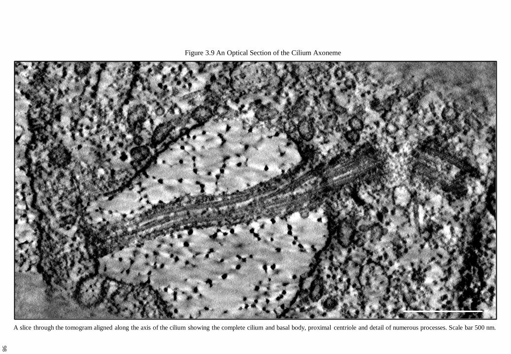
Figure 3.9 An Optical Section of the Cilium Axoneme
A slice through the tomogram aligned along the axis of the cilium showing the complete cilium and basal body, proximal centriole and detail of numerous processes. Scale bar 500 nm.
98

Figure 3.10 Tomographical Modelling of Extracellular Matrix and Matrix Granules
An optical section of extracellular ‘matrix granules’ [A], magnified in [B] and [C] (offset in z-axis). These are comprised of numerous tightly packed aggregates of smaller densely
stained matrix components [C] which are inter-linked by fine filamentous fibres to at least 4 other granules within the matrix. [D-G] A series of slices of the matrix granule inset in
Figure 3.11 [A] showing their fine structure and a series of rotations of the model based upon the granule [H-L]. Scale bars [A] 200 nm, [B] 50 nm and [C-L] 10 nm.
A B C
D F E G
H I J K L
99

J K L M
A B C
D E F
G H I
Figure 3.11 Matrix Ciliary Membrane Interactions
[A] An optical slice showing networks of highly filamentous interconnected matrix granules surrounding and linking to the ciliary axoneme (see Figure 3.14 [C’]). A dimple in the
membrane [B], which is seen enlarged and offset in the z-axis in [C] detailing attachment sites of unidentified extracellular components. [D, E] Detail of attachment of a matrix granule to
the cilium membrane and [F-I] showing fibres to tethered to the lipid membrane [G], enlarged in [H]. [I, J] show the attachment of tethers with unknown membrane associated structures.
Images [K-M] show numerous bound densities on both sides of the lipid membrane, some which span it. Scale: membrane thickness believed to be ~4.5 nm.
10
0

Figure 3.12 Matrix Ciliary Membrane Microtubule Interactions
A
B
C
D
[A] An optical slice of the axoneme interacting with extracellular matrix granules (arrows) of aggrecan (arrowheads) which has been stained with Ruthenium Red. Each consists of
multiple minor precipitates within each granule (see Figure 3.10). [B] Extracellular matrix granules interact with the ciliary membrane (arrows), and electron dense membrane
associated proteins. Membrane to microtubule bridges connect the ciliary membrane to the microtubule doublets (arrowheads). [C] Components of the three dimensional aggrecan
matrix were modelled interacting with the ciliary membrane (arrows). [D] The ciliary membrane is distorted from the combined forces of matrix connections (arrows) and bending of
the microtubule core, causing dimpling and rippling of the membrane. Scale bar [A, C and D] 100 nm and [B] 25 nm. 10
1
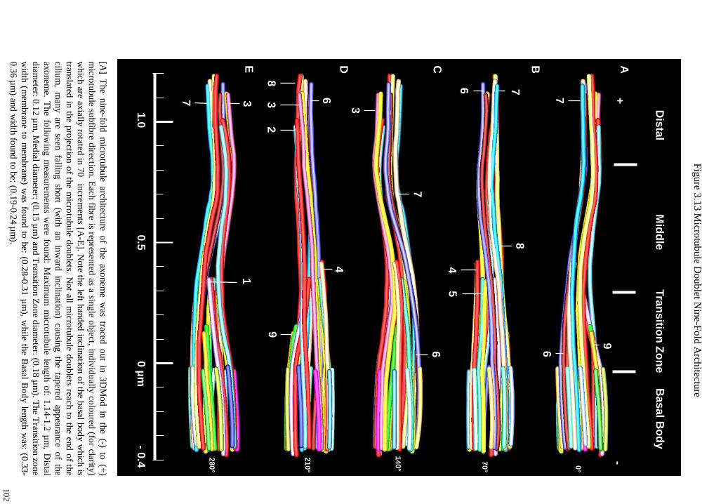
[A]
The
nin
e-fold
m
icrotu
bu
le arc
hitectu
re o
f th
e ax
onem
e w
as traced
o
ut
in 3D
Mo
d in
th
e (-)
to (+
)
micro
tubule su
bfib
re directio
n. E
ach fib
re is repre
sented
as a sing
le obje
ct, ind
ivid
ually
colo
ured
(for clarity
)
whic
h are a
xia
lly ro
tated in
70
in
crem
ents [A
-E]. N
ote th
e left h
and
ed in
clin
ation o
f the b
asal b
od
y w
hic
h is
translated
in th
e pro
jectio
n o
f the m
icrotu
bu
le doublets. N
ot all m
icro
tubule d
oublets reac
h to
the e
nd
of th
e
ciliu
m,
many are
seen
fallin
g sh
ort
(with
an
inw
ard
inclin
ation)
cau
sing th
e tap
ered ap
peara
nce
of
the
axo
nem
e. The fo
llow
ing
measu
rem
ents w
ere fou
nd
: Maxim
um
mic
rotu
bu
le length
of: 1
.14-1
.2 µ
m, D
istal
dia
meter: 0
.12
µm
, Med
ial d
iam
eter: (0.1
5 µ
m) a
nd
Tran
sition Z
one d
iam
eter: (0.1
8 µ
m). T
he T
ransitio
n zo
ne
wid
th (m
em
bra
ne to
mem
bra
ne) w
as fo
und
to b
e: (0
.28-0
.31 µ
m), w
hile th
e Basa
l Bo
dy le
ngth
was: (0
.33-
0.3
6 µ
m) an
d w
idth
found
to b
e: (0.1
9-0
.24 µ
m).
Fig
ure 3
.13
Micro
tub
ule D
oub
let Nin
e-F
old
Arch
itecture
C
D
E
2
3
9
4
7
8
3
3
6
1
6
7
0.5
0 µ
m
1.0
- 0
.4
A
Dis
tal
Mid
dle
T
ran
sitio
n Z
on
e
Basal B
od
y
7
9
B
5
4
6
8
7
6
0°
70°
140°
210°
280°
+
-
10
2

Y-shaped Linkers
Figure 3.14 Localisation of Materials within the Axoneme
E’
D’
C’
B’
A’
A
B
C
D
E
Localisation of yellow 3DMod iso-surface bounding boxes ([A-E]) of ciliary structures of interest which surround and attach to the microtubule doublets of the axoneme [A’-E’].
Note some microtubules in series [A–E] are translucent to show details, whereas [A’-E’] are opaque. IFT trains and larger ‘rafts’ are indicated in [A’-E’] by ‘||’ and arrowheads,
with terminal tip associated deposits (arrows). Y-shaped linkers are seen surrounding the transition zone. Scale bar 100 nm.
Y-shaped linkers
10
3

Ciliary lu
min
al com
ponen
ts [A] en
larged
in [A
’ to A
’’’] show
ing
den
sely stain
ed u
nkn
ow
n m
acrom
olecu
lar com
plex
es attached
to th
e ciliary m
embran
e
and
the n
earby m
icrotu
bule d
oublets. T
hese
were d
efined
into
two categ
ories; ‘lin
kers’ w
hich
were
seen co
nn
ecting
the ciliary
mem
bran
e to th
e
micro
tubu
les, and
‘rafts’ of d
ensely
stained
materials w
hich
were g
roup
ed in
to th
e lum
inal sp
ace betw
een m
embran
e and
som
e micro
tubule d
oublets.
‘Rafts’ o
f perio
dic electro
n d
ense d
eposits w
ere found accu
mu
lated alo
ng
som
e micro
tubules [B
, B’, C
(arrow
head
s)] as seen in
fully
modelled
in [D
]
(arrow
head
s). Micro
tubu
le to m
icrotu
bule lin
kers are seen
brid
gin
g d
oub
lets along
the a
xonem
e (arrow
head
s) in [C
] and
mod
elled in
[D] (m
icrotu
bules
remo
ved
for clarity
). Sm
aller unk
now
n stru
ctures d
ecorate ciliary lu
men
, micro
tubu
le faces and m
emb
rane su
rfaces [D]. A
local m
atrix g
enerated
disto
rtion
in th
e ciliary m
embran
e is seen (arro
w), h
ow
ever w
ith a n
oted
lack o
f lum
inal co
mp
onen
ts seen in
nearb
y th
e vicin
ity [E
]. A n
earby lin
ker [F
]
is seen
co
nn
ecting
b
etween
th
e m
embran
e an
d m
icrotu
bule
doublet,
which
is
deco
rated alo
ng
its
length
w
ith fin
ely stain
ed co
mp
on
ents,
while
surro
undin
g th
e both
sides o
f the m
embran
e attachm
ent site d
ense co
mplex
es should
be n
oted
[ F’, F
’’]. Scale b
ars [A’’, A
’’’, B, E
, F’ an
d F
’’] 10 n
m.
Figure 3.15 The Luminal Axoneme: Intra-Ciliary ‘Rafts’ and Microtubule-Microtubule Interactions
C
A
D
B
B’
A
A’
A’’
A’’’
A’’ A’’’
E
F
F’
F’’
F’’
F’
10
4

Fig
ure 3
.16
The D
istal Ax
onem
e (1)
The ciliu
m h
as been
bro
ken
into
three zo
nes; T
he D
istal zon
e, the M
iddle zo
ne a
nd
the T
ransitio
n z
on
e [B]. T
he ciliary
tip is lo
cated in
close
pro
xim
ity to
the cell m
emb
rane o
f the in
vagin
ation (see F
igure 3
.9) [A
]. Mem
bran
e associated
den
sities were id
entified
in [A
’] in th
e termin
al
zon
e betw
een th
e tips o
f the m
icrotu
bule d
oub
lets [A’], w
hich
were fo
und to
contain
materials at o
r near th
eir termin
al en
ds[A
’’]. Th
ese were
mod
elled in
[C
, D
] (n
ote
ov
erlap of
structu
res m
ay
cause
view
ing
confu
sion).
Stru
ctures
seen in
[A
’’] co
rresponds
to th
e dark
b
lue
micro
tub
ule in
[D]. A
n o
bliq
ue v
iew o
f the m
od
el ciliary
distal tip
is seen in
[E] d
etailing th
e termin
al micro
tub
ule z
on
e, the m
emb
rane tip
and th
eir associated
den
sities. Interactio
ns o
f the d
istal tip w
ith th
e cell mem
bran
e, matrix
and n
earby co
ated p
it should
be n
oted
.
B
Tra
nsitio
n Z
on
e
Mid
dle
Zo
ne
D
ista
l Zo
ne
E
A
A’
A’’
C
A’
A’’
D
E
10
5

Fig
ure 3
.17
The M
idd
le Ax
onem
e (2)
G
D
C
B
A
E
Rafts
of M
ate
rials
6
7
3
7
6
Dis
tal Z
one
Mid
dle
Zone
3
2
8
The m
idd
le axo
nem
e consists o
f a long
itud
inal z
one o
f longer g
ently
taperin
g m
icro
tubu
les, seen
with
adjo
inin
g th
e
dista
l tip in
[E] m
inu
s the p
roxim
al m
em
bra
ne sectio
n (fo
r clarity). N
ote th
e prese
nce o
f perio
dic ‘ra
fts’ of d
ense
ly
stained
m
aterials
(arrow
head
) deco
rating
th
e m
em
bra
ne
and
m
icro
tubu
les, m
icrotu
bu
le to
m
em
bra
ne
/ m
icrotu
bule
linkers a
nd fin
e filam
ents. Y
ello
w iso
-surfac
e bo
und
ing
bo
xes d
esignate in
div
idual m
odel stru
ctures in
[A, B
(arrow
)]
whic
h are e
nclo
sed b
y th
e ciliary
mem
bra
ne, in
clu
din
g a m
em
bra
ne to
micro
tubule lin
ker (b
lue) se
en
in [A
] (arrow
head
).
Cut aw
ay v
iews o
f the a
xo
nem
e are rem
inisc
ent o
f materia
ls whic
h w
ou
ld b
e conta
ined
with
in c
onventio
nal 1
20 n
m
ultrath
in seria
l sectio
ns (see F
igure 3
.4). N
ote in
tra-ciliary
raft lik
e dep
osits (arro
whead
), nu
mbere
d m
icrotu
bu
les and
nearb
y m
embra
ne asso
ciated ex
tracellular m
atrix [C
, D]. S
cale bars [A
, C, D
and E
] 100
nm
.
10
6

Fig
ure 3
.18 T
he T
ransitio
n Z
on
e (1) –
Tap
ered Z
on
e
A
A’
B
B’
C
H’
D
D’
C
E
F
Th
e T
ran
sitio
n Z
on
e
5
4
6
3
A
B
MT
Do
ub
let
C’
The tran
sition zo
ne rep
resents th
e first do
main
where th
e ciliu
m is p
roje
cted fro
m w
ithin
the ce
ll to in
teract
with
the ex
tracellu
lar en
viro
nm
ent. It is a
n are
a whic
h th
e ciliary
neck
lace h
as been n
oted
[51
] whic
h co
nsists
of γ-sh
aped
linkers co
nnectin
g th
e micro
tubu
le do
ublets to
the c
iliary m
em
bra
ne (see F
igure 3
.4 N
). Tw
o sm
all
vesic
les w
ere fou
nd
clo
sely
associated
with
the m
em
bra
ne w
ho
se dia
meters w
ere no
rmally
com
para
ble to
som
e
of th
e observ
ed d
istances b
etwee
n m
atrix g
ranu
les [A
, B]. M
icrotu
bu
le do
ublets (M
T D
oublet) w
ere found
to
be d
ecorated
with
den
se punctu
ations, w
ith fila
mento
us m
aterials see
n lin
kin
g to
the tran
sition m
em
bra
ne [C
and D
]. Mo
delled
in [E
], and
with
oppo
sed v
iew [F
] for v
esicle
s A an
d B
. Scale b
ars [A’-D
’] 10
nm
. 6 7
10
7

Longitudinal views
showing the cell to
ciliary transition zone
[A], with heavily stained
membranes [B], y-
shaped linkers [B’] and
multiple fine linkages
connecting membrane to
the microtubules [B’’
(arrow), C]. These are
seen within the model
interacting with the cell
membrane and basal
body triplets along with
the sub-distal
appendages [D]. The
montage in [E (see A)]
shows filaments
originating around
subfibres linking to the
membrane. These are
seen in close ups of the
sub-distal zone as shown
in [F and G] along with
densities decorating the
surfaces of the nearby
basal body triplets. Alar
sheets proved difficult to
model [G] as they were
stained diffusely, but can
be seen attached to the
transition membrane in
[H, I]. Scale bars [B-C,
E, H, and I] 20 nm.
Figure 3.19 The Transition Zone (2) – Alar Sheets / Transition Fibres
A B B’
C C
D E F
B’’
E
G H I
B
E
Sub-Distal
Appendages
Y-Shaped Linker
10
8
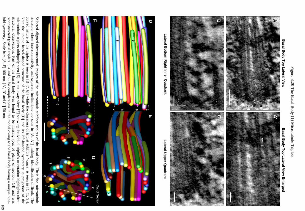
Fig
ure 3
.20
The B
asal Bo
dy (1
) Micro
tub
ule T
riplets
Sele
cted alig
ned
ultrastru
ctural im
ages o
f the m
icro
tubule su
bfib
re triplets o
f the b
asal b
od
y. T
heir fin
e micro
tubu
le
structu
re, clo
se interco
nnectiv
ity and sep
arate inclin
ations are se
en in
[A, A
’] m
akin
g id
entific
ation d
ifficu
lt. The
curv
ed n
ature o
f the trip
lets is seen in
[B (7
, 6)] w
hile th
e clo
seness o
f triplet su
bfib
re ‘vanes’ is see
n in
[C (1
, 9)].
Note th
e uniq
ue
barre
l-shap
ed stru
cture o
f th
e basa
l bo
dy [D
] an
d its
left-h
and
ed sy
mm
etry in
pro
jectio
n o
f th
e
micro
tubule trip
lets obliq
uely
seen [E
]. A cu
t away v
iew [F
] sho
win
g in
div
idu
al trip
let’s orie
ntatio
n h
ighlig
hts u
ltra-
structu
ral o
bserv
ations.
Part
of
the
basa
l bo
dy w
as fo
und
to
be
missin
g (o
ut
of
pla
ne
of
section [G
]) an
d w
as
reconstru
cted (p
artial trip
lets 3, 4
and
5) fo
r com
plete
ness in
the m
odel o
win
g to
the b
asal b
od
y h
avin
g a u
niq
ue n
ine-
fold
sym
metry
. Scale b
ars [A, F
] 100 n
m, [A
’, B’ an
d C
’] 50
nm
.
5
7
1
8
2
9
6
3
4
F
D
G
E
7
8
9
1
9
8
Out o
f Sectio
n
6 1
G
G
G
Late
ral B
otto
m R
igh
t Inn
er Q
uad
ran
t
Bas
al B
od
y T
op
Late
ral V
iew
En
larg
ed
Late
ral U
pp
er Q
uad
ran
t
A
B
A’
Basal B
od
y T
op
Late
ral V
iew
7
8
9
7
9
1
9
8
6
1
C
10
9

Intra-luminal deposits were found inside the basal body, decorating the microtubule triplet faces with some spanning between them [A, B, C]. Numerous densely stained accretions of macro-
molecular complexes were identified attached to the triplet faces [D, G, H], including fine filaments [D, G] and tethered structures [D, I] (which are seen modelled in [B, C]).
A luminal disc-like structure was revealed [E (arrows), F] attached to the triplets by fine linkages whose attachment points encompass several triplet faces at the distal end of the basal body
[F]. [J] An end on view of the basal body reveals the angular dispersal of many structures within the lumen and their relation to the triplet architecture. Materials are seen interconnecting
between triplets (2, 3, 4 and 9) from the A-C subfibres. These are seen in proximity to Basal Appendage One (BA1). Scale bars [A] 100 nm, [D] 50 nm, [G, G’, H and I] 20 nm.
Figure 3.21 The Basal Body (2) Luminal Discs, Deposits and Fibers
A
F
C B
E D
I
G
G’
H
D D
J
BA1
1
1
6
2
8
7
6
5
4
3
9
1
1 6
H
I
G
A B C
11
0

Fig
ure 3
.22
Basal B
od
y (3
) Intern
al Stru
ctures an
d V
esicle
A v
esicle
was n
oted
firmly
attached
to an
inner m
icrotu
bule trip
let [A], en
larged
in [C
] (arrow
head
). The m
odel
[B] sh
ow
s a less clu
ttered v
iew o
f the v
esic
le with
respect to
the b
asa
l bo
dy a
nd
the lu
min
al d
isc. F
ilam
ento
us
linkag
es from
the v
esicle are se
en in
teracting
with
the n
earby lu
min
al d
isc and
mic
rotu
bu
le triplet [D
]. The su
rface
of th
e vesic
le conta
ins d
ensitie
s upo
n its m
em
bra
ne a
nd
is firmly
tethered
to th
e micro
tubu
le do
ublet [E
, F]. S
cale
bars [A
] 100
nm
and [C
] 50 n
m.
A
B
E
C
D
F
11
1

Figure 3.23 Basal Appendage One: Optical Sectioning and Reconstruction
Selection of serial optical sections of the Basal Body Appendage (BA1) [A-H]. The basal appendage was reconstructed in two pieces, the docking complex and extending arm
(green) and the basement complex (red). Note the localisation of a density in [A] which is seen on [I, J and K] (arrowheads). Details of the docking complex and basal arm are seen
in [L]. Note electron dense materials surrounding the proximal end of the basal body microtubules. Scale bar 20 nm.
Serial optical slices through Basal Appendage One (BA1) Model of BA1
A
B
C
D
E
F
G
H
J
L
K
I
11
2

Fig
ure 3
.24
Basal A
pp
end
age O
ne (1
) Alig
nm
ent o
f Basal A
pp
end
age B
asemen
t Stru
ctures
A lo
ng
itud
inal se
ction o
f the b
asal ap
pendag
e [A] sh
ow
ing
cross sectio
nal fin
e deta
il of th
e base
ment stru
cture [B
].
A p
lan [C
] (and
an o
bliq
ue v
iew [D
]) of th
e basa
l append
age sh
ow
ing
deta
il of th
e zones o
f attachm
ent to
the
base
ment m
icrotu
bu
le triplets. T
hre
e dire
ctional b
asem
ent attach
ment sites w
ere identifie
d (arro
ws) [E
, F]. N
ote
the fin
e structu
re of th
e do
ckin
g co
mp
lex h
ead sh
ow
ing
fine th
orn
y p
rotru
sions, th
e long
do
ckin
g arm
and
the
perfo
rated n
ature o
f the b
asemen
t materials. S
cale bars [A
] 100 n
m an
d [B
] 50 n
m.
C
D
E
F
A
B
1
9
2
1
9
2
11
3

Fig
ure 3
.25
Basal A
pp
end
age O
ne (2
) Ultrastru
cture o
f Arm
, Do
ckin
g S
tation an
d S
ub
strate
F
H
A
E
A
B
C
D
J
I
*G
I G
G’
*
*
*
*
The b
asal ap
pend
age is se
en p
roje
cting
from
the b
asal b
od
y (o
ut o
f pla
ne) w
here th
e do
ckin
g h
ead is a
ble to
interact
with
a local fie
ld o
f vesic
les (w
ithin
the ce
ntro
som
e) [A]. T
he d
ock
ing
head
conta
ins d
ense fin
e sp
iny p
rotru
sions [B
]
at one p
oin
t tetherin
g a v
esic
le [C] a
nd
nearb
y b
asal b
od
y asso
ciated
densities [D
]. Note in
dentatio
ns w
ithin
the b
asal
append
age arm
[E]. S
om
e do
ckin
g h
ead
pro
trusio
ns teth
er cyto
solic m
icrotu
bu
les (red) w
hic
h a
ct as co
ndu
its for
vesic
le transp
ort [G
, G’]. N
ote th
e prese
nce o
f vesic
le and
micro
tubu
le associated
materia
ls no
ted in
[J]. Micro
tubule
orie
ntatio
n is se
en in
[F, H
] giv
ing
the b
asal ap
pendag
e a ‘swep
t back
’ persp
ective at a slig
ht an
gle [F
, H] p
ossib
ly
from
com
bin
ed e
ffects m
icrotu
bule a
nd
ciliary
loads. N
ote p
rotru
sion [H
] (arrow
head
). An e
xten
ded
vie
w o
f the b
asal
append
age
deta
iling
m
icrotu
bu
le attach
ment
sites, the
head
co
mp
lex stru
cture
[I]. The
full
mo
del
with
th
e basa
l
append
age
is seen in
[J] interactin
g w
ith a n
um
ber o
f smaller v
esicle
s associated
with
the d
ock
ing co
mp
lex a
nd
attached
m
icro
tubu
les. N
ote
the
extra
unattach
ed
m
icrotu
bu
le in
teracting
w
ith nearb
y
vesic
les
(ultra-stru
cture
of
vesic
les seen
in [G
] translate to
tho
se mark
ed in
[J]) and th
e area around
basal b
ody d
evo
id o
f vesic
les.
Scale b
ars [A] 1
00 n
m, [B
, D, E
, G, G
’] 50 n
m an
d [C
] 25
nm
.
11
4

Figure 3.26 Basal Appendage Two (1) Ultrastructure and Modelling
Serial optical sections of Basal Body Appendage Two (projecting downwards) [A-H]. The docking complex and arm were modelled the same way as for Basal Appendage
One revealing similar anatomical features, however the basement structures were explored finer detail [I-L]. A similar basement profile was found, made up of three
‘attachment zones’ to the microtubule triplet substrates. Note fine linkages connecting the edge of the basement zone of the basal appendage to the outer surfaces of basal
body microtubule triplets, and that the basal arm has a similar spiny protrusion as seen in Basal Appendage One (arrowheads) [J, L]. Scale bar 100 nm.
A
B
C
D
E
F
G
H
I
J
K
L
Serial optical slices through Basal Appendage Two (BA2) Model Perspectives
11
5

Fig
ure 3
.27
Basal A
pp
end
age T
wo
(2) D
ock
ing C
om
plex
es
Exam
inatio
n o
f Basa
l Append
age T
wo
revea
ls that it is slig
htly
‘swep
t back
’ in its p
roject [A
] and
has fiv
e
cyto
solic
micro
tubu
les
attached
to
its
do
ckin
g
com
ple
x
[B].
Ultrastru
ctural
deta
ils o
f th
e do
ckin
g
hea
d
attachm
ent
sites fo
r two
cy
toso
lic m
icro
tubu
les (la
belle
d in
[A
and
B
]) rev
eal
a nu
mber
of
finer
link
ages
radiatin
g fro
m a
long
the m
icro
tubu
le to lo
cal v
esic
les [C, D
]. Note th
e offset im
ages [D
, D’] are d
erived
from
[C] sh
ow
ing
greater d
etail of th
e attached
mic
rotu
bu
les to
the d
ock
ing
head
. The seco
nd
mic
rotu
bu
le is seen
alig
ned
attached
to th
e do
ckin
g h
ead co
mp
lex [E
]. Note stain
ing
densitie
s upo
n m
icrotu
bu
les a
nd
do
ckin
g h
ead
[C’, D
’ and
E’]. N
ote fin
er structu
res with
in th
e ind
entatio
n in
the b
asal arm
[B] (arro
whead
). Sca
le bar [C
, E]
50 n
m an
d [D
, D’, C
’ and
E’] 2
5 n
m.
E
E’
A
B
C
C’
D’
D
*[E]
*[C]
*[C]
*[E]
11
6

Figure 3.28 Basal Body: Appendages Summary; Basement Structures, Basal Arm and Docking Complex
A review of Basal Body Appendages, One [A-E] and Two [F-J] detailing the finer perspectives and structures of their respective basement structures, basal arms and docking
complexes. Rotational perspectives of the basal arms and docking complexes are detailed in [K-T].
A D C B E
B
A
1
G F H J I
B
A
2
Q P R T S
B
A
2
L K M O N
B
A
1
0° 45° 135° 90° 180°
0° 45° 135° 90° 180°
G
11
7
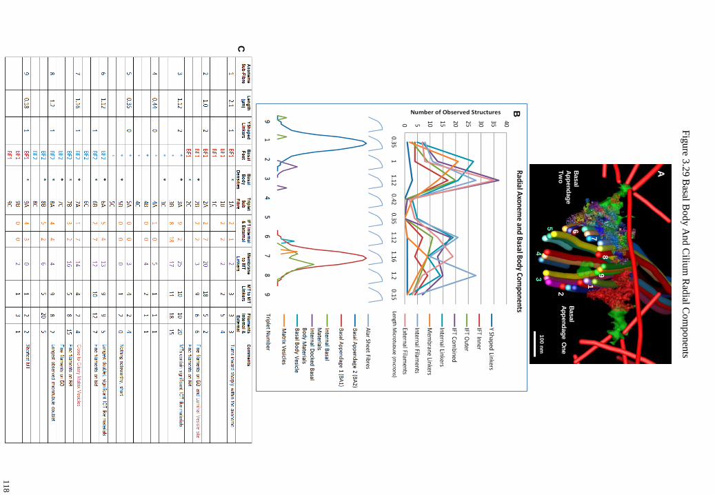
Fig
ure 3
.29
Basal B
od
y A
nd
Ciliu
m R
adial C
om
po
nen
ts
B
C
1 2
4
5 6 7
8
9 3
A
Basal
Ap
pen
dag
e O
ne
Basal
Ap
pen
dag
e
Tw
o
10
0 n
m
11
8

Fig
ure 3
.30
Pro
xim
al Cen
triole U
ltrastuctu
re, Dep
osits an
d M
od
el
Only
part o
f the p
rox
imal
centrio
le w
as contain
ed w
ithin
the to
mogra
m. U
ltrastructu
ral im
ages
of th
e pro
xim
al
centrio
le detail m
icrotu
bu
le directio
n, th
e orien
tation o
f the trip
let structu
res and d
ensely
stained
materia
ls associated
with
the cen
triole [A
]. A lu
min
al disc lik
e structu
re was id
entified
at the d
istal end
of th
e centrio
le alo
ng w
ith sm
aller
dep
osits an
d d
ecoratio
ns co
ating th
e micro
tub
ule trip
lets [B, C
and D
]. Rotatio
nal m
od
el persp
ectives o
f the p
rox
imal
centrio
le are show
n [E
-L] in
cludin
g d
ense d
eposits an
d d
ecoratio
ns id
entified
ultra-stru
cturally
in [A
-D].
No ap
pen
dag
es or m
icrotu
bule n
ucleatio
n sites w
ere noted
. Scale b
ars [A] 1
00
nm
and [B
-D] 5
0 n
m.
E
G
I
F
H
J
K
L
5
4
1
5
1 3 3
1
5
1 2
4
5 3
4
5
4
(-) (+
)
(-) (+
)
184°
46°
92°
138°
230°
276°
368°
0°
A
D
B
C
(-) (+
)
*C
*E
*E
*G
*B
*J *I *D
11
9

The distribution of materials associated with the proximal centriole are shown in [A, B], including a disc like structure spanning the distal end [C]. The
centrosomal environment surrounding the proximal centriole contained numerous dense deposits (not all of them were modelled) [D, E].
The nuclear membrane was in close proximity to the centrosome and was modelled to reveal nuclear pore complexes [F]. Linkages were identified
connecting from the distal end of the centriole to the nearby nuclear membrane in proximity to several nuclear pore complexes [G-I]. Scale bar 50 nm.
Figure 3.31 Proximal Centriole Radial Components and Proximity to Nuclear Pores
G H I
1
9
Negative End View looking (+)
2
1 4
5
6
7 8
3
A 1
3 4
5
2
Positive End View looking (-)
B C
F
H
D E E
5
4
50 nm 50 nm
100 nm
12
0

Figure 3.32 The Centrosome (1) Ultrastucture and Modelling
Selected ultrastructural serial slices
(overlaid with model) of the centrosome
detail the basal body, the proximal
centriole, vesicles, microtubules,
intermediate filaments and numerous
electron dense structures which are
bound between the cell and nuclear
membranes [A-H]. By meticulously
tracing objects though each optical slice
it has been possible to ascertain the
relationship between vesicles within the
centrosome, which were observed
interacting in zones associated with
microtubules and the local cell
membrane [I, L] as well as near the
basal feet. Note nuclear pore proximity
to centrosome [I and J].
Scale bars 100 nm.
A
C
D
E
G
F
H
B
I
J
K
L
12
1
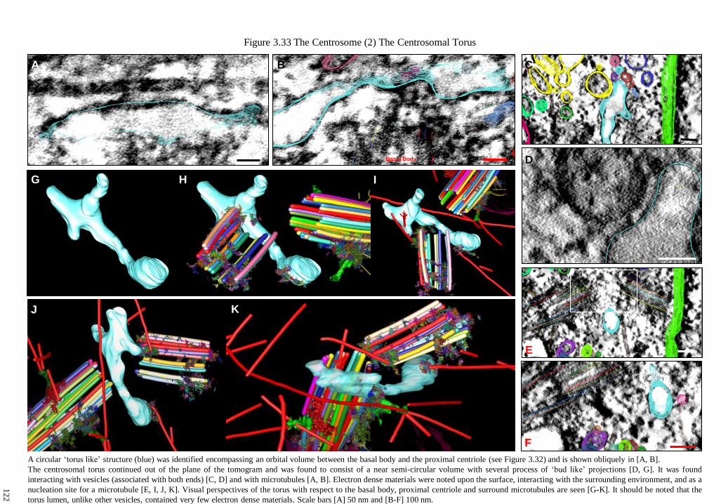
Figure 3.33 The Centrosome (2) The Centrosomal Torus
A circular ‘torus like’ structure (blue) was identified encompassing an orbital volume between the basal body and the proximal centriole (see Figure 3.32) and is shown obliquely in [A, B].
The centrosomal torus continued out of the plane of the tomogram and was found to consist of a near semi-circular volume with several process of ‘bud like’ projections [D, G]. It was found
interacting with vesicles (associated with both ends) [C, D] and with microtubules [A, B]. Electron dense materials were noted upon the surface, interacting with the surrounding environment, and as a
nucleation site for a microtubule [E, I, J, K]. Visual perspectives of the torus with respect to the basal body, proximal centriole and surround microtubules are seen [G-K]. It should be noted that the
torus lumen, unlike other vesicles, contained very few electron dense materials. Scale bars [A] 50 nm and [B-F] 100 nm.
J
I G
A B
H
K
C
D
E
F
Basal Body
12
2

A
A A
Fig
ure 3
.34
The C
entro
som
e (3) T
he P
ericentrio
lar Enviro
nm
ent
C
A
A’
C
PC
en
t
BB
B
PC
en
t
BB
E
D
A’
To
rus
The p
ericentrio
lar enviro
nm
ent en
com
passes an
area surro
und
ing
of th
e basa
l bo
dy (B
B) an
d p
roxim
al c
entrio
le
(PC
ent).
It co
nsists
of
an am
orp
ho
us
filam
ento
us
mass
of
fine
materia
ls lin
kin
g th
e tw
o cen
trioles
at th
eir
(-)
micro
tubule e
nds, an
d d
enser m
icrotu
bu
le associated
materia
ls surro
und
ing
the ce
ntrio
les them
selv
es (see F
igs 3
.30,
3.3
2). T
he fila
mento
us in
ter-centrio
lar zone is se
en in
[A, A
’] and
again
in d
etail in
[B] w
here it is in
pro
xim
ity to
,
and
seen in
teractin
g w
ith, th
e centro
som
al to
rus [C
]. An area su
rround
ing
the p
roxim
al c
entrio
le was n
oted
as a
nu
cleatio
n site fo
r interm
ediate fila
ments [D
, E]. N
ote in
termed
iate fine stru
cture. S
cale b
ars [A] 1
00
nm
, [A’, B
, C]
50 n
m, an
d [D
and
E] 1
0 n
m.
12
3

Fig
ure 3
.35
The C
entro
som
e (4) B
asal Bo
dy, P
rox
imal C
entrio
le with
in th
e MT
OC
A
sele
ction
of
centro
som
al
imag
es [A-E
’]. [A] T
he to
rus
in
pro
xim
ity
to
a
micro
tubule. [B
, C] V
esicles
intera
cting
w
ith
ele
ctron
dense
materia
ls o
f th
e
perice
ntrio
lar enviro
nm
ent
(near th
e pro
xim
al c
entrio
le)
and
nearb
y
a nu
clear
mem
bra
ne
po
re. [D
]
Nu
mbers
of
small
vesic
les
colle
cting
an
d
budd
ing
around
th
e m
icrotu
bu
le
netw
ork
asso
ciated
with
Basa
l A
ppend
age
Tw
o
(BA
2).
[E,
E’]
An
enlarg
em
ent
of
the
perice
ntrio
lar zo
ne
deta
iling
an
ele
ctron
dense
structu
re
located
betw
een
the
two
centrio
les. [F
] A
th
ree
dim
ensio
nal
mo
del
of
the
centro
som
e sho
win
g n
um
bers
of
small
vesic
les
around
micro
tubule
(red)
traffick
ing
zones.
Note
basa
l
append
ages,
the
centro
som
al
‘toru
s’, in
termed
iate
filam
ents (g
reen), th
e nearb
y
nu
clear
mem
bra
ne
and
nu
clear p
ores. S
cale bars 1
00
nm
.
F
A
B
C
D
E
E’
12
4
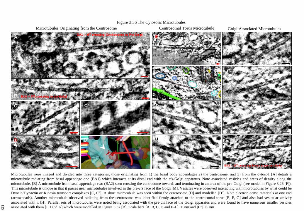
Golgi Associated Microtubules
I
J
K
E
F
Centrosomal Torus Microtubule
G
H L
Figure 3.36 The Cytosolic Microtubules
BA2 – MT crossing Torus to pre-cis-Golgi
Microtubules were imaged and divided into three categories; those originating from 1) the basal body appendages 2) the centrosome, and 3) from the cytosol. [A] details a
microtubule radiating from basal appendage one (BA1) which interacts at its distal end with the cis-Golgi apparatus. Note associated vesicles and areas of density along the
microtubule. [B] A microtubule from basal appendage two (BA2) seen crossing the centrosome towards and terminating in an area of the pre-Golgi (see model in Figure 3.26 [F]).
This microtubule is unique in that it passes near microtubules involved in the pre-cis face of the Golgi [M]. Vesicles were observed interacting with microtubules by what could be
Dynein/Dynactin or Kinesin transport complexes [C, C’]. A short microtubule was seen within the centrosome [D] and modelled [D’]. Note electron dense materials at one end
(arrowheads). Another microtubule observed radiating from the centrosome was identified firmly attached to the centrosomal torus [E, F, G] and also had vesicular activity
associated with it [H]. Parallel sets of microtubules were noted being associated with the pre-cis face of the Golgi apparatus and were found to have numerous smaller vesicles
associated with them [I, J and K] which were modelled in Figure 3.37 [B]. Scale bars [A, B, C, D and E-L] 50 nm and [C’] 25 nm.
Microtubules Originating from the Centrosome
D D’
BA1 – MT crossing Centrosome to Pre Golgi
BA2 – MT crossing to cis-Golgi
C’
A
C
B
12
5

Figure 3.37 Centrosome to Golgi Connectivity
The periphery of the centrosome was found to be closely associated with the nearby Golgi apparatus, which is bound closely on both sides by the nuclear and cell membranes [A]. The pre-cis-
Golgi face was found to be highly aligned with parallel microtubules (with a single intermediate filament (green)), mixed with clusters of numerous small vesicles [B]. The complexity of the
interaction zone is detailed in [C], where a field of vesicles may be seen surrounding the Golgi have been traced. Although little detail is available within a single slice, combination of many
slices reveals significant 3D information in a model. [D] An enlarged perspective of the translucent model in [B], however with the Golgi turned opaque to aid viewing. Note the microtubule
(red) originating from the basal appendage (BA1) is seen interacting and terminating within a zone around the medial to trans-compartments. It was found to be specifically associated with a
vesicle (red) amongst a field of ‘budding’ vesicles from the edges of the cisternae (see Figure 3.36 [A] for an ultrastructural view of the microtubule). Scale bar 200 nm.
A B
C D
12
6

M
Figure 3.38 The Cis, Medial and Trans Golgi Compartments
A
B C
F E
D
G
H I
N
K
J
A series of vertical and lateral views of the Golgi cis, medial and trans compartments which were traced manually (as seen Figure 3.37 C). The cis compartments were found to have
budding protrusions [B] and holes penetrating through the cis stacks [C]. The slender medial compartment was found to contain several fine holes forming a zone of perforation running
diagonally within medial stack [E, F]. The trans compartment contained numerous vesicles budding from the trans face [G, H and J]. Combined stacks [J], are seen with respect to a
microtubule array near the cis compartments [K]. Vesicles were found to normally be associated with the faces or peripheral edges of cisternae, while finer holes and fenestrations were
observed in portions of the stack.
12
7

A large storage vesicle was identified containing tightly packed granular material (reminiscent of matrix granules) however at a higher density greater than the surround extracellular matrix
[A, A’ and C*]. An assortment of vesicles packed with similar granules were observed near the trans face of the Golgi [B, D], including a long tubular structure that could have ‘shed’ from
the face (purple) [C]. Part of the trans-face was connected to the cell membrane via a microtubule [E] where a collection of vesicles were closely associated with the cell membrane [F, F’]
(seen merging in [E]) and modelled in [C’]. Scale bar [A, D, F] 200 nm and [A’, B, E and F’] 100 nm.
A
A’
B
Figure 3.39 Transport of Matrix from the Golgi
C
D
*
F F’
C’
E
12
8

The distal tip of the cilium is seen interacting with the cell membrane [A] where a number receptor mediated endocytotic processes were identified and labelled in [A] as (B), (C) and (E).
Coated pits labelled as (B, C, and D) are seen in their corresponding ultrastructural plates [B, C, D] with an enlarged view in [D’]. Note the circular nature of coated pits, unknown
structures decorating the membrane surfaces, and the extracellular contents of the pits, in the process of endocytosis (see Figure 3.43 for alternative field of [D’]). A semi-circular ‘pitted’
membrane indentation, believed to be a coated pit in the process of formation is seen in [C]. Structures revealing Caveolae, are identified in [E] and [F]. Scale bars 100 nm.
Figure 3.40 The Cilium, Coated Pits and Caveolae
A B
D
C
D’ E’
E
(C)
(B)
(D)
(E) Actin Fibres
12
9

Fig
ure 3
.41
The M
atrix C
ilium
Go
lgi C
ontin
uum
A co
mbin
ed o
verv
iew
of a
ll featu
res mo
delled
from
the to
mo
gra
m (see F
igure 3
.8 fo
r com
pariso
n) d
etailing
matrix
intera
ctions
with
th
e ciliu
m,
its axo
nem
al
mic
rotu
bu
le stru
cture, th
e d
iplo
som
al
centro
som
e, th
e rad
iating
cyto
solic
micro
tubule n
etwo
rk an
d th
e nearb
y G
olg
i app
aratus. C
yto
solic m
icro
tubules are sh
ow
n in
red, in
termed
iate filam
ents in
green
, and actin
fibres in
blu
e.
100 n
m
13
0

Fig
ure 3
.42
Plan
of T
om
ogram
Featu
res
100 n
m
Matrix
Sto
rag
e G
ran
ule
Peri-C
iliary
Mem
bra
ne
Go
lgi A
pp
ara
tus
Nu
cle
ar M
em
bra
ne
En
do
pla
sm
ic R
etic
ulu
m
Matrix
Gra
nu
le N
etw
ork
tran
s-G
olg
i Tu
bu
le
cis
med
ial
tran
s
Pre
-cis
-Go
lgi V
esic
les
En
do
so
mal T
ub
ula
r Sy
ste
m
Recep
tor M
ed
iate
d E
nd
ocy
tosis
Co
ate
d P
its
Caveo
li
Caveo
li
Th
e P
rimary
Ciliu
m
Th
e C
en
troso
me
To
rus
Pro
xim
al C
en
triole
B
asal B
od
y
Matrix
Vesic
les
Inte
rmed
iate
Fila
men
t
Mic
rotu
bu
le
Nu
cle
ar P
ore
s
BA
1
BA
2
13
1

Fig
ure 3
.43
The C
ilium
-Cen
troso
me C
ontin
uum
A p
erspectiv
e view
alo
ng
the ax
onem
e, the b
asal b
ody, th
e pro
xim
al c
entrio
le (with
the ce
ntro
som
al to
rus) an
d
attached
micro
tubu
les o
f the c
entro
som
e. No
te the fin
e structu
res, inclin
ation o
f the a
xo
nem
e and
a coated
pit n
ear
the tip
of th
e ciliu
m. T
his d
etails a p
hysic
al co
ntin
uu
m b
etwee
n th
e extrac
ellu
lar enviro
nm
ent, an
d th
e centro
som
e,
whic
h is co
mprised
of a
s yet m
any u
nk
no
wn p
rotein
s. No
te the p
resence o
f mic
rotu
bules w
hic
h w
ould
interse
ct
atop
the b
asal bo
dy, w
here a th
ird b
asal appen
dag
e wo
uld
be, if co
ntin
ued
out o
f section.
13
2

133
Chapter Four: Discussion
4.0 Introduction and Overview
Despite an ever growing body of knowledge of primary cilia as specialist sensors, many
questions remain about their role as mechanical transducers and the means by which they influence
cellular responses. The introductory chapters of this thesis have provided an overview of the
previously known ultrastructure of primary cilia, a profile of their sensory capabilities, and their
relationship within the extracellular matrix, cilium and the Golgi continuum. In summary, primary
cilia occur nearly ubiquitously in eukaryotic cells and although known of for many years have
recently been acknowledged as an organelle to epigenesis, physiological function, and several disease
processes [1, 85, 229, 460, 1001]. Primary cilia express a unique (9+0) microtubule doublet symmetry
nucleated from the maternal basal body, which is subtended by a proximal centriole, and the two
together forming the diplosome of the centrosome. The centrosome is the microtubule organising
centre of the cell, and is responsible for controlling many cellular functions [120]. During interphase it
is located in a juxta-nuclear position, and is important for organisation of the Golgi apparatus [96, 97,
1002] and determining cell polarity [1003, 1004].
The hypothesis that the cilium acts as a ‘cybernetic probe’ in connective tissues [26] has since
been supported by studies which show that primary cilia are unique sensory organelles within a
variety of tissue types, capable of adaptation for sensing a host of mechanical, chemical, osmotic,
photonic, polarity and other significant changes in the cellular microenvironment [33]. Norbert
Wiener [99] coined the term ‘cybernetic’, derived from the Greek κιβερνηηικοζ, literally the ‘steerer
of a rowing boat’, which is appropriately describes the role of the primary cilium in detecting external
signals, and directing an appropriate response.
Current concepts of ciliary structure are based upon data derived from conventional ultrathin
electron microscope studies of cells and tissues containing predominantly motile cilia [45, 49, 51].
Fewer ultrastructural studies have been made of connective tissue primary cilia, as analysis is difficult
due to the small size of the cilium relative to the volume of the cell, singular occurrence and unknown
orientation within tissues [973, 1005-1006]. However, extensive ultrastructural analysis of
chondrocyte primary cilia [26, 27, 96, 97] culminated in the first electron tomographical study by
Jensen et al., (2004) [25], realising many accurate anatomical details.
Extracellular matrix of connective tissues plays a dynamic and integral role in cellular
adhesion, signalling, proliferation, differentiation, apoptosis and mechanotransduction, through
providing interactions with cell surface receptors such as integrins, which are physically or chemically
coupled to the cytoskeleton [25, 1007-1009]. These tissue specific macromolecular architectures are
influenced functionally by the application of biomechanical forces [1009]. The precise types of
molecular connections and their relative abundance depend largely on the biomechanical function of
the tissue and the required cellular response to biomechanical forces [1010]. Developmentally,
134
mechanical forces drive mesenchymal cells down a particular lineage [1011], while conversely cyclic
hydrostatic pressure initiates chondrogenic differentiation and matrix secretion [1012].
In cartilage, chondrocytes are believed to sense their extracellular environment through their
primary cilia, transducing this information to the centrosome, which responds appropriately by
polarising the Golgi apparatus. This pathway involves a number of unique proteins and biochemical
signalling cascades spanning from the cilium to the centrosome, allowing intracellular regulation and
polarisation of the Golgi for the directed regulated secretion of appropriate extracellular matrix
macromolecules for maintaining the extracellular microenvironment. Many of these known protein
constituents and interactions are tabulated within the Appendices. The rapid increase in knowledge of
these proteins and their dynamic association with the structure of primary cilia make this a truly
unique study.
4.10 Limitations of Study
This study has primarily focussed upon tomography of in situ embryo sternal chondrocytes
and has relied upon conventional chemical fixation and osmication, enhanced with RHT and en bloc
uranyl-acetate stain. Inevitably, the presence of a certain degree of chemically induced artefact,
shrinkage and distortion is expected, when compared to cryo-tomography techniques [938-943]. As
such, RHT represents a trade off in fixation strategies, providing the ability to optimally stain,
stabilise and retain extracellular matrix components that are not conventionally preserved. In addition,
the enhanced resolution provided by RHT allowed resolution of fine structure never before seen. The
time required to chemically fix, prepare and examine samples makes electron microscopy a highly
involved process, however this is compensated for by the improved resolution which tomography can
provide.
This study did not attempt to ascertain the preferential orientation of chondrocyte primary
cilia within cartilage, which has been previously been investigated by Farnum et al, (2011) [973].
Measurement of cilia was carried out in ImageJ, however, there are limitations in measurement
accuracy with semithick as compared to ultra-thin sections. As yet, in situ chondrocyte primary cilia
have not undergone the same level of tomographical investigation as other cilia types (Appendix VI).
This is the first study to carry out both tomography and full modelling of a chondrocyte
cilium, as well as incorporating the Golgi apparatus for visualisation of the ‘matrix-cilium-Golgi
continuum’ as a functional entity. While only a single-axis tomogram was used to construct the
model, it is believed to represent the anatomical and structural characteristics common to most
chondrocyte primary cilia, agrees with, and improves on previous studies [27]. However, it should not
serve as a sole reference interpret or represent all chondrocyte primary cilia. Further models would be
a useful complement, however at present their construction is an intensive, though rewarding,
undertaking.

135
4.11 Ruthenium Interactions with Cells and Membrane Bound Structures
The combined ability of RHT and its osmication products to interact with, stain and contrast
membrane proteins and receptor channel complexes remains uncertain. Several authors have reported
the penetration of RHT into living cells at fixation [960, 1013], including the biological effect of
inhibiting T-cell activation [1014] RHT is reported to interact with a wide variety of membrane
proteins and ion channels, many of which are reported upon cilia and flagella, however its ability to
stain and contrast these structures remains unclear. Ruthenium Red also displays a high affinity for
phospho-lipid membranes, specifically for L-alpha-phosphatidyl serine, where its action upon
membranes has been modelled [1015, 1016].
Following RHT treatment, the ciliary membrane was observed to contain numerous heavy
stained structures, as detailed in Figs 3.11 D-M, 3.12 and 3.15. These may represent numerous
potential membrane bound components of matrix receptors, channels and their associated components
upon the chondrocyte primary cilium. RHT has been used by Czukas et al., (2007) [1017],
demonstrating an ability to resolve fine structures, when using it to identify fine linkages between
stereo-cilia in alligator lizard auditory hair cells. In contrast, RHT in the present work led to such
heavy staining of the perciliary membrane that it rendered some parts of those areas too opaque to
adequately resolve any structure.
4.12 Occurrence and Morphology of Chondrocyte Primary Cilia
The incidence, length, morphology and orientation of primary cilia have been analysed in
many connective tissue studies of fibroblasts [30, 95], tendon [26, 1018] and cartilage [26, 27]. It has
been found that normally only one primary cilium is expressed per in situ interphase cell [30] as
originally noted by Zimmerman [9]. This is believed to hold generally, however recent observations
of primary cilia metaplasia on the apical surface of renal proximal tubules [1019] raises interesting
perspectives as to the role of the primary cilium in these epithelial cells. Observations of cells
containing bi-ciliated primary cilia have been made in many cell types, though the reasons for, and the
dynamics of multi-ciliated centrosomes remain largely unknown [944, 978, 1020-1022]. Although
their presence is reported associated with both cancerous and benign cells [248, 249, 1023, 1024].
Most chick sternal chondrocyte primary cilia were found to be surrounded by fields of matrix
granules with only a sparse presence of collagen fibres near the cilium. The most significant
discrepancy in measuring axoneme lengths may arise from the effect of sectioning, where the lengths
of bent or longer cilia are not included, skewing the results slightly in favour of a shorter average
length. Discrepancies in measuring basal body lengths may result from inclusion of incomplete or
misaligned cilia in sampling (which is difficult to determine with semithick sections, especially in
respect to the basal body) as well as errors from microscope settings, shrinkage and choice of
measurement points. Basal body lengths were measured from the proximal base to the end of the C-

136
subfibre, near the alar sheet attachment site. This leaves the interpretation of the end-point of the basal
body between the start of the transition zone and the ciliary membrane, subject to error.
Many authors have studied chondrocyte primary cilia in articular, growth plate, sternal and
mandibular cartilage [26, 27, 42, 96, 1005, 1025-1026], with Rich et al., (2012) [365] having
reviewed their incidences and lengths. Earlier studies of the presence, length and morphology of
chondrocyte cilia revealed that ciliary length varies little between species [27, 28, 365, 973]. The
measurements of chondrocyte primary cilia by Wilsman in 1978 [28] (1.76 µm, ζ 0.80) closely match
the results of this study (1.80 µm, ζ 0.36) except for lengths of basal bodies (0.5 µm ζ 0.03 to this
study 0.365 µm, ζ 0.01) and the range of axoneme lengths were similar (0.28-3.29 to this study 0.7-
2.41) µm, respectively. On the other hand, primary ciliary length can vary greatly between cell types,
from those expressing minimal extension of the axoneme (such as in the T-cell immunological
synapse [1027]), to a length of 4.2 ± 2.2 µm in the rat flexor tendon [1018], and to extremes of 20-30
µm (in Ovine Meckel-Grueber MKS3 fibroblasts [1028]). The depth of semithick sections prevented
reliable measurement of basal body widths. However, transition membrane widths were measured
(0.262 µm ζ 0.015, Section 3.5) in comparison to basal body width (0.21 µm, ζ 0.01) as reported by
Wilsman [28]. This indicates a 26 nm wide ‘inlet zone’ extending between the membrane and the
microtubules of the transition zone. Anderson (1972) [45] reported a variable luminal diameter of the
basal body (0.13-0.15 µm), indicating the luminal end surface area is at least 1.25 times that of the
transition zone.
4.13 Chick Sternal Cartilage Tomogram Selection
Of the many tomograms acquired, it was decided to model a single-axis dataset that contained
a near complete ciliary axoneme, laying within an indentation resembling an enlarged ‘ciliary pocket’
[1029] (Figure 3.6). This section contained a near complete cilium and proximal centrioles of the
microtubule organising centre and was positioned adjacent to the nucleus, in close proximity to the
Golgi apparatus. The ‘pocket’ itself offers a striking comparison with an image published by
Wheatley (Figure 6.1, pg 149 [3]) [1030] of a cilium extending into a large luminal vacuole with the
centrosome positioned against the nuclear membrane. In the chondrocyte tomogram, the matrix
surrounding the cilium within the indentation is of a lower staining density than that of the
extracellular matrix, and is uniquely devoid of collagen fibres.
4.14 Defining the Cilium and its Relation to the Golgi
Numerous detailed tomographical studies have focussed upon the finer molecular aspects of
the microtubule-based architecture of the motile axoneme, with none attempting to explore either its
complete structure, or the relationship of the cilium within the centrosome and its relationship to other
organelles. The modelling of a continuum between the matrix, the primary cilium, the centrosome and
the Golgi within one tomogram, as achieved in the present study, uniquely indicates the presence of a

137
highly ordered microtubule-based structural continuum mechanically linking the cilium with the
Golgi apparatus. The interrogative and inclusive model enables a three dimensional understanding of
the architecture of the primary cilium not available from conventional ultra-thin studies.
4.20 Interpretation of the Model Structure
In an effort to understand the structural components of the model, and their functional
significance, it is necessary to compare features already described in the literature on cilia, the
cytoskeleton and cellular organelles, including the Golgi apparatus. Appendix VI contains a selected
review of tomographic and modelling studies of components of the extracellular matrix, of cilia,
flagella, the cytoskeleton and the organelles of various species. While many studies have focussed
exclusively upon motile cilia, only Jensen et al., (2004) [25] has utilised tomography to study
chondrocyte primary cilia, investigating linkages between the ciliary membrane and with the
extracellular matrix.
While the microtubule architecture of centrioles, basal bodies and axonemes have been
extensively reviewed, only recently have they been the focus of higher resolution studies [1031,
1032]. While it is not possible to know with any certainty, which proteins make up the structures
detailed within the model, it is possible to deduce what these may be, with some degree of certainty.
Studies based upon lower resolution immunofluorescence and electron microscopical localisation of
known proteins can serve as a bridge for interpretation. Selections of known structural and signalling
proteins specific to primary cilia are included in Appendices, and form an essential basis for
translating ultrastructural based information into putative function. While electron tomography studies
have greatly aided the analysis of cellular organelles, the construction of large-scale models from
datasets remains a laborious process, and is not usually undertaken.
4.21 The Matrix of the Ciliary Pocket
Areas of lower density matrix were frequently observed in a zone surrounding chondrocyte
cilia, which also express various degrees of deflection and invagination of their projection with the
cell membrane. The molecular structure and functionality of this ‘ciliary matrix zone’ remains to be
determined. The matrix granule density within the ciliary pocket is less than the external pericellular
matrix environment (Figs 3.5 C and 3.7). This may indicate an area of special matrix properties, or
that freshly secreted matrix materials are less inter-connected. Knudson (1993) [861] showed that
pericellular matrix assembly occurs in a zone near the cell membrane directed by hyaluronan in an
‘aggrecan rich chondrocyte pericellular matrix’ (sic) anchored by interaction between hyaluronan and
its receptors [1033, 1034]. All tomograms revealed the pericellular matrix usually consisted of a
complex mix of collagen and proteoglycans (Figure 3.5, A’, B’), and a number of cilia were
characterised by a distinct absence of collagen fibres in their vicinity. The periciliary matrix of the
model presented consisted solely of regular arrays of matrix granules (Figs 3.5 and 3.6) 30-45 nm in
138
diameter, each connected to at least 4 others via fine filaments (thought to be hyaluronic acid or Link
protein). It is believed that the basic inter-connectivity and structure of the granule network was
preserved by RHT treatment, rather than being a post-fixational artefact. Tomography revealed the
individual granules to consist of aggregates of multiple minor precipitates of stained proteoglycan
components that may have coalesced due to dehydration during processing (Figure 3.10) [967, 1034].
Thyberg et al., (1973) [1034] analysed epiphyseal cartilage matrix granules, reporting them to
be polygonal in shape, ranging in size between 100-500 Å (10-50 nm), while Phillips et al., (1989)
[967] noted connectivity of up to 7 filaments per granule. Matrix granule substructure consists of
densely stained aggregates, reported by Matukas et al., (1968) [1035] to vary from 15 to 40 Å (1.5 to
4 nm) which closely matches observations from tomogram ultrastructure in this present study of
matrix granules (Figs 3.10 and 3.11), where similar sized punctate deposits were also identified on
fibres linking the matrix granules.
In adult cartilage, matrix granules have been found to have a greater concentration in the
pericellular capsule than in either the pericellular or territorial matrices [788] (Figure 1.25). Slightly
greater numbers of granules of larger diameter are reported within the proliferative compared with the
hypertrophic zone (viz 27 to 24 nm) [1034]. The functional importance of the distribution, inter-
connectivity and size of these granules within cartilage remains to be determined.
Modelling of the matrix granule network within the pocket surrounding the cilium indicates
that both the granules, and their network, contain a regular higher ordered structure (Figs 3.5 C’, 3.12
and 3.41). Such a uniform inter-connective ‘mesh-work’, represents a unique architecture likely to
enable the transmission of mechanical forces to the cell and ciliary membranes [1036]. Zhu et al.,
(1996) [1037] established that proteoglycans, in vitro, form tensile networks which exhibit complex
proteoglycan to proteoglycan shear moduli (where Link proteins determine their viscoelastic
properties) and their stabilisation increases the average strength of the reaction sites in the network
[1038-1041]. How these interact with and transmit forces from their bulk matrix via linkages to the
ciliary and cell membrane offers interesting opportunities for further study.
Of particular interest are the properties of the granule network ‘packing density’, and its
mechanical role in influencing ciliary movement, as well as allowing the diffusion of solutes and
ligands. Matrix components such as proteoglycan residues form integral parts of a number of cellular
pathways associated with receptors known to primary cilia [1042-1044], which also influence Wnt
proteins [419].

139
4.22 The Periciliary Membrane and the Ciliary Pocket
The periciliary membrane and the ciliary pocket encompass an area spanning from the
transition zone, extending into the cell membrane around the cilium and centrosome. In many cases
this is contained within the ciliary pocket, due to the mechanisms of ciliogenesis [295, 1045], or
potentially as the result of movement of the basal body internally within the cell. The ciliary pocket
has been previously described [978] as a tightly enclosed active endocytotic zone [420] surrounding
the base of the cilium. It may express degrees of invagination, and is normally observed sheathing the
primary cilium (Figs 3.3, 3.4 and 3.5).
Examination of numerous candidate cilia shows that the degree of invagination of the cilium
within the ciliary pocket of the chondrocytes varies substantially. This may allow controlled reduction
of the gain of extracellular signals, where the cilium acts as an adaptive antenna. This may also
function to protect the cilium from the external environment while positioning the cell, or
repositioning of the centrosome for other functions. How this process occurs, and how ciliary
receptors and signal transduction function during invagination, is unknown.
Numerous vesicles are closely opposed to the periciliary membrane surrounding the cilium,
that may be inferred to have been derived from the Golgi apparatus (Figure 3.42). The periciliary
membrane was observed to be devoid of actin filaments. Actin clearance is required for centrosome
and basal body docking in the ‘immunological synapse’ in T cells [1027, 1046], which thus becomes
a focus for the endo- and exocytosis of materials [1027, 1047, 1048]. Depletion of actin severing
components is reported to inhibit ciliogenesis [255, 297]. The membrane of the pocket is known to be
involved in ciliary protein sorting and entry [326, 1027], while TGF-β signalling is associated with
endocytosis in the pocket, and also at the interaction sites of the distal tip of the cilium [420]. The
model illustrates the periciliary membrane of the chondrocyte in close proximity with the distal tip of
the cilium, which is associated with an active zone of clathrin-coated pits, presumably undergoing
receptor-mediated endocytosis [26, 978] (Figs 3.8, 3.9, and 3.40-3.43).
4.23 The Ciliary Membrane
The ciliary membrane provides a huge surface area to volume ratio (~4×10⁷ m
-1), potentially
acting as a high sensitivity sensor for detecting and amplifying external stimuli. Due to its small size,
a complete analysis probably lies within the realms of quantum physics for interpretation of nanoscale
interactions of its protein-based components [376, 1049-1051].
Analysis of chondrocyte ciliary membranes revealed that intra-membranous matrix granules
were inter-connected via fine linkages, forming a complex web of tensile proteoglycans, which
generated localised distortions or ‘dimples’ in the membrane, similar to observations by Jensen et al.,
(2004) [25]. These matrix connections were matched by protein complexes decorating both sides of
the ciliary membrane. These are most likely trans-membrane proteins or complexes closely associated
with extracellular linkages (Figure 3.11 E-J). Some unknown structures not associated with linkages
140
were also identified (Figure 3.11 J-L). Internal luminal linkers were found to be physically connecting
the ciliary membrane to microtubule doublets (Figure 3.12). These connections together serve to
physically connect the extracellular matrix to the membrane, the membrane to the axoneme
microtubules, and they also link the microtubules to each other.
Freeze fracture of motile ciliary membranes revealed arrays of intra-membranous materials
consisting of longitudinal rows, rosettes, plaques, and necklaces with attachment sites associated with
the microtubule doublets beneath [51, 52, 345]. Differences in ciliary patterns reportedly exist
between cell types, with higher densities of materials in sensory cilia [52], however the details of the
identity and targeting of membrane proteins has remained largely unknown until recently [345, 376].
This present study has revealed the presence of numerous punctate electron dense materials of
unknown function associated with, and spanning the ciliary membrane (Figs 3.11 and 3.15).
While it is tempting to speculate about the localisation of membrane receptors, it could be
inferred that any potential specific distributions of these may be closely related to the presence of
microtubule IFT-like materials (Figure 3.14). How membrane receptors are organised is relation to
membrane-microtubule linkages is not yet established. The inability at the present time to identify
these proteins within the membrane could to some degree be resolved in future with specialist
immuno-histochemical investigation [385], in combination with higher resolution tomography and
immuno-gold labelling.
4.231 Linking Matrix to the Ciliary Membrane and Transduction
Literature on chondrocyte ciliary receptors is relatively unknown; however, they probably
utilise sensors receptive to tonicity and physical forces appropriate to maintaining cartilage tissue
function. Activation of chondrocyte membrane receptors may occur directly by membrane tension
from physical force impulse transduction, pressure induced deformation resulting from compression,
electrical differences from streaming potentials [817, 1008], and from receptor-ligand signalling
[422].
The reported presence of integrin receptors upon chondrocyte primary cilia provides a
putative physical link to the tethering extracellular matrix components. Furthermore, integrin
receptors are known to exhibit cross-talk with other components, such as growth factor receptors
[1052]. β1-integrins in primary cilia of MDCK cells are reported to potentiate fibronectin-induced
Ca2+
signalling [458] and have been found to associate with PC2 [406], where ciliary bending has
been shown to be increase intracellular calcium levels [34]. Although their role in chondrocyte cilia
remains to be determined, the study of receptors provides a putative framework for focussing upon the
role of potential receptors, their transport, inclusion and functional role within the cilium.
Resident ciliary membrane receptors and their associated peptides may vary in their temporal
expression, providing an adaptive ability to detect a range of signals, ligands and matrix effects,
which may potentially influence function appropriate to their role. Well-known mechanotransductive

141
proteins of ciliary membranes already discussed include the sodium channel, ENac, the universal
polycystin protein complex of PC1 and PC2, and the TRPV4-cation channel (Figs 1.16-1.18).
Receptors such as insulin growth factor IGF-1R also possess an extracellular ligand-binding
domain, and a luminal region with enzyme linked components [1055]. Curiously, IGF-1R regulates
Gβγ signalling, which is reported to aid in G1-S phase progression by inducing ciliary resorption,
linking ciliary signalling functionality to the cell cycle [1054].
In fibroblasts, platelet derived growth factor tyrosine kinase receptor signalling is regulated
through primary cilia, and utilises the MEK1/2-ERK1/2 pathways [344, 1055]. In addition, vascular
endothelial growth factor (VEGF) receptor complexes have been visualised through electron
microscopy [1056], in which direct contacts are required with matrix for their activation and
signalling [1057]. The epidermal growth factor (EGF) receptor contains both extracellular and luminal
components [1058] and may have a sensory function with TRP family member PKD2 on the cilium
[1059]. Other signalling receptors, such as Smoothened, whose sequence is similar to Frizzled, have
recently been modelled [1060], while the structures of TRPV1 [1061] and TRPV4 [1062] have also
been investigated. Receptors within chondrocyte cilia are likely specific to their sensory functions, for
example Interleukin-1β is reported to sequester hypoxia inducible factor-2α (HIF2α) to the cilium in
chondrocytes [1063], vital for anabolic sensation for the phenotype.
While many proteins are known to be localised to the cilium, it is not possible in this study to
identify unequivocally any within the ultrastructure of the chondrocyte or the model. The linking of
membrane receptors to ciliary sensory function, and their signalling pathways represents a new
endeavour of visualisation encompassing the specialist aspects of the mechanisms of transduction,
which lie outside of the scope of this present study, but present fruitful opportunity for future work,
building on the model presented here.
4.232 Cilia, ENac and Mechanotransduction
The sodium channel ENac participates in a diverse range of functions, from maintaining
intracellular sodium levels, to registration of nociception and mechanotransduction [1064-1066], and
has been found localised upon cilia [1066]. The ENac channel also shares compositional similarity
with the P2X receptor family members implicated in ciliary mechanotransduction [422]. Evidence
suggests they respond to flow [1067] as a sensitive membrane shear sensor, activated from membrane
tension, or in combination with impulses from nearby matrix connections [1068, 1069]. Speculatively,
they operate by altering permeability to sodium, thus inducing transient depolarisation, and are
influenced by their order and lipid interactions within the membrane [1070, 1071], where their
signalling and regulation utilises catalytic kinases and their messengers [1072, 1073]. While they have
not yet been reported to be present upon chondrocyte primary cilia, evidence that these structures
transduce mechanical stimuli suggests they will be found there.

142
4.233 The Axoneme Membrane
The translucent three-dimensional model view of the ciliary mid-section (Figs 2.11 A and
3.12) illustrates the cilium laying in matrix within a membrane pocket and shows the tapering of the
proximal transition zone towards the distal end (right to left) of the axoneme. Only selected matrix
zones were modeled here (depicted in Figs 3.12, 3.16 E and 3.41). These impinge upon the membrane
along its length, and where connected by filaments they form localised ‘dimples’, suggesting the
membrane is under tension locally, so that changes in applied force could alter this tension. The net
combination of these forces within the matrix is translated to produce the bending and torsional
moments upon the cilium (Figs 3.12 and 3.13).
The membrane displays a remarkable ‘rippled’ texture, which neatly and uniformly covers the
axonemal contents. Curiously, only small numbers of y-shaped linkers were observed attached to the
membrane in the transition zone. Their low presence could result from functional reasons or from
RHT treatment at the time of fixation. Two small vesicles were found located close to the membrane
of the transition zone (Figure 3.18), closely associated with the membrane, which nearby tapers into
the pericellular membrane at a sharp right angle (Figure 3.35). It remains unclear whether they
represent artefacts of fixation, however exosome-like vesicles containing PC1, PC2 and Fibrocystin
have been discovered associated with the ciliary membrane, indicating that cilia are also possibly
receivers of packages of more complex information [1074]. Thus, the presence of vesicles apparently
emerging from the cilium may represent a normal mode of temporal receptor control and clearance.
4.234 Ciliary Membrane Forces
Forces upon the cilium are visible as localised distortions in the membrane, but also in the
twisting and deflection of the axoneme transferred from the surrounding periciliary matrix. The radii
of the ciliary membrane were found to be generally more consistently uniform between the middle
and distal segments, where the microtubule doublet numbers were reduced to 5, and many IFT-like
materials were located. Osmotic or mechanical loads result in membrane changes that are constrained
by both the matrix and ciliary microtubule structure. Particle attachment to membranes is responsible
for membrane deformation, and results in changes to the elastic energy of the membrane [1075].
The generation of curvature in membranes is intrinsic for both ciliary activity and protein
function [1075-1079]. A number of proteins are involved in influencing the development of
membrane curvature [1075], sorting of lipid components [1080], controlling rigidity [1081],
membrane deformation [1082- 1084] and gradient formation [1085], where it is probable specialist
curvatures of the ciliary membrane influence and have a role in aiding receptor and GTPase function
[1083, 1084, 1086].
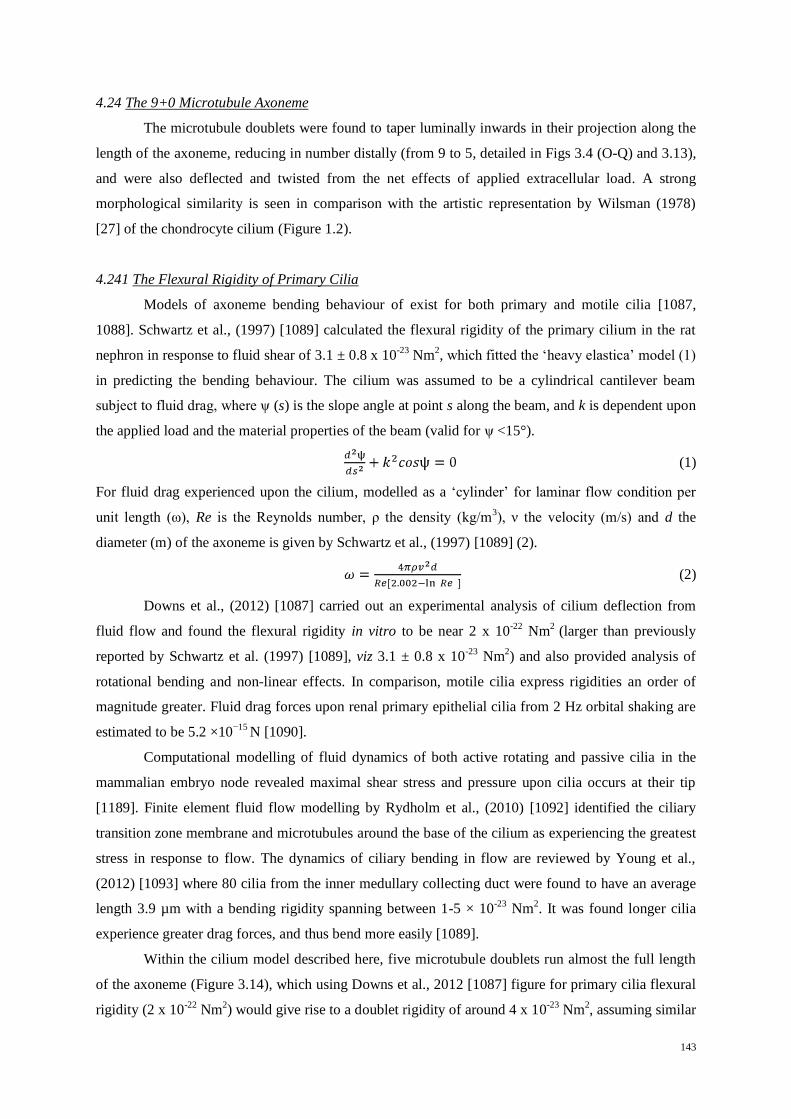
143
4.24 The 9+0 Microtubule Axoneme
The microtubule doublets were found to taper luminally inwards in their projection along the
length of the axoneme, reducing in number distally (from 9 to 5, detailed in Figs 3.4 (O-Q) and 3.13),
and were also deflected and twisted from the net effects of applied extracellular load. A strong
morphological similarity is seen in comparison with the artistic representation by Wilsman (1978)
[27] of the chondrocyte cilium (Figure 1.2).
4.241 The Flexural Rigidity of Primary Cilia
Models of axoneme bending behaviour of exist for both primary and motile cilia [1087,
1088]. Schwartz et al., (1997) [1089] calculated the flexural rigidity of the primary cilium in the rat
nephron in response to fluid shear of 3.1 ± 0.8 x 10⁻²³
Nm2, which fitted the ‘heavy elastica’ model (1)
in predicting the bending behaviour. The cilium was assumed to be a cylindrical cantilever beam
subject to fluid drag, where ψ (s) is the slope angle at point s along the beam, and k is dependent upon
the applied load and the material properties of the beam (valid for ψ <15°).
(1)
For fluid drag experienced upon the cilium, modelled as a ‘cylinder’ for laminar flow condition per
unit length (ω), Re is the Reynolds number, ρ the density (kg/m3), ν the velocity (m/s) and d the
diameter (m) of the axoneme is given by Schwartz et al., (1997) [1089] (2).
(2)
Downs et al., (2012) [1087] carried out an experimental analysis of cilium deflection from
fluid flow and found the flexural rigidity in vitro to be near 2 x 10-22
Nm2
(larger than previously
reported by Schwartz et al. (1997) [1089], viz 3.1 ± 0.8 x 10⁻²³
Nm2) and also provided analysis of
rotational bending and non-linear effects. In comparison, motile cilia express rigidities an order of
magnitude greater. Fluid drag forces upon renal primary epithelial cilia from 2 Hz orbital shaking are
estimated to be 5.2 ×10⁻¹⁵
N [1090].
Computational modelling of fluid dynamics of both active rotating and passive cilia in the
mammalian embryo node revealed maximal shear stress and pressure upon cilia occurs at their tip
[1189]. Finite element fluid flow modelling by Rydholm et al., (2010) [1092] identified the ciliary
transition zone membrane and microtubules around the base of the cilium as experiencing the greatest
stress in response to flow. The dynamics of ciliary bending in flow are reviewed by Young et al.,
(2012) [1093] where 80 cilia from the inner medullary collecting duct were found to have an average
length 3.9 µm with a bending rigidity spanning between 1-5 × 10-23
Nm2. It was found longer cilia
experience greater drag forces, and thus bend more easily [1089].
Within the cilium model described here, five microtubule doublets run almost the full length
of the axoneme (Figure 3.14), which using Downs et al., 2012 [1087] figure for primary cilia flexural
rigidity (2 x 10-22
Nm2) would give rise to a doublet rigidity of around 4 x 10
-23 Nm
2, assuming similar

144
that the cilium architecture. This is in line with flexural rigidity observations for motile cilia doublets
by Gittes el at., (1993) [1094] of 5 x 10-23
Nm2, and with Aoyama et al., (2005) [1095], estimate of the
sperm axoneme microtubule doublet rigidity being around 6.0 × 10⁻²³
Nm2. The value of such
comparisons between motile and non-motile cilia remains unclear. In comparison, the steady state
deformation and mechanical properties of cytosolic microtubules have been studied extensively
[1193], although measurements of microtubule flexural rigidity by various authors has given rise to a
range of conflicting results (0.27-3.0 x 10⁻²³
Nm2) [1094, 1097-1100].
Observations in this study of independent microtubule doublet lengths within the cilium may
explain variances of flow induced non-linear ciliary bending and reports of the presence of ‘kinked’
axonemes [1087]. Such variances in observations of rigidity and nonlinear effects are to be expected
between cell types if cilia can dynamically vary their lengths and microtubule numbers.
Measurements of ciliary rigidity to date have taken place within a less viscous medium of aqueous
saline, however rigidities for chondrocyte cilia are constrained by their embedding within a gel-like
extracellular matrix, and remain unreported. This places chondrocyte primary cilia in a unique
category amongst ciliated cells.
4.242 Distribution of Materials upon the Axoneme Doublets
Materials associated with each microtubule doublet were interpreted and classified according
to their shape and possible function. These are tabulated within Figure 3.29, with respect to which
microtubule doublet, its length, and position relative to the basal appendages, while their linear
distributions are visualised in Figure 3.14.
The distal tip of the cilium may be viewed as a ‘turn-around’ terminus zone for the transport
of materials to the end of the microtubule doublets and has received little ultrastructural investigation.
The membrane luminal surface was coated with a number of structures associated with the
microtubule terminations, some of which were connected by fine linkages and dense deposits (Figure
3.16). Along the axoneme fibre ‘linkers’ connecting microtubule doublets to the membrane, and
shorter non-connecting filaments were identified, decorating both the membrane and luminal sides of
the microtubules (Figure 3.15). These were similar to fine 10 nm long fibres that have been reported
associated with cytoplasmic microtubules [166], and it is likely similar proteins may also act as
scaffolds upon the basal body. Between microtubules of the axoneme, the fine filaments and inter-
connecting linkages were primarily associated with the A-subfibre, for microtubule to membrane
linkers (A:B 86:62), while inter-microtubule linkers being found to be more evenly associated with
the B-subfibre (A:B 46:59). Filaments which decorated the membrane and luminal faces of the
microtubule surfaces, but did not connect to membranes or other components were of higher
incidence upon the B-subfibre (internal A:B 42:75) and external filaments (A:B 43:54) respectively
(tabulated in Figure 3.29 B and C). At present, the nature of these structures remains obscure.

145
4.243 Distribution of IFT-like Particles
This is the first study to take a detailed look at IFT particles within the chondrocyte primary
cilium. A significant limitation of electron microscopy is the difficulty of matching known or
anticipated functions, determined by techniques of molecular biology, to the micro-morphology
exposed by staining with heavy metals. It is however possible to speculate on that relationship, since
recent proteomic analysis of cilia and primary cilia in particular has revealed many of their structural,
transport and signalling protein components [41, 132, 270, 468, 1101-1104]. Maintenance of ciliary
function requires continual turnover of material within the cilium, which only be achieved by
transport from the cell into the ciliary lumen.
Identification of IFT-like particles as macromolecular protein complexes located between the
microtubule doublets and the ciliary membrane resulted from comparison with previous ultrastructural
findings of Rosenbaum et al., (2002) [1105]. Figure 3.29 tabulates the relative doublet position, their
lengths, accumulations of materials, including ‘rafts’ of IFT-like particles, with reference to the
positions of the basal appendages. Distributions of IFT-like ‘rafts’ of materials were identified
predominantly upon longer microtubule doublets, and were clustered towards the distal end of the
axoneme (Figure 3.14).
The choice of microtubule triplet numbering on the chondrocyte cilium was in reference to
the presence of Basal Appendage One (BA1), and incremented in convention with motile cilia. IFT-
like accumulations were predominantly concentrated upon microtubule doublets 2, 3, 6, 7 and 8 with
1, 4, 5 and 9 being less populated or free, while basal appendages spanned microtubule triplets 1, 9
and 2 and 6, 7 and 8 (Figure 3.29). In comparison, Buisson et al., (2013) [261] noted IFT in the
flagellum Trypanosoma brucei was restricted to microtubule doublets 3, 4, 7 and 8, with 1, 2 and 9
being available for transport, while Absalon et al., (2008) [284] noted IFT restriction predominantly to
doublets 3, 4, 7 and 8.
Curiously, Basal Appendage Two (BA2) contained the greatest number of anchored cytosolic
microtubules involved in active transport processes (visualised in Figure 3.27), and it is closely
associated with microtubule doublet 3, which carries the greatest number of IFT-like materials. This
might indicate that within the chondrocyte cilium, transport may be preferential upon distinct radial
populations of microtubule doublets, and the distribution of basal appendages may influence their
loading. This discrete transport relationship and assignment of cargoes upon specific microtubule
doublets is likely to be structurally defined, and may depend on the relative proximity of the triplet to
the basal appendage basement structure, for access of vesicles and fine materials from docked
microtubules (detailed in Figs 3.24, 3.25 and 3.27).
This relationship may be further developed through the observed variances in microtubule
length, which requires IFT for their polymerisation and stabilisation [320]. Curiously, Gluenz et al.,
(2010) [1006] established that certain microtubule doublets may undergo preferential collapse, and

146
furthermore, that the pro-centriole is commonly tethered to a preferred microtubule triplet, indicating
discrete polarisation of basal body processes.
While it is not possible to identify proteins within the tomogram with any certainty, the
chondrocyte model indicated the presence of IFT-like structures accumulating within two laterally
opposed zones (Figure 3.14 arrows). Structures were noted upon both the A and B-subfibres, (with a
slightly higher association with the B-subfibre), including on the internal lumen side, while shorter
microtubules of the axoneme were generally bereft of IFT-like particles. In motile cilia, the A-
subfibre contains motile dynein machinery, while IFT-trains are observed to be tightly contained
between the B-subfibre and the membrane [269, 322]. In comparison with primary cilia, the A-
subfibre is free, and thus could be used for discrete transport (detailed in Figs 1.9, 3.12, 3.15, and
3.17). IFT-complex particles must first be transported to the basal body microtubule substrate, where
they become selected and activated to transport their cargoes to their respective destinations, before
being recycled [261-264]. IFT-complexes and transport motors have been reported to be concentrated
around the transition zone and basal body, where their interaction dynamics are discussed by Pedersen
et al., (2008) [103].
Chondrocyte IFT collections were found to have 20-45 nm periodicities, with the longest
‘raft’ measuring 400 nm, with 150, 80 and 30 nm lengths also being observed, distributed with the
highest concentrations observed upon microtubule doublets 3, 6 and 8 (Figure 3.29). Tomography by
Pigino et al., (2009) [269] identified, in Chlamydomonas reinhardtii, IFT ‘trains’ consisting of both
‘short and long’ trains containing 12–27 particles per train, with mean lengths of 250 and 700 nm with
a periodicity of 16 nm for the short, and 50 nm for the long train, respectively. Engle et al., (2009)
[264] indicated transport particle size was inversely related to flagellar length. Recently, Buisson et
al., (2013) [261] reported anterograde transport trains of 393 ± 51 nm, with retrograde trains 250 ± 51
nm in Trypanosoma brucei [261], with dual anterograde populations exhibiting velocities of 2.22 and
3.12 μm/s (a small population exhibiting low velocities of 0.5 μm/s) and retrograde speeds of up to
7.42 μm/s. Such variable velocities may indicate a more complex microtubule-motor-IFT cargo
relationship, which is yet to be addressed. Anterograde transport frequencies of around 1 Hz were
observed for anterograde trains, whilst retrograde frequencies are three times higher [261]. It has been
proposed that different anterograde velocities result from the actions of different motors [1106].
Given the number of transport motors that have been identified associated with the cilium,
there remains the likelihood of finding further ciliary motor interactions. These may possibly
including KIF12 and KIF13A [103], as well as a greater role of KIF7 and KIF17 motors in the distal
region [47, 270]. Furthermore, Ou et al., (2007) [285] indicated that IFT-components and transport
may be influenced by cell phenotype. This may indicate that chondrocytes display unique structural
and ciliary functionality, as evidenced by close anatomical similarities between the model and the
architecture described originally by Wilsman [27].

147
In conclusion, ciliary morphology affords a strong inference that IFT-trains travel upon
restricted sets of microtubule doublets, individually controlled by discrete mechanisms from the basal
body, akin to a train marshalling yard. This would explain variances in doublet length and cargo size.
4.25 The Transition Zone
The transition zone consists of linking structures of three distinct types: y-shaped linkers, alar
sheets, and interspersed fine filamentous fibres. The term ‘distal’ or ‘transitional fibres’ has also been
used in the literature to describe ‘alar sheets’. Alar sheets function to attach the basal body to the
periciliary membrane, while y-shaped linkers connect the microtubule doublets to the ciliary
membrane. Both are considered to act as both a physical barrier and a regulatory zone. The transition
zone is formed from two distinct compartments: the alar sheet zone from the axoneme juncture to the
basal body and the y-shaped linker zone (Figs 3.18 and 3.19). However, descriptions of these
structures vary between cilia types and between cell types [48, 49, 1107]. Horst et al., (1987) [1108]
identified the presence of cell surface glyco-conjugates linking the microtubule doublets to the ciliary
surface glycocalyx, in rat photoreceptors, indicating the possibility of extracellular interactions in
these cells.
In the present work, tomography revealed densely stained accretions decorating both the
microtubule doublets, and the membrane within this zone, interpreted as indicating the presence of
proteinaceous complexes. Two small membrane associated vesicles were located nearby closely
associated with the ciliary membrane (Figure 3.18). Their small diameter would allow them to
migrate through the ciliary matrix granule zone, but they could also represent used or superfluous
ciliary receptors shed from the membrane.
This zone contains many protein structures whose identities and functions remain obscure
(Figs 3.18 and 3.19). The distribution of ciliary proteins within the transition zone compartment has
been reviewed by Szymanska et al., (2012) [65], Czarnecki et al., (2012) [55] and Garcia-Gonzalo et
al., (2011) [70]. Ciliary transition zone components have recently been likened to a nuclear pore
complex [68], where it is proposed to function both as a regulatory zone and a diffusion barrier [1109]
for the orderly processing and exclusion of materials [66, 297, 1109]. Whether this zone is permeable
to vesicle transport remains to be seen.
4.251 Y-shaped Linker Structure
The presence of few identifiable y-shaped linkers within the ciliary transition zone in the
tomogram was a surprise in view of previous studies [49, 51] and given their very visible presence in
the in vitro work (Figure 3.4). This may reflect the effects of RHT and fixative influencing their
preservation, or temporal variation within the cilium at the time of fixation. Y-shaped linkers were
hard to discern and translate to the model adequately in the tomogram due to poor contrast. Curiously,

148
no y-shaped linkers were identified upon two of the shorter microtubule doublets 4 and 5 (Figure 3.29
C), indicating a possible absence of these structures upon dynamic microtubules.
4.252 Sub-Distal Fibres: Filamentous Structures
A number of fine filaments were observed within both the transition zone and basal body,
linking the microtubules to the membrane. These were investigated and modelled (Figure 3.19)
revealing them to be concentrated in a sub-distal zone upon the basal body, in proximity to the alar
sheets and associated with denser depositions along the microtubule surface (Figure 3.19 D-G). The
composition and function of these fibres remains unknown, as such structures have not been
commonly reported in the literature [49].
4.253 The Alar-Sheets Structure
The alar sheets are believed to function as a regulatory entry barrier for active transport into
the cilium [297, 308], from where the doublet subfibres of the triplets transition to form microtubule
conduits on which transport complexes transfer structural, signalling and receptor proteins to their
required locations, and are recycled back to the centrosome.
Within the tomogram the alar sheets, like the y-shaped linkers, were difficult to discern,
unlike the earlier in vitro ultrastructural investigations (see Figure 3.4 N and O). Each consisted of
lightly stained fine amorphous granules that tether the basal body to points within the periciliary
membrane. Originating from an attachment zone near the distal end of the C-subfibre, the alar sheets
evenly span across each doublet face, focussed upon the B-subfibre base (50 nm wide and 90-100 nm
in length), tapering outwards at right angles from the surface of the triplets, to focus upon anchoring
points within the ciliary membrane (see Figs 3.4 and 3.19). The transition zone membrane width was
found to average 262 nm in diameter (Section 3.5), with the entire alar sheet and basal body docking
system measuring an estimated 500 nm in diameter, in line with observations by Sillibourne et al.,
(2011) [1110].
The tomogram provides perspectives of the alar sheets, which were modelled selectively due
to their lower density structure, consisting of fine inter-dispersed granules (Figure 3.19 H). There was
little evidence to support the alar sheet structure as consisting of ‘wings’, that is frequently reported in
the literature [25, 45, 297]. The model shows the presence of vesicles in proximity to the alar sheets,
however these occur in close association with basal appendage microtubule transport processes. The
full role, structure and function of the alar sheet complexes remains unknown, although they are
known to be composed of the proteins ODF1, ODF2, CEP164 and CCDC123/CEP123 (Appendix I).
CEP123 associated with CEP164 [1110], which in turn associates with CEP170 [293] and HYLS1
[70].
Materials entering the cilium must pass the alar sheets, by active or passive transport
mechanisms into the axoneme. A ‘ciliary gate’ gap of 60 nm is reported [65] around the transition

149
zone, which allows for active transport into, and out of the cilium, regulated from the basal body
(Figure 3.29). It is not clear if this precludes vesicle transport. Lin et al., (2013) [327] established that
the interior of the cilium was accessible to proteins as large as 7.9 nm, through the base of the cilium,
while low molecular weight dextrans (<40 kDa) have been shown to be able to enter the cilium [68],
although information about the shape of these molecules was not reported.
A number of proteins have been located within the transition zone [55, 70, 302, 1111] where
they are believed to contribute to ciliary gating, ciliogenesis [65] as well as membrane composition
[70]. Specific details of their structure and roles have only recently been established [62, 65 70, 1112,
1113]. Mutations to any of these key proteins within the zone give rise to pathologies ranging from
lethal embryonic conditions to impaired ciliary function (Figure 4.1) resulting in a spectrum of
ciliopathies [1, 55, 1114, 1115]. Some of the common ciliopathies have been assigned to a structural
site of action within the transition zone, illustrated in Figure 4.3.
Figure 4.1 The Transition Zone ‘Compartment’: [A] A model of the transition zone located between the basal body and the axoneme, where alar sheets tether the basal body to the periciliary membrane, and y-shaped linkers connect the axoneme
microtubules to the ciliary membrane. The zone is divided into modules which contain distinct components. Reproduced and
edited from Czarnecki et al., (2012) [55]. [B] Organisation of the Transition Zone Modules are based around protein
complexes; The NPHP1-4-8 Complex: NPHP1,4 and 8, the MKS/B9 Complex: (B9D1, B9D2, CC2D2A, MKS1, KCTD10, TCTN 1, 2 and 3, including trans membrane proteins TMEM17, TMEM67 TMEM216, TMEM231 [1116],
TMEM237), and the NPHP5-6 Complex: (CEP290 and NPHP5) and the Inversin compartment (NPHP2, 3, NEK8, and
INV). The vertical height associates complexity of component interactions, while the colour intensity indicates pathology
complexity, while arrows shows associations between molecules. The horizontal axis indicates dispersal of respective complexes from the basal body along the axoneme. Reproduced with permission from Czarnecki et al., (2012) [55], who
noted the shared elements of the transition compartment in ciliogenesis and disease[70, 1111]. [C] Reviews of five
ciliopathy proteins: MKS (Meckel-Grueber), JBTS (Joubert syndrome), NPHP (Nephronophthisis), BBS (Bardet-Biedl
Syndrome) and SLS (Senior-Loken Syndrome). Interactive protein partners included CC2D2A, TMEM216, RPGRIP1L,
TMEM67, CEP290, NPHP3, SDCCAG8, NPHP1, IQCB1 and NPHP4. Mutation of any of these proteins may generate
particular aspects of these ciliopathies, which are joined by common lines. Reproduced with permission from Szymanska et
al., (2012) [65]. The exact functional roles of many of these proteins remains unknown (see Appendix I).

150
4.30 The Centrosome
The centrosome has been simply but accurately described as a ‘polyfunctional multi-protein
cell complex’ by Alieva et al., (2008) [120], a structural processing unit for the regulated control of
cytosolic materials, organelles and cell cycle. During interphase, it acts a cytoskeletal microtubule
nucleating and organising centre, where the primary cilium functions as a physical extension of the
basal body, with the proximal centriole subtending it. Components may be broken into three groups:
structural, either involved in the microtubule structure, or localised with it, centrosomal proteins
associated with the centrosome and cell cycle, or functional proteins such as transport motors and
regulatory complexes [120].
Analysis of the centrosome in Figure 3.32 A-H demonstrates a series of slices detailing the
microtubule architecture, the pericentriolar matrix, the basal appendages, and the nearby nuclear
membrane. Surrounding the centrosome are fields of vesicles shown in Figure 3.32 I-L. These are
interspersed with microtubules, indicating the centrosome as a central organiser of vesicular based
processes. Modelling of the microtubule based architecture of the basal body, proximal centriole and
cytosol surrounding them is illustrated in perspective views in Figure 3.33, uniquely including the
presence of the nucleating microtubule upon the vesicular ‘torus’.
Figs 3.34 and 3.35 detail further ultrastructural and model views of the relationship of
centrosomal components. These images contain large amounts of information covering spatial
components of the centrosome, the tubulin based basal body and the proximal centriole, microtubule
nucleation and attachment points of the basal appendages and pericentriolar matrix, intermediate
filaments and fields of vesicles in the process of directed transport, docking and sorting of materials.
Limitations to interpretation of ultrastructure stem inevitably from the use of a single axis
tomogram containing a complete cilium for model generation. It is possible to expand into great detail
on many of the proteins and molecular pathways associated with aspects of the model components of
the primary cilium, and also the centrosome. These protein components are too numerous to mention
in this discussion, but some of these are tabulated within the Appendices aiding in linking the
structural components that have been visualised, with function. Many of these represent speculative
findings in the literature, and need to be interpreted carefully. However, the focus of this discussion
must be limited, brief and upon technical aspects resolved in the model, to which function could be
attributed. The few previously undertaken tomography based modelling studies of centrosomes, have
focussed upon aspects of centriole structure, pro-centriole formation, microtubule networks and
nucleation or mitotic processes (see Appendix VI).

151
4.31 The Basal Body
The microtubule based architecture of the maternal basal body and its subtending proximal
centriole form the core of the centrosome. These were carefully traced within the tomogram to ensure
the accuracy of each element. Individual triplet subfibres were depicted as simple intersecting tubes,
each extruded to represent the microtubule doublets of the axoneme (Figure 3.20). This provided a
simple basis for modelling all microtubule based components, however it unfortunately constrained
the presentation of the basal body fine structure.
The structure of the microtubule triplet unit and its position within the basal body is similar to
previous basal body studies (Appendix VI). Anderson (1972) [45] and Wilsman et al, (1978) [27]
produced artistic representations of the basal body triplet architecture. These are visualised in the
model, with each microtubule triplets ‘left-handed’ polarisation and decreasing angle observed in their
distal projection along the basal body, detailed in Figs 3.20 and 3.21 J. The microtubule triplet angle
decreases uniformly along the basal body, where each triplet merges into the doublet of the axoneme,
retaining a slight left-handed rotation or ‘curl’. The basal body is observed to be slightly wider at its
centre, giving the appearance of having a ‘barrel’ shaped structure (Figure 3.20), as described by
previous authors [27, 45] (Appendix VI).
4.32 The Microtubule Triplet Structure
The model presented in this study did not fully investigate the basal body triplet fine
structure. This requires higher resolution visualisation, to establish finer details of the whole centriole,
its longitudinal structure, triplet architecture, and the inter-triplet relationship with their associated
proteins. Simple investigative microscopy of primary cilia utilising cell culture was used to define the
basal body subfibres, their inter-linkages and ‘hooks’ (Figure 3.4), but only at a relatively low
resolution.
The A-subfibre feet appear to be functional remnants of centriole assembly, which exist only
within the proximal zone. The A-C-subfibre linkages tether the respective proximal ends of the
triplets, providing mechanical inter-triplet support to the basal body (Figs 1.8 and 3.4) and disappear
towards the distal end of the basal body near the termination of the C-subfibres. It is a matter for
further investigation whether each of the A and A-C subfibre structures are of continuous form like
the A-B-subfibres, or are represented as individual periodic linkages, as observed within the triplet
structure by Li et al., (2011) [89]. These connections occur within the luminal environment of the
cilium, and may function as attachment and regulatory sites upon the inner surface. The longitudinal
fine unfolding protofilament structure of the C-subfibre has received little attention since being
described by Wilsman et al., (1983) [88], as its fine ‘hook’ structure gradually disappears distally
along the cilium.

152
4.33 Luminal Vesicles
Numerous observations have been made of cilia and basal bodies containing vesicles, ranging
in diameter from 25 to 150 nm, however they have customarily been regarded as artefacts of fixation
[95, 1117-1120]. Luminal vesicles observed within the primary cilia of chondrocytes have received
less attention, despite previous mention in ultrastructural and tomographic investigations [25, 26]. In
this study, no vesicles were observed within the axoneme of primary cilia. However, the basal bodies
of three cilia contained vesicles (Figs 3.5 A, B and 3.22), with one distinctly containing three small
vesicles (Figure 3.5 B).
Vesicle size ranged from 35 to 50 nm, curiously similar size distributions to the ciliary
membrane vesicles observed within the matrix (Figure 3.18 for comparison). The single oval shaped
vesicle contained within the model appeared to be firmly attached to the luminal face of a triplet of
the basal body, suggesting internal transport may be possible within the lumen (Figure 3.22). The
vesicle was found to interact with proteinacous materials contained within the lumen.
Presumably cilia undergo chemical fixation rapidly, so the probability of observing vesicles
using ciliary transport should be low, due to their relatively short axonemal transit periods. However,
frequent observations exist of vesicles throughout the cilium, including from within the transition
zone [25, 26]. The presence of a base plate and central microtubule doublet in motile cilia may hinder
luminal vesicular transport, although vesicles have been noted in the motile cilia of patients with
bronchiectasis [1121].
In primary cilia, a large intra-luminal zone does not preclude potential vesicular accumulation
or transport. If these vesicles represent the true structure, and are not artefacts, then it may be
concluded that they are in fact part of the normal transport processes of moving membrane bound
materials to and from the cilium. Thus, the lumen of the primary cilium basal body may represent a
novel conduit between the cilium and the centrosome.
4.34 Basal Luminal Discs and Internal Structures
Various electron dense structures have been commonly occur within the lumen of centrioles
of many species. Formations such as the pro-centriole replication ‘cartwheel’ complex [237]
disappear within the mature centriole [1122, 1123], while other indeterminate structures are
frequently noted within the lumen [1124], including those termed luminal discs [124, 237, 253, 597,
1125]. The tomogram has revealed a presence of granular structures spanning the distal ends of both
the basal body (Figure 3.21) and the proximal centriole (Figs 3.30-3.32). The basal body lumen also
contains numerous deposits and filaments of unknown electron dense materials, many of which are
often attached to the triplet faces.
It is probable that the lumen of the basal body functions as a tightly regulated ‘hangar and
conduit’ for the storage, modification and signalling of key ciliary and centrosomal components,
which are protected from the active cytosolic environment by the microtubule triplets. Key proteins

153
involved during pro-centriole biogenesis [237] include the luminally located components of CP110,
Centrin, CPAP and CEP135, while Centrobin [1126], CEP110, 120, 135 and 192 are known to have
microtubule associations [120, 122, 237]. The presence of centrosomal RNAs (cnRNA) have been
reported, however their exact roles and localisation with respect to the basal body are still to be
determined [1127, 1128]. It is likely that many proteins share both temporal and spatial locations
around and within the basal body during normal cilium operation and the cell cycle.
4.35 The Basal Appendages
Within the tomogram, basal appendages appear as extremely heavily stained electron dense
structures that leave little contrast to their fine structure (spanning Figs 3.23-3.27). Each consists of
three parts, the basement complex, the appendage arm, and a distal docking complex, with attached
cytosolic microtubules. The three ‘basement zones’ were found to obliquely span at least three
microtubule triplets, each consisting of fine porous materials forming a perforated structure. These
were investigated through volume rendering approaches (Basal Appendage One, Figs 3.23 and 3.24)
and (Basal Appendage Two, Figure 3.26). Each is attached obliquely to three microtubule triplets,
comprising of a collection of fine amorphous materials, detailed in Figs 3.24, 3.25 and compared in
Figure 3.28, sharing a strong similarity in their shape, structure and disposition.
A ‘hollow’ low density zone is located in the middle of the basement zones, located under the
appendage arm, which extends perpendicularly outwards, resembling findings by Kunimoto et al.,
(2012) [74]. The arm itself is uniformly extremely electron dense, containing pockets and thorny
protrusions. The docking complex heads appear as finer spiny complexes, some of which are
bifurcated (Figure 3.27), and also act as mechanical attachment points for microtubules. Vesicles were
noted in close proximity to both microtubules and the basal appendage, with one of these being
physically linked via fine fibres to the anchoring complex, and the nearby basal body (Figs 3.23-3.25).
The basal appendages may experience loads resulting from the interplay of combined forces
upon the cilium, and those applied by their anchored cytosolic microtubules. These radiate outward
from the docking complexes and appear to interact with a number of vesicles and organelles (Figs
3.25, 3.27 and 3.35). Other microtubules were observed radiating from the centrosome and these may
function together with intermediate filaments to act as part of a cytoplasmic tensegity network
required to maintain cellular shape. The possible presence of another basal appendage was identified
outside the plane of section of the tomogram, indicated by the occurrence of intersecting trajectories
of cytosolic microtubules within the section (Figure 3.43). If present, this extra appendage could
easily span triplets 3 to 5 (Figure 3.29), and would provide the cilium with a third additional
anchoring point.
Basal appendages are a marker of the formation of the mature basal body, where they act as a
tethering apparatus for the attachment of cytosolic microtubules (Figs 1.9-1.11), and are ubiquitously
associated with both motile and primary cilia. Descriptions in the literature of basal appendage

154
structure and relationship to the basal body are multifarious and imaginative. This present study finds
little evidence to support these, although there exists abundant opportunity for further study.
The protein components constituting the basal appendages, and which are involved with the
mechanisms of microtubule nucleation and anchoring within the centrosome are not yet fully
understood [120]. Some discrete protein members have been identified and their roles are listed in
Appendix I. Basal appendages are known to contain the proteins Cenexin [1129], Ninein [160],
CEP170 [1130], CEP110 and ε-tubulin [1131], although there are undoubtedly many more awaiting
discovery. The basal appendage anchoring head complexes are believed to be comprised of Cenexin,
Centriolin, CEP170 and ε-tubulin [120], where Ninein may also act as a molecular link for anchoring
of γ-TuRC complexes in the centrosome [160, 198]. It is believed CEP192 may act as a scaffold for γ-
TuRC microtubule nucleation, while NEDD-1 (GCP-WD) is required for γ-TuRC complex
centrosomal localisation and binding [1132]. CG-NAP and Pericentrin are believed to play roles in
anchoring nucleation sites within the centrosome. Centrosomin is known to interact with γ-tubulin
[1133], while Pericentrin provides a physical scaffold that interacts with GCP2 and the γ-tubulin
nucleating component [1134, 1135]. BBS4 functions as a critical adaptor link between PCM-1 and
p150Glued
, and also influences microtubule attachment [120]. IFT-complex motor KIF3A recruits
Dynactin (p150Glued
) to organise the appendages, where depletion of p150Glued
results in loss of KIF3A
and Ninein giving rise to ciliary dysfunction [523]. Striated parallel bandings are sometimes reported
decorating basal appendage arms [1120], however their composition remains unknown.
4.36 The Proximal Centriole
Naming conventions for the proximal centriole as a ‘daughter’ centriole (with respect to the
‘mother’ basal body) have resulted in much confusion, with centriole replication pro-centrioles being
referred to as ‘daughter centrioles’. The centrioles of the diplosome exhibit dynamic spatial and
separation inter-relationships throughout the cell cycle [236]. The proximal centriole was only
partially contained within the tomography section, resulting in an open ‘cusp’ like appearance in
model projections (Figs 3.30-3.32) revealing the triplet architecture is similar to the basal body in
structure, handedness and polarisation [64, 94]. However, a notable exception is the absence of the
gentle distal straightening of the triplet angle during their projection, as in the basal body. It is
oriented in the positive microtubule direction away from the axis of the basal body at 135°.
Importantly, it is devoid of any alar-sheets, subdistal fibres or basal appendages, and lacks
microtubules attached to or nucleating from its surface.
Dense areas of heavy stain decorate materials upon both the external and luminal surfaces of
the proximal centriole, indicating a substantial collection of proteins. A series of dense luminal disc-
like accumulations was identified spanning the near distal end of the proximal centriole (Figs 3.30-
3.32) in striking similarity to structures contained within the basal body.

155
The proximal centriole closely subtends the basal body within the centrosome, connected to it
by fine linkages of pericentriolar material connecting their respective proximal ends (detailed in
Figure 3.34 A and B). The proximal end is in close proximity to a vesicular bud projecting from the
centrosomal annulus (Figure 3.33). The distal end of the proximal centriole contains proteinaceous
tendrils linking to the nuclear membrane and to a nearby nuclear pore, one of three in close proximity
(Figure 3.31). Within the literature, centrioles have been commonly reported associated with the
nuclear membrane [1002, 1154]. Proteins Ninein, NEK2 and δ-tubulin are known to be localised to
the proximal ends of the basal body and proximal centriole, while Centrin localises to their respective
distal ends [120]. The function of the proximal centriole remains elusive, however is a vital
component of centrosomal dynamics, where its structure appears every bit as intricate as that of the
basal body.
4.40 Microtubule Populations
Three distinct cytosolic microtubule populations were observed; those derived from the basal
body appendages, those from within the centrosome pericentriolar matrix, and those originating from
within the cytoplasm. The pericentriolar matrix acts as a ‘scaffolding lattice’ for many proteins, as
well as acting as a nucleating site for microtubule ‘minus’ ends (Figs 1.11, 1.12, 3.35 and 3.36). These
are spatially distributed within the matrix surrounding the centrosome, where it has been suggested
that a limited number of nucleation sites exist [124, 135].
Visualisation of microtubule surfaces in their long axis revealed decorations of fine speckled
deposits, denser globular structures, and fine tethers linking to vesicles (Figure 3.36). These were
intimately clustered on microtubules surrounding the basal appendage anchoring heads and the
centrosome (Figs 3.25 and 3.27). These microtubule-associated materials of unknown function may
represent directed motor dependent transport processes of the basal body, the centrosome and the
Golgi apparatus. This raises further questions about the role of the appendage derived microtubules
within the centrosome, their interactions with organelles, and the regulation of transport motors
responsible for their many transport and signaling mechanisms.
Basal Appendage One-derived microtubules were identified extending to the Golgi apparatus,
associated with vesicle fields of the pre-cis compartment area, and both the cis- and medial-
compartment edges (Figs 3.35, 3.37 and 3.41). It is likely that the terminal ends of the microtubules
also interact with the Golgi membrane and resident proteins [53]. For example, Hoff et al., (2011)
[610] has shown that proteins destined for the cilium, such as PC2, may emerge exclusively from the
cis-Golgi. Microtubule transport of such ciliary derived PC2 can be readily envisaged from the
components detailed in Figure 3.37 B and D.
Basal Appendage Two-derived microtubules appear to have a selection of assorted protein
structures and vesicles associated with them, some in close association with the nearby periciliary

156
membrane. These microtubules display surface variances in staining, and exhibit a number of putative
motor-associated tethers attached to them, with some linked to nearby vesicles (Figure 3.27).
A long microtubule originating from Basal Appendage Two transects the centrosome to span
a field of assorted vesicles (including the torus) bound by the nuclear membrane and the cis-Golgi
face Figure 3.41). These areas appear to direct trafficking of vesicles into the Golgi cisternae, from
components of the ERGIC. As Golgi cisternae are also highly dynamic, it is likely that many
microtubule processes are also involved in interfacing the Golgi with the centrosome in this region.
A subset of non-basal appendage, centrosome-derived microtubule fields are observed
running closely parallel to the pre-cis-Golgi face, which appears to be actively involved with a high-
density field of pre-cis face vesicles (Figure 3.37, 3.38 K). Such arrays could easily transit materials
along the ‘building face’ of the Golgi, and also be involved in the spatial organisation and signalling
to the apparatus. It is tempting to speculate basal body derived microtubules may exclusively source
materials from the Golgi as part of their normal centrosome function.
Microtubules from the basal appendages are intimately associated with Golgi cisternae and
the pre-Golgi vesicle fields. Centrosome derived microtubules are associated closely together with the
cis-face, indicating that they are involved with specific aspects of the Golgi. Transport upon these
may be both spatially refined and restricted, while other centrosome microtubules appear to facilitate
the bulk transfer of materials from the endoplasmic reticulum to the Golgi.
4.41 The Intermediate Filament Organisation Centre (IFOC)
Intermediate filaments have been shown to originate within the centrosome from an
organisation centre within the pericentriolar matrix, surrounding the proximal centriole (observed
structurally in Figure 3.34 D and E and modelled in Figs 3.35 F, 3.37 B and 3.41), where intermediate
filaments have long been associated with the centrosome [115, 120, 236]. Examination of the model
reveals that the intermediate filament network originates in a condensed zone within the centrosome
surrounding the proximal centriole. This model concurs with observations of Alieva et al., (1992)
[115], who reported intermediate filaments originating in a zone, with no single focus, contained
within 0.3 µm of the proximal centriole. Filaments were observed attached by fine linkages to
materials within the pericentriolar matrix (Figure 3.34 D, E). Some filaments ran in parallel with the
microtubule network, with one filament originating from near the proximal centriole, radiating into
the pre-cis-Golgi area (Figure 3.35, 3.37 and 3.41), perhaps indicating a functional relationship.
As a component of the cellular tensegrity network, intermediate filaments can be deformed
under strain, and are involved in the dynamic regulation and maintenance of cytoskeletal architecture
[1136-1139]. Their keratin and vimentin members are known to be associated with the centrosome
[1136, 1140, 1141], influencing its stability [1142], with vimentin being reported to be necessary for
the maintenance of the chondrocyte phenotype [139]. De-tyrosinated microtubules reportedly interact
with vimentin intermediate filaments [1143], while the cytoskeletal protein Plectin mediates their

157
interaction [1144]. The centrosome shares both the microtubule and intermediate filament organising
centres, each sharing a zone surrounding the basal body and the proximal centriole, where they each
play an intrinsic regulatory and nano-mechanical role in bearing compressive forces upon the cell.
4.42 Order in the Pericentriolar Matrix
In the electron microscope, the pericentriolar matrix appears as a zone of finely stained
filamentous material that surrounds the diplosome, inter-connecting the distal ends of the centrioles
(Figs 3.32, 3.33 and 3.34). The use of RHT gave an enhancement of contrast deficient in earlier
studies. The matrix is now revealed to be an intricate lattice, made up of many fine interlinked
components, dispersed with small electron dense granules. These linkages inter-connect the basal
body and the proximal centriole, where they also decorate and coat the microtubule triplets. The
luminal contents of the proximal ends of the basal body and the proximal centriole also merge into
this matrix, which permeates around the diplosome. The lattice extends outwards to interact with a
variety of nearby objects, including microtubules, intermediate filaments and vesicles. It is
constrained by the presence of the vesicular centrosomal torus, to which it also binds (Figure 3.34 C).
No striated rootlets or inter-centriolar linkers were observed within the tomogram. The known protein
components of the centriole anchoring complexes are tabulated in Appendix I (see Centriole
Anchoring Complexes).
Two distinct PCM domains appear to exist surrounding the diplosome: a near zone that is
reported to be based upon pericentrin-like-protein fibrils aligned with the centriole wall, and a larger
extended zone linking to the larger pericentriolar-centrosomal matrix [122]. Materials accumulated
upon the microtubule triplets have been proposed to have both higher order structures and functions
[464]. The PCM contains PCM-1, which interacts with a wide range of proteins as well as with the γ-
TuRC nucleation complex [128, 138, 459]. The PCM acts as nucleation site for microtubules, and
tethers centrioles [126], as well as containing key proteins involved in the cell cycle [134], cell
regulation, ciliary formation and regulation [62]. The role of the PCM in supporting interphase
function is less well known [128, 466, 1126] where it varies during the cell cycle [112, 128].
Recent visualisation of numerous key centrosomal proteins during interphase by high
resolution three dimensional microscopy has revealed the detailed organisation of many of these
proteins within a highly organised ‘PCM tube’ and lattice, which are confined in spatial layers
surrounding the basal body [104, 122, 464]. The relationship of this centrosomal matrix to the
vesicular torus described in this study remains unknown.
4.43 The Centrosomal ‘Torus’
This study has identified a unique vesicular annular structure surrounding the centrosome that
has not been previously described. It occupies an orbital volume (torus) between the proximal and
distal centrioles, spanning an intermediate position in association with vesicle fields of the pre cis-

158
Golgi to the endoplasmic reticulum. Smaller vesicles are involved with the end zones surrounding the
annulus, and with the nearby microtubule transport conduits around the basal appendages (Figure
3.33, 3.35 and 3.41). The Golgi associated end of the annulus was found to have small budding
membrane protrusions associated with a vesicle fields and a nearby larger vesicle that is tethered to
the surface by a series of fine connections (Figure 3.33 D). Centrally, the membrane is near to a
microtubule derived from basal appendage two (Figure 3.33 A), which also interacts with the distant
Golgi vesicle field Figure (3.35 F). The pericentriolar matrix of the basal body interacts closely with
the membrane surface (Figs 3.33, 3.34 B and C), while nearby on the lateral side a membrane
protrusion is juxtaposed to a number of electron-dense accumulations on the end of the proximal
centriole, in association with two small vesicles. Opposed at 180°, the annulus was found to contain a
microtubule-nucleating site that is closely associated with smaller vesicles and electron dense
materials (Figure 3.35 F). The presence of smaller vesicles in close proximity to both centrioles is
novel, and offers a possible source for explaining translocation to the basal body.
The close proximity of such a large vesicle engaged closely with the centrosome and
centrioles raises questions as to its function, however the transport of larger vesicles by the
microtubule network is common [1145]. It may function in part as a novel pericentriolar matrix
protein trafficking network of the centrosome. Curiously, Wang et al., (2009) [1116] noted a novel
pericentriolar compartment enriched with ceramide that was located at the base of the primary cilium,
whose transport was associated with the cis-Golgi. Equally, it may be an extension of the ‘ER to
Golgi intermediate compartment’ (ERGIC), whose defined role between the endoplasmic reticulum
and Golgi has so far remained elusive [520, 1147, 1148]. Uniquely, it shares a physical location,
shape and most likely interacts with the ‘PCM tube’, which is an ordered tubular pericentriolar matrix
accumulation of specific materials and protein species required for centrosomal function [122, 129].
Thus, the torus represents a unique, but likely temporary structure, which serves to function as an
interface with the pericentriolar matrix of the centrosome and the Golgi.
4.44 Nuclear Pores
Nuclear pores act as conduits for the transfer of RNA, and also for signalling proteins
shuttling between the nucleus, the cytoplasm and the centrosome [141, 1149, 1150], which are known
to be involved in the regulation of gene expression [1149, 1152]. Within the model, the centrosome,
and in particular the proximal centriole, are positioned in close proximity to several nuclear pores, and
protein based tendrils are observed to connect the distal end of the proximal centriole to the nuclear
membrane, and nearby nuclear pores (Figure 3.31 and 3.32). The association of centrioles with the
nuclear membrane has been previously reported [1001, 1150, 1503], where Bolhy et al., (2011) [1154]
described the attachment of the centrosome to the nuclear membrane via Nup133-dependent
anchoring. A vesicle cluster is located in the model near a nuclear pore, upon the periphery of
pericentriolar matrix and proximal centriole (Figure 3.35 C and F). The significance of such

159
relationships remains unknown. Burakov et al., (2013) [1150] reviewed centrosome positioning and
mechanisms of attachment to the nucleus, mediated by SUN-1 and ZYG-12 [1155]. The close
presence of the centrosome to the nucleus with respect to signalling, transcription and cellular
regulation are intriguing and suggestive.
4.50 The Golgi Apparatus
Current concepts of Golgi structure and organisation have been derived from conventional
electron microscopy studies [724], with images similar in appearance to optical sections visualised in
Figure 3.37. The Golgi apparatus is the central organelle of the secretory pathway, which receives its
materials from the endoplasmic reticulum. It contains the classical description of flattened cisternae,
of cis, medial and trans-compartments [691] (Figure 3.38). The cis-cisternae contain large
fenestrations, some significant branching and a small number of COP-like vesicles around the
periphery (Figure 3.37). The medial-compartment is tightly layered, containing finer fenestrations, a
marked discontinuity running across the compartment and fewer peripheral COP-like vesicles. Fine
tubules, 30-40 nm in diameter, were noted connecting cisternae, which are reportedly triggered by
secretory traffic [729, 1156]. The trans-cisternae are composed of larger compartments, with a
number of ‘exit faces’, with defined fields of COP-like vesicles appearing to bud and emanating from
the faces. Occasionally, fine electron dense material was noted within compartments of the Golgi
cisternae, however no collagen-like precursors were observed within the Golgi network. The trans-
Golgi face was observed to be adjacent to larger vesicular granules and tubular structures, where it is
presumed that cargoes were being sorted for their respective destinations and secretion (Figure 3.41).
Utilisation of tomography has resolved many of the aspects of fine form and understanding of
the Golgi between species (Appendix VI). Recent electron tomography studies examining the three-
dimensional architecture using dual-axis tomography (Ladinsky et al., (1994) [1157], Marsh et al.,
(2001, 2005) [1158, 714]) revealed detail of highly complex, yet conserved structures. These and
others revealed the close relationship between the cytoskeleton and the Golgi apparatus, and also
raised questions about the exact nature of how the Golgi processes materials.
4.51 The trans-Golgi Network
The trans-Golgi network is an inter-connected arrangement of vesicles and membrane
compartments associated with the trans-face of the Golgi, and is an area where vesicles containing
proteins are sorted and then shipped to their intended destinations [691, 1159-1161]. It was originally
defined as a ‘specialised organelle’ on the trans-side of the Golgi stack that is responsible for the
routing of proteins to lysosomes, secretory vesicles, from the Golgi to the plasma membrane [1162].
Novikoff (1964) [1163] described the ‘Golgi-Endoplasmic Reticulum-Lysosome’ (GERL) hypothesis,
which he used to describe structures related to the Golgi saccule, which is part of the endoplasmic
reticulum and trans-Golgi, and which form lysosomes.

160
Post-translationally modified materials are transported into the trans-Golgi, and then
organised for their respective directed secretion [1159]. A large cylindrical tubular process was
located in parallel with the face of the trans-Golgi, being surrounded by a cluster of COP-like vesicles
(Figure 3.39 C). It is not known if this represents a migrating trans-Golgi fragment, or a ‘trans-Golgi
tubule’ (Figure 3.42), or if it is part of the trans-tubular network, described by Clermont et al., (1995)
[1161]. It most likely represents a process of the trans-Golgi network tubule in the process of being
shed [1159].
Microtubules were modelled associated with presumed active transport to the plasma and
periciliary membrane from locations close to the trans-Golgi face (Figure 3.37), demonstrating a role
for the Golgi as a MTOC [646]. These were observed apparently undergoing active transport, with
some vesicles in close contact and others merging with the plasma membrane, indicative of secretory
processes being undertaken (Figure 3.39 E). Examination of individual vesicle membranes revealed
the presence of electron dense coatings (Figure 3.39 F) indicative of membrane mediated processes.
The occurrence of vesicles packed with densely stained ‘matrix granule-like precursor materials’ were
identified predominantly associated with the trans-Golgi network. These were similar in nature to the
extracellular granules, but were contained within their compartments at a higher packing density than
the extracellular matrix granule network (Figure 3.39 A, A’, B and D).
The matrix-granule-containing vesicles were easily recognised by their contents, with the
largest storage granule being of appreciable diameter (600 nm), comparable with the nearby Golgi
stack (Figure 3.42). This vesicle is closely surrounded by a number of trans-Golgi derived vesicles,
and was observed apparently undergoing active transfer processes in close proximity to a long tubular
structure, assumed to be part of the endosomal system. The distal end of the tube was closely
associated with active receptor mediated coated pit processes.
Novikoff (1976) [1164] described the biochemical and cytochemical processes of the
endoplasmic reticulum for the formation of melanosomes and lysosomes, for the metabolism and
secretion of materials for various systems and association with the trans-Golgi. Marsh (2005) [714]
described these processes as being analogous to a ‘distillation tower’, in which cisternae of the trans-
Golgi are sorted with their ‘refined fractions’ for further distribution. In the model, the trans-faces
appear to be residual fragments of the trans-Golgi stack (Figs 3.37 and 3.41). Clermont at al. (1995)
[1161] discussed differences in trans-Golgi network structures, which were found to vary extensively
in size and shape between species. Furthermore, Ladinsky et al., (1999) [727] reported an association
of the endoplasmic reticulum with the trans-face, suggesting that localised post-processing was also
occurring. A full picture of the Golgi apparatus is certain to be complicated, as it has been found that
Golgi and trans-Golgi components are not permanent structures, but dynamically changeable, and
continuously undergoing breakdown and renewal [721].

161
4.52 Coated Pits and Caveoli
A number of coated pits were observed to occur clustered within a zone of the plasma
membrane, in close proximity to the ciliary tip (Figure 3.40). Such relationships have previously been
reported [26], however this is the first functional tomography model of receptor-mediated coated pits
to clearly show the mediated endocytosis of matrix material in proximity of the cilium. Included is a
nearby membrane process in the early stages of initiating pit formation. Endocytosed ‘coated pit’
materials accumulate within endosomes, en route to the Golgi [1165], where they engage with
processes of the trans-Golgi, and its associated network [1166]. Sorting occurs within the early and
late endosomal systems that display a tubular morphology and close trans-Golgi network association
[1167] (Figure 3.42).
The mechanisms of clathrin coating, vesicle budding and fusion have been described [702,
1168], while the processes of endosomal sorting and retrograde transport to the trans-Golgi have been
reviewed by Bonifacino et al., (2006) [1000].
Caveolae were identified in the model in proximity to the field of coated pits, consisting of
fine finger like indentation into the cell membrane (Figure 3.40 E). Wolfgang et al., (1999) [1169]
identified numerous caveoli-like invaginations along the plasmalemmal membrane of chondrocytes
[1170], where they have been described functionally as trans-endothelial channels [1171], however
their functional importance remains to be determined.
4.53 Are Primary Cilia Both Displacement Detectors and Pressure Sensors?
Recent research has clearly identified the role of primary cilia as receptors for a raft of
specialised modalities, transducing external stimuli, into internal information, and enabling an
appropriate response. The primary cilium of the chondrocyte is embedded distally in the surrounding
matrix, and thus almost certainly distorts under applied for to that matrix. The level of distortion
required to trigger transduction remains uncertain.
Attachment of the matrix to the ciliary membrane may physically trigger membrane receptors,
raising the issue of the threshold of displacement required for transduction (Figure 3.43). The three-
dimensional nanoscopic nature of this matrix may possess a novel property of being a negative
Poisson material, offering interesting material and mechanical properties [1172, 1173]. This arises due
to the semi-crystalline properties of a regular three-dimensional ‘mesh-like’ material, which may
possibly be similar to the matrix structures making up the zone of the ‘ciliary pit’.
Bell [1174] raised the interesting example of ciliated cochlea hair cells detecting sound
pressure and displacement, but it remains subject to further investigation to see if the difference in
properties between the fluid of the pit and the surrounding medium enables efficient pressure
detection. This may partially help resolve the concept of the cilium as being both a deflection and
pressure sensor within the matrix [1175]. It also allows the cilium flexibility of movement and permits
chemotactic signalling by enabling the migration of solutes and ligands [455].

162
4.54 Matrix-Cilium-Golgi Continuum in Chondrocytes
Cilia function as highly specialised sensors, where the targeting, functionality of specific
receptors upon the cilium and their pathways are usually specific to cell type, and sensory
requirements [352, 1176, 1177]. For example, chondrocyte primary cilia are specialised for the
detection of applied force. Ciliary membrane integrin-matrix receptors allow the physical coupling of
external matrix forces to the ciliary membrane, which contains receptors that allow for signal
transduction. The application of larger forces induces extreme deflection of the membrane and
axonemal microtubules, which translates through the basal body to the cytoskeletal network, which
dissipates the load via complex cytoskeletal tensegrity interactions. The cilium acts as a specialist
conduit where intra-ciliary transport maintains and adaptively services physiochemical-signalling
components. It is possible signalling pathways within the cilium possibly function similar to a ‘phase-
locked-loop’ for the adaptive amplification of both discrete and continuous signals, tailoring the
physical properties and sensory detection capacity of the cilium to discriminate and adaptively
respond effectively to weak environmental signals. The results in an appropriate response such as
directed growth through matrix secretion and deposition. The refined perspective available with
tomography and advanced image reconstruction software reveals a structural relationship between the
mechanically functional extracellular matrix, the primary cilium, the centrosome, and the Golgi
apparatus. This unique relationship is proposed to be responsible for maintenance of cartilage matrix
integrity, which occurs independently of the physiological activity of the rest of the body.
4.60 Modelling the Primary Cilium
This thesis aimed to investigate the structural continuum between the extracellular matrix and
the primary cilium using electron tomography. The ability of hyaline cartilage connective tissue to
absorb, re-distribute and transmit compressive and shear forces to the subchondral bone renders it an
ideal functional tissue in which to investigate the mechanical relationship between the matrix, the
primary cilium, and the Golgi apparatus. This study is the first to generate and characterise an
anatomically accurate three dimensional ultrastructural tomographic model of an in situ interphase
chondrocyte primary cilium in relation to the extracellular matrix, the centrosome, and the Golgi
apparatus. The ability to interrogate an anatomically accurate model allows investigation of the
architecture of the primary cilium and its relationship with other cellular organelles in a detail that has
not been previously available through conventional studies.
The cilium architecture shows remarkable consistency with previous ultrastructural
investigations of chondrocyte primary cilia [27]. Many aspects of the centrosome, microtubule
networks, and the Golgi conform to findings of previous investigators tomographical findings
(Appendix VI). While single axis reconstruction resolution was limited, the contrast and staining from
Ruthenium Red treatment allowed identification of much finer structural features and their physical
relationship than had previously been obtained.

163
This model clearly demonstrates the existence of a structural continuum between the primary
cilium and the centrosome, allowing for the detection, conduction and translation of biomechanical
signals. The Golgi apparatus lies in close proximity to the centrosome, which intimately interacts with
its microtubules, their cargoes, and vesicle fields. Central to the premise of a functional continuum is
the cilium’s role in detecting and transducing extracellular information to the centrosome, which then
regulates not only the cytoskeleton, but is also believed to be responsible for polarising the Golgi for
the regulated, directed secretion of matrix materials to maintain the local microenvironment.
In the process of curiosity led investigation and modelling of the cilium, a great number of
new questions arise about the many processes, which govern its nano-biomechanical function, its
sensory role within the cell and its highly conserved evolutionary history. While it is difficult to
conclusively infer function from structure alone, a mounting body of literature has been reviewed to
complement the interpretation offered in this thesis. These publications are selectively tabulated
within the Appendices, and provide a basis for the interpretation of many of the biochemical networks
responsible for regulating the cilium, the centrosome, and the intracellular response pathways for
directed organelle regulation and vesicle transport. The model structure thus provides a scaffold on
which function may be superimposed.
In consequence, the primary cilium is becoming the focus of interest in understanding of a
wide spectrum of developmental maladies and common inherited genetic diseases. These may include
milder pathologies ranging from satiety, obesity, and depression, to age related changes in connective
tissues such as cartilage, necessitating joint replacement [1, 920, 1177, 1178].
4.7 Future Work
Further electron microscopy based investigation relies upon the ability to find candidate cilia,
as the detection rate for in situ sectioning remains low. It has been demonstrated that in vitro
techniques viably increase cilia numbers, at the expense of loss of their native environment. Further
investigation requires more advanced electron microscopy techniques for improved imaging
resolution. These may be further complemented with discrete studies focussed upon anatomical areas
of interest that would provide higher levels of molecular detail. As microscopy imaging resolving
power increases, individual molecules will become visible, however full understanding of the
molecular properties of these ciliary constituents will only be elicited through understanding the
quantum mechanical interactions of the many protein based components that govern their function.
The role of quantum physics in biology is necessary for an understanding the function and
structure of many molecular machines, ranging from receptors to transport motors [757, 1179-1181].
The study of nanoscale biological materials, their interactions, and structures under 200 nm in size are
governed by the influences of quantum mechanical effects. These affect a host of small-scale
interactions from molecules, influencing many processes through evolution [757, 1179]. Many
164
structures and microtubule motors discussed within this study are nanoscale machines, where
understanding of their structure will elicit insights into their functional properties.
Further insights into understanding of the three dimensional ‘centrosomal processing unit’ of
the cell, its proteins and control hierarchy may have a myriad of applications. Marriage of
biochemistry with ultrastructure can lead to new understandings of the aetiology of diseases and the
opportunity to develop novel mechanisms of treatment.

165
References
[1] Badano JL, Mitsuma N, Beales PL, Katsanis N. (2006) The ciliopathies: an emerging class of
human genetic disorders. Annual Review of Genomics and Human Genetics, 7, 125-148.
[2] Bloodgood RA. (2009) From Central to Rudimentary to Primary: The History of an
Underappreciated Organelle Whose Time Has Come - The Primary Cilium. Methods in Cell Biology,
94, Elsevier, Amsterdam, New York.
[3] Wheatley DN. (1982) The Centriole: a Central Enigma of Cell Biology. Elsevier Biomedical
Press, Amsterdam, New York.
[4] Leeuwenhoek A. (1677) Concerning little animals observed in rain-, well-, sea- and snow-water;
as also in water wherein pepper had lain infused. Philosophical Transactions of the Royal Society of
London, 12, 821-831.
[5] Clifford D. (1932) Antony van Leeuwenhoek and his "Little Animals". John Bale, Sons and
Danielsson, London. Reprinted by Dover, New York, 1958.
[6] Muller OF. (1786) Animalcula infusoria; fluvia tilia et marina, que detexit, systematice descripsit
et ad vivum delineari curavit. Hauniae, Typis N, Molleri.
[7] Langerhans P. (1876) Zur Anatomie des Amphioxus. Archiv für Mikroskopische Anatomie, 12,
290-348.
[8] Zimmerman KW. (1894) Demonstration: Plastische reconstruction des hirnrohres; Schnittserie,
Kaninch-enembryo; Photogramm; Praparate von Uterus, Nebenhoden, Darm, Ureter, Niere,
Thranendruse. Verhandlungen der Anatomischen Gesellschaft auf der achten Versammlung zu
Strassburg, 13-16, 244-245.
[9] Zimmerman KW. (1898) Beitrage zur Kenntniss einiger Drusen und Epithelien. Archiv für
Mikroskopische Anatomie, 52, 552-706.
[10] Bloodgood RA. (2012) The future of ciliary and flagellar membrane research. Molecular Biology
of the Cell, 23, 2407-2411.
[11] Flemming W. (1875) Studien uber die Entwicklungsgeschichte der Najaden. Sitzungsgeber Akad
Wissensch Wien, 71, 81-147.
[12] Schatten H. (2008) The mammalian centrosome and its functional significance. Histochemistry
and Cell Biology, 129, 667-686.
[13] Boveri T. (1888) Zellenstudien II. Die Befruchtung und Teilung des Eies von Ascaris
megalocephala. Jena Zeitschrift für Naturwissenschaft, 22, 685-882.
[14] Chapman MJ. (1998) One hundred years of centrioles: the Henneguy-Lenhossek theory.
International Microbiology, 1, 233-236.
[15] Jennings HS. (1899) Studies on reactions to stimuli in unicellular organisms. II. The mechanism
of the motor reactions of Paramecium. American Journal of Physiology, 2, 311-341.
[16] Jennings HS. (1906) Behavior of the Lower Organisms. Columbia University Press, New York.
[17] Bloodgood RA. (2010) Sensory reception is an attribute of both primary cilia and motile cilia.
Journal of Cell Science, 123, 505-509.

166
[18] Cowhig J. (1974) The World Under the Microscope. Bounty Books, New York.
[19] Abbe E. (1873) Beitrage zur theorie des mikroskops und der mikroskopischen wahrnehmug.
Archiv für Mikroskopische Anatomie, 9, 413-420.
[20] Ruska E. (1986) Ernst Ruska Autobiography. Nobel Foundation.
[21] Palade GE. (1952) A study of fixation for electron microscopy. Journal of Experimental
Medicine, 95, 285-298.
[22] Luft JH, (1961) Improvements in epoxy resin embedding methods, Journal of Biophysical and
Biochemical Cytology, 9, 409-414.
[23] Silverman MA, Leroux MR. (2009) Intraflagellar transport and the generation of dynamic,
structurally and functionally diverse cilia. Trends in Cell Biology, 19, 306-316.
[24] Wheatley DN. (2005) Landmarks in the first hundred years of primary (9+0) cilium research.
Cell Biology International, 29, 333-339.
[25] Jensen CG, Poole CA, McGlashan SR, Marko M, Issa ZI, Vujcich KV, Bowser SS. (2004)
Ultrastructural, tomographic and confocal imaging of the chondrocyte primary cilium in situ. Cell
Biology International, 28, 101-110.
[26] Poole C A, Flint MH, Beaumont BW. (1985) Analysis of the morphology and function of
primary cilia in connective tissues: a cellular cybernetic probe? Cell Motility, 5, 175-193.
[27] Wilsman NJ. (1978) Cilia of adult canine articular chondrocytes. Journal of Ultrastructural
Research, 64, 270-281.
[28] Wislman NJ, Fletcher TF. (1978) Cilia of neonatal articular chondrocytes: incidence and
morphology. Anatomical Record, 190, 871-889.
[29] Wilsman NJ, Farnum CE, Reed-Aksamit DK. (1980) Incidence and morphology of equine and
murine chondrocytic cilia. Anatomical Record, 197, 355-366.
[30] Archer FL, Wheatley N. (1971) Cilia in cell-cultured fibroblasts. 11. Incidence in mitotic and
post mitotic BHK 21/C13 fibroblasts. Journal of Anatomy, 109, 277-292.
[31] Albrecht-Buehle, G, Bushnell A. (1979) The orientation of centrioles in migrating 3T3 cells.
Experimental Cell Research, 120, 111-118.
[32] Jensen CG, Jensen LC, Rieder CL. (1979) The occurrence and structure of primary cilia in a
subline of Potorous tridactylus. Experimental Cell Research, 123, 444-449.
[33] Anderson CT, Stearns T. (2007) The primary cilium: what once did nothing, now does
everything. Journal of Musculoskeletal and Neuronal Interactions, 7, 299.
[34] Praetorius HA, Spring KR. (2001) Bending the MDCK cell primary cilium increases intracellular
calcium. Journal of Membrane Biology, 184, 71-79.
[35] Praetorius HA, Spring KR. (2003) The renal cell primary cilium functions as a flow sensor.
Current Opinion in Nephrology and Hypertension, 12, 517-520.

167
[36] De Robertis E. (1960) Some observations on the ultrastructure and morphogenesis of
photoreceptors. Journal of General Physiology, 43, 1-13.
[37] Inglis PN, Boroevich KA, Leroux MR. (2006) Piecing together a ciliome. Trends in Genetics, 22,
491-500.
[38] Ishikawa H, Thompson J, Yates JR, Marshall WF. (2012) Proteomic analysis of mammalian
primary cilia. Current Biology, 22, 414-419.
[39] Thomas J, Morle L, Soulavie F, Laurencon A, Sagnol S, Durand B. (2010) Transcriptional
control of genes involved in ciliogenesis: a first step in making cilia. Biology of the Cell, 102, 499-
513.
[40] Gherman A, Davis EE, Katsanis N. (2006) The ciliary proteome database: an integrated
community resource for the genetic and functional dissection of cilia. Nature Genetics, 38, 961-962.
[41] Pazour G J, Agrin N, Leszyk J, Witman G B. (2005) Proteomic analysis of a eukaryotic cilium.
Journal of Cell Biology, 170, 103-113.
[42] Scherft JP, Daems WT. (1967) Single cilia in chondrocytes. Journal of Ultrastructural Research,
19, 546-555.
[43] Hart JAL. (1968) Cilia in articular cartilage. Journal of Anatomy, 103, 222.
[44] Federman M, Nichols G. (1974) Bone cell cilia: vestigial or functional organelles. Calcified
Tissue Research, 17, 8185.
[45] Anderson RG. (1972) The three-dimensional structure of the basal body from the rhesus monkey
oviduct. Journal of Cell Biology, 54, 246-265.
[46] Wei Q, Zhang Y, Li Y, Zhang Q, Ling K, Hu J. (2012) The BBSome controls IFT assembly and
turnaround in cilia. Nature Cell Biology, 14, 950-957.
[47] Bader JR, Kusik BW, Besharse JC. (2012) Analysis of KIF17 distal tip trafficking in zebrafish
cone photoreceptors. Vision Research, 75, 37-43.
[48] Berbari NF, O'Connor AK, Haycraft CJ, Yoder BK. (2009) The primary cilium as a complex
signaling center. Current Biology, 19, R526-R535.
[49] Fisch C, Dupuis-Williams P. (2011) Ultrastructure of cilia and flagella - back to the future!
Biology of the Cell, 103, 249-270.
[50] Portman RW, LeCluyse EL, Dentler WL. (1987) Development of microtubule capping structures
in ciliated epithelial cells. Journal of Cell Science, 87, 85-94
[51] Gilula NB, Satir P. (1972) The ciliary necklace. A ciliary membrane specialization. Journal of
Cell Biology, 53, 494-509.
[52] Bardele CF. (1981) Functional and phylogenetic aspects of the ciliary membrane: a comparative
freeze-fracture study. Biosystems, 14, 403-421.
[53] Stephens DJ. (2012) Functional coupling of microtubules to membranes - implications for
membrane structure and dynamics. Journal of Cell Science, 125, 2795-2804.

168
[54] Gibbons IR. (1961) The relationship between the fine structure and direction of beat in gill cilia
of a lamellibranch mollusc. Journal of Biophysical and Biochemical Cytology, 11, 179-205.
[55] Czarnecki PG, Shah JV. (2012) The ciliary transition zone: from morphology and molecules to
medicine. Trends in Cell Biology, 22, 201-210.
[56] Lin J, Heuser T, Song K, Fu X, Nicastro D. (2012) One of the nine doublet microtubules of
eukaryotic flagella exhibits unique and partially conserved structures. PLoS One, 7(10):e46494.
[57] Kitagawa D, Vakonakis I, Olieric N, Hilbert M, Keller D, Olieric V, Bortfeld M, Erat MC,
Flückiger I, Gönczy P, Steinmetz MO. (2011) Structural basis of the 9-fold symmetry of centrioles.
Cell, 144, 364-375.
[58] Sotelo JR, Trujillo-Cenoz O. (1958) Electron microscope study on the development of ciliary
components of the neural epithelium of the chick embryo. Zeitschrift für Zellforschung und
mikroskopische Anatomie, 49, 1-12.
[59] Carvalho-Santos Z, Azimzadeh J, Pereira-Leal JB, Bettencourt-Dias M. (2011) Evolution:
Tracing the origins of centrioles, cilia, and flagella. Journal of Cell Biology, 194, 165-175.
[60] Manton I, Clarke B. (1952) An electron microscope study of the spermatozoid of Sphagnum.
Journal of Experimental Botany, 3, 265-275.
[61] Afzelius BA, Dallai R, Lanzavecchia S, Bellon PL. (1995) Flagellar structure in normal human
spermatozoa and in spermatozoa that lack dynein arms. Tissue and Cell, 27, 241-247.
[62] Ishikawa H, Marshall W F. (2011) Ciliogenesis: building the cell’s antenna. Nature Reviews,
Molecular and Cell Biology, 12, 222-234.
[63] Vorobjev I A, Chentsov S. (1982) Centrioles in the cell cycle. I. Epithelial cells. Journal of Cell
Biology, 93, 938-949.
[64] Uzbekov RE, Maurel DB, Aveline PC, Pallu S, Benhamou CL, Rochefort GY. (2012)
Centrosome fine ultrastructure of the osteocyte mechanosensitive primary cilium. Microscopy and
Microanalysis, 18, 1430-1441.
[65] Szymanska K, Johnson CA. (2012) The transition zone: an essential functional compartment of
cilia. Cilia, 1, 10.
[66] Reiter JF, Blacque OE, Leroux MR. (2012) The base of the cilium: roles for transition fibres and
the transition zone in ciliary formation, maintenance and compartmentalization. EMBO Report, 13,
608-618.
[67] Kee HL, Verhey KJ. (2013) Molecular connections between nuclear and ciliary import processes.
Cilia, 2, 11.
[68] Kee HL, Dishinger JF, Blasius TL, Liu CJ, Margolis B, Verhey KJ. (2012) A size-exclusion
permeability barrier and nucleoporins characterize a ciliary pore complex that regulates transport into
cilia. Nature Cell Biology, 14, 431-437.
[69] Zhang D, Aravind L. (2012) Novel transglutaminase-like peptidase and C2 domains elucidate the
structure, biogenesis and evolution of the ciliary compartment. Cell Cycle, 11, 3861-3875.
[70] Garcia-Gonzalo FR, Corbit KC, Sirerol-Piquer MS, Ramaswami G, Otto EA, Noriega TR, Seol
AD, Robinson JF, Bennett CL, Josifova DJ, García-Verdugo JM, Katsanis N, Hildebrandt F, Reiter

169
JF. (2011) A transition zone complex regulates mammalian ciliogenesis and ciliary membrane
composition. Nature Genetics, 43, 776-784.
[71] Andersen R A, Barr DJS, Lynn DH, Melkonian M, Moestrup Ø, Sleigh MA. (1991).
Terminology and nomenclature of the cytoskeletal elements associated with the flagellar/ciliary
apparatus in protists. Protoplasma, 164, 1-8.
[72] Alieva I B, Vorobjev A. (1994) Centrosome behavior under the action of a mitochondrial
uncoupler and the effect of disruption of cytoskeleton elements on the uncoupler-induced alterations.
Journal of Structural Biology, 113, 217-224.
[73] Hoyer-Fender S. (2010) Centriole maturation and transformation to basal body. Seminars in
Cellular and Developmental Biology, 21, 142-147.
[74] Kunimoto K, Yamazaki Y, Nishida T, Shinohara K, Ishikawa H, Hasegawa T, Okanoue T,
Hamada H, Noda T, Tamura A, Tsukita S, Tsukita S. (2012) Coordinated ciliary beating requires
Odf2-mediated polarization of basal bodies via basal feet. Cell, 148, 189-200.
[75] Guirao B, Meunier A, Mortaud S, Aguilar A, Corsi JM, Strehl L, Hirota Y, Desoeuvre A, Boutin
C, Han YG, Mirzadeh Z, Cremer H, Montcouquiol M, Sawamoto K, Spassky N. (2010) Coupling
between hydrodynamic forces and planar cell polarity orients mammalian motile cilia. Nature Cell
Biology, 12, 341-350.
[76] Wallingford JB. (2010) Planar cell polarity signalling, cilia and polarized ciliary beating. Current
Opinion in Cell Biology, 22, 597-604.
[77] Wittmann T, Waterman-Storer CM. (2001) Cell motility: can Rho GTPases and microtubules
point the way? Journal of Cell Science, 114, 3795-3803.
[78] Hagiwara H, Aoki T, Ohwada N, Fujimoto T. (1997) Development of striated rootlets during
ciliogenesis in the human oviduct epithelium. Cell and Tissue Research, 290, 39-42.
[79] Yang J, Gao J, Adamian M, Wen XH, Pawlyk B, Zhang L, Sanderson MJ, Zuo J, Makino CL, Li
T. (2005) The ciliary rootlet maintains long-term stability of sensory cilia. Molecular and Cellular
Biology, 25, 4129-4137.
[80] Gonobobleva E, Maldonado M. (2009) Choanocyte ultrastructure in Halisarca dujardini
(Demospongiae, Halisarcida). Journal of Morphology, 270, 615-627.
[81] Kano A, Hagiwara H, Takata K, Mogi K. (2001) Immunolocalization of centriole-associated
striated rootlets in human submandibular gland cells with and without solitary cilia. Histochemical
Journal, 33, 613-620.
[82] Hagiwara H, Takata K. (2002) Depolymerization of microtubules by colcemid induces the
formation of elongated and centriole-nonassociated striated rootlets in PtK(2) cells. Cell and Tissue
Research, 309, 287-292.
[83] Hagiwara H, Harada S, Maeda S, Aoki T, Ohwada N, Takata K. (2002) Ultrastructural and
immunohistochemical study of the basal apparatus of solitary cilia in the human oviduct epithelium.
Journal of Anatomy, 200, 89-96.
[84] Delattre P, Gonczy J. (2004) The arithmetic of centrosome biogenesis. Cell Science, 117, 1619-
1630.

170
[85] Palermo G, Colombero LT, Rosenwaks Z. (1997) The human sperm centrosome is responsible
for normal syngamy and early embryonic development. Journals of Reproduction and Fertility, 2, 19-
27.
[86] Carvalho-Santos Z, Machado P, Branco P, Tavares-Cadete F, Rodrigues-Martins A, Pereira-Leal
JB, Bettencourt-Dias M. (2010) Stepwise evolution of the centriole-assembly pathway. Journal of
Cell Science, 123, 1414-1426.
[87] Marshall W F. (2009) Centriole evolution. Current Opinion in Cell Biology, 21, 14-19.
[88] Wilsman NJ, Farnum CE. (1983) Arrangement of C-tubule protofilaments in mammalian basal
bodies. Journal of Ultrastructural Research, 84, 205-212.
[89] Li S, Fernandez JJ, Marshall WF, Agard DA. (2011) Three-dimensional structure of basal body
triplet revealed by electron cryo-tomography. EMBO Journal, 31, 552-562.
[90] Gönczy P. (2012) Towards a molecular architecture of centriole assembly. Nature Reviews,
Molecular and Cellular Biology, 13, 425-436.
[91] McNitt R. (1974) Centriole ultrastructure and its possible role in microtubule formation in an
aquatic fungus. Protoplasma, 80, 91-108.
[92] Nogales E, Wolf SG, Downing KH. (1998) Structure of the alpha beta tubulin dimer by electron
crystallography. Nature, 391, 199-203.
[93] Linck RW, Stephens RE. (2007) Functional protofilament numbering of ciliary, flagellar, and
centriolar microtubules. Cell Motility and Cytoskeleton, 64, 489-495.
[94] Uzbekov R, Prigent C. (2007) Clockwise or anticlockwise? Turning the centriole triplets in the
right direction! FEBS Letters, 581, 1251-1254.
[95] Sorokin S. (1962) Centrioles and the formation of rudimentary cilia by fibroblasts and smooth
muscle cells. Journal of Cell Biology, 15, 363-377.
[96] Poole CA, Jensen CG, Snyder JA, Gray CG, Hermanutz VL, Wheatley DN. (1997) Confocal
analysis of primary cilia structure and colocalization with the Golgi apparatus in chondrocytes and
aortic smooth muscle cells. Cell Biology International, 21, 483-494.
[97] Poole CA, Zhang ZJ, Ross JM. (2001) The differential distribution of acetylated and
detyrosinated alpha-tubulin in the microtubular cytoskeleton and primary cilia of hyaline cartilage
chondrocytes. Journal of Anatomy, 199, 393-405.
[98] Tenkova T, Chaldakov GN. (1988) Golgi-cilium complex in rabbit ciliary process cells. Cell
Structure and Function, 13, 455-458.
[99] Wiener N. (1948) Cybernetics: Or Control and Communication in the Animal and the Machine.
MIT Press, 2nd revised ed. 1961, Paris, (Hermann & Cie) & Cambridge, Massachusetts.
[100] McGlashan SR, Jensen CG, Poole CA. (2006) Localization of extracellular matrix receptors on
the chondrocyte primary cilium. Journal of Histochemistry and Cytochemistry, 54, 1005-1014.
[101] Qin H, Diener DR, Geimer S, Cole DG, Rosenbaum JL. (2004) Intraflagellar transport (IFT)
cargo: IFT transports flagellar precursors to the tip and turnover products to the cell bodyJournal of
Cell Biology, 164, 255-266.

171
[102] Hou Y, Pazour GJ, Witman GB. (2004) A dynein light intermediate chain, D1bLIC, is required
for retrograde intraflagellar transport. Molecular Biology of the Cell, 15, 4382-4394.
[103] Lotte B, Pedersen JL, Rosenbaum JL. (2008) Intraflagellar transport (IFT) role in ciliary
assembly, resorption and signalling. Current Topics in Developmental Biology, 85, 23-61.
[104] Lawo S, Hasegan M, Gupta GD, Pelletier L. (2012) Subdiffraction imaging of centrosomes
reveals higher-order organizational features of pericentriolar material. Nature Cell Biology, 11, 1148-
1158.
[105] Bornens M. (2012) The centrosome in cells and organisms. Science, 335, 422-426.
[106] Bartolini F, Gundersen GG (2006) Generation of noncentrosomal microtubule arrays. Journal
of Cell Science, 119, 4155-4163.
[107] Kulic IM, Brown AE, Kim H, Kural C, Blehm B, Selvin PR, Nelson PC, Gelfand VI. (2008)
The role of microtubule movement in bidirectional organelle transport. Proceedings of the National
Academy of Sciences, USA, 105, 10011-10016.
[108] Thyberg J, Moskalewski S. (1999) Role of microtubules in the organization of the Golgi
complex. Experimental Cell Research, 246, 263-279.
[109] Bettencourt-Dias M, Glover DM. (2007) Centrosome biogenesis and function: centrosomics
brings new understanding. Nature Reviews of Molecular Cell Biology, 8, 451-463.
[110] Hinchcliffe EH, Miller FJ, Cham M, Khodjakov A, Sluder G. (2001) Requirement of a
centrosomal activity for cell cycle progression through G(1) into S phase. Science, 291, 1547-1550.
[111] Kochanski RS, Borisy GG. (1990) Mode of centriole duplication and distribution. Journal of
Cell Biology, 110, 1599-1605.
[112] Conduit PT, Brunk K, Dobbelaere J, Dix CI, Lucas EP, Raff JW. (2010) Centrioles regulate
centrosome size by controlling the rate of Cnn incorporation into the PCM. Current Biology, 20,
2178-2186.
[113] Barenz F, Mayilo D, Gruss OJ. (2011) Centriolar satellites: Busy orbits around the centrosome.
European Journal of Cell Biology, 90, 983-989.
[114] Kubo AH. Sasaki, H, Yuba-Kubo A, Tsukita S, Shiina N. (1999) Centriolar satellites: molecular
characterization, ATP-dependent movement toward centrioles and possible involvement in
ciliogenesis. Journal of Cell Biology, 147, 969-980.
[115] Alieva IB, Nadezhdina ES, Vaisberg EA, Vorobjev A. (1992) Microtubule and Intermediate
Filament Patterns around the Centrosome in Interphase Cells. The Centrosome, 15, 103-129.
[116] Dammermann A, Desai A, Oegema K. (2003) The minus end in sight. Current Biology, 13,
R614-R624.
[117] Desai A, Mitchison TJ. (1997) Microtubule polymerization dynamics. Annual Reviews of Cell
and Developmental Biology, 13, 83-117.
[118] Bornens M. (2002) Centrosome composition and microtubule anchoring mechanisms. Current
Opinions in Cell Biology, 14, 25-34.

172
[119] Wadsworth P, McGrail M. (1990) Interphase microtubule dynamics are cell type-specific.
Journal of Cell Science, 95, 23-32.
[120] Alieva IB, Uzbekov RE. (2008) The centrosome is a polyfunctional multiprotein cell complex.
Biochemistry (Moscow) 73, 626-643.
[121] Kollman JM, Merdes A, Mourey L, Agard DA. (2011) Microtubule nucleation by γ-tubulin
complexes. Nature Reviews of Molecular and Cell Biology, 12, 709-721.
[122] Mennella V, Keszthelyi B, McDonald KL, Chhun B, Kan F, Rogers GC, Huang B, Agard DA.
(2012) Subdiffraction resolution fluorescence microscopy reveals a domain of the centrosome critical
for pericentriolar material organization. Nature Cell Biology, 14, 1159-1168.
[123] Sonnen KF, Schmermelieh L, Leonhardt H, Nigg EA. (2012) 3D-structured illumination
microscoscopy provides novel insight into architecture of human centrosome. Biology Open, 1, 965-
976.
[124] Azimzadeh J, Bornens M. (2007) Structure and duplication of the centrosome. Journal of Cell
Science, 120, 2139-2142.
[125] Ou Y, Zhang M, Rattner JB. (2004) The centrosome: The centriole-PCM coalition. Cell Motility
and the Cytoskeleton, 57, 1-7.
[126] Graser S, Stierhof YD, Nigg EA. (2007) Cep68 and Cep215 (Cdk5rap2) are required for
centrosome cohesion. Journal of Cell Science, 120, 4321-4331.
[127] Kashina AS, Semenova IV, Ivanov PA, Potekhina ES, Zaliapin I, Rodionov VI. (2004). Protein
kinase A, which regulates intracellular transport, forms complexes with molecular motors on
organelles. Current Biology, 14, 1877-1881.
[128] Moser JJ, Fritzler MJ, Ou Y, Rattner JB. (2010) The PCM-basal body/primary cilium coalition.
Seminars in Cellular and Developmental Biology, 21, 148-155.
[129] Fu J, Glover DM. (2012) Structured illumination of the interface between centriole and peri-
centriolar material. Open Biology 2, 120104.
[130] Jakobsen L, Vanselow K, Skogs M, Toyoda Y, Lundberg E, Poser I, Falkenby LG, Bennetzen
M, Westendorf J, Nigg EA, Uhlen M, Hyman AA, Andersen JJ. (2011). Novel asymmetrically
localizing components of human centrosomes identified by complementary proteomics methods.
EMBO Journal, 30, 1520-1535.
[131] Nogales-Cadenas R, Abascal F, Díez-Pérez J, Carazo JM, Pascual-Montano A. (2009)
CentrosomeDB: a human centrosomal proteins database. Nucleic Acids Research, 37, D175-D180.
[132] Liu Q, Tan G, Levenkova N, Li T, Pugh EN Jr, Rux JJ, Speicher DW, Pierce EA. (2007) The
proteome of the mouse photoreceptor sensory cilium complex. Molecular Cell Proteomics, 6, 1299-
1317.
[133] Sakakibara A1, Sato T, Ando R, Noguchi N, Masaoka M, Miyata T. (2014) Dynamics of
centrosome translocation and microtubule organization in neocortical neurons during distinct modes
of polarization. Cerebral Cortex, 24, 1301-10
[134] Baumann K. (2012) Cell cycle: Order in the pericentriolar material. Nature Reviews of
Molecular and Cellular Biology, 13, 749.

173
[135] Moritz M, Braunfeld MB, Fung JC, Sedat JW, Alberts BM, Agard DA. (1995) Three-
dimensional structural characterization of centrosomes from early Drosophila embryos. Journal of
Cell Biology, 130, 1149-1159.
[136] Pelletier L1, O'Toole E, Schwager A, Hyman AA, Müller-Reichert T. (2006) Centriole
assembly in Caenorhabditis elegans. Nature, 444, 619-623.
[137] Megraw TL, Kilaru S, Turner FR, Kaufman TC. (2002) The centrosome is a dynamic structure
that ejects PCM flares. Journal of Cell Science, 115, 4707-4718.
[138] O’Toole E, Greenan G, Lange KI, Srayko M, Muller-Reichert T. (2012) The role of γ-tubulin in
centrosomal microtubule organization. PLoS One, 7(1):e29795.
[139] Blain EJ. (2009) Involvement of the cytoskeletal elements in articular cartilage homeostasis and
pathology. International Journal of Experimental Pathology, 90, 1-15.
[140] Langelier E, Suetterlin R, Hoemann CD, Aebi U, Buschmann MD. (2000) The chondrocyte
cytoskeleton in mature articular cartilage: structure and distribution of actin, tubulin, and vimentin
filaments. Journal of Histochemistry and Cytochemistry, 48, 1307-1320.
[141] Alberts B, Johnson A, Lewis J, Raff M, Roberts K, Walter P. (2002) Molecular Biology of the
Cell. 4th edn. Garland Science, New York.
[142] Brangwynne CP1, MacKintosh FC, Kumar S, Geisse NA, Talbot J, Mahadevan L, Parker KK,
Ingber DE, Weitz DA. (2006) Microtubules can bear enhanced compressive loads in living cells
because of lateral reinforcement. Journal of Cell Biology, 173, 733-741.
[143] Ingber DE. (1997) Tensegrity: the architectural basis of cellular mechanotransduction. Annual
Reviews of Physiology, 59, 575-599.
[144] Burakov A, Nadezhdina E, Slepchenko B, Rodionov V. (2003) Centrosome positioning in
interphase cells. Journal of Cell Biology, 162, 963-969.
[145] Lüders J, Stearns T. (2007) Microtubule-organizing centres: a re-evaluation. Nature Reviews of
Molecular and Cell Biology, 8, 161-167.
[146] Brangwynne CP, MacKintosh FC, Weitz DA. (2007) Force fluctuations and polymerization
dynamics of intracellular microtubules. Proceedings of the National Academy of Sciences, USA, 104,
16128-16133.
[147] Stehbens S, Wittmann T. (2012) Targeting and transport: how microtubules control focal
adhesion dynamics. Journal of Cell Biology, 198, 481-489.
[148] Schopferer M, Bär H, Hochstein B, Sharma S, Mücke N, Herrmann H, Willenbacher N. (2009)
Desmin and vimentin intermediate filament networks: their viscoelastic properties investigated by
mechanical rheometry. Journal of Molecular Biology, 388, 133-143.
[149] Herrmann H, Aebi U. (2004) Intermediate filaments: molecular structure, assembly mechanism,
and integration into functionally distinct intracellular Scaffolds. Annual Reviews of Biochemistry, 73,
749-89.
[150] Herrmann H, Strelkov SV, Burkhard P, Aebi U. (2009) Intermediate filaments: primary
determinants of cell architecture and plasticity. Journal of Clinical Investigations, 119, 1772-1783.

174
[151] Bursa J, Lebis R, Holata J. (2012) Tensegrity finite element models of mechanical tests of
individual cells. Technology of Health Care, 20, 135-150.
[152] Ingber DE. (2003) Tensegrity I. Cell structure and hierarchical systems biology. Journal of Cell
Science, 116, 1157-1173.
[153] Ingber DE. (2003) Tensegrity II. How structural networks influence cellular information-
processing networks. Journal of Cell Science, 116, 1397-1408.
[154] Ingber DE. (2006) Cellular mechanotransduction: putting all the pieces together again. FASEB
Journal, 20, 811-827.
[155] Chen TJ, Wu CC, Tang MJ, Huang JS, Su FC. (2010) Complexity of the tensegrity structure for
dynamic energy and force distribution of cytoskeleton during cell spreading. PLoS One,
5(12):e14392.
[156] Vinogradova T, Paul R, Grimaldi AD, Loncarek J, Miller PM, Yampolsky D, Magidson V,
Khodjakov A, Mogilner A, Kaverina I. (2012) Concerted effort of centrosomal and Golgi-derived
microtubules is required for proper Golgi complex assembly but not for maintenance. Molecular
Biology of the Cell, 23, 820-833.
[157] Hughes JR, Meireles AM, Fisher KH, Garcia A, Antrobus PR, Wainman A, Zitzmann N, Deane
C, Ohkura H, Wakefield JG. (2008) A microtubule interactome: complexes with roles in cell cycle
and mitosis. PLoS Biology, 6(4):e98.
[158] Syred HM, Welburn J, Rappsilber J, Ohkura H. (2013) Cell cycle regulation of microtubule
interactomes: multi-layered regulation is critical for the interphase/mitosis transition. Molecular and
Cell Proteomics, 12, 3135-3147.
[159] Moritz M, Braunfeld MB, Sedat JW, Alberts B, Agard DA. (1995) Microtubule nucleation by γ-
tubulin-containing rings in the centrosome. Nature, 378, 638-640.
[160] Mogensen MM, Malik A, Piel M, Bouckson-Castaing V, Bornens M. (2000) Microtubule
minus-end anchorage at centrosomal and non-centrosomal sites: the role of ninein. Journal of Cell
Science, 113, 3013-3023.
[161] Guilak F, Tedrow J, Burgkart, R. (2000) Viscoelastic properties of the cell nucleus. Biochemical
and Biophysical Research Communications, 269, 781-786.
[162] Ofek G, Natoli RM, Athanasiou KA. (2009) In situ mechanical properties of the chondrocyte
cytoplasm and nucleus. Journal of Biomechanics, 42, 873-877.
[163] Guilak F. (1995) Compression-induced changes in the shape and volume of the chondrocyte
nucleus. Journal of Biomechanics, 28, 1529-1541.
[164] Buschmann MD, Hunziker EB, Kim YJ, Grodzinsky AJ. (1996) Altered aggrecan synthesis
correlates with cell and nucleus structure in statically compressed cartilage. Journal of Cell Science,
109, 499-508.
[165] Löwe J, Li H, Downing KH, Nogales E. (2001) Refined structure of alpha beta-tubulin at 3.5 A
resolution. Journal of Molecular Biology, 313, 1045-1057.
[166] Unger E, Böhm KJ, Vater W (1990) Structural diversity and dynamics of microtubules and
polymorphic tubulin assemblies. Electron Microscopy Reviews, 3, 355-395.

175
[167] Wu Z, Wang HW, Mu W, Ouyang Z, Nogales E, Xing J. (2009) Simulations of tubulin sheet
polymers as possible structural intermediates in microtubule assembly. PLoS One, 4(10):e7291.
[168] Tilney LG, Bryan J, Bush DJ, Fujiwara K, Mooseker MS, Murphy DB, Snyder DH. (1973)
Microtubules: evidence for 13 protofilaments. Journal of Cell Biology, 59, 267-275.
[169] Karsenti E, Nédélec F, Surrey T. (2006) Modelling microtubule patterns. Nature Cell Biology,
8, 1204-1211.
[170] Dutcher, SK. (2001) The tubulin fraternity: alpha to eta. Current Opinion in Cell Biology, 13,
49-54.
[171] Downing K, Nogales E. (1998) Tubulin and microtubule structure. Current Opinion in
Structural Biology, 8, 785-791.
[172] Saillour Y1, Broix L, Bruel-Jungerman E, Lebrun N, Muraca G, Rucci J, Poirier K, Belvindrah
R, Francis F, Chelly J. (2014) Beta tubulin isoforms are not interchangeable for rescuing impaired
radial migration due to Tubb3 knockdown. Human Molecular Genetics, 23, 1516-1526.
[173] Denoulet P, Filliatreau G, de Nechaud B, Gross F, di Giamferardino L. (1989) Differential
axonal transport of isotubulins in the motor axons of the rat sciatic nerve. Journal of Cell Biology,
108, 965-971.
[174] Ludwig SR, Oppenheimer DG, Silflow CD, Snustad DP. (1987) Characterization of the alpha-
tubulin gene family of Arabidopsis thaliana. Proceedings of the National Academy of Sciences, USA,
84, 5833-5837.
[175] Hurd DD, Miller RM, Núñez L, Portman DS. (2010) Specific alpha- and beta-tubulin isotypes
optimize the functions of sensory cilia in Caenorhabditis elegans. Genetics, 185, 883-896.
[176] Million K, Larcher J, Laoukili J, Bourguignon D, Marano F, Tournier F. (1999)
Polyglutamylation and polyglycylation of alpha- and beta-tubulins during in vitro ciliated cell
differentiation of human respiratory epithelial cells. Journal Cell Science, 112, 4357-4366
[177] Westermann S, Weber K. (2003) Post-translational modifications regulate microtubule function.
Nature Reviews Molecular and Cell Biology, 4, 938-947.
[178] Janke C, Rogowski R, van Dijk J. (2008) Polyglutamylation: a fine-regulator of protein
function? ‘Protein Modifications: Beyond the Usual Suspects'. EMBO Reports, 9, 636-641.
[179] Janke C, Rogowski K, Wloga D, Regnard C, Kajava A, Strub J, Temurak N, Van Dijk J,
Boucher D, Van Dorsselaer A, Survavanshi S, Gaertia J, Edde B. (2005) Tubulin polyglutamylase
enzymes are members of the TTL domain protein family. Science, 308, 1758-1762.
[180] Wiese C, Zheng Y (2006) Microtubule nucleation: gamma-tubulin and beyond. Journal of Cell
Science, 119,4143-4153.
[181] Teixidó-Travesa N, Roig J, Lüders J. (2012) The where, when and how of microtubule
nucleation - one ring to rule them all. Journal of Cell Science, 125, 4445-4456.
[182] Prokop A. (2013) The intricate relationship between microtubules and their associated motor
proteins during axon growth and maintenance. Neural Development, 8, 17.
[183] Mishra RK, Chakraborty P, Arnaoutov A, Fontoura BM, Dasso M. (2010) The Nup107-160
complex and gamma-TuRC regulate microtubule polymerization at kinetochores. Nature Cell
Biology, 12, 164-169.

176
[184] Guillet V, Knibiehler M, Gregory-Pauron L, Remy MH, Chemin C, Raynaud-Messina B, Bon
C, Kollman JM, Agard DA, Merdes A, Mourey L. (2011) Crystal structure of γ-tubulin complex
protein GCP4 provides insight into microtubule nucleation. Nature Structural Molecular Biology, 18,
915-919.
[185] vanBuren V. Cassimeris L, Odde DJ. (2005) Mechanochemical model of microtubule structure
and self-assembly kinetics. Biophysical Journal, 89, 2911-2926.
[186] Conde C, Cáceres A. (2009) Microtubule assembly, organization and dynamics in axons and
dendrites. Nature Reviews Neuroscience, 10, 319-332.
[187] Sandblad L, Busch KE, Tittmann P, Gross H, Damian Brunner D, Hoenger A. (2006) The
Schizosaccharomyces pombe EB1 homolog Mal3p binds and stabilizes the microtubule lattice seam.
Cell, 127, 1415-1424.
[188] Kikkawa M, Ishikawa T, Nakata T, Wakabayashi T, Hirokawa N. (1994) Direct Visualization
of Microtubule Lattice Seam Both in vitro and in vivo. Journal of Cell Biology, 127, 1965-1971.
[189] Moritz M, Braunfeld MB, Guenebaut V, Heuser J, Agard DA. (2000) Structure of the γ-tubulin
ring complex: a template for microtubule nucleation. Nature Cell Biology, 2, 365-370.
[190] Katsuki M1, Drummond DR, Cross RA. (2014) Ectopic A-lattice seams destabilize
microtubules. Nature Communications, 5, 3094.
[191] Stephens RE. (2000) Preferential incorporation of tubulin into the junctional region of ciliary
outer doublet microtubules: a model for treadmilling by lattice dislocation. Cell Motility and the
Cytoskeleton, 47, 130-140.
[192] Song YH, Mandelkow E. (1993) Recombinant kinesin motor domain binds to ß-tubulin and
decorates microtubules with a B surface lattice. Proceedings of the National Academy of Sciences,
USA, 90, 1671-1675.
[193] Satir P. (1968) Studies on cilia. 3. Further studies on the cilium tip and a "sliding filament"
model of ciliary motility. Journal of Cell Biology, 39, 77-94.
[194] Kamimura S, Mandelkow E. (1992) Tubulin protofilaments and kinesin-dependent motility.
Journal of Cell Biology, 118, 865-875.
[195] Nicastro D, Fu X, Heuser T, Tso A, Porter ME, Linck RW. (2011) Cryo-electron tomography
reveals conserved features of doublet microtubules in flagella. Proceedings of the National Academy
of Sciences, USA, 108, E845-E853.
[196] Linck RW, Norrander JM. (2003) Protofilament ribbon compartments of ciliary and flagellar
microtubules. Protist, 154, 299-311.
[197] Vaughan S, Shaw M, Gull K. (2006) A post-assembly structural modification to the lumen of
flagellar microtubule doublets. Current Biology, 16, R449-R450.
[198] Delgehyr N, Sillibourne J, Bornens M. (2005) Microtubule nucleation and anchoring at the
centrosome are independent processes linked by ninein function. Journal Cell Science, 118, 1565-75.
[199] Janke C, Bulinski JC. (2011) Post-translational regulation of the microtubule cytoskeleton:
mechanisms and functions. Nature Reviews Molecular Cell Biology, 12, 773-786.

177
[200] Wloga D, Gaertig J. (2010) Post-translational modifications of microtubules. Journal of Cell
Science, 123, 3447-3455.
[201] Cambray-Deakin M, Burgoyne RD. (1987) Acetylated and detyrosinated alpha-tubulins are co-
localized in stable microtubules in rat meningeal fibroblasts. Cell Motility and the Cytoskeleton, 8,
284-291.
[202] Sadoul K, Boyault C, Pabion M, Khochbin S. (2008) Regulation of protein turnover by
acetyltransferases and deacetylases. Biochimie, 90, 306-312.
[203] Zambito AM, Wolff J. (1997) Palmitoylation of tubulin. Biochemical and Biophysical Research
Communications, 239, 650-654.
[204] Luduena RF, Zimmermann HP, Little M. (1988) Identification of the phosphorylated beta-
tubulin isotype in differentiated neuroblastoma cells. FEBS Letters, 230, 142-146.
[205] van Dijk J, Miro J, Strub JM, Lacroix B, Van Dorsselaer A, Edde´ B, Janke C. (2008)
Polyglutamylation is a post-translational modification with a broad range of substrates. Journal of
Biological Chemistry, 283, 3915-3922.
[206] Iftode F, Clerot JC, Levilliers N, Bré MH. (2000) Tubulin polyglycylation: a morphogenetic
marker in ciliates. Biology of the Cell, 92, 615-628.
[207] Quinones GB, Danowski BA, Devaraj A, Singh V, Ligon LA. (2011) The posttranslational
modification of tubulin undergoes a switch from detyrosination to acetylation as epithelial cells
become polarized. Molecular Biology of the Cell, 22, 1045-1057.
[208] Peris L, Thery M, Fauré J, Saoudi Y, Lafanechère L, Chilton JK, Gordon-Weeks P, Galjart N,
Bornens M, Wordeman L, Wehland J, Andrieux A, Job D. (2006) Tubulin tyrosination is a major
factor affecting the recruitment of CAP-Gly proteins at microtubule plus ends. Journal of Cell
Biology, 174, 839-849.
[209] Nakamura S, Takino H, Kojima MK. (1987) Effect of lithium on flagellar length in
Chlamydomonas reinhardtii. Cell Structure and Function, 12, 369-374.
[210] Barra HS, Arce C, Argaraña CE. (1988) Posttranslational tyrosination/detyrosination of tubulin.
Molecular Neurobiology, 2, 133-153.
[211] Hammond JC, Cai D, Verhey KJ. (2008) Tubulin modifications and their cellular functions.
Current Opinion in Cell Biology, 20, 71-76.
[212] Sirajuddin M, Rice LM, Vale RD. (2014) Regulation of microtubule motors by tubulin isotypes
and post-translational modifications. Nature Cell Biology, 16, 335-344.
[213] Lechtreck KF, Geimer S. (2000) Distribution of polyglutamylated tubulin in the flagellar
apparatus of green flagellates. Cell Motility and the Cytoskeleton, 47, 219-235.
[214] Plessmann U, Weber K. (1997) Mammalian sperm tubulin: an exceptionally large number of
variants based on several posttranslational modifications. Journal of Protein Chemistry, 16, 385-390.
[215] Dossou SJ, Bré MH, Hallworth R. (2007) Mammalian cilia function is independent of the
polymeric state of tubulin glycylation. Cell Motility and the Cytoskeleton, 64, 847-855.

178
[216] Bosch Grau M, Gonzalez Curto G, Rocha C, Magiera MM, Marques Sousa P, Giordano T,
Spassky N, Janke C. (2013) Tubulin glycylases and glutamylases have distinct functions in
stabilization and motility of ependymal cilia. Journal of Cell Biology, 202, 441-451.
[217] Regnard C, Desbruyeres E, Denoulet P, Eddé B. (1999) Tubulin polyglutamylase: isozymic
variants and regulation during the cell cycle in HeLa cells. Journal of Cell Science, 112, 4281-4289.
[218] Utreras E, Jiménez-Mateos EM, Contreras-Vallejos E, Tortosa E, Pérez M, Rojas S, Saragoni L,
Maccioni RB, Avila J, González-Billault C. (2008) Microtubule-associated protein 1B interaction
with tubulin tyrosine ligase contributes to the control of microtubule tyrosination. Developmental
Neuroscience, 30, 200-210.
[219] Takemura R, Okabe S, Umeyama T, Kanai Y, Cowan NJ, Hirokawa N. (1992) Increased
microtubule stability and alpha tubulin acetylation in cells transfected with microtubule associated
proteins. Journal of Cell Science, 103, 953-964.
[220] Reed NA, Cai D, Blasius TL, Jih GT, Meyhofer E, Gaertig J, Verhey KJ. (2006) Microtubule
acetylation promotes kinesin-1 binding and transport. Current Biology, 16, 2166-2172.
[221] Johnson KA. (1998) The axonemal microtubules of the Chlamydomonas flagellum differ in
tubulin isoform content. Journal of Cell Science, 111, 313-320.
[222] Jensen CG, Davison EA, Bowser SS, Rieder CL. (1987) Primary cilia cycle in PtK1 cells:
Effects of colcemid and taxol on cilia formation and resorption. Cell Motility and the Cytoskeleton, 7,
187-197.
[223] Santos N, Reiter JF. (2008) Building it up and taking it down: the regulation of vertebrate
ciliogenesis. Developmental Dynamics, 237, 1972-1981.
[224] Gerdes JM, Davis EE, Katsanis N. (2009) The vertebrate primary cilium in development,
homeostasis, and disease. Cell, 137, 32-45.
[225] Gerdes JM, Liu Y, Zaghioul NAS, Leitch CC, Lawson SS, Kato M, Beachy PA, Beales PL, De
Martinoi GN, Fisher S, Badano JL, Katsanis N. (2007) Disruption of the basal body compromises
proteasomal function and perturbs intracellular Wnt response. Nature Genetics, 39, 1350-1360.
[226] Hodges ME, Wickstead B, Gull K, Langdale JA. (2011) Conservation of ciliary proteins in
plants with no cilia. BMC Plant Biology, 11, 185.
[227] Yamashita YM (2009) The centrosome and asymmetric cell division. Prion, 3, 84-88.
[228] Christensen ST1, Pedersen SF, Satir P, Veland IR, Schneider L. (2008) The primary cilium
coordinates signaling pathways in cell cycle control and migration during development and tissue
repair. Current Topics in Developmental Biology, 85, 261-301.
[229] Sun QY, Schatten H. (2007) Centrosome Inheritance after Fertilization and Nuclear Transfer in
Mammals. Advances in Experimental Biology and Medicine, 591, 58-71.
[230] Rodrigues-Martins A, Riparbelli M, Callaini G, Glover DM, Bettencourt-Dias M. (2007)
Revisiting the role of the mother centriole in centriole biogenesis. Science, 316, 1046-1050.
[231] Rodrigues-Martins A, Riparbelli M, Callaini G, Glover DM, Bettencourt-Dias M. (2008) From
centriole biogenesis to cellular function: centrioles are essential for cell division at critical
developmental stages. Cell Cycle, 7, 11-16.

179
[232] Cavalier-Smith, T. (2009) Predation and eukaryote cell origins: A coevolutionary perspective.
International Journal of Biochemistry and Cell Biology, 41, 307-322.
[233] Koonin EV. (2010) The origin and early evolution of eukaryotes in the light of phylogenomics.
Genome Biology, 5, 209.
[234] Bornens M, Azimzadeh J. (2007) Origin and evolution of the centrosome. Advances in
Experimental Biology and Medicine, 607, 119-129.
[235] Pan J, Snell W. (2007) The primary cilium: keeper of the key to cell division. Cell, 129, 1255-
1257.
[236] Piel M, Meyer P, Khodjakov A, Rieder CL, Bornens M. (2000) The respective contributions of
the mother and daughter centrioles to centrosome activity and behavior in vertebrate cells. Journal of
Cell Biology, 149, 317-330.
[237] Strnad P, Gönczy P. (2008) Mechanisms of procentriole formation. Trends in Cell Biology, 18,
389-396.
[238] Guichard P, Chretien D, Marco S, Tassin, AM. (2010) Procentriole assembly revealed by cryo-
electron tomography. EMBO Journal, 29, 1565-1572.
[239] Salisbury JL. (2007) A mechanistic view on the evolutionary origin for centrin-based control of
centriole duplication. Journal of Cell Physiology, 213, 420-428.
[240] Plotnikova OV, Pugacheva OV, Golemis EA. (2009) Primary cilia and the cell cycle. Methods
in Cell Biology, 94, 137-160.
[241] Parker JD, Hilton LK, Diener DR, Rasi MQ, Mahjoub MR, Rosenbaum JL, Quarmby LM.
(2010) Centrioles are freed from cilia by severing prior to mitosis. Cytoskeleton, 67, 425-430.
[242] Rieder CL, Jensen CG, Jensen LC. (1979) The resorption of primary cilia during mitosis in a
vertebrate (PtK1) cell line. Journal of Ultrastructure Research, 68, 173-185.
[243] Sung CH, Li A. (2011) Ciliary resorption modulates G1 length and cell cycle progression. Cell
Cycle, 10, 2825-2826.
[244] Kobayashi T, Dynlacht BD. (2011) Regulating the transition from centriole to basal body.
Journal of Cell Biology, 193, 435-444.
[245] Paridaen JT1, Wilsch-Bräuninger M, Huttner WB. (2013) Asymmetric inheritance of
centrosome-associated primary cilium membrane directs ciliogenesis after cell division. Cell, 155,
333-344.
[246] Li A, Blow JJ. (2001) The origin of CDK regulation. Nature Cell Biology, 3, E182-E184.
[247] Nishitani H, Lygerou Z. (2002) Control of DNA replication licensing in a cell cycle. Genes to
Cells, 7, 523-534.
[248] Nigg EA. (2002) Centrosome aberrations: cause or consequence of cancer progression? Nature
Reviews Cancer, 2, 815-825.
[249] Nigg EA. (2007) Centrosome duplication: of rules and licenses. Trends in Cell Biology, 17,
344-354.

180
[250] Hinchcliffe EH, Sluder G. (2001) Centrosome duplication: Three kinases come up a winner!
Current Biology, 11, R698-R701.
[251] Goto H, Inoko A, Inagaki M. (2013) Cell cycle progression by the repression of primary cilia
formation in proliferating cells. Cellular and Molecular Life Sciences, 70, 3893-3905.
[252] Schibler L, Gibbs L, Benoist-Lasselin C, Decraene C, Martinovic J, Loget P, Delezoide AL,
Gonzales M, Munnich A, Jais JP, Legeai-Mallet L. (2009) New insight on FGFR3-related
chondrodysplasias molecular physiopathology revealed by human chondrocyte gene expression
profiling. PLoS One, 4(10):e7633.
[253] Azimzadeh J, Marshall WF. (2010) Building the Centriole. Current Biology, 20, R816-R825.
[254] Hinchcliffe EH, Sluder G. (2001) "It takes two to tango": understanding how centrosome
duplication is regulated throughout the cell cycle. Genes and Development, 15, 1167-1181.
[255] Kim J, Lee JE, Heynen-Genel S, Suyama E, Ono K, Lee K, Ideker T, Aza-Blanc P, Gleeson JG.
(2010) Functional genomic screen for modulators of ciliogenesis and cilium length. Nature, 464,
1048-1051.
[256] Avasthi P, Marshall WF. (2012) Stages of ciliogenesis and regulation of ciliary length.
Differentiation, 83, S30-S42.
[257] Hao L, Scholey J M. (2009) Intraflagellar transport at a glance. Journal of. Cell Science, 122,
889-892.
[258] Baldari CT, Rosenbaum J. (2009) Intraflagellar transport: it’s not just for cilia anymore. Current
Opinion Cell Biology, 2022, 75-80.
[259] Follit JA, San Agustin JT, Xu F, Jonassen JA, Samtani R, Lo CW, Pazour GJ. (2008) The
Golgin GMAP210/TRIP11 anchors IFT20 to the Golgi complex. PLoS Genetics, 4(12):e1000315
[260] Richey EA, Qin H. (2012) Dissecting the sequential assembly and localization of intraflagellar
transport particle complex B in Chlamydomonas. PLoS One, 27(8):e43118.
[261] Buisson J, Chenouard N, Lagache T, Blisnick T, Olivo-Marin JC, Bastin P. (2013) Intraflagellar
transport proteins cycle between the flagellum and its base. Journal of Cell Science, 126, 327-338.
[262] Ou G, Blacque O E, Snow JJ, Leroux M R, Scholey JM. (2005) Functional coordination of
intraflagellar transport motors. Nature, 436, 583-587.
[263] Zhao C, Malicki J. (2011) Nephrocystins and MKS proteins interact with IFT particle and
facilitate transport of selected ciliary cargos. EMBO Journal, 30, 2532-2544.
[264] Engel BD, Ludington WB, Marshall WF. (2009) Intra-agellar transport particle size scales
inversely with flagellar length: revisiting the balance-point length control model. Journal of Cell
Biology, 187, 81-89.
[265] Short B. (2009) BBS proteins run an export business. Journal of Cell Biology, 187, 936.
[266] Signor D, Wedaman KP, Orozco JT, Dwyer ND, Bargmann CI, Rose LS, Scholey JM. (1999)
Role of a class DHC1b dynein in retrograde transport of IFT motors and IFT raft particles along cilia,
but not dendrites, in chemosensory neurons of living Caenorhabditis elegans. Journal of Cell Biology,
147, 519-530.

181
[267] Tsao C, Gorovsky M A. (2008) Different effects of Tetrahymena IFT172 domains on
anterograde and retrograde intraflagellar transport. Molecular Biology of the Cell, 19, 1450-1461.
[268] Pedersen L B, Miller MS, Geimer S, Leitch JM, Rosenbaum JL, Cole DG. (2005)
Chlamydomonas IFT172 is encoded by FLA11, interacts with CrEB1, and regulates IFT at the
flagellar tip. Current Biology, 15, 262-266.
[269] Pigino G, Geimer S, Lanzavecchia S, Paccagnini E, Cantele F, Diener DR, Rosenbaum JL,
Lupetti P. (2009) Electron-tomographic analysis of intraflagellar transport particle trains in situ.
Journal of Cell Biology, 187, 135-148.
[270] Hirokawa N, Noda Y, Tanaka Y, Niwa S. (2009) Kinesin superfamily motor proteins and
intracellular transport. Nature Reviews Molecular and Cellular Biology, 10, 682-696.
[271] Insinna C, Pathak N, Perkins B, Drummond I, Besharse JC. (2008) The homodimeric kinesin,
Kif17, is essential for vertebrate photoreceptor sensory outer segment development. Developmental
Biology, 316, 160-170.
[272] Cheung HO, Zhang X, Ribeiro A, Mo R, Makino S, Puviindran V, Law KK, Briscoe J, Hui CC.
(2009) The kinesin protein Kif7 is a critical regulator of Gli transcription factors in mammalian
hedgehog signaling. Science Signalling, 2, ra29.
[273] Nozawa YI, Lin C, Chuang PT. (2013) Hedgehog signalling from the primary cilium to the
nucleus: an emerging picture of ciliary localization, trafficking and transduction. Current Opinion in
Genetics and Development, 23, 429-437.
[274] Endoh-Yamagami S, Evangelista M, Wilson D, Wen X, Theunissen JW, Phamluong K, Davis
M, Scales SJ, Solloway MJ, de Sauvage FJ, Peterson AS. (2009) The mammalian Cos2 homolog Kif7
plays an essential role in modulating Hh signal transduction during development. Current Biology, 19,
1320-1326.
[275] Goetz SC, Anderson KV. (2010) The primary cilium: a signalling centre during vertebrate
development. Nature Reviews Genetics, 11, 331-344.
[276] Maurya AK, Ben J, Zhao Z, Lee RT, Niah W, Ng AS, Iyu A, Yu W, Elworthy S, van Eeden FJ,
Ingham PW. (2013) Positive and negative regulation of Gli activity by Kif7 in the zebrafish embryo.
PLoS Genet, 9, e1003955.
[277] Hsu SH, Zhang X, Yu C, Li ZJ, Wunder JS, Hui CC, Alman BA. (2011) Kif7 promotes
hedgehog signaling in growth plate chondrocytes by restricting the inhibitory function of Sufu.
Development, 138, 3791-3801.
[278] Mukhopadhyay S, Wen X, Chih B, Nelson CD, Lane WS, Scales SJ, Jackson PK. (2010)
TULP3 bridges the IFT-A complex and membrane phosphoinositides to promote trafficking of G
protein-coupled receptors into primary cilia. Genes and Development, 24, 2180-2193.
[279] Mukhopadhyay S, Wen X, Chih B, Nelson CD, Lane WS, Scales SJ, Jackson PK. (2010)
TULP3 bridges the IFT-A complex and membrane phosphoinositides to promote trafficking of G
protein-coupled receptors into primary cilia. Genes and Development, 24, 2180-2193.
[280] Berbari N F, J. S. Lewis JS, Bishop GA, Askwith CC, Mykytyn K. (2008) Bardet-Biedl
syndrome proteins are required for the localization of G protein-coupled receptors to primary cilia.
Proceedings of the National Academy of Sciences, USA, 105, 4242-4246.

182
[281] Liem KF Jr, Ashe A, He M, Satir P, Moran J, Beier D, Wicking C, Anderson KV. (2012) The
IFT-A complex regulates Shh signaling through cilia structure and membrane protein trafficking.
Journal of Cell Biology, 197, 789-800.
[282] Seo S, Zhang Q, Bugge K, Breslow DK, Searby CC, Nachury MV, Sheffield VC. (2011) A
novel protein LZTFL1 regulates ciliary trafficking of the BBSome and Smoothened. PLoS Genetics,
7(11):e1002358.
[283] Jin H, White SR, Shida T, Schulz S, Aguiar M, Gygi SP, Bazan JF, Nachury MV. (2010) The
conserved Bardet-Biedl syndrome proteins assemble a coat that traffics membrane proteins to cilia.
Cell, 141, 1208-1219.
[284] Absalon S, Blisnick T, Kohl L, Toutirais G, Dore´ G, Julkowska D, Tavenet A, Bastin P.
(2008). Intraflagellar transport and functional analysis of genes required for flagellum formation in
trypanosomes. Molecular Biology of the Cell, 19, 929-944.
[285] Ou G, Koga M, Blacque OE, Murayama T, Ohshima Y, Schafer JC, Li C, Yoder BK, Leroux
MR, Scholey JM. (2007) Sensory ciliogenesis in Caenorhabditis elegans: assignment of IFT
components into distinct modules based on transport and phenotypic profiles. Molecular Biology of
the Cell, 18, 1554-1569.
[286] Gorivodsky M, Mukhopadhyay M, Wilsch-Braeuninger M, Phillips M, Teufel A, Kim C, Malik
N, Huttner W, Westphal H. (2009) Intraflagellar transport protein 172 is essential for primary cilia
formation and plays a vital role in patterning the mammalian brain. Developmental Biology, 325, 24-
32.
[287] Caspary T, Larkins CE, Anderson KV. (2007) The graded response to Sonic Hedgehog depends
on cilia architecture. Development of the Cell, 12, 767-778.
[288] Incardona, JP, Gruenberg J, Roelink H. (2002) Sonic hedgehog induces the segregation of
patched and smoothened in endosomes. Current Biology, 12, 983-995.
[289] Oishi I, Kawakami Y, Raya A, Callol-Massot C, Izpisua Belmonte JC. (2006) Regulation of
primary cilia formation and left-right patterning in zebrafish by a noncanonical Wnt signaling
mediator, duboraya. Nature Genetics, 38, 1316-1322.
[290] Park TJ, Mitchell BJ, Abitua PB, Kintner C, Wallingford JB. (2008) Dishevelled controls apical
docking and planar polarization of basal bodies in ciliated epithelial cells. Nature Genetics, 40, 871-
879.
[291] Fan S, Hurd TW, Liu CJ, Straight SW, Weimbs T, Hurd EA, Domino SE, Margolis B. (2004)
Polarity proteins control ciliogenesis via kinesin motor interactions. Current Biology, 14, 1451-1461.
[292] Wallingford JB, Mitchell B. (2011) Strange as it may seem: the many links between Wnt
signaling, planar cell polarity, and cilia. Genes and Development, 25, 201-213.
[293] Graser S, Stierhof YD, Lavoie SB, Gassner OS, Lamla S, Le Clech M, Nigg EA. (2007)
Cep164, a novel centriole appendage protein required for primary cilium formation. Journal of Cell
Biology, 179, 321-330.
[294] Schmidt KN, Kuhns S, Neuner A, Hub B, Zentgraf H, Pereira G. (2012) Cep164 mediates
vesicular docking to the mother centriole during early steps of ciliogenesis. Journal of Cell Biology,
199, 1083-1101.
183
[295] Westlake CJ, Baye LM, Nachury MV, Wright KJ, Ervin KE, Phu L, Chalouni C, Beck JS,
Kirkpatrick DS, Slusarski DC, Sheffield VC, Scheller RH, Jackson PK. (2011) Primary cilia
membrane assembly is initiated by Rab11 and transport protein particle II (TRAPPII) complex-
dependent trafficking of Rabin8 to the centrosome. Proceedings of the National Academy of Sciences,
USA, 108, 2759-2764.
[296] Kaplan OI, Molla-Herman A, Cevik S, Ghossoub R, Kida K, Kimura Y, Jenkins P, Martens JR,
Setou M, Benmerah A, Blacque OE. (2010) The AP-1 clathrin adaptor facilitates cilium formation
and functions with RAB-8 in C. elegans ciliary membrane transport. Journal of Cell Science, 123,
3966-3977.
[297] Nachury MV, Seeley ES, Jin H. (2010) Trafficking to the ciliary membrane: how to get across
the periciliary diffusion barrier? Annual Review of Cell and Developmental Biology, 26, 59-87.
[298] Nachury MV, Loktev AV, Zhang Q, Westlake CJ, Peranen J, Merdes A, Slusarski DC, Scheller
RH, Bazan JF, Sheffield VC, Jackson PK. (2007). A core complex of BBS proteins cooperates with
the GTPase Rab8 to promote ciliary membrane biogenesis. Cell, 129, 1201-1213.
[299] Yoshimura S, Egerer J, Fuchs E, Haas AK, Barr FA. (2007) Functional dissection of Rab
GTPases involved in primary cilium formation. Journal of Cell Biology, 178, 363-369.
[300] Hsiao Y, Tuz K, Ferland RJ. (2012) Trafficking in and to the primary cilium. Cilia, 1, 4.
[301] Zuo X, Guo W, Lipschutz JH. 2009. The exocyst protein Sec10 is necessary for primary
ciliogenesis and cystogenesis in vitro. Molecular Biology of the Cell, 20, 2522-2529.
[302] Garcia-Gonzalom FR, Reiter JF. (2012) Scoring a backstage pass: mechanisms of ciliogenesis
and ciliary access. Journal of Cell Biology, 197, 697-709.
[303] Spektor A, Tsang WY, Khoo D, Dynlacht BD. (2007) Cep97 and CP110 suppress a cilia
assembly program. Cell, 130, 678-690.
[304] Tsang WY, Bossard C, Khanna H, Peränen J, Swaroop A, Malhotra V, Dynlacht BD. (2008)
CP110 suppresses primary cilia formation through its interaction with CEP290, a protein deficient in
human ciliary disease. Developmental Cell, 15, 187-197.
[305] Kobayashi T, Tsang WY, Li J, Lane W, Dynlacht BD. (2011) Centriolar kinesin Kif24 interacts
with CP110 to remodel microtubules and regulate ciliogenesis. Cell, 145, 914-925.
[306] Tsang WY, Dynlacht BD. (2013) CP110 and its network of partners coordinately regulate cilia
assembly. Cilia, 2, 9.
[307] Leroux MR. (2007) Taking vesicular transport to the cilium. Cell, 129, 1041-1043.
[308] Deane JA, Cole DG, Seeley ES, Diener DR, Rosenbaum JL. (2001) Localization of
intraflagellar transport protein IFT52 identifies basal body transitional fibers as the docking site for
IFT particles. Current Biology, 11, 1586-1590.
[309] Wiens CJ, Tong Y, Esmail MA, Oh E, Gerdes JM, Wang J, Tempel W, Rattner JB, Katsanis N,
Park HW, Leroux MR. (2010) Bardet-Biedl syndrome-associated small GTPase ARL6 (BBS3)
functions at or near the ciliary gate and modulates Wnt signaling. Journal of Biological Chemistry,
285, 16218-16230.

184
[310] Rogers KK, Wilson PD, Snyder RW, Zhang X, Guo W, Burrow, CR, Lipschutz JH, (2004) The
exocyst localizes to the primary cilium in MDCK cells. Biochemical and Biophysical Research
Communications, 319, 138-143.
[311] Das A, Guo W. (2011) Rabs and the exocyst in ciliogenesis, tubulogenesis and beyond. Trends
in Cell Biology, 21, 383-386.
[312] Kuhns S, Schmidt KN, Reymann J, Gilbert DF, Neuner A, Hub B, Carvalho R, Wiedemann P,
Zentgraf H, Erfle H, Klingmüller U, Boutros M, Pereira G. (2013) The microtubule affinity regulating
kinase MARK4 promotes axoneme extension during early ciliogenesis. Journal of Cell Biology, 200,
505-522.
[313] Schroder JM, Schneider L, Christensen ST, Pedersen LB. (2007) EB1 is required for primary
cilia assembly in fibroblasts. Current Biology, 17, 1134-1139.
[314] Tsang WY, Dynlacht BD. (2013) CP110 and its network of partners coordinately regulate cilia
assembly. Cilia, 2, 9.
[315] Haycraft CJ, Schafer JC, Zhang Q, Taulman PD, Yoder BK. Identification of CHE-13, a novel
intraflagellar transport protein required for cilia formation. Experimental Cell Research, 284, 251-
263.
[316] Wloga D, Webster DM, Rogowski K, Bré MH, Levilliers N, Jerka-Dziadosz M, Janke C,
Dougan ST, Gaertig J. (2009) TTLL3 is a tubulin glycine ligase that regulates the assembly of cilia.
Developmental Cell, 16, 867-876.
[317] Pathak N, Austin CA, Drummond IA. (2011) Tubulin tyrosine ligase-like genes ttll3 and ttll6
maintain zebrafish cilia structure and motility. Journal of Biological Chemistry, 286, 11685-11695.
[318] Sharma N, Bryant J, Wloga D, Donaldson R, Davis RC, Jerka-Dziadosz M, Gaertig J. (2007).
Katanin regulates dynamics of microtubules and biogenesis of motile cilia. Journal of Cell Biology,
178, 1065-1079.
[319] Blaineau C, Tessier M, Dubessay P, Tasse L, Crobu L, Pagès M, Bastien P. (2007) A novel
microtubule-depolymerizing kinesin involved in length control of a eukaryotic flagellum. Current
Biology, 217, 778-782.
[320] Walter WJ, Koonce MP, Brenner B, Steffen W. (2012) Two independent switches regulate
cytoplasmic dynein's processivity and directionality. Proceedings of the National Academy of
Sciences, USA, 109, 5289-5293.
[321] Bhogaraju S, Cajanek L, Fort C, Blisnick T, Weber K, Taschner M, Mizuno N, Lamla S, Bastin
P, Nigg EA, Lorentzen E. (2013) Molecular basis of tubulin transport within the cilium by IFT74 and
IFT81. Science, 341(6149),1009-1012.
[322] Hao L, Thein M, Brust-Mascher I, Civelekoglu-Scholey G, Lu Y, Acar S, Prevo B, Shaham S,
Scholey JM. (2011) Intraflagellar transport delivers tubulin isotypes to sensory cilium middle and
distal segments. Nature Cell Biology, 13, 790-798.
[323] Marshall WF, and Rosenbaum JL. (2001) Intraflagellar transport balances continuous turnover
of outer double microtubules: Implications for flagellar length control. Journal of Cell Biology, 155,
405-414.
[324] Milenkovic L, Scott MP, Rohatgi R. (2009) Lateral transport of Smoothened from the plasma
membrane to the membrane of the cilium. Journal of Cell Biology, 187, 365-374.

185
[325] Pazour GJ, Bloodgood RA. (2008) Targeting proteins to the ciliary membrane. Current Topics
in Developmental Biology, 85, 115-149.
[326] Francis SS, Sfakianos J, Lo B, Mellman I. (2011) A hierarchy of signals regulates entry of
membrane proteins into the ciliary membrane domain in epithelial cells. Journal of Cell Biology, 193,
219-233.
[327] Lin YC, Niewiadomski P, Lin B, Nakamura H, Phua SC, Jiao J, Levchenko A, Inoue T, Rohatgi
R, Inoue T. (2013) Chemically inducible diffusion trap at cilia reveals molecular sieve-like barrier.
Nature Chemical Biology, 9, 437-443.
[328] Omori Y, Zhao C, Saras A, Mukhopadhyay S, Kim W, Furukawa T, Sengupta P, Veraksa A,
Malicki J. (2008) Elipsa is an early determinant of ciliogenesis that links the IFT particle to membrane
associated small GTPase Rab8. Nature Cell Biology, 10, 437-444.
[329] Silva DA, Huang X, Behal RH, Cole DG, Qin H. (2012) The RABL5 homolog IFT22 regulates
the cellular pool size and the amount of IFT particles partitioned to the flagellar compartment in
Chlamydomonas reinhardtii. Cytoskeleton, 69, 33-48.
[330] Qin H, Wang Z, Diener D, Rosenbaum J. (2007) Intraflagellar transport protein 27 is a small G
protein involved in cell-cycle control. Current Biology, 17, 193-202.
[331] Wood CR, Wang Z, Diener D, Zones JM, Rosenbaum J, Umen JG. (2012) IFT proteins
accumulate during cell division and localize to the cleavage furrow in Chlamydomonas. PLoS One,
7(2):e30729.
[332] Follit JA, Tuft RA, Fogarty KE, Pazour GJ. (2006) The intraflagellar transport protein IFT20 is
associated with the Golgi complex and is required for cilia assembly. Molecular Biology of the Cell,
17, 3781-3792.
[333] Nachury MV. (2008) Tandem affinity purification of the BBSome, a critical regulator of Rab8
in ciliogenesis. Methods in Enzymology, 439, 501-513.
[334] Mokrzan EM, Lewis JS, Mykytyn K. (2007) Differences in Renal Tubule Primary Cilia Length
in a Mouse Model of Bardet-Biedl Syndrome. Nephron Experimental Nephrology, 106, e88-e96.
[335] Kim JC, Badano JL, Sibold S, Esmail M, Hill J, Hoskins B, Leitch CC, Venner K, Ansley SJ,
Ross AJ, Leroux MR, Katsanis N, Beales PL. (2004) The Bardet-Biedl protein BBS4 targets cargo to
the pericentriolar region and is required for microtubule anchoring and cell cycle progression. Nature
Genetics, 36, 462-470.
[336] Forsythe E, Beales PL. (2013) Bardet-Biedl syndrome. European Journal of Human Genetics,
21, 8-13.
[337] Corbit KC, Shyer AE, Dowdle WE, Gaulden J, Singla V, Chen MH, Chuang PT, Reiter JF.
(2008) Kif3a constrains beta-catenin-dependent Wnt signalling through dual ciliary and non-ciliary
mechanisms. Nature Cell Biology, 10, 70-76.
[338] Ross AJ, May-Simera, H, Eichers ER, Kai M, Hill J, Jagger DJ, Leitch CC, Chapple JP, Munro
PM, Fisher S, Tan PL, Phillips HM, Leroux MR, Henderson DJ, Murdoch JN, Copp AJ, Eliot MM,
Lupski JR, Kemp DT, Dollfus H, Tada M, Katsanis N, Forge A, Beales PL. (2005) Disruption of
Bardet-Biedl syndrome ciliary proteins perturbs planar cell polarity in vertebrates. Nature Genetics,
37, 1135-1140.

186
[339] Dishinger JF, Kee H, Jenkins PM, Fan S, Hurd TW, Hammond JW, Truong YN, Margolis B,
Martens JR, Verhey KJ. (2010) Ciliary entry of the kinesin-2 motor KIF17 is regulated by importin-
beta2 and RanGTP. Nature Cell Biology, 12, 703-710.
[340] Verhey KJ, Dishinger J, Kee HL. (2011) Kinesin motors and primary cilia. Biochemical Society
Transactions, 39, 1120-1125.
[341] Fan S, Whiteman EL, Hurd TW, McIntyre JC, Dishinger JF, Liu CJ, Martens JR, Verhey KJ,
Sajjan U, Margolis B. (2011) Induction of Ran GTP drives ciliogenesis. Molecular Biology of the
Cell, 22, 4539-4548.
[342] Cai Y, Singh BB, Aslanukov A, Zhao H, Ferreira PA. (2001) The docking of kinesins, KIF5B
and KIF5C, to Ran-binding protein 2 (RanBP2) is mediated via a novel RanBP2 domain. Journal of
Biological Chemistry, 276, 41594-41602.
[343] Molla-Herman A, Boularan C, Ghossoub R, Scott MG, Burtey A, Zarka M, Saunier S,
Concordet JP, Marullo S, Benmerah A. (2008) Targeting of beta-arrestin2 to the centrosome and
primary cilium: role in cell proliferation control. PLoS One, 3(11):e3728.
[344] Schneider L, Clement CA, Teilmann SC, Pazour GJ, Hoffmann EK, Satir P, Christensen ST.
(2005) PDGFRalphaalpha signaling is regulated through the primary cilium in fibroblasts. Current
Biology, 15, 1861-1866.
[345] Rohatgi R, Snell WJ. (2010) The ciliary membrane. Current Opinions in Cell Biology, 22, 541-
546.
[346] Berbari NF, Johnson AD, Lewis JS, Askwith CC, Mykytyn K (2008) Identification of ciliary
localization sequences within the third intracellular loop of G protein-coupled receptors. Molecular
Biology of the Cell, 19, 1540-1547.
[347] Humbert MC, Weihbrecht K, Searby CC, Li Y, Pope RM, Sheffield VC, Seo S. (2012)
ARL13B, PDE6D, and CEP164 form a functional network for INPP5E ciliary targeting. Proceedings
of the National Academy of Sciences, 109, 19691-19696.
[348] Christensen ST, Clement CA, Satir P, Pedersen LB. (2012) Primary cilia and coordination of
receptor tyrosine kinase (RTK) signalling. Journal of Pathology, 226, 172-184.
[349] Ward HH, Brown-Glaberman U, Wang J, Morita Y, Alper SL, Bedrick EJ, Gattone VH 2nd,
Deretic D, Wandinger-Ness A. (2011) A conserved signal and GTPase complex are required for the
ciliary transport of polycystin-1. Cell, 22, 3289-3305.
[350] Geng L, Okuhara D, Yu Z, Tian X, Cai Y, Shibazak, S, Somlo S. (2006). Polycystin-2 traffics
to cilia independently of polycystin-1 by using an N-terminal RVxP motif. Journal of Cell Science,
119, 1383-1395.
[351] Wolfrum U, Schmitt A. (2000) Rhodopsin transport in the membrane of the connecting cilium
of mammalian photoreceptor cells. Cell Motility and the Cytoskeleton, 46, 95-107.
[352] Follit JA, Li L, Vucica Y, Pazour GJ. (2010) The cytoplasmic tail of fibrocystin contains a
ciliary targeting sequence. Journal of Cell Biology, 188, 21-28.
[353] Lin H, Sassano MF, Roth BL, Shoichet BK. (2013) A pharmacological organization of G
protein-coupled receptors. Nature Methods, 10, 140-146.

187
[354] Nagata A, Hamamoto A, Horikawa M, Yoshimura K, Takeda S, Saito Y. (2013)
Characterization of ciliary targeting sequence of rat melanin-concentrating hormone receptor 1.
General and Comparative Endocrinology, 188, 159-165.
[355] Ezak MJ, Ferkey DM. (2011) A functional nuclear localization sequence in the C. elegans
TRPV channel OCR-2. PLoS One, 6(9):e25047.
[356] Chapin HC, Rajendran V, Capasso A, Caplan MJ. (2009) Detecting the surface localization and
cytoplasmic cleavage of membrane-bound proteins. Methods in Cell Biology, 94, 223-239.
[357] Hatayama M1, Aruga J. (2012) Gli protein nuclear localization signal. Vitamins and Hormones,
88, 73-89.
[358] Johnson LR, Robinson JD, Lester KN, Pitcher JA. (2013) Distinct structural features of G
protein-coupled receptor kinase 5 (GRK5) regulate its nuclear localization and DNA-binding ability.
PLoS One, 8(5):e62508.
[359] Jeffries, S, Capobianco AJ. (2000) Neoplastic transformation by Notch requires nuclear
localization. Molecular and Cellular Biology, 20, 3928-3941.
[360] Itoh K, Brott BK, Bae GU, Ratcliffe MJ, Sokol SY. (2005) Nuclear localization is required for
Dishevelled function in Wnt/beta-catenin signaling. Journal of Biology, 4, 3.
[361] Sheng T, Chi S, Zhang X, Xie J. (2006) Regulation of Gli1 localization by the cAMP/protein
kinase A signaling axis through a site near the nuclear localization signal. Journal of Biological
Chemistry, 281, 9-12.
[362] Mazelova J, Astuto-Gribble L, Inoue H, Tam BM, Schonteich E, Prekeris R, Moritz OL,
Randazzo PA, Deretic D. (2009) Ciliary targeting motif VxPx directs assembly of a trafficking
module through Arf4. EMBO Journal, 28, 183-192.
[363] Miyoshi K, Kasahara K, Miyazaki I, Asanuma M. (2011) Factors that influence primary cilium
length. Acta Medica Okayama, 65(5), 279-285.
[364] Prodromou NV, Thompson CL, Osborn DP, Cogger KF, Ashworth R, Knight MM, Beales PL,
Chapple JP. (2012) Heat shock induces rapid resorption of primary cilia. Journal of Cell Science, 125,
4297-4305.
[365] Rich DR, Clark AL. (2012) Chondrocyte primary cilia shorten in response to osmotic challenge
and are sites for endocytosis. Osteoarthritis and Cartilage, 20, 923-930.
[366] Wann AK, Knight MM. (2012) Primary cilia elongation in response to interleukin-1 mediates
the inflammatory response. Cellular and Molecular Life Sciences, 69, 2967-2977.
[367] Broekhuis JR, Leong WY, Jansen G. (2013) Regulation of cilium length and intraflagellar
transport. International Review of Cellular and Molecular Biology, 303, 101-138.
[368] Hilton LK, Gunawardane K, Kim JW, Schwarz MC, Quarmby LM. (2013) The Kinases LF4
and CNK2 Control Ciliary Length by Feedback Regulation of Assembly and Disassembly Rates.
Current Biology, 23, 2208-2214.
[369] Pan J, Snell WJ. (2005) Chlamydomonas shortens its flagella by activating axonemal
disassembly, stimulating IFT particle trafficking, and blocking anterograde cargo loading.
Developmental Cell, 9, 431-438.

188
[370] Ghossoub R, Hu Q, Failler M, Rouyez MC, Spitzbarth B, Mostowy S, Wolfrum U, Saunier S,
Cossart P, Nelson WJ, Benmerah A. (2013) Septins 2, 7, and 9 and MAP4 co-localize along the
axoneme in the primary cilium and control ciliary length. Journal of Cell Science, 126, 2583-2594.
[371] Rondanino C, Poland PA, Kinlough CL, Li H, Rbaibi Y, Myerburg MM, Al-Bataineh MM,
Kashlan OB, Pastor-Soler NM, Hallows KR, Weisz OA, Apodaca G, Hughey RP. (2011) Galectin-7
modulates the length of the primary cilia and wound repair in polarised kidney epithelial cells.
American Journal of Physiology, 301, F622-F633.
[372] Niwa S, Nakajima K, Miki H, Minato Y, Wang D, Hirokawa N. (2012) KIF19A is a
microtubule-depolymerizing kinesin for ciliary length control. Developmental Cell, 23, 1167-1175.
[373] Jurisch-Yaksi N, Rose AJ, Lu H, Raemaekers T, Munck S, Baatsen P, Baert V, Vermeire W,
Scales SJ, Verleyen D, Vandepoel R, Tylzanowski P, Yaksi E, de Ravel T, Yost HJ, Froyen G,
Arrington CB, Annaert W. (2013) Rer1p maintains ciliary length and signaling by regulating γ-
secretase activity and Foxj1a levels. Journal of Cell Biology, 200, 709-720.
[374] Sato K, Sato M, Nakano A. (2001) Rer1p, a retrieval receptor for endoplasmic reticulum
membrane proteins, is dynamically localized to the Golgi apparatus by coatomer. Journal of Cell
Biology, 152, 935-944.
[375] Wojtyniak M, Brear AG, O'Halloran DM, Sengupta P. (2013) Cell- and subunit-specific
mechanisms of CNG channel ciliary trafficking and localization in C. elegans. Journal of Cell
Science, 126, 4381-4395.
[376] Emmer BT, Maric D, Engman DM. (2010) Molecular mechanisms of protein and lipid targeting
to ciliary membranes. Journal of Cell Science, 123, 529-536.
[377] Tyler K M, Fridberg A, Toriello K M, Olson C L, Cieslak JA, Hazlett T L, Engman D M.
(2009) Flagellar membrane localization via association with lipid rafts. Journal of Cell Science, 122,
859-866.
[378] Ott C, Elia N, Jeong SY, Insinna C, Sengupta P, Lippincott-Schwartz J. (2012) Primary cilia
utilize glycoprotein-dependent adhesion mechanisms to stabilize long-lasting cilia-cilia contacts.
Cilia, 1, 3.
[379] Kooijman R, de Wildt P, Beumer S, van der Vliet G, Homan W, Kalshoven H, Musgrave A,
van den Ende H. (1989) Wheat germ agglutinin induces mating reactions in Chlamydomonas
eugametes by cross-linking agglutinin-associated glycoproteins in the flagellar membrane. Journal of
Cell Biology, 109, 1677-1687.
[380] Schmidt-Rohlfing B, Schneider U, Goost H, Silny J. (2002) Mechanically induced electrical
potentials of articular cartilage. Journal of Biomechanics, 35, 475-482.
[381] Moller PC, Chang JP, Partridge LR. (1981) The distribution of cationized ferritin receptors on
ciliated epithelial cells of rat trachea. Tissue and Cell, 13, 731-737.
[382] Anderson RG, Hein CE. (1977) Distribution of anionic sites on the oviduct ciliary membrane.
Journal of Cell Biology, 72, 482-492.
[383] Ye F, Breslow DK, Koslover EF, Spakowitz AJ, Nelson WJ, Nachury MV. (2013) Single
molecule imaging reveals a major role for diffusion in the exploration of ciliary space by signaling
receptors. Elife, 2, e00654.

189
[384] Green JA, Gu C, Mykytyn K (2012) Heteromerization of Ciliary G Protein-Coupled Receptors
in the Mouse Brain. PLoS ONE, 7(9):e46304
[385] Follit JA, Pazour GJ. (2013) Analysis of ciliary membrane protein dynamics using SNAP
technology. Methods in Enzymology, 524,195-204.
[386] Schaub JR, Stearns T. (2013) The Rilp-like proteins Rilpl1 and Rilpl2 regulate ciliary
membrane content. Molecular Biology of the Cell, 26, 453-464.
[387] Abulrob A, Lu Z, Baumann E, Vobornik D, Taylor R, Stanimirovic D, Johnston LJ. (2010)
Nanoscale imaging of epidermal growth factor receptor clustering: effects of inhibitors. Journal of
Biological Chemistry, 285, 3145-3156.
[388] Li Q, Montalbetti N, Wu Y, Ramos A, Raychowdhury MK, Chen XZ, Cantiello HF. (2006)
Polycystin-2 cation channel function is under the control of microtubular structures in primary cilia of
renal epithelial cells. Journal of Biological Chemistry, 281, 37566-37575.
[389] Phan MN, Leddy HA, Votta BJ, Kumar S, Levy DS, Lipshutz DB, Lee SH, Liedtke W, Guilak
F. (2009) Functional characterization of TRPV4 as an osmotically sensitive ion channel in porcine
articular chondrocytes. Arthritis and Rheumatology, 60, 3028-3037.
[390] Goswami C, Kuhn J, Heptall PA, Hucho T. (2010) Importance of non-selective cation channel
TRPV4 interaction with cytoskeleton and their reciprocal regulations in cultured cells. PLoS One,
5(7):e11654.
[391] Bloodgood RA. (1984) Preferential turnover of membrane proteins in the intact
Chlamydomonas flagellum. Experimental Cell Research, 150, 488-493.
[392] Besharse JC, Hollyfield JG, Rayborn ME. (1977) Turnover of rod photoreceptor outer
segments. II. Membrane addition and loss in relationship to light. Journal of Cell Biology, 75, 507-
527.
[393] Besharse JC, Horst CJ. (1990) The photoreceptor connecting cilium: a model for the transition
zone. In: Bloodgood, RA. (ed). Ciliary and flagellar membranes. Plenum Press, New York, pp. 409-
431.
[394] Trivedi D, Colin E, Louie CM, Williams DS. (2012) Live-cell imaging evidence for the ciliary
transport of rod photoreceptor opsin by heterotrimeric kinesin-2. Journal of Neuroscience, 32, 10587-
10593.
[395] Ezratty EJ, Stokes N, Chai S, Shah AS, Williams SE, Fuchs E. (2011) A role for the primary
cilium in Notch signaling and epidermal differentiation during skin development. Cell, 145, 1129-
1141.
[396] Lancaster MA, Schroth J, Gleeson JG. (2011) Subcellular spatial regulation of canonical Wnt
signalling at the primary cilium. Nature Cell Biology, 13, 700-707.
[397] Haycraft CJ, Banizs B, Aydin-Son Y, Zhang Q, Michaud EJ, Yoder BK. (2005) Gli2 and gli3
localize to cilia and require the intraflagellar transport protein polaris for processing and function.
PLoS Genetics, 1, e53.
[398] Varjosalo M, Taipale J. (2008) Hedgehog: functions and mechanisms. Genes and Development,
22, 2454-2472.
190
[399] Wen X, Lai CK, Evangelista M, Hongo JA, de Sauvage FJ, Scales SJ. (2010) Kinetics of
hedgehog-dependent full-length gli3 accumulation in primary cilia and subsequent degradation.
Molecular and Cellular Biology, 30, 1910-1922.
[400] Zhang Q, Seo S, Bugge K, Stone EM, Sheffield VC. (2012) BBS proteins interact genetically
with the IFT pathway to influence SHH-related phenotypes. Human Molecular Genetics, 21, 1945-
1953.
[401] Rohatgi R, Milenkovic L Scott MP. (2007) Patched1 regulates hedgehog signaling at the
primary cilium. Science, 317, 372-373.
[402] Seeger-Nukpezah T, Golemis EA. (2012) The extracellular matrix and ciliary signaling. Current
Opinion in Cell Biology, 24, 652-661.
[403] Wu LN, Genge BR, Lloyd GC, Wuthier RE. (1991) Collagen-binding proteins in collagenase-
released matrix vesicles from cartilage. Interaction between matrix vesicle proteins and different types
of collagen. Journal of Biological Chemistry, 266, 1195-1203.
[404] Brochhausen C, Zehbe R, Watzer B, Halstenberg S, Schubert H, Kirkpatrick CJ. (2010) Growth
Factors and Signalling Molecules for Cartilage Tissue Engineering - from Embryology to Innovative
Release Strategies for Guided Tissue Engineering, In: Tissue Engineering, Daniel Eberli (Ed.).
[405] Adams SL, Cohen AJ, Lassová L. (2007) Integration of signaling pathways regulating
chondrocyte differentiation during endochondral bone formation. Journal of Cell Physiology, 213,
635-641.
[406] Wang S, Zhang J, Nauli SM, Li X, Starremans PG, Luo Y, Roberts KA, Zhou J. (2007)
Fibrocystin/polyductin, found in the same protein complex with polycystin-2, regulates calcium
responses in kidney epithelia. Molecular and Cell. Biology, 27, 3241-3252.
[407] Anyatonwu GI, Ehrlich BE. (2004) Calcium signaling and polycystin-2. Biochemical and
Biophysical Research Communications, 322, 1364-1373.
[408] Minina E, Kreschel C, Naski MC, Ornitz DM, Vortkamp A. (2002) Interaction of FGF,
Ihh/Pthlh, and BMP signaling integrates chondrocyte proliferation and hypertrophic differentiation.
Journal of Cellular Biochemistry, 114, 735-742.
[409] Viviano BL, Paine-Saunders S, Gasiunas N, Gallagher J, Saunders S (2004) Domain-specific
modification of heparan sulfate by Qsulf1 modulates the binding of the bone morphogenetic protein
antagonist Noggin. Journal of Biological Chemistry, 279, 5604-5611.
[410] Kirn-Safran CB, Gomes RR, Brown AJ, Carson DD. (2004) Heparan sulfate proteoglycans:
coordinators of multiple signaling pathways during chondrogenesis. Birth Defects Research, C
Embryo Today, 72, 69-88.
[411] French MM, Gomes RR Jr, Timpl R, Höök M, Czymmek K, Farach-Carson MC, Carson DD.
(2002) Chondrogenic activity of the heparan sulfate proteoglycan perlecan maps to the N-terminal
domain I. Journal of Bone Mineral Research, 17, 48-55.
[412] Hecht JT, Hayes E, Haynes R, Cole WG, Long RJ, Farach-Carson MC, Carson DD. (2005)
Differentiation-induced loss of heparan sulfate in human exostosis derived chondrocytes.
Differentiation, 73, 212-221.
[413] Wells S, Ayer A. (2006) Heparan sulfate proteoglycan expression in differentiating chondrocyte
cultures. FASEB Journal, 20, LB57.

191
[414] Witt RM, Hecht ML, Pazyra-Murphy MF, Cohen SM, Noti C, van Kuppevelt TH, Fuller M,
Chan JA, Hopwood JJ, Seeberger PH, Segal RA. (2013) Heparan sulfate proteoglycans containing a
glypican 5 core and 2-O-sulfo-iduronic acid function as sonic hedgehog co-receptors to promote
proliferation. Journal of Biological Chemistry, 288, 26275-26288.
[415] Li F, Shi W, Capurro M, Filmus J. (2011) Glypican-5 stimulates rhabdomyosarcoma cell
proliferation by activating Hedgehog signaling. Journal of Cell Biology, 192, 691-704.
[416] Capurro MI, Xu P, Shi W, Li F, Jia A, Filmus J. (2008) Glypican-3 inhibits Hedgehog signaling
during development by competing with patched for Hedgehog binding. Developmental Cell, 14, 700-
711.
[417] Ohlig S, Pickhinke U, Sirko S, Bandari S, Hoffmann D, Dreier R, Farshi P, Götz M, Grobe K.
(2012) An emerging role of Sonic hedgehog shedding as a modulator of heparan sulfate interactions.
Journal of Biological Chemistry, 287, 43708-43719.
[418] Callejo A, Torroja C, Quijada L, Guerrero I. (2006) Hedgehog lipid modifications are required
for Hedgehog stabilization in the extracellular matrix. Development, 133, 471-783.
[419] Fuerer C, Habib SJ, Nusse R. (2010) A study on the interactions between heparan sulfate
proteoglycans and Wnt proteins. Developmental Dynamics, 239, 184-190.
[420] Clement CA, Ajbro KD, Koefoed K, Vestergaard ML, Veland IR, Henriques de Jesus MP,
Pedersen LB, Benmerah A, Andersen CY, Larsen LA, Christensen ST. (2013) TGF-ß signaling is
associated with endocytosis at the pocket region of the primary cilium. Cell Reports, 3, 1806-1814.
[421] Kleene NK, Kleene SJ. (2012) A method for measuring electrical signals in a primary cilium.
Cilia, 1, 17.
[422] Wann AK, Zuo N, Haycraft CJ, Jensen CG, Poole CA, McGlashan SR, Knight MM. (2012)
Primary cilia mediate mechanotransduction through control of ATP-induced Ca2+
signaling in
compressed chondrocytes. FASEB Journal, 26, 1663-1671.
[423] Mobasheri A, Barrett-Jolley B. (2011) Transient receptor potential channels: emerging roles in
health and disease. The Veterinary Journal, 187, 145-146.
[424] Clapham DE. (2003) TRP channels as cellular sensors. Nature, 426, 517-524.
[425] Nauli SM, Alenghat FJ, Luo Y, et al. (2003) Polycystins 1 and 2 mediate mechanosensation in
the primary cilium of kidney cells. Nature Genetics, 33, 129-137.
[426] Gradilone SA, Masyuk AI, Splinter PL, Banales J, Huang BQ, Tiet, PS, Masyuk TV, LaRusso
NF. (2007) Cholangiocyte cilia express TRPV4 and detect changes in luminal tonicity inducing
bicarbonate secretion. Proceedings of the National Academy of Sciences, USA, 104, 19138-19143.
[427] Bargmann CI. Chemosensation in C. elegans. WormBook, 2006, 1-29.
[428] Kottgen M, Buchholz B, Garcia-Gonzalez MA, Kotsis F, Fu X, Doerken M, Boehlke C, Steffl
D, Tauber R, Wegierski T, Nitschke R, Suzuki M, Kramer-Zucker A, Germino GG, Watnick T,
Prenen J, Nilius B, Kuehn EW, Walz G. (2008) TRPP2 and TRPV4 form a polymodal sensory
channel complex. Journal of Cell Biology, 182, 437-447.
[429] Zhou J. (2009) Polycystins and primary cilia: primers for cell cycle progression. Annual
Reviews of Physiology, 71, 83-113.

192
[430] Qian F, (2013) Handbook of Proteolytic Enzymes (Third Edition) ELSEVIER, 3728-3736.
ISBN: 9780123822192
[431] Forman JR, Qamar S Paci E, Sandford RN, Clarke J. (2005) The remarkable mechanical
strength of polycystin-1 supports a direct role in mechanotransduction. Journal of Molecular Biology,
349, 861-871.
[432] Osborn DPS, Boucher C, Wilson P, Gattone V, Beales PL, Drummond I, Sandford R. (2012) A
novel 9 kDa phosphoprotein is a component of the primary cilium and interacts with polycystin-1.
Cilia, 1, P74.
[433] Foy RL, Chitalia VC, Panchenko MV, Zeng L, Lopez D, Lee JW, Rana SV, Boletta A, Qian F,
Tsiokas L, Piontek KB, Germino GG, Zhou MI, Cohen HT. (2012) Polycystin-1 regulates the stability
and ubiquitination of transcription factor Jade-1. Human Molecular Genetics, 21, 5456-5471.
[434] Du J, Ding M, Sours-Brothers S, Graham S, Ma R. (2008) Mediation of angiotensin II-induced
Ca2+
signaling by polycystin 2 in glomerular mesangial cells. American Journal of Physiology, 294,
909-918.
[435] Zhang ZR, Chu WF, Song B, Goo M, Zhang JN. (2013) TRPP2 and TRPV4 Form an EGF-
Activated Calcium Permeable Channel at the Apical Membrane of Renal Collecting Duct Cells. PLoS
One, 8(8), e73424.
[436] Clark AL, Votta BJ, Kumar S, Liedtke W, Guilak F. (2010) Chondroprotective role of the
osmotically sensitive ion channel transient receptor potential vanilloid 4: age- and sex-dependent
progression of osteoarthritis in Trpv4-deficient mice. Arthritis and Rheumatology, 62, 2973-2983.
[437] Muramatsu S, Wakabayashi M, Ohno T, Amano K, Ooishi R, Sugahara T, Shiojiri S, Tashiro
K, Suzuki Y, Nishimura R, Kuhara S, Sugano S, Yoneda T, Matsuda A. (2007) Functional gene
screening system identified TRPV4 as a regulator of chondrogenic differentiation. Journal of
Biological Chemistry, 282, 32158-32167.
[438] Knight MM, McGlashan SR, Garcia M, Jensen CG, Poole CA. (2009) Articular chondrocytes
express connexin 43 hemichannels and P2 receptors - a putative mechanoreceptor complex involving
the primary cilium? Journal of Anatomy, 214, 275-283.
[439] Chowdhury TT, Knight MM. (2006) Purinergic pathway suppresses the release of NO and
stimulates proteoglycan synthesis in chondrocyte/agarose constructs to dynamic compression. Journal
of Cell Physiology, 290, 845-853.
[440] Masyuk AI, Gradilone SA, Banales JM, Huang BQ, Masyuk TV, Lee SO, Splinter PL, Stroope
AJ, Larusso NF. (2008) Cholangiocyte primary cilia are chemosensory organelles that detect biliary
nucleotides via P2Y12 purinergic receptors. American Journal of Physiology, 295, G725-G734.
[441] Pingguan-Murphy B, El-Azzeh M, Bader DL, Knight MM. (2006) Cyclic compression of
chondrocytes modulates a purinergic calcium signalling pathway in a strain rate- and frequency-
dependent manner. Journal of Cell Physiology, 209, 389-397.
[442] Low SH, Vasanth S, Larson CH, Mukherjee S, Sharma N, Kinter MT, Kane ME, Obara T,
Weimbs T. (2006) Polycystin-1, STAT6, and P100 function in a pathway that transduces ciliary
mechanosensation and is activated in polycystic kidney disease. Developmental Cell, 10, 57-69.
[443] Philipp M, Caron MG. (2009) Hedgehog signaling: is Smo a G protein-coupled receptor?
Current Biology, 19, R125-R127.

193
[444] McMahon AP, Ingham PW, Tabin CJ. (2003) Developmental roles and clinical significance of
hedgehog signaling. Current Topics in Developmental Biology, 53, 1-114.
[445] Mukhopadhyay S, Wen X, Ratti N, Loktev A, Rangell L, Scales SJ, Jackson PK. (2013) The
ciliary G-protein-coupled receptor Gpr161 negatively regulates the Sonic hedgehog pathway via
cAMP signaling. Cell, 152, 210-222.
[446] Farnum CE, Wilsman NJ. (2011) Axonemal positioning and orientation in three-dimensional
space for primary cilia: what is known, what is assumed, and what needs clarification. Developmental
Dynamics, 240, 2405-2431.
[447] Kwon RY, Hoey DA, Jacobs CR. (2011) Mechanobiology of primary cilia. Cellular and
Biomolecular Mechanics and Mechanobiology, Vol 4. Gefen, A. (ed) SpringerLink, Heidelberg: pp
99-124.
[448] Hoey DA, Chen JC, Jacobs CR. (2012) The primary cilium as a novel extracellular sensor in
bone. Frontiers in Endocrinology (Lausanne), 3, 75.
[449] Kwon RY, Temiyasathit S, Tummala P, Quah CC, Jacobs CR. (2010) Primary cilium-
dependent mechanosensing is mediated by adenylyl cyclase 6 and cyclic AMP in bone cells. FASEB
Journal, 24, 2859-2868.
[450] Malone AMD, Anderson CT, Tummala P, Kwon RY, Johnston TR, Stearns T, Jacobs CR.
(2007) Primary cilia mediate mechanosensing in bone cells by a calcium independent mechanism.
Proceedings of the National Acadeny of Sciences, USA, 104, 13325-13330.
[451] Puri S, Magenheimer BS, Maser RL, Ryan EM, Zien CA, Walker DD, Wallace DP, Hempson
SJ, Calvet JP. (2004) Polycystin-1 activates the calcineurin/NFAT (nuclear factor of activated T-cells)
signaling pathway. Journal of Biological Chemistry, 279, 55455-55464.
[452] Yoder BK, Hou X, Guay-Woodford LM. (2002) The polycystic kidney disease proteins,
polycystin-1, polycystin-2, polaris, and cystin, are co-localized in renal cilia. Journal of the American
Society of Nephrology, 13, 2508-2516.
[453] Ma M, Tian X, Igarashi P, Pazour GJ, Somlo S. (2013) Loss of cilia suppresses cyst growth in
genetic models of autosomal dominant polycystic kidney disease. Nature Genetics, 45, 1004-1012.
[454] Alenghat FJ, Nauli SM, Kolb R, Zhou J, Ingber DE. (2004) Global cytoskeletal control of
mechanotransduction in kidney epithelial cells. Experimental Cell Research, 301, 23-30.
[455] Kaimori JY, Nagasawa Y, Menezes LF, Garcia-Gonzalez MA, Deng J, Imai E, Onuchic LF,
Guay-Woodford LM, Germino GG. (2007) Polyductin undergoes notch-like processing and regulated
release from primary cilia. Human Molecular Genetics, 16, 942-956.
[456] Lu XL, Mow VC. (2008) Biomechanics of articular cartilage and determination of material
properties. Medicine and Science in Sports and Exercise, 40, 193-199.
[457] Chen CS, Ingber DE. (1999) Tensegrity and mechanoregulation: from skeleton to cytoskeleton.
Osteoarthritis and Cartilage, 7, 81-94.
[458] Praetorius HA, Praetorius J, Nielsen S, Frokiaer J, Spring KR. (2004) Beta1-integrins in the
primary cilium of MDCK cells potentiate fibronectin-induced Ca2+
signaling. American Journal of
Physiology, 287, F969-F978.

194
[459] Wilson PG. (2008) Centriole inheritance. Prion, 2, 9-16.
[460] Sathananthan AH, Ratnam SS, Ng SC, Tarin JJ, Gianaroli L, Trounson A. (1996) The sperm
centriole: its inheritance, replication and perpetuation in early human embryos. Human Reproduction,
11, 345-356.
[461] Doxsey S, Zimmerman W, Mikule K. (2005) Centrosome control of the cell cycle. Trends in
Cell Biology, 15, 303-311.
[462] Tang N, Marshall WF. (2012) Centrosome positioning in vertebrate development. Journal of
Cell Science, 125, 4951-4961.
[463] Mitchison T, Kirschner M. (1984) Dynamic instability of microtubule growth. Nature, 312,
237-242.
[464] Ou YY, Zhang M, Chi S, Matyas JR, Rattner JB. (2003) Higher Order Structure of the PCM
Adjacent to the Centriole. Cell Motilility and the Cytoskeleton, 55, 125-133.
[465] Conduit PT, Feng Z, Richens JH, Baumbach J, Wainman A, Bakshi SD, Dobbelaere J, Johnson
S, Lea SM, Raff JW. (2014) The centrosome-specific phosphorylation of Cnn by Polo/Plk1 drives
Cnn scaffold assembly and centrosome maturation. Developmental Cell, 28, 659-669.
[466] Ou Y, Rattner JB. (2004) The centrosome in higher organisms: structure, composition, and
duplication. International Reviews of Cytology, 238, 119-182.
[467] Vallee RB, Williams JC, Varma D, Barnhart LE. (2004) Dynein: an ancient motor protein
involved in multiple modes of transport. Journal of Neurobiology, 58, 189-200.
[468] Hirokawa N, Noda Y. (2008) Intracellular transport and kinesin superfamily proteins, KIFs:
structure, function, and dynamics. Physiological Reviews, 88, 1089-1118.
[469] Kapitein LC, van Bergeijk P, Lipka J, Keijzer N, Wulf PS, Katrukha EA, Akhmanova A,
Hoogenraad CC. (2013) Myosin-V opposes microtubule-based cargo transport and drives directional
motility on cortical actin. Current Biology, 23, 828-834.
[470] Welte MA. (2004) Bidirectional transport along microtubules. Current Biology, 14, R525-R537.
[471] Vale RD. (2003) The molecular motor toolbox for intracellular transport. Cell, 112, 467-480.
[472] Vale RD, Milligan RA. (2000) The way things move: looking under the hood of molecular
motor proteins. Science, 288, 88-95.
[473] Kardon JR, Vale RD. (2009) Regulators of the cytoplasmic dynein motor. Nature Reviews
Molecular Cellular Biology, 10, 854-865.
[474] Marx A, Hoenger A, Mandelkow E. (2009) Structures of kinesin motor proteins. Cell Motility
and the Cytoskeleton, 66, 958-966.
[475] Yun M, Bronner CE, Park C, Cha S, Park H, Endow SA (2003) Rotation of the stalk/neck and
one head in a new crystal structure of the kinesin motor protein, ncd. EMBO Journal, 22, 1.
[476] Kon T, Oyama T, Shimo-Kon R, Imamula K, Shima T, Sutoh K, Kurisu G. (2012) The 2.8Å
crystal structure of the dynein motor domain. Nature, 484, 345-350.

195
[477] Karcher RL, Deacon SW, Gelfand VI. (2002) Motor-cargo interactions: the key to transport
specificity. Trends in Cell Biology, 12, 21-27.
[478] Caviston JP, Holzbaur EL. (2006) Microtubule motors at the intersection of trafficking and
transport. Trends in Cell Biology, 16, 530-537.
[479] Maliga Z, Junqueira M, Toyoda Y, Ettinger A, Mora-Bermúdez F, Klemm RW, Vasilj A, Guhr
E, Ibarlucea-Benitez I, Poser I, Bonifacio E, Huttner WB, Shevchenko A, Hyman AA. (2013) A
genomic toolkit to investigate kinesin and myosin motor function in cells. Nature Cell Biology, 15,
325-334.
[480] Tikhonenko I, Nag DK, Robinson DN, Koonce MP. (2009) Microtubule-nucleus interactions in
Dictyostelium discoideum mediated by central motor kinesins. Eukaryotic Cell, 8, 723-731.
[481] Kull FJ, Sablin EP, Lau R, Fletterick RJ, Vale RD. (1996) Crystal structure of the kinesin motor
domain reveals a structural similarity to myosin. Nature, 380, 550-550.
[482] Sack S, Müller J, Marx A, Thormählen M, Mandelkow EM, Brady ST, Mandelkow E. (1997)
X-ray structure of motor and neck somains of rat brain kinesin. Biochemistry, 36, 16155-16165.
[483] Marx A. Müller J, Mandelkow EM, Hoenger A, Mandelkow E. (2006) Interaction of kinesin
motors, microtubules, and MAPs. Journal of Muscle Research and Cell Motility, 27, 125-137.
[484] Holzwarth G, Bonin K, Hill DB. (2002) Forces required of kinesin during processive transport
through cytoplasm. Biophysical Journal, 82, 1784-1790.
[485] Böhm KJ, Stracke R, Unger E. (2000). Speeding up kinesin-driven microtubule gliding in vitro
by variation of cofactor composition and physicochemical parameters. Cell Biology International, 24,
335-341.
[486] Berman HM, Henrick K, Nakamura H. (2003) Announcing the worldwide Protein Data Bank.
Nature Structural Biology, 10, 98.
[487] Guex N, Peitsch MC. (1997) SWISS-MODEL and the Swiss-PdbViewer: An environment for
comparative protein modeling. Electrophoresis, 18, 2714-2723.
[488] Kikkawa M. (2013) Big steps toward understanding dynein. Journal of Cell Biology, 202, 15-
23.
[489] Sakakibara H, Oiwa K. (2011) Molecular organization and force-generating mechanism of
dynein. FEBS Journal, 278, 2964-2979.
[490] King SM. (2010) Axonemal dyneins winch the cilium. Nature Structural and Molecular
Biology, 17, 673-674.
[491] Pfister KK, Fisher EM, Gibbons IR, Hays TS, Holzbaur EL, McIntosh JR, Porter ME, Schroer
TA, Vaughan KT, Witman GB, King SM, Vallee RB. (2005) Cytoplasmic Dynein nomenclature.
Journal of Cell Biology, 17, 411-413.
[492] Palmer KJ, Hughes H, Stephens DJ. (2009) Specificity of cytoplasmic dynein subunits in
discrete membrane-trafficking steps. Molecular Biology of the Cell, 20, 2885-2899.
[493] Mallik R, Carter BC, Lex SA, King SJ, Gross SP. (2004) Cytoplasmic dynein functions as a
gear in response to load. Nature, 427, 649-652.

196
[494] Schmidt H, Gleave ES, Carter AP. (2012) Insights into dynein motor domain function from a
3.3-Å crystal structure. Nature Structural and Molecular Biology, 19, 492-497.`
[495] Reck-Peterson SL, Yildiz A, Carter AP, Gennerich A, Zhang N, Vale RD. (2006) Single
molecule analysis of dynein processivity and stepping behavior. Cell, 126, 335-348.
[496] Roberts AJ, Kon T, Knight PJ, Sutoh K, Burgess SA. (2013) Functions and mechanics of
dynein motor proteins. Nature Reviews Molecular Cell Biology, 14, 713-726.
[497] Vallee RB, McKenney RJ, Ori-McKenney KM. (2012) Multiple modes of cytoplasmic dynein
regulation. Nature Cell Biology, 14, 224-230.
[498] Gagnon JA, Kreiling JA, Powrie EA, Wood TR, Mowry KL. (2013) Directional transport is
mediated by a Dynein-dependent step in an RNA localization pathway. PLoS Biology, 11, e1001551.
[499] Tsai JW, Bremner KH, Vallee RB. (2007) Dual subcellular roles for LIS1 and dynein in radial
neuronal migration in live brain tissue. Nature Neuroscience, 10, 970-979.
[500] Yamada M, Kumamoto K, Mikuni S, Arai Y, Kinjo M, Nagai T, Tsukasaki Y, Watanabe TM,
Fukui M, Jin M, Toba S, Hirotsune S. (2013) Rab6a releases LIS1 from a dynein idling complex and
activates dynein for retrograde movement. Nature Communications, 4, 2033.
[501] McKenney RJ, Vershinin M, Kunwar A, Vallee RB, Gross SP. (2010) LIS1 and NudE induce a
persistent dynein force-producing state. Cell, 141, 304-314.
[502] Jordens I, Marsman M, Kuijl C, Neefjes J. (2005) Rab proteins, connecting transport and
vesicle fusion. Traffic, 6, 1070-1077.
[503] Harrell JM, Murphy PJ, MorishimaY, Chen H, Mansfield JF, Galigniana MD, Pratt WB. (2004)
Evidence for glucocorticoid receptor transport on microtubules by dynein. Journal of Biological
Chemistry, 279, 54647-54654.
[504] Wu J, Lee KC, Dickinson RB, Lele TP. (2011) How dynein and microtubules rotate the
nucleus. Journal of Cell Physiology, 226, 2666-2674.
[505] Harada A, Takei Y, Kanai Y, Tanaka Y, Nonaka S, Hirokawa N. (1998) Golgi vesiculation and
lysosome dispersion in cells lacking cytoplasmic dynein. Journal of Cell Biology, 141, 51-59.
[506] Kimura N, Okabayashi S, Ono F. (2012) Dynein dysfunction disrupts intracellular vesicle
trafficking bidirectionally and perturbs synaptic vesicle docking via endocytic disturbances a potential
mechanism underlying age-dependent impairment of cognitive function. American Journal of
Pathology, 180, 550-561.
[507] Hammesfahr B, Kollmar M. (2012) Evolution of the eukaryotic dynactin complex, the activator
of cytoplasmic dynein. BMC Evolutionary Biology, 12, 95.
[508] Schafer DA, Gill SR, Cooper JA, Heuser JE, Schroer TA. (1994) Ultrastructural analysis of the
dynactin complex: an actin-related protein is a component of a filament that resembles F-actin.
Journal of Cell Biology, 126, 403-412.
[509] Deacon SW, Serpinskaya AS, Vaughan PS, Fanarraga ML, Vernos I, Vaughan KT, Gelfand VI.
(2003) Dynactin is required for bidirectional organelle transport. Journal of Cell Biology, 160, 297-
301.
197
[510] Holleran EA, Ligon LA, Tokito M, Stankewich MC, Morrow JS, Holzbaur ELF. (2001). ßIII
Spectrin Binds to the Arp1 Subunit of Dynactin. Journal of Biological Chemistry, 276, 36598-36605.
[511] Zhapparova ON, Burakov AV, Nadezhdina S. (2007) The centrosome keeps nucleating
microtubules but looses the ability to anchor them after the inhibition of dynein-dynactin complex.
Biochemistry (Moscow), 72, 1233-1240.
[512] Cobbold C, Coventry J, Ponnambalam S, Monaco AP. (2004) Actin and microtubule regulation
of trans-Golgi network architecture, and copper-dependent protein transport to the cell surface.
Molecular Membrane Biology 21, 59-66.
[513] Muresan V, Stankewich MC, Steffen W, Morrow JS, Holzbaur EL, Schnapp BJ. (2001)
Dynactin-dependent, dynein-driven vesicle transport in the absence of membrane proteins. Molecular
Cell, 7, 173-183.
[514] Quintyne NJ, Schroer TA. (2002) Distinct cell cycle-dependent roles for dynactin and dynein at
centrosomes. Journal of Cell Biology, 159, 245-254.
[515] Ozaki Y, Matsui H, Nagamachi A, Asou H, Aki D, Inaba T. (2011) The dynactin complex
maintains the integrity of metaphasic centrosomes to ensure transition to anaphase. Journal of
Biological Chemistry, 286, 5589-5598.
[516] Karki S, Holzbaur ELF. (1999) Cytoplasmic dynein and dynactin in cell division and
intracellular transport. Current Opinion in Cell Biology, 11, 45-53.
[517] Vaughan KT, Vallee RB. (1995) Cytoplasmic dynein binds dynactin through a direct interaction
between the intermediate chains and p150Glued
. Journal of Cell Biology, 131, 1507-1516.
[518] Yadav S, Linstedt AD. (2011) Golgi positioning. Cold Spring Harbour Perspectives in Biology,
1, 3 a005322.
[519] Allan VJ, Thompson HM, McNiven MA. (2002) Motoring around the Golgi. Nature Cell
Biology, 4, E236-E242.
[520] Appenzeller-Herzog C, Hauri HP. (2006) The ER-Golgi intermediate compartment (ERGIC): in
search of its identity and function. Journal of Cell Science, 119, 2173-2183.
[521] Salcedo-Sicilia L, Granell S, Jovic M, Sicart A, Mato E, Johannes L, Balla T, Egea G. (2013)
ßIII spectrin regulates the structural integrity and the secretory protein transport of the Golgi complex.
Journal of Biological Chemistry, 288, 2157-2166.
[522] Habermann A, Schroer TA, Griffiths G, Burkhardt JK. (2001) Immunolocalization of
cytoplasmic dynein and dynactin subunits in cultured macrophages: enrichment on early endocytic
organelles. Journal of Cell Science, 114, 229-240.
[523] Kodani A, Salomé Sirerol-Piquer M, Seol A, Garcia-Verdugo JM, Reiter JF. (2013) Kif3a
interacts with Dynactin subunit p150Glued
to organize centriole subdistal appendages. EMBO Journal,
32, 597-607.
[524] Uetake Y, Terada Y, Matuliene J, Kuriyama R. (2004) Interaction of Cep135 with a p50
dynactin subunit in mammalian centrosomes. Cell Motility and the Cytoskeleton, 58, 53-66.
[525] Matanis T, Akhmanova A, Wulf P, Del Nery E, Weide T, Stepanova T, Galjart N, Grosveld F,
Goud B, De Zeeuw CI, Barnekow A, Hoogenraad CC. (2002) Bicaudal-D regulates COPI-

198
independent Golgi-ER transport by recruiting the dynein-dynactin motor complex. Nature Cell
Biology, 4, 986-992.
[526] Short B, Preisinger C, Schaletzky J, Kopajtich R, Barr FA. (2002) The Rab6 GTPase regulates
recruitment of the dynactin complex to Golgi membranes. Current Biology, 12, 1792-1795.
[527] Goud B, Askhanova A. (2012) Rab GTPase. In: Rab GTPases and Membrane Trafficking. Eds:
Li G, Segev N. Bentham Science Publishing, USA. pp. 34-46.
[528] Quintyne NJ, Gill SR, Eckley DM, Crego CL, Compton DA, Schroer TA. (1999) Dynactin is
required for microtubule anchoring at fibroblast centrosomes. Journal of Cell Biology, 147, 321-334.
[529] Palazzo AF, Joseph HL, Chen YJ, Dujardin DL, Alberts AS, Pfister KK, Vallee RB, Gundersen
GG. (2001) Cdc42, dynein, and dynactin regulate MTOC reorientation independent of Rho-regulated
microtubule stabilization. Current Biology, 11, 1536-1541.
[530] Deneka M, Neeft M, van der Sluijs P. (2003) Regulation of membrane transport by rab
GTPases. Critical Reviews in Biochemistry and Molecular Biology, 38,121-114.
[531] Etienne-Manneville S, Hall A. (2002) Rho GTPases in cell biology. Nature, 420, 629-635.
[532] Rojas AM, Fuentes G, Rausell A, Valencia A. (2012) The Ras protein superfamily:
evolutionary tree and role of conserved amino acids. Journal of Cell Biology, 196, 189-201.
[533] Carazo-Salas RE, Gruss OJ, Mattaj IW, Karsenti E. (2001) Ran-GTP coordinates regulation of
microtubule nucleation and dynamics during mitotic-spindle assembly. Nature Cell Biology, 3, 228-
234.
[534] Bourne HR, Sanders DA, McCormick F. (1991) The GTPase superfamily: conserved structure
and molecular mechanism. Nature, 349, 117-127.
[535] Brighouse A, Dacks JB, Field MC. (2010) Rab protein evolution and the history of the
eukaryotic endomembrane system. Cellular and Molecular Life Sciences, 67, 3449-3465.
[536] Grosshans B, Ortiz D, Novick P. (2006) Rabs and their effectors: achieving specificity in
membrane traffic. Proceedings of the National Academy of Sciences, USA, 103, 11821-11827.
[537] Iden S, Collard JG. (2008) Crosstalk between small GTPases and polarity proteins in cell
polarization. Nature Reviews Molecular and Cellular Biology, 9, 846-859.
[538] Schwartz SL, Cao C, Pylypenko O, Rak A, Wandinger-Ness A. (2007) Rab GTPases at a
glance. Journal of Cell Science, 120, 3905-3910.
[539] Baschieri F, Farhan H. (2012) Crosstalk of small GTPases at the Golgi apparatus. Small
GTPases, 3, 80-90.
[540] Feig LA. (2003) Ral-GTPases: approaching their 15 minutes of fame. Trends in Cell Biology,
13, 419-425.
[541] Heo WD, Meyer T. (2003) Switch-of-function mutants based on morphology classification of
Ras superfamily small GTPases. Cell, 113, 315-328.
[542] Goud B, Gleeson PA. (2010) TGN golgins, Rabs and cytoskeleton: regulating the Golgi
trafficking highways. Trends in Cell Biology, 20, 329-336.

199
[542] Kaibuchi K1, Kuroda S, Amano M. (1999) Regulation of the cytoskeleton and cell adhesion by
the Rho family GTPases in mammalian cells. Annual Reviews in Biochemistry, 68, 459-486.
[543] Kang J, Pervaiz S. (2013) Crosstalk between Bcl-2 family and Ras family small GTPases:
potential cell fate regulation? Frontiers in Oncology, 2, 206.
[544] Bustelo XR, Sauzeau V, Berenjeno IM. (2007) GTP-binding proteins of the Rho/Rac family:
regulation, effectors and functions in vivo. BioEssays, 29, 356-370.
[545] Pan J, You Y, Huang T, Brody SL. (2007) RhoA-mediated apical actin enrichment is required
for ciliogenesis and promoted by Foxj1. Journal of Cell Science, 120, 1868-1876.
[546] Dawe H R, Smith U M, Cullinane A R, Gerrelli D, Cox P, Badano J L, Blair-Reid S, Sriram N,
Katsanis N, Attie-Bitach T, Afford SC, Copp AJ, KellyDA, Gull K, Johnson, CA. (2007). The
Meckel-GruberSyndrome proteins MKS1 and meckelin interact and are required for primary cilium
formation. Human and Molecular Genetics, 16, 173-186.
[547] Dawe H, Adams M, Wheway G, Szymanska K, Logan CV, Noegel AA, Gull K, Johnson CA.
(2009) Nesprin-2 interacts with meckelin and mediates ciliogenesis via remodelling of the actin
cytoskeleton. Journal of Cell Science, 122, 2716-2726.
[548] Nayak RC, Chang KH, Vaitinadin NS, Cancelas JA. (2013) Rho GTPases control specific
cytoskeleton-dependent functions of hematopoietic stem cells. Immunological Reviews, 256, 255-268.
[549] Chevrier V, Piel M, Collomb N, Saoudi Y, Frank R, Paintrand M, Narumiya S, Bornens M, Job
D. (2002) The Rho-associated protein kinase p160ROCK is required for centrosome positioning.
Journal of Cell Biology, 157, 807-817.
[550] Huveneers S, Danen EH. (2009) Adhesion signaling - crosstalk between integrins, Src and Rho.
Journal of Cell Science, 122, 1059-1069.
[551] Maeda M, Hasegawa H, Hyodo T, Ito S, Asano E, Yuang H, Funasaka K, Shimokata K,
Hasegawa Y, Hamaguchi M, Senga T. (2011) ARHGAP18, a GTPase-activating protein for RhoA,
controls cell shape, spreading, and motility. Molecular Biology of the Cell, 22, 3840-3852.
[552] Ridley AJ, Schwartz MA, Burridge K, Firtel RA, Ginsberg MH, Borisy G, Parsons JT, Horwitz
AR. (2003) Cell migration: integrating signals from front to back. Science, 302, 1704-1709.
[553] Nobes CD, Hall A. (1999) Rho GTPases control polarity, protrusion, and adhesion during cell
movement. Journal of Cell Biology, 144, 1235-1244.
[554] Tapon N, Hall A. (1997) Rho, Rac and Cdc42 GTPases regulate the organization of the actin
cytoskeleton. Current Opinion in Cell Biology, 9, 86-92.
[555] Fan S, Margolis B. (2011) The Ran importin system in cilia trafficking. Organogenesis, 7, 147-
153.
[556] Di Fiore B, Ciciarello M, Mangiacasale R, Palena A, Tassin AM, Cundari E, Lavia P. (2003)
Mammalian RanBP1 regulates centrosome cohesion during mitosis. Journal of Cell Science, 116,
3399-3411.
[557] Quimby BB, Lamitina T, L'Hernault SW, Corbett AH. (2000) The mechanism of ran import
into the nucleus by nuclear transport factor 2. Journal of Biological Chemistry, 275, 28575-28582.

200
[558] Keryer G, Di Fiore B, Celati C, Lechtreck KF, Mogensen M, Delouvee A, Lavia P, Bornens M,
Tassin AM. (2003) Part of Ran is associated with AKAP450 at the centrosome: involvement in
microtubule-organizing activity. Molecular Biology of the Cell, 14, 4260-4271.
[559] Kahn RA1, Volpicelli-Daley L, Bowzard B, Shrivastava-Ranjan P, Li Y, Zhou C, Cunningham
L. (2005) Arf family GTPases: roles in membrane traffic and microtubule dynamics. Biochemical
Society Transactions, 33, 1269-1272.
[560] Zhou C, Cunningham L, Marcus AI, Li Y, Kahn RA. (2006) Arl2 and Arl3 regulate different
microtubule-dependent processes. Molecular Biology of the Cell, 17, 2476-2487.
[561] Zhang Q, Hu J, Ling K. (2013) Molecular views of Arf-like small GTPases in cilia and
ciliopathies. Experimental Cell Research, 319, 2316-2322.
[562] Li Y, Zhang Q, Wei Q, Zhang Y, Ling K, Hu J. (2012) SUMOylation of the small GTPase
ARL-13 promotes ciliary targeting of sensory receptors. Journal of Cell Biology, 199, 589-598.
[563] Deretic D. (2013) Crosstalk of Arf and Rab GTPases en route to cilia. Small GTPases. 4, 70-77.
[564] Cuif MH, Possmayer F, Zander H, Bordes N, Jollivet F, Couedel-Courteille A, Janoueix-
Lerosey I, Langsley G, Bornens M, Goud B. (1999) Characterization of GAPCenA, a GTPase
activating protein for Rab6, part of which associates with the centrosome. EMBO Journal, 18, 1772-
1782.
[565] Horgan CP, McCaffrey MW. (2011) Rab GTPases and microtubule motors. Biochemical
Society Transactions, 39, 1202-1206.
[566] Stenmark H. (2009) Rab GTPases as coordinators of vesicle traffic. Nature Reviews Molecular
and Cellular Biology, 10, 513-525.
[567] Hutagalung AH, Novick PJ. (2011) Role of Rab GTPases in membrane traffic and cell
physiology. Physiological Reviews, 91, 119-149.
[568] Lim YS, Chua CE, Tang BL. (2011) Rabs and other small GTPases in ciliary transport. Biology
of the Cell, 103, 209-221.
[569] Wanschers B, Van de Vorstenbosch R, Wijers M, Wieringa B, King SM, Fransen J. (2008)
Rab6 family proteins interact with the dynein light chain protein DYNLRB1. Cell Motility and
Cytoskeleton, 65, 183-196.
[570] Jahn R, Scheller RH. (2006) SNAREs--engines for membrane fusion. Nature Reviews
Molecular and Cellular Biology, 7, 631-643.
[571] Hammer JA 3rd, Wu XS. (2002) Rabs grab motors: defining the connections between Rab
GTPases and motor proteins. Current Opinion in Cell Biology, 14, 69-75.
[572] Goldenberg NM, Grinstein S, Silverman M. (2007) Golgi-bound Rab34 Is a Novel Member of
the Secretory Pathway Molecular Biology of the Cell, 18, 4762-4771.
[573] Cai H, Reinisch K, Ferro-Novick S. (2007) Coats, tethers, Rabs, and SNAREs work together to
mediate the intracellular destination of a transport vesicle. Developmental Cell, 12, 671-682.
[574] Rodman JS, Wandinger-Ness A. (2000) Rab GTPases coordinate endocytosis. Journal of Cell
Science, 113, 183-192.

201
[575] Tolmachova T, Anders R, Stinchcombe J, Bossi G, Griffiths GM, Huxley C, Seabra MC. (2004)
A general role for Rab27a in secretory cells. Molecular Biology of the Cell, 15, 332-344.
[576] Berg-Larsen A, Landsverk OJ, Progida C, Gregers TF, Bakke O. (2013) Differential regulation
of Rab GTPase expression in monocyte-derived dendritic cells upon lipopolysaccharide activation: a
correlation to maturation-dependent functional properties. PLoS One, 8(9):e73538.
[577] Kelly EE, Horgan CP, Goud B, McCaffrey MW. (2012) The Rab family of proteins: 25 years
on. Biochemical Society Transactions, 40, 1337-1347.
[578] Sklan EH, Serrano L, Einav S, Pfeffer SR, Lambright DG, Glenn JS. (2007) TBC1D20 is a
Rab1 GTPase-activating protein that mediates hepatitis C virus replication. Journal of Biological
Chemistry, 282, 36354-36361.
[579] Corbeel L, Freson K. (2008) Rab proteins and Rab-associated proteins: major actors in the
mechanism of protein-trafficking disorders. European Journal of Pediatrics, 167, 723-729.
[580] Hou Q, Wu YH, Grabsch H, Zhu Y, Leong SH, Ganesa, K, Cross D, Tan LK, Tao J,
Gopalakrishnan V, Tang L, Ko, OL, Tan P. (2008) Integrative genomics identifies RAB23 as an
invasion mediator gene in diffuse-type gastric cancer. Cancer Research, 68, 4623-4630.
[581] Stenmark H, Olkkonen VM. (2001) The Rab GTPase family. Genome Biology, 2,
REVIEWS3007.
[582] Boehlke C, Bashkurov M, Buescher A, Krick T, John AK, Nitschke R, Walz G, Kuehn EW.
(2010) Differential role of Rab proteins in ciliary trafficking: Rab23 regulates smoothened levels.
Journal of Cell Science, 123, 1460-1467.
[583] Kim J, Krishnaswami SR, Gleeson JG. (2008) CEP290 interacts with the centriolar satellite
component PCM-1 and is required for Rab8 localization to the primary cilium. Human Molecular
Genetics, 17, 3796-3805.
[584] Hehnly H, Chen CT, Powers CM, Liu HL, Doxsey S. (2012) The centrosome regulates the
Rab11- dependent recycling endosome pathway at appendages of the mother centriole. Current
Biology, 22, 1944-1950.
[585] Zhu S, Liu H, Wu Y, Heng BC, Chen P, Liu H, Ouyang HW. (2013) Wnt and Rho GTPase
signaling in osteoarthritis development and intervention: implications for diagnosis and therapy.
Arthritis Research and Therapy, 15, 217.
[586] Wang G, Beier F. (2005) Rac1/Cdc42 and RhoA GTPases antagonistically regulate chondrocyte
proliferation, hypertrophy, and apoptosis. Journal of Bone and Mineral Research, 20, 1022-1031.
[587] Barr FA, Egerer J. (2005) Golgi positioning: are we looking at the right MAP? Journal of Cell
Biology, 168, 993-998.
[588] Watson P, Forster R, Palmer KJ, Pepperkok R, Stephens DJ. (2004) Coupling of ER exit to
microtubules through direct interaction of COPII with dynactin. Nature Cell Biology, 7, 48-55.
[589] Tisdale EJ, Balch WE. (1996) Rab2 is essential for the maturation of pre-Golgi intermediates.
Journal of Biological Chemistry, 271, 29372-29379.
[590] Young J1, Stauber T, del Nery E, Vernos I, Pepperkok R, Nilsson T. (2005) Regulation of
microtubule-dependent recycling at the trans-Golgi network by Rab6A and Rab6A'. Molecular
Biology of the Cell, 16, 162-177.

202
[591] Knödler A1, Feng S, Zhang J, Zhang X, Das A, Peränen J, Guo W. (2010) Coordination of
Rab8 and Rab11 in primary ciliogenesis. Proceedings of the National Academy of Sciences, USA.
107, 6346-6351.
[592] Storrie B, Micaroni M, Morgan GP, Jones N, Kamykowski JA, Wilkins N, Pan TH, Marsh BJ.
(2012) Electron tomography reveals Rab6 is essential to the trafficking of trans-Golgi clathrin and
COPI-coated vesicles and the maintenance of Golgi cisternal number. Traffic, 13, 727-744.
[593] Martinez O, Goud B. (1998) Rab proteins. Biochimica Biophysica Acta, 1404, 101-112.
[594] Barr FA, Short B. (2003) Golgins in the structure and dynamics of the Golgi apparatus. Current
Opinion in Cell Biology, 15, 405-413.
[595] Khandelwal P, Prakasam HS, Clayton DR, Ruiz WG, Gallo LI, van Roekel D, Lukianov S,
Peränen J, Goldenring JR, Apodaca G. (2013) A Rab11a-Rab8a-Myo5B network promotes stretch-
regulated exocytosis in bladder umbrella cells. Molecular Biology of the Cell, 24, 1007-1019.
[596] Grigoriev I, Yu KL, Martinez-Sanchez E, Serra-Marques A, Smal I, Meijering E, Demmers J,
Peränen J, Pasterkamp RJ, van der Sluijs, P, Hoogenraad CC, Akhmanova A. (2011) Rab6, Rab8, and
MICAL3 cooperate in controlling docking and fusion of exocytotic carriers. Current Biology, 21,
967-974.
[597] Ibrahim R, Messaoudi C, Chichon FJ, Celati C, Marco S. (2009) Electron tomography study of
isolated human centrioles. Microscopic Research Techniques, 72, 42-48.
[598] Anitei M, Hoflack B. (2011) Bridging membrane and cytoskeleton dynamics in the secretory
and endocytic pathways. Nature Cell Biology, 14, 11-19.
[599] Tomás M, Martínez-Alonso E, Ballesta J, Martínez-Menárguez JA. (2010) Regulation of ER-
Golgi intermediate compartment tubulation and mobility by COPI coats, motor proteins and
microtubules. Traffic, 11, 616-625.
[600] Bard F, Malhotra V. (2006) The formation of TGN-to-plasma-membrane transport carriers.
Annual Review of Cellular and Developmental Biology, 22, 439-455.
[601] Lieu ZZ, Lock JG, Hammond LA, La Gruta NL, Stow JL, Gleeson PA. (2008) A trans-Golgi
network golgin is required for the regulated secretion of TNF in activated macrophages in vivo.
Proceedings of the National Academy of Sciences, USA, 105, 3351-3356.
[602] Wang J, Deretic D. (2013) Molecular complexes that direct rhodopsin transport to primary cilia.
Progress in Retinitis Eye Research, 14, S1350-S9462.
[603] Kang BH, Nielsen E, Preuss ML, Mastronarde D, Staehelin LA. (2011) Electron tomography of
RabA4b- and PI-4Kß1-labeled trans Golgi network compartments in Arabidopsis. Traffic, 12, 313-
329.
[604] Li Y, Ling K, Hu J. (2012) The emerging role of Arf/Arl small GTPases in cilia and
ciliopathies. Journal of Cellular Biochemistry, 113, 2201-2207.
[605] Evans RJ, Schwarz N, Nagel-Wolfrum K, Wolfrum U, Hardcastle AJ, Cheetham ME. (2010)
The retinitis pigmentosa protein RP2 links pericentriolar vesicle transport between the Golgi and the
primary cilium. Human and Molecular Genetics, 19, 1358-1367.

203
[606] Patil H, Tserentsoodol N, Saha A, Hao Y, Webb M, Ferreira PA. (2012) Selective loss of
RPGRIP1-dependent ciliary targeting of NPHP4, RPGR and SDCCAG8 underlies the degeneration of
photoreceptor neurons. Cell Death and Disease, 3, e355.
[607] Mazelova J, Ransom N, Astuto-Gribble L, Wilson MC, Deretic D. (2009) Syntaxin 3 and
SNAP-25 pairing, regulated by omega-3 docosahexaenoic acid, controls the delivery of rhodopsin for
the biogenesis of cilia-derived sensory organelles, the rod outer segments. Journal of Cell Science,
122, 2003-2013.
[608] Deretic D, Williams AH, Ransom N, Morel V, Hargrave PA, Arendt A. (2005) Rhodopsin C
terminus, the site of mutations causing retinal disease, regulates trafficking by binding to ADP-
ribosylation factor 4 (ARF4). Proceedings of the National Academy of Sciences, USA, 102, 3301-
3306.
[609] Ward HH, Peterson B, Romero E, Deretic D, Wandinger-Ness A. (2008) Polycystin-1
trafficking from Golgi to cilia. The FASEB Journal, 22, 464.3.
[610] Hoffmeister H, Babinger K, Gürster S, Cedzich A, Meese C, Schadendorf K, Osten L, de Vries
U, Rascle A, Witzgall R. (2011) Polycystin-2 takes different routes to the somatic and ciliary plasma
membrane. Journal of Cell Biology, 192, 631-645.
[611] Pavlos NJ, Xu J, Riedel D, Yeoh JS, Teitelbaum S, Papadimitriou JM, Jahn R, Ross P, Zhen,
MH. (2005) Rab3D regulates a novel vesicular trafficking pathway that is required for osteoclastic
bone resorption. Molecular and Cellular Biology, 25, 5253-5269.
[612] Johansson M, Rocha N, Zwart W, Jordens I, Janssen L, Kuijl C, Olkkonen VM, Neefjes J.
(2007) Activation of endosomal dynein motors by stepwise assembly of Rab7-RILP-p150Glued,
ORP1L, and the receptor betalll spectrin. Journal of Cell Biology, 176, 459-471.
[613] Chen JL, Fucini RV, Lacomis L, Erdjument-Bromage H, Tempst P, Stamnes M. (2005)
Coatomer-bound Cdc42 regulates dynein recruitment to COPI vesicles. Journal of Cell Biology, 169,
383-389.
[614] Caviston JP, Holzbaur EL. (2009) Huntingtin as an essential integrator of intracellular vesicular
trafficking. Trends in Cell Biology, 19, 147-155.
[615] Caviston JP, Zajac AL, Tokito M, Holzbaur EL. (2011) Huntingtin coordinates the dynein-
mediated dynamic positioning of endosomes and lysosomes. Molecular Biology of the Cell, 22, 478-
492.
[616] Kimura A, Onami S. (2010) Modeling microtubule-mediated forces and centrosome positioning
in Caenorhabditis elegans embryos. Methods in Cell Biology, 97, 437-453.
[617] Kimura K, Kimura A. (2011) Intracellular organelles mediate cytoplasmic pulling force for
centrosome centration in the Caenorhabditis elegans early embryo. Proceedings of the National
Academy of Sciences, USA, 108, 137-142.
[618] Shiina N, Tsukita S. (1999) Regulation of microtubule organization during interphase and M
phase. Cell Structure and Function, 24, 385-391.
[619] Sellin ME, Holmfeldt P, Stenmark S, Gullberg M. (2008) Global regulation of the interphase
microtubule system by abundantly expressed Op18/stathmin. Molecular Biology of the Cell, 19, 2897-
2906.

204
[620] Sumigray KD, Foote HP, Lechler T. (2012) Noncentrosomal microtubules and type II myosins
potentiate epidermal cell adhesion and barrier formation. Journal of Cell Biology, 199, 513-525.
[621] Keating TJ, Borisy GG. (1999) Centrosomal and non-centrosomal microtubules. Biology of the
Cell, 91, 321-329.
[622] Nagae S, Meng W, Takeichi M. (2013) Non-centrosomal microtubules regulate F-actin
organization through the suppression of GEF-H1 activity. Genes and Cells, 18, 387-396
[623] Rivero S, Cardenas J, Bornens M, Rios RM. (2009) Microtubule nucleation at the cis-side of the
Golgi apparatus requires AKAP450 and GM130. The EMBO Journal, 28, 1016-1028.
[624] King MC, Drivas TG, Blobel G. (2008) A network of nuclear envelope membrane proteins
linking centromeres to microtubules. Cell, 134, 427-438.
[625] Efimov A, Kharitonov A, Efimova N, Loncarek J, Miller PM, Andreyeva N, Gleeson P, Galjart
N, Maia AR, McLeod IX, Yates JR 3rd, Maiato H, Khodjakov A, Akhmanova A, Kaverina I. (2007)
Asymmetric CLASP-dependent nucleation of noncentrosomal microtubules at the trans-Golgi
network. Developmental Cell, 12, 917-930.
[626] Terasaki M, Chen LB, Fujiwara K. (1986) Microtubules and the endoplasmic reticulum are
highly interdependent structures. Journal of Cell Biology, 103, 1557-1568.
[627] Misawa T, Takahama M, Kozaki T, Lee H, Zou J, Saitoh T, Akira S. (2013) Microtubule-driven
spatial arrangement of mitochondria promotes activation of the NLRP3 inflammasome. Nature
Immunology, 14, 454-460.
[628] Huda S, Soh S, Pilans D, Byrska-Bishop M, Kim J, Wilk G, Borisy GG, Kandere-Grzybowska
K, Grzybowski BA. (2012) Microtubule guidance tested through controlled cell geometry. Journal of
Cell Science, 125, 5790-5799.
[629] Brouhard GJ, Stear JH, Noetzel TL, Al-Bassam J, Kinoshita K, Harrison SC, Howard J, Hyman
AA. (2008) XMAP215 is a processive microtubule polymerase. Cell, 132, 79-88.
[630] Fukata M, Watanabe T, Noritake J, Nakagawa M, Yamaga M, Kuroda S, Matsuura Y,
Iwamatsu A, Perez F, Kaibuchi K. (2002) Rac1 and Cdc42 capture microtubules through IQGAP1
and CLIP-170. Cell, 109, 873-885.
[631] Niethammer P, Kronja I, Kandels-Lewis S, Rybina S, Bastiaens P, Karsenti E. (2007) Discrete
states of a protein interaction network govern interphase and mitotic microtubule dynamics. PLoS
Biology, 5(2):e29.
[632] Mennella V, Rogers GC, Rogers SL, Buster DW, Vale RD, Sharp DJ. (2005) Functionally
distinct kinesin-13 family members cooperate to regulate microtubule dynamics during interphase.
Nature Cell Biology, 7, 235-245.
[633] Tamura N, Draviam VM. (2012) Microtubule plus-ends within a mitotic cell are 'moving
platforms' with anchoring, signalling and force-coupling roles. Open Biology, 2, 120132.
[634] Colello D, Mathew S, Ward R, Pumiglia K, LaFlamme SE. (2012) Integrins regulate
microtubule nucleating activity of centrosome through mitogen-activated protein kinase/extracellular
signal-regulated kinase kinase/extracellular signal-regulated kinase (MEK/ERK) signaling. Journal of
Biological Chemistry, 287, 2520-2530.

205
[635] Klann M, Koeppl H, Reuss M. (2012) Spatial modeling of vesicle transport and the
cytoskeleton: the challenge of hitting the right road. PLoS One, 7 (1):e29645.
[636] Sütterlin C, Colanzi A. (2010) The Golgi and the centrosome: building a functional partnership.
Journal Cell Biology, 18, 621-628.
[637] Schneider L, Cammer M, Lehman J, Nielsen SK, Guerra CF, Veland IR, Stock C, Hoffmann
EK, Yoder BK, Schwab A, Satir P, Christensen ST. (2010) Directional cell migration and chemotaxis
in wound healing response to PDGF-AA are coordinated by the primary cilium in fibroblasts. Cell
Physiol Biochem. 25, 279-292.
[638] Yadav, S. et al. (2009) A primary role for Golgi positioning in directed secretion, cell polarity,
and wound healing. Molecular Biology of the Cell, 20, 1728-173.
[639] Salaycik KJ, Fagerstrom CJ, Murthy K, Tulu US, Wadsworth P. (2005). Quantification of
microtubule nucleation, growth and dynamics in wound-edge cells. Journal of Cell Science, 118,
4113-4122
[640] Hehnly H, Xu W, Chen JL, Stamnes M. (2010) Cdc42 regulates microtubule-dependent Golgi
positioning. Traffic, 11, 1067-1078.
[641] Etienne-Manneville S, Hall A. (2001) Integrin-mediated activation of Cdc42 controls cell
polarity in migrating astrocytes through PKCzeta. Cell, 106, 489-498.
[642] Cai D, McEwen DP, Martens JR, Meyhofer E, Verhey KJ (2009) Single molecule imaging
reveals differences in microtubule track selection between kinesin motors. PLoS Biology,
7(10):e1000216.
[643] Cole NB, Sciaky N, Marotta A, Song J, Lippincott-Schwartz J. (1996) Golgi dispersal during
microtubule disruption: regeneration of Golgi stacks at peripheral endoplasmic reticulum exit sites.
Molecular Biology of the Cell, 7, 631-650.
[644] Storrie B, White J, R¨ottger S, Stelzer EHK, Suganuma T, Nilsson T. (1998) Recycling of
Golgi-resident glycosyltransferases through the ER reveals a novel pathway and provides
anexplanation for nocodazole-induced Golgi scattering. Journal of Cell Biology, 143, 1505-1521.
[645] Hoppeler-Lebel A, Celati C, Bellett G, Mogensen MM, Klein-Hitpass L, Bornens M, Tassin
AM. (2007) Centrosomal CAP350 protein stabilises microtubules associated with the Golgi complex.
Journal of Cell Science, 120, 3299-32308.
[646] Chabin-Brion K, Marceiller J, Perez F, Settegrana C, Drechou A, Durand G, Pous C. (2001)
The Golgi complex is a microtubule-organizing organelle. Molecular Biology of the Cell, 12, 2047-
2060.
[647] Rivero S, Cardenas J, Bornens M, Rios RM. (2009) Microtubule nucleation at the cis-side of the
Golgi apparatus requires AKAP450 and GM130. EMBO Journal, 28, 1016-1028.
[648] Miller PM, Folkmann AW, Maia AR, Efimova N, Efimov A, Kaverina I. (2009) Golgi-derived
CLASP dependent microtubules control Golgi organization and polarized trafficking in motile cells.
Nature Cell Biology, 11, 1069-1080.
[649] Infante C, Ramos-Morales F, Fedriani C, Bornens M, Rios RM. (1999) GMAP-210, A cis-
Golgi network associated protein, is a minus end microtubule-binding protein. Journal of Cell
Biology, 145, 83-98.

206
[650] Liu Z, Vong QP, Zheng Y. (2007) CLASPing microtubules at the trans-Golgi network.
Developmental Cell, 12, 839-40.
[651] Pouthas F, Girard P, Lecaudey V, Ly TB, Gilmour D, Boulin C, Pepperkok R, Reynaud EG.
(2008) In migrating cells, the Golgi complex and the position of the centrosome depend on
geometrical constraints of the substratum. Journal of Cell Science, 121, 2406-2414.
[652] Happé H, de Heer E, Peters DJ. (2012) Morphometric analysis of centrosome position in
tissues. Methods in Molecular Biology, 839, 249-255.
[653] Shekhar N, Wu J, Dickinson RB, Lele TP. (2013) Cytoplasmic dynein: tension generation on
microtubules and the nucleus. Cellular and Molecular Bioengineering, 6, 74-81.
[654] Zhu J, Burakov A, Rodionov V, Mogilner A. (2010) Finding the cell center by a balance of
dynein and myosin pulling and microtubule pushing: a computational study. Molecular Biology of the
Cell, 21, 4418-4427.
[655] Tikhonenko I, Magidson V, Gräf R, Khodjakov A, Koonce MP. (2013) A kinesin-mediated
mechanism that couples centrosomes to nuclei. Cellular and Molecular Life Sciences, 70, 1285-1296.
[656] Tanenbaum ME, Akhmanova A, Medema RH. (2011) Bi-directional transport of the nucleus by
dynein and kinesin-1. Communicative and Integrative Biology, 4, 21-25.
[657] Hirokawa N. (1998) Kinesin and dynein superfamily proteins and the mechanism of organelle
transport. Science, 279, 519-526.
[658] Deinhardt K, Salinas S, Verastegui C, Watson R, Worth D, Hanrahan S, Bucci C, Schiavo G.
(2006) Rab5 and Rab7 control endocytic sorting along the axonal retrograde transport pathway.
Neuron, 52, 293-305.
[659] Jordens I, Fernandez-Borja M, Marsman M, Dusseljee S, Janssen L, Calafat J, Janssen
H, Wubbolts R, Neefjes J. (2001) The Rab7 effector protein RILP controls lysosomal transport by
inducing the recruitment of dynein-dynactin motors. Current Biology, 11, 1680-1685.
[660] Nielsen E, Severin F, Backer JM, Hyman AA, Zerial M. (1999) Rab5 regulates motility of early
endosomes on microtubules. Nature Cell Biology, 1, 376-382.
[661] Ding Q, Wang Z, Chen Y. (2009) Endocytosis of adiponectin receptor 1 through a clathrin- and
Rab5-dependent pathway. Cell Research, 19, 317-327.
[662] Matteoni, Kreis TE. (1987) Translocation and clustering of endosomes and lysosomes depends
on microtubules. Journal of Cell Biology, 105, 1253-1265.
[663] Echard A, Jollivet F, Martinez O, Lacapere JJ, Rousselet A, Janoueix-Lerosey I, Goud B.
(1998) Interaction of a Golgi-associated kinesin-like protein with Rab6. Science, 279, 580-585.
[664] Young J, Ménétrey J, Goud B. (2010) RAB6C is a retrogene that encodes a centrosomal protein
involved in cell cycle progression. Journal of Molecular Biology, 397, 69-88.
[665] Jiang S, Storrie B. (2005) Cisternal rab proteins regulate Golgi apparatus redistribution in
response to hypotonic stress. Molecular Biology of the Cell, 16, 2586-2596.
[666] Sweeney HL, Houdusse A. (2010) Structural and functional insights into the Myosin motor
mechanism. Annual Review of Biophysics, 39, 539-557.
207
[667] Berg JS, Powell BC, Cheney RE. (2001) A millennial myosin census. Molecular Biology of the
Cell, 12, 780-794.
[668] Espreafico EM, Coling DE, Tsakraklides V, Krogh K, Wolenski JS, Kalinec G, Kachar B.
(1998) Localization of myosin-V in the centrosome. Proceedings of the National Academy of
Sciences, USA, 95, 8636-8641.
[669] Roland JT, Lapierre LA, Goldenring JR. (2009) Alternative splicing in class V myosins
determines association with Rab10. Journal of Biological Chemistry, 284, 1213-1223.
[670] Lindsay AJ, Jollivet F, Horgan CP, Khan AR, Raposo G, McCaffrey MW, Goud B. (2013)
Identification and characterisation of multiple novel Rab-myosin Va interactions. Molecular Biology
of the Cell, 24, 3420-3434.
[671] Jin Y, Sultana A, Gandhi P, Franklin E, Hamamoto S, Khan AR, Munson M, Schekman R,
Weisman LS. (2011) Myosin V transports secretory vesicles via a Rab GTPase cascade and
interaction with the exocyst complex. Development and Cell, 21, 1156-1170.
[672] Liu X, Kapoor TM, Chen JK, Huse M. (2013) Diacylglycerol promotes centrosome polarization
in T cells via reciprocal localization of dynein and myosin II. Proceedings of the National Academy of
Sciences, USA, 110, 11976-11981.
[673] Chodagam S, Royou A, Whitfield W, Karess R, Raff JW. (2005) The centrosomal protein
CP190 regulates myosin function during early Drosophila development. Current Biology, 15, 1308-
1313.
[674] Xu Y, Takeda S, Nakata T, Noda Y, Tanaka Y, Hirokawa N. (2002) Role of KIFC3 motor
protein in Golgi positioning and integration. Journal of Cell Biology, 158, 293-303.
[675] Yadav S, Puthenveedu MA, Linstedt AD. (2012) Golgin160 recruits the dynein motor to
position the Golgi apparatus. Development and Cell, 23, 153-165.
[676] Corthésy-Theulaz I, Pauloin A, Pfeffer SR. (1992) Cytoplasmic dynein participates in the
centrosomal localization of the Golgi complex. Journal of Cell Biology, 118, 1333-1345.
[677] Lu L, Lee YR, Pan R, Maloof JN, Liu B. (2005) An internal motor kinesin is associated with
the Golgi apparatus and plays a role in trichome morphogenesis in Arabidopsis. Molecular Biology of
the Cell, 16, 811-823.
[678] Hirota Y, Meunier A, Huang S, Shimozawa T, Yamada O, Kida YS, Inoue M, Ito T, Kato H,
Sakaguchi M, Sunabori T, Nakaya MA, Nonaka S, Ogura T, Higuchi H, Okano H, Spassky N,
Sawamoto K. (2010) Planar polarity of multiciliated ependymal cells involves the anterior migration
of basal bodies regulated by non-muscle myosin II. Development, 137, 3037-3046.
[679] Wozniak, M. J. Allan, V. J. (2006) Cargo selection by specific kinesin light chain 1 isoforms.
EMBO Journal, 25, 5457-5468.
[680] Xue X, Jaulin F, Espenel C, Kreitzer G. (2010) PH-domain-dependent selective transport of p75
by kinesin-3 family motors in non-polarized MDCK cells. Journal of Cell Science, 123, 1732-1741.
[681] Blatner NR1, Wilson MI, Lei C, Hong W, Murray D, Williams RL, Cho W. (2007) The
structural basis of novel endosome anchoring activity of KIF16B kinesin. EMBO Journal, 26, 3709-
3719.

208
[682] Golgi, C. (1898) Sur la structure des cellules nerveuses des ganglions spinaux. Archives
Italiennes de Biologie, 30, 278-286.
[683] Golgi, C. (1898) Intorno alla struttura delle cellule nervose, Bollettino della Società Medico-
Chirurgica di Pavia, 13, 3-16.
[684] Golgi, C. (1909) Sur une fiine particularité de structure de l'épithelium de la muqueuse
gastrique et intestinale de quelques vertebras. Archives Italiennes de Biologie, 51, 213-245.
[685] Pavelk M, Mironov AA. (2008) The Golgi Apparatus: State of the art 110 years after Camillo
Golgi's discovery. Springer, Berlin.
[686] Bahr GF, Schell, BE, Engler WF. (1981) Instrumentation for the three dimensional
reconstruction of cellular organelles by electron microscopy, Ultramicroscopy, 6, 251-258.
[687] Olins DE, Olins AL, Levy HA, Durfee RC, Margle SM, Tinnel EP, Dover SD. (1983) Electron
microscope tomography: transcription in three dimensions, Science, 220, 498- 500.
[688] Glick BS, Malhotra V. (1998) The curious status of the Golgi apparatus. Cell, 95, 883-889.
[689] Krieger M, Scott MP, Matsudaira PT, Lodish HF, Darnell JE. Lawrence Z, Kaiser C, Arnold B.
(2004). Molecular Cell Biology (5th edn) W.H. Freeman and Co, New York.
[690] Seguí-Simarro JM, Staehelin LA. (2006) Cell cycle-dependent changes in Golgi stacks,
vacuoles, clathrin-coated vesicles and multivesicular bodies in meristematic cells of Arabidopsis
thaliana: a quantitative and spatial analysis. Planta 223, 223-236.
[691] Leblond CP. (1989) Synthesis and secretion of collagen by cells of connective tissue, bone, and
dentin. Anatomical Record, 224, 123-138.
[692] Colanzi A, Suetterlin C, Malhotra V. (2003) Cell-cycle-specific Golgi fragmentation: how and
why? Current Opinion in Cell Biology, 15, 462-467.
[693] Nilsson T, Au CE, Bergeron JJ. (2009) Sorting out glycosylation enzymes in the Golgi
apparatus. FEBS Letters, 583, 3764-3769.
[694] Capasso JM, Keenan TW, Abeijon C, Hirschberg CB. (1989) Mechanism of phosphorylation in
the lumen of the Golgi apparatus. Translocation of adenosine 5'-triphosphate into Golgi vesicles from
rat liver and mammary gland. Journal of Biological Chemistry, 264, 5233-5240.
[695] Capasso JM, Hirschberg CB. (1984) Mechanisms of glycosylation and sulfation in the Golgi
apparatus: evidence for nucleotide sugar/nucleoside monophosphate and nucleotide sulfate/nucleoside
monophosphate antiports in the Golgi apparatus membrane. Proceedings of the National Academy of
Sciences, USA, 81, 7051-7055.
[696] Lodish H, Berk A, Zipursky SL, Zipursky L, Matsudaira P, Baltimore D, Darnell J. (2000)
Molecular Cell Biology. 4th ednW. H. Freeman, New York, Section 17.8, Golgi and Post-Golgi
Protein Sorting and Proteolytic Processing. Available from:
http://www.ncbi.nlm.nih.gov/books/NBK21487/
[697] Tartakoff A M. (1983) The confined function model of the Golgi complex: centre for ordered
processing of biosynthetic products of the rough endoplasmic reticulum. International Review of
Cytology, 85, 221-252.

209
[698] Serafini T, Orci L, Amherdt M, Brunner M, Kahn RA, Rothman JE (1991) ADP-ribosylation
factor is a subunit of the coat of Golgi-derived COP-coated vesicles: a novel role for a GTP-binding
protein. Cell, 67, 239-253.
[699] Hughes H, Stephens DJ. (2007) Assembly, organization, and function of the COPII coat.
Histochemistry and Cell Biology, 129, 129-151.
[700] Martínez-Menárguez JA. (2013) Intra-Golgi transport: roles for vesicles, tubules, and cisternae.
ISRN Cell Biology, 2013, Article ID 126731.
[701] Grifliths G, Pfeiffer S, Simons K, Marlin K. (1985) Exit of newly synthesized membrane
proteins from the trans-cisternae of the Golgi complex to the plasma membrane. Journal of Cell
Biology, 101, 949-964.
[702] Bonifacino JS, Glick BS. (2004) The mechanisms of vesicle budding and fusion. Cell, 116, 153-
166.
[703] Xu H, Shields D. (1993) Prohormone processing in the trans-Golgi network: endoproteolytic
cleavage of prosomatostatin and formation of nascent secretory vesicles in permeabilized cells.
Journal of Cell Biology, 122, 1169-1184.
[704] Snider MD, Rogers C. (1985) Intracellular movement of cell surface receptors after
endocytosis: resialylation of asyalo-transferrin receptor in human erythroleukemia cells. Journal of
Cell Biology, 100, 826-834.
[705] Saftig P, Klumperman J. (2009) Lysosome biogenesis and lysosomal membrane proteins:
trafficking meets function. Nature Reviews, Molecular and Cellular Biology, 10, 623-635.
[706] Fader CM, Colombo MI. (2009) Autophagy and multivesicular bodies: two closely related
partners. Cell Death and Differentiation, 16, 70-78.
[707] Freeze HH, Ng BG. (2011) Golgi glycosylation and human inherited diseases. Cold Spring
Harbour Perspectives in Biology, 3(9):a005371.
[708] Palade G. (1975) Intracellular aspects of the process of protein synthesis. Science, 189, 347-358.
[709] Pelham RB, Rothman, JE. (2000) The debate about transport in the golgi—two sides of the
same coin? Cell, 102, 713-719.
[710] Pfeffer SR. (2001) Constructing a Golgi complex. Journal of Cell Biology, 155, 873-875.
[711] Rizzo R, Parashuraman S, Mirabelli P, Puri C, Lucocq J, Luini A. (2013). The dynamics of
engineered resident proteins in the mammalian Golgi complex relies on cisternal maturation. Journal
of Cell Biology, 201, 1027-1036.
[712] Klute MJ, Melançon P, Dacks JB. (2011) Evolution and diversity of the Golgi. Cold Spring
Harbour Perspectives in Biology, 3(8):a007849.
[713] Varki A. (2011) Evolutionary forces shaping the Golgi glycosylation machinery: why cell
surface glycans are universal to living cells. Cold Spring Harbour Perspectives in Biology, 3(6). pii:
a005462.
[714] Marsh B J. (2005) Lessons from tomographic studies of the mammalian Golgi. Biochimica et
Biophysica Acta, 1744, 273-292.

210
[715] Marsh BJ, Howell KE. (2002) The mammalian Golgi-complex debates. Nature Reviews,
Molecular and Cellular Biology, 3, 789-795.
[716] Mironov AA, Banin VV, Sesorova IS, Dolgikh VV, Luini A, Beznoussenko GV. (2007)
Evolution of the endoplasmic reticulum and the Golgi complex. Advances in Experimental Medicine
and Biology, 607, 61-72.
[717] Jékely G. (2007) Origin of eukaryotic endomembranes: a critical evaluation of different model
scenarios. Advances in Experimental Medicine and Biology, 607, 38-51.
[718] Makarova KS, Wolf YI, Mekhedov SL, Mirkin BG, Koonin EV. (2005) Ancestral paralogs and
pseudoparalogs and their role in the emergence of the eukaryotic cell. Nucleic Acids Research, 33,
4626-4638.
[719] Lucocq JM, Pryde JG, Berger EG, Warren G (1987) A mitotic form of the Golgi apparatus in
HeLa cells. Journal of Cell Biology, 104, 865-874.
[720] Souter E, Pypaert M, Warren G. (1993) The Golgi stack reassembles during telophase before
arrival of proteins transported from the endoplasmic reticulum. Journal of Cell Biology, 122, 533-540.
[721] Klumperman J. (2011) Architecture of the mammalian Golgi. Cold Spring Harbour
Perspectives in Biology, 3(7) pii: a005181.
[722] Orci L, Ravazzola M, Volchuk A, Engel T, Gmachl M, Amherdt M, Perrelet A, Sollner TH,
Rothman JE. (2000) Anterograde flow of cargo across the golgi stack potentially mediated via
bidirectional "percolating" COPI vesicles. Proceedings of the National Academy of Sciences, USA.
97, 10400-10405.
[723] Adolf F, Herrmann A, Hellwig A, Beck R, Brügger B, Wieland FT. (2013) Scission of COPI
and COPII vesicles is independent of GTP hydrolysis. Traffic, 8, 922-932.
[724] Mogelsvang S, Marsh B, Ladinsky MS, Howell KE. (2004). Predicting function from structure:
3D structure studies of the mammalian Golgi complex. Traffic, 5, 338-345.
[725] Wang Y, Seemann J. (2011) Golgi biogenesis. Cold Spring Harbour Perspectives in Biology,
3(10):a005330.
[726] Rohn WM, Rouillé Y, Waguri S, Hoflack B. (2000) Bi-directional trafficking between the
trans-Golgi network and the endosomal/lysosomal system. Journal of Cell Science, 113, 2093-2101.
[727] Ladinsky MS, Mastronarde DN, McIntosh JR, Howell KE, Staehelin LA. (1999) Golgi structure
in three dimensions: functional insights from the normal rat kidney cell. Journal of Cell Biology, 144,
1135-1149.
[728] Kweon HS, Beznoussenko GV, Micaroni M, Polishchuk RS, Trucco A, Martella O, Di
Giandomenico D, Marra P, Fusella A, Di Pentima A, Berger EG, Geerts WJ, Koster AJ, Burger KN,
Luini A, Mironov AA. (2004) Golgi enzymes are enriched in perforated zones of golgi cisternae but
are depleted in COPI vesicles. Molecular Biology of the Cell, 15, 4710-4724.
[729] Trucco A, Polishchuk RS, Martella O, Di Pentima A, Fusella A, Di Giandomenico D, San
Pietro E, Beznoussenko GV, Polishchuk EV, Baldassarre M, Buccione R, Geerts WJ, Koster AJ,
Burger KN, Mironov AA, Luini, A. (2004) Secretory traffic triggers the formation of tubular
continuities across Golgi sub-compartments. Nature Cell Biology, 6, 1071-1081.

211
[730] Griffiths G, Fuller SD, Back R, Hollinshead M, Pfeiffer S, Simons K. (1989) The dynamic
nature of the Golgi complex. Journal of Cell Biology, 108, 277-297.
[731] Opat AS, Van Vliet C, Gleeson PA. (2001) Trafficking and localisation of resident Golgi
glycosylation enzymes, Biochimie, 83, 763-773.
[732] Glick BS, Elston T, Oster G. (1997) A cisternal maturation mechanism can explain the
asymmetry of the Golgi stack. FEBS Letters, 414, 177-181.
[733] Nickel W, Rabouille C. (2009) Mechanisms of regulated unconventional protein secretion.
Nature Reviews Molecular and Cellular Biology, 10, 148-155.
[734] Tang D, Wang Y. (2013) Cell cycle regulation of Golgi membrane dynamics. Trends in Cell
Biology, 23, 296-304.
[735] Xiang Y, Zhang X, Nix DB, Katoh T, Aoki K, Tiemeyer M, Wang Y. (2013) Regulation of
protein glycosylation and sorting by the Golgi matrix proteins GRASP55/65. Nature
Communications, 4, 1659.
[736] Montagnac G, Meas-Yedid V, Irondelle M, Castro-Castro A, Franco M, Shida T, Nachury MV,
Benmerah A, Olivo-Marin JC, Chavrier P. (2013) aTAT1 catalyses microtubule acetylation at
clathrin-coated pits. Nature, 502, 567-570.
[737] Babayeva S, Zilber Y, Torban E. (2011) Planar cell polarity pathway regulates actin
rearrangement, cell shape, motility and nephrin distribution in podocytes. American Journal of
Physiology, 300, F549-F560.
[738] Werner ME, Hwang P, Huisman F, Taborek P, Yu CC, Mitchell BJ. (2011) Actin and
microtubules drive differential aspects of planar cell polarity in multiciliated cells. Journal of Cell
Biology, 195, 19-26.
[739] Jenny A, Reynolds-Kenneally J, Das G, Burnett M, Mlodzik M. (2005) Diego and Prickle
regulate Frizzled planar cell polarity signalling by competing for Dishevelled binding. Nature Cell
Biology, 7, 691-697.
[740] Sugiyama Y, Lovicu FJ, McAvoy JW. (2011) Planar cell polarity in the mammalian eye lens.
Organogenesis. 3, 191-201.
[741] Gao C, Chen YG. (2010) Dishevelled: The hub of Wnt signaling. Cell Signalling, 22, 717-727.
[742] He X. (2008) Cilia put a brake on Wnt signalling. Nature Cell Biology, 10, 11-13.
[743] Komiya Y, Habas R. (2008) Wnt signal transduction pathways. Organogenesis, 4, 68-75.
[744] May-Simera HL, Kelley MW. (2012) Cilia, Wnt signaling, and the cytoskeleton. Cilia, 1, 7.
[745] Kaldis P, Pagano M. (2009) Wnt Signaling in mitosis. Developmental Cell, 17, 749-750.
[746] Lienkamp S, Ganner A, Walz G. (2012) Inversin, Wnt signaling and primary cilia.
Differentiation, 83, S49-S55.
[747] Li Y, Dudley AT. (2009) Noncanonical frizzled signaling regulates cell polarity of growth plate
chondrocytes. Development, 136, 1083-1092.

212
[748] Borovina A, Superina S, Voskas D, Ciruna B. (2010) Vangl2 directs the posterior tilting and
asymmetric localization of motile primary cilia. Nature Cell Biology, 12, 407-412.
[749] May-Simera HL, Kai M, Hernandez V, Osborn DP, Tada M, Beales PL. (2010) Bbs8, together
with the planar cell polarity protein Vangl2, is required to establish left-right asymmetry in zebrafish.
Developmental Biology, 345, 215-225.
[750] Vladar EK, Axelrod JD. (2008) Dishevelled links basal body docking and orientation in ciliated
epithelial cells. Trends in Cell Biology, 18, 517-520.
[751] Axelrod JD. (2008) Basal bodies, kinocilia and planar cell polarity. Nature Genetics, 40, 10-11.
[752] Yu A, Rual JF, Tamai K, Harada Y, Vidal M, He X, Kirchhausen T. (2007) Association of
Dishevelled with the clathrin AP-2 adaptor is required for Frizzled endocytosis and planar cell
polarity signaling. Developmental Cell, 12, 129-141.
[753] Warrington SJ1, Strutt H, Strutt D. (2013) The Frizzled-dependent planar polarity pathway
locally promotes E-cadherin turnover via recruitment of RhoGEF2. Development, 140, 1045-1054.
[754] Ang SF, Zhao ZS, Lim L, Manser E. (2010) DAAM1 is a formin required for centrosome re-
orientation during cell migration. PLoS One, 5(9):e13064.
[755] Pu J, Zhao M. (2005) Golgi polarization in a strong electric field. Journal of Cell Science, 118,
1117-1128.
[756] Onuma EK, Hui SW. (1988) Electric field-directed cell shape changes, displacement, and
cytoskeletal reorganization are calcium dependent. Journal of Cell Biology, 106, 2067-2075.
[757] Havelka D, Cifra M, Kucera O, Pokorný J, Vrba J. (2011) High-frequency electric field and
radiation characteristics of cellular microtubule network. Journal of Theoretical Biology, 286, 31-40.
[758] Schlessinger K, Hall A, Tolwinski N. (2009) Wnt signaling pathways meet Rho GTPases.
Genes and Development, 23, 265-277.
[759] Hall A. (1998) Rho GTPases and the actin cytoskeleton. Science, 279, 509-514.
[760] Etienne-Manneville S. (2004) CdC42-the centre of polarity. Journal of Cell Science, 117, 1291-
1300.
[761] Bisel B, Calamai M, Vanzi F, Pavone FS (2013) Decoupling Polarization of the Golgi
Apparatus and GM1 in the Plasma Membrane. PLoS One, 8(12):e80446.
[762] Church V, Nohno T, Linker C, Marcelle C, Francis-West P. (2002) Wnt regulation of
chondrocyte differentiation. Journal of Cell Science, 115, 4809-4818.
[763] Randall RM, Shao YY, Wang L, Ballock RT. (2012) Activation of Wnt Planar Cell Polarity
(PCP) signaling promotes growth plate column formation in vitro. Journal of Orthopaedic Research,
30, 1906-1914.
[764] Ma B, Landman EB, Miclea RL, Wit JM, Robanus-Maandag EC, Post JN, Karperien M. (2013)
WNT signaling and cartilage: of mice and men. Calcified Tissue International, 92, 399-411.
[765] Dong YF, Soung Y, Schwarz, EM, O’Keefe RJ, Drissi H. (2006) Wnt induction of chondrocyte
hypertrophy through the Runx2 transcription factor. Journal of Cell Physiology, 208, 77-86.

213
[766] Yates KE, Shortkroff S, Reish RG. (2005) Wnt influence on chondrocyte differentiation and
cartilage function. DNA and Cell Biology, 24, 446-457.
[767] Bradley EW, Drissi MH. (2011) Wnt5b regulates mesenchymal cell aggregation and
chondrocyte differentiation through the planar cell polarity pathway. Journal of Cell Physiology, 226,
1683-1693.
[768] Poole CA. (1993) The structure and Function of Articular cartilage Matrices. In: Joint Cartilage
degradation: Basic and Clinical Aspects. Eds: Woessner JF, Howell DS. Marcel Dekker, Inc, New
York, NY.
[769] Ricard-Blum S. (2011) The collagen family. Cold Spring Harbour Perspectives in Biology, 23,
a004978.
[770] Esko JD, Kimata K, Lindahl U. (2009) Proteoglycans and Sulfated Glycosaminoglycans. Ch.16
In: Essentials of Glycobiology. 2nd edition. Ed: Varka A. Cold Spring Harbor Laboratory Press, Cold
Spring Harbor (NY).
[771] Guilak F, Alexopoulos LG, Upton ML, Youn I, Choi JB, Cao L, Setton LA, Haider MA. (2006)
The pericellular matrix as a transducer of biomechanical and biochemical signals in articular cartilage.
Annals of the New York Academy of Sciences, 1068, 498-512.
[772] Silbert JE, Sugumaran G. (1995) Intracellular membranes in the synthesis, transport, and
metabolism of proteoglycans. Biochimica Biophysica Acta, 1241, 371-384.
[773] Schaefer L, Schaefer RM. (2010) Proteoglycans: from structural compounds to signaling
molecules. Cell and Tissue Research, 339, 237-246.
[774] Bader DL, Salter DM, Chowdhury TT. (2011) Biomechanical influence of cartilage
homeostasis in health and disease. Arthritis, 2011:979-1032.
[775] Maldonado M, Nam J. (2013) The role of changes in extracellular matrix of cartilage in the
presence of inflammation on the pathology of osteoarthritis. Biomedical Research International,
2013, 284873.
[776] Cavalcante FS, Ito S, Brewer K, Sakai H, Alencar AM, Almeida MP, Andrade JS Jr, Majumdar
A, Ingenito EP, Suki B. (2005) Mechanical interactions between collagen and proteoglycans:
implications for the stability of lung tissue. Journal of Applied Physiology, 98, 672-679.
[777] Lee PH, Trowbridge JM, Taylor KR, Morhenn VB, Gallo RL. (2004) Dermatan sulfate
proteoglycan and glycosaminoglycan synthesis is induced in fibroblasts by transfer to a three-
dimensional extracellular environment. Journal of Biological Chemistry, 279, 48640-48646.
[778] Hari G, Garg PJ, Roughley R, Hales CA. (2005) Proteoglycans in Lung Disease. Marcel
Dekker, New York.
[779] Wells RG. (2008) The role of matrix stiffness in regulating cell behavior. Hepatology, 47, 1394-
4000.
[780] Li S, Guan JL, Chien S. (2005) Biochemistry and biomechanics of cell motility. Annual
Reviews of Biomedical Engineering, 7, 105-150.
[781] Schrader J, Gordon-Walker TT, Aucott RL, van Deemter M, Quaas A, Walsh S, Benten D,
Forbes SJ, Wells RG, Iredale JP. (2011) Matrix stiffness modulates proliferation, chemotherapeutic
response, and dormancy in hepatocellular carcinoma cells. Hepatology, 53, 1192-205.

214
[782] Funderburgh JL. (2000) Keratan sulfate: structure, biosynthesis, and function. Mini Review.
Glycobiology, 10, 951-958.
[783] Funderburgh JL, Mann MM, Funderburgh ML. (2003). Keratocyte phenotype mediates
proteoglycan structure: a role for fibroblasts in corneal fibrosis. Journal of Biological Chemistry, 278,
45629-45637.
[784] Martin JA, Buckwalter JA. (2002) Aging, articular cartilage chondrocyte senescence and
osteoarthritis. Biogerontology, 3, 257-264.
[785] Basu R, Kassiri Z. (2013) Extracellular Matrix Remodelling and Abdominal Aortic Aneurysm.
Journal of Clinical and Experimental Cardiology, 4, 259.
[786] Hunziker EB, Schenk RK, Cruz-Orive LM. (1987) Quantitation of chondrocyte performance in
growth-plate cartilage during longitudinal bone growth. Journal of Bone and Joint Surgery, 69, 162-
173.
[787] Aspden RM, Hukins DW. (1979) The lamina splendens of articular cartilage is an artefact of
phase contrast microscopy. Proceedings of the Royal Society of London, B, Biological Sciences, 206,
109-113.
[788] Poole CA, Flint MH, Beaumont BW. (1984) Morphological and functional interrelationships of
articular cartilage matrices. Journal of Anatomy, 138, 113-138.
[789] Poole CA. (1997) Articular cartilage chondrons: form, function and failure. Journal of Anatomy,
191, 1-13.
[790] Poole CA, Flint MH, Beaumont BW. (1987) Chondrons in cartilage: ultrastructural analysis of
the pericellular microenvironment in adult human articular cartilages. Journal of Orthopaedic
Research, 5, 509-522.
[791] Dmitrovsky E, Lane LB, Bullough PG. (1978) The characterisation of the tidemark in human
articular cartilage. Metabolic Bone Disease and Related Research, 1, 115-118.
[792] Maroudas A, Bullough P. (1968) Permeability of articular cartilage. Nature, 219, 1260-1261.
[793] Ateshian GA, Costa KD, Hung CT. (2007) A theoretical analysis of water transport through
chondrocytes. Biomechanics and Modelling in Mechanobiology, 6, 91-101.
[794] Mow VC, Gu WY, Chen FH. (2005) Structure and function of articular cartilage and meniscus.
In: MowVC, Huiskes R. (Eds.), Basic Orthopaedic Biomechanics and Mechano-biology. Lippincott
Williams and Wilkins, Philadelphia, pp.181-258.
[795] Kovach IS. (1995) The importance of polysaccharide configurational entropy in determining the
osmotic swelling pressure of concentrated proteoglycan solution and the bulk compressive modulus of
articular cartilage. Biophysical Chemistry, 53, 181-187.
[796] Benninghoff, A. (1925) Form und Bau der Gelenkknorpel in ihren Beziehungen zur Funktion.
Zeitschrift fur Anatomie und Entwicklungsgeschichte, 75, 783-862.
[797] Sherwin AF, Carter DH, Poole CA, Hoyland JA, Ayad S. (1999) The distribution of type VI
collagen in the developing tissues of the bovine femoral head. Histochemical Journal, 31, 623-632.

215
[798] Poole CA, Ayad S, Schofield, JR. (1988) Chondrons from articular cartilage: I.
Immunolocalization of type VI collagen in the pericellular capsule of isolated canine tibial chondrons.
Journal of Cell Science, 90, 635-643.
[799] Poole CA, Gilbert RT, Herbage D, Hartmann DJ. (1997). Immunolocalization of type IX
collagen in normal and spontaneously osteoarthritic canine tibial cartilage and isolated chondrons.
Osteoarthritis and Cartilage, 5, 191-204.
[800] Hing WA, Sherwin AF, Poole CA. (2002) The influence of the pericellular microenvironment
on the chondrocyte response to osmotic challenge. Osteoarthritis and Cartilage, 10, 297-307.
[801] Durr J, Lammi P, Goodman SL, Aigner T, von der Mark K. (1996) Identification and
immunolocalization of laminin in cartilage. Experimental Cell Research, 222, 225-233.
[802] Luo W, Guo C, Zheng J, Chen TL, Wang PY, Vertel BM, Tanzer ML. (2000) Aggrecan from
start to finish. Journal of Bone and Mineral Metabolism, 18, 51-56.
[803] Ng L, Hung HH, Sprunt A, Chubinskaya S, Ortiz C, Grodzinsky A. (2007) Nanomechanical
properties of individual chondrocytes and their developing growth factor-stimulated pericellular
matrix. Journal of Biomechanics, 240, 1011-1023.
[804] Glant TT, Hadhazy CS, Mikecz K, Sipos A (1985) Appearance and persistence of fibronectin in
cartilage. Specific interaction of fibronectin with collagen type II. Histochemistry, 82, 149-158.
[805] Wilusz RE, Defrate LE, Guilak F. (2012) A biomechanical role for perlecan in the pericellular
matrix of articular cartilage. Matrix Biology, 31, 320-327.
[806] Miosge N, Flachsbart K, Goetz W, Schultz W, Kresse H, Herken R. (1994) Light and electron
microscopical immunohistochemical localization of the small proteoglycan core proteins decorin and
biglycan in human knee joint cartilage. Histochemical Journal, 26, 939-945.
[807] Pap T, Bertrand J. (2013) Syndecans in cartilage breakdown and synovial inflammation. Nature
Reviews Rheumatology, 9, 43-55.
[808] Poole CA, Flint MH, Beaumont BW. (1988a) Chondrons extracted from canine tibial cartilage:
Preliminary report on their isolation and structure. Journal of Orthopaedic Research, 6, 408-419.
[809] Lotz MK, Otsuki S, Grogan SP, Sah R, Terkeltaub RD, Lima D. (2010) Cartilage cell clusters.
Arthritis and Rheumatology, 62, 2206-2218.
[810] Bachrach NM, Mow VC, Guilak F. (1998) Incompressibility of the solid matrix of articular
cartilage under high hydrostatic pressures. Journal of Biomechanics, 31, 445-451.
[811] Guilak F, Erickson GR, Ting-Beall HP. (2002) The effects of osmotic stress on the viscoelastic
and physical properties of articular chondrocytes. Biophysica Journal. 2, 720-727.
[812] Poole CA. (1992) The Chondrocyte and its pericellular Microenvironment. Articular Cartilage
and Osteoarthritis. Ed, Kuettner KE, Raven Press, New York. pp.221-240.
[813] Hall AC, Horwitz ER, Wilkins RJ. (1996) The cellular physiology of articular cartilage.
Experimental Physiology, 81, 535-545.
[814] Morris NP, Keene DR, Horton WA. (2002) Morphology of connective tissue: cartilage, In:
Connective Tissue and Its Heritable Disorders Eds. Royce PM. and Steinmann B. Wiley-Liss Inc,
New York. pp.41-66.

216
[815] Maroudas A. (1979) Physiochemical properties of articular cartilage. Adult Articular Cartilage.
Ed. Freeman MAR. Pitman Medical, London.
[816] Roughley PJ, Lee ER. (1994) Cartilage proteoglycans: structure and potential functions.
Microscopy Research and Techniques, 28, 385-397.
[817] Chen AC, Nguyen TT, Sah RL. (1997) Streaming potentials during the confined compression
creep test of normal and proteoglycan-depleted cartilage. Annals of Biomedical Engineering, 25, 269-
277.
[818] Wilkins RJ, Browning JA, Urban JP. (2000) Chondrocyte regulation by mechanical load.
Biorheology, 37, 67-74.
[819] Milner PI, Wilkins RJ, Gibson JS. (2012) Cellular Physiology of Articular Cartilage in Health
and Disease. In: Principles of Osteoarthritis - Its Definition, Character, Derivation and Modality-
Related Recognition. Ed, Rothschild BM, Intech, Croatia.
[820] Fassbender HG. (1987) Role of chondrocytes in the development of osteoarthritis. American
Journal of Medicine, 83, 17-24.
[821] Viguet-Carrin S, Garnero P, Delmas PD. (2006) The role of collagen in bone strength.
Osteoporosis International, 17, 319-336.
[822] Keene DR, McDonald K. (1993) The ultrastructure of the connective tissue matrix of skin and
cartilage after high-pressure freezing and freeze-substitution. Journal of Histochemistry and
Cytochemistry, 41, 1141-1153.
[823] Olsen B, Reino O. (1995) New insights into the function of collagens from genetic analysis.
Current Opinion in Cell Biology, 7, 720-727.
[824] Iyama KI, Ninomiya Y, Olsen BR, Linsenmayer TF, Trelstad RL, Hayashi M. (1991) Spatio-
temporal pattern of type X collagen gene expression and collagen deposition in embryonic chick
vertebrae undergoing endochondral ossification. Anatomical Record, 229, 462-472.
[825] Jalan AA, Demeler B, Hartgerink JD. (2013) Hydroxyproline-free single composition ABC
collagen heterotrimer. Journal of the American Chemical Society, 135, 6014-6017.
[826] Olsen B R. (1992) Molecular Biology of Cartilage Collagens, Articular Cartilage and
Osteoarthritis. Raven Press, New York.
[827] Gelse K, Pöschl E, Aigner T. (2003) Collagens structure, function, and biosynthesis. Advances
in Drug Delivery Reviews, 55, 1531-1546.
[828] Eyre, D. (2002) Collagen of articular cartilage. Arthritis Research, 4, 30-35.
[829] Eyre DR, Weis MA, Wu JJ. (2006) Articular cartilage collagen: an irreplaceable framework?
European Cells andMaterials, 12, 57-63.
[830] Wu JJ, Weis MA, Kim LS, Eyre DR. (2010) Type III collagen, a fibril network modifier in
articular cartilage. Journal of Biological Chemistry, 285, 18537-18544.
[831] Poole CA, Ayad S, Gilbert RT. (1992) Chondrons from articular cartilage. V.
Immunohistochemical evaluation of type VI collagen organisation in isolated chondrons by light,
confocal and electron microscopy. Journal of Cell Science, 103, 1101-1110.

217
[832] Chang J, Nakajima H, Poole CA. (1997) Structural colocalisation of type VI collagen and
fibronectin in agarose cultured chondrocytes and isolated chondrons extracted from adult canine tibial
cartilage. Journal of Anatomy, 190, 523-532.
[833] Poole CA, Wotton SF, Duance VC. (1988) Localization of type IX collagen in chondrons
isolated from porcine articular cartilage and rat chondrosarcoma. Histochemical Journal, 20, 567-574.
[834] Blumbach K, Bastiaansen-Jenniskens YM, DeGroot J, Paulsson M, van Osch GJ, Zaucke F.
(2009) Combined role of type IX collagen and cartilage oligomeric matrix protein in cartilage matrix
assembly: cartilage oligomeric matrix protein counteracts type IX collagen-induced limitation of
cartilage collagen fibril growth in mouse chondrocyte cultures. Arthritis and Rheumatology, 60, 3676-
3685.
[835] Gahunia HK, Pritzker KP. (2012) Effect of exercise on articular cartilage. Orthopedic Clinics of
North America, 43, 187-199.
[836] Hagg R, Bruckner P, Hedbom E. (1998) Cartilage fibrils of mammals are biochemically
heterogeneous: differential distribution of decorin and collagen IX. Journal of Cell Biology, 142, 285-
294.
[837] Eikenberry EF, Mendler M, Bürgin KH, Winterhalter P, Bruckner P. (1992) Fibrillar
organization in cartilage. In Articular Cartilage and Osteoarthritis. Eds. Kuettner KE, Schleyerbach
R, Peyron JG, Hascall VC. Raven Press, Ltd, New York. pp.133-149.
[838] Ayad S, Boot-Handford R, Humphries MJ, Kadler KE, Shuttleworth A. (Eds) (1998) The
Extracellular Matrix Facts Book (Second Edition) Elsevier Inc.
[839] Wu MH, Urban JP, Cui F, Cui Z, Xu X. (2007) Effect of extracellular pH on matrix synthesis
by chondrocytes in 3D agarose gel. Biotechnology Progress, 23, 430-434.
[840] van der Plas RM, Gomes L, Marquart JA, Vink T, Meijers JC, de Groot PG, Sixma JJ, Huizinga
EG. (2000) Binding of von Willebrand factor to collagen type III: role of specific amino acids in the
collagen binding domain of vWF and effects of neighboring domains. Journal of Thrombosis and
Haemostasis, 84, 1005-1011.
[841] Kielty CM, Whittaker SP, Grant ME, Shuttleworth CA. (1992) Type VI collagen microfibrils:
evidence for a structural association with hyaluronan. Journal of Cell Biology, 118, 979-990.
[842] McDevitt CA, Marcelino J, Tucker L. (1991) Interaction of intact type VI collagen with
hyaluronan. FEBS Letters, 294, 167-170.
[843] Burton-Wurster N, Lust G, Macleod JN (1997) Cartilage fibronectin isoforms: in search of
functions for a special population of matrix glycoproteins. Matrix Biology, 15, 441-454.
[844] Bidanset DJ, Guidry C, Rosenberg LC, Choi HU, Timpl R, Hook M. (1992) Binding of the
proteoglycan decorin to collagen type VI. Journal of Biological Chemistry, 267, 5250-5256.
[845] Söder S, Hambach L, Lissner R, Kirchner T, Aigner T. (2002) Ultrastructural localization of
type VI collagen in normal adult and osteoarthritic human articular cartilage. Osteoarthritis and
Cartilage, 10, 464-470.
[846] Vaughan-Thomas A, Young RD, Phillips AC, Duance VC. (2001) Characterization of type XI
collagen-glycosaminoglycan interactions. Journal of Biological Chemistry, 276, 5303-5309.
218
[847] Bruckner P, Vaughan L, Winterhalter KH. (1985) Type IX collagen from sternal cartilage of
chicken embryo contains covalently bound glycosaminoglycans. Proceedings of the National
Academy of Sciences, 182, 2608-2612.
[848] Parsons P, Gilbert SJ, Vaughan-Thomas A, Sorrell DA, Notman R, Bishop M, Hayes AJ,
Mason DJ, Duance VC. (2011) Type IX collagen interacts with fibronectin providing an important
molecular bridge in articular cartilage. Journal of Biological Chemistry, 286, 34986-34997.
[849] Mendler M, Eich-Bender SG, Vaughan L, Winterhalter KH, Bruckner P. (1989) Cartilage
contains mixed fibrils of collagen types II, IX, and XI. Journal of Cell Biology, 108, 191-197.
[850] Silbert JE, Freilich S. (1980) Biosynthesis of chondroitin sulphate by a Golgi-apparatus-
enriched preparation from cultures of mouse mastocytoma cells. Biochemical Journal, 190, 307-313.
[851] Quantock AJ, Young RD, Akama TO. (2010) Structural and biochemical aspects of keratan
sulphate in the cornea. Cell and Molecular Life Sciences. 67, 891-906.
[852] Carole E, Schanté GZ, Herlin C, Vandamme TF. (2011) Chemical modifications of hyaluronic
acid for the synthesis of derivatives for a broad range of biomedical applications. Carbohydrate
Polymers, 85, 469-489.
[853] Otsuki S, Hanson SR, Miyaki S, Grogan SP, Kinoshita M, Asahara H, Wong CH, Lotz MK.
(2010) Extracellular sulfatases support cartilage homeostasis by regulating BMP and FGF signaling
pathways. Proceedings of the National Academy of Sciences, USA, 107, 10202-10207.
[854] Prydz K, Dalen KT. (2000) Synthesis and sorting of proteoglycans. Journal of Cell Science,
113, 193-205.
[855] Weigel PH, DeAngelis PL. (2007) Hyaluronan synthases: a decade-plus of novel
glycosyltransferases. Journal of Biological Chemistry, 282, 36777-36781.
[856] Oelker AM, Grinstaff MW. (2008) Ophthalmic adhesives: a materials chemistry perspective.
Journal of Materials Chemistry, 18, 2521-2536.
[857] Lohmander S. (1988) Proteoglycans of joint cartilage. Structure, function, turnover and role as
markers of joint disease. Baillieres Clinical Rheumatology, 2, 37-62.
[858] Etzler ME. (2009) Essentials of Glycobiology. 2nd edition. Eds. Varki A, Cummings RD, Esko
JD, Freeze HH, Stanley P, Bertozzi CR, Cold Spring Harbor Laboratory Press, Cold Spring Harbor
(NY).
[859] Couchman JR, Pataki CA. (2012) An introduction to proteoglycans and their localization.
Journal of Histochemistry and Cytochemistry, 60, 885-897.
[860] Kiani C, Chen L, Wu YJ, Yee AJ, Yang BB. (2002) Structure and function of aggrecan. Cell
Research, 12, 19-32.
[861] Knudson CB. (1993) Hyaluronan receptor-directed assembly of chondrocyte pericellular matrix.
Journal of Cell Biology, 120, 825-834.
[862] Knudson CB, Knudson W. (2001) Cartilage proteoglycans. Seminars on Cell and
Developmental Biology, 12, 69-78.

219
[863] Doege KJ, Sasaki M, Kimura T, Yamada Y. (1991) Complete coding sequence and deduced
primary structure of the human cartilage large aggregating proteoglycan, aggrecan. Human-specific
repeats, and additional alternatively spliced forms. Journal of Biological Chemistry, 266, 894-902.
[864] Zhu W, Iatridis JC, Hlibczuk V, Ratcliffe A, Mow VC. (1996) Determination of collagen-
proteoglycan interactions in vitro. Journal of Biomechanics, 29, 773-783.
[865] Hardingham TE. (1979) The role of link-protein in the structure of cartilage proteoglycan
aggregates. Biochemical Journal, 177, 237-247.
[866] Lee GM, Johnstone B, Jacobson K, Caterson B. (1993) The dynamic structure of the
pericellular matrix on living cells. Journal of Cell Biology, 123, 1899-1907.
[867] Shi S, Grothe S, Zhang Y, O'Connor-McCourt MD, Poole AR, Roughley PJ, Mort JS. (2004)
Link protein has greater affinity for versican than aggrecan. Journal of Biological Chemistry, 279,
12060-12066.
[868] Hauser NM, Paulsson M, Heinegard R, Morgelin M. (1995) Crosslinking of cartilage matrix
protein to aggrecan increases with maturation. Acta Orthopaedica Scandinavica, (Suppl. 266) 66, 71-
79.
[869] Sandy JD. (1992) Extracellular Metabolism of Aggrecan. Articular Cartilage and
Osteoarthritis. Raven Press, New York.
[870] Winter G M, Poole CA, Ilic MZ, Ross JM, Robinson HC, Handley CJ. (1998) Identification of
distinct metabolic pools of aggrecan and their relationship to type VI collagen in the chondrons of
mature bovine articular cartilage explants. Connective Tissue Research. 37, 277-293.
[871] Roughley PJ, White RJ, Cs-Szabó G, Mort JS. (1996) Changes with age in the structure of
fibromodulin in human articular cartilage. Osteoarthritis and Cartilage, 4, 153-161.
[872] Rhee DK, Marcelino J, Baker M, Gong Y, Smits P, Lefebvre V, Jay GD, Stewart M, Wang H,
Warman ML, Carpten JD. (2005) The secreted glycoprotein lubricin protects cartilage surfaces and
inhibits synovial cell overgrowth. Journal of Clinical Investigation, 115, 622-631.
[873] Li Y, Wei X, Zhou J, Wei L. (2013) The age-related changes in cartilage and osteoarthritis.
Biomedical Research International, 2013, 916530.
[874] Inerot S, Heinegard D, Audell L, Olsson SE. (1978) Articular-cartilage proteoglycans in aging
and osteoarthritis. Biochemical Journal, 169, 143-156.
[875] Roughley PJ. (2006) The structure and function of cartilage proteoglycans. European Cells and
Materials, 12, 92-101.
[876] Dick G, Akslen-Hoel LK, Grøndahl F, Kjos I, Prydz K. (2012) Proteoglycan synthesis and
Golgi organization in polarized epithelial cells. Journal of Histochemistry and Cytochemistry 60, 926-
935.
[877] Anower-E-Khuda MF, Habuchi H, Nagai N, Habuchi O, Yokochi T, Kimata K. (2013) Heparan
sulfate 6-O-sulfotransferase isoform-dependent regulatory effects of heparin on the activities of
various proteases in mast cells and the biosynthesis of 6-O-sulfated heparin. Journal of Biological
Chemistry, 288, 3705-3717.
[878] Multhaupt HA, Couchman JR. (2012) Heparan sulfate biosynthesis: methods for investigation
of the heparanosome. Journal of Histochemistry and Cytochemistry, 60, 908-915.

220
[879] Ratcliffe A, Fryer PR, Hardingham TE. (1985) Proteoglycan biosynthesis in chondrocytes:
protein A-gold localization of proteoglycan protein core and chondroitin sulfate within Golgi
subcompartments. Journal of Cell Biology, 101, 2355-2365.
[880] Kümmel D, Reinisch KM. (2011) Structure of Golgi transport proteins. Cold Spring Harbour
Perspectives in Biology, 3(12). pii: a007609.
[881] Schoberer J, Strasser R. (2011) Sub-compartmental organization of Golgi-resident N-glycan
processing enzymes in plants. Molecular Plant, 4, 220-228.
[882] Silbert JE, Sugumaran G. (2002) Biosynthesis of chondroitin/dermatan sulfate. IUBMB Life, 54,
177-186.
[883] Shoulders MD, Raines RT. (2009) Collagen structure and stability. Annual Review of
Biochemistry, 78, 929-958.
[884] Mow VC, Ratclife A, Poole R. (1992) Cartilage and diarthrodial joints as paradigms for
hierarchical materials and structures. Biomaterials, 13, 67-97.
[885] Riveline D, Zamir E, Balaban NQ, Schwarz US, Ishizaki T, Narumiya S, Kam Z, Geiger B,
Bershadsky AD. (2001) Focal contacts as mechanosensors: externally applied local mechanical force
induces growth of focal contacts by an mDia1-dependent and ROCK-independent mechanism.
Journal of Cell Biology, 153, 1175-1186.
[886] Wolfenson H, Bershadsky A, Henis YI, Geiger B. (2011) Actomyosin-generated tension
controls the molecular kinetics of focal adhesions. Journal of Cell Science, 124, 1425-1432.
[887] Heino J, Käpylä J. (2009) Cellular receptors of extracellular matrix molecules. Current
Pharmaceutical Design. 15, 1309-1317.
[888] Hynes RO. (2002) Integrins: bidirectional, allosteric signaling machines. Cell, 110, 673-687.
[889] Hehlgans S, Haase M, Cordes N. (2007) Signaling via integrins: Implications for cell survival
and anticancer strategies. Biochimica Biophysica Acta, 1775, 163-180.
[890] Ishida O, Tanaka Y, Morimoto I, Takigawa M, Eto S. (1997) Chondrocytes are regulated by
cellular adhesion through CD44 and hyaluronic acid pathway. Journal of Bone and Mineral Research,
12, 1657-1663.
[891] Varani K, De Mattei M, Vincenzi F, Tosi A, Gessi S, Merighi S, Pellati A, Masieri F, Ongaro
A, Borea PA. (2008) Pharmacological characterization of P2X1 and P2X3 purinergic receptors in
bovine chondrocytes. Osteoarthritis and Cartilage, 16, 1421-1429.
[892] Maslennikov I, Klammt C, Hwang E, Kefala G, Okamura M, Esquivies L, Mörs K, Glaubitz C,
Kwiatkowski W, Jeon YH, Choe S. (2010) Membrane domain structures of three classes of histidine
kinase receptors by cell-free expression and rapid NMR analysis. Proceedings of the National
Academy of Sciences, USA, 107, 10902-10907.
[893] Barton GM, Kagan JC (2009) A cell biological view of Toll-like receptor function: regulation
through compartmentalization. Nature Reviews Immunology, 9, 535-542.
[894] Bosanac I, Alattia JR, Mal TK, Chan J, Talarico S, Tong FK, Tong KI, Yoshikawa F, Furuichi
T, Iwai M, Michikawa T, Mikoshiba K, Ikura M. (2002) Structure of the inositol 1,4,5-trisphosphate
receptor binding core in complex with its ligand. Nature, 420, 696-700.

221
[895] Berridge MJ, Bootman MD, Roderick HL. (2003) Calcium signalling: dynamics, homeostasis
and remodelling. Nature Reviews Molecular and Cellular Biology, 4, 517-529.
[896] Bosanac I, Michikawa T, Mikoshiba K, Ikura M, (2004) Structural insights into the regulatory
mechanism of IP3 receptor. Biochimica Biophysica Acta - Molecular Cell Research, 1742, 89-102.
[897] Díaz J, Martínez-Mekler G. (2005) Interaction of the IP3-Ca2+
and MAPK signaling systems in
the Xenopus blastomere: a possible frequency encoding mechanism for the control of the Xbra gene
expression. Bulletin of Mathematical Biology, 67, 433-465.
[898] Kim K, Choi EJ (2010) Pathological roles of MAPK signaling pathways in human diseases.
Biochimica Biophysica Acta, 1802, 396-405.
[899] Mendoza MC, Er EE, Blenis J. (2011) The Ras-ERK and PI3K-mTOR pathways: cross-talk and
compensation. Trends in Biochemical Sciences, 36, 320-328.
[900] Corrigan RM, Gründling A. (2013) Cyclic di-AMP: another second messenger enters the fray.
Nature Reviews Microbiology, 11, 513-524.
[901] Barrett-Jolley R, Lewis R, Fallman R, Mobasheri A. (2010) The emerging chondrocyte
channelome. Frontiers in Physiology, 1, 135.
[902] Campbell ID, Humphries MJ. (2011) Integrin structure, activation, and interactions. Cold
Spring Harbour Perspectives in Biology, 3(3). pii: a004994.
[903] Danen EH, Sonneveld P, Brakebusch C, Fassler R, Sonnenberg A. (2002) The fibronectin-
binding integrins alpha5beta1 and alphabeta3 differentially modulate RhoA-GTP loading,
organization of cell matrix adhesions, and fibronectin fibrillogenesis. Journal of Cell Biology, 159,
1071-1086.
[904] Tuckwell DS, Ayad S, Grant ME, Takigawa M, Humphries MJ. (1994) Conformation
dependence of integrin-type II collagen binding. Inability of collagen peptides to support alpha 2 beta
1 binding, and mediation of adhesion to denatured collagen by a novel alpha 5 beta 1-fibronectin
bridge. Journal of Cell Science, 107, 993-1005.
[905] Nishiuchi R, Takagi J, Hayashi M, Ido H, Yagi Y, Sanzen N, Tsuji T, Yamada M, Sekiguchi K.
(2006) Ligand-binding specificities of laminin-binding integrins: a comprehensive survey of laminin-
integrin interactions using recombinant alpha3beta1, alpha6beta1, alpha7beta1 and alpha6beta4
integrins. Matrix Biology, 25, 189-197.
[906] Leitinger B, Hohenester E. (2007) Mammalian collagen receptors. Matrix Biology, 26, 146-155.
[907] Doane KJ, Howell SJ, Birk DE. (1998) Identification and functional characterization of two
type VI collagen receptors, alpha 3 beta 1 integrin and NG2, during avian corneal stromal
development. Investigative Ophthalmology and Vision Science, 39, 263-275.
[908] Fukushi J, Makagiansar IT, Stallcup WB. (2004) NG2 proteoglycan promotes endothelial cell
motility and angiogenesis via engagement of galectin-3 and alpha3beta1 integrin. Molecular Biology
of the Cell, 15, 3580-3590.
[909] Knudson W, Loeser F. (2002) CD44 and integrin matrix receptors participate in cartilage
homeostasis. Cellular and Molecular Life Sciences, 59, 36-44.

222
[910] Tillet E, Gential B, Garrone R, Stallcup WB. (2002) NG2 proteoglycan mediates beta1 integrin-
independent cell adhesion and spreading on collagen VI. Journal of Cell Biochemistry, 86, 726-736.
[911] Friedrichs J1, Manninen A, Muller DJ, Helenius J. (2008) Galectin-3 regulates integrin
alpha2beta1-mediated adhesion to collagen-I and -IV. Journal of Biological Chemistry, 283, 32264-
32272.
[912] Kirsch T, Pfäffle M. (1992) Selective binding of anchorin CII (annexin V) to type II and X
collagen and to chondrocalcin (C-propeptide of type II collagen). Implications for anchoring function
between matrix vesicles and matrix proteins. FEBS Letters, 310, 143-147.
[913] Elefteriou F, Exposito JY, Garrone R, Lethias C. (2001) Binding of tenascin-X to decorin.
FEBS Letters, 495, 44-47.
[914] Matsumoto K, SMasafumi S, Mitiko G, Katsuji S, Tamayuki S, Koji K, Hideto W. (2003)
Distinct interaction of versican/PG-M with hyaluronan and link protein. Journal of Biological
Chemistry, 278, 41205-41212.
[915] Santra M, Reed CC, Iozzo RV. (2002) Decorin binds to a narrow region of the epidermal
growth factor (EGF) receptor, partially overlapping but distinct from the EGF-binding epitope.
Journal of Biological Chemistry, 277, 35671-35681.
[916] Moreno M, Muñoz R, Aroca F, Labarca M, Brandan E, Larraín J. (2005) Biglycan is a new
extracellular component of the Chordin-BMP4 signaling pathway. EMBO Journal, 24, 1397-1405.
[917] Vynios DH, Papageorgakopoulou N, Sazakli H, Tsiganos CP. (2001) The interactions of
cartilage proteoglycans with collagens are determined by their structures. Biochimie, 83, 899-906.
[918] Torzilli PA, Grigiene R, Borrelli J Jr, Helfet DL. (1999) Effect of impact load on articular
cartilage: cell metabolism and viability, and matrix water content. Journal of Biomechanical
Engineering, 121, 433-441.
[919] Loening AM, James IE, Levenston ME, Badger AM, Frank EH, Kurz B, Nuttall ME, Hung HH,
Blake SM, Grodzinsky AJ, Lark MW. (2000) Injurious mechanical compression of bovine articular
cartilage induces chondrocyte apoptosis. Archives of Biochemistry and Biophysics, 381, 205-212.
[920] Poole AR. (1986) Changes in the collagen and proteoglycan of articular cartilage in arthritis.
Rheumatology 10, 316-371.
[921] Crang FE, Klomparens L. (1988) Artifacts in Biological Electron Microscopy. Plenum Press,
New York.
[922] Baumeister W, Grimm R, Walz J. (1999) Electron tomography of molecules and cells, Trends
in Cell Biology, 9, 81-85.
[923] McEwen BF, Marko M. (2001) The emergence of electron tomography as an important tool for
investigating cellular ultrastructure, Journal of Histochemistry and Cytochemistry, 49, 553-563.
[924] McIntosh R, Nicastro D, Mastronarde, D. (2005) New views of cells in 3D: an introduction to
electron tomography. Trends in Cell Biology 15, 43-51.
[925] Medalia O, Weber I, Frangakis AS, Nicastro D, Gerisch G, Baumeister W. (2002)
Macromolecular architecture in eukaryotic cells visualized by cryoelectrontomography. Science, 298,
1209-1213.

223
[926] Frank J. (1992) Introduction: principles of electron tomography. In Electron Tomography:
Three Dimensional Imaging with the Transmission Electron Microscope. J. Frank, ed. Plenum Press,
NewYork.
[927] Koster AJ, Grimm R, Typke D, Hegerl R, Stoschek A, Walz, J, Baumeister W. (1997)
Perspectives of molecular and cellular electron tomography. Journal of Structural Biology, 120, 276-
308.
[928] Baumeister W. (2002) Electron tomography: towards visualizing the molecular organization of
the cytoplasm. Current Opinion in Structural Biology, 12, 679-684.
[929] Radon J. (1917) U¨ber dieBestimmung von Funktionnendurchihre Integralwertela¨ngsgewisser
Mannigfaltikeiten Ber. Verh. Sachsische Akademie der Wissemschaften zu Leipzig - Math. Natur. Kl,
69, 262-277.
[930] Crowther R, DeRosier D, Klug A. (1970) The reconstruction of a three-dimensional structure
from projections and its application to electron microscopy. Proceedings of the Royal Society of
London, Series. A, 317, 319-340.
[931] Castaño-Díez D, Al-Amoudi A, Glynn AM, Seybert A, Frangakis AS. (2007) Fiducial-less
alignment of cryo-sections. Journal of Structural Biology, 159, 413-423.
[932] Lucic V, Förster, Baumeister W. (2005) Structural studies by electron tomography: from cells to
molecules. Annual Review of Biochemistry, 74, 833-865.
[933] Yang C, Zhang HB, Li JJ, Takaoka, A. (2005) The top-bottom effect of a tilted thick specimen
and its influence on electron tomography. Journal of Electron Microscopy, 54, 367-371.
[934] Dr Duane Harland, Lincoln Research Centre, AgResearch Lincoln, New Zealand.
[935] Luft JH. (1971) Ruthenium red and violet. I. Chemistry, purification, methods of use for
electron microscopy and mechanism of action. Anatomical Record, 171, 347-368.
[936] Luft JH. (1971) Ruthenium red and violet. II. Fine structural localization in animal tissues.
Anatomical Record, 171, 369-415.
[937] Poole CA, Reilly HC, Flint MH. (1982) The adverse effects of HEPES, TES, and BES
zwitterion buffers on the ultrastructure of cultured chick embryo epiphyseal chondrocytes. In Vitro,
18, 755-765.
[938] Hunziker EB. (1993). Application of cryotechniques in cartilage tissue preservation and
immunoelectron microscopy: potentials and problems. Microscopic Research Techniques, 24, 457-
464.
[939] Hunziker EB, Herrmann W. (1987). In situ localization of cartilage extracellular matrix
components by immunoelectron microscopy after cryotechnical tissue processing. Journal of
Histochemistry and Cytochemistry, 35, 647-655.
[940] Akisaka T, Subita G P, Kawaguchi H, Shigenaga Y. (1987) Improved ultrastructural
preservation of epiphyseal chondrocytes by the freeze-substitution method. Journal of Anatomy, 219,
347-355.
[941] Kielberg V. (1995) Cryopreservation of mammalian cells. Nunc Technical Note, 2, 14.

224
[942] Oegema TR, Deloria LB, Fedewa MM, Bischof JC, Lewis JL. (2000) A simple
cryopreservation method for the maintenance of cell viability and mechanical integrity of a cultured
cartilage analog. Cryobiology, 40, 370-375.
[943] Rendal-Vázquez ME, Maneiro-Pampín E, Rodríguez-Cabarcos M, Fernández-Mallo O, López
de Ullibarri I, Andión-Núñez C, Blanco FJ. (2001) Effect of cryopreservation on human articular
chondrocyte viability, proliferation, and collagen expression. Cryobiology, 42, 2-10.
[944] Albrecht-Buehler G, Bushnell A. (1980) The ultrastructure of primary cilia in quiescent 3T3
cells. Experimental Cell Research, 126, 427-437.
[945] Mollenhauer HH. (1993) Artifacts caused by dehydration and epoxy embedding in transmission
electron microscopy. Microscopic Research Techniques, 26, 496-512.
[946] Loqman MY, Bush G, Farquharson C, Hall AC. (2010) A cell shrinkage artefact in growth plate
chondrocytes with common fixative solutions: importance of fixative osmolarity for maintaining
morphology. European Cells and Materials, 19, 214-227.
[947] Mangin L. (1890) Sur les reactifs colorants des substances fondamentales de la membrane.
Comptes Rendus de l'Académie des Sciences, 111, 120-123.
[948] Bozzola JJ, Russell D. (1999) Specimen Preparation for Transmission Electron Microscopy.
Electron microscopy: Principles and Techniques for Biologists. Jones and Bartlett, Sudbury,
Massachusetts.
[949] Hayat MA. (1981) Fixation for Electron Microscopy, Academic Press, New York.
[950] Hayat MA. (1986) Glutaraldehyde: Role in electron microscopy Micron and Microscopica
Acta, 17, 115-135.
[951] Hayat MA. (2000) Principles and Techniques of Electron Microscopy: Biological Applications.
Cambridge University Press, 45-62.
[952] Glauert AM, Lewis PR. (1998) Biological Specimen Preparation for Transmission Electron
Microscopy. Princeton University Press, USA.
[953] Fletcher JM, Greenfield BF, Hardy CJ, Scargill D, Woodhead JL. (1961) Ruthenium Red.
Journal of Chemical Society, 2000-2006.
[954] Luft JH. (1964) Electron microscopy of cell extraneous coats as revealed by ruthenium red
staining. Journal of Cell Biology, 23, 54A-55A.
[955] Luft J H. (1965) The fine structure of hyaline cartilage matrix following ruthenium red fixation
and staining. Journal of Cell Biology, 27, 37-51.
[956] Tani E, Ametani T. (1970) Substructure of microtubules in brain nerve cells as revealed by
ruthenium red. Journal of Cell Biology, 46, 159-165.
[957] Monis B, Rovasio RA, Valentich MA. (1975) Ultrastructural characterization by ruthenium red
of the surface of the fat globule membrane of human and rat milk with data on carbohydrates of
fractions of rat milk. Cell and Tissue Research, 157, 17-24.
[958] Carrondo MA, Griffith WP, Hall JP, Skapski AC. (1980) X-ray structure of[Ru3 O2 (NH3)14] 6+
,
cation of the cytological reagent Ruthenium Red. Biochimica Biophysica Acta, 627, 332-334.
225
[959] Hunziker EB, Herrmann W, Schenk RK. (1981) Improved cartilage fixation by ruthenium
hexamminetrichloride (RR). A prerequisite for morphometry in growth cartilage. Journal of
Ultrastructural Research, 81, 1-12.
[960] Handley DA, Chien S. (1981) Oxidation of ruthenium red for use as an intercellular tracer.
Histochemistry, 71, 249-258.
[961] Hagiwara H. (1992) Immunoelectron microscopic study of proteoglycans in rat epiphyseal
growth plate cartilage after fixation with ruthenium hexamine trichloride (RR). Histochemistry, 98,
305-309.
[962] Colombo PM, Rascio N. (1977) Ruthenium red staining for electron microscopy of plant
material. Journal of Ultrastructural Research, 60, 135-139.
[963] Gebbers JO, Otto HF. (1974) Electron microscope studies on the intestine using ruthenium red.
Zeitschrift für Zellforschung und mikroskopische Anatomie, 147, 271-283.
[964] Ruggeri A, Dell'orbo C, Quacci D. (1977) Electron microscopic visualisation of proteoglycans
with Ruthenium Red. Histochemical Journal, 9, 249-252. Letter to Editor.
[965] Hunziker EB, Ludi A, Hermann W. (1992) Preservation of cartilage matrix proteoglycans using
cationic dyes chemically related to ruthenium hexaamminetrichloride. Journal of Histochemistry and
Cytochemistry, 40, 909-917.
[966] Matukas VJ, Panner BJ, Orbison JL. (1967) Studies on ultrastructural identification and
distribution of protein-polysaccharide in cartilage matrix. Journal of Cell Biology, 32, 365-377.
[967] Phillips FM, Pottenger LA. (1989) In vitro reconstruction of a cartilage matrix granule network.
Anatomical Record, 225, 26-34.
[968] Hunziker EB, Herrmann W, Schenk RK. (1983) Ruthenium hexamminetrichloride (RR)-
mediated interaction between plasmalemmal components and pericellular matrix proteoglycans is
responsible for the preservation of chondrocytic plasma membranes in situ during cartilage fixation.
Journal of Histochemistry and Cytochemistry, 31, 717-727.
[969] Nuehring LP, Steffens WL, Rowland GN. (1991) Comparison of the Ruthenium
hexamminetrichloride method to other methods of chemical fixation for preservation of avian physeal
cartilage. Histochemical Journal, 23, 201-214.
[970] Buschmann MD, Maurer A, Berger E, Perumbuli P, Hunziker EB. (2000) Ruthenium
hexaammine trichloride chemography for aggrecan mapping in cartilage is a sensitive indicator of
matrix degradation. Journal of Histochemistry and Cytochemistry, 48, 81-88.
[971] Swan MA. (1999) Improved ultrastructural preservation: a role for osmoprotection during
cellular fixation. Microscopy and Analysis, Asia/Pacific, 72, 21-24 ISSN: 0958-1952.
[972] Jennings M, Parker K, Eccles M, Poole CA, McGlashan SR, Jensen CG. (2008) Fibroblast
primary cilia in a sheep model of ARPKD. Queenstown Molecular Biology Meeting: Molecular
mechanisms of kidney disease, Queenstown.
[973] Farnum CE, Wilsman NJ. (2011) Orientation of primary cilia of articular chondrocytes in three-
dimensional space. Anatomical Record, 294, 533-549.
226
[974] Ascenzi MG, Blanco C, Drayer I, Kim H, Wilson R, Retting KN, Lyons KM, Mohler G. (2011)
Effect of localization, length and orientation of chondrocytic primary cilium on murine growth plate
organization. Journal of Theoretical Biology, 285, 147-155.
[975] Ziese U1, Geerts WJ, Van Der Krift TP, Verkleij AJ, Koster AJ. (2003) Correction of
autofocusing errors due to specimen tilt for automated electron tomography. Journal of Microscopy,
211, 179-185.
[976] Wheatley DN, Feilen E, Yin Z, Wheatley SP. (1994) Primary cilia in cultured mammalian cells:
detection with an antibody against detyrosinated a-tubulin (ID5) and by electron microscopy. Journal
of Submicroscopic Cytology and Pathology, 126, 91-102.
[977] Egeberg DL1, Lethan M, Manguso R, Schneider L, Awan A, Jørgensen TS, Byskov AG,
Pedersen LB, Christensen ST. (2012) Primary cilia and aberrant cell signalling in epithelial ovarian
cancer. Cilia, 1, 1-15.
[978] Molla-Herman A, Ghossoub R, Blisnick T, Meunier A, Serres C, Silbermann F, Emmerson C,
Romeo K, Bourdoncle P, Schmitt A, Saunier S, Spassky N, Bastin P, Benmerah A. (2010) The ciliary
pocket: an endocytic membrane domain at the base of primary and motile cilia. Journal of Cell
Science, 123, 1785-1795.
[979] Albrecht-Buehler G. (1977) Phagokinetic tracts of 3T3 cells: Parallels between the orientation
of track segments and of cellular structures which contain actin or tubulin. Cell, 12, 333-339.
[980] Vorobjev IA, Chentsov YS. (1980) The ultrastructure of centriole in mammalian tissue culture
cells. Cell Biological International Report, 14, 1037-1044.
[981] Dawe H R, H. Farr H, Gull K. (2007) Centriole/basal body morphogenesis and migration
during ciliogenesis in animal cells. Journal of Cell Science, 120, 7-15.
[982] McMurray RJ, Wann AK1, Thompson CL, Connelly JT, Knight MM. (2013) Surface
topography regulates wnt signaling through control of primary cilia structure in mesenchymal stem
cells. Science Report, 3, 3545.
[983] Wheatley DN, Bowser SS. (2000) Length control of primary cilia: analysis of monociliate and
multiciliate PtK1 cells. Biology of the Cell, 92, 573-582.
[984] Lin Z, Fitzgerald JB, Xu J, Willers C, Wood D, Grodzinsky AJ, Zheng MH. (2008) Gene
expression profiles of human chondrocytes during passaged monolayer cultivation. Journal of
Orthopaedic Research, 26, 1230-1237.
[985] Alieva IB, Vorobjev IA. (2004) Vertebrate primary cilia: a sensory part of centrosomal complex
in tissue cells, but a "sleeping beauty" in cultured cells? Cell Biology International, 28, 139-150.
[986] Pirkmajer S, Chibalin AV. (2011) Serum starvation: caveat emptor. American Journal Cell
Physiology, 301, C272-9.
[987] Rasband WS. (1997-2012) ImageJ, U. S. National Institutes of Health, Bethesda, Maryland,
USA, http://imagej.nih.gov/ij/
[988] Bartesaghi A, Sprechmann P, Liu J, Randall G, Sapiro G, Subramaniam S. (2008) Classification
and 3D averaging with missing wedge correction in biological electron tomography. Journal of
Structural Biology, 162, 436-450.

227
[989] Frank J. (2006) Electron Tomography: Methods for Three-Dimensional Visualization of
Structures in the Cell, 2nd edn, Springer, New York.
[990] Kobayashi A, Fujigaya T, Itoh M, Taguchi T, Takano H. (2009) Technical note: a tool for
determining rotational tilt axis with or without fiducial markers. Ultramicroscopy, 110, 1-6.
[991] Arslan I, Tong JR, Midgle, PA. (2006) Reducing the missing wedge: High-resolution dual axis
tomography of inorganic materials. Ultramicroscopy, 106, 994-1000.
[992] Mastronarde DN. 1997. Dual-axis tomography: An approach with alignment methods that
preserve resolution. Journal of Structural Biology, 120, 343-352.
[993] http://bio3d.colorado.edu/imod
[994] http://rsb.info.nih.gov/ij/index.html
[995] http://u759.curie.fr/fr/telechargements/softwares/tomoj/tomoj-00733
[996] http://bio3d.colorado.edu/imod and personal correspondence.
[997] Kremer J, Mastronarde DN, McIntosh JR. (1996) Computer visualisation of three-dimensional
image data using IMOD. Journal of Structural Biology, 116, 71-76.
[998] Farnum CE, Wilsman NJ. (1986) Three dimensional orientation of chondrocytic cilia in adult
articular cartilage. In: Proceedings of the 32nd Orthopaedic. Research. Society, New Orleans, 486.
[999] Wuthier RE, Chin JE, Hale JE, Register TC, Hale LV, Ishikawa Y. (1985). Isolation and
characterization of calcium-accumulating matrix vesicles from chondrocytes of chicken epiphyseal
growth plate cartilage in primary culture. Journal Biological Chemistry, 260, 15972-15979.
[1000] Bonifacino JS, Rojas R. (2006) Retrograde transport from endosomes to the trans-Golgi
network. Nature Reviews Molecular Cell Biology, 7, 568-579.
[1001] Riparbelli MG, Callaini G, Megraw TL. (2012) Assembly and persistence of primary cilia in
dividing. Drosophila spermatocytes. Developmental Cell, 23, 425-432.
[1002] Nadezhdina ES, Fais D, Chentsov YS. (1979) On the association of centrioles with the
interphase nucleus. European Journal of Cell Biology, 19, 109-115.
[1003] Jones C, Chen P. (2008) Primary cilia in planar cell polarity regulation of the inner ear.
Current Topics in Developmental Biology, 85, 197-224.
[1004] Bisgrove BW, Yost HJ. (2006) The roles of cilia in developmental disorders and disease.
Development, 133, 4131-4143.
[1005] Ascenzi MG, Lenox M, Farnum C. (2007) Analysis of the orientation of primary cilia in
growth plate cartilage: a mathematical method based on multiphoton microscopical images. Journal
of Structural Biology, 158, 293-306.
[1006] Gluenz E, Höög JL, Smith AE, Dawe HR, Shaw MK, Gull K. (2010) Beyond 9+0:
noncanonical axoneme structures characterize sensory cilia from protists to humans. FASEB Journal,
24, 3117-3121.

228
[1007] Kim, Y-J, Bonassar, LJ, Grodzinsky AJ. (1995) The role of cartilage streaming potential, fluid
flow and pressure in the stimulation of chondrocyte biosynthesis during dynamic compression.
Journal of Biomechanics, 28, 1055-1066.
[1008] Kim SH, Turnbull J, Guimond S. (2011) Extracellular matrix and cell signalling: the dynamic
cooperation of integrin, proteoglycan and growth factor receptor. Journal of Endocrinology, 209, 139-
151.
[1009] Muiznieks LD, Keeley FW. (2013) Molecular assembly and mechanical properties of the
extracellular matrix: A fibrous protein perspective. Biochimica Biophysica Acta, 1832, 866-875.
[1010] Culav EM, Clark CH, Merrilees MJ. (1999) Connective tissues: matrix composition and its
relevance to physical therapy. Physical Therapy, 79, 308-319.
[1011] Delaine-Smith RM, Reilly GC. (2012) Mesenchymal stem cell responses to mechanical
stimuli. Muscles, Ligaments and Tendons Journal, 2, 169-180.
[1012] Puetzer J, Williams J, Gillies A, Bernacki S, Loboa EG. (2013) The effects of cyclic
hydrostatic pressure on chondrogenesis and viability of human adipose- and bone marrow-derived
mesenchymal stem cells in three-dimensional agarose constructs. Tissue Engineering A, 19, 299-306.
[1013] Gupta MP, Dixon IM, Zhao D, Dhalla NS. (1989) Influence of ruthenium red on rat heart
subcellular calcium transport. Canadian Journal of Cardiology, 5, 55-63.
[1014] Dwyer DS, Gordon K, Jones B. (1995) Ruthenium Red potently inhibits immune responses
both in vitro and in vivo. International Journal of Immunopharmacology, 17, 931-940.
[1015] Voelker D, Smejtek P. (1996) Adsorption of ruthenium red to phospholipid membranes.
Biophysical Journal, 70, 818-830.
[1016] Szubinska B, Luft JH. (1971). Ruthenium red and violet. 3. Fine structure of the plasma
membrane and extraneous coats in amoebae (A. proteus and Chaos chaos). Anatomical Record, 171,
417-441.
[1017] Czukas SR, Rosenquist TH, Mulroy MJ. (1987) Connections between stereocilia in auditory
hair cells of the alligator lizard. Hearing Research, 30, 147-155.
[1018] Donnelly E, Ascenzi MG, Farnum C. (2010) Primary cilia are highly oriented with respect to
collagen direction and long axis of extensor tendon Journal of Orthopaedic Research, 28, 77-82.
[1019] Hayek S, Parasuraman R, Desai HS, Samarapungavan D, Li W, Wolforth SC, Reddy GH,
Cohn SR, Rocher LL, Dumler F, Rooney MT, Zhang PL. (2013) Primary Cilia Metaplasia in Renal
Transplant Biopsies with Acute Tubular Injury. Ultrastructural Pathology, 37, 159-163.
[1020] Bird SD, Legge M, Walker RJ. (2004) Cultured peritoneal mesothelial cells exhibit apical
primary cilia. Cell Biology International, 28, 79-92.
[1021] Wheatley D N. (1969) Cilia in cell-cultured fibroblasts. I. On their occurrence and relative
frequencies in primary cultures and established cell lines. Journal of Anatomy, 105, 351-362.
[1022] Wheatley D N. (1967) Cells with two cilia in the rat adenohypophysis. Journal of Anatomy,
101, 479-485.
[1023] Wong C, Stearns T. (2005) Mammalian cells lack checkpoints for tetraploidy, aberrant
centrosome number, and cytokinesis failure. BMC Cell Biology, 6, 6.

229
[1024] Wong C, Stearns T. (2003) Centrosome number is controlled by a centrosome-intrinsic block
to reduplication. Nature Cell Biology, 5, 539-544.
[1025] Cox RW, Peacock MA. (1977) The fine structure of developing elastic cartilage. Journal of
Anatomy, 123, 283-296.
[1026] McGlashan SR, Cluett EC, Jensen CG, Poole CA. (2008) Primary cilia in osteoarthritic
chondrocytes: from chondrons to clusters. Developmental Dynamics, 237, 2013e20.
[1027] Griffiths GM, Tsun A, Stinchcombe JC. (2010) The immunological synapse: a focal point for
endocytosis and exocytosis. Journal of Cell Biology, 189, 399-406.
[1028] Poole CA, Stayner C, McGlashan SR, Parker K, Wiles A, Jennings M, Jensen CG, Johnstone
AC, Walker RJ, Eccles MR. (2012) Primary cilia defects in the polycystic kidneys from an ovine
model of Meckel Gruber syndrome. Cilia, 1, 97.
[1029] Ghossoub R, Molla-Herman A, Bastin P, Benmerah A. (2011) The ciliary pocket: a once-
forgotten membrane domain at the base of cilia. Biology of the Cell, 103, 131-144.
[1030] Wheatley DN. (1967) Cilia and centrioles of the rat adrenal cortex. Journal of Anatomy, 101,
223-237.
[1031] Nicastro D, Schwartz C, Pierson J, Gaudette R, Porter ME, McIntoshJR (2006) The molecular
architecture of axonemes revealed by cryoelectron tomography. Science, 313, 944-948.
[1032] Bui, KH, Sakakibara H, Movassagh T Oiwa, K, Ishikawa T. (2008) Molecular architecture of
inner dynein arms in situ in Chlamydomonas reinhardtii flagella. Journal of Cell Biology, 183, 923-
932.
[1033] Rilla K, Siiskonen H, Spicer AP, Hyttinen JM, Tammi MI, Tammi RH. (2005) Plasma
membrane residence of hyaluronan synthase is coupled to its enzymatic activity. Journal of Biological
Chemistry, 280, 31890-31897.
[1034] Thyberg J, Lohmander S, Friberg U. (1973) Electron microscopic demonstration of
proteoglycans in guinea pig epiphyseal cartilage. Journal of Ultrastructural Research, 45, 407-427.
[1035] Matukas VJ, Panner BJ, Orbison JL. (1967) Studies on ultrastructural identification and
distribution of protein-polysaccharide in cartilage matrix. Journal of Cell Biology, 32, 365-377.
[1036] Stamenovic D, Wang N, Ingber DE. (2006) Cellular Tensegrity Models and Cell-Substrate
Interactions. Principles of Cellular Engineering, Ed King MR, Academic Press, New York.
[1037] Zhu W, Lai WM, Mow VC. (1991) The density and strength of proteoglycan-proteoglycan
interaction sites in concentrated solutions. Journal of Biomechanics, 24, 1007-1018.
[1038] Mow VC, Holmes MH, Lai WM. (1984) Fluid transport and mechanical properties of articular
cartilage. Journal Biomechanics, 17, 377-394.
[1039] Mow VC, Mak, AF, Lai, WM, Rosenberg, LC, Tang, L. H. (1984) Viscoelastic properties of
proteoglycan subunits and aggregates in varying solution concentrations. Journal Biomechanics, 17,
325-338.

230
[1040] Mow V C, Zhu W B, Lai W M, Hardingham T E, Hughes C, Muir H. (1989) The influence of
link protein stabilisation on the viscometric properties of proteoglycan aggregate solutions. Biohimica
Biophysica Acta, 992, 201-208.
[1041] Zhu WB, Lai WM, Mow VC, Tang LH, Roam LC, Hughes C, Hardingham TE, Muir, H.
(1988) Influence of composition, size and structure of cartilage proteoglycan on the strength of
molecular network formed in solution. Zlvurs. ortirop. Res. Sot. 13, 67.
[1042] Carrasco H1, Olivares GH, Faunes F, Oliva C, Larraín J. (2005) Heparan sulfate proteoglycans
exert positive and negative effects in Shh activity. Journal of Cellular Biochemistry, 96, 831-838.
[1043] Mitsi M, Hong Z, Costello CE, Nugent MA (2006) Heparin-mediated conformational changes
in fibronectin expose vascular endothelial growth factor binding sites. Biochemistry, 45, 10319-
10328.
[1044] Wang S, Ai X, Freeman SD, Pownall ME, Lu Q, Kessler DS, Emerson CP. (2004) QSulf1, a
heparan sulfate 6-O-endosulfatase, inhibits fibroblast growth factor signalling in mesoderm induction
and angiogenesis. Proceedings of the National Academy of Sciences, USA, 101, 4833-4838.
[1045] Westlake CJ, Rahajeng J, Lu Q, Scheller RH, Caplan S, Jackson PK. (2012) Building the
primary cilium membrane. Cilia, 1, O15.
[1046] Tsun A, Qureshi I, Stinchcombe JC, Jenkins MR, de la Roche M, Kleczkowska J, Zamoyska
R, Griffiths GM. (2011) Centrosome docking at the immunological synapse is controlled by Lck
signaling. Journal of Cell Biology, 192, 663-674.
[1047] Kukulski W, Schorb M, Kaksonen M, Briggs JA. (2012) Plasma membrane reshaping during
endocytosis is revealed by time-resolved electron tomography. Cell, 150, 508-520.
[1048] Kaplan OI, Doroquez DB, Cevik S, Bowie RV, Clarke L, Sanders AA, Kida K, Rappoport JZ,
Sengupta P, Blacque OE. (2012) Endocytosis genes facilitate protein and membrane transport in C.
elegans sensory cilia. Current Biology, 22, 451-460.
[1049] Cherfils J, Zeghouf M. (2011) Chronicles of the GTPase switch. Nature Chemical Biology, 7,
493-495.
[1050] Zhu F, Tajkhorshid E, Schulten K. (2004) Theory and simulation of water permeation in
aquaporin-1. Biophysical Journal, 86, 50-57.
[1051] Singer SJ, Nicolson GL (1972) The fluid mosaic model of the structure of cell membranes.
Science, 175 (4023), 720-731.
[1052] Eliceiri BP. (2001) Integrin and growth factor receptor crosstalk. Circulation Research, 89,
1104-1110.
[1053] De Meyts P, Wallach B, Christoffersen CT, Ursø B, Grønskov K, Latus LJ, Yakushiji F,
Ilondo MM, Shymko RM. (1994) The insulin-like growth factor-I receptor. Structure, ligand-binding
mechanism and signal transduction. Hormonal Research, 42, 152-169.
[1054] Yeh C, Li A, Jen-Zen Chuang J, Saito M, Ca´ceres A, Sung C. (2013) IGF-1 activates a
cilium-localized noncanonical Gbg signalling pathway that regulates cell-cycle progression.
Developmental Cell, 26, 358-368.

231
[1055] Shim AH, Liu H, Focia PJ, Chen X, Lin PC, He X. (2010) Structures of a platelet-derived
growth factor/propeptide complex and a platelet-derived growth factor/receptor complex. Proceedings
of the National Academy of Sciences, USA, 107, 11307-11312.
[1056] Ruch C, Skiniotis G, Steinmetz MO, Walz T, Ballmer-Hofer K (2007) Structure of a VEGF-
VEGF receptor complex determined by electron microscopy. Nature Structural and Molecular
Biology, 14, 249-250.
[1057] Yang Y, Xie P, Opatowsky Y, Schlessinger J. (2010) Direct contacts between extracellular
membrane-proximal domains are required for VEGF receptor activation and cell signaling.
Proceedings of the National Academy of Sciences, USA, 107, 1906-1911.
[1058] Bessman NJ, Lemmon MA. (2012) Finding the missing links in EGFR. Nature Structural and
Molecular Biology, 19, 1-3.
[1059] Ma R, Li WP, Rundle D, Kong J, Akbarali HI, Tsiokas L. (2005) PKD2 functions as an
epidermal growth factor-activated plasma membrane channel. Molecular and Cellular Biology, 25,
8285-8298.
[1060] Wang C, Wu H, Katritch V, Han GW, Huang XP, Liu W, Siu FY, Roth BL, Cherezov V,
Stevens RC. (2013) Structure of the human smoothened receptor bound to an antitumour agent.
Nature, 497, 338-343.
[1061] Liao M, Cao E, Julius D, Cheng Y. (2013) Structure of the TRPV1 ion channel determined by
electron cryo-microscopy. Nature, 504, 107-112.
[1062] Shigematsu H, Sokabe T, Danev R, Tominaga M, Nagayama K. (2010) A 3.5-nm structure of
rat TRPV4 cation channel revealed by Zernike phase-contrast cryoelectron microscopy. Journal of
Biological Chemistry, 285, 11210-11218.
[1063] Wann AK, Thompson CL, Chapple JP, Knight MM. (2012) Interleukin-1ß sequesters hypoxia
inducible factor 2a to the primary cilium. Cilia, 2, 17.
[1064] Mobasheri A, Martin-Vasallo P. (1999) Epithelial sodium channels in skeletal cells: a role in
mechanotransduction? Cell Biology International, 23, 237-240.
[1065] Kashlan OB, Kleyman TR. (2011) ENaC structure and function in the wake of a resolved
structure of a family member. American Journal of Physiology, 301, F684-F696.
[1066] Enuka Y, Hanukoglu I, Edelheit O, Vaknine H, Hanukoglu A. (2012) Epithelial sodium
channels (ENaC) are uniformly distributed on motile cilia in the oviduct and the respiratory airways.
Histochemistry and Cell Biology, 2137, 339-353.
[1067] Lisa M. Satlin LM, Sheng S, Woda CB, Kleyman TR. (2001) Epithelial Na+ channels are
regulated by flow. American Journal of Physiology, 280, F1010-F1018.
[1068] Benos DJ. (2004) Sensing tension: recognizing ENaC as a stretch sensor. Hypertension, 44,
616-617.
[1069] Hamill OP, Martinac B. (2001) Molecular basis of mechanotransduction in living cells.
Physiological Reviews, 81, 685-740.
[1070] Awayda MS1, Shao W, Guo F, Zeidel M, Hill WG. (2004) ENaC-membrane interactions:
regulation of channel activity by membrane order. Journal of general Physiology, 123, 709-727.

232
[1071] Yoshimura K, Sokabe M. (2010) Mechanosensitivity of ion channels based on protein-lipid
interactions. Journal of the Royal Society Interface. 7, Suppl 3, S307-S320.
[1072] Xu BE, Stippec S, Chu PY, Lazrak A, Li XJ, Lee BH, English JM, Ortega B, Huang CL, Cobb
MH. (2005) WNK1 activates SGK1 to regulate the epithelial sodium channel. Proceedings of the
National Academy of Sciences, USA, 102, 10315-10320.
[1073] Taruno A, Niisato N, Marunaka Y. (2008) Intracellular calcium plays a role as the second
messenger of hypotonic stress in gene regulation of SGK1 and ENaC in renal epithelial A6 cells.
American Journal of Physiology, 294, F177-F186.
[1074] Hogan MC, Manganelli L, Woollard JR, Masyuk AI, Masyuk TV, Tammachote R, Huang BQ,
Leontovich AA, Beito TG, Madden BJ, Charlesworth MC, Torres VE, LaRusso NF, Harris PC, Ward
CJ. (2009) Characterization of PKD protein-positive exosome-like vesicles. Journal of the American
Society of Nephrology, 20, 278-288.
[1075] Bahrami AH, Lipowsky R, Weikl TR. (2012) Tubulation and aggregation of spherical
nanoparticles adsorbed on vesicles. Physical Review Letters, 109, 188102.
[1076] Baumgart T1, Capraro BR, Zhu C, Das SL. (2011) Thermodynamics and mechanics of
membrane curvature generation and sensing by proteins and lipids. Annual Review of Physics and
Chemistry, 62, 483-506.
[1077] Shnyrova AV, Frolov VA, Zimmerberg J. (2009) Domain-driven morphogenesis of cellular
membranes. Current Biology, 19, R772-R780.
[1078] Krauss M, Jia JY, Roux A, Beck R, Wieland FT, De Camilli P, Haucke V. (2008) Arf1-GTP-
induced tubule formation suggests a function of Arf family proteins in curvature acquisition at sites of
vesicle budding. Journal of Biological Chemistry, 283, 27717-27723.
[1079] Zimmerberg J1, Kozlov MM. (2006) How proteins produce cellular membrane curvature.
Nature Reviews Molecular and Cellular Biology, 7, 9-19.
[1080] Tian A, Baumgart T. (2009) Sorting of lipids and proteins in membrane curvature gradients.
Biophysical Journal, 96, 2676-2688.
[1081] Settles EI, Loftus AF, McKeown AN, Parthasarathy R. (2010) The vesicle trafficking protein
Sar1 lowers lipid membrane rigidity. Biophysical Journal, 99, 1539-1545.
[1082] Farsad K, De Camilli P. (2003) Mechanisms of membrane deformation. Current Opinion in
Cell Biology, 15, 372-381.
[1083] McMahon HT, Gallop JL. (2005) Membrane curvature and mechanisms of dynamic cell
membrane remodelling. Nature, 438, 590-596.
[1084] Tian AW, Capraro BR, Esposito C, Baumgart T. 2009. Bending stiffness depends on curvature
of ternary lipid mixture tubular membranes. Biophysical Journal, 97, 1636-1646.
[1085] Ambroggio E, Sorre B, Bassereau P, Goud B, Manneville JB, Antonny B. 2010. ArfGAP1
generates an Arf1 gradient on continuous lipid membranes displaying flat and curved regions. EMBO
Journal, 29, 292-303.
[1086] Beck R, Sun Z, Adolf F, Rutz C, Bassler J, et al. 2008. Membrane curvature induced by Arf1-
GTP is essential for vesicle formation. Proceedings of the National Academy of Sciences, USA, 105,
11731-11736.

233
[1087] Downs ME, Nguyen AM, Herzog FA, Hoey DA, Jacobs CR. (2012) An experimental and
computational analysis of primary cilia deflection under fluid flow. Computer Methods in
Biomechanical and Biomedical Engineering, 17, 2-10.
[1088] Hilfinger A, Chattopadhyay AK, Jülicher F. (2009) Nonlinear dynamics of cilia and flagella.
Physical Review. E, Statistical, Nonlinear, and Soft Matter Physics, 79, 051918.
[1089] Schwartz EA, Leonard ML, Bizios R, Bowser SS. (1997) Analysis and modeling of the
primary cilium bending response to fluid shear. American Journal of Physiology, 272, F132-F138.
[1090] Resnick A, Hopfer U. (2007) Force-response considerations in ciliary mechanosensation.
Biophysical Journal, 93, 1380-1390.
[1091] Chen D, Norris D, Ventikos Y. (2009) The active and passive ciliary motion in the embryo
node: a computational fluid dynamics model. Journal of Biomechanics, 242, 210-216.
[1092] Rydholm S, Zwartz G, Kowalewski JM, Kamali-Zare P, Frisk T, Brismar H. (2010)
Mechanical properties of primary cilia regulate the response to fluid flow. American Journal of
Physiology, 298, F1096-F1102.
[1093] Young YN, Downs M, Jacobs CR. (2012) Dynamics of the primary cilium in shear flow.
Biophysical Journal, 103, 629-639.
[1094] Gittes F, Mickey B, Nettleton J, Howard J. (1993) Flexural rigidity of microtubules and actin
filaments measured from thermal fluctuations in shape. Journal of Cell Biology, 120, 923-934.
[1095] Aoyama S, Kamiya R. (2005) Cyclical interactions between two outer doublet microtubules in
split flagellar axonemes. Biophysical Journal, 89, 3261-3268.
[1096] Schaap IA, Carrasco C, de Pablo PJ, MacKintosh FC, Schmidt CF. (2006) Elastic response,
buckling, and instability of microtubules under radial indentation. Biophysical Journal, 91, 1521-
1531.
[1097] Felgner H, Frank R, Schliwa M. (1996) Flexural rigidity of microtubules measured with the
use of optical tweezers. Journal of Cell Science, 109, 509-516.
[1098] Kikumoto M, Kurachi M, Tosa V, Tashiro H. (2006) Flexural rigidity of individual
microtubules measured by a buckling force with optical traps. Biophysical Journal, 90, 1687-1696.
[1099] Ghavanloo E, Daneshmand F, Amabili M. (2010) Prediction of bending stiffness and
deformed shape of non-axially compressed microtubule by a semi-analytical approach. Journal of
Biological Physics, 36, 427-435.
[1100] Kurachi M, Hoshi M, Tashiro H. (1995) Buckling of a single microtubule by optical trapping
forces: direct measurement of microtubule rigidity. Cell Motility and Cytoskeleton, 30, 221-228.
[1101] Andersen JS. Wilkinson CJ, Mayor T, Mortensen P, Nigg EA, Mann M. (2003) Proteomic
characterization of the human centrosome by protein correlation profiling. Nature, 426, 570-574.
[1102] Keller LC. Romijn EP, Zamora I, Yates JR 3rd, Marshall WF. (2005) Proteomic analysis of
isolated Chlamydomonas centrioles reveals orthologs of ciliary-disease genes. Current Biology, 15,
1090-1098.

234
[1103] Muller H. Schmidt D, Steinbrink S, Mirgorodskaya E, Lehmann V, Habermann K, Dreher
F, Gustavsson N, Kessler T, Lehrach H, Herwig R, Gobom J, Ploubidou A, Boutros M, Lange BM.
(2010) Proteomic and functional analysis of the mitotic Drosophila centrosome. EMBO Journal, 29,
3344-3357.
[1104] Li, J. B. Gerdes JM, Haycraft CJ, Fan Y, Teslovich TM, May-Simera H, Li H, Blacque OE, Li
L, Leitch CC, Lewis RA, Green JS, Parfrey PS, Leroux MR, Davidson WS, Beales PL, Guay-
Woodford LM, Yoder BK, Stormo GD, Katsanis N, Dutcher SK. (2004) Comparative genomics
identifies a flagellar and basal body proteome that includes the BBS5 human disease gene. Cell, 117,
541-552.
[1105] Rosenbaum JL, Witman GB. (2002) Intraflagellar transport. Nature Reviews Molecular and
Cellular Biology, 3, 813-825.
[1106] Snow JJ, Ou G, Gunnarson AL, Walker MR, Zhou HM, Brust-Mascher I, Scholey JM. (2004)
Two anterograde intraflagellar transport motors cooperate to build sensory cilia on C. elegans
neurons. Nature Cell Biology, 6, 1109-1113.
[1107] Besschetnova TY, Kolpakova-Hart E, Guan Y, Zhou J, Olsen BR, Shah JV. (2010)
Identification of signaling pathways regulating primary cilium length and flow-mediated adaptation.
Current Biology, 20, 182-187.
[1108] Horst CJ, Forestner DM, Besharse JC. (1987) Cytoskeletal-membrane interactions: a stable
interaction between cell surface glycoconjugates and doublet microtubules of the photoreceptor
connecting cilium. Journal of Cell Biology, 105, 2973-2987.
[1109] Hu Q, Nelson WJ (2011) Ciliary diffusion barrier: the gatekeeper for the primary cilium
compartment. Cytoskeleton, 68, 313-324.
[1110] Sillibourne JE, Specht CG, Izeddin I, Hurbain I, Tran P, Triller A, Darzacq X, Dahan M,
Bornens M. (2011) Assessing the localization of centrosomal proteins by PALM/STORM nanoscopy.
Cytoskeleton, 68, 619-627.
[1111] Sang L, Miller JJ, Corbit KC, Giles RH, Brauer MJ, Otto EA, Baye LM, Wen X, Scales
SJ, Kwong M, Huntzicker EG, Sfakianos MK, Sandoval W, Bazan JF, Kulkarni P, Garcia-Gonzalo
FR, Seol AD, O'Toole JF, Held S, Reutter HM, Lane WS, Rafiq MA, Noor A, Ansar M, Devi
AR, Sheffield VC, Slusarski DC, Vincent JB, Doherty DA, Hildebrandt F, Reiter JF, Jackson PK.
(2011) Mapping the NPHP-JBTS-MKS protein network reveals ciliopathy disease genes and
pathways. Cell, 145, 513-528.
[1112] Craige B, Tsao CC, Diener DR, Hou Y, Lechtreck KF, Rosenbaum JL, Witman GB. (2010)
CEP290 tethers flagellar transition zone microtubules to the membrane and regulates flagellar protein
content. Journal of Cell Biology, 190, 927-940.
[1113] Shiba D, Manning DK, Koga H, Beier DR, Yokoyama T. (2010) Inv acts as a molecular
anchor for Nphp3 and Nek8 in the proximal segment of primary cilia. Cytoskeleton, 67, 112-119.
[1114] Williams CL, Masyukova SV, Yoder BK. (2010) Normal ciliogenesis requires synergy
between the cystic kidney disease genes MKS-3 and NPHP-4. Journal of the American Society of
Nephrology, 21, 782-793.
[1115] Fliegauf M, Benzing T, Omran H. (2007) When cilia go bad: cilia defects and ciliopathies.
Nature Reviews Molecular and Cellular Biology, 8, 880-893.
http://www.ncbi.nlm.nih.gov/pubmed?term=Steinbrink%20S%5BAuthor%5D&cauthor=true&cauthor_uid=20818332
http://www.ncbi.nlm.nih.gov/pubmed?term=Gustavsson%20N%5BAuthor%5D&cauthor=true&cauthor_uid=20818332
http://www.ncbi.nlm.nih.gov/pubmed?term=Teslovich%20TM%5BAuthor%5D&cauthor=true&cauthor_uid=15137946
http://www.ncbi.nlm.nih.gov/pubmed?term=May-Simera%20H%5BAuthor%5D&cauthor=true&cauthor_uid=15137946
http://www.ncbi.nlm.nih.gov/pubmed?term=Sfakianos%20MK%5BAuthor%5D&cauthor=true&cauthor_uid=21565611
http://www.ncbi.nlm.nih.gov/pubmed?term=O%27Toole%20JF%5BAuthor%5D&cauthor=true&cauthor_uid=21565611
http://www.ncbi.nlm.nih.gov/pubmed?term=Sheffield%20VC%5BAuthor%5D&cauthor=true&cauthor_uid=21565611

235
[1116] Chih B, Liu P, Chinn Y, Chalouni C, Komuves LG, Hass PE, Sandoval W, Peterson AS.
(2011) A ciliopathy complex at the transition zone protects the cilia as a privileged membrane
domain. Nature Cell Biology, 14, 61-72.
[1117] Brooker BE, Goodwin LG, Guy MW. (1971) Ciliated fibroblasts in rabbit ear chambers.
Journal of Anatomy, 110, 363-365.
[1118] Geerts WJ, Vocking K, Schoonen N, Haarbosch L, van Donselaar EG, Regan-Klapisz E, Post
JA. (2011) Cobblestone HUVECs: a human model system for studying primary ciliogenesis. Journal
of Structural Biology, 176, 350-359.
[1119] Reese TS (1965) Olfactory cilia in the frog. Journal of Cell Biology, 25, 209-230.
[1120] Doolin PF, Birge WJ. (1966) Ultrastructural organization of cilia and basal bodies of the
epithelium of the choroid plexus in the chick embryo. Journal of Cell Biology, 29, 333-345.
[1121] Pifferi M, Cangiotti AM, Caramella D, Pietrobelli A, Ragazzo V, De Marco E, Macchia P,
Cinti S, Boner AL. (2004) "Cyst-like" structures within the ciliary shafts in children with
bronchiectasis. European Respiratory Journal, 23, 857-860.
[1122] Jerka-Dziadosz M1, Gogendeau D, Klotz C, Cohen J, Beisson J, Koll F. (2010) Basal body
duplication in Paramecium: the key role of Bld10 in assembly and stability of the cartwheel.
Cytoskeleton, 67, 161-171.
[1123] Alvey PL. (1986) Do adult centrioles contain cartwheels and lie at right angles to each other?
Cell Biology International Reports, 10, 589-589.
[1124] Paintrand M, Moudjou M, Delacroix H, Bornens M. (1992) Centrosome organization and
centriole architecture: their sensitivity to divalent cations. Journal of Structural Biology, 108, 107-
128.
[1125] Ibrahim R, Messaoudi C, Chichon FJ, Celati C, Marco S. (2009) Electron tomography study of
isolated human centrioles. Microscopic Research Techniques, 72, 42-48.
[1126] Jeffery JM, Grigoriev I, Poser I, van der Horst A, Hamilton N, Waterhouse N, Bleier J,
Subramaniam VN, Maly IV, Akhmanova A, Khanna KK. (2013) Centrobin regulates centrosome
function in interphase cells by limiting pericentriolar matrix recruitment. Cell Cycle, 12, 899-906.
[1127] Pederson T. (2006) The centrosome: built on an mRNA? Nature Cell Biology, 8, 652-654.
[1128] Tkemaladze JV, Chichinadze KN. (2005) Centriolar mechanisms of differentiation and
replicative aging of higher animal cells. Biochemistry, 70, 1288-1303.
[1129] Tateishi K, Yamazaki Y, Nishida T, Watanabe S, Kunimoto K, Ishikawa H, Tsukita S. (2013)
Two appendages homologous between basal bodies and centrioles are formed using distinct Odf2
domains. Journal of Cell Biology, 203, 417-425.
[1130] Guarguaglini G, Duncan PI, Stierhof YD, Holmström T, Duensing S, Nigg EA (2005) The
forkhead-associated domain protein Cep170 interacts with Polo-like kinase 1 and serves as a marker
for mature centrioles. Molecular Biology of the Cell, 16, 1095-1107.
[1131] Chang P, Giddings TM, Winey M, Stearns T. (2003) ε-Tubulin is required for centriole
duplication and microtubule organization. Nature Cell Biology, 5, 71-76.

236
[1132] Gomez-Ferreria MA, Bashkurov M, Helbig AO, Larsen B, Pawson T, Gingras AC, Pelletier L.
(2012) Novel NEDD1 phosphorylation sites regulate γ-tubulin binding and mitotic spindle assembly.
Journal of Cell Science, 125, 3745-3751.
[1133] Terada Y, Uetake, Y, Kuriyama R. (2003) Interaction of Aurora-A and centrosomin at the
microtubule-nucleating site in Drosophila and mammalian cells. Journal of Cell Biology, 162, 757-
763.
[1134] Dictenberg JB, Zimmerman W, Sparks CA, Young A, Vidar C, Zheng Y, Carrington W, Fay
FS, Doxsey SJ (1998) Pericentrin and gamma-tubulin form a protein complex and are organized into a
novel lattice at the centrosome. Journal of Cell Biology, 141, 163-174.
[1135] Takahashi M, Yamagiwa A, Nishimura T, Mukai H, Ono Y. (2002) Centrosomal proteins CG-
NAP and kendrin provide microtubule nucleation sites by anchoring gamma-tubulin ring complex.
Molecular Biology of the Cell, 13, 3235-3245.
[1136] Kreplak L, Buehler M J, Schnur JM. (2009) Structure Controls Nanomechanical Properties of
Vimentin Intermediate Filaments Schnur, PLoS One, 4(10):e7294.
[1137] Lemullois M, Gounon P, Sandoz D. (1987) Relationships between cytokeratin filaments and
centriolar derivatives during ciliogenesis in the quail oviduct. Biology of the Cell, 61, 39-49.
[1138] Trevor KT, McGuire JG, Leonova EV. (1995) Association of vimentin intermediate filaments
with the centrosome. Journal of Cell Science, 108, 343-356.
[1139] Herrmann HH, Kreplak L, Strelkov SV, Aebi U. (2007) Intermediate filaments: from cell
architecture to nanomechanics. Nature Reviews Molecular Cell Biology, 8, 562-573.
[1140] Eckert BS, Caputi SE, Brinkley BR. (1984) Localization of the centriole and keratin
intermediate filaments in PtK1 cells by double immunofluorescence. Cell Motility, 4, 241-247.
[1141] Helfand BT, Chang L, Goldman RD. (2004) Intermediate filaments are dynamic and motile
elements of cellular architecture. Journal of Cell Science, 117, 133-141.
[1142] Scliwa M, Höner B. (1993) Microtubules, centrosomes and intermediate filaments in directed
cell movement. Trends in Cell Biology, 3, 377-380.
[1143] Kreitzer G, Liao G, Gundersen GG. (1999) Detyrosination of tubulin regulates the interaction
of intermediate filaments with microtubules in vivo via a kinesin-dependent mechanism. Molecular
Biology of the Cell, 10, 1105-1118.
[1144] Svitkina TM, Verkhovsky AB, Borisy GG (1996) Plectin sidearms mediate interaction of
intermediate filaments with microtubules and other components of the cytoskeleton. Journal of Cell
Biology, 135, 991-1007.
[1145] Herold C, Leduc C, Stock R, Diez S, Schwille P. (2012) Long-range transport of giant vesicles
along microtubule networks. ChemPhysChem, 13, 1001-1006.
[1146] Wang G, Krishnamurthy K, Bieberich E. (2009) Regulation of primary cilia formation by
ceramide. Journal of Lipid Research, 50, 2103-2110.
[1147] Bannai H, Inoue T, Nakayama T, Hattori M, Mikoshiba K. (2004) Kinesin dependent, rapid,
bi-directional transport of ER sub-compartment in dendrites of hippocampal neurons. Journal of Cell
Science, 117, 163-175.

237
[1148] Simpson JC, Nilsson T, Pepperkok R. (2006) Biogenesis of tubular ER-to-Golgi transport
intermediates. Molecular Biology of the Cell, 17, 723-737.
[1149] Resendes KK, Rasala BA, Forbes DJ. (2008) Centrin 2 localizes to the vertebrate nuclear pore
and plays a role in mRNA and protein export. Molecular Biology of the Cell, 28, 1755-1769.
[1150] Burakov AV, Nadezhdina ES. (2013) Association of nucleus and centrosome: magnet or
velcro? Cell Biology International, 37, 95-104.
[1151] Köhler A, Hurt E. (2010) Gene regulation by nucleoporins and links to cancer. Molecular Cell,
38, 6-15.
[1152] Capelson M, Liang Y, Schulte R, Mair W, Wagner U, Hetzer MW. (2010) Chromatin-bound
nuclear pore components regulate gene expression in higher eukaryotes. Cell, 140, 372-383.
[1153] Kallenbach RJ, Mazia D. (1982) Origin and maturation of centrioles in association with the
nuclear envelope in hypertonic-stressed sea urchin eggs. European Journal of Cell Biology, 28, 68-76.
[1154] Bolhy S, Bouhlel I, Dultz E, Nayak T, Zuccolo M, Gatti X, Vallee R, Ellenberg J, Doye V.
(2011) A Nup133-dependent NPC-anchored network tethers centrosomes to the nuclear envelope in
prophase. Journal of Cell Biology, 192, 855-871.
[1155] Minn IL, Rolls MM, Hanna-Rose W, Malone CJ. (2009) SUN-1 and ZYG-12, mediators of
centrosome-nucleus attachment, are a functional SUN/KASH pair in Caenorhabditis elegans.
Molecular Biology of the Cell, 20, 4586-4595.
[1156] Marsh BJ, Volkmann NV, McI ntosh JR, Howell KE. (2004) Direct continuities between
cisternae at different levels of the Golgi complex in glucose-stimulated mouse islet beta cells.
Proceedings of the National Academy of Sciences, USA, 101, 5565-5570.
[1157] Ladinsky MS, Kremer JR, Furcinitti PS, McIntosh JR, Howell KE. (1994) HVEM tomography
of the trans-Golgi network: structural insights and identification of a lace-like vesicle coat. Journal of
Cell Biology, 127, 29-38.
[1158] Marsh BJ, Mastronarde DN, Buttle KF, Howell KE, McIntosh JR. (2001). Organellar
relationships in the Golgi region of pancreatic beta cell line, HIT-T15, visualized by high resolution
electron tomography. Proceedings of the National Academy of Sciences, USA, 98, 2399-2406.
[1159] Ramirez IB, Lowe M. (2009) Golgins and GRASPs: holding the Golgi together. Seminars in
Cellular and Developmental Biology, 20, 770-779.
[1160] Gu F, Crump CM, Thomas G. (2001) Trans-Golgi network sorting. Cellular and Molecular
Life Sciences, 58, 1067-1084.
[1161] Clermont Y, Rambourg A, Hermo L. (1995) Trans-Golgi network (TGN) of different cell
types: three-dimensional structural chracteristics and veriability. Anatomical Record, 242, 289-301.
[1162] Griffiths G, Simons K. (1986) The trans Golgi network: sorting at the exit site of the Golgi
complex. Science, 234, 438-443.
[1163] Novikoff AB. (1964) GERL, its form and function in neurons of rat spinal ganglia, Biological
Bulletin, 127, 358.
[1164] Novikoff AB. (1976) The endoplasmic reticulum: a cytochemist's view. Proceedings of the
National Academy of Sciences, USA, 73, 2781-2787.

238
[1165] Nichols BJ. (2002) A distinct class of endosome mediates clathrin-independent endocytosis to
the Golgi complex. Nature Cell Biology, 4, 374-378.
[1166] Pavelka M, Ellinger A, Debbage P, Loewe C, Vetterlein M, Roth J. (1998) Endocytic routes to
the Golgi apparatus. Histochemistry and Cellular Biology, 109, 555-570.
[1167] Russell MR, Nickerson DP, Odorizzi G. (2006) Molecular mechanisms of late endosome
morphology, identity and sorting. Current Opinion in Cell Biology, 18, 422-428.
[1168] Kirchhausen T. (2009) Imaging endocytic clathrin structures in living cells. Trends in Cell
Biology, 19, 596-605.
[1169] Schwab W, Hempel U, Funk RH, Kasper M. (1999) Ultrastructural identification of caveolae
and immunocytochemical as well as biochemical detection of caveolin in chondrocytes.
Histochemical Journal, 31, 315-320.
[1170] Wilsman NJ, Farnum CE, Reed-Aksamit DK. (1981) Caveolar system of the articular
chondrocyte. Journal of Ultrastructural Research, 74, 1-10.
[1171] Le PU, Nabi IR. (2003) Distinct caveolae-mediated endocytic pathways target the Golgi
apparatus and the endoplasmic reticulum. Journal of Cell Science, 116, 1059-1071.
[1172] Lake RS. (1987) Negative Poisson's ratio materials, Science, 238, 551.
[1173] Lakes RS. (1987) Foam structures with a negative Poisson's ratio, Science, 235, 1038-1040.
[1174] Bell A. (2007) Detection without deflection? A hypothesis for direct sensing of sound pressure
by hair cells. Journal of Bioscience, 32, 385-404.
[1175] Bell A. (2008) The pipe and the pinwheel: is pressure an effective stimulus for the 9+0
primary cilium? Cell Biology International, 32, 462-468.
[1176] Deretic D. (2010) Post-Golgi Trafficking and Ciliary Targetting of Rhodopsin. In
Encyclopedia of the Eye, Ed Darlene AD, 480-487, Academic Press, Oxford.
[1177] Mukhopadhyay S, Jackson PK. (2013) Cilia, tubby mice, and obesity. Cilia, 2, 1.
[1178] Ko HW. (2012) The primary cilium as a multiple cellular signalling scaffold in development
and disease. BMB Report, 45, 427-432.
[1179] Martin-Delgado MA. (2012) On quantum effects in a theory of biological evolution. Science
Report, 2, 302.
[1180] Gauger EM, Rieper E, Morton JJL, Benjamin SC, Vedral V. (2011) Sustained quantum
coherence and entanglement in the avian compass, Physical Review Letters, 106, 4, 040503.
[1181] Vattay G, Kauffman S, Niiranen S. (2014) Quantum biology on the edge of quantum chaos.
PLoS One, 9(3):e89017.
239
Appendices
Supplemental Information to Thesis
These attached sections contain information collated during the thesis and is only a guide to
supplement the reader with some of the many known receptors, their pathways and associated
components within the cilium and the centrosome. References appropriate to each Appendix are listed
in numerical order and at the end of each section.
Appendix I: Receptors and Signalling in the Cilium and Centrosome
Appendix II: The RabGTPases - Ciliary and Golgi Function
Appendix III: Kinesin and Dynein Microtubule Motors
Appendix IV: Intra-Flagellar Transport Complexes
Appendix V: Kinesin and Dynein Motors Responsible for Organelle Processes and Transport
Appendix VI: Select Review of Tomography and Modelling
Appendix VII: Animation List
Appendix VIII: Poster Presentations

240
Appendix I: Receptors and Signalling in the Cilium and Centrosome
1.1 Membrane Receptors, Luminal Components and Signalling Pathways
Membrane Associated
Protein/Matrix Receptor
Interaction, Localisation and Signalling Cascade
Disease Implication / Ancillary Information
Integrins α2-integrin[1] α3-integrin[1, 2] α5-integrin[1, 2] β1-integrin[1, 2, 3]
Receptors which mediate attachment between the extracellular matrix and the cell membrane. Integrins bind ECM components of Collagen, Fibronectin, Laminin and Vitronectin[4] as part of the mechanosensory transduction process. β1-integrin potentiates Fibronectin-induced Ca
2+ signalling
through the primary cilium[3], while McGlashan et al., (2006)[200] observed the presence of α2, α3, β1-integrins and NG2 (but not CD44 and Annexin V) on chondrocyte primary cilia[1]. β1-integrins are involved in inter-connecting stereocilia[2]. It has been proposed by Praetorius et al., (2004)[3] that in MDCK cells β1-integrin is involved in ciliary mechanotransduction through fibronectin and was found localised to primary cilia in collecting ducts and proximal tubules and ascending limbs. β1, α3 and possibly α5-integrins were found localised to MDCK primary cilia[3].
Loss of α8-β1 integrin results in hearing loss in murines[1]. β1-integrins potentiate fluid flow sensing in endothelial cell primary cilia[3]. α3, α5, and β1-integrins are found upon primary cilia in Madin-Darby Canine Kidney cells[3]. α1, α5β1, and αvβ5 integrins mediate chondrocyte adhesion to cartilage[5].
NG2/CSP4 Neuron-Glial Antigen-2 (NG2) chondroitin sulphate proteoglycan-4
Interacts with Galectin-3, and α3β1 integrin (forming a membrane complex)[6] in which the PDZ binding terminus motif binds to the seventh PDZ domain of GRIP1 in progenitor cells[7], and mediates activity of β1-integrins[8].
Involved in cell matrix interactions in tumour metastasis[9]
Tubby/TUB-1 TUB-1 is involved in ciliary transport[10,11]. Associated with obesity, life span and fat storage[10, 11].
TULP(Tulp1–Tulp4) Tubby-Like Proteins
TULP1 and TULP2 are expressed in the retina[12, 13], TULP3 bridges between the IFT complex and membrane. IFT-A directs TULP3 entry into the cilium with TULP3 promoting the transport of G-proteins (GPCRs), but not Smo[14]. Also involved in the negative regulation of Shh and in neural tube defects[14]. TULP1 interacts with F-Actin in photoreceptors, on the inner segments, on the connecting cilium and the outer membrane[15].
Involved in tissue development and patterning.
Sec6(EXCO3)/Sec8(EXCO4) SEC6/SEC8 complex localises to the primary cilium[16], where SEC6 inhibits ciliary shedding and blocks down regulation of Claudin-2 and GP135/CNTN1. Needed for de-ciliation[17].
See the Exocyst and Septins.
SEC10(EXO5) SEC15(EXO6)
The small GTPase CDC42 interacts with Sec10[18]; is required for primary ciliogenesis and cystogenesis[18, 19]. Localises on the primary cilium[18]. Sec15 is a Rab11 effector which interacts with Rab8 and Rabin8[20-22].
See the Exocyst. SEC10 is involved in ciliogenesis[23]. Interacts with PC2 where knock down causes PKD phenotypes[24].
Cystin Membrane associated protein which localises to the cilium via micro-domains[25, 26].
Involved with Polycystic Kidney Disease[26].
Napa
N-ethylmaleimide-sensitive factor attachment protein. Interacts with SNARE protein SNAP23 (vesicle fusion)[27]. Involved with vesicle docking and adhesion[28].
Tie-1/Tie-2 Expressed upon primary cilium[29]. Usually expressed as a cell surface marker, where TIE1 up-regulates E-Selectin, ICAM-1 and VCAM-1 through a p38 mediated process[30]. Involved in atherosclerosis in response to atherogenic shear-stress[31].
Associated with angiogenic factors. Tie-2 is the Angiopoietin receptor[32].
Somatostatin Receptors G-coupling receptors[33]. SSTR3 is coupled into The roles of Receptors 1-5 are not

241
(SSTR1/SSTR2/SSTR3)
regulation of hippocampal type 3 adenylyl-cyclase (AC3) in the cilium[34-37]; required for a number of neurological processes[38, 39]. SSTR1 and SSTR2 localise to retina in rats[40]. SSTR3 contains ciliary localisation sequences and is found in the primary cilium[36]. Somatostatin receptors in neuronal cilia are critical for object recognition learning[38] and in pancreatic islets for insulin and GH release through SSTRS1 and SSTRS5[35]. Ciliary targeting of somatostatin receptor is mediated by the BBSome[41].
fully known.
5-Ht6(Serotonin) Receptor A G-protein–coupled receptor (GPCR), the Serotonin neurotransmitter receptor. Involved in ciliary driven motion through acetylcholine release[42]. Contains a ciliary localisation sequence[36]. Found localised to the membrane of neuronal cilia[32, 43].
Involved in a large number of biological processes and pathways.
EGFR Epidermal Growth Factor Receptor (EGFR)
Involved with PKD2 and localises to the cilium[44, 45]. EGFR is involved in a large number of interactions and signalling cascades.
IGFR Activated noncanonical Gβγ pathway regulating G1-S-phase cell cycle progression[46].
PDGFR Platelet Derived Growth Factor (PDGFR)- α
Activates AKT and MEK1/2-ERK1/2 pathways. Mek1/2 is phosphorylated in the primary cilium and basal body[47]. Localises to the primary cilium[47]. NHE1 Na
+/H
+ ion exchanger is required for directional cell
migration through PDGERR-α in the primary cilium[48].
PDGF/VEGF-signalling transduction pathways localise to the primary cilium[49].
PDGFA/PDGFB/PDGFC (Ligands)
PDGF/VEGF family growth factors. PDGFA and PDGFC may function as a ligands for PDGFR-α[50] PDGF-C interacts with PDGFR-α[51]. Associated with receptors found upon the ciliary membrane (see above).
PDGFC only recently identified with PDGFA and PDGFB. Maybe a pathway in Palatogenesis[50].
MEK1/2 MEK1/2 is phosphorylated in the cilium at the basal body[47].
Involved in cancers[52].
Signal Transduction
CAMLG (Calcium signal-modulating cyclophilin ligand)
Interacts with fibrocystin and involved in calcium signalling. Interacts with fibrocystin and localizes to the primary cilium[53].
STAT6 Signal Transducer and Activator of Transcription-6.
Involved in transcription control and in ciliary mechanosensing with Polycystin-1, and P100[54, 55]. Interacts with the transcription activator, the CREB-binding protein[56].
Interaction partners include many downstream proteins and pathways.
LKB1 (Serine-Threonine Kinase)
Localised to the cilium. Involved in activating the MTORC1 pathway and in cell size regulation[57].
Tumour-suppressor protein.
Purinergic P2 Receptors (P2)
ATP Activated Purinergic ATP Receptors (P2) are divided into two Families; P2X(1-7) which gate cations and P2Y(1-4,5, 11-14) which are G-coupled protein nucleotide receptors (GCPRs) for signal transduction[58, 59]. P2X receptors are heterodimeric cation ligand-gated channels which bind extracellular ATP and contain extracellular and membrane spanning domains within their structures[60-63]. P2Y receptors are involved in downstream signalling cascades[64]. P2 channels are involved in flow sensing in renal cilia[64]. The primary cilium appears to be necessary for flow-mediated release of ATP[65]. Cholangiocyte primary cilia detect biliary nuclides via the P2Y12 purinergic receptor[66].
There is cross talk between P1 and P2 receptor complexes[67]. There is some evidence for P family receptor complexes may be on the cilium[68]. P2X channels are susceptible to influence from cations[59] and can exhibit cross talk with other receptor types[58].
Connexin-43 Mechanosensitive ATP-release channel
Engages with lipid-raft membrane domains, known to interact with Caveolin-1 and -2[69], found in gap junctions[70, 71]. Tight junction protein ZO-1 is associated with Connexin-43 and also localises with α
Involved with atherosclerotic plaques[73]. Articular chondrocyte cilia express Connexin 43 hemichannels and P2

242
and β-tubulin)[72]. Connexin-43 forms hemi-channels expressed in the primary cilium[68] where mechanical forces trigger ATP release. Expressed in the upper 200 microns of articular cartilage by connective tissue primary cilia[68].
receptors[68]. Changes in pressure on astrocytes vary the expression and phosphorylation of Connexin-43[74].
Rhodopsin (GPCR) Fusion of Rhodopsin transport carriers to retinal photoreceptors is achieved via Ezrin and Rac2 phosphoinositides[75]. Syntaxin and SNAP-25 are involved with Rhodopsin transport carriers[76] derived from the trans-Golgi-Network, and are vectored by a ciliary-targeting complex, regulated by ARF4 and RAB11[75, 77].
Syntaxin-3 and SNAP-25 are regulated by omega-3 docosahexaenoic acid, deliver rhodopsin for the ciliary biogenesis of rod outer segments[76].
FA2P NIMA-related kinase. Localises to the proximal ends of the centrioles[78]. Role in axoneme deflagellation and in cell cycle[79].
ß-Arrestins (GPCR) (ß-arr1 & ß-arr2)
Versatile adapter proteins which bind GPCR proteins[80], where they act as scaffolds and recruiters for signaling AP2 and Clathrin. It has been proposed that β-arrestin-SRC complexes control GPCR mediated endocytosis, via ERK1/2, JNK3 and MAP kinases[80]. ß-arr2 localises to the primary cilium in quiescent cells and the centrosome in cycling cells[81]. Arrestin-ß2 was found to co-localise to, and interact with 14-3-3 proteins, KIF3A[81] and also mediate the localisation of Smo[82]. Smo localisation in neuronal precursors has linked Sonic Hedgehog signalling to the cell cycle[81, 83].
Observed upon proximal ends of centrioles, associated with microtubules[81] and involved in signal transduction[84]. ß-arrestins are responsible for regulating membrane bound receptors (including the majority of GPCRs) and some in the cilium, as well as for endocytosis[76, 81].
Collectrin/TMEM7 (Trans-membrane glycoprotein[186])
Localises to the cilium and basal body/pericentriolar region where it is involved in transport of vesicles. Interacts with and binds γ-actin-myosin-IIA, SNAREs, and polycystin-2-polaris complexes[85]. HNF-1β-targetted collectrin is involved in cell polarity and the maintenance of primary cilia in the renal collecting ducts, where the silencing of collectrin can result in loss of ciliary PKD2[85, 86].
Angiotensin converting enzyme homologue (ACE-2)[85].
Septins (2, 7, 9) Filamentous guanine tri-phosphatases (GTPases)
Septins usually act as a diffusion barrier at the base of the cilium[87] where SEPT2 forms part of a diffusion barrier at the base of the ciliary membrane and is required for appropriate signalling[88] as well as control of planar cell polarity[89]. Septins 2, 7, 9 and microtubule associated protein MAP4 control ciliary length[90].
Interacts with the Exocyst components Sec6/Sec8[91] which are required for ciliogenesis[91].
AVPR2 (Vasopressin) Arginine Vasopressin Receptor 2 (G-protein receptor)
Ciliary localisation with adenylyl cyclase (AC) type V/VI[92]. The cilium is required for vasopressin mediated transport of Aquaporin-2 (AQP2) in the renal collecting duct[93]. AVPR2 interacts with Calmodulin (for mediated Ca
2+ mobilisation[94, 95].
Located in the primary cilium[96, 97].
Vasopressin Receptor 1A is referred as the ‘Altruistic gene’[98, 99]. Role in osmo-regulation, insulin release memory and social behavior[100].
AQP2(Aquaporin2) Localised along the ciliary shaft[93].
AP1 (Adaptin)
Clathrin adaptor protein. Facilitates cilium formation and interacts with RAB8 for membrane transport in C. elegans[101].
Bromi Broad Minded Protein
Involved in ciliary membrane assembly. Structurally links the axoneme to the membrane, interacts with IFT and is involved in the cell cycle. Influences Hedgehog signalling and membrane localisation of Gli2[102].
Fibrocystin (PKHD1) The fibrocystin tail contains a ciliary targeting motif[1103] where it is found concentrated on the ciliary membrane and in the basal body area[104-106]. Undergoes Notch-like signalling (proteolytic cleavage of the extracellular domain) and regulated
Polycystin-2 and PKHD1 (fibrocystin/polyductin) are found localised together on the plasma membrane and primary cilium membrane in renal epithelia[107].

243
release from the cilium[106] by ADAM (metallo-proteinase disintegrins) which cleaves the extracellular domain. Interacts with Ca
2+ signalling
protein CAML[53].
Meckelin Type Meckel and Nephrocystin proteins are involved within the transition zone[108].
MKS1
A regulator of ciliary function[109], where it localises to the basal body/transition zone[110] where depletion or disruption of MKS1 and MKS3 result in ciliary and centrosomal defects[111]. MKS1 and Nesprin-2 mediate ciliogenesis via remodeling the actin cytoskeleton[112]. Murine models show MKS1 is required for Sonic-Hedgehog and ciliogenesis[113].
MKS1 mutations are associated with Meckel syndrome[114] or Bardet-Biedel syndrome[115]. Wnt signalling is influenced by MKS1[116].
MKS2/JBTS6/C2CD3 (TMEM216)
Essential regulator of Sonic Hedgehog (Shh) and proteolytic processing of GLI3. Localises to the basal body and ciliary membrane for intracellular transduction of the Hedgehog signals[117, 118]. Loss of TMEM216 function in mutants causes defective ciliogensis, centrosomal docking and hyper-activation of RhoA and Disheveled[117]. Localises to the base of the cilium[117].
Mutations cause disruption of ciliogenesis[118]. Association with Meckel and Joubert Syndromes[119].
MKS3/TMEM67/JBTS6/ (Meckelin)
Putatively localises to ciliary membrane[108, 110, 112]; involved in centriole migration to the apical membrane, and with cilium formation. Requires interaction with NPHP-4 for normal ciliogenesis, chemo-taxis and ciliary length control, where it localises to the axonemal α-tubulin and to the membrane[120]. Required for ER protein-C degradation (tetraspan protein)[121]. Interacts with nesprin for ciliogenesis via remodeling actin[112].
Mutations cause loss of centriole cohesion and disruption of axonemal length regulation. Interacts with HDAC6 with a possible role in ciliary stability[122].
MKS4/JBTS5/NPHP6 (CEP290)
Involved in the formation of microtubule-to-membrane linkers in the transition zone where it may be involved in ciliary transport[123]. Localises to the cilium and centrosome[124] in a cell-cycle dependent manner[126]. Interacts with centriolar satellite protein PCM1[127], which is required for RAB8 localisation as PCM-1 has a vital role in the microtubule organisation network. Loss of CEP290 causes loss of the MTOC network and results in defects of ciliary biogenesis and protein composition of photoreceptors[123-125]. Interacts with RPGR[124] and is expressed in all cilia[128, 129].
Transition zone membrane to microtubule linker[123]. See Rachel et al., (2012)[124]. Loss of CEP290 function leads to anosmia caused by the selective loss of G-proteins in sensory cilia and olfactory neurons[130].
MKS5/JBTS7/NPHP8
Interacts with NPHP4 and NPHP6[131]; localises to basal bodies and the axoneme[132].
RPGRIP1L gene is mutated in Joubert Syndrome Type-B[132].
MKS6 /JST9 (CC2D2A)
Contains calcium binding domains; localises to the transition zone; has involvement in Opsin vesicle transport to the outer segments of photoreceptor rods and cones. Suggested that it facilitates protein transport through the role of RAB8[133].
MKS7 See NPHP3.
TRPP-Family Transient Receptor Potential Channels
TRPP1 (PKD1) Polycystin-1 GPCR-activated channel. Polycystin-1 contains large extracellular glycoprotein binding domains in the ciliary membrane[134-136]. Interacts with Polycystin-2 (TRPP2[136]) where variations in the amounts of Polycystin-1 and 2 regulate pressure sensing[137] and is found co-localised on renal cilia[135, 138]. Involved in skeleto-genesis through stimulating RUNX2-II[139].
PKD1 and PKD2 are ciliary mechanosensors. Activated by shear stress and flow and involved in renal cystic disease[135, 137, 138, 140].
TRPP2 (PKD2) Polycystin-2 Localises to the primary cilium[141, 142]. Interacts with channel TRPC1[143] and PKD1[136]. TRPP2 and
PKD1 and PKD2 function as ciliary mechanosensors (shear stress,

244
TRPC1 assemble to form a GPCR channel involving TRPP2 in mechano-sensation and cilium-based Ca
2+
signalling[144]. Polycystin-2 interacts with filamin-A[150] and is also involved in pressure sensing[137, 145]. Polycystin-2 and TRPV4 form modal sensory channel[146]. Functions together with mucolipins TRPML[147].
flow and pressure activated[137]). Mutations are associated with retinal and renal defects[138, 142, 147, 149]. Probable mechano-transductive extracellular matrix-cell membrane receptor[150].
TRPP3/4/5 TRPP3/PKDL2 may be expressed on kinocilia (with the role of 4 and 5 being poorly understood)[148]. TRPP3 / PKD2L1 functions as a sour taste receptor[151, 152].
Inhibited by amiloride[152].
TRPC Family (Transient Receptor Cation Channels)
Family members interact forming channels. TRPC2 associates with no known family members; however TRPC1 forms channels with TRPC4 and TRPC5, while others form homo- or hetero-dimers TRPC4/5 and TRPC3/6/7[153].
TRPC1 GPCR-activated channel localised to the ciliary membrane in renal epithelial cells[149]. Interacts with PKD2[154, 155], TRPC3[156] and TRPC5[156]. TRPP2 and TRPC1 form a heterotetramer complex[144]. Interacts with RhoA[157] TRPP1, IP3, Caveolin-1, and PMCA[158, 159].
Ion channel activated by Ca
2+[159].
Characterised upon the primary cilium[134].
TRPC2 Interacts with TRPC6 (for erythropoietin control of Ca
2+ flux)[160], with Calmodulin, IP3 and Enkurin[161,
162].
TRPC3 (Possibly on cilia) Erythropoietin regulated calcium channel[164, 165]. Interacts with TRPC1 and TRPC6[153, 166]. Erythropoietin controls TRPC3 activation with IP3 and PLCγ[165], whilst protein kinase-C can inhibit TRPC3 channels[166, 167].
Pore forming signalling molecule involved in vaso-regulation[166]. TRPC3 and TRPC6 form mechano-transductive channels[168].
TRPC6 (Possibly on cilia) Suspected upon cilia[160], but not proven.
TRPM-Family Transient Receptor Potential Mucopolypins
TRPM1/MCHR1 (Melanin concentrating hormone receptor MCHR1)
Involved in the regulation of feeding and energy balance[169, 170]. Required for the depolarised light response of retinal cells[171, 172]. Localises to cilia and possibly regulates feeding behaviour and body-weight[169].
Possibly involved in satiety, obesity[169, 170] and melanoma progression[159].
TRPM2 (suspected on primary cilia)
Involved in insulin secretion[173] through cyclic ADP-ribose; Interacts with calmodulin and cADP-ribose hydrolase[159]. TRPM2 and TRPC5 detect oxidative stress inducing gene expression[176].
TRP Cation channel, function enhanced by hypo-osmolarity[159]. Involved in bipolar disorder[174].
TRPM3 (suspected on primary cilia)
Activated by the steroid pregnenolone sulphate and Ca
2+ resulting in insulin release from β-islet cells[177,
178]. Hypotonic pressure sensor cation channel which results in cytoskeletal re-organisation[179]. Potential channel for renal Ca
2+ absorption[159].
Volume regulated cation channel[179]. Potential renal role for Ca
2+ absorption[159].
TRPM4 (suspected on primary cilia)
Melastatin-4 is a Ca2+
-activated non-selective cation channel[179-181]. Interacts with calmodulin, SUR1[159]. Involved in dendritic cell migration[182].
Loss involved in increased anaphylactic responses[181].
TRPM5 Involved in taste sensation and insulin release[183, 184]. Regulated by PIP2[185]. Expressed on olfactory cilia[186]. Regulates mucin secretion in goblet cells[187].
Impermeable to Ca2+
-/- murines cannot detect sweat, bitter or umami flavours[159].
TRPM6 (suspected on primary cilia)
Expressed on olfactory cilia[186]. Involved in Mg2+
transport[188] and is sensitive to acid pH[159, 189]. Interacts with TRPM7[159, 190], and PIP2[190]. Inhibited by Ruthenium Red[159].
Defects involved with hypomagnesia[188].
TRPM7 (suspected on P primary cilia)
Both an ion channel and a kinase (dependent upon intracellular ATP) capable of phosphorylating itself and translation factors eEF2 via eEF2-k[191]. Interacts with PLCβ1/2/3, PLC-γ, TRMP6[159] and is sensitive to
Involved in vascular hypertension[192].

245
acid pH[159, 189].
TRPM8 (suspected on primary cilia)
Involved in pain response. TRPM8 couples with TRPV1 and TRPA1 to inhibit their mechano-sensory and chemosensory functions[193]. Expressed on olfactory cilia[186].
TRPML3 Similar intracellular localisation to TRPA1; mutations result in hearing loss in stereo-cilia[194]. Suspected role in cilia ‘membrane to membrane’ inter-connecting fibres[195]. Interacts with TRPML1 and TRPML2[159].
pH sensitive channel[186].
TRPV-Family (Vanilloid) Transient Receptor Potential Cation Channel
TRPV1 (suspected on primary cilia)
Thermo-sensitive ion channel. Interactions with Calmodulin[196, 197] and tubulin, where TRPV1 C-terminal preferably binds with β-tubulin and less strongly with α-tubulin[198]. Antagonists involved in neuropathic pain, and the channel is involved in body temperature maintenance[196, 199]. Also involved in infra-red detection[200].
Non selective cation channel in nociceptive neurons of PNS and CNS. Hypotonicity and stretch activated[196]. Implicated in renal hypertension[138].
TRPV2 (suspected on primary cilia)
Thermo-sensitive ion channel involved in intracellular Ca
2+ regulation[201]; regulated by insulin in pancreatic
β-cells[202]. Blocked by Ruthenium Red and Lanthanum[159, 202] but activated by probenecid and cannabidiol[203].
Activated at temperatures T> 53°C, hypotonicity, and possibily stretch[159].
TRPV3 (suspected on primary cilia)
Thermo-sensitive ion channel. Interacts with TRPV1, expressed in skin, keratocytes and brain[148, 204]. Inhibited by Ruthenium Red[159].
Activated by T>30°C, vanilla and camphor type compounds[159].
TRPV4 Tyrosine protein ligase Lyn[205] provide a link between TRPV4 and the microtubular cytoskeleton in mechano-sensation[206]. Binds calmodulin[207]. TRPV4 is involved in osmolarity sensing[159]. Cholangiocyte primary cilia express TRPV4 and can detect changes in tonicity[208]. Interacts with TRPP2 (PC2)[209], MAP7[210], aquaporin-5[211], Calmodulin, and Pacsin3 (inhibits endocytosis)[212]
Hypotonicity and stretch activated, inhibited by Ruthenium Red[159, 208, 213]. TRPV4 is an osmo-sensitive channel in porcine chondrocytes[214].
TRPV5 (suspected on primary cilia)
Calcium selective channel, interacts with the Annexin-2-complex (p11)[215]. Involved in renal calcium re-absorption[216]. Interacts with TRPV6 and RAB11A[159]. Inhibited by Ruthenium Red, Mg
2+ and
Pb2+
[159].
Defects cause idiopathic hyper calciuria[138, 217]. Constitutively active[159].
TRPV6 (suspected on primary cilia)
Expression vitamin-D dependent (increased presence resulted in increased expression[218, 219]). Interacts with TRPV5, Annexin II, and RAB11A (inhibited by Ruthenium Red)[159].
Role in cancer[219].
Other Channels/Receptors
TRPA1 (suspected on primary cilia) (Transient receptor potential ankyrin-1[428])
Stress receptor ion channel[220], sensitive to range of chemical stimuli[221, 222], involved in pain response and hearing[223], blocked by gadolinium, amiloride, gentamicin, and Ruthenium Red[159, 223]. Involved in infra-red detection[224], mechanical and cold sensation[225].
Mechano-stress receptor/Ca2+
channel in stereocilia[223, 226]. Conflicting mechanosensitive evidence[183].
Taurine Transporter (SLC6A6)
Na+-dependent taurine transporter is localized to the
primary cilium. Regulates cell volume, oxidative stress and Ca
2+ levels[227].
Note For review of thermo-TRP ion channels see Wu et al., (2010)[377]. TRPV1, TRPV2, TRPV3, TRPV4, TRPM2, TRPM4, TRPM5, TRPM8, and TRPA1 are thermo sensitive TRP channels[228].
Opsin Membrane-bound G-protein receptors[229] which are transported via a connecting cilium to the ciliary photoreceptor stack[230].
Evolutionary functions in the brain[231].
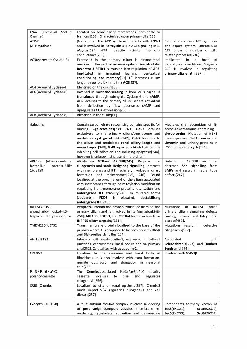
246
ENac (Epithelial Sodium Channel)
Located on some ciliary membranes, permeable to Na
+ ions[232]. Characterised upon primary cilia[233].
ATP-2 (ATP synthase)
β-subunit of the ATP synthase interacts with LOV-1 and is involved in Polycystin-1 (PKD-1) signalling in C. elegans[234]. ATP indirectly activates the cilia conductance[235].
Part of a complex ATP synthesis and export system. Extracellular ATP drives a number of cilia related processes[236].
AC3(Adenylate Cyclase-3) Expressed in the primary cilium in hippocampal neurons of the central nervous system. Somatostatin Receptor-3 SSTR3 is coupled into regulation of AC3. Implicated in impaired learning, contextual conditioning and memory[39]. Li
+ increases cilium
length three fold by inhibiting AC3[237].
Implicated in a host of neurological conditions. Suggests AC3 is involved in regulating primary cilia length[237].
AC4 (Adenylyl Cyclase-4) Identified on the cilium[66].
AC6 (Adenylyl Cyclase-6) Involved in mechano-sensing in bone cells. Signal is transduced through Adenylate Cyclase-6 and cAMP. AC6 localises to the primary cilium, where activation from deflection by flow decreases cAMP and upregulates COX expression[238].
AC8 (Adenylyl Cyclase-8) Identified in the cilium[66].
Galectins Contain carbohydrate recognising domains specific for binding β-galactosides[239, 240]. Gal-3 localises exclusively to the primary cilium/centrosome and modulates cyst growth[240-242], Gal-7 localises to the cilium and modulates renal ciliary length and wound repair[243], Gal8 reportedly binds to integrins inhibiting cell adhesion and inducing apoptosis[244], however is unknown at present in the cilium.
Mediates the recognition of N-acetyl-galactosamine-containing glycoproteins. Mutation of NEK8 over-expresses Gal-1, sorcin and vimentin and urinary proteins in JCK murine renal cysts[240].
ARL13B (ADP-ribosylation factor-like protein-2-like 1)/JBTS8
ARF-Family GTPase ARL13B[245]. Required for ciliogenesis and sonic Hedgehog signalling. Interacts with membranes and IFT machinery involved in ciliary formation and maintenance[245, 246]. Found localised at the proximal end of the cilium associated with membranes through palmitoylation modification regulating trans-membrane proteins localisation and anterograde IFT stability[245]. In mutated forms (Jouberts), PKD2 is elevated, destabilising anterograde IFT[243].
Defects in ARL13B result in aberrant Shh signalling from BMPs and result in neural tube defects[247].
INPP5E/JBTS1 phosphatidylinositol-4,5-bisphosphate5phosphatase
Peripheral membrane protein which localises to the primary cilium and is involved in its formation[248-250]. ARL13B, PDE6D, and CEP164 form a network for INPP5E ciliary targeting[251].
Mutations in INPP5E cause primary cilium signalling defects causing ciliary instability and disease[453].
TMEM216[/JBTS2 Trans-membrane protein localised to the base of the primary where it is proposed to be possibly with RhoA and Dishevelled signalling[117].
Mutations result in defective ciliogenesis[117].
AHI1 /JBTS3 Interacts with nephrocytin-1, expressed in cell-cell junctions, centrosomes, basal bodies and on primary cilia[252]. Colocalises with aquaporin-2.
Associated with Schizophrenia[253] and Joubert Syndrome[254].
CRMP-2 Localises to the axoneme and basal body in fibroblasts. It is also involved with axon formation, neurite outgrowth and elongation in neuronal cells[255].
Involved with GSK-3β.
Par3 / Par6 / aPKC polarity cassette
The Crumbs-associated Par3/Par6/aPKC polarity cassette localises to cilia and regulates ciliogenesis[256].
CRB3 (Crumbs) Localises to cilia of renal epithelia[257]. Crumbs3 binds importin-β2 regulating ciliogenesis and cell divison[257].
Exocyst (EXCO1-8) A multi-subunit rod-like complex involved in docking of post Golgi transport vesicles, membrane re-modelling, cytoskeletal activation and desmosome
Components formerly known as Sec3(EXCO1), Sec5(EXCO2), Sec6(EXCO3), Sec8(EXCO4),

247
assembly[258]. Localises to the primary cilium[16]. RAB10 interacts with the primary cilium and Exocyst[259] components; Sec15(as an effector of Rab11 GTPase[22]), Sec10 (which is vital for ciliogenesis[19]) and Sec8 (is involved in binding to lipid rafts and vesicles[260]). Sec5 is involved in vesicle transport and in tethering vesicles to membranes[261, 262]. Sec10 interacts with PC2[24].
Sec10(EXCO5), Sec15(EXCO6), Exo70(EXCO7) and Exo84(EXCO8). The RalB/Sec5 effector complex directly recruits and activates IkappaB-kinase family member TBK1 (innate immunity and cancer)[262]. See Septins.
Sonic Hedgehog Signalling The Hedgehog Signalling family comprises three members: Indian, Sonic and Desert. Shh are morphogens responsible for signalling and patterning development[263-265]. Dysregulation of Hh signalling is involved in a broad range of conditions and disease[113, 266]. Interference of Sonic Hedgehog (specifically GLI signalling[267]) causes inhibition of cancer cell proliferation and tumour formation[267, 268]. Interacts with IFT122[269] and inhibits p16[270].
Implicated in synovial chondromatosis[271]. The Shh pathway is a target for clinical treatment of various diseases[272].
Smo (Smoothened)
Essential G-protein-receptor involved with Sonic Hedgehog pathway[273, 272] located in the primary cilium[275-277]. The Smo tail is involved with cAMP dependent kinases, and is extensively phosphorylated[277]. Involved in vesicle transport[278] Loss of retrograde IFT disrupts localisation of Smo preventing activator and repressor of Gli function[279]. Patched acts indirectly to regulate Smo function[280], in which Patch (Ptc) and Smoothened are transduction components of the Hedgehog receptor complex[281], suggesting Patched binds Hh without assistance from Smo, favouring Hh/PTCH1 binding inducing conformation release of Smo[281]. Smo is the teratogenic target of cyclopamine[276, 282].
SuFu (Suppressor of Fused)
Smo activates Gli which is inhibited by dual binding mechanisms (SuFu)[283]. Hh stimulation releases SuFu in the cilium, since Hh dissociates the SuFu-Gli complex allowing Gli to target the nucleus[284, 285]. Protein-Kinase A (PKA) inhibits Shh signalling[284]. Interacts with Gli1[285], Gli3[286] and mediates the Gli3/SuFu/GSK3-β complex[286].
SuFu forms Gli-SuFu complexes within the cilium. Hh causes dissociation of permitting Gli to activate nuclear transcription pathways[284].
Patched/PTCH1 PTCH1 suppresses Hh signalling pathway by preventing trafficking of Smo into the cilium. On binding of the Hh ligand to PTCH1, Smo relocates to the cilium[276] where it is responsible for activating the Hh pathway[287]. Smoothened and Patched are found in lipid raft domains. Patched interacts with Caveolin, but Smoothened does not[288]. Localises to cilia inhibiting Smo accumulation[289].
The sterol-sensing domain of Patched may be involved in vesicular trafficking in Smoothened regulation[278].
Gli1 Gli is a Hedgehog Signalling pathway effector[290, 291]. Involved in regulating transcription, cell fate determination and extra cellular sensing. Interacts with SuFu[285, 292, 293] and Shh activation of Gli1 is controlled by Gli3[291]. KIF7 is a regulator of Gli transcription factors[294, 295].
Associated with cancers[296] and synovial chondromatosis[297].
Gli2 (α, β, γ and δ) Transcriptional mediators of Shh signalling. GLI2 activates cytoplasmic PTCH1[298, 299]. GLI2 is able to promote G1-S-phase progression[299], however it requires IFT component Polaris to function[298]. There are implications for Gli2 in cancer[300, 301]. Involved in neuronal patterning[302]. Localises to the primary cilium tip[298] being controlled by Broad Minded kinase BROMO[102]. Gli2 transcription factor binds and inhibits the p16 promoter[302].
Involved with polydactyly[303]. Gli1 and Gli2 are part of a positive-feedback mechanism in Basal Cell Carcinoma[304].

248
Gli3 Transcription repressor, full-length Gli3 localises at the tip of the cilium[305] and required for IFT Polaris to function[298]. Gli3 is an activator for Gli1[291, 306], and interacts with SuFu in Hedgehog Signalling[307].
Pallister-Hall Syndrome[308].
Wnt Signalling Wnt signalling factors are involved in cell-proliferation and differentiation. Wnt proteins interact with multiple receptors and pathways including the canonical Wnt/β-catenin pathway of transcription co-activator β-catenin. The non-canonical Wnt/planar cell polarity (PCP) pathway is involved with development[309-311].
Frizzled Family(FZD1-10) G-protein receptor family, trans-membrane domain proteins which act as receptors for Wnt signalling[ 230, 312]. Transmembrane recptors of FZD1-10, LRP5, LRP6 and ROR2 transduce WNT signals through endocytosis involving Caveolin or Clathrin[313, 314]. Wnt signals are involved in regulation of planar cell polarity, cell adhesion, and motility. CD44 and vimentin gene components of the non-Canonical WNT cascade[313]. These are part of, and interact with, Notch, FGF, BMP and Hedgehog signalling cascades[313, 314]. Frizzled activates cytoplasmic Disheveled[315].
Frizzled Family Members are expressed differentially in tissues and are being explored as platforms for tissue engineering[313].
Disheveled (Dvl) Regulates canonical and non-canonical Wnt-β-catenin and planar cell polarity pathways; involved with basal body polarization and apical docking[316, 317] Dvl and Inturned mediate activity of Rho-GTPase upon basal bodies directing docking to the apical membrane. Docking also involves membrane bound vesicles and the vesicle trafficking protein Sec8[318]. Interacts with Frizzled[315, 316].
Required for ciliogenesis
Chibby Wnt/β-Catenin Pathway antagonist Chibby binds Cenexin at the distal end of the basal body and functions in primary cilium formation[319]. Required for ciliogenesis and centriole formation in Drosophila but not WNT signalling[320].
INV Acts as a molecular anchor for NPHP3 and NEK8 in the proximal segment of the cilium[321].
Notch Signalling
Notch Notch receptors and processing enzymes are co-localised with cilia[238]. Notch1 and 2 modify endochondral ossification in chondrocytes[323] as well as chondrogenic differentiation[324]. Notch signalling regulates imposition of left-right-asymmetry through ciliary length control[325], and suppresses nuclear factor NFat in T-cells and Nfatc in chondrocytes[326].
Involved in osteoarthritis[322].
[1] Littlewood Evans A, Müller U. (2000) Stereocilia defects in the sensory hair cells of the inner ear
in mice deficient in integrin alpha8beta1. Nature Genetics, 24, 424-428.
[2] McGlashan SR, Jensen CG, Poole CA. (2006) Localisation of extracellular matrix receptors on the
chondrocyte primary cilium. Journal of Histochemistry and Cytochemistry, 54, 1005-1014.
[3] Praetorius HA, Praetorius J, Nielsen S, Frokiaer J, Spring KR. (2004) Beta1-integrins in the
primary cilium of MDCK cells potentiate fibronectin-induced Ca2+
signalling. American Journal of
Physiology, 287, F969-F978.
[4] Brauchi S, Orio P, Latorre R. (2004) Clues to understanding cold sensation: Thermodynamics and
electrophysiological analysis of the cold receptor TRPM8. Proceedings of the National Academy of
Sciences, USA, 101, 15494-15499.

249
[5] Kurtis MS, Schmidt TA, Bugbee WD, Loeser RF, Sah RL. (2003) Integrin-mediated adhesion of
human articular chondrocytes to cartilage. Arthritis and Rheumatology, 48, 110-118.
[6] Fukushi J, Makagiansar IT, Stallcup WB (2004) NG2 proteoglycan promotes endothelial cell
motility and angiogenesis via engagement of galectin-3 and alpha3beta1 integrin. Molecular Biology
of the Cell, 15, 3580-3590.
[7] Stegmüller J, Werner H, Nave KA, Trotter J (2003) The proteoglycan NG2 is complexed with
alpha-amino-3-hydroxy-5-methyl-4-isoxazolepropionic acid (AMPA) receptors by the PDZ glutamate
receptor interaction protein (GRIP) in glial progenitor cells. Implications for glial-neuronal signalling.
Journal of Biological Chemistry, 278, 3590-3598.
[8] Stallcup WB, Huang FJ. (2008) A role for the NG2 proteoglycan in glioma progression. Cell
Adhesion and Migration, 2, 192-201.
[9] Burg MA, Pasqualini R, Arap W, Ruoslahti E, Stallcup WB. (1999) NG2 proteoglycan-binding
peptides target tumour neovasculature. Cancer Research, 59, 2869-2874.
[10] Mak HY, Nelson LS, Basson M, Johnson CD, Ruvkun G. (2006) Polygenic control of
Caenorhabditis elegans fat storage. Nature Genetics, 38, 363-368.
[11] Mukhopadhyay A, Deplancke B, Walhout AJ, Tissenbaum HA. (2005) C. elegans tubby
regulates life span and fat storage by two independent mechanisms. Cell Metabolism, 2, 35-42.
[12] North MA, Naggert JK, Yan Y, Noben-Trauth K, Nishina PM. (1997) Molecular characterization
of TUB, TULP1, and TULP2, members of the novel tubby gene family and their possible relation to
ocular diseases. Proceedings of the National Academy of Sciences, USA, 94, 3128-3133.
[13] Hagstrom SA, Adamian M, Scimeca M, Pawlyk BS, Yue G, Li T. (2001) A role for the Tubby-
like protein 1 in rhodopsin transport. Investigative Ophthalmology and Visual Science, 42, 1955-1962.
[14] Mukhopadhyay S, Wen X, Chih B, Nelson CD, Lane WS, Scales SJ, Jackson PK. (2010) TULP3
bridges the IFT-A complex and membrane phosphoinositides to promote trafficking of G protein-
coupled receptors into primary cilia. Genes and Development, 24, 2180-2193.
[15] Xi Q, Pauer GJ, Marmorstein AD, Crabb JW, Hagstrom SA. (2005) Tubby-like protein 1
(TULP1) interacts with F-actin in photoreceptor cells. Investigative Ophthalmology and Visual
Science, 46, 4754-4761.
[16] Rogers KK, Wilson PD, Snyder RW, Zhang X, Guo W, Burrow CR, Lipschutz JH. (2004) The
exocyst localises to the primary cilium in MDCK cells. Biochemical and Biophysical Research
Communications, 319, 138-143.
[17] Overgaard CE, Sanzone KM, Spiczka KS, Sheff DR, Sandra A, Yeaman C. (2009) Deciliation is
associated with dramatic remodeling of epithelial cell junctions and surface domains. Molecular
Biology of the Cell, 20, 102-113.
[18] Zuo X, Fogelgren B, Lipschutz JH. (2011) The small GTPase Cdc42 is necessary for primary
ciliogenesis in renal tubular epithelial cells. Journal of Biological Chemistry, 286, 22469-22477.
[19] Zuo X, Guo W, Lipschutz JH. (2009) The exocyst protein Sec10 is necessary for primary
ciliogenesis and cystogenesis in vitro. Molecular Biology of the Cell, 20, 2522-2529.

250
[20] Westlake CJ, Baye LM, Nachury MV, Wright KJ, Ervin KE, Phu L, Chalouni C, Beck JS,
Kirkpatrick DS, Slusarski DC, Sheffield VC, Scheller RH, Jackson PK. (2011) Primary cilia
membrane assembly is initiated by Rab11 and transport protein particle II (TRAPPII) complex-
dependent trafficking of Rabin8 to the centrosome. Proceedings of the National Academy of Sciences,
USA, 108, 2759-2764.
[21] Knödler A, Feng S, Zhang J, Zhang X, Das A, Peränen J, Guo W. (2010) Coordination of Rab8
and Rab11 in primary ciliogenesis. Proceedings of the National Academy of Sciences, USA, 107,
6346-6351.
[22] Zhang XM, Ellis S, Sriratana A, Mitchell CA, Rowe T. (2004) Sec15 is an effector for the Rab11
GTPase in mammalian cells. Journal of Biological Chemistry, 279, 43027-43034.
[23] Choi SY, Chacon-Heszele MF, Huang L, McKenna S, Wilson FP, Zuo X, Lipschutz JH. (2013)
Cdc42 deficiency causes ciliary abnormalities and cystic kidneys. Journal of the American Society of
Nephrology, 24, 1435-1450.
[24] Fogelgren B, Lin SY, Zuo X, Jaffe KM, Park KM, Reichert RJ, Bell PD, Burdine RD, Lipschutz
JH. (2010) The exocyst protein Sec10 interacts with polycystin-2 and knockdown causes PKD-
phenotypes. PLoS Genetics, 7(4):e1001361.
[25] Tao B, Bu S, Yang Z, Siroky B, Kappes JC, Kispert A, Guay-Woodford LM. (2009) Cystin
localises to primary cilia via membrane microdomains and a targeting motif. Journal of the American
Society of Nephrology, 20, 2570-2580.
[26] Wu M, Yang C, Tao B, Bu S, Guay-Woodford LM. (2013) The ciliary protein Cystin forms a
regulatory complex with Necdin to modulate Myc expression. PLoS One, 8(12):e83062.
[27] Chae TH, Kim S, Marz KE, Hanson PI, Walsh CA. (2004) The hyh mutation uncovers roles for
alpha Snap in apical protein localisationi and control of neural cell fate. Nature Genetics, 36, 264-270.
[28] McMahon HT, Missler M, Li C, Südhof TC. (1995) Complexins: cytosolic proteins that regulate
SNAP receptor function. Cell, 83, 111-119.
[29] Teilmann SC, Christensen ST. (2005) Localization of the angiopoietin receptors Tie-1 and Tie-2
on the primary cilia in the female reproductive organs. Cell Biology International, 29, 340-346.
[30] Chan B, Yuan HT, Ananth Karumanchi S, Sukhatme VP. (2008) Receptor tyrosine kinase Tie-1
over expression in endothelial cells upregulates adhesion molecules. Biochemical and Biophysical
Research Communications, 371, 475-479.
[31] Woo KV, Qu X, Babaev VR, Linton MF, Guzman RJ, Fazio S, Baldwin HS. (2011) Tie1
attenuation reduces murine atherosclerosis in a dose-dependent and shear stress-specific manner.
Journal of Clinical Investigtaion, 121, 1624-1635.
[32] Staton CA, Valluru M, Hoh L, Reed MW, Brown NJ. (2010) Angiopoietin-1, angiopoietin-2 and
Tie-2 receptor expression in human dermal wound repair and scarring. British Journal of
Dermatology, 163, 920-927.
[33] Fasciani A, Quilici P, Biscaldi E, Flamini M, Fioravanti A, Orlandi P, Oliviero J, Repetti F,
Bandelloni R, Danesi R, Simoncini T, Bocci G (2010) Overexpression and functional relevance of
somatostatin receptor-1, -2, and -5 in endometrium and endometriotic lesions. Journal of Clinical
Endocrinology and Metabolism, 95, 5315-5319.

251
[34] Lai CK, Gupta N, Wen X, Rangell L, Chih B, Peterson AS, Bazan JF, Li L, Scales SJ. (2011)
Functional characterization of putative cilia genes by high-content analysis. Molecular Biology of the
Cell, 22, 1104-1119.
[35] Iwanaga T, Takashi Miki T, Takahashi-Iwanaga H. (2011) Restricted expression of somatostatin
receptor 3 to primary cilia in the pancreatic islets and adenohypophysis of mice. Biomedical
Research, 32, 73-81.
[36] Berbari NF, Johnson AD, Lewis JS, Askwith CC, Mykytyn K. (2008) Identification of ciliary
localisation sequences within the third intracellular loop of G protein-coupled receptors. Molecular
Biology of the Cell, 19, 1540-1547.
[37] Yoon SH, Ryu JY, Lee Y, Lee ZH, Kim HH. (2011) Adenylate cyclase and calmodulin-
dependent kinase have opposite effects on osteoclastogenesis by regulating the PKA-NFATc1
pathway. Journal of Bone and Mineral Research, 26, 1217-1229.
[38] Einstein EB, Patterson CA, Hon BJ, Regan KA, Reddi J, Melnikoff DE, Mateer MJ, Schulz S,
Johnson BN, Tallent MK. (2010) Somatostatin signalling in neuronal cilia is critical for object
recognition memory. Journal of Neuroscience, 30, 4306-4314.
[39] Wang Z, Phan T, Storm DR. (2011) The type 3 Adenylyl Cyclase is required for novel object
learning and extinction of contextual memory: role of cAMP signalling in primary cilia. Journal of
Neuroscience, 31, 5557-5561.
[40] Helboe L, Møller M. (1999) Immunohistochemical localisation of somatostatin receptor subtypes
sst1 and sst2 in the rat retina. Investigative Ophthalmology and Vision Science, 40, 2376-2382.
[41] Jin H, White SR, Shida T, Schulz S, Aguiar M, Gygi SP, Bazan JF, Nachury MV. (2010) The
conserved Bardet-Biedl syndrome proteins assemble a coat that traffics membrane proteins to cilia.
Cell, 141, 1208-1219.
[42] König P, Krain B, Krasteva G, Kummer W. (2009) Serotonin increases cilia-driven motion via an
acetylcholine-independent pathway in the mouse trachea. PLoS One, 4(3):e4938.
[43] Brailov I, Bancila M, Brisorgueil MJ, Miquel MC, Harmon M, Verge´ D. (2000) Localisation of
5-HT(6) receptors at the plasma membrane of neuronal cilia in the rat brain. Brain Research, 872,
271-275.
[44] Wu J, Du H, Wang X, Mei C, Sieck GC, Qian Q. (2009) Characterization of primary cilia in
human airway smooth muscle cells. Chest, 136, 561-570.
[45] Ma R, Li WP, Rundle D, Kong J, Akbarali HI, Tsiokas L. (2005) PKD2 functions as an
epidermal growth factor-activated plasma membrane channel. Molecular and Cellular Biology, 25,
8285-8298.
[46] Yeh C, Li A, Jen-Zen Chuang J, Saito M, Caceres A, Sung C. (2013) IGF-1 activates a cilium-
localized noncanonical Gbg signaling pathway that regulates cell-cycle progression. Developmental
Cell, 26, 358-368.
[47] Schneider L, Clement CA, Teilmann SC, Pazour GJ, Hoffmann EK, Satir P, Christensen ST.
(2005) PDGFRalphaalpha signalling is regulated through the primary cilium in fibroblasts. Current
Biology, 15, 1861-1866.

252
[48] Schneider L, Stock C, Dieterich P, Jensen BH, Pedersen B, Satir P, Schwab A, Christensen ST,
Pedersen SF. (2009) The Na+/H+ exchanger NHE1 is required for directional migration stimulated
via PDGFR-a in the primary cilium. Journal of Cell Biology Home Archive, 185, 163-176.
[49] Michaud EJ, Yoder BK. (2006) The primary cilium in cell signalling and cancer. Cancer
Research, 66, 6463-6467.
[50] Choi SJ, Marazita ML, Hart PS, Sulima PP, Field LL, McHenry TG, Govil M, Cooper ME, Letra
A, Menezes R, Narayanan S, Mansilla MA, Granjeiro JM, Vieira AR, Lidral AC, Murray JC, Hart
TC. (2009) The PDGF-C regulatory region SNP rs28999109 decreases promoter transcriptional
activity and is associated with CL/P. European Journal of Human Genetics, 17, 774-784.
[51] Gilbertson DG, Duff ME, West JW, Kelly JD, Sheppard PO, Hofstrand PD, Gao Z, Shoemaker
K, Bukowski TR, Moore M, Feldhaus AL, Humes JM, Palmer TE, Hart CE. (2001) Platelet-derived
growth factor C (PDGF-C), a novel growth factor that binds to PDGF alpha and beta receptor.
Journal of Biological Chemistry, 276, 27406-27414.
[52] Frémin C, Meloche S. (2010) From basic research to clinical development of MEK1/2 inhibitors
for cancer therapy. Journal of Hematological Oncology, 3, 8.
[53] Nagano J, Kitamura K, Hujer KM, Ward CJ, Bram RJ, Hopfer U, Tomita K, Huang C, Miller
RT. (2005) Fibrocystin interacts with CAML, a protein involved in Ca2+
signaling. Biochemical and
Biophysical Research Communications, 338, 880-889.
[54] Low SH, Vasanth S, Larson CH, Mukherjee S, Sharma N, Kinter MT, Kane ME, Obara T,
Weimbs T. (2006) Polycystin-1, STAT6, and P100 function in a pathway that transduces ciliary
mechanosensation and is activated in polycystic kidney disease. Developmental Cell, 10, 57-69.
[55] Paukku K, Yang J, Silvennoinen O. (2003) Tudor and nuclease-like domains containing protein
p100 function as coactivators for signal transducer and activator of transcription 5. Molecular
Endocrinology, 17, 1805-1814.
[56] Välineva T, Yang J, Palovuori R, Silvennoinen O. (2005) The transcriptional co-activator protein
p100 recruits histone acetyltransferase activity to STAT6 and mediates interaction between the
CREB-binding protein and STAT6. Journal of Biological Chemistry, 280, 14989-14996.
[57] Boehlke C, Kotsis F, Patel V, Braeg S, Voelker H, Bredt S, Beyer T, Janusch H, Hamann C,
Gödel M, Müller K, Herbst M, Hornung M, Doerken M, Köttgen M, Nitschke R, Igarashi P, Walz G,
Kuehn EW. (2010) Primary cilia regulate mTORC1 activity and cell size through Lkb1. Nature Cell
Biology, 12, 1115-1122.
[58] Abbracchio MP, Burnstock G, Boeynaems JM, Barnard EA, Boyer JL, Kennedy C, Knight GE,
Fumagalli M, Gachet C, Jacobson KA, Weisman GA. (2006) International Union of Pharmacology
LVIII: update on the P2Y G protein-coupled nucleotide receptors: from molecular mechanisms and
pathophysiology to therapy. Pharmacology Reviews, 58, 281-341.
[59] Gever JR, Cockayne DA, Dillon MP, Burnstock G, Ford AP. (2006) Pharmacology of P2X
channels. European Journal of Physiology, 452, 513-537.
[60] North RA. (2002) Molecular physiology of P2X receptors. Physiology Reviews, 82, 1013-1067.
[61] Egan TM, Samways DS, Li Z. (2006) Biophysics of P2X receptors. European Journal of
Physiology, 452, 501-512.

253
[62] Roberts JA, Vial C, Digby HR, Agboh KC, Wen H, Atterbury-Thomas A, Evans RJ. (2006)
Molecular properties of P2X receptors. European Journal of Physiology, 452, 486-500.
[63] Nicke A, Baumert HG, Rettinger J, Eichele A, Lambrecht G, Mutschler E, Schmalzing G. (1998)
P2X1 and P2X3 receptors form stable trimers: a novel structural motif of ligand-gated ion channels.
EMBO Journal, 17, 3016-3028.
[64] Praetorius HA, Leipziger J. (2009) Released nucleotides amplify the cilium-dependent, flow-
induced[Ca2+
] response in MDCK cells. Acta Physiologica, 197, 241-251.
[65] Leipziger J. (2011) Luminal nucleotides are tonic inhibitors of renal tubular transport. Current
Opinion in Nephrology and Hypertension, 20, 518-522.
[66] Masyuk AI, Gradilone SA, Banales JM, Huang BQ, Masyuk TV, Lee SO, Splinter PL, Stroope
AJ, Larusso NF. (2008) Cholangiocyte primary cilia are chemosensory organelles that detect biliary
nucleotides via P2Y12 purinergic receptors. American Journal of Physiology, 295, G725-G734.
[67] Morales B, Barrera N, Uribe P, Mora C, Villalón M. (2000) Functional cross talk after activation
of P2 and P1 receptors in oviductal ciliated cells. American Journal of Physiology, 279, C658-C669.
[68] Knight MM, McGlashan SR, Garcia M, Jensen CG, Poole CA. (2009) Articular chondrocytes
express connexin 43 hemichannels and P2 receptors - a putative mechanoreceptor complex involving
the primary cilium? Journal of Anatomy, 214, 275-283.
[69] Langlois S, Cowan KN, Shao Q, Cowan BJ, Laird DW. (2008) Caveolin-1 and -2 interact with
connexin43 and regulate gap junctional intercellular communication in keratinocytes. Molecular
Biology of the Cell, 19, 912-928.
[70] Cooper CD, Lampe PD. (2002) Casein kinase 1 regulates connexin-43 gap junction assembly.
Journal of Biological Chemistry, 277, 44962-44968.
[71] Rhett JM, Jourdan J, Gourdie RG. (2011) Connexin 43 connexon to gap junction transition is
regulated by zonula occludens-1. Molecular Biology of the Cell, 22, 1516-1528.
[72] Giepmans BN, Verlaan I, Moolenaar WH. (2001) Connexin-43 interactions with ZO-1 and
alpha- and beta-tubulin. Cell Communication and Adhesion, 8, 219-223.
[73] Kwak BR, Veillard N, Pelli G, Mulhaupt F, James RW, Chanson M, Mach F. (2003) Reduced
connexin43 expression inhibits atherosclerotic lesion formation in low-density lipoprotein receptor-
deficient mice. Circulation, 107, 1033-1039.
[74] Malone P, Miao H, Parker A, Juarez S, Hernandez MR. (2007) Pressure induces loss of gap
junction communication and redistribution of connexin 43 in astrocytes. Glia, 55, 1085-1098
[75] Deretic D, Traverso V, Parkins N, Jackson F, Rodriguez de Turco EB, Ransom N. (2004)
Phosphoinositides, ezrin/moesin, and rac1 regulate fusion of rhodopsin transport carriers in retinal
photoreceptors. Molecular Biology of the Cell, 15, 359-370.
[76] Mazelova J, Ransom N, Astuto-Gribble L, Wilson MC, Deretic D. (2009) Syntaxin 3 and SNAP-
25 pairing, regulated by omega-3 docosahexaenoic acid, controls the delivery of rhodopsin for the
biogenesis of cilia-derived sensory organelles, the rod outer segments. Journal of Cell Science, 122,
2003-2013.
[77] Wang J, Deretic D. (2013) Molecular complexes that direct rhodopsin transport to primary cilia.
Progress in Retinal and Eye Research, pii: S1350-9462(13)00060-8.

254
[78] Mahjoub MR, Qasim Rasi M, Quarmby LM. (2004) A NIMA-related kinase, Fa2p, localises to a
novel site in the proximal cilia of Chlamydomonas and mouse kidney cells. Molecular Biology of the
Cell, 15, 5172-5186.
[79] Quarmby LM, Parker JD. (2005) Cilia and the cell cycle? Journal of Cell Biology, 6, 707-710.
[80] Luttrell LM, Lefkowitz RJ. (2002) The role of beta-arrestins in the termination and transduction
of G-protein-coupled receptor signals. Journal of Cell Science, 115, 455-465.
[81] Molla-Herman A, Boularan C, Ghossoub R, Scott MG, Burtey A, Zarka M, Saunier S, Concordet
JP, Marullo S, Benmerah A. (2008) Targeting of beta-arrestin2 to the centrosome and primary cilium:
role in cell proliferation control. PLoS One, 3(11):e3728.
[82] Kovacs JJ, Whalen EJ, Liu R, Xiao K, Kim J, Chen M, Wang J, Chen W, Lefkowitz RJ. (2008)
Beta-arrestin-mediated localisation of smoothened to the primary cilium. Science, 320(5884), 1777-
1781.
[83] Parathath SR, Mainwaring LA, Fernandez-L A, Guldal CG, Nahlé Z, Kenney AM. (2010) β-
Arrestin-1 links mitogenic sonic Hedgehog signalling to the cell cycle exit machnery in neural
precursors. Cell Cycle, 9, 4013-4024.
[84] Lefkowitz RJ, Shenoy SK (2005) Transduction of receptor signals by beta-arrestins. Science, 308
(5721), 512-517.
[85] Zhang Y, Wada J, Yasuhara A, Iseda I, Eguchi J, Fukui K, Yang Q, Yamagata K, Hiesberger T,
Igarashi P, Zhang H, Wang H, Akagi S, Kanwar YS, Makino H. (2007) The role for HNF-1beta-
targeted collectrin in maintenance of primary cilia and cell polarity in collecting duct cells. PLoS One,
2(5):e414.
[86] Zhang, H, Wada, J, Hida, K, Tsuchiyama, Y, Hiragushi, K, Shikata, K, Wang, H, Lin, S,
Kanwar, Y.S, Makino, H. (2001) Collectrin, a collecting duct-specific transmembrane glycoprotein, is
a novel homolog of ACE2 and is developmentally regulated in embryonic kidneys. Journal of
Biological Chemistry, 276, 17132-17139.
[87] Hu Q, Nelson WJ. (2011) Ciliary diffusion barrier: the gatekeeper for the primary cilium
compartment. Cytoskeleton, 68, 313-324.
[88] Hu Q, Milenkovic L, Jin H, Scott MP, Nachury MV, Spiliotis ET, Nelson WJ. (2010) A septin
diffusion barrier at the base of the primary cilium maintains ciliary membrane protein distribution.
Science, 329, 436-439.
[89] Kim SK, Shindo A, Park TJ, Oh EC, Ghosh S, Gray RS, Lewis RA, Johnson CA, Attie-Bittach
T, Katsanis N, Wallingford JB. (2010) Planar cell polarity acts through septins to control collective
cell movement and ciliogenesis. Science, 329(5997), 1337-1340.
[90] Ghossoub R, Hu Q, Failler M, Rouyez MC, Spitzbarth B, Mostowy S, Wolfrum U, Saunier S,
Cossart P, Nelson WJ, Benmerah A. (2013) Septins 2, 7, and 9 and MAP4 co-localize along the
axoneme in the primary cilium and control ciliary length. Journal of Cell Science, 126, 2583-2594.
[91] Hsu SC, Hazuka CD, Roth R, Foletti DL, Heuser J, Scheller RH. (1998) Subunit composition,
protein interactions, and structures of the mammalian brain sec6/8 complex and septin filaments.
Neuron, 20, 1111-1122.

255
[92] Raychowdhury MK, Ramos AJ, Zhang P, McLaughin M, Dai XQ, Chen XZ, Montalbetti N, Del
Rocío Cantero M, Ausiello DA, Cantiello HF. (2009) Vasopressin receptor-mediated functional
signaling pathway in primary cilia of renal epithelial cells. American Journal of Physiology, 296, F87-
F97.
[93] Saigusa T, Bell PD, Kolb RJ. (2010) Primary cilium is required for vasopressin mediated
aquaporin-2 trafficking. FASEB Journal, 24, 1024.23.
[94] Marion V, Schlicht D, Mockel A, Caillard S, Imhoff O, Stoetzel C, van Dijk P, Brandt C, Moulin
B, Dollfus H. (2011) Bardet-Biedl syndrome highlights the major role of the primary cilium in
efficient water reabsorption. Kidney International, 79, 1013-1025.
[95] Nickols HH, Shah VN, Chazin WJ, Limbird LE. (2004) Calmodulin interacts with the V2
vasopressin receptor: elimination of binding to the C terminus also eliminates arginine vasopressin-
stimulated elevation of intracellular calcium. Journal of Biological Chemistry, 279, 46969-46980.
[96] Innamorati G, Whang MI, Molteni R, Le Gouill C, Birnbaumer M. (2002) GIP, a G-protein-
coupled receptor interacting protein. Regulatory Peptides, 109, 173-179.
[97] Raychowdhury MK, Ramos AJ, Zhang P, McLaughin M, Dai XQ, Chen XZ, Montalbetti N, Del
Rocío Cantero M, Ausiello DA, Cantiello HF. (2009) Vasopressin receptor-mediated functional
signalling pathway in primary cilia of renal epithelial cells. American Journal of Physiology, 296,
F87-F97.
[98] Knafo A, Israel S, Darvasi A, Bachner-Melman R, Uzefovsky F, Cohen L, Feldman E, Lerer E,
Laiba E, Raz Y, Nemanov L, Gritsenko I, Dina C, Agam G, Dean B, Bornstein G, Ebstein RP. (2008)
Individual differences in allocation of funds in the dictator game associated with length of the arginine
vasopressin 1a receptor RS3 promoter region and correlation between RS3 length and hippocampal
mRNA. Genes, Brain and Behavior, 7, 266-275.
[99] Knafo A, Israel S, Darvasi A, Bachner-Melman R, Uzefovsky F, Cohen L, Feldman E, Lerer E,
Laiba E, Raz Y, Nemanov L, Gritsenko I, Dina C, Agam G, Dean B, Bornstein G, Ebstein RP. (2008)
Individual differences in allocation of funds in the dictator game associated with length of the arginine
vasopressin 1a receptor RS3 promoter region and correlation between RS3 length and hippocampal
mRNA. Genes, Brain and Behavior, 7, 266-275.
[100] Vincent JL, Su F. (2008) Physiology and pathophysiology of the vasopressinergic system. Best
Practice and Research Clinical Anaesthesiology, 22, 243-252.
[101] Kaplan OI, Molla-Herman A, Cevik S, Ghossoub R, Kida K, Kimura Y, Jenkins P, Martens JR,
Setou M, Benmerah A, Blacque OE. (2010) The AP-1 clathrin adaptor facilitates cilium formation
and functions with RAB-8 in C. elegans ciliary membrane transport. Journal of Cell Science, 123,
3966-3977.
[102] Ko HW, Norman RX, Tran J, Fuller KP, Fukuda M, Eggenschwiler JT. (2010) Broad-minded
links cell cycle-related kinase to cilia assembly and Hedgehog signal transduction. Developmental
Cell, 18, 237-247.
[103] Follit JA, Li L, Vucica Y, Pazour GJ. (2010) The cytoplasmic tail of fibrocystin contains a
ciliary targeting sequence. Journal of Cell Biology, 188, 21-28.
[104] Wang S, Luo Y, Wilson PD, Witman GB, Zhou J. (2004) The autosomal recessive polycystic
kidney disease protein is localized to primary cilia, with concentration in the basal body area. Journal
of the American Society of Nephrology, 15, 592-602.

256
[105] Zhang M, Mai W, Li C, Cho S, Hao C, Moeckel G, Zhao R, Kim I, Wang J, Xiong H, Wang H,
Sato Y, Wu Y, Nakanuma Y, Lilova M, Pei Y, Harris R, Li S, Coffey R, Sun L, Wu D, Chen X,
Breyer M, Zhao Z, McKanna J, Wu G. (2004) PKHD1 protein encoded by the gene for autosomal
recessive polycystic kidney disease associates with basal bodies and primary cilia in renal epithelial
cells. Proceedings of the National Academy of Sciences, USA, 101, 2311-2316.
[106] Kaimori JY, Nagasawa Y, Menezes LF, Garcia-Gonzalez MA, Deng J, Imai, E, Onuchic LF,
Guay-Woodford, LM, Germino GG. (2007) Polyductin undergoes notch-like processing and regulated
release from primary cilia. Human Molecular Genetics, 16, 942-956.
[107] Wang S, Zhang J, Nauli SM, Li X, Starremans PG, Luo Y, Roberts KA, Zhou J. (2007)
Fibrocystin/polyductin, found in the same protein complex with polycystin-2, regulates calcium
responses in kidney epithelia. Molecular Cell Biology, 27, 3241-3252.
[108] Williams CL, Li C, Kida K, Inglis PN, Mohan S, Semenec L, Bialas NJ, Stupay RM, Chen N,
Blacque OE, Yoder BK, Leroux MR. (2011) MKS and NPHP modules cooperate to establish basal
body/transition zone membrane associations and ciliary gate function during ciliogenesis. Journal of
Cell Biology, 192, 1023-1041.
[109] Leightner AC, Hommerding CJ, Peng Y, Salisbury JL, Gainullin VG, Czarnecki PG, Sussman
CR, Harris PC. (2013) The Meckel syndrome protein meckelin (TMEM67) is a key regulator of cilia
function but is not required for tissue planar polarity. Human Molecular Genetics, 22, 2024-2040.
[110] Dawe HR, Smith UM, Cullinane AR, Gerrelli D, Cox P, Badano JL, Blair-Reid S, Sriram N,
Katsanis N, Attie-Bitach T, Afford SC, Copp AJ, Kelly DA, Gull K, Johnson CA. (2006) The
Meckel-Gruber Syndrome proteins MKS1 and meckelin interact and are required for primary cilium
formation. Human Molecular Genetics, 16, 173-186.
[111] Tammachote R, Hommerding CJ, Sinders RM, Miller CA, Czarnecki PG, Leightner AC,
Salisbury JL, Ward CJ, Torres VE, Gattone VH, Harris PC. (2009) Ciliary and centrosomal defects
associated with mutation and depletion of the Meckel syndrome genes MKS1 and MKS3. Human
Molecular Genetics, 18, 3311-3323.
[112] Dawe HR, Adams M, Wheway G, Szymanska K, Logan CV, Noegel AA, Gull K, Johnson CA.
(2009) Nesprin-2 interacts with meckelin and mediates ciliogenesis via remodelling of the actin
cytoskeleton. Journal of Cell Science, 122, 2716-2726
[113] Weatherbee SD, Niswander LA, Anderson KV (2009) A mouse model for Meckel syndrome
reveals Mks1 is required for ciliogenesis and Hedgehog signalling. Human Molecular Genetics, 18,
4565-4575.
[114] Kyttala M, Tallila J, Salonen R, Kopra O, Kohlschmidt N, Paavola-Sakki P, Peltonen L, Kestila
M. (2006) MKS1, encoding a component of the flagellar apparatus basal body proteome, is mutated in
Meckel syndrome. Nature Genetics, 38, 155-157.
[115] Leitch CC, Zaghloul NA, Davis EE, Stoetzel C, Diaz-Font A, Rix S, Alfadhel M, Al-Fadhel M,
Lewis RA, Eyaid W, Banin E, Dollfus H, Beales PL, Badano JL, Katsanis N. (2008) Hypomorphic
mutations in syndromic encephalocele genes are associated with Bardet-Biedl syndrome. Nature
Genetics, 40, 443-438.
[116] Wheway G, Abdelhamed Z, Natarajan S, Toomes C, Inglehearn C, Johnson CA. (2013)
Aberrant Wnt signalling and cellular over-proliferation in a novel mouse model of Meckel-Gruber
syndrome. Developmental Biology, 377, 55-66.

257
[117] Valente EM, Logan CV, Mougou-Zerelli S, Lee JH, Silhavy JL, Brancati F, Iannicelli M,
Travaglini L, Romani S, Illi B, Adams M, Szymanska K, Mazzotta A, Lee JE, Tolentino JC, Swistun
D, Salpietro CD, Fede C, Gabriel S, Russ C, Cibulskis K, Sougnez C, Hildebrandt F, Otto EA, Held
S, Diplas BH, Davis EE, Mikula M, Strom CM, Ben-Zeev B, Lev D, Sagie TL, Michelson M, Yaron
Y, Krause A, Boltshauser E, Elkhartoufi N, Roume J, Shalev S, Munnich A, Saunier S, Inglehearn C,
Saad A, Alkindy A, Thomas S, Vekemans M, Dallapiccola B, Katsanis N, Johnson CA, Attié-Bitach
T, Gleeson JG. (2010) Mutations in TMEM216 perturb ciliogenesis and cause Joubert, Meckel and
related syndromes. Nature Genetics, 42, 619-625.
[118] Hoover AN, Wynkoop A, Zeng H, Jia J, Niswander LA, Liu A. (2008) C2cd3 is required for
cilia formation and Hedgehog signalling in mouse. Development, 135, 4049-4058.
[119] Wang A. (2010) TMEM216 joins its ciliary cousins in ciliopathies. Clinical Genetics, 79, 45-
47.
[120] Williams CL, Masyukova SV, Yoder BK. (2010) Normal ciliogenesis requires synergy between
the cystic kidney disease genes MKS-3 and NPHP-4. Journal of the American Society of Nephrology,
21, 782-793.
[121] Wang M, Bridges JP, Na CL, Xu Y, Weaver TE. (2009) Meckel-Gruber syndrome protein
MKS3 is required for endoplasmic reticulum-associated degradation of surfactant protein C. Journal
of Biological Chemistry, 284, 33377-33383.
[122] De Mori R, Illi B, Romani S, Valente S, Johnson CA, Mai A, Valente EM. (2012) The ciliary
protein Meckelin/TMEM67 interacts with HDAC6: possible implications for primary cilia stability.
Cilia, 1, 20.
[123] Craige B, Tsao CC, Diener DR, Hou Y, Lechtreck KF, Rosenbaum JL, Witman GB. (2010)
CEP290 tethers flagellar transition zone microtubules to the membrane and regulates flagellar protein
content. Journal of Cell Biology, 190, 927-940.
[124] Rachel RA, Li T, Swaroop A. (2012) Photoreceptor sensory cilia and ciliopathies: focus on
CEP290, RPGR and their interacting proteins. Cilia, 1, 22.
[125] Chang B, Khanna H, Hawes N, Jimeno D, He S, Lillo C, Parapuram SK, Cheng H, Scott A,
Hurd RE, Sayer JA, Otto EA, Attanasio M, O'Toole JF, Jin G, Shou C, Hildebrandt F, Williams DS,
Heckenlively JR, Swaroop A. (2006) In-frame deletion in a novel centrosomal/ciliary protein
CEP290/NPHP6 perturbs its interaction with RPGR and results in early-onset retinal degeneration in
the rd16 mouse. Human Molecular Genetics, 15, 1847-1857.
[126] Sayer JA, Otto EA, O'Toole JF, Nurnberg G, Kennedy MA, Becker C, Hennies HC, Helou J,
Attanasio M, Fausett BV, Utsch B, Khanna H, Liu Y, Drummond I, Kawakami I, Kusakabe T, Tsuda
M, Ma L, Lee H, Larson RG, Allen SJ, Wilkinson CJ, Nigg EA, Shou C, Lillo C, Williams DS,
Hoppe B, Kemper MJ, Neuhaus T, Parisi MA, Glass IA, Petry M, Kispert A, Gloy J, Ganner A, Walz
G, Zhu X, Goldman D, Nurnberg P, Swaroop A, Leroux MR, Hildebrandt F. (2006) The centrosomal
protein nephrocystin-6 is mutated in Joubert syndrome and activates transcription factor ATF4.
Nature Genetics, 38, 674-681.
[127] Kim J, Krishnaswami SR, Gleeson JG. (2008) CEP290 interacts with the centriolar satellite
component PCM-1 and is required for Rab8 localisation to the primary cilium. Human Molecular
Genetics, 17, 3796-3805.
[128] Schäfer T, Pütz M, Lienkamp S, Ganner A, Bergbreiter A, Ramachandran H, Gieloff V, Gerner
M, Mattonet C, Czarnecki PG, Sayer JA, Otto EA, Hildebrandt F, Kramer-Zucker A, Walz G. (2008)
Genetic and physical interaction between the NPHP5 and NPHP6 gene products. Human Molecular
Genetics, 17, 3655-3662.

258
[129] Cideciyan AV, Rachel RA, Aleman TS, Swider M, Schwartz SB, Sumaroka A, Roman AJ,
Stone EM, Jacobson SG, Swaroop A. (2011) Cone photoreceptors are the main targets for gene
therapy of NPHP5 (IQCB1) or NPHP6 (CEP290) blindness: generation of an all-cone Nphp6
hypomorph mouse that mimics the human retinal ciliopathy. Human Molecular Genetics, 20, 1411-
1423.
[130] McEwen DP, Koenekoop RK, Khanna H, Jenkins PM, Lopez I, Swaroop A, Martens JR. (2007)
Hypomorphic CEP290/NPHP6 mutations result in anosmia caused by the selective loss of G proteins
in cilia of olfactory sensory neurons. Proceedings of the National Academy of Sciences, USA, 104,
15917-15922.
[131] Delous M, Baala L, Salomon R, Laclef C, Vierkotten J, Tory K, Golzio C, Lacoste T, Besse L,
Ozilou C, Moutkine I, Hellman NE, Anselme I, Silbermann F, Vesque C, Gerhardt C, Rattenberry E,
Wolf MT, Gubler MC, Martinovic J, Encha-Razavi F, Boddaert N, Gonzales M, Macher MA, Nivet
H, Champion G, Berthélémé JP, Niaudet P, McDonald F, Hildebrandt F, Johnson CA, Vekemans M,
Antignac C, Rüther U, Schneider-Maunoury S, Attié-Bitach T, Saunier S. (2007) The ciliary gene
RPGRIP1L is mutated in cerebello-oculo-renal syndrome (Joubert syndrome type B) and Meckel
syndrome. Nature Genetics, 39, 875-881.
[132] Arts HH, Doherty D, van Beersum SE, Parisi MA, Letteboer SJ, Gorden NT, Peters TA, Märker
T, Voesenek K, Kartono A, Ozyurek H, Farin FM, Kroes HY, Wolfrum U, Brunner HG, Cremers FP,
Glass IA, Knoers NV, Roepman R. (2007) Mutations in the gene encoding the basal body protein
RPGRIP1L, a nephrocystin-4 interactor, cause Joubert syndrome. Nature Genetics, 39, 882-888.
[133] Bachmann-Gagescu R, Phelps IG, Stearns G, Link BA, Brockerhoff SE, Moens CB, Doherty D.
(2011) The ciliopathy gene cc2d2a controls zebrafish photoreceptor outer segment development
through a role in Rab8-dependent vesicle trafficking. Human Molecular Genetics, 20, 4041-4055.
[134] Raychowdhury MK, McLaughlin M, Ramos AJ, Montalbetti N, Bouley R, Ausiello DA,
Cantiello HF. (2005) Characterization of single channel currents from primary cilia of renal epithelial
cells. Journal of Biological Chemistry, 280, 34718-34722.
[135] Yoder BK, Hou X, Guay-Woodford LM. (2002) The polycystic kidney disease proteins,
polycystin-1, polycystin-2, polaris, and cystin, are colocalized in renal cilia. Journal of the American
Society of Nephrology, 13, 2508-2516.
[136] Zhou J. (2009) Polycystins and primary cilia: primers for cell cycle progression. Annual
Reviews of Physiology, 71, 83-113.
[137] Sharif-Naeini R, Folgering JH, Bichet D, Duprat F, Lauritzen I, Arhatte M, Jodar M, Dedman
A, Chatelain FC, Schulte U, Retailleau K, Loufrani L, Patel A, Sachs F, Delmas P, Peters DJ, Honoré
E. (2009) Polycystin-1 and -2 dosage regulates pressure sensing. Cell, 139, 587-596.
[138] Woudenberg-Vrenken TE, Bindels RJ, Hoenderop JG. (2009) The role of transient receptor
potential channels in kidney disease. Nature Reviews Nephrology, 5, 441-449.
[139] Xiao Z, Zhang S, Magenheimer BS, Luo J, Quarles LD. (2008) Polycystin-1 regulates
skeletogenesis through stimulation of the osteoblast-specific transcription factor RUNX2-II. Journal
of Biological Chemistry, 283, 12624-12634.
[140] Sandford RN. (2009) The diversity of PKD1 alleles: implications for disease pathogenesis and
genetic counseling. Kidney International, 75, 765-767.

259
[141] Barr MM, DeModena J, Braun D, Nguyen CQ, Hall DH, Sternberg PW. (2001) The
Caenorhabditis elegans autosomal dominant polycystic kidney disease gene homologs lov-1 and pkd-
2 act in the same pathway. Current Biology, 11, 1341-1346.
[142] Pazour GJ, San Agustin JT, Follit JA, Rosenbaum JL, Witman GB. (2002) Polycystin-2
localises to kidney cilia and the ciliary level is elevated in orpk mice with polycystic kidney disease.
Current Biology, 12, R378-R380.
[143] Tsiokas L, Arnould T, Zhu C, Kim E, Walz G, Sukhatme VP. (1999) Specific association of the
gene product of PKD2 with the TRPC1 channel. Proceedings of the National Academy of Sciences,
USA, 96, 3934-3939.
144] Bai CX, Giamarchi A, Rodat-Despoix L, Padilla F, Downs T, Tsiokas L, Delmas P. (2008)
Formation of a new receptor-operated channel by heteromeric assembly of TRPP2 and TRPC1
subunits. EMBO Reports, 9, 472-479.
[145] Nilius B. (2009) Polycystins under pressure. Cell, 139, 466-467.
[146] Köttgen M, Buchholz B, Garcia-Gonzalez MA, Kotsis F, Fu X, Doerken M, Boehlke C, Steffl
D, Tauber R, Wegierski T, Nitschke R, Suzuki M, Kramer-Zucker A, Germino GG, Watnick T,
Prenen J, Nilius B, Kuehn EW, Walz G. (2008) TRPP2 and TRPV4 form a polymodal sensory
channel complex. Journal of Cell Biology, 182, 437-447.
[147] Qian F, Noben-Trauth K. (2005) Cellular and molecular function of mucolipins (TRPML) and
polycystin 2 (TRPP2). European Journal of Physiology, 451, 277-285.
[148] Kim D, Baraniuk JN. (2007) Sensing the air around us: the voltage-gated-like ion channel
family. Current Allergy and Asthma Reports, 2, 85-92.
[149] Nilius B, Owsianik G, Voets T, Peters JA. (2007) Transient receptor potential cation channels in
disease. Physiological Reviews, 87, 165-217.
[150] Wang Q, Dai X, Li Q, Wang Z, Cantero M, Li S, Shen J, Tu J, Cantiello H, Chen X. (2012)
Structural interaction and functional regulation of polycystin-2 by filamin. PLoS One, 7(7):e40448.
[151] Ishimaru Y, Inada H, Kubota M, Zhuang H, Tominaga M, and Matsunami H (2006) Transient
receptor potential family members PKD1L3 and PKD2L1 form a candidate sour taste receptor.
Proceedings of the National Academy of Sciences, USA, 103, 12569-12574.
[152] Dai XQ, Ramji A, Liu Y, Li Q, Karpinski E, Chen XZ. (2007) Inhibition of TRPP3 channel by
amiloride and analogs. Molecular Pharmacology, 72, 1576-1585.
[153] Hofmann T, Schaefer M, Schultz G, Gudermann T. (2002) Subunit composition of mammalian
transient receptor potential channels in living cells. Proceedings of the National Academy of Sciences,
USA, 99, 7461-7466.
[154] Tsiokas L, Arnould T, Zhu C, Kim E, Walz G, Sukhatme VP. (1999) Specific association of the
gene product of PKD2 with the TRPC1 channel. Proceedings of the National Academy of Sciences,
USA, 96, 3934-3939.
[155] Kobori T, Smith GD, Sandford R, Edwardson JM. (2009) The transient receptor potential
channels TRPP2 and TRPC1 form a heterotetramer with a 2:2 stoichiometry and an alternating
subunit arrangement. Journal of Biological Chemistry, 284, 35507-35513.

260
[156] Cheung KK, Yeung SS, Au SW, Lam LS, Dai ZQ, Li YH, Yeung EW. (2011) Expression and
association of TRPC1 with TRPC3 during skeletal myogenesis. Muscle and Nerve, 44, 358-365.
[157] Mehta D, Ahmmed GU, Paria BC, Holinstat M, Voyno-Yasenetskaya T, Tiruppathi C, Minshall
RD, Malik AB. (2003) RhoA interaction with inositol 1,4,5-trisphosphate receptor and transient
receptor potential channel-1 regulates Ca2+ entry. Role in signalling increased endothelial
permeability. Journal of Biological Chemistry, 278, 33492-33500.
[158] Tu JC, Xiao B, Yuan JP, Lanahan AA, Leoffert K, Li M, Linden DJ, Worley PF (1998) Homer
binds a novel proline-rich motif and links group 1 metabotropic glutamate receptors with IP3
receptors. Neuron, 21, 717-726.
[159] Clapham DE. (2007) SnapShot: mammalian TRP channels. Cell, 129, 220.
[160] Chu X, Tong Q, Cheung JY, Wozney J, Conrad K, Mazack V, Zhang W, Stahl R, Barber DL,
Miller BA. (2004) Interaction of TRPC2 and TRPC6 in erythropoietin modulation of calcium influx.
Journal of Biological Chemistry, 279, 10514-10522.
[161] Sutton KA, Jungnickel MK, Wang Y, Cullen K, Lambert S, Florman HM (2004) Enkurin is a
novel calmodulin and TRPC channel binding protein in sperm. Developmental Biology, 274, 426-435.
[162] Mast TG, Brann JH, Fadool DA. (2010) The TRPC2 channel forms protein-protein interactions
with Homer and RTP in the rat vomeronasal organ. BMC Neuroscience, 11, 61.
[163] Alexander SPH, Mathie A, Peters JA. (2007) Guide to Receptors and Channels (GRAC), 2nd
edn, British Journal ofPharmacology, 150 (Suppl. 1), S1-S168.
[164] Hirschler-Laszkiewicz I, Tong Q, Conrad K, Zhang W, Flint WW, Barber AJ, Barber DL,
Cheung JY, Miller BA. (2009) TRPC3 activation by erythropoietin is modulated by TRPC6. Journal
of Biological Chemistry, 284, 4567-4581.
[165] Tong Q, Hirschler-Laszkiewicz I, Zhang W, Conrad K, Neagley DW, Barber DL, Cheung JY,
Miller BA. (2008) TRPC3 is the erythropoietin-regulated calcium channel in human erythroid cells.
Journal of Biological Chemistry, 283, 10385-10395.
[166] Ramsey IS, Delling M, Clapham DE. (2006) An introduction to TRP channels. Annual Review
of Physiology, 68, 619-647.
[167] Kwan HY, Huang Y, Yao X. (2006) Protein kinase C can inhibit TRPC3 channels indirectly via
stimulating protein kinase G. Journal of Cell Physiology, 207, 315-321.
[168] Quick K, Zhao J, Eijkelkamp N, Linley JE, Rugiero F, Cox JJ, Raouf R, Gringhuis M, Sexton
JE, Abramowitz J, Taylor R, Forge A, Ashmore J, Kirkwood N, Kros CJ, Richardson GP, Freichel M,
Flockerzi V, Birnbaumer L, Wood JN. (2012) TRPC3 and TRPC6 are essential for normal
mechanotransduction in subsets of sensory neurons and cochlear hair cells. Open Biology, 2, 120068.
[169] Chen Y, Hu C, Hsu CK, Zhang Q, Bi C, Asnicar M, Hsiung HM, Fox N, Slieker LJ, Yang DD,
Heiman ML, Shi Y. (2002) Targeted disruption of the melanin-concentrating hormone receptor-1
results in hyperphagia and resistance to diet-induced obesity. Endocrinology, 143, 2469-2477.
[170] Shimada M, Tritos NA, Lowell BB, Flier JS, Maratos-Flier E. (1998) Mice lacking melanin-
concentrating hormone are hypophagic and lean. Nature, 396, 670-674.
[171] Bellone RR, Brooks SA, Sandmeyer L, Murphy BA, Forsyth G, Archer S, Bailey E, Grahn B.
(2008) Differential gene expression of TRPM1, the potential cause of congenital stationary night

261
blindness and coat spotting patterns (LP) in the Appaloosa horse (Equus caballus). Genetics, 179,
1861-1870.
[172] Morgans CW, Zhang J, Jeffrey BG, Nelson SM, Burke NS, Duvoisin RM, Brown RL. (2009)
TRPM1 is required for the depolarizing light response in retinal ON-bipolar cells. Proceedings of the
National Academy of Sciences, USA, 106, 19174-19178.
[173] Uchida K, Tominaga M. (2011) TRPM2 modulates insulin secretion in pancreatic β-cells. Islets,
3, 209-211.
[174] Xu C, Macciardi F, Li PP, Yoon IS, Cooke RG, Hughes B, Parikh SV, McIntyre RS, Kennedy
JL, Warsh JJ. (2006) Association of the putative susceptibility gene, transient receptor potential
protein melastatin type 2, with bipolar disorder. American Journal of Medical Genetics Part B, 141,
36-43.
[175] Miller BA. (2006) The role of TRP channels in oxidative stress-induced cell death. Journal of
Membrane Biology, 209, 31-41.
[176] Yamamoto S, Takahashi N, Mori Y. (2010) Chemical physiology of oxidative stress-activated
TRPM2 and TRPC5 channels. Progress in Biophysics and Molecular Biology, 103, 18-27.
[177] Thiel G, Müller I, Rössler OG. (2013) Signal transduction via TRPM3 channels in pancreatic β-
cells. Journal of Molecular Endocrinology, 50, R75-R83.
[178] Mayer SI, Müller I, Mannebach S, Endo T, Thiel G. (2011) Signal transduction of pregnenolone
sulfate in insulinoma cells: activation of Egr-1 expression involving TRPM3, voltage-gated calcium
channels, ERK, and ternary complex factors. Journal of Biological Chemistry, 286, 10084-10096.
[179] Harteneck C, Schultz G. (2007) TRPV4 and TRPM3 as volume-regulated cation channels.
Frontiers in Neuroscience, Liedtke WB, Heller S, ed,. TRP Ion Channel Function in Sensory
Transduction and Cellular Signalling Cascades. CRC Press, Boca Raton.
[180] Vennekens R, Nilius B. (2007) Insights into TRPM4 function, regulation and physiological role.
Handbook of Experimental Pharmacology, 179, 269-285.
[181] Vennekens R, Olausson J, Meissner M, Bloch W, Mathar I, Philipp SE, Schmitz F, Weissgerber
P, Nilius B, Flockerzi V, Freichel M (2007) Increased IgE-dependent mast cell activation and
anaphylactic responses in mice lacking the calcium-activated nonselective cation channel TRPM4.
Nature Immunology, 8, 312-320.
[182] Shimizu T, Owsianik G, Freichel M, Flockerzi V, Nilius B, Vennekens R. (2009) TRPM4
regulates migration of mast cells in mice. Cell Calcium, 45, 226-232.
[183] Wu LJ, Sweet TB, Clapham DE. (2010) International Union of Basic and Clinical
Pharmacology. LXXVI. Current progress in the mammalian TRP ion channel family. Pharmacology
Reviews, 62, 381-404.
[184] Harteneck C, Reiter B. (2007) TRP channels activated by extracellular hypo-osmoticity in
epithelia. Biochemical Society Transactions, 35, 91-95.
[185] Liu D, Liman ER. (2003) Intracellular Ca2+ and the phospholipid PIP2 regulate the taste
transduction ion channel TRPM5. Proceedings of the National Academy of Sciences, USA, 100,
15160-15165.

262
[186] Nakashimo Y, Takumida M, Fukuiri T, Anniko M, Hirakawa K. (2010) Expression of transient
receptor potential channel vanilloid (TRPV1-4), melastin (TRPM) 5 and 8, and ankyrin (TRPA1) in
the normal and methimazole-treated mouse olfactory epithelium. Acta Otolaryngologia, 130, 1278-
1286.
[187] Mitrovic S, Nogueira C, Cantero-Recasens G, Kiefer K, Fernández-Fernández JM, Popoff JF,
Casano L, Bard FA, Gomez R, Valverde MA, Malhotra V. (2013) TRPM5-mediated calcium uptake
regulates mucin secretion from human colon goblet cells. Elife, 2:e00658.
[188] Chubanov V, Gudermann T, Schlingmann KP (2006) Essential role for TRPM6 in epithelial
magnesium transport and body magnesium homeostasis. European Journal of Physiology, 451, 228-
234.
[189] Li M, Du J, Jiang J, Ratzan W, Su LT, Runnels LW, Yue L. (2007) Molecular determinants of
Mg2+
and Ca2+
permeability and pH sensitivity in TRPM6 and TRPM7. Journal of Biological
Chemistry, 282, 25817-25830.
[190] Xie J, Sun B, Du J, Yang W, Chen HC, Overton JD, Runnels LW, Yue L. (2011)
Phosphatidylinositol 4,5-bisphosphate (PIP(2)) controls magnesium gatekeeper TRPM6 activity.
Science Reports, 1, 146.
[191] Perraud AL, Zhao X, Ryazanov AG, Schmitz C. (2011) The channel-kinase TRPM7 regulates
phosphorylation of the translational factor eEF2 via eEF2-k. Cell Signalling, 23, 586-593.
[192] Yogi A, Callera GE, Antunes TT, Tostes RC, Touyz RM. (2010) Transient receptor potential
melastatin 7 (TRPM7) cation channels, magnesium and the vascular system in hypertension.
Circulation Journal, 75, 237-245.
[193] Harrington AM, Hughes PA, Martin CM, Yang J, Castro J, Isaacs NJ, Blackshaw LA, Brierley
SM. (2011) A novel role for TRPM8 in visceral afferent function. Pain, 152, 1459-1468.
[194] van Aken AF, Atiba-Davies M, Marcotti W, Goodyear RJ, Bryant JE, Richardson GP, Noben-
Trauth K, Kros CJ. (2008) TRPML3 mutations cause impaired mechano-electrical transduction and
depolarization by an inward-rectifier cation current in auditory hair cells of varitint-waddler mice.
Journal of Physiology, 586, 5403-5418.
[195] HKim HJ, Li Q, Tjon-Kon-Sang S, So I, Kiselyov K, Soyombo AA, Muallem S. (2008) A novel
mode of TRPML3 regulation by extracytosolic pH absent in the varitint-waddler phenotype. EMBO
Journal, 27, 1197-1205.
[196] Gavva NR, Bannon AW, Surapaneni S, Hovland DN, Lehto SG, Gore A, Juan T, Deng H, Han
B, Klionsky L, Kuang R, Le A, Tamir R, Wang J, Youngblood B, Zhu D, Norman MH, Magal E,
Treanor JJ, Louis JC. (2007) The vanilloid receptor TRPV1 is tonically activated in vivo and involved
in body temperature regulation. Journal of Neuroscience, 27, 3366-3374.
[197] Rosenbaum T, Gordon-Shaag A, Munari M, Gordon SE. (2004) Ca2+
/calmodulin modulates
TRPV1 activation by capsaicin. Journal of General Physiology, 123, 53-62.
[198] Goswami C, Hucho TB, Hucho F. (2007) Identification and characterisation of novel tubulin-
binding motifs located within the C-terminus of TRPV1. Journal of Neurochemistry, 101, 250-262.
[199] Steiner AA, Turek VF, Almeida MC, Burmeister JJ, Oliveira DL, Roberts JL, Bannon AW,
Norman MH, Louis JC, Treanor JJ, Gavva NR, Romanovsky AA. (2007) Nonthermal activation of
transient receptor potential vanilloid-1 channels in abdominal viscera tonically inhibits autonomic
cold-defense effectors. Journal of Neuroscience, 27, 7459-7468.

263
[200] Gracheva EO, Cordero-Morales JF, González-Carcacía JA, Ingolia NT, Manno C, Aranguren
CI, Weissman JS, Julius D. (2011) Ganglion-specific splicing of TRPV1 underlies infrared sensation
in vampire bats. Nature, 476, 88-91.
[201] Perálvarez-Marín A, Doñate-Macian P, Gaudet R. (2013) What do we know about the transient
receptor potential vanilloid 2 (TRPV2) ion channel? FEBS Journal, 280, 5471-5487.
[202] Hisanaga E, Nagasawa M, Ueki K, Kulkarni RN, Mori M, Kojima I. (2009) Regulation of
calcium-permeable TRPV2 channel by insulin in pancreatic beta-cells. Diabetes, 58, 174-184.
[203] Qin N, Neeper MP, Liu Y, Hutchinson TL, Lubin ML, Flores CM. (2008) TRPV2 is activated
by cannabidiol and mediates CGRP release in cultured rat dorsal root ganglion neurons. Journal of
Neuroscience, 28, 6231-6238.
[204] Peier AM, Reeve AJ, Andersson DA, Moqrich A, Earley TJ, Hergarden AC, Story GM, Colley
S, Hogenesch JB, McIntyre P, Bevan S, Patapoutian A. (2002) A heat-sensitive TRP channel
expressed in keratinocytes. Science, 296 (5575), 2046-2049.
[205] Cohen DM. (2007) The Role of TRPV4 in the Kidney. In: Liedtke WB, Heller S, eds, TRP Ion
Channel Function in Sensory Transductionand Cellular Signaling Cascades. CRC Press, Boca Raton.
[206] Goswami C, Kuhn J, Heptall PA, Hucho T. (2010) Importance of non-selective cation channel
TRPV4 interaction with cytoskeleton and their reciprocal regulations in cultured cells. PLoS One,
5(7):e11654.
[207] Vriens J, Appendino G, Nilius B. (2009) Pharmacology of vanilloid transient receptor potential
cation channels. Molecular Pharmacology, 75, 1262-1279.
[208] Gradilone SA, Masyuk AI, Splinter PL, Banales JM, Huang BQ, Tietz PS, Masyuk TV, Larusso
NF. (2007) Cholangiocyte cilia express TRPV4 and detect changes in luminal tonicity inducing
bicarbonate secretion. Proceedings of the National Academy of Sciences, USA, 104, 19138-19143.
[209] Kwon RY, Hoey DA, Jacobs CR. (2011) Mechanobiology of primary cilia. Cellular and
Biomolecular Mechanics and Mechanobiology, ed A. Gefen, 4, SpringerLink, Heidelberg, 99-124.
[210] Suzuki M, Hirao A, Mizuno A (2003) Microtubule-associated[corrected] protein 7 increases the
membrane expression of transient receptor potential vanilloid 4 (TRPV4). Journal of Biological
Chemistry, 278, 51448-51453.
[211] Sidhaye VK, Güler AD, Schweitzer KS, D'Alessio F, Caterina MJ, King LS. (2006) Transient
receptor potential vanilloid 4 regulates aquaporin-5 abundance under hypotonic conditions.
Proceedings of the National Academy of Sciences, USA, 103, 4747-4752.
[212] Modregger J, Ritter B, Witter B, Paulsson M, Plomann M. (2000) All three PACSIN isoforms
bind to endocytic proteins and inhibit endocytosis. Journal of Cell Science, 113, 4511-4521.
[213] Suzuki M, Mizuno A, Kodaira K, Imai M. (2003) Impaired pressure sensation in mice lacking
TRPV4. Journal of Biological Chemistry, 278, 22664-22668.
[214] Phan MN, Leddy HA, Votta BJ, Kumar S, Levy DS, Lipshutz DB, Lee SH, Liedtke W, Guilak
F. (2009) Functional characterization of TRPV4 as an osmotically sensitive ion channel in porcine
articular chondrocytes. Arthritis and Rheumatology, 60, 3028-3037.

264
[215] van de Graaf SFJ, Hoenderop JGJ, Gkika D, Lamers D, Prenen J, Rescher U, Gerke V, Staub O,
Nilius B, Bindels RJM. (2003) Functional expression of the epithelial Ca(2+) channels (TRPV5 and
TRPV6) requires association of the S100A10-annexin 2 complex. EMBO Reports, 22, 1478-1487.
[216] Hoenderop JG, Nilius B, Bindels RJ. (2002) Transient Receptor Potential Cation Channel.
Molecular mechanism of active Ca2+
reabsorption in the distal nephron. Annual Review of Physiology,
64, 529-549.
[217] Hoenderop JG, van Leeuwen JP, van der Eerden BC, Kersten FF, van der Kemp AW, Merillat
AM, Waarsing JH, Rossier BC, Vallon V, Hummler E, Bindels RJ. (2003) Renal Ca2+
wasting,
hyperabsorption, and reduced bone thickness in mice lacking TRPV5. Journal
ofClinicalInvestigation, 112, 1906-1914.
[218] Barley NF, Howard A, O'Callaghan D, Legon S, Walters JR. (2001) Epithelial calcium
transporter expression in human duodenum. American Journal of Physiology, 280, G285-G290.
[219] Bolanz KA, Hediger MA, Landowski CP. (2008) The role of TRPV6 in breast carcinogenesis.
Molecular Cancer Therapeutics, 7, 271-279.
[220] Gerhold KA, Bautista DM. (2008) TRPA1: irritant detector of the airways. Journal of
Physiology, 586, 3303.
[221] Baraldi PG, Preti D, Materazzi S, Geppetti P. (2010) Transient receptor potential ankyrin 1
(TRPA1) channel as emerging target for novel analgesics and anti-inflammatory agents. Journal of
Medical Chemistry, 53, 5085-5107.
[222] Brône B, Peeters PJ, Marrannes R, Mercken M, Nuydens R, Meert T, Gijsen HJ. (2008) Tear
gasses CN, CR, and CS are potent activators of the human TRPA1 receptor. Toxicology and Applied
Pharmacology, 231, 150-156.
[223] Nagata K, Duggan A, Kumar G, García-Añoveros J. (2005) Nociceptor and hair cell transducer
properties of TRPA1, a channel for pain and hearing. Journal of Neuroscience, 25, 4052-4061.
[224] Geng J, Liang D, Jiang K, Zhang P. (2011) Molecular evolution of the infrared sensory gene
TRPA1 in snakes and implications for functional studies. PLoS One, 6(12):e28644.
[225] Story GM, Gereau RW. (2006) Numbing the senses: role of TRPA1 in mechanical and cold
sensation. Neuron, 50, 177-180.
[226] Clapham DE, Julius D, Montell C, Schultz G. (2005) International Union of Pharmacology.
XLIX. Nomenclature and structure-function relationships of transient receptor potential channels.
Pharmacolgical Reviews, 57, 427-450.
[227] Christensen ST, Voss JW, Teilmann SC, Lambert IH. (2005) High expression of the taurine
transporter TauT in primary cilia of NIH3T3 fibroblasts. Cell Biology International, 29, 347-351.
[228] Saito S, Shingai R. (2006) Evolution of thermo TRP ion channel homologs in vertebrates.
Physiological Genomics, 27, 219-230.
[229] Shichida Y, Matsuyama T. (2009) Evolution of opsins and phototransduction. Philosophical
Transactions of the Royal Society, series B, Biological Sciences, 364, 2881-2895.
[230] Trivedi D, Williams DS. (2010) Ciliary transport of opsin. Advances in Experimental Medicine
and Biology, 664, 185-191.

265
[231] Arendt D, Tessmar-Raible K, Snyman H, Dorresteijn AW, Wittbrodt J. (2004) Ciliary
photoreceptors with a vertebrate-type opsin in an invertebrate brain. Science, 306(5697), 869-871.
[232] Raychowdhury MK, McLaughlin M, Ramos AJ, Montalbetti N, Bouley R, Ausiello DA,
Cantiello HF. (2005) Characterization of single channel currents from primary cilia of renal epithelial
cells. Journal of Biological Chemistry, 280, 34718-34722.
[233] Enuka Y, Hanukoglu I, Edelheit O, Vaknine H, Hanukoglu A. (2012) Epithelial sodium
channels (ENaC) are uniformly distributed on motile cilia in the oviduct and the respiratory airways.
Histochemistry and Cell Biology, 37, 339-353.
[234] Hu J, Barr MM. (2005) ATP-2 interacts with the PLAT domain of LOV-1 and is involved in
Caenorhabditis elegans polycystin signalling. Molecular Biology of the Cell, 16, 458-469.
[235]DeCaen PG, Delling M, Vien TN, Clapham DE. (2013) Direct recording and molecular
identification of the calcium channel of primary cilia. Nature, 504, 315-318.
[236] Korngreen A, Ma W, Priel Z, Silberberg SD. (1998) Extracellular ATP directly gates a cation-
selective channel in rabbit airway ciliated epithelial cells. Journal of Physiology, 508, 703-720.
[237] Ou Y, Ruan Y, Cheng M, Moser JJ, Rattner JB, van der Hoorn FA. (2009) Adenylate cyclase
regulates elongation of mammalian primary cilia. Experimental Cell Research, 315, 2802-2817.
[238] Kwon RY, Temiyasathit S, Tummala P, Quah CC, Jacobs CR. (2010) Primary cilium-
dependent mechanosensing is mediated by adenylyl cyclase 6 and cyclic AMP in bone cells. FASEB
Journal, 24, 2859-2868.
[239] Liu F, Patterson RJ, Wang JL. (2002) Intracellular function of galectins. Biochimica et
Biophysica Acta, 1572, 263-273.
[240] Valkova N, Yunis R, Mak SK, Kang K, Kültz D. (2005) Nek8 mutation causes overexpression
of galectin-1, sorcin, and vimentin and accumulation of the major urinary protein in renal cysts of jck
mice. Molecular and Cellular Proteomics, 4, 1009-1018.
[241] Chiu MG, Johnson TM, Woolf AS, Dahm-Vicker EM, Long DA, Guay-Woodford L, Hillman
KA, Bawumia S, Venner K, Hughes RC, Poirier F, Winyard PJ. (2006) Galectin-3 associates with the
primary cilium and modulates cyst growth in congenital polycystic kidney disease. American Journal
of Pathology, 169, 1925-1938.
[242] Dumic J, Dabelic S, Flögel M. (2006) Galectin-3: an open-ended story. Biochimica et
Biophysica Acta, 1760, 616-635.
[243] Rondanino C, Poland PA, Kinlough CL, Li H, Rbaibi Y, Myerburg MM, Al-Bataineh MM,
Kashlan OB, Pastor-Soler NM, Hallows KR, Weisz OA, Apodaca G, Hughey RP. (2011) Galectin-7
modulates the length of the primary cilia and wound repair in polarised kidney epithelial cells.
American Journal of Physiology, 301, F622-F633.
[244] Hadari YR, Arbel-Goren R, Levy Y, Amsterdam A, Alon R, Zakut R, Zick Y. (2000) Galectin-
8 binding to integrins inhibits cell adhesion and induces apoptosis. Journal of Cell Science, 113,
2385-2397.
[245] Hori Y, Kobayashi T, Kikko Y, Kontani K, Katada T. (2008) Domain architecture of the
atypical Arf-family GTPase Arl13b involved in cilia formation. Biochemical and Biophysical
Research Communications, 373, 119-124.

266
[245] Cevik S, Hori Y, Kaplan OI, Kida K, Toivenon T, Foley-Fisher C, Cottell D, Katada T, Kontani
K, Blacque OE (2010) Joubert syndrome Arl13b functions at ciliary membranes and stabilizes protein
transport in Caenorhabditis elegans. Journal of Cell Biology, 188, 953-969.
[246] Larkins CE, Aviles GD, East MP, Kahn RA, Caspary T. (2011) Arl13b regulates ciliogenesis
and the dynamic localization of Shh signaling proteins. Molecular Biology of the Cell, 22, 4694-4703.
[247] Horner VL, Caspary T. (2011) Disrupted dorsal neural tube BMP signalling in the cilia mutant
Arl13b hnn stems from abnormal Shh signalling. Developmental Biology, 355, 43-54.
[248] Jacoby M, Cox JJ, Gayral S, Hampshire DJ, Ayub M, Blockmans M, Pernot E, Kisseleva MV,
Compère P, Schiffmann SN, Gergely F, Riley JH, Pérez-Morga D, Woods CG, Schurmans S. (2009)
INPP5E mutations cause primary cilium signalling defects, ciliary instability and ciliopathies in
human and mouse Nature Genetics, 41, 1027-1031.
[249] Bielas SL, Silhavy JL, Brancati F, Kisseleva MV, Al-Gazali L, Sztriha L, Bayoumi RA, Zaki
MS, Abdel-Aleem A, Rosti RO, Kayserili H, Swistun D, Scott LC, Bertini E, Boltshauser E, Fazzi E,
Travaglini L, Field SJ, Gayral S, Jacoby M, Schurmans S, Dallapiccola B, Majerus PW, Valente EM,
Gleeson JG. (2009) Mutations in INPP5E, encoding inositol polyphosphate-5-phosphatase E, link
phosphatidyl inositol signalling to the ciliopathies. Nature Genetics, 41, 1032-1036.
[250] Luo N, Lu J, Sun Y. (2012) Evidence of a role of inositol polyphosphate 5-phosphatase INPP5E
in cilia formation in zebrafish. Vision Research, 75, 98-107.
[251] Humbert MC, Weihbrecht K, Searby CC, Li Y, Pope RM, Sheffield VC, Seo S. (2012)
ARL13B, PDE6D, and CEP164 form a functional network for INPP5E ciliary targeting. Proceedings
of the National Academy of Sciences, USA, 109, 19691-19696.
[252] Eley L, Gabrielides C, Adams M, Johnson CA, Hildebrandt F, Sayer JA. (2008) Jouberin
localises to collecting ducts and interacts with nephrocystin-1. Kidney International, 74, 1139-1149.
[253] Amann-Zalcenstein D, Avidan N, Kanyas K, Ebstein RP, Kohn Y, Hamdan A, Ben-Asher E,
Karni O, Mujaheed M, Segman RH, Maier W, Macciardi F, Beckmann JS, Lancet D, Lerer B. (2006)
AHI1, a pivotal neurodevelopmental gene, and C6orf217 are associated with susceptibility to
schizophrenia. European Journal of Human Genetics, 14, 1111-1119.
[254] Utsch, B, Sayer JA, Attanasio M, Pereira RR, Eccles M, Hennies HC, Otto EA, Hildebrandt F.
(2006) Identification of the first AHI1 gene mutations in nephronophthisis-associated Joubert
syndrome. Pediatric Nephrology, 21, 32-35.
[255] Ou Y, Zhang Y, Cheng M, Rattner JB, Dobrinski I, van der Hoorn FA. (2012) Targeting of
CRMP-2 to the primary cilium is modulated by GSK-3b. PLoS One, 7(11):e48773.
[256] Fan S, Hurd TW, Liu CJ, Straight SW, Weimbs T, Hurd EA, Domino SE, Margolis B. (2004)
Polarity proteins control ciliogenesis via kinesin motor interactions. Current Biology, 14, 1451-1461.
[257] Fan S, Fogg V, Wang Q, Chen XW, Liu CJ, Margolis B (2007) A novel Crumbs3 isoform
regulates cell division and ciliogenesis via importin beta interactions. Journal of Cell Biology, 178,
387-398.
[258] Andersen NJ, Yeaman C. (2010) Sec3-containing exocyst complex is required for desmosome
assembly in mammalian epithelial cells. Molecular Biology of the Cell, 21, 152-164.
[259] Babbey CM, Bacallao RL, Dunn KW. (2010) Rab10 associates with primary cilia and the
exocyst complex in renal epithelial cells. American Journal of Physiology, 299, F495-F506.

267
[260] Inoue M, Chiang SH, Chang L, Chen XW, Saltiel AR (2006) Compartmentalization of the
exocyst complex in lipid rafts controls glut4 vesicle tethering. Molecular Biology of the Cell, 17,
2303-2311.
[261] Sjölinder M, Uhlmann J, Ponstingl H. (2002) DelGEF, a homologue of the Ran guanine
nucleotide exchange factor RanGEF, binds to the exocyst component Sec5 and modulates secretion.
FEBS Letters, 532, 211-215.
[262] Chien Y, Kim S, Bumeister R, Loo YM, Kwon SW, Johnson CL, Balakireva MG, Romeo Y,
Kopelovich L, Gale M, Yeaman C, Camonis JH, Zhao Y, White MA. (2006) RalB GTPase-mediated
activation of the IkappaB family kinase TBK1 couples innate immune signalling to tumour cell
survival. Cell, 127, 157-170.
[263] Whalen DM, Malinauskas T, Gilbert RJ, Siebold C. (2013) Structural insights into
proteoglycan-shaped Hedgehog signaling. Proceedings of the National Academy of Sciences, USA,
110, 16420-16425.
[264] Ribes V, Briscoe J. (2009) Establishing and interpreting graded Sonic Hedgehog during
vertebrate neural tube patterning: the role of negative feedback. Cold Spring Harbor Perspectives in
Biology, 1(2): a002014.
[265] Breunig JJ, Sarkisian MR, Arellano JI, Morozov YM, Ayoub AE, Sojitra S, Wang B, Flavell
RA, Rakic P, Town T. (2008) Primary cilia regulate hippocampal neurogenesis by mediating sonic
Hedgehog signalling. Proceedings of the National Academy of Sciences, USA, 105, 13127-13132.
[266] Ruiz I, Altaba A. (1999) Gli proteins encode context-dependent positive and negative functions:
implications for development and disease. Development, 126, 3205-3216.
[267] Stecca B, Mas C, Ruiz i Altaba A. (2005) Interference with HH-GLI signaling inhibits prostate
cancer. Trends in Molecular Biology, 11, 199-203.
[268] Huntzicker EG, Estay IS, Zhen H, Lokteva LA, Jackson PK, Oro AE. (2006) Dual degradation
signals control Gli protein stability and tumour formation. Genes and Development, 20, 276-281.
[269] Qin J, Lin Y, Norman RX, Ko HW, Eggenschwiler JT. (2011) Intraflagellar transport protein
122 antagonizes Sonic Hedgehog signalling and controls ciliary localisation of pathway components.
Proceedings of the National Academy of Sciences, USA, 108, 1456-1461.
[270] Bishop CL, Bergin AM, Fessart D, Borgdorff V, Hatzimasoura E, Garbe JC, Stampfer MR, Koh
J, Beach DH. (2010) Primary cilium-dependent and independent Hedgehog signalling inhibits
p16(INK4A). Molecular Cell, 40, 533-547.
[271] Hopyan S, Nadesan P, Yu C, Wunder J, Alman BA. (2005) Dysregulation of Hedgehog
signalling predisposes to synovial chondromatosis. Journal of Pathology, 206, 143-150.
[272] Liu H, Gu D, Xie J. (2011) Clinical implications of Hedgehog signalling pathway inhibitors.
Chinese Journal of Cancer, 30, 13-26.
[273] Apionishev S, Katanayeva NM, Marks SA, Kalderon D, Tomlinson A. (2005) Drosophila
smoothened phosphorylation sites essential for Hedgehog signal transduction. Nature Cell Biology, 7,
86-92.
[274] Ruiz-Gómez A, Molnar C, Holguín H, Mayor F, de Celis JF. (2007) The cell biology of Smo
signalling and its relationships with GPCRs. Biochimica et Biophysica Acta, 1768, 901-1012.

268
[275] Lum L, Zhang C, Oh S, Mann RK, von Kessler DP, Taipale J, Weis-Garcia F, Gong R, Wang
B, Beachy PA. (2003) Hedgehog signal transduction via Smoothened association with a cytoplasmic
complex scaffolded by the atypical kinesin, Costal-2. Molecular Cell, 12, 1261-1274.
[276] Corbit KC, Aanstad P, Singla V, Norman AR, Stainier DY, Reiter JF. (2005) Vertebrate
smoothened functions at the primary cilium. Nature, 437, 1018-1021.
[277] Zhang C, Williams EH, Guo Y, Lum L, Beachy PA. (2004) Extensive phosphorylation of
smoothened in Hedgehog pathway activation. Proceedings of the National Academy of Sciences, USA,
101, 17900-17907.
[278] Strutt H, Thomas C, Nakano Y, Stark D, Neave B, Taylor AM, Ingham PW. (2001) Mutations
in the sterol-sensing domain of Patched suggest a role for vesicular trafficking in Smoothened
regulation. Current Biology, 11, 608-613.
[279] May SR, Ashique AM, Karlen M, Wang B, Shen Y, Zarbalis K, Reiter J, Ericson J, Peterson
AS. (2005) Loss of the retrograde motor for IFT disrupts localisation of Smo to cilia and prevents the
expression of both activator and repressor functions of Gli. Developmental Biology, 287, 378-389.
[280] Denef N, Neubüser D, Perez L, Cohen SM. (2000) Hedgehog induces opposite changes in
turnover and subcellular localisation of patched and smoothened. Cell, 102, 521-531.
[281] Chen Y, Struhl G. (1998) In vivo evidence that Patched and Smoothened constitute distinct
binding and transducing components of a Hedgehog receptor complex. Development, 125, 4943-4948.
[282] Taipale J, Chen JK, Cooper MK, Wang B, Mann RK, Milenkovic L, Scott MP, Beachy PA
(2000) Effects of oncogenic mutations in Smoothened and Patched can be reversed by cyclopamine.
Nature, 406, 1005-1009.
[283] Merchant M, Vajdos FF, Ultsch M, Maun HR, Wendt U, Cannon J, Desmarais W, Lazarus RA,
de Vos AM, de Sauvage FJ. (2004) Suppressor of fused regulates Gli activity through a dual binding
mechanism. Molecular Cell Biology, 24, 8627-8641.
[284] Tukachinsky H, Lopez LV, Salic A. (2010) A mechanism for vertebrate Hedgehog signalling:
recruitment to cilia and dissociation of SuFu-Gli protein complexes. Journal of Cell Biology, 191,
415-428.
[285] Kogerman P, Grimm T, Kogerman L, Krause D, Undén AB, Sandstedt B, Toftgård R,
Zaphiropoulos PG. (1999) Mammalian suppressor-of-fused modulates nuclear-cytoplasmic shuttling
of Gli-1. Nature Cell Biology, 1, 312-319.
[286] Kise Y, Morinaka A, Teglund S, Miki H. (2009) SuFu recruits GSK3beta for efficient
processing of Gli3. Biochemical and Biophysical Research Communications, 387, 569-574.
[287] Wong SY, Seol AD, So PL, Ermilov AN, Bichakjian CK, Epstein EH, Dlugosz AA, Reiter JF.
(2009) Primary cilia can both mediate and suppress Hedgehog pathway-dependent tumourigenesis.
Nature Medicine, 15, 1055-1061.
[288] Karpen HE, Bukowski JT, Hughes T, Gratton JP, Sessa WC, Gailani MR. (2001) The sonic
Hedgehog receptor patched associates with caveolin-1 in cholesterol-rich microdomains of the plasma
membrane. Journal of Biological Chemistry, 276, 19503-19511.
[289] Rohatgi R, Milenkovic L, Scott MP. (2007) Patched1 regulates Hedgehog signalling at the
primary cilium. Science, 317, 372-376.

269
[290] Liu A, Wang B, Niswander LA. (2005) Mouse intraflagellar transport proteins regulate both the
activator and repressor functions of Gli transcription factors. Development, 132, 3103-3111.
[291] Dai P, Akimaru H, Tanaka Y, Maekawa T, Nakafuku M, Ishii S. (1999) Sonic Hedgehog-
induced activation of the Gli1 promoter is mediated by GLI3. Journal of Biological Chemistry, 274,
8143-8152.
[292] Stone DM, Murone M, Luoh S, Ye W, Armanini MP, Gurney A, Phillips H, Brush J, Goddard
A, de Sauvage FJ, Rosenthal A. (1999) Characterization of the human suppressor of fused, a negative
regulator of the zinc-finger transcription factor Gli. Journal of Cell Science, 112, 4437-4448.
[293] Dunaeva M, Michelson P, Kogerman P, Toftgard R. (2003) Characterization of the physical
interaction of Gli proteins with SUFU proteins. Journal of Biological Chemistry, 278, 5116-5122.
[294] Cheung HO, Zhang X, Ribeiro A, Mo R, Makino S, Puviindran V, Law KK, Briscoe J, Hui CC.
(2009) The kinesin protein Kif7 is a critical regulator of Gli transcription factors in mammalian
hedgehog signaling. Science Signalling, 2(76):ra29.
[295] Law KKL, Makino S, Mo R, Zhang X, Puviindran V, Hui, C. (2012) Antagonistic and
cooperative actions of Kif7 and Sufu define graded intracellular Gli activities in Hedgehog signaling.
PLoS One, 7(11):e50193.
[296] Nolan-Stevaux O, Lau J, Truitt ML, Chu GC, Hebrok M, Fernández-Zapico ME, Hanahan D.
(2009) GLI1 is regulated through Smoothened-independent mechanisms in neoplastic pancreatic
ducts and mediates PDAC cell survival and transformation. Genes and Development, 23, 24-36.
[297] Hopyan S, Nadesan P, Yu C, Wunder J, Alman BA. (2005) Dysregulation of hedgehog
signalling predisposes to synovial chondromatosis. Journal of Pathology, 206, 143-150.
[298] Haycraft CJ, Banizs B, Aydin-Son Y, Zhang Q, Michaud EJ, Yoder BK. (2005) Gli2 and Gli3
localize to cilia and require the intraflagellar transport protein polaris for processing and function.
PLoS Genetics, 1, e53.
[299] Regl G, Kasper M, Schnidar H, Eichberger T, Neill GW, Ikram MS, Quinn AG, Philpott MP,
Frischauf AM, Aberger F. (2004) The zinc-finger transcription factor GLI2 antagonizes contact
inhibition and differentiation of human epidermal cells. Oncogene, 23, 1263-1274.
[300] Tojo M, Kiyosawa H, Iwatsuki K, Nakamura K, Kaneko F. (2003) Expression of the GLI2
oncogene and its isoforms in human basal cell carcinoma. British Journal of Dermatology, 148, 892-
897.
[301] Sheng H, Goich S, Wang A, Grachtchouk M, Lowe L, Mo R, Lin K, de Sauvage FJ, Sasaki H,
Hui CC, Dlugosz AA. (2002) Dissecting the oncogenic potential of Gli2: deletion of an NH(2)-
terminal fragment alters skin tumour phenotype. Cancer Research, 62, 5308-5316.
[302] Bishop CL, Bergin AM, Fessart D, Borgdorff V, Hatzimasoura E, Garbe JC, Stampfer MR, Koh
J, Beach DH. (2010) Primary cilium-dependent and -independent Hedgehog signalling inhibits
p16(INK4A). Biology of the Cell, 240, 533-547.
[303] França MM, Jorge AA, Carvalho LR, Costalonga EF, Vasques GA, Leite CC, Mendonca BB,
Arnhold IJ. (2010) Novel heterozygous nonsense GLI2 mutations in patients with lobe without
holoprosencephaly. Journal of Clinical Endocrinology and Metabolism, 95, E384-E391.

270
[304] Regl G, Neill GW, Eichberger T, Kasper M, Ikram MS, Koller J, Hintner H, Quinn AG,
Frischauf AM, Aberger F. (2002) Human GLI2 and GLI1 are part of a positive feedback mechanism
in basal cell carcinoma. Oncogene, 21, 5529-5539.
[305] Wen X, Lai CK, Evangelista M, Hongo JA, de Sauvage FJ, Scales SJ. (2010) Kinetics of
Hedgehog-dependent full-length Gli3 accumulation in primary cilia and subsequent degradation.
Molecular Cell Biology, 30, 1910-1922.
[306] Bai CB, Stephen D, Joyner AL. (2004) All mouse ventral spinal cord patterning by Hedgehog is
Gli dependent and involves an activator function of Gli3. Developmental Cell, 6, 103-115.
[307] Humke EW, Dorn KV, Milenkovic L, Scott MP, Rohatgi R. (2010) The output of Hedgehog
signalling is controlled by the dynamic association between suppressor of fused and the Gli proteins.
Genes and Development, 24, 670-682.
[308] Kang S, Graham JM, Olney AH, Biesecker LG. (1997) GLI3 frameshift mutations cause
autosomal dominant Pallister-Hall syndrome. Nature Genetics, 15, 266-268.
[309] Park TJ, Mitchell BJ, Abitua PB, Kintner C, WallingfordJB. (2008) Dishevelled controls apical
docking and planar polarization of basal bodies in ciliated epithelial cells. Nature Genetics, 40, 871-
879.
[310] Wallingford JB, Mitchell B. (2011) Strange as it may seem: the many links between Wnt
signaling, planar cell polarity, and cilia. Genes and Development, 25, 201-213.
[311] Barker N. (2008) The canonical Wnt/beta-catenin signalling pathway. Methods in Molecular
Biology, 468, 5-15.
[312] Karasawa T, Yokokura H, Kitajewski J, Lombroso PJ. (2002) Frizzled-9 is activated by Wnt-2
and functions in Wnt/beta -catenin signalling. Journal of Biological Chemistry, 277, 37479-37486.
[313] Katoh M. (2008) WNT signalling in stem cell biology and regenerative medicine. Current Drug
Targets, 9, 565-570.
[314] Katoh M, Katoh M. (2007) WNT signalling pathway and stem cell signalling network. Clinical
Cancer Research, 13, 4042-4045.
[315] Sugiyama Y, Stump RJ, Nguyen A, Wen L, Chen Y, Wang Y, Murdoch JN, Lovicu FJ,
McAvoy JW. (2010) Secreted frizzled-related protein disrupts PCP in eye lens fiber cells that have
polarised primary cilia. Developmental Biology, 338, 193-201.
[316] Penton A, Wodarz A, Nusse R (2002) A mutational analysis of Disheveled in Drosophila
defines novel domains in the Disheveled protein as well as novel suppressing alleles of axin. Genetics,
161, 747-762.
[317] Vladar EK, Axelrod JD. (2008) Disheveled links basal body docking and orientation in ciliated
epithelial cells. Trends in Cell Biology, 18, 517-520.
[318] Park TJ, Mitchell BJ, Abitua PB, Kintner C, Wallingford JB. (2008) Disheveled controls apical
docking and planar polarization of basal bodies in ciliated epithelial cells. Nature Genetics, 40, 871-
879.
[319] Steere N, Chae V, Burke M, Li F-Q, Takemaru K-I, Kuriyama, R. (2012) A Wnt/beta-catenin
pathway antagonist Chibby binds Cenexin at the distal end of mother centrioles and functions in
primary cilia formation. PLoS One, 7(7):e41077.

271
[320] Enjolras C, Thomas J, Chhin B, Cortier E, Duteyrat JL, Soulavie F, Kernan MJ, Laurençon A,
Durand B. (2012) Drosophila chibby is required for basal body formation and ciliogenesis but not for
Wg signalling. Journal of Cell Biology, 197, 313-325.
[321] Shiba D, Manning DK, Koga H, Beier DR, Yokoyama T. (2010) Inv acts as a molecular anchor
for Nphp3 and Nek8 in the proximal segment of primary cilia. Cytoskeleton, 67, 112-119.
[322] Ezratty EJ, Stokes N, Chai S, Shah AS, Williams SE, Fuchs E. (2011) A role for the primary
cilium in Notch signalling and epidermal differentiation during skin development. Cell, 145, 1129-
1141.
[323] Hosaka Y, Saito T, Sugita S, Hikata T, Kobayashi H, Fukai A, Taniguchi Y, Hirata M, Akiyama
H, Chung UI, Kawaguchi H, Chen S, Tao J, Bae Y, Jiang MM, Bertin T, Chen Y, Yang T, Lee B.
(2013) Notch signalling in chondrocytes modulates endochondral ossification and osteoarthritis
development. Proceedings of the National Academy of Sciences, USA, 110, 1875-1880.
[324] Chen S, Tao J, Bae Y, Jiang MM, Bertin T, Chen Y, Yang T, Lee B. (2013) Notch gain of
function inhibits chondrocyte differentiation via Rbpj-dependent suppression of Sox9. Journal of
Bone and Mineral Research, 28, 649-659.
[325] Lopes SS, Lourenço R, Pacheco L, Moreno N, Kreiling J, Saúde L. (2010) Notch signalling
regulates left-right asymmetry through ciliary length control. Development, 137, 3625-3632.
[326] Zanotti S, Canalis E. (2013) Notch suppresses nuclear factor of activated T cells (NFAT)
transactivation and Nfatc1 expression in chondrocytes. Endocrinology, 154, 762-772.

272
1.2 Axoneme Associated Proteins
Chaperones Interaction, Localisation and Signalling Cascade
Disease Implication / Ancillary Information
TCP1 (TCP1 ring complex (TRiC))
Chaperone protein localised to motile cilia[1]. Required for actin and tubulin function[2]. Interacts with histone deacetylase 3 (HDAC3)[3].
NDE1 Partner of dynein of LC8 light chain which controls ciliary length, influences G1-S phase transition[4].
Y-Shaped Linkers Y-shaped linkers are believed to be comprised of a microtubule binding NPHP1, 4, longer coiled domains of CEP290 and NPHP8 (making up the filament) and ciliary membrane (trans-membrane components of TMEMs) and TCTN2, 3, with TCTN1 (providing an extracellular membrane domain)[5].
Proteins MKS-1, (MKSR-1, and MKSR-2), MKS3/TMEM67, MKS-5/RPGRIP1L, MKS-6/CC2D2A, NPHP-1, and NPHP-4 are located within the transition zone[6].
Tektins Found attached to axonemal microtubules. Involved in the assembly of centrioles[7].
Invovled in deteriming microtubule spacings in the axoneme[8]
TEKT1 Important in development of the sperm-tail basal body and axoneme[7, 9].
TEKT2 Expressed pro-metaphase/late anaphase. Similar to tektins in sperm flagella and has an association with the centrosome and centrioles[10].
TEKT3 Expressed in sperm producing cells, associated with axoneme sperm tail[11].
TEKT4 Outer fibre component of sperm flagella[12], absence causes sub-fertility[13].
TEKT5 Forms a component of the middle piece of the sperm flagella[14].
ODF1 (Outer dense fibers-1)
Outer dense fibers of the axonemes of motile cilia. Phosphorylated by CDK5 enhancing ODF1-OIP1 function[15].
Rabin Ciliary membrane assembly is initiated by Rab11 and TRAPPII by transport to the centrosome[16]. Rabin interacts with the GTP ‘locked’ form of RAB11[17].
Involved in ciliogenesis, where Rab8 and RAB11 are required[17].
I2-Inhibitor Phosphatase-Inhibitor-2 promotes acetylation of tubulin within the primary cilium[18].
PACRG PACRG is required for correct development of the (9+2) cilium, and defects affect beat frequency. Interacts with Rib72 (outer doublet proto-filament ribbon) and they localise together. PACRG localises along the full length of the basal body and axoneme, where it is located on the outer sub-fibre doublets, and possibly links between MT doublets[19, 20].
Expression is co-regulated with the Parkinson's Disease gene Parkin. Suspected involvement in human ciliopathies Asthenospermia and Primary Ciliary Dyskinesia. Evolutionarily conserved axonemal protein[21].
TCTEX-1 The TCTEX-1 binding protein is phosphorylated before the S-phase and recruited to the ciliary transition zone, where it has a role in ciliary disassembly and re-sorption[22]. It may modulate ciliary length and G1-S progression. Interacts with NDE1 (partner of LC8) and controls ciliary length[4, 23].
Localisation to the transition zone. Required for Rhodopsin transport by association with dynein light chain[24].
Nephrocystin Family
NPHP1/JBTS4 NPHP1 localises to the transition zone[25] with PLK1[26] where it interacts with the inversin
See INV and NPHP2.

273
compartment (NPHP2)[27] and NPHP4[28].
NPHP2 Inversin / INVS (Nephrocystin2)
Localises to the axonemal shaft and transition zone[29]. Contains Ankyrin domains, which act as an anchor for NPHP3 and NEK8 in the proximal region of the cilium[30]. Interacts with Aurora-A and HDAC6 in ciliary assembly[31]. INVS is an essential intra-ciliary anchor of NPHP8/NEK8 and NPHP3[30]. NPHP1 and NPHP4 localise to the proximal end of ciliary compartment[30]. NPHP2 interacts with NPHP1[33] and functions as a molecular switch between WNT pathways[34].
Acts as a molecular switch between Wnt signaling pathways[34].
NPHP3 Part of the STAND NTPase family[35]. Interacts with NPHP1 and NPHP2, inhibiting canonical Wnt signalling[36]. Depletion or inhibition of NPHP3 leads to planar cell polarity defects[36].
Localises to primary and retinal cilia[36]. Found attached with NEK8 to Inversin[37].
NPHP4 Interacts with NPHP1 and RPGRIP1L[28](Hippo signalling regulating cell proliferation)[28-40], p130Cas and Pyk2 are found with the NPHP4 complex in actin- and microtubule-based structures[40].
Localises to the transition zone, basal body and the centrosome[40, 41].
NPHP5 (IQCB1) Interacts with NPHP1, NPHP6, Calmodulin and RPGR[42 43]. Localised to the centrosome, where it binds NPHP6/CEP290. Depletion prevents centrosomal binding and inhibits ciliogenesis[44].
Localised to cilia. Retinal cilia defects share a similar pathology to those of NPHP6[37, 43, 45]. Localised to the centrosome[44].
NPHP6 See MKS4/JBTS5.
NPHP7/GLIS2 Interacts with CTBP1 as a transcriptional repressor[46] and with p120[47]. BBS1 and NHPH7 are required for ciliary motility[48].
Defects/mutations associated with nephronophthisis[49].
NPHP8/RPGRIPIL/MKS5 RPGRIP1-Ligand *See also MKS5/JBTS7/RPGRIP1L/NPHP8
Localises to the primary cilium and basal body in the centrosome where it is proposed that RPGRIP1 and RPGRIP1L function as cilium-specific scaffolds that recruit a NEK4 serine/threonine kinase (signalling network) which regulates cilium stability[50]. Interacts with NPHP4[28].
Localises to basal bodies and the axoneme[28]. RPGRIP1L is mutated in Joubert Syndrome and in nephronophthisis[28, 51].
NPHP9/NEK8 Serine-threonine protein kinase involved in ciliogenesis and the cell cycle (G2 to M phase)[52]. Localised to the proximal region of primary cilium and expressed weakly in the cytosol. May interacts with expression, localisation and signalling of Polycystin-1 and 2[53]. Mutation results in over expression of Gal-1, Sorcin and Vimentin[54]. Localises to the proximal region of the cilium[52] although reported along the full length[53].
Involved in PKD1 cystogenesis and cell-cell adhesions[55]. Involved in nephronophthisis[56] and cyst formation[57]. Linked to PCD[58].
NEK Family
NEK1 Involved with PRKA in ciliogenesis and Shh signalling[59 60]. Regulates VDAC1 opening and closing of anion channels and is involved in apoptosis[61]. Silencing NEK1 mitigates or slows down DNA repair and blocks phase arrest[62]. Localises to the centrosome[52].
Involved in short ribbed polydactyly[63], linked to PCD[58] and polycystic kidney disease[52]
NEK2 Involved in G2-M transition. Regulates centrosome structure and promotes centrosome mitotic fissioning by phosphorylating centrosomal proteins[31, 64]. NEK2/NIMA proteins are involved in nuclear membrane breakdown and are responsible for centromere separation[52, 65]. Involved in
NEK2A targets nucleolar NEK11 in G1/S arrested cells via a nuclear targeting motif suggesting nuclear function[66]. Located in the distal (-) ends of both centrioles at the beginning of S-phase[67]. Required for G2/M-phase

274
regulating the mitotic spindle[52]. ciliary dissembly[68].
NEK3 (Might be in the cilium) Localises to the centrosome[69]and in the cytoplasm where it regulates microtubule acetylation[70].
Prolactin receptor signaling and cytoskeletal reorganisation, but little known about its role[70, 71].
NEK4 Regulates ciliary assembly and stability. Is recruited to scaffolds of RPGRIP1 and RPGRIP1L and also localises to the ciliary rootlet[50].
Involved in Joubert and Meckel-Gruber syndromes[50]
NEK5 Nuclear protein, localises to the proximal ends of centrioles during Interphase. Loss of NEK5 results in loss of γ-tubulin and premature centrosome fissioning[69].
Negative centrosomal regulator[69].
NEK6 Involved in mitosis[58]. NEK9 activates the NEK6 and NEK7 kinases[72, 73]. NEK6/NEK9 forms a signal transduction module in which binding of NEK6 to NEK9 is regulated by DYNLL/LC8[74].
Depletion of NEK6 results in defective mitosis[73]. Regulates the mitotoic spindle[58, 69].
NEK7 Regulator of mitosis and cytokinesis[58, 73, 868]. Interacts with NEK9[72] where NEK9 activates NEK6 and NEK7 kinases[72]. Localises to the centrosome[73, 75].
Depletion of NEK7 results in defective mitosis[73] and is involved in polyploidy and cancer[76].
NEK8 (see NPHP9) Common to both families.
NEK9 Regulates mitotic G1-S progression[58] and interphase progression[78]. Interacts with NEK6[72, 77], PLK1[72], SSRP1[78], Eg5[72] and Ran(Interphase nuclear pore transporter)[77]. NEK9 is activated by PLK1 and controls early centrosome separation through a complex of NEK6/7 kinases and EG5[67, 72]
Regulates mitotoic spindle[561]. Forms part of a signalling module in which NEK6/NEK9 binding is regulated by DYNLL/LC8[74]. Eg5 is also known as KIF7.
NEK10 Mediates G2/M cell cycle arrest and response to ultra-violet radiation[79].
NEK11 Interacts with NEK2A in the nucleus during G1-S arrest[66].
RP1 (Retinitis Pigmentosa Protein-1)
Oxygen Regulated Protein-1. Role in outer segments of photoreceptors[80].
RP2 (Retinitis Pigmentosa Protein-2)
Ciliary localisation controlled by importin-β2[81].
RPGR Retinitis Pigmentosa Guanosine Triphosphatase (GTPase) Regulator (RPGR)[82].
Interacts with novel transport proteins in outer rod photoreceptor segments and rods[82]. Interacts with RPGRIP-1, CEP290, NPM, SMC1, SMC3, and IFT88[83]. Co-localises to centrioles and basal bodies[84], with isoforms found in photoreceptors and the transition zone of motile cilia[85].
There is no consensus as yet to localisation of RPGR. Isoforms have been found in the retina and mutations are involved with defects in photoreceptors and in ciliopathies[82, 86].
RPGRIP1 Retinitis Pigmentosa GTPase Regulator Interacting Protein
NPHP4 and RPGR form components of the RPGRIP1 interactome complex[87], RPGRIP1 and RPGRIP1L are involved ciliary integrity via the serine/threonine kinase NEK4 via interactions with NEK4[50]. See MKS5/NPHP8.
Mutated in Leber Congenital Amaurosis[88, 89].
DCDC2 A protein involved in binding to microtubules, localises to the primary cilium where it interacts with KIF3A. Increased expression increases ciliary length by activating Shh signalling, whilst decreased expression enhances Wnt signalling[90].
Associated with Dyslexia[90].
TCTN1/TCTN2/TCTN3 (Tectonics)
Tectonics are glycoprotein activators and regulators of Shh[91]. Interacts with Smo and RAB23 where they form complexes with membrane associated cilium proteins. Assciated
The loss of TCTN1 (or TCTN2), TMEM67 or CC2D2A causes phenotypical specific defects in ciliogenesis and in membrane

275
components within the transition zone include ARL13b, AC3, Smo, and PKD2[92]. Components of MKS1, TMEM216, TMEM67, CEP290, B9D1, TCTN2 and CC2D2A which are localised to the transition zone[92]. TCTN2 plays a role in Hh signalling downstream of Smo and RAB23 where it acts as a repressor[91, 93].
composition[92].
LCA5 (Lebercilin) Localises to axoneme microtubules and basal body of photoreceptors[94].
Associated with retinal defects.
PKA (Protein Kinase-A) Dependent on cAMP concentration for function in binding to R-PKA activating C-PKA[95]. A subunit of PKA binds it to the cilium base via A-kinase anchoring proteins (AKAPs) where it is involved in Shh regulation[95].
Importin-β2 (Ciliary Trafficking)
Importin contains α-subunit components which bind the nuclear localization sequences (NLS), and β-subunits which dock and pass through nuclear pore complexes[96]. It has been found that importin-ß2 regulates import of KIF17 and RAN-GTP[97], along with Retinitis Pigmentosa-2[81]. The KIF17 motor tail contains a ciliary localisation sequence (CLS) which is similar to NLS indicating a possible similar regulatory mechanism[97].
Similarity of the pore complex with the primary cilium has been noted. KIF17 function is regulated by a ciliary gradient of the GTPase RAN[97].
Suspected in the Cilium
MEK-1 / MAPK1 (suspected to be in the cilium/centrosome)
Mitogen-activated Protein Kinase-1. Interacts with HOP1 during meiosis[98].
XPNPEP3 (Aminopeptidase-P3) (Suspected to be in the cilium)
Xaa-Pro aminopeptidase-3. Also known as aminopeptidase P3, is involved in a ciliopathic condition (although any role in cilium is still unknown), localises to mitochondria, and has cilium related function as several ciliary cystogenic proteins were found to be substrates of XPNPEP3[99, 100].
Involved in a nephronophthisis like nephropathy[99, 100].
[1] Stephens RE, Lemieux NA. (1999) Molecular chaperones in cilia and flagella: implications for
protein turnover. Cell Motility and Cytoskeleton, 44, 274-283.
[2] Ursic D, Sedbrook JC, Himmel KL, Culbertson MR. (1994) The essential yeast Tcp1 protein
affects actin and microtubules. Molecular Biology of the Cell, 5, 1065-1080.
[3] Guenther MG, Yu J, Kao GD, Yen TJ, Lazar MA. (2002) Assembly of the SMRT-histone
deacetylase 3 repression complex requires the TCP-1 ring complex. Genes and Development, 16,
3130-3135.
[4] Jackson JK. (2011) Do cilia put brakes on the cell cycle? Nature Cell Biology, 13, 340-342.
[5] Garcia-Gonzalom FR, Reiterm, F. (2012) Scoring a backstage pass: mechanisms of ciliogenesis
and ciliary access. Journal of Cell Biology, 197, 697-709.
[6] Williams CL, Li C, Kida K, Inglis PN, Mohan S, Semenec L, Bialas NJ, Stupay RM, Chen N,
Blacque OE, Yoder BK, Leroux MR. (2011) MKS and NPHP modules cooperate to establish basal
body/transition zone membrane associations and ciliary gate function during ciliogenesis. Journal of
Cell Biology, 192, 1023-1041.

276
[7] Larsson M, Norrander J, Gräslund S, Brundell E, Linck R, Ståhl S, Höög C. (2000) The spatial
and temporal expression of Tekt1, a mouse tektin C homologue, during c of the sperm tail basal body
and axoneme. European Journal of Cell Biology, 79, 718-725.
[8] Amos LA, Norrander JM, Perrone CA, Linck RW. (1996) Structural comparison of tektins and
evidence for their determination of complex spacings in flagellar microtubules. Journal of Molecular
Biology, 257, 385-397.
[9] Norrander JM, Amos LA, Linck RW. (1992) Primary structure of tektin A1: comparison with
intermediate-filament proteins and a model for its association with tubulin. Proceedings of the
National Academy of Sciences, USA, 89, 8567-8571.
[10] Steffen W, Fajer EA, Linck RW. (1994) Centrosomal components immunologically related to
tektins from ciliary and flagellar microtubules. Journal of Cell Science, 107, 2095-2105.
[11] Roy A, Yan W, Burns KH, Matzuk MM. (2004) Tektin3 encodes an evolutionarily conserved
putative testicular microtubules-related protein expressed preferentially in male germ cells. Molecular
and Reproductive Development, 67, 295-302.
[12] Iida H, Honda Y, Matsuyama T, Shibata Y, Inai T. (2006) Tektin 4 is located on outer dense
fibers, not associated with axonemal tubulins of flagella in rodent spermatozoa. Molecular and
Reproductive Development, 73, 929-936.
[13] Roy A, Lin YN, Agno JE, DeMayo FJ, Matzuk MM. (2007) Absence of tektin 4 causes
asthenozoospermia and subfertility in male mice. FASEB Journal, 21, 1013-1025.
[14] Murayama E, Yamamoto E, Kaneko T, Shibata Y, Inai T, Iida H. (2008) Tektin5, a new Tektin
family member, is a component of the middle piece of flagella in rat spermatozoa. Molecular and
Reproductive Development, 75, 650-658.
[15] Rosales JL, Sarker K, Ho N, Broniewska M, Wong P, Cheng M, van der Hoorn FA, Lee KY.
(2007) ODF1 phosphorylation by Cdk5/p35 enhances ODF1-OIP1 interaction. Cell Physiology and
Biochemistry, 20, 311-318.
[16] Westlake CJ, Baye LM, Nachury MV, Wright KJ, Ervin KE, Phu L, Chalouni C, Beck JS,
Kirkpatrick DS, Slusarski DC, Sheffield VC, Scheller RH, Jackson PK. (2011) Primary cilia
membrane assembly is initiated by Rab11 and transport protein particle II (TRAPPII) complex-
dependent trafficking of Rabin8 to the centrosome. Proceedings of the National Academy of Sciences,
USA, 108, 2759-2764.
[17] Knödler A, Feng S, Zhang J, Zhang X, Das A, Peränen J, Guo W. (2010) Coordination of Rab8
and Rab11 in primary ciliogenesis. Proceedings of the National Academy of Sciences, USA, 107,
6346-6351.
[18] Wang W, Brautigan DL. (2008) Phosphatase inhibitor 2 promotes acetylation of tubulin in the
primary cilium of human retinal epithelial cells. BMC Cell Biology, 9, 62.
[19] Wilson GR, Wang HX, Egan GF, Robinson PJ, Delatycki MB, O'Bryan MK, Lockhart PJ. (2010)
Deletion of the Parkin co-regulated gene causes defects in ependymal ciliary motility and
hydrocephalus in the quaking viable mutant mouse. Human Molecular Genetics, 19, 1593-1602.
[20] Ikeda K, Ikeda T, Morikawa K, Kamiya R. (2007) Axonemal localisation of Chlamydomonas
PACRG, a homologue of the human Parkin-coregulated gene product. Cell Motility and Cytoskeleton,
64, 814-821.

277
[21] Dawe HR, Farr H, Portman N, Shaw MK, Gull K. (2005) The Parkin co-regulated gene product,
PACRG, is an evolutionarily conserved axonemal protein that functions in outer-doublet microtubule
morphogenesis. Journal of Cell Science, 118, 5421-5430.
[22] Li A, Saito M, Chuang JZ, Tseng YY, Dedesma C, Tomizawa K, Kaitsuka T, Sung CH. (2011)
Ciliary transition zone activation of phosphorylated Tctex-1 controls ciliary resorption, S-phase entry
and fate of neural progenitors. Nature Cell Biology, 13, 402-411.
[23] Palmer KJ, MacCarthy-Morrogh L, Smyllie N, Stephens DJ. (2011) A role for Tctex-1
(DYNLT1) in controlling primary cilium length. European Journal of Cell Biology, 90, 865-871.
[24] Williams JC, Xie H, Hendrickson WA. (2005) Crystal structure of dynein light chain TcTex-1.
Journal of Biological Chemistry, 280, 21981-21986.
[25] Fliegauf M, Horvath J, von Schnakenburg C, Olbrich H, Müller D, Thumfart J, Schermer B,
Pazour GJ, Neumann HP, Zentgraf H, Benzing T, Omran H. (2006) Nephrocystin specifically
localizes to the transition zone of renal and respiratory cilia and photoreceptor connecting cilia.
Journal of the American Society of Nephrology, 17, 2424-2433.
[26] Seeger-Nukpezah T, Liebau MC, Höpker K, Lamkemeyer T, Benzing T, Golemis EA, Schermer
B. (2012) The centrosomal kinase Plk1 localizes to the transition zone of primary cilia and induces
phosphorylation of nephrocystin-1. PLoS One, 7(6):e38838.
[27] Szymanska K, Johnson CA. (2011) The transition zone: an essential functional compartment of
cilia. Cilia, 1, 10.
[28] Arts HH, Doherty D, van Beersum SE, Parisi MA, Letteboer SJ, Gorden NT, Peters TA, Märker
T, Voesenek K, Kartono A, Ozyurek H, Farin FM, Kroes HY, Wolfrum U, Brunner HG, Cremers FP,
Glass IA, Knoers NV, Roepman R. (2007) Mutations in the gene encoding the basal body protein
RPGRIP1L, a nephrocystin-4 interactor, cause Joubert syndrome. Nature Genetics, 39, 882-888.
[29] Shiba D, Yamaoka Y, Hagiwara H, Takamatsu T, Hamada H, Yokoyama T. (2009) Localisation
of Inv in a distinctive intraciliary compartment requires the C-terminal ninein-homolog-containing
region. Journal of Cell Science, 122, 44-54.
[30] Shiba D, Manning DK, Koga H, Beier DR, Yokoyama T. (2010) Inv acts as a molecular anchor
for Nphp3 and Nek8 in the proximal segment of primary cilia. Cytoskeleton, 67, 112-119.
[31] Fry AM. (2002) The Nek2 protein kinase: a novel regulator of centrosome structure. Oncogene,
21, 6184-6194.
[32] Mergen M, Engel C, Müller B, Follo M, Schäfer T, Jung M, Walz G. (2013) The
nephronophthisis gene product NPHP2/Inversin interacts with Aurora A and interferes with HDAC6-
mediated cilia disassembly. Nephrology, Dialysis and Transplantation, 28, 2744-2753.
[33] Otto EA, Schermer B, Obara T, O'Toole JF, Hiller KS, Mueller AM, Ruf RG, Hoefele J,
Beekmann F, Landau D, Foreman JW, Goodship JA, Strachan T, Kispert A, Wolf MT, Gagnadoux
MF, Nivet H, Antignac C, Walz G, Drummond IA, Benzing T, Hildebrandt F. (2003) Mutations in
INVS encoding inversin cause nephronophthisis type 2, linking renal cystic disease to the function of
primary cilia and left-right axis determination. Nature Genetics, 34, 413-420.
[34] Lienkamp S, Ganner A, Walz G. (2012) Inversin, Wnt signaling and primary cilia.
Differentiation, 283, S49-S55.

278
[35] Leipe DD, Koonin EV, Aravind L (2004) STAND, a class of P-loop NTPases including animal
and plant regulators of programmed cell death: multiple, complex domain architectures, unusual
phyletic patterns, and evolution by horizontal gene transfer. Journal of Molecular Biology, 343, 1-28.
[36] Bergmann C, Fliegauf M, Brüchle NO, Frank V, Olbrich H, Kirschner J, Schermer B,
Schmedding I, Kispert A, Kränzlin B, Nürnberg G, Becker C, Grimm T, Girschick G, Lynch SA,
Kelehan P, Senderek J, Neuhaus TJ, Stallmach T, Zentgraf H, Nürnberg P, Gretz N, Lo C, Lienkamp
S, Schäfer T, Walz G, Benzing T, Zerres K, Omran H. (2008) Loss of nephrocystin-3 function can
cause embryonic lethality, Meckel-Gruber-like syndrome, situs inversus, and renal-hepatic-pancreatic
dysplasia. American Journal of Human Genetics, 82, 959-970.
[37] Avner ED, Harmon WE, Niaudet P. (2009) Paediatric Nephrology. Springer, Berlin, Heidelberg.
[38] Habbig S, Bartram MP, Müller RU, Schwarz R, Andriopoulos N, Chen S, Sägmüller JG, Hoehne
M, Burst V, Liebau MC, Reinhardt HC, Benzing T, Schermer B. (2011) NPHP4, a cilia-associated
protein, negatively regulates the Hippo pathway. Journal of Cell Biology, 193, 633-642.
[39] Schuermann MJ, Otto E, Becker A, Saar K, Ruschendorf F, Polak BC, Ala-Mello S, Hoefele J,
Wiedensohler A, Haller M, Omran H, Nurnberg P, Hildebrandt F (2002) Mapping of gene loci for
nephronophthisis type 4 and Senior-Loken syndrome, to chromosome 1p36. American Journal of
Human Genetics, 70, 1240-1246.
[40] Mollet G, Silbermann F, Delous M, Salomon R, Antignac C, Saunier S. (2005) Characterization
of the nephrocystin/nephrocystin-4 complex and subcellular localisation of nephrocystin-4 to primary
cilia and centrosomes. Human Molecular Genetics, 14, 645-656.
[41] Winklebauer ME, Schafer JC, Haycraft CJ, Swoboda P, Yoder BK. (2005) The C. elegans
homologs of nephrocystin-1 and nephrocystin-4 are cilia transition zone proteins involved in
chemosensory perception. Journal of Cell Science, 118, 5575-5587.
[42] Schäfer T, Pütz M, Lienkamp S, Ganner A, Bergbreiter A, Ramachandran H, Gieloff V, Gerner
M, Mattonet C, Czarnecki PG, Sayer JA, Otto EA, Hildebrandt F, Kramer-Zucker A, Walz G. (2008)
Genetic and physical interaction between the NPHP5 and NPHP6 gene products. Human Molecular
Genetics, 17, 3655-3662.
[43] Murga-Zamalloa CA, Swaroop A, Khanna H. (2009) RPGR-containing protein complexes in
syndromic and non-syndromicretinal degeneration due to ciliary dysfunction. Journal of Genetics, 88,
399-407.
[44] Barbelanne M, Song J, Ahmadzai M, Tsang WY. (2013) Pathogenic NPHP5 mutations impair
protein interaction with Cep290, a prerequisite for ciliogenesis. Human Molecular Genetics, 22, 2482-
2494.
[45] Cideciyan AV, Rachel RA, Aleman TS, Swider M, Schwartz SB, Sumaroka A, Roman AJ, Stone
EM, Jacobson SG, Swaroop A. (2011) Cone photoreceptors are the main targets for gene therapy of
NPHP5 (IQCB1) or NPHP6 (CEP290) blindness: generation of an all-cone Nphp6 hypomorph mouse
that mimics the human retinal ciliopathy. Human Molecular Genetics, 20, 1411-1423.
[46] Kim SC, Kim YS, Jetten AM. (2005) Krüppel-like zinc finger protein Gli-similar 2 (Glis2)
represses transcription through interaction with C-terminal binding protein 1 (CtBP1) Nucleic Acids
Research, 33, 6805-6815.
[47] Hosking CR, Ulloa F, Hogan C, Ferber EC, Figueroa A, Gevaert K, Birchmeier W, Briscoe J,
Fujita Y. (2007) The transcriptional repressor Glis2 is a novel binding partner for p120 catenin.
Molecular Biology of the Cell, 18, 1918-1927.

279
[48] Kim YH, Epting D, Slanchev K, Engel C, Walz G, Kramer-Zucker, A. (2013) A complex of
BBS1 and NPHP7 is required for cilia motility in zebrafish. PLoS One, 8(9):e72549.
[49] Attanasio M, Uhlenhaut NH, Sousa VH, O'Toole JF, Otto E, Anlag K, Klugmann C, Treier AC,
Helou J, Sayer JA, Seelow D, Nürnberg G, Becker C, Chudley AE, Nürnberg P, Hildebrandt F, Treier
M. (2007) Loss of GLIS2 causes nephronophthisis in humans and mice by increased apoptosis and
fibrosis. Nature Genetics, 39, 1018-1024.
[50] Coene KL, Mans DA, Boldt K, Gloeckner CJ, van Reeuwijk J, Bolat E, Roosing S, Letteboer SJ,
Peters TA, Cremers FP, Ueffing M, Roepman R. (2011) The ciliopathy-associated protein homologs
RPGRIP1 and RPGRIP1L are linked to cilium integrity through interaction with Nek4
serine/threonine kinase. Human Molecular Genetics, 20, 3592-3605.
[51] Wolf MT, Saunier S, O'Toole JF, Wanner N, Groshong T, Attanasio M, Salomon R, Stallmach T,
Sayer JA, Waldherr R, Griebel M, Oh J, Neuhaus TJ, Josefiak U, Antignac C, Otto EA, Hildebrandt
F. (2007) Mutational analysis of the RPGRIP1L gene in patients with Joubert syndrome and
nephronophthisis. Kidney International, 72, 1520-1526.
[52] Mahjoub MR, Trapp ML, Quarmby LM. (2005) NIMA related kinases defective in murine
models of polycystic kidney diseases localize to primary cilia and centrosomes. Journal of the
American Society of Nephrology, 16, 3485-3489.
[53] Sohara E, Luo Y, Zhang J, Manning DK, Beier DR, Zhou J. (2008) Nek8 regulates the
expression and localisation of polycystin-1 and polycystin-2. Journal of the American Society of
Nephrology, 19, 469-476.
[54] Valkova N, Yunis R, Mak SK, Kang K, Kültz D. (2005) Nek8 mutation causes overexpression of
galectin-1, sorcin, and vimentin and accumulation of the major urinary protein in renal cysts of jck
mice. Molecular and Cellular Proteomics, 4, 1009-1018.
[55] Natoli TA, Gareski TC, Dackowski WR, Smith L, Bukanov NO, Russo RJ, Husson H, Matthews
D, Piepenhagen P, Ibraghimov-Beskrovnaya O. (2008) Pkd1 and Nek8 mutations affect cell-cell
adhesion and cilia in cysts formed in kidney organ cultures. American Journal of Physiology, 294,
F73-F83.
[56] Otto EA, Trapp ML, Schultheiss UT, Helou J, Quarmby LM, Hildebrandt F (2008) NEK8
mutations affect ciliary and centrosomal localisation and may cause nephronophthisis. Journal of the
American Society of Nephrology, 19, 587-592.
[57] Trapp ML, Galtseva A, Manning DK, Beier DR, Rosenblum ND, Quarmby LM. (2008) Defects
in ciliary localisation of Nek8 is associated with cystogenesis. Pediatric Nephrology, 23, 377-387.
[58] Quarmby LM, Mahjoub MR. (2005) Caught Nek-ing: cilia and centrioles. Journal of Cell
Science, 118, 5161-5169.
[59] Shalom O, Shalva N, Altschuler Y, Motro B. (2008) The mammalian Nek1 kinase is involved in
primary cilium formation. FEBS Letters, 582, 1465-1470.
[60] Evangelista M, Lim TY, Lee J, Parker L, Ashique A, Peterson AS, Ye W, Davis DP, de Sauvage
FJ. (2008) Kinome siRNA screen identifies regulators of ciliogenesis and Hedgehog signal
transduction. Science Signalling, 1, ra7.

280
[61] Chen Y, Gaczynska M, Osmulski P, Polci R, Riley DJ. (2010) Phosphorylation by Nek1
regulates opening and closing of voltage dependent anion channel 1. Biochemical and Biophysical
Research Communications, 394, 798-803.
[62] Pelegrini AL, Moura DJ, Brenner BL, Ledur PF, Maques GP, Henriques JA, Saffi J, Lenz G.
(2010) Nek1 silencing slows down DNA repair and blocks DNA damage-induced cell cycle arrest.
Mutagenesis, 25, 447-454.
[63] Thiel C, Kessler K, Giessl A, Dimmler A, Shalev SA, von der Haar S, Zenker M, Zahnleiter D,
Stöss H, Beinder E, Abou Jamra R, Ekici AB, Schröder-Kress N, Aigner T, Kirchner T, Reis A,
Brandstätter JH, Rauch A. (2011) NEK1 mutations cause short-rib polydactyly syndrome type
majewski. American Journal of Human Genetics, 88, 106-114.
[64] Jeong Y, Lee J, Kim K, Yoo JC, Rhee K. (2007) Characterization of NIP2/centrobin, a novel
substrate of Nek2, and its potential role in microtubule stabilization. Journal of Cell Science, 120,
2106-2116.
[65] Faragher AJ, Fry AM. (2003) Nek2A kinase stimulates centrosome disjunction and is required
for formation of bipolar mitotic spindles. Molecular Biology of the Cell, 14, 2876-2889.
[66] Noguchi K, Fukazawa H, Murakami Y, Uehara Y. (2004) Nucleolar Nek11 is a novel target of
Nek2A in G1/S-arrested cells. Journal of Biological Chemistry, 279, 32716-32727.
[67] Alieva IB, Uzbekov RE. (2008) The centrosome is a polyfunctional multiprotein cell complex.
Biochemistry, 73, 626-643.
[68] Spalluto C, Wilson DI, Hearn T. (2012) Nek2 localises to the distal portion of the mother
centriole/basal body and is required for timely cilium disassembly at the G2/M transition. European
Journal of Cell Biology, 91, 675-686.
[69] Sahota NK. (2011) Cell Cycle Studies on the Human Nek3, Nek5 and Nek11 Protein Kinases.
University of Leicester Thesis.
[70] Chang J, Baloh RH, Milbrandt J. (2009) The NIMA-family kinase Nek3 regulates microtubule
acetylation in neurons. Journal of Cell Science, 122, 2274-2282.
[71] Miller SL, Antico G, Raghunath PN, Tomaszewski JE, Clevenger CV. (2007) Nek3 kinase
regulates prolactin-mediated cytoskeletal reorganization and motility of breast cancer cells.
Oncogene, 26, 4668-4678.
[72] Bertran MT, Sdelci S, Regué L, Avruch J, Caelles C, Roig J. (2011) Nek9 is a Plk1-activated
kinase that controls early centrosome separation through Nek6/7 and Eg5. EMBO Journal, 30, 2634-
2647.
[73] O'Regan L, Fry AM. (2009) The Nek6 and Nek7 protein kinases are required for robust mitotic
spindle formation and cytokinesis. Molecular and Cellular Biology, 29, 3975-3990.
[74] Regué L, Sdelci S, Bertran MT, Caelles C, Reverter D, Roig J. (2011) DYNLL/LC8 controls
signal transduction through the Nek9/Nek6 signalling module by regulating Nek6 binding to Nek9.
Journal of Biological Chemistry, 286, 18118-18129.
[75] Yissachar N, Salem H, Tennenbaum T, Motro B. (2006) Nek7 kinase is enriched at the
centrosome, and is required for proper spindle assembly and mitotic progression. FEBS Letters, 580,
6489-6495.

281
[76] Salem H, Rachmin I, Yissachar N, Cohen S, Amiel A, Haffner R, Lavi L, Motro B. (2010) Nek7
kinase targeting leads to early mortality, cytokinesis disturbance and polyploidy. Oncogene, 29, 4046-
4057.
[77] Roig J, Mikhailov A, Belham C, Avruch J. (2002) Nercc1, a mammalian NIMA-family kinase,
binds the Ran GTPase and regulates mitotic progression. Genes and Development, 16, 1640-1658.
[78] Tan BCM, Sheng-Chung L. (2004) Nek9, a novel FACT-associated protein, modulates
interphase progression. Journal of Biological Chemistry, 279, 9321-9330.
[79] Moniz LS, Stambolic V. (2011) Nek10 mediates G2/M cell cycle arrest and MEK autoactivation
in response to UV irradiation. Molecular and Cellular Biology, 31, 30-42.
[80] Roepman R, Bernoud-Hubac N, Schick DE, Maugeri A, Berger W, Ropers HH, Cremers FP,
Ferreira PA. (2000) The retinitis pigmentosa GTPase regulator (RPGR) interacts with novel transport-
like proteins in the outer segments of rod photoreceptors. Human Molecular Genetics, 9, 2095-2105.
[81] Hurd TW, Fan S, Margolis BL. (2011) Localization of retinitis pigmentosa 2 to cilia is regulated
by Importin beta2. Journal of Cell Science, 124, 718-726.
[82] He S, Parapuram SK, Hurd TW, Behnam B, Margolis B, Swaroop A, Khanna H. (2008) Retinitis
Pigmentosa GTPase Regulator (RPGR) protein isoforms in mammalian retina: insights into X-linked
Retinitis Pigmentosa and associated ciliopathies. Vision Research, 48, 366-376.
[83] Hosch J, Lorenz B, Stieger K. (2011) RPGR: role in the photoreceptor cilium, human retinal
disease, and gene therapy. Ophthalmic Genetics, 32, 1-11.
[84] Shu X, Fry AM, Tulloch B, Manson FD, Crabb JW, Khanna H, Faragher AJ, Lennon A, He S,
Trojan P, Giessl A, Wolfrum U, Vervoort R, Swaroop A, Wright AF. (2005) RPGR ORF15 isoform
co-localises with RPGRIP1 at centrioles and basal bodies and interacts with nucleophosmin. Human
Molecular Genetics, 14, 1193-1197.
[85] Hong DH, Pawlyk B, Sokolov M, Strissel KJ, Yang J, Tulloch B, Wright AF, Arshavsky VY, Li
T. (2003) RPGR isoforms in photoreceptor connecting cilia and the transitional zone of motile cilia.
Investigative Ophthalmology and Vision Science, 44, 2413-2421.
[86] Chang B, Khanna H, Hawes N, Jimeno D, He S, Lillo C, Parapuram SK, Cheng H, Scott A, Hurd
RE, Sayer JA, Otto EA, Attanasio M, O'Toole JF, Jin G, Shou C, Hildebrandt F, Williams DS,
Heckenlively JR, Swaroop A. (2006) In-frame deletion in a novel centrosomal/ciliary protein
CEP290/NPHP6 perturbs its interaction with RPGR and results in early-onset retinal degeneration in
the rd16 mouse. Human Molecular Genetics, 15, 1847-1857.
[87] Patil H, Tserentsoodol N, Saha A, Hao Y, Webb M, Ferreira PA. (2012) Selective loss of
RPGRIP1-dependent ciliary targeting of NPHP4, RPGR and SDCCAG8 underlies the degeneration of
photoreceptor neurons. Cell Death and Disease, 3, 355.
[88] Dryja TP, Adams SM, Grimsby JL, McGee TL, Hong DH, Li T, Andréasson S, Berson EL.
(2001) Null RPGRIP1 alleles in patients with Leber congenital amaurosis. American Journal of
Human Genetics, 68, 1295-1298.
[89] Koenekoop RK. (2005) RPGRIP1 is mutated in Leber congenital amaurosis: a mini-review.
Ophthalmic Genetics, 26, 175-179.
[90] Massinen S, Hokkanen ME, Matsson H, Tammimies K, Tapia-Páez I, Dahlström-Heuser V,
Kuja-Panula J, Burghoorn J, Jeppsson KE, Swoboda P, Peyrard-Janvid M, Toftgård R, Castrén E,

282
Kere J. (2011) Increased expression of the dyslexia candidate gene DCDC2 affects length and
signalling of primary cilia in neurons. PLoS One, 6(6):e20580.
[91] Reiter JF, Skarnes WC. (2006) Tectonic, a novel regulator of the Hedgehog pathway required for
both activation and inhibition. Genes and Development, 20, 22-27.
[92] Garcia-Gonzalo FR, Corbit KC, Sirerol-Piquer MS, Ramaswami G, Otto EA, Noriega TR, Seol
AD, Robinson JF, Bennett CL, Josifova DJ, García-Verdugo JM, Katsanis N, Hildebrandt F, Reiter
JF. (2011) A transition zone complex regulates mammalian ciliogenesis and ciliary membrane
composition. Nature Genetics, 43, 776-784.
[93] Shaheen R, Faqeih E, Seidahmed MZ, Sunker A, Alali FE, AlQahtani K, Alkuraya FS. (2011) A
TCTN2 mutation defines a novel Meckel Gruber syndrome locus. Human Mutations, 32, 573-578.
[94] den Hollander AI, Koenekoop RK, Mohamed MD, Arts HH, Boldt K, Towns KV, Sedmak T,
Beer M, Nagel-Wolfrum K, McKibbin M, Dharmaraj S, Lopez I, Ivings L, Williams GA, Springell K,
Woods CG, Jafri H, Rashid Y, Strom TM, van der Zwaag B, Gosens I, Kersten FF, van Wijk E,
Veltman JA, Zonneveld MN, van Beersum SE, Maumenee IH, Wolfrum U, Cheetham ME, Ueffing
M, Cremers FP, Inglehearn CF, Roepman R. (2007) Mutations in LCA5, encoding the ciliary protein
lebercilin, cause Leber congenital amaurosis. Nature Genetics, 39, 889-895.
[95] Barzi M, Berenguer J, Menendez A, Alvarez-Rodriguez R, Pons S. (2010) Sonic-Hedgehog-
mediated proliferation requires the localisation of PKA to the cilium base. Journal of Cell Science,
123, 62-69.
[96] Forwood JK, Lange A, Zachariae U, Marfori M, Preast C, Grubmüller H, Stewart M, Corbett
AH, Kobe B. (2010) Quantitative structural analysis of importin-β flexibility: paradigm for solenoid
protein structures. Structure, 18, 1171-1178.
[97] Dishinger JF, Kee HL, Jenkins PM, Fan S, Hurd TW, Hammond JW, Truong YN, Margolis B,
Martens JR, Verhey KJ. (2010) Ciliary entry of the kinesin-2 motor KIF17 is regulated by importin-
ß2 and Ran-GTP. Nature Cell Biology, 12, 703-710.
[98] de los Santos T, Hollingsworth NM. (1999) Red1p, a MEK1-dependent phosphoprotein that
physically interacts with Hop1p during meiosis in yeast. Journal of Biological Chemistry, 274,1783-
1790.
[99] O'Toole JF, Liu Y, Davis EE, Westlake CJ, Attanasio M, Otto EA, Seelow D, Nurnberg G,
Becker C, Nuutinen M, Kärppä M, Ignatius J, Uusimaa J, Pakanen S, Jaakkola E, van den Heuvel LP,
Fehrenbach H, Wiggins R, Goyal M, Zhou W, Wolf MT, Wise E, Helou J, Allen SJ, Murga-Zamalloa
CA, Ashraf S, Chaki M, Heeringa S, Chernin G, Hoskins BE, Chaib H, Gleeson J, Kusakabe T,
Suzuki T, Isaac RE, Quarmby LM, Tennant B, Fujioka H, Tuominen H, Hassinen I, Lohi H, van
Houten JL, Rotig A, Sayer JA, Rolinski B, Freisinger P, Madhavan SM, Herzer M, Madignier F,
Prokisch H, Nurnberg P, Jackson PK, Khanna H, Katsanis N, Hildebrandt F. (2010) Individuals with
mutations in XPNPEP3, which encodes a mitochondrial protein, develop a nephronophthisis-like
nephropathy. Journal of Clinical Investigation, 120, 791-802.
[100] Otto EA, Ramaswami G, Janssen S, Chaki M, Allen SJ, Zhou W, Airik R, Hurd TW, Ghosh
AK, Wolf MT, Hoppe B, Neuhaus TJ, Bockenhauer D, Milford DV, Soliman NA, Antignac C,
Saunier S, Johnson CA, Hildebrandt F; GPN Study Group. (2011) Mutation analysis of 18
nephronophthisis associated ciliopathy disease genes using a DNA pooling and next generation
sequencing strategy. Journal of Medical Genetics, 48, 105-116.

283
1.3 Centrosome Associated Proteins
Associated Protein Interaction, Localisation and Signalling Cascade
Disease Implication / Ancillary Information
Centriole Duplication Complex
SAS-4, SAS-5, SPD-2 and ZYG-1 are required for centriole formation[1, 2].
SAS2 Complexes with SAS-4, and SAS-5[3], are responsible for histone acetyl-transferase activity of SAS-2[3].
SAS3 Multi-subunit histone acetyl-transferase. Involved in stem cell population self renew and cancer[4].
SAS4/CPAP Controls centriolesize[5]. SAS-4 and SAS-5 complex is silenced by SAS-2[3] SAS-4 determines the centrosome size and centriole stability[5].
SAS5 Forms a complex with SAS-6[2, 3]. Required for centrosome duplication[6].
SAS6 SAS-6 is a cartwheel protein that is responsible for creating the 9-fold centriole symmetry[7]. Required for centrosome duplication. SAS-6 and SAS-5 associate, and interact with SAS-4, SPD-2 and ZYG-1[1]. Involved in ciliogenesis and localises to the basal body and axoneme[1, 2, 8]
SPD2 (Spindle defective protein-2)
SPD2 appears on the maternal centriole post-fertilisation recruiting ZYG1 kinase, which in turn recruits the SAS-5/SAS-6 complex forming a procentriole ‘central-hub’ template upon which microtubule triplets are formed[5, 9, 10].
ZYG1 (Zygote defective protein-1)
Regulates meiotic and mitotic centriole duplication[11, 12]. Curiously, truncation of ZYG-1 blocks centrosome duplication during the mitotic cycle, and results in centrosome amplification during the meiotic cycle[13]. Related to PLK4[13].
PLK4/SAK (Polo Like Kinase)
Initiates centriole assembly (involving SAS6 and SAS4)[14] via recruitment with, and phosphorylation of, CEP152 in the centrosome[15, 16]. Insufficient or aberrant PLK4 causes abnormal centriole induction containing γ-tubulin[17] and produces multiple centrioles through controlling β-TrCP mediated degradation[15, 18, 19]. Down regulated by p53[637] and CUL1, which function as tumour suppressors/regulators[18].
CEP135 Involved in basal body maturation and stabilises against ciliary forces[19]. CEP135 is believed to stabilise the basal body by interacting with the A-C-subfibre linkages[19]. Required for centrosomal localisation of C-NAP1[20].
Part of the PCM involved in microtubule organization[21]. Controls formation of motile cilia central microtubule pair[22].
Assembly Co-Factors
CP110 Cell cycle dependent kinase (G1 - S) co-inciding with chromosome replication[23]. Regulates centriole duplication, cytokinesis and suppresses the ciliogenesis program through unknown mechanisms[23-25]. CP110 interacts with CEP290 and RAB8 for ciliogenesis. Interactions between CEP290 and CP110 are required for CP110 to suppress primary cilium formation[24], while CP110 and CEP97 inhibits centriole to basal body conversion[26, 27]. CEP290 and CP110 interact with Rab8 GTPase, since depletion of CEP290 interferes with localisation of RAB8A to the centrosome and cilium. Interacts with CEP97 and KIF24 to remodel microtubules[28], and also with two Ca
2+ binding
domain proteins, Calmodulin (CaM) and
Involved in ciliogenesis. Loss of CEP97 or CP110 promotes ciliary formation[26]. CEP97 depletion results in missing CP110 causing polyploidy and spindle defects/inhibition. Longer-term interruption of CP110 phosphorylation leads to improper centrosome separation and overt polyploidy[23]. Degraded by Cyclin-F[30]. CP110 suppresses primary cilium formation by interacting with CEP290[24]. CEP97 recruits CP110 to the centriole, where it caps the end of the distal centriole microtubules[26]. Involved with centriole length through

284
Centrin[25]. Interacts with Neurl4 on the daughter centriole[29].
CPAP[31]
NLP (Ninein Like Protein)
Localises to the mother centriole, binds to γ-tubulin, and is the G(2)/M target of NEK2 and PLK1 protein kinases[32]. Localisation and stability controlled by PLK1, NEK2, CDC2 and Aurora-B[33].
Involved in mitotic progression and tumorgenesis[33].
CEP120 Required for centriole assembly[34]. Interacts with TACCs to control nuclear migration and is also involved in neural progenitor pool in neurogenesis[35]. Localises asymmetrically to the daughter centriole[34].
Involved in centriole elongation[36].
CEP152
Required for centriole duplication, interacts with PLK4[16].
CEP152 and CEP63 are involved in centriole duplication[37].
MEC-17 (K40 acetyl-transferase)
MEC-17 exclusively acetylates K40 on α-tubulin[38]. Associated with axonemal stabilised microtubules α-tubulin promotes rapid ciliogenesis and efficient mechano-transduction[39]. Not catalytic for free tubulin. α-TAT1 is required for axonemal microtubules and primary cilium kinetics[39].
Acetylation site on luminal side of MT. So far α-TAT1 is the only α-tubulin K40 acetyltransferase found in mammals and nematodes, and is highly conserved[39]. Controls axis of ciliogenesis with myosin-II[40].
Centrosomal Transport Particles
TRAPII Involved in complex trafficking of Rabin8 to the centrosome. Initiated by RAB11[41].
Involved in Golgi derived transport.
Basal Body Appendages Basal Feet Basal Appendages
Composed of Centriolin/CEP110[42], Pericentrin, Ninein[43], CEP170[44], ODF2/Cenexin[45], PLK1[46] and ε-tubulin[47].
KIF3A interacts with dynactin (p150
Glued) to organize subdistal
appendages[48].
Cenexin/ODF2 Outer Dense Fiber Protein-2. Contains three splice variants[49]. ODF2 and Cenexin are formed from alternative splice sites of the same gene[50]. Localises to the mother centriole in G0/G1-Phase[45, 51], however is acquired by the immature centriole at G2/M transition in prophase[45]. Component of sperm-tail outer dense fibers[49] and scaffold component of the centrosome and distal appendages. Interacts with PLK1[52], binds with Rab8a but not other Rabs[24, 53]. Variations in tubulin tyrosination, detyrosination (Glu-tubulin) may interfere with the substrate function of MTs, influencing G1- phase[54].
Loss of ODF2 results in lack of distal/sub-distal appendages and inability to carry out ciliogenesis[55]. Localises to the distal (+) end of the Basal Body at the beginning of S-phase[9]. Marker for the ‘mature’ centriole*9+.
Ninein (Ninein GSK3B interacting protein)
Forms part of the centrosomal anchoring complex of the PCM, associated with microtubules where it is involved in stability, distribution and attachment of microtubules. Localises around distal end of maternal centriole[9, 56, 57]. At the open end of the PCM tube, CEP110 and Ninein colocalise with CEP250/C-NAP1[58, 59]. This is associated with maturation of the daughter centrosome at G1 transition[58].
Localises to the distal (+) end of the Basal Body and basal feet. Also coats the proximal (-) ends of both the Basal Body and the Proximal Centrioles at the beginning of S-phase[9].
CEP170 Part of the microtubule anchoring complex. CEP170 regulates KIF2[60]. Interacts with Centriolin, Cenexin/ODF-2 forming part of an anchoring complex and peri-centriolar satellites[9, 61]. Presence discriminates centriole over-duplication from amplification[46].
Associates with mother centriole during G1, S, and early G2, but two centrioles during late G2-phase[46].
CEP110/CNTRL/Centriolin
In G1-phase it is only present upon the mature centriole distal end and peri-centriolar satellites. During replication it appears first on the proximal end of the daughter centriole, but on the distal end after mitosis. It appears on the second centrosome in Prophase-Metaphase[9, 58]. After duplication and
Cancer implications with CEP110. Involved in stem cell Myelo-Proliferation Disorder (MPD) where the tyrosine receptor kinase for FGFR1 is found fused to CEP110[58, 62, 63].

285
disjunction it appears on the proximal end of the daughter centriole, and then the distal end after mitosis[9, 58]. Interacts with Ninein and associated with centrosome maturation. CEP110 and Ninein are located in specific domains upon mature centromes[58].
CEP110 and Ninein are associated with the centrioles in both mother and daughter centrosomes[58, 59].
Pericentrin-A / Pericentrin-B / (Kendrin)[67]
Proteins formed from alternative splicing of same gene[64, 65]. Kendrin interacts with PCM-1[64, 66], CG-NAP[67], CEP215[68], and possibly calmodulin[69]; binds to DISC1 (localising it to the centrosome[70]); Over-expression disrupts MT organisation[71]. Interacts with the nucleating component of γ-tubulin[72] and GCP2 providing a physical role for structural support within the centrosome for the nucleating component of γ-tubulin[73]. Required for ciliogenesis, complexes with IFT transport proteins and Polycystin-2[74, 75].
Microcephalin and pericentrin control mitotic entry through Chk-1[76]. Implicated in cancer, as target of membrane type-1 Matrix Metallo-Proteinase[77]. Involved in anchoring γ-TuRC of microtubules to the centrosome and basal feet[78], with roles in cancer, bipolar disorder, schizophrenia[64]. For review of Pericentrin role in cellular function and disease see Delaval et al., (2010)[64].
Given the size of the basal appendages, they most likely consist of hundreds of as yet unclassified proteins and their interaction partners.
Associated Proteins
BBS1-BBS12 Bardet-Biedel Proteins BBSome
The BBsome (BBS1-12)[79] complex localises to the basal body, centriolar satellites and also to the membrane of the cilium[80]. It is required for ciliogenesis during which the BBSome associates with Rabin8 (BBS1 enabling RAB8 GTPase)[81]. BBS4 interacts with DCTN1[82] and also functions as an adaptor between PCM and p150
Glued[9, 61]. Loss
of BBS4 disrupts the PCM, de-anchoring microtubules[9, 61]. BBS5-binds phosphoinositidyl 3-phosphate and is thus involved in endosomal trafficking[83]. BBS6 interacts with CEP55, CEP110 and Centriolin which are involved in cytokinesis[9, 84]. BBS7 and BBS8 are required for stabilising IFT complexes[85] with BBS8 being found localised to basal bodies[86] where it associates with BBS1, BBS2 BBS4 and PCM-1[9, 88]. BBS11 interacts with actin[88].
Common defects (which were usually in BBS1, BBS2[90], BBS4[91], BBS5[92], BBS6, or BBS8[91, 93]) include obesity, retinopathy, motile ciliary function[94], and cognitive impairments[79, 95]. Disruption of BBS proteins results in loss of Planar Cell Polarity[96]. Articular cartilage changes indicative of osteoarthritis have been observed in BBS mutants[95]. BBS8 is involved in planar cell polarity with Vangl2[97]
Centrosomal MT Anchoring Complex
Involved in attaching γ-TuRC to the centrosome through docking stations upon the heads of the basal feet. The pericentriolar complex itself is suspected to comprise of Pericentrin bound PCM-1/BBS4 to Ninein[98], Ninein-Like-Protein (NLP[99]), CG-NAP[100] and Centrin, while BBS4 functions as a link between PCM-1 and p150
Glued[9, 61]. The
anchoring complex itself is believed to be comprised of Ninein[101], ODF-2/Cenexin[45, 46], Centriolin and ε-tubulin[9, 47] which comprises the peri-centriolar satellite heads[102]. Ninein acts as a molecular link for the anchoring of γ-TuRC complexes at the centrosome[101].
Alieva et al., (2008)[9] points out that different proteins complexes can anchor MTs to the centrosome. As they are comprised of different anchoring base structures, it is reasonable to conclude that they may individually have different transport functions.
Centriolar Satellites Satellites are assembly points for proteins, where it has been proposed that ciliopathic proteins are components[103]. PCM1, ODF1, BBS4 and CEP290 are the primary components of the satellites which are responsible for satellite integrity[103-105, stow], where CEP72 is a PCM1 interacting protein needed for CEP290 recruitment[106]. At Interphase they form poly-protein complexes of 70-100 nm sized granules which are observed[61], around the basal body[9, 105, 107]. Loss of Cenexin prevents
CEP72 and CEP290 recruit BBS proteins to the cilium[106]. For review see Kubo et al., (1999)[107] and Tateishi et al., (2013)[44].

286
centriolar satellites from forming. CEP110 has been proposed as having a role as a binding protein[9].
Alar Sheet Components (Alar Sheets)
ODF1, ODF2[44, 55], CEP164[108], CCDC123/CEP123[109, 110] and HYLS1[111] are known components of the Alar Sheets. Docking of the mother centriole to the cell membrane requires ODF1, ODF2, Ninein, MKS1, MKS3, CEP164, POC5 and CEP123[112, 113].
ODF2 isrequired for distal appendage formation[55].
CEP164 Needed for cilium formation, localises to the distal appendages of mature centrioles with Ninein and CEP170[114]. CEP164 plays a role in transition-fiber assembly, where it localises to the distal (+) end of the basal body. Depletion prevents cilium assembly[114]. Centriole appendage protein is required for primary cilium formation and It mediates vesicle docking to the mother centriole[115].
Part of the DNA repair response to UV light[116].
OFD1/ CXORF5 (Oro Facial Digital Syndrome)
Required for primary cilia formation[117] and expressed during mesenchymal to epithelial transition, with a suspected role in mechanosensory renal signal transduction[118]. Interacts with RuvBl1 and localises to the primary cilium, to the nucleus[119] and to the basal body, where it is involved in centriole elongation, distal appendage formation, and recruitment of IFT88[120]. Co-localises with PCM-1, BB4 and CEP290. Co-localises with PCM1, BBS4 and CEP290 as principal components of centriolar satellites which are the assembly points for proteins involved in many ciliopathies. Upon break-down of satellite granules, only OFD1 and CEP290 are reported to remain[121].
Involved with human oral-facial-digital type-1 syndrome. Localises with PCM1, BBS4 and CEP290 as main components of centriolar satellites. Upon breakdown of satellite granules, only OFD1 and CEP290 remain[121].
CEP123 (Cep89/CCDC123)
Required for ciliogenesis at the distal end of the mother centriole, interacts with the centriolar satellite proteins PCM-1, OFD1 and CEP290[122].
Centriole Anchoring Complexes
Known proteins associated with anchoring at the centriole
Knowledge of these components is presently incomplete
Centrobin (BRCA2 interacting protein)
Daughter centriole specific[9], asymmetrically localises to the daughter centriole before replication[123], where between G and S phase it is only detectable on new procentrioles[9]. Involved in microtubule formation and stability[124]. Interacts with PLK1[125], SNARES[126] and tubulin where it is involved in centriole elongation[127]. Depletion induced G1-S phase arrest, leading to p38-p53 mediated cell-cycle arrest[128].
Associated with Chediak-Higashi Syndrome, involving impaired phagolysosome formation through microtubule defects[126]. Localises exclusively to the (+) end of the Basal Body and parts of the basal feet[9].
SGO1 SGO1 functions for centriole cohesion, where it is suspected to act as the ‘glue’ holding centrioles together. Disengages in late mitosis[129].
CG-NAP/ACAP450 CG-NAP and Pericentrin/Kendrin anchor microtubule nucleation sites in the centrosome by anchoring the γ-TuRC complex[130]. Interacts with CDC42 at the Golgi apparatus[131]. Binds to the dynactin[61, 132] complex via p150
Glued interacting
with γ-tubulin, CEP55[133], and Calmodulin[75]. CG-NAP is recruited to the Golgi through interaction with the dynein/dynactin complex[134].
CEP55 CEP55 associates with γ-TuRC anchors, CG-NAP and Kendrin. At mitotic entry CEP55 is activated by ERK2/CDK1 phosphorylation (allowing interaction with PLK1) where it localises for a role in cytokinesis[133].

287
Centrosomin-A /Centrosomin-B
Centrosomin A and B forms are derived from one gene[135, 136]. A forms a centrosomal protein (interacts with γ-tubulin, Aurora-A[137)] while the B isoform has a nuclear association[136].
Interacts with a vast number of proteins.
PCM1 Cell cycle association with the centrosome[138, 139], centriolar satellites and microtubules[102]. Interacts with Pericentrin[140], BBS4 (localises to satellites and basal body, and acts as an adaptor of p150
Glued[61]), BBS9[93] binds to Centrin, Ninein
and Pericentrin[141] where it interacts with pericentrin-B[93].
PCM1 recruits PLK1 to the centrosome for disassembly of the primary cilium[142]. Mutations associated with Schizophrenia (DISC1 results in BBS4 disruption)[143].
Rabin8 Primary cilium membrane assembly is initiated by a RAB11 and TRAPPII complex involved in transport of Rabin8 to the centrosome[41].
CP190 CP190 and CP60 cycle asynchronously between the nucleus and centrosome[144, 145]. Regulates myosin function[146] and interacts with the γ-tubulin ring complex[147].
CEP250/C-NAP1 C-NAP1 and Rootletin regulate centrosome cohesion[148] and fissioning via phosphorylation, balancing the actions of NEK2 and PP1[149].
CP60 The maximum concentration of CP60 is expressed at anaphase and telophase, falling at entry to Interphase where it localises to the nucleus[9, 150]. Required for CP190 to form a complex with γ-tubulin[144, 151].
Rootletin Forms long striated rootlets associated with the proximal end of ciliary basal bodies and they are also found radiating from centrosomes, nominally expressing primary cilia[152, 156]. Rootletin is believed to be localised with β-catenin between the C-NAP1 striates of the rootlet where it is involved in ciliogenesis[154, 155].
Indicates a role for NEK2 kinase(and substrate C-NAP1) with Rootletin as a dynamic controller of centrosome cohesion[154].
CEP68 Required for centrosome cohesion of the striated rootlet. CEP68 interacts with C-NAP1 and Rootletin (collects on fibres)[68].
Confusing information in literature.
CEP76 Involved in regulating centriole and centrosome duplication. Interacts with CEP110[156].
NB: CP110 and OFD1 have as yet undefined roles in the transition from centriole to basal body as well as in ciliogenesis[157].
[1] Leidel S, Delattre M, Cerutti L, Baumer K, Gönczy P. (2005) SAS-6 defines a protein family
required for centrosome duplication in C. elegans and in human cells. Nature Cell Biology, 7, 115-
125.
[2] Vladar EK, Stearns T. (2007) Molecular characterization of centriole assembly in ciliated
epithelial cells. Journal of Cell Biology, 178, 31-42.
[3] Sutton A, Shia WJ, Band D, Kaufman PD, Osada S, Workman JL, Sternglanz R. (2003) Sas4 and
Sas5 are required for the histone acetyltransferase activity of Sas2 in the SAS complex. Journal of
Biological Chemistry, 278, 16887-16892
[4] Voss AK, Thomas T. (2009) MYST family histone acetyltransferases take center stage in stem
cells and development. Bioessays, 31, 1050-1061.
[5] Kirkham M, Muller-Reichert T, Oegema K, Grill S, Hyman AA. (2003) SAS-4 is a C. elegans
centriolar protein that controls centrosome size. Cell, 112, 575-587.
288
[6] Delattre M, Leidel S, Wani K, Baumer K, Bamat J, Schnabel H, Feichtinger R, Schnabel R,
Gönczy P. (2004) Centriolar SAS-5 is required for centrosome duplication in C. elegans. Nature Cell
Biology, 6, 656-664.
[7] Nakazawa Y, Hiraki M, Kamiya R, Hirono M. (2007) SAS-6 is a cartwheel protein that
establishes the 9-fold symmetry of the centriole. Current Biology, 17, 2169-2174.
[8] Dammermann A, Müller-Reichert T, Pelletier L, Habermann B, Desai A, Oegema K. (2004)
Centriole assembly requires both centriolar and pericentriolar material proteins. Developmental Cell,
7, 815-829.
[9] Alieva IB, Uzbekov RE. (2008) The centrosome is a polyfunctional multiprotein cell complex.
Biochemistry, 73, 626-643.
[10] Delattr M, Canard C, Gönczy P. (2006) Sequential protein recruitment in C. elegans centriole
formation. Current Biology, 16, 1844-1849.
[11] O'Connell KF. (2002) The ZYG-1 kinase, a mitotic and meiotic regulator of centriole replication.
Oncogene, 21, 6201-6208.
[12] O'Connell KF, Caron C, Kopish KR, Hurd DD, Kemphues KJ, Li Y, White JG (2001) The C.
elegans zyg-1 gene encodes a regulator of centrosome duplication with distinct maternal and paternal
roles in the embryo. Cell, 105, 547-558.
[13] Peters N, Perez DE, Song MH, Liu Y, Müller-Reichert T, Caron C, Kemphues KJ, O'Connell
KF. (2010) Control of mitotic and meiotic centriole duplication by the Plk4-related kinase ZYG-1.
Journal of Cell Science, 123, 795-805.
[14] Kuriyama R. (2009) Centriole assembly in CHO cells expressing Plk4/SAS6/SAS4 is similar to
centriogenesis in ciliated epithelial cells. Cell Motility and Cytoskeleton, 66, 588-596.
[15] Cizmecioglu O, Arnold M, Bahtz R, Settele F, Ehret L, Haselmann-Weiss U, Antony C,
Hoffmann I. (2010) Cep152 acts as a scaffold for recruitment of Plk4 and CPAP to the centrosome.
Journal of Cell Biology, 191, 731-739.
[16] Hatch EM, Kulukian A, Holland AJ, Cleveland DW, Stearns T. (2010) Cep152 interacts with
Plk4 and is required for centriole duplication. Journal of Cell Biology, 191, 721-729.
[17] Kuriyama R, Bettencourt-Dias M, Hoffmann I, Arnold M, Sandvig L. (2009) Gamma-tubulin-
containing abnormal centrioles are induced by insufficient Plk4 in human HCT116 colorectal cancer
cells. Journal of Cell Science, 122, 2014-2023.
[18] Korzeniewski N, Zheng L, Cuevas R, Parry J, Chatterjee P, Anderton B, Duensing A, Münger K,
Duensing S. (2009) Cullin 1 functions as a centrosomal suppressor of centriole multiplication by
regulating polo-like kinase 4 protein levels. Cancer Research, 69, 6668-6675.
[19] Guderian G, Westendorf J, Uldschmid A, Nigg EA. (2010) Plk4 trans-autophosphorylation
regulates centriole number by controlling betaTrCP-mediated degradation. Journal of Cell Science,
123, 2163-2169.
[19] Bayless BA, Giddings TH, Winey M, Pearson CG. (2012) Bld10/Cep135 stabilizes basal bodies
to resist cilia-generated forces. Molecular Biology of the Cell, 23, 4820-4832.
[20] Kim K, Lee S, Chang J, Rhee K. (2008) A novel function of CEP135 as a platform protein of C-
NAP1 for its centriolar localisation. Experimental Cell Research, 314, 3692-3700.

289
[21] Ohta T, Essner R, Ryu JH, Palazzo RE, Uetake Y, Kuriyama R. (2002) Characterization of
Cep135, a novel coiled-coil centrosomal protein involved in microtubule organization in mammalian
cells. Journal of Cell Biology, 156, 87-99.
[22] Carvalho-Santos Z, Machado P, Alvarez-Martins I, Gouveia SM, Jana SC, Duarte P, Amado T,
Branco P, Freitas MC, Silva ST, Antony C, Bandeiras TM, Bettencourt-Dias M. (2012)
BLD10/CEP135 is a microtubule-associated protein that controls the formation of the flagellum
central microtubule pair. Developmental Cell, 23, 412-424.
[23] Chen Z, Indjeian VB, McManus M, Wang L, Dynlacht BD. (2002) CP110, a cell cycle-
dependent CDK substrate, regulates centrosome duplication in human cells. Developmental Cell, 3,
339-350.
[24] Tsang WY, Bossard C, Khanna H, Peränen J, Swaroop A, Malhotra V, Dynlacht BD. (2008)
CP110 suppresses primary cilia formation through its interaction with CEP290, a protein deficient in
human ciliary disease. Developmental Cell, 15, 187-197.
[25] Tsang WY, Spektor A, Luciano DJ, Indjeian VB, Chen Z, Salisbury JL, Sánchez I, Dynlacht BD.
(2006) CP110 cooperates with two calcium-binding proteins to regulate cytokinesis and genome
stability. Molecular Biology of the Cell, 17, 3423-3434.
[26] Spektor A, Tsang WY, Khoo D, Dynlacht BD. (2007) Cep97 and CP110 suppress a cilia
assembly program. Cell, 130, 678-690.
[27] Bettencourt-Dias M, Carvalho-Santos Z. (2007) Double life of centrioles: CP110 in the spotlight.
Trends in Cell Biology, 18, 8-11.
[28] Kobayashi T, Tsang WY, Li J, Lane W, Dynlacht BD. (2011) Centriolar Kinesin Kif24 Interacts
with CP110 to Remodel Microtubules and Regulate Ciliogenesis. Cell, 145, 914-925.
[29] Li J, Kim S, Kobayashi T, Liang FX, Korzeniewski N, Duensing S, Dynlacht BD. (2012) Neurl4,
a novel daughter centriole protein, prevents formation of ectopic microtubule organizing centres.
EMBO Reports, 13, 547-553.
[30] D'Angiolella V, Donato V, Vijayakumar S, Saraf A, Florens L, Washburn MP, Dynlacht B,
Pagano M. (2010) SCF(Cyclin F) controls centrosome homeostasis and mitotic fidelity through
CP110 degradation. Nature, 466, 138-142.
[31] Schmidt TI, Kleylein-Sohn J, Westendorf J, Le Clech M, Lavoie SB, Stierhof YD, Nigg EA.
(2009) Control of centriole length by CPAP and CP110. Current Biology, 19, 1005-1011.
[32] Rapley J, Baxter JE, Blot J, Wattam SL, Casenghi M, Meraldi P, Nigg EA, Fry AM. (2005)
Coordinate regulation of the mother centriole component nlp by nek2 and plk1 protein kinases.
Molecular and Cellular Biology, 25, 1309-1324.
[33] Li J, Zhan Q. (2011) The role of centrosomal Nlp in the control of mitotic progression and
tumourigenesis. British Journal of Cancer, 104, 1523-1528.
[34] Mahjoub MR, Xie Z, Stearns T. (2010) Cep120 is asymmetrically localized to the daughter
centriole and is essential for centriole assembly. Journal of Cell Biology, 191, 331-346.
[35] Xie Z, Moy LY, Sanada K, Zhou Y, Buchman JJ, Tsai LH (2007) Cep120 and TACCs control
interkinetic nuclear migration and the neural progenitor pool. Neuron, 56, 79-93.

290
[36] Lin YN, Wu CT, Lin YC, Hsu WB, Tang CJ, Chang CW, Tang TK. (2013) CEP120 interacts
with CPAP and positively regulates centriole elongation. Journal of Cell Biology, 202, 211-219.
[37] Brown NJ, Marjanović M, Lüders J, Stracker TH, Costanzo V (2013) Cep63 and Cep152
Cooperate to Ensure Centriole Duplication. PLoS ONE, 8(7):e69986.
[38] Akella JS, Wloga D, Kim J, Starostina NG, Lyons-Abbott S, Morrissette NS, Dougan ST,
Kipreos ET, Gaertig J. (2009) MEC-17 is an alpha-tubulin acetyltransferase. Nature, 467, 218-222.
[39] Shida T, Cueva JG, Xu Z, Goodman MB, Nachury MV. (2010) The major alpha-tubulin K40
acetyltransferase alphaTAT1 promotes rapid ciliogenesis and efficient mechanosensation.
Proceedings of the National Academy of Sciences, USA, 107, 21517-21522.
[40] Rao Y. (2013) An Atat1/Mec-17-Myosin II axis controls ciliogenesis. Dissertation.
http://hdl.handle.net/10161/7224
[41] Westlake CJ, Baye LM, Nachury MV, Wright KJ, Ervin KE, Phu L, Chalouni C, Beck JS,
Kirkpatrick DS, Slusarski DC, Sheffield VC, Scheller RH, Jackson PK. (2011) Primary cilia
membrane assembly is initiated by Rab11 and transport protein particle II (TRAPPII) complex-
dependent trafficking of Rabin8 to the centrosome. Proceedings of the National Academy of Sciences,
USA, 108, 2759-2764.
[42] Gromley A, Jurczyk A, Sillibourne J, Halilovic E, Mogensen M, Groisman I, Blomberg M,
Doxsey S. (2003) A novel human protein of the maternal centriole is required for the final stages of
cytokinesis and entry into S phase. Journal of Cell Biology, 161, 535-545.
[43] Mogensen MM, Malik A, Piel M, Bouckson-Castaing V, Bornens M. (2000) Microtubule minus-
end anchorage at centrosomal and non-centrosomal sites: the role of ninein. Journal of Cell Science,
113, 3013-3023.
[44] Tateishi K, Yamazaki Y, Nishida T, Watanabe S, Kunimoto K, Ishikawa H, Tsukita S. (2013)
Two appendages homologous between basal bodies and centrioles are formed using distinct Odf2
domains. Journal of Cell Biology, 203, 417-425.
[45] Lange BM, Gull K. (1995) A molecular marker for centriole maturation in the mammalian cell
cycle. Journal of Cell Biology, 130, 919-927.
[46] Guarguaglini G, Duncan PI, Stierhof YD, Holmström T, Duensing S, Nigg EA (2005) The
forkhead-associated domain protein Cep170 interacts with Polo-like kinase 1 and serves as a marker
for mature centrioles. Molecular Biology of the Cell, 16, 1095-1107.
[47] Chang P, Giddings TM, Winey M, Stearns T. (2003) ε-Tubulin is required for centriole
duplication and microtubule organization. Nature Cell Biology, 5, 71-76.
[48] Kodani A, Salomé Sirerol-Piquer M, Seol A, Garcia-Verdugo JM, Reiter JF. (2013) Kif3a
interacts with dynactin subunit p150 glued to organize centriole subdistal appendages. EMBO
Journal, 32, 597-607.
[49] Hüber D, Geisler S, Monecke S, Hoyer-Fender S. (2008) Molecular dissection of ODF2/Cenexin
revealed a short stretch of amino acids necessary for targeting to the centrosome and the primary
cilium. European Journal of Cell Biology, 87, 137-146.
[50] Huber D, Hoyer-Fender S. (2007) Alternative splicing of exon 3b gives rise to ODF2 and
cenexin. Cytogenetics and Genome Research, 119, 68-73.

291
[51] Nakagawa Y, Yamane Y, Okanoue T, Tsukita S, Tsukita S. (2001) Outer dense fiber 2 is a
widespread centrosome scaffold component preferentially associated with mother centrioles: its
identification from isolated centrosomes. Molecular Biology of the Cell, 12, 1687-1697.
[52] Soung NK, Kang YH, Kim K, Kamijo K, Yoon H, Seong YS, Kuo YL, Miki T, Kim SR,
Kuriyama R, Giam CZ, Ahn CH, Lee KS. (2006) Requirement of cenexin for proper mitotic functions
of polo-like kinase 1 at the centrosomes. Molecular and Cellular Biology, 26, 8316-8335.
[53] Yoshimura S, Egerer J, Fuchs E, Haas AK, Barr FA. (2007) Functional dissection of Rab
GTPases involved in primary cilium formation. Journal of Cell Biology, 178, 363-369.
[54] Chen Y, Kong Q. (2006) Cell brain: Insight into hepatocarcinogenesis. Medical Hypotheses, 67,
44-52.
[55] Ishikawa H, Kubo A, Tsukita S, Tsukita S. (2005) Odf2-deficient mother centrioles lack
distal/subdistal appendages and the ability to generate primary cilia. Nature Cell Biology, 7, 517-524.
[56] Mogensen MM, Malik A, Piel M, Bouckson Castaing V, Bornens M. (2000) Microtubule minus
end anchorage at centrosomal and non centrosomal sites: the role of ninein. Journal of Cell Science,
113, 3013-3023.
[57] Mogensen M M. (2004) Centrosomes in Development and Disease, (Nigg EA, ed.) Wiley-VCH
Verlag GmbH and Co, KGaA, Weinheim.
[58] Ou YY, Mack GJ, Zhang M, Rattner JB. (2002) CEP110 and ninein are located in a specific
domain of the centrosome associated with centrosome maturation. Journal of Cell Science, 115, 1825-
1835.
[59] Mack GJ, Rees J, Sandblom O, Balczon R, Fritzler MJ, Rattner JB. (1998) Autoantibodies to a
group of centrosomal proteins in human autoimmune sera reactive with the centrosome. Arthritis and
Rheumatology, 41, 551-558.
[60] Welburn JP, Cheeseman IM. (2012) The microtubule-binding protein Cep170 promotes the
targeting of the kinesin-13 depolymerase Kif2b to the mitotic spindle. Molecular Biology of the Cell,
23, 4786-4795.
[61] Kim JC, Badano J L, Sibold S, Esmail MA, Hill J, Hoskins BE, Leitch CC, Venner K, Ansley SJ,
Ross AJ, Leroux MR, Katsanis N, Beales PL. (2004) The Bardet-Biedl protein BBS4 targets cargo to
the pericentriolar region and is required for microtubule anchoring and cell cycle progression. Nature
Genetics, 36, 462-470.
[62] Guasch G, Mack GJ, Popovici C, Dastugue N, Birnbaum D, Rattner JB, Pebusque MJ. (2000)
FGFR1 is fused to the centrosome-associated protein CEP110 in the 8p12 stem cell
myeloproliferative disorder with t(8;9)(p12;q33) Blood, 95, 1788-1796.
[63] Vizmanos JL, Novo FJ, Román JP, Baxter EJ, Lahortiga I, Larráyoz MJ, Odero MD, Giraldo P,
Calasanz MJ, Cross NC. (2006) NIN, a gene encoding a CEP110-like centrosomal protein, is fused to
PDGFRB in a patient with a t(5;14)(q33;q24) and an Imatinib-Responsive myeloproliferative
disorder1. Cancer Research, 64, 2673-2676.
[64] Delaval B, Doxsey SJ. (2010) Pericentrin in cellular function and disease. Journal of Cell
Biology, 188, 181-190.

292
[65] Purohit A, Tynan SH, Vallee R, Doxsey SJ. (1999) Direct interaction of pericentrin with
cytoplasmic dynein light intermediate chain contributes to mitotic spindle organization. Journal of
Cell Biology, 147, 481-492.
[66] Li Q, Hansen D, Killilea A, Joshi HC, Palazzo RE, Balczon R. (2001) Kendrin/pericentrin-B, a
centrosome protein with homology to pericentrin that complexes with PCM-1. Journal of Cell
Science, 114, 797-809.
[67] Takahashi M, Yamagiwa A, Nishimura T, Mukai H, Ono Y. (2002) Centrosomal proteins CG-
NAP and kendrin provide microtubule nucleation sites by anchoring gamma-tubulin ring complex.
Molecular Biology of the Cell, 13, 3235-3234.
[68] Graser S, Stierhof YD, Nigg EA. (2007) Cep68 and Cep215 (Cdk5rap2) are required for
centrosome cohesion is required for the final stages of cytokinesis and entry into S phase. Journal of
Cell Biology, 161, 535-545.
[69] Flory MR, Moser MJ, Monnat RJ Jr, Davis TN. (200) Identification of a human centrosomal
calmodulin-binding protein that shares homology with pericentrin. Proceedings of the National
Academy of SciencesI USA, 97, 5919-5923.
[70] Miyoshi K, Asanuma M, Miyazaki I, Diaz-Corrales FJ, Katayama T, Tohyama M, Ogawa N.
(2004) DISC1 localises to the centrosome by binding to kendrin. Biochemical and Biophysical
Research Communications, 317, 1195-1199.
[71] Shimizu S, Matsuzaki S, Hattori T, Kumamoto N, Miyoshi K, Katayama T, Tohyama M. (2008)
DISC1-kendrin interaction is involved in centrosomal microtubule network formation. Biochemical
and Biophysical Research Communications, 377, 1051-1056.
[72] Dictenberg JB, Zimmerman W, Sparks CA, Young A, Vidar C, Zheng Y, Carrington W, Fay FS,
Doxsey SJ. (1998) Pericentrin and gamma-tubulin form a protein complex and are organized into a
novel lattice at the centrosome. Journal of Cell Biology, 141, 163-174.
[73] Takahashi M, Yamagiwa A, Nishimura T, Mukai H, Ono Y. (2002) Centrosomal proteins CG-
NAP and kendrin provide microtubule nucleation sites by anchoring gamma-tubulin ring complex.
Molecular Biology of the Cell, 13, 3235-3245.
[74] Jurczyk A, Gromley A, Redick S, San Agustin J, Witman G, Pazour GJ, Peters DJ, Doxsey S.
(2004) Pericentrin forms a complex with intraflagellar transport proteins and polycystin-2 and is
required for primary cilia assembly. Journal of Cell Biology, 166, 637-643.
[75] Gillingham A, Munro S. (2000) The PACT domain, a conserved centrosomal targeting motif in
the coiled-coil proteins AKAP450 and pericentrin. EMBO Reports, 1, 524-529.
[76] Tibelius A, Marhold J, Zentgraf H, Heilig CE, Neitzel H, Ducommun B, Rauch A, Ho AD,
Bartek J, Krämer A. (2009) Microcephalin and pericentrin regulate mitotic entry via centrosome-
associated Chk1. Journal of Cell Biology, 185, 1149-1157.
[77] Golubkov VS, Chekanov AV, Doxsey SJ, Strongin AY. (2005) Centrosomal pericentrin is a
direct cleavage target of membrane type-1 matrix metalloproteinase in humans but not in mice:
potential implications for tumourigenesis. Journal of Biological Chemistry, 280, 42237-42241.
[78] Zimmerman WC, Sillibourne J, Rosa J, Doxsey SJ (2004) Mitosis-specific anchoring of gamma
tubulin complexes by pericentrin controls spindle organization and mitotic entry. Molecular Biology
of the Cell, 15, 3642-3657.

293
[79] Rooryck C, Lacombe D. (2008) Bardet-Biedl syndrome. Annals of Endocrinology, 69, 463-471.
[80] Nachury MV, Loktev AV, Zhang Q, Westlake CJ, Peränen J, Merdes A, Slusarski DC, Scheller
RH, Bazan JF, Sheffield VC, Jackson PK. (2007) A core complex of BBS proteins cooperates with
the GTPase Rab8 to Ppromote ciliary membrane biogenesis. Cell, 129, 1201-1213.
[81] Nachury MV. (2008) Tandem affinity purification of the BBSome, a critical regulator of Rab8 in
ciliogenesis. Methods in Enzymology, 439, 501-513.
[82] Carmi R, Rokhlina T, Kwitek-Black AE, Elbedour K, Nishimura D, Stone EM, Sheffield VC
(1995) Use of a DNA pooling strategy to identify a human obesity syndrome locus on chromosome
15. Human Molecular Genetics, 4, 9-13.
[83] Takai Y, Sasaki T, Matozaki T. (2001) Small GTP-binding proteins. Physiology Reviews, 81,
153-208.
[84] Kim JC, Ou YY, Badano JL, Esmail MA, Leitch CC, Fiedrich E, Beales PL, Archibald JM,
Katsanis N, Rattner JB, Leroux MR. (2005) MKKS/BBS6, a divergent chaperonin-like protein linked
to the obesity disorder Bardet-Biedl syndrome, is a novel centrosomal component required for
cytokinesis. Journal of Cell Science, 118, 1007-1020.
[85] Ou G, Blacque OE, Snow JJ, Leroux MR, Scholey JM. (2005) Functional coordination of
intraflagellar transport motors. Nature, 436, 583-587.
[86] Mykytyn K, Sheffield VC. (2004) Establishing a connection between cilia and Bardet-Biedl
Syndrome. Trends in Molecular Medicine, 10, 106-109.
[87] Chang B, Khanna H, Hawes N, Jimeno D, He S, Lillo C, Parapuram SK, Cheng H, Scott A, Hurd
RE, Sayer JA, Otto EA, Attanasio M, O'Toole JF, Jin G, Shou C, Hildebrandt F, Williams DS,
Heckenlively JR, Swaroop A. (2006) In-frame deletion in a novel centrosomal/ciliary protein
CEP290/NPHP6 perturbs its interaction with RPGR and results in early-onset retinal degeneration in
the rd16 mouse. Human Molecular Genetics, 15, 1847-1857.
[88] Kudryashova E, Kudryashov D, Kramerova I, Spencer MJ. (2005) Trim32 is a ubiquitin ligase
mutated in limb girdle muscular dystrophy type 2H that binds to skeletal muscle myosin and
ubiquitinates actin. Journal of Molecular Biology, 354, 413-424.
[89] Kudryashova E, Wu J, Havton LA, Spencer MJ. (2009) Deficiency of the E3 ubiquitin ligase
TRIM32 in mice leads to a myopathy with a neurogenic component. Human Molecular Genetics, 18,
1353-1367.
[90] Nishimura DY, Fath M, Mullins RF, Searby C, Andrews M, Davis R, Andorf JL, Mykytyn K,
Swiderski RE, Yang B, Carmi R, Stone EM, Sheffield VC. (2004) Bbs2-null mice have neurosensory
deficits, a defect in social dominance, and retinopathy associated with mislocalisation of rhodopsin.
Proceedings of the National Academy of Sciences, USA, 101, 16588-16593.
[91] Mykytyn K, Mullins RF, Adrews M, Chiang AP, Swiderski RE, Yang B, Braun T, Casavant T,
Stone EM, Sheffield VC. (2004) Bardet-Biedl syndrome type 4 (BBS4)-null mice implicate Bbs4 in
flagella formation but not global cilia assembly. Proceedings of the National Academy of Sciences,
USA, 101, 8664-8669.
[92] Li JB, Gerdes JM, Haycraft CJ, Fan Y, Teslovich TM, May-Simera H, Li H, Blacque OE, Li L,
Leitch CC, Lewis RA, Green JS, Parfrey PS, Leroux MR, Davidson WS, Beales PL, Guay-Woodford
LM, Yoder BK, Stormo GD, Katsanis N, Dutcher SK. (2004) Comparative genomics identifies a
flagellar and basal body proteome that includes the BBS5 human disease gene. Cell, 117, 541-552.

294
[93] Ansley SJ, Badano JL, Blacque OE, Hill J, Hoskins BE, Leitch CC, Kim JC, Ross AJ, Eichers
ER, Teslovich TM, Mah AK, Johnsen RC, Cavender JC, Lewis RA, Leroux MR, Beales PL, Katsanis
N. (2003) Basal body dysfunction is a likely cause of pleiotropic Bardet-Biedl syndrome. Nature, 425,
628-633.
[94] Shah AS, Farmen SL, Moninger TO, Businga TR, Andrews MP, Bugge K, Searby CC,
Nishimura D, Brogden KA, Kline JN, Sheffield VC, Welsh MJ. (2008) Loss of Bardet-Biedl
syndrome proteins alters themorphology and function of motile cilia in airway epithelia. Proceedings
of the National Academy of Sciences, USA, 105, 3380-3385.
[95] Kaushik AP, Martin JA, Zhang Q, Sheffield VC, Morcuende JA. (2009) Cartilage abnormalities
associated with defects of chondrocytic primary cilia in Bardet-Biedl syndrome mutant mice. Journal
of Orthopaedic Research, 27, 1093-1099.
[96] Ross AJ, May-Simera H, Eichers ER, Kai M, Hill J, Jagger DJ, Leitch CC, Chapple JP, Munro
PM, Fisher S, Tan PL, Phillips HM, Leroux MR, Henderson DJ, Murdoch JN, Copp AJ, Eliot MM,
Lupski JR, Kemp DT, Dollfus H, Tada M, Katsanis N, Forge A, Beales PL. (2005) Disruption of
Bardet-Biedl syndrome ciliary proteins perturbs planar cell polarity in vertebrates. Nature Genetics,
37, 1135-1140.
[97] May-Simera HL, Kai M, Hernandez V, Osborn DP, Tada M, Beales PL. (2010) Bbs8, together
with the planar cell polarity protein Vangl2, is required to establish left-right asymmetry in zebrafish.
Developmental Biology, 345, 215-225.
[98] Delgehyr N, Sillibourne J, Bornens M. (2005) Microtubule nucleation and anchoring at the
centrosome are independent processes linked by ninein function. Journal of Cell Science, 118, 1565-
1575.
[99] Casenghi M, Meraldi P, Weinhart U, Duncan PI, Körner R, Nigg EA. (2003) Polo-like kinase 1
regulates Nlp, a centrosome protein involved in microtubule nucleation. Developmental Cell, 5, 113-
125.
[100] Takahashi, M, Yamagiwa, A, Nishimura, T, Mukai, H, Ono, Y. (2002) Centrosomal proteins
CG-NAP and kendrinprovide microtubule nucleation sites by anchoring γ-tubulin ring complex.
Molecular Biology of the Cell, 13, 3235-3245.
[101] Delgehyr N, Sillibourne J, Bornens M. (2005) Microtubule nucleation and anchoring at the
centrosome are independent processes linked by ninein function. Journal of Cell Science, 118, 1565-
1575.
[102] Dammermann A, Merdes A. (2002) Assembly of centrosomal proteins and microtubule
organization depends on PCM-1. Journal of Cell Biology, 159, 255-266.
[103] Lopes CAM, Prosser SL, Romio L, Hirst RA, O’Callaghan C, Woolf AS, Fry AM. (2011)
Centriolar satellites are assembly points for proteins implicated in human ciliopathies, including oral-
facialdigital syndrome 1. Journal of Cell Science, 124, 600-612.
[104] Kim J, Krishnaswami SR, Gleeson JG. (2008) CEP290 interacts with the centriolar satellite
component PCM-1 and is required for Rab8 localisation to the primary cilium. Human Molecular
Genetics, 17, 3796-3805.
[105] Li Q, Hansen D, Killilea A, Joshi HC, Palazzo RE, Balczon R. (2001) Kendrin/pericentrin-B, a
centrosome protein with homology to pericentrin that complexes with PCM-1. Journal of Cell
Science, 114, 797-809.

295
[106] Stowe TR, Wilkinson CJ, Iqbal A, Stearns T. (2012) The centriolar satellite proteins Cep72 and
Cep290 interact and are required for recruitment of BBS proteins to the cilium. Molecular Biology of
the Cell, 23, 3322-3335.
[107] Kubo A, Sasaki H, Yuba Kudo A, Tsukita S, Shiina N. (1999) Centriolar satellites: molecular
characterization, ATP-dependent movement toward centrioles and possible involvement in
ciliogenesis. Journal of Cell Biology, 147, 969-980.
[108] Schmidt KN, Kuhns S, Neuner A, Hub B, Zentgraf H, Pereira G. (2012) Cep164 mediates
vesicular docking to the mother centriole during early steps of ciliogenesis. Journal of Cell Biology,
199, 1083-1101.
[109] Sillibourne JE, Specht CG, Izeddin I, Hurbain I, Tran P, Triller A, Darzacq X, Dahan M,
Bornens M (2011) Assessing the localisation of centrosomal proteins by PALM/STORM nanoscopy.
Cytoskeleton, 68, 619-627.
[110] Reiter JF, Blacque OE, Leroux MR. (2012) The base of the cilium: roles for transition fibres
and the transition zone in ciliary formation, maintenance and compartmentalization. EMBO Reports,
13, 608-618.
[111] Garcia-Gonzalo FR, Corbit KC, Sirerol-Piquer MS, Ramaswami G, Otto EA, Noriega TR, Seol
AD, Robinson JF, Bennett CL, Josifova DJ, García-Verdugo JM, Katsanis N, Hildebrandt F, Reiter
JF. (2011) A transition zone complex regulates mammalian ciliogenesis and ciliary membrane
composition. Nature Genetics, 43, 776-784.
[112] Reiter JF, Blacque OE, Leroux MR. (2012) The base of the cilium: roles for transition fibres
and the transition zone in ciliary formation, maintenance and compartmentalization. EMBO Reports,
13, 608-618.
[113] Basten SG, Giles RH. (2013) Functional aspects of primary cilia in signaling, cell cycle and
tumorigenesis. Cilia, 2, 6.
[114] Graser S, Stierhof YD, Lavoie SB, Gassner OS, Lamla S, Le Clech M, Nigg EA. (2007)
Cep164, a novel centriole appendage protein required for primary cilium formation. Journal of Cell
Biology, 179, 321-230.
[115] Schmidt KN, Kuhns S, Neuner A, Hub B, Zentgraf H, Pereira G. (2012) Cep164 mediates
vesicular docking to the mother centriole during early steps of ciliogenesis. Journal of Cell Biology,
199, 1083-1101.
[116] Pan YR, Lee, EY (2009) UV-dependent interaction between Cep164 and XPA mediates
localisation of Cep164 at sites of DNA damage and UV sensitivity. Cell Cycle, 8, 655-664.
[117] Ferrante MI, Zullo A, Barra A, Bimonte S, Messaddeq N, Studer M, Dollé P, Franco B. (2006)
Oral-facial-digital type protein is required for primary cilia formation and left-right axis specification.
Nature Genetics, 38, 112-117.
[118] Romio L, Fry AM, Winyard PJ, Malcolm S, Woolf AS, Feather SA. (2004) OFD1 is a
centrosomal/basal body protein expressed during mesenchymal-epithelial transition in human
nephrogenesis. Journal of the American Society of Nephrology, 15, 2556-2568.
[119] Giorgio G, Alfieri M, Prattichizzo C, Zullo A, Cairo S, Franco B. (2007) Functional
characterization of the OFD1 protein reveals a nuclear localisation and physical interaction with
subunits of a chromatin remodeling complex. Molecular Biology of the Cell, 18, 4397-4404.

296
[120] Singla V, Romaguera-Ros M, Garcia-Verdugo JM, Reiter JF. (2010) Ofd1, a human disease
gene, regulates the length and distal structure of centrioles. Developmental Cell, 18, 410-424.
[121] Lopes CA, Prosser SL, Romio L, Hirst RA, O'Callaghan C, Woolf AS, Fry AM. (2011)
Centriolar satellites are assembly points for proteins implicated in human ciliopathies, including oral-
facial-digital syndrome 1. Journal of Cell ScienceI 124, 600-612.
[122] Sillibourne JE, Hurbain I, Grand-Perret T, Goud B, Tran P, Bornens M. (2013) Primary
ciliogenesis requires the distal appendage component Cep123. Biology Open, 2, 535-545.
[123] Zou, C, Li, J, Bai, Y, Gunning, W, Wazer, D, Band, V, Gao, Q. (2005) Centrobin: a novel
daughter centriole-associated protein that is required for centriole duplication. Journal of Cell
Biology, 171, 437-445.
[124] Jeong Y, Lee J, Kim K, Yoo JC, Rhee K. (2007) Characterization of NIP2/centrobin, a novel
substrate of Nek2, and its potential role in microtubule stabilization. Journal of Cell Science, 120,
2106-2116.
[125] Lee J, Jeong Y, Jeong S, Rhee K. (2010) Centrobin/NIP2 is a microtubule stabilizer whose
activity is enhanced by PLK1 phosphorylation during mitosis. Journal of Biological Chemistry, 285,
25476-25484.
[126] Tchernev VT, Mansfield TA, Giot L, Kumar AM, Nandabalan K, Li Y, Mishra, VS, Detter JC,
Rothberg JM, Wallace MR, Southwick FS, Kingsmore SF. (2002) The Chediak-Higashi protein
interacts with SNARE complex and signal transduction proteins. Molecular Medicine, 8, 56-64.
[127] Gudi R, Zou C, Li J, Gao Q. (2011) Centrobin-tubulin interaction is required for centriole
elongation and stability. Journal of Cell Biology, 193, 711-725.
[128] Song L, Dai T, Xiong H, Lin C, Lin H, Shi T, Li J. (2010) Inhibition of centriole duplication by
centrobin depletion leads to p38-p53 mediated cell-cycle arrest. Cell Signalling, 22, 857-864.
[129] Tsang WY, Dynlacht BD. (2008) Sgo1, a guardian of centriole cohesion. Developmental Cell,
14, 320-322.
[130] Takahashi M, Yamagiwa A, Nishimura T, Mukai H, Ono Y. (2002) Centrosomal proteins CG-
NAP and kendrin provide microtubule nucleation sites by anchoring gamma-tubulin ring complex.
Molecular Biology of the Cell, 13, 3235-3245.
[131] Larocca MC, Shanks RA, Tian L, Nelson DL, Stewart DM, Goldenring JR. (2004) AKAP350
interaction with cdc42 interacting protein 4 at the Golgi apparatus. Molecular Biology of the Cell, 15,
2771-2781.
[132] Kim HS, Takahashi M, Matsuo K, Ono Y. (2007) Recruitment of CG-NAP to the Golgi
apparatus through interaction with dynein-dynactin complex. Genes and Cells, 12, 421-434.
[133] Fabbro M, Zhou BB, Takahashi M, Sarcevic B, Lal P, Graham ME, Gabrielli BG, Robinson PJ,
Nigg EA, Ono Y, Khanna KK. (2005) Cdk1/Erk2- and Plk1-dependent phosphorylation of a
centrosome protein, Cep55, is required for its recruitment to midbody and cytokinesis. Developmental
Cell, 9, 477-488.
[134] Gillingham AK, Munro S. (2000) The PACT domain, a conserved centrosomal targeting motif
in the coiled-coil proteins akap450 and pericentrin. EMBO Reports, 1, 524-529.

297
[135] Li K, Kaufman TC. (1996) The homeotic target gene centrosomin encodes an essential
centrosomal component. Cell, 85, 585-596.
[136] Petzelt C, Joswig G, Mincheva A, Lichter P, Stammer H, Werner D. (1997) The centrosomal
protein centrosomin A and the nuclear protein centrosomin B derive from one gene by post-
transcriptional processes involving RNA editing. Journal of Cell Science, 110, 2573-2578.
[137] Terada Y, Uetake Y, Kuriyama R. (2003) Interaction of Aurora-A and centrosomin at the
microtubule-nucleating site in Drosophila and mammalian cells. Journal of Cell Biology, 162, 757-
763.
[138] Calarco-Gillam PD, Siebert MC, Hubble R, Mitchison T, Kirschner M. Cell (1983) Centrosome
development in early mouse embryos as defined by an autoantibody against pericentriolar material.
Cell, 35, 621-629.
[139] Balczon R, Bao L, Zimmer WE, Brown K, Zinkowski RP, Brinkley BR. (1995) Dissociation of
centrosome replication events from cycles of DNA synthesis and mitotic division in hydroxyurea-
arrested Chinese hamster ovary cells. Journal of Cell Biology, 130, 105-115.
[140] Li Q, Hansen D, Killilea A, Joshi H C, Palazzo R E, Balczon R (2001) Kendrin/pericentrin-B, a
centrosome protein with homology to pericentrin that complexes with PCM-1. Journal of Cell
Science, 114, 797-809.
[141] Azimzadeh J, Bornens M. (2007) Structure and duplication of the centrosome. Journal of Cell
Science, 120, 2139-2142.
[142] Wang G, Chen Q, Zhang X, Zhang B, Zhuo X, Liu J, Jiang Q, Zhang C. (2013) PCM1 recruits
Plk1 to the pericentriolar matrix to promote primary cilia disassembly before mitotic entry. Journal of
Cell Science, 126, 1355-1365.
[143] Kamiya A, Tan PL, Kubo K, Engelhard C, Ishizuka K, Kubo A, Tsukita S, Pulver AE,
Nakajima K, Cascella NG, Katsanis N, Sawa A (2008) PCM1 is recruited to the centrosome by the
cooperative action of DISC1 and BBS4 and is a candidate for psychiatric illness. Archives of General
Psychiatry, 65, 996-1006.
[144] Oegema K, Marshall WF, Sedat JW, Alberts BM. (1997) Two proteins that cycle
asynchronously between centrosomes and nuclear structures: Drosophila CP60 and CP190. Journal of
Cell Science, 110, 1573-1583.
[145] Oegema K, Whitfield WG, Alberts B. (1995) The cell cycle-dependent localisation of the
CP190 centrosomal protein is determined by the coordinate action of two separable domains. Journal
of Cell Biology, 131, 1261-1273.
[146] Chodagam S, Royou A, Whitfield W, Karess R, Raff JW. (2005) The centrosomal protein
CP190 regulates myosin function during early Drosophila development. Current Biology, 15, 1308-
1313.
[147] Moritz M, Zheng Y, Alberts BM, Oegema K. (1998) Recruitment of the gamma-tubulin ring
complex to Drosophila salt-stripped centrosome scaffolds. Journal of Cell Biology, 142, 775-786.
[148] Ou YY, Mack GJ, Zhang M, Rattner JB. (2002) CEP110 and ninein are located in a specific
domain of the centrosome associated with centrosome maturation. Journal of Cell Science, 115, 1825-
1835.

298
[149] Mi J, Guo C, Brautigan DL, Larner JM. (2007) Protein phosphatase-1alpha regulates
centrosome splitting through Nek2. Cancer Research, 67, 1082-1089.
[150] Kellogg DR, Oegema K, RaffJ, Schneider K, Alberts BM. (1995) CP60: a microtubule-
associated protein that is localized to the centrosome in a cell cycle-specific manner. Molecular
Biology of the Cell, 6, 1673-1684.
[151] Oegema K, Whitfield WG, Alberts B. (1995) The cell cycle-dependent localisation of the
CP190 centrosomal protein is determined by the coordinate action of two separable domains. Journal
of Cell Biology, 131, 1261-1273.
[152] Yang J, Liu X, Yue G, Adamian M, Bulgakov O, Li T (2002) Rootletin, a novel coiled-coil
protein, is a structural component of the ciliary rootlet. Journal of Cell Biology, 159, 431-440.
[153] Conroy PC, Saladino C, Dantas TJ, Lalor P, Dockery P, Morrison CG. (2012) C-NAP1 and
rootletin restrain DNA damage-induced centriole splitting and facilitate ciliogenesis. Cell Cycle, 11,
3769-3778.
[154] Bahmanyar S, Kaplan DD, Deluca JG, Giddings TH Jr, O'Toole ET, Winey M, Salmon ED,
Casey PJ, Nelson WJ, Barth AI. (2008) beta-Catenin is a Nek2 substrate involved in centrosome
separation. Genes and Development, 22, 91-105.
[155] Klotz C, Bordes N, Laine CM, Sandoz D, Bornens M. (2005) A protein of 175,000 daltons
associated with striated rootlets in ciliated epithelia, as revealed by a monoclonal antibody. Cell
Motility and the Cytoskeleton, 6, 56-67.
[156] Tsang WY, Spektor A, Vijayakumar S, Bista BR, Li J, Sanchez I, Duensing S, Dynlacht BD.
(2009) Cep76, a centrosomal protein that specifically restrains centriole reduplication. Developmental
Cell, 16, 649-660.
[157] Kobayashi T, Dynlacht BD. (2011) Regulating the transition from centriole to basal body.
Journal of Cell Biology, 193, 435-444.
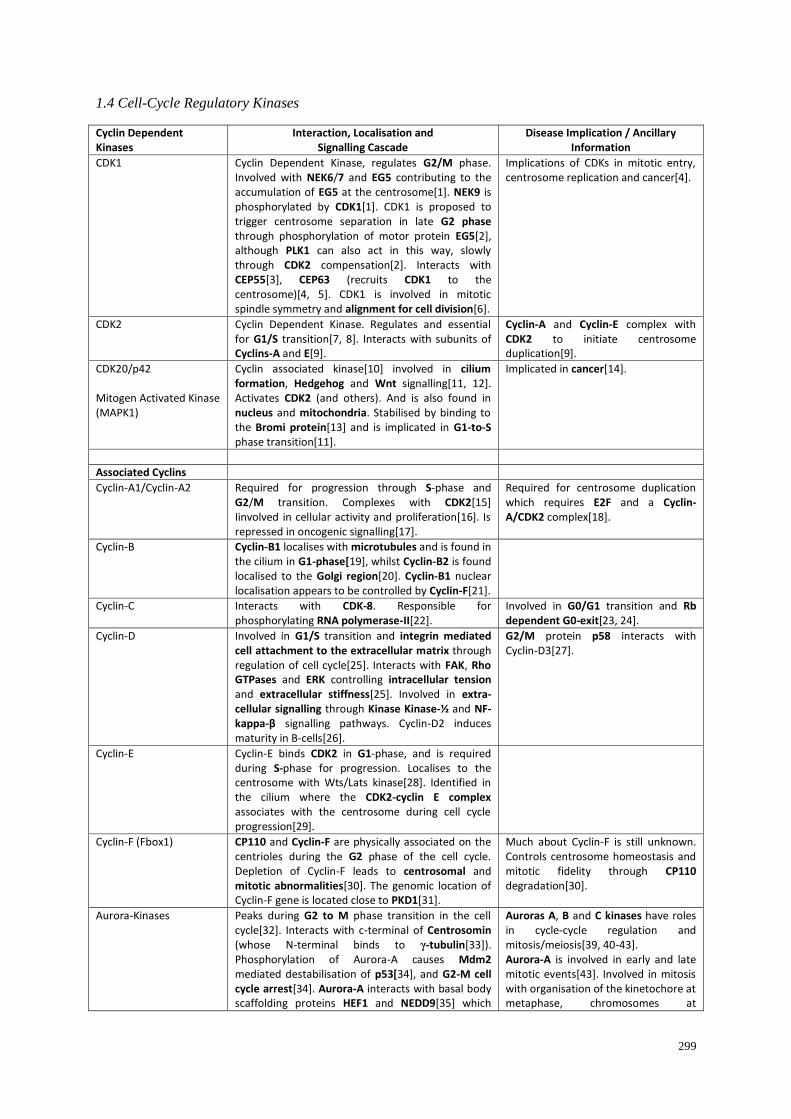
299
1.4 Cell-Cycle Regulatory Kinases
Cyclin Dependent Kinases
Interaction, Localisation and Signalling Cascade
Disease Implication / Ancillary Information
CDK1 Cyclin Dependent Kinase, regulates G2/M phase. Involved with NEK6/7 and EG5 contributing to the accumulation of EG5 at the centrosome[1]. NEK9 is phosphorylated by CDK1[1]. CDK1 is proposed to trigger centrosome separation in late G2 phase through phosphorylation of motor protein EG5[2], although PLK1 can also act in this way, slowly through CDK2 compensation[2]. Interacts with CEP55[3], CEP63 (recruits CDK1 to the centrosome)[4, 5]. CDK1 is involved in mitotic spindle symmetry and alignment for cell division[6].
Implications of CDKs in mitotic entry, centrosome replication and cancer[4].
CDK2 Cyclin Dependent Kinase. Regulates and essential for G1/S transition[7, 8]. Interacts with subunits of Cyclins-A and E[9].
Cyclin-A and Cyclin-E complex with CDK2 to initiate centrosome duplication[9].
CDK20/p42 Mitogen Activated Kinase (MAPK1)
Cyclin associated kinase[10] involved in cilium formation, Hedgehog and Wnt signalling[11, 12]. Activates CDK2 (and others). And is also found in nucleus and mitochondria. Stabilised by binding to the Bromi protein[13] and is implicated in G1-to-S phase transition[11].
Implicated in cancer[14].
Associated Cyclins
Cyclin-A1/Cyclin-A2 Required for progression through S-phase and G2/M transition. Complexes with CDK2[15] Iinvolved in cellular activity and proliferation[16]. Is repressed in oncogenic signalling[17].
Required for centrosome duplication which requires E2F and a Cyclin-A/CDK2 complex[18].
Cyclin-B Cyclin-B1 localises with microtubules and is found in the cilium in G1-phase[19], whilst Cyclin-B2 is found localised to the Golgi region[20]. Cyclin-B1 nuclear localisation appears to be controlled by Cyclin-F[21].
Cyclin-C Interacts with CDK-8. Responsible for phosphorylating RNA polymerase-II[22].
Involved in G0/G1 transition and Rb dependent G0-exit[23, 24].
Cyclin-D Involved in G1/S transition and integrin mediated cell attachment to the extracellular matrix through regulation of cell cycle[25]. Interacts with FAK, Rho GTPases and ERK controlling intracellular tension and extracellular stiffness[25]. Involved in extra-cellular signalling through Kinase Kinase-½ and NF-kappa-β signalling pathways. Cyclin-D2 induces maturity in B-cells[26].
G2/M protein p58 interacts with Cyclin-D3[27].
Cyclin-E Cyclin-E binds CDK2 in G1-phase, and is required during S-phase for progression. Localises to the centrosome with Wts/Lats kinase[28]. Identified in the cilium where the CDK2-cyclin E complex associates with the centrosome during cell cycle progression[29].
Cyclin-F (Fbox1) CP110 and Cyclin-F are physically associated on the centrioles during the G2 phase of the cell cycle. Depletion of Cyclin-F leads to centrosomal and mitotic abnormalities[30]. The genomic location of Cyclin-F gene is located close to PKD1[31].
Much about Cyclin-F is still unknown. Controls centrosome homeostasis and mitotic fidelity through CP110 degradation[30].
Aurora-Kinases Peaks during G2 to M phase transition in the cell cycle[32]. Interacts with c-terminal of Centrosomin (whose N-terminal binds to γ-tubulin[33]). Phosphorylation of Aurora-A causes Mdm2 mediated destabilisation of p53[34], and G2-M cell cycle arrest[34]. Aurora-A interacts with basal body scaffolding proteins HEF1 and NEDD9[35] which
Auroras A, B and C kinases have roles in cycle-cycle regulation and mitosis/meiosis[39, 40-43]. Aurora-A is involved in early and late mitotic events[43]. Involved in mitosis with organisation of the kinetochore at metaphase, chromosomes at

300
complexes and localises to the ciliary base, phosphorylating and activating tubulin de-acetylase (HDAC6), promoting ciliary disassembly[35-37]. Stabilisation of Aurora-A and HDAC6 may prevent cilium resorption[37]. Cofactors with CEP192 for MTOC function and spindle assembly[38]. Aurora-A activation initiates ciliary disassembly[37].
prometaphase[40]. Aurora-B is a regulator of mitotic chromosome segregation and required for chromatid orientation[41]. Binds to microtubules and KIF4[44]. Aurora-C is required for cytokinesis and meosis[45].
CDC2L1 (p58) Protein kinase invoved in the Hedgehog signaling pathway[46] and interacts with Cyclin-D3[27].
EF2 (Suspected in cilium) Elongation Factor 2 Kinase
Promotes G0 to S phase transition. E2F targets gene expression during the progression of cell cycle. EF2 with pRB involves accretion of inhibitory histone modifications invoking chromatin compaction[47].
[1] Bertran MT, Sdelci S, Regué L, Avruch J, Caelles C, Roig J. (2011) Nek9 is a Plk1-activated
kinase that controls early centrosome separation through Nek6/7 and Eg5. EMBO Report, 30, 2634-
2647.
[2] Smith E, Hégarat N, Vesely C, Roseboom I, Larch C, Streicher H, Straatman K, Flynn H, Skehel
M, Hirota T, Kuriyama R, Hochegger H. (2010) Differential control of Eg5-dependent centrosome
separation by Plk1 and Cdk1. EMBO Report, 30, 2233-2245.
[3] Fabbro M, Zhou BB, Takahashi M, Sarcevic B, Lal P, Graham ME, Gabrielli BG, Robinson PJ,
Nigg EA, Ono Y, Khanna KK. (2005) Cdk1/Erk2- and Plk1-dependent phosphorylation of a
centrosome protein, Cep55, is required for its recruitment to midbody and cytokinesis. Developmental
Cell, 9, 477-488.
[4] Löffler H, Fechter A, Matuszewska M, Saffrich R, Mistrik M, Marhold J, Hornung C,
Westermann F, Bartek J, Krämer A. (2011) Cep63 recruits Cdk1 to the centrosome: implications for
regulation of mitotic entry, centrosome amplification, and genome maintenance. Cancer Research,
71, 2129-2139.
[5] Smith E, Dejsuphong D, Balestrini A, Hampel M, Lenz C, Takeda S, Vindigni A, Costanzo V.
(2009) An ATM- and ATR-dependent checkpoint inactivates spindle assembly by targeting CEP63.
Nature Cell Biology, 11, 278-285.
[6] Liakopoulos D, Kusch J, Grava S, Vogel J, Barral Y. (2003) Asymmetric loading of Kar9 onto
spindle poles and microtubules ensures proper spindle alignment. Cell, 112, 561-574.
[7] Morgan DL. (2007) The Cell Cycle: Principles of Control. New Science Press, London, 30-31.
[8] Koff A, Giordano A, Desai D, Yamashita K, Harper JW, Elledge S, Nishimoto T, Morgan DO,
Franza BR, Roberts JM. (1992) Formation and activation of a cyclin E-cdk2 complex during the G1
phase of the human cell cycle. Science, 257, 1689-1694.
[9] Jackman M, Kubota Y, den Elzen N, Hagting A, Pines J. (2002) Cyclin A- and cyclin E-Cdk
complexes shuttle between the nucleus and the cytoplasm. Molecular Biology of the Cell, 13, 1030-
1045.
[10] Liu Y, Wu C, Galaktionov K. (2004) p42, a novel cyclin-dependent kinase-activating kinase in
mammalian cells. Journal of Biological Chemistry, 279, 4507-4514.
[11] Feng H, Cheng AS, Tsang DP, Li MS, Go MY, Cheung YS, Zhao GJ, Ng SS, Lin MC, Yu J, Lai
PB, To KF, Sung JJ. (2011) Cell cycle-related kinase is a direct androgen receptor-regulated gene that
drives ß-catenin/T cell factor-dependent hepatocarcinogenesis. Journal of Clinical Investigation, 121,
3159-3175
301
[12] Phirke P, Efimenko E, Mohan S, Burghoorn J, Crona F, Bakhoum MW, Trieb M, Schuske K,
Jorgensen EM, Piasecki BP, Leroux MR, Swoboda P. (2011) Transcriptional profiling of C. elegans
DAF-19 uncovers a ciliary base-associated protein and a CDK/CCRK/LF2p-related kinase required
for intraflagellar transport. Developmental Biology, 357, 235-247.
[13] Ko HW, Norman RX, Tran J, Fuller KP, Fukuda M, Eggenschwiler JT. (2010) Broad-minded
links cell cycle-related kinase to cilia assembly and Hedgehog signal transduction. Developmental
Cell, 18, 237-247.
[14] An X, Ng SS, Xie D, Zeng YX, Sze J, Wang J, Chen YC, Chow BK, Lu G, Poon WS, Kung HF,
Wong BC, Lin MC. (2010) Functional characterisation of cell cycle-related kinase (CCRK) in
colorectal cancer carcinogenesis. European Journal of Cancer, 46, 1752-1761.
[15] Diederichs S, Bäumer N, Ji P, Metzelder SK, Idos GE, Cauvet T, Wang W, Möller M,
Pierschalski S, Gromoll J, Schrader MG, Koeffler HP, Berdel WE, Serve H, Müller-Tidow C. (2004)
Identification of interaction partners and substrates of the cyclin A1-CDK2 complex. Journal of
Biological Chemistry, 279, 33727-33741.
[16] Tsang WY, Wang L, Chen Z, Sánchez I, Dynlacht BD. (2007) SCAPER, a novel cyclin A
interacting protein that regulates cell cycle progression. Journal of Cell Biology, 178, 621-633.
[17] Bäumer N, Tickenbrock L, Tschanter P, Lohmeyer L, Diederichs S, Bäumer S, Skryabin BV,
Zhang F, Agrawal-Singh S, Köhler G, Berdel WE, Serve H, Koschmieder S, Müller-Tidow C. (2011)
Inhibitor of cyclin-dependent kinase (CDK) interacting with cyclin A1 (INCA1) regulates
proliferation and is repressed by oncogenic signalling. Journal of Biological Chemistry, 286, 28210-
28222.
[18] Meraldi P, Lukas J, Fry AM, Bartek J, Nigg EA. (1999) Centrosome duplication in mammalian
somatic cells requires E2F and Cdk2-cyclin A. Nature Cell Biology, 1, 88-93.
[19] Spalluto C, Wilson DI, Hearn T. (2013) Evidence for reciliation of RPE1 cells in late G1 phase,
and ciliary localisation of cyclin B1. FEBS Open Biology, 3, 334-340.
[20] Jackman M, Firth M, Pines J. (1995) Human cyclins B1 and B2 are localized to strikingly
different structures: B1 to microtubules, B2 primarily to the Golgi apparatus. EMBO Journal, 14,
1646-1654.
[21] Kong M, Barnes EA, Ollendorff V, Donoghue DJ (2000) Cyclin F regulates the nuclear
localisation of cyclin B1 through a cyclin-cyclin interaction. EMBO Journal, 19, 1378-1388.
[22] Kang YK, Guermah M, Yuan CX, Roeder RG (2002) The TRAP/Mediator coactivator complex
interacts directly with estrogen receptors α and β through the TRAP220 subunit and directly enhances
estrogen receptor function in vitro. Proceedings of the National Academy of Sciences, USA, 99, 2642-
2647.
[23] Ren S, Rollins BJ. (2004) Cyclin C/cdk3 promotes Rb-dependent G0 exit. Cell, 117, 239-251.
[24] Sage J. (2004) Cyclin C makes an entry into the cell cycle. Developmental Cell, 6, 607-608.
[25] Assoian RK, Klein EA. (2008) Growth control by intracellular tension and extracellular stiffness.
Trends in Cell Biology, 18, 347-352.
[26] Piatelli MJ, Wardle C, Blois J, Doughty C, Schram BR, Rothstein TL, Chiles TC. (2004)
Phosphatidylinositol 3-kinase-dependent mitogen-activated protein/extracellular signal-regulated

302
kinase kinase 1/2 and NF-kappa B signalling pathways are required for B cell antigen receptor-
mediated cyclin D2 induction in mature B cells. Journal of Immunology, 172, 2753-2762.
[27] Zhang S, Cai M, Zhang S, Xu S, Chen S, Chen X, Chen C, Gu J. (2002) Interaction of
p58(PITSLRE), a G2/M-specific protein kinase, with cyclin D3. Journal of Biological Chemistry,
277, 35314-35322.
[28] Shimizu T, Ho LL, Lai ZC. (2008) The mob as tumour suppressor gene is essential for early
development and regulates tissue growth in Drosophila. Genetics, 178, 957-965.
[29] Plotnikova OV, Golemis EA, Pugacheva EN. (2008) Cell cycle-dependent ciliogenesis and
cancer. Cancer Research, 68, 2058-2061.
[30] D'Angiolella V, Donato V, Vijayakumar S, Saraf A, Florens L, Washburn MP, Dynlacht B,
Pagano M. (2010) SCF (Cyclin F) controls centrosome homeostasis and mitotic fidelity through
CP110 degradation. Nature, 466, 138-142.
[31] Kraus B, Pohlschmidt M, Leung AL, Germino GG, Snarey A, Schneider MC, Reeders ST,
Frischauf AM. (1994) A novel cyclin gene (CCNF) in the region of the polycystic kidney disease
gene (PKD1). Genomics, 24, 27-33.
[32] Lukasiewicz KB, Lingle WL. (2009) Aurora A, centrosome structure, and the centrosome cycle.
Environmental and Molecular Mutagenesis, 50, 602-619.
[33] Terada Y, Uetake Y, Kuriyama R. (2003) Interaction of Aurora-A and centrosomin at the
microtubule-nucleating site in Drosophila and mammalian cells. Journal of Cell Biology, 162, 757-
763.
[34] Katayama H, Sasai K, Kawai H, Yuan ZM, Bondaruk J, Suzuki F, Fujii S, Arlinghaus RB,
Czerniak BA, Sen S. (2004) Phosphorylation by aurora kinase A induces Mdm2-mediated
destabilization and inhibition of p53. Nature Genetics, 36, 55-62.
[35] Alieva IB, Uzbekov RE. (2008) The centrosome is a polyfunctional multiprotein cell complex.
Biochemistry, 73, 626-643.
[36] Pugacheva E, Golemis E. (2005) The focal adhesion scaffolding protein HEF1 regulates
activation of the Aurora-A and Nek2 kinases at the centrosome. Nature Cell Biology, 7, 937-946.
[37] Pugacheva EN, Jablonski SA, Hartman TR, Henske EP, Golemis EA. (2007) HEF1-dependent
Aurora A activation induces disassembly of the primary cilium. Cell, 129, 1351-1363.
[38] Joukov V, De Nicolo A, Rodriguez A, Walter JC, Livingston DM. (2010) Centrosomal protein of
192 kDa (Cep192) promotes centrosome-driven spindle assembly by engaging in organelle-specific
Aurora A activation. Proceedings of the National Academy of Sciences, USA, 107, 21022-21027.
[39] Sabino D, Brown NH, Basto R. (2011) Drosophila Ajuba is not an Aurora-A activator but is
required to maintain Aurora-A at the centrosome. Journal of Cell Science, 124, 1156-1166.
[40] Ma C, Cummings C, Liu XJ. (2003) Biphasic activatin of Aurora-A kinase during the meiosis I-
meiosis II transition in Xenopus oocytes. Molecular and Cellular Biology, 23, 1703-1716.
[41] Hauf S, Biswas A, Langegger M, Kawashima SA, Tsukahara T, Watanabe Y. (2007) Aurora
controls sister kinetochore mono-orientation and homolog bi-orientation in meiosis-I. EMBO Journal,
26, 4475-4486.

303
[42] Nigg EA. (2001) Mitotic kinases as regulators of cell division and its checkpoints. Nature
Reviews Molecular and Cellular Biology, 2, 21-32.
[43] Marumoto T, Honda S, Hara T, Nitta M, Hirota T, Kohmura E, Saya H (2003) Aurora-A kinase
maintains the fidelity of early and late mitotic events in HeLa cells. Journal of Biological Chemistry,
278, 51786-51795.
[44] Ozlü N, Monigatti F, Renard BY, Field CM, Steen H, Mitchison TJ, Steen JJ. (2010) Binding
partner switching on microtubules and aurora-B in the mitosis to cytokinesis transition. Molecular
and Cellular Proteomics, 9,336-350.
[45] Yang KT, Li SK, Chang CC, Tang CJ, Lin YN, Lee SC, Tang TK. (2010) Aurora-C kinase
deficiency causes cytokinesis failure in meiosis I and production of large polyploid oocytes in mice.
Molecular Biology of the Cell, 21, 2371-2383.
[46] Evangelista M, Lim TY, Lee J, Parker L, Ashique A, Peterson AS, Ye W, Davis DP, de Sauvage
FJ. (2008) Kinome siRNA screen identifies regulators of ciliogenesis and hedgehog signal
transduction. Science Signalling, 1, ra7.
[47] Blais A, Dynlacht BD. (2007) E2F-associated chromatin modifiers and cell cycle control.
Current Opinion in Cell Biology, 19, 658-662.

304
1.5 Regulatory Components
Centrosome Localisation Interaction, Localisation and
Signalling Cascade Disease Implication / Ancillary
Information
Nesprin Required for centrosomal positioning, cell polarity and ciliogenesis. Interacts with Meckelin for ciliogenesis via remodeling actin[1].
Gimap5 GTPase IMAP family member. Localises to the ER, Golgi and the centrosome. Interacts with CHOP for mediating endoplasmic reticulum stress induced apoptosis[2].
PLK1(Polo-Like-Kinase-1) Associated with G2/M transition in early prophase[3]. Localises to the transition zone, where it phosphorylates NPHP1[4], and activates histone deacetylase 6 (HDAC6) is recruited to the pericentriolar matrix by PCM1 in a dynactin dependent manner[5]. Interaction between PCM1 and PLK1 is phosphorylation dependent, a process in which CDK1 acts as the priming kinase[5]. PLK1 interacts with many pathways, is associated with aneuploidy and centrosome abnormality, as well as in p53 inactivation[6, 7]. PLK1 prevents centrosome separation from ionizing radiation exposure[8] while PLK1 phosphorylation inhibits ATM/ATR-dependent activation[9] in response to DNA damage[10]. PLK phosphorylates Cyclin-B1 allowing nuclear targetting[11].
CEP215 (CDK5RAP2)
Localises to the centrosome through-out the cell cycle where CEP215 and CEP68 are involved in centrosome cohesion[12]. Interacts with Pericentrin and CDK5R1[13].
CEP97 CEP97 and CP110 suppress ciliary assembly[14]. Depletion of CEP97 results in loss of CP110 giving rise to polyploidy and spindle defects[15].
Involved in the transition from centriole to basal body[16].
CEP63 (Tumour Suppressor)
Acts as an ATM/ATR-dependent checkpoint for Double Strand DNA breaks halting spindle assembly[9]. Invovled as an ATM/ATR-dependent checkpoint for DSBs halting spindle assembly[9]. CEP63 and CEP152 are involved in centriole duplication[17].
Mitotic checkpoint. Interacts with Disrupted-In-Schizophrenia 1 (DISC1)[18]. Recruites CDK-1 to the centrosome[19].
TRAF7 (Suspected in the centrosome)
MoD1 transcription target, where ubiquitylation of NF-kappa-β is regulated exclusively by TRAF7[20].
Also Cytoplasmic.
HSP73 (Heat Shock Protein)
Centrosomally co-localised with Pericentrin and TCP1[21] with a role as a molecular chaperone[22]. Interacts with Cyclin-D (promotes stability for interaction with CDK4[23]).
Centrin (CETN1/2/3)
Centrins are localised to the centriole/pericentriolar material[24, 25] and are involved in microtubule severing[26]. Centrins have a variety of roles within the cell[27]. CETN2 is required for centriole/centrosome duplication and plays a role in organising the microtubule network around the centrosome[28]. Involved in photoreceptors where it interacts with Transducin, CDK2 and in the assembly of Centrin and G-protein complexes[29], Involved in DNA damage recognition and repair[30]. Interacts with CP110[31] and localises to fibrous striated rootlets connected to basal bodies[32].
Localises to the distal (+) end of the basal body, and proximal centriole coating the outer surface and inner (+) end surfaces at the beginning of S-phase[22]. Centrin-2 is involved in the nuclear pore where it plays a role in mRNA and protein export[33].
CEP152 Depletion of CEP152 prevents centriole replication and leads to loss of SAS6. Co-localises with and is phosphorylated by PLK4[34]. Scaffolds PLK4 and
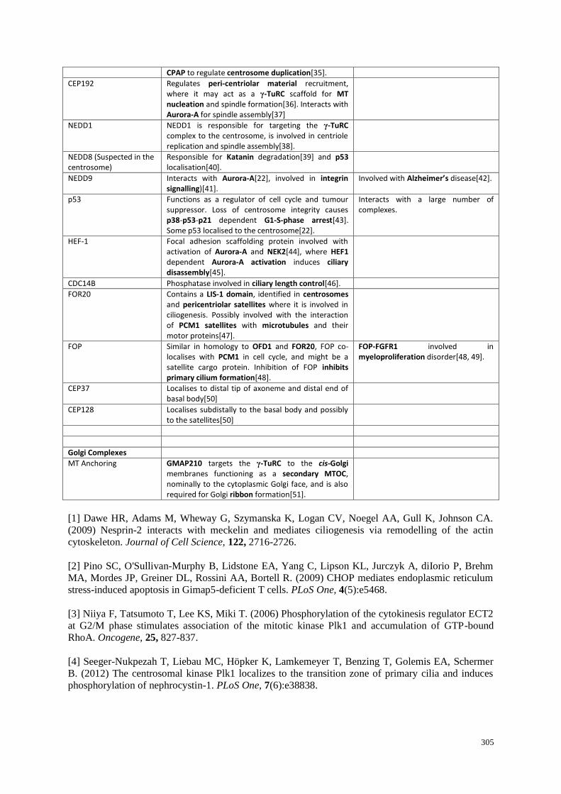
305
CPAP to regulate centrosome duplication[35].
CEP192 Regulates peri-centriolar material recruitment, where it may act as a γ-TuRC scaffold for MT nucleation and spindle formation[36]. Interacts with Aurora-A for spindle assembly[37]
NEDD1 NEDD1 is responsible for targeting the γ-TuRC complex to the centrosome, is involved in centriole replication and spindle assembly[38].
NEDD8 (Suspected in the centrosome)
Responsible for Katanin degradation[39] and p53 localisation[40].
NEDD9 Interacts with Aurora-A[22], involved in integrin signalling)[41].
Involved with Alzheimer’s disease[42].
p53 Functions as a regulator of cell cycle and tumour suppressor. Loss of centrosome integrity causes p38-p53-p21 dependent G1-S-phase arrest[43]. Some p53 localised to the centrosome[22].
Interacts with a large number of complexes.
HEF-1 Focal adhesion scaffolding protein involved with activation of Aurora-A and NEK2[44], where HEF1 dependent Aurora-A activation induces ciliary disassembly[45].
CDC14B Phosphatase involved in ciliary length control[46].
FOR20 Contains a LIS-1 domain, identified in centrosomes and pericentriolar satellites where it is involved in ciliogenesis. Possibly involved with the interaction of PCM1 satellites with microtubules and their motor proteins[47].
FOP Similar in homology to OFD1 and FOR20, FOP co-localises with PCM1 in cell cycle, and might be a satellite cargo protein. Inhibition of FOP inhibits primary cilium formation[48].
FOP-FGFR1 involved in myeloproliferation disorder[48, 49].
CEP37 Localises to distal tip of axoneme and distal end of basal body[50]
CEP128 Localises subdistally to the basal body and possibly to the satellites[50]
Golgi Complexes
MT Anchoring GMAP210 targets the γ-TuRC to the cis-Golgi membranes functioning as a secondary MTOC, nominally to the cytoplasmic Golgi face, and is also required for Golgi ribbon formation[51].
[1] Dawe HR, Adams M, Wheway G, Szymanska K, Logan CV, Noegel AA, Gull K, Johnson CA.
(2009) Nesprin-2 interacts with meckelin and mediates ciliogenesis via remodelling of the actin
cytoskeleton. Journal of Cell Science, 122, 2716-2726.
[2] Pino SC, O'Sullivan-Murphy B, Lidstone EA, Yang C, Lipson KL, Jurczyk A, diIorio P, Brehm
MA, Mordes JP, Greiner DL, Rossini AA, Bortell R. (2009) CHOP mediates endoplasmic reticulum
stress-induced apoptosis in Gimap5-deficient T cells. PLoS One, 4(5):e5468.
[3] Niiya F, Tatsumoto T, Lee KS, Miki T. (2006) Phosphorylation of the cytokinesis regulator ECT2
at G2/M phase stimulates association of the mitotic kinase Plk1 and accumulation of GTP-bound
RhoA. Oncogene, 25, 827-837.
[4] Seeger-Nukpezah T, Liebau MC, Höpker K, Lamkemeyer T, Benzing T, Golemis EA, Schermer
B. (2012) The centrosomal kinase Plk1 localizes to the transition zone of primary cilia and induces
phosphorylation of nephrocystin-1. PLoS One, 7(6):e38838.

306
[5] Wang G, Chen Q, Zhang X, Zhang B, Zhuo X, Liu J, Jiang Q, Zhang C. (2013) PCM1 recruits
Plk1 to the pericentriolar matrix to promote primary cilia disassembly before mitotic entry. Journal of
Cell Science, 126, 1355-1365.
[6] Liu XS, Li H, Song B, Liu X. (2010) Polo-like kinase 1 phosphorylation of G2 and S-phase-
expressed 1 protein is essential for p53 inactivation during G2 checkpoint recovery. EMBO Reports,
11, 626-632.
[7] McKenzie L, King S, Marcar L, Nicol S, Dias SS, Schumm K, Robertson P, Bourdon JC, Perkins
N, Fuller-Pace F, Meek DW. (2010) p53-dependent repression of polo-like kinase-1 (PLK1). Cell
Cycle, 9, 4200-4212.
[8] Zhang W, Fletcher L, Muschel RJ. (2005) The role of Polo-like kinase 1 in the inhibition of
centrosome separation after ionizing radiation. Journal of Biological Chemistry, 280, 42994-42999.
[9] Smith E, Dejsuphong D, Balestrini A, Hampel M, Lenz C, Takeda S, Vindigni A, Costanzo V.
(2009) An ATM- and ATR-dependent checkpoint inactivates spindle assembly by targeting CEP63.
Nature Cell Biology, 11, 278-285.
[10] Tsvetkov L, Stern DF. (2005) Phosphorylation of Plk1 at S137 and T210 is inhibited in response
to DNA damage. Cell Cycle, 4, 166-171
[11] Toyoshima-Morimoto F, Taniguchi E, Shinya N, Iwamatsu A, Nishida E. (2001) Polo-like kinase
1 phosphorylates cyclin B1 and targets it to the nucleus during prophase. Nature, 410, 215-220.
[12] Graser S, Stierhof YD, Nigg EA. (2007) Cep68 and Cep215 (Cdk5rap2) are required for
centrosome cohesion. Journal of Cell Science, 120, 4321-4331.
[13] Wang X, Ching YP, Lam WH, Qi Z, Zhang M, Wang JH. (2000) Identification of a common
protein association region in the neuronal Cdk5 activator. Journal of Biological Chemistry, 275,
31763-31769.
[14] Spektor A, Tsang WY, Khoo D, Dynlacht BD. (2007) Cep97 and CP110 suppress a cilia
assembly program. Cell, 130, 678-690.
[15] Chen Z, Indjeian VB, McManus M, Wang L, Dynlacht BD. (2002) CP110, a cell cycle-
dependent CDK substrate, regulates centrosome duplication in human cells. Developmental Cell, 3,
339-350.
[16] Kobayashi T, Dynlacht BD. (2011) Regulating the transition from centriole to basal body.
Journal of Cell Biology, 193, 435-444.
[17] Brown NJ, Marjanović M, Lüders J, Stracker TH, Costanzo V. (2013) Cep63 and Cep152
cooperate to ensure centriole duplication. PLoS One, 8(7):e69986.
[18] Morris JA, Kandpal G, MaL, Austin CP. (2003) DISC1 (Disrupted-In-Schizophrenia 1) is a
centrosome-associated protein that interacts with MAP1A, MIPT3, ATF4/5 and NUDEL: regulation
and loss of interaction with mutation. Human Molecular Genetics, 12, 1591-1608.
[19] Tsikitis M, Acosta-Alvear D, Blais A, Campos EI, Lane WS, Sánchez I, Dynlacht BD (2010)
Traf7, a MyoD1 transcriptional target, regulates nuclear factor-kappaB activity during myogenesis.
EMBO Reports, 11, 969-976.
[20] Xu LG, Li LY, Shu HB. (2004) TRAF7 potentiates MEKK3-induced AP1 and CHOP activation
and induces apoptosis. Journal of Biological Chemistry, 279, 17278-17282.

307
[21] Brown CR, Hong-Brown LQ, Doxsey SJ, Welch WJ. (1996) Molecular chaperones and the
centrosome. A role for HSP 73 in centrosomal repair following heat shock treatment. Journal of
Biological Chemistry, 271, 833-840.
[22] Alieva IB, Uzbekov RE. (2008) The centrosome is a polyfunctional multiprotein cell complex.
Biochemistry, 73, 626-643.
[23] Diehl JA, Yang W, Rimerman RA, Xiao H, Emili A. (2003) Hsc70 regulates accumulation of
cyclin D1 and cyclin D1-dependent protein kinase. Molecular and Cellular Biology, 23, 1764-1774.
[24] Errabolu R, Sanders MA, Salisbury JL. (1994) Cloning of a cDNA encoding human centrin, an
EF-hand protein of centrosomes and mitotic spindle poles. Journal of Cell Science, 107, 9-16.
[25] Baron AT, Greenwood TM, Bazinet CW Salisbury JL. (1992) Centrin is a component of the
pericentriolar lattice. Biology of the Cell / under the auspices of the European Cell Biology
Organization, 76, 383-388.
[26] Sanders MA, Salisbury JL. (1994) Centrin plays an essential role in microtubule severing during
flagellar excision in Chlamydomonas reinhardtii. Journal of Cell Biology, 124, 795-805.
[27] Vonderfecht T, Stemm-Wolf AJ, Hendershott M, Giddings TH Jr, Meehl JB, Winey M. (2011)
The two domains of centrin have distinct basal body functions in Tetrahymena. Molecular Biology of
the Cell, 22, 2221-2234.
[28] Salisbury JL, Suino KM, Busby R, Springett M. (2002) Centrin-2 is required for centriole
duplication in mammalian cells. Current Biology, 12, 1287-1292.
[29] Trojan P, Krauss N, Choe HW, Giessl A, Pulvermüller A, Wolfrum U. (2008) Centrins in retinal
photoreceptor cells: regulators in the connecting cilium. Progress in Retinal Eye Research, 27, 237-
259.
[30] Krasikova YS, Rechkunova NI, Maltseva EA, Craescu CT, Petruseva IO, Lavrik OI. (2012)
Influence of centrin 2 on the interaction of nucleotide excision repair factors with damaged DNA.
Biochemistry, 77, 346-353.
[31] Tsang WY, Spektor A, Luciano DJ, Indjeian VB, Chen Z, Salisbury JL, Sánchez I, Dynlacht BD.
(2006) CP110 cooperates with two calcium-binding proteins to regulate cytokinesis and genome
stability. Molecular Biology of the Cell, 17, 3423-3434
[32] Lemullois M, Fryd-Versavel G, Fleury-Aubusson A. (2004) Localisation of centrins in the
hypotrich ciliate Paraurostyla weissei. Protist, 155, 331-346.
[33] Resendes KK, Rasala BA, Forbes DJ. (2008) Centrin 2 localises to the vertebrate nuclear pore
and plays a role in mRNA and protein export. Molecular and Cellular Biology, 28, 1755-1769.
[34] Hatch EM, Kulukian A, Holland AJ, Cleveland DW, Stearns T. (2010) Cep152 interacts with
Plk4 and is required for centriole duplication. Journal of Cell Biology, 191, 721-729.
[35] Cizmecioglu O, Arnold M, Bahtz R, Settele F, Ehret L, Haselmann-Weiss U, Antony C,
Hoffmann I. (2010) Cep152 acts as a scaffold for recruitment of Plk4 and CPAP to the centrosome.
Journal of Cell Biology, 191, 731-739.
[36] Gomez-Ferreria M, Rath U, Buster D, Chanda S, Caldwell J, Rines D, Sharp D. (2007) Human
Cep192 is required for mitotic centrosome and spindle assembly. Current Biology, 17, 1960-1966.

308
[37] Joukov V, De Nicolo A, Rodriguez A, Walter JC, Livingston DM. (2010) Centrosomal protein of
192 kDa (Cep192) promotes centrosome-driven spindle assembly by engaging in organelle-specific
Aurora A activation. Proceedings of the National Academy of Sciences, USA, 107, 21022-21027.
[38] Haren L, Remy MH, Bazin I, Callebaut I, Wright M, Merdes AJ. (2006) NEDD1-dependent
recruitment of the gamma-tubulin ring complex to the centrosome is necessary for centriole
duplication and spindle assembly. Journal of Cell Biology, 172, 505-515.
[39] Kurz T, Pintard L, Willis JH, Hamill DR, Gonczy P, Peter M, Bowerman B. (2002) Cytoskeletal
regulation by the Nedd8 ubiquitin-like protein modification pathway. Science, 295, 1294-1298.
[40] Liu G, Xirodimas DP. (2010) NUB1 promotes cytoplasmic localisation of p53 through
cooperation of the NEDD8 and ubiquitin pathways. Oncogene, 29, 2252-2261.
[41] Kondo S, Iwata S, Yamada T, Inoue Y, Ichihara H, Kichikawa Y, Katayose T, Souta-Kuribara A,
Yamazaki H, Hosono O, Kawasaki H, Tanaka H, Hayashi Y, Sakamoto M, Kamiya K, Dang NH,
Morimoto C. (2012) Impact of the integrin signaling adaptor protein NEDD9 on prognosis and
metastatic behavior of human lung cancer. Clinical Cancer Research, 18, 6326-6338.
[42] Xing YY, Yu JT, Yan WJ, Chen W, Zhong XL, Jiang H, Wang P, Tan L. (2011) NEDD9 is
genetically associated with Alzheimer's disease in a Han Chinese population. Brain Research, 1369,
230-234.
[43] Mikule K, Delaval B, Kaldis P, Jurcyzk A, Hergert P, Doxsey S. (2007) Loss of centrosome
integrity causes p38-p53-p21-dependent G1-S arrest. Nature Cell Biology, 9, 160-170.
[44] Pugacheva E, Golemis E. (2005) The focal adhesion scaffolding protein HEF1 regulates
activation of the Aurora-A and Nek2 kinases at the centrosome. Nature Cell Biology, 7, 937-946.
[45] Pugacheva EN, Jablonski SA, Hartman TR, Henske EP, Golemis EA. (2007) HEF1-dependent
Aurora A activation induces disassembly of the primary cilium. Cell, 129, 1351-1363.
[46] Clement A, Solnica-Krezel L, Gould KL. (2011) The Cdc14B phosphatase contributes to
ciliogenesis in zebrafish. Development, 138, 291-302.
[47] Sedjaï F, Acquaviva C, Chevrier V, Chauvin JP, Coppin E, Aouane A, Coulier F, Tolun A,
Pierres M, Birnbaum D, Rosnet O. (2010) Control of ciliogenesis by FOR20, a novel centrosome and
pericentriolar satellite protein. Journal of Cell Science, 123, 2391-2401.
[48] Lee JY, Stearns T. (2013) FOP is a centriolar satellite protein involved in ciliogenesis. PLoS
One, 8(3):e58589.
[49] Guasch G1, Delaval B, Arnoulet C, Xie MJ, Xerri L, Sainty D, Birnbaum D, Pébusque MJ.
(2004) FOP-FGFR1 tyrosine kinase, the product of a t(6;8) translocation, induces a fatal
myeloproliferative disease in mice. Blood, 103, 309-312.
[50] Schrøder JM, Rogowski M, Jakobsen K, Vanselow K, Geimer S, Pedersen LB, Andersen JS.
(2012) Identification and characterization of two novel centriolar appendage component proteins.
Cilia, 1, P44.
[51] Rios RM, Sanchis A, Tassin AM, Fedriani C, Bornens M. (2004) GMAP-210 recruits γ-tubulin
complexes to cis-Golgi membranes and is required for Golgi ribbon formation. Cell, 118, 323-335.

309
Appendix II: Rab-GTPases - Ciliary and Golgi Function
RAB Member Cellular Location Interactions
RAB1 Endoplasmic Reticulum/Golgi Required for transport between ER and Golgi[1, 2, 3] involved in pre-Golgi endoplasmic to Golgi Intermediate compartment ERGIC[4, 5]. Regulates calcium signalling receptor (hCaR)[6]. RAB1A is involved in microtubule anterograde melanosome transport[7]. Interacts with the GM130 effector complex regulating COPII vesicle cis-Golgi tethering[8].
RAB2 Endoplasmic Reticulum/Golgi Essential for the maturation of pre-Golgi intermediates[9], regulates vesicle trafficking between the endoplasmic reticulum and cis-Golgi network[10, 11]. Involved with the endoplasmic reticulum and the Golgi[5] where it is required for pre-Golgi maturation[9] and Endoplasmic Reticulum-to-Golgi Intermediate compartment ERGIC[4].
RAB3A Secretory vesicles RAB3 modulates the activity of the fusion machinery by controlling the formation or the stability of the SNARE complex[12] and regulates vesicle fusion[13-15]. Involved with KIF1 motors[16].
RAB3D trans-Golgi vesicles Associated with a subset of post-trans-Golgi network vesicles not involved with the endosome or lysosome[17].
RAB4 Endosome (early)[20] Rab4 and Rab11 regulate the recycling of angiotensin[18, 19]. Rab4 regulates KIFC2 activity[20], interacts with KIF3[20-22] and interacts with KIF3B[22, 23]. Involved in plasma membrane recycling[24]. Rab4, and Rab5 are important in early regulation of endosome trafficking while Rab7 is involved with the late endosome[25]. Interacts with DYNC1LI1[22].
RAB5A*[39] Clathrin coated pits/membrane Functions for internalisation. Rab5A and Rab7 are lysosomal co-regulators and catalysts[26]. Rab4, and Rab5 are important in early regulation of endosomal trafficking while Rab7 is involved with the late endosome[27]. Rab5 is vital for endosomal biogenesis[27], while clathrin and Rab5 are required for endocytosis[28, 29].
RAB5C*[39] Endosome (early) Wnt11 mediates cell adhesion through Rab5C and E-cadherin, a key regulator of early endocytosis[30].
RAB6*[39] medial to trans-Golgi[31], Golgi to plasma membrane and Golgi to endoplasmic reticulum retrograde transport[24, 32].
Regulates recruitment of the dynactin complex to Golgi membranes[33]. KIF20A interacts with Rab6 at the Golgi, transformation to GDP form releases KIF20A[34] required for cytokinesis[35]. Rab6 family members interact with dynein light chain[36], and target exocytotic carriers[37]. Rab6 effectors may include dynactin, myosin, KIF5A/KIF5B, KIF1C, and KIF20[22, 38]. Rab6 and Rab8 are involved in vesicle docking and fusion for exocytosis[39]. Rab6 is involved with p150
GLUED, p50
Dynamitin in the Golgi[23].
RAB7 Endosomes (late), Lysosomes and Phagosome[24]
Rab7 effector protein RILP controls trafficking through late endosomes to lysosomes[19, 24]. Transported by the dynein-dynactin-complex[36, 37] and are required for lysosome biogenesis[41, 42]. Rab4, and Rab5 are important in early regulation of endosomal trafficking while Rab7 is involved with the late endosome[21]. Interacts with dynactin[23]. Interacts with RILP[41, 45], CHM[46, 47] and AP-1-Clathrin adaptor.
RAB8*[43] RAB8A*[44]
trans-Golgi[20], secretory vesicles, Modulators of ciliary protein trafficking[39].
Rab8 interacts with Rabin and Rab11 for transport of vesicles (with IFT20) containing Golgi derived materials to the ciliary membrane[43]. Rab8a localises to the cilium and interacts with Cenexin/ODF2[44, 49] and CEP290 and the PCM[48]. Involved in macropinosome membrane trafficking and Cholera-B toxin trafficking to the Golgi[50]. Rab8 and Rab11 are involved in ciliogenesis[51]. Interacts with myosin in the Golgi[23].
RAB9 Late Endosome and the trans-Golgi [24]
Regulates late Endosome size[52] and transport of vesicles to the trans-Golgi[53]. Required for viral replication[54].
RAB10*[43] Golgi sorting and endosomes[24]. Associates with the primary cilium in renal epithelium[55].
RAB10 co-localises with Exocyst proteins at the base of the cilium[55]. Involved with transport of TLR4 to the cell membrane[56]. Regulates glutamate receptor recycling in

310
endocytosis[57] and endoplasmic reticulum morphology[58].
RAB11*[43] trans-Golgi network[24], Endosome (early), regulates re-cycling[60, 61]. Stretch activated exocytosis[61].
Regulates compartmentalisation of early endosomes required for efficient transport from early endosomes to the trans-Golgi network[62]. Rab11-FIP3 links Rab11-GTPase and cytoplasmic dynein DYNC1LI1 mediating transport to the endosomal-recycling compartment[63]. Rab4 and Rab11 together regulate the recycling of angiotensin[18]. Interacts with myosin in the recycling compartment[23]. Interacts with KIF3B[22].
RAB12 Secretory granules[64]. Regulates mTORC1 activity though degradation of the amino acid transporter PAT4[65].
RAB13 Cytoplasmic vesicles, and tight Junctions[66].
Mediates recycling of occludin[67], interacts with Kinase-A[68]. Involved in regulation of membrane trafficking between the endosome and the TGN in epithelial cells[69].
RAB14 Endosome (early) and the trans-Golgi network[70]
Involved in trans-Golgi to plasma membrane transport[24] as well as Golgi and endosome membrane trafficking[71]. Interacts with KIF16B[22].
RAB15 Endosome (early and recycling)[32]
Inhibitor of endocytic internalisation[53]. Recycles receptors from the recycling endosome[72].
RAB17*[44] Endosome (apical recycling) Involved in recycling transport[53]. Regulates receptor mediated endocytosis in epithelial cells and localises to the recycling endosome[73].
RAB18 Endoplasmic reticulum, Golgi and Endosome[73-77], lipid droplets[78], synaptic granule[78] and secretory granules[79].
Rab18 and Rab43 are involved in ER-Golgi trafficking, Rab43-T32N redistributes Golgi elements to ER exit sites. Rab43 redistributes the p150(Glued) subunit of dynactin, consistent with a specific role in regulating association of pre-Golgi intermediates with microtubules[74, 4]. Loss of Rab18 function causes Warburg-micro syndrome[81].
RAB20 Epithelial specific Associated with dense apical tubules[53]. Regulates phagosome maturation in macrophages[82].
RAB21 Early endosome[83]. Involved in cell adhesion and endosomal recycling of integrin receptors[84]. Regulates cell adhesion and controls trafficking of β1-integrins[84].
RAB22 Endosomes (late and early)[85], Rab22B involved in trans-Golgi membranes[86].
Affects the morphology and function of endosomes[85], with Rab22B being involved in trans-Golgi membranes[86].
RAB23*[43, 44] Cilium, also locates to the Plasma Membrane, Endosome (early) and Endocytic pathways[24].
GTPase[87] involved in signal transduction, essential negative regulator of Sonic HedgeHog[88, 89]; involved in organogenesis[90]. Rab23 regulates Smoothened levels[91] and has a role in planar polarisation[92].
RAB24 cis-Golgi[93], Endoplasmic Reticulum and Endosome (late), and the Autophagosome[86].
Involved in protein trafficking pathways and may function in vesicle transport[94]. Involved in cell division[95]. Involved with Rab7 and Rab23 in the autophagosome[86].
RAB25 Endosome(late), recycling endosome[24, 86].
Apical re-cycling [24, 53, 96]. Regulates metastasis and invasion in gastric, head and neck cancers[95, 98]. Rab25 is associated with α5β1-integrin in invasive migration[99].
RAB26 Golgi derived trafficking, secretory vesicles
Involved in transport of G-coupled α2-adrenergic receptors from the Golgi[86, 100].
RAB27 Melanosomes, apical plasma membrane vesicles and secretory granules[86, 101]
Involved in regulated secretion and membrane trafficking[88]. Interacts with Slac2 and myosin-Va for transport[24, 101]. Rab27A and Rab27B control components of the secretory exosome pathway[102, 103]. Prenylation of Rab27A may play a role in degeneration of photoreceptors[104]. Interacts with myosins and Rab27A[105] in the melanosome[23]. Interacts with KIF5[22].
RAB28 Basal body and ciliary rootlet Involved in autosomal recessive cone-rod dystrophy and macular degeneration. Localises to basal body and ciliary rootlet[106].
RAB30 Golgi Localises to the Golgi where it is required for Golgi integrity and is involved in control of morphology[107].
RAB31 trans-Golgi network[20] Rab31 levels possibly modulate breast cancer cell matrix adhesion and invasive capacity[108].
RAB32 Mitochondria and melanosome[81].
Involved in apoptosis, and targeting PKA to endoplasmic reticulum and mitochondrial membranes[109]. Rab32 and Rab38 are involved in melanosome biogenesis[110] and post Golgi

311
[1] Batoko H, Zheng H, Hawes, Moore I. (2000) Rab1 GTPase is required for transport between the
endoplasmic reticulum and Golgi apparatus and for normal Golgi movement in plants. Plant Cell, 12,
2201-2218.
[2] Zenner HL, Yoshimura S, Barr FA, Crump CM. (2011) Analysis of RabGTPase-activating
proteins indicates that Rab1a/b and Rab43 are important for HSV-1 secondary envelopment. Journal
of Virology, 21680502.
[3] Schwartz SL, Cao C, Pylypenko O, Rak,A, Wandinger-Ness A. (2007) Rab GTPases at a glance.
Journal of Cell Science, 120, 3905-3910.
[4] Yadav S, Linstedt AD. (2011) Golgi positioning. Cold Spring Harbour Perspectives in Biology,
3(5), pii: a005322.
[5] Sannerud R, Marie M, Nizak C, Dale HA, Pernet-Gallay K, Perez F, Goud B, Saraste J. (2006)
Rab1 defines a novel pathway connecting the pre-Golgi intermediate compartment with the cell
periphery. Molecular Biology of the Cell, 17, 1514-1526.
[6] Zhuang X, Adipietro KA, Datta S, Northup JK, Ray K. (2010) Rab1 small GTP-binding protein
regulates cell surface trafficking of the human calcium-sensing receptor. Endocrinology, 151, 5114-
5123.
[7] Ishida M, Ohbayashi N, Maruta Y, Ebata Y, Fukuda M. (2012) Functional involvement of Rab1A
in microtubule-dependent anterograde melanosome transport in melanocytes. Journal of Cell Science,
125, 5177-5187.
[8] Moyer BD, Allan BB, Balch WE. (2001) Rab1 interaction with a GM130 effector complex
regulates COPII vesicle cis-Golgi tethering. Traffic, 2, 268-276.
sorting[111].
RAB33 Golgi[27], Autophagosome[83]. Involved in retrograde transport to the Endoplasmic Reticulum[32], and with autophagy[24]. Localised to medial-Golgi cisternae[111, 112]; involved with Rab5 in the Macropinosome[86].
RAB34 Golgi, macropinosomes[86] RAB34 controls intra-Golgi and lysosome positioning[24, 113], involved in response to hyperglycemia[114].
RAB35 Plasma membrane, Endosomes[32], recycling endosome[86]
Involved in membrane and endosomal recycling[115].
RAB36 Endosome/Lysosome Regulates distribution of endosomes and lysosomes[116].
RAB37 Secretory granules/vesicles [24] Involved with secretory granules and insulin exocytosis[117, 118].
RAB38 Melanosome[86] Rab32 and Rab38 control post Golgi sorting of melanocytic enzymes[111] Associated with proteinuria and hypertension in renal disease[119] and poor outcome in glioma progression[120].
RAB39A Golgi association, and Caspase-1 involvement.
Involved with binding caspase-1 secretion of interleukin-1β*121+. Golgi associated and also involved in endocytosis[122].
RAB39B Golgi Unknown but associated with autism, epilepsy and retardation[24, 123].
RAB40 Golgi Mediates intra-Golgi trafficking with Rab6 and Rab33[83].
RAB41 Golgi Required for Golgi organisation and ER-to-Golgi trafficking[124]
RAB43 Secretory packaging, trans-Golgi network [125]
RAB1A/B and RAB43 respectively, are important for viron assembly[2]. Retrograde transport from plasma membrane to the trans-Golgi Nework[126].
* Involved with the cilium or periciliary environment[44, 127]. For a review of pathological significance see Kelly et al., (2012) [24]. For a review of Rab-GTPase proteins see Klöpper et al., (2012) [128], Schwartz et al., (2008)[3] and Jorden et al., (2005)[23].

312
[9] Tisdale EJ, Balch WE. (1996) Rab2 is essential for the maturation of pre-Golgi intermediates.
Journal of Biological Chemistry, 271, 29372-29379.
[10] Cheung AY, Chen CY, Glaven RH, de Graaf BH, Vidali L, Hepler PK, Wu HM. (2002) Rab2
GTPase regulates vesicle trafficking between the endoplasmic reticulum and the Golgi bodies and is
important to pollen tube growth. Plant Cell, 14, 945-962.
[11] Alberts B, Bray D, Lewis J, Raff M, Roberts K, Walter P. (2002) Molecular Biology of the Cell,
Garland Publishing New York.
[12] Han JH, Lee C, Cheang Y, Kaang BK. (2005) Suppression of long-term facilitation by Rab3-
effector protein interaction. Brain Research, Molecular Brain Research, 139, 13-22.
[13] Geppert M, Goda Y, Stevens CF, Südhof TC. (1997) The small GTP-binding protein Rab3A
regulates a late step in synaptic vesicle fusion. Nature, 387, 810-814.
[14] Nonet ML, Staunton JE, Kilgard MP, Fergestad T, Hartwieg E, Horvitz HR, Jorgensen EM,
Meyer BJ. (2006) Caenorhabditis elegans Rab-3 mutant synapses exhibit impaired function and are
partially depleted of vesicles. Journal of Neuroscience, 17, 8061-8073.
[15] Mahoney TR, Liu Q, Itoh T, Luo S, Hadwiger G, Vincent R, Wang ZW, Fukuda M, Nonet ML.
(2006) Regulation of synaptic transmission by RAB-3 and RAB-27 in Caenorhabditis elegans.
Molecular Biology of the Cell, 17, 2617-2625.
[16] Niwa S, Tanaka Y, Hirokawa N. (2008) KIF1Bβ - and KIF1A mediated axonal transport of
presynaptic regulator Rab3 occurs in a GTP-dependent manner through DENN/MADD. Nature Cell
Biology, 10, 1269-1279.
[17] Pavlos NJ, Xu J, Riedel D, Yeoh JS, Teitelbaum SL, Papadimitriou JM, Jahn R, Ross FP, Zheng
MH. (2005) Rab3D regulates a novel vesicular trafficking pathway that is required for osteoclastic
bone resorption. Molecular Biology of the Cell, 25, 5253-5269.
[18] Li H, Li HF, Felder RA, Periasamy A, Jose PA. (2008) Rab4 and Rab11 coordinately regulate
the recycling of angiotensin II type I receptor as demonstrated by fluorescence resonance energy
transfer microscopy. Journal of Biomedical Optics, 13(3):031206.
[19] Ward ES, Martinez C, Vaccaro C, Zhou J, Tang Q, Ober RJ. (2005) From sorting endosomes to
exocytosis: association of Rab4 and Rab11 GTPases with the Fc receptor, FcRn, during recycling.
Molecular Biology of the Cell, 16, 2028-2038.
[20] Bananis E, Nath S, Gordon K, Satir P, Stockert RJ, Murray JW, Wolkoff AW. (2004)
Microtubule-dependent movement of late endocytic vesicles in vitro: requirements for dynein and
kinesin. Molecular Biology of the Cell, 15, 3688-3697.
[21] Imamura T, Huang J, Usui I, Satoh H, Bever J, Olefsky JM. (2003) Insulin-induced GLUT4
translocation involves protein kinase Clambda-mediated functional coupling between Rab4 and the
motor protein kinesin. Molecular Biology of the Cell, 23, 4892-4900.
[22] Horgan CP, McCaffrey MW. (2011) Rab GTPases and microtubule motors. Biochemical Society
Transactions, 39, 1202-1206.
[23] Jordens I, Marsman M, Kuijl C, Neefjes J. (2005) Rab proteins, connecting transport and vesicle
fusion. Traffic, 6, 1070-1077.

313
[24] Kelly EE, Horgan CP, Goud B, McCaffrey MW. (2012) The Rab family of proteins: 25 years on.
Biochemical Society Transactions, 40, 1337-1347.
[25] McCaffrey MW, Bielli A, Cantalupo G, Mora S, Roberti V, Santillo M, Drummond F, Bucci C.
(2001) Rab4 affects both recycling and degradative endosomal trafficking. FEBS Letters, 495, 21-30.
[26] Croizet-Berger K, Daumerie C, Couvreur M, Courtoy PJ, van den Hove MF. (2002) The
endocytic catalysts, Rab5a and Rab7, are tandem regulators of thyroid hormone production.
Proceedings of the National Academy of Sciences, USA, 99, 8277-8282.
[27] Zeigerer A, Gilleron J, Bogorad RL, Marsico G, Nonaka H, Seifert S, Epstein-Barash H,
Kuchimanchi S, Peng CG, Ruda VM, del Conte-Zerial P, Hengstler JG, Kalaidzidis Y, Koteliansky V,
Zerial M. (2012) Rab5 is necessary for the biogenesis of the endolysosomal system in vivo. Nature,
485, 465-470.
[28] Nielsen E, Severin F, Backer JM, Hyman AA, Zerial M. (1999) Rab5 regulates motility of early
endosomes on microtubules. Nature Cell Biology, 1, 376-382.
[29] Ding Q, Wang Z, Chen Y. (2009) Endocytosis of adiponectin receptor 1 through a clathrin- and
Rab5-dependent pathway. Cell Research, 19, 317-327.
[30] Ulrich F, Krieg M, Schötz EM, Link V, Castanon I, Schnabel V, Taubenberger A, Mueller D,
Puech PH, Heisenberg CP. (2005) Wnt11 functions in gastrulation by controlling cell cohesion
through Rab5c and E-cadherin. Developmental Cell, 9, 555-564.
[31] Goud B, Zahraoui A, Tavitian A, Saraste J. (1990) Small GTP-binding protein associated with
Golgi cisternae. Nature, 345, 553-556.
[32] Hayes GL, Brown FC, Haas AK, Nottingham RM, Barr FA, Pfeffer SR. (2009) Multiple Rab
GTPase binding sites in GCC185 suggest a model for vesicle tethering at the trans-Golgi. Molecular
Biology of the Cell, 20, 209-217.
[33] Short B, Preisinger C, Schaletzky J, Kopajtich R, Barr FA. (2002) The Rab6 GTPase regulates
recruitment of the dynactin complex to Golgi membranes. Current Biology, 12, 1792-1795.
[34] Echard A, Jollivet F, Martinez O, Lacapère JJ, Rousselet A, Janoueix-Lerosey I, Goud B. (1998)
Interaction of a Golgi-associated kinesin-like protein with Rab6. Science, 279, 580-585.
[35] Fontijn RD, Goud B, Echard A, Jollivet F, van Marle J, Pannekoek H, Horrevoets AJ. (2001) The
human kinesin-like protein RB6K is under tight cell cycle control and is essential for cytokinesis.
Molecular Biology of the Cell, 21, 2944-2955.
[36] Wanschers B, van de Vorstenbosch R, Wijers M, Wieringa B, King SM, Fransen J. (2008) Rab6
family proteins interact with the dynein light chain protein DYNLRB1. Cell Motility and
Cytoskeleton, 65, 183-196.
[37] Zerial M, McBride H. (2001) Rab proteins as membrane organizers. Nature Reviews Molecular
and Cellular Biology, 2, 107-117.
[38] Storrie B, Micaroni M, Morgan GP, Jones N, Kamykowski JA, Wilkins N, Pan TH, Marsh BJ.
(2012) Electron tomography reveals Rab6 is essential to the trafficking of trans-Golgi clathrin and
COPI-coated vesicles and the maintenance of Golgi cisternal number. Traffic, 13, 727-744.
[39] Grigoriev I, Yu KL, Martinez-Sanchez E, Serra-Marques A, Smal I, Meijering E, Demmers J,
Peränen J, Pasterkamp RJ, van der Sluijs P, Hoogenraad CC, Akhmanova A. (2011) Rab6, Rab8, and

314
MICAL3 cooperate in controlling docking and fusion of exocytotic carriers. Current Biology, 21,
967-974.
[40] Jordens I, Fernandez-Borja M, Marsman M, Dusseljee S, Janssen L, Calafat J, Janssen H,
Wubbolts R, Neefjes J. (2001) The Rab7 effector protein RILP controls lysosomal transport by
inducing the recruitment of dynein-dynactin motors.Current Biology, 11, 1680-1685.
[41] Cantalupo G, Alifano P, Roberti V, Bruni CB, Bucci C. (2001) Rab-interacting lysosomal protein
(RILP): the Rab7 effector required for transport to lysosomes. EMBO Journal, 20, 683-693.
[42] Bucci C, Thomsen P, Nicoziani P, McCarthy J, van Deurs B. (2000) Rab7: a key to lysosome
biogenesis. Molecular Biology of the Cell, 11, 467-480.
[43] Hsiao Y, Tuz K, Ferland RJ. (2012) Trafficking in and to the primary cilium. Cilia, 1, 4.
[44] Yoshimura S, Egerer J, Fuchs E, Haas AK, Barr FA. (2007) Functional dissection of
RabGTPases involved in primary cilium formation. Journal of Cell Biology, 178, 363-369.
[45] Caplan S, Hartnell LM, Aguilar RC, Naslavsky N, Bonifacino JS. (2001) Human Vam6p
promotes lysosome clustering and fusion in vivo. Journal of Cell Biology, 154, 109-122.
[46] Rak A, Pylypenko O, Niculae A, Goody RS, Alexandrov K. (2003) Crystallization and
preliminary X-ray diffraction analysis of monoprenylated Rab7 GTPase in complex with Rab escort
protein 1. Journal of Structural Biology, 141, 93-95.
[47] Alexandrov K, Simon I, Iakovenko A, Holz B, Goody RS, Scheidig AJ. (1998) Moderate
discrimination of REP-1 between Rab7 x GDP and Rab7 x GTP arises from a difference of an order
of magnitude in dissociation rates. FEBS Letters, 425, 460-464.
[48] Kim J, Krishnaswami SR, Gleeson JG. (2008) CEP290 interacts with the centriolar satellite
component PCM-1 and is required for Rab8 localization to the primary cilium. Human Molecular
Genetics, 17, 3796-3805.
[49] Yoshimura S, Haas AK, Barr FA. (2008) Analysis of RabGTPase and GTPase-activating protein
function at primary cilia. Methods in Enzymology, 439, 353-364.
[50] Hattula K, Furuhjelm J, Tikkanen J, Tanhuanpää K, Laakkonen P, Peränen J. (2006)
Characterization of the Rab8-specific membrane traffic route linked to protrusion formation. Journal
of Cell Science, 119, 4866-4877.
[51] Knödler A, Feng S, Zhang J, Zhang X, Das A, Peränen J, Guo W. (2010) Coordination of Rab8
and Rab11 in primary ciliogenesis. Proceedings of the National Academy of Sciences, USA, 107,
6346-6351.
[52] Ganley IG, Carroll K, Bittova L, Pfeffer S. (2004) Rab9 GTPase regulates late endosome size
and requires effector interaction for its stability. Molecular Biology of the Cell, 15, 5420-5430.
[53] Rodman JS, Wandinger-Ness A. (2000) Rab GTPases coordinate endocytosis. Journal of Cell
Science, 113, 183-192.
[54] Murray JL, Mavrakis M, McDonald NJ, Yilla M, Sheng J, Bellini WJ, Zhao L, Le Doux JM,
Shaw MW, Luo CC, Lippincott-Schwartz J, Sanchez A, Rubin DH, Hodge TW. (2005) Rab9 GTPase
is required for replication of human immunodeficiency virus type 1, filoviruses, and measles virus.
Journal of Virology, 79, 11742-11751.

315
[55] Babbey CM, Bacallao RL, Dunn KW. (2010) Rab10 associates with primary cilia and the
exocyst complex in renal epithelial cells. American Journal of Physiology, 299, F495-F506.
[56] Wang D, Lou J, Ouyang C, Chen W, Liu Y, Liu X, Cao X, Wang J, Lu L. (2010) Ras-related
protein Rab10 facilitates TLR4 signaling by promoting replenishment of TLR4 onto the plasma
membrane. Proceedings of the National Academy of Sciences, USA, 107, 13806-13811.
[57] Glodowski DR, Chen CC, Schaefer H, Grant BD, Rongo C. (2007) RAB-10 regulates glutamate
receptor recycling in a cholesterol-dependent endocytosis pathway. Molecular Biology of the Cell, 18,
4387-4396.
[58] English AR, Voeltz GK. (2013) Rab10 GTPase regulates ER dynamics and morphology. Nature
Cell Biology, 15, 169-178.
[59] Ullrich O, Reinsch S, Urbé S, Zerial M, Parton RG. (1996) Rab11 regulates recycling through the
pericentriolar recycling endosome. Journal of Cell Biology, 135, 913-924.
[60] Jeffries TR, Morgan GW, Field MC. (2001) A developmentally regulated Rab11 homologue in
Trypanosoma brucei is involved in recycling processes. Journal of Cell Science, 114, 2617-2626.
[61] Khandelwal P, Prakasam HS, Clayton DR, Ruiz WG, Gallo LI, van Roekel D, Lukianov S,
Peränen J, Goldenring JR, Apodaca G. (2013) A Rab11a-Rab8a-Myo5B network promotes stretch-
regulated exocytosis in bladder umbrella cells. Molecular Biology of the Cell, 24, 1007-1019.
[62] Wilcke M, Johannes L, Galli T, Mayau V, Goud B, Salamero J. (2000) Rab11 regulates the
compartmentalization of early endosomes required for efficient transport from early endosomes to the
trans-golgi network. Journal of Cell Biology, 151, 1207-1220.
[63] Horgan CP, Hanscom SR, Jolly RS, Futter CE, McCaffrey MW. (2010) Rab11-FIP3 links the
Rab11 GTPase and cytoplasmic dynein to mediate transport to the endosomal-recycling compartment.
Journal of Cell Science, 123, 181-191.
[64] Iida H, Wang L, Nishii K, Ookuma A, Shibata Y. (1996) Identification of rab12 as a secretory
granule-associated small GTP-binding protein in atrial myocytes. Circulation Research, 78, 343-347.
[65] Matsui T, Fukuda M. (2013) Rab12 regulates mTORC1 activity and autophagy through
controlling the degradation of amino-acid transporter PAT4. EMBO Report, 14, 450-457.
[66] Zahraoui A, Joberty G, Arpin M, Fontaine JJ, Hellio R, Tavitian A, Louvard D. (1994) A small
rabGTPase is distributed in cytoplasmic vesicles in non polarized cells but colocalizes with the tight
junction marker ZO-1 in polarized epithelial cells. Journal of Cell Biology, 124, 101-115.
[67] Morimoto S, Nishimura N, Terai T, Manabe S, Yamamoto Y, Shinahara W, Miyake H, Tashiro
S, Shimada M, Sasaki T. (2005) Rab13 mediates the continuous endocytic recycling of occludin to the
cell surface. Journal of Biological Chemistry, 280, 2220-2228.
[68] Zahraoui A. (2005) Properties of Rab13 interaction with protein kinase A. Methods in
Enzymology, 403, 723-732.
[69] Nokes RL, Fields IC, Collins RN, Fölsch H. (2008) Rab13 regulates membrane trafficking
between TGN and recycling endosomes in polarized epithelial cells. Journal of Cell Biology, 182,
845-853.
[70] Hutagalung AH, Novick PJ. (2011) Role of Rab GTPases in membrane traffic and cell
physiology. Physiological Reviews, 91, 119-149.

316
[71] Junutula JR, De Maziére AM, Peden AA, Ervin KE, Advani RJ, van Dijk SM, Klumperman J,
Scheller RH. (2004) Rab14 is involved in membrane trafficking between the Golgi complex and
endosomes. Molecular Biology of the Cell, 15, 2218-2229.
[72] Strick DJ, Elferink LA. (2005) Rab15 effector protein: a novel protein for receptor recycling
from the endocytic recycling compartment. Molecular Biology of the Cell, 16, 5699-5709.
[73] Hunziker W, Peters PJ. (1998) Rab17 localizes to recycling endosomes and regulates receptor-
mediated transcytosis in epithelial cells. Journal of Biological Chemistry, 273, 15734-15741.
[74] Schafer U, Seibold S, Schneider A, Neugebauer E. (2000) Isolation and characterisation of the
human rab18 gene after stimulation of endothelial cells with histamine. FEBS Letters, 466, 148-154.
[75] Dejgaard SY, Murshid A, Erman A, Kizilay O, Verbich D, Lodge R, Dejgaard K, Ly-Hartig
TBN, Pepperkok R, Simpson JC, Presley JF. (2008) Rab18 and Rab43 have key roles in ER-Golgi
trafficking. Journal of Cell Science, 121, 2768-2781.
[76] Martin S, Driessen K, Nixon SJ, Zerial M, Parton RG. (2005) Regulated localization of Rab18 to
lipid droplets: effects of lipolytic stimulation and inhibition of lipid droplet catabolism. Journal of
Biological Chemistry, 280, 42325-42335.
[77] Ozeki S, Cheng J, Tauchi-Sato K, Hatano N, Taniguchi H, Fujimoto T. (2005) Rab18 localizes to
lipid droplets and induces their close apposition to the endoplasmic reticulum-derived membrane.
Journal of Cell Science, 118, 2601-2611.
[78] Burré J, Beckhaus T, Corvey C, Karas M, Zimmermann H, Volknandt W. (2006) Synaptic
vesicle proteins under conditions of rest and activation: analysis by 2-D difference gel electrophoresis.
Electrophoresis, 27, 3488-3496.
[79] Takamori S, Holt M, Stenius K, Lemke EA, Grønborg M, Riedel D, Urlaub H, Schenck S,
Brügger B, Ringler P, Müller SA, Rammner B, Gräter F, Hub JS, De Groot BL, Mieskes G,
Moriyama Y, Klingauf J, Grubmüller H, Heuser J, Wieland F, Jahn R. (2006) Molecular anatomy of a
trafficking organelle. Cell, 127, 831-846.
[80] Vazquez-Martinez R, Cruz-Garcia D, Duran-Prado M, Peinado JR, Castaño JP, Malagon MM.
(2007) Rab18 inhibits secretory activity in neuroendocrine cells by interacting with secretory
granules. Traffic, 8, 867-882.
[81] Bem D, Yoshimura S, Nunes-Bastos R, Bond FC, Kurian MA, Rahman F, Handley MT,
Hadzhiev Y, Masood I, Straatman-Iwanowska AA, Cullinane AR, McNeill A, Pasha SS, Kirby GA,
Foster K, Ahmed Z, Morton JE, Williams D, Graham JM, Dobyns WB, Burglen L, Ainsworth JR,
Gissen P, Müller F, Maher ER, Barr FA, Aligianis IA. (2011) Loss-of-function mutations in RAB18
cause Warburg micro syndrome. American Journal of Human Genetics, 88, 499-507.
[82] Egami Y, Araki N. (2012) Rab20 regulates phagosome maturation in RAW264 macrophages
during Fc gamma receptor-mediated phagocytosis. PLoS One, 7(4):e35663.
[83] Stenmark H. (2009) Rab GTPases as coordinators of vesicle traffic. Nature Reviews Molecular
and Cellular Biology, 10, 513-525.
[84] Pellinen T, Arjonen A, Vuoriluoto K, Kallio K, Fransen JA, Ivaska J. (2006) Small GTPase
Rab21 regulates cell adhesion and controls endosomal traffic of beta1-integrins. Journal of Cell
Biology, 173, 767-780.

317
[85] Mesa R, Salomón C, Roggero M, Stahl PD, Mayorga LS. (2001) Rab22a affects the morphology
and function of the endocytic pathway. Journal of Cell Science, 114, 4041-4049.
[86] Ng EL, Wang Y, Tang BL. (2007) Rab22B's role in trans-Golgi network membrane dynamics.
Biochemical and Biophysical Research Communications, 361, 751-757.
[87] Zhang, QH, Ye M, Wu XY, Ren SX, Zhao M, Zhao CJ, Fu G, Shen Y, Fan HY, Lu G, Zhong M,
Xu XR, Han ZG, Zhang JW, Tao J, Huang QH, Zhou J, Hu GX, Gu J, Chen SJ, Chen Z. (2000)
Cloning and functional analysis of cDNAs with open reading frames for 300 previously undefined
genes expressed in CD34+ hematopoietic stem/progenitor cells. Genome Research, 10, 1546-1560.
[88] Eggenschwiler JT, Espinoza E, Anderson KV. (2001) Rab23 is an essential negative regulator of
the mouse Sonic hedgehog signalling pathway. Nature, 412, 194-198.
[89] Evans TM, Ferguson C, Wainwright BJ, Parton RG, Wicking C. (2004) Rab23, a negative
regulator of hedgehog signaling, localizes to the plasma membrane and the endocytic pathway.
Traffic, 4, 869-884
[90] Günther T, Struwe M, Aguzzi A, Schughart K. (1994) Open brain, a new mouse mutant with
severe neural tube defects, shows altered gene expression patterns in the developing spinal cord.
Development, 120, 3119-3130.
[91] Boehlke C, Bashkurov M, Buescher A, Krick T, John AK, Nitschke R, Walz G, Kuehn EW.
(2010) Differential role of Rab proteins in ciliary trafficking: Rab23 regulates smoothened levels.
Journal of Cell Science, 123, 1460-1467.
[92] Pataki C, Matusek T, Kurucz E, Andó I, Jenny A, Mihály J. (2010) Drosophila Rab23 is involved
in the regulation of the number and planar polarization of the adult cuticular hairs. Genetics, 184,
1051-1065.
[93] Martinez O, Goud B. (1998) Rab proteins. Biochimica Biophysica Acta, 1404, 101-112.
[94] Erdman RA, Shellenberger KE, Overmeyer JH, Maltese WA. (2000) Rab24 is an atypical
member of the Rab GTPase family. Deficient GTPase activity, GDP dissociation inhibitor interaction,
and prenylation of Rab24 expressed in cultured cells. Journal of Biological Chemistry, 275, 3848-
3856.
[95] Militello RD, Munafó DB, Berón W, López LA, Monier S, Goud B, Colombo MI. (2013) Rab24
is required for normal cell division. Traffic, 14, 502-518.
[96] Hales CM, Griner R, Hobdy-Henderson KC, Dorn MC, Hardy D, Kumar R, Navarre J, Chan EK,
Lapierre LA, Goldenring JR. (2001) Identification and characterization of a family of Rab11-
interacting proteins. Journal of Biological Chemistry, 276, 39067-39075.
[97] Amornphimoltham P, Rechache K, Thompson J, Masedunskas A, Leelahavanichkul K, Patel V,
Molinolo A, Gutkind JS, Weigert R. (2013) Rab25 regulates invasion and metastasis in head and neck
cancer. Clinical Cancer Research, 19, 1375-1388.
[98] Cao C, Lu C, Xu J, Zhang J, Zhang J, Li M. (2013) Expression of Rab25 correlates with the
invasion and metastasis of gastric cancer. Chinese Journal of Cancer Research, 25, 192-199.
[99] Caswell PT, Spence HJ, Parsons M, White DP, Clark K, Cheng KW, Mills GB, Humphries MJ,
Messent AJ, Anderson KI, McCaffrey MW, Ozanne BW, Norman JC. (2007) Rab25 associates with
α5β1 integrin to promote invasive migration in 3D microenvironments. Developmental Cell, 13, 496-
510.

318
[100] Li C, Fan Y, Lan TH, Lambert NA, Wu G. (2012) Rab26 modulates the cell surface transport of
a2-adrenergic receptors from the Golgi. Journal of Biological Chemistry, 287, 42784-42794.
[101] Imai A, Yoshie S, Nashida T, Shimomura H, Fukuda M. (2006) Functional involvement of
Noc2, a Rab27 effector, in rat parotid acinar cells. Archives of Biochemistry and Biophysics, 455, 127-
135.
[102] Fukuda M. (2005) Versatile role of Rab27 in membrane trafficking: focus on the Rab27 effector
families. Journal of Biochemistry, 137, 9-16.
[103] Tolmachova T, Anders R, Abrink M, Bugeon L, Dallman MJ, Futter CE, Ramalho JS, Tonagel
F, Tanimoto N, Seeliger MW, Huxley C, Seabra MC. (2006) Independent degeneration of
photoreceptors and retinal pigment epithelium in conditional knockout mouse models of
choroideremia. Journal of Clinical Investigation, 116, 386-394.
[104] Ostrowski M, Carmo NB, Krumeich S, Fanget I, Raposo G, Savina A, Moita CF, Schauer K,
Hume AN, Freitas RP, Goud B, Benaroch P, Hacohen N, Fukuda M, Desnos C, Seabra MC, Darchen
F, Amigorena S, Moita LF, Thery C. (2010) Rab27a and Rab27b control different steps of the
exosome secretion pathway. Nature Cell Biology, 12, 19-30.
[105] Hammer JA, Wu XS. (2002) Rabs grab motors: defining the connections between Rab GTPases
and motor proteins. Current Opinion in Cell Biology, 14, 69-75.
[106] Roosing S, Rohrschneider K, Beryozkin A, Sharon D, Weisschuh N, Staller J, Kohl S, Zelinger
L, Peters TA, Neveling K, Strom TM; European Retinal Disease Consortium, van den Born LI,
Hoyng CB, Klaver CC, Roepman R, Wissinger B, Banin E, Cremers FP, den Hollander AI. (2013)
Mutations in RAB28, encoding a farnesylated small GTPase, are associated with autosomal-recessive
cone-rod dystrophy. American Journal Human Genetics, 93, 110-117.
[107] Kelly EE, Giordano F, Horgan CP, Jollivet F, Raposo G, McCaffrey MW. (2012) Rab30 is
required for the morphological integrity of the Golgi apparatus. Biology of the Cell, 104, 84-101.
[108] Grismayer B, Sölch S, Seubert B, Kirchner T, Schäfer S, Baretton G, Schmitt M, Luther T,
Krüger A, Kotzsch M, Magdolen V. (2010) Rab31 expression levels modulate tumor-relevant
characteristics of breast cancer cells. Molecular Cancer, 11, 62.
[109] Bui M, Gilady SY, Fitzsimmons RE, Benson MD, Lynes EM, Gesson K, Alto NM, Strack S,
Scott JD, Simmen T. (2010) Rab32 modulates apoptosis onset and mitochondria-associated
membrane (MAM) properties. Journal of Biological Chemistry, 285, 31590-315602.
[110] Bultema JJ, Di Pietro SM. (2013) Cell type-specific Rab32 and Rab38 cooperate with the
ubiquitous lysosome biogenesis machinery to synthesize specialized lysosome-related organelles.
Small GTPases, 4, 16-21.
[111] Wasmeier C, Romao M, Plowright L, Bennett DC, Raposo G, Seabra MC. (2006) Rab38 and
Rab32 control post-Golgi trafficking of melanogenic enzymes. Journal of Cell Biology, 175, 271-281.
[112] Zheng JY, Koda T, Fujiwara T, Kishi M, Ikehara Y, Kakinuma M. (1998) A novel RabGTPase,
Rab33B, is ubiquitously expressed and localized to the medial Golgi cisternae. Journal of Cell
Science, 111, 1061-1069.
[113] Wang T, Hong W. (2002) Interorganellar regulation of lysosome positioning by the Golgi
apparatus through Rab34 interaction with Rab-interacting lysosomal protein. Molecular Biology of the
Cell, 13, 4317-4332.

319
[114] Goldenberg NM, Silverman M. (2009) Rab34 and its effector munc13-2 constitute a new
pathway modulating protein secretion in the cellular response to hyperglycemia. American Journal of
Physiology, 297, C1053-C1058.
[115] Grant BD, Donaldson JG. (2009) Pathways and mechanisms of endocytic recycling. Nature
Reviews Molecular and Cellular Biology, 10, 597-608.
[116] Chen L, Hu J, Yun Y, Wang T. (2010) Rab36 regulates the spatial distribution of late
endosomes and lysosomes through a similar mechanism to Rab34. Molecular Membrane Biology, 27,
24-31.
[117] Ljubici, S, Bezzi P, Brajkovic S, Nesca V, Guay C, Ohbayashi N, Fukuda M, Abderrhamani A,
Regazzi R. (2013) The GTPase Rab37 participates in the control of insulin exocytosis. PLoS One,
8(6):e68255.
[118] Masuda ES, Luo Y, Young C, Shen M, Rossi AB, Huang BC, Yu S, Bennett MK, Payan DG,
Scheller RH. (2000) Rab37 is a novel mast cell specific GTPase localized to secretory granules. FEBS
Letters, 470, 61-64.
[119] Rangel-Filho A, Lazar J, Moreno C, Geurts A, Jacob HJ. (2013) Rab38 modulates proteinuria in
model of hypertension-associated renal disease. Journal of the American Society of Nephrology, 24,
283-292.
[120] Wang H, Jiang C. (2013) RAB38 confers a poor prognosis, associated with malignant
progression and subtype preference in glioma. Oncology Report, 30, 2350-2356.
[121] Becker CE, Creagh EM, O'Neill LA. (2009) Rab39a binds caspase-1 and is required for
caspase-1-dependent interleukin-1beta secretion. Journal of Biological Chemistry, 284, 34531-34537.
[122] Chen T, Han Y, Yang M, Zhang W, Li N, Wan T, Guo J, Cao X. (2003) Rab39, a novel Golgi-
associated Rab GTPase from human dendritic cells involved in cellular endocytosis. Biochemical and
Biophysical Research Communications, 303, 1114-1120.
[123] Giannandrea M, Bianchi V, Mignogna ML, Sirri A, Carrabino S, D'Elia E, Vecellio M, Russo
S, Cogliati F, Larizza L, Ropers HH, Tzschach A, Kalscheuer V, Oehl-Jaschkowitz B, Skinner C,
Schwartz CE, Gecz J, van Esch H, Raynaud M, Chelly J, de Brouwer AP, Toniolo D, D'Adamo P.
(2010) Mutations in the small GTPase gene RAB39B are responsible for X-linked mental retardation
associated with autism, epilepsy, and macrocephaly. American Journal of Human Genetics, 86, 185-
195.
[124] Liu S, Hunt L, Storrie B. (2013) Rab41 is a novel regulator of Golgi apparatus organization that
is needed for ER-to-Golgi trafficking and cell growth. PLoS One, 8(8):e71886.
[125] Alexander K, Haas AK, Yoshimura S, Stephens DJ, Preisinger C, Fuchs E, Francis A, Barr FA.
(2007) Analysis of GTPase-activating proteins: Rab1 and Rab43 are key Rabs required to maintain a
functional Golgi complex in human cells. Journal of Cell Science, 120, 2997-3010.
[126] Fuchs E, Haas AK, Spooner RA, Yoshimura S, Lord JM, Barr FA. (2007) Specific Rab
GTPase-activating proteins define the Shiga toxin and epidermal growth factor uptake pathways.
Journal of Cell Biology, 177, 1133-1143.
[127] Hsiao Y, Tuz K, Ferland RJ, (2012) Trafficking in and to the primary cilium. Cilia, 1, 4.

320
[128] Klöpper TH, Kienle N, Fasshauer D, Munro S. (2012) Untangling the evolution of Rab G
proteins: implications of a comprehensive genomic analysis. BMC Biology, 10, 71.

321
Appendix III: Kinesin and Dynein Microtubule Motors
3.1 Kinesin Motor Proteins
Kinesin Kinesin-like protein KIF Interacts with/function Importance/Known Disease(s)
KIF1A Axonal vesicle transporter[1], synaptic vesicle transport and binding[2]. Anterograde motor, one of the faster, with a velocity of 1.2 microns/second[1]. Interacts with synaptic vesicles[3], Rab3A[1] and Liprin-α (integrin matrix interactions/focal adhesions)[4, 5].
Hereditary spastic paraparesis[6], neuropathies and neuroblastoma[7]. KIF1A and KIF1B mediated transport of the Rab3A[8] regulator DENN/MADD[9].
KIF1B Interacts with glucose transporter binding protein[10], and is involved in synaptic vesicle precursor transport[12]. KIF1Bα involved with mitochondria localisation[13] scaffolding proteins S-Scam[14] and PSD-95/97[12].
Charcot–Marie–Tooth disease (type CMT2A)[12].
KIF1C Involved in the transport of α5β1 integrins and in cell migration[15].
Possible effector of Rab6[16], Rab6A and Rab6B[9].
KIF2A Kinesin-13 members are involved in interphase microtubule depolymerisation[19]. Role in mitosis[11], a vaccinia viral target[18], KIF2A, B and C proteins have distinct roles in mitosis[19].
KIF2 role in Schizophrenia[20].
KIF2B Required for successful chromosome segregation[21]. KIF2A-C assist in destabilising microtubules[12, 19].
KIF2C Anaphase centromere association, microtubule depolymerisation[12, 19, 22, 23]. A critical regulator of microtubule activity[22].
Found in the centrosome, involved in Wnt signalling[22].
KIF3A* KIF3A and KIF3B are part of the IFT Complex. KIF3A and KIF3C form a novel neuronal heteromeric kinesin that associates with membrane vesicles[24, 25] and retinal transport[26]. Interacts with dynactin sununit p150
Glued organising sub-distal
appendages[29].
Form part of the IFT complex for ciliary transport. Transports Fodrin[28] and interacts with KDEL receptor in ER[29]. KIF3 interacts with Rab7[30, 31].
KIF3B* Interacts with the RAB4A[32] cascade, also intereacts with polycystin-2 and fibrocystin[33] and the SMC3 subunit of the cohesin complex. Intracellular movement of organelles and vesicles.During mitosis/meiosis moves chromosomes[25, 34, 35]; vertebrate retinal transport[28]. Mediates interaction and functionality between polycystin and fibrocystin[33].
Lack of KIF3B results in randomised left-right asymmetry due to loss of nodal cilia generating leftward flow[36]. KIF3B is involved with APC tumour suppressor gene[37] and is also involved with β-catenin[12]. Interacts with Rab4[8, 38] and Rab11A[8].
KIF3C* Association with RNA granules[39], membrane vesicles[24]. Fragile X and mental retardation[39].
KIF4A Known to interact with BRAC2[41]; associated with chromosomes during mitosis[40, 42]; involved in mid-zone formation[43]. Localises to the nucleus during interphase where it associates with the DNA damage response[41].
KIF4 is involved in neuronal survival[44].
KIF4B Organises central spindle mid-zone[43] and mid-body and is necessary for successful cytokinesis[42-48].
Involved in suppression of PARP-1[49].
KIF5A Known to interact with the kinesin light chain and glutamate receptor protein GRIP1[50]. Involved in cargo transport through GRIP1 binding GLuR2 of the AMPA receptor[50]. Interacts with Rab6A[8], Rab27A and Rab27B[8]. KIF5 functions in dendrites for axon vesicle transport[12].
Spastic paraplegia[51] and retroviral budding[52]. KIF5 and DISC2 form a cargo complex with LIS2, NUDEL and 14-3-3ε[53]. Interacts with p115[54]. Involved in vaccinia virus transport to apical membrane[55].
KIF5B KIF5B is known to interact with SNARE proteins SNAP-23 and SNAP-25[56], YWHAH[57] and the kinesin light chain[56, 58]. Associated with transport of mitochondria[59] and the ribosome receptor[60]. KIF5 and the kinesin light chain transports Rab4 for endosomal function[62], also tubulin, APP and ApoER2 and JIP1[12]. KIF5 shows preferential binding to GTP-tubulin rich microtubules[63]. For a review see Hirokawa et al., (2008)[12].
KIF5 Family[62]: Involved in AMPA receptor transport, mRNA transport (KIF5A, B and C)[64], SNAP25, syntaxin, GluR2, GRIP1, Kinectin and mitochondria[65]. Involved in perinuclear autophagosome localisation in cancer cells[66]. Possible effector of Rab6[16].
KIF5C Found in neurons[67], specifically motor neurons[68, 69], associated with (GABAA) receptor interacting factor-1 as an anterograde adaptor[70]. KIF5B and KIF5C bind to Ran binding protein two (RBP-2)[71].
Carries M6PR and Clathrin AP1[12].

322
KIF7* Cilia associated. Involved with and mediates Sonic Hedgehog signalling[72, 73]. Interacts with Gli2 and Gli3 and is responsible for Gil3 localisation[74, 75].
Cancer association[72] and involvement with Hedgehog signalling[72, 75].
KIF9 Involved in maintaining cytoskeletal cell shape through interacting with Ras-like GTPase Gem[76].
KIF10 Attaches to kinetochore[77], probably a kinetochore motor.
KIF11 Required for RanBP1 dependent centrosome fissioning, microtubule integrity[78]. Inhibition blocks cell cycle and can induce apoptosis[79].
KIF12 Expressed in the foetal liver, adult brain and in pancreatic islets[80].
Kidney, uterine, pancreatic tumour expression[80].
KIF13A Associated with the Golgi[81], transports mannose-6-phosphate receptor from TGN to plasma membrane through AP-1 complex[81].
Associated with predisposition to Schizophrenia[83] and RB oncogenesis[84].
KIF13B Involved with ARF6 modulation, where KIF13B binding suppresses activity[85].
Carries PIP3 and Centaurin-1α[12].
KIF14 Cytokinesis and mid-body formation[86], associated with forming spindle poles[87]; silencing disrupts cytokinesis and cell cycle[86].
Poor prognosis marker, over-expressed in lung tumours[87], and retinoblastoma[88].
KIF15 Mitotic processes and neuronal migration[88]. KIF15 cooperates with Eg5 for bipolar spindle assembly[89].
KIF16B Unique PX domain involved in endosomal anchoring and membrane trafficking[91]. Interacts with PIP3, EGF (and receptor modulating recycling) and Rab5[92].
Binds PIP3 containing vesicles[12]. Interacts with Rab5[40] and Rab14[8].
KIF17* Dendritic vesicle transport for receptors (NMDA[93] and glutamate[94]), KIF17B and RNA-binding protein TB-RBP transport cAMP responsive element[95], regulation of N-methyl-D-aspartate (NMDA) receptor subunit NR2B transport[96, 97]. Involved in processes in the distal axoneme[98], has a role in learning[99] and is vital for outer photoreceptor development[98].
KIF17 may have a role in Catalepsy[100], opiate dependency[101], and Huntington’s disease[102]. Targets cyclic nucleotide-gated (CNG) channel CNGB1B to the primary cilium in olfaction sensory neurons[103].
KIF18A Reported interaction with oestrogen and endoplasmic reticulum[104 105], involved in kinetochores[106].
KIF19A* Regulates ciliary length through depolymerisation at microtubule tips in the cilium[107].
KIF20A Interacts with RAB6A and the Golgi[16, 108, 112]. Required for cytokinesis and nuclear control[109-111]. RAB6-KIFL reported to have tissue specific expression[112]. Also known as Rabkinesin-6, Rab6-kinesin and RAB6KIFL[62].
Involved in pancreatic cancer[113]. Possible effector of Rab6[16], Rab6A and Rab6B[8].
KIF21A Neuronal association[114]. Congenital fibrosis[114].
KIF21B Neuronal dendritic association[114]. Little known.
KIF22 Known to interact with SIAH1 (ubiquitin-protein ligase E3) as part of the β-catenin degradation pathway involving p53-mediated apoptosis[116, 117]. Interacts with α-tubulin and degrades kinesin Kid[116].
Parkinson’s disease association.
KIF23 Involved in cytokinesis and mid-body bridging of anti-parallel micro-tubules, exists in two isoforms known to interact with BIRC6[118] and ARF3[119, 120].
KIF24* Binds to microtubules, interacts with centrosomal CEP110 and CEP97 centrosomal proteins which regulate centriole length and ciliogenesis. KIF24 localises to the basal body (mother centriole) where loss of KIF24 produces aberrant cilia, and loss of CP110 from the mother centriole. KIF24 binds to, depolymerises and remodels centriolar microtubules without significantly altering cytoplasmic microtubules[121].
Localises preferentiality to the basal body/mother centriole. Possible risk factor for Fronto-temporal Lobe Degeneration[122].
KIF27 Expressed in embryogenesis. Sonic Hedgehog involvement[73, 118].
Expressed in melanoma.
KIFC1 Involved in mitotic spindle[120]; associated with the nuclear membrane and acrosome in spermatids, as well as nucleoporin NUP62 and RAN-GTPase[123]. Transports dsDNA in the cytoplasm[124].
Found in the centrosome. Possible effector of Rab6[16].
KIFC2[132] Cell division, localises with some axon transport vesicles and Multi-vesicular bodies[127], regulated

323
organelles[125], accumulates at the proximal and distal ends of the ligature[126, 127].
by RAB4[31].
KIFC3[131] Minus-end directed, Golgi association in positioning and integration[128, 129], co-localisation with Annexin XIIIb[130] involved in apical transport of trans-Golgi-Network derived vesicles[131], expressed in morphogenesis of retina and RPE[129]. Involved in apical transport of Annexin XIIb where it localises to apical membrane processes[131].
Potentially involved in human retinal degeneration[129, 132], and Bardet-Biedl syndrome[132].
KIFC4[132] Identified but role uncertain[133].
KIFC5A? Similar in genetic coding to KIFC1. Localised to male germ cells suggesting role with microtubule complexes, meiotic spindle, manchette and flagella[134].
Kinesin13 Novel kinesin involved in length control of the flagellum[135].
XKLP2 Kinesin like protein which binds to the centrosome and is detected upon microtubules in mitosis and during Interphase[136].
XKLP2 is associated with malignant transformation and squamous cell carcinoma of the lung[136].
[*] Primary cilium associated[137, 138]
[1] Okada Y, Yamazaki H, Sekine-Aizawa Y, Hirokawa N. (1995) The neuron-specific kinesin
superfamily protein KIF1A is a unique monomeric motor for anterograde axonal transport of synaptic
vesicle precursors. Cell, 81, 769-780.
[2] Kikkawa M, Hirokawa N. (2006) High-resolution cryo-EM maps show the nucleotide binding
pocket of KIF1A in open and closed conformations. EMBO Journal, 25, 4187-4194.
[3] Rivière JB, Ramalingam S, Lavastre V, Shekarabi M, Holbert S, Lafontaine J, Srour M, Merner N,
Rochefort D, Hince P, Gaudet R, Mes-Masson AM, Baets J, Houlden H, Brais B, Nicholson GA, Van
Esch H, Nafissi S, De Jonghe P, Reilly MM, Timmerman V, Dion PA, Rouleau GA. (2011) KIF1A,
an axonal transporter of synaptic vesicles, is mutated in hereditary sensory and autonomic neuropathy
type 2. American Journal of Human Genetics, 89, 219-230.
[4] Asperti C, Pettinato E, de Curtis I. (2010) Liprin-alpha1 affects the distribution of low-affinity
beta1 integrins and stabilizes their permanence at the cell surface. Experimental Cell Research, 316,
915-926.
[5] Shin H, Wyszynski M, Huh KH, Valtschanoff JG, Lee JR, Ko J, Streuli M, Weinberg RJ, Sheng
M, Kim E. (2003) Association of the kinesin motor KIF1A with the multimodular protein liprin-alpha.
Journal of Biological Chemistry, 278, 11393-11401.
[6] Erlich Y, Edvardson S, Hodges E, Zenvirt S, Thekkat P, Shaag A, Dor T, Hannon GJ, Elpeleg O.
(2011) Exome sequencing and disease-network analysis of a single family implicate a mutation in
KIF1A in hereditary spastic paraparesis. Genome Research, 21, 658-664.
[7] Nagai M, Ichimiya S, Ozaki T, Seki N, Mihara M, Furuta S, Ohira M, Tomioka N, Nomura N,
Sakiyama S, Kubo O, Takakura K, Hori T, Nakagawara A. (2000) Identification of the full-length
KIAA0591 gene encoding a novel kinesin-related protein which is mapped to the neuroblastoma
suppressor gene locus at 1p36.2. International Journal of Oncology, 16, 907-916.
[8] Horgan CP, McCaffrey MW. (2011) Rab GTPases and microtubule motors. Biochemical Society
Transactions, 39, 1202-1206.
[9] Niwa S, Tanaka Y, Hirokawa N. (2008) KIF1B-β and KIF1A mediated axonal transport of
presynaptic regulator Rab3 occurs in a GTP-dependent manner through DENN/MADD. Nature Cell
Biology, 10, 1269-1279.

324
[10] Bunn RC, Jensen MA, Reed BC. (1999) Protein interactions with the glucose transporter binding
protein GLUT1CBP that provide a link between GLUT1 and the cytoskeleton. Molecular Biology of
the Cell, 10, 819-932.
[12] Hirokawa N, Noda Y. (2008) Intracellular transport and kinesin superfamily proteins, KIFs:
structure, function, and dynamics. Physiological Reviews, 88, 1089-1118.
[13] Wozniak MJ, Melzer M, Dorner C, Haring HU, Lammers R. (2005) The novel protein KBP
regulates mitochondria localization by interaction with a kinesin-like protein. BMC Cell Biology, 6,
35.
[14] Mok H, Shin H, Kim S, Lee JR, Yoon J, Kim E. (2002) Association of the kinesin superfamily
motor protein KIF1Balpha with postsynaptic density-95 (PSD-95), synapse-associated protein-97,
synaptic scaffolding molecule PSD-95/discs large/zona occludens-1 proteins. Journal of
Neuroscience, 22, 5253-5258.
[15] Theisen U, Straube E, Straube A. (2012) Directional persistence of migrating cells requires
Kif1C-mediated stabilization of trailing adhesions. Developmental Cell, 23, 1153-1166.
[16] Storrie B, Micaroni M, Morgan GP, Jones N, Kamykowski JA, Wilkins N, Pan TH, Marsh BJ.
(2012) Electron tomography reveals Rab6 is essential to the trafficking of trans-Golgi clathrin and
COPI-coated vesicles and the maintenance of Golgi cisternal number. Traffic, 13, 727-744.
[17] Mennella V, Rogers GC, Rogers SL, Buster DW, Vale RD, Sharp DJ. (2005) Functionally
distinct kinesin-13 family members cooperate to regulate microtubule dynamics during interphase.
Nature Cell Biology, 7, 235-245.
[18] Schepis A, Stauber T, Krijnse Locker J. (2008) Kinesin-1 plays multiple roles during the vaccinia
virus life cycle. Cellular Microbiology, 9, 1960-1973.
[19] Manning AL, Ganem NJ, Bakhoum SF, Wagenbach M, Wordeman L, Compton DA. (2007) The
kinesin-13 proteins Kif2a, Kif2b, and Kif2c/MCAK have distinct roles during mitosis in human cells.
Molecular Biology of the Cell, 18, 2970-2979.
[20] Li C, Zheng Y, Qin W, Tao R, Pan Y, Xu Y, Li X, Gu N, Feng G, He L. (2006) A family-based
association study of kinesin heavy chain member 2 gene (KIF2) and schizophrenia. Neuroscience
Letters, 407, 151-155.
[21] Hood EA, Kettenbach AN, Gerber SA, Compton DA. (2012) Plk1 regulates the kinesin-13
protein Kif2b to promote faithful chromosome segregation. Molecular Biology of the Cell, 23, 2264-
2274.
[22] May-Simera HL, Kelley MW. (2012) Cilia, Wnt signaling, and the cytoskeleton. Cilia, 1, 7.
[23] Moore AT, Rankin KE, von Dassow G, Peris L, Wagenbach M, Ovechkina Y, Andrieux A, Job
D, Wordeman L. (2005) MCAK associates with the tips of polymerizing microtubules. Journal of
Cell Biology, 169, 391-397.
[24] Muresan V, Abramson T, Lyass A, Winter D, Porro E, Hong F, Chamberlin NL, Schnapp BJ.
(1998) KIF3C and KIF3A form a novel neuronal heteromeric kinesin that associates with membrane
vesicles. Molecular Biology of the Cell, 9, 637-652.
[25] Yamazaki H, Nakata T, Okada Y, Hirokawa N. (1995) KIF3A/B: a heterodimeric kinesin
superfamily protein that works as a microtubule plus end-directed motor for membrane organelle
transport. Journal of Cell Biology, 130, 1387-1399.

325
[26] Whitehead JL, Wang SY, Bost-Usinger L, Hoang E, Frazer KA, Burnside B. (1999)
Photoreceptor localization of the KIF3A and KIF3B subunits of the heterotrimeric microtubule motor
kinesin II in vertebrate retina. Experimental Eye Research, 69, 491-503.
[27] Kodani A, Salomé Sirerol-Piquer M, Seol A, Garcia-Verdugo JM, Reiter JF. (2013) Kif3a
interacts with dynactin subunit p150 glued to organize centriole subdistal appendages. EMBO
Journal, 32, 597-607.
[28] Takeda S, Yamazaki H, Seog DH, Kanai Y, Terada S, Hirokawa N. (2000) Kinesin superfamily
protein 3 (KIF3) motor transports fodrin associating vesicles important for neurite building. Journal
of Cell Biology, 148, 1255-1265.
[29] Stauber T, Simpson JC, Pepperkok R, Vernos I. (2006) A role for kinesin-2 in COPI-dependent
recycling between the ER and the Golgi complex. Current Biology, 16, 2245-2251.
[30] Bananis E, Murray JW, Stockert RJ, Satir P, Wolkoff A.W. (2003) Regulation of early endocytic
vesicle motility and fission in are constituted system. Journal of Cell Science, 116, 2749-2761.
[31] Bananis E, Nath S, Gordon K, Satir P, Stockert RJ, Murray JW, Wolkoff AW. (2004)
Microtubule-dependent movement of late endocytic vesiclesin vitro: requirements for Dynein and
Kinesin. Molecular Biology of the Cell, 15, 3688-3697.
[32] Imamura T, Huang J, Usui I, Satoh H, Bever J, Olefsky JM. (2003) Insulin-induced GLUT4
translocation involves protein kinase C-lambda-mediated functional coupling between Rab4 and the
motor protein kinesin. Molecular Biology of the Cell, 23, 4892-4900.
[33] Wu Y, Dai X, Li Q, Chen CX, Mai W, Hussain Z, Long W, Montalbetti N, Li G, Glynne R,
Wang S, Cantiello HF, Wu G, Chen X. (2006) Kinesin-2 mediates physical and functional
interactions between polycystin-2 and fibrocystin. Human Molecular Genetics, 15, 3280-3292.
[34] Haraguchi K, Hayashi T, Jimbo T, Yamamoto T, Akiyama T. (2006) Role of the kinesin-2 family
protein, KIF3, during mitosis. Journal of Biological Chemistry, 281, 4094-4099.
[35] Shimizu K, Shirataki H, Honda T, Minami S, Takai Y. (1998) Complex formation of
SMAP/KAP3, a KIF3A/B ATPase motor-associated protein, with a human chromosome-associated
polypeptide. Journal of Biological Chemistry, 273, 6591-6594.
[36] Nonaka S, Tanaka Y, Okada Y, Takeda S, Harada A, Kanai Y, Kido M, Hirokawa N. (1999)
Randomization of left-right asymmetry due to loss of nodal cilia generating leftward flow of
extraembryonic fluid in mice lacking KIF3B motor protein. Cell, 95, 829-837.
[37] Jimbo T, Kawasaki Y, Koyama R, Sato R, Takada S, Haraguchi K, Akiyama T. (2002)
Identification of a link between the tumour suppressor APC and the kinesin superfamily. Nature Cell
Biology, 4, 323-327.
[38] Jordens I, Marsman M, Kuijl C, Neefjes J. (2005) Rab proteins, connecting transport and vesicle
fusion. Traffic, 6, 1070-1077.
[39] Davidovic L, Jaglin XH, Lepagnol-Bestel AM, Tremblay S, Simonneau M, Bardoni B,
Khandjian EW. (2008) The fragile X mental retardation protein is a molecular adaptor between the
neurospecific KIF3C kinesin and dendritic RNA granules. Human Molecular Genetics, 16, 3047-
3058.

326
[40] Geiman TM, Sankpal UT, Robertson AK, Chen Y, Mazumdar M, Heale JT, Schmiesing JA, Kim
W, Yokomori K, Zhao Y, Robertson K D. (2004) Isolation and characterization of a novel DNA
methyltransferase complex linking DNMT3B with components of the mitotic chromosome
condensation machinery. Nucleic Acids Research, 32, 2716-2729.
[41] Wu G, Zhou L, Khidr L, Guo XE, Kim W, Lee YM, Krasieva T, Chen PL. (2008) A novel role
of the chromokinesin Kif4A in DNA damage response. Cell Cycle, 7, 2013-2020.
[42] Mazumdar M, Sundareshan S, Misteli T. (2004) Human chromokinesin KIF4A functions in
chromosome condensation and segregation. Journal of Cell Biology, 166, 613-620.
[43] Zhu C, Jiang W. (2005) Cell cycle-dependent translocation of PRC1 on the spindle by Kif4 is
essential for midzone formation and cytokinesis. Proceedings of theNational Academy of Sciences,
USA, 102, 343-348.
[44] Peretti D, Peris L, Rosso S, Quiroga S, Caceres A. (2000) Evidence for the involvement of KIF4
in the anterograde transport of L1-containing vesicles. Journal of Cell Biology, 149, 141-152.
[45] Hanada T, Lin L, Tibaldi EV, Reinherz EL, Chishti AH. (2000) GAKIN, a novel kinesin-like
protein associates with the human homologue of the Drosophila discs large tumor suppressor in T
lymphocytes. Journal of Biological Chemistry, 275, 28774-28784.
[46] Ha MJ, Yoon J, Moon E, Lee YM, Kim HJ, Kim W. (2000) Assignment of the kinesin family
member 4 genes (KIF4A and KIF4B) to human chromosome bands Xq13.1 and 5q33.1 by in situ
hybridization. Cytogenetics and Cellular Genetics, 88, 41-42.
[47] Kurasawa Y, Earnshaw WC, Mochizuki Y, Dohmae N, Todokoro K. (2005) Essential roles of
KIF4 and its binding partner PRC1 in organized central spindle midzone formation. EMBO Journal,
23, 3237-3248.
[48] Lee YM, Lee S, Lee E, Shin H, Hahn H, Choi W, Kim W. (2002) Human kinesin superfamily
member 4 is dominantly localized in the nuclear matrix and is associated with chromosomes during
mitosis. Biochemical Journal, 360, 549-556.
[49] Midorikawa R, Takei Y, Hirokawa N. (2006) KIF4 motor regulates activity-dependent neuronal
survival by suppressing PARP-1 enzymatic activity. Cell, 125, 371-383.
[50] Setou M, Seog DH, Tanaka Y, Yoshimitsu KY, Takei Y, Kawagishi M, Hirokawa N. (2002)
Glutamate-receptor-interacting protein GRIP1 directly steers kinesin to dendrites. Nature, 417, 83-87.
[51] Fichera M, Lo Giudice M, Falco M, Sturnio M, Amata S, Calabrese O, Bigoni S, Calzolari E,
Neri M. (2005) Evidence of kinesin heavy chain (KIF5A) involvement in pure hereditary spastic
paraplegia. Neurology, 63, 1108-1110.
[52] Amit I, Yakir L, Katz M, Zwang Y, Marmor MD, Citri A, Shtiegman K, Alroy I, Tuvia S, Reiss ,
Roubini E, Cohen M, Wides R, Bacharach E, Schubert U, Yarden Y. (2004) Tal, a Tsg101-specific
E3 ubiquitin ligase, regulates receptor endocytosis and retrovirus budding. Genes and Development,
18, 1737-1752.
[53] Taya S, Shinoda T, Tsuboi D, Asaki J, Nagai K, Hikita T, Kuroda S, Kuroda K, Shimizu M,
Hirotsune S, Iwamatsu A, Kaibuchi K. (2007) DISC1 regulates the transport of the NUDEL/LIS1/14-
3-3epsilon complex through kinesin-1. Journal of Neuroscience, 27, 15-26.
[54] Wozniak MJ, Allan VJ. (2006) Cargo selection by specific kinesin light chain 1 isoforms. EMBO
Journal, 25, 5457-5468.

327
[55] Rietdorf J, Ploubidou A, Reckmann I, Holmstrom A, Frischknecht F, Zettl M, Zimmermann T,
Way M. (2001) Kinesin-dependent movement on microtubules precedes actin-based motility of
vaccinia virus. Nature Cell Biology, 3, 992-1000.
[56] Diefenbach RJ, Diefenbach E, Douglas MW, Cunningham AL. (2002) The heavy chain of
conventional kinesin interacts with the SNARE proteins SNAP25 and SNAP23. Biochemistry, 41,
14906-14915.
[57] Ichimura T, Wakamiya-Tsuruta A, Itagaki C, Taoka M, Hayano T, Natsume T, Isobe T. (2002)
Phosphorylation-dependent interaction of kinesin light chain 2 and the 14-3-3 protein. Biochemistry,
41, 5566-5572.
[58] Rahman A, Kamal A, Roberts EA, Goldstein LS. (1999) Defective kinesin heavy chain behavior
in mouse kinesin light chain mutants. Journal of Cell Biology, 146, 1277-1288.
[59] Tanaka Y, Kanai Y, Okada Y, Nonaka S, Takeda S, Harada A, Hirokawa N. (1998) Targeted
disruption of mouse conventional kinesin heavy chain, kif5B, results in abnormal perinuclear
clustering of mitochondria. Cell, 93, 1147-1158.
[60] Diefenbach RJ, Diefenbach E, Douglas MW, Cunningham AL. (2004) The ribosome receptor,
p180, interacts with kinesin heavy chain, KIF5B. Biochemical and Biophysical Research
Communications, 319, 987-992.
[61] Hirokawa N, Noda Y, Okada Y. (1998) Kinesin and dynein superfamily proteins in organelle
transport and cell division. Current Opinion in Cell Biology, 10, 60-73.
[62] Hirokawa N, Noda Y, Tanaka Y, Niwa S. (2009) Kinesin superfamily motor proteins and
intracellular transport. Nature Reviews Molecular and Cellular Biology, 10, 682-696.
[63] Nakata T, Niwa S, Okada Y, Perez F, Hirokawa N. (2011) Preferential binding of a kinesin-1
motor to GTP-tubulin-rich microtubules underlies polarized vesicle transport. Journal of Cell Biology,
194, 245-255.
[64] Hirokawa N. (2006) mRNA transport in dendrites: RNA granules, motors, and tracks. Journal of
Neuroscience, 26, 7139-7142.
[65] Sheng Z, Cai Q (2012) Mitochondrial transport in neurons: impact on synaptic homeostasis and
neurodegeneration. Nature Reviews Neuroscience, 13, 77-93.
[66] Cardoso CMP, Groth-Pedersen L, Høyer-Hansen M, Kirkegaard T, Corcelle E, Andersen JS,
Jäättelä M, Nylandsted J. (2009) Depletion of kinesin 5B affects lysosomal distribution and stability
and induces peri-nuclear accumulation of autophagosomes in cancer cells. PLoS One, 4(2):e4424.
[67] Niclas J, Navone F, Hom-Booher N, Vale RD. (1994) Cloning and localization of a conventional
kinesin motor expressed exclusively in neurons. Neuron, 12, 1059-1072.
[68] Kanai Y, Okada Y, Tanaka Y, Harada A, Terada S, Hirokawa N. (2000) KIF5C, a novel neuronal
kinesin enriched in motor neurons. Journal of Neuroscience, 20, 6374-6384;
[69] Goldstein LS. (1991) The kinesin superfamily: tails of functional redundancy. Trends in Cell
Biology, 1, 93-98.

328
[70] Smith MJ, Pozo K, Brickley K, Stephenson FA. (2006) Mapping the GRIF-1 binding domain of
the kinesin, KIF5C, substantiates a role for GRIF-1 as an adaptor protein in the anterograde
trafficking of cargoes. Journal of Biological Chemistry, 281, 27216-27228.
[71] Cai Y, Singh BB, Aslanukov A, Zhao H, Ferreira PA. (2001) The docking of kinesins, KIF5B
and KIF5C, to Ran-binding protein 2 (RanBP2) is mediated via a novel RanBP2 domain. Journal of
Biological Chemistry, 276, 41594-41602.
[72] Katoh Y, Katoh M. (2004) Characterization of KIF7 gene in silico. International Journal of
Oncology, 25, 1881-1886.
[73] Katoh Y, Katoh M. (2006) Hedgehog signaling pathway and gastrointestinal stem cell signaling
network. International Journal of Molecular Medicine, 18, 1019-1023.
[74] Endoh-Yamagami S, Evangelista M, Wilson D, Wen X, Theunissen JW, Phamluong K, Davis M,
Scales SJ, Solloway MJ, de Sauvage FJ, Peterson AS. (2009) The mammalian Cos2 homolog Kif7
plays an essential role in modulating Hh signal transduction during development. Current Biology, 19,
1320-1326.
[75] Cheung HO, Zhang X, Ribeiro A, Mo R, Makino S, Puviindran V, Law KK, Briscoe J, Hui CC.
(2009) The kinesin protein Kif7 is a critical regulator of Gli transcription factors in mammalian
hedgehog signaling. Science Signaling, 2, ra29.
[76] Piddini E, Schmid JA, de Martin R, Dotti CG. (2001) The Ras-like GTPase Gem is involved in
cell shape remodelling and interacts with the novel kinesin-like protein KIF9. EMBO Journal, 20,
4076 - 4087.
[77] Wood KW, Sakowicz R, Goldstein LS, Cleveland DW (1997) CENP-E is a plus end-directed
kinetochore motor required for metaphase chromosome alignment. Cell, 91, 357-366.
[78] Di Fiore B, Ciciarello M, Mangiacasale R, Palena A, Tassin AM, Cundari E, Lavia P. (2003)
Mammalian RanBP1 regulates centrosome cohesion during mitosis. Journal of Cell Science, 116,
3399-3411.
[79] Carter BZ, Mak DH, Shi Y, Schober WD, Wang RY, Konopleva M, Koller E, Dean NM,
Andreeff M. (2006) Regulation and targeting of Eg5, a mitotic motor protein in blast crisis CML:
overcoming imatinib resistance. Cell Cycle, 5, 2223-2229.
[80] Katoh M, Katoh M. (2005) Characterization of KIF12 gene in silico. Oncology Reports, 13, 367-
370.
[81] Nakagawa T, Setou M, Seog D, Ogasawara K, Dohmae N, Takio K, Hirokawa N. (2001) A novel
motor, KIF13A, transports mannose-6-phosphate receptor to plasma membrane through direct
interaction with AP-1 complex. Cell, 103, 569-581.
[82] Lu L, Lee YR, Pan R, Maloof JN, Liu B. (2004) An internal motor kinesin is associated with the
Golgi apparatus and plays a role in trichome morphogenesis in Arabidopsis. Molecular Biology of the
Cell, 16, 811-823.
[83] Jamain S, Quach H, Fellous M, Bourgeron T. (2001) Identification of the human KIF13A gene
homologous to Drosophila kinesin-73 and candidate for schizophrenia. Genomics, 74, 36-44.
[84] Chen D, Pajovic S, Duckett A, Brown VD, Squire JA, Gallie BL. (2002) Genomic amplification
in retinoblastoma narrowed to 0.6 megabase on chromosome 6p containing a kinesin-like gene,
RBKIN. Cancer Research, 62, 967-971.

329
[85] Venkateswarlu K, Hanada T, Chishti AH. (2005) Centaurin-alpha1 interacts directly with kinesin
motor protein KIF13B. Journal of Cell Science, 118, 2471-2484.
[86] Carleton M, Mao M, Biery M, Warrener P, Kim S, Buser C, Marshall CG, Fernandes C, Annis J,
Linsley PS (2006) RNA interference-mediated silencing of mitotic kinesin KIF14 disrupts cell cycle
progression and induces cytokinesis failure. Molecular Biology of the Cell, 26, 3853-3863.
[87] Corson TW, Huang A, Tsao MS, Gallie BL. (2005) KIF14 is a candidate oncogene in the 1q
minimal region of genomic gain in multiple cancers. Oncogene, 24, 4741-4753.
[87] Corson TW, Zhu CQ, Lau SK, Shepherd FA, Tsao M, Gallie BL. (2007) KIF14 messenger RNA
expression is independently prognostic for outcome in lung cancer. Clinical Cancer Research, 13,
3229-3234.
[88] Buster DW, Baird DH, Yu W, Solowska JM, Chauvière M, Mazurek A, Kress M, Baas PW.
(2003) Expression of the mitotic kinesin Kif15 in postmitotic neurons: implications for neuronal
migration and development. Journal of Neurocytology, 32, 79-96.
[89] Tanenbaum ME, Macůrek L, Janssen A, Geers EF, Alvarez-Fernández M, Medema RH. (2009)
Kif15 cooperates with Eg5 to promote bipolar spindle assembly. Current Biology, 19, 1703-1711.
[91] Blatner NR, Wilson MI, Lei C, Hong W, Murray D, Williams RL, Cho W. (2007) The structural
basis of novel endosome anchoring activity of KIF16B kinesin. EMBO Journal, 26, 3709-3719.
[92] Hoepfner S, Severin F, Cabezas A, Habermann B, Runge A, Gillooly D, Stenmark H, Zerial M.
(2005) Modulation of receptor recycling and degradation by the endosomal kinesin KIF16B. Cell,
121, 437-450.
[93] Setou M, NakagawaT, Seog DH, Hirokawa N. (2000) Kinesin superfamily motor protein KIF17
and mLin-10 in NMDA receptor-containing vesicle transport. Science, 288, 1796-1802.
[94] Kayadjanian N, Lee HS, Piña-Crespo J, Heinemann SF. (2007) Localization of glutamate
receptors to distal dendrites depends on subunit composition and the kinesin motor protein KIF17.
Molecular and Cellular Neuroscience, 34, 219-230.
[95] Chennathukuzhi V, Morales CR, El-Alfy M, Hecht NB. (2004) The kinesin KIF17b and RNA-
binding protein TB-RBP transport specific cAMP-responsive element modulator-regulated mRNAs in
male germ cells. Proceedings of the National Academy of Sciences, USA, 100, 15566-15571.
[96] Guillaud L, Setou M, Hirokawa N. (2003) KIF17 dynamics and regulation of NR2B trafficking
in hippocampal neurons. Journal of Neuroscience, 23, 131-140.
[97] Dhar SS, Wong-Riley MT. (2011) Biochim Biophys Acta. The kinesin superfamily protein
KIF17 is regulated by the same transcription factor (NRF-1) as its cargo NR2B in neurons. Biochimca
Biophysica Acta, 1813, 403-411.
[98] Insinna C, Pathak N, Perkins B, Drummond I, Besharse JC. (2008) The homodimeric kinesin,
Kif17, is essential for vertebrate photoreceptor sensory outer segment development. Developmental
Biology, 316, 160-170.
[99] Yin X, Takei Y, Kido MA, Hirokawa N. (2011) Molecular motor KIF17 is fundamental for
memory and learning via differential support of synaptic NR2A/2B levels. Neuron, 70, 310-325.

330
[100] Yanahashi S, Hashimoto K, Hattori K, Yuasa S, Iyo M. (2004) Role of NMDA receptor
subtypes in the induction of catalepsy and increase in Fos protein expression after administration of
haloperidol. Brain Research, 1011, 84-93.
[101] Wei J, Dong M, Xiao C, Jiang F, Castellino FJ, Prorok M, Dai Q. (2006) Conantokins and
variants derived from cone snail venom inhibit naloxone-induced withdrawal jumping in morphine-
dependent mice. Neuroscience Letters, 405, 137-141.
[102] Li L, Fan M, Icton CD, Chen N, Leavitt BR, Hayden MR, Murphy TH, Raymond LA. (2003)
Role of NR2B-type NMDA receptors in selective neurodegeneration in Huntington disease.
Neurobiology Aging, 24, 1113-1121.
[103] Jenkins PM, Hurd TW, Zhang L, McEwen DP, Brown RL, Margolis B, Verhey KJ, Martens JR.
(2006) Ciliary targeting of olfactory CNG channels requires the CNGB1b subunit and the kinesin-2
motor protein, KIF17. Current Biology, 16, 1211-1216.
[104] Luboshits G, Benayahu D. (2005) MS-KIF18A, new kinesin; structure and cellular expression.
Gene, 351, 19-28.
[105] Luboshits G, Benayahu DJ. (2007) MS-KIF18A, a kinesin, is associated with estrogen receptor.
Journal of Cellular Biochemistry, 100, 693-702.
[106] Stumpff J, von Dassow G, Wagenbach M, Asbury C, Wordeman L. (2008) The kinesin-8 motor
Kif18A suppresses kinetochore movements to control mitotic chromosome alignment. Developmental
Cell, 14, 252-262.
[107] Niwa S, Nakajima K, Miki H, Minato Y, Wang D, Hirokawa N. (2012) KIF19A Is a
microtubule-depolymerizing kinesin for ciliary length control. Developmental Cell, 23, 1167-1175.
[108] Echard A, Jollivet F, Martinez O, Lacapère JJ, Rousselet A, Janoueix-Lerosey I, Goud B.
(1998) Interaction of a Golgi-associated kinesin-like protein with Rab6. Science, 279, 580-585.
[109] Hill E, Clarke M, Barr FA. (2000) The Rab6-binding kinesin, Rab6-KIFL, is required for
cytokinesis. EMBO Journal, 19, 5711-5719.
[110] Fontijn RD, Goud B, Echard A, Jollivet F, van Marle J, Pannekoek H, Horrevoets AJ. (2001)
The human kinesin-like protein RB6K is under tight cell cycle control and is essential for cytokinesis.
Molecular Biology of the Cell, 21, 2944-2955.
[111] Miki H, Setou M, Kaneshiro, K, Hirokawa N. (2001) All kinesin superfamily protein, KIF,
genes in mouse and human. Proceedings of the National Academy of Sciences, USA, 98, 7004-7011.
[112] Lai F, Fernald AA, Zhao N, Le Beau MM. (2000) cDNA cloning, expression pattern, genomic
structure and chromosomal location of RAB6KIFL, a human kinesin-like gene. Gene, 248, 117-125.
[113] Taniuchi K, Nakagawa H, Nakamura T, Eguchi H, Ohigashi H, Ishikawa O, Katagiri T,
Nakamura Y. (2005) Down-regulation of RAB6KIFL/KIF20A, a kinesin involved with membrane
trafficking of discs large homologue 5, can attenuate growth of pancreatic cancer cell. Cancer
Research, 65, 105-112.
[114] Marszalek JR, Weiner JA, Farlow SJ, Chun J, Goldstein LS. (1999) Novel dendritic kinesin
sorting identified by different process targeting of two related kinesins: KIF21A and KIF21B. Journal
of Cell Biology, 145, 469-479.

331
[115] Yamada K, Hunter DG, Andrews C, Engle EC. (2005) A novel KIF21A mutation in a patient
with congenital fibrosis of the extraocular muscles and Marcus Gunn jaw-winking phenomenon.
Archives in Ophthalmology, 123, 1254-1259.
[116] Germani A, Bruzzoni-Giovanelli H, Fellous A, Gisselbrecht S, Varin-Blank N, Calvo F. (2000)
SIAH-1 interacts with alpha-tubulin and degrades the kinesin Kid by the proteasome pathway during
mitosis. Oncogene, 19, 5997-6006.
[117] Liu J, Stevens J, Rote CA, Yost HJ, Hu Y, Neufeld KL, White RL, Matsunami N. (2001) Siah-1
mediates a novel beta-catenin degradation pathway linking p53 to the adenomatous polyposis coli
protein. Molecular Cell, 7, 927-936.
[118] Katoh Y, Katoh M. (2004) KIF27 is one of orthologs for Drosophila Costal-2. International
Journal of Oncology, 25, 1875-1880.
[119] Boman AL, Kuai J, Zhu X, Chen J, Kuriyama R, Kahn RA (October) Arf proteins bind to
mitotic kinesin-like protein 1 (MKLP1) in a GTP-dependent fashion. Cell Motility and Cytoskeleton,
44, 119-32.
[120] Neef R, Preisinger C, Sutcliffe J, Kopajtich R, Nigg EA, Mayer TU, Barr FA. (2003)
Phosphorylation of mitotic kinesin-like protein 2 by polo-like kinase 1 is required for cytokinesis.
Journal of Cell Biology, 162, 863-875.
[121] Kobayashi T, Tsang WY, Li J, Lane W, Dynlacht BD. (2011) Centriolar kinesin Kif24 interacts
with CP110 to remodel microtubules and regulate ciliogenesis. Cell, 145, 914-925.
[122] Venturelli E, Villa C, Fenoglio C, Clerici F, Marcone A, Benussi L, Ghidoni R, Gallone S,
Scalabrini D, Cortini F, Fumagalli G, Cappa S, Binetti G, Franceschi M, Rainero I, Giordana MT,
Mariani C, Bresolin N, Scarpini E, Galimberti D. (2011) Is KIF24 a genetic risk factor for
frontotemporal lobar degeneration? Neuroscience Letters, 482, 240-244.
[123] Yang WX, Jefferson H, Sperry AO. (2006) The molecular motor KIFC1 associates with a
complex containing nucleoporin NUP62 that is regulated during development and by the small
GTPase RAN. Biology and Reproduction, 74, 684-690.
[124] Farina F, Pierobon P, Delevoye C, Monnet J, Dingli F, Loew D, Quanz M, Dutreix M, Cappello
G. (2013) Kinesin KIFC1 actively transports bare double-stranded DNA. Nucleic Acids Research, 41,
4926-4937.
[125] Hanlon DW, Yang Z, Goldstein LS. (1997) Characterization of KIFC2, a neuronal kinesin
superfamily member in mouse. Neuron, 8, 439-451.
[126] Hunter AW, Caplow M, Coy DL, Hancock WO, Diez S, Wordeman L, Howard J. (2003) The
kinesin-related protein MCAK is a microtubule depolymerase that forms an ATP-hydrolyzing
complex at microtubule ends. Molecular Cell, 11, 445-457.
[127] Saito N, Okada Y, Noda Y, Kinoshita Y, Kondo S, Hirokawa N. (1997) KIFC2 is a novel
neuron-specific C-terminal type kinesin superfamily motor for dendritic transport of multivesicular
body-like organelles. Neuron, 18, 425-438.
[128] Xu Y, Takeda S, Nakata T, Noda Y, Tanaka Y, Hirokawa N. (2002) Role of KIFC3 motor
protein in Golgi positioning and integration. Journal of Cell Biology, 158, 293-303.
[129] Hoang E, Bost-Usinger L, Burnside B. (1999) Characterization of a novel C-kinesin (KIFC3)
abundantly expressed in vertebrate retina and RPE. Experimental Eye Research, 69, 57-68.

332
[130] Lafont F, Lecat S, Verkade P, Simons K. (1998) Annexin XIIIb associates with lipid
microdomains to function in apical delivery. Journal of Cell Biology, 142, 1413-1427.
[131] Noda Y, Okada Y, Saito N, Setou M, Xu Y, Zhang Z, Hirokawa NJ. (2001) KIFC3, a
microtubule minus end-directed motor for the apical transport of annexin XIIIb-associated Triton-
insoluble membranes. Cell Biology, 155, 77-88.
[132] Hoang EH, Whitehead JL, Dosé AC, Burnside B. (1998) Cloning of a novel C-terminal kinesin
(KIFC3) that maps to human chromosome 16q13-q21 and thus is a candidate gene for Bardet-Biedl
syndrome. Genomics, 52, 219-222.
[133] Yang Z, Hanlon DW, Marszalek JR, Goldstein LS. (1997) Identification, partial
characterization, and genetic mapping of kinesin-like protein genes in mouse. Genomics, 45, 123-131.
[134] Navolanic PM, Sperry AO. (2000) Identification of isoforms of a mitotic motor in mammalian
spermatogenesis. Biology and Reproduction, 62, 1360-1369.
[135] Blaineau C, Tessier M, Dubessay P, Tasse L, Crobu L, Pagès M, Bastien P. (2007) A novel
microtubule-depolymerizing kinesin involved in length control of a eukaryotic flagellum. Current
Biology, 17, 778-782.
[136] Boleti H, Karsenti E, Vernos I. (1996) Xklp2, a novel Xenopus centrosomal kinesin-like protein
required for centrosome separation during mitosis. Cell, 84, 49-59.
[137] Ma Y, Lin D, Sun W, Xiao T, Yuan J, Han N, Guo S, Feng X, Su K, Mao Y, Cheng S, Gao Y.
(2006) Expression of targeting protein for xklp2 associated with both malignant transformation of
respiratory epithelium and progression of squamous cell lung cancer. Clinical Cancer Research, 12,
1121-1127.
[138] Rosenbaum JL, Witman GB. (2002) Intraflagellar transport. Nature Reviews Molecular and
Cellular Biology, 3, 813-825.
[139] Hirokawa N, Takemura R. (2004) Molecular motors in neuronal development, intracellular
transport and diseases. Current Opinion in Neurobiology, 14, 564-557.

333
3.2 Axonemal Dynein Proteins
Axonemal
Dynein Dynein-like protein Interacts with/function Importance/Known Disease(s)
DNAH1-4 Heavy Chain; Little information
DNAH5 Heavy Chain; Involved in sperm and motile cilia movement[1-3] outer dynein arm protein.
Primary Ciliary Dyskinesia-3 (also known as PCD3) and defects in left-right asymmetry[1, 4-6].
DNAH6-8 Heavy Chain;
DNAH9 Heavy Chain; involved in ciliary movement[3, 7] PCD12[4, 8].
DNAH10 Heavy Chain;
DNAH11 Heavy Chain; ATP based motor involved in respiratory cilia[9] regulates the sub-cellular distribution of mitochondria[10] required for activation of MKK3/6 and p38 MAPKs[11].
PCD7. Mutations responsible for minority of cases of PCD[6, 12] and situs inversus, Kartagener syndrome and association with uniparental disomy[13], involvement in ectopic pregnancy[14].
DNAH12-14, 17
Heavy Chain; little information.
DNALI1 Light Intermediate Inner dynein arm chain; associated with cilia in the trachea and in flagella of mature sperm (active dynein), in spermatids and spermatocytes[15, 16].
Asthenozoospermia[6], PCD and situs inversus totalis[19].
DNAHL1 Intermediate Chain;
DNAI1 Intermediate chain; Dynein chain bridges between microtubule doublets and responsible for motion of the axoneme[17].
PCD1. Mutations resulting in PCD[18] also result in solitus or situs inversus (Karatagener Syndrome)[19] Links with neuroblastoma[19].
DNAI2 Intermediate Chain; composed of 14 exons located at 17q25, is highly expressed in trachea, testis, respiratory cilia and sperm flagella[20].
PCD9[20, 21].
DNAL1 Dynein Light Chain; Components for part of outer dynein arm provide ATP powered movement[22]
Mutations associated with PCD[23].
DNAL4 Dynein Light Chain
3.3 Cytoplasmic Dynein Proteins
Cytoplasmic Dynein/Gene
Dynein-like protein Interacts with/function Importance/Known Disease(s)
DYNC1H1 Heavy dynein-1 heavy chain-1[24, 25]; Known to interact with PAFAH1B1[26] and CDC5L[27]. Involved in nuclear pore complex and in mediating fertilisation nuclear union[28], peri-nuclear aggregation[29] and in localisation to Golgi apparatus[30, 31]. Interacts with light chain component and neuronal proteins[32].
DYNC2H1* Dynein-2 heavy chain-1[33-35]; localises primarily to the Golgi apparatus[25] where it is involved in establishing Golgi organisation and identified in IFT transport[37]. Found in neuronal and ciliated cells[37]. For review of dynein IFT role see Krock et al., (2009)[38].
Associated with Short-Rib-Polydactyly[39]
DYNC1I1 Cytoplasmic dynein-1 intermediate chain-1; DYNC1I1 is known to interact with DYNLL1[24]; interacts with SNARE proteins SNAP25 and 23 and is involved in actin cytoskeleton remodelling[40]. It is up regulated in senescent cells[41]. Microtubule ending binding protein one EB1 associates with components of the dynactin complex and the dynein intermediate cytoplasmic chain[42].
The Herpes Simplex Virus-1 U(L)34 binding of viral protein VP26[44]. Protein binds with a cytoplasmic dynein intermediate chain and targets nuclear membrane pores[43, 44]. Binds viral protein VP26[44].
DYNC1I2 Cytoplasmic dynein-1 intermediate chain-2; Acts as a scaffold and is involved in linking dynein to cargos and to adapter proteins that regulate dynein function[24, 45-46].
DYNC1LI1 Cytoplasmic dynein-1 light intermediate chain-1; important in linking dynein and adaptor proteins to cargoes which
Interacts with Rab4 and Rab11A[50].

334
regulate dynein function. Binds to organelles and membranes and chromosomes. Involved in transport of pericentrin[47]; required as a check point for spindle assembly, and involved in removal of MAD1L1 and MAD1L2 from kinetochores[24, 47-49].
DYNC1LI2 Cytoplasmic dynein-1 light intermediate chain-2; confusion exists with at least two isoforms known[49, 51], interacts with many transcription factors.
Interacts with Rab11A[50].
DYNC2LI1* Cytoplasmic dynein-1 light intermediate chain-2[52] Involved in intraflagellar transport[36, 53, 54].
DYNLL1 Cytoplasmic dynein light chain-1; Involved in intracellular transport, kinetochore and motility[24, 55, 56]. Components have been found to interact with myosin-Va[57, 58], IκBα[77], p21[10, 60], neuronal nitric oxidesynthase (nNOS)[66] and nuclear respiratory factor-1[62, 63]. Co-localises with TRPS1 in the cell nucleus[64]. Involved in the nuclear accumulation of p53 from DNA damage[65]. Down regulated in chondrocytes in response to hydrostatic pressure through mechanotransduction[66].
A viral target in retrograde transport from the plasma membrane to the nucleus and also involved in apoptosis[56].
DYNLL2 Cytoplasmic dynein light chain-2; Interacts with myosin Va[58, 67], Bcl-2 modification factor[57, 59, 68] and localises to mitochondria[69].
Low expression in certain cancers[59, 71, 72].
DYNLRB1 Dynein light chain Roadblock-Type-1;[24, 72, 73]. Interacts with Rab6A and Rab6B[50, 72].
DYNLRB2 Little information
DYNLT1 Dynein light chain Tctex-type 1; Involved in cargo binding[40, 74, 75], vesicle transport[76], apical delivery of rhodopsin[77] (potential for ciliary transport), voltage dependent anion-selective channel (VDAC) ion channel and heat-shock protein PBP74 interaction with light chain Tctex-1[78], involved with neural progenitor cells where Tctex-1 is involved in actin remodelling during neurite growth[40].
Mutation involved in pulmonary hypertension through BMPR-11 and Tctex-1[79], Interacts with CD155 receptor in Polio virus endocytosis[80] and pathogensis[81], binds herpes viral capsid VP26 dynein light chains RP3 and Tctex-1[75].
DYNLT3 Dynein, light chain, Tctex type-3; cytoplasmic transport, binds with BUB3 (spindle checkpoint at prometophase on kinetochores)[82, 83], functions as a transcription regulator of Bcl-2 through binding to SATB1 (chromatin organiser and transcription organiser[84, 85]) in a dynein-independent manner[86], interacts with voltage dependent anion selective channels[78].
Bcl-2 implicated in number of diseases; melanoma, breast, prostate, lung cancer, schizophrenia and in apoptosis through cytochrome-c[87, 88].
For review of cytoplasmic dynein naming nomeclature see Pfister et al., (2005)[24].
[1] Olbrich H, Haffner K, Kispert A, Volkel A, Volz A, Sasmaz G, Reinhardt R, Hennig S, Lehrach
H, Konietzko N, Zariwala M, Noone PG, Knowles M, Mitchison HM, Meeks M, Chung EM,
Hildebrandt F, Sudbrak R, Omran H. (2002) Mutations in DNAH5 cause primary ciliary dyskinesia
and randomization of left-right asymmetry. Nature Genetics, 30, 143-144.
[2] Maiti AK, Mattéi MG, Jorissen M, Volz A, Zeigler A, Bouvagnet P. (2000) Identification, tissue
specific expression, and chromosomal localisation of several human dynein heavy chain genes.
European Journal of Human Genetics, 8, 923-932.
[3] Fliegauf M, Olbrich H, Horvath J, Wildhaber JH, Zariwala MA, Kennedy M, Knowles MR,
Omran H. (2005) Mislocalization of DNAH5 and DNAH9 in respiratory cells from patients with
primary ciliary dyskinesia. American Journal of Respiratory and Critical Care Medicine, 171, 1343-
1349.
[4] Olbrich H, Horváth J, Fekete A, Loges NT, van's Gravesande KS, Blum A, Hörmann K, Omran H.
(2006) Axonemal localization of the dynein component DNAH5 is not altered in secondary ciliary
dyskinesia. Pediatric Research, 59, 418-422.

335
[5] Thai CH, Gambling TM, Carson JL. (2002) Freeze fracture study of airway epithelium from
patients with primary ciliary dyskinesia. Thorax, 57, 363-365.
[6] Zuccarello D, Ferlin A, Cazzadore C, Pepe A, Garolla A, Moretti A, Cordeschi G, Francavilla S,
Foresta C. (2008) Mutations in dynein genes in patients affected by isolated non-syndromic
asthenozoospermia. Human Reproduction, 23, 1957-1962.
[7] Milisav I, Jones MH, Affara NA. (1996) Characterization of a novel human dynein-related gene
that is specifically expressed in testis. Mammalian Genome, 7, 667-672.
[8] Bartoloni L, Blouin JL, Maiti AK, Sainsbury A, Rossier C, Gehrig C, She JX, Marron MP, Lander
ES, Meeks M, Chung E, Armengot M, Jorissen M, Scott HS, Delozier-Blanchet CD, Gardiner RM,
Antonarakis SE. (2001) Axonemal beta heavy chain dynein DNAH9: cDNA sequence, genomic
structure, and investigation of its role in primary ciliary dyskinesia. Genomics, 72, 21-33.
[9] Chapelin C, Duriez B, Magnino F, Goossens M, Escudier E, Amselem S. (1997) Isolation of
several human axonemal dynein heavy chain genes: genomic structure of the catalytic site,
phylogenetic analysis and chromosomal assignment. FEBS Letters, 412, 325-330.
[10] Varadi A, Johnson-Cadwell LI, Cirulli V, Yoon Y, Allan VJ, Rutter GA. (2005) Cytoplasmic
dynein regulates the subcellular distribution of mitochondria by controlling the recruitment of the
fission factor dynamin-related protein-1. Journal of Cell Science, 117, 4389-4400.
[11] Cheung PY, Zhang Y, Long J, Lin S, Zhang M, Jiang Y, Wu Z. (2004) p150(Glued), Dynein, and
microtubules are specifically required for activation of MKK3/6 and p38 MAPKs. Journal of
Biological Chemistry, 279, 45308-45311.
[12] Schwabe GC, Hoffmann K, Loges NT, Birker D, Rossier C, de Santi MM, Olbrich H, Fliegauf
M, Failly M, Liebers U, Collura M, Gaedicke G, Mundlos S, Wahn U, Blouin JL, Niggemann B,
Omran H, Antonarakis SE, Bartoloni L. (2008) Primary ciliary dyskinesia associated with normal
axoneme ultrastructure is caused by DNAH11 mutations. Human Mutations, 29, 289-298.
[13] Bartoloni L, Blouin JL, Pan Y, Gehrig C, Maiti AK, Scamuffa N, Rossier C, Jorissen M,
Armengot M, Meeks M, Mitchison HM, Chung EM, Delozier-Blanchet CD, Craigen WJ, Antonarakis
SE. (2002) Mutations in the DNAH11 (axonemal heavy chain dynein type 11) gene cause one form of
situs inversus totalis and most likely primary ciliary dyskinesia. Proceedings of the National Academy
of Sciences, USA, 99, 10282-10286.
[14] Blyth M, Wellesley D. (2008) Ectopic pregnancy in primary ciliary dyskinesia. Journal of
Obstetrics and Gynaecology, 28, 358.
[15] Rashid S, Breckle R, Hupe M, Geisler S, Doerwald N, Neesen J. (2006) The murine Dnali1 gene
encodes a flagellar protein that interacts with the cytoplasmic dynein heavy chain 1. Molecular
Reproduction and Development, 73, 784-794.
[16] Zariwala M, Noone PG, Sannuti A, Minnix S, Zhou Z, Leigh MW, Hazucha M, Carson JL,
Knowles MR. (2002) Germline mutations in an intermediate chain dynein cause primary ciliary
dyskinesia. American Journal of Respiratory Cell and Molecular Biology, 25, 577-583.
[16] Pennarun G, Escudier E, Chapelin C, Bridoux AM, Cacheux V, Roger G, Clement A, Goossens
M, Amselem S, Duriez B. (2000) Loss-of-function mutations in a human gene related to
Chlamydomonas reinhardtii dynein IC78 result in primary ciliary dyskinesia. American Journal of
Human Genetics, 65, 1508-1519.

336
[18] Zariwala MA, Leigh MW, Ceppa F, Kennedy MP, Noone PG, Carson JL, Hazucha MJ, Lori A,
Horvath J, Olbrich H, Loges NT, Bridoux AM, Pennarun G, Duriez B, Escudier E, Mitchison HM,
Chodhari R, Chung EM, Morgan LC, de Iongh RU, Rutland J, Pradal U, Omran H, Amselem S,
Knowles MR. (2006) Mutations of DNAI1 in primary ciliary dyskinesia: evidence of founder effect in
a common mutation. American Journal of Respiratory and Critical Care Medicine, 174, 858-866.
[19] Guichard C, Harricane MC, Lafitte JJ, Godard P, Zaegel M, Tack V, Lalau G, Bouvagnet P.
(2001) Axonemal dynein intermediate-chain gene (DNAI1) mutations result in situs inversus and
primary ciliary dyskinesia (Kartagener syndrome). American Journal of Human Genetics, 68, 1030-
1035.
[20] Pennarun G, Chapelin C, Escudier E, Bridoux AM, Dastot F, Cacheux V, Goossens M, Amselem
S, Duriez B. (2000) The human dynein intermediate chain 2 gene (DNAI2): cloning, mapping,
expression pattern, and evaluation as a candidate for primary ciliary dyskinesia. Human Genetics,
107, 642-649.
[21] Loges NT, Olbrich H, Fenske L, Mussaffi H, Horvath J, Fliegauf M, Kuhl H, Baktai G, Peterffy
E, Chodhari R, Chung EM, Rutman A, O'Callaghan C, Blau H, Tiszlavicz L, Voelkel K, Witt M,
Zietkiewicz E, Neesen J, Reinhardt R, Mitchison HM, Omran H. (2008) DNAI2 mutations cause
primary ciliary dyskinesia with defects in the outer dynein arm. American Journal of Human
Genetics, 83, 547-558.
[22] Horváth J, Fliegauf M, Olbrich H, Kispert A, King SM, Mitchison H, Zariwala MA, Knowles
MR, Sudbrak R, Fekete G, Neesen J, Reinhardt R, Omran H. (2005) Identification and analysis of
axonemal dynein light chain 1 in primary ciliary dyskinesia patients. American Journal of Respiratory
Cell and Molecular Biology, 33, 41-47.
[23] Lancaster MA, Gleeson JG. (2009) The primary cilium as a cellular signaling center: lessons
from disease. Current Opinion in Genetics and Development, 19, 220-229.
[24] Pfister KK, Fisher EM, Gibbons IR, Hays TS, Holzbaur EL, McIntosh JR, Porter ME, Schroer
TA, Vaughan KT, Witman GB, King SM, Vallee RB. (2005) Cytoplasmic dynein nomenclature.
Journal of Cell Biology, 171, 411-413.
[25] Vaisberg EA, Grissom PM, McIntosh JR. (1996) Mammalian cells express three distinct dynein
heavy chains that are localized to different cytoplasmic organelles. Journal of Cell Biology, 133, 831-
842.
[26] Tai C, Dujardin DL, Faulkner NE, Vallee RB. (2002) Role of dynein, dynactin, and CLIP-170
interactions in LIS1 kinetochore function. Journal of Cell Biology, 156, 959-968.
[27] Ajuh P, Kuster B, Panov K, Zomerdijk JC, Mann M, Lamond AI. (2000) Functional analysis of
the human CDC5L complex and identification of its components by mass spectrometry. EMBO
Journal, 19, 6569-6581.
[28] Payne C, Rawe V, Ramalho-Santos J, Simerly C, Schatten G. (2004) Preferentially localized
dynein and perinuclear dynactin associate with nuclear pore complex proteins to mediate genomic
union during mammalian fertilization. Journal of Cell Science, 116, 4727-4738.
[29] Byers HR, Maheshwary S, Amodeo DM, Dykstra SG. (2003) Role of cytoplasmic dynein in
perinuclear aggregation of phagocytosed melanosomes and supranuclear melanin cap formation in
human keratinocytes. Journal of Investigative Dermatology, 121, 813-820.
[30] Roghi C, Allan VJ. (1999) Dynamic association of cytoplasmic dynein heavy chain 1a with the
Golgi apparatus and intermediate compartment. Journal of Cell Science, 112, 4673-4685.
337
[31] Navarro-Lérida I, Martínez MM, Roncal F, Gavilanes F, Albar JP, Rodríguez-Crespo I. (2004)
Proteomic identification of brain proteins that interact with dynein light chain LC8. Proteomics, 4,
339-346.
[32] Ito D, Murata M, Watanabe K, Yoshida T, Saito I, Tanahashi N, Fukuuchi Y. (2000) C242T
polymorphism of NADPH oxidase p22 PHOX gene and ischemic cerebrovascular disease in the
Japanese population. Stroke, 31, 936.
[33] Hirokawa N, Noda Y, Okada Y. (1998) Kinesin and dynein superfamily proteins in organelle
transport and cell division. Current Opinion in Cell Biology, 10, 60-73.
[34] Grissom PM, Vaisberg EA, McIntosh JR. (2002) Identification of a novel light intermediate
chain (D2LIC) for mammalian cytoplasmic dynein 2. Molecular Biology of the Cell, 13, 817-829.
[35] Neesen J, Koehler MR, Kirschner R, Steinlein C, Kreutzberger J, Engel W, Schmid M. (1997)
Identification of dynein heavy chain genes expressed in human and mouse testis: chromosomal
localization of an axonemal dynein gene. Gene, 200, 193-202.
[36] Hou Y, Pazour GJ, Witman G B. (2004) A dynein light intermediate chain, D1bLIC, is required
for retrograde intraflagellar transport. Molecular Biology of the Cell, 15, 4382-4394.
[37] Mikami A, Tynan SH, Hama T, Luby-Phelps K, Saito T, Crandall JE, Besharse JC, Vallee RB.
(2003) Molecular structure of cytoplasmic dynein 2 and its distribution in neuronal and ciliated cells.
Journal of Cell Science, 115, 4801-4808.
[38] Krock BL, Mills-Henry I, Perkins BD. (2009) Retrograde intraflagellar transport by cytoplasmic
dynein-2 is required for outer segment extension in vertebrate photoreceptors but not arrestin
translocation. Investigative Ophthalmology and Vision Science, 50, 5463-5471.
[39] Merrill AE, Merriman B, Farrington-Rock C, Camacho N, Sebald ET, Funari VA, Schibler MJ,
Firestein MH, Cohn ZA, Priore MA, Thompson AK, Rimoin DL, Nelson SF, Cohn DH, Krakow D.
(2009) Ciliary abnormalities due to defects in the retrograde transport protein DYNC2H1 in short-rib
polydactyly syndrome. American Journal of Human Genetics, 84, 542-549.
[40] Chuang JZ, Yeh TY, Bollati F, Conde C, Canavosio F, Caceres A, Sung C. (2005) The dynein
light chain Tctex-1 has a dynein-independent role in actin remodeling during neurite outgrowth.
Developmental Cell, 9, 75-86.
[41] Horikawa I, Parker ES, Solomon GG, Barrett JC. (2001) Upregulation of the gene encoding a
cytoplasmic dynein intermediate chain in senescent human cells. Journal of Cell. Biochemistry, 82,
415-421.
[42] Berrueta L, Tirnauer JS, Schuyler SC, Pellman D, Bierer BE. (1999) The APC-associated protein
EB1 associates with components of the dynactin complex and cytoplasmic dynein intermediate chain.
Current Biology, 9, 425-428.
[43] Ye GJ, Vaughan KT, Vallee RB, Roizma, B. (2000) The herpes simplex virus 1 U(L)34 protein
interacts with a cytoplasmic dynein intermediate chain and targets nuclear membrane. Journal of
Virology, 74, 1355-1363.
[44] Döhner K, Wolfstein A, Prank U, Echeverri C, Dujardin D, Vallee R, Sodeik B. (2002) Function
of dynein and dynactin in Herpes simplex virus capsid transport. Molecular Biology of the Cell, 13,
2795-2809.

338
[45] Vaughan KT, Vallee RB. (1996) Cytoplasmic dynein binds dynactin through a direct interaction
between the intermediate chains and p150Glued. Journal of Cell Biology, 131, 1507-1516.
[46] Mayya V, Lundgren DH, Hwang SI, Rezaul K, Wu L, Eng JK, Rodionov V, Han DK. (2009)
Quantitative phosphoproteomic analysis of T cell receptor signaling reveals system-wide modulation
of protein-protein interactions. Science Signaling, 18, ra46.
[47] Tynan SH, Purohit A, Doxsey SJ, Vallee RB. (2000) Light intermediate chain 1 defines a
functional subfraction of cytoplasmic dynein which binds to pericentrin. Journal of Biological
Chemistry, 275, 32763-32768.
[48] Bielli A, Thörnqvist PO, Hendrick AG, Finn R, Fitzgerald K, McCaffrey MW. (2001) The small
GTPase Rab4A interacts with the central region of cytoplasmic dynein light intermediate chain-1.
Biochemical andBiophysical Research Communications, 281, 1141-1153.
[49] Olsen JV, Blagoev B, Gnad F, Macek B, Kumar C, Mortensen P, Mann M. (2006) Global, in
vivo, and site-specific phosphorylation dynamics in signaling networks. Cell, 127, 635-648.
[50] Horgan CP, McCaffrey MW. (2011) Rab GTPases and microtubule motors. Biochemical Society
Transactions, 39, 1202-1206.
[51] Hughes SM, Vaughan KT, Herskovits JS, Vallee RB. (1995) Molecular analysis of a cytoplasmic
dynein light intermediate chain reveals homology to a family of ATPases. Journal of Cell Science,
108, 17-24.
[52] Benison G, Nyarko A, Barbar E. (2006) Heteronuclear NMR identifies a nascent helix in
intrinsically disordered Dynein intermediate chain: implications for folding and dimerization. Journal
of Molecular Biology, 362, 1082-1093.
[53] Goetz SC, Anderson KV. (2010) The primary cilium: a signalling centre during vertebrate
development. Nature Reviews Genetics, 11, 331-344.
[54] Signor D, Wedaman KP, Orozco JT, Dwyer ND, Bargmann CI, Rose LS, Scholey JM. (1999)
Role of a class DHC1b dynein in retrograde transport of IFT motors and IFT raft particles along cilia,
but not dendrites, in chemosensory neurons of living Caenorhabditis elegans. Journal of Cell Biology,
147, 519-530.
[55] Dick T, Ray K, Salz HK, Chia W. (1996) Cytoplasmic dynein (ddlc1) mutations cause
morphogenetic defects and apoptotic cell death in Drosophila melanogaster. Molecular and Cellular
Biology, 16, 1966-1977.
[56] Rodríguez-Crespo I, Yélamos B, Roncal F, Albar JP, Ortiz de Montellano PR, Gavilanes F.
(2001) Identification of novel cellular proteins that bind to the LC8 dynein light chain using a pepscan
technique. FEBS Letters, 503, 135-141.
[57] Naisbitt S, Valtschanoff J, Allison DW, Sala C, Kim E, Craig AM, Weinberg RJ, Sheng M.
(2000) Interaction of the postsynaptic density-95/guanylate kinase domain-associated protein complex
with a light chain of myosin-V and dynein. Journal of Neuroscience, 20, 4524-4534.
[58] Puthalakath H, Villunger A, O'Reilly LA, Beaumont JG, Coultas L, Cheney RE, Huang DC,
Strasser A. (2001) Bmf: a proapoptotic BH3-only protein regulated by interaction with the myosin V
actin motor complex, activated by anoikis. Science, 293, 1829-1832.

339
[59] Day CL, Puthalakath H, Skea G, Strasser A, Barsukov I, Lian L, Huang D, Hinds MG. (2004)
Localization of dynein light chains 1 and 2 and their pro-apoptotic ligands. Biochemical Journal, 377,
597-605.
[60] Vadlamudi RK, Bagheri-Yarmand R, Yang Z, Balasenthil S, Nguyen D, Sahin Aysegul A, den
Hollander P, Kumar R. (2004) Dynein light chain 1, a p21-activated kinase 1-interacting substrate,
promotes cancerous phenotypes. Cancer Cell, 5, 575-585.
[61] Bekri S, Adelaide J, Merscher S, Grosgeorge J, Caroli-Bosc F, Perucca-Lostanlen D, Kelley PM,
Pebusque MJ, Theillet C, Birnbaum D, Gaudray P. (1998) Detailed map of a region commonly
amplified at 11q13-->q14 in human breast carcinoma. Cytogenetics and Cell Genetics, 79, 125-131.
[62] Herzig RP, Andersson U, Scarpulla RC. (2000) Dynein light chain interacts with NRF-1 and
EWG, structurally and functionally related transcription factors from humans and drosophila. Journal
of Cell Science, 113, 4263-4273.
[63] Biswas M, Chan JY. (2010) Role of Nrf1 in antioxidant response element-mediated gene
expression and beyond. Toxicology and Applied Pharmacology, 244, 16-20.
[64] Kaiser FJ, Tavassoli K, Van den Bemd GJ, Chang GT, Horsthemke B, Möröy T, Lüdecke HJ
(2003) Nuclear interaction of the dynein light chain LC8a with the TRPS1 transcription factor
suppresses the transcriptional repression activity of TRPS1. Human Molecular Genetics, 12, 1349-
1358.
[65] Lo K, Kan H, Chan L, Xu W, Wang K, Wu Z, Sheng M, Zhang M. (2005) The 8-kDa dynein
light chain binds to p53-binding protein 1 and mediates DNA damage-induced p53 nuclear
accumulation. Journal of Biological Chemistry, 280, 8172-8179.
[66] Sironen RK, Karjalainen HM, Elo MA, Kaarniranta K, Törrönen K, Takigawa M, Helminen HJ,
Lammi MJ. (2002) cDNA array reveals mechanosensitive genes in chondrocytic cells under
hydrostatic pressure. Biochimica Biophysica Acta, 1591, 45-54.
[67] Wagner W, Fodor E, Ginsburg A, Hammer JA. (2006) The binding of DYNLL2 to myosin Va
requires alternatively spliced exon B and stabilizes a portion of the myosin's coiled-coil domain.
Biochemistry, 45, 11564-11577.
[68] Ng DC, Chan SF, Kok KH, Yam JW, Ching YP, Ng IO, Jin DY. (2006) Mitochondrial targeting
of growth suppressor protein DLC2 through the START domain. FEBS Letters, 580, 191-198.
[69] Hódi Z, Németh AL, Radnai L, Hetényi C, Schlett K, Bodor A, Perczel A, Nyitray L. (2006)
Alternatively spliced exon B of Myosin va is essential for binding the tail-associated light chain
shared by dynein. Biochemistry, 45, 12582-12595.
[70] Ullmannova V, Popescu NC. (2006) Expression profile of the tumor suppressor genes DLC-1
and DLC-2 in solid tumors. International Journal of Oncology, 29, 1127-1132.
[71] Morales AA, Olsson A, Celsing F, Osterborg A, Jondal M, Osorio LM (2004) Expression and
transcriptional regulation of functionally distinct Bmf isoforms in B-chronic lymphocytic leukemia
cells. Leukemia, 18, 41-47.
[72] Wanschers B, van de Vorstenbosch R, Wijers M, Wieringa B, King SM, Fransen J. (2008) Rab6
family proteins interact with the dynein light chain protein DYNLRB1. Cell Motility and
Cytoskeleton, 65, 183-196.

340
[73] Jiang J, Yu L, Huang X, Chen X, Li D, Zhang Y, Tang L, Zhao S. (2001) Identification of two
novel human dynein light chain genes, DNLC2A and DNLC2B, and their expression changes in
hepatocellular carcinoma tissues from 68 Chinese patients. Gene, 281, 103-113.
[74] Dedesma C, Chuang JZ, Alfinito PD, Sung CH. (2006) Dynein light chain Tctex-1 identifies
neural progenitors in adult brain. Journal of Comparative Neurology, 496, 773-786.
[75] Douglas MW, Diefenbach RJ, Homa FL, Miranda-Saksena M, Rixon FJ, Vittone V, Byth K,
Cunningham AL. (2004) Herpes simplex virus type 1 capsid protein VP26 interacts with dynein light
chains RP3 and Tctex1 and plays a role in retrograde cellular transport. Journal of Biological
Chemistry, 279, 28522-28530.
[76] Nagano F, Orita S, Sasaki T, Naito A, Sakaguchi G, Maeda M, Watanabe T, Kominami E,
Uchiyama Y, Takai Y. (1998) Interaction of Doc2 with tctex-1, a light chain of cytoplasmic dynein.
Implication in dynein-dependent vesicle transport. Journal of Biological Chemistry, 273, 30065-
30068.
[77] Yeh TY, Peretti D, Chuang JZ, Rodriguez-Boulan E, Sung CH. (2007) Regulatory dissociation of
Tctex-1 light chain from dynein complex is essential for the apical delivery of rhodopsin. Traffic, 7,
1495-1502.
[78] Schwarzer C, Barnikol-Watanabe S, Thinnes FP, Hilschmann N. (2002) Voltage-dependent
anion-selective channel (VDAC) interacts with the dynein light chain Tctex1 and the heat-shock
protein PBP74. International Journal of Biochemistry and Cell Biology, 34, 1059-1070.
[79] Machado RD, Rudarakanchana N, Atkinson C, Flanagan JA, Harrison R, Morrell NW, Trembath
RC. (2004) Functional interaction between BMPR-II and Tctex-1, a light chain of Dynein, is isoform-
specific and disrupted by mutations underlying primary pulmonary hypertension. Human Molecular
Genetics, 12, 3277-3286.
[80] Ohka S, Matsuda N, Tohyama K, Oda T, Morikawa M, Kuge S, Nomoto A. (2004) Receptor
(CD155)-dependent endocytosis of poliovirus and retrograde axonal transport of the endosome.
Journal of Virology, 78, 7186-7198.
[81] Mueller S, Cao X, Welker R, Wimmer E. (2002) Interaction of the poliovirus receptor CD155
with the dynein light chain Tctex-1 and its implication for poliovirus pathogenesis. Journal of
Biological Chemistry, 277, 7897-7904.
[82] Logarinho E, Bousbaa H. (2008) Kinetochore-microtubule interactions "in check" by Bub1,
Bub3 and BubR1: The dual task of attaching and signalling. Cell Cycle, 7, 1763-1768.
[83] Doncic A, Ben-Jacob E, Einav S, Barkai N. (2009) Reverse engineering of the spindle assembly
checkpoint. PLoS One, 4(8):e6495.
[84] Yasui D, Miyano M, Cai S, Varga-Weisz P, Kohwi-Shigematsu T. (2002) SATB1 targets
chromatin remodelling to regulate genes over long distances. Nature, 419, 641-645.
[85] Lo KW, Kogoy JM, Pfister KK. (2007) The DYNLT3 light chain directly links cytoplasmic
dynein to a spindle checkpoint protein, Bub3. Journal of Biological Chemistry, 282, 11205-11212.
[86] Yeh TY, Chuang JZ, Sung CH. (2005) Dynein light chain rp3 acts as a nuclear matrix-associated
transcriptional modulator in a dynein-independent pathway. Journal of Cell Science, 118, 3431-3443.

341
[87] Shimizu S, Konishi A, Kodama T, Tsujimot, Y. (2000) BH4 domain of antiapoptotic Bcl-2
family members closes voltage-dependent anion channel and inhibits apoptotic mitochondrial changes
and cell death. Proceedings of the National Academy of Sciences, USA, 97, 3100-3105.
[88] Shimizu S, Narita M, Tsujimoto Y. (1999) Bcl-2 family proteins regulate the release of
apoptogenic cytochrome c by the mitochondrial channel VDAC. Nature, 399, 483-487.

342
Appendix IV: Intra-Flagellar Transport Complexes
General ICT Components
Human or Equivalent Ancillary Information / Interacts with
Anterograde Motors
Kinesin-2 Heterodimer
KIF3A KIF3A constrains β-catenin/Wnt signalling through ciliary and cytosolic mechanisms[1].
KIF3B
KAP, KIFAP3
Kinesin-2 Homodimer
KIF17 Ciliary proteins are required to activate Kinesin-2 by docking the motor to the IFT complex[2, 3, 4, 5]. Involved in photoreceptor outer segment development, co-localises with IFT yet its full role is yet to be determined[6].
Kinesin13 KIF24 Depolymerises microtubules[7-9], role with IFT complexes unknown.
Retrograde Motors
Dynein
Heavy Chain DYNC2H1
Intermediate Chain DYNC1I1-DYNC1I2 (WDR34)[5, 11]
TAK1-associated suppressor of the IL-1R/TLR3/TLR4-induced NF-Kappa-β activation pathway[11].
Light Intermediate Chain
DYNC2LL1
Light Chain DYNLL1
Retrograde Complex
IFT Complex A Involved in functionally distinct zones in mechano-sensory cilia[12, 13].
IFT144, WDR19[14]
IFT140 IFT140
IFT139 THM1, TTC21B Required to restrict Sonic Hedgehog (Shh) activity[15].
IFT122/IFT122A IFT122, WDR10[16] Antagonises Shh pathway in the cilium, and controls ciliary localisation of Shh +/- regulators. Disruption results in tip accumulation of Gli2/Gli3 while blocking functionality of TULP3. SuFu also localises to the cilium through IFT122[17].
IFT121/ IFT121B WDR35 Involved in vesicle trafficking, mutations cause short-ribbed polydactyly[18], required for mammalian ciliogenesis[19].
IFT43 IFT43
Anterograde Complex
IFT Complex B The complex has been shown to function through the direct interaction of the IFT81 and IFT74/72 subunits[20].
IFT172 IFT172 Encoded by FLA11, Interacts with Chlamydomonas microtubule end binding protein (EB1), believed to regulate IFT at the ciliary tip[21, 22].
IFT88 IFT88 (Polaris, T737)[23]
Involved in G1-S-phase transition where over-expression induces apoptosis[24]. Involved in spindle orientation in mitosis[25]. Role in Sonic Hedgehog signalling[26].
IFT81 IFT81[27] Expressed in sensory neurons, localises at the base of cilium, interacts and co-localises with IFT-74[28]. Interacts with basal body components, and CEP170, regulating disassembly of the cilium[20, 29].
IFT80 IFT80 (WDR56) Required for ciliary assembly and osteogenesis[30].
IFT74/IFT72 IFT74 (CCDC2) Involved in ciliary function[20, 28] and disassembly under shear stress[31].
IFT70 Core component of IFT-B complex. Required for flagellar assembly[32] regulator of ciliary tubulin polyglutamylation[33].
IFT57 IFT57 Required for ciliary maintenance and regulates IFT Kinesin-2 dissociation in photoreceptors and generally required for efficient IFT transport[34].
IFT54 IFT54
IFT52 IFT52, NGDS Involved in cyto-kinesis and hypoxic response[35].
IFT46 IFT46, C11orf60 Involved in chondrocytes, regulating BMP expression[36].
IFT27 IFT27, RABL4 RAB-like-GTPase- small G-protein involved in cell-cycle control[37, 38].

343
[1] Silverman MA, Leroux MR. (2009) Intraflagellar transport and the generation of dynamic,
structurally and functionally diverse cilia. Trends in Cell Biology, 9, 306-316.
[2] Ou G, Blacque OE, Snow JJ, Leroux MR, Scholey JM. (2005) Functional coordination of
intraflagellar transport motors. Nature, 436, 583-587.
[3] Snow JJ, Ou G, Gunnarson AL, Walker MR, Zhou HM, Brust-Mascher I, Scholey JM. (2004)
Two anterograde intraflagellar transport motors cooperate to build sensory cilia on C. elegans
neurons. Nature Cell Biology, 6, 1109-1113.
[4] Dishinger JF, Kee HL, Jenkins PM, Fan, S, Hurd, TW, Hammond JW, Truong, YN, Margolis B,
Martens JR, Verhey KJ. (2010) Ciliary entry of the kinesin-2 motor KIF17 is regulated by importin-
beta2 and RanGTP. Nature Cell Biology, 12, 703-710.
[5] Ishikawa H, Marshall WF. (2011) Ciliogenesis: building the cell's antenna. Nature Reviews
Molecular and Cellular Biology, 12, 222-234.
[6] Insinna C, Pathak N, Perkins B, Drummond I, Besharse JC. (2008) The homodimeric kinesin,
Kif17, is essential for vertebrate photoreceptor sensory outer segment development. Developmental
Biology, 316, 160-170.
[7] Blaineau C, Tessier M, Dubessay P, Tasse L, Crobu L, Pagès M, Bastien P. (2007) A novel
microtubule-depolymerizing kinesin involved in length control of a eukaryotic flagellum. Current
Biology, 17, 778-782.
[8] Dawson SC, Sagolla MS, Mancuso JJ, Woessner DJ, House SA, Fritz-Laylin L, Cande WZ.
(2007) Kinesin-13 regulates flagellar, interphase, and mitotic microtubule dynamics in Giardia
intestinalis. Eukaryotic Cell, 6, 2354-2364.
[9] Piao T, Luo M, Wang L, Guo Y, Li D, Li P, Snell WJ, Pan J. (2009) A microtubule
depolymerizing kinesin functions during both flagellar disassembly and flagellar assembly in
Chlamydomonas. Proceedings of the National Academy of Sciences, USA, 106, 4713-4718.
[10] Moores CA, Milligan RA. (2006) Lucky 13 - microtubule depolymerisation by kinesin-13
motors. Journal of Cell Science, 119, 3905-3913.
[11] Gao D, Wang R, Li B, Yang Y, Zhai Z, Chen DY. (2009) WDR34 is a novel TAK1-associated
suppressor of the IL-1R/TLR3/TLR4-induced NF-kappaB activation pathway. Cellular and
Molecular Life Science, 66, 2573-2584.
[12] Iomini C, Li L, Esparza JM, Dutcher SK. (2009) Retrograde intraflagellar transport mutants
identify complex A proteins with multiple genetic interactions in Chlamydomonas reinhardtii.
Genetics, 183, 885-896.
IFT25 IFT25, HSPB11 Phospho-protein component[39], interacts with IFT27[38, 40].
IFT22 RABL5 Address label for vesicle trafficking. Regulates the cellular pool size and the number of IFT particles in the flagellar compartment[41].
IFT20 IFT20 Highly dynamic, localising between the cilium, the centrosome and the Golgi complex where it travels along microtubules[42, 43]. Required for opsin trafficking and development of photo-receptors[44].
*Mutations in IFT components are linked in many disease processes. Modified and adapted from Ishikawa et al., (2011)[5]. For review of IFT see Absalon et al., (2008)[45], Julkowska et al., (2009)[46], Ou et al., (2007)[47], Goetz et al., (2010)[26], and Bhogaraju et al., (2013)[48].

344
[13] Lee E, Sivan-Loukianova E, Eberl DF, Kernan MJ. (2008) An IFT-A protein is required to
delimit functionally distinct zones in mechanosensory cilia. Current Biology, 18, 1899-1906.
[14] Lin B, White JT, Utleg AG, Wang S, Ferguson C, True LD, Vessella R, Hood L, Nelson PS.
(2003) Isolation and characterization of human and mouse WDR19, a novel WD-repeat protein
exhibiting androgen-regulated expression in prostate epithelium. Genomics, 82, 331-342.
[15] Stottmanna RW, Trana PV, Turbe-Doana A, Beier DR. (2009) Ttc21b is required to restrict sonic
hedgehog activity in the developing mouse forebrain. Developmental Biology, 335, 166-178.
[16] Gross C, De Baere E, Lo A, Chang W, Messiaen L. (2001) Cloning and characterization of
human WDR10, a novel gene located at 3q21 encoding a WD-repeat protein that is highly expressed
in pituitary and testis. DNA and Cell Biology, 20, 41-52.
[17] Qin J, Lin Y, Norman RX, Ko HW, Eggenschwiler JT. (2011) Intraflagellar transport protein 122
antagonizes sonic hedgehog signaling and controls ciliary localization of pathway components.
Proceedings of the National Academy of Sciences, USA, 108, 1456-1461.
[18] Mill P, Lockhart PJ, Fitzpatrick E, Mountford HS, Hall EA, Reijns MA, Keighren M, Bahlo M,
Bromhead CJ, Budd P, Aftimos S, Delatycki MB, Savarirayan R, Jackson IJ, Amor DJ. (2011)
Human and mouse mutations in WDR35 cause short-rib polydactyly syndromes due to abnormal
ciliogenesis. American Journal of Human Genetics, 88, 508-515.
[19] Mill P, Hall E, Keigren M, Lawson K, Jackson I. (2009) 16-P011 Wdr35 is required for
mammalian ciliogenesis and Hh responsiveness. Mechanisms of Development, 126, S265.
[20] Lucker BF, Behal RH, Qin H, Siron LC, Taggart WD, Rosenbaum JL, Cole DG. (2005)
Characterization of the intraflagellar transport complex B core: direct interaction of the IFT81 and
IFT74/72 subunits. Journal of Biological Chemistry, 280, 27688-27696.
[21] Pedersen LB, Miller MS, Geimer S, Leitch JM, Rosenbaum JL, Cole DG. (2005)
Chlamydomonas IFT172 is encoded by FLA11, interacts with CrEB1, and regulates IFT at the
flagellar tip. Current Biology, 15, 262-266.
[22] Mukhopadhyay S, Wen X, Chih B, Nelson CD, Lane WS, Scales SJ, Jackson PK. (2010) TULP3
bridges the IFT-A complex and membrane phosphoinositides to promote trafficking of G protein-
coupled receptors into primary cilia. Genes and Development, 24, 2180-2193.
[23] Schrick JJ, Onuchic LF, Reeders ST, Korenberg J, Chen XN, Moyer JH, Wilkinson JE, Woychik
RP. (1995) Characterization of the human homologue of the mouse Tg737 candidate polycystic
kidney disease gene. Human and Molecular Genetics, 4, 559-567.
[24] Robert A, Margall-Ducos G, Guidotti J, Brégerie O, Celati C, Bréchot C, Desdouets C. (2007)
The intraflagellar transport component IFT88/polaris is a centrosomal protein regulating G1-S
transition in non-ciliated cells. Journal of Cell Science, 120, 628-637.
[25] Delaval B, Bright A, Lawson ND, Doxsey S. (2011) The cilia protein IFT88 is required for
spindle orientation in mitosis. Nature Cell Biology, 13, 461-468.
[26] Goetz SC, Anderson KV. (2010) The primary cilium: a signalling centre during vertebrate
development. Nature Reviews Genetics, 11, 331-344.
[27] Higashi M, Kobayashi K, Iijima M, Wakana S, Horiuchi M, Yasuda T, Yoshida G, Kanmura Y,
Saheki T. (2000) Genomic organization and mapping of mouse CDV (carnitine deficiency-associated
gene expressed in ventricle)-1 and its related CDV-1R gene. Mammalian Genome, 11, 1053-1057.

345
[28] Kobayashi T, Gengyo-Ando K, Ishihara T, Katsura I, Mitani S. (2007) IFT-81 and IFT-74 are
required for intraflagellar transport in C. elegans. Genes and Cells, 12, 593-602.
[29] Lamla S. (2009) Functional Characterisation of the Centrosomal Protein Cep170. Dissertation,
Faculty of Biology, LMU München, http://edoc.ub.uni-muenchen.de/9783/.
[30] Yang S, Wang C. (2012) The intraflagellar transport protein IFT80 is required for cilia formation
and osteogenesis. Bone, 51, 407-417.
[31] Iomini C, Tejada K, Mo W, Vaananen H, Piperno G. (2004) Primary cilia of human endothelial
cells disassemble under laminar shear stress. Journal of Cell Biology, 164, 811-817.
[32] Fan Z, Behal RH, Geimer S, Wang Z, Williamson SM, Zhang H, Cole DG, Qin H. (2010)
Chlamydomonas IFT70/CrDYF-1 is a core component of IFT particle complex B and is required for
flagellar assembly. Molecular Biology of the Cell, 21, 2696-2706.
[33] Pathak N, Obara T, Mangos S, Liu Y, Drummond IA. (2007) The zebrafish fleer gene encodes an
essential regulator of cilia tubulin polyglutamylation. Molecular Biology of the Cell, 18, 4353-4364.
[34] Krock BL, Perkins BD. (2008) The intraflagellar transport protein IFT57 is required for cilia
maintenance and regulates IFT-particle-kinesin-II dissociation in vertebrate photoreceptors. Journal
of Cell Science, 121, 1907-1915.
[35] Brown JM, Fine NA, Pandiyan G, Thazhath R, Gaertig J. (2003) Hypoxia regulates assembly of
cilia in suppressors of Tetrahymena lacking an intraflagellar transport subunit gene. Molecular
Biology of the Cell, 14, 3192-3207.
[36] Gouttenoire J, Valcourt U, Bougault C, Aubert-Foucher E, Arnaud E, Giraud L, Mallein-Gerin F.
(2007) Knockdown of the intraflagellar transport protein IFT46 stimulates selective gene expression
in mouse chondrocytes and affects early development in zebrafish. Journal of Biological Chemistry,
282, 30960-30973.
[37] Hou Y, Qin H, Follit JA, Pazour GJ, Rosenbaum JL, Witman GB. (2007) Functional analysis of
an individual IFT protein: IFT46 is required for transport of outer dynein arms into flagella. Journal
of Cell Biology, 176, 653-665.
[38] Qin H, Wang Z, Diener D, Rosenbaum J. (2007) Intraflagellar transport protein 27 is a small G
protein involved in cell-cycle control. Current Biology, 17, 193-202.
[39] Wang Z, Fan ZC, Williamson SM, Qin H. (2009) Intraflagellar transport (IFT) protein IFT25 is a
phosphoprotein component of IFT complex B and physically interacts with IFT27 in
Chlamydomonas. PLoS One, 4(5):e5384.
[40] Lechtreck K, Luro S, Awata J, Witman GB. (2009) HA-tagging of putative flagellar proteins in
Chlamydomonas reinhardtii identifies a novel protein of intraflagellar transport complex B. Cell
Motility and the Cytoskeleton, 66, 469-482.
[41] Silva DA, Huang X, Behal RH, Cole DG, Qin H. (2012) The RABL5 homolog IFT22 regulates
the cellular pool size and the amount of IFT particles partitioned to the flagellar compartment in
Chlamydomonas reinhardtii. Cytoskeleton, 69, 33-48.
[42] Yin G, Dai J, Ji C, Ni X, Shu G, Ye X, Dai J, Wu Q, Gu S, Xie Y, Zhao RC, Mao Y. (2003)
Cloning and characterization of the human IFT20 gene. Molecular Biology Reports, 30, 255-260.

346
[43] Follit JA, Tuft RA, Fogarty KE, Pazour GJ. (2006) The intraflagellar transport protein IFT20 is
associated with the Golgi complex and is required for cilia assembly Molecular Biology of the Cell,
17, 3781-3792.
[44] Keady BT, Le YZ, Pazour GJ. (2011) IFT20 is required for opsin trafficking and photoreceptor
outer segment development. Molecular Biology of the Cell, 22, 921-930.
[45] Absalon S, Blisnick T, Kohl L, Toutirais G, Doré G, Julkowska D, Tavenet A, Bastin P. (2008)
Intraflagellar transport and functional analysis of genes required for flagellum formation in
trypanosomes. Molecular Biology of the Cell, 19, 929-944.
[46] Julkowska D, Bastin, P. (2009) Tools for analyzing intraflagellar transport in trypanosomes.
Methods in Cell Biology, 93, 59-80.
[47] Ou G, Koga M, Blacque OE, Murayama T, Ohshima Y, Schafer JC, Li C, Yoder BK, Leroux
MR, Scholey JM. (2007) Sensory ciliogenesis in Caenorhabditis elegans: assignment of IFT
components into distinct modules based on transport and phenotypic profiles. Molecular Biology of
the Cell, 18, 1554-1569.
[48] Bhogaraju S, Engel BD, Lorentzen E. (2013) Intraflagellar transport complex structure and cargo
interactions. Cilia, 2, 10.

347
Appendix V:
Kinesin and Dynein Motors Responsible for Organelle Processes and Transport
Organelle Transport Configuration Ancillary Information
Possible reaction components
Endoplasmic Reticulum
Positioning involves kinesin-light-chain (kinesin-1)[1]. Trafficking involves dynein and dynactin associating with vesicles[2, 3]. Kinesin light-chain-1B[1] may be involved in ER-tubular extension and motility[4].
Rab18[5].
ER Sub Compartments
Kinesin[6] couples ER sub-compartments to microtubules though interaction of COPII vesicles with dynactin and dynein[7], as well as being involved in bi-directional vesicle transport in dendrite sub-compartments[6].
Rab18[5].
ER Exit Sites to Golgi
Kinesin-2, KIF5B and KIF1C[1, 6, 8, 9] are required for bi-directional motility of materials at ER exit sites and for efficient ER-to-Golgi transport. Dynein and dynactin are required for effective ER exit site transport and traffic to the Golgi[10, 11].
COPII transport processes[1]. The Xklp protein subunit is involved in ER-Golgi transport[1,10]. Rab1[5].
Golgi to ER Transport
Involves KIF5 and dynein/dynactin[1]. Bicaudal-D regulate COPI-Golgi-ER transport by recruiting the dynein-dynactin motor complex[1, 2, 12].
COPI transport processes[1], where kinesin-2 is involved in recycling[13]. Rab2[5].
Golgi Apparatus (Golgi Positioning)
Positioning of the Golgi requires mysosin II, VI[14], KIFC3 and dynein/dynactin[1, 15]. Transport processes require KIF5, KIF20 and KIF24A[2, 16], KIFC3 (kinesin-14B)[17] and dynein heavy chain-1a [18, 19]. Golgi regulation through GTP binding CDC42 for regulating dynein recruitment to COP1-coated vesicles[3]. KIF13A is associated with the Golgi stacks[20].
Rab1, Rab6, Rab8[14], Rab33, Rab40[5, 16]. Rab34 is a novel component involved the Golgi secretory pathway[21].
Golgi Intermediate Compartment
Kinesin-2 (KIF3A and either KIF3B or KIF3C)[22] and dynein (heavy-chain-1a) are believed to move the endoplasmic-reticulum-Golgi complex towards the centrosome[19].
Dynein dependent viral transfer from ER to Golgi[23].
trans-Golgi Network to Plasma Membrane
Secretion pathways to the apical plasma membrane of epithelia involve myosin, KIF1A (kinesin-3), KIF1B (kinesin-3), KIFC3 (kinesin-14B)[24,25], KIF5C (kinesin-1), KIF13A[4], KIF17 (kinesin-2), dynein and dynactin, while basolateral membrane transport involves myosin I, KIF1A, KIF5B (kinesin-1), KIF5C, KIF17, dynein and dynactin, with GTPase Rab8 [1, 2, 5, 26]. KIF17 is also involved in the cilium[26].
TGN to membrane trafficking and membrane fusion is still poorly understood. Rab3, Rab8, Rab26, Rab27 and Rab37 are involved in vesicle trafficking[5].
trans-Golgi Network to Endosome
Secretory endosomal trafficking involves KIF5B (kinesin-1)[1], KIF13A (kinesin-3)[27] and mysosin Ib[1]. Recycling endosomes and the TGN utilise KIF3B and dynein/dynactin[1, 28]. Involves dynein, dynactin and Nexins (NX1-6), with SNX5/SNX6 interacting with the p150
Glued component of dynactin[29].
SNX1 interacts with the proposed TGN-localised tether Rab6 interacting protein-1[29]. Rab22[5].
Golgi to Endoplasmic Reticulum transport
Little is known about regulation of the ER to Golgi transport intermediates and their microtubule interactions[30, 31]. Involvement of KIF5B (kinesin-1)[31,32], kinesin-2, KIF1C (kinesin-3), dynein[8], myosin II and dynactin[1].
Rab2[5].
Golgi Derived Vesicles
Kinesin-2[26] with dynein and dynactin are associated with COPI vesicle transport[3].
Rab22 and Rab37[5].
Lysosomes KIF3A, KIF1B and KIF5B with dynein and dynactin are involved in positioning[1]. KIF2β[33], KIF3A[22], KIF5[34], KIF5B (involved in Lysosome distribution)[35, 36], with kinesin-2[22] and dynein[37]. Arl8 and SKIP link Lysosomes to kinesin-1[38].
Huntington protein mediates dynein positioning of Endosomes and Lysosomes[39]. Rab7[5].
Late Lysosomes Involvement of kinesin-2 (KIF3A and either KIF3B or KIF3C)[29], and dynein[37]. Dynein transfers late
Interacts with spectrin, dynactin, and ORP1. Activation by Rab7[40].

348
Endosomes after interaction with β-Spectrin which requires lysosomal protein Rab7-RILP and ORP1L[40].
Endosomes Endosome bidirectional microtubule transport involves kinesin-3, with dynein, dynactin and Lis1 accumulating at the microtubule (+) ends as a reservoir of inactive retrograde motor complexes[41]. KIF1A[40], KIF5B and KIFC1[41], KIFC2[7] and cytoplasmic dynein have a role in signalling[42, 43] as well as KIF3A/kinesin-2[1, 22] and are required for retrograde endosome motility[39]. Rab7 controls (-) end directed motor complex with dynein/dynactin[40].
Rab7 mediates fusion between Early and Late Endosomes, and the Late Endosomes with Lysosomes[44]. Involves Rab7-RILP-p150
Glued[40].
Early Endosomes Positioning involves myosin 1b, KIF5B (kinesin-1), kinesin-2, KIF16B (kinesin-3)[45], dynein/dynactin[1], where dynein binding controls motility[46]. Involvement of KIFC2[47, 48], KIF16B[45], KIF3A and dynein[46, 47]. Dynein and dynactin participate in the Early Endosome[50, 51] with dynein required for sorting and morphogenesis[51]. Early endosome endocytotic recycling in the TGN involves myosin, KIF3B, dynein/dynactin[1]. Early Endosome endocytotic dynamics involve KIF3A, KIFC1, KIF5B, and myosin[1].
Rab4 controls entry into the endocytotic pathway [5, 52]. Involvement of Rab5[1], Rab4, Rab22, Rab21 and Rab15[5]. KIF16B is involved in endosomal anchoring[53].
Late Endosomes Positioning involves KIF3A (kinesin-2), KIF1B (kinesin-3), KIF5B (kinesin-1) and dynein/dynactin[1] as well as Kinesin-1, KIF2B and dynein/dynactin[1]. KIF3A is needed for both Late endosome and for Lysomes, but not for Early or Recycling endosomes[7]. Kinesin-2 (KIF3A and either KIF3B or KIF3C)[22] and dynein[37] are involved in late endosomal trafficking and are required for receptor sorting and the morphogenesis of the early endosome[51]. Late endomosome/lysosome dynamics require myosin VIIa, kinesin-1, KIF2B and dynein/dynactin[1].
KIF2B involved in mitotic fidelity and progression[54]. Rab7 and Rab9 are located in the late endosomes[5, 49].
Endosomal Anchoring
KIF16B is involved in receptor recycling[43, 45], while dynein is also required for receptor sorting and morphogenesis[51].
Endosome Recycling
KIF3[36] is responsible for Endosome delivery while dynein and dynactin are involved in retrograde recycling[1, 55]. Dynein/dynactin activity is regulated by the IKK ε-Kinase and KIF5B (kinesin-1)[1]. Endosome to Golgi transport sorting occurs via Nexin-1 and phosphoinositides[29]. Positioning involves KIF5B and dynein/dynactin[1], and KIF2B, myosin V and dynein/dynactin are involved in endosomal and lysosomal dynamics[1].
Rab11-FIP3, Rab15 and Rab35 are required in recycling endosomes while Rab15, Rab17 and Rab25 are found in apical recycling [1, 5, 47].
Endocytotic Vesicles Kinesis-1 (KIF1) and KIFC2 (regulated by Rab4) are involved in transfer and fissioning of endocytotic vesicles[47, 48]. Late stage vesicles require dynein and kinesin for transport[48] and are closely associated with dynein, dynactin, Rab7, and KIF3A[48]. Ligand containing endocytotic vesicles interact with the microtubule cytoskeleton via dynein[56].
Involvement with Rab5, Rab15, Rab33[5]
Vesicles KIF3A, KIF3B and KAP3 have a role in dendritic vesicle transport[4], while KIF5B and KIFC1 are required for motility and fission of early endocytic vesicles[7]. KIF4 is implicated in anterograde vesicle transport[57].
GLUT4[49] vesicles associated with Rab8, Rab10 and Rab14. Golgi vesicles with Rab3, Rab26, Rab27, and Rab37[5].
*For a review of the cytoskeletal pathways involved in organelle positioning, secretory and endocytic pathways and the cytoskeleton see Anitei et al., (2010)[1] and Hirokawa et al., (2010)[36].
Melanosomes Movement involves myosin V[14], kinesin[58], KIF5, KIF3[2], and dynein[59], which co-localises with the Melanosome[60].
Regulated by RAB1A [61], RAB27A[44, 62], RAB32 and RAB38[5].
Pigment Granules Kinesin-2[63] with Dynein has a role in centrosome independent microtubule organisation for

349
transport[59], and also in dynein-dependent nucleation of microtubules and pigment transport[63].
Podosomes
KIF1C (kinesin-3 family) and dynein[64]. Involvement with MMPs and Golgi derived vesicles[64]. KIF9 is involved in regulating matrix breakdown in macrophage podosomes[65].
KIF1C binds myosin providing linkage to the actin cytoskeleton[64].
Desmosomes Kinesin-1 and -2 members are needed to assemble Desmosomes whereas cadherins require kinesins (KIF3A), which are involved in their assembly[66]. Lis1, Ndel1, and CLIP170 are involved in microtubule organisation for cortical desmosomes[67] that are controlled by centrosomal proteins[68].
Lis involved with Nudel at mother centriole[69]. Lis1 regulates osteoclast formation and interactions with Plekhm1 and dynein/dynactin[70].
Nuclear Membrane / Nuclear Matrix
Bicaudal D2, dynein, and kinesin-1 are associated with nuclear pore complexes[71] with kinesin-1 and dynein also being involved in nuclear transport[72] and the bi-directional migration of nuclei[73, 74]. KIF4[75], kinesin-1-light-chain Klc1/2 also transport viral capsids[76].
Bicaudal-D2 is involved in regulating centrosome and nuclear positioning during mitotic entry[71, 77]. Viral targeting the nucleus with dynein[78].
Nuclear Processes Dynein and kinesin-1 are involved in moving[79] and rotating[80] the nucleus, with kinesin-1 involved with the pore complex[71]. KIF9 is involved in centrosome-nucleus positioning[81].
UNC-83 coordinates kinesin-1 and dynein for nuclear migration[79].
Cell Membrane Dynein is required to localise apical membrane protein Crumbs and is involved in apico-basal polarity[82]. Also involved in the immunological synapse[83]. Kinesin-5B is necessary for delivery of membrane and FcγR mediated phagocytosis[84]. Dynein regulates immune synapse formation in T-cells where the MTOC is involved in transport of membrane receptors [83, 85]
Macropinosome utilises Rab5 and Rab34, the early phagosome Rab5, Rab14 and Rab22, with caveoli utilising Rab5[5].
Focal Adhesions KIF1A is associated with Liprin-α1, which influences the distribution of β1-integrins, stabilising their presence at the cell surface[86, 87]. Kinesin-1 modulates substrate adhesion through microtubule targeting[88], while the dynein intermediate chain is involved in focal adhesion dynamics[89]. Osteocyte cilia respond to fluid flow by increasing focal adhesions and AKT signaling pathways[90].
Kinesin transport is associated with cell adhesion processes[91].
Matrix Interactions KIF5B and KIF3A/KIF3B drive MT1-MMP surface expression, CD44 shedding and matrix degradation in macrophages[92].
Cilium / Basal Body KIF3, KIF17[26], KIF7[93]) and KIF24 (Interacts with CP110 and CEP97)[94].
Rab8, Rab17 and Rab23[5].
Intra Flagellar Transport
KIF3A, KIF3B[26], DHC1b[95] are involved with the cilium[96], specifically the heavy chain DHC1B[97] and light intermediate chain D1BLIC[98].
See Appendix III for ciliary associated kinesin motors.
Sonic Hedgehog KIF7[99, 100] promotes HedgeHog signalling[101, 102] in growth plate chondrocytes by restricting the inhibitory function of SuFu[93] and interacts with Gli transcription factors controlling proteolysis and stability[103].
RNA Transport KIF5 is involved in mRNA transport[104-106] (KIF5B and KIF5C are part of the RAN-BP2 complex[107]). RAN-BP2 is reported to be an activator of KIF5B[108]. RNA granule traffic is reported to occur through KIF3C[109] and dynein[110], while multiple kinesins are reported to transfer RNA cargoes in Oocytes[111]. KIF17B and TB-RBP transport cAMP regulator-RNAs in male germ-cells[112]. RNA localisation sequences regulate dynein/dynactin copy number for transcription cargoes[113], where transport can be mediated by dynein[114].
RNA binding occurs selectively via Egalitarian to the dynein motor[110].

350
DNA transport KIFC1 transports double-stranded DNA[115].
Cell Cycle Related Kinesins (KIF23[116]), and dynein[117] have a role in cell division[118]. CLIP170 is transported by kinesin and controls dynein activity, including microtubule stability[119].
KIF23 implicated in glioma cell division[116].
Mitotic Processes Many motors are involved in nuclear processes[120], including nuclear breakdown[74]. Kinesin KLP-18 is required for the mitotic spindle[121]. KIF4A is involved in chromosomal segregation[75]. Bicaudal-D2, dynein, and kinesin-1 regulate the centrosome and nuclear positioning during mitotic entry[71].
Centrosome
Centrosome Tensegrity
Dynein is involved in centrosome dynamics[122], where it regulates dynamic positioning and provides the appropriate cytoskeletal forces for the centrosome in interphase cells[122,123]. Bicaudal-D2, dynein, and kinesin-1 are additionally involved in centrosome positioning[71].
Forces may be transduced via the cytoskeleton to the nucleus[124,125].
Ciliogenesis CRB3 and CRB1 are localised to the cilium end for formation. Crumbs-associated Par3/Par6/aPKC ‘polarity cassette’ localises to the cilium and regulates ciliogenesis. Both CRB3 and the ‘cassette’ polarity proteins associate with KIF3 (kinesin-II)[126].
Peri-Centrosomal Vesicle Recycling
Recycling in the peri-centrosome compartment involves myosin Vb, whose tail interacts with Rab11A, Rab11B and Rab25[127]. ARF GTPase protein ASAP1 interacts with Rabb11 and FIP3 regulating pericentrosomal of the recycling endosome[128].
Others
Mitochondria KIF1B[130-132], KIF5, KIF5B[129, 133], and dynein[129, 134].
Rab32[133].
Multi-vesicular bodies
Involves KIFC2[48, 135] and dynein which are required for maturity progression [136].
Synaptic vesicles KIF1B (Charcot-Marie-Tooth syndrome)[137] and dynein, where loss of function of dynein is implicated in Alzheimer’s Disease[138].
Charcot-Marie[137] and Alzheimers disease[138].
Cyto-skeletal shape KIF9 interacts with GTPase Gem is involved in cytoskeleton remodelling[139] and couples the centrosome to the nucleus[140].
[1] Anitei M, Hoflack B. (2011) Bridging membrane and cytoskeleton dynamics in the secretory and
endocytic pathways. Nature Cell Biology, 14, 11-19.
[2] Hirokawa N, Noda Y, Tanaka Y, Niwa S. (2009) Kinesin superfamily motor proteins and
intracellular transport. Nature Reviews Molecular and Cellular Biology, 10, 682-696.
[3] Chen JL, Fucini RV, Lacomis L, Erdjument-Bromage H, Tempst P, Stamnes M. (2005) Coatomer-
bound Cdc42 regulates dynein recruitment to COPI vesicles. Journal of Cell Biology, 169, 383-389.
[4] Woźniak MJ, Bola B, Brownhill K, Yang YC, Levakova V, Allan VJ. (2009) Role of kinesin-1
and cytoplasmic dynein in endoplasmic reticulum movement in VERO cells. Journal of Cell Science,
122, 1979-1989.
[5] Stenmark H. (2009) Rab GTPases as coordinators of vesicle traffic. Nature Reviews Molecular
and Cellular Biology, 10, 513-525.
[6] Bannai H, Inoue T, Nakayama T, Hattori M, Mikoshiba K. (2004) Kinesin dependent, rapid, bi-
directional transport of ER sub-compartment in dendrites of hippocampal neurons. Journal of Cell
Science, 117, 163-175.

351
[7] Nath S, Bananis E, Sarkar S, Stockert RJ, Sperry AO, Murray JW, Wolkoff AW. (2007) Kif5B
and Kifc1 interact and are required for motility and fission of early endocytic vesicles in mouse liver.
Molecular Biology of the Cell, 18, 1839-1849.
[8] Le Bot N, Antony C, White J, Karsenti E, Vernos I. (1998) Role of xklp3, a subunit of the
Xenopus kinesin II heterotrimeric complex, in membrane transport between the endoplasmic
reticulum and the Golgi apparatus. Journal of Cell Biology, 143, 1559-1573.
[9] Gupta V, Palmer KJ, Spence P, Hudson A, Stephens DJ. (2009) Kinesin-1 (uKHC/KIF5B) is
required for bidirectional motility of ER exit sites and efficient ER-to-Golgi transport. Traffic, 9,
1850-1866.
[10] Watson P, Forster R, Palmer KJ, Pepperkok R, Stephens DJ. (2005) Coupling of ER exit to
microtubules through direct interaction of COPII with dynactin. Nature Cell Biology, 7, 48-55.
[11] Barr A, Egerer J. (2005) Golgi positioning are we looking at the right MAP? Journal of Cell
Biology, 168, 993-998.
[12] Matanis T, Akhmanova A, Wulf P, Del Nery E, Weide T, Stepanova T, Galjart N, Grosveld F,
Goud B, De Zeeuw CI, Barnekow A, Hoogenraad CC. (2002) Bicaudal-D regulates COPI-
independent Golgi-ER transport by recruiting the dynein-dynactin motor complex. Nature Cell
Biology, 4, 986-992.
[13] Stauber T, Simpson JC, Pepperkok R, Vernos I. (2006) A role for kinesin-2 in COPI-dependent
recycling between the ER and the Golgi complex. Current Biology, 16, 2245-2251.
[14] Jordens I, Marsman M, Kuijl C, Neefjes J. (2005) Rab proteins, connecting transport and vesicle
fusion. Traffic, 6, 1070-1077.
[15] Corthésy-Theulaz I, Pauloin A, Pfeffer SR. (1992) Cytoplasmic dynein participates in the
centrosomal localization of the Golgi complex. Journal of Cell Biology, 118, 1333-1345.
[16] Echard A, Jollivet F, Martinez O, Lacapere JJ, Rousselet A, Janoueix-Lerosey I, Goud B. (1998)
Interaction of a Golgi-associated kinesin-like protein with Rab6. Science, 279, 580-585.
[17] Hirokawa N, Noda Y. (2008) Intracellular transport and kinesin superfamily proteins, KIFs:
structure, function, and dynamics. Physiological Reviews, 88, 1089-1118.
[18] Vaisberg EA, Grissom PM, McIntosh JR. (1996) Mammalian cells express three distinct dynein
heavy chains that are localized to different cytoplasmic organelles. Journal of Cell Biology, 133, 831-
842.
[19] Roghi C, Allan VJ. (1999) Dynamic association of cytoplasmic dynein heavy chain 1a with the
Golgi apparatus and intermediate compartment. Journal of Cell Science, 112, 4673-4685.
[20] Lu L, Lee YR, Pan R, Maloof JN, Liu B. (2005) An internal motor kinesin is associated with the
Golgi apparatus and plays a role in trichome morphogenesis in Arabidopsis. Molecular Biology of the
Cell, 16, 811-823.
[21] Goldenberg NM, Grinstein S, Silverman M. (2007) Golgi-bound Rab34 is a novel member of the
secretory pathway. Molecular Biology of the Cell, 18, 4762-4771.
[22] Brown CL, Maier KC, Stauber T, Ginkel LM, Wordeman L, Vernos I, Schroer TA. (2005)
Kinesin-2 is a motor for late endosomes and lysosomes. Traffic, 6, 1114-1124.

352
[23] Ramanathan HN, Chung DH, Plane SJ, Sztul E, Chu YK, Guttieri MC, McDowell M, Ali G,
Jonsson CB. (2007) Dynein-dependent transport of the hantaan virus nucleocapsid protein to the
endoplasmic reticulum-Golgi intermediate compartment. Journal of Virology, 81, 8634-8647.
[24] Noda Y, Okada Y, Saito N, Setou M, Xu Y, Zhang Z, Hirokawa N. (2001) KIFC3, a microtubule
minus end-directed motor for the apical transport of annexinXIIIb-associated Triton-insoluble
membranes. Journal of Cell Biology, 155, 77-88.
[25] Bard F, Malhotra V. (2006) The formation of TGN-to-plasma-membrane transport carriers.
Annual Review of Cellular and Developmental Biology, 22, 439-455
[26] Dishinger JF, Kee HL, Jenkins PM, Fan S, Hurd TW, Hammond JW, Truong YN, Margolis B,
Martens JR, Verhey KJ. (2010) Ciliary entry of the kinesin-2 motor KIF17 is regulated by importin-
beta2 and RanGTP. Nature Cell Biology, 12, 703-710.
[27] Nakagawa T, Setou M, Seog D, Ogasawara K, Dohmae N, Takio K, Hirokawa N. (2000) A novel
motor, KIF13A, transports mannose- 6-phosphate receptor to plasma membrane through direct
interaction with AP-1 complex. Cell, 103, 569-581.
[28] Wassmer T, Attar N, Harterink M, van Weering JR, Traer CJ, Oakley J, Goud B, Stephens DJ,
Verkade P, Korswagen HC, Cullen PJ. (2009) The retromer coat complex coordinates endosomal
sorting and dynein-mediated transport, with carrier recognition by the trans-Golgi network.
Developmental Cell, 17, 110-122.
[29] Carlton J, Bujny M, Peter BJ, Oorschot VM, Rutherford A, Mellor H, Klumperman J, McMahon
HT, Cullen PJ. (2004) Sorting nexin-1 mediates tubular endosome-to-TGN transport through
coincidence sensing of high- curvature membranes and 3-phosphoinositides. Current Biology, 14,
1791-1800.
[30] Tomás M, Martínez-Alonso E, Ballesta J, Martínez-Menárguez JA. (2010) Regulation of ER-
Golgi intermediate compartment tubulation and mobility by COPI coats, motor proteins and
microtubules. Traffic, 11, 616-625.
[31] Lippincott-Schwartz J, Cole NB, Marotta A, Conrad PA, Bloom GS. (1995) Kinesin is the motor
for microtubule-mediated Golgi to-ER membrane traffic. Journal of Cell Biology, 128, 293-306.
[32] Wozniak MJ, Allan VJ. (2006) Cargo selection by specific kinesin light chain 1 isoforms. EMBO
Journal, 25, 5457-5468.
[33] Santama N, Krijnse-Locker J, Griffiths G, Noda Y, Hirokawa N, Dotti CG. (1998) KIF2β, a new
kinesin superfamily protein in non-neuronal cells, is associated with lysosomes and may be implicated
in their centrifugal translocation. EMBO Journal, 17, 5855-5867.
[34] Nakata T, Hirokawa N. (1995) Point mutation of adenosine triphosphate-binding motif generated
rigor kinesin that selectively blocks anterograde lysosome membrane transport. Journal of Cell
Biology, 131, 1039-1053.
[35] Cardoso CMP, Groth-Pedersen L, Høyer-Hansen M, Kirkegaard T, Corcelle E, Andersen JS,
Jäättelä M, Nylandsted J. (2009) Depletion of kinesin 5B affects lysosomal distribution and stability
and induces peri-nuclear accumulation of autophagosomes in cancer cells. PLoS One, 4(2):e4424.
[36] Hirokawa N, Niwa S, Tanaka Y. (2010) Molecular motors in neurons: transport mechanisms and
roles in brain function, development, and disease. Neuron, 68, 610-638.

353
[37] Tan SC, Scherer J, Vallee RB. (2010) Recruitment of dynein to late endosomes and lysosomes
through light intermediate chains. Molecular Biology of the Cell, 22, 467-477.
[38] Rosa-Ferreira C, Munro S. (2011) Arl8 and SKIP act together to link lysosomes to kinesin-1.
Developmental Cell, 21, 1171-1178.
[39] Caviston JP, Zajac AL, Tokito M, Holzbaur EL. (2011) Huntingtin coordinates the dynein-
mediated dynamic positioning of endosomes and lysosomes. Molecular Biology of the Cell, 22, 478-
492.
[40] Johansson M, Rocha N, Zwart W, Jordens I, Janssen L, Kuijl C, Olkkonen VM, Neefjes J. (2007)
Activation of endosomal dynein motors by stepwise assembly of Rab7-RILP-p150Glued, ORP1L, and
the receptor βlllspectrin. Journal of Cell Biology, 176, 459-471.
[41] Lenz JH, Schuchardt I, Straube A, Steinberg G. (2006) A dynein loading zone for retrograde
endosome motility at microtubule plus-ends. EMBO Journal, 25, 2275-2286.
[42] Ha J, Lo KW, Myers KR, Carr TM, Humsi MK, Rasoul BA, Segal RA, Pfister KK. (2008) A
neuron-specific cytoplasmic dynein isoform preferentially transports TrkB signaling endosomes.
Journal of Cell Biology, 181, 1027-1039.
[43] Schuster M, Kilaru S, Ashwin P, Lin C, Severs NJ, Steinberg G. (2011) Controlled and stochastic
retention concentrates dynein at microtubule ends to keep endosomes on track. EMBO Journal, 30,
652-664.
[44] Itzstein C, Coxon FP, Rogers MJ. (2011) The regulation of osteoclast function and bone
resorption by small GTPases. Small GTPases, 2:3, 117-130.
[45] Hoepfner S, Severin F, Cabezas A, Habermann B, Runge A, Gillooly D, Stenmark H, Zerial M.
(2005) Modulation of receptor recycling and degradation by the endosomal kinesin KIF16B. Cell,
121, 437-450.
[46] Schuster M, Lipowsky R, Assmann MA, Lenz P, Steinberg G. (2011) Transient binding of
dynein controls bidirectional long-range motility of early endosomes. Proceedings of the National
Academy of Sciences, USA, 108, 3618-3623.
[47] Bananis E, Murray JW, Stockert RJ, Satir P, Wolkoff AW. (2003) Regulation of early endocytic
vesicle motility and fission in a reconstituted system. Journal of Cell Science, 116, 2749-2761.
[48] Bananis E, Nath S, Gordon K, Satir P, Stockert RJ, Murray JW, Wolkoff AW. (2004)
Microtubule-dependent movement of late endocytic vesicles in vitro: requirements for dynein and
kinesin. Molecular Biology of the Cell, 15, 3688-3697.
[49] Imamura T, Huang J, Usui I, Satoh H, Bever J, Olefsky JM. (2003) Insulin-induced GLUT4
translocation involves protein kinase Clambda-mediated functional coupling between Rab4 and the
motor protein kinesin. Molecular Biology of the Cell, 23, 4892-4900.
[50] Flores-Rodriguez N, Rogers SS, Kenwright DA, Waigh TA, Woodman PG, Allan VJ. (2011)
Roles of dynein and dynactin in early endosome dynamics revealed using automated tracking and
global analysis. PLoS One, 6(9):e24479.
[51] Driskell OJ, Mironov A, Allan VJ, Woodman PG. (2007) Dynein is required for receptor sorting
and the morphogenesis of early endosomes. Nature Cell Biology, 9, 113-120.

354
[52] van der Sluijs P, Hull M, Webster P, Mâle P, Goud B, Mellman I. (1992) The small GTP-binding
protein rab4 controls an early sorting event on the endocytic pathway. Cell, 70, 729-740.
[53] Blatner NR, Wilson MI, Lei C, Hong W, Murray D, Williams RL, Cho W. (2007) The structural
basis of novel endosome anchoring activity of KIF16B kinesin. EMBO Journal, 26, 3709-3719.
[54] Manning AL, Bakhoum SF, Maffini S, Correia-Melo C, Maiato H, Compton DA. (2010)
CLASP1, astrin and Kif2b form a molecular switch that regulates kinetochore-microtubule dynamics
to promote mitotic progression and fidelity. EMBO Journal, 29, 3531-3543.
[55] Ai E, Skop AR. (2009) Endosomal recycling regulation during cytokinesis. Communicative
Integrative Biology, 2, 444-447.
[56] Oda H, Stockert RJ, Collins C, Wang H, Novikoff PM, Satir P, Wolkoff AW. (1995) Interaction
of the microtubule cytoskeleton with endocytic vesicles and cytoplasmic dynein in cultured rat
hepatocytes. Journal of Biological Chemistry, 270, 15242-15249.
[57] Peretti D, Peris L, Rosso S, Quiroga S, Caceres A. (2000) Evidence for the involvement of KIF4
in the anterograde transport of L1-containing vesicles. Journal of Cell Biology, 149, 141-152.
[58] Hara M, Yaar M, Byers HR, Goukassian D, Fine RE, Gonsalves J, Gilchrest BA. (2000) Kinesin
participates in melanosomal movement along melanocyte dendrites. Journal of Investigative
Dermatology, 114, 438-443.
[59] Reilein AR, Serpinskaya AS, Karcher RL, Dujardin DL, Vallee R.B, Gelfand VI. (2003)
Differential regulation of dynein-driven melanosome movement. Biochemical and Biophysical
Research Communications, 309, 652-658.
[60] Vancoillie G, Lambert J, Mulder A, Koerten HK, Mommaas AM, Van Oostveldt P, Naeyaert JM.
(2000) Cytoplasmic dynein colocalizes with melanosomes in normal human melanocytes. British
Journal of Dermatology, 143, 298-306.
[61] Ishida M, Ohbayashi N, Maruta Y, Ebata Y, Fukuda M. (2012) Functional involvement of
Rab1A in microtubule-dependent anterograde melanosome transport in melanocytes. Journal of Cell
Science, 125, 5177-5187.
[62] Hume AN, Collinson LM, Rapak A, Gomes AQ, Hopkins CR, Seabra MC. (2001) Rab27a
regulates the peripheral distribution of melanosomes in melanocytes. Journal of Cell Biology, 152,
795-808.
[63] Tuma MC, Zill A, Le Bot N, Vernos I, Gelfand V. (1998) Heterotrimerickinesin II is the
microtubule motor protein responsible for pigment dispersion in Xenopus melanophores. Journal of
Cell Biology, 143, 1547-1558.
[64] Kopp P, Lammers R, Aepfelbacher M, Woehlke G, Rudel T, Machuy N, Steffen W, Linder S.
(2006) The kinesin KIF1C and microtubule plus ends regulate podosome dynamics in macrophages.
Molecular Biology of the Cell, 17, 2811-2823.
[65] Cornfine S, Himmel M, Kopp P, El Azzouzi K, Wiesner C, Kruger M, Rudel T, Linder S. (2011)
The kinesin KIF9 and reggie/flotillin proteins regulate matrix degradation by macrophage podosomes.
Molecular Biology of the Cell, 22, 202-215.
[66] Nekrasova OE, Amargo EV, Smith WO, Chen J, Kreitzer GE, Green KJ. (2011) Desmosomal
cadherins utilize distinct kinesins for assembly into desmosomes. Journal of Cell Biology, 195, 1185-
1203.

355
[67] Sumigray KD, Chen H, Lechler T. (2011) Lis1 is essential for cortical microtubule organization
and desmosome stability in the epidermis. Journal of Cell Biology, 194, 631-641.
[68] Sumigray KD, Lechler T. (2011) Control of cortical microtubule organization and desmosome
stability by centrosomal proteins. Bioarchitecture, 1, 221-225.
[69] Guo J, Yang Z, Song W, Chen Q, Wang F, Zhang Q, Zhu X. (2006) Nudel contributes to
microtubule anchoring at the mother centriole and is involved in both dynein dependent and -
independent centrosomal protein assembly. Molecular Biology of the Cell, 17, 680-689.
[70] Sumigray KD, Chen H, Lechler T. (2011) Lis1 regulates osteoclast formation and function
through its interactions with dynein/dynactin and Plekhm1. PLoS One, 6(11):e27285.
[71] Splinter D, Tanenbaum ME, Lindqvist A, Jaarsma D, Flotho A, Yu KL, Grigoriev I, Engelsma
D, Haasdijk ED, Keijzer N, Demmers J, Fornerod M, Melchior F, Hoogenraad CC, Medema RH,
Akhmanova A. (2010) Bicaudal D2, dynein, and kinesin-1 associate with nuclear pore complexes and
regulate centrosome and nuclear positioning during mitotic entry. PLoS Biology, 8(4):e1000350.
[72] Starr DA. (2011) Watching nuclei move: Insights into how kinesin-1 and dynein function
together. Bioarchitecture, 1, 9-13.
[73] Fridolfsson HN, Starr DA. (2010) Kinesin-1 and dynein at the nuclear envelope mediate the
bidirectional migrations of nuclei. Journal of Cell Biology, 191, 115-128.
[74] Tanenbaum ME, Akhmanova A, Medem RH. (2010) Dynein at the nuclear envelope. EMBO
Report, 11, 649.
[75] Mazumdar M, Sundareshan S, Misteli T. (2004) Human chromokinesin KIF4A functions in
chromosome condensation and segregation. Journal of Cell Biology, 166, 613-620.
[76] Strunze S, Engelke MF, Wang IH, Puntener D, Boucke K, Schleich S, Way M, Schoenenberger
P, Burckhardt CJ, Greber UF. (2011) Kinesin-1-mediated capsid disassembly and disruption of the
nuclear pore complex promote virus infection. Cell Host and Microbe, 10, 210-223.
[77] Tanenbaum ME, Akhmanova A, Medema RH. (2011) Bi-directional transport of the nucleus by
dynein and kinesin-1. Communicative and Integrative Biology, 4, 21-25.
[78] Suikkanen S, Aaltonen T, Nevalainen M, Välilehto O, Lindholm L, Vuento M, Vihinen-Ranta M.
(2003) Exploitation of microtubule cytoskeleton and dynein during parvoviral traffic toward the
nucleus. Journal of Virology, 77, 10270-10279.
[79] Fridolfsson HN, Ly N, Meyerzon M, Starr DA. (2010) UNC-83 coordinates kinesin-1 and dynein
activities at the nuclear envelope during nuclear migration. Developmental Biology, 338, 237-250.
[80] Wu J, Lee KC, Dickinson RB, Lele TP. (2011) How dynein and microtubules rotate the nucleus.
Journal of Cell Physiology, 226, 2666-2674.
[81] Tikhonenko I, Magidson V, Gräf R, Khodjakov A, Koonce MP. (2012) A kinesin-mediated
mechanism that couples centrosomes to nuclei. Cellular and Molecular Life Sciences, 70, 1285-1296.
[82] Li Z, Wang L, Hays TS, Cai Y. (2008) Dynein-mediated apical localization of crumbs transcripts
is required for Crumbs activity in epithelial polarity. Journal of Cell Biology, 180, 31-38.

356
[83] Hashimoto-Tane A, Yokosuka T, Sakata-Sogawa K, Sakuma M, Ishihara C, Tokunaga M, Saito
T. (2011) Dynein-driven transport of T cell receptor microclusters regulates immune synapse
formation and T cell activation. Immunity, 34, 919-931.
[84] Silver KE, Harrison RE. (2010) Kinesin 5B is necessary for delivery of membrane and receptors
during Fc?R-mediated phagocytosis. Journal of Immunology, 186, 816-825.
[85] Wubbolts R, Fernandez-Borja M, Jordens I, Reits E, Dusseljee S, Echeverri C, Vallee RB,
Neefjes J. (1999) Opposing motor activities of dynein and kinesin determine retention and transport of
MHC class II-containing compartments. Cell Science, 112, 785-795.
[86] Asperti C, Pettinato E, de Curtis I. (2010) Liprin-alpha1 affects the distribution of low-affinity β1
integrins and stabilizes their permanence at the cell surface. Experimental Cell Research, 316, 915-
926.
[87] Shin H, Wyszynski M, Huh KH, Valtschanoff JG, Lee JR, Ko J, Streuli M, Weinberg RJ, Sheng
M, Kim E. (2003) Association of the kinesin motor KIF1A with the multimodular protein liprin-alpha.
Journal of Biological Chemistry, 278, 11393-11401.
[88] Krylyshkina O, Kaverina I, Kranewitter W, Steffen W, Alonso MC, Cross RA, Small, J.V.
(2002) Modulation of substrate adhesion dynamics via microtubule targeting requires kinesin-1.
Journal of Cell Biology, 156, 349-359.
[89] Rosse C, Boeckeler K, Linch M, Radtke S, Frith D, Barnouin K, Morsi AS, Hafezparast M,
Howell M, Parker PJ. (2012) Binding of dynein intermediate chain 2 to paxillin controls focal
adhesion dynamics and migration. Journal of Cell Science, 125, 3733-3738.
[90] Jeon O, Yoo Y, Kim K, Jacobs C, Kim C. (2011) Primary cilia-mediated osteogenic response to
fluid flow occurs via increases in focal adhesion and Akt Signaling pathway in MC3T3-E1
osteoblastic cells. Cellular and Molecular Bioengineering, 1-10.
[91] Kaverina IN, Minin AA, Gyoeva FK, Vasiliev JM. (1997) Kinesin-associated transport is
involved in the regulation of cell adhesion. Cell Biology International, 21, 229-236.
[92] Wiesner C, Faix J, Himmel M, Bentzien F, Linder S. (2010) KIF5B and KIF3A/KIF3B kinesins
drive MT1-MMP surface exposure, CD44 shedding, and extracellular matrix degradation in primary
macrophages. Blood, 2116, 1559-1569.
[93] Hsu SH, Zhang X, Yu C, Li ZJ, Wunder JS, Hui CC, Alman BA. (2011) Kif7 promotes
hedgehog signalling in growth plate chondrocytes by restricting the inhibitory function of Sufu.
Development, 138, 3791-3801.
[94] Kobayashi T, Tsang WY, Li J, Lane W, Dynlacht BD. (2011) Centriolar kinesin Kif24 interacts
with CP110 to remodel microtubules and regulate ciliogenesis. Cell, 145, 914-925.
[95] Signor D, Wedaman KP, Orozco JT, Dwyer ND, Bargmann CI, Rose LS, Scholey JM. (1999)
Role of a class DHC1b dynein in retrograde transport of IFT motors and IFT raft particles along cilia,
but not dendrites, in chemosensory neurons of living Caenorhabditis elegans. Journal of Cell Biology,
147, 519-530.
[96] Goetz SC, Anderson KV. (2011) The Primary Cilium: A signaling center during vertebrate
development. Nature Reviews Genetics, 11, 331-344.

357
[97] Qin H, Diener DR, Geimer S, Cole DG, Rosenbaum JL. (2004) Intraflagellar transport (IFT)
cargo: IFT transports flagellar precursors to the tip and turnover products to the cell body. Journal of
Cell Biology, 164, 255-266.
[98] Hou Y, Pazour GJ, Witman GB. (2004) A dynein light intermediate chain, D1bLIC, is required
for retrograde intraflagellar transport. Molecular Biology of the Cell, 15, 4382-4394.
[99] Liem KF, He M, Ocbina PJ, Anderson KV. (2009) Mouse Kif7/Costal2 is a cilia-associated
protein that regulates Sonic hedgehog signalling. Proceedings of the National Academy of Sciences,
USA, 106, 13377-13382.
[100] Dafinger C, Liebau MC, Elsayed SM, Hellenbroich Y, Boltshauser E, Korenke GC, Fabretti F,
Janecke AR, Ebermann I, Nürnberg G, Nürnberg P, Zentgraf H, Koerber F, Addicks K, Elsobky E,
Benzing T, Schermer B, Bolz HJ. (2011) Mutations in KIF7 link Joubert syndrome with Sonic
Hedgehog signalling and microtubule dynamics. Journal of Clinical Investigation, 121, 2662-2667.
[101] Klejnot M, Kozielski F. (2012) Structural insights into human Kif7, a kinesin involved in
Hedgehog signalling. Acta Crystallographica D Biological Crystallography, 68, 154-159.
[102] Ingham PW, McMahon AP. (2009) Hedgehog signalling: Kif7 is not that fishy after all. Current
Biology, 19, R729-R731.
[103] Cheung HO, Zhang X, Ribeiro A, Mo R, Makino S, Puviindran V, Law KK, Briscoe J, Hui CC.
(2009) The kinesin protein Kif7 is a critical regulator of Gli transcription factors in mammalian
hedgehog signalling. Science and Signalling, 2, ra29.
[104] Kanai Y, Dohmae N, Hirokawa N. (2004) Kinesin transports RNA: isolation and
characterization of an RNA-transporting granule. Neuron, 43, 513-525.
[105] Hirokawa N, Takemura R. (2005) Molecular motors and mechanisms of directional transport in
neurons. Nature Reviews Neuroscience, 6, 201-214.
[106] Hirokawa N. (2006) mRNA transport in dendrites: RNA granules, motors, and tracks. Journal
of Neuroscience, 26, 7139-7142.
[107] Cai Y, Singh BB, Aslanukov A, Zhao H, Ferreira PA. (2001) The docking of kinesins, KIF5B
and KIF5C, to Ran-binding protein 2 (RanBP2) is mediated via a novel RanBP2 domain. Journal of
Biological Chemistry, 276, 41594-41602.
[108] Cho KI, Yi H, Desai R, Hand AR, Haas AL, Ferreira PA. (2009) RANBP2 is an allosteric
activator of the conventional kinesin-1 motor protein, KIF5B, in a minimal cell-free system. EMBO
Report, 10, 480-486.
[109] Davidovic L, Jaglin XH, Lepagnol-Bestel AM, Tremblay S, Simonneau M, Bardoni B,
Khandjian EW. (2007) The fragile X mental retardation protein is a molecular adaptor between the
neurospecific KIF3C kinesin and dendritic RNA granules. Human and Molecular Genetics, 16, 3047-
3058.
[110] Dienstbier M, Boehl F, Li X, Bullock SL. (2009) Egalitarian is a selective RNA-binding protein
linking mRNA localization signals to the dynein motor. Genes and Development, 23, 1546-1558.
[111] Messitt TJ, Gagnon JA, Kreiling JA, Pratt CA, Yoon YJ, Mowry KL. (2008) Multiple kinesin
motors coordinate cytoplasmic RNA transport on a subpopulation of microtubules in Xenopus
oocytes. Developmental Cell, 15, 426-436.

358
[112] Chennathukuzhi V, Morales CR, El-Alfy M, Hecht NB. (2003) The kinesin KIF17b and RNA-
binding protein TB-RBP transport specific cAMP-responsive element modulator-regulated mRNAs in
male germ cells. Proceedings of the National Academy of Sciences, USA, 100, 15566-15571.
[113] Amrute-Nayak M, Bullock SL. (2012) Single-molecule assays reveal that RNA localization
signals regulate dynein-dynactin copy number on individual transcript cargoes. Nature Cell Biology,
14, 416-423.
[114] Gagnon JA, Kreiling JA, Powrie EA, Wood TR, Mowry KL. (2013) Directional transport is
mediated by a dynein-dependent step in an RNA localization pathway. PLoS Biology,
11(4):e1001551.
[115] Farina F, Pierobon P, Delevoye C, Monnet J, Dingli F, Loew D, Quanz M, Dutreix M, Cappello
G. (2013) Kinesin KIFC1 actively transports bare double-stranded DNA. Nucleic Acids Research, 41,
4926-4937.
[116] Takahashi S, Fusaki N, Ohta S, Iwahori Y, Iizuka Y, Inagawa K, Kawakami Y, Yoshida K,
Toda M. (2011) Down regulation of KIF23 suppresses glioma proliferation. Journal of
Neurooncology, 106, 519-529.
[117] Niclas J, Allan VJ, Vale RD. (1996) Cell cycle regulation of dynein association with
membranes modulates microtubule-based organelle transport. Journal of Cell Biology, 133, 585-593.
[118] Karki S, Holzbaur EL. (1999) Cytoplasmic dynein and dynactin in cell division and intracellular
transport. Current Opinion in Cell Biology, 11, 45-53.
[119] Carvalho P, Gupta ML, Hoyt M.A, Pellman D. (2004) Cell cycle control of kinesin-mediated
transport of Bik1 (CLIP-170) regulates microtubule stability and dynein activation. Developmental
Cell, 6, 815-829.
[120] Hirokawa N, Noda Y, Okada Y. (1998) Kinesin and dynein superfamily proteins in organelle
transport and cell division. Current Opinion in Cell Biology, 10, 60-73.
[121] Segbert C, Barkus R, Powers J, Strome S, Saxton WM, Bossinger O. (2003) KLP-18, a Klp2
kinesin, is required for assembly of acentrosomal meiotic spindles in Caenorhabditis elegans.
Molecular Biology of the Cell, 14, 4458-4469.
[122] Wu J, Misra G, Russell RJ, Ladd AJ, Lele TP, Dickinson RB. (2011) Effects of dynein on
microtubule mechanics and centrosome positioning. Molecular Biology of the Cell, 22, 4834-4841.
[123] Ueda M, Gräf R, MacWilliams HK, Schliwa M, Euteneuer U. (1997) Centrosome positioning
and directionality of cell movements. Proceedings of the National Academy of Sciences, USA, 94,
9674-9678.
[124] De Santis G, Lennon AB, Boschetti F, Verhegghe B, Verdonck P, Prendergast PJ. (2011) How
can cells sense the elasticity of a substrate? An analysis using a cell tensegrity model. European Cell
Materials, 22, 202-213.
[125] Wang N, Tytell JD, Ingber DE. (2009) Mechanotransduction at a distance: mechanically
coupling the extracellular matrix with the nucleus. Nature Reviews Molecular and Cellular Biology,
10, 75-82.
[126] Fan S, Hurd TW, Liu CJ, Straight SW, Weimbs T, Hurd EA, Domino SE, Margolis B. (2004)
Polarity proteins control ciliogenesis via kinesin motor interactions. Current Biology, 14, 1451-1461.

359
[127] Lapierre LA, Kumar R, Hales C, Navarre J, Bhartur SG, Burnette JO, Provance DW, Mercer
JA, Bahler M, Goldenring, J.R. (2001) Myosin Vb is associated with the plasma membrane recycling
systems. Molecular Biology of the Cell, 12, 1843-1857.
[128] Inoue H, Ha VL, Prekeris R, Randazzo PA. (2008) Arf GTPase-activating protein ASAP1
interacts with Rab11 effector FIP3 and regulates pericentrosomal localization of transferrin receptor-
positive recycling endosome. Molecular Biology of the Cell, 19, 4224-4237.
[129] Glater EE, Megeath LJ, Stowers RS, Schwarz TL. (2006) Axonal transport of mitochondria
requires milton to recruit kinesin heavy chain and is light chain independent. Journal of Cell Biology,
173, 545-557.
[130] Nangaku M, Sato-Yoshitake R, Okada Y, Noda Y, Takemura R, Yamazaki H, Hirokawa N.
(1994) KIF1B, a novel microtubule plus enddirected monomeric motor protein for transport of
mitochondria. Cell, 79, 1209-1220.
[131] Xu Y, Takeda S, Nakata T, Noda Y, Tanaka Y, Hirokawa N. (2002) Role of KIFC3 motor
protein in Golgi positioning and integration. Journal of Cell Biology, 158, 293-303.
[132] Pilling AD, Horiuchi D, Lively CM, Saxton WM. (2006) Kinesin-1 and Dynein are the primary
motors for fast transport of mitochondria in Drosophila motor axons. Molecular Biology of the Cell,
17, 2057-2068.
[133] Cai Q, Gerwin C, Sheng ZH. (2005) Syntabulin-mediated anterograde transport of mitochondria
along neuronal processes. Journal of Cell Biology, 170, 959-969.
[134] Varadi A, Johnson-Cadwell LI, Cirulli V, Yoon Y, Allan VJ, Rutter GA. (2004) Cytoplasmic
dynein regulates the subcellular distribution of mitochondria by controlling the recruitment of the
fission factor dynamin-related protein-1. Journal of Cell Science, 117, 4389-4400.
[135] Saito N, Okada Y, Noda Y, Kinoshita Y, Kondo S, Hirokawa N. (1997) KIFC2 is a novel
neuron-specific C-terminal type kinesin superfamily motor for dendritic transport of multivesicular
body-like organelles. Neuron, 18, 425-438.
[136] Woodman PG, Futter CE. (2008) Multivesicular bodies: co-ordinated progression to maturity.
Current Opinion in Cell Biology, 20, 408-414.
[137] Zhao C, Takita J, Tanaka Y, Setou M, Nakagawa T, Takeda S, Yang HW, Terada S, Nakata T,
Takei Y, Saito M, Tsuji S, Hayashi Y, Hirokawa N. (2001) Charcot-Marie-Tooth disease type 2A
caused by mutation in a microtubule motor KIF1Bβ. Cell, 105, 587-597.
[138] Kimura N, Okabayashi S, Ono F. (2012) Dynein dysfunction disrupts intracellular vesicle
trafficking bidirectionally and perturbs synaptic vesicle docking via endocytic disturbances a potential
mechanism underlying age-dependent impairment of cognitive function. American Journal of
Pathology, 180, 550-561
[139] Piddini E, Schmid JA, de Martin R, Dotti CG. (2001) The Ras-like GTPase Gem is involved in
cell shape remodelling and interacts with the novel kinesin-like protein KIF9. EMBO Journal, 20,
4076-4087.
[140] Tikhonenko I, Magidson V, Gräf R, Khodjakov A, Koonce MP. (2013) A kinesin-mediated
mechanism that couples centrosomes to nuclei. Cellular and Molecular Life Sciences, 70, 1285-1296.

360
Appendix VI: A Select Review of Tomography and Modelling
6.1 Overview of Select Tomography Studies on Matrix, Cilia, Flagella, Cytoskeleton and Organelles
Structure Tomography Investigation
Extracellular Matrix Proteoglycan complexes and collagen architecture in Sly Syndrome, Young et al., (2011)[1]. Interactions between collagen and proteoglycans (2010) bovine cornea[2]. Imaging of extracellular matrix polymers of collagen fibrillin microfibrils, Baldcock et al., (2002)[3]. Reconstruction of collagen bundles, Starborg et al., (2008)[4].
The Primary Cilium Tomography of the primary cilium, basal body and matrix in Gallus gallus chick sternal cartilage. Details of the matrix of proteoglycans and collagens interacting with the ciliary membrane. Jensen et al., (2004)[5].
Motile Axoneme Components
Structural analysis of cilia/flagella by cryo-electron tomography, Bui et al., (2013)[6]. Chlamydomonas (9+2) axoneme architecture in detail by Lin et al., (2012)[7]. Structural arrangement of dynein motors in the Chlamydomonas axoneme, Bui et al., (2012)[8]. Chlamydomonas microtubule doublet structure and dynein-I1 interactions, Heuser et al., (2012)[9]. Tetrahymena cilia structure in comparison to Chlamydomonas and sea urchin sperm, Pigino et al., (2012)[10]. Trypanosoma brucei flagellum, Höög et al., (2012)[11]. Radial spokes of (9+2) cilia and flagella, Pigino et al., (2011)[12]. Microtubule doublets in flagellar, Nicastro et al., (2011)[13]. Chlamydomonas reinhardtii structure of flagellar inner and outer dynein arms, Movassagh et al., (2010)[14]. Tomography and model of Leishmania mexicana amastigote flagellum, demonstrating flagella tip interaction Gluenz et al., (2010)[15]. Asymmetry of Chlamydomonas flagella inner dynein arms, Bui et al., (2009)[16]. Molecular architecture of inner dynein arms of Chlamydomonas reinhardtii Bui et al., (2008)[17]. Chlamydomonas outer dynein arms, Iskikawa et al., (2007)[18]. C. reinhardii microtubules and outer dynein arms, Oda et al., (2007)[19]. Axoneme microtubule architecture details, Nicastro et al., (2006)[20]. Microtubule doublets of sea urchin Strongylocentrotus purpuratus by Sui et al., (2006)[21]. Sea urchins flagella, Arbacia lixula by Nicastro et al., (2005)[22] Outer dynein arms of the Monarthopalpus flavus sperm axoneme, Lupetti et al., (2005)[23]. Axoneme microtubules McEwen et al., (2002)[23]. Reconstruction of a (9+2) axoneme from thick sections McEwen et al., (1986)[25].
Intra Flagella Transport Analysis of IFT particles in situ (2009) in Chlamydomonas reinhardtii, Pigino et al., (2009)[26].
Centrosome Microtubule and γ-tubulin structure of the centrosome of C. elegans, O’Toole et al., (2012)[27]. Murine CD4+ T-cell synapse, with Golgi located in proximity to centrosome, Ueda et al., (2011)[28]. Chlamydomonas reinhardtii centrosome, basal body, transition zone, alar sheets, doublets, y-shape linkers. O’Toole et al., (2007)[29]. Detailing structurally different microtubule ends of the centrosome of Caenorhabditis elegans, O'Toole et al., (2003)[30]. High-voltage electron tomography of spindle pole bodies and early mitotic spindles in the yeast Saccharomyces cerevisia, O’Toole et al., (1999)*31+. Centrosome of early Drosophila embryos, Mortiz et al., (1995)[32]
Peri Centrosomal Matrix O’Toole et al., (2012)*26+ and Moritz et al., (1995)*33+.
Basal Body
Chlamydomonas reinhardtii basal body and triplets, Esparza et al., (2013)[34]. Motile basal body, basal foot structure and alar sheets in motile cilia, Kunimoto et al., (2012)[35]. Basal body and triplet architecture revealed by cryo-tomography, Li et al., (2011)[36]. Tetrahymena thermophila transition zone basal body, Giddings et al., (2010)[37]. Tomography of the human centriole detailing basal appendages, microtubule triplets and internal luminal components, Ibrahim et al., (2009) [38]. Structures of the microtubule axoneme, y-shaped linkers, alar sheets and basal body in Chlamydomonas, O'Toole et al., (2003)[39].

361
Centriole
Human procentriole formation and assembly, Guichard et al., (2010)[40].
Transition zone Basal bodies in Tetrahymena thermophila, Giddings et al., (2010)[37].
Alar Sheets Transition zone fibres by Kunimoto et al., (2012)[35]. O’Toole et al., (2007)*30+.
Basal Appendage Structural detail of basal foot Kunimoto et al., (2012) [35].
Microtubules Microtubule Associated Proteins (MAP) γ –TuRC / γ –TuSC
Microtubule associated TIP proteins, Höög et al., (2013)[41]. Microtubule structure in Ashbya gossypii, Gibeaux et al., (2012)[42]. Growing microtubule ends in yeast, Höög et al., (2011) [43]. Microtubules in situ, review, Koning et al., (2010) [44]. Structural and molecular basis for microtubule mechanical properties, Sui et al., (2010)[45]. Lattice structure of microtubules, McIntosh et al., (2009)[46]. Visualization of cell microtubules in their native state, Bouchet-Marquis et al., (2007) [47]. Morphologically distinct microtubule ends in the mitotic centrosome O’Toole et al., (2003) [29]. Mitotic spindle microtubule of Saccharomyces cerevisiae, O’Toole et al., (1999)*31+. Microtubule model, Nogales et al., (1999)[48]. MAP in bovine tubulin, Cope et al., (2010)[49]. MAP interactions, Schwartz et al., (2010)[50]. Microtubule ends in C. elegans embryos, O'Toole et al., (2009)[52]. Structure of the γ-TuRC, Moritz et al, (2000)[52]. Microtubule nucleation by γ-tubulin within the centrosome, Moritz et al., (1995)[33].
Intermediate filaments Keratin filament networks, Sailer et al., (2010)[53]. Vimentin and keratin intermediate filaments, Norlén et al., (2007)[56]. Vimentin intermediate filaments, Goldie et al., (2007)[57].
Actin Baculovirus induced actin polymerisation, Mueller et al., (2014)[56]. Amellipodium actin filaments of migrating keratocytes ,Weichsel et al., (2012)[57]. Networks of actin filaments in lamellipodia, Urban et al., (2010)[58]. Actin network in cytokinesis, Reichl et al., (2008)[59]. Actin filaments, Weber et al., (2005)[60]. Actin filaments, Grim et al., (1997)[61].
Golgi Apparatus Polarisation of the Golgi
Golgi apparatus, Han et al., (2013)[62]. Cisternae cis-Golgi assembly, COPI and COPII vesicles, Donohoe et al., (2013)[63]. RabA4b and PI-4Kß1-labeled trans-Golgi network in Arabidopsis Kang et al., (2011)[64]. Golgi bodies, Hendersen et al., (2007)[65]. Tomography of ER exit sites and COPII transport vesicles, Zeuschner et al., (2006)[66]. Golgi structure, Review, Marsh et al., (2005)[67]. Continuities between cisternae at different levels of the Golgi complex in mouse β-islet cells, Marsh et al., (2004)[68]. Secretion induces the formation of tubular structures across Golgi sub-compartments, Trucco et al., (2004)[69]. Golgi structure and function, Mogelsvang et al., (2004)[70]. Evidence for continuous turnover of Golgi cisternae in Pichia pastoris, Mogelsvang et al., (2003)[71]. ERGIC, Golgi structure and ribbon in NRK cells, Ladinsky et al., (2002)[72]. Golgi region of pancreatic β-cell line, HIT-T15, Marsh et al., (2001)[73]. Multiple transport mechanisms through the Golgi in the pancreatic β-cells, Marsh et al., (2001)[74]. Golgi structure and ribbons in rat kidney Ladinsky et al., (1999)[75]. Tomography of the trans-Golgi and vesicle coats in NRK cells, Ladinsky et al., (1994)[76]. Polarised centrosome delivering secretory granules in the immunological synapse, Stinchcombe et al., (2006)[77].
Transport Motors
Kinesin-14 microtubule interaction, Gonzalex et al., (2013)[78] Kinesin microtubule motor complexes, Cope et al., (2010)[49]. Dynein regulatory complex, Heuser et al., (2009)[79].
Membranes
Clathrin coated pits, Milosevic et al., (2011)[81]. Synaptic vesicle tethers, Fernández-Busnadiego, et al., (2010)[82].

362
Membrane Receptors Vesicles
Membrane protein structure, Bartesaghi et al., (2009)[83]. Synaptic membrane of rat hippocampal neurons, Chen et al., (2008)[84]. Membrane-bound cellular organelles, O’Toole et al., (2003)*30+. Chemoreceptor arrays in Escherichia coli, Liu et al., (2012)[85]. Process of endocytosis, Kukulski et al., (2012)[86]. Synaptic vesicles, Raimondi et al., (2011)[87]. Integrin-mediated focal adhesion sites, Patla et al., (2010)[88]. Membrane molecular arrangement of receptor arrays, Chen et al., (2008)[89].
Receptor Cytosis FcRn mediated antibody transport, He et al., (2008)[80].
Ciliary Pocket
Vesicles in the ciliary pocket, cobblestone HUVEV cells, Geerts et al., (2011)[90]. Trypanosoma brucei architecture, flagellum, basal body and flagella pocket, Lacombe et al., (2009)[91].
Endoplasmic Reticulum Connections between the phagophore and endoplasmic reticulum, Ylä-Anttila et al., (2009) [92]. Organisation of the endoplasmic reticulum, Lu et al., (2009)[93].
Endosomal Compartment Endosomal compartment, Murk et al., (2003)[94].
Nuclear Pore Complexes Metazoan nuclear pore complex, Maimon et al.,(2010)[95]. Nuclear pore complexes, Beck et al., (2007)[96]. Nuclear pore complexes of Dictyostelium discoideum, Beck et al., (2004)[97] Cryo-electron tomography, Stoffler et al., (2003)[98]. Nuclear pore complex, Winey et al., (1997)[99].
Vesicles Clathrin coated vesicles and cargo, Heymann et al., (2013)[100] Calthrin vesicle polyhedral protein structure, Heyman et al., (2008)[101]. Clathrin coated vesicles, Cheng et al., (2007)[102]. Coated vesicle, Zampighi et al., (2005)[103].
Cell Cycle The Centrosome Cell Cycle, Chrétien et al., (1997) [104].
Further papers can be found at the University of Utrecht website, http://www.electronmicroscopy.nl/research/publications.html
[1] Young RD, Liskova P, Pinali C, Palka.P, Palos M, Jirsova K, Hrdlickova E, Tesarova M, Elleder
M, Zeman J, Meek KM, Knupp C, Quantock AJ. (2011) Large proteoglycan complexes and disturbed
collagen architecture in the corneal extracellular matrix of mucopolysaccharidosis type VII (Sly
syndrome). Investigative Ophthalmology and Vision Science, 52, 6720-6728.
[2] Lewis PN, Pinali C, Young RD, Meek KM, Quantock AJ, Knupp C. (2010) Structural interactions
between collagen and proteoglycans are elucidated by three-dimensional electron tomography of
bovine cornea. Structure, 18, 239-245.
[3] Baldock C, Gilpin CJ, Koster AJ, Ziese U, Kadler KE, Kielty CM, Holmes DF. (2002) Three-
dimensional reconstructions of extracellular matrix polymers using automated electron tomography.
Journal of Structural Biology, 138, 130-136.
[4] Starborg T, Lu Y, Kadler KE, Holmes DF. (2008) Electron microscopy of collagen fibril structure
in vitro and in vivo including three-dimensional reconstruction. Methods in Cell Biology, 88, 319-345.
[5] Jensen CG, Poole CA, McGlashan SR, Marko M, Issa ZI, Vujcich KV, Bowser SS. (2004)
Ultrastructural, tomographic and confocal imaging of the chondrocyte primary cilium in situ. Cell
Biology International, 28, 101-110.
[6] Bui KH, Ishikawa T. (2013) 3D structural analysis of flagella/cilia by cryo-electron tomography.
Methods in Enzymology, 524, 305-323.
[7] Lin J, Heuser T, Song K, Fu X, Nicastro D. (2012) One of the nine doublet microtubules of
eukaryotic flagella exhibits unique and partially conserved structures. PLoS One, 7(10):e46494.

363
[8] Bui KH, Toshiki Y, Yamamoto R, Kamiy, R, Ishikawa T. (2012) Polarity and asymmetry in the
arrangement of dynein and related structures in the Chlamydomonas axoneme. Journal of Cell
Biology, 198, 913-925.
[9] Heuser T, Barber CF, Lin J, Krell J, Rebesco M, Porter ME, Nicastro D. (2012) Cryoelectron
tomography reveals doublet-specific structures and unique interactions in the I1 dynein. Proceedings
of the National Academy of Sciences, USA, 109, E2067-E2076.
[10] Pigino G, Maheshwari A, Bui KH, Shingyoji C, Kamimura S, Ishikawa T. (2012) Comparative
structural analysis of eukaryotic flagella and cilia from Chlamydomonas, Tetrahymena, and sea
urchins. Journal of Structural Biology, 178, 199-206.
[11] Höög JL, Bouchet-Marquis C, McIntosh JR, Hoenger A, Gull K. (2012) Cryo-electron
tomography and 3-D analysis of the intact flagellum in Trypanosoma brucei. Journal of Structural
Biology, 178, 189-198.
[12] Pigino G, Bui KH, Maheshwari A, Lupetti P, Diener D, Ishikawa T. (2011) Cryoelectron
tomography of radial spokes in cilia and flagella. Journal of Cell Biology, 195, 673-687.
[13] Nicastro D, Fu X, Heuser T, Tso A, Porter ME, Linck R.W. (2011) Cryo-electron tomography
reveals conserved features of doublet microtubules in flagella. Proceedings of the National Academy
of Sciences, USA, 108, E845-E853.
[14] Movassagh T, Bui KH, Sakakibara H, Oiwa K, Ishikawa T. (2010) Nucleotide-induced global
conformational changes of flagellar dynein arms revealed by in situ analysis. Nature Structural and
Molecular Biology, 17, 761-767.
[15] Gluenz E, Hoog J, Smith AE, Dawe HR, Shaw MK, Gull K. (2010) Beyond 9+0: noncanonical
axoneme structures characterize sensory cilia from protists to humans. FASEB Journal, 24, 3117-
3121.
[16] Bui KH, Sakakibara H, Movassagh T, Oiwa K, Ishikawa T. (2009) Asymmetry of inner dynein
arms and inter-doublet links in Chlamydomonas flagella. Journal of Cell Biology, 186, 437-46.
[17] Bui KH, Sakakibara H, Movassagh T, Oiwa K, Ishikawa T. (2008) Molecular architecture of
inner dynein arms in situ in Chlamydomonas reinhardtii flagella. Journal of Cell Biology, 183, 923-
932.
[18] Ishikawa T, Sakakibara H, Oiwa K. (2007) The architecture of outer dynein arms in situ. Journal
of Molecular Biology, 368, 1249-1258.
[19] Oda T, Hirokawa N, Kikkawa M. (2007) Three-dimensional structures of the flagellar dynein-
microtubule complex by cryoelectron microscopy. Journal of Cell Biology, 177, 243-252.
[20] Nicastro D, Schwartz C, Pierson J, Gaudette R, Porter ME, McIntosh JR. (2006) The molecular
architecture of axonemes revealed by cryoelectron tomography. Science, 313, 944-948.
[21] Sui H, Downing KH. (2006) Molecular architecture of axonemal microtubule doublets revealed
by cryo-electron tomography. Nature, 442, 475-478.
[22] Nicastro D, McIntosh JR, Baumeister W. (2005) 3D structure of eukaryotic flagella in a
quiescent state revealed by cryo-electron tomography. Proceedings of the National Academy of
Sciences, USA, 102, 15889-15894.

364
[23] Lupetti P, Lanzavecchia S, Mercati D, Cantele F, Dallai R, Mencarelli C. (2005) Three-
dimensional reconstruction of axonemal outer dynein arms in situ by electron tomography. Cell
Motility, 62, 69-83.
[24] McEwen BF, Marko M, Hsieh CE, Mannella C. (2002) Use of frozen-hydrated axonemes to
assess imaging parameters and resolution limits in cryoelectron tomography. Journal of Structural
Biology, 138, 47-57.
[25] McEwen BF, Radermacher M, Rieder CL, Frank J. (1986) Tomographic three-dimensional
reconstruction of cilia ultrastructure from thick sections. Proceedings of the National Academy of
Sciences, USA, 83, 9040-9044.
[26] Pigino G, Geimer S, Lanzavecchia S, Paccagnini E, Cantele F, Diener DR, Rosenbaum JL,
Lupetti P. (2009) Electron-tomographic analysis of intraflagellar transport particle trains in situ.
Journal Cell Biology, 187, 135-148.
[27] O'Toole E, Greenan G, Lange KI, Srayko M, Müller-Reichert T. (2012) The role of γ-tubulin in
centrosomal microtubule organization. PLoS One, 7(1):e29795.
[28] Ueda H, Morphew MK, McIntosh JR, Davis MM. (2011) CD4+ T-cell synapses involve multiple
distinct stages. Proceedings of the National Academy of Sciences, USA, 2108, 17099-17104.
[29] O’Toole ET, Giddings TH, Dutcher SK. (2007) Understanding microtubule organizing centers by
comparing mutant and wild-type structures with electron tomography. Methods in Cell Biology, 79,
125-143.
[30] O'Toole ET, McDonald KL, Mäntler J, McIntosh JR, Hyman AA, Müller-Reichert T. (2003)
Morphologically distinct microtubule ends in the mitotic centrosome of Caenorhabditis elegans.
Journal Cell Biology, 163, 451-456.
[31] O’Toole ET, Winey M, McIntosh JR. (1999) High-voltage electron tomography of spindle pole
bodies and early mitotic spindles in the yeast Saccharomyces cerevisiae. Molecular Biology of the
Cell, 10, 2017-2031.
[32] Moritz M, Braunfeld MB, Fung JC, Sedat JW, Alberts BM, Agard DA. (1995) Three-
dimensional structural characterization of centrosomes from early Drosophila embryos. Journal Cell
Biology, 130, 1149-1159.
[33] Moritz M, Braunfeld MB, Sedat JW, Alberts B, Agard DA. (1995) Microtubule nucleation by
gamma-tubulin-containing rings in the centrosome. Nature, 378, 638-640.
[34] Esparza JM, O'Toole E, Li L, Giddings TH, Kozak B, Albee AJ, Dutcher SK. (2013) Katanin
localization requires triplet microtubules in Chlamydomonas reinhardtii. PLoS One, 8(1):e53940.
[35] Kunimoto K, Yamazaki Y, Nishida T, Shinohara K, Ishikawa H, Hasegawa T, Okanoue T,
Hamada H, Noda T, Tamura A, Tsukita S, Tsukita S. (2012) Coordinated ciliary beating requires
Odf2-mediated polarization of basal bodies via basal feet. Cell, 148, 189-200.
[36] Li S, Fernandez JJ, Marshall WF, Agard DA. (2011) Three-dimensional structure of basal body
triplet revealed by electron cryo-tomography. EMBO Journal, 31, 552-562.
[37] Giddings TH, Meehl JB, Pearson CG, Winey M. (2010) Electron tomography and immuno-
labeling of Tetrahymena thermophila basal bodies. Methods in Cell Biology, 96, 117-141.

365
[38] Ibrahim R, Messaoudi C, Chichon FJ, Celati C, Marco S. (2009) Electron tomography study of
isolated human centrioles. Microscopic Research Techniques, 72, 42-48.
[39] O'Toole ET, Giddings TH, McIntosh JR, Dutcher SK. (2003) Three-dimensional organization of
basal bodies from wild type delta tubulin deletion strains of Chlamydomonas reinhardtii. Molecular
Biology of the Cell, 14, 2999-3012.
[40] Guichard P, Chrétien D, Marco S, Tassin AM. (2010) Procentriole assembly revealed by cryo-
electron tomography. EMBO Journal, 29, 1565-1572.
[41] Höög JL, Huisman SM, Brunner D, Antony C. (2013) Electron tomography reveals novel
microtubule lattice and microtubule organizing centre defects in +TIP mutants. PLoS One,
8(4):e61698.
[42] Gibeaux R, Lang C, Politi AZ, Jaspersen SL, Philippsen P, Antony C. (2012) Electron
tomography of the microtubule cytoskeleton in multinucleated hyphae of Ashbya gossypii. Journal of
Cell Science, 125, 5830-5839.
[43] Höög JL, Huisman SM, Sebö-Lemke Z, Sandblad L, McIntosh JR, Antony C, Brunner D. (2011)
Electron tomography reveals a flared morphology on growing microtubule ends Journal of Cell
Science, 124, 693-698.
[44] Koningm RI. (2010) Cryo-electron tomography of cellular microtubules. Methods in Cell
Biology, 97, 455-473.
[45] Sui H, Downing KH. (2010) Structural basis of interprotofilament interaction and lateral
deformation of microtubules. Structure, 18, 1022-1031.
[46] McIntosh JR, Morphew MK, Grissom PM, Gilbert SP, Hoenger A. (2009) Lattice structure of
cytoplasmic microtubules in a cultured Mammalian cell. Journal of Molecular Biology, 394, 177-182.
[47] Bouchet-Marquis C, Zuber B, Glynn AM, Eltsov M, Grabenbauer M, Goldie KN, Thomas D,
Frangakis AS, Dubochet J, Chrétien D. (2007) Visualization of cell microtubules in their native state.
Biology of the Cell, 99, 45-53.
[48] Nogales E, Whittaker M, Milligan RA, Downing KH. (1999) High-resolution model of the
microtubule. Cell, 96, 79-88.
[49] Cope J, Gilbert S, Rayment I, Mastronarde D, Hoenger A. (2010) Cryo-electron tomography of
microtubule-kinesin motor complexes. Journal of Structural Biology, 170, 257-265.
[50] Schwartz CL, Bouchet-Marquis C, Dawson SC, Hoenger A. (2010) 3D Cryo-electron
tomography studies on microtubule - MAP interactions in vitro and in situ. Microscopy and
Microanalysis, 16, 994-995.
[51] O'Toole E, Müller-Reichert T. (2009) Electron tomography of microtubule end-morphologies in
C. elegans embryos. Methods in Molecular Biology, 545, 135-144.
[52] Moritz M, Braunfeld MB, Guenebaut V, Heuser J, Agard DA. (2000) Structure of the γ-tubulin
ring complex: a template for microtubule nucleation. Nature Cell Biology, 2, 365-370.
[53] Sailer M, Höhn K, Lück S, Schmidt V, Beil M, Walther P. (2010) Novel electron tomographic
methods to study the morphology of keratin filament networks. Microscopy and Microanalysis, 16,
462-471.

366
[54] Norlén L, Masich S, Goldie KN, Hoenger A. (2007) Structural analysis of vimentin and keratin
intermediate filaments by cryo-electron tomography. Experimental Cell Research, 313, 2217-2227.
[55] Goldie KN, Wedig T, Mitra AK, Aebi U, Herrmann H, Hoenger A. (2007) Dissecting the 3-D
structure of vimentin intermediate filaments by cryo-electron tomography. Journal of Structural
Biology, 158, 378-385.
[56] Mueller J, Pfanzelter J, Winkler C, Narita A, Le Clainche C, Nemethova M, Carlier MF, Maeda
Y, Weich MD, Ohkawa T, Schmeiser C, Resch GP, Small V. (2014) Electron tomography and
simulation of Baculovirus actin comet tails support a tethered filament model of pathogen propulsion.
PLoS Biology, 12(1):e1001765.
[57] Weichsel J, Urban E, Small JV, Schwarz.S. (2012) Reconstructing the orientation distribution of
actin filaments in the lamellipodium of migrating keratocytes from electron microscopy tomography
data. Cytometry A, 81, 496-507.
[58] Urban E, Jacob S, Nemethova M, Resch GP, Small JV. (2010) Electron tomography reveals
unbranched networks of actin filaments in lamellipodia. Nature Cell Biology, 12, 429-435.
[59] Reichl, EM, Ren Y, Morphew MK, Delannoy M, Effler JC, Girard KD, Divi S, Iglesias PA, Kuo
SC, Robinson DN. (2008) Interactions between myosin and actin crosslinkers control cytokinesis
contractility dynamics and mechanics. Current Biology, 18, 471-480.
[60] Weber I. (2005) Cryoelectron tomography: implications for actin cytoskeleton research. Croatia
Chemica Acta, 78, 325-331.
[61] Grimm R, Bärmann M, Häckl W, Typke D, Sackmann E, Baumeister, W. (1997) Energy filtered
electron tomography of ice-embedded actin and vesicles. Biophysical Journal, 72, 482-489.
[62] Han HM. Bouchet-Marquis C, Huebinger J, Grabenbauer M. (2013) Golgi apparatus analyzed by
cryo-electron microscopy. Histochemistry and Cell Biology, 140, 369-381.
[63] Donohoe BS, Kang BH, Gerl MJ, Gergely ZR, McMichael CM, Bednarek SY, Staehelin LA.
(2013) Cis-Golgi cisternal assembly and biosynthetic activation occur sequentially in plants and algae.
Traffic, 14, 551-67.
[64] Kang BH, Nielsen E, Preuss ML, Mastronarde D, Staehelin LA. (2011) Electron tomography of
RabA4b- and PI-4Kß1-labeled trans Golgi network compartments in Arabidopsis. Traffic, 12, 313-
329.
[65] Henderson GP, Gan L, Jensen GJ. (2007) 3-D ultrastructure of O. tauri: electron cryotomography
of an entire eukaryotic cell. PLoS One, 2(8):e749.
[66] Zeuschner D, Geerts WJ, van Donselaar E, Humbel BM, Slot JW, Koster AJ, Klumperman J.
(2006) Immuno-electron tomography of ER exit sites reveals the existence of free COPII-coated
transport carriers. Nature Cell Biology, 8, 377-383.
[67] Marsh B.J. (2005) Lessons from tomographic studies of the mammalian Golgi. Biochimica et
Biophysica Acta, 1744, 273-292.
[68] Marsh BJ, Volkmann N, McIntosh JR, Howell KE. (2004) Direct continuities between cisternae
at different levels of the Golgi complex in glucose-stimulated mouse islet beta cells. Proceedings of
the National Academy of Sciences, USA, 101, 5565-5570.

367
[69] Trucco A, Polishchuk RS, Martella O, Di Pentima A, Fusella A, Di Giandomenico D, San Pietro
E, Beznoussenko GV, Polishchuk EV, Baldassarre M, Buccione R, Geerts WJ, Koster AJ, Burger
KN, Mironov AA, Luini A. (2004) Secretory traffic triggers the formation of tubular continuities
across Golgi sub-compartments. Nature Cell Biology, 6, 1071-1081.
[70] Mogelsvang S, Marsh BJ, Ladinsky MS, Howell KE. (2004) Predicting function from structure:
3D structure studies of the mammalian Golgi complex. Traffic, 5, 338-345.
[71] Mogelsvang S, Gomez-Ospina N, Soderholm J, Glick BS, Staehelin LA. (2003) Tomographic
evidence for continuous turnover of Golgi cisternae in Pichia pastoris. Molecular Biology of the Cell,
14, 2277-2291.
[72] Ladinsky MS, Wu CC, McIntosh S, McIntosh JR, Howell KE. (2002) Structure of the Golgi and
distribution of reporter molecules at 20 degrees C reveals the complexity of the exit compartments.
Molecular Biology of the Cell, 13, 2810-2825.
[73] Marsh BJ, Mastronarde DN, Buttle KF, Howell KE, McIntosh JR. (2001) Organellar
relationships in the Golgi region of pancreatic beta cell line, HIT-T15, visualized by high resolution
electron tomography. Proceedings of the National Academy of Sciences, USA, 98, 2399-2406.
[74] Marsh BJ, Mastronarde DN, McIntosh JR, Howell KE. (2001) Structural evidence for multiple
transport mechanisms through the Golgi in the pancreatic beta-cell line, HITT15. Biochemical Society
Transactions, 29, 461-467.
[75] Ladinsky MS, Mastronarde DN, McIntosh JR, Howell KE, Staehelin LA. (1999) Golgi structure
in three dimensions: functional insights from the normal rat kidney cell. Journal Cell Biology, 144,
1135-1149.
[76] Ladinsky MS, Kremer JR, Furcinitti PS, McIntosh JR, Howell KE. (1994) HVEM tomography of
the trans-Golgi network: structural insights and identification of a lace-like vesicle coat. Journal Cell
Biology, 127, 29-38.
[77] Stinchcombe JC, Majorovits E, Bossi G, Fulle, S, Griffiths GM. (2006) Centrosome polarization
delivers secretory granules to the immunological synapse. Nature, 443, 462-465.
[78] Gonzalez MA, Cope J, Rank KC, Chen CJ, Tittmann P, Rayment I, Gilbert SP, Hoenger A.
(2013) Common mechanistic themes for the powerstroke of kinesin-14 motors. Journal of Structural
Biology, 184, 335-344.
[79] Heuser T, Raytchev M, Krell J, Porter ME, Nicastro D. (2009) The dynein regulatory complex is
the nexin link and a major regulatory node in cilia and flagella. Journal Cell Biology, 187, 921-933.
[80] He W, Ladinsky MS, Huey-Tubman KE, Jensen GJ, McIntosh JR, Björkman PJ. (2008) FcRn-
mediated antibody transport across epithelial cells revealed by electron tomography. Nature, 455,
542-546
[81] Milosevic I, Giovedi S, Lou X, Raimondi A, Collesi C, Shen H, Paradise S, O'Toole E, Ferguson
S, Cremona O, De Camilli P. (2011) Recruitment of endophilin to clathrin-coated pit necks is required
for efficient vesicle uncoating after fission. Neuron, 72, 587-601.
[82] Fernández-Busnadiego R, Zuber B, Maurer.E, Cyrklaff M, Baumeister W, Lucic V. (2010)
Quantitative analysis of the native presynaptic cytomatrix by cryoelectron tomography. Journal Cell
Biology, 188, 145-156.

368
[83] Bartesaghi A, Subramaniam S. (2009) Membrane protein structure determination using cryo-
electron tomography and 3D image averaging. Current Opinion in Structural Biology, 19, 402-407.
[84] Chen X, Winters CA, Reese TS. (2008) Life inside a thin section: tomography. Journal of
Neuroscience, 28, 9321-9327.
[85] Liu J, Hu B, Morado DR, Jani S, Manson MD, Margolin W. (2012) Molecular architecture of
chemoreceptor arrays revealed by cryoelectron tomography of Escherichia coli minicells. Proceedings
of the National Academy of Sciences, USA, 109, E1481-E1488.
[86] Kukulsk, W, Schorb M, Kaksonen M, Briggs JA. (2012) Plasma membrane reshaping during
endocytosis is revealed by time-resolved electron tomography. Cell, 150, 508-520.
[87] Raimondi A, Ferguson SM, Lou X, Armbruster M, Paradise S, Giovedi S, Messa M, Kono N,
Takasaki J, Cappello V, O'Toole, E, Ryan TA, De Camilli P. (2011) Overlapping role of dynamin
isoforms in synaptic vesicle endocytosis. Neuron, 70, 1100-1114.
[88] Patla I, Volberg T, Elad N, Hirschfeld-Warneken V, Grashoff C, Fässler R, Spatz JP, Geiger B,
Medalia O. (2010) Dissecting the molecular architecture of integrin adhesion sites by cryo-electron
tomography. Nature Cell Biology, 12, 909-915.
[89] Chen X, Winters C, Azzam R, Li X, Galbraith JA, Leapman RD, Reese TS. (2008) Organization
of the core structure of the postsynaptic density. Proceedings of the National Academy of Sciences,
USA, 105, 4453-4458.
[90] Geerts WJ, Vocking K, Schoonen N, Haarbosch L, van Donselaar EG, Regan-Klapisz E, Post
JA. (2011) Cobblestone HUVECs: a human model system for studying primary ciliogenesis. Journal
of Structural Biology, 176, 350-359.
[91] Lacomble S, Vaughan S, Gadelha C, Morphew MK, Shaw MK, McIntosh JR, Gull K. (2009)
Three-dimensional cellular architecture of the flagellar pocket and associated cytoskeleton in
trypanosomes revealed by electron microscope tomography. Journal of Cell Science, 122, 1081-1090.
[92] Ylä-Anttila P, Vihinen H, Jokitalo E, Eskelinen EL. (2009) 3D tomography dimensional cellular
architecture of the between the phagophore and endoplasmic reticulum. Autophagy, 5, 1180-1185.
[93] Lu L, Ladinsky MS, Kirchhause, T. (2009) Cisternal organization of the endoplasmic reticulum
during mitosis. Molecular Biology of the Cell, 20, 3471-3480.
[94] Murk JL, Humbel BM, Ziese U, Griffith JM, Posthuma G, Slot JW, Koster AJ, Verkleij AJ,
Geuze HJ, Kleijmeer MJ. (2003) Endosomal compartmentalization in three dimensions: implications
for membrane fusion. Proceedings of the National Academy of Sciences, USA, 100, 13332-13337.
[95] Maimon T, Medalia O. (2010) Perspective nuclear pore tomography on the metazoan nuclear
pore complex. Nucleus, 1, 383-386.
[96] Beck M, Lucic V, Förster F, Baumeister W, Medalia O. (2007) Snapshots of nuclear pore
complexes in action captured by cryo-electron tomography. Nature, 449, 611-615.
[97] Beck M, Forster F, Ecke M, Plitzko JM, Melchior F, Gerisch G, Baumeister W, Medalia O.
(2004) Nuclear pore complex structure and dynamics revealed by cryoelectron tomography. Science,
306, 1387-1390.

369
[98] Stoffler D, Feja B, Fahrenkrog B, Walz J, Typke D, Aebi U. (2003) Cryo-electron tomography
provides novel insights into nuclear pore architecture: implications for nucleocytoplasmic transport.
Journal of Molecular Biology, 328, 119-130.
[99] Winey M, Yarar D, Giddings TH, Mastronarde, D.N. (1997) Nuclear pore complex number and
distribution throughout the Saccharomyces cerevisiae cell cycle by three-dimensional reconstruction
from electron micrographs of nuclear envelopes. Molecular Biology of the Cell, 8, 2119-2132.
[100] Heymann JB, Winkler DC, Yim YI, Eisenberg E, Greene LE, Steven AC. (2013) Clathrin-
coated vesicles from brain have small payloads: a cryo-electron tomographic study. Journal of
Structural Biology, 184, 43-51.
[101] Heymann JB, Winkler DC, Yim Y, Eisenberg E, Greene LE, Steven AC. (2008) Cryo-electron
tomography of coated vesicles and modeling the polyhedral clathrin network. Microscopy and
Microanalysis, 14(Suppl 2), 1064-1065.
[102] Cheng Y, Boll W, Kirchhausen T, Harrison SC, Walz T. (2007) Cryo-electron tomography of
clathrin-coated vesicles: structural implications for coat assembly. Journal of Molecular Biology, 365,
892-899.
[103] Zampighi GA, Zampighi L, Fain N, Wright EM, Cantele F, Lanzavecchia S. (2005) Conical
tomography II: A method for the study of cellular organelles in thin sections. Journal of Structural
Biology, 151, 263-274.
[104] Chrétien D, Buendia B, Fuller SD, Karsenti E (1997) Reconstruction of the centrosome cycle
from cryoelectron micrographs. Journal of Structural Biology, 120, 117-133.

370
Appendix VII: Animation List
7.0 Animations of Data Acquisition, Processing and Modelling
The interpretation of tomogram datasets and models derived from them is best achieved
through direct visualisation. Included in this Appendix are a series of animations depicting the steps
of the processes of data acquisition, processing, tomogram formation leading to the final staged of
model construction. Movies are in Audio Video Interleave (AVI) format and supplied on the DVD
attached.
7.1 Tilt Series Data Acquisition
A series of aligned tilt series are presented which form the imaging component of forming a
tomogram. It is not until these tilt series are reconstructed that the quality of the information and
structures contained within the tilt series can be established. Tilt series movies include <TiltSeriesB4-
3.avi>, <TiltSeriesK10.avi> and <TiltSeriesCulturedChondrocyte.avi>
7.2 Fiducial Tracking
Tomogram generation requires the use of gold particle fiducial markers to enable tracking
through the tilt series to form a final stack alignment. Tracking is an iterative process to accurately
produce a high quality stack. Tracking errors frequently occur in automated tracking processes,
requiring careful manual inspection and correction. See <Fidtrackone.avi> and <Fidtracktwo.avi>.
7.3 Stack Generation
Tomogram presentation is in the z-axis stack usually in nanometre increments of sequential
two dimensional images traversing the section, as demonstrated in <StackTomogramB4.avi >,
<StackK10.avi> and <StackD2.avi>.
7.31 Stack Image Series
Visualisation of areas of interest within the stack allows inspection of finer intricate features
of the dataset. <StackAxoneme.avi> and <StackCentrosomeB4.avi>.
7.4 Stack and Model
Modelling occurs through manual tracing and volume rendering structures of interest as they
are bisected by the z-axis image sets. <StackandModel.avi> demonstrates the derivation of the
model from the stack, while <StackandModelWithRotation.avi> places the 3D makeup of the
model in perspective with respect to the stack.

371
7.5 Model Construction
Basic hand tracing of contour arrays outlining vesicles contained within the section making
up the Golgi apparatus <BasicStackGolgiModel.avi> which is visualised as a basic wireframe model
<BasicWireFrameGolgi.avi>. Once traced, each contour is inspected for accuracy
<BasicModellingCilium.avi>.
7.6 Model Interrogation
Interpretation of model features of matrix granules < ModelofMatrixGranule.avi>, the
Axoneme < ModelofAxoneme.avi> and the Matrix-Cilium-Golgi Continuum
<ModelMCGContinuum.avi>.
7.7 Alternative Visualisation Approaches
A virtual microtubule based ‘primary cilium’ was extruded in Solidworks as to represent the
anatomical features of the cilium model <SolidWorksModel.avi>.
7.8 Science Learning Hub Documentary
A mini documentary made for the Science Learning Hub ©2007-2014 at The University of
Waikato <3DMODELOFTHEPRIMARYCILIUMMASTER.mp4>.
Available online from: http://www.sciencelearn.org.nz/Contexts/Exploring-with-Microscopes/Sci-
Media/Video/A-3D-model-of-the-primary-cilium
7.9 Animation Quality
The .avi movie format permits modest levels of video compression, without lost of
image quality from the original image frames. The addition of introductory titles and labels
resulted in larger files, while post editing resulted in unnecessary lossy compression of
animations.
As such,videos are presented in their basic raw format, to achieve optimal viewing
quality and translation between operating systems and video codecs. For the best performance,
it is recommended that animation files are transferred to the computer hard drive for viewing.

372
Appendix VIII: Poster Presentations
Jennings M, Parker K, Eccles M, Poole CA, McGlashan SR, Jensen CG. (2008) Fibroblast primary
cilia in a sheep model of ARPKD. Queenstown Molecular Biology Meeting: Molecular mechanisms
of kidney disease, QueenstownI August 31-September 3rd (Funded by a travel grant from the Maurice
and Phyllis Paykel Trust).
Jennings M, Parker K, Eccles M, Poole CA. (2009) Tomography of primary cilia in connective tissue
cells from an ovine model of autosomal recessive polycystic kidney disease. Asian and Pacific
Congress on Electron Tomography, Brisbane, Australia.
Jennings M, Parker K, Eccles M, Poole CA. (2009) Tomography of primary cilia in connective tissue
cells from an ovine model of autosomal recessive polycystic kidney disease. Biennial meeting of
Microscopy New Zealand, Rotorua.
*Jennings M, Poole CA, Walker RJ, Mitchell A, Easingwood R, Harland D, Walls R. (2010) Electron
tomographic model of the Matrix-Cilium-Golgi continuum in hyaline chondrocytes. (poster) Triennial
FASEB Summer Research Conference on the Biology of Cilia and Flagella, Vermont, USA.
*Poole T, Jennings M, Walker R.J (2012) Modelling the Matrix-Cilium-Golgi continuum in hyaline
chondrocytes by electron tomography. Cilia, 1, P39.
*NB: These posters have been presented many times.
Images of Nanotechnology Competition 2012
An image was entered into the Images of Nanotechnology Competition held by The
MacDiarmid Institute, for advanced Materials and Nanotechnology, titled ‘The Primary Cilium - a
Cellular Nanoswitch’. It was awarded first prize, and received media attention.

373
Television Three News, coverage at the Gus Fisher Gallery, Auckland New Zealand, 2013.
See Figure 3.41
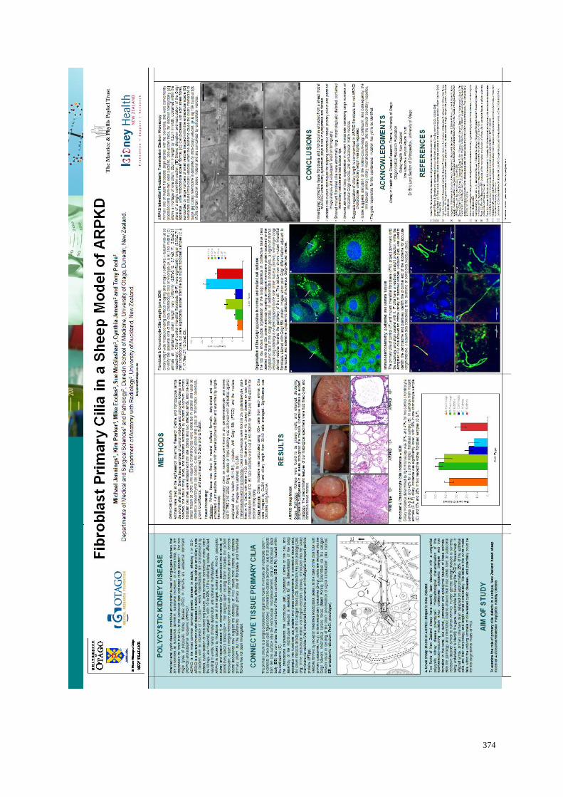
374

375

376

377

378
'Have a great idea, one great object, and follow it and never give in until you achieve it. You will win
in the end though you may have to wait long for it.'
‘It is the dreamers that move the world. Practical men are so busy being practical that they cannot see
beyond their own lifetime. Dreamers and visionaries have made civilisations. It is trying to do the
things that cannot be done that makes life worth while. The dream of today becomes the custom of
tomorrow.'
CJR