Ultrastructural study of the human neurohypophysis
12
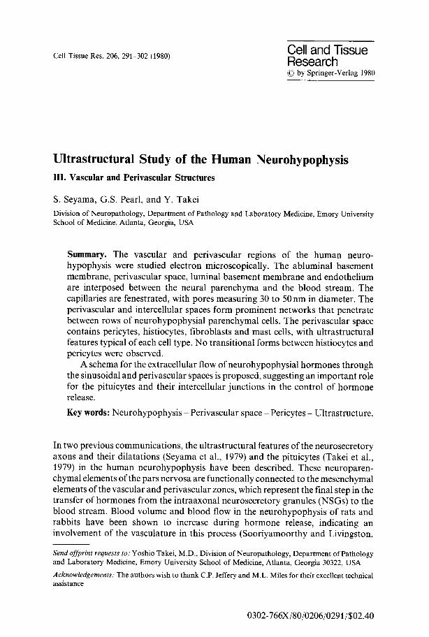
Cell Tissue Res. 206, 291-302 (1980) Cell and Tissue Research by Springer-Verlag 1980 Ultrastructural Study of the Human Neurohypophysis III. Vascular and Perivascular Structures S. Seyama, G.S. Pearl, and Y. Takei Division of Neuropathology,Department of Pathology and Laboratory Medicine, Emory University School of Medicine,Atlanta, Georgia, USA Summary. The vascular and perivascular regions of the human neuro- hypophysis were studied electron microscopically. The abluminal basement membrane, perivascular space, luminal basement membrane and endothelium are interposed between the neural parenchyma and the blood stream. The capillaries are fenestrated, with pores measuring 30 to 50 nm in diameter. The perivascular and intercellular spaces form prominent networks that penetrate between rows of neurohypophysial parenchymal cells. The perivascular space contains pericytes, histiocytes, fibroblasts and mast cells, with ultrastructural features typical of each cell type. No transitional forms between histiocytes and pericytes were observed. A schema for the extracellular flow of neurohypophysial hormones through the sinusoidal and perivascular spaces is proposed, suggesting an important role for the pituicytes and their intercellular junctions in the control of hormone release. Key words: Neurohypophysis -Perivascular space -Pericytes - Ultrastructure. In two previous communications, the ultrastructural features of the neurosecretory axons and their dilatations (Seyama et al., 1979) and the pituicytes (Takei et al., 1979) in the human neurohypophysis have been described. These neuroparen- chymal elements of the pars nervosa are functionally connected to the mesenchymal elements of the vascular and perivascular zones, which represent the final step in the transfer of hormones from the intraaxonal neurosecretory granules (NSGs) to the blood stream. Blood volume and blood flow in the neurohypophysis of rats and rabbits have been shown to increase during hormone release, indicating an involvement of the vasculature in this process (Sooriyamoorthy and Livingston, Send offprint requests to: Yoshio Takei, M.D., Divisionof Neuropathology,Department of Pathology and Laboratory Medicine, Emory University School of Medicine,Atlanta, Georgia 30322, USA Acknowledgements: The authors wish to thank C.P.Jefferyand M.L. Milesfor their excellent technical assistance 0302-766X/80/0206/0291/$02.40
-
Upload
independent -
Category
Documents
-
view
3 -
download
0
Transcript of Ultrastructural study of the human neurohypophysis
Cell Tissue Res. 206, 291-302 (1980) Cell and Tissue Research �9 by Springer-Verlag 1980
Ultrastructural Study of the Human Neurohypophysis III. Vascular and Perivascular Structures
S. Seyama, G.S. Pearl, and Y. Takei
Division of Neuropathology, Department of Pathology and Laboratory Medicine, Emory University School of Medicine, Atlanta, Georgia, USA
Summary. The vascular and perivascular regions of the human neuro- hypophysis were studied electron microscopically. The abluminal basement membrane, perivascular space, luminal basement membrane and endothelium are interposed between the neural parenchyma and the blood stream. The capillaries are fenestrated, with pores measuring 30 to 50 nm in diameter. The perivascular and intercellular spaces form prominent networks that penetrate between rows of neurohypophysial parenchymal cells. The perivascular space contains pericytes, histiocytes, fibroblasts and mast cells, with ultrastructural features typical of each cell type. No transitional forms between histiocytes and pericytes were observed.
A schema for the extracellular flow of neurohypophysial hormones through the sinusoidal and perivascular spaces is proposed, suggesting an important role for the pituicytes and their intercellular junctions in the control of hormone release.
Key words: Neurohypophysis -Perivascular space -Pericytes - Ultrastructure.
In two previous communications, the ultrastructural features of the neurosecretory axons and their dilatations (Seyama et al., 1979) and the pituicytes (Takei et al., 1979) in the human neurohypophysis have been described. These neuroparen- chymal elements of the pars nervosa are functionally connected to the mesenchymal elements of the vascular and perivascular zones, which represent the final step in the transfer of hormones from the intraaxonal neurosecretory granules (NSGs) to the blood stream. Blood volume and blood flow in the neurohypophysis of rats and rabbits have been shown to increase during hormone release, indicating an involvement of the vasculature in this process (Sooriyamoorthy and Livingston,
Send offprint requests to: Yoshio Takei, M.D., Division of Neuropathology, Department of Pathology and Laboratory Medicine, Emory University School of Medicine, Atlanta, Georgia 30322, USA
Acknowledgements: The authors wish to thank C.P. Jeffery and M.L. Miles for their excellent technical assistance
0302-766X/80/0206/0291/$02.40

292 s. Seyama et al.
1971, 1972, 1973; Livingston and Wilks, 1976). The perivascular spaces, which increase in area during hormone release (Livingston, 1978), are structurally connected to the parenchymal extracellular spaces and have been suggested to represent channels for hormone transfer and storage sites of importance in controlling the rate of hormone release (Lederis, 1974; Livingston, 1975; Livingston and Wilks, 1976). The cellular elements within the perivascular spaces have also been proposed to play a role in controlling hormone release (Dreifuss et al., 1975; Zambrano and De Robertis, 1968; Olivieri- Sangiacomo, 1972). Despite these observations in non-human mammals, there have been no detailed ultrastructural studies of the vascular and perivascular structures of the human neurohypophysis. This communication describes the ultrastructural features of the vascular and perivascular regions of the human neurohypophysis and attempts to summarize our observations on the functional morphology of the neurosecretory process.
Materials and Methods
The partes nervosae from 13 patients undergoing transsphenoidal hypophysectomy for palliative treatment of metastatic breast or prostate carcinoma were examined according to the methods described in a previous report (Seyama et al., 1979). Tissue from the pars intermedia or infundibulum was excluded from this study.
Results
Capillaries of the human pars nervosa are lined by relatively narrow endothelial cells interconnected by tight junctions between interdigitating contact surfaces. These cells display multiple fenestrations with a thin membranous diaphragm measuring 30 to 50nm in diameter (Fig. l). The endothelial cells contain cytoplasmic organelles similar to those seen in capillaries of other organs, including well-developed Golgi complexes, centrioles, filaments, microtubules and pinocy- totic vesicles. "Microtubular bodies", which consist of tufts of short microtubules measuring 20nm in outer and 14nm in inner diameter enveloped by a single membrane, were also observed (Fig. 1, inset). A single continuous basement membrane measuring 50nm in width, which represents the luminal basement membrane is located immediately adjacent to the outer surface of the endothelium (Figs. 1, 2). Another set of basement membranes is also present external to and independent of the luminal basement membrane, and is termed the abluminal basement membrane (Figs. 1, 2). The abluminal basement membrane, also measuring 50 nm in thickness, frequently has a complex orientation, pursuing a meandering course, twisting around cellular surfaces, and occasionally showing abrupt discontinuations.
The neurosecretory axons and pituicytic processes generally abut on the outer surface of the abluminal basement membrane (Fig. 3 a, b) and never show direct contact with the luminal basement membrane. Neurosecretory processes and their dilatations are also observed in apposition to the extracellular spaces. Type VI-

Ultrastructure of Neurohypophysial Vascular and Perivascular Spaces. III 293
Fig. 1. Capillary endothelium. The endothelial cell cytoplasm contains mitochondria, lipid, Golgi complexes, and rough endoplasmic reticulum. The endothelium is fenestrated (arrowheads) and surrounded by luminal (Lbm) and abluminal (Abm) basement membranes. Pericytic processes extend between the basement membranes. Inset: Microtubular bodies (arrows) and prominent Golgi complexes, x 7600; inset x 23,000

294 S. Seyama et al.
Fig. 2. Pericytes. Pericyte (Pc) surrounded by a basement membrane that is focally confluent with the luminal basement membrane (Lbm) and distinct from the abluminal basement membrane (Abm). The cytoplasm contains lysosomal bodies, lipid and aggregates of filaments. Inset: Aggregates of filaments with focal condensations (C) and pinocytotic vesicles (PI/) in the pericyte cytoplasm. • 6100; inset x 29,000
dilatations (Seyama et al., 1979) covered by the abluminal basement membrane occasionally enroach upon a perivascular space.
The gap between the luminal and abluminal basement membranes varies considerably, ranging from 0.1 I.tm to several i~m, and comprises the perivascular space. Various types o f mesenchymal cellular elements, a long with collagen fibrils, are present in this space. The most conspicuous o f these cells are the pericytes, which are characteristically located beneath the luminal basement membrane. They are generally surrounded by a basement membrane o f their own, which frequently

Ultrastructure of Neurohypophysial Vascular and Perivascular Spaces. III 295
Fig. 3. a Type A-fiber (A) and b Type B-fibers (B) abutting on the abluminal basement membrane. Synaptoid aggregates of vesicles (arrowheads) are present along their surface, c Histiocyte with cytoplasmic lysosomal bodies (L) and filopodial cytoplasmic projections (F). Pc Pericyte; C fenestrated capillary, a x 14,000; b • 14,000; c x 8800

296 S. Seyama et al.
Fig. 4. a Fibroblast with prominent rough endoplasmic reticulum and Golgi complexes within the perivascular space, b Mast cells within the perivascular space, exhibiting characteristic granules and microvillous cytoplasmic processes. Pc pericyte; En endothelial cell. a x 11,000; b x6000

Ultrastructure of Neurohypophysial Vascular and Perivascular Spaces. III 297
Fig. 5. Sinusoids. Channels lined by basement membrane (arrows) extending from the perivascular space into the neural parenchyma. Junctions are observed between pituicyte processes, associated with focal narrowing of the sinusoids (Sn). Pt pituicyte; En endothelial cell. x 10,000
fuses with the adjacent luminal basement membrane (Figs. 2, 3 c, 4b). In general, the pericytes appear to encircle the capillary partially, sending processes along the luminal basement membrane. The processes are occasionally closely applied to the endothelium, but no intercellular junctions were observed between these cells. The nuclei of pericytes are oval or irregularly shaped. In addition to the common cellular organelles, such as mitochondria and rough endoplasmic reticulum, the cytoplasm contains abundant microfilaments, usually located in areas close to the endothelium, oriented along the long axis of cytoplasmic processes, and often fused

298 S. Seyama et al.
to form focal condensations of amorphous material (Fig. 2, inset). Microtubules, measuring 20nm in outer diameter, are rarely present. A large number of pinocytotic vesicles were observed along the cytoplasmic membrane of the pericytes and their larger processes (Fig. 2, inset). The pericytes contain occasional lipid-like droplets, but lysosomal bodies are rare (Fig. 2). It was sometimes difficult to distinguish these capillary pericytes from the smooth muscle cells attached to arterioles and venules, and there appeared to be a gradual transition between the two cell types.
Several other cell types are present in the perivascular space, which, unlike the pericytes, possess no basement membrane and show no particular relationship to the luminal or abluminal basement membranes. These include histiocytes, fibroblasts and mast cells, in addition to very rare lymphocytes and polymorpho- nuclear leukocytes. The histiocytic cells are characterized by numerous cytoplasmic vacuoles and lysosomes and by irregular, microvillous or filopodial projections of their cell surface, which occasionally enclose amorphous material (Fig. 3c). The fibroblasts contain welt-developed Golgi complexes and saccular or tubular rough endoplasmic reticulum (Fig. 4a), as well as occasional lysosomes. The mast cells, which are easily recognizable by their characteristic cytoplasmic granules and microvillous projections, were primarily observed along the blood vessels (Fig. 4b).
The perivascular space is distinctly separated from the adjacent neural parenchyma by a complex abluminal basement membrane and usually does not exhibit direct contact with the parenchymal extracellular space. Occasionally, however, pituicytic processes are exposed to the perivascular space in areas of discontinuity of the abluminal basement membrane (Fig. 1). Rarely, a narrow space lined by the abluminal basement membrane on each side extends away from the original perivascular space to penetrate into a narrow extracellular space between rows of neuroparenchymal processes. These fine channels appear to divide the parenchyma into ill-defined lobules (Fig. 5). The paired, penetrating abluminal basement membranes focally become obscured, allowing the neuroparenchymal extracellular space to open directly into the perivascular space. In addition, electron-dense material identical to that of the basement membranes was occasionally observed in narrow extracellular spaces measuring 50 to 100 nm in diameter and are located in areas quite distant from the perivascular spaces (Fig. 5).
Discussion
The capillaries of the human pars nervosa show fenestrations, as demonstrated previously in various animal species (Barer and Lederis, 1966; Bergland and Torack, 1969; Lederis, 1965; Livingston, 1965; Tasso and Rua, 1978), thus providing ready access for substances to diffuse into the blood stream from the perivascular spaces. This finding establishes the neurohypophysis as one of the unique locations in the central nervous system where the blood-brain barrier does not exist. The endothelial cells, however, do not exhibit any particular ultrastructural features unique to the neurohypophysis. The "microtubular bodies" (Fig. 1, inset), although not previously described in the human pars nervosa, are a non-specific organelle found in the endothelial cells of various tissues in normal as well as pathologic

Ultrastructure of Neurohypophysial Vascular and Perivascular Spaces. III 299
conditions (Kawamura et al., 1974). They were first reported independently by Weibel and Palade (1964) and by Stehbens (1965). Although Burri and Weibel (1968) have implicated these structures in the process of blood coagulation, their exact function remains obscure at the present time (Thorgeirsson and Robertson, 1978).
The perivascular space intervening between the endothelium and neuroparen- chyma is lined by the luminal and abluminal basement membranes and harbors several different types of mesenchymal cells, including pericytes, histiocytes, fibroblasts and mast cells. The pericytes observed in the present study exhibit ultrastructural features identical with those of pericytes in other tissues (Fernando and Movat, 1964; Kuhn and Rosai, 1969; Movat and Fernando, 1964; Rhodin, 1968). Since the description by Zimmermann (1923) of their existence in a wide variety of tissues, various roles have been proposed for capillary pericytes based on their functional and morphologic properties, including: (1) contractility, with similarities to smooth muscle cells (Forbes et al., 1977; Rhodin, 1968; Stensaas, 1975; Weibel, 1974; Zimmermann, 1923); (2) ability to synthesize their own basement membrane and adjacent ground substance (Hart and Avery, 1963; Rhodin, 1968); and (3) multipotentiality, with the capability of developing into other cell types, such as phagocytes (Cancilla et al., 1972; Majino, 1965; Maxwell and Kruger, 1965; Nystrom, 1960; Torack, 1961). In the present ultrastructural study, pericytes were observed to contain cytoplasmic bundles of fine filaments with focal densities, similar to those seen in smooth muscle cells. The possibility that these pericytic filaments are similar or identical to actin has been suggested by LeBeux and Willemor (1975), who demonstrated in the rat brain that these filaments react with heavy meromyosin. It is, therefore, logical to assume that pericytes in the human pars nervosa are also contractile, modulating the caliber of capillaries and thereby playing a role in the regulation of local blood flow. Since the neurohypophysial blood volume and flow have been shown to increase under conditions of hormone release (Livingston and Wilks, 1976; Sooriyamoorthy and Livingston, 1971, 1972, 1973), it is possible that the pericytes participate, at least indirectly, in the release of neurohypophysial hormones into the blood stream.
Some investigators have claimed that neurohypophysial pericytes possess a phagocytic capability (Zambrano and De Robertis, 1968; Olivieri-Sangiacomo, 1972). In the present study of the human pars nervosa under non-pathologic conditions, the pericytes contained too few lysosomal elements to suggest a similarity with the histiocytes in the perivascular spaces, which appear to be the phagocytic cells in this region. No morphologic similarities are observed between the histiocytes and pericytes, and no cells with transitional features were observed. However, there is no ultrastructural evidence at the present time relating to the exact origin of these histiocytes. It is possible that they are hematogenous in origin or are derived from other neurohypophysial cells, such as pericytes or pituicytes. Interestingly, this argument is quite analogous to that regarding the origin of microglia in the central nervous system (Baron and Gallego, 1972; Boya, 1976; Kitamura and Fujita, 1973; Stensaas, 1975).
Light microscopically, the neurohypophysis in mammals has been reported to contain large numbers of mast cells, with the mast cells being most numerous in the infundibulum, few in number in the pars nervosa, and absent in the pars anterior of

300 S. Seyama et al.
Fig. 6. Extracellular hormone pathway. Hormone is released by exocytosis (?) or molecular dispersion (open arrows) into the extraceUular or perivascular spaces. Some neurosecretory material is phagocytosed by pituicytes, while the remaining hormone travels through the sinusoids, where junctional complexes control flow, to the perivascular space. After traversing the abluminal basement membrane and perivascular space, the hormone crosses the luminal basement membrane and enters the bloodstream through the fenestrated capillary endothelium
the hypophysis (Gray, 1935). Little attention has been given ultrastructurally to their presence in the neurohypophysis, except by Bodian (1963), who suggested that the products of mast cells may affect endothelial permeability, thereby triggering neurosecretion from the axonal terminals. In the present study, mast cells were not infrequently observed in the perivascular spaces of the human pars nervosa. However, their functional significance in neurohypophysial hormone release remains unclear.
In the present study several layers were shown to be interposed between the neuroparenchyrna and the blood stream in the human neurohypophysis, including the abluminal basement membrane, the perivascular space, the luminal basement membrane and the capillary endothelium. In animal species, it has likewise been observed that neurosecretory processes have no direct contact with the capillary endothelium. Instead, the prominent perivascular spaces communicate with a

Ultrastructure of Neurohypophysial Vascular and Perivascular Spaces. 1II 301
complex system o f in te rweaving channels , some o f which are l ined by a basement membrane , which pe rmea te the n e u r o p a r e n c h y m a (Barer and Lederis , 1966; Bodian, 1963). These channels are though t to be involved in h o r m o n e t r a n s p o r t and storage. In par t icu la r , Barer and Lederis (1966) and Livings ton and Wi lks (1976) have suggested tha t these channels p rov ide a " readi ly re leasable pool" o f ho rmone , a concept set for th pha rmaco log ica l l y by Thorn (1966) and by Sachs et al. (1967). W e have observed such a system of channels in the h u m a n pars nervosa. Since the n e u r o h y p o p h y s i a l h o r m o n e s are p r o b a b l y l ibera ted ini t ial ly into the ext racel lu lar spaces a long the axona l p l a s m a membrane , as discussed in a previous c o m m u n i c a t i o n (Seyama et al., 1979), these channels would be the mos t logical p a t h w a y for h o r m o n e t ranspor t . Based on our da t a on the h u m a n neu rohypophys i s (Seyama et al., 1979; Takei et al., 1979) and the da t a cur rent ly avai lab le in the l i terature, we p ropose a mechan i sm o f neu rohypophys i a l h o r m o n e release involving passage t h rough the ext racel lu lar spaces, with in tercel lular j unc t ions between pi tuicytes serving to regula te flow. The h o r m o n e mus t then cross the ab lumina l basement membrane , per ivascu la r space, luminal basement membrane , and fenes t ra ted capi l la ry endo the l ium to reach the b lood stream. This mode l is schemat ica l ly presented in Fig. 6. I t mus t be emphas ized , however , tha t this schema is speculat ive and fur ther research is necessary to evaluate this hypothesis .
References
Barer, R., Lederis, K.: Ultrastructure of the rabbit neurohypophysis with special reference to the release of hormone. Z. Zellforsch. 75, 201-239 (1966)
Baron, M., Gallego, A.: The relation of the microglia with the pericytes in the cerebral cortex. Z. Zellforsch. 128, 42-57 (1972)
Bergland, R.M., Torack, R.M.: An electron microscopic study of the human infundibulum. Z. Zellforsch. 99, 1-12 (1969)
Bodian, O.: Cytological aspects of neurosecretion in opossum neurohypophysis. Bull. John Hopk. Hosp. 113, 57-93 (1963)
Boya, J.: An ultrastructural study of the relationship between pericytes and cerebral macrophages. Acta Anat. 95, 598-608 (1976)
Burri, P.H., Weibel, E.R.: Beeinflussung einer spezifischen cytoplasmatischen Organelle von Endothelzellen durch Adrenalin. Z. Zellforsch. 88, 426M40 (1968)
Cancilla, P.A., Baker, K.N., Pollak, P.S., Frommes, S.P.: The relation of pericytes of the central nervous system to exogenous protein. Lab. Invest. 26, 376-383 (1972)
Dreifuss, J J., Sandri, C., Akert, K., Moor, H.: Ultrastructural evidence for sinusoid spaces and coupling between pituicytes in the rat. Cell Tissue Res. 161, 33~15 (1975)
Fernando, N.V.P., Movat, H.Z.: The fine structure of the terminal vascular bed: Ill. The capillaries. Exp. Molec. Pathol. 3, 87-97 (1964)
Forbes, M.S., Rennels, M.L., Nelson, E.: Ultrastructure of pericytes in mouse heart. Am. J. Anat. 149, 47-70 (1977)
Gray, J.: Preliminary note on the mast cells of the human pituitary and of the mammalian pituitary in general. J. Anat. 69, 153-158 (1935)
Han, S.S., Avery, J.K.: The ultrastructure of capillaries and arterioles of the hamster dental pulp. Anat. Rec. 145, 549-571 (1963)
Kawamura, J., Kamijo, Y., Sunaga, T., Nelson, E.: Tubular bodies in vascular endothelium of a cerebellar neoplasm. Lab. Invest. 30, 358-365 (1974)
Kitamura, T., Fujita, S.: Cells of the reticuloendothelial system in the brain and their relationship to circulating leucocytes, microglia and pericytes. Rec. Adv. RES Res. 13, 48-60 (1973)
Kuhn, C., Rosai, J.: Tumors arising from pericytes. Ultrastructure and organ culture of a case. Arch. Pathol. 88, 653-663 (1969)
LeBeux, YJ., Willemot, J. : Identification of actin-like filaments in rat brain by means of HMM labeling. J. Cell Biol. 6% 236a (1975)

302 S. Seyama et al.
Lederis, K.: An electron microscopical study of the human neurohypophysis. Z. Zellforsch. 65, 84%868 (1965)
Lederis, K.: Neurosecretion and the functional structure of the neurohypophysis. In: Handbook of Physiology-Endocrinology IV, part I. (E. Knobil and W.H. Sawyer, eds.), pp. 81-102. Washington: American Physiological Soc. 1974
Livingston, A.: Morphology of the perivascular regions of the rat neural lobe in relation to hormone release. Cell Tissue Res. 159, 551-561 (1975)
Livingston, A.: Effects of hormone-releasing stimuli on the area of the perivascular space in the neural lobe of the rat. Cell Tissue Res. 191, 501-506 (1978)
Livingston, A., Wilks, P.N.: Perivascular regions of the rat neural lobe. Cell Tissue Res. 174, 273-280 (1976)
Majino, G.: Ultrastructure of the vascular membrane. In: Handbook of Physiology-Circulation Vol. III. (W.F. Hamilton and P. Dow, eds.), pp. 2293-2375. Washington: American Physiological Soc. 1965
Maxwell, D.S., Kruger, L.: Small blood vessels and the origin of phagocytes in the rat cerebral cortex following heavy particle irradiation. Exp. Neurol. 12, 33-54 (1965)
Movat, H.Z., Fernando, N.V.P.: The fine structure of the terminal vascular bed: IV. The venules and their perivascular cells (pericytes, adventitial cells). Exp. Molec. Pathol. 3, 98-114 (1964)
Nystrom, S.: Pathological changes in blood vessels of human glioblastoma multiforme. Acta Pathol. Microbiol. Scand. 49 (Suppl. 137), 1-83 (1960)
Olivieri-Sangiacomo, C.: On the fine structure of the perivascular cells in the neural lobe of rats. Z. Zellforsch. 132, 25-34 (1972)
Rhodin, J.A.G.: Ultrastructure of mammalian venous capillaries, venules, and small collecting veins. J. Ultrastruct. Res. 25, 452-500 (1968)
Sachs, H., Sate, L., Osinchak, J., Carpi, A.: Capacity of the neurohypophysis to release vasopressin. Endocrinology 81, 755-770 (1967)
Seyama, S., Pearl, G.S., Takei, Y.: Ultrastructural study of the human neurohypophysis. I. Neurosecretory axons and their dilatations in the pars nervosa. Cell Tissue Res. 205, 253-271 (1980)
Sooriyamoorthy, T., Livingston, A.: Vasodilation in the rat neurohypophysis associated with hormone- releasing stimuli. J. Endocrinol 51, 11-12 (1971)
Sooriyamoorthy, T., Livingston, A.: Variations in blood volume of the neural and anterior lobes of the pituitary of the rat associated with neurohypophysial hormone-releasing stimuli. J. Endocrinol. 54, 407415 (1972)
Sooriyamoorthy, T., Livingston, A.: Blood flow studies in the neural lobe of the pituitary of rabbit associated with neurohypophysial hormone releasing stimuli. J. Endocrinol. 57, 75-85 (1973)
Stehbens, W.E.: Ultrastructure of vascular endothelium in the frog. Q.J. Exp. Physiol. 511, 375-384 (1965)
Stensaas, LJ.: Pericytes and perivascular microglial cells in the basal forebrain of the neonatal rabit. Cell Tissue Res. 158, 517-541 (1975)
Takei, Y., Seyama, S., Pearl, G.S., Tindall, G.T.: Ultrastructural study of the human neurohypophysis. II. Cellular elements of neural parenchyma, the pituicytes. Cell Tissue Res. 205, 273-287 (1980)
Tasso, F., Rua, S.: Ultrastructural observations on the hypothalamo-posthypophysial complex of the Brattleboro rat. Cell Tissue Res. 191, 267-286 (1978)
Thorgeirsson, G., Robertson, A.L.: The vascular endothelium-pathobiologic significance, Amer. J. Pathol. 93, 801-848 (1978)
Thorn, N.A.: In-vitro studies of the release mechanism for vasopressin in rats. Acta Endocrino153, 644- 654 (1966)
Torack, R.M.: Ultrastructure of capillary reaction to brain tumors. Arch. Neurol. 5, 416-498 (1961) Weibel, E.R.: On pericytes, particularly their existence on lung capillaries. Microvasc. Res. 8, 218-235
(1974) Weibel, E.R., Palade, G.E.: New cytoplasmic components in arterial endothelia. J. Cell Biol. 23, 101-
112 (1964) Zambrano, D., De Robertis, F.: The ultrastructural changes in the neurohypophysis after destruction of
the paraventricular nuclei in normal and castrated rats. Z. Zellforsch. 88, 496-510 (1968) Zimmermann, K.W.: Der feinere Bau der Blutcapillaren. Z. Anat. Entwicklungsgesch. 68, 29-109 (1923)
Accepted November 14, 1979