Ultrastructural Study of Axon Branching
126
Dissertation zur Erlangung des Doktorgrades der Fakultät für Chemie und Pharmazie der Ludwig-Maximilians-Universität München Ultrastructural Study of Axon Branching Hana Nedozrálová aus Brno, Czech Republic 2021
-
Upload
khangminh22 -
Category
Documents
-
view
2 -
download
0
Transcript of Ultrastructural Study of Axon Branching

Dissertation zur Erlangung desDoktorgrades der Fakultät für Chemie
und Pharmazie derLudwig-Maximilians-Universität
München
Ultrastructural Study ofAxon Branching
Hana Nedozrálová
aus
Brno, Czech Republic
2021


Erklärung
Diese Dissertation wurde im Sinne von § 7 der Promotionsordnung vom 28
November 2011 von Frau Prof. Elena Conti betreut.
Eidesstattliche Versicherung:
Diese Dissertation wurde selbstständig, ohne unerlaubte Hilfe erarbeitet.
Brno den 1.7.2021
____________
Hana Nedozrálová
Dissertation eingereicht am: 6.7.2021
1. Gutachterin / 1. Gutachter: Prof. Dr. Elena Conti
2. Gutachterin / 2. Gutachter: Prof. Dr. Frank Bradke
Mündliche Prüfung am: 16.9.2021


Contents
Summary . . . . . . . . . . . . . . . . . . . . . . . . . . . . . . . . . . . . . . . . . . . . . . . . . . . . . . . . . . . . . . . . . . . 7
List of publication . . . . . . . . . . . . . . . . . . . . . . . . . . . . . . . . . . . . . . . . . . . . . . . . . . . . . . . . . . 11
Preface . . . . . . . . . . . . . . . . . . . . . . . . . . . . . . . . . . . . . . . . . . . . . . . . . . . . . . . . . . . . . . . . . . . . . 13
1. Introduction . . . . . . . . . . . . . . . . . . . . . . . . . . . . . . . . . . . . . . . . . . . . . . . . . . . . . . . . . . . . . 151.1 Neuron . . . . . . . . . . . . . . . . . . . . . . . . . . . . . . . . . . . . . . . . . . . . . . . 15
1.1.1 Neuronal ultrastructure . . . . . . . . . . . . . . . . . . . . . . . . . . . . . . . . 161.1.2 Neuronal development . . . . . . . . . . . . . . . . . . . . . . . . . . . . . . . . 17
1.2 Axon branching . . . . . . . . . . . . . . . . . . . . . . . . . . . . . . . . . . . . . . . . . 201.2.1 Topology of axon branching . . . . . . . . . . . . . . . . . . . . . . . . . . . . 201.2.2 Axon branching in CNS . . . . . . . . . . . . . . . . . . . . . . . . . . . . . . . 21
1.3 Regulation of axon branching . . . . . . . . . . . . . . . . . . . . . . . . . . . . . . . . 211.3.1 Extracellular cues . . . . . . . . . . . . . . . . . . . . . . . . . . . . . . . . . . . 211.3.2 Signaling for branching . . . . . . . . . . . . . . . . . . . . . . . . . . . . . . . . 24
1.4 Cytoskeleton organization and dynamics at the axon branch . . . . . . . . . . . 261.4.1 Actin remodeling . . . . . . . . . . . . . . . . . . . . . . . . . . . . . . . . . . . . 261.4.2 Microtubule remodeling . . . . . . . . . . . . . . . . . . . . . . . . . . . . . . . 27
1.5 Organelle interplay within the neuronal branch . . . . . . . . . . . . . . . . . . . . 291.5.1 Mitochondria . . . . . . . . . . . . . . . . . . . . . . . . . . . . . . . . . . . . . . 291.5.2 Endoplasmic reticulum . . . . . . . . . . . . . . . . . . . . . . . . . . . . . . . . 301.5.3 Golgi complex . . . . . . . . . . . . . . . . . . . . . . . . . . . . . . . . . . . . . . 32
1.6 Membrane remodeling at axon branches . . . . . . . . . . . . . . . . . . . . . . . . 331.6.1 Membrane expansion via fusion with synaptic vesicles . . . . . . . . . . 331.6.2 Membrane expansion via ER contacts . . . . . . . . . . . . . . . . . . . . . . 341.6.3 Membrane retraction . . . . . . . . . . . . . . . . . . . . . . . . . . . . . . . . . 34
1.7 Local translation in neurons . . . . . . . . . . . . . . . . . . . . . . . . . . . . . . . . . 351.7.1 Ribosomes . . . . . . . . . . . . . . . . . . . . . . . . . . . . . . . . . . . . . . . . 351.7.2 Local translation and axon branching . . . . . . . . . . . . . . . . . . . . . . 36
Aim of thesis . . . . . . . . . . . . . . . . . . . . . . . . . . . . . . . . . . . . . . . . . . . . . . . . . . . . . . . . . . . . . . . . 39
2. Results . . . . . . . . . . . . . . . . . . . . . . . . . . . . . . . . . . . . . . . . . . . . . . . . . . . . . . . . . . . . . . . . . . 412.1 Local orchestration of cellular machineries at axon branch by in-situ cryo-ET 41
– –

2.2 Direct induction of microtubule branching by microtubule nucleation factorSSNA1 . . . . . . . . . . . . . . . . . . . . . . . . . . . . . . . . . . . . . . . . . . . . . . . 75
3. Discussion . . . . . . . . . . . . . . . . . . . . . . . . . . . . . . . . . . . . . . . . . . . . . . . . . . . . . . . . . . . . . . . 1013.1 Visualization of axon branch . . . . . . . . . . . . . . . . . . . . . . . . . . . . . . . . 1013.2 Cytoskeleton remodeling . . . . . . . . . . . . . . . . . . . . . . . . . . . . . . . . . . . 1023.3 Role of ER . . . . . . . . . . . . . . . . . . . . . . . . . . . . . . . . . . . . . . . . . . . . 103
4. Outlook and future perspectives . . . . . . . . . . . . . . . . . . . . . . . . . . . . . . . . . . . . . . . . . . 105
List of figures . . . . . . . . . . . . . . . . . . . . . . . . . . . . . . . . . . . . . . . . . . . . . . . . . . . . . . . . . . . . . . . 108
References . . . . . . . . . . . . . . . . . . . . . . . . . . . . . . . . . . . . . . . . . . . . . . . . . . . . . . . . . . . . . . . . . . 109
Acknowledgment . . . . . . . . . . . . . . . . . . . . . . . . . . . . . . . . . . . . . . . . . . . . . . . . . . . . . . . . . . . . 125

Summary
The brain function depends on a vast number of intricate connections between neuronsto process, transmit and store information. To fulfill these tasks, neuronal cells adapt adistinct polarized morphology during their development and must maintain the polarityover the life span of the organism to sustain the correct brain function. Neurons establishtwo main compartments, the somatodendritic and the axonal compartments, which aredistinct in their morphology, molecular composition, and subcellular functions. At theearly stages of development, several long protrusions or neurites extend from the maincell body (soma), where the nucleus is located. One of the neurites develops into theaxon, while the remaining become dendrites [1]. Dendrites receive signals from axons ofupstream cells via synaptic connections, whereas axons transmit signals to the dendritesof downstream cells. At the tip of a growing axon, an actin-rich growth cone probes theextracellular signaling molecules to find the synaptic target on dendrites of another neuron[2, 3]. Besides the outgrowth and path-finding, axons form additional branches to createmultiple synaptic contacts which are crucial for the proper intertwining of neuronal circuits[4].
Reflecting the specialized function of axons, their molecular organization is also unique inorder to sustain the local developmental process. The cytoskeleton provides the structuralsupports needed to maintain the length of the axon shaft. Within the backbone of theaxon, microtubules are parallelly forming bundles with their plus ends pointing towardsthe distal end of the axon whereas actin forms a ring-like lattice around the circumferenceof the axon [5, 6]. Actin is also accumulated at the areas undergoing dynamic changes, likegrowth cones and filopodia [7, 8, 9, 10]. Due to the long distance from the soma to the tipof an axon, neurons employ an intricate transport system delivering organelles, proteins,and other cell components into the distal regions, providing energy, synaptic vesicles, andnew building material for growth and maintenance.
In addition to the cell-body based translation and transport of new proteins into distalregions of the axon, it has been suggested that neurons employ a regulatory system con-trolling the actions of molecules in local areas [11, 12, 13, 14]. This is advantageous tosustain the fast-changing local requirements at the areas far from the soma. Examples in-clude localized translation and enrichment of protein, which are needed for the local axondevelopment, regulation, and homeostasis. In particular, clusters of mRNAs are foundin local areas within dendrites and axons [15, 13, 16, 17] giving an indication of localprotein synthesis and the type of these mRNAs depends on the developmental stage andthe location within a neuron [15]. Recent polysome profiling and ribosome footprinting
– 7 –

study revealed that neuronal ribosomes show local preferential translation by monosomes(single ribosomes engaged with an mRNA) and that these neuronal monosomes were inthe process of active protein synthesis [18]. Local translation by monosomes has beensuggested as a way to control protein expression when only a small amount of protein islocally needed [13]. Despite the critical role of local axon translation, details of the trans-lation are not fully understood. While the evidence for the presence of ribosomes alongthe axons has been growing [19, 20, 21], the direct observation of ribosomes undergoingprotein synthesis and the following effect on the cytoskeleton and organelle reorganizationhas remained to be a major challenge.
Axon branching is a major process during axon development and morphogenesis, whichis necessary for the propagation of signals from one neuron to diverse regions within thenervous system [22, 23, 24]. The initiation of axon branching is regulated by extracellularcues, which activate signaling pathways affecting cytoskeleton dynamics. Local accu-mulation of actin leads to the formation of actin patch, which is applying force on axonshaft membrane forming short protrusion from the axon [25, 26, 27]. These protrusions,called filopodia, are filled with actin and they act as a structural precursor for the branchuntil microtubules are recruited to enter and stabilize the axon branch [28, 23, 29]. Theenrichment of mRNA [30, 25] encoding proteins such as beta-actin has been reported atthe axon branch point [31], suggesting that the proteins required to build up the branchare synthesized locally [32]. Interestingly, it has been reported that mitochondria are alsoenriched at axon branching points and that they are colocalizing with the mRNA [25]. Thespecific localization of mitochondria is suggested to generate necessary energy [33] andadjust Ca2+ concentration for establishing branch morphogenesis and signal transduction[34]. It was also reported that mitochondria along axons undergo fission and that the reg-ulation of the mitochondria size coincided with terminal axon branching [35]. Curiously,very little is known about the role of the endoplasmic reticulum (ER) during axon branch-ing, although ER is known to be present throughout the axon shaft and is suggested toinfluence microtubule stability through various interactions [36]. Nonetheless, the struc-tural details of cytoskeleton and organelle orchestration and the organization of proteinsynthesis machineries at axon branches are largely unknown.
In this thesis, I aimed to directly visualize axon branching points using mouse primaryneurons by in-situ cryo-electron tomography (cryo-ET), and to understand the structuralorganization of the key cellular machineries during axon branching. I compared the or-ganization of the premature branch (filopodium before the microtubule invasion) and themature branch (branch stabilized by microtubules). Small fragments of actin and smallsize mitochondria (∼ 500 nm) were localized at axon branches. An intricate network ofER membranes was often found between microtubule bundles and mitochondria, and occa-sional interactions of the ER membrane to microtubules and mitochondria were visualized.In most cases ER was found only in mature branches together with microtubules, indicatingthat the migration of ER into the axon branch is guided by microtubules, raising a possiblerole of ER in branch maturation and stabilization. I further demonstrated the first directobservation of clusters of ribosomes selectively accumulated at axon branches. They werelocated in the cytoplasm as well as attached to planar ER membranes at places where ERtubes widen, spreading over the space made for the branching activity. Subtomogram-
– 8 –

averaging and distance analysis of clustered ribosomes indicated that the ribosomes formpolysomes, suggesting these ribosomes are active. Axon branches also contained iso-lated ribosomes, which agree with the report of monosomes, presumably synthesizing asmall number of proteins. Our observation provides a comprehensive picture of the axonbranching process utilizing the unique contextual advantage of in-situ cryo-ET.
– 9 –


List of publications
1. Nedozralova H., Basnet N., Ibiricu I., Bodakuntla S., Biertuempfel Ch., MizunoN., (2021). Local orchestration of cellular machineries at axon branch by in-situcryo-ET observation. Submitted –Research article–
2. Basnet N., Nedozralova H., Crevenna A.H., Bodakuntla S., Schlichthaerle T.,Taschner M., Cardone G., Janke C., Jungmann R., Magiera M.M., BiertuempfelCh., Mizuno N., (2018). Direct induction of microtubule branching by microtubulenucleation factor SSNA1. Nature Cell Biology 20, 1172–1180. –Research article–
3. Bodakuntla S., Nedozralova H., Basnet N., Mizuno N, (2021). Cytoskeleton andmembrane organization at axon branches. Frontiers in Cell and DevelopmentalBiology. Submitted. –Review article–
– 11 –


Preface
The work presented in this thesis was performed at the laboratory of Dr. Naoko Mizuno atMax Planck Institute of Biochemistry, Martinsried, Germany, and at National Heart Lungand Blood Institute, Bethesda, Maryland, United States. The thesis mainly comprises tworesearch topics: 1) “In-situ survey of local reorganization of cellular machineries at axonbranch”, and 2) “Direct induction of microtubule branching by microtubule nucleationfactor SSNA1”. Therefore, this thesis is presented in a cumulative manner.
Chapter 1 includes the general introduction into the specialized polarized morphology ofneurons, their ultrastructure, and the stages of neuronal development. The introductionthen focuses on axon branching as an important event during neuronal development. Thedifferent topologies of axon branching are discussed and examples of axon branchingin CNS are presented. The following sub-chapters are dedicated to different aspectsof axon branching: regulation, cytoskeleton remodeling, organelle interplay, membraneremodeling, and local translation. Some of these topics are also covered in our reviewpublication ”Cytoskeleton and membrane organization at axon branches” and thus thereview is not presented separately in this thesis.
Chapter 2 presents the result section which consists of the research articles divided into twosub-chapters. The first sub-chapter 2.1 presents the research article’s first topic: “In-situsurvey of local reorganization of cellular machineries at axon branch” followed by secondsub-chapter 2.2 which includes the research article: “Direct induction of microtubulebranching by microtubule nucleation factor SSNA1”.
Finally, chapter 3 includes an extended discussion and the last chapter 4 includes theoutlook and future directions of the first topic.
– 13 –


1. Introduction
Figure 1: Morphology and ultrastructure of neuron cell. The scheme represents mainneuronal compartments: soma, dendrites with spines, axon shaft and branches, axonalinitial segment (AIS), growth cone and synaptic terminal. The cutouts are highlighting thedistinct ultrastructure of the given segment. The color code for cell elements is depictedin the legend.
1.1 NeuronNeurons are one of the most specialized cells with distinct highly polarized morphologywhich is directly linked to their function. The brain function is derived from neurons orga-nized into a complex network. The processing and transmission of information depend ona vast number of intricate connections between neurons. The correct wiring of the neuronalnetwork facilitates the development of cognitive functions and storage of memory. To fulfillthe scale and complexity of its functions, it is crucial for neurons to develop properly and
– 15 –

maintain neuronal homeostasis over the life span of the organism. Neurons are comprisedof two main compartments, the somatodendritic and the axonal compartment, which aredistinct in their morphology, molecular composition, and subcellular functions[1]. Axonis a single long neurite transferring signal over long distances to pass it to the downstreamcells and dendrites are multiple shorter neurites protruding from the cell body whose taskis to receive the incoming signal from other cells. The contacts between neurons occurat specialized junctions called synapses formed between axon terminals or boutons of thepresynaptic neurons and dendritic spines of the postsynaptic neuron. Axonal boutonscontain synaptic vesicles filled with neurotransmitters as well as cytoskeleton scaffolding,regulatory molecules, and organelles that organize, promote, and regulate the release ofneurotransmitters [2, 3]. Synapses are established during the maturation of neuron, inearlier stages, during the growth phase, the distal tips of extending neurites are finished bya growth cone, which is seeking their synaptic target by following guidance cues [23]. Themorphology of neuron is illustrated in Figure 1.
1.1.1 Neuronal ultrastructureThe inner organization of the neuron follows its polarized morphology. The cell body issmall and contains the usual organelles present in every cell whereas dendrites and axons,thin and long membranous protrusions filled with cytoplasm, are mainly occupied by acytoskeleton which provides scaffolding and stability to these protrusions. Compositionof the cytoskeleton microtubules, actin, and neurofilaments differ between dendrites andaxons. Both dendrites and axons have bundled arrays of parallel microtubule filamentscrosslinked by various microtubule-associated proteins (MAP) [37], but axonal micro-tubules are oriented with their plus tip facing towards the distal end of the axon, whereasdendritic microtubule filaments have mixed polarity [37, 38]. Actin is mainly present inareas undergoing remodeling, like filopodia, growth cones, or dendritic spines, and asadditional support along the length of neurites [38]. The growth cone is a large dynamiccytoskeleton-supported extension at neurite tip with motile function. The steering andmobility are maintained by constant building and rebuilding of the F-actin cytoskeleton,rising into filopodia and lamellipodia, dynamically reacting to the external stimuli vianumerous membrane receptors present in the growth cone [23]. Actin also forms a latticeof ring-like patches around the length of the axon shaft, spaced out by spectrin dimers,supporting the axonal scaffold and increasing its elasticity [6, 39]. Neurofilaments arearranged along the axons between the microtubule bundles and membrane, they form anarray of parallel filaments providing structural support and were suggested to control axondiameter [38].
Another structurally distinct compartment of the neuron is the axonal initial segment(AIS) which separates the somatodendritic region from the axon. AIS is positioned at thebeginning of the axon adjacent to the soma and has two main functions, first, to maintaincell polarity by sorting the cellular elements which can enter into the axon, and second, tointegrate synaptic inputs and generate an action potential. The function is carried by thedistinct organization into three layers; plasma membrane, submembrane cytoskeleton, andinner AIS shaft. These layers are integrated by a multidomain scaffolding protein ankyrinG (AnkG) that functions as a master organizer of the AIS [40].
– 16 –

Organelles, synaptic vesicles, proteins, lipids, and other cell material are actively trans-ported into the peripheries of neurons via motor proteins. The vesicle trafficking isbidirectional; anterograde transport is carried out by kinesins and retrograde transport ismediated by dyneins. Mitochondria transport into dendrites and axons is crucial to provideenergy at the regions far from the cell body. Synaptic vesicles containing neurotransmittersare transported to axonal terminals. Various other vesicles travel between soma, dendrites,and axons [41]. The endoplasmic reticulum is present through the whole neuron whereasthe Golgi apparatus is mainly located in soma, with outposts and satellites in dendrites[42]. Proteins are synthesized centrally in the soma and distributed throughout the lengthof the neuron. However, in recent years it was reported that part of translation is carriedout locally at axons and dendrites [13]. Local translation serves as a unique solution toproduce, maintain and modify the proteins that are required for the correct development,function, and plasticity of the nervous system.
The above description of neuron ultrastructure is only a brief summary of the topic. A morein-depth description of axonal ultrastructure will be discussed in later chapters. Visualrepresentation of the summary in Figure 1.
1.1.2 Neuronal developmentNeurons undergo multiple stages of development from unpolarized embryonic neurons todifferentiated neurons with mature synapses. First, the polarity of the neuron is establishedand the axon starts to elongate, then collateral branches and terminal arbors are formed tofacilitate the correct wiring of the neural network, finally, the maturation of the neuron iscomplete when synapses are formed.
The process of polarization and neuronal development was first described in culturedhippocampal neurons [43] and comprises 5 well-defined steps (Figure 2). At stage 1, soonafter seeding, round neurons form a lamellipodium while they attach to their substrate.Stage 2 starts within few hours when undifferentiated neurites begin to sprout. Theneurites are cylindrical protrusions that contain a growth cone, the expanded motile tip ofgrowing protrusion. They lack molecular and structural characteristics of mature axonalor dendritic processes. At this stage, multiple neurites can extend and retract withoutsignificant elongation. At stage 3, usually one day after seeding, one of the neurites withan enlarged growth cone starts to elongate rapidly without retraction to form an axon whilethe other neurites pause. Within a week at stage 4, the remaining neurites continue togrow and branch to form multiple dendrites. Later at the final step, the axon and dendritescontinue to develop further, axon starts forming collateral and terminal branches along itsshaft, and dendritic protrusions, or spines, appear. After two weeks, the maturation of theneuron is completed by synapse formation allowing for electrical activity throughout theneuronal network [43, 44, 45, 46].
The polarization process begins between stage 2 and 3 of neuron development when oneof the neurites start to elongate to become the axon. Further polarization occurs during thefollowing days of development when axon and dendrites become increasingly different,adopting their specialized shape and function. Neuronal wiring refers to the process ofneuronal branching during which neurons start to form complex networks by diversifying
– 17 –

their connections to multiple target cells. Both dendrites and axons can form branches,dendrites in form of branches with spines and axons as either collateral branches or terminalbranches or arbors. Thus one axon can create a synaptic connection with multiple dendritesof target cells. The development of neurons as well as the maintenance of their homeostasisand plasticity during the whole life span is a complex task and requires perfect coordinationbetween signaling, cytoskeleton remodeling, cellular transport, and protein synthesis, soall the components are at the right time at the right location.
– 18 –

Figure 2: Establishment of polarity and stages of neuronal development in hippocam-pal neurons in culture. DIV (days in-vitro) refers to a time in culture.
– 19 –

1.2 Axon branchingDuring the establishment of the correct intra-neuronal connectivity between neurons andtarget cells, axon branches and even whole axons can be remodeled. To generate andspecify their correct wiring into the neuronal network, developing axons sprout collateraland terminal branches with variable length, density, and complexity, allowing them to createsynapse with multiple target cells simultaneously, with excess synapses being pruned atlater stages [24].
1.2.1 Topology of axon branching
Figure 3: Axon binding topology. A) Collateral branching. New branch emerges alongthe length of the axon shaft. B) Bifurcation. The tip of the growth cone splits into twoequal branches.
Even though growth cones lead the elongation of axons towards their target locationfollowing the pathways marked by the extracellular guidance cues [47], in the vertebrateCNS, axonal growth cones themselves do not typically enter all the way into their targetregion. Instead, axons form synaptic connections with their target through growth cone-tipped collateral branches that emerge from the axon shaft, which further re-branch from thecollateral branches into terminal arbors [22]. This collateral or interstitial type of branchingis the most common (Figure 3A) [48], nevertheless, the second type of branching can occur
– 20 –

in certain circumstances [49]. Branches can arise by bifurcation (Figure 3B), a splittingof the terminal growth cone [50], such as at the mouse dorsal root entry zone, where thegrowth cones of dorsal root ganglion (DRG) axons split to form two daughter branchesthat arborize in the spinal cord [51, 52].
1.2.2 Axon branching in CNSIn the mammalian CNS, axon branches typically extend collaterally from the axon shaftbehind the terminal growth cone. This branching typically occurs after the main axongrowth cone has bypassed the target region [48]. Cortical axons in rodents initially bypassthe basilar pons [53], but after a delay, they form filopodia, which can develop into stablebranches that arborize in the pons [54] (Figure 4A). Developing corticospinal axons alsobypass spinal targets and later form interstitial branches that arborize once they have enteredtopographically appropriate target sites in the spine [55] (Figure 4A). Segments of the axonsdistal to the target are later eliminated [48, 56]. Callosal axons, which connect the twocerebral hemispheres, also undergo delayed interstitial branchin [57] beneath their corticaltargets and developing thalamocortical axons in vivo form layer-specific lower and uppertiers of terminal arbors in the barrel field, which together form a spatial map of the facialvibrissae in the rodent somatosensory cortex [58] (Figure 4B). In the avian [59] and rodent[60] retinotectal systems, retinal ganglion cell axons initially overshoot their terminationzone in the tectum and later emit collateral branches at correct tectal positions, which isfollowed by terminal arborization and regression of the distal axon [61, 62] (Figure 4C).However, in frogs and fish, growth cones of retinal axons form only terminal arbors [63].Thus, the neuron wiring in the vertebrate CNS is mediated via collateral axon branchingand terminal arborization.
1.3 Regulation of axon branchingThe process of axon branching must be tightly controlled and regulated. The length of theaxon shaft must maintain its stability while at the selected site of new branch formation,dynamic remodeling of axon into branch takes place.
1.3.1 Extracellular cuesAxon branching occurs at localized regions of the axon and is regulated by target-derivedmolecular cues. Families of extracellular axon guidance cues, growth factors, and mor-phogens can regulate axon branching by determining the correct position of branches orby shaping terminal arbors [64, 24, 22].
Netrins, such as netrin-1, are diffusible guidance cues with attractive effect [65]. Theyguide axon path and induce axon branching [66, 67]. Focal application of netrin-1 caninduce localized filopodial protrusions de novo along the axon shaft and increase branchlength without increasing the extension of the primary axon [66, 26]. Netrin-1 alsoincreases the total number of terminal arbor branches in the frog optic tectum [68].
Ephrins are membrane-bound repulsive cues [69, 70], they have a role in specifying the
– 21 –

Figure 4: Axon branching in developing CNS. A) Sensorimotor cortical axon formsbranches into the pons and spinal cord. B) In corpus callosum and thalamocortical axonsinitially extend past their eventual terminal regions (1). After a delay, branches extendfrom the axon shaft (2) and the distal axons are eliminated (3). Finally, terminal arbors areformed in target regions (4). C) Retinotectal terminal branching [22].
– 22 –

locations of axon branches. Ephrin-A5 and its receptor (EPHA5) can both promote orrepress the branching of various cortical axons by growing on membranes from specificcortical layers [71]. Ephrin-As can both stimulate the branching of one type of axons whilerepelling axons from other regions that do not innervate the same brain area [22]. In somecases, EPHAs act both as receptors activated by ephrin ligands (forward signaling) and asligands that activate ephrins (reverse signaling) [61, 72]. Their activity also cooperates withthe other branch-promoting molecules such as brain-derived neurotrophic factor (BDNF)[73]. Axon branching is repressed while extending axon is traversing the non-target regionsuntil reaching the area with enriched positive cues which signal the arrival to the correcttarget region where axon branching is evoked.
Semaphorins (SEMA) are repellent axon guidance cues that function in the assembly ofneuronal circuits [74]. For example, in primary cortical neurons, SEMA3A repels axons,inhibits axon branching, and decreases branch length without affecting the length of theprimary axon [66, 68]. SEMA3D has been also shown to selectively affect branchesdepending on their position along the axon. Central branches were not affected whilethe branching at the peripheral areas was induced [75]. However, SEMA3A can alsopositively influence axon branching. Recently, SEMA3A was shown to promote branchingby cerebellar basket cell axons onto Purkinje cells in the cerebellar cortex [76].
Like semaphorins, SLITs, which act on ROBO (Roundabout) receptors, are repulsive cuesand they can also both promote and inhibit branching. For example, SLITs can promote thecollateral axon branching of mammalian sensory DRG axons in-vitro [27] but also inhibitarborization of retinal ganglion cell axons in the zebrafish optic tectum [77]. It seems thatboth SLITs and semaphorins manifest context-dependent repression or promotion of axonbranching, depending on the particular population of neurons.
Growth factors, such as fibroblast growth factor (FGF), and neurotrophins, such as nervegrowth factor (NGF) and brain-derived neurotrophic factor (BDNF), can also promote axonbranching and terminal arbor formation. For example, NGF increases DRG axon branching[78, 79], BDNF induces filopodial and lamellipodial protrusions along frog spinal axons[80], and FGF-2 and BDNF stimulate branching in cortical axons [81, 50]. BDNF hasbeen also shown to induces rapid extension of new terminal branches on frog retinal axons[82]. Interestingly, although both BDNF and netrin-1 increase the arbor complexity ofretinal arbors, their mode of action seems to be different [68]. BDNF promotes the additionand stability of axon branches whereas netrin-1 induces new branch growth but not branchstabilization. This example shows that different guidance cues can, therefore, have a similareffect on final arbor morphology by different dynamic strategies [22].
WNTs comprise a diverse family of secreted morphogens that shape embryonic devel-opment. Several WNTs also function as axon guidance cues [83, 84], enhances axonextension [85], or regulate axon branching [86, 87]. For example, WNT7A, secreted bycerebellar granule cells, induces remodeling of pontine mossy fiber axons by inhibitingtheir elongation and enlarging their growth cones causing an increase in axon branching[88]. WNT3A, secreted by motor neurons, induces axon branching by regulating presynap-tic terminal arborization of spinal sensory neurons [86, 87], whereas WNT5A, a repulsiveaxon guidance cue for cortical axons in-vivo [89], induced the elongation of axons and
– 23 –

branches [85] but did not increase the numbers of axon branches itself [90].
Together these findings indicate that extracellular cues can selectively promote or repressonly axon branching without affecting the growth and guidance of the parent axon. Theexplanation, why the same guidance molecules can have a distinct effect on axon guid-ance, elongation, and branching, could be in the differences between local signaling andcytoskeleton remodeling mechanisms at the growth cone versus along the axon shaft.
1.3.2 Signaling for branching
Figure 5: Signaling pathways promoting axon branching. A) Signaling promoting actinpolymerization. NGF-induced TRKA signaling promotes axon branching by activatingPI3K and, in turn, RAC1, which activates actin-associated proteins to increase actin poly-merization and the formation of actin patches. Cortactin, recruited by Septin6, promotesthe emergence of filopodia from actin patches. B) Signaling promoting microtubule desta-bilization at early branching stages (top) and microtubule stabilization at later branchingstage (bottom). Both pathways promote axon branching each by opposing effects on micro-tubule stability. BDNF-induced TRKB signaling activates MKP-1 and, in turn, inactivatesJNK, resulting in microtubule destabilization caused by increased tyrosination of Stath-min. WNT induced GSK3beta inhibition decreases MAP1B phosphorylation leading toan increase in microtubule stability [22].
The numerous guidance cues described in the previous chapter are affecting axon branchingvia the activation of multiple signal transduction pathways. So far signaling throughRhoGTPases [91] and the protein kinase glycogen synthase kinase 3 beta (GSK3beta) [92]have emerged as a transduction node for the signal from the multiple extracellular cues ontocytoskeletal effectors resulting in changes of actin and microtubule dynamics. Although nosingle pathway from receptor to the cytoskeleton has been completely defined yet, severalexamples of signaling pathways regulating axon branching have been described already[22].
NGF promotes the formation of filopodia and branches in chick sensory axons [78] by
– 24 –

affecting the rate of actin patch formation [93] (Figure 5A). In particular, NGF binds TRKA(Tropomyosin receptor kinase A) receptor, activating PI3K (phosphoinositide-3-kinase),resulting in activation of RAC1 (Ras-related C3 botulinum toxin substrate1) GTPase, whichdrives the activity of the ARP2/3 (actin-related protein 2/3) activator WAVE1 (WASP-family verprolin homologous protein 1). WAVE1 promotes ARP2/3 dependent actin patchinitiation and subsequent axon branch formation [94, 30].
The actin-associated protein cortactin influences axon branching by promoting actin poly-merization and the membrane protrusion by the emergence of filopodia [95, 96]. Proteolysisof cortactin by caplain represses actin polymerization and keeps axon shafts in a consol-idated state. The effect of calpain can be negated by factors such as netrin-1 and BDNFto initiate branching. These examples illustrate the part of the axon branching signalingpathway responsible for the remodeling of actin.
Multiple pathways regulating the remodeling of microtubule cytoskeleton have been de-scribed so far. The BDNF has been shown to induce axon branching of mouse corticalneurons by microtubule destabilization through binding to TRKB (Tropomyosin recep-tor kinase B) receptor [81], whereas WNT has been reported to promote axon branchingby having stabilizing effect on microtubules through GSK3beta signaling [88] (Figure5B). The seemingly contradictory effects of BDNF and WNT on microtubules have beenexplained by discovering that microtubule destabilization occurs during the early phaseof branching whereas the destabilization comes into play in the later phase of branchmaturation when stable neurons enter the nascent branch.
BDNF binds TRKB receptor, inducing MPK-1 phosphates (mitogen-activated protein ki-nase phosphates) which then inactivates JNK (MAPK c-jun N-terminal kinase) by dephos-phorylation, which in turn reduces the phosphorylation of the JNK substrate stathmin-1(STMN1), thereby activating STMN1 leading to increased microtubule tyrosination andtheir consequent destabilization resulting in increased cortical axon branching [81, 37].
WNT inhibits GSK3beta activity [84, 88], which normally phosphorylates MAPs (mi-crotubule associated proteins) such as tau, APC (Adenomatous polyposis coli protein),and MAP1B. Phosphorylated MAP1B maintains microtubules in a dynamic state [97],therefore the activation of the WNT pathway decreases MAPs phosphorylation and in turnincreases microtubule stability leading to axon branching [81].
BDNF and netrins have been shown to specifically promote terminal axon branching infrog retinotectal axons [68, 82]. In this signaling pathway, NEDD4 (E3 ubiquitin-proteinligase) ubiquitinates PTEN (Phosphatidylinositol 3,4,5-trisphosphate 3-phosphatase anddual-specificity protein phosphatase) and PTEN degradation increases terminal arboriza-tion by promoting PI3K, which is known to regulate cytoskeletal dynamics [98]. Onthe other hand, inhibition of NEDD4 caused an increase in PTEN levels and subsequentinhibition of axon branching.
– 25 –

1.4 Cytoskeleton organization and dynamics at the axonbranch
The extensive reorganization of the cytoskeleton is an important part of the axon branchformation [23]. First, actin-filled filopodium emerges to form a premature branch which islater stabilized by the insertion of microtubules into the mature branch (Figure 6).
Figure 6: Remodeling of the cytoskeleton during axon branching. A) Extracellularcues activate signaling pathways that locally increase actin remodeling. B) Actin patch isformed by the pressure of accumulated actin on the outer membrane. C) Further extensionof plasma membrane under the force of actin-based components leads to the formation offilopodium protrusions. D)-E) Microtubules start invading filopodium to stabilize the newbranch and promote its maturation. The color code for cell elements is depicted in thelegend.
1.4.1 Actin remodelingThe first step of axon branching is the formation of the actin patch (Figure 6B) when shortpieces of actin accumulate along the axon shaft pushing at the plasma membrane to protrudeoutwards [94]. Further extension of plasma membrane under the force of actin-basedcomponents leads to the formation of protrusions (Figure 6C), either finger-like filopodiaor sheet-like lamellipodia. Actin in the axon branch is nucleated and remodeled via severalactin-associated proteins such as a nucleator complex ARP2/3 (actin-related protein 2/3)and remodeling protein ENA/VASP (enabled/vasodilator-stimulated phosphoprotein) [99].These proteins are directly linked to the formation of filopodia, the deletion of ARP2/3 inhippocampal neurons reduced the frequency of filopodia and the reduction of ENA/VASP
– 26 –

proteins in the retinal ganglion neurons led to diminished filopodia formation and axonbranching [100]. ARP2/3 is also required for NGF (nerve growth factor) induced branchingof sensory neurons [94, 30, 101].
The formation of the actin patch is also regulated by cortactin which stabilizes the ARP2/3complex. Cortactin also positively regulates actin patch duration and contributes to theprobability of the emergence of a filopodium from the patch [30]. The duration andfrequency of filopodia formation have been shown to influence the number of axon branches[102]. Together, these studies demonstrated the importance of the actin cytoskeletonremodeling for the initiation of axon branch formation. However, due to the dynamicnature of actin, actin-filled filopodium alone is not sufficient for the maturation of the axonbranch. Hence the extending protrusions in this stage are referred to as premature branches.
1.4.2 Microtubule remodeling
The premature branches are thought to be stabilized by the insertion of microtubulesinto the emerging protrusion (Figure 6D), as the disruption of microtubules causes areduction in axon branching [28]. However, the microtubule entry into a premature branchdoes not always result in the establishment of a matured branch with a new synapse.It has been shown that even longer axon branches containing microtubules can regress[103], presumably because of the dynamic instability of microtubules [104, 105] switchingbetween phases of microtubule polymerization and depolymerization. Hence, additionalstabilizing mechanisms likely play a role during the branch maturation.
It is thought that microtubules are stabilized by the interaction with pre-organized actinfilaments in filopodium [28, 22, 106] (Figure 7). It has been shown that septins localizeat actin patches during the initiation of axon branching. Septins control axon branchingby regulation of the microtubule-actin interactions in filopodium [107]. The actin-bindingprotein drebrin also localizes at axon branching sites and was shown to promote the entry ofmicrotubules into filopodia, resulting in the formation of mature branches [108]. Drebrinmechanism seems to be responsive to extracellular branch promoting signaling, since thetreatment with NGF which promotes axon branching also increased the levels of axonaldrebrin [101]. The interplay of microtubule and actin cytoskeleton has a key role in theaxonal branch formation process, nevertheless, a more detailed understanding of thesemechanisms as well as their exact spatiotemporal coordination at the axon branch regionare to be explored.
Besides the promotion of branch maturation via stabilizing microtubule-actin interactions,several neuronal microtubule-associated proteins (MAPs) contribute by promoting micro-tubule polymerization and stability [22] (Figure 7). For example, MAP7 (ensconsin orE-MAP-115) promotes microtubule polymerization in-vitro and it has been shown to ac-cumulate at the newly forming axon branches and increase the number of axon branches[109]. Similarly, SSNA1 (NA14) also accumulates at axon branching sites [110] and itsoverexpression induces axon branching [111]. Interestingly SSNA1 has been shown toinduce not only microtubule nucleation but also a unique microtubule branching in-vitro[110].
– 27 –

Figure 7: Remodeling of cytoskeleton facilitating the branch growth. Bundled micro-tubule arrays in the axon are fragmented with the help of microtubule severing enzymessuch as spastin and katanin to increase microtubule mass available for polymerization.Different microtubule-associated proteins (e.g. MAP7 and SSNA1) that promote and sta-bilize the microtubule growth are reported to localize at the axonal branch. In addition,other cytoskeletal proteins like drebins and septins were suggested to promote the entry ofmicrotubules into the actin-rich filopodia. The color code for cell elements is depicted inthe legend.
The mechanism by which are tightly bundled microtubules in the axon shaft able to enterthe new branch is suggested to rely on local microtubule destabilization and fragmentation[112, 22, 113]. These microtubule fragments would be then transported to the formingbranch to serve as a nucleation template and the local increase of free tubulin pool wouldbe available for new microtubule polymerization promoting the axon branch growth [114,103] (Figure 7).
Microtubules are fragmented by microtubule severing enzymes spastin and katanin, theiroverexpression increases the local microtubule mass [115, 116]. Both spastin and kataninare expressed in neurons, overexpression of spastin dramatically enhances the formationof axon branches [117]. But interestingly overexpression of katanin increases branchingonly in tau depleted neurons, suggesting a branching regulation mechanism based on localchanges in microtubule-associated protein interactions [117].
In agreement with the microtubule destabilization hypothesis, the increase of local calciumconcentration along axon shaft was reported to promote collateral branching [118], whereasthe exposure of neurons to taxol, microtubule-stabilizing drug, reduced the number ofmicrotubules entering the new filopodia resulting in decreased branching [28, 2].
Another mechanism regulating microtubule stability and dynamics with reported influenceon axon branching is the post-translational modifications of tubulin including acetylationand polyglutamylation [119]. Particularly, tubulin acetylation controls axonal branchingby regulating microtubule dynamics [120].
Further, it was shown that the spatiotemporal coordination of microtubule dynamic behav-
– 28 –

ior is critical for axon branching. As reported in the case of branch-specific destabilizationof microtubules by the enzyme spastin at neuromuscular synapses, which resulted in theloss of branches instead of their induction [121]. Altogether, these observations highlighta key role of microtubule dynamics and the cooperation between actin and microtubulecytoskeleton in axon branching.
1.5 Organelle interplay within the neuronal branch
While the crucial structural role in the remodeling during branching belongs to the cy-toskeleton, the other cellular organelles also play an important function during axon branchformation and maintenance.
1.5.1 MitochondriaMitochondria transport within the axon is crucial for the correct function of neurons.The malfunction of mitochondria transport has been linked to various neurodegenerativeconditions as Alzheimer’s disease, Huntington’s disease, or amyotrophic lateral sclerosis(ALS) [122, 123, 124]. In healthy neurons, mitochondria undergo both anterograde andretrograde transport, delivering mitochondria to the synapses and back to the cell body.The transport and distribution of mitochondria is mediated via the motor proteins dyneinand kinesin, mitochondria adapter proteins such as TRAKs/Milton, and the anchoringprotein, syntaphilin [125]. Mitochondria are continuously transported throughout the axonbut tend to accumulate at the sites which have a higher demand for energy supply likesynaptic terminals, growth cones, and axon branching points [33]. Mitochondria werereported to be actively transported into the new axon branches [126] and their stalling atthe branching site is linked to branch maturation [25], suggesting their role during branchformation. Mitochondria were found together with translational machinery at the baseof filopodia, linking the mitochondrial respiration to the sites of preferential local proteinsynthesis hot spots in the axon. The maturation of filopodia into branches was shownto depend on the mitochondria respiration [25]. The mitochondria colocalized with themRNA coding for actin, providing new material for the elongation of the forming branch.Interestingly, the regulation of the microtubule cytoskeleton aspect of branching was notfound to be dependent on mitochondria function [25].
However, stalling of mitochondria inside the axon shaft is not sufficient to induce newbranch formation on its own, as notably about 70% of mitochondria are stalled along axonsat a given time [127]. It is only when mitochondria stall at the sites of emerging filopodiawhen the branching maturation effect takes place. Other evidence indicates the involvementof additional coordination of mitochondria by signaling and adapter proteins during thebranching process. In cortical neurons, overexpression of liver kinase B1 (LKB1) orthe anchoring protein syntaphilin increased the number of stalled mitochondria in axonsand also the number of axon branches, whereas the depletion of these proteins caused areduction in mitochondria stalling events and decrease in branching [128]. Similar effectswere observed by manipulating adenosine monophosphate-activated protein kinase AMPKsignaling [129] and the deletion of the mitochondria adaptor protein TRAK1 (trafficking
– 29 –

kinesin-binding protein 1) lead to diminished axon growth and branching [130].
Further studies demonstrated that the fission and fusion of mitochondria may also playa role in axon branching. Especially Drp1 (dynamin-related protein 1) induced fissionmediated via MFF (mitochondrial fission factor) was found to be specific for axons [35].MFF has been shown to regulate the size of mitochondria in axons and coincided withterminal axon branching. The regulation of mitochondria size via MFF has been alsolinked to the calcium-controlled presynaptic release. The size of mitochondria presumablycorrelated with the calcium concentration homeostasis in the presynaptic site [35]. Insensory neurons, the fission of mitochondria was also observed to be induced by branching-promoting neurotrophins [131]. NGF has been shown to induce the activity of Drp1 viaMek-Erk signaling while also contributing to an actin-dependent aspect of fission via PI3K.In-vitro, Drp1-mediated fission was required for NGF-induced collateral branching whileexpression of dominant-negative Drp1 impaired the sensory axon branching in-vivo [131].NGF-induced mitochondrial fission was also required for local translation of the actinregulatory protein cortactin, which was previously described in the NGF-dependent axonbranching [131, 30, 25, 102]. Thus this study links mitochondria fission with the actincytoskeleton remodeling events of axon branching described earlier.
Together these observations indicate that the fission-mediated control over the numbersof mitochondria and their size plays a significant role during the axon branching process(Figure 8A).
1.5.2 Endoplasmic reticulumThe endoplasmic reticulum (ER) forms an extensive continuous network spanning thewhole cell. The ER membrane adopts two structurally and functionally distinctive forms,the ER cisternae, and tubules. In unpolarized cells, the sheets of ER cisternae are distributedaround the nucleus and ER tubules are located at the cell periphery while connecting into acomplex ER matrix [132]. In mature polarized neurons, ER extends along the whole axonshaft mostly in tubular form, while in dendrites both tubular and planar form have beenreported [133]. Neuronal cell bodies contain both rough and smooth ER but the presence ofrough ER in axons has been an open question. The presence of mRNAs coding for plasmamembrane proteins and components of the secretory machinery gave the first indication oftheir existence and local translation in axons [134, 135]. More direct evidence was so farobserved only by electron microscopic data which showed densities resembling rough ERat axonal tips [136].
The function of ER is diverse such as lipid synthesis, a platform for secretory protein syn-thesis, maintenance of calcium and glucose homeostasis, and redistribution of membrane-associated proteins [137]. ER interacts with microtubules, mitochondria, cytoplasmicmembrane, and other organelles [25, 133, 36]. It was implicated that ER plays a role in es-tablishing neuronal polarity and dendrite arborization [138]. More recently it was reportedthat the crosstalk between ER and axonal microtubules is decisive for the neuronal polarity[36]. It has been shown that an ER protein p180 (also known as ribosome binding protein1 homolog 180-kDa, RRBP1) interacts with both ER and microtubules in axon specificmanner, controlling neurite transformation into axon by inducing microtubule stabilization.
– 30 –

Figure 8: Organelle organization and remodeling of the plasma membrane at the axonbranch. A) Schematic representation of a growing matured branch. At the branching site,mitochondria increase in numbers through fission mediated by mitochondria fission factors.ER is suggested to stabilize microtubules via ER-MT interacting proteins. B) Magnifiedinset from panel A highlights the various mechanisms taking place to regulate mem-brane expansion and retraction. Membrane expansion in response to attraction guidancemolecules is carried out using the fusion of synaptic vesicles to the plasma membrane orexocytosis using SNARE proteins. Retraction of membrane initiated by repulsion guidancemolecules is achieved through endocytosis of membrane material. The color code for cellelements is depicted in the legend.
Other ER-associated proteins also interacting with microtubules include CLIMP63 (cy-toskeleton linking protein 63), kinectin (KTN1) [139], and atlastin-1. Notably, enrichmentof atlastin-1 in vesicular structures was found at the growth cones and at the branch points,while the depletion of atlastin-1 compromised the development of rat cortical neurons[140]. Atlastin-1 also regulates the number of mitochondria at dendritic branch points insensory neurons [141], raising the question if atlastin-1 may have a similar role also atthe axon branches. Even though these reports show the importance of ER during axondevelopment, the role of ER in axon branching is not fully understood. Yet interestingly re-ports from other cell types demonstrated that ER facilitates mitochondria fission regulatedby Drp1 by wrapping around mitochondria cleavage site [142]. Another report showedthat ER-associated formin INF2 (inverted formin 2) is required for efficient mitochondriafission. It was suggested that INF2-induced actin filaments drive initial mitochondrialconstriction, which allows Drp1-driven secondary constriction resulting in actin-mediated
– 31 –

mitochondria division [143]. Thus these observations are raising the question of whetherthe ER-mediated mitochondria fission plays a role in axon branching as well.
Together this accumulating evidence supports the active role of ER as an important cellularorganelle involved in the orchestration of axon branching event, regulating the cytoskeletonstability and possibly also the mitochondria fission (Figure 8A).
1.5.3 Golgi complex
In neurons, the Golgi apparatus facilitates forward trafficking of transmembrane and se-creted proteins and performs posttranslational modification of proteins and lipids [144].The membrane and secreted proteins are synthesized in the neuronal cell body progressthrough rough ER and Golgi complex, finally ending up in vesicles that are transportedinto the dendrites and axons [135].
In the cell body, the Golgi complex is composed of distinct compartments or cisternae [145],located adjacent to the endoplasmic reticulum. Different Golgi compartments have distinctfunctions for sorting proteins and conducting post-translational modifications [146, 147].Endoplasmic reticulum-related structures referred to as ER exit sites (ERES) and ER-Golgiintermediate compartments (ERGICs) are distributed throughout the soma and dendriticcompartment [148, 149]. A special Golgi satellite containing glycosylation machinery hasbeen recently described in dendrites of pyramidal neurons [150]. These Golgi satellitesare located between ERGIC and retromer and the Golgi satellite system allows for proteinrecycling and local processing of transmembrane proteins in dendrites [150]. Differentdiscrete Golgi units referred to as Golgi outposts have been found in proximal dendritesof cultured hippocampal neurons and in apical dendrites of pyramidal neurons in-vivo[149]. The Golgi outposts have been shown to increase dendritic complexity [151] andwere frequently found localized at dendritic branch points [152], suggesting their activerole during dendrite branching.
While ultrastructural studies suggest that axons do not have Golgi apparatus or rough ER,mRNAs for transmembrane and secreted proteins have been found to localize to axons.Recent studies showed that axons contain ER and Golgi components needed for classicalprotein synthesis and secretion [135] suggesting an axon localized mechanism for proteinsecretion. Others described mix-identity organelles or endosomes in axons, traffickingproteins from ER to plasma membrane [42]. The evidence is rather still speculative, but itsuggests that mixed-identity organelles may combine exocytic, lysosomal, and endocyticfunctions, determining the composition of ion channels and adhesion proteins at the axonalplasma membrane. Another suggested mechanism is via ER endosome contacts possiblyfacilitating the translocation of protein from ER to endosome followed by endosome fusionwith the outer cell membrane [153, 42]. Golgi outposts, present in dendrite branch points,were only rarely found in axons [152], but it was suggested that Golgi satellites, alsocharacterized in dendrites, may be present in axons, however, their existence needs to bestill confirmed [42]. Even though the evidence of possible alternative organization of Golgiapparatus has been accumulating in recent years, the current understanding of the localaxon secretory trafficking is still very limited.
– 32 –

Thus Golgi complex is undoubtedly an important organelle in neuron cells and its rolein the soma and dendrites has been studied extensively. However, the role of Golgi orits functional equivalent in axons is still not clear and needs further investigation. Golgioutposts in dendrites were suggested to play a role during dendrite branching, but whetherthere is some equivalent Golgi-related structure involved in axon branching still needs tobe explored.
1.6 Membrane remodeling at axon branches
The growth of axons is driven by the constant remodeling of the cell membrane. Especiallyat the actively remodeling areas of growth cones and axon branches, the membrane exten-sion requires a variety of proteins and lipid components. These areas undergo expansionor retraction depending on the external guidance cues (Figure 8B).
1.6.1 Membrane expansion via fusion with synaptic vesiclesThe main material supply for the extending cell membrane is the accumulation of synapticvesicles, dense-core vesicles, and ER membranes [154]. Synaptic vesicles are reported tobe present in high numbers along the axon shaft and at branching sites [154]. Synapticvesicles deliver membrane materials to the expanding plasma membrane by fusion or exo-cytosis via SNARE (soluble N-ethylmaleimide-sensitive factor-attached protein receptor)complexes [155, 154]. The fusion-mediated supply of membrane and secretory materials iswell studied at synapses of mature neurons, but the presence of clustered synaptic vesiclesand accompanying exocytosis machinery has been also shown in developing axons evenbefore synaptogenesis [156, 157]. Moreover, the number of branches was enhanced afterthe overexpression of these components in neurons [158, 82, 159]. The insight into thepotential function of these vesicle accumulations has been further revealed by the fluo-rescent imaging of GFP-tagged synaptic vesicle components in neurons. Previous studiesdemonstrated that synaptobrevin II, a synaptic vesicle marker, is enriched at branching sitesof retinal ganglion cells, and most of the observed branches emerged from GFP-labelledsites [82]. When neurons were exposed to brain-derived growth factor (BDNF), the axonbranching was increased as well as the density of GFP-synaptobrevin at given branchpoints. Whereas the reduction of the NDNF levels by neutralizing antibodies resulted inthe reduction of synaptobrevin and also depletion of axonal branches [160]. These ob-servations are showing the direct response of synaptic vesicles to extracellular stimulationand they demonstrate the importance of the accumulation of synaptic vesicles prior to axonbranching to create a source of new membrane material for the emerging branch. The useof synaptic vesicles makes the membrane expansion mechanism dynamically adaptable tothe various physiological needs during the development, maintenance, and communicationof neurons.
Similarly, Netrin-1, an extracellular signaling molecule with attractive properties, has beenshown to increase the number of axon branches in cultured cortical neurons [66]. A studydemonstrated that a high local concentration of Netrin-1 induced an increase in calciumtransients in neurons [34], possibly causing the modulation of synaptic exocytosis and
– 33 –

stimulated branch formation. A fusion of synaptic vesicles during the Netrin-1 increasedbranch formation in cortical neurons was also observed [161]. Another study used fluores-cently labeled synaptic-vesicle protein synaptophysin in live-cell imaging in zebrafish andXenopus retinotectal projections to investigate its role in axon branching. They showedthat terminal branches emerge from sites displaying high fluorescence intensity whereasthe branches emerging from only faintly labeled puncta retracted themselves, proposingthat the maturation of axon branch needs a critical accumulation of synaptic vesicle com-ponents [163, 162]. Also, the overexpression of syntaxin1-binding protein Sec1, involvedin the SNARE complex formation [164, 165], resulted in the increase in collateral axonbranching in hippocampal neurons [166]. Together, these studies demonstrate how thesynaptic vesicles accumulation and their fusion to the plasma membrane are important forthe formation of axon branches.
1.6.2 Membrane expansion via ER contactsThe ER present in axons has been shown to form contacts with the plasma membrane.These contacts were suggested to provide new lipids for the expansion of cell membranenecessary for the growth and extension of axons [167]. At axon branches, ER is contributingto the membrane remodeling by regulating the pool of available synaptic vesicles and themembrane fusion via the action of ER-associated proteins. Changing the levels of atlastin,an ER-associated protein, affects the release of synaptic vesicles along axons in Drosophilamotor neurons [168]. Whereas the overexpression of protrudin, an ER-resident protein,causes membrane deformation and the formation of long neurites [169]. This effect islikely mediated by the interaction with the GDP-form of Rab11 and Kinesin-1 since theyare both involved in the regulation of the anterograde transport of recycling endosomesto the plasma membrane during the axon growth [169, 170, 153]. A similar protrudinmediated effect was observed in cultured cortical neurons and in injured optic nerve in-vivo [171]. Recently was reported that the interaction of protrudin with ER is mediated viaPDZD8 (PDZ domain-containing protein 8), which was shown to have a lipid transferringactivity at the contact sites between the ER and endosomes [172]. This finding is suggestingthat protrudin might play a role as part of the lipid shuttling machinery which controls themembrane expansion and shrinkage.
1.6.3 Membrane retractionWhile the expansion of membranes at the branching axon is mediated by exocytosis, theretraction of branched axons is controlled by the elimination of the membrane through en-docytosis. The inhibition of endocytosis and related pathways which resulted in increasedbranching was observed across different types of neurons [173, 174], likely by preventingthe pruning process [175]. In particular, the inhibition of endocytosis through fibroblastgrowth factor receptor (FGFR1) has been shown to increase the axon branching in dorsalroot ganglion (DRG) neurons [174], and the knockdown of syndapin I and other proteins ofF-BAR (FER/CIP4 Homology Bin-Amphiphysin-RVS) family increased axon branching.The F-BAR are membrane curvature forming proteins that are involved in endocytosis andso are considered to be negative regulators of axon branching [154]. Furthermore, GFP-
– 34 –

labelled Rab5 vesicles, that mark early endosomes, were shown to accumulate at the axonbranching site [176]. These experiments demonstrate that endocytosis itself negativelyregulates axon branching, while exocytosis of synaptic vesicles positively correlates withincreased axon branching
1.7 Local translation in neuronsDue to the complex polarized morphology of neuronal cells and their compartmental-ized function, neurons are thought to employ a regulatory system controlling their localenvironments [11, 12, 13, 14]. This includes the differential expression of proteins andtheir enrichment, which is critical for the local development and regulation of axonalhomeostasis and plasticity [13].
To facilitate the prompt adjustment of local requirements, specific areas of neurons arethought to have capacities to locally translate proteins. In particular, clusters of mRNAsare found in local areas within dendrites and axons [15, 13, 16, 17]. Recently 2,500 mRNAslocalized at dendrites and axons of hippocampal pyramidal neurons were identified by in-depth RNA sequencing combined with FISH and Nanostring analysis [178] and morethan 450 mRNA transcripts were localized in excitatory presynaptic nerve terminals ofpurified mouse synaptosomes [179]. The type of locally enriched mRNAs depends on thedevelopmental stage and the location within a neuron [15]. During the growing phase,mRNAs coding for proteins of the synthesis machinery and cytoskeletal components areparticularly found in the growth cones [180, 181], reflecting the necessity of producingthose building blocks to grow longer. Such regulatory mechanisms have also been observedin distal axons during regeneration [182].
Another study reported RNA granules associated with endosomes along the axon of retinalganglion cells. RNA-bearing Rab7a late endosomes were associate with ribosomes andwere also identified as the sites of local protein synthesis. RNA-bearing late endosome wasreported to interact with mitochondria and to translate proteins for mitochondrial function[183].
1.7.1 RibosomesThe presence of ribosomes was first identified in dendrites [184, 185], but only recentlyribosomes were also visualized along the axon shaft [19, 20, 21] and at the synapses [179,186]. Ribosomes are thought to be sparsely scattered, only rarely grouped into polysomes(clusters of mRNA and two or more ribosomes). Even though some polysomes have beendetected in dendritic spines and throughout the length of the dendrite [187, 188], data frompolysome profiling and ribosome footprinting of microdissected synaptic regions in-vivohave shown local preferential translation by monosomes (single ribosomes engaged withan mRNA) [18]. Furthermore, the neuronal monosomes were in the process of activeprotein synthesis. Most mRNAs showed a similar translational status in the cell bodies andneurites, but some transcripts exhibited a preference for monosomes in the dendritic andaxonal compartments [18]. These ribosomes are suggested to be involved in synthesizingproteins that are only locally needed [189, 13].
– 35 –

However, despite the critical role of locally focused translation in axons, the actions of thetranslations are not fully understood. Despite the growing evidence for the presence ofmRNAs, ribosomes, and locally synthesized proteins, the direct observation of the proteinsynthesis accompanied by cytoskeleton and organelle re-organization has remained to bea major challenge. Understanding how the promotion of neuronal growth and the localreorganization is hampered by a lack of direct observations of local axonal environmentsat a molecular level.
Figure 9: Local translation at axon. A) Schematic representation of suggested cellularcomponents involved in local translation. mRNA transcripts are transported to distalregions of the axon via mRNA binding proteins. Once at the correct region, mRNA is thentranslated into new proteins. The translation along the axon is suggested to be performedby single ribosomes (monosomes). B) Locally synthesized beta-actin has been linked tothe NGF mediated branching and locally synthesized ribosomal proteins were shown asessential for axon branching. The color code for cell elements is depicted in the legend.
1.7.2 Local translation and axon branchingThe local translation is known to have a role in the branching of axons. At the axonbranch point, an indication of the transnational machinery presence and the enrichmentmRNA [25] encoding proteins such as beta-actin have been reported [31], presumably tosynthesize required proteins to build up a branched axon [32]. In particular, data fromchick embryonic sensory neurons suggest that NGF promoted axonal branching modulatesthe actin cytoskeleton by stimulation of local protein syntheses through phosphoinositide3 kinase (PI3K) signaling detected as the accumulation of actin-related mRNA transcripts[25]. These axon branching sites were enriched with newly synthesized beta-actin impor-tant for axon arbor dynamics [31]. Furthermore, RNA granules were found to dock at thebases of newly formed branches and to invade into the stable branches as well [31].
Recently it was also reported that ribosomal protein mRNA is locally translated in axonsand that these ribosomal proteins are then incorporated into existing ribosomes, possiblyworking to maintain the ribosome function [190], giving a possible scenario that ribosome
– 36 –

itself may as well be locally remodeled. It was also shown that axonal ribosomal proteinsynthesis is essential for axon branching in-vivo [190]. Altogether these findings implicatethat local synthesis plays an important role during axon branching (Figure 9).
– 37 –


Aim of thesis
Axon branching is an important event during neuron development and plasticity. Branchingof axon allows to establish synaptic connections of one neuron cell with multiple targets,thus creating a more complex network of interneuronal connections and the topology ofthese connections allows the development of cognitive functions in the brain. Understand-ing the process of axon branching is a challenging task as several pathways have to worktogether in a highly coordinated fashion on an intracellular as well as intercellular level.The branching event is regulated by the binding of extracellular cues, presented by targetneurons, to the membrane receptor in the axon shaft. Binding event triggers signaling path-ways resulting in the activation of local protein synthesis, the remodeling of cytoskeletonand cell membrane, and the reorganization of organelles, all working together to raise andstabilize the new branch.
So far, research has been focusing on the different aspects of axon branching providingin-depth information about the individual events of cellular reorganization but lacking theability to see them in full context within the cell. Due to the neuronal cells being extremelysensitive to stress and environmental cues, it has been difficult to apply many biological andbiochemical approaches used in other cell types, but thanks to the development of in-situcryo-electron tomography (cryo-ET) techniques in recent years it starts to be possible toobserve the event of axon branching in a cellular context.
The aim of this thesis and my doctoral research is to provide a real view of the whole axonbranching event by using in-situ cryo-ET to visualize the axon branches of primary miceneurons. My goal is to answer how the axon branch looks like, how are the cytoskeleton,organelles, and other components organized. Describe the structural difference betweenpremature and mature axon branches. Elucidated the presence of translational machineryin axon branch, analyze how many ribosomes there are there and if they are present aspolysomes or monosomes. Try to visualize the interaction between actin and microtubulecytoskeleton and the way how are microtubules entering the filopodia.
– 39 –


2. Results
2.1 Local orchestration of cellular machineries at axonbranch by in-situ cryo-ET
Nedozralova H., Basnet N., Ibiricu I., Bodakuntla S., Biertuempfel Ch., Mizuno N., (2021).Submitted
This study presents a direct visualization of premature and mature axon branches of primarymouse neurons. The use of in-situ cellular cryo-ET allowed us to uncover the orchestrationof the remodeling of organelles and the cytoskeleton at axon branch points. In the prematurebranch, filopodia protrusions were filled with aligned actin filaments while short piecesof unaligned actin filaments accumulated at the base of the filopodia. Upon maturation,microtubules and ER co-migrated into the preformed branch replacing actin and supportingits outgrowth, while the short actin filaments remained as membrane support. In both,premature and mature branches, mitochondria localized at the root of branches togetherwith clusters of ribosomes, which are typically not present along axons. The visualizationof ribosomes is the first direct evidence of local translation selectively taking place at axonbranches, making them local regulation centers for axon development.
This study was conducted under the supervision of Dr. Naoko Mizuno within my doctoralproject in which I was focusing on learning and establishing the in-vitro cryo-ET andprimary neuron preparation techniques in our laboratory. For this study, I prepared theneuronal samples for cryo-ET including brain dissection, cell cultures, and vitrification.I collected and reconstructed tomograms and performed segmentation and data analysistogether with my colleagues. Detailed author contributions are included in the attachedarticle.
– 41 –


1
Insitucryo-electrontomographyrevealslocalcellularmachineriesforaxon
branchdevelopment
Authors:HanaNedozralova1,2*,NirakarBasnet2*,IosuneIbiricu1,SatishBodakuntla2,
ChristianBiertümpfel2,NaokoMizuno2,3**
Affiliations:
1DepartmentofStructuralCellBiology,MaxPlanckInstituteofBiochemistry,Am
Klopferspitz18,D-82152Martinsried,Germany
2LaboratoryofStructuralCellBiology,NationalHeart,Lung,andBloodInstitute,
NationalInstitutesofHealth,50SouthDr.,Bethesda,MD,20892,USA
3NationalInstituteofArthritisandMusculoskeletalandSkinDiseases,National
InstitutesofHealth,50SouthDr.,Bethesda,MD,20892,USA
*Equalcontribution
**Correspondenceandleadcontact:[email protected]
– 43 –

2
Abstract
Neuronsarehighlypolarizedcellsforminganintricatenetworkofdendritesandaxons.
Theyareshapedbythedynamicreorganizationofcytoskeletoncomponentsandcellular
organelles.Axonbranchingallowstoformnewpathsandincreasescircuitcomplexity.
However,ourunderstandingofbranchformationissparseduetotechnicallimitations.
Usinginsitucellularcryo-electrontomographyonprimarymouseneurons,wedirectly
visualized the remodelingof organelles and cytoskeleton structures at axonbranches.
Strikingly, branched areas functioned as hotspots concentrating organelles to support
dynamic activities. Unaligned actin filaments assembled at the base of premature
branches and remainedwhile filopodia diminished.Microtubules and ER co-migrated
into preformed branches to support outgrowth together with accumulating compact
~500nmmitochondriaandlocallyclusteredribosomes.Weobtainedaroadmapofevents
andpresentthefirstdirectevidenceoflocalproteinsynthesisselectivelytakingplaceat
axon branches, allowing to serve as unique control hubs for axon development and
downstreamneuralnetworkformation.
– 44 –

3
Introduction
The development of neuronswith their extremely polarized structure and function is
unique.Severallongprotrusionsorneuritesextendfromthemaincellbody,thesoma,
wherethenucleus is located.Oneof theprotrusionsdevelops into theaxon,while the
remaining protrusions develop into dendrites(Barnes and Polleux, 2009). Axons are
functionallydistinctfromdendrites;dendritesreceivesignalsfromtheaxonsofupstream
cells via synaptic connections, whereas axons transmit signals to the dendrites of
downstreamcells.Themolecularorganizationoftheaxonisuniquelysuitedtosupport
localdevelopmentalprocesses,reflectingitsspecializedfunction.Withinthebackboneof
theaxon,microtubulesformparallelbundleswiththeirplusendsorientedtowardsthe
distalendoftheaxon(Baasetal.,1988;BaasandLin,2011;Stepanovaetal.,2003;van
BeuningenandHoogenraad,2016). Anactin-richgrowthconeatthetipofagrowingaxon
probesextracellularsignallingmoleculestoidentifythesynaptictargetondendritesofan
adjacentneuron(DentandGertler,2003;LoweryandVanVactor,2009).
The longdistance from the soma to the tipof an axon indicates that there is a
regulatorysystemcontrolling localmolecules(DallaCostaetal.,2021;Goldberg,2003;
Holt et al., 2019; Stiess et al., 2010). This regulatory system includes differential
expressionofandenrichmentforproteinsthatarecriticalforthelocaldevelopmentand
regulation of axonal homeostasis. In particular, clusters of mRNAs localized within
dendritesandaxons(Cionietal.,2018;Holtetal.,2019;Poonetal.,2006;Tayloretal.,
2009)indicatelocalproteinsynthesis.ThetypesoflocallyenrichedmRNAsdependon
thedevelopmentalstageandtheirlocationwithinaneuron(Cionietal.,2018).Duringthe
growthphase,mRNAscodingforproteinsofthesynthesismachinerysuchasribosome
and for cytoskeletal components, which are needed to extend the cell, are found
predominantlyinthegrowthcones(Basselletal.,1998;Zivrajetal.,2010).Thistypeof
regulationhasalsobeenobservedindistalaxonsduringregeneration(Gumyetal.,2010).
However,despitethecriticalroleoflocaltranslationinaxons,actionsofthetranslation
processisnotwellunderstood.Whiletherehasbeengrowingevidenceforthepresence
ofribosomesalongtheaxons(Koenigetal.,2000;Nomaetal.,2017;Tcherkezianetal.,
2010), there is no direct observation for protein synthesis that is accompanied by
cytoskeletonandorganellere-organizationandthishasbeenamajorchallenge.Direct
observations of the local axonal environment at a molecular level would aid in our
understandingofneuronalgrowthandlocalreorganization.
– 45 –

4
During the development of the nervous system, axon branching serves to
propagatesignalstodiverseregionsofthenervoussystem(KalilandDent,2014).Axon
branchingbeginswith the formationof actin-rich filopodia, short cellularprotrusions,
resultingfromasignalingpathwaythatisinduced byextracellularcues(Spillaneetal.,
2013;TangandKalil,2005;Wangetal.,1999).Filopodiaarethestructuralprecursorsof
axon branches, and they develop to mature branches by the action of microtubules
recruitmenttothefilopodia(DentandKalil,2001;Gallo,2011;Gallo,2013).Attheaxon
branchpoint,thereisanenrichmentofmRNAs(Spillaneetal.,2013)encodingproteins
suchasbeta-actin(Wongetal.,2017)thatmayberequiredtoformtheinitialpremature
branched axon(Donnelly et al., 2013). Interestingly, it has also been reported that
mitochondria are enriched at axonbranchingpoints(Spillane et al., 2013),whichmay
provide energy (Sheng, 2017) and may adjust the Ca2+ concentration for signal
transduction and branch morphogenesis(Hutchins and Kalil, 2008). While the
informationforthoseindividualcomponentsareavailable,theorchestrationthatcontrol
the organized assembly of the protein synthesis machinery, organelles, and the
cytoskeletonataxonbranchesarelargelyunknown.
Tounderstandtheorganizationofthekeyplayersforaxonbranching,wedirectly
visualizedthemolecularorganizationofbothprematureandmatureaxonbranchingsites
of mouse primary neurons by cryo-electron tomography (cryo-ET). We show the
localizationofsmall,~500nmmitochondriaandshortactinfragmentsatthebranches.
An intricate network of endoplasmic reticulum (ER) membranes was often found
betweenmicrotubulebundlesandmitochondria,withoccasionalinteractionsoftheER
membranewithmicrotubulesandmitochondria.TheERwasgenerallyaccompaniedby
microtubules at the mature axon branch, indicating that ER migration is guided
exclusively by microtubules. We further demonstrate the first direct observation of
clustersof ribosomesaccumulatedpreferentially at axonbranches. In somecases, the
ribosome clusters attached to meshed-planar ER membranes as ER tubes widened,
spreading intothespacemadeforthebranchingactivity.Subtomogram-averagingand
distance analysis indicated that the clustered ribosomes formed active polysomes.
Isolatedribosomes,possiblymonosomes(Suetal.,2016)synthesizingasmallnumberof
proteins,werealsoseenbutonlysparsely.Thisisinstarkcontrasttoribosomesfoundat
synapses, where the majority of ribosome formed monosomes (Biever et al., 2020),
highlighting the requirement for different types and amounts of newly synthesized
– 46 –

5
proteins at various local environments within an axon. Our observations provide a
comprehensivepictureoftheaxonbranchingprocess.
– 47 –

6
Results
Structuralanalysisofbranchingaxons
Tovisualizethemolecularorganizationofaxonsandaxonbranches,wepreparedprimary
neuronal cell cultures fromhippocampusand thalamusexplantsofmouseembryosat
stage E15.5. We observed 117 tomographic reconstructions of axons (Table 1)
corresponding to ~260 µm in total length. Among these reconstructions, 43 axon
branchescontainedmicrotubulesinthebranchsite,indicatingmatureaxonbranches(a
representativetomograminFig.1,Fig.2G,2H,movie1),and20nascentbranchpoints
hadmembraneprotrusionsmadeofactin-containingfilopodiabutlackedmicrotubule-
reinforcements,indicatingprematureaxonbranches(representativetomograminFig.2,
movie2).Atmaturebranches,microtubulesweretightlypackedalongtheaxon(Fig.1B
andC,arrows,alsoFig.S6C,S6D[axonshaft])butwerelooseratthebranchpointswhere
sections ofmicrotubules spread apart to enter the branch. Inmature branches, short
unorganizedfragmentsofactinappearedtofillthespace(Fig.1C,lightblue),whichlacked
otherlargeorganelles.Incontrast,inprematurebranches,adenseparallelactinnetwork
formedfilipodiaperpendiculartotheaxisoftheaxon,followedbyaccumulationofshort
fragmentsofactinsattheconnectiontotheaxon,whilemicrotubulesranalongtheaxon
(Fig.2C).
Organelleorganizationwithinaxonbranches
In addition to the unique organization of the cytoskeleton, branching sites showed
localizationofmitochondria.Outofthesurveyedareasincluding63branches(43mature
and20premature)and54unbranchedareas(44axonshafts,10growthcones),wefound
44mitochondriaamong23matureandprematurebranches,withonly9mitochondria
alongaxonshafts, indicatingastrongassociationofmitochondriawithaxonbranches.
Axonbranchmitochondriahadamediansizeof500nm(N=44)(Fig.3A,3B)Thisisin
accordance with previous studies showing that axonal mitochondria are significantly
smaller than dendriticmitochondria,which are about 4–13 µm (Delgado et al., 2019;
Lewisetal.,2018;Popovetal.,2005).MitochondrialsizemayberelatedtothelocalCa2+
concentrationortopresynapticneurotransmitterrelease(Lewisetal.,2018),whichthen
eventuallyleadtoterminalaxonbranchformation.Branchingaxonsaccumulatedseveral
adjacent~500nmmitochondriaandmitochondriaundergoingfissioninsteadofasingle
largemitochondrion(Fig.3A,1D,S1,).
– 48 –

7
The small mitochondria were often next to the ER network, and tubular ER was
occasionallywrappedaroundthemitochondria,eitherloosely(Fig.1D),ormoretightly
contactingtothewidesurfacesoftheirmembranes(Fig.2A,2B,S1A).Thewrappingof
the mitochondrion by the ER likely represents a stage of ER-facilitated fission of
mitochondria(Lewis et al., 2016; Wu et al., 2017), but it appears the fission of
mitochondriamaynotrequirewrappingbytheER(Fig.S1B,S1D).TheERatbranching
sitestookonaplanarmesh-likespreadingform(Fig.3B,3D),whilealongtheaxonshaft
itadoptedathin,tightly-packedtubularstructure.ThesizeoftheERrangedfromthin
tubesassmallas4nm(Fig.S2A,S2D)towideflatareasover200nminwidth(Fig.1F,
3D-G,5G).ERmembraneswereoftenintertwinedwithmicrotubules(Fig.1EandF,S2B)
or occasionally tethered to thewalls ofmicrotubules (Fig. 3F, 3G, S2C).We found no
densitybridgingmicrotubulesandERmembranes,presumablyduetothelowsignal-to-
noise ratio, though ER-microtubule connections can bemade bymolecules like p180,
CLIMP63, andkinectin(Cui-Wanget al., 2012;Farias et al., 2019; Shibataet al., 2010).
Interestingly,wefoundthattheERpropagationtoaxonbranches(Fig.S3)occurredfor
41maturebranchesoutof43, but foronly3outof20prematurebranches (Fig. 3C),
indicatingthattheERandmicrotubulesco-migratedtothebranchingaxon.
Ribosomesareinactionataxonbranchsitesbutnotalongthestableaxonshaft
It is critical that neurons control their local environment due to their polarized and
compartmentalizedmorphologies.Toreactrapidlytolocalrequirements,specificareas
ofneuronsmaylocallytranslateproteins(Holtetal.,2019);however,directobservation
of localized protein synthesis is challenging. Our tomographic observations of axons
providedirectevidenceofribosomes(Fig.1,2,4,S4),particularlyataxonbranchingsites
wherewefoundclustersofribosomesin29of63axonbranches.Ribosomeclusterswere
found in65%ofprematureaxonsand37%ofmatureaxons, indicating that ribosome
clusters localized to developing axon branches. Ribosomes were also found in the
filopodiaatthegrowthcone(Fig.S6A,S6B).Incontrast,wedidnotobserveribosomes
along the tightly packed axon shafts. The visualization of ribosomes provides direct
evidencethatproteinsaresynthesized locally inthedistalareaof theaxon, thesiteof
dynamic cellular activities. The clustered, closely-packed ribosomes were close each
otherwithadistanceof29.5nm±3.4nm(Fig.4F,4G),similartothedistancebetween
adjacentribosomesinapolysome(25–35nm)(Brandtetal.,2010;Brandtetal.,2009;
Mahamid et al., 2016). Therefore, clustered ribosomes likely represent a polysome in
– 49 –

8
whichribosomesarearrangedalongastrandofmRNA,sequentiallysynthesizingprotein.
Shortdistancesbetweenribosomeswereobservedconsistently inaxonbrancheswith
70%ofribosomesfallingwithinthisshortdistancerange(Fig.4H,S4).Thissuggeststhat
mostoftheribosomes(inpolysomes)inthebranchingaxonswereengagedactivelyin
thepolysomebasedtranslation,synthesizingthesametypesofproteins.However,13%
ofribosomesweremorethan50nmfromthenearestribosome,toofartobeconsidered
apolysome.Althoughourobservationdoesnotoffertoexcludeiftheseribosomesarein
therestingstate,notengagedinproteinsynthesis,itagreeswiththenotionthatisolated
ribosomes, termed monosomes, may be active in neurons, especially in synthesizing
proteins that are only needed locally(Heyer and Moore, 2016; Holt et al., 2019). In
contrast,themajorityofribosomesfoundatsynapsesarereportedtoformmonosomes
(Bieveretal.,2020).
To understand the molecular topology of ribosomes in axon branches, we
computationally extracted1614 ribosomes andperformed subtomogramaveraging to
38.4nmresolution(Fig.5A,5B,S7).Theparametersoftheorientationoftheindividual
ribosomes derived from the analysiswere plotted back to the original tomograms to
visualizethepolysomearrangement(Fig5D,S5).Theribosomesformedaspiral,shape
resemblingtothereportedpolysomearrangements(Myasnikovetal.,2014)(Brandtet
al.,2010;Mahamidetal.,2016),indicatingthattheywereinapolysomealignedalonga
strand of RNA. Althoughmost of the observed ER in axons had no bound ribosomes
regardless of their morphology, we found a few instances in which ribosomes were
attachedtothesurfaceoftheplanarareaoftheERmeshnetwork(Fig.5E–G).TheER-
boundribosomesadoptedapolysome-likeorganizationformingaspiralsimilartothose
inthecytoplasm.Thoseribosomessynthesizetransmembraneproteins,assuggestedby
a cell biological analysis, but they have not been observed by ultrastructural
analyses(Meriandaetal.,2009).WhentheERformedathintubularshape,therewereno
ribosomesattachedtothemembranesurface,presumablyduetothehighcurvature.This
alsoexplainstheabsenceofribosomesalongtheaxonshaft.
Discussion
Molecularorchestrationattheaxonbranch
Neuronal polarization is a unique cellular developmental process that is of critical
importance. Direct observation of the process is central to gain an overview of the
orchestrationoftheinvolvedplayers,howeverithasbeenlongmissing.Here,usingcryo-
– 50 –

9
electrontomographyofmouseprimaryhippocampalneuronsandthalamusexplants,we
providesnapshotsofmatureandprematureaxonbrancheswithmapsofthemolecular
players. Axons are thin and filled with microtubule bundles, therefore, the space for
cellulareventsislimited.Thatcouldbeattributedtothefactthatthematureaxonisstable
withatightlypackedrobustcytoskeleton.Attheaxonbranchingpoint,themicrotubule
bundle bifurcates at the axon branching point, providing space for additional cellular
componentsanddynamiccellularevents.Indeed,atthebranchingpoint,wefoundactual
evidenceofongoingcellularactivities,includingactivemitochondriaundergoingfission,
activeribosomes,spreadingoftheER,andfragmentsofactinfilaments.
Our study presents observation for ribosome clusters locally concentrated at the
branchingpoint,providingdirectproofoflocalproteinsynthesisattheaxonbranching
point.Incontrast,ribosomeswerespreadwidelywithinlessspecializedcells,indicating
thatproteinsynthesisistightlyregulatedwithinvariousregionsoftheaxon.Howthese
ribosomesareregulatedandaccumulateatthebranchingsite,whichisdistantfromthe
cell body in the soma, is unknown. Recently it was reported that ribosomal protein
components and their codingmRNAare essential for axonbranching (Shigeoka et al.,
2019),givingapossiblescenariothattheribosomeitselfmayaswellberemodeledlocally.
Identifying the steps in ribosome remodeling in situ at the branching pointwould be
challenging, However, it would provide visual cues that could explain site-specific
regulationofthemolecularmachinerythatcontrolsneuronaldynamicsandtheprocesses
thatleadtotheestablishmentofanewstructureliketheaxonbranch.
Cytoskeletonremodeling
Themechanisms thatdrive therearrangementofcytoskeletonelements,microtubules
and actins, at the axon and the axon branch are poorly understood. Actins are found
sparsely along the axon shaft outside of microtubule bundles. Actin rings form
perpendiculartotheaxisoftheaxon(Vassilopoulosetal.,2019;Xuetal.,2013)andthey
arepredominantactinstructurealongtheaxon,althoughtechnicallimitationsconstrain
ourtomographicreconstructionoftheserings.Attheaxonbranchingpoint,abundanceof
short,unalignedfragmentedactinsaccumulated,accompaniedbyalignedactinfilaments
forming filopodia-based membrane protrusion. Upon maturation of the branch, actin
density decreased at the branched axon, but fragmented actins still remain at the
branching point. Where are these actins originated from? Interestingly, actins are
– 51 –

10
indicatedtobelocallysynthesizedduringtheaxonbranchdevelopment(Spillaneetal.,
2013; Wong et al., 2017). Our data on the colocalization of short actins and active
polysomesupportthenotionandbringahypothesisthatribosomessynthesizeactinas
partofthemachinerytobuildupfilopodia,whichisthemajorcentralprocessforforming
cellshape.
Stabilizationofthemicrotubuleswithinaxonsrequiresneuronalmicrotubule-associated
proteins(MAPs)suchasTau,MAP7,andDCX(Baasetal.,2016;Chenetal.,1992;Moores
et al., 2004; Tymanskyj and Ma, 2019), but at the axon branching point, dynamic
remodelingofmicrotubulesfacilitatesnewbranchformation(Yuetal.,2008).Amongthe
factorsinvolvedinthemicrotubulesattheaxonbranch,wehavepreviouslyreporteda
novelmicrotubulenucleation factorSSNA1,which localizesat theaxonbranchingsite,
promotes axon branching (Basnet et al., 2018). In vitro, we observed a surprising
microtubulenucleationthatfacilitatesthecreationofalatticesharingmicrotubulebranch.
Axonbranchingcorrelateswiththelossofmicrotubule-branchingactivityinvitro,leaving
openquestionsabouttheremodelingofthemicrotubulesduringaxonbranching.While
theseinterestinginsightsaretobeaddressed,theassessingthemicrotubulebundlesby
electrontomographywaschallengingbecausethebranchingaxonwastoothicktoallow
visualization of individual microtubules within bundles. Determining how branched
cytoskeletonbundlesaremadewillbeanimportantareaforfutureresearchonaxonal
development.
Furthermore,weobservedtheERandmicrotubulescomigratedtotheaxonbranching
point,whiletheERwasrarelyobservedintheprematurebranches.TheERmembrane
mayserveasalipidsourceforthegrowingplasmamembraneofthebranchingaxon.The
comigratingERandmicrotubulesmaycooperatetogrowstablytowardsbranchingaxons,
astheydointhegeneralaxon(Fariasetal.,2019).
Implicationsofaxonbranchinginneuralnetworkformation
Ourstudyprovidesadirectclose-upviewofaxonbranchesthathavearemarkablelocal
concentration of cellular machineries, which are critical for axon development and
outgrowth.Thispictureofthebranchespresentsacontrasttothestablestructureofthe
axonshaft,whichservesasarailforthetransportofdiversesignalsandmaterials.Itis
intriguing to find a synthesis hub supporting dynamic cellular activities within the
confinedspaceofaxonbranches.Futurestudieswilladdressthemechanismsbywhich
– 52 –

11
thecellularmachineriesarerecruitedtoandregulatedattheaxonbranches.Theprocess
ofaxonbranchesiscriticalforneuralnetworkformationduringthedevelopmentofthe
nervoussystem.Moreover,itplaysacriticalroleinneuralhomeostasisthroughoutthe
lifecycleofthebrain,includingaxonpruningduringthematurationofthebrainandaxon
regenerationafterbraininjury.Elucidatingthemechanismsgoverningtheformationof
axonbrancheswillnotonlyprovide insights into fundamentalneuronalprocessesbut
alsobethebasisforunderstandingofneuronalcircuitformationandfunction.
Acknowledgements
We thank Daniel del Toro and Rüdiger Klein (Max Planck Institute of Neurobiology,
Germany)forgivingusadvicesforneuronalculture,ElenaConti,WolfgangBaumeister,
particularly, Juergen Plitzko, Ben Engel, and Marion Jasnin (Max Planck Institute of
Biochemistry,Germany)fortheiradviceandhelpintheinitialstageoftheproject.The
datawascollectedatthecryo-EMcorefacilityatMaxPlanckInstituteofBiochemistry,
GermanyandMICEFattheNationalInstitutesofHealth,USA.NMacknowledgestheMax
Planck Society, Boehringer Ingelheim Foundation Plus 3 Program, and the European
Research Council (ERC-CoG, 724209), and the Intramural Research Program of the
National Heart Lung and Blood Institute, and the National Institute of Arthritis and
MusculoskeletalandSkinDiseasesofNationalInstitutesofHealth,USA,forfunding.NM
isarecipientoftheEMBOYoungInvestigatoraward.
Declarationofinterests
Theauthorsdeclarenocompetinginterests.
– 53 –

12
MethodsNeuronculturesonEMgrids
Quantifoil (R1/4 Au200, MultiA Au300, R1.2/1.3 Au300) gold EM grids were plasma
cleanedfor40sandsterilizedbyUVlightfor30min.Gridswerethencoatedwith1mg/ml
poly-L-lysinein0.1Mboratebuffer(Sigma-Aldrich)overnight,thenwashedthreetimes
in PBS and coatedwith laminin (Sigma-Aldrich) for 4 hours (5 µg/ml for suspension
culture,20µg/mlforexplants).GridswerewashedthreetimeswithPBSandcoveredwith
neurobasal/B27mediumandincubatedat37°C.
Primary embryonicmouse neuronswere prepared as either dissociated hippocampal
culturesorasatissueexplantsfromthalamus.Neuronswerepreparedfromembryonic
day 15.5 (E15.5) mice. Dissected hippocampi were placed into cold HBSS (HBSS
supplementedwith1xHEPES,1xGlutamax,1xPen/Strep)media treatedwith trypsin
incubating at 37 °C for 16 min followed by washing with HBSS with FBS and then
neurobasal/B27 medium followed by trituration. Thalamus tissue was placed into
neurobasal/B27medium and explants were prepared by cutting thalamus into small
pieceswhichwereincubatedat37°Cfor30min.11oftheobservedcryo-EMimagesof
hippocampusneuronswerefromtheeffortoftransducingmicrotubulebindingprotein
SSNA1.Thetransductionratewaslowandfurthermoremicrotubuleorganizationwasnot
assessedinthisstudy.Theothercomponentsdidnotshowanynotabledifferencestothe
extentofourexperimentalevaluation.ThedissociatedcellswereplatedoncoatedEM
grids at a concentration 150,000 cell/ml and incubated at 37 °C in 5% CO2. For the
explants,theywereplacedonEMgridscoveredinneurobasal/B27medium.Theculture
mediumwasNeuroBasal(Invitrogen)supplementedwith1xGlutamax,1xB27serumand
1xPen/Strep(neurobasal/B27medium).HalfofculturemediawaschangedatDIV1(day
invitro).Cell culturesweregrown for6-10days.ThenEMgridswithneuroncultures
were manually vitrified in liquid ethane using home-made plunger or vitrobot
(ThermoFisherScientific).
Cryo-ETdatacollectionandprocessing
Cryo-electrontomographydatawerecollectedonTitanKrios(ThermoFisherScientific)
withaGatanQuantum967LSandK2Summitdirectdetectorwithanoptionofphase-
platewithanaccelerationvoltageof300kV.63tilt-serieswerecollectedusingphase-
– 54 –

13
plate option and 54 tilt-serieswere collectedwithout phase-platewith defocus range
between-3.5µmto-5µm.Tiltserieswerecollected-60°to60°with2°angularincrement
usingdoessymmetricschemeusingSerial-EMsoftware (Hagenetal.,2017).The total
electron dose was around 90 e-/Å and the nominal magnification was 26,000 x,
corresponding to the finalpixel sizeof5.46Å. Imageswere taken in super-resolution
modeasten-framemoviesandthemovieframeswerealignedandcombinedanddose
filteredusingin-houseframealignmentsoftwarewithimplementedMotionCor2.Atotal
of117tilt-serieswereassessedforthisstudyincluding52tilt-seriesofthalamusexplants
and65tilt-seriesofhippocampusneurons.
Individual images of the collected tilt-series were assessedmanually and low-quality
imagesat thehigh tilt anglewere removed from thedataset.Tilt-serieswere filtered
accordingtothecumulativeradiationdose (GrantandGrigorieff,2015)andalignedon
thebasisofthepatchtrackingalgorithmusingtheIMODETOMOpackage (Kremeret
al.,1996).Tomogramswerereconstructedfromalignedstacksusingweightedback-
projectioninIMOD.Tomogramswerefurther4xbinned,resultinginpixelsizesof21.8
Å. Tomograms were denoised by edge enhancing diffusion (bnad command in
Bsoft(Heymannetal.,2008)).
Tomogramsegmentation
Tomograms were manually segmented using Amira software. Additionally when
applicable, membranes were segmented automatically using deconvolution
filtering(Tegunov and Cramer, 2019) and a tool for membrane segmentation
TomoSegMemTV(Martinez-Sanchezetal.,2014).Microtubulesweresegmentedmanually
inIMODsoftware.Measurementsofmitochondrialength(44mitochondria)andERtube
diameter(19forthinnestERmeasurementsand29forERtubediameter)weremeasured
manuallyinIMOD.Thedataobtainedwererepresentedusingboxandwhiskersgraph,
where each dot representsmeasurement for individualmitochondrion or ER and the
horizontal line in thegraph indicates themedianof thedistribution.Datawasplotted
usingPrismsoftware.
Subtomogramaveragingofribosomesanddistanceanalysis
1614ribosomeparticlesfrom11unbinnedtomogramswerepickedusingIMOD3dmod
software. Thepicked coordinateswere then transferred intoDynamoa subtomogram
– 55 –

14
averagingsoftwarepackage(Castano-Diezetal.,2012)andtheribosomeaverageswere
calculatedasfollows.Theinitialtemplateusedforthealignmentwaslowpassfilteredto
>100Å(EMDB-5224).Atthisresolution,onlygeneralshapeandsizeoftheribosomewas
visible.TheinitialalignmentwasdoneusingstandardglobalsettingsfromDynamoand
thesearchspaceandangularincrementswerethengraduallydecreasedforsubsequent
refinement.Duringrefinement,thesubtomogramsweresplitintooddandevenhalf-sets
andDynamo’sadaptivebandpass filteringwasperformed inordertoavoidoverfitting
andestimatetheattainedresolution.Theattainedresolutionwasestimatedbycomparing
theFSCofseparatelycomputedaveragesfromoddandevenhalf-sets.Abandpassfilter
wasthenappliedinnextiterationbasedonthisestimation.Distancesbetweenribosome
particleswere calculated from refined coordinates fromsubtomogramaveraging runs
using Python3 (numpy and scipy libraries). For each particle, the closest neighboring
distancewasplottedintothedistancedistributionhistogramandfittedwithnon-linear
GaussiancurveinPrismsoftware.Theorientationofribosomeswasassessedbyplacing
reconstructed ribosome volume into tomograms in Chimera using coordinates and
alignmentparametersderivedfromsubtomogramaveraging.
– 56 –

15
References:
Baas, P.W., J.S. Deitch, M.M. Black, and G.A. Banker. 1988. Polarity orientation of microtubules in hippocampal neurons: uniformity in the axon and nonuniformity in the dendrite. Proc Natl Acad Sci U S A. 85:8335-8339.
Baas, P.W., and S. Lin. 2011. Hooks and comets: The story of microtubule polarity orientation in the neuron. Dev Neurobiol. 71:403-418.
Baas, P.W., A.N. Rao, A.J. Matamoros, and L. Leo. 2016. Stability properties of neuronal microtubules. Cytoskeleton (Hoboken). 73:442-460.
Barnes, A.P., and F. Polleux. 2009. Establishment of axon-dendrite polarity in developing neurons. Annu Rev Neurosci. 32:347-381.
Basnet, N., H. Nedozralova, A.H. Crevenna, S. Bodakuntla, T. Schlichthaerle, M. Taschner, G. Cardone, C. Janke, R. Jungmann, M.M. Magiera, C. Biertümpfel, and N. Mizuno. 2018. Direct induction of microtubule branching by microtubule nucleation factor SSNA1. Nature Cell Biology. 20:1172-1180.
Bassell, G.J., H. Zhang, A.L. Byrd, A.M. Femino, R.H. Singer, K.L. Taneja, L.M. Lifshitz, I.M. Herman, and K.S. Kosik. 1998. Sorting of beta-actin mRNA and protein to neurites and growth cones in culture. J Neurosci. 18:251-265.
Biever, A., C. Glock, G. Tushev, E. Ciirdaeva, T. Dalmay, J.D. Langer, and E.M. Schuman. 2020. Monosomes actively translate synaptic mRNAs in neuronal processes. Science. 367.
Brandt, F., L.A. Carlson, F.U. Hartl, W. Baumeister, and K. Grunewald. 2010. The three-dimensional organization of polyribosomes in intact human cells. Mol Cell. 39:560-569.
Brandt, F., S.A. Etchells, J.O. Ortiz, A.H. Elcock, F.U. Hartl, and W. Baumeister. 2009. The native 3D organization of bacterial polysomes. Cell. 136:261-271.
Chen, J., Y. Kanai, N.J. Cowan, and N. Hirokawa. 1992. Projection domains of MAP2 and tau determine spacings between microtubules in dendrites and axons. Nature. 360:674-677.
Cioni, J.M., M. Koppers, and C.E. Holt. 2018. Molecular control of local translation in axon development and maintenance. Curr Opin Neurobiol. 51:86-94.
Cui-Wang, T., C. Hanus, T. Cui, T. Helton, J. Bourne, D. Watson, K.M. Harris, and M.D. Ehlers. 2012. Local zones of endoplasmic reticulum complexity confine cargo in neuronal dendrites. Cell. 148:309-321.
Dalla Costa, I., C.N. Buchanan, M.D. Zdradzinski, P.K. Sahoo, T.P. Smith, E. Thames, A.N. Kar, and J.L. Twiss. 2021. The functional organization of axonal mRNA transport and translation. Nat Rev Neurosci. 22:77-91.
Delgado, T., R.S. Petralia, D.W. Freeman, M. Sedlacek, Y.X. Wang, S.D. Brenowitz, S.H. Sheu, J.W. Gu, D. Kapogiannis, M.P. Mattson, and P.J. Yao. 2019. Comparing 3D ultrastructure of presynaptic and postsynaptic mitochondria. Biol Open. 8.
Dent, E.W., and F.B. Gertler. 2003. Cytoskeletal dynamics and transport in growth cone motility and axon guidance. Neuron. 40:209-227.
Dent, E.W., and K. Kalil. 2001. Axon branching requires interactions between dynamic microtubules and actin filaments. J Neurosci. 21:9757-9769.
Donnelly, C.J., M. Park, M. Spillane, S. Yoo, A. Pacheco, C. Gomes, D. Vuppalanchi, M. McDonald, H.H. Kim, T.T. Merianda, G. Gallo, and J.L. Twiss. 2013. Axonally synthesized beta-actin and GAP-43 proteins support distinct modes of axonal growth. J Neurosci. 33:3311-3322.
Farias, G.G., A. Freal, E. Tortosa, R. Stucchi, X. Pan, S. Portegies, L. Will, M. Altelaar, and C.C. Hoogenraad. 2019. Feedback-Driven Mechanisms between Microtubules and the Endoplasmic Reticulum Instruct Neuronal Polarity. Neuron. 102:184-201 e188.
– 57 –

16
Gallo, G. 2011. The cytoskeletal and signaling mechanisms of axon collateral branching. Dev Neurobiol. 71:201-220.
Gallo, G. 2013. Mechanisms underlying the initiation and dynamics of neuronal filopodia: from neurite formation to synaptogenesis. Int Rev Cell Mol Biol. 301:95-156.
Goldberg, J.L. 2003. How does an axon grow? Genes Dev. 17:941-958. Grant, T., and N. Grigorieff. 2015. Measuring the optimal exposure for single particle cryo-EM
using a 2.6 A reconstruction of rotavirus VP6. Elife. 4:e06980. Gumy, L.F., C.L. Tan, and J.W. Fawcett. 2010. The role of local protein synthesis and
degradation in axon regeneration. Exp Neurol. 223:28-37. Hagen, W.J.H., W. Wan, and J.A.G. Briggs. 2017. Implementation of a cryo-electron
tomography tilt-scheme optimized for high resolution subtomogram averaging. J Struct Biol. 197:191-198.
Heyer, E.E., and M.J. Moore. 2016. Redefining the Translational Status of 80S Monosomes. Cell. 164:757-769.
Heymann, J.B., G. Cardone, D.C. Winkler, and A.C. Steven. 2008. Computational resources for cryo-electron tomography in Bsoft. J Struct Biol. 161:232-242.
Holt, C.E., K.C. Martin, and E.M. Schuman. 2019. Local translation in neurons: visualization and function. Nat Struct Mol Biol. 26:557-566.
Hutchins, B.I., and K. Kalil. 2008. Differential outgrowth of axons and their branches is regulated by localized calcium transients. J Neurosci. 28:143-153.
Kalil, K., and E.W. Dent. 2014. Branch management: mechanisms of axon branching in the developing vertebrate CNS. Nat Rev Neurosci. 15:7-18.
Koenig, E., R. Martin, M. Titmus, and J.R. Sotelo-Silveira. 2000. Cryptic peripheral ribosomal domains distributed intermittently along mammalian myelinated axons. J Neurosci. 20:8390-8400.
Kremer, J.R., D.N. Mastronarde, and J.R. McIntosh. 1996. Computer visualization of three-dimensional image data using IMOD. J Struct Biol. 116:71-76.
Lewis, S.C., L.F. Uchiyama, and J. Nunnari. 2016. ER-mitochondria contacts couple mtDNA synthesis with mitochondrial division in human cells. Science. 353:aaf5549.
Lewis, T.L., Jr., S.K. Kwon, A. Lee, R. Shaw, and F. Polleux. 2018. MFF-dependent mitochondrial fission regulates presynaptic release and axon branching by limiting axonal mitochondria size. Nat Commun. 9:5008.
Lowery, L.A., and D. Van Vactor. 2009. The trip of the tip: understanding the growth cone machinery. Nat Rev Mol Cell Biol. 10:332-343.
Mahamid, J., S. Pfeffer, M. Schaffer, E. Villa, R. Danev, L.K. Cuellar, F. Forster, A.A. Hyman, J.M. Plitzko, and W. Baumeister. 2016. Visualizing the molecular sociology at the HeLa cell nuclear periphery. Science. 351:969-972.
Martinez-Sanchez, A., I. Garcia, S. Asano, V. Lucic, and J.J. Fernandez. 2014. Robust membrane detection based on tensor voting for electron tomography. J Struct Biol. 186:49-61.
Merianda, T.T., A.C. Lin, J.S. Lam, D. Vuppalanchi, D.E. Willis, N. Karin, C.E. Holt, and J.L. Twiss. 2009. A functional equivalent of endoplasmic reticulum and Golgi in axons for secretion of locally synthesized proteins. Mol Cell Neurosci. 40:128-142.
Moores, C.A., M. Perderiset, F. Francis, J. Chelly, A. Houdusse, and R.A. Milligan. 2004. Mechanism of microtubule stabilization by doublecortin. Mol Cell. 14:833-839.
Myasnikov, A.G., Z.A. Afonina, J.F. Menetret, V.A. Shirokov, A.S. Spirin, and B.P. Klaholz. 2014. The molecular structure of the left-handed supra-molecular helix of eukaryotic polyribosomes. Nat Commun. 5:5294.
– 58 –

17
Noma, K., A. Goncharov, M.H. Ellisman, and Y. Jin. 2017. Microtubule-dependent ribosome localization in C. elegans neurons. Elife. 6.
Poon, M.M., S.H. Choi, C.A. Jamieson, D.H. Geschwind, and K.C. Martin. 2006. Identification of process-localized mRNAs from cultured rodent hippocampal neurons. J Neurosci. 26:13390-13399.
Popov, V., N.I. Medvedev, H.A. Davies, and M.G. Stewart. 2005. Mitochondria form a filamentous reticular network in hippocampal dendrites but are present as discrete bodies in axons: a three-dimensional ultrastructural study. J Comp Neurol. 492:50-65.
Sheng, Z.H. 2017. The Interplay of Axonal Energy Homeostasis and Mitochondrial Trafficking and Anchoring. Trends Cell Biol. 27:403-416.
Shibata, Y., T. Shemesh, W.A. Prinz, A.F. Palazzo, M.M. Kozlov, and T.A. Rapoport. 2010. Mechanisms determining the morphology of the peripheral ER. Cell. 143:774-788.
Shigeoka, T., M. Koppers, H.H. Wong, J.Q. Lin, R. Cagnetta, A. Dwivedy, J. de Freitas Nascimento, F.W. van Tartwijk, F. Strohl, J.M. Cioni, J. Schaeffer, M. Carrington, C.F. Kaminski, H. Jung, W.A. Harris, and C.E. Holt. 2019. On-Site Ribosome Remodeling by Locally Synthesized Ribosomal Proteins in Axons. Cell Rep. 29:3605-3619 e3610.
Spillane, M., A. Ketschek, T.T. Merianda, J.L. Twiss, and G. Gallo. 2013. Mitochondria coordinate sites of axon branching through localized intra-axonal protein synthesis. Cell Rep. 5:1564-1575.
Stepanova, T., J. Slemmer, C.C. Hoogenraad, G. Lansbergen, B. Dortland, C.I. De Zeeuw, F. Grosveld, G. van Cappellen, A. Akhmanova, and N. Galjart. 2003. Visualization of microtubule growth in cultured neurons via the use of EB3-GFP (end-binding protein 3-green fluorescent protein). J Neurosci. 23:2655-2664.
Stiess, M., N. Maghelli, L.C. Kapitein, S. Gomis-Ruth, M. Wilsch-Brauninger, C.C. Hoogenraad, I.M. Tolic-Norrelykke, and F. Bradke. 2010. Axon extension occurs independently of centrosomal microtubule nucleation. Science. 327:704-707.
Su, Y., W. Xia, J. Li, T. Walz, M.J. Humphries, D. Vestweber, C. Cabanas, C. Lu, and T.A. Springer. 2016. Relating conformation to function in integrin alpha5beta1. Proc Natl Acad Sci U S A. 113:E3872-3881.
Tang, F., and K. Kalil. 2005. Netrin-1 induces axon branching in developing cortical neurons by frequency-dependent calcium signaling pathways. J Neurosci. 25:6702-6715.
Taylor, A.M., N.C. Berchtold, V.M. Perreau, C.H. Tu, N. Li Jeon, and C.W. Cotman. 2009. Axonal mRNA in uninjured and regenerating cortical mammalian axons. J Neurosci. 29:4697-4707.
Tcherkezian, J., P.A. Brittis, F. Thomas, P.P. Roux, and J.G. Flanagan. 2010. Transmembrane receptor DCC associates with protein synthesis machinery and regulates translation. Cell. 141:632-644.
Tegunov, D., and P. Cramer. 2019. Real-time cryo-electron microscopy data preprocessing with Warp. Nat Methods. 16:1146-1152.
Tymanskyj, S.R., and L. Ma. 2019. MAP7 Prevents Axonal Branch Retraction by Creating a Stable Microtubule Boundary to Rescue Polymerization. J Neurosci. 39:7118-7131.
van Beuningen, S.F., and C.C. Hoogenraad. 2016. Neuronal polarity: remodeling microtubule organization. Curr Opin Neurobiol. 39:1-7.
Vassilopoulos, S., S. Gibaud, A. Jimenez, G. Caillol, and C. Leterrier. 2019. Ultrastructure of the axonal periodic scaffold reveals a braid-like organization of actin rings. Nat Commun. 10:5803.
– 59 –

18
Wang, K.H., K. Brose, D. Arnott, T. Kidd, C.S. Goodman, W. Henzel, and M. Tessier-Lavigne. 1999. Biochemical purification of a mammalian slit protein as a positive regulator of sensory axon elongation and branching. Cell. 96:771-784.
Wong, H.H., J.Q. Lin, F. Strohl, C.G. Roque, J.M. Cioni, R. Cagnetta, B. Turner-Bridger, R.F. Laine, W.A. Harris, C.F. Kaminski, and C.E. Holt. 2017. RNA Docking and Local Translation Regulate Site-Specific Axon Remodeling In Vivo. Neuron. 95:852-868 e858.
Wu, Y., C. Whiteus, C.S. Xu, K.J. Hayworth, R.J. Weinberg, H.F. Hess, and P. De Camilli. 2017. Contacts between the endoplasmic reticulum and other membranes in neurons. Proc Natl Acad Sci U S A. 114:E4859-E4867.
Xu, K., G. Zhong, and X. Zhuang. 2013. Actin, spectrin, and associated proteins form a periodic cytoskeletal structure in axons. Science. 339:452-456.
Yu, W., L. Qiang, J.M. Solowska, A. Karabay, S. Korulu, and P.W. Baas. 2008. The microtubule-severing proteins spastin and katanin participate differently in the formation of axonal branches. Mol Biol Cell. 19:1485-1498.
Zivraj, K.H., Y.C. Tung, M. Piper, L. Gumy, J.W. Fawcett, G.S. Yeo, and C.E. Holt. 2010. Subcellular profiling reveals distinct and developmentally regulated repertoire of growth cone mRNAs. J Neurosci. 30:15464-15478.
– 60 –

Figures
Figure 1. Cryo-electron tomogram of mature axon branch.
A) Low magnification view at a mature axon branching site. White box
depicts area of tomographic data collection shown in B. B) A slice of the
tomogram reconstruction of the branching site. C) Segmentation of the
430-nm thickness tomographic volume from B. Colour code: grey –
cellular membrane, green – microtubules, light blue – actin, pink –
mitochondria, yellow – ER, dark blue – ribosomes, orange – vesicles,
White arrow shows direction of bundled microtubules following axon
growth, white arrowheads depict microtubules entering the branch. D)-G)
Zoom in views of segmented volume, D) ER wrapping around
mitochondrion (white arrowhead), E) ER wraps around microtubule, F)
ER forms a flat sheet (white arrowhead), G) ribosomes in the vicinity of
mitochondrion. Scale bars: A, 500 nm; B, C 100 nm.
– 61 –
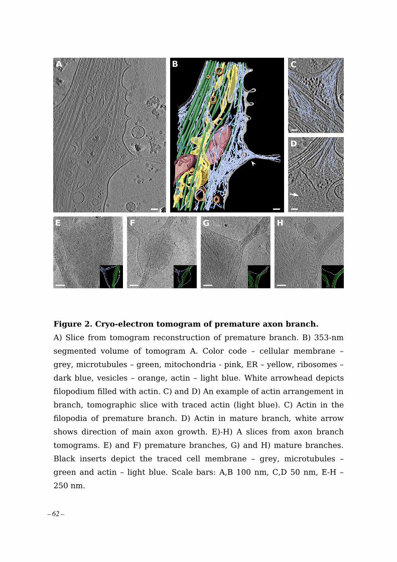
Figure 2. Cryo-electron tomogram of premature axon branch.
A) Slice from tomogram reconstruction of premature branch. B) 353-nm
segmented volume of tomogram A. Color code – cellular membrane –
grey, microtubules – green, mitochondria - pink, ER – yellow, ribosomes –
dark blue, vesicles – orange, actin – light blue. White arrowhead depicts
filopodium filled with actin. C) and D) An example of actin arrangement in
branch, tomographic slice with traced actin (light blue). C) Actin in the
filopodia of premature branch. D) Actin in mature branch, white arrow
shows direction of main axon growth. E)-H) A slices from axon branch
tomograms. E) and F) premature branches, G) and H) mature branches.
Black inserts depict the traced cell membrane – grey, microtubules –
green and actin – light blue. Scale bars: A,B 100 nm, C,D 50 nm, E-H –
250 nm.
– 62 –

Figure 3. Mitochondria and ER in axon branch.
A) Tomographic slice of mitochondria enriched in axon branching site. B)
Analysis of mitochondria length. Median length: 500.1 nm N=44. C) Bar
graph depicting number of branches with and without ER in daughter
branch for mature branches with MT and for premature branches with
only actin. D) Slice from tomogram showing different kind of ER – thin
tubes and flat sheets. White square depicts area in F and G. E)
Segmented tomogram D, color code: grey – cell membrane, green –
microtubules, yellow –ER, white arrowhead shows ER in branch together
with microtubules. F) ER – MT contact (black arrowhead). G)
Segmentation of ER tube wrapping around microtubule (white
arrowhead). Scale bar: A,D,E 100 nm, F,G = 50 nm.
– 63 –

Figure 4. Ribosome clusters at axon branch.
A)-D) Examples of clustered ribosomes in axon branches. A) and B)
Ribosomes filling premature branch. Red arrowhead depicts an example
of ribosome density. C) and D) Ribosomes present at mature branch site.
E)-G) Analysis of distance between ribosomes. E) Slice of analyzed
tomogram. (Of interest: Orange triangle shows vesicle with inner
membrane densities) F) Distribution of ribosomes in the 3D volume of
tomogram in E, color code by distance between ribosome particle
coordinates: green < 35 nm, yellow > 35 nm. G) Distance distribution of
ribosome particles in tomogram E, graph shows particles with closest
neighbor distance value bellow 40 nm, N=178. H) Cumulative distance
distribution for all analyzed ribosomes. N=1614. Scale bar: A-F = 100
nm.
– 64 –

Figure 5. Ribosome reconstruction and polysome orientation.
A) Reconstructed ribosome (light blue) fitted into the 80S ribosome
volume from database (orange mesh, EMD-3420). B) Reconstructed
ribosome volume with depicted 40S (light blue) and 60S (dark blue)
subunits and L7/L12 stalk. C) Rotated view of ribosome from B, Putative
path of mRNA depicted inn green, White arrow shows L7/L12 stalk. D)
Polysomes found in various tomograms. E)-G) Ribosomes on the surface
of ER. E), F) slice from tomogram, G) Segmented view for E and F. Scale
bar: C = 10 nm, G = 100 nm.
– 65 –

Table 1. Numeric summary of analysed tomograms.
Showing localization of tomographic data collection site within axon and
individual cellular features observed at the given locality.
Movie 1. Reconstructed tomogram of axon branch.
Slice view through 430-nm thick tomographic volume in Figure 1 and
segmentation. Colour code: grey – cellular membrane, green –
microtubules, light blue – actin, pink – mitochondria, yellow – ER, dark
blue – ribosomes, orange – vesicles. In final slice, upper layer of
segmented actin was removed for better clarity.
– 66 –

Supplementary material
Figure S1. Examples of mitochondria found at axon branch.
A) Mitochondrion undergoing fission facilitated by ER. White arrowheads
depict ER pinching at mitochondria membrane. B) Possibly dividing
mitochondrion. C) Examples of different sizes of mitochondria. D)
Mitochondrion presumably undergoing fission. Scale bar: A-D = 100 nm.
– 67 –

Figure S2. Thin ER tubes in axon branches.
A) Examples of ER tube reaching extremely thin diameters. Red
arrowheads follow continuous ER tubes, White arrowheads depict
different ER. B) ER (white arrowheads) wrapping around microtubules. C)
Cross-section of ER tube (white arrowhead) wrapping around
microtubules. D) Diameter of thinnest ER tubes found in tomograms,
median diameter: 7.19 nm, N=19. E) Diameter of ER tube wrapping
around microtubule, median diameter: 12.21 nm, N= 29. Scale bar: A-C =
50 nm.
– 68 –

Figure S3. ER enters axon branches together with microtubules.
A)-D) Examples of ER tube present in axon branch together with
microtubules, red arrowheads – ER, white arrowheads – microtubules,
black arrow shows direction of new branch, white arrow points in the
direction of main axon growth. Scale bar: A-D = 100 nm.
– 69 –

Figure S4. Visual representation of the ribosome distance
distribution.
A) Slice from tomogram. B) Ribosome particles placed into the tomogram
volume in color representation according their closest neighbor distance,
– 70 –

green < 35 nm, yellow > 35 nm. C) Graph representing the distance
distribution between clustered ribosomes. Scale bar: A = 500 nm, B = 50
nm.
Figure S5. Clustered ribosomes form polysomes.
Examples of strings of ribosomes found in axon branches forming
polysomes.
– 71 –

Figure S6. Growth cone and axon shaft.
A), B) Slices of tomograms showing growth cone filled with actin, red
– 72 –

arrowhead depicts ribosome. C) Axon shaft packed with bundled
microtubules, thin ER tubes and vesicles. Scale bar: A-C = 200 nm.
Figure S7. Ribosome reconstruction resolution estimation, FCS
curve.
– 73 –


2.2 Direct induction of microtubule branching by micro-tubule nucleation factor SSNA1
Basnet N., Nedozralova H., Crevenna A.H., Bodakuntla S., Schlichthaerle T., Taschner M.,Cardone G., Janke C., Jungmann R., Magiera M.M., Biertuempfel Ch., Mizuno N., (2018).Nature Cell Biology 20, 1172–1180.
During my PhD, I was able to contribute to the project about microtubule nucleation factorSSNA1. This study shows that SSNA1 can nucleate and induce the remodeling of themicrotubule into a branched structure where a new microtubule directly branches out fromthe existing old microtubule. The remodeling or branching property of SSNA1 is related toits ability of self-assembly and the mutants which abrogate the self-assembly of SSNA1 alsoabolish microtubule branching. These SSNA1 mutants that abolish microtubule branchingin-vitro also fail to promote axon branching when overexpressed in neurons.
For this study, I performed tomogram segmentation of microtubule branches, preparedneuron samples for cellular DNA-PAINT experiments, and analyzed light microscopydata. Detailed author contributions are included in the attached article.
– 75 –

Articleshttps://doi.org/10.1038/s41556-018-0199-8
1Max Planck Institute of Biochemistry, Martinsried, Germany. 2Biomolecular Self-Organization, Instituto de Tecnologia Química e Biológica António Xavier, Universidade Nova de Lisboa, Oeiras, Portugal. 3Institut Curie, PSL Research University, CNRS, Orsay, France. 4Universite Paris Sud, Universite Paris-Saclay, Orsay, France. 5Department of Physics and Center for Nanoscience, Ludwig Maximilian University, Munich, Germany. 6Graduate School of Quantitative Biosciences Munich (QBM), Ludwig Maximilian University, Munich, Germany. 7Present address: Department of Fundamental Microbiology, University of Lausanne, Lausanne, Switzerland. *e-mail: [email protected]
Cell-shape control is critical in a number of physiological processes. Microtubules, the major cytoskeletal component determining cell shape, are mostly nucleated at the centro-
some in proliferating cells. During specialized cell-shaping events, such as mitosis or cell polarization, cytoskeletal remodelling is thought to be driven by local nucleation of microtubules using a centrosome-independent mechanism1–3.
Neuronal cells are a distinctive example of cells with highly com-plex morphologies. Neurons are shaped in an extremely polarized fashion with a unique-shaped axon protruding from the cell body and stretching over long distances. Individual cells develop branch points from their axons to connect to neighbouring cells, creating an intricate communication network in the nervous system. As the shape of axons is determined by microtubules, these branch points require remodelling of microtubules to split the cytoskeletal path into separate branches1–5. As the centrosome is not necessary for the morphological development of the axon6, it is possible that axonal transformation occurs in a locally regulated manner within the axon. At axon branching points, the local destabilization and fragmentation of microtubules is mediated by the microtubule-severing enzyme spastin, which leads to the emergence of short microtubules7. However, the process of splitting the microtubule networks and, specifically, how the local rearrangement of spastin-processed tubulin oligomers or short microtubule fragments occurs has been enigmatic.
Due to its importance in various cell activities, the microtubule cytoskeleton has been well characterized in vitro. In the classical view, microtubules are considered as cylindrical polymers made of ~13 protofilaments. However, in living cells, it has been sug-gested that microtubules form higher-order branched networks to regulate their distribution within complex cytoskeletal networks1,8,9.
The branched networks could be envisioned either through the attachment of new microtubule modules onto the side of an exist-ing microtubule, or through direct branching of microtubules. So far, only one example of branching microtubule network has been shown, involving the microtubule nucleators augmin and γ -tubulin, which allow microtubules to grow out from nucleation points that attach to the side of existing microtubules8,10–13. There was, however, no evidence that protofilaments in a single microtubule can split apart to form a branched structure. Particularly at axon branch sites, augmin is less likely to be involved in generating branched microtubule networks, but has rather been implicated in determin-ing microtubule orientation by crosslinking adjacent microtubules within an axon14.
Here we focused on the protein SSNA1, a microtubule-binding protein implicated in the dynamic assembly of microtubules. SSNA1 is found at centrosomes or basal bodies in sperm cells15,16, and at the midbody in dividing cells17. A recent study reported that SSNA1 accelerates neuronal development by promoting axon elongation and branch formation17. These observations collectively suggest a versatile role for SSNA1 in microtubule remodelling. However, the nature of its activity in controlling microtubule dynamics is unclear.
We now demonstrate that SSNA1 is a powerful microtubule-nucleating and -branching factor. In vitro reconstitution of SSNA1-mediated microtubule nucleation showed an induction of branched microtubules, where new daughter microtubules directly branch out from existing microtubules. SSNA1 attaches along single proto-filaments, guiding them to grow away from a microtubule and tem-plate a branched microtubule. Mutation of residues essential for the oligomerization and the microtubule-branching activity of SSNA1, which we designed by structure-guided in vitro experiments, leads to defective axonal branching in primary neurons, showing that the
Direct induction of microtubule branching by microtubule nucleation factor SSNA1Nirakar Basnet1, Hana Nedozralova1, Alvaro H. Crevenna2, Satish Bodakuntla3,4, Thomas Schlichthaerle1,5,6, Michael Taschner1,7, Giovanni Cardone1, Carsten Janke 3,4, Ralf Jungmann 1,5, Maria M. Magiera3,4, Christian Biertümpfel 1 and Naoko Mizuno 1*
Microtubules are central elements of the eukaryotic cytoskeleton that often function as part of branched networks. Current models for branching include nucleation of new microtubules from severed microtubule seeds or from γ -tubulin recruited to the side of a pre-existing microtubule. Here, we found that microtubules can be directly remodelled into branched structures by the microtubule-remodelling factor SSNA1 (also known as NA14 or DIP13). The branching activity of SSNA1 relies on its ability to self-assemble into fibrils in a head-to-tail fashion. SSNA1 fibrils guide protofilaments of a microtubule to split apart to form daughter microtubules. We further found that SSNA1 localizes at axon branching sites and has a key role in neuronal develop-ment. SSNA1 mutants that abolish microtubule branching in vitro also fail to promote axon development and branching when overexpressed in neurons. We have, therefore, discovered a mechanism for microtubule branching and implicated its role in neuronal development.
NATuRe Cell BioloGy | VOL 20 | OCTOBER 2018 | 1172–1180 | www.nature.com/naturecellbiology1172
– 76 –

ArticlesNATure Cell BIology
simple scaffolding mechanism of SSNA1 can lead to vast morpho-logical changes in neurons.
ResultsSSNA1 localizes at axon branching sites in primary neurons. A previous study implicated SSNA1 in the promotion of axon branching17, but the underlying mechanism remained elusive. To investigate how SSNA1 plays a role in neuron development, we trans-duced wild-type murine primary hippocampal neurons with lenti-viral particles encoding GFP-2A-mouse_SSNA1. Overexpression of SSNA1 led to the promotion of axon outgrowth (Fig. 1a,c), in agreement with a previous report17. In addition, we found a striking accumulation of SSNA1 at axon branches (Fig. 1b and Supplementary Fig. 1), which was also observed at secondary branch sites that emanate from an already existing axon branch (Fig. 1b, panel 4, and Supplementary Fig. 1). In agreement with its localiza-tion, SSNA1 overexpression led to increased and more complex branching as characterized by the Strahler number (Fig. 1d). Since SSNA1 localizes at the cytosolic compartments where microtubule are dynamic15–17, we hypothesized that clusters of SSNA1 at branch-ing sites in neurons might facilitate local microtubule nucleation.
SSNA1 induces direct microtubule branching. To assess the influ-ence of SSNA1 on microtubules, we prepared recombinant SSNA1 (from Chlamydomonas reinhardtii, CrSSNA1, Supplementary Fig. 2A–C) and tested its interaction using cryo-electron micros-copy (cryo-EM; Fig. 2a and Supplementary Fig. 2D). CrSSNA1 induced a formation of direct microtubule branches that split from a single microtubule (Fig. 2a, arrowheads), in contrast to an attachment of a second microtubule on the microtubule surface. Branching occurs by splitting the lattice of the microtubule, and protofilaments of mother microtubules directly continue into the outer surface of the branched microtubule. The bending angle was variable (Fig. 2b,c, 47˚ ± 15˚, and Supplementary Fig. 2D,E), which suggests a rather flexible junction, in contrast to the more rigid, 70˚ Arp2/3-mediated actin branching18,19. Moreover, microtubules occasionally formed fork-like structures with several branches or junctions (Fig. 2b and Supplementary Fig. 2D). This has so far not been observed in any other system, and further underpins the uniqueness of SSNA1-mediated microtubule branching.
Cryo-ET shows diverging microtubules with a break in the microtubule lattice. To further understand the organization of microtubule branches, we performed cryo-electron tomography (cryo-ET) on branched microtubules (Fig. 3 and Supplementary Fig. 2F). Even though SSNA1 itself was not detectable due to the resolution limit of tomographic reconstructions, the microtubule lattice was clearly visible (Fig. 3a,b) and facilitated a tracing of indi-vidual protofilaments at the branch (Fig. 3c–e). The tracing showed that two branching microtubules shared a subset of protofilaments with their mother microtubule. In addition, we traced newly assem-bled protofilaments that were not connected to the mother micro-tubule (Fig. 3d), as the number of protofilaments doubles compared to the mother microtubule. This shows a discontinuity in the micro-tubule lattice at the splitting point of the branch.
SSNA1 self-clusters and nucleates microtubules. To explore the dynamic behaviour of SSNA1 causing this unique action in micro-tubules, we tested the interaction of SSNA1 with unpolymerized tubulin using fluorescence microscopy (Fig. 4a). Considering the average cellular concentration of SSNA1 of 187 nM20, we mixed 200 nM CrSSNA1 and 8 µ M tubulin in the presence of polyethyl-ene glycol (PEG)21. Above a concentration of 5% PEG (Fig. 4b), we observed condensates of CrSSNA1 clustering with tubulin (Fig. 4a–c). Interestingly, several microtubules emerged from these CrSSNA1–tubulin clusters (Fig. 4a,b), reminiscent of aster
Control (2A–GFP) SSNA1(2A–GFP)a
100 µm
An
ti-M
AP
2A
nti-
tau
bAnti-SSNA1 Anti-β3-tubulin DAPI
20 µmS
SN
A1
-ove
rexp
ress
ion
Zo
om
4×
1
1
2
2
3
4
3 4
90°
OverexpressionControl
012≥ 3
dNumber of branches
0
Axo
n le
ng
th (
µm)
200
400
600
800
1,000
OverexpressionControl
P < 0.0001c
0–12–3≥ 4
Strahler number (degree of branch)
Fig. 1 | The effect of SSNA1 overexpression on primary hippocampal neurons. a, Immunostaining of MAP2 (green) and Tau (red) in control (GFP overexpression) and SSNA1-wild-type overexpression. b, Immunostaining of SSNA1 (red) and β III-tubulin (green) in neurons overexpressing SSNA1 wild type shows the localization of SSNA1 at axon branch sites. c, Scatter dot plots of axon length under overexpression of SSNA1. The longest protrusion from the soma was defined as the axon, and cells with very short protrusions were also included in the counting, so that underdeveloped neurons could be assessed as well. The promotion of axon development occurs only in overexpression of wild-type SSNA1. Experiments were performed in triplicates, shown in magenta, green and yellow. Every cell is represented by a single point: control (n = 505 cells), wild type (n = 499 cells), pooled from 3 independent experiments, and the overlaid box-and-whisker plots cover 50% (boxes) and 90% (whiskers) of the entire population, with median values indicated as lines within the boxes. The results show statistical significance (P < 0.0001) as tested using the Kruskal–Wallis test, followed by Dunn’s multiple comparison post hoc test. d, Pie graphs showing the distribution of the number of branches under overexpression conditions (control (n = 496 cells), wild type (n = 490 cells) pooled from 3 independent experiments) and Strahler number (degree of sub-branch formations on the existing branches; control (n = 266 cells), wild type (n = 289 cells) pooled from 3 independent experiments). Distributions of the branches and the Strahler number in SSNA1-expressing neurons differ significantly from the control (GFP overexpression) according to χ2 two-sample test (χ2 = 20.7, P < 0.01 and 18.6, P < 0.005, respectively). See Supplementary Table 3 for source data.
NATuRe Cell BioloGy | VOL 20 | OCTOBER 2018 | 1172–1180 | www.nature.com/naturecellbiology 1173
– 77 –

Articles NATure Cell BIology
formation seen during microtubule nucleation1,22, and thus indi-cating that CrSSNA1 nucleates microtubules. This process was effective at a CrSSNA1 concentration of as little as 50 nM (Fig. 4d,e). Nucleation events were confined to the local condensates of CrSSNA1 and tubulin did not polymerize without CrSSNA1, high-lighting the effect of the SSNA1 condensates and the requirement of a high local concentration. The number of growing asters and microtubules (Fig. 4d,e) correlated well with the concentration of CrSSNA1. We also observed new microtubules emerging from already formed microtubules (Supplementary Videos 1 and 2).
SSNA1 induces microtubule nucleation from mother microtu-bules. To understand and assess SSNA1-mediated nucleation from template microtubules, we mixed (3 or 30 µ M) CrSSNA1 with tubu-lin in the presence of GTP with GMPCPP-stabilized microtubule seeds23. Several microtubules were able to grow out from the ends or the wall of pre-existing microtubules (Fig. 4f,g, Supplementary Fig. 3A,B and Supplementary Videos 3 and 4), agreeing with the cryo-EM observation (Fig. 2a). By differentially labelling pre-exist-ing microtubules (red) and newly polymerized dynamic micro-tubules (green), we could categorize the branching events into: ‘splitting’, in which newly formed microtubules split from the end of a pre-formed microtubule; ‘end-joining’, showing three pre-existing microtubules connected through newly formed tubulin oligomers; ‘side branching’, seen as new microtubules coming out from the side of pre-existing microtubules; and ‘dynamic branching’, in which newly generated dynamic microtubules form a branch. The ‘side branching’ is reminiscent of local microtubule nucleation mediated by augmin and γ -tubulin in cell extracts8,13. However, in contrast to the augmin-mediated mechanism, the formation of CrSSNA1-mediated branching did not require γ -tubulin, indicating that SSNA1 works by a novel mode of action.
SSNA1 forms a fibril-like assembly on the surface of the micro-tubule with 11-nm periodicity. Although cryo-ET did not visualize
the decoration of SSNA1 on microtubules, we observed that the free ends of microtubules are often extended with thin fibrils (Supplementary Fig. 2D, red arrowheads). These fibrils extend from splitting microtubules, seemingly to work as a ‘guide rail’ for the growth of branched microtubules (Fig. 2b; and Supplementary Fig. 2D, ‘guide rail’). Computational averages of the cryo-EM images of microtubules allowed the visualization of CrSSNA1 directly attached to the surface of microtubules, revealing a ladder-like pat-tern (Fig. 5a, compare to ‘microtubule-only control’) with a peri-odicity of ~11 nm (Fig. 5b). We observed that CrSSNA1 facilitated preferential assembly of 13-protofilament microtubules similar to doublecortin24 and EBs25. In contrast, 14-protofilament microtubules are predominantly polymerized in the absence of SSNA1 (Fig. 5c).
SSNA1 forms a head-to-tail fibril with 11-nm periodicity and covers the C-terminal tail of microtubules. SSNA1 is a protein with a relative molecular mass of 14,000 Da predicted to adopt a tropomyosin-like single parallel coiled-coil configuration26,27 (Supplementary Fig. 3E). Fibril formation has previously been observed as a result of head-to-tail self-assembly27,28. Accordingly, we observed that CrSSNA1 readily forms short fibrillar appearances with occasional long fibril formations (Fig. 5g, ‘FL’). Furthermore, the shorter fibrils of CrSSNA1 were converted into longer, orga-nized bundles of fibrils after ~24 h incubation (Supplementary Fig. 3F). A closer look at these bundles revealed a striped, knob-like pattern, which leads to the formation of a sheet (Supplementary Fig. 3F, 24 h) with a 11-nm periodicity (Supplementary Fig. 3F, inset), and the inter-fibril distance of 3.5 nm. This pattern is compa-rable to that observed on the microtubule surface (Fig. 5a), indicat-ing that the fibrils are covering microtubules along their long axis, giving a 11-nm spaced ladder-like pattern.
Nu
mb
er
of
bra
nch
ing
MT
s
0
5
10
15
20
25
30
10 30 50 70 90 11013
015
0
α
Branching angle α (°)
100 nm
a
b c
Guide rails
Split-wide
4-way
Split
50 nm
Fig. 2 | Characterization of in vitro-reconstituted microtubule branching. a, A cryo-EM image of branched microtubules. The arrowheads show examples of branching points. Microtubules were stabilized with 1 mM GMPCPP. b, Snapshots of branching microtubules. The ‘guide rail’ depicts thin lines of density often seen at the split of the branch point. c, Distribution of branching angles (47 ± 15°, n = 99 branch points). These experiments were performed three independent times.
a b c d e
9
13
5
6
148
14
25 nm
180°
Fig. 3 | The cryo-eT reconstruction of SSNA1-mediated microtubule branching. a, A 25-nm slice of a tomographic reconstruction highlighting a branching point of a microtubule. With this view, individual protofilaments and tubulin units are visible, but the SSNA1 density is too thin to be visualized. b, Cross-sections of the branched microtubules in a. c, Individual protofilaments are overlaid with colour represented in the segmentation in d. d, Tracing of protofilaments in the 3D density map in a. Individual protofilaments are coloured in rainbow-colour coding. The newly formed protofilaments from the branched microtubules are coloured in green (left) and in pink (right). The number of protofilaments in this particular branched microtubule is counted to be 13 (mother microtubule), 14 (left branched microtubule) and 14 (right, branched microtubule). Thirteen mother protofilaments are split to 6 to the left and 5 to the right side of branched microtubules. e, 180o-rotated segmentation of the branched microtubule.
NATuRe Cell BioloGy | VOL 20 | OCTOBER 2018 | 1172–1180 | www.nature.com/naturecellbiology1174
– 78 –

ArticlesNATure Cell BIology
To further characterize the interaction between SSNA1 and microtubules, we obtained a cryo-EM three-dimensional (3D) structure of CrSSNA1 in complex with microtubules (Fig. 5d–f and Supplementary Fig. 4A) with an overall resolution of 6.1 Å (Supplementary Fig. 4B). Due to the symmetry mismatch between microtubules (4- or 8-nm periodicity) and SSNA1 (11-nm period-icity), SSNA1 was averaged out, and the fibril appeared as a ‘cloud’ of protein density running parallel to the microtubule surface, with an apparent local resolution of ~ 11 Å (Supplementary Fig. 4C).
However, it was possible to visualize thin lines of additional densi-ties running parallel to the microtubule surface (Fig. 5e,f), which we interpreted as SSNA1 filaments. The SSNA1 filaments run between two protofilaments, proximal to the unstructured, highly acidic carboxy-terminal tails (E-hooks) of tubulin (Fig. 5f labelled ‘C’). Removal of E-hooks resulted in weakening of SSNA1 crosslinking with microtubules as determined by EDC (~49% less crosslinked; Supplementary Fig. 4E). E-hooks create a negative electrostatic cloud by their periodical arrangement on the microtubule surface29,
0 5 10 150.0
0.2
0.4
0.6
0.8
1.0 50 nM100 nM200 nM
Tubulin SSNA1
0
50
100
Time (min)
Pe
rce
nta
ge
ast
ers
SSNA1
Mic
rotu
bu
les
(co
un
t)5 µm
5 µm
a
b c
e
d
Splitting
End-joining
Side branching
5 µm
100 nm
h
g
5 µm
Bra
nch
-typ
e r
atio
(%
)
Dynamicbranching
3 min 6 min 7.6 min 13 min
f
No PEG 1% 5% 6.5% 7.5% 10%
? X
0
10
20
30
40
50
200
nM
100
nM50
nM
0 nM
Fig. 4 | Nucleation and branching of microtubules mediated by CrSSNA1 under various conditions. a, Aster-like formation of microtubules (20% HiLyte488 tubulin) occurs within 3 min after mixing tubulin with a lower concentration (200 nM) of CrSSNA1 (upper) under conditions mimicking molecular crowding (7.5% PEG, typically used as a crowding agent), where tubulin alone does not form any polymers. Microtubules propagate out from tubulin concentrate, serving as a nucleation centre. These experiments were performed three independent times with similar results. b, 200 nM CrSSNA1 and 8 µ M tubulin self-associate, forming clusters in the presence of PEG with concentration > ~5%. c, SSNA1 antibody recognizes the microtubule nucleation centre. d, A plot of the percentage of the concentrates growing into asters with microtubules as a function of time (min) in the presence of 50, 100 and 200 nM CrSSNA1. The error bars are mean ± s.d. from n = 3 independent experiments. As little as 50 nM of CrSSNA1 is sufficient to observe aster formation in the presence of 7.5% PEG. e, Counts of microtubules observed per field of view, in the presence of different concentrations of CrSSNA1. The box plots cover 50% (boxes) and 90% (whiskers) of the entire population, with median values indicated as lines within the boxes. Sample size: 0 nM: n = 42 fields of view; 50 nM: n = 29; 100 nM: n = 30; 200 nM: n = 25. Data were pooled from three independent experiments except for the first point (0 nM) for which data were pooled from four independent experiments. f, Green coloured dynamic microtubules on red-microtubule GMPCPP seeds in the presence of a higher concentration of CrSSNA1 (30 µ M) without molecular crowding agents to achieve globally concentrated conditions. ‘branch-like’ nucleation is observed. Branches were categorized as ‘splitting’, ‘end-joining’, ‘side branching’ or ‘dynamic branching’. g, Ratios of different branch types (n = 895 branches, mean ± s.d. pooled from 3 independent experiments). ‘?’ shows the bundled microtubules, which are difficult to categorize. ‘X’ shows microtubules without branching. Branch-like nucleation can be seen from the locally concentrated SSNA1 condition described in a–e; however observations of individual microtubules are challenging due to the high local protein concentrations. h, A negative-stain EM image of SSNA1-mediated branched microtubules in the presence of 200 nM CrSSNA1 and 7.5% PEG, representative of 3 independent experiments. See Supplementary Table 3 for source data.
NATuRe Cell BioloGy | VOL 20 | OCTOBER 2018 | 1172–1180 | www.nature.com/naturecellbiology 1175
– 79 –

Articles NATure Cell BIology
which could attract the SSNA1 fibrils. This could explain why SSNA1 interacts with microtubules despite the symmetry mismatch. It also suggests that the head-to-tail assembly of SSNA1 fibrils could guide protofilament assembly and microtubule polymerization by cover-ing and neutralizing the E-hooks as shown previously30.
The head-to-tail fibril formation of SSNA1 is essential for micro-tubule branching. On the basis of the observation that SSNA1 fibrils appear to guide the protofilaments of microtubules, we hypothesized that the microtubule branching activity is mediated by the formation of long SSNA1 fibrils that curve away and guide the protofilament out of the lattice (guide rail, Fig. 2b and Supplementary Fig. 2D). To test this, we created a series of truncated SSNA1 fragments that abolish fibril formation. On the basis of a PHYRE2 analysis31 and previous reports27, we found that SSNA1 contains a well-conserved α -helical region (residues 6–104) followed by an unstructured C-terminal tail (Supplementary Fig. 3E). A series of amino-terminal truncations showed that the first 19 residues were not necessary for fibril forma-tion, as CrSSNA1(20–111) (20-C) formed cable-like bundled fibrils, which were less ordered compared to full-length protein (CrSSNA1 FL), but displayed an ~11-nm pattern (Fig. 5g,h, Supplementary Fig. 5A and Supplementary Table 1). In contrast, CrSSNA1(21-C), a truncation missing one more residue, Glu 20, was unable to form fibrils (Fig. 5g,h, Supplementary Fig. 5A and Supplementary Table 1). This observation correlates with the ability of CrSSNA1(20-C), but not CrSSNA1(21-C), to mediate microtubule branching (Supplementary Fig. 5A and Supplementary Table 1). The key role of the residue Glu 20 for fibril-formation and microtubule-branching activity of SSNA1 was further underpinned by point mutations E20A and E20A/D21A, which drastically reduced micro-tubule branch formation (Supplementary Fig. 5A and Supplementary Table 1). These mutants may form fibrils, but with much lower fre-quency and without a distinct higher-order organization.
In the C-terminal region of CrSSNA1, three distinctive lysine residues (Lys 105, Lys 106 and Lys 107) mark the beginning of the unstructured C terminus. CrSSNA1 truncations 1–104 and 1–105 (Fig. 5g,h, Supplementary Fig. 5A and Supplementary Table 1) showed that CrSSNA1(1–104) can no longer form fibril-lar oligomers, or induce microtubule branching (Fig. 5g,h), while CrSSNA1(1–105) was purified as fibrils and showed microtubule branching activity (Supplementary Fig. 5A) at a similar efficiency to CrSSNA1-FL. These results indicate that the positive charge of the lysine residues is essential for the ability of CrSSNA1 to form fibrils. We confirmed this by generating a triple point mutant, K105A/K106A/K107A, which indeed abolished fibril formation and micro-tubule branching (Supplementary Fig. 5A and Supplementary Table 1) for both the full length and the 1–107 fragment. Altogether, our mutational analyses indicate that the key interaction for lon-gitudinal fibril formation is mediated by Glu 20 of one unit and the C-terminal tail (Lys 105–107) of the adjacent interacting unit (Fig. 5i). To confirm this, we created mutants in which the charges of residues Glu 20/Asp 21 and Lys 105/Lys 106/Lys 107 were swapped. When two of the opposite charges were swapped (E20K/D21K/K105E/K106E or E20K/D21K/K106E/K107E), both cable-like formation and microtubule branching activity of SSNA1 were retained. In contrast, swapping of the two negative residues at the N terminus and the three positive residues at the C terminus (E20K/D21K/K105E/K106E/K107E), resulting in a change of net charge from + 1 to − 1, abolished microtubule branching (Supplementary Fig. 5B). However, this construct was still able to form SSNA1 fibrils and cable-like structures, indicating that the microtubule branch-ing activity depended not only on fibril formation of SSNA1, but also on the presence of an extra negative charge at the unstruc-tured SSNA1 C terminus. This was confirmed by the mutant E20A/D21A/K105A/K106A/K107A, termed 5A, showing a complete loss of microtubule branching activity (Fig. 5g).
Microtubule-branching-deficient SSNA1 mutants abolished the promotion of axon branches. Having gained insights into the molecular organization of SSNA1 and its effect on microtubule nucleation and branching, we hypothesized that the promotion of axon growth and branching observed in neurons overexpressing wild-type SSNA1 might be altered when microtubule-branching-deficient SSNA1 versions are expressed. Our results indeed showed that, in contrast to the SSNA1 wild type (Figs. 1 and 6a–c), SSNA1 with mutations abolishing microtubule branching in vitro also failed to promote the growth of axons or axon branches (Fig. 6a–c and Supplementary Fig. 6A–E) in primary neurons. Notably, a dominant-negative effect was also observed when the 5A mutant was overexpressed for the number of total neurite processes (Fig. 6c), showing a decreased number of major and minor branches. This dominant-negative effect was also found when the two nega-tive residues at the N terminus and the three positive residues at the C terminus were swapped (swap-KK/EEE). Notably, swapping only two opposite charges and leaving the third C-terminal lysine intact (swap-KK/EE) could still promote axon growth (Supplementary Fig. 6D,E). Together these findings show that the ability of SSNA1 to induce fibril formation and microtubule branching at the molec-ular level correlates with its function of mediating axon branching and development, suggesting the intriguing possibility that it locally generates branched microtubules at axon branch sites.
Morphological change of microtubule networks in non-neuronal cells. To test whether the function of SSNA1 is conserved in differ-ent cell types, we used fibroblasts, which are structurally less spe-cialized than neurons, and tested whether overexpression of SSNA1 has the capacity to change the microtubule organization (Fig. 6e–j). Super-resolution light microscopy with DNA-PAINT showed that individual microtubules are well resolved in the control cells with a wide-ranging network (Fig. 6e–g). In contrast, microtubules were rather short in SSNA1-overexpressing cells (Fig. 6h–j), sug-gesting that SSNA1 can promote nucleation, generating more but shorter microtubules. We also occasionally found microtubules forming three-way intersections, as if one microtubule emerged out of another in both control and SSNA1-overexpressing cells (Fig. 6g,j, arrowheads). These events occurred more often in SSNA1-overexpressing cells (2.8 ± 1.2 occurrences per 100 µ m of microtubule) than control cells (1.0 ± 0.35 occurrences per 100 µ m of microtubule). Although the limited resolution in light micros-copy prevented us from discerning whether microtubules branched with a shared lattice or if two microtubules only attached to each other, the observations were consistent with our in vitro studies by electron microscopy.
DiscussionDuring cell polarization, the dynamics and distribution of the microtubule cytoskeleton is tightly regulated. Although the centro-some has a major role as a microtubule-organizing centre in less differentiated cells, the inactivation of centrosomes in neurons does not affect axon growth, a process strongly dependent on micro-tubule assembly6. Thus far, the molecular mechanisms regulating axonal microtubule nucleation, especially in the form of branching, have remained a mystery. Here, we show that SSNA1 accumulates at axon branches and promotes axon branching in primary neurons, and can nucleate microtubules in vitro. Mutations interfering with SSNA1 in vitro nucleation activity also affect the occurrence of axon branches in neurons. Together, these results suggest that SSNA1 could act as a microtubule nucleator at axon branch sites.
Strikingly, our work revealed that SSNA1 independently medi-ates microtubule branching by causing protofilaments to splay apart from the lattice. To our knowledge, no other microtubule-binding protein shows this activity. When tested, EB3 and ch-TOG, known regulators of microtubule dynamics, under the same conditions,
NATuRe Cell BioloGy | VOL 20 | OCTOBER 2018 | 1172–1180 | www.nature.com/naturecellbiology1176
– 80 –

ArticlesNATure Cell BIology
90°
d
e f
SSNA1pf
C
C 8 nm
+SSNA1
25 nm
a
010206080
100
Perc
enta
ge
Perc
enta
ge
Microtubules SSNA1-inducedmicrotubules
020406080
100
4 nm–1
11 nm–1
Control +SSNA125 nm
MT-only control
c
b
FL
21-C
h
K E
K E
K
E
E
K
11 nm
N
C
Alone
+ Tubulin
SSNA1‘guide rail’
SSNA1
g
50 nm
1–104
FL-E20AD21A-AAA (5A)
AA AAA
FL
1–104
21-C
5A
i
13 p
f14
pf
15 p
f16
pf
13 p
f14
pf
15 p
f16
pf
Fig. 5 | Molecular characterization of the branching action of SSNA1. a, Left: representative class average of the SSNA1-induced microtubules. Right: SSNA1 decoration emphasized by computationally subtracting microtubule densities35. Bottom left: average of microtubules without decoration for comparison. b, The power spectrum of microtubule class averages shows an additional 11 nm periodicity in the presence of SSNA1. c, Distribution of protofilament (pf) numbers of microtubules reconstituted from brain tubulin in the absence (left) and in the presence of SSNA1 (right) shifting the majority from 14- to 13-protofilament microtubules. d, Greyscale slice from the density map of the plus-end-on view of the SSNA1–microtubule 3D reconstruction. SSNA1 decoration and the secondary structures of tubulin density are well resolved. e, Rendering of the microtubule surface decorated with SSNA1. The resolution of the microtubule surface (~10 Å) is not as high as the core (< 8 Å) due to the SSNA1 decoration. f, Tubulin atomic model (PDB ID: 3jal) fitted to the map. The SSNA1 coiled-coil fibril is indicated as a tube representation. Note that the periodical feature of SSNA1 is averaged out because of the symmetrical mismatch between tubulin dimers (8 nm) and SSNA1 fibrils (11 nm). g, Morphological observation of SSNA1 and its branching activity. Left: observation of the purified protein at 0 h incubation (that is, immediately after purification). Right: a magnified view of the co-polymerized microtubules. Microtubule branching was observed with SSNA1-FL, while other protein fragments do not facilitate branching. For the proteins that do not cause the branching, examples of typical crossing of microtubules (white and beige bars at the scheme within the image), instead of branching, are shown. Detailed observations are available in Supplementary Fig. 5A. h, A graphical scheme of the SSNA1 constructs used in g. i, A scheme of the SSNA1 self-assembly and microtubule nucleation mediated by SSNA1. While SSNA1 oligomers alone can also undergo a slow self-assembly process, the SSNA1 oligomers interact with tubulin dimers to promote their co-polymerization. The polymerized SSNA1 may further act as a guide rail (bottom inset) for protofilament splitting, resulting in microtubule branch formation. A class average indicating the guide rail mechanism is shown. Other class averages are available in Supplementary Fig. 2E.
NATuRe Cell BioloGy | VOL 20 | OCTOBER 2018 | 1172–1180 | www.nature.com/naturecellbiology 1177
– 81 –

Articles NATure Cell BIology
did not induce branching (Supplementary Fig. 6F,G). In addition, we showed that microtubule branching requires co-polymerization of SSNA1 with microtubules, as well as specific amino-acid inter-actions. Taken together, these results indicate that the observed branching activity is highly specific to SSNA1.
Our work has uncovered a surprising example of how co-polymerization of a simple coiled-coil protein with tubulin can induce global remodelling of the microtubule network. Association of SSNA1 may reinforce longitudinal connections of tubulin oligomers, facilitate protofilament formation and act as a polym-erization seed for microtubule formation. Concomitantly, the pref-erence of SSNA1 for lateral connections may facilitate the lateral associations between microtubule protofilaments. Polymerized SSNA1 may precede microtubule protofilaments, guiding proto-filaments out of the microtubule axis, thus providing a template for a new microtubule branch (‘guide rail’ mechanism). In a cellular context, however, the situation is more complex as SSNA1 activity is probably modulated by other factors. Further experiments are
necessary to test this mode of action of SSNA1 for microtubule branching in cells.
Our in vitro reconstitutions showed that SSNA1 self-assembles into clusters together with tubulin at a high local concentration. As axons are densely packed with cytoskeletal components, this orga-nization is a plausible prerequisite for the physiological function of SSNA1, allowing SSNA1 to concentrate locally, self-assemble and become a microtubule nucleation centre at designated locations. Alternatively, the requirement of a high local concentration of SSNA1 may be a means to limit the microtubule-remodelling activ-ity of SSNA1 to specific subcellular areas such as axon branches, the midbody in dividing cells and the base of cilia.
As microtubules are much less dynamic in axons compared to less polarized cells32–34, SSNA1-mediated branching may be restricted to locally destabilized sites of the microtubule cytoskel-eton. Interestingly, SSNA1 interacts with spastin, a protein impor-tant for the initiation of axon branching and thought to increase the pool of soluble tubulin through microtubule fragmentation7.
a
Spastin MicrotubuleTubulinSSNA1
d
21-C1–104Wild type
(positive cont)Control
012≥ 3
5A
cNumber ofbranches
0
Axo
n le
ng
th (
µm)
200
400
600
800
1,000
21-C1–104 5AWild type(positive cont)
Control
P < 0.0001b
0–12–3≥ 4
Strahler number(degree of branch)
?
100 µm
An
ti-M
AP
2A
nti-
tau
GF
P
Control (2A–GFP) SSNA1 wild type(2A–GFP)
SSNA1 21-C(2A–GFP)
SSNA1 1–104(2A–GFP)
SSNA1 5A(2A–GFP)
e
f
g
h
i
j
5 µm
Control SSNA1-overexpression
5 µm
5 µm
Fig. 6 | The effect of various SSNA1 constructs on primary hippocampal neurons and fibroblast cells a, Immunostaining of MAP2 (green), Tau (red) and GFP (blue, expression control) in SSNA1-overexpressing cells. For the SSNA1 wild type, the axon is indicated with a dashed line. b, Scatter dot plots of axon length under overexpression of various SSNA1 constructs. The control and wild-type profiles shown in Fig. 1 are placed as a negative and positive control, respectively. The promotion of axon development occurs only in overexpression of wild-type SSNA1, while no apparent effect was observed for the constructs that fail to mediate microtubule branching. Every cell is represented by a single point; control (n = 496 cells), wild type (n = 490), 1–104 (n = 788), 21-C (n = 610), 5A (n = 642) from 3 independent experiments, shown in magenta, green and yellow, and the overlaid box-and-whisker plots cover 50% (boxes) and 90% (whiskers) of the entire population, with median values indicated as lines within the boxes. c, Pie graphs showing the distribution of the number of branches and Strahler number under different overexpression conditions. The GFP-expression-control and SSNA1-wild-type-overexpression profiles in Fig. 1 are placed as controls. d, A schematic model describing how SSNA1-mediated microtubule nucleation could contribute to axon branch formation. Spastin has been shown to localize at axon branches7 and to interact with SSNA136. Taken together with our finding of SSNA1 localization at axon branches, it is possible that the two proteins work sequentially by spastin severing microtubules to provide tubulin oligomers, and SSNA1 nucleating microtubules at the branching site. e, A DNA-PAINT image of a 500-nm slice of the microtubule network in untreated cells (control). f, Zoomed-in view from e; the object is coloured in a rainbow code according to the depth. g, Individually recognized microtubules are highlighted in various colours. Three-way intersections are indicated with red arrowheads. h–j, Corresponding view of a 500-nm slice of the microtubule network in SSNA1 overexpressing cells. For analysis, 3 independent SSNA1-overexpressing and control cells were assessed each, containing the total microtubule lengths of 5,700 µ m, 7,900 µ m and 1,900 µ m and 7,700 µ m, 8,500 µ m and 7,700 µ m, respectively. See Supplementary Table 3 for source data.
NATuRe Cell BioloGy | VOL 20 | OCTOBER 2018 | 1172–1180 | www.nature.com/naturecellbiology1178
– 82 –

ArticlesNATure Cell BIology
It is tempting to speculate that short, spastin-severed microtubules (that is, tubulin oligomers) provide the building blocks for SSNA1-mediated microtubule nucleation and branching. Thus, the syner-gistic action of spastin and SSNA1 could facilitate the formation of axonal branch points (Fig. 6d). Further investigation of SSNA1 activity in situ will provide valuable insights into the initiation and organization of axon branches. In particular, it will be interesting to explore whether SSNA1-mediated microtubule branching is a direct driving force for axon branching, or an intermediate state during early stages of neuronal morphogenesis. Considering the diverse sites at which SSNA1 is localized in various cell types17,27, the micro-tubule-branching mechanism discovered here could have broad implications for understanding the regulation of various microtu-bule functions, providing new clues to previously unanswered ques-tions about cytoskeleton and intracellular transport.
online contentAny methods, additional references, Nature Research reporting summaries, source data, statements of data availability and asso-ciated accession codes are available at https://doi.org/10.1038/s41556-018-0199-8.
Received: 19 November 2017; Accepted: 20 August 2018; Published online: 24 September 2018
References 1. Ishihara, K., Nguyen, P. A., Groen, A. C., Field, C. M. & Mitchison, T. J.
Microtubule nucleation remote from centrosomes may explain how asters span large cells. Proc. Natl Acad. Sci. USA 111, 17715–17722 (2014).
2. Meunier, S. & Vernos, I. Acentrosomal microtubule assembly in mitosis: the where, when, and how. Trends. Cell Biol. 26, 80–87 (2016).
3. Petry, S. & Vale, R. D. Microtubule nucleation at the centrosome and beyond. Nat. Cell Biol. 17, 1089–1093 (2015).
4. Kalil, K. & Dent, E. W. Branch management: mechanisms of axon branching in the developing vertebrate CNS. Nat. Rev. Neurosci. 15, 7–18 (2014).
5. Lewis, T. L., Courchet, J. & Polleux, F. Cell biology in neuroscience: cellular and molecular mechanisms underlying axon formation, growth, and branching. J. Cell Biol. 202, 837–848 (2013).
6. Stiess, M. et al. Axon extension occurs independently of centrosomal microtubule nucleation. Science 327, 704–707 (2010).
7. Yu, W. et al. The microtubule-severing proteins spastin and katanin participate differently in the formation of axonal branches. Mol. Biol. Cell 19, 1485–1498 (2008).
8. Petry, S., Groen, A. C., Ishihara, K., Mitchison, T. J. & Vale, R. D. Branching microtubule nucleation in Xenopus egg extracts mediated by augmin and TPX2. Cell 152, 768–777 (2013).
9. Decker, F., Oriola, D., Dalton, B. & Brugués, J. Autocatalytic microtubule nucleation determines the size and mass of Xenopus laevis egg extract spindles. eLife 7, e31149 (2018).
10. Murata, T. et al. Microtubule-dependent microtubule nucleation based on recruitment of gamma-tubulin in higher plants. Nat. Cell Biol. 7, 961–968 (2005).
11. Janson, M. E., Setty, T. G., Paoletti, A. & Tran, P. T. Efficient formation of bipolar microtubule bundles requires microtubule-bound gamma-tubulin complexes. J. Cell Biol. 169, 297–308 (2005).
12. Goshima, G., Mayer, M., Zhang, N., Stuurman, N. & Vale, R. D. Augmin: a protein complex required for centrosome-independent microtubule generation within the spindle. J. Cell Biol. 181, 421–429 (2008).
13. Kamasaki, T. et al. Augmin-dependent microtubule nucleation at microtubule walls in the spindle. J. Cell Biol. 202, 25–33 (2013).
14. Sánchez-Huertas, C. et al. Non-centrosomal nucleation mediated by augmin organizes microtubules in post-mitotic neurons and controls axonal microtubule polarity. Nat. Commun. 7, 12187 (2016).
15. Pfannenschmid, F. et al. Chlamydomonas DIP13 and human NA14: a new class of proteins associated with microtubule structures is involved in cell division. J. Cell Sci. 116, 1449–1462 (2003).
16. Lai, C. K. et al. Functional characterization of putative cilia genes by high-content analysis. Mol. Biol. Cell 22, 1104–1119 (2011).
17. Goyal, U., Renvoisé, B., Chang, J. & Blackstone, C. Spastin-interacting protein NA14/SSNA1 functions in cytokinesis and axon development. PLoS ONE 9, e112428 (2014).
18. Blanchoin, L. et al. Direct observation of dendritic actin filament networks nucleated by Arp2/3 complex and WASP/Scar proteins. Nature 404, 1007–1011 (2000).
19. Volkmann, N. et al. Structure of Arp2/3 complex in its activated state and in actin filament branch junctions. Science 293, 2456–2459 (2001).
20. Itzhak, D. N., Tyanova, S., Cox, J. & Borner, G. H. Global, quantitative and dynamic mapping of protein subcellular localization. eLife 5, e16950 (2016).
21. Woodruff, J. B. et al. The centrosome is a selective condensate that nucleates microtubules by concentrating tubulin. Cell 169, 1066–1077.e10 (2017).
22. Wilde, A. & Zheng, Y. Stimulation of microtubule aster formation and spindle assembly by the small GTPase Ran. Science 284, 1359–1362 (1999).
23. Hyman, A. A., Salser, S., Drechsel, D. N., Unwin, N. & Mitchison, T. J. Role of GTP hydrolysis in microtubule dynamics: information from a slowly hydrolyzable analogue, GMPCPP. Mol. Biol. Cell 3, 1155–1167 (1992).
24. Bechstedt, S. & Brouhard, G. J. Doublecortin recognizes the 13-protofilament microtubule cooperatively and tracks microtubule ends. Dev. Cell 23, 181–192 (2012).
25. Des Georges, A. et al. Mal3, the Schizosaccharomyces pombe homolog of EB1, changes the microtubule lattice. Nat. Struct. Mol. Biol. 15, 1102–1108 (2008).
26. Ramos-Morales, F., Infante, C., Fedriani, C., Bornens, M. & Rios, R. M. NA14 is a novel nuclear autoantigen with a coiled-coil domain. J. Biol. Chem. 273, 1634–1639 (1998).
27. Price, H. P. et al. The orthologue of Sjögren’s syndrome nuclear autoantigen 1 (SSNA1) in Trypanosoma brucei is an immunogenic self-assembling molecule. PLoS ONE 7, e31842 (2012).
28. Rodríguez-Rodríguez, M. et al. Characterization of the structure and self-recognition of the human centrosomal protein NA14: implications for stability and function. Protein Eng. Des. Sel. 24, 883–892 (2011).
29. Janke, C. The tubulin code: molecular components, readout mechanisms, and functions. J. Cell Biol. 206, 461–472 (2014).
30. Wang, Q., Crevenna, A. H., Kunze, I. & Mizuno, N. Structural basis for the extended CAP-Gly domains of p150(glued) binding to microtubules and the implication for tubulin dynamics. Proc. Natl Acad. Sci. USA 111, 11347–11352 (2014).
31. Kelley, L. A., Mezulis, S., Yates, C. M., Wass, M. N. & Sternberg, M. J. E. The Phyre2 web portal for protein modeling, prediction and analysis. Nat. Protoc. 10, 845–858 (2015).
32. Witte, H. & Bradke, F. The role of the cytoskeleton during neuronal polarization. Curr. Opin. Neurobiol. 18, 479–487 (2008).
33. Conde, C. & Cáceres, A. Microtubule assembly, organization and dynamics in axons and dendrites. Nat. Rev. Neurosci. 10, 319–332 (2009).
34. Kapitein, L. C. & Hoogenraad, C. C. Building the neuronal microtubule cytoskeleton. Neuron 87, 492–506 (2015).
35. Mizuno, N., Narita, A., Kon, T., Sutoh, K. & Kikkawa, M. Three-dimensional structure of cytoplasmic dynein bound to microtubules. Proc. Natl Acad. Sci. USA 104, 20832–20837 (2007).
36. Errico, A., Claudiani, P., D’Addio, M. & Rugarli, E. I. Spastin interacts with the centrosomal protein NA14, and is enriched in the spindle pole, the midbody and the distal axon. Hum. Mol. Genet. 13, 2121–2132 (2004).
AcknowledgementsWe thank C. Grashoff, E. Lorentzen and J. von Blume for assistance at various stages of the project, E. Conti, W. Baumeister and the imaging, biochemistry and cryo-EM core facilities for resources and infrastructure (Max Planck Institute of Biochemistry). We thank A. Carter (University of Cambridge) and C. Kelley (Max Planck Institute of Biochemistry) for careful proofreading of the manuscript and helpful discussions. We thank PICT-IBiSA@Orsay and S. Leboucher (Institut Curie, Orsay) for technical assistance. We are grateful to G. Woehlke (Technical University Munich, Germany) for combined efforts of purification of tubulin, and to C. Gonzalez-Billault and C. Villablanca (University of Chile, Santiago) for help with the analysis of neurons. We also acknowledge C. Sindelar and R. Zhang for providing the software package for microtubule analysis and giving guidance on its use. The EM map of the SSNA1–microtubule complex is deposited with EMDB ID: EMD-4188. N.M., R.J. and C.B. acknowledge the Max Planck Society for the financial support. This study was supported by the DFG through grants within GRK1721, MI 1745/1, and the European Research Council (ERC-CoG-724209 to N.M., ERC-StG-680241 to R.J.). N.M. is a recipient of an EMBO Young Investigator award and a Boehringer Ingelheim Foundation Plus 3 Program grant. R.J. is supported by the DFG Emmy Noether Program (DFG JU 2957/1-1). C.J. is supported by the Institut Curie, the CNRS, the ANR-10-LBX-0038, ANR-10-IDEX-0001-02 PSL, ANR-12-BSV2-0007, INCA 2013-1-PLBIO-02-ICR-1 and 2014-PLBIO-11-ICR-1. A.H.C. acknowledges LISBOA-01-0145-FEDER-007660 and FCT.
Author contributionsN.B. and N.M. performed electron and light microscopy, designed mutant constructs and performed biochemistry experiments, analysed the data and prepared the figures. S.B. and M.M.M. performed experiments with neuron primary culture. N.B. and A.H.C. performed light microscopy experiments. N.B., C.B. and M.T. cloned and purified proteins. N.M., N.B., H.N., S.B., M.M.M. and C.J. analysed neuron data and G.C.
NATuRe Cell BioloGy | VOL 20 | OCTOBER 2018 | 1172–1180 | www.nature.com/naturecellbiology 1179
– 83 –

Articles NATure Cell BIology
facilitated the automation of neuron analysis. N.M., N.B. and H.N. performed electron microscopic data collection and analysed the data. H.N. and T.S. performed super-resolution light microscopy experiments and N.M., H.N., G.C., C.B and R.J. analysed the data. Experiments were designed by N.M. and the manuscript was written by N.M. with contributions from the other authors.
Competing interestsThe authors declare no competing interests.
Additional informationSupplementary information is available for this paper at https://doi.org/10.1038/s41556-018-0199-8.
Reprints and permissions information is available at www.nature.com/reprints.
Correspondence and requests for materials should be addressed to N.M.
Publisher’s note: Springer Nature remains neutral with regard to jurisdictional claims in published maps and institutional affiliations.
NATuRe Cell BioloGy | VOL 20 | OCTOBER 2018 | 1172–1180 | www.nature.com/naturecellbiology1180
– 84 –

ArticlesNATure Cell BIology
MethodsProtein preparation and purification. The DNAs of CrSSNA1 and mouse SSNA1 were obtained by gene synthesis (GeneArt, ThermoFisher) and cloned into self-generated LIC (ligation-independent cloning) vectors. The SSNA1 fragments were prepared as hexahistidine (His) fusion proteins with a TEV-protease recognition site. The proteins were expressed in Escherichia coli BL21(DE3) (Merck) by induction with 0.4 mM IPTG (Carl Roth) overnight at 18 °C. Cells were sonicated in lysis buffer (50 mM Na-phosphate buffer pH 7.5, 150 mM NaCl, 10% (v/v) glycerol, 5 mM β -mercaptoethanol) supplemented with protease inhibitors (1 mM pepstatin A, 1 mM AEBSF and 1 mM leupeptin) and clarified. The soluble fraction was purified by Ni-NTA affinity chromatography. The His tag was either removed by TEV cleavage or left on the protein. Biochemical analysis did not show any differences with or without the tag. For CrSSNA1(1–104), an additional step of size-exclusion chromatography (Superdex 200, GE Healthcare) was applied. Circular dichroism spectra were obtained on a JASCO 715 CD spectrometer equipped with a Peltier thermostat, at 4 °C, 25 °C and 37 °C. Tubulin was purified from porcine brains (The Bayerische Landesanstalt für Landwirtschaft) according to a previously published protocol37 or purchased from Cytoskeleton. The oligomerizations of SSNA1 variants were monitored for 0 h (immediately after purification), 24 h and 48 h.
The DNAs for mouse ch-TOG (amino acids 1–505) and human EB3 (amino acids 1–281) were obtained from the Mammalian Gene Collection (MGC, Source BioScience LifeSciences). mTOG protein was expressed in E. coli BL21(DE3)pLysS by induction with 0.5 mM IPTG and cells were grown overnight at 16 °C. Cells were sonicated in lysis buffer (50 mM Tris, pH 8.0, 300 mM NaCl, 10 mM imidazole,1 mM dithiothreitol) supplemented with protease inhibitors (1 mM pepstatin A, 1 mM leupeptin and 1 mM phenylmethylsulfonyl fluoride (PMSF)) and clarified. The protein was purified using Ni-affinity chromatography followed by ion exchange chromatography (Hi Trap S, GE Healthcare) and size-exclusion chromatography (Superdex 200, GE Healthcare). His tag was removed by 3C protease. EB3 was expressed in E. coli BL21(DE3). Cells were sonicated in lysis buffer (20 mM Pipes, 1 mM MgCl2, 1 mM EGTA 500 mM NaCl, pH 6.8, 1 mM dithiothreitol) supplemented with protease inhibitors (1 mM pepstatin A, 1 mM leupeptin and 1 mM PMSF) and clarified. The protein was purified using Ni-affinity chromatography followed by size-exclusion chromatography (Superdex 200; GE Healthcare). His tag was removed by 3C protease.
Electron microscopy of SSNA1. SSNA1 constructs (0.05 mg ml−1) of different time points of incubation (0, 24 and 48 h) were applied on manually prepared carbon-coated grids and stained with 1% (w/v) uranyl acetate. The specimens were observed using a CM200 (Thermo Fisher Scientific) at 160 kV at a nominal magnification of 50,000× , corresponding to 2.16 Å per pixel.
Electron microscopy of SSNA1–microtubule complex and image processing. For screening conditions that were also used for light microscopic observations, 8 or 15 µ M of tubulin was mixed with 0.1 to 30 µ M of SSNA1 in BRB80 buffer (80 mM Pipes-KOH pH 6.8, 1 mM MgCl2, 1 mM EDTA) supplemented with 1 mM GTP or GMPCPP for 5 min, and directly applied on an EM grid for negative staining. The cluster of the microtubules observed in light microscopic environments was only partially preserved under the negative stain condition, due to the fixation process of the sample for negative-stain EM. The centre of the cluster is not visible due to high electron densities.
For cryo-EM, we used 15 µ M of tubulin and two to five times excess of SSNA1 to maximize the decoration with the protein. Microtubules were stabilized using a non-hydrolysable GTP analogue mimicking GTP-bound conditions, GMPCPP. After 5 min, 5 µ l of sample without dilution was applied to glow-discharged grids with holey carbon (Quantifoil, Cu, R1.2/1.3) and vitrification was carried out in liquid ethane using a home-made manual plunger. The cryo-EM specimens were observed on a Tecnai F20 (Thermo Fisher Scientific) at 200 kV with a magnification of 29,000× . Images were taken using a Falcon2 direct detector (Thermo Fisher Scientific), corresponding to 3.46 Å per pixel with a defocus of about − 2.5 µ m. The total 98 images with the dose of 50 electrons per square ångström were used for image analysis.
For counting the numbers of the protofilaments of the microtubules, we referred to a well-known specific interference pattern (moiré pattern) of the microtubules38 observed under cryo-EM.
For the measurement of the branching angles, we used 99 branched microtubules. Using FIJI software, two-connector segments were drawn with each segment approximately 50 nm long, placing the junction of the segments at the centre of the branching points. Then the two segments were aligned along the direction of the two branched microtubules. Examples of branches with various angles are shown in Supplementary Fig. 2D.
For the initial analysis visualizing the 11-nm periodicity of SSNA1 on the microtubule surface, a data set acquired on the F20 (described above) was used. The EMAN239 e2helixboxer scheme was used to extract the segments of microtubules. The box size was set to 256 pixels corresponding to 886 Å with 90% overlap and 6,160 segments were extracted in total. For classification and averaging of the images, RELION240 software was used.
Data sets were collected using a Titan Arctica microscope (Thermo Fisher Scientific) working at 200 kV and equipped with a Falcon3 direct detector (Thermo Fisher Scientific), at a magnification of 92,000× , corresponding to 1.6 Å per pixel, and a Titan Krios microscope (Thermo Fisher Scientific) working at 300 kV and equipped with a K2 Summit direct electron detector, and controlled with SerialEM software, at a magnification of 105,000× , corresponding to 1.34 Å per pixel. The final reconstruction included in this report was carried out using the data set taken with the Titan Krios. For the data set that was included in the final reconstruction, 762 images were collected with defocus varying from − 1.5 µ m to − 3.5 µ m. The detector was operated in counting mode with a dose rate of 10.1 electrons per pixels per second. A total exposure time of 6 s, corresponding to an accumulative dose of 34.08 electrons per square ångström was fractionated into 24 video frames with 0.25 s exposure time and a dose of 1.42 electrons per square ångström for each frame. The video frames were aligned, and averaged using the UCSF Motioncorr2 program41.
For the 2D classification of branched microtubules, RELION2 was used. Two hundred and twenty-six branched microtubules not overlapping with other microtubules were selected from the data set recorded with the Titan Krios and boxed out with a box size of 1,000 pixels, corresponding to 1,340 Å. The branch angles were variable, causing structural heterogeneity, limiting the resolution of averages.
For image analysis leading to the 3D reconstruction of the microtubule–SSNA1 complex, quality, defocus and astigmatism of each micrograph were assessed using CTFFIND442. Out of 762 images, 478 images containing microtubules were selected for further processing. A total of 1,774 selected microtubules were segmented with a box size of 480 Å with 90% of overlap. As microtubules with 13 protofilaments were the majority, we chose to process 13-protofilament microtubules further. The 13-protofilament microtubules contain a seam that breaks the helical continuity of tubulin dimers, which is a building block. To circumvent this problem, a specially designed package described earlier43 was used in combination with the method described previously44–46. Briefly, multi-reference alignment was performed using 20 Å low-pass-filtered, 2D projections of a microtubule with 13 protofilaments as a reference. The package uses a reference that was computationally synthesized using the atomic structure of tubulin decorated by kinesin. The alignment revealed the polarity and the position of the seam by following the segmented boxes that position along a single microtubule. After the determination of the seam, re-segmentation of the microtubules from the micrograph was performed using the alignment information and with the box size of 600 Å and every 80 Å as an interval. The FREALIGN47 package was implemented in the package for refinement with options of helical analysis. For this, the known helical parameter of a microtubule with 13 protofilaments was used (helical_rise: − 9.37308 Å, helical_twist: 27.692 degree, helical_subunit: 13). Afterwards, the method in refs 44–46 was implemented for refinement as this method follows the consistency of patterns within individual microtubules, independent of the kinesin-decorated pattern, as a reference of alignment. We however observed that the SSNA1 decoration on the microtubules affected the accuracy of the particle alignment as well as the seam detection as indicated in the local resolution estimation shown in Supplementary Fig. 4. While the reconstruction of the microtubules could be further improved by a more laborious strategy, SSNA1 on the contrary cannot be better resolved because of the existing symmetry mismatch with the microtubules. The resulting reconstruction is nevertheless informative, as it allows us to visualize a long SSNA1 fibril attached along a protofilament of microtubules. The global resolution was determined to be 6.1 Å by calculating the Fourier shell correlation of two independent reconstructions. However, we note that the alpha and beta tubulins are not sufficiently separated and the higher resolutions are only effective in the core of tubulin. The reconstruction was filtered based on local resolution estimation by the ‘blocres’ scheme48 with a scan box size of 50 pixels.
Cryo-ET of SSNA1–microtubule complex and image processing. Ten-nanometre BSA-coated gold (Aurion) was used as a fiducial marker. A 4 µ l volume of sample was mixed with 1 µ l fiducial marker and then applied to glow-discharged grids (Quantifoil, Cu, R2/2). Plunge freezing immediately followed using Vitrobot (Thermo-Scientific). Tomographic tilt series were collected on a Titan Krios (Thermo-Scientific) operated at an acceleration voltage of 300 kV, equipped with a Gatan K2 Summit direct electron detector, with magnification of 64,000× corresponding to 2.23 Å per pixel. Images were collected in a sequential manner, starting at 0o and increasing to + 59o with 1o increments. After acquiring + 59o, the stage was returned to 0o and the tilt series was collected until − 59o with 1o increments as well. Each tilt series was collected with the defocus value set between 3 and 7 µ m. Images were acquired as videos in counting mode using a dose rate of 4.7 electrons per pixels per second. The total accumulative dose of the tilt series was 112.46 electrons per square ångström. The video frames were aligned using the UCSF Motioncorr2 program.
Tomogram reconstruction was performed using the IMOD package49. Tilt series were aligned using fiducial gold markers and further binned by a factor of 4 (final pixel size of 8.92 Å per pixel). Tomograms were reconstructed by back projection and a simultaneous iterative reconstruction technique with seven iterations in IMOD.
NATuRe Cell BioloGy | www.nature.com/naturecellbiology
– 85 –

Articles NATure Cell BIology
Subtilisin treatment of microtubules and crosslinking. Taxol-stabilized microtubules (20 µ M) were mixed with 7.4 µ M subtilisin (Sigma Aldrich) and incubated for 0–60 min at 37 °C. The reaction was stopped by adding 2.5 mM PMSF. In 10 min, subtilisin completes the cleavage of β -tubulin E-hooks and the cleavage of α -tubulin E-hooks follows. For the crosslinking assay, 5 µ M microtubules were mixed with 25 µ M of SSNA1, and 1-ethyl-3- [3-dimethylaminopropyl]carbodiimide hydrochloride (EDC) (Fisher) was added to a final concentration of 5 mM. Samples were incubated at room temperature for 1 h. The densities of SDS–PAGE were measured using Fiji.
Light microscopy of in vitro SSNA1-mediated microtubule nucleation. Flow cells were assembled with cover glass and passivated coverslips as described before50. The use of the GODCAT oxygen scavenging system, common for microtubule growth observation, blocked the effects of CrSSNA1 on microtubule polymerization in our assays. Therefore, instead we used the PCA/PCD/Trolox oxygen scavenging system51, which contains 10 nM protocatechuate 3,4-dioxygenase from the Pseudomonas species, 2.5 mM 3,4-dihydroxybenzoic acid ‘PCA’ and 1 mM Trolox (Sigma). Total internal reflection fluorescence microscopy was performed on a DeltaVision Elite imaging system (GE Healthcare). For the formation of ‘asters’, the conditions used were: 8 µ M tubulin (20% HiLyte488 Tubulin, Cytoskeleton), 50–200 nM CrSSNA1, 0–10 % PEG and 2 mM GTP. Further experiments containing PEG were performed in the presence of 7.5% PEG. For the detection of the localization of CrSSNA1, after 5 min of the incubation of the mixture of the samples, an anti-SSNA1 antibody was added, and then an anti-rabbit antibody labelled with Alexa Fluor 568 (Life Technology) was added for the visualization of the antibody. We observed that PEG causes formations of concentrate of SSNA1, which is detectable with > 4% of PEG, and can mediate microtubule formation with > 5% PEG, in good agreement with other proteins previously reported to nucleate microtubules21. With any of the above-mentioned conditions, it is confirmed that spontaneous formations of microtubules do not occur without SSNA1. As little as 50 nM CrSSNA1 was effective to mediate a microtubule formation in the presence of 7.5% PEG.
To mimic the nucleation event, seeds were used as a template. The seeds were formed by incubating 30 µ M of tubulin with 15% of atto565-labelled tubulin in the presence of 0.5 mM GMPCPP at 37 °C for 30 min and then centrifuged at 15,800g for 8 min to remove excess GMPCPP. Pellets were dissolved in BRB80 buffer. Seeds (1 µ M) were mixed with 15 µ M tubulin containing 20% HiLyte488 tubulin, 2 mM GTP and 3–30 µ M CrSSNA1, and then the microtubule growth was immediately observed. Snapshots were taken after 30 min of incubation. Videos were made by acquiring one frame every 15–20 s for 15–20 min. All experiments were performed at least three times independently. We observed branch formations both from templated GMPCPP-stabilized microtubules as well as dynamic microtubules. At 3 µ M, branch-like microtubules started appearing (20%, 115 out of 559 microtubules) and at 30 µ M, 50% (448 out of 895 microtubules) had branch-like protrusions of microtubules. To categorize the types of branch, the snapshots of microtubules in the presence of 30 µ M SSNA1 were used, and all of the microtubules (n = 895) were selected out of 89 snapshots from 3 independent experiments (n = 47, 21, 21) and categorized into: ‘splitting’—dynamic microtubules with GTP are growing out from the end of the preformed microtubules; ‘end joining’—two pre-existing microtubules are annealed through dynamic tubulin oligomers; ‘side branching’—dynamic microtubules are growing out of the wall of the pre-existing microtubules; ‘dynamic branching’—newly formed dynamic microtubules branch out; ‘indistinguishable’; and ‘no branch’—microtubules without branching.
Mouse hippocampal primary neuron cultures. Animal care and use for this study were performed in accordance with the recommendations of the European Community (2010/63/UE). Experimental procedures were specifically approved by the ethics committee of the Institut Curie CEEA-IC no. 118 (authorization no. 04395.03 given by the National Authority) in compliance with the international guidelines. The study is compliant with all relevant ethical regulations regarding animal research.
Mouse hippocampal neurons were cultured as described previously52. Briefly, wild-type dams at 17.5 days of pregnancy were euthanized using cervical dislocation, the embryos were decapitated and their hippocampi were dissected. Hippocampi were digested with 0.25% trypsin–EDTA (ThermoFisher, 15090046) for 20 min at 37 °C, followed by mechanical dissociation with glass pipettes. Dissociated neurons were then plated in plating medium (MEM supplemented with 10% FBS and 0.6% w/v glucose (Sigma G-8769)) on coverslips coated with poly-d-lysine (no. 354210, Corning). Four hours after plating, media were replaced with neurobasal media containing 1% glutamate, 2% B27 and with/without lentivirus.
Overexpression of SSNA1 in cultured mouse hippocampal neurons. Mouse SSNA1(FL), SSNA1(1–104), SSNA1(21–119) (corresponding to 21-C), SSNA1(E20A/E21A/K105A/K112A/K117A) (corresponding to 5A), SSNA1 swap-KK/EE (E20K/E21K/K105E/K112E) and swap-KK/EEE (E20K/E21K/K105E/K112E/K117E) were cloned into modified lentiviral vector pTRIP using a one-step sequence and ligation-independent cloning method. The pTRIP vector
contains a 2A peptide sequence between the EGFP and SSNA1 sequence so that the expression of protein constructs can be ensured with the expression of GFP signals without tagging. Lentiviral particles for the mouse SSNA1 constructs were produced as described previously53. Briefly, lentiviral vectors along with viral packaging vectors (psPAX2 and pCMV-VSV-G) were co-transfected in Lenti-X-293T cells using TransIT-293 transfection reagent (Mirus Bio LLC). The virus-containing medium was filtered and stored at − 80 °C. The amount of virus to be used for experiments was determined by adding different volumes of virus to the neurons. Mouse hippocampal neurons were cultured as described in ref. 52. On DIV0 (days in vitro 0), 4 h after plating, neurons were transduced with lentiviruses encoding different SSNA1 constructs. On DIV3, neurons were fixed as described in ref. 54.
Immunofluorescence analyses of primary neuron. Primary neurons fixed on DIV3 were stained with anti-MAP2 and Tau1 antibodies. Cells were then incubated with anti-mouse Alexa Fluor 647 and anti-rabbit Alexa Fluor 568. Nuclei were stained with DAPI (0.02 µ g ml−1, ThermoFisher Scientific). For immunostaining of SSNA1, anti-SSNA1 and anti-β III tubulin were used. Antibody information is provided in Supplementary Table 2. Cells were mounted using ProLong Gold anti-fade (ThermoFisher Scientific). Cells were imaged on a Zeiss Axio Imager.M2 with 20× or 40× objectives. Acquired images were analysed using FIJI55. Cells with very short axons were included in the analysis so that underdeveloped neurons could be assessed as well. Note for the data set overexpressing swap-KK/EE and swap-KK/EEE, primary neurons were prepared at a different time, causing the change in general growth profiles of axons. Control (GFP transfected) was used as a standard for comparison of promotion or reduction of axon development of different mutants. Axons, defined as the longest protrusion from the soma, were selected using the Simple Neurite Tracer plugin56. The collateral branches longer than 15 µ m were defined as major branches. The total number of collateral branches and the total length of all the branches for each axon was determined by tracing of the neuron morphology. In each image, the position of the cell bodies was determined by segmentation of the nuclei: after applying a Gaussian filter and subtracting the background, the image was thresholded using Otsu’s method. In the overexpression experiments, the neurons were screened for transduction efficiency, as measured by EGFP expression. The branch network was obtained by segmenting and combining the intensity in the Tau1 and MAP2 fluorescence images: for each channel, the neurites were highlighted by mapping the curvature of the image (Compute Curvature plugin) and thresholding this quantity using Otsu’s method. The resulting binary mask was then skeletonized to outline all of the neurites detected. By overlapping the traced axons with this image, only the neurites branching from the axons were kept and measured. The branching complexity of each neuron was summarized with the Strahler number57,58 (Strahler Analysis plugin), using the location of its corresponding cell body to mark the root branch, which is the start point of the axon.
Immunostaining for DNA-PAINT. DNA-labelled antibodies were prepared as previously reported59. In brief, 300 µ l of 1 mg ml−1 secondary donkey anti-rat antibody (Jackson ImmunoResearch, 711-005-152) was reacted with 10× mole excess maleimide–PEG2–succinimidyl ester crosslinker (Sigma-Aldrich, 746223), and then 10× mole excess of DNA was added to the antibody-crosslinker. Final usage concentration was 10 µ g ml−1.
Mouse embryonic fibroblast cells were transfected with pTRIP_2A_EGFP vector using Lipofectamine 2000 (Thermo Fisher Scientific). Cells were fixed and stained as described previously59, and then were incubated at 4 °C overnight with primary rat α -tubulin (YL1/2) antibody. Antibody information is provided in Supplementary Table 2. DNA-labelled secondary antibody (10 µ g ml−1) was added and incubated for 1 h. Samples were then incubated for 5 min with 90-nm gold particles (Cytodiagnostics, G-90-100) at a 1:10 ratio in PBS, and then residual gold was washed away. Cells were kept at 4 °C until they were used for imaging within 48 h.
DNA-PAINT. Fluorescence imaging was carried out on an inverted Nikon Eclipse Ti microscope (Nikon Instruments) with the Perfect Focus System, applying an objective-type total internal reflection fluorescence (TIRF) configuration with an oil-immersion objective (Apo SR TIRF 100× , NA 1.49, oil). Two lasers were used for excitation: 561 nm (200 mW, Coherent Sapphire) or 488 nm (200 mW, Toptica iBeam smart). The laser beam was passed through a cleanup filter (ZET488/10× or ZET561/10× , Chroma Technology) and coupled into the microscope objective using a beamsplitter (ZT488rdc or ZT561rdc, Chroma Technology). Fluorescent light was spectrally filtered with two emission filters (ET525/50 m and ET500lp for 488 nm excitation and ET600/50 and ET575lp for 561 nm excitation, Chroma Technology) and imaged on a sCMOS camera (Andor Zyla 4.2) without further magnification, resulting in an effective pixel size of 130 nm after 2 × 2 binning. The camera readout sensitivity was set to 16-bit, and the readout bandwidth was set to 200 MHz.
Transfected cells were screened using 488 nm laser excitation at 0.01 kW cm−2. The excitation was switched to 561 nm, the focal plane and TIRF angle were readjusted and imaging was subsequently performed using ~1.5 kW cm−2 561 nm laser excitation. The imager strand concentration varied dependent on the
NATuRe Cell BioloGy | www.nature.com/naturecellbiology
– 86 –

ArticlesNATure Cell BIology
measurement from 2 nM to 5 nM Cy3b-P1 and was adjusted to minimize double-binding events. Imaging was performed in 1 × PCA (Sigma-Aldrich, 37580-25G-F)/1 × PCD (Sigma-Aldrich, P8279-25UN)/1 × Trolox (Sigma-Aldrich, 238813-1G) in Buffer C (PBS + 500 mM NaCl) and imaged for 20,000–40,000 frames at 200 ms exposure time. 3D imaging was performed using a cylindrical lens in the detection path as previously reported60.
Super-resolution data analysis. Raw data videos were reconstructed with the Picasso software59. Drift correction was performed with a redundant cross-correlation and/or gold particles as fiducials. Using Picasso, the localization information was converted to an image volume with isotropic pixel sampling of 10 nm. The volumes were denoised by applying a Gaussian filter with a standard deviation of 30 nm. The topology of the microtubules was derived using stretching open active contour modelling, as implemented in the SOAX software61. Three independent SSNA1-overexpressing fibroblasts and control cells were assessed each, containing the total tube lengths (that is, microtubule lengths) of 7,700 µ m, 8,500 µ m and 7,700 µ m for control cells and 5,700 µ m, 7,900 µ m and 1,900 µ m for SSNA1-overexpressing cells. In each cell, the occurrences of the three-way intersections were counted to be 0.96, 0.78 and 1.2 per 100 µ m for control cells and 1.6, 3.2 and 4.2 per 100 µ m for SSNA1-overexpressing cells. As the expression level of SSNA1 varies between individual cells, the transfected cells were selected on the basis of the signal of GFP, which was co-expressed with SSNA1. Three independent cells containing the strongest signals out of > 500 cells have been selected.
Statistics and reproducibility. All microtubule nucleation assays and TIRF-based assays were performed independently at least three times unless otherwise stated. Similar results were observed in all of the replicates performed. Primary neuron preparation was performed from three independent mice.
The χ2 two-sample test was performed to determine the significance of differences between two data sets. The Kruskal–Wallis test, followed by Dunn’s multiple comparison post-hoc test, was performed to test the significance across multiple independent samples. Reproducibility was confirmed.
Reporting Summary. Further information on experimental design is available in the Nature Research Reporting Summary linked to this article.
Code availability. Morphology analysis of the neurons (total number of collateral branches and total length of all the branches for each axon) was performed using Fiji, with the help of scripts written ad hoc for the task. All scripts are available from the corresponding author upon request.
Data availabilityThe cryo-EM structure of the SSNA1–microtubule is available through EMDB with the accession code EMD-4188. The additional tomography images are available in Figshare (https://figshare.com/articles/Microtubule_branch_png/6809795). Source data for Figs. 1, 4, 6 and Supplementary Fig. 6 have been provided as Supplementary Table 3. Other data supporting the findings of this study such as the cryo-tomography data are available from the corresponding author on reasonable request.
References 37. Shelanski, M. L., Gaskin, F. & Cantor, C. R. Microtubule assembly in the
absence of added nucleotides. Proc. Natl Acad. Sci. USA 70, 765–768 (1973). 38. Chrétien, D., Kenney, J. M., Fuller, S. D. & Wade, R. H. Determination
of microtubule polarity by cryo-electron microscopy. Structure 4, 1031–1040 (1996).
39. Tang, G. et al. EMAN2: an extensible image processing suite for electron microscopy. J. Struct. Biol. 157, 38–46 (2007).
40. Scheres, S. H. W. RELION: implementation of a Bayesian approach to cryo-EM structure determination. J. Struct. Biol. 180, 519–530 (2012).
41. Li, X. et al. Electron counting and beam-induced motion correction enable near-atomic-resolution single-particle cryo-EM. Nat. Methods 10, 584–590 (2013).
42. Rohou, A. & Grigorieff, N. CTFFIND4: Fast and accurate defocus estimation from electron micrographs. J. Struct. Biol. 192, 216–221 (2015).
43. Shang, Z. et al. High-resolution structures of kinesin on microtubules provide a basis for nucleotide-gated force-generation. eLife 3, e04686 (2014).
44. Zhang, R. & Nogales, E. A new protocol to accurately determine microtubule lattice seam location. J. Struct. Biol. 192, 245–254 (2015).
45. Zhang, R., Alushin, G. M., Brown, A. & Nogales, E. Mechanistic origin of microtubule dynamic instability and its modulation by EB proteins. Cell 162, 849–859 (2015).
46. Alushin, G. M. et al. High-resolution microtubule structures reveal the structural transitions in α β -tubulin upon GTP hydrolysis. Cell 157, 1117–1129 (2014).
47. Grigorieff, N. Frealign: an exploratory tool for single-particle cryo-EM. Methods Enzymol. 579, 191–226 (2016).
48. Cardone, G., Heymann, J. B. & Steven, A. C. One number does not fit all: mapping local variations in resolution in cryo-EM reconstructions. J. Struct. Biol. 184, 226–236 (2013).
49. Kremer, J. R., Mastronarde, D. N. & McIntosh, J. R. Computer visualization of three-dimensional image data using IMOD. J. Struct. Biol. 116, 71–76 (1996).
50. Crevenna, A. H. et al. Side-binding proteins modulate actin filament dynamics. eLife 4, e04599 (2015).
51. Wieczorek, M., Bechstedt, S., Chaaban, S. & Brouhard, G. J. Microtubule-associated proteins control the kinetics of microtubule nucleation. Nat. Cell Biol. 17, 907–916 (2015).
52. Kaech, S. & Banker, G. Culturing hippocampal neurons. Nat. Protoc. 1, 2406–2415 (2006).
53. Lahaye, X. et al. The capsids of HIV-1 and HIV-2 determine immune detection of the viral cDNA by the innate sensor cGAS in dendritic cells. Immunity 39, 1132–1142 (2013).
54. Magiera, M. M. & Janke, C. Investigating tubulin posttranslational modifications with specific antibodies. Methods Cell Biol. 115, 247–267 (2013).
55. Schindelin, J. et al. Fiji: an open-source platform for biological-image analysis. Nat. Methods 9, 676–682 (2012).
56. Longair, M. H., Baker, D. A. & Armstrong, J. D. Simple Neurite Tracer: open source software for reconstruction, visualization and analysis of neuronal processes. Bioinformatics 27, 2453–2454 (2011).
57. Hollingworth, T. & Berry, M. Network analysis of dendritic fields of pyramidal cells in neocortex and Purkinje cells in the cerebellum of the rat. Phil. Trans. R. Soc. Lond. B 270, 227–264 (1975).
58. Vormberg, A., Effenberger, F., Muellerleile, J. & Cuntz, H. Universal features of dendrites through centripetal branch ordering. PLoS Comput. Biol. 13, e1005615 (2017).
59. Schnitzbauer, J., Strauss, M. T., Schlichthaerle, T., Schueder, F. & Jungmann, R. Super-resolution microscopy with DNA-PAINT. Nat. Protoc. 12, 1198–1228 (2017).
60. Huang, B., Wang, W., Bates, M. & Zhuang, X. Three-dimensional super-resolution imaging by stochastic optical reconstruction microscopy. Science 319, 810–813 (2008).
61. Xu, T. et al. SOAX: a software for quantification of 3D biopolymer networks. Sci. Rep. 5, 9081 (2015).
NATuRe Cell BioloGy | www.nature.com/naturecellbiology
– 87 –

Articleshttps://doi.org/10.1038/s41556-018-0199-8
Direct induction of microtubule branching by microtubule nucleation factor SSNA1Nirakar Basnet1, Hana Nedozralova1, Alvaro H. Crevenna2, Satish Bodakuntla3,4, Thomas Schlichthaerle1,5,6, Michael Taschner1,7, Giovanni Cardone1, Carsten Janke 3,4, Ralf Jungmann 1,5, Maria M. Magiera3,4, Christian Biertümpfel 1 and Naoko Mizuno 1*
1Max Planck Institute of Biochemistry, Martinsried, Germany. 2Biomolecular Self-Organization, Instituto de Tecnologia Química e Biológica António Xavier, Universidade Nova de Lisboa, Oeiras, Portugal. 3Institut Curie, PSL Research University, CNRS, Orsay, France. 4Universite Paris Sud, Universite Paris-Saclay, Orsay, France. 5Department of Physics and Center for Nanoscience, Ludwig Maximilian University, Munich, Germany. 6Graduate School of Quantitative Biosciences Munich (QBM), Ludwig Maximilian University, Munich, Germany. 7Present address: Department of Fundamental Microbiology, University of Lausanne, Lausanne, Switzerland. *e-mail: [email protected]
SUPPLEMENTARY INFORMATION
In the format provided by the authors and unedited.
NATuRe Cell BioloGy | www.nature.com/naturecellbiology
– 88 –

– 89 –

Supplementary Figure 1
Gallery of primary neurons (DIV3) showing the localization of SSNA1 (red) at the axon branching sites. β3-tubulin is shown in green. These images are representing three independent experiments.
– 90 –

Supplementary Figure 2
– 91 –

(A) Purified CrSSNA1 FL observed under negative stain EM, showing no aggregation. Fibril formation can also occur concomitantly, which is shown in Fig. S5A. (B) SDS-PAGE of purified protein fragments reported in the study. (C) Circular dichroism spectra (Mean
-helical configurations. (D)
Snapshots of microtubule branching during nucleation observed by cryo-EM in addition to the images shown in Fig. 2B. In addition to typically observed split of microtubules (“split”, see Fig. 2B), widely or narrowly opened split (“split-wide” and “split-narrow”), microtubules branched into 3 splits (“fork-like”) or a microtubule splitting both ways was observed. Red arrowheads indicate the split protofilaments or the cloud of molecules that are joining to the polymerizing microtubules. (E) Representative 2D class averages of
microtubule branches (n = 226 branches) showing wide-range of branching formation. Due to the flexible junction points, 2D averages do not resolve details. (F) Gallery of cryo-electron tomographic slices of microtubule branches. At the branching points, the breakage of
the protofilament lattice can be observed. These images represent 15 cryo-tomograms.
– 92 –

– 93 –

Supplementary Figure 3
(A) Dynamic microtubules (green) on microtubule seeds (red) in the presence of high concentration of SSNA1 (3 or 30 µM) without
molecular crowding agent, to achieve globally concentrated conditions. At 3 µM, branch-like microtubules started appearing (20%, 115 out of 559 microtubules observed) and at 30 µM, 50% (448 out of 895 microtubules) had branch-like protrusion of microtubules. (B)
Snapshots of microtubules showing ‘branch-like’ formations. Branches were categorized as ‘splitting’, ‘end-joining’, ‘side-branching’ or ‘dynamic-branching’. (C) Negative-stain EM snapshots of branched microtubules with conditions tested in a fluorescence microscopy-based dynamic assay, showing branching happens under various conditions. (D) Snapshot of branched microtubules protruding out of a nucleation center in the presence of GMPCPP, used for cryo-EM ultrastructure observation. (E) The sequence based alignment of the
SSNA1 proteins. Secondary structure elements, based on the prediction from PHYRE2 are depicted below the sequences with red bars for α-helices. Coiled-coil prediction from the Marcoil server is shown above the sequences. The charged amino acids are colored in blue for the positive, and red for the negative charge. The colors or grey-scale (for uncharged amino acids) are intensified based on the degree of conservation of the amino acids. The green box highlights residues E20/E(D)21, and the red box shows the unstructured tail region, which are both essential for microtubule branching. (F) A snapshot of SSNA1-FL fibrils forming sheet-like structure after 8 hours
and 24 hours of incubation. The red arrows show the direction of a fibril. Fibrils laterally assemble together making an ordered sheet. This allows us to assess the basic arrangement of individual fibrils, showing 11-nm of repeat within a fibril as indicated in power spectrum of an average of fibril sheets. These images are representing three independent experiments.
– 94 –

Supplementary Figure 4
(A) Snapshots showing ‘cluster’ formation embedded in vitreous ice for cryo-EM observation. The centers of microtubule nucleation clusters have high densities of microtubules. (B) Fourier Shell Correlation (FSC) of the 3D reconstruction of the SSNA1-microtubule
complex. The global resolution of the 3D reconstruction is 6.1 Å according to the FSC=0.143 criteria, though it is only effective for the core of tubulin according to the local resolution mapping shown in D. (C and D) Local resolution representation of the 3D reconstruction according to the resolution-color code on the right color bar. (C) with a threshold that includes the SSNA1 decoration and (D) with the
threshold that allows the visualization of the secondary structure elements in the tubulin core. While the tubulin core part shows a resolution ~6 Å, the decorated SSNA1 fibril is not resolved due to the symmetry mismatch between microtubules and SSNA1. The surface of the microtubules is not resolved either, presumably due to the coverage of SSNA1 knob-like pattern with 11 nm periodicity, blurring surrounding densities. Note the 11-nm knob-like pattern is averaged out due to the symmetry mismatch to the microtubule symmetry. (E) Chemical crosslinking of microtubules in the absence (top) and presence (bottom) of SSNA1. Subtilisin proteolyzes
tubulin E-hooks, and subsequent crosslinking shows the loss of SSNA1-tubulin binding.
– 95 –

– 96 –

Supplementary Figure 5
(A) Gallery of electron micrographs of various CrSSNA1 truncation series tested for microtubule nucleation and branching. From left,
observation of the purified protein at 0 h incubation (i.e. immediately after purification), 24h incubation at RT, an overview after the addition of tubulin resulting in co-polymerized microtubules, and a magnified view of the copolymerized microtubules. Microtubule branching is shown with FL, 20-C and 1-105, while other protein fragments do not facilitate branching. For the proteins that do not cause the branching, examples of typical crossing of microtubules (red and blue bars at the scheme at the right column), instead of branching are shown. (B) Gallery of electron micrographs of various CrSSNA1 swap mutants. Left – all the mutants form cable-like
fibrils. Right - branch formation is not induced with the E20K/D21K/K105E/K106E/K107E mutant. These images are representing three independent experiments.
– 97 –

Supplementary Figure 6
(A) Unmerged images of primary neurons (DIV3) overexpressing various SSNA1 variants shown in Fig. 6. The axons are labeled in red
by Tau1 antibody (first row, red), while the dendrites are marked with the MAP2 antibody (second row, green). The expression of the SSNA1 proteins was confirmed by concomitant GFP expression (third row, cyan). In the merged view of the SSNA1 WT, axon is guided with a dotted line. (B-E) Neuron morphology analysis of various overexpression conditions B) Distribution of neurons based on total
branch length/axon length and pie graphs showing the distribution of the number of processes (major branches plus minor protrusions along axon). Sample size: Control (n=266 cells), wild type (n=289 cells), 1-104 (n=537 cells), 21-C (n=274 cells), 5A (n=358 cells)
– 98 –

pooled from 3 independent experiments and (C) Pie graphs showing the distribution of the total number of processes. Sample size:
Control (n=266 cells), wild type (n=289 cells), 1-104 (n=537 cells), 21-C (n=274 cells), 5A (n=358 cells) pooled from 3 independent
experiments. In (B) and (C), statistics of 5A show significant difference (2 = 23.0, p < 0.001 and 36.3, p < 0.001 respectively) compared to control, indicating a negative effect of the 5A mutant overexpression to neurons. In (C), wild type overexpressed neurons
show the significant difference (2 = 12.83, p < 0.01). (D) Scatter dot plots of axon length under over-expression of various SSNA1
swap mutants. The promotion of axon development occurs in over-expression of swap-KK/EE, while slight dominant negative effect (shortening of axon) was observed in over-expression of swap-KK/EEE. Every cell is represented by a single point: Control (n=1348 cells), swap-KK/EE (n=789 cells), swap-KK/EEE (n=1129 cells) and the overlaid box-and-whisker plots cover 50% (boxes) and 90% (whiskers) of the entire population, with median values indicated as lines within the boxes. (E) Pie graphs showing the distribution of the
number of branches. Distributions of the branches in swap-mutants expressed neurons differ significantly from control (GFP over-
expression) according to 2 two-sample test (
2 = 14.4, p < 0.005 and 29.1, p < 0.000005, respectively). Sample size: Control (n=1348
cells), swap-KK/EE (n=789 cells), swap-KK/EEE (n=1127 cells). (F) Purification of chTOG and EB3. (G) Mixtures of tubulin with chTOG
(upper) and EB3 (lower) were treated in the same way as SSNA1-tubulin mixture to test the induction of microtubule branches. No microtubule branching was observed in the tested conditions.
Supplementary Tables Table 1: Series of truncations and mutations created and their microtubule branching activities Table 2: Information of antibodies used in Immunoflourescence and DNA Paint experiments Table 3: Statistics Source Data
Supplementary Videos Supplementary video 1: Aster-like microtubule formation in the presence of SSNA1 TIRF microscopy showing aster-like microtubule formation in the presence of 200 nM SSNA1, 8 µM tubulin, 2 mM GTP and 7.5 % (w/v) PEG. The movie was recorded at 22 ºC for 15 min, 20 s each frame. The movie is played at 20 fps. Microtubules propagate out from the tubulin concentrate, which serves as nucleation center.
Supplementary video 2: Nucleation of microtubules from the side of a microtubule with 100 nM SSNA1
TIRF microscopy showing nucleation of microtubules from the side of a microtubule (pointed by the arrow) in the presence of 100 nM CrSSNA1, 8 µM tubulin, 2 mM GTP and 7.5% (w/v) PEG. The movie was recorded at 22 ºC for 15 min, 20 s each frame. The movie is played at 20 fps. Supplementary video 3: Nucleation of microtubules from the side of a microtubule with 30 µM SSNA1
TIRF microscopy showing nucleation of microtubules from the side of a microtubule (pointed by the arrow) in the presence of 30 µM CrSSNA1 15 µM tubulin, 2 mM GTP and 7.5% (w/v) PEG. The movie was recorded at 22 ºC for 15 min, 15 s each frame. The movie is played at 20 fps.
Supplementary video 4: Nucleation of microtubules from the end of the microtubule seed
TIRF microscopy showing nucleation of microtubules (green) from the end of the red microtubule seed (pointed by the arrow) in the presence of 30 µM CrSSNA1, 15 µM tubulin, 2 mM GTP. The seed (red) was incubated with tubulin (green) and CrSSNA1 and the movie was recorded at 22 ºC for 15 min, 15 s each frame. The movie is played at 20 fps. .
– 99 –


3. Discussion
3.1 Visualization of axon branch
Axon branching is an important neuronal development process facilitating the creation ofthe complex neural network, allowing one axon to generate multiple synaptic connectionssimultaneously. Direct observation of the branching process is key to gain a comprehensiveoverview of the orchestration of the involved cellular components, however, the view hadbeen long missing. In our study, We used in-situ cryo-ET to elucidate the axon branchingof mouse primary hippocampal neurons and thalamus explants. We obtained a directsnapshot view into the structure of mature and premature axon branches and provided thesegmented maps of the molecular players.
Based on our observations we report that along the shaft axons are thin, tightly filled withbundled microtubules intertwined with thin ER tubes, and that the space along the shaft islimited for cellular events to take place. That could be attributed to the fact that the areasof axon which are not currently undergoing remodeling need to maintain robust stability.At the branching points, the microtubule bundle loosens up and splits into two and thereis space that can be filled with other cellular components. More space could mean thatthe area is more prone to have the occurrence of dynamic cellular events. Indeed, wefound series of evidence of ongoing activities, i.e., colocalization of active mitochondriaundergoing fission, active ribosomes, spreading ER, and short pieces of actin filaments.
In particular, our study highlights the presence of the ribosome clusters, the direct proof oflocal protein synthesis at the axon branching point. Remarkably these clusters are locallyconcentrated to the branching point whereas there are almost no ribosomes along the axonoutside the branch regions. This is an indication that the activity of the protein synthesisis tightly regulated to the location of the designated spot within the axon. How theseribosomes accumulate and how are regulated at the site so distal from the soma is still tobe elucidated. Recently it has been reported that ribosomal protein components and theircoding mRNA are essential for axon branching [190], suggesting a possible scenario thatthe ribosome itself may as well be locally synthesized or parts of them repaired. Analyzingthe steps of ribosome biogenesis at the local site in-situ would be technically challenging,but could offer a visual clue on how the local translation is regulated at the specific sites.
– 101 –

3.2 Cytoskeleton remodeling
The details of cytoskeleton elements remodeling at the axon branch are still not fullyunderstood. Actin is sparsely found in the axon along with the microtubule bundles,instead, a lattice of actin rings perpendicular to the axis of the axon is present [191, 6],even though our tomographic reconstruction does not enable the visualization of the actinrings due to the technical limitations of the method.
However, at the axon branching point, we observed an accumulation of short actin filamentswithout apparent alignment at the base of the membrane protrusion and aligned actinfilaments in the filopodia protrusion. Upon the branch maturation, after the insertion ofmicrotubules into the protrusion, the density of actin decreased and the remaining actinfilaments are either aligned along the cell membrane at the branching point or are occupyingthe enlarged area from where the branch emerges out of the axon shaft. The accumulationof short actin filaments and their colocalization with active polysomes are suggesting thatthe actin needed to form the filopodia protrusion is locally synthesized. This observationis in accordance with the reports of locally translated actin mRNA transcripts during axonbranching induced described previously [25, 31]. Altogether these observations indicatethat the local protein synthesis might be a central process that controls the dynamics ofbranch filopodium formation.
Microtubules within the axon shaft are stabilized by interactions with neuronal MAPs suchas Tau, MAP7, and DCX [192, 193, 194, 195], which are regulating microtubule dynamicsand thus maintaining the integrity of microtubule filaments over long distances and time.However, at the axon branching point, microtubules undergo dynamical remodeling inorder to facilitate the new branch formation [117] and so the MAPs interactions change.Among the factors involved in the dynamics of microtubules at the axon branch, wehave previously reported a novel microtubule nucleation factor SSNA1, which localizesat the axon branching site in-vivo as an axon branching promoting factor [110]. Thein-vitro structural study revealed the surprising microtubule nucleation effect of SSNA1and the unique ability of SSNA1 to facilitate the direct microtubule branching when themicrotubule filament splits into two to create a lattice sharing microtubule branch. Wealso reported a correlation between the loss of microtubule-branching activity in-vitro andaxon branching, leaving an open question if the described mechanism also contributes tothe remodeling of the microtubules during axon branching. While we hoped that theseinteresting insights might be addressed by our cryo-ET observation of axon branches, theassessment of the microtubules in axons was challenging with our current data, due to thesample thickness and dense microtubule bundling. We were unable to clearly visualizethe individual microtubules within bundles, due to the technical challenges of electrontomography and the limitations of our sample preparation options. Understanding themechanism by which microtubules enter the newly forming branch is an important futuretopic to elucidate axonal development. Our study provided a two-stage snapshot view of theaxon branching event. The actin-filled filopodium of premature branch and mature branchwith already inserted microtubules. Nevertheless, the steps in between, when microtubulesstart to enter the filipodium, are still to be understood and the interactions between actinand microtubule cytoskeleton to be visualized.
– 102 –

3.3 Role of ERFurthermore, our study showed enrichment of ER at the axon branching points. We ob-served a loosened ER tubular network intertwined with microtubules at the axon branches,in contrast to the tight and thin ER tubes within microtubule bundles along the axonshaft. We found ER tubes wrapping around microtubule filaments, possibly representingER-microtubule interaction sites. We could not interpret whether the interactions serveto stabilize or loosen the microtubule bundles, but we hypothesize that possibly both,depending on the context, to stabilize bundles along the axon shaft and to loosen them upat the branching point. Moreover, we observed ER comigrating with microtubules into theaxon branch, while in premature branches, ER was rarely observed inside the filopodia.We speculate that, ER and microtubules may stabilize each other to facilitate the stabiliza-tion and maturation of the axon branch by comigrating together, similarly like the case ofgeneral axon growth [36].
ER membrane is also an important lipid source for the production of growing plasmamembrane, presumably providing lipids by contacts with the plasma membrane and byproducing vesicles for exocytosis [167]. Since the growth of the new axon branch isdependent on cell membrane extension it can be expected that the presence of ER in theaxon branch is also important as the source of new lipids. In our data, we observed ER inthe proximity of the plasma membrane, but the direct contact between them has not beenresolved. In some tomographs we observed accumulation of vesicles of different sizes andshapes near ER membranes, suggesting that those could have originated from the localER. In some cases, we observed ER-membrane-bound ribosomes suggesting the presenceof rough ER in axons. Together with the previous reports of axon localized ER and Golgicomponents needed for classical protein synthesis and secretion [135] and the presence ofmRNAs for transmembrane and secreted proteins, these findings are suggesting an axonlocalized mechanism for protein secretion [42]. Thus, we cannot exclude that some ofthe membranous entities present in our axon branches might actually be Golgi derived ormixed-identity organelles, as proposed by Gonzales at al. [42]. However, more experimentsselectively targeting the ER and Golgi components would need to be performed in orderto correctly identify the different membrane entities and to provide actual data to thishypothesis.
– 103 –


4. Outlook and future perspectives
Axon branching is a dynamic process that requires local reorganization of axonal archi-tecture to establish new connections with neighboring neurons. The branching processrequires a highly coordinated and regulated cooperation of numerous cellular machineries.Our study provides a direct close-up view into the ultrastructure of axon branches withremarkable insight into the spatial architecture of involved cell components and their inter-actions. The view into the dynamically changing area of the branch presents a contrastingpicture to the rather stable arrangement of cell components inside the axon shaft, whichhas to maintain its stability to support the requirements of axonal transport. Moreover,our intriguing observation of ribosome clusters accumulated at axon branches brings anew perspective on the scope of local protein synthesis used to support dynamic cellularactivities.
Future studies will address the mechanisms of how cellular machineries are actively re-cruited and regulated at axon branches, how the change from filopodia to the mature branchtakes place, and what is the full sequence of molecular events from the initiation of branch-ing to the stabilized, matured branch. Understanding the branching process is critical notonly for the formation of neural network during the development of the nervous system,but also because it plays a critical role in neural homeostasis throughout the life cycle ofthe brain, such as axon pruning during the maturation of the brain, and axon regenerationafter sustaining brain injuries. Elucidating the mechanisms of axon branching will pro-vide insights into the fundamental neuronal processes and also the underlying mechanismsupporting the formation of complex neuronal circuits.
– 105 –


List of Figures
1 Morphology and ultrastructure of neuron cell. The scheme representsmain neuronal compartments: soma, dendrites with spines, axon shaft andbranches, axonal initial segment (AIS), growth cone and synaptic terminal.The cutouts are highlighting the distinct ultrastructure of the given segment.The color code for cell elements is depicted in the legend. . . . . . . . . . 15
2 Establishment of polarity and stages of neuronal development in hip-pocampal neurons in culture. DIV (days in-vitro) refers to a time inculture. . . . . . . . . . . . . . . . . . . . . . . . . . . . . . . . . . . . . 19
3 Axon binding topology. A) Collateral branching. New branch emergesalong the length of the axon shaft. B) Bifurcation. The tip of the growthcone splits into two equal branches. . . . . . . . . . . . . . . . . . . . . . 20
4 Axon branching in developing CNS. A) Sensorimotor cortical axon formsbranches into the pons and spinal cord. B) In corpus callosum and thala-mocortical axons initially extend past their eventual terminal regions (1).After a delay, branches extend from the axon shaft (2) and the distal axonsare eliminated (3). Finally, terminal arbors are formed in target regions (4).C) Retinotectal terminal branching [22]. . . . . . . . . . . . . . . . . . . 22
5 Signaling pathways promoting axon branching. A) Signaling promot-ing actin polymerization. NGF-induced TRKA signaling promotes axonbranching by activating PI3K and, in turn, RAC1, which activates actin-associated proteins to increase actin polymerization and the formation ofactin patches. Cortactin, recruited by Septin6, promotes the emergence offilopodia from actin patches. B) Signaling promoting microtubule desta-bilization at early branching stages (top) and microtubule stabilization atlater branching stage (bottom). Both pathways promote axon branchingeach by opposing effects on microtubule stability. BDNF-induced TRKBsignaling activates MKP-1 and, in turn, inactivates JNK, resulting in micro-tubule destabilization caused by increased tyrosination of Stathmin. WNTinduced GSK3beta inhibition decreases MAP1B phosphorylation leadingto an increase in microtubule stability [22]. . . . . . . . . . . . . . . . . . 24
– 107 –

6 Remodeling of the cytoskeleton during axon branching. A) Extracellu-lar cues activate signaling pathways that locally increase actin remodeling.B) Actin patch is formed by the pressure of accumulated actin on the outermembrane. C) Further extension of plasma membrane under the force ofactin-based components leads to the formation of filopodium protrusions.D)-E) Microtubules start invading filopodium to stabilize the new branchand promote its maturation. The color code for cell elements is depictedin the legend. . . . . . . . . . . . . . . . . . . . . . . . . . . . . . . . . 26
7 Remodeling of cytoskeleton facilitating the branch growth. Bundledmicrotubule arrays in the axon are fragmented with the help of microtubulesevering enzymes such as spastin and katanin to increase microtubule massavailable for polymerization. Different microtubule-associated proteins(e.g. MAP7 and SSNA1) that promote and stabilize the microtubule growthare reported to localize at the axonal branch. In addition, other cytoskeletalproteins like drebins and septins were suggested to promote the entry ofmicrotubules into the actin-rich filopodia. The color code for cell elementsis depicted in the legend. . . . . . . . . . . . . . . . . . . . . . . . . . . 28
8 Organelle organization and remodeling of the plasma membrane at theaxon branch. A) Schematic representation of a growing matured branch.At the branching site, mitochondria increase in numbers through fissionmediated by mitochondria fission factors. ER is suggested to stabilize mi-crotubules via ER-MT interacting proteins. B) Magnified inset from panelA highlights the various mechanisms taking place to regulate membraneexpansion and retraction. Membrane expansion in response to attractionguidance molecules is carried out using the fusion of synaptic vesicles tothe plasma membrane or exocytosis using SNARE proteins. Retraction ofmembrane initiated by repulsion guidance molecules is achieved throughendocytosis of membrane material. The color code for cell elements isdepicted in the legend. . . . . . . . . . . . . . . . . . . . . . . . . . . . 31
9 Local translation at axon. A) Schematic representation of suggestedcellular components involved in local translation. mRNA transcripts aretransported to distal regions of the axon via mRNA binding proteins. Onceat the correct region, mRNA is then translated into new proteins. Thetranslation along the axon is suggested to be performed by single ribosomes(monosomes). B) Locally synthesized beta-actin has been linked to theNGF mediated branching and locally synthesized ribosomal proteins wereshown as essential for axon branching. The color code for cell elements isdepicted in the legend. . . . . . . . . . . . . . . . . . . . . . . . . . . . 36
– 108 –

References
[1] Barnes, Anthony P. and Polleux, Franck. “Establishment of axon-dendrite polarityin developing neurons”. Annual Review of Neuroscience 32 (2009), pp. 347–381.ISSN: 1545-4126. DOI: 10.1146/annurev.neuro.31.060407.125536.
[2] Dent, Erik W. and Gertler, Frank B. “Cytoskeletal dynamics and transport ingrowth cone motility and axon guidance”. Neuron 40.2 (2003), pp. 209–227. ISSN:0896-6273. DOI: 10.1016/s0896-6273(03)00633-0.
[3] Lowery, Laura Anne and Van Vactor, David. “The trip of the tip: understandingthe growth cone machinery”. Nature Reviews. Molecular Cell Biology 10.5 (2009),pp. 332–343. ISSN: 1471-0080. DOI: 10.1038/nrm2679.
[4] McCormick, Laura E. and Gupton, Stephanie L. “Mechanistic advances in axonpathfinding”. Current Opinion in Cell Biology 63 (2020), pp. 11–19. ISSN: 1879-0410. DOI: 10.1016/j.ceb.2019.12.003.
[5] Vassilopoulos, Stéphane et al. “Ultrastructure of the axonal periodic scaffold revealsa braid-like organization of actin rings”. Nature Communications 10.1 (2019),p. 5803. ISSN: 2041-1723. DOI: 10.1038/s41467-019-13835-6.
[6] Xu, Ke, Zhong, Guisheng, and Zhuang, Xiaowei. “Actin, spectrin, and associatedproteins form a periodic cytoskeletal structure in axons”. Science 339.6118 (2013),pp. 452–456. ISSN: 1095-9203. DOI: 10.1126/science.1232251.
[7] Baas, P. W. et al. “Polarity orientation of microtubules in hippocampal neurons:uniformity in the axon and nonuniformity in the dendrite”. Proceedings of theNational Academy of Sciences of the United States of America 85.21 (1988),pp. 8335–8339. ISSN: 0027-8424. DOI: 10.1073/pnas.85.21.8335.
[8] Baas, P.W. and Lin, S. “Hooks and comets: The story of microtubule polarityorientation in the neuron”. Developmental Neurobiology 71.6 (2011), pp. 403–418. DOI: 10.1002/dneu.20818.
[9] Stepanova, Tatiana et al. “Visualization of microtubule growth in cultured neuronsvia the use of EB3-GFP (end-binding protein 3-green fluorescent protein)”. TheJournal of Neuroscience 23.7 (2003), pp. 2655–2664. ISSN: 1529-2401.
[10] Beuningen, Sam Fb van and Hoogenraad, Casper C. “Neuronal polarity: remodelingmicrotubule organization”. Current Opinion in Neurobiology 39 (2016), pp. 1–7.ISSN: 1873-6882. DOI: 10.1016/j.conb.2016.02.003.
[11] Dalla Costa, Irene et al. “The functional organization of axonal mRNA transportand translation”. Nature Reviews. Neuroscience 22.2 (2021), pp. 77–91. ISSN: 1471-0048. DOI: 10.1038/s41583-020-00407-7.
– 109 –

[12] Goldberg, Jeffrey L. “How does an axon grow?” Genes & Development 17.8 (2003),pp. 941–958. ISSN: 0890-9369. DOI: 10.1101/gad.1062303.
[13] Holt, C. E., Martin, K. C., and Schuman, E. M. “Local translation in neurons:visualization and function”. Nat Struct Mol Biol 26.7 (2019), pp. 557–566. ISSN:1545-9985. DOI: 10.1038/s41594-019-0263-5.
[14] Stiess, Michael et al. “Axon extension occurs independently of centrosomal micro-tubule nucleation”. Science 327.5966 (2010), pp. 704–707. ISSN: 1095-9203. DOI:10.1126/science.1182179.
[15] Cioni, Jean-Michel, Koppers, Max, and Holt, Christine E. “Molecular controlof local translation in axon development and maintenance”. Current Opinion inNeurobiology 51 (2018), pp. 86–94. ISSN: 1873-6882. DOI: 10.1016/j.conb.2018.02.025.
[16] Poon, M. M. et al. “Identification of process-localized mRNAs from cultured rodenthippocampal neurons”. J Neurosci 26.51 (2006), pp. 13390–9. ISSN: 1529-2401.DOI: 10.1523/JNEUROSCI.3432-06.2006.
[17] Taylor, A. M. et al. “Axonal mRNA in uninjured and regenerating cortical mam-malian axons”. J Neurosci 29.15 (2009), pp. 4697–707. ISSN: 1529-2401. DOI:10.1523/JNEUROSCI.6130-08.2009.
[18] Biever, Anne et al. “Monosomes actively translate synaptic mRNAs in neuronalprocesses”. Science 367.6477 (2020), eaay4991. ISSN: 1095-9203. DOI: 10.1126/science.aay4991.
[19] Koenig, E. et al. “Cryptic peripheral ribosomal domains distributed intermittentlyalong mammalian myelinated axons”. The Journal of Neuroscience 20.22 (2000),pp. 8390–8400. ISSN: 1529-2401.
[20] Noma, K. et al. “Microtubule-dependent ribosome localization in C. elegans neu-rons”. Elife 6 (2017). ISSN: 2050-084X. DOI: 10.7554/eLife.26376.
[21] Tcherkezian, Joseph et al. “Transmembrane receptor DCC associates with proteinsynthesis machinery and regulates translation”. Cell 141.4 (2010), pp. 632–644.ISSN: 1097-4172. DOI: 10.1016/j.cell.2010.04.008.
[22] Kalil, Katherine and Dent, Erik W. “Branch management: mechanisms of axonbranching in the developing vertebrate CNS”. Nature Reviews. Neuroscience 15.1(2014), pp. 7–18. ISSN: 1471-0048. DOI: 10.1038/nrn3650.
[23] Gallo, G. “The cytoskeletal and signaling mechanisms of axon collateral branch-ing”. Dev Neurobiol 71.3 (2011), pp. 201–20. ISSN: 1932-846X. DOI: 10.1002/dneu.20852.
[24] Gibson, Daniel A. and Ma, Le. “Developmental regulation of axon branching inthe vertebrate nervous system”. Development 138.2 (2011), pp. 183–195. ISSN:1477-9129. DOI: 10.1242/dev.046441.
[25] Spillane, M. et al. “Mitochondria coordinate sites of axon branching through lo-calized intra-axonal protein synthesis”. Cell Rep 5.6 (2013), pp. 1564–75. ISSN:2211-1247. DOI: 10.1016/j.celrep.2013.11.022.
[26] Tang, Fangjun and Kalil, Katherine. “Netrin-1 induces axon branching in devel-oping cortical neurons by frequency-dependent calcium signaling pathways”. TheJournal of Neuroscience 25.28 (2005), pp. 6702–6715. ISSN: 1529-2401. DOI:10.1523/JNEUROSCI.0871-05.2005.
– 110 –

[27] Wang, K. H. et al. “Biochemical purification of a mammalian slit protein as apositive regulator of sensory axon elongation and branching”. Cell 96.6 (1999),pp. 771–784. ISSN: 0092-8674. DOI: 10.1016/s0092-8674(00)80588-7.
[28] Dent, E. W. and Kalil, K. “Axon branching requires interactions between dynamicmicrotubules and actin filaments”. The Journal of Neuroscience 21.24 (2001),pp. 9757–9769. ISSN: 1529-2401.
[29] Gallo, G. “Mechanisms underlying the initiation and dynamics of neuronal filopo-dia: from neurite formation to synaptogenesis”. Int Rev Cell Mol Biol 301 (2013),pp. 95–156. ISSN: 1937-6448. DOI: 10.1016/B978-0-12-407704-1.00003-8.
[30] Spillane, M. et al. “Nerve growth factor-induced formation of axonal filopodiaand collateral branches involves the intra-axonal synthesis of regulators of theactin-nucleating Arp2/3 complex”. J Neurosci 32.49 (2012), pp. 17671–89. ISSN:1529-2401. DOI: 10.1523/JNEUROSCI.1079-12.2012.
[31] Wong, H. H. et al. “RNA Docking and Local Translation Regulate Site-SpecificAxon Remodeling In Vivo”. Neuron 95.4 (2017), 852–868 e8. ISSN: 1097-4199.DOI: 10.1016/j.neuron.2017.07.016.
[32] Donnelly, C. J. et al. “Axonally synthesized beta-actin and GAP-43 proteins supportdistinct modes of axonal growth”. J Neurosci 33.8 (2013), pp. 3311–22. ISSN: 1529-2401. DOI: 10.1523/JNEUROSCI.1722-12.2013.
[33] Sheng, Zu-Hang. “The Interplay of Axonal Energy Homeostasis and MitochondrialTrafficking and Anchoring”. Trends in Cell Biology 27.6 (2017), pp. 403–416. ISSN:1879-3088. DOI: 10.1016/j.tcb.2017.01.005.
[34] Hutchins, B. Ian and Kalil, Katherine. “Differential outgrowth of axons and theirbranches is regulated by localized calcium transients”. The Journal of Neuroscience28.1 (2008), pp. 143–153. ISSN: 1529-2401. DOI: 10.1523/JNEUROSCI.4548-07.2008.
[35] Lewis, T. L. et al. “MFF-dependent mitochondrial fission regulates presynapticrelease and axon branching by limiting axonal mitochondria size”. Nat Commun9.1 (2018), p. 5008. ISSN: 2041-1723. DOI: 10.1038/s41467-018-07416-2.
[36] Farías, Ginny G. et al. “Feedback-Driven Mechanisms between Microtubules andthe Endoplasmic Reticulum Instruct Neuronal Polarity”. Neuron 102.1 (2019),184–201.e8. ISSN: 0896-6273. DOI: 10.1016/j.neuron.2019.01.030.
[37] Conde, Cecilia and Cáceres, Alfredo. “Microtubule assembly, organization anddynamics in axons and dendrites”. Nature Reviews. Neuroscience 10.5 (2009),pp. 319–332. ISSN: 1471-0048. DOI: 10.1038/nrn2631.
[38] Kapitein, Lukas C. and Hoogenraad, Casper C. “Building the Neuronal MicrotubuleCytoskeleton”. Neuron 87.3 (2015), pp. 492–506. ISSN: 1097-4199. DOI: 10.1016/j.neuron.2015.05.046.
[39] D’Este, Elisa et al. “STED Nanoscopy Reveals the Ubiquity of Subcortical Cy-toskeleton Periodicity in Living Neurons”. Cell Reports 10.8 (2015). Publisher: El-sevier, pp. 1246–1251. ISSN: 2211-1247. DOI: 10.1016/j.celrep.2015.02.007.
[40] Leterrier, Christophe. “The Axon Initial Segment: An Updated Viewpoint”. Jour-nal of Neuroscience 38.9 (2018), pp. 2135–2145.
– 111 –

[41] Mandal, Amrita and Drerup, Catherine M. “Axonal Transport and MitochondrialFunction in Neurons”. Frontiers in Cellular Neuroscience 13 (2019). Publisher:Frontiers. ISSN: 1662-5102. DOI: 10.3389/fncel.2019.00373.
[42] González, Carolina, Cornejo, Víctor Hugo, and Couve, Andrés. “Golgi bypass forlocal delivery of axonal proteins, fact or fiction?” Current Opinion in Cell Biology.Membrane Trafficking 53 (2018), pp. 9–14. ISSN: 0955-0674. DOI: 10.1016/j.ceb.2018.03.010.
[43] Dotti, C. G., Sullivan, C. A., and Banker, G. A. “The establishment of polarityby hippocampal neurons in culture”. The Journal of Neuroscience 8.4 (1988),pp. 1454–1468. ISSN: 0270-6474.
[44] Higgins, D. et al. “Mechanisms of neuronal polarity”. Current Opinion in Neu-robiology 7.5 (1997), pp. 599–604. ISSN: 0959-4388. DOI: 10.1016/s0959-4388(97)80078-5.
[45] Silva, Jorge Santos da and Dotti, Carlos G. “Breaking the neuronal sphere: regu-lation of the actin cytoskeleton in neuritogenesis”. Nature Reviews. Neuroscience3.9 (2002), pp. 694–704. ISSN: 1471-003X. DOI: 10.1038/nrn918.
[46] Arimura, Nariko et al. “Role of CRMP-2 in neuronal polarity”. Journal of Neuro-biology 58.1 (2004), pp. 34–47. ISSN: 0022-3034. DOI: 10.1002/neu.10269.
[47] Dickson, Barry J. “Molecular mechanisms of axon guidance”. Science 298.5600(2002), pp. 1959–1964. ISSN: 1095-9203. DOI: 10.1126/science.1072165.
[48] O’Leary, D. D. et al. “Target selection by cortical axons: alternative mechanismsto establish axonal connections in the developing brain”. Cold Spring HarborSymposia on Quantitative Biology 55 (1990), pp. 453–468. ISSN: 0091-7451. DOI:10.1101/sqb.1990.055.01.045.
[49] Schmidt, Hannes and Rathjen, Fritz G. “Signalling mechanisms regulating axonalbranching in vivo”. BioEssays 32.11 (2010), pp. 977–985. ISSN: 1521-1878. DOI:https://doi.org/10.1002/bies.201000054.
[50] Szebenyi, G. et al. “Fibroblast growth factor-2 promotes axon branching of corticalneurons by influencing morphology and behavior of the primary growth cone”.The Journal of Neuroscience 21.11 (2001), pp. 3932–3941. ISSN: 1529-2401.
[51] Ma, Le and Tessier-Lavigne, Marc. “Dual Branch-Promoting and Branch-RepellingActions of Slit/Robo Signaling on Peripheral and Central Branches of DevelopingSensory Axons”. The Journal of Neuroscience 27.25 (2007), pp. 6843–6851. ISSN:0270-6474. DOI: 10.1523/JNEUROSCI.1479-07.2007.
[52] Schmidt, Hannes et al. “The receptor guanylyl cyclase Npr2 is essential for sensoryaxon bifurcation within the spinal cord”. The Journal of Cell Biology 179.2 (2007),pp. 331–340. ISSN: 0021-9525. DOI: 10.1083/jcb.200707176.
[53] O’Leary, D. D. and Terashima, T. “Cortical axons branch to multiple subcorticaltargets by interstitial axon budding: implications for target recognition and ”waitingperiods””. Neuron 1.10 (1988), pp. 901–910. ISSN: 0896-6273. DOI: 10.1016/0896-6273(88)90147-x.
[54] Bastmeyer, M. and O’Leary, D. D. “Dynamics of target recognition by interstitialaxon branching along developing cortical axons”. The Journal of Neuroscience16.4 (1996), pp. 1450–1459. ISSN: 0270-6474.
– 112 –

[55] Kuang, R. Z. and Kalil, K. “Development of specificity in corticospinal connec-tions by axon collaterals branching selectively into appropriate spinal targets”. TheJournal of Comparative Neurology 344.2 (1994), pp. 270–282. ISSN: 0021-9967.DOI: 10.1002/cne.903440208.
[56] Luo, Liqun and O’Leary, Dennis D. M. “Axon retraction and degeneration indevelopment and disease”. Annual Review of Neuroscience 28 (2005), pp. 127–156. ISSN: 0147-006X. DOI: 10.1146/annurev.neuro.28.061604.135632.
[57] Norris, C. R. and Kalil, K. “Guidance of callosal axons by radial glia in thedeveloping cerebral cortex”. The Journal of Neuroscience 11.11 (1991), pp. 3481–3492. ISSN: 0270-6474.
[58] Agmon, A. et al. “Organized growth of thalamocortical axons from the deep tierof terminations into layer IV of developing mouse barrel cortex”. The Journal ofNeuroscience 13.12 (1993), pp. 5365–5382. ISSN: 0270-6474.
[59] Yates, P. A. et al. “Topographic-specific axon branching controlled by ephrin-As isthe critical event in retinotectal map development”. The Journal of Neuroscience21.21 (2001), pp. 8548–8563. ISSN: 1529-2401.
[60] Simon, D. K. and O’Leary, D. D. “Limited topographic specificity in the targetingand branching of mammalian retinal axons”. Developmental Biology 137.1 (1990),pp. 125–134. ISSN: 0012-1606. DOI: 10.1016/0012-1606(90)90013-9.
[61] Feldheim, David A. and O’Leary, Dennis D. M. “Visual map development: bidirec-tional signaling, bifunctional guidance molecules, and competition”. Cold SpringHarbor Perspectives in Biology 2.11 (2010), a001768. ISSN: 1943-0264. DOI:10.1101/cshperspect.a001768.
[62] McLaughlin, Todd and O’Leary, Dennis D. M. “Molecular gradients and develop-ment of retinotopic maps”. Annual Review of Neuroscience 28 (2005), pp. 327–355. ISSN: 0147-006X. DOI: 10.1146/annurev.neuro.28.061604.135714.
[63] Kaethner, R. J. and Stuermer, C. A. “Dynamics of terminal arbor formation andtarget approach of retinotectal axons in living zebrafish embryos: a time-lapse studyof single axons”. The Journal of Neuroscience 12.8 (1992), pp. 3257–3271. ISSN:0270-6474.
[64] Bilimoria, Parizad M. and Bonni, Azad. “Molecular control of axon branching”.The Neuroscientist 19.1 (2013), pp. 16–24. ISSN: 1089-4098. DOI: 10.1177/1073858411426201.
[65] Lai Wing Sun, Karen, Correia, James P., and Kennedy, Timothy E. “Netrins:versatile extracellular cues with diverse functions”. Development 138.11 (2011),pp. 2153–2169. ISSN: 1477-9129. DOI: 10.1242/dev.044529.
[66] Dent, Erik W. et al. “Netrin-1 and semaphorin 3A promote or inhibit corticalaxon branching, respectively, by reorganization of the cytoskeleton”. The Journalof Neuroscience 24.12 (2004), pp. 3002–3012. ISSN: 1529-2401. DOI: 10.1523/JNEUROSCI.4963-03.2004.
[67] Lebrand, Cecile et al. “Critical role of Ena/VASP proteins for filopodia formationin neurons and in function downstream of netrin-1”. Neuron 42.1 (2004), pp. 37–49. ISSN: 0896-6273. DOI: 10.1016/s0896-6273(04)00108-4.
[68] Manitt, Colleen et al. “Netrin participates in the development of retinotectal synap-tic connectivity by modulating axon arborization and synapse formation in the
– 113 –

developing brain”. The Journal of Neuroscience 29.36 (2009), pp. 11065–11077.ISSN: 1529-2401. DOI: 10.1523/JNEUROSCI.0947-09.2009.
[69] Palmer, Amparo and Klein, Rudiger. “Multiple roles of ephrins in morphogenesis,neuronal networking, and brain function”. Genes & Development 17.12 (2003),pp. 1429–1450. ISSN: 0890-9369. DOI: 10.1101/gad.1093703.
[70] Wilkinson, D. G. “Multiple roles of EPH receptors and ephrins in neural develop-ment”. Nature Reviews. Neuroscience 2.3 (2001), pp. 155–164. ISSN: 1471-003X.DOI: 10.1038/35058515.
[71] Castellani, V. et al. “Dual action of a ligand for Eph receptor tyrosine kinases onspecific populations of axons during the development of cortical circuits”. TheJournal of Neuroscience 18.12 (1998), pp. 4663–4672. ISSN: 0270-6474.
[72] Klein, Rüdiger. “Bidirectional modulation of synaptic functions by Eph/ephrinsignaling”. Nature Neuroscience 12.1 (2009), pp. 15–20. ISSN: 1546-1726. DOI:10.1038/nn.2231.
[73] Marler, Katharine et al. “A TrkB/EphrinA interaction controls retinal axon branch-ing and synaptogenesis”. The Journal of Neuroscience 28.48 (2008), pp. 12700–12712. ISSN: 1529-2401. DOI: 10.1523/JNEUROSCI.1915-08.2008.
[74] Pasterkamp, R. Jeroen. “Getting neural circuits into shape with semaphorins”.Nature Reviews. Neuroscience 13.9 (2012), pp. 605–618. ISSN: 1471-0048. DOI:10.1038/nrn3302.
[75] Liu, Yan and Halloran, Mary C. “Central and peripheral axon branches from oneneuron are guided differentially by Semaphorin3D and transient axonal glyco-protein1”. The Journal of Neuroscience 25.45 (2005), pp. 10556–10563. ISSN:1529-2401. DOI: 10.1523/JNEUROSCI.2710-05.2005.
[76] Cioni, Jean-Michel et al. “SEMA3A signaling controls layer-specific interneuronbranching in the cerebellum”. Current biology: CB 23.10 (2013), pp. 850–861.ISSN: 1879-0445. DOI: 10.1016/j.cub.2013.04.007.
[77] Campbell, Douglas S. et al. “Slit1a inhibits retinal ganglion cell arborization andsynaptogenesis via Robo2-dependent and -independent pathways”. Neuron 55.2(2007), pp. 231–245. ISSN: 0896-6273. DOI: 10.1016/j.neuron.2007.06.034.
[78] Gallo, G. and Letourneau, P. C. “Localized sources of neurotrophins initiate axoncollateral sprouting”. The Journal of Neuroscience 18.14 (1998), pp. 5403–5414.ISSN: 0270-6474.
[79] Gallo, G. and Letourneau, P. C. “Neurotrophins and the dynamic regulation of theneuronal cytoskeleton”. Journal of Neurobiology 44.2 (2000), pp. 159–173. ISSN:0022-3034. DOI: 10.1002/1097-4695(200008)44:2<159::aid-neu6>3.0.co;2-h.
[80] Gibney, Jean and Zheng, James Q. “Cytoskeletal dynamics underlying collateralmembrane protrusions induced by neurotrophins in cultured Xenopus embryonicneurons”. Journal of Neurobiology 54.2 (2003), pp. 393–405. ISSN: 0022-3034.DOI: 10.1002/neu.10149.
[81] Jeanneteau, Freddy et al. “The MAP kinase phosphatase MKP-1 regulates BDNF-induced axon branching”. Nature Neuroscience 13.11 (2010), pp. 1373–1379. ISSN:1546-1726. DOI: 10.1038/nn.2655.
– 114 –

[82] Alsina, B., Vu, T., and Cohen-Cory, S. “Visualizing synapse formation in arborizingoptic axons in vivo: dynamics and modulation by BDNF”. Nature Neuroscience4.11 (2001), pp. 1093–1101. ISSN: 1097-6256. DOI: 10.1038/nn735.
[83] Ciani, Lorenza and Salinas, Patricia C. “WNTs in the vertebrate nervous sys-tem: from patterning to neuronal connectivity”. Nature Reviews. Neuroscience 6.5(2005), pp. 351–362. ISSN: 1471-003X. DOI: 10.1038/nrn1665.
[84] Salinas, Patricia C. “Modulation of the microtubule cytoskeleton: a role for adivergent canonical Wnt pathway”. Trends in Cell Biology 17.7 (2007), pp. 333–342. ISSN: 1879-3088. DOI: 10.1016/j.tcb.2007.07.003.
[85] Hutchins, B. Ian, Li, Li, and Kalil, Katherine. “Wnt/calcium signaling mediatesaxon growth and guidance in the developing corpus callosum”. DevelopmentalNeurobiology 71.4 (2011), pp. 269–283. ISSN: 1932-846X. DOI: 10.1002/dneu.20846.
[86] Krylova, Olga et al. “WNT-3, expressed by motoneurons, regulates terminal ar-borization of neurotrophin-3-responsive spinal sensory neurons”. Neuron 35.6(2002), pp. 1043–1056. ISSN: 0896-6273. DOI: 10.1016/s0896-6273(02)00860-7.
[87] Purro, Silvia A. et al. “Wnt regulates axon behavior through changes in microtubulegrowth directionality: a new role for adenomatous polyposis coli”. The Journal ofNeuroscience 28.34 (2008), pp. 8644–8654. ISSN: 1529-2401. DOI: 10.1523/JNEUROSCI.2320-08.2008.
[88] Hall, A. C., Lucas, F. R., and Salinas, P. C. “Axonal remodeling and synapticdifferentiation in the cerebellum is regulated by WNT-7a signaling”. Cell 100.5(2000), pp. 525–535. ISSN: 0092-8674. DOI: 10.1016/s0092-8674(00)80689-3.
[89] Liu, Yaobo et al. “Ryk-mediated Wnt repulsion regulates posterior-directed growthof corticospinal tract”. Nature Neuroscience 8.9 (2005), pp. 1151–1159. ISSN:1097-6256. DOI: 10.1038/nn1520.
[90] Li, Li, Hutchins, B. Ian, and Kalil, Katherine. “Wnt5a induces simultaneouscortical axon outgrowth and repulsive axon guidance through distinct signalingmechanisms”. The Journal of Neuroscience 29.18 (2009), pp. 5873–5883. ISSN:1529-2401. DOI: 10.1523/JNEUROSCI.0183-09.2009.
[91] Hall, Alan and Lalli, Giovanna. “Rho and Ras GTPases in Axon Growth, Guidance,and Branching”. Cold Spring Harbor Perspectives in Biology 2.2 (2010), a001818.ISSN: 1943-0264. DOI: 10.1101/cshperspect.a001818.
[92] Kim, Woo-Yang et al. “Essential roles for GSK-3s and GSK-3-primed substratesin neurotrophin-induced and hippocampal axon growth”. Neuron 52.6 (2006),pp. 981–996. ISSN: 0896-6273. DOI: 10.1016/j.neuron.2006.10.031.
[93] Ketschek, Andrea and Gallo, Gianluca. “Nerve growth factor induces axonal filopo-dia through localized microdomains of phosphoinositide 3-kinase activity that drivethe formation of cytoskeletal precursors to filopodia”. The Journal of Neuroscience30.36 (2010), pp. 12185–12197. ISSN: 1529-2401. DOI: 10.1523/JNEUROSCI.1740-10.2010.
[94] Spillane, Mirela et al. “The actin nucleating Arp2/3 complex contributes to theformation of axonal filopodia and branches through the regulation of actin patch
– 115 –

precursors to filopodia”. Developmental Neurobiology 71.9 (2011), pp. 747–758.ISSN: 1932-846X. DOI: 10.1002/dneu.20907.
[95] Ren, Gang, Crampton, Matthew S., and Yap, Alpha S. “Cortactin: Coordinatingadhesion and the actin cytoskeleton at cellular protrusions”. Cell Motility and theCytoskeleton 66.10 (2009), pp. 865–873. ISSN: 1097-0169. DOI: 10.1002/cm.20380.
[96] Mingorance-Le Meur, Ana and O’Connor, Timothy P. “Neurite consolidation isan active process requiring constant repression of protrusive activity”. The EMBOjournal 28.3 (2009), pp. 248–260. ISSN: 1460-2075. DOI: 10.1038/emboj.2008.265.
[97] Goold, R. G., Owen, R., and Gordon-Weeks, P. R. “Glycogen synthase kinase3beta phosphorylation of microtubule-associated protein 1B regulates the stabilityof microtubules in growth cones”. Journal of Cell Science 112 ( Pt 19) (1999),pp. 3373–3384. ISSN: 0021-9533.
[98] Drinjakovic, Jovana et al. “E3 ligase Nedd4 promotes axon branching by down-regulating PTEN”. Neuron 65.3 (2010), pp. 341–357. ISSN: 1097-4199. DOI: 10.1016/j.neuron.2010.01.017.
[99] Dwivedy, Asha et al. “Ena/VASP function in retinal axons is required for terminalarborization but not pathway navigation”. Development 134.11 (2007), pp. 2137–2146. ISSN: 0950-1991. DOI: 10.1242/dev.002345.
[100] Strasser, Geraldine A. et al. “Arp2/3 is a negative regulator of growth cone translo-cation”. Neuron 43.1 (2004), pp. 81–94. ISSN: 0896-6273. DOI: 10.1016/j.neuron.2004.05.015.
[101] Ketschek, Andrea et al. “Nerve growth factor promotes reorganization of the axonalmicrotubule array at sites of axon collateral branching”. Developmental Neurobiol-ogy 75.12 (2015), pp. 1441–1461. ISSN: 1932-846X. DOI: 10.1002/dneu.22294.
[102] Sainath, Rajiv et al. “CSPGs inhibit axon branching by impairing mitochondria-dependent regulation of actin dynamics and axonal translation”. DevelopmentalNeurobiology 77.4 (2017), pp. 454–473. ISSN: 1932-846X. DOI: 10.1002/dneu.22420.
[103] Kalil, K., Szebenyi, G., and Dent, E. W. “Common mechanisms underlying growthcone guidance and axon branching”. Journal of Neurobiology 44.2 (2000), pp. 145–158. ISSN: 0022-3034.
[104] Mitchison, T. and Kirschner, M. “Dynamic instability of microtubule growth”.Nature 312.5991 (1984), pp. 237–242. ISSN: 0028-0836. DOI: 10.1038/312237a0.
[105] Mitchison, T. J. “Localization of an exchangeable GTP binding site at the plus endof microtubules”. Science 261.5124 (1993), pp. 1044–1047. ISSN: 0036-8075. DOI:10.1126/science.8102497.
[106] Sainath, Rajiv and Gallo, Gianluca. “Cytoskeletal and signaling mechanisms ofneurite formation”. Cell and Tissue Research 359.1 (2015), pp. 267–278. ISSN:1432-0878. DOI: 10.1007/s00441-014-1955-0.
[107] Hu, Jianli et al. “Septin-driven coordination of actin and microtubule remodelingregulates the collateral branching of axons”. Current biology: CB 22.12 (2012),pp. 1109–1115. ISSN: 1879-0445. DOI: 10.1016/j.cub.2012.04.019.
– 116 –

[108] Ketschek, Andrea et al. “Drebrin coordinates the actin and microtubule cytoskele-ton during the initiation of axon collateral branches”. Developmental Neurobiology76.10 (2016), pp. 1092–1110. ISSN: 1932-846X. DOI: 10.1002/dneu.22377.
[109] Tymanskyj, Stephen R. et al. “MAP7 Regulates Axon Collateral Branch Develop-ment in Dorsal Root Ganglion Neurons”. The Journal of Neuroscience 37.6 (2017),pp. 1648–1661. ISSN: 1529-2401. DOI: 10.1523/JNEUROSCI.3260-16.2017.
[110] Basnet, N. et al. “Direct induction of microtubule branching by microtubule nucle-ation factor SSNA1”. Nat Cell Biol 20.10 (2018), pp. 1172–1180. ISSN: 1476-4679.DOI: 10.1038/s41556-018-0199-8.
[111] Goyal, Uma et al. “Spastin-Interacting Protein NA14/SSNA1 Functions in Cytoki-nesis and Axon Development”. PLoS ONE 9.11 (2014). ISSN: 1932-6203. DOI:10.1371/journal.pone.0112428.
[112] Gallo, G. and Letourneau, P. C. “Different contributions of microtubule dynam-ics and transport to the growth of axons and collateral sprouts”. The Journal ofNeuroscience 19.10 (1999), pp. 3860–3873. ISSN: 1529-2401.
[113] Armijo-Weingart, Lorena and Gallo, Gianluca. “It takes a village to raise a branch:Cellular mechanisms of the initiation of axon collateral branches”. Molecular andCellular Neuroscience. Cytoskeleton-dependent regulation of neuronal networkformation 84 (2017), pp. 36–47. ISSN: 1044-7431. DOI: 10.1016/j.mcn.2017.03.007.
[114] Yu, W., Ahmad, F. J., and Baas, P. W. “Microtubule fragmentation and partitioningin the axon during collateral branch formation”. The Journal of Neuroscience 14.10(1994), pp. 5872–5884. ISSN: 0270-6474.
[115] Kuo, Yin-Wei, Trottier, Olivier, and Howard, Jonathon. “Predicted Effects of Sev-ering Enzymes on the Length Distribution and Total Mass of Microtubules”. Bio-physical Journal 117.11 (2019), pp. 2066–2078. ISSN: 1542-0086. DOI: 10.1016/j.bpj.2019.10.027.
[116] Kuo, Yin-Wei et al. “Spastin is a dual-function enzyme that severs microtubulesand promotes their regrowth to increase the number and mass of microtubules”.Proceedings of the National Academy of Sciences of the United States of Amer-ica 116.12 (2019), pp. 5533–5541. ISSN: 1091-6490. DOI: 10 . 1073 / pnas .1818824116.
[117] Yu, Wenqian et al. “The microtubule-severing proteins spastin and katanin partici-pate differently in the formation of axonal branches”. Molecular Biology of the Cell19.4 (2008), pp. 1485–1498. ISSN: 1939-4586. DOI: 10.1091/mbc.e07-09-0878.
[118] Ziv, N. E. and Spira, M. E. “Localized and transient elevations of intracellular Ca2+induce the dedifferentiation of axonal segments into growth cones”. The Journalof Neuroscience 17.10 (1997), pp. 3568–3579. ISSN: 0270-6474.
[119] Janke, Carsten and Magiera, Maria M. “The tubulin code and its role in controllingmicrotubule properties and functions”. Nature Reviews. Molecular Cell Biology21.6 (2020), pp. 307–326. ISSN: 1471-0080. DOI: 10.1038/s41580-020-0214-3.
[120] Dan Wei, null et al. “alpha-Tubulin Acetylation Restricts Axon Overbranching byDampening Microtubule Plus-End Dynamics in Neurons”. Cerebral Cortex 28.9(2018), pp. 3332–3346. ISSN: 1460-2199. DOI: 10.1093/cercor/bhx225.
– 117 –

[121] Brill, Monika S. et al. “Branch-Specific Microtubule Destabilization MediatesAxon Branch Loss during Neuromuscular Synapse Elimination”. Neuron 92.4(2016), pp. 845–856. ISSN: 1097-4199. DOI: 10.1016/j.neuron.2016.09.049.
[122] Johri, Ashu and Beal, M. Flint. “Mitochondrial dysfunction in neurodegenerativediseases”. The Journal of Pharmacology and Experimental Therapeutics 342.3(2012), pp. 619–630. ISSN: 1521-0103. DOI: 10.1124/jpet.112.192138.
[123] Magiera, Maria M. et al. “Tubulin Posttranslational Modifications and EmergingLinks to Human Disease”. Cell 173.6 (2018), pp. 1323–1327. ISSN: 1097-4172.DOI: 10.1016/j.cell.2018.05.018.
[124] Guo, Wenting, Stoklund Dittlau, Katarina, and Van Den Bosch, Ludo. “Axonaltransport defects and neurodegeneration: Molecular mechanisms and therapeuticimplications”. Seminars in Cell & Developmental Biology 99 (2020), pp. 133–150.ISSN: 1096-3634. DOI: 10.1016/j.semcdb.2019.07.010.
[125] Sheng, Zu-Hang and Cai, Qian. “Mitochondrial transport in neurons: impact onsynaptic homeostasis and neurodegeneration”. Nature Reviews. Neuroscience 13.2(2012), pp. 77–93. ISSN: 1471-0048. DOI: 10.1038/nrn3156.
[126] Ruthel, Gordon and Hollenbeck, Peter J. “Response of mitochondrial traffic to axondetermination and differential branch growth”. The Journal of Neuroscience 23.24(2003), pp. 8618–8624. ISSN: 1529-2401.
[127] Cai, Qian and Sheng, Zu-Hang. “Mitochondrial transport and docking in axons”.Experimental Neurology 218.2 (2009), pp. 257–267. ISSN: 1090-2430. DOI: 10.1016/j.expneurol.2009.03.024.
[128] Courchet, Julien et al. “Terminal axon branching is regulated by the LKB1-NUAK1 kinase pathway via presynaptic mitochondrial capture”. Cell 153.7 (2013),pp. 1510–1525. ISSN: 1097-4172. DOI: 10.1016/j.cell.2013.05.021.
[129] Tao, Kentaro, Matsuki, Norio, and Koyama, Ryuta. “AMP-activated protein kinasemediates activity-dependent axon branching by recruiting mitochondria to axon”.Developmental Neurobiology 74.6 (2014), pp. 557–573. ISSN: 1932-846X. DOI:10.1002/dneu.22149.
[130] Spronsen, Myrrhe van et al. “TRAK/Milton motor-adaptor proteins steer mito-chondrial trafficking to axons and dendrites”. Neuron 77.3 (2013), pp. 485–502.ISSN: 1097-4199. DOI: 10.1016/j.neuron.2012.11.027.
[131] Armijo-Weingart, Lorena et al. “Neurotrophins induce fission of mitochondriaalong embryonic sensory axons”. eLife 8 (2019). ISSN: 2050-084X. DOI: 10.7554/eLife.49494.
[132] Nixon-Abell, Jonathon et al. “Increased spatiotemporal resolution reveals highlydynamic dense tubular matrices in the peripheral ER”. Science 354.6311 (2016).Publisher: American Association for the Advancement of Science Section: Re-search Articles. ISSN: 0036-8075, 1095-9203. DOI: 10.1126/science.aaf3928.
[133] Wu, Y. et al. “Contacts between the endoplasmic reticulum and other membranesin neurons”. Proc Natl Acad Sci U S A 114.24 (2017), E4859–E4867. ISSN: 1091-6490. DOI: 10.1073/pnas.1701078114.
[134] Willis, Dianna E. et al. “Extracellular stimuli specifically regulate localized levels ofindividual neuronal mRNAs”. The Journal of Cell Biology 178.6 (2007), pp. 965–980. ISSN: 0021-9525. DOI: 10.1083/jcb.200703209.
– 118 –

[135] Merianda, Tanuja T. et al. “A functional equivalent of endoplasmic reticulumand Golgi in axons for secretion of locally synthesized proteins”. Molecular andCellular Neurosciences 40.2 (2009), pp. 128–142. ISSN: 1095-9327. DOI: 10.1016/j.mcn.2008.09.008.
[136] Jin, Li-Qing et al. “Protein synthetic machinery and mRNA in regenerating tipsof spinal cord axons in lamprey”. The Journal of Comparative Neurology 524.17(2016), pp. 3614–3640. ISSN: 1096-9861. DOI: 10.1002/cne.24020.
[137] Schwarz, Dianne S. and Blower, Michael D. “The endoplasmic reticulum: structure,function and response to cellular signaling”. Cellular and molecular life sciences:CMLS 73.1 (2016), pp. 79–94. ISSN: 1420-9071. DOI: 10.1007/s00018-015-2052-6.
[138] Renvoisé, Benoı̂t and Blackstone, Craig. “Emerging themes of ER organization inthe development and maintenance of axons”. Current Opinion in Neurobiology 20.5(2010), pp. 531–537. ISSN: 1873-6882. DOI: 10.1016/j.conb.2010.07.001.
[139] Shibata, Yoko et al. “Mechanisms determining the morphology of the peripheralER”. Cell 143.5 (2010), pp. 774–788. ISSN: 1097-4172. DOI: 10.1016/j.cell.2010.11.007.
[140] Zhu, Peng-Peng et al. “SPG3A protein atlastin-1 is enriched in growth conesand promotes axon elongation during neuronal development”. Human MolecularGenetics 15.8 (2006), pp. 1343–1353. ISSN: 0964-6906. DOI: 10.1093/hmg/ddl054.
[141] Liu, Xianzhuang et al. “Atlastin-1 regulates morphology and function of endoplas-mic reticulum in dendrites”. Nature Communications 10.1 (2019), p. 568. ISSN:2041-1723. DOI: 10.1038/s41467-019-08478-6.
[142] Friedman, Jonathan R. et al. “ER tubules mark sites of mitochondrial division”.Science 334.6054 (2011), pp. 358–362. ISSN: 1095-9203. DOI: 10.1126/science.1207385.
[143] Korobova, Farida, Ramabhadran, Vinay, and Higgs, Henry N. “An actin-dependentstep in mitochondrial fission mediated by the ER-associated formin INF2”. Sci-ence 339.6118 (2013), pp. 464–467. ISSN: 1095-9203. DOI: 10.1126/science.1228360.
[144] Thayer, Desiree A., Jan, Yuh Nung, and Jan, Lily Yeh. “Increased neuronal activityfragments the Golgi complex”. Proceedings of the National Academy of Sciencesof the United States of America 110.4 (2013), pp. 1482–1487. ISSN: 0027-8424.DOI: 10.1073/pnas.1220978110.
[145] Chen, Xiao et al. “Simultaneous acquisition of neuronal morphology and cytoar-chitecture in the same Golgi-stained brain”. Biomedical Optics Express 9.1 (2018),pp. 230–244. ISSN: 2156-7085. DOI: 10.1364/BOE.9.000230.
[146] Lowe, Martin. “Structural organization of the Golgi apparatus”. Current Opinionin Cell Biology 23.1 (2011), pp. 85–93. ISSN: 1879-0410. DOI: 10.1016/j.ceb.2010.10.004.
[147] Guo, Yusong, Sirkis, Daniel W., and Schekman, Randy. “Protein sorting at thetrans-Golgi network”. Annual Review of Cell and Developmental Biology 30(2014), pp. 169–206. ISSN: 1530-8995. DOI: 10 . 1146 / annurev - cellbio -100913-013012.
– 119 –

[148] Hanus, Cyril et al. “Unconventional secretory processing diversifies neuronal ionchannel properties”. eLife 5 (2016), e20609. ISSN: 2050-084X. DOI: 10.7554/eLife.20609.
[149] Ramírez, Omar A. and Couve, Andrés. “The endoplasmic reticulum and proteintrafficking in dendrites and axons”. Trends in Cell Biology 21.4 (2011), pp. 219–227. ISSN: 1879-3088. DOI: 10.1016/j.tcb.2010.12.003.
[150] Mikhaylova, Marina et al. “A Dendritic Golgi Satellite between ERGIC andRetromer”. Cell Reports 14.2 (2016). Publisher: Elsevier, pp. 189–199. ISSN: 2211-1247. DOI: 10.1016/j.celrep.2015.12.024.
[151] Horton, April C. and Ehlers, Michael D. “Dual modes of endoplasmic reticulum-to-Golgi transport in dendrites revealed by live-cell imaging”. The Journal ofNeuroscience 23.15 (2003), pp. 6188–6199. ISSN: 1529-2401.
[152] Ye, Bing et al. “Growing dendrites and axons differ in their reliance on the secretorypathway”. Cell 130.4 (2007), pp. 717–729. ISSN: 0092-8674. DOI: 10.1016/j.cell.2007.06.032.
[153] Raiborg, Camilla et al. “Repeated ER-endosome contacts promote endosometranslocation and neurite outgrowth”. Nature 520.7546 (2015), pp. 234–238. ISSN:1476-4687. DOI: 10.1038/nature14359.
[154] Winkle, Cortney Chelise and Gupton, Stephanie L. “Membrane Trafficking inNeuronal Development: Ins and Outs of Neural Connectivity”. International reviewof cell and molecular biology 322 (2016), pp. 247–280. ISSN: 1937-6448. DOI:10.1016/bs.ircmb.2015.10.003.
[155] Söllner, T. et al. “SNAP receptors implicated in vesicle targeting and fusion”. Nature362.6418 (1993), pp. 318–324. ISSN: 0028-0836. DOI: 10.1038/362318a0.
[156] Kraszewski, K. et al. “Synaptic vesicle dynamics in living cultured hippocampalneurons visualized with CY3-conjugated antibodies directed against the lumenaldomain of synaptotagmin”. The Journal of Neuroscience 15.6 (1995), pp. 4328–4342. ISSN: 0270-6474.
[157] Verderio, C. et al. “Tetanus toxin blocks the exocytosis of synaptic vesicles clus-tered at synapses but not of synaptic vesicles in isolated axons”. The Journal ofNeuroscience 19.16 (1999), pp. 6723–6732. ISSN: 1529-2401.
[158] Perin, M. S. et al. “Phospholipid binding by a synaptic vesicle protein homologousto the regulatory region of protein kinase C”. Nature 345.6272 (1990), pp. 260–263. ISSN: 0028-0836. DOI: 10.1038/345260a0.
[159] Greif, K. F. et al. “Synaptotagmin-1 promotes the formation of axonal filopodiaand branches along the developing axons of forebrain neurons”. Dev Neurobiol73.1 (2013), pp. 27–44. ISSN: 1932-846X. DOI: 10.1002/dneu.22033.
[160] Hu, Bing, Nikolakopoulou, Angeliki Maria, and Cohen-Cory, Susana. “BDNFstabilizes synapses and maintains the structural complexity of optic axons in vivo”.Development 132.19 (2005), pp. 4285–4298. ISSN: 0950-1991. DOI: 10.1242/dev.02017.
[161] Winkle, Cortney C. et al. “A novel Netrin-1-sensitive mechanism promotes localSNARE-mediated exocytosis during axon branching”. The Journal of Cell Biology205.2 (2014), pp. 217–232. ISSN: 1540-8140. DOI: 10.1083/jcb.201311003.
– 120 –

[162] Ruthazer, Edward S., Li, Jianli, and Cline, Hollis T. “Stabilization of axon branchdynamics by synaptic maturation”. The Journal of Neuroscience 26.13 (2006),pp. 3594–3603. ISSN: 1529-2401. DOI: 10.1523/JNEUROSCI.0069-06.2006.
[163] Meyer, Martin P. and Smith, Stephen J. “Evidence from in vivo imaging thatsynaptogenesis guides the growth and branching of axonal arbors by two distinctmechanisms”. The Journal of Neuroscience 26.13 (2006), pp. 3604–3614. ISSN:1529-2401. DOI: 10.1523/JNEUROSCI.0223-06.2006.
[164] Jahn, R. “Sec1/Munc18 proteins: mediators of membrane fusion moving to centerstage”. Neuron 27.2 (2000), pp. 201–204. ISSN: 0896-6273. DOI: 10.1016/s0896-6273(00)00029-5.
[165] Waters, M. G. and Hughson, F. M. “Membrane tethering and fusion in the secretoryand endocytic pathways”. Traffic 1.8 (2000), pp. 588–597. ISSN: 1398-9219. DOI:10.1034/j.1600-0854.2000.010802.x.
[166] Steiner, P. et al. “Overexpression of neuronal Sec1 enhances axonal branching inhippocampal neurons”. Neuroscience 113.4 (2002), pp. 893–905. ISSN: 0306-4522.DOI: 10.1016/s0306-4522(02)00225-7.
[167] Fowler, Philippa C. et al. “NeurodegenERation: The Central Role for ER Contacts inNeuronal Function and Axonopathy, Lessons From Hereditary Spastic Paraplegiasand Related Diseases”. Frontiers in Neuroscience 13 (2019), p. 1051. ISSN: 1662-4548. DOI: 10.3389/fnins.2019.01051.
[168] De Gregorio, Cristian et al. “Drosophila Atlastin in motor neurons is requiredfor locomotion and presynaptic function”. Journal of Cell Science 130.20 (2017),pp. 3507–3516. ISSN: 1477-9137. DOI: 10.1242/jcs.201657.
[169] Shirane, Michiko and Nakayama, Keiichi I. “Protrudin induces neurite formationby directional membrane trafficking”. Science 314.5800 (2006), pp. 818–821. ISSN:1095-9203. DOI: 10.1126/science.1134027.
[170] Matsuzaki, Fumiko et al. “Protrudin serves as an adaptor molecule that connectsKIF5 and its cargoes in vesicular transport during process formation”. MolecularBiology of the Cell 22.23 (2011), pp. 4602–4620. ISSN: 1939-4586. DOI: 10.1091/mbc.E11-01-0068.
[171] Petrova, Veselina et al. “Protrudin functions from the endoplasmic reticulum tosupport axon regeneration in the adult CNS”. Nature Communications 11.1 (2020),p. 5614. ISSN: 2041-1723. DOI: 10.1038/s41467-020-19436-y.
[172] Shirane, Michiko. “Lipid Transfer-Dependent Endosome Maturation Mediated byProtrudin and PDZD8 in Neurons”. Frontiers in Cell and Developmental Biology8 (2020), p. 615600. ISSN: 2296-634X. DOI: 10.3389/fcell.2020.615600.
[173] Zhou, Xiang et al. “Unc-51-like kinase 1/2-mediated endocytic processes regulatefilopodia extension and branching of sensory axons”. Proceedings of the NationalAcademy of Sciences of the United States of America 104.14 (2007), pp. 5842–5847. ISSN: 0027-8424. DOI: 10.1073/pnas.0701402104.
[174] Hausott, B. et al. “Inhibition of fibroblast growth factor receptor 1 endocytosispromotes axonal branching of adult sensory neurons”. Neuroscience 188 (2011),pp. 13–22. ISSN: 1873-7544. DOI: 10.1016/j.neuroscience.2011.04.064.
– 121 –
[175] Kanamori, Takahiro et al. “Local endocytosis triggers dendritic thinning and prun-ing in Drosophila sensory neurons”. Nature Communications 6 (2015), p. 6515.ISSN: 2041-1723. DOI: 10.1038/ncomms7515.
[176] Ponomareva, Olga Y. et al. “Calsyntenin-1 regulates axon branching and endo-somal trafficking during sensory neuron development in vivo”. The Journal ofNeuroscience 34.28 (2014), pp. 9235–9248. ISSN: 1529-2401. DOI: 10.1523/JNEUROSCI.0561-14.2014.
[177] Bishop, Derron L. et al. “Axon branch removal at developing synapses by axosomeshedding”. Neuron 44.4 (2004), pp. 651–661. ISSN: 0896-6273. DOI: 10.1016/j.neuron.2004.10.026.
[178] Cajigas, I. J. et al. “The local transcriptome in the synaptic neuropil revealed bydeep sequencing and high-resolution imaging”. Neuron 74.3 (2012), pp. 453–66.ISSN: 1097-4199. DOI: 10.1016/j.neuron.2012.02.036.
[179] Hafner, A. S. et al. “Local protein synthesis is a ubiquitous feature of neuronal pre-and postsynaptic compartments”. Science 364.6441 (2019). ISSN: 1095-9203. DOI:10.1126/science.aau3644.
[180] Bassell, G. J. et al. “Sorting of beta-actin mRNA and protein to neurites and growthcones in culture”. The Journal of Neuroscience 18.1 (1998), pp. 251–265. ISSN:0270-6474.
[181] Zivraj, K. H. et al. “Subcellular profiling reveals distinct and developmentallyregulated repertoire of growth cone mRNAs”. J Neurosci 30.46 (2010), pp. 15464–78. ISSN: 1529-2401. DOI: 10.1523/JNEUROSCI.1800-10.2010.
[182] Gumy, Laura F., Tan, Chin Lik, and Fawcett, James W. “The role of local proteinsynthesis and degradation in axon regeneration”. Experimental Neurology 223.1(2010), pp. 28–37. ISSN: 1090-2430. DOI: 10.1016/j.expneurol.2009.06.004.
[183] Cioni, J. M. et al. “Late Endosomes Act as mRNA Translation Platforms and SustainMitochondria in Axons”. Cell 176.1-2 (2019), 56–72 e15. ISSN: 1097-4172. DOI:10.1016/j.cell.2018.11.030.
[184] Bodian, D. “A SUGGESTIVE RELATIONSHIP OF NERVE CELL RNA WITHSPECIFIC SYNAPTIC SITES”. Proceedings of the National Academy of Sciencesof the United States of America 53 (1965), pp. 418–425. ISSN: 0027-8424. DOI:10.1073/pnas.53.2.418.
[185] Steward, O. and Levy, W. B. “Preferential localization of polyribosomes underthe base of dendritic spines in granule cells of the dentate gyrus”. The Journal ofNeuroscience 2.3 (1982), pp. 284–291. ISSN: 0270-6474.
[186] Shigeoka, T. et al. “Dynamic Axonal Translation in Developing and Mature VisualCircuits”. Cell 166.1 (2016), pp. 181–92. ISSN: 1097-4172. DOI: 10.1016/j.cell.2016.05.029.
[187] Ostroff, L. E. et al. “Polyribosomes redistribute from dendritic shafts into spineswith enlarged synapses during LTP in developing rat hippocampal slices”. Neuron35.3 (2002), pp. 535–45. ISSN: 0896-6273. DOI: 10.1016/s0896- 6273(02)00785-7.
[188] Walker, B. A. et al. “Reprogramming axonal behavior by axon-specific viraltransduction”. Gene Therapy 19.9 (2012), pp. 947–955. ISSN: 1476-5462. DOI:10.1038/gt.2011.217.
– 122 –
[189] Heyer, E. E. and Moore, M. J. “Redefining the Translational Status of 80S Mono-somes”. Cell 164.4 (2016), pp. 757–69. ISSN: 1097-4172. DOI: 10.1016/j.cell.2016.01.003.
[190] Shigeoka, T. et al. “On-Site Ribosome Remodeling by Locally Synthesized Riboso-mal Proteins in Axons”. Cell Rep 29.11 (2019), 3605–3619 e10. ISSN: 2211-1247.DOI: 10.1016/j.celrep.2019.11.025.
[191] Vassilopoulos, Stéphane et al. “Ultrastructure of the axonal periodic scaffold revealsa braid-like organization of actin rings”. Nature Communications 10.1 (2019).Number: 1 Publisher: Nature Publishing Group, p. 5803. ISSN: 2041-1723. DOI:10.1038/s41467-019-13835-6.
[192] Baas, Peter W. et al. “Stability properties of neuronal microtubules”. Cytoskeleton73.9 (2016), pp. 442–460. ISSN: 1949-3592. DOI: 10.1002/cm.21286.
[193] Chen, J. et al. “Projection domains of MAP2 and tau determine spacings betweenmicrotubules in dendrites and axons”. Nature 360.6405 (1992), pp. 674–677. ISSN:0028-0836. DOI: 10.1038/360674a0.
[194] Moores, Carolyn et al. “Mechanism of microtubule stabilization by doublecortin”.Molecular Cell 14.6 (2004), pp. 833–839. ISSN: 1097-2765. DOI: 10.1016/j.molcel.2004.06.009.
[195] Tymanskyj, Stephen R. and Ma, Le. “MAP7 Prevents Axonal Branch Retraction byCreating a Stable Microtubule Boundary to Rescue Polymerization”. The Journalof Neuroscience 39.36 (2019), pp. 7118–7131. ISSN: 1529-2401. DOI: 10.1523/JNEUROSCI.0775-19.2019.
– 123 –


Acknowledgment
First, I want to thank my PhD supervisor Naoko Mizuno for giving me the opportunityto explore and learn about neurons and cryo-ET. I greatly appreciate the support andguidance not only in my science career but also in difficult lifetimes. I am grateful for herunderstanding. I also want to thank Christian Biertümpfel for his advice, help, and supportespecially during challenging situations and adapting to the new environment.
I would like to thank my thesis advisor Elena Conti for providing a great research environ-ment to work in the department at Max Planck Institute of Biochemistry. I also want toexpress my extended thanks to my thesis advisory committee members.
I would also like to thank the Graduate Partnership Program of the National Health Instituteand the National Heart Lung and Blood Institute, USA, for giving me the opportunity forthe valuable experience abroad.
I want to thank my colleagues Nirakar, Iosune, Satish, Keni and Chris for their great helpwith my project, valuable discussions, and suggestions. I also thank all my lab membersand friends, from NIH and from MPI for making my time in the lab a nice experience.
I want to thank all members of the cryo-EM core facility at Max Planck Institute ofBiochemistry, Mike Strauss, Daniel Bollschweiler and Tillman Schäfer, and GiovanniCardone, and at MICEF at the National Institutes of Health, USA, for all the support andtechnical help.
I want to thank all the people who helped me to learn the skills needed to conduct myresearch. I appreciate all their valuable expertise and advice. I want to thank Danieldel Toro Ruiz and Rüdiger Klein (Max Planck Institute of Neurobiology), and CarstenJanke (Institut Curie, Orsay, Paris), for giving me advice for neuronal cultures. I alsowant to thank the lab of Wolfgang Baumeister (Max Planck Institut of Biochemistry),particularly, Juergen Plitzko, Ben Engel, Marion Jasnin and Will Wan for their advice andhelp in the initial stage of the project, and Philipp Erdmann, Christos Papantoiou, andVladan Lucic, for sharing their laboratory facilities and experience with the preparation ofcryo-ET samples.
Finally, I would like to thank my partner and my family for their love and support on myadventurous journey.
– 125 –