Single axon analysis of pulvinocortical connections to several visual areas in the Macaque
Upload
independentCategory
view
1download
0
Tangential Neuronal Migration ControlsAxon Guidance: A Role for Neuregulin-1in Thalamocortical Axon NavigationGuillermina Lopez-Bendito,1,5 Aline Cautinat,2,5 Juan Antonio Sanchez,1 Franck Bielle,2 Nuria Flames,1
Alistair N. Garratt,3 David A. Talmage,4 Lorna W. Role,4 Patrick Charnay,2 Oscar Marın,1,6,* and Sonia Garel2,6,*1 Instituto de Neurociencias de Alicante, CSIC & Universidad Miguel Hernandez, 03550 Sant Joan d’Alacant, Spain2 INSERM, U368, Ecole Normale Superieure, 75230 Paris cedex 05, France3Max-Delbrueck-Centrum, Robert-Roessle-Strasse 10, D-13125 Berlin-Buch, Germany4Columbia University Medical Center, New York, NY 10032, USA5These authors contributed equally to this work.6These authors contributed equally to this work.
*Contact: [email protected] (O.M.); [email protected] (S.G.)
DOI 10.1016/j.cell.2006.01.042
SUMMARY
Neuronal migration and axon guidance consti-tute fundamental processes in brain develop-ment that are generally studied independently.Although both share common mechanisms ofcell biology and biochemistry, little is knownabout their coordinated integration in the forma-tion of neural circuits. Here we show that thedevelopment of the thalamocortical projection,one of the most prominent tracts in the mam-malian brain, depends on the early tangentialmigration of a population of neurons derivedfrom the ventral telencephalon. This tangentialmigration contributes to the establishment ofa permissive corridor that is essential for thala-mocortical axon pathfinding. Our results alsodemonstrate that in this process two differentproducts of the Neuregulin-1 gene, CRD-NRG1and Ig-NRG1, mediate the guidance of thalamo-cortical axons. These results show that neuronaltangential migration constitutes a novel mecha-nism to control the timely arrangement of guid-ance cues required for axonal tract formationin the mammalian brain.
INTRODUCTION
The neural assembly underlying the formation of functional
networks in the central nervous system (CNS) is probably
the most complex biological system in vertebrates. Ulti-
mately, brain function depends on the ability of specific
populations of neurons to connect with a restricted num-
ber of appropriate synaptic partners among an astonishing
number of undesired targets. This pattern of connections,
which is highly reproducible among different individuals of
the same species, is established during development
through a series of consecutive events. This program be-
gins with the process of neural induction and the differen-
tiation of distinct classes of neurons from progenitor cells
(Jessell, 2000). Once distinct neuronal populations have
been generated, immature neurons migrate from progeni-
tor regions to more superficial positions of the neural tube,
where axonal connections eventually occur (Hatten, 2002).
Neurons then extend axons, which navigate through the
developing brain following highly stereotyped routes to
find specific targets (Tessier-Lavigne and Goodman,
1996). Finally, refinement of axonal terminals shapes the
pattern of synaptic connections that will ultimately imprint
the behaviors of the adult organism (Benson et al., 2000).
Somewhat surprisingly, these different events are normally
analyzed as independent processes, although it is evident
that they must have been efficiently linked through evolu-
tion to ensure the precise formation of neural circuits.
Axons are guided along specific pathways by guidance
molecules positioned in the extracellular environment
(Tessier-Lavigne and Goodman, 1996; Dickson, 2002).
They navigate following a series of distinct steps in which
specific guidance cues located at defined decision points
determine their direction. In axon pathfinding, therefore,
not only guidance factors are important; the precise distri-
bution of guidance molecules in time and space consti-
tutes an essential part of the process. Much progress
has been made during the past ten years in the identifica-
tion of molecules controlling growth cone guidance (Tess-
ier-Lavigne and Goodman, 1996; Dickson, 2002). In con-
trast, our understanding of the mechanisms controlling
the precise timing and arrangement of guidance cues is
much more limited. Patterning mechanisms contribute to
ensure that growth cones find their way in the brain by in-
ducing the expression of appropriate sets of guidance
molecules in specific groups of neuroepithelial cells. In ad-
dition to neuroepithelial cells, postmitotic neurons also
contribute to display guidance information as the brain
Cell 125, 127–142, April 7, 2006 ª2006 Elsevier Inc. 127

develops. Patterning information—and therefore the ex-
pression of guidance cues—is transferred from neural pro-
genitors to postmitotic cells through the process of radial
migration, in which neurons that are born nearby occupy
adjacent locations in the mantle (Rakic, 1988). Thus, radial
migration contributes to the arrangement of guidance cues
for axon guidance by faithfully conveying patterning infor-
mation from progenitors to postmitotic neurons.
Tangential migration represents a second general
mechanism of neuronal translocation in the developing
CNS (Hatten, 2002). This mode of migration is a primitive
trait of the vertebrate brain, and it is thought to have
evolved as a mechanism to increase the complexity of
neuronal circuits because it allows neurons born from
distant progenitor zones to intermingle in a final common
destination (Corbin et al., 2001; Marın and Rubenstein,
2001). The ability of tangential migration to supply distinct
regions of the nervous system with immigrant neurons
raises the intriguing question of whether this mode of mi-
gration may contribute to axonal pathfinding by providing
with novel guidance cues for growing axons. Tangential
migration occurs extensively throughout the nervous sys-
tem but is more prominent in the ventral telencephalon,
through which various major axonal tracts, such as the tha-
lamocortical connection, traverse. Thalamocortical pro-
jections constitute one of the most prominent higher-level
processing connections in the mammalian brain. Thalamo-
cortical axons (TCAs) convey sensory and motor inputs to
the cerebral cortex, where integration of this information
leads to perception and the organization of appropriate re-
sponses. The functional complexity of the thalamocortical
projection is the consequence of an extremely elaborate
process of axon guidance, orderly linking the various tha-
lamic nuclei with specific cortical regions.
Here we provide evidence for a novel mechanism of
axon guidance by demonstrating that the tangential migra-
tion of a specific neuronal population is essential for the
normal guidance of thalamocortical projections. These
neurons, which we have designated corridor cells, migrate
tangentially within the ventral telencephalon to generate an
intermediate target for TCAs, forming a permissive bridge
through an otherwise nonpermissive territory for the
growth of TCAs. Extension of TCAs through this permis-
sive corridor also requires the existence of axon-growth-
promoting factors generated in the developing cortex.
The molecular basis for this novel guidance mechanism
relies on different forms of the Neuregulin-1 (Nrg1) gene
and their ErbB4 receptor, which coordinately represent
the first signaling system identified to mediate the role of
tangentially migrating corridor cells in the guidance of
thalamocortical projections.
RESULTS
TCAs Navigate through a Corridor Generated
by Tangential Migration
TCAs follow a highly stereotyped pathway from their origin
in the dorsal thalamus to their final target, the cerebral
128 Cell 125, 127–142, April 7, 2006 ª2006 Elsevier Inc.
cortex (Lopez-Bendito and Molnar, 2003; Garel and Ru-
benstein, 2004). They run rostrally toward the telencepha-
lon, make a sharp turn dorsally to enter the mantle region of
the medial ganglionic eminence (MGE), and then advance
through the striatum to finally reach the developing cortex
(Figures 1A and 1I). As they first enter the telencephalon
around embryonic day (E) 13, TCAs navigate through
a narrow corridor located superficial to the progenitor
zones of the MGE and deep to the developing mantle,
where the globus pallidus is starting to form (Figure 1A).
The existence of this corridor through which TCAs specif-
ically extend suggests it may be involved in their guidance.
To test this possibility, we first examined the molecular
identity of cells present in this domain.
Cells located within the MGE corridor do not express de-
tectable levels of genes characteristic of this region, such
as Nkx2-1 or Lhx6 (Figures 1B and S1) (Sussel et al.,
1999). In contrast, we found that corridor cells specifically
express markers of lateral ganglionic eminence (LGE) de-
rivatives. In particular, corridor cells express Islet1, Ebf1,
and Meis2 (Figures 1C–1F and S1), three transcription fac-
tors present in the neighboring striatum, the main LGE
mantle derivative. Corridor cells also express the g-amino-
butyric acid (GABA) synthesis enzyme Gad67, suggesting
they are GABA-containing (GABAergic) neurons (Fig-
ure S1). Double immunohistochemistry using Calretinin
as a marker for TCAs demonstrated that incoming axons
grow in close contact with Islet1-expressing corridor cells
(Figures 1G and 1H). In sum, the MGE domain used by
TCAs to first grow into the telencephalon is unexpectedly
made of neurons expressing a combination of molecular
markers common to LGE derivatives (Figure 1I).
To understand how the corridor forms within the MGE,
we next examined the expression of the LGE markers at
early stages of development. A progressive expansion of
LGE markers into the MGE was found between E11.5
and E13.5 (Figures 1E and 2A–2D), raising the possibility
that corridor cells may migrate tangentially from the LGE
to the MGE before TCAs reach this region. To test this hy-
pothesis, we performed homotypic and isochronic trans-
plants of LGE progenitor zones from transgenic embryos
expressing green fluorescent protein (GFP) into wild-type
host slices (Figure 2E). In addition to an expected striatal
radialmigration (Figure 2F), transplantsgeneratedastream
of GFP-positive cells migrating tangentially into the MGE
(n = 17 at E12.5; n = 23 at E13.5) (Figures 2F and 2G). These
cells displayed a morphology characteristic of tangentially
migrating neurons in the developing telencephalon
(Figure 2H) (Marın and Rubenstein, 2001) and expressed
the LGE marker Islet1 (Figure 2I), reinforcing the idea that
MGE corridor cells derive from the LGE.
To confirm that the majority of corridor cells originate in
the LGE, we mechanically blocked their ventral migration
by inserting a semipermeable membrane between the
LGE and the MGE in E11.5-E12 telencephalic slices
(Figure 2J). After 48 hr in culture, the distribution of Islet1-
expressing cells was normal in control slices (Figure 2K),
whereas the insertion of a semipermeable membrane

Figure 1. TCAs Enter the Telencephalon through a Restricted Corridor in the MGE
(A) E13.5 coronal mouse telencephalic section showing axonal tracing of dorsal thalamic (dTh) axons (arrowhead) by insertion of a DiI crystal.
Coronal sections through the telencephalon of E13.5 embryos showing the expression pattern of Nkx2-1, Islet1, Ebf1, and Calretinin.
(B) Nkx2-1 expression is not detected in a corridor of cells (bracket) between the ventricular and subventricular zones (VZ/SVZ) of the medial gan-
glionic eminence (MGE) and the globus pallidus (GP), where TCAs navigate (arrowhead).
(C) Complementary expression of Islet1 and Nkx2-1 proteins.
(D) Higher magnification of the area boxed in (C).
(E) Coexpression of Ebf1 mRNA and Islet1 protein in the striatum (Str) and in the MGE corridor but not in preoptic area (POa, solid arrow).
(F) Higher magnification of the area boxed in (E), showing coexpression of Ebf1 and Islet1 in corridor cells (arrowheads).
(G) Calretinin-expressing TCAs navigate through the MGE corridor formed by Islet1-expressing cells. The corridor is just superficial to the route used
by Calbindin-expressing interneurons to migrate toward the cortex. The triple staining image was composed from immediate adjacent sections using
Adobe Photoshop software.
(H) Higher magnification of the corridor, showing that Calretinin-expressing TCAs (open arrowheads) navigate through the superficial part of the MGE
corridor, in close contact with Islet1-expressing cells (solid arrowheads). The bracket indicates the width of the corridor.
(I) Schema summarizing gene expression during TCA pathfinding in the ventral telencephalon. NCx, neocortex; LGE, lateral ganglionic eminence.
Scale bars = 300 mm (A, B, C, E, and G) and 50 mm (D, F, and H).
drastically reduced (n = 14) or abolished (n = 5) the pres-
ence of Islet1-positive cells in the MGE corridor (Figures
2L and 2L’). Taken together, our results show that the
MGE corridor is largely generated by tangential migration
from the LGE prior to the entrance of TCAs in the telen-
cephalon.
Cell 125, 127–142, April 7, 2006 ª2006 Elsevier Inc. 129

Figure 2. MGE Corridor Cells Derive from the LGE and Are Permissive for TCAs Outgrowth
(A–D) Coronal sections through the telencephalon of E11.5 (A and C) and E12.5 (B and D) embryos showing the expression pattern of Ebf1 (A and B)
and double immunohistochemistry Islet1 and bIII-tubulin (C and D).
(E) Experimental paradigm used to test the origin of corridor cells.
(F) GFP immunohistochemistry showing LGE-derived cells in the striatum (solid arrowhead), neocortex (NCx), and MGE mantle (open arrowhead).
(G) Higher magnification of LGE-derived GFP cells forming a stream superficial to the globus pallidus (GP).
(H and I) Migratory morphology of GFP cells at the MGE corridor (H). Most of them express Islet1 (I, open arrowheads).
(J) Experimental paradigm used to block cell migration between the LGE and MGE.
(K and L) Expression of Islet1 in control (K) and experimental slices (L). Note that the membrane (delineated by arrows) does not affect Islet1-positive
cells in the POa. Arrows in (K) indicate the location of the control incision.
(L’) Double immunohistochemistry for Islet1 and Nkx2-1 in the same slice shown in (L).
(M) Experimental paradigm used to test the growth of E13.5 GFP dorsal thalamic (dTh) in the MGE.
(N) Bright-field image of a slice with a GFP dTh explant in the POa after 72 hr in culture.
(O and P) Islet1 and GFP immunohistochemistry showing that TCAs grow preferentially through the MGE Islet1-positive corridor (open arrowhead,
bracket in [P]) before fanning out in the striatum (Str; solid arrowheads). VZ/SVZ, ventricular/subventricular zone. Scale bars = 100 mm (A, C, and
O), 200 mm (B, D, K, L, L’, and N), 300 mm (F), 60 mm (G), 20 mm (H and I), and 70 mm (P).
130 Cell 125, 127–142, April 7, 2006 ª2006 Elsevier Inc.

Cell 125, 127–142, April 7, 2006 ª2006 Elsevier Inc. 131
Territories Derived from the Medial Ganglionic
Eminence Are Nonpermissive for TCAs
To investigate how this early LGE migration relates to TCAs
pathfinding, we analyzed the ability of TCAs to grow into
MGE- and LGE-derived territories using a slice coculture
assay. In this assay, we dissected dorsal thalamic (dTh)
explants from GFP-expressing embryonic brains and con-
fronted them with wild-type telencephalic slices for 72–96
hr (Figure 2M). When confronted with the MGE, TCAs pref-
erentially grew into the LGE-derived corridor, avoiding the
MGE ventricular and subventricular zones (VZ and SVZ,
respectively) and globus pallidus (n = 38) (Figures 2O and
2P). The apposition of dTh explants to the striatum showed
a widespread axon invasion (n = 45) (Figure S2), demon-
strating that this territory is highly permissive for the growth
of TCAs. Thus, our in vitro assay reproduces the in vivo
behavior of TCAs in the ventral telencephalon: highly
fasciculated growth in the Islet1-positive corridor of the
MGE, avoiding progenitors and derivatives, and wide-
spread growth through the striatum.
The preferential growth of TCAs through the LGE-
derived corridor present in the MGE could be due to two
nonexclusive mechanisms: (1) MGE-derived territories
(i.e., VZ/SVZ and globus pallidus) are nonpermissive for
TCAs; and (2) The corridor is specifically attractive for
TCAs. To test these ideas, we first inserted small explants
of the MGE VZ/SVZ (n = 8) or globus pallidus (n = 9) into the
striatum and examined the behavior of GFP-positive TCAs
after 72 hr in culture (Figure S2). TCAs grew normally
through the striatum in control slices (n = 5), while they
systematically avoided the heterotypic MGE VZ/SVZ
(n = 8) or globus pallidus (n = 9) transplants (Figure S2).
Thus, compared to the striatum, MGE progenitors and
their derived territories are relatively nonpermissive to
TCAs outgrowth (Figure S2).
Corridor Cells Are Required for TCAs Guidance
We next tested whether corridor cells could facilitate the
growth of TCAs in an otherwise nonpermissive environ-
ment. In the experiments performed to validate our slice
assays, we found that the most caudal part of the ventral
telencephalon constitutes a nonpermissive territory for
TCAs outgrowth (n = 7) (Figures 3A and 3B). In contrast,
when a transplant containing corridor cells was grafted
into the caudal ventral telencephalon, TCAs were found
to invade the telencephalon specifically through the trans-
plant, as visualized by Islet1 immunohistochemistry (n = 8)
(Figures 3A, 3C, 3D, and 3D’). Thus, corridor cells are suf-
ficient to provide a permissive environment for TCAs to
cross a nonpermissive region.
To test if corridor cells are required for the normal growth
of TCAs, we performed a series of experiments in which we
prevented the formation of the MGE corridor by mechani-
cally blocking cell migration between the LGE and the MGE
(Figure 3E). Telencephalic slices in which corridor forma-
tion was blocked by the insertion of a semipermeable
membrane showed a drastic reduction in TCAs navigation
in the MGE domain as compared to control slices (n = 8)
(Figures 3F–3H). These experiments support an essential
role for tangential migration of corridor cells in TCAs
pathfinding.
To establish the involvement of corridor cells in the guid-
ance of TCAs in vivo, we searched for mouse mutants in
which the development of corridor cells is affected. The
Mash1 mutant constitutes an excellent candidate to test
our hypothesis since loss of this transcription factor leads
to a defect in the early development of the basal telenceph-
alon that correlates with an abnormal pathfinding of TCAs
(Casarosa et al., 1999; Tuttle et al., 1999). We found that
the MGE corridor does not form or is severely reduced in
Mash1 mutant embryos (Figures 3I and 3J and data not
shown), which may cause the initial blockage of TCAs at
their entry point in the telencephalon (Figures 3K and 3L).
Slice experiments showed that cell migration from the
LGE is drastically impaired in Mash1 mutant embryos,
most likely causing the observed defect in corridor forma-
tion (data not shown). Thus, Mash1 mutant slices represent
a corridor-free system in which we could further test the
role of these cells in TCAs pathfinding.
As expected from our previous results, wild-type TCAs
largely failed to transverse the MGE in rostral slices from
Mash1 mutant embryos (n = 11) (Figures 3M and 3N). We
next tested whether a graft of wild-type LGE progenitor
zones—the origin of corridor cells—in Mash1 mutant slices
could rescue the formation of the corridor in the MGE and
thereby restore TCAs pathfinding. Wild-type LGE trans-
plants gave rise to ventrally migrating cells in approxi-
mately half of the experiments (n = 13 out of 25 slices)
(Figures 3O and 3P). In a vast majority of these experi-
ments, migrating cells reached the dTh explants (n = 9
out of 13). Remarkably, formation of the corridor restored
the growth of wild-type TCAs into the Mash1 mutant MGE
territory (Figure 3O). Analysis of the nontransplanted side
to the slice, used as a control, demonstrated that TCAs
consistently fail to grow toward the cortex in the absence
of the corridor (n = 25 out of 25 slices). Finally, since the
MGE is also affected in Mash1 mutants (Casarosa et al.,
1999), we performed additional control experiments in
which wild-type MGE was homotypically transplanted in
Mash1 mutant slices. MGE transplants restored the migra-
tion of interneurons from the MGE into the cortex (which is
perturbed in Mash1 mutants; see Casarosa et al., 1999) but
did not rescue the growth of TCAs through the ventral tel-
encephalon (n = 24 slices) (Figure S3). In sum, this series of
experiments indicates that the absence of LGE-derived
corridor cells in Mash1 mutants specifically contributes
to the inability of TCAs to extend through the MGE. Further-
more, they show that tangential migration of corridor cells
is necessary and sufficient for the normal navigation of
TCAs in the ventral telencephalon.
CRD-NRG1 Is Expressed by Corridor Cells and
Contributes to TCAs Guidance
We next investigated the molecular basis for the role of cor-
ridor cells in TCAs guidance. We have recently described
that different isoforms of the Neuregulin-1 (Nrg1) gene act

Figure 3. The Corridor Is a Permissive Territory Necessary and Sufficient for TCA Pathfinding
(A) Experimental paradigm used to test whether the medial ganglionic eminence (MGE) corridor is necessary for TCAs extension.
(B and C) GFP immunohistochemistry showing the behavior of TCAs in control (B) and experimental slices (C).
(D and D’) Higher magnification of TCAs in (C) showing the presence of Islet1-positive corridor cells.
(E) Experimental paradigm used to test the requirement of LGE to MGE migration for TCA guidance.
(F) GFP immunohistochemistry showing that a control incision (left hemisphere) does not affect TCAs growth toward the neocortex (NCx, open ar-
rowheads), whereas insertion of a membrane (asterisk, right hemisphere) impairs the growth of TCAs.
(G and H) A higher magnification of control (G) and a membrane inserted (dashed line in H) slices showing GFP and Islet1 immunohistochemistry.
TCAs outgrowth correlates with the presence of Islet1-expressing cells (open arrowhead in [G]).
(I and J) Ebf1 mRNA expression at E14.5 shows that MGE corridor formation (solid arrowhead in [I]) is impaired in Mash1 mutant embryos (J). Open
arrowheads mark the LGE/MGE boundary, and a red dashed line delineates the pallium/subpallium boundary (P/Sp).
(K and L) DiI labeling of TCAs (open arrowheads) in coronal sections through E14.5 brains in control (K) and Mash1 mutant embryos (L).
(M) Experimental paradigm used to test the ability of LGE-derived MGE corridor cells to restore TCAs growth in the Mash1 mutant telencephalon.
132 Cell 125, 127–142, April 7, 2006 ª2006 Elsevier Inc.

as short- and long-range attractants for migrating cortical
interneurons (Flames et al., 2004). Interestingly, we ob-
served that the LGE-derived corridor found within the
MGE expresses high levels of membrane bound isoforms
ofNrg1 (CRD-Nrg1or type III NRG1) (Figures 4A–4C), raising
the possibility thatCRD-NRG1may participate incontrolling
TCAs navigation. To test this hypothesis, we adapted an
assay in which the complete TCA pathway is preserved in
a single slice culture (Agmon and Connors, 1991) and
placed aggregates of CRD-Nrg1-expressing COS cells in
the ventral telencephalon prior to the entrance of TCAs in
this territory (Figure 4E). TCAs that encountered COS cell
aggregates expressing CRD-Nrg1 were diverted from their
normal pathway (n = 21) (Figures 4I–JK), whereas control
cell aggregates did not influence the guidance of TCAs
(n = 28) (Figures 4F–4H). Thus, TCAs preferentially grow in
contact with CRD-NRG1-expressing cells in slice cultures.
To assess the function of CRD-NRG1 in the guidance of
TCAs in vivo, we examined mutant embryos in which CRD-
containing isoforms of NRG1 are disrupted through gene
targeting, but diffusible NRG1 proteins (type I and type II
NRG1, also known as Ig-NRG1 isoforms) are still produced
(Wolpowitz et al., 2000). We analyzed TCAs development
in these mutant mice at E14.5 by placing crystals of DiI in
the developing dTh. These experiments revealed a disor-
ganized arrangement of TCAs as they progress through
the MGE in CRD-Nrg1 mutants (n = 3) (Figures 4P, 4Q,
and 4S) as compared to controls (n = 3) (Figures 4L, 4M,
and 4O). Labeling of TCAs using L1 immunohistochemistry
(Fukuda et al., 1997) confirmed this observation (n = 3)
(Figure S4) and showed that fewer TCAs reached the
neocortex in CRD-Nrg1 mutants than controls (n = 3)
(Figures 4N, 4O, 4R, and 4S). Thus, CRD-NRG1 expression
in corridor cells contributes to the navigation of TCAs
within the ventral telencephalon in vivo.
Different Isoforms of NRG1 Cooperate to Control
TCAs Pathfinding
The previous results prompted us to search for additional
molecules that could contribute to the growth of TCAs
even in the absence of the permissive substrate that
CRD-NRG1 represents. Since Ig-Nrg1is expressed in the
cortex at the time TCAs first enter the telencephalon
(Figure 5A), we wondered whether the diffusible forms of
NRG1 could also contribute to their guidance. To test
this hypothesis, we cocultured E13.5 dTh explants with
COS cells aggregates expressing Ig-Nrg1 in a three-
dimensional matrix. In these experiments, axons did not
specifically extend toward the source of NRG1, but instead
Ig-NRG1 dramatically promoted their outgrowth (n = 21)
(Figures 5B–5D and S5). This effect was observed inde-
pendently of the type of three-dimensional matrix used
and was reproduced by the purified EGF-like domain of
human Ig-NRG1 (Figure S5).
We next examined the consequences of loss of Ig-NRG1
in the outgrowth of TCAs within the telencephalon.
Although Ig-Nrg1 expression is found throughout the
cerebral cortex around E15.5 (Flames et al., 2004), the
domain of Ig-Nrg1 expression is largely confined to ventral
and lateral divisions of the pallium when the earliest TCAs
enter the telencephalon, around E13-E13.5 (Figure 5A).
Based on this observation, we predicted that ablation of
the VZ at the pallial/subpallial boundary—the ‘‘angle’’ re-
gion—would result in a reduction of secreted NRG1,
thereby influencing TCAs in our slice culture system
(Figure 5E). While in control experiments TCAs reached
the cortex within 72 hr in culture (n = 8) (Figures 5F and
5H), complete angle ablations drastically affected axonal
navigation, preventing TCAs to reach the cortex (n = 19
out of 21) (Figures 5G and 5H). These experiments suggest
that the angle region contributes to the growth of TCAs
through long-range factors since it influences axonal
outgrowth long before they reach the pallium.
To directly test if the TCA outgrowth blockage produced
by the angle ablation was partly due to a reduction of
Ig-NRG1 levels, we supplied angle-ablated slice cultures
with exogenous Ig-Nrg1-expressing COS cells, placed in
the lateral cortex or at the pallial/subpallial boundary
(Figure 5I). While ablation experiments with control cells
drastically affected TCA navigation (n = 16 out of 19)
(Figures 5J and 5J’ and data not shown), addition of
Ig-Nrg1-expressing COS cells to the slice cultures res-
cued the growth of TCAs toward the cortex (n = 16 out
of 27) (Figures 5K–L’). In addition, these experiments dem-
onstrated that in a physiologically relevant context
Ig-NRG1 controls the oriented growth of TCAs since later-
ally placed aggregates were able to partially derail grow-
ing axons from their normal trajectory to the neocortex
(Figures 5K and 5K’).
To reveal the contribution of Ig-NRG1 to the guidance of
TCAs in vivo, we next analyzed embryos in which all forms
of NRG1 were disrupted through gene targeting specifi-
cally restricted to the telencephalon. L1 staining and DiI
tracing at E14 revealed that in the absence of telence-
phalic NRG1 TCAs entered the telencephalon as in con-
trols but defasciculated through the MGE corridor and
largely failed to progress toward the cortex (n = 3) (Figures
6 and S4). This defect was found to be partially persistent
in neonatal embryos (n = 3) (Figure S6) and was not due to
an absence of corridor formation, as shown by Islet1
(N) DiI-labeled TCAs do not grow toward the neocortex in Mash1 mutant slices, although they can ectopically invade the piriform cortex (PCx; arrow).
(O) GFP expression showing that a graft of GFP-LGE VZ/SVZ (dotted circle) into the LGE of Mash1 mutant slices generates cells that migrate tangen-
tially into the MGE (solid arrowheads), forming a corridor used by DiI-labeled TCAs (open arrowhead) to extend toward the NCx.
(P) GFP and Islet1 immunohistochemistry showing that wild-type GFP-expressing neurons migrate from the LGE into the MGE of Mash1 mutant
slices. GP, globus pallidus; Str, striatum; vTh, ventral thalamus; VZ/SVZ, ventricular/subventricular zone. Scale bars = 200 mm (B, C, and F),
100 mm (D, D’, G, and H), 300 mm (I–L), 150 mm (N and O), and 100 mm (P).
Cell 125, 127–142, April 7, 2006 ª2006 Elsevier Inc. 133

Figure 4. CRD-Nrg1 Is Expressed in MGE Corridor Cells and Contributes to TCA Pathfinding
Serial coronal sections through the telencephalon of E13.5 embryos.
(A) CRD-Nrg1 expression in the striatum (Str) and in cells forming the medial ganglionic eminence (MGE) corridor (arrowhead).
(B) Islet1 expression in the Str and in cells forming the MGE corridor (arrowhead, bracket).
(C) Islet 1 and CRD-Nrg1 in MGE corridor cells (arrowhead, bracket). Double in situ image was composed from immediate adjacent sections using
Adobe Photoshop software.
(D) ErbB4 expression in the dorsal thalamus (dTh).
(E) Experimental paradigm used to analyze the response of TCAs to CRD-Nrg1-transfected COS cell aggregates in slice cultures.
(F and I) DiI-labeled TCAs traveled normally through the telencephalon toward the neocortex (NCx) in controls (F) but derailed from their normal path
(arrowhead in [I]) when they contact a COS cell aggregate expressing CRD-NRG1. Asterisks mark DiI placements in the dorsal thalamus (dTh).
134 Cell 125, 127–142, April 7, 2006 ª2006 Elsevier Inc.

expression in the Nrg1 mutant MGE (Figure S7). Alto-
gether, these experiments suggest that CRD-NRG1 and
Ig-NRG1 proteins cooperate in vivo to guide TCAs through
the MGE corridor on their way to the neocortex.
ErbB4, a NRG1 Receptor, Is Required for TCAs
Navigation
NRG1 directly binds to ErbB3 and ErbB4 receptors, which
alone or in combination with ErbB2 mediate a large range
of functions (Falls, 2003). ErbB4 receptors are expressed
by thalamic neurons at the time they start extending their
axons toward the telencephalon (Figure 4D and data not
shown), suggesting that ErbB4 signaling may underlie
the function of NRG1 in TCAs guidance. To test this
hypothesis, we analyzed the thalamocortical projection
by DiI tracing at E14 in a strain of ErbB4 mutant embryos
(Tidcombe et al., 2003). As in the case of Nrg1 mutant
embryos, TCAs largely failed to progress normally through
the MGE in ErbB4 mutants, extending in all directions
within ventral telencephalic region (n = 3) (Figures 7A–7F).
Further analysis of ErbB4 mutants revealed that the mi-
gration of LGE-derived corridor cells to the MGE does not
depend on ErbB4 function (Figure S7), suggesting that the
observed defects were cause by a cell-autonomous
mechanism. To directly test this, we performed two sets
of experiments: (1) We expressed a dominant-negative
form of ErbB4 (dnErbB4) in the dTh by focal electropora-
tion in embryonic slices (Figure 7G); and (2) We recom-
bined wild-type telencephalic slices with dTh explants
from ErbB4 mutant embryos (Figure 7J). In control electro-
poration experiments (n = 26) (Figure 7H) or recombination
experiments (n = 10 out of 10 slices) (Figures 7K), axons
formed a tight organized bundle in the ventral telenceph-
alon before reaching the cortex. In contrast, in slices
electroporated with dnErbB4, axons failed to organize in
a compact bundle through the MGE, navigating randomly
in all directions within the ventral telencephalon (n = 27)
(Figure 7I). Similarly, axons derived from ErbB4 mutant
explants largely fail to reach the neocortex (n = 22 out of
23 slices) (Figures 7L). Thus, loss of ErbB4 function in
the dTh resulted in a similar phenotype to the ErbB4 mu-
tant, in which TCAs enter the telencephalon but fail to
progress efficiently toward the cortex (Figures 7C and
7F). Altogether, these results strongly suggest that
ErbB4 signaling in TCAs is required for their proper navi-
gation in the ventral telencephalon and are in agreement
with the hypothesis that ErbB4 mediates the function of
NRG1 in this process.
DISCUSSION
During development of the nervous system, axons are
guided by specific cues presented at defined decision
points along their pathway. The precise distribution of
the various guidance cues frequently depends on early
patterning mechanisms, which control their timely expres-
sion in the neuroepithelium. We have shown that tangen-
tial migration of intermediate targets constitute a novel
mechanism to effectively position guidance cues—both
in time and space—for growing axons. Specifically, the
normal development of the thalamocortical projection,
one of the most prominent tracts in the forebrain, depends
on the early tangential migration of GABAergic neurons
from the LGE to the MGE. This tangential migration is es-
sential to form a permissive corridor required for TCAs to
navigate through the telencephalon (Figure 8A). Our results
also demonstrate that ErbB4 and two different products of
the Nrg1 gene, CRD-NRG1 and Ig-NRG1, control the guid-
ance of TCAs in the telencephalon.
Corridor Cells Constitute an Essential Territory for
the Guidance of TCAs
TCAs convey sensory and motor inputs to the cerebral
cortex, where integration of this information leads to the
organization of appropriate responses to internal and
external stimuli. To reach the cortex, TCAs follow a very
complex path that includes multiple guidance decision
points (Braisted et al., 1999; Auladell et al., 2000). In this
study, we have characterized the territory used by TCAs
to initially extend through the telencephalon. Somewhat
surprisingly, this MGE domain is formed by LGE-derived
GABAergic neurons that migrate tangentially to their final
position before TCAs reach the telencephalon. These
neurons, which we have named corridor cells, appear to
be essential for the proper pathfinding of TCAs at early
stages of development. This is well illustrated by the anal-
ysis of Mash1 mutants, in which corridor cells fail to invade
the MGE and TCAs can hardly progress through the
ventral telencephalon. The requirement of corridor cells
in the normal guidance of TCAs is further supported by
experiments in which rescue of the corridor domain in
Mash1 mutant slices restores thalamocortical projections.
Thus, corridor cells form a bridge between two permissive
territories for TCAs, the prethalamic region and the stria-
tum, which are initially separated by nonpermissive
MGE-derived cells (Figure 8A). This conclusion is sup-
ported by the analysis of Nkx2-1 mutant embryos, in which
(G and J) Higher magnifications of the images shown in (F) and (I), respectively.
(H and K) Schematic representation of the pathway taken by TCAs in response to control and CRD-Nrg1 transfected COS cell aggregates.
(L and P) Nuclear counterstain of CRD-Nrg1 heterozygous (L) and CRD-Nrg1 mutant (P) E14.5 coronal sections shows that TCAs abnormally defas-
ciculate in the MGE corridor of mutants (open arrowheads and brackets in [P] and [Q]) compared to controls (arrowheads and brackets in [L] and [M]).
(M and Q) High magnifications of DiI-labeled axons in E14.5 CRD-Nrg1 heterozygous (M) and CRD-Nrg1 mutant (Q) showing that the MGE corridor is
wider and more disorganized in CRD-Nrg1 mutants (arrowheads) than in control brains.
(N and R) High magnification of L1-labeled axons observed in the NCx of control (N) and CRD-Nrg1 mutants (R) at E14.5.
(O and S) Schematic representation of the pathway taken by TCAs in control and CRD-Nrg1 mutants. H, hippocampus; Hb, habenula; Hyp, hypo-
thalamus; LGE, lateral ganglionic eminence; GP, globus pallidus; PCx, piriform cortex. Scale bars = 200 mm (A–D, H, L, J, and P), 1 mm (F and I),
300 mm (G and J), and 100 mm (M, N, Q, and R).
Cell 125, 127–142, April 7, 2006 ª2006 Elsevier Inc. 135

Figure 5. Ig-NRG1 Controls the Oriented Outgrowth of TCAs
(A) Ig-Nrg1 mRNA expression in the developing cortex at E13.5.
(B and C) bIII-Tubulin immunohistochemistry showing dorsal thalamic (dTh) explants from E13.5 embryos after 96 hr in culture adjacent to mock trans-
fected (B) or Ig-Nrg1 transfected (C) COS cell aggregates (dotted lines). Insets show GFP expression in transfected COS cells.
(D) Quantification of axonal length in the experiments shown in (B) and (C). Additional quantifications are displayed in Figure S4.
(E) Experimental paradigm used to test the effect of ventricular zone ablations in the angle region on the growth of GFP-positive dTh axons in E13.5
telencephalic slices.
(F and G) GFP expression showing that TCAs (open arrowheads) extend through the medial ganglionic eminence (MGE), lateral ganglionic eminence
(LGE), and neocortex (NCx) in control slices but fail to do so in angle ablation slices (asterisk in [G]).
(H) Qualification of the experiments shown in (F) and (G).
(I) Experimental paradigm used to test the effect of control or Ig-Nrg1 transfected COS cell aggregates on the growth of GFP-positive dTh axons in
E13.5 angle-ablated telencephalic slices.
136 Cell 125, 127–142, April 7, 2006 ª2006 Elsevier Inc.

Figure 6. Abnormal Development of
TCAs in the Absence of All Nrg1 Isoforms
(A and E) Coronal sections through E14 control
(A) and Nrg1 mutant (E) embryos showing nu-
clear staining and DiI labeling after dorsal tha-
lamic (dTh) injections.
(B, C, F, and G) Higher magnifications of the in-
ternal capsule region (arrows) in control (B and
C) and Nrg1 mutant (F and G) embryos.
(D and H) Schematic representation of the
pathway taken by TCAs in control and Nrg1
mutant brains. GP, globus pallidus; Hyp, hypo-
thalamus; Str, striatum. Scale bar = 300 mm
(A and E) and 200 mm (B, C, F, and G).
MGE-derived territories fail to form at the expense of
an expanded LGE domain (Sussel et al., 1999) and thala-
mocortical connections still form normally (Marın et al.,
2002).
The Angle Region Is Essential for TCA Growth
In the course of our experiments, we have found that the
most ventral region of the pallium—the angle region—is
essential for the growth of TCAs through the telencepha-
lon. Specifically, removal of this small region in telence-
phalic slices prevents the extension of TCAs through the
telencephalon. This behavior does not seem to depend
on structural changes in the pathway followed by TCAs
since ablation of the angle region was performed after
corridor cells occupied their normal position in the MGE
and the rest of the subpallium was unaffected by the
manipulation. Instead, our experiments suggest that the
angle region containsanactivity that isnecessary for the ex-
tension of TCAs.
Previous studies have proposed that the structural in-
tegrity of the pallial/subpallial boundary is required for
the guidance of TCAs and corticofugal axons (Jones
et al., 2002). Unexpectedly, we have found that the angle
region also influences TCAs growth at a long distance,
suggesting that diffusible factor(s) released by this region
regulates the guidance of TCAs.
NRG1 Guides Thalamocortical Projections through
the Ventral Telencephalon
Although thalamocortical projections are probably among
the most studied connections in the developing brain
(Wise and Jones, 1978; Ghosh et al., 1990; Molnar et al.,
1998a; Garel et al., 2002; Lopez-Bendito et al., 2002;
Marın et al., 2002), little was known about the molecular
nature of the signals controlling their guidance. The first
guidance decision point, which enables TCAs to enter
the telencephalon, appears to rely on the repulsive activity
of Slit1 and Slit2 present in the hypothalamus (Bagri et al.,
2002). In addition, several studies have identified cell pop-
ulations in the ventral thalamus and telencephalon that
grow axonal projections in opposite direction to TCAs
and may facilitate TCAs guidance (Mitrofanis and Baker,
1993; Metin and Godement, 1996; Braisted et al., 1999;
Molnar and Cordery, 1999), although the exact role and
molecular basis of this axonal interaction remains to be
determined.
Once in the telencephalon, very few guidance cues
have shown a prominent effect on TCAs. Slits have been
involved in the repulsion of TCAs away from the ventral
midline (Bagri et al., 2002), Netrin-1 in the restriction of the
internal capsule width (Braisted et al., 2000), and Sema6A
and Eph/ephrins in the guidance of some TCAs (Leighton
etal., 2001; Dufouretal., 2003). Inaddition, Netrin-G1 ligand
(J–L) Nuclear staining and dsRed expression in angle-ablated telencephalic slices with control (J) or Ig-Nrg1 transfected (K and L) COS cell aggre-
gates. The dotted lines delineate COS cell aggregates, whereas dashed lines delineate dTh explants.
(J’–L’) GFP expression in the same slices (J–L), showing that TCAs (open arrowheads) fail to extend toward the cortex in control slices (J’) but do so in
angle-ablated slices containing Ig-Nrg1 transfected COS cell aggregates (K’ and L’). Scale bar = 500 mm (A–C) and 200 mm (F, G, J, J’, K, K’, L, and L’).
Cell 125, 127–142, April 7, 2006 ª2006 Elsevier Inc. 137

Figure 7. Loss of ErbB4 Function Per-
turbs TCA Guidance
(A and D) Coronal sections through E13.5
ErbB4 heterozygous (A) and ErbB4 mutant (D)
brains showing nuclear staining and DiI label-
ing (arrowheads in D) after dorsal thalamic
(dTh) injections.
(B and E) Higher magnifications of the images
shown in (A) and (E), respectively.
(C and F) Schematic representation of the path-
way taken by TCAs in a control situation (C) or
in the absence of ErbB4 function (F).
(G) Experimental paradigm used to analyze the
effect of a dominant-negative form of ErbB4
(dnErbB4) in the guidance of dTh axons.
(H and I) GFP immunohistochemistry showing
TCAs as they extend through the striatum
(Str) toward the neocortex (NCx) in control (H)
and Gfp + dnErbB4 electroporated slices (I).
(J) Experimental paradigm used to test the
growth of E13.5 wild-type or ErbB4�/� dTh
explants in wild-type telencephalic slices.
(K and L) DiI labeling and nuclear staining
showing wild-type (K) and ErbB4�/� (L) TCAs
as they extend through wild-type telencephalic
slices. GP, globus pallidus; Hyp, hypothala-
mus; PCx, piriform cortex. Scale bars = 1 mm
(A, D, H, I, K, and L) and 200 mm (B and E).
and hepatocyte growth factor promote thalamic axons
outgrowth in vitro (Lin et al., 2003; Powell et al., 2003).
Our results suggest that different isoforms of NRG1 are
important signals for the guidance of TCAs and that ErbB4
is part of the receptor complex required to transduce the
effect of NRG1 (Figure 8A). First, CRD-NRG1 expression
in corridor cells contributes to the pathfinding of TCAs
as they initially enter the developing telencephalon. The
lack of a complete block in the growth of TCAs through
the MGE corridor in CRD-Nrg1 and conditional Nrg1 mu-
tant embryos suggests that additional factors accounting
for the permissive activity of corridor cells remain to be
identified. Alternatively, the nonpermissive nature of the
138 Cell 125, 127–142, April 7, 2006 ª2006 Elsevier Inc.
territories surrounding the MGE corridor may force TCAs
to use corridor cells as a substrate even in the absence
of CRD-NRG1, especially in view of the fact that TCAs
outgrowth is strongly promoted in the telencephalon.
Several lines of evidence suggest that expression of
Ig-Nrg1 in the pallium accounts at least in part for the out-
growth-promoting activity found in this region. First, this
diffusible form of the Nrg1 gene is timely expressed by
progenitors cells in the most ventral region of the pallium
prior to the entrance of TCAs in the telencephalon. Sec-
ond, Ig-NRG1, largely via its EGF-like domain, is a promi-
nent axonal outgrowth-promoting factor for dTh axons in
vitro. Third, in the context of the slice assays, Ig-NRG1

Figure 8. Tangential Migration and Axon Guidance in the Central Nervous System
(A) A model of TCAs guidance by tangential migration of corridor cells and NRG1 expression. GABAergic neurons migrate tangentially from the lateral
ganglionic eminence (LGE) to form a corridor in the medial ganglionic eminence (MGE) around E12.5 (blue line) prior to the entrance of TCAs in the
telencephalon. At this early stage, the MGE territory is not permissive for TCAs (dark purple area). LGE-derived neurons colonize the MGE mantle
around E13.5, forming a permissive corridor for TCAs in this region. CRD-NRG1 expression by corridor cells contributes to the guidance of TCAs
through this region, which also requires secreted Ig-NRG1 from the pallium (green gradient). Tangential migration and axon guidance in the devel-
oping neural tube.
(B) Radial glia provides structural support for radial migration, a process that results in the generation of different nuclei topographically organized in
relation to their place of origin.
(B’) Tangential migration is independent of radial glia processes and therefore does not respect topographical references. As a result, tangential mi-
gration produces an increase in the cellular complexity of neural circuits by providing cell types distinct from those locally generated and represents
a novel mechanism for presenting cues to navigating axons.
Cell 125, 127–142, April 7, 2006 ª2006 Elsevier Inc. 139
can replace the function of the angle region in the guid-
ance of TCAs, acting as a long-range attractant. Fourth,
TCAs fail to extend normally through the telencephalon
in mice with a loss of function mutation in the Nrg1 gene.
These results strongly suggest that both isoforms of
NRG1 cooperate in the guidance of TCAs through the ven-
tral telencephalon. Thus, in addition to the recent role of
these two isoforms as attractive cues for interneurons mi-
grating toward the developing cortex (Flames et al., 2004),
our study demonstrates a novel involvement of NRG1 in
axonal guidance. Furthermore, it shows that the same
set of cues coordinates the guidance of two major inputs
to cortical development and function, interneurons and
TCAs. It is worth noting, however, that cortical interneu-
rons and thalamic axons navigate in parallel, nonoverlap-
ping routes within the ventral telencephalon (Figure S3).
In addition, since both interneurons and TCAs persist in
neonatal Nrg1 and ErbB4 mutant embryos (Flames et al.,

2004) (Figure S6 and data not shown), it is clear that this
signaling system cooperates with other unidentified mole-
cules to control their guidance.
Tangential Migration-Mediated Axon Guidance
Axon guidance depends on the precise arrangement of
guidance molecules in the extracellular environment
(Tessier-Lavigne and Goodman, 1996; Dickson, 2002).
During early stages of development, neuroepithelial cells
are patterned to secrete most guidance cues and thereby
influence the establishment of early axonal tracts. This is,
for example, the case of the floor plate, which controls the
guidance of spinal commissural axons through the pro-
duction of Netrin-1 and Sonic hedgehog (Serafini et al.,
1996; Charron et al., 2003). As development proceeds,
however, additional guidance cues need to be deployed
to ensure the guidance of axonal tracts that navigate
away from progenitor regions. In a general sense, it has
been assumed that radial migration has served as the
general mechanism involved in transferring patterning
information from the neuroepithelium to the mantle and
is therefore responsible for positioning guidance cues at
appropriate times and locations in the brain (Figure 8B).
Here we have shown that neuronal tangential migration
is a novel mechanism controlling the timely arrangement
of guidance cues during development of the mammalian
brain (Figure 8B’).
During evolution, tangential migration may have evolved
to increase the complexity of neuronal circuits (Marın and
Rubenstein, 2001). In addition to this major evolutionary
advantage, tangential migration may have contributed to
the development or reorganization of axonal projections
in the brain by providing additional intermediate targets
and guidance cues for growing axons. Through this pro-
cess, tangentially migrating neurons may have permitted
developing axons to bypass nonpermissive territories,
thereby contributing to the emergence of new connec-
tions. Our experiments on the development of thalamo-
cortical projections illustrate this point. In the absence of
corridor cells in the MGE domain, embryonic TCAs fail to
enter the telencephalon or grow into ventral telencephalic
regions, suggesting that corridor formation is a major
requirement for the development of thalamic projections.
Thus, our experimental evidence supports the hypothesis
that tangential migration of corridor cells is likely to consti-
tute a fundamental evolutionary step in the development of
the forebrain.
Our results illustrate the importance that tangential
migration has on the development of thalamocortical pro-
jections but also suggest that this may be a general mech-
anism controlling axonal pathfinding in the developing
brain. In agreement with this idea, the development of sev-
eral major tracts in the forebrain appears to be preceded
by the tangential migration of an intermediate population.
One clear example is the formation of the lateral olfactory
tract (LOT), which transmits smell information from the
olfactory bulb to the piriform cortex. Formation of the
LOT correlates with the development of a subset of
140 Cell 125, 127–142, April 7, 2006 ª2006 Elsevier Inc.
early-generated neurons designated as LOT cells, which
reach their final destination through tangential migration
(Tomioka et al., 2000) and have been suggested to guide
LOT axons (Sato et al., 1998). Furthermore, a widespread
network of early-born cells in the human forebrain forms
tangential links between intermediate zones of the thala-
mus, ganglionic eminence, hypothalamus, and cortical
preplate. This cellular network precedes the establish-
ment of axonal connectivity in the forebrain and may pro-
vide guidance cues necessary for the navigation of grow-
ing axons (Bystron et al., 2005). Although this is difficult to
test experimentally, this network of tangentially migrating
neurons may include populations of neurons similar to the
corridor cells described in our study, reinforcing the view
that this process constitutes a general mechanism con-
trolling the guidance of major axonal tracts in the forebrain
of mammals, including humans.
EXPERIMENTAL PROCEDURES
Mouse Lines
Wild-type and GFP-expressing transgenic mice (Hadjantonakis et al.,
1998), maintained in a CD1 or Swiss OF1 background, were used for
expression analysis and tissue culture experiments. HER4heart trans-
genic mice, which express a human ErbB4 (HER4) cDNA under the con-
trol of the cardiac-specific a-HMC (myosin heavy chain) promoter,
were maintained in a mixed C57Bl/6� 129/SvJ background. HER4heart
transgenic mice were mated to ErbB4 heterozygous mice (Gassmann
and Lemke, 1997) to generate ErbB4+/� HER4heart and ErbB4�/�
HER4heart mice, which were used in our experiments as control and
ErbB4 mutants, respectively. ErbB4�/� HER4heart mice are null for the
ErbB4 gene except in the heart (Tidcombe et al., 2003). CRD-Nrg1
heterozygous and homozygous mutant embryos were generated by
crosses of heterozygous parents maintained on a mixed C57Bl/6 �129/SvJ background. Null and floxed alleles for the Nrg1 gene have
been described elsewhere (Meyer and Birchmeier, 1995). Foxg1Cre/+
mice (Hebert and McConnell, 2000), a knock-in of the Cre recombi-
nase, were used to obtain telencephalic Nrg1 mutant embryos.
Mash1 heterozygous mice (Guillemot et al., 1993) were maintained in
a mixed C57Bl6/DBA2 genetic background and crossed to produce
homozygous embryos. Animals were kept under Spanish, French,
and EU regulation.
In Situ Hybridization, Immunohistochemistry, and Axonal
Tracing
For in situ hybridization, brains were fixed overnight in 4% paraformal-
dehyde in PBS (PFA). 20 mm frozen sections or 80 mm free-floating vi-
bratome sections were hybridized with digoxigenin-labeled probes as
described before (Garel et al., 2003; Flames et al., 2004). For combined
fluorescent in situ hybridization and immunohistochemistry, fast Red
(Roche) was used as an alkaline phosphatase fluorescent substrate.
For immunohistochemistry, cultured slices/explants and embryos
were fixed in4% PFA at4ºC for 30minand from 6–12hr, respectively. Im-
munohistochemistry was performed on: (1) culture slices; (2) dorsal tha-
lamic explants, matrigel, or collagen pads; (3) 80 mm–100 mm free-floating
embryo vibratome sections; or (4) 12–20 mm cryostat sections. The fol-
lowing antibodies were used: mouse anti-b3-Tubulin 1/1000 (Promega);
rabbit anti-Calretinin 1/5000 (Swant); rabbit anti-GFP 1/1000 (Molecular
Probes); mouse anti-Islet1 39.4D5 1/100 (Developmental Studies Hybrid-
oma Bank); rabbit anti-Islet1/2 K5 1/5000 (a kind gift from T. Jessell);
rabbit anti-Nkx2-1 1/2000 (Biopat); and rat anti-L1 1/200 (Chemicon).
For axonal tracing, embryonic brains were fixed by perfusion and
overnight fixation in 4% PFA. Small DiI crystals (1,10-dioctadecyl 3,
3, 30, 30-tetramethylindocarbocyanine perchlorate; Molecular Probes)

Cell 125, 127–142, April 7, 2006 ª2006 Elsevier Inc. 141
were inserted into the rostral part of the dTh thalamus after hemidis-
section of the brains. Brains were cut on a vibratome into 80–100 mm
sections and mounted in Aquamount. Hoechst or Sytox Green (Molec-
ular probes) was used for fluorescent nuclear counterstaining.
Slice Culture Experiments
Organotypic slice cultures of different levels of the embryonic mouse
telencephalon were prepared as previously described (Anderson
et al., 1997; Seibt et al., 2003). In Mash1 mutant experiments, only ros-
tral telencephalic slices were selected because they consistently
lacked a MGE corridor. Brain slices were cultured on polycarbonate
culture membranes (8 mm pore size; Corning Costar) or PET cell inserts
(1 mm pore size; Beckton-Dickinson) in organ tissue dishes containing
1 ml of medium (Neurobasal/B-27 [Life Technologies] or BME/HBSS
[Life Technologies] supplemented with glutamine, 5% horse serum,
and pen/strep). In these assays, TCAs begin to grow after 36 hr; slices
were cultured for 72–96 hr. Aggregates of COS7 transfected cells were
prepared by diluting transfected cells with matrigel (Flames et al.,
2004). Focal electroporation was performed as described before
(Flames et al., 2004).
Quantification of Axonal Length
Dorsal thalamic explants were dissected from E13.5 wild-type mice
and cultured in collagen, laminin, or matrigel for up to 96 hr. Explants
were (1) confronted with COS cells aggregates transfected with Gfp
or cotransfected with Ig-Nrg1 and Gfp; or (2) cultured with medium
supplemented with purified EGF-like domain from Heregulin b1 (0.1
mM, Peprotech). After fixation, dTh explants were subdivided into
four sectors, and the length of the 15 longest axons was measured
in every explant using Sigma Scan Pro software.
Supplemental Data
Supplemental Data include seven figures and can be found with this
article online at http://www.cell.com/cgi/content/full/125/1/127/DC1/.
ACKNOWLEDGMENTS
We thank T. Gil, M. Perez, B. Mathieu, and C. Hong for excellent tech-
nical assistance; B. Condie, T. Jessell, C. Lai, M. Tessier-Lavigne, and
D.F. Stern for plasmids and reagents; and C. Birchmeier, A. Nagy, M.
Gassmann, and F. Guillemot for Foxg1Cre/Nrg1, Gfp, ErbB4, and
Mash1 mice, respectively. We are grateful to M. Domınguez, F. Guille-
mot, A. Nieto, A. Pierani, B. Rico, and members from the Charnay,
Marın, and Rico labs for critical reading of this manuscript. We have
been supported by grants from Spanish Government BMC2002-
03337, GVA GRUPOS03/053, NARSAD, the European Commission
through STREP contract number 005139 (INTERDEVO), and the
EURYI program to O.M.; by grants from INSERM, MENRT, ARC, and
AFM to P.C.; by NIH grant NS29071 to L.R. and D.T.; and by the
Picasso PAI/Programa de Acciones Integradas to O.M. and S.G.
G.L.-B. is a ‘‘Ramon y Cajal’’ Investigator from the CSIC. F.B. was sup-
ported by a fellowship from the Academie Nationale de Medecine. S.G.
is a recipient of the Human Frontier Science Program Organization
CDA. O.M. is an EMBO Young Investigator, a NARSAD Young Investi-
gator, and an EURYI Awardee.
Received: August 3, 2005
Revised: November 29, 2005
Accepted: January 18, 2006
Published: April 6, 2006
REFERENCES
Agmon, A., and Connors, B.W. (1991). Thalamocortical responses
of mouse somatosensory (barrel) cortex in vitro. Neuroscience 41,
365–379.
Anderson, S.A., Eisenstat, D.D., Shi, L., and Rubenstein, J.L.R. (1997).
Interneuron migration from basal forebrain to neocortex: dependence
on Dlx genes. Science 278, 474–476.
Auladell, C., Perez-Sust, P., Super, H., and Soriano, E. (2000). The
early development of thalamocortical and corticothalamic projections
in the mouse. Anat. Embryol. (Berl.) 201, 169–179.
Bagri, A., Marın, O., Plump, A.S., Mak, J., Pleasure, S.J., Rubenstein,
J.L., and Tessier-Lavigne, M. (2002). Slit proteins prevent midline
crossing and determine the dorsoventral position of major axonal
pathways in the mammalian forebrain. Neuron 33, 233–248.
Benson, D.L., Schnapp, L.M., Shapiro, L., and Huntley, G.W. (2000).
Making memories stick: cell-adhesion molecules in synaptic plasticity.
Trends Cell Biol. 10, 473–482.
Braisted, J.E., Tuttle, R., and O’Leary, D.D. (1999). Thalamocortical
axons are influenced by chemorepellent and chemoattractant activi-
ties localized to decision points along their path. Dev. Biol. 208, 430–
440.
Braisted, J.E., Catalano, S.M., Stimac, R., Kennedy, T.E., Tessier-Lav-
igne, M., Shatz, C.J., and O’Leary, D.D. (2000). Netrin-1 promotes
thalamic axon growth and is required for proper development of the
thalamocortical projection. J. Neurosci. 20, 5792–5801.
Bystron, I., Molnar, Z., Otellin, V., and Blakemore, C. (2005). Tangential
networks of precocious neurons and early axonal outgrowth in the
embryonic human forebrain. J. Neurosci. 25, 2781–2792.
Casarosa, S., Fode, C., and Guillemot, F. (1999). Mash1 regulates neu-
rogenesis in the ventral telencephalon. Development 126, 525–534.
Charron, F., Stein, E., Jeong, J., McMahon, A.P., and Tessier-Lavigne,
M. (2003). The morphogen sonic hedgehog is an axonal chemoattrac-
tant that collaborates with netrin-1 in midline axon guidance. Cell 113,
11–23.
Corbin, J.G., Nery, S., and Fishell, G. (2001). Telencephalic cells take
a tangent: non-radial migration in the mammalian forebrain. Nat. Neu-
rosci. 4, 1177–1182.
Dickson, B.J. (2002). Molecular mechanisms of axon guidance.
Science 298, 1959–1964.
Dufour, A., Seibt, J., Passante, L., Depaepe, V., Ciossek, T., Frisen, J.,
Kullander, K., Flanagan, J.G., Polleux, F., and Vanderhaeghen, P.
(2003). Area specificity and topography of thalamocortical projections
are controlled by ephrin/Eph genes. Neuron 39, 453–465.
Falls, D.L. (2003). Neuregulins: functions, forms, and signaling strate-
gies. Exp. Cell Res. 284, 14–30.
Flames, N., Long, J.E., Garratt, A.N., Fischer, T.M., Gassmann, M.,
Birchmeier, C., Lai, C., Rubenstein, J.L., and Marın, O. (2004). Short-
and long-range attraction of cortical GABAergic interneurons by
neuregulin-1. Neuron 44, 251–261.
Fukuda, T., Kawano, H., Ohyama, K., Li, H.P., Takeda, Y., Oohira, A.,
and Kawamura, K. (1997). Immunohistochemical localization of neuro-
can and L1 in the formation of thalamocortical pathway of developing
rats. J. Comp. Neurol. 382, 141–152.
Garel, S., Yun, K., Grosschedl, R., and Rubenstein, J.L. (2002). The
early topography of thalamocortical projections is shifted in Ebf1 and
Dlx1/2 mutant mice. Development 129, 5621–5634.
Garel, S., Huffman, K.J., and Rubenstein, J.L. (2003). Molecular re-
gionalization of the neocortex is disrupted in Fgf8 hypomorphic
mutants. Development 130, 1903–1914.
Garel, S., and Rubenstein, J.L. (2004). Intermediate targets in forma-
tion of topographic projections: inputs from the thalamocortical
system. Trends Neurosci. 27, 533–539.
Gassmann, M., and Lemke, G. (1997). Neuregulins and neuregulin
receptors in neural development. Curr. Opin. Neurobiol. 7, 87–92.
Ghosh, A., Antonini, A., McConnell, S.K., and Shatz, C.J. (1990). Re-
quirement for subplate neurons in the formation of thalamocortical
connections. Nature 347, 179–181.

Guillemot, F., Lo, L.C., Johnson, J.E., Auerbach, A., Anderson, D.J.,
and Joyner, A.L. (1993). Mammalian achaete-scute homolog 1 is
required for the early development of olfactory and autonomic
neurons. Cell 75, 463–476.
Hadjantonakis, A.K., Gertsenstein, M., Ikawa, M., Okabe, M., and
Nagy, A. (1998). Generating green fluorescent mice by germline trans-
mission of green fluorescent ES cells. Mech. Dev. 76, 79–90.
Hatten, M.E. (2002). New directions in neuronal migration. Science
297, 1660–1663.
Hebert, J.M., and McConnell, S.K. (2000). Targeting of cre to the Foxg1
(BF-1) locus mediates loxP recombination in the telencephalon and
other developing head structures. Dev. Biol. 222, 296–306.
Jessell, T.M. (2000). Neuronal specification in the spinal cord: inductive
signals and transcriptional codes. Nat. Rev. Genet. 1, 20–29.
Jones, L., Lopez-Bendito, G., Gruss, P., Stoykova, A., and Molnar, Z.
(2002). Pax6 is required for the normal development of the forebrain
axonal connections. Development 129, 5041–5052.
Leighton, P.A., Mitchell, K.J., Goodrich, L.V., Lu, X., Pinson, K., Scherz,
P., Skarnes, W.C., and Tessier-Lavigne, M. (2001). Defining brain wir-
ing patterns and mechanisms through gene trapping in mice. Nature
410, 174–179.
Lin, J.C., Ho, W.H., Gurney, A., and Rosenthal, A. (2003). The netrin-G1
ligand NGL-1 promotes the outgrowth of thalamocortical axons. Nat.
Neurosci. 6, 1270–1276.
Lopez-Bendito, G., Chan, C.H., Mallamaci, A., Parnavelas, J., and Mol-
nar, Z. (2002). Role of Emx2 in the development of the reciprocal con-
nectivity between cortex and thalamus. J. Comp. Neurol. 451, 153–169.
Lopez-Bendito, G., and Molnar, Z. (2003). Thalamocortical develop-
ment: how are we going to get there? Nat. Rev. Neurosci. 4, 276–289.
Marın, O., and Rubenstein, J.L.R. (2001). A long, remarkable journey:
tangential migration in the telencephalon. Nat. Rev. Neurosci. 2,
780–790.
Marın, O., Baker, J., Puelles, L., and Rubenstein, J.L. (2002). Patterning
of the basal telencephalon and hypothalamus is essential for guidance
of cortical projections. Development 129, 761–773.
Meyer, D., and Birchmeier, C. (1995). Multiple essential functions of
neuregulin in development. Nature 378, 386–390.
Mitrofanis, J., and Baker, G.E. (1993). Development of the thalamic re-
ticular and perireticular nuclei in rats and their relationship to the
course of growing corticofugal and corticopetal axons. J. Comp. Neu-
rol. 338, 575–587.
Molnar, Z., Adams, R., and Blakemore, C. (1998a). Mechanisms under-
lying the early establishment of thalamocortical connections in the rat.
J. Neurosci. 18, 5723–5745.
142 Cell 125, 127–142, April 7, 2006 ª2006 Elsevier Inc.
Molnar, Z., and Cordery, P. (1999). Connections between cells of the
internal capsule, thalamus, and cerebral cortex in embryonic rat.
J. Comp. Neurol. 413, 1–25.
Metin, C., and Godement, P. (1996). The ganglionic eminence may be
an intermediate target for corticofugal and thalamocortical axons.
J. Neurosci. 16, 3219–3235.
Powell, E.M., Muhlfriedel, S., Bolz, J., and Levitt, P. (2003). Differential
regulation of thalamic and cortical axonal growth by hepatocyte
growth factor/scatter factor. Dev. Neurosci. 25, 197–206.
Rakic, P. (1988). Specification of cerebral cortical areas. Science 241,
170–176.
Sato, Y., Hirata, T., Ogawa, M., and Fujisawa, H. (1998). Requirement
for early-generated neurons recognized by monoclonal antibody lot1
in the formation of lateral olfactory tract. J. Neurosci. 18, 7800–7810.
Seibt, J., Schuurmans, C., Gradwhol, G., Dehay, C., Vanderhaeghen,
P., Guillemot, F., and Polleux, F. (2003). Neurogenin2 specifies the
connectivity of thalamic neurons by controlling axon responsiveness
to intermediate target cues. Neuron 39, 439–452.
Serafini, T., Colamarino, S.A., Leonardo, E.D., Wang, H., Beddington,
R., Skarnes, W.C., and Tessier-Lavigne, M. (1996). Netrin-1 is required
for commissural axon guidance in the developing vertebrate nervous
system. Cell 87, 1001–1014.
Sussel, L., Marın, O., Kimura, S., and Rubenstein, J.L. (1999). Loss of
Nkx2.1 homeobox gene function results in a ventral to dorsal molecular
respecification within the basal telencephalon: evidence for a transfor-
mation of the pallidum into the striatum. Development 126, 3359–3370.
Tessier-Lavigne, M., and Goodman, C.S. (1996). The molecular biol-
ogy of axon guidance. Science 274, 1123–1133.
Tidcombe, H., Jackson-Fisher, A., Mathers, K., Stern, D.F., Gass-
mann, M., and Golding, J.P. (2003). Neural and mammary gland
defects in ErbB4 knockout mice genetically rescued from embryonic
lethality. Proc. Natl. Acad. Sci. USA 100, 8281–8286.
Tomioka, N., Osumi, N., Sato, Y., Inoue, T., Nakamura, S., Fujisawa,
H., and Hirata, T. (2000). Neocortical origin and tangential migration
of guidepost neurons in the lateral olfactory tract. J. Neurosci. 20,
5802–5812.
Tuttle, R., Nakagawa, Y., Johnson, J.E., and O’Leary, D.D. (1999).
Defects in thalamocortical axon pathfinding correlate with altered
cell domains in Mash-1-deficient mice. Development 126, 1903–1916.
Wise, S.P., and Jones, E.G. (1978). Developmental studies of thalamo-
cortical and commissural connections in the rat somatic sensory
cortex. J. Comp. Neurol. 178, 187–208.
Wolpowitz, D., Mason, T.B., Dietrich, P., Mendelsohn, M., Talmage,
D.A., and Role, L.W. (2000). Cysteine-rich domain isoforms of the neu-
regulin-1 gene are required for maintenance of peripheral synapses.
Neuron 25, 79–91.

1
Supplemental Data
Tangential Neuronal Migration Controls
Axon Guidance: A Role for Neuregulin-1
in Thalamocortical Axon Navigation
Guillermina López-Bendito, Aline Cautinat, Juan Antonio Sánchez, Franck
Bielle, Nuria Flames, Alistair N. Garratt, David A. Talmage, Lorna W. Role,
Patrick Charnay, Oscar Marín, and Sonia Garel

2
Figure S1. Expression of MGE and LGE Markers at the MGE Corridor
(A, B, and C) In situ hybridization on E13.5 coronal sections showing that Lhx6 (A) is not
expressed in the medial ganglionic eminence (MGE) corridor (bracket in A), whereas
Sema3F (B) and Meis2 (C) are expressed in this domain as well as in the adjacent lateral
ganglionic eminence (LGE) and striatum (Str) (open arrowheads).
(D) Immunohistochemistry for Islet1 and in situ hybridization for Meis2 showing co-
expression of these two markers by corridor cells (open arrowheads).
(E and F) GAD67 in situ hybridization and Islet1 immunohistochemistry showing that
corridor cells (open arrowheads) express both markers.

3
NCx, neocortex; GP, globus pallidus.
Scale bars equal 300 µm (A, B, C, and E) and 50 µm (D and F).
Figure S2. A Slice Culture Assay Reveals Nonpermissive Territories for TCA in the
Ventral Telencephalon
(A) Experimental paradigm used to test the growth of E13.5 GFP dTh explants in the
lateral ganglionic eminence (LGE).
(B and C) Bright field image of a GFP dTh explant confronted with the LGE after 72 h in
culture (B). In the same slice, TCAs grow in the Islet1-positive striatal mantle (B’ and C)

4
and reach the neocortex (NCx) (open arrowheads in B’), as well as in the piriform cortex
(solid arrowhead in B’).
(D and G) Experimental paradigms used to test the response of E13.5 GFP dTh to the
ventricular and subventricular zones (VZ/SVZ) of the MGE (D) and to the globus
pallidus (GP) (G).
(E) TCAs extend normally through a control homotypic Str transplant (dotted line).
(F, F’, H, and H’) Expression of Islet1 and GFP showing that TCAs extending toward the
cortex avoid entering the SV/SVZ MGE transplant (dotted line) or the GF transplant
(dotted line) in the Str.
(I) Schematic drawing summarizing the distribution of permissive and non-permissive
territories for the growth of TCAs.
Scale bars equal 200 µm (B, B’, E, F, F’, H, and H’) and 100 µm (C).
Figure S3. MGE Transplants Do Not Rescue TCAs Growth in Mash1 Mutants
(A) Experimental paradigm used to test the ability of cells derived from the medial
ganglionic eminence (MGE) to restore TCA growth in the Mash1 mutant telencephalon.
(B) GFP expression showing that a graft of GFP-MGE VZ into the MGE of a Mash1
mutant slice generates cells that migrate tangentially towards the cortex (open

5
arrowhead), following a route deep to that used by DiI-labeled TCAs (arrowhead) to
extend toward the neocortex (NCx).
(C) GFP expression showing that a GFP-MGE VZ transplant into the MGE of a Mash1
mutant slice restores interneuron migration to the cortex (open arrowhead), but does not
rescue the growth of DiI-labeled TCAs toward the NCx. TCAs ectopically invade the
piriform cortex (PCx; arrow).
H, Hippocampus; Str, striatum; VZ, ventricular zone.
Scale bars equal 150 µm (B and C).

6
Figure S4. Abnormal TCA Development in Mice Deficient for CRD-Nrg1 or for all Nrg1
Isoforms.

7
(A and D) Coronal sections through the telencephalon of CRD-Nrg1 heterozygous (A)
and CRD-Nrg1 mutant (D) E14.5 brains, showing the trajectory of TCAs labeled by L1
immunohistochemistry and nuclear staining.
(B and E) Higher magnifications of L1-labeled axons at the MGE corridor of CRD-Nrg1
heterozygous (B) and CRD-Nrg1 mutant (E) embryos. Note that the MGE corridor is
wider in CRD-Nrg1 mutants (brackets) than in control brains.
(C and F) Higher magnification of L1-labeled axons at the neocortex (NCx) of control
(C) and CRD-Nrg1 mutants (L). Less L1-positive fibers arrive to the NCx in CRD-Nrg1
mutant embryos (arrowheads).
(G and I) Coronal sections through the telencephalon of control (G) and telencephalic
Nrg1 conditional mutant (I) E13 brains, showing the trajectory of TCAs labeled by L1
immunohistochemistry and nuclear staining.
(H and J) Higher magnifications of L1-labeled axons in the ventral telencephalon of
control (H) and telencephalic Nrg1 conditional mutant (J) embryos. L1-labeled TCAs
largely fail to progress towards the NCx in mutants (open arrowheads), as compared to
control brains (arrowhead).
DTh, dorsal thalamus; Hyp, hypothalamus; Str, striatum.
Scale bars equal 300 µm (G and I), 200 µm (A, D, H, and J), and 100 µm (B, C, E, and
F).

8
Figure S5. Ig-NRG1 Promotes dTh Axonal Outgrowth in Collagen and Laminin
Substrates via its EGF-like domain
(A) Scoring scheme used to test the effect of Ig-NRG1 on dorsal thalamus (dTh) axons in
the experiments presented in Figures 6B and 6C. Explants were subdivided into four
equal sectors designated as proximal (P), distal (D), lateral 1 (L1), and lateral 2 (L2) in
relation to the COS cell aggregate. The length of the six longest axons in each sector was
measured in every explant.

9
(B) Quantification of the axonal growth of dTh explants cultured with mock-transfected
COS cells (n = 18) or Ig-Nrg1-transfected COS cells (n = 21) show a growth promoting
activity of Ig-NRG1. Considering all sectors, mean length of axons was 570,61 ± 28,35
µm (average ± SEM; bright grey dotted line) in controls and 762,62 ± 22,89 µm (average
± SEM; dark grey dotted line) in experimental cases. p < 0,001. No significant
differences were observed among different sectors, suggesting that Ig-NRG1 is not a
chemoattractant for dTh axons.
(C) Experimental paradigm used to test the effect of the EGF-like Domain of Ig-NRG1 in
the growth of E13.5 dTh axons cultured in collagen (B and C) or laminin (D and E).
(D and E) dTh explants cultured in collagen with control medium (D) or medium
supplemented with EGF-like domain (E).
(G and H) Similar experiments realized in laminin.
(F) Quantification of dTh axonal outgrowth in explants cultured in collagen with control
medium (n = 17) or EGF-like supplemented medium (n = 21). The length of the fifteen
longest axons was measure in every explant. Mean length of axons was 570,27 ± 54,26
µm (average ± SEM; bright grey bar) in controls and 773,32 ± 52,10 µm (average ±
SEM; dark grey bar) in experimental cases. p = 0,011.
Scale bar equals 200 µm.
10
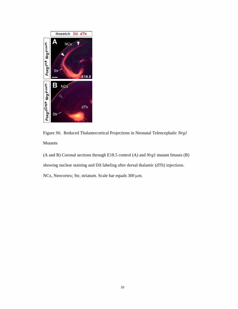
Figure S6. Reduced Thalamocortical Projections in Neonatal Telencephalic Nrg1
Mutants
(A and B) Coronal sections through E18.5 control (A) and Nrg1 mutant fetuses (B)
showing nuclear staining and DiI labeling after dorsal thalamic (dTh) injections.
NCx, Neocortex; Str, striatum. Scale bar equals 300 µm.

11
Figure S7. MGE Corridor Cells Are Present in Nrg1 and ErbB4 Mutants
(A and B) Coronal sections through control (A) and Nrg1 mutant (B) E13.5 brains
showing Islet1 expression in the striatum (Str) as well as in the medial ganglionic
eminence (MGE) corridor (arrowheads) in both control (A) and Nrg1 mutants (B).
(C and D) Similar experiments on coronal sections through control (C) and
ErbB4 (Β) Ε14.

1
Supplemental Data
Tangential Neuronal Migration Controls
Axon Guidance: A Role for Neuregulin-1
in Thalamocortical Axon Navigation
Guillermina López-Bendito, Aline Cautinat, Juan Antonio Sánchez, Franck
Bielle, Nuria Flames, Alistair N. Garratt, David A. Talmage, Lorna W. Role,
Patrick Charnay, Oscar Marín, and Sonia Garel

2
Figure S1. Expression of MGE and LGE Markers at the MGE Corridor
(A, B, and C) In situ hybridization on E13.5 coronal sections showing that Lhx6 (A) is not
expressed in the medial ganglionic eminence (MGE) corridor (bracket in A), whereas
Sema3F (B) and Meis2 (C) are expressed in this domain as well as in the adjacent lateral
ganglionic eminence (LGE) and striatum (Str) (open arrowheads).
(D) Immunohistochemistry for Islet1 and in situ hybridization for Meis2 showing co-
expression of these two markers by corridor cells (open arrowheads).
(E and F) GAD67 in situ hybridization and Islet1 immunohistochemistry showing that
corridor cells (open arrowheads) express both markers.

3
NCx, neocortex; GP, globus pallidus.
Scale bars equal 300 µm (A, B, C, and E) and 50 µm (D and F).
Figure S2. A Slice Culture Assay Reveals Nonpermissive Territories for TCA in the
Ventral Telencephalon
(A) Experimental paradigm used to test the growth of E13.5 GFP dTh explants in the
lateral ganglionic eminence (LGE).
(B and C) Bright field image of a GFP dTh explant confronted with the LGE after 72 h in
culture (B). In the same slice, TCAs grow in the Islet1-positive striatal mantle (B’ and C)

4
and reach the neocortex (NCx) (open arrowheads in B’), as well as in the piriform cortex
(solid arrowhead in B’).
(D and G) Experimental paradigms used to test the response of E13.5 GFP dTh to the
ventricular and subventricular zones (VZ/SVZ) of the MGE (D) and to the globus
pallidus (GP) (G).
(E) TCAs extend normally through a control homotypic Str transplant (dotted line).
(F, F’, H, and H’) Expression of Islet1 and GFP showing that TCAs extending toward the
cortex avoid entering the SV/SVZ MGE transplant (dotted line) or the GF transplant
(dotted line) in the Str.
(I) Schematic drawing summarizing the distribution of permissive and non-permissive
territories for the growth of TCAs.
Scale bars equal 200 µm (B, B’, E, F, F’, H, and H’) and 100 µm (C).
Figure S3. MGE Transplants Do Not Rescue TCAs Growth in Mash1 Mutants
(A) Experimental paradigm used to test the ability of cells derived from the medial
ganglionic eminence (MGE) to restore TCA growth in the Mash1 mutant telencephalon.
(B) GFP expression showing that a graft of GFP-MGE VZ into the MGE of a Mash1
mutant slice generates cells that migrate tangentially towards the cortex (open

5
arrowhead), following a route deep to that used by DiI-labeled TCAs (arrowhead) to
extend toward the neocortex (NCx).
(C) GFP expression showing that a GFP-MGE VZ transplant into the MGE of a Mash1
mutant slice restores interneuron migration to the cortex (open arrowhead), but does not
rescue the growth of DiI-labeled TCAs toward the NCx. TCAs ectopically invade the
piriform cortex (PCx; arrow).
H, Hippocampus; Str, striatum; VZ, ventricular zone.
Scale bars equal 150 µm (B and C).

6
Figure S4. Abnormal TCA Development in Mice Deficient for CRD-Nrg1 or for all Nrg1
Isoforms.

7
(A and D) Coronal sections through the telencephalon of CRD-Nrg1 heterozygous (A)
and CRD-Nrg1 mutant (D) E14.5 brains, showing the trajectory of TCAs labeled by L1
immunohistochemistry and nuclear staining.
(B and E) Higher magnifications of L1-labeled axons at the MGE corridor of CRD-Nrg1
heterozygous (B) and CRD-Nrg1 mutant (E) embryos. Note that the MGE corridor is
wider in CRD-Nrg1 mutants (brackets) than in control brains.
(C and F) Higher magnification of L1-labeled axons at the neocortex (NCx) of control
(C) and CRD-Nrg1 mutants (L). Less L1-positive fibers arrive to the NCx in CRD-Nrg1
mutant embryos (arrowheads).
(G and I) Coronal sections through the telencephalon of control (G) and telencephalic
Nrg1 conditional mutant (I) E13 brains, showing the trajectory of TCAs labeled by L1
immunohistochemistry and nuclear staining.
(H and J) Higher magnifications of L1-labeled axons in the ventral telencephalon of
control (H) and telencephalic Nrg1 conditional mutant (J) embryos. L1-labeled TCAs
largely fail to progress towards the NCx in mutants (open arrowheads), as compared to
control brains (arrowhead).
DTh, dorsal thalamus; Hyp, hypothalamus; Str, striatum.
Scale bars equal 300 µm (G and I), 200 µm (A, D, H, and J), and 100 µm (B, C, E, and
F).

8
Figure S5. Ig-NRG1 Promotes dTh Axonal Outgrowth in Collagen and Laminin
Substrates via its EGF-like domain
(A) Scoring scheme used to test the effect of Ig-NRG1 on dorsal thalamus (dTh) axons in
the experiments presented in Figures 6B and 6C. Explants were subdivided into four
equal sectors designated as proximal (P), distal (D), lateral 1 (L1), and lateral 2 (L2) in
relation to the COS cell aggregate. The length of the six longest axons in each sector was
measured in every explant.

9
(B) Quantification of the axonal growth of dTh explants cultured with mock-transfected
COS cells (n = 18) or Ig-Nrg1-transfected COS cells (n = 21) show a growth promoting
activity of Ig-NRG1. Considering all sectors, mean length of axons was 570,61 ± 28,35
µm (average ± SEM; bright grey dotted line) in controls and 762,62 ± 22,89 µm (average
± SEM; dark grey dotted line) in experimental cases. p < 0,001. No significant
differences were observed among different sectors, suggesting that Ig-NRG1 is not a
chemoattractant for dTh axons.
(C) Experimental paradigm used to test the effect of the EGF-like Domain of Ig-NRG1 in
the growth of E13.5 dTh axons cultured in collagen (B and C) or laminin (D and E).
(D and E) dTh explants cultured in collagen with control medium (D) or medium
supplemented with EGF-like domain (E).
(G and H) Similar experiments realized in laminin.
(F) Quantification of dTh axonal outgrowth in explants cultured in collagen with control
medium (n = 17) or EGF-like supplemented medium (n = 21). The length of the fifteen
longest axons was measure in every explant. Mean length of axons was 570,27 ± 54,26
µm (average ± SEM; bright grey bar) in controls and 773,32 ± 52,10 µm (average ±
SEM; dark grey bar) in experimental cases. p = 0,011.
Scale bar equals 200 µm.

10
Figure S6. Reduced Thalamocortical Projections in Neonatal Telencephalic Nrg1
Mutants
(A and B) Coronal sections through E18.5 control (A) and Nrg1 mutant fetuses (B)
showing nuclear staining and DiI labeling after dorsal thalamic (dTh) injections.
NCx, Neocortex; Str, striatum. Scale bar equals 300 µm.

11
Figure S7. MGE Corridor Cells Are Present in Nrg1 and ErbB4 Mutants
(A and B) Coronal sections through control (A) and Nrg1 mutant (B) E13.5 brains
showing Islet1 expression in the striatum (Str) as well as in the medial ganglionic
eminence (MGE) corridor (arrowheads) in both control (A) and Nrg1 mutants (B).
(C and D) Similar experiments on coronal sections through control (C) and
ErbB4 (Β) Ε14.
Copyright © 2022 FDOKUMEN