Prenatal Development of Cat Retinogeniculate Axon Arbors in ...

Cellular/Molecular
Requirement of Myeloid Cells for Axon Regeneration
Benoit Barrette, Marc-Andre Hebert, Mohammed Filali, Kathleen Lafortune, Nicolas Vallieres, Genevieve Gowing,Jean-Pierre Julien, and Steve LacroixDepartment of Anatomy and Physiology, Laval University, Ste-Foy, Quebec, Canada G1V 4G2
The role of CD11b � myeloid cells in axonal regeneration was assessed using axonal injury models and CD11b-TK mt-30 mice expressing amutated HSV-1 thymidine kinase (TK) gene regulated by the myeloid-specific CD11b promoter. Continuous delivery of ganciclovir at asciatic nerve lesion site greatly decreased the number of granulocytes/inflammatory monocytes and macrophages in the distal stump ofCD11b-TK mt-30 mice. Axonal regeneration and locomotor function recovery were severely compromised in ganciclovir-treated CD11b-TK mt-30 mice. This was caused by an unsuitable growth environment rather than an altered regeneration capacity of neurons. In absenceof CD11b � cells, the clearance of inhibitory myelin debris was prevented, neurotrophin synthesis was abolished, and blood vesselformation/maintenance was severely compromised in the sciatic nerve distal stump. Spinal cord-injured axons also failed to regeneratethrough peripheral nerve grafts in the absence of CD11b � cells. Therefore, myeloid cells support axonal regeneration and functionalrecovery by creating a growth-permissive milieu for injured axons.
Key words: nervous system; macrophages; neurotrophins; sciatic nerve; spinal cord injury; angiogenesis
IntroductionOver recent years, it has become increasingly evident that axonregeneration after neural injury is regulated by many factors.Some studies have suggested that changes in neuronal expressionof regeneration-associated genes (RAGs), caused by alterations ofintrinsic and/or extrinsic conditions, could account for the in-ability of neurons to regrow their axons (Plunet et al., 2002).Others have identified and characterized in the adult nervoussystem proteins that can inhibit axonal growth, such as myelin-associated inhibitors (David and Lacroix, 2003; Yiu and He,2006). Growth factors, including neurotrophins, and axon guid-ance molecules have also received their share of attention becausethey can support cell survival and promote and/or direct axonalgrowth (Lacroix and Tuszynski, 2000; Harel and Strittmatter,2006). An interesting observation came from the recent discoverythat neurotrophins and axon guidance molecules may also func-tion as angiogenic factors in the injured/diseased nervous system(Carmeliet and Tessier-Lavigne, 2005; Klagsbrun and Eichmann,2005; Folkman, 2007; Kermani and Hempstead, 2007). Whetherthe formation of new blood vessels may influence axonal regen-eration after injury remains, however, to be investigated. Of par-ticular interest to the present study is the fact that immune cells
may be able to influence all of the above-mentioned cellular re-sponses after injury.
The hypothesis that non-neuronal cells may influence axonregeneration was put forward many years ago by Ramon y Cajal(1991). However, confirmation of this hypothesis only came atthe beginning of the 1980s with seminal experiments by Davidand Aguayo (1981) in which peripheral nerve segments weregrafted into the injured CNS. Subsequent studies showed that thepresence of viable cells into the grafts was required for axongrowth to proceed (for review, see Fu and Gordon, 1997). Inter-estingly, regeneration of injured axons into acellular peripheralnerve grafts (frozen/thawed grafts) appears to depend on the re-population of the grafts by migrating/infiltrating cells, becauseaxonal regeneration in these grafts is completely prevented by theadministration of a mitosis inhibitor (Hall, 1986). AlthoughSchwann cells were considered to be responsible for promotingaxon regeneration in this study, it is likely that other proliferatingcells, such as immune cells, may also have been implicated inthese effects, especially because the antimitotic drug used did nottarget Schwann cells specifically. That immune cells may play acrucial role in axonal regeneration only became fully appreciatedwith the work of Dahlin (1995), who showed that physically pre-venting cell infiltration into peripheral nerve segments impairedthe outgrowth of injured sciatic nerve axons. Yet the exact rolesplayed by immune cells and the mechanisms by which they couldregulate regeneration and repair after neural injury are not welldefined.
Here, we demonstrate that myeloid cells play a key role inaxonal regeneration using various models of axonal injuries andtransgenic mice. Selective ablation of CD11b� cells in vivo usingthe HSV-1 thymidine kinase (TK)/ganciclovir (GCV) suicidegene system almost completely prevented granulocyte andmonocyte/macrophage recruitment and/or proliferation into theinjured sciatic nerve. In absence of these immune cells, axonal
Received April 4, 2008; revised July 21, 2008; accepted Aug. 10, 2008.This work was supported by a grant from the Natural Sciences and Engineering Research Council of Canada (S.L.).
S.L. is supported by a Career Award from the Rx&D Health Research Foundation and the Canadian Institutes of HealthResearch (CIHR). J.-P.J. holds a Canada Research Chair in Neurodegeneration. G.G. is a recipient of a CIHR DoctoralResearch Award. All other authors were supported by grants from the CIHR (S.L.). We thank Nadia Fortin, MelanieSimard, and Renee Paradis for their technical assistance. We acknowledge the work of Julie-Christine Levesque(Bio-Imaging Platform, Research Centre for Infectious Diseases, Centre Hospitalier Universite Laval) for her technicalassistance for cell imaging. We also thank Marc-Andre Laniel for his help editing this manuscript.
Correspondence should be addressed to Dr. Steve Lacroix, Centre Hospitalier Universite Laval Research Centerand Laval University, 2705, Laurier Boulevard, Ste-Foy, Quebec, Canada G1V 4G2. E-mail:[email protected].
DOI:10.1523/JNEUROSCI.1447-08.2008Copyright © 2008 Society for Neuroscience 0270-6474/08/289363-14$15.00/0
The Journal of Neuroscience, September 17, 2008 • 28(38):9363–9376 • 9363

regeneration in the sciatic nerve and recovery of locomotor func-tion were severely compromised with large deficits persistingover several weeks after injury. Careful examination of these micerevealed that CD11b� cells were responsible for clearance of in-hibitory myelin, neurotrophin synthesis, and formation/mainte-nance of blood vessels into and beyond the lesion site. Moreover,we show that spinal cord-injured (SCI) axons fail to regenerateinto peripheral nerve grafts in the absence of CD11b� cells. Theseresults show that granulocytes and monocytes/macrophages di-rectly influence axon regeneration by regulating the balance be-tween inhibitory molecules and growth factors.
Materials and MethodsAnimalsA total of 452 adult female mice (8 –12 weeks of age) were used in thisstudy. Transgenic mice expressing a mutant form of the HSV-1 TK sui-cide gene under the control of the myeloid-specific CD11b gene pro-moter, referred to as CD11b-TK mt-30 mice, were generated, reproduced,and genotyped, as described before by Gowing et al. (2006a). Adminis-tration of GCV to these mice causes the selective depletion of proliferat-ing CD11b � cells. Transgenic mice expressing the yellow fluorescentprotein (YFP) marker into a subset of their neurons and axons, thy1-YFP-H mice (referred to as YFP mice throughout the text), and thoseexpressing the enhanced green fluorescent protein (EGFP) in Schwanncells, referred to as S100�-EGFP mice, were purchased from The JacksonLaboratory and genotyped according to the protocol available on theirwebsite. For a complete description of these mice, please refer to thestudies by Feng et al. (2000) and Zuo et al. (2004). YFP and S100�-EGFPmice were crossed with CD11b-TK mt-30 mice for the experiments involv-ing peripheral nerve (PN) grafts and flow cytometry, respectively. NOD/CB17-Prkdc scid/J mice in the NOD/LtSz background and their wild-typecounterparts, NOD/ShiLtJ mice, were also purchased from The JacksonLaboratory. NOD/CB17-Prkdc scid/J mice homozygous for the severecombined immune deficiency (scid) spontaneous mutation are charac-terized by an absence of functional T-cells and B-cells. Mice had adlibitum access to food and water. All surgical procedures were approvedby the Laval University Animal Care Committee and followed CanadianCouncil on Animal Care guidelines.
Cell culturePrimary Schwann cell and fibroblast cultures derived from sciatic nervesof adult CD11b-TK mt-30 mice and their wild-type littermates were pre-pared according to established protocols (Morrissey et al., 1991; Weidneret al., 1999). Details are available in the supplemental Methods (availableat www.jneurosci.org as supplemental material).
To confirm that the HSV-1 TK mt-30 transgene was expressed specifi-cally by cells of myeloid origin, and not by other cell types in the sciaticnerve, cultures of Schwann cells and fibroblasts obtained from CD11b-TK mt-30 transgenic mice or their wild-type littermates were treated or notwith GCV. For this experiment, Schwann cells and fibroblasts wereseeded at 2.5 � 10 4 cells/well and positive control cells (i.e., GL261-CMV-TK) at 1 � 10 5 cells/well in CultureSlides (Falcon; BD BiosciencesDiscovery Labware) in their respective cell culture medium. Three dayslater, the medium was changed, GCV at 1 �M or PBS was added, and cellswere allowed to grow for another 3 d. Cells were then fixed with 4%paraformaldehyde (PFA) in PBS, pH 7.4, for 20 min and immediatelyprocessed for immunofluorescence following our previously describedprotocol (Pineau and Lacroix, 2007). Details are available in the supple-mental Methods (available at www.jneurosci.org as supplementalmaterial).
For quantification of Schwann cells (p75 NTR�/S100� �) and fibro-blasts (fibronectin �/S100� �), the number of labeled cells was estimatedby the optical fractionator method using the Bioquant Nova Prime soft-ware (Bioquant Image Analysis). Details are available in the supplemen-tal Methods (available at www.jneurosci.org as supplemental material).
Surgeries and animal treatmentSciatic nerve microcrush lesions. Mice were deeply anesthetized withisoflurane and underwent a microcrush lesion of their left sciatic nerve at
the midthigh level, following our previously published method (Boivin etal., 2007). As before, the lesion site was marked with a 10-0 Ethilon suture(Ethicon) passed through the epineurium only.
GCV treatment. Mice were treated with four intraperitoneal injectionsof either saline or GCV (Cytovene; Roche) at 50 mg/kg, starting 12 hbefore the lesion followed by injections every 12 h until the 24 h timepoint after lesion. In addition, mice were implanted with miniosmoticpumps (model 2004; Alzet) to continuously deliver saline or GCV at thelesion site (0.25 �l/h at 10 �g/�l for 28 d) throughout the duration ofthe experiments. For the purpose of the experiment in which we assessedthe role of CD11b � cells in angiogenesis/blood vessel integrity after pe-ripheral nerve lesion, mice received bidaily intraperitoneal injections ofeither saline or GCV at 50 mg/kg starting 12 h before the lesion until theend of the experimental protocol (i.e., 7 d after lesion).
Sciatic–sciatic nerve grafts. To investigate the capacity of peripheralaxons to regenerate in the absence of CD11b � cells, we performed ex-periments in which pre-degenerated sciatic nerve segments collectedfrom either saline-treated or GCV-treated YFP �/TK � (i.e., CD11b-TK mt-30�/�) and YFP �/TK � [i.e., wild type (WT) for the CD11b-TK mt-30 transgene] mice were grafted to the sciatic nerve of YFP �/TK �
and YFP �/TK � recipient mice, allowing to visualize YFP � axons thathave regenerated into grafts (for an example of this, see Fig. 4a). All graftswere allografts and performed under deep isoflurane anesthesia. First,YFP �/TK � and YFP �/TK � donor mice underwent sciatic nerve micro-crush lesions at midthigh level and treatment with either saline or GCV,as described above. One week later, a 3-mm-long segment of their sciaticnerve distal stump was removed, trimmed of excess epineurial fat insterile HBSS, and immediately sutured between the sciatic nerve proxi-mal and distal stumps of freshly transected recipient mice (i.e., YFP �/TK � and YFP �/TK � mice). Grafts were aligned with the proximal anddistal ends of the recipient sciatic nerve and secured using 10-0 Ethilonsutures. Recipient mice were treated with either saline or GCV for theentire duration of the experimental protocol. For this particular experi-ment, we selected 2 weeks as the optimal survival time after grafting basedon recommendations from a previous study by English et al. (2005).
Intraspinal transplantation of sciatic nerve segments. To investigate thecapacity of central axons to regenerate in the absence of CD11b � cells, weperformed experiments in which pre-degenerated sciatic nerve graftscollected from either saline-treated or GCV-treated YFP �/TK � andYFP �/TK � mice were grafted into the injured spinal cord of YFP �/TK �
and YFP �/TK � recipient mice (for an example of this, see Fig. 9a).Briefly, YFP �/TK � and YFP �/TK � mice underwent sciatic nerve mi-crocrush lesions at midthigh level and treatment with either saline orGCV. One week later, pre-degenerated sciatic nerve distal stumps de-pleted or not in CD11b � cells were isolated, cut into 2 mm segments, andimmediately transplanted into the injured spinal cord of dorsal he-misected YFP �/TK � and YFP �/TK � recipient mice. Once again, recip-ient mice were treated with either saline or GCV for the entire duration ofthe experimental protocol. SCI mice were killed after 1 week after graft-ing. Please refer to our previously published protocols for the lesion/grafting surgery (Lacroix et al., 2002; Vallieres et al., 2006).
Tissue processingFor the experiments involving in situ hybridization (ISH) or ISH com-bined to immunohistochemistry, mice were overdosed with a mixture ofketamine and xylazine and transcardially perfused with 0.9% saline so-lution followed by 4% PFA, pH 9.5, in borax buffer. After perfusion withthe fixative, brains, sciatic nerves, dorsal root ganglia (DRGs), and spinalcords were dissected out, postfixed for 2 d, and placed overnight in a 4%PFA-borax/10% sucrose solution until tissue processing. For the purposeof histological and immunohistochemical labeling, animals were per-fused instead with cold PBS followed by 4% PFA, pH 7.4, in PBS. Sciaticnerves were postfixed for 1 h only and then placed in a PBS/20% sucrosesolution until sectioning. More details are provided in the supplementalMethods (available at www.jneurosci.org as supplemental material).
ISHISH was performed to detect mRNAs coding for the following molecules:GAP-43, CAP-23, T�-1 tubulin, NGF, BDNF, Neurotrophin-3 (NT-3),
9364 • J. Neurosci., September 17, 2008 • 28(38):9363–9376 Barrette et al. • Myeloid Cells and Axonal Regeneration

NT-4/5, Tie2, and p75 NTR. Radiolabeled cRNA probes were synthesizedusing full-length cDNAs cloned into expression vectors pCRII-TOPOand the Riboprobe Combination System SP6/T7 (Promega) and T3 RNApolymerase (Promega). Sequences chosen for probe synthesis were se-lected to match only the intended genes, as verified by BLAST analysis inGenBank. ISH was performed according to a previously describedmethod (Barrette et al., 2007; Pineau and Lacroix, 2007). All sectionswere prehybridized, hybridized, and posthybridized in parallel to equal-ize background intensity. A combination of ISH with immunohisto-chemistry was performed to localize gene expression in individual cells,according to our previously published protocol (Pineau et al., 2006).
Histology and immunohistochemistryOne series of adjacent sciatic nerve sections was stained with Luxol fastblue (LFB) to identify myelin, following our previously published proto-col (Vallieres et al., 2006). These sections were used to quantify myelinclearance in the degenerating sciatic nerve distal stump.
Immunoperoxidase labeling was performed following our previouslypublished protocol (Vallieres et al., 2006), except that the hydrogen per-oxide (H2O2) quenching procedure (to reduce the activity of endoge-nous peroxidases) was performed with 6% H2O2 in methanol for 1 h. Allsections were stained in parallel to equalize the background intensity andthe peroxidase reaction. Multiple immunofluorescence labeling was per-formed as described previously (Pineau and Lacroix, 2007). The com-plete description of the primary antibodies used in this study and theirdilution are given in supplemental Table 1 (available at www.jneurosci.org as supplemental material).
To further confirm the clearance of myelin debris and examine themyelin phagocytic activity of macrophages in our experiments, Oil Red O(ORO) staining was combined with CD68 immunofluorescence on thesame series of sciatic nerve sections. As previously reported by us andothers, ORO staining is highly specific for degenerating myelin and formyelin that has been ingested by macrophages (Ma et al., 2002; Valliereset al., 2006). Immunofluorescence was performed first followed by OROstaining, as previously described (Boivin et al., 2007).
Epon embedding and toluidine blue stainingFor examination of motor and sensory neuron viability after sciatic nervemicrocrush lesion in mice depleted in CD11b � cells, animals were per-fused with 4% PFA, pH 7.4, at 50 d after lesion. Ipsilateral L5 dorsal andventral roots were immediately collected, postfixed in 2.5% glutaralde-hyde and 0.5% PFA in PBS buffer, pH 7.4, for 2 h, postfixed in 2%osmium tetroxide for another 2 h, and then processed for embedding inEpon following our previously described protocol (Vallieres et al., 2006).One-micrometer-thick cross sections of the roots were stained with 1%toluidine blue for light microscopy.
Quantification of ISH signal and histological andimmunohistochemical labelingQuantification was performed using the Bioquant Nova Prime imageanalysis system (Bioquant Image Analysis). Details are available in thesupplemental Methods (available at www.jneurosci.org as supplementalmaterial.
Flow cytometry analysis of sciatic nerve Schwann cellsTo address the question of whether depletion of CD11b � myeloid cellsmay have affected Schwann cell function, rather than Schwann cell pro-liferation, the expression of Galectin-3 in Schwann cells was examined invivo using flow cytometry. This was done on Schwann cells purified fromsciatic nerve distal stump biopsies obtained from mice generated fromcrossing S100�-EGFP with CD11b-TK mt-30 mice. In this experimentalsetting, Schwann cells were gated based on their expression of S100� (i.e.,GFP) and p75 NTR. Surface and intracellular expression of Galectin-3 wasthen examined in Schwann cells collected from S100�-EGFP/CD11b-TK mt-30 transgenic mice or their wild-type littermates after treatmentwith either saline or GCV. More details are available in the supplementalMethods (available at www.jneurosci.org as supplemental material).
Behavioral analysisRecovery of locomotor function after sciatic nerve lesion was quantifiedusing the sciatic nerve functional index (SFI), as described previously
(Boivin et al., 2007). This test was performed for all animals preopera-tively and every week for 7 weeks after the operation. The SFI was mea-sured using the formula adapted for mice by Inserra et al. (1998). SFIvalues oscillated around 0 � 10 for noninjured animals and around�100 � 10 after complete lesion of the sciatic nerve (de Medinaceli et al.,1982). All behavioral analyses were done blind with respect to the identityof the animals.
Statistical analysisAll statistical evaluations were performed with one- or two-way ANOVAor repeated-measures ANOVA. Post-ANOVA comparisons were madeusing the Bonferroni test. All statistical analyses were performed usingthe GraphPad Prism software (GraphPad Software). A value of p � 0.05was considered to be statistically significant. Data in graphs are presentedas mean � SEM.
ResultsThe HSV-TK transgene is specifically expressed by cells ofmyeloid origin in the injured sciatic nerve of CD11b-TK mt-30
miceThe specificity of expression of the HSV-TK transgene was firstassessed in vitro using primary cultures of Schwann cells andfibroblasts derived from sciatic nerves of adult CD11b-TK mt-30
transgenic mice and wild-type littermates. First, we establishedthat Schwann cell and fibroblast cultures were pure (�98% pu-rity) based on multiple immunofluorescence labeling using cell-specific markers (supplemental Fig. 1a– d, available at www.jneurosci.org as supplemental material). Cells were then treatedwith either GCV or PBS for 3 d and counted to verify whethertreatment induced cell death (supplemental Fig. 1e, available atwww.jneurosci.org as supplemental material). Cells from amouse glioma cell line, GL261, transduced with a lentiviral vectorexpressing the HSV-TK construction under the control of theCMV promoter (GL261-CMV-TK) were used as positive controlcells. As demonstrated in supplemental Figure 1e (available atwww.jneurosci.org as supplemental material), the addition ofGCV to the culture medium led to the killing of almost all GL261-CMV-TK control cells, but had no effect on the survival of bothSchwann cells and fibroblasts. This confirmed the lack of ectopicexpression of the HSV-TK transgene in these cells.
It was imperative to obtain in vivo evidence that GCV treat-ment did not ablate proliferating Schwann cells in our CD11b-TK mt-30 transgenic mice, because these cells have been long rec-ognized to play an indispensable role in promoting peripheralnerve regeneration and repair after injury (Fu and Gordon,1997). Thus, we examined whether there was a decrease ofSchwann cells after a sciatic nerve microcrush lesion in GCV-treated CD11b-TK mt-30. For this, transgenic mice and their wild-type littermates received intraperitoneal injections (twice a dayfor 2 d) of either GCV or saline plus continuous delivery of thedrugs at the lesion site using Alzet miniosmotic pumps. Schwanncells were visualized through detection of the Schwann cellmarker, p75 NTR, in peripheral nerve preparations using ISH. Asshown in supplemental Figure 2a (available at www.jneurosci.orgas supplemental material), the levels of p75 NTR mRNA signal atvarious distances from the site of sciatic nerve lesion did notstatistically differ between groups at 7 d after lesion.
GCV administration to CD11b-TK mt-30 transgenic miceresults in the depletion of granulocytes, monocytes, andmacrophages in the sciatic nerve after injuryTo identify the populations of myeloid cells that were depleted inGCV-treated CD11b-TK mt-30 transgenic mice, immunolabelingwas performed on tissue sections prepared from injured sciatic
Barrette et al. • Myeloid Cells and Axonal Regeneration J. Neurosci., September 17, 2008 • 28(38):9363–9376 • 9365
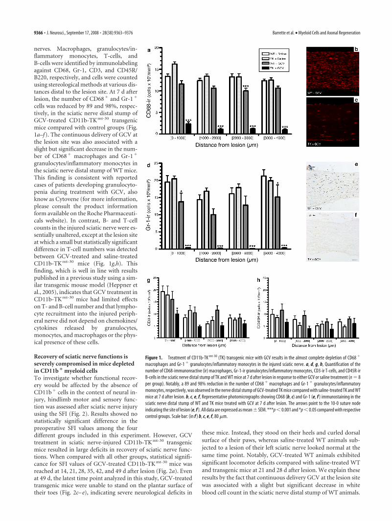
nerves. Macrophages, granulocytes/in-flammatory monocytes, T-cells, andB-cells were identified by immunolabelingagainst CD68, Gr-1, CD3, and CD45R/B220, respectively, and cells were countedusing stereological methods at various dis-tances distal to the lesion site. At 7 d afterlesion, the number of CD68� and Gr-1�
cells was reduced by 89 and 98%, respec-tively, in the sciatic nerve distal stump ofGCV-treated CD11b-TK mt-30 transgenicmice compared with control groups (Fig.1a–f). The continuous delivery of GCV atthe lesion site was also associated with aslight but significant decrease in the num-ber of CD68� macrophages and Gr-1�
granulocytes/inflammatory monocytes inthe sciatic nerve distal stump of WT mice.This finding is consistent with reportedcases of patients developing granulocyto-penia during treatment with GCV, alsoknow as Cytovene (for more information,please consult the product informationform available on the Roche Pharmaceuti-cals website). In contrast, B- and T-cellcounts in the injured sciatic nerve were es-sentially unaltered, except at the lesion siteat which a small but statistically significantdifference in T-cell numbers was detectedbetween GCV-treated and saline-treatedCD11b-TK mt-30 mice (Fig. 1g,h). Thisfinding, which is well in line with resultspublished in a previous study using a sim-ilar transgenic mouse model (Heppner etal., 2005), indicates that GCV treatment inCD11b-TK mt-30 mice had limited effectson T- and B-cell number and that lympho-cyte recruitment into the injured periph-eral nerve did not depend on chemokines/cytokines released by granulocytes,monocytes, and macrophages or the phys-ical presence of these cells.
Recovery of sciatic nerve functions isseverely compromised in mice depletedin CD11b � myeloid cellsTo investigate whether functional recov-ery would be affected by the absence ofCD11b� cells in the context of neural in-jury, hindlimb motor and sensory func-tion was assessed after sciatic nerve injuryusing the SFI (Fig. 2). Results showed nostatistically significant difference in thepreoperative SFI values among the fourdifferent groups included in this experiment. However, GCVtreatment in sciatic nerve-injured CD11b-TK mt-30 transgenicmice resulted in large deficits in recovery of sciatic nerve func-tions. When compared with all other groups, statistical signifi-cance for SFI values of GCV-treated CD11b-TK mt-30 mice wasreached at 14, 21, 28, 35, 42, and 49 d after lesion (Fig. 2a). Evenat 49 d, the latest time point analyzed in this study, GCV-treatedtransgenic mice were unable to stand on the plantar surface oftheir toes (Fig. 2c– e), indicating severe neurological deficits in
these mice. Instead, they stood on their heels and curled dorsalsurface of their paws, whereas saline-treated WT animals sub-jected to a lesion of their left sciatic nerve looked normal at thesame time point. Notably, GCV-treated WT animals exhibitedsignificant locomotor deficits compared with saline-treated WTand transgenic mice at 21 and 28 d after lesion. We explain theseresults by the fact that continuous delivery GCV at the lesion sitewas associated with a slight but significant decrease in whiteblood cell count in the sciatic nerve distal stump of WT animals.
Figure 1. Treatment of CD11b-TK mt-30 (TK) transgenic mice with GCV results in the almost complete depletion of CD68 �
macrophages and Gr-1 � granulocytes/inflammatory monocytes in the injured sciatic nerve. a, d, g, h, Quantification of thenumber of CD68-immunoreactive (ir) macrophages, Gr-1-ir granulocytes/inflammatory monocytes, CD3-ir T-cells, and CD45R-irB-cells in the sciatic nerve distal stump of TK and WT mice at 7 d after lesion in response to either GCV or saline treatment (n � 8per group). Notably, a 89 and 98% reduction in the number of CD68 � macrophages and Gr-1 � granulocytes/inflammatorymonocytes, respectively, was observed in the nerve distal stump of GCV-treated TK mice compared with saline-treated TK and WTmice at 7 d after lesion. b, c, e, f, Representative photomicrographs showing CD68 (b, c) and Gr-1 (e, f ) immunostaining in thesciatic nerve distal stump of WT and TK mice treated with GCV at 7 d after lesion. The arrows point to the 10-0 suture nodeindicating the site of lesion (e, f ). All data are expressed as mean � SEM. ***p � 0.001 and *p � 0.05 compared with respectivecontrol groups. Scale bar: (in f ) b, c, e, f, 80 �m.
9366 • J. Neurosci., September 17, 2008 • 28(38):9363–9376 Barrette et al. • Myeloid Cells and Axonal Regeneration

When miniosmotic pumps used to continuously deliver GCVat the lesion site were removed after 1 week after injury of treat-ment, CD11b-TK mt-30 mice recovered sciatic functions similar tocontrol animals after 6 –7 weeks (Fig. 2b). This represents a delayof �1–2 weeks compared with the typical time course of recoveryseen in control mice. Our interpretation of the latter result is thatnerve regeneration and repair may have started to proceed assoon as CD11b� myeloid cells were allowed to proliferate andinvade the injured sciatic nerve.
Myeloid cells are not required for survival of axotomizedsciatic motor and sensory neuronsWe next asked whether the long-term deficits described abovecould have been caused by the death of axotomized neurons,because CD11b� cells could have produced or affected the re-lease of factors that are critical for neuronal survival after injury.To test this possibility, we analyzed the viability of motor andsensory neurons at 50 d after sciatic axotomy. Results showed that�2.5% of all L5 dorsal root ganglion neurons and spinal cordmotor neurons projecting into the sciatic nerve had died from themicrocrush lesion at 50 d after injury (data not shown). As dem-onstrated in Figure 3, neuronal viability did not differ betweenGCV-treated CD11b-TK mt-30 transgenic mice and control miceat 50 d. These data suggest that the severe and persistent neuro-logical deficits observed in GCV-treated CD11b-TK mt-30 trans-genic mice after peripheral nerve injury are not the result of neu-ronal cell loss, leaving axonal regeneration failure as the mostlikely explanation.
Peripheral nerve axon regeneration iscompromised in mice depleted inCD11b � cellsWe thus tested whether axonal regenera-tion was compromised in the absence ofCD11b� cells. For this, we took advantageof transgenic mice that were generatedfrom breeding thy1-YFP-H mice (that ex-press the YFP marker in a subset of theirneurons and axons) with CD11b-TK mt-30
mice and quantified the total number ofYFP� sciatic nerve axons that had regen-erated into YFP� peripheral nerve allo-grafts depleted or not in CD11b� cells. Arepresentative example of the regenerationof YFP� sciatic nerve axons into a grafttaken from a saline-treated TK�/YFP�
mouse at 2 weeks after lesion/grafting ispresented in Figure 4, a and b. Proximal tothe graft at 2 weeks after surgery, no statis-tically significant difference in the count ofYFP� axons was found between the fourdifferent groups included in this experi-ment (Fig. 4c). However, the situationwithin the graft differed drastically amongthe groups, because peripheral axons al-most completely failed to regenerate in theabsence of CD11b� cells. The followingaxonal numbers (�SEM) were counted atpredetermined distances within periph-eral nerve grafts from GCV-treatedCD11b-TK mt-30 mice: 500 �m, 3.7 � 1.2axons; 1000 �m, 3.0 � 0.9 axons; 1500�m, 2.3 � 0.8 axons; and 2000 �m, 1.3 �0.8 axons. In all other groups, average
counts of YFP� axons within grafts varied between 45.5 � 8.1and 61.0 � 5.4 axons at 500 �m, 37.0 � 2.8 and 55.3 � 6.2 axonsat 1000 �m, 31.0 � 2.9 and 43.1 � 3.3 axons at 1500 �m, and16.8 � 3.1 and 36.6 � 3.6 axons at 2000 �m. The difference inaxonal counts between GCV-treated TK� grafts (these graftswere used to repair the sciatic nerve of TK�/YFP� recipientmice) and grafts from other groups is significant for all distancesanalyzed (two-way repeated-measures ANOVA, p � 0.001). To-gether with our previous finding that sciatic nerve function re-covery is severely compromised in GCV-treated CD11b-TK mt-30
mice, these results indicate that myeloid cells are essential forperipheral axon regeneration.
Neuronal cell bodies of axotomized sciatic nerve axons reactnormally to injury by overexpressing RAGs even in theabsence of myeloid cellsWe then investigated some of the mechanisms that could be usedby CD11b� myeloid cells to promote axonal regeneration. Onelikely theory was that CD11b� cells may somehow alter the in-trinsic capacity of neuronal cell bodies to regenerate. Using ISH,we therefore studied the expression of RAGs such as GAP-43,CAP-23, and T�-1 tubulin in cell bodies of axotomized motorand sensory neurons projecting through the sciatic nerve. Thesethree RAGs were chosen based on previous studies that showedthat the expression of these particular genes reflected the intrinsiccapacity of neurons to regenerate their axons [these studies aresummarized in the review article by Plunet et al. (2002)]. For thisexperiment, mice were killed at 7 d after lesion. This time point
Figure 2. CD11b � cells are required for recovery of sciatic nerve functions after injury. a, Recovery of locomotor functions froma sciatic nerve lesion, as determined by the sciatic function index (SFI), is severely compromised in GCV-treated CD11b-TK mt-30
(TK) transgenic mice compared with control groups, with large deficits persisting even at 49 d after lesion (n � 12 per group). b,When minipumps were removed at 7 d after lesion in TK mice, transgenic animals recovered after 6 –7 weeks (n � 12 per group).Error bars indicate SEM. c– e, Representative examples of the typical posture adopted by saline-treated WT (c) and GCV-treated TK(d, e) mice at 35 (d) and 49 (c, e) days after lesion of their left sciatic nerve. ***p � 0.001 and **p � 0.01 compared withrespective control groups.
Barrette et al. • Myeloid Cells and Axonal Regeneration J. Neurosci., September 17, 2008 • 28(38):9363–9376 • 9367

was selected based on a time course study that demonstratedmaximal expression between 3 and 7 d (data not shown). Asshown in Figure 5, cell bodies from motor neurons projectingthrough the sciatic nerve appeared to react normally to the ab-sence of CD11b� cells by overexpressing GAP-43, CAP-23, andT�-1 tubulin mRNAs (Fig. 5). Similarly, L5 DRG sensory neu-rons were also found to overexpress these RAGs after injury andmRNA levels remained similar between all groups at 7 d (data notshown). Obviously, these results do not exclude the possibilitythat expression of these or other RAG genes was modulated in cellbodies of axotomized neurons at other times after injury. Never-theless, they do suggest that CD11b� myeloid cells modulateaxonal regeneration by directly influencing growth cone dynam-ics through the creation of a more favorable growth environ-ment, having little or no detectable effects at the perikaryon level.
Myeloid cells are essential for the clearance of inhibitorymyelin debris after peripheral nerve injuryOne way by which myeloid cells may create a more permissiveenvironment for axon regeneration is by clearing inhibitory my-elin debris during wallerian degeneration. We therefore investi-gated whether clearance of myelin debris would be compromisedin the absence of CD11b� cells. Quantification of LFB staining, amarker of myelin, in the sciatic nerve distal stump revealed thatthe load of myelin debris was �200% higher in GCV-treatedCD11b-TK mt-30 mice compared with control groups at 7 d after
lesion (Fig. 6a). This was further supported by the finding that thenumber of CD68� macrophages that had ingested myelin debris,as visualized using ORO staining, was decreased by �66% indegenerating sciatic nerves of GCV-treated CD11b-TK mt-30 micecompared with control groups at 7 d (data not shown). Exami-nation of our sciatic nerve preparations also revealed that, al-though myelin debris clearance was severely compromised in thesciatic nerve distal stump of GCV-treated CD11b-TK mt-30 trans-genic mice (Fig. 6b,c), myelin breakdown and formation ofovoids of degenerating myelin was not prevented in these mice.Furthermore, we found that most of the remaining CD68� mac-rophages (i.e., those not depleted by the GCV treatment) werefilled with myelin debris. In our opinion, this may explain whymacrophage depletion strategies did not completely abolish my-elin debris clearance in this and another study (Bruck et al.,1996). Still, we cannot rule out the possibility that Schwann cellsmay have also been involved in the phagocytosis of myelin debris,as suggested by others (Stoll et al., 1989; Reichert et al., 1994;Fernandez-Valle et al., 1995; Saada et al., 1996; Hirata and Kawa-buchi, 2002).
Because some of the above-mentioned studies have suggestedthat myelin phagocytosis by Schwann cells may occur via a lectin-mediated mechanism implicating Galectin-3, also referred to asMac-2, we next investigated whether depletion of CD11b� my-eloid cells may affect the expression of Galectin-3 in Schwanncells. Using immunohistochemistry, we first found that, although
Figure 3. Depletion of CD11b � cells does not lead to death of axotomized neurons. a, d, Counts of axonal profiles in Epon-embedded L5 ventral and dorsal roots at 50 d after lesion revealed thatdifferences in functional recovery in CD11b-TK mt-30 (TK) transgenic mice compared with WT are not caused by the loss of motor or sensory neurons. Error bars indicate SEM. b, c, e, f, Photomicro-graphs showing toluidine blue staining of L5 ventral (b, c) and dorsal (e, f ) roots ipsilateral to the lesion. Representative cross sections were taken from GCV-treated WT and TK mice. Scale bar: (inf ) b, c, e, f, 12.5 �m.
9368 • J. Neurosci., September 17, 2008 • 28(38):9363–9376 Barrette et al. • Myeloid Cells and Axonal Regeneration

Galectin-3 signal was decreased in the sciatic nerve distal stumpof GCV-treated CD11b-TK mt-30 mice compared with controlgroups, protein levels remained relatively high in transgenic miceeven in the absence of close to 90% of all CD68� macrophages(supplemental Fig. 3a– d, available at www.jneurosci.org as sup-plemental material). These results therefore support the idea thatSchwann cells are an important source of Galectin-3 after periph-eral nerve injury. We then aimed to determine whether Schwanncell function related to myelin debris clearance could have beenaltered in the absence CD11b� myeloid cells. For this, flow cy-tometry was used to examine the expression of Galectin-3 inSchwann cells purified from sciatic nerve distal stump biopsiescollected from mice generated from crossing S100�-EGFP withCD11b-TK mt-30 transgenic mice. In GCV-treated S100�-EGFP/
CD11b-TK mt-30 mice, we found that the proportion of Schwanncells expressing Galectin-3 was 68.9 � 8.6% at 7 d after lesion(supplemental Fig. 3e, available at www.jneurosci.org as supple-mental material). This proportion did not differ significantly be-tween groups, with percentages of S100��P75 NTR�Galectin-3�
of 79.3 � 4.8, 76.9 � 5.7, and 79.6 � 5.9% in saline-treated WT,saline-treated S100�-EGFP/CD11b-TK mt-30, and GCV-treatedWT mice, respectively. In summary, these results indicate that amajor subset of Schwann cells express Galectin-3 at 7 d afterperipheral nerve injury and that upregulation of the galactose-specific lectin does not depend on the presence CD11b� myeloidcells. Along these lines, the Rotshenker Laboratory (Saada et al.,1996) previously proposed that fibroblasts are responsible,through the production and release of granulocyte-macrophagecolony-stimulating factor, for the upregulation of Galectin-3 inSchwann cells and macrophages in lesioned peripheral nerves.
Neurotrophin synthesis is prevented in the absence ofCD11b � myeloid cellsAnother way by which axon regeneration is regulated in the in-jured nervous system is through the release of growth factors suchas neurotrophins by non-neuronal cells. One current dogma inthe regeneration field is that Schwann cells in the peripheral nervedistal stump secrete neurotrophins in a macrophage-dependentmanner (Heumann et al., 1987b; Lindholm et al., 1987). Thus, wenext investigated whether neurotrophin synthesis would be com-
Figure 4. Regeneration of peripheral axons is prevented in the absence of CD11b � cells. a,Photograph showing an example of sciatic–sciatic nerve graft. b, Representative fluorescencephotomicrograph showing sciatic nerve axons expressing the YFP marker that have regeneratedinto a PN graft collected from a saline-treated YFP �/TK� mouse (i.e., CD11b-TK mt-30�/�).The arrows point to the two 10-0 sutures used to connect the PN graft between the proximal anddistal ends of the recipient sciatic nerve from the YFP transgenic mouse. c, Quantification of thenumber of YFP-labeled axons at predetermined distances from the proximal host– graft inter-face (n � 8 per group). Although the total number of YFP � axons was similar in all groups at adistance of 1.5 mm from the proximal host– graft interface (i.e., into the proximal end of therecipient sciatic nerve), YFP � axons were only able to regenerate into predegenerated PNgrafts in the presence of CD11b � cells. Error bars indicate SEM. ***p � 0.001. Scale bar,500 �m.
Figure 5. Cell bodies from axotomized sciatic motor neurons reacted normally to injury inthe absence of CD11b � cells by overexpressing regeneration-associated genes. a– c, Quanti-fication of in situ hybridization signal for GAP-43 (a), CAP-23 (b), and T�-1 tubulin (c) mRNAs[in optical density (O.D.); arbitrary units] in the ipsilateral motoneuronal cell group of Rexed’slamina IX at spinal levels L2–L5 in CD11b-TK mt-30�/� (TK) and WT mice treated with eitherGCV or saline at 7 d after sciatic nerve lesion (n � 8 per group). Error bars indicate SEM.
Barrette et al. • Myeloid Cells and Axonal Regeneration J. Neurosci., September 17, 2008 • 28(38):9363–9376 • 9369

promised after sciatic nerve lesion in theabsence of CD11b� myeloid cells. Becauseneurotrophins are apparently expressed inlow abundance and because specific anti-bodies for these growth factors that workwell in fixed tissue sections are difficult tofind, we chose to use radioactive ISH andhighly specific RNA riboprobes to detectmRNAs coding for each of the four mam-malian neurotrophins. This method hasbeen used with great success by our labo-ratories in previous studies in which wereported the expression of cytokines/growth factors in models of CNS diseasesand injuries (Gowing et al., 2006b; Boivinet al., 2007). The specificity of our ribo-probes was first tested in the adult mousebrain. As demonstrated in supplementalFigure 4 (available at www.jneurosci.org assupplemental material), the distributionof NGF, BDNF, NT-3, and NT-4/5 mR-NAs using our radiolabeled cRNA probesand ISH protocol is similar to what hasbeen reported by others in intact adult ratand mouse brains (Bandtlow et al., 1990;Hofer et al., 1990; Maisonpierre et al.,1990; Guthrie and Gall, 1991). Consistentwith findings from these previous studies,both similarities and differences were ob-served in the distribution pattern of neu-rotrophin gene expression in the adult ro-dent brain.
Using ISH, we first performed a timecourse study to examine neurotrophin ex-pression after sciatic nerve injury. For this,C57BL/6J mice were killed at 6 h and at 1,3, 5, 7, 10, and 14 d after lesion. In general,all neurotrophins had a similar pattern ofexpression. As expected, we found no hy-bridization signal for any of the neurotro-phins analyzed on sciatic nerve sectionsobtained from noninjured mice. At 6 h af-ter lesion, cells expressing neurotrophinswere rare and strictly found at the lesionsite, in close association with theepineurium. At 1 d, more positive cellswere seen, again closely associated with theepineurium and blood vessels irrigatingthe nerve. At 3 d, neurotrophin-positivecells had already invaded the entire sciaticnerve distal stump, with cell numbers in-creasing over time up to day 14 after le-sion, the latest time point analyzed. An ex-ample of NT-3 mRNA signal in the sciaticnerve distal stump at 7 d is shown in Figure7b. Quantitative analyses of the number ofcells expressing neurotrophins in the nerve distal stump showedthat the number of NT-3� (Fig. 7a), BDNF� (supplemental Fig.5a– c, available at www.jneurosci.org as supplemental material),NGF� (supplemental Fig. 5d–f, available at www.jneurosci.org assupplemental material), and NT-4/5� (supplemental Fig. 5g–i,available at www.jneurosci.org as supplemental material) cellshad decreased by 92, 88, 87, and 86%, respectively, in GCV-
treated CD11b-TK mt-30 transgenic mice compared with controlgroups at 7 d. We interpret these findings as evidence that neu-rotrophin synthesis by non-neuronal cells depends on the pres-ence of CD11b� myeloid cells.
To determine whether neurotrophins were produced by cellsof myeloid origin or by other cell types in response to factorsreleased by infiltrating CD11b� cells, as suggested by others
Figure 6. Inhibitory myelin debris could not be cleared in the absence of CD11b � cells. a, Quantification of LFB staining ofmyelin in the sciatic nerve distal stump of CD11b-TK mt-30�/� (TK) and WT mice treated with either GCV or saline at 7 d after sciaticnerve lesion (n�8 per group). Although myelin breakdown and formation of ovoids of degenerating myelin were apparent in thesciatic nerve distal stump of GCV-treated TK mice, quantification of LFB staining revealed that the load of myelin debris was�200% higher in mice depleted in CD11b � cells compared with mice of control groups at 7 d after lesion. Error bars indicate SEM.b, c, Representative bright-field photomicrographs showing myelin stained with LFB in the sciatic nerve distal stump of GCV-treated WT and GCV-treated TK mice at 7 d after lesion. ***p � 0.001. Scale bar: (in b) b, c, 100 �m.
Figure 7. NT-3 synthesis in the injured peripheral nerve is prevented in the absence of CD11b � cells. a, Quantification of thenumber of cells expressing NT-3 mRNA in the sciatic nerve distal stump of CD11b-TK mt-30 (TK) and WT mice at 7 d after lesion inresponse to either GCV or saline treatment (n � 8 per group). Error bars indicate SEM. b, c, Representative dark-field photomi-crographs showing NT-3 mRNA expression in the sciatic nerve distal stump of GCV-treated WT (b) and GCV-treated TK (c) mice at7 d after lesion. d, e, Photomicrographs of the sciatic nerve distal stump of a WT mouse showing colocalization of NT-3 mRNA withGr-1-immunoreactive granulocytes/inflammatory monocytes. The arrows point to double-labeled cells. f, Representative dark-field photomicrograph showing the presence of numerous cells expressing NT-3 mRNA in the sciatic nerve distal stump of a scidmouse that has no functional T- and B-cells at 7 d after lesion. Together, these results indicate that a subset of CD11b � myeloidcells expressing the cell surface protein Gr-1 are responsible for neurotrophin synthesis in the injured peripheral nerve. ***p �0.001. Scale bars: (in e) d, e, 5 �m; (in f ) b, c, f, 100 �m.
9370 • J. Neurosci., September 17, 2008 • 28(38):9363–9376 Barrette et al. • Myeloid Cells and Axonal Regeneration
(Lindholm et al., 1987), double-labeling studies combining ISHwith immunohistochemistry were performed on the same sciaticnerve section. Results showed that granulocytes and/or a subsetof monocytes expressing Gr-1� were the main, if not only, cellu-lar source of neurotrophins in the injured sciatic nerve (Fig. 7d,e).Not surprisingly, scid mice (NOD/CB17-Prkdc scid/J mice) thathave no functional T- and B-cells were still capable of expressingneurotrophins after sciatic nerve injury (Fig. 7f). These results arewell in line with our previous finding that GCV treatment inCD11b-TK mt-30 transgenic mice had very limited effects on T-and B-cell recruitment after peripheral nerve injury, althoughneurotrophin synthesis was almost completely prevented in thesemice. Together, these results demonstrate that, in vivo, Gr-1�
granulocytes/inflammatory monocytes are recruited to the in-jured peripheral nervous system and may contribute to nerveregeneration and repair through the production ofneurotrophins.
The formation/stabilization of blood vessels is compromisedin the absence of CD11b � myeloid cellsDuring the course of this study, we also observed that a certainpercentage of axons that regenerate into PN grafts grew along thetrajectory of blood vessels (Fig. 8a– g). We therefore askedwhether depletion in CD11b� cells could have affected the for-mation/stabilization of blood vessels in the injured peripheralnerve, because angiogenesis and axonal regeneration may be reg-ulated by common cues/factors. To test this, we performed im-munohistochemistry against the endothelial marker CD31 andfound that the number of blood vessels was significantly de-creased in the sciatic nerve distal stump of GCV-treated CD11b-TK mt-30 mice compared with all other groups (Fig. 8h–j,o–r).These results were confirmed by another method of quantifica-tion in which the proportional area of tissue occupied by CD31immunofluorescence was measured. Using this type of analysis,results showed that the area occupied by blood vessels in thesciatic nerve distal stump was reduced by �75% in mice depletedin CD11b� cells compared with control groups (data notshown). Importantly, the number of CD31� blood vessels in thecontralateral nerve (unlesioned) was equivalent for all groups(Fig. 8k–n), indicating that ganciclovir treatment did not causevessel death. Finally, we found that cells expressing the Tie2 an-giopoietin receptor, which were recognized for their proangio-genic properties and may account for 1–15% of all CD11b� my-eloid cells (De Palma et al., 2003, 2005), were absent from theinjured sciatic nerve of GCV-treated CD11b-TK mt-30 transgenicmice (Fig. 8s,t). These results indicate that myeloid cells, or asubset of these cells expressing CD11b, are essential for the for-mation/stabilization of blood vessels in the injured nervous sys-tem. We suggest that specific factors produced by these CD11b�
myeloid cells regulate axonal growth and blood vesselformation/stabilization.
Regeneration of spinal cord-injured axons into PN grafts iscompromised in mice depleted in CD11b � cellsThe preceding experiments demonstrated that CD11b� cells reg-ulate peripheral axon regeneration after injury by creating agrowth-permissive environment at the growth cone level. Morespecifically, they showed that CD11b� cells are responsible forclearance of inhibitory myelin debris, neurotrophin synthesis,and blood vessel formation/stabilization in the nerve distalstump. However, whether regeneration of lesioned CNS axonsinto PN grafts depends on the presence of CD11b� myeloid cellsremains unknown. To study this, predegenerated peripheral
nerve grafts depleted or not in CD11b� cells were implanted intothe injured spinal cord of YFP� recipient mice at the thoraco-lumbar junction (Fig. 9a). Examples of YFP fluorescence ob-served within the thoracic and lumbar spinal cord of a thy1-YFP-H recipient mouse are shown in Figure 9, b and c. Note thataxons from almost all spinal axon tracts express YFP in thesemice. PN grafts were first identified in histological sections usinglaminin immunolabeling and the number of SCI YFP fluorescentaxons that had regenerated into grafts counted (Fig. 9d,e). As forperipheral axons, central axons almost completely failed to re-generate into PN grafts in the absence of CD11b� cells (Fig. 9f).On average, we counted �1 YFP� axons per graft (i.e., 0.8 � 0.5axon per graft) in nerve grafts obtained from GCV-treatedCD11b-TK mt-30 mice at 1 week after grafting/treatment. Thisrepresents a decrease of at least 85% compared with average ax-onal numbers counted in other groups. Together with our previ-ous finding that regeneration of sciatic nerve axons is abolished inGCV-treated CD11b-TK mt-30 mice, these results indicate thatmyeloid cells are required for both peripheral and central axonregeneration.
DiscussionHere, we investigated the long-term effects of persistent granulo-cyte and macrophage depletion after neural injury. Longitudinalmacrophage depletion studies have not been possible in the pastbecause of their reported toxicity for animals. Here, we bypassedthis problem by using transgenic mice expressing a suicide geneunder the control of the myeloid-specific CD11b gene promoterand a new drug delivery approach to continuously and directlyinfuse the prodrug GCV at the site of sciatic nerve lesion forprolonged periods of times. Using this model, we were able todeplete the number of Gr-1� granulocytes/inflammatory mono-cytes and CD68� macrophages by 98 and 89%, respectively, inthe injured peripheral nerve of transgenic mice. Depletion ofCD11b� cells had limited effects on the infiltration of T- andB-cells and did not affect Schwann cell number in the sciaticnerve distal stump. Thus, the use of the CD11b-TK mt-30 trans-genic mice in the present study represented a model of choice toclarify the role of CD11b� cells after neural injury.
Surprisingly little is known regarding the exact consequencesof depleting specific subsets of myeloid cells after neural injury.Whether peripheral nerve regeneration depends on the presenceof supporting cells within and beyond the site of injury had beencontroversial for many years until studies demonstrated that thesuccess of axonal regeneration through acellular nerve grafts de-pended on the migration of cells into the grafts (for review, see Fuand Gordon, 1997). In these studies, however, the populations ofcells responsible for these effects were never clearly identified,and to this day, many assumed that Schwann cells were solelyresponsible for the regeneration of peripheral axons. Here, weshow that recovery of sciatic nerve functions was severely com-promised in mice depleted in CD11b� cells, with large deficitspersisting for as long as myeloid cells were depleted. Importantly,no difference was observed in the number of Schwann cells de-tected in the sciatic nerve distal stump of transgenic and controlmice. This result is consistent with reports from two other groupswho recently demonstrated that a partial depletion in macro-phages has very limited effects on Schwann cell proliferation aftersciatic nerve injury (Kubota and Suzuki, 2000; Gray et al., 2007).Although it is possible that depletion of CD11b� cells may havealtered Schwann cell gene expression without influencing theirproliferation, these results demonstrate the importance of my-eloid cells for nerve repair and recovery of neurological functions.
Barrette et al. • Myeloid Cells and Axonal Regeneration J. Neurosci., September 17, 2008 • 28(38):9363–9376 • 9371
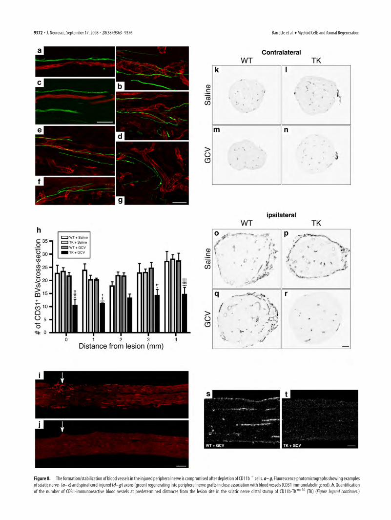
Figure 8. The formation/stabilization of blood vessels in the injured peripheral nerve is compromised after depletion of CD11b � cells. a– g, Fluorescence photomicrographs showing examplesof sciatic nerve- (a– c) and spinal cord-injured (d– g) axons (green) regenerating into peripheral nerve grafts in close association with blood vessels (CD31 immunolabeling; red). h, Quantificationof the number of CD31-immunoreactive blood vessels at predetermined distances from the lesion site in the sciatic nerve distal stump of CD11b-TK mt-30 (TK) (Figure legend continues.)
9372 • J. Neurosci., September 17, 2008 • 28(38):9363–9376 Barrette et al. • Myeloid Cells and Axonal Regeneration

Using in vivo models of peripheral nerve grafts, we establishedthat CD11b� myeloid cells are required for regeneration of pe-ripheral nerve axons and that the presence of these cells can evensupport regeneration of SCI axons. Finally, we defined some ofthe key mechanisms by which CD11b� cells support axonal re-generation and neural repair. Specifically, we found thatCD11b� cells are responsible for the removal of inhibitory mye-lin debris, production of neurotrophins, and formation and sta-bilization of blood vessels in the injured nervous system.
One likely mechanism by which CD11b� myeloid cells maycontribute to a permissive environment for axon growth isthrough the clearance of degenerating myelin and inhibitors ofaxonal regeneration. Several proteins associated with myelinhave been shown to inhibit neurite outgrowth or collapse growthcones, including Nogo-A, MAG, OMgp (oligodendrocyte myelinglycoprotein), ephrin B3, and semaphorin 4D (David and Lac-roix, 2003; Yiu and He, 2006). Although some of these proteinsare not expressed in the injured PNS, MAG and semaphorins areand could contribute to prevent axon regeneration (Liu et al.,2006). After peripheral nerve injury, immune cells such asCD11b� macrophages are rapidly recruited and eliminate mye-lin debris within 10 –14 d (George and Griffin, 1994). This obser-vation is consistent with our results showing that the load ofmyelin debris was �200% higher in animals depleted in CD11b�
cells compared with wild-type littermates at 7 d after injury. Thissuggests that myelin debris were not removed efficiently in theabsence of CD11b� cells. It should be pointed out, however, thatmyelin breakdown and the formation of ovoids of degeneratingmyelin were apparent in the distal sciatic nerve segment of GCV-treated CD11b-TK mt-30 transgenic mice, indicating that these re-sponses did not depend on CD11b� cells. Overall, these findingssuggest that CD11b� macrophages are primarily, although per-haps not entirely, responsible for the phagocytosis and removal ofmyelin debris and its inhibitory effects.
Peripheral nerve injury leads to the expression of a wide vari-ety of neurotrophic factors, notably neurotrophins (Fu and Gor-don, 1997; Terenghi, 1999). Neurotrophins, which include NGF,BDNF, NT-3, and NT-4/5, are well known for their capacity topromote neuronal survival and axon growth in the PNS and CNS(Lacroix and Tuszynski, 2000). Addition of exogenous neurotro-phins to the injured peripheral nerve environment has been re-ported to facilitate axon regeneration and remyelination and toimprove functional recovery, whereas blocking these factors wasfound to compromise regeneration and cause additional deficitsin recovery of nerve functions (Terenghi, 1999; Lykissas et al.,2007). Despite abundant evidence showing that neurotrophinsare key molecules for nerve regeneration and repair, the cellularand molecular mechanisms regulating neurotrophin synthesis
have yet to be fully investigated. The Thoenen Laboratory re-ported the first evidence that immune cells might play a role inregulating neurotrophin synthesis after neural injury. They ob-served that NGF mRNA levels decrease over time when sciaticnerve is kept in culture, and that NGF levels increase when acti-vated macrophages are added to the culture medium (Heumannet al., 1987b). In a subsequent study, they found that IL-1� de-rived from macrophages was responsible for the synthesis of NGFby non-neuronal cells after sciatic nerve injury (Lindholm et al.,1987). Based on the time course of BDNF expression, they furthersuggested that different regulatory mechanisms were likely tocontrol NGF and BDNF expression in the injured peripheralnerve in vivo. To clarify this issue and investigate the possibilitythat CD11b� myeloid cells may also contribute to nerve regen-eration through the production of neurotrophic factors, we ex-amined whether depletion of CD11b� would affect neurotro-phin synthesis after sciatic nerve injury.
In line with previous studies (Heumann et al., 1987a,b; Funa-koshi et al., 1993), our results show that NGF, BDNF, and NT-4/5expression was absent in the intact sciatic nerve but induced innon-neuronal cells of the nerve distal stump after injury. Ourresults differ, however, from the study by Funakoshi et al. (1993)with regard to the expression of NT-3, which we found to beregulated exactly like the other three neurotrophins. The differ-ences observed between the study by Funakoshi et al. (1993) andours are discussed in more details the supplemental Discussion(available at www.jneurosci.org as supplemental material). Addi-tional analyses using CD11b-TK mt-30 transgenic mice revealedthat neurotrophin expression was almost completely abolished inanimals depleted in CD11b� cells. Double-labeling studies com-bining ISH with immunohistochemistry and experiments per-formed in scid mice that have no functional T- and B-cells con-firmed that granulocytes and/or a subset of monocytes expressingthe Gr-1 marker were the main, if not only, cellular source ofneurotrophins in the injured sciatic nerve at 1 week after lesion.Work is currently underway to determine the exact phenotype ofthese cells.
Another particularly intriguing observation that we madeduring the course of this study is that the formation of new bloodvessels at the lesion site and within the nerve distal stump wasalmost completely prevented in mice depleted in CD11b� cells.This, together with the fact that we found no evidence of GFP�
endothelial cells in blood vessels of GFP bone marrow chimericmice after peripheral nerve injury (S. Lacroix, unpublished ob-servations), suggests that CD11b� cells are essential for vesselformation after nerve injury but unlikely to develop intoendothelial-like cells, as suggested by others when cultured undercertain experimental conditions (for review, see De Palma andNaldini, 2006). Interestingly, Gray et al. (2007) reported in theirrecent macrophage depletion study the presence of significantlyless Evans blue albumin (used as a marker of blood vessel integ-rity) in their sciatic nerve preparations after injury. Although thisissue was not investigated in their study, one possible interpreta-tion of these results is that de novo vessel formation was compro-mised in sciatic nerves of these animals in the absence of mono-cytes/macrophages. A proangiogenic role for a specific subtype ofmyeloid cells expressing the Tie2 angiopoietin receptor and rep-resenting �1–15% of all CD11b� myeloid cells has been re-ported before in a brain tumor model (De Palma et al., 2003,2005). The demonstration that cells expressing Tie2 were de-pleted in the sciatic nerve distal stump of GCV-treated CD11b-TK mt-30 transgenic mice supports the possibility that such a pop-ulation of myeloid cells (CD11b�Tie2�) may have been
4
(Figure legend continued.) and WT mice at 7 d after lesion in response to either GCV or salinetreatment (n � 8 per group). Error bars indicate SEM. i, j, Representative photomicrographsshowing immunofluorescence for CD31 in the sciatic nerve distal stump of WT (i) and TK (j) micetreated with GCV at 7 d after lesion. The arrows point to the lesion site. k–r, Representativebright-field photomicrographs showing immunoreactivity for CD31 in sciatic nerve cross sec-tions taken from contralateral (unlesioned) and ipsilateral (lesioned) nerves of WT and TK micetreated with either saline or GCV at 7 d after lesion. s, t, Representative dark-field photomicro-graphs showing Tie2 mRNA expression in the sciatic nerve distal stump of GCV-treated WT andGCV-treated TK mice at 7 d after lesion. Note the absence of Tie2 mRNA signal after depletion ofCD11b � cells. ***p � 0.001, **p � 0.01, and *p � 0.05 compared with the WT plus salinegroup; §§§p � 0.001, §§p � 0.01, and §p � 0.05 compared with the TK plus saline group;†††p �0.001, ††p �0.01, and †p �0.05 compared with the WT plus GCV group. Scale bars: (inc) a– c, 50 �m; (in g) d– g, 50 �m; (in j) i, j, 250 �m; (in r) k–r, 80 �m; (in t) s, t, 100 �m.
Barrette et al. • Myeloid Cells and Axonal Regeneration J. Neurosci., September 17, 2008 • 28(38):9363–9376 • 9373

instrumental to the formation of newblood vessels. Some of the potential mech-anisms by which CD11b� myeloid cellscould regulate blood vessel formation/stabilization are discussed in the supple-mental Discussion (available at www.jneurosci.org as supplemental material).
Together, the results discussed abovecould help resolve a long-standing contro-versy regarding the potentially beneficialeffects of transplanting macrophages intothe injured spinal cord (Schwartz andYoles, 2006; Donnelly and Popovich,2008). This controversy stemmed mostlyfrom two separate studies, one that re-ported that macrophages preincubatedwith sciatic nerve segments can promotepartial recovery of motor functions oncetransplanted into the fully transected ratspinal cord (Rapalino et al., 1998) and an-other that showed that depletion of blood-derived macrophages decreases tissuedamage and promotes recovery of hind-limb function after spinal cord contusion(Popovich et al., 1999). Perhaps mono-cytes/macrophages play a role in both tis-sue damage and repair, depending on thecellular population involved and theirstate of activation. Additional studies aretherefore needed to investigate whether aspecific subset of the macrophages thatwere transplanted or depleted into the in-jured spinal cord were responsible for thereported beneficial and detrimental ef-fects, respectively. Along these lines, theexistence of at least two monocyte subsetswith divergent but complementary func-tions was recently demonstrated (Auffrayet al., 2007; Nahrendorf et al., 2007). Ac-cording to these studies, one of these twosubsets, termed inflammatory monocytes(Gr-1 high/CCR2�/CX3CR1 low), are re-cruited to injured tissues via the CCR2 re-ceptor and are primarily involved in in-flammation, proteolysis, andphagocytosis. The other monocytic popu-lation, termed Gr-1 low/CCR2�/CX3CR1 high monocytes, respond to a dif-ferent chemokine, fractalkine (alsoreferred to as CX3CL1), and is apparentlyimplicated in both immune surveillanceand the healing process. A role for Gr-1 low/CX3CR1 high monocytes in angiogenesis has also been suggested(Nahrendorf et al., 2007). Clearly, more studies are needed tobetter define the exact roles of the various monocyte/macrophagesubsets after injury to the nervous system.
In conclusion, our results indicate that CD11b� myeloid cellsmodulate axonal regeneration after injury by directly influencingthe growth cone dynamics through the creation of a more favor-able growth environment. These cells contribute to recreate apermissive milieu for regeneration of injured peripheral and cen-tral axons through the removal of inhibitory myelin debris andrelease of neurotrophic and proangiogenic factors.
ReferencesAuffray C, Fogg D, Garfa M, Elain G, Join-Lambert O, Kayal S, Sarnacki S,
Cumano A, Lauvau G, Geissmann F (2007) Monitoring of blood vesselsand tissues by a population of monocytes with patrolling behavior. Sci-ence 317:666 – 670.
Bandtlow CE, Meyer M, Lindholm D, Spranger M, Heumann R, Thoenen H(1990) Regional and cellular codistribution of interleukin 1 beta andnerve growth factor mRNA in the adult rat brain: possible relationship tothe regulation of nerve growth factor synthesis. J Cell Biol 111:1701–1711.
Barrette B, Vallieres N, Dube M, Lacroix S (2007) Expression profile of recep-tors for myelin-associated inhibitors of axonal regeneration in the intact andinjured mouse central nervous system. Mol Cell Neurosci 34:519–538.
Figure 9. Regeneration of SCI axons into PN grafts is prevented in the absence of CD11b � cells. a, Photograph showing threesciatic nerve segments transplanted into the spinal cord of a T12/L1 dorsal hemisected Thy1-YFP-H �/� transgenic mouse. Thearrows point to PN grafts/spinal cord tissue interfaces. b, c, YFP expression in the thoracic (b) and lumbar (c) spinal cord of aThy1-YFP-H �/� transgenic mouse. Note the presence of fluorescence in axons traveling through most, if not all, spinal cordprojection systems, including the descending corticospinal (CST) and rubrospinal (RST) tracts and the ascending sensory tract(AST). d, e, Visualization of laminin immunolabeling (d) (to visualize PN tissue) and YFP fluorescence (e) on adjacent spinal cordsections revealed that PN grafts are densely penetrated by regenerating SCI axons at 2 weeks after SCI/grafting. The dotted linesin d indicate the anatomical boundaries of a PN graft. f, Quantification of the number of YFP-labeled axons at predetermineddistances from the rostrocaudal host– graft interface. Note that SCI YFP-labeled axons did not regenerate into predegenerated PNgrafts lacking CD11b � cells. Error bars indicate SEM. **p � 0.01 compared with the WT plus saline group; §p � 0.05 comparedwith the TK plus saline group. Scale bars: (in c) b, c, 275 �m; (in e) d, e, 100 �m.
9374 • J. Neurosci., September 17, 2008 • 28(38):9363–9376 Barrette et al. • Myeloid Cells and Axonal Regeneration

Boivin A, Pineau I, Barrette B, Filali M, Vallieres N, Rivest S, Lacroix S (2007)Toll-like receptor signaling is critical for Wallerian degeneration andfunctional recovery after peripheral nerve injury. J Neurosci27:12565–12576.
Bruck W, Huitinga I, Dijkstra CD (1996) Liposome-mediated monocytedepletion during wallerian degeneration defines the role of hematoge-nous phagocytes in myelin removal. J Neurosci Res 46:477– 484.
Carmeliet P, Tessier-Lavigne M (2005) Common mechanisms of nerve andblood vessel wiring. Nature 436:193–200.
Dahlin LB (1995) Prevention of macrophage invasion impairs regenerationin nerve grafts. Brain Res 679:274 –280.
David S, Aguayo AJ (1981) Axonal elongation into peripheral nervous sys-tem “bridges” after central nervous system injury in adult rats. Science214:931–933.
David S, Lacroix S (2003) Molecular approaches to spinal cord repair. AnnuRev Neurosci 26:411– 440.
de Medinaceli L, Freed WJ, Wyatt RJ (1982) An index of the functionalcondition of rat sciatic nerve based on measurements made from walkingtracks. Exp Neurol 77:634 – 643.
De Palma M, Naldini L (2006) Role of haematopoietic cells and endothelialprogenitors in tumour angiogenesis. Biochim Biophys Acta1766:159 –166.
De Palma M, Venneri MA, Roca C, Naldini L (2003) Targeting exogenousgenes to tumor angiogenesis by transplantation of genetically modifiedhematopoietic stem cells. Nat Med 9:789 –795.
De Palma M, Venneri MA, Galli R, Sergi Sergi L, Politi LS, Sampaolesi M,Naldini L (2005) Tie2 identifies a hematopoietic lineage of proangio-genic monocytes required for tumor vessel formation and a mesenchymalpopulation of pericyte progenitors. Cancer Cell 8:211–226.
Donnelly DJ, Popovich PG (2008) Inflammation and its role in neuropro-tection, axonal regeneration and functional recovery after spinal cordinjury. Exp Neurol 209:378 –388.
English AW, Meador W, Carrasco DI (2005) Neurotrophin-4/5 is requiredfor the early growth of regenerating axons in peripheral nerves. Eur J Neu-rosci 21:2624 –2634.
Feng G, Mellor RH, Bernstein M, Keller-Peck C, Nguyen QT, Wallace M,Nerbonne JM, Lichtman JW, Sanes JR (2000) Imaging neuronal subsetsin transgenic mice expressing multiple spectral variants of GFP. Neuron28:41–51.
Fernandez-Valle C, Bunge RP, Bunge MB (1995) Schwann cells degrademyelin and proliferate in the absence of macrophages: evidence from invitro studies of Wallerian degeneration. J Neurocytol 24:667– 679.
Folkman J (2007) Angiogenesis: an organizing principle for drug discovery?Nat Rev Drug Discov 6:273–286.
Fu SY, Gordon T (1997) The cellular and molecular basis of peripheralnerve regeneration. Mol Neurobiol 14:67–116.
Funakoshi H, Frisen J, Barbany G, Timmusk T, Zachrisson O, Verge VM,Persson H (1993) Differential expression of mRNAs for neurotrophinsand their receptors after axotomy of the sciatic nerve. J Cell Biol123:455– 465.
George R, Griffin JW (1994) Delayed macrophage responses and myelinclearance during Wallerian degeneration in the central nervous system:the dorsal radiculotomy model. Exp Neurol 129:225–236.
Gowing G, Vallieres L, Julien JP (2006a) Mouse model for ablation of pro-liferating microglia in acute CNS injuries. Glia 53:331–337.
Gowing G, Dequen F, Soucy G, Julien JP (2006b) Absence of tumor necrosisfactor-� does not affect motor neuron disease caused by superoxide dis-mutase 1 mutations. J Neurosci 26:11397–11402.
Gray M, Palispis W, Popovich PG, van Rooijen N, Gupta R (2007) Macro-phage depletion alters the blood-nerve barrier without affecting Schwanncell function after neural injury. J Neurosci Res 85:766 –777.
Guthrie KM, Gall CM (1991) Differential expression of mRNAs for theNGF family of neurotrophic factors in the adult rat central olfactorysystem. J Comp Neurol 313:95–102.
Hall SM (1986) The effect of inhibiting Schwann cell mitosis on the re-innervation of acellular autografts in the peripheral nervous system of themouse. Neuropathol Appl Neurobiol 12:401– 414.
Harel NY, Strittmatter SM (2006) Can regenerating axons recapitulate de-velopmental guidance during recovery from spinal cord injury? Nat RevNeurosci 7:603– 616.
Heppner FL, Greter M, Marino D, Falsig J, Raivich G, Hovelmeyer N, Wais-man A, Rulicke T, Prinz M, Priller J, Becher B, Aguzzi A (2005) Experi-
mental autoimmune encephalomyelitis repressed by microglial paralysis.Nat Med 11:146 –152.
Heumann R, Korsching S, Bandtlow C, Thoenen H (1987a) Changes ofnerve growth factor synthesis in nonneuronal cells in response to sciaticnerve transection. J Cell Biol 104:1623–1631.
Heumann R, Lindholm D, Bandtlow C, Meyer M, Radeke MJ, Misko TP,Shooter E, Thoenen H (1987b) Differential regulation of mRNA encod-ing nerve growth factor and its receptor in rat sciatic nerve during devel-opment, degeneration, and regeneration: role of macrophages. Proc NatlAcad Sci U S A 84:8735– 8739.
Hirata K, Kawabuchi M (2002) Myelin phagocytosis by macrophages andnonmacrophages during Wallerian degeneration. Microsc Res Tech57:541–547.
Hofer M, Pagliusi SR, Hohn A, Leibrock J, Barde YA (1990) Regional distri-bution of brain-derived neurotrophic factor mRNA in the adult mousebrain. EMBO J 9:2459 –2464.
Inserra MM, Bloch DA, Terris DJ (1998) Functional indices for sciatic, per-oneal, and posterior tibial nerve lesions in the mouse. Microsurgery18:119 –124.
Kermani P, Hempstead B (2007) Brain-derived neurotrophic factor: anewly described mediator of angiogenesis. Trends Cardiovasc Med17:140 –143.
Klagsbrun M, Eichmann A (2005) A role for axon guidance receptors andligands in blood vessel development and tumor angiogenesis. CytokineGrowth Factor Rev 16:535–548.
Kubota A, Suzuki K (2000) Effect of liposome-mediated macrophage deple-tion on Schwann cell proliferation during Wallerian degeneration. J Neu-rotrauma 17:789 –798.
Lacroix S, Tuszynski MH (2000) Neurotrophic factors and gene therapy inspinal cord injury. Neurorehabil Neural Repair 14:265–275.
Lacroix S, Chang L, Rose-John S, Tuszynski MH (2002) Delivery of hyper-IL-6 to the injured spinal cord increases neutrophil and macrophage in-filtration and inhibits axonal growth. J Comp Neurol 454:213–228.
Lindholm D, Heumann R, Meyer M, Thoenen H (1987) Interleukin-1 reg-ulates synthesis of nerve growth factor in non-neuronal cells of rat sciaticnerve. Nature 330:658 – 659.
Liu BP, Cafferty WB, Budel SO, Strittmatter SM (2006) Extracellular regu-lators of axonal growth in the adult central nervous system. Philos TransR Soc Lond B Biol Sci 361:1593–1610.
Lykissas MG, Batistatou AK, Charalabopoulos KA, Beris AE (2007) The roleof neurotrophins in axonal growth, guidance, and regeneration. CurrNeurovasc Res 4:143–151.
Ma M, Wei T, Boring L, Charo IF, Ransohoff RM, Jakeman LB (2002)Monocyte recruitment and myelin removal are delayed following spinalcord injury in mice with CCR2 chemokine receptor deletion. J NeurosciRes 68:691–702.
Maisonpierre PC, Belluscio L, Friedman B, Alderson RF, Wiegand SJ, FurthME, Lindsay RM, Yancopoulos GD (1990) NT-3, BDNF, and NGF inthe developing rat nervous system: parallel as well as reciprocal patterns ofexpression. Neuron 5:501–509.
Morrissey TK, Kleitman N, Bunge RP (1991) Isolation and functional char-acterization of Schwann cells derived from adult peripheral nerve. J Neu-rosci 11:2433–2442.
Nahrendorf M, Swirski FK, Aikawa E, Stangenberg L, Wurdinger T, Figueir-edo JL, Libby P, Weissleder R, Pittet MJ (2007) The healing myocardiumsequentially mobilizes two monocyte subsets with divergent and comple-mentary functions. J Exp Med 204:3037–3047.
Pineau I, Lacroix S (2007) Proinflammatory cytokine synthesis in the in-jured mouse spinal cord: multiphasic expression pattern and identifica-tion of the cell types involved. J Comp Neurol 500:267–285.
Pineau I, Barrette B, Vallieres N, Lacroix S (2006) A novel method for mul-tiple labeling combining in situ hybridization with immunofluorescence.J Histochem Cytochem 54:1303–1313.
Plunet W, Kwon BK, Tetzlaff W (2002) Promoting axonal regeneration inthe central nervous system by enhancing the cell body response to axo-tomy. J Neurosci Res 68:1– 6.
Popovich PG, Guan Z, Wei P, Huitinga I, van Rooijen N, Stokes BT (1999)Depletion of hematogenous macrophages promotes partial hindlimb re-covery and neuroanatomical repair after experimental spinal cord injury.Exp Neurol 158:351–365.
Ramon y Cajal S (1991) Cajal’s degeneration and regeneration of the ner-vous system. New York: Oxford UP.
Barrette et al. • Myeloid Cells and Axonal Regeneration J. Neurosci., September 17, 2008 • 28(38):9363–9376 • 9375

Rapalino O, Lazarov-Spiegler O, Agranov E, Velan GJ, Yoles E, Fraidakis M,Solomon A, Gepstein R, Katz A, Belkin M, Hadani M, Schwartz M (1998)Implantation of stimulated homologous macrophages results in partialrecovery of paraplegic rats. Nat Med 4:814 – 821.
Reichert F, Saada A, Rotshenker S (1994) Peripheral nerve injury inducesSchwann cells to express two macrophage phenotypes: phagocytosis andthe galactose-specific lectin MAC-2. J Neurosci 14:3231–3245.
Saada A, Reichert F, Rotshenker S (1996) Granulocyte macrophage colonystimulating factor produced in lesioned peripheral nerves induces theup-regulation of cell surface expression of MAC-2 by macrophages andSchwann cells. J Cell Biol 133:159 –167.
Schwartz M, Yoles E (2006) Immune-based therapy for spinal cord repair:autologous macrophages and beyond. J Neurotrauma 23:360 –370.
Stoll G, Griffin JW, Li CY, Trapp BD (1989) Wallerian degeneration in theperipheral nervous system: participation of both Schwann cells and mac-rophages in myelin degradation. J Neurocytol 18:671– 683.
Terenghi G (1999) Peripheral nerve regeneration and neurotrophic factors.J Anat 194:1–14.
Vallieres N, Berard JL, David S, Lacroix S (2006) Systemic injections of li-popolysaccharide accelerates myelin phagocytosis during Wallerian de-generation in the injured mouse spinal cord. Glia 53:103–113.
Weidner N, Blesch A, Grill RJ, Tuszynski MH (1999) Nerve growth factor-hypersecreting Schwann cell grafts augment and guide spinal cord axonalgrowth and remyelinate central nervous system axons in a phenotypicallyappropriate manner that correlates with expression of L1. J Comp Neurol413:495–506.
Yiu G, He Z (2006) Glial inhibition of CNS axon regeneration. Nat RevNeurosci 7:617– 627.
Zuo Y, Lubischer JL, Kang H, Tian L, Mikesh M, Marks A, Scofield VL, Maika S,Newman C, Krieg P, Thompson WJ (2004) Fluorescent proteins expressedin mouse transgenic lines mark subsets of glia, neurons, macrophages, anddendritic cells for vital examination. J Neurosci 24:10999–11009.
9376 • J. Neurosci., September 17, 2008 • 28(38):9363–9376 Barrette et al. • Myeloid Cells and Axonal Regeneration
Copyright © 2022 FDOKUMEN